Note : Les descriptions sont présentées dans la langue officielle dans laquelle elles ont été soumises.
CA 02956563 2017-01-27
Use of SUSD2 Protein as Marker
TECHNICAL FIELD
The present invention relates to the field of biotechnology, and in
particular to the use of the SUSD2 protein as a marker.
BACKGROUND ART
Human pluripotent stem cell-derived pancreatic beta-cells provide
sufficient sources of donor islet cells for use in cell replacement therapy
for diabetes, particularly for type I diabetes. In addition, the directed
differentiation of human pluripotent stem cells(hPSCs) into pancreatic
beta-cells can also provide a model for studying human pancreatic
development in vitro. The process of the directed differentiation of hPSCs
into pancreatic beta-cells can be divided into several stages by mimicking
development events in vivo: definitive endoderm, gut tube, posterior
foregut, pancreatic endoderm, and pancreatic endocrine cells. Among
them, pancreatic endoderm cells are mixed cell populations, usually
including pancreatic progenitor cells, pancreatic endocrine progenitor cells
and nascent pancreatic endocrine cells and the like. The cell fate is
determinate by detecting the expression of pancreatic development-related
genes. Pancreatic progenitor cells are mainly indicated by expression of
the relevant genes such as PDX1, HNF1B, SOX9, NKX6.1, HNF6 and so
CA 02956563 2017-01-27
on. NGN3 is the most important marker gene of pancreatic endocrine
progenitor cells, and however, due to its transient expression
characteristics, it is required to combine with the expression of its
downstream genes such as NKX2.2 and NEUROD1, as well as negative
expression of endocrine-related genes such as CHROMOGRANIN A,
INSULIN, and GLUCAGON to indicate the fate of pancreatic endocrine
progenitor cells. Although the expression of these marker genes can be
used to indicate the fate of specific cells, the expression of these proteins
cannot be used to isolate and purify specific cell populations as they are all
transcription factors or secretory proteins, generally locating within the
cytoplasm or nucleusõ in particular human pancreatic endocrine
progenitor cells and nascent pancreatic endocrine cells, so that their
molecular characteristics cannot be studied in more detail.
At present, pancreatic progenitor cells can be obtained by directed
differentiation of hPSCs, however, these cells differentiated into functional
mature pancreatic beta-cells with low efficiency in vitro. Although after
pancreatic precursor cells derived from human pluripotent stem cells are
transplanted into immunodeficient mouse, functionally mature islet cells
can be obtained, due to the proliferative capacity of pancreatic precursor
cells, their tumorigenicity limits their promotion to clinical application.
The functional mature beta-cells are more desirable donor cell sources
2
CA 02956563 2017-01-27
than pancreatic progenitor cells. However, how to differentiate pancreatic
progenitor cells into functional mature beta-cells in vitro greatly limits the
implementation of alternative treatment of diabetes cells, while in the
differentiation process from pancreatic precursor cells to pancreatic beta-
cells, the most important step isthe correct realization of efficient induced
differentiation of pancreatic endocrine precursor cells. Studies on animal
model such as mouse have greatly promoted the understanding of the
development of pancreatic beta-cells. These studies have played a decisive
role in directing the directed differentiation of pancreatic beta-cells in
vitro. In spite of this, studies on the fate specialization of pancreatic
endocrine precursor cells and the fate selection between different
endocrine cells are still few. In addition, there exist some species
differences between human and model animal such as mouse. Therefore,
finding relevant molecular markers to directly isolate pancreas-related
cells at a particular developmental stage for researching can significantly
speed up the acquisition of functional mature pancreatic beta-cells in vitro.
SUMMARY
It is an object of the present invention to provide a method for sorting or
assisting the sorting of pancreatic endocrine progenitor cells or nascent
endocrine cells in a population of cells to be tested.
3
CA 02956563 2017-01-27
The method provided by the present invention comprises the following
steps: detecting whether the cells in the population of cells to be tested
express SUSD2 gene; if some cells express the SUSD2 gene, these cells
are or are candidates for pancreatic endocrine precursor cells or nascent
endocrine cells; If some cells do not express the SUSD2 gene, these cells
are not or are not candidates for pancreatic endocrine precursor cells or
nascent endocrine cells.
The nucleotide sequence of the SUSD2 gene is illustrated as SEQ ID NO:
2 in the Sequence Listing.
In the above method, the step of detecting whether the cells in the
population of cells to be tested express a SUSD2 gene is performed by
detecting whether the cell in the population of cells to be tested contains a
protein expressed by the SUSD2 gene, and the method for detecting
whether the cell in the population of cells to be tested contains the protein
expressed by the SUSD2 gene is as following 1) or 2) or 3):
1) immunofluorescence antibody assay, using anti-SUSD2 monoclonal
antibody;
2)flow cytometry, using anti-SUSD2 monoclonal antibody;
3) magnetic beads cell sorting, using anti-SUSD2 monoclonal antibody.
4
CA 02956563 2017-01-27
In the above method, the cells to be tested are pancreatic endoderm cells.
In the above method, the pancreatic endoderm cell is human-derived
pancreatic endoderm cells.
By analyzing gene expression profiles of the pancreatic endoderm cells,
the present inventors also find that the expression of the SUSD2 gene is
enriched in pancreatic endocrine progenitor cells and neonatal endocrine
cells, i.e., SUSD2 protein can be used as the molecular marker of the both
cells.
Based on the above findings, the present invention provides the use of a
SUSD2 protein as a marker in the identification, screening or sorting of
pancreatic endocrine progenitor cells and / or nascent pancreatic endocrine
cells, wherein the amino acid sequence of the SUSD2 protein is shown as
SEQ ID NO: 1.
NCBI Reference Sequence of the SUSD2 protein is NP_062547.1.
The nascent pancreatic endocrine cells refer to a population of hormone
(including Insulin, Glucagon, Ghrelin, Pancreatic Polypeptide,
Somatostatin, etc.)-positive cells during the process of direct
differentiation from human pluripotent stem cells into pancreatic beta-
cells. This cell population usually co-expresses multi-hottnone, and are
CA 02956563 2017-01-27
less mature functionally as compared to mature pancreatic endocrine cells.
During in vivo development, nascent pancreatic endocrine cells are
primarily pancreatic endocrine cells that are functionally immature during
embryonic development.
The present invention also provides use of an antibody capable of
specifically binding to a SUSD2 protein in preparation of a reagent for
identification, screening or sorting of pancreatic endocrine precursor cells
and / or nascent pancreatic endocrine cells.
The SUSD2 protein is a protein expressed by the SUSD2gene, which is
neither a transcription factor nor a secretory protein, but a receptor protein
located on cell membrane.
The cells to be tested are detected by immunofluorescent antibody method
to determine whether they expressed SUSD2 proteins or not, in order to
determine whether the cells to be tested are pancreatic endocrine precursor
cells or nascent pancreatic endocrine cells.
The antibody may be selected froman intact antibody molecule, a chimeric
antibody, a single chain antibody, abispecific antibody, a heavy chain of
an antibody, a light chain of an antibody, homodimer and heterodimer of
heavy and light chains, antigen binding fragment,and their derivatives.
The intact antibody molecule can be a polyclonal or monoclonal antibody,
6
CA 02956563 2017-01-27
preferably a monoclonal antibody.
As described above, the SUSD2 protein can generally be detected by an
immunofluorescent antibody method, wherein the antibody against the
protein or a secondary antibody against the antibody is required to carry a
corresponding fluorescent label.
The pancreatic endocrine precursor cells and nascent pancreatic endocrine
cells are a population of cells in pancreatic endoderm cells, which can
produce functionally mature pancreatic endocrine cells under suitable
conditions.
The human pancreatic endoderm cells mainly refer to the cells at
pancreatic endoderm stage during the process of direct differentiation from
human pluripotent stem cells to pancreatic beta-cells, or human embryonic
pancreatic tissue cells at corresponding developmental stage which are
freshly isolated and have been cultured in vitro, mainly comprising
pancreatic precursor cells, pancreatic endocrine precursor cells and
nascent pancreatic endocrine cells. The primary object of the present
invention is to screen or sort pancreatic endocrine precursor cells and
nascent pancreatic endocrine cells from pancreatic endoderm cell
populations.
The sources of pancreatic endoderm cells include: 1. cells obtained by
7
CA 02956563 2017-01-27
direct differentiation from human pluripotent stem cells; 2. cells obtained
from isolated human embryonic pancreas tissue; 3. cells obtained by in
vitro culture of isolated human embryonic pancreatic endoderm cells.
The method for screening or sorting mainly adopts imrnunomagnetic
separation or flow cytometry sorting.
In the process of SUSD2 gene expression, it is first transcribed into
mRNA, then the mRNA is translated into SUSD2 precursor protein, and
then the precursor protein is processed to produce mature SUSD2 protein.
For the above reasons, the present invention also provides a use of an
mRNA encoding a precursor protein of a SUSD2 protein as a marker in
identification of pancreatic endocrine precursor cells and / or nascent
pancreatic endocrine cells.
The invention further provides the use of a primer, probe or their
complementary strands capable of specifically binding to the mRNA in
preparation of a reagent for identification of pancreatic endocrine
precursor cells and / or nascent pancreatic endocrine cells.
The primer may be a primer for amplifying the whole mRNA or a primer
for amplifying a characteristic region of the mRNA, and the probe is a
nucleotide which recognizes a specific region of the mRNA and generally
8
CA 02956563 2017-01-27
carries a label.
According to the description of the examples, the present invention
provides a pair of primers that specifically amplify the rnRNA, as follows:
upstream primer: GGCACCGCCAACACCTCA
downstream primer: GCGTGGGCAGCGACTTGA.
The method for identification may employ fluorescence quantitative PCR.
By analyzing the gene expression profiles of the endoderm cells, the
inventors find that the expression of SUSD2 gene is enriched in the
pancreatic endocrine precursor cells and nascent pancreatic endocrine
cells, and its encoded protein is a receptor protein on a cell membrane and
the protein can be used as a marker to identify, screen or sort pancreatic
endocrine precursor cells and nascent pancreatic endocrine cells, which is
of great significance for the study of pancreas-related cells at various
developmental stages.
BRIEF DESCRIPTION OF THE DRAWINGS
Figure 1 shows the direct differentiation from human pluripotent stem
cells into pancreatic endoderm cells. (a) Schematic representation of direct
differentiation from human pluripotent stem cells into pancreatic beta-
9
CA 02956563 2017-01-27
cells; (b) immunofluorescent staining for detecting the expression of green
fluorescent protein (EGFP) marking NGN3 genes and pancreatic
endoderm-related proteins at the end of the stage 4 of differentiation.
Figure 2 shows the results of flow cytometry for identifying that NGN3-
EGFP + cells in pancreatic endoderm are pancreatic endocrine precursor
cells or nascent endocrine cells. (a) Flow cytometry is used to detect the
expression of NGN3-EGFP and pancreatic endocrine-related proteins such
as NGN3, NKX2.2, NEUROD1, CHROMOGRANIN A (CHGA) in the
pancreatic endoderm cells; (b) flow cytometry is used to detect the
expressions of NGN3-EGFP and pancreatic precursor cell marker protein
PDX1 in pancreatic endoderm cells, indicating that pancreatic precursor-
related protein PDX1 is low expressed or not expressed in NGN3-EGFP +
cells.
Figure 3 shows the expression of pancreas-related genes in NGN3-EGFP+
cells and NGN3-EGFP-cells obtained by sorting with mRNA sequencing
analysis, indicating that the population of pancreatic endocrine precursor
cells and nascent endocrine cells of NGN3-EGFP + are relatively enriched
with the expression of pancreatic endocrine-related genes.
Figure 4 shows immunofluorescent staining for detecting the merged
staining pattern of SUSD2 and NGN3-EGFP, indicating that SUSD2 can
CA 02956563 2017-01-27
be used to mark NGN3-EGFP + cells derived from human pluripotent
stem cells.
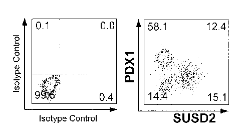
Fig. 5 shows an analysis graph of a flow cytometry for detecting the
expression of SUSD2 and pancreatic development-related proteins in
pancreatic endoderm cells derived from human pluripotent stem cells,
indicating that SUSD2 can be used to mark pancreatic endocrine precursor
cells and nascent endocrine cells obtained by differentiation in vitro. (a)
Flow cytometry is used to detect the expression of SUSD2 and pancreatic
endocrine-related proteins such as NGN3, NKX2.2, NEUROD1,
CHROMOGRANIN A (CHGA) in pancreatic endoderm cells; (b) flow
cytometry is used to detect the expression of SUSD2 and pancreatic
precursor cell-related proteins PDX1 in pancreatic endodeini cells.
Figure 6 shows the expression of pancreatic development-related genes in
SUSD2 + cells, SUSD2-cells obtained by sorting with flow cytometry and
in unsorted pancreatic endoderm cells identified by RT-QPCR, indicating
that SUSD2+ cells are relatively enriched with the expression of
pancreatic endocrine precursor cell-related genes and pancreatic endocrine
cell-related genes and relatively weakly express the pancreatic precursor
cell-related genes, demonstrating that cells labeled by SUSD2 are
pancreatic endocrine precursor cells and / or nascent endocrine cells.
11
CA 02956563 2017-01-27
Figure 7 shows the SUSD2 antibody is used in magnetic cell sorting to
enrich pancreatic endocrine precursor cells and nascent pancreatic
endocrine cells. (a) The expression of NKX2.2 marker protein of
pancreatic endocrine precursor cells and pancreatic endocrine cells in
SUSD2 + cells and SUSD2-cells enriched by performing magnetic cell
sorting on pancreatic endoderm cells obtained by differentiation in vitro
with SUSD2 antibody is detected by flow cytometry, indicating that the
use of SUSD2 antibody in magnetic cell sorting can efficiently enrich
NKX2.2+ pancreatic endocrine precursor cells and nascent pancreatic
endocrine cells. (b) After SUSD2+ cells and SUSD2-cells obtained by
magnetic cell sorting and unsorted pancreatic endoderm cells are subjected
to extended culture, the expression of pancreatic precursor- and pancreatic
endocrine-related proteins is detected by immunohistochemistry,
indicating that SUSD2+ cells can produce a large number of endocrine
cells which are proved to be endocrine precursor cells or nascent
endocrine cells. (c) SUSD2+ cells and SUSD2-cells obtained by magnetic
cell sorting are transplanted into the immunodeficient mice. After 19
weeks, an immunofluorescent staining of the implants is carried out to
detect the expression of pancreatic endocrine-related protein, indicating
that SUSD2+ cells are able to produce multiple types of pancreatic
endocrine cells, while SUSD2-cells mainly produce duct-like cells,
demonstrating that the cells enriched by SUSD2 are pancreatic endocrine
12
CA 02956563 2017-01-27
precursor cells or nascent pancreatic endocrine cells.
Figure 8 shows that SUSD2 can be used to mark pancreatic endocrine
precursor cells and nascent pancreatic endocrine cells derived from human
embryos. (a) Co-expression of SUSD2 with pancreas endocrine progenitor
cell-related proteins and pancreatic endocrine cell-related proteins in
embryonic pancreas tissues and adult pancreas tissues is detected by
immunohistochemistry, indicating that SUSD2 is only expressed in
pancreatic endocrine precursors or endocrine cells in embryonic pancreas
tissues. (b) The expression of SUSD2 and the pancreatic endocrine-related
protein and pancreatic duct-related protein in the embryonic pancreas
tissues is detected by immunofluorescence.
Figure 9 shows that SUSD2 can be used to enrich pancreatic endocrine
precursor cells and nascent pancreatic endocrine cells derived from human
embryonic pancreas.
DETAILED DESCRIPTION
(
All of the experimental methods used in the following examples are
conventional methods unless otherwise specified. All of the materials,
reagents and the like used in the following examples are commercially
available unless otherwise specified. The SUSD2 gene is shown in SEQ
ID NO: 2 and the amino acid sequence of the encoded protein is SEQ ID
13
CA 02956563 2017-01-27
NO: 1.
Example 1: Discovery and application of SUSD2 as a marker gene for
pancreatic endocrine precursor cells.
I. SUSD2 as a marker gene of pancreatic endocrine precursor cells
obtained by differentiation of modified human pluripotent stem cells
1.1 Acquisition of the pancreatic endoderm cell
The EGFP gene (NC_013179.1 (3313..4126)) was homologously
recombined (Ngn3contig No.: NT_030059.13, located in the Chromosome
10, the position of the left homologous arm in the genome: 71325654-
71332154; the position of the right homologous arm in the genome:
71332158-71467568) into an isolated human pluripotent stem cell line I-11
(WiCell Research Institute, NIH No.: WA01) to obtain the modified
human pluripotent stem cell line NGN3-EGFP. The cells were seeded into
cell culture solution I in 15% -20% and cultured for 1 day; then directly
transferred into cell culture solution II and cultured for 1-2 days;
subsequently directly transferred into cell culture solution III and cultured
for 1 day; then directly transferred into cell culture solution IV and
cultured for 2 days; directly transferred into cell culture solution V and
cultured for 1 day; directly transferred into cell culture solution VI and
cultured for 3 days; directly transferred into cell culture solution VII and
14
CA 02956563 2017-01-27
cultured for 4 days; directly transferred into cell culture solution VIII and
cultured for 4-6 days; directly transferred into cell culture solution IX and
cultured for 4-6 days to obtain pancreatic endoderm cells.
The expression of several marker transcription factors related to pancreatic
endoderm in cell populations at this stage was detected by
immunofluorescent staining.
The antibody for detecting NKX2.2 was a murine-derived monoclonal
antibody (commercially available from DSHB, Catalog No. 74.5A5); the
antibody for detecting PDX1 was a goat-derived polyclonal antibody
(commercially available from R&D Systems, Catalog No.AF2419); the
antibody for detecting EGFP was chicken-derived polyclonal antibody
(commercially available from Abeam, Catalog No. AB13970); the
antibody for detecting NGN3 was sheep-derived polyclonal antibody
(commercially available from R & D System, Catalog No.AF3444).
All antibodies were non-directly labeled antibody. In the assay, the
following antibodies commercially available from Jackson
ImmunoResearch were used for counterstaining: Alexa Fluor 488-
labeleddonkey-derived anti-goat polyclonal antibody (Catalog No. 705-
545-147); Cyanine Cy3-labeled donkey-derived anti-goat polyclonal
antibody (Catalog No. 705-165-147); Cyanine Cy3-labeled donkey-
CA 02956563 2017-01-27
derived anti-sheep polyclonal antibody (Catalog No. 715-165-147); Alexa
Fluor 488-labeled donkey-derived anti-mouse polyclonal antibody
(Catalog No. 715-545-151); Cyanine Cy3-labeled donkey-derived anti-
mouse polyclonal antibody (Catalog No. 715-145-151); Alexa Fluor 488 -
labeled donkey-derived anti-chicken polyclonal antibodies (Catalog No.
703-545-155).
The results were shown in Fig. 1. In the cells obtained in this stage, some
of the cells expressed PDX1, a marker protein of pancreatic precursor
(Fig. 1A), the other cells expressed NGN3-EGFP, and there was almost
not co-staining of the two (Fig. 1B); NGN3-EGFP + cells expressed
pancreatic endocrine progenitor cell marker transcription factor NGN3 or
early stage endocrine cell-related proteins NKX2.2, CHROMOGRANIN
A (CHGA), etc. (Fig. 1C and Fig. 1D); indicating that the obtained cells
are pancreatic endodermal stage cells, in which there exist NGN3-EGFP-
labeled pancreatic endocrine progenitor cells or nascent endocrine cells.
The above-mentioned culture solutions were formulated as follows:
The cell culture solution I was prepared by mixing Essential 8 medium
and Y27632 to obtain cell culture solution I, wherein the ratio of Essential
8 medium and Y27632 was 1 ml: 10 pmol.
Cell culture solution II was Essential 8 medium.
16
CA 02956563 2017-01-27
The cell culture solution III was prepared by mixing DMEM/F12 medium,
BSA, rmWnt3A and ActivinA to obtain cell culture solution III, wherein
the ratio of DMEM / F12 medium, BSA, rmWnt3A and ActivinA was 1
ml: 0.1 g: 25 ng: 120 ng.
The cell culture solution IV was prepared by mixing DMEM/F12 medium,
BSA and ActivinA to obtain cell culture solution IV, wherein the ratio of
DMEM/F12 medium, BSA and ActivinA was 1 ml: 0.1 g: 120 ng.
The cell culture solution V was prepared by mixing DMEM/F12 medium,
BSA, ActivinA and Wnt-059 to obtain cell culture solution V, wherein the
ratio of DMEM / F12 medium, BSA, ActivinA and Wnt-059 was 1 ml: 0.1
g: 120 ng: 50 nmol.
The cell culture solution VI was prepared by mixing DMEM/F12 medium,
B27 supplement without VitaminA, KGF and SB525334 to obtain cell
culture solution VI, wherein the ratio of DMEM/F12 medium, B27
supplement without VitaminA, KGF and S13525334 was 1 ml: 10 I: 50
ng: 1 mot
The cell culture solution VII was prepared by mixing DMEM-H medium,
B27 supplement, all-trans retinoic acid, NOGGIN and SANT-1 to obtain
cell culture solution VII, wherein DMEM-H medium, B27 supplement,
all-trans Retinoic acid, NOGGIN and SANT-1: 1 ml: 10 id: 2 mol: 250
17
CA 02956563 2017-01-27
ng: 0.25 pmol.
The cell culture solution VIII was prepared by mixing DMEM-H medium,
B27 supplement without VitaminA, NOGGIN and TPB ((2S, 5S)-(E, E)-8-
(5-(4-trifluoromethyl)pheny1)-2,4-pentadienoylamino benzolactam) to
obtain cell culture solution VIII, wherein the ratio of DMEM-H medium,
B27 supplement without VitaminA, NOGGIN and TPB is lml: 10p.1:
250ng: 50nmo1.
The cell culture solution IX was prepared by mixing DMEM-H medium,
B27 supplement without VitaminA, NOGGIN, human LIF and
Alk5inhibitor II to obtain cell culture solution VIII, wherein the ratio of
DMEM-H medium, B27 supplement without VitaminA, NOGGIN and
TPB is 1 ml: 10 Al: 250 ng: 10 ng: 1 Amol.
1.2 Flow cytometry analysis was used to identify whether NGN3-EGFP+
cells were pancreatic endocrine progenitor cells or nascent endocrine cells.
The pancreatic endoderm cells obtained as described above were subjected
to flow cytometry analysis with BD FACS Aria IIu to obtain two cell
populations, NGN3-EGFP+ (cell population containing pancreatic
endocrine precursor origin) and NGN3-EGFP- (cell population not
containing pancreatic endocrine precursor origin). EGFP is FL1 channel.
18
CA 02956563 2017-01-27
The presence of multiple marker transcription factors or secretory proteins
of pancreatic endocrine progenitor cells in the NGN3-EGFP + and NGN3-
EGFP- populations was detected by intracellular flow cytometry analysis.
Antibody for detecting NKX2.2 was a murine-derived monoclonal
antibody (commercially available from DSHB, Catalog No. 74.5A5);
antibodies for detecting NGN3 included a sheep-derived polyclonal
antibody (commercially available from R&D Systems, Catalog No.
AF3444) and a mouse-derived monoclonal antibody (commercially
available from DSHB, Catalog No. F25A1B3); antibody for detecting
NEUROD1 was a goat-derived polyclonal antibody (commercially
available from R&D Systems, Catalog No. AF2746); antibody for
detecting PDX1 was goat-derived polyclonal antibody (commercially
available from R & D Systems, Catalog No. AF2419); and antibody for
detecting CHROMOGRANIN A was a rabbit-derived polyclonal antibody
(commercially available from Catalog No. ZA-0507).
All antibodies were non-directly labeled antibody. In the assay, the
following antibodies commercially available from Jackson
ImmunoResearch were used for counterstaining: Alexa Fluor 488-labeled
donkey-derived anti-goat polyclonal antibody (Catalog No. 705-545-147);
Alexa Fluor 488-labeled donkey-derived anti-mouse polyclonal antibody
(Catalog No. 715-545-151), Alexa Fluor 647-labeled donkey-derived anti-
19
CA 02956563 2017-01-27
mouse polyclonal antibody (Catalog No. 715-605-151); Alexa Fluor 488-
labeled goat-derived anti-mouse antigen subtype 1 polyclonal antibody
(Catalog No. 115-545-205); Alexa Fluor 647-labeled goat-derived anti-
mouse polyclonal antibody (Catalog No. 115-605-205); Alexa Fluor 488-
labeled goat-derived anti-mouse antigen subtype 2b polyclonal antibody
(Catalog No. 115-545-207), Alexa Fluor 647-labeled goat-derived anti-
mouse antigen subtype 2b polyclonal antibody (Catalog No. 115-605-
207); Alexa Fluor 488-labeled donkey-derived anti-rabbit polyclonal
antibody (Catalog No. 711-545-152).
Results were shown in Figure 2. NKX2.2, NGN3, and
CHROMOGRANIN A (CHGA) were expressed in NGN3-EGFP+
(containing pancreatic endocrine progenitor cells or nascent endocrine
cells) population, but the pancreatic precursor-related gene PDX1 was not
expressed or was low expressed therein, indicating that this cell population
indeed mainly contains pancreatic endocrine progenitor cells or nascent
endocrine cells .
There was no expression of NIOC2.2, CHROMOGRANIN A (CHGA) and
GFP in the NGN3-EGFP-population, which mainly highly expressed
pancreatic precursor-related transcription factor PDX1 with a very small
number of cells expressing NGN3 or NEUROD1, indicating that the cell
population predominantly contains pancreatic progenitor cells or non-
CA 02956563 2017-01-27
pancreatic cell type of cells.
1.3 NGN3-EGFP + cell population was enriched with expression of
SUSD2
1) RNA sequencing showed that the expression of SUSD2 was enriched in
NGN3-EGFP + cell population
The NGN3-EGFP + cells and the NGN3-EGFP-cells obtained in the above
2 were each extracted with RNeayPlus Mini Kit from QIAGEN to obtain 2
,g of total mRNA. The obtained mRNA was purified and subjected to
reverse transcription to obtain single-stranded cDNA. 3'end of the
resulting single-stranded cDNA was added with a polyA tail using
terminal deoxynucleotidyltransferase. After amplification of the cDNA for
12 PCR cycles, a cDNA library was constructed with Illumina Paired-End
DNA Sample Prep Kit, and then sequenced by Illumina Hiseq2000. The
raw reads and the human reference genome (NCBI Build 37, hg19) were
aligned by using Tophat software, and a transcriptome was reconstructed
after forward or reverse matching of the mapping reads and the reference
database of NCBI (RefSeq Genes hg19). After calculating the expression
abundance of all genes and normalizing them with RPKM, the following
criteria were used to determine the significance of differential expression
of a gene between NGN3-EGFP + and NGN3-EGFP-cells: 1) fold change
21
CA 02956563 2017-01-27
of expression is greater than 2 or less than 0.5; 2) P value is less than
0.05.
A heat map package of R software was used to analyze the obtained data
based on RPKM using log10. GO analysis was performed using
differential gene expression (DGE).
The results were shown in Figure 3. Compared with NGN3-EGFP-cells,
the expression of pancreatic endocrine-related genes NGN3, NEUROD1,
NKX2.2, PAX4, ARX and the like was enriched in NGN3-EGFP + cells,
while the expression of pancreatic endocrine-related genes PDX1, HNF1B
and HNF6 was low therein, indicating that the NGN3-EGFP + cells
obtained by sorting are pancreatic endocrine progenitor cells and nascent
endocrine cells.
The expression of SUSD2 (NR02212) was enriched in NGN3-EGFP +
cells and the fold change was 2.30, indicating that the SUSD2 gene (SEQ
ID NO: 2) can be used as a marker to identify whether the cell to be tested
is a pancreatic endocrine progenitor cell or a nascent pancreatic endocrine
cell. The next step is to identify whether the protein expressed by SUSD2
gene is in the same way.
(2) Immunofluorescence antibody method was used to demonstrate that
SUSD2 could mark NGN3-EGFP + cell population
Expression of EGFP and SUSD2 in NGN3-EGFP+ cells and NGN3-
22
CA 02956563 2017-01-27
EGFP-cells obtained in above 2 was detected by immunofluorescent
antibody method.
Antibody for detecting EGFP is a chicken-derived polyclonal antibody
(commercially available from Abeam, Catalog No. AB13970); antibody
for detecting SUSD2 is a murine-derived monoclonal antibody
(commercially available from BioLegend, Catalog No. 327401).
All antibodies were non-directly labeled antibody. In the assay, the
following antibodies commercially available from Jackson
ImmunoResearch were used for counterstaining: Cyanine Cy3-labeled
donkey-derived anti-mouse polyclonal antibody (Catalog No. 715-165-
151); Alexa Fluor 488-labeled donkey-derived anti-chicken polyclonal
antibody (Catalog No. 703-545-155).
The results were shown in Fig. 4. The results of immunohistochemistry
showed that the expression of SUSD2 was consistent with that of NGN3-
EGFP and SUSD2 was not expressed in NGN3-EGFP-cells, i.e. the
expression of SUSD2 was enriched in NGN3-EGFP + cells; indicating
that SUSD2 could be used to identify or mark NGN3-EGFP + cells, that
is, a mixed cell population composed of pancreatic endocrine progenitor
cells or nascent pancreatic endocrine cells.
2. SUSD2 as a marker gene of pancreatic endocrine progenitor cell
23
CA 02956563 2017-01-27
obtained by differentiation of human pluripotent stem cell.
2.1 Acquisition of pancreatic endoderm cell
The isolated human pluripotent stem cell line NGN3-EGFP was
differentiated into pancreatic endoderm cells according to the method of
1.1.
2.2 The expression of SUSD2 gene and the expression of pancreatic
endocrine progenitor cells marker gene NKX2.2 and NEUROD1 were
detected by flow cytometry.
The pancreatic endoderm cells obtained in the above 2.1 were subjected to
flow cytometry to detect the expression of the protein encoded by SUSD2
gene according to a cell immobilization/permeabilization kit commercially
available from BD Biosciences (Catalog No. 554714). After immobilizing
the pancreatic endoderm cells obtained in the above 2.1, the corresponding
antibody and the direct-labeled SUSD2 antibody (mouse-derived PE-
labeled anti-SUSD2 monoclonal antibody (commercially available from
BioLegend, catalog No. 327406) and mouse-derived APC-labeled
monoclonal antibody (commercially available from BioLegend, Catalog
No. 327408)) were used. Mouse-derived unlabeled monoclonal antibody
(commercially available from BioLegend, Catalog No. 327401) was used
for staining, and then fluorescent secondary antibody was used for
24
CA 02956563 2017-01-27
counterstaining. Subsequently, an analysis was conducted by a flow sorter.
Antibody for detecting NKX2.2 was murine-derived monoclonal antibody
(commercially available from DSHB, Catalog No. 74.5A5); antibodies for
detecting NGN3 included sheep-derived polyclonal antibody
(commercially available from R & D Systems, Catalog No. AF3444) and
mouse-derived monoclonal antibody (commercially available from DSHB,
Catalog No. F25A1B3); antibody for detecting N EUROD1 was goat-
derived polyclonal antibody (commercially available from R & D
Systems, Catalog No.AF2746); antibody for detecting PDX1 was goat-
derived polyclonal antibody (commercially available from R & D
Systems, Catalog No. AF2419); and antibody for detecting
CHROMOGRANINA was rabbit-derived polyclonal antibody
(commercially available from Zhong Shan Jinqiao, Catalog No.ZA-0507).
The secondary antibodies were commercially available from Jackson
ImmunoResearch: Alexa Fluor 488-labeled donkey-derived anti-goat
polyclonal antibody (Catalog No. 705-545-147); Alexa Fluor 488-labeled
goat-derived anti-mouse antigen subtype 2b polyclonal antibody (Catalog
No. 115-545-207), Alexa Fluor 647-labeled goat-derived anti-mouse
antigen subtype 2b polyclonal antibody (Catalog No. 115-605-207); Alexa
Fluor 488-labeled donkey-derived anti-rabbit polyclonal antibody (Catalog
No. 711-545-152); Alexa Fluor 488-labeled donkey-derived anti-chicken
CA 02956563 2017-01-27
polyclonal antibody (Catalog No. 703-545-155).
As shown in Fig. 5, NGN3 positive or weakly positive cells did not
express SUSD2 or NEUROD1 and weakly expressed NKX2.2 on day 0 of
the fourth stage. On day 1 of the fourth stage, 80%, 93% and 88% of
SUSD2 + cells expressed NGN3, NKX2.2 and NEUROD1, respectively.
This indicates that most of SUSD2 + cells express NGN3, NKX2.2 and
NEUROD1 simultaneously, indicating that this population of SUSD2 +
cells has the characteristics of endocrine progenitor cell. On day 4 of the
fourth stage, most of the SUSD2+ cells expressed NKX2.2 or
CHROMOGRANIN A (CHGA) but did not express NGN3, indicating that
this population of SUSD2 + / NGN3- cells had the characteristics of
endocrine cell during this period. In addition, SUSD2 + cells weakly
expressed or did not express PDX1, the marker of pancreatic precursors,
sustainedly, indicating that SUSD2 could be used to mark pancreatic
endocrine progenitor cells and their progeny endocrine cells.
In the whole differentiation process, although part of SUSD2 cells express
pancreatic endocrine-related proteins NGN3, NKX2.2 and the like, most
SUSD2-cells do not express these pancreatic endocrine-related proteins
but express pancreatic precursor-related protein PDX1, indicating that
SUSD2-cells mainly contain pancreatic progenitor cells or non-pancreatic
cell type of cells (Figure 5).
26
CA 02956563 2017-01-27
These results indicate that SUSD2 + cells are pancreatic endocrine
progenitor cells or nascent pancreatic endocrine cells, further
demonstrating that expression of the protein encoded by SUSD2 gene can
be used to identify whether the cells are target cells.
2.3 Detection of SUSD2 + cells at mRNA level showed that SUSD2-cells
and SUSD2 + cells were obtained by flow sorting, and the gene expression
profiles of these two populations of cells and unsorted pancreatic
endoderm cells were analyzed by quantitative PCR. Power SYBR Master
Mix kit was used for quantitative PCR (commercially available from Life
Technologies, Catalog No. 4367659). The primers used in the
amplification were shown in Table 1 and the internal reference primer was
GAPDH. See the instructions for specific operations.
Table 1 shows amplification primers
Gene 5' ¨primer 3' ¨primer
INEX ACCTCTACTCTGGAGCCCCTTCT ATCTCACCTGGCCGCCM
WNT3 ACAAGCACAACAACGAGGCG GAGGTGCATGTGGTCCAGGATAG
A f I XL 1 CCGAGTCCAGGATCCAGGTA CTCTGACGCCGAGACTTGG
AfE0X1 GCGATGACTACGGGGTGCTT TTCTCCGCCTGGATGATTTC
GARDE TGCACCACCAACTGCTTAGC GGCATGGACTGTGGTCATGAG
OCT4 CCGAAAGAGAAAGCGAACCAG ATGTGGCTGATCTGCTGCAGT
SOX/ 7 GCATGACTCCGGTGTGAATCT TCACACGTCAGGATAGTTGCAGT
GATGATCGTGACCAAGAACGG CCACGAAGTCCAGCAGGAA
FOXA2 CTGAGCGAGATCTACCAGIGGA CAGTCGTTGAAGGAGAGCGAGT
CXCR4 CCATCGTCCACGCCACCAAC ACGCCAACATAGACCACCTT
CE!?] TGAAGTACATTGGGAGACCTGC CACAGCCTTCGTGGGTTATAGT
27
CA 02956563 2017-01-27
BRA X 1 CACGCCGGACAGAATAGATC GGTACCACGTCTTCACCTGCAAC
CDX2 CTGGAGCTGGAGAAGGAGTTTC ATTTTAACCTGCCTCTCAGAGAGC
AFP CCCGAACTTTCCAAGCCATA TACATGGGCCACATCCAGG
SOX2 CCATGACCAGCTCGCAGAC GGACTTGACCACCGAACCC
HNF1B GCACCTCTCCCAGCATCTCA GTCGGAGGATCTCTCGTTGC
HNF4A ACTACATCAACGACCGCCAGT ATCTGCTCGAICATCIGCCACT
HNF6 TGTGGAAGTGGCTGCAGGA TGTGAAGACCAACCTGGGCT
HB9 GCTCATGCTCACCGAGACCC TTTGCTGCGTTTCCATTTCATC
Ai/116/ GGGCTCGTTTGGCCTATTCGTT CCACTTGGTCCGGCGGTTCT
[DX] CGGAACTTTCTATTTAGGATGTGG AAGATGTGAAGGTCATACTGGCTC
CAP] CTCGGAAGATTTGGCACTGACTAT CGTGGTGGGCATTGTGGAGATA
PTF1A GAAGGICATCATCTGCCATCG GGCCATAATCAGGGICGCT
SOX9 CTGAGCTCGGCGTTGTG AAAGGCTACGACTGGACG
NOD] ATTGCACCAGCCCTTCCTTTGAT ACTCGGCGGACGGTTCGTGTTT
NGN3 GGCTGIGGGIGCTAAGGGTAAG CAGGGAGAAGCAGAAGGAACAA
NKX22 TTCCAGAACCACCGCTACAAG GGGCGTCACCTCCATACCT
PAX6 CGAATTCTGCAGGTGTCCAA ACAGACCCCCTCGGACAGTAAT
ARX GGAGGCAGAAAGGCACAAAGA GGTGGGGTTAGATAGCGGGTT
PAX4 AGTGTCTCCTCCATCAACCG TGGTGACCTGAGCCGTGT
AMY AGGAGGTAAITGATCIGGGIGG AAGTGCTCTGTCAGAAGGCATG
GCG GAGATTTCCCAGAAGAGGTCG TGGCGGCAAGATTATCAAGAA
GCK CTTCCCTCAGTTTTTCGGTGG TTGATTCCAGCGAGAAAGGTG
INS GCAGCCTTTGTGAACCAACAC CCCCGCACACTAGGTAGAGA
SL 1 ATTTCCCTATGTGTTGGTTGCG CGTTCTTGCTGAAGCCGATG
AfAFB CCCGACCGAACAGAAGACA ACTGGGTGCGAGCCGATGAG
SST CGCTGTCCATCGTCCTG GGGCATCATTCTCCGTCTG
GRELM GAGGCCCCAGCCGACAAGTG AAGCAAGCGAAAAGCCAGAT
PPY AGTGTACCCAGGGGACAATGC CAGCATGTTGATGTATCTACGGA
MAFA CAGAGCCAGGTGGAGCAGC CGTATTICTCCTTGTACAGGICCC
CELA2A CATCGTCAGCTTCGGGTCTCGC GAAGACGGAGGGCTTGTGGTAG
C71?B1 CGCCATCCACCCTGTGCTCA GACGGCGTCCTCCCCATTCA
CPA 1 V2 CCTGGGCTGGGTGGCTATGG GCGGCATCATTCATTTCTTTCA
28
CA 02956563 2017-01-27
0E010
GRANIN
A CGCAAACCGCAGACCAGAGGA AGCTCTGCTTCAATGGCCGACA
(CHGA
SUSD2 GGCACCGCCAACACCTCA GCGTGGGCAGCGACTTGA
The results were shown in Fig. 6. The expression of genes NGN3,
NEUROD1, NKX2.2, PAX4, ARX and the like related to pancreatic
endocrine progenitor cells and endocrine cells was enriched in SUSD2 +
cells, indicating that they were pancreatic endocrine progenitor cells or
nascent endocrine cells, while the expression of PDX1, HNF6, SOX9,
PTF1A and the like related to pancreatic precursors was enriched in
SUSD2 cells, indicating that they were not pancreatic endocrine
progenitor cells or new endocrine cells.
3. SUSD2-positive cells and SUSD2-negative cells were sorted by
magnetic beads
3.1 Acquisition of pancreatic endoderm cell
The isolated human pluripotent stem cell line H1 was differentiated into
pancreatic endoderm cells according to the method of 1.1.
3.2. Magnetic beads cell sorting
29
The pancreatic endoderm cells were subjected to magnetic beads cell
sorting, wherein the required antibody is the directly labeled
SUSD2antibody (mouse-derived PE-labeled anti-SUSD2 monoclonal
antibody was commercially available from BioLegend, Catalog No.
327406), the magnetic cell sorting-related reagents were commercially
available from MiltenyiBiotec, and the SUSD2-positive cells and SUSD2-
negative cells were obtained according to the instructions for use.
3.3. Detection
A. flow cytometry analysis
SUSD2 + cells, SUSD2-cells and unsorted cells were analyzed by flow
cytoinetry as described above,
The results were shown in Fig. 7 (a). SUSD2 + cells were able to enrich
NKX2.2+ cells with high purity, which were NGN3+ endocrine progenitor
cells or nascent endocrine cells (Fig. 7 (a)).
B) Progeny cells obtained by culturing SUSD2-positive cells were
identified by immunofluorescence. The SUSD2 + cells and SUSD2-cells
obtained by sorting and the unsorted pancreatic endoderm cells were
subjected to extended culture in vitro. The target cells were resuspended in
TM
cell culture solution X and plated on rvlatrigel-coated cell culture plates
for
CA 2956563 2018-08-13
CA 02956563 2017-01-27
one day to adhere. The next day, the medium was removed and the cells
were washed with PBS. Cells were cultured for another 5 days in cell
culture solution XI. The culture conditions were 37 C and 5% CO2.
The cell culture solution X was obtained by the following method:
DMEM-H: B27 without VitaminA: Y27632 = 1 ml: 10 1: 10 M.
The cell culture solution XI was obtained by the following method:
DMEM-H: B27 without vitamin A: Noggin: human LIF: Alk5inhibitor II
=1 ml: 10 1: 250 ng: 10 ng: 100 nM.
Immunohistochemical staining was conducted.
The results were shown in Fig. 7 (b). Cells obtained by culturing SUSD2 +
cells were able to produce a large amount of INSULIN+ cells, i.e. a large
number of endocrine cells were obtained. The cells obtained by culturing
SUSD2- cells were mainly PDX1+ pancreatic progenitor cells and were
capable of producing only a small amount of INSULIN + cells, indicating
that cells enriched by SUSD2 were pancreatic endocrine precursor cells.
It is indicated that the cells enriched by SUSD2 are pancreatic endocrine
precursor cells or nascent endocrine cells.
C) Transplantation into renal cysts of immunodeficient mouse
31
CA 02956563 2017-01-27
The transplantation of renal cysts into immunodeficient mouse (6-8
weeks) was performed by implanting SUSD2-positive cells and SUSD2-
negative cells obtained by sorting as well as unsorted pancreatic endoderm
cells. After 19 weeks of transplantation, the implants were harvested and
frozen sections were prepared for immunohistochemical staining.
The results were shown in Fig. 7 (c). SUSD2 + and SUSD2- cells obtained
by MACS were transplanted into mice, and as a result, SUSD2 + cells
were able to produce all kinds of endocrine cells (INSULIN + beta-cells,
Glucagon + alpha cells, SST + delta cells, Ghrelin + epsilon cells, and
PPY + PPY cells), while no duct-like structure was produced, indicating
that SUSD2 + cells are pancreatic endocrine precursor cells or nascent
endocrine cells.
4. SUSD2 used for in vivo sorting of human embryonic-derived pancreatic
endocrine precursor cells and nascent endocrine cells.
4.1 Immunofluorescence staining of frozen section of human embryonic
pancreatic tissue
Tissue sectioning: isolated human fetal pancreatic tissue was fixed with
4% PFA at 4 C for 2 hours and washed three times with PBS at 4 C for a
moment, 10 minutes and 2 hours, respectively, followed by placing the
tissue masses in 30% sucrose solution at 4 C overnight until the tissue
32
was settled.The resulting tissue masses were embedded in Optimal Cutting
Temperature Compound (0.0 .T) (Tissue-Tek), frozen in liquid nitrogen,
sliced into 10 p.m sections by freezing microtomeCryostat (Lcica).
The resulting sections were detected by using iturnunofluorescent antibody
assay according to 1.1, showing that the sections were cells of the
pancreatic endoderm stage and the cells labeled by SUSD2 were
pancreatic endocrine precursor cells and / or nascent pancreatic endocrine
cells (Fig. 8).
4.2 Enrichment of human embryo pancreas-derived endocrine progenitor
cells and nascent endocrine cells by magnetic cell sorting
The fetal pancreatic tissue was washed twice with cold PBS and cut into
small pieces of 1 cubic mm with an ophthalmic shears. Then the pieces
were digested with digestion solution (PRMI 1640, 100-400 U / ml
TM
Collagenase IV (Life Technologies), 1.2 U / ml Dispase 11 (Roche), DNase
1 (0.02%, (wt / vol)) and 0.5% fetal bovine serum (FBS, nyclone) at 37 C
for 30 minutes, and the cells were dispersed by gentle pipetting with a
pipette every 5 minutes. The digested single cells were transferred to
PRMI 1640 with 0.5% FBS and washed twice with PBS containing 0.5%
BSA and 2 mM EDTA. The remaining tissue masses were collected and
digested again as described above. The resulting cell suspension was
33
CA 2956563 2018-08-13
=
CA 02956563 2017-01-27
filtered through 40 pm cell sieve (BD Biosciences) and the resulting single
cell suspension was stored in PBS containing 0.5% BSA and 2 mM EDTA
and placed on ice for subsequent analysis.
The resulting single cell suspension was subjected to magnetic cell
sorting, and the desired antibody was the directly labeled SUSD2antibody
(mouse-derived PE-labeled anti-SUSD2 monoclonal antibody,
commercially available from BioLegend, catalog No. 327406) to obtain
SUSD2-positive cells and SUSD2-negative cells.
4.3 The positive cells were identified as pancreatic endocrine progenitor
cells or nascent endocrine cells by fluorescent quantitative PCR and
SUSD2-positive cells and SUSD2-negative cells obtained by magnetic cell
sorting and unsorted embryonic pancreas cells were quantified by
quantitative PCR to detect the expression of genes related to pancreatic
endoderm cells. For details, refer to 2.3.
Results were shown in Fig. 9. SUSD2-positive cells were enriched with
the expression of SUSD2 gene, and also with the expression of genes
NGN3, NEUROD1, NKX2.2, PAX4, ARX and so onrelated to pancreatic
endocrine progenitor cells and early stage endocrine cells,they also weakly
expressed pancreatic progenitor cell-related genes PDX1, HNF6, SOX9,
PTF1A, late phase endocrine cell-related genes INSULIN, GLUCAGON,
34
CA 02956563 2017-01-27
PAX6, MAFB, MAFA, CHROMOGRANIN A (CHGA) and exocrine cell-
related gene CPA1 and the like, indicating that SUSD2-positive cells are
pancreatic endocrine progenitor cells or nascent endocrine cells.
Therefore, it can be seen that pancreatic endocrine progenitor cells or
nascent pancreatic endocrine cells in a population of cells to be tested can
be sorted or assisted in sorting by detecting whether the SUSD2 gene used
as a marker gene of pancreatic endocrine progenitor cells or nascent
endocrine cells expresses or not, the details of which are as follows:
The cells in the population of cells to be tested were tested for expression
of the SUSD2 gene, and if some cells express the SUSD2 gene, these cells
are or are candidates for human pancreatic endocrine progenitor cells or
their progeny cells that do not secrete hormones; if some cells do not
express SUSD2, these cells are not, or are not candidates for, human
pancreatic endocrine progenitor cells or their nascent pancreatic endocrine
cells.
The detection method is carried out by detecting whether the cells in the
population of cells to be tested contain a protein expressed by the SUSD2
gene, specifically by detecting the expression of the SUSD2 gene at
mRNA level or by detecting the protein expressed by the SUSD2 gene by
a immunofluorescent antibody method or by detecting the protein
CA 02956563 2017-01-27
expressed by the SUSD2 gene by flow cytometry or by sorting the protein
expressed bythe SUSD2 genewith magnetic beads.
Example 2 Human pancreatic endocrine progenitor cells or their
progenitor cells that do not secrete hormones were sorted by detecting the
expression of the SUSD2 gene
1. Pancreatic endoderm cells were obtained
The isolated human pluripotent stem cell line was differentiated into
pancreatic endoderm cells according to the method of 1.1.
2. Flow cytometrywas performed to sort human pancreatic endocrine
progenitor cells or their progenitor cells that do not secrete hormones by
detecting the expression of SUSD2 gene
Pancreatic endoderm cells were detected by flow cytometry using the
directly labeled SUSD2antibody (mouse-derived PE-labeled anti-SUSD2
monoclonal antibody, commercially available from BioLegend, catalog
no. 327406). Specifically, the cells were subjected to staining with the
antibody, followed by counterstainingwith a Fluorescent secondary
antibody.
If some cells express the SUSD2 gene, these cells are or are candidates for
human pancreatic endocrine progenitor cells or their progeny cells that do
36
CA 02956563 2017-01-27
not secrete hormones; if some cells do not express the SUSD2 gene, these
cells are not or are not candidates for human pancreatic endocrine
progenitor cells or their progeny cells that do not secrete hormones;
SUSD2 + cells were selected as human pancreatic endocrine progenitor
cells or nascent pancreatic endocrine cells.
Meanwhile, the expression of pancreatic endocrine progenitor cell marker
genes NGN3, NKX2.2 and NEUROD1 was detected by flow cytometryin
SUSD2 + cells. Antibody for detecting NKX2.2 was murine-derived
monoclonal antibody (commercially available from DSHB, Catalog No.
74.5A5); antibodies for detecting NGN3 included sheep-derived
polyclonal antibodies (the antibody was commercially available from R &
D Systems, Catalog No. AF3444) and murine-derived monoclonal
antibodies (the antibody was commercially available from DSHB, Catalog
No. F25A1B3); antibody for detecting NEUROD1 was goat-derived
polyclonal antibody (the antibody was commercially available from R &
D Systems, Catalog No. AF2746).
Results show that SUSD2 + cells express the pancreatic endocrine
precursor cell marker genes NGN3, NKX2.2 and NEUROD1, proving that
they areindeedhuman pancreatic endocrine precursors or their progeny
cells that do not secrete hormones. Thus, it is proved that the method of
the present invention is applicable.
37