Note : Les descriptions sont présentées dans la langue officielle dans laquelle elles ont été soumises.
CA 02965550 2017-04-21
WO 2016/065046
PCT/US2015/056730
TRIPLE TRANSGENIC PIGS SUITABLE FOR XENOGRAFT
CROSS-REFERENCE TO RELATED APPLICATIONS
[0001] The present application claims priority to U.S. Provisional Application
No: 62/067,129 filed
October 22, 2014, the contents of which are incorporated herein by reference.
INCORPORATION OF SEQUENCE LISTING
[0002] The sequence listing in text format submitted herewith is herein
incorporated by reference in
its entirety for all purposes.
STATEMENT REGARDING FEDERALLY SPONSORED RESEARCH OR DEVELOPMENT
[0003] Not applicable.
FIELD OF THE INVENTION
[0004] The present invention relates generally to the field of
xenotransplantation and genetic
modification to develop transgenic pigs, transgenic porcine organs, tissue or
cells suitable for
transplant into a human, particularly transgenic pigs with a reduced
propensity to cause
thrombocytopenia, a hyperacute rejection (HAR) response or platelet uptake.
BACKGROUND
[0005] It is well known that transplants from one animal into another animal
of the same species,
such as human to human, are a routine treatment option for many serious
conditions including kidney,
heart, lung, liver and other organ disease and skin damage such as severe burn
disease. However, it
is well known that there are not enough suitable organs available for
transplant to meet current or
expected clinical demands for organ transplants. Approximately 100,000
patients are on the kidney
transplant list, and they remain on the waiting list an average of nearly five
years before receiving a
transplant or dying. In patients with kidney failure, dialysis increases the
length of time the patient can
wait for a transplant. More than 18,000 patients are on the UNOS liver
transplant national waiting list,
yet less than 7,000 transplants are performed annually in the United States.
There is no system
comparable to dialysis available for patients with liver disease or liver
failure.
[0006] Xenotransplantation, the transplant of organs, tissues or cells from
one animal into another
animal of a different species, such as the transplantation of a pig organ into
a human recipient has the
potential to reduce the shortage of organs available for transplant,
potentially helping thousands of
people worldwide. However, xenotransplantation using standard, unmodified pig
tissue into a human
or other primate is accompanied by severe rejection of the transplanted
tissue. The rejection may be
a hyperacute rejection, an acute rejection, a chronic rejection, may involve
survival limiting
thrombocytopenia coagulopathy or may be an acute humoral xenograft reaction
(AHXR). The human
hyperacute rejection response to pig antibodies present on transplanted tissue
is so strong that the
transplant tissue is typically damaged by the human immune system within
minutes or hours of
1
CA 02965550 2017-04-21
WO 2016/065046
PCT/US2015/056730
transplant into the human. Furthermore, different rejection mechanisms may
predominate in an
organ-preferred manner. See Demetris et al. 1998 "Antibody-mediated Rejection
of Human
Orthotopic Liver Allografts. A study of liver transplantation across ABO blood
group barriers", Am J.
Pathol 132:489-502; Nakamura et al 1993 "Liver allograft rejection in
sensitized recipients.
Observations in a Clinically Relevant Small Animal Model" Am J. Pathol.
142:1383-91; Furuya et al
1992. "Preformed Lymphocytotoxic Antibodies: the Effects of Class, Titer and
Specificity on Liver v
Heart Allografts" Hepatology16:1415-22; Tector et al 2001. "Rejection of Pig
Liver Xenografts in
Patients with Liver Failure: Implications for Xenotransplantation", Liver
Transpl pp.82-9; herein
incorporated by reference in their entirety. For example, early development of
thrombocytopenic
coagulopathy is a major factor in non-human primate recipient death following
xeno-transplant of a pig
liver. Yet, if antibody mediated xenograft rejection (AMXR, AMR) is prevented,
non-human primate
(NHP) recipients of pig kidneys do not develop significant thrombocytopenia
nor exhibit clinical
manifestations of coagulopathy. See for example Ekser et al. 2012 "Genetically
Engineered Pig to
Baboon Liver Xenotransplantation: Histopathology of Xenografts and Native
Organs" PLoS ONE pp
e29720; Knosalla et al 2009, "Renal and Cardia Endothelial Heterogeneity
Impact Acute Vascular
Rejection in Pig to Baboon Xenotransplantation", Am J Transplant 1006-16;
Shimizu et al 2012.
"Pathologic Characteristics of Transplanted Kidney Xenografts", J. Am. Soc.
Nephrology 225-35;
herein incorporated by reference in their entirety.
[0007] In addition to the need for organs, tissues and cells for
transplantation, there is a shortage of
safe blood for transfusion. Eleven million blood transfusions utilizing packed
human red blood cells
(RBC) are administered in the U.S. each year (National Blood Data Source,
1998). The U.S. blood
supply is chronically inadequate. In 2001 it was anticipated that U.S. Blood
Banks would obtain about
250,000 units less than optimally desired. Officials routinely forecast a
critical national shortage
during the summer months when regular blood donors go on vacation and college
students also leave
the major urban centers. Because the nation has a robust and competitive blood
collection and
distribution system, periodic shortages do not usually result in deaths, but
elective surgeries may
need to be postponed and non-critical needs are not met. As normally donated
blood can only be
stored for about 42 days and less than 5% of eligible donors give blood,
severe weather conditions
such as snowstorms or hurricanes often result in dangerously low blood
reserves.
[0008] Not only is human blood a scarce resource, it also comes with a
potential risk to the recipient.
Despite viral screening processes, donated human blood is not 100% safe (FDA,
Annual Summary of
Fatalities Reported to the FDA Following Blood Collection and Transfusion,
FY2013). The incidence
of Hepatitis C and HIV in the general population necessitates costly and
difficult testing of donated
blood products. Because of the difficulty and expense of ensuring that human
blood is free of any
infectious microorganisms, it would be highly desirable to develop a source of
red blood cells and
other transfusion products that would be unlimited in quantity and free of
infectious agents.
[0009] Pig cells express a(1,3) galactosyltransferase (aGal), and cytidine
monophosphate-N-
acetylneuraminic acid hydroxylase (CMAH), which are not found in human cells.
Pig cells also
express porcine [31,4 N-acetylgalactosaminyltransferase (64GaINT2); many
humans are thought to
2
CA 02965550 2017-04-21
WO 2016/065046
PCT/US2015/056730
express a form of [34GaINT2 (Morton et al (1970) Vox Sang 19:472-482). The
aGal enzyme catalyzes
the formation of galactose-a1,3-galactose (aGal) residues on glycoproteins.
CMAH converts the sialic
acid N-acetylneuraminic acid (Neu5Ac) to N-glycolylneuraminic acid (Neu5Gc).
Porcine [31,4 N-
acetylgalactosaminyltransferase ([34GaINT2) catalyzes the terminal addition of
N-acetylgalactosamine
to a sialic acid modified lactose amine to yield Sda-like antigens including,
but not limited to, Sda, and
GaINAc[31,4[Neu5Ac a2,3] Gal 131-4GIcNAc [31-3 Gal, also known as the CAD or
CT blood group
antigen (Blanchard et al (1983) JBC 258:7691-7695). Porcine [34GaINT2 may
catalyze the formation
of additional glycans as well. Antibodies to the Neu5Gc, aGal and Sda epitopes
are present in human
blood prior to implantation of the tissue, and are involved in the intense and
immediate antibody
mediated rejection of implanted tissue. Antibodies to additional [34GaINT2
(Sda ¨ like) epitopes may
also be present in the patient's blood and may contribute to the antibody
mediated rejection of
implanted tissue.
[0010] Many strategies have been employed to address the rejection response
including removing
the genes encoding a(1,3) galactosyltransferase and CMAH to prevent expression
of the enzymes,
modifying the genes encoding a(1,3) galactosyltransferase and CMAH to reduce
or limit expression of
the enzymes, or otherwise limit the rejection response. U.S. Patent 7,795,493
to Phelps et al
describes a method for the production of a pig that lacks any expression of
functional aGal. For
instance, U.S. Patent 7,547,816 to Day et al, describes a knockout pig with
decreased expression of
a(1,3) galactosyltransferase as compared to wild-type pigs. Although the Day
pigs may have
decreased expression of a(1,3) galactosyltransferase, Neu5Gc antigenic
epitopes remain present and
glycolipids from the Day pigs have aGal antigenic epitopes. Unfortunately,
while the GTKO pig may
have reduced anti-a-Gal antibodies as a barrier to xenotransplantation,
studies using GTKO cardiac
and renal xenografts in baboons show that the GTKO organs still trigger an
immunogenic response,
resulting in rejection or damage to the transplanted organ. Baboons
transplanted with GTKO kidneys
and treated with two different immunosuppressive regimens died within 16 days
of surgery. Chen et
al concluded "genetic depletion of Gal antigens does not provide a major
benefit in xenograft survival"
(Chen et al., (2005) Nature Med 11(12):1295-1298. U.S. Patent 7,560,538 to
Koike et al and U.S.
Patents 7,166,378 and 8,034,330 to Zhu et al describe methods for making
porcine organs for
transplantation that are less likely to be subject to delayed xenograft
rejection and hyperacute
rejection, respectively. The Zhu patents discuss reduction of CMAH activity or
epitopes on porcine
cells. However, Basnet et al examined the cytotoxic response of human serum to
CMAH-/- mouse
cells. Basnet et al concluded the anti-Neu5Gc Ab-mediated immune response may
be significantly
involved in graft loss in xenogeneic cell transplantation, but not in organ
transplantation" (Basnet et
al., 2010 Xenotransplantation 17(6):440-448). U.S. Patent 6,166,288 to Diamond
et al describes
methods of preparing organs, tissues or cells for xenotransplantation into
human beings with reduced
rejection of the xenotransplant material. U.S. Patent 6,166,288 describes
transgenic pigs expressing
a fucosyltransferase gene that encodes an enzyme that purportedly removes
certain xenoreactive
antigens from porcine organs, tissues and cells. U.S. Patent 6,166,288 does
not provide transgenic
pigs with alterations in three porcine genes.
3
CA 02965550 2017-04-21
WO 2016/065046
PCT/US2015/056730
[0011] U.S. Patent 6,572,867 to Schwarz et al provide immunosuppressive
compositions for
reducing plasma levels of anti-(aGal(1,3)Gal) antibodies in a primate.
Immunosuppressive therapies
are known in the transplant arts. Immunosuppressive drug regimens increase the
risk of infection as
they dampen the patient's immune responses, require costly maintenance
medicines, may include
drugs that interact with other medications and may cause additional side
effects such as weight gain.
[0012] Progress in this field is critically dependent upon the development of
genetically modified
pigs with modifications that reduce the rejection response. Unfortunately,
developing homozygous
transgenic pigs is a slow process, requiring as long as three years using
traditional methods of
homologous recombination in fetal fibroblasts followed by somatic cell nuclear
transfer (SCNT), and
then breeding of heterozygous transgenic animals to yield a homozygous
transgenic pig. The
development of new transgenic pigs for xenotransplantation has been hampered
by the lack of
pluripotent stem cells, relying instead on the fetal fibroblast as the cell
upon which genetic engineering
was carried out.
[0013] Thus there is a need in the art for an improved, simple, replicable,
efficient and standardized
method of producing triple knockout (aGal, 64GaINT2, CMAH) pigs having reduced
aGal, Sda-like and
Neu5Gc epitopes as a source of transplant material for organs, tissue, blood
and cells for human
transplant recipients.
BRIEF SUMMARY
[0014] This disclosure relates generally to methods of making porcine organs,
tissues or cells with
reduced a(1,3)galactosyltransferase, CMAH and 64GaINT2 expression for
transplantation into a
human.
[0015] A transgenic pig comprising a disrupted a(1,3)-galactosyltransferase,
CMAH and 64GaINT2
gene in the nuclear genome of at least one cell of the pig is provided.
Expression of a(1,3)-
galactosyltransferase, CMAH and i3 1,4 N-acetylgalactosaminyltransferase in
the transgenic pig is
decreased as compared to expression in a wild-type pig. A porcine organ,
tissue, transfusion product,
or cell obtained from the triple transgenic pig is provided. A porcine organ,
tissue, transfusion product
or cell may be selected from the group consisting of skin, heart, liver,
kidneys, lung, pancreas, thyroid,
small bowel and components thereof. In an aspect, when tissue from the triple
transgenic pig is
transplanted into a human, a rejection related symptom is improved as compared
to when tissue from
a wild-type pig is transplanted into a human. In an aspect, the rejection
related symptom is selected
from the group comprising a cellular rejection response related symptom, a
humoral rejection
response related symptom, a hyperacute rejection related symptom, an acute
humoral xenograft
reaction rejection related symptom, and an acute vascular rejection response
related symptom. In an
aspect, when an organ, tissue, transfusion product or cell from the triple
transgenic pig is transplanted
into a human, thrombocytopenia is decreased as compared to when an organ,
tissue, transfusion
product or cell from a wild-type pig is transplanted into a human. In an
aspect, when a liver from the
triple transgenic pig is exposed to human platelets, the liver exhibits
reduced uptake of human
4
CA 02965550 2017-04-21
WO 2016/065046
PCT/US2015/056730
platelets as compared to when a liver from a wild-type pig is exposed to human
platelets. In an
aspect, when a kidney from the triple transgenic pig is transplanted into a
human, a rejection related
symptom is decreased as compared to when a kidney from a wild-type pig is
transplanted into a
human.
[0016] In an embodiment, a skin related product obtained from a triple
transgenic pig comprising a
disrupted a(1,3)-galactosyltransferase, CMAH and 64GaINT2 gene in the nuclear
genome of at least
one cell of the pig and wherein expression of a(1,3)-galactosyltransferase,
CMAH and [31,4 N-
acetylgalactosaminyltransferase is decreased as compared to a wild-type pig is
provided. In an
aspect of the application the skin related product exhibits reduced premature
separation from a
wound, particularly from a human skin wound.
[0017] Methods of preparing transplant material for xenotransplantation into a
human are provided.
The methods comprise providing a triple transgenic pig of the application as a
source of the transplant
material and wherein the transplant material is selected from the group
consisting of organs, tissues,
transfusion products and cells and wherein the transplant material has reduced
levels of aGal, Sda-
like antigens and Neu5Gc antigens.
[0018] Triple transgenic pigs comprising a disrupted a(1,3)-
galactosyltransferase, CMAH and
61,4GaINT2 gene in the nuclear genome of at least one cell of the pig are
provided. In an
embodiment the disruption of the a(1,3)-galactosyltransferase gene is selected
from the group of
disruptions including but not limited to a three base pair deletion adjacent
to a G to A substitution, a
single base pair deletion, a single base pair insertion, a two base pair
insertion, a six base pair
deletion, a ten base pair deletion, a seven base pair deletion, an eight base
pair insertion for a five
base pair deletion, a five base pair insertion, an eleven base pair deletion,
and an eighteen base pair
deletion; the disruption of the CMAH gene is selected from the group of
disruptions including but not
limited to a four base pair insertion, a one base pair deletion, a two base
pair deletion, a three base
pair deletion, a four base pair insertion, a five base pair deletion, an eight
base pair deletion, an
eleven base pair deletion, a twelve base pair deletion, a single base pair
insertion, a two base pair
insertion for a single base pair deletion, a twelve base pair insertion for a
sixty-six base pair deletion,
a a four base pair insertion for a three base pair deletion; and a five base
pair deletion/one base pair
substitution, and the disruption of the 64GaINT2 gene is selected from the
group of disruptions
including but not limited to a one base pair insertion, a twelve base pair
deletion/one base pair
substitution, a twelve base pair deletion, a fourteen base pair deletion, a
five base pair deletion and a
271 base pair deletion/1 base pair insertion. In an embodiment, the disruption
of the a(1,3)-
galactosyltransferase gene is selected from the group of disruptions including
a five base pair deletion
and a seven base pair deletion, the disruption of the CMAH gene is selected
from the group of
disruptions including a twelve base pair deletion and a three base pair
deletion/four base pair
insertion, the disruption of the 64GaINT2 gene is selected from the group of
disruptions including a
one base pair insertion, a twelve base pair deletion and a five base pair
deletion. In an embodiment,
the disruption of the a(1,3)-galactosyltransferase gene is selected from the
group of disruptions
CA 02965550 2017-04-21
WO 2016/065046
PCT/US2015/056730
including an eleven base pair deletion and an eighteen base pair deletion, the
disruption of the CMAH
gene is selected from the group of disruptions including a sixty-six base pair
deletion/twelve base pair
insertion and a five base pair deletion/one base pair substitution and the
disruption of the [34GaINT2
gene is selected from the group of disruptions including a fourteen base pair
deletion, a twelve base
pair deletion/one base pair substitution, and a 271 base pair deletion/1 base
pair insertion.
Expression of functional a(1,3)-galactosyltransferase, CMAH and [34GaINT2 in
the triple transgenic
pig is decreased as compared to a wild-type pig. When tissue from the triple
transgenic pig is
transplanted into a human, a hyperacute rejection related syndrome is improved
as compared to
when tissue from a wild-type pig is transplanted into a human.
[0019] Methods of increasing the duration of the period between when a human
subject is identified
as a subject in need of a human organ transplant and when the human organ
transplant occurs are
provided. The methods involve providing an organ from a triple transgenic pig
comprising disrupted
a(1,3)-galactosyltransferase, CMAH and [34GaINT2 genes wherein expression of
a(1,3)-
galactosyltransferase, CMAH and [31,4 N-acetylgalactosaminyltransferase are
decreased as
compared to a wild-type pig and surgically attaching an organ from the triple
transgenic pig to the
human subject in a therapeutically effective manner. In an aspect, the organ
is surgically attached
internal to the human subject. In an aspect, the organ is surgically attached
external to the human
subject. The organ may be directly or indirectly attached to the subject.
[0020] Methods of increasing the duration of the period between when a human
subject is identified
as a subject in need of a human liver ransplant and when the human liver
transplant occurs are
provided. The methods involve providing an organ from a triple transgenic pig
comprising disrupted
a(1,3)-galactosyltransferase, CMAH and [34GaINT2 genes wherein expression of
a(1,3)-
galactosyltransferase, CMAH and [31,4 N-acetylgalactosaminyltransferase are
decreased as
compared to a wild-type pig and surgically attaching a liver from the triple
transgenic pig to the human
subject in a therapeutically effective manner. In an aspect, the liver is
surgically attached internal to
the human subject. In an aspect, the liver is surgically attached external to
the human subject. The
liver may be directly or indirectly attached to the subject.
[0021] Methods of increasing the duration of the period between when a human
subject is identified
as a subject in need of a human kidney transplant and when the human kidney
transplant occurs are
provided. The methods involve providing a kidney from a triple transgenic pig
comprising disrupted
a(1,3)-galactosyltransferase, CMAH and [34GaINT2 genes wherein expression of
a(1,3)-
galactosyltransferase, CMAH and [31,4 N-acetylgalactosaminyltransferase are
decreased as
compared to a wild-type pig and surgically attaching a kidney from the triple
transgenic pig to the
human subject in a therapeutically effective manner. In an aspect, the kidney
is surgically attached
internal to the human subject. In an aspect, the kidney is surgically attached
external to the human
subject. The kidney may be directly or indirectly attached to the subject.
[0022] Methods of reducing premature separation of a skin related product from
a human subject are
provided. The methods involve the steps of providing a triple transgenic pig
comprising disrupted
6
CA 02965550 2017-04-21
WO 2016/065046
PCT/US2015/056730
a(1,3)-galactosyltransferase, CMAH and 134GaINT2 genes and preparing a skin
related product from
the triple transgenic pig. Expression of a(1,3)-galactosyltransferase, CMAH
and 134GaINT2 in the
triple transgenic pig is decreased as compared to a wild-type pig.
[0023] Methods of improving a hyperacute rejection related symptom in a
patient are provided. The
methods involve transplanting porcine transplant material having a reduced
level of aGal antigens,
Sda-like antigens, and Neu5Gc antigens into a subject. A hyperacute rejection
related symptom is
improved as compared to when porcine transplant material from a wild-type pig
is transplanted into a
human.
[0024] A cell culture reagent that exhibits an altered epitope profile is
provided. The cell culture
reagent is isolated from a triple transgenic pig comprising disrupted a(1,3)-
galactosyltransferase,
CMAH and 134GaINT2 genes. Expression of a(1,3)-galactosyltransferase, CMAH and
134GaINT2 in
the triple transgenic pig is decreased as compared to a wild-type pig. The
cell culture reagent is
selected from the group comprising cell culture media, cell culture serum,
cell culture additives and
isolated cells capable of proliferation. In an aspect, the cell culture
reagent is isolated from a triple
transgenic pig wherein the disruption of the a(1,3)-galactosyltransferase gene
is selected from the
group of disruptions consisting of a five base pair deletion, a seven base
pair deletion or a single base
pair insertion at the indicated position, the disruption of the CMAH gene is
selected from the group of
disruptions consisting of a twelve base pair deletion, a three base pair
deletion/four base pair
insertion, a seven base pair deletion and an eleven base pair deletion, and
the disruption of the
134GaINT2 gene is selected from a single base pair insertion at the indicated
site, a twelve base pair
deletion and a five base pair deletion. In an aspect, the cell culture reagent
is isolated from a triple
transgenic pig wherein the disruption of the a(1,3)-galactosyltransferase gene
is selected from the
group of disruptions including an eleven base pair deletion and an eighteen
base pair deletion, the
disruption of the CMAH gene is selected from the group of disruptions
including a sixty-six base pair
deletion/twelve base pair insertion and a five base pair deletion/one base
pair substitution and the
disruption of the 134GaINT2 gene is selected from the group of disruptions
including a fourteen base
pair deletion, a twelve base pair deletion/1 base pair substitution, and a 271
base pair deletion/1
base pair insertion.
[0025] Methods of producing a compound of interest with an altered epitope
profile are provided.
The method involves the steps of providing a cell culture reagent that
exhibits an altered epitope
profile and incubating an isolated cell capable of expressing the compound of
interest with the cell
culture reagent that exhibits an altered epitope profile. The cell culture
reagent with an altered
epitope profile is isolated from a triple transgenic pig comprising disrupted
a(1,3)-
galactosyltransferase, CMAH and 134GaINT2 genes. Expression of a(1,3)-
galactosyltransferase,
CMAH and 134GaINT2 in the triple transgenic pig is decreased as compared to a
wild-type pig. The
level of Neu5Gc, alphaGal and Sda-like epitopes on the compound of interest is
lower than the level of
Neu5Gc, alphaGal and Sda-like epitopes on the compound of interest when the
compound of interest
is produced from an isolated cell incubated with a cell culture reagent
isolated from a wild-type pig. In
7
CA 02965550 2017-04-21
WO 2016/065046
PCT/US2015/056730
an embodiment the compound of interest is selected from the group comprising
glycolipids and
glycoproteins. In various aspects, the compound of interest is a glycoprotein
selected from the group
of glycoproteins comprising antibodies, growth factors, cytokines, hormones
and clotting factors. In
an embodiment the disruption of the a(1,3)-galactosyltransferase gene is
selected from the group of
disruptions consisting of a five base pair deletion, a seven base pair
deletion or a single base pair
insertion at the indicated position, the disruption of the CMAH gene is
selected from the group of
disruptions consisting of a twelve base pair deletion, a three base pair
deletion/four base pair
insertion, a seven base pair deletion and an eleven base pair deletion, and
the disruption of the
64GaINT2 gene is selected from a single base pair insertion at the indicated
site, a twelve base pair
deletion and a five base pair deletion. In an embodiment the disruption of the
a(1,3)-
galactosyltransferase gene is selected from the group of disruptions including
an eleven base pair
deletion and an eighteen base pair deletion, the disruption of the CMAH gene
is selected from the
group of disruptions including a sixty-six base pair deletion/twelve base pair
insertion and a five base
pair deletion/one base pair substitution and the disruption of the 64GaINT2
gene is selected from the
group of disruptions including a fourteen base pair deletion, a twelve base
pair deletion/one base pair
substitution, and a 271 base pair deletion/1 base pair insertion.
[0026] Porcine transplant materials for transplantation into a human are
provided. Lipids and
proteins of the porcine transplant material have a reduced level of aGal
epitopes, and the transplant
material has reduced level of Neu5Gc and Sda-like epitopes.
[0027] Transgenic pigs comprising a disrupted a1,3-galactosyltransferase, CMAH
and 64GaINT2
gene in the nuclear genome of at least one cell of the pig wherein expression
of a1,3-
galactosyltransferase, CMAH and 64GaINT2 is decreased as compared to a wild-
type pig and
wherein VVL binding is reduced, are provided.
BRIEF DESCRIPTION OF THE DRAWINGS
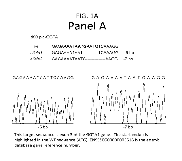
[0028] Figure 1 depicts a schematic of the sequence alterations in exemplary
knockout pig (pig
isolate identifier 47-1). Panel A presents portions of the wild-type (wt) and
disrupted a1,3
galactosyltransferase gene (GGTA-1) nucleotide sequences. A portion of the
wild-type GGTA-1
nucleotide sequence (WT) is shown in the top line. The same region of the
altered GGTA-1 nucleotide
sequence from a triple transgenic pig are shown in the two bottom lines each
of which provides the
altered sequence of one GGTA-1 allele. The disrupted GGTA-1 nucleotide
sequences from an
exemplary triple transgenic pig are a five base pair and seven base pair
deletions. Dashes indicate
the portion of the nucleotide sequence where the deleted nucleotides would
occur in the wild-type
sequence.
[0029] Panel B presents portions of wild-type and disrupted CMAH genes for an
exemplary triple
transgenic pig. A portion of the wildtype (wt) CMAH gene is shown in the top
line. The nucleotide
8
CA 02965550 2017-04-21
WO 2016/065046
PCT/US2015/056730
sequences of both alleles of the comparable regions from the same triple
transgenic pig are shown
below the wild type sequence. The disrupted CMAH sequences of the triple
transgenic pig include a
twelve base pair deletion and a three base pair deletion/four base pair
insertion. Dashes indicate the
region of the nucleotide sequence in which the deletions occurred.
[0030] Panel C presents portions of wild-type and disrupted 134GaINT2 genes
for an exemplary triple
transgenic pig. A portion of the wildtype (wt)134GaINT2 gene is shown in the
top line. The nucleotide
sequences of the comparable regions from the same exemplary triple transgenic
pig are shown below
the wild type sequence. As shown, the disrupted 134GaINT2 sequences of the
triple transgenic pig
include a single base pair insertion, a twelve base pair deletion and a five
base pair deletion. Unlike
the GGTA1 and CMAH sequences, three different alleles were detected. It is
possible that the
134GaINT2 sequence occurs at more than one distinct locus in the porcine
genome. Dashes indicate
the region of the nucleotide sequence in which the 134GaINT2 deletions
occurred.
[0031] Figure 2 depicts a graph of flow cytometry data obtained from
peripheral blood monocyte cells
(PBMC) from either the CMAH/aGal double transgenic pig (CMAH, upper panel) or
the
CMAH/aGa1/134GaINT2 triple transgenic pig (B4, lower panel). Cells were either
unstained (cells only,
white curves) or stained with Dolichus biflorus agglutinin conjugated with
FITC (DBA-FITC, solid black
curve). DBA binds a-N-acetylgalactosamine bearing glycans or Sda-like glycans
produced by
134GaINT2. In the upper panel the profile of PBMC's from double knockout
(CMAH/aGal) pigs with
wildtype 134GaINT2 sequence (CMAH) after incubation with DBA (black curve) is
distinctly shifted from
the profile of unstained cells from the double transgenic CMAH/aGal pig (white
curve). In the lower
panel the profile of PMBC from triple transgenic pigs with disrupted 134GaINT2
sequences (B4) after
incubation with DBA predominantly overlies the profile from the unstained
cells from the triple
transgenic CMAH/aGa1/134GaINT2 pig (regions of white and black curves that
overlay are represented
as gray). The limited change in the profile of DBA-FITC treated cells
indicates a lack of DBA binding
to the PBMC's from the triple transgenic pig and reduced occurrence of Sda-
like antigens on cells
from the triple transgenic pig.
[0032] Figure 3 presents results epitope analysis of PBMC's from a variety of
pigs. Panel A presents
flow cytometry results of experiments performed with PBMC's from wildtype (WT)
and triple
transgenic (GGTA1-/-, CMAH-/-, B4GaINT2-/-) pigs. Cell counts are shown on the
y axis;
fluorescence is shown on the x-axis.
[0033] The far left graphs depict data from a negative control (white) and
cells incubated with IB4
lectin (dark). IB4 interacts with the alpha galactose linked carbohydrates
created by the gene product
of GGTA1. Curves with overlap between the control (negative) and experimental
(positive) results are
shown with gray. Wildtype cells incubated with IB4 show a distinct separate
peak from unstained
wildtype cells. Cells from the triple transgenic pig incubated with IB4 show
no significant second
peak, indicative of significantly reduced aGal linked carbohydrates.
9
CA 02965550 2017-04-21
WO 2016/065046
PCT/US2015/056730
[0034] The center graphs depict data from a negative control (white) and cells
incubated with HD
antibody (dark). The negative control was an irrelevant isotype control
antibody. The HD antibody
interacts with the Neu5Gc carbohydrate produced by the product of the CMAH
gene. Curves with
overlap between the control (negative) and experimental (positive) results are
shown with gray.
Wildtype cells incubated with HD antibody show a distinct separate peak from
wildtype cells treated
with an unrelated antibody. Cells from the triple transgenic pig incubated
with HD antibody show no
significant second peak, indicative of significantly reduced Neu5Gc epitope
levels.
[0035] The far left graphs depict data from a negative control (white) and
cells incubated with DBA
lectin (dark). DBA lectin interacts with the carbohydrate structure produced
by the gene product of
64GaINT2. Curves with overlap between the control (negative) and experimental
(positive) results
are shown with gray. Wildtype cells incubated with DBA show a distinct
separate peak from unstained
wildtype cells. Cells from the triple transgenic pig incubated with DBA show
no significant second
peak, indicative of significantly reduced carbohydrates produced by the gene
product of 64GaINT2.
[0036] Panel B presents a scatter graph of results obtained from flow
cytometry experiments to
evaluate the relative binding of human IgG (X-axis) and IgM (Y-axis) in serum
from 44 unique humans
to PBMCs from various pig types. Results with PBMC from wildtype (WT) pigs are
shown with open
squares, results from single transgenic aGal disrupted (GGTA1-/-) pigs are
shown with triangles,
results from double transgenic CMAH/ aGal disrupted (GGTA1-/-and CMAH4-) pigs
are shown with
empty circles, and results from triple transgenic CMAH/aGal/B4GaINT2 disrupted
(GGTA14-and
CMAH4- and 64GaINT24-) pigs are shown with solid circles. Results from triple
transgenic
CMAH/aGal/B4GaINT2 disrupted (GGTA14- and CMAH4- and 64GaINT24-) pigs are
clustered at low
levels of IgM and IgG binding; a few data points indicate moderate IgG binding
to triple transgenic
cells.
[0037] Figure 4 presents a scatter graph of results obtained from flow
cytometry experiments to
evaluate the relative binding of Rhesus Macaque IgG (X-axis) and IgM (Y-axis)
in serum from Rhesus
macaques, baboons and humans to PBMCs from various pig types. Results with
PBMC from single
transgenic aGal disrupted (GGTA14-) pigs are shown on the x-axis, results from
double transgenic
CMAH/ GGTA14- disrupted (GGTA14- and CMAH4-) pigs are shown with empty
circles, and results
from triple transgenic CMAH/ GGTA14-/B4GaINT2 disrupted (GGTA14- and CMAH4-
and 64GaINT2-/-)
pigs are shown with solid circles. CMAH/ GGTA14- and CMAH/ GGTA14-434GaINT2
are on the y-
axis. Binding below the line indicates binding to the pig cells on the x axis
is more antigenic than the
pig cells on the y axis. Human (top), baboon (middle) and rhesus monkey
(bottom) sera were tested
with the pig cells. IgM results are shown on the left; IgG results are shown
on the right.
[0038] Figure 5 provides series of flow cytometry results indicating the level
of IgG antibody binding
to red blood cells (RBC). Multiple types of RBC were evaluated against three
human sera. Results
from human A sera are in the left column, human 0 sera 1 in the middle column
and human 0 sera 2
in the right column. B4Gal/CMAH/Gal triple knockout (B4G triple KO) RBC
results are in the top row.
Human 0 1 RBC results are in the second row. Human 0 2 RBC results are in the
third row. Human
CA 02965550 2017-04-21
WO 2016/065046
PCT/US2015/056730
A RBC results are in the fourth row. Porcine wildtype RBC are in the bottom
row. Multiple significant
peaks are present when wildtype porcine RBC is evaluated with human A and
human 0 sera 1;
multiple minor peaks are present when wildtype porcine RBC is evaluated with
human 0 sera 2. In
the trace of human A RBC and two human 0 sera, there are significant peaks.
However, the human
A RBC and human A sera show only one overlapping peak. As expected both human
0 RBC show
only a single overlapping peak when tested against all three sera. The
B4Gal/CMAH/Gal triple
knockout RBC show only a single overlapping peak when tested against all three
sera, similar to
human 0 RBC.
[0039] Figure 6 provides a summary of data obtained from flow cytometry
comparisons of human
antibody binding to various swine and human RBC. Sera from 74 humans were
incubated with pig
and human RBC. Data from wildtype swine RBC (W), animals lacking GGTA1 and
CMAH (CMAH
RBC), or GGTA1/CMAH/[34GaINT2 (B4G RBC) and human allogeneic RBC (h) are
shown. Panel A
shows a summary of IgG binding to various RBC. The data represents a
normalized median
fluorescence intensity (MFI) and standard deviation for IgG. Human A, B, 0 and
AB sera were
utilized. Intergroup comparisons of antibody binding were performed using
repeated measures one
way ANOVA and Tukey's multiple comparison test. Human antibody binding to the
various RBC
was maximal on wild type cells and diminished as each glycan producing gene
was inactivated. The
triple knockout-RBC (B4G) most closely approximated the levels of antibody
binding seen on human
allo-RBC (h). Panel B shows a summary of IgM binding to various RBC. The data
represents a
normalized median fluorescence intensity (MFI) and standard deviation for IgM.
The trend of
wildtype>double knockout>triple knockout human was observed for both IgG and
IgM. In Panel C,
flow cytometry was used to reveal the effects of inactivating the GGTA1, CMAH
and [34GaINT2 genes
on the expression of the a-Gal, Neu5Gc and DBA-reactive glycans. Results from
porcine wildtype
(W) red blood cells, CMAH/aGal double knockout (D) red blood cells,
CMAH/aGa1/13GaINT2 triple
transgenic red blood cells (T) and human blood group 0 red blood cells are
shown in the figure. Flow
cytometry results indicate loss of antigenic structures with each gene
disruption. In Panel D, the data
were plotted to display the individual MFI of each human serum sample when
examining antibody
binding to human 0 RBC on the x-axis and the individual MFI of each human
serum sample when
examining antibody binding to triple knockout swine RBC (B4G) on the y-axis.
Data points falling
below the diagonal line on the graphs represent less binding to the pig triple
knockout cells than to
human blood group 0 cells. Human group 0 blood is considered a universal
donor; transfused
human group 0 RBC undergo limited humoral destruction.
[0040] Figure 7 provides results of immunoglobulin analysis using quantitative
mass spectrometry to
measure the abundance of individual antibody isotypes. Panel 7A is a schema
describing the
biochemical approach used to evaluate the binding of IgG and IgM to RBC. RBC
from wildtype swine
(W), GGTA1/CMAH deficient swine (D), GGTA/CMAH/[34GaINT2 swine (T) and
autologous human
RBC (H) were evaluated. The details of the process are described elsewhere
herein. A
representative gel showing material eluted from RBC is shown in Panel B.
Molecular weight markers
(Mw), crude starting serum (S) and purified human IgG were loaded for
comparison. Other controls
11
CA 02965550 2017-04-21
WO 2016/065046
PCT/US2015/056730
included untreated RBC (lane 1 all samples), and RBC that were acid washed but
not incubated with
serum (lane 2 all samples). Material was stripped from serum-treated RBC by
low pH incubation for
either 2 or 3 minutes (lanes 3 and 4 respectively, all samples). The single
arrowhead marks a protein
migrating with a size similar to albumin. Although autologous human RBC
released less of this
protein than the swine cells, this result was not reproduced in other
experiments. Double arrowheads
highlight a protein migrating with a size similar to IgG (lanes 3 and 4). The
intensity of the IgG heavy
chain sized band varied between the different RBC. Incubating cells at low pH
for either two or three
minutes released similar protein levels. To quantitate the amount of antibody
released from the cells,
gel slices encompassing the approximate size of immunoglobulin heavy chain in
the two minute
elution samples were collected, incubated with trypsin and assessed by mass
spectrometry (see
below herein). Three arrowheads note a protein that migrates with a size
identical to hemoglobin.
The presence of hemoglobin in the supernatant of serum-free samples indicated
that RBC lysis
occurred during the manipulations. Variations between lanes 1 and 2 for all
RBC types suggest acid
washing accelerated lysis and released increasing amounts of hemoglobin from
the cells. Unknown
polypeptides, marked by an *, that migrated as a double slightly above the
immunoglobulin light
chains were released from RBC even in the absence of serum.
[0041] Panel C shows the relative levels of IgM and IgG that eluted from each
type of RBC after
incubation with separate aliquots of the same serum as determined by mass
spectroscopy. The AUC
from the mass spectrometry analyses were all normalized to the values obtained
for the total IgG or
IgM binding to the autologous human red blood cells for each serum. Total
antibody was calculated
for IgG by summing the AUC for each isotype. Results from wildtype pig red
blood cells (W), triple
knockout pig red blood cells (B), and autologous human red blood cells (A) are
shown. Autologous
human red blood cells are from the same subject from which the tested serum
was obtained.
[0042] Figure 8 provides representative mass spectroscopy chromatograms of
immunoglobulin-
derived peptides. Relative abundance is shown on the y-axis. Time in minutes
(min) is shown on the
x-axis. Chromatograms such as this were used to calculate AUC.
[0043] Figure 9 presents flow cytometry analysis gating and fate. Three human
sera were incubated
with RBC from wildtype pigs (W), autologous human RBC (A) and RBC from
GGTA1/CMAH/84GaINT2 deficient pigs (T). After incubating with fluorescent
secondary antibodies to
report bound human immunoglobulin, cells were analyzed by flow cytometry.
Forward scatter (FSC,
x-axis) and side scatter (SSC, y-axis) were used to identify RBC. Black ovals
represent gates used to
select RBC for analysis. The percentages shown next to each gate represents
the fraction of total
events that resided within the gate. Human sera disrupted wild type swine RBC
as seen by an
increase in debris and the reduction in RBC falling in the gated regions.
Wildtype RBC high
antigenicity and consequent cell destruction may have contributed to the
histogram quality of the
wildtype (W) samples in some experiments.
[0044] Figure 10 depicts area under the curve (AUC) values obtained for each
IgG isotype as
determined by mass spectrometry. AUC is shown on the y-axis; IgG isotypes and
the RBC type are
12
CA 02965550 2017-04-21
WO 2016/065046
PCT/US2015/056730
shown on the x-axis. Results from triple knockout pig red blood cells (TKO),
human autologous red
blood cells (Human Auto) and wildtype pig red blood cells(WT) are shown.
[0045] Figure 11 depicts flow cytometry results obtained from peripheral blood
monocytes (PBMC)
obtained from wildtype (WT) or Gal/CMAH/134GaINT2 triple knockout pigs (64G).
PBMC from two pig
isolates (58-1 and 59-2) are shown. 164 is bound by aGal; in the presence of
reduced aGal
expression, 164 binding is reduced. Neu5Gc is an epitope produced by the CMAH
gene product.
DBA is a lectin bound by the 134GaINT2 product. The grey histogram shows
negative control results.
Two peaks are clearly present for the wildtype cells with each of 164, DBA and
Neu5Gc. For both
triple knockout pig isolates the second peak is eliminated or shifted to
overlap with the negative
control, indicating reduced binding.
[0046] Figure 12 provides flow cytometry results obtained from aortic
endothelial cells (AEC) or
immortalized renal endothelial cells (iREC) after lectin staining. The
indicated cell types were
incubated with 164, Neu5Gc, DBA, PNA, Jacalin or VVL. The left column shows
results obtained from
wildtype AEC (WT/AEC), the second column shows results obtained from aGal
disrupted pigs
(GAL/AEC), the middle column shows results obtained from double knockout
CMAH/aGal pigs
(CMAH/AEC), the fourth column shows results obtained from triple knockout
CMAH/aGa1/134GaINT2
pigs (64G/AEC), the fifth column shows results obtained from wildtype
immortalized renal endothelial
cells, the sixth column shows results obtained from GGTA1/CMAH/134GaINT2 and
SLA antigen
disrupted immortalized renal endothelial cells. Gray histograms are no lectin
negative controls. The
solid black line represents lectin binding. Note the absence of a second 164
peak in Gal disrupted
cells, the absence of a second Neu5Gc peak in CMAH disrupted cells and the
absence of a second
DBA peak in 134GaINT2 disrupted cells. Second PNA and Jacalin peaks occur in
cells from the single,
double and triple knockout pigs. In the triple knockout AEC and the
134GaINT2/SLA triple knockout,
Sla antigen disrupted iRMEC, the second VVL peak shifts substantially. While
not being bound by
mechanism, reduced VVL lectin recognition of an antigen on the triple knockout
cells may contribute
to the surprising results obtained from triple knockout CMAH/aGa1/134GaINT2
cells and pigs.
[0047] Figure 13 presents portions of wild-type and disrupted 134GaINT2, GGTA1
and CMAH genes
in exemplary triple transgenic pigs. The underlined portion of each wild-type
sequence indicates the
Crispr target region. The figure shows sequence disruptions from multiple
triple transgenic pigs (pig
isolate identifiers 54-2, 58-1 and 59-2). Three 134GaINT2 variations are
shown, a 14 nucleotide
deletion, a 12 nucleotide deletion/1 nucleotide substitution and a 271
nucleotide deletion/1 nucleotide
insertion. The third 134GaINT2 mutation depicted is a 271 nucleotide
deletion/1 nucleotide insertion
wherein double slashes (//) demark the deletion. Two aGal (GGTA1) variations
are shown, an 11
nucleotide (nt) deletion and an 18 nucleotide (nt) deletion. Two CMAH
variations are shown, a 66
nucleotide deletion/ 12 nucleotide insertion and a 5 nucleotide deletion/1
nucleotide substitution.
[0048] Figure 14 presents data obtained from triple transgenic cells from
triple transgenic pigs
(aGal/B4GaINT2/CMAH deficient) pigs evaluated with multiple human sera in a
human clinical
crossmatch test. The upper graph shows results from 31 human sera with a
PRA=0; the lower graph
13
CA 02965550 2017-04-21
WO 2016/065046
PCT/US2015/056730
shows results from 19 human sera with a PRA>80. IgM and IgG results in the
absence of DTT are
indicated with a solid bar; IgG results after treatment with DTT are indicated
by an empty bar. The
cytotoxicity score is shown on the y-axis. Cytotoxicity scores of 1 are very
good candidates for
allotransplant.
DETAILED DESCRIPTION OF THE INVENTION
[0049] The present application provides transgenic pigs and porcine organs,
tissues and cells for
transplantation into a human that do not express the indicated pig genome
encoded products and
methods of making and using the same. In one embodiment the application
provides a triple
transgenic pig comprising disrupted a (1,3)-galactosyltransferase, [34GaINT2
and cytidine
monophosphate-N-acetylneuraminic acid hydroxylase genes, wherein expression of
functional a
(1,3)-galactosyltransferase, [34GaINT2 and cytidine monophosphate-N-
acetylneuraminic acid
hydroxylase in the knockout pig is decreased as compared to a wild-type pig.
l. In General
[0050] In the specification and in the claims, the terms "including" and
"comprising" are open-ended
terms and should be interpreted to mean "including, but not limited to..."
These terms encompass the
more restrictive terms "consisting essentially of" and "consisting of".
[0051] As used herein and in the appended claims, the singular forms "a",
"an", and "the" include
plural reference unless the context clearly dictates otherwise. As well, the
terms "a" (or "an"), one or
more" and at least one" can be used interchangeably herein. It is also to be
noted that the terms
"comprising", "including", and "having" can be used interchangeably.
[0052] Unless defined otherwise, all technical and scientific terms used
herein have the same
meanings as commonly understood by one of ordinary skill in the art to which
this invention belongs.
All publications and patents specifically mentioned herein are incorporated by
reference in their
entirety for all purposes including describing and disclosing the chemicals,
instruments, statistical
analyses and methodologies which are reported in the publications which might
be used in connection
with the invention. All references cited in this specification are to be taken
as indicative of the level of
skill in the art. Nothing herein is to be construed as an admission that the
invention is not entitled to
antedate such disclosure by virtue of prior invention.
11. COMPOSITIONS AND METHODS
[0053] Transgenic animals suitable for use in xenotransplantation and methods
of producing
mammals suitable for use in xenotransplantation are provided. Specifically,
the present application
describes the production of triple transgenic pigs with decreased expression
of alpha 1,3
galactosyltransferase (aGal), [3 1,4 N-acetylgalactosaminyltransferase
([34GaINT2) and cytidine
monophosphate-N-acetylneuraminic acid hydroxylase (CMAH).
14
CA 02965550 2017-04-21
WO 2016/065046
PCT/US2015/056730
[0054] In embodiments of the present invention, pigs and porcine organs,
tissues and cells therefrom
are provided in which the aGal, 64GaINT2 and CMAH genes are less active, such
that the resultant
aGal, 64GaINT2 and CMAH products no longer generate wild-type levels of a1,3-
galactosyl epitopes,
Sda-like epitopes, or Neu5Gc on a cell surface, glycoprotein or glycolipid. In
an alternative
embodiment the aGal, 64GaINT2 and CMAH genes are inactivated in such a way
that no transcription
of the gene occurs. In various embodiments triple aGal/B4GaINT2/CMAH knockout
pigs were made.
Methods of making transgenic pigs, and the challenges thereto, are discussed
in Galli et al 2010
Xenotransplantation 17(6) p.397-410. Methods and cell cultures of the
invention are further detailed
below herein.
[0055] The term "transgenic mammal" refers to a transgenic mammal wherein a
given gene has been
altered, removed or disrupted. It is to be emphasized that the term is to be
intended to include all
progeny generations. Thus, the founder animal and all F1, F2, F3 and so on
progeny thereof are
included, regardless of whether progeny were generated by somatic cell nuclear
transfer (SCNT) from
the founder animal or a progeny animal or by traditional reproductive methods.
By "single transgenic"
is meant a transgenic mammal wherein one gene has been altered, removed or
disrupted. By
"double transgenic" is meant a transgenic mammal wherein two genes have been
altered, removed or
disrupted. By "triple transgenic" is meant a transgenic mammal wherein three
genes have been
altered, removed or disrupted. By "quadruple transgenic" is meant a transgenic
mammal wherein four
genes have been altered, removed or disrupted.
[0056] In principle, transgenic animals may have one or both copies of the
gene sequence of interest
disrupted. In the case where only one copy or allele of the nucleic acid
sequence of interest is
disrupted, the knockout animal is termed a "heterozygous transgenic animal".
The term "null"
mutation encompasses both instances in which the two copies of a nucleotide
sequence of interest
are disrupted differently but for which the disruptions overlap such that some
genetic material has
been removed from both alleles, and instances in which both alleles of the
nucleotide sequence of
interest share the same disruption. In various embodiments disruptions of the
three genes of interest
may occur in at least one cell of the transgenic animal, at least a plurality
of the animal's cells, at least
half the animal's cells, at least a majority of animal's cells, at least a
supermajority of the animal's
cells, at least 70%, 75", 80%, 85%, 90%, 95%, 98%, or 99
/0 of the animal's cells.
[0057] The term "chimera", "mosaic" or "chimeric mammal" refers to a
transgenic mammal with a
knockout in some of its genome-containing cells. A chimera has at least one
cell with an unaltered
gene sequence, at least several cells with an unaltered gene sequence or a
plurality of cells with an
unaltered sequence.
[0058] The term "heterozygote" or "heterozygotic mammal" refers to a
transgenic mammal with a
disruption on one of a chromosome pair in all of its genome containing cells.
CA 02965550 2017-04-21
WO 2016/065046
PCT/US2015/056730
[0059] The term "homozygote" or "homozygotic mammal" refers to a transgenic
mammal with a
disruption on both members of a chromosome pair in all of its genome
containing cells. A
"homozygous alteration" refers to an alteration on both members of a
chromosome pair.
[0060] A "non-human mammal" of the application includes mammals such as
rodents, sheep, dogs,
ovine such as sheep, bovine such as beef cattle and milk cows, and swine such
as pigs and hogs.
Although the application provides a typical non-human animal (pigs), other
animals can similarly be
genetically modified.
[0061] A "mutation" is a detectable change in the genetic material in the
animal that is transmitted to
the animal's progeny. A mutation is usually a change in one or more
deoxyribonucleotides, such as,
for example adding, inserting, deleting, inverting or substituting
nucleotides.
[0062] By "pig" is intended any pig known to the art including, but not
limited to, a wild pig, domestic
pig, mini pigs, a Sus scrofa pig, a Sus scrofa domesticus pig, as well as in-
bred pigs. Without
limitation the pig can be selected from the group comprising Landrace,
Yorkshire, Hampshire, Duroc,
Chinese Meishan, Chester White, Berkshire Goettingen, Landrace/York/Chester
White, Yucatan,
Bama Xiang Zhu, Wuzhishan, Xi Shuang Banna and Pietrain pigs. Porcine organs,
tissues, cells or
transfusion products are organs, tissues, devitalized animal tissues, cells or
transfusion products from
a pig.
[0063] The alpha 1,3 galactosyltransferase (aGal, GGTA, GGT1, GT, aGT, GGTA1,
GGTA-1) gene
encodes an enzyme (GT, aGal, a1,3 galactosyltransferase). Ensemble transcript
ENSSSCG00000005518 includes the porcine GGTA1 nucleotide sequence. Functional
a1,3
galactosyltransferase catalyzes formation of galactose-a1,3-galactose
(aGal,Gal,Gal, gal1,3gal, gall-
3gal) residues on glycoproteins. The galactose-a1,3-galactose (aGal) residue
is an antigenic epitope
or antigen recognized by the human immunological system. Removing aGal from
transgenic organ
material does not eliminate the human immunological response to transplant of
foreign material,
suggesting an involvement of additional antibodies in the rapid immunological
response to
xenotransplant. (Mohiudden et al (2014), Am J. Transplantation 14:488-489 and
Mohiudden et al
2014 Xenotransplantation 21:35-45). Disruptions of the aGal gene that result
in decreased
expression of functional aGal may include but are not limited to a 3 base pair
deletion adjacent to a G
to A substitution, a single base pair deletion, a single base pair insertion,
a two base pair insertion, a
six base pair deletion, a ten base pair deletion, a seven base pair deletion,
an eight base pair
insertions for a five base pair deletion, a five base pair insertion, an
eleven base pair deletion, and an
eighteen base pair deletion (see Table 1). The Crispr target sequence is in
exon 3 of the gene, near
the start codon.
[0064] The cytidine monophosphate-N-acetylneuraminic acid hydroxylase (CMP-
Neu5Ac
hydroxylase gene, CMAH) gene encodes an enzyme (CMAH). Functional CMAH
catalyzes
conversion of sialic acid N-acetylneuraminic acid (Neu5Ac) to N-
glycolylneuraminic acid (Neu5Gc).
The Neu5Gc residue is an antigenic epitope or antigen recognized by the human
immunological
16
CA 02965550 2017-04-21
WO 2016/065046
PCT/US2015/056730
system. The Ensembl database id Gene: ENSSSCG00000001099 includes the porcine
CMAH
nucleotide sequence, and the Crispr target area is near exon 6. Disruptions of
the CMAH gene that
result in decreased expression of functional CMAH may include but are not
limited to a four base pair
insertion, a one base pair deletion, a two base pair deletion, a three base
pair deletion, a twenty
base pair deletion, a five base pair deletion, an eight base pair deletion, an
eleven base pair deletion,
a twelve base pair deletion, a single base pair insertion, a two base pair
insertion for single base pair
deletion, a three base pair deletion for a four base pair insertion, a sixty-
six base pair deletion/twelve
base pair insertion, and a five base pair deletion/1 base pair
substitution(see Table 1).
[0065] The [31,4 N-acetylgalactosaminyltransferase (B4GaINT2, [34GaINT2,
B1,4GaINT2,
[31,4GaINT2) gene encodes the [31,4 N-acetylgalactosaminyltransferase 2
glycosyltransferase
(B4GaINT2). Functional B4GaINT2 produces Sda-like glycans (Dall'Olio et al
(2014) Biochemica
Biophysica Acta 1840:443-453 and Blanchard et al (1983) JBC 258:7691-7685).
B4GaINT2 is
thought to be expressed in most humans; prior work suggested that only 5% of
humans lack
expression of functional B4GaINT2. Disruption of [34GaINT2 in pig cells
significantly decreases
crossmatching in more than 5% of the human samples tested; this result was
unexpected. Sda ¨like
glycans may include Sda and similar glycans related to blood type or blood
group determination and
gastro-intestinal cancer inhibition. The Ensembl database EN555CG00000030269
entry includes the
[34GaINT2 cDNA and amino acid sequences. Genomic porcine [34GaINT2 spans
multiple exons
across approximately 40,000 base pairs and may occur at multiple loci. The
CrispR target region
utilized in these experiment is found in exon 2 and is CTGTATCGAGGAACACGCTT.
Disruptions of
the [34GaINT2 gene that result in decreased expression of functional [34GaINT2
may include, but are
not limited to, a one base pair insertion, a twelve base pair deletion, a five
base pair deletion, a
fourteen base pair deletion, a twelve base pair deletion/one base pair
substitution, a 271 base pair
deletion/one base pair insertion.
Table 1. Examples of Disruptions of Genes of Interest in Viable Pigs with
Decreased Functional Gene
Product
wildtype disruption Disruption Class
Descriptor
GGTA1-SEQ ID NO:1 GGTA1- 3 base pair deletion
GICATCTITTACATCATGGIGGAT GTCATC1 ACATCATG__ adjacent to a G to A
GATATCTCCAGGATGCC AATGATATCTCCAGGATGCC substitution
GGTA1- GGTA1- Single base pair
CTTTTCCCAG GAGAAAATAAT CTTTTCCCAG deletion
GAATGTCAAA GGAAGAGTGG TTCT GAGAAAATAAT
GAATGT_AAA
GGAAGAGTGG TTCT
GGTA1- GGTA1- 6 base pair deletion
17
CA 02965550 2017-04-21
WO 2016/065046
PCT/US2015/056730
CTTTTCCCAG GAGAAAATAAT CTTTTCCCAG
GAATGTCAAA GGAAGAGTGG TTCT GAGAAAATAAT
CAAA GGAAGAGTGG TTCT
GGTA1- GGTA1- 2 base pair insertion
CTTTTCCCAG GAGAAAATAAT CTTTTCCCAG
GAATGTCAAA GGAAGAGTGG TTCT GAGAAAATAAT
GAATGTATCAAA
GGAAGAGTGG TTCT
GGTA1- GGTA1- 10 base pair deletion
CTTTTCCCAG GAGAAAATAAT CTTTTCCCAG
GAATGTCAAA GGAAGAGTGG TTCT GAGAAAATAA_
A GGAAGAGTGG TTCT
GGTA1- GGTA1 7 base pair deletion
CTTTTCCCAG GAGAAAATAAT CTTTTCCCAG
GAATGTCAAA GGAAGAGTGG TTCT GAGAAAATAAT GAA
GGAAGAGTGG TTCT
GGTA1- GGTA1 7 base pair deletion
GAGAAAATAAT GAATGTCAAAGG GAGAAAATAAT G
AAGG
GGTA1- GGTA1 8 base pair substitution
CTTTTCCCAG GAGAAAATAAT CTTTTCCCAG for 5 base pair deletion
GAATGTCAAA GGAAGAGTGG TTCT GAGAAAATAAT GAA
GGAATAAT AA
GGAAGAGTGG TTCT
GGTA1- GGTA1- Single base pair
CTTTTCCCAG GAGAAAATAAT CTTTTCCCAG insertion
GAATGTCAAA GGAAGAGTGG TTCT GAGAAAATAAT
GAATGTTCAAA
GGAAGAGTGG TTCT
GGTA1- GGTA1- 5 base pair insertion
GAGAAAATAAT GAATGTCAAAGG GAGAAAATAAT
TCAAAGG
GGTA1- GGTA1- 11 base pair deletion
AGGAGAAAATAAT GAATGTCAAAGG AGGAGAAAATAAT
AAGAGTGGTTCTGT GAAGAGTGGTTCTGT
GGTA1- GGTA1- 18 base pair deletion
AGGAGAAAATAAT AGGAGAAAAT
GAATGTCAAAGGAAGAGTGGTTCTGT AGTGGTTCTGT
CMAH- SEQ ID NO:2 CMAH- 4 base pair insertion
18
CA 02965550 2017-04-21
WO 2016/065046
PCT/US2015/056730
AAACTCCTGA ACTACAAGGC AAACTCCTGA
TCGGCTGGTG AAGGA ACTACAAGGAA GGC
TCGGCTGGTG AAGGA
CMAH- CMAH- 2 base pair deletion
CAGGCGTGAG TAAGGTACGT CAGGCGTGAG
GATCTGTTGGA AGACAGTGA TAAGGTACGT GATC_
GATTCAGATGAT TTGGA AGACAGTGA
GATTCAGATGAT
CMAH- CAGGCGTGAG 8 base pair deletion
CAGGCGTGAG TAAGGTACGT TAAGGTACGT G
GATCTGTTGGA AGACAGTGA GA AGACAGTGA
GATTCAGATGAT GATTCAGATGAT
CMAH- CAGGCGTGAG 5 base pair deletion
CAGGCGTGAG TAAGGTACGT TAAGGTACGT G
GATCTGTTGGA AGACAGTGA TTGGA AGACAGTGA
GATTCAGATGAT GATTCAGATGAT
CMAH- CAGGCGTGAG 3 base pair deletion
CAGGCGTGAG TAAGGTACGT TAAGGTACGT GA
GATCTGTTGGA AGACAGTGA GTTGGA AGACAGTGA
GATTCAGATGAT GATTCAGATGAT
CMAH- CAGGCGTGAG 2 base pair insertion for
CAGGCGTGAG TAAGGTACGT TAAGGTACGT single base pair deletion
GATCTGTTGGA AGACAGTGA GATCACGTTGGA
GATTCAGATGAT AGACAGTGA
GATTCAGATGAT
CMAH- CAGGCGTGAG 20 base pair deletion
CAGGCGTGAG TAAGGTACGT TAAGGTACGT GAT
GATCTGTTGGA AGACAGTGA
GATTCAGATGAT TCAGATGAT
CMAH- CAGGCGTGAG 1 base pair insertion
CAGGCGTGAG TAAGGTACGT TAAGGTACGT
GATCTGTTGGA AGACAGTGA GATCTTGTTGGA
GATTCAGATGAT AGACAGTGA
GATTCAGATGAT
CMAH- CAGGCGTGAG 1 base pair deletion
CAGGCGT GAG TAAGGTACGT TAAGGTACGT
GATCTGTTGGA AGACAGTGA GATC_GTTGGA
GATTCAGATGAT AGACAGTGA
GATTCAGATGAT
19
CA 02965550 2017-04-21
WO 2016/065046
PCT/US2015/056730
CMAH CMAH 11 base pair deletion
GAGTAAGGTACG TGATCTGTTGG GAGTAAGG
AAGACAGT TTGG AAGACAGT
CMAH CMAH 12 base pair deletions
GAGTAAGGTACG TGATCTGTTGG GAGTAAGGTACG TGA
AAGACAGT CAGT
CMAH CMAH 3 base pair deletion 4
GAGTAAGGTACG GAGTAAGGTACG base pair insertion
TGATCTGTTGGAAGACAGT TGAGTAAG TTGG
AAGACAGT
CMAH CMAH 66 base pair deletion,
ATTAACCAAG GATCGAACAC ATTAACtctcaataaciaa 12 base pair insertion
CATCCTCTTG TCTCCCAGCT
TGAGGTCCAT GCAGGCGT
GAGTAAGGTACG TGATCTGTTGG AA GA--CTGTTGGAA
CMAH CMAH 5 base pair deletion, 1
ATTAACCAAG GATCGAACAC ATTAACCAAG G2TCGAACAC base pair substitution
CATCCTCTTG TCTCCCAGCT CATCCTCTTG TCTCCCAGCT
TGAGGTCCAT GCAGGCGT TGAGGTCCAT
GAGTAAGGTACG TGATCTGTTGG AA GCAGGCGTGA
GTAAGGTACG TG
TTGGAA
B4 Gal NT2 SEQ ID NO:3- CTGTATCGAGG AACACG G 1 base pair insertion
CTGTATCGAGG CTTCGGAACATAAA
AACACGCTTCGGAACATAAA
B4 Gal NT2 CTGTATCGAGG AAC A 12 base pair deletion
CTGTATCGAGG AACA TAAA
CGCTTCGGAACATAAA
B4 Gal NT2 B4 Gal NT2 5 base pair deletion
CTGTATCGAGG CTGTATCGAGG AACACG_ _
AACACGCTTCGGAACATAAA GAACATAAA
B4 Gal NT2 B4 Gal NT2 14 base pair deletion
TCTGTATCGA GGAACACGCT TCTGTATCG-
TCGGAACATA AAGAGTCCAA ---GAACATA AAGAGTCCAA
CGCTCAGGACC CGCTCAGGACC
B4 Gal NT2 B4 Gal NT2 12 base pair deletion, 1
TCTGTATCGA GGAACACGCT TCTGTATCGA GGAACA base pair substitution
TCGGAACATA AAGAGTCCAA TA AAGAGCCCAA
CGCTCAGGACC CGCTCAGGACC
B4 Gal NT2 SEQ ID NO:4 B4 Gal NT2 271 nt deletion, 1 nt
CA 02965550 2017-04-21
WO 2016/065046
PCT/US2015/056730
GATGGGTG AGTTGAAG AGACTGAA GATGGGTG AGTTGAAG insertion
GTCTGTATC GAGGAACAC
//GCTTCGGAAC ATAAAGAG AGACTGAA GTCTGTATC
TCCAACGCT CAGGACCAA GAGGAACAC // //
AAGCACCAT CGATATCTT
GAGCAAGCTT
GAGGATCGA CAGACATCT
AGGGCTGTT GGGACACAA TTGCACAGCA
GAGAGCAAA CGCTGTTAAA
CC
ATCTTTTCTG AGTATGTTAA AAGGAAA A TAAAATAACC
AAAAGATTT CATTGTGCGA CAAAAAGATC
CATAGATGGG AATAGCAACT
TGAGCAAAAA TGCAAGTCAA
ACCTGTTTTGT ACACTACGTAT
CAAAATTGAT TTCTTCCCAAA
GCAAAAGAGA AAGAAAAGCA
AAAATAAACC TAAGCAAACT//
GAGCAAGCTT TTGCACAGCA
AAGGAAACCA TAAAATAACC
CAAAAAGATC
[0066] The phrase "disrupted gene" is intended to encompass insertion,
interruption, or deletion of a
nucleotide sequence of interest wherein the disrupted gene either encodes a
polypeptide having an
altered amino acid sequence that differs from the amino acid sequence of the
endogenous sequence,
encodes a polypeptide having fewer amino acid residues than the endogenous
amino acid sequence
or does not encode a polypeptide although the wild-type nucleotide sequence of
interest encodes a
polypeptide.
[0067] The present specification provides a transgenic animal with reduced
expression of functional
aGal, [34GaINT2 and CMAH genes. The animal can be any mammal suitable for
xenotransplantation.
In a specific embodiment, the animal is a pig. "CMAH/aGAL double knockouts",
"CMAH/aGAL DKO",
"CMAH/aGal", "CMAH/aGal DKO", "CMAH47GAL4-", "aGal/CMAH DKOs", "aGAL/CMAH
double
knockouts", "GGTA1/CMAH DKO", "GT1/CMAH DKO", "GGTA1-/-/CMAH4-", "GGT141CMAH4-
",
"CMAH/GGTA DKO", "GT/CMAH-KO", "GGTA1/CMAH KO", "DKO (aGal/CMAH)", "DKO (aGAL
&
CMAH)", "CMAH-/aGal-", "aGal-/CMAH-", "CMAH-/aGAL-" and variants thereof refer
to transgenic
animals, cells, or tissues that lack expression of functional alpha 1,3
galactosyltransferase and
cytidine monophosphate-N-acetylneuraminic acid hydroxylase. A triple knockout
product or pig may
be created in a wild-type background or in a CMAH/aGal double knockout
background.
"CMAH/aGAL/B4GaINT2 triple knockouts", "CMAH/aGAL/[34GaINT2 triple knockouts',
"CMAH/aGAL/B4GaINT2 TKO", "CMAH/aGa1/134GaINT2", "CMAH/aGal/B4GaINT2 TKO",
"CMAH4-
/GAL4-/B4GaINT24-", "aGal/CMAH/[34GaINT2 TKOs", "aGAL/CMAH/134Gal triple
knockouts",
"GGTA1/CMAH/B4GaINT2 TKO", "GT1/CMAH/[34GaINT2 TKO", "GGTA14-/CMAH4-
/[34GaINT24-",
"GGT1-/-/CMAH4-/ [34GaINT24- ", "CMAH/GGTA/[34GaINT2 TKO", "GT/CMAH/134GaINT2-
KO",
"GGTA1/CMAH/[34GaINT2 KO", "TKO (aGal/CMAH/[34GaINT2)", "TKO (aGAL , CMAH,
[34GaINT2)",
"CMAH-/aGal-/[34GaINT2-", "aGal-/CMAH-/[34GaINT2-", "CMAH-/aGAL-134GaINT2-"
and variants
21
CA 02965550 2017-04-21
WO 2016/065046
PCT/US2015/056730
thereof refer to transgenic animals, cells, or tissues that lack expression of
functional alpha 1,3
galactosyltransferase, cytidine monophosphate-N-acetylneuraminic acid
hydroxylase and 64GaINT2.
[0068] Transgenic transplant material. Transplant material encompasses organs,
tissue, transfusion
products and/or cells from an animal for use as xenografts. Transplant
material for use as xenografts
may be isolated from transgenic animals with decreased expression of aGal,
64GaINT2 and CMAH.
Transgenic transplant material from transgenic or knockout pigs can be
isolated from a prenatal,
neonatal, immature or fully mature transgenic animal. The transplant material
may be used as
temporary or permanent organ replacement for a human subject in need of an
organ transplant. Any
porcine organ can be used including, but not limited to, the brain, heart,
lungs, eye, stomach,
pancreas, kidneys, liver, intestines, uterus, bladder, skin, hair, nails,
ears, glands, nose, mouth, lips,
spleen, gums, teeth, tongue, salivary glands, tonsils, pharynx, esophagus,
large intestine, small
intestine, small bowel, rectum, anus, thyroid gland, thymus gland, bones,
cartilage, tendons,
ligaments, suprarenal capsule, skeletal muscles, smooth muscles, blood
vessels, blood, spinal cord,
trachea, ureters, urethra, hypothalamus, pituitary, pylorus, adrenal glands,
ovaries, oviducts, uterus,
vagina, mammary glands, testes, seminal vesicles, penis, lymph, lymph nodes
and lymph vessels.
[0069] In another embodiment, the application provides non-human tissues that
are useful for
xenotransplantation. In various embodiments, the non-human tissue is porcine
tissue from a triple
aGal/CMAH/64GaINT2 transgenic pig. Any porcine tissue can be used including
but not limited to,
epithelium, connective tissue, blood, bone, cartilage, muscle, nerve, adenoid,
adipose, areolar, brown
adipose, cancellous muscle, cartilaginous, cavernous, chondroid, chromaffin,
dartoic, elastic,
epithelial, fatty, fibrohyaline, fibrous, Gamgee, gelatinous, granulation, gut-
associated lymphoid,
skeletal muscle, Haller's vascular, indifferent, interstitial, investing,
islet, lymphatic, lymphoid,
mesenchymal, mesonephric, multilocular adipose, thymus tissue, mucous
connective, myeloid, nasion
soft, nephrogenic, nodal, osteoid, osseus, osteogenic, bone marrow, retiform,
periapical, reticular,
smooth muscle, hard hemopoietic and subcutaneous tissue, devitalized animal
tissues including heart
valves, skin, and tendons, and vital porcine skin.
[0070] Another embodiment provides cells and cell lines from porcine triple
transgenic animals with
reduced or decreased expression of aGal, B4GaINT2 and CMAH. In one embodiment
these cells or
cell lines can be used for xenotransplantation. Cells from any porcine tissue
or organ can be used
including, but not limited to: epithelial cells, fibroblast cells, neural
cells, keratinocytes, hematopoietic
cells, melanocytes, chondrocytes, lymphocytes (B and T), macrophages,
monocytes, mononuclear
cells, cardiac muscle cells, other muscle cells, granulosa cells, cumulus
cells, epidermal cells,
endothelial cells, Islet of Langerhans cells, pancreatic insulin secreting
cells, bone cells, bone
precursor cells, neuronal stem cells, primordial stem cells, hepatocytes,
aortic endothelial cells,
microvascular endothelial cells, fibroblasts, liver stellate cells, aortic
smooth muscle cells, cardiac
myocytes, neurons, Kupferr cells, smooth muscle cells, Schwann cells,
erythrocytes, platelets,
neutrophils, lymphocytes, monocytes, eosinophils, basophils, adipocytes,
chondrocytes, pancreatic
islet cells, thyroid cells, thymus cells, parathyroid cells, parotid cells,
glial cells, astrocytes, red blood
cells, white blood cells, macrophages, somatic cells, pituitary cells, adrenal
cells, hair cells, bladder
22
CA 02965550 2017-04-21
WO 2016/065046
PCT/US2015/056730
cells, kidney cells, retinal cells, rod cells, cone cells, heart cells,
pacemaker cells, spleen cells, antigen
presenting cells, memory cells, T cells, B cells, plasma cells, muscle cells,
ovarian cells, uterine cells,
prostate cells, vaginal epithelial cells, sperm cells, testicular cells, germ
cells, egg cells, leydig cells,
peritubular cells, sertoli cells, lutein cells, cervical cells, endometrial
cells, mammary cells, follicle
cells, mucous cells, ciliated cells, nonkeratinized epithelial cells,
keratinized epithelial cells, lung cells,
goblet cells, columnar epithelial cells, dopaminergic cells, squamous
epithelial cells, osteocytes,
osteoblasts, osteoclasts, bone marrow, embryonic stem cells, fibroblasts and
fetal fibroblasts.
[0071] Nonviable derivatives include tissues stripped of viable cells by
enzymatic or chemical
treatment these tissue derivatives can be further processed through
crosslinking or other chemical
treatments prior to use in transplantation. In a preferred embodiment, the
derivatives include
extracellular matrix derived from a variety of tissues, including skin, bone,
urinary, bladder or organ
submucosal tissues. In addition, tendons, joints, and bones stripped of viable
tissue to including but
not limited to heart valves and other nonviable tissues as medical devices are
provided. In an
embodiment, serum or medium suitable for cell culture and isolated from a
transgenic pig of the
invention are provided. Components of porcine transgenic organs, tissues or
cells are also provided.
Components may also be modified through any means known in the art including
but not limited to
crosslinking and aldehyde crosslinking. Components may vary depending on the
larger organ or
tissue from which the component is obtained. Skin components may include but
are not limited to
stripped skin, collagen, epithelial cells, fibroblasts and dermis. Bone
components may include but are
not limited to collagen and extracellular matrix. Heart components may include
but are not limited to
valves and valve tissue.
[0072] "Xenotransplantation" encompasses any procedure that involves the
transplantation,
implantation or infusion of cells, tissues or organs into a recipient subject
from a different species.
Xenotransplantation in which the recipient is a human is particularly
envisioned. Thus
xenotransplantation includes but is not limited to vascularized
xenotransplant, partially vascularized
xenotransplant, unvascularized xenotransplant, xenodressings, xenobandages,
xenostructures and
xenotransfusions.
[0073] In embodiments, cell culture reagents isolated from a transgenic pig
comprising disrupted
a(1,3)-galactosyltransferase, 134GaINT2 and CMAH genes are provided. Cell
culture reagents are
reagents utilized for tissue culture, in vitro tissue culture, microfluidic
tissue culture, cell culture or
other means of growing isolated cells or cell lines. Cell culture reagents may
include but are not
limited to cell culture media, cell culture serum, a cell culture additive, a
feeder cell, and an isolated
cell capable of proliferation. By an "isolated cell capable of proliferation"
is intended a cell isolated or
partially isolated from other cell types or other cells wherein the cell is
capable of proliferating, dividing
or multiplying into at least one additional clonal cell.
[0074] Cells grown in culture may synthesize or metabolically incorporate
antigenic epitopes into a
compound of interest produced by the cultured cell. The antigenic epitopes may
result in increased
binding by human antibodies and decreased efficacy of the compound of
interest. See Ghaderi et al
23
CA 02965550 2017-04-21
WO 2016/065046
PCT/US2015/056730
2010 Nature Biotechnology 28(8):863-867, herein incorporated by reference in
its entirety. Growing
the producing cell in a cell culture reagent with an altered epitope profile
such as a reduced level of
aGal, B4GaINT2 or Neu5Gc may reduce the level of aGal antigens, Neu5Gc
antigens, and/or Sda-like
antigens, on the compound of interest. Compounds of interest may include but
are not limited to
glycoproteins and glycolipids. Glycoproteins of interest may include but are
not limited to an antibody,
growth factor, cytokine, hormone or clotting factor. Glycolipids of interest
may include but are not
limited to therapeutics, antigens, and biosurfactants.
[0075] The word "providing" is intended to encompass preparing, procuring,
getting ready, making
ready, supplying or furnishing. It is recognized that methods of providing a
cell may differ from
methods of providing a subject, methods of providing an organ may differ from
methods of providing a
pig, methods of providing a kidney may differ from methods of providing a
liver and methods of
providing an organ may differ from methods of providing a material suitable
for transfusion.
[0076] Transplant rejection occurs when transplanted tissue, organs, cells or
material are not
accepted by the recipient's body. In transplant rejection, the recipient's
immune system attacks the
transplanted material. Multiple types of transplant rejection exist and may
occur separately or
together. Rejection processes included but are not limited to hyperacute
rejection (HAR), acute
humoral xenograft rejection reaction (AHXR), thrombocytopenia, acute humoral
rejection, hyperacute
vascular rejection, antibody mediated rejection and graft versus host disease.
By "hyperacute
rejection" we mean rejection of the transplanted material or tissue occurring
or beginning within the
first 24 hours post-transplant involving one or more mechanisms of rejection.
Rejection encompasses
but is not limited to "hyperacute rejection", "humoral rejection", "acute
humoral rejection", "cellular
rejection" and "antibody mediated rejection". The acute humoral xenograft
reaction (AHXR) is
characterized by a spectrum of pathologies including, but not limited to,
acute antibody mediated
rejection occurring within days of transplant, the development of thrombotic
microangiopathy (TMA),
microvascular angiopathy, pre-formed non-Gal IgM and IgG binding, complement
activation,
microvascular thrombosis and consumptive thrombocytopenia within the first few
weeks post
transplant. Thrombocytopenia is a quantity of platelets below the normal range
of 140,000 to
440,000/pl. Thrombocytopenia related symptoms include, but are not limited to,
internal hemorrhage,
intracranial bleeding, hematuria, hematemesis, bleeding gums, abdominal
distension, melena,
prolonged menstruation, epistaxis, ecchymosis, petechiae or purpura. Uptake of
human platelets by
pig livers contributes to the development of thrombocytopenia in xenograft
recipients.
Thrombocytopenia may occur upon reperfusion of the xenotransplanted organ or
after the immediate
post-reperfusion period.
[0077] In another embodiment, the invention provides a method of improving a
rejection related
symptom in a patient comprising transplanting porcine organs, tissue or cells
having reduced
expression of aGa1,134GaINT2 and Neu5Gc on the porcine organs, tissue or cells
into a human,
wherein one or more rejection related symptoms is improved as compared to when
tissue from a wild-
type swine is transplanted into a human. By "improving", "bettering",
"ameliorating", "enhancing", and
"helping" is intended advancing or making progress in what is desirable. It is
also envisioned that
24
CA 02965550 2017-04-21
WO 2016/065046
PCT/US2015/056730
improving a rejection related symptom may encompass a decrease, lessening, or
diminishing of an
undesirable symptom. It is further recognized that a rejection related symptom
may be improved
while another rejection related symptom is altered. The altered second
rejection related symptom
may be improved or increased. A second altered rejection related symptom may
be altered in a less
desirable manner. Rejection related symptoms include but are not limited to
hyperacute rejection
related symptoms and acute humoral xenograft reaction related symptoms.
Rejection related
symptoms may include, but are not limited to, thrombotic microangiopathy
(TMA), microvascular
angiopathy, pre-formed non-Gal IgM and IgG binding, complement activation,
agglutination, fibrosis,
microvascular thrombosis, consumptive thrombocytopenia, consumptive
coagulopathy, profound
thrombocytopenia, refractory coagulopathy, graft interstitial hemorrhage,
mottling, cyanosis, edema,
thrombosis, necrosis, fibrin thrombi formation, systemic disseminated
intravascular coagulation, IgM
deposition in glomerular capillaries, IgG deposition in glomerular
capillaries, elevated creatinine
levels, elevated BUN levels, T cell infiltrate, infiltrating eosinophils,
infiltrating plasma cells, infiltrating
neutrophils, arteritis, antibody binding to endothelium, altered expression of
ICOS, CTLA-4, BTLA,
PD-1, LAG-3, or TIM-3, and systemic inflammation.
[0078] "Hyperacute rejection related symptom" is intended to encompass any
symptom known to the
field as related to or caused by hyperacute rejection. It is recognized that
hyperacute rejection related
symptoms may vary depending upon the type of organ, tissue or cell that was
transplanted.
Hyperacute rejection related symptoms may include, but are not limited to,
thrombotic occlusion,
hemorrhage of the graft vasculature, neutrophil influx, ischemia, mottling,
cyanosis, edema, organ
failure, reduced organ function, necrosis, glomerular capillary thrombosis,
lack of function, hemolysis,
fever, clotting, decreased bile production, asthenia, hypotension, oliguria,
coagulopathy, elevated
serum aminotransferase levels, elevated alkaline phosphatase levels, jaundice,
lethargy, acidosis and
hyperbilirubenemia and thrombocytopenia.
[0079] Thrombocytopenia is a quantity of platelets below the normal range of
140,000 to 440,000/pl.
Thrombocytopenia related symptoms include, but are not limited to, internal
hemorrhage, intracranial
bleeding, hematuria, hematemesis, bleeding gums, abdominal distension, melena,
prolonged
menstruation, epistaxis, ecchymosis, petechiae or purpura. Uptake of human
platelets by pig livers
contributes to the development of thrombocytopenia in xenograft recipients.
[0080] Platelets, also known as thrombocytes, are enucleate fragments of
megakaryocytes involved
in blood coagulation, hemostasis and blood thrombus formation. Human platelets
are routinely
isolated through a variety of methods including, but not limited to, platelet
apheresis, plateletpheresis
and ultracentrifugation.
[0081] The phrase "platelet uptake" is intended to encompass the incorporation
of a platelet into a
liver or liver cell. While not being limited by mechanism, such uptake may
occur through a phagocytic
process. Platelet uptake may be monitored by any platelet uptake monitoring
assay known in the art.
Platelet uptake monitoring assays include, but are not limited to
immunological methods, western
blots, immunoblotting, microscopy, confocal microscopy, transmission electron
microscopy and
CA 02965550 2017-04-21
WO 2016/065046
PCT/US2015/056730
phagosome isolation. It is recognized that the appropriate platelet uptake
monitoring assay may
depend upon the type of label used. Platelet uptake may be measured as a
percentage of total
platelets absorbed, percentage of total platelets not absorbed, a ratio of
absorbed to unabsorbed
platelets, percentage of cells absorbing at least one platelet, percentage of
cells not absorbing a
platelet, or number of platelets absorbed per cell. It is recognized that
platelet uptake by more than
one cell type may contribute to the total platelet uptake of the liver. Total
platelet uptake by an animal
liver may include platelet uptake by liver sinusoidal endothelial cells,
platelet uptake by Kuppffer cells,
platelet uptake by LSECs and Kupffer cells and platelet uptake by additional
cell types. It is
recognized that platelet uptake by different cell types may contribute similar
or disparate fractions of
the total platelet uptake by a liver. Thus an alteration, inhibition,
reduction, decrease, or lowering of
platelet uptake by a liver comprises an alteration, inhibition, reduction,
decrease, or lowering of
platelet uptake by one or more liver cell types.
[0082] While not being limited by mechanism, platelet uptake may occur through
phagocytosis by
LSEC and Kupffer cells. Phagocytosis is characterized by the formation of an
endosome which by
the fusion of lysosomes containing degradative enzymes becomes a phagosome.
[0083] Any method of evaluating, assessing, analyzing, measuring, quantifying,
or determining a
rejection related symptom known in the art may be used with the claimed
compositions and methods.
Methods of analyzing a rejection related symptom may include, but are not
limited to, laboratory
assessments including CBC with platelet count, coagulation studies, liver
function tests, flow
cytometry, immunohistochemistry, standard diagnostic criteria, immunological
methods, western blots,
immunoblotting, microscopy, confocal microscopy, transmission electron
microscopy, IgG binding
assays, IgM binding assays, expression asays, creatinine assays and phagosome
isolation.
[0084] Expression of a gene product is decreased when total expression of the
gene product is
decreased, a gene product of an altered size is produced or when the gene
product exhibits an
altered functionality. Thus if a gene expresses a wild-type amount of product
but the product has an
altered enzymatic activity, altered size, altered cellular localization
pattern, altered receptor-ligand
binding or other altered activity, expression of that gene product is
considered decreased.
Expression may be analyzed by any means known in the art including, but not
limited to, RT-PCR,
Western blots, Northern blots, microarray analysis, immunoprecipitation,
radiological assays,
polypeptide purification, spectrophotometric analysis, Coomassie staining of
acrylamide gels, ELISAs,
2-D gel electrophoresis, in situ hybridization, chemiluminescence, silver
staining, enzymatic assays,
ponceau S staining, multiplex RT-PCR, immunohistochemical assays,
radioimmunoassay,
colorimetric assays, immunoradiometric assays, positron emission tomography,
fluorometric assays,
fluorescence activated cell sorter staining of permeablized cells,
radioimunnosorbent assays, real-
time PCR, hybridization assays, sandwich immunoassays, flow cytometry, SAGE,
differential
amplification or electronic analysis. Expression may be analyzed directly or
indirectly. Indirect
expression analysis may include but is not limited to, analyzing levels of a
product catalyzed by an
enzyme to evaluate expression of the enzyme. See for example, Ausubel et al,
eds (2013) Current
Protocols in Molecular Biology, Wiley-Interscience, New York, N.Y. and Coligan
et al (2013) Current
26
CA 02965550 2017-04-21
WO 2016/065046
PCT/US2015/056730
Protocols in Protein Science, Wiley-lnterscience New York, NY. Gene expression
assays for porcine
ASGR1 are commercially available (Applied BiosystemsTM, Carlsbad CA).
[0085] As compared to" is intended encompass comparing something to a similar
but different thing,
such as comparing a data point obtained from an experiment with a transgenic
pig to a data point
obtained from a similar experiment with a wildtype pig. The word "comparing"
is intended to
encompass examining character, qualities, values, quantities, or ratios in
order to discover
resemblances or differences between that which is being compared. Comparing
may reveal a
significant difference in that which is being compared. By "significant
difference" is intended a
statistically significant difference in results obtained for multiple groups
such as the results for material
from a transgenic pig and material from a wild-type pig. Generally statistical
significance is assessed
by a statistical significance test such as but not limited to the student's t-
test, Chi-square, one-tailed t-
test, two-tailed t-test, ANOVA, Dunett's post hoc test, Fisher's test and z-
test. A significant difference
between two results may be results with a p<0.1, p<0.05, p<0.04, p<0.03,
p<0.02, p<0.01 or greater.
[0086] The word "isolated" is intended to encompass an entity that is
physically separated from
another entity or group. An isolated cell is physically separated from another
group of cells. Examples
of a group of cells include, but are not limited to, a developing cell mass, a
cell culture, a cell line, a
tissue, and an animal. The word "isolating" is intended to encompass
physically separating an entity
from another entity or group. Examples include physically separating a cell
from other cells,
physically separating a cell component from the remainder of the cell and
physically separating tissue
or organ from an animal. An isolated cell or cell component is separated by
10%, 15%, 20%, 25%,
30%, 35%, 40%, 45%, 50%, 55%, 60%, 65%, 70%, 75%, 80%, 85%, 90%, 95%, 96%,
97%, 98%,
99%, up to 100% from other naturally occurring cells or cell components.
Methods for isolating one or
more cells from another group of cells are known in the art. See for example
Freshney (ED) Culture
of Animal Cells: a manual of basic techniques (3rd Ed.) 1994, Wiley-Liss;
Spector et al (Eds)(1998)
Cells: a Laboratory Manual (vol.1) Cold Spring Harbor Laboratory Press and
Darling et al (1994)
Animal Cells: culture and media John Wiley & Sons. Methods of isolating a
tissue or an organ from
an animal are known in the art and vary depending on the tissue or organ to be
isolated and the
desired method of transplanting the tissue or organ. Methods of isolating a
transfusion product from
an animal or sample are known in the art and vary depending on the desired
transfusion product.
Such methods include but are not limited to centrifugation, dialysis, elution,
apheresis and
cryoprecipitation.
[0087] A "skin related product" encompasses products isolated from skin and
products intended for
use with skin. Skin related products isolated from skin or other tissues may
be modified before use
with skin. Skin related products include but are not limited to replacement
dressings, burn coverings,
dermal products, replacement dermis, dermal fibroblasts,collagen, chondroitin,
connective tissue,
keratinocytes, cell-free xenodermis,cell-free pig dermis,composite skin
substitutes and epidermis and
temporary wound coverings. See for example Matou-Kovd et al (1994) Ann Med
Burn Club 7:143,
herein incorporated by reference in its entirety.
27
CA 02965550 2017-04-21
WO 2016/065046
PCT/US2015/056730
[0088] The attachment period of a skin related product is the time between
application of the skin
related product to a human subject and natural separation of the skin related
product from the human
subject. When a human subject's skin wound has sealed, a skin related product
may be removed by
natural separation or mechanical separation. However natural separation of a
skin related product
from a human subject may occur prematurely. Premature natural separation
occurs before separation
is desired by a medical practitioner. By way of example and not limitation,
premature natural
separation may occur before the wound has been sealed. Premature natural
separation may also be
termed "sloughing", "shedding", or "flaking". Clinical management of premature
natural separation
may include reapplication of a skin related product, dressing application,
bandage application,
administering antibiotic, and administering fluids. A skin wound may be sealed
by any means known
in the art including but not limited to by growth of the subject's skin and by
skin grafting. Reduced
premature separation encompasses a decreased, lower, less frequent,
diminished, smaller amount of
natural separation of a skin related product before separation is desired by a
medical practitioner.
The reduced premature separation may relate to a lower number of complete, a
lower number of
partial premature separation events, and involvement of a smaller portion of
the skin related product
in a partial premature separation event than compared to a skin related
product obtained from a wild-
type pig. A skin related product of the instant application may also exhibit
an increased, lengthened,
improved, extended, or expanded attachment period. Use of a skin related
product of the instant
application may increase the duration of the attachment period.
[0089] A skin wound encompasses any injury to the integument including but not
limited to an open
wound, burn, laceration, ulcer, leg ulcer, foot ulcer, melanoma removal,
cancer removal, plastic
surgery, and bite.
[0090] By "surgically attaching" is intended joining, combining, uniting,
attaching, fastening,
connecting, joining or associating through any surgical method known in the
art.
[0091] In an embodiment the application provides non-human material suitable
for transfusions from
multiple knockout porcine animals with reduced expression of the aGal, CMAH
and 84GaINT2 genes.
These materials suitable for transfusions may include, but are not limited to,
blood, whole blood,
plasma, serum, red blood cells, platelets, and white bloods cells. Such
materials may be isolated,
enriched or purified. Methods of isolating, enriching or purifying material
suitable for transfusion are
known in the art. Serologically porcine red blood cells (RBCs) share a number
of common
characteristics with human RBCs (Pond W.G, Houpt K.A, The Biology of the Pig
Ithaca: Comstock
Pub. Associates, 1978 and Jandl, J. H. Blood: Textbook of Hematology,
Boston:Little, Brown, 1996).
As in other mammals, the primary site for erythropoiesis in pigs is bone
marrow. The pRBC is a
biconcave disk of approximately 4-8 microns in diameter. The hematocrit of pig
blood is 35-47%, with
a hemoglobin concentration of 6-17 g/100 ml. The half-life of pRBC is
approximately 40 days, in
comparison to 60 days for human RBCs.
[0092] Fifteen pig blood group systems have so far been identified. The most
important and well-
studied is the A-0(H), which is closely related to the human ABO system. The A
and 0 antigens on
28
CA 02965550 2017-04-21
WO 2016/065046
PCT/US2015/056730
pRBC are passively adsorbed from circulating plasma glycosphingolipids, in a
similar mechanism as
human Lewis antigens (Marcus, D.M. & Cass, L.E. (1969) Science 164:553-555).
pRBC phenotyping
is not entirely reliable. Phenotyping of pigs can be achieved by
immunohistochemical staining of
buccal epithelial cells with an anti-A monoclonal antibody (mAb)(as used in
Blood Banks) and an anti-
H lectin antibody (Ulex europaeus)(Villarroya H et al 1990 Autoimmunity 6:47-
60). The glycolipids
bearing blood group A have been isolated from porcine stomach mucosa,
epithelial cells and
erythrocytes. Among a few of the well-characterized proteins derived from
pRBC, porcine hemoglobin
shares 85% sequence identity with its human counterpart and demonstrates a
similar three-
dimensional structure at 2.8A resolution. Unlike most tissue cells, porcine
RBCs do not contain a
nucleus and therefore are less likely to harbor retroviruses such as, but not
limited to, porcine
endogenous retroviruses (PERVs). pRBCs also lack intracellular organelles and
a relatively short
half-life in vivo.
[0093] The following Examples are offered for illustrative purposes only and
are not intended to limit
the scope of the present invention in any way. Indeed various modifications in
addition to those
shown and described herein will become apparent to those skilled in the art
from the foregoing
description and the following examples and fall within the scope of the
appended claims.
EXAMPLES
Example 1. DNA Sequencing Analysis of Targeted CMAH, GGTA1 and B4GaINT2
Regions
[0094] Genomic DNA from a cloned pig was extracted using GenElute Mammalian
Genomic
DNA Miniprep Kit (Sigma-Aldrich, St. Louis, MO). PCR amplification of the
CMAH, GGTA1 and
84GaINT2 Crispr/Cas9 target regions was performed. Primers were used to
sequence the targeted
CMAH, GGTA1 and 84GaINT2 regions.
[0095] Pwo Master (Roche, Indianapolis IN) was used, Pwo SuperYield DNA
polymerase, dNTPack
(Roche Applied Science, Indianapolis, IN) was used. PCR conditions for GGTA1
were as follows:
94 C, 2 min; 94 C, 15 sec, 54 C, 30 sec, and 72 C, 45 sec for 15 cycles; 94 C,
15 sec, 54 C, 30 sec,
72 C, 45 sec with additional 5 sec each cycle for 25 cycles; and a final
extension step of 72 C for 5
min. For CMAH, 94 C, 2 min; 94 C, 15 sec, 56 C, 30 sec, and 72 C, 45 sec for
15 cycles; 94 C, 15
sec, 56 C, 30 sec, 72 C, 45 sec with additional 5 sec each cycle for 25
cycles; and a final extension
step of 72 C for 5 min. For 134GALNT2, 94 C, 2 min; 94 C, 15 sec, 62 C, 30
sec, 72 C, 40 sec for 15
cycles, 94 C 15 sec, 62 C, 30 sec, 72 C 40 sec with additional 5 sec each
cycle for 25 cycles; and a
final extension step of 72 C for 5 min. The PCR products were separated on 1
/0 agarose gel and
purified by GenElute Gel Extraction Kit (Sigma-Aldrich, St. Louis, MO). The
PCR products were
sequenced by the Sanger method (DNA Sequencing Core Facility, Indiana
University School of
Medicine) with the specific sequencing primer, 5' CCTTAGTATCCTTCCCAACCCAGAC 3'
(SEQ ID
NO:5) for GGTA1; 5' CATTTTCTTCGGAGTTGAGGGC 3' (SEQ ID NO:6) for CMAH; 5'
AAAGCCACAGGAGGAGCCAG 3' (SEQ ID NO:7) for 84GALNT2. Once the mutations were
detected, PCR products were inserted into pCR4blunt-TOPO vector and
transformed into E. coli.
29
CA 02965550 2017-04-21
WO 2016/065046
PCT/US2015/056730
Individual clones were sequenced again to further investigate the mutation in
each allele of the
individual gene.
[0096] Results from an exemplary DNA sequence analysis are summarized in
Figure 1, panels A-C.
DNA sequence analysis confirmed alterations of the GGTA1 and CMAH gene in both
alleles in at
least one pig. Sequence analysis showed overlapping five and seven base pair
deletions in the
GGTA1 target region (panel A). In the same pig, sequence analysis confirmed a
disruption of the
CMAH gene consisting of a 12 base pair deletion on one allele and an
overlapping 5 base pair
substitution for a 3 base pair deletion on the other allele (panel B). DNA
sequence analysis confirmed
alterations of the 64GaINT2 gene sequence in the same pig. The 64GaINT2 gene
sequence data
show three variations; the presence of three mutations may suggest the
64GaINT2 gene occurs in at
least two loci. In an overlapping region of the 64GaINT2 gene, one allele
shows a single base pair
insertion, one allele shows a five base pair deletion and one allele shows a
twelve base pair
deletion/one base pair substitution. No evidence of a wildtype 64GaINT2
sequence was found (panel
C). Figure 13 summarizes DNA sequence analysis of additional triple knockout
pig isolates.
Example 2. Production of Knockout PiCIS (Triple Transoenic Pius)
[0097] Oligo annealing and cloning into the PX330 plasmid to drive gRNA
expression was performed
using Addgene plasmid 42230 [http://www.addgene.org/42230 and 20]. Oligo pairs
for the targeted
genes are GGTA1 (NCB1 Accession:XM_005660398.1), 5'CACCGAGAGAAAATAATGAATGTCAA-
3'
forward) (SEQ ID NO:8), 5'AAATTGACATTCATTATTTTCTC-3' (reverse) (SEQ ID NO:9);
CMAH
(NCB! Accession: NM_001113015.1) 5'-CACCGAGTAAGGTACGTGATCTGT-3' (forward) (SEQ
ID
NO:10), 5'-AAACACAGATCACGTACCTTACTC-3' (reverse) (SEQ ID NO:11; 64GaINT2 (NCB!
Accession: NM_001244330.1) 5'-CACCGTGTATCGAGGAACACGCTT-3' (forward) (SEQ ID
NO:12),
5'-AAACAAGCGTGTTCCTCGATACAC-3' (reverse) (SEQ ID NO:13).
[0098] Liver-derived cells were cotransfected with all three gRNA/Cas9
plasmids. After 48 hr, the
treated cells were passed over an 1134-lectin column to isolate a-Gal null
cells. Two million a-Gal
negative cells were further stained with fluorescein labeled Dolichos biflorus
Agglutinin (DBA)-FITC
(Vector Laboratories, Burlingame, CA, USA) at 2 pg/ml in 500 pl HBSS with 0.5%
BSA and flow-
sorted for DBA-negative cells using a BD FACSARia sorter (BD Bioscience, San
Jose, CA, USA).
The presence of Neu5Gc, an indicator of CMAH gene function, was not analyzed
prior to somatic cell
nuclear transfer.
[0099] Somatic cell nuclear transfer (SCNT) was performed using in vitro
matured oocytes (DeSoto
Biosciences Inc., St. Seymour TN and Minitube of America (Mount Horeb WI).
Cumulus cells were
removed from the oocytes by pipetting in 0.1`)/0 hyaluronidase. Oocytes with
normal morphology and
a visible polar body were selected and incubated in manipulation media
(calcium-free NCSU-23 with
5% fetal bovine serum (FBS) containing 5 pg/ml bizbenzimide and 7.5 pg/ml
cytochalasin B for 15
minutes. Following this incubation period, oocytes were enucleated by removing
the first polar body
and metaphase II plate. For the triple transgenic pigs, single cells of site-
targeted, IB4 counter-
CA 02965550 2017-04-21
WO 2016/065046
PCT/US2015/056730
selected, DBA negative liver derived cells (LDC) were injected into each
enucleated oocyte. Some
triple transgenic pigs were developed from SCNT of fetal fibroblasts obtained
from an aborted triple
knockout pig fetus created from site-targeted, 164 counter-selected, DBA
negative liver derived cells.
Electrical fusion was induced with a BTX electroporator (Harvard Apparatus,
Holliston MA). In various
instances enucleated oocytes injected with a cell (couples) were exposed to
two DC pulses of 140 V
of 50 ps in 280 mM mannitol, 0.001 mM CaCl2 and 0.05 mM MgC12or 180 V for 50
ps in 280 mM
mannitol, 0.1 mM CaCl2, and 0.05 mM MgC12. After activation the oocytes were
placed in NCSU-23
medium with 0.4% bovine serum albumin (BSA) and incubated at 38.5 C, 5% CO2 in
a humidified
atmosphere for less than one hour. Within an hour after activation, oocytes
were transferred into a
recipient pig. Recipient pigs were synchronized occidental pigs on their first
day of estrus.
Pregnancies were verified by ultrasound at day 25 or day 26 after embryo
transfer.
[0100] All animals used in this study were approved by the Institutional
Biosafety Committee (IBC)
and Institutional Animal Care and Use Committee (IACUC).
Example 3. IB4 Counterselection of Triple Knockouts
[0101] Liver-Derived Cells (LDCs) were transfected with three sets of
targeting constructs (aGal,
134GaINT2 and CMAH). Cells were selected with 164, a substance that binds
aGal. DNA from cells in
the bulk population of cells that survived 164 counterselection was obtained,
and the target gene
sequences were evaluated. The bulk population cells that survived 164
counterselection were used
directly in SCNT to make pregnant pigs.
Example 4. Design of Targeting Vectors
[0102] Targeting vectors such as CrispR constructs, Zn finger constructs and
TALEN constructs are
designed to target the sequence of porcine CMAH (Ensemble transcript
ENSSSCT00000001195) at a
suitable site. Targeting vector constructs are designed to target GGTA1
(Ensemble transcript
ENSSSCT00000006069) at a suitable site. Targeting vector constructs are
designed to target
134GaINT2 at a suitable site in NCB! GenelD:100621328 and Ensemble:
EN555CG00000030269.
[0103] A CMAH Crispr construct with a primer sequence that is the reverse
complement of a portion
of the sequence listed in the Ensemble transcript was created and utilized in
the creation of a triple
knockout pig. A Gal Crispr construct with a primer sequence identical to a
portion of that in the
appropriate Ensemble transcript was created and utilized in the creation of a
triple transgenic pig. An
134GaINT2 Crispr construct with a primer sequence identical to a portion of
the above indicated NCB!
sequence NCBI GeneID:100621328 was created and utilized in the creation of a
triple transgenic pig.
Example 5. Crossmatch of Human Sera with Transgenic PBMCs
[0104] Porcine whole blood from transgenic (triple GGTA-1/134GaINT2/CMAH for
example) and wild-
type pigs were collected in ACD from venous puncture. The whole blood was
mixed 1:1 with PBS
and separated with Ficoll. Porcine peripheral blood monocytes (PBMCs) were
prepared from the
whole blood using Ficoll-Paque Plus (GE Healthcare). PBMCs were removed from
Ficoll layers and
31
CA 02965550 2017-04-21
WO 2016/065046
PCT/US2015/056730
washed several times with PBS followed by lysis solution as need to remove red
blood cells. PBMCs
were suspended in EX-CELL media at 4 million cells/ml (EX-Cell 610 HSF-Serum
free Medium for
Hybridoma Cells 14610C).
[0105] Sera were obtained from 44 healthy human volunteers. Twenty-five
percent heat inactivated
serum was prepared. 25 pl of heat-inactivated serum, 25 pl of EX-CELL media
and 50 pl of
suspended cells were added to each well on a 96 well V-bottom plate. Cells
were incubated at 4 C
for 30 minutes, and then washed three times with EX-CELL media. Cells were
stained with
secondary antibody as follows: Alexa Flour 488 goat anti-human IgG 1 09-546-1
70 at 1:250
concentration in EX-CELL media, Alexa Flour 488 goat anti-human IgM 709-546-
073 at 1:250
concentration in EX-CELL media, 100 pl of secondary was added. Cells were
suspended with a
pipet. Cells were incubated at 4 C for 30 minutes. Cells were washed once with
EX-CELL media and
resuspended with pipet in EX-Cell media or 1:1 EX-CELL media and Flow Fixative
Solution (1`)/0
buffered paraformaldehyde). Flow cytometric analysis was completed with
channel FL-1 gating for
Alexa flour 488. Results from one such experiment are shown in Figure 3, panel
B.
Example 6. DBA Lectin Flow Cvtometrv Staining
[0106] Whole blood from triple transgenic pigs and other pigs of interest
was collected in
anticoagulant citrate dextrose (ACD). The whole blood was mixed 1:1 with PBS
and separated with
ficoll (Ficoll-Paque PLUS, GE Healthcare 17-1440-03). Flow wash and staining
buffers containing
calcium were obtained. The flow wash buffer was HBSS with 0.5% BSA IgG-free
0.1`)/0 sodium azide
pH 7.4), 0.45 pM filtered to remove particulates. PBMCs were resuspended in
Flow Wash Buffer at 2
x 106 cells/ml and suspended homogenously. The cells were blocked for 15
minutes on ice. Cells
were resuspended again. 100 pl (2 X 10⁵ cells) were added to 5 ml flow tubes.
DBA-fluorescein (2
mg/ml stock) was obtained. The DBA-fluorescein lectin stock was diluted 1:10
in Flow Buffer to a final
concentration of 0.2 pg/ml. DBA-fluorescein lectin was added to the cells at a
ratio of 1 pg DBA: 1 x
106 cells (0.2 pg/ 2 x 10⁵ cells or 1 pl). DBA lectin and cells were incubated
30 minutes at room
temperature. Cells were washed thoroughly with 4 ml Flow Wash HBSS. Cells were
spun at 400 x g
for 5 minutes and the wash supernatant was removed. Cells were resuspended in
200 pl Flow Wash
HBSS. The same parameters were utilized if additional washes were performed.
If desired, cells
were fixed in approximately 200 pl Flow Fixative/2 x 105 cells after the last
wash and stored at 4 C
until analysis. Flow cytometry analysis was performed with the gate on forward
and sidelight scatter.
Cellular fluorescent events were collected in FL-1; 10,000 events were
collected in scatter gate. For
each cell line both cells only and DBA-fluorescein data were collected. IB4
lectin interacts with aGal
linked carbohydrates produced by the aGal gene product. The HD antibody
interacts with the
Neu5Gc carbohydrate produced by the CMAH gene product. DBA lectin interacts
with the
carbohydrate structure produced by the [34GaINT2 gene product. Results from
one such experiment
are presented in Figure 3, panel A.
32
CA 02965550 2017-04-21
WO 2016/065046
PCT/US2015/056730
Example 7. IgG Antibody Binding to RBC
[0107] Human A and two human 0 sera and RBC were obtained. Porcine wildtype
and j/CMAH/GAL
triple knockout RBC were obtained. Erythrocytes were isolated from whole blood
collected in acid-
citrate-dextrose tubes (Becton Dickinson & Co., Franklin Lakes NJ) using
Ficoll-Paque Plus (GE
Health, Uppsala Sweden). Fresh serum was isolated by collecting whole blood in
the absence of
anticoagulant and centrifuging to remove clotted material. After density
isolation, RBC were washed
three times with PBS and diluted 1:10 in PBS at room temperature. To determine
the antibody
binding to RBC (2 X 10⁵ cells/well) were incubated with diluted, heat-
inactivated human serum for 30
min at 4 C, final serum concentration 25%. The cells were washed three times
in PBS with azide and
stained with goat anti-human IgG Alexa Fluor 488 or donkey anti-human IgM
Alexa Fluor 488
(Jackson ImmunoResearch Laboratories, Inc., West Grove, PA, USA). Flow
cytometry analysis was
performed using an Accuri C6 flow cytometer and CFlow software (Accuri, Ann
Arbor, MI USA). RBC
gating was based on forward and side scatter. Exemplary IgG antibody binding
to RBC flow
cytometry traces are shown in Figure 5.
[0108] Cell surface analysis of porcine RBC cell surface glycans was performed
using
antibodies/lectin isolectin Griffonia simplicifolia GS-164 Alexa Fluor 647
(Invitrogen, Grand Island NY,
USA) for aGal, a chicken anti-Neu5Gc antibody kit (BioLegend, San Diego CA)
for Neu5Gc, and
Fluorescein Dolichos Biflorus Agglutinin (DBA) (Vector Laboratories, Inc.
Burlingame, CA USA) for
134GaINT2-derived carbohydrates. Traces from one such a series of experiments
are presented in
Figure 6. [Matt- please confirm experimental details.]
Example 8. RBC Stripping and Associated Flow Cytometry
[0109] Following density isolation, RBCs were washed three times in PBS and
suspended in 50%
Alsever's solution. RBC's were pelleted at 21,300 x g for 2 minutes. Serum was
added to the
pelleted cells at a 1:1 of cell pellet volume to serum volume. Serum RBC
mixture was mixed and
incubated for 20 minutes at 4 C. Cells were pelleted at 21,300 x g for 2
minutes and washed once
with Alsever's solution. Cells were mixed with acid stripping buffer (pH 2.75
Citric Acid/Phosphate at
300 mOs/kg) to remove bound antibodies and neutralized with 1 M Tris-base, pH
9.0 (Calbiochem,
LaJolla CA). Material that eluted during the low pH treatment was analyzed by
SDS-PAGE and mass
spectrometry (see below). Matching flow cytometric analysis for antibody
elution sample was
completed using 2 x 10⁶ RBCs. Cells were suspended in EX-CELL 610-HSF Serum-
Free Medium
(Sigma, St. Louis, MO USA) with 0.1`)/0 sodium azide incubated for 30 min at 4
C with a mixture of
25% heat inactivated serum. RBCs were washed three times and stained with goat
anti-human IgG
Alexa Fluor 488 or donkey anti-human IgM Alexa Fluor 488 (Jackson
ImmunoResearch Laboratories
Inc., West Grove PA, USA) antibodies. Secondary antibodies were incubated with
RBCs for 30
minutes at 4 C and washed. Flow cytometric analysis completed on BD Accuri C6
flow cytometer
(Accuri, Ann Arbor, MI, USA) and histograms were generated using FlowJo 7.6.5
(FlowJo LLC,
Ashland OR, USA). Representative traces are shown in Figure 8. RBC gating was
based on forward
and side scatter. A representative gating is shown in Figure 10.
33
CA 02965550 2017-04-21
WO 2016/065046
PCT/US2015/056730
Example 9. SDS-PAGE gel analysis of proteins eluting from RBC
[0110] A single human serum was incubated with various RBC (wildtype pigs (W),
CMAH/GAL DKO
(D), CMAH/GAL/64GaINT2 (T); and autologous human (A)). After the stripping
described above,
neutralized eluates were precipitated by diluting 100 pl elution into 900 pl
acetone and then incubated
in minus 20 C for 30 minutes. Precipitates were spun 20,000 X g at 4 C for 15
min. Supernatant was
discarded and pellets were washed with 70% v/v ethanol in water. Precipitates
were spun again at
20,000 x g at 4 C for 15 minutes. Supernatant was again discarded and pellets
were dried in a
Vacufuge Plus (Eppendorf, Hauppauge, NY, USA) at 37 C for 30 min. Samples were
then dissolved
in 100 microliters of 2 X Laemmeli Buffer (Bio-Rad, Hercules, CA, USA)
containing 2-mercaptoethanol
(Sigma-Aldrich, St. Louis, MO, USA) according to the manufacturer's
instructions. Samples were then
heated at 95 C for 5 min and then allowed to cool to room temperature. Fifteen
microliters of each
sample was then loaded onto a 26-well, 4-20% Stain-Free TGX gel (Bio-Rad) and
electrophoresed
under denaturing and reducing conditions at 200 V for 42 min using the Bio-Rad
criterion system.
After electrophoresis, gel was removed and stained for 30 min with 100
milliliters G-250 Bio-Safe
Coomassie Stain (Bio-Rad). Gel was then destained in water and imaged using a
700 nm laser on a
Li-Cor Classic Imager (LiCor Biosciences, Lincoln, NE USA). Respective samples
were then shipped
on dry ice for mass spectrometry preparation and analysis. A process schematic
is shown in Figure
7, panel A; a representative gel of material eluted from RBC is shown in
Figure 7, panel B.
Example 10. Mass Spectrometry Quantitation of Immunoglobulin Eluted from RBC
[0111] Mass spectrometry analysis was performed by MSBioworks, LLC. 50 pl
aliquots of each
sample were treated with PNGase F (New England Biolabs) according to
manufacturer's instructions.
Each sample was acetone precipitated for 30 minutes followed by a wash with
70% ethanol at 4 C
per client protocol. Resultant precipitates were dried and re-suspended in 40
pl 1.4 X LDS loading
buffer with DTT. 20 pl was run on a 4-12%bis tris SDS PAGE gel in the MOPS
buffer system.
[0112] The region containing the heavy chain (50kDa) was excised and trypsin
digestion was
performed using a robot (ProGest, DigiLab) with the following protocol: 1)
Washed with 25mM
ammonium bicarbonate followed by acetonitrile. 2) Reduced with 10 mM
dithiothreitol at 60 C
followed by alkylation with 50 mM iodoacetamide at RT. 3) Digested with
trypsin (Promega) at 37 C
for 4 h. 4) Quenched with formic acid and the supernatant was analyzed
directly without further
processing.
[0113] The gel digests were analyzed by nano LC/MS/MS with a Waters
NanoAcquity HPLC system
interfaced to a ThermoFisher Q Exactive. Peptides were loaded on a trapping
column and eluted
over a 75 pm analytical column at 350 nL/min; both columns were packed with
Jupiter Proteo resin
(Phenomenex). The mass spectrometer was operated in data-dependent mode, with
MS and
MS/MS performed in the Orbitrap at 70,000 FWHM resolution and 17,500 FWHM
resolution,
respectively. The fifteen most abundant ions were selected for MS/MS. The area
under the curve
(AUC) was identified and calculated for each isotype specific-peptide.
Comparing AUC enables a
34
CA 02965550 2017-04-21
WO 2016/065046 PCT/US2015/056730
quantitative evaluation of the levels of each antibody in a sample.
Representative traces are shown
in Figure 9. Sequences of the peptides used to identify and quantitate each
antibody by mass
spectroscopy are shown in Table 2.
Peptide Missed-Cleaved Missed-
Cleaved
Isotype Peptide
(M/Z) Peptide Peptide (M/Z)
IgG1 EEQYNSTYR 595.7516 TKPREEQYNSTYR 836.8999
IgG2 EEQFNSTFR 579.7567 TKPREEQFNSTFR 820.9049
IgG3 EEQYNSTFR 587.7542 TKPREEQYNSTFR 828.9024
IgG4 EEQFNSTYR 587.7542 TKPREEQFNSTYR 828.9024
IgM YKnNSDISSTR 643.3044
= Lower case n in the IgM peptide represents a deamidated asparagine
residue presumably occurring as a consequence of PNGase F treatment.
Example 11. Data Processing
[0114] Data were searched using a local copy of Mascot with the following
parameters: Enzyme:
Trypsin, Database: Swissprot Human (forward and reverse appended with common
contaminants),
Fixed modification: Carbidomethyl (C), Variable modifications: Oxidation (M),
Acetyl (Protein N-term),
Deamidation (NQ), Pyro-Glu (N-term Q). Mass values: Monoisotopic Peptide Mass
Tolerance: 10
ppm Fragment Mass Tolerance: 0.02 Da Max Missed Cleavages: 2 Mascot DAT files
were parsed
into the Scaffold software for validation, filtering and to create a
nonredundant list per sample. Data
were filtered 1% protein and peptide level false discover rate (FDR) and
requiring at least two unique
peptides per protein. Raw LC/MS data was inspected for the accurate m/z values
of the target
peptides the selected ion chromatograms were extracted in QualBrowser
(Thermo); the peak area in
each case was calculated.
Example 12. Comparison of Mass Spectrometry and Flow Cytometry Techniques for
Quantitating Antibody Binding to RBC
[0115] The use of fluorescent secondary antibodies, a potential for artificial
masking and the inability
to easily report on different IgG isotypes may impact flow cytometry results.
Quantitative mass
spectrometry may allow direct peptide analysis without secondary reagents and
can provide
information on individual isotype levels. Mass spectrometry and flow cytometry
provide measures of
antibody binding that may reflect different inherent biases in the methods.
Individual isotype levels
may contribute differently to various immune effector functions. Serum and RBC
were collected from
three people. Each serum was incubated with its autologous RBC (A) and with
pig RBC (wildtype (W)
CA 02965550 2017-04-21
WO 2016/065046
PCT/US2015/056730
or triple knockout (T)). Immunoglobulin binding was evaluated using flow
cytometry and mass
spectrometry. Figure 8, panels A and B provide flow cytometry traces from one
such experiment.
Mass spectrometry quantitation indicated that triple knockout swine cells (T)
bound less IgG than
autologous human RBC (A) for two of three sera and less IgM for all three
sera. Both flow cytometry
and quantitative mass spectrometry indicated low levels of human
immunoglobulin binding to RBC
from the tripe knockout pigs. Comparisons were made to human blood group 0 RBC
because the
antigenicity of human blood group 0 RBC is sufficiently low to avoid humoral
damage even in the
absence of immune suppression.
Example 13. Isotype analysis of Antibody Binding to RBC
[0116] Mass spectrometry AUC was used to quantitate the binding of each
isotype to the different
RBC. Evaluations were performed using GGTA1/CMAH knockout RBC (D),
GGTA1/CMAH/84GaINT2 knockout RBC (T) and autologous human RBC (A). Zero values
in serum
1 indicate no binding of the particular isotypes to a target cell. IgG4
binding to autologous human
cells in sera 2, 3 and 4 increased in a range from 10-fold to 16-fold. IgG2
was the only other isotype
to show increased binding to human cells (A) compared to the pig (T) RBC in
multiple sera, but the
increases were smaller than those seen for IgG4 (ranges 3 to 6-fold, see sera
2, 3, and 4). Results
from one such series of experiments are shown in Figure 10.
Example 14. Lectin Staining of Different PiCIS
[0117] Porcine kidneys from GGTA1/CMAH DKO pigs were flushed with 0.025% of
collagenase type
IV from Clostridium histolyticum (Sigma, St. Louis, MO, USA). Primary RMEC
were isolated and
cultured with RPM! medium supplemented with 10% of DKO pig serum, 100 pg/ml
endothelial cell-
specific growth factor, penicillin, streptomycin, and amphotericin B. After a
3-day culture, the porcine
RMEC were infected for 24 h with lentiviral supernatant, containing lentiviral
vector in which a c-DNA
expresses the large and small T antigen of 5V40 (Applied Biological Materials
Inc, Richmond, BC,
Canada). Single-cell clones were isolated and amplified up to 10 passages.
iRMEC were cultured
with RPM! medium supplemented with 10% of DKO pig serum, 100 pg/ml endothelial
cell-specific
growth factor, penicillin, streptomycin, and were used for characterization
within passages 15-40.
aGal/CMAH/84GaINT2/SLA antigen disrupted iREC's were obtained.
[0118] Primary aortic endothelial cells from aortic thoracic and abdominal
branches of
GGTA1/CMAH/B4GALNT2 KO pig were isolated with 0.025% of collagenase type IV
from Clostridium
histolyticum (Sigma, St.Louis, MO, USA) 2. The AEC were cultured with RPM!
1640 medium
supplemented with GGTA1/CMAH/B4GALNT2 KO pig serum, 100 ug/ml endothelial cell-
specific
growth factor, penicillin, streptomycin, and amphotericin B, as well as 10%
FBS for WT/GGTA1 KO
AEC or 5% GGTA1/CMAH DKO pig serum for GGTA1/CMAH DKO and GGTA1/CMAH/B4GALNT2
AEC. These cells were used within 5 passages. CMAH deficient porcine cells
were grown in
GGTA1/CMAH DKO pig serum to prevent uptake of Neu5Gc from bovine serum.
36
CA 02965550 2017-04-21
WO 2016/065046
PCT/US2015/056730
[0119] Primary AEC cells (2 x 10⁵ cells) were staining using Fluorescein
labeled Dolichos biflorus
agglutinin lectin (diluted at 1:1000 in HBSS) (DBA, Vector Laboratories,
Burlingame, CA, USA),
fluorescein labeled Artocarpus integrifolia lectin (diluted at 1:1000 in HBSS)
(AIL, Vector
Laboratories), fluorescein labeled Vicia villosa lectin (diluted at 1:1000 in
HBSS) (VVL, Vector
Laboratories), fluorescein labeled Arachis hypogaea lectin (diluted at 1:1000
in HBSS) (PNA, Vector
Laboratories), and Alexa Fluor 488 labeled Isolectin IB4 (diluted at 1:1000 in
HBSS) from Griffonia
simplicifolia (Invitrogen, Grand Island, NY, USA). For Neu5GC, cells were
stained at a ratio of 1:5000
final dilution from a 0.5mg/mL stock with Alexa Fluor labeled Anti-Neu5GC or
normal chicken IgY
(BioLegend). Cells were stained for 30 minutes at 4 C. Cells were then washed
and analyzed using a
C6 flow cytometer (BD Biosciences). Fluorescence intensity was then calculated
compared to either
unstained cells in the case of lectins or isotype control in the case of
Neu5GC. Representative traces
are shown in Figure 12.
Example 15. Phenotype Analysis of PBMC
[0120] Peripheral blood mononuclear cells (PBMCs) were isolated by Ficoll-
Paque (GE Healthcare,
Uppsala, Sweden) and counted. Cells were then resuspended at a density of 2 x
106 cells/milliliter in
Neu5GC Blocking Buffer (Biolegend, San Diego, CA, USA) diluted in Hank's
Balanced Salt Solution
(HBSS) per manufacturer's instructions. Cells were incubated on ice for 15
minutes prior to staining.
One hundred microliters (2 x 10⁵ cells) were added to 12x75mm polystyrene flow
staining tubes (BD
Biosciences, Bedford, MA, USA). Cells were stained at a ratio of one microgram
per 1 x 10⁶ cells of:
Fluorescein labeled Dolichos biflorus agglutinin lectin (DBA, Vector
Laboratories, Burlingame, CA,
USA), fluorescein labeled Artocarpus integrifolia lectin (AIL, Vector
Laboratories), fluorescein labeled
Vicia villosa lectin (VVL, Vector Laboratories), fluorescein labeled Arachis
hypogaea lectin (PNA,
Vector Laboratories), and Alexa Fluor 488 labeled Isolectin IB4 from Griffonia
simplicifolia (Invitrogen,
Grand Island, NY, USA). For Neu5GC, cells were stained at a ratio of 1:5000
final dilution from a
0.5mg/mL stock with Alexa Fluor labeled Anti-Neu5GC or normal chicken IgY
(BioLegend). Cells were
stained for 30 minutes while incubating on ice. Cells were then washed with
four milliliters of Neu5GC
Blocking Buffer and pelleted at 400 x g for five minutes. The supernatant was
discarded and cells
were resuspended in 200 microliters of the blocking buffer and analyzed
immediately. Cells were
analyzed using a C6 flow cytometer (BD Biosciences) by collecting 10,000
events using a forward and
side scatter gating method. Fluorescence intensity was then calculated
compared to either unstained
cells in the case of lectins or isotype control in the case of Neu5GC.
Example 16. Clinical Crossmatch Testing
[0121] PBMC from triple transgenic pigs were subjected to human clinical
crossmatch testing utilized
in allotransplantation. Sera were obtained from 31 human subjects with a panel
of reactive antibodies
(PRA) of 0 (top graph) and 19 human subjects with a panel PRA score exceeding
80 (bottom graph).
Clinicians routinely transplant human organs with a cytotoxicity score of 1.
Ficoll treated PBMC from
triple transgenic pigs were adjusted to a cell concentration of 2 X 10⁶
cells/ml. Some sera aliquots
were treated with DTT. Two sets of crossmatch trays were prepared. Sera and
controls were loaded
37
CA 02965550 2017-04-21
WO 2016/065046
PCT/US2015/056730
in the crossmatch trays. The cell preparation was thoroughly mixed and drawn
up slowly in a
Hamilton repeating dispenser. One microliter of cells were added to each well
containing sera over
an illuminated view box. Test cells were added last to positive control wells.
Equal numbers of trays
were placed into opposing buckets of a Sorvall centrifuge. The centrifuge was
turned on and allowed
to reach 1000 rpm, then the centrifuge was turned off. Trays were examined
over an illuminated view
box to ensure cell droplets were mixed with serum. Trays were incubated for 30
minutes at room
temperature. All trays were washed four times with 15 pl PBS to each well
using Jet Pipette. Cells
were allowed to settle for 2-3 minutes using a gentle wash technique. PBS, oil
and serum was
removed.
[0122] A working dilution of AHG/C' Class 1 mixture was prepared. One set of
trays was treated with
pl AHG/C'; the other set of tray was treated with 5 pl undiluted rabbit
complement (No AHG trays).
Trays were optionally placed on a rotator for 4 minutes at 60 rpm. Trays were
incubated for 60
minutes at room temperature. Cells were stained with 5 pl Fluoroquench and
allowed to stand for 5'.
Trays were placed on an inverted phase fluorescent microscope; each well was
evaluated using a
total magnification of 160X.
[0123] The percentage of dead cells in each well is estimated. AO crosses
intact cell membranes of
living cells, intercalates DNA and fluoresces green (535 nm) when excited at
490 nm. Dead cells
stained with ethidium bromide fluoresce red (605 nm) when excited at 490 nm.
Each well is scored
using the system described below:
`)/0 Dead Cells Score Interpretation
0 - 10% 1 Negative
11 - 20% 2 Positive
21 - 50% 4 Positive
51 - 80% 6 Positive
81 - 100% 8 Strong Positive
0 Not Readable.
[0124] Results from one such series of experiments are shown in Figure 14.
Note multiple sera with
cytotoxicity scores of 1.
Example 17. Ex vivo Perfusion of Human Platelets through Transgenic Liver
[0125] A triple transgenic GGTA1/CMAH/84GaINT2 pig is anesthetized and
intubated. A midline
abdominal incision is made. The liver is removed and placed in a perfusion
device under
normothermic conditions. Humidity, temperature and air flow are maintained in
the perfusion device.
The perfusion device maintains constant pressure by varying the flow rate.
Centrifugal flow through
38
CA 02965550 2017-04-21
WO 2016/065046
PCT/US2015/056730
the portal vein and pulsatile flow through the hepatic artery are used. Both
flow rates are set at
porcine physiological pressure. The base perfusion solution is an oxygenated
Ringers solution with
physiologic nutrition and insulin.
[0126] Human platelets are obtained from healthy volunteer subjects or
purchased commercially less
than six days from isolation and are stored at 20-24 C. Approximately 1 x 10¹¹
human platelets are
washed in sterile phosphate buffered saline (PBS) containing the anti-
coagulant citrate dextrose.
Platelets may be labeled with CFSE according to the manufacturer's protocol.
[0127] Pig livers are perfused two hours prior to the addition of platelets.
Platelet samples are
obtained prior to addition to the perfusion system and after the addition of
the platelets at pre-
determined time points. Platelet levels in the pre-perfusion and post-
perfusion samples are
evaluated. Pre and post-perfusion evaluation of the pig liver are performed.
Wild-type pig livers are
obtained, and the livers are perfused under similar conditions.
Example 18. Evaluation of Response to a Transuenic Xenouraft
[0128] Porcine livers are obtained from a triple knockout pig
(aGal,CMAH,84GaINT2). The livers are
surgically transplanted into a recently deceased human cadaver using the
piggyback method. After
the surgery, biological samples are obtained from the human cadaver. Clinical
indicia of a rejection
related response are monitored.
Example 19. Evaluation of Response to a Transgenic Xenograft
[0129] Porcine kidneys are obtained from a triple transgenic pig
(aGal,CMAH,84GaINT2). A highly
sensitized human subject is administered compounds to manage preexisting and
de novo donor-
specific antibodies. The porcine kidneys are surgically transplanted into the
subject. After the
surgery, biological samples are obtained from the human cadaver. Clinical
indicia of a graft rejection
are monitored.
Example 20. Confocal microscopy analysis
[0130] Piglets (triple GGTA1, CMAH, 8GaINT2 knockouts, wild type or other
piglets of interest) are
euthanized. Liver, heart and kidney tissue are obtained from the pig. Frozen
sections of each tissue
are prepared. Mounted tissues are blocked in Odyssey blocking buffer (Li-Cor
Biosciences, Lincoln
NE) in HBSS for one hour. The slides are fixed in 4% paraformaldehyde for 10
minutes. Tissues are
stained with IB4 lectin Alexa Fluor 647 (Invitrogen, Grand Island NY) to
visualize the presence of the
Gal epitope. To visualize the Neu5Gc epitope, tissues are stained with a
chicken anti-Neu5Gc
antibody or with a control antibody (Sialix, Vista CA) for an hour. Tissues
are stained with DBA to
visualize the presence of Sda-like epitopes Tissues are washed three times
with HBSS. Donkey anti-
chicken Dylight 649 (Jackson ImmunoResearch Laboratories Inc, West Grove PA)
secondary
antibody is incubated with the tissue for approximately an hour. Tissues are
washed three times with
0.1 `)/0 HBSS Tween. To stain the nucleus, DAPI stain (Invitrogen, Grand
Island NY) is added to all
39
CA 02965550 2017-04-21
WO 2016/065046
PCT/US2015/056730
the slides for 1 minute followed by two 0.1`)/0 HBSS Tween washes. Tissues are
mounted in ProLong
Gold (Invitrogen, Grand Island NY). Confocal microscopy is performed using an
Olympus FV1000.
Example 21. Antibody-mediated Complement-Dependent Cytotoxicity
[0131] Antibody-mediated complement dependent cytotoxic assays are known in
the art. A method
of Diaz et al (Diaz et al., 2004 Transplant Immunology 13(4):313-317) is
performed. Human serum is
obtained from healthy volunteers. Twenty-five percent heat inactivated serum
is prepared. Heat-
inactivated human sera are serially diluted and 100 pl of each concentration
is placed in a 96 well v-
bottom assay plate. The sera is mixed with a 100 pi aliquot of PBMC obtained
from a pig of interest
(GGTA1/CMAH/134GaINT2 triple transgenic or other). PBMC final concentrations
are either 5 x 10⁶/m1
or 1 x 10⁶/ml. Serum concentrations vary from 50%, 17%, 2%, 0.6%, 0.2%, and
0.07%. The mixtures
are incubated for 30 minutes at 4 C. After 30 minutes, the plates are
centrifuged for 4 minutes at 400
x g. The plates are decanted and washed with HBSS. Rabbit complement (150 p I
of a 1:15 dilution)
is added to each well and incubated for 30 minutes at 37 C. PBMC are labeled
with a fluorescein
diacetate (FDA) stock solution, prepared fresh daily in HBSS (1 pg/ml) from a
1 mg/ml stock solution
in acetone and with propidium iodide (PI), prepared at 50 pg/ml in phosphate
buffered saline (PBS).
After incubation in complement, the samples are transferred by pipette to
tubes containing 250 p I of
HBSS and 10 p I of FDA/PI for analysis using an Accuri C6 flow cytometer.
[0132] The percentage of dead cells (P1+/FDA-), damaged cells (P1+/FDA+) and
live cells is
determined. Double negative events (Pk/FDA-) are excluded from calculations.
The percentage of
cytotoxicity in cells not exposed to serum is considered spontaneous killing.
Values for cytotoxicity
are corrected for spontaneous killing.
Example 22. Porcine Liver Procurement
[0133] Pigs are premedicated, intubated and anesthetized with propofol and
placed in the supine
position. A midline incision to the abdomen is made. Ligamentous attachments
to the liver are taken
down. The portal vein and hepatic artery are cannulated and flushed with 2
liters of cold histidine-
tryptophan-ketoglutarate solution (Essential Pharmaceuticals, LLC). Livers are
removed from pigs
and stored in histidine-tryptophan-ketoglutarate solution on ice at 4 C until
being placed in a liver
perfusion circuit. Cold-ischemia time varies between 45 minutes to 3 hours. In
certain experiments
porcine livers may be obtained from abbatoirs. Porcine livers from abbatoirs
are flushed with
histidine-tryptophan-ketoglutarate solution containing heparin (2000 U/L)
within two minutes of
exsanguinations.
Example 23. Platelet Uptake Analysis l
[0134] Platelets are labeled with carboxyfluorecein diacetate succinimidyl
ester (CFSE), a
fluorescent green cytoplasmic marker. A normothermic pig liver perfusion
system is used. Pig livers
from knockout pigs of interest or wild-type pigs are perfused for two hours
prior to the addition of
platelets. Approximately 300 billion platelets (unlabeled 70%, labeled 30%)
are added to the
CA 02965550 2017-04-21
WO 2016/065046
PCT/US2015/056730
perfusion system. Biopsies are taken from the pig livers at various
predetermined time points.
Biopsies are examined by confocal microscopy. Biopsies are treated with a
stain specific for Wieble-
Palade bodies. Wieble-Palade bodies occur in platelets and endothelial cells.
Fluorescent ELISA
based assays are performed. Platelets are human or baboon. Alternatively
biopsies are labeled with
endothelial markers and a lysosomal marker, prior to confocal microscopy
Example 24. Platelet Uptake Analysis II
[0135] Platelets are labeled with carboxyfluorecein diacetate succinimidyl
ester (CFSE), a
fluorescent green cytoplasmic marker. A normothermic pig liver perfusion
system is used. Pig livers
from knockout pigs of interest or wild-type pigs are perfused for two hours
prior to the addition of
platelets. Biopsies are analyzed by transmission electron microscopy (TEM).
Example 25. In vitro Platelet Uptake
[0136] Primary liver sinusoidal endothelial cells (LSECs) are isolated from
the sinusoid of a pig liver
or livers of interest. Primary wild-type porcine LSECs loose phagocytic
ability after 5 days in culture;
these experiments are performed with day 3 and 4 primary LSECs. Human or
baboon platelets are
labeled with CFSE as described elsewhere herein. Isolated LSECs and labeled
platelets are
incubated together. Samples are analyzed by confocal microscopy.
Example 26. Kidney Xenodraft in NHP
[0137] Recipient non-human primates (NHP) are treated with one dose of anti-
CD4/anti-CD8 (50
mg/kg), anti-CD154/anti-CD28 dAbs, MMF and steroids. In certain studies
tacrolimus is used (target
levels 8-12). Rhesus macaques (Macaca mulatta) are used as the NHP. In some
experiments,
macaques may be 3-5 years old and less than 6 kg. Knockout porcine kidneys (or
wildtype control
kidneys) are transplanted into the NHP recipients. Samples (blood, urine and
kidney biopsy samples)
are collected at defined time points for analysis. Renal function, serum
creatinine, the presence and
quantity of xenoantibodies (flow cytometry and multi-parameter flow
cytometry), cytokine secretion,
transcript profiles from peripheral blood, urine and graft biopsies, xenograft
histology and
development of anti-pig antibody (flow-based xenocrossmatch assay) are
followed. The CMAH
deletion is not helpful for study in NHP. For NHP studies, pigs with a wild-
type CMAH gene are used
(Gal-/64GaINT2). Ultrasound guided needle biopsies are performed at 2, 5 and
10 weeks post
transplant.
Example 27. Veterinary Care of NHP
[0138] NHP are housed in individual cages and provided with clean, adequately
sized living quarters;
fed twice daily; and are checked at least twice daily by animal care
technicians and once daily by
clinical veterinary staff. Physical examinations are performed each time an
animal is anesthetized for
blood collection or other procedures.
41
CA 02965550 2017-04-21
WO 2016/065046
PCT/US2015/056730
Example 28. NHP Phlebotomy and Tissue Sampling
[0139] Phlebotomy and tissue sampling (for example: blood collections, lymph
node biopsies and
bone marrow aspirates) of NHP's are performed either under ketamine (10 mg/kg)
or Telazol (4
mg/kg) anesthesia on fasting animals. Buprenephrine (0.01 mg/kg every 6 hrs)
is administered as
post-operative analgesia for animals undergoing renal transplant and as needed
as determined by the
attending veterinarian. Animals are monitored for "irreversible critical
illness" such as but not limited
to loss of 25% of body weight from baseline; complete anorexia for 4 days;
major organ failure or
medical conditions unresponsive to treatment such as respiratory distress,
icterus, uremia, intractable
diarrhea, self-mutilation or persistent vomiting, and surgical complications
unresponsive to immediate
intervention: bleeding, vascular graft/circulation failure, infection and
wound dehiscence.
Example 29. Porcine Embryo Transfer Surgery, Phlebotomy and Harvesting
Procedures
[0140] Embryo transfer surgery: Before surgery, the sow is anesthetized with
TKX (Telazol (500 mg)
+ Ketamine (250 mg) and Xylazine (250 mg); 1 cc per 50 lbs, IM) for intubation
plus isoflurane by
inhalation through ET tube using a precision vaporizer and waste gas
scavenging. During the
recovery period, animals are monitored at least once every 15 minutes and
vital signs (temperature,
heart rate, respiration rate and capillary refill time) are assessed and
recorded. Trained animal care
technicians or veterinarians monitor the animals until they can maintain
themselves in voluntary
sternal recumbrance. Animals are returned to regular housing areas upon
approval by the attending
veterinarian. Post-operative analgesics include buprenorphine 0.01-0.05 mg/kg
IM every 8-12 hours
or carprofen 2-4 mg/kg SC daily. Approximately 26 days after embryo transfer,
ultrasound is
performed to confirm establishment of pregnancy while the sow is distracted by
food. About 10 days
later a second ultrasound is performed. Birth occurs through natural
parturition unless clinical
difficulty arises. Caesarian section is performed recommended by the
veterinary staff. Standard
caesarian section protocols are used with the general anesthesia protocol
utilized in the embryo
transfer surgery. Experimental piglets are cleaned and the umbilical cord is
disinfected. Every piglet
receives colostrum during the first hours after birth. Piglets are watched
24/7 until they are at least 7
days old. Farrowing crates are used to protect the piglets from their mother
while maintaining the
piglets' ability to nurse.
[0141] All phlebotomy is performed under either ketamine (10 mg/k) or Telazol
(4 mg/kg) anesthesia
on fasting animals. Organ harvesting, a terminal surgical procedure, uses the
anesthesia protocol
(Telazol (500 mg) + ketamine (250 mg) +xylazine (250 mg); 1 cc per 50 lbs; IM)
+/- pentothal (10-20
mg/kg) IV if needed for intubation and isoflurane by inhalation through ET
tube using a precision
vaporizer, to effect with waste gas scavenging. Swine are perfused with saline
followed by removal of
the heart and other tissue/organs. Alternatively swine are anesthetized with
inhaled anesthetic and
treated with a barbituric acid derivative (100-150 mg/kg) and a bilateral
pneumothorax is performed.
42
CA 02965550 2017-04-21
WO 2016/065046
PCT/US2015/056730
Example 30. CFSE MLR Assessment of Immunosuppressive Aoents on T Cell
Proliferative
Response
[0142] PBMCs from rhesus macaques are incubated with pig PBMCs from GGTA-
/84GaINT2-
double knockout or control pigs. Dilution of CFSE is used to asses T cell
proliferation in T cell
subsets.
Example 31. Assessment of Immunosuppressive Agents in Renal Xenograft
[0143] Recipient macaques are immunologically mature (CMV+, LCV+, SV40+, >4
kg), MHC- and
pedigree-defined. Immunosuppressive candidates (anti-CD154 dAb, clone 5C8 anti-
CD154) are
administered to the rhesus macaques. Recipient macaques are treated with T-
cell depletion (anti-
CD4/anti-CD8, single dose), MMF, steroids and the immunosuppressive candidate
prior to transplant.
Renal function is assessed using serum creatinine. Increase in creatinine
and/or BUN > 5.0 mg/di or
100 mg/di respectively are considered negative outcomes. Ultrasound guided
needle biopsies are
performed at 2, 5 and 10 weeks post-transplant. Pre-transplant and weekly post-
transplant peripheral
blood samples are collected for immunophenotyping using multi-parameter flow
cytometry (T, B and
other cellular subsets). Functional assays including ex vivo assessment of
cytokine secretion as well
as transcript profiles are peripheral blood, urine and graft biopsies at
defined time points. Expression
of costimulatory and co-inhibitory receptors (such as, but not limited to,
ICOS, CTLA-4, BTLA, PD-1,
LAG-3, TIM-3) in peripheral blood samples may be evaluated.
[0144] The invention is not limited to the embodiments set forth herein for
illustration but includes
everything that is within the scope of the claims. Having described the
invention with reference to the
exemplary embodiments, it is to be understood that it is not intended that any
limitations or elements
describing the exemplary embodiments set forth herein are to be incorporated
into the meanings of
the patent claims unless such limitations or elements are explicitly listed in
the claims. Likewise, it is
to be understood that it is not necessary to meet any or all of the identified
advantages or objects of
the invention disclosed herein in order to fall within the scope of any
claims, since the invention is
defined by the claims, and since inherent and/or unforeseen advantages of the
present invention may
exist even though they may not be explicitly discussed herein.
[0145] Furthermore all references cited herein are hereby incorporated by
reference in their entirety
and for all purposes as if fully set forth herein.
43