Note : Les descriptions sont présentées dans la langue officielle dans laquelle elles ont été soumises.
CA 02966772 2017-05-03
WO 2016/077347
PCT/US2015/059956
MIR-214 AS A DIAGNOSTIC AND PROGNOSTIC BIOMARKER SPECIFIC FOR
ULCERATIVE COLITIS AND A MIR-214 INHIBITOR FOR TREATMENT OF SAME
TECHNICAL FIELD OF THE INVENTION
[0001] The present invention relates generally to detection, diagnosis, and
monitoring of
ulcerative colitis and colitis-associated colon cancer. The invention more
specific:ally pertains
to use of miR-214 as a marker for diagnosis, prognosis, and monitoring of
ulcerative colitis
(UC) and UC-related dysplasia, and as a treatment target for UC and UC-related
dysplasia,
such as by use of a chemical inhibitor of miR-214.
BACKGROUND OF THE INVENTION
[0002] There is an increasing incidence of UC potentially due to the
westernized way of life,
diet and environmental alterations. UC patients undergo phases of remission
and relapse,
but there is lack of reliable biomarkers that correlate with disease activity.
Patients with long
standing and extensive disease are at increased risk of developing colorectal
cancer.
Medical treatments, including TNFa-modulating agents, induce remission but are
often
limited by side effects, lymphoma, lack of response, or development of
resistance. Despite
medical therapy, surgical intervention is frequently required and patient's
quality of life is
decreased. Genome-wide association study (GWAS) revealed susceptibility loci
and pathways
that influence the risk of disease, but solely insufficient to determine
specific causative
roles, suggesting that other genetic and/or epigenetic alterations contribute
to UC
pathogenesis. MicroRNAs, small non-coding RNAs acting as master regulators of
gene
expression, primarily through binding in 3'-UTR of target mRNAs, are essential
regulators of
signaling pathways, contributing to the pathogenesis of inflammatory diseases,
including
IBD. MicroRNA signaling pathways link chronic inflammation to malignant
transformation in
breast and liver, however their role in UC development remains largely
unexplored.
[0003] Moreover, it can be difficult to distinguish between the similar
symptoms of
gastrointestinal disorders, such as inflammatory bowel disease (IBD) and
irritable bowel
syndrome (IBS). IBS is a group of symptoms that include chronic abdominal pain
or
discomfort and diarrhea, constipation, or alternating bouts of the two. People
with IBS are
not at higher risk for colon cancer, nor are they more likely to develop IBD
or other
gastrointestinal diseases.
CA 02966772 2017-05-03
WO 2016/077347
PCT/US2015/059956
[0004] There is a need to identify improved methods for the detection and
treatment of
ulcerative colitis (UC) or UC-related dysplasia. There is also a need to
distinguish UC from
Crohn's disease or irrritable bowel syndrome (IBS).
SUMMARY OF THE INVENTION
[0005] The invention provides a method for detection of ulcerative colitis
(UC) in a subject.
In a typical embodiment, the method comprises contacting a specimen obtained
from the
subject with reagents for assaying for miR-214. The method further comprises
measuring the
amount of miR-214 present in the specimen as compared to a control sample. The
method
can also be used for detection of UC-related dysplasia. Presence of ulcerative
colitis (or UC-
related dysplasia) is determined when an elevated amount of rniR-214 is
present in the
specimen compared to the control sample. The invention additionally provides a
method of
detecting or monitoring inflammatory disease by assaying for levels of miR-
214,
phosphatase and tensin hornolog (PTEN), and for PDZ and LIM domain protein 2
(PDLIM2).
The presence or risk of inflammatory disease is determined when an elevated
amount of
miR-214 is present in the specimen compared to the control sample, or when a
decreased
amount of PTEN and/or PDLIM2 is present in the specimen compared to the
control sample.
The methods can be used to distinguish between ulcerative colitis and Crohn's
disease.
[00061 In one embodiment, the specimen is intestinal biopsy tissue, such as,
for example,
colon tissue, or intestinal fluid. In a typical embodiment, the measuring
comprises
polymerase chain reaction (PCR) assay, such as real-time PCR. Alternatively,
the
measuring comprises in situ hybridization. in another embodiment, the
measuring
comprises an immunoassay (e.g., for PTEN or PDLIM2).
[0007] The method for monitoring the efficacy of treatment of ulcerative
colitis (or UC-related
dysplasia) in a subject typically comprises contacting a specimen obtained
from the subject
at a first time point with reagents for assaying for miR-214, PTEN and/or
PDLIM2; contacting
a specimen obtained from the subject at a second time point with reagents for
assaying for
miR-214, PTEN and/or PDLIM2, wherein the subject has been treated for
ulcerative colitis
(or UC-related dysplasia) prior to the second time point. The method further
comprises
measuring the amount of miR-214, PTEN and/or PDLIM2 present in the specimens
obtained
at the first and second time points; and determining whether an increased or
decreased
amount of miR-214, PTEN and/or PDLIM2 is present in the specimen obtained at
the second
time point compared to the specimen obtained at the first time point, which
decreased
amount of miR-214, or increased amount of PTEN and/or PDLIM2, is indicative of
effective
amelioration of the ulcerative colitis (UC) or UC-related dysplasia. In one
embodiment, the
above method is modified to monitor the progression of ulcerative colitis (UC)
or UC-related
2
CA 02966772 2017-05-03
WO 2016/077347
PCT/US2015/059956
dysplasia in a subject, optionally performed in the absence of treatment, by
comparing
measurements obtained at the two time points. An increase in the amount of miR-
214 (or
decrease in the amount of PTEN and/or PDLIN/12) at the second time point
compared to the
first time point is indicative of disease progression.
[00081 The specimen can be blood or other bodily fluid, such as peritoneal
fluid, or a tissue
specimen. Typically, the specimen is intestinal fluid or tissue. Examples of
specimens
include intestinal biopsy tissue, such as colon biopsy. The measuring
typically comprises
FOR, in situ hybridization, or immunoassay.
[0009] The invention additionally provides a method of treating ulcerative
colitis (UC) or UC-
related dysplasia, or colitis-associated colon cancer, in a subject. In one
embodiment, the
method comprises administering to the subject a therapeutically effective
amount of an
inhibitor of miR-214. In one embodiment, the administering is intracolonic or
intravenous.
Examples of inhibitors of miR-214 include an antisense miR-214
oligonucleotide. The
antisense oligonucleotide can be provided in a more stabilized form, such as,
in one
example, by having locked nucleic acid and phosphorothioate linkages. The
method can
comprise administering the antisense miR-214 oligonucleotide either directly,
or via a
lentiviral vector.
BRIEF DESCRIPTION OF THE DRAWINGS
[00101 Figures 1A-1E show that high-content inhibitor screening identifies
microRNAs that
control NFKB in ulcerative colitis. Figs. 1A-1B, Screening workflow and data.
Fig. 1A: A
library of 348 microRNA inhibitors was transfected in colonic epithelial cells
in triplicates and
the activation of NFKB was measured after treatment with 1L-6 and (Fig. 1B)
screen data
plotted as absolute levels (x- axis) of phosphorylated NFKB (on S536),
compared to
scrambled sequence controls (no effect, value=1.34) and P value from Student's
t-test (y-
axis). Box shows microRNA inhibitors that affect NFKB phosphorylation by more
than 50%.
Figs. 1C-1E, miR-214 is up- regulated in the colonic epithelium of human
active ulcerative
colitis samples. Fig. 1C: miR-214 is specifically overexpressed in colonic
tissues from
patients with UC, as demonstrated by quantitative polymerase chain reaction
(qPCR). IBS,
inflammatory bowel syndrome; CD, Crohn's disease; UC, ulcerative colitis. Data
are
represented as mean standard error of the mean (s.e.m.). ***P<0.001, in
comparison to
control; #P<0.001, in comparison to UC, Student's t-test. Fig. 1D: In situ
hybridization,
revealing miR-214 expression (blue) in the colonic epithelium UC patients and
control.
Nuclear Red was used as counterstain. Scale bars, 50 pm. Fig. 1E: miR-214
expression
correlates with ulcerative colitis activity, as demonstrated by quantitative
polymerase chain
reaction (qPCR). Data are represented as mean standard error of the mean
(s.e,m.).
3
CA 02966772 2017-05-03
WO 2016/077347
PCT/US2015/059956
***P<0.001, in colonic tissues from UC patients with active disease compared
to UC patients
in remission, Student's t-test.
[0011] Figures 2A-2J show that miR-214 directly targets PDL1M2 and PTEN and
activates an feedback loop circuit in the colonic epithelium. a, b, c,
Strategy for the
identification and validation of miR-214 targets regulating NFKB. (a) miR-214
targets
predicted by 4 different prediction softwares (n=280) were analyzed by
quantitative
poiymerase chain reaction (qPCR) in rniR-214-treated human colonic epithelial
cells
(NCM356). The validated miR-214 targets (n=71, inhibited by more than 50% and
**P<0.01,
Student's t- test) were sifted based on relation to NFKB, the alignment of the
3'UTR of
mRNAs (SEQ ID NO: 3, 4) and miR-214 (SEQ ID NO: 2) sequence, and confirmed by
luciferase assays in a second colonic epithelial cell line (NCM460). (b)
Western blot analysis
revealed that miR-214 regulates the protein levels of PDL1M2 and PTEN in
NCM356. (c)
PDLI11,12 (P<0.01) and PTEN (P<0.05) expression is decreased in colonic
tissues from
patients with UC, as demonstrated by quantitative polymerase chain reaction
(qPCR). d,
miR-214 regulates the phosphorylation of NFKB on S536, as demonstrated by
ELISA. e, f,
Inflammatory signals transcriptionally activate miR-214 through STAT3. 1L6
induces the
binding of STAT3 on the promoter of M1R214 gene (e), while mutagenesis of the
STAT3
binding site (M) abolishes promoter activation by 1L6 WT, wild type and M,
mutated
STAT3 binding site on MIR214 gene promoter. Data are shown as the mean
s.e.m. ** P <
0.01, *** P < 0.001 in comparison to untreated and control (pre-rniR-scramble)-
treated cells;
P < 0.001, in comparison to the 1L6-induced WT promoter activity; Student's 1-
test. g,
Schematic representation of the proposed model. h, i, j, A chemical inhibitor
reverses the
effects of miR-214 on colonic tissues from UC patients and ameliorates disease
activity in
an experimental mouse model of colitis. Fresh colonic biopsies isolated at
colonoscopy,
divided in two and treated with the chemical inhibitor against miR-214 or the
negative control
(NC) were subjected to analyses for miR-214 (h), PDL1M2 and PTEN mRNA levels
(i).
Effect of the chemical inhibitor against miR-214 on the disease activity in
the chronic
DSS mouse model of colitis. *** P < 0.001 in comparison to untreated and NC-
treated
tissues; ** P < 0.01 in comparison to untreated and NC-treated DSS mice;
Student's t-test.
[0012] Figures 3A-3J illustrate the tumorigenic potential of miR-214 circuit
in
human colonocytes. Figs. 3A-3B: Chronic colonic inflammation regulates miR-214
expression in mice (Fig. 3A) and humans (Fig. 3B). Data are shown as the mean
s.e.m. P < 0.01, *** P < 0.001 in comparison to control mice and control (No
UC)
tissues, Student's t-test. Figs. 3C-3D: miR-214 is hyperexpressed in colitis
associated colorectal cancer (CAC) but not sporadic colorectal cancer (CRC),
as
4
CA 02966772 2017-05-03
WO 2016/077347
PCT/US2015/059956
demonstrated by ciPCR (Fig. 3C) and in situ hybridization (Fig. 3D). Data are
shown
as the mean s.e.m. *** P < 0.001, in comparison to control tissues; # P <
0.001, in
comparison to CAC; Student's t-test. Figs. 3E-3H: 1L-6 through miR-214
regulates
the tumorigenic properties of colon cancer cells, miR-214 overexpression
enhances,
whereas rniR-214 inhibition reverses the 1L6-induced, colon cancer cell colony
formation in soft agar (Fig. 3E) and invasion in matrigel (Fig. 3F). 1L6 dose-
dependently induces the expression of miR-214 (Fig. 3G), while miR-214
inhibition
suppresses the effects of 1L6 on the phosphorylation of Akt (Fig. 3H). Fig.
31:
PDL1M2 and PTEN mRNAs are suppressed in CAC and Fig. 3J, miR-214
positively correlates with tumour staging. Data are shown as the mean s.e.m.
* P
<0.05, in comparison to early CAC stages (land H); P < 0.01, *** P < 0.001 in
comparison to untreated and control (miR-scramble)-treated cells; # P< 0,001
in
comparison to anti-miR-negative control (NC)- treated cells; Student's Nest.
[0013] Figures 4A-4L show that a chemical inhibitor of miR-214 effectively
suppresses colitis-associated colorectal cancer in vivo. Fig. 4A: Protocol for
anti-
miR-214 therapy in the mouse AOM-DSS colitis-associated colorectal cancer
model.
Figs. 4B-4D: Evaluation of the therapeutic potential of miR-214-inhibitor
intracolonical
administration on the (Fig. 4B) number and (Fig. 40) size of tumours, and
(Fig. 4D)
the levels of cleaved caspase 3 in the tumours formed in the AOM-DSS mouse
model of colitis-associated colorectal cancer. Data are shown as the mean
*** P < 0.001 in comparison to untreated and control (miR-NC inhibitor)-
administered mice, Student's t-test. Figs. 4E-4L: Haematoxylinleosin (Figs. 4E-
4F),
pNFKB (S536) (Figs. 4G-4H), pAkt (S473) (Figs. 41-44 and Ki67 (Figs. 4K-4L)
stained sections of colonic tissues from mice treated with the chemical miR-
214
inhibitor or negative control (NC).
[0014] Figures 5A-5D are bar graphs showing miR-214 expression in patients
with
ulcerative colitis and Crohn's disease. MiR-214 expression, as assessed by
ciRT-
PCR, (Fig. 5A) Expression of miR-214 in UC patients does not correlate to
gender
and does not differ in CD patients based on (Fig. 58) disease location or
(Fig. 5C)
gender. Data are shown as the mean s.e.rn.
[0015] Figure 6 is a bar graph showing that miR-214 regulates the expression
of PDLIM2
and PTEN in colonic epithelial cells Effect of miR-214 on the expression of
PDLIM2 and
PTEN in N0M460 cells, as assessed by ciRT-PCR. Data are shown as the mean
s.e.m. ***
5
CA 02966772 2017-05-03
WO 2016/077347
PCT/US2015/059956
P < 0.001 in comparison to untreated and control (scramble) miR-treated cells,
Student's t-
test.
[0016] Figure 7 is a bar graph showing that miR-214 regulates the expression
of IL6 in
colonic epithelial cells. Effect of miR-214 on the expression of 11_6 in
NCM460 cells, as
assessed by qRT-PCR. Data are shown as the mean s.e.m. ' P < 0.001 in
comparison to
untreated and control (scramble) miR-treated cells, Student's t-test.
[0017] Figures 8A-8B show that inflammatory signals transcriptionally activate
miR-214
through STAT3. (Fig. 8A) Effects of IL6 on the expression of miR-214, as
assessed by qRT-
FOR. *** P < 0.001 in comparison to untreated and control (scramble) miR-
treated cells,
Student's t-test. Fig. 8B shows the outcome of Lever and PhylCRM algorithms
that were
employed for the prediction of STAT3 binding sites on the promoter of A41R214
gene.
[0018] Figure 9 is a graph showing that the phosphorylation of STAT3 (on Y705)
positively
correlates to miR-214 expression in colonic tissues from control and patients
with UC.
STAT3 phosphorylation, as assessed by ELISA, and its correlation to miR-214
levels, as
assessed by qRT-PCR, in colonic tissues. (r, Pearson's correlation).
[0019] Figure 10 is a bar graph showing that IL6 suppresses PDLIM2 expression
in
colonocytes through miR-214. Effect of miR-214 inhibition on IL6-induced
PDLIN,12
suppression, as assessed by qRT-PCR. ' P <0.001 in comparison to untreated
cells, (a) P
<0.001 in comparison to negative control miR-treated cells, Student's t-test.
[0020] Figure 11 is a bar graph showing that IL6 suppresses PTEN expression in
colonocytes through miR-214. Effect of miR-214 inhibition on IL6-induced PTEN
suppression, as assessed by qRT-PCR. ' P <0.001 in comparison to untreated
cells, # P <
0.01 in comparison to negative control miR-treated cells, Student's t-test.
[0021] Figure 12 is a bar graph showing that IL6 induces Akt phosphorylation
in colonocytes
through miR-214. Effect of miR-214 inhibition on IL6-induced Akt
phosphorylation (S473), as
assessed by ELISA. ** P<0.01, *** P< 0.001 in comparison to untreated cells, #
P< 0.001
in comparison to negative control miR-treated cells, Student's t-test.
[0022] Figure 13 is a bar graph showing that a chemical inhibitor effectively
suppresses
miR-214 expression in colonocytes in vitro. The effectiveness of a miR-214-
specific chemical
inhibitor was tested in vitro in NCM356 colonocytes. Cells treated with the
miR-214 chemical
inhibitor (for 30 min) were analyzed for miR-214 levels, as assessed by qRT-
PCR, 24 hours
later.
[0023] Figures 14A-14B illustrate the therapeutic protocol applied on the
chronic DSS
mouse model. To address the therapeutic potential of miR-214 inhibition we
used a mouse
6
CA 02966772 2017-05-03
WO 2016/077347
PCT/US2015/059956
model of chronic colitis. In this model, colitis is induced by repeated oral
administration of
dextran sulfate sodium (DSS). (Fig. 14A) miR-214 inhibitor or the negative
control were
intracolonically administered in DSS-treated mice after day 8 for four cycles
(every 2 days).
(Fig. 14B) The criteria used for the assessment of the disease activity score
for each group
of mice (untreated, miR-214 inhibitor or miR-NC inhibitor). On day 20, the
mice were
sacrificed, and colon length was measured.
[0024] Figures 15A-15B are bar graphs showing that MiR-214 induces the
tumorigenic
potential of colon cancer cells and mediates the tumorigenic effects of IL6.
MiR-214
overexpression enhances the tumorigenic potential, while miR-214 inhibition
reverses the
tumorigenic effects of IL6, on colon cancer cells. Effects of miR-214
overexpression and
miR-214 inhibition on IL6-induced (Fig. 15A) colony formation in soft agar and
(Fig. 15B)
invasion of 5W480 cells. Data are shown as the mean s.e.m. ** P < 0.01, ***
P < 0.001 in
comparison to untreated cells and control (miR-scramble)-treated cells and # P
< 0.001 in
comparison to anti-miR-negative control (NC)-treated cells, Student's t-test.
[0025] Figures 16A-168 are bar graphs showing that inflammatory signals
transcriptionally
activate miR-214 through STAT3 in colon cancer cells. (Fig. 16A) The binding
of STAT3 on
the promoter of MIR214 gene in response to IL6 treatment, as assessed by ChIP
and ciPOR.
(Fig. 16B) Luciferase activity of the MIR214 gene promoter, bearing the wild-
type (WT) or
mutated (M) STAT3 binding site. Data are shown as the mean s.e.m. *** P <
0.001 in
comparison to untreated cells. L'a) P < 0.01, # P < 0.001 in comparison to WT
promoter
activity under the respective treatment conditions, Student's t-test.
[0026] Figures 17A-17B are bar graphs showing that IL6 suppresses the
expression of
PDLIM2 and PTEN in colon cancer cells through miR-214. Effects of IL6 and
inhibition of
miR-214 on the expression of (Fig. 17A) PDLIM2 and (Fig. 17B) PTEN mRNAs, as
assessed
by dRT-PCR, in HCT116 and HT29 cells. Data are shown as the mean s.e.m. ***
P <
0.001 in comparison to untreated cells. @ P < 0.01 in comparison to the anti-
miR-negative
control (NC)-treated cells, Student's t-test.
[0027] Figures 18A-18B are bar graphs showing that MiR-214 regulates the
expression of
PDLIM2 and PTEN and the phosphoryiation of NFKB in colon cancer cells. Effects
of miR-
214 on the (Fig. 18A) expression of PDLIM2 and PTEN mRNAs, as assessed by ciRT-
PCR,
and (Fig. 188) phosphorylation of NFKB (on 3536), as assessed by ELISA, in
HCT116 cells.
Data are shown as the mean s.e.m. *** P < 0.001 in comparison to untreated
and control
(scramble) miR-treated cells, Student's t-test.
[0028] Figure 19 illustrates the therapeutic protocol applied on AOM-DSS mouse
model. To
address the therapeutic potential of miR-214 inhibition we used a mouse model
of colitis-
7
CA 02966772 2017-05-03
WO 2016/077347
PCT/US2015/059956
associated colorectal cancer. In this model colorectal cancer is induced by
administration of
azoxymethane (AOM) followed by repeated oral administration of dextran sulfate
sodium
(DSS). Chemical microRNA negative control (miR-NC) or miR-214 inhibitor were
intracolonically administered in AOM-DSS-treated mice on a weekly basis for
four cycles. On
day 91, the mice were sacrificed, and tumor burden was assessed.
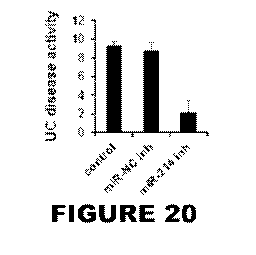
[0029] Figure 20 is a bar graph showing that intracolonic administration of
18mg/kg of the
miR-214 inhibitor, reduced 80% the ulcerative colitis disease activity in a
mouse model of
UC.
[0030] Figure 21 is a bar graph showing that the miR-214 inhibitor did not
affect the
expression levels of other, randomly selected, microRNAs (miR-133a, miR-210,
miR-21),
confirming its specificity for targeting only miR-214.
[0031] Figures 22A-22C are bar graphs showing the absence of toxicity in mice
(3
mice/group) treated with 18mg/kg of miR-214. Levels of ALT and AST liver
enzymes,
together with urea levels, were measured 48h post treatment. The levels of ALT
(Fig. 22A),
AST (Figs. 22B) and urea (Fig. 22C) were not statistically significant
different between the
untreated (control), the miR-NC inhibitor and the miR-214 inhibitor treated
mice.
DETAILED DESCRIPTION OF THE INVENTION
[0032] The invention described herein is based on the unexpected and specific
overexpression of miR-214 in ulcerative colitis (UC). The invention is further
based on the
demonstration that intracolonic administration of anti-sense miR-214 reduces
UC with a high
level of efficacy and specificity, and without toxicity. The data show that
miR-214 is an
important mediator in UC and a target for treatment. The invention is
particularly
advantageous in that it provides methods that can be used to distinguish
between ulcerative
colitis and Crohn's disease.
Definitions
[0033] All scientific and technical terms used in this application have
meanings commonly
used in the art unless otherwise specified. As used in this application, the
following words or
phrases have the meanings specified.
[0034] As used herein, a "specimen" from a subject means a specimen obtained
from the
subject that contains blood or blood-derived cells, other bodily fluid, such
as intestinal fluid,
or biopsy tissue. Examples of biopsy tissue include intestinal tissue, such as
colon biopsy
tissue.
8
CA 02966772 2017-05-03
WO 2016/077347
PCT/US2015/059956
[0035] As used herein, a "control sample" means a specimen that represents a
normal,
healthy condition. The specimen may be blood, serum, or other fluid or tissue
understood by
those skilled in the art to serve as a suitable control. A sample of normal
colon tissue or
intestinal fluid obtained from a healthy patient is a typical example of a
control sample.
[0036] As used herein, the term "subject" includes any human or non-human
animal. The
term "non-human animal" includes all vertebrates, e.g., mammals and non-
mammals, such
as non-human primates, horses, sheep, dogs, cows, pigs, chickens, and other
veterinary
subjects.
[0037] As used herein, "a" or "an" means at least one, unless clearly
indicated otherwise,
Methods of Detecting and Treating Ulcerative Colitis and Colitis-Associated
Cancer
[0038] The invention provides a method for detection of ulcerative colitis
(UC) or UC-related
dysplasia, in a subject. In one embodiment, the method comprises assaying a
specimen
obtained from the subject for miR-214, an oligonucleotide having the sequence:
5-
TGTCTGTGCCTGCTG-3' (SEQ ID NO: 1). In a further embodiment, the method
comprises
assaying the specimen for two, three, or more markers, including phosphatase
and tensin
homolog (PTEN), and PDZ and LIM domain protein 2 (PDLIM2). The assaying
typically
comprises contacting the specimen with reagents specific for miR-214, PTEN,
and/or
PDLl M2, and measuring the amount of miR-214, PTEN, and/or PDLIM2 present in
the
specimen. Representative reagents suitable for such assays are described in
the Examples
below. For example, quantitative real-time PCR can be used to determine
expression levels.
In the examples below, miR-214 expression levels were normalized to levels of
U6 small
nuclear RNA (203904; Exigon), Those skilled in the art can appreciate suitable
alternatives
to be used to normalize expression levels.
[0039] The disease detected in this manner includes both colon cancer and
ulcerative colitis
(UC) or UC-related dysplasia. In particular, this method can be used to detect
ulcerative
colitis (UC), and to distinguish UC from other conditions, such as Crohn's
disease or irritable
bowel syndrome (IBS). Thus, the invention additionally provides a method of
specifically
detecting UC. This method is particularly useful for cases in which a
diagnosis of UC or
Crohn's disease has not been determined. The invention additionally provides a
method of
determining the status (active or inactive/remission) of UC in a patient,
whereby detecting
elevated levels of miR-214 compared to a control sample is indicative of
active disease.
Levels of miR-214 that are reduced relative to a prior measurement for the
same patient, or
not significantly higher than a control sample, are indicative of disease
regression or
remission.
9
CA 02966772 2017-05-03
WO 2016/077347
PCT/US2015/059956
[0040] The amount of rniR-214, PTEN, and/or PDLIM2 present in the specimen is
then
compared to that present in a control sample. An elevated amount of miR-214,
or
decreased amount of PTEN and/or PDLIM2, present in the specimen compared to
the
control sample is indicative of inflammatory disease. Typically, the amount of
increase or
decrease in the presence of the marker (miR-214, PTEN, and/or PDLIM2) in the
specimen
obtained from a subject who has inflammatory disease is a statistically
significant difference
compared to a normal control sample.
[0041] In one embodiment, the amount of miR-214, PTEN, and/or PDLIM2 in a
specimen
obtained from a subject is elevated 2-fold compared to a normal control, and
the amount of
PTEN, and/or PDLIM2 is decreased to about half that of a normal control. In
some
embodiments, the difference is an increase (in the case of miR-214) or
decrease (in the case
of PTEN, and/or PDLIM2) of 10%, 20%, 30%, 40%, 50%, 75%, or 100% relative to
normal
control.
[0042] The measuring or assay for miR-214 can involve isolating microRNA from
the
specimen and/or performing a polymerase chain reaction (PCR) assay, such as
real-time
PCR, or other suitable PCR assay known in the art. Alternatively, the assay
can be an in situ
hybridization assay. The assay for PTEN, and/or PDLIM2 can be PCR or an
immunoassay,
such as enzyme-linked immunosorbent assay (ELISA), immunoblotting,
radioimrnunoassay,
or other immunoassays known in the art. Such assays can be performed on
biopsied tissue
samples.
[0043] Probes for detection of miR-214, PTEN, and/or PDLIM2 can be detectably
labeled. In
one embodiment, the probe is labeled with a radioisotope, an enzyme, a
fluorescent
substance, a luminescent substance or biotin. In another embodiment, the 5'
end of an
oligonucleotide probe is labeled with a reporter fluorescent dye and the 3'
end of the probe is
labeled with a quencher dye. In some embodiments, the probe is an antibody and
the
detectable label is an antibody that binds PTEN, and/or PDLIM2, or a secondary
antibody
that binds a primary antibody. Representative antibodies are described in the
Examples
below. Reagents and kits, including antibodies, probes, PCR primers and
related materials
are commercially available.
[0044] The specimen is typically intestinal fluid or intestinal tissue, such
as biopsy tissue.
Other specimens may be obtained in accordance with the judgment of the
treating physician.
Specimens can be obtained from subjects using conventional means.
[0045] Those skilled in the art will appreciate additional variations suitable
for the method of
detecting UC or UC-related dysplasia through detection of miR-214 in a
specimen, as it
provides a means of monitoring to assess disease activity and response to
treatment. This
CA 02966772 2017-05-03
WO 2016/077347
PCT/US2015/059956
method can also be used to monitor levels of miR-214 in a sample from a
patient undergoing
treatment. The suitability of a therapeutic regimen for initial or continued
treatment can be
determined by monitoring miR-214 levels using this method. The extent of miR-
214 present
in a given patient or specimen can provide a prognostic indicator to guide
treatment strategy,
Accordingly, one can use information about the level of miR-214 present in a
subject to
assist in selecting an appropriate treatment protocol. For example, mesalamine
treatment of
ulcerative colitis could be monitored by miR-214 as a surrogate biomarker to
quantitatively
measure the level of persisting disease activity. If disease activity persists
above an
acceptable level, the clinician would consider increasing the treatment dose,
or changing to
a different therapeutic agent.
[0046] The invention additionally provides a method of treating ulcerative
colitis (UC) or UC-
related dysplasia, or cancer, in a subject. In one embodiment, the method
comprises
administering to the subject a therapeutically effective amount of an
inhibitor of miR-214. A
therapeutically effective amount is an amount sufficient to ameliorate disease
symptoms,
including, but not limited to, inflammation, and 1L-8 expression. In one
embodiment, the
administering is intracolonic or intravenous. In another embodiment, the
administering is
intratumoral. Examples of inhibitors of miR-214 include an antisense miR-214
oligonucleotide, such as an antisense oligonucleotide directed against SEQ ID
NO: 1 (5'-
TGTCTGTGCCTGCTG-3'). The antisense oligonucleotide can be provided in a more
stabilized form, such as, in one example, an antisense miR-214 oligonucleotide
that
comprises locked nucleic acid and phosphorothioate linkages. Such
modifications both
stabilize the molecule and protect it from endonuclease cleavage, which serve
to increase
the stability and efficacy of the inhibitor in vivo. The method can comprise
administering the
antisense miR-214 oligonucleotide directly, or via a lentiviral vector. Use of
a suitable
delivery vector, such as a lentiviral or adenoviral vector, for example, may
be selected to
enhance the efficacy of intravenous administration.
[0047] Treatment of UC or colon cancer in a subject comprises administering a
therapeutically effective amount of an inhibitor of miR-214, such as an
antisense miR-214
oligonucleotide, to the subject. A therapeutically effective amount is an
amount sufficient to
ameliorate symptoms of disease, such as tumor growth and/or size, and IL-8
expression.
The administering can be intravenous, intracolonic, and/or intratumoral. In
one embodiment,
the inhibitor of miR-214 is administered intraoperatively at the time of
biopsy and/or tumor
resection. Representative doses of 12 and 18 mg/kg for treating UC in a mouse
model are
described in the Examples below, and shown to be specific, effective and non-
toxic. Those
skilled in the art can use this information to guide selection of an effective
dose amount and
schedule to other subjects.
11
CA 02966772 2017-05-03
WO 2016/077347
PCT/US2015/059956
[0048] Treatment of UC or cancer can be administered in a single dose or as a
series of
doses administered over time. Dosage and treatment regimens can be determined
by the
treating physician, taking into account disease severity, patient condition,
and other factors.
Kits
[0049] For use in the diagnostic applications described herein, kits are also
within the scope
of the invention. Such kits can comprise a carrier, package or container that
is
compartmentalized to receive one or more containers such as vials, tubes, and
the like, each
of the container(s) comprising one of the separate elements to be used in the
method. The
antibodies, probes, primers, and other reagents of the kit may be provided in
any suitable
form, including frozen, lyophilized, or in a pharmaceutically acceptable
buffer such as TBS or
PBS. The kit may also include other reagents required for utilization of the
reagents in vitro
or in vivo such as buffers (i.e., TBS, PBS), blocking agents (solutions
including nonfat dry
milk, normal sera, Tween-20 Detergent, BSA, or casein), and / or detection
reagents (i.e.,
goat anti-mouse IgG biotin, streptavidin-HRP conjugates, allophycocyanin, B-
phycoerythrin,
R- phycoerythrin, peroxidase, fluors (i.e., DyLight, Cy3, Cy5, FITC, HiLyte
Fluor 555, HiLyte
Fluor 647), and / or staining kits (i.e., ABC Staining Kit, Pierce)).
Nucleotide probes may be
labeled for detection. The kits may also include other reagents and / or
instructions for using
antibodies and other reagents in commonly utilized assays described above such
as, for
example, flow cytometric analysis, ELISA, immunoblotting (i.e., western blot),
in situ
detection, irnrnunocytochemistry, immunohistochernistry.
[0050] In one embodiment, the kit provides the reagent in purified form. in
another
embodiment, the reagents are immunoreagents that are provided in biotinylated
form either
alone or along with an avidin-conjugated detection reagent (i.e., antibody).
In another
embodiment, the kit includes a fluorescently labeled imrnunoreagent which may
be used to
directly detect antigen. Buffers and the like required for using any of these
systems are well-
known in the art and may be prepared by the end-user or provided as a
component of the
kit. The kit may also include a solid support containing positive- and
negative-control protein
and I or tissue samples. For example, kits for performing spotting or western
blot-type
assays may include control cell or tissue ysates for use in SDS-PAGE or nylon
or other
membranes containing pre-fixed control samples with additional space for
experimental
samples.
[0051] The kit of the invention will typically comprise the container
described above and one
or more other containers comprising materials desirable from a commercial and
user
standpoint, including buffers, diluents, filters, needles, syringes, and
package inserts with
instructions for use. In addition, a label can be provided on the container to
indicate that the
12
CA 02966772 2017-05-03
WO 2016/077347
PCT/US2015/059956
composition is used for a specific application, and can also indicate
directions for use, such
as those described above. Directions and or other information can also be
included on an
insert, which is included with the kit.
Representative Applications of the Invention
[0052] The identification of novel targets and compounds regulating NFKB
activity can be
achieved by use of the materials and methods described herein. Specifically,
the invention
identifies novel microRNA targets and inhibitors regulating NFKB activity in
human
colonocytes. The NFKB pathway is found to be activated in many different types
of cancer
and auto-immune and inflammatory diseases, and these inhibitors can be used to
treat these
diseases by suppressing NFKB phosphorylation,
[0053] MiR-214 is a diagnostic biomarker that is specific for ulcerative
colitis patients.
Several studies have identified IBD biomarkers, however most of these
biomarkers are not
highly specific for UC, but they are also biomarkers for Crohn's disease and
other GI
diseases. Most identified biomarkers are general inflammatory biomarkers. The
findings
described herein verify the specificity of miR-214 biomarker derived from
analysis of 3
different cohorts of patients. Examination of miR-214 levels can help
clinicians diagnose a
patient with UC and choose the appropriate therapy. This is particularly
useful in cases
where the available clinical tests cannot distinguish UC from Crohn's and
other
gastrointestinal diseases.
[0054] MiR-214 is a prognostic biomarker of UC disease activity. MiR-214
levels are higher
in UC patients with active disease relative to those in remission. Evaluation
of miR-214
levels can be used to monitor the disease status even in the absence of
symptoms and
evaluate if there is need for therapeutic intervention. MiR-214 is highly
specific to UC and
correlates with UC disease activity, and can also be used for predicting risk
for ulcerative
colitis patients to develop colon cancer.
[0055] Evaluation of microRNA levels in human FFPE tissue by in situ
hybridization.
Although protein levels could be examined by immunohistochemistry (11-iC) in
formalin-fixed
paraffin embedded (FFPE) sections from patient samples, it is preferable to
develop a
protocol for evaluating microRNA levels in the same tissues. The invention
provides an
optimized in situ hybridization protocol, improving its efficacy and
specificity. The protocol
can be found in the examples below.
[0056] MiR-214 levels provide a biomarker identifying UC patients who have
higher risk to
develop colon cancer. This is a significant advantage, since there is nothing
available for UC
patients. Even if the colon tissue seems normal during the colonoscopy
procedure,
13
CA 02966772 2017-05-03
WO 2016/077347
PCT/US2015/059956
evaluation of miR-214 levels can be used to identify early molecular oncogenic
alterations to
help the clinician decide the appropriate time for removing this area of the
colon by surgery.
Patients with >10 years with colitis have increased risk for colon cancer and
the only solution
is surgery. Mir-214 is the first biorriarker that has 100% specificity for
colon cancer patients
with UC history and 0% specificity for sporadic colon cancer patients. MiR-214
is ¨30-fold
increased in colitis-associated colorectal cancer (CAC) patients, which is a
difference that
can be easily and specifically identified by FOR in a prognostic test.
[0057] Development of an inhibitor towards miR-214 (SEQ ID NO: 1). The
invention
identifies the appropriate sequence in order to target efficiently miR-214
expression.
[0058] Efficacy of miR-214 inhibitor to suppress colitis ex vivo (fresh
colonic explants from
UC patients). This is the first demonstration that targeting microRNAs has
therapeutic
potential ex vivo.
[0059] Targeting miR-214 as a novel therapy for ulcerative colitis patients.
Inhibition of miR-
214 by enema reduces colitis in a mouse model of experimental colitis. The
therapeutic
protocol can be found in detail in Figure 14.
[0060] Strategy to identify microRNA downstream targets that are disease
relevant. Through
a combination of bioinformatics, expression and 3'UTR analysis, the invention
identifies miR-
214 targets.
[0061] Identification of a gene signature regulated by miR-214 (see Table 3).
The invention
identifies direct downstream targets of miR-214. These genes provide markers
for evaluating
the specificity and effectiveness of miR-214 inhibition.
[0062] PTEN and PDLIM2 levels provide biomarkers for UC patients (see fig.
2c).
Identification of reduced PDLIM2 and PTEN mRNA levels in UC patients relative
to healthy
controls provides a method for detecting and monitoring UC.
[0063] MiR-214 is a novel oncogene in colon cancer. The examples below provide
an
extensive analysis validating the oncogenic properties of miR-214
overexpression.
[0064] MiR-214 provides a biomarker for colon cancer progression in patients
with a history
of colitis (Fig, 3j). MiR-214 has increased expression levels in colon cancer
patients at
advanced stages (stages III, IV) relative to those in early stages (I, II).
The invention thus
provides a method of staging colon cancer whereby detecting a 10-20-fold
increase in miR-
214 is indicative of stage I-II colon cancer, while detecting a 40-fold or
greater increase in
miR-214 is indicative of stage III-IV colon cancer,
[0065] The invention provides a method for administration of microRNAs by
enema in colon
cancer, as demonstrated by animal models (see fig, 4a). Delivery of nucleic
acids, including
14
CA 02966772 2017-05-03
WO 2016/077347
PCT/US2015/059956
microRNAs and siRNAs, by intravenous injections results in multiple side
effects and there is
need to have a carrier (e.g. nanoparticles) that increases the specificity and
efficacy. The
invention identifies that, for patients with GI diseases, including UC and
colon cancer,
intracolonic (enema) delivery of microRNAs is highly efficient. This approach
minimizes side
effects, as there is no need for a particle carrier of the microRNA that could
induce different
side effects (e.g. immune responses).
[0066] MiR-214 inhibition provides a therapeutic strategy for colon cancer
patients with a
history of ulcerative colitis. MiR-214 inhibition suppresses colon cancer
growth in vivo.
Description of the specific therapeutic protocol can be found in Fig. 19.
EXAMPLES
[0067] The following examples are presented to illustrate the present
invention and to assist
one of ordinary skill in making and using the same. The examples are not
intended in any
way to otherwise limit the scope of the invention.
Example 1: Therapeutically targeting miR-214 circuit in ulcerative colitis and
colitis-
associated cancer
[0068] Inflammatory Bowel Diseases (1BD), consist of ulcerative colitis (UC)
and Crohn's
Disease (CD), which are characterized by activation of inflammatory responses,
and patients
with longstanding disease are at higher risk of developing colorectal cancer.
Thus, the
identification of novel molecular targets with therapeutic potential for UC
and UC-related
fiysplasia are of major importance. MicroRNAs are deregulated in ulcerative
colitis, but their
mechanistic role and therapeutic value remains speculative. Here, using a high
throughput
functional suppressor screen of the human rnicroRNAome, we identified miR-214
as master
regulator of nuclear factor kappa beta (NF-kB). MiR-214 is amplified
specifically in colonic
tissues of patients with active and longstanding UC, and hyper- expressed in
colitis-
associated colorectal cancer (CAC). Integration of bioinformatic and genome-
wide profiling
analyses revealed that miR-214 by suppressing PDZ and LIM domain 2 (PDLIM2)
and
phosphatase and tensin homolog (PTEN) genes is amplified through a feedback
loop circuit.
A chemical inhibitor perturbed this circuit in colonic biopsies from UC
patients ex vivo and
intracoionic administration therapeutically suppressed UC and CAC in mice.
Taken together,
miR-214 correlates with disease activity and progression to colorectal cancer
while its
inhibition provides a novel therapeutic option for both UC and CAC patients.
[0069] UC development is characterized by activation of inflammatory pathways,
including
NF- KB signaling. To identify microRNAs that regulate NF-KB activity in human
colonocytes,
we performed a microRNA functional screen by targeting the human microRNAome
in 1L6-
treated NOM-460 cells. (Fig.la). In the primary screen, two microRNA
inhibitors (miR-26b
CA 02966772 2017-05-03
WO 2016/077347
PCT/US2015/059956
and 199a) induced, while seven inhibitors (miR-7, 146a, 373, 372, 181b, 21 and
214)
suppressed NF-KB phosphorylation by >50%, P<0.05 (Fig. 1b). The miR-214
inhibitor was
identified as the most effective suppressor (>90%). Examination of miR-214
levels in UC
(n=120) and control (n=107) colonic tissues from 3 different cohorts (Tables 1
and 2)
revealed that miR-214 levels are significantly increased in UC, but not other
GI diseases,
irritable bowel syndrome (IBS) (n=22) and CD (n=60) (Fig. 1c). In situ
hybridization in human
colonic tissues identified epithelial cells as the origin of miR-214
overexpression (Fig. 1d). To
evaluate the clinical relevance, we examined miR-214 expression in correlation
with
clinicopathological parameters. MiR-214 levels were significantly higher in UC
patients with
active disease relative to patients in remission (Fig. le), but did not differ
in relation to UC
patient's gender neither to gender/disease location in CD patients (Fig. 5).
These results
demonstrate that MiR214 is an epithelial expressed gene that regulates NF-KB
activity and is
deregulated specifically in UC in correlation with disease activity.
[00701 To establish the link between miR-214 and UC development, we aimed to
identify
downstream gene targets that could mediate NF-KB activation. Our strategy
consisted of 5
steps (Fig. 2a). We employed 4 different microRNA target prediction software
and identified
280 common miR-214 gene targets. Their expression was analyzed by gPCR in miR-
214-
treated colonocytes. The validated 71 (Table 3) genes (expression <50%,
P<0.01) were
examined for their correlation with the NF-KB signaling pathway and direct
interaction with
miR-214 employing 3'UTRiuciferase assays in a second colonic epithelial cell
line. This
comprehensive analysis revealed PDLIM2, a nuclear ubiguitin E3 ligase
inhibitor of NF-KB
activityl 5, and PTEN, a suppressor of the AKT signaling pathwayl 6 previously
shown to
intervene with NF-KB activation17,18, as the top miR-214 regulated genes. MiR-
214
overexpression inhibited 3'UTR luciferase activities and resulted in
suppression of PTEN
and PDLIM2 rnRNA (Fig. 6) and protein levels (Fig. 2B). In reflection of the
human relevance
of these findings, PTEN and PDLIM2 mRNA levels were decreased in UC colonic
tissues
relative to controls (Fig. 2C), thus inversely correlated with miR-214
expression.
Interestingly, overexpression of miR-214 induced NF-KB (s536) phosphorylation
(Fig. 2D)
and subsequently the expression of IL6 (Fig, 7), indicating that PTEN and
PDLIM2 are direct
downstream effectors of miR-214 involved in the regulation of the NF-KB
inflammatory
response.
[00711 Table 1. Cohorts of patients analyzed for miR-214 expression
Cohorts Control UC CD IBS CAC CRC
UCLA 43 18 24 22 6 18
16
CA 02966772 2017-05-03
WO 2016/077347 PCT/US2015/059956
Leiden 52 63 13 22 27
. .
Origene 12 39 23 4 15
Total Patients 107 120 60 22 32 60
[0072] Table 2. Characterlstics of patients analyzed for miR-214 expression
Groups Control UC CD IBS CAC CRC
, .
No of 107 120 60 22 32 60
Patients
Age-yr 42 39,5 33 38.5 55.5 70
Median
Range 16-80 20-72 17-74 18-55 25-82 31-91
. .
Missing 9 10
Data
. .
P value ns ns ns *** ***
Sex-no (%)
. .
Female 31(29) 41(34) 15(25) 11(50) 18(56) 28(47)
Male 57(53) 44(37) 29(48) 11(50) 14(44) 32(53
Missing 19(18) 35(29) 16(27)
Data
P value ns ns ns ns ns (0.192)
Disease Duration 11(0.3-30)
TNM stage-no (%)
' .
I 6(19) 13(22)
II 16(50) 19(32)
17
CA 02966772 2017-05-03
WO 2016/077347
PCT/US2015/059956
III 7(22) 18(30)
IV 3(9) 10(17)
P value ns(0.390)
"P<0.001 between groups (Kruskal Wallis test); #P<0.001 in comparison to CAC
(Wilcoxon
two sample test).
[00731 Table 3. Expression of miR-214 targets vs Control (miR-scramble) in
colonic
epithelial cells
Symbol ID Name Fold
Change P<
ABR 29 active BCR-related -2.16
ACER3 55331 alkalineceramidase3 -1.67 * *
ADSS 159 adenylosucc,inate synthase -3.57
AP3B1 8546 adaptor-related protein complex 3, beta 1 subunit -1.53
ARPC5L 81873 actin related protein 2/3 complex, subunit 5-like -2.07
BAZ2A 11176 bromodornain adjacent to zinc finger domain, 2A -1.79
* *
BRPF3 27154 bromodomain and PHD finger containing, 3 -1.58
CD151 977 CD151 molecule (Raph blood group) -2.62 ***
* -
CDC25B 994 cell division cycle 25 homoiog B (S. pombe) -1.90
CDIPT 10423 CDP-
diacylglycerol¨inositol 3-phosphatidyltransferase -1.79 * *
CEP85 84793 centrosomal protein 85kDa -1.57
CHIVIP4B 128866 charged multivesicular body protein 4B -1.69 ***
*
CPEB4 80315 cytoplasmic
polyadenylation element binding protein 4 -1.92
CTDSP1 58190 CTD (carboxy-
terminal domain, RNA polymerase 1. -1.70 * *
polypeptide A) small phosphatase 1
CTSS 1520 cathepsin S -2.12
DNAJC5 80331 DnaJ (Hsp40) homolog, subfamily C, member 5 -1.59 ***
F8 2157 coagulation factor VIII, procoagulant component -1.62
FAM134A 79137 family with sequence similarity 134, member A -3.66
* *
FAM189B 10712 family with sequence similarity 189, member B -1.99
GALNT7 51809 'LJDP-N-acetyl-alpha-D-galactosamine: polypeptide N- -2.08
acetylgalactosaminyltransferase 7
GLI3 2737 GLI family zinc finger 3 -2.13 -***
HMG20A 10363 high mobility group 20A -1.75
IGSF3 3321 irnmLmoglobulin superfamily, member 3 -1.64 ***
=
IN0800 125476 'IN080 complex subunit C -2.40
INTS2 57508 integrator complex subunit 2 -1.61 ***
18
CA 02966772 2017-05-03
WO 2016/077347
PCT/US2015/059956
IP011 51194 importin 11 -2.06
ITCH 83737 itchy E3 ubiquitin protein ligase -1.67
KLHDC3 116138 kelch domain containing 3 -1.56
***
KLK10 5655 kallikrein-related peptidase 10 -1.71
LAPTM4B 55353 lysosomal protein transmembrane 4 beta -1.84
LARP1 23367 La ribonucleoprotein domain family, member 1 -2.12
LEPROTL1 23484 leptin receptor overlapping transcript-like 1 -2.24
***
MED19 219541 mediator complex subunit 19 -1.90
MTM1 4534 rnyotubularin 1 -1.56
NAA15 80155 N(alpha)-
acetyltransferase 15, NatA auxiliary subunit -2.94
OTUB1 55611 OTU domain, ubiquitin aldehyde binding 1 -1.59
***
PAPD5 64282 PAP associated domain containing 5 -1.73
PDLIM2 64236 PDZ and LIM domain 2 (mystique) -3.37
PGGT1B 5229 PGGT1B 2.25
PLA2G15 23659 phospholipase A2, group XV -1.66
***
PLK4 10733 polo-like kinase 4 -1.55
POLE3 54107 polo-like kinase 4 -1.55
PPP2CB 5516 protein
phosphatase 2, catalytic subunit, beta isozyrne -1.68
PPP6C 5537 protein phosphatase 6, catalytic subunit -1.72
***
PSMD10 5716 proteasome
(prosome, macropain) 26S subunit, non- -3.19
ATPase, 10
PTEN 5728 phosphatase and tensin hornolog -2.47
RAB14 51552 RAB14, member RAS oncogene family -2.54
RASSF5 83593 Ras association (RaIGDS/AF-6) domain family -1.52
***
member 5
RBM22 55696 RNA binding motif protein 22 -2.46
RNF115 27246 ring finger protein 115 -1.71
RPIA 22934 ribose 5-phosphate isomerase A -1.57
=
[0074] Given that miR-214 activates NF-KB and mediates IL6-induced NF-K8
phosphorylation, we examined whether inflammatory signals regulate miR-214
expression.
OPCR analysis revealed IL6 dose-dependent induction of miR-214 expression
(Fig. 8A).
Lever/PhylORM algorithm analysis indicated the presence of STAT3 binding site
in the
promoter of MIR214 gene (Fig. 88) and chromatin immunoprecipitation analyses
revealed
increased enrichment of STAT3 on MIR214 promoter upon treatment with IL6 (Fig,
2E).
Similarly, IL6 dose-dependently induced MIR214 promoter activity, an effect
abolished by
mutation of the STAT3 binding site (Fig. 2F). The clinical importance of this
interaction was
evidenced by the correlation (r=0.97) of phospho-STAT3 to miR-214 levels (Fig.
9). The role
of miR-214 as inflammatory mediator was studied in NCM356 colonocytes. IL6
dose-
19
CA 02966772 2017-05-03
WO 2016/077347
PCT/US2015/059956
dependently suppressed PDLIM2 and PTEN expression an effect reversed by the
inhibition
of miR-214 (Figs. 10, 11). In accord, Akt phosphorylation was increased upon
IL6 treatment,
and reduced by miR-2141nhibit1on (Fig. 12). These findings suggest that L6
induces miR-
214 through STAT3-mediated transcriptional activation, creating a positive
feedback loop
circuit (Fig. 20).
[0075] To evaluate the therapeutic potential of miR-214 inhibition in UC
through regulation
of the feedback loop circuit, we used a chemically stable inhibitor, highly
efficient in
suppressing miR-214 in human colonocytes in vitro (Fig. 13). We studied the
effects of the
m1R-214 chemical inhibitor ex vivo on freshly isolated colonic biopsies from
UC patients with
active disease. The inhibitor efficiently suppressed miR-214 in the colonic
explants (Fig. 2H),
and suppressed the inflammatory response by increasing the expression of both
PDLIM2
and PTEN (Fig. 21). Furthermore, miR-214 inhibitor reduced disease activity in
an animal
model of experimental colitis (Fig. 2J and Fig. 14), Taken together, these in
vitro, in vivo and
ex vivo data suggest the therapeutic potential of the miR-214 inhibitor in UC
patients with
active disease.
[0076] Although the association between UC and colorectal cancer has been
documented
as early as 192519, its molecular biology remains to be explored. We
questioned the role of
miR-214 during the transition from UC to cancer. Interestingly, we found miR-
214 is up-
regulated in mice with chronic but not acute inflammation induced by DSS (Fig.
3A), In the
same line, colonic tissues from patients with longstanding (>10 years) UC have
increased
miR-214 compared to those with <10 years (Fig. 3B), suggesting that miR-214 is
a link
between UC and colorectal cancer development. We compared miR-214 levels by
qPCR
and in situ hybridization in tumors from CAC and sporadic colon cancer (CRC)
patients. This
analysis showed the hyper-expression of miR-214 in CAC, but no difference
between CRC
and controls (Fig, 3C, 3D), Overall, these findings suggest that rniR-214 is
increased during
UC development and amplified during progression to colorectal cancer,
[0077] To examine the importance of miR-214 in colorectal oncogenesis we
performed gain-
and loss-of function studies. Overexpression of miR-214 induced the colony
formation ability
and invasiveness of human colon cancer cells, suggesting its oncogenic role
(Figs. 3E, 3F
and Fig, 15). On the other hand, suppression of miR-214 inhibited the L6-
induced
turnorigenic and invasive phenotype, pointing to the dependence of IL6
tumorigenic effects
on miR-214. Next, we investigated whether miR-214 acts through regulation of
the feedback
loop circuit. L6 dose-dependently up-regulated miR-214 in colon cancer cells
(Fig. 3G),
mediated by the binding of STAT3 in MIR214 promoter area (Fig. 16). 1L6-
induced miR-214
expression resulted in PDLIM2 and PTEN suppression (Fig. 17), Akt
phosphorylation (Fig.
3H) and NF-KB activation (Fig. 18), suggesting that the miR-214 feedback loop
circuit is
CA 02966772 2017-05-03
WO 2016/077347
PCT/US2015/059956
hyper-activated during oncogenesis. In support of its human relevance, PDL1M2
and PTEN
levels were decreased in tumors from CAC patients relative to controls (Fig,
31). Given that
miR-214 increases cancer cell aggressiveness, we examined miR-214 correlation
to colon
cancer progression. Examination of human tumors indicated that miR-214 is
specifically up-
regulated during CAC disease progression (Fig. 3J), providing additional
evidence on the
specificity of rniR-214 circuit in UC and CAC. According to our data miR-214-
positive
feedback loop circuit links chronic inflammation to colon carcinogenesis.
Therefore, targeting
of miR-214 may be sufficient to suppress the development of CAC. We applied a
therapeutic
protocol consisting of intracolonic administration of a chemical miR-214
inhibitor or
microRNA negative control for 4 cycles, weekly (Fig. 4A and Fig. 19) on the
AGM- DSS
mouse model. The miR-214 inhibitor reduced significantly the number (Fig. 4B)
and size
(Fig. 4C) of tumors. Mechanistically, miR-214 inhibitor suppressed tumor
growth through the
induction of apoptosis and activation of caspase-3 (Fig. 40). In addition,
irnmunohistochemical analysis revealed that miR-214 inhibition reduced NF-KB
and Akt
phosphorylation levels (Fig. 4E), indicating suppression of the miR-214-
molecular circuit,
and decreased the proliferation rate of colon cancer cells (Fig. 4E).
[0078] MiR-214 expression correlates with UC disease activity and disease
duration and
provides the molecular rationale for the current practice guidelines
recommending
surveillance colonoscopy 8 to 10 years after diagnosis of UC20. Given that
current
surveillance procedures (21) and new imaging techniques (22-24) identify late-
stage
oncogenic events, rniR-214 could serve as a biornarker, identifying UC
patients at risk for
malignant transformation. Furthermore, it is essential that although
inflammatory pathways
have been found to be activated in UC and CD, miR-214 is specific to UC.
Intravenous
administration of microRNA mimics has proven effective against liver cancer
(8) and
microRNA inhibitors are in Phasell-111 clinical trials25. Here, we present a
novel and efficient
delivery method for microRNA therapeutics. Intracolonic administration of a
chemically
modified miR-214 inhibitor results in suppression of UC and CAC, suggesting
the
applicability of such approaches in patients with colitis and UC-related
dysplasia. In
conclusion, we demonstrate the UC-specific deregulation of miR-214 and its
correlation with
disease activity and duration, and identify a miR-214-driven inflammatory
feedback loop
circuit involved in the development of colonic oncogenesis. Tissue miR-214
could
significantly alter current surveillance strategies and suppression of miR-214
could have
therapeutic potential for colitis and colitis-associated colon cancer.
21
CA 02966772 2017-05-03
WO 2016/077347
PCT/US2015/059956
Methods
[0079] The study was approved by the institutional review board at each study
center. All
patients from whom tissue samples were obtained at UCLA provided written
informed
consent. The patient samples were obtained at the Leiden University Medical
Center
according to the instructions and guidelines of the LUMC Medical Ethics
Committee and in
accordance with the Helsinki Declaration.
MicroRNA library screen
[0080] NCM460 immortalized epithelial cells were plated in 96-well plates and
transfected
with a microRNA inhibitor library consisting of 348 microRNA inhibitors and 2
negative
control microRNAs (100 nM) (Dharmacon Inc), as previously described'. At 24
hours post-
transfection, the cells were treated with IL6 for 24 hours, and the
phosphorylation of NFKB
was assessed by Phospho-RelAiNFKB p65 (S536) Cell-Based ELISA (KCB7226, R&D).
MicroRNA inhibitors that inhibited >50% the phosphon/lation of NFKB were
considered
positive hits.
Bioinformatic analysis
[0081] The Lever and PhylCRM algorithms have been used to identify STAT3
binding motifs
in an area 5 kb upstream and 2 kb downstream of microRNAs.
In situ hybridization
[0082] Double-DIG labeled miRCURY LNA Detection probe for the detection of miR-
214
(38494-15, Exigon) by in situ hybridization was used as previously described
with
modifications'. Sections of control, ulcerative colitis, and colorectal
carcinomas were
deparaffinized with xylene (three times for 5 min), followed by treatment with
serial dilutions
of ethanol (three times in 100%, twice in 96%, and three times in 70%) and by
two changes
of DEPC-PBS. Tissues were then digested with proteinase K (15 mg/ml) for 30
min at 37 C,
rinsed three times with DEPC-PBS. Sections were dehydrated twice with 70%,
96%, and
100% ethanol, air-dried and hybridized for 1 hour with the hsa-miR-214 (40 Al)
diluted in
microRNA ISH buffer (90000, Exicion), at 60cC. Following hybridization,
sections were
rinsed twice with 5XSSC, twice with 1XSSC, and three times with 0.2XSSC, 5 min
each, at
60'C, and PBS. The slides were incubated with blocking solution (11585762001,
Roche) for
15 min and then with anti-DIG antibody (1:800) in 2% sheep serum (013-000-121,
Jackson
Immunoresearch) blocking solution for 1 hour, at RT. Following three washes
with PBS-T
(PBS, 0.1% Tween-20), slides were incubated with the AP substrate buffer (NBT-
BCIP tablet
[11697471001. Roche] in 10 ml 0.2 mM Levarnisole [31742, Fluka]) for 2 hours
at 30'C in the
22
CA 02966772 2017-05-03
WO 2016/077347
PCT/US2015/059956
dark. The reaction was stopped with two washes of AP stop solution (50 mM Tris-
HCI, 150
mM NaCI, 10 mM KCi) and two washes with water. Tissues were counter-stained
with
Nuclear Fast Red for 1 min and rinsed with water. At the end, sections were
dehydrated
twice with 70%, 96%, and 100% ethanol and mounted with coverslips in Eukitt
mounting
medium (361894G, V\A/R). Images were captured with a Nikon 80i Upright
Microscope
equipped with a Nikon Digital Sight DS-Fil color camera, using the MS-Elements
image
acquisition software. All images were captured and processed using identical
settings.
Immunohistochemistry
[0083] For tissue irnmunostaining for phospho-NFKB, phospho-Akt and Ki67, FFPE
sections
of colonic tissues from AOM-DSS mice treated with the chemical miR-214
inhibitor or
negative control were deparaffinized with xylene (3x5 min) followed by
treatment with serial
dilutions of ethanol (100%, 100%, 95% and 95%, 10 min each) and by two changes
of
ddH20. Antigen unmasking was achieved by boiling the slides (95-99 C) for 10
min, in 10
mM sodium citrate, pH 6Ø Sections were rinsed three times with ddH20,
immersed in 3%
H202 for 20 min, washed twice with ddH20 and once with TBS-T (TBS, 0.1% Tween-
20)
and blocked for 1 hr with blocking solution (5% normal goat serum [5425, Cell
Signaling
Technology] in TBS-T). Phospho-P65 (Ser536) (SA84300009, Sigma-Aldrich),
phospho-Akt
(Ser473) (4060, Cell Signaling Technology) and Ki67 (12202, Cell Signaling
Technology)
antibodies were diluted 1:200, 1:50 and 1:400, respectively, in Signal Stain
antibody diluent
(8112, Cell Signaling Technology) and incubated with the sections overnight at
4 C. Following
incubation with the antibodies, sections were washed three times, 5 min each,
with TBS-T
and incubated for 1 hr at room temperature with SignalStain Boost ([HRP,
Rabbit] 8114, or
[HRP, Mouse] 8125, Cell Signaling Technology). Sections were washed three
times, 5 min
each, with TBS-T, and stained with the DAB Peroxidase Substrate Kit (SK-4100,
Vector
Laboratories) for 30 min, washed and counterstained with the hernatoxylin QS
(H-3404,
Vector Laboratories). Finally, tissues were dehydrated and mounted in Eukitt
medium.
Microscope equipped with a Nikon Digital Sight DS-Fil color camera, using the
NIS-
Elements image acquisition software. All images were captured and processed
using
identical settings.
Quantitative real-time FOR analysis
[0084] Real-time FOR was performed to determine the expression levels of miR-
214 in
human colon carcinomas and tissues. RNA was isolated, using Trizol (15596-026,
Invitrogen). Reverse Transcription was carried out using the Universal cDNA
synthesis kit
(203300). Real-time PCR was carried out in triplicate using the SYBR Green
master mix
(203450) and primer for miR-214 (204510, Exiqon) in a CFX384 Real Time PCR
detection
23
CA 02966772 2017-05-03
WO 2016/077347
PCT/US2015/059956
system (BioRad). MIR214 expression levels were normalized to the levels of U6
snRNA
(203907, Exiqon).
[0085] Real-time PCR was employed to determine the expression levels of PTEN
and
PDL/A42. Reverse transcription was carried out using the Retroscript Kit
(AM1710, Appiied
Biosystems). Real-time PCR for was carried out using IQ SYBR Green supermix
(170-8882,
BioRad). Glyceraldehyde-3- phosphate dehydrogenase (GAPDH) and B-ACTIN were
used
as the internal control. The sequences of the primers used are the following:
[0086] PTEN-F: 5'-cccagacatgacagccatc-3' (SEQ ID NO: 5)
[0087] PTEN-R: 5'-tctgcaggaaatcccatagc-3' (SEQ ID NO: 6)
[0088] PDLIM2-F: 5'-atggccacgattatgtctcc-3' (SEQ ID NO: 7)
[0089] PDLIM2-R: 5'-gcccatcatggtgactaagg-3' (SEQ ID NO: 8)
[0090] B.-ACTIN-F: 5'-cccagcacaatgaagatcaa-3 (SEQ ID NO: 9)
[0091] B.-ACTIN-R: 5'-acatctgctggaaggtggac-3' (SEQ ID NO: 10)
[0092] GAPDH-F: 5'-atgttcgtcatgggtgtgaa-3' (SEQ ID NO: 11)
[0093] GAPDH-R: 5'-ggtgctaagcagttggtggt-3' (SEQ ID NO: 12)
Animal studies
[0094] Mouse studies were approved by the University of California
Institutional Animal Care
and Use Committee and conformed to the US National Institutes of Health Guide
for the
Care and Use of Laboratory Animals. The therapeutic potential of the miR-214
inhibition was
tested in a mouse model of colitis-associated colon cancer (CAC). CAC is
induced by
administration of 12.5 mg/kg azoxymethane (AOM) at day 1 followed by repeated
oral
administration of 3% dextran sulfate sodium (DSS) at days 8, 29, 50. According
to our
treatment protocol, miR-NC inhibitor or miR-214 chemical inhibitor (12 mg/kg)
were
intracolonically administered in AOM-DSS-treated mice on a weekly basis for
four cycles
(days 64, 70, 77, 84). On day 91, the mice were sacrificed and the tumor
burden was
assessed. The miR-214 inhibitor targeted the miR-214 sequence 5'-
TGTCTGTGCCTGCTG-
3' (SEQ ID NO: 1).
BioPlex ELISA assays
[0095] We used a sandwich ELISA assay to assess the phosphorylation status of
Tyrosine
705 of STAT3 protein in control colonic tissues and samples from patients with
ulcerative
colitis and colitis- associated cancer, the levels of cleaved caspase-3 in
colonic tumors
derived from AOM-DSS- treated mice, the phosphorylation status of Serine 473
of Akt
24
CA 02966772 2017-05-03
WO 2016/077347
PCT/US2015/059956
protein, the phosphorylation status of Serine 536 of NFKB protein and the
secretion of IL6 in
colonic cell lines. The data were analyzed in a BioPlex FlexMap3D (Bio-Rad)
analyzer using
the BioPlex Manager software.
Luciferase assays
[0096] Colonic epithelial cells were transfected with a firefly luciferase
reporter gene construct
containing the promoter of MIR214. Cells were treated with IL6 24 hours after
transfection of
the luciferase vector. Cell extracts were prepared 24 later, and luciferase
activity was
measured using the Dual Luciferase Reporter Assay System (Promega, WI, USA).
For the
3'UTR assays, cells were transfected with the reporter vectors carrying the
3'UTR of PTEN
or PDLIM2. At 24 hours they were transfected with miR-214 (or miR-scramble)
and at 48
hours luciferase activity was measured using the Dual Luciferase Reporter
Assay System.
Invasion assays
[0097] We performed invasion assays in colonic epithelial cell lines 24 hours
after transfection
with miR-214 or anti-miR-214 or their respective controls. Invasion in
matrigel has been
conducted by using standardized conditions with BDBioCoat growth factor
reduced
MATRIGEL invasion chambers (PharMingen). Assays were conducted according to
manufacturer's protocol, using 2% FBS as chemoattractant. Non-invading cells
on the top
side of the membrane were removed while invading cells were fixed and stained
with 4'-6-
diamidino-2-phenylindole (DAP!, Vector Laboratories Inc.), 16h post seeding.
In all assays,
10 fields per insert were scored and se was calculated.
Colony formation assays
[0098] Colonic epithelial cells were transfected with miR-214 or anti-miR-214
or their
respective controls. Then, triplicate samples of 105 cells from each cell line
were mixed 4:1
(\fly) with 2.0% agarose in growth medium for a final concentration of 0.4%
agarose. The cell
mixture was plated on top of a solidified layer of 0.8% agarose in growth
medium. The
number of colonies was counted after 12 days.
Chromatin immunoprecipitation
[0099] Chromatin irnrnunoprecipitation was carried out as described
previously8. Briefly, the
chromatin fragments, derived from untreated and IL6-treated colonic epithelial
cells, were
irnmunoprecipitated with 6ug of antibody against STAT3. DNA extraction was
performed
using QIAGEN Purification Kit, Real-time PCR analysis was performed for STAT3
binding
site in the miR-214 promoter using the following primers: forward: 5-
CA 02966772 2017-05-03
WO 2016/077347
PCT/US2015/059956
GGGCTTGAGTCCATCAGCTT-3' (SEQ ID NO: 13) and reverse 5'-
GTTCAGCAGGACAGGTCTCA-3' (SEQ ID NO: 14) (Product size: 94 bp),
Statistical analysis
[0100] All experiments were performed in triplicate unless otherwise stated.
Statistical
analyses were performed with the use of Origin software, version 8.6 and SAS
statistical
package version 9.4. Student's t-test was used to examine the statistical
difference in miR-
214 expression between control colonic tissues and specimens derived from
different
intestinal pathologies, between active and inactive UC specimens and between
UC
specimens categorized based on disease duration. The Kruskal Wallis test was
used for age
comparison and posthoc Wilcoxon two sample test with Bonferroni significance
level
adjustment for multiple testing were used for age comparisons, Chi-square test
for gender
comparisons and Fisher's Exact test for tumour stage comparison. The
correlation
significance was determined by means of Spearman and Pearson correlation
analyses. The
Student's t-test was used for comparisons between the differentially treated
cells and mice
groups. A P value of 0.05 or less was considered to indicate statistical
significance.
References
1. Eaden, J.A., etal. Gut 48, 526-535(2001).
2. Ekbom, A., eta?. The New England journal of medicine 323, 1228-1233
(1990).
3. Soderlund, S., etal. Gastroenterology 136, 1561-1567; quiz 1818-
1569(2009).
4. Cho, W.., etal. Journal of the Korean Surgical Society 83, 135-140(2012).
5. GUO, H., eta?. Nature 466, 835-840 (2010).
6. Boldin, MR & Baltimore, D. Immunological reviews 246, 205-220 (2012).
7. Androulidaki, A.õ et al. Immunity 31, 220-231 (2009).
8. Hatziapostolou, M., et al. Cell 147, 1233-1247 (2011).
9. Baltimore, D,, et al. Nature immunology 9, 839-845 (2008).
10. Wu, F., etal. Gastroenterology 135, 1624-1635 e1624 (2008).
11. Stagakis, E., et a/. Annals of the rheumatic diseases 70, 1496-1506
(2011).
12. Koukos, G., et et. Gastroenterology 145, 842-852 e842 (2013).
13. lliopoulos, D., etal. Cell 139, 693-706 (2009).
14. Hatziapostolou, M,, etal. Trends in endocrinology and metabolism; TEM 24,
361-373 (2013).
15. Tanaka, T,, et at Nature immunology 8, 584-591 (2007),
16. Polytarchou, C., et al. Cancer research 71, 4720-4731 (2011).
17. Rornashkova, J.A. & Makarov, S.S. Nature 401, 86-90 (1999).
18. Dan, HG., etal. Genes & development 22, 1490-1500 (2008).
19. Crohn, U.B. & Rosenberg, H. Am J Med Sc/170, 220-228(1925).
20. Farraye, F.A., etal. Gastroenterology 138, 746-774, 774 e741-744; quiz
e712-743 (2010).
21. Huristone, D.P., etal. Endoscopy 37, 1186-1192(2005).
26
CA 02966772 2017-05-03
WO 2016/077347
PCT/US2015/059956
22. Kiesslich, R., etal. Gastroenterology 124, 880-888 (2003).
23. Dekker, E., etal. Encioscopy 39, 216-221 (2007),
24. Hailstone, D.P., etal. Gut 57, 196-204 (2008).
25. Janssen, HI., etal. The New England journal of medicine 368, 1685-
1694(2013).
Example 2: Effectiveness of miR-214 inhibitor administered at 18mcjfkg to
suppress
ulcerative colitis in mice
[0101] As described in Example 1 above, we evaluated the effectiveness of
administering
12rngikg of the miR-214 inhibitor intracolonically, to suppress ulcerative
colitis in a chronic
DSS mouse model (Figure 2i). Figures 14A-1413 illustrates the therapeutic
protocol applied
on the chronic DSS mouse model. To address the therapeutic potential of miR-
214 inhibition
we used a mouse model of chronic colitis. In this model, colitis is induced by
repeated oral
administration of dextran sulfate sodium (DSS). (Fig. 14A) rniR-214 inhibitor
or the negative
control were intracolonically administered in DSS-treated mice after day 8 for
four cycles
(every 2 days). (Fig. 14B) The criteria used for the assessment of the disease
activity score
for each group of mice (untreated, miR-214 inhibitor or miR-NC inhibitor).
According to this
experimental design, we found that 12ing/kg of the miR-214 inhibitor reduced
by 50% the
ulcerative colitis disease activity (Figure 2i).
[0102] We have used the same therapeutic protocol and increased the dose of
the miR-214
inhibitor to 18mgikg and evaluated its therapeutic potential in the chronic
DSS mouse model.
As shown below, we found that intracolonic administration of 18mg/kg of the
miR-214
inhibitor, reduced 80% the ulcerative colitis disease activity, suggesting the
effectiveness of
this concentration to block colitis in mice. The results are shown in Figure
20.
Example 3: Specificity of the miR-214 inhibitor administered at 18mq/kel in
mice
[0103] To evaluate the specificity of the miR-inhibitor to block rniR-214
expression levels, we
evaluated miR-214 expression levels and found that the miR-214 inhibitor
(administered
intracolonically) blocked >95% miR-214 expression levels. To examine the
specificity of
these findings we examined the expression levels of other microRNAs (miR-133a,
miR-210,
miR-21). We found that the miR-214 inhibitor did not affect the expression
levels of these
randomly selected microRNAs, showing its specificity on targeting only miR-
214. These
results are shown in Figure 21,
Example 4: Evaluation of toxicity effects of miR-214 inhibitor in mice
[0104] We examined if intracolonic administration of the miR-214 inhibitor has
any potential
toxic effects on liver and kidney function in mice. Specifically, mice (3
mice/group) were
treated with 18mgikg of miR-214 and levels of ALT and AST liver enzymes
together with
urea levels were measured 48h post treatment. As shown in the graphs of
Figures 22A-220,
27
CA 02966772 2017-05-03
WO 2016/077347
PCT/US2015/059956
the levels of ALT, AST and urea were not statistically significant different
between the
untreated (control), the miR-NC inhibitor and the rniR-214 inhibitor treated
mice.
[0105] Throughout this application various publications are referenced. The
disclosures of
these publications in their entireties are hereby incorporated by reference
into this
application in order to describe more fully the state of the art to which this
invention pertains.
[0106] From the foregoing it will be appreciated that, although specific
embodiments of the
invention have been described herein for purposes of illustration, various
modifications may
be made without deviating from the spirit and scope of the invention.
Accordingly, the
invention is not limited except as by the appended claims.
28