Note : Les descriptions sont présentées dans la langue officielle dans laquelle elles ont été soumises.
CA 02971732 2017-06-20
WO 2016/106302 PCT/US2015/067332
ANTIBODIES TO TIGIT
BACKGROUND
[0001] TIGIT (T cell immunoreceptor with Ig and ITIM domains) is a co-
inhibitory
receptor protein also known as WUCAM, Vstm3 or Vsig9. TIGIT was discovered in
genomic searches for proteins specifically expressed on T cells, and has an
immunoglobulin
variable domain, a transmembrane domain, and an immunoreceptor tyrosine-based
inhibitory motif (ITIM), and contains signature sequence elements of the PVR
protein
family. It is known to interact with poliovirus receptor (PVR; CD155) and with
nectin2
(CD112). See e.g. Stengel et al. (2012) Proc. Nat'l Acad. Sci. (USA) 19:5399;
WO
2006/124667; WO 2009/126688. Although PVR may interact with the co-activating
receptor DNAM-1 (CD226) to enhance tumor killing, the high affinity TIGIT/PVR
interaction would inhibit such killing, and may act to prevent killing of
normal (self) cells
that also express PVR. Stanietsky et al. (2009) Proc. Nat'l Acad. Sci. (USA)
106:17858.
The dominance of this inhibitory interaction may be important in suppression
of anti-self
immune reactions, but in the tumor context it suppresses tumor eradication.
Id.
[0002] TIGIT suppresses T cell activation by promoting the generation of
mature
immunoregulatory dendritic cells. Yu et at. (2009) Nat. Immunol. 10:48. TIGIT
and other
such co-inhibitory molecules (e.g. CTLA-4, PD-1, Lag3 and BTLA) may play a
role in
evasion of immunosurveillance by tumor cells. Experiments have shown that
PVR/CD155
is over-expressed on melanoma cells (Inozume et al. (2014)1 Invest. Dermatol.
134:S121 -
Abstract 693) and various other tumors. It is possible that the TIGIT/PVR
interaction can
shield such tumor cells from immune-mediated eradication by inhibiting anti-
tumor
responses of T and NK cells. Stanietsky et al. (2009) Proc. Nat? Acad. Sci.
(USA)
106:17858 and Lozano et at. (2012)1 Immunol. 188:3869. Other experiments have
identified a TIGIT + subset of regulatory T cells (Tregs) that selectively
suppress Thl and
Th17 responses (Joller et at. (2014) Immunity 40:569), suggesting an
alternative mechanism
by which an anti-TIGIT antibody may enhance anti-tumor immune response.
[0003] TIGIT may act to "turn off' the immune response similarly to other
co-
inhibitory receptors such as CTLA-4, PD-1 and BTLA. Id. Antibodies targeting
CTLA-4
(ipilimumab) and PD-1 (nivolumab, pembrolizumab) have been approved for the
treatment
of human cancers, validating this therapeutic approach. Antibodies that bind
to human
TIGIT might also find use in treatment of cancers. See e.g. WO 2006/124667. In
mouse
1
CA 02971732 2017-06-20
WO 2016/106302 PCT/US2015/067332
models, antibody blockade of both PD-Li and TIGIT leads to a synergistic
enhancement of
CD8+ T cell mediated tumor rejection. Grogan et at. (2014) I Immunol. 192(1)
Suppl.
203.15; Johnston et at. (2014) Cancer Cell 26:1-15. Similar results have been
obtained in
animal models of melanoma. Inozume et at. (2014) I Invest. Dermatol. 134:S121 -
Abstract 693. Some experiments suggest that TIGIT blockade is effective to
enhance anti-
tumor CD8+ T cell response only in the presence of the co-activating receptor
DNAM-
1/CD226, which competes with TIGIT for binding to PVR/CD155. Johnston et at.
(2014)
Cancer Cell 26:1-15.
[0004] Recent experiments have demonstrated that intratumoral bacteria
expressing
Fap2 protein may inhibit NK cell mediated tumor killing by binding to TIGIT
(Gur et at.
(2015) Immunity 42:344), suggesting that eliminating such bacteria, blocking
the interaction
of TIGIT with Fap2, or blocking the activity of TIGIT generally, may be useful
in treatment
of cancer, e.g. colorectal cancer. Hampton (2015) AMA 313:1305.
[0005] The need exists for improved methods of treating cancer and chronic
viral
infections and medicaments, such as therapeutic monoclonal antibodies, for use
in the
methods. Medicines for use in such improved methods of treatment may comprise
antibodies or antibody fragments that specifically bind to TIGIT and reverse
or partially
reverse the TIGIT-mediated suppression of anti-tumor or anti-viral immune
responses.
SUMMARY OF THE INVENTION
[0006] The present invention provides improved medicines and methods of
treatment
for cancer and chronic viral infection comprising antibodies, or antigen-
binding fragments
thereof, that bind to huTIGIT. Provided herein are isolated antibodies, such
as monoclonal
antibodies, in particular human monoclonal antibodies, that specifically bind
huTIGIT and
have desirable functional properties, such as high affinity specific binding
to huTIGIT,
binding to monkey TIGIT (e.g., cynomolgus TIGIT), the ability to block binding
of TIGIT
to PVR and/or Nectin-2, the ability to block the interaction of TIGIT with
DNAM, or any
combination of these properties.
[0007] The present invention further provides improved methods of treating
cancer and
therapeutic antibodies for use in the methods, including cancers in which
TIGIT-mediated
signaling suppresses anti-tumor immune response, tumors in which TIGIT
interaction with
the co-activating receptor DNAM-1/CD226 suppresses anti-tumor immune response,
tumors in which TIGIT-expressing regulatory T cells suppress anti-tumor immune
response,
or tumors in which TIGIT otherwise inhibits anti-tumor immune response. The
invention
2
CA 02971732 2017-06-20
WO 2016/106302 PCT/US2015/067332
also provides methods and therapeutic antibodies for use in treating chronic
viral infections
in which TIGIT suppresses anti-viral immune response.
[0008] In another aspect, the present invention relates to antibodies that
compete with
the antibodies having heavy and light chain variable domain sequences
disclosed herein for
binding to huTIGIT, and/or that cross-block the antibodies having heavy and
light chain
variable domain sequences disclosed herein from binding to huTIGIT.
[0009] In certain embodiments, the anti-TIGIT antibodies of the present
invention, or
antigen binding fragments thereof, enhance an anti-tumor immune response, e.g.
an antigen-
specific T cell response. In other embodiments, the anti-TIGIT antibodies of
the present
invention, or antigen binding fragments thereof, block TIGIT mediated
inhibitory signaling
allowing PVR/DNAM co-stimulation of NK cells to increase NK-mediated anti-
tumor
response killing. In yet another embodiment the anti-TIGIT antibodies of the
present
invention, or antigen binding fragments thereof, deplete a population of
regulatory T cells
within a tumor that would otherwise suppress anti-tumor immune response. In
yet another
embodiment, anti-TIGIT antibodies of the present invention formatted as IgGls
deplete
CD8+ exhausted T cells and Tregs, allowing for the influx of fresh, non-
exhausted CD8+ T
cells. In other embodiments the anti-TIGIT antibodies of the present
invention, or antigen
binding fragments thereof, act by one of more of the above-referenced
mechanisms since
the mechanisms are not necessarily mutually exclusive.
[0010] In certain embodiments, the anti-TIGIT antibodies of the present
invention, or
antigen binding fragments thereof, do not bind to activating Fcy receptors
(FcyRs), e.g. in
embodiments relying on enhancing the anti-tumor activity of TIGIT-expressing
cells. In
alternative embodiments, the anti-TIGIT antibodies of the present invention,
or antigen
binding fragments thereof, bind to one or more activating FcyRs, e.g. in
embodiments
relying on killing of TIGIT-expressing cells, such as exhausted CD8+ T cells
or Tregs=
[0011] The present invention also provides isolated monoclonal
antibodies (15A6),
or antigen binding fragments thereof, that specifically bind to huTIGIT and
comprise heavy
chain CDRH1, CDRH2, and CDRH3 sequences comprising SEQ ID NOs: 14, 15 and 16,
respectively, and/or light chain CDRL1, CDRL2, and CDRL3 sequences comprising
SEQ
ID NOs: 17, 18, and 19, respectively.
[0012] The present invention also provides isolated monoclonal
antibodies (22G2),
or antigen binding fragments thereof, that specifically bind to huTIGIT and
comprise heavy
chain CDRH1, CDRH2, and CDRH3 sequences comprising SEQ ID NOs: 20, 21 and 22,
3
CA 02971732 2017-06-20
WO 2016/106302 PCT/US2015/067332
respectively, and/or light chain CDRL1, CDRL2, and CDRL3 sequences comprising
SEQ
ID NOs: 23, 24, and 25, respectively.
[0013] The present invention further provides isolated monoclonal
antibodies
(11G11), or antigen binding fragments thereof, that specifically bind to
huTIGIT and
comprise heavy chain CDRH1, CDRH2, and CDRH3 sequences comprising SEQ ID NOs:
26, 27 and 28, respectively, and/or light chain CDRL1, CDRL2, and CDRL3
sequences
comprising SEQ ID NOs: 29, 30, and 31, respectively.
[0014] The present invention yet further provides isolated monoclonal
antibodies
(10D7), or antigen binding fragments thereof, that specifically bind to
huTIGIT and
comprise heavy chain CDRH1, CDRH2, and CDRH3 sequences comprising SEQ ID NOs:
32, 33 and 34, respectively, and/or light chain CDRL1, CDRL2, and CDRL3
sequences
comprising SEQ ID NOs: 35, 36, and 37, respectively.
[0015] The present invention also provides isolated monoclonal
antibodies, or
antigen binding fragments thereof, that specifically bind to huTIGIT and
comprise the
variable heavy chain and variable light chain sequence disclosed at SEQ ID
NOs: 2 (or 3, 4,
5) and 6; SEQ ID NOs: 7 (or 8) and 9; SEQ ID NOs: 10 and 11; and SEQ ID NOs:
12 and
13.
[0016] The present invention provides isolated monoclonal antibodies, or
antigen
binding fragments thereof, that bind to huTIGIT and comprise heavy and light
chain
variable regions, wherein the heavy chain variable region comprises an amino
acid
sequence that is at least 90%, 95% or 99% identical to the amino acid sequence
selected
from the group consisting of SEQ ID NOs: 2, 3, 4, 5, 7, 8, 10 and 12.
[0017] The present invention also provides isolated monoclonal
antibodies, or
antigen binding fragments thereof, that bind to huTIGIT and comprise heavy and
light chain
variable regions, wherein the light chain variable region comprises an amino
acid sequence
that is at least 90%, 95% or 99% identical to the amino acid sequence selected
from the
group consisting of SEQ ID NOs: 6, 9, 11, and 13.
[0018] In certain embodiments, the isolated monoclonal antibodies of the
present
invention, or antigen binding fragments thereof, (a) bind to the same epitope
on huTIGIT as
15A6, 22G2, 111, and/or 10D7, and/or (b) inhibit binding of 15A6, 22G2, 111,
and/or
10D7 to huTIGIT as measured, e.g., by FACS or ELISA.
[0019] In certain embodiments, the anti-huTIGIT antibodies of the
present
invention, or antigen binding fragments thereof, bind to an epitope comprising
or consisting
of one or more of residues E60, 1109, L65, N70, F107, T117, 168, H76 and N58
(antibody
4
CA 02971732 2017-06-20
WO 2016/106302 PCT/US2015/067332
22G2) of huTIGIT (SEQ ID NO: 1), at an epitope comprising or consisting of one
or more
of residues G74, N70, H76, L65, L73, Q56, 168, H111 and P114 (antibody 11G11),
or at an
epitope comprising or consisting of one or more of residues H76, G74, L65,
N58, 168,
Q139, G135, L73, F107, N70, E60, H134, A132 and 1109 (antibody 15A6).
[0020] Alternatively, the anti-huTIGIT antibodies of the present invention,
or antigen
binding fragments thereof, bind at an epitope comprising or consisting of one
or more
sequences selected from the group consisting of NWEQQDQLLAICNADLGWH (SEQ ID
NO: 38) and FCIYHTYPDGT (SEQ ID NO: 39) (antibody 22G2), or from the group
consisting of QVNWEQQDQLLAICNADLGWH (SEQ ID NO: 40) and HTYP (SEQ ID
NO: 41) (antibody 11G11), or from the group consisting of
NWEQQDQLLAICNADLGWH (SEQ ID NO: 38), FCI, and AEHGARFQ (SEQ ID NO:
43) (antibody 15A6).
[0021] In still further embodiments, the anti-TIGIT antibody of the present
invention, or
antigen binding fragment thereof, binds to a core epitope on huTIGIT (SEQ ID
NO: 1)
comprising or consisting of one or more of residues L65, 168, N70 and H76,
and/or at an
epitope comprising or consisting of LLAICNADLGWH (SEQ ID NO: 44).
[0022] In some embodiments, the anti-huTIGIT antibodies of the present
invention, or
antigen binding fragments thereof, also bind to cynomolgus TIGIT.
[0023] In various embodiments, the anti-TIGIT antibodies, or antigen-
binding
fragments thereof, of the present invention are human IgGl, IgG2, IgG3, or
IgG4
antibodies, or variants thereof In certain embodiments, including but not
limited to
methods of blocking TIGIT signaling in "exhausted" tumor-specific T cells or
blocking
inhibitory signals on NK cells allowing DNAM-1/PVR-mediated co-stimulation or
methods
of blocking TIGIT interaction with DNAM-1/CD226 to impair DNAM-1
homodimerization, the anti-TIGIT antibodies, or antigen-binding fragments
thereof,
comprise an effectorless or mostly effectorless Fc. Such Fc regions include,
e.g., human
IgG2 or IgG4, or an effectorless variant of human IgG1 with one or more of the
following
mutations: L234A, L235E, G237A, A3305 and P331S (EU numbering), including
IgG1.1f
(SEQ ID NO: 48) comprising all five of the listed mutations.
[0024] In alternative embodiments, including but not limited to methods of
depleting
TIGIT + regulatory T cells, the anti-TIGIT antibodies, or antigen-binding
fragments thereof,
comprise an Fc that preferentially binds to an activating FcyR (FcyRI, FcyRIIa
or FcyRIIIa),
such as a human IgGl, or a sequence variant having enhanced binding to an
activating FcyR
relative to a wild-type IgG1 Fc. In embodiments involving use of IgG1 forms of
the anti-
CA 02971732 2017-06-20
WO 2016/106302 PCT/US2015/067332
TIGIT antibodies of the present invention to drive depletion of 'Legs,
intratumoral injection
may be optionally used to localize effects to the tumor microenvironment,
minimizing
potential side effects caused by activity in peripheral tissues.
[0025] In certain embodiments, methionine residues in the CDR regions of
the anti-
TIGIT antibodies of the present invention (e.g. M115 in CDRH3 of 10D7, SEQ ID
NO: 34),
or antigen-binding fragments thereof, are replaced with amino acid residues
that do not
undergo oxidation.
[0026] In certain embodiments, the anti-huTIGIT antibodies that compete for
binding,
cross-block, or bind to the same epitope as 15A6, 22G2, 11G11 or 10D7, or
antigen-binding
fragments thereof, are human or humanized antibodies.
[0027] In some embodiments, the anti-huTIGIT antibodies of the present
invention are
not, or do not bind to the same epitope as, antibodies described at U.S. Pat.
App. Pub. No.
2009/0258013, e.g. they do not bind to the same epitope as anti-huTIGIT mAb
10A7 or
1F4. See also Johnston et al. (2014) Cancer Cell 26:1; Yu et al. (2009) Nat.
Immunol.
10:48.
[0028] In other embodiments, the anti-huTIGIT antibodies comprise variable
domains
derived from the same human V domain germline sequences as the antibodies
disclosed
herein, including heavy chain V domains V4-39, V4-61, or V1-69. In more
specific
embodiments, the anti-huTIGIT antibodies comprise heavy and light chain
variable
domains derived from the same human heavy and light chain V domain germline
sequences
as the antibodies disclosed herein, such as V4-39/VA27 (15A6), V4-61/VL6
(22G2), V4-
39/VL6 (11G11), and V1-69/VL15 (10D7).
[0029] In various embodiments anti-huTIGIT antibodies of the present
invention bind to
huTIGIT with a KD of less than lOnM, 5nM, 2nM, 1nM, 300pM or 100pM. In other
embodiments, the anti-huTIGIT antibodies of the present invention bind to
huTIGIT with a
KD between 2nM and 100pM.
[0030] In other embodiments, the anti-huTIGIT antibodies of the present
invention
consist essentially of, or comprise, some combination of the CDRs of
antibodies 15A6,
22G2, 11G11 and 10D7, such as CDRH1 (SEQ ID NOs: 14, 20, 26 and 32); CDRH2
(SEQ
ID NOs: 15, 21, 27 and 33); CDRH3 (SEQ ID NOs: 16, 22, 28 and 34); CDRL1 (SEQ
ID
NOs: 17, 23, 29 and 35); CDRL2 (SEQ ID NOs: 18, 24, 30 and 36); and CDRL3 (SEQ
ID
NOs: 19, 25, 31 and 37). In other embodiments the antibodies consist
essentially of, or
comprise, the separate specific combinations of the CDR sequences of
antibodies 15A6,
22G2, 11G11 and 10D7.
6
CA 02971732 2017-06-20
WO 2016/106302 PCT/US2015/067332
[0031] In further embodiments the anti-huTIGIT antibodies of the present
invention
consist essentially of, or comprise, the heavy and/or light chain variable
domains of
antibodies 15A6 (SEQ ID NOs: 2-5 and 6), 22G2 (SEQ ID NOs: 7-8 and 9), 11G11
(SEQ
ID NOs: 10 and 11) and 10D7 (SEQ ID NOs: 12 and 13), or sequences sharing at
least 80%,
85%, 90% and 95% sequence identity with these disclosed sequences.
[0032] In yet further embodiments the anti-huTIGIT antibodies of the
present invention
consist essentially of, or comprise, heavy and/or light chains comprising the
variable
domain sequences of antibodies 15A6 (SEQ ID NOs: 2-5 and 6), 22G2 (SEQ ID NOs:
7-8
and 9), 11G11 (SEQ ID NOs: 10 and 11) and 10D7 (SEQ ID NOs: 12 and 13), or
sequences
sharing at least 80%, 85%, 90% and 95% sequence identity with these disclosed
sequences.
[0033] In other embodiments the antigen binding domains of the
antibodies of the
present invention are present in bispecific molecules further comprising an
antigen binding
domain that binds specifically to a different immunomodulatory receptor,
including but not
limited to PD-1, CTLA-4 or LAG3.
[0034] The present invention further provides nucleic acids encoding the
heavy
and/or light chain variable regions, of the anti-huTIGIT antibodies of the
present invention,
or antigen binding fragments thereof, expression vectors comprising the
nucleic acid
molecules, cells transformed with the expression vectors, and methods of
producing the
antibodies by expressing the cells transformed with the expression vectors and
recovering
the antibody.
[0035] The present invention also provides immunoconjugates comprising
the anti-
huTIGIT antibodies described herein, linked to an agent, such as a detectable
label or
cytotoxic agent.
[0036] The present invention also provides pharmaceutical compositions
comprising
anti-huTIGIT antibodies of the present invention, or antigen binding fragments
thereof, and
a carrier. Also provided herein are kits comprising the anti-TIGIT antibodies,
or antigen
binding fragments thereof, and instructions for use.
[0037] In another aspect, the present invention provides a method of
enhancing an
antigen-specific T cell response comprising contacting the T cell with an anti-
huTIGIT
antibody of the present invention, or antigen binding fragment thereof, such
that an antigen-
specific T cell response is enhanced, e.g. by reduction of an inhibitory
signal that would
otherwise dampen anti-tumor response. In some embodiments, the antigen-
specific T cell is
a tumor-antigen specific effector T cell, such as a CD8+ T cells, and the
enhancement, e.g.
through blocking of a TIGIT-mediated inhibitory effect, results in increased
anti-tumor
7
CA 02971732 2017-06-20
WO 2016/106302 PCT/US2015/067332
activity. Anti-huTIGIT antibodies of the present invention, or antigen-binding
fragments
thereof, may also reduce inhibitory signals in NK cells and thus increase
their anti-tumor
activity. Without intending to be limited by theory, anti-huTIGIT antibodies
of the present
invention increase effector T cell or NK cell function by blocking binding of
TIGIT to PVR,
thus reducing or eliminating an inhibitory signal that would otherwise be
delivered to the
cell. Alternatively, or in addition, anti-TIGIT antibodies of the present
invention, or antigen
binding fragments thereof, may inhibit interaction between TIGIT and DNAM-
1/CD226
that would otherwise reduce DNAM-1-mediated immune activation.
[0038] The present invention further provides a method of increasing IL-
2 and/or
IFN-y production in, and/or proliferation of, a T cell comprising contacting
the T cell with
an effective amount of an anti-TIGIT antibody, or antigen binding fragment
thereof.
[0039] In another aspect, the present invention provides a method of
reducing or
depleting Legs in a tumor in a subject in need thereof comprising
administering an effective
amount of an anti-huTIGIT antibody of the present invention, wherein the
antibody has
effector function or enhanced effector function, to reduce the number of Legs
in the tumor.
[0040] The present invention provides a method of enhancing an immune
response in a
subject comprising administering an effective amount of an anti-huTIGIT
antibody of the
present invention, or antigen binding fragment thereof, to the subject such
that an immune
response in the subject is enhanced. In certain embodiments, the subject has a
tumor and an
immune response against the tumor is enhanced. In another embodiment, the
subject has a
viral infection and an anti-viral immune response is enhanced.
[0041] The present invention also provides a method of inhibiting the
growth of
tumors in a subject comprising administering to the subject an anti-huTIGIT
antibody of the
present invention, or antigen binding fragment thereof, such that growth of
the tumor is
inhibited.
[0042] The present invention further provides a method of treating
cancer, e.g., by
immunotherapy, comprising administering to a subject in need thereof a
therapeutically
effective amount an anti-huTIGIT antibody of the present invention, or antigen
binding
fragment thereof, e.g. as a pharmaceutical composition, thereby treating the
cancer. In
certain embodiments, the cancer is bladder cancer, breast cancer,
uterine/cervical cancer,
ovarian cancer, prostate cancer, testicular cancer, esophageal cancer,
gastrointestinal cancer,
pancreatic cancer, colorectal cancer, colon cancer, kidney cancer, head and
neck cancer,
lung cancer, stomach cancer, germ cell cancer, bone cancer, liver cancer,
thyroid cancer,
skin cancer, neoplasm of the central nervous system, lymphoma, leukemia,
myeloma,
8
CA 02971732 2017-06-20
WO 2016/106302 PCT/US2015/067332
sarcoma, and virus-related cancer. In certain embodiments, the cancer is a
metastatic
cancer, refractory cancer, or recurrent cancer.
[0043] In certain embodiments, the methods of modulating immune function
and
methods of treatment described herein comprise administering an anti-huTIGIT
antibody of
the present invention in combination with, or as a bispecific reagent with,
one or more
additional therapeutics, for example, an anti-PD-1 antibody, an anti-PD-Li
antibody, an
anti-LAG3 antibody, an anti-GITR antibody, an anti-0X40 antibody, an anti-CD73
antibody, an anti-CD40 antibody, an anti-CD137 mAb, an anti-CD27 mAb, an anti-
CSF-1R
antibody, and/or an anti-CTLA-4 antibody, a TLR agonist, or a small molecule
antagonist
of IDO or TGFP. In specific embodiments, anti-huTIGIT therapy is combined with
anti-
PD-1 and/or anti-PD-Li therapy, e.g. treatment with an antibody or antigen
binding
fragment thereof that binds to human PD-1 or an antibody or antigen binding
fragment
thereof that binds to human PD-Li.
[0044] In some embodiments, samples from patients, e.g. biopsies, are
screened for
expression of DNAM-1 on T cells or NK cells to select patients most likely to
respond to
anti-TIGIT therapy, wherein the presence of DNAM-1 on T cells suggests a the
patient will
have a beneficial anti-tumor response upon anti-TIGIT therapy, e.g. treatment
with the anti-
huTIGIT antibody or fragment of the present invention, and the absence of DNAM-
1
identifies patients that are less likely to benefit from anti-TIGIT therapy.
In other
embodiments, samples from patients are screened for expression of PVR and/or
Nectin-2 on
tumor cells or tumor infiltrating myeloid cells to select patients most likely
to respond to
anti-TIGIT therapy, wherein the presence of PVR and/or Nectin-2 suggests a the
patient
will have a beneficial anti-tumor response upon anti-TIGIT therapy, e.g.
treatment with the
anti-huTIGIT antibody or fragment of the present invention, and the absence of
PVR and/or
Nectin-2/CD112 identifies patients that are less likely to benefit from anti-
TIGIT therapy.
In various embodiments, cell-surface expression of TIGIT, DNAM, PVR and/or
Nectin-2 is
by FACS, IHC or LC-MS. In another aspect, the present invention provides
methods of
treatment of subjects in need thereof involving determination of the cell-
surface expression
of TIGIT, DNAM, PVR and/or Nectin-2 as described herein and administration of
anti-
TIGIT antibodies of the present invention preferentially, or exclusively, to
those for whom
is it most likely to provide therapeutic benefit.
[0045] In one embodiment, the level of soluble PVR and/or soluble Nectin-
2 (sPVR,
sNectin-2) is measured in subjects being considered for treatment with anti-
TIGIT
antibodies of the present invention, and only subjects exhibiting elevated
soluble PVR
9
CA 02971732 2017-06-20
WO 2016/106302 PCT/US2015/067332
and/or Nectin-2 are treated with the antibodies. In some embodiments, sPVR
and/or
sNectin-2 is detected in serum by ELISA or LC-MS.
[0046] The present invention also provides methods of detecting the
presence of
TIGIT in a sample, on a cell within a sample (e.g. FACS), or in specific
locations in a cell
or tissue (e.g. IHC), or of sorting cells based on the presence or absence of
TIGIT on their
surface (e.g. FACS), comprising contacting the sample with an anti-huTIGIT
antibody of
the present invention, or an antigen binding fragment thereof, under
conditions that allow
for formation of a complex between the antibody, or antigen binding fragment
thereof, and
TIGIT, and detecting the formation of the complex. In some embodiments the
anti-TIGIT
antibody used for detection is conjugated with a detectable label.
[0047] Other features and advantages of the instant disclosure will be
apparent from
the following detailed description and examples, which should not be construed
as limiting.
BRIEF DESCRIPTION OF THE DRAWINGS
[0048] FIG. 1 shows a schematic diagram of "binning" experiments in which
various
anti-huTIGIT antibodies of the present invention are tested, pairwise, for the
ability to block
the binding of other antibodies to huTIGIT. Results show that antibodies fall
into a limited
number of categories, or "bins." See Example 3.
[0049] FIGs. 2A, 2B, and 2C show yeast display data for binding of huTIGIT
sequence
variants to antibodies 22G2, 11G11, and 15A6, respectively. The residue
numbers for each
amino acid residue in mature huTIGIT are presented along the abscissa. Residue
numbers
are 21 lower than the numbering for SEQ ID NO: 1 because the sequence listing
includes
the signal peptide, which is not included in the figures. As detailed in
Example 4, yeast
displaying sequence variants of huTIGIT were selected based on their inability
to bind to
the respective antibodies (22G2, 111, 15A6). Accordingly, positions along the
huTIGIT
sequence that are critical to antibody binding appear at high frequency (i.e.
as bars/lines
rising above the ordinate) due to their over-representation in the pool of non-
binding yeast
clones. The frequencies at which variant (non-wild type) residues appear at
each residue are
represented (on a logarithmic scale) on the ordinate, with one bar (line) or
each residue.
Frequency data are normalized to the frequencies at which variant residues
appear at each
position in an unselected library, i.e. libraries that had not been subjected
to selection based
on the inability to bind to the anti-huTIGIT antibodies of the present
invention. See
Example 4.
CA 02971732 2017-06-20
WO 2016/106302 PCT/US2015/067332
[0050] FIG. 3 shows the effect of anti-TIGIT mAb 22G2 on lysis, expressed
as percent
specific lysis of cells expressing human PVR by human NK cells. See Example 5.
For each
antibody, the left bar is wildtype P815 cells and the right bar is P815 cells
expressing
human PVR.
[0051] FIG. 4A shows that treatment of healthy human donor blood with a
cocktail of
antigenic peptides (CETF = peptides from CMV, EBV, influenza and tetanus)
induces
upregulation of PD-1 and TIGIT on CD8+ T cells. The "No Stim" sample was not
treated
with CEFT, whereas the "Stim" sample was. FIG. 4B shows the effect of anti-
TIGIT mAb
and/or anti-PD-1 mAb on IFNy expression from four healthy human donor blood
samples
stimulated with CETF. See Example 6.
[0052] FIGs. 5A and 5B show the effects of anti-TIGIT antibodies, alone or
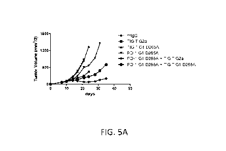
in
combination with other immunomodulatory therapy, on tumor growth in a mouse
model.
FIG. 5A shows tumor volume (cubic millimeters), calculated by multiplying the
square of
the width of the tumor by half the length, in a CT26 mouse colon cancer model
for mice
treated with an anti-mouse TIGIT antibody having an effector function enabled
murine
IgG2a Fc domain ("TIGIT G2a"), an anti-mouse TIGIT antibody having an effector
function deficient IgG1 D265A Fc domain ("TIGIT G1 D265A"), an anti-mouse PD-1
antibody having an effector function deficient IgG1 D265A Fc domain ("PD-1 G1
D265A"), combinations thereof, or a control IgG1 antibody. FIG. 5B shows the
effects of
anti-TIGIT monotherapy, as well as combination therapy with anti-PD-1 and anti-
CTLA-4
antibodies. Tumor volumes are provided along with the number of tumor-free
(TF) mice in
each group of ten mice at the end of the experiment. Each line represents one
mouse.
mIgG1 isotype control gave no tumor free mice, as did anti-TIGIT as
monotherapy. Anti-
PD-1 gave one tumor-free mouse as monotherapy, and five when combined with
anti-
TIGIT. Anti-CTLA-4 gave three tumor-free mice as monotherapy, and six when
combined
with anti-TIGIT. See Example 7.
[0053] FIGs. 6A and 6B show elevated PVR expression in cancer tissues. FIG.
6A
shows PVR mRNA expression in various tumor types as detected in The Cancer
Genome
Atlas (TCGA) datasets. Data are provided for adrenocortical carcinoma (ACC),
chromophobe renal cell carcinoma (KICH), liver hepatocellular carcinoma
(LIHC), colon
and rectal adenocarcinoma (COAD, READ), pancreatic ductal adenocarcinoma
(PAAD),
pheochromocytoma & paraganglioma (PCPG), papillary kidney carcinoma (KIRP),
lung
adenocarcinoma (LUAD), head and neck squamous cell carcinoma (HNSC), prostate
adenocarcinoma (PRAD), uterine corpus endometrial carcinoma (UCEC), cervical
cancer
11
CA 02971732 2017-06-20
WO 2016/106302 PCT/US2015/067332
(CESC), cutaneous melanoma (SKCM), mesothelioma (MESO), urothelial bladder
cancer
(BLCA), clear cell kidney carcinoma (KIRC), lung squamous cell carcinoma
(LUSC),
uterine carcinosarcoma (UCS), sarcoma (SARC), ovarian serous
cystadenocarcinoma (OV),
papillary thyroid carcinoma (THCA), glioblastoma multiforme (GBM), breast
cancer
(BRCA), lower grade glioma (LGG), and diffuse large B-cell lymphoma (DLBC).
The
results disclosed here are in whole or part based upon data generated by the
TCGA
Research Network. FIG. 6B shows human PVR in colon adenocarcinoma tissue as
compared to normal colon epithelium, with darker regions in the adenocarcinoma
sample
indicating elevated PVR expression. See Example 9.
[0054] FIG. 7 shows Fcy receptor binding, expressed as a percentage of the
theoretical
maximum receptor binding value (Rmax), for anti-TIGIT mAb 22G2 formatted as an
IgGlf
(SEQ ID NO: 45) or an IgG1.1f (SEQ ID NO: 48). Data are presented for six
different Fcy
receptors, for two different lots of IgG1.1f antibody, used at 1011M and l[tm,
as indicated.
Within each cluster of bars, the data for Fcy receptors are presented in
order, left to right:
hCD64 (FcyRI); hCD32a-H131 (FcyRIIA-H131); hCD32a-R131 (FcyRIIA-R131); hCD32b
(FcyRIII3); hCD16a-V158 (FcyRIIIA-V158); and hCD16b-NA2 (FcyRIIIB-NA2, where
NA2 designates the allotypic variant). Although pairs of Fcy receptors are
represented by
identical bars, their identities are clear from the order in which they are
presented. The
same decreases were observed for binding to cynomolgus monkey Fcy receptors
CD64,
CD32a, CD32b, and CD16 (not shown).
DETAILED DESCRIPTION
[0055] The present invention discloses isolated antibodies, particularly
monoclonal
antibodies, e.g., human monoclonal antibodies, that specifically bind to human
TIGIT
("huTIGIT") and block binding to PVR/CD155, thereby reducing or eliminating
the
immunosuppressive signal that would otherwise occur in the TIGIT-expressing
cells. The
present invention also provides isolated antibodies, particularly monoclonal
antibodies, e.g.,
human or humanized monoclonal antibodies, that specifically bind to human
TIGIT and
block the interaction of human TIGIT to DNAM-1/CD226 that would otherwise
prevent the
DNAM-1 homodimerization and thus DNAM-1-mediated co-stimulation. Sequences are
provided for various human anti-huTIGIT monoclonal antibodies. In certain
embodiments,
the antibodies described herein are derived from particular heavy and light
chain germline
sequences and/or comprise particular structural features such as CDR regions
comprising
particular amino acid sequences.
12
CA 02971732 2017-06-20
WO 2016/106302 PCT/US2015/067332
[0056] Further provided herein are methods of making such antibodies,
immunoconjugates and bispecific molecules comprising such antibodies or
antigen-binding
fragments thereof, and pharmaceutical compositions formulated to contain the
antibodies or
fragments. Also provided herein are methods of using the antibodies for immune
response
enhancement, alone or in combination with other immunostimulatory agents
(e.g.,
antibodies) and/or cancer or anti-infective therapies. Accordingly, the anti-
huTIGIT
antibodies described herein may be used in a treatment in a wide variety of
therapeutic
applications, including, for example, inhibiting tumor growth and treating
chronic viral
infections.
Definitions
[0057] In order that the present description may be more readily
understood, certain
terms are first defined. Additional definitions are set forth throughout the
detailed
description.
[0058] TIGIT refers to "T cell immunoreceptor with Ig and ITIM domains," a
member
of the PVR (poliovirus receptor) family of immunoglobin proteins, which binds
to
PVR/CD155 and Nectin-2/CD112. TIGIT is also referred to as TIGIT, WUCAM, Vstm3
and Vsig9. Unless otherwise indicated, or clear from the context, references
to TIGIT
herein refer to human TIGIT ("huTIGIT"), and anti-TIGIT antibodies refer to
anti-human
TIGIT antibodies. Human TIGIT is further described at GENE ID NO: 201633 and
MIM
(Mendelian Inheritance in Man): 612859. The sequence of human TIGIT (NP
776160.2),
including 21 amino acid signal sequence, is provided at SEQ ID NO: 1. Unless
otherwise
indicated, or clear from the context, "inhibition" of TIGIT refers to blocking
of PVR
binding and signaling. Anti-TIGIT antibodies of the present invention may act
by inhibition
of TIGIT signaling, blockade of TIGIT/DNAM-1 interaction and/or other
mechanisms, such
as directing the depletion of regulatory T cells.
[0059] PVR (poliovirus receptor) interacts with TIGIT to induce an
immunosuppressive
signal. PVR is also referred to as PVS; HVED; CD155; NECL5; TAGE4; Ned-S.
Unless
otherwise indicated, or clear from the context, references to PVR/CD155 herein
refer to
human PVR ("huPVR"). Human PVR is further described at GENE ID NO: 5817 and
MIM: 173850. There are four known human PVR transcript variants: alpha
(NP 006496.4), beta (NP 001129240.1), gamma (NP 001129241.1) and delta
(NP 001129242.2), the sequences of which are provided at SEQ ID NOs: 50 ¨ 53.
Unless
13
CA 02971732 2017-06-20
WO 2016/106302 PCT/US2015/067332
otherwise indicated, reference to PVR or human PVR relates to the alpha
transcript
polypeptide.
[0060] Unless otherwise indicated or clear from the context, the term
"antibody" as
used to herein may include whole antibodies and any antigen binding fragments
(i.e.,
"antigen-binding portions") or single chains thereof An "antibody" refers, in
one
embodiment, to a glycoprotein comprising at least two heavy (H) chains and two
light (L)
chains inter-connected by disulfide bonds, or an antigen binding fragment
thereof. Each
heavy chain is comprised of a heavy chain variable region (abbreviated herein
as VH) and a
heavy chain constant region. In certain naturally occurring IgG, IgD and IgA
antibodies,
the heavy chain constant region is comprised of three domains, CH1, CH2 and
CH3. In
certain naturally occurring antibodies, each light chain is comprised of a
light chain variable
region (abbreviated herein as VL) and a light chain constant region. The light
chain constant
region is comprised of one domain, CL. The VH and VL regions can be further
subdivided
into regions of hypervariability, termed complementarity determining regions
(CDR),
interspersed with regions that are more conserved, termed framework regions
(FR). Each
VH and VL is composed of three CDRs and four framework regions (FRs), arranged
from
amino-terminus to carboxy-terminus in the following order: FR1, CDR1, FR2,
CDR2, FR3,
CDR3, FR4. The variable regions of the heavy and light chains contain a
binding domain
that interacts with an antigen. The constant regions of the antibodies may
mediate the
binding of the immunoglobulin to host tissues or factors, including various
cells of the
immune system (e.g., effector cells) and the first component (Clq) of the
classical
complement system.
[0061] Antibodies typically bind specifically to their cognate antigen with
high affinity,
reflected by a dissociation constant (KD) of 10' to 10-11 M or less. Any KD
greater than
about 10' M is generally considered to indicate nonspecific binding. As used
herein, an
antibody that "binds specifically" to an antigen refers to an antibody that
binds to the
antigen and substantially identical antigens with high affinity, which means
having a KD of
10-7 M or less, preferably 10' M or less, even more preferably 5 x 10⁻⁹ M or
less, and most
preferably between 10' M and 10-10 M or less, but does not bind with high
affinity to
unrelated antigens. An antigen is "substantially identical" to a given antigen
if it exhibits a
high degree of sequence identity to the given antigen, for example, if it
exhibits at least
80%, at least 90%, preferably at least 95%, more preferably at least 97%, or
even more
preferably at least 99% sequence identity to the sequence of the given
antigen. By way of
example, an antibody that binds specifically to human TIGIT might also cross-
react with
14
CA 02971732 2017-06-20
WO 2016/106302 PCT/US2015/067332
TIGIT from certain non-human primate species (e.g., cynomolgus monkey), but
might not
cross-react with TIGIT from other species, or with an antigen other than
TIGIT.
[0062] Antibodies may exhibit modifications at the N- and/or C-terminal
amino acid
residues. For example, antibodies of the present invention may be produced
from a
construct encoding a C-terminal lysine residue, for example on the heavy
chain, but such C-
terminal lysine may be partially or totally absent in the therapeutic antibody
that is sold or
administered. Alternatively, an antibody may be produced from constructs that
specifically
do not encode a C-terminal lysine residue even though such lysine was present
in the
parental antibody from which the therapeutic antibody was derived. In another
example, an
N-terminal glutamine or glutamic acid residue in an antibody of the present
invention may
be partially or fully converted to pyro-glutamic acid in the therapeutic
antibody that is sold
or administered. Any form of glutamine or glutamic acid present at the N-
terminus of an
antibody chain, including pyro-glutamic acid, is encompassed within the term
"glutamine"
as used herein. Accordingly, antibody chain sequences provided herein having N-
terminal
glutamine or glutamic acid residue encompass antibody chains regardless of the
level of
pyro-glutamic acid formation.
[0063] Unless otherwise indicated, an immunoglobulin may be from any of the
commonly known isotypes, including but not limited to IgA, secretory IgA, IgG
and IgM.
The IgG isotype is divided in subclasses in certain species: IgGl, IgG2, IgG3
and IgG4 in
humans, and IgGl, IgG2a, IgG2b and IgG3 in mice. Immunoglobulins, e.g., human
IgGl,
exist in several allotypes, which differ from each other in at most a few
amino acids.
Unless otherwise indicated, "antibody" may include, by way of example,
monoclonal and
polyclonal antibodies; chimeric and humanized antibodies; human and non-human
antibodies; wholly synthetic antibodies; and single chain antibodies.
[0064] The term "antigen-binding portion" or "antigen binding fragment" of
an
antibody, as used herein, refers to one or more fragments of an antibody that
retain the
ability to specifically bind to an antigen (e.g., human TIGIT). Examples of
binding
fragments encompassed within the term "antigen-binding portion/fragment" of an
antibody
include (i) a Fab fragment - a monovalent fragment consisting of the VL, VH,
CL and CH1
domains; (ii) a F(a1302 fragment - a bivalent fragment comprising two Fab
fragments linked
by a disulfide bridge at the hinge region; (iii) a Fd fragment consisting of
the VH and CH1
domains; (iv) a Fv fragment consisting of the VL and VH domains of a single
arm of an
antibody, and (v) a dAb fragment (Ward et al., (1989) Nature 341:544-546)
consisting of a
CA 02971732 2017-06-20
WO 2016/106302 PCT/US2015/067332
VH domain. An isolated complementarity determining region (CDR), or a
combination of
two or more isolated CDRs joined by a synthetic linker, may comprise and
antigen binding
domain of an antibody if able to bind antigen.
[0065] Single chain antibody constructs are also included in the invention.
Although
the two domains of the Fv fragment, VL and VH, are coded for by separate
genes, they can
be joined, using recombinant methods, by a synthetic linker that enables them
to be made as
a single protein chain in which the VL and VH regions pair to form monovalent
molecules
known as single chain Fv (scFv); see e.g., Bird et al. (1988) Science 242:423-
426; and
Huston et al. (1988) Proc. Natl. Acad. Sci. USA 85:5879-5883). Such single
chain
antibodies are also intended to be encompassed within the term "antigen-
binding
portion/fragment" of an antibody. These and other potential constructs are
described at
Chan & Carter (2010) Nat. Rev. Immunol. 10:301. These antibody fragments are
obtained
using conventional techniques known to those with skill in the art, and the
fragments are
screened for utility in the same manner as are intact antibodies. Antigen-
binding
portions/fragments can be produced by recombinant DNA techniques, or by
enzymatic or
chemical cleavage of intact immunoglobulins.
[0066] Unless otherwise indicated, the word "fragment" when used with
reference to an
antibody, such as in a claim, refers to an antigen binding fragment of the
antibody, such that
"antibody or fragment" has the same meaning as "antibody or antigen binding
fragment
thereof"
[0067] A "bispecific" or "bifunctional antibody" is an artificial hybrid
antibody having
two different heavy/light chain pairs, giving rise to two antigen binding
sites with
specificity for different antigens. Bispecific antibodies can be produced by a
variety of
methods including fusion of hybridomas or linking of Fab' fragments. See,
e.g., Songsivilai
& Lachmann (1990) Cl/n. Exp. Immunol. 79:315; Kostelny et al. (1992)1 Immunol.
148:1547.
[0068] The term "monoclonal antibody," as used herein, refers to an
antibody that
displays a single binding specificity and affinity for a particular epitope or
a composition of
antibodies in which all antibodies display a single binding specificity and
affinity for a
particular epitope. Typically such monoclonal antibodies will be derived from
a single cell
or nucleic acid encoding the antibody, and will be propagated without
intentionally
introducing any sequence alterations. Accordingly, the term "human monoclonal
antibody"
refers to a monoclonal antibody that has variable and optional constant
regions derived from
human germline immunoglobulin sequences. In one embodiment, human monoclonal
16
CA 02971732 2017-06-20
WO 2016/106302 PCT/US2015/067332
antibodies are produced by a hybridoma, for example, obtained by fusing a B
cell obtained
from a transgenic or transchromosomal non-human animal (e.g., a transgenic
mouse having
a genome comprising a human heavy chain transgene and a light chain
transgene), to an
immortalized cell.
[0069] The term "recombinant human antibody," as used herein, includes all
human
antibodies that are prepared, expressed, created or isolated by recombinant
means, such as
(a) antibodies isolated from an animal (e.g., a mouse) that is transgenic or
transchromosomal for human immunoglobulin genes or a hybridoma prepared
therefrom,
(b) antibodies isolated from a host cell transformed to express the antibody,
e.g., from a
transfectoma, (c) antibodies isolated from a recombinant, combinatorial human
antibody
library, and (d) antibodies prepared, expressed, created or isolated by any
other means that
involve splicing of human immunoglobulin gene sequences to other DNA
sequences. Such
recombinant human antibodies comprise variable and constant regions that
utilize particular
human germline immunoglobulin sequences are encoded by the germline genes, but
include
subsequent rearrangements and mutations that occur, for example, during
antibody
maturation. As known in the art (see, e.g., Lonberg (2005) Nature Biotech.
23(9):1117-
1125), the variable region contains the antigen binding domain, which is
encoded by
various genes that rearrange to form an antibody specific for a foreign
antigen. In addition
to rearrangement, the variable region can be further modified by multiple
single amino acid
changes (referred to as somatic mutation or hypermutation) to increase the
affinity of the
antibody to the foreign antigen. The constant region will change in further
response to an
antigen (i.e., isotype switch). Therefore, the rearranged and somatically
mutated nucleic
acid sequences that encode the light chain and heavy chain immunoglobulin
polypeptides in
response to an antigen may not be identical to the original germline
sequences, but instead
will be substantially identical or similar (i.e., have at least 80% identity).
[0070] A "human" antibody (HuMAb) refers to an antibody having variable
regions in
which both the framework and CDR regions are derived from human germline
immunoglobulin sequences. Furthermore, if the antibody contains a constant
region, the
constant region also is derived from human germline immunoglobulin sequences.
Human
antibodies of the present invention may include amino acid residues not
encoded by human
germline immunoglobulin sequences (e.g., mutations introduced by random or
site-specific
mutagenesis in vitro or by somatic mutation in vivo). However, the term "human
antibody,"
as used herein, is not intended to include antibodies in which CDR sequences
derived from
the germline of another mammalian species, such as a mouse, have been grafted
onto
17
CA 02971732 2017-06-20
WO 2016/106302 PCT/US2015/067332
human framework sequences. The terms "human" antibodies and "fully human"
antibodies
are used synonymously.
[0071] A "humanized" antibody refers to an antibody in which some, most or
all of the
amino acids outside the CDR domains of a non-human antibody, e.g. a mouse
antibody, are
replaced with corresponding amino acids derived from human immunoglobulins. In
one
embodiment of a humanized form of an antibody, some, most or all of the amino
acids
outside the CDR domains have been replaced with amino acids from human
immunoglobulins, whereas some, most or all amino acids within one or more CDR
regions
are unchanged. Small additions, deletions, insertions, substitutions or
modifications of
amino acids are permissible as long as they do not abrogate the ability of the
antibody to
bind to a particular antigen. A "humanized" antibody retains an antigenic
specificity similar
to that of the original antibody.
[0072] A "chimeric antibody" refers to an antibody in which the variable
regions are
derived from one species and the constant regions are derived from another
species, such as
an antibody in which the variable regions are derived from a mouse antibody
and the
constant regions are derived from a human antibody. A "hybrid" antibody refers
to an
antibody having heavy and light chains of different types, such as a mouse
(parental) heavy
chain and a humanized light chain, or vice versa.
[0073] As used herein, "isotype" refers to the antibody class (e.g., IgGl,
IgG2, IgG3,
IgG4, IgM, IgAl, IgA2, IgD, and IgE antibody) that is encoded by the heavy
chain constant
region genes.
[0074] "Allotype" refers to naturally occurring variants within a specific
isotype group,
which variants differ in one or a few amino acids. See, e.g., Jefferis et at.
(2009) mAbs 1:1.
[0075] The phrases "an antibody recognizing an antigen" and "an antibody
specific for
an antigen" are used interchangeably herein with the term "an antibody that
binds
specifically to an antigen."
[0076] An "isolated antibody," as used herein, refers to an antibody that
is substantially
free of other antibodies having different antigenic specificities (e.g., an
isolated antibody
that specifically binds to TIGIT is substantially free of antibodies that
specifically bind
antigens other than TIGIT). An isolated antibody that specifically binds to an
epitope of
human TIGIT may, however, have cross-reactivity to other TIGIT proteins from
different
species.
[0077] As used herein, an antibody that "inhibits binding of PVR to TIGIT"
refers to an
antibody that inhibits the binding of human PVR to human TIGIT with an EC50 of
about 1
18
CA 02971732 2017-06-20
WO 2016/106302 PCT/US2015/067332
[tg/mL or less, such as about 0.9 [tg/mL or less, about 0.85 [tg/mL or less,
about 0.8 [tg/mL
or less, about 0.75 [tg/mL or less, about 0.7 [tg/mL or less, about 0.65
[tg/mL or less, about
0.6 [tg/mL or less, about 0.55 [tg/mL or less, about 0.5 [tg/mL or less, about
0.45 [tg/mL or
less, about 0.4 [tg/mL or less, about 0.35 [tg/mL or less, about 0.3 [tg/mL or
less, about 0.25
[tg/mL or less, about 0.2 [tg/mL or less, about 0.15 [tg/mL or less, or about
0.1 [tg/mL or
less, in art-recognized methods, e.g., in a FACS-based cell-binding assay.
[0078] "Effector functions," deriving from the interaction of an antibody
Fc region with
certain Fc receptors, include but are not necessarily limited to Clq binding,
complement
dependent cytotoxicity (CDC), Fc receptor binding, FcyR-mediated effector
functions such
as ADCC and antibody dependent cell-mediated phagocytosis (ADCP), and down
regulation of a cell surface receptor (e.g., the B cell receptor; BCR). Such
effector
functions generally require the Fc region to be combined with an antigen
binding domain
(e.g., an antibody variable domain).
[0079] An "Fc receptor" or "FcR" is a receptor that binds to the Fc region
of an
immunoglobulin. FcRs that bind to an IgG antibody comprise receptors of the
FcyR family,
including allelic variants and alternatively spliced forms of these receptors.
The FcyR
family consists of three activating (FcyRI, FcyRIII, and FcyRIV in mice;
FcyRIA, FcyRIIA,
and FcyRIIIA in humans) and one inhibitory (FcyRIIb, or equivalently FcyRIII3)
receptor.
Various properties of human FcyRs are summarized in Table 1. The majority of
innate
effector cell types co-express one or more activating FcyR and the inhibitory
FcyRIIb,
whereas natural killer (NK) cells selectively express one activating Fc
receptor (FcyRIII in
mice and FcyRIIIA in humans) but not the inhibitory FcyRIIb in mice and
humans. Human
IgG1 binds to most human Fc receptors and is considered equivalent to murine
IgG2a with
respect to the types of activating Fc receptors that it binds to.
TABLE 1
Properties of Human FcyRs
Fcy Allelic Affinity for Isotype preference
Cellular distribution
variants human IgG
FcyRI None High (KD ¨10 IgG1=3>4>>2 Monocytes, macrophages,
described nM) activated neutrophils,
dendritic
cells?
FcyRIIA H131 Low to medium IgGl>3>2>4 Neutrophils, monocytes,
macrophages, eosinophils,
dendritic cells, platelets
R131 Low IgGl>3>4>2
FcyRIIIA V158 Medium IgG1=3>>4>2 NK cells, monocytes,
19
CA 02971732 2017-06-20
WO 2016/106302 PCT/US2015/067332
Fcy Allelic Affinity for Isotype preference Cellular
distribution
variants human IgG
F158 Low IgG1=3>>4>2 macrophages, mast cells,
eosinophils, dendritic cells?
FcyRIIb 1232 Low IgG1=3=4>2 B cells, monocytes,
T232 Low IgG1=3=4>2 macrophages, dendritic
cells,
mast cells
[0080] An "Fe region" (fragment crystallizable region) or "Fe domain" or
"Fe" refers to
the C-terminal region of the heavy chain of an antibody that mediates the
binding of the
immunoglobulin to host tissues or factors, including binding to Fe receptors
located on
various cells of the immune system (e.g., effector cells) or to the first
component (Clq) of
the classical complement system. Thus, an Fe region comprises the constant
region of an
antibody excluding the first constant region immunoglobulin domain (e.g., CH1
or CL). In
IgG, IgA and IgD antibody isotypes, the Fe region comprises CH2 and CH3
constant domains
in each of the antibody's two heavy chains; IgM and IgE Fe regions comprise
three heavy
chain constant domains (CH domains 2-4) in each polypeptide chain. For IgG,
the Fe region
comprises immunoglobulin domains Cy2 and Cy3 and the hinge between Cyl and
Cy2.
Although the boundaries of the Fe region of an immunoglobulin heavy chain
might vary,
the human IgG heavy chain Fe region is usually defined to stretch from an
amino acid
residue at position C226 or P230 (or an amino acid between these two amino
acids) to the
carboxy-terminus of the heavy chain, wherein the numbering is according to the
EU index
as in Kabat. Kabat et at. (1991) Sequences of Proteins of Immunological
Interest, National
Institutes of Health, Bethesda, MD; see also FIGs. 3c-3f of U.S. Pat. App.
Pub.
No. 2008/0248028. The CH2 domain of a human IgG Fe region extends from about
amino
acid 231 to about amino acid 340, whereas the CH3 domain is positioned on C-
terminal side
of a CH2 domain in an Fe region, i.e., it extends from about amino acid 341 to
about amino
acid 447 of an IgG (including a C-terminal lysine). As used herein, the Fe
region may be a
native sequence Fe, including any allotypic variant, or a variant Fe (e.g., a
non-naturally
occurring Fe). Fe may also refer to this region in isolation or in the context
of an Fe-
comprising protein polypeptide such as a "binding protein comprising an Fe
region," also
referred to as an "Fe fusion protein" (e.g., an antibody or immunoadhesin).
[0081] Unless otherwise indicated, or clear from the context, amino acid
residue
numbering in the Fe region of an antibody is according to the EU numbering
convention,
except when specifically referring to residues in a sequence in the Sequence
Listing, in
which case numbering is necessarily consecutive. For example, literature
references
CA 02971732 2017-06-20
WO 2016/106302 PCT/US2015/067332
regarding the effects of amino acid substitutions in the Fc region will
typically use EU
numbering, which allows for reference to any given residue in the Fc region of
an antibody
by the same number regardless of the length of the variable domain to which is
it attached.
In rare cases it may be necessary to refer to the document being referenced to
confirm the
precise Fc residue being referred to.
[0082] A "native sequence Fc region" or "native sequence Fc" comprises an
amino acid
sequence that is identical to the amino acid sequence of an Fc region found in
nature.
Native sequence human Fc regions include a native sequence human IgG1 Fc
region; native
sequence human IgG2 Fc region; native sequence human IgG3 Fc region; and
native
sequence human IgG4 Fc region as well as naturally occurring variants thereof
Native
sequence Fc include the various allotypes of Fcs. See, e.g., Jefferis et al.
(2009) mAbs 1:1.
[0083] The term "epitope" or "antigenic determinant" refers to a site on an
antigen (e.g.,
TIGIT) to which an immunoglobulin or antibody specifically binds. Epitopes
within
protein antigens can be formed both from contiguous amino acids (usually a
linear epitope)
or noncontiguous amino acids juxtaposed by tertiary folding of the protein
(usually a
conformational epitope). Epitopes formed from contiguous amino acids are
typically, but
not always, retained on exposure to denaturing solvents, whereas epitopes
formed by
tertiary folding are typically lost on treatment with denaturing solvents. An
epitope
typically includes at least 3, 4, 5, 6, 7, 8, 9, 10, 11, 12, 13, 14 or 15
amino acids in a unique
spatial conformation.
[0084] The term "epitope mapping" refers to the process of identification
of the
molecular determinants on the antigen involved in antibody-antigen
recognition. Methods
for determining what epitopes are bound by a given antibody are well known in
the art and
include, for example, immunoblotting and immunoprecipitation assays, wherein
overlapping or contiguous peptides from (e.g., from TIGIT) are tested for
reactivity with a
given antibody (e.g., anti-TIGIT antibody); x-ray crystallography; 2-
dimensional nuclear
magnetic resonance; yeast display (see Example 4 herein); and HDX-MS (see,
e.g., Epitope
Mapping Protocols in Methods in Molecular Biology, Vol. 66, G. E. Morris, Ed.
(1996)).
[0085] The term "binds to the same epitope" with reference to two or more
antibodies
means that the antibodies bind to the same segment of amino acid residues, as
determined
by a given method. Techniques for determining whether antibodies bind to the
"same
epitope on TIGIT" with the antibodies described herein include, for example,
epitope
mapping methods, such as, x-ray analyses of crystals of antigen:antibody
complexes, which
provides atomic resolution of the epitope, and hydrogen/deuterium exchange
mass
21
CA 02971732 2017-06-20
WO 2016/106302 PCT/US2015/067332
spectrometry (HDX-MS). Other methods monitor the binding of the antibody to
antigen
fragments (e.g. proteolytic fragments) or to mutated variations of the antigen
where loss of
binding due to a modification of an amino acid residue within the antigen
sequence is often
considered an indication of an epitope component, such as alanine scanning
mutagenesis
(Cunningham & Wells (1985) Science 244:1081) or yeast display of mutant target
sequence
variants (see Example 4 herein). In addition, computational combinatorial
methods for
epitope mapping can also be used. These methods rely on the ability of the
antibody of
interest to affinity isolate specific short peptides from combinatorial phage
display peptide
libraries. Antibodies having the same or closely related VH and VL or the same
CDR
sequences are expected to bind to the same epitope.
[0086] Antibodies that "compete with another antibody for binding to a
target" refer to
antibodies that inhibit (partially or completely) the binding of the other
antibody to the
target. Whether two antibodies compete with each other for binding to a
target, i.e.,
whether and to what extent one antibody inhibits the binding of the other
antibody to a
target, may be determined using known competition experiments. In certain
embodiments,
an antibody competes with, and inhibits binding of another antibody to a
target by at least
10%, 20%, 30%, 40%, 50%, 60%, 70%, 80%, 90% or 100%. The level of inhibition
or
competition may be different depending on which antibody is the "blocking
antibody" (i.e.,
the cold antibody that is incubated first with the target). Competition assays
can be
conducted as described, for example, in Ed Harlow and David Lane, Cold Spring
Harb.
Protoc.; 2006; doi:10.1101/pdb.prot4277 or in Chapter 11 of "Using Antibodies"
by Ed
Harlow and David Lane, Cold Spring Harbor Laboratory Press, Cold Spring
Harbor, NY,
USA 1999. Competing antibodies bind to the same epitope, an overlapping
epitope or to
adjacent epitopes (e.g., as evidenced by steric hindrance).
[0087] Other competitive binding assays include: solid phase direct or
indirect
radioimmunoassay (RIA), solid phase direct or indirect enzyme immunoassay
(ETA),
sandwich competition assay (see Stahli et at. (1983) Methods in Enzymology
9:242); solid
phase direct biotin-avidin ETA (see Kirkland et at. (1986) J Immunol.
137:3614); solid
phase direct labeled assay, solid phase direct labeled sandwich assay (see
Harlow and Lane
(1988), Antibodies: A Laboratory Manual, Cold Spring Harbor Press); solid
phase direct
label RIA using 1-125 label (see Morel et at. (1988) Mot. Immunol. 25(1):7);
solid phase
direct biotin-avidin ETA (Cheung et at. (1990) Virology 176:546); and direct
labeled RIA.
(Moldenhauer et at. (1990) Scand. I Immunol. 32:77).
22
CA 02971732 2017-06-20
WO 2016/106302 PCT/US2015/067332
[0088] As used herein, the terms "specific binding," "selective binding,"
"selectively
binds," and "specifically binds," refer to antibody binding to an epitope on a
predetermined
antigen but not to other antigens. Typically, the antibody (i) binds with an
equilibrium
dissociation constant (KD) of approximately less than 10' M, such as
approximately less
than 10 -8 M, 10-9 M or 10-10 M or even lower when determined by, e.g.,
surface plasmon
resonance (SPR) technology in a BIACORE 2000 surface plasmon resonance
instrument
using the predetermined antigen, e.g., recombinant human TIGIT, as the analyte
and the
antibody as the ligand, or Scatchard analysis of binding of the antibody to
antigen positive
cells, and (ii) binds to the predetermined antigen with an affinity that is at
least two-fold
greater than its affinity for binding to a non-specific antigen (e.g., BSA,
casein) other than
the predetermined antigen or a closely-related antigen. Accordingly, an
antibody that
"specifically binds to human TIGIT" refers to an antibody that binds to
soluble or cell
bound human TIGIT with a KD of 10' M or less, such as approximately less than
10 -8 M,
10-9 M or 10-10 M or even lower. An antibody that "cross-reacts with
cynomolgus TIGIT"
refers to an antibody that binds to cynomolgus TIGIT with a KD of 10' M or
less, such as
approximately less than 10 -8 M, 10-9 M or 10-10 M or even lower.
[0089] The term "kassoc" or "ka", as used herein, refers to the association
rate constant of
a particular antibody-antigen interaction, whereas the term "Ls" or "lcd," as
used herein,
refers to the dissociation rate constant of a particular antibody-antigen
interaction. The term
"Kip", as used herein, refers to the equilibrium dissociation constant, which
is obtained from
the ratio of lcd to ka (i.e.,. kdka) and is expressed as a molar concentration
(M). KD values
for antibodies can be determined using methods well established in the art.
Preferred
methods for determining the KD of an antibody include biolayer interferometry
(BLI)
analysis, preferably using a Fortebio Octet RED device, surface plasmon
resonance,
preferably using a biosensor system such as a BIACORE surface plasmon
resonance
system (see e.g. Example 2), or flow cytometry and Scatchard analysis.
[0090] As used herein, the term "high affinity" for an IgG antibody refers
to an
antibody having a KD of 10' M or less, more preferably 10' M or less and even
more
preferably 10-10 M or less for a target antigen. However, "high affinity"
binding can vary
for other antibody isotypes. For example, "high affinity" binding for an IgM
isotype refers
to an antibody having a KD of 10' M or less, more preferably 10-8 M or less.
[0091] The term "EC50" in the context of an in vitro or in vivo assay using
an antibody
or antigen binding fragment thereof, refers to the concentration of an
antibody or an
23
CA 02971732 2017-06-20
WO 2016/106302 PCT/US2015/067332
antigen-binding fragment thereof that induces a response that is 50% of the
maximal
response, i.e., halfway between the maximal response and the baseline.
[0092] The term "binds to immobilized TIGIT" refers to the ability of an
antibody
described herein to bind to TIGIT, for example, expressed on the surface of a
cell or
attached to a solid support.
[0093] The term "cross-reacts," as used herein, refers to the ability of an
antibody
described herein to bind to TIGIT from a different species. For example, an
antibody
described herein that binds human TIGIT may also bind TIGIT from another
species (e.g.,
cynomolgus TIGIT). As used herein, cross-reactivity may be measured by
detecting a
specific reactivity with purified antigen in binding assays (e.g., SPR, ELISA)
or binding to,
or otherwise functionally interacting with, cells physiologically expressing
TIGIT. Methods
for determining cross-reactivity include standard binding assays as described
herein, for
example, by BIACORE surface plasmon resonance (SPR) analysis using a BIACORE
2000 SPR instrument (Biacore AB, Uppsala, Sweden), or flow cytometric
techniques.
[0094] The term "naturally-occurring" as used herein as applied to an
object refers to
the fact that an object can be found in nature. For example, a polypeptide or
polynucleotide
sequence that is present in an organism (including viruses) that can be
isolated from a
source in nature and which has not been intentionally modified by man in the
laboratory is
naturally-occurring.
[0095] A "polypeptide" refers to a chain comprising at least two
consecutively linked
amino acid residues, with no upper limit on the length of the chain. One or
more amino acid
residues in the protein may contain a modification such as, but not limited
to, glycosylation,
phosphorylation or a disulfide bond. A "protein" may comprise one or more
polypeptides.
[0096] The term "nucleic acid molecule," as used herein, is intended to
include DNA
molecules and RNA molecules. A nucleic acid molecule may be single-stranded or
double-
stranded, and may be cDNA.
[0097] Also provided are "conservative sequence modifications" to the
antibody
sequence provided herein, i.e. nucleotide and amino acid sequence
modifications that do not
abrogate the binding of the antibody encoded by the nucleotide sequence or
containing the
amino acid sequence, to the antigen. For example, modifications can be
introduced by
standard techniques known in the art, such as site-directed mutagenesis and
PCR-mediated
mutagenesis. Conservative sequence modifications include conservative amino
acid
substitutions, in which the amino acid residue is replaced with an amino acid
residue having
a similar side chain. Families of amino acid residues having similar side
chains have been
24
CA 02971732 2017-06-20
WO 2016/106302 PCT/US2015/067332
defined in the art. These families include amino acids with basic side chains
(e.g., lysine,
arginine, histidine), acidic side chains (e.g., aspartic acid, glutamic acid),
uncharged polar
side chains (e.g., glycine, asparagine, glutamine, serine, threonine,
tyrosine, cysteine,
tryptophan), nonpolar side chains (e.g., alanine, valine, leucine, isoleucine,
proline,
phenylalanine, methionine), beta-branched side chains (e.g., threonine,
valine, isoleucine)
and aromatic side chains (e.g., tyrosine, phenylalanine, tryptophan,
histidine). Thus, a
predicted nonessential amino acid residue in an anti-TIGIT antibody is
preferably replaced
with another amino acid residue from the same side chain family. Methods of
identifying
nucleotide and amino acid conservative substitutions that do not eliminate
antigen binding
are well-known in the art. See, e.g., Brummell et at., Biochem. 32:1180-1187
(1993);
Kobayashi et at. Protein Eng. 12(10):879-884 (1999); and Burks et at. Proc.
Natl. Acad.
Sci. USA 94:412-417 (1997)).
[0098] Alternatively, in another embodiment, mutations can be introduced
randomly
along all or part of an anti-TIGIT antibody coding sequence, such as by
saturation
mutagenesis, and the resulting modified anti-TIGIT antibodies can be screened
for
improved binding activity.
[0099] For nucleic acids, the term "substantial homology" indicates that
two nucleic
acids, or designated sequences thereof, when optimally aligned and compared,
are identical,
with appropriate nucleotide insertions or deletions, in at least about 80% of
the nucleotides,
usually at least about 90% to 95%, and more preferably at least about 98% to
99.5% of the
nucleotides. Alternatively, substantial homology exists when the segments will
hybridize
under selective hybridization conditions, to the complement of the strand.
[00100] For polypeptides, the term "substantial homology" indicates that two
polypeptides, or designated sequences thereof, when optimally aligned and
compared, are
identical, with appropriate amino acid insertions or deletions, in at least
about 80% of the
amino acids, usually at least about 90% to 95%, and more preferably at least
about 98% to
99.5% of the amino acids.
[00101] The percent identity between two sequences is a function of the number
of
identical positions shared by the sequences when the sequences are optimally
aligned (i.e.,
% homology = # of identical positions/total # of positions x 100), with
optimal alignment
determined taking into account the number of gaps, and the length of each gap,
which need
to be introduced for optimal alignment of the two sequences. The comparison of
sequences
and determination of percent identity between two sequences can be
accomplished using a
mathematical algorithm, as described in the non-limiting examples below.
CA 02971732 2017-06-20
WO 2016/106302 PCT/US2015/067332
[00102] The percent identity between two nucleotide sequences can be
determined using
the GAP program in the GCG software package, using a NWSgapdna.CMP matrix and
a
gap weight of 40, 50, 60, 70, or 80 and a length weight of 1, 2, 3, 4, 5, or
6. The percent
identity between two nucleotide or amino acid sequences can also be determined
using the
algorithm of E. Meyers and W. Miller (CABIOS, 4:11-17 (1989)) which has been
incorporated into the ALIGN program (version 2.0), using a PAM120 weight
residue table,
a gap length penalty of 12 and a gap penalty of 4. In addition, the percent
identity between
two amino acid sequences can be determined using the Needleman and Wunsch (J.
Mot.
Biol. (48):444-453 (1970)) algorithm which has been incorporated into the GAP
program in
the GCG software package, using either a Blossum 62 matrix or a PAM250 matrix,
and a
gap weight of 16, 14, 12, 10, 8, 6, or 4 and a length weight of 1, 2, 3, 4, 5,
or 6.
[00103] The nucleic acid and protein sequences described herein can further be
used as a
"query sequence" to perform a search against public databases to, for example,
identify
related sequences. Such searches can be performed using the NBLAST and )(BLAST
programs (version 2.0) of Altschul et al. (1990)1 Mot. Biol. 215:403-10. BLAST
nucleotide searches can be performed with the NBLAST program, score = 100,
wordlength
= 12 to obtain nucleotide sequences homologous to the nucleic acid molecules
described
herein. BLAST protein searches can be performed with the )(BLAST program,
score = 50,
wordlength = 3 to obtain amino acid sequences homologous to the protein
molecules
described herein. To obtain gapped alignments for comparison purposes, Gapped
BLAST
can be utilized as described in Altschul et at. (1997) Nucleic Acids Res.
25(17):3389-3402.
When utilizing BLAST and Gapped BLAST programs, the default parameters of the
respective programs (e.g., )03LAST and NBLAST) can be used.
[00104] The nucleic acids may be present in whole cells, in a cell lysate,
or in a partially
purified or substantially pure form. A nucleic acid is "isolated" or "rendered
substantially
pure" when purified away from other cellular components or other contaminants,
e.g., other
cellular nucleic acids (e.g., the other parts of the chromosome) or proteins,
by standard
techniques, including alkaline/SDS treatment, CsC1 banding, column
chromatography,
agarose gel electrophoresis and others well known in the art. See, F. Ausubel,
et al., ed.
Current Protocols in Molecular Biology, Greene Publishing and Wiley
Interscience, New
York (1987).
[00105] The term "vector," as used herein, is intended to refer to a nucleic
acid molecule
capable of transporting another nucleic acid to which it has been linked. One
type of vector
is a "plasmid," which refers to a circular double stranded DNA loop into which
additional
26
CA 02971732 2017-06-20
WO 2016/106302 PCT/US2015/067332
DNA segments may be ligated. Another type of vector is a viral vector, wherein
additional
DNA segments may be ligated into the viral genome. Certain vectors are capable
of
autonomous replication in a host cell into which they are introduced (e.g.,
bacterial vectors
having a bacterial origin of replication and episomal mammalian vectors).
Other vectors
(e.g., non-episomal mammalian vectors) can be integrated into the genome of a
host cell
upon introduction into the host cell, and thereby are replicated along with
the host genome.
Moreover, certain vectors are capable of directing the expression of genes to
which they are
operatively linked. Such vectors are referred to herein as "recombinant
expression vectors"
(or simply, "expression vectors"). In general, expression vectors of utility
in recombinant
DNA techniques are often in the form of plasmids. In the present
specification, "plasmid"
and "vector" may be used interchangeably as the plasmid is the most commonly
used form
of vector. However, also included are other forms of expression vectors, such
as viral
vectors (e.g., replication defective retroviruses, adenoviruses and adeno-
associated viruses),
which serve equivalent functions.
[00106] The term "recombinant host cell" (or simply "host cell"), as used
herein, is
intended to refer to a cell that comprises a nucleic acid that is not
naturally present in the
cell, and may be a cell into which a recombinant expression vector has been
introduced. It
should be understood that such terms are intended to refer not only to the
particular subject
cell but to the progeny of such a cell. Because certain modifications may
occur in
succeeding generations due to either mutation or environmental influences,
such progeny
may not, in fact, be identical to the parent cell, but are still included
within the scope of the
term "host cell" as used herein.
[00107] An "immune response" refers to a biological response within a
vertebrate against
foreign agents, which response protects the organism against these agents and
diseases
caused by them. An immune response is mediated by the action of a cell of the
immune
system (for example, a T lymphocyte, B lymphocyte, natural killer (NK) cell,
macrophage,
eosinophil, mast cell, dendritic cell or neutrophil) and soluble
macromolecules produced by
any of these cells or the liver (including antibodies, cytokines, and
complement) that results
in selective targeting, binding to, damage to, destruction of, and/or
elimination from the
vertebrate's body of invading pathogens, cells or tissues infected with
pathogens, cancerous
or other abnormal cells, or, in cases of autoimmunity or pathological
inflammation, normal
human cells or tissues. An immune reaction includes, e.g., activation or
inhibition of a T
cell, e.g., an effector T cell or a Th cell, such as a CDS+ or CD4+ T cell, or
the inhibition or
depletion of a Treg cell. "T effector" ("Tay") cells refers to T cells (e.g.,
CD4+ and CDS+ T
27
CA 02971732 2017-06-20
WO 2016/106302 PCT/US2015/067332
cells) with cytolytic activities as well as T helper (Th) cells, which secrete
cytokines and
activate and direct other immune cells, but does not include regulatory T
cells (Leg cells).
[00108] As used herein, the term "T cell-mediated response" refers to a
response
mediated by T cells, including effector T cells (e.g., CD8+ cells) and helper
T cells (e.g.,
CD4+ cells). T cell mediated responses include, for example, T cell
cytotoxicity and
proliferation.
[00109] As used herein, the term "cytotoxic T lymphocyte (CTL) response"
refers to an
immune response induced by cytotoxic T cells. CTL responses are mediated
primarily by
CD8+ T cells.
[00110] An "immunomodulator" or "immunoregulator" refers to an agent, e.g., a
component of a signaling pathway, that may be involved in modulating,
regulating, or
modifying an immune response. "Modulating," "regulating," or "modifying" an
immune
response refers to any alteration in a cell of the immune system or in the
activity of such cell
(e.g., an effector T cell). Such modulation includes stimulation or
suppression of the
immune system, which may be manifested by an increase or decrease in the
number of
various cell types, an increase or decrease in the activity of these cells, or
any other changes
that can occur within the immune system. Both inhibitory and stimulatory
immunomodulators have been identified, some of which may have enhanced
function in a
tumor microenvironment. In preferred embodiments, the immunomodulator is
located on
the surface of a T cell. An "immunomodulatory target" or "immunoregulatory
target" is an
immunomodulator that is targeted for binding by, and whose activity is altered
by the
binding of, a substance, agent, moiety, compound or molecule. Immunomodulatory
targets
include, for example, receptors on the surface of a cell ("immunomodulatory
receptors")
and receptor ligands ("immunomodulatory ligands").
[00111] "Immunotherapy" refers to the treatment of a subject afflicted with,
or at risk of
contracting or suffering a recurrence of, a disease by a method comprising
inducing,
enhancing, suppressing or otherwise modifying an immune response.
[00112] "Immunostimulating therapy" or "immunostimulatory therapy" refers to a
therapy that results in increasing (inducing or enhancing) an immune response
in a subject
for, e.g., treating cancer.
[00113] "Potentiating an endogenous immune response" means increasing the
effectiveness or potency of an existing immune response in a subject. This
increase in
effectiveness and potency may be achieved, for example, by overcoming
mechanisms that
28
CA 02971732 2017-06-20
WO 2016/106302 PCT/US2015/067332
suppress the endogenous host immune response or by stimulating mechanisms that
enhance
the endogenous host immune response.
[00114] As used herein, the term "linked" refers to the association of two or
more
molecules. The linkage can be covalent or non-covalent. The linkage also can
be genetic
(i.e., recombinantly fused). Such linkages can be achieved using a wide
variety of art
recognized techniques, such as chemical conjugation and recombinant protein
production.
[00115] As used herein, "administering" refers to the physical introduction of
a
composition comprising a therapeutic agent to a subject, using any of the
various methods
and delivery systems known to those skilled in the art. Preferred routes of
administration for
antibodies described herein include intravenous, intraperitoneal,
intramuscular,
subcutaneous, spinal or other parenteral routes of administration, for example
by injection
or infusion. The phrase "parenteral administration" as used herein means modes
of
administration other than enteral and topical administration, usually by
injection, and
includes, without limitation, intravenous, intraperitoneal, intramuscular,
intraarterial,
intrathecal, intralymphatic, intralesional, intracapsular, intraorbital,
intracardiac,
intradermal, transtracheal, subcutaneous, subcuticular, intraarticular,
subcapsular,
subarachnoid, intraspinal, epidural and intrasternal injection and infusion,
as well as in vivo
electroporation. Alternatively, an antibody described herein can be
administered via a non-
parenteral route, such as a topical, epidermal or mucosal route of
administration, for
example, intranasally, orally, vaginally, rectally, sublingually or topically.
Administering
can also be performed, for example, once, a plurality of times, and/or over
one or more
extended periods.
[00116] As used herein, the terms "inhibits" or "blocks" (e.g., referring
to
inhibition/blocking of binding of PVR to TIGIT on cells) are used
interchangeably and
encompass both partial and complete inhibition/blocking by, for example, at
least about
50%, 60%, 70%, 80%, 90%, 95%, 99%, or 100%.
[00117] As used herein, "cancer" refers a broad group of diseases
characterized by the
uncontrolled growth of abnormal cells in the body. Unregulated cell division
may result in
the formation of malignant tumors or cells that invade neighboring tissues and
may
metastasize to distant parts of the body through the lymphatic system or
bloodstream.
[00118] A "hematological malignancy" includes a lymphoma, leukemia, myeloma or
a
lymphoid malignancy, as well as a cancer of the spleen and the lymph nodes.
Exemplary
lymphomas include both B cell lymphomas and T cell lymphomas. B-cell lymphomas
include both Hodgkin's lymphomas and most non-Hodgkin's lymphomas. Non-
limiting
29
CA 02971732 2017-06-20
WO 2016/106302 PCT/US2015/067332
examples of B cell lymphomas include diffuse large B-cell lymphoma, follicular
lymphoma, mucosa-associated lymphatic tissue lymphoma, small cell lymphocytic
lymphoma (overlaps with chronic lymphocytic leukemia), mantle cell lymphoma
(MCL),
Burkitt's lymphoma, mediastinal large B cell lymphoma, Waldenstrom
macroglobulinemia,
nodal marginal zone B cell lymphoma, splenic marginal zone lymphoma,
intravascular
large B-cell lymphoma, primary effusion lymphoma, lymphomatoid granulomatosis.
Non-
limiting examples of T cell lymphomas include extranodal T cell lymphoma,
cutaneous T
cell lymphomas, anaplastic large cell lymphoma, and angioimmunoblastic T cell
lymphoma. Hematological malignancies also include leukemia, such as, but not
limited to,
secondary leukemia, chronic lymphocytic leukemia, acute myelogenous leukemia,
chronic
myelogenous leukemia, and acute lymphoblastic leukemia. Hematological
malignancies
further include myelomas, such as, but not limited to, multiple myeloma and
smoldering
multiple myeloma. Other hematological and/or B cell- or T-cell-associated
cancers are
encompassed by the term hematological malignancy.
[00119] The terms "treat," "treating," and "treatment," as used herein,
refer to any type
of intervention or process performed on, or administering an active agent to,
the subject
with the objective of reversing, alleviating, ameliorating, inhibiting, or
slowing down or
preventing the progression, development, severity or recurrence of a symptom,
complication, condition or biochemical indicia associated with a disease.
Prophylaxis refers
to administration to a subject who does not have a disease, to prevent the
disease from
occurring or minimize its effects if it does.
[00120] The term "effective dose" or "effective dosage" is defined as an
amount
sufficient to achieve or at least partially achieve a desired effect. A
"therapeutically
effective amount" or "therapeutically effective dosage" of a drug or
therapeutic agent is any
amount of the drug that, when used alone or in combination with another
therapeutic agent,
promotes disease regression evidenced by a decrease in severity of disease
symptoms, an
increase in frequency and duration of disease symptom-free periods, or a
prevention of
impairment or disability due to the disease affliction. A "prophylactically
effective amount"
or a "prophylactically effective dosage" of a drug is an amount of the drug
that, when
administered alone or in combination with another therapeutic agent to a
subject at risk of
developing a disease or of suffering a recurrence of disease, inhibits the
development or
recurrence of the disease. The ability of a therapeutic or prophylactic agent
to promote
disease regression or inhibit the development or recurrence of the disease can
be evaluated
using a variety of methods known to the skilled practitioner, such as in human
subjects
CA 02971732 2017-06-20
WO 2016/106302 PCT/US2015/067332
during clinical trials, in animal model systems predictive of efficacy in
humans, or by
assaying the activity of the agent in in vitro assays.
[00121] By way of example, an anti-cancer agent is a drug that slows cancer
progression
or promotes cancer regression in a subject. In preferred embodiments, a
therapeutically
effective amount of the drug promotes cancer regression to the point of
eliminating the
cancer. "Promoting cancer regression" means that administering an effective
amount of the
drug, alone or in combination with an anti-neoplastic agent, results in a
reduction in tumor
growth or size, necrosis of the tumor, a decrease in severity of at least one
disease symptom,
an increase in frequency and duration of disease symptom-free periods, a
prevention of
impairment or disability due to the disease affliction, or otherwise
amelioration of disease
symptoms in the patient. Pharmacological effectiveness refers to the ability
of the drug to
promote cancer regression in the patient. Physiological safety refers to an
acceptably low
level of toxicity, or other adverse physiological effects at the cellular,
organ and/or
organism level (adverse effects) resulting from administration of the drug.
[00122] By way of example for the treatment of tumors, a therapeutically
effective
amount or dosage of the drug preferably inhibits cell growth or tumor growth
by at least
about 20%, more preferably by at least about 40%, even more preferably by at
least about
60%, and still more preferably by at least about 80% relative to untreated
subjects. In the
most preferred embodiments, a therapeutically effective amount or dosage of
the drug
completely inhibits cell growth or tumor growth, i.e., preferably inhibits
cell growth or
tumor growth by 100%. The ability of a compound to inhibit tumor growth can be
evaluated
using the assays described infra. Inhibition of tumor growth may not be
immediate after
treatment, and may only occur after a period of time or after repeated
administration.
Alternatively, this property of a composition can be evaluated by examining
the ability of
the compound to inhibit cell growth, such inhibition can be measured in vitro
by assays
known to the skilled practitioner. In other preferred embodiments described
herein, tumor
regression may be observed and may continue for a period of at least about 20
days, more
preferably at least about 40 days, or even more preferably at least about 60
days.
[00123] "Combination" therapy, as used herein, unless otherwise clear from the
context,
is meant to encompass administration of two or more therapeutic agents in a
coordinated
fashion, and includes, but is not limited to, concurrent dosing. Specifically,
combination
therapy encompasses both co-administration (e.g. administration of a co-
formulation or
simultaneous administration of separate therapeutic compositions) and serial
or sequential
administration, provided that administration of one therapeutic agent is
conditioned in some
31
CA 02971732 2017-06-20
WO 2016/106302 PCT/US2015/067332
way on administration of another therapeutic agent. For example, one
therapeutic agent
may be administered only after a different therapeutic agent has been
administered and
allowed to act for a prescribed period of time. See, e.g., Kohrt et at. (2011)
Blood
117:2423.
[00124] The terms "patient" and "subject" refer to any human or non-human
animal that
receives either prophylactic or therapeutic treatment. For example, the
methods and
compositions described herein can be used to treat a subject having cancer.
The term "non-
human animal" includes all vertebrates, e.g., mammals and non-mammals, such as
non-
human primates, sheep, dog, cow, chickens, amphibians, reptiles, etc.
[00125] Various aspects described herein are described in further detail in
the following
subsections.
I. Anti-TIGIT Antibodies
[00126] The present application discloses fully human anti-huTIGIT
antibodies
having desirable properties for use as therapeutic agents in treating diseases
such as cancers.
These properties include one or more of the ability to bind to human TIGIT
with high
affinity, the ability to bind to cynomolgus monkey TIGIT, the ability to block
PVR binding
(and thus signaling), and the absence of sequence liabilities that might
reduce the chemical
stability of the antibody.
[00127] The anti-TIGIT antibodies disclosed herein by sequence bind to
specific
epitopes on human TIGIT determined as described in Example 4 and shown in
FIGS. 2A ¨
2C. The three specific antibodies for which epitopes were determined bind at
similar
regions of human TIGIT but differ in which specific amino acid residues are
contacted. The
antibodies share the properties of binding to human TIGIT with high affinity
and having the
ability to block PVR binding. Accordingly, other antibodies that bind to the
same or closely
related epitopes would likely share these desirable properties, and may be
discovered by
competition experiments.
[00128] In addition, antibody 22G2 binds to cynomolgus monkey TIGIT with
substantially the same affinity as it binds to human TIGIT, which is
convenient when it is
necessary to perform toxicity studies in support of regulatory approval for
use of the
antibody as a human therapeutic. Other anti-TIGIT antibodies that bind to the
same or
similar epitopes as 15A6 and 22G2 are likely to share this advantageous
property of binding
to cyno TIGIT. Antibodies binding to similar epitopes can be discovered by
doing
competition experiments or by determining their epitopes directly.
32
CA 02971732 2017-06-20
WO 2016/106302 PCT/US2015/067332
Anti-TIGIT antibodies that compete with anti-huTIGIT antibodies disclosed
herein
[00129] Anti-huTIGIT antibodies that compete with the antibodies of the
present
invention for binding to huTIGIT, such as 15A6 and 22G2, may be raised using
immunization protocols similar to those described herein (Example 1).
Antibodies that
compete for binding with the anti-huTIGIT antibodies described herein may also
be
generated immunizing mice may with human TIGIT or a construct comprising the
extracellular domain thereof (residues 22-141 of SEQ ID NO: 1; NP 776160.2),
or by
immunizing with a fragment of human TIGIT containing the epitope bound by the
anti-
huTIGIT antibodies disclosed herein (e.g. 15A6, 22G2 and 11G11). The resulting
antibodies can be screened for the ability to block binding of 15A6 or 22G2 to
human
TIGIT by methods well known in the art, for example blocking binding to fusion
protein of
the extracellular domain of TIGIT and an immunoglobulin Fc domain in a ELISA,
or
blocking the ability to bind to cells expressing huTIGIT on their surface,
e.g. by FACS. In
various embodiments, the test antibody is contacted with the TIGIT-Fc fusion
protein (or to
cells expressing huTIGIT on their surface) prior to, at the same time as, or
after the addition
of 15A6 or 22G2. For example, "binning" experiments may be performed (Example
3) to
determine whether an antibody falls into the same "bin" as antibodies 15A6 or
22G2, in
which experiments antibodies 15A6 or 22G2 are referred to as the "reference"
antibodies,
and the antibodies to be tested are referred to as the "test" antibodies.
Antibodies that
reduce binding of 15A6 and/or 22G2 to TIGIT (either as an Fc fusion or on a
cell),
particularly at roughly stoichiometric concentrations, are likely to bind at
the same,
overlapping, or adjacent epitopes, and thus may share the desirable functional
properties of
15A6 and 22G2.
[00130] Competing antibodies can also be identified using other methods
known in
the art. For example, standard ELISA assays or competitive ELISA assays can be
used in
which a recombinant human TIGIT protein construct is immobilized on the plate,
various
concentrations of unlabeled test antibody are added, the plate is washed,
labeled reference
antibody is added, washed, and the amount of bound label is measured. If the
increasing
concentration of the unlabeled test antibody inhibits the binding of the
labeled reference
antibody, the test antibody is said to inhibit the binding of the reference
antibody to the
target on the plate, or is said to compete with the binding of the reference
antibody.
Additionally or alternatively, BIACORE SPR analysis can be used to assess the
ability of
the antibodies to compete. The ability of a test antibody to inhibit the
binding of an anti-
33
CA 02971732 2017-06-20
WO 2016/106302 PCT/US2015/067332
huTIGIT antibody described herein to TIGIT demonstrates that the test antibody
can
compete with the reference antibody for binding to TIGIT.
[00131] Accordingly, provided herein are anti-TIGIT antibodies that inhibit
the binding
of an anti-huTIGIT antibodies described herein to TIGIT on cells, e.g.,
activated T cells, by
at least 10%, 20%, 30%, 40%, 50%, 55%, 60%, 65%, 70%, 75%, 80%, 85%, 90%, 91%,
92%, 93%, 94%, 95%, 96%, 97%, 98%, 99%, or by 100% and/or whose binding to
TIGIT on cells, e.g., activated T cells, is inhibited by at least 10%, 20%,
30%, 40%, 50%,
55%, 60%, 65%, 70%, 75%, 80%, 85%, 90%, 91%, 92%, 93%, 94%, 95%, 96%, 97%,
98%, 99%, or by 100%, e.g., as measured by ELISA or FACS, such as by using the
assay
described in the following paragraph.
[00132] An exemplary competition experiment to determine whether a test
antibody
blocks the binding of (i.e., "competes with") a reference antibody, may be
conducted as
follows: activated human T cells are prepared as follows: Peripheral Blood
Mononuclear
Cells (PBMCs) are isolated from human whole blood using Ficoll gradient and
activated
with 10 g/mL phytohaemagglutinin (PHA-L) (USBio1#P3370-30) and 200IU/mL
recombinant IL-2 (Peprotech#200-02) for 3 days. The activated T cells are
resuspended in
FACS buffer (PBS with 5% Fetal Bovine Serum) and seeded at 105 cells per
sample well in
a 96 well plate. Unconjugated test antibody is added to the plate at
concentrations ranging
from 0 to 50 i.tg/mL (three-fold titration starting from a highest
concentration of 50 pg/mL).
An unrelated IgG may be used as an isotype control for the test antibody and
added at the
same concentrations (three-fold titration starting from a highest
concentration of 50 g/mL).
A sample pre-incubated with 50 g/mL unlabeled reference antibody may be
included as a
positive control for complete blocking (100% inhibition) and a sample without
antibody in
the primary incubation may be used as a negative control (no competition; 0%
inhibition).
After 30 minutes of incubation, labeled, e.g., biotinylated, reference
antibody is added at a
concentration of 211g/mL per well without washing. Samples are incubated for
another 30
minutes. Unbound antibodies are removed by washing the cells with FACS buffer.
Cell-
bound labeled reference antibody is detected with an agent that detects the
label, e.g., PE
conjugated streptavidin (Invitrogen, catalog#521388) for detecting biotin. The
samples are
acquired on a FACS Calibur Flow Cytometer (BD, San Jose) and analyzed with
Flowjo
software (Tree Star, Inc, Ashland, OR). The results may be represented as the
% inhibition
(i.e., subtracting from 100% the amount of label at each concentration divided
by the
amount of label obtained with no blocking antibody).
34
CA 02971732 2017-06-20
WO 2016/106302 PCT/US2015/067332
[00133] Typically, the same experiment is then conducted in the reverse,
i.e., the test
antibody is the reference antibody and the reference antibody is the test
antibody. In certain
embodiments, an antibody at least partially (e.g., at least 10%, 20%, 30%,
40%, 50%, 60%,
70%, 80%, or 90%) or completely (100%) blocks the binding of the other
antibody to the
target, e.g. human TIGIT or fragment thereof, and regardless of whether
inhibition occurs
when one or the other antibody is the test antibody. A test and a reference
antibody "cross-
block" binding of each other to the target when the antibodies compete with
each other both
ways, i.e., in competition experiments in which the test antibody is added
first and in
competition experiments in which the reference antibody is added first.
[00134] Anti-huTIGIT antibodies are considered to compete with the anti-
huTIGIT
antibodies disclosed herein if they inhibit binding of 15A6 and/or 22G2 to
human TIGIT by
at least 10%, 20%, 30%, 40%, 50%, 60%, 70%, 80%, 90% or by 100% when present
at
roughly equal concentrations, for example in competition experiments like
those described
in Example 3. Unless indicated otherwise, an antibody will be considered to
compete with
an antibody selected from the group consisting of the anti-huTIGIT antibodies
of the
present invention if it reduces binding of the selected antibody to human
TIGIT by at least
20% when used at a roughly equal molar concentration with the selected
antibody, as
measured in competition ELISA experiments as outlined in the preceding two
paragraphs.
Anti-TIGIT antibodies that bind to the same epitope
[00135] Anti-huTIGIT antibodies that bind to the same or similar epitopes
to the
antibodies disclosed herein may be raised using immunization protocols similar
to those
described herein (Example 1). The resulting antibodies can be screened for
high affinity
binding to human TIGIT (Example 2). Selected antibodies can then be studied in
yeast
display assay in which sequence variants of huTIGIT are presented on the
surface of yeast
cells (Example 4) to determine the precise epitope bound by the antibody.
[00136] Epitope determinations may be made by any method known in the
art. The
epitopes disclosed herein were determined by yeast display, as described at
Example 4 and
presented at FIGs. 2A-2C. In various embodiments, anti-huTIGIT antibodies are
considered
to bind to the same epitope as an anti-huTIGIT mAb disclosed herein, e.g. 15A6
and/or
22G2, if they make contact with one or more of the same residues within at
least one region
of huTIGIT contacted by 15A6 or 22G2; if they make contacts with a majority of
the
residues within at least one region of huTIGIT contacted by 15A6 or 22G2; if
they make
contacts with a majority of the residues within each region of huTIGIT
contacted by 15A6
CA 02971732 2017-06-20
WO 2016/106302 PCT/US2015/067332
or 22G2; if they make contact with a majority of contacts along the entire
length of
huTIGIT contacted by 15A6 or 22G2; if they make contacts within all of the
distinct
regions of human TIGIT contacted by 15A6 or 22G2; if they make contact with
all of the
same residues at any one region on human TIGIT contacted by 15A6 or 22G2; or
if they
make contact with all residues at all regions contacted by contacted by 15A6
or 22G2.
Epitope "regions" are clusters of residues along the primary sequence that are
contacted by
antibodies 15A6 or 22G2, e.g. as provided at SEQ ID NOs: 38 ¨44.
[00137] Techniques for determining antibodies that bind to the "same epitope
on TIGIT"
with the antibodies described herein include x-ray analyses of crystals of
antigen:antibody
complexes, which provides atomic resolution of the epitope. Other methods
monitor the
binding of the antibody to antigen fragments or mutated variations of the
antigen where loss
of binding due to a modification of an amino acid residue within the antigen
sequence is
considered an indication of an epitope component. Methods may also rely on the
ability of
an antibody of interest to affinity isolate specific short peptides (either in
native three
dimensional form or in denatured form) from combinatorial phage display
peptide libraries,
or from a protease digest of the target protein. The peptides are then
regarded as leads for
the definition of the epitope corresponding to the antibody used to screen the
peptide
library. For epitope mapping, computational algorithms have also been
developed that have
been shown to map conformational discontinuous epitopes.
[00138] The epitope or region comprising the epitope can also be identified by
screening
for binding to a series of overlapping peptides spanning TIGIT. Alternatively,
the method
of Jespers et at. (1994) Biotechnology 12:899 may be used to guide the
selection of
antibodies having the same epitope and therefore similar properties to the an
anti-TIGIT
antibodies described herein. Using phage display, first the heavy chain of the
anti-TIGIT
antibody is paired with a repertoire of (preferably human) light chains to
select a TIGIT-
binding antibody, and then the new light chain is paired with a repertoire of
(preferably
human) heavy chains to select a (preferably human) TIGIT-binding antibody
having the
same epitope or epitope region as an anti-huTIGIT antibody described herein.
Alternatively
variants of an antibody described herein can be obtained by mutagenesis of
cDNA encoding
the heavy and light chains of the antibody.
[00139] Alanine scanning mutagenesis, as described by Cunningham & Wells
(1989)
Science 244: 1081, or some other form of point mutagenesis of amino acid
residues in
TIGIT (such as the yeast display method provided at Example 4) may also be
used to
determine the functional epitope for an anti-TIGIT antibody.
36
CA 02971732 2017-06-20
WO 2016/106302 PCT/US2015/067332
[00140] The epitope or epitope region (an "epitope region" is a region
comprising
the epitope or overlapping with the epitope) bound by a specific antibody may
also be
determined by assessing binding of the antibody to peptides comprising
fragments of
TIGIT. A series of overlapping peptides encompassing the sequence of TIGIT
(e.g., human
TIGIT) may be synthesized and screened for binding, e.g. in a direct ELISA, a
competitive
ELISA (where the peptide is assessed for its ability to prevent binding of an
antibody to
TIGIT bound to a well of a microtiter plate), or on a chip. Such peptide
screening methods
may not be capable of detecting some discontinuous functional epitopes, i.e.
functional
epitopes that involve amino acid residues that are not contiguous along the
primary
sequence of the TIGIT polypeptide chain.
[00141] An epitope may also be identified by MS-based protein
footprinting, such as
hydrogen/deuterium exchange mass spectrometry (HDX-MS) and Fast Photochemical
Oxidation of Proteins (FPOP). HDX-MS may be conducted, e.g., as further
described at
Wei et al. (2014) Drug Discovery Today 19:95, the methods of which are
specifically
incorporated by reference herein. FPOP may be conducted as described, e.g., in
Hambley
& Gross (2005) J American Soc. Mass Spectrometry 16:2057, the methods of which
are
specifically incorporated by reference herein.
[00142] The epitope bound by anti-TIGIT antibodies may also be determined
by
structural methods, such as X-ray crystal structure determination (e.g.,
W02005/044853),
molecular modeling and nuclear magnetic resonance (NMR) spectroscopy,
including NMR
determination of the H-D exchange rates of labile amide hydrogens in TIGIT
when free and
when bound in a complex with an antibody of interest (Zinn-Justin et at.
(1992)
Biochemistry 31:11335; Zinn-Justin et at. (1993) Biochemistry 32:6884).
[00143] With regard to X-ray crystallography, crystallization may be
accomplished
using any of the known methods in the art (e.g. Giege et at. (1994) Acta
Crystallogr.
D50:339; McPherson (1990) Eur. I Biochem. 189:1), including microbatch (e.g.
Chayen
(1997) Structure 5:1269), hanging-drop vapor diffusion (e.g. McPherson (1976)
J Biol.
Chem. 251:6300), seeding and dialysis. It is desirable to use a protein
preparation having a
concentration of at least about 1 mg/mL and preferably about 10 mg/mL to about
20
mg/mL. Crystallization may be best achieved in a precipitant solution
containing
polyethylene glycol 1000-20,000 (PEG; average molecular weight ranging from
about 1000
to about 20,000 Da), preferably about 5000 to about 7000 Da, more preferably
about 6000
Da, with concentrations ranging from about 10% to about 30% (w/v). It may also
be
desirable to include a protein stabilizing agent, e.g. glycerol at a
concentration ranging from
37
CA 02971732 2017-06-20
WO 2016/106302 PCT/US2015/067332
about 0.5% to about 20%. A suitable salt, such as sodium chloride, lithium
chloride or
sodium citrate may also be desirable in the precipitant solution, preferably
in a
concentration ranging from about 1 mM to about 1000 mM. The precipitant is
preferably
buffered to a pH of from about 3.0 to about 5.0, preferably about 4Ø
Specific buffers
useful in the precipitant solution may vary and are well-known in the art
(Scopes, Protein
Purification: Principles and Practice, Third ed., (1994) Springer-Verlag, New
York).
Examples of useful buffers include, but are not limited to, HEPES, Tris, MES
and acetate.
Crystals may be grow at a wide range of temperatures, including 2 C, 4 C, 8 C
and 26 C.
[00144] Antibody:antigen crystals may be studied using well-known X-ray
diffraction techniques and may be refined using computer software such as X-
PLOR (Yale
University, 1992, distributed by Molecular Simulations, Inc.; see e.g.
Blundell & Johnson
(1985)Meth. Enzymol. 114 & 115, H. W. Wyckoff et at., eds., Academic Press;
U.S. Patent
Application Publication No. 2004/0014194), and BUSTER (Bricogne (1993) Acta
Cryst.
D49:37-60; Bricogne (1997) Meth. Enzymol. 276A:361-423, Carter & Sweet, eds.;
Roversi
et at. (2000) Acta Cryst. D56:1313-1323), the disclosures of which are hereby
incorporated
by reference in their entireties.
Anti-TIGIT antibodies that bind with high affinity
[00145] In some embodiments the anti-huTIGIT antibodies of the present
invention
bind to huTIGIT with high affinity, like the anti-huTIGIT antibodies disclosed
herein,
increasing their likelihood of being effective therapeutic agents. In various
embodiments
anti-huTIGIT antibodies of the present invention bind to huTIGIT with a KD of
less than
lOnM, 5nM, 2nM, 1nM, 300pM, 100pM or 60 pM. In other embodiments, the anti-
huTIGIT antibodies of the present invention bind to huTIGIT with a KD between
2nM and
60pM. Standard assays to evaluate the binding ability of the antibodies toward
huTIGIT
include ELISAs, RIAs, Western blots, biolayer interferometry (BLI) and BIACORE
SPR
analysis (see Example 2).
Anti-TIGIT antibody sequence variants
[00146] Some variability in the antibody sequences disclosed herein may
be tolerated
and still maintain the desirable properties of the antibody. The CDR regions
are delineated
using the Kabat system (Kabat, E. A., et al. (1991) Sequences of Proteins of
Immunological
Interest, Fifth Edition, U.S. Department of Health and Human Services, NIH
Publication
No. 91-3242). Accordingly, the present invention further provides anti-huTIGIT
antibodies
38
CA 02971732 2017-06-20
WO 2016/106302 PCT/US2015/067332
comprising CDR sequences that are at least 70%, 75%, 80%, 85%, 90%, 95%, 96%,
97%,
98%, or 99% identical to the CDR sequences of the antibodies disclosed herein
(e.g. 15A6,
22G2 and 11G11). The present invention also provides anti-huTIGIT antibodies
comprising heavy and/or light chain variable domain sequences that are at
least 70%, 75%,
80%, 85%, 90%, 95%, 96%, 97%, 98%, or 99% identical to the heavy and/or light
chain
variable domain sequences of the antibodies disclosed herein (e.g. 15A6, 22G2
and 11G11).
Anti-TIGIT antibodies derivedfrom the same germlines
[00147] Given that antigen-binding specificity is determined primarily by
the CDRs,
antibodies sharing CDRs sequences with antibodies disclosed herein (e.g. 15A6,
22G2 and
11G11) are likely to share their desirable properties. In addition, selected
antibodies
disclosed herein (15A6, 22G2 and 11G11) bind to similar regions along the
primary
sequence of huTIGIT, and some heavy and light chains are derived from the same
germline
sequences. Accordingly, antibodies combining ("mixing and matching") CDR
regions from
antibodies with 15A6, 22G2 and 11G11 might also be expected to bind to huTIGIT
and
retain desirable properties thereof. "Mixed and matched" antibodies having
binding
affinity, bioactivity and/or other properties equivalent or superior to the
specific antibodies
disclosed herein may be selected for use in the methods of the present
invention.
[00148] In certain embodiments, anti-huTIGIT antibodies of the present
invention
comprises a heavy chain variable region derived from a particular human
germline heavy
chain immunoglobulin gene and/or a light chain variable region from a
particular human
germline light chain immunoglobulin gene. Antibody 15A6 has a heavy chain
derived from
human germlines V4-39, D6-19 and JH4b, and light chain germlines VA27 and JK2.
Antibody 22G2 has a heavy chain derived from human germlines V4-61, D3-10 and
JH6b,
and light chain germlines VL6 and JK3. Antibody 11G11 has a heavy chain
derived from
human germlines V4-39, D3-10 and JH4b, and light chain germlines VL6 and JK2.
Antibody 10D7 has a heavy chain derived from human germlines V1-69, D6-13 and
JH6b,
and light chain germlines VL15 and JK5. Other antibodies that bind to human
TIGIT and
derived from some or all of these germline sequences are likely to be closely
related in
sequence, particularly those derived from the same V-region genes, and thus
would be
expected to share the same desirable properties.
[00149] As used herein, a human antibody comprises heavy or light chain
variable
regions that are "derived from" a particular germline sequence if the variable
regions of the
antibody are obtained from a system that uses human germline immunoglobulin
genes, and
39
CA 02971732 2017-06-20
WO 2016/106302 PCT/US2015/067332
the antibody sequence is sufficiently related to the germline that it is more
likely derived
from the given germline than from any other. Such systems include immunizing a
transgenic mouse carrying human immunoglobulin genes with the antigen of
interest or
screening a human immunoglobulin gene library displayed on phage with the
antigen of
interest. The human germline immunoglobulin sequence(s) from which the
sequence of an
antibody is "derived" can be identified by comparing the amino acid sequence
of the human
antibody to the amino acid sequences of human germline immunoglobulins and
selecting
the human germline immunoglobulin sequence that is closest in sequence (i.e.,
greatest %
identity) to the sequence of the human antibody. A human antibody that is
"derived from" a
particular human germline immunoglobulin sequence may contain amino acid
differences
as compared to the germline sequence due to, for example, naturally-occurring
somatic
mutations or intentional introduction of site-directed mutation. However, a
selected human
antibody typically is at least 90% identical in amino acids sequence to an
amino acid
sequence encoded by a human germline immunoglobulin gene (e.g. V regions) and
contains
amino acid residues that identify the human antibody as being human when
compared to the
germline immunoglobulin amino acid sequences of other species (e.g., murine
germline
sequences). In certain cases, a human antibody may be at least 95%, or even at
least 96%,
97%, 98%, or 99% identical in amino acid sequence to the amino acid sequence
encoded by
the germline immunoglobulin gene (e.g. V regions). Typically, a human antibody
derived
from a particular human germline sequence will display no more than 10 amino
acid
differences from the amino acid sequence encoded by the human germline
immunoglobulin
gene (e.g. V regions). In certain cases, the human antibody may display no
more than 5, or
even no more than 4, 3, 2, or 1 amino acid difference from the amino acid
sequence
encoded by the germline immunoglobulin gene (e.g. V regions).
Engineered and Modified Antibodies
VH and VL regions
[00150] Also provided are engineered and modified antibodies that can be
prepared
using an antibody having one or more of the VH and/or VL sequences disclosed
herein as
starting material to engineer a modified antibody, which modified antibody may
have
altered properties from the starting antibody. An antibody can be engineered
by modifying
one or more residues within one or both variable regions (i.e., VH and/or VL),
for example
within one or more CDR regions and/or within one or more framework regions.
CA 02971732 2017-06-20
WO 2016/106302 PCT/US2015/067332
Additionally or alternatively, an antibody can be engineered by modifying
residues within
the constant region(s), for example to alter the effector function(s) of the
antibody.
[00151] One type of variable region engineering that can be performed is
CDR
grafting. Such grafting is of particular use in humanizing non-human anti-
TIGIT antibodies
that compete for binding with the anti-huTIGIT antibodies disclosed herein
and/or bind to
the same epitope as the anti-huTIGIT antibodies disclosed herein. Antibodies
interact with
target antigens predominantly through amino acid residues that are located in
the six heavy
and light chain complementarity determining regions (CDRs). For this reason,
the amino
acid sequences within CDRs are more diverse between individual antibodies than
sequences
outside of CDRs. Because CDR sequences are responsible for most antibody-
antigen
interactions, it is possible to express recombinant antibodies that mimic the
properties of
specific reference antibodies by constructing expression vectors that include
CDR
sequences from the specific reference antibody grafted onto framework
sequences from a
different antibody with different properties (see, e.g., Riechmann, L. et at.
(1998) Nature
332:323-327; Jones, P. et al. (1986) Nature 321:522-525; Queen, C. et al.
(1989) Proc.
Natl. Acad. See. U.S.A. 86:10029-10033; U.S. Patent No. 5,225,539 to Winter,
and U.S.
Patent Nos. 5,530,101; 5,585,089; 5,693,762 and 6,180,370 to Queen et al.)
[00152] Such framework sequences can be obtained from public DNA
databases or
published references that include germline antibody gene sequences. For
example,
germline DNA sequences for human heavy and light chain variable region genes
can be
found in the "VBase" human germline sequence database, as well as in Kabat, E.
A., et at.
(1991) Sequences of Proteins of Immunological Interest, Fifth Edition, U.S.
Department of
Health and Human Services, NIH Publication No. 91-3242; Tomlinson, I. M., et
at. (1992)
"The Repertoire of Human Germline VH Sequences Reveals about Fifty Groups of
VH
Segments with Different Hypervariable Loops" I Mot. Biol. 227:776-798; and
Cox, J. P. L.
et at. (1994) "A Directory of Human Germ-line VH Segments Reveals a Strong
Bias in their
Usage" Eur. I Immunol. 24:827-836; the contents of each of which are expressly
incorporated herein by reference.
[00153] Preferred framework sequences for use in the antibodies described
herein are
those that are structurally similar to the framework sequences used by
antibodies described
herein. The VH CDR1, 2 and 3 sequences, and the VL CDR1, 2 and 3 sequences,
can be
grafted onto framework regions that have the identical sequence as that found
in the
germline immunoglobulin gene from which the framework sequence derive, or the
CDR
sequences can be grafted onto framework regions that contain up to 20,
preferably
41
CA 02971732 2017-06-20
WO 2016/106302 PCT/US2015/067332
conservative, amino acid substitutions as compared to the germline sequences.
For
example, it has been found that in certain instances it is beneficial to
mutate residues within
the framework regions to maintain or enhance the antigen binding ability of
the antibody
(see e.g., U.S. Patent Nos. 5,530,101; 5,585,089; 5,693,762 and 6,180,370 to
Queen et al).
[00154] Engineered antibodies described herein include those in which
modifications
have been made to framework residues within VH and/or VL, e.g. to improve the
properties
of the antibody. Often such framework modifications are made to decrease the
immunogenicity of the antibody. For example, one approach is to "backmutate"
one or
more framework residues to the corresponding germline sequence. More
specifically, an
antibody that has undergone somatic mutation may contain framework residues
that differ
from the germline sequence from which the antibody is derived. Such residues
can be
identified by comparing the antibody framework sequences to the germline
sequences from
which the antibody is derived. To return the framework region sequences to
their germline
configuration, the somatic mutations can be "backmutated" to the germline
sequence by, for
example, site-directed mutagenesis or PCR-mediated mutagenesis. Such
"backmutated"
antibodies are also intended to be encompassed.
[00155] Another type of framework modification involves mutating one or more
residues
within the framework region, or even within one or more CDR regions, to remove
T cell
epitopes to thereby reduce the potential immunogenicity of the antibody. This
approach is
also referred to as "deimmunization" and is described in further detail in
U.S. Patent
Publication No. 20030153043 by Carr et al.
[00156] Another type of variable region modification is to mutate amino
acid
residues within the CDR regions to improve one or more binding properties
(e.g., affinity)
of the antibody of interest. Site-directed mutagenesis or PCR-mediated
mutagenesis can be
performed to introduce the mutation(s) and the effect on antibody binding, or
other
functional property of interest. Preferably conservative modifications are
introduced. The
mutations may be amino acid additions, deletions, or preferably substitutions.
Moreover,
typically no more than one, two, three, four or five residues within a CDR
region are
altered.
[00157] Methionine residues in CDRs of antibodies can be oxidized, resulting
in
potential chemical degradation and consequent reduction in potency of the
antibody.
Accordingly, also provided are anti-TIGIT antibodies that have one or more
methionine
residues in the heavy and/or light chain CDRs replaced with amino acid
residues that do not
undergo oxidative degradation.
42
CA 02971732 2017-06-20
WO 2016/106302 PCT/US2015/067332
[00158] Similarly, deamidation sites may be removed from anti-TIGIT
antibodies,
particularly in the CDRs.
[00159] Potential glycosylation sites within the antigen binding domain are
preferably
eliminated to prevent glycosylation that may interfere with antigen binding.
See, e.g., U.S.
Patent No. 5,714,350.
Targeted antigen binding
[00160] In various embodiments, the antibody of the present invention is
modified to
selectively block antigen binding in tissues and environments where antigen
binding would
be detrimental, but allow antigen binding where it would be beneficial. In one
embodiment,
a blocking peptide "mask" is generated that specifically binds to the antigen
binding surface
of the antibody and interferes with antigen binding, which mask is linked to
each of the
binding arms of the antibody by a peptidase cleavable linker. See, e.g., U.S.
Pat. No.
8,518,404 to CytomX. Such constructs are useful for treatment of cancers in
which
protease levels are greatly increased in the tumor microenvironment compared
with non-
tumor tissues. Selective cleavage of the cleavable linker in the tumor
microenvironment
allows disassociation of the masking/blocking peptide, enabling antigen
binding selectively
in the tumor, rather than in peripheral tissues in which antigen binding might
cause
unwanted side effects.
[00161] Alternatively, in a related embodiment, a bivalent binding compound
("masking
ligand") comprising two antigen binding domains is developed that binds to
both antigen
binding surfaces of the (bivalent) antibody and interfere with antigen
binding, in which the
two binding domains masks are linked to each other (but not the antibody) by a
cleavable
linker, for example cleavable by a peptidase. See, e.g., Int'l Pat. App. Pub.
No. WO
2010/077643 to Tegopharm Corp. Masking ligands may comprise, or be derived
from, the
antigen to which the antibody is intended to bind, or may be independently
generated. Such
masking ligands are useful for treatment of cancers in which protease levels
are greatly
increased in the tumor microenvironment compared with non-tumor tissues.
Selective
cleavage of the cleavable linker in the tumor microenvironment allows
disassociation of the
two binding domains from each other, reducing the avidity for the antigen-
binding surfaces
of the antibody. The resulting dissociation of the masking ligand from the
antibody enables
antigen binding selectively in the tumor, rather than in peripheral tissues in
which antigen
binding might cause unwanted side effects.
43
CA 02971732 2017-06-20
WO 2016/106302 PCT/US2015/067332
Fcs and modified Fcs
1001621 In addition to the activity of a therapeutic antibody arising from
binding of the
antigen binding domain to the antigen (e.g. blocking of a cognate ligand or
receptor protein
in the case of antagonist antibodies, or induced signaling in the case of
agonist antibodies),
the Fc portion of the antibody interact with the immune system generally in
complex ways
to elicit any number of biological effects. Effector functions, such as The Fe
region of an
immunoglobulin is responsible for many important antibody functions, such as
antigen-
dependent cellular cytotoxicity (ADCC), complement dependent cytotoxicity
(CDC), and
antibody-dependent cell-mediated phagocytosis (ADCP), result in killing of
target cells,
albeit by different mechanisms. . There are five major classes, or isotypes,
of heavy chain
constant region (IgA., IgG; igD, IgE, IgN4), each with characteristic effector
functions.
These isotypes can be further subdivided into subclasses, for example, IgG is
separated into
four subclasses known as IgGl, IgG2, IgG3, and 42-,GLI. IgG molecules interact
with three
classes of I'cy receptors (17cyR) specific for the IgG class of antibody,
namely FcyRI,
FeyRII, and FcyRIII. The important sequences for the binding of IgG to the
FcyR receptors
have been reported to be located in the C1-12 and C1-13 domains. The serum
half-life of an
antibody is influenced by the ability of that antibody to bind to the neonatal
Fc receptor
(FcRti).
1001631 Antibodies of the present invention may comprise the variable domains
of the
invention combined with constant domains comprising different Fc regions,
selected based
on the biological activities (if any) of the antibody for the intended use.
Salfeld (2007) Nat.
Blotechnol . 25:1369. Human IgGs, for example, can be classified into four
subclasses,
IgGl, IgG2, IgG3, and IgG4, and each these of these comprises an Fc region
having a
unique profile for binding to one or more of Fcy receptors (activating
receptors FcyRI
(CD64), FcyRIIA, FcyRIIC (CD32); FcyRIIIA and FcyRIIIB (CD16) and inhibiting
receptor FcyRIM), and for the first component of complement (Clq). Human IgG1
and
IgG3 bind to all Fcy receptors; IgG2 binds to FcyRIIAHni, and with lower
affinity to
FcyRIIARni FcyRIIIAvi58; IgG4 binds to FcyRI, FcyRIIA, FcyRIM, FcyRIIC, and
FcyRIIIAvi58; and the inhibitory receptor FcyRIIB has a lower affinity for
IgGl, IgG2 and
IgG3 than all other Fcy receptors. Bruhns et al. (2009) Blood 113:3716.
Studies have
shown that FcyRI does not bind to IgG2, and FcyRIIIB does not bind to IgG2 or
IgG4. Id.
In general, with regard to ADCC activity, human IgG1 IgG3 >> IgG4 IgG2. As a
consequence, for example, an IgG1 constant domain, rather than an IgG2 or
IgG4, might be
chosen for use in a drug where ADCC is desired; IgG3 might be chosen if
activation of
44
CA 02971732 2017-06-20
WO 2016/106302 PCT/US2015/067332
FcyRIIIA-expressing NK cells, monocytes of macrophages; and IgG4 might be
chosen if
the antibody is to be used to desensitize allergy patients. IgG4 may also be
selected if it is
desired that the antibody lack all effector function.
[00164] Anti-TIGIT variable regions described herein may be linked (e.g.,
covalently
linked or fused) to an Fc, e.g., an IgGl, IgG2, IgG3 or IgG4 Fc, which may be
of any
allotype or isoallotype, e.g., for IgGl: Glm, Glml(a), G1m2(x), G1m3(f),
G1m17(z); for
IgG2: G2m, G2m23(n); for IgG3: G3m, G3m21(g1), G3m28(g5), G3m11(b0), G3m5(b1),
G3m13(b3), G3m14(b4), G3m10(b5), G3m15(s), G3m16(t), G3m6(c3), G3m24(c5),
G3m26(u), G3m27(v). See, e.g., Jefferis et al. (2009) mAbs 1:1). Selection of
allotype may
be influenced by the potential immunogenicity concerns, e.g. to minimize the
formation of
anti-drug antibodies.
[00165] In certain embodiments, anti-TIGIT variable regions described herein
are linked
to an Fc that binds to one or more activating Fc receptors (FcyRI/CD64,
FcyRIIa/CD32 or
FcyRIIIa/CD16), and thereby stimulate ADCC and may cause T cell depletion. In
certain
embodiments, anti-TIGIT variable regions described herein are linked to a
human IgG1 or
IgG3 Fc, i.e., the antibodies are of the IgG1 or IgG3 isotype. In certain
embodiments, anti-
TIGIT antibodies are depleting antibodies, in particular, they deplete Treg
cells, but not Teri-
cells, in the tumor microenvironment (and thereby enhance anti-tumor
activity), but do not
significantly deplete Treg and Teff cells outside of the tumor
microenvironment, e.g., in the
periphery. In certain embodiments, anti-TIGIT antibodies are of an isotype,
either naturally
occurring or non-naturally occurring (e.g., including mutation(s)) that
stimulate Treg cell
depletion or elimination at the tumor site and concomitant activation of Teri
cells. In certain
embodiments, anti-TIGIT antibodies create an elevated Tar- to Treg ratio at
the tumor site,
which is indicative of potent anti-tumor activity, preferably without
significantly depleting
Treg and Teff cells that are outside of the tumor microenvironment, e.g., in
the periphery.
[00166] In other embodiments, anti-TIGIT antibodies block the
immunosuppressive
activity of Tregs. In certain embodiments, anti-TIGIT antibodies have an Fc
with reduced or
eliminated FcR binding, e.g., reduced binding to activating FcRs.
[00167] Anti-TIGIT variable regions described herein may be linked to a non-
naturally
occurring Fc region, e.g., an effectorless or mostly effectorless Fc (e.g.,
human IgG2 or
IgG4) or, alternatively, an Fc with enhanced binding to one or more activating
Fc receptors
(FcyRI, FcyRIIa or FcyRIIIa), such as to enhance Treg depletion in the tumor
environment.
[00168] Variable regions described herein may be linked to an Fc comprising
one or
more modifications, typically to alter one or more functional properties of
the antibody,
CA 02971732 2017-06-20
WO 2016/106302 PCT/US2015/067332
such as serum half-life, complement fixation, Fc receptor binding, and/or
antigen-dependent
cellular cytotoxicity. Furthermore, an antibody described herein may be
chemically
modified (e.g., one or more chemical moieties can be attached to the antibody)
or it may be
modified to alter its glycosylation, to alter one or more functional
properties of the antibody.
Each of these embodiments is described in further detail below. The numbering
of residues
in the Fc region is that of the EU index of Kabat. Sequence variants disclosed
herein are
provided with reference to the residue number followed by the amino acid that
is substituted
in place of the naturally occurring amino acid, optionally preceded by the
naturally
occurring residue at that position. Where multiple amino acids may be present
at a given
position, e.g. if sequences differ between naturally occurring isotypes, or if
multiple
mutations may be substituted at the position, they are separated by slashes
(e.g. "X/Y/Z").
100169j For example, one may make modifications in the Fe region in order to
generate
an Fe variant with (a) increased or decreased antibody-dependent cell-mediated
cytotoxicity
(ADCC), (b) increased or decreased complement mediated cytotoxicity (CDC), (c)
increased or decreased affinity for Clq and/or (d) increased or decreased
affinity for a Fc
receptor relative to the parent Fe. Such Fe region variants will generally
comprise at least
one amino acid modification in the Fc region. Combining amino acid
modifications is
thought to be particularly desirable. For example, the variant Fe region may
include two,
three, four, five, etc. substitutions therein, e.g. of the specific Pc region
positions identified
herein. Exemplary Fc sequence variants are disclosed herein, and are also
provided at U.S.
Pat. Nos. 5,624,821; 6,277,375; 6,737,056; 6,194,551; 7,317;091; 8,101,720;
PCT Patent
Publications WO 00/42072; WO 01/58957; WO 04/016750; WO 04/029207; WO
04/035752; WO 04/074455; WO 04/099249; WO 04/063351; WO 05/070963; WO
05/040217, WO 05/092925 and WO 06/020114.
Reducing Effector Function
[00170] ADCC activity may be reduced by modifying the Fc region. In certain
embodiments, sites that affect binding to Fc receptors may be removed,
preferably sites
other than salvage receptor binding sites. In other embodiments, an Fc region
may be
modified to remove an ADCC site. ADCC sites are known in the art; see, for
example,
Sarmay et at. (1992) Molec. Immunol. 29 (5): 633-9 with regard to ADCC sites
in IgGl. In
one embodiment, the G236R and L328R variant of human IgG1 effectively
eliminates Fc y
R binding. Horton et al. (2011)1 Immunol. 186:4223 and Chu et al. (2008)Mol.
Immunol.
46
CA 02971732 2017-06-20
WO 2016/106302 PCT/US2015/067332
45:3926. In other embodiments, the Fe having reduced binding to FcyRs
comprised the
amino acid substitutions L234A, L235E and G237A. Gross et at. (2001) Immunity
15:289.
[00171] CDC activity may also be reduced by modifying the Fe region. Mutations
at
IgG1 positions D270, K322, P329 and P331, specifically alanine mutations
D270A, K322A,
P329A and P331A, significantly reduce the ability of the corresponding
antibody to bind
Clq and activate complement. Idusogie et al. (2000)1 Immunol. 164:4178; WO
99/51642.
Modification of position 331 of IgG1 (e.g. P331S) has been shown to reduce
complement
binding. Tao et al. (1993) 1 Exp. Med. 178:661 and Canfield & Morrison (1991)1
Exp.
Med. 173:1483. In another example, one or more amino acid residues within
amino acid
positions 231 to 239 are altered to thereby reduce the ability of the antibody
to fix
complement. WO 94/29351.
[00172] In some embodiments, the Fe with reduced complement fixation has the
amino
acid substitutions A330S and P33 1S. Gross et at. (2001) Immunity 15:289.
[00173] For uses where effector function is to be avoided altogether, e.g.
when antigen
binding alone is sufficient to generate the desired therapeutic benefit, and
effector function
only leads to (or increases the risk of) undesired side effects, IgG4
antibodies may be used,
or antibodies or fragments lacking the Fe region or a substantial portion
thereof can be
devised, or the Fe may be mutated to eliminate glycosylation altogether (e.g.
N297A).
Alternatively, a hybrid construct of human IgG2 (CH1 domain and hinge region)
and human
IgG4 (CH2 and CH3 domains) has been generated that is devoid of effector
function, lacking
the ability to bind the FcyRs (like IgG2) and unable to activate complement
(like IgG4).
Rother et at. (2007) Nat. Biotechnol. 25:1256. See also Mueller et at.
(1997)Mot. Immunol.
34:441; Labrijn et at. (2008) Curr. Op. Immunol. 20:479 (discussing Fe
modifications to
reduce effector function generally).
[00174] In other embodiments, the Fe region is altered by replacing at least
one amino
acid residue with a different amino acid residue to reduce all effector
function(s) of the
antibody. For example, one or more amino acids selected from amino acid
residues 234,
235, 236, 237, 297, 318, 320 and 322 can be replaced with a different amino
acid residue
such that the antibody has decreased affinity for an effector ligand but
retains the antigen-
binding ability of the parent antibody. The effector ligand to which affinity
is altered can
be, for example, an Fe receptor (residues 234, 235, 236, 237, 297) or the Cl
component of
complement (residues 297, 318, 320, 322). U.S. Patent Nos. 5,624,821 and
5,648,260, both
by Winter et at.
47
CA 02971732 2017-06-20
WO 2016/106302 PCT/US2015/067332
[00175] One early patent application proposed modifications in the IgG Fc
region to
decrease binding to FcyRI to decrease ADCC (234A; 235E; 236A; G237A) or block
binding to complement component Clq to eliminate CDC (E318A or V/K320A and
K322A/Q). WO 88/007089. See also Duncan & Winter (1988) Nature 332:563;
Chappel et
al. (1991) Proc. Nat'l Acad. Sci. (USA) 88:9036; and Sondermann et al. (2000)
Nature
406:267 (discussing the effects of these mutations on FcyRIII binding).
[00176] Fc modifications reducing effector function also include
substitutions, insertions,
and deletions at positions 234, 235, 236, 237, 267, 269, 325, and 328, such as
234G, 235G,
236R, 237K, 267R, 269R, 325L, and 328R. An Fc variant may comprise 236R/328R.
Other
modifications for reducing FcyR and complement interactions include
substitutions 297A,
234A, 235A, 237A, 318A, 228P, 236E, 268Q, 309L, 330S, 331 5, 220S, 226S, 229S,
238S,
233P, and 234V. These and other modifications are reviewed in Strohl (2009)
Current
Opinion in Biotechnology 20:685-691. Effector functions (both ADCC and
complement
activation) can be reduced, while maintaining neonatal FcR binding
(maintaining half-life),
by mutating IgG residues at one or more of positions 233 -236 and 327 - 331,
such as
E233P, L234V, L235A, optionally G2364, A327G, A330S and P33 1S in IgGl; E233P,
F234V, L235A, optionally G2364 in IgG4; and A330S and P33 1S in IgG2. See
Armour et
al. (1999) Eur. I Immunol. 29:2613; WO 99/58572. Other mutations that reduce
effector
function include L234A and L235A in IgG1 (Alegre et al. (1994) Transplantation
57:1537); V234A and G237A in IgG2 (Cole et al. (1997)1 Immunol. 159:3613; see
also
U.S. Pat. No. 5,834,597); and S228P and L235E for IgG4 (Reddy et al. (2000)1
Immunol.
164:1925). Another combination of mutations for reducing effector function in
a human
IgG1 include L234F, L235E and P33 1S. Oganesyan et al. (2008) Acta
Crystallogr. D. Biol.
Crystallogr. 64:700. See generally Labrijn et al. (2008) Curr. Op. Immunol.
20:479.
Additional mutations found to decrease effector function in the context of an
Fc (IgG1)
fusion protein (abatacept) are C2265, C2295 and P23 8S (EU residue numbering).
Davis et
al. (2007)1 Immunol. 34:2204.
[00177] Other Fc variants having reduced ADCC and/or CDC are disclosed at
Glaesner
et al. (2010) Diabetes Metab. Res. Rev. 26:287 (F234A and L235A to decrease
ADCC and
ADCP in an IgG4); Hutchins et al. (1995) Proc. Nat'l Acad. Sci. (USA) 92:11980
(F234A,
G237A and E318A in an IgG4); An et al. (2009) MAbs 1:572 and U.S. Pat. App.
Pub.
2007/0148167 (H268Q, V309L, A3305 and P331S in an IgG2); McEarchern et al.
(2007)
Blood 109:1185 (C2265, C2295, E233P, L234V, L235A in an IgG1); Vafa et al.
(2014)
Methods 65:114 (V234V, G237A, P238S, H268A, V309L, A3305, P331S in an IgG2).
48
CA 02971732 2017-06-20
WO 2016/106302
PCT/US2015/067332
[00178] In
certain embodiments, an Fe is chosen that has essentially no effector
function, i.e., it has reduced binding to FcyRs and reduced complement
fixation. An
exemplary Fe, e.g., IgG1 Fe, that is effectorless comprises the following five
mutations:
L234A, L235E, G237A, A330S and P33 1S. Gross et al. (2001) Immunity 15:289.
These
five substitutions may be combined with N297A to eliminate glycosylation as
well.
Enhancing Effector Function
100179] _Alternatively, ADCC activity may be increased by modifying the Fc.
region.
With regard to ADCC activity, human IgG1 IgG3 >> IgG4 IgG2, so an IgG1
constant
domain, rather than an IgG2 or IgG4, might be chosen for use in a drug where
ADCC is
desired. Alternatively, the Fe, region may be modified to increase antibody
dependent
cellular cytotoxicity (.ADCC) and/or to increase the affinity for an Fcy
receptor by
modifying one or more amino acids at the following positions: 234, 235, 236,
238, 239,
240, 241, 243, 244, 245, 247, 248, 249, 252, 254, 255, 256, 258, 262, 263,
264, 265, 267,
268, 269, 270, 272, 276, 278, 280, 283, 285, 286, 289, 290, 292, 293, 294,
295, 296, 298,
299, 301, 303, 305, 307, 309, 312, 313, 315, 320, 322, 324, 325, 326, 327,
329, 330, 331,
332, 333, 334, 335, 337, 338, 340, 360, 373, 376, 378, 382, 388, 389, 398,
414, 416, 419,
430, 433, 434, 435, 436, 437, 438 or 439. See WO 2012/142515; see also WO
00/42072.
Exemplary substitutions include 236A, 239D, 239E, 2681), 267E, 268E, 268F,
324T, 3321),
and 332E. Exemplary variants include 239D-332E, 236A-332E, 236A-239D-332E,
268F-
324T, 267E-268F, 267E-324T, and 267E-268F-324T. For example, human IgGl.Fcs
comprising the G236A variant, which can optionally be combined with 1332E,
have been
shown to increase the FcyRIIA / FcyRI113 binding affinity ratio approximately
15-fold.
Richards et al. (2008) Mol. Cancer Therap. 7:2517; Moore et al. (2010) mAbs
2:181. Other
modifications for enhancing FcyR and complement interactions include but are
not limited
to substitutions 298A., 333A., 334A., 326A., 2471, 339D, 339Q, 280H, 290S,
298D, 298V,
243L, 292P, 300L, 396L, 30512 and 396L. These and other modifications are
reviewed in
Strohl. (2009) Current Opinion in Biotechnology 20:685-691. Specifically, both
ADCC and
CDC may be enhanced by changes at position E333 of IgGl, e.g. E333A. Shields
et aL
(2001)j Biol. Chem. 276:6591. The use of P2471 and A339D/Q mutations to
enhance
effector function in an IgGi is disclosed at WO 2006/020114, and D280H, K290S
S298DIV is disclosed at WO 2004/074455. The K326A/W and E333A/S variants have
been
shown to increase effector function in human IgGl, and E333S in IgG2. Idusogie
et al
(2001) Jr. Immuna 166:2571.
49
CA 02971732 2017-06-20
WO 2016/106302 PCT/US2015/067332
[00180] Specifically, the binding sites on human IgG1 for FcyR1, FcyRII,
FcyRIII and
FcRn have been mapped, and variants with improved binding have been described.
Shields
et at. (2001)1 Biol. Chem. 276:6591-6604. Specific mutations at positions 256,
290, 298,
333, 334 and 339 were shown to improve binding to FcyRIII, including the
combination
mutants T256A-5298A, 5298A-E333A, 5298A-K224A and 5298A-E333A-K334A (having
enhanced FcyRIIIa binding and ADCC activity). Other IgG1 variants with
strongly
enhanced binding to FcyRIIIa have been identified, including variants with
5239D-I332E
and 5239D-1332E-A330L mutations, which showed the greatest increase in
affinity for
FcyRIIIa, a decrease in FcyRIIb binding, and strong cytotoxic activity in
cynomolgus
monkeys. Lazar et at. (2006) Proc. Nat? Acad. Sci. (USA) 103:4005; Awan et at.
(2010)
Blood 115:1204; Desjarlais & Lazar (2011) Exp. Cell Res. 317:1278. .
Introduction of the
triple mutations into antibodies such as alemtuzumab (CD52-specific),
trastuzumab
(HER2/neu-specific), rituximab (CD20-specific), and cetuximab (EGFR-specific)
translated
into greatly enhanced ADCC activity in vitro, and the 5239D-I332E variant
showed an
enhanced capacity to deplete B cells in monkeys. Lazar et at. (2006) Proc.
Nat'l Acad. Sci.
(USA) 103:4005. In addition, IgG1 mutants containing L235V, F243L, R292P,
Y300L,
V3051 and P396L mutations, which exhibited enhanced binding to FcyRIIIa and
concomitantly enhanced ADCC activity in transgenic mice expressing human
FcyRIIIa in
models of B cell malignancies and breast cancer have been identified.
Stavenhagen et at.
(2007) Cancer Res. 67:8882; U.S. Pat. No. 8,652,466; Nordstrom et at. (2011)
Breast
Cancer Res. 13:R123.
[00181] Different IgG isotypes also exhibit differential CDC activity
(IgG3>IgG1>>IgG2,-=4G4). Dangl et at. (1988) EMBO 1 7:1989. For uses in which
enhanced CDC is desired, it is also possible to introduce mutations that
increase binding to
Clq. The ability to recruit complement (CDC) may be enhanced by mutations at
K326
and/or E333 in an IgG2, such as K326W (which reduces ADCC activity) and E3335,
to
increase binding to Clq, the first component of the complement cascade.
Idusogie et at.
(2001)1 Immunol. 166:2571. Introduction of 5267E / H268F / 5324T (alone or in
any
combination) into human IgG1 enhances Clq binding. Moore et al. (2010) mAbs
2:181.
The Fc region of the IgGl/IgG3 hybrid isotype antibody "113F" of Natsume et
al. (2008)
Cancer Res. 68:3863 (figure 1 therein) also confers enhanced CDC. See also
Michaelsen et
at. (2009) Scand. I Immunol. 70:553 and Redpath et at. (1998) Immunology
93:595.
CA 02971732 2017-06-20
WO 2016/106302 PCT/US2015/067332
[00182] Additional mutations that can increase or decrease effector
function are
disclosed at Dail 'Aqua et al. (2006)J. Immunol. 177:1129. See also Carter
(2006) Nat.
Rev, Immunol. 6:343; Presta (2008) Curr. Op. Immunol. 20:460.
1001831 Although not necessarily relevant to the antagonist anti -TIGIT mAbs
of the
present invention. Fe variants that enhance affinity for the inhibitory
receptor Fey:Kith may
enhance apoptosis-inducing or adjuvant activity. Li & Ravetch (2011) Science
333:1030;
Li & Ravetch (2012) Proc. Nat'l Acad. Sci. (USA) 109:10966; U.S. Pat. App.
Pub.
2014/0010812. Such variants may provide an antibody with immunomodulatory
activities
related to FcyRIIlf cells, including for example B cells and monocytes. In one
embodiment,
the Fe variants provide selectively enhanced affinity to FcylkiIb relative to
one or more
activating receptors. Modifications for altering binding to FeyRllb include
one or more
modifications at a position selected from the group consisting of 234, 235,
236, 237, 239,
266, 267, 268, 325, 326, 327, 328, and 332, according to the EU index.
Exemplary
substitutions for enhancing FeyRilb affinity include but are not limited to
2341), 234E,
234F, 234W, 235D, 235F, 235R, 235Y, 236D, 236N, 237D, 237N, 239D, 239E, 266M,
2671), 267E, 268D, 268E, 3271), 327E, 328F, 328W, 328Y, and 332E. Exemplary
substitutions include 235Y, 236D, 239D, 266M, 267E, 268D, 268E, 328F, 328W;
and
328Y, Other Fe variants for enhancing binding to FcyRIlb include 235Y-267E,
236D-267E,
239D-268D, 239D-267E, 267E-268D, 267E-268E, and 267E-328F. Specifically, the
5267E, G236D, 5239D, L328F and 1332E variants, including the 5267E-L328F
double
variant, of human IgG1 are of particular value in specifically enhancing
affinity for the
inhibitory FeyRilb receptor. Chu et al. (2008)Mol. Immunol. 45:3926; U.S. Pat.
App. Pub.
2006/024298; WO 2012/087928. Enhanced specificity for FcyRIIb (as
distinguished from
FcyRIIaR131) may be obtained by adding the P238D substitution and other
mutations
(Mimoto et al. (2013) Protein. Eng. Des. & Selection 26:589; WO 2012/115241),
as well as
V262E and V264E (Yu et al. (2013)1 Am. Chem. Soc. 135:9723, and WO
2014/184545).
Half-life Extension
1001841 In certain embodiments, the antibody is modified to increase its
biological half-
life. Various approaches are possible. For example, this may be done by
increasing the
binding affinity of the Fc region for FcRri. In one embodiment, the antibody
is altered
within the CH1 or CL region to contain a salvage receptor binding epitope
taken from two
loops of a CH2 domain of an Fe region of an IgG, as described in U.S. Patent
Nos.
5,869,046 and 6,121,022 by Presta et al. Other exemplary Fe variants that
increase binding
51
CA 02971732 2017-06-20
WO 2016/106302
PCT/US2015/067332
to FcIth and/or improve pharmacokinetic properties include substitutions at
positions 259,
308, and 434, including for example 2591, 308F, 428L, 428M, 434S, 43411, 434F,
434Y,
and 434M. Other variants that increase Fe binding to FeRn include: 250E, 250Q,
428L,
428F, 250Q/4281, (Hinton et 2004, J.
Biol. Chem. 279(8): 6213-6216, Hinton etal.
2006 Journal of immunology 176:346-356), 256.A, 272.A, 305A, 307A, 31 IA,
312A, 378Q,
380A, 382A, 434A (Shields et al, Journal of Biological Chemistry, 2001,
276(9):6591-
6604), 252F, 252Y, 252W, 254T, 256Q, 256E, 2561), 433R, 434F, 434Y,
252Y/25417256E,
433K/434F/436H (Dall'Acqua et al. Journal of Immunology, 2002, 169:5171-5180,
Dall'Acqua et al., 2006, Journal of Biological Chemistry 281:23514-23524). See
U.S. Pat,
No. 8,367,805.
[00185] Modification of certain conserved residues in IgG Fe (1253, H310,
Q311, H433,
N434), such as the N434A variant (Yeung et al. (2009) J. Immunol. 182:7663),
have been
proposed as a way to increase Ran affinity, thus increasing the half-life of
the antibody in
circulation. WO 98/023289. The combination Fe variant comprising M428L and -
N434S
has been shown to increase FeRn binding and increase serum half-life up to
five-fold.
Zalevsky etal. (2010) Nat. .Biotechnol. 28:157. The combination Fe variant
comprising
T307A, E380A and N434A modifications also extends half-life of IgG1
antibodies.
Petkova et al. (2006) Int Immunol, 18:1759, In addition, combination Fe
variants
comprising M252Y-M428L, M428L-N434H, M428L-N434-F, M428L-N434Y, M428L-
N434A, M4281,-N434M, and M4281,-N434S variants have also been shown to extend
half-
life. WO 2009/086320.
[00186] Further, a combination Fe variant comprising M252Y, S254T and T256E
increases half-life-nearly 4-fold. Dail' Acqua. etal. (2006) J. Biol. Chem,
281:23514, A.
related IgG1 modification providing increased Ran affinity but reduced pH
dependence
(M252Y-S254T- T256E- 11433K- N434F) has been used to create an IgG1 construct
("MST-HN Abdeg") for use as a competitor to prevent binding of other
antibodies to FeRn,
resulting in increased clearance of that other antibody, either endogenous IgG
(e.g. in an
autoimmune setting) or another exogenous (therapeutic) inAb. -Vaccaro etal.
(2005) Nat.
Biotechnol. 23:1283; WO 2006/130834.
[00187] Other modifications for increasing FeRn binding are described in
Yeung etal.
(2010)J. Immunol, 182:7663-7671; 6,277,375; 6,821,505; WO 97/34631; WO
2002/060919.
1001881 In certain embodiments, hybrid Ig,G isotypes may be used to increase
FeRn
binding, and potentially increase half-life. For example, an IgGl./IgG3 hybrid
variant may
52
CA 02971732 2017-06-20
WO 2016/106302 PCT/US2015/067332
be constructed by substituting IgG1 positions in the CH2 and/or CH3 region
with the amino
acids from IgG3 at positions where the two isotypes differ. Thus a hybrid
variant IgG
antibody may be constructed that comprises one or more substitutions, e.g,
274Q, 276K,
300F, 339T, 356E, 358M, 384S, 392N, 397M, 4221, 435R, and 436F. In other
embodiments
described herein, an IgGilIgG2 hybrid variant may be constructed by
substituting IgG2
positions in the CH2 and/or CH3 region with amino acids from IgG1 at positions
where the
two isotypes differ. Thus a hybrid variant IgG anti body may be constructed
that comprises
one or more substitutions, e.g., one or more of the following amino acid
substitutions: 233E,
2341õ 2351_õ ¨236G (referring to an insertion of a glycine at position 236),
and 327A., See
U.S. Pat. No. 8,629,113. A hybrid of IgGiagG2/Ig,Ci4 sequences has been
generated that
purportedly increases serum half-life and improves expression. U.S. Pat. No.
7,867,491
(sequence number 18 therein).
[00189] The serum half-life of the antibodies of the present invention can
also be
increased by pegylation. An antibody can be pegylated to, for example,
increase the
biological (e.g., serum) half-life of the antibody. To pegylate an antibody,
the antibody, or
fragment thereof, typically is reacted with a polyethylene glycol (PEG)
reagent, such as a
reactive ester or aldehyde derivative of PEG, under conditions in which one or
more PEG
groups become attached to the antibody or antibody fragment. Preferably, the
pegylation is
carried out via an acylation reaction or an alkylation reaction with a
reactive PEG molecule
(or an analogous reactive water-soluble polymer). As used herein, the term
"polyethylene
glycol" is intended to encompass any of the forms of PEG that have been used
to derivatize
other proteins, such as mono (CI-CIO) alkoxy- or aryloxy-polyethylene glycol
or
polyethylene glycol-maleimide. In certain embodiments, the antibody to be
pegylated is an
aglycosylated antibody. Methods for pegylating proteins are known in the art
and can be
applied to the antibodies described herein. See for example, EP 0154316 by
Nishimura et
at. and EP 0401384 by Ishikawa et at.
[00190] Alternatively, under some circumstances it may be desirable to
decrease the half-
life of an antibody of the present invention, rather than increase it.
Modifications such as
I253A (Hornick et at. (2000)1 Nucl. Med. 41:355) and H435A/R, I253A or H310A
(Kim
et at. (2000) Eur. I Immunol. 29:2819) in Fc of human IgG1 can decrease FcRn
binding,
thus decreasing half-life (increasing clearance) for use in situations where
rapid clearance is
preferred, such a medical imaging. See also Kenanova et al. (2005) Cancer Res.
65:622.
Other means to enhance clearance include formatting the antigen binding
domains of the
present invention as antibody fragments lacking the ability to bind FcRn, such
as Fab
53
CA 02971732 2017-06-20
WO 2016/106302 PCT/US2015/067332
fragments. Such modification can reduce the circulating half-life of an
antibody from a
couple of weeks to a matter of hours. Selective PEGylation of antibody
fragments can then
be used to fine-tune (increase) the half-life of the antibody fragments if
necessary.
Chapman et at. (1999) Nat. Biotechnol. 17:780. Antibody fragments may also be
fused to
human serum albumin, e.g. in a fusion protein construct, to increase half-
life. Yeh et at.
(1992) Proc. Nat? Acad. Sci. 89:1904. Alternatively, a bispecific antibody may
be
constructed with a first antigen binding domain of the present invention and a
second
antigen binding domain that binds to human serum albumin (HSA). See Int'l Pat.
Appl.
Pub. WO 2009/127691 and patent references cited therein. Alternatively,
specialized
polypeptide sequences can be added to antibody fragments to increase half-
life, e.g.
"XTEN" polypeptide sequences. Schellenberger et at. (2009) Nat. Biotechnol.
27:1186;
Int'l Pat. Appl. Pub. WO 2010/091122.
Additional Fc Variants
[00191] When using an IgG4 constant domain, it is usually preferable to
include the
substitution 5228P, which mimics the hinge sequence in IgG1 and thereby
stabilizes IgG4
molecules, e.g. reducing Fab-arm exchange between the therapeutic antibody and
endogenous IgG4 in the patient being treated. Labrijn et at. (2009) Nat.
Biotechnol. 27:767;
Reddy et al. (2000)1 Immunol. 164:1925.
[00192] A potential protease cleavage site in the hinge of IgG1 constructs
can be
eliminated by D221G and K2225 modifications, increasing the stability of the
antibody.
WO 2014/043344.
1001.93] The affinities and binding properties of an Fe variant for its
ligands (Fc
receptors) may be determined by a variety of in vitro assay methods
(biochemical or
immunological based assays) known in the art including but not limited to,
equilibrium
methods (e.g., enzyme-linked immunosorbent assay (ELISA), or radioimmunoassay
(RIA)),
or kinetics (e.g,, BIACORE SPR analysis), and other methods such as indirect
binding
assays, competitive inhibition assays, fluorescence resonance energy transfer
(FRET), gel
electrophoresis and chromatography (e.g., gel filtration). These and other
methods may
utilize a label on one or more of the components being examined and/or employ
a variety of
detection methods including but not limited to chromogenic, fluorescent,
luminescent, or
isotopic labels. A detailed description of binding affinities and kinetics can
be found in
Paul, W. E., ed., Fundamental Immunology, 4th Ed., Lippincott-Raven,
Philadelphia
(1999), which focuses on antibody-immunogen interactions.
54
CA 02971732 2017-06-20
WO 2016/106302 PCT/US2015/067332
[00194] In still other embodiments, the glycosylation of an antibody is
modified to
increase or decrease effector function. For example, an aglycoslated antibody
can be made
that lacks all effector function by mutating the conserved asparagine residue
at position 297
(e.g. N297A), thus abolishing complement and FcyRI binding. Bolt et al. (1993)
Eur. I
Immunol. 23:403. See also Tao & Morrison (1989)1 Immunol. 143:2595 (using
N297Q in
IgG1 to eliminate glycosylation at position 297).
[00195] Although aglycosylated antibodies generally lack effector function,
mutations
can be introduced to restore that function. Aglycosylated antibodies, e.g.
those resulting
from N297A/C/D/or H mutations or produced in systems (e.g. E. coli) that do
not
glycosylate proteins, can be further mutated to restore FcyR binding, e.g.
S298G and/or
T299A/G/or H (WO 2009/079242), or E3 82V and M428I (Jung et at. (2010) Proc.
Nat'l
Acad. Sci. (USA) 107:604).
[00196] Additionally, an antibody with enhanced ADCC can be made by altering
the
glycosylation. For example, removal of fucose from heavy chain Asn297-linked
oligosaccharides has been shown to enhance ADCC, based on improved binding to
FcyRIIIa. Shields et at. (2002) ,IBC 277:26733; Niwa et at. (2005)1 Immunol.
Methods
306: 151; Cardarelli et al. (2009) Cl/n. Cancer Res.15:3376 (MDX-1401);
Cardarelli et al.
(2010) Cancer Immunol. Immunotherap. 59:257 (MDX-1342). Such low fucose
antibodies
may be produced, e.g., in knockout Chinese hamster ovary (CHO) cells lacking
fucosyltransferase (FUT8) (Yamane-Ohnuki et at. (2004) Biotechnol. Bioeng.
87:614), or in
other cells that generate afucosylated antibodies. See, e.g., Zhang et at.
(2011) mAbs 3:289
and Li et at. (2006) Nat. Biotechnol. 24:210 (both describing antibody
production in
glycoengineered Pichia pastor/s.); Mossner et at. (2010) Blood 115:4393;
Shields et at.
(2002)1 Biol. Chem. 277:26733; Shinkawa et at. (2003)1 Biol. Chem. 278:3466;
EP 1176195B1. ADCC can also be enhanced as described in PCT Publication WO
03/035835, which discloses use of a variant CHO cell line, Lec13, with reduced
ability to
attach fucose to Asn(297)-linked carbohydrates, also resulting in
hypofucosylation of
antibodies expressed in that host cell. See also Shields, R.L. et at. (2002)1
Biol. Chem.
277:26733-26740. Alternatively, fucose analogs may be added to culture medium
during
antibody production to inhibit incorporation of fucose into the carbohydrate
on the
antibody. WO 2009/135181.
[00197] Increasing bisecting GlcNac structures in antibody-linked
oligosaccharides also
enhances ADCC. PCT Publication WO 99/54342 by Umana et at. describes cell
lines
engineered to express glycoprotein-modifying glycosyl transferases (e.g.,
beta(1,4)-N-
CA 02971732 2017-06-20
WO 2016/106302 PCT/US2015/067332
acetylglucosaminyltransferase III (GnTIII)) such that antibodies expressed in
the engineered
cell lines exhibit increased bisecting GlcNac structures which results in
increased ADCC
activity of the antibodies (see also Umana et al. (1999) Nat. Biotech. 17:176-
180).
[00198] Additional glycosylation variants have been developed that are devoid
of
galactose, sialic acid, fucose and xylose residues (so-called GNGN
glycoforms), which
exhibit enhanced ADCC and ADCP but decreased CDC, as well as others that are
devoid of
sialic acid, fucose and xylose (so-called G1/G2 glycoforms), which exhibit
enhanced
ADCC, ADCP and CDC. U.S. Pat. App. Pub. No. 2013/0149300. Antibodies having
these
glycosylation patterns are optionally produced in genetically modified N.
benthamiana
plants in which the endogenous xylosyl and fucosyl transferase genes have been
knocked-
out.
[00199] Glycoengineering can also be used to modify the anti-inflammatory
properties of
an IgG construct by changing the a2,6 sialyl content of the carbohydrate
chains attached at
Asn297 of the Fc regions, wherein an increased proportion of a2,6 sialylated
forms results
in enhanced anti-inflammatory effects. See Nimmerjahn et al. (2008) Ann. Rev.
Immunol.
26:513. Conversely, reduction in the proportion of antibodies having a2,6
sialylated
carbohydrates may be useful in cases where anti-inflammatory properties are
not wanted.
Methods of modifying a2,6 sialylation content of antibodies, for example by
selective
purification of a2,6 sialylated forms or by enzymatic modification, are
provided at U.S. Pat.
Appl. Pub. No. 2008/0206246. In other embodiments, the amino acid sequence of
the Fc
region may be modified to mimic the effect of a2,6 sialylation, for example by
inclusion of
an F241A modification. WO 2013/095966.
III. Antibody Physical Properties
[00200] Antibodies described herein can contain one or more glycosylation
sites in either
the light or heavy chain variable region. Such glycosylation sites may result
in increased
immunogenicity of the antibody or an alteration of the pK of the antibody due
to altered
antigen binding (Marshall et al. (1972) Ann. Rev. Biochem. 41:673-702; Gala
and Morrison
(2004)1 Immunol. 172:5489-94; Wallick et al. (1988)1 Exp. Med. 168:1099-109;
Spiro
(2002) Glycobiology 12:43R-56R; Parekh et al. (1985) Nature 316:452-7; Mimura
et al.
(2000)Mol. Immunol. 37:697-706). Glycosylation has been known to occur at
motifs
containing an N-X-S/T sequence. In some instances, it is preferred to have an
anti-TIGIT
antibody that does not contain variable region glycosylation. This can be
achieved either by
56
CA 02971732 2017-06-20
WO 2016/106302 PCT/US2015/067332
selecting antibodies that do not contain the glycosylation motif in the
variable region or by
mutating residues within the glycosylation region.
[00201] In certain embodiments, the antibodies described herein do not contain
asparagine isomerism sites. The deamidation of asparagine may occur on N-G or
D-G
sequences and result in the creation of an isoaspartic acid residue that
introduces a kink into
the polypeptide chain and decreases its stability (isoaspartic acid effect).
[00202] Each antibody will have a unique isoelectric point (pI), which
generally falls in
the pH range between 6 and 9.5. The pI for an IgG1 antibody typically falls
within the pH
range of 7-9.5 and the pI for an IgG4 antibody typically falls within the pH
range of 6-8.
There is speculation that antibodies with a pI outside the normal range may
have some
unfolding and instability under in vivo conditions. Thus, it is preferred to
have an anti-
TIGIT antibody that contains a pI value that falls in the normal range. This
can be achieved
either by selecting antibodies with a pI in the normal range or by mutating
charged surface
residues.
[00203] Each antibody will have a characteristic melting temperature, with a
higher
melting temperature indicating greater overall stability in vivo
(Krishnamurthy R and
Manning M C (2002) Curr Pharm Biotechnol 3:361-71). Generally, it is preferred
that the
Tmi (the temperature of initial unfolding) be greater than 60 C, preferably
greater than 65
C., even more preferably greater than 70 C. The melting point of an antibody
can be
measured using differential scanning calorimetry (Chen et al (2003) Pharm Res
20:1952-60;
Ghirlando et at. (1999) Immunol Lett. 68:47-52) or circular dichroism (Murray
et at. (2002)
Chromatogr. Sci. 40:343-9).
[00204] In a preferred embodiment, antibodies are selected that do not degrade
rapidly.
Degradation of an antibody can be measured using capillary electrophoresis
(CE) and
MALDI-MS (Alexander A J and Hughes D E (1995) Anal Chem. 67:3626-32).
[00205] In another preferred embodiment, antibodies are selected that have
minimal
aggregation effects, which can lead to the triggering of an unwanted immune
response
and/or altered or unfavorable pharmacokinetic properties. Generally,
antibodies are
acceptable with aggregation of 25% or less, preferably 20% or less, even more
preferably
15% or less, even more preferably 10% or less and even more preferably 5% or
less.
Aggregation can be measured by several techniques, including size-exclusion
column
(SEC), high performance liquid chromatography (HPLC), and light scattering.
57
CA 02971732 2017-06-20
WO 2016/106302 PCT/US2015/067332
IV. Nucleic Acid Molecules
[00206] Another aspect described herein pertains to nucleic acid
molecules that
encode the antibodies described herein. The nucleic acids may be present in
whole cells, in
a cell lysate, or in a partially purified or substantially pure form. A
nucleic acid is "isolated"
or "rendered substantially pure" when purified away from other cellular
components or
other contaminants, e.g., other cellular nucleic acids (e.g., other
chromosomal DNA, e.g.,
the chromosomal DNA that is linked to the isolated DNA in nature) or proteins,
by standard
techniques, including alkaline/SDS treatment, CsC1 banding, column
chromatography,
restriction enzymes, agarose gel electrophoresis and others well known in the
art. See, F.
Ausubel, et at., ed. (1987) Current Protocols in Molecular Biology, Greene
Publishing and
Wiley Interscience, New York. A nucleic acid described herein can be, for
example, DNA
or RNA and may or may not contain intronic sequences. In a certain
embodiments, the
nucleic acid is a cDNA molecule.
[00207] Nucleic acids described herein can be obtained using standard
molecular
biology techniques. For antibodies expressed by hybridomas (e.g., hybridomas
prepared
from transgenic mice carrying human immunoglobulin genes as described further
below),
cDNAs encoding the light and heavy chains of the antibody made by the
hybridoma can be
obtained by standard PCR amplification or cDNA cloning techniques. For
antibodies
obtained from an immunoglobulin gene library (e.g., using phage display
techniques),
nucleic acid encoding the antibody can be recovered from the library.
[00208] Once DNA fragments encoding VH and VL segments are obtained, these DNA
fragments can be further manipulated by standard recombinant DNA techniques,
for
example to convert the variable region genes to full-length antibody chain
genes, to Fab
fragment genes or to a scFv gene. In these manipulations, a VL- or VH-encoding
DNA
fragment is operatively linked to another DNA fragment encoding another
protein, such as
an antibody constant region or a flexible linker. The term "operatively
linked", as used in
this context, is intended to mean that the two DNA fragments are joined such
that the amino
acid sequences encoded by the two DNA fragments remain in-frame.
[00209] The isolated DNA encoding the VH region can be converted to a full-
length
heavy chain gene by operatively linking the VH-encoding DNA to another DNA
molecule
encoding heavy chain constant regions (hinge, CHL CH2 and/or CH3). The
sequences of
human heavy chain constant region genes are known in the art (see e.g., Kabat,
E. A., el al.
(1991) Sequences of Proteins of Immunological Interest, Fifth Edition, U.S.
Department of
Health and Human Services, NIH Publication No. 91-3242) and DNA fragments
58
CA 02971732 2017-06-20
WO 2016/106302 PCT/US2015/067332
encompassing these regions can be obtained by standard PCR amplification. The
heavy
chain constant region can be an IgGl, IgG2, IgG3, IgG4, IgA, IgE, IgM or IgD
constant
region, for example, an IgG1 region. For a Fab fragment heavy chain gene, the
VH-
encoding DNA can be operatively linked to another DNA molecule encoding only
the
heavy chain CH1 constant region.
[00210] The isolated DNA encoding the VL region can be converted to a
full-length
light chain gene (as well as a Fab light chain gene) by operatively linking
the VL-encoding
DNA to another DNA molecule encoding the light chain constant region, CL. The
sequences of human light chain constant region genes are known in the art (see
e.g., Kabat,
E. A., et al. (1991) Sequences of Proteins of Immunological Interest, Fifth
Edition, U.S.
Department of Health and Human Services, NIH Publication No. 91-3242) and DNA
fragments encompassing these regions can be obtained by standard PCR
amplification. The
light chain constant region can be a kappa or lambda constant region.
[00211] To create a scFv gene, the VH- and VL-encoding DNA fragments are
operatively linked to another fragment encoding a flexible linker, e.g.,
encoding the amino
acid sequence (G1y4 -Ser)3, such that the VH and VL sequences can be expressed
as a
contiguous single-chain protein, with the VL and VH regions joined by the
flexible linker
(see e.g., Bird et at. (1988) Science 242:423-426; Huston et at. (1988) Proc.
Natl. Acad. Sci.
USA 85:5879-5883; McCafferty et al., (1990) Nature 348:552-554).
V. Antibody Generation
[00212] Various antibodies of the present invention, e.g. those that
compete with or
bind to the same epitope as the anti-human TIGIT antibodies disclosed herein,
can be
produced using a variety of known techniques, such as the standard somatic
cell
hybridization technique described by Kohler and Milstein, Nature 256: 495
(1975).
Although somatic cell hybridization procedures are preferred, in principle,
other techniques
for producing monoclonal antibodies also can be employed, e.g., viral or
oncogenic
transformation of B lymphocytes, phage display technique using libraries of
human
antibody genes.
[00213] The preferred animal system for preparing hybridomas is the murine
system.
Hybridoma production in the mouse is a very well-established procedure.
Immunization
protocols and techniques for isolation of immunized splenocytes for fusion are
known in the
art. Fusion partners (e.g., murine myeloma cells) and fusion procedures are
also known.
59
CA 02971732 2017-06-20
WO 2016/106302 PCT/US2015/067332
[00214] Chimeric or humanized antibodies described herein can be prepared
based on the
sequence of a murine monoclonal antibody prepared as described above. DNA
encoding
the heavy and light chain immunoglobulins can be obtained from the murine
hybridoma of
interest and engineered to contain non-murine (e.g.,. human) immunoglobulin
sequences
using standard molecular biology techniques. For example, to create a chimeric
antibody,
the murine variable regions can be linked to human constant regions using
methods known
in the art (see e.g.,U U.S. Patent No. 4,816,567 to Cabilly et al.). To create
a humanized
antibody, the murine CDR regions can be inserted into a human framework using
methods
known in the art (see e.g.,U U.S. Patent No. 5,225,539 to Winter, and U.S.
Patent Nos.
5,530,101; 5,585,089; 5,693,762 and 6,180,370 to Queen et al.).
[00215] In one embodiment, the antibodies described herein are human
monoclonal
antibodies. Such human monoclonal antibodies directed against TIGIT can be
generated
using transgenic or transchromosomic mice carrying parts of the human immune
system
rather than the mouse system. These transgenic and transchromosomic mice
include mice
referred to herein as HuMAb mice and KM mice, respectively, and are
collectively referred
to herein as "human Ig mice."
[00216] The HuMAb mouse (Medarex, Inc.) contains human immunoglobulin gene
miniloci that encode unrearranged human heavy ( and y) and lc light chain
immunoglobulin sequences, together with targeted mutations that inactivate the
endogenous
and lc chain loci (see e.g., Lonberg, et al. (1994) Nature 368(6474): 856-
859).
Accordingly, the mice exhibit reduced expression of mouse IgM or lc, and in
response to
immunization, the introduced human heavy and light chain transgenes undergo
class
switching and somatic mutation to generate high affinity human IgGic
monoclonal
(Lonberg, N. et at. (1994), supra; reviewed in Lonberg, N. (1994) Handbook of
Experimental Pharmacology 113:49-101; Lonberg, N. and Huszar, D. (1995)
Intern. Rev.
Immunol. 13: 65-93, and Harding, F. and Lonberg, N. (1995) Ann. N.Y. Acad.
Sci. 764:536-
546). The preparation and use of HuMab mice, and the genomic modifications
carried by
such mice, is further described in Taylor, L. et at. (1992) Nucleic Acids
Research 20:6287-
6295; Chen, J. et al. (1993) International Immunology 5: 647-656; Tuaillon et
al. (1993)
Proc. Natl. Acad. Sci. USA 90:3720-3724; Choi et at. (1993) Nature Genetics
4:117-123;
Chen, J. et al. (1993) Eil/B0 1 12: 821-830; Tuaillon et al. (1994)1 Immunol.
152:2912-
2920; Taylor, L. et al. (1994) International Immunology 6: 579-591; and
Fishwild, D. et al.
(1996) Nature Biotechnology 14: 845-851, the contents of all of which are
hereby
specifically incorporated by reference in their entirety. See further, U.S.
Patent Nos.
CA 02971732 2017-06-20
WO 2016/106302 PCT/US2015/067332
5,545,806; 5,569,825; 5,625,126; 5,633,425; 5,789,650; 5,877,397; 5,661,016;
5,814,318;
5,874,299; and 5,770,429; all to Lonberg and Kay; U.S. Patent No. 5,545,807 to
Surani et
at.; PCT Publication Nos. WO 92/03918, WO 93/12227, WO 94/25585, WO 97/13852,
WO
98/24884 and WO 99/45962, all to Lonberg and Kay; and PCT Publication No. WO
01/14424 to Korman et al.
[00217] In certain embodiments, antibodies described herein are raised using a
mouse
that carries human immunoglobulin sequences on transgenes and
transchromosomes, such
as a mouse that carries a human heavy chain transgene and a human light chain
transchromosome. Such mice, referred to herein as "KM mice", are described in
detail in
PCT Publication WO 02/43478 to Ishida et at.
[00218] Still further, alternative transgenic animal systems expressing
human
immunoglobulin genes are available in the art and can be used to raise anti-
TIGIT
antibodies described herein. For example, an alternative transgenic system
referred to as
the Xenomouse (Abgenix, Inc.) can be used; such mice are described in, for
example, U.S.
Patent Nos. 5,939,598; 6,075,181; 6,114,598; 6, 150,584 and 6,162,963 to
Kucherlapati et
at.
[00219] Moreover, alternative transchromosomic animal systems expressing human
immunoglobulin genes are available in the art and can be used to raise anti-
TIGIT
antibodies described herein. For example, mice carrying both a human heavy
chain
transchromosome and a human light chain transchromosome, referred to as "TC
mice" can
be used; such mice are described in Tomizuka et at. (2000) Proc. Natl. Acad.
Sci. USA
97:722-727. Furthermore, cows carrying human heavy and light chain
transchromosomes
have been described in the art (Kuroiwa et at. (2002) Nature Biotechnology
20:889-894)
and can be used to raise anti-TIGIT antibodies described herein.
[00220] Additional mouse systems described in the art for raising human
antibodies, e.g.,
human anti-TIGIT antibodies, include (i) the VELOCIMMUNE mouse (Regeneron
Pharmaceuticals, Inc.), in which the endogenous mouse heavy and light chain
variable
regions have been replaced, via homologous recombination, with human heavy and
light
chain variable regions, operatively linked to the endogenous mouse constant
regions, such
that chimeric antibodies (human V/mouse C) are raised in the mice, and then
subsequently
converted to fully human antibodies using standard recombinant DNA techniques;
and (ii)
the MeMo mouse (Merus Biopharmaceuticals, Inc.), in which the mouse contains
unrearranged human heavy chain variable regions but a single rearranged human
common
light chain variable region. Such mice, and use thereof to raise antibodies,
are described in,
61
CA 02971732 2017-06-20
WO 2016/106302 PCT/US2015/067332
for example, WO 2009/15777, US 2010/0069614, WO 2011/072204, WO 2011/097603,
WO 2011/163311, WO 2011/163314, WO 2012/148873, US 2012/0070861 and US
2012/0073004.
[00221] Human monoclonal antibodies described herein can also be prepared
using
phage display methods for screening libraries of human immunoglobulin genes.
Such
phage display methods for isolating human antibodies are established in the
art. See for
example: U.S. Patent Nos. 5,223,409; 5,403,484; and 5,571,698 to Ladner et
al.;U.S.
Patent Nos. 5,427,908 and 5,580,717 to Dower et al.; U.S. Patent Nos.
5,969,108 and
6,172,197 to McCafferty et at.; and U.S. Patent Nos. 5,885,793; 6,521,404;
6,544,731;
6,555,313; 6,582,915 and 6,593,081 to Griffiths et al.
[00222] Human monoclonal antibodies described herein can also be prepared
using SCID
mice into which human immune cells have been reconstituted such that a human
antibody
response can be generated upon immunization. Such mice are described in, for
example,
U.S. Patent Nos. 5,476,996 and 5,698,767 to Wilson et at.
Immunizations
[00223] To generate fully human antibodies to human TIGIT, transgenic or
transchromosomal mice containing human immunoglobulin genes (e.g., HCo12, HCo7
or
KM mice) can be immunized with a purified or enriched preparation of the TIGIT
antigen
and/or cells expressing TIGIT, as described for other antigens, for example,
by Lonberg et
at. (1994) Nature 368(6474): 856-859; Fishwild et at. (1996) Nature
Biotechnology 14:
845-851 and WO 98/24884. Alternatively, mice can be immunized with DNA
encoding
human TIGIT. Preferably, the mice will be 6-16 weeks of age upon the first
infusion. For
example, a purified or enriched preparation (5-50 g) of the recombinant TIGIT
antigen can
be used to immunize the HuMAb mice intraperitoneally. In the event that
immunizations
using a purified or enriched preparation of the TIGIT antigen do not result in
antibodies,
mice can also be immunized with cells expressing TIGIT, e.g., a cell line, to
promote
immune responses. Exemplary cell lines include TIGIT-overexpressing stable CHO
and
Raji cell lines.
[00224] Cumulative experience with various antigens has shown that the HuMAb
transgenic mice respond best when initially immunized intraperitoneally (IP)
or
subcutaneously (SC) with antigen in Ribi's adjuvant, followed by every other
week IP/SC
immunizations (up to a total of 10) with antigen in Ribi's adjuvant. The
immune response
can be monitored over the course of the immunization protocol with plasma
samples being
62
CA 02971732 2017-06-20
WO 2016/106302 PCT/US2015/067332
obtained by retroorbital bleeds. The plasma can be screened by ELISA and FACS
(as
described below), and mice with sufficient titers of anti-TIGIT human
immunoglobulin can
be used for fusions. Mice can be boosted intravenously with antigen 3 days
before sacrifice
and removal of the spleen and lymph nodes. It is expected that 2-3 fusions for
each
immunization may need to be performed. Between 6 and 24 mice are typically
immunized
for each antigen. Usually, HCo7, HCo12, and KM strains are used. In addition,
both HCo7
and HCo12 transgene can be bred together into a single mouse having two
different human
heavy chain transgenes (HCo7/HCo12).
Generation of Hybridomas Producing Monoclonal Antibodies to TIGIT
[00225] To generate hybridomas producing monoclonal antibodies described
herein,
splenocytes and/or lymph node cells from immunized mice can be isolated and
fused to an
appropriate immortalized cell line, such as a mouse myeloma cell line. The
resulting
hybridomas can be screened for the production of antigen-specific antibodies.
For example,
single cell suspensions of splenic lymphocytes from immunized mice can be
fused to Sp2/0
nonsecreting mouse myeloma cells (ATCC, CRL 1581) with 50% PEG. Cells are
plated at
approximately 2 x 10⁵ in flat bottom microtiter plate, followed by a two week
incubation in
selective medium containing 10% fetal Clone Serum, 18% "653" conditioned
media, 5%
origen (IGEN), 4 mM L-glutamine, 1 mM sodium pyruvate, 5mM HEPES, 0.055 mM
2-mercaptoethanol, 50 units/ml penicillin, 50 mg/ml streptomycin, 50 mg/ml
gentamycin
and 1X HAT (Sigma). After approximately two weeks, cells can be cultured in
medium in
which the HAT is replaced with HT. Individual wells can then be screened by
ELISA for
human monoclonal IgM and IgG antibodies. Once extensive hybridoma growth
occurs,
medium can be observed usually after 10-14 days. The antibody secreting
hybridomas can
be replated, screened again, and if still positive for human IgG, the
monoclonal antibodies
can be subcloned at least twice by limiting dilution. The stable subclones can
then be
cultured in vitro to generate small amounts of antibody in tissue culture
medium for
characterization.
[00226] To purify monoclonal antibodies, selected hybridomas can be grown
in two-
liter spinner-flasks for monoclonal antibody purification. Supernatants can be
filtered and
concentrated before affinity chromatography with protein A-Sepharose
(Pharmacia,
Piscataway, N.J.). Eluted IgG can be checked by gel electrophoresis and high
performance
liquid chromatography to ensure purity. The buffer solution can be exchanged
into PBS,
63
CA 02971732 2017-06-20
WO 2016/106302 PCT/US2015/067332
and the concentration can be determined by 0D280 using 1.43 extinction
coefficient. The
monoclonal antibodies can be aliquoted and stored at -80 C.
VI. Antibody Manufacture
Generation of Transfectomas Producing Monoclonal Antibodies to TIGIT
[00227] Antibodies of the present invention, including both specific
antibodies for
which sequences are provided and other, related anti-TIGIT antibodies, can be
produced in
a host cell transfectoma using, for example, a combination of recombinant DNA
techniques
and gene transfection methods as is well known in the art (Morrison, S. (1985)
Science
229:1202).
[00228] For example, to express antibodies, or antibody fragments thereof,
DNAs
encoding partial or full-length light and heavy chains, can be obtained by
standard
molecular biology techniques (e.g., PCR amplification or cDNA cloning using a
hybridoma
that expresses the antibody of interest) and the DNAs can be inserted into
expression
vectors such that the genes are operatively linked to transcriptional and
translational control
sequences. In this context, the term "operatively linked" is intended to mean
that an
antibody gene is ligated into a vector such that transcriptional and
translational control
sequences within the vector serve their intended function of regulating the
transcription and
translation of the antibody gene. The expression vector and expression control
sequences
are chosen to be compatible with the expression host cell used. The antibody
light chain
gene and the antibody heavy chain gene can be inserted into separate vector or
both genes
are inserted into the same expression vector. The antibody genes are inserted
into the
expression vector(s) by standard methods (e.g., ligation of complementary
restriction sites
on the antibody gene fragment and vector, or blunt end ligation if no
restriction sites are
present). The light and heavy chain variable regions of the antibodies
described herein can
be used to create full-length antibody genes of any antibody isotype by
inserting them into
expression vectors already encoding heavy chain constant and light chain
constant regions
of the desired isotype such that the VH segment is operatively linked to the
CH segment(s)
within the vector and the VL segment is operatively linked to the CL segment
within the
vector. Additionally or alternatively, the recombinant expression vector can
encode a signal
peptide that facilitates secretion of the antibody chain from a host cell. The
antibody chain
gene can be cloned into the vector such that the signal peptide is linked in-
frame to the
amino terminus of the antibody chain gene. The signal peptide can be an
immunoglobulin
64
CA 02971732 2017-06-20
WO 2016/106302 PCT/US2015/067332
signal peptide or a heterologous signal peptide (i.e., a signal peptide from a
non-
immunoglobulin protein).
[00229] In addition to the antibody chain genes, recombinant expression
vectors may
carry regulatory sequences that control the expression of the antibody chain
genes in a host
cell. The term "regulatory sequence" is intended to include promoters,
enhancers and other
expression control elements (e.g., polyadenylation signals) that control the
transcription or
translation of the antibody chain genes. Such regulatory sequences are
described, for
example, in Goeddel (Gene Expression Technology. Methods in Enzymology 185,
Academic Press, San Diego, CA (1990)). It will be appreciated by those skilled
in the art
that the design of the expression vector, including the selection of
regulatory sequences,
may depend on such factors as the choice of the host cell to be transformed,
the level of
expression of protein desired, etc. Preferred regulatory sequences for
mammalian host cell
expression include viral elements that direct high levels of protein
expression in mammalian
cells, such as promoters and/or enhancers derived from cytomegalovirus (CMV),
Simian
Virus 40 (5V40), adenovirus, (e.g., the adenovirus major late promoter (AdMLP)
and
polyoma. Alternatively, nonviral regulatory sequences may be used, such as the
ubiquitin
promoter or 13-globin promoter. Still further, regulatory elements composed of
sequences
from different sources, such as the SRa promoter system, which contains
sequences from
the 5V40 early promoter and the long terminal repeat of human T cell leukemia
virus type 1
(Takebe, Y. et al. (1988) Mol. Cell. Biol. 8:466-472).
[00230] In addition to the antibody chain genes and regulatory sequences,
recombinant
expression vectors may carry additional sequences, such as sequences that
regulate
replication of the vector in host cells (e.g., origins of replication) and
selectable marker
genes. The selectable marker gene facilitates selection of host cells into
which the vector
has been introduced (see, e.g., U.S. Pat. Nos. 4,399,216, 4,634,665 and
5,179,017, all by
Axel et al.). For example, typically the selectable marker gene confers
resistance to drugs,
such as G418, hygromycin or methotrexate, on a host cell into which the vector
has been
introduced. Preferred selectable marker genes include the dihydrofolate
reductase (DHFR)
gene (for use in dhfr- host cells with methotrexate selection/amplification)
and the neo gene
(for G418 selection).
[00231] For expression of the light and heavy chains, the expression vector(s)
encoding
the heavy and light chains is transfected into a host cell by standard
techniques. The
various forms of the term "transfection" are intended to encompass a wide
variety of
CA 02971732 2017-06-20
WO 2016/106302 PCT/US2015/067332
techniques commonly used for the introduction of exogenous DNA into a
prokaryotic or
eukaryotic host cell, e.g., electroporation, calcium-phosphate precipitation,
DEAE-dextran
transfection and the like. Although it is theoretically possible to express
the antibodies
described herein in either prokaryotic or eukaryotic host cells, expression of
antibodies in
eukaryotic cells, and most preferably mammalian host cells, is the most
preferred because
such eukaryotic cells, and in particular mammalian cells, are more likely than
prokaryotic
cells to assemble and secrete a properly folded and immunologically active
antibody.
Prokaryotic expression of antibody genes has been reported to be ineffective
for production
of high yields of active antibody (Boss, M. A. and Wood, C. R. (1985)
Immunology Today
6:12-13). Antibodies of the present invention can also be produced in
glycoengineered
strains of the yeast Pichia pastor/s. Li et at. (2006) Nat. Biotechnol.
24:210.
[00232] Preferred mammalian host cells for expressing the recombinant
antibodies
described herein include Chinese Hamster Ovary (CHO cells) (including dhfr-
CHO cells,
described in Urlaub and Chasin, (1980) Proc. Natl. Acad. Sci. USA 77:4216-
4220, used
with a DHFR selectable marker, e.g., as described in R. J. Kaufman and P. A.
Sharp (1982)
Mot. Biol. /59:601-621), NSO myeloma cells, COS cells and SP2 cells. In
particular, for
use with NSO myeloma cells, another preferred expression system is the GS gene
expression system disclosed in WO 87/04462, WO 89/01036 and EP 338,841. When
recombinant expression vectors encoding antibody genes are introduced into
mammalian
host cells, the antibodies are produced by culturing the host cells for a
period of time
sufficient to allow for expression of the antibody in the host cells or, more
preferably,
secretion of the antibody into the culture medium in which the host cells are
grown.
Antibodies can be recovered from the culture medium using standard protein
purification
methods.
[00233] The N¨ and C¨termini of antibody polypeptide chains of the present
invention
may differ from the expected sequence due to commonly observed post-
translational
modifications. For example, C¨terminal lysine residues are often missing from
antibody
heavy chains. Dick et at. (2008) Biotechnol. Bioeng. 100:1132. N¨terminal
glutamine
residues, and to a lesser extent glutamate residues, are frequently converted
to
pyroglutamate residues on both light and heavy chains of therapeutic
antibodies. Dick et at.
(2007) Biotechnol. Bioeng. 97:544; Liu et al. (2011)./BC 28611211; Liu et al.
(2011)1
Biol. Chem. 286:11211.
[00234] Amino acid sequences for various agonist anti-huTIGIT antibodies of
the present
invention are provided in the Sequence Listing, which is summarized at Table
5. For the
66
CA 02971732 2017-06-20
WO 2016/106302 PCT/US2015/067332
reasons mentioned above, the C-terminal lysine is not included in any of
sequences in the
Sequence Listing for heavy chains or heavy chain constant domains. However, in
an
alternative embodiment, each heavy chain for the anti-huTIGIT antibodies of
the present
invention, and/or genetic construct encoding such antibodies or the heavy or
light chains
thereof, includes this additional lysine residue at the C-terminus of the
heavy chain(s).
VII. Assays
[00235] Antibodies described herein can be tested for binding to TIGIT by, for
example,
standard ELISA. Briefly, microtiter plates are coated with purified TIGIT at 1-
2 t.g/m1 in
PBS, and then blocked with 5% bovine serum albumin in PBS. Dilutions of
antibody (e.g.,
dilutions of plasma from TIGIT-immunized mice) are added to each well and
incubated for
1-2 hours at 37 C. The plates are washed with PBS/Tween and then incubated
with
secondary reagent, e.g., for human antibodies, or antibodies otherwise having
a human
heavy chain constant region, a goat-anti-human IgG Fc-specific polyclonal
reagent
conjugated to horseradish peroxidase (HRP) for 1 hour at 37 C. After washing,
the plates
are developed with ABTS substrate (Moss Inc, product: ABTS-1000) and analyzed
by a
spectrophotometer at OD 415-495. Sera from immunized mice are then further
screened by
flow cytometry for binding to a cell line expressing human TIGIT, but not to a
control cell
line that does not express TIGIT. Briefly, the binding of anti-TIGIT
antibodies is assessed
by incubating TIGIT expressing CHO cells with the anti-TIGIT antibody at 1:20
dilution.
The cells are washed and binding is detected with a PE-labeled anti-human IgG
Ab. Flow
cytometric analyses are performed using a FAC Scan flow cytometry (Becton
Dickinson,
San Jose, CA). Preferably, mice that develop the highest titers will be used
for fusions.
Analogous experiments may be performed using anti-mouse detection antibodies
if mouse
anti-huTIGIT antibodies are to be detected.
[00236] An ELISA assay as described above can be used to screen for
antibodies and,
thus, hybridomas that produce antibodies that show positive reactivity with
the TIGIT
immunogen. Hybridomas that produce antibodies that bind, preferably with high
affinity, to
TIGIT can then be subcloned and further characterized. One clone from each
hybridoma,
which retains the reactivity of the parent cells (by ELISA), can then be
chosen for making a
cell bank, and for antibody purification.
[00237] To purify anti-TIGIT antibodies, selected hybridomas can be grown
in two-
liter spinner-flasks for monoclonal antibody purification. Supernatants can be
filtered and
concentrated before affinity chromatography with protein A-Sepharose
(Pharmacia,
67
CA 02971732 2017-06-20
WO 2016/106302 PCT/US2015/067332
Piscataway, NJ). Eluted IgG can be checked by gel electrophoresis and high
performance
liquid chromatography to ensure purity. The buffer solution can be exchanged
into PBS,
and the concentration can be determined by 013280 using 1.43 extinction
coefficient. The
monoclonal antibodies can be aliquoted and stored at -80 C.
[00238] To determine if the selected anti-TIGIT monoclonal antibodies
bind to
unique epitopes, each antibody can be biotinylated using commercially
available reagents
(Pierce, Rockford, IL). Biotinylated MAb binding can be detected with a
streptavidin
labeled probe. Competition studies using unlabeled monoclonal antibodies and
biotinylated
monoclonal antibodies can be performed using TIGIT coated-ELISA plates as
described
above.
[00239] To determine the isotype of purified antibodies, isotype ELISAs can be
performed using reagents specific for antibodies of a particular isotype. For
example, to
determine the isotype of a human monoclonal antibody, wells of microtiter
plates can be
coated with 1 g/m1 of anti-human immunoglobulin overnight at 4 C. After
blocking with
1% BSA, the plates are reacted with 1 [tg /ml or less of test monoclonal
antibodies or
purified isotype controls, at ambient temperature for one to two hours. The
wells can then
be reacted with either human IgG1 or human IgM-specific alkaline phosphatase-
conjugated
probes. Plates are developed and analyzed as described above.
[00240] To test the binding of monoclonal antibodies to live cells expressing
TIGIT,
flow cytometry can be used, as described in the Examples. Briefly, cell lines
expressing
membrane-bound TIGIT (grown under standard growth conditions) are mixed with
various
concentrations of monoclonal antibodies in PBS containing 0.1% BSA at 4 C for
1 hour.
After washing, the cells are reacted with Phycoerythrin (PE)-labeled anti- IgG
antibody
under the same conditions as the primary antibody staining. The samples can be
analyzed
by FACScan instrument using light and side scatter properties to gate on
single cells and
binding of the labeled antibodies is determined. An alternative assay using
fluorescence
microscopy may be used (in addition to or instead of) the flow cytometry
assay. Cells can
be stained exactly as described above and examined by fluorescence microscopy.
This
method allows visualization of individual cells, but may have diminished
sensitivity
depending on the density of the antigen.
[00241] Anti-TIGIT antibodies can be further tested for reactivity with
the TIGIT
antigen by Western blotting. Briefly, cell extracts from cells expressing
TIGIT can be
prepared and subjected to sodium dodecyl sulfate polyacrylamide gel
electrophoresis. After
electrophoresis, the separated antigens will be transferred to nitrocellulose
membranes,
68
CA 02971732 2017-06-20
WO 2016/106302 PCT/US2015/067332
blocked with 20% mouse serum, and probed with the monoclonal antibodies to be
tested.
IgG binding can be detected using anti-IgG alkaline phosphatase and developed
with
BCIP/NBT substrate tablets (Sigma Chem. Co., St. Louis, MO).
[00242] Methods for analyzing binding affinity, cross-reactivity, and
binding kinetics
of various anti-TIGIT antibodies include standard assays known in the art, for
example,
Biolayer Interferometry (BLI) analysis, and BIACORE surface plasmon resonance
(SPR)
analysis using a BIACORE 2000 SPR instrument (Biacore AB, Uppsala, Sweden).
[00243] In one embodiment, an antibody specifically binds to the
extracellular region
of human TIGIT. An antibody may specifically bind to a particular domain
(e.g., a
functional domain) within the extracellular domain of TIGIT. In a particular
embodiment,
the antibody specifically binds to the site on TIGIT to which PVR binds. In
certain
embodiments, the antibody specifically binds to the extracellular region of
human TIGIT
and the extracellular region of cynomolgus TIGIT. Preferably, an antibody
binds to human
TIGIT with high affinity.
VIII. Bispecific Molecules
[00244] Antibodies described herein may be used for forming bispecific
molecules.
An anti- TIGIT antibody, or antigen-binding fragments thereof, can be
derivatized or linked
to another functional molecule, e.g., another peptide or protein (e.g.,
another antibody or
ligand for a receptor) to generate a bispecific molecule that binds to at
least two different
binding sites or target molecules. The antibody described herein may in fact
be derivatized
or linked to more than one other functional molecule to generate multispecific
molecules
that bind to more than two different binding sites and/or target molecules;
such
multispecific molecules are also intended to be encompassed by the term
"bispecific
molecule" as used herein. To create a bispecific molecule described herein, an
antibody
described herein can be functionally linked (e.g., by chemical coupling,
genetic fusion,
noncovalent association or otherwise) to one or more other binding molecules,
such as
another antibody, antibody fragment, peptide or binding mimetic, such that a
bispecific
molecule results.
[00245] Accordingly, provided herein are bispecific molecules comprising at
least one
first binding specificity for TIGIT and a second binding specificity for a
second target
epitope. In an embodiment described herein in which the bispecific molecule is
multispecific, the molecule can further include a third binding specificity.
69
CA 02971732 2017-06-20
WO 2016/106302 PCT/US2015/067332
[00246] In one embodiment, the bispecific molecules described herein comprise
as a
binding specificity at least one antibody, or an antibody fragment thereof,
including, e.g., an
Fab, Fab', F(a1302, Fv, or a single chain Fv. The antibody may also be a light
chain or heavy
chain dimer, or any minimal fragment thereof such as a Fv or a single chain
construct as
described in Ladner et at. U.S. Patent No. 4,946,778, the contents of which is
expressly
incorporated by reference.
[00247] While human monoclonal antibodies are preferred, other antibodies that
can be
employed in the bispecific molecules described herein are murine, chimeric and
humanized
monoclonal antibodies.
[00248] The bispecific molecules described herein can be prepared by
conjugating the
constituent binding specificities using methods known in the art. For example,
each binding
specificity of the bispecific molecule can be generated separately and then
conjugated to
one another. When the binding specificities are proteins or peptides, a
variety of coupling
or cross-linking agents can be used for covalent conjugation. Examples of
cross-linking
agents include protein A, carbodiimide, N-succinimidyl-S-acetyl-thioacetate
(SATA), 5,5'-
dithiobis(2-nitrobenzoic acid) (DTNB), o-phenylenedimaleimide (oPDM), N-
succinimidy1-
3-(2-pyridyldithio)propionate (SPDP), and sulfosuccinimidyl 4-(N-
maleimidomethyl)
cyclohaxane-1-carboxylate (sulfo-SMCC) (see e.g., Karpovsky et at. (1984)1
Exp. Med.
160:1686; Liu, MA et al. (1985) Proc. Natl. Acad. Sci. USA 82:8648). Other
methods
include those described in Paulus (1985) Behring Ins. Mitt. No. 78, 118-132;
Brennan et at.
(1985) Science 229:81-83), and Glennie et at. (1987)1 Immunol. 139: 2367-
2375).
Preferred conjugating agents are SATA and sulfo-SMCC, both available from
Pierce
Chemical Co. (Rockford, IL).
[00249] When the binding specificities are antibodies, they can be conjugated
via
sulfhydryl bonding of the C-terminus hinge regions of the two heavy chains. In
a
particularly preferred embodiment, the hinge region is modified to contain an
odd number
of sulfhydryl residues, preferably one, prior to conjugation.
[00250] Alternatively, both binding specificities can be encoded in the same
vector and
expressed and assembled in the same host cell. This method is particularly
useful where the
bispecific molecule is a mAb x mAb, mAb x Fab, Fab x F(ab)2 or ligand x Fab
fusion
protein. A bispecific molecule described herein can be a single chain molecule
comprising
one single chain antibody and a binding determinant, or a single chain
bispecific molecule
comprising two binding determinants. Bispecific molecules may comprise at
least two
CA 02971732 2017-06-20
WO 2016/106302 PCT/US2015/067332
single chain molecules. Methods for preparing bispecific molecules are
described for
example in U.S. Patent Number 5,260,203; U.S. Patent Number 5,455,030; U.S.
Patent
Number 4,881,175; U.S. Patent Number 5,132,405; U.S. Patent Number 5,091,513;
U.S.
Patent Number 5,476,786; U.S. Patent Number 5,013,653; U.S. Patent Number
5,258,498;
and U.S. Patent Number 5,482,858.
[00251] Binding of the bispecific molecules to their specific targets can be
confirmed
using art-recognized methods, such as enzyme-linked immunosorbent assay
(ELISA),
radioimmunoassay (RIA), FACS analysis, bioassay (e.g., growth inhibition), or
Western
Blot assay. Each of these assays generally detects the presence of protein-
antibody
complexes of particular interest by employing a labeled reagent (e.g., an
antibody) specific
for the complex of interest.
IX. Compositions
[00252] Further provided are compositions, e.g., a pharmaceutical
compositions,
containing anti-TIGIT antibodies, or antigen-binding fragment(s) thereof,
described herein,
formulated together with a pharmaceutically acceptable carrier. Such
compositions may
include one or a combination of (e.g., two or more different) antibodies, or
immunoconjugates or bispecific molecules described herein. For example, a
pharmaceutical composition described herein can comprise a combination of
antibodies (or
immunoconjugates or bispecifics) that bind to different epitopes on the target
antigen or that
have complementary activities.
[00253] In certain embodiments, a composition comprises an anti-TIGIT antibody
at a
concentration of at least 1 mg/ml, 5 mg/ml, 10 mg/ml, 50 mg/ml, 100 mg/ml, 150
mg/ml,
200 mg/ml, or at 1-300 mg/ml, or 100-300 mg/ml.
[00254] Pharmaceutical compositions described herein also can be administered
in
combination therapy, i.e., combined with other agents. For example, the
combination
therapy can include an anti-TIGIT antibody described herein combined with at
least one
other anti-cancer and/or T-cell stimulating (e.g., activating) agent. Examples
of therapeutic
agents that can be used in combination therapy are described in greater detail
below in the
section on uses of the antibodies described herein.
[00255] In some embodiments, therapeutic compositions disclosed herein can
include
other compounds, drugs, and/or agents used for the treatment of cancer. Such
compounds,
drugs, and/or agents can include, for example, chemotherapy drugs, small
molecule drugs or
antibodies that stimulate the immune response to a given cancer. In some
instances,
71
CA 02971732 2017-06-20
WO 2016/106302 PCT/US2015/067332
therapeutic compositions can include, for example, one or more of an anti-CTLA-
4
antibody, an anti-PD-1 antibody, an anti-PD-Li antibody, an anti-CD40
antibody, an anti-
0X40 (also known as CD134, TNFRSF4, ACT35 and/or TXGP1L) antibody, an anti-LAG-
3 antibody, an anti-CD73 antibody, an anti-CD137 antibody, an anti-CD27
antibody, an
anti-CSF-1R antibody, a TLR agonist, or a small molecule antagonist of IDO or
TGFP.
[00256] As used herein, "pharmaceutically acceptable carrier" includes any and
all
solvents, dispersion media, coatings, antibacterial and antifungal agents,
isotonic and
absorption delaying agents, and the like that are physiologically compatible.
Preferably, the
carrier is suitable for intravenous, intramuscular, subcutaneous, parenteral,
spinal or
epidermal administration (e.g., by injection or infusion). Depending on the
route of
administration, the active compound, i.e., antibody, immunoconjugate, or
bispecific
molecule, may be coated in a material to protect the compound from the action
of acids and
other natural conditions that may inactivate the compound.
[00257] The pharmaceutical compounds described herein may include one or more
pharmaceutically acceptable salts. A "pharmaceutically acceptable salt" refers
to a salt that
retains the desired biological activity of the parent compound and does not
impart any
undesired toxicological effects (see e.g., Berge, S.M., et al. (1977) J Pharm.
Sci. 66:1-19).
Examples of such salts include acid addition salts and base addition salts.
Acid addition
salts include those derived from nontoxic inorganic acids, such as
hydrochloric, nitric,
phosphoric, sulfuric, hydrobromic, hydroiodic, phosphorous and the like, as
well as from
nontoxic organic acids such as aliphatic mono- and dicarboxylic acids, phenyl-
substituted
alkanoic acids, hydroxy alkanoic acids, aromatic acids, aliphatic and aromatic
sulfonic acids
and the like. Base addition salts include those derived from alkaline earth
metals, such as
sodium, potassium, magnesium, calcium and the like, as well as from nontoxic
organic
amines, such as N,N'-dibenzylethylenediamine, N-methylglucamine,
chloroprocaine,
choline, diethanolamine, ethylenediamine, procaine and the like.
[00258] A pharmaceutical composition described herein also may include a
pharmaceutically acceptable anti-oxidant. Examples of pharmaceutically
acceptable
antioxidants include: (1) water soluble antioxidants, such as ascorbic acid,
cysteine
hydrochloride, sodium bisulfate, sodium metabisulfite, sodium sulfite and the
like; (2) oil-
soluble antioxidants, such as ascorbyl palmitate, butylated hydroxyanisole
(BHA), butylated
hydroxytoluene (BHT), lecithin, propyl gallate, alpha-tocopherol, and the
like; and (3)
metal chelating agents, such as citric acid, ethylenediamine tetraacetic acid
(EDTA),
sorbitol, tartaric acid, phosphoric acid, and the like.
72
CA 02971732 2017-06-20
WO 2016/106302 PCT/US2015/067332
[00259] Examples of suitable aqueous and nonaqueous carriers that may be
employed in
the pharmaceutical compositions described herein include water, ethanol,
polyols (such as
glycerol, propylene glycol, polyethylene glycol, and the like), and suitable
mixtures thereof,
vegetable oils, such as olive oil, and injectable organic esters, such as
ethyl oleate. Proper
fluidity can be maintained, for example, by the use of coating materials, such
as lecithin, by
the maintenance of the required particle size in the case of dispersions, and
by the use of
surfactants.
[00260] These compositions may also contain adjuvants such as preservatives,
wetting
agents, emulsifying agents and dispersing agents. Prevention of presence of
microorganisms may be ensured both by sterilization procedures, supra, and by
the
inclusion of various antibacterial and antifungal agents, for example,
paraben,
chlorobutanol, phenol sorbic acid, and the like. It may also be desirable to
include isotonic
agents, such as sugars, sodium chloride, and the like into the compositions.
In addition,
prolonged absorption of the injectable pharmaceutical form may be brought
about by the
inclusion of agents that delay absorption such as aluminum monostearate and
gelatin.
[00261] Pharmaceutically acceptable carriers include sterile aqueous solutions
or
dispersions and sterile powders for the extemporaneous preparation of sterile
injectable
solutions or dispersion. The use of such media and agents for pharmaceutically
active
substances is known in the art. Except insofar as any conventional media or
agent is
incompatible with the active compound, use thereof in the pharmaceutical
compositions
described herein is contemplated. Supplementary active compounds can also be
incorporated into the compositions.
[00262] Therapeutic compositions typically must be sterile and stable under
the
conditions of manufacture and storage. The composition can be formulated as a
solution,
microemulsion, liposome, or other ordered structure suitable to high drug
concentration.
The carrier can be a solvent or dispersion medium containing, for example,
water, ethanol,
polyol (for example, glycerol, propylene glycol, and liquid polyethylene
glycol, and the
like), and suitable mixtures thereof The proper fluidity can be maintained,
for example, by
the use of a coating such as lecithin, by the maintenance of the required
particle size in the
case of dispersion and by the use of surfactants. In many cases, it will be
preferable to
include isotonic agents, for example, sugars, polyalcohols such as mannitol,
sorbitol, or
sodium chloride in the composition. Prolonged absorption of the injectable
compositions
can be brought about by including in the composition an agent that delays
absorption, for
example, monostearate salts and gelatin.
73
CA 02971732 2017-06-20
WO 2016/106302 PCT/US2015/067332
[00263] Sterile injectable solutions can be prepared by incorporating the
active
compound in the required amount in an appropriate solvent with one or a
combination of
ingredients enumerated above, as required, followed by sterilization
microfiltration.
Generally, dispersions are prepared by incorporating the active compound into
a sterile
vehicle that contains a basic dispersion medium and the required other
ingredients from
those enumerated above. In the case of sterile powders for the preparation of
sterile
injectable solutions, the preferred methods of preparation are vacuum drying
and freeze-
drying (lyophilization) that yield a powder of the active ingredient plus any
additional
desired ingredient from a previously sterile-filtered solution thereof.
[00264] The amount of active ingredient that can be combined with a carrier
material to
produce a single dosage form will vary depending upon the subject being
treated, and the
particular mode of administration. The amount of active ingredient that can be
combined
with a carrier material to produce a single dosage form will generally be that
amount of the
composition that produces a therapeutic effect. Generally, out of one hundred
per cent, this
amount will range from about 0.01 per cent to about ninety-nine percent of
active
ingredient, preferably from about 0.1 per cent to about 70 per cent, most
preferably from
about 1 per cent to about 30 per cent of active ingredient in combination with
a
pharmaceutically acceptable carrier.
[00265] Dosage regimens are adjusted to provide the optimum desired response
(e.g., a
therapeutic response). For example, a single bolus may be administered,
several divided
doses may be administered over time or the dose may be proportionally reduced
or
increased as indicated by the exigencies of the therapeutic situation. It is
especially
advantageous to formulate parenteral compositions in dosage unit form for ease
of
administration and uniformity of dosage. Dosage unit form as used herein
refers to
physically discrete units suited as unitary dosages for the subjects to be
treated; each unit
contains a predetermined quantity of active compound calculated to produce the
desired
therapeutic effect in association with the required pharmaceutical carrier.
The specification
for the dosage unit forms described herein are dictated by and directly
dependent on (a) the
unique characteristics of the active compound and the particular therapeutic
effect to be
achieved, and (b) the limitations inherent in the art of compounding such an
active
compound for the treatment of sensitivity in individuals.
[00266] For administration of the antibody, the dosage ranges from about
0.0001 to 100
mg/kg, and more usually 0.01 to 5 mg/kg, of the host body weight. For example
dosages
can be 0.3 mg/kg body weight, 1 mg/kg body weight, 3 mg/kg body weight, 5
mg/kg body
74
CA 02971732 2017-06-20
WO 2016/106302 PCT/US2015/067332
weight or 10 mg/kg body weight or within the range of 1-10 mg/kg. An exemplary
treatment regime entails administration once per week, once every two weeks,
once every
three weeks, once every four weeks, once a month, once every 3 months or once
every three
to 6 months. In a preferred embodiment, anti-TIGIT antibody of the present
invention is
administered every two weeks. Other preferred dosage regimens for an anti-
TIGIT
antibody described herein include 1 mg/kg, 3 mg/kg or 5 mg/kg body weight via
intravenous administration, with the antibody being given using one of the
following dosing
schedules: (i) every four weeks for six dosages, then every three months; (ii)
every three
weeks; (iii) 3 mg/kg body weight once followed by 1 mg/kg body weight every
three weeks.
[00267] In some methods, two or more monoclonal antibodies with different
binding
specificities are administered simultaneously, in which case the dosage of
each antibody
administered falls within the ranges indicated. A therapeutic antibody is
usually
administered on multiple occasions. Intervals between single dosages can be,
for example,
weekly, monthly, every three months or yearly. Intervals can also be irregular
as indicated
by measuring blood levels of antibody to the target antigen in the patient. In
some methods,
dosage is adjusted to achieve a plasma antibody concentration of about 1-1000
tg/m1 and in
some methods about 25-300 tg/ml.
[00268] An antibody can be administered as a sustained release formulation, in
which
case less frequent administration is required. Dosage and frequency vary
depending on the
half-life of the antibody in the patient. In general, human antibodies show
the longest half-
life, followed by humanized antibodies, chimeric antibodies, and nonhuman
antibodies.
The dosage and frequency of administration can vary depending on whether the
treatment is
prophylactic or therapeutic. In prophylactic applications, a relatively low
dosage is
administered at relatively infrequent intervals over a long period of time.
Some patients
continue to receive treatment for the rest of their lives. In therapeutic
applications, a
relatively high dosage at relatively short intervals is sometimes required
until progression of
the disease is reduced or terminated, and preferably until the patient shows
partial or
complete amelioration of symptoms of disease. Thereafter, the patient can
optionally be
administered a prophylactic regime, although in many immune-oncology
indications
continued treatment is not necessary.
[00269] Actual dosage levels of the active ingredients in the pharmaceutical
compositions described herein may be varied so as to obtain an amount of the
active
ingredient that is effective to achieve the desired therapeutic response for a
particular
patient, composition, and mode of administration, without being toxic to the
patient. The
CA 02971732 2017-06-20
WO 2016/106302 PCT/US2015/067332
selected dosage level will depend upon a variety of pharmacokinetic factors
including the
activity of the particular compositions described herein employed, or the
ester, salt or amide
thereof, the route of administration, the time of administration, the rate of
excretion of the
particular compound being employed, the duration of the treatment, other
drugs, compounds
and/or materials used in combination with the particular compositions
employed, the age,
sex, weight, condition, general health and prior medical history of the
patient being treated,
and like factors well known in the medical arts.
[00270] A "therapeutically effective dosage" of an anti-TIGIT antibody
described herein
preferably results in a decrease in severity of disease symptoms, an increase
in frequency
and duration of disease symptom-free periods, or a prevention of impairment or
disability
due to the disease affliction. In the context of cancer, a therapeutically
effective dose
preferably prevents further deterioration of physical symptoms associated with
cancer.
Symptoms of cancer are well-known in the art and include, for example, unusual
mole
features, a change in the appearance of a mole, including asymmetry, border,
color and/or
diameter, a newly pigmented skin area, an abnormal mole, darkened area under
nail, breast
lumps, nipple changes, breast cysts, breast pain, death, weight loss,
weakness, excessive
fatigue, difficulty eating, loss of appetite, chronic cough, worsening
breathlessness,
coughing up blood, blood in the urine, blood in stool, nausea, vomiting, liver
metastases,
lung metastases, bone metastases, abdominal fullness, bloating, fluid in
peritoneal cavity,
vaginal bleeding, constipation, abdominal distension, perforation of colon,
acute peritonitis
(infection, fever, pain), pain, vomiting blood, heavy sweating, fever, high
blood pressure,
anemia, diarrhea, jaundice, dizziness, chills, muscle spasms, colon
metastases, lung
metastases, bladder metastases, liver metastases, bone metastases, kidney
metastases, and
pancreatic metastases, difficulty swallowing, and the like. Therapeutic
efficacy may be
observable immediately after the first administration of an anti-huTIGIT mAb
of the present
invention, or it may only be observed after a period of time and/or a series
of doses. Such
delayed efficacy my only be observed after several months of treatment, up to
6, 9 or 12
months. It is critical not to decide prematurely that an anti-huTIGIT mAb of
the present
invention lacks therapeutically efficacy in light of the delayed efficacy
exhibited by some
immune-oncology agents.
[00271] A therapeutically effective dose may prevent or delay onset of cancer,
such as
may be desired when early or preliminary signs of the disease are present.
Laboratory tests
utilized in the diagnosis of cancer include chemistries (including the
measurement of TIGIT
levels), hematology, serology and radiology. Accordingly, any clinical or
biochemical
76
CA 02971732 2017-06-20
WO 2016/106302 PCT/US2015/067332
assay that monitors any of the foregoing may be used to determine whether a
particular
treatment is a therapeutically effective dose for treating cancer. One of
ordinary skill in the
art would be able to determine such amounts based on such factors as the
subject's size, the
severity of the subject's symptoms, and the particular composition or route of
administration
selected.
[00272] A composition described herein can be administered via one or more
routes of
administration using one or more of a variety of methods known in the art. As
will be
appreciated by the skilled artisan, the route and/or mode of administration
will vary
depending upon the desired results. Preferred routes of administration for
antibodies
described herein include intravenous, intramuscular, intradermal,
intraperitoneal,
subcutaneous, spinal or other parenteral routes of administration, for example
by injection
or infusion. The phrase "parenteral administration" as used herein means modes
of
administration other than enteral and topical administration, usually by
injection, and
includes, without limitation, intravenous, intramuscular, intraarterial,
intrathecal,
intracapsular, intraorbital, intracardiac, intradermal, intraperitoneal,
transtracheal,
subcutaneous, subcuticular, intraarticular, subcapsular, subarachnoid,
intraspinal, epidural
and intrasternal injection and infusion.
[00273] Alternatively, an antibody described herein can be administered via a
non-
parenteral route, such as a topical, epidermal or mucosal route of
administration, for
example, intranasally, orally, vaginally, rectally, sublingually or topically.
[00274] The active compounds can be prepared with carriers that will protect
the
compound against rapid release, such as a controlled release formulation,
including
implants, transdermal patches, and microencapsulated delivery systems.
Biodegradable,
biocompatible polymers can be used, such as ethylene vinyl acetate,
polyanhydrides,
polyglycolic acid, collagen, polyorthoesters, and polylactic acid. Many
methods for the
preparation of such formulations are patented or generally known to those
skilled in the art.
See, e.g., Sustained and Controlled Release Drug Delivery Systems, J.R.
Robinson, ed.,
Marcel Dekker, Inc., New York, 1978.
[00275] Therapeutic compositions can be administered with medical devices
known in
the art. For example, in a preferred embodiment, a therapeutic composition
described
herein can be administered with a needleless hypodermic injection device, such
as the
devices disclosed in U.S. Patent Nos. 5,399,163; 5,383,851; 5,312,335;
5,064,413;
4,941,880; 4,790,824; or 4,596,556. Examples of well-known implants and
modules for use
with anti-TIGIT antibodies described herein include: U.S. Patent No.
4,487,603, which
77
CA 02971732 2017-06-20
WO 2016/106302 PCT/US2015/067332
discloses an implantable micro-infusion pump for dispensing medication at a
controlled
rate; U.S. Patent No. 4,486,194, which discloses a therapeutic device for
administering
medicaments through the skin; U.S. Patent No. 4,447,233, which discloses a
medication
infusion pump for delivering medication at a precise infusion rate; U.S.
Patent
No. 4,447,224, which discloses a variable flow implantable infusion apparatus
for
continuous drug delivery; U.S. Patent No. 4,439,196, which discloses an
osmotic drug
delivery system having multi-chamber compartments; and U.S. Patent No.
4,475,196, which
discloses an osmotic drug delivery system. These patents are incorporated
herein by
reference. Many other such implants, delivery systems, and modules are known
to those
skilled in the art.
[00276] In certain embodiments, the anti-TIGIT antibodies described herein can
be
formulated to ensure proper distribution in vivo. For example, the blood-brain
barrier
(BBB) excludes many highly hydrophilic compounds. To ensure that the
therapeutic
compounds described herein cross the BBB (if desired), they can be formulated,
for
example, in liposomes. For methods of manufacturing liposomes, see, e.g.,U
U.S. Patents
4,522,811; 5,374,548; and 5,399,331. The liposomes may comprise one or more
moieties
that are selectively transported into specific cells or organs, thus enhance
targeted drug
delivery (see, e.g. ,V .V . Ranade (1989)1 Cl/n. Pharmacol. 29:685). Exemplary
targeting
moieties include folate or biotin (see, e.g.,U U.S. Patent 5,416,016 to Low et
al.); mannosides
(Umezawa et at., (1988) Biochem. Biophys. Res. Commun. 153:1038); antibodies
(P.G.
Bloeman et al. (1995) FEBS Lett. 357:140; M. Owais et al. (1995) Antimicrob.
Agents
Chemother. . 39:180); surfactant protein A receptor (Briscoe et at. (1995)Am.
I Physiol.
1233:134); p120 (Schreier et at. (1994)1 Biol. Chem. 269:9090); see also K.
Keinanen;
M.L. Laukkanen (1994) FEBS Lett. 346:123; J.J. Killion; I.J. Fidler (1994)
Immunomethods
4:273.
X. Uses and Methods
[00277] The antibodies, antibody compositions and methods described herein
have
numerous in vitro and in vivo utilities involving, for example, enhancement of
immune
response by blocking TIGIT signaling, or detection of TIGIT. In a preferred
embodiment,
the antibodies described herein are human or humanized antibodies. For
example, anti-
TIGIT antibodies described herein can be administered to cells in culture, in
vitro or ex
vivo, or to human subjects, e.g., in vivo, to enhance immunity in a variety of
diseases.
Accordingly, provided herein are methods of modifying an immune response in a
subject
78
CA 02971732 2017-06-20
WO 2016/106302 PCT/US2015/067332
comprising administering to the subject an antibody, or antigen-binding
fragment thereof,
described herein such that the immune response in the subject is enhanced,
stimulated or
up-regulated.
[00278] Preferred subjects include human patients in whom enhancement of an
immune
response would be desirable. The methods are particularly suitable for
treating human
patients having a disorder that can be treated by augmenting an immune
response (e.g., the
T-cell mediated immune response). In a particular embodiment, the methods are
particularly
suitable for treatment of cancer in vivo. To achieve antigen-specific
enhancement of
immunity, anti-TIGIT antibodies described herein can be administered together
with an
antigen of interest or the antigen may already be present in the subject to be
treated (e.g., a
tumor-bearing or virus-bearing subject). When antibodies to TIGIT are
administered
together with another agent, the two can be administered separately or
simultaneously.
[00279] Also encompassed are methods for detecting the presence of human TIGIT
antigen in a sample, or measuring the amount of human TIGIT antigen,
comprising
contacting the sample, and a control sample, with a human monoclonal antibody,
or an
antigen binding fragment thereof, which specifically binds to human TIGIT,
under
conditions that allow for formation of a complex between the antibody or
fragment thereof
and human TIGIT. The formation of a complex is then detected, wherein a
difference in
complex formation between the sample compared to the control sample is
indicative the
presence of human TIGIT antigen in the sample. Moreover, the anti-TIGIT
antibodies
described herein can be used to purify human TIGIT via immunoaffinity
purification.
[00280] Given the ability of anti- TIGIT antibodies described herein to block
inhibition
or co-inhibition of T cell responses, e.g., antigen-specific T cell responses,
provided herein
are in vitro and in vivo methods of using the antibodies described herein to
stimulate,
enhance or upregulate antigen-specific T cell responses, e.g., anti-tumor T
cell responses.
In certain embodiments, CD3 stimulation is also provided (e.g., by
coincubation with a cell
expressing membrane CD3), which stimulation can be provided at the same time,
before, or
after treatment with an anti-TIGIT antibody. For example, provided herein are
methods of
enhancing an antigen-specific T cell response comprising contacting said T
cell with an
anti-TIGIT antibody described herein, and optionally with CD3, such that an
antigen-
specific T cell response is enhanced, e.g. by removal of a TIGIT-mediated
inhibitory effect.
Any suitable indicator of an antigen-specific T cell response can be used to
measure the
antigen-specific T cell response. Non-limiting examples of such suitable
indicators include
increased T cell proliferation in the presence of the antibody and/or increase
cytokine
79
CA 02971732 2017-06-20
WO 2016/106302 PCT/US2015/067332
production in the presence of the antibody. In a preferred embodiment,
interleukin-2 and/or
interferon-y production by the antigen-specific T cell is enhanced.
[00281] Further encompassed are methods of enhancing an immune response (e.g.,
an
antigen-specific T cell response) in a subject comprising administering an
anti-TIGIT
antibody described herein to the subject such that an immune response (e.g.,
an antigen-
specific T cell response) in the subject is enhanced. In a preferred
embodiment, the subject
is a tumor-bearing subject and an immune response against the tumor is
enhanced. A tumor
may be a solid tumor or a liquid tumor, e.g., a hematological malignancy. In
certain
embodiments, a tumor is an immunogenic tumor. In certain embodiments, a tumor
is non-
immunogenic. In certain embodiments, a tumor is PD-Li positive. In certain
embodiments
a tumor is PD-Li negative. A subject may also be a virus-bearing subject and
an immune
response against the virus is enhanced.
[00282] Further provided are methods for inhibiting growth of tumor cells in a
subject
comprising administering to the subject an anti- TIGIT antibody described
herein such that
growth of the tumor is inhibited in the subject. Also provided are methods of
treating
chronic viral infection in a subject comprising administering to the subject
an anti- TIGIT
antibody described herein such that the chronic viral infection is treated in
the subject.
[00283] Also encompassed herein are methods for depleting Treg cells from the
tumor
microenvironment of a subject having a tumor, e.g., cancerous tumor,
comprising
administering to the subject a therapeutically effective amount of an anti-
TIGIT antibody
described herein that comprises an Fc that stimulates depletion of Treg cells
in the tumor
microenvironment. An Fc may, e.g., be an Fc with effector function or enhanced
effector
function, such as binding or having enhanced binding to one or more activating
Fc
receptors. In a preferred embodiment, Treg depletion occurs without
significant depletion or
inhibition of Tay in the tumor microenvironment, and without significant
depletion or
inhibition of Teff cells and Treg cells outside of the tumor microenvironment.
In certain
embodiments, the subject has higher levels of TIGIT on 'Leg cells than on Teff
cells, e.g., in
the tumor microenvironment. In certain embodiments, anti-TIGIT antibodies may
deplete
Tregs in tumors and/or Tregs in tumor infiltrating lymphocytes (TILs). For
example, in the
CT26 tumor model, an anti-mouse TIGIT antibody formatted as a mouse IgG2a
(which
exhibits effector function) partially depleted both Treg and CD8+ T cells, but
did not deplete
CD4+ T cells. An effectorless counterpart anti-TIGIT antibody, formatted as a
mouse IgG1
D265A, did not deplete T cells. When considering whether or not to use an
having effector
function or an effectorless anti-TIGIT antibody due consideration must be paid
to the
CA 02971732 2017-06-20
WO 2016/106302 PCT/US2015/067332
tradeoff between depletion of 'Legs, which may enhance anti-tumor immune
response, and
depletion of CD8+ T cells, which would eliminate some of the cells needed to
actually kill
tumor cells. Although depletion of 'Legs might be expected to enhance anti-
tumor activity,
recent studies have demonstrated that ligation of TIGIT on TIGIT + Tregs
promotes Treg cell-
mediated suppression of Tay cell proliferation (Joller et at. (2014) Immunity
40:569),
suggesting that blocking of TIGIT signaling (e.g. using an antagonist anti-
TIGIT antibody
of the present invention) might also enhance anti-tumor activity. Accordingly,
it may be
most efficacious to use an antagonist anti-TIGIT antibody lacking effector
function, which
i) blocks TIGIT signaling in 'Legs thus reducing their immunesuppresive
activity, ii)
activates anti-tumor CD8+ T cells by blocking TIGIT's inhibitory effects,
while at the same
time avoiding their effector-function-mediated depletion, and iii) enhances
DNAM-
mediated activation by allowing DNAM to bind to PVR that would otherwise have
been
bound by TIGIT (and by reducing direct TIGIT-DNAM interactions) (Johnston et
at. (2014)
Cancer Cell 26:923).
[00284] In certain embodiments, an anti-TIGIT antibody is given to a subject
as an
adjunctive therapy. Treatments of subjects having cancer with an anti-TIGIT
antibody may
lead to a long-term durable response relative to the current standard of care;
long term
survival of at least 1, 2, 3, 4, 5, 10 or more years, recurrence free survival
of at least 1, 2, 3,
4, 5, or 10 or more years. In certain embodiments, treatment of a subject
having cancer
with an anti-TIGIT antibody prevents recurrence of cancer or delays recurrence
of cancer
by, e.g., 1, 2, 3, 4, 5, or 10 or more years. An anti-TIGIT treatment can be
used as a
primary or secondary line of treatment.
[00285] These and other methods described herein are discussed in further
detail below.
Cancer
[00286] Blocking of PVR/Nectin-2 signaling through TIGIT by anti-TIGIT
antibodies
can enhance the immune response to cancerous cells in the patient. Provided
herein are
methods for treating a subject having cancer, comprising administering to the
subject an
anti-TIGIT antibody described herein, such that the subject is treated, e.g.,
such that growth
of cancerous tumors is inhibited or reduced and/or that the tumors regress. An
anti-TIGIT
antibody can be used alone to inhibit the growth of cancerous tumors.
Alternatively, an anti-
TIGIT antibody can be used in conjunction with another agent, e.g., other
immunogenic
agents, standard cancer treatments, or other antibodies, as described below.
Combination
with an inhibitor of PD-1, such as an anti-PD-1 or anti-PD-Li antibody, is
also provided.
81
CA 02971732 2017-06-20
WO 2016/106302 PCT/US2015/067332
[00287] Accordingly, provided herein are methods of treating cancer, e.g., by
inhibiting
growth of tumor cells, in a subject, comprising administering to the subject a
therapeutically
effective amount of an anti-TIGIT antibody described herein, e.g., 15A6, 22G2,
11G11 or
10D7, or antigen-binding fragment thereof The antibody may be a human anti-
TIGIT
antibody (such as any of the human anti-huTIGIT antibodies described herein),
or it may be
a chimeric or humanized non-human anti-huTIGIT antibody, e.g., a chimeric or
humanized
anti-TIGIT antibody that competes for binding with, or binds to the same
epitope as, at least
one of the anti-TIGIT antibodies described herein.
[00288] Cancers whose growth may be inhibited using the antibodies of the
invention
include cancers typically responsive to immunotherapy. Non-limiting examples
of cancers
for treatment include squamous cell carcinoma, small-cell lung cancer, non-
small cell lung
cancer, squamous non-small cell lung cancer (NSCLC), non NSCLC, glioma,
gastrointestinal cancer, renal cancer (e.g. clear cell carcinoma), ovarian
cancer, liver cancer,
colorectal cancer, endometrial cancer, kidney cancer (e.g., renal cell
carcinoma (RCC)),
prostate cancer (e.g. hormone refractory prostate adenocarcinoma), thyroid
cancer,
neuroblastoma, pancreatic cancer, glioblastoma (glioblastoma multiforme),
cervical cancer,
stomach cancer, bladder cancer, hepatoma, breast cancer, colon carcinoma, and
head and
neck cancer (or carcinoma), gastric cancer, germ cell tumor, pediatric
sarcoma, sinonasal
natural killer, melanoma (e.g., metastatic malignant melanoma, such as
cutaneous or
intraocular malignant melanoma), bone cancer, skin cancer, uterine cancer,
cancer of the
anal region, testicular cancer, carcinoma of the fallopian tubes, carcinoma of
the
endometrium, carcinoma of the cervix, carcinoma of the vagina, carcinoma of
the
vulva, cancer of the esophagus, cancer of the small intestine, cancer of the
endocrine
system, cancer of the parathyroid gland, cancer of the adrenal gland, sarcoma
of soft tissue,
cancer of the urethra, cancer of the penis, solid tumors of childhood, cancer
of the ureter,
carcinoma of the renal pelvis, neoplasm of the central nervous system (CNS),
primary CNS
lymphoma, tumor angiogenesis, spinal axis tumor, brain stem glioma, pituitary
adenoma,
Kaposi's sarcoma, epidermoid cancer, squamous cell cancer, T-cell lymphoma,
environmentally-induced cancers including those induced by asbestos, virus-
related cancers
(e.g., human papilloma virus (HPV)-related tumor), and hematologic
malignancies derived
from either of the two major blood cell lineages, i.e., the myeloid cell line
(which produces
granulocytes, erythrocytes, thrombocytes, macrophages and mast cells) or
lymphoid cell
line (which produces B, T, NK and plasma cells), such as all types of
leukemias,
lymphomas, and myelomas, e.g., acute, chronic, lymphocytic and/or myelogenous
82
CA 02971732 2017-06-20
WO 2016/106302 PCT/US2015/067332
leukemias, such as acute leukemia (ALL), acute myelogenous leukemia (AML),
chronic
lymphocytic leukemia (CLL), and chronic myelogenous leukemia (CML),
undifferentiated
AML (MO), myeloblastic leukemia (M1), myeloblastic leukemia (M2; with cell
maturation), promyelocytic leukemia (M3 or M3 variant [M3V]), myelomonocytic
leukemia (M4 or M4 variant with eosinophilia [M4E]), monocytic leukemia (M5),
erythroleukemia (M6), megakaryoblastic leukemia (M7), isolated granulocytic
sarcoma,
and chloroma; lymphomas, such as Hodgkin's lymphoma (HL), non-Hodgkin's
lymphoma
(NHL), B-cell lymphomas, T-cell lymphomas, lymphoplasmacytoid lymphoma,
monocytoid B-cell lymphoma, mucosa-associated lymphoid tissue (MALT) lymphoma,
anaplastic (e.g., Ki 1+) large-cell lymphoma, adult T-cell lymphoma/leukemia,
mantle cell
lymphoma, angio immunoblastic T-cell lymphoma, angiocentric lymphoma,
intestinal T-
cell lymphoma, primary mediastinal B-cell lymphoma, precursor T-lymphoblastic
lymphoma, T-lymphoblastic; and lymphoma/leukemia (T-Lbly/T-ALL), peripheral T-
cell
lymphoma, lymphoblastic lymphoma, post-transplantation lymphoproliferative
disorder,
true histiocytic lymphoma, primary central nervous system lymphoma, primary
effusion
lymphoma, lymphoblastic lymphoma (LBL), hematopoietic tumors of lymphoid
lineage,
acute lymphoblastic leukemia, diffuse large B-cell lymphoma, Burkitt's
lymphoma,
follicular lymphoma, diffuse histiocytic lymphoma (DHL), immunoblastic large
cell
lymphoma, precursor B-lymphoblastic lymphoma, cutaneous T-cell lymphoma (CTLC)
(also called mycosis fungoides or Sezary syndrome), and lymphoplasmacytoid
lymphoma
(LPL) with Waldenstrom's macroglobulinemia; myelomas, such as IgG myeloma,
light
chain myeloma, nonsecretory myeloma, smoldering myeloma (also called indolent
myeloma), solitary plasmocytoma, and multiple myelomas, chronic lymphocytic
leukemia
(CLL), hairy cell lymphoma; hematopoietic tumors of myeloid lineage, tumors of
mesenchymal origin, including fibrosarcoma and rhabdomyoscarcoma; seminoma,
teratocarcinoma, tumors of the central and peripheral nervous, including
astrocytoma,
schwannomas; tumors of mesenchymal origin, including fibrosarcoma,
rhabdomyoscaroma,
and osteosarcoma; and other tumors, including melanoma, xeroderma pigmentosum,
keratoacanthoma, seminoma, thyroid follicular cancer and teratocarcinoma,
hematopoietic
tumors of lymphoid lineage, for example T-cell and B-cell tumors, including
but not limited
to T-cell disorders such as T-prolymphocytic leukemia (T-PLL), including of
the small cell
and cerebriform cell type; large granular lymphocyte leukemia (LGL) preferably
of the T-
cell type; a/d T-NHL hepatosplenic lymphoma; peripheral/post-thymic T cell
lymphoma
(pleomorphic and immunoblastic subtypes); angiocentric (nasal) T-cell
lymphoma; cancer
83
CA 02971732 2017-06-20
WO 2016/106302 PCT/US2015/067332
of the head or neck, renal cancer, rectal cancer, cancer of the thyroid gland;
acute myeloid
lymphoma, as well as any combinations of said cancers. The methods described
herein may
also be used for treatment of metastatic cancers, refractory cancers (e.g.,
cancers refractory
to previous immunotherapy, e.g., with a blocking CTLA-4 or PD-1 antibody), and
recurrent
cancers.
[00289] An anti-TIGIT antibody can be administered as a monotherapy, or as the
only
immunostimulating therapy, or it can be combined with an immunogenic agent,
such as
cancerous cells, purified tumor antigens (including recombinant proteins,
peptides, and
carbohydrate molecules), or cells transfected with genes encoding immune
stimulating
cytokines, in a cancer vaccine strategy (He et al. (2004)1 Immunol. 173:4919-
28). Non-
limiting examples of tumor vaccines that can be used include peptides of
melanoma
antigens, such as peptides of gp100, MAGE antigens, Trp-2, MARTI and/or
tyrosinase, or
tumor cells transfected to express the cytokine GM-CSF.
[00290] Many experimental strategies for vaccination against tumors have been
devised
(see Rosenberg, S., 2000, Development of Cancer Vaccines, ASCO Educational
Book
Spring: 60-62; Logothetis, C., 2000, ASCO Educational Book Spring: 300-302;
Khayat, D.
2000, ASCO Educational Book Spring: 414-428; Foon, K. 2000, ASCO Educational
Book
Spring: 730-738; see also Restifo, N. and Sznol, M., Cancer Vaccines, Ch. 61,
pp. 3023-
3043 in DeVita et at. (eds.), 1997, Cancer: Principles and Practice of
Oncology, Fifth
Edition). In one of these strategies, a vaccine is prepared using autologous
or allogeneic
tumor cells. These cellular vaccines have been shown to be most effective when
the tumor
cells are transduced to express GM-CSF. GM-CSF has been shown to be a potent
activator
of antigen presentation for tumor vaccination (Dranoff et at. (1993) Proc.
Natl. Acad. Sci.
U.S.A. 90: 3539-43).
[00291] The study of gene expression and large scale gene expression patterns
in various
tumors has led to the definition of so called tumor specific antigens
(Rosenberg, S A (1999)
Immunity 10: 281-7). In many cases, these tumor specific antigens are
differentiation
antigens expressed in the tumors and in the cell from which the tumor arose,
for example
melanocyte antigens gp100, MAGE antigens, and Trp-2. More importantly, many of
these
antigens can be shown to be the targets of tumor specific T cells found in the
host. TIGIT
inhibition can be used in conjunction with a collection of recombinant
proteins and/or
peptides expressed in a tumor in order to generate an immune response to these
proteins.
These proteins are normally viewed by the immune system as self antigens and
are therefore
tolerant to them. The tumor antigen can include the protein telomerase, which
is required
84
CA 02971732 2017-06-20
WO 2016/106302 PCT/US2015/067332
for the synthesis of telomeres of chromosomes and which is expressed in more
than 85% of
human cancers and in only a limited number of somatic tissues (Kim et at.
(1994) Science
266: 2011-2013). Tumor antigen can also be "neo-antigens" expressed in cancer
cells
because of somatic mutations that alter protein sequence or create fusion
proteins between
two unrelated sequences (i.e., bcr-abl in the Philadelphia chromosome), or
idiotype from B
cell tumors.
[00292] Other tumor vaccines can include the proteins from viruses implicated
in human
cancers such a Human Papilloma Viruses (HPV), Hepatitis Viruses (HBV and HCV)
and
Kaposi's Herpes Sarcoma Virus (KHSV). Another form of tumor specific antigen
that can
be used in conjunction with TIGIT inhibition is purified heat shock proteins
(HSP) isolated
from the tumor tissue itself These heat shock proteins contain fragments of
proteins from
the tumor cells and these HSPs are highly efficient at delivery to antigen
presenting cells for
eliciting tumor immunity (Suot & Srivastava (1995) Science 269:1585-1588;
Tamura et al.
(1997) Science 278:117-120).
[00293] Dendritic cells (DC) are potent antigen presenting cells that can be
used to prime
antigen-specific responses. DC's can be produced ex vivo and loaded with
various protein
and peptide antigens as well as tumor cell extracts (Nestle et at. (1998)
Nature Medicine 4:
328-332). DCs can also be transduced by genetic means to express these tumor
antigens as
well. DCs have also been fused directly to tumor cells for the purposes of
immunization
(Kugler et at. (2000) Nature Medicine 6:332-336). As a method of vaccination,
DC
immunization can be effectively combined with TIGIT blocking to activate
(unleash) more
potent anti-tumor responses.
[00294] TIGIT inhibition can also be combined with standard cancer treatments
(e.g.,
surgery, radiation, and chemotherapy). TIGIT inhibition can be effectively
combined with
chemotherapeutic regimes. In these instances, it may be possible to reduce the
dose of
chemotherapeutic reagent administered (Mokyr et at. (1998) Cancer Research 58:
5301-
5304). An example of such a combination is an anti-TIGIT antibody in
combination with
decarbazine for the treatment of melanoma. Another example of such a
combination is an
anti-TIGIT antibody in combination with interleukin-2 (IL-2) for the treatment
of
melanoma. The scientific rationale behind the combined use of TIGIT inhibition
and
chemotherapy is that cell death, that is a consequence of the cytotoxic action
of most
chemotherapeutic compounds, should result in increased levels of tumor antigen
in the
antigen presentation pathway. Other combination therapies that may result in
synergy with
TIGIT inhibition through cell death are radiation, surgery, and hormone
deprivation. Each
CA 02971732 2017-06-20
WO 2016/106302 PCT/US2015/067332
of these protocols creates a source of tumor antigen in the host. Angiogenesis
inhibitors can
also be combined with TIGIT inhibition. Inhibition of angiogenesis leads to
tumor cell
death, which may feed tumor antigen into host antigen presentation pathways.
[00295] The anti-TIGIT antibodies described herein can also be used in
combination
with bispecific antibodies that target Fca or Fcy receptor-expressing
effectors cells to tumor
cells (see, e.g., U.S. Pat. Nos. 5,922,845 and 5,837,243). Bispecific
antibodies can be used
to target two separate antigens. For example anti-Fc receptor/anti tumor
antigen (e.g., Her-
2/neu) bispecific antibodies have been used to target macrophages to sites of
tumor. This
targeting may more effectively activate tumor specific responses. The T cell
arm of these
responses would be augmented by the inhibition of TIGIT. Alternatively,
antigen may be
delivered directly to DCs by the use of bispecific antibodies that bind to
tumor antigen and a
dendritic cell specific cell surface marker.
[00296] Tumors evade host immune surveillance by a large variety of
mechanisms.
Many of these mechanisms may be overcome by the inactivation of
immunosuppressive
proteins expressed by the tumors. These include among others TGF-13 (Kehrl et
at. (1986) J
Exp. Med. 163: 1037-1050), IL-10 (Howard & O'Garra (1992) Immunology Today 13:
198-
200), and Fas ligand (Hahne et at. (1996) Science 274: 1363-1365). Antibodies
to each of
these entities can be used in combination with anti-TIGIT antibodies to
counteract the
effects of the immunosuppressive agent and favor tumor immune responses by the
host.
[00297] Other antibodies that activate host immune responsiveness can be used
in
combination with anti-TIGIT antibodies. These include molecules on the surface
of
dendritic cells that activate DC function and antigen presentation. Anti-CD40
antibodies are
able to substitute effectively for T cell helper activity (Ridge et at. (1998)
Nature 393: 474-
478) and can be used in conjunction with anti-TIGIT antibodies. Activating
antibodies to T
cell costimulatory molecules such as OX-40 (Weinberg et at. (2000) Immunol
164: 2160-
2169), CD137/4-1BB (Melero et al. (1997) Nature Medicine 3: 682-685 (1997),
and ICOS
(Hutloff et at. (1999) Nature 397: 262-266) may also provide for increased
levels of T cell
activation. Inhibitors of PD-1 or PD-L1, or CTLA-4 (e.g., U.S. Pat. No.
5,811,097), may
also be used in conjunction with anti-TIGIT antibodies.
[00298] Bone marrow transplantation is currently being used to treat a variety
of tumors
of hematopoietic origin. While graft versus host disease is a consequence of
this treatment,
therapeutic benefit may be obtained from graft vs. tumor responses. TIGIT
inhibition can be
used to increase the effectiveness of the donor engrafted tumor specific T
cells.
86
CA 02971732 2017-06-20
WO 2016/106302 PCT/US2015/067332
[00299] There are also several experimental treatment protocols that involve
ex vivo
activation and expansion of antigen specific T cells and adoptive transfer of
these cells into
recipients in order to stimulate antigen-specific T cells against tumor
(Greenberg & Riddell
(1999) Science 285: 546-51). These methods can also be used to activate T cell
responses to
infectious agents such as CMV. Ex vivo activation in the presence of anti-
TIGIT antibodies
can increase the frequency and activity of the adoptively transferred T cells.
Chronic Viral Infections
[00300] In another aspect, the invention described herein provides a method of
treating
an infectious disease in a subject comprising administering to the subject an
anti-TIGIT
antibody, or antigen-binding fragment thereof, such that the subject is
treated for the
infectious disease.
[00301] Similar to its application to tumors as discussed above, antibody-
mediated
TIGIT inhibition can be used alone, or as an adjuvant, in combination with
vaccines, to
enhance the immune response to pathogens, toxins, and self-antigens. Examples
of
pathogens for which this therapeutic approach can be particularly useful
include, but are not
limited to, HIV, Hepatitis (A, B, & C), Influenza, Herpes, Giardia, Malaria,
Leishmania,
Staphylococcus aureus, Pseudomonas aeruginosa. TIGIT inhibition is
particularly useful
against established infections by agents such as HIV that present altered
antigens over the
course of the infections. These novel epitopes are recognized as foreign at
the time of anti-
human TIGIT antibody administration, thus provoking a strong T cell response.
[00302] Some examples of pathogenic viruses causing infections treatable by
methods
described herein include HIV, hepatitis (A, B, or C), herpes virus (e.g., VZV,
HSV-1,
HAV-6, HSV-II, and CMV, Epstein Barr virus), adenovirus, influenza virus,
flaviviruses,
echovirus, rhinovirus, coxsackie virus, coronavirus, respiratory syncytial
virus, mumps
virus, rotavirus, measles virus, rubella virus, parvovirus, vaccinia virus,
HTLV virus,
dengue virus, papillomavirus, molluscum virus, poliovirus, rabies virus, JC
virus and
arboviral encephalitis virus.
[00303] Some examples of pathogenic bacteria causing infections treatable by
methods
described herein include chlamydia, rickettsial bacteria, mycobacteria,
staphylococci,
streptococci, pneumonococci, meningococci and gonococci, klebsiella, proteus,
serratia,
pseudomonas, legionella, diphtheria, salmonella, bacilli, cholera, tetanus,
botulism, anthrax,
plague, leptospirosis, and Lymes disease bacteria.
87
CA 02971732 2017-06-20
WO 2016/106302 PCT/US2015/067332
[00304] Some examples of pathogenic fungi causing infections treatable by
methods
described herein include Candida (albicans, krusei, glabrata, tropicalis,
etc.), Cryptococcus
neoformans, Aspergillus (fumigatus, niger, etc.), Genus Mucorales (mucor,
absidia,
rhizopus), Sporothrix schenkii, Blastomyces dermatitidis, Paracoccidioides
bras/liens/s,
Coccidioides immitis and Histoplasma capsulatum.
[00305] Some examples of pathogenic parasites causing infections treatable by
methods
described herein include Entamoeba histolytica, Balantidium coli,
Naegleriafowleri,
Acanthamoeba sp., Giardia iamb/a, Cryptosporidium sp., Pneumocystis carinii,
Plasmodium vivax, Babesia microti, Trypanosoma brucei, Trypanosoma cruzi,
Leishmania
donovani, Toxoplasma gondii, Nippostrongylus bras/liens/s.
[00306] In all of the above methods, TIGIT inhibition can be combined with
other forms
of immunotherapy such as cytokine treatment (e.g., interferons, GM-CSF, G-CSF,
IL-2), or
bispecific antibody therapy, which provides for enhanced presentation of tumor
antigens
(see, e.g., Holliger (1993) Proc. Natl. Acad. Sci. USA 90:6444-6448; Poljak
(1994)
Structure 2:1121-1123).
Vaccine Adjuvants
[00307] Anti-TIGIT antibodies described herein can be used to enhance antigen-
specific
immune responses by co-administration of an anti-TIGIT antibody with an
antigen of
interest (e.g., a vaccine). Accordingly, provided herein are methods of
enhancing an
immune response to an antigen in a subject, comprising administering to the
subject: (i) the
antigen; and (ii) an anti-TIGIT antibody, or antigen-binding fragment thereof,
such that an
immune response to the antigen in the subject is enhanced. The antigen can be,
for example,
a tumor antigen, a viral antigen, a bacterial antigen or an antigen from a
pathogen. Non-
limiting examples of such antigens include those discussed in the sections
above, such as
the tumor antigens (or tumor vaccines) discussed above, or antigens from the
viruses,
bacteria or other pathogens described above.
[00308] In certain embodiments, a peptide or fusion protein comprising the
epitope to
which an anti-TIGIT antibody binds is used as a vaccine instead of, or in
addition to, an
anti-TIGIT antibody.
[00309] Suitable routes of administering the antibody compositions (e.g.,
human
monoclonal antibodies, multi specific and bispecific molecules and
immunoconjugates)
described herein in vivo and in vitro are well known in the art and can be
selected by those
of ordinary skill. For example, the antibody compositions can be administered
by injection
88
CA 02971732 2017-06-20
WO 2016/106302 PCT/US2015/067332
(e.g., intravenous or subcutaneous). Suitable dosages of the molecules used
will depend on
the age and weight of the subject and the concentration and/or formulation of
the antibody
composition.
[00310] As previously described, anti-TIGIT antibodies described herein can be
co-
administered with one or other more therapeutic agents, e.g., a cytotoxic
agent, a radiotoxic
agent or an immunosuppressive agent. The antibody can be linked to the agent
(as an
immuno-complex) or can be administered separate from the agent. In the latter
case
(separate administration), the antibody can be administered before, after or
concurrently
with the agent or can be co-administered with other known therapies, e.g., an
anti-cancer
therapy, e.g., radiation. Such therapeutic agents include, among others, anti-
neoplastic
agents such as doxorubicin (adriamycin), cisplatin bleomycin sulfate,
carmustine,
chlorambucil, dacarbazine and cyclophosphamide hydroxyurea which, by
themselves, are
only effective at levels that are toxic or subtoxic to a patient. Cisplatin is
intravenously
administered as a 100 mg/ml dose once every four weeks and adriamycin is
intravenously
administered as a 60-75 mg/ml dose once every 21 days. Co-administration of
anti-TIGIT
antibodies, or antigen binding fragments thereof, described herein with
chemotherapeutic
agents provides two anti-cancer agents that operate via different mechanisms
to yield a
cytotoxic effect to human tumor cells. Such co-administration can solve
problems due to
development of resistance to drugs or a change in the antigenicity of the
tumor cells that
would render them unreactive with the antibody.
[00311] Also within the scope described herein are kits comprising the
antibody
compositions described herein (e.g., human antibodies, bispecific or
multispecific
molecules, or immunoconjugates) and instructions for use. The kit can further
contain at
least one additional reagent, or one or more additional human antibodies
described herein
(e.g., a human antibody having a complementary activity that binds to an
epitope in TIGIT
antigen distinct from the first human antibody). Kits typically include a
label indicating the
intended use of the contents of the kit. The term label includes any writing,
or recorded
material supplied on or with the kit, or which otherwise accompanies the kit.
Combination Therapies
[00312] In addition to the combinations therapies provided above, anti-TIGIT
antibodies
described herein can also be used in combination therapy, e.g., for treating
cancer, as
described below.
89
CA 02971732 2017-06-20
WO 2016/106302 PCT/US2015/067332
[00313] The present invention provides methods of combination therapy in which
an
anti-TIGIT antibody is co-administered with one or more additional agents,
e.g., antibodies,
that are effective in stimulating immune responses to thereby further enhance,
stimulate or
upregulate immune responses in a subject. As shown in Example 7, and shown in
FIGs. 5A
and 5B, administration of an antagonist anti-TIGIT antibody and an antagonist
anti-PD-1
antibody to mice had an enhanced effect in inhibiting tumor growth.
[00314] Generally, an anti-TIGIT antibody described herein can be combined
with (i) an
agonist of a co-stimulatory receptor and/or (ii) an antagonist of an
inhibitory signal on T
cells, either of which results in amplifying antigen-specific T cell responses
(immune
checkpoint regulators). Most of the co-stimulatory and co-inhibitory molecules
are
members of the immunoglobulin super family (IgSF), and anti-TIGIT antibodies
described
herein may be administered with an agent that targets a member of the IgSF
family to
increase an immune response. One important family of membrane-bound ligands
that bind
to co-stimulatory or co-inhibitory receptors is the B7 family, which includes
B7-1, B7-2,
B7-H1 (PD-L1), B7-DC (PD-L2), B7-H2 (ICOS-L), B7-H3, B7-H4, B7-H5 (VISTA), and
B7-H6. Another family of membrane bound ligands that bind to co-stimulatory or
co-
inhibitory receptors is the TNF family of molecules that bind to cognate TNF
receptor
family members, which include CD40 and CD4OL, OX-40, OX-40L, CD70, CD27L,
CD30,
CD3OL, 4-1BBL, CD137/4-1BB, TRAIL/Apo2-L, TRAILR1/DR4, TRAILR2/DR5,
TRAILR3, TRAILR4, OPG, RANK, RANKL, TWEAKR/Fn14, TWEAK, BAFFR, EDAR,
XEDAR, TACI, APRIL, BCMA, LTBR, LIGHT, DcR3, HVEM, VEGUTL1A,
TRAMP/DR3, EDAR, EDA1, XEDAR, EDA2, TNFR1, Lymphotoxin a/TNF(3, TNFR2,
TNFa, LTBR, Lymphotoxin a 1(32, FAS, FASL, RELT, DR6, TROY, NGFR (see, e.g.,
Tansey (2009) Drug Discovery Today 00:1).
[00315] T cell activation is also regulated by soluble cytokines. Thus,
anti-TIGIT
antibodies can be used in combination with (i) antagonists (or inhibitors or
blocking agents)
of proteins of the IgSF family or B7 family or the TNF family that inhibit T
cell activation
or antagonists of cytokines that inhibit T cell activation (e.g., IL-6, IL-10,
TGF-B, VEGF, or
other immunosuppressive cytokines) and/or (ii) agonists of stimulatory
receptors of the
IgSF family, B7 family or the TNF family or of cytokines that stimulate T cell
activation,
for stimulating an immune response, e.g., for treating proliferative diseases,
such as cancer.
[00316] In one aspect, T cell responses can be stimulated by a combination of
the anti-
TIGIT mAbs of the present invention and one or more of (i) an antagonist of a
protein that
inhibits T cell activation (e.g., immune checkpoint inhibitors) such as CTLA-
4, PD-1, PD-
CA 02971732 2017-06-20
WO 2016/106302 PCT/US2015/067332
Li, PD-L2, LAG-3, TIM-3, Galectin 9, CEACAM-1, BTLA, CD69, Galectin-1, CD113,
GPR56, VISTA, 2B4, CD48, GARP, PD1H, LAIR1, TIM-1, CD96 (WO 2015/024060;
Bernhardt et at. (2014) Nat. Immunol. 15:406) and TIM-4, and (ii) an agonist
of a protein
that stimulates T cell activation such as B7-1, B7-2, CD28, 4-1BB (CD137), 4-
1BBL,
ICOS, CD40, ICOS-L, 0X40, OX4OL, GITR, GITRL, CD70, CD27, CD40, DR3 and
CD28H.
[00317] Exemplary agents that modulate one of the above proteins and may be
combined
with agonist anti-TIGIT antibodies, e.g., those described herein, for treating
cancer, include:
YERVOY /ipilimumab or tremelimumab (to CTLA-4), galiximab (to B7.1),
OPDIV0c)/nivolumab/BMS-936558 (to PD-1), pidilizumab/CT-011 (to PD-1),
KEYTRUDAP/pembrolizumab/MK-3475 (to PD-1), AMP224 (to B7-DC/PD-L2), BMS-
936559 (to B7-H1), MPDL3280A (to B7-H1), MEDI-570 (to ICOS), AMG557 (to B7H2),
MGA271 (to B7H3 - WO 11/109400), IMP321 (to LAG-3), urelumab/BMS-663513 and
PF-05082566 (to CD137/4-1BB), CDX-1127 (to CD27), MEDI-6383 and MEDI-6469 (to
0X40), RG-7888 (to OX4OL - WO 06/029879), Atacicept (to TACI), CP-870893 (to
CD40), lucatumumab (to CD40), dacetuzumab (to CD40), and muromonab-CD3 (to
CD3).
[00318] Other molecules that can be combined with antagonist anti-TIGIT
antibodies
for the treatment of cancer include antagonists of inhibitory receptors on NK
cells or
agonists of activating receptors on NK cells. For example, antagonist anti-
TIGT antibodies
can be combined with antagonists of KIR (e.g., lirilumab).
[00319] Yet other agents for combination therapies include agents that inhibit
or deplete
macrophages or monocytes, including but not limited to CSF-1R antagonists such
as CSF-
1R antagonist antibodies including RG7155 (W011/70024, W011/107553,
W011/131407,
W013/87699, W013/119716, W013/132044) or FPA-008 (W011/140249; W013169264;
W014/036357).
[00320] Generally, antagonist anti-TIGIT antibodies described herein can be
used
together with one or more of agonistic agents that ligate positive co-
stimulatory receptors,
blocking agents that attenuate signaling through inhibitory receptors, and one
or more
agents that increase systemically the frequency of anti-tumor T cells, agents
that overcome
distinct immune suppressive pathways within the tumor microenvironment (e.g.,
block
inhibitory receptor engagement (e.g., PD-Ll/PD-1 interactions), deplete or
inhibit 'Legs
(e.g., using an anti-CD25 monoclonal antibody (e.g., daclizumab) or by ex vivo
anti-CD25
bead depletion), inhibit metabolic enzymes such as DO, or reverse/prevent T
cell anergy or
91
CA 02971732 2017-06-20
WO 2016/106302 PCT/US2015/067332
exhaustion) and agents that trigger innate immune activation and/or
inflammation at tumor
sites.
[00321] Provided herein are methods for stimulating an immune response in a
subject
comprising administering to the subject an antagonist anti-TIGIT molecule,
e.g., an
antibody, and one or more additional immunostimulatory antibodies, such as a
PD-1
antagonist, e.g., antagonist antibody, a PD-Li antagonist, e.g., antagonist
antibody, a
CTLA-4 antagonist, e.g., antagonist antibody and/or a LAG3 antagonist, e.g.,
an antagonist
antibody, such that an immune response is stimulated in the subject, for
example to inhibit
tumor growth or to stimulate an anti-viral response. In one embodiment, the
subject is
administered an antagonist anti-TIGIT antibody and an antagonist anti-PD-1
antibody. In
one embodiment, the subject is administered an antagonist anti-TIGIT antibody
and an
antagonist anti-PD-Li antibody. In one embodiment, the subject is administered
an
antagonist anti-TIGIT antibody and an antagonist anti-CTLA-4 antibody. In one
embodiment, the at least one additional immunostimulatory antibody (e.g., anti-
PD-1, anti-
PD-L1, anti-CTLA-4 and/or anti-LAG3) is a human antibody. Alternatively, the
at least one
additional immunostimulatory antibody can be, for example, a chimeric or
humanized
antibody, e.g., prepared from a mouse anti-PD-1, anti-PD-L1, anti-CTLA-4
and/or anti-
LAG3 antibody.
[00322] Provided herein are methods for treating a
hyperproliferative disease
(e.g., cancer), comprising administering an antagonist anti-TIGIT antibody and
an
antagonist PD-1 antibody to a subject. TIGIT and PD-1 are co-expressed in
melanoma
(Chauvin et at. (2015)1 Cl/n. Invest. 125:2046), and are also co-expressed at
relatively
high levels on CD8+ TILS from non-small cell lung cancer (NSCLC) and renal
cell
carcinoma (RCC) patients. See Table 2 (showing the percentage of TIGIT+ / PD-
1+ cells as
a percentage of total CD3+CD8+ TILS for samples from several patients).
92
CA 02971732 2017-06-20
WO 2016/106302 PCT/US2015/067332
TABLE 2
Percentage TIGIT+ / PD-1+ TILS in Cancers
Sample %TIGIT+ / PD-1+
NSCLC-1 13
NSCLC-3 5.8
NSCLC-4 37.4
NSCLC-5 14.6
NSCLC-6 49.1
NSCLC-7 57.8
NSCLC-8 50.5
NSCLC-9 21
RCC-002 25.5
RCC-003 65.6
RCC-006 20.5
[00323] In certain embodiments, the anti-TIGIT antibody is administered at a
subtherapeutic dose, the anti-PD-1 antibody is administered at a
subtherapeutic dose, or
both are administered at a subtherapeutic dose. Also provided herein are
methods for
altering an adverse event associated with treatment of a hyperproliferative
disease with an
immunostimulatory agent, comprising administering an anti-TIGIT antibody and a
subtherapeutic dose of anti-PD-1 antibody to a subject. In certain
embodiments, the subject
is human. In certain embodiments, the anti-PD-1 antibody is a human sequence
monoclonal
antibody and the anti-TIGIT antibody is human sequence monoclonal antibody,
such as an
antibody comprising the CDRs or variable regions of the antibodies disclosed
herein.
[00324] In one embodiment, only subjects with tumors exhibiting high
expression of
PVR and/or Nectin-2 and high expression PD-Li are selected for combination
treatment
with the anti-TIGIT antibody of the present invention and a PD-1 antagonist.
In another
embodiment, subjects with tumors exhibiting high expression of PVR and/or
Nectin-2 but
low expression PD-Li are selected for monotherapy with anti-TIGIT antibodies
of the
present invention, or combination therapy with another therapeutic agent other
than the PD-
1 antagonist.
[00325] In other embodiments, the present invention provides combination
therapy in
which the anti-TIGIT antibody of the present invention is administered
subsequent to
treatment with the PD-1/PD-L1 antagonist. In one embodiment, anti-TIGIT
antibodies are
administered only after treatment with a PD-1/PD-L1 antagonist has failed, led
to
incomplete therapeutic response, or there has been recurrence of the tumor or
relapse
(referred to herein as "PD-1 failures"). In a further embodiment, tumors in
such PD-1
93
CA 02971732 2017-06-20
WO 2016/106302
PCT/US2015/067332
failures are screened for expression of PVR and/or Nectin-2 and only those
having high
level expression are treated with anti-TIGIT antibodies.
[00326] Suitable
PD-1 antagonists for use in the methods described herein, include,
without limitation, ligands, antibodies (e.g., monoclonal antibodies and
bispecific
antibodies), and multivalent agents. In one embodiment, the PD-1 antagonist is
a fusion
protein, e.g., an Fc fusion protein, such as AMP-244. In one embodiment, the
PD-1
antagonist is an anti-PD-1 or anti-PD-Li antibody.
[00327] An exemplary anti-PD-1 antibody is OPDIV0c)/nivolumab (BMS-936558) or
an
antibody that comprises the CDRs or variable regions of one of antibodies
17D8, 2D3, 4H1,
5C4, 7D3, 5F4 and 4All described in WO 2006/121168. In certain embodiments, an
anti-
PD-1 antibody is MK-3475 (KEYTRUDAP/pembrolizumab/ formerly lambrolizumab)
described in W02012/145493; AMP-514/MEDI-0680 described in WO 2012/145493; and
CT-011 (pidilizumab; previously CT-AcTibody or BAT; see, e.g., Rosenblatt et
al. (2011)
J. Immunotherapy 34:409). Further known PD-1 antibodies and other PD-1
inhibitors
include those described in WO 2009/014708, WO 03/099196, WO 2009/114335, WO
2011/066389, WO 2011/161699, WO 2012/145493, U.S. Patent Nos. 7,635,757 and
8,217,149, and U.S. Patent Publication No. 2009/0317368. Any of the anti-PD-1
antibodies
disclosed in W02013/173223 may also be used. An anti-PD-1 antibody that
competes for
binding with, and/or binds to the same epitope on PD-1 as, as one of these
antibodies may
also be used in combination treatments.
[00328] Provided herein are methods for treating a hyperproliferative disease
(e.g.,
cancer), comprising administering an antagonist anti-TIGIT antibody and an
antagonist PD-
Li antibody to a subject. In certain embodiments, the anti-TIGIT antibody is
administered
at a subtherapeutic dose, the anti-PD-Li antibody is administered at a
subtherapeutic dose,
or both are administered at a subtherapeutic dose. Provided herein are methods
for altering
an adverse event associated with treatment of a hyperproliferative disease
with an
immunostimulatory agent, comprising administering an anti-TIGIT antibody and a
subtherapeutic dose of anti-PD-Li antibody to a subject. In certain
embodiments, the
subject is human. In certain embodiments, the anti-PD-Li antibody is a human
sequence
monoclonal antibody and the anti-TIGIT antibody is human sequence monoclonal
antibody,
such as an antibody comprising the CDRs or variable regions of the antibodies
disclosed
herein.
[00329] In one embodiment, the anti-PD-Li antibody is BMS-936559 (referred to
as
12A4 in WO 2007/005874 and US Patent No. 7,943,743), MSB0010718C
94
CA 02971732 2017-06-20
WO 2016/106302 PCT/US2015/067332
(W02013/79174), or an antibody that comprises the CDRs or variable regions of
3G10,
12A4, 10A5, 5F8, 10H10, 1B12, 7H1, 11E6, 12B7 and 13G4, which are described in
PCT
Publication WO 07/005874 and US Patent No. 7,943,743. In certain embodiment an
anti-
PD-Li antibody is MEDI4736 (also known as Anti-B7-H1) or MPDL3280A (also known
as
RG7446). Any of the anti-PD-Li antibodies disclosed in W02013/173223,
W02011/066389, W02012/145493, U.S. Patent Nos. 7,635,757 and 8,217,149 and
U.S.
Publication No. 2009/145493 may also be used. Anti-PD-Li antibodies that
compete with
and/or bind to the same epitope as that of any of these antibodies may also be
used in
combination treatments.
[00330] In yet further embodiment, the agonist anti-huCD40 antibody of the
present
invention is combined with an antagonist of PD-1/PD-L1 signaling, such as a PD-
1
antagonist or a PD-Li antagonist, in combination with a third
immunotherapeutic agent. In
one embodiment the third immunotherapeutic agent is a GITR antagonist or an OX-
40
antagonist, such as the anti-GITR or anti-0X40 antibodies disclosed herein.
[00331] In another aspect, the immuno-oncology agent is a GITR agonist, such
as an
agonistic GITR antibody. Suitable GITR antibodies include, for example, BMS-
986153,
BMS-986156, TRX-518 (WO 06/105021, WO 09/009116) and MK-4166 (WO 11/028683).
[00332] In another aspect, the immuno-oncology agent is an DO antagonist.
Suitable
DO antagonists include, for example, INCB-024360 (WO 2006/122150, WO 07/75598,
WO 08/36653, WO 08/36642), indoximod, or NLG-919 (WO 09/73620, WO 09/1156652,
WO 11/56652, WO 12/142237).
[00333] Provided herein are methods for treating a hyperproliferative disease
(e.g.,
cancer), comprising administering an anti-TIGIT antibody described herein and
a CTLA-4
antagonist antibody to a subject. In certain embodiments, the anti-TIGIT
antibody is
administered at a subtherapeutic dose, the anti-CTLA-4 antibody is
administered at a
subtherapeutic dose, or both are administered at a subtherapeutic dose.
Provided herein are
methods for altering an adverse event associated with treatment of a
hyperproliferative
disease with an immunostimulatory agent, comprising administering an anti-
TIGIT
antibody and a subtherapeutic dose of anti-CTLA-4 antibody to a subject. In
certain
embodiments, the subject is human. In certain embodiments, the anti-CTLA-4
antibody is
an antibody selected from the group consisting of: YERVOY (ipilimumab or
antibody
10D1, described in PCT Publication WO 01/14424), tremelimumab (formerly
ticilimumab,
CP-675,206), and the anti-CTLA-4 antibody described in the following
publications: WO
98/42752; WO 00/37504; U.S. Pat. No. 6,207,156; Hurwitz et al. (1998) Proc.
Natl. Acad.
CA 02971732 2017-06-20
WO 2016/106302 PCT/US2015/067332
Sci. USA 95(17):10067-10071; Camacho et al. (2004)1 Cl/n. Oncology 22(145):
Abstract
No. 2505 (antibody CP-675206); and Mokyr et al. (1998) Cancer Res. 58:5301-
5304. Any
of the anti-CTLA-4 antibodies disclosed in W02013/173223 may also be used.
[00334] Provided herein are methods for treating a hyperproliferative disease
(e.g.,
cancer), comprising administering an anti-TIGIT antibody and an anti-LAG-3
antibody to a
subject. In further embodiments, the anti-TIGIT antibody is administered at a
subtherapeutic dose, the anti-LAG-3 antibody is administered at a
subtherapeutic dose, or
both are administered at a subtherapeutic dose. Provide herein are methods for
altering an
adverse event associated with treatment of a hyperproliferative disease with
an
immunostimulatory agent, comprising administering an anti-TIGIT antibody and a
subtherapeutic dose of anti-LAG-3 antibody to a subject. In certain
embodiments, the
subject is human. In certain embodiments, the anti-LAG-3 antibody is a human
sequence
monoclonal antibody and the anti-TIGIT antibody is human sequence monoclonal
antibody,
such as an antibody comprising the CDRs or variable regions of the antibodies
disclosed
herein. Examples of anti-LAG3 antibodies include antibodies comprising the
CDRs or
variable regions of antibodies 25F7, 26H10, 25E3, 8B7, 11F2 or 17E5, which are
described
in U.S. Patent Publication No. US2011/0150892 and W02014/008218. In one
embodiment, an anti-LAG-3 antibody is BMS-986016. Other art recognized anti-
LAG-3
antibodies that can be used include IMP731 described in US 2011/007023. IMP-
321 may
also be used. Anti-LAG-3 antibodies that compete with and/or bind to the same
epitope as
that of any of these antibodies may also be used in combination treatments.
[00335] Administration of anti-TIGIT antibodies described herein and
antagonists, e.g.,
antagonist antibodies, to one or more second target antigens such as LAG-3
and/or CTLA-4
and/or PD-1 and/or PD-Li can enhance the immune response to cancerous cells in
the
patient. Cancers whose growth may be inhibited using the antibodies of the
instant
disclosure include cancers typically responsive to immunotherapy.
Representative examples
of cancers for treatment with the combination therapy of the instant
disclosure include those
cancers specifically listed above in the discussion of monotherapy with anti-
TIGIT
antibodies.
[00336] In certain embodiments, the combination of therapeutic antibodies
discussed
herein can be administered concurrently as a single composition in a
pharmaceutically
acceptable carrier, or concurrently as separate compositions with each
antibody in a
pharmaceutically acceptable carrier. In another embodiment, the combination of
therapeutic
antibodies can be administered sequentially. For example, an anti-CTLA-4
antibody and an
96
CA 02971732 2017-06-20
WO 2016/106302 PCT/US2015/067332
anti-TIGIT antibody can be administered sequentially, such as anti-CTLA-4
antibody being
administered first and anti-TIGIT antibody second, or anti-TIGIT antibody
being
administered first and anti-CTLA-4 antibody second. Additionally or
alternatively, an anti-
PD-1 antibody and an anti-TIGIT antibody can be administered sequentially,
such as anti-
PD-1 antibody being administered first and anti-TIGIT antibody second, or anti-
TIGIT
antibody being administered first and anti-PD-1 antibody second. Additionally
or
alternatively, an anti-PD-Li antibody and an anti-TIGIT antibody can be
administered
sequentially, such as anti-PD-Li antibody being administered first and anti-
TIGIT antibody
second, or anti-TIGIT antibody being administered first and anti-PD-Li
antibody second.
Additionally or alternatively, an anti-LAG-3 antibody and an anti-TIGIT
antibody can be
administered sequentially, such as anti-LAG-3 antibody being administered
first and anti-
TIGIT antibody second, or anti-TIGIT antibody being administered first and
anti-LAG-3
antibody second.
[00337] Furthermore, if more than one dose of the combination therapy is
administered
sequentially, the order of the sequential administration can be reversed or
kept in the same
order at each time point of administration, sequential administrations can be
combined with
concurrent administrations, or any combination thereof For example, the first
administration of a combination anti-CTLA-4 antibody and anti-TIGIT antibody
can be
concurrent, the second administration can be sequential with anti-CTLA-4
antibody first
and anti-TIGIT antibody second, and the third administration can be sequential
with anti-
TIGIT antibody first and anti-CTLA-4 antibody second, etc. Additionally or
alternatively,
the first administration of a combination anti-PD-1 antibody and anti-TIGIT
antibody can
be concurrent, the second administration can be sequential with anti-PD-1
antibody first and
anti-TIGIT antibody second, and the third administration can be sequential
with anti-TIGIT
antibody first and anti-PD-1 antibody second, etc. Additionally or
alternatively, the first
administration of a combination anti-PD-Li antibody and anti-TIGIT antibody
can be
concurrent, the second administration can be sequential with anti-PD-Li
antibody first and
anti-TIGIT antibody second, and the third administration can be sequential
with anti-TIGIT
antibody first and anti-PD-Li antibody second, etc. Additionally or
alternatively, the first
administration of a combination anti-LAG-3 antibody and anti-TIGIT antibody
can be
concurrent, the second administration can be sequential with anti-LAG-3
antibody first and
anti-TIGIT antibody second, and the third administration can be sequential
with anti-TIGIT
antibody first and anti-LAG-3 antibody second, etc. Another representative
dosing scheme
can involve a first administration that is sequential with anti-TIGIT first
and anti-CTLA-4
97
CA 02971732 2017-06-20
WO 2016/106302 PCT/US2015/067332
antibody (and/or anti-PD-1 antibody and/or anti-PD-Li antibody and/or anti-LAG-
3
antibody) second, and subsequent administrations may be concurrent.
[00338] Optionally, an anti-TIGIT as sole immunotherapeutic agent, or the
combination
of an anti-TIGIT antibody and one or more additional immunotherapeutic
antibodies (e.g.,
anti-CTLA-4 and/or anti-PD-1 and/or anti-PD-Li and/or anti-LAG-3 blockade) can
be
further combined with an immunogenic agent, such as cancerous cells, purified
tumor
antigens (including recombinant proteins, peptides, and carbohydrate
molecules), cells, and
cells transfected with genes encoding immune stimulating cytokines (He et at.
(2004)1
Immunol. 173:4919-28). Non-limiting examples of tumor vaccines that can be
used include
peptides of melanoma antigens, such as peptides of gp100, MAGE antigens, Trp-
2, MARTI
and/or tyrosinase, or tumor cells transfected to express the cytokine GM-CSF
(discussed
further below). A TIGIT inhibitor and one or more additional antibodies (e.g.,
CTLA-4
and/or PD-1 and/or PD-Li and/or LAG-3 blockade) can also be further combined
with
standard cancer treatments. For example, a TIGIT inhibitor and one or more
additional
antibodies (e.g., CTLA-4 and/or PD-1 and/or PD-Li and/or LAG-3 blockade) can
be
effectively combined with chemotherapeutic regimes. In these instances, it is
possible to
reduce the dose of other chemotherapeutic reagent administered with the
combination of the
instant disclosure (Mokyr et at. (1998) Cancer Research 58: 5301-5304). An
example of
such a combination is a combination of anti-TIGIT agonist antibody with or
without and an
additional antibody, such as anti-CTLA-4 antibodies and/or anti-PD-1
antibodies and/or
anti-PD-Li antibodies and/or anti-LAG-3 antibodies) further in combination
with
decarbazine for the treatment of melanoma. Another example is a combination of
anti-
TIGIT antibody with or without and anti-CTLA-4 antibodies and/or anti-PD-1
antibodies
and/or anti-PD-Li antibodies and/or LAG-3 antibodies further in combination
with
interleukin-2 (IL-2) for the treatment of melanoma. The scientific rationale
behind the
combined use of TIGIT inhibition and CTLA-4 and/or PD-1 and/or PD-Li and/or
LAG-3
blockade with chemotherapy is that cell death, which is a consequence of the
cytotoxic
action of most chemotherapeutic compounds, should result in increased levels
of tumor
antigen in the antigen presentation pathway. Other combination therapies that
may result in
synergy with a combined TIGIT inhibition with or without and CTLA-4 and/or PD-
1 and/or
PD-Li and/or LAG-3 blockade through cell death include radiation, surgery, or
hormone
deprivation. Each of these protocols creates a source of tumor antigen in the
host.
Angiogenesis inhibitors can also be combined with a combined TIGIT inhibition
and
CTLA-4 and/or PD-1 and/or PD-Li and/or LAG-3 blockade. Inhibition of
angiogenesis
98
CA 02971732 2017-06-20
WO 2016/106302 PCT/US2015/067332
leads to tumor cell death, which can be a source of tumor antigen fed into
host antigen
presentation pathways.
[00339] An anti-TIGIT antagonist antibody as sole immunotherapeutic agent, or
a
combination of TIGIT antagonistic and CTLA-4 and/or PD-1 and/or PD-Li and/or
LAG-3
blocking antibodies can also be used in combination with bispecific antibodies
that target
Fca or Fcy receptor-expressing effector cells to tumor cells (see, e.g.,U U.S.
Pat. Nos.
5,922,845 and 5,837,243). Bispecific antibodies can be used to target two
separate antigens.
The T cell arm of these responses would be augmented by the use of a combined
TIGIT
inhibition and CTLA-4 and/or PD-1 and/or PD-Li and/or LAG-3 blockade.
[00340] In another example, an anti-TIGIT antagonist antibody as sole
immunotherapeutic agent or a combination of an anti-TIGIT antibody and
additional
immunostimulating agent, e.g., anti-CTLA-4 antibody and/or anti-PD-1 antibody
and/or
anti-PD-Li antibody and/or LAG-3 agent, e.g., antibody, can be used in
conjunction with an
anti-neoplastic antibody, such as RITUXAN (rituximab), HERCEPTIN
(trastuzumab),
BEXXAR (tositumomab), ZEVALIN (ibritumomab), CAMPATH (alemtuzumab),
LYMPHOCIDE (eprtuzumab), AVASTIN (bevacizumab), and TARCEVA (erlotinib),
and the like. By way of example and not wishing to be bound by theory,
treatment with an
anti-cancer antibody or an anti-cancer antibody conjugated to a toxin can lead
to cancer cell
death (e.g., tumor cells) which would potentiate an immune response mediated
by the
immunostimulating agent, e.g., TIGIT, CTLA-4, PD-1, PD-Li or LAG-3 agent,
e.g.,
antibody. In an exemplary embodiment, a treatment of a hyperproliferative
disease (e.g., a
cancer tumor) can include an anti-cancer agent, e.g., antibody, in combination
with anti-
TIGIT and optionally an additional immunostimulating agent, e.g., anti-CTLA-4
and/or
anti-PD-1 and/or anti-PD-Li and/or anti-LAG-3 agent, e.g., antibody,
concurrently or
sequentially or any combination thereof, which can potentiate an anti-tumor
immune
responses by the host.
[00341] Tumors evade host immune surveillance by a large variety of
mechanisms.
Many of these mechanisms may be overcome by the inactivation of proteins,
which are
expressed by the tumors and which are immunosuppressive. These include, among
others,
TGF-13 (Kehrl et al. (1986)1 Exp. Med. 163: 1037-1050), IL-10 (Howard &
O'Garra (1992)
Immunology Today 13: 198-200), and Fas ligand (Hahne et al. (1996) Science
274: 1363-
1365). Antibodies to each of these entities can be further combined with an
anti-TIGIT
antibody with or without an additional immunostimulating agent, e.g., an anti-
CTLA-4
and/or anti-PD-1 and/or anti-PD-Li and/or anti-LAG-3 agent, such as antibody,
to
99
CA 02971732 2017-06-20
WO 2016/106302 PCT/US2015/067332
counteract the effects of immunosuppressive agents and favor anti-tumor immune
responses
by the host.
[00342] Other agents, e.g., antibodies, that can be used to activate host
immune
responsiveness can be further used in combination with an anti-TIGIT antibody
with or
without an additional immunostimulating agent, such as anti-CTLA-4 and/or anti-
PD-1
and/or anti-PD-Li and/or anti-LAG-3 antibody. These include molecules on the
surface of
dendritic cells that activate DC function and antigen presentation. Anti-CD40
antibodies
(Ridge et at., supra) can be used in conjunction with an anti-TIGIT antibody
and optionally
an additional immunostimulating agent, e.g., an anti-CTLA-4 and/or anti-PD-1
and/or anti-
PD-Li and/or anti-LAG-3 agent, e.g., antibody. Other activating antibodies to
T cell co-
stimulatory molecules OX-40 (Weinberg et at. (2000) Immunol 164:2160-2169),
CD137/4-
1BB (Melero et al. (1997) Nature Medicine 3:682-685 (1997), and ICOS (Hutloff
et al.
(1999) Nature 397:262-266) may also provide for increased levels of T cell
activation.
[00343] As discussed above, bone marrow transplantation is currently being
used to treat
a variety of tumors of hematopoietic origin. Anti-TIGIT immunotherapy alone or
combined
with CTLA-4 and/or PD-1 and/or PD-Li and/or LAG-3 blockade can be used to
increase
the effectiveness of the donor engrafted tumor specific T cells.
[00344] Several experimental treatment protocols involve ex vivo activation
and
expansion of antigen specific T cells and adoptive transfer of these cells
into recipients in
order to antigen-specific T cells against tumor (Greenberg & Riddell, supra).
These
methods can also be used to activate T cell responses to infectious agents
such as CMV. Ex
vivo activation in the presence of anti-TIGIT with or without an additional
immunostimulating therapy, e.g., anti-CTLA-4 and/or anti-PD-1 and/or anti-PD-
Li and/or
anti-LAG-3 antibodies can be expected to increase the frequency and activity
of the
adoptively transferred T cells.
[00345] Provided herein are methods for altering an adverse event associated
with
treatment of a hyperproliferative disease (e.g., cancer) with an
immunostimulatory agent,
comprising administering an anti-TIGIT antibody with or without and a
subtherapeutic dose
of anti-CTLA-4 and/or anti-PD-1 and/or anti-PD-Li and/or anti-LAG-3 agent,
e.g.,
antibody, to a subject. For example, the methods described herein provide for
a method of
reducing the incidence of immunostimulatory therapeutic antibody-induced
colitis or
diarrhea by administering a non-absorbable steroid to the patient. As used
herein, a "non-
absorbable steroid" is a glucocorticoid that exhibits extensive first pass
metabolism such
that, following metabolism in the liver, the bioavailability of the steroid is
low, i.e., less
100
CA 02971732 2017-06-20
WO 2016/106302 PCT/US2015/067332
than about 20%. In one embodiment described herein, the non-absorbable steroid
is
budesonide. Budesonide is a locally-acting glucocorticosteroid, which is
extensively
metabolized, primarily by the liver, following oral administration. ENTOCORT
EC
(Astra-Zeneca) is a pH- and time-dependent oral formulation of budesonide
developed to
optimize drug delivery to the ileum and throughout the colon. ENTOCORT EC is
approved in the U.S. for the treatment of mild to moderate Crohn's disease
involving the
ileum and/or ascending colon. The usual oral dosage of ENTOCORT EC for the
treatment
of Crohn's disease is 6 to 9 mg/day. ENTOCORT EC is released in the
intestines before
being absorbed and retained in the gut mucosa. Once it passes through the gut
mucosa target
tissue, ENTOCORT EC is extensively metabolized by the cytochrome P450 system
in the
liver to metabolites with negligible glucocorticoid activity. Therefore, the
bioavailability is
low (about 10%). The low bioavailability of budesonide results in an improved
therapeutic
ratio compared to other glucocorticoids with less extensive first-pass
metabolism.
Budesonide results in fewer adverse effects, including less hypothalamic-
pituitary
suppression, than systemically-acting corticosteroids. However, chronic
administration of
ENTOCORT EC can result in systemic glucocorticoid effects such as
hypercorticism and
adrenal suppression. See PDR 58th ed. 2004; 608-610.
[00346] In still further embodiments, a TIGIT inhibition with or without CTLA-
4 and/or
PD-1 and/or PD-Li and/or LAG-3 blockade (i.e., immunostimulatory therapeutic
antibodies
anti-TIGIT and optionally anti-CTLA-4 and/or anti-PD-1 and/or anti-PD-Li
and/or anti-
LAG-3 antibodies) in conjunction with a non-absorbable steroid can be further
combined
with a salicylate. Salicylates include 5-ASA agents such as, for example:
sulfasalazine
(AZULFIDINE , Pharmacia & UpJohn); olsalazine (DIPENTUM , Pharmacia & UpJohn);
balsalazide (COLAZAL , Salix Pharmaceuticals, Inc.); and mesalamine (ASACOL ,
Procter & Gamble Pharmaceuticals; PENTASA , Shire US; CANASA , Axcan
Scandipharm, Inc.; ROWASA , Solvay).
[00347] In accordance with the methods described herein, a salicylate
administered in
combination with anti-TIGIT with or without anti-CTLA-4 and/or anti-PD-1
and/or anti-
PD-Li and/or LAG-3 antibodies and a non-absorbable steroid can includes any
overlapping
or sequential administration of the salicylate and the non-absorbable steroid
for the purpose
of decreasing the incidence of colitis induced by the immunostimulatory
antibodies. Thus,
for example, methods for reducing the incidence of colitis induced by the
immunostimulatory antibodies described herein encompass administering a
salicylate and a
non-absorbable concurrently or sequentially (e.g., a salicylate is
administered 6 hours after a
101
CA 02971732 2017-06-20
WO 2016/106302 PCT/US2015/067332
non-absorbable steroid), or any combination thereof Further, a salicylate and
a non-
absorbable steroid can be administered by the same route (e.g., both are
administered orally)
or by different routes (e.g., a salicylate is administered orally and a non-
absorbable steroid
is administered rectally), which may differ from the route(s) used to
administer the anti-
TIGIT and anti-CTLA-4 and/or anti-PD-1 and/or anti-PD-Li and/or anti-LAG-3
antibodies.
[00348] The anti-TIGIT antibodies and combination antibody therapies described
herein
may also be used in conjunction with other well-known therapies that are
selected for their
particular usefulness against the indication being treated (e.g., cancer).
Combinations of the
anti-TIGIT antibodies described herein may be used sequentially with known
pharmaceutically acceptable agent(s).
[00349] For example, the anti-TIGIT antibodies and combination antibody
therapies
described herein can be used in combination (e.g., simultaneously or
separately) with an
additional treatment, such as irradiation, chemotherapy (e.g., using
camptothecin (CPT-11),
5-fluorouracil (5-FU), cisplatin, doxorubicin, irinotecan, paclitaxel,
gemcitabine, cisplatin,
paclitaxel, carboplatin-paclitaxel (Taxol), doxorubicin, 5-fu, or camptothecin
+
apo21/TRAIL (a 6X combo)), one or more proteasome inhibitors (e.g., bortezomib
or
MG132), one or more Bc1-2 inhibitors (e.g., BH3I-2' (bcl-xl inhibitor),
indoleamine
dioxygenase-1 (MOO inhibitor (e.g., INCB24360), AT-101 (R-(-)-gossypol
derivative),
ABT-263 (small molecule), GX-15-070 (obatoclax), or MCL-1 (myeloid leukemia
cell
differentiation protein-1) antagonists), iAP (inhibitor of apoptosis protein)
antagonists (e.g.,
smac7, smac4, small molecule smac mimetic, synthetic smac peptides (see Fulda
et at., Nat
Med 2002;8:808-15), ISIS23722 (LY2181308), or AEG-35156 (GEM-640)), HDAC
(hi stone deacetylase) inhibitors, anti-CD20 antibodies (e.g., rituximab),
angiogenesis
inhibitors (e.g., bevacizumab), anti-angiogenic agents targeting VEGF and
VEGFR (e.g.,
AVASTINc)), synthetic triterpenoids (see Hyer et at., Cancer Research
2005;65:4799-808),
c-FLIP (cellular FLICE-inhibitory protein) modulators (e.g., natural and
synthetic ligands of
PPARy (peroxisome proliferator-activated receptor y), 5809354 or 5569100),
kinase
inhibitors (e.g., Sorafenib), trastuzumab, cetuximab, Temsirolimus, mTOR
inhibitors such
as rapamycin and temsirolimus, Bortezomib, JAK2 inhibitors, HSP90 inhibitors,
PI3K-
AKT inhibitors, Lenalildomide, GSK3P inhibitors, TAP inhibitors and/or
genotoxic drugs.
[00350] The anti-TIGIT antibodies and combination antibody therapies described
herein
can further be used in combination with one or more anti-proliferative
cytotoxic agents.
Classes of compounds that may be used as anti-proliferative cytotoxic agents
include, but
are not limited to, the following:
102
CA 02971732 2017-06-20
WO 2016/106302 PCT/US2015/067332
[00351] Alkylating agents (including, without limitation, nitrogen mustards,
ethylenimine derivatives, alkyl sulfonates, nitrosoureas and triazenes):
Uracil mustard,
Chlormethine, Cyclophosphamide (CYTOXANTm) fosfamide, Melphalan, Chlorambucil,
Pipobroman, Triethylenemelamine, Triethylenethiophosphoramine, Busulfan,
Carmustine,
Lomustine, Streptozocin, Dacarbazine, and Temozolomide.
[00352] Antimetabolites (including, without limitation, folic acid
antagonists, pyrimidine
analogs, purine analogs and adenosine deaminase inhibitors): Methotrexate, 5-
Fluorouracil,
Floxuridine, Cytarabine, 6-Mercaptopurine, 6-Thioguanine, Fludarabine
phosphate,
Pentostatine, and Gemcitabine.
[00353] Suitable anti-proliferative agents for combining with antagonist
anti-TIGIT
antibodies, without limitation, taxanes, paclitaxel (paclitaxel is
commercially available as
TAXOLTm), docetaxel, discodermolide (DDM), dictyostatin (DCT), Peloruside A,
epothilones, epothilone A, epothilone B, epothilone C, epothilone D,
epothilone E,
epothilone F, furanoepothilone D, desoxyepothilone Bl, [17]-
dehydrodesoxyepothilone B,
[18]dehydrodesoxyepothilones B, C12,13-cyclopropyl-epothilone A, C6-C8 bridged
epothilone A, trans-9,10-dehydroepothilone D, cis-9,10-dehydroepothilone D, 16-
desmethylepothilone B, epothilone B10, discoderomolide, patupilone (EPO-906),
KOS-862,
KOS-1584, ZK-EPO, ABJ-789, XAA296A (Discodermolide), TZT-1027 (soblidotin),
ILX-
651 (tasidotin hydrochloride), Halichondrin B, Eribulin mesylate (E-7389),
Hemiasterlin
(HTI-286), E-7974, Cyrptophycins, LY-355703, Maytansinoid immunoconjugates (DM-
1),
MKC-1, ABT-751, T1-38067, T-900607, SB-715992 (ispinesib), SB-743921, MK-0731,
STA-5312, eleutherobin, 17beta-acetoxy-2-ethoxy-6-oxo-B-homo-estra-1,3,5(10)-
trien-3-
ol, cyclostreptin, isolaulimalide, laulimalide, 4-epi-7-dehydroxy-14,16-
didemethyl-(+)-
discodermolides, and cryptothilone 1, in addition to other microtubuline
stabilizing agents
known in the art.
[00354] In cases where it is desirable to render aberrantly proliferative
cells quiescent in
conjunction with or prior to treatment with anti-TIGIT antibodies described
herein,
hormones and steroids (including synthetic analogs), such as 17a-
Ethinylestradiol,
Diethylstilbestrol, Testosterone, Prednisone, Fluoxymesterone, Dromostanolone
propionate,
Testolactone, Megestrolacetate, Methylprednisolone, Methyl-testosterone,
Prednisolone,
Triamcinolone, Chlorotrianisene, Hydroxyprogesterone, Aminoglutethimide,
Estramustine,
Medroxyprogesteroneacetate, Leuprolide, Flutamide, Toremifene, ZOLADEXTh4, can
also
be administered to the patient. When employing the methods or compositions
described
103
CA 02971732 2017-06-20
WO 2016/106302 PCT/US2015/067332
herein, other agents used in the modulation of tumor growth or metastasis in a
clinical
setting, such as antimimetics, can also be administered as desired.
[00355] Methods for the safe and effective administration of chemotherapeutic
agents are
known to those skilled in the art. In addition, their administration is
described in the
standard literature. For example, the administration of many of the
chemotherapeutic
agents is described in the Physicians' Desk Reference (PDR), e.g., 1996
edition (Medical
Economics Company, Montvale, N.J. 07645-1742, USA); the disclosure of which is
incorporated herein by reference thereto.
[00356] The chemotherapeutic agent(s) and/or radiation therapy can be
administered
according to therapeutic protocols well known in the art. It will be apparent
to those skilled
in the art that the administration of the chemotherapeutic agent(s) and/or
radiation therapy
can be varied depending on the disease being treated and the known effects of
the
chemotherapeutic agent(s) and/or radiation therapy on that disease. Also, in
accordance
with the knowledge of the skilled clinician, the therapeutic protocols (e.g.,
dosage amounts
and times of administration) can be varied in view of the observed effects of
the
administered therapeutic agents on the patient, and in view of the observed
responses of the
disease to the administered therapeutic agents.
Patient Selection
[00357] In various embodiments of the present invention, patients are tested
prior to
being treated with anti-TIGIT antibodies of the present invention to determine
whether they
are likely to respond to anti-TIGIT therapy, and only those exhibiting traits
associated with
a therapeutic response are treated. Expression of proteins associated with the
TIGIT
pathway, including TIGIT, DNAM, PVR, Nectin-2, soluble PVR (sPVR) and soluble
Nectin-2 (sNectin-2), or combinations thereof, may be measured. PVR and Nectin-
2
mRNA are both highly expressed in the majority of human tumors. See Example 9,
and
FIG. 6A. In one embodiment sPVR and/or sNectin-2 are detected in human serum,
for
example by ELISA, wherein elevated sPVR and/or sNectin-2 levels are indicative
of
subjects having a cancer that is likely to respond to treatment with anti-
TIGIT antibodies of
the present invention.
[00358] In some embodiments, samples from patients are screened for
expression of
DNAM-1 on T cells to select patients most likely to respond to anti-TIGIT
therapy, wherein
the presence of DNAM-1 on T cells or NK cells suggests a the patient will have
a beneficial
anti-tumor response upon anti-TIGIT therapy, e.g. treatment with the anti-
huTIGIT
104
CA 02971732 2017-06-20
WO 2016/106302 PCT/US2015/067332
antibody or fragment of the present invention, and the absence of DNAM-1 on T
cells or
NK cells identifies patients that are less likely to benefit from anti-TIGIT
therapy. In other
embodiments, samples from patients are screened for expression of PVR and/or
Nectin-
2/CD112 on tumor cells or tumor-infiltrating myeloid cells to select patients
most likely to
respond to anti-TIGIT therapy, wherein the presence of PVR and/or Nectin-
2/CD112 on
tumor cells or tumor-infiltrating myeloid cells suggests a the patient will
have a beneficial
anti-tumor response upon anti-TIGIT therapy, e.g. treatment with the anti-
huTIGIT
antibody or fragment of the present invention, and the absence of PVR and/or
Nectin-
2/CD112 on tumor cells or tumor-infiltrating myeloid cells identifies patients
that are less
likely to benefit from anti-TIGIT therapy.
[00359] In one embodiment, the level of soluble PVR and/or Nectin-2 is
measured in
subjects being considered for treatment with anti-TIGIT antibodies of the
present invention,
and only subjects exhibiting elevated soluble PVR and/or Nectin-2 are treated
with the
antibodies. For example, high soluble PVR and/or Nectin-2 may be used as a
patient
selection biomarker.
[00360] Tumor types, and tumors in individual subjects, are most likely
to respond to
treatment with the anti-TIGIT antibodies of the present invention if the tumor
cells express
elevated levels of PVR and/or Nectin-2, and also if such tumors have high
levels of
infiltrating TIGIT+ CD8+ T cells.
[00361] The present disclosure is further illustrated by the following
examples, which
should not be construed as further limiting. The contents of all figures and
all references,
Genbank sequences, patents and published patent applications cited throughout
this
application are expressly incorporated herein by reference. In particular, the
disclosures of
PCT publications WO 09/045957, WO 09/073533, WO 09/073546, WO 09/054863 and
PCT/US2013/072918, and U.S. Patent Publication No. 2011/0150892 are expressly
incorporated herein by reference.
EXAMPLES
EXAMPLE 1
Generation of Anti-huTIGIT Antibodies
[00362] Human anti-huTIGIT monoclonal antibodies were generated using
transgenic
mice that express human antibody genes, as follows.
105
CA 02971732 2017-06-20
WO 2016/106302 PCT/US2015/067332
Antigen
[00363] A huTIGIT soluble recombinant protein was used as the antigen for
immunizations. The soluble fusion protein has a MW of 40.7 kD and is composed
of the
extracellular portion of huTIGIT linked to a mouse IgG2a Fc at its C-terminus.
This fusion
protein is referred to herein as "huTIGIT-muFc fusion protein." The fusion
protein was
generated by standard recombinant DNA methods and expressed in transfected CHO
cells,
which secreted the soluble fusion protein into the culture supernatant. The
CHO host cells
used for transfection were obtained from Invitrogen (Cat #11619-012). The
secreted
soluble fusion protein was purified for use as immunogen. The sequence of full
length
human TIGIT including signal sequence is provided at SEQ ID NO: 1.
Transgenic Mice
[00364] Fully human monoclonal antibodies to human TIGIT were prepared using
mice
from the CHD**;CKD2**;CMD++;JKD++;KCo5(9272)+A;SC20+ genotype (hereafter
called KIVI mice). Individual transgene designations are in parentheses,
followed by line
numbers for randomly integrated transgenes. The symbols ++ and + indicate
homozygous
or hemizygous; however, because the mice are routinely screened using a PCR-
based assay
that does not allow us to distinguish between heterozygosity and homozygosity
for the
randomly integrated human Ig transgenes, a + designation may be given to mice
that are
actually homozygous for these elements. In this strain, the endogenous mouse
kappa light
chain gene has been homozygously disrupted as described in Chen et at. (1993)
EMBO I
12:811-820 and the endogenous mouse heavy chain gene has been homozygously
disrupted
as described in example 1 of WO 2001/09187. Furthermore, this mouse strain
carries a
human kappa light chain transgene, KCo5, as described in Fishwild et at.
(1996) Nature
Biotechnology 14:845-851, a yeast artificial chromosome (YAC) carrying most of
the
human kappa light chain locus, as described in WO 2000/026373.
Immunization of Mice
[00365] To generate fully human monoclonal antibodies to human TIGIT, KM mice
were immunized with purified huTIGIT-muFc fusion protein. General immunization
schemes are described in Lonberg, N. et at (1994) Nature 368(6474): 856-859;
Fishwild, D.
et at. (1996) Nature Biotechnology 14: 845-851 and WO 98/24884. The mice were
approximately 4 months of age upon the first infusion of antigen. Either
purified
recombinant huTIGIT-muFc antigen preparation (10 1.1g purified from
transfected
mammalian cells expressing the fusion protein) or 300-19 cells transfected
with human
106
CA 02971732 2017-06-20
WO 2016/106302 PCT/US2015/067332
TIGIT were used to immunize the mice intraperitoneally and subcutaneously. The
immunogens were mixed 1:1 with RIBI adjuvant (Sigma Cat#M6536).
[00366] The mice were immunized 5 times at 5-7 day intervals. The first and
second
immunizations were performed with the recombinant protein. The third
immunization was
with the cells, the 4th immunization with the protein and the 5th immunization
with the
cells. Mice were bled one week after the last immunizations to assess antigen
specific titers.
The immune response was monitored by retro orbital bleeds. The plasma was
screened by
FACS analysis using the transfected 300-19 cells, and mice with highest titers
for anti-
human TIGIT human IgG were used for fusions. Mice received a final boost by
intravenous
(IV) and intraperitoneal (IP) injection of soluble antigen 2 days and
transfected cells 3 days
before sacrifice and removal of the spleen.
Generation of Hybridomas Producing Human Monoclonal Antibodies to Human TIGIT
[00367] Mouse splenocytes isolated from high titer KM mice and a mouse myeloma
fusion partner were fused with an electric field based electrofusion using a
Cyto Pulse large
chamber cell fusion electroporator (Cyto Pulse Sciences, Inc., Glen Burnie,
MD). Single
cell suspensions of splenic lymphocytes from immunized mice were fused to an
equal
number of P3X63 Ag8.6.53 (ATCC CRL 1580) non secreting mouse myeloma cells
(fusion
number: 2541). Resulting cells were plated at 2.0 x 10⁴ cells/well in flat
bottom microtiter
plates in selective DMEM medium containing high glucose (Cellgro #10-013-CM)
and 10%
fetal calf serum (Hyclone #5H30071.03), and supplemented with beta-
mercaptoethanol
(1000X, Gibco #21985-023), 7 mM HEPES (Cellgro 25-060-C1), additional 2 mM L-
glutamine (Cellgro 25-005-C1), HAT (50X, Sigma #H-0262), 5% Hybridoma Cloning
Factor (BioVeris #210001), 10% P388DI (ATCC #CRL Tfl3-63) conditioned medium
and
Penicillin-Streptomycin (100x, Cellgro #30-002-CI). After about 7 days, some
of the
medium containing HAT was replaced with medium containing HT (Cellgro #25-047-
CI).
[00368] After 10 to 12 days, individual wells were screened for the presence
of human
IgG/human kappa light chain antibodies using a homogenous HTRF assay. In this
assay,
supernatants from 96 well fusion plates were mixed with Europium-cryptate
labeled goat
anti-human IgG (Fc fragment specific), biotinylated goat anti-human kappa
light chain
(Bethyl #A80-115B), streptavidin-XLent and incubated for 1 hour. The plates
were then
read on a RUBYstar reader.
[00369] Hybridoma cells from wells positive for human IgG/human kappa light
chain or
human IgG/human lambda light chain antibodies were then screened by FACS using
300-19
cells transfected with human TIGIT and 300-19 untransfected cells as control.
FACS
107
CA 02971732 2017-06-20
WO 2016/106302 PCT/US2015/067332
positive parental lines were transferred to 24-well plates. A few days later,
cell supernatants
from individual wells were rescreened by FACS to confirm IgG specificity to
human
TIGIT.
[00370] The hybridomas were cloned by serial dilution and rescreened by FACS.
Thirty
six antibodies were selected for expansion and purification. Four antibodies
(15A6, 22G2,
11G11, 10D7) were subsequently selected for sequencing and further analysis.
EXAMPLE 2
Binding of Anti-hu TIGIT Antibodies to Soluble Human TIGIT
[00371] Binding of anti-huTIGIT antibodies to soluble human TIGIT was
determined by
BIACORE surface plasmon resonance (SPR) analysis. Anti-huTIGIT antibodies
were
captured on human kappa coated chips (-5KRUs; Southernbiotech cat#2060-01),
and
recombinant human TIGIT (rhTIGIT/Fc) was flowed across the chip at
concentrations of
500 nM, 250 nM, 125 nM, 62 nM, and 31 nM. The capture concentration of the
mAb/volume was 2-40 g/mL (5 tL at 10 L/min). The antigen association time
was 5
minutes at 15 L/min, the antigen dissociation time was 6 minutes, and
regeneration was
performed with 50 mM HC1/50 mM NaOH (12 tL each at 100 L/min). Results are
shown
in Table 3.
TABLE 3
Binding of anti-huTIGIT mAbs to human TIGIT
Antibody ka (M's') (x105) ka (s-') (x10-3) KD (nM)
22G2 23.7 0.403 0.17
24E8 2.19 0.704 3.22
10B8 4.19 4.27 10.2
26F7 1.11 1.30 11.8
13D1 1.12 1.44 12.8
19H2 1.66 2.42 14.6
15A6 2.02 4.04 19.9
16F6 1.38 3.75 27.2
11G11 0.503 1.44 28.7
25E7 1.33 4.00 30.1
24G1 1.25 4.37 35.0
10D7 1.71 6.51 38.1
17G4 2.19 8.42 38.4
4E4 7.35 37.3 50.7
5F4 0.561 3.14 56.0
20G6 3.18 18.3 57.6
6F9 4.68 31.9 68.2
108
CA 02971732 2017-06-20
WO 2016/106302 PCT/US2015/067332
6F9 2.99 20.6 69.0
11C9 0.4 2.94 73.5
9B11 1.23 11.7 94.9
27F1 0.777 8.56 110
13E6 2.03 22.5 111
27F1 0.544 8.38 154
11G2 1.74 33.4 192
10C9 1.52 29.4 194
8C8 0.582 33.1 568
[00372] The binding of antibodies 14B2, 19H2 and 26D8 was too weak to be
reliably
measured.
[00373] The preliminary binding constant determination shown in Table 3 was
used to
help select anti-huTIGIT antibodies for further study. Binding constants for
subcloned and
purified antibodies 15A6 and 22G2 were then determined using full titration
curves to be
1.5 nM and 90 pM, respectively.
[00374] Additional SPR experiments comparing 15A6 and 22G2 antibodies having
modified framework residues and altered human IgG1 constant regions were
performed.
Human IgGlf sequence is provided at SEQ ID NO: 45, with an allotypic variant
provided at
SEQ ID NO: 46 (which differs from SEQ ID NO: 45 at R97K, E239D, and M241L).
Human IgG1.3 is provided at SEQ ID NO: 47 (which differs from SEQ ID NO: 45 at
L1 17A, L1 18E and G120A, which correspond to L234A, L235E and G237A under EU
numbering). Another effectorless human IgG1 constant region, human IgG1.1f, is
provided
at SEQ ID NO: 48 (which differs from SEQ ID NO: 45 at L1 17A, L1 18E, G120A,
A2135
and P214S, which correspond to L234A, L235E, G237A, A3305 and P331S under EU
numbering). FIG. 7 shows the dramatic decrease in Fcy receptor binding in
IgG1.1f
constructs. Use of such "inert" Fc regions may be advisable because TIGIT is
highly
expressed on CD8+ TILs, and anti-TIGIT antibodies having effector function
might deplete
the very anti-tumor CD8+ TILs and/or NK cells needed to eradicate the tumor.
[00375] SPR experiments comparing 15A6 having framework variants A725 and/or
N1 12T, and 22G2 antibody having framework variant H3Q, to their unmodified
forms
demonstrated that neither the framework changes nor the isotype meaningfully
effected
binding to human TIGIT. Results are provided at Table 4.
109
CA 02971732 2017-06-20
WO 2016/106302 PCT/US2015/067332
TABLE 4
Binding of Selected Anti-huTIGIT mAbs to Human TIGIT
Antibody Sequence Variant Isotype ka (M's') (x106) kd
(s-') (X10-3) KD (nM)
15A6 IgGlf 1.0 1.5 1.5
15A6 IgG1.1f 1.1 1.6 1.5
15A6 A725 IgG1.1f 1.1 1.7 1.5
15A6 N112T IgG1.1f 1.1 1.9 1.7
15A6 A725 & N112T IgG1.1f 1.0 2.0 2.0
22G2 IgGlf 1.9 0.17 0.09
22G2 IgG1.1f 2.0 0.13 0.07
22G2 H3Q IgG1 1.9 0.30 0.16
22G2 H3Q IgG1.1f 2.0 0.16 0.08
[00376] Additional SPR experiments found that mAb 22G2 bound to human TIGIT
with
KD of 0.06 nM and to cynomolgus monkey TIGIT with a KD of 0.09 nM.
[00377] Amino acid residue numbering in the Sequence Listing is 117 lower than
the
numbering in the literature due to use of EU numbering and to the absence of
the variable
domain in the IgG sequences in the Sequence Listing. The C-terminal lysine (K)
residue
present in the genetic constructs of human antibodies is often missing from
commercially
produced antibodies, such as therapeutic antibodies of the present invention.
Dick et at.
(2008) Biotechnol. Bioeng. 100:1132. Accordingly, this lysine is not included
in any of
SEQ ID NOs: 45-48, but in one embodiment, the anti-huTIGIT antibody of the
present
invention includes this additional lysine residue at the C-terminus of the
heavy chain(s). In
some embodiments the antibodies of the present invention are in preparations
comprising a
mixture of heavy chains having C-terminal lysine and heavy chains lacking C-
terminal
lysine, e.g. as the result of unintended C-terminal clipping. In some
embodiments the anti-
huTIGIT antibody of the present invention comprises one or more heavy chains
and one or
more light chains, such as two heavy chains and two light chains.
EXAMPLE 3
Anti-TIGIT Antibodies Bin into Multiple Groups
[00378] Antibody binning experiments were conducted to determine which anti-
human
TIGIT antibodies complete with which others for binding to huTIGIT, and thus
bind to
similar epitopes. Antibodies 14B2, 13E6, 6F9, 111, 10C9, 16F6, 11C9, 27A9,
10D7,
20G6, 24E8, 24G1, 27F1, 15A6, 4E4, 13D1, 9B11, 10B8, 22G2, 19H2, 8C8, 17G4,
25E7,
26D8 and 16A8 were studied.
110
CA 02971732 2017-06-20
WO 2016/106302 PCT/US2015/067332
[00379] Pairwise competition between anti-huTIGIT antibodies was determined as
follows, in which a first antibody is bound to the surface of a sensor chip, a
second antibody
is pre-incubated with a TIGIT polypeptide construct in a mixture, and the pre-
incubated
mixture is flowed over the sensor chip to determine the degree to which the
second antibody
interferes with binding of the TIGIT polypeptide construct to the first
antibody on the chip
surface. Briefly, first anti-huTIGIT antibodies were immobilized onto Sensor
Chip CM5
chip (Series S, GE Healthcare CAT# BR-1005-30) surfaces, flowce112, flowce113
&
flowce114 (5000 RUs), and flowcell 1 was used as a negative control. Second
antibodies
were diluted to 120 .g/mL (2X) at starting concentration. A series of
dilutions of the
second antibodies were made by diluting 1:3 concentration of antibody with
buffer for
seven different concentrations and a control sample with (0.0 g/m1)-to obtain
a titration
curve. Each antibody concentration series was divided into two halves. In the
first half of
the concentration series, 40 nM (2X) TIGIT antigen (rhTIGIT/Fc) was added to
make the
final concentration series (60 g/m1-0.0 g/m1) and 20 nM of final antigen
concentration in
each well. In the second half of the concentration series, in place of
antigen, buffer was
added to have the antibody diluted to the same concentration, and this half
was treated as
the blank. Complexes of the second anti-TIGIT antibodies and rhTIGIT/Fc were
incubated
for 2 hours. 40 L, complexes were injected on the antibody (first) coated
surface at a 30
L/min flow rate. A BIACORE T200 surface plasmon resonance instrument was used
and
the running buffer was HBE-EP, GE Healthcare CAT# BR-1001-88, filtered
degassed,
0.01M HEPES, pH7.4, 0.15 NaC1, 3mM EDTA, 0.005% Surfactant P20. The surface
was
regenerated with 25 mM NaOH (Order code: BR-1003-58, GE Healthcare) at 100
L/min
for 5 seconds. The data was analyzed using Microsoft Excel where the
concentration series
of second antibodies were plotted against the corresponding response unit to
obtain titration
curves.
[00380] The results indicate that the tested antibodies bin into four
groups. See FIG. 1.
Antibodies in Bin 1 (14B2, 13E6, 6F9, 111, 10C9, 16F6, 11C9, 27A9, 10D7, 20G6,
24E8, 24G1, 27F1, 15A6, 4E4, 13D1, 9B11, 10B8) block binding of other
antibodies within
Bin 1, as well as 22G2, 19H2, 8C8 and 17G4. Antibodies in Bin 2 (25E7, 26D8,
16A8)
block binding of other antibodies within Bin 2, as well as 22G2, 19H2 and 8C8.
Antibodies
22G2, 19H2 and 8C8 (Bin 3) block binding of other antibodies in Bin 3, as well
as
antibodies in both of Bins 1 and 2, but not 17G4 (Bin 4). Antibody 17G4 blocks
binding of
the antibodies within Bin 1 but not any of the other antibodies.
111
CA 02971732 2017-06-20
WO 2016/106302 PCT/US2015/067332
EXAMPLE 4
Epitope Mapping by Yeast Display
[00381] The epitopes for selected anti-huTIGIT antibodies of the present
invention
(clones 22G2, 11G11 and 15A6) were determined by displaying randomly
mutagenized
huTIGIT extracellular region variants on yeast, and sorting these yeast based
on their failure
to bind to particular antibodies. Selected yeast cells that failed to bind
were amplified and
subjected to additional rounds of selection based on their inability to bind
to particular
antibodies of the present invention. See, e.g., Chao et al. (2004)1 Mol. Biol.
342:539.
Sequences for huTIGIT variants were determined for the resulting yeast and
analyzed for
the effects of each residue on antibody binding. The binding epitope for the
antibodies of
the present invention was determined as the loci within the huTIGIT sequence
where single
amino acid mutations disrupted binding to the anti-huTIGIT antibodies of the
present
invention.
[00382] Briefly, error-prone PCR was used to clone human TIGIT-encoding DNA
into
constructs allowing expression of the huTIGIT variants as the amino-terminal
portions of
fusion proteins further comprising a c-myc tag sequence and yeast cell wall
protein Agalp.
Such constructs, when expressed in yeast (Saccharomyces cerevisiae), display
the variant
huTIGIT polypeptides on the surface of yeast cells, anchored to the cell
surface by the
Agalp polypeptide . The c-myc tag can optionally be used as a positive control
for display
of huTIGIT-fusion proteins on a given yeast cell. Yeast cells were sorted by
FACS, and
those that expressed as properly folded huTIGIT-fusion proteins (as determined
by binding
of a control mouse anti-huTIGIT antibody detected by an allophycocyanin (APC)-
labeled
goat anti-mouse IgG secondary), but did not bind to the antibodies of the
present invention
(as determined by detection with a phycoerythrin (PE) labeled goat anti-human
IgG as a
secondary), were pooled, amplified, and used in subsequent rounds of
selection. The
huTIGIT sequence was determined for constructs from yeast remaining after
several rounds
of selection. Control experiments without anti-huTIGIT antibody selection
confirmed good
mutant coverage at each position along the huTIGIT sequence, and provided a
baseline for
normalizing the results obtained with the selected libraries.
[00383] Millions of high quality sequence reads were performed for each
antibody
selected population of huTIGIT mutant molecules. It was found that residue 60
was
structurally tolerant to mutation, but mutants at this position did not bind
to antibody 22G2,
indicating that E60 is involved in the epitope. Similarly, residues 1109, L65,
N70, F107,
T117, 168, H76 and N58 were also found to be important residues in the epitope
for
112
CA 02971732 2017-06-20
WO 2016/106302 PCT/US2015/067332
antibody 22G2. The epitope residues for 22G2 cluster into primary sequence
regions
comprising residues 58 - 76 (NWEQQDQLLAICNADLGWH; SEQ ID NO: 38) and
residues 107 - 117 (FCIYHTYPDGT; SEQ ID NO: 39). See FIG. 2A. This epitope
overlaps with the TIGIT/PVR binding interface as determined by X-ray crystal
structure.
Stengel et al. (2012) Proc. Nat'l Acad. Sci. (USA) 109:5399.
[00384] Analogous experiments with antibody 11G11 showed important contact
points at
residues G74, N70, H76, L65, L73, Q56, 168, H111 and P114. The epitope
residues for
11G11 cluster into primary sequence regions comprising residues 56 - 76
(QVNWEQQDQLLAICNADLGWH; SEQ ID NO: 40) and residues 111 - 114 (HTYP;
SEQ ID NO: 41), with other potential contacts in residues 120 - 139
(GRIFLEVLESSVAEHGARFQ; SEQ ID NO: 42). See FIG. 2B.
[00385] Analogous experiments with antibody 15A6 showed important contact
points at
residues H76, G74, L65, N58, 168, Q139, G135, L73, F107, N70, E60, H134, A132
and
1109. The epitope residues for 15A6 cluster into primary sequence regions
comprising
residues 58 -76 (NWEQQDQLLAICNADLGWH; SEQ ID NO: 38) and residues 107 -
109 (FCI), with other potential contacts in residues 132 - 139 (AEHGARFQ; SEQ
ID NO:
43). See FIG. 2C.
[00386] All three epitopes include residues L65, 168, N70 and H76, suggesting
that these
residues and this region, i.e. residues 65 - 76 (LLAICNADLGWH; SEQ ID NO: 44),
or 58
- 76 considering only 15A6 and 22G2, represents an important region ("core
epitope") for
binding of the anti-huTIGIT antibodies of the present invention.
[00387] Analogous experiments on the binding of huTIGIT to human PVR/CD155
showed important contact points at residues Q56, N58, 168, N70, L73, G74, W75,
H76,
H111, T112, Y113, P114, D115and G116, which contacts fall predominantly in the
epitope
regions bound by antibodies 22G2, 11G11 and 15A6.
EXAMPLE 5
Enhanced Lysis of PVR Cells by NK Cells Treated with Anti-TIGIT Antibody
[00388] The effect of anti-human TIGIT antibody 22G2 on NK-cell mediated lysis
of
PVR+ cells in vitro was assessed. P815 cells (mouse mastocytoma cell line),
both wild type
and engineered to express human PVR, were exposed to human NK cells in the
presence
anti-huTIGIT mAb 22G2-IgGl, 22G2-IgG1.1, or an isotype control.
[00389] Briefly, human NK cells, effectors, were isolated from whole blood and
incubated overnight with IL-2. NK cells were plated with target cells, either
P815/PVR or
113
CA 02971732 2017-06-20
WO 2016/106302 PCT/US2015/067332
P815[wt], at an E:T cell ratio of 10:1 or 20:1. Antibodies were added to the
wells at a final
concentration of 54mL. Plates were incubated for 2-4 hours and cell
supernatant was
evaluated for release of lactate dehydrogenase (LDH), a product of dead or
dying cells.
[00390] Results are presented at FIG. 3. Percent (%) specific lysis was
calculated as
[(test signal-mean spontaneous lysis)/(mean maximum lysis-mean spontaneous
lysis)] x
100. TIGIT blockade using anti-TIGIT antibody 22G2 reduces inhibitory
signaling on NK
cells, which results in increased lysis of PVR-expressing target cells in a
DNAM-1 specific
manner.
EXAMPLE 6
Activation of CD8+ T Cells by Anti-TIGIT Antibody
[00391] Experiments were performed to determine the effect of anti-human TIGIT
antibody, alone and in combination with anti-human PD-1 antibody, on human T
cells
stimulated with antigenic peptides. As a preliminary matter, it was observed
that PD-
r/TIGIT+ CDS+ T cells were more prevalent in blood from healthy human donors
exposed
to a cocktail of antigenic peptides (from CMV, EBV, influenza and tetanus)
than from
unexposed blood. See FIG. 4A. IFNy production was measured in blood treated
with
CEFT. See FIG. 4B. Although ineffective as monotherapy, anti-TIGIT mAb 22G2
enhanced anti-PD-1's ability to stimulate IFNy production from human T cells
exposed to
the cocktail of antigenic peptides. See also Chauvin et al. (2015)1 Clin.
Invest. 125:2046,
in which the effect of anti-human TIGIT antibody 10D7 on human tumor specific
CDS+ T
cells was determined in peripheral blood mononuclear cells (PBMC) from
melanoma
patients exposed to NY-ESO-1 peptide, with or without concurrent
administration of anti-
huPD-1 antibody.
EXAMPLE 7
Anti-tumor Activity of anti-TIGIT Antibodies in Murine CT26 Tumor Model
[00392] Anti-mouse TIGIT antibodies were tested for anti-tumor activity, alone
and in
combination with anti-mouse PD-1 antibodies, in the syngeneic CT26 colon
adenocarcinoma model. The anti-muTIGIT antibodies used in these experiments
are mouse
surrogates of the anti-huTIGIT antibodies of the present invention.
[00393] Briefly, an anti-muTIGIT mAb (clone 4B1) was prepared with either a
murine
IgG2a or a murine IgG1 D265A (having reduced effector function) Fc region, as
well as an
anti-muPD-1 (clone 4H2) with a murine IgG1 D265A Fc region. These antibodies,
and
114
CA 02971732 2017-06-20
WO 2016/106302 PCT/US2015/067332
combinations thereof, were administered to mice together with a mouse IgG1
isotype control
to determine for anti-tumor activity in a syngeneic CT26 colon adenocarcinoma
model. The
IgG1 control antibody used for the studies is a recombinant human anti-
diphtheria toxin
antibody with a mouse IgG1 isotype.
[00394] Fifteen BALB/c mice per group (90 mice in total) were
subcutaneously injected
with 1 x 10⁶ CT26 tumor cells on day 0. Treatment was begun at Day 7 after
implantation.
Tumors were measured, randomized into treatment groups so as to have
comparable mean
tumor volumes (45-50 mm3), and then treated intraperitoneally (IP) with the
designated
antibody (200 tg/dose) and again on Days 10 and 14. Each experiment included
200
i.tg/dose of control IgG1 antibody as well, and thus the control IgG1
experiment itself
included 400 tg/dose. Tumor volumes were measured twice weekly.
[00395] Results are presented in FIG. 5A, which presents the mean tumor
volumes for
each experiment as a function of time. The reduced effector function version
of the anti-
TIGIT antibody (G1 D265A) did not affect tumor growth and did not rid any mice
of
tumors, whereas the IgG2a reduced tumor growth and resulted in three of
fifteen mice being
tumor free at day 35. The combination of the IgG2a anti-TIGIT antibody with
the anti-PD-
1 antibody was highly effective at reducing tumor growth and led to ten of
fifteen mice
being tumor free, whereas the combination of anti-TIGIT G1 D265A with anti-PD-
1 was
somewhat less effective at reducing tumor growth and resulted in seven of
fifteen mice
being tumor free. Nevertheless, both the IgG2a and IgG1 D265A anti-TIGIT
antibodies
enhanced the activity of the anti-PD-1 antibody, which alone resulted in only
two of fifteen
mice being tumor free.
[00396] Similar experiments were performed to compare anti-TIGIT as
monotherapy
with anti-TIGIT/anti-PD-1 and anti-TIGIT/anti-CTLA-4 combination therapy. Both
anti-
TIGIT and anti-PD-1 antibodies were formatted as Fc-inert mouse IgGl-D265A
isotype,
and anti-CTLA-4 as a mouse IgG2b. Female BALB/c mice were implanted with 1 x
106
tumor cells on day 0, and antibodies were administered IP at 10 mg/kg on days
10, 14 and
17. Results are provided at FIG. 5B. Addition of anti-TIGIT to treatment with
anti-PD-1
and anti-CTLA-4 significantly enhanced tumor growth inhibition (TGI), as is
apparent from
the combination therapy curves, with 56% TGI for anti-TIGIT/anti-PD-1 and 49%
TGI for
anti-TIGIT/anti-CTLA-4, compared with 7%, 18% and 13% TGI for monotherapy with
anti-TIGIT, anti-PD-1 and anti-CTLA-4, respectively. Combination therapy also
increased
the number of tumor free mice at the end of the experiment from 1/10 to 5/10
for the
combination with anti-PD-1, and from 3/10 to 6/10 for the combination with
anti-CTLA-4.
115
CA 02971732 2017-06-20
WO 2016/106302 PCT/US2015/067332
EXAMPLE 8
Other Properties of Anti-huTIGIT Antibodies 15A6, 22G2, 11G11 and 10D7
[00397] Various other in vitro assays were performed to determine the
properties of
selected antibodies of the present invention. Anti-huTIGIT mAbs 15A6, 22G2,
11G11 and
10D7 were found to bind to Jurkat cells expressing huTIGIT. Bioassay of
Jurkat/hTIGIT
cells demonstrated that antibodies 15A6, 11G11 and 10D7 are all block PVR
signaling with
roughly equivalent efficacy, and antibody 22G2 is about two-fold better (IC50
= 0.21M).
Antibodies 15A6 and 22G2 were shown to bind to TIGIT from cynomolgus monkeys
with
substantially the same affinities as human TIGIT when expressed on CHO cells,
whereas
11G11 and 10D7 did not. For example, antibody 22G2 IgG1.1f bound to human and
monkey TIGIT with KD of 0.09 nM and 0.07 nM, respectively, and also had an
EC50 for
binding to CD8+ T cells of 0.55 nM and 0.28-0.58 nM, respectively, but did not
bind to rat
or mouse TIGIT. Subsequent experiments with primary cells, however,
demonstrated that
15A6 did not bind well to cyno TIGIT in that context. Antibodies 22G2 and 15A6
stained
human lymphocytes, but not 22 other human tissues up to a concentration of 10
[tg/m1
(cerebrum, cerebellum, heart, liver, lung, kidney, spleen, tonsil, thymus,
colon, small
intestine, stomach, pancreas, skin, skeletal muscle, adrenal, thyroid,
peripheral nerve,
prostate, placenta, testis, and uterus). Antibody 22G2 did not increase
expression of 75
different cytokines and chemokines (including GM-CSF, IL-10, IL-12, IL-13, IL-
2, IFNy,
IP-10) from human whole blood from eight donors when incubated at 10 [tg/m1
for 20
hours, suggesting low risk of cytokine release syndrome.
[00398] In separate experiments, anti-TIGIT mAb 22G2 IgG1.1f showed IC5Os of
15.4
nM and 5.72 nM for blockade of TIGIT-mFc binding to P815 cells over-expressing
human
PVR and human Nectin-2, respectively, when each was present at its EC90
concentration
(14.1 nM and 12.8 nM, respectively).
[00399] Antibody 22G2 IgG1.1f was found to have a single N-glycosylation site
at N310
on the heavy chain, with a glycan profile typical for monoclonal antibodies
produced in
CHO.
[00400] These results suggest that antibody 22G2 has ideal properties for
therapeutic use
in the methods of the present invention, in that it binds to TIGIT when it is
present on the
surface of a cell, it inhibits PVR and Nectin-2 signaling, it does not bind to
extraneous
human tissues or induce unwanted cytokine or chemokine release, and it binds
to cyno
116
CA 02971732 2017-06-20
WO 2016/106302 PCT/US2015/067332
TIGIT, which is helpful in performing toxicology studies for use in studies in
support of
regulatory approval.
EXAMPLE 9
Patient Selection Based on Expression of TIGIT, DNAM, PVR and Nectin-2
[00401] Expression levels of proteins associated with the TIGIT pathway
(TIGIT,
PVR/CD155, Nectin-2/CD112, DNAM/CD226) may be used to guide treatment with
anti-
TIGIT antibodies of the present invention. Soluble forms of PVR and Nectin-2
(sPVR and
sNectin-2) may be detected in serum, e.g. by ELISA or other conventional
means. TIGIT,
PVR, Nectin-2, and DNAM may be detected on the surface of cells, such as tumor
cells,
CD8+ T cells, regulatory T cells, NK cells, or tumor infiltrating myeloid
cells, e.g. by
immunohistochemistry (IHC), flow cytometry (FACS), or mass spectrometric
methods,
including liquid chromatography mass spectrometry (LC-MS).
[00402] Without intending to be limited by theory, treatment with anti-TIGIT
antibodies
of the present invention will be based on blocking TIGIT binding to its
ligands, e.g. PVR or
Nectin-2, on an interacting cell and/or preventing interaction of TIGIT with
DNAM on the
same cell. Accordingly, tumors or tumor types most likely the respond to such
treatment
will be those in which TIGIT activity is important to tumor progression, such
that blocking
such TIGIT activity would enhance tumor erradication. Specifically, high
levels of TIGIT+
TILs, such as TIGIT + CD8+ T cells, TIGIT + Legs or TIGIT+NK cells, would
suggest tumors
likely to respond to antagonist anti-TIGIT treatment. DNAM expression on these
TIGIT+
TILs would further suggest that the tumor may respond to anti-TIGIT treatment.
Similarly,
tumors that express high levels of the TIGIT ligands PVR and/or Nectin-2,
either on tumor
cells themselves or on tumor infiltrating myeloid cells, would also be good
candidates for
treatment with anti-TIGIT antibodies of the present invention.
[00403] Screening for expression levels of proteins of the TIGIT pathway may
be
performed at the level of a therapeutic indication selection or at the
individual patient level
("patient stratification"). For example, expression levels may be determined
in tissue
samples from many patients having each of a number of different cancers, so as
to
determine which types of cancer show protein expression patterns suggesting
that the
particular type of cancer would be amenable to treatment with the antagonist
anti-TIGIT
mAbs of the present invention. Once such determination is made for a
statistically adequate
number of samples, anti-TIGIT therapy could be recommended for any individual
patient
suffering from the type of cancer expected to be responsive to anti-TIGIT
therapy.
117
CA 02971732 2017-06-20
WO 2016/106302 PCT/US2015/067332
Alternatively, samples from individual patients may be tested instead, to help
guide
treatment decisions specifically for that patient.
[00404] Because the relevant cells to be tested for expression of TIGIT
pathway proteins
are those in the tumor and surrounding microenviroment, such screening is
likely to require
obtain a sample of the tumor, e.g. by biopsy or resection.
[00405] Accordingly, the present invention also provides methods for
identifying tumor
types or specific tumors that are good candidates for treatment with the
antagonist anti-
TIGIT antibodies of the present invention by measuring levels of TIGIT in
infiltrating CD8+
T cells, Tregs orNK cells, and/or by measuring the expression of PVR and/or
Nectin-2 in
tumor cells or tumor infiltrating myeloid cells.
[00406] In one example, anti-TIGIT therapy is employed in place of, in
conjunction with,
or supplementary to, treatment with a PD-1 or PD-Li inhibitor. Tumors
exhibiting high
PVR/Nectin-2 expression and low PD-Li expression may be treated with anti-
TIGIT
antibody as monotherapy, or an anti-TIGIT antibody in combination with another
immuno-
oncology agent other than a PD-1/PD-L1 antagonist if there is a biological
rationale for
such agent. Anti-TIGIT antibodies of the present invention may be administered
substantially concurrently with anti-PD-1/PD-L1 antibodies in tumors
exhibiting high
PVR/Nectin-2 expression and also high PD-Li expression. Alternatively, anti-
TIGIT
antibodies of the present invention may be administered after anti-PD-1/PD-L1
therapy in
refractory patients, relapsing patients, or those with any other incomplete or
unsatisfactory
response, if their tumors show elevated expression of PVR and/or Nectin-2.
[00407] In experiments to identify tumor types likely to be amenable to
treatment with
the antagonist anti-TIGIT antibodies of the present invention, expression of
human PVR
mRNA was determined for various human cancers using TCGA datasets. Results are
presented at FIG. 6A. Results are presented in descending order, with tumors
with highest
levels of PVR mRNA, and thus most likely to amenable to treatment with anti-
TIGIT
antibodies of the present invention, at the top.
[00408] PVR was also detected at a much higher level in a colon adenocarcinoma
sample
than in a colon epithelium control sample by IHC. See FIG. 6B. Further IHC
experiments
revealed elevated PVR expression in 100% of hepatocellular cancer samples
(10/10), 90%
colorectal cancer samples (9/10), and 44% ovarian cancer samples (4/9). These
results
suggest that patients with these cancers, particularly hepatocellular and
colorectal cancers,
would be good candidates for treatment with the antagonist anti-TIGIT
antibodies of the
present invention.
118
CA 02971732 2017-06-20
WO 2016/106302 PCT/US2015/067332
[00409] The present invention also provides methods of treatment, e.g.
treatment of
colorectal cancer, comprising determining the presence of absence of
intratumoral bacteria
Fusobacterium nucleatum, the presence of which suggests that the tumor may be
a good
candidate for treatment of the anti-TIGIT antibody of the present invention.
Such methods
of treatment also optionally include administering an antibiotic to a subject
having
intratumoral bacteria Fusobacterium nucleatum, e.g. metronidazole,
piperacillin/tazobactum, ticarcillin/clavulanate, amoxicillin/sulbactum,
ampicillin/sulbactum, ertupenem, imipenem, meropenem, clindamycin, or
cefoxitin.
TABLE 5
SUMMARY OF SEQUENCE LISTING
SEQ ID NO. Description
1 Human TIGIT polypeptide (NP_776160.2)
2 15A6 VH domain
3 15A6 VH domain A72,8
4 15A6 VH domain N112T
15A6 VH domain A728 N1 12T
6 15A6 VL domain
7 22G2 VH domain
8 22G2 VH domain H3Q
9 22G2 VL domain
11G11 VH domain
11 11G11 VL domain)
12 10D7 VH domain
13 10D7 VL domain
14 15A6 CDRH1
15A6 CDRH2
16 15A6 CDRH3
17 15A6 CDRL1
18 15A6 CDRL2
19 15A6 CDRL3
22G2 CDRH1
21 22G2 CDRH2
22 22G2 CDRH3
23 22G2 CDRL1
24 22G2 CDRL2
22G2 CDRL3
26 11G11 CDRH1
27 11G11 CDRH2
28 11G11 CDRH3
29 11G11 CDRL1
11G11 CDRL2
31 11G11 CDRL3
32 10D7 CDRH1
33 10D7 CDRH2
34 10D7 CDRH3
10D7 CDRL1
36 10D7 CDRL2
37 10D7 CDRL3
38 22G2/15A6 epitope ¨ huTIGIT residues 58 ¨ 76
39 22G2 epitope ¨ huTIGIT residues 107 ¨ 117
119
CA 02971732 2017-06-20
WO 2016/106302 PCT/US2015/067332
40 11G11 epitope ¨ huTIGIT residues 56 ¨ 76
41 11G11 epitope ¨ huTIGIT residues 111 ¨ 114
42 11G11 epitope ¨ huTIGIT residues 120¨ 139
43 15A6 epitope ¨ huTIGIT residues 132 ¨ 139
44 22G2/11G11/15A6 core epitope ¨ huTIGIT residues 65 ¨ 76
45 IgGlf constant domain (human)
46 IgG1 constant domain, allotypic variant (human)
47 IgG1.3 constant domain (human)
48 IgG1.1f constant domain (human)
49 Kappa constant domain (human)
50 PVR/CD155 precursor alpha (human) NP_006496.4
51 PVR/CD155 precursor beta (human) NP_001129240.1
52 PVR/CD155 precursor gamma (human) NP 001129241.1
53 PVR/CD155 precursor delta (human) NP 001129242.2
[00410] With regard to antibody sequences, the Sequence Listing provides the
sequences
of the mature variable regions of the heavy and light chains, i.e. the
sequences do not
include signal peptides.
Equivalents:
[00411] Those skilled in the art will recognize, or be able to ascertain
using no more than
routine experimentation, many equivalents of the specific embodiments
disclosed herein.
Such equivalents are intended to be encompassed by the following claims.
120