Note : Les descriptions sont présentées dans la langue officielle dans laquelle elles ont été soumises.
CA 02974911 2017-07-25
WO 2016/120809 PCT/1B2016/050414
ANTI-TRANSTHYRETIN ANTIBODIES
CROSS REFERENCE TO RELATED APPLICATIONS
[0001] This application is related to U.S. Provisional Application No.
62/109,001 filed
January 28, 2015 and U.S. Provisional Application No. 62/266,557 filed
December 11, 2015,
each of which is incorporated by reference in its entirety
REFERENCE TO A SEQUENCE LISTING
[0002] This application includes an electronic sequence listing in a file
named
473380_SEQLST.TXT, created January 28, 2016 and containing 70,775 bytes, which
is hereby
incorporated by reference in its entirety for all purposes.
BACKGROUND
[0003] Several diseases are thought to be caused by the abnormal folding
and aggregation of
disease-specific proteins. These proteins can accumulate into pathologically
diagnostic
accumulations, known as amyloids, which are visualized by certain histologic
stains. Amyloids
are thought to elicit inflammatory responses and have multiple negative
consequences for the
involved tissues. In addition, smaller aggregates of abnormally folded protein
may exist and
exert cytotoxic effects.
[0004] Transthyretin (TTR) is one of the many proteins that are known to
misfold and
aggregate (e.g., undergo amyloidogenesis). Transthyretin-related amyloidosis
encompasses two
forms of disease: familial disease arising from misfolding of a mutated or
variant TTR, and a
sporadic, non-genetic disease caused by misaggregation of wild-type TTR. The
process of TTR
amyloidogenesis can cause pathology in the nervous system and/or heart, as
well as in other
tissues.
SUMMARY OF THE CLAIMED INVENTION
[0005] In one aspect, the invention provides antibodies that specifically
bind transthyretin
comprising three heavy chain CDRs and three light chain CDRs substantially
from antibody
6C1. Some such antibodies comprise three Kabat heavy chain CDRs (SEQ ID NOS:
10-12,
CA 02974911 2017-07-25
WO 2016/120809 PCT/1B2016/050414
respectively) and three light CDRs (SEQ ID NOS: 18-20, respectively) of
antibody 6C1. In
some antibodies, the heavy chain CDR-H1 is a composite Kabat-Chothia CDR-H1
(SEQ ID NO:
63). Some such antibodies are monoclonal antibodies. Some such antibodies are
chimeric,
humanized, veneered, or human antibodies. Some such antibodies have a human
IgG1 isotype.
Some such antibodies have a human IgG2 or IgG4 isotype.
[0006] Some such antibodies are humanized or chimeric 6C1 antibodies that
specifically
bind to transthyretin, wherein 6C1 is a mouse antibody characterized by a
mature heavy chain
variable region of SEQ ID NO:1 and a mature light chain variable region of SEQ
ID NO:13.
[0007] In some antibodies, the humanized mature heavy chain variable region
comprises the
three heavy chain CDRs of 6C1 and the humanized mature light chain variable
region comprises
the three light chain CDRs of 6C1. In some antibodies, the humanized mature
heavy chain
variable region comprises the three Kabat heavy chain CDRs of 6C1 (SEQ ID
NOs:10-12) and
the humanized mature light chain variable region comprises the three Kabat
light chain CDRs of
6C1 (SEQ ID NOs:18-20).
[0008] In some antibodies, the humanized mature heavy chain variable region
has an amino
acid sequence at least 90% identical to SEQ ID NO:9 and the humanized mature
light chain
variable region has an amino acid sequence at least 90% identical to SEQ ID
NO:17. In some
such antibodies, position H77 is occupied by T. In some such antibodies,
position H49 is
occupied by A. In some such antibodies, positions H76 and H82(a) are occupied
by S. In some
such antibodies, position H49 is occupied by A. In some such antibodies,
positions H19, H44,
H83, and H89 are occupied by K, R, K, and M, respectively. In some such
antibodies, position
H49 is occupied by A. In some such antibodies, position L45 is occupied by K.
In some such
antibodies, position L2 is occupied by V.
[0009] Some antibodies comprise a mature heavy chain variable region having
an amino acid
sequence at least 95% identical to SEQ ID NO:9 and a mature light chain
variable region having
an amino acid sequence at least 95% identical to SEQ ID NO:17. Some antibodies
comprise a
mature heavy chain variable region having an amino acid sequence at least 98%
identical to SEQ
ID NO:9 and a mature light chain variable region having an amino acid sequence
at least 98%
identical to SEQ ID NO:17.
2
CA 02974911 2017-07-25
WO 2016/120809 PCT/1B2016/050414
[0010] In some such antibodies, the mature heavy chain variable region has
an amino acid
sequence of SEQ ID NO:4. In some such antibodies, the mature heavy chain
variable region has
an amino acid sequence of SEQ ID NO:5. In some such antibodies, the mature
heavy chain
variable region has an amino acid sequence of SEQ ID NO:6. In some such
antibodies, the
mature heavy chain variable region has an amino acid sequence of SEQ ID NO:7.
In some such
antibodies, the mature heavy chain variable region has an amino acid sequence
of SEQ ID NO:8.
In some such antibodies, the mature heavy chain variable region has an amino
acid sequence of
SEQ ID NO:9.
[0011] In some such antibodies, the mature light chain variable region has
an amino acid
sequence of SEQ ID NO:16. In some such antibodies, the mature light chain
variable region has
an amino acid sequence of SEQ ID NO:17.
[0012] In some such antibodies, the mature heavy chain variable region has
an amino acid
sequence of SEQ ID NO:4 and the mature light chain variable region has an
amino acid sequence
of SEQ ID NO:16. In some such antibodies, the mature heavy chain variable
region has an
amino acid sequence of SEQ ID NO:4 and the mature light chain variable region
has an amino
acid sequence of SEQ ID NO:17.
[0013] In some such antibodies, the mature heavy chain variable region has
an amino acid
sequence of SEQ ID NO:5 and the mature light chain variable region has an
amino acid sequence
of SEQ ID NO:16. In some such antibodies, the mature heavy chain variable
region has an
amino acid sequence of SEQ ID NO:5 and the mature light chain variable region
has an amino
acid sequence of SEQ ID NO:17.
[0014] In some such antibodies, the mature heavy chain variable region has
an amino acid
sequence of SEQ ID NO:6 and the mature light chain variable region has an
amino acid sequence
of SEQ ID NO:16. In some such antibodies, the mature heavy chain variable
region has an
amino acid sequence of SEQ ID NO:6 and the mature light chain variable region
has an amino
acid sequence of SEQ ID NO:17.
[0015] In some such antibodies, the mature heavy chain variable region has
an amino acid
sequence of SEQ ID NO:7 and the mature light chain variable region has an
amino acid sequence
3
CA 02974911 2017-07-25
WO 2016/120809 PCT/1B2016/050414
of SEQ ID NO:16. In some such antibodies, the mature heavy chain variable
region has an
amino acid sequence of SEQ ID NO:7 and the mature light chain variable region
has an amino
acid sequence of SEQ ID NO:17.
[0016] In some such antibodies, the mature heavy chain variable region has
an amino acid
sequence of SEQ ID NO:8 and the mature light chain variable region has an
amino acid sequence
of SEQ ID NO:16. In some such antibodies, the mature heavy chain variable
region has an
amino acid sequence of SEQ ID NO:8 and the mature light chain variable region
has an amino
acid sequence of SEQ ID NO:17.
[0017] In some such antibodies, the mature heavy chain variable region has
an amino acid
sequence of SEQ ID NO:9 and the mature light chain variable region has an
amino acid sequence
of SEQ ID NO:16. In some such antibodies, the mature heavy chain variable
region has an
amino acid sequence of SEQ ID NO:9 and the mature light chain variable region
has an amino
acid sequence of SEQ ID NO:17.
[0018] In some antibodies, the antibody is an intact antibody. In some
antibodies, the
antibody is a binding fragment. In some such antibodies, the binding fragment
is a single-chain
antibody, Fab, or Fab'2 fragment.
[0019] In some antibodies, the mature light chain variable region is fused
to a light chain
constant region and the mature heavy chain variable region is fused to a heavy
chain constant
region. In some such antibodies, the heavy chain constant region is a mutant
form of a natural
human heavy chain constant region which has reduced binding to a Fcy receptor
relative to the
natural human heavy chain constant region. In some such antibodies, the heavy
chain constant
region is of IgG1 isotype. In some such antibodies, the mature heavy chain
variable region is
fused to a heavy chain constant region having the sequence of SEQ ID NO:26
and/or the mature
light chain variable region is fused to a light chain constant region having
the sequence of SEQ
ID NO:28.
[0020] In some antibodies, any differences in CDRs of the mature heavy
chain variable
region and mature light chain variable region from SEQ ID NOS:1 and 13,
respectively, reside in
positions H60-H65.
4
CA 02974911 2017-07-25
WO 2016/120809 PCT/1B2016/050414
[0021] In another aspect, the invention provides a pharmaceutical
composition comprising
the any of the above mentioned antibodies and a pharmaceutically acceptable
carrier.
[0022] In another aspect, the invention provides a nucleic acid encoding
the heavy chain
and/or light chain of any of the above mentioned antibodies. In another
aspect, the invention
provides a recombinant expression vector comprising such a nucleic acid. In
another aspect, the
invention provides a host cell transformed with such a recombinant expression
vector.
[0023] In another aspect, the invention provides a method of humanizing an
antibody, the
method comprising:
(a) selecting an acceptor antibody;
(b) identifying the amino acid residues of the mouse antibody to be
retained;
(c) synthesizing a nucleic acid encoding a humanized heavy chain
comprising CDRs of the mouse antibody heavy chain and a nucleic acid encoding
a humanized
light chain comprising CDRs of the mouse antibody light chain; and
(d) expressing the nucleic acids in a host cell to produce a humanized
antibody;
wherein the mouse antibody comprises a heavy chain variable region having an
amino acid
sequence of SEQ ID NO:1 and a light chain variable region having an amino acid
sequence of
SEQ ID NO:13.
[0024] In another aspect, the invention provides a method of producing a
humanized,
chimeric, or veneered antibody, the method comprising:
(a) culturing cells transformed with nucleic acids encoding the heavy and
light chains of the antibody, so that the cells secrete the antibody; and
(b) purifying the antibody from cell culture media;
wherein the antibody is a humanized, chimeric, or veneered form of 6C1.
[0025] In another aspect, the invention provides a method of producing a
cell line producing
a humanized, chimeric, or veneered antibody, the method comprising:
(a) introducing a vector encoding heavy and light chains of an antibody
and a selectable marker into cells;
CA 02974911 2017-07-25
WO 2016/120809 PCT/1B2016/050414
(b) propagating the cells under conditions to select for cells having
increased copy number of the vector;
(c) isolating single cells from the selected cells; and
(d) banking cells cloned from a single cell selected based on yield of
antibody;
wherein the antibody is a humanized, chimeric, or veneered form of 6C1.
[0026] Some such methods further comprise propagating the cells under
selective conditions
and screening for cell lines naturally expressing and secreting at least 100
mg/L/106cells/24h.
[0027] In another aspect, the invention provides a method of inhibiting or
reducing
aggregation of transthyretin in a subject having or at risk of developing a
transthyretin-mediated
amyloidosis, comprising administering to the subject an effective regime of
any of the above
mentioned antibodies, thereby inhibiting or reducing aggregation of
transthyretin in the subject.
[0028] In another aspect, the invention provides a method of inhibiting or
reducing
transthyretin fibril formation in a subject having or at risk of developing a
transthyretin-mediated
amyloidosis, comprising administering to the subject an effective regime of
any of the above
mentioned antibodies, thereby inhibiting or reducing transthyretin
accumulation in the subject.
[0029] In another aspect, the invention provides a method of reducing
transthyretin deposits
in a subject having or at risk of developing a transthyretin-mediated
amyloidosis, comprising
administering to the subject an effective regime of any of the above mentioned
antibodies,
thereby reducing transthyretin deposits in the subject.
[0030] In another aspect, the invention provides a method of clearing
aggregated
transthyretin in a subject having or at risk of developing a transthyretin-
mediated amyloidosis,
comprising administering to the subject an effective regime of any of the
above mentioned
antibodies, thereby clearing aggregated transthyretin from the subject
relative to a subject having
or at risk of developing a transthyretin-mediated amyloidosis who has not
received the antibody.
[0031] In another aspect, the invention provides a method of stabilizing a
non-toxic
conformation of transthyretin in a subject having or at risk of developing a
transthyretin-
mediated amyloidosis, comprising administering to the subject an effective
regime of any of the
6
CA 02974911 2017-07-25
WO 2016/120809 PCT/1B2016/050414
above mentioned antibodies, thereby stabilizing a non-toxic conformation of
transthyretin in the
subject.
[0032] In another aspect, the invention provides a method of treating or
effecting prophylaxis
of a transthyretin-mediated amyloidosis in a subject, comprising administering
to the subject an
effective regime of any of the above mentioned antibodies.
[0033] In another aspect, the invention provides a method of delaying the
onset of a
transthyretin-mediated amyloidosis in a subject, comprising administering to
the subject an
effective regime of any of the above mentioned antibodies.
[0034] In another aspect, the invention provides a method of diagnosing a
transthyretin-
mediated amyloidosis in a subject, comprising contacting a biological sample
from the subject
with an effective amount of any of the above mentioned antibodies. Some such
methods further
comprise detecting the binding of antibody to transthyretin, wherein the
presence of bound
antibody indicates the subject has a transthyretin-mediated amyloidosis. Some
such methods
further comprise comparing binding of the antibody to the biological sample
with binding of the
antibody to a control sample, whereby increased binding of the antibody to the
biological sample
relative to the control sample indicates the subject has a transthyretin-
mediated amyloidosis.
[0035] In some such methods, the biological sample and the control sample
comprise cells of
the same tissue origin. In some such methods, the biological sample and/or the
control sample is
blood, serum, plasma, or solid tissue. In some such methods, the solid tissue
is from the heart,
peripheral nervous system, autonomic nervous system, kidneys, eyes, or
gastrointestinal tract.
[0036] In some methods, the transthyretin-mediated amyloidosis is a
familial transthyretin
amyloidosis or a sporadic transthyretin amyloidosis. In some such methods, the
familial
transthyretin amyloidosis is familial amyloid cardiomyopathy (FAC), familial
amyloid
polyneuropathy (FAP), or central nervous system selective amyloidosis (CNSA).
In some such
methods, the sporadic transthyretin amyloidosis is senile systemic amyloidosis
(SSA) or senile
cardiac amyloidosis (SCA).
7
CA 02974911 2017-07-25
WO 2016/120809 PCT/1B2016/050414
[0037] In some methods, the transthyretin-mediated amyloidosis is
associated with amyloid
accumulation in the heart, peripheral nervous system, autonomic nervous
system, kidneys, eyes,
or gastrointestinal tract of the subject.
[0038] In another aspect, the invention provides a method of detecting the
presence or
absence of transthyretin deposits in a subject, comprising contacting a
biological sample from the
subject suspected of comprising the amyloid accumulation with an effective
amount of any of the
above mentioned antibodies. Some such methods further comprise detecting the
binding of
antibody to transthyretin, wherein detection of bound antibody indicates the
presence of
transthyretin deposits. Some such methods further comprise comparing binding
of the antibody
to the biological sample with binding of the antibody to a control sample,
whereby increased
binding of the antibody to the biological sample relative to the control
sample indicates the
subject has a transthyretin-mediated amyloidosis. In some such methods, the
biological sample
and the control sample comprise cells of the same tissue origin. In some such
methods, the
biological sample and/or the control sample is blood, serum, plasma, or solid
tissue. In some
such methods, the solid tissue is from the heart, peripheral nervous system,
autonomic nervous
system, kidneys, eyes, or gastrointestinal tract.
[0039] In another aspect, the invention provides a method of determining a
level of
transthyretin deposits in a subject, comprising administering any of the above
mentioned
antibodies and detecting the presence of bound antibody in the subject. In
some such methods,
the presence of bound antibody is determined by positron emission tomography
(PET).
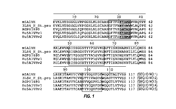
BRIEF DESCRIPTION OF THE DRAWINGS
[0040] FIG. 1 depicts an alignment of heavy chain variable regions of the
mouse 6C1
antibody, mouse model antibodies, human acceptor antibodies, and humanized
versions of the
6C1 antibody. The CDRs as defined by Kabat are enclosed in boxes, except that
the first
enclosed box is a composite of the Chothia CDR-H1 and the Kabat CDR-H1, with
the Kabat
CDR-H1 underlined and bolded.
8
CA 02974911 2017-07-25
WO 2016/120809 PCT/1B2016/050414
[0041] FIG. 2 depicts an alignment of light chain variable regions of the
mouse 6C1
antibody, mouse model antibodies, human acceptor antibodies, and humanized
versions of the
6C1 antibody. The CDRs as defined by Kabat are enclosed in boxes.
[0042] FIGS. 3A & 3B: FIG. 3A depicts the binding curve of murine 5A1, 6C1,
9D5, and
14G8 antibodies to ph4-treated TTR. FIG. 3B depicts the binding curve of
murine 5A1, 6C1,
9D5, and 14G8 antibodies to ph4-treated or native TTR
[0043] FIG. 4A, 4B & 4C: FIG. 4A depicts the inhibition of TTR-Y78F fiber
formation by
mis-TTR antibodies. FIG. 4B depicts the inhibition of TTR-V1221 fiber
formation by 14G8.
FIG. 4C depicts the inhibition of TTR-V1221 fiber formation by a control
antibody.
[0044] FIGS. 5A & 5B: FIG 5A depicts a densitometry analysis of a Western
Blot analysis
of plasma samples from patients confirmed for V3OM ATTR (Sample #11, #12, #15,
#18, #19,
##20) and samples from normal subjects (Sample #21, #22, #23, #24, #25, and
#27) using the
9D5 mis-TTR antibody. FIG. 5B depicts a densitometry analysis of a Western
blot analysis of
the same samples using the 5A1 mis-TTR antibody.
[0045] FIG. 6 depicts a MesoScale Discovery (MSD) plate assay of plasma
samples from
patients confirmed for V3OM ATTR (Sample #11, #12, #15, #18, #19, #20) and
samples from
normal subjects (#21, #22, #23, #24, #25, #27) using the 6C1 antibody.
[0046] FIGS. 7A & 7B: FIG. 7A depicts the effect of antibody 14G8 on the
uptake
of F87M/L110M TTR by THP-1 cells. FIG. 7B depicts the effect of each of the
mis-TTR antibodies
on the uptake of V3OM TTR by THP-1 cells.
BRIEF DESCRIPTION OF THE SEQUENCES
[0047] SEQ ID NO:1 sets forth the amino acid sequence of the heavy chain
variable region
of the mouse 6C1 antibody.
[0048] SEQ ID NO:2 sets forth the amino acid sequence of the mouse heavy
chain variable
region structure template.
9
CA 02974911 2017-07-25
WO 2016/120809 PCT/1B2016/050414
[0049] SEQ ID NO:3 sets forth the amino acid sequence of the heavy chain
variable acceptor
accession number ADX65650.
[0050] SEQ ID NO:4 sets forth the amino acid sequence of the heavy chain
variable region
of the humanized 6C1 antibody version 1 (Hu6C1VHv1).
[0051] SEQ ID NO:5 sets forth the amino acid sequence of the heavy chain
variable region
of the humanized 6C1 antibody version lb (Hu6C1VHv1b).
[0052] SEQ ID NO:6 sets forth the amino acid sequence of the heavy chain
variable region
of the humanized 6C1 antibody version 2 (Hu6C1VHv2).
[0053] SEQ ID NO:7 sets forth the amino acid sequence of the heavy chain
variable region
of the humanized 6C1 antibody version 2b (Hu6C1VHv2b).
[0054] SEQ ID NO:8 sets forth the amino acid sequence of the heavy chain
variable region
of the humanized 6C1 antibody version 3 (Hu6C1VHv3).
[0055] SEQ ID NO:9 sets forth the amino acid sequence of the heavy chain
variable region
of the humanized 6C1 antibody version 3b (Hu6C1VHv3b).
[0056] SEQ ID NO:10 sets forth the amino acid sequence of Kabat CDR-H1 of
the mouse
6C1 antibody.
[0057] SEQ ID NO:11 sets forth the amino acid sequence of Kabat CDR-H2 of
the mouse
6C1 antibody.
[0058] SEQ ID NO:12 sets forth the amino acid sequence of Kabat CDR-H3 of
the mouse
6C1 antibody.
[0059] SEQ ID NO:13 sets forth the amino acid sequence of the light chain
variable region
of the mouse 6C1 antibody.
[0060] SEQ ID NO:14 sets forth the amino acid sequence of the mouse light
chain variable
region structure template.
CA 02974911 2017-07-25
WO 2016/120809 PCT/1B2016/050414
[0061] SEQ ID NO:15 sets forth the amino acid sequence of the light chain
variable acceptor
accession number ABI74084.
[0062] SEQ ID NO:16 sets forth the amino acid sequence of the light chain
variable region
of the humanized 6C1 antibody version 1 (Hu6C1VLv1).
[0063] SEQ ID NO:17 sets forth the amino acid sequence of the light chain
variable region
of the humanized 6C1 antibody version 2 (Hu6C1VLv2).
[0064] SEQ ID NO:18 sets forth the amino acid sequence of Kabat CDR-L1 of
the mouse
6C1 antibody.
[0065] SEQ ID NO:19 sets forth the amino acid sequence of Kabat CDR-L2 of
the mouse
6C1 antibody.
[0066] SEQ ID NO:20 sets forth the amino acid sequence of Kabat CDR-L3 of
the mouse
6C1 antibody.
[0067] SEQ ID NO:21 sets forth a nucleic acid sequence encoding the heavy
chain variable
region of the mouse 6C1 antibody with signal peptide.
[0068] SEQ ID NO:22 sets forth the amino acid sequence of the heavy chain
variable region
of the mouse 6C1 antibody with signal peptide.
[0069] SEQ ID NO:23 sets forth a nucleic acid sequence encoding the light
chain variable
region of the mouse 6C1 antibody with signal peptide.
[0070] SEQ ID NO:24 sets forth the amino acid sequence of the light chain
variable region
of the mouse 6C1 antibody with signal peptide.
[0071] SEQ ID NO:25 sets forth the amino acid sequence of an exemplary IgG1
heavy chain
constant region.
[0072] SEQ ID NO:26 sets forth the amino acid sequence of an exemplary IgG1
G1m3
heavy chain constant region.
11
CA 02974911 2017-07-25
WO 2016/120809 PCT/1B2016/050414
[0073] SEQ ID NO:27 sets forth the amino acid sequence of an exemplary IgG1
G1m3
heavy chain constant region.
[0074] SEQ ID NO:28 sets forth the amino acid sequence of an exemplary
light chain
constant region with C-terminal Arginine.
[0075] SEQ ID NO:29 sets forth the amino acid sequence of an exemplary
light chain
constant region without C-terminal Arginine.
[0076] SEQ ID NO:30 sets forth the amino acid sequence of the heavy chain
region of the
humanized 6C1 antibody version 1.
[0077] SEQ ID NO:31 sets forth the amino acid sequence of the heavy chain
region of the
humanized 6C1 antibody version lb.
[0078] SEQ ID NO:32 sets forth the amino acid sequence of the heavy chain
region of the
humanized 6C1 antibody version 2.
[0079] SEQ ID NO:33 sets forth the amino acid sequence of the heavy chain
region of the
humanized 6C1 antibody version 2b.
[0080] SEQ ID NO:34 sets forth the amino acid sequence of the heavy chain
region of the
humanized 6C1 antibody version 3.
[0081] SEQ ID NO:35 sets forth the amino acid sequence of the heavy chain
region of the
humanized 6C1 antibody version 3b.
[0082] SEQ ID NO:36 sets forth the amino acid sequence of the light chain
region of the
humanized 6C1 antibody version 1.
[0083] SEQ ID NO:37 sets forth the amino acid sequence of the light chain
region of the
humanized 6C1 antibody version 2.
[0084] SEQ ID NO:38 sets forth the amino acid sequence of human
transthyretin set forth in
accession number P02766.1 (UniProt).
12
CA 02974911 2017-07-25
WO 2016/120809 PCT/1B2016/050414
[0085] SEQ ID NO:39 sets forth the amino acid sequence of human
transthyretin set forth in
accession number AAB35639.1 (GenBank).
[0086] SEQ ID NO:40 sets forth the amino acid sequence of human
transthyretin set forth in
accession number AAB35640.1 (GenBank).
[0087] SEQ ID NO:41 sets forth the amino acid sequence of human
transthyretin set forth in
accession number and ABI63351.1 (GenBank).
[0088] SEQ ID NO:42 sets forth the amino acid sequence of residues 89-97 of
human
transthyretin.
[0089] SEQ ID NO:43 sets forth the amino acid sequence of a potential
transthyretin
immunogen.
[0090] SEQ ID NO:44 sets forth the amino acid sequence of a potential
transthyretin
immunogen.
[0091] SEQ ID NO:45 sets forth the amino acid sequence of a potential
transthyretin
immunogen.
[0092] SEQ ID NO:46 sets forth a nucleic acid sequence encoding an
exemplary IgG1 G1m3
heavy chain constant region.
[0093] SEQ ID NO:47 sets forth a nucleic acid sequence encoding an
exemplary light chain
constant region with C-terminal Arginine.
[0094] SEQ ID NO:48 sets forth a nucleic acid sequence encoding an
exemplary light chain
constant region without C-terminal Arginine.
[0095] SEQ ID NO:49 sets forth the amino acid sequence of a heavy chain
constant region
signal peptide.
[0096] SEQ ID NO:50 sets forth a nucleic acid sequence encoding a heavy
chain constant
region signal peptide.
13
CA 02974911 2017-07-25
WO 2016/120809
PCT/1B2016/050414
[0097] SEQ ID NO:51 sets forth the amino acid sequence of a light chain
constant region
signal peptide.
[0098] SEQ ID NO:52 sets forth a nucleic acid sequence encoding a light
chain constant
region signal peptide.
[0099] SEQ ID NO:53 sets forth a nucleic acid sequence encoding a mouse 6C1
variable
light chain region.
[00100] SEQ ID NO:54 sets forth a nucleic acid sequence encoding a mouse 6C1
variable
heavy chain region.
[00101] SEQ ID NO:55 sets forth a nucleic acid sequence encoding a heavy chain
variable
region of the humanized 6C1 antibody version 1 (Hu6C1VHv1).
[00102] SEQ ID NO:56 sets forth a nucleic acid sequence encoding a heavy chain
variable
region of the humanized 6C1 antibody version lb (Hu6C1VHv1b).
[00103] SEQ ID NO:57 sets forth a nucleic acid sequence encoding a heavy chain
variable
region of the humanized 6C1 antibody version 2 (Hu6C1VHv2).
[00104] SEQ ID NO:58 sets forth a nucleic acid sequence encoding a heavy chain
variable
region of the humanized 6C1 antibody version 2b (Hu6C1VHv2b).
[00105] SEQ ID NO:59 sets forth a nucleic acid sequence encoding a heavy chain
variable
region of the humanized 6C1 antibody version 3 (Hu6C1VHv3).
[00106] SEQ ID NO:60 sets forth a nucleic acid sequence encoding a heavy chain
variable
region of the humanized 6C1 antibody version 3b (Hu6C1VHv3b).
[00107] SEQ ID NO:61 sets forth a nucleic acid sequence encoding a light chain
variable
region of the humanized 6C1 antibody version 1 (Hu6C1VLv1).
[00108] SEQ ID NO:62 sets forth a nucleic acid sequence encoding a light chain
variable
region of the humanized 6C1 antibody version 2 (Hu6C1VLv2).
14
CA 02974911 2017-07-25
WO 2016/120809 PCT/1B2016/050414
[00109] SEQ ID NO:63 sets forth the amino acid sequence of a composite CDR-H1
(residues
26-35) of the mouse 6C1 antibody.
DEFINITIONS
[00110] Monoclonal antibodies or other biological entities are typically
provided in isolated
form. This means that an antibody or other biologically entity is typically at
least 50% w/w pure
of interfering proteins and other contaminants arising from its production or
purification but does
not exclude the possibility that the monoclonal antibody is combined with an
excess of
pharmaceutically acceptable carrier(s) or other vehicle intended to facilitate
its use. Sometimes
monoclonal antibodies are at least 60%, 70%, 80%, 90%, 95% or 99% w/w pure of
interfering
proteins and contaminants from production or purification. Often an isolated
monoclonal
antibody or other biological entity is the predominant macromolecular species
remaining after its
purification.
[00111] Specific binding of an antibody to its target antigen means an
affinity of at least 106,
107, 108, 109, or 1010 M-1. Specific binding is detectably higher in magnitude
and distinguishable
from non-specific binding occurring to at least one unrelated target. Specific
binding can be the
result of formation of bonds between particular functional groups or
particular spatial fit (e.g.,
lock and key type) whereas nonspecific binding is usually the result of van
der Waals forces.
Specific binding does not however necessarily imply that an antibody binds one
and only one
target.
[00112] The basic antibody structural unit is a tetramer of subunits. Each
tetramer includes
two identical pairs of polypeptide chains, each pair having one "light" (about
25 kDa) and one
"heavy" chain (about 50-70 kDa). The amino-terminal portion of each chain
includes a variable
region of about 100 to 110 or more amino acids primarily responsible for
antigen recognition.
This variable region is initially expressed linked to a cleavable signal
peptide. The variable
region without the signal peptide is sometimes referred to as a mature
variable region. Thus, for
example, a light chain mature variable region means a light chain variable
region without the
light chain signal peptide. The carboxy-terminal portion of each chain defines
a constant region
primarily responsible for effector function.
CA 02974911 2017-07-25
WO 2016/120809 PCT/1B2016/050414
[00113] Light chains are classified as either kappa or lambda. Heavy chains
are classified as
gamma, mu, alpha, delta, or epsilon, and define the antibody's isotype as IgG,
IgM, IgA, IgD and
IgE, respectively. Within light and heavy chains, the variable and constant
regions are joined by
a "J" region of about 12 or more amino acids, with the heavy chain also
including a "D" region
of about 10 or more amino acids. See generally, Fundamental Immunology, Paul,
W., ed., 2nd
ed. Raven Press, N.Y., 1989, Ch. 7 (incorporated by reference in its entirety
for all purposes).
[00114] An immunoglobulin light or heavy chain variable region (also referred
to herein as a
"light chain variable domain" ("VL domain") or "heavy chain variable domain"
("VH domain"),
respectively) consists of a "framework" region interrupted by three
"complementarity
determining regions" or "CDRs." The framework regions serve to align the CDRs
for specific
binding to an epitope of an antigen. The CDRs include the amino acid residues
of an antibody
that are primarily responsible for antigen binding. From amino-terminus to
carboxyl-terminus,
both VL and VH domains comprise the following framework (FR) and CDR regions:
FR1,
CDR1, FR2, CDR2, FR3, CDR3, and FR4. CDRs 1, 2, and 3 of a VL domain are also
referred
to herein, respectively, as CDR-L1, CDR-L2, and CDR-L3; CDRs 1, 2, and 3 of a
VH domain
are also referred to herein, respectively, as CDR-H1, CDR-H2, and CDR-H3.
[00115] The assignment of amino acids to each VL and VH domain is in
accordance with any
conventional definition of CDRs. Conventional definitions include, the Kabat
definition (Kabat,
Sequences of Proteins of Immunological Interest (National Institutes of
Health, Bethesda, MD,
1987 and 1991), The Chothia definition (Chothia & Lesk, J. Mol. Biol. 196:901-
917, 1987;
Chothia et al., Nature 342:878-883, 1989); a composite of Chothia Kabat CDR in
which CDR-
H1 is a composite of Chothia and Kabat CDRs; the AbM definition used by Oxford
Molecular's
antibody modelling software; and, the contact definition of Martin et al
(bioinfo.org.uk/abs) (see
Table 1). Kabat provides a widely used numbering convention (Kabat numbering)
in which
corresponding residues between different heavy chains or between different
light chains are
assigned the same number. When an antibody is said to comprise CDRs by a
certain definition
of CDRs (e.g., Kabat) that definition specifies the minimum number of CDR
residues present in
the antibody (i.e., the Kabat CDRs). It does not exclude that other residues
falling within another
conventional CDR definition but outside the specified definition are also
present. For example,
an antibody comprising CDRs defined by Kabat includes among other
possibilities, an antibody
16
CA 02974911 2017-07-25
WO 2016/120809
PCT/1B2016/050414
in which the CDRs contain Kabat CDR residues and no other CDR residues, and an
antibody in
which CDR H1 is a composite Chothia-Kabat CDR H1 and other CDRs contain Kabat
CDR
residues and no additional CDR residues based on other definitions.
Table 1
Conventional Definitions of CDRs Using Kabat Numbering
Composite of
Loop Kabat Chothia Chothia AbM
Contact
&
Kabat
Ll L24--L34 L24--L34 L24--L34 L24--L34 L30--L36
L2 L50--L56 L50--L56 L50--L56 L50--L56 L46--L55
L3 L89--L97 L89--L97 L89--L97 L89--L97 L89--L96
H1 H31--H35B H26--H32..H34* H26--H35B* H26--H35B H30--H35B
H2 H50--H65 H52--H56 H50--H65 H50--H58 H47--H58
H3 H95--H102 H95--H102 H95--H102 H95--H102 H93--H101
*CDR-H1 by Chothia can end at H32, H33, or H34 (depending on the length of
the loop). This is because the Kabat numbering scheme places insertions of
extra
residues at 35A and 35B, whereas Chothia numbering places them at 31A and
31B. If neither H35A nor H35B (Kabat numbering) is present, the Chothia CDR-
H1 loop ends at H32. If only H35A is present, it ends at H33. If both H35A and
H35B are present, it ends at H34.
[00116] The term "antibody" includes intact antibodies and binding fragments
thereof
Typically, fragments compete with the intact antibody from which they were
derived for specific
binding to the target including separate heavy chains, light chains Fab, Fab',
F(abl)2, F(ab)c,
Dabs, nanobodies, and Fv. Fragments can be produced by recombinant DNA
techniques, or by
enzymatic or chemical separation of intact immunoglobulins. The term
"antibody" also includes
a bispecific antibody and/or a humanized antibody. A bispecific or
bifunctional antibody is an
artificial hybrid antibody having two different heavy/light chain pairs and
two different binding
sites (see, e.g., Songsivilai and Lachmann, Clin. Exp. Immunol., 79:315-321
(1990); Kostelny et
al., J. Immunol., 148:1547-53 (1992)). In some bispecific antibodies, the two
different
17
CA 02974911 2017-07-25
WO 2016/120809 PCT/1B2016/050414
heavy/light chain pairs include a humanized 6C1 heavy chain/light chain pair
and a heavy
chain/light chain pair specific for a different epitope on transthyretin than
that bound by 6C1.
[00117] In some bispecific antibodies, one heavy chain/light chain pair is a
humanized 6C1
antibody as further disclosed below and the other heavy chain/light chain pair
is from an
antibody that binds to a receptor expressed on the blood brain barrier, such
as an insulin receptor,
an insulin-like growth factor (IGF) receptor, a leptin receptor, or a
lipoprotein receptor, or a
transferrin receptor (Friden et al., Proc. Natl. Acad. Sci. USA 88:4771-4775,
1991; Friden et al.,
Science 259:373-377, 1993). Such a bispecific antibody can be transferred
cross the blood brain
barrier by receptor-mediated transcytosis. Brain uptake of the bispecific
antibody can be further
enhanced by engineering the bi-specific antibody to reduce its affinity to the
blood brain barrier
receptor. Reduced affinity for the receptor resulted in a broader
distributioin in the brain (see,
e.g., Atwal et al., Sci. Trans. Med. 3, 84ra43, 2011; Yu et al., Sci. Trans.
Med. 3, 84ra44, 2011).
[00118] Exemplary bispecific antibodies can also be: (1) a dual-variable-
domain antibody
(DVD-Ig), where each light chain and heavy chain contains two variable domains
in tandem
through a short peptide linkage (Wu et al., Generation and Characterization of
a Dual Variable
Domain Immunoglobulin (DVD-IgTm) Molecule, In: Antibody Engineering, Springer
Berlin
Heidelberg (2010)); (2) a Tandab, which is a fusion of two single chain
diabodies resulting in a
tetravalent bispecific antibody that has two binding sites for each of the
target antigens; (3) a
flexibody, which is a combination of scFvs with a diabody resulting in a
multivalent molecule;
(4) a so-called "dock and lock" molecule, based on the "dimerization and
docking domain" in
Protein Kinase A, which, when applied to Fabs, can yield a trivalent
bispecific binding protein
consisting of two identical Fab fragments linked to a different Fab fragment;
or (5) a so-called
Scorpion molecule, comprising, e.g., two scFvs fused to both termini of a
human Fc-region.
Examples of platforms useful for preparing bispecific antibodies include BiTE
(Micromet),
DART (MacroGenics), Fcab and Mab2 (F-star), Fc-engineered IgG1(Xencor) or
DuoBody
(based on Fab arm exchange, Genmab).
[00119] The term "epitope" refers to a site on an antigen to which an antibody
binds. An
epitope can be formed from contiguous amino acids or noncontiguous amino acids
juxtaposed by
tertiary folding of one or more proteins. Epitopes formed from contiguous
amino acids (also
18
CA 02974911 2017-07-25
WO 2016/120809 PCT/1B2016/050414
known as linear epitopes) are typically retained on exposure to denaturing
solvents whereas
epitopes formed by tertiary folding (also known as conformational epitopes)
are typically lost on
treatment with denaturing solvents. An epitope typically includes at least 3,
and more usually, at
least 5 or 8-10 amino acids in a unique spatial conformation. Methods of
determining spatial
conformation of epitopes include, for example, x-ray crystallography and 2-
dimensional nuclear
magnetic resonance. See, e.g., Epitope Mapping Protocols, in Methods in
Molecular Biology,
Vol. 66, Glenn E. Morris, Ed. (1996). The epitope can be linear, such as an
epitope of, for
example, 2-5, 3-5, 3-9, or 5-9 contiguous amino acids from SEQ ID NO:38. The
epitope can
also be a conformational epitope including, for example, two or more non-
contiguous segments
of amino acids within residues 89-97 of SEQ ID NO:38. If an antibody is said
to bind to an
epitope within amino acids 89-97 of transthyretin (TTR), for example, what is
meant is that the
epitope is within the recited range of amino acids including those defining
the outer-limits of the
range. It does not necessarily mean that every amino acid within the range
constitutes part of the
epitope. Thus, for example, an epitope within amino acids 89-97 of TTR may
consist of amino
acids 89-97, 89-96, 90-96, 91-96, 92-96, 93-96, 94-96, 89-96, 89-95, 89-94, 89-
93, 89-92 or 89-
93, among other linear segments of SEQ ID NO:42, or in the case of
conformational epitopes,
non-contiguous segments of amino acids of SEQ ID NO:42.
[00120] Antibodies that recognize the same or overlapping epitopes can be
identified in a
simple immunoassay showing the ability of one antibody to compete with the
binding of another
antibody to a target antigen. The epitope of an antibody can also be defined X-
ray
crystallography of the antibody bound to its antigen to identify contact
residues. Alternatively,
two antibodies have the same epitope if all amino acid mutations in the
antigen that reduce or
eliminate binding of one antibody reduce or eliminate binding of the other.
Two antibodies have
overlapping epitopes if some amino acid mutations that reduce or eliminate
binding of one
antibody reduce or eliminate binding of the other.
[00121] Competition between antibodies is determined by an assay in which an
antibody
under test inhibits specific binding of a reference antibody to a common
antigen (see, e.g.,
Junghans et al., Cancer Res. 50:1495, 1990). A test antibody competes with a
reference
antibody if an excess of a test antibody (e.g., at least 2x, 5x, 10x, 20x or
100x) inhibits binding of
the reference antibody by at least 50% as measured in a competitive binding
assay. Some test
19
CA 02974911 2017-07-25
WO 2016/120809 PCT/1B2016/050414
antibodies inhibit binding of the references antibody by at least 75%, 90% or
99%. Antibodies
identified by competition assay (competing antibodies) include antibodies
binding to the same
epitope as the reference antibody and antibodies binding to an adjacent
epitope sufficiently
proximal to the epitope bound by the reference antibody for steric hindrance
to occur.
[00122] The term "native" with respect to the structure transthyretin (TTR)
refers to the
normal folded structure of TTR in its properly functioning state (L e., a TTR
tetramer). As TTR
is a tetramer in its natively folded form, non-native forms of TTR include,
for example,
misfolded TTR tetramers, TTR monomers, aggregated forms of TTR, and fibril
forms of TTR.
Non-native forms of TTR can include molecules comprising wild-type TTR amino
acid
sequences or mutations.
[00123] The term "misfolded" with respect to TTR refers to the secondary and
tertiary
structure of a TTR polypeptide monomer or multimer, and indicates that the
polypeptide has
adopted a conformation that is not normal for that protein in its properly
functioning state.
Although TTR misfolding can be caused by mutations in the protein (e.g.,
deletion, substitution,
or addition), wild-type TTR proteins can also be misfolded in diseases,
exposing specific
epitopes.
[00124] The term "pharmaceutically acceptable" means that the carrier,
diluent, excipient, or
auxiliary is compatible with the other ingredients of the formulation and not
substantially
deleterious to the recipient thereof.
[00125] The term "patient" includes human and other mammalian subjects that
receive either
prophylactic or therapeutic treatment.
[00126] An individual is at increased risk of a disease if the subject has at
least one known
risk-factor (e.g., genetic, biochemical, family history, and situational
exposure) placing
individuals with that risk factor at a statistically significant greater risk
of developing the disease
than individuals without the risk factor.
[00127] The term "biological sample" refers to a sample of biological material
within or
obtainable from a biological source, for example a human or mammalian subject.
Such samples
can be organs, organelles, tissues, sections of tissues, bodily fluids,
peripheral blood, blood
CA 02974911 2017-07-25
WO 2016/120809 PCT/1B2016/050414
plasma, blood serum, cells, molecules such as proteins and peptides, and any
parts or
combinations derived therefrom. The term biological sample can also encompass
any material
derived by processing the sample. Derived material can include cells or their
progeny.
Processing of the biological sample may involve one or more of filtration,
distillation, extraction,
concentration, fixation, inactivation of interfering components, and the like.
[00128] The term "control sample" refers to a biological sample not known or
suspected to
include monomeric, misfolded, aggregated, or fibril forms of transthyretin
(TTR), such as in
TTR amyloid deposits. Control samples can be obtained from individuals not
afflicted with a
TTR amyloidosis or a specifically chosen type of TTR amyloidosis.
Alternatively, control
samples can be obtained from patients afflicted with TTR amyloidosis or a
specifically chosen
type of TTR amyloidosis. Such samples can be obtained at the same time as a
biological sample
thought to comprise the TTR amyloidosis or on a different occasion. A
biological sample and a
control sample can both be obtained from the same tissue (e.g., a tissue
section containing both
TTR amyloid deposits and surrounding normal tissue). Preferably, control
samples consist
essentially or entirely of tissue free of TTR amyloid deposits and can be used
in comparison to a
biological sample thought to comprise TTR amyloid deposits. Preferably, the
tissue in the
control sample is the same type as the tissue in the biological sample (e.g.,
cardiomyocytes in the
heart).
[00129] The term "disease" refers to any abnormal condition that impairs
physiological
function. The term is used broadly to encompass any disorder, illness,
abnormality, pathology,
sickness, condition, or syndrome in which physiological function is impaired,
irrespective of the
nature of the etiology.
[00130] The term "symptom" refers to a subjective evidence of a disease, such
as altered gait,
as perceived by the subject. A "sign" refers to objective evidence of a
disease as observed by a
physician.
[00131] For purposes of classifying amino acids substitutions as conservative
or
nonconservative, amino acids are grouped as follows: Group I (hydrophobic side
chains): met,
ala, val, leu, ile; Group II (neutral hydrophilic side chains): cys, ser, thr;
Group III (acidic side
chains): asp, glu; Group IV (basic side chains): asn, gln, his, lys, arg;
Group V (residues
21
CA 02974911 2017-07-25
WO 2016/120809 PCT/1B2016/050414
influencing chain orientation): gly, pro; and Group VI (aromatic side chains):
trp, tyr, phe.
Conservative substitutions involve substitutions between amino acids in the
same class. Non-
conservative substitutions constitute exchanging a member of one of these
classes for a member
of another.
[00132] Percentage sequence identities are determined with antibody sequences
maximally
aligned by the Kabat numbering convention. After alignment, if a subject
antibody region (e.g.,
the entire mature variable region of a heavy or light chain) is being compared
with the same
region of a reference antibody, the percentage sequence identity between the
subject and
reference antibody regions is the number of positions occupied by the same
amino acid in both
the subject and reference antibody region divided by the total number of
aligned positions of the
two regions, with gaps not counted, multiplied by 100 to convert to
percentage.
[00133] Compositions or methods "comprising" or "including" one or more
recited elements
may include other elements not specifically recited. For example, a
composition that
"comprises" or "includes" an antibody may contain the antibody alone or in
combination with
other ingredients.
[00134] Designation of a range of values includes all integers within or
defining the range,
and all subranges defined by integers within the range.
[00135] Unless otherwise apparent from the context, the term "about"
encompasses values
within a standard margin of error of measurement (e.g., SEM) of a stated
value.
[00136] Statistical significance means p0.05.
[00137] The singular forms of the articles "a," "an," and "the" include plural
references unless
the context clearly dictates otherwise. For example, the term "a compound" or
"at least one
compound" can include a plurality of compounds, including mixtures thereof.
22
CA 02974911 2017-07-25
WO 2016/120809 PCT/1B2016/050414
DETAILED DESCRIPTION
I. General
[00138] The invention provides antibodies that specifically bind to residues
89-97 of
transthyretin (TTR). The antibodies have the capacity to bind to monomeric,
misfolded,
aggregated, or fibril forms of TTR. The antibodies can be used for treating or
effecting
prophylaxis of diseases or disorders associated with TTR accumulation or
accumulation of TTR
deposits (e.g., TTR amyloidosis). The antibodies can also be used for
diagnosing TTR
amyloidosis and inhibiting or reducing aggregation of TTR, among other
applications.
II. Target Molecules
[00139] Transthyretin (TTR) is a 127-amino acid, 55 kDa serum and
cerebrospinal fluid
transport protein primarily synthesized by the liver. It has also been
referred to as prealbumin,
thyroxine binding prealbumin, ATTR, and TBPA. In its native state, TTR exists
as a tetramer.
In homozygotes, the tetramers comprise identical 127-amino-acid beta-sheet-
rich subunits. In
heterozygotes, the TTR tetramers are made up of variant and/or wild-type
subunits, typically
combined in a statistical fashion.
[00140] The established function of TTR in the blood is to transport ho/o-
retinol binding
protein. Although TTR is the major carrier of thyroxine (T4) in the blood of
rodents, utilizing
binding sites that are orthogonal to those used for ho/o-retinol binding
protein, the T4 binding
sites are effectively unoccupied in humans.
[00141] TTR is one of at least thirty different human proteins whose
extracellular misfolding
and/or misassembly (amyloidogenesis) into a spectrum of aggregate structures
is thought to
cause degenerative diseases referred to as amyloid diseases. TTR undergoes
conformational
changes in order to become amyloidogenic. Partial unfolding exposes stretches
of largely
uncharged hydrophobic residues in an extended conformation that efficiently
misassemble into
largely unstructured spherical aggregates that ultimately undergo conformation
conversion into
cross-beta sheet amyloid structures.
23
CA 02974911 2017-07-25
WO 2016/120809 PCT/1B2016/050414
[00142] Unless otherwise apparent from context, reference to transthyretin
(TTR) or its
fragments or domains includes the natural human amino acid sequences including
isoforms,
mutants, and allelic variants thereof. Exemplary TTR polypeptide sequences are
designated by
Accession Numbers P02766.1 (UniProt) (SEQ ID NO:38), AAB35639.1 (GenBank) (SEQ
ID
NO:39), AAB35640.1 (GenBank) (SEQ ID NO:40), and ABI63351.1 (GenBank) (SEQ ID
NO:41). Residues are numbered according to Swiss Prot P02766.1, with the first
amino acid of
the mature protein (i.e., not including the 20 amino acid signal sequence)
designated residue 1.
In any other TTR protein, residues are numbered according to the corresponding
residues in
P02766.1 on maximum alignment.
III. Transthyretin Amyloidosis
[00143] Transthyretin (TTR) amyloidosis is a systemic disorder characterized
by pathogenic,
misfolded TTR and the extracellular deposition of amyloid fibrils composed of
TTR. TTR
amyloidosis is generally caused by destabilization of the native TTR tetramer
form (due to
environmental or genetic conditions), leading to dissociation, misfolding, and
aggregation of
TTR into amyloid fibrils that accumulate in various organs and tissues,
causing progressive
dysfunction. See, e.g., Almeida and Saraiva, FEBS Letters 586:2891-2896
(2012); Ando et al.,
Orphanet Journal of Rare Diseases 8:31 (2013).
[00144] In humans, both wild-type TTR tetramers and mixed tetramers comprised
of mutant
and wild-type subunits can dissociate, misfold, and aggregate, with the
process of
amyloidogenesis leading to the degeneration of post-mitotic tissue. Thus, TTR
amyloidoses
encompass diseases caused by pathogenic misfolded TTR resulting from mutations
in TTR or
resulting from non-mutated, misfolded TTR.
[00145] For example, senile systemic amyloidosis (SSA) and senile cardiac
amyloidosis
(SCA) are age-related types of amyloidosis that result from the deposition of
wild-type TTR
amyloid outside and within the cardiomyocytes of the heart. TTR amyloidosis is
also the most
common form of hereditary (familial) amyloidosis, which is caused by mutations
that destabilize
the TTR protein. The TTR amyloidoses associated with point mutations in the
TTR gene include
familial amyloid polyneuropathy (FAP), familial amyloid cardiomyopathy (FAC),
and the rare
central nervous system selective amyloidosis (CNSA). Patients with hereditary
(familial) TTR
24
CA 02974911 2017-07-25
WO 2016/120809 PCT/1B2016/050414
amyloidosis are almost always heterozygotes, meaning that the TTR tetramers
are composed of
mutant and/or wild-type TTR subunits, generally statistically distributed.
Hereditary (familial)
versions of TTR amyloidosis are generally autosomal dominant and are typically
earlier onset
than the sporadic diseases (SSA and SCA).
[00146] There are over 100 mutations in the gene encoding TTR that have been
implicated in
the autosomal dominant disorders FAP and FAC. See, e.g., US 2014/0056904;
Saraiva, Hum.
Mutat. 17(6):493-503 (2001); Damas and Saraiva, J. Struct. Biol. 130:290-299;
Dwulet and
Benson, Biochem. Biophys. Res. Commun. 114:657-662 (1983). These amyloid-
causing
mutations are distributed throughout the entire molecule of TTR. Generally,
the more
destabilizing the mutant subunits are to the TTR tetramer structure, the
earlier the onset of
amyloid disease. The pathogenic potential of a TTR variant is generally
determined by a
combination of its instability and its cellular secretion efficiency. The
initial pathology caused
by some TTR variants comes from their selective destruction of cardiac tissue,
whereas that from
other TTR variants comes from compromising the peripheral and autonomic
nervous system.
The tissue damage caused by TTR amyloidogenesis appear to stem largely from
the toxicity of
small, diffusible TTR aggregates, although accumulation of extracellular
amyloid may contribute
and almost certainly compromises organ structure in the late stages of the TTR
amyloidosis.
[00147] TTR amyloidosis presents in many different forms, with considerable
phenotypic
variation across individuals and geographic locations. For example, TTR
amyloidosis can
present as a progressive, axonal sensory autonomic and motor neuropathy. TTR
amyloidosis can
also present as an infiltrative cardiomyopathy.
[00148] The age at onset of disease-related symptoms varies between the second
and ninth
decades of life, with great variations across different populations. The
multisystem involvement
of TTR amyloidosis is a clue to its diagnosis. For example, TTR amyloidosis
diagnosis is
considered when one or several of the following are present: (1) family
history of neuropathic
disease, especially associated with heart failure; (2) neuropathic pain or
progressive sensory
disturbances of unknown etiology; (3) carpal tunnel syndrome without obvious
cause,
particularly if it is bilateral and requires surgical release; (4)
gastrointestinal motility
disturbances or autonomic nerve dysfunction of unknown etiology (e.g.,
erectile dysfunction,
CA 02974911 2017-07-25
WO 2016/120809 PCT/1B2016/050414
orthostatic hypotension, neurogenic gladder); (5) cardiac disease
characterized by thickened
ventricular walls in the absence of hypertension; (6) advanced atrio-
ventricular block of
unknown origin, particularly when accompanied by a thickened heart; and (6)
vitreous body
inclusions of the cotton-wool type. See Ando et al., Orphanet Journal of Rare
Diseases 8:31
(2013). Other symptoms can include, for example, polyneuropathy, sensory loss,
pain, weakness
in lower limbs, dyshidrosis, diarrhea, constipation, weight loss, and urinary
incontinence/retention.
[00149] Diagnosis of TTR amyloidosis typically relies on target organ
biopsies, followed by
histological staining of the excised tissue with the amyloid-specific dye,
Congo red. If a positive
test for amyloid is observed, immunohistochemical staining for TTR is
subsequently performed
to ensure that the precursor protein responsible for amyloid formation is
indeed TTR. For
familial forms of the diseases, demonstration of a mutation in the gene
encoding TTR is then
needed before diagnosis can be made. This can be accomplished, for example,
through
isoelectric focusing electrophoresis, polymerase chain reaction, or laser
dissection/liquid
chromatography-tandem mass spectrometry. See, e.g., US 2014/0056904; Ruberg
and Berk,
Circulation 126:1286-1300 (2012); Ando et al., Orphanet Journal of Rare
Diseases 8:31 (2013).
IV. Antibodies
A. Binding Specificity and Functional Properties
[00150] The invention provides monoclonal antibodies binding to
transthyretin (TTR)
protein, more specifically, to epitopes within amino acid residues 89-97 (SEQ
ID NO:42) of
TTR. Such epitopes are buried in the native TTR tetramer and exposed in
monomeric,
misfolded, aggregated, or fibril forms of TTR.
[00151] An antibody designated 6C1 is such an exemplary mouse antibody. This
antibody
specifically binds within amino acid residues 89-97 (SEQ ID NO:42) of TTR.
This antibody is
further characterized by its ability to bind to monomeric, misfolded,
aggregated, or fibril forms
of TTR but not to native tetrameric forms of TTR. In addition, this antibody
is characterized by
its immunoreactivity on TTR-mediated amyloidosis cardiac tissue but not on
healthy cardiac
26
CA 02974911 2017-07-25
WO 2016/120809 PCT/1B2016/050414
tissue. Ability to bind to specific proteins or fragments thereof may be
demonstrated using
exemplary assay formats provided in the examples.
[00152] Some antibodies bind to the same or overlapping epitope as an antibody
designated
6C1. The sequences of the heavy and light chain mature variable regions of 6C1
are designated
SEQ ID NOS: 1 and 13, respectively. Other antibodies having such a binding
specificity can be
produced by immunizing mice with TTR, or a portion thereof including the
desired epitope (e.g.,
SEQ ID NO:42), and screening resulting antibodies for binding to monomeric TTR
or a peptide
comprising SEQ ID NO:42, optionally in competition with an antibody having the
variable
regions of mouse 6C1 (IgGl,kappa). Fragments of TTR including the desired
epitope can be
linked to a carrier that helps elicit an antibody response to the fragment
and/or be combined with
an adjuvant that helps elicit such a response. Such antibodies can be screened
for differential
binding to wild-type, monomeric versions of TTR or a fragment thereof (e.g.,
SEQ ID NO:38)
compared with mutants of specified residues. Screening against such mutants
more precisely
defines the binding specificity to allow identification of antibodies whose
binding is inhibited by
mutagenesis of particular residues and which are likely to share the
functional properties of other
exemplified antibodies. The mutations can be systematic replacement
substitution with alanine
(or serine if an alanine is present already) one residue at a time, or more
broadly spaced intervals,
throughout the target or throughout a section thereof in which an epitope is
known to reside. If
the same set of mutations significantly reduces the binding of two antibodies,
the two antibodies
bind the same epitope.
[00153] Antibodies having the binding specificity of a selected murine
antibody (e.g., 6C1)
can also be produced using a variant of the phage display method. See Winter,
WO 92/20791.
This method is particularly suitable for producing human antibodies. In this
method, either the
heavy or light chain variable region of the selected murine antibody is used
as a starting material.
If, for example, a light chain variable region is selected as the starting
material, a phage library is
constructed in which members display the same light chain variable region
(i.e., the murine
starting material) and a different heavy chain variable region. The heavy
chain variable regions
can for example be obtained from a library of rearranged human heavy chain
variable regions. A
phage showing strong specific binding (e.g., at least 108 and preferably at
least 109 AV) for
monomeric TTR or a fragment thereof (e.g., amino acid residues 89-97) is
selected. The heavy
27
CA 02974911 2017-07-25
WO 2016/120809 PCT/1B2016/050414
chain variable region from this phage then serves as a starting material for
constructing a further
phage library. In this library, each phage displays the same heavy chain
variable region (i.e., the
region identified from the first display library) and a different light chain
variable region. The
light chain variable regions can be obtained for example from a library of
rearranged human
variable light chain regions. Again, phage showing strong specific binding for
monomeric TTR
or a fragment thereof (e.g., amino acid residues 89-97) are selected. The
resulting antibodies
usually have the same or similar epitope specificity as the murine starting
material.
[00154] Other antibodies can be obtained by mutagenesis of cDNA encoding the
heavy and
light chains of an exemplary antibody, such as 6C1. Monoclonal antibodies that
are at least
70%, 80%, 90%, 95%, 96%, 97%, 98%, or 99% identical to 6C1 in amino acid
sequence of the
mature heavy and/or light chain variable regions and maintain its functional
properties, and/or
which differ from the respective antibody by a small number of functionally
inconsequential
amino acid substitutions (e.g., conservative substitutions), deletions, or
insertions are also
included in the invention. Monoclonal antibodies having at least one or all
six CDR(s) as
defined by conventional definition, but preferably Kabat, that are 90%, 95%,
99% or 100%
identical to corresponding CDRs of 6C1 are also included.
[00155] The invention also provides antibodies having some or all (e.g., 3, 4,
5, and 6) CDRs
entirely or substantially from 6C1. Such antibodies can include a heavy chain
variable region
that has at least two, and usually all three, CDRs entirely or substantially
from the heavy chain
variable region of 6C1 and/or a light chain variable region having at least
two, and usually all
three, CDRs entirely or substantially from the light chain variable region of
6C1. The antibodies
can include both heavy and light chains. A CDR is substantially from a
corresponding 6C1 CDR
when it contains no more than 4, 3, 2, or 1 substitutions, insertions, or
deletions, except that
CDR-H2 (when defined by Kabat) can have no more than 6, 5, 4, 3, 2, or 1
substitutions,
insertions, or deletions. Such antibodies can have at least 70%, 80%, 90%,
95%, 96%, 97%,
98%, or 99% identity to 6C1 in the amino acid sequence of the mature heavy
and/or light chain
variable regions and maintain their functional properties, and/or differ from
6C1 by a small
number of functionally inconsequential amino acid substitutions (e.g.,
conservative
substitutions), deletions, or insertions.
28
CA 02974911 2017-07-25
WO 2016/120809 PCT/1B2016/050414
[00156] Some antibodies identified by such assays can bind to monomeric,
misfolded,
aggregated, or fibril forms of TTR but not to native tetrameric forms of TTR,
as described in the
examples or otherwise. Likewise, some antibodies are immunoreactive on TTR-
mediated
amyloidosis tissue but not on healthy tissue.
[00157] Some antibodies can inhibit or reduce aggregation of TTR, inhibit or
reduce TTR
fibril formation, reduce or clear TTR deposits or aggregated TTR, or stabilize
non-toxic
conformations of TTR in an animal model or clinical trial. Some antibodies can
treat, effect
prophylaxis of, or delay the onset of a TTR amyloidosis as shown in an animal
model or clinical
trial. Exemplary animal models for testing activity against a TTR amyloidosis
include those
described in Kohno et al., Am. J. Path. 150(4):1497-1508 (1997); Teng et al.,
Laboratory
Investigations 81:385-396 (2001); Wakasugi et al., Proc. Japan Acad. 63B:344-
347 (1987);
Shimada et al., Mol. Biol. Med. 6:333-343 (1989); Nagata et al., J. Biochem.
117:169-175
(1995); Sousa et al., Am. J. Path. 161:1935-1948 (2002); and Santos et al.,
Neurobiology of
Aging 31:280-289 (2010).
B. Non-Human Antibodies
[00158] The production of other non-human antibodies, e.g., murine, guinea
pig, primate,
rabbit or rat, against monomeric TTR or a fragment thereof (e.g., amino acid
residues 89-97) can
be accomplished by, for example, immunizing the animal with TTR or a fragment
thereof. See
Harlow & Lane, Antibodies, A Laboratory Manual (CSHP NY, 1988) (incorporated
by reference
for all purposes). Such an immunogen can be obtained from a natural source, by
peptide
synthesis, or by recombinant expression. Optionally, the immunogen can be
administered fused
or otherwise complexed with a carrier protein. Optionally, the immunogen can
be administered
with an adjuvant. Several types of adjuvant can be used as described below.
Complete Freund's
adjuvant followed by incomplete adjuvant is preferred for immunization of
laboratory animals.
Rabbits or guinea pigs are typically used for making polyclonal antibodies.
Mice are typically
used for making monoclonal antibodies. Antibodies are screened for specific
binding to
monomeric TTR or an epitope within TTR (e.g., an epitope comprising one or
more of amino
acid residues 89-97). Such screening can be accomplished by determining
binding of an
antibody to a collection of monomeric TTR variants, such as TTR variants
containing amino acid
29
CA 02974911 2017-07-25
WO 2016/120809 PCT/1B2016/050414
residues 89-97 or mutations within these residues, and determining which TTR
variants bind to
the antibody. Binding can be assessed, for example, by Western blot, FACS or
ELISA.
C. Humanized Antibodies
[00159] A humanized antibody is a genetically engineered antibody in which
CDRs from a
non-human "donor" antibody are grafted into human "acceptor" antibody
sequences (see, e.g.,
Queen, US 5,530,101 and 5,585,089; Winter, US 5,225,539; Carter, US 6,407,213;
Adair, US
5,859,205; and Foote, US 6,881,557). The acceptor antibody sequences can be,
for example, a
mature human antibody sequence, a composite of such sequences, a consensus
sequence of
human antibody sequences, or a germline region sequence. Thus, a humanized
antibody is an
antibody having at least three, four, five or all CDRs entirely or
substantially from a donor
antibody and variable region framework sequences and constant regions, if
present, entirely or
substantially from human antibody sequences. Similarly a humanized heavy chain
has at least
one, two and usually all three CDRs entirely or substantially from a donor
antibody heavy chain,
and a heavy chain variable region framework sequence and heavy chain constant
region, if
present, substantially from human heavy chain variable region framework and
constant region
sequences. Similarly a humanized light chain has at least one, two and usually
all three CDRs
entirely or substantially from a donor antibody light chain, and a light chain
variable region
framework sequence and light chain constant region, if present, substantially
from human light
chain variable region framework and constant region sequences. Other than
nanobodies and
dAbs, a humanized antibody comprises a humanized heavy chain and a humanized
light chain.
A CDR in a humanized antibody is substantially from a corresponding CDR in a
non-human
antibody when at least 85%, 90%, 95% or 100% of corresponding residues (as
defined by any
conventional definition but preferably defined by Kabat) are identical between
the respective
CDRs. The variable region framework sequences of an antibody chain or the
constant region of
an antibody chain are substantially from a human variable region framework
sequence or human
constant region respectively when at least 85%, 90%, 95% or 100% of
corresponding residues
defined by any conventional definition but preferably defined by Kabat are
identical.
[00160] Although humanized antibodies often incorporate all six CDRs
(preferably as defined
by Kabat) from a mouse antibody, they can also be made with less than all CDRs
(e.g., at least 3,
CA 02974911 2017-07-25
WO 2016/120809 PCT/1B2016/050414
4, or 5 CDRs) from a mouse antibody (e.g., Pascalis et al., J. Immunol.
169:3076, 2002; Vajdos
et al., J. of MoL Biol., 320: 415-428, 2002; Iwahashi et al., MoL Immunol.
36:1079-1091, 1999;
Tamura et al, J. Immunol., 164:1432-1441, 2000).
[00161] In some antibodies only part of the CDRs, namely the subset of CDR
residues
required for binding, termed the SDRs, are needed to retain binding in a
humanized antibody.
CDR residues not contacting antigen and not in the SDRs can be identified
based on previous
studies (for example residues H60-H65 in CDR H2 are often not required), from
regions of
Kabat CDRs lying outside Chothia hypervariable loops (Chothia, J. MoL Biol.
196:901, 1987),
by molecular modeling and/or empirically, or as described in Gonzales et al.,
MoL Immunol. 41:
863, 2004. In such humanized antibodies at positions in which one or more
donor CDR residues
is absent or in which an entire donor CDR is omitted, the amino acid occupying
the position can
be an amino acid occupying the corresponding position (by Kabat numbering) in
the acceptor
antibody sequence. The number of such substitutions of acceptor for donor
amino acids in the
CDRs to include reflects a balance of competing considerations. Such
substitutions are
potentially advantageous in decreasing the number of mouse amino acids in a
humanized
antibody and consequently decreasing potential immunogenicity. However,
substitutions can
also cause changes of affinity, and significant reductions in affinity are
preferably avoided.
Positions for substitution within CDRs and amino acids to substitute can also
be selected
empirically.
[00162] The human acceptor antibody sequences can optionally be selected from
among the
many known human antibody sequences to provide a high degree of sequence
identity (e.g., 65-
85% identity) between a human acceptor sequence variable region frameworks and
corresponding variable region frameworks of a donor antibody chain.
[00163] An example of an acceptor sequence for the heavy chain is the human
mature heavy
chain variable region with NCBI accession code ADX65650 (SEQ ID NO:3). This
acceptor
sequence includes two CDRs having the same canonical form as mouse 6C1 heavy
chain. An
examples of an acceptor sequence for the light chain is the human mature light
chain variable
region with NCBI accession code ABI74084 (SEQ ID NO:15). This acceptor
sequence includes
two CDRs having the same canonical form as mouse 6C1 light chain.
31
CA 02974911 2017-07-25
WO 2016/120809 PCT/1B2016/050414
[00164] Certain amino acids from the human variable region framework residues
can be
selected for substitution based on their possible influence on CDR
conformation and/or binding
to antigen. Investigation of such possible influences is by modeling,
examination of the
characteristics of the amino acids at particular locations, or empirical
observation of the effects
of substitution or mutagenesis of particular amino acids.
[00165] For example, when an amino acid differs between a murine variable
region
framework residue and a selected human variable region framework residue, the
human
framework amino acid can be substituted by the equivalent framework amino acid
from the
mouse antibody when it is reasonably expected that the amino acid:
(1) noncovalently binds antigen directly;
(2) is adjacent to a CDR region or within a CDR as defined by Chothia but
not Kabat;
(3) otherwise interacts with a CDR region (e.g., is within about 6 A of a
CDR
region), (e.g., identified by modeling the light or heavy chain on the solved
structure of a
homologous known immunoglobulin chain); or
(4) is a residue participating in the VL-VH interface.
[00166] Framework residues from classes (1) through (3) as defined by Queen,
US 5,530,101,
are sometimes alternately referred to as canonical and vernier residues.
Framework residues that
help define the conformation of a CDR loop are sometimes referred to as
canonical residues
(Chothia & Lesk, J. MoL Biol. 196:901-917 (1987); Thornton & Martin, J. Mol.
Biol. 263:800-
815 (1996)). Framework residues that support antigen-binding loop
conformations and play a
role in fine-tuning the fit of an antibody to antigen are sometimes referred
to as vernier residues
(Foote & Winter, J. MoL Biol 224:487-499 (1992)).
[00167] Other framework residues that are candidates for substitution are
residues creating a
potential glycosylation site. Still other candidates for substitution are
acceptor human
framework amino acids that are unusual for a human immunoglobulin at that
position. These
amino acids can be substituted with amino acids from the equivalent position
of the mouse donor
antibody or from the equivalent positions of more typical human
immunoglobulins.
[00168] Exemplary humanized antibodies are humanized forms of the mouse 6C1
antibody,
designated Hu6C1. The mouse antibody comprises mature heavy and light chain
variable
32
CA 02974911 2017-07-25
WO 2016/120809 PCT/1B2016/050414
regions having amino acid sequences comprising SEQ ID NO:1 and SEQ ID NO:13,
respectively. The invention provides six exemplified humanized mature heavy
chain variable
regions: Hu6C1VHv1 (SEQ ID NO:4), Hu6C1VHv1b (SEQ ID NO:5), Hu6C1VHv2 (SEQ ID
NO:6), Hu6C1VHv2b (SEQ ID NO:7), Hu6C1VHv3 (SEQ ID NO:8), and Hu6C1VHv3b (SEQ
ID NO:9). The invention further provides two exemplified human mature light
chain variable
regions: Hu6C1VLv1 (SEQ ID NO:16) and Hu6C1VLv2 (SEQ ID NO:17). Figures 1 and
2
show alignments of the heavy chain variable region and light chain variable
region, respectively,
of 6C1, mouse model antibodies, human acceptor antibodies, and humanized
antibody versions
of 6C1.
[00169] For reasons such as possible influence on CDR conformation and/or
binding to
antigen, mediating interaction between heavy and light chains, interaction
with the constant
region, being a site for desired or undesired post-translational modification,
being an unusual
residue for its position in a human variable region sequence and therefore
potentially
immunogenic, getting aggregation potential, and other reasons, the following
ten variable region
framework positions were considered as candidates for substitutions in the six
exemplified
human mature light chain variable regions and the two exemplified human mature
heavy chain
variable regions, as further specified in the examples: L2, L45, H19, H44,
H49, H76, H77,
H82(a), H83, and H89.
[00170] Here, as elsewhere, the first-mentioned residue is the residue of a
humanized antibody
formed by grafting Kabat CDRs or a composite Chothia Kabat CDR in the case of
CDR-H1 into
a human acceptor framework, and the second-mentioned residue is a residue
being considered
for replacing such residue. Thus, within variable region frameworks, the first
mentioned residue
is human, and within CDRs, the first mentioned residue is mouse.
[00171] Exemplified antibodies include any permutations or combinations of the
exemplified
mature heavy and light chain variable regions (e. g. , Hu6C1VHv1/VLv1 or H1L1,
Hu6C1VHv1bNLv1 or H1bL1, Hu6C1VHv1/VLv2 or H1L2, Hu6C1VHv1bNLv2 or H1bL2,
Hu6C1VHv2/VLv1 or H2L1, Hu6C1VHv2bNLv1 or H2bL1, Hu6C1VHv2/VLv2 or H2L2,
Hu6C1VHv2bNLv2 or H2bL2, Hu6C1VHv3/VLv1 or H3L1, Hu6C1VHv3bNLv1 or H3bL1,
Hu6C1VHv3/VLv2 or H3L2, and Hu6C1VHv3b/VLv2 or H3bL2).
33
CA 02974911 2017-07-25
WO 2016/120809 PCT/1B2016/050414
[00172] The invention provides variants of humanized antibodies in which the
humanized
mature heavy chain variable region shows at least 90%, 95%, 96%, 97%, 98%, or
99% identity
to SEQ ID NOs:4-9, and the humanized mature light chain variable region shows
at least 90%,
95%, 96%, 97%, 98%, or 99% identity to SEQ ID NOs:16 or 17. In some such
antibodies at
least 1, 2, 3, 4, 5, 6, 7, 8, 9, or all 10 of the backmutations or other
mutations in SEQ ID NOs:4-
9, 16, and 17 are retained.
[00173] In some antibodies, at least one of positions H19, H44, H49, H76, H77,
H82(a), H83,
and H89 in the Vll region is occupied by K, R, A, S, T, S, K, and V,
respectively. In some
antibodies, position H77 in the Vll region is occupied by T, as in Hu6C1VHv1.
In some
antibodies, positions H49 and H77 in the Vll region are occupied by A and T,
respectively, as in
Hu6C1VHv1b. In some antibodies, positions H76, H77, and H82(a) in the Vll
region are
occupied by S, T, and S, respectively, as in Hu6C1VHv2. In some antibodies,
positions H49,
H76, H77, and H82(a) in the Vll region are occupied by A, S, T, and S,
respectively, as in
Hu6C1VHv2b. In some antibodies, positions H19, H44, H77, H83, and H89 in the
Vll region
are occupied by K, R, T, K, and M, respectively, as in Hu6C1VHv3. In some
antibodies,
positions H19, H44, H49, H77, H83, and H89 in the Vll region are occupied by
K, R, A, T, K,
and M, respectively, as in Hu6C1VHv3b. In some antibodies, position L45 in the
VL region is
occupied by K. In some antibodies, position L2 in the VL region is occupied by
I. In some
antibodies, one or both of positions L2 and L45 in the VL region are occupied
by V and K,
respectively, as in Hu6C1VLv1. In some antibodies, one or both of positions L2
and L45 in the
VL region are occupied by I and K, respectively, as in Hu6C1VLv2. The CDR
regions of such
humanized antibodies can be identical or substantially identical to the CDR
regions of the 6C1
mouse donor antibody. The CDR regions can be defined by any conventional
definition (e.g.,
Chothia, or composite of Chothia and Kabat) but are preferably as defined by
Kabat.
[00174] Variable regions framework positions are in accordance with Kabat
numbering unless
otherwise stated. Other such variants typically differ from the sequences of
the exemplified
Hu6Clantibodies by a small number (e.g., typically no more than 1, 2, 3, 5,
10, or 15) of
replacements, deletions or insertions. Such differences are usually in the
framework but can also
occur in the CDRs.
34
CA 02974911 2017-07-25
WO 2016/120809 PCT/1B2016/050414
[00175] A possibility for additional variation in humanized 6C1 variants is
additional
backmutations in the variable region frameworks. Many of the framework
residues not in
contact with the CDRs in the humanized mAb can accommodate substitutions of
amino acids
from the corresponding positions of the donor mouse mAb or other mouse or
human antibodies,
and even many potential CDR-contact residues are also amenable to
substitution. Even amino
acids within the CDRs may be altered, for example, with residues found at the
corresponding
position of the human acceptor sequence used to supply variable region
frameworks. In addition,
alternate human acceptor sequences can be used, for example, for the heavy
and/or light chain.
If different acceptor sequences are used, one or more of the backmutations
recommended above
may not be performed because the corresponding donor and acceptor residues are
already the
same without backmutations.
[00176] Preferably, replacements or backmutations in Hu6C1 variants (whether
or not
conservative) have no substantial effect on the binding affinity or potency of
the humanized
mAb, that is, its ability to bind to monomeric TTR (e.g., the potency in some
or all of the assays
described in the present examples of the variant humanized 6C1 antibody is
essentially the same,
i.e., within experimental error, as that of murine 6C1).
D. Chimeric and Veneered Antibodies
[00177] The invention further provides chimeric and veneered forms of non-
human
antibodies, particularly the 6C1 antibodies of the examples.
[00178] A chimeric antibody is an antibody in which the mature variable
regions of light and
heavy chains of a non-human antibody (e.g., a mouse) are combined with human
light and heavy
chain constant regions. Such antibodies substantially or entirely retain the
binding specificity of
the mouse antibody, and are about two-thirds human sequence.
[00179] A veneered antibody is a type of humanized antibody that retains some
and usually all
of the CDRs and some of the non-human variable region framework residues of a
non-human
antibody but replaces other variable region framework residues that may
contribute to B- or T-
cell epitopes, for example exposed residues (Padlan, Mol. Immunol. 28:489,
1991) with residues
from the corresponding positions of a human antibody sequence. The result is
an antibody in
CA 02974911 2017-07-25
WO 2016/120809 PCT/1B2016/050414
which the CDRs are entirely or substantially from a non-human antibody and the
variable region
frameworks of the non-human antibody are made more human-like by the
substitutions.
Veneered forms of the 6C1 antibody are included in the invention.
E. Human Antibodies
[00180] Human antibodies against monomeric TTR or a fragment thereof (e.g.,
amino acid
residues 89-97 (SEQ ID NO:42) of TTR) are provided by a variety of techniques
described
below. Some human antibodies are selected by competitive binding experiments,
by the phage
display method of Winter, above, or otherwise, to have the same epitope
specificity as a
particular mouse antibody, such as one of the mouse monoclonal antibodies
described in the
examples. Human antibodies can also be screened for particular epitope
specificity by using
only a fragment of TTR, such as a TTR variant containing only amino acid
residues 89-97 of
TTR, as the target antigen, and/or by screening antibodies against a
collection of TTR variants,
such as TTR variants containing various mutations within amino acid residues
89-97 of TTR.
[00181] Methods for producing human antibodies include the trioma method of
Oestberg et
al., Hybridoma 2:361-367 (1983); Oestberg, U.S. Patent No. 4,634,664; and
Engleman et al., US
Patent 4,634,666, use of transgenic mice including human immunoglobulin genes
(see, e.g.,
Lonberg et al., W093/12227 (1993); US 5,877,397; US 5,874,299; US 5,814,318;
US 5,789,650;
US 5,770,429; US 5,661,016; US 5,633,425; US 5,625,126; US 5,569,825; US
5,545,806;
Neuberger, Nat. Biotechnol. 14:826 (1996); and Kucherlapati, WO 91/10741
(1991)) and phage
display methods (see, e.g., Dower et al., WO 91/17271; McCafferty et al., WO
92/01047; US
5,877,218; US 5,871,907; US 5,858,657; US 5,837,242; US 5,733,743; and US
5,565,332).
F. Selection of Constant Region
[00182] The heavy and light chain variable regions of chimeric, veneered or
humanized
antibodies can be linked to at least a portion of a human constant region. The
choice of constant
region depends, in part, whether antibody-dependent cell-mediated
cytotoxicity, antibody
dependent cellular phagocytosis and/or complement dependent cytotoxicity are
desired. For
example, human isotopes IgG1 and IgG3 have complement-dependent cytotoxicity
and human
isotypes IgG2 and IgG4 do not. Human IgG1 and IgG3 also induce stronger cell
mediated
36
CA 02974911 2017-07-25
WO 2016/120809 PCT/1B2016/050414
effector functions than human IgG2 and IgG4. Light chain constant regions can
be lambda or
kappa.
[00183] One or several amino acids at the amino or carboxy terminus of the
light and/or heavy
chain, such as the C-terminal lysine of the heavy chain, may be missing or
derivatized in a
proportion or all of the molecules. Substitutions can be made in the constant
regions to reduce or
increase effector function such as complement-mediated cytotoxicity or ADCC
(see, e.g., Winter
et al., US Patent No. 5,624,821; Tso et al., US Patent No. 5,834,597; and
Lazar et al., Proc. Natl.
Acad. ScL USA 103:4005, 2006), or to prolong half-life in humans (see, e.g.,
Hinton et al., J.
Biol. Chem. 279:6213, 2004). Exemplary substitutions include a Gln at position
250 and/or a
Leu at position 428 (EU numbering is used in this paragraph for the constant
region) for
increasing the half-life of an antibody. Substitution at any or all of
positions 234, 235, 236
and/or 237 reduce affinity for Fcy receptors, particularly FcyRI receptor
(see, e.g., US
6,624,821). An alanine substitution at positions 234, 235, and 237 of human
IgG1 can be used
for reducing effector functions. Some antibodies have alanine substitution at
positions 234, 235
and 237 of human IgG1 for reducing effector functions. Optionally, positions
234, 236 and/or
237 in human IgG2 are substituted with alanine and position 235 with glutamine
(see, e.g., US
5,624,821). In some antibodies, a mutation at one or more of positions 241,
264, 265, 270, 296,
297, 322, 329, and 331 by EU numbering of human IgG1 is used. In some
antibodies, a
mutation at one or more of positions 318, 320, and 322 by EU numbering of
human IgG1 is
used. In some antibodies, positions 234 and/or 235 are substituted with
alanine and/or position
329 is substituted with glycine. In some antibodies, positions 234 and 235 are
substituted with
alanine, such as in SEQ ID NO:27. In some antibodies, the isotype is human
IgG2 or IgG4.
[00184] An exemplary human light chain kappa constant region has the amino
acid sequence
of SEQ ID NO:28. The N-terminal arginine of SEQ ID NO:28 can be omitted, in
which case
light chain kappa constant region has the amino acid sequence of SEQ ID NO:29.
An exemplary
human IgG1 heavy chain constant region has the amino acid sequence of SEQ ID
NO:25 (with
or without the C-terminal lysine). Antibodies can be expressed as tetramers
containing two light
and two heavy chains, as separate heavy chains, light chains, as Fab, Fab',
F(ab')2, and Fv, or as
single chain antibodies in which heavy and light chain mature variable domains
are linked
through a spacer.
37
CA 02974911 2017-07-25
WO 2016/120809 PCT/1B2016/050414
[00185] Human constant regions show allotypic variation and isoallotypic
variation between
different individuals, that is, the constant regions can differ in different
individuals at one or
more polymorphic positions. Isoallotypes differ from allotypes in that sera
recognizing an
isoallotype bind to a non-polymorphic region of a one or more other isotypes.
Thus, for
example, another heavy chain constant region is of IgG1 G1m3 allotype and has
the amino acid
sequence of SEQ ID NO:26. Another heavy chain constant region of the IgG1 G1m3
allotype
has the amino acid sequence of SEQ ID NO:27 (with or without the C-terminal
lysine).
Reference to a human constant region includes a constant region with any
natural allotype or any
permutation of residues occupying positions in natural allotypes.
G. Expression of Recombinant Antibodies
[00186] A number of methods are known for producing chimeric and humanized
antibodies
using an antibody-expressing cell line (e.g., hybridoma). For example, the
immunoglobulin
variable regions of antibodies can be cloned and sequenced using well known
methods. In one
method, the heavy chain variable VH region is cloned by RT-PCR using mRNA
prepared from
hybridoma cells. Consensus primers are employed to the VH region leader
peptide
encompassing the translation initiation codon as the 5 primer and a g2b
constant regions specific
3' primer. Exemplary primers are described in U.S. patent publication US
2005/0009150 by
Schenk et al. (hereinafter "Schenk"). The sequences from multiple,
independently derived
clones can be compared to ensure no changes are introduced during
amplification. The sequence
of the VH region can also be determined or confirmed by sequencing a VH
fragment obtained by
5' RACE RT-PCR methodology and the 3' g2b specific primer.
[00187] The light chain variable VL region can be cloned in an analogous
manner. In one
approach, a consensus primer set is designed for amplification of VL regions
using a 5' primer
designed to hybridize to the VL region encompassing the translation initiation
codon and a 3'
primer specific for the Ck region downstream of the V-J joining region. In a
second approach,
5'RACE RT-PCR methodology is employed to clone a VL encoding cDNA. Exemplary
primers
are described in Schenk, supra. The cloned sequences are then combined with
sequences
encoding human (or other non-human species) constant regions. Exemplary
sequences encoding
38
CA 02974911 2017-07-25
WO 2016/120809 PCT/1B2016/050414
human constant regions include SEQ ID NO:46, which encodes a human IgG1
constant region,
and SEQ ID NOs:47 and 48, which encode a human kappa light chain constant
region.
[00188] In one approach, the heavy and light chain variable regions are re-
engineered to
encode splice donor sequences downstream of the respective VDJ or VJ junctions
and are cloned
into a mammalian expression vector, such as pCMV-h71 for the heavy chain and
pCMV-Mcl for
the light chain. These vectors encode human 71 and Ck constant regions as
exonic fragments
downstream of the inserted variable region cassette. Following sequence
verification, the heavy
chain and light chain expression vectors can be co-transfected into CHO cells
to produce
chimeric antibodies. Conditioned media is collected 48 hours post-transfection
and assayed by
western blot analysis for antibody production or ELISA for antigen binding.
The chimeric
antibodies are humanized as described above.
[00189] Chimeric, veneered, humanized, and human antibodies are typically
produced by
recombinant expression. Recombinant polynucleotide constructs typically
include an expression
control sequence operably linked to the coding sequences of antibody chains,
including naturally
associated or heterologous expression control elements, such as a promoter.
The expression
control sequences can be promoter systems in vectors capable of transforming
or transfecting
eukaryotic or prokaryotic host cells. Once the vector has been incorporated
into the appropriate
host, the host is maintained under conditions suitable for high level
expression of the nucleotide
sequences and the collection and purification of the crossreacting antibodies.
[00190] These expression vectors are typically replicable in the host
organisms either as
episomes or as an integral part of the host chromosomal DNA. Commonly,
expression vectors
contain selection markers, e.g., ampicillin resistance or hygromycin
resistance, to permit
detection of those cells transformed with the desired DNA sequences.
[00191] E. coli is one prokaryotic host useful for expressing antibodies,
particularly antibody
fragments. Microbes, such as yeast, are also useful for expression.
Saccharomyces is a yeast
host with suitable vectors having expression control sequences, an origin of
replication,
termination sequences, and the like as desired. Typical promoters include 3-
phosphoglycerate
kinase and other glycolytic enzymes. Inducible yeast promoters include, among
others,
39
CA 02974911 2017-07-25
WO 2016/120809 PCT/1B2016/050414
promoters from alcohol dehydrogenase, isocytochrome C, and enzymes responsible
for maltose
and galactose utilization.
[00192] Mammalian cells can be used for expressing nucleotide segments
encoding
immunoglobulins or fragments thereof See Winnacker, From Genes to Clones, (VCH
Publishers, NY, 1987). A number of suitable host cell lines capable of
secreting intact
heterologous proteins have been developed, and include CHO cell lines, various
COS cell lines,
HeLa cells, HEK293 cells, L cells, and non-antibody-producing myelomas
including Sp2/0 and
NSO. The cells can be nonhuman. Expression vectors for these cells can include
expression
control sequences, such as an origin of replication, a promoter, an enhancer
(Queen et al.,
Immunol. Rev. 89:49 (1986)), and necessary processing information sites, such
as ribosome
binding sites, RNA splice sites, polyadenylation sites, and transcriptional
terminator sequences.
Expression control sequences can include promoters derived from endogenous
genes,
cytomegalovirus, 5V40, adenovirus, bovine papillomavirus, and the like. See Co
et al., J.
Immunol. 148:1149 (1992).
[00193] Alternatively, antibody coding sequences can be incorporated in
transgenes for
introduction into the genome of a transgenic animal and subsequent expression
in the milk of the
transgenic animal (see, e.g., U.S. Pat. No. 5,741,957; U.S. Pat. No.
5,304,489; and U.S. Pat. No.
5,849,992). Suitable transgenes include coding sequences for light and/or
heavy chains operably
linked with a promoter and enhancer from a mammary gland specific gene, such
as casein or
beta lactoglobulin.
[00194] The vectors containing the DNA segments of interest can be transferred
into the host
cell by methods depending on the type of cellular host. For example, calcium
chloride
transfection is commonly utilized for prokaryotic cells, whereas calcium
phosphate treatment,
electroporation, lipofection, biolistics, or viral-based transfection can be
used for other cellular
hosts. Other methods used to transform mammalian cells include the use of
polybrene,
protoplast fusion, liposomes, electroporation, and microinjection. For
production of transgenic
animals, transgenes can be microinjected into fertilized oocytes or can be
incorporated into the
genome of embryonic stem cells, and the nuclei of such cells transferred into
enucleated oocytes.
CA 02974911 2017-07-25
WO 2016/120809 PCT/1B2016/050414
[00195] Having introduced vector(s) encoding antibody heavy and light chains
into cell
culture, cell pools can be screened for growth productivity and product
quality in serum-free
media. Top-producing cell pools can then be subjected of FACS-based single-
cell cloning to
generate monoclonal lines. Specific productivities above 50 pg or 100 pg per
cell per day, which
correspond to product titers of greater than 7.5 g/L culture, can be used.
Antibodies produced by
single cell clones can also be tested for turbidity, filtration properties,
PAGE, IEF, UV scan, HP-
SEC, carbohydrate-oligosaccharide mapping, mass spectrometry, and binding
assay, such as
ELISA or Biacore. A selected clone can then be banked in multiple vials and
stored frozen for
subsequent use.
[00196] Once expressed, antibodies can be purified according to standard
procedures of the
art, including protein A capture, HPLC purification, column chromatography,
gel electrophoresis
and the like (see generally, Scopes, Protein Purification (Springer-Verlag,
NY, 1982)).
[00197] Methodology for commercial production of antibodies can be employed,
including
codon optimization, selection of promoters, selection of transcription
elements, selection of
terminators, serum-free single cell cloning, cell banking, use of selection
markers for
amplification of copy number, CHO terminator, or improvement of protein titers
(see, e.g., US
5,786,464; US 6,114,148; US 6,063,598; US 7,569,339; W02004/050884;
W02008/012142;
W02008/012142; W02005/019442; W02008/107388; W02009/027471; and US 5,888,809).
H. Antibody Screening Assays
[00198] Antibodies can be subject to several screens including binding assays,
functional
screens, screens in animal models of diseases associated with TTR deposits,
and clinical trials.
Binding assays test for specific binding and, optionally, affinity and epitope
specificity to
monomeric TTR or a fragment thereof For example, binding assays can screen for
antibodies
that bind to amino acid residues 89-97 (SEQ ID NO:42) of TTR, which is an
epitope that is
buried in the native TTR tetramer and exposed in monomeric, misfolded,
aggregated, or fibril
forms of TTR. Antibodies can also be screened for the ability to bind pre-
fibrillar, non-native
conformations of TTR and TTR amyloid fibrils but not native TTR conformations.
For example,
antibodies can be screened for the ability to bind to monomeric forms of TTR
created by
dissociation or disaggregation of native tetrameric TTR, and can be counter-
screened against
41
CA 02974911 2017-07-25
WO 2016/120809 PCT/1B2016/050414
native tetrameric TTR, as described in the examples or otherwise. Likewise,
antibodies can also
be screened for their immunoreactivity on TTR-mediated amyloidosis tissue but
not on healthy
tissue. Such screens are sometimes performed in competition with an exemplary
antibody, such
as an antibody having the variable regions of 6C1 or IgG1 kappa isotype.
Optionally, either the
antibody or TTR target is immobilized in such assay.
[00199] Functional assays can be performed in cellular models including cells
naturally
expressing TTR or transfected with DNA encoding TTR or a fragment thereof
Suitable cells
include cells derived from cardiac tissue or other tissues affected by TTR
amyloidogenesis.
Cells can be screened for reduced levels of monomeric, misfolded, aggregated,
or fibril forms of
TTR (e.g., by Western blotting or immunoprecipitation of cell extracts or
supernatants) or
reduced toxicity attributable to monomeric, misfolded, aggregated, or fibril
forms of TTR. For
example, antibodies can tested for the ability to inhibit or reduce
aggregation of TTR, inhibit or
reduce TTR fibril formation, reduce TTR deposits, clear aggregated TTR, or
stabilize non-toxic
conformations of TTR.
[00200] Other functional assays can be performed in solution, such as testing
whether an
antibody is capable of disrupting or reducing TTR fibril formation when
monomeric TTR or
misfolded TTR intermediates in solution are contacted with the antibody. The
extent of fibril
formation can be probed by turbidity measurements, for example, at 400 nm on a
UV-visible
spectrometer equipped with a temperature control unit. Thioflavin-T can also
be used to assess
the extent of amyloid fibril formation. For example, a five-fold molar excess
of Thioflavin-T
can be added to TTR samples and left at room temperature for 30 minutes before
measurements
are taken. Thioflavin-T fluorescence can be monitored using a
spectrofluorimeter. See US
2014/0056904.
[00201] Animal model screens test the ability of the antibody to
therapeutically or
prophylactically treat signs or symptoms in an animal model simulating a human
disease
associated with accumulation of TTR or TTR deposits. Such diseases include
types of TTR
amyloidosis, such as senile systemic amyloidosis (SSA), senile cardiac
amyloidosis (SCA),
familial amyloid polyneuropathy (FAP), familial amyloid cardiomyopathy (FAC),
and central
nervous system selective amyloidosis (CNSA). Suitable signs or symptoms that
can be
42
CA 02974911 2017-07-25
WO 2016/120809 PCT/1B2016/050414
monitored include the presence and extent of amyloid deposits in various
tissues, such as the
gastrointestinal tract or heart. The extent of reduction of amyloid deposits
can be determined by
comparison with an appropriate control, such the level of TTR amyloid deposits
in control
animals that have received a control antibody (e.g., an isotype matched
control antibody), a
placebo, or no treatment at all. An exemplary animal model for testing
activity against a TTR
amyloidosis is a mouse model carrying a null mutation at the endogenous mouse
Ttr locus and
the human mutant TTR gene comprising a V3OM mutation that is associated with
familial
amyloidotic polyneuropathy. See, e.g., Kohno et al., Am. J. Path. 150(4):1497-
1508 (1997);
Cardoso and Saraiva, FASEB J 20(2):234-239 (2006). Similar models also exist,
including other
models for familial versions of TTR amyloidosis and models for sporadic
versions of TTR
amyloidosis. See, e.g., Teng et al., Lab. Invest. 81(3): 385-396 (2001); Ito
and Maeda, Mouse
Models of Transthyretin Amyloidosis, in Recent Advances in Transthyretin
Evolution, Structure,
and Biological Functions, pp. 261-280 (2009) (Springer Berlin Heidelberg).
Transgenic animals
can include a human TTR transgene, such as a TTR transgene with a mutation
associated with
TTR amyloidosis or a wild-type TTR transgene. To facilitate testing in animal
models, chimeric
antibodies having a constant region appropriate for the animal model can be
used (e.g., mouse-
rat chimeras could be used for testing antibodies in rats). It can be
concluded that a humanized
version of an antibody will be effective if the corresponding mouse antibody
or chimeric
antibody is effective in an appropriate animal model and the humanized
antibody has similar
binding affinity (e.g., within experimental error, such as by a factor of 1.5,
2, or 3).
[00202] Clinical trials test for safety and efficacy in a human having a
disease associated with
TTR amyloidosis.
I. Nucleic Acids
[00203] The invention further provides nucleic acids encoding any of the heavy
and light
chains described above (e.g., SEQ ID NOS:4-9, 16, and 17). Optionally, such
nucleic acids
further encode a signal peptide and can be expressed with the signal peptide
linked to the
constant region (e.g., signal peptides having amino acid sequences of SEQ ID
NOS:49 (heavy
chain) and 51 (light chain) that can be encoded by SEQ ID NOS:50, respectively
(heavy chain)
and 52, respectively (light chain)). Coding sequences of nucleic acids can be
operably linked
43
CA 02974911 2017-07-25
WO 2016/120809 PCT/1B2016/050414
with regulatory sequences to ensure expression of the coding sequences, such
as a promoter,
enhancer, ribosome binding site, transcription termination signal, and the
like. The nucleic acids
encoding heavy and light chains can occur in isolated form or can be cloned
into one or more
vectors. The nucleic acids can be synthesized by, for example, solid state
synthesis or PCR of
overlapping oligonucleotides. Nucleic acids encoding heavy and light chains
can be joined as
one contiguous nucleic acid, e.g., within an expression vector, or can be
separate, e.g., each
cloned into its own expression vector.
J. Conjugated Antibodies
[00204] Conjugated antibodies that specifically bind to antigens exposed in
pathogenic forms
of TTR but not in native tetrameric forms of TTR, such as amino acid residues
89-97 (SEQ ID
NO:42) of TTR, are useful in detecting the presence of monomeric, misfolded,
aggregated, or
fibril forms of TTR; monitoring and evaluating the efficacy of therapeutic
agents being used to
treat patients diagnosed with a TTR amyloidosis; inhibiting or reducing
aggregation of TTR;
inhibiting or reducing TTR fibril formation; reducing or clearing TTR
deposits; stabilizing non-
toxic conformations of TTR; or treating or effecting prophylaxis of a TTR
amyloidosis in a
patient. For example, such antibodies can be conjugated with other therapeutic
moieties, other
proteins, other antibodies, and/or detectable labels. See WO 03/057838; US
8,455,622.
[00205] Conjugated therapeutic moieties can be any agent that can be used to
treat, combat,
ameliorate, prevent, or improve an unwanted condition or disease in a patient,
such as a TTR
amyloidosis. Therapeutic moieties can include, for example, immunomodulators
or any
biologically active agents that facilitate or enhance the activity of the
antibody. An
immunomodulator can be any agent that stimulates or inhibits the development
or maintenance
of an immunologic response. If such therapeutic moieties are coupled to an
antibody specific for
monomeric, misfolded, aggregated, or fibril forms of TTR, such as the
antibodies described
herein, the coupled therapeutic moieties will have a specific affinity for non-
native, pathogenic
forms of TTR over native tetrameric forms of TTR. Consequently, administration
of the
conjugated antibodies directly targets tissues comprising pathogenic forms of
TTR with minimal
damage to surrounding normal, healthy tissue. This can be particularly useful
for therapeutic
44
CA 02974911 2017-07-25
WO 2016/120809 PCT/1B2016/050414
moieties that are too toxic to be administered on their own. In addition,
smaller quantities of the
therapeutic moieties can be used.
[00206] Examples of suitable therapeutic moieties include drugs that reduce
levels of TTR,
stabilize the native tetrameric structure of TTR, inhibit aggregation of TTR,
disrupt TTR fibril or
amyloid formation, or counteract cellular toxicity. See, e.g., Almeida and
Saraiva, FEBS Letters
586:2891-2896 (2012); Saraiva, FEBS Letters 498:201-203 (2001); Ando et al.,
Orphanet
Journal of Rare Diseases 8:31 (2013); Ruberg and Berk, Circulation 126:1286-
1300 (2012); and
Johnson et al., J. Mol. Biol. 421(2-3):185-203 (2012). For example, antibodies
can be
conjugated to tafamidis, diflunisal, ALN-TTR01, ALNTTR02, ISIS-TTRRx,
doxycycline
(doxy), tauroursodeoxycholic acid (TUDCA), Doxy-TUDCA, epigallocatechin
gallate (EGCG),
curcumin, or resveratrol (3,5,4'-trihydroxystilbene). Other representative
therapeutic moieties
include other agents known to be useful for treatment, management, or
amelioration of a TTR
amyloidosis or symptoms of a TTR amyloidosis. See, e.g., Ando et al., Orphanet
Journal of
Rare Diseases 8:31 (2013) for common clinical symptoms of TTR amyloidosis and
typical
agents used to treat those symptoms.
[00207] Antibodies can also be coupled with other proteins. For example,
antibodies can be
coupled with Fynomers. Fynomers are small binding proteins (e.g., 7 kDa)
derived from the
human Fyn SH3 domain. They can be stable and soluble, and they can lack
cysteine residues
and disulfide bonds. Fynomers can be engineered to bind to target molecules
with the same
affinity and specificity as antibodies. They are suitable for creating multi-
specific fusion
proteins based on antibodies. For example, Fynomers can be fused to N-terminal
and/or C-
terminal ends of antibodies to create bi- and tri-specific FynomAbs with
different architectures.
Fynomers can be selected using Fynomer libraries through screening
technologies using FACS,
Biacore, and cell-based assays that allow efficient selection of Fynomers with
optimal properties.
Examples of Fynomers are disclosed in Grabulovski et al., J. Biol. Chem.
282:3196-3204 (2007);
Bertschinger et al., Protein Eng. Des. SeL 20:57-68 (2007); Schlatter et al.,
MAbs. 4:497-508
(2011); Banner et al., Acta. Crystallogr. D. Biol. Crystallogr. 69(Pt6):1124-
1137 (2013); and
Brack et al., MoL Cancer Ther. 13:2030-2039 (2014).
CA 02974911 2017-07-25
WO 2016/120809 PCT/1B2016/050414
[00208] The antibodies disclosed herein can also be coupled or conjugated to
one or more
other antibodies (e.g., to form antibody heteroconjugates). Such other
antibodies can bind to
different epitopes within TTR or a portion thereof or can bind to a different
target antigen.
[00209] Antibodies can also be coupled with a detectable label. Such
antibodies can be used,
for example, for diagnosing a TTR amyloidosis, for monitoring progression of a
TTR
amyloidosis, and/or for assessing efficacy of treatment. Such antibodies are
particularly useful
for performing such determinations in subjects having or being susceptible to
a TTR
amyloidosis, or in appropriate biological samples obtained from such subjects.
Representative
detectable labels that may be coupled or linked to a humanized 6C1 antibody
include various
enzymes, such as horseradish peroxidase, alkaline phosphatase, beta-
galactosidase, or
acetylcholinesterase; prosthetic groups, such streptavidin/biotin and
avidin/biotin; fluorescent
materials, such as umbelliferone, fluorescein, fluorescein isothiocyanate,
rhodamine,
dichlorotriazinylamine fluorescein, dansyl chloride or phycoerythrin;
luminescent materials, such
as luminol; bioluminescent materials, such as luciferase, luciferin, and
aequorin; radioactive
materials, such as yttrium9 (90Y), radiosilver-111, radiosilver-199,
Bismuth213, iodine (1311, 1251,
1231, 12110, carbon (14C), sulfur (5S), tritium (3H), indium (115In, 1131n,
1121n, '''In,), technetium
(99Tc), thallium (2opri), gallium (68Ga, 67Ga), palladium (1 3Pd), molybdenum
(99Mo), xenon
(133Xe), fluorine (18F), 1535m, 177Ln, 159Gd, 149pm, 140La, 175yb, 166H0, 90y,
47sc, 186Re, 188Re,
142pr, 105Rh,97RU, 68ue, 7 5 co, 65zn, 855r, 32p, 153Gd, 169yb, 51cr, 54mn,
755e, 113Sn, and 117Tin;
-
positron emitting metals using various positron emission tomographies;
nonradioactive
paramagnetic metal ions; and molecules that are radiolabelled or conjugated to
specific
radioisotopes.
[00210] Linkage of radioisotopes to antibodies may be performed with
conventional
bifunction chelates. For radiosilver-111 and radiosilver-199 linkage, sulfur-
based linkers may be
used. See Hazra et al., Cell Biophys. 24-25:1-7 (1994). Linkage of silver
radioisotopes may
involve reducing the immunoglobulin with ascorbic acid. For radioisotopes such
as 111In and
90Y, ibritumomab tiuxetan can be used and will react with such isotopes to
form 111In-
ibritumomab tiuxetan and 90Y-ibritumomab tiuxetan, respectively. See Witzig,
Cancer
Chemother. Phannacol., 48 Suppl 1:S91-S95 (2001).
46
CA 02974911 2017-07-25
WO 2016/120809 PCT/1B2016/050414
[00211] Therapeutic moieties, other proteins, other antibodies, and/or
detectable labels may be
coupled or conjugated, directly or indirectly through an intermediate (e.g., a
linker), to a murine,
chimeric, veneered, or humanized 6C1 antibody using techniques known in the
art. See e.g.,
Arnon et al., "Monoclonal Antibodies For Immunotargeting Of Drugs In Cancer
Therapy," in
Monoclonal Antibodies And Cancer Therapy, Reisfeld et al. (eds.), pp. 243-56
(Alan R. Liss,
Inc. 1985); Hellstrom et al., "Antibodies For Drug Delivery," in Controlled
Drug Delivery (2nd
Ed.), Robinson et al. (eds.), pp. 623-53 (Marcel Dekker, Inc. 1987); Thorpe,
"Antibody Carriers
Of Cytotoxic Agents In Cancer Therapy: A Review," in Monoclonal Antibodies 84:
Biological
And Clinical Applications, Pinchera et al. (eds.), pp. 475-506 (1985);
"Analysis, Results, And
Future Prospective Of The Therapeutic Use Of Radiolabeled Antibody In Cancer
Therapy," in
Monoclonal Antibodies For Cancer Detection And Therapy, Baldwin et al. (eds.),
pp. 303-16
(Academic Press 1985); and Thorpe et al., Immunol. Rev., 62:119-58 (1982).
Suitable linkers
include, for example, cleavable and non-cleavable linkers. Different linkers
that release the
coupled therapeutic moieties, proteins, antibodies, and/or detectable labels
under acidic or
reducing conditions, on exposure to specific proteases, or under other defined
conditions can be
employed.
V. Therapeutic Applications
[00212] The above antibodies can be used for treating or effecting prophylaxis
of a disease in
a patient having or at risk for the disease mediated at least in part by
transthyretin (TTR), and
particularly by monomeric, misfolded, aggregated, or fibril forms of TTR.
Although an
understanding of mechanism is not required for practice, it is believed that
any or all of the
following mechanisms may contribute to treatment of TTR amyloidosis using the
above
antibodies: antibody-mediated inhibition of TTR aggregation and fibril
formation, antibody-
mediated stabilization of non-toxic conformations of TTR (e.g., tetrameric
forms), or antibody-
mediated clearance of aggregated TTR, oligomeric TTR, or monomeric TTR.
Antibody-drug
conjugates can have additional mechanisms of action determined by the
conjugated moiety.
[00213] Antibodies are administered in an effective regime meaning a dosage,
route of
administration and frequency of administration that delays the onset, reduces
the severity,
inhibits further deterioration, and/or ameliorates at least one sign or
symptom of a disorder being
47
CA 02974911 2017-07-25
WO 2016/120809 PCT/1B2016/050414
treated. If a patient is already suffering from a disorder, the regime can be
referred to as a
therapeutically effective regime. If the patient is at elevated risk of the
disorder relative to the
general population but is not yet experiencing symptoms, the regime can be
referred to as a
prophylactically effective regime. In some instances, therapeutic or
prophylactic efficacy can be
observed in an individual patient relative to historical controls or past
experience in the same
patient. In other instances, therapeutic or prophylactic efficacy can be
demonstrated in a
preclinical or clinical trial in a population of treated patients relative to
a control population of
untreated patients.
[00214] The frequency of administration depends on the half-life of the
antibody in the
circulation, the condition of the patient and the route of administration
among other factors. The
frequency can be daily, weekly, monthly, quarterly, or at irregular intervals
in response to
changes in the patient's condition or progression of the disorder being
treated. An exemplary
frequency for intravenous administration is between weekly and quarterly over
a continuous
cause of treatment, although more or less frequent dosing is also possible.
For subcutaneous
administration, an exemplary dosing frequency is daily to monthly, although
more or less
frequent dosing is also possible.
[00215] The number of dosages administered depends on whether the disorder is
acute or
chronic and the response of the disorder to the treatment. For acute disorders
or acute
exacerbations of a chronic disorder, between 1 and 10 doses are often
sufficient. Sometimes a
single bolus dose, optionally in divided form, is sufficient for an acute
disorder or acute
exacerbation of a chronic disorder. Treatment can be repeated for recurrence
of an acute
disorder or acute exacerbation. For chronic disorders, an antibody can be
administered at regular
intervals, e.g., weekly, fortnightly, monthly, quarterly, every six months for
at least 1, 5 or 10
years, or the life of the patient.
VI. Pharmaceutical Compositions and Methods of Use
[00216] Provided herein are several methods of diagnosing, monitoring,
treating or effecting
prophylaxis of diseases or conditions mediated at least in part by
transthyretin (TTR), and
particularly by monomeric, misfolded, aggregated, or fibril forms of TTR
(e.g., TTR
amyloidosis). Examples of such diseases include familial TTR amyloidoses, such
as familial
48
CA 02974911 2017-07-25
WO 2016/120809 PCT/1B2016/050414
amyloid cardiomyopathy (FAC), familial amyloid polyneuropathy (FAP), or
central nervous
system selective amyloidosis (CNSA), and sporadic TTR amyloidoses, such as
senile systemic
amyloidosis (SSA) or senile cardiac amyloidosis (SCA). Antibodies described
above can be
incorporated into a pharmaceutical composition for use in such methods. In
general, an antibody
or pharmaceutical composition containing an antibody is administered to a
subject in need
thereof. Patients amenable to treatment include individuals at risk of TTR
amyloidosis but not
showing symptoms, as well as patients presently showing symptoms. Some
patients can be
treated during the prodromal stage of TTR amyloidosis.
[00217] The pharmaceutical compositions can be administered prophylactically
to individuals
who have a known genetic risk of TTR amyloidosis. Such individuals include
those having
relatives who have experienced such a disease, and those whose risk is
determined by analysis of
genetic or biochemical markers (e.g., mutations in TTR associated with TTR
amyloidosis),
including using the diagnostic methods provided herein. For example, there are
over 100
mutations in the gene encoding TTR that have been implicated in TTR
amyloidosis. See, e.g.,
US 2014/0056904; Saraiva, Hum. Mutat. 17(6):493-503 (2001); Damas and Saraiva,
J. Struct.
Biol. 130:290-299; Dwulet and Benson, Biochem. Biophys. Res. Commun. 114:657-
662 (1983).
[00218] Individuals suffering from TTR amyloidosis can sometimes be recognized
from the
clinical manifestations of TTR amyloidosis, including one or more of the
following: (1) family
history of neuropathic disease, especially associated with heart failure; (2)
neuropathic pain or
progressive sensory disturbances of unknown etiology; (3) carpal tunnel
syndrome without
obvious cause, particularly if it is bilateral and requires surgical release;
(4) gastrointestinal
motility disturbances or autonomic nerve dysfunction of unknown etiology
(e.g., erectile
dysfunction, orthostatic hypotension, neurogenic gladder); (5) cardiac disease
characterized by
thickened ventricular walls in the absence of hypertension; (6) advanced atrio-
ventricular block
of unknown origin, particularly when accompanied by a thickened heart; and (6)
vitreous body
inclusions of the cotton-wool type. See Ando et al., Orphanet Journal of Rare
Diseases 8:31
(2013). Definitive diagnosis of TTR amyloidosis, however, typically relies on
target organ
biopsies, followed by histological staining of the excised tissue with the
amyloid-specific dye,
Congo red. If a positive test for amyloid is observed, immunohistochemical
staining for TTR is
subsequently performed to ensure that the precursor protein responsible for
amyloid formation is
49
CA 02974911 2017-07-25
WO 2016/120809 PCT/1B2016/050414
indeed TTR. For familial forms of the diseases, demonstration of a mutation in
the gene
encoding TTR is then needed before a definitive diagnosis can be made.
[00219] The identification of the subject can occur in a clinical setting, or
elsewhere, such as
in the subject's home, for example, through the subject's own use of a self-
testing kit. For
example, the subject can be identified based on various symptoms such as
peripheral neuropathy
(sensory and motor), autonomic neuropathy, gastrointestinal impairment,
cardiomyopathy,
nephropathy, or ocular deposition. See Ando et al., Orphanet Journal of Rare
Diseases 8:31
(2013). The subject can also be identified by increased levels of non-native
forms of TTR in
plasma samples from the subject compared to control samples, as disclosed in
the examples.
[00220] As warranted by family history, genetic testing, or medical screening
for TTR
amyloidosis, treatment can begin at any age (e.g., 20, 30, 40, 50, 60, or 70
years of age).
Treatment typically entails multiple dosages over a period of time and can be
monitored by
assaying antibody or activated T-cell or B-cell responses to a therapeutic
agent (e.g., a
truncated form of TTR comprising amino acid residues 89-97) over time. If the
response falls,
a booster dosage is indicated.
[00221] In prophylactic applications, an antibody or a pharmaceutical
composition of the
same is administered to a subject susceptible to, or otherwise at risk of a
disease (e.g., TTR
amyloidosis) in a regime (dose, frequency and route of administration)
effective to reduce the
risk, lessen the severity, or delay the onset of at least one sign or symptom
of the disease. In
therapeutic applications, an antibody or immunogen to induce an antibody is
administered to a
subject suspected of, or already suffering from a disease (e.g., TTR
amyloidosis) in a regime
(dose, frequency and route of administration) effective to ameliorate or at
least inhibit further
deterioration of at least one sign or symptom of the disease.
[00222] A regime is considered therapeutically or prophylactically effective
if an individual
treated subject achieves an outcome more favorable than the mean outcome in a
control
population of comparable subjects not treated by methods disclosed herein, or
if a more
favorable outcome is demonstrated for a regime in treated subjects versus
control subjects in a
controlled clinical trial (e.g., a phase II, phase or
phase III trial) or an animal model at the p
< 0.05 or 0.01 or even 0.001 level.
CA 02974911 2017-07-25
WO 2016/120809 PCT/1B2016/050414
[00223] An effective regime of an antibody can be used for, e.g., inhibiting
or reducing
aggregation of TTR in a subject having or at risk of a condition associated
with TTR
accumulation; inhibiting or reducing TTR fibril formation in a subject having
or at risk of a
condition associated with TTR accumulation; reducing or clearing TTR deposits
or aggregated
TTR in a subject having or at risk of a condition associated with TTR
accumulation; stabilizing
non-toxic conformations of TTR in a subject having or at risk of a condition
associated with TTR
accumulation; inhibiting toxic effects of TTR aggregates, fibrils or deposits
in a subject having
or at risk of a condition associated with TTR accumulation; diagnosing the
presence or absence
of TTR amyloid accumulation in a tissue suspected of comprising the amyloid
accumulation;
determining a level of TTR deposits in a subject by detecting the presence of
bound antibody in
the subject following administration of the antibody; detecting the presence
of monomeric,
misfolded, aggregated, or fibril forms of TTR in a subject; monitoring and
evaluating the
efficacy of therapeutic agents being used to treat patients diagnosed with a
TTR amyloidosis;
inducing an immune response comprising antibodies to TTR in a subject;
delaying the onset of a
condition associated with TTR amyloid accumulation in a subject; or treating
or effecting
prophylaxis of a TTR amyloidosis in a patient.
[00224] Effective doses vary depending on many different factors, such as
means of
administration, target site, physiological state of the subject, whether the
subject is human or an
animal, other medications administered, and whether treatment is prophylactic
or therapeutic.
[00225] An exemplary dose range for antibodies can be from about 0.1-20, or
0.5-5 mg/kg
body weight (e.g., 0.5, 1, 2, 3, 4 or 5 mg/kg) or 10-1500 mg as a fixed
dosage. The dosage
depends on the condition of the patient and response to prior treatment, if
any, whether the
treatment is prophylactic or therapeutic and whether the disorder is acute or
chronic, among
other factors.
[00226] Antibody can be administered in such doses daily, on alternative days,
weekly,
fortnightly, monthly, quarterly, or according to any other schedule determined
by empirical
analysis. An exemplary treatment entails administration in multiple doses over
a prolonged
period, for example, of at least six months. Additional exemplary treatment
regimes entail
administration once per every two weeks or once a month or once every 3 to 6
months.
51
CA 02974911 2017-07-25
WO 2016/120809 PCT/1B2016/050414
[00227] Antibodies can be administered via a peripheral route. Routes of
administration
include topical, intravenous, oral, subcutaneous, intraarterial, intracranial,
intrathecal,
intraperitoneal, intranasal or intramuscular. Routes for administration of
antibodies can be
intravenous or subcutaneous. Intravenous administration can be, for example,
by infusion over a
period such as 30-90 min. This type of injection is most typically performed
in the arm or leg
muscles. In some methods, agents are injected directly into a particular
tissue where deposits
have accumulated, for example intracranial injection.
[00228] Pharmaceutical compositions for parenteral administration can be
sterile and
substantially isotonic (250-350 mOsm/kg water) and manufactured under GMP
conditions.
Pharmaceutical compositions can be provided in unit dose form (i.e., the dose
for a single
administration). Pharmaceutical compositions can be formulated using one or
more
physiologically acceptable carriers, diluents, excipients or auxiliaries. The
formulation depends
on the route of administration chosen. For injection, antibodies can be
formulated in aqueous
solutions, e.g., in physiologically compatible buffers such as Hank's
solution, Ringer's solution,
or physiological saline or acetate buffer (to reduce discomfort at the site of
injection). The
solution can contain formulatory agents such as suspending, stabilizing and/or
dispersing agents.
Alternatively antibodies can be in lyophilized form for constitution with a
suitable vehicle, e.g.,
sterile pyrogen-free water, before use.
[00229] The regimes can be administered in combination with another agent
effective in
treatment or prophylaxis of the disease being treated. Such agents can include
siRNA to inhibit
expression of TTR or Vyndaqel, a stabilizer of TTR in tetramer formation.
[00230] After treatment, the subject's condition can be evaluated to determine
the progress or
efficacy of such treatment. Such methods preferably test for changes in TTR
amyloid levels or
levels of non-native forms of TTR. For example, TTR amyloid levels may be
evaluated to
determine improvement relative to the subject's TTR amyloid levels under
comparable
circumstances prior to treatment. The subject's TTR amyloid levels can also be
compared with
control populations under comparable circumstances. The control populations
can be similarly
afflicted, untreated subjects or normal untreated subjects (among other
control subjects).
52
CA 02974911 2017-07-25
WO 2016/120809 PCT/1B2016/050414
Improvement relative to similarly afflicted, untreated subjects or levels
approaching or reaching
the levels in untreated normal subjects indicates a positive response to
treatment.
[00231] TTR amyloid levels can be measured by a number of methods, including
imaging
techniques. Examples of suitable imaging techniques include PET scanning with
radiolabeled
TTR of fragments thereof, TTR antibodies or fragments thereof, Congo-red-based
amyloid
imaging agents, such as, e.g., PIB (US 2011/0008255), amyloid-imaging peptide
p31
(Biodistribution of amyloid-imaging peptide, p31, correlates with amyloid
quantitation based on
Congo red tissue staining, Wall et al., Abstract No. 1573, 2011 ISNM Annual
Meeting), and
other PET labels. Levels of non-native forms of TTR can be measured, for
example, by
performing SDS-PAGE/Western blot or Meso Scale Discovery plate assays with the
antibodies
disclosed herein on plasma samples or biopsy samples from a subject and
comparing to control
samples, as described in the examples.
A. Diagnostics and Monitoring Methods
[00232] Also provided are methods of detecting an immune response against TTR
in a patient
suffering from or susceptible to diseases associated with TTR deposition or
pathogenic forms of
TTR (e.g., monomeric, misfolded, aggregated, or fibril forms of TTR). The
methods can be used
to monitor a course of therapeutic and prophylactic treatment with the agents
provided herein.
The antibody profile following passive immunization typically shows an
immediate peak in
antibody concentration followed by an exponential decay. Without a further
dose, the decay
approaches pretreatment levels within a period of days to months depending on
the half-life of
the antibody administered. For example, the half-life of some human antibodies
is of the order
of 20 days.
[00233] In some methods, a baseline measurement of antibody to TTR in the
subject is made
before administration, a second measurement is made soon thereafter to
determine the peak
antibody level, and one or more further measurements are made at intervals to
monitor decay of
antibody levels. When the level of antibody has declined to baseline or a
predetermined
percentage of the peak less baseline (e.g., 50%, 25% or 10%), administration
of a further dose of
antibody is administered. In some methods, peak or subsequent measured levels
less background
are compared with reference levels previously determined to constitute a
beneficial prophylactic
53
CA 02974911 2017-07-25
WO 2016/120809 PCT/1B2016/050414
or therapeutic treatment regime in other subjects. If the measured antibody
level is significantly
less than a reference level (e.g., less than the mean minus one or,
preferably, two standard
deviations of the reference value in a population of subjects benefiting from
treatment)
administration of an additional dose of antibody is indicated.
[00234] Also provided are methods of detecting monomeric, misfolded,
aggregated, or fibril
forms of TTR in a subject, for example, by measuring TTR amyloid or pathogenic
forms of TTR
(e.g., monomeric, misfolded, aggregated, or fibril forms of TTR) in a sample
from a subject or
by in vivo imaging of TTR in a subject. Such methods are useful to diagnose or
confirm
diagnosis of diseases associated with such pathogenic forms of TTR (e.g., TTR
amyloidosis), or
susceptibility thereto. The methods can also be used on asymptomatic subjects.
The presence of
monomeric, misfolded, aggregated, or fibril forms of TTR indicates
susceptibility to future
symptomatic disease. The methods are also useful for monitoring disease
progression and/or
response to treatment in subjects who have been previously diagnosed with a
TTR amyloidosis.
[00235] Biological samples obtained from a subject having, suspected of
having, or at risk of
having a TTR amyloidosis can be contacted with the antibodies disclosed herein
to assess the
presence of monomeric, misfolded, aggregated, or fibril forms of TTR. For
example, levels of
monomeric, misfolded, aggregated, or fibril forms of TTR in such subjects may
be compared to
those present in healthy subjects. Alternatively, levels of TTR amyloid or
pathogenic forms of
TTR (e.g., monomeric, misfolded, aggregated, or fibril forms of TTR) in such
subjects receiving
treatment for the disease may be compared to those of subjects who have not
been treated for a
TTR amyloidosis. Some such tests involve a biopsy of tissue obtained from such
subjects.
ELISA assays may also be useful methods, for example, for assessing levels of
monomeric,
misfolded, aggregated, or fibril forms of TTR in fluid samples. Some such
ELISA assays
involve anti-TTR antibodies that preferentially bind monomeric, misfolded,
aggregated, or fibril
forms of TTR relative to normal tetrameric forms of TTR.
[00236] The in vivo imaging methods can work by administering a reagent, such
as antibody
that binds to monomeric, misfolded, aggregated, or fibril forms of TTR in the
subject, and then
detecting the reagent after it has bound. Such antibodies typically bind to an
epitope within
residues 89-97 of TTR. If desired, the clearing response can be avoided by
using antibody
54
CA 02974911 2017-07-25
WO 2016/120809 PCT/1B2016/050414
fragments lacking a full length constant region, such as Fabs. In some
methods, the same
antibody can serve as both a treatment and diagnostic reagent.
[00237] Diagnostic reagents can be administered by intravenous injection into
the body of the
subject, or via other routes deemed reasonable. The dose of reagent should be
within the same
ranges as for treatment methods. Typically, the reagent is labeled, although
in some methods,
the primary reagent with affinity for monomeric, misfolded, aggregated, or
fibril forms of TTR is
unlabeled and a secondary labeling agent is used to bind to the primary
reagent. The choice of
label depends on the means of detection. For example, a fluorescent label is
suitable for optical
detection. Use of paramagnetic labels is suitable for tomographic detection
without surgical
intervention. Radioactive labels can also be detected using PET or SPECT.
[00238] Diagnosis is performed by comparing the number, size, and/or intensity
of labeled
loci to corresponding base line values. The base line values can represent the
mean levels in a
population of undiseased individuals. Base line values can also represent
previous levels
determined in the same subject. For example, base line values can be
determined in a subject
before beginning treatment, and measured values thereafter compared with the
base line values.
A decrease in values relative to base line generally signals a positive
response to treatment.
IX. Kits
[00239] The invention further provides kits (e.g., containers) comprising the
humanized 6C1
antibodies disclosed herein and related materials, such as instructions for
use (e.g., package
insert). The instructions for use may contain, for example, instructions for
administration of the
antibodies and optionally one or more additional agents. The containers of
antibodies may be
unit doses, bulk packages (e.g., multi-dose packages), or sub-unit doses.
[00240] Package insert refers to instructions customarily included in
commercial packages of
therapeutic products that contain information about the indications, usage,
dosage,
administration, contraindications and/or warnings concerning the use of such
therapeutic
products
[00241] Kits can also include a second container comprising a pharmaceutically-
acceptable
buffer, such as bacteriostatic water for injection (BWFI), phosphate-buffered
saline, Ringer's
CA 02974911 2017-07-25
WO 2016/120809 PCT/1B2016/050414
solution and dextrose solution. It can also include other materials desirable
from a commercial
and user standpoint, including other buffers, diluents, filters, needles, and
syringes.
X. Other Applications
[00242] The antibodies can be used for detecting monomeric, misfolded,
aggregated, or fibril
forms of transthyretin (TTR), or fragments thereof, in the context of clinical
diagnosis or
treatment or in research. For example, the antibodies can be used to detect
the presence of
monomeric, misfolded, aggregated, or fibril forms of TTR in a biological
sample as an indication
that the biological sample comprises TTR amyloid deposits. Binding of the
antibodies to the
biological sample can be compared to binding of the antibodies to a control
sample. The control
sample and the biological sample can comprise cells of the same tissue origin.
Control samples
and biological samples can be obtained from the same individual or different
individuals and on
the same occasion or on different occasions. If desired, multiple biological
samples and multiple
control samples are evaluated on multiple occasions to protect against random
variation
independent of the differences between the samples. A direct comparison can
then be made
between the biological sample(s) and the control sample(s) to determine
whether antibody
binding (i.e., the presence of monomeric, misfolded, aggregated, or fibril
forms of TTR) to the
biological sample(s) is increased, decreased, or the same relative to antibody
binding to the
control sample(s). Increased binding of the antibody to the biological
sample(s) relative to the
control sample(s) indicates the presence of monomeric, misfolded, aggregated,
or fibril forms of
TTR in the biological sample(s). In some instances, the increased antibody
binding is
statistically significant. Optionally, antibody binding to the biological
sample is at least 1.5-fold,
2-fold, 3-fold, 4-fold, 5-fold, 10-fold, 20-fold, or 100-fold higher than
antibody binding to the
control sample.
[00243] In addition, the antibodies can be used to detect the presence of
monomeric,
misfolded, aggregated, or fibril forms of TTR in a biological sample to
monitor and evaluate the
efficacy of a therapeutic agent being used to treat a patient diagnosed with a
TTR amyloidosis.
A biological sample from a patient diagnosed with a TTR amyloidosis is
evaluated to establish a
baseline for the binding of the antibodies to the sample (i.e., a baseline for
the presence of the
monomeric, misfolded, aggregated, or fibril forms of TTR in the sample) before
commencing
56
CA 02974911 2017-07-25
WO 2016/120809 PCT/1B2016/050414
therapy with the therapeutic agent. In some instances, multiple biological
samples from the
patient are evaluated on multiple occasions to establish both a baseline and
measure of random
variation independent of treatment. A therapeutic agent is then administered
in a regime. The
regime may include multiple administrations of the agent over a period of
time. Optionally,
binding of the antibodies (i.e., presence of monomeric, misfolded, aggregated,
or fibril forms of
TTR) is evaluated on multiple occasions in multiple biological samples from
the patient, both to
establish a measure of random variation and to show a trend in response to
immunotherapy. The
various assessments of antibody binding to the biological samples are then
compared. If only
two assessments are made, a direct comparison can be made between the two
assessments to
determine whether antibody binding (i.e., presence of monomeric, misfolded,
aggregated, or
fibril forms of TTR) has increased, decreased, or remained the same between
the two
assessments. If more than two measurements are made, the measurements can be
analyzed as a
time course starting before treatment with the therapeutic agent and
proceeding through the
course of therapy. In patients for whom antibody binding to biological samples
has decreased
(i.e., the presence of monomeric, misfolded, aggregated, or fibril forms of
TTR), it can be
concluded that the therapeutic agent was effective in treating the TTR
amyloidosis in the patient.
The decrease in antibody binding can be statistically significant. Optionally,
binding decreases
by at least 1%, 2%, 3%, 4%, 5%, 10%, 15%, 20%, 30%, 40%, 50%, 60%, 70%, 80%,
90%, or
100%. Assessment of antibody binding can be made in conjunction with assessing
other signs
and symptoms of TTR amyloidosis.
[00244] The antibodies can also be used as research reagents for laboratory
research in
detecting monomeric, misfolded, aggregated, or fibril forms of TTR, or
fragments thereof. In
such uses, antibodies can be labeled with fluorescent molecules, spin-labeled
molecules,
enzymes, or radioisotopes, and can be provided in the form of kit with all the
necessary reagents
to perform the detection assay. The antibodies can also be used to purify
monomeric, misfolded,
aggregated, or fibril forms of TTR, or binding partners of monomeric,
misfolded, aggregated, or
fibril forms of TTR, e.g., by affinity chromatography.
[00245] The antibodies can also be used for inhibiting or reducing aggregation
of TTR,
inhibiting or reducing TTR fibril formation, reducing or clearing TTR deposits
or TTR
aggregates, or stabilizing non-toxic conformations of TTR in a biological
sample. The biological
57
CA 02974911 2017-07-25
WO 2016/120809 PCT/1B2016/050414
sample can comprise, for example, blood, serum, plasma, or tissue (e.g.,
tissue from the heart,
peripheral nervous system, autonomic nervous system, kidneys, eyes, or
gastrointestinal tract).
In some instances, TTR aggregation, TTR fibril formation, or TTR deposits are
inhibited or
reduced by at least 10%, 20%, 25%, 30%, 40%, 50%, or 75%, (e.g., 10%-75% or
30%-70%).
Assays for detecting fibril formation are described elsewhere herein. See also
US
2014/0056904.
[00246] All patent filings, websites, other publications, accession numbers
and the like cited
above or below are incorporated by reference in their entirety for all
purposes to the same extent
as if each individual item were specifically and individually indicated to be
so incorporated by
reference. If different versions of a sequence are associated with an
accession number at
different times, the version associated with the accession number at the
effective filing date of
this application is meant. The effective filing date means the earlier of the
actual filing date or
filing date of a priority application referring to the accession number if
applicable. Likewise if
different versions of a publication, website or the like are published at
different times, the
version most recently published at the effective filing date of the
application is meant unless
otherwise indicated. Any feature, step, element, embodiment, or aspect of the
invention can be
used in combination with any other unless specifically indicated otherwise.
Although the present
invention has been described in some detail by way of illustration and example
for purposes of
clarity and understanding, it will be apparent that certain changes and
modifications may be
practiced within the scope of the appended claims.
EXAMPLES
Example 1. Identification of mis-TTR Monoclonal Antibodies
[00247] Conformationally-specific monoclonal antibodies against monomeric, mis-
folded,
fibril, or aggregated forms of TTR (mis-TTR) were generated, screened,
expressed, and purified
as described in Materials and Methods (a-d). In order to generate mis-TTR
monoclonal
antibodies, the crystal structure of human tetrameric TTR was examined to find
regions of the
protein that are buried in the tetramer, but exposed upon dissociation of the
tetramer into its
monomeric subunits. The region identified was residues 89-97 (EHAEVVFTA) (SEQ
ID
NO:42) located within the F strand of TTR and sequestered at the dimer
interface of the
58
CA 02974911 2017-07-25
WO 2016/120809 PCT/1B2016/050414
tetrameric protein. A BLAST search of the protein database did not reveal any
other human
proteins possessing this sequence.
[00248] A peptide comprising this sequence (ggEHAEVVFTAggkg) (SEQ ID NO:43),
was
synthesized. Capitalized letters represent residues 89-97 of TTR. Lower case
letters represent
additional linker residues added to increase the solubility of the antigenic
peptide and to establish
the 9 amino acid fragment as an internal sequence. This peptide was linked to
a poly-lysine
dendritic core, generating a multiple antigenic peptide immunogen (TTR-MAP)
comprising a
core of lysine residues with multiple branches linked to the TTR 89-97
peptide. The antibodies
listed in Table 2 were generated against TTR-MAP.
[00249] In addition to this multiple antigenic peptide, two other immunogens
containing the
same TTR fragment were generated by covalently linking similar TTR 89-97
peptides (Ac-
cggEHAEVVFTA-amide (SEQ ID NO:44) and Ac-EHAEVVFTAcgg-amide) (SEQ ID NO:45)
via the N- and C-terminal cysteine residues to keyhole limpet hemocyanin
(TTR89-97-N-KLH
and TTR89-97-C-KLH).
[00250] Following antibody generation, screening, expression, and
purification, detailed
binding kinetic parameters (association rate (ka), dissociation rate (kd), and
binding affinity
constant (KD)) were determined for lead mis-TTR antibodies by Surface Plasmon
Resonance
(SPR) for recombinant human TTR F87M/L110M, as shown in Table 2. Anti-mouse
IgG (GE
Healthcare) was immobilized on a sensor chip C5 (lacking dextran chains) via
amine coupling
following the instructions provided in the GE Healthcare anti-mouse kit, and
mis-TTR mAbs
were captured to a level to ensure a maximum binding of analyte of 30-50 RU.
Various
concentrations of analyte (recombinant human TTR F87M/L110M) were passed over
the
captured ligand at 30 p1/min in running buffer (HBS + 0.05% P-20, 1 mg/mL BSA)
in 3-fold
dilutions. For each concentration, the reaction proceeded for a time frame
allowing for the higher
analyte concentrations to reach equilibrium during association, as well as at
least 10% of signal
to decay during dissociation. At least one concentration (not the highest or
lowest) was run in
duplicate. Concentration ranges of analyte were selected based on preliminary
experimentation
to span at least 10-fold above KD to 10-fold below KD.
[00251] The results of SPR analysis of lead mis-TTR mAbs is shown in Table 2
below.
59
CA 02974911 2017-07-25
WO 2016/120809 PCT/1B2016/050414
Table 2
SPR Analysis of Lead mis-TTR Antibodies Binding to Human TTR (F87M/L110M)
mAb ka(1/Ms) ka(l/s) Ko(M) Rmax
9D5 2.715E +4 4.930E-4 1.816E-8 31.55
14G8 2.880E+4 5.358E-4 1.861E-8 27.13
5A1 6.107E+4 4.693E-4 7.684E-9 30.98
6C1 4.607E+4 4.151E-4 9.010E-9 26.32
Example 2. Binding of mis-TTR Antibodies to TTR Antigen
[00252] Four lead mis-TTR mAbs (9D5, 14G8, 6C1, and 5A1) were assayed by ELISA
at
concentrations ranging from 0.31 to 2.5 g/ml using both pH4.0-treated TTR
(pH4-TTR) and
native TTR as the coating antigen. TTR antigen preparation and ELISA protocols
are described
elsewhere in Materials and Methods (e-g).
[00253] The resulting binding curves and tabulated Ka and B. values are shown
in Figure 3
and Table 3 below. The results in Figure 3 are presented in arbitrary units
(a.u.) on the y-axis.
All mAbs showed significant binding to pH4-TTR with Ka values ranging from 16
nM (6C1) to
282 nM (9D5). B. values for binding to pH4-TTR ranged from a low of 0.65 a.u.
(14G8) to a
high of 2.02 (9D5). In contrast to the binding to pH4-TTR, none of the
antibodies showed
significant binding to native TTR, indicating that all TTR antibodies
generated were specific for
non-native forms of TTR.
Table 3
ELISA Analysis of Lead mis-TTR Antibodies Binding to pH4-TTR
mAb Ka (nM) Bmax (a.u.)
9D5 282 2.02
14G8 108 0.65
6C1 16 1.07
5A1 23 1.61
CA 02974911 2017-07-25
WO 2016/120809 PCT/1B2016/050414
Example 3. Analysis of mis-TTR Antibodies by SDS-PAGE and Native-PAGE
[00254] 9D5 and 14G8 were analyzed by SDS-PAGE/Western to demonstrate
specificity of
binding toward monomeric/denatured forms of TTR versus native, non-denatured
TTR. SDS-
PAGE, Native-PAGE, and Western Blot protocols are described elsewhere in the
Methods and
Materials (h-j).
[00255] Non-denatured TTR or pH4-TTR was was run on an SDS-PAGE gel alongside
heat-
denatured TTR and heat-denatured pH4-TTR. After electrophoresis, the gel was
Western blotted
onto nitrocellulose and stained with TTR mAbs 9D5 and 14G8. Both antibodies
only recognized
TTR when it was treated at pH4 or when TTR or pH4-TTR was first heat-denatured
prior to
SDS-PAGE. These 9D5 and 14G8 thus show a specificity for TTR conformers
generated either
by denaturation of TTR or by treatment of TTR at pH4.
[00256] 6C1 and 5A1 along with total TTR mAbs (7G7, 8C3) and the commercially
available
Sigma polyclonal antibody were also analyzed by SDS-PAGE/Western. Each blot
contained
stained molecular weight markers, non-denatured TTR, and pH4-TTR.
[00257] The stained SDS-PAGE gel showed that the major species present in the
non-
denatured TTR sample was an ¨38 kDa dimer. In contrast, the major component
present in the
pH4-TTR sample ran as an ¨35kDa dimer with a small amount of dimer of an
¨15kDa monomer.
This dimer ran as a slightly smaller protein than the dimer present in the non-
denatured TTR
sample, indicating a conformational difference between these two TTR dimer
species.
[00258] The Western blots of TTR and pH4-TTR using the four mis-TTR antibodies
showed
that these mAbs do not recognize non-denatured TTR, but do bind to both the
denatured
monomer and dimer present in the pH4-TTR sample. Thus, the four mis-TTR mAbs
(9D5,
14G8, 6C1, and 5A1) show similar specificities for non-native conformations of
TTR when
analyzed by SDS-PAGE/Westerns.
[00259] In contrast to the four mis-TTR mAbs, the two TTR control mAbs, 7G7
and 8C3
generated through immunization of mice with intact TTR recognized all TTR
species present in
the TTR and pH4-TTR samples, including tetrameric TTR species. Thus unlike the
mis-TTR
61
CA 02974911 2017-07-25
WO 2016/120809 PCT/1B2016/050414
mAbs, these control mAbs bind TTR but with no conformational specificity. The
Sigma
polyclonal antibody behaved similarly to the 7G7 and 8C3 control mAbs.
[00260] TTR and pH4-TTR were also run on a native gel to see if the four mis-
TTR mAbs
were capable of showing conformation specificity under non-denaturing gel
conditions. On a
stained native PAGE gel, TTR ran as an ¨35 kDa native dimer with a small
amount of tetramer.
In contrast, pH4-TTR ran primarily as a high molecular-weight smear with a
trace amount of the
¨35 kDa dimer. The non-specific Sigma polyclonal antibody recognized all TTR
species present
in both the TTR and the pH4-TTR sample. In contrast, 9D5 only recognized the
high molecular
weight TTR species present in the pH4-TTR sample. As observed in the SDS-
PAGE/Western
study, 9D5 did not recognize any of the native TTR species.
[00261] All four mis-TTR mAbs were subsequently analyzed by native-
PAGE/Western blot.
As expected and similar to 9D5, the other mis-TTR mAbs, 14G8, 6C1, and 5A1,
specifically
bound to the high molecular weight non-native forms of TTR present in the pH4-
TTR sample.
None of these antibodies recognized the-35 kDa native TTR dimer. These results
indicate that
the four mis-TTR mAbs behave similarly and recognize only non-native TTR
species that are
conformationally distinct from native TTR.
Example 4. Inhibition of TTR Fiber Formation by mis-TTR Antibodies
[00262] TTR-Y78F is a TTR variant containing a point mutation at position 78
in the protein
sequence that destabilizes the TTR tetramer. With time and under mildly acidic
conditions, this
TTR variant dissociates into its monomeric subunits which can then go on to
aggregate and form
fibers capable of binding to thioflavin-T. The extent of fiber formation can
thus be monitored by
measuring thioflavin-T fluorescence at 480nm. Introduction of a mis-TTR
antibody specific for
dissociated TTR monomers or aggregates would prevent the assembly of TTR
fibers resulting in
a decrease in thioflavin-T fluorescence relative to a no-antibody control
reaction. Protocols for
examining inhibition of TTR fiber formation are described elsewhere in the
Materials and
Methods (k).
[00263] All four mis-TTR antibodies strongly inhibited the formation of
thioflavin-T reactive
TTR-Y78F fibers relative to the isotype control(the results are shown in
Figure 4 and are
62
CA 02974911 2017-07-25
WO 2016/120809 PCT/1B2016/050414
presented in arbitrary units (a.u.) on the y-axis). Mis-TTR antibody 5A1
almost completely
inhibited fiber formation. These results are consistent with the notion that
mis-TTR antibodies
bind monomeric and/or aggregated forms of TTR, thereby preventing the
formation of TTR
fibers.
[00264] Table 4 summarizes the characterization data obtained for the set of 4
mis-TTR
antibodies (9D5, 14G8, 6C1, and 5A1) that showed good conformational
selectivity for non-
native forms of TTR. These antibodies had affinities (KD)for pH4-TTR ranging
from 14.5 nM
(6C1) to 257 nM (9D5) and B.,, values ranging from 0.65 a.u. (14G8) to 2.02
(9D5). None of
these antibodies recognized native TTR, but did bind to pH4-TTR on an SDS-
PAGE/Western
and to the high molecular weight TTR aggregates on a native-PAGE/Western.
These antibodies
also inhibited the formation of TTR fibrils in the fibril formation assay
using Thio-T as the read-
out.
Table 4
mis-TTR-Y78F mAb Characterization Summary Table
Sandwich ELISA Western Blot
(pH4-TTR)
% Inn. Fibrils
v ID brils
Cloe
KD Bmax SDS-PAGE (Thio-T)
Native
(nM) (00450 a.u. =) (TTR) (pH4-TTR) (HMW-TTR)
9D5 257 2.02 +++ +++ 83
14G8 98.7 0.65 +++ ++ 65
6C1 14.6 1.07 +++ +++ 72
5A1 21.3 1.61 +++ +++ 100
63
CA 02974911 2017-07-25
WO 2016/120809 PCT/1B2016/050414
[00265] TTR-V1221 is a TTR variant containing a single point mutation at
position 122 that
destabilizes the tetramer. Fibril formation was associated with an increase in
ThT fluorescence
Increasing 14G8 mAb concentrations caused a monotonic decrease in ThT
fluorescence
indicating a substoichiometric inhibition of TTR fibrillation (IC50=0.028
0.009 mg/mL; n=3;
Figure 4B and Table 4a). The isotype control mAb did not cause inhibition of
TTR fibrillation
(Figure 4C), thus demonstrating the specificity of 14G8 mediated inhibition.
[00266] Comparable substoichiometric ICso values determined for 5A1 and 6C1
(Table 4a)
suggested analogous mechanisms of fibril inhibition for each of these mis-TTR
mAbs. In
contrast, 9D5 unexpectedly failed to inhibit TTR-V1221 fibril formation,
despite showing similar
specificity and affinity for non-native TTR. It remains to be explored whether
9D5 is more
sensitive to the assay conditions used.
Table 4a
mis-TTR-V1221 mAb Characterization Summary Table
Antibody ICso SD (mg/mL)
9D5 No inhibition
14G8 0.028 0.009
6C1 0.048 0.059
5A1 0.015 0.02
EG 27/1 No inhibition
64
CA 02974911 2017-07-25
WO 2016/120809 PCT/1B2016/050414
Example 5. Immunohistochemical (IHC) Characterization of ATTR Tissue Using mis-
TTR
mAbs
[00267] The lead mis-TTR mAbs raised to the TTR 89-97 fragment of the
transthyretin
protein were immunohistochemically tested on fresh frozen and paraffin
processed tissue from
confirmed TTR cardiac amyloidosis patients. Protocols for obtaining and
preparing cardiac
tissue samples, immunohistochemistry (IHC), and image analysis, are provided
elsewhere in the
Materials and Methods (1-o). The antibodies used for IHC are described in
Table 5.
Table 5
Antibodies Used for Immunohistochemical Characterization
Vendor
Antibody Stain Cardiac
Antibody Concentration
Type Tissue
Prothena
14G8 mis-TTR Yes 0.5 ii.g/mL
Biosciences
Prothena
9D5 mis-TTR Yes 0.5 ii.g/mL
Biosciences
Prothena
6C1 mis-TTR Yes 0.5 ii.g/mL
Biosciences
Prothena
5A1 mis-TTR Yes 0.5 ii.g/mL
Biosciences
Prothena
7G7 TTR Yes 0.5 ii.g/mL
Biosciences
Isotype Prothena
6F10 No 0.5 ii.g/mL
Control Biosciences
Prealbumin 1:2,000 &
TTR Dako North America Yes
(A0002) 1:20,000
Kappa Light
Chains LC-ic Dako North America No 1:8,000
(A0191)
Lamda Light
Chains LC-A, Dako North America No 1:8,000
(A0193)
Amyloid A
AA Dako North America No 1:8,000
(M0759)
[00268] Cardiac tissue samples were obtained from patients with confirmed
diagnoses of
ATTR mutations. Demographics for cases examined immunohistochemically were as
follows
CA 02974911 2017-07-25
WO 2016/120809 PCT/1B2016/050414
and are summarized in Table 6: FAC = familial amyloidotic cardiomyopathy; FAP
= familial
amyloidotic polyneuropathy; 1 AL = light-chain amyloidosis; ATTR =
transthyretin-mediated
amyloidosis; Unk = Unknown
Table 6
Immunohistochemical Staining of Cardiac Tissue Samples with mis-TTR Antibodies
Stained with
Case Diagnosis TTR Mutations Format TTR
Antibodies?
Patient 1 FAC I1eu122 Frozen Yes
Patient 2 FAP Wild type Frozen Yes
Patient 3 FAP 84Ser Frozen Yes
Patient 4 FAP 84Ser Frozen Yes
Patient 5 10 AL -- Frozen No
Patient 6 10 AL -- Frozen No
Patient 7 ATTR 10Arg Frozen Yes
Patient 8 ATTR V122I Frozen Yes
Patient H1 ATTR Va112211e FFPE Yes
Patient H2 ATTR Thr60Ala FFPE Yes
Patient H3 ATTR Thr49Ala FFPE Yes
Patient H4 ATTR 11e84Ser FFPE Yes
Patient H5 Unk. Senile Cardiac FFPE Yes
Patient H6 ATTR 11e84Ser FFPE Yes
[00269] Mouse monoclonal antibodies (mis-TTR mAbs) raised to the 89-97
fragment of the
transthyretin protein were immunohistochemically tested on fresh frozen and
paraffin processed
tissue from confirmed TTR cardiac amyloidosis patients. Each mis-TTR antibody
showed
immunoreactivity on ATTR cardiac tissue. Dark staining was observed in
deposits throughout
the myocardium and the vasculature. When immunoreactivity was compared to
staining with
Congo Red of Thioflavin-T, the majority of the immunoreactivity in the tissue
showed high
congruence with Congo red birefringence and Thioflavin T-positive staining.
This confirms the
beta pleated sheet nature of the TTR amyloid deposited in this tissue. These
mis-TTR antibodies
also detected pre-amyloid TTR, which were localized to areas of the myocardium
that were
66
CA 02974911 2017-07-25
WO 2016/120809 PCT/1B2016/050414
TTR- immunopositive but Congo red or Thioflavin T-negative. Both the IgG-
isotype control
antibody and primary antibody omission sections were negative for staining
across all tissues
tested. Antibodies reactive toward other amyloidogenic proteins (lambda and
kappa light chains
or amyloid A) were non-reactive toward the ATTR cardiac tissue used in this
analysis, indicating
that deposits were specifically TTR in nature.
[00270] The staining pattern of mis-TTR antibodies were compared to that
obtained with a
well characterized commercial TTR reference antibody (prealbumin, A0002; Dako;
Carpinteria,
CA). The DAKO reference antibody stained the diseased myocardium in the same
areas as the
mis-TTR antibodies, but produced a more diffuse staining pattern. The DAKO
reference
antibody did not stain the congophillic TTR amyloid deposits present on the
vasculature as
strongly as the mis-TTR antibodies.
[00271] The mis-TTR antibodies did not stain normal, non-disease tissue.
Furthermore, as
expected, staining with an isotype control antibody, 6F10 was also negative.
[00272] To determine if the reactivity of mis-TTR antibodies was specific for
TTR deposits,
cross reactivity of these antibodies toward cardiac tissue derived from
patients diagnosed with
primary AL amyloidosis was examined. As expected, no staining of AL amyloid
tissue was
observed, confirming that TTR antibodies react specifically toward ATTR
diseased tissue.
[00273] Cardiac tissue from patients with confirmed diagnoses of senile
systemic amyloidosis
or from patients with confirmed FAC, or FAP caused by point mutations in the
TTR gene also
stained positively with 14G8, 9D5, 6C1, and 5A1. These results indicate that
mis-TTR
antibodies have the ability to recognize TTR deposits in cardiac tissue
regardless of the ATTR
genotype.
[00274] Other non-cardiac tissues known to express TTR were also examined for
staining by
14G8, 9D5, 6C1, and 5A1 and compared to the staining obtained using the DAKO
reference
antibody. As expected, the liver, pancreas and choroid plexus all stained
positively for TTR
using the Dako reference antibody. In contrast, mis-TTR antibodies only
stained the pancreatic
alpha cells located in the islets of Langerhans and the choroid plexus,
suggesting that some of the
TTR localized to these organs are conformationally distinct from TTR expressed
in the liver.
67
CA 02974911 2017-07-25
WO 2016/120809 PCT/1B2016/050414
The lack of mis-TTR mAb immunoreactivity in the liver suggests that the large
amount of TTR
expressed there is primarily tetrameric, native TTR and does not have the
exposed mis-TTR
epitope.
Example 6. Analysis of ATTR vs Normal Human Plasma by SDS-PAGE/Western Blot
and
by Meso Scale Discovery (MSD) Plate Assay
[00275] Six plasma samples from patients confirmed for V3OM ATTR (Sample #11,
#12,
#15, #18, #19, #20) and 6 samples (#21, #22, #23, #24, #25, #27) from normal
subjects were
obtained from M. Saraiva (Porto University, Portugal). Sample #C6 was a normal
human serum
sample obtained from a commercial source (BioreclamationIVT). Samples were
analyzed by
SDS-PAGE and Western blot, or by MesoScale Discovery (MSD) Plate Assay.
Protocols for
these assays are described elsewhere in the Materials and Methods (p-r). A
standard curve was
generated for the MSD Plate Assay using 6C1.
[00276] In the resulting Western blots using either the 9D5 or 5A1 mis-TTR
mAb, differences
between normal and TTR-V3OM diseased plasma samples were evident. All plasma
samples
contained an ¨14 kDa TTR band that co-migrated with the non-native TTR monomer
present in
the pH4-TTR reference sample. In general, plasma samples derived from TTR-V3OM
patients
(#21, 22, 23, 24, 25, & 27) had more of this mis-TTR species. In addition,
plasma samples
derived from V3OM patients also contained an ¨30kDa band that co-migrates with
the non-
native TTR dimer present in the reference sample. With the exception of
samples #12 and #18,
plasma samples derived from normal individuals possessed less of this dimer
species.
[00277] The resulting Western blots were scanned and the intensities of the
combined 9D5- or
5A1-reactive TTR dimer and monomer bands were plotted for each sample (the
results are
shown in Figure 5A (9D5) and 5B (5A1) and are presented in arbitrary units
(a.u.) on the y-axis).
With the exception of plasma samples #15 and #18, plasma samples derived from
normal
individuals (11, 12, 19, and 20) contained less 9D5 reactive dimer and monomer
than samples
derived from V3OM patients (21-25 and 27).
[00278] The 12 serum samples analyzed by 9D5 and 5A1 Western blot were also
analyzed by
MSD plate assay using 6C1 as the mis-TTR capture antibody and the Dako-
SulfoTag antibody as
68
CA 02974911 2017-07-25
WO 2016/120809 PCT/1B2016/050414
the detection antibody. Results of these MSD assays are shown in Figure 6 and
are presented in
arbitrary units (a.u.) on the y-axis. Samples 11, 12, 15, 18, 19, and 20
represent normal plasma.
Samples 21-25 and 27 represent V3OM diseased plasma.
[00279] With the exception of plasma samples #15 and #18, the amount of 6C1-
reactive TTR
present in plasma samples derived from normal individuals was lower than that
observed in
plasma from TTR-V3OM diseased individuals. The levels of 6C1 reactivity
measured by MSD
assay correlated very well with the amount of 9D5 reactive dimer and monomer
observed above
by SDS-PAGE/Western.
[00280] In order to determine the concentration of the reactive TTR species
present in plasma
samples, the same samples were re-assayed using 6C1 as the capture antibody
and 8C3-SulfoTag
as the detection antibody. MSD signals were converted to ng/ml concentrations
of reactive TTR
species using the TTR F87M/L110M standard curve generated above. Based on this
analysis,
the average concentration of 6C1-reactive TTR present in the control samples
was 271 +/- 185
ng/ml. In contrast, the average concentration of reactive TTR present in the
V3OM diseased
plasma samples was higher, at 331 +/- 95 ng/ml. Taken together, these MSD
results suggest that
mis-TTR antibodies are capable of distinguishing between ATTR disease versus
normal plasma.
This warrants further development of mis-TTR antibodies for use in diagnostic
assays of ATTR
disease.
Example 7. Design of Humanized 6C1 Antibodies
[00281] The starting point or donor antibody for humanization was the mouse
antibody 6C1.
The heavy chain variable amino acid sequence of mature m6C1 is provided as SEQ
ID NO: 1.
The light chain variable amino acid sequence of mature m6C1 is provided as SEQ
ID NO:13.
The heavy chain CDR1, CDR2, and CDR3 amino acid sequences are provided as SEQ
ID
NOS:10-12, respectively. The light chain CDR1, CDR2, and CDR3 amino acid
sequences are
provided as SEQ ID NOS:18-20, respectively. Kabat numbering is used throughout
in this
Example.
[00282] The variable kappa (Vk) of m6C1 belongs to mouse Kabat subgroup 2,
which
corresponds to human Kabat subgroup 2. The variable heavy (Vh) of m6C1 belongs
to mouse
69
CA 02974911 2017-07-25
WO 2016/120809 PCT/1B2016/050414
Kabat subgroup 3d, which corresponds to Kabat subgroup 3. See Kabat et al.
Sequences of
Proteins of Immunological Interest, Fifth Edition. NIH Publication No. 91-
3242, 1991. The 16-
residue CDR-L1 belongs to canonical class 4, the 7-residue CDR-L2 belongs to
canonical class
1, and the 9-residue CDR-L3 belongs to canonical class 1 in Vk. See Martin &
Thornton, J.
Mol. Biol. 263:800-15, 1996. The 10-residue CDR-H1 (a composite of Chothia and
Kabat CDR-
H1, residues 26-35 as shown in Table 7) belongs to canonical class 1, and the
17-residue CDR-
H2 belongs to canonical class 1. See Martin & Thornton, J Mol. Biol. 263:800-
15, 1996. The
CDR-H3 has no canonical classes.
[00283] The residues at the interface between the Vk and Vh domains are the
ones commonly
found.
[00284] A search was made over the protein sequences in the PDB database
(Deshpande et
al., Nucleic Acids Res. 33: D233-7, 2005) to find structures which would
provide a rough
structural model of 6C1. The crystal structure of antibody fab (pdb code 3EYS)
(Gardberg et al.,
Biochemistry (2009) Vol. 48(23), pp. 5210-5217) was used for Vk structure
since it had good
resolution (1.95A), overall sequence similarity to 6C1 Vk, and retained the
same canonical
structure for the loops as 6C1. A dimeric antibody (pdb code 20TU) (Li et al.,
Submission to
GenBank (2007)) was used for the Vh structure since it had good similarity and
resolution (1.68
A) and contained the same canonical structures for CDR-H1 and CDR-H2 as that
of 6C1 VH.
BioLuminate software (licensed from Schrodinger Inc.) was used to model a
rough structure of
6C1.
[00285] A search of the non-redundant protein sequence database from NCBI
allowed
selection of suitable human frameworks into which to graft the murine CDRs.
For Vh, human Ig
heavy chain ADX65650 (GI: 323432015) (SEQ ID NO:3) was chosen (Scheel et al.,
Submission
to GenBank (2010)). It shares the canonical forms of 6C1. For Vk, a human
kappa light chain
with NCBI accession code ABI74084 (GI: 114385652) was chosen (SEQ ID NO:15)
(Shriner et
al. Submission to GenBank (2006)). It has the same canonical classes for CDR-
L1 and L2 as
that for the parental Vk.
[00286] Six humanized heavy chain variable region variants and two humanized
light chain
variable region variants were constructed containing different permutations of
substitutions
CA 02974911 2017-07-25
WO 2016/120809 PCT/1B2016/050414
(Hu6C1VHv1, Hu6C1VHv1b, Hu6C1VHv2, Hu6C1VHv2b, Hu6C1VHv3, and Hu6C1VHv3b
(SEQ ID NOS:4-9, respectively) and Hu6C1VLv1-2 (SEQ ID NOS:16 and 17,
respectively))
(Tables 7 and 8). The exemplary humanized Vh and Vk designs, with
backmutations and other
mutations based on selected human frameworks, are shown in Tables 7 and 8,
respectively. The
gray-shaded areas in the first column in Tables 7 and 8 indicate the CDRs as
defined by Chothia,
and the gray-shaded areas in the remaining columns in Tables 7 and 8 indicate
the CDRs as
defined by Kabat. SEQ ID NOS:4-9, 16, and 17 contain backmutations and other
mutations as
shown in Table 9. The amino acids at positions L2, L45, H19, H44, H49, H76,
H77, H82(a),
H83, and H89 in Hu6C1VHv1, Hu6C1VHv1b, Hu6C1VHv2, Hu6C1VHv2b, Hu6C1VHv3, and
Hu6C1VHv3b, and in Hu6C1VLv1-2, are listed in Table 10.
Table 7
Humanized 6C1 Vh Regions
8 S D
a.)
c/) c/)
=- ro z) oo
u u (-1 cr,
f, z z 4 z z 4 z z 4 z
cy
a4)
µ&¨')
1 1 1 Frl E E EEEEEE
2 2 2 Frl V V V V V V V V
3 3 3 Frl Q Q QQQQQQ
4 4 4 Frl L L L L L L L L
5 5 Frl V V V V V V V V
6 6 6 Frl E E EEEEEE
7 7 7 Frl S S SS SS S S
8 8 8 Frl G G GGGGGG
9 9 9 Frl G G GGGGGG
10 10 Frl G G GGGGGG
11 11 11 Frl L L L L L L L L
12 12 12 Frl V V V V V V V V
13 13 13 Frl Q Q QQQQQQ
14 14 14 Frl P P PPPPPP
15 15 Frl G G GGGGGG
16 16 16 Frl G G GGGGGG
17 17 17 Frl S S SS SS S S
18 18 18 Frl L L L L L L L L
19 19 19 Frl K R R R R R K K
20 20 Frl L L L L L L L L
21 21 21 Frl S S SS SS S S
22 22 22 Frl C C CCCCCC
23 23 23 Frl A A A A A A A A
71
CA 02974911 2017-07-25
WO 2016/120809 PCT/1B2016/050414
Table 7
Humanized 6C1 Vh Regions
24 24 24 Frl A A A A A A A A
25 25 25 Frl S S S S S S S S
CDR-
ii 26 ii 26 26 G G GGGGGG
*. H1
t,--------- ----
27 27 27 CDR-
F F F F F F F F
L H1
.,,,,,k ......
77---
28 28 28 CDR-
T T T T T T T T
H1
:'----
ii 29 CDR -
ii 29 29 F F F F F F F F
. H1
ii!..
30 30 30 CDR-
S S S S S S S S
-
. H1
..r:=::.:.:.!,!,!,!,!,!,!,:.:.:.:.: ::.:::::.:.:::::::.:.: ::..
õ...,r,,D_=:.:. :.:.:.!:!:!:!:!:!::.:.:.
:.:.:.:.:.:.:.:.:.:.:.:.:.:.:.:.:.:.:.:.:.:.:.:.:::.:.:.:.:.:.:.:.:.:.:.:.*:.:.
:.:.:.:.:.:.:::.:.:.!i:!::.:.:.:.:.:.:.:.:.:.:.:.: ::.:.:.:.:.:.:.:.:.:.:.:.:
::.:.:.:.:.:.:.:.:.:.:.:::.:.:.:.:.:.:.:.:.:.:.:.
:.:...
N g :.::: ::fq: :::::: ::R::. w:: ::::
:.N::: :::: :.K: ::::: II
::.:.
.:32 32 31 Y Y Y Y Y Y Y Y
HI
.:.:.:
:::.......x.:::::.:
33 33 33 Y E Y
. Y z Y Y Y Y
HI
34 34 34 M M M M M M M M
..
.......... :,:*
('DR-
35 35 35 S . N. . S S S S S.s:.
*: :....... *
36 36 36 Fr2 AA/ AV AA/ AA/ AA/ AA/ AA/ AV
37 37 37 Fr2 Ai Ai Ai Ai Ai Ai Ai Ai
38 38 38 Fr2 R R R R R R R R
39 39 39 Fr2 Q Q Q Q Q Q Q Q
40 40 40 Fr2 T A A A A A A A
41 41 41 Fr2 P P P P P P P P
42 42 42 Fr2 E G G G G G G G
43 43 43 Fr2 K K K K K K K K
44 44 44 Fr2 R G G G G G R R
45 45 45 Fr2 1- 1- 1- 1- 1- 1- 1- 1-
46 46 46 Fr2 E E E E E E E E
47 47 47 Fr2 AA/ AA/ AA/ AA/ AA/ AA/ AA/
AA/
48 48 48 Fr2 Ai Ai Ai Ai Ai Ai Ai Ai
49 49 49 Fr2 , A S S A S , A S A
c DR_ .I.-::::::::::::======
====================::::::::::============================::::::::::.-.
======::::::::::::-.*-::::::::::::-.I.-::::::::::::n
======::::::::::::==============::::::::::======
50 5(1 5(1 :y. ""Y. ¨ Y. .... 'Y'
=-= =T'. =-= l''. ===== V:
.... H2 :.:.:::: ......
........................................
" CDR-
51 51 51 I. ll 1 1 T
H2
1%. .. 52 52 S : ::: ' S S S S S ' S
H2
...
52A 52A 53 I S 1 1 I I 1 ... I
:=== CDR- :.:.:.
53 53 54 D D D D D D D
.:.. H2 ...:.:
:... CDR-
54 54 55 G G G G G (1 G G
::..:5. .. :. ...5.5.. .......5 ... .. N. ... NIN.
.. .N. .. N
a........z..........L.J..z............L......z..............z...........,la,...
....1.....na.....................z.z....................a..-
:=AL=......4U=A:=..........=!::::::a,-=...
72
CA 02974911 2017-07-25
WO 2016/120809
PCT/1B2016/050414
Table 7
Humanized 6C1 Vh Regions
......::::::::............ ...........:::::::::............::::::::::::::
....:(7DR_
.:5=6 =-= .56 ===== =57.. N T N N =1\T= " =N "
"N ""N
...
== CDR- .õ....::::
57 57 58 1 't . 1 1 1 1 1 1
58 58 59 Y Y Y Y Y Y
Y Y
:.:.= H2 .:.:.:
59 59 60 H Y H H H H
H H
--.= ........ ........................................
4
60 60 61 P A. P P P P
P P
--.= ........ ...... ::::::
=:4:k.
61 ii 61 62 D i...,D D D D D D
......
........ ...................... ..........
........ ...................... ..........
CDR- .:A= ======
62 ii 62 63 S s S S S S s
......
.......................... ..........
........................................
..
CDR- ======
63 ii 63 64 V =V= V V V V V V
H2 ......
.......... ........................................
.... "".=
.. CDR- ========== =="""" ==========
=,,.........
64 64 65 K = rs: K K K K K K
¨t ..
65 65 66 CDR-
Gç G G G G G G
== F12 .1, .:t 4. =
. r
66 66 67 Fr3 R R ' R R R R R
R
67 67 68 Fr3 F F F F F F F F
68 68 69 Fr3 T T T T T T T T
69 69 70 Fr3 I I I I I I I I
70 70 71 Fr3 S S S S S S S S
71 71 72 Fr3 R R R R R R R R
72 72 73 Fr3 D D D D D D D D
73 73 74 Fr3 N N N N N N N N
74 74 75 Fr3 A A A A A A A A
75 75 76 Fr3 K K K K K K K K
76 76 77 Fr3 N N N N S S N N
77 77 78 Fr3 T S T T T T T T
78 78 79 Fr3 L L L L L L L L
79 79 80 Fr3 Y Y Y Y Y Y Y Y
80 80 81 Fr3 L L L L L L L L
81 81 82 Fr3 Q Q Q Q Q Q Q Q
82 82 83 Fr3 M M M M M M M M
82A 82A 84 Fr3 S N N N S S N N
82B 82B 85 Fr3 S S S S S S S S
82C 82C 86 Fr3 L L L L L L L L
83 83 87 Fr3 K R R R R R K K
84 84 88 Fr3 S A A A A A A A
85 85 89 Fr3 E E E E E E E E
86 86 90 Fr3 D D D D D D D D
87 87 91 Fr3 T T T T T T T T
88 88 92 Fr3 A A A A A A A A
89 89 93 Fr3 M V V V V V M M
90 90 94 Fr3 Y Y Y Y Y Y Y Y
73
CA 02974911 2017-07-25
WO 2016/120809
PCT/1B2016/050414
Table 7
Humanized 6C1 Vh Regions
91 91 95 Fr3 Y Y Y Y Y Y Y Y
92 92 96 Fr3 C C C C C C C C
93 93 97 Fr3 A A A A A A A A
94 1 94 98 Fr3 R R R R R R R R
C DR-
95 95 99 D "D= = D D D
D D D
a: H3
.:.:.:
96 96 100 S :1::::.
s S S S S s
.:.:.:
i
97 97 101 D .S. D D D D D D
H3
.:.:.:
CDR- ======::
H3
98 Q. 98 102 Y Q Y Y Y Y Y
Y
11.. ......
........ .......................... ..........
CDR- ........ ..........................
..........
::.:.: ======::
99 99 103 G .S. G G G G G G ::
i.. H3 ......
.......................... ..........
========================================
CDR- ======
100 100 104 Y Y Y Y Y Y Y Y
........................................
...
-
= ........................................
-
= CDR-
1 cv:::: ======::
00G 100G 105 F F F F F F F
H3
n: n::n:::=:=:: gii
CDR-
1()1 1()1 106 D ri. D D D D
D D
===== (DR -
1.02 102 107 V 1:7' v v v v v :=:1 v
H3 f
103 103 108 Fr4 W W fv,/ 1 Iv Iv 1 Iv Iv
vs/
104 104 109 Fr4 G G GG G GG G
105 105 110 Fr4 T Q QQQQQQ
106 106 111 Fr4 G G GG G GG G
107 107 112 Fr4 T T T T T T T T
108 108 113 Fr4 T L L L L L L L
109 109 114 Fr4 V V V V V V V V
110 110 115 Fr4 T T T T T T T T
111 111 116 Fr4 V V V V V V V V
112 112 117 Fr4 S S S S S S S S
113 113 118 Fr4 S S S S S S S S
Table 8
Humanized 6C1 Vk Regions
.`,:,' 8 i `L ,'
u u a arx
-c)
-c) ¨ ::,. g (5
Z
(u
;--1 (u
;..1 ;--1 ;..1
cd i 0 ,-) /2 L; ';HC3 CI C3
'¨' A ;
P4 '0 CY t CY Z Z
W O W
Ll
Z)
1 1 1 Frl D D D D
2 2 2 Frl V I V I
3 3 3 Frl L V V V
4 4 4 Frl M M M M
74
CA 02974911 2017-07-25
WO 2016/120809
PCT/1B2016/050414
Table 8
Humanized 6C1 Vk Regions
5 5 Frl T T T T
6 6 6 Frl Q Q QQ
7 7 7 Frl T T T T
8 8 8 Frl P P P P
9 9 9 Frl L L L L
10 10 Frl S S S S
11 11 11 Frl L L L L
12 12 12 Frl P P P P
13 13 13 Frl V V V V
14 14 14 Frl S T T T
15 15 Frl L P P P
16 16 16 Frl G G G G
17 17 17 Frl D E E E
18 18 18 Frl Q P P P
19 19 19 Frl A A A A
20 20 Frl S S S S
21 21 21 Frl I I I I
22 22 22 Frl S S S S
23 23 23 Fr 1 C C
CDR-
24 24 24 R R R R
1..1
,
CDR-
25 25 25 S S SS
L1
CDR.-
26 26 26 S S S S
L I
C DR-
27 27 27 Q Q QQ
L I
27A 27A 28 CDR-
S S SS
Li
DR-
27B 278 29 C I L II
1,1
DR-
27C 27C 30 C V L v "V
Li
CDR -
27D 27E) 31 H H H H
Li
27E 27E 32 CDR-
S S S
LI
28 28 33 CDR- N N NN
LI
CDR-
29 29 34 G O C; C;
1..1
CDR-
30 30 35 NYNN
LI
CDR-
31 31 36 T N T T
L I
C DR-
32 32 37 Y Y
LI
CDR-
33 33 38 L L L L
Ll
CA 02974911 2017-07-25
WO 2016/120809
PCT/1B2016/050414
Table 8
Humanized 6C1 Vk Regions
=: ................. :::: ::
$4. 1* =3% m :.:.:. :.E..... ....
,.E..)..:.:.:. :...:. ...S....=: ==g....=: .,.:=:.:
47 47 52 46 46 51 L L L L
45 45 50 K Q K K
44 44 49 P P P P
43 43 48 S S S S
42 42 47 Q Q Q Q
41 41 46 G G G G
40 40 45 R P P P
39 39 44 K K K K
38 38 43 Q Q Q Q
37 37 42 L L L L
36 36 41 Y Y Y Y
35 35 40 W W W W
:: :...:.:.:.:.:. :: :: :::...:::::.:: . : ......k
.......:::::::::: ........ ..............::::::::::.. ............ :
........::::::::: :........ ........::::::::: :........
L L L L
48 48 53 Fr2 I I I I
49 49 54 , Fr2 Y Y Y 1 Y
..,.:::::4::. c DR_
.1:::.......:.:.:.:.:.:........:::::...............:.:.:.:................:::::
:.......:.:.:.:.:.:........:V......:.:.:.:.:.:........::
r:5() 5( ) 55' ... :.R:. :.* Ix
....:;::.= C DR- .=:;..7 .=::
1 51 56 V = = Cf. V V
::: , = C DR-
2 52 57 S . -S. S S
==== L2 .
58 K N K K
L2 .
=--- :*.. ,....
::: .:= C DR- .. :.:.:
54 59 R =R R R
('DR-
55 55 6() F . A F F
L2 .:.:.:.:::
('DR-
:56 56 61= . S. .. .:S.: ... S. ..
S..
:7.. .r. L2
....t........::::::!:........A ..............!:!:!:!:!:..............
........::::::!:..........f........::::::!:!:........k
57 57 62 Fr3 G G G G
58 58 63 Fr3 V V V V
59 59 64 Fr3 P P P P
60 60 65 Fr3 D D D D
61 61 66 Fr3 R R R R
62 62 67 Fr3 F F F F
63 63 68 Fr3 S S S S
64 64 69 Fr3 G G G G
65 65 70 Fr3 S S S S
66 66 71 Fr3 G G G G
67 67 72 Fr3 S S S S
68 68 73 Fr3 G G G G
69 69 74 Fr3 T T T T
70 70 75 Fr3 D D D D
71 71 76 Fr3 F F F F
72 72 77 Fr3 I T T T
73 73 78 Fr3 L L L L
76
CA 02974911 2017-07-25
WO 2016/120809
PCT/1B2016/050414
Table 8
Humanized 6C1 Vk Regions
74 74 79 Fr3 K K K K
75 75 80 Fr3 I I I I
76 76 81 Fr3 S S S S
77 77 82 Fr3 R R R R
78 78 83 Fr3 V V V V
79 79 84 Fr3 E E E E
80 80 85 Fr3 A A A A
81 81 86 Fr3 E E E E
82 82 87 Fr3 D D D D
83 83 88 Fr3 L V V V
84 84 89 Fr3 G G G G
85 85 90 Fr3 V V V V
86 86 91 Fr3 Y Y Y Y
8787 92 Fr3 Y Y Y Y
88 88 93
....::..
9 89 94 . . .F. . . M. .. . .F. . . =F
90 90 95 Q . Q . Q Q
7. CDR- ..:t,====
01 91 96 G . t: G G
.....= L3
CDR- ::' :=!:!:!:!::' :': ,,,
492 92 97 S I . S S
L3
r,
CDR- .... .............. ,,,
93 93 98 H H H
L3
CDR- :.
94 94 99 V y V V
95 95 100 P . P . P P
96 96 101 L L. L L
L3
pi ,
C DR-
4/7 97 102 T . T. ... T. ... T
:::... L3 :.%gõõõJ
98 98 103 Fr4 F F F F
99 99 104 Fr4 G G G G
100 100 105 Fr4 G G G G
101 101 106 Fr4 G G G G
102 102 107 Fr4 T T T T
103 103 108 Fr4 K K K K
104 104 109 Fr4 L V V V
105 105 110 Fr4 E E E E
106 106 111 Fr4 L I I I
107 107 112 Fr4 K K K K
77
CA 02974911 2017-07-25
WO 2016/120809 PCT/1B2016/050414
Table 9
Vo, VL Backmutations and Other Mutations
Vll or VL Variant Vll or VL Exon Acceptor Sequence Donor Framework
Residues
NCBI accession code ADX65650
Hu6C1VHv1 (SEQ ID NO:4) H77
(SEQ ID NO:3)
NCBI accession code ADX65650
Hu6C1VHv1b (SEQ ID NO:5) H49, H77
(SEQ ID NO:3)
NCBI accession code ADX65650
Hu6C1VHv2 (SEQ ID NO:6) H76, H77, H82(a)
(SEQ ID NO:3)
NCBI accession code ADX65650
Hu6C1VHv2b (SEQ ID NO:7) H49, H76, H77, H82(a)
(SEQ ID NO:3)
NCBI accession code ADX65650
Hu6C1VHv3 (SEQ ID NO:8) H19, H44, H77, H83,
H89
(SEQ ID NO:3)
NCBI accession code ADX65650 H19, H44, H49, H77,
H83,
Hu6C1VHv3b (SEQ ID NO:9)
(SEQ ID NO:3) H89
NCBI accession code ABI74084
Hu6C1VLv1 (SEQ ID NO:16) L2, L45
(SEQ ID NO:15)
NCBI accession code ABI74084
Hu6C1VLv2 (SEQ ID NO:17) L45
(SEQ ID NO:15)
78
CA 02974911 2017-07-25
WO 2016/120809 PCT/1B2016/050414
Table 10
Kabat Numbering of Framework Residues
for Backmutations and Other Mutations in Humanized 6C1
Antibodies
-0 N -0 cr., -0 ,_, c.1
0 = ,-, ,¨I > > N > cr) > >
-
...,
, 4
L2 - I V - - - - - - V I
L45 - Q K - - - - - - K K
H19 R - K R R R R K K _ _
H44 G - R GG GGR R _ _
H49 S - A S A S A S A - -
H76 N - NNN S S NN - _
H77 S - T T T T T T T _ _
H82(a) N - S NN S S NN - _
H83 R - K R R R R K K _ _
H89 V - MV V V V MM - -
[00287] An alignment of the murine 6C1 Vh sequence (SEQ ID NO:1) with the
mouse model
sequence (2 OUT_B.pro; SEQ ID NO:2), the human acceptor sequence (ADX65650;
SEQ ID
NO:3), and the Hu6C1VHv1, Hu6C1VHv1b, Hu6C1VHv2, Hu6C1VHv2b, Hu6C1VHv3, and
Hu6C1VHv3b sequences (SEQ ID NOS:4-9, respectively), is shown in Figure 1. The
CDR
regions as defined by Kabat are shaded. Positions at which canonical, vernier,
or interface
residues differ between mouse and human acceptor sequences are candidates for
substitution.
Examples of vernier/CDR foundation residues include Kabat residues 2, 49, 69,
71, 75, 78, and
94 in Table 7. Examples of canonical/CDR interacting residues include Kabat
residues 24, 48,
and 73 in Table 7. Examples of interface/packing (VH+VL) residues include
Kabat residues 37,
39, 45, 47, 91, 93, and 103 in Table 7.
[00288] An alignment of the murine 6C1 Vk sequence (SEQ ID NO:13) with the
mouse
model sequence (3EYS_L_St.pro; SEQ ID NO:14), the human acceptor sequence
(ABI74084;
SEQ ID NO:15), and the Hu6C1VLv1 and Hu6C1VLv2 sequences (SEQ ID NOS:16 and
17,
respectively), is shown in Figure 2. The CDR regions as defined by Kabat are
shaded. Positions
79
CA 02974911 2017-07-25
WO 2016/120809 PCT/1B2016/050414
at which canonical, vernier, or interface residues differ between mouse and
human acceptor
sequences are candidates for substitution. Examples of vernier/CDR foundation
residues include
Kabat residues 4, 35, 46, 49, 66, 68, and 69 in Table 8. Examples of
canonical/CDR interacting
residues include Kabat residues 2, 48, 64, and 71 in Table 8. Examples of
interface/packing
(VH+VL) residues include Kabat residues 36, 38, 44, 87, and 98 in Table 8.
[00289] The rationales for selection of the positions indicated in Tables 9
and 10 in the light
chain variable region as candidates for substitution are as follows.
[00290] I2V: This is a canonical CDR interacting residue. The bulkier side
chain of Ile could
potentially interfere with CDRs Ll and L2 packing. This residue is backmutated
to Val in
Hu6C1VH1.
[00291] Q45K: Lys is more frequent at this position than Gln in the human
sequence;
therefore this is a frequency based backmutation.
[00292] The rationales for selection of the positions indicated in Tables 9
and 10 in the heavy
chain variable region as candidates for substitution are as follows.
[00293] R19K: Lys forms H-bonds with adjoining residues whereas Arg does not.
[00294] G44R: Arg forms H-bonds with interface residue Phe98 in the light
chain, whereas
Gly does not.
[00295] S49A: Ser can potentially form an H-bond with Hys in CDR-H2.
[00296] N76S: There is a high deamidation exposure at this residue. Ser is
second most
frequent in human germline at this position.
[00297] 577T: Serine at this position is very rare in the human germane heavy
chain
frameworks, whereas, Threonine is most frequent at position 77. This back
mutation has been
made to mitigate any immunogenecity potential.
[00298] N82(a)S: There is a high deamidation exposure at this residue. Ser is
second most
frequent in human germline at this position.
CA 02974911 2017-07-25
WO 2016/120809 PCT/1B2016/050414
[00299] R83K: Lys at this position forms multiple interactions with adjoining
residues,
seeming to exert a stabilizing effect on the loop, whereas Arg does not.
[00300] V89M: Met forms H-bonds with interference residue Tyr91 and appears to
stabilize
the interface, whereas Val does not interact with Tyr91.
[00301] The two humanized light chain variable region variants and two
humanized heavy
chain variable region variants are as follows:
[00302] Hu6C1VL version 1 (I2V and Q45K backmutations in lowercase):
DvVMTQTPLSLPVTPGEPASISCRSSQSIVHSNGNTYLEWYLQKPGQSPkWYKVSKRFS
GVPDRFSGSGSGTDFTLKISRVEAEDVGVYYCFQGSHVPLTFGGGTKVEIK (SEQ ID
NO:16)
[00303] Hu6C1VL version 2 (Q45K backmutation in lowercase):
DIVMTQTPLSLPVTPGEPASISCRSSQSIVHSNGNTYLEWYLQKPGQSPkWYKVSKRFS
GVPDRFSGSGSGTDFTLKISRVEAEDVGVYYCFQGSHVPLTFGGGTKVEIK (SEQ ID
NO:17)
[00304] Hu6C1VH version 1 (577T backmutation in lowercase):
EVQLVESGGGLVQPGGSLRLSCAASGFTFSNYYMSWVRQAPGKGLEWVSYISIDGNNIY
HPDSVKGRFTISRDNAKNtLYLQMNSLRAEDTAVYYCARDSDYGYFDVWGQGTLVTVS
S (SEQ ID NO:4)
[00305] Hu6C1VH version lb (549A and 577T backmutations in lowercase):
EVQLVESGGGLVQPGGSLRLSCAASGFTFSNYYMSWVRQAPGKGLEWVaYISIDGNNIY
HPDSVKGRFTISRDNAKNtLYLQMNSLRAEDTAVYYCARDSDYGYFDVWGQGTLVTVS
S (SEQ ID NO:5)
[00306] Hu6C1VH version 2 (N765, 577T, and N82(a)S backmutations in
lowercase):
81
CA 02974911 2017-07-25
WO 2016/120809 PCT/1B2016/050414
EVQLVESGGGLVQPGGSLRLSCAASGFTFSNYYMSWVRQAPGKGLEWVSYISIDGNNIY
HPDSVKGRFTISRDNAKstLYLQMsSLRAEDTAVYYCARDSDYGYFDVWGQGTLVTVSS
(SEQ ID NO:6)
[00307] Hu6C1VH version 2b (549A, N765, 577T, and N82(a)S backmutations in
lowercase):
EVQLVESGGGLVQPGGSLRLSCAASGFTFSNYYMSWVRQAPGKGLEWVaYISIDGNNIY
HPDSVKGRFTISRDNAKstLYLQMsSLRAEDTAVYYCARDSDYGYFDVWGQGTLVTVSS
(SEQ ID NO:7)
[00308] Hu6C1VH version 3 (R19K, G44R, 577T, R83K, and V89M backmutations in
lowercase):
EVQLVESGGGLVQPGGSLkLSCAASGFTFSNYYMSWVRQAPGI(rLEWVSYISIDGNNIY
HPDSVKGRFTISRDNAKNtLYLQMNSLkAEDTAmYYCARDSDYGYFDVWGQGTLVTVS
S (SEQ ID NO:8)
[00309] Hu6C1VH version 3b (R19K, G44R, 549A, 577T, R83K, and V89M
backmutations
in lowercase):
EVQLVESGGGLVQPGGSLkLSCAASGFTFSNYYMSWVRQAPGI(rLEWVaYISIDGNNIYH
PDSVKGRFTISRDNAKNtLYLQMNSLkAEDTAmYYCARDSDYGYFDVWGQGTLVTVSS
(SEQ ID NO:9)
Example 8. Binding Kinetic Analysis of Humanized 6C1 Antibodies
[00310] Binding kinetics of humanized 6C1 antibodies comprising a heavy chain
selected
from version 3b and a light chain selected from version 2 were characterized
by Biacore and
shown below.
mAb ka( 1/Ms) kd( 1/s) KD(M) Rmax
Hu-6C1-H3bL2 3.724E+5 5.449E-4 1.463E-9 38.80
82
CA 02974911 2017-07-25
WO 2016/120809 PCT/1B2016/050414
Example 9. Materials and Methods
a. Antibody Generation Protocol
[00311] Mice were immunized weekly with the antigenic peptides TTR-MAP, TTR89-
97-N-
KLH or TTR89-97-C-KLH in RIBI adjuvant or monthly in TiterMax adjuvant. Three
to four
days prior to fusion, selected mice were given a final IV boost with immunogen
in saline
solution. Spleen were homogenized to prepare splenocytes and fused with SP2/0
myeloma cells
using a standard electrofusion protocol. Fused cells in selection media were
plated in 96-well
plates and screened after 7-10 days.
b. Antibody Screening Protocol
[00312] Hybridoma selection was based on the following ELISA screen: 96-well
ELISA
plates were coated with chicken anti-His, 1 pg/mL PBS and incubated for 1
hour. Plates were
blocked with of 1% BSA/PBS solution, 200 uL/well for 15 minutes then 0.5 pg/mL
pH4-TTR,
50 L/well was added and incubated for 1 hour. pH4-TTR is TTR that has been
subjected to low
pH (50 mM sodium acetate, pH 4.0) in order to dissociate/aggregate TTR,
exposing the TTR89-
97 epitope. Plates were washed twice with TBS-T. Supernatant from fusion
plates was added,
50 L/well and incubated for 1 hour. Plates were washed twice with TBS-T. The
detection
antibody, goat anti-mouse (IgGl, 2a, 2b, 3 specific)-HRP diluted 1:5,000 in
0.5%
BSA/PBS/TBS-T, 50 L/well was added and incubated for 1 hour. Finally, plates
were washed
five times with TBS-T and TMB substrate, 100 L/well was added. After 15
minutes, substrate
development was stopped with 2N Sulfuric Acid, 50 L/well. Plates were read at
450 nm.
Wells with an O.D. > 1.0 were selected and cells were transferred to a 24-well
plate. After 3
days of growth, clones were counter screened with the above assay to confirm
binding, and
substituting native TTR for pH4-TTR as a negative counter screen, allowing for
selection of
clones producing TTR mAbs specific for non-native forms of TTR.
c. Antibody Expression Protocols
[00313] CMV driven light chain and heavy chain plasmids carrying humanized
monoclonal
antibody sequences were transfected into CHO-S1 cells (Life Technology). Dual
selection was
83
CA 02974911 2017-07-25
WO 2016/120809 PCT/1B2016/050414
applied to make a selected pool. Conditioned media was assayed for titer,
binding and analyzed
by SDS-PAGE/Western blotting. Selected pools were used for clone generation
using Clonepix
system (Molecular Devices). Clones were ranked based on antibody titer.
Selected clones were
expanded and banked.
[00314] The highest producing clone was expanded in shake flasks and the
culture was used
to inoculate 10-25L Wave bag cultures. A mixture of FreeStyle-CHO, CD OptiCHO
and
FreeStyle F17 expression media supplemented with Glutamax (media and Glutamax
from Life
Technology) was used for shake flask as well as for Wave bag cultures. Batch
culture was made
using a Wave Bioreactor (GE Heathcare) at 37 C, 7% CO2 under constant
agitation. Samples
were drawn periodically to monitor cell number, viability and antibody
production.
Supplementation with Cell Boost (HyClone) was made if needed. The batch
culture was
harvested when cell viability starts to decline below 90% (5 -7 days).
d. Antibody Purification Protocol
[00315] The cell culture was harvested after first allowing the cells in
suspension to settle
down to the bottom of the Wave bag via gravity at 4 C. Harvested media was
clarified through a
depth filter (Millistak Pod COHC, Millipore), concentrated 10-fold by
tangential flow filtration
(Pelicon 2PLC 30K, Millipore) and sterile filtered through a 0.2 pm filter
(Opticap XL,
Millipore). The concentrated conditioned media was then loaded onto a Protein
G Sepharose
Fast Flow column (GE Lifesciences) pre-equilibrated in 1xPBS, pH 7.4 using an
FPLC (Akta
Avant, GE Lifesciences). Unbound proteins were washed off the column with 5-10
column
volumes of 1xPBS, pH 7.4 until the 0D28() reached baseline. The bound antibody
was eluted
from the column with 2 column volumes of IgG Elution Buffer (Thermo
Scientific). Elution
fractions were collected and pH neutralized with 2M Tris, pH 9.0 (60 L per
lml elution).
[00316] Antibody-containing fractions were pooled and dialyzed overnight at 4
C against
1xPBS, pH 7.4. The dialyzed sample was then sterilized by ultrafiltration
through a 0.2 pm PES
filter and stored at 4 C. The final protein concentration was determined by
bicinchoninic acid
(BCA) using bovine gamma-globulin as the protein standard (Thermo Scientific).
84
CA 02974911 2017-07-25
WO 2016/120809 PCT/1B2016/050414
e. Recombinant TTR Expression and Purification Protocols
[00317] E. coli (BL21-A1) cells were transformed with a pET21a(+) plasmid
containing a
TTR insert (Met-hTTR-(His)6 or a TTR variant containing an F87M/L110M double
mutation.
Cells were grown in 2YT broth containing 100 pg/m1 ampicillin. Expression of
TTR was
induced overnight at 20 C in the presence of 1mM IPTG and 005% arabinose.
[00318] The cells were collected by centrifugation at 4000 x g for 10 min. and
stored at -80 C
until used. 10-15g cell pellets were thawed and lysed in 50m1 Buffer A (1xPBS
containing 500
mM NaC1, 20mM imidazole) by processing through an LV-1 high-shear processor
(Microfluidics, Inc.). Lysed cells were centrifuged at 12,000 x g for 15 min,
filtered through a
0.2 lam PES filter prior to purification on a His-Trap HP column (GE
Lifesciences). After
loading, the column was washed with 10 c.v. of Buffer A and eluted with Buffer
B (1xPBS with
500 mM NaC1, 500 mM imidazole). Peak fractions corresponding to TTR were
collected,
dialyzed against 1xPBS and stored at -80 C until used.
f TTR Antigen Preparation
[00319] Native TTR antigen was prepared by diluting a concentrated stock of
recombinant
TTR-6His to a final concentration of 2.5 pg/m1 in lx PBS. pH4-treated TTR was
generated by
incubating recombinant TTR at a concentration of 0.2 mg/ml in 50mM sodium
acetate, pH 3.95
for 72 hours at room temperature. Under these conditions, TTR dissociates into
mixture of TTR
monomers and aggregated forms that are structurally distinct from native TTR.
The pH4-TTR
was then diluted to a final concentration of 2.5 pg/m1 in 1xPBS immediately
before use in the
assay. 96-well plates (Costar #3690) were coated at room temperature with 50
pl per well of 1.0
pg/m1 chicken-anti-his polyclonal antibody (Abcam #Ab9107) in 1xPBS for 1 hr.
The coating
solution was discarded and the plate was blocked with a 250 pi/well volume of
lx BSA-
containing block buffer diluted in 1xPBS (G-Biosciences #786-193) for lhr.
g= ELISA Protocol
[00320] Coated and blocked 96-well plates were treated with 50 1 per well of
2.5 pg/m1TTR
antigen (either native TTR or pH4-TTR) for lhr. at room temperature. The
plates were then
CA 02974911 2017-07-25
WO 2016/120809 PCT/1B2016/050414
washed two times with 250 piper well of wash buffer (lx Tris Buffered Saline
containing 0.05%
Tween-20). Washed plates were then treated with 50 piper well of the
appropriate anti-TTR
monoclonal antibody at concentrations ranging from of 0.31 to 2.5 g/ml, for
lhr.
[00321] The treated plates were washed 3 times with 250 lperwell wash buffer.
After
washing, the plates were treated for 1 hr. with 50 piper well of detection
antibody comprising a
1:5,000 dilution of peroxide-conjugated goat-anti-mouse (Jackson
ImmunoResearch #115-035-
164) in 1xPBS. The plate was then washed 3 times prior to the addition of 100
piper well TMB
substrate (Rockland). The HRP reaction was allowed to proceed at room
temperature for 15min.
before quenching with a 50 1 per well volume of 1N H2SO4. Spectroscopic
absorbance was
measured at a wavelength of 450nm.
h. SDS-PAGE
[00322] Electrophoresis on SDS-polyacrylamide gels was carried out as follows.
0.1-1 it g
TTR or pH 4.0-TTR in 1xLDS sample buffer (Life Technologies) was loaded onto a
10%
NuPAGE bis-tris gel and subjected to electrophoresis in MES buffer at a
constant 90V for 105
minutes. After electrophoresis, the gel was either stained in Instant Blue
(Expedeon) or
transferred to nitrocellulose filters for Western blot analysis.
i. Native-PAGE
[00323] Electrophoresis on native Tris-glycine gels was carried out as
follows. 0.1-1 g TTR
or pH 4.0-TTR in 1xTris-glycine sample buffer (Life Technologies) was loaded
onto a 10-20%
Tris-glycine gel and subjected to electrophoresis in lx Native Tris-glycine
running buffer at a
constant 120V for 105 minutes. After electrophoresis, the gel was either
stained in Instant Blue
(Expedeon) or transferred to nitrocellulose filters for Western blot analysis.
.l. Western Blot
[00324] SDS- or Native- PAGE gels were blotted onto nitrocellulose filter
paper (iBlot, P7
Program) and blocked with blocking buffer (Licor) for 30 minutes. The filters
were then
incubated in 0.5 g/ml primary antibody in blocking buffer for 1 hour at room
temperature (or
86
CA 02974911 2017-07-25
WO 2016/120809 PCT/1B2016/050414
over-night at 4 C), followed by three, 10 minutes washes with 1xTBS. The
filters were placed in
IRDye 800CW-conjugated goat-anti-mouse secondary diluted 1:20,000 in block
buffer. After
incubating the filters in secondary antibody solution for 1 hour at room
temperature, the filters
were washed and imaged on an Odyssey CLx infrared imager (Licor).
k. TTR Fiber Formation Assay Procotol
[00325] A solution of 3.6 !AM (0.2mg/m1) TTR-Y78F in 50mM sodium acetate, pH
4.8 was
incubated at 37 C for 72 hours in the presence of 1.4 !AM (0.2mg/m1) mis-TTR
antibody or an
isotype control. After incubation, a 5X molar excess of thioflavin-T was added
to the mixture
and allowed to bind for 30 minutes. Fluorometric measurements were measured at
an emissions
wavelength of 480nm with an excitation wavelength set at 440nm. The 0%
inhibition was set as
the fluorescence intensity in the presence of an isotype control antibody (83
a.u.) and the 100%
inhibition point was set as the fluorescence in the absence of TTR-Y78F
protein (38 a.u.).
l. Cardiac tissue samples
[00326] Fresh frozen and paraffin-processed blocks of cardiac tissue with
confirmed
diagnoses of ATTR mutations were obtained from Dr. Merrill Benson at Indiana
University.
Samples included eight fresh frozen samples and six FFPE samples and each
sample was
diagnosed with either ATTR or some other cardiac amyloidosis. The diagnosis of
the tissue was
further confirmed at Prothena via IHC staining with antibodies to kappa and
lambda light chains
and amyloid A prior to characterization with the TTR antibodies.
m. Immunohistochemistry
[00327] Immunohistochemistry was performed on lightly paraformaldheyde-
fixed,10 it m
slide-mounted cryosections and on 5 pm paraffin sections. The immunoperoxidase
method was
the principal detection system, which was performed on the Leica Bond Rx
(Leica Biosystems,
Buffalo Grove, IL) using the Bond Polymer Refine Detection Kit (DS980, Leica
Biosystems).
The primary antibodies were incubated for one hour (according to
concentrations in Table 2.)
followed by incubation with anti-mouse and anti-rabbit polymeric HRP-linker
antibody
conjugates. The staining was visualized with a DAB chromogen, which produced a
brown
87
CA 02974911 2017-07-25
WO 2016/120809 PCT/1B2016/050414
deposit.The slides were counterstained with hematoxylin, dehydrated in an
ascending series of
alcohols, cleared in xylenes, and coverslipped with CytoSeal 60 (Richard Allen
Scientific;
Kalamazoo, MI). Negative control consisted of performing the entire
immunohistochemical
procedure on adjacent sections with a non-immune IgG isotype control or an
omission of the
primary antibody.
n. Demonstration of amyloid: Congo Red and Thioflavin T Staining
[00328] Congo red stain was performed to demonstrate TTR amyloid in the tissue
using a kit
from American MasterTech (Lodi, California). The staining was performed
according to the
manufacturer's procedure. Slides were stained in the Congo Red solution for 1
hour followed by
differentiation in 1% sodium hydroxide for approximately 15 seconds. The
slides were then
rinsed in running water, dehydrated through an alcohol series of increasing
concentrations, and
cleared through three changes of xylenes, and coverslipped with CytoSeal 60.
[00329] A modified Thioflavin T staining protocol (Schmidt et al 1995.) was
employed to
determine the presence of TTR amyloid in the tissue. Briefly, slides were
counterstained with a
Mayers hematoxylin, rinsed in running water and stained with a filtered
solution of 0.015%
Thioflavin T (T3516-25G; Sigma-Aldrich, St. Louis, MO) in 50% ethanol for ten
minutes. The
slides were then rinsed in running water and differentiated in 1% (v/v) acetic
acid for 10 minutes
and rinsed three times in water. The slides were allowed to air dry before
being coverslipped
with ProLong Gold (Life Technologies).
o. Image Analysis
[00330] Slides were imaged with either an Olympus BX61 microscope, Hamamatsu
Nanozoomer 2.0HT digital slide scanner, or a Leica SPE spectral confocal
system. Images were
collected and stored as TIFF files.
P. Analysis of human plasma samples by SDS-PAGE/Westem
[00331] Six plasma samples from patients confirmed for V3OM ATTR (Sample #11,
#12,
#15, #18, #19, #20) and 6 samples (#21, #22, #23, #24, #25, #27) from normal
subjects were
obtained from M. Saraiva (Porto University, Portugal). Sample #C6 was a normal
human serum
88
CA 02974911 2017-07-25
WO 2016/120809 PCT/1B2016/050414
sample obtained from a commercial source (BioreclamationIVT). These plasma
samples were
separated by SDS-PAGE and Western blotted with 9D5 as follow. A 1.4 1 volume
of plasma
was diluted 1:8 into 1xLDS sample buffer in the absence of reducing agent
(Life Technologies).
Samples were subjected to SDS-PAGE separation and Western blotted with 0.5
g/ml 9D5 as
described previously.
q= Analysis of human plasma samples by MesoScale Discovery (MSD) Plate
Assay
[00332] 96-well MSD plates were coated with monoclonal antibody 6C1 at a
concentration of
4 g/mL in PBS and incubated for 2 hours at room temperature with shaking, or
overnight at
4 C. Plates were washed three times with 1xTBST before being blocked with of
3% MSD
Blocker A solution, 150 L per well for 1 hour shaking. A 30 piper well volume
of human
plasma samples diluted 1:10 in a sample buffer comprised of 0.6% globulin-free
bovine serum
albumin, 1.5 mM monobasic sodium phosphate, 8mM dibasic sodium phosphate, 145
mM
sodium chloride, 0.05% Triton X-405, and 0.05% thimerosal was added to the
blocked MSD
plates for 1 hour. Plates were washed 3 times with 1xTBST. A 50 piper well
volume of 1
g/ml sulfo-tagged detection antibody (either 8C3 total TTR antibody of the
Dako polyclonal
antibody) in sample buffer was added for 1 hr. at room temperature with
shaking. Plates were
washed three times with 1xTBST followed by the addition of 150 piper well 1X
Read Buffer T
solution (Meso Scale Discovery). Plates were then read in the MSD Sector
imager.
r. Generation of an MSD Standard Curve
[00333] In order to quantitate the amount of non-native, 6C1-reactive TTR
protein present in
human plasma samples, a MSD standard curve was generated using recombinant TTR-
F87M/L110M as a 6C1-reactive TTR standard. This TTR variant contains two amino
acid
substitutions that prevent tetramer formation and keeps the protein in the
monomer state (Jiang et
al. (2001) Biochemistry 40, 11442-11452). As such, this TTR variant is
recognized by all mis-
TTR mAbs and is therefore well-suited for use as a reference standard in the
MSD assay.
[00334] To generate the standard curve, 96-well MSD plates were coated with
mis-TTR
antibody 6C1 at a concentration of 4 g/mL in PBS and incubated for 2 hours at
room
temperature with shaking, or overnight at 4 C. Plates were washed three times
with 1xTBST
89
CA 02974911 2017-07-25
WO 2016/120809 PCT/1B2016/050414
before being blocked with of 3% MSD Blocker A solution, 150 !AL per well for 1
hour shaking.
The blocked plates were then treated for 1 hour with 50 IA per well of 25
g/mL TTR-
F87M/L110M serially diluted 1:5 with the last dilution being a buffer blank.
Plates were washed
3 times with 1xTBST before the addition of a 50 !Alper well volume of 1 g/m1
SulfoTag-
detection antibody (8C3-SulfoTag or Dako pAb-SulfoTag) for 1 hour at room
temperature with
shaking. Both 8C3 mAb and the Dako antibody were coupled to the SulfoTag and
could be
used at the detection antibody since they bound to total TTR and were not
conformation specific.
[00335] After treatment with the detection antibody, plates were washed three
times with a
150 !Alper well volume of 1xTBST, followed by the addition of 150 !Alper well
lx Read Buffer
T (MSD). Plates were read in the MSD Sector imager and a resulting TTR
F87M/L110M
calibration curve was generated.
Example 10. Evaluation of mis-TTR Antibodies in Transgenic Mouse Model
[00336] in vivo studies are conducted in a humanized transgenic mouse model
V3OM IITTR
(Inoue et al.., (2008) Specific pathogen free conditions prevent transthyretin
amyloidosis in
mouse models. Transgenic Research 17:81_7-826) to assess the efficacy of anti-
TTR antibodies in
the -binding and removal of aggregated hri.R.
[00337] Transgenic mice are bred using standard procedures and their
circulating IITTR. levels
are assessed by ELIS A. Mice with a serum level of 200-400 lig/nil of hTFR are
used for
subsequent efficacy studies. The first set of studies examine the natural
deposition of hTTR in
transgenic mice. Detection of liTTR deposits begins at 12 months of age and is
repeated every 3-
6 months thereafter. Once an acceptable level of aggregates is seen in
transgenic mice, efficacy
studies are initiated. Animals are divided into three treatment groups (n=10/
group) and treated
weekly for _four weeks with an IP dose of vehicle, control antibody (isotype
control, 10 rnpk) or
an anti-hTRR antibody (10 mpk). One week after the last treatment the mice are
e-uthanized,
tissues collected and processed, and then stained to assess the number and
size of remaining TTR
deposits. Quantitative methods and statistics are employed to determine the
degree of clearance
seen among groups.
CA 02974911 2017-07-25
WO 2016/120809 PCT/1B2016/050414
[00338] In an alternative approach, hTTR aggregates are prepared in vitro and
then injected
into the kidney of transgenic mice to seed the deposition of new aggregates.
Applicant has
determined that the injection of these preparations can expedite the
deposition of new aggregates
in a predictable manner. Based on these findings, animals are sedated, the
left kidney exposed
and pre-aggregated hTTR material injected into the cortex of the kidney. After
a suitable
recovery period, mice are divided into three treatment groups (n=10/ group)
and treated weekly
for four-eight weeks with an IP dose of vehicle, control antibody (isotype
control, 10 mpk) or an
anti-hTRR antibody (10 mpk). One week after the last treatment the mice are
euthanized, the
kidneys collected and processed, and then stained to assess the number and
size of TTR deposits.
Quantitative methods and statistics are employed to determine the degree of
change seen among
groups.
Example 11. Evaluation of mis-TTR Antibodies in a Matrigel Implant Model
[00339] Applicant has determined that pre-aggregated hTTR can be suspended in
Matrigel
(BD Bioscience, Cat #354263), allowed to solidify and then placed
subcutaneously in mice. At
four weeks post implantation, the Matrigel implant maintained its structure
and the aggregated
hTTR was still present within the implant. Moreover, the implant was well
tolerated by the mice
and anti-hTTR antibodies were able to penetrate and bind to the aggregates
suspended in the
Matrigel. Based on these findings, an antibody efficacy study is conducted.
Animals are sedated
and an implant containing pre-aggregated hTTR suspended in Matrigel placed
subcutaneously in
mice. After a suitable recovery period, mice are divided into three treatment
groups (n=10/
group) and treated weekly, for two-four weeks with an IP dose of vehicle,
control antibody
(isotype control, 10 mpk) or an anti-hTRR antibody (10 mpk). After the last
treatment, the mice
are euthanized, the skin containing the implant collected and processed, and
then the amount of
TTR deposits remaining assessed using histological and/ or biochemical
methods. Quantitative
analysis and statistics are employed to determine the degree of clearance seen
among groups.
91