Note : Les descriptions sont présentées dans la langue officielle dans laquelle elles ont été soumises.
CA 02978633 2017-09-05
WO 2016/141480
PCT/CA2016/050256
TOOLS AND METHODS FOR USING CELL DIVISION LOCI TO CONTROL
PROLIFERATION OF CELLS
CROSS REFERENCE TO PRIOR APPLICATIONS
[0001] This application claims priority under the Paris Convention to US
Provisional
Patent Application 62/130,258, filed March 9, 2015, and US Provisional Patent
Application
62/130,270, filed March 9, 2015, each of which are incorporated herein by
reference as if set
forth in their entirety.
FIELD OF THE DISCLOSURE
[0002] The present description relates generally to the fields of cell and
molecular
biology. More particularly, the description relates to molecular tools,
methods and kits for
controlling division of animal cells and genetically modified cells related to
same.
BACKGROUND OF THE DISCLOSURE
[0003] Human pluripotent stem (hPS) cells, may be used as tools for
understanding
normal cellular development, disease development and for use in cellular
therapeutics for
treating currently incurable disorders, such as, for example, genetic
disorders, degenerative
diseases and/or various injuries. The pluripotent nature of these cells
renders them able to
differentiate into any cell type after a period of self-renewal in the stem
cell state (Rossant
and Nagy, 1999). The gold standard of hPS cells are the human embryonic stem
(hES) cells
reported in 1998 (Thomson et al., 1998). In 2006 and 2007 a method for
reprogramming
differentiated somatic cells, such as skin fibroblasts, into ES cell-like
"induced pluripotent
stem" (iPS) cells was reported and expanded the types of pluripotent cells
(Takahashi and
Yamanaka, 2006; Takahashi et al., 2007). The methods of generation of iPS
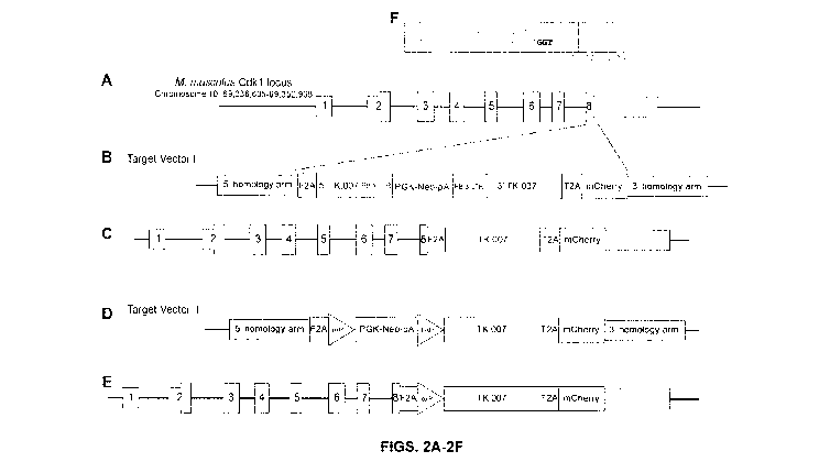
cells and their
applications toward many directions including cell-based therapies for
treating diseases and
aberrant physiological conditions have been developed further in the years
since.
[0004] One concern regarding pluripotent cell-based therapies is safety.
For example,
malignant growth originating from a cell graft is of concern. The process of
reprogramming
differentiated cells into iPS cells is also relevant to safety, as it has been
reported that
reprogramming methods can cause geno me damage and aberrant epigenetic changes
(Hussein et al., 2011; Laurent et al., 2011; Lister et al., 2011), which may
pose a risk for
malignant transformation of iPS cell-derived cells.
[0005] One challenge with cell-based therapies involving pluripotent cells
expanded in
vitro is the pluripotent nature of the cells themselves. For example, if
pluripotent cells remain
among differentiated therapeutic cells, the pluripotent cells may develop into
teratomas
CA 02978633 2017-09-05
WO 2016/141480
PCT/CA2016/050256
(Yoshida and Yamanaka, 2010). Attempts to increase the safety of pluripotent
cell-derived
products and therapies have included efforts to eliminate pluripotent cells
from cell cultures
after in vitro differentiation. For example: cytotoxic antibodies have been
used to eliminate
cells having pluripotent-specific antigens (Choo et al., 2008; Tan et al.,
2009); cells have
been sorted based on pluripotency cell surface markers (Ben-David et al.,
2013a; Fong et
al., 2009; Tang et al., 2011); tumour progression genes have been genetically
altered in cells
(Blum et al., 2009; Menendez et al., 2012); transgenes for assisting with
separation of
differentiated cells have been introduced into cells (Chung et al., 2006;
Eiges et al., 2001;
Huber et al., 2007); suicide genes have been introduced into cells and used to
eliminate
residual pluripotent stem cells after differentiation (Rong et al., 2012;
Schuldiner et al.,
2003); and undesired pluripotent cells have been ablated using chemicals (Ben-
David et al.,
2013b; Dabir et al., 2013; Tohyama et al., 2013). It is possible that even if
residual
pluripotent cells are eliminated from differentiated cultures, the
differentiated derivatives of
pluripotent cells may have oncogenic properties (Ghosh et al., 2011). Related
oncogenic
events could occur in therapeutic cells i) during in vitro preparation of
cells; or ii) following
grafting of cells into a host.
[0006] Most current strategies for eliminating or preventing unwanted cell
growth and/or
differentiation are based on the herpes simplex virus ¨ thymidine kinase (HSV-
TK) /
ganciclovir (GCV) negatively selectable system, which may be used to eliminate
a graft
entirely, if malignancy develops (Schuldiner et al., 2003) or to eliminate
only the pluripotent
cells 'contaminating' the intended differentiated derivatives (Ben-David and
Benvenisty,
2014; Lim et al., 2013). The mechanism of GCV-induced cell killing and
apoptosis is well
understood. It creates a replication-dependent formation of DNA double-strand
breaks
(Halloran and Fenton, 1998), which leads to apoptosis (Tomicic et al., 2002).
However,
many HSV-TK/GCV-based systems are unreliably expressed, at least because they
rely on
random integration or transient expression of HSV-TK. Strategies involving
negative
selectable markers with different killing mechanisms, such as, for example,
Caspase 9 (Di
Stasi et al., 2011) have been tested, but reliable expression of the negative
selectable
marker has not been shown. Cell-based therapies may require millions or
billions of cells,
which may amplify any issues caused by unwanted cell growth and/or
differentiation.
[0007] It is an object of the present disclosure to mitigate and/or obviate
one or more of
the above deficiencies.
SUMMARY OF THE DISCLOSURE
[0008] In an aspect, a method of controlling proliferation of an animal
cell is provided.
The method comprises: providing an animal cell; genetically modifying in the
animal cell a
2
CA 02978633 2017-09-05
WO 2016/141480
PCT/CA2016/050256
cell division locus (CDL), the CDL being one or more loci whose transcription
product(s) is
expressed by dividing cells; the genetic modification of the CDL comprising
one or more of:
a) an ablation link (ALINK) system, the ALINK system comprising a DNA sequence
encoding
a negative selectable marker that is transcriptionally linked to a DNA
sequence encoding the
CDL; and b) an inducible exogenous activator of regulation of a CDL (EARC)
system, the
EARC system comprising an inducible activator-based gene expression system
that is
operably linked to the CDL; controlling proliferation of the genetically
modified animal cell
comprising the ALINK system with an inducer of the negative selectable marker;
and/or
controlling proliferation of the genetically modified animal cell comprising
the EARC system
with an inducer of the inducible activator-based gene expression system.
[0009] In an embodiment of the method of controlling proliferation of an
animal cell
provided herein, the controlling of the ALINK-modified animal cell comprises
one or more of:
permitting proliferation of the genetically modified animal cell comprising
the ALINK system
by maintaining the genetically modified animal cell comprising the ALINK
system in the
absence of an inducer of the negative selectable marker; and ablating or
inhibiting
proliferation of the genetically modified animal cell comprising the ALINK
system by
exposing the animal cell comprising the ALINK system to the inducer of the
negative
selectable marker.
[0010] In an embodiment of the method of controlling proliferation of an
animal cell
provided herein, the controlling of the EARC-modified animal cell comprises
one or more of:
permitting proliferation of the genetically modified animal cell comprising
the EARC system
by exposing the genetically modified animal cell comprising the EARC system to
an inducer
of the inducible activator-based gene expression system; and preventing or
inhibiting
proliferation of the genetically modified animal cell comprising the EARC
system by
maintaining the animal cell comprising the EARC system in the absence of the
inducer of the
inducible activator-based gene expression system.
[0011] In various embodiments of the method of controlling proliferation of
an animal cell
provided herein, the genetic modification of the CDL comprises preforming
targeted
replacement of the CDL with one or more of: a) a DNA vector comprising the
ALINK system;
b) a DNA vector comprising the EARC system; and c) a DNA vector comprising the
ALINK
system and the EARC system.
[0012] In various embodiments of the method of controlling proliferation of
an animal cell
provided herein, the ALINK genetic modification of the CDL is homozygous,
heterozygous,
he mizygous or compound heterozygous and/or wherein the EARC genetic
modification
3
CA 02978633 2017-09-05
WO 2016/141480
PCT/CA2016/050256
ensures that functional CDL modification can only be generated through EARC-
modified
alleles.
[0013] In various embodiments of the method of controlling proliferation of
an animal cell
provided herein, the CDL is one or more loci recited in Table 2. In various
embodiments, the
CDL encodes a gene product whose function is involved with one or more of:
cell cycle, DNA
replication, RNA transcription, protein translation, and metabolism. In
various embodiments
the CDL is one or more of Cdk1/CDK1,Top2A/TOP2A, Cenpa/CENPA, Birc5/BIRC5, and
Eef2/EEF2, preferably the CDL is Cdk1 or CDK1.
[0014] In various embodiments of the method of controlling proliferation of
an animal cell
provided herein, the ALINK system comprises a herpes simplex virus-thymidine
kinase/ganciclovir system, a cytosine deaminase/5-fluorocytosine system, a
carboxyl
esterase/irinotecan system or an iCasp9/AP1903 system, preferably the ALINK
system is a
herpes simplex virus-thymidine kinase/ganciclovir system.
[0015] In various embodiments of the method of controlling proliferation of
an animal cell
provided herein, the EARC system is a dox-bridge system, a cumate switch
inducible
system, an ecdysone inducible system, a radio wave inducible system, or a
ligand-reversible
dimerization system, preferably the EARC system is a dox-bridge system.
[0016] In various embodiments of the method of controlling proliferation of
an animal cell
provided herein, the animal cell is a mammalian cell or an avian cell. In
various embodiment,
the mammalian cell is a human, mouse, rat, hamster, guinea pig, cat, dog, cow,
horse, deer,
elk, bison, oxen, camel, llama, rabbit, pig, goat, sheep, or non-human primate
cell, preferably
the mammalian cell is a human cell.
[0017] In various embodiments of the method of controlling proliferation of
an animal cell
provided herein, the animal cell is a pluripotent stem cell a multi potent
cell, a monopotent
progenitor cell, or a terminally differentiated cell.
[0018] In various embodiments of the method of controlling proliferation of
an animal cell
provided herein, the animal cell is derived from a pluripotent stem cell, a
multipotent cell, a
monopotent progenitor cell, or a terminally differentiated cell.
[0019] In an aspect, an animal cell genetically modified to comprise at
least one
mechanism for controlling cell proliferation is provided. The genetically
modified animal cell
comprises: a genetic modification of one or more cell division locus (CDL),
the CDL being
one or more loci whose transcription product(s) is expressed by dividing
cells. The genetic
modification being one or more of: a) an ablation link (ALINK) system, the
ALINK system
comprising a DNA sequence encoding a negative selectable marker that is
transcriptionally
4
CA 02978633 2017-09-05
WO 2016/141480
PCT/CA2016/050256
linked to a DNA sequence encoding the CDL; and b) an exogenous activator of
regulation of
a CEDL (EARC) system, the EARC system comprising an inducible activator-based
gene
expression system that is operably linked to the CDL.
[0020] In an embodiment of the animal cell genetically modified to comprise
at least one
mechanism for controlling cell proliferation provided herein, the genetic
modification of the
CDL comprises preforming targeted replacement of the CDL with one or more of:
a) a DNA
vector comprising the ALINK system; b) a DNA vector comprising the EARC
system; and c)
a DNA vector comprising the ALINK system and the EARC system.
[0021] In various embodiments of the animal cell genetically modified to
comprise at
least one mechanism for controlling cell proliferation provided herein, the
ALINK genetic
modification of the CDL is homozygous, heterozygous, he mizygous or compound
heterozygous and/or wherein the EARC genetic modification ensures that
functional CDL
modification can only be generated through EARC-modified alleles.
[0022] In various embodiments of the animal cell genetically modified to
comprise at
least one mechanism for controlling cell proliferation provided herein, the
CDL is one or
more loci recited in Table 2. In various embodiments, the CDL encodes a gene
product
whose function is involved with one or more of: cell cycle, DNA replication,
RNA
transcription, protein translation, and metabolism. In various embodiments,
the CDL is one
or more of Cdk1/CDK1, Top2ATTOP2A, Cenpa/CENPA, Birc5/BIRC5, and Eef2/EEF2,
preferably the CDL is Cdk1 or CDK1.
[0023] In various embodiments of the animal cell genetically modified to
comprise at
least one mechanism for controlling cell proliferation provided herein, the
ALINK system
comprises a herpes simplex virus-thymidine kinase/ganciclovir system, a
cytosine
deaminase/5-fluorocytosine system, a carboxyl esterase/irinotecan system or an
iCasp9/AP1903 system, preferably the ALINK system is a herpes simplex virus-
thymidine
kinase/ganciclovir system.
[0024] In various embodiments of the animal cell genetically modified to
comprise at
least one mechanism for controlling cell proliferation provided herein, the
EARC system is a
dox-bridge system, a cumate switch inducible system, an ecdysone inducible
system, a
radio wave inducible system, or a ligand-reversible dimerization system,
preferably the
EARC system is a dox-bridge system.
[0025] In various embodiments of the animal cell genetically modified to
comprise at
least one mechanism for controlling cell proliferation provided herein, the
animal cell is a
mammalian cell or an avian cell. In various embodiments, the mammalian cell is
a human,
CA 02978633 2017-09-05
WO 2016/141480
PCT/CA2016/050256
mouse, rat, hamster, guinea pig, cat, dog, cow, horse, deer, elk, bison, oxen,
camel, llama,
rabbit, pig, goat, sheep, or non-human primate cell, preferably the mammalian
cell is a
human cell.
[0026] In various embodiments of the animal cell genetically modified to
comprise at
least one mechanism for controlling cell proliferation provided herein, the
animal cell is a
pluripotent stem cell a multipotent cell, a monopotent progenitor cell, or a
terminally
differentiated cell.
[0027] In various embodiments of the animal cell genetically modified to
comprise at
least one mechanism for controlling cell proliferation provided herein, the
animal cell is
derived from a pluripotent stem cell, a multipotent cell, a monopotent
progenitor cell, or a
terminally differentiated cell.
[0028] In an aspect, a DNA vector for modifying expression of a cell
division locus
(CDL), the CDL being one or more loci whose transcription product(s) is
expressed by
dividing cells is provided. The DNA vector comprises: an ablation link (ALINK)
system, the
ALINK system comprising a DNA sequence encoding a negative selectable marker
that is
transcriptionally linked to the CDL, wherein if the DNA vector is inserted
into one or more
host cells, proliferating host cells comprising the DNA vector will be killed
if the proliferating
host cells comprising the DNA vector are exposed to an inducer of the negative
selectable
marker.
[0029] In an aspect, DNA vector for modifying expression of a cell division
essential
locus (CDL), the CDL being one or more loci whose transcription product(s) is
expressed by
dividing cells is provided. The DNA vector comprises: an exogenous activator
of regulation
of a CDL (EARC) system, the EARC system comprising an inducible activator-
based gene
expression system that is operably linked to the CDL, wherein if the DNA
vector is inserted
into one or more host cells, proliferating host cells comprising the DNA
vector will be killed if
the proliferating host cells comprising the DNA vector are not exposed to an
inducer of the
inducible activator-based gene expression system.
[0030] In an aspect, a DNA vector for modifying expression of a cell
division essential
locus (CDL), the CDL being one or more loci whose transcription product(s) is
expressed by
dividing cells is provided. The DNA vector comprises: an ablation link (ALINK)
system, the
ALINK system being a DNA sequence encoding a negative selectable marker that
is
transcriptionally linked to the CDL; and an exogenous activator of regulation
of CDL (EARC)
system, the EARC system comprising an inducible activator-based gene
expression system
that is operably linked to the CDL, wherein if the DNA vector is inserted into
one or more
6
CA 02978633 2017-09-05
WO 2016/141480
PCT/CA2016/050256
host cells, proliferating host cells comprising the DNA vector will be killed
if the proliferating
host cells comprising the DNA vector are exposed to an inducer of the negative
selectable
marker and if the proliferating host cells comprising the DNA vector are not
exposed to an
inducer of the inducible activator-based gene expression system.
[0031] In various embodiments of the DNA vectors provided herein, the CDL
is one or
more loci recited in Table 2. In various embodiments, the CDL encodes a gene
product
whose function is involved with one or more of: cell cycle, DNA replication,
RNA
transcription, protein translation, and metabolism. In various embodiments,
the CDL is one
or more of Cdk1/CDK1,Top2A/TOP2A, Cenpa/CENPA, Birc5/BIRC5, and Eef2/EEF2,
preferably the CDL is Cdk1 or CDK1.
[0032] In various embodiments of the DNA vectors provided herein, the ALINK
system
comprises a herpes simplex virus-thymidine kinase/ganciclovir system, a
cytosine
deaminase/5-fluorocytosine system, a carboxyl esterase/irinotecan system or an
iCasp9/AP1903 system, preferably the ALINK system is a herpes simplex virus-
thymidine
kinase/ganciclovir system.
[0033] In various embodiments of the DNA vectors provided herein, the EARC
system is
a dox-bridge system, a cumate switch inducible system, an ealysone inducible
system, a
radio wave inducible system, or a ligand-reversible dimerization system,
preferably the
EARC system is a dox-bridge system.
[0034] In an aspect, a kit for controlling proliferation of an animal cell
by genetically
modifying one or more cell division essential locus/loci (CDL), the CDL being
one or more
loci whose transcription product(s) is expressed by dividing cells is
provided. The kit
comprises: a DNA vector comprising an ablation link (ALINK) system, the ALINK
system
comprising a DNA sequence encoding a negative selectable marker that is
transcriptionally
linked to a DNA sequence encoding the CDL; and/or a DNA vector comprising an
exogenous activator of regulation of a CDL (EARC) system, the EARC system
comprising
an inducible activator-based gene expression system that is operably linked to
the CDL;
and/or a DNA vector comprising an ALINK system and an EARC system, the ALINK
and
EARC systems each being operably linked to the CDL; and instructions for
targeted
replacement of the CDL in an animal cell using one or more of the DNA vectors.
[0035] In an embodiment of the kit provided herein, the CDL is one or more
loci recited
in Table 2. In various embodiments, the CDL encodes a gene product whose
function is
involved with one or more of: cell cycle, DNA replication, RNA transcription,
protein
translation, and metabolism. In various embodiments, the CDL is one or more of
7
CA 02978633 2017-09-05
WO 2016/141480
PCT/CA2016/050256
Cdk1/CDK1,Top2ATTOP2A, Cenpa/CENPA, Birc5/BIRC5, and Eef2/EEF2, preferably the
CDL is Cdk1 or CDK1.
[0036] In various embodiments of the kit provided herein, the ALINK system
comprises a
herpes simplex virus-thymidine kinase/ganciclovir system, a cytosine
deaminase/5-
fluorocytosine system, a carboxyl esterase/irinotecan system or an
iCasp9/AP1903 system,
preferably the ALINK system is a herpes simplex virus-thymidine
kinase/ganciclovir system.
[0037] In various embodiments of the kit provided herein, the EARC system
is a dox-
bridge system, a cumate switch inducible system, an ecdysone inducible system,
a radio
wave inducible system, or a ligand-reversible dimerization system, preferably
the EARC
system is a dox-bridge system.
DESCRIPTION OF THE DRAWINGS
[0038] The patent or application file contains at least one drawing in
color. Copies of
this patent or patent application publication with color drawings will be
provided by the Office
upon request and payment of the necessary fee.
[0039] These and other features of the disclosure will become more apparent
in the
following detailed description in which reference is made to the appended
drawings wherein:
[0040] Figures 1A-1G depict schematics illustrating the concept of induced
negative
effectors of proliferation (iNEPs) and examples of iNEP systems contemplated
for use in the
methods and tools provided herein. FIG. 1A depicts a schematic representing
different
examples of iNEP-modified CDLs, including a homozygous modification in CDL1,
homozygous insertions in CDL1 and CDL2, CDL comprising bno separate loci that
together
are essential for cell division (CDL3). FIG. 1B depicts schematics
representing examples of
iNEP comprising an ablation link (ALINK) and an exogenous activator of
regulation of a CDL
(EARC) in different configurations. FIG. 1C depicts a schematic illustrating
transcription
activator-like effector (TALE) technology combined with dimerizer-regulated
expression
induction. FIG. 1D depicts a schematic illustrating a reverse-cumate-Trans-
Activator (rcTA)
system. FIG. lE depicts a schematic illustrating a retinoid X receptor (RXR)
and an N-
termina I truncation of ecclysone receptor (EcR) fused to the activation
domain of Vp16
(VpEcR). FIG. 1F depicts a schematic illustrating a transient receptor
potential vanilloid-1
(TRPV1), together with ferritin, which is one example of an iNEP system, as
set forth herein.
FIG. 1G depicts a schematic illustrating how an IRES and a dimerization agent
may be used
as an iNEP.
[0041] Figures 2A-2F depict schematics illustrating targeting HSV-TK into
the 3'UTR of
the Cdk1 locus to generate an ALINK, which enables elimination of dividing
modified CDK1-
8
CA 02978633 2017-09-05
WO 2016/141480
PCT/CA2016/050256
expressing cells. FIG. 2A shows a schematic of the mouse Cdkl locus. FIG. 2B
shows a
schematic of mouse target vector I. FIG. 2C shows a schematic of a Cdk1TC
allele. FIG. 2D
shows a schematic of mouse target vector II. FIG. 2E shows a schematic of a
Cdk1Ta x
allele. FIG. 2F depicts the position of the CRISPR guide RNA; the sequence in
the yellow
box is the 8th exon of Cdk1.
[0042] Figures 3A-3G depict generation of ALINK example, HSV-TK-mCherry
into the
3'UTR of the CDK1 locus to generate ALINK in mouse ES cell lines. Fig. 3A
shows the
overall steps of generating ALINK in mouse C2 ES cells. Fig. 3B shows southern
blotting
result of correct genotyping of Cdk1(TK/+), Cdk1(TK, loxP-TK), and
Cdk1(TK/TK). Fig. 3C
shows the locations of the primers used in ALINK genotyping in mouse cells.
Fig. 3D
includes PCR results illustrating targeting of Targeting Vector 1 into the
3'UTR of the CDK1
locus. Fig. 3E shows PCR results illustrating the excision event of selection
marker in a
mouse ES cell line already correctly targeted with Targeting Vector Ito
activate the
expression of HSV-TK-mCherry. Fig. 3F shows PCR results illustrating targeting
of Targeting
Vector 11 into Cdk1(TK/+) cells. Fig. 3G shows PCR results illustrating the
excision event of
selection marker in Cdk1(TK, loxP-TK) to activate the 2nd allele expression of
HSV-TK-
mCherry, thus generating Cdk1(TK/TK).
[0043] Figures 4A-4K depict generation of an ALINK modification, HSV-TK-
mCherry into
the 3'UTR of the CDK1 locus, in human ES cell lines. Fig. 4A shows the overall
steps of
generating ALINK in human CA1 ES cells. Fig. 4B shows the locations of the
primers used in
ALINK genotyping in human CA1 cells. Fig. 4C shows PCR results illustrating
targeting of
Targeting Vector 1 into the 3'UTR of the CDK1 locus. Fig. 4D shows flow
cytometry
illustrating the excision event of selection marker in human Cdk1(PB-TK/+) ES
cell line to
activate the expression of HSV-TK-mCherry; the Y-axis shows the mCherry
expression
level, while the X-axis is an autofluorescence channel. Fig. 4E shows PCR
results illustrating
targeting of Targeting Vector 11 (puro-version) into Cdk1(TK/+) cells; the
upper panel is PCR
using primers flanking the 5'homology arm; the lower panel is PCR using
primers inside 5'
and 3' homology arm, so absence of 0.7kb band and presence of 2.8kb band means
that the
clone is homozygous in ALINK, and presence of 0.7kb band means that the clone
is
heterozygous in ALINK or the population is not clonal. Fig. 4F shows flow
cytometry analysis
illustrating the excision event of selection marker in Cdk1(TK, loxP-TK) to
activate the 2nd
allele expression of HSV-TK-mCherry; the Y-axis shows the mCherry expression
level, while
the X-axis is an autofluorescence channel. Fig. 4G shows the overall steps of
generating
ALINK in human H1 ES cells. Fig. 4H shows the locations of the primers used in
ALINK
genotyping in human H1 cells. Fig. 41 shows PCR results illustrating targeting
of Targeting
9
CA 02978633 2017-09-05
WO 2016/141480
PCT/CA2016/050256
Vector II into the 3'UTR of the CDK1 locus. Fig. 4J shows PCR results
illustrating the
excision event of selection marker in human H1 Cdk1(loxP-TK/+) to activate the
expression
of HSV-TK-mCherry; the Y-axis shows the mCherry expression level, while the X-
axis is an
autofluorescence channel. Fig. 4K shows fluorescence-activated cell sorting
(FACS) of
targeting of Targeting Vector III (GFP-version) into Cdk1(TK/+) cells. After
FACS sorting,
clones picked from sparse plating were genotyped with mCherry-allele-specific
primers,
eGFP-allele-specific primers and primers in 5' and 3' homology arms; clones
labeled with
orange star sign are homozygous ALINK with one allele of mCherry and one
allele of eGFP;
the one clone labeled with green star sign is homozygous ALINK with bno
alleles of eGFP.
[0044] Figures 5A-C depict teratoma histology (endoderm, mesoderm and
ectoderm
portions of the teratoma are shown from left to right, respectively). FIG. 5A
depicts
photomicrographs of a teratoma derived from a mouse ES Cdk1 / , alinWalink
cell. FIG 5B
depicts photomicrographs of a teratoma derived from a mouse ES Cdk1 earc/earc,
alink/alink cell.
FIG. 5C depicts photomicrographs of a teratoma derived from a human ES Cdk1 /
,al1nkial1nk
cell.
[0045] Figures 6A-6B depict in vitro functional analysis of mouse ES cells
with an HSV-
TK ¨ mCherry knock-in into the 3'UTR of the CDK1 locus. FIG. 6A illustrates
killing efficiency
provided by the TK.007 gene after cells were exposed to different
concentrations of GCV for
3 days. Colony size and number are directly proportional to GCV concentration.
The second
lowest concentration of 0.01pM did not affect the colony number but slowed
down cell
growth as evidenced by the reduced colony size (n=5). FIG. 6B illustrates
expression of
mCherry before (Cdk1.1-ISV-TK=NeolN) and after (Cdk1.1-ISV-TK) PB-mediated
removal of
the neo-cassette.
[0046] Figures 7A-F depict results of cellular experiments using ALINK-
modified cells.
FIG. 7A graphically depicts results of GCV treatment of subcutaneous teratomas
comprising
ALINK-modified mouse C2 cells. FIG. 7B graphically depicts results of GCV
treatment of
subcutaneous teratomas comprising ALINK-modified H1 ES cells. FIG. 7C
graphically
depicts results of GCV treatment of mammary gland tumors comprising ALINK-
modified
cells. FIG. 7D schematically depicts experimental design of neural assay. FIG.
7E is a
microscopic image of Neural Epithelial Progenitor (NEP) cells derived from
Cdk1 / , /allnk
human CA1 ES cells. FIG. 7F depicts microscopic images illustrating GCV-
induced killing of
dividing AL INK-modified NEPs and non-killing of non-dividing neurons.
[0047] Figure 8 depicts a graph showing the expected number of cells
comprising
spontaneous mutations in the HSV-TK gene as a population is expanded from
heterozygous
(blue line) and homozygous (red line) ALINK cells.
CA 02978633 2017-09-05
WO 2016/141480
PCT/CA2016/050256
[0048] Figures 9A-9B depict targeting of a dox-bridge into the 5'UTR of the
mouse Cdk1
locus to generate EARC and behavior of the bridge after insertion into Cdk1.
FIG. 9A is a
schematic illustrating the structure of the mouseCdk1 locus, the target
vector, and the
position of the primers used for genotyping for homologous recombination
events. FIG. 9B
depicts PCR results showing the genotyping of the puromycin resistant colonies
to identify
those that integrated the dox-bridge to the Cdk1 5'UTR.
[0049] Figure 10 depicts a flowchart illustrating that ES cells having a
homozygous dox-
bridge knock-in survive and divide only in the presence of doxycycline (or
drug with
doxycycline overlapping function).
[0050] Figure 11 depicts representative photomicrographs illustrating that
homozygous
dox-bridge knock-in ES cells show doxycycline concentration dependent survival
and
growth.
[0051] Figure 12 depicts dox-bridge removal with Cre recombinase-mediated
excision,
which rescues the doxycycline dependent survival of the ES cells.
[0052] Figures 13A-13B depict the effect of doxycycline withdrawal on the
growth of dox-
bridged ES cells. FIG. 13A depicts a graph showing that in the presence of
doxycycline the
cells grew exponentially (red line with circle), indicating their normal
growth. Upon
doxycycline withdrawal on Day 1, the cells grew only for bno days and then
they started
disappearing from the plates until no cell left on Day 9 on (dark blue line
with square). The
20x lower doxycycline concentration (50ng/m1) after an initial 3 days of
growth kept a
constant number of cells on the plate for at least five days (Fig. 13, light
blue line with
triangle). On Day 10 the normal concentration of doxycycline was added back to
the plates
and the cells started growing again as normal ES cells. FIG. 13B depicts a bar
graph
showing the level of Cdk1 mRNA (as measured by quantitative-PCR) after 0, 1
and 2 days of
Dox removal. Expression levels are normalized to beta-actin.
[0053] Figure 14 depicts the process of growing dox-bridged ES cells and
illustrates that
no escaper cells were found among 100,000,000 dox-bridged ES cells when
doxycycline
was withdrawn from the media, but the sentinel (wild type, GFP positive) cells
survived with
high efficiency.
[0054] Figure 15 depicts a graph showing the effect of high doxycycline
concentration
(10 pg/ml) on dox-bridged ES cells: in the presence of high doxycycline, the
cells slow down
their growth rate similarly to when in low-doxycycline (high dox was 10 pg/ml,
normal dox
was 1 pg/ml, low dox was 0.05 pg/ml), indicating that there is a window for
Dox
concentration defining optimal level of CDK1 expression for cell
proliferation.
11
CA 02978633 2017-09-05
WO 2016/141480
PCT/CA2016/050256
[0055] Figures 16A-16B depict targeting of dox-bridge into the 5'UTR of the
Cdk1 locus
of mouse cells comprising AL INK modifications (i.e., Cdk1(TK/TK) cells; the
cell product
described in FIGS. 3A-3G). FIG. 16A is a schematic illustrating the structure
of the Cdk1
locus in Cdk1(TK/TK) cells, the bridge target vector, and the location of
genotyping primers.
FIG. 16B depicts PCR results showing the genotyping of the puromycin resistant
colonies to
identify those that integrated the dox-bridge to the Cdk1 5'UTR in mouse
Cdk1(TK/TK) cells,
thus generating mouse cell product Cdk1 earc/earc, alink/alink.
[0056] Figures 17A-17B depict targeting of dox-bridge into the 5'UTR of the
Cdk1 locus
of human cells comprising ALINK modifications (i.e., Cdk1(TK/TK) cells; the
cell product
described in FIGS. 4A-4F). FIG. 17A is a schematic illustrating the structure
of the Cdk1
locus in Cdk1(TK/TK) cells, the bridge target vector, and the location of
genotyping primers.
Fig. 17B depicts PCR results showing the genotyping of the puromycin resistant
colonies to
identify those that integrated the dox-bridge to the Cdk1 5'UTR in human
Cdk1(TK/TK) cells,
thus generating human cell product Cdk1 earc/earc, alink/alink.
[0057] Figures 18A-18B depict targeting of a dox-bridge into the 5'UTR of
the Top2
locus to generate EARC insertion into Top2a. FIG. 18A is a schematic
illustrating the
structure of the Top2a locus and the target vector. TOP2a_5scrF, rttaRev,
CMVforw and
TOP2a_3scrR indicate the position of the primers used for genotyping for
homologous
recombination events. FIG. 18B depicts PCR results showing the genotyping of
the puro
resistant colonies to identify those that integrated the dox-bridge to the
Top2a 5'UTR. Nine
of these cell lines was found to be homozygous targeted comprising a dox-
bridge inserted by
homologous recombination into the 5'UTR of both alleles of Top2a.
[0058] Figures 19A-19B depict the effect of doxycycline withdrawal on the
growth of
Top2a-EARC ES cells. FIG. 19A shows that withdrawal of doxycycline results in
complete
elimination of mitotically active ES cells within 4 days. FIG. 19B depicts how
different
concentrations of doxycycline affected proliferation of the dox-bridge ES
cells by measuring
cell growth for 4 days. ES cells in the presence of doxycycline grew
exponentially, indicating
their normal growth. In contrast, two days after doxycycline removal, cells
growth was
completely arrested.
[0059] Figures 20A-20B depict targeting of a dox-bridge into the 5'UTR of
the Cenpa
locus to generate EARC insertion into Cenpa. FIG. 20A is a schematic
illustrating the
structure of the Cenpa locus and the target vector. Cenpa_5scrF, rttaRev,
CMVforw and
Cenpa_3scrR indicate the position of the primers used for genotyping for
homologous
recombination events. FIG. 20B depicts PCR results showing the genotyping of
the puro
resistant colonies to identify those that integrated the dox-bridge to the
Cenpa 5'UTR. Six of
12
CA 02978633 2017-09-05
WO 2016/141480
PCT/CA2016/050256
these cells were found to have a correct insertion at the 5' and 3', and at
least one clone
(Cenpa#4), was found to have homozygous targeting comprising a dox-bridge
inserted by
homologous recombination into the 5'UTR of both alleles of Cenpa.
[0060] Figures 21A-21B depict the effect of doxycycline withdrawal on the
growth of
Cenpa-EARC ES cells. FIG. 21A depicts that withdrawal of doxycycline results
in complete
elimination of mitotically active ES cells within 4 days. FIG. 21B is the
Cenpa gene
expression level (determined by q-PCR) in Cenpa-EARC cells with Dox and after
2 days of
Dox removal, and compared it to the expression level in wild type mouse ES
cells (C2). As
expected Cenpa expression level is greatly reduced in Cenpa-EARC cells without
Dox for 2
days.
[0061] Figure 22 depicts how different concentrations of doxycycline
affected
proliferation of the Cenpa-EARC ES cells by measuring cell growth for 4 days.
ES cells in
the presence of doxycycline grew exponentially, indicating their normal
growth. In contrast,
80 hours after doxycycline removal, cells growth was completely arrested.
[0062] Figures 23A-23B depict targeting of a dox-bridge into the 5'UTR of
the Birc5
locus to generate EARC insertion into Birc5. FIG. 23A is a schematic
illustrating the structure
of the Birc5 locus and the target vector. Birc_5scrF and rttaRev indicate the
position of the
primers used for genotyping for homologous recombination events. FIG. 23B
depicts PCR
results showing the genotyping of the puro resistant colonies to identify
those that integrated
the dox-bridge to the Birc5 5'UTR. Five clones were found to be correctly
targeted
comprising a dox-bridge inserted by recombination into the 5'UTR of both
alleles of Birc5.
One of these clones was Birc#3, was found to stop growing or die in the
absence of Dox.
[0063] Figures 24A-24B depict the effect of doxycycline withdrawal on the
growth of
Birc5-EARC ES cells. FIG. 24A depicts that withdrawal of doxycycline results
in complete
elimination of mitotically active ES cells within 4 days. FIG. 24B is the
Birc5 gene expression
level (determined by q-PCR) in Birc5-EARC cells with Dox and after 2 days of
Dox removal,
and compared it to the expression level in wild type mouse ES cells (C2). As
expected Birc5
expression level is greatly reduced in Birc5-EARC cells without Dox for 2
days.
[0064] Figure 25 depicts how different concentrations of doxycycline
affected
proliferation of the Birc5-EARC ES cells by measuring cell growth for 4 days.
ES cells in the
presence of doxycycline grew exponentially, indicating their normal growth. In
contrast, 50
hours after doxycycline removal, cells growth was completely arrested.
Interestingly, it
appears that lower Dox concentrations (0.5 and 0.05 pg/ml) promote better cell
growth than
a higher concentration (1 pg/ml).
13
CA 02978633 2017-09-05
WO 2016/141480
PCT/CA2016/050256
[0065] Figures 26A-26B depict targeting of a dox-bridge into the 5'UTR of
the Eef2 locus
to generate EARC insertion into Eef2. FIG. 26A is a schematic illustrating the
structure of the
Eef2 locus and the target vector. Eef2_5scrF and rttaRev indicate the position
of the primers
used for genotyping for homologous recombination events. FIG. 26B depicts PCR
results
showing the genotyping of the puro resistant colonies to identify those that
integrated the
dox-bridge to the Eef2 5'UTR. Nine of these cell lines was found to be
correctly targeted with
at least one clone growing only in Dox-media.
[0066] Figures 27 depict the effect of doxycycline withdrawal on the growth
of Eef2-
EARC ES cells. Withdrawal of doxycycline results in complete elimination of
mitotically
active ES cells within 4 days.
[0067] Figure 28 depicts how different concentrations of doxycycline
affected
proliferation of the Eef2-EARC ES cells by measuring cell growth for 4 days.
ES cells in the
presence of doxycycline grew exponentially, indicating their normal growth. In
contrast,
without doxycycline cells completely fail to grow.
DETAILED DESCRIPTION OF THE DISCLOSURE
[0068] Unless defined otherwise, all technical and scientific terms used
herein have the
same meaning as commonly understood by one of ordinary skill in the art to
which this
disclosure belongs.
[0069] Definitions
[0070] The terms "cell division locus", "cell division loci", and "CDL" as
used herein, refer
to a genomic locus (or loci) whose transcription product(s) is expressed by
dividing cells.
When a CDL comprises a single locus, absence of CDL expression in a cell (or
its
derivatives) means that tumour initiation and/or formation is prohibited
either because the
cell(s) will be ablated in the absence of CDL expression or because
proliferation of the cell(s)
will be blocked or compromised in the absence of CDL expression. When a CDL
comprises
multiple loci, absence of expression by all or subsets of the loci in a cell
(or its derivatives)
means that tumour initiation and/or formation is prohibited either because the
cell(s) will be
ablated in the absence of CDL expression or because proliferation of the
cell(s) will be
blocked or compromised in the absence of CDL expression. A CDL may or may not
be
expressed in non-dividing and/or non-proliferating cells. A CDL may be
endogenous to a
host cell or it may be a transgene. If a CDL is a transgene, it may be from
the same or
different species as a host cell or it may be of synthetic origin. In an
embodiment, a CDL is a
single locus that is transcribed during cell division. For example, in an
embodiment, a single
locus CDL is CDK1. In an embodiment, a CDL comprises two or more loci that are
14
CA 02978633 2017-09-05
WO 2016/141480
PCT/CA2016/050256
transcribed during cell division. For example, in an embodiment, a mufti-locus
CDL
comprises two MYC genes (c-Myc and N-myc) (Scognamiglio et al., 2016). In an
embodiment, a multi-locus CDL comprises AURORA B and C kinases, Mich may have
overlapping functions (Fernandez-Miranda et al., 2011). Cell division and cell
proliferation
are terms that may be used interchangeably herein.
[0071] The terms "normal rate of cell division", "normal cell division
rate", "normal rate of
cell proliferation", and "normal cell proliferation rate" as used herein,
refer to a rate of cell
division and/or proliferation that is typical of a non-cancerous healthy cell.
A normal rate of
cell division and/or proliferation may be specific to cell type. For example,
it is widely
accepted that the number of cells in the epidermis, intestine, lung, blood,
bone marrow,
thymus, testis, uterus and mammary gland is maintained by a high rate of cell
division and a
high rate of cell death. In contrast, the number of cells in the pancreas,
kidney, cornea,
prostate, bone, heart and brain is maintained by a low rate of cell division
and a low rate of
cell death (Pellettieri and Sanchez Alvarado, 2007).
[0072] The terms "inducible negative effector of proliferation" and "iNEP"
as used herein,
refer to a genetic modification that facilitates use of CDL expression to
control cell division
and/or proliferation by: i) inducibly stopping or blocking CDL expression,
thereby prohibiting
cell division and proliferation; ii) inducibly ablating at least a portion of
CDL-expressing cells
(i.e., killing at least a portion of proliferating cells); or iii) inducibly
slowing the rate of cell
division relative to a cell's normal cell division rate, such that the rate of
cell division would
not be fast enough to contribute to tumor formation.
[0073] The terms "ablation link" and "ALINK" as used herein, refer to an
example of an
iNEP, which comprises a transcriptional link between a CDL and a sequence
encoding a
negative selectable marker. The ALINK modification allows a user to inducibly
kill
proliferating host cells comprising the ALINK or inhibit the host cell's
proliferation by killing at
least a portion of proliferating cells by exposing the ALINK-modified cells to
an inducer of the
negative selectable marker. For example, a cell modified to comprise an ALINK
at a CDL
may be treated with an inducer (e.g., a prodrug) of the negative selectable
marker in order to
ablate proliferating cells or to inhibit cell proliferation by killing at
least a portion of
proliferating cells (Figure 1B).
[0074] The terms "exogenous activator of regulation of CDL" and "EARC" as
used
herein, refer to an example of an iNEP, which comprises a mechanism or system
that
facilitates exogenous alteration of non-coding or coding DNA transcription or
corresponding
translation via an activator. An EARC modification allows a user to inducibly
stop or inhibit
division of cells comprising the EARC by removing from the EARC-modified cells
an inducer
CA 02978633 2017-09-05
WO 2016/141480
PCT/CA2016/050256
that permits transcription and/or translation of the EARC-modified CDL. For
example, an
inducible activator-based gene expression system may be operably linked to a
CDL and
used to exogenously control expression of a CDL or CDL translation, such that
the presence
of a drug inducible activator and corresponding inducer drug are required for
CDL
transcription and/or translation. In the absence of the inducer drug, cell
division and/or
proliferation would be stopped or inhibited (e.g., slowed to a normal cell
division rate). For
example, the CDL Cdk1/CDK1 may be modified to comprise a dox-bridge (Figure
1B), such
that expression of Cdk1/CDK1 and cell division and proliferation are only
possible in the
presence of an inducer (e.g., doxycycline).
[0075] The term "proliferation antagonist system" as used herein, refers to
a natural or
engineered compound(s) whose presence inhibits (completely or partially)
proliferation of a
cell.
[0076] General Description of Tools and Methods
[0077] As described herein, the inventors have provided molecular tools,
methods and
kits for using one or more cell division loci (CDL) in an animal cell to
generate genetically
modified cells in which cell division and/or proliferation can be controlled
by a user through
one or more iNEPs (FIG. 1A). For example, division of cells generated using
one or more
tools and/or methods provided herein could be stopped, blocked or inhibited by
a user such
that a cell's division rate would not be fast enough to contribute to tumor
formation. For
example, proliferation of cells generated using one or more tools and/or
methods provided
herein could be stopped, blocked or inhibited by a user, by killing or
stopping at least a
portion of proliferating cells, such that a cell's proliferation rate or
volume may be maintained
at a rate or size, respectively, desired by the user.
[0078] Tools and methods for controlling cell division and/or proliferation
are desirable,
for example, in instances wherein faster cell division rates (relative to
normal cell division
rates) are undesirable. For example, cells that divide at faster than normal
rates may form
tumors in situ, which may be harmful to a host. In an embodiment, the
genetically modified
animal cells provided herein comprise one or more mechanisms for allowing
normal cell
division and/or proliferation and for stopping, ablating, blocking and/or
slowing cell division
and/or proliferation, such that undesirable cell division and/or proliferation
may be controlled
by a user (FIG. 1B). Referring to FIG. 1B, in example (I) EARC is inserted at
the 5' UTR of
the CDL and ALINK is inserted at the 3' UTR, the product of transcription is a
bi-cistronic
mRNA that get processed in two proteins. In example (II) both EARC and ALINK
are
inserted at the 5' UTR of the CDL, the product of transcription is a bi-
cistronic mRNA that get
processed in bno proteins. In example (III) EARC is inserted at the 5' UTR of
the CDL and
16
CA 02978633 2017-09-05
WO 2016/141480
PCT/CA2016/050256
ALINK is inserted within the CDL coding sequence, the product of transcription
is a mRNA
that get processed in a precursor protein that will generate tm separate
protein upon
cleavage of specifically designed cleavage sequences. In example (IV) both
EARC and
ALINK are inserted at the 5' UTR of the CDL, the product of transcription is a
mRNA that get
processed into a fusion protein that maintains both CDL and ALINK functions.
In example
(V) EARC is inserted at the 5' UTR of the CDL and ALINK is inserted at the 3'
UTR, the
product of transcription is a mRNA that get processed into a fusion protein
that maintains
both CDL and ALINK functions.
[0079] For example, the genetically modified animal cells provided herein
may be used
in a cell therapeutic treatment applied to a subject. If one or more of the
genetically modified
animal cells provided to the subject were to begin dividing at an undesirable
rate (e.g., faster
than normal), then a user could stop or slow division of cells dividing at the
undesirable rate
or block, slow or stop cells proliferating at the undesirable rate by i)
applying to the cells
dividing at the undesirable rate an inducer corresponding to the genetic
modification in the
cells; or ii) restricting access of the cells dividing at the undesirable rate
to an inducer
corresponding to the genetic modification in the cells, i) or ii) being
determined based on the
type of iNEP(s) provided in the genetically modified animal cells.
[0080] In an embodiment, the genetically modified animal cells provided
herein may be
referred to as "fail-safe cells". A fail-safe cell contains one or more
homozygous,
heterozygous, hemizygous or compound heterozygous ALINKs in one or more CDLs.
In an
embodiment, a fail-safe cell further comprises one or more EARCs in one or
more CDL. In
an embodiment, a fail-safe cell comprises a CDL comprising both ALINK and EARC
modifications.
[0081] As used herein, the term "fail-safe", refers to the probability
(designated as pFS)
defining a cell number. For example, the number of cells that can be grown
from a single
fail-safe cell (clone volume) where the probability of obtaining a clone
containing cells, which
have lost all ALINKs is less than an arbitrary value (pFS). For example, a pFS
= 0.01 refers
to a scenario wherein if clones were grown from a single cell comprising an
ALINK-modified
CDL 100 times, only one clone expected to have cells, which lost ALINK
function (the
expression of the negative selectable marker) while still capable of cell
division. The fail-safe
volume will depend on the number of ALINKs and the number of ALINK-targeted
CDLs. The
fail-safe property is further described in Table 1.
[0082] Table 1. Fail-safe cell volumes and their relationship to a human
body were
calculated using mathematical modelling. The model did not take into a count
the events
when CDL expression was co-lost with the loss of negative selectable marker
activity,
17
CA 02978633 2017-09-05
WO 2016/141480
PCT/CA2016/050256
compromising cell proliferation. Therefore the values are underestimates and
were
calculated assuming 10-6 forward mutation rate for the negative selectable
marker. The
estimated number of cells in a human body as 3.72x10¹³ was taken from
(Bianconi et al.,
2013).
CDL # ALINK# Genotype Fail-safe Relative (x) to a human
Estimated weight of
in CDLs volume body=3.72x10¹³ cells clones
(#cells)
1 1 het 512 0.0000000000137 1pg
1 2 hom 16777216 0.000000451 31 mg
2 3 het, hom 1.374E+11 0.004 0.26
kg
2 4 hom, hom 1.13E+15 30 2100
kg
[0083] It is contemplated herein that fail-safe cells may be of use in cell-
based therapies
wherein it may be desirable to eliminate cells exhibiting undesirable growth
rates,
irrespective of Mether such cells are generated before or after grafting the
cells into a host.
[0084] Cell Division Loci (CDLs)
[0085] The systems, methods and compositions provided herein are based on
the
identification of one or more CDLs, such as, for example, the CDLs set forth
in Table 2. It is
contemplated herein that various CDLs could be targeted using the methods
provided
herein.
[0086] In various embodiments, a CDL is a locus identified as an "essential
gene" as set
forth in Wang et al., 2015, which is incorporated herein by reference as if
set forth in its
entirety. Essential genes in Wang et al., 2015, were identified by computing a
score (i.e., a
CRISPR score) for each gene that reflects the fitness cost imposed by
inactivation of the
gene. In an embodiment, a CDL has a CRISPR score of less than about ¨1.0
(Table 2,
column 5).
[0087] In various embodiments, a CDL is a locus/loci that encodes a gene
product that
is relevant to cell division and/or replication (Table 2, column 6). For
example, in various
embodiments, a CDL is a locus/loci that encodes a gene product that is
relevant to one or
more of: i) cell cycle; ii) DNA replication; iii) RNA transcription and/or
protein translation; and
iv) metabolism (Table 2, column 7).
[0088] In an embodiment, a CDL is one or more cyclin-dependent kinases that
are
involved with regulating progression of the cell cycle (e.g., control of G1/S
G2/M and
metaphase-to-anaphase transition), such as CDK1, CDK2, CDK3, CDK4, CDK5, CDK6,
CDK7, CDK8, CDK9 and/or CDK11 (Morgan, 2007). In an embodiment, a CDL is one
or
more cyclins that are involved with controlling progression of the cell cycle
by activating one
or more CDK, such as, for example, cyclinB, cyclinE, cyclinA, cyclinC,
cyclinD, cyclinH,
18
CA 02978633 2017-09-05
WO 2016/141480
PCT/CA2016/050256
cyclinC, cyclinT, cyclinL and/or cyclinF (FUNG and POON, 2005). In an
embodiment, a CDL
is one or more loci involved in the anaphase-promoting complex that controls
the
progression of metaphase to anaphase transition in the M phase of the cell
cycle (Peters,
2002). In an embodiment, a CDL is one or more loci involved with kinetochore
components
that control the progression of metaphase to anaphase transition in the M
phase of the cell
cycle (Fukagawa, 2007). In an embodiment, a CDL is one or more loci involved
with
microtuble components that control microtubule dynamics required for the cell
cycle
(Cassimeris, 1999).
[0089] In various embodiments, a CDL is a locus/loci involved with
housekeeping. As
used herein, the term "housekeeping gene" or "housekeeping locus" refers to
one or more
genes that are required for the maintenance of basic cellular function.
Housekeeping genes
are expressed in all cells of an organism under normal and patho-physiological
conditions.
[0090] In various embodiments, a CDL is a locus/loci that encodes a gene
product that
is relevant to cell division and/or proliferation and has a CRISPR score of
less than about -
1Ø For example, in an embodiment, a CDL is a locus/loci that encodes a gene
product that
is relevant to one or more of: i) cell cycle; ii) DNA replication; iii) RNA
transcription and/or
protein translation; and iv) metabolism, and has a CRISPR score of less than
about -1Ø In
an embodiment, the CDL may also be a housekeeping gene.
[0091] In an embodiment, to identify potential CDLs, the inventors examined
early
mouse embryonic lethal phenotypes of gene knockouts (KOs; Table 2, column 8).
For
example, the inventors found that mouse embryos homozygous null for Cdkl
(cyclin-
dependent kinase 1, also referred to as cell division cycle protein 2 ho mo
log (CDC2)) null
mutation die at the 2-cell stage (E1.5) (Santamaria et al., 2007). Cdk1
(referred to as CDK1
in humans) is a highly conserved serine/threonine kinase whose function is
critical in
regulating the cell cycle. Protein complexes of Cdk1 phosphorylate a large
number of target
substrates, which leads to cell cycle progression. In the absence of Cdk1
expression, a cell
cannot transition through the G2 to M phase of the cell cycle.
[0092] Cdk1/CDK1 is one example of a single locus CDL. Genetic
modifications of
Cdk1/CDK1, in which transcription of the locus is ablated by insertion of an
ALINK
modification and/or exogenously controlled by insertion of an EARC
modification, are
examined herein as set forth in Examples 1,2 and 3. Top2A/TOP2A is one example
of a
CDL. Cenpa/CEPNA is one example of a CDL. Birc5/BIRC5 is one example of a CDL.
Eef2/EEF2 is one example of a CDL. Genetic modifications of Top2a, Cenpa,
Birc5, and
Eef2 in which transcription of the locus can be exogenously controlled by
insertion of an
EARC modification are examined herein as set forth in Examples 4-7,
respectively.
19
CA 02978633 2017-09-05
WO 2016/141480
PCT/CA2016/050256
[0093] It an embodiment, is contemplated herein that alternative and/or
additional loci
are CDLs that could be targeted using the method provided herein.
[0094] For example, RNAi screening of human cell lines identified a
plurality of genes
essential for cell proliferation (Harborth et al., 2001; Kittler et al.,
2004). The inventors
predicted that a subset of these loci were CDLs after confirming the loci's
early embryonic
lethal phenotype of mouse deficient of the orthologues and/or analyzing the
Loci's GO term
and/or genecards (Table 2, column 8).
[0095] Targeting a CDL with an Ablation Link (ALINK) Genetic Modification
[0096] In one aspect, the disclosure provides molecular tools, methods and
kits for
modifying a CDL by linking the expression of a CDL with that of a DNA sequence
encoding a
negative selectable marker, thereby allowing drug-induced ablation of
mitotically active cells
consequently expressing the CDL and the negative selectable marker. Ablation
of
proliferating cells may be desirable, for example, when cell proliferation is
uncontrolled
and/or accelerated relative to a cell's normal division rate (e.g.,
uncontrolled cell division
exhibited by cancerous cells). Ablation of proliferating cells may be achieved
via a genetic
modification to the cell, referred to herein as an "ablation link" (ALINK),
which links the
expression of a DNA sequence encoding a negative selectable marker to that of
a CDL,
thereby allowing elimination or sufficient inhibition of ALINK-modified
proliferating cells
consequently expressing the CDL locus (sufficient inhibition being inhibition
of cell expansion
rate to a rate that is too low to contribute to tumour formation). In the
presence of a pro-drug
or other inducer of the negatively selectable system, cells expressing the
negative selectable
marker will stop proliferating or die, depending on the mechanism of action of
the selectable
marker. Cells may be modified to comprise homozygous, heterozygous, hemizygous
or
compound heterozygous ALINKS. In one embodiment, to improve fidelity of
ablation, a
negative selectable marker may be introduced into all alleles functional of a
CDL. In one
preferred embodiment, a negative selectable marker may be introduced into all
functional
alleles of a CDL.
[0097] An ALINK may be inserted in any position of CDL, which allows co-
expression of
the CDL and the negative selectable marker.
[0098] As discussed further below in Example 1, DNA encoding a negatively
selectable
marker (e.g., HSV-TK), may be inserted into a CDL (e.g., CDK1) in a host cell,
such that
expression of the negative selectable marker causes host cells expressing the
negative
selectable marker and, necessarily, the CDL, to be killed in the presence of
an inducer (e.g.,
prodrug) of the negative selectable marker (e.g., ganciclovir (GCV)). In this
example, host
CA 02978633 2017-09-05
WO 2016/141480
PCT/CA2016/050256
cells modified with the ALINK will produce thymidine kinase (TK) and the TK
protein will
convert GCV into GCV monophosphate, which is then converted into GCV
triphosphate by
cellular kinases. GCV triphosphate incorporates into the replicating DNA
during S phase,
which leads to the termination of DNA elongation and cell apoptosis (Halloran
and Fenton,
1998).
[0099] A modified HSV-TK gene (Preufl et al., 2010) is disclosed herein as
one example
of DNA encoding a negative selectable marker that may be used in an ALINK
genetic
modification to selectively ablate cells comprising undesirable cell division
rate.
[00100] It is contemplated herein that alternative and/or additional
negative selectable
systems could be used in the tools and/or methods provided herein. Various
negative
selectable marker systems are known in the art (e.g., dCK.DM (Neschadim et
al., 2012)).
[00101] For example, various negative selectable system having clinical
relevance have
been under active development in the field of "gene-direct enzyme/prodrug
therapy" (GEPT),
which aims to improve therapeutic efficacy of conventional cancer therapy with
no or minimal
side-effects (Hedley et al., 2007; Nawa et al., 2008). Frequently, GEPT
involves the use of
viral vectors to deliver a gene into cancer cells or into the vicinity of
cancer cells in an area of
the cancer cells that is not found in mammalian cells and that produces
enzymes, which can
convert a relatively non-toxic prodrug into a toxic agent.
[00102] HSV-TK/GCV, cytosine deaminase/5-fluorocytosine (CD/5-FC), and
carboxyl
esterase/irinotecan (CE/CPT-11) are examples of negative selectable marker
systems being
evaluated in GEPT pre- and clinical trials (Danks et al., 2007; Shah, 2012).
[00103] To overcome the potential immunogenicity issue of Herpes Simplex Virus
type 1
thymidine kinase/ganciclovir (TK/GCV) system, a "humanized" suicide system has
been
developed by engineering the human deoxycytidine kinase enzyme to become
thymidine-
active and to work as a negative selectable (suicide) system with non-toxic
prodrugs:
bromovinyl-deoxyuri dine (BVdU), L-deoxythymi dine (LdT) or L-deoxyuridine
(LdU)
(Neschadim et al., 2012).
[00104] The CD/5-FC negative selectable marker system is a widely used
"suicide gene"
system. Cytosine deaminase (CD) is a non-mammalian enzyme that may be obtained
from
bacteria or yeast (e.g., from Escherichia coil or Saccharomyces cerevisiae,
respectively)
(Ramnaraine et al., 2003). CD catalyzes conversion of cytosine into uracil and
is an
important member of the pyrimidine salvage pathway in prokaryotes and fungi,
but it does
not exist in mammalian cells. 5-fluorocytosine (5-FC) is an antifungal prodrug
that causes a
21
CA 02978633 2017-09-05
WO 2016/141480
PCT/CA2016/050256
low level of cytotoxicity in humans (Denny, 2003). CD catalyzes conversion of
5-FC into the
genotoxic agent 5-FU, which has a high level of toxicity in humans (Ireton et
al., 2002).
[00105] The CE/CPT-11 system is based on the carboxyl esterase enzyme, which
is a
serine esterase found in a different tissues of mammalian species
(Humerickhouse et al.,
2000). The anti-cancer agent CPT-11 is a prodrug that is activated by CE to
generate an
active referred to as 7-ethyl-10-hydroxycamptothecin (SN-38), which is a
strong mammalian
topoisomerase I inhibitor (Wierdl et al., 2001). SN-38 induces accumulation of
double-strand
DNA breaks in dividing cells (Kojima et al., 1998).
[00106] Another example of a negative selectable marker system is the
iCasp9/AP1903
suicide system, which is based on a modified human caspase 9 fused to a human
FK506
binding protein (FKBP) to allow chemical dimerization using a small molecule
API 903, which
has tested safely in humans. Administration of the dimerizing drug induces
apoptosis of cells
expressing the engineered caspase 9 components. This system has several
advantages,
such as, for example, including low potential immunogenicity, since it
consists of human
gene products, the dimerizer drug only effects the cells expressing the
engineered caspase
9 components (Straathof et al., 2005). The iCasp/AP1903 suicide system is
being tested in
clinical settings (Di Stasi et al., 2011).
[00107] It is contemplated herein that the negative selectable marker
system of the
ALINK system could be replaced with a proliferation antagonist system. The
term
"proliferation antagonist" as used herein, refers to a natural or engineered
compound(s)
whose presence inhibits (completely or partially) division of a cell. For
example, OmomycER
is the fusion protein of MYC dominant negative Omomyc with mutant murine
estrogen
receptor (ER) domain. When induced with tamoxifen (TAM), the fusion protein
OmomycER
localizes to the nucleus, where the dominant negative Omomyc dimerizes with C-
Myc, L-
Myc and N-Myc, sequestering them in complexes that are unable to bind the Myc
DNA
binding consensus sequences (Soucek et al., 2002). As a consequence of the
lack of Myc
activity, cells are unable to divide (Oricchio et al., 2014). Another example
of a proliferation
antagonist is A-Fos, a dominant negative to activation protein-1 (API) (a
heterodimer of the
oncogenes Fos and Jun) that inhibits DNA binding in an equimolar competition
(Olive et al.,
1997). A-Fos can also be fused to ER domain, rendering its nuclear
localization to be
induced by TAM. OmomycER / tamoxifen or A-FosER / tamoxifen could be a
replacement for
TK/GCV to be an ALINK.
[00108] Targeting a CDL with an EARC Genetic Modification
22
CA 02978633 2017-09-05
WO 2016/141480
PCT/CA2016/050256
[00109] In an aspect, the disclosure provides molecular tools, methods and
kits for
exogenously controlling a CDL by operably linking the CDL with an EARC, such
as an
inducible activator-based gene expression system. Under these conditions, the
CDL will
only be expressed (and the cell can only divide) in the presence of the
inducer of the
inducible activator-based gene expression system. Under these conditions, EARC-
modified
cells stop dividing, significantly slowdown, or die in the absence of the
inducer, depending
on the mechanism of action of the inducible activator-based gene expression
system and
CDL function. Cells may be modified to comprise homozygous or compound
heterozygous
EARCs or may be altered such that only EARC-modified alleles could produce
functional
CDLs. In an embodiment, an EARC modification may be introduced into all
alleles of a CDL,
for example, to provide a mechanism for cell division control.
[00110] An EARC may be inserted in any position of CDL that permits co-
expression of
the CDL and the activator component of the inducible system in the presence of
the inducer.
[00111] In an embodiment, an "activator" based gene expression system is
preferable to
a "repressor" based gene expression system. For example, if a repressor is
used to
suppress a CDL a loss of function mutation of the repressor could release CDL
expression,
thereby allowing cell proliferation. In a case of an activation-based
suppression of cell
division, the loss of activator function (mutation) would shut down CDL
expression, thereby
disallowing cell proliferation.
[00112] As discussed further below in Examples 2-6, a dox-bridge may be
inserted into a
CDL (e.g., CDK1) in a host cell, such that in the presence of an inducer
(e.g., doxycycline or
"DOX") the dox-bridge permits CDL expression, thereby allowing cell division
and
proliferation. Host cells modified with a dox-bridge EARC may comprise a
reverse
tetracycline Trans-Activator (rtTA) gene (Urlinger et al., 2000) under the
transcriptional
control of a promoter, which is active in dividing cells (e.g., in the CDL).
This targeted
insertion makes the CDL promoter no longer available for CDL transcription. To
regain CDL
transcription, a tetracycline responder element promoter (for example TRE
(Agha-
Mohammadi et al., 2004)) is inserted in front of the CDL transcript, which
will express the
CDL gene only in a situation when rtTA is expressed and doxycycline is
present. When the
only source of CDL expression is dox-bridged alleles, there is no CDL gene
expression in
the absence of doxycycline. The lack of CDL expression causes the EARC-
modified cells to
be compromised in their proliferation, either by death, stopping cell
division, or by rendering
the cell mitotic rate so slow that the EARC-modified cell could not contribute
to tumor
formation.
23
CA 02978633 2017-09-05
WO 2016/141480
PCT/CA2016/050256
[00113] The term "dox-bridge" as used herein, refers to a mechanism for
separating
activity of a promoter from a target transcribed region by expressing rtTA
(Gossen et al.,
1995) by the endogenous or exogenous promoter and rendering the transcription
of target
region under the control of TRE. As used herein, "rtTA" refers to the reverse
tetracycline
transactivator elements of the tetracycline inducible system (Gossen et al.,
1995) and "TRE"
refers to a promoter consisting of Tet0 operator sequences upstream of a
minimal promoter.
Upon binding of rtTA to the TRE promoter in the presence of doxycycline,
transcription of
loci downstream of the TRE promoter increases. The rtTA sequence may be
inserted in the
same transcriptional unit as the CDL or in a different location of the genome,
so long as the
transcriptional expression's permissive or non-permissive status of the target
region is
controlled by doxycycline. A dox- bridge is an example of an EARC.
[00114] Introduction of an EARC system into the 5' regulatory region of a
CDL is also
contemplated herein.
[00115] It is contemplated herein that alternative and/or additional
inducible activator-
based gene expression systems could be used in the tools and or methods
provided herein
to produce EARC modifications. Various inducible activator-based gene
expression systems
are known in the art.
[00116] For example, destabilizing protein domains (Banaszynski et al.,
2006) fused with
an acting protein product of a coding CDL could be used in conjunction with a
small
molecule synthetic ligand to stabilize a CDL fusion protein when cell division
and/or
proliferation is desirable. In the absence of a stabilizer, destabilized-CDL-
protein will be
degraded by the cell, which in turn would stop proliferation. When the
stabilizer compound is
added, it would bind to the destabilized-CDL-protein, which would not be
degraded, thereby
allowing the cell to proliferate.
[00117] For example, transcription activator-like effector (TALE)
technology (Maeder et
al., 2013) could be combined with dimerizer-regulated expression induction
(Pollock and
Clackson, 2002). The TALE technology could be used to generate a DNA binding
domain
designed to be specific to a sequence, placed together with a minimal promoter
replacing
the promoter of a CDL. The TALE DNA binding domain also extended with a drug
dimerizing
domain. The latter can bind to another engineered protein having corresponding
dimerizing
domain and a transcriptional activation domain. (FIG. 1C)
[00118] For example, referring to FIG. 1D, a reverse-cumate-Trans-Activator
(rcTA) may
be inserted in the 5' untranslated region of the CDL, such that it will be
expressed by the
endogenous CDL promoter. A 6-times repeat of a Cumate Operator (6xCuO) may be
24
CA 02978633 2017-09-05
WO 2016/141480
PCT/CA2016/050256
inserted just before the translational start (ATG) of CDL. In the absence of
cumate in the
system, rcTA cannot bind to the 6xCuO, so the CDL will not be transcribed
because the
6xCuO is not active. When cumate is added, it will form a complex with rcTA,
enabling
binding to 6xCuO and enabling CDL transcription (Mu!lick et al., 2006).
[00119] For example, referring to FIG. 1E, a retinoid X receptor (RXR) and
an N-terminal
truncation of ealysone receptor (EcR) fused to the activation domain of Vp16
(VpEcR) may
be inserted in the 5' untranslated region of a CDL such that they are co-
expressed by an
endogenous CDL promoter. Ecclysone responsive element (EcRE), with a
downstream
minimal promoter, may also be inserted in the CDL, just upstream of the
starting codon. Co-
expressed RXR and VpEcR can heterodimerize with each other. In the absence of
ealysone
or a synthetic drug analog muristerone A, dimerized RXRA/pEcR cannot bind to
EcRE, so
the CDL is not transcribed. In the presence of ealysone or muristerone A,
dimerized
RXRA/pEcR can bind to EcRE, such that the CDL is transcribed (No et al.,
1996).
[00120] For example, referring to FIG. 1F, a transient receptor potential
vanilloid-1
(TRPV1), together with ferritin, may be inserted in the 5' untranslated region
of a CDL and
co-expressed by an endogenous CDL promoter. A promoter inducible by NFAT
(NFATre)
may also be inserted in the CDL, just upstream of the starting codon. In a
normal
environment, the NFAT promoter is not active. However, upon exposure to low-
frequency
radio waves, TRPV1 and ferritin create a wave of Ca ++ entering the cell,
which in turn
converts cytoplasmatic-NFAT (NFATc) to nuclear-NFAT (NFATn), that ultimately
will activate
the NFATre and transcribe the CDL (Stanley et al., 2015).
[00121] For example, referring to FIG. 1G, a CDL may be functionally
divided in to
parts/domains: 5'-CDL and 3'CDL, and a FKBP peptide sequence may be inserted
into each
domain. An IRES (internal ribosomal entry site) sequence may be placed between
the two
domains, which will be transcribed simultaneously by a CDL promoter but will
generate two
separate proteins. Without the presence of an inducer, the bno separate CDL
domains will
be functionally inactive. Upon introduction of a dimerization agent, such as
rapamycin or
AP20187, the FKBP peptides will dimerize, bringing together the 5' and 3' CDL
parts and
reconstituting an active protein (Rollins et al., 2000).
[00122] Methods of Controlling Division of an Animal Cell
[00123] In an aspect, a method of controlling division of an animal cell is
provided herein.
[00124] The method comprises providing an animal cell. For example, the
animal cell
may be an avian or mammalian cell. For example, the mammalian cell may be an
isolated
human or non-human cell that is pluripotent (e.g., embryonic stem cell or iPS
cell),
CA 02978633 2017-09-05
WO 2016/141480
PCT/CA2016/050256
multipotent, monopotent progenitor, or terminally differentiated. The
mammalian cell may be
derived from a pluripotent, multipotent, monopotent progenitor, or terminally
differentiated
cell. The mammalian cell may be a somatic stem cell, a multipotent or
monopotent
progenitor cell, a multipotent somatic cell or a cell derived from a somatic
stem cell, a
multipotent progenitor cell or a somatic cell. Preferably, the animal cell is
amenable to
genetic modification. Preferably, the animal cell is deemed by a user to have
therapeutic
value, meaning that the cell may be used to treat a disease, disorder, defect
or injury in a
subject in need of treatment for same. In various embodiments, the non-human
mammalian
cell may be a mouse, rat, hamster, guinea pig, cat, dog, cow, horse, deer,
elk, bison, oxen,
camel, llama, rabbit, pig, goat, sheep, or non-human primate cell. In a
preferred
embodiment, the animal cell is a human cell.
[00125] The method further comprises genetically modifying in the animal
cell a CDL. The
step of genetically modifying the CDL comprises introducing into the host
animal cell an
iNEP, such as one or more ALINK systems or one or more of an ALINK system and
an
EARC system. Techniques for introducing into animal cells various genetic
modifications,
such as negative selectable marker systems and inducible activator-based gene
expression
systems, are known in the art, including techniques for targeted (i.e., non-
random),
compound heterozygous and homozygous introduction of same. In cases involving
use of
EARC modifications, the modification should ensure that functional CDL
expression can only
be generated through EARC-modified alleles. For example, targeted replacement
of a CDL
or a CDL with a DNA vector comprising one or more of an ALINKa lone or
together with one
or more EARC systems may be carried out to genetically modify the host animal
cell.
[00126] The method further comprises permitting division of the genetically
modified
animal cell(s) comprising the iNEP system.
[00127] For example, permitting division of ALINK-modified cells by
maintaining the
genetically modified animal cells comprising the ALINK system in the absence
of an inducer
of the corresponding ALINK negative selectable marker. Cell division and
proliferation may
be carried out in vitro and/or in vivo. For example, genetically modified
cells may be allowed
to proliferate and expand in vitro until a population of cells that is large
enough for
therapeutic use has been generated. For example, one or more of the
genetically modified
animal cell(s) cells that have been proliferated and expanded may be
introduced into a host
(e.g., by grafting) and allowed to proliferate further in vivo. In various
embodiment, ablating
and/or inhibiting division of the genetically modified animal cell(s)
comprising an ALINK
system, may be done, in vitro and/or in vivo, by exposing the genetically
modified animal
cell(s) comprising the ALINK system to the inducer of the corresponding
negative selectable
26
CA 02978633 2017-09-05
WO 2016/141480
PCT/CA2016/050256
marker. Such exposure will ablate proliferating cells and/or inhibit the
genetically modified
animal cell's rate of proliferation by killing at least a portion of
proliferating cells. Ablation of
genetically modified cells and/or inhibition of cell proliferation of the
genetically modified
animal cells may be desirable if, for example, the cells begin dividing at a
rate that is faster
than normal in vitro or in vivo, which could lead to tumor formation and/or
undesirable cell
growth.
[00128] For example, permitting division of EARC-modified cells by
maintaining the
genetically modified animal cell comprising the EARC system in the presence of
an inducer
of the inducible activator-based gene expression system. Cell division and
proliferation may
be carried out in vitro and/or in vivo. For example, genetically modified
cells may be allowed
to proliferate and expand in vitro until a population of cells that is large
enough for
therapeutic use has been generated. For example, one or more of the
genetically modified
animal cell(s) cells that have been proliferated and expanded may be
introduced into a host
(e.g., by grafting) and allowed to proliferate further in vivo. In various
embodiment, ablating
and/or inhibiting division of the genetically modified animal cell(s)
comprising the EARC
system, may be done, in vitro and/or in vivo, by preventing or inhibiting
exposure the
genetically modified animal cell(s) comprising the EARC system to the inducer
of the
inducible activator-based gene expression system. The absence of the inducer
will ablate
proliferating cells and/or inhibit the genetically modified animal cell's
expansion by
proliferation such that it is too slow to contribute to tumor formation.
Ablation and/or
inhibition of cell division of the genetically modified animal cells may be
desirable if, for
example, the cells begin dividing at a rate that is faster than normal in
vitro or in vivo, which
could lead to tumor formation and/or undesirable cell growth.
[00129] For example, in various embodiments of the method provided herein,
set forth in
various Examples below, the inducers are doxycycline and ganciclovir.
[00130] In an embodiment, doxycycline may be delivered to cells in vitro by
adding to cell
growth media a concentrated solution of the inducer, such as, for example,
about 1 mg/ml of
Dox dissolved in H20 to a final concentration in growth media of about 1
pg/ml. In vivo,
doxycycline may be administered to a subject orally, for example through
drinking water
(e.g., at a dosage of about 5-10 mg/kg) or eating food (e.g., at a dosage of
about 100
mg/kg), by injection (e.g., I.V. or I.P. at a dosage of about 50 mg/kg) or by
way of tablets
(e.g., at a dosage of about 1-4 mg/kg).
[00131] In an embodiment, ganciclovir may be delivered to cells in vitro by
adding to cell
growth media a concentrated solution of the inducer, such as, for example,
about 10 mg/ml
of GCV dissolved in H20 to a final concentration in growth media of about 0.25-
25 pg/ml. In
27
CA 02978633 2017-09-05
WO 2016/141480
PCT/CA2016/050256
vivo, GCV may be administered to a subject orally, for example through
drinking water (e.g.,
at a dosage of about 4-20 mg/kg) or eating food (e.g., at a dosage of about 4-
20 mg/kg), by
injection (e.g., at a dosage of about I.V. or I.P. 50 mg/kg) or by way of
tablets (e.g., at a
dosage of about 4-20 mg/kg).
[00132] In an embodiment, to assess whether the inducers are working in
vitro, cell
growth and cell death may be measured (e.g., by cell counting and viability
assay), for
example every 24 hours after treatment begins. To assess whether the inducers
are working
in vivo, the size of teratomas generated from genetically modified pluripotent
cells may be
measured, for example, every 1-2 days after treatment begins.
[00133] In a particularly preferred embodiment of the method provided
herein, an animal
cell may be genetically modified to comprise both ALINK and EARC systems. The
ALINK
and EARC systems may target the same or different CDLs. Such cells may be
desirable for
certain applications, for example, because they provide a user with at least
bno mechanisms
for ablating and/or inhibiting cell division and/or ablating and/or inhibiting
proliferation by
killing at least a portion of proliferating cells.
[00134] It is contemplated herein that the method provided herein may be
used to control
division and/or proliferation of an avian cell, such as, for example, a
chicken cell.
[00135] Cells Engineered to Comprise at Least One Mechanism for Controlling
Cell
Division
[00136] In an aspect, an animal cell genetically modified to comprise at
least one
mechanism for controlling cell division and/or proliferation, and populations
of same, are
provided herein. For example, the mammalian cell may be an isolated human or
non-human
cell that is pluripotent (e.g., embryonic stem cell or iPS cell), multipotent,
monopotent
progenitor, or terminally differentiated. The mammalian cell may be derived
from a
pluripotent, multipotent, monopotent progenitor, or terminally differentiated
cell. The
mammalian cell may be a somatic stem cell, a multipotent, mono potent
progenitor,
progenitor cell or a somatic cell or a cell derived from a somatic stem cell,
a multipotent or
monopotent progenitor cell or a somatic cell. Preferably, the animal cell is
amenable to
genetic modification. Preferably, the animal cell is deemed by a user to have
therapeutic
value, meaning that the cell may be used to treat a disease, disorder, defect
or injury in a
subject in need of treatment for same. In some embodiments, the non-human
mammalian
cell may be a mouse, rat, hamster, guinea pig, cat, dog, cow, horse, deer,
elk, bison, oxen,
camel, llama, rabbit, pig, goat, sheep, or non-human primate cell.
28
CA 02978633 2017-09-05
WO 2016/141480
PCT/CA2016/050256
[00137] The genetically modified cells provided herein comprise one or more
genetic
modification of one or more CDL. The genetic modification of a CDL being an
ALINK system
and, in the case of CDLs, one or more of an ALINK system and an EARC system,
such as,
for example, one or more of the ALINK and/or EARC systems described herein.
For
example, a genetically modified animal cell provided herein may comprise: an
ALINK system
in one or more CDLs; an EARC system in one or more CDLs; or ALINK and EARC
systems
in one or more CDLS, Merein the ALINK and EARC systems correspond to the same
or
different CDLs. The genetically modified cells may comprise homozygous,
heterozygous,
he mizygous or compound heterozygous ALINK genetic modifications. In the case
of EARC
modifications, the modification should ensure that functional CDL expression
can only be
generated through EARC-modified alleles.
[00138] It is contemplated that the genetically modified cells provided
herein may be
useful in cellular therapies directed to treat a disease, disorder or injury
and/or in cellular
therapeutics that comprise controlled cellular delivery of compounds and/or
compositions
(e.g., natural or engineered biologics). As indicated above, patient safety is
a concern in
cellular therapeutics, particularly with respect to the possibility of
malignant growth arising
from therapeutic cell grafts. For cell-based therapies Mere intensive
proliferation of the
therapeutic cell graft is not required, it is contemplated that the
genetically modified cells
comprising one or more iNEP modifications, as described herein, would be
suitable for
addressing therapeutic and safety needs. For cell-based therapies where
intensive
proliferation of the therapeutic cell graft is required, it is contemplated
that the genetically
modified cells comprising two or more iNEP modifications, as described herein,
would be
suitable for addressing therapeutic and safety needs.
[00139] It is contemplated herein that avian cells, such as chicken cells,
may be provided,
wherein the avian cells comprise the above genetic modifications.
[00140] Molecular Tools for Targeting CDLs
[00141] In an aspect, various DNA vectors for modifying expression of a CDL
are
provided herein.
[00142] In one embodiment, the DNA vector comprises an ALINK system, the ALINK
system comprising a DNA sequence encoding a negative selectable marker. The
expression
of the negative selectable marker is linked to that of a CDL.
[00143] In one embodiment, the DNA vector comprises an EARC system, the EARC
system comprising an inducible activator-based gene expression system that is
operably
29
CA 02978633 2017-09-05
WO 2016/141480
PCT/CA2016/050256
linked to a CDL, wherein expression of the CDL is inducible by an inducer of
the inducible
activator-based gene expression system.
[00144] In one embodiment, the DNA vector comprises an ALINK system, as
described
herein, and an EARC system, as described herein. When such a cassette is
inserted into a
host cell, CDL transcription product expression may be prevented and/or
inhibited by an
inducer of the negative selectable marker of the ALINK system and expression
of the CDL is
inducible by an inducer of the inducible activator-based gene expression
system of the
EARC system.
[00145] In various embodiments, the CDL in the DNA vector is a CDL listed
in Table 2.
[00146] In various embodiments, the ALINK system in the DNA vector is a herpes
simplex virus-thymidine kinase/ ganciclovir system, a cytosine deaminase/5-
fluorocytosine
system, a carboxyl esterase/irinotecan system or an iCasp9/AP1903 system.
[00147] In various embodiments, the EARC system in the DNA vector is a dox-
bridge
system, a cumate switch inducible system, an ecdysone inducible system, a
radio wave
inducible system, or a ligand-reversible dimerization system.
[00148] Kits
[00149] The present disclosure contemplates kits for carrying out the
methods disclosed
herein. Such kits typically comprise two or more components required for using
CDLs and/or
CDLs to control cell proliferation. Components of the kit include, but are not
limited to, one or
more of compounds, reagents, containers, equipment and instructions for using
the kit.
Accordingly, the methods described herein may be performed by utilizing pre-
packaged kits
provided herein. In one embodiment, the kit comprises one or more DNA vectors
and
instructions. In some embodiments, the instructions comprise one or more
protocols for
introducing the one or more DNA vectors into host cells. In some embodiments,
the kit
comprises one or more controls.
[00150] In one embodiment, the kit comprises one or more DNA vector for
modifying
expression of a CDL, as described herein. By way of example, the kit may
contain a DNA
vector comprising an ALINK system; and/or a DNA vector comprising an EARC
system;
and/or a DNA vector comprising an ALINK system and an EARC system; and
instructions for
targeted replacement of a CDL and/or CDL in an animal cell using one or more
of the DNA
vectors. In preferred embodiments, the kit may further comprise one or more
inducers (e.g.,
drug inducer) that correspond with the ALINK and/or EARC systems provided in
the DNA
vector(s) of the kit.
CA 02978633 2017-09-05
WO 2016/141480
PCT/CA2016/050256
[00151] The following non-limiting examples illustrative of the disclosure
are provided.
[00152] Example 1: Generation of ALINK-Modified Cells (Mouse and Human)
[00153] In Example 1, construction of ALINK (HSV-TK) vectors targeting
Cdk1/CDK1 and
use of same to control cell proliferation in mouse and human ES cells, by way
of killing at
least a portion of proliferating cells, is described. In this example,
Cdk1/CDK1 is the CDL
and HSV-TK is the negative selectable marker.
[00154] Cdk1/CDK1 is expressed in all mitotically active (i.e., dividing)
cells. In cells
modified to comprise a homozygous ALINK between the CDK1 locus and HSV-TK, all
mitotically active cells express CDK1 and HSV-TK. Thus, the ALINK-modified
mitotically
active cells can be eliminated by treatment with GCV (the pro-drug of HSV-TK).
If all the
functional CDK1 expressing allele is ALINK modified and the cells were to
silence HSV-TK
expression then likely CDK1 expression would also be silenced and the cells
would no
longer be able to divide. Quiescent (i.e., non-dividing) cells do not express
Cdk1/CDK1.
Thus, ALINK-modified quiescent cells would not express the Cdk1/CDK1-HSV-TK
link.
[00155] In Example 1, the transcriptional link between Cdk1/CDK1 and HSV-TK
was
achieved by homologous recombination-based knock-ins.
[00156] METHODS
[00157] Generation of Target Vectors
[00158] Mouse Target Vector I: The mouse Cdkl genomic locus is shown in FIG.
2A.
Referring to FIG. 2B, two DNA fragments: 5TK (SEQ ID NO: 1) and 3TK (SEQ ID
NO: 2)
(Sall-F2A-5'TK.007-PB 5'LTR-Notl-Sacll and Sall-SacII-3'TK.007-PB 31TR-
3'TK.007-T2A-
Xhol-mCherry-Nhel) were obtained by gene synthesis in a pUC57 vector
(GenScript).
Fragment 5TK was digested with Sall + Sacll and cloned into 3TK with the same
digestion to
generate pUC57-5TK-3TK. A PGK-Neomycin cassette was obtained by cutting the
plasmid
pBluescript-M214 (SEQ ID NO: 3) with Notl + Hindil and it was ligated into the
Notl + Sacll
site of pUC57-5TK-3TK to generate the AL INK cassette to be inserted at the 3'
end of Cdk1
(i.e., the CDL).
[00159] Homology arms for the insertion ALINK at the 3' of the CDL: Cdk1 DNA
coding
sequences were cloned by recombineering: DH1OB E. coil cell strain containing
bacterial
artificial chromosomes (BACs) with the genomic sequences of Cdk1 (SEQ ID NO:
4), which
were purchased from The Center for Applied Genomics (TCAG). The recombineering
process was mediated by the plasmid pSC101-BAD-y[3a Red/ET (pRET)
(GeneBridges,
Heidelberg Germany). pRET was first electroporated into BAC-containing DH1OB
E. coil at
31
CA 02978633 2017-09-05
WO 2016/141480
PCT/CA2016/050256
1.8kV, 25pF, 4000hms (BioRad GenePulser1/11 system, BioRad, ON, CA) and then
selected
for choloramphenicol and tetracycline resistance. Short homology arms (50bp)
(SEQ ID
NOs: 5 and 6 respectively) spanning the ALINK insertion point (5' and 3' of
the Cdk1 stop
codon) were added by PCR to the cassette, F2A-5'TK-PB-PGKneo-PB-3'TK-T2A-
cherry.
This PCR product was then electroporated into Bac+pRET DH1OB E. coli under the
conditions described above and then selected for kanamycin resistance. The
final targeting
cassette, consisting of 755bp and 842 base pair (bp) homology arms (SEQ ID
NOs: 7 and 8,
respectively), was retrieved by PCR with primers (SEQ ID NOs: 9 and 10,
respectively) and
cloned into a pGemT-Easy vector to generate mouse Target Vector I. The
critical junction
regions of the vector were sequenced at TCAG and confirmed.
[00160] Mouse Target Vector//: referring to FIG. 2D, F2A-loxP-PGK-neo-pA-loxP-
Ascl
(SEQ ID NO: 11) was PCR amplified from pLoxPNeo1 vector and TA cloned into a
pDrive
vector (Qiagen). Ascl-TK-T2A-mCherry-EcoRI (SEQ ID NO: 12) was PCR amplified
from
excised TC allele!, and TA cloned into the pDrive vector. The latter fragment
was then
cloned into the former vector by BamHI + Ascl restriction sites. This F2A-loxP-
PGK-neo-pA-
loxP-TK-T2A-mCherry cassette was inserted between mouse Cdk1 homology arms by
GeneArt0 Seamless Cloning and Assembly Kit (Life Technologies). To generate
the
puromycin (puro) version vector, PGK-puro-pA fragment (SEQ ID NO: 13) was cut
from
pNewDockZ with BamHI + Notl and T4 blunted. The neo version vector was cut
with
Ascl+Clal, T4 blunted and ligated with PGK-puro-pA.
[00161] Human Target Vector/: Similar to mouse Target Vector!, 847 bp upstream
of
human CDK1 stop codon (SEQ ID NO: 14) + F2A-5'TK-PB-PGKneo-PB-3'TK-T2A-cherry
(SEQ ID NO: 15) + 831 bp downstream of human CDK1 stop codon (SEQ ID NO: 16)
was
generated by recombineering technology. A different version of the vector
containing a
puromycin resistant cassette for selection, was generated to facilitate one-
shot generation of
homozygous targeting: Agel-PGK-puro-pA-Fsel (SEQ ID NO: 17) was amplified from
pNewDockZ vector, digested and cloned into neo version vector cut by
Agel+Fsel.
[00162] Human Target Vector It BamHI-F2A-loxP-PGK-neo-pA-loxP-TK-T2A-mCherry
(SEQ ID NO: 18) and BamHI-F2A-loxP-PGK-puro-pA-loxP-TK-T2A-mCherry (SEQ ID NO:
19) were amplified from the corresponding mouse Target Vector II, and digested
with
BamHI+SgrAl. The mCherry (3' 30bp)-hCDK13'HA-pGemEasy-hCDK15'HA-BamHI (SEQ ID
NO: 20) was PCR-amplified and also digested with BamHI+SgrAl. The neo and
puromycin
version of human Target Vector!! were generated by ligation of the homology
arm backbone
and the neo or puromycin version ALINK cassette.
32
CA 02978633 2017-09-05
WO 2016/141480
PCT/CA2016/050256
[00163] Human Target Vector III: Target vectors with no selection cassette
were made for
targeting with fluorescent marker (mCherry or eGFP) by FACS and avoiding the
step of
excision of selection cassette. BamHI-F2A-TK-T2A-mCherry-SgrAl (SEQ ID NO: 58)
was
PCR amplified from excised TC allele!, digested with BamHI+SgrAl, and ligated
with
digested mCherry (3' 30bp)-hCDK13'HA-pGemEasy-hCDK15'HA-BamHI (SEQ ID NO: 20).
The CRISPR PAM site in the target vector was mutagenized with primers PAM_fwd
(SEQ ID
NO: 59) and PAM_rev (SEQ ID NO: 60) using site-directed PCR-based mutagenesis
protocol. The GFP version vector was generated by fusion of PCR-amplified Xhol-
GFP
(SEQ ID NO: 61) and pGemT-hCdk1-TK-PAMmut (SEQ ID NO: 62) with NEBuiler HiFi
DNA
Assembly Cloning Kit (New England Biolabs Inc.).
[00164] Generation of CRISPR/Cas9 plasmids
[00165] CRISPR/Cas9-assisted gene targeting was used to achieve high targeting
efficiency (Cong et al., 2013). Guide sequences for CRISPR/Cas9 were analyzed
using the
online CRISPR design tool (http://crisprmit.edu) (Hsu et al., 2013).
[00166] CRISPR/Cas9 plasmids pX335-mCdkTK-A (SEQ ID NO: 21) and pX335-
mCdkTK-B (SEQ ID NO: 22) were designed to target mouse Cdk1 at SEQ ID NO: 23.
[00167] CRISPR/Cas9 plasmids pX330-hCdkTK-A (SEQ ID NO: 24) and pX459-hCdkTK-
A (SEQ ID NO: 25) were designed to target the human Cdk1 at SEQ ID NO: 26.
[00168] CRISPRs were generated according to the suggested protocol with
backbone
plasmids purchased from Addgene. (Ran et al., 2013).
[00169] Generation of ALINK-Modified Mouse ES Cells
[00170] Mouse ES Cell Culture: Mouse ES cells are cultured in Dulbecco's
modified
Eagle's medium (DMEM) (high glucose, 4500 mg/liter) (Invitrogen), supplemented
with 15%
Fetal Bovine Serum (Invitrogen), 1mM Sodium pyruvate (Invitrogen), 0.1mM MEM
Non-
essential Amino-acids (Invitrogen), 2mM Gluta MAX (Invitrogen), 0.1mM 2-
mEARCaptoethano I (Sigma), 50U/m1 each Penicillin/ Streptomycin (Invitrogen)
and
1000U/mILeukemia-inhibiting factor (LIF) (Chemicon). Mouse ES cells are passed
with
0.25% trypsin and 0.1% EDTA.
[00171] Targeting: 5x10⁵ mouse C57BL/6 C2 ES cells (Gertsenstein et al.,
2010) were
transfected with 2ug DNA (Target Vector:0.5 pg, CRISPR vector: 1.5 pg ) by
JetPrime
transfection (Polyplus). 48h after transfection cells were selected for G418
or/and
puromycin-resistant. Resistant clones were picked independently and
transferred to 96-well
plates. 96-well plates were replicated for freezing and genotyping (SEQ ID
NOs: 27, 28, 29
33
CA 02978633 2017-09-05
WO 2016/141480
PCT/CA2016/050256
and 30). PCR-positive clones were expanded, frozen to multiple vials, and
genotyped by
southern blotting.
[00172] Excision of the selection cassette: correctly targeted ES clones were
transfected
with Episomal-hyPBase (for Target Vector I) (SEQ ID NO: 34) or pCAGGs-NLS-Cre-
lres-
Puromycin(for Target Vector II) (SEQ ID NO: 35). 2-3 days following
transfection, cells were
trypsinized and plated clonally (1000-2000 cells per 10cm plate). mCherry-
positive clones
were picked and transferred to 96-well plates independently and genotyped by
PCR (SEQ ID
NOs: 31 and 36) and Southern blots to confirm the excision event. The
junctions of the
removal region were PCR-amplified, sequenced and confirmed to be intact and
seamless
without frame shift.
[00173] Homozygous targeting: ES clones that had already been correctly
targeted with a
neo version target vector and excised of selection cassette were transfected
again with a
puromycin-resistant version of the target vector. Selection of puromycin was
added after 48
hours of transfection, then colonies were picked and analyzed, as described
above (SEQ ID
NOs: 31 and 32). Independent puro-resistant clones were grown on gelatin, then
DNA was
extracted for PCR to confirm the absence of a wild-type allele band (SEQ ID
NOs: 31, 33).
[00174] Generation of ALINK-Modified Human ES Cells
[00175] Human ES Cell Culture: Human CA1 or H1 (Adewumi et al., 2007) ES cells
were
cultured with mTeSR1 media (STEMCELL Technologies) plus penicillin-
streptomycin (Gibco
by Life Technologies) on Geltrex (Life Technologies) feeder-free condition.
Cells were
passed by TrypIE Express (Life Technologies) or Accutase (STEMCELL
Technologies) and
plated on mTeSR media plus ROCK inhibitor (STEMCELL Technologies) for the
first 24h,
then changed to mTeSR media. Half of cells from a fully confluent 6-well plate
were frozen
in lml 90% FBS (Life Technologies) + 10%DMS0 (Sigma).
[00176] Targeting: 6x10⁶ CA1 hES cells were transfected by Neon protocol 14
with 24ug
DNA (Target Vector: pX330-hCdkTK-A = 18ug:6ug). After transfection, cells were
plated on
four 10-cm plates. G418 and/or puromycin selection was started 48h after
transfection.
Independent colonies were picked to 96-well plates. Each plate was duplicated
for further
growth and genotyping (SEQ ID NOs: 37, 38, 39 and 40). PCR-positive clones
were
expanded, frozen to multiple vials and genotyped with southern blotting.
[00177] Excision of the selection cassette: ALINK-targeted ES clones were
transfected
with hyPBase or pCAGGs-NLS-Cre-IRES-Puromycin and plated in a 6-well plate.
When
cells reached confluence in 6-well plates, cells were suspended in Hanks
Balanced Salt
Solution (HBSS) (Ca2+/ Mg2+ Free) (25 mM HEPES pH7.0, 1% Fetal Calf Serum),
and
34
CA 02978633 2017-09-05
WO 2016/141480
PCT/CA2016/050256
mCherry-positive cells were sorted to a 96-well plate using an ASTRIOS EQ cell
sorter
(Beckman Coulter).
[00178] Homozygous Targeting: Homozygous targeting can be achieved by the same
way as in the mouse system or by transfecting mCherry and eGFP human target
vector III
plus pX330-hCdkTK-A or pX459-hCdkTK-A followed by FACS sorting for mCherry-and-
eGFP double-positive cells.
[00179] Teratoma Assay
[00180] Matrigel Matrix High Concentration (Corning) was diluted 1:3 with
cold DMEM
media on ice. 5-10)(106 cells were suspended into 100u1 of 66% DMEM + 33%
Matrige I
media and injected subcutaneously into either or both dorsal flanks of B6N
mice (for mouse
C2 ES cells) and NOD-SCID mice (for human ES cells). Teratomas formed 2-4
weeks after
injection. Teratoma size was measured by caliper, and teratoma volume was
calculated
using the formula V= (LxWxH)-rr/6. GCV/PBS treatment was performed by daily
injection with
50mg/kg into the peritoneal cavity with different treatment durations. At the
end of treatment,
mice were sacrificed and tumors were dissected and fixed in 4%
paraformaldehyde for
histology analysis.
[00181] Mammary gland tumor assay
[00182] Chimeras of Cdk1 / , +/Ioxp-alink mouse C2 ES and CD-1 backgrounds
were
generated through diploid aggregation, and then were bred with B6N VVT mice to
generate
cdki / , +/Ioxp-alink mice through germline transmission. Cdk1 / , +/Ioxp-
alink mice were bred with
Ella-Cre mice to generate Cdk1 / , /alink mice. Cdk1 / , /al1nk mice were then
bred with MMTV-
PyMT mice (Guy et al., 1992) to get double-positive pups with mammary gland
tumors and
ALINK modification. Mammary gland tumors with fail-safe modification were
isolated, cut into
1mm3 pieces, and transplanted into the 4th mammary gland of wild-type B6N
females.
GCV/PBS treatment was injected every other day at the dosage of 50mg/kg into
the
peritoneal cavity with different treatment durations. Mammary gland tumor size
was measured
by calipers and calculated with the formula V=Length*Width*Height* -rr/6.
[00183] Neuronal progenitor vs. neuron killing assay
[00184] Cdk1+/+, /alink human CA1 ES cells were differentiated to neural
epithelial
progenitor cells (NEPs). NEPs were subsequently cultured under conditions for
differentiation into neurons, thereby generating a mixed culture of non-
dividing neurons and
dividing NEPs, which were characterized by immunostaining of DAPI, Ki67 and
Sox2. GCV
(10uM) was provided to the mixed culture every other day for 20 days. Then,
GCV was
withdrawn from culture for 4 days before cells were fixed by 4% PFA. Fixed
cells were
CA 02978633 2017-09-05
WO 2016/141480
PCT/CA2016/050256
immunostained for proliferation marker Ki67 to check whether all the leftover
cells have
exited cell cycle, and mature neutron marker beta-TublinIII.
[00185] RESULTS
[00186] The mouse Cdkl genomic locus is shown in FIG. 2a. Tv\o vectors
targeting
murine Cdkl were generated (FIGs. 2B and D), each configured to modify the
3'UTR of the
Cdkl gene (FIG. 2A) by replacing the STOP codon of the last exon with an F2A
(Szymczak
et al., 2004) sequence followed by an enhanced HSV-TK (TK.007 (Preufl et al.,
2010)) gene
connected to an mCherry reporter with a T2A (Szymczak et al., 2004) sequence.
[00187] Referring to FIG. 2B and mouse target vector!, the PGK-neo-pA
selectable
marker (necessary for targeting) was inserted into the TK.007 open-reading-
frame with a
piggyBac transposon, interrupting TK expression. The piggyBac transposon
insertion was
designed such that transposon removal restored the normal ORF of TK.007,
resulting in
expression of functional thymidine kinase (FIG. 2C).
[00188] Referring to FIG. 2D and mouse target vector II, the neo cassette
was loxP-
flanked and inserted between the F2A and TK.007.
[00189] Target vectors I and 11 had short (-800 bp) homology arms, which were
sufficient
for CRISPRs assisted homologous recombination targeting and made the PCR
genotyping
for identifying targeting events easy and reliable. The CRISPRs facilitated
high targeting
frequency at 40% PCR-positive of drug-resistant clones (FIG. 3D).
[00190] Both the piggyBac-inserted and the loxP-flanked neo cassettes were
removed by
transient expression of the piggyBac transposase and Cre recombinase,
respectively,
resulting in cell lines comprising alleles shown in FIGs. 2C and 2E,
respectively. Referring to
FIG. 2E, the remaining loxP site was in frame with TK and added 13 amino acids
to the N-
terminus of TK. The TK functionality test (GCV killing) proved that this N-
terminus insertion
did not interfere with TK function.
[00191] Referring to FIG. 4, assisted with CRISPR-Cas9 technology, homozygous
ALINK
can also be generated efficiently in bno different human ES cell lines, CA1
and H1 (Adewumi
et al., 2007).
[00192] Referring to FIG. 5A and 5C, the data indicate that: i) the TK.007
insertion into
the 3'UTR of Cdkl does not interfere with Cdkl expression; ii) the ALINK-
modified
homozygous mouse C2 ES cells properly self-renew under ES cell conditions and
differentiate in vivo and form complex teratomas; iii) the ALINK-modified
homozygous
36
CA 02978633 2017-09-05
WO 2016/141480
PCT/CA2016/050256
human CA1 ES cells properly self-renew under ES cell conditions and
differentiate in vivo
and form complex teratomas.
[00193] Referring to FIG. 6, the data indicate that: i) TK.007 is properly
expressed; GCV
treatment of undifferentiated ES cells ablates both homozygously- and
heterozygously-
modified cells (FIG. 6A); and ii) the T2A-linked mCherry is constitutively
expressed in ES
cells (FIG. 6B).
[00194] Referring to FIG. 7A, the data indicate that in hosts comprising
ALINK-modified
cell grafts, GCV treatment of subcutaneous teratomas comprising the ALINK-
modified ES
cells stops teratoma growth by ablating dividing cells. GCV treatment did not
affect
quiescent cells of the teratoma. A brief (3 week) GCV treatment period of the
recipient was
sufficient to render the teratomas dormant. Referring to FIG. 7B, in NOD scid
gamma mouse
hosts comprising ALINK-modified human cell grafts, two rounds of GCV treatment
(1st round
15 days + 2nd round 40 days) rendered the teratomas to dormancy.
[00195] Referring to FIG. 7C, in B6N hosts comprising ALINK-modified MMTV-PyMT-
transformed mammary epithelial tumorigenic cell grafts, GCV treatment was able
to render
the mammary gland tumors to dormancy.
[00196] Referring to FIGS. 7D-F, in a mixed culture of non-dividing neurons
and dividing
NEPs, all cells having been derived from Cdk1+/+, /allnk human CA1 ES cells,
GCV killed the
dividing NEPs but did not kill the non-dividing neurons.
[00197] In an embodiment, it is contemplated that one or more dividing
cells could escape
GCV-mediated ablation if an inactivating mutation were to occur in the HSV-TK
component
of the CDL-HSV-TK transcriptional link. To address the probability of cell
escape, the
inventors considered the general mutation rate per cell division (i.e., 10-6)
and determined
that the expected number of cell divisions required to create 1 mutant cell
would be 16 in
cells comprising a heterozygous Cdk1 - HSV-TK transcriptional link, and 30
cell divisions in
cells comprising a homozygous Cdk1 - HSV-TK transcriptional link. This means
that if a
single heterozygous ALINK-modified cell is expanded to 216 (i.e., 65,000
cells) and a single
homozygous ALINK-modified cell is expanded to 23 (i.e., 1 billion cells),
then an average of
one mutant cell comprising lost HSV-TK activity per heterozygous and
homozygous cell
population would be generated (FIG. 8). Accordingly, the inventors have
determined that
homozygous ALINK-modified cells would be very safe for use in cell-based
therapies.
Another way of calculating the level of safety of cell therapy was presented
above.
[00198] Example 2: Generation of EARC-Modified Mouse ES Cells in the Cdk1
Locus
37
CA 02978633 2017-09-05
WO 2016/141480
PCT/CA2016/050256
[00199] In Example 2, construction of EARC (dox-bridge) vectors targeting
Cdk1 and use
of same to control cell division in mouse ES cells is described. In this
example, Cdk1/CDK1
is the CDL, which is targeted with an inducible gene expression system,
wherein a dox-
bridge is inserted and doxycycline induces expression of the CDL.
[00200] As described above, Cdk1/CDK1 is expressed in all mitotically
active (i.e.,
dividing) cells. In cells modified to comprise an EARC (dox-bridge) insertion
at the Cdk1
locus, cell division is only possible in the presence of the inducer
(doxycycline), which
permits expression of Cdk1. Thus, cell division of EARC-modified mitotically
active cells can
be eliminated in the absence of doxycycline.
[00201] In Example 2, dox-bridge insertion into the 5'UTR of the Cdkl gene
was achieved
by homologous recombination knock-in technology.
[00202] METHODS
[00203] Construction of EARC Targeting Vector Comprising a Dox-bridge
[00204] A fragment containing an rTTA coding sequence (SEQ ID NO: 41) followed
by a
3x SV40 pA signal was amplified by PCR from a pPB-CAGG-rtta plasmid, using
primers
containing a lox71 site added at the 5' of the rTTA (rtta3xpaFrw1 (SEQ ID NO:
63),
rtta3xpaRev1(SEQ ID NO: 64)). This fragment was subcloned into a pGemT
plasmid, to
generate pGem-bridge-step1. Subsequently, a Sacll fragment containing a Tet0
promoter
(SEQ ID NO: 42) (derived from pPB-Tet0-IRES-mCherry) was cloned into the Sacll
site of
the pGem-bridge-step1, generating a pGem-bridge-step2. The final element of
the bridge
was cloned by inserting a BamHI IRES-Puromycin fragment (SEQ ID NO: 43) into
the
BamHI site of the pGem-bridge-step2, generating a pGem-bridge-step3. The 5'
homology
arm was cloned by PCR-amplifying a 900 bp fragment (SEQ ID NO: 44) from C57/B6
genomic DNA (primers cdk5FrwPst (SEQ ID NO: 45) and cdk5RevSpe (SEQ ID NO: 46)
and
cloning it into Sbfl and Spel of the pGem-bridge-step3. Similarly, the 3'
homology arm (900
bp) (SEQ ID NO: 47) was amplified by PCR using primers dkex3_5'FSpe (SEQ ID
NO: 48),
cdkex3_3Iox (SEQ ID NO: 49) and cloned into Sphl and Ncol to generate a final
targeting
vector, referred to as pBridge (SEQ ID NO: 148).
[00205] Construction of CRISPR/Cas9 Plasmids
[00206] A double-nickase strategy was chosen to minimize the possibility of
off-target
mutations. Guide RNA sequences (SEQ ID NOs: 50, 51, 52 and 53) were cloned
into pX335
(obtained from Addgene, according to the suggested protocol) (Ran et al.,
2013).
[00207] Generation of EARC-modified Mouse ES cells
38
CA 02978633 2017-09-05
WO 2016/141480
PCT/CA2016/050256
[00208] Mouse ES cell culture: All genetic manipulations were performed on a
C57BL/6N
mouse ES cell line previously characterized (C2) (Gertsenstein et al., 2010).
Mouse ES cells
were grow in media based on high-glucose DMEM (Invitrogen), supplemented with
15% ES
cell-grade FBS (Gibco), 0.1 mM 2-mEARCaptophenol, 2 mM L-glutamine, 1 mM
sodium
pyruvate, 0.1 mM non-essential amino acids, and 2,000 units/ml leukemia
inhibitory factor
(LIF). Cells were maintained at 37 C in 5% CO2 on mitomycin C-treated mouse
embryonic
fibroblasts (MEFs).
[00209] Targeting: Plasmids containing the CRISPR/Cas9 components (pX335-cdk-
ex3A
(SEQ ID NO: 151) and px335-cdk-ex3B (SEQ ID NO: 152)) and the targeting
plasmid
(pBridge; SEQ ID NO: 148) were co-transfected in mouse ES cells using FuGENE
HD
(Clontech), according to the manufacturer's instructions, using a FuGENE:DNA
ratio of 8:2,
(2 pg total DNA: 250 ng for each pX330 and 1500 ng for pBridge). Typical
transfection was
performed on 3x10⁵ cells, plated on 35 mm plates. Upon transfection,
doxycycline was
added to the media to a final concentration of 1 pg/ml. 2 days following
transfection, cells
were plated on a 100 mm plate and selection was applied with 1 pg/ml of
puromycin.
Puromycin-resistant colonies were picked 8-10 days after start of selection
and maintained
in 96 well plates until PCR-screening.
[00210] Genotyping: DNA was extracted from ES cells directly in 96 well plates
according
to (Nagy et al., 2003). Clones positive for correct insertion by homologous
recombination of
pBridge in the 5' of the Cdk1 gene were screened by PCR using primers spanning
the 5' and
3' homology arms (primers rttaRev (SEQ ID NO: 54), ex3_5scr (SEQ ID NO: 55)
for the 5'
arm, primers CMVforw(SEQ ID NO: 56), ex3_3scr (SEQ ID NO: 57) for the 3' arm).
[00211] Targeted cell growth: F3-bridge targeted cells were trypsinized and
plated on
gelatinized 24 well plates at a density of 5x10⁴ cells per well. Starting one
day after plating,
cell counting was performed by trypsinizing 3 wells for each condition and
counting live cells
using a Countess automated cell counter (Life Technologies). Doxycycline was
removed or
reduced to 0.05 ng/ml 2 days after plating and live cells were counted every
day up to 18
days in the different conditions.
[00212] Cre-excision: F3-bridge cells (grown in Dox+ media) were
trypsinized and
transfected with 2 pg of a plasmid expressing Cre (pCAGG-NLS-Cre).
Transfection was
performed using JetPrime (Polyplus) according to the manufacturer's protocol.
After
transfection, doxycycline was removed and colonies were trypsinized and
expanded as a
pool.
39
CA 02978633 2017-09-05
WO 2016/141480
PCT/CA2016/050256
[00213] Quantitative PCR: Total RNA was extracted from cells treated for 2
days with 1
pg/ml and 0 pg/ml of Dox using the Gene Elute total RNA miniprep kit (Sigma)
according to
the manufacturer's protocol. cDNA was generated by reverse transcription of 1
pg of RNA
using the QuantiTect reverse transcription kit (Qiagen), according to the
manufacturer's
protocol. Real-time qPCR were set up in a BioRad CFX thermocycler, using
SensiFast-
SYBR qPCR mix (Bioline). The primers used were: qpercdk1_F (SEQ ID NO: 65),
qpercdk1_R (SEQ ID NO:66) and actBf (SEQ ID NO: 67), actBr (SEQ ID NO: 68).
Results
were analyzed with the MET method and normalized for beta-actin.
[00214] RESULTS
[00215] Referring to FIG. 9, the dox-bridge target vector, depicted in FIG.
9A, was used to
generate three targeted C2 mouse ES cell lines (FIG. 9B). One of these cell
lines was found
to be a homozygous targeted line (3F in FIG. 9B) comprising a dox-bridge
inserted by
homologous recombination into the 5'UTR of both alleles of Cdk1.
[00216] As expected, this ES cell line grows only in the presence of
doxycycline. In the
presence of doxycycline, the Cdkl promoter activity produced rtTA binds to TRE
and
initiates transcription of the Cdkl . Similarly to the 3' modification, the
dox-bridge may be
inserted into the 5'UTR into both alleles of Cdkl , to ensure that the CDL
expression could
occur only through EARC. An alternative is to generate null mutations in all
the remaining,
non-EARC modified alleles of CDL.
[00217] Withdrawal of doxycycline resulted in complete elimination of
mitotically active ES
cells within 5 days (FIG. 10). Lowering the doxycycline concentration by 20x
(50 ng/ml)
compared to the concentration used for derivation and maintenance of the doc-
bridged cell
line, allowed some cells/colonies to survive the 5 days period (FIG. 11).
[00218] Referring to FIG. 12, the dox-bridge was removable with a Cre
recombinase
mediated excision of the segment between the two lox71 sites, which restore
the original
endogenous expression regulation of the allele and rescues the cell lethality
from the lack of
doxycycline. These data indicate that the dox- bridge was working in the cells
as predicted.
[00219] Referring to FIG. 13, the inventors determined how doxycycline
withdrawal
affected elimination of the dox-bridge ES cells by measuring cell growth in
the presence and
absent of doxycycline. ES cells in the presence of doxycycline grew
exponentially, indicating
their normal growth. In contrast, upon withdrawal of doxycycline (Day 1) cells
grew for only
two days and then cells death began until no live cells were present on Day 9.
A 20x lower
doxycycline concentration (50ng/m1) provided after an initial 3 days of cell
growth was
sufficient to maintain a constant number of cells on the plates for at least
five days (FIG. 13,
CA 02978633 2017-09-05
WO 2016/141480
PCT/CA2016/050256
light blue line). When the normal concentration of doxycycline was added back
to the plate
on day 10, cells started growing again as normal ES cells.
[00220] It is contemplated that dividing cells could escape EARC (dox-
bridge)-
modification of Cdk1 when grown in media lacking doxycycline. To address the
probability
of cell escape, EARC (dox-bridge)-modified mouse ES cells were grown up to
100,000,000
cells/plate on ten plates in medium containing doxycycline. 300 GFP-positive
wild-type ES
cells (sentinels) were then mixed into each 10 plate of modified ES cells and
doxycycline
was withdrawn from the culture medium. Only GFP positive colonies were
recovered (FIG.
14) indicating that there were no escapee dox-bridged ES cells among the
100,000,000 cells
in the culture. Accordingly, the inventors have determined that EARC (dox-
bridge)-modified
ES cells would add an additional level of safety to ALINK modification for
certain cell therapy
applications, because loss of the dox-bridge is unlikely to occur by mutation
and cell division
is not possible in the absence of the inducer (doxycycline) due to the block
of CDL
expression.
[00221] Referring to FIG. 15, the effect of high doxycycline concentration
(10 pg/ml) on
the growth of dox-bridged ES cells was examined. In the presence of high
concentration
doxycycline, the growth rate of dox-bridged ES cells slowed to a rate similar
to that of cells
growl in low concentration doxycycline. These data suggest that there is a
range of
doxycycline concentrations that may permit optimal Cdk1 expression for wild-
type cell-like
proliferation.
[00222] Example 3: Generation of EARC-ALINK Modified Cells in the CDK1 locus
(Mouse and Human)
[00223] In Example 3, construction of EARC (dox-bridge) vectors targeting CDK1
and use
of same to control cell division in both mouse and human ALINK-modified ES
cells is
described. In this example, Cdk1/CDK1 is the CDL, the dox-bridge is the EARC,
and HSV-
TK is the ALINK. CDL Cdk1 is modified with both EARC and ALINK systems in the
homozygous form, wherein doxycycline is required to induce expression of the
CDL, and
wherein doxycycline and GCV together provide a way of killing the modified
proliferating
cells.
[00224] In Example 3, dox-bridge insertion into the 5'UTR of the CDK1 gene was
achieved by homologous recombination knock-in technology.
[00225] METHODS
[00226] Construction of mouse EARC targeting vector, CRISPR/Cas9 plasmids for
mouse targeting are the same as in Example 2. Targeting and genotyping methods
are also
41
CA 02978633 2017-09-05
WO 2016/141480
PCT/CA2016/050256
the same as described in Example 2 except that instead of C2 VVT cells,
Cdk1(TK/TK) cells
generated in Example 1 (FIG. 3A-3G) were used for transfection.
[00227] Construction of EARC Targeting Vector Comprising a Dox-bridge for
human
CDK1
[00228] The 5' homology arm (SEQ ID NO: 69) was cloned by PCR-amplifying a 981
bp
fragment from CA1 genomic DNA (primers hcdk5'F (SEQ ID NO: 70) and hcdk5'R
(SEQ ID
NO: 71) and cloning it into Sbfl of the pGem-bridge-step3. Similarly, the 3'
homology arm
(943 bp; SEQ ID NO: 72) was amplified by PCR using primers hcdk3'F (SEQ ID NO:
73) and
hcdk3'R (SEQ ID NO: 74) and cloned into Sphl and Ncol to generate a final
targeting
vector, referred to as pBridge-hCdk1 (SEQ ID NO: 75).
[00229] Construction of CRISPR/Cas9 Plasmids for human targeting
[00230] Guide RNA (hCdk1A_up (SEQ ID NO: 76), hCdk1A_low (SEQ ID NO: 77),
hCdk1B_up (SEQ ID NO: 78), hCdk1B_low (SEQ ID NO: 79)) were cloned in to pX335
(SEQ
ID NO: 149) and pX330 (SEQ ID NO: 150) to generate pX335-1A (SEQ ID NO: 80),
pX335-
1B (SEQ ID NO: 81) and pX330-1B (SEQ ID NO: 82).
[00231] Generation of EARC-modified Human ES cells
[00232] Targeting: 2x10⁶ CA1 Cdk1(TK/TK) (i.e., the cell product described
in FIGS. 4A-
4F) hES cells were transfected by Neon protocol 14 with 8ug DNA (Target
Vector: pX330-
hCdkTK-A = 6ug:2ug). After transfection, cells were plated on four 10-cm
plates. Upon
transfection, doxycycline was added to the media to a final concentration of 1
pg/ml. 2 days
following transfection, selection was applied with 0.75 pg/ml of puromycin.
Puromycin-
resistant colonies were picked to 96-well plates, duplicated for further
growth and genotyping
with primers (hCdk1Br-5HAgen_F1 (SEQ ID NO: 83), rtTA_rev_1 (SEQ ID NO: 84),
mCMV_F (SEQ ID NO:85), hCdk1Br-3HAgen_R1 (SEQ ID NO: 86)).
[00233] RESULTS
[00234] Referring to FIG. 16A, the mouse dox-bridge target vector, pBridge
was used to
target mouse cell products generated in Example 1, Cdk1(TKTTK), generating
mouse
cdki earc/earc,alink/alink cells. Nine Cdk1earc/earc,alink/alink clones were
generated by one-shot
transfection (FIG. 16B).
[00235] Referring to FIG. 5B, the data indicate that the EARC-and-ALINK-
modified
homozygous mouse C2 ES Cdk1 earc/earc,alink/alink cells properly self-renewed
under ES cell
conditions, differentiated in vivo, and formed complex teratomas.
42
CA 02978633 2017-09-05
WO 2016/141480
PCT/CA2016/050256
[00236] Referring to FIG. 17A, the human dox-bridge target vector, pBridge-
hCdk1 was
used to target human CA1 cell products generated in Example 1, Cdk1(TKTTK),
generating
human Cdk1 earc/earc,alink/alink cells. At least Cdk1 earc/earc,alink/alink
CA1 clones were generated by
one-shot transfection (FIG. 17B).
[00237] Example 4: Generation of EARC-Modified Mouse ES Cells in the Top2a
locus
[00238] In Example 4, construction of EARC (dox-bridge) vectors targeting
Top2a and
use of same to control cell division in mouse ES cells is described. In this
example,
Top2a/TOP2A is the CDL, which is targeted with an inducible gene expression
system,
wherein a dox-bridge is inserted and doxycycline induces expression of the
CDL.
[00239] As described above, Top2a/TOP2A is expressed in all mitotically
active (i.e.,
dividing) cells. In cells modified to comprise an EARC (dox-bridge) insertion
at the Top2a
locus, cell division is only possible in the presence of the inducer
(doxycycline), which
permits expression of Top2a. Thus, cell division of EARC-modified mitotically
active cells
can be eliminated in the absence of doxycycline.
[00240] In Example 4, dox-bridge insertion into the 5'UTR of the Top2a gene
was
achieved by homologous recombination knock-in technology.
[00241] METHODS
[00242] Construction of EARC Targeting Vector Comprising a Dox-bridge for
Top2a
[00243] The 5' homology arm (SEQ ID NO: 87) was cloned by PCR-amplifying a 870
bp
fragment from C57/B6 genomic DNA (primers Top5F (SEQ ID NO: 88) and Top5R (SEQ
ID
NO: 89) and cloning it into Sbfl and Spel of the pGem-bridge-step3. Similarly,
the 3'
homology arm (818 bp; SEQ ID NO: 90) was amplified by PCR using primers Top3F
(SEQ
ID NO: 91), Top3R (SEQ ID NO: 92) and cloned into Sphl and Ncol to generate a
final
targeting vector, referred to as pBridge-Top2a (SEQ ID NO: 93).
[00244] Construction of CRISPR/Cas9 Plasmids
[00245] A double-nickase strategy was chosen to minimize the possibility of
off-target
mutations. Guide RNA sequences were cloned into pX335 (Addgene) using
oligos:TOP2A1BF (SEQ ID NO: 94), TOP2A1BR (SEQ ID NO: 95), TOP2A1AF (SEQ ID
NO: 96), TOP2A1AR (SEQ ID NO: 97), according to the suggested protocol (Ran et
al.,
2013), generating the CRISPR vectors pX335-Top2aA (SEQ ID NO: 98) and px335-
Top2aB
(SEQ ID NO: 99).
[00246] Generation of EARC-modified Mouse ES cells
43
CA 02978633 2017-09-05
WO 2016/141480
PCT/CA2016/050256
[00247] Mouse ES cell culture: All genetic manipulations were performed on a
C57/B6
mouse ES cell line previously characterized (C2) (Gertsenstein et al., 2010).
Mouse ES cells
were grow in media based on high-glucose DMEM (Invitrogen), supplemented with
15% ES
cell-grade FBS (Gibco), 0.1 mM 2-mEARCaptophenol, 2 mM L-glutamine, 1 mM
sodium
pyruvate, 0.1 mM non-essential amino acids, and 2,000 units/ml leukemia
inhibitory factor
(LIF). Cells were maintained at 37 C in 5% CO2 on mitomycin C-treated mouse
embryonic
fibroblasts (MEFs).
[00248] Targeting: Plasmids containing the CRISPR/Cas9 components (pX335-
Top2aA
(SEQ ID NO: 98) and px335-Top2aB (SEQ ID NO: 99)) and the targeting plasmid
(pBridge-
Top2a (SEQ ID NO: 93)) were co-transfected in mouse ES cells using FuGENE HD
(Clontech), according to the manufacturer's instructions, using a FuGENE:DNA
ratio of 8:2,
(2 pg total DNA: 250 ng for each pX335 and 1500 ng for pBridge-Top2a). Typical
transfection was performed on 3x10⁵ cells, plated on 35 mm plates. Upon
transfection,
doxycycline was added to the media to a final concentration of 1 pg/ml. 2 days
following
transfection, cells were plated on a 100 mm plate and selection was applied
with 1 pg/ml of
puromycin. Puromycin-resistant colonies were picked 8-10 days after start of
selection and
maintained in 96 well plates until PCR-screening.
[00249] Genotyping: DNA was extracted from ES cells directly in 96 well plates
according
to (Nagy et al., 2003). Clones positive for correct insertion by homologous
recombination of
pBridge-Top2a in the 5' of the Top2a gene were screened by PCR using primers
spanning
the 5' and 3' homology arms (primers rttaRev (SEQ ID NO: 54), top2a_5scrF (SEQ
ID NO:
55) for the 5' arm, primers CMVforw(SEQ ID NO: 56), top2a_3scrR (SEQ ID NO:
57) for the
3' arm).
[00250] Targeted cell growth: Top2a homozygous ly-targeted cells were
trypsinized and
plated on gelatinized 24 well plates at a density of 5x10⁴ cells per well.
Starting one day after
plating, cells were exposed to different Dox concentrations (1 pg/ml, 0.5
pg/ml, 0.05 pg/ml
and 0 pg/ml), the plate was analyzed in a IncucyteZoom system (Essen
Bioscience) by
taking pictures every bno hours for 3-4 days and measuring confluency.
[00251] RESULTS
[00252] Referring to FIG. 18, the dox-bridge target vector, depicted in
FIG. 18A, was used
to generate several targeted C2 mouse ES cell lines (FIG. 18B). Nine of these
cell lines were
found to be homozygous targeted (FIG. 18B) comprising a dox-bridge inserted by
homologous recombination into the 5'UTR of both alleles of Top2a.
44
CA 02978633 2017-09-05
WO 2016/141480
PCT/CA2016/050256
[00253] As expected, this ES cell lines grows only in the presence of
doxycycline. In the
presence of doxycycline, the rtTA produced by Top2a promoter, binds to TRE and
initiates
transcription of the Top2a coding sequence. The dox-bridge may be inserted
into the 5'UTR
into both alleles of Top2a to ensure that the CDL expression could occur only
through
EARC. An alternative is to generate null mutations in all the remaining, non-
EARC modified
alleles of CDL.
[00254] Withdrawal of doxycycline resulted in complete elimination of
mitotically active ES
cells within 4 days (FIG. 19A).
[00255] Referring to FIG. 19B, the inventors determined how different
concentrations of
doxycycline affected proliferation of the dox-bridge ES cells by measuring
cell growth for 4
days. ES cells in the presence of doxycycline grew exponentially, indicating
their normal
growth. In contrast, bno days after doxycycline removal, cells growth of EARC-
modified cells
was completely arrested.
[00256] Example 5: Generation of EARC-Modified Mouse ES Cells in the Cenpa
locus
[00257] In Example 5, construction of EARC (dox-bridge) vectors targeting
Cenpa and
use of same to control cell division in mouse ES cells is described. In this
example,
Cenpa/CENPA is the CDL, which is targeted with an inducible gene expression
system,
wherein a dox-bridge is inserted and doxycycline induces expression of the
CDL.
[00258] As described above, Cenpa/CENPA is expressed in all mitotically
active (i.e.,
dividing) cells. In cells modified to comprise an EARC (dox-bridge) insertion
at the Cenpa
locus, cell division is only possible in the presence of the inducer
(doxycycline), which
permits expression of Cenpa. Thus, cell division of EARC-modified mitotically
active cells
can be eliminated in the absence of doxycycline.
[00259] In Example 5, dox-bridge insertion into the 5'UTR of the Cenpa gene
was
achieved by homologous recombination knock-in technology.
[00260] METHODS
[00261] Construction of EARC Targeting Vector Comprising a Dox-bridoe
[00262] The 5' homology arm (SEQ ID NO: 100) was cloned by PCR-amplifying a
874 bp
fragment from C57/B6 genomic DNA (primers Cenpa5F (SEQ ID NO: 101) and Cenpa5R
(SEQ ID NO: 102) and cloning it into Sbfl and Spel of the pGem-bridge-step3.
Similarly, the
3' homology arm (825 bp; SEQ ID NO: 103) was amplified by PCR using primers
Cenpa3F
CA 02978633 2017-09-05
WO 2016/141480
PCT/CA2016/050256
(SEQ ID NO: 104), Cenpa3R (SEQ ID NO: 105) and cloned into Sphl and Ncol to
generate a
final targeting vector, referred to as pBridge-Cenpa (SEQ ID NO: 106).
[00263] Construction of CRISPR/Cas9 Plasmids
[00264] A double-nickase strategy was chosen to minimize the possibility of
off-target
mutations. Guide RNA sequences were cloned into pX335 (Addgene) using oligos
CenpaAF (SEQ ID NO: 107), CenpaAR (SEQ ID NO: 108), CenpaBF (SEQ ID NO: 109),
CenpaBR (SEQ ID NO: 110), according to the suggested protocol (Ran et al.,
2013),
generating the CRISPR vectors pX335-CenpaA (SEQ ID NO: 111) and px335-CenpaB
(SEQ
ID NO: 112).
[00265] Generation of EARC-modified Mouse ES cells
[00266] Mouse ES cell culture: As in Example 4.
[00267] Targeting: Plasmids containing the CRISPR/Cas9 components (pX335-
CenpaA;
SEQ ID NO: 111, and px335-CenpaB; SEQ ID NO: 112) and the targeting plasmid
(pBridge-
Cenpa; SEQ ID NO: 106) were co-transfected in mouse ES cells using FuGENE HD
(Clontech), as in Example 4.
[00268] Genotyping: DNA was extracted as in Example 4. Clones positive for
correct
insertion by homologous recombination of pBridge-Cenpa in the 5' of the Cenpa
gene were
screened by PCR using primers spanning the 5' and 3' homology arms (primers
rttaRev
(SEQ ID NO: 54), Cenpa_5scr (SEQ ID NO: 113) for the 5' arm, primers
CMVforw(SEQ ID
NO: 114), Cenpa_3scr (SEQ ID NO: 115) for the 3' arm).
[00269] Targeted cell growth: Cenpa homozygously-targeted cells were
trypsinized and
plated on gelatinized 24 well plates at a density of 5x10⁴ cells per well.
Starting one day after
plating, cells were exposed to different Dox concentrations (1 pg/ml, 0.5
pg/ml, 0.05 pg/ml
and 0 pg/ml), the plate was analyzed in a IncucyteZoom system (Essen
Bioscience) by
taking pictures every bno hours for 3-4 days and measuring confluency.
[00270] Quantitative PCR: Total RNA was extracted from cells treated for 2
days with 1
pg/ml and 0 pg/ml of Dox using the Gene Elute total RNA miniprep kit (Sigma)
according to
the manufacturer's protocol. cDNA was generated by reverse transcription of 1
pg of RNA
using the QuantiTect reverse transcription kit (Qiagen), according to the
manufacturer's
protocol. Real-time qPCR were set up in a BioRad CFX thermocycler, using
SensiFast-
SYBR qPCR mix (Bioline). The primers used were: qpercenpa_F (SEQ ID NO: 116),
qpercenpa_R (SEQ ID NO: 117) and actBf (SEQ ID NO: 67), actBr (SEQ ID NO: 68).
Results were analyzed with the MET method and normalized for beta-actin.
46
CA 02978633 2017-09-05
WO 2016/141480
PCT/CA2016/050256
[00271] RESULTS
[00272] Referring to FIG. 20, the dox-bridge target vector, depicted in
FIG. 20A, was used
to generate several targeted C2 mouse ES cell lines (FIG. 20B). Six of these
cells were
found to have a correct insertion at the 5' and 3', and at least one clone
(Cenpa#4), was
found to have homozygous targeting (FIG. 20B) comprising a dox-bridge inserted
by
homologous recombination into the 5'UTR of both alleles of Cenpa.
[00273] As expected, this ES cell lines grows only in the presence of
doxycycline. In the
presence of doxycycline, the rtTA produced by Cenpa promoter, binds to TRE and
initiates
transcription of the Cenpa coding sequence. The dox-bridge may be inserted
into the 5'UTR
into both alleles of Cenpa, to ensure that the CDL expression could occur only
through
EARC. An alternative is to generate null mutations in all the remaining, non-
EARC modified
alleles of CDL.
[00274] Withdrawal of doxycycline resulted in complete elimination of
mitotically active ES
cells within 4 days (FIG. 21A).
[00275] Referring to FIG. 21B, the inventors determined by qPCR the Cenpa gene
expression level in Cenpa-EARC cells with Dox and after 2 days of Dox removal,
and
compared it to the expression level in wild type mouse ES cells (C2). As
expected Cenpa
expression level is greatly reduced in Cenpa-EARC cells without Dox for 2
days.
[00276] Referring to FIG. 22, the inventors determined how different
concentrations of
doxycycline affected proliferation of the dox-bridge ES cells by measuring
cell growth for 4
days. ES cells in the presence of doxycycline grew exponentially, indicating
their normal
growth. In contrast, 80 hours after doxycycline removal, cells growth was
completely
arrested.
[00277] Example 6: Generation of EARC-Modified Mouse ES Cells in the Birc5
locus
[00278] In Example 6, construction of EARC (dox-bridge) vectors targeting
Birc5 and use
of same to control cell division in mouse ES cells is described. In this
example, Birc5/BIRC5
is the CDL, which is targeted with an inducible gene expression system,
wherein a dox-
bridge is inserted and doxycycline induces expression of the CDL.
[00279] As described above, Birc5/BIRC5 is expressed in all mitotically
active (i.e.,
dividing) cells. In cells modified to comprise an EARC (dox-bridge) insertion
at the Birc5
locus, cell division is only possible in the presence of the inducer
(doxycycline), which
permits expression of Birc5. Thus, cell division of EARC-modified mitotically
active cells can
be eliminated in the absence of doxycycline.
47
CA 02978633 2017-09-05
WO 2016/141480
PCT/CA2016/050256
[00280] In Example 6, dox-bridge insertion into the 5'UTR of the Birc5 gene
was achieved
by homologous recombination knock-in technology.
[00281] METHODS
[00282] Construction of EARC Targeting Vector Comprising a Dox-bridoe
[00283] The 3' homology arm (SEQ ID NO: 118) was cloned by PCR-amplifying a
775 bp
fragment from C57/B6 genomic DNA (primers Birc3F (SEQ ID NO: 119), Birc3R (SEQ
ID
NO: 120)), and cloning it into Sbfl and Ncol of the pGem-bridge-step3.
Similarly, the 5'
homology arm (617 bp; SEQ ID NO: 121) was amplified by PCR using primers
Birc5F (SEQ
ID NO: 122) and Birc5R Pstl (SEQ ID NO: 123) and Spel and cloned into to
generate a final
targeting vector, referred to as pBridge-Birc5 (SEQ ID NO: 124).
[00284] Construction of CRISPR/Cas9 Plasmids
[00285] A double-nickase strategy was chosen to minimize the possibility of
off-target
mutations. Guide RNA sequences were cloned into pX335 (Addgene) using oligos
Birc5AF
(SEQ ID NO: 125), Birc5AR (SEQ ID NO: 126), Birc5BF (SEQ ID NO: 127), Birc5BR
(SEQ
ID NO: 128), according to the suggested protocol (Ran et al., 2013),
generating the CRISPR
vectors pX335-Birc5A (SEQ ID NO: 129) and px335-Birc5B (SEQ ID NO: 130).
[00286] Generation of EARC-modified Mouse ES cells
[00287] Mouse ES cell culture: As in Example 4.
[00288] Targeting: Plasmids containing the CRISPR/Cas9 components (pX335-
Birc5A
and px335-Birc5B) and the targeting plas mid (pBridge-Birc5) were co-
transfected in mouse
ES cells using FuGENE HD (Clontech), as in Example 4.
[00289] Genotyping: DNA was extracted as in Example 4. Clones positive for
correct
insertion by homologous recombination of pBridge-Birc5 in the 5' of the Birc5
gene were
screened by PCR using primers spanning the 5' homology arm (primers rttaRev
(SEQ ID
NO: 54), Birc_5scrF (SEQ ID NO: 131)).
[00290] Targeted cell growth: Birc5 homozygously-targeted cells were
trypsinized and
plated on gelatinized 24 well plates at a density of 5x10⁴ cells per well.
Starting one day after
plating, cells were exposed to different Dox concentrations (1 pg/ml, 0.5
pg/ml, 0.05 pg/ml
and 0 pg/ml), the plate was analyzed in a IncucyteZoom system (Essen
Bioscience) by
taking pictures every bno hours for 3-4 days and measuring confluence.
[00291] Quantitative PCR: Total RNA was extracted from cells treated for 2
days with 1
pg/ml and 0 pg/ml of Dox using the Gene Elute total RNA miniprep kit (Sigma)
according to
48
CA 02978633 2017-09-05
WO 2016/141480
PCT/CA2016/050256
the manufacturer's protocol. cDNA was generated by reverse transcription of 1
pg of RNA
using the QuantiTect reverse transcription kit (Qiagen), according to the
manufacturer's
protocol. Real-time qPCR were set up in a BioRad CFX thermocycler, using
SensiFast-
SYBR qPCR mix (Bioline). The primers used were: qperbirc_F (SEQ ID NO: 132),
qperbirc_R (SEQ ID NO: 133) and actBf (SEQ ID NO: 67), actBr (SEQ ID NO: 68).
Results
were analyzed with the MET method and normalized for beta-actin.
[00292] RESULTS
[00293] Referring to FIG. 23, the dox-bridge target vector, depicted in
FIG. 23A, was used
to generate targeted C2 mouse ES cell lines (FIG. 23B). Five clones were found
to be
correctly targeted (FIG. 23B) comprising a dox-bridge inserted by
recombination into the
5'UTR of both alleles of Birc5. One of these clones was Birc#3, was found to
stop growing or
die in the absence of Dox.
[00294] As expected, this ES cell lines grows only in the presence of
doxycycline. In the
presence of doxycycline, the rtTA produced by Birc5 promoter, binds to TRE and
initiates
transcription of the Birc5 coding sequence. The dox-bridge may be inserted
into the 5'UTR
into both alleles of Birc5, to ensure that the CDL expression could occur only
through EARC.
An alternative is to generate null mutations in all the remaining, non-EARC
modified alleles
of CDL.
[00295] Withdrawal of doxycycline resulted in complete elimination of
mitotically active ES
cells within 4 days (FIG. 24A).
[00296] Referring to FIG. 24B, the inventors determined by qPCR the Birc5
gene
expression level in Birc5-EARC cells with Dox and after 2 days of Dox removal,
and
compared it to the expression level in wild type mouse ES cells (C2). As
expected Birc5
expression level is greatly reduced in Birc5-EARC cells without Dox for 2
days.
[00297] Referring to FIG. 25, the inventors determined how different
concentrations of
doxycycline affected proliferation of the dox-bridge ES cells by measuring
cell growth for 4
days. ES cells in the presence of doxycycline grew exponentially, indicating
their normal
growth. In contrast, 50 hours after doxycycline removal, cells growth was
completely
arrested. Interestingly, it appears that lower Dox concentrations (0.5 and
0.05 pg/ml)
promote better cell growth than a higher concentration (1 pg/ml).
[00298] Example 7: Generation of EARC-Modified Mouse ES Cells in the Eef2
locus
[00299] In Example 7, construction of EARC (dox-bridge) vectors targeting
Eef2 and use
of same to control cell division in mouse ES cells is described. In this
example, Eef2/EEF2
49
CA 02978633 2017-09-05
WO 2016/141480
PCT/CA2016/050256
is the CDL, which is targeted with an inducible gene expression system,
wherein a dox-
bridge is inserted and doxycycline induces expression of the CDL.
[00300] As described above, Eef2/EEF2 is expressed in all mitotically
active (i.e.,
dividing) cells. In cells modified to comprise an EARC (dox-bridge) insertion
at the Eef2
locus, cell division is only possible in the presence of the inducer
(doxycycline), which
permits expression of Eef2. Thus, cell division of EARC-modified mitotically
active cells can
be eliminated in the absence of doxycycline.
[00301] In Example 7, dox-bridge insertion into the 5'UTR of the Eef2 gene
was achieved
by homologous recombination knock-in technology.
[00302] METHODS
[00303] Construction of EARC Targeting Vector Comprising a Dox-bridge
[00304] The 5' homology arm was cloned by PCR-amplifying a 817 bp fragment(SEQ
ID
NO: 134) from C57/B6 genomic DNA (primers Eef2_5F (SEQ ID NO: 135) and Eef2_5R
(SEQ ID NO: 136) and cloning it into Sbfl and Spel of the pGem-bridge-step3.
Similarly, the
3' homology arm (826 bp; SEQ ID NO: 137) was amplified by PCR using primers
Eef2_3F
(SEQ ID NO: 138), Eef2_3R (SEQ ID NO: 139) and cloned into Sphl to generate a
final
targeting vector, referred to as pBridge-Eef2 (SEQ ID NO: 140).
[00305] Construction of CRISPR/Cas9 Plasmids
[00306] A double-nickase strategy was chosen to minimize the possibility of
off-target
mutations. Guide RNA sequences were cloned into pX335 (Addgene) using oligos
Eef2aFWD (SEQ ID NO: 141), Eef2aREV (SEQ ID NO: 142), Eef2bFWD (SEQ ID NO:
143),
Eef2bREV (SEQ ID NO: 144), according to the suggested protocol (Ran et al.,
2013),
generating the CRISPR vectors pX335-Eef2A (SEQ ID NO: 145) and px335-Eef2B
(SEQ ID
NO: 146).
[00307] Generation of EARC-modified Mouse ES cells
[00308] Mouse ES cell culture: As in Example 4.
[00309] Targeting: Plasmids containing the CRISPR/Cas9 components (pX335-Eef2A
and px335-Eef2B) and the targeting plasmid (pBridge-Eef2) were co-transfected
in mouse
ES cells using FuGENE HD (Clontech), as in Example 4.
[00310] Genotyping: DNA was extracted as in Example 4. Clones positive for
correct
insertion by homologous recombination of pBridge-Eef2 in the 5' of the Eef2
gene were
CA 02978633 2017-09-05
WO 2016/141480
PCT/CA2016/050256
screened by PCR using primers spanning the 5' homology arm (primers rttaRev
(SEQ ID
NO: 54), Eef2_5scrF (SEQ ID NO: 147)).
[00311] Targeted cell growth: Eef2 homozygous ly-targeted cells were
trypsinized and
plated on gelatinized 24 well plates at a density of 5x10⁴ cells per well.
Starting one day after
plating, cells were exposed to different Dox concentrations (1 pg/ml, 0.5
pg/ml, 0.05 pg/ml
and 0 pg/ml), the plate was analyzed in a IncucyteZoom system (Essen
Bioscience) by
taking pictures every two hours for 3-4 days and measuring confluence.
[00312] RESULTS
[00313] Referring to FIG. 26, the dox-bridge target vector, depicted in
FIG. 26A, was used
to generate several targeted C2 mouse ES cell lines (FIG. 26B). Nine of these
cell lines was
found to be correctly targeted (FIG. 26B) with at least one clone growing only
in Dox-media.
[00314] As expected, this ES cell lines grows only in the presence of
doxycycline. In the
presence of doxycycline, the rtTA produced by Eef2 promoter, binds to TRE and
initiates
transcription of the Eef2 coding sequence. The dox-bridge may be inserted into
the 5'UTR
into both alleles of Eef2, to ensure that the CDL expression could occur only
through EARC.
An alternative is to generate null mutations in all the remaining, non-EARC
modified alleles
of CDL.
[00315] Withdrawal of doxycycline resulted in complete elimination of
mitotically active ES
cells within 4 days (FIG. 27).
[00316] Referring to FIG. 28, the inventors determined how different
concentrations of
doxycycline affected proliferation of the dox-bridge ES cells by measuring
cell growth for 4
days. ES cells in the presence of doxycycline grew exponentially, indicating
their normal
growth. In contrast, without doxycycline cells completely failed to grow.
[00317] Although the disclosure has been described with reference to
certain specific
embodiments, various modifications thereof will be apparent to those skilled
in the art. Any
examples provided herein are included solely for the purpose of illustrating
the disclosure
and are not intended to limit the disclosure in any way. Any drawings provided
herein are
solely for the purpose of illustrating various aspects of the disclosure and
are not intended to
be drawn to scale or to limit the disclosure in any way. The scope of the
claims appended
hereto should not be limited by the preferred embodiments set forth in the
above description,
but should be given the broadest interpretation consistent with the present
specification as a
whole. The disclosures of all prior art recited herein are incorporated herein
by reference in
their entirety.
51
CA 02978633 2017-09-05
WO 2016/141480 PCT/CA2016/050256
1 [00318] Table 2: Predicted CDLs (ID refers to EntrezGene
identification number: CS
2 score refers to the CRISPR score average provided in Wang et al., 2015;
function refers to
3 the known or predicted function of the locus, predictions being based on
GO terms, as set
4 forth in the Gene Ontology Consortium website http://geneontology.orgi;
functional category
refers to 4 categories of cell functions based on the GO term-predicted
function; CDL (basis)
6 refers to information that the inventors used to predict that a gene is a
CDL, predictions
7 being based on CS score, available gene knockout (KO) data, gene
function, and
8 experimental data provided herein) .
9 [00319]
Name ID Name ID CS Functional
(mouse) (mouse) (human) (human) score
Function (GO term) CDL (basis) citation
category
chromatin CS score,
Actr8 56249 ACTR8 93973 -1.88 remodeling Cell
cycle function
dolichol-linked
oligosaccharide CS score,
Alg11 207958 ALG11 440138 -1.27 biosynthetic process
Cell cycle function
protein ubiquitination
involved in ubiquitin-
ANAPC1 dependent protein CS score,
Anapc11 66156 1 51529 -2.68 catabolic process
Cell cycle function
CS score,
mouse Wirth KG, et al.
KO., Genes Day. 2004
Anapc2 99152 ANAPC2 29882 -2.88 mitotic cell cycle
Cell cycle function Jan 1.18 1188-98
regulation of mitotic
metaphase/anaphas CS score,
Anapc4 52206 ANAPC4 29945 -1.79 e transition Cell
cycle function
CS score,
Anapc5 59008 ANAPC5 51433 -1.66 mitotic cell cycle
Cell cycle function
CS score,
mouse Sasai K, at al.
meiotic spindle K.O., Oncogere. 2008
Jul
Aurka 20878 AURKA 6790 -2.26 organization Cell
cycle function 327(2914122-7
CS score,
Banf1 23825 BANF1 8815 -2.14 mitotic cell cycle
Cell cycle function
CS score,
mouse Uren AG et al.
Curr
regulation of signal K.O., Biol. 200) Nov
Birc5 11799 BIRC5 332 -2.24 transduction Cell
cycle function 2,10(211:1319-28
CS score, Kalitsis F, et
al.
mitotic sister mouse Genes Day.
2000
chromatid K.O., Sep
Bub3 12237 BUB3 9184 -3.15 segregation Cell
cycle function 1514(181:2277-82
CS score,
mouse OyerbeeK PA, et
al.
K.O., MGI Direzt Data
Casc5 76464 CASC5 57082 -1.16 mitotic cell cycle
Cell cycle function Submisson. 2011
regulation of cychn- CS score,
dependent protein mouse Kalaszczynska
I, et
serine/threonine K.O., al. Cell. 2009
Jul
Ccna2 12428 CCNA2 890 -1.59 kinase activity
Cell cycle function 23,138(2052-65
regulation of cyclin-
dependent protein
serine/threonine CS score.
Ccnh 66671 CCNH 902 -2.01 kinase activity
Cell cycle function
CS score.
Cdc123 98828 CDC123 8872 -2.45 cell cycle Cell
cycle function
CS score,
Cdc16 69957 CDC16 8881 -3.58 cell division Cell
cycle function
CS score,
mouse Li M, et at. Mel
Cell
K 0.. Biol. 2007
Cdc20 107995 CDC20 991 -2.97 mitotic cell cycle
Cell acj e function May;27(9):3481-8
CS score.
Cdc23 52563 CDC23 8697 -2.28 mitotic cell cycle
Cell cycle function
52
SUBSTITUTE SHEET (RULE 26)
CA 02978633 2017-09-05
WO 2016/141480 PCT/CA2016/050256
CS score, Diril MK, et al.
Proc
mouse Natl Acad Sci U S
K.O., A. 2012 Mar
Cdk1 12534 CDK1 983 -2.44 cell cycle Cell cycle
function 6;109(10):3826-31
CS score, Howman EV, et al.
mouse Proc Natl Acad Sci
K.O., U S A. 2000 Feb
Cenpa 12615 CENPA 1058 -1.87 cell cycle Cell cycle
function 1;97(3):1148-53
CS score,
Cenpm 66570 CENPM 79019 -2.53 mitotic cell cycle Cell
cycle function
CS score, Takai H, et al.
mouse Genes Dev. 2000
protein K.O., Jun 15;14(12):1439-
Chek1 12649 CHEK1 1111 -1.67 phosphorylation Cell
cycle function 47
CS score,
Chmp2a 68953 CHMP2A 27243 -2.40 vacuolar transport Cell
cycle function
CS score,
mouse Barbarese E, et al.
G2/M transition of K.O., PLoS One.
Ckap5 75786 CKAP5 9793 -2.94 mitotic cell cycle Cell
cycle function 2013;8(8:e69989
intracellular protein CS score,
Cltc 67300 CLTC 1213 -1.75 transport Cell cycle
function
CS score, Tian L, et al.
mouse Oncogene. 2010
K.O., Nov
Cops5 26754 COPS5 10987 -1.75 protein deneddylation
Cell cycle function 18;29(46:6125-37
G2/M transition of CS score,
Dctn2 69654 DCTN2 10540 -1.48 mitotic cell cycle Cell
cycle function
G2/M transition of CS score,
Dctn3 53598 DCTN3 11258 -1.77 mitotic cell cycle Cell
cycle function
G1/S transition of CS score,
Dhfr 13361 DHFR 1719 -2.84 mitotic cell cycle Cell
cycle function
CS score,
mouse Liu CL, et al. J
Biol
protein K.O., Chem. 2007 Jan
Dt1 76843 DTL 51514 -2.69 polyubiguitination Cell
cycle function 12;282(2):1109-18
CS score,
mouse Harada A, et al. J
DYNC1H G2/M transition of K.O., Cell Biol. 1998
Apr
Dync1h1 13424 1 1778 -3.44 mitotic cell cycle Cell
cycle function 6;141(1):51-9
regulation of CS score,
Ecd 70601 ECD 11319 -3.18 glycolytic process Cell
cycle function
CS score, Hansen J, et al.
mouse Proc Natl Acad Sci
K.O., U S A. 2CO3 Aug
Ect2 13605 ECT2 1894 -1.80 cell morphogenesis Cell
cycle function 19;100(17):9918-22
CS score,
mouse Yao TP, et al. Cell.
G2/M transition of K.O., 1998 May
Ep300 328572 EP300 2033 -2.04 mitotic cell cycle Cell
cycle function 1;93(3):3i31-72
CS score, Andressco JO, et
mouse al. Mol Cell Biol.
nucleotide-excision K.O., 2009
Ercc3 13872 ERCC3 2071 -2.10 repair Cell cycle
function Mar;29(5;:1276-90
CS score,
mouse Wirth KG et al. J
K.O., Cell Biol. 2006 Mar
Esp11 105988 ESPL1 9700 -3.24 proteolysis Cell cycle
function 13;172(6 :847-60
CS score,
mouse Mijimolle N, et al.
phototransduction, K.O., Cancer Cell. 2005
Fntb 110606 FNTB 2342 -2.42 visible light Cell
cycle function Apr;7(4):313-24
CS score,
mouse Kwon MC, et al.
Gadd45gi GADD45 organelle K.O., EMBO J. 2008 Feb
p1 102060 GIP1 90480 -1.81 organization Cell cycle
function 20;27(4):i342-53
CS score, Ueno M, ,et al. Mol
mouse Cell Biol. 2005
K.O., Dec;25(23): 10528-
Gins1 69270 GINS1 9837 -1.84 mitotic cell cycle
Cell cycle function 32
osteoblast CS score,
Gnb2I1 14694 GNB2L1 10399 -2.84 differentiation Cell
cycle function
G1/S transition of CS score,
Gspt1 14852 GSPT1 2935 -1.77 mitotic cell cycle Cell
cycle function
CS score,
Haus1 225745 HAUS1 115106 -1.92 spindle assembly Cell
cycle function
mitotic nuclear CS score,
Haus3 231123 HAUS3 79441 -1 38 division Cell cycle
function
CS score,
Haus5 71909 NAUSS 23354 -2.55 spindle assembly Cell
cycle function
53
SUBSTITUTE SHEET (RULE 26)
CA 02978633 2017-09-05
WO 2016/141480 PCT/CA2016/050256
mitotic nuclear CS score,
Haus8 76478 HAUS8 93323 -1.73 division Cell cycle
function
CS score,
mouse Bhaskara S, et al.
K.O., Mol Cell. 2008 Apr
Hdac3 15183 HDAC3 8841 -2.12 histone deacetylation
Cell cycle function 11;30(1):61-72
CS score, Castillo A, et al.
mouse Biochem Biophys
microtubule-based K.O., Res Commun. 2007
Kif11 16551 KIF11 3832 -3.23 movement Cell cycle
function Jun 8;357(3):694-9
microtubule-based CS score,
Kif23 71819 KIF23 9493 -1.59 movement Cell cycle
function
CS score, Miura K, st al.
mouse Biochem Biophys
nucleocytoplasmic K.O., Res Commun. 2006
Kpnb1 16211 KPNB1 3837 -3.19 transport Cell cycle
function Mar 3;341(1):132-8
Alvarez-Fernandez
CS score, M, et al. Proc Nail
mouse Acad Sci U S A.
protein K.O., 2013 Oct
Mast! 67121 MASTL 84930 -2.36 phosphorylation Cell
cycle function 22:110(43):17374-9
CS score,
mouse Smith TG, et al.
K.O., Genesis. 2014
Mau2 74549 MAU2 23383 -2.71 mitotic cell cycle Cell
cycle function Jul:52(7) 687-94
G1/S transition of CS score,
Mcm3 17215 MCM3 4172 -2.52 mitotic cell cycle Cell
cycle function
CS score,
mouse Shima N. et al. Nat
G1/S transition of K.O., Genet, 2007
Mcm4 17217 MCM4 4173 -1.87 mitotic cell cycle Cell
cycle function Jan;39(1 :93-8
G1/S transition of CS score,
Mcm7 17220 MCM7 4176 -2.39 mitotic cell cycle Cell
cycle function
regulation of cyclin- CS score,
dependent protein mouse Rossi DJ. et al.
serine/threonine K.O., EMBO J. 2001 Jun
Mnat1 17420 MNAT1 4331 -1.22 kinase activity Cell
cycle function 120(11):2844-56
CS score,
mouse Mod S, al. PLoS
MYBBP1 osteoblast K.O., One.
Mybbp1a 18432 A 10514 -2.17 differentiation Cell
cycle function 2012;7(10):e39723
mitotic chromosome CS score,
, Ncapd2 68298 NCAPD2 9918 -2.03 condensation Cell cycle
function
CS score, Nishide K, et al.
mouse PLoS Genet. 2014
mitotic chromosome K.O., Dec:10(12):e10048
Ncaph 215387 NCAPH 23397 -2.33 condensation Cell cycle
function 47
attachment of mitotic
spindle microtubules CS score,
Ndc80 67052 NDC80 10403 -2.98 to kinetochore Cell
cycle function
CS score, Hentges KE, et al.
mouse Gene Exor
K.O., Patterns. 2006
Niel 217011 NLE1 54475 -1.88 somitogenesis Cell
cycle function Aug:6(6) 653-65
CS score,
Ns11 381318 NSL1 25936 -1.90 mitotic cell cycle Cell
cycle function
CS score,
Nudc 18221 NUDC 10726 -1.93 mitotic cell cycle Cell
cycle function
mitotic nuclear CS score,
Nuf2 66977 NUF2 83540 -1.78 division Cell cycle
function
Garcia-Garcia MJ,
CS score, et al. Pros Nall
mouse Acad Sci U S A.
K.O., 2005 Apr
Nup133 234865 NUP133 55746 -2.26 mitotic cell cycle Cell
cycle function 26;102(17).5913-9
CS score,
Nup160 59015 NUP160 23279 -2.64 mitotic cell cycle Cell
cycle function
CS score,
Nup188 227699 NUP188 23511 -1.16 mitotic cell cycle Cell
cycle function
CS score, van Deursen J, et
mouse al. EMBO J. 1996
K.O., Oct 15;15(20):5574-
Nup214 227720 NUP214 8021 -2.70 mitotic cell cycle Cell
cycle function 83
CS score,
n/a n/a NUP62 23636 -2.35 mitotic cell cycle Cell cycle
function
CS score,
Nup85 445007 NUP85 79902 -2.47 mitotic cell cycle Cell
cycle function
G1/S transition of CS score,
Orc3 50793 ORC3 23595 -1.67 mitotic cell cycle Cell
cycle function
PAFAH1 G2/M transition of CS score, Cahana A, et
al.
Pafah1b1 18472 B1 5048 -2.34 mitotic cell cycle Cell
cycle mouse Proc Natl Acad Sci
54
SUBSTITUTE SHEET (RULE 26)
CA 02978633 2017-09-05
WO 2016/141480 PCT/CA2016/050256
K.O., U S A. 2001 May
function 22;98(11):6429-34
negative regulation of CS score,
Pcid2 234069 P0102 55795 -1.98 apoptotic process Cell
cycle function
purine nucleotide CS score,
Pfas 237823 PFAS 5198 -2.58 biosynthetic process
Cell cycle function
CS score,
protein import into mouse Park SE, et al.
Mol
nucleus, K.O., Cell Biol. 2005
Phb2 12034 PHB2 11331 -2.98 translocation Cell
cycle function Mar;25(5):1989-99
regulation of cyclin-
dependent protein
serine/threonine CS score,
Pkmyt1 268930 PKMYT1 9088 -1.93 kinase activity Cell
cycle function
CS score,
mouse Lu LY, et al. Mol
protein K.O., Cell Biol. 2008
Plk1 18817 PLK1 5347 -2.83 phosphorylation Cell
cycle function Nov;28(22):6870-6
CS score,
Pmf1 67037 PMF1 11243 -2.15 mitotic cell cycle Cell
cycle function
01/S transition of CS score,
Pole2 18974 POLE2 5427 -3.08 mitotic cell cycle Cell
cycle function
G1/S transition of CS score,
Ppat 231327 PPAT 5471 -2.15 mitotic cell cycle Cell
cycle function
G1/S transition of CS score,
Psma6 26443 PSMA6 5687 -3.51 mitotic cell cycle Cell
cycle function
G1/S transition of CS score,
Psma7 26444 PSMA7 5688 -2.91 mitotic cell cycle Cell
cycle function
G1/S transition of CS score,
Psmb1 19170 PSMB1 5689 -1.63 mitotic cell cycle Cell
cycle function
G1/S transition of CS score,
Psmb4 19172 PSMB4 5692 -2.91 mitotic cell cycle Cell
cycle function
G1/S transition of CS score,
Psmd12 66997 PSMD12 5718 -1.69 mitotic cell cycle Cell
cycle function
G1/S transition of CS score,
Psmd13 23997 PSMD13 5719 -1.57 mitotic cell cycle Cell
cycle function
G1/S transition of CS score,
Psmd14 59029 PSMD14 10213 -3.01 mitotic cell cycle Cell
cycle function
CS score,
mouse Soriano P, et al.
G1/S transition of K.O., Genes Dev. 1987
Psmd7 17463 PSMD7 5713 -2.18 mitotic cell cycle Cell
cycle function Jun; 1(4): 366-75
CS score,
mouse Van de Futte T, et
RACGAP mitotic spindle K.O., al. Mech Dev. 2001
Racgap1 26934 1 29127 -1.94 assembly Cell cycle
function Ap0102(1-2):33-44
CS score,
Rad21 19357 RAD21 5885 -2.12 mitotic cell cycle Cell
cycle function
CS score,
mouse Babu JR. et al. J
K.O., Cell Biol. 2003 Feb
Rae1 66679 RAE1 8480 -2.15 mitotic cell cycle Cell
cycle function 3;160(3):341-53
G1/S transition of CS score,
Rcc1 100088 RCC1 1104 -2.91 mitotic cell cycle Cell
cycle function
CS score,
Rfc3 69263 RFC3 5983 -2.74 mitotic cell cycle Cell
cycle function
G1/S transition of CS score,
Rps27a 78294 RPS27A 6233 -2.74 mitotic cell cycle Cell
cycle function
G1/S transition of CS score,
Rrm2 20135 RRM2 6241 -3.09 mitotic cell cycle Cell
cycle function
cellular protein CS score,
Sae1 56459 SAE1 10055 -2.08 modification process
Cell cycle function
CS score,
Sec13 110379 SEC13 6396 -2.96 mitotic cell cycle Cell
cycle function
CS score,
mouse Guidi CJ, et al. Mol
SMARCB chromatin K.O., Cell Biol. 2001
May
Smarcb1 20587 1 6598 -1.98 remodeling Cell cycle
function 15;21(143598-603
CS score, Nishide K, et al.
mouse PLoS Genet. 2014
mitotic chromosome K.O., Dec;10(12):e10048
Smc2 14211 SMC2 10592 -2.13 condensation Cell cycle
function 47
chromosome CS score,
Smc4 70099 SMC4 10051 -1.47 organization Cell cycle
function
microtubule
cytoskeleton CS score,
Son 20658 SON 6651 -1.99 organization Cell
cycle function
CS score,
Spc24 67629 SPC24 147841 -2.83 mitotic cell cycle
Cell cycle function
CS score,
Spc25 66442 SPC25 57405 -1.63 mitotic cell cycle Cell
cycle function
SUBSTITUTE SHEET (RULE 26)
CA 02978633 2017-09-05
WO 2016/141480 PCT/CA2016/050256
CS score,
mouse Celli GB, et al. Nat
telomere K.O., Cell Biol. 2005
Terf2 21750 TERF2 7014 -2.17 maintenance Cell cycle
function Jul;7(7):712-8
CS score, Aguirre-Portoles C,
mouse et al. Cancer Res.
K.O., 2012 Mar
Tpx2 72119 TPX2 22974 -2.08 apoptotic process Cell
cycle function 15;72(6):1518-28
CS score,
mouse Yuba-Kubo A, et al.
microtubule K.O., Dev Biol. 2005 Jun
Tubg1 103733 TUBG1 7283 -2.08 nucleation Cell cycle
function 15;282(2):361-73
microtubule
TUBGCP cytoskeleton CS score,
Tubgcp2 74237 2 10844 -2.78 organization Cell cycle
function
microtubule
TUBGCP cytoskeleton CS score,
Tubgcp5 233276 5 114791 -1.76 organization Cell cycle
function
microtubule
TUBGCP cytoskeleton CS score,
Tubgcp6 328580 6 85378 -1.52 organization Cell cycle
function
mitotic nuclear CS score,
Txn14a 27366 TXNL4A 10907 -3.89 division Cell cycle
function
spliceosomal CS score,
Usp39 28035 USP39 10713 -2.85 complex assembly Cell
cycle function
CS score,
Wdr43 72515 WDR43 23160 -3.02 reproduction Cell cycle
function
CS score,
mouse Houlard M, et al.
K.O., Cell Cycle. 2011
Zfp830 66983 ZNF830 91603 -1.52 blastocyst growth Cell
cycle function Jan 1:10(1):108-17
CS score,
cellular response to DNA mouse Thomas 7, et al.
DNA damage replication , K.O., Dev Biol.
2000 Nov
Aatf 56321 AATF 26574 -1.46 stimulus DNA repair
function 15;227(2i:324-42
DNA
regulation of DNA replication , CS score,
Alyref 21681 ALYREF 10189 -1.92 recombination DNA
repair function
DNA-templated DNA
transcription, replication , CS score,
Brf2 66653 BRF2 55290 -2.30 initiation DNA repair
function
CS score,
DNA mouse Yoshida K, et al.
DNA replication replication , K.O., Mol Cell
13iol. 2001
Cdc45 12544 CDC45 8318 -3.69 checkpoint DNA repair
function Jul;21(14):4598-603
DNA
DNA replication replication , CS score,
Cdc6 23834 _____ CDC6 990 -1.87 initiation DNA repair
function
DNA
DNA replication replication , CS score,
Cdt1 67177 CDT1 81620 -274 checkpoint DNA repair
function
DNA
replication , CS score,
Cinp 67236 CINP 51550 -1.64 DNA replication DNA
repair function
DNA
transcription, DNA- replication , CS score,
Cirh1a 21771 CIRH1A 84916 -2.62 templated DNA repair
function
CS score,
nucleotide-excision DNA mouse Gang Y, ,at al.
Cell.
repair, DNA damage replication , K.O., 2006 Dec
Ddb1 13194 DDB1 1642 -2.14 removal DNA repair
function 1;127(5):929-40
CS score.
DNA mouse de Boer J, et al.
DNA duplex replication , K.O., Cancer
Res. 1998
Ercc2 13871 ERCC2 2068 -2.80 unwinding DNA repair
function Jan 1;58(11:89-94
CS score,
DNA mouse Xue HH, et al. Mol
transcription, DNA- replication , K.O., Cell
Biol. 2008
Gabpb1 14391 GABPB1 2553 -1.74 templated DNA repair
function Jul;28(13):4300-9
regulation of DNA
transcription, DNA- replication , CS score,
Gtf2b 229906 GTF2B 2959 -2.76 templated DNA repair
function
nucleotide-excision DNA
repair, DNA damage replication , CS score,
Gtf2h4 14885 GTF2H4 2968 -1.93 removal DNA repair
function
regulation of DNA
transcription, DNA- replication , CS score,
Gtf3a 66596 GTF3A 2971 -2.25 templated DNA repair
function
DNA
transcription. DNA- replication , CS score,
Gtf3c1 233863 GTF3C 1 2975 -2.45 templated DNA repair
function
56
SUBSTITUTE SHEET (RULE 26)
CA 02978633 2017-09-05
WO 2016/141480 PCT/CA2016/050256
DNA
transcription, DNA- replication , CS score,
Gtf3c2 71752 GTF3C2 2976 -2.09 templated DNA repair
function
CS score,
DNA mouse Xie R, et al. Proc
DNA damage replication , K.O., Natl Acad
Sci U S
Hi nfp 102423 HINFP 25988 -2.35 checkpoint DNA repair
function A. 2009 Jul 9
DNA
HIST2H2 replication , CS score,
n/a n/a AA3 8337 -1.71 DNA repair DNA repair
function
DNA
replication , CS score,
Ints3 229543 INTS3 65123 -3.14 DNA repair DNA repair
function
DNA
replication , CS score,
Kin 16588 KIN 22944 -1.99 DNA replication DNA
repair function
DNA
DNA replication replication , CS score,
Mcm2 17216 MCM2 4171 -2.86 initiation DNA repair
function
DNA
replication , CS score,
Mcm6 17219 MCM6 4175 -1.55 DNA replication DNA
repair function
DNA
replication , CS score,
Mcrs 1 51812 MCRS1 10445 -1.23 DNA repair DNA repair
function
DNA
transcription, DNA- replication , CS score,
Medi 1 66172 MED11 400569 -2.39 templated DNA repair
function
DNA
transcription, DNA- replication , CS score,
Mtpap 67440 MTPAP 55149 -1.86 templated DNA repair
function
CS score, Trumpp A, et al.
regulation of DNA mouse Nature. 2001 Dec
transcription, DNA- replication , K.O.,
13;414(6865):768-
Myc 17869 MYC 4609 -2.49 templated DNA repair
function 73
DNA
replication , CS score,
NdnI2 66647 NDNL2 56160 -2.03 DNA repair DNA repair
function
DNA
transcription, DNA- replication , CS score,
No111 68979 NOL11 25926 -1.59 templated DNA repair
function
DNA
replication , CS score,
No18 70930 NOL8 55035 -1.35 DNA replication DNA
repair function
Roa S, et al. Proc
CS score, Natl Acad Sci U S
DNA mouse A. 2008 Oct
replication , K.O., 21;105(42):16248-
Pcna 18538 PCNA 5111 -3.60 DNA replication DNA
repair function 53
DNA
DNA-dependent DNA replication , CS score,
Pola1 18968 POLA1 5422 -2.28 replication DNA repair
function
DNA
replication , CS score,
Pold2 18972 POLD2 5425 -2.51 DNA replication DNA
repair function
DNA
replication , CS score,
Pole 18973 POLE 5426 -2.90 DNA replication DNA
repair function
DNA
transcription, DNA- replication , CS score,
Polr1a 20019 POLR1A 25885 -2.62 templated DNA repair
function
DNA
transcription, DNA- replication , CS score,
n/a n/a POLR2J2 246721 -3.08 templated DNA repair
function
DNA
transcription, DNA- replication , CS score,
Polr3a 218832 POLR3A 11128 -2.43 templated DNA repair
function
DNA
transcription, DNA- replication , CS score,
Polr3c 74414 POLR3C 10623 -2.02 templated DNA repair
function
DNA
transcription, DNA- replication , CS score,
Polr3h 78929 POLR3H 171568 -2.66 templated DNA repair
function
CS score,
regulation of DNA mouse Pawlak MR, et al.
transcription, DNA- replication , K.O., Mol Cell
Biol. 2000
Prmt1 15469 PRMT1 3276 -2.40 templated DNA repair
function Jul;20(13)14859-69
CS score, Tee \AAA/, et al.
regulation of DNA mouse Genes Dev. 2010
transcription, DNA- replication , K.O., Dec
P rmt5 27374 PRMT5 10419 -2_69 templated DNA repair
function 15;24(24):2772-7
57
SUBSTITUTE SHEET (RULE 26)
CA 02978633 2017-09-05
WO 2016/141480 PCT/CA2016/050256
DNA
transcription, DNA- replication , CS score,
Puf60 67959 PUF60 22827 -2.69 templated DNA repair
function
CS score, Tsuzuki T, et al.
DNA mouse Proc Natl Acad Sci
replication , K.O., U S A. 1996 Jun
Rad51 19361 RAD51 5888 -2.29 DNA repair DNA repair
function 25;93(13):6236-40
CS score,
DNA mouse Smeenk G, et al.
replication , K.O., Mutat Res. 2010
Jul
Rad51c 114714 RAD51C 5889 -1.62 DNA repair DNA repair
function 7;689(1-2):50-58
CS score, Tan M, et al. Proc
DNA mouse Natl Acad Sci U S
replication , K.O., A. 2009 Apr
Rbx1 56438 RBX1 9978 -2.19 DNA repair DNA repair
function 14;106(15)6203-8
DNA
DNA-dependent DNA replication , CS score,
Rfc2 19718 RFC2 5982 -2.88 replication DNA repair
function
DNA
DNA-dependent DNA replication , CS score,
Rfc4 106344 PFC4 5984 -1.92 replication DNA repair
function
DNA
DNA-dependent DNA replication , CS score,
Rfc5 72151 RFC5 5985 -2.78 replication DNA repair
function
CS score,
DNA mouse Wang Y, at al. Nat
replication , K.O., Genet. 2005
Rpa1 68275 RPA1 6117 -2.61 DNA replication DNA
repair function Jul;37(7):750-5
DNA
replication , CS score,
Rps3 27050 RPS3 6188 -2.75 DNA repair DNA repair
function
DNA
replication , CS score,
Rrm1 20133 RRM1 6240 -4.16 DNA replication DNA
repair function
DNA
DNA duplex replication , CS score,
Ruvbl 1 56505 RUVBL1 8607 -3.26 unwinding DNA repair
function
DNA
replication , CS score,
RuvbI2 20174 RUVBL2 10856 -3.91 DNA repair DNA repair
function
regulation of DNA
transcription, DNA- replication , CS score,
Sap3Obp 57230 SAP3OBP 29115 -2.18 templated DNA repair
function
DNA
replication , CS score,
Smc1a 24061 SMC1A 8243 -2.76 DNA repair DNA repair
function
CS score,
DNA mouse White JK. et al.
Cell.
replication , K.O., 2013 Jul
Smc3 13006 SMC3 9126 -3.22 DNA repair DNA repair
function 18;154(2):452-64
regulation of DNA
transcription, DNA- replication , CS score,
Snapc4 227644 SNAPC4 6621 -2.78 templated DNA repair
function
regulation of DNA
transcription, DNA- replication , CS score,
Snapc5 330959 SNAPC5 10302 -2.24 templated DNA repair
function
regulation of DNA
transcription, DNA- replication , CS score,
Snip1 76793 SNIP1 79753 -1.78 templated DNA repair
function
CS score,
DNA mouse Wilson MD, et al.
transcription, DNA- replication , K.O., Mol Cell
Biol. 2008
Srrt 83701 SRRT 51593 -2.18 templated DNA repair
function Mar;28(5):1503-14
CS score, Cao S, et al:5
DNA mouse mouse embryos.
replication , K.O., Mol Cell Biol.
2003
Ssrp1 20833 SSRP1 6749 -1.45 DNA replication DNA
repair function Aug;23(15):5301-7
CS score,
DNA-templated DNA mouse Mohan WS Jr, et
al.
transcription, replication , K.O., Mol Cell
Biol. 2003
Taf10 24075 TAF10 6881 -1.38 initiation DNA repair
function Jun23(12):4307-18
DNA
chromatin silencing replication , CS score,
Tali c 21341 TAF1C 9013 -1.80 at rDNA DNA repair
function
DNA-templated DNA
transcription, replication , CS score,
Taf6 21343 TAF6 6878 -1.84 initiation DNA repair
function
DNA-templated DNA
transcription, replication , CS score,
Taf6I 67706 TAF6L 10629 -1.53 initiation DNA repair
function
58
SUBSTITUTE SHEET (RULE 26)
CA 02978633 2017-09-05
WO 2016/141480 PCT/CA2016/050256
DNA
replication , CS score,
Tim 77011 TICRR 90381 -2.03 DNA replication DNA
repair function
CS score,
DNA mouse Morham SG, et al.
DNA topological replication, K.O., Mol Cell
Biol. 1996
Top1 21969 TOP1 7150 -2.02 change DNA repair
function Dec;16(12):6804-9
DNA
replication , CS score,
Top2a 21973 TOP2A 7153 -1.50 DNA replication DNA
repair function
CS score,
DNA mouse Herceg Z et al.
Nat
replication , K.O., Genet. 2001
Trrap 100683 TRRAP 8295 -2.36 DNA repair DNA repair
function Oct;29(2) 206-11
DNA
transcription, DNA- replication , CS score,
Zbtb11 271377 ZBTB11 27107 -2.34 templated DNA repair
function
CS score,
DNA mouse Krasteva V, et al.
neural retina replication, K.O., Blood.
2012 Dec
Actl6a 56456 ACTL6A 86 -2.33 development DNA repair
function 6;120(24):4720-32
double-strand break CS score,
repair via DNA mouse de Klein A, et al.
homologous replication, K.O., Curr Biol.
2000 Apr
Atr 245000 ATR 545 -2.01 recombination DNA repair
function 20;10(8):479-82
DNA
chromatin replication, CS score,
Chd4 107932 CHD4 1108 -1.71 organization DNA repair
function
DNA
chromosome replication, CS score,
Ciao1 26371 CIA01 9391 -1.94 segregation DNA repair
function
DNA
osteoblast replication, CS score,
Ddx21 56200 DDX21 9188 -2.84 differentiation DNA
repair function
CS score,
DNA mouse Lo JF, et .31. Mol
mitochondrion replication, K.O., Cell Biol.
2004
Dnaja3 83945 DNAJA3 9093 -2.19 organization DNA repair
function Mar;24(6):2226-36
CS score, Lei H, et al.
DNA mouse Development. 1996
replication, K.O., Oct;122(10):3195-
Dnmt1 13433 DNMT1 1786 -1.97 methylation DNA repair
function 205
double-strand break DNA
repair via break- replication, CS score,
Gins2 272551 GINS2 51659 -3.32 induced replication DNA
repair function
DNA
nucleotide-excision replication, CS score,
Gtf2h3 209357 GTF2H3 2967 -1.84 repair DNA repair
function
DNA
HIST2H2 chromatin replication, CS score,
n/a n/a BF 440689 -1.70 organization DNA repair
function
double-strand break
repair via DNA
homologous replication, CS score,
Mms22I 212377 MMS22L 253714 -1.38 recombination DNA
repair function
double-strand break CS score,
repair via DNA mouse Murakarn M, et al.
homologous replication, K.O., Mol Cell
Biol. 2004
Mtor 56717 MTOR 2475 -1.98 recombination DNA
repair function Aug;24(15):6710-8
CS score,
DNA mouse
replication, K.O., Song D, nt al. J
Biol
Narfl 67563 NARFL 64428 -2.13 response to hypoxia DNA
repair function Chem. 2011 Mar 2
CS score,
DNA mouse Huang G, et al.
Mol
NDUFA1 positive regulation of replication,
K.O., Cell Biol. 2004
Ndufa13 67184 3 51079 -1.31 peptidase activity DNA
repair function Oct;24(19):8447-56
DNA
replication, CS score,
No112 97961 NOL12 79159 -1,61 poly(A) RNA binding DNA
repair function
DNA
replication, CS score,
Nup107 103468 NUP107 57122 -1.30 transport DNA repair
function
DNA
replication, CS score,
Oraov1 72284 ORA0V1 220064 -2.26 biological_process DNA
repair function
DNA
protein import into replication, CS score,
Pam16 66449 PAM16 51025 -2.13 mitochondrial matrix
DNA repair function
59
SUBSTITUTE SHEET (RULE 26)
CA 02978633 2017-09-05
WO 2016/141480 PCT/CA2016/050256
protein import into DNA
nucleus, replication, CS score,
Pola2 18969 POLA2 23649 -2.84 translocation DNA
repair function
DNA
protein peptidyl-prolyl replication, CS score, =
Ppie 56031 PPIE 10450 -1.63 isomerization DNA
repair function
generation of
catalytic spliceosome CS score, = Fortschegger K,
et
for first DNA mouse al. Mol Cell Biol.
transesterification replication, K.O., 2007
Prpf19 28000 PRPF19 27339 -3.96 step DNA repair
function Apr;27(8)3123-30
ER-associated
ubiquitin-dependent DNA
protein catabolic replication. CS score,
Psmc5 19184 PSMC5 5705 -2.57 process DNA repair
function
DNA
chromatin replication, CS score,
Rbbp5 213464 RBBP5 5929 -1.70 organization DNA repair
function
CS score, , Li L, et al Proc Natl
DNA mouse Acad Sci USA
in utero embryonic replication, K.O., 2007 May
Rbbp6 19647 RBBP6 5930 -1.78 development DNA repair
function 8:104(19.,:7951-6
CS score,
DNA mouse Guertin CA, et al.
replication, K.O., Dev Cell. 2006
Rptor 74370 RPTOR 57521 -2.43 TOR signaling DNA
repair function Dec:11(6.:859-71
CS score,
DNA mouse Yuan X, t,t al.
Mol
in utero embryonic replication, K.O., Cell.
200:3 Jul
Rrn3 106298 RRN3 54700 -1.85 development DNA repair
function 1;19(1)1,-87
double-strand break CS score, Roberts 1L, et
al.
repair via DNA mouse Proc Natl Acad Sci
homologous replication, K.O., U S A.
2C13 Jan
Smg1 233789 SMG1 23049 -1.94 recombination DNA
repair function 22;110(4 :B285-94
CS score,
DNA mouse Dietrich JE, et
al.
chromatin replication, K.O., EMBO R31).
2015
Supt6 20926 SUPT6H 6830 -1.78 remodeling DNA repair
function Aug;16(8 :1005-21
DNA
chromatin replication, CS score,
Tada2b 231151 TADA2B 93624 -1.23 organization DNA repair
function
DNA
spliceosomal replication, CS score,
Tfip11 54723 TFIP11 24144 -2.19 complex disassembly DNA
repair function
double-strand break
repair via DNA
homologous replication, CS score,
Tonsl 66914 TONSL 4796 -3.03 recombination DNA repair
function
CS score,
DNA mouse Susini L, at al.
Cell
replication, K.O., Death Differ. 2008
Tpt1 22070 TPT1 7178 -2.05 calcium ion transport
DNA repair function Aug;15(8 :1211-20
DNA
replication, CS score,
Uba1 22201 UBA1 7317 -2.90 protein ubiquitination
DNA repair function
protein targeting to
vacuole involved in
ubiquitin-dependent
protein catabolic
process via the DNA
multivesicular body replication, CS score,
Vps25 28084 VPS25 84313 -2.31 sorting pathway DNA
repair function
DNA
WBSCR2 replication, CS score,
VVbscr22 66138 2 114049 -2.70 methylation DNA repair
function
DNA
skeletal system replication, CS score,
VVdr5 140858 WDR5 11091 -1.99 development DNA repair
function
generation of
catalytic spliceosome CS score, Yonemasu R, at
al.
for first DNA mouse DNA Repair (Amst).
transesterification replication, K.O., 2005 Apr
Xab2 67439 XAB2 56949 -2.86 step DNA repair
function 4,4(4),473-91
DNA
histidine-tRNA ligase replication, CS score,
Zmat2 66492 ZMAT2 153527 -2.17 activity DNA repair
function
CS score,
DNA mouse Yang YJ, at al.
Cell.
= in utero embryonic replication,
K.O., 2012 Nov
Zfp335 329559 ZNF335 63925 -1.58 development DNA repair
function = 21,151(5:1097-112
acetyl-CoA metabolic CS score, Beigneux AP,
et al.
Acly 104112 ACLY ' 47 -1.54 process Metabolism
mouse J Biol Chem. 2004
SUBSTITUTE SHEET (RULE 26)
CA 02978633 2017-09-05
WO 2016/141480
PCT/CA2016/050256
K.O., Mar
function 5:279(10):9557-64
CS score,
Ads! 11564 ADSL 158 -2.39 metabolic process
Metabolism function
sulfur amino acid CS score,
Ahcy 269378 AHCY 191 -2.07 metabolic process Metabolism
function
energy reserve CS score,
Ar12 56327 ARL2 402 -2.29 metabolic process
Metabolism function
CS score,
mouse Wu G, et al. J Biol
lipid metabolic K.O., Chem. 2008 Jan
Chka 12660 CHKA 1119 -1.64 process Metabolism function
18;283(3)1456-62
vitamin metabolic CS score,
Coasy 71743 COASY 80347 -1.82 process Metabolism function
generation of
precursor
metabolites and CS score,
Cox4i1 12857 COX411 1327 -2.00 energy Metabolism function
generation of
precursor
metabolites and CS score,
n/a n/a COX7C 1350 -1.59 energy Metabolism function
nucleobase-
containing compound CS score,
n/a n/a CTPS1 1503 -2.52 metabolic process Metabolism
function
CS score,
Ddx10 77591 DDX10 1662 -2.02 metabolic process
Metabolism function
CS score, Mouillet JF, et al.
mouse Endocrinology.
K.O., 2008
Ddx20 53975 DDX20 11218 -2.49 metabolic process
Metabolism function May; 149( 5):2168-75
CS score,
Dhdds 67422 DHDDS 79947 -2.86 metabolic process
Metabolism function
CS score,
Dhx30 72831 DHX30 22907 -1.93 metabolic process
Metabolism function
CS score,
Dhx8 217207 DHX8 1659 -2.61 metabolic process
Metabolism function
CS score, Lee CG, at al. Proc
mouse Nall Acac Sci U S
K.O., A. 1998 Nov
Dhx9 13211 DHX9 1660 -1.73 metabolic process
Metabolism function 10;95(23 :13709-13
OS score,
Dist 78920 DLST 1743 -1.93 metabolic process
Metabolism function
CS score,
UDP-N- mouse Marek KW, et al.
acetylglucosamine K.O., Glycobiolagy. 1999
Dpagt1 13478 DPAGT1 1798 -2.80 metabolic process
Metabolism function Nov;9(11 .:1263-71
fructose 6-phosphate CS score,
Gfpt1 14583 GFPT1 2673 -1.81 metabolic process
Metabolism function
Purine nucleobase CS score,
Gmps 229363 GMPS 8833 -1.80 metabolic process
Metabolism function
CS score,
Gpn 1 74254 GPN1 11321 -1.79 metabolic process
Metabolism function
CS score,
Gpn3 68080 GPN3 51184 -3.12 metabolic process
Metabolism function
purine nucleotide CS score,
Quk1 14923 GUK1 2987 -2.67 metabolic process
Metabolism function
HSD17B1 lipid metabolic CS score,
Hsd17b10 15108 0 3028 -1.84 process Metabolism function
CS score,
Lrr1 69706 LRR1 122769 -3.44 metabolic process
Metabolism function
CS score,
Mtg2 52856 MTG2 26164 -2.04 metabolic process
Metabolism function
Matsushita T, et al.
CS score, Biochem Biophys
mouse Res Commun. 2004
K.O., Dec
Myh9 17886 MYH9 4627 -1.70 metabolic process
Metabolism function 24;325(4.:1163-71
CS score,
mouse Revollo JR, at al.
vitamin metabolic Ka, Cell Metal:). 2007
Nampt 59027 NAMPT 10135 -2.40 process Metabolism function
Nov;6(5):363-75
RNA metabolic CS score,
Ncbp1 433702 NCBP1 4686 -1.62 process Metabolism function
CS score,
Nisi 18041 NFS1 9054 -2.40 metabolic process
Metabolism function
CS score.
Ppcdc 66812 PPCDC 60490 -1.98 metabolic process
Metabolism function
CS score,
Ors11 76563 QRSL1 55278 -1.67 metabolic process
Metabolism function
61
SUBSTITUTE SHEET (RULE 26)
CA 02978633 2017-09-05
WO 2016/141480 PCT/CA2016/050256
fatty acid metabolic CS score,
Rpp14 67053 RPP14 11102 -1.72 process Metabolism function
CS score,
mouse Bultman S, et al.
SMARCA K.O., Mol Cell. 2000
Smarca4 20586 4 6597 -1.89 metabolic process Metabolism
function Dec:6(6).1287-95
SNRNP2 CS score,
Snrnp200 320632 00 23020 -2.50 metabolic process
Metabolism function
nucleobase-
containing compound CS score,
Srbd1 78586 SRBD1 55133 -2.35 metabolic process
Metabolism function
100043 CS score,
Srcap 597 SRCAP 10847 -1.43 metabolic process
Metabolism function
CS score,
mouse Nacerddine K, et al.
K.O., Dev Cell. 2005
Ube2i 22196 UBE2I 7329 -2.55 metabolic process
Metabolism function Dec;9(6):769-79
CS score,
Ube2m 22192 UBE2M 9040 -2.39 metabolic process
Metabolism function
Muller JIVI, et al.
CS score, Biochem Biophys
mouse Res Commun. 2007
K.O., Mar 9:354(2):459-
Vcp 269523 VCP 7415 -2.85 metabolic process Metabolism
function 465
CS score,
Aamp 227290 AAMP 14 -2.37 angiogenesis Metabolism function
positive regulation of
defense response to CS score,
Acin1 56215 ACIN1 22985 -1.53 virus by host
Metabolism function
tricarboxylic acid CS score,
Aco2 11429 ACO2 50 -2.08 cycle Metabolism function
purine nucleotide CS score,
Adss 11566 ADSS 159 -2.46 biosynthetic process
Metabolism function
CS score,
A1g2 56737 ALG2 85365 -2.29 biosynthetic process
Metabolism function
intracellular protein CS score,
Ap2s1 232910 AP2S1 1175 -2.00 transport Metabolism function
intracellular protein CS score,
Arcn1 213827 ARCN1 372 -1.91 transport Metabolism function
CS score,
Armc7 276905 ARMC7 79637 -2.02 molecular function
Metabolism function
CS score, Andersscn KB, et
calcium ion mouse al. Cell Calcium.
transmembrane K.O., 2009
Atp2a2 11938 ATP2A2 488 -3.01 transport Metabolism function
Sep;46(3K219-25
negative regulation of
endothelial cell CS score,
Atp5a1 11946 ATP5A1 498 -1.99 proliferation Metabolism function
oxidative CS score,
Atp5d 66043 ATP5D 513 -2.21 phosphorylation Metabolism function
ATP biosynthetic CS score,
Atp5o 28080 ATP50 539 -1.17 process Metabolism function
cellular iron ion CS score,
Atp6v0b 114143 ATP6VOB 533 -3.01 homeostasis Metabolism function
CS score, Sun-Waca GH, et
mouse al. Dev Bol. 2000
cellular iron ion K.O., Dec 15;228(2):315-
Atp6v0c 11984 ATP6VOC 527 -3.84 homeostasis Metabolism
function 25
CS score,
Atp6v1a 11964 ATP6V1A 523 -3.58 proton transport
Metabolism function
ATP6V1B cellular iron ion CS score,
Atp6v1b2 11966 2 526 -2.94 homeostasis Metabolism function
transmembrane CS score,
Atp6v1d 73834 ATP6V1D 51382 -2.58 transport Metabolism function
AURKAIP organelle CS score,
Aurkaip1 66077 1 54998 -1.56 organization Metabolism function
CS score,
n/a n/a C101-1109 54955 -2.43 molecular function
Metabolism function
cell projection CS score,
n/a n/a C21orf59 56683 -2.77 morphogenesis
Metabolism function
CS score,
Ccdc84 382073 CCDC84 338657 -1.86 molecular_function Metabolism function
CS score,
Cct2 12461 CCT2 10576 -3.23 protein folding
Metabolism function
CS score,
Cct3 12462 CCT3 7203 -3.31 protein folding
Metabolism function
CS score,
Cct4 12464 CCT4 10575 -2.62 protein folding
Metabolism function
CS score,
Cct5 12465 CCT5 22948 -2.84 protein folding
Metabolism function
62
SUBSTITUTE SHEET (RULE 26)
CA 02978633 2017-09-05
WO 2016/141480
PCT/CA2016/050256
CS score,
Cct7 12468 CCT7 10574 -2.47 protein folding
Metabolism function
CS score,
Cct8 12469 CCT8 10694 -2.03 protein folding
Metabolism function
phospholipid CS score,
Cdipt 52858 CDIPT 10423 -2.53 biosynthetic process
Metabolism function
centromere complex CS score,
Cenpi 102920 CENPI 2491 -1.81 assembly Metabolism function
CS score,
regulation of mouse Ferretti R, et al.
Dev
CHORDC centrosome K.O., Cell. 2010 Mar
Chordc1 66917 1 26973 -1.52 duplication Metabolism function
16;18(3):486-95
CS score,
Coa5 76178 COA5 493753 -2.33 mitochondrion Metabolism function
Golgi vesicle CS score,
Cog4 102339 COG4 25839 -1.39 transport Metabolism function
intracellular protein CS score,
Copa 12847 COPA 1314 -1.63 transport Metabolism function
intracellular protein CS score,
Copb1 70349 COPB1 1315 -2.30 transport Metabolism function
intracellular protein CS score,
Copb2 50797 COPB2 9276 -2.65 transport Metabolism function
ER to Golgi vesicle- CS score,
Cope 59042 COPE 11316 -2.93 mediated transport
Metabolism function
CS score,
Copz1 56447 COPZ1 22818 -1.87 transport Metabolism function
ubiquinone CS score,
Coq4 227683 COQ4 51117 -1.29 biosynthetic process
Metabolism function
mitochondrial CS score,
electron transport, mouse Viscomi C, et al.
cytochrome c to K.O., Cell Metab. 2011
Cox15 226139 COX15 1355 -2.14 oxygen Metabolism
function Jul 6;14(480-90
CS score,
mouse Takahashi Y, et al.
K.O., Mol Cell 13iol. 2002
Cox17 12856 COX17 10063 -1.97 copper ion transport
Metabolism function Nov;22(21):7614-21
CS score,
mouse Bera TK, et al. Mol
protein export from K.O., Cell Biol. 2001
Cse1I 110750 CSE1L 1434 -2.31 nucleus Metabolism function
Oct;21(20):7020-4
CS score,
mouse Buchou T, et al. Mol
regulation of protein K.O., Cell Biol, 2003
Csnk2b 13001 CSNK2B 1460 -1.94 kinase activity
Metabolism function Feb;23(3):908-15
CS score,
mouse Li K, et al. Cell.
response to reactive K.O., 2000 May
Cycs 13063 CYCS 54205 -2.36 oxygen species
Metabolism function 12;101(4i:389-99
CS score,
mouse Brewster JL, et al.
K.O., Genesis. 2000
Dad1 13135 DAD1 1603 -2.21 protein glycosylation
Metabolism function Apr;26(4):271-8
CS score,
mouse Kim HR, at al.
K.O., FASEB J 2007
Dap3 65111 DAP3 7818 -1.70 apoptotic process
Metabolism function Jan; 21(1)1 88-96
antigen processing
and presentation of
exogenous peptide
antigen via MHC CS score,
Dctn5 59288 DCTN5 84516 -2.39 class II Metabolism
function
protein N-linked
glycosylation via CS score,
Ddost 13200 BOOST 1650 -2.38 asparagine Metabolism function
CS score,
mouse Ouchi Y, et al. J
K.O., Neurosci 2013 May
Dgcr8 94223 DGCR8 54487 -2.10 gene expression
Metabolism function 29;33(22):9408-19
de novo' pyrimidine
nucleobase CS score,
Dhodh 56749 DHODH 1723 -2.57 biosynthetic process
Metabolism function
CS score,
Dnlz 52838 DNLZ 728489 -1.92 protein folding
Metabolism function
CS score, Wakabayashi J, et
mouse al. J Cell Biol.
2009
K.O., Sep 21;186(6):805-
Dnm11 74006 DNM1L 10059 -3.25 mitochondrial fission
Metabolism function 16
CS score,
mouse Ferguson SM, et al.
K.O., Dev Cell. 2009
Dnm2 13430 DNM2 1785 -3.98 endocytosis Metabolism function
Dec;17(6):811-22
63
SUBSTITUTE SHEET (RULE 26)
CA 02978633 2017-09-05
WO 2016/141480
PCT/CA2016/050256
peptidyl-lysine
modification to CS score,
Dohh 102115 DOHH 83475 -1.76 peptidyl-hypusine Metabolism function
dolichol-linked
oligosaccharide CS score,
Dolk 227697 DOLK 22845 -2.38 biosynthetic process
Metabolism function
multicellular
organismal CS score,
Donson 60364 DONSON 29980 -2.30 development Metabolism function
peptidyl-diphthamide CS score,
biosynthetic process mouse Liu S, et al. Mol
Cell
from peptidyl- K.O., Biol. 2006
Dph3 105638 DPH3 285381 -1.62 histidine Metabolism function
May;26(10):3835-41
CS score,
Dtymk 21915 DTYMK 1841 -3.54 phosphorylation Metabolism function
ovarian follicle CS score,
Eif2b2 217715 ElF2B2 8892 -2.24 development
Metabolism function
CS score, Heaney jD, et al.
mouse Hum Mol Genet.
in utero embryonic K.O., 2009 Apr
Eif2s2 67204 E1F252 8894 -2.33 development
Metabolism function 15;18(8):1395-404
protein folding in
endoplasmic CS score,
Emc1 230866 EMC1 23065 -1.34 reticulum Metabolism function
CS score,
Emc7 73024 EMC7 56851 -2.27 biological_process Metabolism function
CS score,
mouse Couldrey C, et al.
KO, Dev Dyn. 1998
Eno1 13806 EN01 2023 -2.03 glycolytic process Metabolism
function Jun;212(2):284-92
CS score,
Fam50a 108160 FAM50A 9130 -3.16 spermatogenesis Metabolism function
iron-sulfur cluster CS score,
Fam96b 68523 FAM96B 51647 -1.90 assembly Metabolism function
isoprenoid CS score,
Fdps 110196 FOPS 2224 -2.41 biosynthetic process
Metabolism function
oxidation-reduction CS score,
Gapdh 14433 GAPDH 2597 -2.40 process Metabolism function
purine nucleobase CS score,
Gait 14450 GART 2618 -1.87 biosynthetic process Metabolism
function
spliceosomal snRNP CS score,
Gemin4 276919 GEMIN4 50628 -1.56 assembly Metabolism function
spliceosomal snRNP CS score,
Gemin5 216766 GEMIN5 25929 -2.51 assembly Metabolism
function
cholesterol CS score,
Ggps1 14593 GGPS1 9453 -1.62 biosynthetic process
Metabolism function
CS score,
Gmppb 331026 GMPPB 29925 -3.22 biosynthetic process Metabolism
function
G-protein coupled
receptor signaling CS score,
Gnb11 13972 GNB1L 54584 -1.93 pathway Metabolism function
GOLGA6 CS score,
n/a n/a 1:1 283767 -3.15 Metabolism function
protein targeting to CS score,
Gosr2 56494 GOSR2 9570 -1.13 vacuole Metabolism function
CS score,
Gpkow 209416 GPKOW 27238 -1.36 biological_process Metabolism function
CS score,
Gpn2 100210 GPN2 54707 -3.71 biological_process Metabolism function
inactivation of MAPK CS score,
Gps1 209318 GPS1 2873 -2.11 activity Metabolism function
CS score,
Grpell 17713 GRPEL1 80273 -2.61 protein folding
Metabolism function
CS score,
Grwd1 101612 GRWD1 83743 -1.90 poly(A) RNA binding
Metabolism function
Ohashi K. et al. J
CS score, Biol Cheri. 2003
mouse Oct
cholesterol K.O., 31;278(44):42936-
Hmgcr 15357 HMGCR 3156 -2.94 biosynthetic process
Metabolism function 41
CS score,
Hmgcs1 208715 HMGCS1 3157 -2.41 liver development
Metabolism function
CS score,
mouse Luo S, et al. Mol
K.O., Cell Biol. 2006
Hspa5 14828 HSPA5 3309 -3.86 platelet degranulation
Metabolism function Aug;26(15):5688-97
CS score,
Hspa9 15526 HSPA9 3313 -3.55 protein folding
Metabolism function
CS score, Christensen JH, et
Hspd 1 15510 HSPD1 3329 -1.95 response to hypoxia
Metabolism mouse al. Cell Stress
R4
SUBSTITUTE SHEET (RULE 26)
CA 02978633 2017-09-05
WO 2016/141480
PCT/CA2016/050256
K.O., Chaperones. 2010
function Nov;15(6): 851-63
osteoblast CS score,
Hspe1 15528 HSPE1 3336 -3.75 differentiation Metabolism function
CS score,
Hyou1 12282 HYOUI 10525 -2.06 response to ischemia Metabolism
function
intracellular protein CS score,
Ipo13 230673 IP013 9670 -2.84 transport Metabolism function
cellular iron ion CS score,
Iscu 66383 ISCU 23479 -2.40 homeostasis Metabolism function
CS score,
Itpk1 217837 ITPK1 3705 -1.55 phosphorylation Metabolism function
chromatin CS score,
KansI2 69612 KANSL2 54934 -1.19 organization
Metabolism function
chromatin CS score,
KansI3 226976 KANSL3 55683 -1.53 organization
Metabolism function
CS score,
Kri 1 215194 KRI I 65095 -2.49 poly(A) RNA binding
Metabolism function
CS score,
mouse Teis D, et al. J
Cell
LAMTOR activation of MAPKK K.O., Biol. 2006 Dec
Lamtor2 83409 2 28956 -1.62 activity Metabolism function
18;175(6):861-8
CS score,
Leng8 232798 LENG8 114823 -1.75 biological process
Metabolism function
CS score,
Ltv1 353258 LTV1 84946 -1.81 nucleoplasm Metabolism function
CS score,
MakI6 67920 MAK16 84549 -2.30 poly(A) RNA binding
Metabolism function
S-
adenosylmethionine CS score,
Mat2a 232087 MAT2A 4144 -2.34 biosynthetic process
Metabolism function
CS score,
mouse Yoshida 51, et al.
immune system K.O., Genes Cells. 2007
Mcm3ap 54387 MCM3AP 8888 -1.58 process Metabolism
function Oct;12(10):1 205-13
protein complex CS score,
Mdn1 100019 MDN1 23195 -1.68 assembly Metabolism function
CS score,
n/a nia MFAP1 4236 -1.94 biological process Metabolism
function
magnesium ion CS score,
Mmgt1 236792 MMGTI 93380 -1.55 transport Metabolism function
organelle CS score,
Mrp116 94063 MRPL16 54948 -1.80 organization Metabolism function
mitochondrial
genome CS score,
Mrp117 27397 MRPL17 63875 -1.80 maintenance Metabolism function
organelle CS score,
Mrp133 66845 MRPL33 9553 -1.62 organization
Metabolism function
organelle CS score,
Mrp138 60441 MRPL38 64978 -1,95 organization Metabolism function
organelle CS score,
Mrp139 27393 MRPL39 54148 -1,71 organization Metabolism function
organelle CS score,
Mrp145 67036 MRPL45 84311 -1.75 organization Metabolism function
organelle CS score,
Mrp146 67308 MRPL46 26589 -1,83 organization Metabolism function
organelle CS score,
Mrp153 68499 MRPL53 116540 -1.84 organization Metabolism function
organelle CS score,
Mrps22 64655 MRPS22 56945 -1,32 organization Metabolism function
organelle CS score,
Mrps25 64658 MRPS25 64432 -1.63 organization Metabolism function
organelle CS score,
Mrps35 232536 MRPS35 60488 -1.60 organization Metabolism function
organelle CS score,
Mrps5 77721 MRPS5 64969 -1.65 organization Metabolism function
isoprenoid CS score,
Mvd 192156 MVD 4597 -3.24 biosynthetic process
Metabolism function
isoprenoid CS score,
Mvk 17855 MVK 4598 -1.73 biosynthetic process
Metabolism function
peptide alpha-N-
acetyltransferase CS score,
Naa25 231713 NAA25 80018 -2.40 activity Metabolism function
intracellular protein CS score,
Napa 108124 NAPA 8775 -2.31 transport Metabolism function
CS score,
Nat10 98956 NAT10 55226 -2.16 biological_process Metabolism function
CS score,
Ndor1 78797 NDOR1 27158 -2.10 cell death Metabolism
function
SUBSTITUTE SHEET (RULE 26)
CA 02978633 2017-09-05
WO 2016/141480
PCT/CA2016/050256
NDUFAB fatty acid biosynthetic CS score,
Ndufab1 70316 1 4706 -1.83 process Metabolism function
CS score,
No110 217431 NOLI 0 79954 -1.79 poly(A) RNA binding
Metabolism function
CS score,
Nop9 67842 NOP9 161424 -1,44 biological_process Metabolism function
CS score,
Nrde2 217827 NRDE2 55051 -2.69 biological_process Metabolism function
intra-Golgi vesicle- CS score,
Nsf 18195 NSF 4905 -2.76 mediated transport
Metabolism function
cellular iron ion CS score,
Nubp1 26425 , NUBP1 4682 -2.05 homeostasis Metabolism
function
CS score,
Nudcd3 209586 NUDCD3 23386 -1.71 molecular_function Metabolism function
CS score,
mouse Zhang X, et al.
Cell.
nucleocytoplasmic K.O., 2008 Dec
Nup155 170762 NUPI55 9631 -1.59 transport Metabolism function
12;135(6):1017-27
protein import into CS score,
Nup93 71805 NUP93 9688 -2.11 nucleus Metabolism function
CS score,
mouse Park EJ, et al. Cell
K.O., Metab. 2014 Sep
Nus1 52014 NUS1 116150 -1.94 angiogenesis Metabolism function
2;20(3):448-57
positive regulation of CS score,
Nvl 67459 NVL 4931 -2.61 telomerase activity
Metabolism function
tricarboxylic acid CS score,
Ogdh 18293 OGDH 4967 -2.98 cycle Metabolism function
CS score,
Osbp 76303 OSBP 5007 -2.06 lipid transport
Metabolism function
CS score,
Pak1ip1 68083 PAKI IPI 55003 -2.28 cell proliferation
Metabolism function
CS score,
Pfdn2 18637 PFDN2 5202 -1.32 protein folding
Metabolism function
CS score,
Pgam1 18648 PGAM1 5223 -2.37 glycolytic process
Metabolism function
CS score,
mouse Lewis SE, et al.
K.O., 1983:267-78.
Pkm 18746 PKM 5315 -1.68 glycolytic process
Metabolism function Plenum Publ. Corp.
CS score,
Pmpcb 73078 PMPCB 9512 -1.77 proteolysis Metabolism function
protein CS score,
Ppil2 66053 PPIL2 23759 -3.01 polyubiquitination Metabolism function
CS score,
mouse Toyo-oka K, et al. J
protein K.O., Cell Biol. 2008
Mar
Ppp4c 56420 PPP4C 5531 -2.89 dephosphorylation Metabolism function
24;180(6:1133-47
CS score,
Prelid1 66494 PRELID1 27166 -2.27 apoptotic process
Metabolism function
CS score, Bujakowska K, et al.
spliceosomal tri- mouse Invest Ophthalmol
snRNP complex K.O., Vis Sci. 2009
Prpf31 68988 PRPF31 26121 -3.20 assembly Metabolism function
Dec;50(12):5927-33
spliceosomal tri-
snRNP complex CS score,
Prpf6 68879 PRPF6 24148 -2.96 assembly Metabolism function
proteasomal
ubiquitin-independent
protein catabolic CS score,
Psma1 26440 PSMA1 5682 -2.39 process Metabolism function
proteasomal
ubiquitin-independent
protein catabolic CS score,
Psma2 19166 PSMA2 5683 -2.23 process Metabolism function
proteasomal
ubiquitin-independent
protein catabolic CS score,
Psma3 19167 PSMA3 5684 -2.30 process Metabolism function
proteasomal
ubiquitin-independent
protein catabolic CS score,
Psmb2 26445 PSMB2 5690 -2.12 process Metabolism function
proteolysis involved
in cellular protein CS score,
Psmb3 26446 PSMB3 5691 -2.78 catabolic process
Metabolism function
proteasomal
ubiquitin-independent
protein catabolic CS score,
Psmb5 19173 PSMB5 5693 -1.67 process Metabolism function
66
SUBSTITUTE SHEET (RULE 26)
CA 02978633 2017-09-05
WO 2016/141480 PCT/CA2016/050256
proteasomal
ubiquitin-independent
protein catabolic CS score,
Psmb6 19175 PSMB6 5694 -2.42 process Metabolism function
proteasomal
ubiquitin-independent
protein catabolic CS score,
Psmb7 19177 PSMB7 5695 -2.69 process Metabolism function
protein catabolic CS score,
Psmc2 19181 PSMC2 5701 -2.35 process Metabolism function
ER-associated CS score,
ubiquitin-dependent mouse Sakao Y, et al.
protein catabolic K.O., Genomics. 2000 Jul
Psmc3 19182 PSMC3 5702 -2.76 process Metabolism function 1;67(1):1-7
CS score,
mouse Sakao Y, et al.
blastocyst K.O., Genomic.s. 2000
Jul
Psmc4 23996 PSMC4 5704 -2.36 development Metabolism function
1:67(1):1-7
regulation of protein CS score,
Psmd1 70247 PSMD1 5707 -1.88 catabolic process
Metabolism function
regulation of protein CS score,
Psmd2 21762 PSMD2 5708 -2.16 catabolic process
Metabolism function
regulation of protein CS score,
Psmd3 22123 PSMD3 5709 -2.10 catabolic process
Metabolism function
CS score,
ubiquitin-dependent mouse Soriano P, et al.
protein catabolic K.O., Genes Dev. 1987
Psmd4 19185 PSMD4 5710 -1.77 process Metabolism function
Jun;1(4)::366-75
proteasome-
mediated ubiquitin-
dependent protein CS score,
Psmd6 66413 PSMD6 9861 -2.27 catabolic process
Metabolism function
CS score,
Psmg3 66506 PSMG3 84262 -2.57 molecular _function
Metabolism function
CS score,
mouse Shen J, et al. Mol
protein K.O., Cell Biol. 2011
Ptpmt1 66461 PTPMT1 114971 -2.89 dephosphorylation Metabolism function
Dec;31(24):4902-16
CS score,
negative regulation of = mouse Gingras MC, et al.
epithelial cell K.O., Int J Dev Biol.
Ptpn23 104831 PTPN23 25930 -1.59 migration Metabolism function
2009;53(7):1069-74
RABGGT CS score,
Rabggta 56187 A 5875 -3.18 protein prenylation Metabolism
function
RABGGT protein CS score,
Rabggtb 19352 B 5876 -2.44 geranylgeranylation Metabolism function
CS score,
multicellular mouse Zhang J, et al.
BMC
organismal K.O., Dev Biol.
Rbm19 74111 RBM19 9904 -2.03 development Metabolism function
20088:115
Yazdanpanah B, at
CS score, al. Nature. 2009
mouse Aug
riboflavin biosynthetic K.O., 27;460(7259):1159-
Rfk 54391 RFK 55312 -1.56 process Metabolism function 63
CS score,
mouse Zou J, et al. Dev
K.O., Cell. 201 1 Jan
Rheb 19744 RHEB 6009 -1.38 signal transduction
Metabolism function 18;20(1):97-108
protein CS score,
Riok1 71340 RIOK1 83732 -1.27 phosphorylation Metabolism function
CS score,
Rpn1 103963 RPN1 6184 -2.13 protein glycosylation
Metabolism function
CS score,
Rtfdc1 66404 RTFDC1 51507 -2.09 biological_process Metabolism function
protein CS score,
Sacm1I 83493 SACM1L 22908 -1.80 dephosphorylation Metabolism function
protein targeting to CS score,
Samm50 68653 SAMM50 25813 -1.62 mitochondrion Metabolism function
CS score,
mouse Yang H, et al. Hum
100126 K.O., Mol Genet. 2010
Sco2 824 SCO2 9997 -1 60 eye development
Metabolism function Jan 1;19(1)170-80
tricarboxylic acid CS score,
Sdha 66945 SDHA 6389 -2.20 cycle Metabolism function
tricarboxylic acid CS score,
Sdhb 67680 SDHB 6390 -2.33 cycle Metabolism function
CS score,
Sec61a1 53421 SEC61A1 29927 -2.42 protein transport
Metabolism function
67
SUBSTITUTE SHEET (RULE 26)
CA 02978633 2017-09-05
WO 2016/141480
PCT/CA2016/050256
CS score,
mouse Festing MH, et al.
K.O., Genesis. 2009
Slc20a1 20515 SLC20A1 6574 -2.38 sodium ion transport
Metabolism function Dec;47(12):858-63
hematopoietic
SLC7A60 progenitor cell CS score,
Slc7a6os 66432 S 84138 -2.30 differentiation Metabolism function
CS score,
mouse Hsieh-Li HM, et al.
spliceosomal K.O., Nat Genet. 2000
Smn1 20595 SMN1 6606 -1.58 complex assembly
Metabolism function Jan;24(1',:66-70
CS score,
Smu1 74255 SMU1 55234 -3.65 moleculariunction Metabolism function
spliceosomal CS score,
Snrpd1 20641 SNRPD1 6632 -2.79 complex assembly
Metabolism function
spliceosomal CS score,
Snrpd3 67332 SNRPD3 6634 -3.62 complex assembly Metabolism
function
spliceosomal CS score,
Snrpe 20643 SNRPE 6635 -2.74 complex assembly
Metabolism function
multicellular
organismal CS score,
Spata5 57815 SPATA5 166378 -1.50 development Metabolism function
SPATA5L CS score,
Spata511 214616 1 79029 -2.70 molecular_function Metabolism function
integral component of CS score,
Tango6 272538 TANG06 79613 -2.29 membrane Metabolism function
positive regulation of CS score,
n/a n/a TBC1D3B 414059 -1.67 GTPase activity
Metabolism function
TBC1D3 positive regulation of CS score,
n/a n/a C 414060 -2.01 GTPase activity Metabolism
function
nervous system CS score,
Tbcb 66411 TBCB 1155 -1.97 development Metabolism function
CS score,
Tbcc 72726 TBCC 6903 -3.02 cell morphogenesis Metabolism
function
microtubule
cytoskeleton CS score,
Tbcd 108903 TBCD 6904 -1.82 organization Metabolism function
CS score,
Tcp1 21454 TCP1 6950 -2.34 protein folding
Metabolism function
CS score,
mouse Takai H, et al.
Cell.
regulation of TOR K.O., 2007 Dec
Telo2 71718 TEL02 9894 -2.34 signaling Metabolism function
28;131(71:1248-59
integral component of CS score,
Tex10 269536 TEX10 54881 -1.26 membrane Metabolism function
CS score,
mouse Levy JE, et al. Nat
cellular iron ion K.O., Genet. 1999
Tfrc 22042 TFRC 7037 -3.40 homeostasis Metabolism function
Apr;21(4):396-9
protein targeting to CS score,
Timm10 30059 TIMM10 26519 -1.99 mitochondrion
Metabolism function
protein targeting to CS score,
Timm13 30055 TIMM13 26517 -1.62 mitochondrion
Metabolism function
CS score, Ahting U, et al.
mouse Biochim Biophys
100287 protein targeting to K.O., Acta. 20C9
Timm23 53600 TIMM23 932 -2.00 mitochondrion
Metabolism function May;1787(5):371-6
protein import into CS score,
Timm44 21856 TIMM44 10469 -1.73 mitochondrial matrix
Metabolism function
CS score,
Tmx2 66958 TMX2 51075 -2_29 biological process
Metabolism function
splicing factor protein CS score,
Tnpo3 320938 TNP03 23534 -1.82 import into nucleus
Metabolism function
peptidyl-glutamine CS score,
Trmt112 67674 TRMT112 51504 -3.70 methylation Metabolism function
TRNAU1 selenocysteine CS score,
Trnau1ap 71787 AP 54952 -1.40 incorporation Metabolism function
CS score,
Ttc1 66827 TTC1 7265 -1.74 protein folding
Metabolism function
CS score,
Ttc27 74196 TTC27 55622 -2.54 biological_process Metabolism function
regulation of TOR CS score,
Tti1 75425 TTI1 9675 -2.91 signaling Metabolism function
CS score,
Tti2 234138 TTI2 80185 -1.94 moleculariunction Metabolism function
microtubule-based CS score,
n/a n/a TUBS 203068 -3.40 process Metabolism function
CS score,
mouse Nonn L, et al. Mol
K.O., Cell Biol. 2003
Txn2 56551 TXN2 25828 -1.41 sulfate assimilation
Metabolism function Feb;23(3i:916-22
AR
SUBSTITUTE SHEET (RULE 26)
CA 02978633 2017-09-05
WO 2016/141480
PCT/CA2016/050256
oxidative CS score,
Ucicrc1 22273 UQCRC1 7384 -1_29 phosphorylation
Metabolism function
oxidative CS score,
Uqcrh 66576 UQCRH 7388 -1.28 phosphorylation Metabolism function
CS score,
Urb2 382038 URB2 9816 -2.25 molecular function Metabolism
function
CS score,
Vmp1 75909 VMP1 81671 -1.75 exocytosis Metabolism function
protein targeting to
vacuole involved in
ubiquitin-dependent
protein catabolic
process via the
multivesicular body CS score,
nla n/a VPS28 51160 -3.06 sorting pathway Metabolism
function
intracellular protein CS score,
Vps29 56433 VPS29 51699 -2.05 transport Metabolism function
CS score,
mouse Sugimotc M, et al.
ectodermal cell K.O., Cell Rep. 2012 Nov
Vps52 224705 VPS52 6293 -1.85 differentiation
Metabolism function 29; 2(5): 1363-74
CS score,
Wars2 70560 WARS2 10352 -1.16 vasculogenesis Metabolism function
hematopoietic
progenitor cell CS score,
Wdr7 104082 WDR7 23335 -1.47 differentiation Metabolism function
CS score,
Wd r70 545085 WDR70 55100 -1.69 enzyme binding
Metabolism function
CS score,
Wdr74 107071 WDR74 54663 -2.84 blastocyst formation Metabolism
function
CS score,
mouse Zhou L, et al. J Mol
spliceosomal snRNP K.O., Endocrinol. 2006
Wdr77 70465 WDR77 79084 -2.19 assembly Metabolism function
Oct;37(2):283-300
CS score,
Yae1d1 67008 YAE1D1 57002 -1.71 moleculariunction
Metabolism function
negative regulation of CS score,
Yrdc , 230734 YRDC 79693 -2.33 transport Metabolism
function
CS score,
Znhit2 29805 ZNHIT2 741 -2.70 metal ion binding
Metabolism function
RNA
transcription
alanyl-tRNA , protein CS score,
Aars 234734 AARS 16 -2.48 aminoacylation
translation function
RNA
transcription
, protein CS score,
Bms1 213895 BMS1 9790 -1.36 ribosome assembly translation
function
RNA
transcription
mRNA splicing, via , protein CS score,
Bud31 231889 BUD31 8896 -2.46 spliceosome
translation function
maturation of SSU-
rRNA from tricistronic RNA CS score,
rRNA transcript transcription mouse Aoki R,
et al. FEBS
(SSU-rRNA, 5.8S , protein K.O., Lett. 2006
Nov
Bysl 53414 BYSL 705 -2.24 rRNA, LSU-rRNA) translation
function 13;580(26):6062-8
RNA
transcription
tRNA aminoacylation , protein CS score,
Cars 27267 CARS 833 -2.45 for protein translation
translation function
RNA
transcription
mRNA splicing, via , protein CS score,
Cdc5I 71702 CDC5L 988 -2.09 spliceosome translation function
negative regulation of RNA CS score,
transcription from transcription mouse Wang P,
et al. Mol
RNA polymerase II , protein K.O., Cell Biol.
2008
Cdc73 214498 CDC73 79577 -2_58 promoter translation
function May:28(9):2930-40
RNA
transcription from transcription
RNA polymerase II , protein CS score,
Cebpz 12607 CEBPZ 10153 -2.11 promoter translation function
RNA
transcription
, protein CS score,
Clasrp 53609 CLASRP 11129 -1.30 mRNA processing
translation function
RNA CS score, Hanada T, et
al.
transcription mouse Nature. 2013 Mar
mRNA splicing, via , protein K.O.,
28;495(7442):474-
Clp1 98985 CLP1 10978 -3.47 spliceosome translation function 80
69
SUBSTITUTE SHEET (RULE 26)
CA 02978633 2017-09-05
WO 2016/141480
PCT/CA2016/050256
transcription initiation RNA
from RNA transcription
polymerase II , protein CS score,
Cox5b 12859 COX5B 1329 -1.50 promoter translation function
RNA
transcription
mRNA splicing, via , protein CS score,
Cpsf1 94230 CPSF1 29894 -2.58 spliceosome translation function
RNA
transcription
mRNA , protein CS score,
Cpsf2 51786 CPSF2 53981 -2.55 polyadenylation translation function
RNA
transcription
, protein CS score,
Cpsf3I 71957 CPSF3L 54973 -2.09 snRNA processing
translation function
RNA
transcription
, protein CS score,
Dais 226414 DARS 1615 -2.90 translation translation function
RNA
RNA splicing, via transcription
transesterification , protein CS score,
Dbr1 83703 DBR1 51163 -3.75 reactions translation function
RNA
transcription
RNA secondary , protein CS score,
Ddx18 66942 DDX18 8886 -2.33 structure unwinding
translation function
RNA
transcription
RNA secondary , protein CS score,
Ddx23 74351 DDX23 9416 -3.01 structure unwinding
translation function
RNA
transcription
RNA secondary , protein CS score,
, Ddx24 27225 DDX24 57062 -1.40 structure unwinding
translation function
RNA
transcription
RNA secondary , protein CS score,
Ddx41 72935 DDX41 51428 -1.74 structure unwinding
translation function
RNA
transcription
mRNA splicing, via , protein CS score,
Ddx46 212880 DDX46 9879 -2.79 spliceosome translation function
RNA
transcription
RNA secondary , protein CS score,
Ddx47 67755 DDX47 51202 -2.20 structure unwinding
translation function
RNA
transcription
RNA secondary , protein CS score,
Ddx49 234374 0DX49 54555 -3.20 structure unwinding
translation function
RNA
transcription
RNA secondary , protein CS score,
Ddx54 71990 DDX54 79039 -2.94 structure unwinding
translation function
RNA
transcription
, protein CS score,
Ddx56 52513 DDX56 54606 -2.85 rRNA processing
translation function
RNA
transcription
mRNA splicing, via , protein CS score,
Dgcr14 27886 DGCR14 8220 -1.76 spliceosome
translation function
RNA
transcription
, protein CS score,
Dhx15 13204 DHX15 1665 -2.58 mRNA processing
translation function
RNA
transcription
, protein CS score,
Dhx16 69192 DHX 16 8449 -1.35 mRNA processing
translation function
RNA
transcription
mRNA splicing, via , protein CS score,
Dhx38 64340 DHX38 9785 -1.76 spliceosome translation function
maturation of SSU-
rRNA from tricistronic RNA
rRNA transcript transcription
(SSU-rRNA, 5.8S , protein CS score,
Diexf 215193 DIEXF 27042 -2.03 rRNA, LSU-rRNA)
translation function
SUBSTITUTE SHEET (RULE 26)
CA 02978633 2017-09-05
WO 2016/141480
PCT/CA2016/050256
RNA
transcription
, protein CS score,
Dimt1 66254 DIMT1 27292 -1.87 rRNA methylation
translation function
RNA
transcription
mRNA catabolic , protein CS score,
Dis3 72662 DIS3 22894 -1.77 process translation function
RNA CS score, He J, et al.
transcription mouse Oncogene. 2002
box H/ACA snoRNA , protein K.O., Oct
31:21(50):7740-
Dkc1 245474 DKC1 1736 -2.37 3'-end processing
translation function 4
negative regulation of RNA CS score, Amendola E, et
al.
transcription from transcription mouse
Endocrinology.
RNA polymerase II , protein K.O., 2010
Dnajc17 69408 DNAJC17 55192 -2.25 promoter translation function
Apr;151(4):1948-58
RNA
transcription
tRNA aminoacylation , protein CS score,
Ears2 67417 EARS2 124454 -1.91 for protein translation
translation function
RNA
transcription
EBNA1B , protein CS score,
Ebna1bp2 69072 P2 10969 -1.52 ribosome biogenesis
translation function
RNA
transcription
translational , protein CS score,
Eef1a1 13627 EEF1A1 1915 -3.11 elongation
translation function
RNA
transcription
, protein CS score,
Eef1g 67160 EEF1G 1937 -1.42 translation translation function
RNA
transcription
, protein CS score,
Eef2 13629 EEF2 1938 -3.53 translation translation function
RNA
transcription
mRNA splicing, via , protein CS score,
Eftud2 20624 EFTUD2 9343 -3.79 spliceosome
translation function
RNA
transcription
, protein CS score,
Eif1ad 69860 ElF1AD 84285 -2.26 translational initiation
translation function
RNA
transcription
regulation of , protein CS score,
Eif2b1 209354 ElF2B1 1967 -2.23 translational initiation
translation function
RNA
transcription
, protein CS score,
Eif2b3 108067 ElF2B3 8891 -3,00 translational initiation
translation function
RNA
transcription
, protein CS score,
Eif2s1 13665 ElF2S1 1965 -3.93 translation
translation function
RNA
formation of transcription
translation , protein CS score,
Eif3c 56347 ElF3C 8663 -2.59 preinitiation complex
translation function
RNA
formation of transcription
translation , protein CS score,
n/a n/a ElF3CL 728689 -211 preinitiation complex
translation function
RNA
formation of transcription
translation , protein CS score,
Eif3d 55944 ElF3D 8664 -123 preinitiation complex
translation function
RNA
formation of transcription
translation , protein CS score,
Eif3f 66085 ElF3F 8665 -1.44 preinitiation complex
translation function
RNA
transcription
, protein CS score,
Eif3g 53356 ElF3G 8666 -3.10 translational initiation
translation function
RNA
formation of transcription
translation , protein CS score,
Eif3i 54709 ElF31_ 8668 -2.24 preinitiation complex
translation function
RNA CS score,
Eif3I 223691 ElF3L 51386 -1.28 translational initiation
transcription function
71
SUBSTITUTE SHEET (RULE 26)
CA 02978633 2017-09-05
WO 2016/141480
PCT/CA2016/050256
, protein
translation
RNA
transcription
, protein CS score,
Eif4a1 13681 ElF4A1 1973 -1.97 translational initiation
translation function
RNA
transcription
, protein CS score,
Eif4a3 192170 ElF4A3 9775 -4.32 RNA splicing
translation function
nuclear-transcribed RNA
mRNA catabolic transcription
process, nonsense- , protein CS score,
E1f491 208643 ElF4G1 1981 -1.79 mediated decay
translation function
RNA
transcription
, protein CS score,
Eif5b 226982 ElF5B 9669 -2.93 translational initiation
translation function
RNA CS score,
transcription mouse Gandin V et al.
mature ribosome , protein K.O., Nature,
2008 Oct
Eif6 16418 ElF6 3692 -2.75 assembly translation function
2;455(7213):684-8
RNA
tRNA 3.-trailer transcription
cleavage, , protein CS score,
Elac2 68626 ELAC2 60528 -2.06 endonucleolytic translation function
Mitani K, It al.
transcription RNA CS score, Biochem
Biophys
elongation from RNA transcription mouse Res
Commun. 2000
polymerase II , protein K.O., Dec
20;27'9(4563-
E11 13716 ELL 8178 -2.23 promoter translation function 7
RNA
transcription
translational , protein CS score,
Etf1 225363 ETF1 2107 -2.44 termination translation function
exonucleolytic
trimming to generate
mature 3.-end of 5.8S
rRNA from tricistronic RNA
rRNA transcript transcription
(SSU-rRNA, 5.8S , protein CS score,
Exosc2 227715 EXOSC2 23404 -1.66 rRNA, LSU-rRNA)
translation function
nuclear-transcribed
mRNA catabolic RNA
process, transcription
deadenylation- , protein CS score,
Exosc4 109075 EXOSC4 54512 -3.21 dependent decay
translation function
RNA
transcription
rRNA catabolic , protein CS score,
Exosc5 27998 EXOSC5 56915 -2.09 process translation
function
nuclear-transcribed
mRNA catabolic RNA
process, transcription
deadenylation- , protein CS score,
n/a n/a EXOSC6 118460 -3.20 dependent decay translation
function
nuclear-transcribed
mRNA catabolic RNA
process, transcription
deadenylation- , protein CS score,
Exosc7 66446 EXOSC7 23016 -2.17 dependent decay
translation function
nuclear-transcribed
mRNA catabolic RNA
process, transcription
deadenylation- , protein CS score,
Exosc8 69639 EXOSC8 11340 -2.08 dependent decay
translation function
RNA
transcription
tRNA aminoacylation , protein CS score,
Fars2 69955 FARS2 10667 -1.90 for protein translation
translation function
RNA
transcription
phenylalanyl-tRNA , protein CS score,
Farsa 66590 FARSA 2193 -3.30 aminoacylation translation function
RNA
transcription
phenylalanyl-tRNA , protein CS score,
Farsb 23874 FARSB 10056 -2.49 aminoacylation translation function
RNA
transcription
, protein CS score,
Fu 14109 FAU 2197 -2.64 translation translation function
72
=
SUBSTITUTE SHEET (RULE 26)
CA 02978633 2017-09-05
WO 2016/141480 PCT/CA2016/050256
RNA
transcription
, protein CS score,
Fip1I1 66899 FIP1L1 81608 -1.93 mRNA processing
translation function
RNA
transcription
, protein CS score,
Ftsj3 56095 FTSJ3 117246 -1.50 rRNA methylation
translation function
RNA
transcription
mRNA export from , protein CS score,
Gle1 74412 GLE1 2733 -1.89 nucleus translation function
RNA
transcription
, protein CS score,
GnI31 237107 GNL3L 54552 -1.35 ribosome biogenesis
translation function
transcriptional open RNA
complex formation at transcription
RNA polymerase II , protein CS score,
Gtf2e1 74197 GTF2E1 2960 -1.22 promoter translation
function
RNA
transcription
, protein CS score,
Gtpbp4 69237 GTPBP4 23560 -2.25 ribosome biogenesis
translation function
RNA
transcription
histidyl-tRNA , protein CS score,
Hars 15115 HARS 3035 -3.49 aminoacylation translation function
RNA
transcription
histidyl-tRNA , protein CS score,
Hars2 70791 HARS2 23438 -1.92 aminoacylation translation function
maturation of SSU-
rRNA from tricistronic RNA
rRNA transcript transcription
(SSU-rRNA, 5.8S , protein CS score,
Heatr1 217995 HEATR1 55127 -2.58 rRNA, LSU-rRNA)
translation function
Williams c n DJ, et
RNA CS score, al. Mol Cell
Biol.
transcription mouse 2000
mRNA splicing, via , protein K.O., Jum20(1
):4094-
Hnrnpc 15381 HNRNPC 3183 -1.95 spliceosome translation function 105
RNA
transcription
mRNA splicing, via , protein CS score,
Hnrnpk 15387 HNRNPK 3190 -2.39 spliceosome
translation function
RNA CS score, Gaudreau MC,
at al.
transcription mouse J Immuncil. 2012
,protein K.O., Jun 1;18((11),5377-
Hnrnpl 15388 HNRNPL 3191 -1.88 mRNA processing
translation function 88
RNA CS score, Roshon MJ, at
al.
transcription mouse Transgeric Res.
mRNA splicing, via ,protein K.O., 2005 Apr
14(2):179-
Hnrnpu 51810 HNRNPU 3192 -2.44 spliceosome translation function 92
RNA
transcription
isoleucyl-tRNA , protein CS score,
lars 105148 IARS 3376 -3.87 aminoacylation translation function
RNA
transcription
tRNA aminoacylation , protein CS score,
lars2 381314 IARS2 55699 -2.83 for protein translation
translation function
RNA
transcription
, protein CS score,
Imp3 102462 IMP3 55272 -3.46 rRNA processing
translation function
RNA
transcription
, protein CS score,
Imp4 27993 IMP4 92856 -2.01 rRNA processing
translation function
RNA CS score,
transcription mouse Nakayama M, et al.
, protein K.O., FASEB J 2006
Ints1 68510 INTS1 26173 -1.93 snRNA processing
translation function Aug:20(10): 1718-20
RNA
transcription
, protein CS score,
Ints4 101861 INTS4 92105 -1.75 snRNA processing
translation function
RNA
transcription
, protein CS score,
Ints5 109077 INTS5 80789 -2.10 snRNA processing
translation function
73
SUBSTITUTE SHEET (RULE 26)
CA 02978633 2017-09-05
WO 2016/141480
PCT/CA2016/050256
RNA
transcription
, protein CS score,
Ints8 72656 INTS8 55656 -1.35 snRNA processing
translation function
RNA
transcription
, protein CS score,
Ints9 210925 INTS9 55756 -2.26 snRNA processing
translation function
RNA
transcription
, protein CS score,
Isg2012 229504 ISG20L2 81875 -2.27 ribosome biogenesis
translation function
RNA
transcription
tRNA aminoacylation , protein CS score,
Kars 85305 KARS 3735 -2.76 for protein translation
translation function
RNA
transcription
, protein CS score,
n/a KIAA0391 9692 -1.56 tRNA processing translation
function
RNA
transcription
tRNA aminoacylation , protein CS score,
Lars 107045 LARS 51520 -1.83 for protein translation
translation function
RNA
transcription
tRNA aminoacylation , protein CS score,
Lars2 102436 LARS2 23395 -1.60 for protein translation
translation function
RNA
transcription
, protein CS score,
Las11 76130 LAS1L 81887 -2.12 rRNA processing
translation function
RNA CS score,
negative regulation of transcription mouse
Ruzzenelte B, et al.
mitochondria! RNA , protein K.O., EMBO J.
2012 Jan
Lrpprc 72416 LRPPRC 10128 -1.39 catabolic process
translation function 18:31(2):443-56
nuclear-transcribed
mRNA catabolic RNA
process, transcription
deadenylation- , protein CS score,
Lsm2 27756 LSM2 57819 -2.96 dependent decay
translation function
nuclear-transcribed
mRNA catabolic RNA
process, transcription
deadenylation- , protein CS score,
Lsm3 67678 LSM3 27258 -1.66 dependent decay
translation function
nuclear-transcribed
mRNA catabolic RNA
process, transcription
deadenylation- , protein CS score,
Lsm7 66094 LSM7 51690 -1.96 dependent decay
translation function
nuclear-transcribed RNA CS score,
mRNA catabolic transcription mouse Silver DL
et al. Nat
process, nonsense- , protein K.O., Neurosci
2010
Magoh 17149 MAGOH 4116 -1.78 mediated decay
translation function May,13(E1551-8
RNA
transcription
methionyl-tRNA , protein CS score,
Mars 216443 MARS 4141 -3.24 aminoacylation translation function
RNA
transcription
tRNA aminoacylation , protein CS score,
Mars2 212679 MARS2 92935 -2.31 for protein translation
translation function
regulation of RNA
transcription from transcription
RNA polymerase 11 , protein CS score,
Med17 234959 MED17 9440 -1.78 promoter translation function
regulation of RNA
transcription from transcription
RNA polymerase 11 , protein CS score,
Med20 56771 MED20 9477 -2.00 promoter translation function
regulation of RNA
transcription from transcription
RNA polymerase 11 , protein CS score,
Med22 20933 MED22 6837 -1.86 promoter translation function
regulation of RNA
transcription from transcription
RNA polymerase 11 , protein CS score,
Med27 68975 MED27 9442 -1.48 promoter translation function
regulation of RNA CS score,
Med30 69790 MED30 90390 -2.21 transcription from
transcription function
74
SUBSTITUTE SHEET (RULE 26)
CA 02978633 2017-09-05
WO 2016/141480
PCT/CA2016/050256
RNA polymerase II , protein
promoter translation
regulation of RNA
transcription from transcription
RNA polymerase 11 , protein CS score,
Med8 80509 MED8 112950 -1.64 promoter translation function
negative regulation of RNA
transcription from transcription
RNA polymerase II , protein CS score,
Mepce 231803 MEPCE 56257 -2.08 promoter translation
function
RNA
transcription
rRNA base , protein CS score,
Mett116 67493 METTL16 79066 -2.10 methylation translation function
RNA
RNA splicing, via transcription
Mphosph MPHOSP transesterification , protein CS score,
67973 H10 10199 -1.85 reactions translation function
RNA
transcription
, protein CS score,
Mrp110 107732 MRPL10 124995 -1.38 translation translation function
RNA
transcription
, protein CS score,
Mrp112 56282 MRPL12 6182 -1.56 translation
translation function
RNA
transcription
, protein CS score,
MrpI21 353242 MRPL21 219927 -1.91 translation translation function
RNA
transcription
, protein CS score,
Mrp128 68611 MRPL28 10573 -1.50 translation translation function
RNA
transcription
, protein OS score,
Mrp13 94062 MRPL3 11222 -1.58 translation translation function
RNA
transcription
, protein CS score,
Mrp134 94065 MRPL34 64981 -1.66 translation translation function
RNA
transcription
, protein CS score,
Mrp14 66163 MRPL4 51073 -2.41 translation translation function
RNA
transcription
, protein CS score,
MrpI41 107733 MRPL41 64975 -2.15 translation
translation function
RNA
transcription
, protein CS score,
MrpI51 66493 MRPL51 51258 -1.40 translation
translation function
RNA
transcription
, protein CS score,
Mrps14 64659 MRPS14 63931 -1.82 translation translation function
RNA
transcription
, protein CS score,
Mrps15 66407 MRPS15 64960 -1.28 translation
translation function
RNA
transcription
, protein CS score,
Mrps16 66242 MRPS16 51021 -2.29 translation translation function
RNA
transcription
MRPS18 , protein CS score,
Mrps18a 68565 A 55168 -1.55 translation translation function
RNA
transcription
, protein CS score,
Mrps2 118451 MRPS2 51116 -1.59 translation
translation function
RNA
transcription
, protein CS score,
Mrps21 66292 MRPS21 54460 -1.51 translation translation function
RNA
transcription
, protein CS score,
Mrps24 64660 MRPS24 64951 -1.71 translation translation function
SUBSTITUTE SHEET (RULE 26)
CA 02978633 2017-09-05
WO 2016/141480
PCT/CA2016/050256
RNA
transcription
, protein CS score,
Mrps6 121022 MRPS6 64968 -1.65 translation translation function '
RNA
transcription
tRNA aminoacylation , protein CS score,
Nars 70223 NARS 4677 -3.31 for protein translation
translation function
RNA
transcription
tRNA aminoacylation , protein CS score,
Nars2 244141 NARS2 79731 -1.32 for protein translation
translation function
RNA
transcription
mRNA cis splicing, , protein CS score,
Ncbp2 68092 NCBP2 22916 -3.00 via spliceosome
translation function
regulation of RNA
transcription from transcription
RNA polymerase II , protein CS score,
Nedd8 18002 NEDD8 4738 -2.45 promoter translation function
maturation of SSU-
rRNA from tricistronic RNA
rRNA transcript transcription
(SSU-rRNA, 5.8S , protein CS score,
Ngdn 68966 NGDN 25983 -2.35 rRNA, LSU-rRNA) translation
function
RNA
transcription
rRNA pseudouridine , protein CS score,
Nhp2 52530 NHP2 55651 -1.74 synthesis translation function
RNA
transcription
, protein CS score,
Nip7 66164 NIP7 51388 -2.03 ribosome assembly translation
function
negative regulation of RNA
transcription from transcription
RNA polymerase 11 , protein CS score,
Noc2I 57741 NOC2L 26155 -2.34 promoter translation function
RNA
transcription
, protein CS score,
Noc4I 100608 NOC4L 79050 -2.11 ribosome biogenesis
translation function
RNA
transcription
, protein CS score,
No16 230082 NOL6 65083 -2.28 rRNA processing
translation function
cleavage in ITS2
between 5.8S rRNA
and LSU-rRNA of
tricistronic rRNA RNA
transcript (SSU- transcription
rRNA, 5.8S rRNA, , protein CS score,
No19 74035 NOL9 79707 -2.20 LSU-rRNA) translation function
RNA
transcription
ribosomal large , protein CS score,
Nop16 28126 NOP16 51491 -2.10 subunit biogenesis
translation function
RNA
transcription
, protein CS score,
Nop2 110109 NOP2 4839 -2.14 rRNA processing
translation function
RNA
transcription
, protein CS score,
Nop58 55989 N0P58 51602 -2.54 rRNA modification
translation function
RNA
transcription
, protein CS score,
Nsa2 59050 NSA2 10412 -1.78 rRNA processing translation
function
RNA
transcription
mRNA , protein CS score,
Nudt21 68219 NUDT21 11051 -2.36 polyadenylation
translation function
RNA
transcription
, protein CS score,
Osgep 66246 OSGEP 55644 -1.98 tRNA processing
translation function
RNA
transcription
mRNA splicing, via , protein CS score,
Pabpn1 54196 PABPN1 8106 -1.92 spliceosome
translation function
RNA CS score,
Pdcd11 18572 PDCD11 22984 -1.47 rRNA processing I
transcnption function
76
SUBSTITUTE SHEET (RULE 26)
CA 02978633 2017-09-05
WO 2016/141480
PCT/CA2016/050256
, protein
translation
maturation of LSU- Lerch-Gaggl A, et
rRNA from tricistronic RNA CS score, al. J Biol
Chem.
rRNA transcript transcription mouse 2002 Nov
(SSU-rRNA, 5.8S , protein K.O.,
22:277(47):45347-
Pes1 64934 PES1 23481 -2.92 rRNA, Lal-rRNA) translation
function 55
regulation of RNA CS score, He B, et al.
transcription from transcription mouse
Endocrinology.
RNA polymerase It , protein K.O., 2011
Phb 18673 PHB 5245 -2.26 promoter translation function
Mar;152(3):1047-56
RNA
transcription
mRNA splicing, via , protein CS score,
Phf5a 68479 PHF5A 84844 -3.52 spliceosome translation function
RNA CS score,
transcription mouse Joo JH, et al. Dev
mRNA splicing, via , protein K.O., Dyn. 200 7
Pnn , 18949 PNN 5411 -1.34 spliceosome translation function
Aug:236(3):2147-58
Chen H, at al.
RNA CS score, Biochem
Biophys
transcription from transcription mouse Res
Commun. 2008
RNA polymerase I , protein K.O., Jan
25;365(4):636-
Polr1b 20017 POLR1B 84172 -3.23 promoter translation function 42
RNA
transcription from transcription
RNA polymerase I , protein CS score,
Polr1c , 20016 POLR1C 9533 , -2.79 promoter translation
function
RNA
transcription from transcription
RNA polymerase II , protein CS score,
Polr2a 20020 POLR2A 5430 -3.15 promoter translation
function
RNA
transcription from transcription
RNA polymerase II , protein CS score,
Polr2b 231329 POLR2B 5431 -3.09 promoter translation
function
RNA
transcription
mRNA splicing, via , protein CS score,
Polr2c 20021 POLR2C 5432 -3.15 spliceosome
translation function
nuclear-transcribed
mRNA catabolic RNA
process, transcription
deadenylation- , protein CS score,
Polr2d 69241 POLR2D 5433 -2.23 dependent decay
translation function
RNA
transcription from transcription
RNA polymerase I , protein CS score,
Polr2f , 69833 POLR2F 5435 -2.31 promoter translation function
nuclear-transcribed RNA
mRNA catabolic transcription
process, , protein CS score,
Polr2g 67710 POLR2G 5436 -2.78 exonucleolytic
translation function
RNA
transcription from transcription
RNA polymerase I , protein CS score,
Polr2h 245841 POLR2H 5437 -1.83 promoter translation
function
maintenance of
transcriptional fidelity
during DNA-
templated
transcription RNA
elongation from RNA transcription
polymerase II , protein CS score,
Polr2i 69920 POLR2I 5438 -2.92 promoter translation
function
RNA
transcription
mRNA splicing, via , protein CS score,
Polr2j 20022 POLR2J 5439 -3.31 spliceosome
translation function
RNA
transcription
mRNA splicing, via , protein CS score,
Polr21 66491 POLR2L 5441 -3.55 spliceosome
translation function
RNA
transcription from transcription
RNA polymerase III , protein CS score,
Polr3e 26939 POLR3E 55718 -2.33 promoter translation function
RNA
transcription
tRNA 5-leader , protein CS score,
Popl 67724 POP1 10940 -1.79 removal translation function
77
SUBSTITUTE SHEET (RULE 26)
CA 02978633 2017-09-05
WO 2016/141480
PCT/CA2016/050256
RNA
transcription
RNA phosphodiester , protein CS score,
Pop4 66161 POP4 10775 -1.87 bond hydrolysis
translation function
RNA
transcription
tRNA aminoacylation , protein CS score,
Ppa1 67895 PPA1 5464 -1.63 for protein translation
translation function
RNA
transcription
ribosomal large , protein CS score,
Ppan 235036 PPAN 56342 -1.62 subunit assembly
translation function
nuclear-transcribed RNA CS score,
mRNA catabolic transcription mouse Gu P, et
al.
process, nonsense- , protein K.O., Genesis.
2012
Ppp2ca 19052 PPP2CA 5515 -3.01 mediated decay
translation function May;50(5):429-36
RNA
DNA replication, transcription
synthesis of RNA , protein CS score,
Priml 19075 PRIM1 5557 -2.07 primer translation function
RNA
transcription
, protein CS score,
Prpf38b 66921 PRPF38B 55119 -2.68 mRNA processing
translation function
RNA
transcription
, protein CS score,
Prpf4 70052 PRPF4 9128 -2.24 RNA splicing
translation function
RNA
transcription
mRNA splicing, via , protein CS score,
Prpf8 192159 PRPF8 10594 -3,43 spliceosome translation function
RNA
transcription
tRNA 3.-end , protein CS score,
Ptcd1 71799 PTCD1 26024 -1.77 processing translation function
RNA
transcription
ribosomal small , protein CS score,
Pwp2 110816 PWP2 5822 -2.52 subunit assembly
translation function
RNA
transcription
tRNA aminoacylation , protein CS score,
Qars 97541 QARS 5859 -3.35 for protein translation
translation function
RNA
ribosomal large transcription
subunit export from , protein CS score,
Ran 19384 RAN 5901 -3.09 nucleus translation function
RNA
transcription
tRNA aminoacylation , protein CS score,
Rars 104458 RARS 5917 -2.30 for protein translation
translation function
RNA
transcription
arginyl-tRNA , protein CS score,
Rars2 109093 RARS2 57038 -1.93 aminoacylation translation function
regulation of RNA
alternative mRNA transcription
splicing, via , protein CS score,
Rbm25 67039 RBM25 58517 -2.15 spliceosome translation function
nuclear-transcribed RNA
mRNA catabolic transcription
process, nonsense- , protein CS score,
Rbm8a 60365 RBM8A 9939 -2.97 mediated decay
translation function
regulation of RNA
alternative mRNA transcription
splicing, via , protein OS score,
Rbmx 19655 RBMX 27316 -1.95 spliceosome translation function
endonucleolytic
cleavage of
tricistronic rRNA RNA
transcript (SSU- transcription
rRNA, 5.8S rRNA, , protein CS score,
RcI1 59028 RCL1 10171 -2.08 LSU-rRNA) translation function
RNA
transcription from transcription
RNA polymerase II , protein CS score,
Rngtt 24018 RNGTT 8732 -2.90 promoter translation function
RNA
transcription
7-methylguanosine protein CS score,
Rnmt 67897 RNMT 8731 -1.45 mRNA capping
translation function
78
SUBSTITUTE SHEET (RULE 26)
CA 02978633 2017-09-05
WO 2016/141480
PCT/CA2016/050256
RNA
transcription
mRNA splicing, via , protein CS score,
Rnpc3 67225 RNPC3 55599 -1.95 spliceosome translation function
RNA
transcription from transcription
RNA polymerase 11 , protein CS score,
Rpap1 68925 RPAP1 26015 -2.58 promoter translation function
RNA
transcription
, protein CS score,
Rp110 110954 RPL10 6134 -3.76 translation translation function
nuclear-transcribed RNA
mRNA catabolic transcription
process, nonsense- , protein OS score,
Rp110a 19896 RPL10A 4736 -2.15 mediated decay
translation function
RNA
transcription
, protein OS score,
Rp111 67025 RPL11 6135 -2.99 translation translation function
RNA
transcription
ribosomal large , protein OS score,
Rp112 269261 RPL12 6136 -2.64 subunit assembly
translation function
RNA
transcription
, protein CS score,
Rp113 270106 RPL13 6137 -3.28 translation translation function
nuclear-transcribed RNA
mRNA catabolic transcription
process, nonsense- , protein OS score,
Rp114 67115 RPL14 9045 -2.92 mediated decay
translation function
RNA
transcription
, protein CS score,
Rp115 66480 RPL15 6138 -3.50 translation translation function
RNA
transcription
, protein OS score,
Rp118 19899 RPL18 6141 -3.72 translation translation function
RNA
transcription
, protein CS score,
RpI18a 76808 RPL18A 6142 -3.37 translation
translation function
RNA
transcription
, protein CS score,
Rp123 65019 14PL23 9349 -3.02 translation translation function
RNA
transcription
, protein OS score,
n/a n/a RPL23A 6147 -4.25 translation translation
function
RNA CS score, Oliver ER, et
at.
transcription mouse Development. 2004
ribosomal large , protein K.O.,
Aug;131(16):3907-
Rp124 68193 RPL24 6152 -2.55 subunit assembly
translation function 20
RNA
transcription
, protein CS score,
Rp126 19941 RPL26 6154 -2.88 translation translation function
RNA
transcription
, protein CS score,
Rp127 19942 RPL27 6155 -2.25 translation translation function
RNA CS score,
transcription mouse Terzian T et at. J
, protein K.O., Pathol. 2011
Rp127a 26451 RPL27A 6157 -2.87 translation
translation function Aug:224(4):540-52
RNA
transcription
ribosomal large , protein CS score,
Rp13 27367 RPL3 6122 -3.27 subunit assembly
translation function
nuclear-transcribed RNA
mRNA catabolic transcription
process, nonsense- , protein CS score,
Rp130 19946 RPL30 6156 -2.53 mediated decay
translation function
RNA
transcription
, protein CS score,
RpI31 114641 RPL31 6160 -1.92 translation translation function
nuclear-transcribed RNA CS score,
Rp132 19951 RPL32 6161 -3.70 mRNA catabolic
transcription function
79
SUBSTITUTE SHEET (RULE 26)
CA 02978633 2017-09-05
WO 2016/141480
PCT/CA2016/050256
process, nonsense- , protein
mediated decay translation
nuclear-transcribed RNA
mRNA catabolic transcription
process, nonsense- , protein CS score,
n/a n/a RPL34 6164 -2.37 mediated decay translation
function
nuclear-transcribed RNA
mRNA catabolic transcription
process, nonsense- , protein CS score,
Rp135 66489 RPL35 11224 -2.25 mediated decay
translation function
RNA
transcription
, protein CS score,
Rp135a 57808 RPL35A 6165 -3.20 translation
translation function
nuclear-transcribed RNA
mRNA catabolic transcription
process, nonsense- , protein CS score,
Rp136 54217 RPL36 25873 -3.44 mediated decay
translation function
RNA
transcription
, protein CS score,
Rp137 67281 RPL37 6167 -3.02 translation translation function
nuclear-transcribed RNA
mRNA catabolic transcription
process, nonsense- , protein CS score,
Rp137a 19981 RPL37A 6168 -2.62 mediated decay
translation function
RNA CS score,
transcription mouse MORGAN WC, et
, protein K.O., at. J Hered. 1950
Rp138 67671 RPL38 6169 -2.57 translation translation function
Aug;41(8):208-15
nuclear-transcribed RNA
mRNA catabolic transcription
process, nonsense- , protein CS score,
Rp14 67891 RPL4 6124 -2.67 mediated decay
translation function
RNA
transcription
100503 , protein CS score,
Rp15 670 RPL5 6125 -3.20 translation translation function
RNA
transcription
, protein CS score,
Rp16 19988 RPL6 6128 -3.07 translation translation function
nuclear-transcribed RNA
mRNA catabolic transcription
process, nonsense- , protein CS score,
Rp17 19989 RPL7 6129 -2.15 mediated decay
translation function
RNA
transcription
, protein CS score,
Rpl7a 27176 RPL7A 6130 -3.45 ribosome biogenesis
translation function
maturation of LSU-
rRNA from tricistronic RNA
rRNA transcript transcription
(SSU-rRNA, 5.8S , protein CS score,
RpI711 66229 RPL7L1 285855 -1.86 rRNA, LSU-rRNA)
translation function
RNA
transcription
, protein CS score,
Rp18 26961 RPL8 6132 -4.00 translation translation function
RNA
transcription
, protein CS score,
Rp19 20005 RPL9 6133 -3.57 translation translation function
nuclear-transcribed RNA
mRNA catabolic transcription
process, nonsense- , protein CS score,
Rp1p0 11837 RPLPO 6175 -2.61 mediated decay
translation function
RNA
transcription
, protein CS score,
Rpp21 67676 RPP21 79897 -2.96 tRNA processing_
translation function
RNA
transcription
, protein CS score,
Rpp30 54364 RPP3O 10556 -1.79 tRNA processing
translation function
RNA
transcription
ribosomal small , protein CS score,
Rps10 67097 RPS10 6204 -2.88 subunit assembly
translation function
RNA CS score,
Rps11 27207 RPS11 6205 -2.93 translation transcription function '
SUBSTITUTE SHEET (RULE 26)
CA 02978633 2017-09-05
WO 2016/141480
PCT/CA2016/050256
, protein
translation
nuclear-transcribed RNA
mRNA catabolic transcription
process, nonsense- , protein CS score,
Rps12 20042 RPS12 6206 -3.33 mediated decay
translation function
RNA
transcription
, protein CS score,
Rps13 68052 RPS13 6207 -3.13 translation translation function
RNA
transcription
, protein CS score,
n/a n/a RPS14 6208 -3.18 translation translation
function
RNA
transcription
ribosomal small , protein CS score,
Rps15 20054 RPS15 6209 -3.20 subunit assembly
translation function
RNA
transcription
, protein CS score,
Rps15a 267019 RPS15A 6210 -3.18 translation translation function
RNA
transcription
, protein CS score,
Rps16 20055 RPS16 6217 -2.35 translation translation function
RNA
transcription
ribosomal small , protein CS score,
Rps17 20068 RPS17 6218 -2.69 subunit assembly
translation function
RNA CS score,
transcription mouse Matsson H, et at.
, protein K.O., Mol Cell Biol.
2004
Rps19 20085 RPS19 6223 -3.49 translation translation function
May;24(9):4032-7
RNA
transcription
, protein CS score,
Rps2 16898 RPS2 6187 -2.50 translation translation function
nuclear-transcribed RNA
mRNA catabolic transcription
process, nonsense- , protein CS score,
Rps21 66481 RPS21 6227 -1.84 mediated decay
translation function
RNA
transcription
, protein CS score,
Rps23 66475 RP523 6228 -2.86 translation translation function
nuclear-transcribed RNA
mRNA catabolic transcription
process, nonsense- , protein CS score,
Rps25 75617 RPS25 6230 -2.38 mediated decay
translation function
RNA
transcription
protein CS score,
n/a n/a RPS3A 6189 -3.72 translation translation
function
RNA
transcription
, protein CS score,
Rps4x 20102 RPS4X 6191 -3.04 translation translation function
RNA
transcription
, protein CS score,
Rps5 20103 RPS5 6193 -2.61 translation translation function
RNA
transcription
, protein CS score,
Rps6 20104 RPS6 6194 -3.31 translation translation function
nuclear-transcribed RNA
mRNA catabolic transcription
process, nonsense- , protein CS score,
Rps7 20115 RPS7 6201 -2.97 mediated decay
translation function
nuclear-transcribed RNA
mRNA catabolic transcription
process, nonsense- , protein CS score,
Rps8 20116 RPS8 6202 -3.44 mediated decay
translation function
RNA
transcription
, protein CS score,
Rps9 76846 RPS9 6203 -3.16 translation translation function
RNA CS score,
transcription mouse Han J, et al. MG!
ribosomal small , protein K.O., Direct Data
Rpsa 16785 RPSA 3921 -3.06 subunit assembly
translation function Submission. 2008
81
SUBSTITUTE SHEET (RULE 26)
CA 02978633 2017-09-05
WO 2016/141480
PCT/CA2016/050256
RNA
transcription
, protein CS score,
RsI24d1 225215 RSL24D1 51187 -2.76 translation translation function
RNA
transcription
tRNA aminoacylation , protein CS score,
Sars 20226 SARS 6301 -2.67 for protein translation
translation function
RNA
transcription
seryl-tRNA , protein CS score,
Sars2 71984 SARS2 54938 -2.25 aminoacylation translation function
RNA
transcription
maturation of 5S , protein CS score,
Sart1 20227 SART1 9092 -2.13 rRNA translation
function
RNA
transcription
, protein CS score,
Sart3 53890 SART3 9733 -1.88 RNA processing translation
function
RNA
ribosomal large transcription
subunit export from , protein CS score,
Sdad1 231452 SDAD1 55153 -1.96 nucleus translation
function
RNA CS score,
transcription mouse Shitashige M, et
al.
mRNA splicing, via , protein K.O., Cancer Sci.
2007
Sf1 22668 SF1 7536 -3.04 spliceosome translation function
Dec;98(12):1862-7
RNA
transcription
mRNA 3-splice site , protein CS score,
Sf3a1 67465 5F3A1 10291 -3.18 recognition
translation function
RNA
transcription
mRNA 3-splice site , protein CS score,
Sf3a2 20222 SF3A2 8175 -2.66 recognition
translation function
RNA
RNA splicing, via transcription
transesterification , protein CS score,
Sf3a3 75062 SF3A3 10946 -2.26 reactions
translation function
RNA
transcription
mRNA splicing, via , protein CS score,
Sf3b2 319322 SF3B2 10992 -2.51 spliceosome
translation function
RNA
RNA splicing, via transcription
transesterification , protein CS score,
Sf3b3 101943 SF3B3 23450 -4.13 reactions
translation function
RNA
RNA splicing, via transcription
transesterification , protein CS score,
Sf3b4 107701 SF3B4 10262 -2.60 reactions
translation function
negative regulation of RNA
transcription from transcription
RNA polymerase II , protein CS score,
Sfpq 71514 SFPQ 6421 -2.27 promoter translation
function
negative regulation of RNA CS score, Dannenberg JH,
et
transcription from transcription mouse al. Genes
Dev.
RNA polymerase II , protein K.O., 2005 Jul
Sin3a 20466 SIN3A 25942 -1.74 promoter translation
function 1;19(13):581-95
nuclear-transcribed RNA
mRNA catabolic transcription
process, nonsense- , protein CS score,
Smg5 229512 SMG5 23381 -2.35 mediated decay
translation function
nuclear-transcribed RNA
mRNA catabolic transcription
process, nonsense- , protein CS score,
Smg6 103677 SMG6 23293 -1.18 mediated decay
translation function
RNA
transcription
SNRNP2 , protein CS score,
Snrnp25 78372 5 79622 -2.43 mRNA processing
translation function
RNA
transcription
SNRNP2 , protein CS score,
Snrnp27 66618 7 11017 -1.36 mRNA processing
translation function
RNA
transcription
, protein CS score,
Snrpd2 107686 SNRPD2 6633 -2.47 RNA splicing
translation function
mRNA splicing, via RNA CS score,
Snrpf 69878 SNRPF 6636 -3.58 spliceosome transcription function
82
SUBSTITUTE SHEET (RULE 26)
CA 02978633 2017-09-05
WO 2016/141480
PCT/CA2016/050256
, protein
translation
RNA
transcription
, protein CS score,
Srrm1 51796 SRRM1 10250 -1.81 mRNA processing
translation function
RNA CS score,
transcription mouse Xu X, at al. Cell.
mRNA 5-splice site , protein K.O., 2005 Jan
Srsf1 110809 SRSF1 6426 -2.75 recognition translation function
14;120(1):59-72
regulation of RNA CS score,
alternative mRNA transcription mouse Ding JH,
at al.
splicing, via , protein K.O., EMBO J.
2004 Feb
Srsf2 20382 SRSF2 6427 -3.66 spliceosome translation function
25;23(4):885-96
RNA CS score,
transcription mouse Jumaa H at al.
Curr
mRNA splicing, via , protein K.O., Biol. 1999
Aug
Srsf3 20383 SRSF3 6428 -2.28 spliceosome translation function
26;9(16):399-902
RNA
transcription
mRNA splicing, via , protein CS score,
Srsf7 225027 SRSF7 6432 -2.06 spliceosome translation function
RNA
transcription
mRNA , protein CS score,
Ssu72 68991 SSU72 29101 -2.57 polyadenylation translation function
RNA
transcription
, protein CS score,
Sugp1 70616 SUGP1 57794 -1.36 RNA processing
translation function
RNA
transcription
tRNA aminoacylation , protein CS score,
Tars 110960 TARS 6897 -2.53 for protein translation
translation function
RNA
transcription
threonyl-tRNA , protein CS score,
Tars2 71807 TARS2 80222 -1.91 aminoacylation translation function
maturation of SSU-
rRNA from tricistronic RNA
rRNA transcript transcription
(SSU-rRNA, 5.8S , protein CS score,
Tb13 213773 TBL3 10607 -2.41 rRNA, LSU-rRNA)
translation function
RNA
transcription
, protein CS score,
Thoc2 331401 THOC2 57187 -2.52 mRNA processing
translation function
RNA CS score,
transcription mouse
, protein K.O., Mancini A, et al.
Thoc5 107829 TH005 8563 -1.57 mRNA processing translation
function BMC Biol 2010;8:1
RNA
transcription
, protein CS score,
Thoc7 66231 THOC7 80145 -2.23 mRNA processing
translation function
negative regulation of RNA CS score,
transcription from transcription mouse Gotter
AL, at al. Nat
TIMELES RNA polymerase 11 , protein K.O.,
Neurosci. 2000
Timeless 21853 S 8914 -2.27 promoter translation function Aug;3(8):755-
6
RNA
tRNA-type intron transcription
splice site recognition , protein CS score,
Tsen2 381802 TSEN2 80746 -1.41 and cleavage
translation function
RNA
transcription
, protein CS score,
Tsr1 104662 TSR1 55720 -1.76 ribosome biogenesis
translation function
maturation of SSU-
rRNA from tricistronic RNA
rRNA transcript transcription
(SSU-rRNA, 5.8S , protein CS score,
Tsr2 69499 TSR2 90121 -2.82 rRNA, LSU-rRNA)
translation function
RNA
transcription
translational , protein CS score,
Tufm 233870 TUFM 7284 -1.92 elongation translation function
RNA
transcription
mRNA , protein CS score,
Tut1 70044 TUT1 64852 -2.65 polyadenylation translation function
83
SUBSTITUTE SHEET (RULE 26)
CA 02978633 2017-09-05
WO 2016/141480
PCT/CA2016/050256
RNA
transcription from transcription
RNA polymerase I , protein CS score,
Twistnb 28071 TWISTNB 221830 -2.17 promoter translation function
RNA
transcription
mRNA splicing, via , protein CS score,
U2af1 108121 U2AF1 7307 -2.41 spliceosome
translation function
RNA
transcription
, protein CS score,
U2af2 22185 U2AF2 11338 -2.80 mRNA processing
translation function
RNA
transcription
, protein CS score,
Uba52 22186 UBA52 7311 -2.54 translation
translation function
RNA
transcription
mRNA splicing, via , protein CS score,
Ub15 66177 UBL5 59286 -2.56 spliceosome
translation function
nuclear-transcribed RNA CS score, Medghalchi SM,
et
mRNA catabolic transcription mouse al. Hum
Mol Genet.
process, nonsense- , protein K.O., 2001 Jan
Upf1 19704 UPF1 5976 -2.63 mediated decay
translation function 15;10(2):99-105
nuclear-transcribed RNA CS score, Weischenfeldt
J, et
mRNA catabolic transcription mouse al. Genes
Dev.
process, nonsense- , protein K.O., 2008 May
Upf2 326622 UPF2 26019 -2.16 mediated decay translation
function 15;22(10):1381-96
maturation of SSU-
rRNA from tricistronic RNA
rRNA transcript transcription
(SSU-rRNA, 5.8S , protein CS score,
Utp15 105372 UTP15 84135 -1.65 rRNA, LSU-rRNA)
translation function
endonucleolytic
cleavage in ITS1 to
separate SSU-rRNA
from 5.8S rRNA and
LSU-rRNA from
tricistronic rRNA RNA
transcript (SSU- transcription
rRNA, 5.8S rRNA, , protein CS score,
Utp20 70683 UTP20 27340 -2.28 LSU-rRNA)
translation function
RNA
transcription
, protein CS score,
Utp23 78581 U1P23 84294 -2.54 rRNA processing
translation function
maturation of SSU-
rRNA from tricistronic RNA
rRNA transcript transcription
(SSU-rRNA, 5.8S , protein CS score,
Utp3 65961 UTP3 57050 -1.58 rRNA, LSU-rRNA) translation
function
maturation of SSU-
rRNA from tricistronic RNA
rRNA transcript transcription
(SSU-rRNA, 5.8S , protein CS score,
Utp6 216987 UTP6 55813 -1.99 rRNA, LSU-rRNA)
translation function
RNA
transcription
tRNA aminoacylation , protein CS score,
Vars 22321 VARS 7407 -3.35 for protein translation
translation function
RNA
transcription
tryptophanyl-tRNA , protein CS score,
Wars 22325 WARS 7453 -2.22 aminoacylation
translation function
maturation of LSU-
rRNA from tricistronic RNA
rRNA transcript transcription
(SSU-rRNA, 5.8S , protein CS score,
Wdr12 57750 WDR12 55759 -2.16 rRNA, LSU-rRNA)
translation function
maturation of SSU-
rRNA from tricistronic RNA
rRNA transcript transcription
(SSU-rRNA, 5.8S , protein CS score,
Wdr3 269470 WDR3 10885 -2.65 rRNA, LSU-rRNA)
translation function
RNA
transcription
mRNA , protein CS score,
Wdr33 74320 W0R33 55339 -2.63 polyadenylation
translation function
RNA CS score, Gallenberger
M, et
transcription mouse al. Hum Mol Genet.
, protein K.O., 2011 Feb
Wdr36 225348 WDR36 134430 -2.04 rRNA processing
translation function 1;20(3)422-35
84
SUBSTITUTE SHEET (RULE 26)
CA 02978633 2017-09-05
WO 2016/141480
PCT/CA2016/050256
maturation of SSU-
rRNA from tricistronic RNA
rRNA transcript transcription
(SSU-rRNA, 5.8S , protein CS score,
Wdr46 57315 WDR46 9277 -2.41 rRNA, LSU-rRNA) translation
function
nuclear-transcribed RNA
mRNA catabolic transcription
process, , protein CS score,
Wdr61 66317 WIDIR61 80349 -2.63 exonucleolytic, 3.-5
translation function
regulation of RNA
transcription from transcription
RNA polymerase II , protein CS score,
Wdr75 73674 WDR75 84128 -2.12 promoter translation function
RNA
ribosomal large transcription
subunit export from , protein CS score,
Xpo1 103573 XPO1 7514 -3.50 nucleus translation function
RNA
transcription
tRNA aminoacylation , protein CS score,
Yars 107271 YARS 8565 -2.78 for protein
translation translation function
RNA
transcription
, protein CS score,
Yars2 70120 YARS2 51067 -2.40 translation translation function
RNA
transcription
mRNA splice site , protein CS score,
Ythdc1 231386 YTHDC1 91746 -2.35 selection translation function
RNA
tRNA splicing, via transcription
ZBTB80 endonucleolytic , protein CS score,
Zbtb8os 67106 S 339487 -2.54 cleavage and ligation
translation function
RNA
transcription
mRNA , protein CS score,
Zc3h3 223642 ZC3H3 23144 -1.22 polyadenylation translation
function
1
2 [00320] Documents Cited
3 [00321] Rossant J, Nagy A. Nature Biotechnology, Jan., pp. 23.
(1999)
4 [00322] Thomson JA et al. Science. 282(5391):1145. (1998)
[00323] Takahashi K, Yamanaka S. Cell. 126(4):663. (2006)
6 [00324] Takahashi K et al. Cell. 131(5):861. (2007)
7 [00325] Hussein SM et at. Nature. 471(7336):58. (2011)
8 [00326] Laurent LC et al. Stem Cell. 8(1):106. (2011)
9 [00327] Lister R et at. Nature. 471(7336):68. (2011)
[00328] Yoshida Y, Yamanaka S. Circulation. 122(1):80. (2010)
11 [00329] Choo AB et al. STEM CELLS. 26(6):1454. (2008)
12 [00330] Tan HL et al. STEM CELLS. 27(8):1792. (2009)
13 [00331] Ben-David U, Nudel N, Benvenisty N. Nature Communications.
4:. (2013a)
14 [00332] Fong CY, Peh GSL, Gauthaman K, Bongso A. Stem Cell Rev and Rep.
5(1):72.
(2009)
SUBSTITUTE SHEET (RULE 26)
CA 02978633 2017-09-05
WO 2016/141480
PCT/CA2016/050256
1 [00333] Tang C et at. Nature Biotechnology. 29(9):829. (2011)
2 [00334] Blum B, Bar-Nur 0, Golan-Lev T, Benvenisty N. Nature
Biotechnology. 27(3):281.
3 (2009)
4 [00335] Menendez Set at. Aging Cell. 11(1):41. (2012)
[00336] Chung S et at. J Neurochem. 97(5):1467. (2006)
6 [00337] Eiges R et at. Current Biology. 11(7):514. (2001)
7 [00338] Hubert et al. The FASEB Journal. 21(10):2551. (2007)
8 [00339] Rang Z, Fu X, Wang M, Xu Y. Journal of Biological Chemistry.
287(39):32338.
9 (2012)
[00340] Schuldiner M, Itskovitz-Eldor J, Benvenisty N. STEM CELLS.
21(3):257. (2003)
11 [00341] Ben-David U et al. Cell Stem Cell. 12(2):167. (2013b)
12 [00342] Dabir DV et al. Developmental Cell. 25(1):81. (2013)
13 [00343] Tohyama S et al. Stem Cell. 12(1):127. (2013)
14 [00344] Ghosh Z et at. Cancer Research. 71(14):5030. (2011)
[00345] Ben-David U, Benvenisty N. Nat Protoc. 9(3):729. (2014)
16 [00346] Lim T-T et at. PLoS ONE. 8(7):e70543. (2013)
17 [00347] Halloran PJ, Fenton RG. Cancer Research. 58(17):3855.
(1998)
18 [00348] Tomicic MT, Thust R, Kaina B. Oncogene. 21(14):2141.
(2002)
19 [00349] Di Stasi A et al. N Engl J Med. 365(18):1673. (2011)
[00350] Santamaria D et al. Nature. 448(7155):811. (2007)
21 [00351] Dint MK et al. Proceedings of the National Academy of
Sciences. 109(10):3826.
22 (2012)
23 [00352] Scognamiglio R et at. Cell. 164(4):668. (2016)
24 [00353] Fernandez-Miranda G et at. Development. 138(13):2661.
(2011)
[00354] Pellettieri J, Sanchez Alvarado A. Annu. Rev. Genet. 41:83. (2007)
26 [00355] Bianconi E et at. Ann. Hum. Biol. 40(6):463. (2013)
27 [00356] Wang T et at. Science. 350(6264):1096. (2015)
28 [00357] Morgan DO. (2007)
86
SUBSTITUTE SHEET (RULE 26)
CA 02978633 2017-09-05
WO 2016/141480
PCT/CA2016/050256
1 [00358] FUNG T, POON R. Seminars in Cell and Developmental
Biology. 16(3):335.
2 (2005)
3 [00359] Peters J-M. Mo/ Ce//. 9(5):931. (2002)
4 [00360] Fukagawa T. Front Biosci. 13(13):2705. (2007)
[00361] Cassimeris L. Current Opinion in Cell Biology. 11(1):134. (1999)
6 [00362] Satyanarayana A et al. Development. 135(20):3389. (2008)
7 [00363] Kittler R et al. Nature. 432(7020):1036. (2004)
8 [00364] Harborth J et al. J. Cell. Sci. 114(Pt 24):4557. (2001)
9 [00365] Silk AD, Holland AJ, Cleveland DW. The Journal of Cell Biology.
184(5):677.
(2009)
11 [00366] Oda H et al. Molecular and Cellular Biology. 29(8):2278.
(2009)
12 [00367] Liu Q et al. Genes & Development. 14(12):1448. (2000)
13 [00368] Martin-McCaffrey L et al. Developmental Ce//. 7(5):763.
(2004)
14 [00369] Uren AG et al. Curr. Biol. 10(21):1319. (2000)
[00370] Leung CG et al. J Exp Med. 204(7):1603. (2007)
16 [00371] Cutts SM et al. Human Molecular Genetics. 8(7):1145.
(1999)
17 [00372] Howman EV et al. Proceedings of the National Academy of
Sciences.
18 97(3):1148. (2000)
19 [00373] Chauviere M, Kress C, Kress M. Biochem Biophys Res Commun.
372(4):513.
(2008)
21 [00374] Miura K et al. Biochem Biophys Res Commun. 341(1):132. (2006)
22 [00375] Magnaghi P et al. Nat Chem Biol. 9(9):548. (2013)
23 [00376] Smith ED et al. Molecular and Cellular Biology.
24(3):1168. (2004)
24 [00377] Morham SG, Kluckman KD, Voulomanos N, Smithies 0. Molecular and
Cellular
Biology. 16(12):6804. (1996)
26 [00378] Akimitsu N et al. Genes Cells. 8(4):393. (2003)
27 [00379] Jeganathan K et al. The Journal of Cell Biology.
179(2):255. (2007)
28 [00380] Ono T et al. Cell. 115(1)109. (2003)
87
SUBSTITUTE SHEET (RULE 26)
CA 02978633 2017-09-05
WO 2016/141480
PCT/CA2016/050256
1 [00381] Bharadwaj R, 01W, Yu H. J. Biol. Chem. 279(13):13076.
(2004)
2 [00382] Parisi G, Fornasari MS, Echave J. FEBS Lett. 562(1-3):1.
(2004)
3 [00383] Harborth J, Weber K, Osborn M. J. Biol. Chem.
275(41):31979. (2000)
4 [00384] Krull Set al. Mol. Biol. Cell 15(9):4261. (2004)
[00385] Topper LM et al. Cell Cycle. 1(4):282. (2002)
6 [00386] Zhang Y-W et at. J. Biol. Chem. 284(27):18085. (2009)
7 [00387] Simmons AD et at. Human Molecular Genetics. 8(12):2155. (1999)
8 [00388] Plate M et at. Mol Cells. 29(4):355. (2010)
9 [00389] Franco M et al. Proceedings of the National Academy of
Sciences. 95(17):9926.
(1998)
11 [00390] Ohno M, Shinnura Y. Genes & Development. 10(8):997. (1996)
12 [00391] Achsel T et al. EMBO J. 18(20):5789. (1999)
13 [00392] Shichijo S et al. J Exp Med. 187(3):277. (1998)
14 [00393] Jurica MS et al. RNA. 8(4):426. (2002)
[00394] Chari A et al. Cell. 135(3):497. (2008)
16 [00395] X u C et al. J. Biol. Chem. 281(23):15900. (2006)
17 [00396] Yeh H et al. Genomics. 23(2):443. (1994)
18 [00397] Kokkola T et al. FEBS Lett. 585(17):2665. (2011)
19 [00398] Chan CC et al. RNA. 10(2):200. (2004)
[00399] Zhou M et at. Proceedings of the National Academy of Sciences.
105(47):18139.
21 (2008)
22 [00400] Takatsu H et at. J. Biol. Chem. 273(38):24693. (1998)
23 [00401] Chen Y et at. Cell Research. 22(2):333. (2012)
24 [00402] Mansfeld Jet at. Nat. Cell Biol. 13(10):1234. (2011)
[00403] He F, Wang C-T, Gou L-T. Genet. Mol. Res. 8(4):1466. (2009)
26 [00404] Lieu A-S et at. Int J Oncol. 37(2):429. (2010)
27 [00405] Preuf3 E et at. Human Gene Therapy. 21(8):929. (2010)
88
SUBSTITUTE SHEET (RULE 26)
CA 02978633 2017-09-05
WO 2016/141480
PCT/CA2016/050256
1 [00406] Neschadim A et al. Molecular Therapy. 20(5):1002. (2012)
2 [00407] Hedley D, Ogilvie L, Springer C. Nat Rev Cancer.
7(11):870. (2007)
3 [00408] Nawa A et al. Anticancer Agents Med Chem. 8(2):232. (2008)
4 [00409] Danks MK et al. Cancer Research. 67(1):22. (2007)
[00410] Shah K. Adv. Drug Deliv. Rev. 64(8):739. (2012)
6 [00411] Ramnaraine M et al. Cancer Research. 63(20):6847. (2003)
7 [00412] Denny WA. J. Biomed. Biotechnol. 2003(1):48. (2003)
8 [00413] Ireton GC, McDermott G, Black ME, Stoddard BL. J. Mol.
Biol. 315(4):687. (2002)
9 [00414] Humerickhouse R et al. Cancer Research. 60(5):1189. (2000)
[00415] Wierdl Met al. Cancer Research. 61(13):5078. (2001)
11 [00416] Kojima A, Hackett NR, Ohwada A, Crystal RG. J. Clin.
Invest. 101(8):1789.
12 (1998)
13 [00417] Straathof KC et al. Blood. 105(11):4247. (2005)
14 [00418] Soucek L et al. Cancer Research. 62(12):3507. (2002)
[00419] Oricchio E et al. CellReports, pp. 1. (2014)
16 [00420] Olive M et al. J. Biol. Chem. 272(30):18586. (1997)
17 [00421] Urlinger Set al. Proceedings of the National Academy of
Sciences. 97(14):7963.
18 (2000)
19 [00422] Agha-Mohammadi S et al. J Gene Med. 6(7):817. (2004)
[00423] Gossen M et al. Science. 268(5218):1766. (1995)
21 [00424] Banaszynski LA et al. Cell. 126(5):995. (2006)
22 [00425] Maeder ML et al. Nature Methods. 10(3):243. (2013)
23 [00426] Pollock R, Clackson T. Current Opinion in Biotechnology.
13(5):459. (2002)
24 [00427] Mullick A et al. BMC Biotechnology. 6(1):43. (2006)
[00428] No D, Yao TP, Evans RM. Proceedings of the National Academy of
Sciences.
26 93(8):3346. (1996)
27 [00429] Stanley SA et al. Nature Medicine. 21(1):92. (2015)
89
SUBSTITUTE SHEET (RULE 26)
CA 02978633 2017-09-05
WO 2016/141480
PCT/CA2016/050256
1 [00430] Rollins CT et at. Proceedings of the National Academy of
Sciences. 97(13):7096.
2 (2000)
3 [00431] Cong Let at. Science. 339(6121):819. (2013)
4 [00432] Hsu PD et at. Nature Biotechnology. 31(9):827. (2013)
[00433] Ran FA et al. Nat Protoc. 8(11):2281. (2013)
6 [00434] Gertsenstein M et at. PLoS ONE. 5(6):e11260. (2010)
7 [00435] Adewumi 0 et at. Nature Biotechnology. 25(7):803. (2007)
8 [00436] Guy CT, Cardiff RD, Muller WJ. Molecular and Cellular Biology.
12(3):954. (1992)
9 [00437] Szymczak AL et at. Nature Biotechnology. 22(5):589. (2004)
[00438] Nagy A. et at. Manipulating the Mouse Embryo. (2003)
SUBSTITUTE SHEET (RULE 26)