Note : Les descriptions sont présentées dans la langue officielle dans laquelle elles ont été soumises.
CA 02996205 2018-02-20
WO 2017/049296
PCT/US2016/052496
MONOCLONAL ANTIBODIES SPECIFIC FOR FIBROBLAST GROWTH FACTOR
RECEPTOR 4 (FGFR4) AND METHODS OF THEIR USE
CROSS REFERENCE TO RELATED APPLICATIONS
This application claims the benefit of U.S. Application No. 62/221,045, filed
September 20,
2015, which is herein incorporated by reference in its entirety.
FIELD
This disclosure concerns monoclonal antibodies that specifically bind
fibroblast growth
factor receptor 4 (FGFR4) and conjugates thereof. This disclosure further
concerns use of the
FGFR4-specific monoclonal antibodies and conjugates for the treatment of FGFR4-
positive cancer.
BACKGROUND
Rhabdomyosarcoma (RMS) is the most common soft tissue sarcoma in childhood and
adolescence, arising from skeletal myoblasts. There are two major subtypes of
RMS ¨ alveolar
RMS (ARMS) and embryonal RMS (ERMS). With current treatment methods, relapse-
free
survival rates have improved to 70%-80%. However, the five-year survival rate
for patients with
metastatic disease remains only 30%. The main drivers of tumor progression and
metastatic
regulation are still unknown. It is speculated, like in most cancers, there
could be many factors that
are involved in metastasis. One such factor known to play a role as a
metastatic regulator is
fibroblast growth factor receptor 4 (FGFR4). FGFR4, a member of the FGFR gene
family, is a
receptor tyrosine kinase that is highly expressed in RMS.
Previous studies have associated activating mutations in FGFR4 to RMS
metastasis.
FGFR4 is overexpressed in both subtypes of RMS, and in ARMS, the PAX3/7-FOX01
fusion gene
directly induces FGFR4 expression. High FGFR4 expression in RMS tumors is
associated with
advanced-stage cancer, an aggressive phenotype and poor survival. These
findings suggest that
FGFR4 can be further exploited as a potential therapeutic target in RMS.
Recent reports have also
shown overexpression of FGFR4 in several other human cancers including liver,
lung, pancreas,
ovary, prostate and bladder cancer.
SUMMARY
Disclosed herein are monoclonal antibodies, or antigen-binding fragments
thereof, that
specifically bind fibroblast growth factor receptor 4 (FGFR4). The antibodies
were selected from
mice and rabbits immunized with the extracellular domain of human FGFR4
(hFGFR4-ECD), and
- 1 -
CA 02996205 2018-02-20
WO 2017/049296
PCT/US2016/052496
from a human scFv library. Chimeric antigen receptors, antibody-drug
conjugates,
immunoconjugates, bispecific antibodies, immunoliposomes and compositions
comprising the
FGFR4-specific antibodies are also disclosed herein. The monoclonal antibodies
and antibody
compositions can be used to diagnose or treat a FGFR4-positive cancer, such as
rhabdomyosarcoma, lung cancer, liver cancer, breast cancer, pancreatic cancer
or prostate cancer.
Provided herein are monoclonal antibodies, or antigen-binding fragments
thereof, that
specifically bind FGFR4. In some embodiments, the monoclonal antibodies or
antigen-binding
fragments comprise the VH domain and VL domain complementarity determining
region (CDR)
sequences of a mouse antibody selected from BT53, 3A11 and 1G5, a rabbit
antibody selected from
29.2 and 57.1, or a human antibody selected from M408, M409, M410, M412, M414,
M415,
M417, M418, M422 and M424, as disclosed herein. Also provided herein are
antibody drug
conjugates (ADCs), chimeric antigen receptors (CARs), immunoconjugates,
bispecific antibodies,
immunoliposomes and compositions that include the FGFR4-specific monoclonal
antibodies and
antigen-binding fragments. Isolated cells expressing a CAR are also provided.
Further provided
herein are nucleic acid molecules and vectors encoding the FGFR4-specific
monoclonal antibodies,
antigen-binding fragments, CARs, immunoconjugates and bispecific antibodies
disclosed herein.
Also provided herein is a method of inhibiting tumor growth or metastasis of a
FGFR4-
positive cancer by selecting a subject with a FGFR4-positive cancer and
administering to the
subject a therapeutically effective amount of a monoclonal antibody, antigen-
binding fragment,
ADC, CAR, isolated CAR-expressing cell, immunoconjugate, bispecific antibody,
immunoliposome or composition disclosed herein. Further provided is a method
of treating a
FGFR4-positive cancer in a subject by selecting a subject with a FGFR4-
positive cancer and
administering to the subject a therapeutically effective amount of a
monoclonal antibody, antigen-
binding fragment, ADC, CAR, isolated CAR-expressing cell, immunoconjugate,
bispecific
antibody, immunoliposome or composition disclosed herein. In some embodiments,
the FGFR-
positive cancer is a rhabdomyosarcoma (RMS), such as alveolar RMS or embryonal
RMS, lung
cancer, liver cancer, breast cancer, pancreatic cancer or prostate cancer.
A method of detecting expression of FGFR4 in a sample (such as a biopsy
sample) is also
provided herein. In some embodiments, the method includes contacting the
sample with a FGFR4-
specific monoclonal antibody or antigen-binding fragment disclosed herein and
detecting binding
of the antibody or antigen-binding fragment to the sample. In some examples,
the sample is
obtained from a subject suspected of having a FGFR4-positive cancer.
- 2 -
CA 02996205 2018-02-20
WO 2017/049296
PCT/US2016/052496
The foregoing and other objects, features, and advantages of the invention
will become
more apparent from the following detailed description, which proceeds with
reference to the
accompanying figures.
BRIEF DESCRIPTION OF THE DRAWINGS
FIG. 1 is a series of flow cytometry plots showing the specificity of anti-
FGFR4 mAbs
binding to cell surface FGFR4. The murine RMS772 cell line was transfected
with a plasmid
containing the puromycin resistance gene alone (RMS772-puro) or a plasmid
containing the
puromycin resistance gene and the gene encoding full-length human wild type
FGFR4 (RMS772-
FGFR4). Cells grown in selection medium were stained with 1 pg/mL of anti-
hFGFR4 monoclonal
antibody from rabbit (29.2) or mouse (BT53, 3A11), and subsequently stained
with fluorochrome-
conjugated secondary antibody. Flow cytometry was performed using FACSCalibur.
All three
monoclonal antibodies exhibited significant binding to the FGFR4-transfected
cells (non-filled
histograms), but not to the vector control cells (filled histograms). Normal
rabbit IgG and mouse
IgG were used as isotype controls.
FIGS. 2A-2B show that cell surface FGFR4 facilitates rapid internalization of
bound mAbs.
RMS cell lines were incubated with saturating amounts of mAb 29.2 or BT53 at
40 C. After
washing, the cells were maintained at 40 C (1), or further incubated at 37 C
for the indicated time
in the presence of 10 p,M phenylarsine oxide (PAO) (2), or its diluent DMSO or
medium only (3).
Subsequently all cells were stained with fluorochrome-conjugated appropriate
secondary antibody.
FIG. 2A shows internalization that occurred at 2 hours in the indicated cell
lines. FIG. 2B indicates
the percent monoclonal antibody retained on the surface (MFI without
incubation at 37 C was set
as 100%) during the time course of the experiment.
FIG. 3 is a graph showing FGFR4 protein expression in RMS cell lines is
significantly
higher than in normal tissues. Whole cell lysates of normal tissues and RMS
cell lines were
normalized to total protein concentration of 1 mg/mL and tested on Meso Scale
Discovery (MSD)
assay (a noncompetitive sandwich assay). A standard curve was obtained using
human FGFR4
extracellular domain (ECD) protein. Samples were measured based on
electrochemiluminescence
signal.
FIG. 4 is a graph showing BT53 in conjunction with secondary antibody-drug
conjugate
can mediate cytotoxicity in RMS cell lines. RMS cell line (RH30) was incubated
with differing
amounts of mIgG or BT53 mAb at the indicated concentrations. Subsequently,
secondary ADC
(anti-mouse-Fc-drug) was added at 6.6 nM. Dose-dependent cytotoxic activity
was observed
- 3 -
CA 02996205 2018-02-20
WO 2017/049296
PCT/US2016/052496
following the addition of secondary ADC. Among the two drugs tested,
duocarmycin DM
(DMDM) showed more potent activity than monomethyl auristatin F (MMAF).
FIG. 5 is a pair of flow cytometry plots showing CAR expression in transduced
T cells.
Shown are expression of the FGFR4 CARs 29.2L (left) and 57.1L (right).
FIG. 6 is a series of graphs showing the results of cytotoxicity assays using
T cells
expressing the FGFR4 CARs 29.2L (left) and 57.1L. Percent lysis of
rhabdomyosarcoma cells
(RH41), osteosarcoma cells (143B) and myelogenous leukemia cells (K562) is
shown.
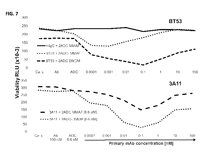
FIG. 7 is a pair of graphs showing cytotoxicity mediated by anti-FGFR4
monoclonal
antibodies BT53 and 3A11 conjugated to secondary antibody-drug conjugates in
the RH30 cell
line.
FIG. 8 is a pair of graphs showing cytotoxicity mediated by anti-FGFR4
monoclonal
antibody BT53 conjugated to secondary antibody-drug conjugates (ADC-MMAF ¨
top; ADC-
DMDM ¨ bottom) in RMS-559 cells.
FIG. 9 is a pair of graphs showing specific cytotoxicity of anti-FGFR4
monoclonal
antibodies BT53 (top) and 3A11 (bottom) conjugated to secondary antibody-drug
conjugates. The
FGFR4-specific secondary antibody-drug conjugates induced killing of FGFR4-
positive
rhabdomyosarcoma cells (RH30), but not FGFR4-negative human skeletal muscle
cells (SKMC)
cells.
FIGS. 10A-10B are graphs showing growth of FGFR-positive RH30 cells (FIG. 10A)
and
FGFR4-negative SKMC cells (FIG. 10B) in the presence of the BT53 monoclonal
antibody and
BT53 secondary ADC.
FIGS. 11A-11B are graphs showing cytotoxicity of T cells expressing FGFR4 CARs
M410
long, M412 long and M412 short. A CD22 CAR was used as a control. Target RH30
(FGRR4+/CD22-) and Raji (FGFR4-/CD22+) cells were transduced with luciferase
and the
CELLTITER-GLOTm assay was used to measure the number of viable cells. Percent
specific lysis
induced by each CAR is shown. FGFR4-specific CARs induced lysis of FGFR4-
postive RH30
cells, but not FGFR4-negative Raji cells.
FIG. 12 is a graph showing interferon (IFN)-y release induced by T cells
expressing the
FGFR4-specific M410 long, M412 short and M412 long CARs. IFN-y released by
RH30
(FGFR4+), SKES1 (FGFR4+), Raji (FGFR4-) and K562 (FGFR4-) cells is shown.
SEQUENCE LISTING
The amino acid sequences listed in the accompanying sequence listing are shown
using
standard three letter code for amino acids, as defined in 37 C.F.R. 1.822. The
Sequence Listing is
- 4 -
CA 02996205 2018-02-20
WO 2017/049296
PCT/US2016/052496
submitted as an ASCII text file, created on September 8, 2016, 104 KB, which
is incorporated by
reference herein. In the accompanying sequence listing:
SEQ ID NO: 1 is the amino acid sequence of the VH of the BT53 mouse anti-FGFR4
mAb.
SEQ ID NO: 2 is the amino acid sequence of the VL of the BT53 mouse anti-FGFR4
mAb.
SEQ ID NO: 3 is the amino acid sequence of the VH of the 3A11 mouse anti-FGFR4
mAb.
SEQ ID NO: 4 is the amino acid sequence of the VL of the 3A11 mouse anti-FGFR4
mAb.
SEQ ID NO: 5 is the amino acid sequence of the VH of the 1G5 mouse anti-FGFR4
mAb.
SEQ ID NO: 6 is the amino acid sequence of the VL of the 1G5 mouse anti-FGFR4
mAb.
SEQ ID NO: 7 is the amino acid sequence of the VH of the 29.2 rabbit anti-
FGFR4 mAb.
SEQ ID NO: 8 is the amino acid sequence of the VL of the 29.2 rabbit anti-
FGFR4 mAb.
SEQ ID NO: 9 is the amino acid sequence of the VH of the 57.1 rabbit anti-
FGFR4 mAb.
SEQ ID NO: 10 is the amino acid sequence of the VL of the 57.1 rabbit anti-
FGFR4 mAb.
SEQ ID NO: 11 is the amino acid sequence of the VH of the M408 human anti-
FGFR4
scFv.
SEQ ID NO: 12 is the amino acid sequence of the VL of the M408 human anti-
FGFR4
scFv.
SEQ ID NO: 13 is the amino acid sequence of the VH of the M409 human anti-
FGFR4
scFv.
SEQ ID NO: 14 is the amino acid sequence of the VL of the M409 human anti-
FGFR4
scFv.
SEQ ID NO: 15 is the amino acid sequence of the VH of the M410 human anti-
FGFR4
scFv.
SEQ ID NO: 16 is the amino acid sequence of the VL of the M410 human anti-
FGFR4
scFv.
SEQ ID NO: 17 is the amino acid sequence of the VH of the M412 human anti-
FGFR4
scFv.
SEQ ID NO: 18 is the amino acid sequence of the VL of the M412 human anti-
FGFR4
scFv.
SEQ ID NO: 19 is the amino acid sequence of the VH of the M414 human anti-
FGFR4
scFv.
SEQ ID NO: 20 is the amino acid sequence of the VL of the M414 human anti-
FGFR4
scFv.
SEQ ID NO: 21 is the amino acid sequence of the VH of the M415 human anti-
FGFR4
scFv.
- 5 -
CA 02996205 2018-02-20
WO 2017/049296
PCT/US2016/052496
SEQ ID NO: 22 is the amino acid sequence of the VL of the M415 human anti-
FGFR4
scFv.
SEQ ID NO: 23 is the amino acid sequence of the VH of the M417 human anti-
FGFR4
scFv.
SEQ ID NO: 24 is the amino acid sequence of the VL of the M417 human anti-
FGFR4
scFv.
SEQ ID NO: 25 is the amino acid sequence of the VH of the M418 human anti-
FGFR4
scFv.
SEQ ID NO: 26 is the amino acid sequence of the VL of the M418 human anti-
FGFR4
scFv.
SEQ ID NO: 27 is the amino acid sequence of the VH of the M422 human anti-
FGFR4
scFv.
SEQ ID NO: 28 is the amino acid sequence of the VL of the M422 human anti-
FGFR4
scFv.
SEQ ID NO: 29 is the amino acid sequence of the VH of the M424 human anti-
FGFR4
scFv.
SEQ ID NO: 30 is the amino acid sequence of the VL of the M424 human anti-
FGFR4
scFv.
SEQ ID NO: 31 is the amino acid sequence of a peptide linker for antibody-
based CARs.
SEQ ID NO: 32 is the amino acid sequence of a peptide linker featured in scFv
sequences.
SEQ ID NO: 33 is the amino acid sequence of an alternative peptide linker
featured in scFv
sequences.
SEQ ID NO: 34 is the amino acid sequence of an exemplary signal peptide.
SEQ ID NO: 35 is a short linker domain for Ig binding domains to transmembrane
sequences (short spacer).
SEQ ID NO: 36 is a linker domain composed of 2 Ig C domains (CH2CH3) used to
link Ig
binding domains to transmembrane sequences (long spacer).
SEQ ID NO: 37 is the amino acid sequence of a scFv including the VH and the VL
of the
BT53 mouse anti-FGFR4 mAb.
SEQ ID NO: 38 is the amino acid sequence of a scFv including the VH and the VL
of the
3A11 mouse anti-FGFR4 mAb.
SEQ ID NO: 39 is the amino acid sequence of a scFv including the VH and the VL
of the
1G5 mouse anti-FGFR4 mAb.
- 6 -
CA 02996205 2018-02-20
WO 2017/049296
PCT/US2016/052496
SEQ ID NO: 40 is the amino acid sequence of a scFv including the humanized VH
and the
VL of the BT53 mouse anti-FGFR4 mAb.
SEQ ID NO: 41 is the amino acid sequence of a scFv including the humanized VH
and the
VL of the 3A11 mouse anti-FGFR4 mAb.
SEQ ID NO: 42 is the amino acid sequence of a scFv including the humanized VH
and the
VL of the 1G5 mouse anti-FGFR4 mAb.
SEQ ID NO: 43 is the amino acid sequence of a scFv including the VH and VL of
the 29.2
rabbit anti-FGFR4 mAb.
SEQ ID NO: 44 is the amino acid sequence of a scFv including the VH and VL of
the 57.1
rabbit anti-FGFR4 mAb.
SEQ ID NO: 45 is the amino acid sequence of a scFv including the humanized VH
and VL
of the 29.2 rabbit anti-FGFR4 mAb.
SEQ ID NO: 46 is the amino acid sequence of a scFv including the humanized VH
and VL
of the 57.1 rabbit anti-FGFR4 mAb.
SEQ ID NO: 47 is the amino acid sequence of the M408 human anti-FGFR4 scFv.
SEQ ID NO: 48 is the amino acid sequence of the M409 human anti-FGFR4 scFv.
SEQ ID NO: 49 is the amino acid sequence of the M410 human anti-FGFR4 scFv.
SEQ ID NO: 50 is the amino acid sequence of the M412 human anti-FGFR4 scFv.
SEQ ID NO: 51 is the amino acid sequence of the M414 human anti-FGFR4 scFv.
SEQ ID NO: 52 is the amino acid sequence of the M415 human anti-FGFR4 scFv.
SEQ ID NO: 53 is the amino acid sequence of the M417 human anti-FGFR4 scFv.
SEQ ID NO: 54 is the amino acid sequence of the M418 human anti-FGFR4 scFv.
SEQ ID NO: 55 is the amino acid sequence of the M422 human anti-FGFR4 scFv.
SEQ ID NO: 56 is the amino acid sequence of the M424 human anti-FGFR4 scFv.
SEQ ID NO: 57 is the amino acid sequence of an exemplary CD28 transmembrane
domain.
SEQ ID NO: 58 is the amino acid sequence of an exemplary CD28 signaling
domain.
SEQ ID NO: 59 is the amino acid sequence of exemplary CD28 transmembrane and
signaling domains.
SEQ ID NO: 60 is the amino acid sequence of an exemplary CD8 transmembrane
domain.
SEQ ID NO: 61 is the amino acid sequence of an exemplary CD8 extended
transmembrane
domain.
SEQ ID NO: 62 is the amino acid sequence of an exemplary CD137 signaling
domain.
SEQ ID NO: 63 is the amino acid sequence of an exemplary CD137 signaling
domain.
SEQ ID NO: 64 is the amino acid sequence of an exemplary CD3 zeta signaling
domain.
- 7 -
CA 02996205 2018-02-20
WO 2017/049296
PCT/US2016/052496
SEQ ID NO: 65 is the amino acid sequence of the transmembrane and
intracellular
domains of an exemplary second generation CAR including a CD28 transmembrane
domain and a
CD3 zeta signaling domain ("28z").
SEQ ID NO: 66 is the amino acid sequence of the transmembrane and
intracellular
domains of an exemplary second generation CAR including a CD8 transmembrane
domain, CD137
(4-1BB) signaling domain, and a CD3 zeta signaling domain ("BBz").
SEQ ID NO: 67 is the amino acid sequence of the transmembrane and
intracellular
domains of an exemplary third generation CAR including a CD8 transmembrane
domain, a CD28
signaling domain, a CD137 (4-1BB) signaling domain, and a CD3 zeta signaling
domain
("28BBz").
SEQ ID NO: 68 is the amino acid sequence of the 29.2L CAR.
SEQ ID NO: 69 is the amino acid sequence of the 29.2 CAR.
SEQ ID NO: 70 is the amino acid sequence of the 57.1L CAR.
SEQ ID NO: 71 is the amino acid sequence of the 57.1 CAR.
DETAILED DESCRIPTION
I. Abbreviations
ADC antibody-drug conjugate
ADCC antibody-dependent cell-mediated cytotoxicity
ARMS alveolar rhabdomyosarcoma
CAR chimeric antigen receptor
CDC complement-dependent cytotoxicity
CDR complementarity determining region
CTL cytotoxic T lymphocyte
DMDM duocarmycin DM
ECD extracellular domain
ELISA enzyme-linked immunosorbent assay
ERMS embryonal rhabdomyosarcoma
Fc constant fragment
FGFR4 fibroblast growth factor receptor 4
IHC immunohistochemistry
ITAM immunoreceptor tyrosine-based activation motif
mAb monoclonal antibody
MMAF monomethyl auristatin F
- 8 -
CA 02996205 2018-02-20
WO 2017/049296
PCT/US2016/052496
PBD pyrrolobenzodiazepine
PBMC peripheral blood mononuclear cell
PE Pseudomonas exotoxin
RMS rhabdomyosarcoma
scFv single chain variable fragment
TMA tissue microarray
VH variable heavy
VL variable light
II. Terms and Methods
Unless otherwise noted, technical terms are used according to conventional
usage.
Definitions of common terms in molecular biology may be found in Benjamin
Lewin, Genes V,
published by Oxford University Press, 1994 (ISBN 0-19-854287-9); Kendrew et
al. (eds.), The
Encyclopedia of Molecular Biology, published by Blackwell Science Ltd., 1994
(ISBN 0-632-
02182-9); and Robert A. Meyers (ed.), Molecular Biology and Biotechnology: a
Comprehensive
Desk Reference, published by VCH Publishers, Inc., 1995 (ISBN 1-56081-569-8).
In order to facilitate review of the various embodiments of the disclosure,
the following
explanations of specific terms are provided:
Anti-microtubule agent: A drug that interferes with microtubules. Anti-
microtubule
agents block cell growth by stopping mitosis.
Anti-mitotic agent: A drug or compound that blocks mitosis.
Antibody: A polypeptide ligand comprising at least a light chain and/or heavy
chain
immunoglobulin variable region which recognizes and binds (such as
specifically recognizes and
specifically binds) an epitope of an antigen, such as FGFR4, or a fragment
thereof.
Immunoglobulin molecules are composed of a heavy and a light chain, each of
which has a variable
region, termed the variable heavy (VH) region and the variable light (VL)
region. Together, the VH
region and the VL region are responsible for binding the antigen recognized by
the antibody.
Antibodies include intact immunoglobulins and the variants and portions
(fragments) of
antibodies well known in the art, such as single-domain antibodies (e.g. VH
domain antibodies),
Fab fragments, Fab fragments, F(ab)'2 fragments, single chain Fv proteins
("scFv"), and disulfide
stabilized Fv proteins ("dsFv"). A scFv protein is a fusion protein in which a
light chain variable
region of an immunoglobulin and a heavy chain variable region of an
immunoglobulin are bound
by a linker, while in dsFvs, the chains have been mutated to introduce a
disulfide bond to stabilize
the association of the chains. The term "antibody" also includes genetically
engineered forms such
- 9 -
CA 02996205 2018-02-20
WO 2017/049296
PCT/US2016/052496
as chimeric antibodies (for example, humanized murine antibodies) and
heteroconjugate antibodies
(such as bispecific antibodies). See also, Pierce Catalog and Handbook, 1994-
1995 (Pierce
Chemical Co., Rockford, IL); Kuby, J., Immunology, 3rd Ed., W. H. Freeman &
Co., New York,
1997.
Typically, a naturally occurring immunoglobulin has heavy (H) chains and light
(L) chains
interconnected by disulfide bonds. There are two types of light chain, lambda
(X) and kappa (K).
There are five main heavy chain classes (or isotypes) which determine the
functional activity of an
antibody molecule: IgM, IgD, IgG, IgA and IgE.
Each heavy and light chain contains a constant region and a variable region
(the regions are
also known as "domains"). In combination, the heavy and the light chain
variable regions
specifically bind the antigen. Light and heavy chain variable regions contain
a "framework" region
interrupted by three hypervariable regions, also called "complementarity-
determining regions" or
"CDRs." The amino acid sequence boundaries of a given CDR can be readily
determined using
any of a number of well-known schemes, including those described by Kabat et
al. (Sequences of
Proteins of Immunological Interest, U.S. Department of Health and Human
Services, 1991; the
"Kabat" numbering scheme), Chothia et al. (see Chothia and Lesk, J Mol Biol
196:901-917, 1987;
Chothia et al., Nature 342:877, 1989; and Al-Lazikani et al., (JMB 273,927-
948, 1997; the
"Chothia" numbering scheme), and the ImMunoGeneTics (IMGT) database (see,
Lefranc, Nucleic
Acids Res 29:207-9, 2001; the "IMGT" numbering scheme). The Kabat and IMGT
databases are
maintained online. The sequences of the framework regions of different light
or heavy chains are
relatively conserved within a species, such as humans. The framework region of
an antibody, that
is the combined framework regions of the constituent light and heavy chains,
serves to position and
align the CDRs in three-dimensional space.
The CDRs are primarily responsible for binding to an epitope of an antigen.
The CDRs of
each chain are typically referred to as CDR1, CDR2, and CDR3, numbered
sequentially starting
from the N-terminus, and are often identified by the chain in which the
particular CDR is located.
Thus, a VH CDR3 (or HCDR3) is located in the variable domain of the heavy
chain of the antibody
in which it is found, whereas a VL CDR1 (or LCDR1) is the CDR1 from the
variable domain of the
light chain of the antibody in which it is found. An antibody that binds
FGFR4, for example, will
have a specific VH region and the VL region sequence, and thus specific CDR
sequences.
Antibodies with different specificities (i.e. different combining sites for
different antigens) have
different CDRs. Although it is the CDRs that vary from antibody to antibody,
only a limited
number of amino acid positions within the CDRs are directly involved in
antigen binding. These
positions within the CDRs are called specificity determining residues (SDRs).
- 10 -
CA 02996205 2018-02-20
WO 2017/049296
PCT/US2016/052496
References to "VH" or "VH" refer to the variable region of an immunoglobulin
heavy chain,
including that of an Fv, scFv, dsFy or Fab. References to "VL" or "VL" refer
to the variable region
of an immunoglobulin light chain, including that of an Fv, scFv, dsFy or Fab.
A "monoclonal antibody" is an antibody produced by a single clone of B-
lymphocytes or by
a cell into which the light and/or heavy chain genes of a single antibody have
been transfected.
Monoclonal antibodies are produced by methods known to those of skill in the
art, for instance by
making hybrid antibody-forming cells from a fusion of myeloma cells with
immune spleen cells.
Monoclonal antibodies include humanized monoclonal antibodies.
A "chimeric antibody" has framework residues from one species, such as human,
and CDRs
(which generally confer antigen binding) from another species, such as a
murine antibody that
specifically binds FGFR4.
A "human" antibody (also called a "fully human" antibody) is an antibody that
includes
human framework regions and all of the CDRs from a human immunoglobulin. In
one example,
the framework and the CDRs are from the same originating human heavy and/or
light chain amino
acid sequence. However, frameworks from one human antibody can be engineered
to include
CDRs from a different human antibody. A "humanized" immunoglobulin is an
immunoglobulin
including a human framework region and one or more CDRs from a non-human (for
example a
mouse, rabbit, rat, or synthetic) immunoglobulin. The non-human immunoglobulin
providing the
CDRs is termed a "donor," and the human immunoglobulin providing the framework
is termed an
"acceptor." In one embodiment, all the CDRs are from the donor immunoglobulin
in a humanized
immunoglobulin. Constant regions need not be present, but if they are, they
must be substantially
identical to human immunoglobulin constant regions, i.e., at least about 85-
90%, such as about 95%
or more identical. Hence, all parts of a humanized immunoglobulin, except
possibly the CDRs, are
substantially identical to corresponding parts of natural human immunoglobulin
sequences. A
"humanized antibody" is an antibody comprising a humanized light chain and a
humanized heavy
chain immunoglobulin. A humanized antibody binds to the same antigen as the
donor antibody that
provides the CDRs. The acceptor framework of a humanized immunoglobulin or
antibody may
have a limited number of substitutions by amino acids taken from the donor
framework.
Humanized or other monoclonal antibodies can have additional conservative
amino acid
substitutions which have substantially no effect on antigen binding or other
immunoglobulin
functions. Humanized immunoglobulins can be constructed by means of genetic
engineering (see
for example, U.S. Patent No. 5,585,089).
Antibody-drug conjugate (ADC): A molecule that includes an antibody (or
antigen-
binding fragment of an antibody) conjugated to a drug, such as a cytotoxic
agent. ADCs can be
-11-
CA 02996205 2018-02-20
WO 2017/049296
PCT/US2016/052496
used to specifically target a drug to cancer cells through specific binding of
the antibody to a tumor
antigen expressed on the cell surface. Exemplary drugs for use with ADCs
include anti-
microtubule agents (such as maytansinoids, auristatin E and auristatin F) and
interstrand
crosslinking agents (e.g., pyrrolobenzodiazepines; PDBs).
Anti-microtubule agent: A type of drug that blocks cell growth by stopping
mitosis.
Anti-microtubule agents, also referred to as "anti-mitotic agents," are used
to treat cancer.
Binding affinity: Affinity of an antibody for an antigen. In one embodiment,
affinity is
calculated by a modification of the Scatchard method described by Frankel et
al. (Mol. Immunol.,
16:101-106, 1979). In another embodiment, binding affinity is measured by an
antigen/antibody
dissociation rate. In another embodiment, binding affinity is measured by a
competition
radioimmunoassay. In another embodiment, binding affinity is measured by
ELISA. An antibody
that "specifically binds" an antigen (such as FGFR4) is an antibody that binds
the antigen with high
affinity and does not significantly bind other unrelated antigens.
Breast cancer: A type of cancer that forms in the tissues of the breast,
typically in the
ducts and lobules. In some embodiments, a patient with breast cancer is node-
positive, meaning the
breast cancer has spread to the lymph nodes.
Chemotherapeutic agent: Any chemical agent with therapeutic usefulness in the
treatment
of diseases characterized by abnormal cell growth. Such diseases include
tumors, neoplasms, and
cancer as well as diseases characterized by hyperplastic growth, such as
psoriasis. In one
embodiment, a chemotherapeutic agent is an agent of use in treating a FGFR4-
positive cancer, such
as rhabdomyosarcoma, lung cancer, liver cancer, breast cancer, pancreatic
cancer and prostate
cancer. In one embodiment, a chemotherapeutic agent is a radioactive compound.
One of skill in
the art can readily identify a chemotherapeutic agent of use (see for example,
Slapak and Kufe,
Principles of Cancer Therapy, Chapter 86 in Harrison's Principles of Internal
Medicine, 14th
edition; Perry et al., Chemotherapy, Ch. 17 in Abeloff, Clinical Oncology 2nd
ed., 2000 Churchill
Livingstone, Inc; Baltzer, L., Berkery, R. (eds.): Oncology Pocket Guide to
Chemotherapy, 2nd ed.
St. Louis, Mosby-Year Book, 1995; Fischer, D.S., Knobf, M.F., Durivage, H.J.
(eds): The Cancer
Chemotherapy Handbook, 4th ed. St. Louis, Mosby-Year Book, 1993). Combination
chemotherapy is the administration of more than one agent to treat cancer. One
example is the
administration of an antibody (or immunoconjugate or ADC) that binds FGFR4
used in
combination with a radioactive or chemical compound.
Chimeric antigen receptor (CAR): A chimeric molecule that includes an antigen-
binding
portion (such as a monoclonal antibody or fragment thereof) and a signaling
domain, such as a
signaling domain from a T cell receptor (e.g. CD3c). Typically, CARs are
comprised of a binding
- 12 -
CA 02996205 2018-02-20
WO 2017/049296
PCT/US2016/052496
moiety (e.g. a scFv), a transmembrane domain and an endodomain. The endodomain
typically
includes a signaling chain having an immunoreceptor tyrosine-based activation
motif (ITAM), such
as CD3C or FcERIy. In some instances, the endodomain further includes the
intracellular portion of
at least one additional co-stimulatory domain, such as CD28 and/or CD137.
Conservative variant: "Conservative" amino acid substitutions are those
substitutions that
do not substantially affect or decrease the affinity of a protein, such as an
antibody to FGFR4. For
example, a monoclonal antibody that specifically binds FGFR4 can include at
most about 1, at most
about 2, at most about 5, at most about 10, or at most about 15 conservative
substitutions and
specifically bind a FGFR4 polypeptide. The term "conservative variant" also
includes the use of a
substituted amino acid in place of an unsubstituted parent amino acid,
provided that the antibody
specifically binds FGFR4. Non-conservative substitutions are those that reduce
an activity or
binding to FGFR4.
Complementarity determining region (CDR): Amino acid sequences which together
define the binding affinity and specificity of the natural Fv region of a
native Ig binding site. The
light and heavy chains of an Ig each have three CDRs, designated LCDRI, LCDR2,
LCDR3 and
HCDR I, HCDR2 and HCDR3, respectively.
Contacting: Placement in direct physical association; includes both in solid
and liquid
form.
Cytotoxic agent: Any drug or compound that kills cells.
Degenerate variant: In the context of the present disclosure, a "degenerate
variant" refers
to a polynucleotide encoding a FGFR4 polypeptide or an antibody that binds
FGFR4 that includes a
sequence that is degenerate as a result of the genetic code. There are 20
natural amino acids, most
of which are specified by more than one codon. Therefore, all degenerate
nucleotide sequences are
included as long as the amino acid sequence of the FGFR4 polypeptide or
antibody that binds
FGFR4 encoded by the nucleotide sequence is unchanged.
Diagnostic: Identifying the presence or nature of a pathologic condition, such
as, but not
limited to, cancer. Diagnostic methods differ in their sensitivity and
specificity. The "sensitivity"
of a diagnostic assay is the percentage of diseased individuals who test
positive (percent of true
positives). The "specificity" of a diagnostic assay is one minus the false
positive rate, where the
false positive rate is defined as the proportion of those without the disease
who test positive. While
a particular diagnostic method may not provide a definitive diagnosis of a
condition, it suffices if
the method provides a positive indication that aids in diagnosis. "Prognostic"
is the probability of
development (e.g., severity) of a pathologic condition, such as cancer or
metastasis.
- 13 -
CA 02996205 2018-02-20
WO 2017/049296
PCT/US2016/052496
Drug: Any compound used to treat, ameliorate or prevent a disease or condition
in a
subject. In some embodiments herein, the drug is an anti-cancer agent, for
example a cytotoxic
agent, such as an anti-mitotic or anti-microtubule agent.
Duocarmycin: A cytotoxic small molecule that induces cell death by binding to
the minor
groove of DNA and alkylating the adenine.
Effector molecule: The portion of an antibody conjugate (or immunoconjugate)
that is
intended to have a desired effect on a cell to which the conjugate is
targeted. Effector molecules
are also known as effector moieties (EMs), therapeutic agents, diagnostic
agents, or similar terms.
Therapeutic agents (or drugs) include such compounds as small molecules,
nucleic acids, proteins,
peptides, amino acids or derivatives, glycoproteins, radioisotopes, lipids,
carbohydrates, or
recombinant viruses. Nucleic acid therapeutic and diagnostic moieties include
antisense nucleic
acids, derivatized oligonucleotides for covalent cross-linking with single or
duplex DNA, and
triplex forming oligonucleotides. Alternatively, the effector molecule can be
contained within an
encapsulation system, such as a liposome or micelle, which is conjugated to
the antibody.
Encapsulation shields the effector molecule from direct exposure to the
circulatory system. Means
of preparing liposomes attached to antibodies are well known to those of skill
in the art (see, for
example, U.S. Patent No. 4,957,735; and Connor et al., Pharm Ther 28:341-365,
1985). Diagnostic
agents or moieties include radioisotopes and other detectable labels (e.g.,
fluorophores,
chemiluminescent agents, and enzymes). Radioactive isotopes include 35S, 11C,
13N, 150, 18F, 19F,
99mTc, 1311, 3H, 14C, 15N, 90y, 99TC, "'In and 1251.
Epitope: An antigenic determinant. These are particular chemical groups or
peptide
sequences on a molecule that are antigenic, i.e. that elicit a specific immune
response. An antibody
specifically binds a particular antigenic epitope on a polypeptide, such as
FGFR4.
Fibroblast growth factor receptor (FGFR): A family of tyrosine kinase
receptors
activated by fibroblast growth factors (FGF), comprising extracellular
immunoglobulin-like
domains, a transmembrane domain, and an intracellular tyrosine kinase domain.
The family
includes at least four members: FGFR1, FGFR2, FGFR3, and FGFR4.
Framework region: Amino acid sequences interposed between CDRs. Framework
regions include variable light and variable heavy framework regions. The
framework regions serve
to hold the CDRs in an appropriate orientation for antigen binding.
Immune response: A response of a cell of the immune system, such as a B cell,
T cell, or
monocyte, to a stimulus. In one embodiment, the response is specific for a
particular antigen (an
"antigen-specific response"). In one embodiment, an immune response is a T
cell response, such as
- 14 -
CA 02996205 2018-02-20
WO 2017/049296
PCT/US2016/052496
a CD4+ response or a CD8+ response. In another embodiment, the response is a B
cell response,
and results in the production of antigen-specific antibodies.
Immunoliposome: A liposome with antibodies or antibody fragments conjugated to
its
surface. Immunoliposomes can carry cytotoxic agents or other drugs to antibody-
targeted cells,
such as tumor cells.
Interstrand crosslinking agent: A type of cytotoxic drug capable of binding
covalently
between two strands of DNA, thereby preventing DNA replication and/or
transcription.
Isolated: An "isolated" biological component, such as a nucleic acid, protein
(including
antibodies) or organelle, has been substantially separated or purified away
from other biological
components in the environment (such as a cell) in which the component
naturally occurs, i.e., other
chromosomal and extra-chromosomal DNA and RNA, proteins and organelles.
Nucleic acids and
proteins that have been "isolated" include nucleic acids and proteins purified
by standard
purification methods. The term also embraces nucleic acids and proteins
prepared by recombinant
expression in a host cell as well as chemically synthesized nucleic acids.
Label: A detectable compound or composition that is conjugated directly or
indirectly to
another molecule, such as an antibody or a protein, to facilitate detection of
that molecule.
Specific, non-limiting examples of labels include fluorescent tags, enzymatic
linkages, and
radioactive isotopes. In one example, a "labeled antibody" refers to
incorporation of another
molecule in the antibody. For example, the label is a detectable marker, such
as the incorporation
of a radiolabeled amino acid or attachment to a polypeptide of biotinyl
moieties that can be
detected by marked avidin (for example, streptavidin containing a fluorescent
marker or enzymatic
activity that can be detected by optical or colorimetric methods). Various
methods of labeling
polypeptides and glycoproteins are known in the art and may be used. Examples
of labels for
polypeptides include, but are not limited to, the following: radioisotopes or
radionucleotides (such
as 35S, "C, "N, 150, "F, '9F, 99mTc, 1311, 3H, 14C, 15N, 90y, 99Tc, "'In and
1251), fluorescent labels
(such as fluorescein isothiocyanate (FITC), rhodamine, lanthanide phosphors),
enzymatic labels
(such as horseradish peroxidase, beta-galactosidase, luciferase, alkaline
phosphatase),
chemiluminescent markers, biotinyl groups, predetermined polypeptide epitopes
recognized by a
secondary reporter (such as a leucine zipper pair sequences, binding sites for
secondary antibodies,
metal binding domains, epitope tags), or magnetic agents, such as gadolinium
chelates. In some
embodiments, labels are attached by spacer arms of various lengths to reduce
potential steric
hindrance.
Linker: In some cases, a linker is a peptide within an antibody binding
fragment (such as
an Fv fragment) which serves to indirectly bond the variable heavy chain to
the variable light chain.
- 15 -
CA 02996205 2018-02-20
WO 2017/049296
PCT/US2016/052496
"Linker" can also refer to a peptide serving to link a targeting moiety, such
as an antibody, to an
effector molecule, such as a cytotoxin or a detectable label.
The terms "conjugating," "joining," "bonding" or "linking" refer to making two
polypeptides into one contiguous polypeptide molecule, or to covalently
attaching a radionuclide,
drug or other molecule to a polypeptide, such as an antibody or antibody
fragment. In the specific
context, the terms include reference to joining a ligand, such as an antibody
moiety, to an effector
molecule. The linkage can be either by chemical or recombinant means.
"Chemical means" refers
to a reaction between the antibody moiety and the effector molecule such that
there is a covalent
bond formed between the two molecules to form one molecule.
Liver cancer: A type of cancer than forms in the tissues of the liver. Types
of liver
cancers include, for example, hepatocellular carcinoma (HCC),
cholangiocarcinoma (also known as
bile duct cancer), angiosarcoma and hepatoblastoma.
Lung cancer: Cancer that forms in tissues of the lung, usually in the cells
lining air
passages. The two main types are small cell lung cancer and non-small cell
lung cancer. These
types are diagnosed based on how the cells look under a microscope.
Mammal: This term includes both human and non-human mammals. Similarly, the
term
"subject" includes both human and veterinary subjects.
Neoplasia, malignancy, cancer or tumor: A neoplasm is an abnormal growth of
tissue or
cells that results from excessive cell division. Neoplastic growth can produce
a tumor. The amount
of a tumor in an individual is the "tumor burden" which can be measured as the
number, volume, or
weight of the tumor. A tumor that does not metastasize is referred to as
"benign." A tumor that
invades the surrounding tissue and/or can metastasize is referred to as
"malignant."
Operably linked: A first nucleic acid sequence is operably linked with a
second nucleic
acid sequence when the first nucleic acid sequence is placed in a functional
relationship with the
second nucleic acid sequence. For instance, a promoter is operably linked to a
coding sequence if
the promoter affects the transcription or expression of the coding sequence.
Generally, operably
linked DNA sequences are contiguous and, where necessary to join two protein-
coding regions, in
the same reading frame.
Pancreatic cancer: A disease in which malignant (cancer) cells are found in
the tissues of
the pancreas. Also called exocrine cancer.
Pharmaceutical agent: A chemical compound or composition capable of inducing a
desired therapeutic or prophylactic effect when properly administered to a
subject or a cell.
Pharmaceutically acceptable carriers: The pharmaceutically acceptable carriers
of use
are conventional. Remington's Pharmaceutical Sciences, by E.W. Martin, Mack
Publishing Co.,
- 16 -
CA 02996205 2018-02-20
WO 2017/049296
PCT/US2016/052496
Easton, PA, 15th Edition, 1975, describes compositions and formulations
suitable for
pharmaceutical delivery of the antibodies and conjugates disclosed herein.
In general, the nature of the carrier will depend on the particular mode of
administration
being employed. For instance, parenteral formulations usually comprise
injectable fluids that
include pharmaceutically and physiologically acceptable fluids such as water,
physiological saline,
balanced salt solutions, aqueous dextrose, glycerol or the like as a vehicle.
For solid compositions
(such as powder, pill, tablet, or capsule forms), conventional non-toxic solid
carriers can include,
for example, pharmaceutical grades of mannitol, lactose, starch, or magnesium
stearate. In addition
to biologically neutral carriers, pharmaceutical compositions to be
administered can contain minor
amounts of non-toxic auxiliary substances, such as wetting or emulsifying
agents, preservatives,
and pH buffering agents and the like, for example sodium acetate or sorbitan
monolaurate.
Preventing, treating or ameliorating a disease: "Preventing" a disease refers
to inhibiting
the full development of a disease. "Treating" refers to a therapeutic
intervention that ameliorates a
sign or symptom of a disease or pathological condition after it has begun to
develop, such as a
reduction in tumor burden or a decrease in the number of size of metastases.
"Ameliorating" refers
to the reduction in the number or severity of signs or symptoms of a disease,
such as cancer.
Prostate Cancer: A malignant tumor, generally of glandular origin, of the
prostate.
Prostate cancers include adenocarcinomas and small cell carcinomas. Many
prostate cancers
express prostate specific antigen (PSA).
Purified: The term purified does not require absolute purity; rather, it is
intended as a
relative term. Thus, for example, a purified peptide preparation is one in
which the peptide or
protein is more enriched than the peptide or protein is in its natural
environment within a cell. In
one embodiment, a preparation is purified such that the protein or peptide
represents at least 50% of
the total peptide or protein content of the preparation. Substantial
purification denotes purification
from other proteins or cellular components. A substantially purified protein
is at least 60%, 70%,
80%, 90%, 95% or 98% pure. Thus, in one specific, non-limiting example, a
substantially purified
protein is 90% free of other proteins or cellular components.
Pyrrolobenzodiazepine (PBD): A class of sequence-selective DNA minor-groove
binding
crosslinking agents originally discovered in Streptomyces species. PDBs are
significantly more
potent than systemic chemotherapeutic drugs. The mechanism of action of PBDs
is associated with
their ability to form an adduct in the minor groove of DNA, thereby
interfering with DNA
processing. In the context of the present disclosure, PBDs include naturally
produced and isolated
PBDs, chemically synthesized naturally occurring PBDs, and chemically
synthesized non-naturally
- 17 -
CA 02996205 2018-02-20
WO 2017/049296
PCT/US2016/052496
occurring PBDs. PBDs also include monomeric, dimeric and hybrid PBDs (for a
review see
Gerratana, Med Res Rev 32(2):254-293, 2012).
Recombinant: A recombinant nucleic acid is one that has a sequence that is not
naturally
occurring or has a sequence that is made by an artificial combination of two
otherwise separated
segments of sequence. This artificial combination is often accomplished by
chemical synthesis or
by the artificial manipulation of isolated segments of nucleic acids, for
example, by genetic
engineering techniques.
Rhabdomyosarcoma (RMS): A soft tissue malignant tumor of skeletal muscle
origin.
The most common primary sites for rhabdomyosarcoma are the head and neck
(e.g.,
parameningeal, orbit, pharyngeal, etc.), the genitourinary tract, and the
extremities. Other less
common primary sites include the trunk, chest wall, the abdomen (including the
retroperitoneum
and biliary tract), and the perineal/anal region. There are at least two types
of RMS; the most
common forms are alveolar RMS (ARMS) and embryonal histological RMS (ERMS).
Approximately 20% of children with rhabdomyosarcoma have the ARMS subtype. An
increased
frequency of this subtype is noted in adolescents and in patients with primary
sites involving the
extremities, trunk, and perineum/perianal region. ARMS is associated with
chromosomal
translocations encoding a fusion gene involving FKHR on chromosome 13 and
members of the
PAX family. The embryonal subtype is the most frequently observed subtype in
children,
accounting for approximately 60-70% of rhabdomyosarcomas of childhood. Tumors
with
embryonal histology typically arise in the head and neck region or in the
genitourinary tract,
although they may occur at any primary site. ERMS is characterized by a
younger age at diagnosis,
loss of heterozygosity, and altered genomic imprinting.
Sample (or biological sample): A biological specimen containing genomic DNA,
RNA
(including mRNA), protein, or combinations thereof, obtained from a subject.
Examples include,
but are not limited to, peripheral blood, tissue, cells, urine, saliva, tissue
biopsy, fine needle
aspirate, surgical specimen, and autopsy material. In one example, a sample
includes a tumor
biopsy.
Sequence identity: The similarity between amino acid or nucleic acid sequences
is expressed
in terms of the similarity between the sequences, otherwise referred to as
sequence identity.
Sequence identity is frequently measured in terms of percentage identity (or
similarity or homology);
the higher the percentage, the more similar the two sequences are. Homologs or
variants of a
polypeptide or nucleic acid molecule will possess a relatively high degree of
sequence identity when
aligned using standard methods.
- 18 -
CA 02996205 2018-02-20
WO 2017/049296
PCT/US2016/052496
Methods of alignment of sequences for comparison are well known in the art.
Various
programs and alignment algorithms are described in: Smith and Waterman, Adv.
Appl. Math. 2:482,
1981; Needleman and Wunsch, J. Mol. Biol. 48:443, 1970; Pearson and Lipman,
Proc. Natl. Acad.
Sci. U.S.A. 85:2444, 1988; Higgins and Sharp, Gene 73:237, 1988; Higgins and
Sharp, CABIOS
5:151, 1989; Corpet et al., Nucleic Acids Research 16:10881, 1988; and Pearson
and Lipman, Proc.
Natl. Acad. Sci. U.S.A. 85:2444, 1988. Altschul et al., Nature Genet. 6:119,
1994, presents a
detailed consideration of sequence alignment methods and homology
calculations.
The NCBI Basic Local Alignment Search Tool (BLAST) (Altschul et al., J. Mol.
Biol.
215:403, 1990) is available from several sources, including the National
Center for Biotechnology
Information (NCBI, Bethesda, MD) and on the internet, for use in connection
with the sequence
analysis programs blastp, blastn, blastx, tblastn and tblastx. A description
of how to determine
sequence identity using this program is available on the NCBI website on the
internet.
Homologs and variants of a VL or a VH of an antibody that specifically binds
FGFR4 or a
fragment thereof are typically characterized by possession of at least about
75%, for example at least
about 80%, 90%, 95%, 96%, 97%, 98% or 99% sequence identity counted over the
full length
alignment with the amino acid sequence of the antibody using the NCBI Blast
2.0, gapped blastp set
to default parameters. For comparisons of amino acid sequences of greater than
about 30 amino
acids, the Blast 2 sequences function is employed using the default BLOSUM62
matrix set to default
parameters, (gap existence cost of 11, and a per residue gap cost of 1). When
aligning short peptides
(fewer than around 30 amino acids), the alignment should be performed using
the Blast 2 sequences
function, employing the PAM30 matrix set to default parameters (open gap 9,
extension gap 1
penalties). Proteins with even greater similarity to the reference sequences
will show increasing
percentage identities when assessed by this method, such as at least 80%, at
least 85%, at least 90%,
at least 95%, at least 98%, or at least 99% sequence identity. When less than
the entire sequence is
being compared for sequence identity, homologs and variants will typically
possess at least 80%
sequence identity over short windows of 10-20 amino acids, and may possess
sequence identities of
at least 85% or at least 90% or 95% depending on their similarity to the
reference sequence. Methods
for determining sequence identity over such short windows are available at the
NCBI website on the
internet. One of skill in the art will appreciate that these sequence identity
ranges are provided for
guidance only; it is entirely possible that strongly significant homologs
could be obtained that fall
outside of the ranges provided.
Small molecule: A molecule, typically with a molecular weight less than about
1000
Daltons, or in some embodiments, less than about 500 Daltons, wherein the
molecule is capable of
modulating, to some measurable extent, an activity of a target molecule.
- 19 -
CA 02996205 2018-02-20
WO 2017/049296
PCT/US2016/052496
Subject: Living multi-cellular vertebrate organisms, a category that includes
both human and
veterinary subjects, including human and non-human mammals.
Therapeutically effective amount: A quantity of a specific substance
sufficient to achieve
a desired effect in a subject being treated. For instance, this can be the
amount necessary to inhibit
or suppress growth of a tumor. In one embodiment, a therapeutically effective
amount is the
amount necessary to eliminate, reduce the size, or prevent metastasis of a
tumor. When
administered to a subject, a dosage will generally be used that will achieve
target tissue
concentrations (for example, in tumors) that has been shown to achieve a
desired in vitro effect.
Toxin: An agent that directly or indirectly inhibits the growth of and/or
kills cells. Toxins
include, for example, Pseudomonas exotoxin (PE, such as PE35, PE37, PE38 and
PE40), diphtheria
toxin (DT), botulinum toxin, abrin, ricin, saporin, restrictocin or gelonin,
or modified toxins
thereof. For example, PE and DT are highly toxic compounds that typically
bring about death
through liver toxicity. PE and DT, however, can be modified into a form for
use as an
immunotoxin by removing the native targeting component of the toxin (such as
domain Ia of PE or
the B chain of DT) and replacing it with a different targeting moiety, such as
an antibody.
Vector: A nucleic acid molecule as introduced into a host cell, thereby
producing a
transformed host cell. A vector may include nucleic acid sequences that permit
it to replicate in a
host cell, such as an origin of replication. A vector may also include one or
more selectable marker
genes and other genetic elements known in the art.
Unless otherwise explained, all technical and scientific terms used herein
have the same
meaning as commonly understood by one of ordinary skill in the art to which
this disclosure
belongs. The singular terms "a," "an," and "the" include plural referents
unless context clearly
indicates otherwise. "Comprising A or B" means including A, or B, or A and B.
It is further to be
understood that all base sizes or amino acid sizes, and all molecular weight
or molecular mass
values, given for nucleic acids or polypeptides are approximate, and are
provided for description.
Although methods and materials similar or equivalent to those described herein
can be used in the
practice or testing of the present disclosure, suitable methods and materials
are described below.
All publications, patent applications, patents, and other references mentioned
herein are
incorporated by reference in their entirety. In case of conflict, the present
specification, including
explanations of terms, will control. In addition, the materials, methods, and
examples are
illustrative only and not intended to be limiting.
- 20 -
CA 02996205 2018-02-20
WO 2017/049296
PCT/US2016/052496
III. Monoclonal Antibodies Specific for FGFR4
Rhabdomyosarcoma (RMS) is the most common soft tissue sarcoma of childhood.
Two
major subtypes, embryonal RMS (ERMS) and alveolar RMS (ARMS), harbor distinct
cytogenetic
and molecular abnormalities, some of which are associated with poor prognosis.
The FGFR4 gene
is amplified, overexpressed, and mutationally activated in human RMS, and as
such is a tumor-
specific target. Previous studies of FGFR4 indicate that this gene plays a
role in tumorigenesis and
is crucial for the survival, proliferation, metastasis and drug-resistance of
RMS. High expression of
FGFR4 has been associated with an aggressive phenotype and poor survival.
Conversely, genetic
or pharmacologic inhibition of FGFR4 signaling has been found to inhibit tumor
growth in vitro
and in vivo.
Disclosed herein are monoclonal antibodies, or antigen-binding fragments
thereof, that
specifically bind fibroblast growth factor receptor 4 (FGFR4). The antibodies
were selected from
mice and rabbits immunized with the extracellular domain of human FGFR4
(hFGFR4-ECD), and
from a human scFv library. Chimeric antigen receptors, antibody-drug
conjugates,
immunoconjugates, bispecific antibodies, immunoliposomes and compositions
comprising the
FGFR4-specific antibodies are also disclosed herein. The monoclonal antibodies
and antibody
compositions can be used to diagnose or treat a FGFR4-positive cancer, such as
rhabdomyosarcoma, lung cancer, liver cancer, breast cancer, pancreatic cancer
or prostate cancer.
Three mouse monoclonal antibodies (BT53, 3A11 and 1G5), two rabbit monoclonal
antibodies (29.2 and 57.1) and 10 human scFv (M408, M409, M410, M412, M414,
M415, M417,
M418, M422 and M424) that specifically bind FGFR4 were identified. The CDR
sequences of the
antibodies disclosed herein were determined using IMGT. However, one of skill
in the art could
readily determine the CDR boundaries using alternative numbering schemes,
including the Kabat or
Chothia numbering schemes.
Mouse Monoclonal Antibody Sequences
The VH and VL domain sequences of mouse monoclonal antibodies BT53, 3A11 and
1G5
and provided below. The CDR sequences, as determined by IMGT, are shown in
bold.
SEQ ID NO: 1 is the amino acid sequence of the VH of the BT53 mouse anti-FGFR4
mAb:
QVQLQQSGAELVRPGASVTLSCKAS GYTFTDYEMHWVKKIPVYGLEWIGAIDPETYGTA
YNQKFKGKATLTADKSSSTAYMEVRSLTSEDSAVYYCTRGGYYGSDFDYWGQGTTLTV
SS
-21 -
CA 02996205 2018-02-20
WO 2017/049296
PCT/US2016/052496
SEQ ID NO: 2 is the amino acid sequence of the VL of the BT53 mouse anti-FGFR4
mAb:
NIVMTQSPKSMSMSVGERVTLTCKASENVVTYVSWYQQKPEQSPKLLIYGASNRYTGVP
DRFTGSGSATDFTLTISSVQAEDLADYHCGQGYSDPYTFGGGTKLEIK
SEQ ID NO: 3 is the amino acid sequence of the VH of the 3A11 mouse anti-FGFR4
mAb:
QVQLEQSGAELVRPGASVTLSCKAS GYTFTDYEMHWVKQTPVHGLEWIGAIDPETGGTA
YNQKFKGKAILTADKSSSTAYMELRSLTSEDSAVYYCTRGNYYGSDYDYWGQGTTLTVS
SEQ ID NO: 4 is the amino acid sequence of the VL of the 3A11 mouse anti-FGFR4
mAb:
DVVMTQTPLTLSVTIGQPASISCKSS QSLLDSDGETYLNWLLKRPGQSPKRLIYLVSKLDS G
VPDRFTGS GS GTDFTLKISRVEAEDLGVYYCWQGTHFPQTEGGGTKLEIK
SEQ ID NO: 5 is the amino acid sequence of the VH of the 1G5 mouse anti-FGFR4
mAb:
QVQLQQSGAELVRPGASVTLSCKAS GYTFTDYEMHWVKKIPVYGLEWIGAIDPETYGTA
YNQKFKGKATLTADKSSSTAYMEVRSLTSEDSAVYYCTRGGYYGSDFDYWGQ
SEQ ID NO: 6 is the amino acid sequence of the VL of the 1G5 mouse anti-FGFR4
mAb:
DIQMNQSPSSLSASLGDTITITCHAS QNINVWLSWYQQKPGNIPKLLIYKASNLHTGVPSRF
SGSGSGTGFTLTISSLQPEDIATYYCQQGQSYPWTEGGGTKLEIK
Rabbit Monoclonal Antibody Sequences
The VH and VL domain sequences of rabbit monoclonal antibodies 29.2 and 57.1
are
provided below. The CDR sequences, as determined by IMGT, are shown in bold.
SEQ ID NO: 7 is the amino acid sequence of the VH of the 29.2 rabbit anti-
FGFR4 mAb:
QSVKESEGRLVTPGTPLTLTCTVS GFSLSSNSVGWVRQAPGKGLEWIGIISSSGNRYYASW
AKGRFTIS KTSTTVDLKITSPTTEDTATYFCGGDPVSWYGDIWGPGTLVTVS S
SEQ ID NO: 8 is the amino acid sequence of the VL of the 29.2 rabbit anti-
FGFR4 mAb:
LLVTSLLLCELPHPAFLLIPDTELVLTQTPSSVSAAVGGTVTINCQSSPSLYKNNYLSWYQQ
KPGQPPKLLIYSASTLAS GVPSRFKGS GS GTEYTLTIS GVQCDDAATYYCLGGYSLSSDSPR
AFGGGTEVVVK
- 22 -
CA 02996205 2018-02-20
WO 2017/049296
PCT/US2016/052496
SEQ ID NO: 9 is the amino acid sequence of the VH of the 57.1 rabbit anti-
FGFR4 mAb:
QSVKESEGRLVTPGTPLTLTCTVSGFSLSTYAMSWVRQAPEKGLEWIGIIYATAETYYAT
WARGRFTISKTSTTVDLKITSPATEDTATYFCARLNGDGSGTYAYDIWGPGTLVTVSS
SEQ ID NO: 10 is the amino acid sequence of the VL of the 57.1 rabbit anti-
FGFR4 mAb:
LLVTSLLLCELPHPAFLLIPDTELVMTQTPSPVSAAVGGTVTINCQAS QSISSSYLSWYQQK
PGQPPKLLIYKA STRPSGVSSRFKGSGSGTQFTLTISGVQCADAATYYCLYGYYIDSGADN
SFGGGTEVVVK
Human scFv Sequences
Provided below are the amino acid sequences of the VH and VL domains of human
scFv
M408, M409, M410, M412, M414, M415, M417, M418, M422 and M424. The CDR
sequences,
as determined by IMGT, are shown in bold.
SEQ ID NO: 11 is the amino acid sequence of the VH of the M408 human anti-
FGFR4
scFv:
EVQLVQSGVEGKKPEAPVKVSCKASGYTFTNYYMHWVQQAPGKGLEWMGLVDPEDGE
TIYAEKFQGRVTITADTSTDTAYMELSSLRSEDTAVYYCARDPVLLWDGMDVWGQGTT
SEQ ID NO: 12 is the amino acid sequence of the VL of the M408 human anti-
FGFR4 scFv.
DIQMTQSPSSLSASVGDRVTITCRASQTISRYLNWYQQKPGKAPKLLIYAASSLQSGVSSRF
SGSGSGTEFTLTISSLQPEDFATYFCQQTYSPPITFGQGTRLEIKR
SEQ ID NO: 13 is the amino acid sequence of the VH of the M409 human anti-
FGFR4
scFv.
AAQAAQVQLQQSGPGLVKPSQTLSLTCAIS GDSVSSNSAAWNWIRQSPSRGLEWLGRTYY
RSKWYNDYAVSVKSRITINPDAS KNQFSLQLNSVTPEDTAVYYCSGSYSTFDIWGQGTM
SEQ ID NO: 14 is the amino acid sequence of the VL of the M409 human anti-
FGFR4 scFv.
NFMLTQPHSVSGSPGKTVTLSCTCS GGNIADAYVQWYQQRPGSAPRIVIYEDKQRPSGVP
DRFSGSIDSSSNSASLTISGLRTEDEADYYCQSYDTNNFWVF GGGTKLTVLG
SEQ ID NO: 15 is the amino acid sequence of the VH of the M410 human anti-
FGFR4
scFv.
- 23 -
CA 02996205 2018-02-20
WO 2017/049296
PCT/US2016/052496
QVQLQQSGAEVKKPGSS VKVSCKAS GGTFSSYAISWVRQAPGQGLEWMGGIIPIFGTAN
YAQKFQGRVTITADES TSTAYMELS S LRSED TAVYY CA STIPYYGDYVEDYYGMDVWG
QGTT
SEQ ID NO: 16 is the amino acid sequence of the VL of the M410 human anti-
FGFR4 scFv.
NFMLTQPHSVSESPGRTVSISCTRGSGSIADDYVQWYQQRPGGSPTIVIYEDNQRPSGVPDR
FS GSIDTS SNSA SLTIS GLTTEDEAVYYCQSYDYRDHWVFGGGTQLTVLG
SEQ ID NO: 17 is the amino acid sequence of the VH of the M412 human anti-
FGFR4
scFv.
QAAQVQLVES GGGLVQPGGSLRLSCAAS GFTFSSYAMSWVRQAPGKGLEWVSVIYSGGS
TYYAD S VKGRFTMSRDNS KNTLYLQMNSLRAEDTAVYYCARVGL QS GAFDIWGQGTT
SEQ ID NO: 18 is the amino acid sequence of the VL of the M412 human anti-
FGFR4 scFv.
DIQMTQ SPS S LS AS VGDRVTITCQAS QDIYTYLNWYQQKPGKAPMLVIHDTSNLETGAPSR
FS GGGS GTDFS FTIS SLQPEDFATYYCQQYDALPFTFGQGTKLEIKR
SEQ ID NO: 19 is the amino acid sequence of the VH of the M414 human anti-
FGFR4
scFv.
EVQLVQFGAEVKKPGS S VKV SC KAS GGTFS SYAISWVQQAPGKGLEWMGLVDPEDGETI
YAEKFQGRVTMTRD TS TS TVYMELS S LRSEDTAVYYCARDPGGEGL GAIDGFDIWGQGT
T
SEQ ID NO: 20 is the amino acid sequence of the VL of the M414 human anti-
FGFR4 scFv.
DIQMTQ SPS S LS AS VGDRVTIACRAS QTISRYLNWYQQ KPGKAPKLLIYAA SS LQS GVS SRF
S GS GS GTEFTLTISSLQPEDFATYFCQQTYSPPITFGQGTRLEIKR
SEQ ID NO: 21 is the amino acid sequence of the VH of the M415 human anti-
FGFR4
scFv.
QVQLVES GGGVVQPGGS LRLSCAAS GFTFSSYAMHWVRQAPGKGLEWVAVISYDGSNK
YYADSVKGRFTISRDNSKNTLYLQMNSLRAEDTAVYYCARAWPEYSSSADAFDIWGQGT
M
- 24 -
CA 02996205 2018-02-20
WO 2017/049296
PCT/US2016/052496
SEQ ID NO: 22 is the amino acid sequence of the VL of the M415 human anti-
FGFR4 scFv.
DIQLTQSPSSLSASVGDRVTITCQAS QDIDNYLNWFQQKPGKPPKLLISDA SSLETGVPSRFS
GS GS GTDFTFTIS SLQPEDIATYYC QQYDNF PITF GQGTKLEIKRGQAGQGPD KT
SEQ ID NO: 23 is the amino acid sequence of the VH of the M417 human anti-
FGFR4
scFv.
EVQLVESGGALVQPGGSLRLSCAAS GFTFTNYGIIWVRQAPGKGPEWVSGVSGNAVHTY
YADS VKGRFTISRDNS KNMVYLQMNSLRSDDTAVYY CARGWDLDYW GQGTL
SEQ ID NO: 24 is the amino acid sequence of the VL of the M417 human anti-
FGFR4 scFv.
EIVLTQSPS S LS A S VGDRVTITC QAS QDISNYLNWYQQKPGKAPKLLIYDA SNLETGVPSRF
IGGGS GTDFTLTISSLQPEDFATYYCQQHDSLPLSFGGGTKLEIKR
SEQ ID NO: 25 is the amino acid sequence of the VH of the M418 human anti-
FGFR4
scFv.
QLQLQESGPGLVKPSETLSLTCVVFDYSISSGYYWGWIRQPPGKGLEWIGSINYSGNTYYN
PS LKSRVTIS VDTS KN QFSLNLRS VTAADTAVYYCARSVDTAPGFDYWGQGTL
SEQ ID NO: 26 is the amino acid sequence of the VL of the M418 human anti-
FGFR4 scFv.
DIQMTQSPSFLSASVGDRVTITCRASQGISSYLAWYQQKPGKAPKLLIYDASNLETGVPSRF
SGS GS GTDFTFTISSLQPEDIATYYCQQYDNLPLTFGGGTKLDIKR
SEQ ID NO: 27 is the amino acid sequence of the VH of the M422 human anti-
FGFR4
scFv.
EVQLVQS GAEVKKPGATVKISCKVS GYTFTDYYMHWVQQAPGKGLEWMGLVDPEDGE
TIYAEKFQGRVTITADTSTDTAYMELSSLRSEDTAVYYCATERAVAGPGAFDIWGQGTM
SEQ ID NO: 28 is the amino acid sequence of the VL of the M422 human anti-
FGFR4 scFv.
EIVLTQSPS S LS A S VGDRVTIACRAS QTISRYLNWYQQKPGKAPKLLIYAA SSLQSGVSSRF
S GS GS GTEFTLTISSLQPEDFATYFCQQTYSPPITFGQGTRLEIKR
SEQ ID NO: 29 is the amino acid sequence of the VH of the M424 human anti-
FGFR4
scFv.
- 25 -
CA 02996205 2018-02-20
WO 2017/049296 PCT/US2016/052496
QVQLVETGGGVVQPGTSLRLSCAGS GFTFSESGMHVVVRQAPGKGLEWMALILNDGISNF
YADSVKGRFTISRDNSKNTLYLQMNSLRAEDTAVYYCASSLGGNGAFDIWGQGTM
SEQ ID NO: 30 is the amino acid sequence of the VL of the M424 human anti-
FGFR4 scFv.
DIQLTQSPSSLSASVGDRVTITCQAS QDISNYLNWYQQKPGKAPKLLIYDASNLEIGVPSRF
SGS GS GTDFTFTISSLQPEDIATYYCQQHDNLPLSFGGGTKLDIKR
Table 1. IMGT CDR Sequences of FGFR4 Specific Antibodies
BT53
SEQ ID A.A. Sequence SEQ ID A.A. Sequence
NO:1 NO:2
HCDR1 26-33 GYTFTDYE LCDR1 27-32 ENVVTY
HCDR2 51-58 IDPETYGT LCDR2 50-52 GAS
HCDR3 96-109 CTRGGYYGSDFDYW LCDR3 88-98 CGQGYSDPYTF
3A11
SEQ ID A.A. Sequence SEQ ID A.A. Sequence
NO:3 NO:4
HCDR1 26-33 GYTFTDYE LCDR1 27-33 QSLLDSD
HCDR2 51-58 IDPETGGT LCDR2 55-57 LVS
HCDR3 96-109 CTRGNYYGSDYDYW LCDR3 93-103 CWQGTHFPQTF
IG5
SEQ ID A.A. Sequence SEQ ID A.A. Sequence
NO: 5 NO: 6
HCDR1 26-33 GYTFTDYE LCDR1 27-32 QNINVW
HCDR2 52-59 IDPETYGT LCDR2 50-52 KAS
HCDR3 98-111 CTRGGYYGSDFDYW LCDR3 88-98 CQQGQSYPWTF
29.2
SEQ ID A.A. Sequence SEQ ID A.A. Sequence
NO: 7 NO: 8
HCDR1 25-32 GFSLSSNS LCDR1 49-56 PSLYKNNY
HCDR2 50-56 ISSSGNR LCDR2 74-76 SAS
HCDR3 92-104 CGGDPVSWYGDIVV LCDR3 112-126 CLGGYSLSSDSPRAF
57.1 SEQ ID A.A. Sequence SEQ ID A.A. Sequence
NO: 9 NO: 10
HCDR1 25-32 GFSLSTYA LCDR1 49-55 QSISSSY
HCDR2 50-56 IYATAET LCDR2 73-75 KAS
- 26 -
CA 02996205 2018-02-20
WO 2017/049296
PCT/US2016/052496
HCDR3 92-108 CARLNGDGSGTYAY LCDR3 111-125 CLYGYYIDSGADNSF
DIVV
M408
SEQ ID A.A. Sequence SEQ ID A.A. Sequence
NO:11 NO: 12
HCDR1 26-33 GYTFTNYY LCDR1 27-32 QTISRY
HCDR2 51-58 VDPEDGET LCDR2 50-52 AAS
HCDR3 96-110 CARDPVLLWDGMDV LCDR3 88-97 CQQTYSPPITF
W
M409 SEQ ID A.A. Sequence SEQ ID A.A. Sequence
NO: 13 NO: 14
HCDR1 31-40 GDSVSSNSAA LCDR1 26-33 GGNIADAY
HCDR2 58-66 TYYRSKWYN LCDR2 51-53 EDK
HCDR3 104-114 CSGSYSTFDIW LCDR3 91-102 CQSYDTNNFVVVF
M410
SEQ ID A.A. Sequence SEQ ID A.A. Sequence
NO: 15 NO: 16
HCDR1 26-33 GGTFSSYA LCDR1 26-33 SGSIADDY
HCDR2 51-58 IIPIFGTA LCDR2 51-53 EDN
HCDR3 96-116 CASTIPYYGDYVEDY LCDR3 91-102 CQSYDYRDHWVF
YGMDVW
M412 SEQ ID A.A. Sequence SEQ ID A.A. Sequence
NO: 17 NO: 18
HCDR1 29-36 GFTFSSYA LCDR1 27-33 QDIYTYL
HCDR2 54-60 IYSGGST LCDR2 50-53 DTS
HCDR3 98-111 CARVGLQSGAFDIW LCDR3 89-99 CQQYDALPFTF
M414
SEQ ID A.A. Sequence SEQ ID A.A. Sequence
NO: 19 NO: 20
HCDR1 26-33 GGTFSSYA LCDR1 37-32 QTISRY
HCDR2 51-58 VDPEDGET LCDR2 51-53 AAS
HCDR3 96-114 CARDPGGEGLGAIDG LCDR3 89-99 CQQTYSPPITF
FDIVV
M415 SEQ ID A.A. Sequence SEQ ID A.A. Sequence
NO: 21 NO: 22
HCDR1 26-33 GFTFSSYA LCDR1 27-32 QDIDNY
-27 -
CA 02996205 2018-02-20
WO 2017/049296
PCT/US2016/052496
HCDR2 51-58 ISYDGSNK LCDR2 50-52 DAS
HCDR3 96-113 CARAWPEYSSSADAF LCDR3 88-98 CQQYDNFPITF
DIW
M417
SEQ ID A.A. Sequence SEQ ID A.A. Sequence
NO: 23 NO: 24
HCDR1 26-33 GFTFTNYG LCDR1 27-32 QDISNY
HCDR2 51-58 VSGNAVHT LCDR2 50-52 DAS
HCDR3 97-106 CARGWDLDYW LCDR3 88-98 CQQHDSLPLSF
M418 SEQ ID A.A. Sequence SEQ ID A.A. Sequence
NO: 25 NO: 26
HCDR1 26-34 DYSISSGYY LCDR1 27-32 QGISSY
HCDR2 52-58 INYSGNT LCDR2 50-52 DAS
HCDR3 96-109 CARS VDTAPGFDYW LCDR3 88-98 CQQYDNLPLTF
M422
SEQ ID A.A. Sequence SEQ ID A.A. Sequence
NO: 27 NO: 28
HCDR1 27-34 GYTFTDYY LCDR1 27-32 QTISRY
HCDR2 52-59 VDPEDGET LCDR2 50-52 AAS
HCDR3 96-111 CATERAVAGPGAFDI LCDR3 88-98 CQQTYSPPITF
M424 SEQ ID A.A. Sequence SEQ ID A.A. Sequence
NO: 29 NO: 30
HCDR1 26-33 GFTFSESG LCDR1 27-32 QDISNY
HCDR2 51-58 ILNDGISN LCDR2 50-52 DAS
HCDR3 96-109 CASSLGGNGAFDIW LCDR3 88-98 CQQHDNLPLSF
Peptide Linker and Signal Peptide Sequences
SEQ ID NO: 31 is the amino acid sequence of a peptide linker for antibody-
based CARs:
GGGGSGGGGSGGGGS
SEQ ID NO: 32 is the amino acid sequence of a peptide linker featured in scFv
sequences:
VTVSSGGGGSGGGASSGGGS
SEQ ID NO: 33 is the amino acid sequence of an alternative peptide linker
featured in scFv
sequences: VTVSSGGGGSGGGASGGGGS
SEQ ID NO: 34 is the amino acid sequence of an exemplary signal peptide:
LLVTSLLLCELPHPAFLLIPDT
-28-
CA 02996205 2018-02-20
WO 2017/049296
PCT/US2016/052496
SEQ ID NO: 35 is a short linker domain for Ig binding domains to transmembrane
sequences (short spacer): KTTPPSVYGRVKDPKAAAIE
SEQ ID NO: 36 is a linker domain composed of 2 Ig C domains (CH2CH3) used to
link Ig
binding domains to transmembrane sequences (long spacer):
EPKS CDKTHTCPPCPAPELLGGPS VFLFPPKPKDTLMISRTPEVTCVVVDVS HED PEV KFNW
YVD GVEVHNAKT KPREEQYNSTYRVVS VLTVLHQDWLNGKEYKC KVS NKALPAPIEKTIS
KAKGQPREPQVYTLPPSRDELTKNQVSLTCLVKGFYPSDIAVEWESNGQPENNYKTTPPVL
D S D GS FFLY S KLTVD KSRWQQGNVFS C S VMHEALHNHYTQKS LS LSPGKKDPKAAAIE
scFv Sequences
Provided below are the amino acid sequences of exemplary scFv. CDR sequences,
as
determined by IMGT, are shown in bold; linker sequences are shown in italics.
SEQ ID NO: 37 is the amino acid sequence of a scFv including the VH and the VL
of the
BT53 mouse anti-FGFR4 mAb:
QVQLQQS GAELVRPGASVTLS C KA S GYTFTDYEMHWVKKIPVYGLEWIGAIDPETYGTA
YNQKFKGKATLTAD KS S S TAYMEVRS LT S ED S AVYYCTRGGYYGSDFDYWGQGTTLTV
S S GGGGSGGGGSGGGGSNIVMTQ SPKS MS MS VGERVTLTC KAS ENVVTYVSWYQQKPEQ
SPKLLIYGASNRYTGVPDRFTGS GS ATDFTLTIS SVQAEDLADYHCGQGYSDPYTFGGGT
KLEIK
SEQ ID NO: 38 is the amino acid sequence of a scFv including the VH and the VL
of the
3A11 mouse anti-FGFR4 mAb:
QVQLEQSGAELVRPGASVTLSCKAS GYTFTDYEMHWVKQTPVHGLEWIGAIDPETGGTA
YNQKFKGKAILTAD KS S S TAYMELRS LTS ED SAVYYCTRGNYYGSDYDYWGQGTTLTVS
SGGGGSGGGGSGGGGSDVVMTQTPLTLS VTIGQPA SIS C KS S QSLLDSDGETYLNWLLKRP
GQSPKRLIYLVSKLDS GVPDRFTGS GS GTD FTLKISRVEAED LGVYYCWQ GTHFPQTF GG
GTKLEIK
SEQ ID NO: 39 is the amino acid sequence of a scFv including the VH and the VL
of the
1G5 mouse anti-FGFR4 mAb:
QVQLQQS GAELVRPGASVTLS C KA S GYTFTDYEMHWVKKIPVYGLEWIGAIDPETYGTA
YNQKFKGKATLTAD KS S S TAYMEVRS LT S ED S AVYYCTRGGYY GSDFDYWGQGGGGSG
GGGSGGGGSDIQMNQ SPS S LS A S LGDTITITCHAS QNINVWLSWYQQKPGNIPKLLIYKAS
NLHTGVPSRFS GS GS GTGFTLTIS S LQPEDIATYYC QQGQSYPWTF GGGTKLEIK
- 29 -
CA 02996205 2018-02-20
WO 2017/049296
PCT/US2016/052496
SEQ ID NO: 40 is the amino acid sequence of a scFv including the humanized VH
and the
VL of the BT53 mouse anti-FGFR4 mAb:
QVQLQQSGAEVKKPGSS VKVSCKASGYTFTDYEISWVRQAPGQGLEWMGGIDPETYGT
NYAQKFQGRVTITADESTS TAYMELSSLRSEDTAVYYCTRGGYYGSDFDYWWGQGTMV
TVSSGGGGSGGGGSGGGGSEIVLTQSPATLSLSPGERATLSCRASENVVTYAWYQQKPGQ
APRLLIYGASRATGIPARFSGSGSGTDFTLTISSLEPEDFAVYYCGQGYSDPYTEFGQGTKL
EIKR
SEQ ID NO: 41 is the amino acid sequence of a scFv including the humanized VH
and the
VL of the 3A11 mouse anti-FGFR4 mAb:
QVQLQQSGAEVKKPGSS VKVSCKASGYTFTDYEISWVRQAPGQGLEWMGGIDPETGGT
NYAQKFQGRVTITADESTS TAYMELSSLRSEDTAVYYCTRGNYYGSDYDYWWGQGTMV
TVSSGGGGSGGGGSGGGGSEIVLTQSPATLSLSPGERATLSCRAS QSLLDSDAWYQQKPGQ
APRLLIYLVSRATGIPARFSGSGSGTDFTLTISSLEPEDFAVYYCWQGTHFPQTFFGQGTKL
EIKR
SEQ ID NO: 42 is the amino acid sequence of a scFv including the humanized VH
and the
VL of the 1G5 mouse anti-FGFR4 mAb:
QVQLQQSGAEVKKPGSS VKVSCKASGYTFTDYEISWVRQAPGQGLEWMGGIDPETYGT
NYAQKFQGRVTITADESTS TAYMELSSLRSEDTAVYYCTRGGYYGSDFDYWWGQGTMV
TVSSGGGGSGGGGSGGGGSEIVLTQSPATLSLSPGERATLSCRASQNINVWAWYQQKPGQ
APRLLIYKASRATGIPARFSGSGSGTDFTLTISSLEPEDFAVYYCQQGQSYPWTFFGQGTK
LEIKR
SEQ ID NO: 43 is the amino acid sequence of a scFv including the VH and VL of
the 29.2
rabbit anti-FGFR4 mAb:
QSVKESEGRLVTPGTPLTLTCTVS GFSLSSNSVGWVRQAPGKGLEWIGIISSSGNRYYASW
AKGRFTIS KTSTTVDLKITSPTTEDTATYFCGGDPVSWYGDIWGPGTLVTVS SGGGGSGG
GGSGGGGSLLVTSLLLCELPHPAFLLIPDTELVLTQTPSSVSAAVGGTVTINCQSSPSLYKN
NYLSWYQQKPGQPPKLLIYSASTLAS GVPSRFKGSGSGTEYTLTISGVQCDDAATYYCLG
GYSLSSDSPRAFGGGTEVVVK
- 30 -
CA 02996205 2018-02-20
WO 2017/049296
PCT/US2016/052496
SEQ ID NO: 44 is the amino acid sequence of a scFv including the VH and VL of
the 57.1
rabbit anti-FGFR4 mAb:
QSVKESEGRLVTPGTPLTLTCTVS GFSLSTYAMSWVRQAPEKGLEWIGIIYATAETYYAT
WARGRFTISKTSTTVDLKITSPATEDTATYFCARLNGDGSGTYAYDIWGPGTLVTVSS GG
GGSGGGGSGGGGSLLVTSLLLCELPHPAFLLIPDTELVMTQTPSPV SAAVGGTVTINCQASQ
SISSSYLSWYQQKPGQPPKLLIYKA STRPS GVS SRFKGS GS GTQFTLTIS GV QCADAATYYC
LYGYYIDSGADNSFGGGTEVVVK
SEQ ID NO: 45 is the amino acid sequence of a scFv including the humanized VH
and VL
of the 29.2 rabbit anti-FGFR4 mAb:
QVQLQQSGAEVKKPGSS VKVSCKAS GFSLSSNSISWVRQAPGQGLEWMGGISSSGNRNY
AQKFQGRVTITADES TS TAYMELS SLRSEDTAVYYCGGDPVSWYGDIWWGQGTMVTVS
S GGGGSGGGGSGGGGSEIVLTQSPATLSLS PGERATLS CRASPSLYKNNYAWYQQKPGQA
PRLLIY SA SRATGIPARFS GS GS GTDFTLTIS SLEPEDFAVYYCLGGYSLSSDSPRAFFGQGT
KLEIKR
SEQ ID NO: 46 is the amino acid sequence of a scFv including the humanized VH
and VL
of the 57.1 rabbit anti-FGFR4 mAb:
QVQLQQSGAEVKKPGSS VKVSCKAS GFSLSTYAISWVRQAPGQGLEWMGGIYATAETNY
AQKFQGRVTITADES TS TAYMELS SLRSEDTAVYYCARLNGDGSGTYAYDIWWGQGTM
VTVSS GGGGSGGGGSGGGGSEIVLTQSPATLSLSPGERATLSCRAS QSISSSYAWYQQKPG
QAPRLLIYKA SRATGIPARFS GS GS GTDFTLTIS S LEPEDFAVYYCLYGYYIDS GADNSF FG
QGTKLEIKR
SEQ ID NO: 47 is the amino acid sequence of the M408 human anti-FGFR4 scFv:
EVQLVQ S GVEGKKPEAPVKVS C KA S GYTFTNYYMHWVQQAPGKGLEWMGLVDPED GET
IYAEKFQGRVTITADTS TDTAYMELS S LRSEDTAVYYCARDPVLLWD GMDVWGQGTTVT
VSSGGGGSGGGAS S GGGSDIQMTQSPS S LSASVGDRVTITCRASQTISRYLNWYQQKPGKA
PKLLIYAA S SLQ S GVS SRFS GS GS GTEFTLTIS SLQPEDFATYFCQQTYSPPITFGQGTRLEIK
R
SEQ ID NO: 48 is the amino acid sequence of the M409 human anti-FGFR4 scFv:
AAQAAQVQLQQSGPGLVKPS QTLSLTCAIS GD S VS SNSAAWNWIRQSPSRGLEWLGRTYY
RS KWYNDYAVS VKS RITINPDAS KNQFSLQLNS VTPEDTAVYYC S GS YS TFDIWGQGTMV
-31 -
CA 02996205 2018-02-20
WO 2017/049296
PCT/US2016/052496
TVSSGGGGSGGGASSGGGSNFMLTQPHSVSGSPGKTVTLSCTCSGGNIADAYVQWYQQRP
GSAPRIVIYEDKQRPSGVPDRFSGSIDS S SNS AS LTIS GLRTEDEADYYC QS YDTNNFWVFG
GGTKLTVLG
SEQ ID NO: 49 is the amino acid sequence of the M410 human anti-FGFR4 scFv:
QVQLQQSGAEVKKPGSS VKVSCKASGGTFSSYAISWVRQAPGQGLEWMGGIIPIFGTANY
AQKFQGRVTITADES TS TAYMELS SLRSEDTAVYYCASTIPYYGDYVEDYYGMDVWGQG
TTVTVS SGGGGS GGGAS S GGGSNFMLTQPHSVS ES PGRTVSISCTRGS GSIADDYVQWYQQ
RPGGSPTIVIYEDNQRPS GVPDRFS GSIDTS SNS AS LTIS GLTTEDEAVYYCQS YDYRDHWV
FGGGTQLTVLG
SEQ ID NO: 50 is the amino acid sequence of the M412 human anti-FGFR4 scFv:
QAAQVQLVES GGGLVQPGGSLRLSCAAS GFTFSSYAMSWVRQAPGKGLEWVSVIYS GGS
TYYADS VKGRFTMSRDNS KNTLYLQMNS LRAEDTAVYYCARVGLQS GAFDIWGQGTTVT
VSSGGGGSGGGAS GGGGSDIQMTQSPSSLSASVGDRVTITCQAS QDIYTYLNWYQQKPGK
APMLVIHDTSNLETGAPSRFSGGGSGTDFSFTISSLQPEDFATYYCQQYDALPFTFGQGTKL
EIKR
SEQ ID NO: 51 is the amino acid sequence of the M414 human anti-FGFR4 scFv:
EVQLVQFGAEVKKPGSSVKVSCKASGGTFSS YAISWVQQAPGKGLEWMGLVDPEDGETIY
AEKFQGRVTMTRDTS TS TVYMELS S LRSEDTAVYYCARDPGGEGLGAID GI-DIWGQGTTV
TVS S GGGGS GGGAS GGGGSDIQMTQS PS SLSASVGDRVTIACRAS QTISRYLNWYQQKPG
KAPKLLIYAAS SLQ S GVS SRFS GS GS GTEFTLTIS S LQPEDFATYFC QQTYSPPITFGQGTRLE
IKR
SEQ ID NO: 52 is the amino acid sequence of the M415 human anti-FGFR4 scFv:
QVQLVESGGGVVQPGGSLRLSCAASGFTFS SYAMHWVRQAPGKGLEWVAVISYDGSNKY
YADSVKGRFTISRDNSKNTLYLQMNSLRAEDTAVYYCARAWPEYSS S AD AFDIWGQGTM
VTVSSGGGGSGGGASGGGGSDIQLTQSPSSLSASVGDRVTITCQASQDIDNYLNWFQQKPG
KPPKLLISDASSLETGVPSRFS GS GS GTDFTFTIS SLQPEDIATYYCQQYDNFPITFGQGTKLE
IKRGQAGQGPDKT
- 32 -
CA 02996205 2018-02-20
WO 2017/049296
PCT/US2016/052496
SEQ ID NO: 53 is the amino acid sequence of the M417 human anti-FGFR4 scFv:
EVQLVESGGALVQPGGSLRLSCAASGFTFTNYGIIWVRQAPGKGPEWVSGVSGNAVHTYY
ADSVKGRFTISRDNSKNMVYLQMNSLRSDDTAVYYCARGWDLDYWGQGTLVTVSSGGG
GSGGGASGGGGSEIVLTQSPSSLSASVGDRVTITCQASQDISNYLNWYQQKPGKAPKLLIY
DASNLETGVPSRFIGGGSGTDFTLTISSLQPEDFATYYCQQHDSLPLSFGGGTKLEIKR
SEQ ID NO: 54 is the amino acid sequence of the M418 human anti-FGFR4 scFv:
QLQLQESGPGLVKPSETLSLTCVVFDYSISS GYYWGWIRQPPGKGLEWIGSINYSGNTYYN
PSLKSRVTISVDTSKNQFSLNLRSVTAADTAVYYCARSVDTAPGFDYWGQGTLVTVSSGG
GGSGGGASSGGGSDIQMTQSPSFLSASVGDRVTITCRASQGIS SYLAWYQQKPGKAPKLLI
YDASNLETGVPSRFS GSGSGTDFTFTISSLQPEDIATYYCQQYDNLPLTFGGGTKLDIKR
SEQ ID NO: 55 is the amino acid sequence of the M422 human anti-FGFR4 scFv:
EVQLVQSGAEVKKPGATVKISCKVSGYTFTDYYMHWVQQAPGKGLEWMGLVDPEDGET
IYAEKFQGRVTITADTSTDTAYMELSSLRSEDTAVYYCATERAVAGPGAFDIWGQGTMVT
VSSGGGGSGGGAS GGGGSEIVLTQSPSSLSASVGDRVTIACRASQTISRYLNWYQQKPGKA
PKLLIYAASSLQSGVSSRFSGSGSGTEFTLTISSLQPEDFATYFCQQTYSPPITFGQGTRLEIK
SEQ ID NO: 56 is the amino acid sequence of the M424 human anti-FGFR4 scFv:
QVQLVETGGGVVQPGTSLRLSCAGSGFTFSESGMHWVRQAPGKGLEWMALILNDGISNFY
ADSVKGRFTISRDNSKNTLYLQMNSLRAEDTAVYYCASSLGGNGAFDIWGQGTMVTVSS
GGGGSGGGASSGGGSDIQLTQSPSSLSASVGDRVTITCQASQDISNYLNWYQQKPGKAPKL
LIYDASNLEIGVPSRFSGSGSGTDFTFTISSLQPEDIATYYCQQHDNLPLSFGGGTKLDIKR
Signaling Domains
Amino acid sequences of exemplary signaling domains that can be used for
chimeric
antigen receptors (CARs) are provided below.
SEQ ID NO: 57 is the amino acid sequence of an exemplary CD28 transmembrane
domain:
IEVMYPPPYLDNEKSNGTIIHVKGKHLCPSPLFPGPSKPFWVLVVVGGVLACYSLLVTVAFI
IFWVR
SEQ ID NO: 58 is the amino acid sequence of an exemplary CD28 signaling
domain:
SKRSRLLHSDYMNMTPRRPGPTRKHYQPYAPPRDFAAYRS
-33 -
CA 02996205 2018-02-20
WO 2017/049296
PCT/US2016/052496
SEQ ID NO: 59 is the amino acid sequence of exemplary CD28 transmembrane and
signaling domains:
IEVMYPPPYLDNEKSNGTIIHVKGKHLCPSPLFPGPSKPFWVLVVVGGVLACYSLLVTVAFI
IFWVRS KRS RLLHS D YMNMTPRRPGPTRKHYQPYAPPRDFAAYR S
SEQ ID NO: 60 is the amino acid sequence of an exemplary CD8 transmembrane
domain:
TTTPAPRPPTPAPTIAS QPLSLRPEACRPAAGGAVHTRGLDFACDIYIWAPLAGTCGVLLLS
LVITLYC
SEQ ID NO: 61 is the amino acid sequence of an exemplary CD8 extended
transmembrane
domain:
FVPVFLPAKPTTTPAPRPPTPAPTIAS QPLSLRPEACRPAAGGAVHTRGLDFACDIYIWAPLA
GTCGVLLLSLVITLYCNHRNR
SEQ ID NO: 62 is the amino acid sequence of an exemplary CD137 signaling
domain:
KRGRKKLLYIFKQPFMRPVQTTQEEDGCSCRFPEEEEGGCEL
SEQ ID NO: 63 is the amino acid sequence of an exemplary CD137 signaling
domain:
RFS VV KRGRKKLLYIFKQPFMRPVQTTQEED GC SCRFPEEEEGGCEL
SEQ ID NO: 64 is the amino acid sequence of an exemplary CD3 zeta signaling
domain:
RVKFSRSADAPAYQQGQNQLYNELNLGRREEYDVLDKRRGRDPEMGGKPRRKNPQEGLY
NELQKD KMAEAYS EIGMKGERRRGKGHD GLY QGLS TATKDTYDALHMQALPPR
SEQ ID NO: 65 is the amino acid sequence of the transmembrane and
intracellular domains
of an exemplary second generation CAR including a CD28 transmembrane domain
and a CD3 zeta
signaling domain ("28z"):
AAAIEVMYPPPYLDNEKSNGTIIHVKGKHLCPS PLFPGPS KPFWVLVVVGGVLACY S LLVT
VAFIIFWVRS KRS RLLHS DYMNMTPRRPGPTRKHY QPYAPPRDFAAYRS RV KFS RS ADAPA
YQQGQNQLYNELNLGRREEYDVLDKRRGRDPEMGGKPRRKNPQEGLYNELQKDKMAEA
YSEIGMKGERRRGKGHDGLYQGLSTATKDTYDALHMQALPPR
SEQ ID NO: 66 is the amino acid sequence of the transmembrane and
intracellular domains
of an exemplary 2nd generation CAR including a CD8 transmembrane domain, CD137
(4-1BB)
signaling domain, and a CD3 zeta signaling domain ("BBz"):
AAATTTPAPRPPTPAPTIAS QPLSLRPEACRPAAGGAVHTRGLDFACDIYIWAPLAGTCGVL
LLS LVITLYC KRGRKKLLYIFKQPFMRPVQTTQEED GC S CRFPEEEEGGCELRV KFS RS ADA
PAYKQGQNQLYNELNLGRREEYDVLDKRRGRDPEMGGKPRRKNPQEGLYNELQKDKMA
EAYSEIGMKGERRRGKGHDGLYQGLSTATKDTYDALHMQALPPR
-34-
CA 02996205 2018-02-20
WO 2017/049296
PCT/US2016/052496
SEQ ID NO: 67 is the amino acid sequence of the transmembrane and
intracellular domains
of an exemplary 3rd generation CAR including a CD8 transmembrane domain, a
CD28 signaling
domain, a CD137 (4-1BB) signaling domain, and a CD3 zeta signaling domain
("28BBz"):
AAAFVPVFLPAKPTTTPAPRPPTPAPTIASQPLSLRPEACRPAAGGAVHTRGLDFACDIYIW
APLAGTCGVLLLSLVITLYCNHRNRSKRSRLLHSDYMNMTPRRPGPTRKHYQPYAPPRDF
AAYRSRFSVVKRGRKKLLYIFKQPFMRPVQTTQEEDGCSCRFPEEEEGGCELRVKFSRSAD
APAYQQGQNQLYNELNLGRREEYDVLDKRRGRDPEMGGKPRRKNPQEGLYNELQKDKM
AEAYSEIGMKGERRRGKGHDGLYQGLSTATKDTYDALHMQALPPR
Exemplary CARs
Listed below are exemplary FGFR4-specific CARs that can be used for adoptive
immunotherapy. All the CAR molecules include an N-terminal signal peptide (SP;
SEQ ID NO:
34), a scFv sequence, a transmembrane (TM) sequence and a CD3 zeta signaling
sequence. CARs
that refer to "SH" do not have a CH2CH3 spacer domain.
The extracellular domain of the following exemplary CARs is a "short"
extracellular
domain, which is a CAR without a CH2CH3 spacer:
BT53 SP - murine BT53 scFv
3A11 SP - murine 3A11 scFv
1G5 SP - murine 1G5 scFv
29.2 SP - rabbit 29.2 scFv
57.1 SP - rabbit 57.1 scFv
hBT53 SP - humanized BT53 scFv
h3All SP - humanized 3A11 scFv
h1G5 SP - humanized 1G5 scFv
h29.2 SP - humanized 29.2 scFv
h57.1 SP- humanized 57.1 scFv
M408 SP - M408 scFv
M409 SP - M409 scFv
M410 SP - M410 scFv
M412 SP - M412 scFv
M414 SP - M414 scFv
M415 SP - M415 scFv
M417 SP - M417 scFv
- 35 -
CA 02996205 2018-02-20
WO 2017/049296
PCT/US2016/052496
M418 SP - M418 scFv
M422 SP - M422 scFv
M424 SP - M424 scFv
The extracellular domain of the following exemplary CARs is a "long" CAR
extracellular
domain with a CH2CH3 spacer (SEQ ID: NO 36, CH2CH3):
BT53L SP - murine BT53 scFv¨CH2CH3
3A11L SP - murine 3A11 scFv¨CH2CH3
1G5L SP - murine 1G5 scFv¨CH2CH3
29.2L SP - rabbit 29.2 scFv¨CH2CH3
57.1L SP ¨ rabbit 57.1 scFv¨CH2CH3
hBT53L SP - humanized BT53 scFv¨CH2CH3
h3A11L SP - humanized 3A11 scFv¨CH2CH3
h1G5L SP - humanized 1G5 scFv¨CH2CH3
h29.2L SP - humanized 29.2 scFv¨CH2CH3
h57.1L SP- humanized 57.1 scFv¨CH2CH3
M408L SP- M408 scFv¨CH2CH3
M409L SP - M409 scFv¨CH2CH3
M410L SP - M410 scFv¨CH2CH3
M412L SP - M412 scFv¨CH2CH3
M414L SP - M414 scFv¨CH2CH3
M415L SP - M415 scFv¨CH2CH3
M417L SP - M417 scFv¨CH2CH3
M418L SP - M418 scFv¨CH2CH3
M422L SP - M422 scFv¨CH2CH3
M424L SP - M424 scFv¨CH2CH3
For the CAR transmembrane domain and intracellular domains, the following
nomenclature
is used:
28z CD28 transmembrane - CD28 signaling - CD3 zeta signaling
BBz CD8 transmembrane - 4-1BB/CD137 signaling - CD3 zeta signaling
28BBz CD8 transmembrane - CD28 signaling - 4-1BB/CD137 signaling - CD3 zeta
signaling
- 36 -
CA 02996205 2018-02-20
WO 2017/049296
PCT/US2016/052496
In some embodiments in which the CAR extracellular domains are not linked to a
CD8
transmembrane domain (i.e. 28z), a short linker (SEQ ID NO: 35) follows the
scFv prior to joining
the transmembrane sequence.
Exemplary CAR Sequences
The amino acid sequences of four exemplary CARs are provided below. Shown in
italics
are scFv peptide linkers; the long (CH2CH3) and short linkers for linking Ig
domains to
transmembrane domains are shown in bold.
29.2L (SEQ ID NO: 68):
LLVTS LLLCELPHPAFLLIPDTELVLTQTPS S VS AAVGGTVTINC QS S PS LYKNNYLSWY QQ
KPGQPPKLLIY S AS TLAS GVPSRFKGS GS GTEYTLTIS GVQCDDAATYYCLGGY S LS S D S PR
AFGGGTEVVV KGGGGSGGGGSGGGGSQS V KES EGRLVTPGTPLTLTCTVS GFS LS S NS VG
WVRQAPGKGLEWIGIISSS GNRYYASWAKGRFTISKTSTTVDLKITSPTTEDTATYFCGGDP
VSWYGDIWGPGTLVTVS S EPKSCDKTHTCPPCPAPELLGGPSVFLEPPKPKDTLMISRT
PEVTCVVVDVSHEDPEVKFNWYVDGVEVHNAKTKPREEQYNSTYRVVSVLTVLHQD
WLNGKEYKCKVSNKALPAPIEKTISKAKGQPREPQVYTLPPSRDELTKNQVSLTCLV
KGFYPSDIAVEWESNGQPENNYKTTPPVLDSDGSFELYSKLTVDKSRWQQGNVESCSV
MHEALHNHYTQKSLSLSPGKKDPK
29.2 (SEQ ID NO: 69):
LLVTS LLLCELPHPAFLLIPDTELVLTQTPS S VS AAVGGTVTINC QS S PS LYKNNYLSWY QQ
KPGQPPKLLIY S AS TLAS GVPSRFKGS GS GTEYTLTIS GVQCDDAATYYCLGGY S LS S D S PR
AFGGGTEVVV KGGGGSGGGGSGGGGSQS V KES EGRLVTPGTPLTLTCTVS GFS LS S NS VG
WVRQAPGKGLEWIGIISSS GNRYYASWAKGRFTISKTSTTVDLKITSPTTEDTATYFCGGDP
VSWYGDIWGPGTLVTVSSKTTPPSVYGRVKDPK
57.1L (SEQ ID NO: 70):
LLVTSLLLCELPHPAFLLIPDTELVMTQTPSPVSAAVGGTVTINCQAS QS IS S SYLSWYQQKP
GQPPKLLIYKASTRPS GVSSRFKGSGS GTQFTLTIS GVQCADAATYYCLYGYYIDS GADNSF
GGGTEVVVKGGGGSGGGGSGGGGSQSVKESEGRLVTPGTPLTLTCTVS GFS LS TYAMSWV
RQAPEKGLEWIGIIYATAETYYATWARGRFTIS KTSTTVDLKITSPATEDTATYFCARLNGD
GS GTYAYDIWGPGTLVTVS S EPKSCDKTHTCPPCPAPELLGGPSVFLEPPKPKDTLMIS
RTPEVTCVVVDVSHEDPEVKFNWYVDGVEVHNAKTKPREEQYNSTYRVVSVLTVLH
QDWLNGKEYKCKVSNKALPAPIEKTISKAKGQPREPQVYTLPPSRDELTKNQVSLTC
- 37 -
CA 02996205 2018-02-20
WO 2017/049296
PCT/US2016/052496
LVKGFYPSDIAVEWESNGQPENNYKTTPPVLDSDGSFELYSKLTVDKSRWQQGNVES
CSVMHEALHNHYTQKSLSLSPGKKDPK
57.1 (SEQ ID NO: 71):
LLVTSLLLCELPHPAFLLIPDTELVMTQTPSPVSAAVGGTVTINCQASQSISSSYLSWYQQKP
GQPPKLLIYKASTRPS GVSSRFKGSGSGTQFTLTISGVQCADAATYYCLYGYYIDSGADNSF
GGGTEVVVKGGGGSGGGGSGGGGSQSVKESEGRLVTPGTPLTLTCTVSGFSLSTYAMSWV
RQAPEKGLEWIGIIYATAETYYATWARGRFTIS KTSTTVDLKITSPATEDTATYFCARLNGD
GSGTYAYDIWGPGTLVTVSSKTTPPSVYGRVKDPK
Provided herein are isolated monoclonal antibodies that bind FGFR4, or antigen-
binding
fragments thereof, comprising a variable heavy (VH) domain and a variable
light (VL) domain. In
some embodiments, the monoclonal antibodies or antigen-binding fragments
comprise at least a
portion of one of the amino acid sequences set forth herein as SEQ ID NOs: 1-
30, such as one or
more (such as all three) CDR sequences from one of SEQ ID NOs: 1-30. In some
examples, the
CDR locations are determined IMGT, Kabat or Chothia.
In some embodiments, the VH domain of the antibody comprises the CDR sequences
of
SEQ ID NO: 1 and the VL domain of the antibody comprises the CDR sequences of
SEQ ID NO:
2; the VH domain of the antibody comprises the CDR sequences of SEQ ID NO: 3
and the VL
domain of the antibody comprises the CDR sequences of SEQ ID NO: 4; the VH
domain of the
antibody comprises the CDR sequences of SEQ ID NO: 5 and the VL domain of the
antibody
comprises the CDR sequences of SEQ ID NO: 6; the VH domain of the antibody
comprises the
CDR sequences of SEQ ID NO: 7 and the VL domain of the antibody comprises the
CDR
sequences of SEQ ID NO: 8; the VH domain of the antibody comprises the CDR
sequences of SEQ
ID NO: 9 and the VL domain of the antibody comprises the CDR sequences of SEQ
ID NO: 10; the
VH domain of the antibody comprises the CDR sequences of SEQ ID NO: 11 and the
VL domain
of the antibody comprises the CDR sequences of SEQ ID NO: 12; the VH domain of
the antibody
comprises the CDR sequences of SEQ ID NO: 13 and the VL domain of the antibody
comprises the
CDR sequences of SEQ ID NO: 14; the VH domain of the antibody comprises the
CDR sequences
of SEQ ID NO: 15 and the VL domain of the antibody comprises the CDR sequences
of SEQ ID
NO: 16; the VH domain of the antibody comprises the CDR sequences of SEQ ID
NO: 17 and the
VL domain of the antibody comprises the CDR sequences of SEQ ID NO: 18; the VH
domain of
the antibody comprises the CDR sequences of SEQ ID NO: 19 and the VL domain of
the antibody
comprises the CDR sequences of SEQ ID NO: 20; the VH domain of the antibody
comprises the
- 38 -
CA 02996205 2018-02-20
WO 2017/049296
PCT/US2016/052496
CDR sequences of SEQ ID NO: 21 and the VL domain of the antibody comprises the
CDR
sequences of SEQ ID NO: 22; the VH domain of the antibody comprises the CDR
sequences of
SEQ ID NO: 23 and the VL domain of the antibody comprises the CDR sequences of
SEQ ID NO:
24; the VH domain of the antibody comprises the CDR sequences of SEQ ID NO: 25
and the VL
domain of the antibody comprises the CDR sequences of SEQ ID NO: 26; the VH
domain of the
antibody comprises the CDR sequences of SEQ ID NO: 27 and the VL domain of the
antibody
comprises the CDR sequences of SEQ ID NO: 28; or the VH domain of the antibody
comprises the
CDR sequences of SEQ ID NO: 29 and the VL domain of the antibody comprises the
CDR
sequences of SEQ ID NO: 30. In some examples, the CDR sequences are determined
using the
IMGT, Kabat or Chothia numbering scheme.
In some embodiments, the VH domain of the antibody comprises residues 26-33,
51-58 and
96-109 of SEQ ID NO: 1 and the VL domain of the antibody comprises residues 27-
32, 50-52 and
88-98 of SEQ ID NO: 2; the VH domain of the antibody comprises residues 26-33,
51-58 and 96-
109 of SEQ ID NO: 3 and the VL domain of the antibody comprises residues 27-
33, 55-57 and 93-
103 of SEQ ID NO: 4; the VH domain of the antibody comprises residues 26-33,
52-59 and 98-111
of SEQ ID NO: 5 and the VL domain of the antibody comprises residues 27-32, 50-
52 and 88-98 of
SEQ ID NO: 6; the VH domain of the antibody comprises residues 25-32, 50-56
and 92-104 of
SEQ ID NO: 7 and the VL domain of the antibody comprises residues 49-56, 74-76
and 112-126 of
SEQ ID NO: 8; the VH domain of the antibody comprises residues 25-32, 50-56
and 92-108 of
SEQ ID NO: 9 and the VL domain of the antibody comprises residues 49-55, 73-75
and 111-125 of
SEQ ID NO: 10; the VH domain of the antibody comprises residues 26-33, 51-58
and 96-110 of
SEQ ID NO: 11 and the VL domain of the antibody comprises residues 27-32, 50-
52 and 88-97 of
SEQ ID NO: 12; the VH domain of the antibody comprises residues 31-40, 58-66
and 104-114 of
SEQ ID NO: 13 and the VL domain of the antibody comprises residues 26-33, 51-
53 and 91-102 of
SEQ ID NO: 14; the VH domain of the antibody comprises residues 26-33, 51-58
and 96-116 of
SEQ ID NO: 15 and the VL domain of the antibody comprises residues 26-33, 51-
53 and 91-102 of
SEQ ID NO: 16; the VH domain of the antibody comprises residues 29-36, 54-60
and 98-111 of
SEQ ID NO: 17 and the VL domain of the antibody comprises residues 27-33, 50-
53 and 89-99 of
SEQ ID NO: 18; the VH domain of the antibody comprises residues 26-33, 51-58
and 96-114 of
SEQ ID NO: 19 and the VL domain of the antibody comprises residues 37-32, 51-
53 and 89-99 of
SEQ ID NO: 20; the VH domain of the antibody comprises residues 26-33, 51-58
and 96-113 of
SEQ ID NO: 21 and the VL domain of the antibody comprises residues 27-32, 50-
52 and 88-98 of
SEQ ID NO: 22; the VH domain of the antibody comprises residues 26-33, 51-58
and 97-106 of
SEQ ID NO: 23 and the VL domain of the antibody comprises residues 27-32, 50-
52 and 88-98 of
- 39 -
CA 02996205 2018-02-20
WO 2017/049296
PCT/US2016/052496
SEQ ID NO: 24; the VH domain of the antibody comprises residues 26-34, 52-58
and 96-109 of
SEQ ID NO: 25 and the VL domain of the antibody comprises residues 27-32, 50-
52 and 88-98 of
SEQ ID NO: 26; the VH domain of the antibody comprises residues 27-34, 52-59
and 96-111 of
SEQ ID NO: 27 and the VL domain of the antibody comprises residues 27-32, 50-
52 and 88-98 of
SEQ ID NO: 28; or the VH domain of the antibody comprises residues 26-33, 51-
58 and 96-109 of
SEQ ID NO: 29 and the VL domain of the antibody comprises residues 27-32, 50-
52 and 88-98 of
SEQ ID NO: 30.
In some embodiments, the amino acid sequence of the VH domain is at least 80%,
at least
85%, at least 90%, at least 95%, at least 96%, at least 97%, at least 98% or
at least 99% identical to
SEQ ID NO: 1 and the amino acid sequence of the VL domain is at least 80%, at
least 85%, at least
90%, at least 95%, at least 96%, at least 97%, at least 98% or at least 99%
identical to SEQ ID NO:
2; the amino acid sequence of the VH domain is at least 80%, at least 85%, at
least 90%, at least
95%, at least 96%, at least 97%, at least 98% or at least 99% identical to SEQ
ID NO: 3 and the
amino acid sequence of the VL domain is at least 80%, at least 85%, at least
90%, at least 95%, at
least 96%, at least 97%, at least 98% or at least 99% identical to SEQ ID NO:
4; the amino acid
sequence of the VH domain is at least 80%, at least 85%, at least 90%, at
least 95%, at least 96%, at
least 97%, at least 98% or at least 99% identical to SEQ ID NO: 5 and the
amino acid sequence of
the VL domain is at least 80%, at least 85%, at least 90%, at least 95%, at
least 96%, at least 97%,
at least 98% or at least 99% identical to SEQ ID NO: 6; the amino acid
sequence of the VH domain
is at least 80%, at least 85%, at least 90%, at least 95%, at least 96%, at
least 97%, at least 98% or
at least 99% identical to SEQ ID NO: 7 and the amino acid sequence of the VL
domain is at least
80%, at least 85%, at least 90%, at least 95%, at least 96%, at least 97%, at
least 98% or at least
99% identical to SEQ ID NO: 8; the amino acid sequence of the VH domain is at
least 80%, at least
85%, at least 90%, at least 95%, at least 96%, at least 97%, at least 98% or
at least 99% identical to
SEQ ID NO: 9 and the amino acid sequence of the VL domain is at least 80%, at
least 85%, at least
90%, at least 95%, at least 96%, at least 97%, at least 98% or at least 99%
identical to SEQ ID NO:
10; the amino acid sequence of the VH domain is at least 80%, at least 85%, at
least 90%, at least
95%, at least 96%, at least 97%, at least 98% or at least 99% identical to SEQ
ID NO: 11 and the
amino acid sequence of the VL domain is at least 80%, at least 85%, at least
90%, at least 95%, at
least 96%, at least 97%, at least 98% or at least 99% identical to SEQ ID NO:
12; the amino acid
sequence of the VH domain is at least 80%, at least 85%, at least 90%, at
least 95%, at least 96%, at
least 97%, at least 98% or at least 99% identical to SEQ ID NO: 13 and the
amino acid sequence of
the VL domain is at least 80%, at least 85%, at least 90%, at least 95%, at
least 96%, at least 97%,
at least 98% or at least 99% identical to SEQ ID NO: 14; the amino acid
sequence of the VH
- 40 -
CA 02996205 2018-02-20
WO 2017/049296
PCT/US2016/052496
domain is at least 80%, at least 85%, at least 90%, at least 95%, at least
96%, at least 97%, at least
98% or at least 99% identical to SEQ ID NO: 15 and the amino acid sequence of
the VL domain is
at least 80%, at least 85%, at least 90%, at least 95%, at least 96%, at least
97%, at least 98% or at
least 99% identical to SEQ ID NO: 16; the amino acid sequence of the VH domain
is at least 80%,
at least 85%, at least 90%, at least 95%, at least 96%, at least 97%, at least
98% or at least 99%
identical to SEQ ID NO: 17 and the amino acid sequence of the VL domain is at
least 80%, at least
85%, at least 90%, at least 95%, at least 96%, at least 97%, at least 98% or
at least 99% identical to
SEQ ID NO: 18; the amino acid sequence of the VH domain is at least 80%, at
least 85%, at least
90%, at least 95%, at least 96%, at least 97%, at least 98% or at least 99%
identical to SEQ ID NO:
19 and the amino acid sequence of the VL domain is at least 80%, at least 85%,
at least 90%, at
least 95%, at least 96%, at least 97%, at least 98% or at least 99% identical
to SEQ ID NO: 20; the
amino acid sequence of the VH domain is at least 80%, at least 85%, at least
90%, at least 95%, at
least 96%, at least 97%, at least 98% or at least 99% identical to SEQ ID NO:
21 and the amino
acid sequence of the VL domain is at least 80%, at least 85%, at least 90%, at
least 95%, at least
96%, at least 97%, at least 98% or at least 99% identical to SEQ ID NO: 22;
the amino acid
sequence of the VH domain is at least 80%, at least 85%, at least 90%, at
least 95%, at least 96%, at
least 97%, at least 98% or at least 99% identical to SEQ ID NO: 23 and the
amino acid sequence of
the VL domain is at least 80%, at least 85%, at least 90%, at least 95%, at
least 96%, at least 97%,
at least 98% or at least 99% identical to SEQ ID NO: 24; the amino acid
sequence of the VH
domain is at least 80%, at least 85%, at least 90%, at least 95%, at least
96%, at least 97%, at least
98% or at least 99% identical to SEQ ID NO: 25 and the amino acid sequence of
the VL domain is
at least 80%, at least 85%, at least 90%, at least 95%, at least 96%, at least
97%, at least 98% or at
least 99% identical to SEQ ID NO: 26; the amino acid sequence of the VH domain
is at least 80%,
at least 85%, at least 90%, at least 95%, at least 96%, at least 97%, at least
98% or at least 99%
identical to SEQ ID NO: 27 and the amino acid sequence of the VL domain is at
least 80%, at least
85%, at least 90%, at least 95%, at least 96%, at least 97%, at least 98% or
at least 99% identical to
SEQ ID NO: 28; or the amino acid sequence of the VH domain is at least 80%, at
least 85%, at
least 90%, at least 95%, at least 96%, at least 97%, at least 98% or at least
99% identical to SEQ ID
NO: 29 and the amino acid sequence of the VL domain is at least 80%, at least
85%, at least 90%,
at least 95%, at least 96%, at least 97%, at least 98% or at least 99%
identical to SEQ ID NO: 30.
In some embodiments, the amino acid sequence of the VH domain comprises SEQ ID
NO:
1 and the amino acid sequence of the VL domain comprises SEQ ID NO: 2; the
amino acid
sequence of the VH domain comprises SEQ ID NO: 3 and the amino acid sequence
of the VL
domain comprises SEQ ID NO: 4; the amino acid sequence of the VH domain
comprises SEQ ID
-41 -
CA 02996205 2018-02-20
WO 2017/049296
PCT/US2016/052496
NO: 5 and the amino acid sequence of the VL domain comprises SEQ ID NO: 6; the
amino acid
sequence of the VH domain comprises SEQ ID NO: 7 and the amino acid sequence
of the VL
domain comprises SEQ ID NO: 8; the amino acid sequence of the VH domain
comprises SEQ ID
NO: 9 and the amino acid sequence of the VL domain comprises SEQ ID NO: 10;
the amino acid
sequence of the VH domain comprises SEQ ID NO: 11 and the amino acid sequence
of the VL
domain comprises SEQ ID NO: 12; the amino acid sequence of the VH domain
comprises SEQ ID
NO: 13 and the amino acid sequence of the VL domain comprises SEQ ID NO: 14;
the amino acid
sequence of the VH domain comprises SEQ ID NO: 15 and the amino acid sequence
of the VL
domain comprises SEQ ID NO: 16; the amino acid sequence of the VH domain
comprises SEQ ID
NO: 17 and the amino acid sequence of the VL domain comprises SEQ ID NO: 18;
the amino acid
sequence of the VH domain comprises SEQ ID NO: 19 and the amino acid sequence
of the VL
domain comprises SEQ ID NO: 20; the amino acid sequence of the VH domain
comprises SEQ ID
NO: 21 and the amino acid sequence of the VL domain comprises SEQ ID NO: 22;
the amino acid
sequence of the VH domain comprises SEQ ID NO: 23 and the amino acid sequence
of the VL
domain comprises SEQ ID NO: 24; the amino acid sequence of the VH domain
comprises SEQ ID
NO: 25 and the amino acid sequence of the VL domain comprises SEQ ID NO: 26;
the amino acid
sequence of the VH domain comprises SEQ ID NO: 27 and the amino acid sequence
of the VL
domain comprises SEQ ID NO: 28; or the amino acid sequence of the VH domain
comprises SEQ
ID NO: 29 and the amino acid sequence of the VL domain comprises SEQ ID NO:
30.
In some examples, antigen-binding fragment that binds FGFR4 is an Fab
fragment, an Fab'
fragment, an F(ab)' 2 fragment, a single chain variable fragment (scFv) or a
disulfide stabilized
variable fragment (dsFv).
In particular non-limiting examples, the fragment is a scFv comprising an
amino acid
sequence at least 80%, at least 85%, at least 90%, at least 95%, at least 96%,
at least 97%, at least
98% or at least 99% identical to SEQ ID NO: 37, SEQ ID NO: 38, SEQ ID NO: 39,
SEQ ID NO:
43 or SEQ ID NO: 44. In non-limiting examples, the fragment is a scFv
comprising the amino acid
sequence of SEQ ID NO: 37, SEQ ID NO: 38, SEQ ID NO: 39, SEQ ID NO: 43 or SEQ
ID NO:
44.
In particular non-limiting examples, the fragment is a humanized scFv
comprising an amino
acid sequence at least 80%, at least 85%, at least 90%, at least 95%, at least
96%, at least 97%, at
least 98% or at least 99% identical to SEQ ID NO: 40, SEQ ID NO: 41, SEQ ID
NO: 42, SEQ ID
NO: 45 or SEQ ID NO: 46. In specific examples, the fragment is a humanized
scFv comprising the
amino acid sequence of SEQ ID NO: 40, SEQ ID NO: 41, SEQ ID NO: 42, SEQ ID NO:
45 or
SEQ ID NO: 46.
- 42 -
CA 02996205 2018-02-20
WO 2017/049296
PCT/US2016/052496
In some examples, the monoclonal antibody is an IgG. In other examples, the
monoclonal
antibody is an IgA, IgD, IgE or IgM.
In some embodiments, the antibody or antigen-binding fragment is a fully human
antibody
or antigen-binding fragment. In other embodiments, the antibody or antigen-
binding fragment is a
chimeric, synthetic, humanized or human antibody.
Further provided herein are antibody-drug conjugates (ADCs) that include a
drug
conjugated to a FGFR4 monoclonal antibody or antigen-binding fragment
disclosed herein. In
some embodiments, the drug is a small molecule. In some embodiments, the drug
is an anti-
microtubule agent, an anti-mitotic agent and/or a cytotoxic agent. In
particular examples, the drug
is monomethyl auristatin F (MMAF) or duocarmycin. ADCs are further discussed
herein in section
V below.
Also provided herein are chimeric antigen receptors (CARs) that include a
monoclonal
antibody or antigen-binding fragment disclosed herein, a transmembrane domain
and a signaling
domain. In some examples, the CARs further include a signal peptide and/or one
or more linker
peptides.
In some embodiments, the transmembrane domain of the CAR comprises a CD28 or a
CD8
transmembrane domain. In some examples, the amino acid sequence of the CD28
transmembrane
domain is at least 80%, at least 85%, at least 90%, at least 95%, at least
96%, at least 97%, at least
98% or at least 99% identical to the amino acid sequence of SEQ ID NO: 57; or
the amino acid
sequence of the CD8 transmembrane domain is at least 80%, at least 85%, at
least 90%, at least
95%, at least 96%, at least 97%, at least 98% or at least 99% identical to the
amino acid sequence
of SEQ ID NO: 60 or SEQ ID NO: 61. In specific non-limiting examples, the CD28
transmembrane domain comprises or consists of the amino acid sequence of SEQ
ID NO: 57; or the
CD8 transmembrane domain comprises or consists of the amino acid sequence of
SEQ ID NO: 60
or SEQ ID NO: 61.
In some embodiments, the signaling domain of the CAR comprises a CD28, CD137
or
CD3C signaling domain. In some examples, the amino acid sequence of the CD28
signaling
domain is at least 80%, at least 85%, at least 90%, at least 95%, at least
96%, at least 97%, at least
98% or at least 99% identical to the amino acid sequence of SEQ ID NO: 58; the
amino acid
sequence of the CD137 signaling domain is at least 80%, at least 85%, at least
90%, at least 95%, at
least 96%, at least 97%, at least 98% or at least 99% identical to the amino
acid sequence of SEQ
ID NO: 62 or SEQ ID NO: 63; or the amino acid sequence of the CD3C signaling
domain is at least
80%, at least 85%, at least 90%, at least 95%, at least 96%, at least 97%, at
least 98% or at least
99% identical to the amino acid sequence of SEQ ID NO: 64. In specific non-
limiting
-43 -
CA 02996205 2018-02-20
WO 2017/049296
PCT/US2016/052496
embodiments, the CD28 signaling domain comprises or consists of the amino acid
sequence of
SEQ ID NO: 58; the CD137 signaling domain comprises or consists of the amino
acid sequence of
SEQ ID NO: 62 or SEQ ID NO: 63; or the CD3C signaling domain comprises or
consists of the
amino acid sequence of SEQ ID NO: 64.
In some examples, the amino acid sequence of the transmembrane and signaling
domains of
the CAR is at least 80%, at least 85%, at least 90%, at least 95%, at least
96%, at least 97%, at least
98% or at least 99% identical to the amino acid sequence of SEQ ID NO: 59, SEQ
ID NO: 65, SEQ
ID NO: 66 or SEQ ID NO: 67. In particular examples, the transmembrane and
signaling domains
of the CAR comprise or consist of the amino acid sequence of SEQ ID NO: 59,
SEQ ID NO: 65,
SEQ ID NO: 66 or SEQ ID NO: 67.
In some examples, the amino acid sequence of the CAR is at least 80%, at least
85%, at
least 90%, at least 95%, at least 96%, at least 97%, at least 98% or at least
99% identical to SEQ ID
NO: 68, SEQ ID NO: 69, SEQ ID NO: 70 or SEQ ID NO: 71. In particular examples,
the amino
acid sequence of the CAR comprises SEQ ID NO: 68, SEQ ID NO: 69, SEQ ID NO: 70
or SEQ ID
NO: 71.
Also provided are isolated cells expressing a CAR disclosed herein. In some
embodiments,
the cell is a cytotoxic T lymphocyte (CTL). CARs are described further in
section VI below.
Immunoconjugates that include a FGFR4-specific monoclonal antibody or antigen-
binding
fragment disclosed herein and an effector molecule are also provided by the
present disclosure. In
some embodiments, the effector molecule is a toxin, such as Pseudomonas
exotoxin or a variant
thereof. In other embodiments, the effector molecule is a detectable label,
such as a fluorescent,
radioactive or enzymatic label. Immunoconjugates are discussed in greater
detail in section VIII
below.
Bispecific antibodies that include a FGFR4-specific monoclonal antibody or
antigen-
binding fragment disclosed herein and a second monoclonal antibody or antigen-
binding fragment
thereof are further provided. In some embodiments, the second monoclonal
antibody or antigen-
binding fragment thereof specifically binds a component of the T cell
receptor, such as CD3, or
specifically binds a natural killer (NK) cell activating receptor, such as
CD16. In some examples,
the FGFR4-specific antigen-binding fragment and the second antigen-binding
fragment are scFv
molecules. Bispecific antibodies are discussed in greater detail in section
VII below.
Also provided herein are immunoliposomes that include a liposome conjugated to
a
FGFR4-specific monoclonal antibody or antigen-binding fragment disclosed
herein. In some
embodiments, the liposome comprises a cytotoxic agent, such as an anti-cancer
agent.
Immunoliposomes are further described in section IX.
- 44 -
CA 02996205 2018-02-20
WO 2017/049296
PCT/US2016/052496
Further provided herein are compositions that include a disclosed FGFR4-
speicific
monoclonal antibody or antigen-binding fragment thereof, ADC, CAR, isolated
cell,
immunoconjugate, bispecific antibody or immunoliposome and a pharmaceutically
acceptable
carrier. Compositions and methods of their use are discussed further in
section X below.
Also provided herein are isolated nucleic acid molecules encoding the FGFR4-
specific
monoclonal antibodies or antigen-binding fragments, CARs, immunoconjugates and
bispecific
antibodies disclosed herein. In some embodiments, the nucleic acid molecules
are operably linked
to a promoter. Further provided are vectors that include the nucleic acid
molecules disclosed
herein. Isolated host cells transformed with the disclosed nucleic acid
molecules and vectors are
further provided by the present disclosure.
Methods of inhibiting tumor growth or metastasis of a FGFR4-positive cancer
are provided
herein. In some embodiments, the method includes selecting a subject with a
FGFR4-positive
cancer and administering to the subject a therapeutically effective amount of
a FGFR4-specific
monoclonal antibody, antigen-binding fragment, ADC, CAR, isolated cell,
immunoconjugate,
bispecific antibody, immunoliposome or composition disclosed herein. Also
provided are methods
of treating a FGFR4-positive cancer in a subject by selecting a subject with a
FGFR4-positive
cancer and administering to the subject a therapeutically effective amount of
a monoclonal
antibody, antigen-binding fragment, ADC, CAR, isolated cell, immunoconjugate,
bispecific
antibody, immunoliposome or composition disclosed herein. In some examples,
the FGFR4-
positive cancer is a rhabdomyosarcoma (RMS), lung cancer, liver cancer, breast
cancer, pancreatic
cancer or prostate cancer. In particular examples, the RMS is alveolar RMS
(ARMS) or embryonal
RMS (ERMS).
Further provided herein are methods of detecting expression of FGFR4 in a
sample. In
some embodiments, the method includes contacting the sample with the
monoclonal antibody or
antigen-binding fragment disclosed herein; and detecting binding of the
antibody or antigen-
binding fragment to the sample. In some examples, the monoclonal antibody or
antigen-binding
fragment is directly labeled. In other examples, the method further includes
contacting the
monoclonal antibody or antigen-binding fragment with a second antibody (for
example, an anti-IgG
antibody), and detecting the binding of the second antibody to the monoclonal
antibody or antigen-
binding fragment. In specific examples, the sample is obtained from a subject
suspected of having
a FGFR4-positive cancer. The sample can be any suitable biological samples,
such as a cell or
tissue sample. In some instances, the sample is a tumor biopsy.
- 45 -
CA 02996205 2018-02-20
WO 2017/049296
PCT/US2016/052496
IV. Monoclonal Antibodies and Antigen-Binding Fragments Thereof
The monoclonal antibodies disclosed herein can be of any isotype. The
monoclonal
antibody can be, for example, an IgM or an IgG antibody, such as IgG ior an
IgG2. The class of an
antibody that specifically binds FGFR4 can be switched with another (for
example, IgG can be
switched to IgM), according to well-known procedures. Class switching can also
be used to
convert one IgG subclass to another, such as from IgGi to IgG2.
Antibody fragments are also encompassed by the present disclosure, such as
single-domain
antibodies (e.g., VH domain antibodies), Fab, F(ab')2, and Fv. These antibody
fragments retain the
ability to selectively bind with the antigen. These antigen-binding fragments
include:
(1) Fab, the fragment which contains a monovalent antigen-binding fragment
of an
antibody molecule, can be produced by digestion of whole antibody with the
enzyme papain to
yield an intact light chain and a portion of one heavy chain;
(2) Fab', the fragment of an antibody molecule can be obtained by treating
whole
antibody with pepsin, followed by reduction, to yield an intact light chain
and a portion of the
heavy chain; two Fab fragments are obtained per antibody molecule;
(3) (Fab)2, the fragment of the antibody that can be obtained by treating
whole antibody
with the enzyme pepsin without subsequent reduction; F(ab')2 is a dimer of two
Fab' fragments held
together by two disulfide bonds;
(4) Fv, a genetically engineered fragment containing the variable region of
the light
chain and the variable region of the heavy chain expressed as two chains;
(5) Single chain antibody (such as scFv), a genetically engineered molecule
containing
the variable region of the light chain, the variable region of the heavy
chain, linked by a suitable
polypeptide linker as a genetically fused single chain molecule;
(6) A dimer of a single chain antibody (scFV2), defined as a dimer of a
scFv (also
known as a "miniantibody"); and
(7) VH single-domain antibody, an antibody fragment consisting of the heavy
chain
variable domain.
Methods of making these fragments are known in the art (see for example,
Harlow and
Lane, Antibodies: A Laboratory Manual, Cold Spring Harbor Laboratory, New
York, 1988).
In some cases, antibody fragments can be prepared by proteolytic hydrolysis of
the antibody
or by expression in a host cell (such as E. coli) of DNA encoding the
fragment. Antibody
fragments can be obtained by pepsin or papain digestion of whole antibodies by
conventional
methods. For example, antibody fragments can be produced by enzymatic cleavage
of antibodies
with pepsin to provide a 5S fragment denoted F(ab')2. This fragment can be
further cleaved using a
- 46 -
CA 02996205 2018-02-20
WO 2017/049296
PCT/US2016/052496
thiol reducing agent, and optionally a blocking group for the sulfhydryl
groups resulting from
cleavage of disulfide linkages, to produce 3.5S Fab monovalent fragments.
Alternatively, an
enzymatic cleavage using pepsin produces two monovalent Fab' fragments and an
Fc fragment
directly (see U.S. Patent No. 4,036,945 and U.S. Patent No. 4,331,647).
Other methods of cleaving antibodies, such as separation of heavy chains to
form
monovalent light-heavy chain fragments, further cleavage of fragments, or
other enzymatic,
chemical, or genetic techniques may also be used, so long as the fragments
bind to the antigen that
is recognized by the intact antibody.
One of skill will realize that conservative variants of the antibodies can be
produced. Such
conservative variants employed in antibody fragments, such as dsFy fragments
or in scFv
fragments, will retain critical amino acid residues necessary for correct
folding and stabilizing
between the VH and the VL regions, and will retain the charge characteristics
of the residues in
order to preserve the low pI and low toxicity of the molecules. Amino acid
substitutions (such as at
most one, at most two, at most three, at most four, or at most five amino acid
substitutions) can be
made in the VH and/or the VL regions to increase yield. Conservative amino
acid substitution tables
providing functionally similar amino acids are well known to one of ordinary
skill in the art. The
following six groups are examples of amino acids that are considered to be
conservative
substitutions for one another:
1) Alanine (A), Serine (S), Threonine (T);
2) Aspartic acid (D), Glutamic acid (E);
3) Asparagine (N), Glutamine (Q);
4) Arginine (R), Lysine (K);
5) Isoleucine (I), Leucine (L), Methionine (M), Valine (V); and
6) Phenylalanine (F), Tyrosine (Y), Tryptophan (W).
V. Antibody-Drug Conjugates (ADCs)
ADCs are compounds comprised of a tumor antigen-specific antibody and a drug,
typically
a cytotoxic agent, such as an anti-microtubule agent or cross-linking agent.
Because ADCs are
capable of specifically targeting cancer cells, the drug can be much more
potent than agents used
for standard chemotherapy. The most common cytotoxic drugs currently used with
ADCs have an
IC5() that is 100- to 1000-fold more potent than conventional chemotherapeutic
agents. Common
cytotoxic drugs include anti-microtubule agents, such as maytansinoids and
auristatins (such as
auristatin E and auristatin F). Other cytotoxins for use with ADCs include
pyrrolobenzodiazepines
(PDBs), which covalently bind the minor groove of DNA to form interstrand
crosslinks. In many
-47 -
CA 02996205 2018-02-20
WO 2017/049296
PCT/US2016/052496
instances, ADCs comprise a 1:2 to 1:4 ratio of antibody to drug (Bander,
Clinical Advances in
Hematology & Oncology 10(8; suppl 10):3-7, 2012).
The antibody and drug can be linked by a cleavable or non-cleavable linker.
However, in
some instances, it is desirable to have a linker that is stable in the
circulation to prevent systemic
release of the cytotoxic drug that could result in significant off-target
toxicity. Non-cleavable
linkers prevent release of the cytotoxic agent before the ADC is internalized
by the target cell.
Once in the lysosome, digestion of the antibody by lysosomal proteases results
in the release of the
cytotoxic agent (Bander, Clinical Advances in Hematology & Oncology 10(8;
suppl 10):3-7, 2012).
One method for site-specific and stable conjugation of a drug to a monoclonal
antibody is
via glycan engineering. Monoclonal antibodies have one conserved N-linked
oligosaccharide chain
at the Asn297 residue in the CH2 domain of each heavy chain (Qasba et al.,
Biotechnol Prog
24:520-526, 2008). Using a mutanti31,4-galactosyltransferase enzyme (Y289L-Gal-
T1; U.S.
Patent Application Publication Nos. 2007/0258986 and 2006/0084162, herein
incorporated by
reference), 2-keto-galactose is transferred to free GlcNAc residues on the
antibody heavy chain to
provide a chemical handle for conjugation.
The oligosaccharide chain attached to monoclonal antibodies can be classified
into three
groups based on the terminal galactose residues ¨ fully galactosylated (two
galactose residues; IgG-
G2), one galactose residue (IgG-G1) or completely degalactosylated (IgG-G0).
Treatment of a
monoclonal antibody with 131,4-galactosidase converts the antibody to the IgG-
GO glycoform. The
mutant 131,4-galactosyltransferase enzyme is capable of transferring 2-keto-
galactose or 2-azido-
galactose from their respective UDP derivatives to the GlcNAc residues on the
IgG-G1 and IgG-GO
glycoforms. The chemical handle on the transferred sugar enables conjugation
of a variety of
molecules to the monoclonal antibody via the glycan residues (Qasba et al.,
Biotechnol Prog
24:520-526, 2008).
Provided herein are ADCs that include a drug (such as a cytotoxic agent)
conjugated to a
monoclonal antibody that binds (such as specifically binds) FGFR4. In some
embodiments, the
drug is a small molecule. In some examples, the drug is a cross-linking agent,
an anti-microtubule
agent and/or anti-mitotic agent, or any cytotoxic agent suitable for mediating
killing of tumor cells.
Exemplary cytotoxic agents include, but are not limited to, a PDB, an
auristatin, a maytansinoid,
dolastatin, calicheamicin, nemorubicin and its derivatives, PNU-159682,
anthracycline, vinca
alkaloid, taxane, trichothecene, CC1065, camptothecin, elinafide, a
combretastain, a dolastatin, a
duocarmycin, an enediyne, a geldanamycin, an indolino-benzodiazepine dimer, a
puromycin, a
tubulysin, a hemiasterlin, a spliceostatin, or a pladienolide, as well as
stereoisomers, isosteres,
analogs, and derivatives thereof that have cytotoxic activity.
-48-
CA 02996205 2018-02-20
WO 2017/049296
PCT/US2016/052496
In some embodiments, the ADC comprises a pyrrolobenzodiazepine (PBD). The
natural
product anthramycin (a PBD) was first reported in 1965 (Leimgruber et al., J
Am Chem Soc,
87:5793-5795, 1965; Leimgruber et al., J Am Chem Soc, 87:5791-5793, 1965).
Since then, a
number of PBDs, both naturally-occurring and synthetic analogues, have been
reported (Gerratana,
Med Res Rev 32(2):254-293, 2012; and U.S. Patent Nos. 6,884,799; 7,049,311;
7,067,511;
7,265,105; 7,511,032; 7,528,126; and 7,557,099). As one example, PDB dimers
recognize and
bind to specific DNA sequences, and have been shown to be useful as cytotoxic
agents. PBD
dimers have been conjugated to antibodies and the resulting ADC shown to have
anti-cancer
properties (see, for example, US 2010/0203007). Exemplary linkage sites on the
PBD dimer
include the five-membered pyrrolo ring, the tether between the PBD units, and
the N10-C11 imine
group (see WO 2009/016516; US 2009/304710; US 2010/047257; US 2009/036431; US
2011/0256157; and WO 2011/130598).
In some embodiments, the ADC comprises an antibody conjugated to one or more
maytansinoid molecules. Maytansinoids are derivatives of maytansine, and are
mitototic inhibitors
which act by inhibiting tubulin polymerization. Maytansine was first isolated
from the east African
shrub Maytenus serrata (U.S. Patent No. 3,896,111). Subsequently, it was
discovered that certain
microbes also produce maytansinoids, such as maytansinol and C-3 maytansinol
esters (U.S. Patent
No. 4,151,042). Synthetic maytansinoids are disclosed, for example, in U.S.
Patent Nos.
4,137,230; 4,248,870; 4,256,746; 4,260,608; 4,265,814; 4,294,757; 4,307,016;
4,308,268;
4,308,269; 4,309,428; 4,313,946; 4,315,929; 4,317,821; 4,322,348; 4,331,598;
4,361,650;
4,364,866; 4,424,219; 4,450,254; 4,362,663; and 4,371,533.
In some embodiments, the ADC includes an antibody conjugated to a dolastatin
or
auristatin, or an analog or derivative thereof (see U.S. Patent Nos.
5,635,483; 5,780,588; 5,767,237;
and 6,124,431). Auristatins are derivatives of the marine mollusk compound
dolastatin-10.
Dolastatins and auristatins have been shown to interfere with microtubule
dynamics, GTP
hydrolysis, and nuclear and cellular division (Woyke et al., Antimicrob Agents
and Chemother
45(12):3580-3584, 2001) and have anticancer (U.S. Patent No. 5,663,149) and
antifungal activity
(Pettit et al., Antimicrob Agents Chemother 42:2961-2965, 1998). Exemplary
dolastatins and
auristatins include, but are not limited to, dolastatin 10, auristatin E,
auristatin F, auristatin EB
(AEB), auristatin EFP (AEFP), MMAD (Monomethyl Auristatin D or monomethyl
dolastatin 10),
MMAF (Monomethyl Auristatin F or N-methylvaline-valine-dolaisoleuine-
dolaproine-
phenylalanine), MMAE (Monomethyl Auristatin E or N-methylvaline-valine-
dolaisoleuine-
dolaproine-norephedrine), 5-benzoylvaleric acid-AE ester (AEVB), and other
auristatins (see, for
example, U.S. Publication No. 2013/0129753).
- 49 -
CA 02996205 2018-02-20
WO 2017/049296
PCT/US2016/052496
In some embodiments, the ADC comprises an antibody conjugated to one or more
calicheamicin molecules. The calicheamicin family of antibiotics, and
analogues thereof, are
capable of producing double-stranded DNA breaks at sub-picomolar
concentrations (Hinman et al.,
Cancer Res 53:3336-3342, 1993; Lode et al., Cancer Res 58:2925-2928, 1998).
Exemplary
methods for preparing ADCs with a calicheamicin drug moiety are described in
U.S. Patent Nos.
5,712,374; 5,714,586; 5,739,116; and 5,767,285.
In some embodiments, the ADC comprises an anthracycline. Anthracyclines are
antibiotic
compounds that exhibit cytotoxic activity. It is believed that anthracyclines
can operate to kill cells
by a number of different mechanisms, including intercalation of the drug
molecules into the DNA
of the cell thereby inhibiting DNA-dependent nucleic acid synthesis; inducing
production of free
radicals which then react with cellular macromolecules to cause damage to the
cells; and/or
interactions of the drug molecules with the cell membrane. Non-limiting
exemplary anthracyclines
include doxorubicin, epirubicin, idarubicin, daunomycin, daunorubicin,
doxorubicin, epirubicin,
nemorubicin, valrubicin and mitoxantrone, and derivatives thereof. For
example, PNU-159682 is a
potent metabolite (or derivative) of nemorubicin (Quintieri et al., Clin
Cancer Res 11(4):1608-
1617, 2005). Nemorubicin is a semisynthetic analog of doxorubicin with a 2-
methoxymorpholino
group on the glycoside amino of doxorubicin (Grandi et al., Cancer Treat Rev
17:133, 1990;
Ripamonti et al., Br J Cancer 65:703-707, 1992).
In some embodiments, the ADC can further include a linker. In some examples,
the linker
is a bifunctional or multifunctional moiety that can be used to link one or
more drug moieties to an
antibody to form an ADC. In some embodiments, ADCs are prepared using a linker
having
reactive functionalities for covalently attaching to the drug and to the
antibody. For example, a
cysteine thiol of an antibody can form a bond with a reactive functional group
of a linker or a drug-
linker intermediate to make an ADC.
In some examples, a linker has a functionality that is capable of reacting
with a free cysteine
present on an antibody to form a covalent bond. Exemplary linkers with such
reactive
functionalities include maleimide, haloacetamides, a-haloacetyl, activated
esters such as
succinimide esters, 4-nitrophenyl esters, pentafluorophenyl esters,
tetrafluorophenyl esters,
anhydrides, acid chlorides, sulfonyl chlorides, isocyanates, and
isothiocyanates.
In some examples, a linker has a functionality that is capable of reacting
with an
electrophilic group present on an antibody. Examples of such electrophilic
groups include, but are
not limited to, aldehyde and ketone carbonyl groups. In some cases, a
heteroatom of the reactive
functionality of the linker can react with an electrophilic group on an
antibody and form a covalent
- 50 -
CA 02996205 2018-02-20
WO 2017/049296
PCT/US2016/052496
bond to an antibody unit. Non-limiting examples include hydrazide, oxime,
amino, hydrazine,
thiosemicarbazone, hydrazine carboxylate and arylhydrazide.
In some examples, the linker is a cleavable linker, which facilitates release
of the drug.
Examples of cleavable linkers include acid-labile linkers (for example,
comprising hydrazone),
protease-sensitive linkers (for example, peptidase-sensitive), photolabile
linkers, and disulfide-
containing linkers (Chari et al., Cancer Res 52:127-131, 1992; U.S. Patent No.
5,208,020).
The ADCs disclosed herein can be used for the treatment of a FGFR4-positive
cancer alone
or in combination with another therapeutic agent and/or in combination with
any standard therapy
for the treatment of cancer (such as surgical resection of the tumor,
chemotherapy or radiation
therapy).
VI. Chimeric Antigen Receptors (CARs)
The disclosed monoclonal antibodies can also be used to produce CARs (also
known as
chimeric T cell receptors, artificial T cell receptors or chimeric
immunoreceptors) and/or cytotoxic
T lymphocytes (CTLs) engineered to express CARs. Generally, CARs include a
binding moiety,
an extracellular hinge and spacer element, a transmembrane region and an
endodomain that
performs signaling functions (Cartellieri et al., J Biomed Biotechnol
2010:956304, 2010). In many
instances, the binding moiety is an antigen binding fragment of a monoclonal
antibody, such as a
scFv. Several different endodomains have been used to generate CARs. For
example, the
endodomain can consist of a signaling chain having an ITAM, such as CD3C or
FcERIy. In some
instances, the endodomain further includes the intracellular portion of at
least one additional co-
stimulatory domain, such as CD28 and/or CD137.
CTLs expressing CARs can be used to target a specific cell type, such as a
tumor cell.
Thus, the monoclonal antibodies disclosed herein can be used to engineer CTLs
that express a CAR
containing an antigen-binding fragment of a FGFR4-specific antibody, thereby
targeting the
engineered CTLs to FGFR4-expressing tumor cells. Engineered T cells have
previously been used
for adoptive therapy for some types of cancer (see, for example, Park et al.,
Mol Ther 15(4):825-
833, 2007). The use of T cells expressing CARs is more universal than standard
CTL-based
immunotherapy because CTLs expressing CARs are HLA unrestricted and can
therefore be used
for any patient having a tumor that expresses the target antigen.
Accordingly, provided herein are CARs that include a FGFR4-specific monoclonal
antibody, or antigen-binding fragment thereof, such as a scFv. Also provided
are isolated nucleic
acid molecules and vectors encoding the CARs, and host cells, such as CTLs,
expressing the CARs.
CTLs expressing CARs comprised of a FGFR4-specific monoclonal antibody (or
antibody binding
-51 -
CA 02996205 2018-02-20
WO 2017/049296
PCT/US2016/052496
fragment) can be used for the treatment of cancers that express FGFR4, such as
rhabdomyosarcoma, lung cancer, liver cancer, breast cancer, pancreatic cancer
and prostate cancer.
VII. Bispecific Antibodies
Bispecific antibodies are recombinant proteins comprised of antigen-binding
fragments of
two different monoclonal antibodies. Thus, bispecific antibodies bind two
different antigens.
Bispecific antibodies can be used for cancer immunotherapy by simultaneously
targeting, for
example, both CTLs (such as a CTL receptor component such as CD3) or effector
natural killer
(NK) cells, and a tumor antigen. The FGFR4-specific monoclonal antibodies
disclosed herein can
be used to generate bispecific antibodies that target both FGFR4 and CTLs, or
targeting both
FGFR4 and NK cells, thereby providing a means to treat FGFR4-expressing
cancers.
Bi-specific T-cell engagers (BiTEs) are a type of bispecific monoclonal
antibody that are
fusions of a first single-chain variable fragment (scFv) that targets a tumor
antigen and a second
scFv that binds T cells, such as bind CD3 on T cells.
Bi-specific killer cell engagers (BiKEs) are a type of bispecific monoclonal
antibody that
are fusions of a first scFv that targets a tumor antigen and a second scFv
that binds a NK cell
activating receptor, such as CD16.
Provided herein are bispecific monoclonal antibodies comprising a FGFR4-
specific
monoclonal antibody, or antigen-binding fragment thereof. In some embodiments,
the bispecific
monoclonal antibody further comprises a monoclonal antibody, or antigen-
binding fragment
thereof, that specifically binds a component of the T cell receptor, such as
CD3. In other
embodiments, the bispecific monoclonal antibody further comprises a monoclonal
antibody, or
antigen-binding fragment thereof, that specifically binds a NK cell activating
receptor, such as
CD16, Ly49, or CD94. In some examples, the antigen-binding fragments are scFv.
Also provided
are isolated nucleic acid molecules and vectors encoding the bispecific
antibodies, and host cells
comprising the nucleic acid molecules or vectors. Bispecific antibodies
comprising a FGFR4-
specific antibody, or antigen-binding fragment thereof, can be used for the
treatment of cancers that
express FGFR4, such as rhabdomyosarcoma, lung cancer, liver cancer, breast
cancer, pancreatic
cancer and prostate cancer. Thus, provided herein are methods of treating a
subject with cancer by
selecting a subject with a cancer that expresses FGFR4, and administering to
the subject a
therapeutically effective amount of the FGFR4-targeting bispecific antibody.
- 52 -
CA 02996205 2018-02-20
WO 2017/049296
PCT/US2016/052496
VIII. Immunoconjugates
The disclosed monoclonal antibodies specific for FGFR4 can be conjugated to a
therapeutic
agent or effector molecule. Immunoconjugates include, but are not limited to,
molecules in which
there is a covalent linkage of a therapeutic agent to an antibody. A
therapeutic agent is an agent
with a particular biological activity directed against a particular target
molecule or a cell bearing a
target molecule. One of skill in the art will appreciate that therapeutic
agents can include various
drugs such as vinblastine, daunomycin and the like, cytotoxins such as native
or modified
Pseudomonas exotoxin or diphtheria toxin, encapsulating agents (such as
liposomes) that contain
pharmacological compositions, radioactive agents such as 1251, 32p,
3H and 35S and other labels,
target moieties and ligands.
The choice of a particular therapeutic agent depends on the particular target
molecule or
cell, and the desired biological effect. Thus, for example, the therapeutic
agent can be a cytotoxin
that is used to bring about the death of a particular target cell (such as a
tumor cell). Conversely,
where it is desired to invoke a non-lethal biological response, the
therapeutic agent can be
conjugated to a non-lethal pharmacological agent or a liposome containing a
non-lethal
pharmacological agent.
With the therapeutic agents and antibodies described herein, one of skill can
readily
construct a variety of clones containing functionally equivalent nucleic
acids, such as nucleic acids
which differ in sequence but which encode the same effector moiety or antibody
sequence. Thus,
the present disclosure provides nucleic acids encoding antibodies and
conjugates and fusion
proteins thereof.
Effector molecules can be linked to an antibody of interest using any number
of means
known to those of skill in the art. Both covalent and noncovalent attachment
means may be used.
The procedure for attaching an effector molecule to an antibody varies
according to the chemical
structure of the effector. Polypeptides typically contain a variety of
functional groups; such as
carboxylic acid (COOH), free amine (-NH2) or sulfhydryl (-SH) groups, which
are available for
reaction with a suitable functional group on an antibody to result in the
binding of the effector
molecule. Alternatively, the antibody is derivatized to expose or attach
additional reactive
functional groups. The derivatization may involve attachment of any of a
number of known linker
molecules. The linker can be any molecule used to join the antibody to the
effector molecule. The
linker is capable of forming covalent bonds to both the antibody and to the
effector molecule.
Suitable linkers are well known to those of skill in the art and include, but
are not limited to,
straight or branched-chain carbon linkers, heterocyclic carbon linkers, or
peptide linkers. Where
the antibody and the effector molecule are polypeptides, the linkers may be
joined to the constituent
- 53 -
CA 02996205 2018-02-20
WO 2017/049296
PCT/US2016/052496
amino acids through their side groups (such as through a disulfide linkage to
cysteine) or to the
alpha carbon amino and carboxyl groups of the terminal amino acids.
In some circumstances, it is desirable to free the effector molecule from the
antibody when
the immunoconjugate has reached its target site. Therefore, in these
circumstances,
immunoconjugates will comprise linkages that are cleavable in the vicinity of
the target site.
Cleavage of the linker to release the effector molecule from the antibody may
be prompted by
enzymatic activity or conditions to which the immunoconjugate is subjected
either inside the target
cell or in the vicinity of the target site.
In view of the large number of methods that have been reported for attaching a
variety of
radiodiagnostic compounds, radiotherapeutic compounds, labels (such as enzymes
or fluorescent
molecules), drugs, toxins, and other agents to antibodies one skilled in the
art will be able to
determine a suitable method for attaching a given agent to an antibody or
other polypeptide.
The antibodies or antibody fragments disclosed herein can be derivatized or
linked to
another molecule (such as another peptide or protein). In general, the
antibodies or portion thereof
is derivatized such that the binding to the target antigen is not affected
adversely by the
derivatization or labeling. For example, the antibody can be functionally
linked (by chemical
coupling, genetic fusion, noncovalent association or otherwise) to one or more
other molecular
entities, such as another antibody (for example, a bispecific antibody or a
diabody), a detection
agent, a pharmaceutical agent, and/or a protein or peptide that can mediate
association of the
antibody or antibody portion with another molecule (such as a streptavidin
core region or a
polyhistidine tag).
One type of derivatized antibody is produced by cross-linking two or more
antibodies (of
the same type or of different types, such as to create bispecific antibodies).
Suitable crosslinkers
include those that are heterobifunctional, having two distinctly reactive
groups separated by an
appropriate spacer (such as m-maleimidobenzoyl-N-hydroxysuccinimide ester) or
homobifunctional (such as disuccinimidyl suberate). Such linkers are
commercially available.
The antibody can be conjugated with a detectable marker; for example, a
detectable marker
capable of detection by ELISA, spectrophotometry, flow cytometry, microscopy
or diagnostic
imaging techniques (such as computed tomography (CT), computed axial
tomography (CAT)
scans, magnetic resonance imaging (MRI), nuclear magnetic resonance imaging
NMRI), magnetic
resonance tomography (MTR), ultrasound, fiberoptic examination, and
laparoscopic examination).
Specific, non-limiting examples of detectable markers include fluorophores,
chemiluminescent
agents, enzymatic linkages, radioactive isotopes and heavy metals or compounds
(for example
super paramagnetic iron oxide nanocrystals for detection by MRI). For example,
useful detectable
- 54 -
CA 02996205 2018-02-20
WO 2017/049296
PCT/US2016/052496
markers include fluorescent compounds, including fluorescein, fluorescein
isothiocyanate,
rhodamine, 5-dimethylamine-1-napthalenesulfonyl chloride, phycoerythrin,
lanthanide phosphors
and the like. Bioluminescent markers are also of use, such as luciferase,
green fluorescent protein
(GFP) and yellow fluorescent protein (YFP). An antibody or antigen binding
fragment can also be
conjugated with enzymes that are useful for detection, such as horseradish
peroxidase, (3-
galactosidase, luciferase, alkaline phosphatase, glucose oxidase and the like.
When an antibody or
antigen binding fragment is conjugated with a detectable enzyme, it can be
detected by adding
additional reagents that the enzyme uses to produce a reaction product that
can be discerned. For
example, when the agent horseradish peroxidase is present the addition of
hydrogen peroxide and
diaminobenzidine leads to a colored reaction product, which is visually
detectable. An antibody or
antigen binding fragment may also be conjugated with biotin, and detected
through indirect
measurement of avidin or streptavidin binding. It should be noted that the
avidin itself can be
conjugated with an enzyme or a fluorescent label.
An antibody may be labeled with a magnetic agent, such as gadolinium.
Antibodies can
also be labeled with lanthanides (such as europium and dysprosium), and
manganese.
Paramagnetic particles such as superparamagnetic iron oxide are also of use as
labels. An antibody
may also be labeled with a predetermined polypeptide epitopes recognized by a
secondary reporter
(such as leucine zipper pair sequences, binding sites for secondary
antibodies, metal binding
domains, epitope tags). In some embodiments, labels are attached by spacer
arms of various
lengths to reduce potential steric hindrance.
An antibody can also be labeled with a radiolabeled amino acid. The radiolabel
may be
used for both diagnostic and therapeutic purposes. For instance, the
radiolabel may be used to
detect FGFR4 by x-ray, emission spectra, or other diagnostic techniques.
Examples of labels for
polypeptides include, but are not limited to, the following radioisotopes or
radionucleotides: 3H,
14C, 15N, 35s, 90y, 99TC, "'In, 1251, 1311.
An antibody can also be derivatized with a chemical group such as polyethylene
glycol
(PEG), a methyl or ethyl group, or a carbohydrate group. These groups may be
useful to improve
the biological characteristics of the antibody, such as to increase serum half-
life or to increase
tissue binding.
Toxins can be employed with the monoclonal antibodies described herein to
produce
immunotoxins. Exemplary toxins include ricin, abrin, diphtheria toxin and
subunits thereof, as well
as botulinum toxins A through F. These toxins are readily available from
commercial sources (for
example, Sigma Chemical Company, St. Louis, MO). Contemplated toxins also
include variants of
the toxins described herein (see, for example, see, U.S. Patent Nos. 5,079,163
and 4,689,401). In
- 55 -
CA 02996205 2018-02-20
WO 2017/049296
PCT/US2016/052496
one embodiment, the toxin is Pseudomonas exotoxin (PE) (U.S. Patent No.
5,602,095). As used
herein "Pseudomonas exotoxin" refers to a full-length native (naturally
occurring) PE or a PE that
has been modified. Such modifications can include, but are not limited to,
elimination of domain
Ia, various amino acid deletions in domains Ib, II and III, single amino acid
substitutions and the
addition of one or more sequences at the carboxyl terminus (for example, see
Siegall et al., J. Biol.
Chem. 264:14256-14261, 1989).
PE employed with the monoclonal antibodies described herein can include the
native
sequence, cytotoxic fragments of the native sequence, and conservatively
modified variants of
native PE and its cytotoxic fragments. Cytotoxic fragments of PE include those
which are
cytotoxic with or without subsequent proteolytic or other processing in the
target cell. Cytotoxic
fragments of PE include PE40, PE38, and PE35. For additional description of PE
and variants
thereof, see for example, U.S. Patent Nos. 4,892,827; 5,512,658; 5,602,095;
5,608,039; 5,821,238;
and 5,854,044; PCT Publication Nos. WO 99/51643 and WO 2014/052064; Pai et
al., Proc. Natl.
Acad. Sci. USA 88:3358-3362, 1991; Kondo et al., J. Biol. Chem. 263:9470-9475,
1988; Pastan et
al., Biochim. Biophys. Acta 1333:C1-C6, 1997.
Also contemplated herein are protease-resistant PE variants and PE variants
with reduced
immunogenicity, such as, but not limited to PE-LR, PE-6X, PE-8X, PE-LR/6X and
PE-LR/8X (see,
for example, Weldon et al., Blood 113(16):3792-3800, 2009; Onda et al., Proc
Natl Acad Sci USA
105(32):11311-11316, 2008; and PCT Publication Nos. WO 2007/016150, WO
2009/032954 and
WO 2011/032022, which are herein incorporated by reference).
In some examples, the PE is a variant that is resistant to lysosomal
degradation, such as PE-
LR (Weldon et al., Blood 113(16):3792-3800, 2009; PCT Publication No. WO
2009/032954). In
other examples, the PE is a variant designated PE-LR/6X (PCT Publication No.
WO 2011/032022).
In other examples, the PE variant is PE with reducing immunogenicity. In yet
other examples, the
PE is a variant designated PE-LR/8M (PCT Publication No. WO 2011/032022).
Modification of PE may occur in any previously described variant, including
cytotoxic
fragments of PE (for example, PE38, PE-LR and PE-LR/8M). Modified PEs may
include any
substitution(s), for one or more amino acid residues within one or more T-cell
epitopes and/or B
cell epitopes of PE.
The antibodies described herein can also be used to target any number of
different
diagnostic or therapeutic compounds to cells expressing FGFR4 on their
surface. Thus, an
antibody of the present disclosure can be attached directly or via a linker to
a drug that is to be
delivered directly to cells expressing cell-surface FGFR4. This can be done
for therapeutic,
diagnostic or research purposes. Therapeutic agents include such compounds as
nucleic acids,
- 56 -
CA 02996205 2018-02-20
WO 2017/049296
PCT/US2016/052496
proteins, peptides, amino acids or derivatives, glycoproteins, radioisotopes,
lipids, carbohydrates, or
recombinant viruses. Nucleic acid therapeutic and diagnostic moieties include
antisense nucleic
acids, derivatized oligonucleotides for covalent cross-linking with single or
duplex DNA, and
triplex forming oligonucleotides.
Alternatively, the molecule linked to an anti-FGFR4 antibody can be an
encapsulation
system, such as a liposome or micelle that contains a therapeutic composition
such as a drug, a
nucleic acid (for example, an antisense nucleic acid), or another therapeutic
moiety that is
preferably shielded from direct exposure to the circulatory system. Means of
preparing liposomes
attached to antibodies are well known to those of skill in the art (see, for
example, U.S. Patent No.
4,957,735; Connor et al., Pharm. Ther. 28:341-365, 1985).
Antibodies described herein can also be covalently or non-covalently linked to
a detectable
label. Detectable labels suitable for such use include any composition
detectable by spectroscopic,
photochemical, biochemical, immunochemical, electrical, optical or chemical
means. Useful labels
include magnetic beads, fluorescent dyes (for example, fluorescein
isothiocyanate, Texas red,
rhodamine, green fluorescent protein, and the like), radiolabels (for example,
3H, 1251, 35s, 14C, or
32P), enzymes (such as horseradish peroxidase, alkaline phosphatase and others
commonly used in
an ELISA), and colorimetric labels such as colloidal gold or colored glass or
plastic (such as
polystyrene, polypropylene, latex, and the like) beads.
Means of detecting such labels are well known to those of skill in the art.
Thus, for
example, radiolabels may be detected using photographic film or scintillation
counters, fluorescent
markers may be detected using a photodetector to detect emitted illumination.
Enzymatic labels are
typically detected by providing the enzyme with a substrate and detecting the
reaction product
produced by the action of the enzyme on the substrate, and colorimetric labels
are detected by
simply visualizing the colored label.
IX. Immunoliposomes
Immunoliposomes are antibody-conjugated liposomes that can be used to deliver
cytotoxic
agents or other anti-cancer agents directly to tumor cells via binding of the
antibody to a tumor
specific antigen expressed on the surface of tumor cells.
The liposomal component of an immunoliposome is typically a lipid vesicle of
one or more
concentric phospholipid bilayers. In some cases, the phospholipids are
composed of a hydrophilic
head group and two hydrophobic chains to enable encapsulation of both
hydrophobic and
hydrophilic drugs. Conventional liposomes are rapidly removed from the
circulation via
macrophages of the reticuloendothelial system (RES). To generate long-
circulating liposomes, the
-57 -
CA 02996205 2018-02-20
WO 2017/049296
PCT/US2016/052496
composition, size and charge of the liposome can be modulated. The surface of
the liposome may
also be modified, such as with a glycolipid or sialic acid. For example, the
inclusion of
polyethylene glycol (PEG) significantly increases circulation half-life.
Liposomes for use as drug
delivery agents, including for preparation of immunoliposomes, have been
described in the art (see,
for example, Paszko and Senge, Curr Med Chem 19(31)5239-5277, 2012; Immordino
et al., Int J
Nanomedicine 1(3):297-315, 2006; U.S. Patent Application Publication Nos.
2011/0268655;
2010/00329981).
Antibodies or antibody fragments can be conjugated to a suitable liposome
according to
standard methods known in the art. For example, conjugation can be either
covalent or non-
covalent. In some embodiments, the antibody or antibody fragment is attached
to a sterically
stabilized, long circulation liposome via a PEG chain. Coupling of antibodies
or antibody
fragments to a liposome can also involve thioester bonds, for example by
reaction of thiols and
maleimide groups. Cross-linking agents can be used to create sulfhydryl groups
for attachment of
antibodies or antibody fragments (Paszko and Senge, Curr Med Chem 19(31)5239-
5277, 2012).
X. Compositions and Methods of Use
Compositions are provided that include one or more of the disclosed antibodies
that bind
(for example specifically bind) FGFR4 in a carrier. Compositions comprising
ADCs, CARs (and
CTLs comprising CARs), bispecific antibodies, immunoliposomes and
immunoconjugates are also
provided. The compositions can be prepared in unit dosage forms for
administration to a subject.
The amount and timing of administration are at the discretion of the treating
clinician to achieve the
desired outcome. The antibody, ADC, CAR, CTL, bispecific antibody,
immunoliposome or
immunoconjugate can be formulated for systemic or local (such as intra-tumor)
administration. In
one example, the antibody is formulated for parenteral administration, such as
intravenous
administration.
The compositions for administration can include a solution of the antibody,
ADC, CAR,
CTL, bispecific antibody, immunoliposome or immunoconjugate in a
pharmaceutically acceptable
carrier, such as an aqueous carrier. A variety of aqueous carriers can be
used, for example,
buffered saline and the like. These solutions are sterile and generally free
of undesirable matter.
These compositions may be sterilized by conventional, well known sterilization
techniques. The
compositions may contain pharmaceutically acceptable auxiliary substances as
required to
approximate physiological conditions such as pH adjusting and buffering
agents, toxicity adjusting
agents and the like, for example, sodium acetate, sodium chloride, potassium
chloride, calcium
chloride, sodium lactate and the like. The concentration of antibody in these
formulations can vary
- 58 -
CA 02996205 2018-02-20
WO 2017/049296
PCT/US2016/052496
widely, and will be selected primarily based on fluid volumes, viscosities,
body weight and the like
in accordance with the particular mode of administration selected and the
subject's needs.
A typical pharmaceutical composition for intravenous administration includes
about 0.1 to
mg of antibody (or ADC, CAR, bispecific antibody or immunoconjugate) per
subject per day.
5 Dosages from 0.1 up to about 100 mg per subject per day may be used,
particularly if the agent is
administered to a secluded site and not into the circulatory or lymph system,
such as into a body
cavity or into a lumen of an organ. Actual methods for preparing administrable
compositions will
be known or apparent to those skilled in the art and are described in more
detail in such
publications as Remington's Pharmaceutical Science, 19th ed., Mack Publishing
Company, Easton,
10 PA (1995).
Antibodies (or other therapeutic molecules) may be provided in lyophilized
form and
rehydrated with sterile water before administration, although they are also
provided in sterile
solutions of known concentration. The antibody solution is then added to an
infusion bag
containing 0.9% sodium chloride, USP, and in some cases administered at a
dosage of from 0.5 to
15 mg/kg of body weight. Considerable experience is available in the art in
the administration of
antibody drugs, which have been marketed in the U.S. since the approval of
RITUXANTm in 1997.
Antibodies, ADCs, CARs, bispecific antibodies, immunoliposomes or
immunoconjugates can be
administered by slow infusion, rather than in an intravenous push or bolus. In
one example, a
higher loading dose is administered, with subsequent, maintenance doses being
administered at a
lower level. For example, an initial loading dose of 4 mg/kg may be infused
over a period of some
90 minutes, followed by weekly maintenance doses for 4-8 weeks of 2 mg/kg
infused over a 30
minute period if the previous dose was well tolerated.
Controlled release parenteral formulations can be made as implants, oily
injections, or as
particulate systems. For a broad overview of protein delivery systems see,
Banga, A.J.,
Therapeutic Peptides and Proteins: Formulation, Processing, and Delivery
Systems, Technomic
Publishing Company, Inc., Lancaster, PA, (1995). Particulate systems include
microspheres,
microparticles, microcapsules, nanocapsules, nanospheres, and nanoparticles.
Microcapsules
contain the therapeutic protein, such as a cytotoxin or a drug, as a central
core. In microspheres the
therapeutic is dispersed throughout the particle. Particles, microspheres, and
microcapsules smaller
than about 1 wn are generally referred to as nanoparticles, nanospheres, and
nanocapsules,
respectively. Capillaries have a diameter of approximately 5 wn so that only
nanoparticles are
administered intravenously. Microparticles are typically around 100 wn in
diameter and are
administered subcutaneously or intramuscularly. See, for example, Kreuter, J.,
Colloidal Drug
Delivery Systems, J. Kreuter, ed., Marcel Dekker, Inc., New York, NY, pp. 219-
342 (1994); and
- 59 -
CA 02996205 2018-02-20
WO 2017/049296
PCT/US2016/052496
Tice & Tabibi, Treatise on Controlled Drug Delivery, A. Kydonieus, ed., Marcel
Dekker, Inc. New
York, NY, pp. 315-339, (1992).
Polymers can be used for ion-controlled release of the antibody-based
compositions
disclosed herein. Various degradable and nondegradable polymeric matrices for
use in controlled
drug delivery are known in the art (Langer, Accounts Chem. Res. 26:537-542,
1993). For example,
the block copolymer, polaxamer 407, exists as a viscous yet mobile liquid at
low temperatures but
forms a semisolid gel at body temperature. It has been shown to be an
effective vehicle for
formulation and sustained delivery of recombinant interleukin-2 and urease
(Johnston et al., Pharm.
Res. 9:425-434, 1992; and Pec et al., J. Parent. Sci. Tech. 44(2):58-65,
1990). Alternatively,
hydroxyapatite has been used as a microcarrier for controlled release of
proteins (Ijntema et al., mt.
J. Pharm.112:215-224, 1994). In yet another aspect, liposomes are used for
controlled release as
well as drug targeting of the lipid-capsulated drug (Betageri et al., Liposome
Drug Delivery
Systems, Technomic Publishing Co., Inc., Lancaster, PA (1993)). Numerous
additional systems for
controlled delivery of therapeutic proteins are known (see U.S. Patent Nos.
5,055,303; 5,188,837;
4,235,871; 4,501,728; 4,837,028; 4,957,735; 5,019,369; 5,055,303; 5,514,670;
5,413,797;
5,268,164; 5,004,697; 4,902,505; 5,506,206; 5,271,961; 5,254,342 and
5,534,496).
A. Therapeutic Methods
The antibodies, compositions, CARs (and CTLs expressing CARs), ADCs,
bispecific
antibodies, immunoliposomes and immunoconjugates disclosed herein can be
administered to slow
or inhibit the growth of tumor cells or inhibit the metastasis of tumor cells,
such as
rhabdomyosarcoma, lung cancer, liver cancer, breast cancer, pancreatic cancer
and prostate cancer
cells. In these applications, a therapeutically effective amount of a
composition is administered to a
subject in an amount sufficient to inhibit growth, replication or metastasis
of cancer cells, or to
inhibit a sign or a symptom of the cancer. Suitable subjects may include those
diagnosed with a
cancer that expresses FGFR4, such as, but not limited to, rhabdomyosarcoma,
lung cancer, liver
cancer, breast cancer, pancreatic cancer and prostate cancer.
Provided herein is a method of treating a subject having a FGFR4-positive
cancer by
selecting a subject with a FGFR4-positive cancer and administering to the
subject a therapeutically
effective amount of an antibody, ADC, CAR (e.g. a CTL expressing a CAR),
bispecific antibody,
immunoconjugate, immunoliposome or composition disclosed herein. Also provided
herein is a
method of inhibiting tumor growth or metastasis of a FGFR4-positive cancer in
a subject by
selecting a subject with a FGFR4-positive cancer and administering to the
subject a therapeutically
effective amount of an antibody, ADC, CAR (e.g. a CTL expressing a CAR),
bispecific antibody,
immunoconjugate, immunoliposome or composition disclosed herein. In some
embodiments, the
- 60 -
CA 02996205 2018-02-20
WO 2017/049296
PCT/US2016/052496
FGFR4-positive cancer is rhabdomyosarcoma, lung cancer, liver cancer, breast
cancer, pancreatic
cancer or prostate cancer.
A therapeutically effective amount of a FGFR4-specific antibody, ADC, CAR
(e.g. a CTL
expressing a CAR), bispecific antibody, immunoconjugate, immunoliposome or
composition will
depend upon the severity of the disease and the general state of the patient's
health. A
therapeutically effective amount of the antibody-based composition is that
which provides either
subjective relief of a symptom(s) or an objectively identifiable improvement
as noted by the
clinician or other qualified observer.
Administration of the antibodies, ADCs, CARs, immunoconjugates, bispecific
antibodies,
immunoliposomes and compositions disclosed herein can also be accompanied by
administration of
other anti-cancer agents or therapeutic treatments (such as surgical resection
of a tumor). Any
suitable anti-cancer agent can be administered in combination with the
antibodies, compositions
and immunoconjugates disclosed herein. Exemplary anti-cancer agents include,
but are not limited
to, chemotherapeutic agents, such as, for example, mitotic inhibitors,
alkylating agents, anti-
metabolites, intercalating antibiotics, growth factor inhibitors, cell cycle
inhibitors, enzymes,
topoisomerase inhibitors, anti-survival agents, biological response modifiers,
anti-hormones (e.g.
anti-androgens) and anti-angiogenesis agents. Other anti-cancer treatments
include radiation
therapy and other antibodies that specifically target cancer cells.
Non-limiting examples of alkylating agents include nitrogen mustards (such as
mechlorethamine, cyclophosphamide, melphalan, uracil mustard or chlorambucil),
alkyl sulfonates
(such as busulfan), nitrosoureas (such as carmustine, lomustine, semustine,
streptozocin, or
dacarbazine).
Non-limiting examples of antimetabolites include folic acid analogs (such as
methotrexate),
pyrimidine analogs (such as 5-FU or cytarabine), and purine analogs, such as
mercaptopurine or
thioguanine.
Non-limiting examples of natural products include vinca alkaloids (such as
vinblastine,
vincristine, or vindesine), epipodophyllotoxins (such as etoposide or
teniposide), antibiotics (such
as dactinomycin, daunorubicin, doxorubicin, bleomycin, plicamycin, or
mitomycin C), and
enzymes (such as L-asparaginase).
Non-limiting examples of miscellaneous agents include platinum coordination
complexes
(such as cis-diamine-dichloroplatinum II also known as cisplatin), substituted
ureas (such as
hydroxyurea), methyl hydrazine derivatives (such as procarbazine), and
adrenocrotical suppressants
(such as mitotane and aminoglutethimide).
- 61 -
CA 02996205 2018-02-20
WO 2017/049296
PCT/US2016/052496
Non-limiting examples of hormones and antagonists include
adrenocorticosteroids (such as
prednisone), progestins (such as hydroxyprogesterone caproate,
medroxyprogesterone acetate, and
magestrol acetate), estrogens (such as diethylstilbestrol and ethinyl
estradiol), antiestrogens (such
as tamoxifen), and androgens (such as testerone proprionate and
fluoxymesterone). Examples of
the most commonly used chemotherapy drugs include Adriamycin, Alkeran, Ara-C,
BiCNU,
Busulfan, CCNU, Carboplatinum, Cisplatinum, Cytoxan, Daunorubicin, DTIC, 5-FU,
Fludarabine,
Hydrea, Idarubicin, Ifosfamide, Methotrexate, Mithramycin, Mitomycin,
Mitoxantrone, Nitrogen
Mustard, Taxol (or other taxanes, such as docetaxel), Velban, Vincristine, VP-
16, while some more
newer drugs include Gemcitabine (Gemzar), Herceptin, Irinotecan (Camptosar,
CPT-11),
Leustatin, Navelbine, Rituxan STI-571, Taxotere, Topotecan (Hycamtin), Xeloda
(Capecitabine),
Zevelin and calcitriol.
Non-limiting examples of immunomodulators that can be used include AS-101
(Wyeth-
Ayerst Labs.), bropirimine (Upjohn), gamma interferon (Genentech), GM-CSF
(granulocyte
macrophage colony stimulating factor; Genetics Institute), IL-2 (Cetus or
Hoffman-LaRoche),
human immune globulin (Cutter Biological), IMREG (from Imreg of New Orleans,
La.), SK&F
106528, and TNF (tumor necrosis factor; Genentech).
Another common treatment for some types of cancer is surgical treatment, for
example surgical
resection of the cancer or a portion of it. Another example of a treatment is
radiotherapy, for
example administration of radioactive material or energy (such as external
beam therapy) to the
tumor site to help eradicate the tumor or shrink it prior to surgical
resection.
B. Methods for Diagnosis and Detection
Methods are provided herein for detecting expression of FGFR4 in vitro or in
vivo. In some
cases, FGFR4 expression is detected in a biological sample. The sample can be
any sample,
including, but not limited to, tissue from biopsies, autopsies and pathology
specimens. Biological
samples also include sections of tissues, for example, frozen sections taken
for histological
purposes. Biological samples further include body fluids, such as blood,
serum, plasma, sputum,
spinal fluid or urine. A biological sample is typically obtained from a
mammal, such as a human or
non-human primate.
In one embodiment, provided is a method of determining if a subject has a
FGFR4-positive
cancer by contacting a sample from the subject with a monoclonal antibody
disclosed herein; and
detecting binding of the antibody to the sample. An increase in binding of the
antibody to the
sample as compared to binding of the antibody to a control sample identifies
the subject as having a
FGFR4-positive cancer.
- 62 -
CA 02996205 2018-02-20
WO 2017/049296
PCT/US2016/052496
In another embodiment, provided is a method of confirming a diagnosis of a
FGFR4-
positive cancer in a subject by contacting a sample from a subject diagnosed
with a FGFR4-positive
cancer with a monoclonal antibody disclosed herein; and detecting binding of
the antibody to the
sample. An increase in binding of the antibody to the sample as compared to
binding of the
antibody to a control sample confirms the diagnosis of a FGFR4-positive cancer
in the subject.
In some examples of the disclosed methods, the monoclonal antibody is directly
labeled.
In some examples, the methods further include contacting a second antibody
that
specifically binds the monoclonal antibody with the sample; and detecting the
binding of the
second antibody. An increase in binding of the second antibody to the sample
as compared to
binding of the second antibody to a control sample detects a FGFR4-positive
cancer in the subject
or confirms the diagnosis of a FGFR4-positive cancer in the subject.
In some cases, the cancer is rhabdomyosarcoma, lung cancer, liver cancer,
breast cancer,
pancreatic cancer or prostate cancer, or any other type of cancer that
expresses FGFR4.
In some examples, the control sample is a sample from a subject without
cancer. In
particular examples, the sample is a blood or tissue sample.
In some cases, the antibody that binds (for example specifically binds) FGFR4
is directly
labeled with a detectable label. In another embodiment, the antibody that
binds (for example,
specifically binds) FGFR4 (the first antibody) is unlabeled and a second
antibody or other molecule
that can bind the antibody that specifically binds FGFR4 is labeled. As is
well known to one of
skill in the art, a second antibody is chosen that is able to specifically
bind the specific species and
class of the first antibody. For example, if the first antibody is a human
IgG, then the secondary
antibody may be an anti-human-IgG. Other molecules that can bind to antibodies
include, without
limitation, Protein A and Protein G, both of which are available commercially.
Suitable labels for the antibody or secondary antibody are described above,
and include
various enzymes, prosthetic groups, fluorescent materials, luminescent
materials, magnetic agents
and radioactive materials. Non-limiting examples of suitable enzymes include
horseradish
peroxidase, alkaline phosphatase, beta-galactosidase, or acetylcholinesterase.
Non-limiting
examples of suitable prosthetic group complexes include streptavidin/biotin
and avidin/biotin.
Non-limiting examples of suitable fluorescent materials include umbelliferone,
fluorescein,
fluorescein isothiocyanate, rhodamine, dichlorotriazinylamine fluorescein,
dansyl chloride or
phycoerythrin. A non-limiting exemplary luminescent material is luminol; a non-
limiting
exemplary a magnetic agent is gadolinium, and non-limiting exemplary
radioactive labels include
1251, "IL 35S or 3H.
- 63 -
CA 02996205 2018-02-20
WO 2017/049296
PCT/US2016/052496
In an alternative embodiment, FGFR4 can be assayed in a biological sample by a
competition immunoassay utilizing FGFR4 standards labeled with a detectable
substance and an
unlabeled antibody that specifically binds FGFR4. In this assay, the
biological sample, the labeled
FGFR4 standards and the antibody that specifically bind FGFR4 are combined and
the amount of
labeled FGFR4 standard bound to the unlabeled antibody is determined. The
amount of FGFR4 in
the biological sample is inversely proportional to the amount of labeled FGFR4
standard bound to
the antibody that specifically binds FGFR4.
The immunoassays and method disclosed herein can be used for a number of
purposes. In
one embodiment, the antibody that specifically binds FGFR4 may be used to
detect the production
of FGFR4 in cells in cell culture. In another embodiment, the antibody can be
used to detect the
amount of FGFR4 in a biological sample, such as a tissue sample, or a blood or
serum sample. In
some examples, the FGFR4 is cell-surface FGFR4. In other examples, the FGFR4
is soluble
FGFR4 (e.g. FGFR4 in a cell culture supernatant or soluble FGFR4 in a body
fluid sample, such as
a blood or serum sample).
In one embodiment, a kit is provided for detecting FGFR4 in a biological
sample, such as a
blood sample or tissue sample. For example, to confirm a cancer diagnosis in a
subject, a biopsy
can be performed to obtain a tissue sample for histological examination.
Alternatively, a blood
sample can be obtained to detect the presence of soluble FGFR4 protein or
fragment. Kits for
detecting a polypeptide will typically comprise a monoclonal antibody that
specifically binds
FGFR4, such as any of the antibodies disclosed herein. In some embodiments, an
antibody
fragment, such as a scFv fragment, a VH domain, or a Fab is included in the
kit. In a further
embodiment, the antibody is labeled (for example, with a fluorescent,
radioactive, or an enzymatic
label).
In one embodiment, a kit includes instructional materials disclosing means of
use of an
antibody that binds FGFR4. The instructional materials may be written, in an
electronic form (such
as a computer diskette or compact disk) or may be visual (such as video
files). The kits may also
include additional components to facilitate the particular application for
which the kit is designed.
Thus, for example, the kit may additionally contain means of detecting a label
(such as enzyme
substrates for enzymatic labels, filter sets to detect fluorescent labels,
appropriate secondary labels
such as a secondary antibody, or the like). The kits may additionally include
buffers and other
reagents routinely used for the practice of a particular method. Such kits and
appropriate contents
are well known to those of skill in the art.
In one embodiment, the diagnostic kit comprises an immunoassay. Although the
details of
the immunoassays may vary with the particular format employed, the method of
detecting FGFR4
- 64 -
CA 02996205 2018-02-20
WO 2017/049296
PCT/US2016/052496
in a biological sample generally includes the steps of contacting the
biological sample with an
antibody which specifically reacts, under immunologically reactive conditions,
to a FGFR4
polypeptide. The antibody is allowed to specifically bind under
immunologically reactive
conditions to form an immune complex, and the presence of the immune complex
(bound antibody)
is detected directly or indirectly.
Methods of determining the presence or absence of a cell surface marker are
well known in
the art. For example, the antibodies can be conjugated to other compounds
including, but not
limited to, enzymes, magnetic beads, colloidal magnetic beads, haptens,
fluorochromes, metal
compounds, radioactive compounds or drugs. The antibodies can also be utilized
in immunoassays
such as but not limited to radioimmunoassays (RIAs), ELISA, or
immunohistochemical assays.
The antibodies can also be used for fluorescence activated cell sorting
(FACS). FACS employs a
plurality of color channels, low angle and obtuse light-scattering detection
channels, and impedance
channels, among other more sophisticated levels of detection, to separate or
sort cells (see U.S.
Patent No. 5, 061,620). Any of the monoclonal antibodies that bind FGFR4, as
disclosed herein,
can be used in these assays. Thus, the antibodies can be used in a
conventional immunoassay,
including, without limitation, an ELISA, an RIA, FACS, tissue
immunohistochemistry, Western
blot or immunoprecipitation.
The following examples are provided to illustrate certain particular features
and/or
embodiments. These examples should not be construed to limit the disclosure to
the particular
features or embodiments described.
EXAMPLES
Example 1: Development and characterization of anti-FGFR4 monoclonal
antibodies as
therapeutic agents
Rhabdomyosarcoma (RMS) is the most common soft tissue sarcoma of childhood
with two
major subtypes ¨ embryonal (ERMS) and alveolar (ARMS), and current treatment
modalities have
yielded event free 5-year survival in only 30% of the patients with high-risk
disease. Therefore,
there is a need for novel strategies to identify and validate clinically
relevant targets for the
treatment of RMS. The fibroblast growth factor receptor 4 (FGFR4) is a very
attractive therapeutic
target because: 1) the FGFR4 gene is over expressed in RMS, 2) it is crucial
for survival,
proliferation, metastasis and drug resistance, 3) activating mutations in the
kinase domain lead to
aggressive growth and poor survival in patients with alveolar RMS and 4)
genetic or pharmacologic
inhibition of FGFR4-mediated signaling inhibited tumor growth in vitro and in
vivo. Recent
- 65 -
CA 02996205 2018-02-20
WO 2017/049296
PCT/US2016/052496
reports have shown overexpression of FGFR4 in several other human pediatric
and adult cancers
including liver, lung, pancreas, ovary, prostate and breast cancer.
Table 2 provides a summary of five rabbit or mouse monoclonal antibodies that
specifically
bind FGFR4. All five antibodies detect FGFR4 protein by both ELISA and flow
cytometry.
Table 2: Monoclonal antibodies against human FGFR4
mAb
Immunogen Host Species Isotype Assay Positive
Clone
29.2 hFGFR4-ECD Rabbit IgG ELISA, flow cytometer
57.1 hFGFR4-ECD Rabbit IgG ELISA, flow cytometer
hFGFR4
BT53 Mouse IgGbk ELISA, flow cytometer
transfected cells
1G5 hFGFR4-Fc Mouse ND ELISA, flow cytometer
3A11 hFGFR4-Fc Mouse ND ELISA, flow cytometer
The affinity of two of the FGFR4-specific mAbs was tested using surface
plasmon
resonance. The results are shown in Table 3.
Table 3: Affinity measurements of anti-FGFR4 mAbs
-
Ligand Analyte (mAb) K. [10451M1] Koff [104S-1] Observed KD [10-9M]
29.2 FGFR4-ECD 2.5 4.3 17
57.1 FGFR4-ECD 0.17 1.8 110
The mAb was immobilized in a CMS chip and the binding of FGFR4 extracellular
domain
(FGFR4-ECD) was detected by measuring surface plasmon resonance in a BIACORETm
instrument
The specificity of anti-FGFR4 monoclonal antibodies 29.2, BT53 and 3A11 for
binding cell
surface FGFR4 was evaluated by flow cytometry. The murine RMS772 cell line was
transfected
with a control plasmid (RMS772-puro) or a plasmid encoding full-length human
wild type FGFR4
(RMS772-FGFR4). Both plasmids contained a puromycin resistance gene. Cells
grown in
puromycin selection medium were stained with 1 pg/mL anti-hFGFR4 mAbs from
rabbit (29.2) or
mouse (BT53, 3A11), and subsequently stained with fluorochrome-conjugated
secondary (anti-
mouse or anti-rabbit as appropriate) antibody. Flow cytometry was performed
using FACSCalibur.
As shown in FIG. 1, all the three mAbs exhibited significant binding to the
FGFR4 transfected
cells, but not to the vector control cells.
- 66 -
CA 02996205 2018-02-20
WO 2017/049296
PCT/US2016/052496
A similar experiment was carried out to evaluate binding of FGFR4-specific
mAbs to two
well-characterized RMS cell lines, each cell line representing one of the two
subtypes of the
disease. Binding of 29.2, BT53 and 3A11 to RD cells (representing ERMS) and
RH30 cells
(representing ARMS) was evaluated by flow cytometry. All three antibodies
bound to both cell
types.
Eight ARMS cell lines and six ERMS cell lines were tested for cell surface
expression of
FGFR4. The values listed in Table 4 represent mean fluorescence intensity
(MFI) obtained with
individual mAbs minus the MFI obtained with corresponding isotype controls for
each cell line.
The mAbs were used at different concentrations and the values obtained with
the optimal
concentration (1-2 pg/mL).
Table 4. Comparison of FGFR4 expression between ERMS and ARMS cell lines
Cell Line Fusion Gene Subtype 29.2* BT53* 3A11*
Birch Negative Embryonal 15.0 5.3 3.6
CT10 Negative Embryonal 6.6 13.9 10.1
RD Negative Embryonal 44.8 12.1 4.0
Rh36 Negative Embryonal 20.6 0.2 0.2
TTC442 Negative Embryonal 12.9 0.6 0.1
JR.seq PAX3/FOX01 Embryonal 20.2 51.2 36.3
RJ3.UK PAX3/FOX01 Alveolar 22.3 16.9 11.9
Rh30 PAX3/FOX01 Alveolar 69.2 34.7 17.6
Rh41 PAX3/FOX01 Alveolar 22.9 8.2 5.3
Rh41.UK PAX3/FOX01 Alveolar 15.6 14.0 9.2
Rh5.UK PAX3/FOX01 Alveolar 13.0 13.2 8.9
Rh5 PAX3/FOX01 Alveolar 17.0 3.2 2.1
SCMC.UK PAX3/FOX01 Alveolar 12.9 10.2 7.1
T91-95.UK PAX7/FOX01 Alveolar 58.1 0.2 0.1
*.AMFI with indicated mAb
FGFR4 expression was also evaluated in fresh and short-term cultured ARMS
tumor cells.
Fresh tumor cells isolated from a metastatic breast nodule of a patient with
ARMS were incubated
with rabbit IgG (as a control) or the FGFR4-specific mAb 29.2, and
subsequently stained with
fluorochrome-conjugated goat anti-rabbit IgG. The fresh tumor cells exhibited
low levels of
FGFR4 expression. The tumor cells were also cultured for about one month in
the presence of
- 67 -
CA 02996205 2018-02-20
WO 2017/049296
PCT/US2016/052496
irradiated fibroblasts (3T3-.12) and ROCK inhibitor Y-27632. The cultured
cells were harvested
and stained for FGFR4 as described above. The short-term cultured ARMS tumor
cells exhibited
higher levels of FGFR4 expression than the fresh tumor cells.
In another study, binding of anti-FGFR4 monoclonal antibodies to RMS cell
lines RH30
and RD was evaluated. Each RMS cell line was incubated with increasing
concentrations of mAb
29.2 (0, 0.0001, 0.001, 0.01, 0.1, 1 and 10 p,g/m1) or mAb BT53 (0, 0.00032,
0.0016, 0.008, 0.04,
0.2 and 1 p,g/m1). Significant binding was observed with as little as 10 ng/mL
of mAb. The results
also demonstrated that ARMS cell lines expressed higher levels of cell surface
FGFR4 than the
ERMS cell lines.
To determine whether cell-surface binding of the FGFR4-specific mAbs leads to
FGFR4
internalization, the follow study was carried out. RMS cell lines (RM5772-
WtFGFR4, RH30,
RH41, RD and CT10) were incubated with saturating amounts of mAb 29.2 or BT53
at 40 C.
After washing, the cells were kept at 40 C or further incubated at 37 C for 0,
15, 30, 60 or 120
minutes in the presence of 10 p,M phenylarsine oxide (PAO), an inhibitor of
receptor-mediated
endocytosis, or its diluent DMSO, or medium only. Subsequently, all cells were
stained with an
appropriate (anti-rabbit or anti-mouse) fluorochrome-conjugated secondary
antibody. As shown in
FIG. 2, binding of 29.2 or BT53 led to FGFR4 internalization.
Conclusions
The cell surface expression of FGFR4 protein in well characterized RMS cell
lines, as well
as in freshly isolated tumor cells from a patient, indicates that FGFR4 can
serve as a therapeutic
target for antibody-mediated intervention of RMS. The specificity of the anti-
FGFR4 mAbs
described herein indicates that one or more of these mAbs can be developed
into a therapeutic
agent. The finding that cell-surface FGFR4 facilitated rapid internalization
of the membrane bound
mAb encourages the development of antibody drug conjugates, immunotoxins and
chimeric antigen
receptor bearing T cells (CAR-T cells) and their evaluation as therapeutic
agents.
Example 2: FGFR4 as a therapeutic target for monoclonal antibody based
intervention in
rhabdomyosarcoma
This example describes rabbit, mouse and human monoclonal antibodies that
specifically
bind FGFR4 and their characterization.
Two rabbit (29.2 and 57.1), three mouse (BT53, 3A11 and 1G5) and 10 human
(M408,
M409, M410, M412, M414, M415, M417, M418, M422 and M424) anti-FGFR4 monoclonal
antibodies were identified. The immunogen used to develop the rabbit and human
antibodies was
- 68 -
CA 02996205 2018-02-20
WO 2017/049296
PCT/US2016/052496
hFGFR4-ECD. Mouse mAb BT53 was developed using hFGFR4-transfected cells as the
antigen,
while mouse mAbs 3A11 and 1G5 were generated using hFGFR4-Fc as the immunogen.
The
mouse and rabbit antibodies were produced using hybridoma technology. Using
recombinant DNA
technology, a human immunoglobulin library was selected using FGFR4
extracellular domain
(FGFR4 ECD) to derive human anti-FGFR4 mAbs.
FGFR4 is overexpressed in RMS cell lines and RMS tumors in patients, compared
to
normal tissues. FGFR4 overexpression in RMS tumors and RMS xenograft tissue
was further
demonstrated by IHC on tissue microarrays. Rh30 (ARMS), Rh41 (ARMS), Rh18
(ERMS) and
Rh36 (ERMS) tumor xenograft tissue exhibited positive staining with anti-FGFR4
mAb 29.2.
RMS tumors from calf, cheek, intra-abdominal, nasopharynx, flank and nose
tissue also stained
positive by IHC using mAb 29.2.
FGFR4 expression was further evaluated in total lysates of normal tissue and
RMS cell
lines. As shown in FIG. 3, FGFR4 expression is significantly higher in RMS
cell lines (RH30, RD,
RH5, RH28 and RMS559) than in normal tissues (heart, stomach, bladder, lung,
liver, cerebellum,
pancreas, colon, kidney and spleen).
To evaluate cell-surface expression of FGFR4, the murine RM5772 cell line was
transfected
with a control plasmid (RM5772-puro) or with a plasmid expressing full-length
human FGFR4
(RM5772-FGFR4). Both plasmids contained a puromycin resistance gene. Cells
grown in
puromycin selection medium were stained with 1 pg/mL anti-hFGFR4 mAbs from
rabbit (29.2) or
mouse (BT53), and subsequently stained with fluorochrome-conjugated secondary
antibody. Flow
cytometry was performed using FACSCalibur. All the three mAbs exhibited
significant binding to
the FGFR4 transfected cells, but not to the vector control cells. Normal
rabbit IgG and mouse IgG
were used as isotype controls. In addition, RMS cell lines were incubated with
different amounts
of anti-FGFR4 mAb 29.2 (0, 0.0001, 0.001, 0.01, 1 and 10 p,g/m1) or mAb BT53
(0, 0.00032,
0.0016, 0.008, 0.04, 0.04 or 1 p,g/m1). Significant binding was observed with
as little as 10 ng/mL
of each mAb. It was further noted that ARMS cell lines expressed higher levels
of cell surface
FGFR4 than the ERMS cell lines.
As described in Example 1, binding of anti-FGFR4 mAb induced internalization
of cell-
surface FGFR4. Therefore, an experiment was carried out to determine whether
an anti-FGFR4
mAb conjugated to a secondary antibody-drug conjugate (ADC) could mediate
cytotoxicity of
RMS cells. RMS cell line RH30 was incubated with murine IgG or BT53 mAb at a
concentration
of 0.0001, 0.001, 0.01, 0.1, 1, 10 or 100 nM. Subsequently, secondary ADC
(anti-mouse-Fc-drug)
was added at 6.6 nM (FIG. 4). Dose-dependent cytotoxic activity was observed
following the
- 69 -
CA 02996205 2018-02-20
WO 2017/049296
PCT/US2016/052496
addition of secondary ADC. Among the two drugs tested, DMDM showed more potent
activity
than MMAF.
Conclusions
IHC analysis showed that FGFR4 is highly expressed in RMS cell lines and
xenograft tissue
compared to normal tissues. In addition, a different independent and more
sensitive MSD assay
also provided confirmation of high FGFR4 expression on RMS cell lines compared
to normal
tissues. The cell-surface expression of FGFR4 protein in well-characterized
RMS cells indicates
that FGFR4 can serve as a therapeutic target for antibody-mediated
intervention of RMS. The
specificity of the anti-FGFR4 mAbs disclosed herein indicate that these mAbs
can be used as
therapeutic agents. The data disclosed herein also demonstrated that cell-
surface FGFR4 facilitated
rapid internalization of the membrane-bound mAb and secondary ADC mediated
cytotoxicity.
Thus, the disclosed anti-FGFR4 mAbs can be used to develop ADCs for the
treatment of RMS and
other cancers expressing FGFR4.
Example 3: Cytotoxic activity mediated by FGFR4-specific CARs
This example describes two FGFR4-specific CARs that include the scFv sequence
of the
29.2 or 57.1 antibody and their cytotoxicity against FGFR4-positive cells.
The 29.2L and 57.1L CARs include an N-terminal signal peptide, the 29.2 scFv
or 57.1
scFv sequences (respectively) and a CH2CH3 spacer. As shown in FIG. 5,
transduced T cells are
capable of expressing the FGFR4 CARs.
The FGFR4 CARs were evaluated in 51Cr release assays. The cytotoxicity of T
cells
expressing the FGFR4 CARs 29.2L and 57.1L (at 10 days post-activation) was
evaluated. Percent
lysis of rhabdomyosarcoma cells (RH41), osteosarcoma cells (143B) and
myelogenous leukemia
cells (K562) is shown in FIG. 6. Both FGFR4 CARs are cytotoxic against FGFR4-
positive cells.
Example 4: Cytotoxic activity mediated by FGFR4-specific secondary ADCs
This example describes secondary ADCs comprising the anti-FGFR4 monoclonal
antibodies BT53 and 3A11 and their cytotoxicity against FGFR4-expressing
cells.
Secondary ADCs comprising BT53 or 3A11 bound to a secondary antibody
conjugated to a
drug (either MMAF or DMDM) were tested. As shown in FIG. 7, both secondary
ADCs were
cytotoxic to the FGFR4-positive RH30 cell line. FIG. 8 shows that the BT53
secondary ADCs
were also cytotoxic to RMS-559 cells.
- 70 -
CA 02996205 2018-02-20
WO 2017/049296
PCT/US2016/052496
Next, the specificity of the FGFR4 ADCs was evaluated. The BT53 and 3A11
secondary
ADCs comprising the drug DMDM were tested with FGFR4-positive rhabdomyosarcoma
cells
(RH30) and FGFR4-negative human skeletal muscle cells (SKMC) cells. The
results are shown in
FIG. 9. The FGFR4 secondary ADCs were cytotoxic only to the FGFR4 expressing
cells. This
finding was confirmed by evaluating the growth of FGFR4-positive RH30 cells
and FGFR4-
negative SKMC cells in the presence of the BT53 monoclonal antibody or the
BT53 secondary
ADC. As shown in FIG. 10A, the BT53 secondary ADC inhibited growth of the RH30
cells, while
antibody alone did not. Neither the BT53 monoclonal antibody nor the BT53
secondary ADC
inhibited growth of the FGFR4-negative SKMC cells (FIG. 10B).
Example 5: Cytotoxic activity mediated by FGFR4-specific M410 and M412 CARs
This example describes three FGFR4-specific CARs that include the scFv
sequences of
either M410 or M412.
Three CARs were generated and tested: M410 long, M412 short and M412 long.
Each of
the FGFR4-specific CARs described in this example include a CD8 transmembrane
domain,
CD137 (4-1BB) signaling domain, and a CD3 zeta signaling domain (collectively
having the amino
acid sequence of SEQ ID NO: 66). The M410 long and M412 long CARs also include
a linker
domain composed of CH2CH3 (SEQ ID NO: 36).
T cells expressing the CARs were evaluated for their ability to induce
cytotoxicity and IFN-
y release of FGFR4-expressing cells. Target RH30 (FGRR4+/CD22-) and Raji
(FGFR4-/CD22+)
cells were transduced with luciferase and the CELLTITER-GLOTm assay was used
to measure the
number of viable cells. A CD22-specific CAR was used as a control. Percent
specific lysis
induced by each CAR is shown in FIGS. 11A and 11B. FGFR4-specific CARs induced
lysis of
FGFR4-postive RH30 cells, but not FGFR4-negative Raji cells. The CD22-specific
CAR was
capable of inducing significant cell lysis of CD22+ Raji cells, but induced
only low levels of lysis
when tested with CD22-negative RH30 cells.
IFN-y release induced by the FGFR4-specific CARs was also tested in RH30,
Raji, SKES1
and K562 cells. SKES1 cells are a Ewing's sarcoma cells line with detectable
(by Western blot)
levels of FGFR4, but FGFR4 expression in these cells is not as high as in RH30
cells. K562 cells
are FGFR4-negative. As shown in FIG. 12, M410 long induced the greatest level
of IFN-y release
in FGFR4-positive cells. Background IFN-y release was also observed on K562
cells. The CD22-
specific CAR induced IFN-y release of only CD22-positive Raji cells.
-71 -
CA 02996205 2018-02-20
WO 2017/049296
PCT/US2016/052496
In view of the many possible embodiments to which the principles of the
disclosed
invention may be applied, it should be recognized that the illustrated
embodiments are only
preferred examples of the invention and should not be taken as limiting the
scope of the invention.
Rather, the scope of the invention is defined by the following claims. We
therefore claim as our
invention all that comes within the scope and spirit of these claims.
-72 -