Note : Les descriptions sont présentées dans la langue officielle dans laquelle elles ont été soumises.
CA 03005067 2018-05-10
WO 2017/083828 PCT/US2016/061859
RAPID SEQUENCING OF SHORT DNA FRAGMENTS USING NANOPORE
TECHNOLOGY
CROSS-REFERENCE TO RELATED APPLICATIONS
[0001] This application claims the benefit of United States Provisional
Application No.
62/254,579, filed November 12, 2015, the disclosure of which is hereby
incorporated by reference
as if written herein in its entirety.
FIELD OF THE DISCLOSURE
[0002] The field of this disclosure relates to library preparation and a data
analysis method to
enable rapid short-length DNA sequencing. In particular, it relates to a
method to sequence short
DNA fragments of DNA, in real-time, to enable the rapid diagnosis of
aneuploidy or presence of
genetic mutations in facilities outside of a laboratory.
BACKGROUND OF THE DISCLOSURE
[0003] Nanopore-based sequencing records, in real-time, changes in electric
current as an applied
electric field drives single stranded DNA (ssDNA) through ¨500 nanopores
assembled on the
memory stick-sized device. The DNA library preparation and data analysis
pipeline is designed
to sequence and analyze, in parallel, ultra-long DNA fragments, as long as
100kb in length. The
purpose of assembling ultra-long DNA fragments have been for de novo genome
assembly and
non-reference scaffold building.
[0004] In the standard nanopore-based sequencing protocol, DNA is fragmented
to an average
length of > 6kb. DNA ends are then repaired, dA-tailed, and long DNA fragments
are ligated to a
kit adapter mix. The adapter mix consists of two DNA adapters: a Y-shaped
adapter and a hairpin-
shaped adapter. The Y-shape adapter has a leader strand that guides DNA to the
nanopore, and a
pre-attached ES protein that separates the complimentary DNA strands and aids
the passage of
DNA through the pore. The hairpin shaped adapter enables a "U-turn" at the
hairpin and continued
sequencing of the complementary strand of a double strand DNA (dsDNA). The
structure of the
Y adapter/template/hairpin-adapter allows the sequencer to generate a template
read, a
complementary read, and a calibration of these two reads, (i.e., a 2D read for
dsDNA). 2D reads
improve sequencing quality from a single dsDNA molecule. A His-Tagged E3
protein, attached
1
CA 03005067 2018-05-10
WO 2017/083828 PCT/US2016/061859
to the hairpin-shaped adapter during the ligation process, slows sequencing
speeds and is used for
purification of DNA fragments ligated to the hairpin adapter using His-Tag
bead purification. The
parallel sequencing capacity of MinION, Oxford Nanopore Technologies,(¨ 500)
is much lower
than several other sequencing platforms. (MiSeq, Illumina 25 x 106; Ion
Proton, Life
Technologies, 80 x 106). However, the MinION platform sequences individual
nucleotides at a
much faster rate (1200-1800 nt/min), compared to Ion Proton and MiSeq,
respectively (1 nt/min
and 0.17nt/min).
SUMMARY OF THE DISCLOSURE
[0005] Nanopore-based sequencing has the distinct advantages that after
completing sequencing
of one DNA fragment, the DNA sequencing of another DNA fragment begins, and
reads are
generated in real-time so sequencing can be stopped when sufficient reads are
obtained.
[0006] The current MinION nanopore genomic DNA library preparation and
sequencing protocols
cannot be used for short fragment library preparations. The disclosure
described herein relates to
a library preparation and a data analysis method to enable rapid short length
DNA sequencing.
[0007] In one embodiment, the disclosure provides a nanopore-based sequencing
method to
generate many fold reads in a given time compared with long-fragment
sequencing.
[0008] In another embodiment, the disclosure provides a nanopore-based
sequencing method on
a biological sample which comprises detecting the presence of a nucleic acid
of fetal origin in the
biological sample.
[0009] In yet another embodiment, the disclosure provides a nanopore-based
sequencing method
for prenatal diagnosis. The term "prenatal diagnosis" as used herein covers
determination of any
fetal condition or characteristic which is related to the fetal DNA sequenced
by the nanopore-based
sequencing method described herein.
[0010] In another embodiment of this disclosure comprises a nanopore-based
sequencing method
for sex determination and detection of fetal abnormalities, which may include,
but are not limited
to, chromosomal aneuploidies or simple mutations.
[0011] In yet another embodiment of the disclosure are nanopore-based
sequencing methods for
rapid detection and phenotyping of pathological agents.
[0012] The disclosure described herein enables a wide range of new research
and clinical
applications which can be performed in physician's offices and field settings.
2
CA 03005067 2018-05-10
WO 2017/083828 PCT/US2016/061859
BRIEF DESCRIPTION OF THE DRAWINGS
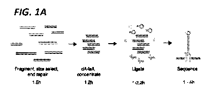
[0013] FIG. 1A. Schematic of the short-fragment sequencing library
preparation. dsDNA is
fragmented, size selected, end repaired, and coOncentrated. Increased
concentrations of Y-shape
adapters with attached E5 proteins and hairpin adapters are ligated onto the
dsDNA and E3 proteins
(green) bind to hairpin adapters. Electric current then drives a single strand
of DNA through the
nanopore (light gray).
[0014] FIG. 1B. Optimization of short-fragment Library preparation. Lane 1,
control DNA
fragment; lane 2, ligation of control fragment and adapters using
manufacturer's protocol; lanes 3-
7, incremental improvements in ligation efficiency using purification of
fragmented and dA-tailed
template DNA (lane 3), reduced reaction volume (lane 4), incorporation of a 1-
2 hour incubation
at 4 C (lanes 5, 6) and reducing RT incubation time to 5 min in order to
reduce release of E5
proteins from adapters (lane 7).
[0015] FIG. 2A. Use of short-DNA fragment sequencing using Minion was able to
correctly
determine gender and detect aneuploidy in DNA samples from a normal male and
female, a female
with monosomy X, a male with trisomy 12, and a male with trisomy 21 (p<0.001).
The copy
number of each chromosome was reflected by the corrected normalized percentage
of UA
(Norm' %UA,). Black dots represent chromosomes without significant copy number
changes; red
dots represent chromosomes with significant copy number changes comparing to a
normal male
reference; dotted line represent 99.9% confidence intervals.
[0016] FIG. 2B. Theoretical lower unique alignment (UA) required for
aneuploidy detection
under Poisson distribution. When k = 41, p(x > 1.5 k) = 0.0008. pr3(x' < 1.25
k) = 0.10.
[0017] FIG. 2C. Theoretical lower detection power using the 15K reference
under Poisson
distribution. The Y chromosome has fewest UA, 79-80, assigned. When k = 79,
p(x > 1.5 k) =
1.07x10⁻⁵. < 1.25 k) = 0.034.
[0018] FIG. 2D. Sequencing yield of a short-fragment library across time
showing raw reads, 2D
reads, and reads uniquely aligned to Hg19 reference genome.
[0019] FIG. 3. MinION library preparation.
[0020] FIG. 4. Software comparision.
[0021] FIG. 5. MinION Run Summary.
3
CA 03005067 2018-05-10
WO 2017/083828 PCT/US2016/061859
[0022] FIG. 6. Comparison of the 15K normal male reference and the GRCh37
human reference
genome.
[0023] FIG. 7. ULCS cytogenetics analysis.
[0024] FIG. 8. Internal normalization. Runs 1-4, using an internal reference,
has a very low
coefficient of variation, whether using our own DNA sequencing data or that
obtained from other
groups.
DETAILED DESCRIPTION OF THE DISCLOSURE
[0025] To maintain equivalent molar concentrations for short DNA fragment-
length library
preparations compared with long fragment-length, ¨18-fold lower total ng of
input DNA and
improved ligation efficiency was required (FIG. IB). We systematically
modified the protocol
to improve ligation efficiency. To monitor ligation reactions, a 434 bp PCR
product and a 57 bp
control adapter duplex with a T-overhang were used (Table 1).
Table 1. Sequence Information
SEQ ID NO:1 Control fragment sequence, CAGGAAACAGCTATGACCATGATTAC
434bp GCCAAGCTATTTAGGTGACGCGTTAGA
ATACTCAAGCTATGCATCAAGCTTGGT
ACCGAGCTCGGATCCACTAGTAACGGC
CGCCAGTGTGCTGGAATTCAGGCAAGC
AGAAGACGGCATACGAGATCGTGATG
TGACTGGAGTTCAGACGTGTGCTCTTC
CGATCTCTGCACAATGTGCACATGTAC
CCTAAAACTTAGAGTATAATAAAAATA
AAAAATAAAAAAAGAAGTCCAAAAAA
AGATCGGAAGAGCGTCGTGTAGGGAA
AGAGTGTAGATCTCGGTGGTCGCCGTA
TCATTCCTGAATTCTGCAGATATCCAT
CACACTGGCGGCCGCTCGAGCATGCAT
CTAGAGGGCCCAATTCGCCCTATAGTG
AGTCGTATTACAATTCACTGGCCGTCG
TTTTAC
SEQ ID NO:2 M13F (-20) primer GTAAAACGACGGCCAG
SEQ ID NO:3 M13R primer CAGGAAACAGCTATGAC
4
CA 03005067 2018-05-10
WO 2017/083828 PCT/US2016/061859
SEQ ID NO:4 Control adaptor (Top 5')
GGAAGCTTGACATTCTGGATCGGTGAC
TGGAGTTCAGACGTGTGCTCTTCCGAT
CTT
SEQ ID NO:5 Control adaptor (Bottom 5') AGATCGGAAGAGCACACGTCT
Use of the manufacturer's protocol resulted in <5% of all end products having
two adapters
attached (FIG. IB, lane 2). By purifying dA-tailed DNA prior to ligations, the
percentage of end
products having two adapters ligated increased to 25% (FIG. IB, lane 3).
Reducing reaction
volumes from 100 pt to 20 [IL further increased the percentage of end products
with two adapters
ligated to 48% (FIG. IB, lane 4). By combining a 10 min RT and 1-2 h at 4 C
incubation, we
were able to increase the percentage of fragments with adapters ligated to
both ends to 61-63%
(FIG. IB, lanes 5 - 7) without releasing the pre-attached ES protein. Thus, by
purifying and then
concentrating dA-tailed DNA to reduce the reaction volume and introducing a
prolonged 2 h
ligation at 4 C, we increased the percentage of final products with adapters
ligated to both ends
from <5% to 63% (FIG. IB lane 2 vs 7) and provided sufficient materials for
downstream His-
Tag bead purification (FIG. 3).
[0026] To determine the optimal tool for data analysis of the increased number
of reads obtained
with sequencing of short DNA, we compared LAST - an alignment program
recommended by
MAP - with two similar programs, Bowtie2 and Blat(8-10), using a training
library generated
through a MinION short DNA sequencing run (FIG. 4). While Bowtie2 and LAST
completed
alignments more quickly (1 min and 14 min, respectively) than Blat (68 min),
Blat generated more
good alignments (65%) compared with Bowtie2 and LAST (58% and 61%,
respectively) for the
same datasets, likely due to the tendency for MinION sequencing errors
resulting in deletions
(FIGS. 3-4). Blat also generated more unique alignment (62%) compared with
Bowtie2 and LAST
(45% and 55%, respectively). Blat was used for alignment of the MinION short
DNA sequencing
results to provide the most inclusive alignment results. Given sufficient
computational resources
on a high performance server, increasing parallel threats can further reduce
the run time.
[0027] To demonstrate clinical utility of nanopore-based sequencing of short
DNA fragments, we
tested the ability of this approach to diagnose aneuploidy. Fetal aneuploidy
testing is routinely
performed as a component of prenatal testing (e.g. amniocentesis, chorionic
villus sampling
(CVS)), preimplantation genetic screening (PGS) of embryos in in-vitro
fertilization (IVF) and
CA 03005067 2018-05-10
WO 2017/083828 PCT/US2016/061859
evaluation of miscarriage tissue. A rapid diagnosis is clinically vital in
order to enable timely
management. In the case of prenatal samples obtained through an amniocentesis
or CVS, rapid
results will enable treatment before the pregnancy progresses to a more
advanced gestational age
when treatment options are more limited, technically difficult and dangerous
to the mother. In the
case of PGS, rapid testing will enable transfer of the embryo in a given IVF
cycle without the need
to freeze embryos. However, standard methods to diagnose aneuploidy, such a
karyotyping and
microarray analysis, take 7-21 days to complete. Ultra-low coverage sequencing
(ULCS) for
detection of aneuploidy is a new strategy for whole-genome aneuploidy
detection that requires
alignment of reads to a reference genome assembly to assess for aneuploidy but
still requires 15-
21 h to complete and requires costly and technically advanced library
preparation and sequencing
platforms that cannot be readily used in a physician's office or in low
complexity settings. The
ULCS approach for determining aneuploidy requires that the reads need only be
sufficiently long
to enable unique alignment to the genome. Thus, a method to rapidly sequence
large numbers of
short DNA fragments in real-time would enable rapid diagnosis of aneuploidy in
settings outside
of an advanced laboratory facility.
[0028] Purified genomic DNA samples from a normal male and female, a male with
trisomy 12,
a male with trisomy 21 and a female monosomy X were fragmented, size-selected
(350-600bp),
and processed as described (FIG. 3). Sequencing short DNA fragment libraries
prepared using
our protocol with MinION generated ¨500 unique reads after the first 3 min of
sequencing and 43-
87K raw reads and 27-58K 2D reads (32-67%) after 4 hours of sequencing (FIG.
2, FIG. 5). This
compares favorably to the traditional MinION sequencing protocol that
sequenced fewer than
12,000 reads after 36 h. Of the reads generated using our protocol, 40-70% of
the 2D reads could
be uniquely mapped to one location (FIG. 5).
[0029] Using the short fragment length DNA sequencing library preparation and
analysis pipeline
we obtained sufficient numbers of reads for successful determination of gender
and aneuploidy
(p<0.001) in all samples within 2-4 h (FIG. 2A). By Normal approximation of
Poisson
distribution, the chance of a type II error for detecting aneuploidy (p0-
aneuploidy) was <0.05
(FIG. 2C, FIG. 7). As MinION is easily scalable, cytogenetic analysis can be
done within 1-2 h
by running two MinION sequencers in parallel and 30 min-1 h by running four
MinION sequencers
in parallel.
6
CA 03005067 2018-05-10
WO 2017/083828 PCT/US2016/061859
[0030] In summary, in addition to the intended role of MinION for sequencing
long fragments of
DNA, our results demonstrate that MinION can also be used for very rapid real-
time acquisition
of short DNA reads that can be used for time sensitive aneuploidy detection in
prenatal and IVF
care as well as sequencing of small DNA fragments and amplicons in the field
or clinic. This
ability can expand the utility of the MinION into new clinical and research
applications.
[0031] The disclosure will now be illustrated in the following Examples, which
do not in any way
limit the scope of the invention.
EXAMPLES
Example]
Development of Ligation Conditions
[0032] To assess the ligation efficiency, a short DNA control fragment were
used for initial
ligation reactions. The fragment was generated using PCR with M13 forward and
reverse primers
to amplify a 434-bp fragment from a pCR-Blunt vector using Q5 High-Fidelity
DNA Polymerase
(NEB). See Table 1.
[0033] A 50-ml PCR reaction was prepared following the manufacturer's
protocol. The PCR
reaction was subjected to a 30-sec initial denaturation at 98 C, 25 cycles of
10-sec denaturation at
98 C, a 30-sec annealing at 57 C, and a 20-sec elongation at 72 C. A final
elongation step at 72 C
for 2 min was added to ensure complete amplification. The PCR product was
purified using a
QIAquick PCR Purification Kit following the manufacturer's protocol. A 57-bp
asymmetric
adapter with a T overhang was used as a control adapter to assess ligation
efficiency See Table 1.
The control adapters were diluted to 0.4 mM in MinION adaptor buffer (50 mM
NaC1 and 10 mM
Tris-HC1, pH 7.5) to simulate the 0.2-mM concentration of the Y shaped and
hairpin adapters in
the adaptor mix (Oxford Nanopore).
[0034] Ligation reactions were initially performed following the MinION
Genomic Sequencing
Kit protocol (Oxford Nanopore, SQK-MAP004). Control DNA fragments (0.2 pmol,
52 ng) were
added to a 30 [11 NEB Next dA-Tailing Module (NEB) reaction [4 ml of control
fragments, 21 1
of Qiagen Buffer EB, 3 [11 of 103 NEB Next dA-tailing reaction buffer, and 2
1 of Klenow
fragments (3'45' exo-)]. Reactions were performed at 37 for 30 min in a Bio-
Rad C1000Touch
Thermal Cycler. All the dA-tailing reactions were added to a total volume of
100 1 [30 1 of dA-
7
CA 03005067 2018-05-10
WO 2017/083828 PCT/US2016/061859
tailing reaction, 10 [11 of control adapter, 10 [11 of nuclease-free water, 50
[11 of NEB Blunt/TA
Ligase Master Mix (NEB)] and incubated at room temperature (23-25 C) for 10
min.
[0035] Because so few control fragments had adapters ligated on both ends
(FIG. IB, lane 2), an
alternative Klenow fragment (39/59 exo-) (NEB) was used for dA tailing, and
the dA-tailing
reactions were purified before being added to the ligation reactions. Control
DNA fragments (250
ng) were subjected to a dA-tailing reaction [2.5 [11 of NEBuffer II, 5 ml of 1
mM deoxyadeno sine
triphosphate (dATP), 1 ml of Klenow fragment (39/59 exo-), and nuclease-free
water to a total
volume of 25 [11]. After purification with 1.8-fold AMPure XP beads (Beckman
Coulter following
the manufacturer's protocol for the SPRI select reagent (Beckman Coulter), the
dA-tailed control
fragment was eluted in 12 [11 of 1/5 Qiagen Buffer EB (2 mM Tris-C1, pH 8;
Qiagen) and diluted
to 0.05 mM (13 ng/ml).
[0036] Overnight ligation reactions at 16 C using T4 DNA ligase (NEB) to
ligate a 10:1 adapter-
fragment mixture (4 pmol control adapter, 0.2 pmol control fragment in 2 [11
10 x T4 DNA ligase
buffer, 1 ml T4 DNA ligase, and NF H20 to 20- [11 final volume) resulted in
¨75% of the control
fragments having adapters on both ends, which would not be sufficient final
products for
downstream steps. Therefore, the reactions were run in duplicate and combined.
Then 5:1 ratios
were used to preserve the adapters provided in the MinION kits.
[0037] The second ligation reactions were a replication of the manufacturer's
ligation protocol
using the purified dA-tailed DNA, as described previously (FIG. IB, lane 3),
using 100 [11 of
ligation reaction with 0.4 pmol of DNA, 26 1 of Buffer EB, 10 1 of control
adapter, 50 1 of
Blunt/TA Ligase MasterMix (NEB), and 10 1 of nuclease-free water (Ambion).
Reactions were
incubated at room temperature for 10 min and purified using 1.8-fold AMPure XP
beads, washed
with the wash buffer in the SQK-MAP003MinION Genomic DNA Sequencing Kit (750
mM
NaC1, 10%PEG 8000, 50 mM Tris-HC1, pH 8.0), and eluted in 20 1 of Buffer EB.
[0038] The third ligation reactions were a reduced-volume system using
purified dA-tailed DNA,
as described previously (FIG. IB, lanes 4-7). A 20-ml ligation reaction
containing 0.2 pmol of
DNA (4 ml), 2 pmol of control DNA adaptor (5 [11), 10 1 of Blunt/TA Ligase
Master Mix, and 1
1 of nuclease-free water was incubated for 10 min at room temperature,
purified using one-fold
AMPure XP beads with the SQK-MAP003 wash buffer, and eluted in 20 1 of Buffer
EB (FIG.
IB, lane 4). Reactions were carried out at room temperature for 5-10 min,
followed by 4 C
8
CA 03005067 2018-05-10
WO 2017/083828 PCT/US2016/061859
incubation for 1-2 hr (FIG. IB, lanes 5-7). Reactions were purified using one-
fold AMPure XP
beads with SQK-MAP003 wash buffer and eluted in 20 1 of Buffer EB. Purified
ligation products
were run on 2% agarose gels. Portions of the ligation products were estimated
using ImageJ
densitometry analysis with two technical replicates.
Example 2
Nucleic Acid Manipulations
[0039] To facilitate maximum recovery of material, 1.5-ml low-retention
microcentrifuge tubes
and low-retention tips were used unless stated otherwise. For all reactions
performed in a thermal
cycler, 0.2-ml PCR tubes were used (Axygen). An Agencourt SPRIStand Magnetic 6-
tube Stand
(Beckman Coulter) was used for pelleting of SPRI select and AMPure XP
bead¨related
purification; a DynaMag-2 magnet (Life Technologies) was used for His-tag bead
isolation.
Example 3
Genomic DNA Samples
[0040] Genomic DNA (gDNA) samples from a karyotypically normal male and
female, a male
with trisomy 12, a male with trisomy21, and a female with monosomy X were used
for cytogenetic
analysis using short-DNA-fragment ULCS with the MinION. Blood B-lymphocytes
from
karyotypically normal human male and female samples were obtained from the
Coriell Institute
Cell Repositories (GM12877 and GM12878) and cultured according to the protocol
provided by
the Coriell Institute. gDNA was extracted from cell cultures from the second
passage using a
QIAamp Blood DNA Mini Kit (Qiagen) following the manufacturer's manual. gDNA
from a male
with trisomy 21 was provided by the Coriell Institute Cell Repositories
(NG05397). DNA samples
from a male with trisomy 12 and a female with monosomy X were obtained from
the products of
conception of miscarriage cases that had cytogenetic testing performed using G-
band karyotyping.
gDNA was extracted using an All Prep DNA/RNA/Protein Mini Kit (Qiagen) from
the
trophoblastic primary cell cultures of the chorionic villus. The quality of
gDNA was examined on
0.8%agarose gel and quantified using a NanoDrop 1000 Spectrophotometer (Thermo
Fisher
Scientific). DNA was stored at -20 C until needed.
9
CA 03005067 2018-05-10
WO 2017/083828 PCT/US2016/061859
Example 4
Library Preparation
[0041] For library preparation, 120 [11 of 25 ng/ml gDNA in TE Buffer (pH 8.0)
was fragmented
using a Covaris S220 focused ultra-sonicator at the manufacturer's 500-bp
setting in micro-TUBEs
(Covaris). For size selection, 100 [11 of fragmented gDNA was used. Size
selection was performed
in a 1.5-ml DNA LoBind tube (Eppendorf) using SPRIselect reagent following the
manufacturer's
double-sized selection protocol using a right-side 0.55 times, left side 0.7
times setting (Beckman
Coulter). DNA was eluted in 40-50 [11 of Buffer EB in a 1.5-ml DNA LoBind
tube. Then 2 1 of
DNA was used for a 2% gel electrophoresis to confirm fragment size. Purified
DNA (3 [11) was
saved for NanoDrop quantification. Size-selected DNA fragments were ¨350-600
bp in length.
[0042] Buffer EB was added to size selected DNA to a final volume of 80 [11.
End-repair reactions
were performed using a NEB Next End Repair Module (NEB) in a 1.5-ml DNA LoBind
tube.
Then 5 [11 of DNA CS (Oxford Nanopore, SQK-MAP004), 10 [11 of 10 x NEB Next
End Repair
Reaction Buffer, and 5 1 of NEB Next End Repair Enzyme Mix were added to the
size-selected
DNA fragment and mixed by gently pipetting. The reactions were incubated at
room temperature
for 25 min and purified using 1.8-fold AMPure XP beads following the SPRI
select reagent
protocol in a DNA LoBind tube. The end-repaired DNA was eluted in 22 [11 of
Buffer EB, and the
DNA was quantified using a Qubit dsDNA HS AssayKit (Life Technologies).
[0043] End-repaired DNA was subjected to a dA-tailing reactionusing a Klenow
fragment (3'45'
exo-) in a total volume of 25 [11 in a sterile PCR tube. The reaction
contained 2.5 1 of NEBuffer
II, 1 1 of Klenow fragment (3'45' exo-), 16.5 1 of end-repaired purified
DNA, and 5 1 of dATP
(1 mM). Reactions were incubated in a Bio-Rad C1000 Thermal Cyclerat 37 C for
45 min,
purified using 1.8-fold AMPure XP beads, and then eluted in 12 [t1 of 1/5
Buffer EB. The purified
product was quantified using NanoDrop and a Qubit dsDNA HSAssay Kit (Life
Technologies)
and diluted to ¨0.05 mM (-18 ng/ml) with 1/5 Buffer EB to be used as the dA-
tailed DNA in
subsequent reactions.
[0044] His-tag Dynabeads (10 ml) (Invitrogen) were washed in 1.5-ml low-
retention tubes in a
MinION Genomic DNA Sequencing Kit following the manufacture's protocol on a
DynaMag-2
magnetic stand (Invitrogen). Washed beads were resuspended in 40 [t1 of
undiluted wash buffer
(SQK-MAP004) and kept on ice. Ligation reactions were performed in a 1.5-ml
low-retention
CA 03005067 2018-05-10
WO 2017/083828 PCT/US2016/061859
tube. Twenty-microliter reactions contain 4 [11 of dA-tailed DNA (0.2 pmol), 5
[11 of adaptor mix
(1 pmol) (SQK-MAP004), 1 [11 of HP adapter ( lpmol) (SQK-MAP004), and 10 [11
of Blunt/TA
Ligase Master Mix (NEB). The reactions were mixed by pipetting gently between
each sequential
addition and spun down briefly in a benchtop centrifuge. Ligation reactions
were incubated at
room temperature for5 min follow by 4 C for 2 hr. For each sample, 2 x 20 [11
reactions were
performed in separate tubes and combined for His-tag bead purification.
[0045] In 1.5-ml low-retention tubes, 40 [11 of washed His-tag beads were
added to the adapter-
ligated DNA and carefully mix by gentle pipetting. The mixture was incubated
at room
temperature for 5 min and placed on ice for 30 sec. His-tag bead purification
was performed
following the protocol of the MinION Genomic DNA Sequencing Kit (SQK-MAP004).
Pelleted
beads were resuspended 28 [11 of the ELB elution buffer (SQK-MAP004) by gently
pipetting 10
times. The suspension was incubated at room temperature for 5 min and placed
on ice for 30 sec,
and this was repeated once before placing the suspension back on the magnetic
rack for pelleting.
The eluate was transferred to a clean 1.5-ml low-retention tube, incubated on
ice for 30 sec, and
then placed on a magnetic rack for 2 min for pelleting any residual beads. The
eluate then was
carefully transferred to a 1.5-ml low-retention tube. This library was called
the presequencing mix.
Then 4 [11 of the presequencing mix was used for quantification by a Qubit
dsDNA HS Assay Kit.
Example 5
MinION Sequencing
[0046] Then 150 ml of the priming mix (147 [11 of EP buffer and 3 1 of fuel
mix) was loaded on
a MinION Flow Cell (R7.3) and incubated for 10 min. The priming process was
repeated once.
Then 150 [11 of the MinION sequencing library (12 1 of the presequencing mix,
135 ml of EP
buffer, and 3 ml of fuel mix) was gently mixed and loaded to the MinION Flow
Cell. The MAP
48-hr gDNA sequencing protocol was used, and the sequencing reaction was
stopped when
sufficient data were collected.
Example 6
Data analysis
[0047] Metrichor Agent V2.26 was used to transfer local fast5 files, and 2D
Base calling Rev1.14
was used to convert currency into base events (Oxford Nanopore Technologies).
Pore tools v0.5.0
11
CA 03005067 2018-05-10
WO 2017/083828 PCT/US2016/061859
was used to convert Fast5 to fastQ files. The first and last 50 bases were
removed from each
sequence using cut adapt v1.7.1, and sequences that were at least 50 bases
long were kept after the
removal. Both 1D and 2D reads were aligned to the Ensembl GRCh37 human
reference genome
using BLAT (FIG. 3).
[0048] Less than 1% of 1D sequences passed the screening criteria (covers >
40% of query, > 80%
alignment identity) and consequently only 2D sequences were used for further
analysis. 2D reads
with a unique alignment match (UA) to a genomic location were retained for
further analysis.
Bowtie2 was also tested for mapping 2D sequences to a human reference genome.
As Bowtie2
was designed for high-throughput mapping of short sequences (50-200bp), <5%
full length 2D
reads could be mapped. Bowtie2 --bwa-sw-like settings developed for 454 data
were also tested,
only 36% of the 2D reads were UA. Therefore, we used Bowtie2 to align the
first 200bp of the
2D reads, and generated 45% UA in ¨1 min (FIG. 4). 2D reads were also mapped
to the reference
genome using LAST using the recommended setting that were reported to be most
inclusive for
alignment for MinION long reads, however, it produced fewer UA comparing to
the BLAT
pipeline using the same screening criteria (FIG. 3). Hence, only the UA from
the BLAT pipeline
were used for the fast cytogenetic analysis using the ultralow coverage
sequencing (ULCS).
Example 7
Digital karyotyping using Ultra low coverage sequencing (ULCS)
[0049] Ultralow coverage sequencing (ULCS) is a powerful tool for cytogenetic
analysis. As a
proof of concept, we performed the analysis on 5 samples and a modified ULCS
strategy was used
for this study. Previous study indicated coefficient of variation (CV) in ULCS
(< 0.01-fold
coverage) was lower than 15% on each autosome and there was no significant
difference of the
autosomal CVs between MiSeq and Ion Proton platforms. In a ULCS analysis, we
assumed the
UA on each chromosome (labeled as subscript i, i=1,2,. ,22,X, Y) fits Poisson
distribution.
UAi = nicpi
Where n, is the number of reads needed to cover a chromosome i, and cp, is the
coverage of a
chromosome i. The percentage of UA on each chromosome (%UA,) is determined by
the length
and copy number of each chromosome under the same coverage.
[0050] The lower limit of sequencing read needed for ULCS was primarily
determined by the UA
assigned to Chromosome Y because a) it is one of the shortest chromosomes, and
thus fewer DNA
12
CA 03005067 2018-05-10
WO 2017/083828 PCT/US2016/061859
fragments would be sequenced from it, b) less than 50% of chromosome Y has
been sequenced
and annotated in the human reference genome, and hence more than half of the
Chromosome Y
reads would not be able to be mapped to reference genome, and then being
counted and c) reads
mapped to the identical regions of the chromosome X and Y would not be
considered as UA by
the analysis pipeline. Moreover, crosslinking between chromosome X and Y, and
the present of
repetitive elements will cause a small portion of misplacement of reads from X
and Y chromosome,
which will further reduced reads that could have been mapped to the Y
chromosome.
[0051] To estimate the lower limit of UA, needed for ULCS cytogenetic
analysis, we used Normal
Approximation of Poisson distribution in R (qpois function) to estimate the
detection power of UA
for aneuploidy. It was estimated that the when UA, = 41, p(x > 1.25 k) = 0.04,
p(x > 1.5 k) =
0.0008, and the detection power of aneuploidy is 90 %. When the UAi was 79,
the detection power
of aneuploidy would be 95.6%. The corresponding total UA for UAy ¨79 is
¨15,000 in the normal
male sample. 15,000 UA were randomly selected from the sequencing result of
the normal male
for 30 times, and the average UA for each chromosome was used as reference for
normalization
purpose (Ref UA,). To examine if the 15K reference is representing human
genome under Poisson
distribution, we compared the percentage of ungapped length (%UL) and %UA of
each
chromosome. Their ratios (Norm Ref %UA) on autosomes was 1.04 (SD=0.0687,
CV=6.6%)
(FIG. 6).
[0052] The 15K reference represent the %UA represented about a half of the %UL
of the sex
chromosomes, which could be the result of depletion of non-unique alignments
on homogenous
regions of sex chromosomes. The mitochondrial chromosome (MT) is a multi-copy
small
chromosome, and it was not included in ULCS cytogenetics analysis. According
to Poisson
distribution, the 99.9% confidential intervals of each chromosome of the
normal male reference
can be estimated as Ref UAi 3.29VRef UAi under the same coverage.
[0053] To access the copy number of each chromosome of a query sample using
15,000 UA reads
(FIG. 7), we assumed the number of uniquely aligned read on each chromosome
(UA,) fits Poisson
distribution as described before.
[0054] Using 15,000 UA reads, the normalized ratio between a query sample and
the reference
(Norm %UA,) was determined by the copy number of chromosomes:
______________________________________ Query_%1JAi Query_ni x Query_cpi
Norm %UA = ____________________________
_
Ref _%1JA1 Ref _ni x Ref _cpi
13
CA 03005067 2018-05-10
WO 2017/083828 PCT/US2016/061859
To address the change in coverage y due to loss or gain of chromosomes, the
corrected normalized
%UAIequals:
Norm_%UAi
Norm'_%UA, =
Norm_%UA,'
Where Norm_%UA,' is the average Norm %UAI of normal autosomes as determined by
Z-score.
For an unknown sample, The standard deviation (SD) of Norm %UAI of normal
autosomes
(SDnormal) was estimated by known normal autosomes (within Ref UAi + 3.29VRef
UAi) in this
study (n=105, SDnormal=0.0489). The Z- score was calculated for each
chromosome:
Norm_%UAi¨ Mean_%UAautosome
Z ¨ scorei=
SDnormal
Chromosomes having a 1Z-score! of > 3.29 were considered as an abnormal
chromosome with p
<0.001. When the Z-score was > 3.29, we consider there to be a gain of a
chromosome, when the
Z-score was < -3.29, we consider there to be a loss of a chromosome. While the
modified Z-score
method would be less specific in detecting abnormality on small autosomes than
the Z-score
method based on census of each chromosome, it provided sufficient detection
power for
aneuploidy detection ( > 95%) (FIG. 2C). The theoretical value of a normal
autosome
Norm' %1JAnormal=1, a full trisomy of autosome Norm' %1JAtrisomy=1.5, a
monosomy of
autosome Norm' %1JAmonosomy=0.5, the X chromosome of a normal female Norm'
%UAx female
> 1.5, the Y chromosome of a normal female or missing Y chromosome Norm' %UAy
female <0.5.
[0055] We hypothesized that the corrected normalized %UAi (Norm' %UA1)
reflects the copy
number of chromosomes. The Norm' %UAI were used to compute the adjusted Z-
score (Z' -
score). Norm' %UAI of normal autosomes with IZ-scorel<3.29 were summarized
(Mean Norm' %UA=0.9999, SD Norm' %UA=0.0481). Z'-score for each chromosome
equals:
Norm'_%UAi¨ Mean_Norm'_%UA
Z' ¨ score i= _____________________________________________
SD_N orm'_%UA
[0056] In brief, 15,000 UA were randomly selected from the normal male sample -
and this was
repeated for a total of 30 times - and averaged for normalization purpose (Ref
UA). For each
sample, the first 15,000 UA (Query UA) were selected for gender determination
and aneuploidy
14
CA 03005067 2018-05-10
WO 2017/083828 PCT/US2016/061859
detection. The UA were summarized and counted for each chromosome (UAõ
X, Y), and
corresponding percentage were calculated for each chromosome (%UA,) by
UA,/15,000x100. The
%UA, for each of the chromosome of a query sample (Query %UA,) was normalized
to the normal
male reference (Ref %UA,) and corrected to detect the copy number of each
chromosome
(Norm' %UA,) (FIG. 7 FIG. 2A).
Example 8
Internal Normalization
[0057] For determination of a copy number variation and /or aneuploidy using
DNA sequencing
or microarray, the signal abundance in a test samples is compared with the
signal abundance in a
reference sample. For example, when "X" ng of DNA from Test sample A is
sequenced, 100k
unique reads map to Chromosome 21. When "X" ng of DNA from Test sample B is
sequenced in
the same sequencing run, 150k unique reads map to Chromosome 21. However, when
"X" ng of
reference, normal, DNA sample is sequenced in the same sequencing run, 100k
unique reads are
map to Chromosome 21. Thus Sample A has the same abundance of Chromosome 21 as
does the
reference sample while Sample B has 50% more, i.e. trisomy 21.
[0058] In another embodiment, the relative abundance of reads mapping to
chromosome 21 are
compared with an internal reference, such as chromosome 1. A normal ratio can
be determined
using a reference sample. In future runs, the ratio of reads from chromosome 1
relative to the
number of reads from chromosome 21 would be determined. A decrease in this
ratio would
suggest a relative increase in the abundance of chromosome 21 relative to the
reference
chromosome.
[0059] This analysis can be done in conjunction with traditional analysis with
a reference sample
in order to improve the sensitivity and specificity of the test (e.g. low
coverage sequencing or
microarray) or it can be run alone in order avoid the need to also run a
reference sample.
[0060] As shown in FIG. 8, Runs 1-4, using an internal reference has a very
low coefficient of
variation, whether using our own DNA sequencing data, or that obtained from
other groups.