Note : Les descriptions sont présentées dans la langue officielle dans laquelle elles ont été soumises.
CA 03014013 2018-08-08
WO 2017/137830 PCT/IB2017/000100
- 1 ¨
ANTI-VISTA (B7H5) ANTIBODIES
RELATED APPLICATION(S)
[0001] This application claims the benefit of U.S. Provisional Application
No. 62/294,922,
filed on February 12, 2016. The entire teachings of the above application are
incorporated herein
by reference.
BACKGROUND OF THE INVENTION
[0002] The expression of negative immune regulators by cancer cells or
immune cells in the
tumor microenvironment can suppress the host's immune response against the
tumor. To
effectively combat the cancer, it is desirable to block tumor-mediated
suppression of the host
immune response. Accordingly, there is a need for new and effective
therapeutic agents that
inhibit negative immune regulators in the tumor microenvironment that suppress
anti-tumor
immune responses.
SUMMARY OF THE INVENTION
[0003] The present invention provides, in an embodiment, a method of
eliciting a biological
response in a subject. The method comprises the steps of administering to a
subject an antibody
that binds a V-domain Ig Suppressor of T cell Activation (VISTA) protein, or
an antigen-binding
fragment thereof, in an amount sufficient to elicit a biological response in
the subject. In certain
embodiments, the biological response is selected from the group consisting of
a decrease in the
number of circulating immune cells; a decrease in the number of granulocytes
in bone marrow
and spleen; an increase in the number of neutrophils, macrophages, T cells, or
a combination
thereof in a tumor microenvironment (TME); and an increase in the level of one
or more
cytokines (e.g. chemokines); or any combination of these responses. In a
particular embodiment,
the antibody that binds VISTA, or antigen-binding fragment thereof, comprises
an Fc region that
binds to an Fc receptor (e.g., CD16 receptor) on immune cells (e.g., NK
cells).
CA 03014013 2018-08-08
WO 2017/137830 PCT/IB2017/000100
- 2 -
[0004] In another embodiment, the invention provides a method of
identifying an antibody
that binds a V-domain Ig Suppressor of T cell Activation (VISTA) protein and
elicits a
biological response. The method comprises the steps of providing an antibody
that binds
VISTA, or an antibody fragment thereof, to a cell, tissue, organ or organism,
and determining
whether the antibody or antibody fragment thereof induces a biological
response in the cell,
tissue, organ or organism. In certain embodiments, the biological response is
selected from the
group consisting of activation of monocytes; activation of T cells; a decrease
in the number of
circulating immune cells; a decrease in the number of granulocytes in bone
marrow and spleen;
an increase in the number of neutrophils, macrophages or both in a tumor
microenvironment; and
an increase in the level of one or more cytokines; or any combination of these
responses. In a
particular embodiment, the antibody that binds VISTA, or antigen-binding
fragment thereof,
comprises an Fc region that binds to an Fc receptor (e.g., CD16 receptor) on
immune cells (e.g.,
NK cells).
[0005] The methods of the present invention are useful for, for example,
characterizing
antibodies that bind VISTA protein and for screening candidate anti-VISTA
antibodies for
biological activities associated with potential therapeutic efficacy.
BRIEF DESCRIPTION OF THE DRAWINGS
[0006] The patent or application file contains at least one drawing
executed in color. Copies
of this patent or patent application publication with color drawing(s) will be
provided by the
Office upon request and payment of the necessary fee.
[0007] Figure 1A-1C: Graphs showing VISTA expression on TF1 AML Cells
Expression of
VISTA protein by flow cytometry is shown in the TF-1 AML cell line.
[0008] Figure 2A-2E: Graphs showing staining and gating strategies for
identification of
Human Myeloid and Lymphoid Subsets.
[0009] Figure 3A-3G: Graphs showing expression of VISTA on Human Myeloid
and
Lymphoid Subsets from one healthy normal donor.
[0010] Figure 4: Graph showing expression of VISTA on Human Myeloid and
Lymphoid
Subsets across multiple healthy normal donors.
[0011] Figure 5A-5B: Graph showing staining and gating strategies for
identification of
expression of VISTA on Human Monocytes and Macrophages.
CA 03014013 2018-08-08
WO 2017/137830 PCT/IB2017/000100
- 3 -
[0012] Figure 6A-6C: Graphs showing expression of VISTA on Human Monocytes
and
Macrophages.
[0013] Figure 7A-7E: Graphs showing staining and gating strategies for
identification of
expression of VISTA on Human T and NK Cell Subsets.
[0014] Figure 8A-8G: Graphs showing expression of VISTA on Human T and NK
Cell
Subsets from one healthy normal donor.
[0015] Figure 9: Graph showing expression of VISTA on Human T and NK Cell
Subsets
across multiple healthy normal donors.
[0016] Figure 10A-10D: Graphs showing staining and gating strategies for
identification of
expression of VISTA on Human Dendritic Cell subsets.
[0017] Figure 11A-11C: Graphs showing expression of VISTA on Human
Dendritic Cell
subsets and basophils from one healthy normal donor.
[0018] Figure 12: Graph showing expression of VISTA on Human Dendritic Cell
Subsets
and basophils across multiple healthy normal donors.
[0019] Figure 13A-13D: Analysis of VISTA expression on healthy human
peripheral blood
cells. Profile of VISTA expression on healthy human peripheral blood cells
using multicolor
flow cytometry analysis: Whole blood samples from 2 different individuals were
analyzed for
VISTA expression on (Figure 13A) monocytes SS&CD1lbhICD14hICD16'CD33+veHLA-
DR+veCD19) (Figure 13B) neutrophils (SSChICD177 CD1lbhICD141 CD16+veCD33+"HLA-
DR-veCD19-ve). Peripheral blood mononuclear cells were isolated using Ficoll
gradient for
analysis of (Figure 13C) CD4+ T cells (CDPeCDeve), and (Figure 13D) CD8+ T
cells
(CD3+veCD8+ve).
[0020] Figure 14A-14C: Analysis of VISTA expression on peripheral blood
cells from a
lung cancer patient and a healthy control donor. Profile of VISTA expression
on lung cancer
patient peripheral blood cells using multicolor flow cytometry analysis:
Representative FACS
plot (Figure 14A) from one individual is shown. Peripheral blood mononuclear
cells were
isolated by Ficoll and analyzed for VISTA expression on (Figure 14B) monocytes
(CD14+
CD11 b+ CD33+ HLADR+ CD15-) and (Figure 14C) myeloid derived suppressor cells
(CD14-
CD11b+ CD33-HLADR-CD15+ CD16+).
[0021] Figure 15A-15C: Profile of VISTA expression in peripheral blood
cells from a
patient with colon cancer, using multicolor flow cytometry analysis:
Representative FACS plot
CA 03014013 2018-08-08
WO 2017/137830 PCT/IB2017/000100
- 4 -
(Figure 15A) from one individual is shown. Peripheral blood mononuclear cells
were isolated by
Ficoll and analyzed for VISTA expression on (Figure 15B) monocytes (CD14+
CD11b+ CD33+
HLADR+ CD15-) and (Figure 15C) myeloid derived suppressor cells (CD14- CD! lb+
CD33-
HLADR-CD15+ CD16+).
[0022] Figure 16A-16D: Profile of VISTA expression on Cynomolgus monkey
peripheral
blood cells using multicolor flow cytometry analysis: Whole blood from 4
different monkeys
was analyzed for VISTA expression on (Figure 16A) monocytes
(SSCI0CD111PCD141"HLA-
DRhICD16-"CD19-ve and (Figure 16B) neutrophils CD! 1bhICD1410HLA-DR-"CD16-
"CD19-ve.
Peripheral blood mononuclear cells from three monkeys were isolated using
Ficoll gradient for
analysis of (Figure 16C) CD4+ T cells (TCRa/13+veCD4+ve) and (Figure 16D) CD8+
T cells
(TCRa/f3+"CD8+") .
[0023] Figure 17: Graph showing absolute expression values of VISTA RNA in
Heme cell
lines.
[0024] Figure 18: Mouse A20 cells were stably transfected with either GFP
or human
VISTA. They were incubated with ova peptide and with D011.10 T cells. CD25
expression by
the T cells was measured 24 hours after incubation began. The A20-huVISTA
cells suppress
CD25 expression by the T cells, but this readout is significantly restored by
incubation with
VSTB95.
[0025] Figure 19A-19F: Graphs showing Human VISTA ELISA results.
[0026] Figure 20A-20F: Human VISTA FACS results, showing anti-VISTA
antibodies
binding to cells expressing human VISTA.
[0027] Figure 21A-21D: Dilution study of 6 anti-VISTA antibody candidates
in the mixed
lymphocyte reaction from 30 j_tg/m1 to 0.0 tig/ml.
[0028] Figure 22A-22B: Dilution studies of 6 anti-VISTA antibody candidates
in the SEB
assay (individual CPM counts and IFN-g concentrations) from 30 ,g/m1 to
0.011g/ml.
[0029] Figure 23: Sensorgram plot using anti-VISTA antibody VSTB85 coated
on a Proteon
SPR chip and VISTA protein with the indicated competitors run over the chip
(competitors listed
in Table 16).
[0030] Figure 24: Experimental design for MB49 murine bladder tumor model
CA 03014013 2018-08-08
WO 2017/137830 PCT/IB2017/000100
-5-
100311 Figure 25A-25B: MB49 tumor growth in female C57B1/6 mice. Graphs
illustrate
tumor growth in individual mice treated with anti-mouse VISTA antibody (Figure
25B) or
control IgG (Figure 25A).
[0032] Figure 26: Amino acid sequence of human VISTA (SEQ ID NO:46).
[0033] Figure 27: Multiple sequence alignment of VISTA orthologues
[0034] Figure 28: Regions of human VISTA bound by VSTB50 and VSTB60
antibodies
(top) or VSTB95 and VSTB112 antibodies (bottom), as determined by HDX
[0035] Figure 29: VISTA Epitope bound by VSTB112. (Top) VISTA is shown in
cartoon
with strands labeled. Residues having at least one atom within 5 A of VSTB112
in the complex
are colored blue. Blue and orange spheres highlight a chain break, and the
cyan and green
spheres mark the N- and C-termini of the VISTA structure, respectively.
(Bottom) Sequence of
VISTA construct used in structure determination. Circles below the sequence
are used to
indicate residues which make only main chain contacts with VSTB112, triangles
indicate a side
chain contact, and squares indicate the side chain contact results in either a
hydrogen bond or salt
bridge interaction as calculated by PISA. Shapes are colored to indicate the
CDR having the
greatest number of atoms contacted by the given residue with CDR colors
defined in Figure 59.
Secondary structural elements are as defined in the program MOE with yellow
arrows
representing 13-strands and red rectangles indicating a-helices.
[0036] Figure 30: VSTB112 Paratope. (Top) VISTA antigen is shown in
illustration and
VSTB112 within 5 angstrom (A) of VISTA is shown in surface with colors used to
designate
CDR identity as specified in the sequence below. Contacting framework residues
adjacent to a
CDR are colored similarly to the corresponding CDR (Bottom) Sequence of
VSTB112 Fv
region. Colored backgrounds specify CDRs according to Kabat definitions.
Circles below the
sequence are used to indicate residues which make main chain only contacts
with VISTA,
triangles indicate a side-chain contact, and squares indicate the side chain
contact results in either
a hydrogen bond or salt bridge interaction as calculated by PISA.
[0037] Figure 31: Comparison of epitope regions identified by
crystallography and
hydrogen deuterium exchange (HDX). Sequence of VISTA construct used in
structure
determination. Circles below the sequence are used to indicate residues which
make only main
chain contacts with VSTB112, triangles indicate a side chain contact, and
squares indicate the
CA 03014013 2018-08-08
WO 2017/137830 PCT/IB2017/000100
- 6 -
side chain contact results in either a hydrogen bond or salt bridge
interaction as calculated by
PISA.
[0038] Figure 32: Activation of CD14+ monocytes in whole PBMC by VSTB174
(derived
from VSTB112). In each part of the experiment, cells were incubated with PBS,
IgG1 control
antibody, or VSTB174 at 1, 0.1 or 0.01 ug/ml. Left panel shows CD80 MFI; right
panel shows
HLA-DR MFI (two donors tested with representative results shown).
[0039] Figure 33: Graph showing ADCC activity of VSTB174 directed against
K562-
VISTA cells.
[0040] Figure 34: Graph showing ADCP activity of VSTB174 directed against
K562-VISTA
cells. Both antibodies depicted have the same Fab, but VSTB174 has an IgG1 Fc
and VSTB140
has Fc silent IgG2 .
[0041] Figure 35: Graph showing phagocytosis mediated by VSTB174, VSTB149
or
VSTB140 mAbs against K562-VISTA. Each mAb was tested with 7 half log doses,
ranging from
0.00081.1g/m1 to 0.56 ughnl.
[0042] Figure 36: Graph showing phagocytosis mediated by VSTB174, VSTB149
or
VSTB140 mAbs against myeloma cell line K562 cells. Each mAb was tested with 7
half log
doses, ranging from 0.0008 ig/ml to 0.56 ug/ml.
[0043] Figure 37: MB49 tumor efficacy study evaluating VSTB123 1, 5, 7.5,
and 10 mg/kg
in female VISTA-KI mice. Tumor volumes were approximately 50 mm3 when dosing
began at
day 6 after implant. VSTB123 is the VSTB112 Fab grafted onto a mouse Fc
scaffold and binds
to human VISTA in the VISTA-KI mouse.
[0044] Figure 38: Graph shows that CD14+ cells expressing high/intermediate
levels of
VISTA are found in 13/13 lung cancer samples, as well as in distant lung
tissue and peripheral
blood of patients.
[0045] Figure 39: IHC staining for VISTA in Lung Cancer using GG8.
[0046] Figure 40: Anti-VISTA antibody triggers monocyte activation via CD16
crosslinking.
PD-L1 expression is used as a marker of myeloid activation using human PBMCs.
Anti-CD16,
used as the positive control, induced robust monocyte activation; Fc block
(mix of Fc IgG1
fragments), used as the negative control, blocked monocyte activation.
VSTB112, but not
VSTB140, induces monocyte activation compared to the human IgG1 control.
CA 03014013 2018-08-08
WO 2017/137830 PCT/IB2017/000100
-7-
100471 Figure 41: Schematic of study design for VSTB123 or VSTB124 effect
on the growth
of established MB49 tumors in hVISTA KI (knock-in) mice. Antibodies were
injected on study
days 5, 7, 10, 12, 14, 17, 19, 21, 24, and 26. Blood samples were collected on
days -2, -1, 0, 4, 7,
11, 14, 18, 24, 31, and 39. If blood samples were taken on the same day as an
antibody injection,
blood samples were taken first.
[0048] Figures 42A and 42B: MB49 tumor growth in hVISTA KI female mice
(Figure 42A)
and survival of mice bearing MB49 tumors after treatment with VSTB123 or
VSTB124 in
hVISTA KI female mice (Figure 42B). In Figure 42A, mean tumor volume
measurements
(mm3) are shown for female mice treated with VSTB123 or VSTB124 as compared to
the mouse
IgG2a control group. The treatment period was from day 5-26. Mean +/- SEM
shown. Note
that the y axes differ among the graphs. Data for the female mice are graphed
until day 33, when
>70% of mice were still alive in each group (n=6 or 7 per group). In Figure
42B, the percent
survival of hVISTA KI female mice is graphed, VSTB123 10 mg/kg, p = 0.0108.
The treatment
period was from day 5-26.
[0049] Figure 43: Fold-change in expression of 41 cytokines by PBMC treated
with
VSTB174. Whole PBMC from three healthy human donors were treated with VSTB174
or IgG1
control antibodies for 24 hours at the indicated concentrations. Cytokine
production was
analyzed by a 41-cytokine multiplex kit. Average fold-change in expression
over the IgG1
control is shown as a heat map with a log color scale. Asterisks indicate
significant differences
between treatment and control sample means. * p<0.05, ** p<0.01, *** p<0.001,
****p<0.0001.
Out-of-range (00R) denotes that all samples for that cytokine in that donor
were beyond the
limits of accurate detection (> above, < below).
[0050] Figures 44A and 44B: Macrophage activation in MB49 tumors. Figure
44A shows a
schematic of the study design. In Figure 44B indicates increased CD80+
macrophages in the
tumor microenvironment with VSTB123, but not VSTB124. MB49 tumor-bearing
hVISTA KI
mice were treated with VSTB123, VSTB124, or control mIgG2a and their tumors
were analyzed,
24h post-third dose, to determine the relative expression of CD80 on tumor-
infiltrating
macrophages. Each group contained 5 mice. Horizontal bars indicate means.
*p<0.05.
[0051] Figure 45: Migration of MPO+ cells to the tumor microenvironment.
[0052] Figure 46: VSTB174 induces transient decrease in neutrophils.
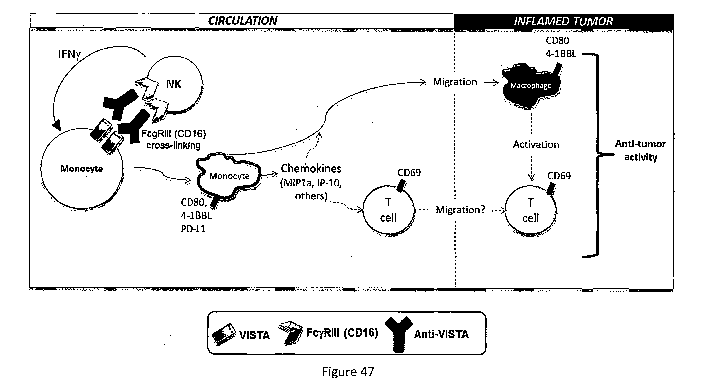
[0053] Figure 47: Anti-VISTA antibody (e.g., VSTB174) proposed mechanism of
action.
CA 03014013 2018-08-08
WO 2017/137830 PCT/IB2017/000100
-8-
100541 DETAILED DESCRIPTION OF THE INVENTION
[0055] A description of example embodiments of the invention follows.
[0056] The present invention relates to antibodies to novel Immunoglobulin
family ligand
designated V-domain Immunoglobulin Suppressor of T cell Activation (VISTA)
(Genbank:
JN602184) (Wang et al., 2010, 2011). VISTA bears homology to PD-Li but
displays a unique
expression pattern that is restricted to the hematopoietic compartment.
Specifically, VISTA is
constitutively and highly expressed on CD1lbhIgh myeloid cells, and expressed
at lower levels on
CD4+ and CD8+ T cells. The human homologue shares approximately 85% homology
with
murine VISTA and has similar expression patterns (Lines et al., Cancer
Research 74:1924,
2014). VISTA expressed on antigen presenting cells (APCs) suppresses CD4+ and
CD8+ T cell
proliferation and cytokine production via a cognate receptor independent of PD-
1. In a passive
EAE (experimental autoimmune encephalomyelitis) disease model, a VISTA
specific
monoclonal antibody enhanced T-cell dependent immune responses and exacerbated
disease.
VISTA over-expression on tumor cells impaired protective anti-tumor immunity
in tumor-
bearing hosts. Studies of human VISTA confirmed its suppressive function on
human T cells
(Lines et al Cancer Research 74:1924, 2014,. Studies from Flies et al. also
identified VISTA
(named PD-1H) as a potent immune suppressive molecule (Flies et al., 2011).
VISTA is
described in further detail in U.S. Published application US 20130177557 Al
and U.S. Patent
Nos. 7,919,585 and 8,236,304, all of which are incorporated herein by
reference in their entirety.
[0057] VISTA is a novel negative immune regulator that suppresses immune
responses. As
described, for example, in Example 12 herein, treatment with a VISTA-specific
monoclonal
antibody in murine tumor models has been shown to reverse the suppressive
character of the
tumor immune microenvironment and enhance protective anti-tumor immunity,
thus,
demonstrating the potential of a VISTA monoclonal antibody as a novel
therapeutic for cancer
immunotherapy.
[0058] ANTIBODIES AND FRAGMENTS OF THE PRESENT INVENTION
[0059] The term "antibody" is meant to include polyclonal antibodies,
monoclonal antibodies
(mAbs), chimeric antibodies, humanized antibodies, human antibodies and anti-
idiotypic (anti-
Id) antibodies, as well as fragments, regions or derivatives thereof, provided
by any known
technique, such as, but not limited to, enzymatic cleavage, peptide synthesis
or recombinant
techniques. Anti-VISTA antibodies of the present invention are capable of
binding portions of
CA 03014013 2018-08-08
WO 2017/137830 PCT/IB2017/000100
- 9 -
VISTA that modulate, regulate, or enhance an immune response. In some
embodiments, the
antibodies competitively inhibit one or more of the anti-VISTA antibodies
described herein.
Methods for determining whether two or more antibodies compete for binding to
the same target
are known in the art. For example, a competitive binding assay can be used to
determine
whether one antibody blocks the binding of another antibody to the target.
Typically, a
competitive binding assay involves the use of purified target antigen (e.g.,
PD-1) bound either to
a solid substrate or cells, an unlabeled test binding molecule, and a labeled
reference binding
molecule. Competitive inhibition is measured by determining the amount of
label bound to the
solid surface or cells in the presence of the test binding molecule. Usually
the test binding
molecule is present in excess. Typically, when a competing binding molecule is
present in
excess, it will inhibit specific binding of a reference binding molecule to a
common antigen by at
least 50-55%, 55-60%, 60-65%, 65-70%, 70-75%, or more. In some embodiments,
competitive
inhibition is determined using a competitive inhibition ELISA assay.
[0060] Polyclonal antibodies are heterogeneous populations of antibody
molecules derived
from the sera of animals immunized with an antigen. A monoclonal antibody
contains a
substantially homogeneous population of antibodies specific to antigens, which
population
contains substantially similar epitope binding sites. Monoclonal antibodies
may be obtained by
methods known to those skilled in the art. See, for example Kohler and
Milstein, Nature,
256:495-497 (1975); U.S. Pat. No. 4,376,110; Ausubel et al., eds., Current
Protocols in
Molecular Biology, Greene Publishing Assoc. and Wiley Interscience, N.Y.,
(1987, 1992); and
Harlow and Lane ANTIBODIES: A Laboratory Manual Cold Spring Harbor Laboratory
(1988);
Colligan et al., eds., Current Protocols in Immunology, Greene Publishing
Assoc. and Wiley
Interscience, N.Y., (1992, 1993), the contents of all of which are
incorporated entirely herein by
reference. Such antibodies may be of any immunoglobulin class including IgG,
IgM, IgE, IgA,
GILD and any subclass thereof. A hybridoma producing a monoclonal antibody of
the present
invention may be cultivated in vitro, in situ or in vivo.
[0061] The invention also encompasses digestion fragments, specified
portions and variants
thereof, including antibody mimetics or comprising portions of antibodies that
mimic the
structure and/or function of an antibody or specified fragment or portion
thereof, including single
chain antibodies and fragments thereof. Functional fragments include antigen-
binding fragments
that bind to a mammalian VISTA protein. For example, antibody fragments
capable of binding to
=
CA 03014013 2018-08-08
WO 2017/137830 PCT/IB2017/000100
- 10 -
VISTA or portions thereof, including, but not limited to Fab (e.g., by papain
digestion), Fab'
(e.g., by pepsin digestion and partial reduction) and F(ab1)2 (e.g., by pepsin
digestion), facb (e.g.,
by plasmin digestion), pFc' (e.g., by pepsin or plasmin digestion), Fd (e.g.,
by pepsin digestion,
partial reduction and reaggregation), Fv or scFv (e.g., by molecular biology
techniques)
fragments, are encompassed by the invention (see, e.g., Colligan, Immunology,
supra). Antibody
fragments of the present invention also include those discussed and described
in Aaron L.
Nelson, mAbs 2:1, 77-83 (January/February 2010), the contents of which are
incorporated by
reference in their entirety.
[0062] Such fragments can be produced, for example, by enzymatic cleavage,
synthetic or
recombinant techniques, as known in the art and/or as described herein,
antibodies can also be
produced in a variety of truncated forms using antibody genes in which one or
more stop codons
have been introduced upstream of the natural stop site. For example, a
combination gene
encoding a F(a1:02 heavy chain portion can be designed to include DNA
sequences encoding the
CHI domain and/or hinge region of the heavy chain. The various portions of
antibodies can be
joined together chemically by conventional techniques, or can be prepared as a
contiguous
protein using genetic engineering techniques.
[0063] In one embodiment, the amino acid sequence of an immunoglobulin
chain, or portion
thereof (e.g., variable region, CDR) has about 70-100% identity (e.g., 70, 71,
72, 73, 74, 75, 76,
77, 78, 79, 80, 81, 82, 83, 84, 85, 86, 87, 88, 89, 90, 91, 92, 93, 94, 95,
96, 97, 98, 99, 100 or any
range or value therein) to the amino acid sequence of the corresponding
variable sequence chain
described herein. Preferably, 70-100% amino acid identity (e.g., 85, 89, 90,
91, 92, 93, 94, 95,
96, 97, 98, 99, 100 or any range or value therein) is determined using a
suitable computer
algorithm, as known in the art.
100641 Examples of heavy chain and light chain variable regions sequences
are provided
herein.
[0065] The antibodies of the present invention, or specified variants
thereof, can comprise
any number of contiguous amino acid residues from an antibody of the present
invention,
wherein that number is selected from the group of integers consisting of from
10-100% of the
number of contiguous residues in an anti-TNF antibody. Optionally, this
subsequence of
contiguous amino acids is at least about 10, 20, 30, 40, 50, 60, 70, 80, 90,
100, 110, 120, 130,
140, 150, 160, 170, 180, 190, 200, 210, 220, 230, 240, 250 or more amino acids
in length, or any
CA 03014013 2018-08-08
WO 2017/137830 PCT/IB2017/000100
- 11 -
range or value therein. Further, the number of such subsequences can be any
integer selected
from the group consisting of from 1 to 20, such as at least 2, 3,4, or 5.
[0066] As those of skill will appreciate, the present invention includes at
least one
biologically active antibody of the present invention. Biologically active
antibodies have a
specific activity at least 20%, 30%, or 40%, and preferably at least 50%, 60%,
or 70%, and most
preferably at least 80%, 90%, or 95%-100% of that of the native (non-
synthetic), endogenous or
related and known antibody. Methods of assaying and quantifying measures of
enzymatic
activity and substrate specificity, are well known to those of skill in the
art.
[0067] Substantial similarity refers to a compound having at least 85%
(e.g., at least 95%)
identity and at least 85% (e.g., at least 95%) of activity of the native (non-
synthetic), endogenous
or related and known antibody.
[0068] As used herein, the term "human antibody" refers to an antibody in
which
substantially every part of the protein (e.g., CDR, framework, CL, CH domains
(e.g., CHI, CH2,
CH3), hinge, (VL, VH)) is substantially non-immunogenic in humans, with only
minor sequence
changes or variations. Similarly, antibodies designated primate (monkey,
baboon, chimpanzee,
and the like), rodent (mouse, rat, and the like) and other mammals designate
such species, sub-
genus, genus, sub-family, family specific antibodies. Further, chimeric
antibodies can include
any combination of the above. Such changes or variations optionally and
preferably retain or
reduce the immunogenicity in humans or other species relative to non-modified
antibodies. Thus,
a human antibody is distinct from a chimeric or humanized antibody. It is
pointed out that a
human antibody can be produced by a non-human animal or prokaryotic or
eukaryotic cell that is
capable of expressing functionally rearranged human immunoglobulin (e.g.,
heavy chain and/or
light chain) genes. Further, when a human antibody is a single chain antibody,
it can comprise a
linker peptide that is not found in native human antibodies. For example, an
Fv can comprise a
linker peptide, such as two to about eight glycine or other amino acid
residues, which connects
the variable region of the heavy chain and the variable region of the light
chain. Such linker
peptides are considered to be of human origin.
[0069] Bispecific, heterospecific, heteroconjugate or similar antibodies
can also be used that
are monoclonal, preferably human or humanized, antibodies that have binding
specificities for at
least two different antigens. In the present case, one of the binding
specificities is for at least one
VISTA protein, the other one is for any other antigen. Methods for making
bispecific antibodies
CA 03014013 2018-08-08
WO 2017/137830 PCT/IB2017/000100
- 12 -
are known in the art. The recombinant production of bispecific antibodies can
be based on the
co-expression of two immunoglobulin heavy chain-light chain pairs, where the
two heavy chains
have different specificities (Milstein and Cuello, Nature 305:537 (1983)). See
also WO
93/08829, U.S. Pat. Nos. 6,210,668, 6,193,967, 6,132,992, 6,106,833,
6,060,285, 6,037,453,
6,010,902, 5,989,530, 5,959,084, 5,959,083, 5,932,448, 5,833,985, 5,821,333,
5,807,706,
5,643,759, 5,601,819, 5,582,996, 5,496,549, 4,676,980, WO 91/00360, WO
92/00373, EP
03089, Traunecker et al., EMBO J. 10:3655 (1991), Suresh et al., Methods in
Enzymology
121:210 (1986), each entirely incorporated herein by reference.
[0070] In one embodiment, the invention relates to a bispecific antibody
targeting VISTA
and a second target protein (e.g., an immune checkpoint protein). Exemplary
bispecific
antibodies include a bispecific antibody targeting VISTA and PD-L I and a
bispecific antibody
targeting VISTA and PD-L2.
[0071] Human antibodies that are specific for human VISTA proteins or
fragments thereof
can be raised against an appropriate immunogenic antigen, such as VISTA
protein or a portion
thereof (including synthetic molecules, such as synthetic peptides).
[0072] Other specific or general mammalian antibodies can be similarly
raised.
Immunogenic antigens preparation and monoclonal antibody production can be
performed using
any suitable technique.
[0073] For example, a hybridoma is produced by fusing a suitable immortal
cell line (e.g., a
myeloma cell line such as, but not limited to, Sp2/0, Sp2/0-AG14, NSO, NS1,
NS2, AE-1, L.5,
>243, P3X63Ag8.653, Sp2 SA3, Sp2 MAI, Sp2 SS1, Sp2 SAS, U937, MLA 144, ACT IV,
MOLT4, DA-1, JURKAT, WEHI, K-562, COS, RAJI, NIH 3T3, HL-60, MLA 144,
NAMAIWA, NEURO 2A, or the like, or heteromylomas, fusion products thereof, or
any cell or
fusion cell derived therefrom, or any other suitable cell line as known in the
art, See, e.g.,
www.atcc.org, with antibody-producing cells. Antibody-producing cells can
include isolated or
cloned spleen, peripheral blood, lymph, tonsil, or other immune cells (e.g., B
cells), or any other
cells expressing heavy or light chain constant or variable or framework or
complementarity
determining region (CDR) sequences. Such antibody-producing cells can be
recombinant or
endogenous cells, and can also be prokaryotic or eukaryotic (e.g., mammalian,
such as, rodent,
equine, ovine, goat, sheep, primate). See, e.g., Ausubel, supra, and Colligan,
Immunology, supra,
chapter 2, entirely incorporated herein by reference.
CA 03014013 2018-08-08
WO 2017/137830 PCT/IB2017/000100
- 13 -
[0074] Antibody producing cells can also be obtained from the peripheral
blood or, the
spleen or lymph nodes, of humans or other suitable animals that have been
immunized with the
antigen of interest. Any other suitable host cell can also be used for
expressing heterologous or
endogenous nucleic acid encoding an antibody, specified fragment or variant
thereof, of the
present invention. Fused cells (hybridomas) or recombinant cells can be
isolated using selective
culture conditions or other suitable known methods, and cloned by limiting
dilution or cell
sorting, or other known methods. Cells which produce antibodies with the
desired specificity can
be selected by a suitable assay (e.g., enzyme-linked immunosorbent assay
(ELISA)).
[0075] Other suitable methods of producing or isolating antibodies of the
requisite specificity
can be used, including, but not limited to, methods that select recombinant
antibody from a
peptide or protein library (e.g., but not limited to, a bacteriophage,
ribosome, oligonucleotide,
RNA, cDNA, or the like, display library; e.g., as available from Cambridge
antibody
Technologies, Cambridgeshire, UK; MorphoSys, Martinsreid/Planegg, DE;
Biovation,
Aberdeen, Scotland, UK; Bioinvent, Lund, Sweden; Dyax Corp., Enzon,
Affymax/Biosite;
Xoma, Berkeley, Calif.; Ixsys. See, e.g., PCT/GB91/01134; PCT/GB92/01755;
PCT/GB92/002240; PCT/GB92/00883; PCT/GB93/00605; PCT/GB94/01422;
PCT/GB94/02662; PCT/GB97/01835; W090/14443; W090/14424; W090/14430;
PCT/U594/1234; W092/18619; W096/07754; EP 614 989; W095/16027 ; W088/06630;
W090/3809 ; U.S. Pat. No. 4,704,692 ; PCT/US91/02989 ; W089/06283; EP 371 998;
EP 550
400; EP 229 046; PCT/US91/07149 ; or stochastically-generated peptides or
proteins--U.S.
Patent Nos. 5,723,323; 5,763,192; 5,814,476; 5,817,483; 5,824,514;
5,976,862;WO 86/05803,
EP 590 689, each entirely incorporated herein by reference, or that rely upon
immunization of
transgenic animals (e.g., SCID mice, Nguyen et al., Microbiol. Immunol. 41:901-
907 (1997);
Sandhu et al., Crit. Rev. Biotechnol. 16:95-118 (1996); Eren etal., Immunol.
93:154-161 (1998),
each entirely incorporated by reference as well as related patents and
applications) that are
capable of producing a repertoire of human antibodies, as known in the art
and/or as described
herein. Such techniques, include, but are not limited to, ribosome display
(Hanes et al., Proc.
Natl, Acad. Sci. USA, 94:4937-4942 (May 1997); Hanes et al, Proc. Natl. Acad.
Sci. USA,
95:14130-14135 (November 1998)); single cell antibody producing technologies
(U.S. Pat. No.
5,627,052, Wen et al., J. Immunol. 17:887-892 (1987); Babcook et al., Proc.
Natl. Acad. Sci.
USA 93:7843-7848 (1996)); gel microdroplet and flow cytometry (Powell et al.,
Biotechnol.
CA 03014013 2018-08-08
WO 2017/137830 PCT/IB2017/000100
- 14 -
8:333-337 (1990); One Cell Systems, Cambridge, Mass.; Gray et al., J. Imm.
Meth. 182:155-163
(1995); Kenny et al., Bio/Technol. 13:787-790 (1995)); B-cell selection
(Steenbakkers etal.,
Molec. Biol. Reports 19:125-134 (1994); Jonak et al., Progress Biotech, Vol.
5, In Vitro
Immunization in Hybridoma Technology, Borrebaeck, ed., Elsevier Science
Publishers B.V.,
Amsterdam, Netherlands (1988)).
[0076] Methods for engineering or humanizing non-human or human antibodies
can also be
used and are well known in the art. Generally, a humanized or engineered
antibody has one or
more amino acid residues from a source which is non-human, e.g., but not
limited to mouse, rat,
rabbit, non-human primate or other mammal. These human amino acid residues are
often
referred to as "import" residues, which are typically taken from an "import"
variable, constant or
other domain of a known human sequence. Known human Ig sequences are
disclosed, e.g.,
www.ncbi.nlm.nih.gov/entrez/query.fcgi; www.atcc.org/phage/hdb.html, each
entirely
incorporated herein by reference.
[0077] Such imported sequences can be used to reduce immunogenicity or
reduce, enhance
or modify binding, affinity, avidity, specificity, half-life, or any other
suitable characteristic, as
known in the art. Generally part or all of the non-human or human CDR
sequences are
maintained while part or all of the non-human sequences of the framework
and/or constant
regions are replaced with human or other amino acids. Antibodies can also
optionally be
humanized with retention of high affinity for the antigen and other favorable
biological
properties using three-dimensional immunoglobulin models that are known to
those skilled in the
art. Computer programs are available which illustrate and display probable
three-dimensional
conformational structures of selected candidate immunoglobulin sequences.
Inspection of these
displays permits analysis of the likely role of the residues in the
functioning of the candidate
immunoglobulin sequence, i.e., the analysis of residues that influence the
ability of the candidate
immunoglobulin to bind its antigen. In this way, framework (FR) residues can
be selected and
combined from the consensus and import sequences so that the desired antibody
characteristic,
such as increased affinity for the target antigen(s), is achieved. In general,
the CDR residues are
directly and most substantially involved in influencing antigen binding.
Humanization or
engineering of antibodies of the present invention can be performed using any
known method,
such as but not limited to those described in, for example, Winter (Jones et
al., Nature 321:522
(1986); Riechmann et al., Nature 332:323 (1988); Verhoeyen et al., Science
239:1534 (1988)),
CA 03014013 2018-08-08
WO 2017/137830 PCT/IB2017/000100
- 15 -
Sims et al., J. Immunol. 151: 2296 (1993); Chothia and Lesk, J. Mol. Biol.
196:901(1987),
Carter et al., Proc. Natl. Acad. Sci. U.S.A. 89:4285 (1992); Presta et al., J.
Immunol. 151:2623
(1993), U.S. Pat, Nos. 5,723,323, 5,976862, 5,824514, 5,817483, 5,814476,
5,763,192,
5,723,323, 5,766,886, 5,714,352, 6,204,023, 6,180,370, 5,693,762, 5,530,101,
5,585,089,
5,225,539; 4,816,567, each entirely incorporated herein by reference, included
references cited
therein.
[0078] The anti-VISTA antibody can also be optionally generated by
immunization of a
transgenic animal (e.g., mouse, rat, rabbit, hamster, non-human primate, and
the like) capable of
producing a repertoire of human antibodies, as described herein and/or as
known in the art. Cells
that produce a human anti-VISTA antibody can be isolated from such animals and
immortalized
using suitable methods, such as the methods described herein.
[0079] Transgenic animals that can produce a repertoire of human antibodies
that bind to
human antigens can be produced by known methods (e.g., but not limited to,
U.S. Pat. Nos.
5,770,428, 5,569,825, 5,545,806, 5,625,126, 5,625,825,5,633,425, 5,661,016 and
5,789,650
issued to Lonberg et al.; Jakobovits et al. WO 98/50433, Jakobovits et al. WO
98/24893,
Lonberg etal. WO 98/24884, Lonberg etal. WO 97/13852, Lonberg etal. WO
94/25585,
Kucherlapate et al. WO 96/34096, Kucherlapate et al. EP 0463 151 B1,
Kucherlapate et al. EP
0710 719 Al, Surani et al. U.S. Pat. No. 5,545,807, Bruggemann etal. WO
90/04036,
Bruggemann etal. EP 0438 474 Bl, Lonberg etal. EP 0814 259 A2, Lonberg etal.
GB 2 272
440 A, Lonberg et al. Nature 368:856-859 (1994), Taylor et al., Int. Immunol.
6(4)579-591
(1994), Green eta!, Nature Genetics 7:13-21 (1994), Mendez etal., Nature
Genetics 15:146-156
(1997), Taylor et al., Nucleic Acids Research 20(23):6287-6295 (1992),
Tuaillon et al., Proc Natl
Acad Sci USA 90(8)3720-3724 (1993), Lonberg et al., Int Rev Immunol 13(1):65-
93 (1995) and
Fishwald etal., Nat Biotechnol 14(7):845-851 (1996), which are each entirely
incorporated
herein by reference). Generally, these mice comprise at least one transgene
comprising DNA
from at least one human immunoglobulin locus that is functionally rearranged,
or which can
undergo functional rearrangement. The endogenous immunoglobulin loci in such
mice can be
disrupted or deleted to eliminate the capacity of the animal to produce
antibodies encoded by
endogenous genes.
[0080] Screening antibodies for specific binding to similar proteins or
fragments can be
conveniently achieved using peptide display libraries. This method involves
the screening of
CA 03014013 2018-08-08
WO 2017/137830
PCT/IB2017/000100
- 16 -
large collections of peptides for individual members having the desired
function or structure.
Antibody screening of peptide display libraries is well known in the art. The
displayed peptide
sequences can be from 3 to 5000 or more amino acids in length, frequently from
5-100 amino
acids long, and often from about 8 to 25 amino acids long. In addition to
direct chemical
synthetic methods for generating peptide libraries, several recombinant DNA
methods have been
described. One type involves the display of a peptide sequence on the surface
of a bacteriophage
or cell. Each bacteriophage or cell contains the nucleotide sequence encoding
the particular
displayed peptide sequence. Such methods are described in PCT Patent
Publication Nos.
91/17271, 91/18980, 91/19818, and 93/08278. Other systems for generating
libraries of peptides
have aspects of both in vitro chemical synthesis and recombinant methods. See,
PCT Patent
Publication Nos. 92/05258, 92/14843, and 96/19256. See also, U.S. Patent Nos.
5,658,754; and
5,643,768. Peptide display libraries, vector, and screening kits are
commercially available from
such suppliers as Invitrogen (Carlsbad, Calif.), and Cambridge antibody
Technologies
(Cambridgeshire, UK). See, e.g., U.S. Patent Nos. 4,704,692, 4,939,666,
4,946,778, 5,260,203,
5,455,030, 5,518,889, 5,534,621, 5,656,730, 5,763,733, 5,767,260, 5,856,456;
5,223,409,
5,403,484, 5,571,698, 5,837,500, assigned to Dyax, 5,427,908, 5,580,717;
5,885,793, assigned to
Cambridge antibody Technologies; 5,750,373, assigned to Genentech, 5,618,920,
5,595,898,
5,576,195, 5,698,435, 5,693,493, and 5,698,417.
[0081]
Antibodies of the present invention can also be prepared using at least one
anti-
VISTA antibody encoding nucleic acid to provide transgenic animals, such as
goats, cows,
sheep, and the like, that produce such antibodies in their milk. Such animals
can be provided
using known methods. See, e.g., but not limited to, U.S. Pat. Nos. 5,827,690;
5,849,992;
4,873,316; 5,849,992; 5,994,616; 5,565,362; 5,304,489, and the like, each of
which is entirely
incorporated herein by reference.
[0082] The
anti-VISTA antibodies of the present invention can also be produced using
transgenic plants, according to known methods. See also, e.g., Fischer et al.,
Biotechnol. App!.
Biochem. 30:99-108 (October, 1999), Cramer et al., Cuff. Top. Microbol.
Immunol. 240:95-118
(1999) and references cited therein; Ma et al., Trends Biotechnol. 13:522-7
(1995); Ma et al.,
Plant Physiol. 109:341-6 (1995); Whitelam et al., Biochem. Soc. Trans. 22:940-
944 (1994); and
references cited therein. Each of the above references is entirely
incorporated herein by
reference.
CA 03014013 2018-08-08
WO 2017/137830 PCT/IB2017/000100
- 17 -
[0083] The antibodies of the invention can bind human VISTA with a wide
range of
affinities (KD). In a preferred embodiment, at least one human monoclonal
antibody of the
present invention can optionally bind human VISTA with high affinity. For
example, a human
monoclonal antibody can bind human VISTA with a KD equal to or less than about
10-7 M, such
as but not limited to, 0.1-9.9 (or any range or value therein) x 10-7, 10-8,
10-9, 10-1 , 10-11, 10-12,
10-13 or any range or value therein. In some embodiments, the antibody or
antibody fragment
can binds human VISTA with an affinity of at least 1x10⁻⁷ liter/mole, for
example, at least 1x10⁻⁸
liter/mole, for example, at least 1x10⁻⁹ liter/mole liter/mole.
[0084] The affinity or avidity of an antibody for an antigen can be
determined
experimentally using any suitable method. (See, for example, Berzofsky, et
al., "Antibody-
Antigen Interactions," In Fundamental Immunology, Paul, W. E., Ed., Raven
Press: New York,
N.Y. (1984); Kuby, Janis Immunology, W.H. Freeman and Company: New York, N.Y.
(1992);
and methods described herein). The measured affinity of a particular antibody-
antigen interaction
can vary if measured under different conditions (e.g., salt concentration,
pH). Thus,
measurements of affinity and other antigen-binding parameters (e.g., KID, Ka,
Kd) are preferably
made with standardized solutions of antibody and antigen, and a standardized
buffer.
[0085] NUCLEIC ACID MOLECULES
[0086] Using the information provided herein, such as the nucleotide
sequences encoding at
least 70-100% of the contiguous amino acids of at least one of specified
fragments, variants or
consensus sequences thereof, or a deposited vector comprising at least one of
these sequences, a
nucleic acid molecule of the present invention encoding at least one anti-
VISTA antibody
comprising all of the heavy chain variable CDR regions of SEQ ID NOS:1, 2 and
3 and/or all of
the light chain variable CDR regions of SEQ ID NOS:4, 5 and 6 can be obtained
using methods
described herein or as known in the art.
[0087] Nucleic acid molecules of the present invention can be in the form
of RNA, such as
mRNA, hnRNA, tRNA or any other form, or in the form of DNA, including, but not
limited to,
cDNA and genomic DNA obtained by cloning or produced synthetically, or any
combinations
thereof. The DNA can be triple-stranded, double-stranded or single-stranded,
or any combination
thereof. Any portion of at least one strand of the DNA or RNA can be the
coding strand, also
known as the sense strand, or it can be the non-coding strand, also referred
to as the anti-sense
strand.
CA 03014013 2018-08-08
WO 2017/137830 PCT/IB2017/000100
- 18 -
[0088] Isolated nucleic acid molecules of the present invention can include
nucleic acid
molecules comprising an open reading frame (ORF), for example, but not limited
to, at least one
specified portion of at least one CDR, as CDR I, CDR2 and/or CDR3 of at least
one heavy chain
or light chain; nucleic acid molecules comprising the coding sequence for an
anti-VISTA
antibody or fragment, e.g., a fragment comprising a variable region; and
nucleic acid molecules
which comprise a nucleotide sequence different from those described above but
which, due to
the degeneracy of the genetic code, still encode at least one anti-VISTA
antibody as described
herein and/or as known in the art. It would be routine for one skilled in the
art to generate such
degenerate nucleic acid variants that code for specific anti-VISTA antibodies
of the present
invention. See, e.g., Ausubel, et al., supra, and such nucleic acid variants
are included in the
present invention.
[0089] As indicated herein, nucleic acid molecules of the present invention
which comprise a
nucleic acid encoding an anti-VISTA antibody can include, but are not limited
to, those encoding
the amino acid sequence of an antibody fragment; the coding sequence for the
entire antibody or
a portion thereof; the coding sequence for an antibody, fragment or portion,
as well as additional
sequences, such as the coding sequence of at least one signal leader or fusion
peptide, with or
without the aforementioned additional coding sequences, such as at least one
intron, together
with additional, non-coding sequences, including but not limited to, non-
coding 5' and 3'
sequences, such as the transcribed, non-translated sequences that play a role
in transcription,
mRNA processing, including splicing and polyadenylation signals (for example--
ribosome
binding and stability of mRNA); an additional coding sequence that codes for
additional amino
acids, such as those that provide additional functionalities. Thus, the
sequence encoding an
antibody can be fused to a marker sequence, such as a sequence encoding a
peptide that
facilitates purification of the fused antibody comprising an antibody fragment
or portion.
[0090] Human genes which encode the constant (C) regions of the antibodies,
fragments and
regions of the present invention can be derived from a human fetal liver
library, by known
methods. Human C regions genes can be derived from any human cell including
those which
express and produce human immunoglobulins. The human CH region can be derived
from any of
the known classes or isotypes of human H chains, including 7, pt, a, 8 or c
and subtypes thereof,
such as GI, G2, G3 and G4. Since the H chain isotype is responsible for the
various effector
functions of an antibody, the choice of CH region will be guided by the
desired effector
CA 03014013 2018-08-08
WO 2017/137830 PCT/IB2017/000100
- 19 -
functions, such as complement fixation, or activity in antibody-dependent
cellular cytotoxicity
(ADCC).
[0091] COMPOSITIONS
[0092] The pharmaceutical compositions disclosed herein are prepared in
accordance with
standard procedures and are administered at dosages that are selected to
treat, e.g., reduce,
prevent, or eliminate, or to slow or halt the progression of, the condition
being treated (See, e.g.,
Remington's Pharmaceutical Sciences, Mack Publishing Company, Easton, PA, and
Goodman
and Gilman's The Pharmaceutical Basis of Therapeutics, McGraw-Hill, New York,
N.Y., the
contents of which are incorporated herein by reference, for a general
description of the methods
for administering various agents for human therapy). The compositions
comprising the disclosed
antibodies and agents can be delivered using controlled or sustained-release
delivery systems
(e.g., capsules, biodegradable matrices). Examples of delayed-release delivery
systems for drug
delivery that would be suitable for administration of the compositions of the
disclosed
compounds are described in, e.g., U.S. Patent Nos. US 5,990,092; 5,039,660;
4,452,775; and
3,854,480, the entire teachings of which are incorporated herein by reference.
[0093] For preparing pharmaceutical compositions from the anti-VISTA
antibodies and/or
fragments of the present invention, pharmaceutically acceptable carriers can
be solid or liquid.
Solid form preparations include powders, tablets, pills, capsules, cachets,
suppositories, and
dispersible granules. For example, the compounds of the present invention can
be in powder
form for reconstitution at the time of delivery. A solid carrier can be one or
more substances
which can also act as diluents, flavoring agents, solubilizers, lubricants,
suspending agents,
binders, preservatives, tablet disintegrating agents, or an encapsulating
material. In powders, the
carrier is a finely divided solid which is in a mixture with the finely
divided active ingredient.
[0094] The powders and tablets preferably contain from about one to about
seventy percent
of the active ingredient. Suitable carriers are magnesium carbonate, magnesium
stearate, talc,
sugar, lactose, pectin, dextrin, starch, gelatin, tragacanth, methylcellulose,
sodium
caboxymethylcellulose, a low-melting wax, cocoa butter, and the like. Tablets,
powders,
cachets, lozenges, fast-melt strips, capsules and pills can be used as solid
dosage forms
containing the active ingredient suitable for oral administration.
CA 03014013 2018-08-08
WO 2017/137830 PCT/IB2017/000100
- 20 -
[0095] Liquid form preparations include solutions, suspensions, retention
enemas, and
emulsions, for example, water or water propylene glycol solutions. For
parenteral injection,
liquid preparations can be formulated in solution in aqueous polyethylene
glycol solution.
[0096] The pharmaceutical composition can be in unit dosage form. In such
form, the
composition is subdivided into unit doses containing appropriate quantities of
the active
ingredient. The unit dosage form can be a packaged preparation, the package
containing discrete
quantities of unit doses. The dosages can be varied depending upon the
requirements of the
patient, the severity of the condition being treated, the compound and the
route of administration
being employed. Determination of the proper dosage for a particular situation
is within the skill
in the art.
100971 Also, the pharmaceutical composition can contain, if desired, other
compatible
agents, e.g., pharmaceutical, therapeutic or prophylactic agents. Therapeutic
or prophylactic
agents include, but are not limited to, peptides, polypeptides, proteins,
fusion proteins, nucleic
acid molecules, small molecules, mimetic agents, synthetic drugs, inorganic
molecules, and
organic molecules. Examples of the classes of such agents (e.g., anti-cancer
agents) include, but
are not limited to, cytotoxins, angiogenesis inhibitors, immunomodulatory
agents, immuno-
oncology agents, and agents used to provide relief from pain or to offset the
deleterious effects of
one or more therapeutic agents (e.g., bisphosphonate use to reduce the
hypercalcemic effects of
glucocorticoids).
[0098] Angiogenesis inhibitors, agents and therapies that are suitable for
use in the
compositions and methods described herein include, but are not limited to,
angiostatin
(plasminogen fragment); antiangiogenic antithrombin III; angiozyme.
Bisphosphonates include,
but are not limited to, alendronate, clodronate, etidronate, ibandronate,
pamidronate, risedronate,
tiludronate, and zoledronate.
[0099] Immunomodulatory agents and therapies that are suitable for use in
the compositions
and methods described herein include, but are not limited to, anti-T cell
receptor antibodies such
as anti-CD3 antibodies (e.g. Nuvion (Protein Design Labs), OKT3 (Johnson &
Johnson), or anti-
CD20 antibodies Rituxan (IDEC)), anti-CD52 antibodies (e.g. CAMPATH 1H
(Ilex)), anti-
CD1la antibodies (e.g. Xanelim (Genentech)); anti-cytokine or anti-cytokine
receptor antibodies
and antagonists such as anti-IL-2 receptor antibodies (Zenapax (Protein Design
Labs)), anti-IL-6
receptor antibodies (e.g. MRA (Chugai)), and anti-IL-12 antibodies
(CNT01275(Janssen)), anti-
CA 03014013 2018-08-08
WO 2017/137830 PCT/IB2017/000100
-21 -
TNFalpha antibodies (Remicade(Janssen)) or TNF receptor antagonist (Enbrel
(Immunex)), anti-
IL-6 antibodies (BE8 (Diaclone) and siltuximab (CNT032 (Centocor)), and
antibodies that
immunospecifically bind to tumor-associated antigens (e.g., trastuzimab
(Genentech)).
1001001 Immuno-oncology agents that are suitable for use in the compositions
and methods
described herein include, but are not limited to, ipilimumab (anti-CTLA-4),
nivolumab (anti-PD-
1), pembrolizumab (anti-PD-1), anti-PD-L1 antibodies, and anti-LAG-3
antibodies.
[00101] The composition is preferably made in the form of a dosage unit
containing a
therapeutically effective amount of the antibody or fragment. Examples of
dosage units are
tablets and capsules. For therapeutic purposes, the tablets and capsules can
contain, in addition
to the active ingredient, conventional carriers such as binding agents, for
example, acacia gum,
gelatin, polyvinylpyrrolidone, sorbitol, or tragacanth; fillers, for example,
calcium phosphate,
glycine, lactose, maize-starch, sorbitol, or sucrose; lubricants, for example,
magnesium stearate,
polyethylene glycol, silica, or talc; disintegrants, for example potato
starch, flavoring or coloring
agents, or acceptable wetting agents. Oral liquid preparations generally in
the form of aqueous
or oily solutions, suspensions, emulsions, syrups or elixirs can contain
conventional additives
such as suspending agents, emulsifying agents, non-aqueous agents,
preservatives, coloring
agents and flavoring agents. Examples of additives for liquid preparations
include acacia,
almond oil, ethyl alcohol, fractionated coconut oil, gelatin, glucose syrup,
glycerin, hydrogenated
edible fats, lecithin, methyl cellulose, methyl or propyl para-
hydroxybenzoate, propylene glycol,
sorbitol, or sorbic acid.
[00102] Other general details regarding methods of making and using the
compounds and
compositions described herein are well-known in the art. See, e.g., U.S.
Patent No. 7,820,169,
the contents of which are incorporated in their entirely.
[00103] METHODS OF TREATMENT
[00104] One of skill in the art, e.g., a clinician, can determine the
suitable dosage and route of
administration for a particular antibody, fragment or composition for
administration to an
individual, considering the agents chosen, pharmaceutical formulation and
route of
administration, various patient factors and other considerations. Preferably,
the dosage does not
cause or produces minimal or no adverse side effects. In standard multi-dosing
regimens, a
pharmacological agent may be administered on a dosage schedule that is
designed to maintain a
pre-determined or optimal plasma concentration in the subject undergoing
treatment. The
CA 03014013 2018-08-08
WO 2017/137830 PCT/IB2017/000100
- 22 -
antibodies, fragments and compositions can be added at any appropriate dosage
ranges or
therapeutically effective amount, for example, 0.1 mg/kg, 0.2 mg/kg, 0.3
mg/kg, 0.4 mg/kg, 0.5
mg/kg, 0.6 mg/kg, 0.7 mg/kg, 0.8 mg/kg, 0.9 mg/kg, 1.0 mg/kg, 1.5 mg/kg, 2.0
mg/kg, 2.5
mg/kg, 3.0 mg/kg, 4.0 mg/kg, 5.0 mg/kg, 6.0 mg/kg, 7.0 mg/kg, 8.0 mg/kg, 9.0
mg/kg, 10.0
mg/kg, 11.0 mg/kg, 12.0 mg/kg, 13.0 mg/kg, 14.0 mg/kg, 15.0 mg/kg, 16.0 mg/kg,
17.0 mg/kg,
18.0 mg/kg, 19.0 mg/kg, 20.0 mg/kg, 30 mg/kg, 40 mg/kg, 50 mg/kg 60 mg/kg, 70
mg/kg, 80
mg/kg, 90 mg/kg and 100 mg/kg. In one embodiment, the dosage of the
administered
composition, antibody or fragment is 0.1-15 mg/kg per administration.
[00105] The antibody or fragment can be administered once, at least once,
twice, at least
twice, three times, or at least three times per day. The antibody or fragment
can be administered
once, at least once, twice, at least twice, three times, at least three times,
four times, at least four
times, five times, at least five times, six times per week, or at least six
times per week. The
antibody or fragment can be administered once per month, at least once per
month, twice per
month, at least twice per month, three times per month or at least three times
per month. The
antibody or antibody fragment can be administered once per year, at least once
per year, twice
per year, at least twice per year, three times per year, at least three times
per year, four times per
year, at least four times per year, five times per year, at least five times
per year, six times per
year or at least six times per year.
[00106] The anti-VISTA antibodies, fragments and compositions can, for
example, be
administered through parenteral or nonparenteral means, including, but not
limited to,
intravenously, subcutaneously, orally, rectally, intramuscularly,
intraperitoneally,
transmucosally, transdermally, intrathecally, nasally, or topically. One of
ordinary skill in the art
will recognize that the following dosage forms can comprise as the active
ingredient, either
compounds or a corresponding pharmaceutically acceptable salt of a compound of
the present
invention. In some embodiments, the dosage forms can comprise as the active
ingredient, either
a compound or a corresponding pharmaceutically acceptable salt of a compound.
[00107] The anti-VISTA antibodies of the invention can be administered as part
of a
combination therapy (e.g., with each other, or with one or more other
therapeutic agents). The
compounds of the invention can be administered before, after or concurrently
with one or more
other therapeutic agents. In some embodiments, a compound of the invention and
other
therapeutic agent can be co-administered simultaneously (e.g., concurrently)
as either separate
CA 03014013 2018-08-08
WO 2017/137830 PCT/IB2017/000100
- 23 -
formulations or as a joint formulation. Alternatively, the agents can be
administered
sequentially, as separate compositions, within an appropriate time frame, as
determined by the
skilled clinician (e.g., a time sufficient to allow an overlap of the
pharmaceutical effects of the
therapies). A compound of the invention and one or more other therapeutic
agents can be
administered in a single dose or in multiple doses, in an order and on a
schedule suitable to
achieve a desired therapeutic effect.
[00108] The present invention also provides a method for modulating or
treating at least one
malignant disease in a cell, tissue, organ, animal or patient. In some
embodiments, the
compounds and compositions of the present invention are used to treat or
prevent cancer. Cancer
can include any malignant or benign tumor of any organ or body system.
Examples include, but
are not limited to, the following: breast, digestive/gastrointestinal,
endocrine, neuroendocrine,
eye, genitourinary, germ cell, gynecologic, head and neck, hematologic/blood,
musculoskeletal,
neurologic, respiratory/thoracic, bladder, colon, rectal, lung, endometrial,
kidney, pancreatic,
liver, stomach, testicular, esophageal, prostate, brain, cervical, ovarian and
thyroid cancers.
Other cancers can include leukemias, melanomas, and lymphomas, and any cancer
described
herein. In some embodiments, the solid tumor is infiltrated with myeloid
and/or T-cells. In some
embodiments, the cancer is a leukemia, lymphoma, myelodysplastic syndrome
and/or myeloma.
In some embodiments, the cancer can be any kind or type of leukemia, including
a lymphocytic
leukemia or a myelogenous leukemia, such as, e.g., acute lymphoblastic
leukemia (ALL),
chronic lymphocytic leukemia (CLL), acute myeloid (myelogenous) leukemia
(AML), chronic
myelogenous leukemia (CML), hairy cell leukemia, T-cell prolymphocytic
leukemia, large
granular lymphocytic leukemia, or adult T-cell leukemia. In some embodiments,
the lymphoma
is a histocytic lymphoma, follicular lymphoma or Hodgkin lymphoma, and in some
embodiments, the cancer is a multiple myeloma. In some embodiments, the cancer
is a solid
tumor, for example, a melanoma, or bladder cancer. In a particular embodiment,
the cancer is a
lung cancer, such as a non-small cell lung cancer (NSCLC).
[00109] The present invention also provides a method for modulating or
treating at least one
malignant disease in a cell, tissue, organ, animal or patient, including, but
not limited to, at least
one of: leukemia, acute leukemia, acute lymphoblastic leukemia (ALL), B-cell,
T-cell or FAB
ALL, acute myeloid leukemia (AML), chronic myelocytic leukemia (CML), chronic
lymphocytic leukemia (CLL), hairy cell leukemia, myelodysplastic syndrome
(MDS), a
CA 03014013 2018-08-08
WO 2017/137830 PCT/IB2017/000100
- 24 -
lymphoma, Hodgkin's disease, a malignant lymphoma, non-hodgkin's lymphoma,
Burkitt's
lymphoma, multiple myeloma, Kaposi's sarcoma, colorectal carcinoma, pancreatic
carcinoma,
nasopharyngeal carcinoma, malignant histiocytosis, paraneoplastic
syndrome/hypercalcemia of
malignancy, solid tumors, adenocarcinomas, sarcomas, malignant melanoma,
hemangioma,
metastatic disease, cancer related bone resorption, cancer-related bone pain,
and the like. In
some embodiments, the solid tumor is infiltrated with myeloid and/or T-cells.
In a particular
embodiment, the solid tumor is a lung cancer, such as a non-small cell lung
cancer (NSCLC).
[00110] In some embodiments, the compounds and therapies described herein are
co-
administered with a vaccine (such as a viral vector vaccine, bacterial
vaccine, cell-based vaccine,
DNA vaccine, RNA vaccine, peptide vaccine, or protein vaccine). Such vaccines
are well
known in the art. See, e.g., Jeffrey Schlom, "Therapeutic Cancer Vaccines:
Current Status and
Moving Forward," J Natl Cancer Inst; 104:599-613 (2012), the contents of which
are
incorporated herein in their entirely.
[00111] In some embodiments, the compounds and therapies described herein are
co-
administered with agents for chemotherapy, hormone therapies and biological
therapies, and/or
bisphosphonates. In some embodiments, the agent(s) for chemotherapy include
one or more of
the following: arboplatin (Paraplatin), cisplatin (Platinol, Platinol-AQ),
cyclophosphamide
(Cytoxan, Neosar), doxorubicin (Adriamycin), etoposide (VePesid), fluorouracil
(5-FU),
gemcitabine (Gemzar), irinotecan (Camptosar), paclitaxel (Taxol), topotecan
(Hycamtin),
vincristine (Oncovin, Vincasar PFS), vinblastine (Velban).
[00112] In other embodiments, the anti-VISTA compounds and therapies described
herein are
co-administered with one or more immune checkpoint antibodies, such as, for
example,
nivolumab, pembrolizumab, tremelimumab, ipilimumab, anti-PD-Li antibody, anti-
PD-L2
antibody, anti-TIM-3 antibody, anti-LAG-3v, anti-0X40 antibody and anti-GITR
antibody.
[00113] In another embodiment, the anti-VISTA compounds and therapies
described herein
are co-administered with a small molecule inhibitor of indoleamine 2,3-
dioxygenase (IDO).
[00114] The anti-VISTA compounds and composition of the invention may be
administered
to a subject in need thereof to prevent (including preventing the recurrence
of cancer) or treat
(e.g., manage or ameliorate a cancer or one or more symptoms thereof) cancer.
Any agent or
therapy (e.g., chemotherapies, radiation therapies, targeted therapies, such
as imatinib, sorafenib
and vemurafenib, hormonal therapies, and/or biological therapies or
immunotherapies) which is
CA 03014013 2018-08-08
WO 2017/137830 PCT/IB2017/000100
-25 -
known to be useful, or which has been used or is currently being used for the
prevention,
treatment, management or amelioration of cancer or one or more symptoms
thereof can be used
in combination with a compound or composition of the invention described
herein. Anti-cancer
agents, but not limited to: 5-fluoruracil; acivicin; aldesleukin; altretamine;
aminoglutethimide;
amsacrine; anastrozole; anthramycin; asparaginase; azaaitidine; azetepa;
azotomycin; batimastat;
bicalutamide; bleomycin sulfate; brequinar sodium; bropirimine; busulfan;
carboplatin;
carmustine; carubicin hydrochloride; carzelesin; cedefingol; chlorambucil;
cirolemycin;
cisplatin; cladribine; crisnatol mesylate; cyclophosphamide; cytarabine;
dacarbazine;
dactinomycin; daunorubicin hydrochloride; decitabine; dexormaplatin;
dezaguanine;
dezaguanine mesylate; diaziquone; docetaxel; doxorubicin; doxorubicin
hydrochloride;
droloxifene; droloxifene citrate; dromostanolone propionate; duazomycin;
edatrexate;
eflornithine hydrochloride; enloplatin; enpromate; epipropidine; epirubicin
hydrochloride;
erbulozole; esorubicin hydrochloride; estramustine; estramustine phosphate
sodium; etanidazole;
etoposide; etoposide phosphate; fazarabine; fenretinide; floxuridine;
fludarabine phosphate;
fluorouracil; flurocitabine; fosquidone; fostriecin sodium; gemcitabine;
gemcitabine
hydrochloride; hydroxyurea; idarubicin hydrochloride; ifosfamide; ilmofosine;
interleukin II
(including recombinant interleukin II, or rIL2) , interferon alpha-2a;
interferon alpha-2b;
interferon alpha-m; interferon alpha-n3; interferon beta-I a; interferon gamma-
I b; iproplatin;
irinotecan hydrochloride; lanreotide acetate; letrozole; leuprolide acetate;
liarozole
hydrochloride; lometrexol sodium; lomustine; losoxantrone hydrochloride;
masoprocol;
mechlorethamine hydrochloride; megestrol acetate; melengestrol acetate;
melphalan; menogaril;
mercaptopurine; methotrexate; methotrexate sodium; metoprine; meturedepa;
mitomycin;
mitosper; mitotane; mitoxantrone hydrochloride; mycophenolic acid; nocodazole;
ormaplatin;
paclitaxel; pegaspargase; porfromycin; prednimustine; procarbazine
hydrochloride; puromycin;
rogletimide; safingol hydrochloride; semustine; simtrazene; sparfosate sodium;
sparsomycin;
spiromustine; spiroplatin; streptonigrin; streptozocin; sulofenur;
talisomycin; tegafur;
teloxantrone hydrochloride; temoporfin; teniposide; teroxirone; testolactone;
thiamiprine;
thioguanine; thiotepa; tiazofurin; tirapazamine; topotecan; ttimetrexate;
trimetrexate glucuronate;
triptorelin; uracil mustard; uredepa; vapreotide; verteporfn; vinblastine
sulfate; vincristine
sulfate; vindesine; vindesine sulfate; vinepidine sulfate; vinglycinate
sulfate; vinleurosine
sulfate; vinorelbine tartrate; vinrosidine sulfate; vinzolidine sulfate;
vorozole; zeniplatin;
CA 03014013 2018-08-08
WO 2017/137830 PCT/IB2017/000100
- 26 -
zinostatin; zorubicin hydrochloride. Targeted therapies include, but are not
limited to, tyrosine
kinase inhibitors (e.g., imatinib, sorafenib, and vemurafenib). The invention
also encompasses
administration of an anti-VISTA compound of the invention in combination with
radiation
therapy comprising the use of x-rays, gamma rays and other sources of
radiation to destroy the
cancer cells. Cancer treatments are known in the art and have been described
in such literature
as the Physician's Desk Reference (57th ed., 2003).
1001151 The anti-VISTA antibodies described herein are also useful, for
example, in the
treatment of chronic infectious diseases, such as HIV, HBV, HCV, and HSV,
among others.
[001161
Various properties and sequence information for select anti-VISTA antibodies
of the
invention are provided in Tables 1A, 1B and 2 herein.
[00117] Table IA: CDR Sequences of Select Fully Human or Humanized anti-human
VISTA
antibodies
VH
family Heavy-chain Heavy-chain Heavy-chain
Light-chain Light-chain Light-chain
mAb ID cdrl (Imgt) cdr2 (Imgt) cdr3 (Imgt) cdrl
(Imgt) cdr2 (Imgt) cdr3 (Imgt)
RAS
GYTFTNYG INPYTGEP AREGYGNYIFPY ESVDTYANSL
(SEQ ID QQTNEDPRT
VSTB50 B (SEQ ID NO:1) (SEQ ID NO:2) (SEQ ID NO:3)
(SEQ ID NO:4) NO:5) (SEQ ID NO:6)
KVS
GYTFTHYT IIPSSGYS ARGAYDDYYDYYAMDY QTIVHSNGNTY
(SEQ ID FQASHVPWT(SE
VSTB53 (SEQ ID NO:7) (SEQ ID NO:8) (SEQ ID NO:9) (SEQ
ID NO:10) NO:11) Q ID NO:12)
INTYTGES RAS
GYTFTNYG (SEQ ID ARDYYGIYVSAY ESVDNYANSF (SEQ ID
QQSHEDPYT
VSTB60 B (SEQ ID NO:13) NO:14) (SEQ ID NO:15) -
(SEQ ID NO:16) NO:17) (SEQ ID NO:18)
IISGGSYT KVS
GFTFRNYG (SEQ ID ARIYDHDGDYYAMDY QSIVHSNGNTY
(SEQ ID FQGSHVPWT
VSTB95 (SEQ ID NO:19) NO:20) (SEQ ID NO:21)
(SEQ ID NO:22) NO:23) (SEQ ID NO:24)
IIPIFGTA SAS
GGTFSSYA (SEQ ID ARSSYGWSYEFDY QSIDTR (SEQ ID =
QQSAYNPIT
VSTB112 D (SEQ ID NO:25) NO:26) (SEQ ID NO:27)
(SEQ ID NO:28) NO:29) " (SEQ ID NO:30)
IIPIFGTA AAS
GGTFSSYA (SEQ ID ARSSYGWSYEFDY QSINTN (SEQ ID
QQARDTPIT(SEQ
VSTB116 D (SEQ ID NO:31) NO:32) (SEQ ID NO:33)
(SEQ ID NO:34) NO:35) ID NO:36)
1001181 Table 1B: Heavy and Light Chain Sequences of Select Fully Human or
Humanized
anti-human VISTA antibodies
Protein ID Heavy-chain AA CDS Light-chain AA CDS
VSTB50 QVQLVQSGSELKKPGASVINSCKASGYTFTNYGLNWVRQAPGQGLEW
DIVMTQTPLSLSVTPGQPASISCRASESVDT
MGWINPYTGEPTYADDFKGRFVFSLDTSVSTAYLQICSLKAEDTAVYYCA
YANSLMHWYLQKPGQPPQLLIYRASNLES
REGYGNYIFPYWGQGTLVTVSSASTKGPSVFPLAPSSKSTSGGTAALGCL
GVPDRFSGSGSGTDFTLKISRVEAEDVGVY
VKDYFPEPVTVSWNSGALTSGVHTFPAVLQSSGLYSLSSVVTVPSSSLGT
YCQQTNEDPRTFGQGTKLEIKRTVAAPSVF
QTYICNVNHKPSNTKVDKKVEPKSCDKTHTCPPCPAPELLGGPSVFLFPP
IFPPSDEQLKSGTASVVCLLNNFYPREAKVQ
KPKDTLMISRTPEVTCVVVDVSHEDPEVKFNWYVDGVEVHNAKTKPREE
WKVDNALQSGNSQESVTEQDSKDSTYSLS
QYNSTYRVVSVLTVLHQDWLNGKEYKCKVSNKALPAPIEKTISKAKGQPR
STLTLSKADYEKHKVYACEVTHQGLSSPVTK
CA 03014013 2018-08-08
WO 2017/137830
PCT/IB2017/000100
- 27 -
EPQVYTLPPSRDELTKNQVSLTCLVKGFYPSDIAVEWESNGQPENNYKTT SFNRGEC (SEQ ID NO:48)
PPVLDSDGSFFLYSKLTVDKSRWQQGNVFSCSVMHEALHNHYTQKSLSL
SPGK (SEQ ID NO:47)
VSTB53 QVQLVQSGAEVKKPGASVKVSCKASGYTFTHYTIHWVRQAPGQGLEW
DIVMTQSPLSLPVTPGEPASISCRSSQTIVH
MGYIIPSSGYSEYNQKF KDRVTMTRDTSTSTVYMELSSLRSEDTAVYYCA
SNGNTYLEWYLQKPGQSPQLLIYKVSNRFS
RGAYDDYYDYYAMDYWGQGTLVIVSSASTKGPSVFPLAPSSKSTSGGTA GVPDRFSGSGSGTDFTLKISRVEAE
DVGVY
ALGCLVKDYFPEPVTVSWNSGALTSGVHTFPAVLQSSGLYSLSSVVTVPS
YCFQASHVPWTFGQGTKLEIKRTVAAPSVF
SSLGTQTYICNVNHKPSNTKVDKKVEPKSCDKTHTCPPCPAPELLGGPSV
IFPPSDEQLKSGTASVVCLLNNFYPREAINQ
FLFPPKPKDTLMISRTPEVTCVVVDVSHEDPEVKFNWYVDGVEVHNAKT
WKVDNALQSGNSQESVTEQDSKDSTYSLS
KPREEQYNSTYRVVSVLTVLHQDWLNGKEYKCKVSNKALPAPIEKTISKA
STLTLSKADYEKHKVYACEVTHQGLSSPVTK
KGQPREPQVYTLPPSRDELTKNQVSLTCLVKGFYPSDIAVEWESNGQPE SFNRGEC (SEQ ID NO:50)
NNYKTTPPVLDSDGSFFLYSKLTVDKSRWQQGNVFSCSVMHEALHNHY
TQKSLSLSPGK (SEQ ID NO:49)
VSTB60 QVQLVQSGSE LKKPGASVINSCKASGYTFTNYGMTWVRQAPGQGLEW
DIVMTQTPLSLSVTPGQPASISCRASESVD
MGWINTYTGESTYADDFKGRFVFSLDTSVSTAYLQICSLKAEDTAVYYCA
NYANSFMHWYLQKPGQSPQLLIYRASNLE
RDYYGIYVSAYWGQGTLVTVSSASTKGPSVFPLAPSSKSTSGGTAALGCL
SGVPDRFSGSGSGTDFTLKISRVEAEDVGV
VKDYFPEPVTVSWNSGALTSGVHTFPAVLCISSGLYSLSSVVTVPSSSLGT
YYCQQSHEDPYTFGQGTKLEIKRTVAAPSV
QTYICNVNHKPSNTKVDKINEPKSCDKTHTCPPCPAPELLGGPSVFLFPP
FIFPPSDEQLKSGTASVVCLLNNFYPREAKV
KPKDTLMISRTPEVTCVVVDVSHE DPEVKFNWYVDGVEVHNAKTKPREE
QWKVDNALQSGNSQESVTEQDSKDSTYSL
QYNSTYRVVSVLTVLHQDWLNGKEYKCKVSNICALPAPIEKTISKAKGQPR
SSTLTLSKADYEKHKVYACEVTHQGLSSPVT
EPQVYTLPPSRDELTKNQVSLTCLVKGFYPSDIAVEWESNGQPENNYKTT KSFNRGEC (SEQ ID NO:52)
PPVLDSDGSFFLYSKLTVDKSRWQQGNVFSCSVMHEALHNHYTQKSLSL
SPGK (SEQ ID NO:51)
VSTB95 EVQLVESGGGLVQPGGSLRLSCAASGFTFRNYGMSWVRQAPGKGLEW
DIVMTQSPLSLPVTPGEPASISCRSSQSIVH
VASIISGGSYTYYPDSVKGRFTISRDNAKNSLYLQMNSLRAEDTAVYYCAR SNGNTYLEVVYLQKPGQSPQLLIY
KVSNR FS
IYDHDGDYYAMDYWGQGTTVTVSSASTKGPSVFPLAPSSKSTSGGTAAL
GVPDRFSGSGSGTDFTLKISRVEAEDVGVY
GCLVKDYFPEPVTVSWNSGALTSGVHTFPAVLQSSGLYSLSSVVTVPSSSL
YCFQGSHVPWTFGQGTKLEIKRTVAAPSVF
GTQTYICNVNHKPSNTKVDKKVEPKSCDKTHTCPPCPAPELLGGPSVFLF
IFPPSDEQLKSGTASVVCLLNNFYPREAINQ
PPKPKDTLMISRTPEVTCVVVDVSHEDPEVKFNWYVDGVEVHNAKTKP
WKVDNALQSGNSQESVTEQDSKDSTYSLS
RE EQYNSTYRVVSVLTVLHQDWLNGKEYKCKVSNKALPAPIEKTISKAKG
STLTLSKADYEKHKVYACEVTHQGLSSPVTK
QPREPQVYTLPPSRDELTKNQVSLTCLVKGFYPSDIAVEWESNGQPENN SFNRGEC (SEQ ID NO:54)
YKTIPPVLDSDGSFFLYSKUTVDKSRWQQGNVFSCSVMHEALHNHYTQ -
KSLSLSPGK (SEQ ID NO:53)
VSTB112 QVQLVQSGAEVKKPGSSVKVSCKASGGTFSSYAISWVRQAPGQGLEW
DIQMTQSPSSLSASVGDRVTITCRASQSIDT
MGGIIPIFGTANYAQKFQGRVTITADESTSTAYMELSSLRSEDTAVYYCAR
RLNWYQQKPGKAPKWYSASSLQSGVPSR
SSYGWSYEFDYWGQGTLVTVSSASTKGPSVFPLAPSSKSTSGGTAALGCL FSGSGSGTDFTLTISSLQPE
DFATYYCQQSA
VK DYFPEPVTVSWNSGALTSGVHTFPAVLQSSG LYSLSSVVTVPSSSLGT
YNPITFGQGTKVEIKRTVAAPSVF IF PPSDE
QTYICNVNHKPSNTKVOKKVEPKSCDKTHTCPPCPAPELLGGPSVFLFPP
QLKSGTASVVCLLNNFYPREAKVQWKVDN
KPKDTLMISRTPEVTCVVVDVSHEDPEVKFNWYVDGVEVHNAKTKPREE
ALQSGNSQESVTEQDSKDSTYSLSSTLTLSK
QYNSTYRVVSVLTVLHQOWLNGKEYKCKVSNKALPAPIEKTISKAKGQPR
ADYEKHKVYACEVTHQGLSSPVTKSFNRGE
EPQVYTLPPSRDELTKNQVSLTCLVKGFYPSDIAVEWESNGQPENNYKTT C (SEQ ID NO:56)
PPVLDSDGSFFLYSKLTVDKSRWQQGNVFSCSVMHEALHNHYTQKSLSL
CA 03014013 2018-08-08
WO 2017/137830
PCT/IB2017/000100
- 28 -
SPGK (SEQ ID NO:55)
VSTB1.16 QVQLVQSGAEVKKPGSSVKVSCKASGGTFSSYAISWVRQAPGQGLEW
DIQMTQSPSSLSASVGDRVTITCRASQSINT
MGGIIPIFGTANYAQKFQGRVTITADESTSTAYMELSSLRSE DTAVYYCAR
NLNWYQQKPGKAPKLLIYAASSLQSGVPSR
SSYGWSYEFDYWGQGTLVTVSSASTKGPSVFPLAPSSKSTSGGTAALGCL
FSGSGSGTDFTLTISSLQPEDFATYYCQQAR
VKDYFPEPVTVSWNSGALTSGVHTFPAVLQSSGLYSLSSVVTVPSSSLGT DTPITFGQGTKVE
IKRIVAAPSVF IF PPSDE
QTYICNVNHKPSNTINDKKVEPKSCDKTHTCPPCPAPELLGGPSVFLFPP
QLKSGTASVVCLLNNFYPREAKVQWKVDN
KPKDTLMISRTPEVTCVVVDVSHEDPEVKFNWYVDGVEVHNAKTKPREE
ALQSGNSQESVTEQDSKDSTYSLSSTLTLSK
QY NSTYRVVSVLTVLHQDWLNGKEYKCKVSN KALPAPIEKTISKAKGQPR
ADYEKHKVYACEVTHQGLSSPVTKSFNRGE
EPQVYTLPPSRDELTKNQVSLICLVKGFYPSDIAVEWESNGQPENNYKTT C (SEQ ID NO:58)
PPVLDSDGSFFLYSKLTVDKSRWQQGNVFSCSVMHEALHNHYTQKSLSL
SPGK (SEQ ID NO:57)
VSTB140* QVQLVQSGAEVKKPGSSVKVSCKASGGTFSSYAISWVRQAPGQGLEW
DIQMTQSPSSLSASVGDRVTITCRASQSIDT
MGGI IP I FGTANYAQKFQGRVTITADESTSTAYME LSSLRSE DTAVYYCAR
RINWYQQKPGKAPKWYSASSLQSGVPSR
SSYGWSYEFDYWGQGTLVTVSSASTKGPSVFPLAPCSRSTSESTAALGCL FSGSGSGTDFTLTISSLQPE
DFATYYCQQSA
VKDY FPEPVTVSW NSGA LTSGVHTF PAVLQSSG LYSLSSVVTVPSSNFGT
YNPITFGQGTKVEIKRTVAAPSVF IF PPSDE
QTYTCNVDHKPSNTKVDKTVERKCCVECPPCPAPPAAASSVFLFPPKPKD
QLKSGTASVVCLLNNFYPREAKVQWKVDN
TLMISRTPEVTCVVVDVSAEDPEVQFNWYVDGVEVHNAKTKPREEQFN
ALQSGNSQESVTEQDSKDSTYSLSSTLTLSK
STFRVVSVLTVLHQDWLNGKEYKCKVSNKGLPSSIEKTISKTKGQPREPQ
ADYEKHKVYACEVTHQGLSSPVTKSFNRGE
VYTLPPSREEMTKNQVSLTCLVKGFYPSDIAVEWESNGQPENNYKTTPP C (SEQ ID NO:56)
MLDSDGSFFLYSKLTVDKSRWQQGNVFSCSVMHEALHNHYTQKSL.S.LS
PGK (SEQ ID NO:59)
VSTB149* QVQLVQSGAEVKKPGSSVKVSCKASGGTFSSYAISWVRQAPGQGLEW
DIQMTQSPSSLSASVGDRVTITCRASQSIDT
MGGIIPIFGTANYAQKFQGRVTITADESTSTAYMELSSLRSEDTAVYYCAR
RLNWYQQKPGKAPKLLIYSASSLQSGVPSR
SSYGWSYEFDYWGQGTLVTVSSASTKGPSVFPLAPSSKSTSGGTAALGCL
FSGSGSGTDFTLTISSLQPEDFATYYCQQSA
VKDYFPEPVTVSWNSGALTSGVHTFPAVLQSSGLYSLSSVVTVPSSSLGT YNP ITFGQGTKVE I
KRTVAAPSVF IF PPSDE
QTYICNVNHKPSNTKVDKKVEPKSCDKTHTCPPCPAPPVAGPDVFLFPP
QLKSGTASVVCLLNNFYPREAKVQWKVDN
KPKDILMISRTPEVTCVVVDVSHEDPEVKFNVVYVDGVEVHNAKTKPREE
ALOSGNSQESVTEQDSKDSTYSLSSTLTLSK
QY NSTYRVVSVLTVLHQDWLNGKEYKCKVSNAALPAPIAKTISKAKGQP
ADYEKHKVYACEVTHQGLSSPVTKSFNRGE
REPQVYTLPPSREEMTKNQVSLTCLVKGFYPSDIAVEWESNGQPENNYK C (SEQ ID NO:56)
TTPPVLDSDGSFFLYSKLTVDKSRWQQGNVFSCSVMHEALHNHYTQKSL
SLSPGK (SEQ ID NO:60)
VSTB174* QVQLVQSGAEVKKPGSSVKVSCKASGGTFSSYAISWVRQAPGQGLEW
DIQMTQSPSSLSASVGDRVTITCRASQSIDT
MGGIIPIFGTANYAQKFQGRVTITADESTSTAYMELSSLRSEDTAVYYCAR
RLNWYQQKPGKAPKWYSASSLQSGVPSR
SSYGWSYEFDYWGQGTLVTVSSASTKGPSVFPLAPSSKSTSGGTAALGCL
FSGSGSGTDFTLTISSLQPEDFATYYCQQSA
VKDYFPEPVTVSWNSGALTSGVHTFPAVLQSSGLYSLSSVVTVPSSSLGT
YNPITFGQGTKVEIKRTVAAPSVFIFPPSDE
QTYICNVNHKPSNTINDKINEPKSCDKTHTCPPCPAPELLGGPSVFLFPP
QLKSGTASVVCLLNNFYPREAKVQWKVDN
KPKDTLMISRTPEVTCVVVDVSHEDPEVKFNWYVDGVEVHNAKTKPREE
ALQSGNSQESVTEQDSKDSTYSLSSTLTLSK
QYNSTYRVVSVLTVLHQDWLNGKEYKCKVSNKALPAPIEKTISKAKGQPR
ADYEKHKVYACEVTHQGLSSPVTKSFNRGE
EPQVYTLPPSREEMTKNQVSLTCLVKGFYPSDIAVEWESNGQPENNYKT C (SEQ ID NO:56)
TPPVLDSDGSFFLYSKLTVDKSRWQQGNVFSCSVMHEALHNHYTQKSLS
CA 03014013 2018-08-08
WO 2017/137830 PCT/IB2017/000100
- 29 -
LSPGK (SEQ ID NO:61)
*Constant region sequences in VSTB140, VSTB149 and VSTB174 are underlined.
'6'Amino acid
residues conferring protease resistance in the heavy chain of VSTB149 are
indicated in bold.
[00119] Table 2: Dissociation constant (KD) for select anti-VISTA
antibodies
Sample KD (M) ka1 (1/Ms) kdl (Vs)
Si 1.71E-10 1.69E+06 2.89E-04 1.09E-10
1.11E+06 1.21E-04
540 5.07E-10 1.46E+05 7.40E-05 6.96E-10
1.39E+05 9.69E-05
S41 6.32E-10 , 4.82E+05 3.05E-04 3.10E-
10 7.08E+05 2.19E-04
542 1.04E-10 1.05E+06 1.09E-04 2.65E-10
5.13E+05 1.36E-04
543 2.64E-11 1.25E+06 3.30E-05 5.28E-11
1.18E+06 6.22E-05
S44 2.53E-11 1.23E+06 3.12E-05 6.40E-11
9.93E+05 6.36E-05
S45 2.35E-11 1.58E+06 3.72E-05 2.58E-11
1.46E+06 3.77E-05
546 1.06E-10 1.56E+06 1.66E-04 2.96E-10
1.50E+06 4.44E-04
S47 3.56E-10 5.14E+05 1.83E-04 2.52E-10
5.69E+05 1.43E-04
S33 , 8.30E-10 1.23E+06 1.02E-03 1.22E-09
8.96E+05 1.10E-03
S34 1.08E-09 5.95E+05 6.43E-04 2.80E-09
5.20E+05 1.46E-03
S35 8.06E-11 2.08E+06 1.68E-04 1.35E-10
1.78E+06 2.41E-04
536 6.29E-11 1.77E+06 1.12E-04 2.90E-11
1.58E+06 4.58E-05
S37 2.23E-09 5.10E+05 1.14E-03 4.43E-09
3.94E+05 1.75E-03
S38 2.26E-09 5.18E+05 1.17E-03 2.03E-
09 5.37E+05 , 1.09E-03
S39 5.62E-10 3.97E+05 2.23E-04 3.47E-10
4.15E+05 1.44E-04
S25 1.31E-09 6.21E+05 8.12E-04 1.10E-09
5.65E+05 6.24E-04
S26 No Binding 3.53E-09 2.38E+05
8.41E-04
S27 1.13E-09 8.86E+05 9.97E-04 1.61E-09
7.12E+05 1.15E-03
S48 3.12E-10 1.24E+06 3.87E-04 1.21E-09
8.78E+05 1.06E-03
S28 2.03E-09 1.08E+06 2.19E-03 2.03E-
09 , 9.30E+05 1.88E-03
S29 3.78E-11 1.42E+06 5.38E-05 8.90E-11
9.06E+05 8.06E-05
530 No Binding No Binding
S31 Weak Binding Weak Binding
S32 Weak Binding Weak Binding
S15 9.34E-11 6.46E+05 6.04E-05 5.13E-10
3.50E+05 1.80E-04
S16 1.26E-10 5.54E+05 6.99E-05 1.92E-10
4.43E+05 8.53E-05
S17 7.68E-10 9.88E+05 7.59E-04 4.10E-10
7.09E+05 2.91E-04
S18 2.28E-09 4.90E+05 1.12E-03 , 1.05E-
09 3.13E+05 3.29E-04
S19 1.54E-09 1.02E+06 1.58E-03 2.86E-10
7.03E+05 2.01E-04
S20 1.48E-09 6.67E+05 9.85E-04 4.57E-10
6.36E+05 2.91E-04
CA 03014013 2018-08-08
WO 2017/137830 PCT/IB2017/000100
- 30 -
S21 3.18E-09 3.16E+05 1.00E-03 _ _1.34E-09 2.70E+05 3.60E-
04
S22 2.98E-09 1.09E+06 3.25E-03 _ 1.27E-09
1.25E+06 1.59E-03
S6 6.36E-10 5.28E+05 3.36E-04 3.02E-10 5.98E+05 1.80E-
04
S7 6.75E-10 1.31E+06 8.87E-04 3.27E-10 1.15E+06 3.75E-
04
58 1.15E-10 1.89E+06 2.18E-04 5.97E-11 1.25E+06 7.48E-
05
S9 1.67E-10 1.87E+06 3.11E-04 9.31E-11 1.27E+06 1.18E-
04
S10 8.90E-11 1.55E+06 1.38E-04 4.30E-11 1.22E+06 5.27E-
05
S12 4.94E-10 1.57E+06 7.76E-04 2.39E-10 1.19E+06 2.86E-
04
S13 1.02E-10 1.42E+06 1.44E-04 6.46E-11 9.55E+05 6.17E-
05
514 2.02E-10 1.26E+06 2.55E-04 7.55E-11 1.12E+06 8.43E-
05
Si 2.06E-10 1.60E+06 3.29E-04 8.35E-11 1.21E+06 1.01E-
04
S2 1.56E-10 9.74E+05 1.52E-04 8.66E-11 7.25E+05 6.28E-
05
S3 4.33E-11 9.07E+05 3.93E-05 4.89E-11 7.41E+05 3.63E-
05
S4 1.52E-10 8.98E+05 1.36E-04 7.54E-11 6.93E+05 5.23E-
05
S49 1.45E-10 1.01E+06 1.46E-04 1.04E-10 7.28E+05 7.60E-
05
S5 2.13E-10 1.25E+06 2.67E-04 1.37E-10 8.51E+05 1.17E-
04
METHODS OF ELICITING A BIOLOGICAL RESPONSE _
[00120] In an embodiment, the present invention provides a method of
eliciting a biological
response in a subject using an antibody that binds a V-domain Ig Suppressor of
T cell Activation
(VISTA) protein. The method comprises the steps of administering to a subject
an antibody that
binds a VISTA protein, or an antigen-binding fragment thereof, in an amount
sufficient to elicit a
biological response in the subject.
[00121] In certain embodiments, the biological response that is elicited by
the antibody, or
antigen-binding fragment, that binds VISTA is a decrease in the number of
circulating immune
cells; a decrease in the number of granulocytes in bone marrow and/or spleen;
an increase in the
number of neutrophils, macrophages, T cells, or a combination thereof in a
tumor
microenvironment (TME); an increase in the level of one or more cytokines; or
any combination
of these responses.
[00122] In an
embodiment, the biological response that is elicited is a decrease in the
number
of circulating immune cells. The circulating immune cells that are decreased
can be, for
example, monocytes, neutrophils, lymphoctyes, eosinophils, basophils, or any
combination
thereof. In one embodiment, the decrease in the number of circulating immune
cells is transient.
[00123] As used herein, a "transient" decrease in the number of circulating
immune cells
refers to a temporary decrease relative to a level prior to administration of
the antibody, or
CA 03014013 2018-08-08
WO 2017/137830 PCT/IB2017/000100
- 31 -
antigen-binding fragment, wherein the decreased level is restored to, or
surpasses, the prior level
at a subsequent time point.
[00124] In an embodiment, the biological response that is elicited is a
decrease in the number
of granulocytes in one or more tissues (e.g., bone marrow) and/or organs
(e.g., spleen) of the
subject.
[00125] In an embodiment, the biological response that is elicited is an
increase in the number
of immune cells in a tumor microenvironment (TME). The immune cells that are
increased in
the TME can include, but are not limited to, neutrophils, macrophages, T
cells, or any
combination thereof.
[00126] In an embodiment, the biological response that is elicited is an
increase in the level of
one or more cytokines (e.g., one or more chemokines). Examples of cytokines
that can be
increased in response to administration of an anti-VISTA antibody or antigen-
binding fragment
include, for example, IL-6, TNFa, MCP-3, MDC, MIP-113, IP-10, IL-1Ra, GM-CSF,
IL-12p70,
GRO, MIP-la, IL-l3, RANTES, G-CSF, IL-la, IL-7, IL-12p40, IL-13, IFNy, TNF13,
IFNa, IL-
4, IL-10, FGF-2, fractalkine, VEGF, IL-17A, Flt3L, IL-9, TGFa, IL-15, EGF,
PDGF-aa, MCP-
1, IL-8, sCD4OL, eotaxin, IL-2, IL-3, and IL-5, and PDGF-BB.
[00127] In a particular embodiment, the antibody that binds VISTA, or antigen-
binding
fragment thereof, that is administered to the subject comprises an Fc region
having effector
function (e.g., binds to an Fe receptor on a cell). In a particular
embodiment, the antibody or
antigen-binding fragment thereof, binds to a CD16 receptor (e.g., FcyRIIIa,
FcyRIIIb) on an
immune cell (e.g., NK cell).
[00128] In an embodiment, the antibody that binds VISTA, or antigen-binding
fragment
thereof, that is administered to the subject comprises an antibody VH domain
comprising a VH
CDR1 having the amino acid sequence of SEQ ID NO:25, a VH CDR2 having the
amino acid
sequence of SEQ ID NO:26 and a VH CDR3 having the amino acid sequence of SEQ
ID NO:27,
and which further comprises an antibody VL domain comprising a VL CDR1 having
the amino
acid sequence of SEQ ID NO:28, a VL CDR2 having the amino acid sequence of SEQ
ID NO:29
and a VL CDR3 having the amino acid sequence of SEQ ID NO:30. In a particular
embodiment,
the antibody is VSTB174, or an antigen-binding fragment thereof.
[00129] In an embodiment, the subject to which the antibody that binds VISTA,
or antigen-
binding fragment thereof, is administered is a mammal. In one embodiment, the
mammal is a
CA 03014013 2018-08-08
WO 2017/137830 PCT/IB2017/000100
- 32 -
human. In another embodiment, the mammal is a non-human primate. In yet
another
embodiment, the mammal is a rodent (e.g., a mouse, a rat).
[00130] In an embodiment, the subject has cancer (e.g., a solid tumor, a
leukemia, a
lymphoma). In some embodiments, the cancer is a leukemia, lymphoma,
myelodysplastic
syndrome and/or myeloma. In some embodiments, the cancer can be any kind or
type of
leukemia, including a lymphocytic leukemia or a myelogenous leukemia, such as,
e.g., acute
lymphoblastic leukemia (ALL), chronic lymphocytic leukemia (CLL), acute
myeloid
(myelogenous) leukemia (AML), chronic myelogenous leukemia (CML), hairy cell
leukemia, T-
cell prolymphocytic leukemia, large granular lymphocytic leukemia, or adult T-
cell leukemia. In
some embodiments, the lymphoma is a histocytic lymphoma, and in some
embodiments, the
cancer is a multiple myeloma. In some embodiments, the cancer is a solid
tumor, for example, a
melanoma, a breast cancer or bladder cancer. In some embodiments, the cancer
is a lung cancer
(e.g., a non-small cell lung carcinoma (NSCLC)). In some embodiments, the
cancer is a bladder
cancer. In some embodiments, the cancer is a breast cancer.
[00131] The antibody or fragment thereof can be administered by any suitable
parenteral or
nonparenteral means including, for example, intravenously (IV), subcutaneously
(SQ) or orally
(PO).
METHODS OF SCREENING FOR ANTI-VISTA ANTIBODIES
[00132] The present invention provides, in an embodiment, a method of
identifying an
antibody that binds a V-domain Ig Suppressor of T cell Activation (VISTA)
protein and elicits a
biological response. The method comprises the steps of providing an antibody
that binds
VISTA, or an antibody fragment thereof, to e.g., a cell, tissue, organ and/or
organism, and
determining whether the antibody or antibody fragment thereof induces a
biological response in
the cell, tissue, organ or organism.
[00133] In an embodiment, the biological response to be determined includes
activation of
monocytes; activation of T cells; a decrease in the number of circulating
immune cells; a
decrease in the number of granulocytes in bone marrow and spleen; an increase
in the number of
neutrophils, macrophages, T cells, or a combination thereof in a tumor
microenvironment
(TME); an increase in the level of one or more cytokines; or any combination
of these responses.
[00134] In an embodiment, the antibody that binds VISTA, or antigen-binding
fragment
thereof, to be identified according to the present method comprises an Fc
region having effector
CA 03014013 2018-08-08
WO 2017/137830 PCT/IB2017/000100
- 33 -
function (e.g., binds to an Fc receptor on a cell). In a particular
embodiment, the antibody or
antigen-binding fragment thereof, binds to a CD16 receptor (e.g., FcyRIIIa,
FcyRIIIb) on an
immune cell (e.g., NK cell).
[00135] In an embodiment, the antibody that binds VISTA, or antigen-binding
fragment
thereof, to be identified according to the present method comprises an
antibody VH domain
comprising a VH CDR I having the amino acid sequence of SEQ ID NO:25, a VH
CDR2 having
the amino acid sequence of SEQ ID NO:26 and a VH CDR3 having the amino acid
sequence of
SEQ ID NO:27, and which further comprises an antibody VL domain comprising a
VL CDR I
having the amino acid sequence of SEQ ID NO:28, a VL CDR2 having the amino
acid sequence
of SEQ ID NO:29 and a VL CDR3 having the amino acid sequence of SEQ ID NO:30.
[00136] In an embodiment, the biological response can be assayed in various
types of
samples, including but not limited to, a tissue sample, a biological fluid
sample (e.g. mammalian
plasma, serum, lymph, whole blood, spinal, amniotic, or other animal-derived
fluid), a cell(s)
(e.g., a tumor cell, an immune cell) sample, and the like. Samples can
include, for instance: (a)
preparations comprising un-fixed fresh tissues and/or cells; (b) fixed and
embedded tissue
specimens, such as archived material; and (c) frozen tissues or cells. Thus,
samples can be fresh
or preserved, for example, in liquid solution, flash-frozen or lyophilized,
smeared or dried,
embedded, or fixed on slides or other supports.
[00137] In some embodiments, tissue or cell samples are fixed or embedded.
Fixatives are
used, for example, to preserve cells and tissues in a reproducible and life-
like manner. Fixatives
also stabilize cells and tissues, thereby protecting them from the rigors of
processing and staining
techniques. For example, samples comprising tissue blocks, sections, or smears
can be immersed
in a fixative fluid, or in the case of smears, dried.
[00138] Any means of sampling from a subject, for example, by blood draw,
spinal tap, tissue
smear or scrape, or tissue biopsy can be used to obtain a sample. Thus, the
sample can be a
biopsy specimen (e.g., tumor, polyp, mass (solid, cell)), aspirate, smear or
blood sample. The
sample can be a tissue that has a tumor (e.g., cancerous growth) and/or tumor
cells, or is
suspected of having a tumor and/or tumor cells. For example, a tumor biopsy
can be obtained in
an open biopsy, a procedure in which an entire (excisional biopsy) or partial
(incisional biopsy)
mass is removed from a target area. Alternatively, a tumor sample can be
obtained through a
percutaneous biopsy, a procedure performed with a needle-like instrument
through a small
CA 03014013 2018-08-08
WO 2017/137830 PCT/IB2017/000100
- 34 -
incision or puncture (with or without the aid of a imaging device) to obtain
individual cells or
clusters of cells (e.g., a fine needle aspiration (FNA)) or a core or fragment
of tissues (core
biopsy).
[00139] The samples can be examined cytologically (e.g., smear),
histologically (e.g., frozen
or paraffin section) or using any other suitable method (e.g., molecular
diagnostic methods). A
tumor sample can also be obtained by in vitro harvest of cultured human cells
derived from an
individual's tissue. Tumor samples can, if desired, be stored before analysis
by suitable storage
means that preserve a sample's protein and/or nucleic acid in an analyzable
condition, such as
quick freezing, or a controlled freezing regime. If desired, freezing can be
performed in the
presence of a cryoprotectant, for example, dimethyl sulfoxide (DMSO),
glycerol, or propanediol-
sucrose. Tumor samples can be pooled, as appropriate, before or after storage
for purposes of
analysis.
[00140] In an embodiment, the biological response can be determined using an
in vitro assay
including, for example, immunological and immunochemical methods including,
but not limited
to, flow cytometry (e.g., FACS analysis), a cytokine release assay, a
chemokine release assay, a
cell activation assay, a cell proliferation assay, a cell migration assay,
enzyme-linked
immunosorbent assays (ELISA), including chemiluminescence assays,
radioimmunoassay,
immunoblot (e.g., Western blot), immunohistochemistry (IHC),
immunoprecipitation and other
antibody-based quantitative methods (e.g., Luminex beads-based assays). Other
suitable
methods include, for example, mass spectroscopy.
[00141] In an embodiment, the biological response can be determined in vivo in
a non-human
animal. In one embodiment, the non-human animal is a non-human primate. In yet
another
embodiment, the non-human animal is a rodent (e.g., a mouse, a rat). In some
embodiments, the
non-human animal is a transgenic animal.
[00142] In an embodiment, the non-human animal has cancer (e.g., a solid
tumor, a leukemia,
a lymphoma). In some embodiments, the cancer is a leukemia, lymphoma,
myelodysplastic
syndrome and/or myeloma. In some embodiments, the cancer can be any kind or
type of
leukemia, including a lymphocytic leukemia or a myelogenous leukemia, such as,
e.g., acute
lymphoblastic leukemia (ALL), chronic lymphocytic leukemia (CLL), acute
myeloid
(myelogenous) leukemia (AML), chronic myelogenous leukemia (CML), hairy cell
leukemia, T-
cell prolymphocytic leukemia, large granular lymphocytic leukemia, or adult T-
cell leukemia. In
CA 03014013 2018-08-08
WO 2017/137830 PCT/IB2017/000100
- 35 -
some embodiments, the lymphoma is a histocytic lymphoma, and in some
embodiments, the
cancer is a multiple myeloma. In some embodiments, the cancer is a solid
tumor, for example, a
melanoma, a breast cancer or bladder cancer. In some embodiments, the cancer
is a lung cancer
(e.g., a non-small cell lung carcinoma (NSCLC)). In some embodiments, the
cancer is a bladder
cancer. In some embodiments, the cancer is a breast cancer.
[00143] In an embodiment, the biological response to be assayed is a decrease
in the number
of circulating immune cells. The circulating immune cells that are decreased
can be, for
example, monocytes, neutrophils, lymphoctyes, eosinophils, basophils, or any
combination
thereof. In one embodiment, the decrease in the number of circulating immune
cells is transient.
[00144] In an embodiment, the biological response to be assayed is a
decrease in the number
of granulocytes in one or more tissues (e.g., bone marrow) or organs (e.g.,
spleen) of the subject.
[00145] In an embodiment, the biological response to be assayed is an
increase in the number
of immune cells in a tumor microenvironment (TME). The immune cells that are
increased in
the TME can include, but are not limited to, neutrophils, macrophages, T
cells, or any
combination thereof.
[00146] In an embodiment, the biological response to be assayed is an
increase in the level of
one or more cytokines (e.g., one or more chemokines). Examples of cytokines
that can be
increased in response to administration of an anti-VISTA antibody or antigen-
binding fragment
include, for example, IL-6, TNFa, MCP-3, MDC, MIP-1P, IP-10, IL-1Ra, GM-CSF,
IL-12p70,
GRO, MIP-la, IL-10, RANTES, G-CSF, IL-la, IL-7, IL-12p40, IL-13, IFNy, TNFP,
IFNa, IL-
4, IL-10, FGF-2, fractalkine, VEGF, IL-17A, Flt3L, IL-9, TGFa, IL-15, EGF,
PDGF-aa, MCP-
1, IL-8, sCD4OL, eotaxin, IL-2, IL-3, and IL-5, and PDGF-BB.
EXAMPLES
[00147] EXAMPLE 1: ANALYSIS OF VISTA EXPRESSION ON HUMAN
HEMATOPOIETIC CELLS
[00148] Methods:
[00149] Preparation and Staining of Fresh Human PBMCs For VISTA Expression
[00150] Expression of VISTA was tested on freshly isolated human PBMCs
(peripheral blood
mononuclear cells) from several donors. Anti-Human VISTA-biotin (GA-1) was
used for
staining (5 1.1.g/m1). Mouse IgG I, K-biotin (Clone MOPC-21 at 5 gimp was
used as an isotype
control.
CA 03014013 2018-08-08
WO 2017/137830 PCT/IB2017/000100
- 36 -
[00151] Donor Material
[00152] Blood samples were obtained from Biological Specialty Corp. (Colmar,
PA) and were
collected and analyzed the same day. 10 ml of whole blood containing heparin
sulfate were
couriered for analysis.
[00153] Sample Preparation
[00154] Blood was diluted 1:1 in sterile PBS. 22 ml diluted cord blood was
layered onto 20m1
sterile Ficoll-Hypaque (GE Healthcare Cat# 17-144003) in 50 ml conical tubes.
Tubes were
centrifuged at 1800 rpm for 20 minutes at room temperature. Mononuclear cells
at the interface
following centrifugation were harvested using a 1 ml pipettor and combined
into two 50 ml
conical tubes. Sterile PBS was added to each tube to make the volume up to 50
ml and the cells
were centrifuged at 300g for 10 minutes at 4 C. Supernatant was discarded.
Cells were
resuspended in 50 ml of sterile PBS and tubes were spun at 300g for 10 minutes
at 4 C.
Supernatant was discarded. Cells were combined and resuspended in 50 ml
sterile PBS prior to
counting.
[00155] Staining Protocol: A frozen vial containing 5x10⁷PBMCs was used for
compensation controls and as a control for staining.
[00156] The following reagents and/or consumables were used:
[00157] FACS Stain Buffer (BSA) from BD Biosciences (Cat# 554657) supplemented
with
0.2% EDTA; Phosphate-Buffered saline (PBS) (Gibco cat#14190); 96-well
polypropylene
round-bottomed plate (BD #3077); 1.2 ml polypropylene cluster tubes (Corning
#4451);
biotinylated Anti-VISTA clone GA-1 from ImmunoNext Lot# 080612B (used at 5
g/ml);
biotinylated mIgGI, K isotype control (Clone MOPC-21); Biolegend cat#400104,
Lot#B116649
(used at 5 g/ml); anti-human antibodies (See staining table below); near-
Infrared live/dead dye
(Invitrogen, cat# L10119); and streptavidin reagents including STP-APC (BD
Biosciences
cat#554067, Lot#04251) (used at 1:200 dilution in FACS buffer), STP-PE
(Biolegend cat#
405203, Lot#B139688) (used at 1:200 dilution in FACS buffer), STP-PE Cy7
(showed non-
specific binding in isotype control samples), STP-Q605 (Invitrogen cat#
Q10101MP,
Lot#53449A) (used at 1:200 dilution in FACS buffer).
[00158] Cell Surface Staining Protocol
[00159] Prior to staining, lx106 cells were transferred into 96-well round-
bottomed plates and
were washed with 150 I PBS. Plates were then centrifuged at 1300 rpm at 4 C
for 3 minutes.
CA 03014013 2018-08-08
WO 2017/137830
PCT/IB2017/000100
- 37 -
[00160] Subsequently, cells were washed again in PBS and centrifuged as
described above.
[00161] Live/dead staining was then performed in 50 pi PBS containing 0.25 1
of near-
infrared live/dead dye. After 10 minutes at room temperature the wells were
washed with 150 .1
FACs staining buffer and centrifuged at 1300 rpm at 4 C for 3 minutes.
Supernatant was
discarded.
[00162] Cells were blocked with human serum at 1:100 in 50 p.1 FACS
staining buffer. Plates
were incubated at 4 C for 15 minutes. Wells were then washed with 150 I FACs
staining buffer
and centrifuged at 1300rpm at 4 C for 3 minutes. Supernatant was discarded.
[00163] A cocktail containing the following antibodies was then added to each
well for
surface staining: The cocktails are described in Tables 3-6 below. Each
cocktail would be
utilized separately from the others depending on the populations of interest.
[00164] Table 3: Lineage Stain
Target
Titer
(p.v1o6
Fluoro Antigen Mouse Rat Human Isotype Clone Supplier
Cat No, Lot No, Cells)
FITC/AF48
8 CD19 x mIgG1 HIB19 Biolegend 302206 B123019 2
PE CD11b X mIgGl, K ICRF44 BD Bio, 555388
45134 2
PerCP- mIgG2a,
Cy5.5 HLA-DR X K G46-6 BD Bio. 560652 25161
0.5
PE Cy7 CD16 X mIgG1, K 3G8 BD Bio. 557744
87825 0.2
NIR
APC Cy7 Live/Dead X
AF700 CD56 X- mIgG1, K 8159 BD Bio. 557919
19470 1
APC/AF647 VISTA-Bio
PB/V450 CD3 X mIgG1, K UCHT1 BD Bio. 558117
90926 0.5
mIgG2a,
0605 CD14 X K TuK4 Invitrogen 010013 1049158 0.2
[00165] Table 4: T Cell Stain
Target
Titer
(J.11/106
Fluoro Antigen Mouse Rat Human Isotype Clone Supplier
Cat No. Lot No. Cells)
mIgGl,
FITC/AF488 CD4 X K RPA-T4 BD Bio. 555346 38460
2
_
VISTA-
PE Bio X
mIgG1,
PerCP-Cy5.5 CD8 X K RPA-T8 BD Bio, 560662 1037
0,5
mIgGl,
PE Cy7 CD56 X K B159 BD Bio. 557747 47968
0.5
APC Cy7 NIR X
CA 03014013 2018-08-08
WO 2017/137830 PCT/IB2017/000100
- 38 -
mIgG2a,
AF700 CD45R0 X 1< UCHL1 Biolegend 304218 B143062 1
APC/AF647 TCRgd X mIgG, K B1 Biolegend 331212
B126473 2
mIgG2b,
PB/V450 CD45RA X K HI100 BD Bio. 560363 90928
0.5
Invitroge
Q655 CD3 X mIgG2a 54.1 n
Q10012 982352 0.5
[00166] Table 5: DC Stain
Target
Titer
[00167]
(.11/106
Fluoro Antigen Mouse Rat Human Isotype Clone Supplier
Cat No, Lot No. Cells) able
FITC/AF488 Linl X Mix Mix BD Bio. 340546 2152758
5
6:
PE CD11c X K BD Bio, 555392 45123 2
PerCP- mIgG2a Myel
Cy5.5 MA-DR X , K G46-6 BD Bio, 560652 25161
0.5
oid
APC Cy7 NIR X
mIgGl, HB15 Stain
APC/AF647 CD83 X K e BD Bio, 551073 57688 2
mIgGl,
BV421 CD123 X K 6H6 Biolegend 306017
8148193 0.5
Q605 VISTA-Bio X
Target
Titer
Mous (.11/106
Fluoro Antigen e Rat Human Isotype Clone
Supplier Cat No. Lot No. Cells)
HM3-
FITC/AF488 CD33 X mIgG1 4 Biolegend 303304
8100963 3
mIgGl, ICRF4
PE CD11b X K 4 BD Bio, 555388 45134 2
APC Cy7 MR X
APC/AF647 VISTA-Bio X
mIgGl, Q1005
Q605 CD45 X K HI30 Invitrogen 1 880470 1
[00168] Following the surface staining, cells were washed twice as
previously described with
FACS staining buffer and centrifuged at 1300 rpm at 4 C for 5 minutes. Samples
were
resuspended in 50 41 of FACS staining buffer containing the appropriate
fluorescently-labeled
streptavidin. Samples were incubated at 4 C for 30 minutes. Cells were washed
with 1501.11
FACS staining buffer and centrifuged at 1300 rpm at 4 C for 5 minutes. This
wash step was
repeated before samples were resuspended in 250 vtl of FACS staining buffer.
Samples were
analyzed on a BD LSRFortessaTM cell analyzer (BD Biosciences) the same day.
[00169] Data Analysis
[00170] Flow cytometry data was reanalyzed using FlowJo Version 9 software to
gate specific
phenotypic populations. Enumeration of geometric mean was used to compare
VISTA
CA 03014013 2018-08-08
WO 2017/137830 PCT/IB2017/000100
- 39 -
expression in different cell subsets. Each population was normalized for
background by
subtracting isotype control values from the mean values of the anti-VISTA
treated samples.
Graphs were prepared in Prism and statistics were performed using either
student's T-test if only
two samples were compared, or one-way ANOVA with Bonferroni post-tests.
[00171] Results:
[00172] Expression of VISTA on Human Myeloid and Lymphoid Subsets:
[00173] As shown in Figures 2A-2E, 3A-3G, 4, 5A-5B and 6A-6C, VISTA expression
on
CD14+ monocytes was significantly different from all other populations (p
<0.001). No
significant differences between other populations were seen. Monocytes
expressed the highest
levels of VISTA in peripheral blood, with the CD14+CD16" subset having
significantly higher
expression than CD14I0CD16+ cells. While APCs showed moderate expression of
VISTA,
lymphoid subsets showed low expression levels.
[00174] Expression of VISTA on Human T and NK Subsets:
[00175] As shown in Figures 7A-7E, 8A-8G and 9, with NK subsets, CD56I cells
exhibited
significantly higher expression levels of VISTA than CD561-1 NK cells. Of T
cell subsets, CD8+
memory cells expressed the highest expression levels, although they are not
significantly higher
than CD8+ naive or CD4+ T cells.
[00176] Expression of VISTA on Human Dendritic Cell Subsets:
[00177] As shown in Figures 10A-10D, 11A-11C and 12, no significant
differences in VISTA
expression seen; DCs and basophils exhibited low expression of VISTA, with
plasmacytoid
dendritic cells (pDCs) generally being higher but not to a significant extent.
[00178] Conclusion: These results show expression of VISTA on various immune
cell
subsets, and that VISTA is expressed on monocytes most highly, with some
expression on
different T cell subsets and NK cells, and little to no expression on B cells.
[00179] EXAMPLE 2: VISTA EXPRESSION ON PERIPHERAL BLOOD CELLS
[00180] Methods:
[00181] Staining of whole blood: Freshly isolated whole blood (100 IA) was
stained with
antibody cocktails as indicated below by incubation for 30 minutes at 4 C. Red
blood cells
(RBCs) were lysed with RBC lysis buffer and the remaining cells were washed lx
with staining
buffer. Cells were re-suspended in 200 1 of staining buffer. The data were
collected using a
MACSQuant flow cytometer and analyzed using Flowio analysis software.
CA 03014013 2018-08-08
WO 2017/137830 PCT/IB2017/000100
- 40 -
[00182] Staining of peripheral blood mononuclear cells (PBMCs): Peripheral
blood
mononuclear cells were isolated from whole blood using Ficoll gradient.
Freshly isolated lx106
PBMCs were stained with antibody cocktails in 100 pi of staining buffer.
Samples were
incubated for 30 minutes at 4 C then washed once with staining buffer. Cells
were re-suspended
in 100 1 of staining buffer. The data were collected using MACSQuante flow
cytometer
(Miltenyi Biotec) and analyzed using FlowJo analysis software.
[00183] The antibodies used were CD11b, CD33, CD177, CD16, CD15, CD14, CD20,
HLADR, CD3, CD4, CD8, CD127, CD69, and FOXP3 antibodies (Biolegend, San Diego,
CA).
The APC-conjugated mouse anti-human VISTA (clone GG8) was made by ImmuNext
(Lebanon,
NH).
[00184] Conclusions:
[00185] Expression of VISTA on healthy human peripheral blood cells
[00186] Whole blood and peripheral blood mononuclear cells were analyzed for
VISTA
expression using multicolor flow cytometry. As shown in Fig. 15A and 15B, the
highest level of
VISTA expression was detected on monocytes followed by-neutrophils. Both the
CD4+ and
CD8+ T cells expressed low level of VISTA as shown in Figure 13C and 13D.
[00187] Expression of VISTA on cancer patient peripheral blood cells
[00188] As shown in Figures 14A-C, peripheral blood mononuclear cells (PBMCs)
from lung
cancer patients were analyzed. Figure 14A is a representative FACS plot
showing analysis of
CD14+ monocytes and CD15+ myeloid derived suppressive cells (MDSCs). The
results suggest
that phenotypically CD15+ cells are neutrophil derived MDSCs. Additionally,
these cells are
absent in healthy blood samples. Figure 14B is a representative histogram of
VISTA expression
on healthy and cancer patient derived monocytes, suggesting a higher level of
VISTA expression
on cancer patient cells compared to healthy controls. Similarly higher level
of VISTA was found
on MDSCs in cancer patients, as shown in Figure 14C.
[00189] Figure 15A is a representative FACS plot showing the presence of
neutrophil derived
MDSCs in the blood of colon cancer patients. Figure 15B and 15C are
representative histograms
showing higher level of VISTA expression on cancer patients' monocytes
compared to healthy
donor blood samples.
[00190] Expression of VISTA on cynomolgus monkey peripheral blood cells
CA 03014013 2018-08-08
WO 2017/137830 PCT/IB2017/000100
-41 -
[00191] As shown in Figure 16A and 16B flow cytometry analysis of monkey whole
blood
revealed the VISTA expression pattern similar to human cells. Both monocytes
and neutrophils
expressed the highest level of VISTA compared to CD4+ (Figure 16C) and CD8+
(Figure 16D) T
cells.
[00192] EXAMPLE 3: VISTA EXPRESSION IN HEME MALIGNANCY CELL LINES AT
THE RNA LEVEL AND PROTEIN LEVEL
[00193] Because VISTA is expressed in heme malignancies, an anti-VISTA
antibody could
potentially target the malignant cells for destruction, as well as block VISTA
and promote anti-
tumor immune responses.
[00194] The data includes RNAseq analysis of ¨140 heme malignancy cell lines
(some cell
lines are repeated in the analysis). The data is shown in Figure 17.
[00195] The RNAseq values are listed as FPKM (Fragments Per Kilobase of exon
per Million
fragments mapped) values.
[00196] In essence, this means that all reads falling in the exonic regions
of a gene were
counted and normalized by both the length of the gene and the total number of
reads per sample
(to account for inter-sample differences). The cutoff value is 1; above 1 is
positive for VISTA
expression (at the RNA level), below 1 is negative for VISTA expression.
[00197] The results indicated that many cell lines are positive at the RNA
level, primarily
acute myeloid leukemias and chronic myelogenous leukemias. This may be
expected since
VISTA is highly expressed in normal myeloid cells, and because its function is
believed to
dampen immune responses, including anti-tumor immune responses.
[00198] EXAMPLE 4: GENERATION OF MONOCLONAL ANTIBODIES AGAINST
VISTA
[00199] Phage Panning
[00200] Twenty four phage panning experiments were carried out to enrich for
phage reactive
to Cyno VISTA-His. The cynomolgus VISTA protein was used for these experiments
as it
showed better biotin conjugation than the human VISTA protein. To determine
the success of
the phage experiments, phage pools from the individual panning rounds were
added to
neutravidin plates coated with biotinylated cyno VISTA-His and detected with a
HRP-
conjugated anti-M13 antibody. Individual colonies were picked from the phage
selection rounds
CA 03014013 2018-08-08
WO 2017/137830 PCT/IB2017/000100
- 42 -
and Fabs proteins were produced in 96 well plates. The expressed Fab
supernatants were assayed
for binding to biotinylated cyno VISTA-His. This resulted in more than 200
hits.
[00201] The VH and VL regions from the Fab plates were amplified, submitted
for DNA
sequencing and were exported as FASTA files. When picking the clones that
should be
converted and tested as MABs, the clones were chosen based on sequence
diversity as well as
having limited post-translational modification risks and as few hydrophobic
residues as possible.
[00202] The VH and VL from the phage clones were sub-cloned into mammalian
IgGl/kappa
expression vectors and transfected into HEIC293 cells. The antibodies were
purified on Protein A
Sepharose Fast Flow affinity resin. The concentration of the phage MABs was
determined by
quantitative ELISA using Nanodrop measurements, The antibody panel was
expressed at high
levels. SDS-PAGE analysis demonstrated the integrity of each expressed
antibody variant.
[00203] In-line maturation of the phage antibodies was done by amplifying the
VH domains
from the polyclonal antibody mixes from the last round of panning for cloning
into phage vectors
that have diversity in the VL. This resulted in an enriched VH pool which was
sampled with
additional diversity in the VL. The phage were taken through 1-2 rounds of
stringent panning
with the expectation to identify very high affinity binders to VISTA ECD His
protein. A
monoclonal Fab ELISA was run to determine the success of the maturation. ELISA
and
expression data was normalized to a reference clone set to 100% from the
original de novo
panning experiment and affinity matured clones with higher binding signal to
cyno VISTA
antigen than the reference clone were identified. This process generated
several clones that
demonstrated up to 200% binding when screened at low antigen concentration (1
nM), the clones
with highest affinity were sequenced and produced as MABs.
[00204] Hybridoma generation
[00205] One group of BALB/cAnNCrl mice received one intraperitoneal (IP)
injection of 50
g Hu VISTA-Ig recombinant protein (Sino) emulsified in Complete Freund's
Adjuvant
followed two weeks later by one IP injection of 50 vtg Hu VISTA-Ig recombinant
protein
emulsified in Incomplete Freund's Adjuvant. Two weeks later the mice received
one IP injection
of 50 tg cyno VISTA-Fe recombinant protein emulsified in Incomplete Freund's
Adjuvant. All
mice received a final injection of 25 lig human and 25
cyno VISTA at the base of tail in PBS,
five days prior to splenic harvest for fusion.
CA 03014013 2018-08-08
WO 2017/137830 PCT/IB2017/000100
- 43 -
[00206] Another group of BALB/cAnNCrl mice received one IP injection of 501_tg
Hu
VISTA-His recombinant protein emulsified in Complete Freund's Adjuvant. Two
weeks later
the mice received one IP injection of 50 g Hu VISTA-His recombinant protein
emulsified in
Incomplete Freund's Adjuvant. Two weeks later the mice received one IP
injection of 50 g
Cyno VISTA-His recombinant protein emulsified in Incomplete Freund's Adjuvant.
Two weeks
later all mice received a final injection of 25 g Hu VISTA-His and 25 p.g
Cyno VISTA-His in
PBS, three days prior to splenic harvest for fusion.
[00207] On the day of fusion, mice were euthanized by CO2 asphyxiation, the
spleens were
removed and placed into 10 mL of cold phosphate-buffered saline. A single cell
suspension of
splenocytes was prepared by grinding spleens through a fine mesh screen with a
small pestle and
rinsing with PBS at room temperature. Cells were washed once in PBS and
subjected to RBC
lysis. Briefly, cells were resuspended in 3mL of RBC lysis buffer (Sigma
#R7757) per every
spleen and placed on ice for 5 minutes. Cells were again washed once in PBS at
room
temperature and labeled for magnetic sorting. As per manufacturer's
instructions, cells were
labeled with anti-murine Thy1.2, anti-murine CD1 lb and anti-murine IgM
magnetic beads
(Miltenyi Biotec # 130-049-101, 130-049-601 and 130-047-301 respectively) then
sorted using a
MS column with a Midi MACS. The negative cell fractions (positive cell
fractions were
discarded) were fused to FO cells. Fusion was carried out at a 1:1 ratio of
murine myeloma cells
to viable spleen cells. Briefly, spleen and myeloma cells were mixed together,
pelleted and
washed once in 50 mL of PBS. The pellet was resuspended with 1 mL of
polyethylene glycol
(PEG) solution (2 g PEG molecular weight 4000, 2 mL DMEM, 0.4 mL DMSO) per
10e8
splenocytes at 37 C for 30 seconds. The cell/fusion mixture was then immersed
in a 37 C water
bath for approximately 60 seconds with gentle agitation. The fusion reaction
was stopped by
slowly adding 37 C DMEM over 1 minute. The fused cells were allowed to rest
for 5 minutes at
room temperature and then centrifuged at 150 x g for 5 minutes. Cells were
then resuspended in
Medium E-HAT (MediumE (StemCell Technologies cat#03805) containing HAT (Sigma
cat#H0262) and seeded in 96-well flat bottom polystyrene tissue culture plates
(Corning # 3997).
[00208] A capture EIA was used to screen hybridoma supernatants for antibodies
specific for
cyno VISTA. Briefly, plates (Nunc-Maxisorp #446612) were coated at 4 g/m1 for
at least 60
minutes with goat anti-mouse IgG (Fc) antibody (Jackson #115-006-071) in
coating buffer
(Thermo 28382). Plates were blocked with 200 I/well of 0.4% (w/v) bovine
serum albumin
CA 03014013 2018-08-08
WO 2017/137830 PCT/IB2017/000100
- 44 -
(BSA) in PBS at for 30 minutes at RT. Plates were washed once and 50 l/well
of hybridoma
supernatant was added and incubated at room temperature for at least 30
minutes. Plates were
washed once and 50 p.1/well of 0.1 ptg/mL of cyno VISTA-huIg was added and
incubated at RT
for 30 minutes. Plates were washed once and 1:40,000 Streptavidin HRP (Jackson
016-030-084)
in 0.4% BSA/PBS was added to plates and incubated for 30 minutes at RT. Plates
were washed
3x and subsequently developed using 100p1/well TMB Turbo substrate (Thermo
Scientific
34022) incubating approximately 10 minutes at RT. The reaction was stopped
using 25 1/well
4N Sulfuric Acid and absorbance was measured at 450 nm using an automated
plate
spectrophotometer. Fifteen of the primary hits were selected for subcloning by
limiting dilution
and were screened in the same primary screen format.
[00209] All cyno VISTA reactive hybridoma cell lines were cross screened using
human
VISTA-Ig to assess cross-reactivity. Briefly, plates (Nunc-Maxisorp #446612)
were coated at
4 g/mL with goat anti-ms Fc (Jackson#115-006-071) in 0.1M sodium carbonate-
bicarbonate
buffer, pH 9.4 (Pierce 28382 BupHTM) 0/N at 4 C. Without washing, the wells
were blocked
with 200 pl of block (0.4% BSA (Sigma) (w/v) in PBS (Invitrogen)) overnight at
4 C. After
removing block solution, undiluted hybridoma supernatants were incubated on
coated plates for
30 minutes at RT. Plates were washed once with PBST (0.02% Tween 20 (Sigma)
(w/v) in PBS),
and then incubated for 30 minutes with Hu VISTA-Ig diluted to 100 ng/ml in
block. Plates were
washed once with and probed with Goat antihuman-Fc-HRP (Jackson 4109-036-098)
diluted
1:10,000 in block for 30 minutes at RT. Plates were again washed and
subsequently developed
using 100 1/well TMB Turbo substrate (Thermo Scientific 34022) incubating
approximately 10
minutes at RT. The reaction was stopped using 25p1/well 4N Sulfuric Acid and
absorbance was
measured at 450 nm using an automated plate spectrophotometer.
[00210] Hybridomas that were shown to be reactive to both human and cynomolgus
VISTA
had their V region antibody sequences cloned. Hybridoma cells were prepared
prior to the
reverse transcriptase (RT) reactions with Invitrogen's SuperScript III cells
Direct cDNA System.
Briefly, the culture medium was discarded and the plate placed on ice and
resuspended in 200 pl
cold PBS. Forty microliters was transferred to a MicroAmp fast 96 well
Reaction PCR plate and
the plate was placed on a cold metal plate base, sealed with plastic film and
spun at 700 rpm for
3 minutes. The PBS was discarded and to each well, 10 IA Resuspension Buffer
and 1 pl Lysis
Enhancer was added. The plate was sealed and incubated at 75 C for 10 min and
stored at -80 C.
CA 03014013 2018-08-08
WO 2017/137830 PCT/IB2017/000100
- 45 -
[00211] For the RT reaction, each well contained 5 1 water, 1.6 I 10X
DNase Buffer, 1.2 pl
50 mM EDTA, 2 pi Oligo(dT)20 (50 mM) and 1 p.110 mM dNTP Mix. The plate was
incubated
at 70 C for 5 min, followed by incubation on ice for 2 min, then the following
reagents were
added for each well; 6 pl 5X RT Buffer, 1121 RNaseOUTTm (40 U/ 1), 1 1
SuperScriptTM III RT
(200 U/ 1) and 1 pl of 0.1M DTT. The plate was sealed and placed on a thermal
cycler preheated
to 50 C and incubated at 50 C for 50 minutes, followed by inactivation (5 min
incubation at
85 C). The reaction was chilled on ice and the single-stranded cDNA was stored
at ¨80 C until
further use.
[00212] For V region amplifications, 20 ptl PCR reactions were set up. Each
well contained
16.2 1 water, 2.0 p,1 10X PCR Reaction buffer, 0.8 pl MgSO4 (50 mM), 0.4 I
10mM dNTP,
0.15 p.1100 uM Forward primer mix 0.05 p.1100 uM Reverse primer, 0.2 pl HiFi
Tag enzyme.
The cDNA, prepared as described above, was transferred (2 l/well) to the PCR
components
mixture, the plate was sealed and an amplification reaction was run; for VH
the program was (i)
94 C for 1 min (ii) 94 C for 15 sec (iii) 55 C for 30 sec (iv) 68 C for 1 min.
Steps (ii ¨ iv) were
repeated for a total of 35 cycles followed by a final extension at 68 C for 3
min. for VL the
program was (i) 94 C for 1 min (ii) 94 C for 15 sec (iii) 55 C for 30 sec (iv)
65 C for 30 sec, (v)
68 C for 1 min. Steps (ii ¨ v) were repeated for a total of 35 cycles followed
by a final extension
at 68 C for 3 min.
[00213] Forward primers were pre-mixed and such mixture was used in ration 3:1
with the
reverse primer. PCR products were verified on an agarose gel. The reactions
were prepared for
infusion cloning by the addition of Enhancer (In-Fusion HC Cloning Kit, cat
#639650,
Clontech). Five microliters of the PCR reaction was transferred to a PCR plate
followed by the
transfer of 2 1 of enhancer/well. The plate was sealed and incubated in a
thermal cycler (15 min
at 37 C and 15 min at 80 C). The destination vector (vDR243 or vDR301) was
prepared by
Esp3I digestion; (1.5 g vector was digested in 3 p,1 Tango Buffer, 2 1 Esp3I
and water in a 30 pl
reaction at 37 C for 2 hours).
[00214] For infusion cloning, 2 1 of enhancer treated PCR product was mixed
with 100 ng
Esp3I digested vector and 2 I of 5X infusion enzyme (Clontech). The infusion
reaction was
done in 96-well PCR plate format. The plate was incubated for 15 min at 50 C
on a PCR
machine and Stella competent cells were transformed by heat shock for 40
seconds at 42 C
without shaking and spread on LB agar plates with select antibiotic and
incubated overnight at
CA 03014013 2018-08-08
WO 2017/137830 PCT/IB2017/000100
- 46 -
37 C. Next day, colonies were picked into 96-well deep well plates containing
LB/Carbenicillin
media and grown overnight at 37 C. Frozen stocks were made from overnight
culture mixing
with equal volume of 30% w/v glycerol. The V regions were sequenced using
sequencing primer
SPF0052. The sequences were analyzed, one positive well per hybridoma vH and
vL was
chosen, re-arrayed in new plates and grown overnight in rich medium with
ampicillin. Clones
then had miniprep DNA prepared for small scale transfection in 96-well plate.
[00215] Forty eight selected mouse hybridoma sequences for both heavy and
light chain were
human framework adapted using an internal software program. One human
framework was
chosen for each one of the mouse vH or vL. V region DNA sequences were
obtained through
back-translation. Synthetic DNA regions corresponding to the HFA amino acid
sequences were
ordered from Integrated DNA Technologies (Coralville, IA). Cloning was
performed into pre-cut
vDR149 and vDR157, human IgG1 and human kappa respectively. Qiagen Endo-free
Maxi-prep
kits were used to prepare the DNA. Expi293 (100 ml) cultures were used to
express this antibody
panel.
[00216] EXAMPLE 5: PROTOCOL FOR HUMAN VISTA-IG T CELL SUPPRESSION
ASSAY IN VITRO
[00217] Mouse A20 cells were stably transfected with either GFP or human
VISTA. They
were incubated with ova peptide and with D011.10 T cells. CD25 expression by
the T cells was
measured 24 hours after incubation began. The A20-huVISTA cells suppress CD25
expression
by the T cells, but this readout is significantly restored by incubation with
VSTB95 (Figure 18).
[00218] EXAMPLE 6: HUMAN FRAMEWORK REGIONS ADAPTATION OF ANTI-
VISTA ANTIBODIES
[00219] Mouse hybridoma sequences for both heavy and light chain were human
framework
adapted by CDR-grafting (Jones, et al. Nature, 321: 522525 (1986) using an
internal software
program. The program delineates the complementarity determining regions (CDRs)
of the V
region sequences according to the Kabat definitions (Wu, T. T. & Kabat, E. A.
(1970). J Exp
Med, 132, 211-50) and compares the framework regions with the human germline
genes using
Blast. The human germline with the highest sequence identity to the mouse
frameworks was
chosen as the acceptor gene for human framework adaptation (HFA). In a few
cases, closely
related human germline genes were chosen instead, based on previous experience
with well-
expressed human frameworks. DNA sequences for the human frameworks chosen for
each one
CA 03014013 2018-08-08
WO 2017/137830 PCT/IB2017/000100
- 47 -
of the mouse vH or vL V regions were obtained through back-translation.
Synthetic DNA
regions corresponding to the HFA amino acid sequences were ordered from
Integrated DNA
Technologies (Coralville, IA). Cloning was performed into human IgG1 and human
kappa,
respectively.
[00220] EXAMPLE 7: ANTI-VISTA ANTIBODY CONSTRUCTS
[00221] Plasmid and sequence information for the molecules for cell line
development:
Plasmid constructs were generated for anti-VISTA antibodies having the VSTB112
variable
regions and an IgGlx constant regions (VSTB174, new number due to an allotypic
change in the
constant region), an IgG2sigma constant region (VSTB140) or an IgG1 protease-
resistant
constant region (VSTB149).
[00222] Lonza Vectors
[00223] The pEE6.4 and pEE12.4 Chinese hamster ovary (CHO) expression vector
system
(Lonza Biologics, PLC) was established in Biologics Research (BR) and
Pharmaceutical
Development & Manufacturing Sciences (PDMS) as the primary expression system
for
generation of therapeutic mAbs in mammalian expression cell lines. Each vector
contains a
human cytomegalovirus (huCMV-MIE) promoter to drive the expression of the
heavy chain
(HC) or light chain (LC) and contains the ampicillin resistence gene. pEE12.4
vector also
includes the gene encoding the glutamine synthetase (GS) enzyme. Growth
conditions which
require glutamine synthetase activity places selective pressure on the cells
to maintain the
expression vector (GS Gene Expression System Manual Version 4.0). pEE6.4 was
used to clone
the HC gene and pEE12.4 to clone the LC gene as single gene vectors. The Lonza
double gene
plasmid is created from these two Lonza single genes vectors.
[00224] Amino Acid Sequences of Variable Heavy Chain Regions of Select VISTA
mAbs
[00225] > VSTB112 heavy chain (SEQ ID NO:37)
[00226] QVQLVQSGAEVKKPGSSVKVSCKASGGTFSSYAISWVRQAPGQGLEWMGGI
IPIFGTANYAQKFQGRVTITADESTSTAYMELSSLRSEDTAVYYCARSSYGWSYEFDYW
GQGTLVTVSS
[00227] > VSTB50 heavy chain (SEQ ID NO:38)
[00228] QVQLVQSGSELKKPGASVKVSCKASGYTFTNYGLNWVRQAPGQGLEWMG
WINPYTGEPTYADDFKGRFVFSLDTSVSTAYLQICSLKAEDTAVYYCAREGYGNYIFPY
WGQGTLVTVSS
CA 03014013 2018-08-08
WO 2017/137830 PCT/IB2017/000100
- 48 -
[00229] > VSTB53 heavy chain (SEQ ID NO:39)
1002301 QVQLVQSGAEVKKPGASVKVSCKASGYTFTHYTIHWVRQAPGQGLEWMGY
IIPSSGYSEYNQKFKDRVTMTRDTSTSTVYMELSSLRSEDTAVYYCARGAYDDYYDYY
AMDYWGQGTLVTVSS
1002311 > VSTB95 heavy chain (SEQ ID NO:40)
100232] EVQLVESGGGLVQPGGSLRLSCAASGFTFRNYGMSWVRQAPGKGLEWVASI
ISGGSYTYYPDSVKGRFTISRDNAKNSLYLQMNSLRAEDTAVYYCARIYDHDGDYYAM
DYWGQGTTVTVSS
[00233] Amino Acid Sequences of Variable Light Chain Regions of Select VISTA
mAbs
[00234] >VSTB50 light chain (SEQ ID NO:41)
[00235] DIVMTQTPLSLSVTPGQPASISCRASESVDTYANSLMHWYLQKPGQPPQLLIY
RASNLESGVPDRFSGSGSGTDFTLKISRVEAEDVGVYYCQQTNEDPRTFGQGTKLEIK
[00236] >VSTB53 light chain (SEQ ID NO:42)
[00237] DIVMTQSPLSLPVTPGEPASISCRSSQTIVHSNGNTYLEWYLQKPGQSPQLLIY
KVSNRFSGVPDRFSGSGSGTDFTLKISRVEAEDVGVYYCFQASHVPWTFGQGTKLEIK
[00238] >VSTB95 light chain (SEQ ID NO:43)
[00239] DIVMTQSPLSLPVTPGEPASISCRSSQSIVHSNGNTYLEWYLQKPGQSPQLLIY
KVSNRFSGVPDRFSGSGSGTDFTLKISRVEAEDVGVYYCFQGSHVPWTFGQGTKLEIK
[00240] >VSTB112 light chain (SEQ ID NO:44)
[00241] DIQMTQSPSSLSASVGDRVTITCRASQSIDTRLNWYQQKPGKAPKWYSASSL
QSGVPSRFSGSGSGTDFTLTISSLQPEDFATYYCQQSAYNPITFGQGTKVEIK
[00242] >VSTB116 light chain (SEQ ID NO:45)
[00243] DIQMTQSPSSLSASVGDRVTITCRASQSINTNLNWYQQKPGKAPKLLIYAASS
LQSGVPSRFSGSGSGTDFTLTISSLQPEDFATYYCQQARDTPITFGQGTKVEIK
[00244] EXAMPLE 8: ELISA and FACS SCREENING OF ANTI-VISTA ANTIBODIES
[00245] These experiments were to determine the ability of the produced
antibodies to bind
human or cynomolgus VISTA protein in an ELISA, as well as to determine, using
FACS
screening, the ability of the antibodies to bind VISTA protein on the surface
of K562 cells
(human myelogenous leukemia cell line) expressing human or cynomolgus VISTA
proteins.
[00246] Methods:
CA 03014013 2018-08-08
WO 2017/137830 PCT/IB2017/000100
- 49 -
[00247] ELISA procedure summary: Plates were coated overnight at 4 C with 1
i_ig/m1
SB0361 (human) or SB0361 (cyno (cynomolgus)) proteins, which are the
extracellular domains
of VISTA from the respective species. Antibodies were diluted to 1 g/ml as a
starting
concentration with 1:4 step-wise dilutions for a total of 4 concentrations and
incubated at room
temperature room temperature (RT) for 2 hours. Mouse anti-human IgGI-HRP
(horseradish
peroxidase) was used for detection and incubated for 1 hour at RT. All washes
were performed
using PBS-Tween (0.05%).
[00248] FACS procedure summary: 2 x 10⁵ K562-G8 (human) or K562-C7 (cyno)
cells were
stained with 5 pg/m1 of each test antibody and incubated for 30 minutes at 4
C. Goat anti-human
IgGl-PE (phycoerytlirin) antibody was used as a secondary detection antibody
at 5 p.g/ml. Cells
were run on a BD Fortessa and analyzed using FlowJo software (Tree Star, Inc.,
Ashlang, OR)
for MFI (mean fluorescence intensity) of the live population.
[00249] Data Analysis/Results: For each antibody, a subjective score
(Yes/No) was given
relating to whether the antibody bound robustly or not for both the ELISA and
FACS analysis for
each of the 4 assays. If an antibody gave a "No" result for binding in either
assay, it was
repeated to confirm that it was negative. The results are shown in Table 7
below and in Figures
19A-19F and 20A-20F.
[00250] Table 7.
INX Code Hu ELISA Cyno ELISA Hu FACS Cyno FACS
1 Y Y Y Y
2 y
Y Y Y
3 Y Y Y Y
4 Y Y Y Y
Y Y Y Y
6 Y Y Y Y
7 Y Y Y Y
8 Y Y Y . Y
9 Y Y Y Y
Y Y Y Y
11 N N N N
12 Y Y Y Y
14 Y Y Y Y
16 Y Y Y Y
17 Y Y Y Y
18 Y Y Y Y
19 Y Y Y Y
Y Y Y Y
CA 03014013 2018-08-08
WO 2017/137830 PCT/IB2017/000100
-50-
21 Y Y Y Y
22 Y Y Y Y
23 N N N N
24 N N N N
25 Y Y Y Y
26 N Y N Y
28 Y Y Y Y
30 N N N N
31 N N N N
32 N N N N
33 Y Y Y Y
34 Y Y Y Y
35 Y Y Y Y
36 Y Y Y Y
37 Y Y Y Y
38 y y Y Y
39 Y Y N N
40 Y Y Y Y
41 Y Y Y Y
42 Y Y Y Y
43 Y Y Y Y
44 Y Y Y Y
45 Y Y Y y
46 Y Y Y Y
47 Y Y Y Y
48 Y Y Y Y
49 Y Y Y Y
[00251] EXAMPLE 9: SCREENING RESULTS OF ANTI-HUMAN VISTA ANTIBODIES
USING THE MIXED LYMPHOCYTE REACTION (MLR) AND STAPHYLOCOCCUS
ENTEROTOXIN B (SEB) ACTIVATION ASSAYS
[00252] The purpose of this study was to present data supporting the
identification of multiple
functional a-VISTA antibodies that enhance cellular immune responses in the
mixed lymphocyte
reaction (MLR) assay, as well as the staphylococcus enterotoxin B activation
(SEB) assay.
[00253] The mixed lymphocyte reaction (MLR) is a standard immunological assay
that
depends upon MHC class I and II mismatching to drive an allogeneic T cell
response. Peripheral
blood mononuclear cells are isolated from two mismatched individuals,
incubated together and
as a result of these mismatches, proliferation and cytokine production occurs.
[00254] Material and Methods:
CA 03014013 2018-08-08
WO 2017/137830 PCT/IB2017/000100
- 51 -
[00255] 10% AB Media was prepared by combining 500 ml of RPMI with 50 ml of
human
AB serum, 5 ml of Penicillin/Streptomycin (10,000 U/ml), 5 ml of L-glutamine
(100x) and 10 ml
of HEPES (1M). Media was stored for no longer than 14 days. 1 mCi tritiated
thymidine was
prepared by diluting 0.2 ml of thymidine stock (1 mCi/m1) in 9.8 ml of RPMI.
[00256] Soluble VISTA antibodies were diluted to 20 g/m1 in 10% AB serum
media. 100
I of the appropriate antibody solutions was added to the appropriate wells of
a 96 well U-
bottom plate (Falcon product #353077 or equivalent). After the various
cellular populations
were added, the final concentration was 10 g/ml.
[00257] Isolation of white blood cells: Donors were at least 18 years of
age, generally healthy
and selected randomly from the local population. Transferred donor blood from
isolation tubes to
50 ml conicals. Under-laid 15 ml of Ficoll 1077 per 25 ml of blood being
careful not to mix with
the blood. Centrifuged the cells at 1250g for 25 minutes at room temperate
with no brake. White
blood cells were isolated at the interphase of the Ficoll and the serum and
diluted the cells into
40 ml of Hanks Balances Salt Solution (HBSS). Centrifuged the cells at 453g
(1500 rpm) for 10
minutes at 4 C. Resuspended the cells in 50 ml of HBSS and counted by
transferring 500 1 to a
separate tube.
[00258] Mixed lymphocyte reaction (MLR) 96 well plate setup: Determined the
appropriate
number of "stimulator cells" and "responder cells" needed for the assay based
on the number of
samples to be analyzed. The stimulator population is seeded at 0.5 x 10⁵
cells/well and the
responder population is seeded at 1.0 x 10⁵ cells/well of a 96 well U-bottom
plate. All conditions
must be performed in triplicate. The appropriate number of "stimulator cells"
were pipetted into
a new conical and centrifuged as previously described. Resuspended cells in 10
ml and
irradiated with 4000 rads. Centrifuged cells as previously described and
resuspended at a
concentration of 1 x 10⁶/m1 in 10% AB serum media and added 50 ill to
appropriate wells.
Isolated the required number of responder cells and centrifuged as previously
described and
resuspended at a concentration of 2 x 10⁶/m1 in 10% AB serum media and added
50 p.! to
appropriate wells. Incubated the cells for 5 days at 37 C and 5% CO2. On the
fifth day, removed
30 IA of supernatant for analysis of interferon gamma (IFN-y) production. On
the fifth day,
added 25 I of a 40 Ci/m1 tritiated thymidine solution to each well and
incubated for 8 hours at
37 C and 5% CO2. Transferred cells to the 96 well micro scintillation plate
per manufacturer's
instructions. Counted using the micro scintillation counter per manufacturer's
instructions. IFN-y
CA 03014013 2018-08-08
WO 2017/137830 PCT/IB2017/000100 .
_
- 52 -
concentration was determined by ELISA (eBioscience cat# 88-7316-88) using
manufacturer's
protocol.
[00259] Data analysis: Calculated the average counts per minute (CPM) or IFN-y
concentration for the non-treated wells. Calculated the average CPM or IFN- y
for each of the
test groups. Logi transform the data set. Using 12 MLR fold-scores for each
compound,
calculated the average for the set of 12 test groups of each compound Average
score for 12
experiments = E [(logo (Average CPM of triplicate for test compound)) - (logio
(Average CPM
of triplicate for No Treatment))]/12
[00260] Acceptance criteria: All test reagents and appropriate controls
were tested for
endotoxin prior to running the assay and have levels of < 0.1 EU/mg. The
responder cells alone
had CPM counts below 700 CPM on average indicating that the cells were
quiescent when
incubated alone. The CPM for the MLR group was at least 2 fold higher than the
CPM for
responder cells incubated alone indicating that a reaction had occurred and
that the donors are a
mismatch. All MLR assays included a human IgG1 negative control protein. The
result of the
human IgG1 negative control was not statistically different from the non-
treated samples based
upon use of a student's t-test.
[00261] Screening of anti-VISTA antibodies in the MLR: Initial screen of
all compounds.
Prior to running the MLR with the anti-VISTA antibodies, antibodies were
confirmed to bind
both cell bound VISTA via FACS analysis and VISTA protein via ELISA.
Antibodies S26
(VSTB77), S30 (VSTB86), S31 (VSTB88), S32 (VSTB90) and S39 (VSTB74) failed
this initial
screen but were still tested in the assay. For the purpose of initial
screening, all antibodies were
tested at 10 jig/ml in the MLR with proliferation and IFN-y being the
parameters measured
(Figures 21A-21D and 22A-22B).
[00262] Selection of six lead antibodies. From the initial screen, six
candidates were chosen
for further analysis: VSTB112 (S2), VSTB116 (S5), VSTB95 (S16), VSTB50 (S41),
VSTB53
(S43) and VSTB60 (S47).
[00263] Dilution studies of the top six candidates in the MLR: Protocol
adjustments. The
protocol is identical as previously described with the adjustment that
antibodies were diluted to
the following concentrations: 30, 10, 3, 1, 0.3, 0.1, 0.03, 0.01 and 0 pig/ml.
[00264] Determination of IC50 values: Raw CPM counts and IFN-y concentrations
were used
to determine the IC50 for each of the antibodies. Calculations of IC50 were
determined through
CA 03014013 2018-08-08
WO 2017/137830 PCT/IB2017/000100
- 53 -
use of the program "EZ-R stats." Six individual responders were used to
determine the ICso
values. Individual CPM counts and IFN-y concentrations in the MLR with dose
titrations of the
lead candidates.
[00265] Table 8: IC50 values for both CPM and IFN-y in the MLR
VSTB112 VSTB116 VSTB95 VSTB50 VSTB53 VSTB60
(52) (55) (S16) (S41) (S43) (547)
CPM -0.67 -0.78 -0.54 -0.12 -0.33 0.02
Gamma -0.42 -0.16 0.22 0.06 0.27 0.4
** Values are in logio of antibody concentrations.
[00266] Conclusion: The initial screen indicated that multiple VISTA
specific antibodies
were capable of enhancing the MLR cellular immune response. Antibodies were
then ranked
based upon efficacy and variance and based upon these results, VSTB112,
VSTB116, VSTB95,
VSTB50, VSTB53 and VSTB60 were chosen to evaluate in dose-titration
experiments. VSTB60
induced a weaker response than the other five antibodies in the dose-titration
experiments.
[00267] The staphylococcus enterotoxin B (SEB) activation assay: SEB is a
bacterial super-
antigen that induces activation of specific VI3+ T cells. Peripheral blood
mononuclear cells are
isolated and incubated with the SEB antigen in culture, which induces robust
cytokine
production. This assay was conducted on the five lead candidates.
[00268] Preparation of 10% AB Media, preparation of 1 mCi tritiated
thymidine, preparation
of soluble VISTA antibodies, and isolation of white blood cells were all
performed as previous
described above in the MLR.
[00269] SEB 96 well plate setup: Determined the appropriate number of
responder cells
needed for the assay based on the number of samples to be analyzed. The
responder population
is seeded at 2.0 x 10⁵ cells/well of a 96 well U-bottom plate. All conditions
must were performed
in triplicate. Centrifuged cells as previously described and resuspended at a
concentration of 4 x
106/m1 in 10% AB serum media and added 50 I to the appropriate wells. Added
50 I of 10%
AB serum media containing the SEB antigen at a concentration of 40 ng/ml. In
the described
experiments, SEB was obtained from Sigma Aldrich (cat# S0812). The final
concentration in the
well was at 10 ng/ml. Incubated the cells for 3 days at 37 C and 5% CO2. On
the third day,
removed 30 I of supernatant for analysis of IFNI production. Added 25 gAl of
a 1 mCi/m1
tritiated thymidine solution to each well and incubated for 8 hours at 37 C
and 5% CO2. Cells
CA 03014013 2018-08-08
WO 2017/137830
PCT/IB2017/000100
- 54 -
were transferred to the 96 well micro scintillation plate per manufacturer's
instructions. Counted
using the micro scintillation counter per manufacturer's instructions. IFN-y
concentration was
determined by ELISA (eBioscience cat # 88-7316-88) using manufacturer's
protocol.
[00270] Protocol: Data analysis. Calculated the average counts per minute
(CPM) or IFN-7
concentration for each of antibodies at all concentrations. Acceptance
criteria were performed as
previously described. Determination of IC50 values was performed as described.
Individual CPM
counts and IFN-y concentrations in the SEB assay with dOse titrations of the
lead candidates.
[00271] Table 9: IC50 values for both CPM and IFNI in the SEB.
VSTB95 VSTB50 VSTB53 VSTB60
VSTB112 (S2) VSTB116 (S5) (S16) (541) (S43) (S47)
CPM -1.16 -1.44 -1.12 -0.74 -1.06 not done
Gamma -1.24 -0.35 0.05 1.69 -1.05 not done
**Values are in log10 of antibody concentrations.
[00272] Conclusions: VISTA specific antibodies enhanced cytokine production
and
proliferation in a dose dependent manner in the SEB assay. IC50 values from
the SEB study were
generally similar to the results from the MLR dilution studies.
[00273] EXAMPLE 10: EPITOPE BINNING ASSAY
[00274] Methods: ProteOn XPR36 system (BioRad) was used to perform epitope
binning.
ProteOn GLC chips (BioRad, Cat#176-5011) were coated with two sets of 6
monoclonal
antibodies (mAbs) using the manufacturer instructions for amine-coupling
chemistry (BioRad,
cat #176-2410).
[00275] Competing mAbs were pre-incubated in excess (250 nM final
concentration) with
human VISTA (25 nM final concentration) for 4 hours at room temperature and 6
at a time were
run over the chip coated with the panels of coated mAbs with an association
time of 4 minutes
followed by dissociation for 5 minutes. Following each run, the chips were
regenerated with 100
mM phosphoric acid.
[00276] The data analysis involved grouping all sensorgrams by ligand and
applying an
alignment wizard, which automatically performs an X and Y axis alignment, and
artifact
removal. An Interspot correction was then applied to the data.
[00277] A non-competing mAb was defined as having a binding signal the same or
> Al
signal (binding to human VISTA only).
CA 03014013 2018-08-08
WO 2017/137830 PCT/IB2017/000100
-55-
1002781 A competing mAb was defined as having binding signal << Al signal
(i.e., binding to
human VISTA only).
[00279] Results: In the example sensorgram shown in Figure 23, theVSTB85
antibody was
coated on the Proteon SPR chip and VISTA protein preincubated with the
indicated competitors
was run over the chip. VSTB50 is an example of a non-competitive antibody, as
a positive
response was seen when the VISTANSTB50 complex was run. GG8, VSTB49 and VSTB51
complexed with VISTA did not bind to the VSTB85 coated on the chip and were
therefore
classified as competing for the same binding site on VISTA as VSTB85.
[00280] Table 10:
Sample Set #1: coupled to sensor Sample Set #2: coupled to
sensor
Li L2 13 L4 L5 L6 Li L2 13 14
1.5 L6
Samples Group GG8 685 695 6104 B112 B113 B50 B53 666 667 1E8 6116
GG8 1 Y Y Y Y Y 1' N V N Y Y Y
VSTB100.001 1 Y se Y Y Y Y N Y N Y Y 11
VSTB101.001 1 Y Y Y Y Y Y N Y N Y Y Y
VSTB102.001 1 Y Y Y Y Y Y N Y N Y Y 11
VSTB103.001 1 Y Y Y Y Y Y N Y N Y Y Y
VSTB104.001 1 Y 1' Y Y Y Y N Y N Y Y Y
VSTB105.001 1 Y Y Y Y Y Y N Y N Y Y 1'
VSTB106.001 1 Y Y Y Y Y 11 N se N Y Y
Y
VSTB107.001 1 se Y Y Y 11 1' N Y N 1' Y
Y
VSTB108.001 1 se Y Y Y se 1' N Y N Y Y
Y
VSTB109.001 1 se Y Y Y se Y N Y N Y Y Y
VSTB110.001 1 Y Y Y Y Y Y N - Y N Y Y
Y
VSTB111.001 1 Y Y Y Y Y Y N Y N Y Y Y
VSTB112.001 1 Y Y Y Y Y Y N Y N Y Y Y
VS1B113.001 1 Y Y Y Y Y Y N Y N Y Y 11
VSTB114.001 1 Y Y Y Y Y 1' N Y N 1' Y
Y
VSTB115.001 1 Y Y Y Y Y Y N `I N Y Y Y
VSTB116.001 1 Y Y Y Y Y Y N Y N Y Y Y
VSTB49.001 1 Y Y Y Y Y Y N Y N Y se Y
VSTB51.001 1 Y se Y 1' Y Y N Y N Y Y Y
VSTB53.001 1 Y Y se Y Y 1' N , Y N Y Y Y
VSTB59.001 1 Y 1' se Y 1' Y N Y N se Y
Y
VSTB65.001 1 11 Y Y Y Y Y N Y N Y Y Y
VSTB67.001 1 Y Y Y Y Y Y N Y N Y se y
VSTB70.001 1 Y 11 Y Y Y Y N Y N Y Y Y
VSTB81.001 1 Y se Y Y y Y N Y N Y Y Y
VSTB92.001 1 Y Y se Y Y `I N _ se N Y Y Y
VSTB95.001 1 Y Y Y Y Y Y N Y N Y Y Y
VSTB97.001 1 Y Y se 1, Y Y N se N Y se
Y
CA 03014013 2018-08-08
WO 2017/137830 PCT/IB2017/000100
- 56 -
VSTB98.001 1 Y 11 Y Y `I Y N Y N Y Y Y
VSTB99.001 1 Y Y Y 1/ Y Y N V , N Y Y Y
VSTB50.001 2 N N N N N N Y,N V NN N
VSTB54.001 2 N N N N N N YN Y NN N
_
VSTB56.001 2 N N N N N N Y N Y NN N
VSTB60.001 2 N N N N N N Y N Y NN N
VSTB63.001 2 N N N N N N Y N Y NN N
VSTB66.001 2 N N N N N N Y N Y NN N
VSTB73.001 2 N N N N N N Y N Y NN N
VSTB76.001 2 N N N N N N Y N Y NN N
VSTB78.001 2 N N N N N N YN Y NN __ N
VSTB84.001 2 N N N N N N Y N Y NN N
VSTB85.001 3 Y Y Y Y Y Y N YN Y 1 Y
VSTB74.001 4 N N N N N N NNNNN N
1E8 5 Y I Y Y Y `I N Y N Y Y Y
mAb immobilized on sensor
Y = Yes competed (signal << than Al- human VISTA only)
N = No competed (signal > than Al- human VISTA only)
I = Inconclusive (signal similar to Al-human VISTA only)
[00281] EXAMPLE 11: PROTEON AFFINITY DETERMINATION
[00282] Antibodies were captured on ProteOn chips using anti-IgG Fe coated
surfaces. The
antibodies were tested for binding of human and cynomolgus (cyno) VISTA
extracellular
domains (ECDs) at concentrations of VISTA proteins ranging from 0.39 nM to 100
nM. The
antigens were allowed to bind/associate to the antibody-coated chips for 4
minutes after which
time dissociation was monitored for 30 minutes. Chips were regenerated with
two treatments of
100 mM phosphoric acid for 18 seconds. All experiments were run at 25 C and
data was fit to
1:1 Langmuir binding model.
[00283] EXAMPLE 12: EFFECTS OF ANTI-VISTA TREATMENT IN A MB49 MURINE
BLADDER TUMOR MODEL
[00284] Methods:
[00285] C57B1/6 mice were injected with MB49 tumor cells. Once the tumors were
established, anti-VISTA treatment was initiated. Tumor growth was then
monitored 3
times/week. Mice were euthanized, in accordance with IACUC regulations, once
the tumors
reached 15 mm in any dimension.
[00286] For each experiment, a frozen vial of MB49 cells was thawed and grown
in RPMI
1640 (+ L-Glut) with 10% serum and penicillin/streptomycin antibiotics. After
three days in
CA 03014013 2018-08-08
WO 2017/137830 PCT/IB2017/000100
- 57 -
culture, the cells were harvested using StemPro Accutase and resuspended in
RPMI at a
concentration of 5x10⁶ cells/ml and 50 Ill injected per mouse.
[00287] Female C57B1/6 mice, aged 6-8 weeks were purchased from the National
Cancer
Institute. Upon arrival they were allowed to acclimatize for one day prior to
having their right
flanks shaved and their tails tattooed. They were then injected three-five
days later.
[00288] Tumor Injection (Intradermal): Mice were injected intradermally
(i.d.) on their
shaved flank with 50111 of MB49 cell suspension (-250,000 cells).
[00289] Monitoring Tumor Growth: Tumor growth was measured using electronic
calipers
first across the widest dimension (L) and secondly at a 90 angle to the first
measurement (W).
Tumor volume derived as follows:
[00290] Volume = (L2.W2)/2
[00291] Tumors were considered established once they reached ¨5mm in diameter
(-60 mm3
volume). Once established, treatment was initiated. Tumor growth was measured
three times
per week over the course of treatment and until the experiment was terminated.
[00292] Anti-VISTA Treatment: Chimerized 13F3-mIgG2a monoclonal antibody was
injected
intraperitoneally at 10 mg/kg. Injection schedules were thrice weekly for four
weeks.
[00293] Euthanizing Mice: As per IACUC requirements, animals were euthanized
once their
tumors reached 15mm in the longest dimension.
[00294] Analyzing Efficacy: Mouse tumor volumes were analyzed using Excel for
data
management, and GraphPad Prism for graphing. Statistical analysis was
performed using a
macro for R statistical computing software.
[00295] The experimental design is shown in Figure 24.
[00296] Results:
[00297] Ch13F3-mIgG2a treatment in female mice led to complete tumor
rejection (CR) in
70% of the animals and partial remission (PR) in 30% (n=7) (Table 13 and
Figure 25B). In
contrast, all of the control mIgG2a-treated mice showed progressive growth of
the tumors
(6/6)(Figure 25A). These data demonstrate that anti-VISTA treatment can have a
profound effect
on tumor growth.
[00298] Table 11: Complete remission (CR) versus partial remission (PR)
Female 13F3 IgG2a (n=7)
CR 5
PR 2 till day 32
CA 03014013 2018-08-08
WO 2017/137830 PCT/IB2017/000100
- 58 -
[00299] The human VISTA sequence is shown in Figures 26 and 27, adapted from
Wang et
al., 2011, supra, the contents of which are incorporated herein in their
entirety.
[00300] EXAMPLE 13: EPITOPE MAPPING OF ANTI-VISTA ANTIBODIES USING
HYDROGEN/DEUTERIUM (HID) EXCHANGE STUDIES
[00301] To identify the epitopes for VSTB50, 60, 95 and 112 on human VISTA,
solution
hydrogen/deuterium exchange-mass spectrometry (HDX-MS) was performed using the
corresponding Fabs. For H/D exchange, the procedures used to analyze the Fab
perturbation
were similar to that described previously (Hamuro et al., J. Biomol.
Techniques 14:171-182,
2003; Horn et al., Biochemistry 45:8488-8498, 2006) with some modifications.
Fabs were
prepared from the IgGs with papain digestion and Protein A capture using
Pierce Fab Preparation
Kit (Thermo Scientific, Cat# 44985) . The human VISTA protein sequence
contains six N-linked
glycosylation sites. To improve the sequence coverage, the protein was
deglycosylated with
PNGase F. The deglycosylated VISTA protein was incubated in a deuterated water
solution for
predetermined times resulting in deuterium incorporation at exchangeable
hydrogen atoms. The
deuterated VISTA protein was in complex with either Fab of VSTB50, VSTB60,
VSTB95 or
VSTB112 in 46 ptL deuterium oxide (D20) at 4 C for 30 sec, 2 min, 10 min and
60 min. The
exchange reaction was quenched by low pH and the proteins were digested with
pepsin. The
deuterium levels at the identified peptides were monitored from the mass shift
on LC-MS. As a
reference control, VISTA protein was processed similarly except that it was
not in complex with
the Fab molecules. Regions bound to the Fab were inferred to be those sites
relatively protected
from exchange and, thus, containing a higher fraction of deuterium than the
reference VISTA
protein. About 94% of the protein could be mapped to specific peptides.
[00302] The solution HDX-MS perturbation maps of VISTA with VSTB50 / VSTB60,
and
VSTB95 / VSTB112 are shown in Figure 28 top and bottom, respectively. Two
epitope groups
were identified. Anti-VISTA VSTB50 recognizes the same epitope as VSTB60 does;
VSTB95
binds to another epitope region as VSTB112 does on VISTA. Anti-VISTA VSTB50
and 60
share the same epitope which comprises segments, 103NLTLLDSGL1ii(SEQ ID
NO:62), and
136VQTGKDAPSNC146 (SEQ ID NO:63) (Figure 28 top). Anti-VISTA VSTB95 and 112
appear
to target similar epitopes, comprising segments 27PVDKGHDVTF36(SEQ ID NO:75),
and
54RRPIRNLTFQDL65 (SEQ ID NO:65) (Figure 28 bottom). There are two other
segments
CA 03014013 2018-08-08
WO 2017/137830 PCT/IB2017/000100
- 59 -
showing weak perturbation by VSTB95 and 112, including residues 39-52 and 118-
134.
However, the levels of the reduction are not as strong as the previous regions
(27-36 and 54-65)
in the differential map. Although one peptide, 100TMR102 showing strong
perturbation by
VSTB95 and 112, is located on the other face of VISTA surface, it is distant
from the epitope
regions, 27-36 and 54-65. This perturbation could be due to allosteric effect.
These HDX-MS
results provide the peptide level epitopes for anti-VISTA antibodies. There
were no overlapping
epitope regions for these two epitope groups. These results are in agreement
with the previous
competition binning data in that they do not compete with each other.
[00303] EXAMPLE 14: STRUCTURE DETERMINATION OF THE HUMAN VISTA
ECD:VSTB112 FAB COMPLEX BY PROTEIN CRYSTALLOGRAPHY
[00304] In an effort to determine the VISTA structure and to delineate the
epitope and
paratope defining the interaction between VISTA extracellular domain (ECD) and
the Fab
fragment of lead antibody VSTB112, the complex was crystallized and structure
determined to
1.85 A resolution. The structure of the ECD of human VISTA in complex with the
Fab fragment
of the antibody VSTB112 was determined in an effort both to determine the
structure of VISTA
ECD itself and to define the epitope/paratope for this interaction. The
structure reveals VISTA
to adopt an IgV fold with a chain topology similar to the TCR Va chain. In
addition to the
canonical disulfide bond bridging B and F strands in the back and front faces
of the 0-sandwich,
the structure reveals the ECD to have two additional disulfide bonds, one
tethering the CC' loop
to the front sheet and a second between the A' and G' strands. Although
crystal contacts
between VISTA molecules are present, they are minor and there is no evidence
for a dimer of
VISTA ECDs based on this structure. The VSTB112 epitope is shown to comprise
the portions
of the VISTA BC, CC', and FG loops together with residues of the front beta
sheet (C'CFG)
nearest those loops. The paratope is biased largely toward heavy chain
interactions with CDR
L3 making minimal contact.
[00305] Epitope/paratope defining VISTA:VSTB112 interaction
[00306] VSTB112 Fab buries a surface area of 1024.3 A2 upon binding VISTA ECD,
with
burial of the heavy chain surface accounting for 715.3 A2 of this total. Seven
hydrogen bonds
and 4 salt bridge interactions are formed between VISTA and VSTB112 light
chain and 10
hydrogens and 2 salt bridge interactions between VISTA and VSTB112 heavy
chain. VSTB112
recognizes residues in the front sheet strands C', C, F, and G on the ends
proximal to the FG loop
CA 03014013 2018-08-08
WO 2017/137830 PCT/IB2017/000100
- 60 -
as well as residues in the BC, FG, and CC' loops (Figures 29 and 30).
Interactions with the CC'
loop account for most of the contacts with the Fab light chain with only
residues E125 and R127
in the FG loop making additional light chain interactions. Residues 119 to 127
corresponding to
the VISTA FG loop account for 38% of the total 1034.8 A2 of surface area
buried upon binding
VSTB112. Notably, this loop is highly polar, comprised of the following
sequence ¨
IRHHHSEHR- (SEQ ID NO:76), Additionally, W103 in the VSTB112 CDR H3 packs
nicely
against the backbone of VISTA residues H122 and H123, and VISTA H121 makes an
edge on
interaction with the aromatic ring of F55 in CDR H2.
[00307] A comparison of epitope regions identified by crystallography and HDX
is shown in
Figure 31.
[00308] EXAMPLE 15: ACTIVATION OFT CELLS AND MONOCYTES BY ANTI-
VISTA ANTIBODIES
[00309] The functional effect of anti-VISTA antibodies was evaluated in two in
vitro assays,
mixed leukocyte reaction (MLR) and SEB (Staphylococcus enterotoxin B). Both
assays measure
T cell proliferation and cytokine induction as their primary readouts, but
these effects are due to
different mechanisms. In the MLR, peripheral blood mononuclear cells (PBMCs)
from two
different human donors are incubated together, and major histocompatibility
complex (MHC)
mismatch between T cells of one donor and dendritic cells of the other donor
results in T cell
proliferation and interferon (IFNy) production. In the SEB assay, PBMCs from a
single donor
are incubated with a bacterial superantigen, which directly links MHC Class II
protein on the
surface of antigen-presenting cells (APC) to the T-cell receptor (TCR) on T
cells, causing T cell
activation, proliferation, and cytokine secretion. In both assays, VSTB112,
which is the parent
molecule of VSTB174, demonstrated dose-dependent induction of T cell
proliferation and
cytokine production, and was most potent among the candidates (Figures 21A-
21D, Table 12).
[00310] Table 12. EC50 values for the MLR assay readouts. VSTB112 (parent of
VSTB174)
was the most potent molecule.
Candidate EC50 proliferation Wimp EC50 IFNy production
(ug/m1)
VSTB112 0.21 0.38
VSTB116 0.17 0.69
VSTB95 0.29 1.67
VSTB50 0.77 1.14
VSTB53 0.47 1.88
VSTB60 1.04 2.48
CA 03014013 2018-08-08
WO 2017/137830 PCT/IB2017/000100
- 61 -
[00311] Monocyte Activation Assays
[00312] The assay data, shown in Table 12, was generated with VSTB112, the
parent
molecule of VSTB174. To better understand the activity of VSTB174, monocyte
activation
assays were conducted. The results showed that incubation of VSTB174 with
whole PBMCs
induced upregulation of activation markers (CD80 and HLA-DR) on CD14+
monocytes,
indicating an effect of antibody binding to an immune cell subset known to
expres high levels of
VISTA (Figure 32). A further question is whether the effects on monocyte
activation in whole
PBMC could be facilitated by any antibody that binds VISTA and has an IgG1 Fe.
Antibodies
VSTB103 and VSTB63 bind to VISTA with high affinity (KD 6.36E-10 and 8.30E-10
respectively) and to cells expressing VISTA protein, similar to VSTB112 and
VSTB111.
VSTB103 is in the same epitope bin as VSTB112, while VSTB63 is in a different
epitope bin;
neither antibody facilitated monocyte activation. Taken together, these
results show that one
mechanism by which VSTB174 may exert its effect on T cell
activation/proliferation is via
monocyte activation facilitated by NK cells.
[00313] Preparation of Media
[00314] 500 ml of RpMI 1640 (Corning, 10-040-CV) was combined with 50 ml of
human AB
serum (Valley Biomedical, Inc, Lot # 3C0405), 5 ml of Penicillin/Streptomycin
(Lonza, 17-
602E) 10,000 U/ml, 5 ml of L-glutamine (100x) (Gibco, 25030-081) and 10 ml of
HEPES (1M)
(Fisher BP299-100, Lot#-1). Media was stored for no longer than 14 days at 4
C.
[00315] Preparation of soluble VISTA and control antibodies
[00316] Antibodies were diluted to 2X desired concentration in 10% AB serum
media:
VSTB174: lot VSTB174.003
[00317] Added 100 1,11 of the appropriate antibody solutions to the
appropriate wells of a 96
well U-bottom plate (Falcon, 353077). After the various cellular populations
were added in 100
pl, the final concentration of each antibody was 1, 0.1 or 0.01 g/ml. IgG1
control antibody
CNTO 3930 (Lot 6405, ENDO <0.1 EU/mg) was added at a final concentration of 1
Ag/ml.
[00318] The PBMCs were isolated
[00319] Donors were at least 18 years of age, generally healthy and
selected randomly from
the local population.
[00320] Donor blood was transferred from isolation tube to 50 ml conicals.
CA 03014013 2018-08-08
WO 2017/137830 PCT/IB2017/000100
- 62 -
[00321] 15 mls of Ficoll 1077 (SIGMA, 10771) were under-laid being careful
not to mix with
the blood. This was per 25 mls of blood.
[00322] The cells were centrifuged at 1250g for 25 minutes at room temperature
with no
brake.
[00323] The white blood cells were isolated at the interphase of the Ficoll
and the serum and
the cells were diluted into 40 ml of Hanks Balanced Salt Solution (HBSS).
[00324] The cells were centrifuged at 453g (1500 rpm) for 10 minutes at 4 C.
[00325] The cells were resuspended in 50 mls of HBSS and were counted by
transferring
500 1 to a separate eppendorf tube.
[00326] Additionally, a Pan Monocyte isolation kit from Miltenyi was used per
manufacturer's instructions (cat# 130-096-537) to isolate CD14+ cells by
negative selection in
several treatment groups.
[00327] In vitro culture setup
[00328] The appropriate number of cells needed was determined for the assay
based on the
number of samples to be analyzed. The responder population was seeded at
2.0x10⁵ce11s/well of
a 96-well U-bottom plate. For the CD14 negatively selected population, 0.5x10⁵
cells were
seeded. All conditions were performed in triplicate.
[00329] The cells were centrifuged as described above and resuspended at a
concentration of
2x10⁶/m1 for the whole PBMC population and 0.5x10⁶/m1 for the CD14 negatively
selected
population in 10% AB serum media and added 100 1 of test antibody to
appropriate wells
bringing the total volume in each well to 200 1.
[00330] The cells were incubated for 1,2, or 3 days at 37 C and 5% CO2.
[00331] Antibody staining and flow cytometry
[00332] The 96 well U-bottom plate was centrifuged for 5 minutes at 453g and
removed the
supernatant.
[00333] Cells were washed with 200 1 PBS and centrifuged as in step 5.5.1.
[00334] The supernatant was discarded and resuspended in 50 I of PBS
containing the
following antibodies:
= CD14-APC (clone HCD14) 1:250 (Biolegend cat #325608)
= HLA-DR-PE Cy7 (clone L243) 1:250 (Biolegend cat # 307616)
CA 03014013 2018-08-08
WO 2017/137830
PCT/IB2017/000100
- 63 -
= CD8O-PE (clone 2D10) 1:250 (Biolegend cat #305208)
= Hu FcR binding inhibitor (eBioscience cat # 14-9161-'73)
[00335] Was incubated for 20 minutes on wet ice in the dark.
[00336] 150 t1 of PBS was added and centrifuged as in step 5.5.1.
[00337] 150 1 of PBS buffer was added and analyzed via FACS.
[00338] Samples were run on a Miltenyi MACSQuant 10-parameter flow cytometer
and
analyzed using FlowJo 9.7.5 for expression of HLA-DR and CD80 on the CD14+
population. Geometric mean fluorescence intensity (MFI), a statistic that
defines the central
tendency of a set of numbers, was used as the defining statistic to compare
treatments.
CA 03014013 2018-08-08
WO 2017/137830 PCT/IB2017/000100
- 64 -
[00339] Statistical Analysis
[00340] All statistics were carried out in Prism GraphPad, version 6. Pair-
wise comparisons
amongst the groups were made at each of the time-points using One-Way ANOVA
with Tukey
correction for multiplicity. P-values less than 0.05 for all tests and
comparisons were deemed
significant. For all graphs and tables, * p<0.05, ** p<0.01, *** p<0.001,
****p<0.0001.
[00341] EXAMPLE 16: ADCC AND ADCP ACTIVITIES OF ANTI-VISTA ANTIBODIES
[00342] VSTB174 has an IgG1 Fc, which can confer antibody-dependent cell-
mediated
cytotoxicity (ADCC) and antibody-dependent cell-mediated phagocytosis (ADCP)
activity. Both
types of assays were conducted and showed that VSTB174 could lyse or
phagocytose K562-
VISTA cells (Figures 33-34), but not K562 myeloma cell line parental cells
(data not shown). An
additional mechanism of action of VSTB174 to modulate the inhibitory action of
VISTA may be
the lysis or engulfment of cells expressing high levels of VISTA, thus
removing them from the
local microenvironment.
[00343] EXAMPLE 17: ADCP ACTIVITIES OF ADDITIONAL ANTI-VISTA
ANTIBODIES
[00344] An in vitro phagocytosis assay was used to study the enhancement of
macrophage-
mediated phagocytosis of cells ectopically expressing VISTA by anti-human
VISTA mAbs
(VSTB173 and VSTB174). These mAbs were cloned into different Fc backbones
(IgG1 WT
(wild type), IgG1 PR (protease resistant), and IgG2a) and were postulated to
potentially have
different activities with respect to enhancing phagocytosis. The IgG1 and IgG1
PR backbones
are capable of binding to Fc receptors and have the potential to cause ADCP,
while the IgG2u
does not bind to Fc receptors and should not mediate ADCP.
[00345] Anti-VISTA antibodies were tested in ADCP assays with K562 parental
and K562-
VISTA target cells. As shown in Figures 35-36, VSTB174, VSTB149, VSTB173 and
VSTB145
enhanced hMac phagocytosis of K562-VISTA cells. VISTA antibodies VSTB140 or
VSTB132,
with the IgG2a Fc that did not bind Fc receptors, did not enhance phagocytosis
as expected.
VISTA mAbs VSTB174 and VSTB173 with IgG1 Fc showed more robust phagocytosis
than
V5TB149 and VSTB145 with the IgG1PR Fc (see Tables 13 and 14 for EC50 values).
CA 03014013 2018-08-08
WO 2017/137830 PCT/IB2017/000100
- 65 -
[00346] Table 13, Anti-human VISTA mAb ECso values.
Treatment VSTB174 VSTB149 VSTB140
EC50 0.0782 0.1142 NA
[00347] Table 14. Anti-human VISTA mAb EC50 values.
Treatment VSTB173 VSTB145 VSTB132
EC50 0.0146 0.1075 NA
[00348] VSTB174 and VSTB173 showed weak enhancement of phagocytosis of K562
parental cells at the highest concentration (Figures 35-36), which may be due
to low expression
of VISTA by the K562 cells. The other anti-VISTA antibodies did not enhance
phagocytosis of
the K562 cells.
[00349] The negative control antibodies were each tested at two different
concentrations in the
K562-VISTA phagocytosis assay, but did not induce any phagocytosis. This
result indicates that
the phagocytosis mediated by the anti-VISTA antibodies is specific and due to
VISTA antigen
expression by the K562-VISTA cells.
[00350] EXAMPLE 18: ADCC ACTIVITIES OF ADDITIONAL ANTI-VISTA
ANTIBODIES
[00351] In order to test their ability to induce ADCC, the following three
human anti-VISTA
antibodies were tested:
VSTB174 (IgG1)
VSTB149 (IgG1 PR)
VSTB174.LF (IgG1 LF (low fucose)).
[00352] Each antibody was tested at six different concentrations within the
same plate, in
triplicate over two separate experiments for a total of six data points.
[00353] VSTB174, V5TB149, and V51B174.LF each demonstrated measurable ADCC
activity at 10, 1, 0.1 and 0.01 g/mL, while only the LF antibody demonstrated
measurable
ADCC activity at 0.001 g/mL; none of the antibodies demonstrated ADCC at
0.0001 vtg/mL.
As each of these antibodies has an IgG1 or IgG1 variant Fc, this result is
expected. The LF
antibody demonstrated increased ADCC potency as evidenced by the smaller EC50
value for the
LF antibody curve (0.002293 ,g/mL) as compared to the regular IgG1 antibody
curve (0.02381
CA 03014013 2018-08-08
WO 2017/137830 PCT/IB2017/000100
- 66 -
g/mL). The IgG1 PR antibody curve had an EC50 value similar to the regular IgG
I curve
(0.01846 ps/mL).
[00354] Table 15. EC50 values ( g/mL) of three tested anti-VISTA antibodies as
determined
by ADCC analysis.
anti-VISTA Antibody EC50 (pg/mL)
VSTB174 (IgG1) 0.02381
VSTB149 (IgG I PR) 0.01'846
VSTB174.LF (IgG1 LF) 0.002293
[00355] The human IgG I, human IgG1 PR and human IgG1 LF antibodies all showed
measurable ADCC mediated killing at the 10, 1, 0.1 and 0.01 p.g/mL antibody
concentrations,
while only the LF antibody showed killing at the 0.001 g/mL antibody
concentration. None of
the anti-VISTA antibodies showed killing at the 0.0001 g/mL antibody
concentration.
[00356] The LF antibody showed approximately 10 times more potent ADCC killing
than
either the regular IgG1 antibody or the IgG1 PR antibody, as seen in the EC50
values.
[00357] EXAMPLE 19: AFFINITY OF VSTB174 FOR HUMAN AND CYNOMOLGUS
VISTA
[00358] The affinity of VSTB174 for human and cynomolgus monkey VISTA
extracellular
domain (ECD) was determined by surface plasmon resonance (SPR) methods on a
ProteOn
instrument. VSTB174 displayed very similar KD values for each protein, 1.56E-
10 M for human
VISTA ECD and 8.66E-11 M for cynomolgus VISTA.
[00359] EXAMPLE 20: VISTA ANTIBODIES EXHIBIT EFFICACY IN MURINE TUMOR
MODELS
[00360] Mouse Strains, Reagents and Tumor Models
[00361] For the in vivo studies, human VISTA knockin (VISTA-KI) mice back-
crossed onto a
C57B1/6 background were used.
[00362] An anti-human VISTA antibody was generated to enable testing in the
VISTA-KI
mice, using the VSTB174 variable region grafted onto mouse Pc IgG2a (VSTB123).
[00363] The MB49 bladder cancer was evaluated in the VISTA KI mice,
[00364] In addition to published studies demonstrating that anti-VISTA
antibody therapy
inhibits tumor growth in wild type mice (Le Mercier et al., 2014), anti-tumor
efficacy has been
CA 03014013 2018-08-08
WO 2017/137830 PCT/IB2017/000100
- 67 -
demonstrated with the surrogate hamster antibody in wt mice using different
dosing schedules,
and in the VISTA-KI mice treated with VSTB123.
[00365] In Vivo Efficacy Studies in the MB49 Tumor Model in VISTA-KI Mice
[00366] MB49 efficacy studies were conducted in female VISTA-KI mice, testing
VSTB123
at several doses ranging from 1- 10 mg/kg. Mice were injected intradermally
with 250,000
MB49 tumor cells on day 0. On day 6, dosing began as indicated in Figure 37
(either 10 mg/kg
of the isotype control mIgG2a, or the indicated doses of VSTB123; 10
mice/group).
[00367] VSTB123 was more effective at higher vs lower doses, as shown in
Figure 37. Doses
of 10 mg/kg and 7.5 mg/kg were equivalent, while tumors grew more quickly in
the mice dosed
at 5 or 1 mg/kg.
[00368] EXAMPLE 21: DETECTION OF VISTA EXPRESSION IN HUMAN TUMORS
WITH ANTI-VISTA ANTIBODIES
Figure 1 shows VISTA expression by an AML tumor cell line¨this and the RNA seq
expression
data in Figure 17 support the idea that VISTA is expressed by AML cells and
that anti-VISTA
drug be efficacious through directly targeting these cells for immune
modulation or antibody-
mediated killing.
[00369] Data to evaluate VISTA expression in lung cancer was obtained from
lung tumor
samples from surgical resections. Cells were dissociated and characterized for
expression of
VISTA and many other markers. Results showed that 13/13 lung tumors (squamous
or
adenocarcinomas) contained CD14+ VISTA+ myeloid cells, (Figure 38).
[00370] EXAMPLE 22: DETECTION OF VISTA EXPRESSION IN LUNG TUMORS
USING ANTI-VISTA ANTIBODIES
[00371] An immunohistochemistry assay was developed using clone GG8, an anti-
human
VISTA mouse IgGl. This mAb was used to investigate the staining of VISTA in
non small cell
lung cancer (NSCLC) FFPE tumor sections.
[00372] FFPE tumor sections were treated with standard antigen retrieval
methods prior to
staining. GG8 mouse anti-human VISTA antibody was used at a 1:500 dilution.
GG8 binding
was detected using a rabbit anti-mouse polyclonal antibody, followed by anti-
rabbit polymer
HRP. Counterstain with hematoxylin followed, then tumor sections were scored.
CA 03014013 2018-08-08
WO 2017/137830 PCT/IB2017/000100
- 68 -
[00373] VISTA expression in lung cancer was mostly restricted to the immune
infiltrate
(example shown in Figure 39) and high levels of VISTA positive cells were
present in many lung
cancer samples
[00374] EXAMPLE 23: STRUCTURE OF THE EXTRACELLULAR DOMAIN (ECD) OF
HUMAN VISTA IN COMPLEX WITH THE FAB FRAGMENT OF VSTB174
[00375] VISTA antigen variants were generated and purified for
crystallography.
Recombinant his-tagged VSTB174 Fab was internally expressed and purified.
Crystals were
generated and used to collect higher resolution data for the VISTA ECD:VSTB174
Fab complex
using synchrotron radiation and the structural determination was solved using
combinations of
homology modeling and electron density analyses (Figure 29(Top)).
[00376] The structure of the VISTA ECD:VSTB174 Fab complex was determined by x-
ray
crystallography to a resolution of 1.85A, providing the first structure of the
VISTA ECD and
delineating the VSTB174 epitope and paratope. The VISTA ECD adopts an IgV fold
with a
topology similar to CTLA-4 ECD, but possesses a unique G' strand that extends
the front sheet
of the I3-sandwich. A' and G' are further tethered chemically via a disulfide
bridge formed
between residues C12 in the A' strand and C146 in the G' strand. Six cysteines
were found to be
engaged in three intramolecular disulfide bonds, and, based on crystal
contacts, there is no
evidence for a dimeric VISTA.
[00377] VSTB174 recognizes residues in the front sheet strands C', C, F, and G
on the ends
proximal to the FG loop as well as residues in the BC, FG, and CC' loops.
[00378] EXAMPLE 24: MONOCYTE ACTIVATION BY ANTI-VISTA ANTIBODIES
REQUIRES CD16 (FcyRIII) CROSS-LINKING
[00379] The present study was designed to evaluate the ability of anti-
VISTA antibodies to
activate monocytes in culture. Monocyte activation was assessed by the
upregulation of surface
expression of canonical markers of monocyte activation: CD80, CD86, HLA-DR,
and PD-Ll.
Given that VSTB174 has an active Fc, the roles of CD16 and other Fc receptors
in anti-VISTA-
mediated monocyte activation were studied. In particular, the ability of anti-
CD16, anti-CD32,
and anti-CD64 antibodies in blocking anti-VISTA-mediated monocyte activation
was examined,
in which VSTB112 (HuIgG1 active Fc) or VSTB140 (IgG2sigma silent Fc) was used
to elicit
monocyte activation. Soluble Fc fragments were also used in the same assay to
block Fc binding
non-specifically.
CA 03014013 2018-08-08
WO 2017/137830 PCT/IB2017/000100
- 69 -
[00380] CD16 expression on PBMC (which lack neutrophils) is typically
restricted to NK
cells and inflammatory monocytes (CD14+/-, CD16+). NK cells were depleted to
determine their
role in anti-VISTA induced monocyte activation. Additionally, IFN-y, a major
product of
activated NK cells capable of monocyte activation was blocked to assess its
individual
contribution.
[00381] Methods
[00382] Preparation of Media
[00383] All dilutions and culturing were done in RPM' (Life Technologies;
cat # 11875-093)
containing 10% Human AB serum (Sigma-Aldrich, Cat# H5667) and 1% Penn/Strep
(Life
Technologies; cat# 15140-122).
[00384] Blocking Antibodies
[00385] Anti-CD16 (Biolegend Cat# 302050, Clone 3G8), anti-CD32 (BD Cat#
552930,
Clone FLI8.26), and anti-CD64 (Biolegend Cat# 305016, clone 10.1) were diluted
to 20 tg,/m1 in
complete media as indicated. The in-house block (IHB) was diluted to 8 mg/mL,
also in
complete medium.
[00386] 50 ii of these stocks were plated out in triplicate on a 96 well
plate for each
activating/blocking condition indicated. 50 1..LL of media containing no
antibodies was used for
the "no block control".
[00387] Cell Preparation
[00388] 2 vials of frozen PBMC (HemaCare PBOO9C-1, 10x106cells per vial) per
each donor
indicated were thawed in a 37 degree water bath, diluted to 10mL in complete
media, and
centrifuged @1500RPM for 5 minutes.
[00389] The media containing diluted freezing media was removed and cells were
washed
with 10 mL of fresh media and spun down as above. Prior to this spin, a 10
1..(L sample was
retained for each sample.
[00390] Cell counts were determined by diluting samples 1:2 in trypan blue and
counting on a
CountessTM Automated Cell Counter (Cat #C10227). Cells were resuspended in
complete media
at a concentration of 2x10' cells/mL. 100 iL of this cell preparation was
added to all
experimental wells. The cell/blocking antibody mixture-containing plates were
incubated for 15
minutes at 37 degrees C.
[00391] Activation
CA 03014013 2018-08-08
WO 2017/137830 PCT/IB2017/000100
- 70 -
[00392] VSTB112, VSTB140, and HuIgG1 isotype control (11.76 mg/mL) were all
diluted to
20 .tg/mL in complete media. After completion of the incubation step, 50 IAL
of the activating
antibody stocks were added to the appropriate well containing cells and
blocking reagents. Final
concentration of blocking and activating antibodies was 5 [tg/mL. Final
concentration of IHB
was 2 mg/mL. Cells were incubated at 37 degrees C overnight (20 hours).
[00393] Analysis
[00394] After incubation, all experimental plates were spun down at 1500RPM
for 5 minutes.
150 ptL of the medium was removed via pipetting and stored at -80 C for later
analysis. 150 L,
of FACS buffer (Becton Dickinson; cat #554657) was added to each well, mixed
by pipetting,
and samples were spun down once more at 1500 RPM for 5 minutes. Cells were
resuspended in
50uL of FACS buffer containing 2 mg/mL of the IHB and were incubated at 4
degrees C in the
dark for 20 minutes. After incubation, 501AL of the following staining mix
diluted in FACS
buffer was added to all samples (all antibodies diluted 1:25): CD14 ¨APC
(Biolegend, clone
HCD14); CD8O-PE (Biolegend, clone CD10); CD86-FITC (Biolegend, clone IT2.2);
HLA-DR ¨
APCCy7 (Biolegend, clone L243); PD-Li ¨ PeCy7 (Biolegend, clone 29E2A3).
Samples were
incubated for 30 minutes at 4 degrees C in the dark. Following incubation, 100
tL of FACS
buffer was added to each well and plates were spun down (&1500 RPM for 5
minutes. Cells
were washed IX as described above and finally resuspended in 200 fl L of FACS
buffer
containing Aqua LIVE/DEAD (LifeTechnologies, Cat# L34957) per the
manufacturer's
instructions. Samples were run on a BD FACS CantoTM II and analyzed with
FlowJo ver9.2.
[00395] Results
[00396] After the completion of culture, viable CD14+ cells were analyzed
for expression of
CD80, CD86, HLA-DR, and PD-Li (Figure 40, showing PD-L1 expression). Compared
to
monocytes in PBMC cultures that were treated with the HuIgG1 isotype control,
monocytes in
cultures treated with VSTB112 had increased levels of ea'ch of these markers.
The level of this
increase varied from donor to donor (data not shown). Monocytes in cultures
treated with
VSTB140 did not show elevated levels of activating markers indicating that an
active Fc domain
is required for the effect (Figure 40). Further, the addition of competitive
binding inhibitor Fc
fragments (IHB) to the cultures muted and, in some cases, completely abrogated
VSTB112-
mediated activation. This effect was not CD32 or CD64-dependent as antibodies
blocking these
receptors did not block VSTB112-mediated monocyte activation (data not shown).
CA 03014013 2018-08-08
WO 2017/137830 PCT/IB2017/000100
- 71 -
[00397] CD16 expression on PBMC (which lack neutrophils) is typically
restricted to NK
cells and inflammatory monocytes (CD14+/-CD16+). Preliminary data had
identified a role for
NK cells in the in vitro activation of monocytes by anti-VISTA antibodies.
Depletion of NK cells
or blockade of the IFNy receptor significantly reduced the extent of anti-
VISTA induced
monocyte activation, suggesting that both NK cells and IFNy contribute to anti-
VISTA-mediated
monocyte activation in vitro (data not shown).
[00398] In summary, using CD80, CD86, HLA-DR, and PD-Ll as markers of monocyte
activation, the ability of VSTB112 (parent molecule of antibody VSTBI74) and
its silent Fe
derivative, VSTB140, to activate monocytes in PBMC cultures was compared with
a non-
specific human IgG1 control antibody (CNTO 3930). By all parameters, monocytes
were
activated by VSTB112 and not by VSTB140. This activity appears to be Fe
dependent due to the
inability of VSTB140 to activate and the ability of soluble Fe fragments to
partially mute the
activating capabilities of VSTB112 (Figure 40, showing PD-Li as marker of
monocyte
activation). Additionally, this activation was dependent on the presence of NK
cells in these
cultures and on antibody-induced production of IFN-y as removal of either of
these components
from the in vitro culture system markedly attenuated monocyte activation (data
not shown). As
shown herein, the mechanism of activation appears to be dependent on
crosslinking of the Fe
receptor CD16, as it can be completely and robustly mimicked by the addition
of antibodies that
crosslink CD16, even in the absence of VISTA-binding antibodies.
[00399] EXAMPLE 25: TUMOR GROWTH INHIBITION BY ANTI-VISTA ANTIBODY
REQUIRES EFFECTOR FUNCTION
[00400] VISTA, a negative regulator of T cells, is expressed on many
hematopoietic cells and
highly expressed in the tumor microenvironment (Le Mercier, et al., Cancer
Research
74(7):1933-44, 2014). Treatment of mice bearing tumors with an anti-mouse
VISTA antibody in
vivo results in significantly reduced tumor growth (Le Mercier, et al.).
[00401] This study was conducted to determine the effect of anti-human VISTA
antibodies
VSTB123 and VSTB124 on the growth of established MB49 tumors in male or female
hVISTA
KI (knock-in) mice. These mice have the human VISTA cDNA knocked-in in place
of the
mouse VISTA gene, and express only human VISTA both at RNA and protein level.
MB49
tumor cells express male H-Y antigen (Wasiuk et al., Cancer Immunol Immunother
61:2273-82,
2012), a self-antigen in male mice but a foreign antigen in female mice.
Treatment was initiated
CA 03014013 2018-08-08
WO 2017/137830 PCT/IB2017/000100
- 72 -
when tumors reached 3-5 mm in diameter. Anti-VISTA or control antibodies were
dosed 3 times
per week at 10 and 5 mg/kg for ten doses. All mouse groups were evaluated for
tumor volume,
survival, weight and changes in immune populations in peripheral blood during
the course of the
experiment. Drug pharmacokinetics (PK) and anti-drug antibody (ADA)
development were also
evaluated.
[00402] Methods
[00403] Study design
[00404] Parallel and identical studies were conducted in male and female
hVISTA KI mice.
For each gender, the mice were divided in 5 groups of 6-7 mice treated
respectively with
VSTB123 or VSTB124, either at 10 or 5 mg/kg, or mIgG2a control antibody at 10
mg/kg. See
Figure 41 for experimental design.
[00405] Cell source and preparation
[00406] MB49 cells were obtained from Dr. R. NoeIle's laboratory
(originally from Dr. P.
Matzinger). The MB49 cells were confirmed to be free of mycoplasma and other
contaminants
(IMPACTTm SC testing at IDDEX RADIL Case # 22209-2014). One cell vial was
thawed and
grown in RPMI 1640 (+ L-Glut) with 10 /9 FBS and pen/strep antibiotics. After
three days in
culture, cells were harvested by brief incubation with StemPro Accutase0,
washed twice and
resuspended in cold RPMI at 5x10⁶ cells/ml prior to injection into the mice.
All culture reagents
were purchased from Gibco and Hyclone.
[00407] Test agents and dosage
[00408] VSTB123 and VSTB124 are chimeric anti-human VISTA antibodies made by
Janssen. Each has the same anti-human VISTA variable region, derived from
VSTB174, but
cloned into a mouse IgG2a Fc (VSTB123) or a mouse IgG2a ala/ala silent Fc
(VSTB124).
Antibodies and mIgG2a (BioXcell BE0085, clone C1.18.14 lot# 5035/0514)
controls were
diluted in PBS and administered by intraperitoneal injection in a volume of
0.2m1 to deliver a
dose of 10 or 5 mg/kg.
[00409] Mice
[00410] hVISTA KI mice were bred at Sage Labs (Boyertown, PA). The mice, aged
8-12
weeks, first transited for 3 weeks in the quarantine facility, and then were
transferred to the
regular facility. They were acclimated for 2 days prior to having their right
flanks shaved and
their tails tattooed. Tumor cells were injected 5 days later.
CA 03014013 2018-08-08
WO 2017/137830 PCT/IB2017/000100
- 73 -
[00411] Intradermal cell injection
[00412] Mice were injected intradermally in their shaved right flank with
50 1 of MB49 cell
suspension (-250,000 cells). All mice in which the injection went poorly (leak
from injection
site or subcutaneous injection instead of intradermal) were removed from the
experiment.
[00413] Randomization, treatment initiation and tumor measurement
[00414] Tumors were measured on day 4 post-injection in most male mice, when
tumors had
reached a diameter between 3 mm and 5 mm. Based on the observation that most
mice showed
evidence of tumor growth by measurement or visual inspection, the mice were
randomly
assigned to treatment groups. Treatment was initiated on day 5. Tumor growth
was monitored
2-3 times a week over the course of treatment and until the experiment was
terminated. The
formula (L x W2)/2 was used to determine tumor volume (L is the length or
longest dimension,
and W is the width of the tumor).
[00415] Partial remission (PR) was reached when tumor was half (or more
reduced in size but
greater than 13.5 mm3) of the initial volume for 3 consecutive measurements.
Complete
remission was reached when any tumor was less than 13.5 mm3 for 3 consecutive
measurements.
[00416] Results
[00417] As shown in Figure 42A (left), female mice treated with VSTB123 at
10mg/kg
showed significant reductions in tumor volume as compared to the control
group. The effect on
tumor growth could be detected as early as day 13, after 3 doses of antibody.
In addition, some
mice in each VSTB123 treatment group showed complete and durable tumor
regressions: 5/7
mice in the 10mg/kg group and 3/6 mice in the 5mg/kg group. None of the 6
control group mice
regressed (data not shown).
[00418] VSTB124 treatment did not inhibit tumor growth in females at either
dose (Figure
42A, right). As VSTB123 has a mouse IgG2a that can bind to Fc receptors, while
VSTB124 has
a silent Fc, this result suggests that Fc binding is important for efficacy in
this model.
[00419] The mice were monitored for survival for 52 days (Figure 42B). For
female mice,
survival comparisons were more difficult because two of six control animals
were still alive at 52
days. However, 7/7 mice treated with VSTB123 at 10 mg/kg were alive at day 52
(p=0.0108),
while 4/6 mice treated with VSTB123 at 5 mg/kg were still alive at that day.
Treatment of female
mice with VSTB124 did not result in survival improvement.
CA 03014013 2018-08-08
WO 2017/137830 PCT/IB2017/000100
- 74 -
[00420] As demonstrated herein, treatment with VSTB123 in female hVISTA KI
mice
bearing MB49 tumors led to complete remission (CR) in 85% (5/7 mice) of the
group at 10
mg/kg, with a significant increase in survival (p=0.0108); VSTB123 treatment
at 5 mg/kg led to
CR in 3 out of 6 mice (50%). In contrast, VSTB124 did not significantly affect
tumor growth or
survival. This result, compared with results of VSTB123, suggests that
efficacy with anti-
VISTA antibody in the MB49 model may require an active Fc.
[00421] EXAMPLE 26: VSTB174 TRIGGERS RELEASE OF CYTOKINES
[00422] Anti-VISTA antibodies induce activation of CD14+ monocytes in whole
PBMC
cultures, as measured by upregulation of costimulatory markers such as CD80
and HLADR. The
present study determined changes, if any, in cytokines in human PBMC cultures
cultured with
anti-VISTA antibody VSTB174.
[00423] Monocytes are innate leukocytes that represent ¨10-30% of the
peripheral blood
isolated by Ficoll density centrifugation. Monocytes have been shown to play
important roles in
both inflammatory and anti-inflammatory responses based upon the level of
costimulatory
proteins and cytokines they express. Anti-VISTA antibodies (including VSTB174)
activate
CD14+ monocytes in whole PBMC cultures as measured by upregulation of
costimulatory
markers on the cell surface, such as CD80 and HLA-DR. To determine whether
anti-VISTA
antibody treatment would alter the production of cytokines in the assay, whole
PBMCs were
treated for 24 hours with VSTB174 and the supernatants were analyzed by
Luminext for the
differential expression of 41 cytokines.
[00424] As shown herein, culture of the anti-human VISTA antibody VSTB174 with
whole
PBMC resulted in significantly increased expression of many cytokines in human
PBMCs in
vitro.
[00425] Methods
[00426] Preparation of media
[00427] Combined 500 ml of RPMI 1640 (Corning, 10-040-CV) with 50 ml of human
AB
serum (Valley Biomedical, Inc., Lot # 3C0405), 5 ml of Penicillin/Streptomycin
(Lonza, 17-
602E) 10,000 U/ml, 5 ml of L-glutamine (100x) (Gibco, 25030-081) and 10 ml of
HEPES (1M)
(Fisher BP299-100, Lot#-1). Media was stored for no longer than 14 days at 4
C.
[00428] Preparation of anti-VISTA VSTB174 and control antibodies
CA 03014013 2018-08-08
WO 2017/137830 PCT/IB2017/000100
- 75 -
[00429] Antibodies were diluted to 2X desired concentration in 10% AB serum
media. Added
100 1.1,1 of the appropriate antibody solutions to the appropriate wells of a
96 well U-bottom plate
(Falcon, 353077). After the cells were added in 100 pl, the final
concentration of each antibody
was 10, 1, 0.1 or 0.01 g/ml. IgG I control antibody CNTO 3930 (Lot 6405, ENDO
<0.1
EU/mg) was added at a final concentration of 10 g/ml. Each condition was run
in triplicate.
[00430] Isolation of PBMCs
[00431] Donors were at least 18 years of age, generally healthy and selected
randomly from
the local population. Three donors provided PBMCs for this study. Transferred
donor blood from
isolation tube to 50 ml conicals. Under-laid 15 mls of Ficoll 1077 (SIGMA,
10771) being
careful not to mix with the blood. This was per 25 mls of blood. Cells were
centrifuged at
1250g for 25 minutes at room temperature with no brake. Isolated the white
blood cells at the
interphase of the Ficoll and the serum and diluted the cells into 40 ml of
Hanks Balanced Salt
Solution (HBSS). Cells were centrifuged at 453g (1500 rpm) for 10 minutes at 4
C. Cells were
resuspended the cells in 50 mls of HBSS and counted by transferring 500 I to
a separate
Eppendorf tube.
[00432] In vitro culture setup
[00433] The appropriate number of cells needed for the assay was determined
based on the
number of samples to be analyzed. The PBMCs were seeded at 2.0x10⁵cells/well
of a 96-well
U-bottom plate. All conditions were performed in technical triplicates.
[00434] Cells were centrifuged at 453g (1500 rpm) for 10 minutes at 4 C and
resuspended at a
concentration of 2x10⁶/m1 in 10% AB serum media and added 100 p.1 to
appropriate wells
bringing the total volume in each well to 200 1. Cells were incubated for 24
hours at 37 C and
5% CO2. Collected 100 I of supernatant for analysis by Luminexe.
[00435] Multiplex analysis
[00436] Cytokines were measured using Millipore Human cyto/chemo MAG Premix 41
Plex
kit (Cat# HCYTMAG-60K-PX41, EMD Millipore Corporation, Billerica, MA).
Calibration
curves from recombinant cytokine standards were prepared with three-fold
dilution steps in the
same matrix as the samples. High and low spikes (supernatants from stimulated
human PBMCs
and dendritic cells) were included to determine cytokine recovery. Standards
and quality
controls were measured in technical triplicate, each triplicate test sample
was measured once,
and blank values were subtracted from all readings. All assays were carried
out directly in a 96-
CA 03014013 2018-08-08
WO 2017/137830 PCT/IB2017/000100
- 76 -
well filtration plate (Millipore, Billerica, MA) at room temperature and
protected from light.
Briefly, wells were pre-wet with 100 !A PBS containing 1% BSA, then beads,
together with a
standard, sample, spikes, or blank were added in a final volume of 100 pi, and
incubated together
at room temperature for 30 minutes with continuous shaking. Beads were washed
three times
with 100 !A PBS containing 1% BSA and 0.05% Tween 20. A cocktail of
biotinylated antibodies
(50 l/well) was added to beads for 30-minute incubation at room temperature
with continuous
shaking. Beads were washed three times, then streptavidin-PE was added for 10
minutes. Beads
were again washed three times and resuspended in 125 I of PBS containing 1%
BSA and 0.05%
Tween 20. The fluorescence intensity of the beads was measured using the Bio-
Plex array
reader. Bio-Plex Manager software with five parametric-curve fitting was used
for data
analysis.
[00437] Statistical analysis
[00438] All statistics were carried out in R Statistical Computing
Language. Cytokine
concentration values below detection (<00R) were resealed to the lowest
detectable
concentration, and values above accurate quantitation (>00R) were resealed to
the maximum
linearly quantifiable concentration. Statistical outliers were removed prior
to statistical analysis
on the basis of Grubbs' p<0.05 and outlier distance of greater than 1 standard
deviation from the
group mean using a single step. Pair-wise comparisons amongst the groups were
made at each
of the time-points using One-Way ANOVA with Tukey Honest Significant
Differences. P-
values less than 0.05 for all tests and comparisons were deemed significant.
For all graphs and
tables, * p<0.05, ** p<0.01, *** p<0.001, ****p<0.0001. Figure 43 was produced
with complete
hierarchical clustering of cytokines using the heatmap.2 function in the g-
plots package in R.
[00439] Results
[00440] To determine effects of anti-VISTA antibodies on whole PBMCs, VSTB174
was
added to cell cultures at different concentrations for 24 hours, and
supernatants were analyzed
for levels of 41 cytokines. Whole PBMCs treated with VSTB174 had statistically
significant
increases in the expression of a large number of cytokines, many of which are
canonically
expressed by monocytic and granulocytic cells (Figure 43). Each donor PBMC
response was
unique in the intensity of the response driven by VSTB174. Donors 1 and 2
showed significant
increases in many more analytes than Donor 3 did (Figure 43). Also, when all
three donors
CA 03014013 2018-08-08
WO 2017/137830 PCT/IB2017/000100
- 77 -
showed significant increases in a particular analyte, the fold change over
baseline was usually
lowest for Donor 3 (data not shown).
[00441] Effects of VSTB174 were most likely to be detected when the drug was
present
between 0.1- 10 pg/ml, with few analytes significantly up.regulated at 0.01
g/ml (Figure 43).
[00442] The cytokines significantly elevated over the IgG1 control for all
donors were the
following: IL-6, TNFot, MCP-3, MDC, MIP-113, IP-10, IL-1Ra, GM-CSF, IL-12p70
and GRO.
[00443] Some cytokines were significantly elevated only in Donors 1 and 2:
MIP-la, IL-1I3,
RANTES, G-CSF, IL-la, IL-7, IL-12p40, IL-13, IFNy, TNF13, IFNa (elevated in
donor 3 but
still close to baseline), IL-4, IL-10, FGF-2, fractalkine, VEGF, IL-17, Flt3L,
IL-9, TGFa, IL-15,
EGF, and PDGF-aa.
[00444] Two cytokines, MCP-1 and IL-8, were significantly elevated only in
Donor 3. The
baseline levels of these cytokines were above the range that could be
quantitated in the assay for
Donors 1 and 2, so it is possible that the analytes became elevated with
VSTB174 treatment, but
it was not measurable (listed as 00R>). The baseline and treatment levels of
MCP-1 and IL-8
were within the dynamic range of the assay for Donor 3.
[00445] There were several cytokines that did not change compared to baseline
in any donor
with VSTB174 treatment: sCD40L, eotaxin, IL-5, PDGF-1313, IL-2, IL-3. IL-2, IL-
3 and sCD40L
were significantly elevated in Donor 1 with drug treatment, but the levels of
IL-2 and IL-3 still
remained very low. sCD40L only became elevated in Donor 1 at the 0.1 ii,g/m1
dose, and not at
any other doses.
[00446] As demonstrated herein, VSTB174 induced enhanced production of many
cytokines
from PBMCs in multiple human donor samples, in a dose-dependent fashion.
[00447] Further, in vivo studies in female hVISTA KI mice also showed
increased production
of proinflammatory cytokines (e.g., MCP-1, IP-10, IL-8, 1L-6, MIP-lb, IL-10,
IL-7, IFN-y, G-
CSF, RANTES, IL-15, TNFa, MIP-la, IL-la, GM-CSF, IL-12p40, IL-13, and/or
eotaxin)
in response to VSTB123. In particular, the upregulated cytokines have been
shown to be
involved in recruitment, migration or activation of myeloid cells. Both hVISTA
KI mice
implanted with MB49 tumor cells as well as naïve hVISTA KI mice showed similar
cytokine
release profiles (data not shown).
[00448] EXAMPLE 27: VSTB123 INDUCES MIGRATION OF CD80+ MACROPHAGES
TO THE TUMOR ENVIRONMENT
CA 03014013 2018-08-08
WO 2017/137830 PCT/IB2017/000100
- 78 -
[00449] VISTA is a negative regulator of T cells that is expressed on most
hematopoietic
cells. The present study was conducted to identify changes in immune
population numbers and
activation phenotype in MB49 tumor-bearing hVISTA KI mice in response to
treatment with
anti-human VISTA VSTB123 or VSTB124 antibodies. The hVISTA KI mice have the
human
VISTA cDNA knocked-in in place of the mouse VISTA gene, and were previously
confirmed to
express only human VISTA both at RNA and protein level. MB49 tumor cells
express male H-
Y antigen, a self-antigen in male mice but a foreign antigen in female mice
and highly expressed
in the tumor microenvironment. As shown herein, mice with tumors responded
with increased
myeloid infiltration after treatment with either VSTB123 or VSTB124, and
increased expression
of CD80 activation marker on tumor-infiltrating macrophages with VSTB123
treatment.
[00450] Methods
[00451] Study design
[00452] The hVISTA KI mice were divided into 3 groups of 5 female mice each.
Each mouse
was injected with MB49 tumor cells in the right flank on day 0. At day 7, 9,
and 11, mice were
injected with 10 mg/kg of mIgG2a control antibody, VSTB123, or VSTB124. At day
12, mice
were euthanized and blood, spleens, and tumors were analyzed by multiple
parameters. Figure
44A illustrates this experimental design.
[00453] Mice
[00454] The hVISTA KI mice are bred at Sage Labs (Boyertown, PA). The mice,
aged 8-12
weeks, first transited for 3 weeks in the quarantine facility, and then were
transferred to the
regular facility. They were acclimated for 2 days prior to having their right
flanks shaved and
their tails tattooed. Tumor cells were injected 5 days later.
[00455] Cell source and preparation
[00456] MB49 cells were obtained from Dr. R. Noelle's laboratory
(originally from Dr. P.
Matzinger). The MB49 cells were confirmed to be free of mycoplasma and other
contaminants
(IMPACTTm SC testing at IDDEX RADIL Case # 22209-2014). One cell vial was
thawed and
grown in RPMI 1640 (+ L-Glut) with 10% FBS and pen/strep antibiotics. After
three days in
culture, cells were harvested by brief incubation with StemProg Accutase0,
washed twice and
resuspended in cold RPMI at a concentration of 5x10⁶cells/ml, and 50 ml
(2.5x10⁵ cells) injected
per mouse. All culture reagents were purchased from Gibco and Hyclone.
[00457] Intradermal cell injection
CA 03014013 2018-08-08
WO 2017/137830 PCT/IB2017/000100
- 79 -
[00458] Mice were injected intradermally in their shaved flank with 50 ml of
MB49 cell
suspension (-250,000 cells). All mice in which the injection went poorly (leak
from injection
site or subcutaneous injection instead of intradermal) were removed from the
experiment.
[00459] Test agents and dosing
[00460] VSTB123 and VSTB124 were generated by Janssen. VSTB123 is an anti-
human
VISTA antibody comprised of the VSTB174 variable region on a muIgG2a Fe
scaffold.
VSTB124 is an anti-human VISTA antibody comprised of the VSTB174 variable
region on a
muIgG2a Fe scaffold with ala/ala mutations that silence the Fe. Control mouse
antibody
(mIgG2a) was generated by BioXcell, clone C1.18.4, Lot # 5386-2/1014, 8.4
mg/ml lx in PBS.
[00461] Antibodies were diluted in PBS to 1 mg/ml for dosing. Mice were
injected
intraperitoneally with a volume of 0.2 ml, to deliver a final concentration of
10 mg/kg. As
outlined in Figure 44A, mice received antibody therapy on days 7, 9, and 11
post tumor
injection.
CA 03014013 2018-08-08
WO 2017/137830 PCT/IB2017/000100
- 80 -
[00462] Tissue harvest
[00463] Mice were euthanized using CO2 in compliance with the Dartmouth IACUC
protocol.
Mice were exsanguinated by cardiac puncture and blood collected. Spleen and
tumors were
dissected.
[00464] Spleens were dissociated using collagenase (1 mg/ml, Sigma Aldrich) in
HBSS and
the gentle MACSTM dissociator (Miltenyi Biotec) in accordance with the
manufacturer's
instructions. Cells were passed through a 40 [tm filter, then subjected to red
blood cell lysis in 3
ml ACK lysis buffer (Lonza, Lot No. 0000400419) for 5 minutes. After 1 wash in
HBSS, cells
were resuspended in PBS and immunostained.
[00465] Tumors were dissociated using the Tumor Dissociation Kit and the
gentle MACSTM
dissociator (Miltenyi Biotec), following manufacturer instructions. Following
dissociation, cells
were passed over a 40 ptm filter, then subjected to red blood cell lysis using
ACK lysis buffer.
After 1 wash in HBSS, cells were resuspended in a tracked volume of PBS and
immunostained.
[00466] Single cell suspensions from the draining lymph node were prepared by
mechanical
disruption and passage through a 40 p.m filter. Cells were washed, counted,
and resuspended in
RPMI.
[00467] Blood samples were spun down to separate plasma and whole blood cells.
Plasma
was collected and subsequently frozen and kept at -80 C to be used for
cytokine and ANA
ELISA analysis. Whole blood cells were subjected to red blood cell lysis in 3
ml ACK lysis
buffer (Lonza, Lot No. 0000400419) for 5 minutes. After 1 wash in HBSS, cells
were
resuspended in PBS and used for immunostaining.
[00468] Flow cytometry
[00469] Single-cell suspensions were Fc-blocked with anti-murine CD16/32
(Miltenyi)
(1:200) for 15 minutes at 4 C. Cells were incubated with antibody cocktails
diluted in PBS for
30 minutes. After 2 washes in PBS, cells were resuspended in
Fix/Permeabilization working
solution (eBioscience) for 30 minutes on ice. Cells were spun, supernatant
discarded and
resuspended in PBS and incubated overnight.
[00470] Myeloid Panel:
- Live/Dead Yellow (Life Technologies) (1:1000)
- Ly6G-FITC (Biolegend, 1A8) (1:200)
- CD45-PE (Biolegend, 30-F11) (1:800)
CA 03014013 2018-08-08
WO 2017/137830 PCT/IB2017/000100
- 81 -
- CD8O-PE/CF594 (BD Biosciences, 16-10A1) (1:200)
- Ly6C-PerCP/Cy5.5 (Biolegend, HK1.4) (1:100)
- CD11c-PE/Cy7 (Biolgend, N418)(1/200)
- MHC class II-Alexa Fluor 647 (Biolegend, M5/114.15.2) (1/400)
- CD86- Alexa Fluor 700 (Biolegend, GL-1) (1/200)
- F4/80-APC/Cy7 (Biolegend, BM8) (1/200)
- CD11b-BrV421 (Biolegend, M1/70) (1/100)
[00471] Cells were rinsed and run on a Gallios 10-color flow cytometer. All
cells were gated
on live/dead and positive CD45 staining. Granulocytes were CD11b+, Ly6G+ and
Ly6C-.
Monocytes were CD11b+, Ly6G-, and Ly6C+. Macrophages were CD11b+, Ly6G-, Ly6C-
, and
F4/80+. Dendritic cells were gated on Ly6G-, Ly6C-, CD11c+ and were CD! lb
medium or
high. Flow cytometry data were analyzed using FlowJo.
[00472] Statistical analysis
[00473] All statistics were carried out in Prism GraphPad, version 6. For
each condition,
values were compared using ANOVA with Tukey correction. In all cases
comparisons were
made to the mIgG2a control-treated animals. Significance is summarized by
asterisks using
GraphPad standards, with one asterisk indicating *P <0.05, two ** indicating P
<0.01, three
*** indicating P <0.001, and four **** indicating P <0.0001.
[00474] Results
[00475] Tumors dissected from the flanks of anti-VISTA or control antibody-
treated animals
were analyzed to determine effects upon cellular populations by flow
cytometry. Tumor-
infiltrating macrophages were examined for anti-VISTA induced changes in
expression of
activation markers CD80, CD86, and MHC class II. Figure 448 illustrates the
results of the flow
cytometry analysis. Tumor-infiltrating macrophages, regardless of treatment,
expressed higher
levels of CD86 than did splenic macrophages (data not shown), but did not
further upregulate
CD86 as a function of anti-VISTA treatment. Conversely, CD80 expression was
significantly
increased on tumor-infiltrating macrophages of hVISTA K1 mice treated with
VSTB123, but not
on those treated with VSTB124 (Figure 44B).
[00476] EXAMPLE 28: VSTB123 INDUCES MIGRATION OF MPO+ CELLS TO THE
TUMOR MICROENVIRONMENT
CA 03014013 2018-08-08
WO 2017/137830 PCT/IB2017/000100
- 82 -
[00477] The present study was conducted to identify changes, if any, in the
tumor
environment in MB49 tumor-bearing hVISTA KI mice in response to treatment with
anti-human
VISTA VSTB123 or VSTB124 antibodies. As shown herein, VSTB123 induced
migration of
myeloperoxidase-stained cells to the tumor environment.
[00478] Methods
[00479] Tumors and spleen were rapidly dissected after death from MB49 tumor-
bearing
hVISTA KI mice that were treated as described in Example 27. Tumor and spleen
samples were
then put into cassettes and fixed for 2-3 weeks (and typically fixed for 4
days or less) in 10%
Formalin at room temperature, then briefly washed in PBS and transferred and
kept into 70%
Ethanol (Fisher Scientifics) prior to being transferred to the Pathology
Translational Research
Core at the Geisel School of Medicine at Dartmouth where they were paraffin
embedded,
sectioned and then stained.
[00480] Paraffin embedded tissue sections (4 m) were stained using a Leica
BOND RX
automated stainer. After dewaxing, the sections were subjected to antigen
retrieval (Bond
epitope retrieval solution 2, 100 C, 20 minutes) and incubated with the
primary antibody (see
dilution in Table 17, below) for 30-60 minutes, at room temperature in Leica
diluent. Slides
were then washed 3 x 5 min washes in PBS and incubated with secondary antibody
(from Leica
Bond Refine detection kit, D59800). After 3 final washes in PBS the sections
were incubated
with DAB (Leica Bond polymer detection kit), rinsed, counterstained with
hematoxylin and
mounted.
[00481] Slides were scanned with a Leica (Aperio0 AT2, SCN400) whole slide
scanner.
Whole slide scans were quantified using HALO software (Indica Labs).
[00482] Table 17: Antibodies used in staining
Specificity Ig type Clone/Format Catalog # Company
Retrieval Dilution
mouse CD3 rabbit polyclonal AB5690 Abeam EDTA 1:300
mouse CD4 rabbit clone 1 50134-R001 SinoBiologicals EDTA 1:400
mouse MPO rabbit polyclonal A0398 Dako EDTA 1:1000
mouse CD1 1 b rabbit polyclonal ab-75476 Abcam EDTA
1:300
mouse F4/80 rabbit clone SP115 NBP2-12506 Novus _
EDTA 1:100
[00483] Results
[00484] Myeloperoxidase (MPO)-stained slides were scanned as described herein
and
evaluated in Aperio ImageScope. The twelve tumor samples (6 mIgG2a, 6 VSTB123
tumors)
CA 03014013 2018-08-08
WO 2017/137830 PCT/IB2017/000100
- 83 -
were visualized singly and in aggregate. MPO-stained positive cells had
morphology consistent
with neutrophils. In IgG-treated controls, MPO cells showed dense but focal,
well-demarcated
clusters located throughout the tumor tissue. Typically, these dense
aggregates surrounded a
single adipocyte. More MPO-positive cells were observed in VSTB123-treated
tumors, showing
a much broader infiltrative distribution into the tumor parenchyma compared to
mIgG2a
controls.
[00485] EXAMPLE 29: VSTB174 INDUCES TRANSIENT NEUTROPHIL DECREASE IN
BLOOD
[00486] VSTB174 (CNTO 8548) was administered to cynomolgus monkeys by
intravenous
bolus injection for 1 month to evaluate potential toxicity and to evaluate the
potential
reversibility of toxicity, if any. Clinical pathology parameters (e.g.,
hematology) were measured,
including red blood cell mass and while blood cell counts. As shown herein,
VSTB174 induced
a transient decrease in circulating neutrophils.
[00487] Methods
[00488] For purposes of this study, procedures described herein apply to
all animals through
Day 36.
[00489] Test article: VSTB174, at 50 mg/ml, maintained at -70 C and
protected from light.
On each day of dosing, new vials of stock test article were removed from
frozen storage,
equilibrated for approximatelyl hour to ambient room temperature and the vial
was swirled
gently (was not shaken or vortexed) to mix the solution until it was
homogeneous. The nominal
concentration (50 mg/mL) of the test article was used for dilution
calculations for preparation of
the dose solutions. The formulations were prepared by diluting the test
article with the control
article (0.9% Sodium Chloride) while under a biosafety hood. The final test
article formulation
was filtered through a 0.22 micron syringe filter (PVDF membrane) and held for
no longer than
4 hours prior to filling appropriately-sized syringes for dosing. Syringe size
was the smallest
possible for the volume to be administered. The prepared dosing syringes were
used within 4
hours of preparation. The preparation procedure was maintained in the raw
data. Residual
volumes were discarded.
[00490] Control article: 0.9% sodium chloride for injection, USP; batch
number P326603,
stored at room temperature.
[00491] The experimental design is shown in Table 18.
CA 03014013 2018-08-08
WO 2017/137830 PCT/IB2017/000100
- 84 -
[00492] Table 18: Experimental design
Dose Dose No. of Animals
Group Test Dose Dose Level
Volume Conc. Mani Study
Recovery Study
No. Material Route (mg/kg/week)
(mL/kg) (mg/mL) Males Females Males Females
1 VSTB174 IV 0 0 3 3 2 2
2 VSTB174 IV 10 5 3 3 2 2
2
3 VSTB174 IV 30 15 3 3 2 2
4 VSTB174 IV 100 50 3 3 2 2
[00493] Administration of test and control articles
[00494] The test or control articles were administered to the appropriate
animals in Groups 1,
2, 3, and 4 via intravenous (slow bolus) injection into a suitable peripheral
vein once weekly for
weeks (i.e., Days 1, 8, 15, 22, and 29) for a total of 5 doses. The dose
volume for each animal
was based on the most recent body weight measurement obtained up to the day
prior to dosing.
The animals were temporarily restrained for dose administration and were not
sedated.
Disposable sterile syringes were used for each animal/dose. The first day of
dosing was
designated as Day 1.
[00495] Sample collection
[00496] Blood was collected by venipuncture. Urine was collected by drainage
from special
stainless steel cage pans pretreatment and on the day of necropsy. When cage
pan collection was
unsuccessful, urine was collected by cystocentesis at necropsy. After
collection, samples were
transferred to the appropriate laboratory for processing. Animals were fasted
prior to clinical
chemistry blood collections, Samples were collected as follows: Week (-2),
Week (-1), Day 1 (4
hours post dose), Day 2, Day 4, Day 8 (pre), Day 15 (pre), Day 22 (pre), Day
29 (4 hours post
dose), Day 31, Day 34, Week 6, Week 7, and Week 8.
[00497] Hematology
[00498] Blood samples were analyzed for neutrophil-count (absolute). A blood
smear was
prepared from each hematology sample. Blood smears were labeled, stained,
stored, and
archived.
[00499] Results
CA 03014013 2018-08-08
WO 2017/137830 PCT/IB2017/000100
- 85 -
[00500] Generally, administration of VSTB174 by once-weekly intravenous (slow
bolus)
injection for 5 weeks (i.e., Days 1, 8, 15, 22, and 29) for a total of 5 doses
was generally well
tolerated in cynomolgus monkeys at levels < 30 mg/kg/week Neutrophils were
markedly
decreased beginning on Day 2 with a gradual return to baseline by Day 29
followed by post-dose
decreases at 100 mg/kg/week on Days 31 and 34 (Figure 46).
[00501] The teachings of all patents, published applications and references
cited herein are
incorporated by reference in their entirety.
[00502] While this invention has been particularly shown and described with
references to
example embodiments thereof, it will be understood by those skilled in the art
that various
changes in form and details may be made therein without departing from the
scope of the
invention encompassed by the appended claims.