Note : Les descriptions sont présentées dans la langue officielle dans laquelle elles ont été soumises.
CA 03018958 2018-09-25
WO 2017/151668
PCT/US2017/020025
- 1 -
TITLE
DIVIDING OF REPORTER PROTEINS BY DNA SEQUENCES AND ITS
APPLICATION IN SITE SPECIFIC RECOMBINATION
FIELD OF THE INVENTION
[0001] This invention relates to monitoring and visualizing genetic
modifications by
activation of DNA sequences encoding at least one reporter protein introduced
by
site specific recombination.
BACKGROUND
[0002] It is common to model human diseases in non-human mammals by
modifying or deleting (excising) the specific gene or genes hypothesized to be
responsible for the disease.
[0003] A commonly used technique is to remove the entire gene or an essential
part
of it in the animal model. There are at least two ways to achieve this. First,
the gene
can be removed from the germline stage, in early life, which is also called
"knockout". In the knockout animal, every cell carries the gene deletion. As
many
genes are essential to embryonic development, embryonic death can occur.
[0004] To solve this problem, a second technique was developed, called
conditional
knockout, in which a specific gene can be deleted at a specific tissue and
time rather
than early in life. This is commonly done by activating the transcription of a
certain
recombinase, such as Cre. The recombinase will delete the sequence between two
recombination sites when the sites are facing the same direction. Since the
expression of the recombinase is controlled by its own gene promoter, the
deletion of
the target gene will be determined by where and when this promoter becomes
active.
[0005] The commonly used promoters are not well defined in terms of where
(e.g. in
which tissue) and when (e.g. developmental stage or presence of physiological
stressors) they will drive the expression of the recombinase. When researchers
went
back to trace where and when the gene was deleted, they faced daunting
problems.
CA 03018958 2018-09-25
WO 2017/151668
PCT/US2017/020025
As organs consist of many different cells and cell types, without a reporter,
researchers could not pin-point when and where the deletion had taken place.
[0006] Therefore, there is a need to monitor and visualize where and when a
gene is
deleted in a conditional knockout model.
BRIEF DESCRIPTION OF THE DRAWINGS
[0007] FIG. 1 shows a schematic diagram of an embodiment described herein.
[0008] FIG. 2 shows a schematic view of an embodiment of a conditional
construct
described herein.
[0009] FIG. 3 shows a schematic view of a method described herein.
[0010] FIG. 4 shows an exon trapping sequence that can be utilized in the
method of
FIG. 3.
[0011] FIG. 5 shows a schematic diagram of another embodiment described
herein.
[0012] FIG. 6 shows a schematic diagram of the vector for experimental Example
1.
[0013] FIG. 7 shows a schematic diagram of the vector for experimental Example
2.
[0014] FIG. 8 shows a schematic diagram of the vector for experimental Example
3.
[0015] FIG. 9a shows a schematic diagram of the vector for experimental
Example
4.
[0016] FIG. 9b shows a schematic diagram of the vector for experimental
Example
5.
[0017] FIG. 10a shows a schematic diagram of the targeted allele by a
targeting
vector for a mouse 5LC39A4 gene.
[0018] FIG. 10b shows a schematic diagram of the targeted allele by a
targeting
vector for a mouse Basigin gene with an addition of an EN2 exon trapping site.
[0019] FIG. 10c shows a schematic diagram of the targeted allele of a
different
mouse gene, KLHL12, with an EN2 exon trapping site.
[0020] FIG. 11 shows the green fluorescence generated in Example 1.
[0021] FIG. 12 shows the green fluorescence generated in Example 2.
[0022] FIG. 13 shows the red fluorescence generated in Example 3.
[0023] FIG. 14 shows results of southern blot analysis for targeted ES clones
of
mouse 5LC39A4 gene.
CA 03018958 2018-09-25
WO 2017/151668
PCT/US2017/020025
[0024] FIG. 15 shows the green fluorescence generated by mating targeted
SLC39A4 gene with a Cre recombinase containing mouse.
[0025] FIG. 16a and FIG. 16b show results of southern blot analysis for
targeted ES
clones of mouse Basigin gene.
[0026] FIG. 17 shows the green fluorescence generated by mating targeted
Basigin
gene with a Cre recombinase containing mouse.
[0027] FIG. 18a and FIG. 18b shows results of southern blot analysis for
targeted
ES clones of mouse KLHL12 gene and green fluorescence identified in mouse
intestine cells
[0028] FIG. 19 KLHL targeted mouse was mated with a IL17 driving Cre
recombinase and green fluorescence was observed in mouse intestine cells by a
confocal microscope.
[0029]
SUMMARY OF THE INVENTION
[0030] The embodiments described herein provide a unique solution to problems
associated with human disease modeling in an animal. Conditional gene knockout
is
a method often used to model human disease to avoid embryonic death caused by
traditional gene knockout techniques. The conditional gene knockout method
includes a site specific recombinase and its recombination site. The
recombinase will
delete or invert the sequence (target sequence) between two of these
recombination
sites. The expression of the recombinase thus controls where and when the
target
sequence will be deleted or inverted, which is difficult to identify. In one
embodiment, a recombinant nucleic acid construct is provided, the construct
comprising in order from upstream to downstream and /or operably connected, a
promoter sequence a nucleic acid sequence encoding a first portion of a
reporter
protein including an N- terminus, wherein said first portion is insufficient
to provide
reporter expression, a splice donor site, a heterologous nucleic acid
sequence, a
splice acceptor site, a nucleic acid sequence encoding a second portion of a
reporter
protein including a C- terminus; and a poly(A) signal sequence. In an
embodiment,
the promoter may be a nucleic acid sequence capable of driving gene expression
of
downstream sequences in eukaryotic cells. In some embodiments, the promoter
can
CA 03018958 2018-09-25
WO 2017/151668
PCT/US2017/020025
be a polymerase II promoter. In some embodiments, the promoter can be a
ubiquitous promoter, a cell specific promoter, an inducible promoter, and/or a
constitutive promoter in eukaryotic cells In some embodiments, the promoter
can be
CAG (SEQ ID NO: 1), CAGGS, CMV, hCMV, EF1, PGK, FABP, Lck, CamKII,
CD19, Keratin, Albumin, aP2, Insulin, MCK, MyHC, WAP, Col2A, Mx, tet, and / or
Trex promoter. In some embodiments, the report protein includes fluorescent
proteins and other proteins. The fluorescent protein can be a protein capable
of
absorption of a higher energy photon and emission of a lower energy photon in
eukaryotic cells. In some embodiments, the fluorescent protein can be blue/UV
fluorescent proteins, cyan fluorescent proteins, green fluorescent proteins
(GFP),
yellow fluorescent proteins, orange fluorescent proteins, red fluorescent
proteins, far-
red fluorescent proteins, Near-IR fluorescent proteins, Long strokes shift
fluorescent
proteins, Photoactivable fluorescent proteins, Photoconvertible fluorescent
proteins,
and/or Photoswitchable fluorescent proteins. In some embodiments, the protein
can
be GFP, EGFP (SEQ ID NO: 2), and / or DsRed (SEQ ID NO: 3). Other nonlimiting
examples of reporter proteins include beta-galactosidase, luciferase, and
chloramphenicol acetyltransferase.
[0031] In some embodiments, the splice donor site can be a functional DNA
sequence which can be spliced by splicesome. In some embodiments, an intron
sequence may comprise the heterologous sequence, the splice donor sequence
and/or
the splice acceptor sequence. In some embodiment, the intron has a 5' end
(part of
splice donor site), wherein the first nucleotide of 5' end of the intron can
be a G
nucleotide. In some embodiments, intron has a 3' end (part of splice acceptor
site),
wherein the last nucleotide of 3' end of the intron can be also a G. In some
embodiments the splice donor site can be a functional DNA sequence which can
be
spliced to a splice acceptor by splicesome.
[0032] In one embodiment, a method of introducing conditional and divided
polynucleotide sequences coding for a fluorescent protein into a mouse
embryonic
stem (ES) cell, is described, the method comprises constructing a DNA
targeting
vector comprising, in order and / or operably connected, a 5' homology arm,
the
recombinant nucleic acid construct described above, wherein the heterologous
CA 03018958 2018-09-25
WO 2017/151668
PCT/US2017/020025
sequence comprises a target sequence flanked by two recombination sites, a 3'
homology arm, wherein the DNA targeting vector further comprises an antibiotic
selectable marker gene inserted between the 5' homology arm and 3' homology
arm,
introducing the DNA targeting vector into the ES cell, and selecting the ES
cell for a
targeted clone. In some embodiments, both of the recombination sites are
identical.
In some embodiments, both of the recombination sites are different or are not
identical. In some embodiments, one of the recombination sites can be a mutant
recombination site. In some embodiments, the recombination site can be a
wildtype
recombination site. In some embodiments, the wildtype recombination site can
be
loxP (SEQ ID NO: 4), frt (SEQ ID NO: 5), rox (SEQ ID NO: 6), Vlox, Slox, attR,
attL, attP, attB, or IR/DR sequences. In some embodiments, the recombination
site
can be lox511, 1ox5171, 1ox2272, M2, M7, M11, lox71, 1ox66, loxN, loxp 5171,
F3,
F5, F7, FL-IL10A, Vlox2272, 51ox2272, VloxMl, SloxM2, VloxM2, SloxM2,
Vlox43R, Vlox43L, Slox1R, and / or Slox1L.
[0033] In an embodiment, a method of reporting gene deletion is described, the
method can comprise constructing a DNA targeting vector as described above,
generating targeted germline mouse, mating the targeted mouse with a
recombinase
expressing mouse, activating a fluorescent protein by removing the target
sequence
by recombination between its recombination sites. In some embodiments, a
sequence encoding a second fluorescent protein can be included in the target
sequence, such that removal of the target sequence also removes the expression
of
the second fluorescent protein, e.g., changing the fluorescence from the
second
emitted fluorescence to the first or indicating fluorescent protein emission,
e.g., red
changing to green (GFP). In some embodiments, the recombinase can be an enzyme
capable of deleting or inversing sequence between two recombination sites. In
some
embodiments, the recombinase can be an enzyme capable of deleting or inversing
sequence between two of its recognizable sites. In some embodiments, the
recombinase can be Cre, Flp, Dre, Vcre, Scre, Nigri, Panto, PhiC31, and / or
Sleepingbeauty transposase.
CA 03018958 2018-09-25
WO 2017/151668
PCT/US2017/020025
DETAILED DESCRIPTION OF THE INVENTION
[0034] The term nucleic acid sequence and or gene sequence refers to a
nucleotide
sequence having at least a minimal amount of homology therewith. For example,
a
specified SEQ ID can also include a sequence with 80%, 85%, 90%, 95%, 98%, and
/ or identical nucleic acid sequence,
[0035] The term "promoter" as used herein refers to any polynucleotide
sequence
that can be capable of initiating transcription of a gene in a eukaryotic
cell. The
sequences of the promoter could come from, typically, but not limited to
eukaryotic
organisms, viruses, or man-made sequences.
[0036] The term "target sequence" as used herein refers to a nucleotide
sequence
having one recombination site on the upstream and downstream of the sequence.
Upon the action of a recombinase, the target sequence could be modified from
its
original and / or native state. It could be, but not limited to, deletion,
inversion.
[0037] The term "intron" as used herein refers to a nucleotide sequence
present
within the transcribed region of a gene or within a messenger RNA precursor,
which
nucleotide sequence is capable of being excised, or spliced, from the
messenger
RNA precursor by a host cell prior to translation. The sequences of introns
suitable
for use in one embodiment in the present invention could be naturally occurred
or
could be man-made sequence. The man-made sequence can comprise a splice donor
and an acceptor sequence and other sequences connect the donor and acceptor
sequences.
[0038] The term "heterologous sequence" as used herein refers to a nucleotide
sequence, refers to a foreign, i.e. "exogenous", such as not found naturally
in an
organism in which genetic modification takes place. The sequences naturally
occurred in the organism are called "endogenous" sequences. A nucleic acid
sequence comprising the heterologous nucleotide sequence may differ in at
least one
nucleotide from the endogenous nucleotide sequence. Specifically, heterologous
nucleotide sequences are those not found in the same relationship to cells of
the
organism in nature. In some embodiment, the heterologous nucleotide sequence
can
be completely different than the endogenous sequence. In other embodiment,
heterologous nucleotide sequence is homological to the endogenous sequence.
CA 03018958 2018-09-25
WO 2017/151668
PCT/US2017/020025
[0039] The term "exon" as used herein refers to a nucleotide sequence that
will
encode a part of the final mature RNA produced by a gene after introns have
been
removed by RNA splicing.
[0040] The term exon refers to both the DNA sequence within the gene and to
the
corresponding sequence in RNA transcripts. In RNA splicing, introns are
removed
and exons are covalently joined to one another as part of generating the
mature messenger RNA which in turn will be translated into a protein.
[0041] The term "fluorescent protein" as used herein refers to a protein is
capable of
absorption of a higher energy photon (excitation) and emission of a lower
energy
photon from a molecule (fluorophore) or more than one molecules inside the
protein
from prior absorption.
[0042] The term "recombinase" as used herein refers to a group of enzymes that
can
facilitate site specific recombination between defined sites, where the sites
are
physically separated on a single nucleotide sequence or where the sites reside
on
separate nucleotide sequence. The nucleotide sequences of the defined
recombination sites could be not necessarily identical.
[0043] The term "recombination site" as used herein refers to a specific
nucleotide
sequence can be recombined by a recombinase. There could be wild type
recombination site and mutant recombination site. Typically, wild-type
recombination site occurs in the nature, specifically, homologous
phage/bacteria
system. Mutant recombination site refers to a site at which recombinase can
facilitate
recombination even though the site may not have a sequence identical to the
sequence of its wild-type recombination site. A recombinase could bind both
its
wild-type and mutant recombination sites. In a broad embodiment, the term
"mutant"
as used herein in the context of the present invention shall specifically
refer to any
sequence derived from a parent sequence (wild type), e.g. by size variation,
e.g.
elongation or fragmentation, mutation, hybridization (including combination of
sequences), or with a specific degree of homology, or analogy.
[0044] By "hybrid-recombination site" as used herein refers to a recombination
site
constructed from portions of wild-type and/or pseudo-recombination sites. As
an
example, a wild-type recombination site may have a short, core region flanked
by
CA 03018958 2018-09-25
WO 2017/151668
PCT/US2017/020025
palindromes. In one embodiment of a "hybrid-recombination site" the short,
core
region sequence of the hybrid-recombination site matches a core sequence of a
pseudo-recombination site and the palindromes of the hybrid-recombination site
match the wild-type recombination site. In an alternative embodiment, the
hybrid-
recombination site may be comprised of flanking sites derived from a mutant
recombination site and a core region derived from a wild-type recombination
site.
[0045] The term "exon trapping" as used herein refers to a nucleotide sequence
contains a splice acceptor that forces splicing from any exon upstream to
itself
during transcription. Typically, the exon trapping sequence can be inserted
into an
intron directly downstream of an exon which can be intended to be trapped
through
RNA splice. The resulting sequence could get transcribed as a hybrid message
with
the initial portion of the exon and a hybrid protein can be produced.
[0046] The term "poly(A) signal" as used herein refers to a nucleotide
sequence
which is, typically, recognized by polyadenylation complex to initiate and
perform
polyadenylation which adds a poly(A) tail to a messenger RNA. The poly(A) tail
could consist of multiple adenosine monophosphates which could be a stretch of
RNA that has only adenine bases. In eukaryotes, polyadenylation could be part
of the
process that produces mature messenger RNA (mRNA) for translation (Wahle et
al.,
The EMBO Journal. 12 (2): 585-594. (1993)). The sequence elements for
polyadenylation include the polyadenylation signal (Poly(A) Signal) and the
polyadenylation site (Poly(A) Site). In mRNA or cDNA the added stretch of
polyadenosine monophosphate can be the polyadenylation tail (Poly(A) tail).
The
typical sequence for poly(A) signal could be, but not limited to, AATAAA, but
other
similar sequence can also be used as poly(A) signal by polyadenylation complex
(Ohler et al., Bioinformatics,29(13): i108¨i116 (2013)). Many protein-coding
genes
could have more than one polyadenylation site, so a gene can code for several
mRNAs that differ in their 3' end (Lutz et al., Nucleic Acids Research. 33
(1): 201-
12.(2005)).
[0047] The term "functional fluorescent protein" as used herein refers to a
protein
capable of absorption of a higher energy photon (excitation) and emission of a
lower
energy photon from a molecule or more than one molecule (fluorophore) inside
the
CA 03018958 2018-09-25
WO 2017/151668
PCT/US2017/020025
protein from prior absorption. The fluorescence generated by said protein
could be
detected by an optical detector.
[0048] The term "fluorescence expression" as used herein refers to
fluorescence
generated through the fluorescent protein could be detectable by an optical
detector.
[0049] The term "dual fluorescent reporter" as used herein refers to there
being two
fluorescent proteins with different wavelengths within the same cell. In some
aspects, the one could turn on. In other aspects both are off or on.
[0050] The term "a splice trapping acceptor site" as used herein refers to a
nucleotide sequence forces splicing from any exon upstream to itself during
transcription. Typically, a splice trapping acceptor site can be inserted into
an intron
directly downstream of an exon which was intended to be trapped through RNA
splice. The resulting sequence could get transcribed as a hybrid message with
the
initial portion of the exon and a hybrid protein can be produced. A splice
trapping
acceptor site could comprise of, but not limited to, natural occurring splice
acceptor,
man-made splice acceptor, or acceptor that generated by computer assisted
programs.
[0051] The term "genetic modification" as used herein refers to at least one
nucleotide change including insertion and deletion of an endogenous nucleotide
sequence.
[0052] The term "homologous recombination" as used herein refers to a type of
genetic recombination in which nucleotide sequences are exchanged between two
similar or identical molecules of DNA.
[0053] The term "gene targeting" as used herein refers to a genetic technique
that
uses homologous recombination to change an endogenous gene. Specifically,
recombination between homologous regions contained within the introduced DNA
fragment and the native chromosome will lead to the replacement of a portion
of the
chromosome with the engineered DNA.
[0054] The term "targeting vector" as used herein shall refers to a DNA
sequence
that includes two homology arms, such as 5' and 3' homology arms, an
antibiotic
selectable marker gene and other sequences between the two homology arms.
Targeting vector has the same meaning as targeting construct.
CA 03018958 2018-09-25
WO 2017/151668
PCT/US2017/020025
[0055] The term "5' and 3' homology arms" as used herein refers to DNA
sequences
in a targeting vector that are identical, or have significant homology to the
endogenous DNA sequences where a homologous recombination will take place.
The homology can be in a range from 80%400%.
[0056] The term "an antibiotic selectable marker gene inside 5' and 3'
homology
arms" as used herein refers to an antibiotic selectable marker gene could be
inserted
in many locations inside the targeting vector. It could be inserted, but not
limited to,
sequences next to 5'GFP, 3'GFP, recombination sites, Poly(A) signal, promoter,
target sequence. The antibiotic selectable marker gene could be often flanked
by two
recombination sites, such as frt site, from a different recombination system,
such as
FLP recombination system. When the targeting or during the targeting process,
the
antibiotic selectable marker gene can be removed by introducing the second
recombinase. In general, the insertion of antibiotic selectable marker gene
should
avoid, but not limited to, functional exon, functional promoter, functional
splice
sites, functional recombination site, functional target sequence, functional
Poly(A)
sequence, functional 5' and 3' DNA sequences coding for GFP, 5' and 3'
homology
arms. Typically, the insertion of the selectable marker gene would not
interfere, or
substantially not interfere, the functional part of the gene and other
introduced
sequences inside targeting vector.
[0057] The term "splicesome" as used herein refers to a complex molecular
machine
found primarily within the splicing speckles of the cell nucleus of eukaryotic
cells.
The spliceosome can be assembled from snRNAs and protein complexes. The
spliceosome removes introns from a transcribed pre-mRNA, a type of primary
transcript. This process is generally referred to as splicing.
[0058] The term "targeted germline mouse" as used herein shall mean a mouse
carries the targeted modification in its germline. This targeted modification
can be
passed down to next generation.
[0059] The term "reporter" as used herein shall mean a gene which codes for a
protein which can be easily identified and measured within an organism. The
reporter can be used as a selectable marker. The reporter can be often used as
an
CA 03018958 2018-09-25
WO 2017/151668
PCT/US2017/020025
indication of whether a certain gene has been taken up by or expressed in the
cell or
organism population.
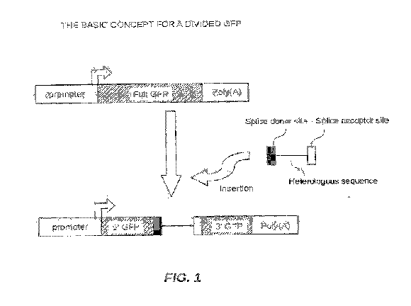
[0060] In FIG. 1 there is shown the basic concept of dividing a fluorescent
protein,
e.g. green fluorescent protein, into at least two portions by a heterologous
sequence
with a splice donor and an acceptor sequences. A fluorescent protein can be
divided
into two portions, e.g., a 5' portion and a 3' portion. The 5' portion in Fig.
1 was
linked to a promoter sequence and the 3' portion was linked to a poly (A)
sequence.
One embodiment described herein can be that a fluorescent protein, such as
green
fluorescent protein (GFP) can be utilized as it is often used as a reporter.
Once
expressed, the reporter fluorescent protein can be detected by observation
under
fluorescent microscope or other fluorescence detection apparatus. In order to
observe where and when the gene was deleted by a recombinase, the reporter
needs
to stay non-active, or substantially non-active, for example, for GFP, no-
green or
substantially no-green before the action of the recombinase. After the action
of the
recombinase, and removal of the targeted gene, the reporter needs to be active
or
substantially active, for example, green or substantially green in the
targeted cells to
indicate where and when the targeted gene was deleted. In some embodiments,
the
endogenous promoter from a target gene is not used, as very often the target
gene
promoter may not be strong enough to generate detectable of fluorescence and
there
is large variation of promoter strength among natural genes.
[0061] Heretofore, in the field of animal model creation, GFP was mostly used
as a
single linear chain of amino acid which was coded by a single stretch of
polynucleotides.
[0062] In accordance with the invention, a stretch of polynucleotide sequence
is
inserted into a pre-determined coding region of a reporter, for example, GFP,
dividing the reporter (e.g., GFP) into at least two parts (or two portions),
e.g., an N-
terminus part and a C-terminus part. In one embodiment, at least the N-
terminus part
(first portion) will not be able to generate a reporter signal, e.g., green
fluorescence.
In some embodiments, neither part nor portion can generate a reporter signal,
e.g.,
green fluorescence. The inserted stretch sequence may contain intron splicing
elements which can be spliced out by RNA splicesome.
CA 03018958 2018-09-25
WO 2017/151668
PCT/US2017/020025
[0063] In order to keep the target gene expression intact or substantially
intact
before the action of recombinase, in some embodiments the target gene and/or
the
region that can be essential to the function of the gene and can be flanked by
two
recombination sites, e.g., a first and second recombination sites. In some
embodiments the first and second recombinant sites can be at positions where
the
desired deleted sequence starts and ends, respectively. While not wanting to
be
limited by theory, it is believed that keeping any genetic modification to a
minimum
can provide the benefit of the target gene expression not being disturbed
during the
insertion of these two recombination sites and other genetic modifications. In
some
embodiments, the first and second loxP sites could also carry other sequences
such
as extra modification sequences, e.g., restriction enzyme sites and other
useful
sequences. In some embodiments, the recombination sites can be identical or
different.
[0064] In an embodiment, the sequence encoding the reporter, e.g., fluorescent
protein, can be placed in the opposite direction of transcription of the
target gene.
The N-terminus part or first portion of said reporter (e.g., fluorescent
protein)
together with a promoter can be inserted at the 3' end of the target gene,
behind a
polyA signal of the target gene, with a first recombination site. In some
embodiments, the first recombination site can be a loxP site or Rox. In some
embodiments, the C-terminus part of the reporter, e.g., fluorescent protein,
with a
second recombination site, such as another loxP and or Rox site, can be
inserted in
an intron of the target gene. The placement of the second recombination site
can be
determined by how long the target sequence needed to be deleted such that, but
not
limited to, the target sequence could contain functional sequence. When the
target
gene is deleted, it may lead to change of phenotype of the organism, more
specifically, an animal.
[0065] Upon expression of the recombinase, the target gene between the two
recombination sites can be deleted, which can bring the sequences encoding for
N-
terminus and C-terminus together with a much shorter intron. Since the N-
terminus
has its own promoter, this promoter may drive expression of RNA which includes
the coding regions for the N-terminus, intron, and C-terminus of the reporter
(e.g.,
CA 03018958 2018-09-25
WO 2017/151668
PCT/US2017/020025
fluorescent protein). RNA splicesome can splice and bring the RNA sequence for
the
N-terminus and C-terminus together to create an mRNA. This mRNA will code for
a
complete reporter, such as fluorescent protein, and turn on the reporter
signal (e.g.,
fluorescence) upon excitation by certain wavelength.
[0066] In one embodiment, the C-terminus part of the reporter (e.g.,
fluorescent
protein) could be kept as small as possible as far as the sequence encoding
for N-
terminus reporter protein (e.g., fluorescent protein) could not create a
functional
reporter (e.g., fluorescent protein), in order to minimize the impact of the
insertion of
a foreign sequence.
[0067] In another embodiment, said promoter can be selected independently from
the target gene promoter, which offers flexibility and diversity. Researchers
can
select promoters among, but not limited to, promoters that are ubiquitous,
cell
specific, and inducible.
[0068] In some embodiments, a recombinant nucleic acid construct is described,
wherein the construct comprises, in order from upstream (5' end) to downstream
(3'
end) and/or operably linked to one another, a promoter sequence, a nucleic
acid
sequence encoding a gene product of a first portion including an N- terminus
of
reporter, (e.g., a fluorescent protein, beta-galactosidase, luciferase, and
chloramphenicol acetyltransferase.), wherein the protein product of the first
portion
is insufficient to provide reporter expression (e.g., fluorescent expression);
a splice
donor site; a heterologous nucleic acid sequence; a splice acceptor site; a
nucleic
acid sequence encoding a protein gene product of second portion including a C-
terminus of the reporter (e.g., fluorescent protein); and a poly(A) signal
sequence. In
some embodiments, the promoter can be pCAG. In some embodiments, the
promoter sequence is of sufficient strength to initiate the expression of its
downstream sequences in the cell of interest. In some embodiments, the
promoter
sequence is of sufficient strength to initiate the expression of its
downstream
sequences in most cells. In some embodiments, fluorescence cannot be generated
unless the first and second portions of the sequences are connected by a
heterologous
sequence containing splice donor and acceptor. Once they are connected, RNA
splicesome can splice the heterologous sequence containing splice donor and
CA 03018958 2018-09-25
WO 2017/151668
PCT/US2017/020025
acceptor and bring the first and second portions together to form a complete
reporter
sequence (e.g., fluorescent protein sequence) which can be translated into a
functional reporter (e.g., a functional fluorescent protein).
[0069] As shown in FIG. 2, a further developed construct of FIG. 1 is
provided. The
heterologous sequence is comprised of a target sequence flanked by two
recombination sites. The target sequence could be a sequence of interest which
genetic modifications may take place. In some embodiments, the construct can
further comprise a 5'-homology arm and a 3' homology arm, two recombination
sites, and sequences of interest for genetic manipulation to form a targeting
vector.
[0070] As shown in FIG. 3, in some embodiments, a method for turning on
fluorescence is described, the method can comprise exposing the further
developed
construct of Fig. 2 to a recombinase, e.g. Cre recombinase. In some
embodiments,
the method can comprise (Step 1), constructing a DNA construct comprising
sequences described in Fig. 2, wherein the target gene is a part of an
endogenous
gene. The construct can further comprise a 5' and 3' homology arms for a
target
insertion. DNA constructs in accordance with the invention can further
comprise an
antibiotic select marker gene for providing selection by an appropriate drug.
In some
embodiments, the method can comprise (Step 2), (a) introducing the DNA
construct
into a cell, e.g. a mouse embryonic stem cell; (b) screening for a targeted
clone; and
/or (c) generating a mouse derived from the targeted clone, e.g., a
conditional mouse.
In some embodiments, the method can comprise (Step 3), mating said conditional
mouse with a recombinase containing mouse The recombinase could remove the
target sequence, e.g., the sequence between the recombination sites which
could
include the targeted gene for deletion and bring the 5' and 3' sequences
coding for a
fluorescent protein operably together, e.g., by splicing the 5' and 3'
fluorescent
portions together. The promoter linked to the 5' portion of the sequence could
drive
the expression of RNA containing the 5' and 3' sequences, which can be spliced
by a
RNA splicesome to generate a full RNA which can be translated into a full
fluorescent protein. Fig.3 further shows that the 5' and 3' portion sequences
coding
for a fluorescent protein can be inserted in the opposite direction of a
target gene. In
some embodiments, a method of introducing divided polynucleotide sequences
CA 03018958 2018-09-25
WO 2017/151668
PCT/US2017/020025
coding for a fluorescent protein into a mouse embryonic stem (ES) cell and
selection
of the targeted clone is described. In another embodiment, the method further
includes generating targeted germline mouse (conditional) using the ES cell
of; and
mating said mouse with a recombinase containing mouse. The recombinase can
recognize the two recombination sites in a second generation mouse and can
delete
the sequence between these two sites including the sequences of interest for
genetic
manipulation. When the sequence of interest is deleted, the first and second
portions
of sequences encoding for a fluorescent protein may be spliced together by RNA
splicesome to form a sequence encoding for a full fluorescent protein. Upon
the
translation, fluorescence can be generated by exposing the cell to light of
certain
wavelength to indicate the deleting event by a recombinase in a cell. The
fluorescence can be detected by systems receptive or able to read the emissive
wavelengths generated by the fluorescent protein.
[0071] In some embodiments, a plasmid is described, the plasmid can comprise
the
nucleic acid constructs described above. In some embodiments, a cell is
described,
the genome can comprise the nucleic acid constructs described above. In some
embodiments, a kit is described, the kit can comprise components of the
nucleic acid
constructs as described above.
[0072] In some embodiments, a recombinase is described, the recombinase can be
capable of recognizing and/or reacting to the above described recombination
sites,
e.g., loxP. In some embodiments, the recombinase can be Cre recombinase (SEQ
ID
NO: 19) or a codon optimized iCre.
[0073] In some embodiments the reporter protein can be a fluorescent protein.
In
some embodiments, the fluorescent protein can be selection of green
fluorescent
protein (GFP), enhanced green fluorescent protein (EGFP) (SEQ ID NO: 2), or a
red
fluorescent protein (DsRed) (SEQ ID NO: 3). In some embodiments, the reporter
protein can be beta-gal, luciferase, and chloramphenicol acetyltransferase.
[0074] In some embodiments, the construct can comprise a target sequence. FIG.
4
shows an exon trapping sequence can be inserted into the construct as
described in
FIG. 3 to prevent 5' portion to splice to 3' portion of a fluorescent protein
before
exposing to a recombinase. The exon trapping sequence could be inserted within
the
CA 03018958 2018-09-25
WO 2017/151668
PCT/US2017/020025
two recombination sites. The term recombination site or sites refers to the
nucleic
acid sequence recognized by or binding with the recombinase to enable excision
of a
sequence by action of the recombinase. Before exposing a recombinase, 5'
portion
could splice into the exon trapping sequence instead of the 3' portion
sequence. After
exposing to a recombinase, sequence including the exon trapping and target
sequence could be removed, so the 5' portion was brought together with 3'
portion to
turn on a full expression of a fluorescent protein as described in Fig. 3.
[0075] In some embodiments, the target sequence can comprise a sequence
capable
of trapping exon. In some embodiments, the sequence capable contains EN2 exon
trapping sequence (SEQ ID NO: 7).
[0076] In some embodiments, the first portion of the sequence encoding the N-
terminus end of a fluorescent protein has an ATG initiation site. In some
embodiments, the heterologous sequence can comprise sequences of endogenous
sequence from the organism where the gene modification takes place. In some
embodiments, the heterologous sequence can comprise at least one recombination
site. In some embodiments, the heterologous sequence can comprise at least two
recombination sites.
[0077] In some embodiments, as shown in FIG. 5, a method described herein
utilizing dual fluorescent proteins, e.g. red and green, to provide color
switching
before and after exposing to a recombinase in a cell. In this embodiments,
before
exposing to a recombinase, the cell and/or animal has red fluorescent protein
encoding, expressing an observed red fluorescence. After exposing to a
recombinase,
the sequence coding for the red fluorescent protein was removed and a green
fluorescent protein can be expressed. This can provide a different way to
demonstrate deletion of the targeted sequence
Promoters and Their Particular Usage
[0078] In some embodiments, the construct can comprise a promoter. Typically,
a
promoter can be a region of DNA that initiates transcription of a gene. In
general, the
promoter can be located near the transcription start sites of genes, on the
same strand
and upstream on the DNA (towards the 5' region of the sense strand) in
eukaryotic
cells (Gagniuc et al., BMC Genomics. 13 (1): 512. (2012)). In one embodiment,
the
CA 03018958 2018-09-25
WO 2017/151668
PCT/US2017/020025
promoter can be a naturally occurring and/or native DNA sequence, which is not
manmade. In another embodiment, the promoter can be manmade and/or derived
from native. In further embodiment, the promoter can be combinations of
manmade
and non-manmade DNA sequences. In some embodiments, the promoters can be
composite promoters which combine promoter elements of different origins or
were
generated by assembling a distal enhancer with a promoter of the same origin
or
different origin. In a broad sense, a eukaryotic promoter can be any DNA
sequence
that could be capable of initiating transcription of a gene, in particular, a
fluorescent
gene or part of fluorescent gene in eukaryotic cells.
[0079] In one embodiment, a eukaryotic promoter can contain regulatory
sequences
typically bound by proteins called transcription factors that can be involved
in the
formation of the transcriptional complex. Some promoters that could be
targeted by
multiple transcription factors might achieve hyperactive or hypoactive state,
leading
to increased or decreased transcriptional activity (Liefice at el., Genome
Med. 7 (1):
66. (June 2015).
[0080] In one embodiment, promoters can be selected based on particular cells
of
interest. If there is an indication of a gene in this particular cells
involved in a
particular biological process suggested by other experiments, a promoter known
to
drive gene expression in these cell can be selected. In some embodiments, the
gene
of interest can be flanked by two recombination sites. In some embodiments,
the
gene of interest can be flanked by two loxP sites and the GFP cassettes
containing 5-
terminus and 3'-terminus can be inserted in the opposite direction of the
gene. In
some embodiments, suitable mutant loxP sited can be used. In some embodiments,
once the gene was deleted by Cre recombinase, this promoter could drive GFP
expression. Subsequent of the gene deletion, the GFP, previously split in the
original
construct, could be turned on in the expected cells, indicating the successful
removal
or deletion of the intervening target gene / desire sequence to be removed. In
some
embodiments, the experiment could go un-expected, such that Cre recombinase
expressed at different cell types or different timing than one has planned in
the target
cells. In some embodiments, the Cre recombinase may have been expressed, but
could not delete the target cell as the gene location could be protected by
chromatin
CA 03018958 2018-09-25
WO 2017/151668
PCT/US2017/020025
structures or other chromosomal protection mechanisms. In some embodiments,
Cre
deletion may have taken place in un-wanted locations. Typically, there may be
genetic and epi-genetic variations among animals. Locations where the deletion
took
place and timing can be quite different among each other although all the
animals
have the same genotypes. Practically, it can be difficult, if not possible, to
predict
the express pattern a promoter creates.
[0081] Ideally, in order to track the deletion created by a recombinase, such
as Cre
recombinase, the 5'-GFP can be expressed in the cells where deletion possibly
could
take place. Typically, since the event of deletion could not be predicted, one
of the
safest ways could be trying to express 5'-GFP in every cell to anticipate the
deletion.
Since 5'-GFP by itself could not generate functional fluorescent protein, in
the event
of no deletion, the cell will not turn fluorescence. Only when a deletion
occurred,
recombination by the recombinase could bring 5' and 3' GFP sequence together
to
turn on fluorescence.
[0082] In some embodiments, promoters that drive gene expression in every cell
may practically not exist. But there are promoters could drive gene expression
in
majority of cells. They were often called ubiquitous promoters. Ubiquitous
Promoters are the promoters drive strongly expression in a wide range of
cells,
tissues and cell cycles (Schorpp et al., Nucleic Acids Res 24 (9): 1787-1788.
(1996)).0ne of them, but not limited to, pCAG promoter is capable to drive GFP
expression in many tissues (SEQ. ID NO:1).
[0083] pCAG promoter was constructed in the lab of Dr Jun-ichi Miyazaki
(Miyazaki et at., Gene. 79 (2): 269-77. (1989); and Niwa et at., Gene. 108
(2): 193-
9.(1991) from the following sequences: 1) the cytomegalovirus (CMV)
early enhancer element, 2) the promoter, the first exon and the first intron
of
chicken beta-actin gene, 3) the splice acceptor of the rabbit beta-globin
gene.
[0084] The pCAG promoter can be a strong synthetic promoter frequently used to
drive high levels of gene expression in many mammalian expression vectors
(Okabe
et al., FEBS Lett. 5;407(3):313-9. (1997); and Alexopoulou et al., BMC Cell
Biology
9: 2.(2008).
CA 03018958 2018-09-25
WO 2017/151668
PCT/US2017/020025
[0085] In one embodiment, in a target cell before the introduction of the Cre
recombinase, pCAG may only drive expression of 5' part of the GFP as the 3'
GFP
may be located farther away from the 5'GFP sequence. There can be many
potential
exon trapping sequences inside the target sequence. The transcribed 5'GFP may
not
be able to reach to the 3'GFP before it could be intercepted. Without
available
3'GFP, the 5' GFP may not be functional, affecting a fluorescence. As a
result, no
fluorescence may be detected. In some embodiments, the transcripted 5'GFP
could
be constantly available in the target cell. Only upon the expression of cre
recombinase, 5'GFP and 3'GFP may be spliced together to product a functional
GFP
which can turn fluorescence upon proper excitation to indicate where and when
a
deletion had taken place.
[0086] In further embodiment, they are, but not limited to, many ubiquitous
promoter including: beta-Actin promoter, EF1 ( elongation factor-1 alpha)
promoter,
EGR1 (early growth response 1) promoter, elF4A1 promoter, FerH (human ferritin
heavy chain) promoter, FerL (human ferritin light chain) promoter, GAPDH
(glyceraldehyde-3-phosphate dehydrogenase) promoter, GPR78 (glucose-regulated
protein 78) promoter, GPR94 (glucose-regulated protein 94) promoter, HSP70
(heat
shock protein 70) promoter, beta-Kin promoter, PGK-1 (phosphoglycerate kinase
1)
promoter, Ubiquitin B promoter, beta Act/RU5' promoter, CMV (cytomegalovirus)
promoter. The MC1 (polyoma enhancer/herpes simplex virus thymidine kinase)
promoter. A non-limiting list of suitable promoters includes CAGGS, hCMV, PGK,
FABP, Lck, CamKII, CD19, Keratin, Albumin, aP2, Insulin, MCK, MyHC, WAP,
Col2A, Mx, tet, ubiquitin C, and Trex promoter.
[0087] The ubiquitous promoter could include other promoter selected from
polymerases I, II and III dependent promoters, preferably is a polymerase II
or III
dependent promoter including, a snRNA promoter such as U6, a RNAse P RNA
promoter such as H1, a tRNA promoter, a 7SL RNA promoter, a 5 S rRNA
promoter, etc.
[0088] In some embodiments the promoter can be three other types of promoters,
but not limited to, can be used according to the intended type of control of
gene
expression. In some embodiments, these other promoters can be:
CA 03018958 2018-09-25
WO 2017/151668 PCT/US2017/020025
1. Constitutive promoters. These promoters direct expression in virtually all
tissues and are largely, if not entirely, independent of environmental and
developmental factors. As their expression is normally not conditioned by
endogenous factors, constitutive promoters are usually active across species
and even across kingdoms.
2. Tissue-specific or development-stage-specific promoters. These direct the
expression of a gene in specific tissue(s) or at certain stages of
development.
Tissue specific promoters could include FABP (Saam & Gordon, J. Biol.
Chem., 274:38071-38082 (1999)), Lck (Orban et al., Proc. Natl. Acad. Sci.
USA, 89:6861-5 (1992)), CamKII (Tsien et al., Cell 87: 1317-1326 (1996)),
CD19 (Rickert et al., Nucleic Acids Res. 25:1317-1318 (1997)); Keratin (Li et
al., Development, 128:675-88 (201)), Albumin (Postic & Magnuson, Genesis,
26:149-150 (2000)), aP2 (Barlow et al., Nucleic Acids Res., 25 (1997)),
Insulin
(Ray et al., Int. J. Pancreatol. 25:157-63 (1999)), MCK (Bruning et al.,
Molecular Cell 2:559-569 (1998)), MyHC (Agak et al., J. Clin. Invest.,
100:169-179 (1997), WAP (Utomo et al., Nat. Biotechnol. 17:1091-1096
(1999)), Col2A (Ovchinnikov et al., Genesis, 26:145-146 (2000)); examples of
inducible promoter sites are Mx (Kuhn et al. Science, 269: 1427-1429 (1995)),
tet (Urlinger et al., Proc. Natl. Acad. Sci. USA, 97:7963-8 (2000)), Trex
(Feng
and Erikson, Human Gene Therapy, 10:419-27). Above-mentioned promoters
can turn into inducible promoters by combining them with an operator
sequence including, but not limited to, tet, Ga14, lac, etc.
3. Inducible promoters. Their performance may not condition to endogenous
factors but to environmental conditions and external stimuli that can be
artificially controlled. Within this group, there are promoters modulated by
abiotic factors such as light, oxygen levels, heat, cold and wounding. Since
some of these factors are difficult to control outside an experimental
setting,
promoters that respond to chemical compounds, not found naturally in the
organism of interest, are of interest. Along those lines, promoters that
respond
to antibiotics, copper, alcohol, steroids, and herbicides, among other
CA 03018958 2018-09-25
WO 2017/151668
PCT/US2017/020025
compounds, have been adapted and refined to allow the induction of gene
activity at will and independently of other biotic or abiotic factors.
4. Synthetic promoters. Promoters made by bringing together the primary
elements of a promoter region from diverse origins.
[0089] Apart from the promoter types mentioned above, there are regulatory
expression systems based on transactivating proteins. These systems regulate
the
expression of genes of interest irrespective of their physical position to the
target
genes. In fact, several chemical-inducible promoters incorporate
transactivating
proteins and constitutive promoters as part of the regulatory system.
Transactivating
proteins constitute a whole realm of molecules in the field of gene regulation
(Beaulieu et al., Br. I Pharmacol. 172 (1): 1-23. (2015).
Reporter Proteins and their Usage
[0090] In one embodiment, the reporter protein is a fluorescent protein. The
fluorescent proteins can be, but are not limited to, capable of absorption a
higher
energy photon (excitation) and emission of a lower energy photon from a
molecule
(fluorophore) inside the protein from prior absorption.
[0091] In other embodiment, the fluorophore can be more than one molecule.
[0092] In some other embodiment, but are not limited to, protein containing
tryptophan, tyrosine, or phenylalanine residue within its sequence can be used
utilized as fluorescent protein.
[0093] In some other embodiment, but are not limited to, fluorescent protein
can be
protein capable of binding to non-proteinaceous chromophores to become
fluorescence.
[0094] In one embodiment, the fluorescent protein can be also called an
optical
marker. If it is used inside a cell, it may be called an optical cell marker.
[0095] The green fluorescent protein (GFP) can be a protein composed of 238
amino acid residues (26.9 kDa) that exhibits bright green fluorescence when
exposed
to light in the blue to ultraviolet range (Prendergast et al, Biochemistry. 17
(17):
3448-53. (1978); and Tsien et al., Annual Review of Biochemistry. 67: 509-
44. (1998)). Although many other marine organisms have similar green
fluorescent
proteins, GFP traditionally refers to the protein first isolated from the
jellyfish
CA 03018958 2018-09-25
WO 2017/151668
PCT/US2017/020025
Aequorea victoria. The GFP from A. Victoria has a major excitation peak at a
wavelength of 395 nm and a minor one at 475 nm. Its emission peak is at 509
nm,
which is in the lower green portion of the visible spectrum. The fluorescence
quantum yield (QY) of GFP is 0.79. The GFP from the sea pansy (Renilla
reniformis) has a single major excitation peak at 498 nm. GFP makes for an
excellent tool in many forms of biology due to its ability to form internal
chromophore without requiring any accessory cofactors, gene products, or
enzymes /
substrates other than molecular oxygen.
[0096] Another use of GFP can be to express the protein in small sets of
specific
cells. This allows researchers to optically detect specific types of cells in
vitro (in a
dish), or even in vivo (in the living organism) (Chudakov et al.,
Biotechnology. 23 (12): 605-13.(2005)).
[0097] Due to the potential for widespread usage and the evolving needs of
researchers, many different mutants of GFP have been engineered (Shaner et
al.,
Nature Methods. 2 (12): 905-9 (2005); and Wilhelmsson and Tor, Fluorescent
Analogs of Biomolecular Building Blocks: Design and Applications. New Jersey:
Wiley. ISBN 978-1-118-17586-6. (2016)).
[0098] The first major improvement was a single point mutation (S65T) reported
in
1995 in by Roger Tsien (Heim et al., Nature. 373 (6516): 663-4. (1995)). This
mutation dramatically improved the spectral characteristics of GFP, resulting
in
increased fluorescence, photostability. A 37 C folding efficiency (F64L)
point
mutant to this scaffold, yielding enhanced GFP (EGFP), was discovered in 1995
( US patent 6172188, Thastrup et al.; and Cormack et al., Gene. 173 (1 Spec
No):
33-38, (1996).). Superfolder GFP, a series of mutations that allow GFP to
rapidly
fold and mature even when fused to poorly folding peptides, was reported in
2006
(Pedelacq et al., Nature Biotechnology. 24 (1): 79-88. (Jan 2006)).
[0099] Besides GFP, there are other fluorescent proteins can also report the
genetic
modification in the cells. They can be, but are not limited to, blue/UV
fluorescent
proteins, cyan fluorescent proteins, green fluorescent proteins, yellow
fluorescent
proteins, orange fluorescent proteins, red fluorescent proteins, far-red
fluorescent
proteins, Near-IR fluorescent proteins, Long strokes shift fluorescent
proteins,
CA 03018958 2018-09-25
WO 2017/151668
PCT/US2017/020025
Photoactivable fluorescent proteins, Photoconvertible fluorescent proteins,
Photoswitchable fluorescent proteins.
[00100] In some embodiments, fluorescent proteins could be interchangeable and
have been used as reporters. For example, but not limited to, a GFP or EGFP
can be
replaced by YFP, or Cerulean, or mTFP1 as far as it could serve as an optical
reporter. In other embodiments, if two or more fluorescent proteins are
required for a
particular experiment, a careful planning may be needed for the compatibility
of the
color they generated. Spectral crosstalk and inter-variant interactions
between
fluorescent proteins should be carefully examined for multi-color imaging. In
one
embodiment, the coding sequence could be changed, but it still coded the same
amino acid.
[00101] In some embodiments, an interaction among multiple fluorescent
proteins
could cause changes in fluorescence (or Forster) resonance energy transfer
(FRET)
to report on biochemical processes in living cells.
[0100] In some embodiments, blue/UV fluorescent proteins can be, but are not
limited to, Y66H, Y66F, Y66W, EBFP, mCFP, ECFP, Azurite (Marco et al., Nature
Biotechnology 24, 1569 - 1571 (2006)), GFPuv (wang et al., Hum Vaccin
Immunother 9(7): 1558-1564. (2013), EBFP2 (Wu et al., Front Microbiol. 6: 607.
(2015)), Cerulean (Wu et al., Front Microbiol. 6: 607. (2015)), CyPet (Scott
et al.,
Sci Rep; 5: 10270. (2015)), TagBFP (Wu et al., Front Microbiol. 6: 607.
(2015)),
mTagBFP2 (Subach et al, PLoS One. 6(12):e28674.(2011)). EBFP2 (Ai et al.,
Biochemistry 46: 5904-5910. (2007)), mKalamal (Ai et al., Biochemistry 46:
5904-
5910. (2007)), Sirius (Tomosugi, et al., Nat. Method. 5: p. 351-353. (2009)),
Sapphire (Cubitt et al., Meth Cell Biol, 58: p. 19-30. (1999), T-Sapphire
(Zapata-
Hommer et al., BMC Biotechnol, 3(5).(2003)), TagBFP, TagCFP (Wu et al., Front
Microbiol. 6: 607. (2015)), SBFP2 (Wu et al., Front Microbiol. 6: 607.
(2015)),
AmCyanl (Clontech), mTFP1(Rizzo et al., doi:10.1101/pdb.top63 Cold Spring Harb
Protoc (2009)), 565A (Biochemistry 44: 1960-1970. (2005)).
[0101] In some embodiments, cyan fluorescent proteins can be, but are not
limited
to, ECFP (Wall et al., Biochem Mol Biol Educ. 43(1):52-9 (2015)), Cerulean
(Rizzo
et al., Nat Biotechnol,. 22(4):p. 445-449 (2004)), SCFP3A (Kremers et al., G-
J. et
CA 03018958 2018-09-25
WO 2017/151668
PCT/US2017/020025
al., 45: p. 6570-6580. (2006)), mTurquoise (Goedhart etal., Nat. Meth. 7:
p.137-141.
(2010), mTurquoise2 (Goedhart etal., Nat Commun. 20;3:751. (2012), monomeric
Midoriishi-Cyan, mTFP1 (Rizzo et al., doi:10.1101/pdb.top63 Cold Spring Harb
Protoc (2009)).
[0102] In some embodiments, green fluorescent proteins can be, but are not
limited
to, EGFP, Emerald (Cubitt et al., Meth Cell Biol. 58: p. 19-30. (1999)),
Superfolder
GFP (Pedelacq et al., Nat. Biotech. 24: p. 79-88. (2006)), Monomeric Azami
Green
(MBL international), TagGFP2 (Evrogen), mUKG (Tsutsui et al., Nat. Methods.
5(8): p.683-685. (2008)), mWasabi (Rizzo etal., doi:10.1101/pdb.top63 Cold
Spring
Harb Protoc (2009)), Clover (Lam etal., Nature Methods 9, 1005-1012. (2012)),
mNeonGreen (Shaner et al., Nature Methods. 10: 407-409. (2013)), 565C, 565L,
565T (Pang etal., Plant Physiol. 112:893-900. (1996)), ZsGreen1 (Clontech),
Dronpa-Green (Habuchi etal., PNAS. 102:9511-9516. (2005)), TagGFP (Evrogen),
AcGFP1 (Clontech), CopGreen (Condon et al., Insect Mol Bio1.16(5):573-
80.(2007)).
[0103] In some embodiments, yellow fluorescent proteins can be, but are not
limited
to, EYFP, Citrine ( Griesbeck etal., Biol Chem. 276(31): p. 29188-94. (2001)),
Venus (Nagai et al., Nat Biotechnol. 20(1): p. 87-90. (2002)), SYFP2
(Ledermann et
al., Mol Plant Microbe Interact. 28(9):959-67. (2015)), TagYFP (Evrogen),
Topaz
(Yu etal., Genom Data. 5: 318-319. (2015)), mCitrine (Rizzo etal.,
doi:10.1101/pdb.top63 Cold Spring Harb Protoc (2009)), Ypet (Scott et al., Sci
Rep;
5: 10270. (2015)), TurboYFP, PhiYFP (Condon etal., Insect Mol Bio1.16(5):573-
80.(2007)), PhiYFP-m (Condon et al., Insect Mol Bio1.16(5):573-80.(2007)),
ZsYellowl (Richards etal., Cytometry. 1;48(2):106-12. (2002)), mBanana (Zhou
et
al., Protein Pept Lett. 15(1):113-4. (2008)), Y665 (Biochemistry 44: 1960-
1970.
(2005)).
[0104] In some embodiments, orange fluorescent proteins can be, but are not
limited
to, Monomeric Kusabira-Orange (MBL international), mK0x (Tsutsui et al., Nat.
Methods. 5(8): p. 683-685. (2008)), mK02 (MBL international), mOrange (Shaner
et
al., Nat Biotechnol, 22(12):1567-72. (2004)), m0range2 (Shaner etal., Nat.
Meth. 5:
p.545-551 (2008)), and mK0 (Sung etal., PLoS One. 20; 10(11):e0141585 (2015)).
CA 03018958 2018-09-25
WO 2017/151668
PCT/US2017/020025
[0105] In some embodiments, red fluorescent proteins can be, but not limited
to,
TurboRFP, dKeima-Red, mKeima-Red, mRaspberry, mCherry, mStrawberry,
mTangerine, tdTomato, TagRFP (Scott et al., Sci Rep; 5: 10270. (2015)),
TagRFPt
(Scott et al., Sci Rep; 5: 10270. (. 2015)), mApple, mRuby (Scott et al., Sci
Rep; 5:
10270. (2015)), mRuby2 (Scott et al., Sci Rep; 5: 10270. (2015)), DsRed
(Yarbrough
et al., Proc Natl Acad Sci U S A. 16;98(2):462-7. (2001)), DsRed-Express2
(Gottwein et al., J Virol. 85(17):8913-28. (2011)), DsRed monomer (Chou et
al.,
Chin J Physiol. 28;58(1):27-37. (2015)), DsRed2 (He et al., Cell Biosci. 5:67.
(2015)), TurboRF602 (Khodosevich et al., Front Mol Neurosci. 2: 7 (2009)),
AsRed2(Hirrlinger et al., Mol Cell Neurosci. 30(3):291-303. (2005)), mRFP1
(Wallrabe et al., Cytometry A. 87(6):580-8.(2015)), J-Red (Condon et al.,
Insect Mol
Bio1.16(5):573-80.(2007)), HcRedl (Subach et al., Chem Biol. 20;15(10):1116-
24.
(2008)), TurboRF635 (Evrogen), Katushka (Kinnear et al., PLoS One.
19;10(6):e0130375. (2015)), and Katushka2 (Kinnear et al., PLoS One.
19;10(6):e0130375.(2015)), mRaspberry (Wang et al., Proc Natl Acad Sci,
101(48):p. 16745-16749. (2004)), mCherry (Shaner et al., Nat Biotechnol,
22(12):1567-72. (2004)), mStrawberry (Shaner et al., Nat Biotechnol,
22(12):1567-
72. (2004)), mTangerine (Shaner et al., Nat Biotechnol, 22(12):1567-72.
(2004)),
tdTomato (Shaner et al., Nat Biotechnol, 22(12):1567-72. (2004)), TagRFP
(Evrogen), TagRFP-T (Shaner et al., Nat. Meth. 5: p.545-551. (2008)), mApple
(Shaner et al., Nat. Meth. 5: p.545-551. (2008)), mRuby (Kredel et al., PLOS
One, 4:
e4391. (2009)), mRuby2 (Lam et al., Nature Methods. 9, 1005-1012. (2012)).
[0106] In some embodiments, far-red fluorescent proteins can be, but not
limited to,
mPlum (Wang et al., Proc Natl Acad Sci. 101(48):p. 16745-16749. (2004)), HcRed-
Tandem (Maynard-Smith et al., JBC 282, 24866-24872. (2007)), mKate (Guess et
al., Skelet Muscle. 3: 19. (2013)), mKate2 (Tanida et al., PLoS One. Oct
23;9(10):e110600. (2014)), mNeptune (Lin et al., Chem. Biol. 16: p. 1169-1179.
(2009)), NirFP (Evrogen), E2-Crimson (Barbier et al., PLoS One.
3;11(3):e0146827. (2016)).
[0107] In some embodiments, near infar-red fluorescent proteins can be, but
not
limited to, TagRFP657 (morozova, Biophys J. 21;99(2):L13-5. (2010)), IFP1.4
(Yu
CA 03018958 2018-09-25
WO 2017/151668
PCT/US2017/020025
et al., Nat Commun. 15;5:3626 (2014)), iRFP (Agollah et al., J Cancer.
23;5(9):774-83. (2014).
[0108] In some embodiments, Long Stokes Shift fluorescent Proteins can be, but
are
not limited to, mKeima Red (Yang et al., PLoS One. 20;8(6):e64849. (2013)).,
LSS-
mKatel (Piatkevich et al., PNAS, 107: p. 5369-5374. (2010)), LSS-mKate2
(Piatkevich et al., PNAS, 107: p. 5369-5374. (2010)), mBeRFP (Yang et al.,
PLoS
One. 20;8(6):e64849. (2013)).
[0109] In some embodiments, photoactivatible fluorescent proteins can be, but
are
not limited to, PA-GFP (Patterson et al., Science,. 297(5588): p. 1873-7.
(2002)),
PAmCherryl (Subach et al., Nat. Meth. 6: p. 153-159. (2009)), PATagRFP (Subach
et al., JACS 132: p. 6481 -6491. (2010)).
[0110] In some embodiments, Photoconvertible fluorescent proteins can be, but
are
not limited to, Kaede (green) (MBL international), Kaede(red) (MBL
international),
KikGR1 (green) (MBL international), KikGR1 (green) (MBL international),
KikGR1 (red) (MBL international), PS-CFP2 (Evrogen), mEos2 (green) (McKinney
et al., Nat Meth. 6: p. 131-133. (2009)), mEos2 (red) (McKinney et al., Nat
Meth. 6:
p. 131-133. (2009)), mEos3.2 (green) (Zhang et al., Nat. Meth. 9: p. 727-729.
(2012)), mEos3.2 (red) (Zhang et al., Nat. Meth. 9: p. 727-729. (2012)),
PSmOrange
(Subach et al., Nat. Meth. 8: p. 771-777. (2011)).
[0111] In some embodiments, photoactivatible fluorescent proteins PA-GFP,
PAmCherryl, PATagRFP may remain silent, may only turn on when the protein was
activated by certain wavelength lights.
[0112] In some embodiments, photoconvertible fluorescent proteins, such as
Kaede
(green), Kaede(red), KikGR1 (green), KikGR1 (green), KikGR1 (red), PS-CFP2,
mEos2 (green), mEos2 (red), mEos3.2 (green), mEos3.2 (red), PSmOrange can turn
from a green fluorescence to red fluorescence under radiation of certain wave-
length.
It provides the ability to track individual neuronal cell and cell movement.
It can
often be that tissue of interest has auto-fluorescence. It could be the same
color as
the reporter color. It may make it very difficult to distinguish fluorescence
that was
generated from the background (auto-fluorescence) or target deletion. To solve
this
background auto-fluorescent problem, photoconvertible fluorescent protein,
such as
CA 03018958 2018-09-25
WO 2017/151668
PCT/US2017/020025
Kaede, can be used. In a way that two photos of microscope excited two
different
wavelengths can be taken. For example the original color has a background of
auto-
fluorescence. The same sample can be treated by radiation of certain wave-
length
which causes the structure changes on the fluorescence protein. The
fluorescent
protein will change to different color, but the background may not. So only
the signal
going with the color change could be the signal where target gene was deleted.
In some embodiments, two fluorescent proteins can be fused together as dual
fluorescent protein.
[0113] In some embodiments, more than two fluorescent proteins can be fused
together to form a multi- fluorescent protein.
[0114] In some embodiments, part of a fluorescent protein fused with a full
fluorescent protein.
[0115] In some embodiments, part of a fluorescent protein fused with multi-
fluorescent protein.
[0116] In some embodiments, there are newly developed fluorescent proteins.
They
are, but not limited to, UnaG (Kumagai et al., Cell. Jun 20; 153(7):1602-11.
(2013)),
eqFP611 (Kredel et al., PLoS One. 4(2):e4391.(2009)), KFP (Khrenova et al.,
Biophys J. 108(1):126-32. (2015)), EosFP (Shcherbakova et al., Annu Rev
Biophys.
43:303-29. (2014)), IrisFP (Gayda et al., Biophys J. 103(12):2521-31. (2012)),
smurfp, FMN-binding fluorescent proteins (FbFPs) (Drepper et al., Appl Environ
Microbiol. Sep; 76(17):5990-4. (2010)).
[0117] In some embodiments, any protein capable of being detected and measured
in cells and organisms, such as beta-galactosidase, luciferase, and
chloramphenicol
acetyltransferase and other suitable proteins can be used as reporter
proteins.
[0118] Inventive Details of Splice Sites and Its Insertion into a Sequence
Coding for
Fluorescent Protein
[0119] In some embodiments, the construct can comprise a reporter protein. In
some embodiments, the reporter protein can be a fluorescent protein, further
the
fluorescent protein could be green fluorescent protein (GFP). Dividing the
coding
sequence of GFP by splice donor and acceptor sites from an intron into at
least two
parts, or more than two parts, could keep the GFP silent (no indicated or
perceived
CA 03018958 2018-09-25
WO 2017/151668
PCT/US2017/020025
fluorescence) in event there is no recombinase and deletion and active when
there is
a deletion (indicated or perceived fluorescence).
[0120] In one embodiment, the insertion can be determined by the coding
sequences
around the insertion site and the sequences of splice donor and acceptor
sites.
[0121] In some embodiments, the construct comprises an intron. In some
embodiments, the intron can comprise a donor site (5' end of the intron), a
branch
site (near the 3' end of the intron), a heterologous sequence and/or an
acceptor site (3'
end of the intron) that may be required for splicing. The intron can obtain
from
nature sources or fully man-made.
[0122] In one embodiment, splicesome recognize a consensus sequence A-G-[cut]-
G-T-R-A-G-T from the sequences around the 5' end of an intron, wherein A-G is
provide from the upstream exon and G-T-R-A-G-T is from the 5' end of the
intron,
which could be essential part of the splice donor site, wherein R represents a
choice
of A or G. ( Iupac-Iub Comm. On Biochem. Nomencl. Biochemistry. 9: 4022-4027
(1970); and De Conti et al., Wiley Interdiscip Rev RNA, 4(1):49-60. (2013);
and
Lewis et al., book of "Molecular Biology of the Cell". 2012; and William et
al,
Nature Reviews Genetics. 7(3): 211-21. (2006); and Ohshima et al, J. Mol.
Biol.,
195:247-259(1987)).
[0123] In another embodiment, splicesome recognize another consensus sequence
Y-rich-N-C-A G-[cut]-G from the sequences around the 3' end of the intronõ
wherein Y-rich-N-C-A G comes from the 3' end of the intron, which could be
essential part of the splice acceptor site. The G at the end of said consensus
sequence
is contributed from exon downstream of the intron, wherein Y represents a
possibility of C or T. N represents a possibility of A, C, G, or T. (Iupac-Iub
Comm.
On Biochem. Nomencl. Biochemistry. 9: 4022-4027 (1970); and De Conti et al.,
Wiley Interdiscip Rev RNA, 4(1):49-60. (2013); and Lewis et al., book of
"Molecular Biology of the Cell" 2012; and William et al, Nature Reviews
Genetics.
7(3): 211-21. (2006); and Ohshima et al, J. Mol. Biol., 195:247-259(1987)).
[0124] In another embodiment, upstream (5'-ward) from the AG at 3' end of the
intron could be a region high in pyrimidines (C and T), or polypyrimidine
tract.
Further upstream from the polypyrimidine tract is the branchpoint, which may
CA 03018958 2018-09-25
WO 2017/151668
PCT/US2017/020025
include an adenine nucleotide involved in lariat formation with a consensus
sequence
Y-N-C-T-R-A-C (Clancy et al., Nature Education.1 (1): 31. (2008); and Black et
al.,
Annual Review of Biochemistry. 72 (1): 291-336. (June 2003)). The branchpoint
could be 20-50 nucleotides upstream of splice acceptor site.
[0125] In one embodiment, many locations inside the nucleotide coding sequence
for a fluorescent protein could be selected such that the end of the first
part of a
fluorescent protein coding sequence can be connected to a splice donor site of
an
intron and the beginning of the second part of a fluorescent protein coding
sequence
can be connected to a splice acceptor site of the intron.
[0126] In one embodiment, in order to divide the coding sequence of a
fluorescent
protein into two separate parts, sequences surrounding the potential insertion
sites
(donor and acceptor site) should be closely examined by satisfying the rules
suggested by the consensus sequences described above. Only designs with
sequences
very closely resembling to the consensus sequences may start to be constructed
and
tested.
[0127] In another embodiment, any sequences can be utilized as splice donor
and
acceptor as far as they can be recognized and spliced correctly by RNA
splicesome.
[0128] In another embodiment, intron sequences can be utilized according to
the U2
and U12 categorization (Sharp et al., Cell. 91: 875-879. (1997)).
[0129] Introns suitable for use in embodiments herein could be prepared by
several
methods such as purification from a naturally occurring nucleic acid or de
novo
synthesis. The introns present in many naturally occurring eukaryotic genes
have
been identified and characterized (Mount et al., Nuc. Acids Res., 10:459
(1982)).
Artificial introns comprising functional splice sites also have been
described. (Winey
et al., Mol. Cell Biol., 9:329 (1989); and Gatermann et al, Mol. Cell Biol.,
9:1526
(1989)). Introns may be obtained from naturally occurring nucleic acids, for
example, by digestion of a naturally occurring nucleic acid with a suitable
restriction
endonuclease, or by PCR cloning using primers complementary to sequences at
the
5' and 3' ends of the intron. Alternatively, introns of defined sequence and
length
may be prepared synthetically using various methods in organic chemistry
(Narang
CA 03018958 2018-09-25
WO 2017/151668
PCT/US2017/020025
et al., Meth. Enzymol., 68:90 (1979); and Caruthers et al, Meth. Enzymol.,
154:287
(1985); and Froehler et al, Nuc. Acids Res., 14:5399 (1986)).
[0130] In some embodiments, vectors contain the suitable sequences including a
cell
specific promoter, the sequences encoding for 5' and 3' of a fluorescent
protein,
intron with splicing sites, and a Poly(A) signal can be constructed and tested
in vitro
(in cells). If the design of the intron insertion leads to right splicing,
fluorescence
could be observed under a fluorescent microscope. If fluorescence could not be
observed, the insertion of the intron may need to change to a different
location inside
the coding sequence of a fluorescent protein with further tests.
[0131] In some embodiments, following above description, intron can be
inserted in
many locations inside GFP, or other fluorescent proteins of researchers'
choices.
Further, coding fluorescent protein or reporter protein can be divided by a
heterologous sequence with splice donor and acceptor sites with different
ratios of
N-terminus to C-terminus portions, for example, but not limited to; 10% front,
90%
back; 20% to 80%; 30% to 70%; 40% to 60%; 50% to 50%; 60% to 40%, 70% to
30%, 80% to 20%, respectively. The coding sequence of a fluorescent protein
can be
also divided into a ratio that the sum of percentages can be added to 100%.
Each
front part can only connect to its own back part to facilitate fluorescence
expression.
Different ratio of dividing coding sequence of a fluorescent protein can be
very
useful in tracking cell development including neuron development and disease
animal model creations.
[0132] In one embodiment, the size of intron may vary from as little as a few
nucleotides to over hundreds of thousands nucleotide. It thus provides further
flexibility for researchers to design their research such that many functional
components can be built inside the intron.
[0133] In further embodiment, at least four distinct classes of introns have
been
identified in the nature (Alberts, Bruce (2008). Molecular biology of the
cell. New
York: Garland Science) and most of these introns can be used as entirely or
partially
to insert into the fluorescent proteins. The four classes could include, but
not limited
to; 1) Introns in nuclear protein-coding genes that are removed by
spliceosomes
(spliceosomal introns). 2) Introns in nuclear and archaeal transfer RNA genes
that
CA 03018958 2018-09-25
WO 2017/151668
PCT/US2017/020025
are removed by proteins (tRNA introns). 3) Self-splicing group I introns that
are
removed by RNA catalysis. 4) Self-splicing group II introns that are removed
by
RNA catalysis.
[0134] In further embodiment, there is a fully or partially man-made intron,
which
can be also used to insert into the fluorescent protein.
[0135] In other embodiment, there may have other ways than site specific
recombination to cut out of a stretch of sequences and subsequently bring
together
N- and C- terminus of GFP to activate GFP expression. They can be, but are not
limited to, Crispr, TALENs, Zinc finger, rare cutting restriction enzymes,
genomic
translocation, hot-spot related deletions, immunology VDJ recombination,
immune-
class switching, gene inversion, natural deletion, and other known in the art.
[0136] In further embodiment, a suitable intron can be retrieved from genomic
data
source such as, but not limited to, ensemble, UC Davis genome browser.
[0137] In one embodiment, bioinformatic analysis can be employed to help the
design of the intron insertion (Reese et al., J Comp Blot 4(3), 311-23.
(1997)).
Cre-loxP and Other Applicable Recombination Systems
[0138] With an intent to delete a target gene and subsequently turn on
fluorescence,
the target sequence could be flanked by two recombination sites through
genetic
engineering. In some embodiments, an expressing second fluorescent protein can
be
included in the target sequence, such that removal of the target sequence also
removes the expression of the second fluorescent protein, e.g., changing the
fluorescence from the second emitted fluorescence to the first or indicating
fluorescent protein emission, e.g., red changing to green (GFP). Two parts of
the
coding sequences for a fluorescent protein can be linked to these two
recombination
sites, in a configuration that the coding sequence of 5' a fluorescent protein
with a
promoter could be connected to an intron donor site and 3' a fluorescent
protein was
connected to an intron accepting site. The 5' and 3' sequence coding a
fluorescent
protein could be inserted along with these two sites in a direction opposite
to the
target gene expression direction. Upon the recombinase expression, the
sequences
between these two recombination sites could be deleted or excised and this
process
CA 03018958 2018-09-25
WO 2017/151668 PCT/US2017/020025
will bring the 5' and 3' coding sequence together. The promoter which
connected
with 5'GFP could drive the expression of a functional fluorescent protein.
[0139] In one embodiment, there could be many recombinases can be used to
remove sequences between recombination sites. Cre recombinase is one of the
most
frequently used ones.
[0140] The Cre protein is a site-specific DNA recombinase. It can catalyze the
recombination of DNA between specific sites in a DNA molecule. These sites,
known as loxP sequences, contain specific binding sites for Cre that surround
a
directional core sequence where recombination can occur.
[0141] When cells that have loxP sites in their genome expressing Cre, a
recombination event can occur between the loxP sites. Cre recombinase proteins
bind to the first and last 13 bp regions of a loxP site forming a dimer. This
dimer
then binds to a dimer on another lox site to form a tetramer. LoxP sites are
directional and the two sites joined by the tetramer are parallel in
orientation. The
double stranded DNA is cut at both loxP sites by the Cre protein. The strands
are
then rejoined with DNA ligase in a quick and efficient process. The result of
recombination depends on the orientation of the loxP sites. For two lox sites
on the
same chromosome arm, a direct repeat of loxP sites will cause a deletion
event,
while an inverted loxP sites will cause an inversion of the target DNA.
[0142] In one embodiment, other systems to delete or inverse sequences may
include by way of nonlimiting example,
1. FLP/frt recombination system (Schlake et al., Biochemistry. 33 (43): 12746-
12751. (1994) and Turan et al., J. Mol. Biol. 402 (1): 52-69. (2010)).
2. Dre/rox recombination system (Anastassiadis et al., Dis Model Mech. Sep-
Oct; 2(9-10):508-15. (2009)), Vcre/VloxP recombination system (Suzuki et al.,
Nucleic Acids Res. 39(8): e49. (2011)).
3. Scre/SloxpP recombination system (Suzuki et al., Nucleic Acids Res. 39(8):
e49. (2011)).
4. Nigri/nox recombination system (Karimova et al., Scientific Reports 6,
Article number: 30130 (2016)).
CA 03018958 2018-09-25
WO 2017/151668
PCT/US2017/020025
5. Panto/pox recombination system (Karimova et al., Scientific Reports 6,
Article number: 30130 (2016)).
6. PhiC31/att recombination systems (Thomson et al., BMC Biotechnology.
10:17. (2010)). Typically, the site specific recombination could only occur
within its
own recombinase.
7. Sleepingbeauty transposas which deletes the DR sequences in the mirrored
IR/DR sequences (Zayed et al., Mol Ther. 9(2):292-304. (2004).
[0143] In general, one particular recombinase may only works with its own
particular recombination sites. It may be a rare event that cross talk occurs
among
different recombinase based systems.
[0144] In the FLP/frt system, FLP recombinase was discovered in
yeast Saccharomyces cerevisiae (Schlake et al., Biochemistry. 33 (43): 12746-
12751. (1994) and Turan et al., J. Mol. Biol. 402 (1): 52-69. (2010)). For
every FLP-
mediated recombination, a total of four FLP recombinases and twofrt sequences
could be required. Two of the four proteins bind to one FRT sequence because
every
frt has two 13-bp FLP-binding sites which are interrupted by an 8-bp spacer
region.
In this spacer DNA strand breakage takes place, producing 8-bp overhanging
ends.
After strand breakage, the overhanging ends of the two FRT fragments come
together by complementary base pairing so that a mutantfrt sequence is
generated.
[0145] Another recombination system is Dre/rox recombination system, Dre
recombinase was first described in the P1-like transducing bacteriophage D6
isolated
from Salmonella enteric. The genes encoding Dre and Cre recombinases share 39%
sequence similarity. Dre recombinase could catalyze site-specific DNA
recombination by recognizing rox sites, whereas Cre recombinase could not be
able
to recognize rox sites, which are distinct from loxP sites. Similar to Cre
recombinase, Dre could delete the sequence between two rox sites if they are
facing
the same direction.
[0146] VCre/VloxP recombination system may also be used. Vcre showed very
weak similarity to Cre, sharing 29% identity to the Cre amino acid sequence.
It
recognizes the VloxP sites.
CA 03018958 2018-09-25
WO 2017/151668
PCT/US2017/020025
[0147] There are other systems that may be used to delete the sequence between
two recognizable sites. They are, but are not limited to, KD, B2 B3, lambda,
HK022,
HP1, lambda gamma, ParA, Tn3, Gin, Bxbl, R4, and TP901-1.
[0148] Typically, site specific recombinase recognized its own recombination
site. It
was discovered that the recombinase can also recognize sequences similar to
the
wildtype recombinase site with only one or a few bases. These sites were also
called
mutant sites.
[0149] As for example, in Cre/lox system, many mutant loxP sites were created.
A
mutant recombination site is a nucleotide sequence that is similar but not
identical to
the minimal native loxP recombination site set forth in SEQ ID NO:4. While the
mutant loxP recombination site can be functional. Unless otherwise noted, a
mutant
loxP recombination site retains the biological activity of the wild type loxP
recombination site and comprises a functional recombination site that is
recognized
by a Cre recombinase and capable of a recombinase-mediate recombination
reaction.
Thus, a mutant loxP recombination site can comprise a deletion, addition,
and/or
substitution of one or more nucleotides in the 5' or 3' end of the minimal
native loxP
recombination site, in one or more internal sites in the minimal native loxP
recombination site. Generally, modified recombination sites will have at least
about
40%, 45%, 50%, 55%, 60%, 65%, 70%, 75%, 76%, 77%, 78%, 79%, 80%, 81%,
82%, 83%, 84%, 85%, 86%, 87%, 88%, 89%, 90%, 91%, 92%, 93%, 94%, 95%,
96%, 97%, 98%, 99% or more sequence identity to the minimal native
recombination site over its complete length or to any domain contained
therein. The
mutant loxP recombination site could therefore include 1, 2, 3, 4, 5, 8, 10,
12, 13, 14,
15, 16, 17, 18, 19, 20, 21, 22, 23, 24, 25, 28, 29 or greater nucleotide
substitutions,
additions, and/or deletions across the entire length of the minimal
recombination site,
or alternatively, in each of the various domains of the recombination site as
outlined
above.
[0150] In one embodiment, the mutant loxP sites may have different characters
than
that the wildtype (native) loxP site. Some could be not compatible with the
wildtype
loxP site and some could. Some could be substantially compatible. When a
mutant
loxp site recombines with a wildtype loxP site, they will create a new mutant
loxP
CA 03018958 2018-09-25
WO 2017/151668
PCT/US2017/020025
site. Some of the combinations are reversible, some are not. These mutant loxP
sites
can be used to flank the target gene and sequences between the sites which can
be
deleted by Cre recombinase. Upon the Cre reaction, combination of two
identical
mutant loxP could leave a single mutant site, which could have an identical
sequence
as parental mutant loxP site. Combination of two different mutant loxP could
leave a
single mutant loxP site, which has the sequence which is different than any of
its
parent (wildtype, or native).
[0151] In one embodiment, among the mutant loxP sites, they are, but not
limited to,
lox511, 1ox5171, 1ox2272, M2, M7, M11, lox71, 1ox66, loxN, loxP 5171, and
other
published and un-published, and under-develop mutant loxP sites. In some
embodiments, the mutant loxP site can also be called heterospecific loxP site
or
modified loxP site. Its diversity could be often created by variation inside
the 8-bp
spacer sequence.
[0152] In other embodiment, many mutant recombination systems could have their
own sets of wildtype (native) recombination sites and their mutant
recombination
sites. They worked in a very similar way as described above. Among them, but
not
limited to, they include F3, F5, FL-IL 10A, Vlox2272, 51ox2272, VloxMl,
SloxM2,
VloxM2, SloxM2, Vlox43R, Vlox43L, Slox1R, Slox1L, attR, attL, attP, attB,
IR/DR
sequences.
[0153] In one embodiment, proper usage and combinational usage of these
recombination sites can be incorporated into the design of turning
fluorescence on
and off, off and on with many other different combinations and configurations.
Poly (A)
[0154] In some embodiments, the construct can comprise a poly (A) sequence.
Typically, a Polyadenylation signal poly(A) could be required for proper
expression
of an eukaryotic protein. Polyadenylation adds a poly(A) tail to a messenger
RNA.
The poly(A) tail could consist of multiple adenosine monophosphates which
constitute a stretch of RNA with only adenine bases. In eukaryotes,
polyadenylation
could be part of the process that produces mature messenger RNA (mRNA) for
translation. It could form part of the larger process of gene expression.
CA 03018958 2018-09-25
WO 2017/151668
PCT/US2017/020025
[0155] The process of polyadenylation could begin as the transcription of a
gene
terminate. The 3'-most segment of the newly made pre-mRNA can be first cleaved
off by a set of proteins (Proudfoot et al., Cell. 108(4): 501-12. (2002) and
Guhaniyogi et al., Gene. 265(1-2): 11-23. G(2001)).
[0156] These proteins then synthesize the poly(A) tail at the RNA's 3' end. In
some
embodiments, these proteins add a poly(A) tail at one of several possible
sites and
produce more than one transcript from a single gene (alternative
polyadenylation),
similar to alternative splicing (Proudfoot et al., Cell. 108 (4): 501-12.
(2002)).
[0157] The poly(A) tail can be important for the nuclear export, translation,
and
stability of mRNA. The tail is shortened over time, and, when it is short
enough, the
mRNA is enzymatically degraded (Guhaniyogi et al., Gene. 265(1-2): 11-23.
(2001)). However, in a few cell types, mRNAs with short poly(A) tails are
stored for
later activation by re-polyadenylation in the cytosol (Richter, Joel D.
Microbiology
and Molecular Biology Reviews, 63(2): 446-56.(1999)).
[0158] In one embodiment, the poly (A) sequence could be linked to the end of
the
3' fluorescent protein coding sequence after the termination sequence. The
combined
sequences including the 3' end of the coding sequence with a termination
sequence
and poly (A) sequence could be inserted into a specific location inside one of
the
intron of a target gene.
[0159] In one embodiment, in order to keep minimum impact on the endogenous
sequence, it could be desirable to keep the poly (A) sequence as shorter as it
could.
[0160] In some embodiments, the ploy (A) can be acquired by extension of 3'
GFP
coding sequence into a region where similar sequence can be functional as a
signal
(Edwalds-Gilbert Get al., Nucleic Acids Res. 25(13):2547-61. (1997)).
[0161] In some embodiments, in mammalian genes, polyadenylation sites can be
usually preceded by AATAAA or ATTAAA -20 bases before the cleavage site and
could be followed by a more weakly conserved GT-based motif There may be other
possible sequences which can be recognizable by the poly (A) related set of
proteins.
These sequences could be also be used. (Cheng et al., Bioinformatics.
22(19):2320-5.
(2006), and Hu et al., RNA. 11(10):1485-93 (2005)).
CA 03018958 2018-09-25
WO 2017/151668
PCT/US2017/020025
[0162] In a broad embodiment, the poly (A) signal could be NNTANN, where N
represents any of a nucleotide A, C, G, or T (Cheng et al.,
Bioinformatics. 22(19):2320-5. (2006), and Hu et al., RNA. 11(10):1485-93.
(2005)).
Exon Splice Trapping Acceptors
[0163] In one embodiment, target sequence may contain sequences that can be
recognizable and be spliced by the RNA splicesome, which provide a natural
barrier
to block 5' coding sequence of a fluorescent protein to be spliced with the 3'
coding
sequence of a fluorescent protein. Before exposing to recombinase activity, 5'
coding
sequence of a fluorescent protein may be spliced with one of the splice
trapping
acceptor site to generate a chimeric protein encoding by the 5' coding
sequence of a
fluorescent protein and the sequence downstream of the splice trapping
acceptor site
and sequence behind it till a poly(A) (Burn et al., Gene 161:183-187 (1995))
and
Datson et al., Nucleic Acids Research. 24:1105-1111(1996)).
[0164] In one embodiment, in event that there is no such suitable splice
trapping
acceptor site naturally available in the target region, a splice trapping
acceptor
sequence from other sources could be inserted at the downstream sequence of
the
first loxP site which located downstream of 5' coding sequence of a
fluorescent
protein. It may also be inserted at any location of the target sequence
between two
recombination sites to block the premature splicing between the 5' and 3'
coding
sequence of a fluorescent protein. The other sources of splice trapping
acceptor
sequence could include sequences came from, but not limited to, a naturally
found
exon acceptor site, a man-made sequence, or any sequences that are capable of
trapping upstream exon sequence.
[0165] In some embodiments, there are many types of splice trapping acceptor
site
could be suitable to the purpose of intercept the 5' coding sequence of a
fluorescent
protein. Specifically, an EN2 (mouse En2 intron 2/exon 3 splice acceptor
sequence)
(SEQ ID NO: 7) splicing trapping acceptor could be used to trap the 5' coding
sequence of a fluorescent protein (Nature Reviews Cancer 10,696-706 (2010)).
CA 03018958 2018-09-25
WO 2017/151668
PCT/US2017/020025
Dual Fluorescence Reporters (FIG. 5)
[0166] In some embodiments, a single color fluorescence could not provide
sufficient contrast in relation to surrounding cells as non-fluorescent cells
could not
be well detected by a fluorescent microscope. In some embodiments, a second
fluorescent reporter can be introduced inside the target sequence, which may
carry its
own promoter or shares with the promoter from the first fluorescent protein.
Before a
cell exposed to a recombinase, the cell has fluorescence from the second
fluorescent
protein. After the cell exposed to a recombinase, the cell obtains
fluorescence from
the first fluorescent protein. This color switch scheme could provide more
detailed
visualization where and when a gene deletion had taken place in a cell.
Research Kits
[0167] A kit may include one or more containers housing the components of the
invention and instructions for use. Specifically, such kits may include one or
more
agents described herein, along with instructions describing the intended
application
and the proper use of these agents. In certain embodiments agents in a kit may
be in
a pharmaceutical formulation and dosage suitable for a particular application
and for
a method of administration of the agents. Kits for research purposes may
contain the
components in appropriate concentrations or quantities for running various
experiments.
[0168] The following is a listing of embodiments that are specifically
contemplated
herein.
[0169] In one embodiment, a recombinant nucleic acid construct is provided
comprising in order from upstream to downstream:
(1) a promoter sequence;
(2) a nucleic acid sequence encoding a first portion of a reporter protein
(e.g.,
fluorescent protein) including an N- terminus, wherein a protein product of
said
first portion is insufficient to provide fluorescent expression;
(3) a splice donor site;
(4) a heterologous nucleic acid sequence;
(5) a splice acceptor site;
CA 03018958 2018-09-25
WO 2017/151668
PCT/US2017/020025
(6) a nucleic acid sequence encoding a second portion of a reporter protein
(e.g.,
fluorescent protein) including a C- terminus; and
(7) a poly(A) signal sequence.
[0170] In another embodiment, a method of introducing conditional and divided
polynucleotide sequences coding for a reporter protein (e.g., fluorescent
protein) into
a mouse embryonic stem (ES) cell is provided, comprising:
(a) constructing a DNA targeting vector comprising, in order,
(1) a 5' homology arm;
(2) a recombinant nucleic acid construct in accordance with the invention,
wherein the heterologous sequence comprises a target sequence flanked by
two recombination sites; and
(3) a 3' homology arm.
[0171] The DNA targeting vector may further comprises an antibiotic selectable
marker gene inserted between the 5' homology arm and 3' homology arm.
(b) introducing the DNA targeting vector of (a) into the ES cell;
(c) selecting the ES cell of (b) for a targeted clone.
[0172] In another embodiment, a method of reporting gene deletion is provided
comprising,
(a) constructing a DNA targeting vector as described herein;
(b) generating targeted germline mouse;
(c) mating the targeted mouse with a recombinase expressing mouse;
(d) activating a fluorescent protein by removing the target sequence by
recombination between its recombination sites.
[0173] In a further embodiment, the nucleic acid construct of the invention
may
comprise a promoter wherein the promoter comprises a nucleic acid sequence
capable of driving gene expression of downstream sequences in eukaryotic
cells.
[0174] In an embodiment, the promoter may be a polymerase II promoter.
CA 03018958 2018-09-25
WO 2017/151668
PCT/US2017/020025
[0175] In another embodiment, the promoter may comprise a polymerase II
promoter that is selected from the group consisting of ubiquitous promoter,
cell
specific promoter, inducible promoter, and constitutive promoter in eukaryotic
cells.
[0176] In a further embodiment, the promoter is selected from the group
consisting
of CAG (SEQ ID NO: 1), CAGGS, CMV, hCMV, EF1, PGK, FABP, Lck, CamKII,
CD19, Keratin, Albumin, aP2, Insulin, MCK, MyHC, WAP, Col2A, Mx, tet, and
Trex promoter.
[0177] In other embodiments, the nucleic acid construct of the invention may
comprise a reporter, wherein the reporter is a fluorescent protein which
comprises a
protein capable of absorption of a higher energy photon and emission of a
lower
energy photon in eukaryotic cells.
[0178] In particular embodiments, the reporter may comprise a fluorescent
protein
selected from the group consisting of blue/UV fluorescent proteins, cyan
fluorescent
proteins, green fluorescent proteins, yellow fluorescent proteins, orange
fluorescent
proteins, red fluorescent proteins, far-red fluorescent proteins, Near-IR
fluorescent
proteins, Long strokes shift fluorescent proteins, Photoactivable fluorescent
proteins,
Photoconvertible fluorescent proteins, and Photoswitchable fluorescent
proteins.
[0179] In a further embodiment, the reporter comprises a fluorescent protein
selected from GFP, EGFP (SEQ ID NO: 2), and DsRed (SEQ ID NO: 3).
[0180] In an embodiment of the invention, the nucleic acid construct may
comprise
splice donor site, wherein the splice donor site is a functional DNA sequence
which
can be spliced by splicesome.
[0181] In an embodiment, the first nucleotide of a 5' end of the intron is a
G.
[0182] In another embodiment of the invention, the nucleic acid construct may
comprise a splice acceptor site, wherein the splice acceptor site is a
functional DNA
sequence which can be spliced by splicesome.
[0183] In an embodiment, the last nucleotide of a 3' end of the intron is a G.
[0184] In one embodiment of the invention, the nucleic acid construct may
comprise
at least two recombination sites, wherein both of the recombination sites are
identical.
CA 03018958 2018-09-25
WO 2017/151668
PCT/US2017/020025
[0185] In an embodiment of the invention, the nucleic acid construct may
comprise
at least two recombination sites, wherein both of the recombination sites are
not
identical.
[0186] In a further embodiment of the invention, the nucleic acid construct
may
comprise at least two recombination sites, wherein one of the recombination
sites is a
mutant recombination site.
[0187] In an embodiment of the invention, the recombination site is a wildtype
recombination site selected from the group consisting of loxP (SEQ ID NO: 4),
frt
(SEQ ID NO: 5), rox (SEQ ID NO: 6), Vlox, Slox, attR, attL, attP, attB, or
IR/DR
sequences.
[0188] In an embodiment, one of the recombination sites is selected from the
group
consisting of lox511, lox5171, 1ox2272, M2, M7, M11, lox71, 1ox66, loxN, loxp
5171, F3, F5, F7, FL-IL10A, Vlox2272, 51ox2272, VloxMl, SloxM2, VloxM2,
SloxM2, Vlox43R, Vlox43L, Slox1R, or Slox1L.
[0189] In a further embodiment the method of reporter gene deletion comprises
a
recombinase enzyme, wherein the recombinase is an enzyme capable of deleting
or
inversing sequence between two recombination sites.
[0190] In a further embodiment, the recombinase is an enzyme capable of
deleting
or inversing sequence between two of its recognizable sites.
[0191] In another embodiment, the recombinase is selected from the group
consisting of Cre, Flp, Dre, Vcre, Scre, Nigri, Panto, PhiC31, or
Sleepingbeauty
transposas.
[0192] In a further embodiment of the invention, the heterologous sequence
further
comprises an exon trapping sequence.
[0193] In an embodiment, the exon trapping sequence is EN2 exon trapping
sequence (SEQ ID NO: 7).
[0194] In a further embodiment of the invention, the nucleic acid construct
may
further comprise a nucleotide sequence coding for a second fluorescent
protein.
[0195] In an embodiment, the second fluorescent protein is DsRed fluorescent
protein (SEQ ID NO: 3).
CA 03018958 2018-09-25
WO 2017/151668
PCT/US2017/020025
[0196] The following examples are offered by way of illustration only and are
not
intended to limit the invention in any manner. All patent and literature
references
cited herein are expressly incorporated by reference.
CA 03018958 2018-09-25
WO 2017/151668
PCT/US2017/020025
EXAMPLES
[0197] Example 1
[0198] Inserting a Synthetic Intron Into a Site of GFP Sequence and Its
Visualization in Vitro (Figure 6).
[0199] Step 1. Constructing Vector
[0200] FIG. 6 shows a schematic diagram of the vector, wherein sequences
containing a mouse PGK-1 intron 1 splice donor (SEQ ID NO: 8) and acceptor
(SEQ
ID NO: 9) and part of the PGK-1 intron sequence. The part of the PGK-1 intron
sequence was incorporated in (SEQ ID NO: 8) and (SEQ ID NO: 9), respectively.
An antibiotic select marker gene for providing selection by an appropriate
drug was
also included. The vector was inserted into coding sequence of a fluorescent
protein,
e.g., GFP. A wildtype loxP is included in the sequence between the splice
donor and
splice acceptor sites to test if loxP could interfere the splice of 5' GFP
(SEQ ID NO:
10) and 3' GFP (SEQ ID NO: 11) since after cre mediated deletion could
generate a
single loxP site. The construct was introduced into a cell, e.g., a mouse stem
cells.
Green fluorescence was observed by a fluorescent microscope. The results
showed
that the location of insertion and splicing was a success and the loxP site
could not
interfere with the expression of the fluorescent protein. The divided GFP
coding
sequence has a 5' portion which has a smaller and 3' portion which has a
larger part
of GFP coding sequence.
[0201] It was sought to express an optical marker, GFP, after inserting an
intron into
the coding sequence of the GFP.
[0202] Vector included, from 5' to 3',
1) a pCAG promoter (SEQ ID NO: 1);
2) a synthesized 5' GFP coding sequence (SEQ ID NO: 10);
3) a synthetic shorter version of 5' end of mouse phosphoglycerate kinase 1
(PGK-1) intron 1 including splicing donor site (SEQ ID NO: 8);
4) a synthetic wildtype loxP site (SEQ ID NO: 4);
5) a synthetic shorter version of 3' end of mouse phosphoglycerate kinase 1
(PGK-1) intron 1 including splicing acceptor site (SEQ ID NO: 9);
6) a synthesized 3' coding sequence GFP (SEQ ID NO: 11);
CA 03018958 2018-09-25
WO 2017/151668
PCT/US2017/020025
7) a synthetic poly(A) site (SEQ ID NO: 32).
[0203] Components from 1 to 7were assembled into a carrier vector pSP72
(purchased from Promega) by using standard molecular biology procedures as
described in (Sambrook et al., Molecular Cloning: A Laboratory Manual (New
York:
Cold Spring Harbor Laboratory Press, 1989).
[0204] An eukaryotic selection cassette, neomycin resistance gene, was also
inserted
inside the carrier vector with above components 1-8 such that the insertion of
the
neomycin resistance gene cassette located outside of the sequence of
components I-
8.
[0205] The neomycin resistance gene has a PGK-1 promoter (SEQ ID NO: 18).
[0206] Step 2. Introducing Vector Into Mouse Embryonic Stem Cells By
Electroporation
The Embryonic Stem Cell Medium (ES) was prepared by adding following
components: Dulbecco's modified Eagle's Medium (DMEM) with high glucose
(Gibco #11960-044) to the 500m1 bottle add: = 6m1s of GlutaMAX, (GlutaMAX-1,
Gibco 35050-061) = 6m1 of diluted B-mercaptoethanol, 100[tM final
concentration. =
6m1s of Sodium Pyruvate, 1mM final (Gibco, 11360-070) = 6m1s of Non-essential
amino acids, 100 [tM final (Gibco, 11140-050) = 1000U/m1 of LIF (Leukaemia
inhibitory factor, Chemicon ESG1107) = penicillin/streptomycin (Gibco #15140-
148,
final concentration 50ug/m1 each) Plus the 15% fetal bovine serum.
[0207] Prior to electroporation day the following should be prepared: (i) one
10 cm
plates containing a feeder layer of gamma ray inactivated neomycin resistant
fibroblasts. (ii) ES cells should be grown to approximately 80% confluency
such that
approximately 1×10<sup>7</sup> cells are available on day of electroporation.
[0208] On the electroporation day, 9 procedures need to be followed:
1. The ES media was changed two to four hours before the electroporation,
cells will be harvested. Usually one 10 cm plate of ES cells will be needed.
This plate
will provide approximately 2× I 0<sup>7</sup> cells. This is adequate for one
electroporation.
CA 03018958 2018-09-25
WO 2017/151668 PCT/US2017/020025
2. Remove the media and wash by 10 ml PBS. Digest the ES cells using 3 ml
of 0.25% trypsin/EDTA and incubating for 5 min at 37degree C. Add 10 ml of
DMEM
media to stop the trypsin reaction.
3. Collect cells into a 15 mL tube and spin the ES cells at 1500 rpm for 5
min.
4. Resuspend the ES cells in 1 ml of the electroporation buffer such that
cell
density is approximately 1×10<sup>7</sup> cells/mL.
5. Cut the vector by NotI restriction enzyme at 37degree C in 0.1 ml reaction
for 2 hours and heat inactivate the enzyme at 80 degree C for 10 minutes. Add
0.1 ml
cut vector into the 1 ml ES cell suspension solution.
6. Add the mixture to a 0.4 cm electroporation cuvette. Mix up and down
gently with a sterile transfer pipette. Electroporate with gene pulsar with
settings at 0.4
Kvolts, 25 µFD (time constant should be 0.4 or 0.5 sec).
7. Allow to stand for 10 min at room temperature. Plate out 0.3 ml of the
electroporated ES cells to two times.10 cm plates, along with the
proportionate
amount of DMEM media.
8. twenty four hours later, begin selection with geneticin (G418) with a
concentration of 150-250[tg per ml.
9. Change media daily. ES cells will grow to form colonies by day 10 or 11.
[0209] Step 3. Green Fluorescence Visualization Under Fluorescent Microscope
The ES media from procedure number 9 was partially removed. The plate was
transferred onto the observation platform of Nikon fluorescent zoom
microscope.
Green fluorescent signal excited by blue light generated from the Epi-
fluorescent
illuminator was observed and captured by digital camera (FIG. 11)
EXAMPLE 2
[0210] Inserting a Synthetic Intron Into a Different Site of GFP Sequence and
Its
Visualization in Vitro
[0211] Step 1. Constructing Vector
[0212] FIG. 7 shows a schematic diagram of the vector to test if the coding
sequence
of a fluorescent protein, e.g., GFP can be divided at a different location.
The same
PGK-1 intron from EXAMPLE 1 was inserted into different location of the GFP
coding sequence. The selected site for this experiment was more towards
CA 03018958 2018-09-25
WO 2017/151668
PCT/US2017/020025
downstream of the GFP sequence. This design kept the 3'GFP sequence much
shorter than that of EXAMPLE 1.
[0213] Vector included, from 5' to 3',
1) a pCAG promoter (SEQ ID NO: 1),
2) a synthesized 5' GFP coding sequence (SEQ ID NO: 12);
3) a synthetic shorter version of 5' end of mouse phosphoglycerate kinase 1
(PGK-1) intron 1 including splicing donor site (SEQ ID NO: 8);
4) a synthetic wildtype loxP site (SEQ ID NO: 4);
5) a synthetic shorter version of 3' end of mouse phosphoglycerate kinase 1
(PGK-1) intron 1 including splicing acceptor site (SEQ ID NO: 9);
6) a synthesized 3' coding sequence GFP (SEQ ID NO: 13);
7) a synthetic poly(A) site (SEQ ID NO: 20).
[0214] Components from 1 to 7 were assembled into a carrier vector pSP72
(purchased from Promega) by using standard molecular biology procedures as
described in (Sambrook et al., Molecular Cloning: A Laboratory Manual (New
York:
Cold Spring Harbor Laboratory Press, 1989).
[0215] An eukaryotic selection cassette, neomycin resistance gene, was also
inserted
inside the carrier vector with above components 1-8 such that the insertion of
the
neomycin resistance gene cassette located outside of the sequence of
components 1-
8.
[0216] The neomycin resistance gene has a PGK-1 promoter (SEQ ID NO: 18).
[0217] Step 2. As described as Example 1, step 2.
[0218] Step 3. Green Fluorescence Visualization Under Fluorescent Microscope
[0219] The ES media from procedure number 9 was partially removed. The plate
was transferred onto the observation platform of Nikon fluorescent zoom
microscope. Green fluorescent signal was observed and captured by digital
camera
(FIG. 12). The results demonstrated that the insertion could be highly
flexible.
EXAMPLE 3
[0220] Inserting a Synthetic Intron Into Other Fluorescent Protein and Its
Visualization in Vitro
CA 03018958 2018-09-25
WO 2017/151668
PCT/US2017/020025
[0221] FIG. 8 shows a schematic diagram of the vector to test if the coding
sequence
of a different fluorescent protein, e.g., DsRed (a red fluorescent protein)
could be
divided by a different set of splice donor and acceptor. Splice donor and
acceptor
were selected from intron 9 of mouse albumin gene with sequences for splice
donor
(SEQ ID NO: 14) and splice acceptor (SEQ ID NO:15). The coding sequence of
DsRed was divided into 5' and 3' parts. The 5' portion (SEQ ID NO: 16) has a
shorter
sequence and 3' (SEQ ID NO: 17) portion has a much longer sequence. The
experiment constituted following 4 steps.
[0222] Step 1. Constructing Vector
[0223] Construct a vector included, from 5' to 3',
1) a pCAG promoter (SEQ ID NO:1);
2) a synthesized 5' DsRed coding sequence (SEQ ID NO: 16);
3) a synthetic shorter version of 5' end of mouse albumin intron 9 including
splicing donor site (SEQ ID NO: 14);
4) a synthetic wildtype loxP site (SEQ ID NO: 4);
5) a synthetic shorter version of 3' end of mouse albumin intron 9 including
splicing acceptor site (SEQ ID NO: 15);
6) a synthesized 3' coding sequence DsRed (SEQ ID NO: 17);
7) a synthetic poly(A) site (SEQ ID NO: 20).
[0224] Components from 1 to 7 were assembled into a carrier vector pSP72
(purchased from Promega) by using standard molecular biology procedures as
described in (Sambrook et al., Molecular Cloning: A Laboratory Manual (New
York:
Cold Spring Harbor Laboratory Press, 1989).
[0225] An eukaryotic selection cassette, neomycin resistance gene, was also
inserted inside the carrier vector with above components 1-7 such that the
insertion
of the neomycin resistance gene cassette located outside of the sequence of
components 1-7.
[0226] The neomycin resistance gene has a PGK-1 promoter (SEQ ID NO: 18).
[0227] Step 2. As described as Example 1, step 2.
[0228] Step 3. Pick Up Colonies In following Procedures
a) Prepare a ES cell plate by rinsing with 10m1 of PBS.
CA 03018958 2018-09-25
WO 2017/151668 PCT/US2017/020025
b) Place the plate under the microscope to select undifferentiated healthy
colonies.
c) Pick colonies using a P-20 pipette tips.
d) Carefully pick a colony without breaking up the colony into single cells,
and transfer it into a 96-well plate with 0.25% trypsin.
e) Once all 16 colonies have been picked into the wells, incubate the plate in
a
37 degree. C. incubator for 5 minutes.
f) Add 100u1 of media to each well. Pipette up and down ¨10-15 times using
the multichannel pipette to break up the colonies.
g) Transfer the cell suspension to a 96-well gelatinized plate.
h) Change media every day until 80% confluence.
i) Remove the media from the wells and wash with 100u1PBS.j) Add 15u1 of
0.25%trypsin and incubate at 37 degree. C. for 5 minutes.
k) Add 100u1 of ES media containing fetal bovine serum and Pipette up and
down ¨10-15 times and mix and combine 16 wells of ES cells.
[0229] Step 4. Red Fluorescence Visualization Under Fluorescent Microscope
[0230] Ten ul of ES media containing the ES cells was transferred to a glass
bottom
plate for confocal microscope observation. The red fluorescence was observed
and
captured by a confocal microscope (FIG. 13). The results demonstrated that DNA
sequence coding for other florescent protein can be fairly easy to divide into
two
parts by insertion of an intron sequence and other exon donor and acceptor can
be
used as well.
EXAMPLE 4
[0231] FIG. 9a shows a schematic diagram of the vector, wherein as per design,
the
5' part of DeRed or 3' part of DsRed alone should not generate a functional
red
fluorescent protein. To test if it is true, two constructs were made:
construct Cl (as
Control 1) and construct C2 (as Control 2).
[0232] Construct Cl has only 5' part of DSred, which was inserted directly
behind
the pCAG promoter. Experiments were conducted in 4 steps (Figure 10 a).
[0233] Step 1. Constructing Vector
[0234] Construct a vector which included of, from 5' to 3',
CA 03018958 2018-09-25
WO 2017/151668
PCT/US2017/020025
1) a pCAG promoter (SEQ ID NO: 1),
2) a synthesized 5' DsRed coding sequence (SEQ ID NO: 16);
3) a synthetic shorter version of 5' end of mouse albumin intron 9 including
splicing
donor site (SEQ ID NO: 14);
[0235] Components from 1 to 3 were assembled into a carrier vector pSP72
(purchased from Promega) by using standard molecular biology procedures as
described in (Sambrook et al., Molecular Cloning: A Laboratory Manual (New
York:
Cold Spring Harbor Laboratory Press, 1989).
[0236] An eukaryotic selection cassette, neomycin resistance gene, was also
inserted inside the carrier vector with above components 1-3 such that the
insertion
of the neomycin resistance gene cassette located outside of the sequence of
components 1-3.
[0237] The rest of steps are the same, or almost the same, as described in
steps 2-4
as described by EXAMPLE 3. There was no red fluorescence observed under
confocal fluorescent microscope, which proved that 5' DsRed alone will not
yield
functional red fluorescent protein.
EXAMPLE 5
[0238] FIG. 9b shows a schematic diagram of the vector for experimental
Example
5, wherein only the 3' portion sequence of the fluorescent protein was linked
to the
same promoter as described in Example 1 (FIG. 6).
[0239] Construct C2 has only 3' part of DsRed, which was inserted directly
behind
the pCAG promoter. Experiments were conducted in 4 steps (Figure 10 b).
[0240] Step 1. Constructing Vector
[0241] Construct a vector which included, from 5' to 3',
1) a pCAG promoter (SEQ ID NO: 1),
2) a synthesized 3' DsRed coding sequence (SEQ ID NO: 17);
3) a poly (A) signal sequence (SEQ ID NO: 20)
[0242] Components from 1 to 3 were assembled into a carrier vector pSP72
(purchased from Promega) by using standard molecular biology procedures as
described in (Sambrook et al., Molecular Cloning: A Laboratory Manual (New
York:
Cold Spring Harbor Laboratory Press, 1989).
CA 03018958 2018-09-25
WO 2017/151668
PCT/US2017/020025
[0243] An eukaryotic selection cassette, neomycin resistance gene, was also
inserted inside the carrier vector with above components 1-3 such that the
insertion
of the neomycin resistance gene cassette located outside of the sequence of
components 1-3.
[0244] The rest of steps are the same, or almost the same, as described in
steps 2-4
as described by EXAMPLE 3. There was no red fluorescence observed under
confocal fluorescent microscope, which proved that 3' DsRed alone will not
generate
functional red fluorescent protein.
EXAMPLE 6
[0245] To test the divided GFP In Vivo, a mouse was created by targeting its
SLC39A4 gene.
[0246] FIG. 10a shows a schematic diagram of the targeted allele by a
targeting
vector for a mouse SLC39A4 gene. A Psp72 ( Promega) based plasmid vector
including two sequences: 5'-
CCCATCACTATCAAAGACGACAAGGGCAATCTCAACCGCTGCATTG-3'
(SEQ ID NO: 30) and 5'- CCTGTTGCCCAGAACAGTGACAGATTCTTGGC-3'
(SEQ ID NO: 31), was used to subclone a genomic fragment from a mouse BAC
clone RP23-156P23 by using bacterial homologous-based recombination technique
(Heermann et al., Microb Cell Fact. 7: 14. (2008) and Angrand et al., Nucleic
Acids
Res 27, e16 (1999). A 3' GFP cassette including poly (A) signal sequence, 3'
GFP, a
splice acceptor site, and a loxP site was inserted into the intron 2 of the
mouse
5LC39A4 gene, and a 5' GFP cassette including a loxP site, a splice donor
site, 5'
GFP, pCAG (promoter of CAG), a G418 resistance selection cassette (Neo) (SEQ
ID
NO: 18). was inserted into the sequence downstream of the exon 12. The
targeting
vector for 5LC39A4 gene comprises:
(1) a 5' homology arm (SEQ ID NO: 21);
(2) a synthetic poly(A) site (SEQ ID NO: 32);
(3) a synthesized 3' coding sequence GFP (SEQ ID NO: 11);
(4) a synthetic shorter version of 3' end of mouse phosphoglycerate kinase 1
(PGK-1) intron 1 including splicing acceptor site (SEQ ID NO: 9);
(5) a synthetic wildtype loxP site (SEQ ID NO: 4);
CA 03018958 2018-09-25
WO 2017/151668
PCT/US2017/020025
(6) a target sequence of mouse SLC39A4 gene (SEQ ID NO: 22);
(7) a synthetic wildtype loxP site (SEQ ID NO: 4);
(8) a synthetic shorter version of 5' end of mouse kinase 1 (PGK-1) intron 1
including splicing donor site (SEQ ID NO: 8);
(9) a synthesized 5' GFP coding sequence (SEQ ID NO: 10);
(10) a pCAG promoter (SEQ ID NO: 1);
(11) a G418 resistance selection cassette (Neo) (SEQ ID NO: 18).
(12) a 3' homology arm (SEQ ID NO: 23).
[0247] The targeting vector was introduced into mouse stem cells by
electroporation
(Example 1, step 2) After selection of G418, targeted clones were identified
by
southern blot analysis (Southern et al, Journal of Molecular Biology. 98 (3):
503-
517.(1975)) (FIG. 14).
[0248] Positive ES clones were injected into mouse blastocysts to generate
mouse
chimeras. These chimeras were then mated with wildtype mice to generate
germline
mouse with targeted modifications as described in the book "Gene Targeting: A
Practical Approach" (ISBN-13: 978-0199637928. (2000)).
[0249] The germline mouse with target allele was further mated with a Cre
recombinase containing mouse (e.g., Sox2Cre (Jackson Laboratory)) to generate
target gene deletion mice, wherein GFP was expressed (FIG. 15).
EXAMPLE 7
[0250] To further test the divided GFP In Vivo including an EN2 exon trapping
acceptor sequence, a mouse was created by targeting its Basigin gene.
[0251] FIG. 10b shows a schematic diagram of the targeted allele by a
targeting
vector for a mouse Basigin gene. A Psp72 ( Promega) based plasmid vector was
used
to subclone a genomic fragment from a mouse fosmid clone WI1-1405E10 using
standard molecular biology procedures (restriction enzyme cutting and
ligation) as
described in (Sambrook et al., Molecular Cloning: A Laboratory Manual (New
York:
Cold Spring Harbor Laboratory Press, 1989). Similarly, a 3' GFP cassette
including
poly (A) signal sequence, 3' GFP, a splice acceptor site, and a loxP site was
inserted
into the intron 1 of the mouse Basigin gene. And a 5' GFP cassette including a
EN2
exon trapping site, loxP site, a splice donor site, 5' GFP, pCAG (promoter of
CAG),
CA 03018958 2018-09-25
WO 2017/151668
PCT/US2017/020025
a G418 resistance selection cassette (Neo) was inserted into the sequence
downstream of the exon 8 by using bacterial homologous-based recombination
technique (Heermann et al., Microb Cell Fact. 7: 14. (2008) and Angrand et
al.,
Nucleic Acids Res 27, e16 (1999)).
[0252] The targeting vector for Basigin gene comprises:
(1) a 5' homology arm (SEQ ID NO: 24);
(2) a synthetic poly(A) site (SEQ ID NO: 20);
(3) a synthesized 3' coding sequence GFP (SEQ ID NO: 13);
(4) a synthetic shorter version of 3' end of mouse phosphoglycerate kinase 1
(PGK-1) intron 1 including splicing acceptor site (SEQ ID NO: 9);
(5) a synthetic wildtype loxP site (SEQ ID NO: 4);
(6) a target sequence of mouse Basigin gene (SEQ ID NO: 25);
(7) a synthetic wildtype loxP site (SEQ ID NO: 4);
(8) a synthetic shorter version of 5' end of mouse phosphoglycerate kinase 1
(PGK-1) intron 1 including splicing donor site (SEQ ID NO: 8);
(9) a synthesized 5' GFP coding sequence (SEQ ID NO: 12);
(10) a pCAG promoter (SEQ ID NO: 1);
(11) a G418 resistance selection cassette (Neo) (SEQ ID NO: 18).
(12) a 3' homology arm (SEQ ID NO: 26).
[0253] The targeting vector was introduced into mouse stem cells by
electroporation
(Example 1, step 2) After selection of G418, targeted clones were identified
by
southern blot analysis (Southern et al, Journal of Molecular Biology. 98 (3):
503-
517.(1975)) (FIG. 16a and 16b).
[0254] Positive ES clones were injected into mouse blastocysts to generate
mouse
chimeras. These chimeras were then mated with wildtype mice to generate
germline
mouse with targeted modifications as described in the book "Gene Targeting: A
Practical Approach" (ISBN-13: 978-0199637928. (2000)).
[0255] The germline mouse with target allele was further mated with a Cre
recombinase containing mouse (e.g., Sox2Cre (Jackson Laboratory)) to generate
target gene deletion mice, wherein GFP was expressed (FIG. 17).
CA 03018958 2018-09-25
WO 2017/151668
PCT/US2017/020025
EXAMPLE 8
[0256] To further test the divided GFP In Vivo including an EN2 exon trapping
acceptor sequence, a mouse gene KLHL12 was targeted. FIG. 10c shows a
schematic diagram of the targeted allele of a different mouse gene, KLHL12,
with an
EN2 exon trapping site. A mouse fosmid clone WI1-2351K21 was used as starting
material similar to as described method in Example 6 and 7. The targeting
vector was
constructed by inserting a 3' GFP cassette including poly (A) signal sequence,
3'
GFP, a splice acceptor site, and a loxP site into the intron 2 of the mouse
KLHL12
gene and inserting a 5' GFP cassette including a EN2 exon trapping site, loxP
site, a
splice donor site, 5' GFP, pCAG (promoter of CAG), a G418 resistance selection
cassette (Neo) into the sequence downstream of the exon 12. The targeting
vector of
mouse KLHL gene includes:
(1) a 5' homology arm (SEQ ID NO: 27);
(2) a synthetic poly(A) site (SEQ ID NO: 20);
(3) a synthesized 3' coding sequence GFP (SEQ ID NO: 13);
(4) a synthetic shorter version of 3' end of mouse phosphoglycerate kinase 1
(PGK-1) intron 1 including splicing acceptor site (SEQ ID NO: 9);
(5) a synthetic wildtype loxP site (SEQ ID NO: 4);
(6) a target sequence of mouse KLHL12 gene (SEQ ID NO: 28);
(7) a synthetic wildtype loxP site (SEQ ID NO: 4);
(8) a synthetic shorter version of 5' end of mouse phosphoglycerate kinase 1
(PGK-1) intron 1 including splicing donor site (SEQ ID NO: 8);
(9) a synthesized 5' GFP coding sequence (SEQ ID NO: 12);
(10) a pCAG promoter (SEQ ID NO: 1);
(11) a G418 resistance selection cassette (Neo) (SEQ ID NO: 18).
(12) a 3' homology arm (SEQ ID NO: 29).
[0257] The targeting vector was introduced into mouse stem cells by
electroporation
(Example 1, step 2) After selection of G418, targeted clones were identified
by
southern blot analysis (Southern et al, Journal of Molecular Biology. 98 (3):
503-
517 (1975)) (FIG. 18a and 18b). Positive ES clones were injected into mouse
blastocysts to generate mouse chimeras. These chimeras were then mated with
CA 03018958 2018-09-25
WO 2017/151668
PCT/US2017/020025
wildtype mice to generate germline mouse with targeted modifications as
described
in the book "Gene Targeting: A Practical Approach" ( ISBN-13: 978-0199637928.
(2000)).
[0258] Positive ES clones were injected into mouse blastocysts to generate
mouse
chimeras. These chimeras were then mated with wildtype mice to generate
germline
mouse with targeted modifications as described in the book "Gene Targeting: A
Practical Approach" (ISBN-13: 978-0199637928. (2000)). The targeted mice were
mated with mice carrying a Cre recombinase driven by interleukin 17 promoter
(Jackson laboratory, stock # 016879) green fluorescence was observed in
intestine
(FIG 19).