Note : Les descriptions sont présentées dans la langue officielle dans laquelle elles ont été soumises.
CA 03031289 2019-01-18
WO 2018/014122 PCT/CA2017/050860
CAR IMMUNE CELLS DIRECTED TO CARCINOEMBRYONIC ANTIGEN RELATED
CELL ADHESION MOLECULE 6 TO TREAT CANCER
FIELD OF THE INVENTION
The present invention relates to cancer immunotherapy, more specifically
compositions
and methods for treating cancer in humans. The invention includes engineered
CARs (chimeric
receptor antigens) and genetically modified immune cells that express such a
CAR with a high
affinity for a cancer-associated antigen. More specifically, the cells are CAR-
T cells recognizing
solid tumor antigens, uses thereof, compositions thereof and methods of
making. The invention
includes therapeutic methods to treat CEACAM6 dependent cancers.
BACKGROUND OF THE INVENTION
Adoptive cell transfer (ACT) is the transfer of cells into a patient. In
particular cases, this
involves engineering the patients' own immune cells to recognize and attack
their tumor cells. In
some approaches, T cells are collected from a subject, genetically engineered
to produce special
receptors on their surface called chimeric antigen receptors (CARs) that allow
the T cells to
recognize a specific protein (antigen) on tumor cells. These engineered CAR-T
cells are then
expanded in the laboratory and reintroduced into the patient where they detect
the tumor antigen
and promptly activate, triggering their cytotoxic activity, releasing
cytokines within the tumor
microenvironment and further proliferating. This leads to the killing of the
cancer cells that
harbor the antigen on their surfaces.
To date, research has focused on the identification and use of non-solid tumor
antigens
for developing ACT therapies. CD19 antigen is present on the surface of nearly
all B cells, both
normal and cancerous, making it a good target for treatment of lymphomas.
However, for a
majority of solid cancers, tumor-specific antigens are not yet well defined
making the selection
of an antigen target difficult.
Carcinoembryonic antigen related cell adhesion molecule 6 (CEACAM6) is a
glycosylphosphoinositol (GPI)-linked cell surface protein and a member of the
CEACAM family
proteins whose members are glycosyl phosphatidyl inositol (GPI) anchored cell
surface
glycoproteins. CEACAM6 expression is elevated in many solid tumors such as
breast,
pancreatic, ovarian, lung, hepatocellular and colon cancer (Blumenthal et al,
2007, BMC Cancer
2007; 7:2.7). Additionally, CEACAM6 over-expression in pancreatic cancer
tissues promotes
1
CA 03031289 2019-01-18
WO 2018/014122 PCT/CA2017/050860
pancreatic cancer cell invasion, metastasis, and angiogenesis, making CEACAM6
a target for
pancreatic cancer therapy.
While adoptive cell transfer utilizing CAR cell therapy appears an attractive
alternative to
surgery, chemotherapy and radiation therapy, treatments have been restricted
to small clinical
trials and there are issues with respect to the clinical applications thereof.
For example, there
may be limited in vivo expansion of CAR-T cells, disappearance of the CAR-T
cells after
infusion and side-effects such as cytokine-release syndrome. Most notably,
there may be a
widely varying affinity to the target cancer antigen and thus varied clinical
activity. Furthermore,
CAR-T cells could indiscriminately attack healthy and tumor cells alike,
resulting in "on-target,
off-tumor toxicity." If the on-target, off-tumor reactivity destroys or
damages essential tissues or
results in overwhelming cytokine secretion, the side effects of that CAR-T
cell therapy may be
intolerable. Thus it would be advantageous to develop CAR and CAR cells with
high affinities
for the target cancer antigen and thus effective treatment for a specific
cancer expressing such
antigen, where the clinical effectiveness is demonstrated and possible side
effects tolerable.
There is an urgent need in the art for compositions, methods of making such
compositions and methods for treatment of solid tumors using CARs that
recognize CEACAM6
tumor antigens with a specific and desired effective clinical activity. The
present invention
addresses this need.
SUMMARY OF THE INVENTION
The present invention provides CARs engineered to target solid tumor antigens.
The
CARs described herein have high affinity for solid tumor antigens. In aspects,
the CARs
described herein have high affinity for CEACAM6 dependent cancers. The unique
specificity of
the CARs described herein comes from the use of sdAbs (single domain
antibodies) in place of
the scFv of an engineered CAR. This provides a higher affinity due to the
small size thereof.
Such antibodies also have a propensity to refold easily and biophysical
stability. In addition, they
may recognize epitopes that are inaccessible to conventional antibodies and
can be engineered.
In aspects described herein, the sdAbs are camelid single domain antibodies
specific for a
solid tumor antigen. In aspects, the sdAbs are specific for a CEACAM6 tumor
antigen as well as
fragments or variants thereof.
2
CA 03031289 2019-01-18
WO 2018/014122 PCT/CA2017/050860
In aspects, there are provided immune cells that express the CARs described
herein. The
immune cells are typically T cells or OK cells.
In aspects described herein, there are provided methods for making a CAR-T
specific for
CEACAM6. In aspects, the methods may be viral or non-viral. More specifically,
the methods in
aspects are non-viral methods comprising transposons.
The present invention provides an isolated nucleic acid sequence encoding a
chimeric
antigen receptor (CAR), wherein the CAR comprises an antigen binding domain
that binds to
CEACAM6 as well as variants, fragments and specific epitopes thereof, a
transmembrane
domain, one or more co-stimulatory signaling region, and a CD3 zeta signaling
domain.In one
aspect, the nucleic acid sequence encoding a CAR comprises the nucleic acid
sequence of SEQ
ID NO: 5.
In one aspect, the antigen binding domain in the CAR is an antibody or an
antigen-
binding fragment thereof. Typically, the antigen-binding fragment is a single
domain antibody or
fragment thereof.
In one aspect, the antigen binding domain in the CAR binds to a tumor antigen.
In one
aspect, the tumor antigen is associated with a solid tumor. In yet another
aspect, the tumor
antigen is selected from the group consisting of CEACAM6, fragments thereof,
variants thereof
and epitopes thereof.
In one aspect, the co-stimulatory signaling region in the CAR comprises the
intracellular
domain of a co-stimulatory molecule selected from the group consisting of
CD27, CD28, 4-1BB,
0X40, CD30, CD40, PD-1, ICOS, lymphocyte function-associated antigen-1 (LFA-
1), CD2,
CD7, LIGHT, NKG2C, B7-H3, a ligand that specifically binds with CD83, and any
combination
thereof
The invention also provides an isolated CAR comprising an antigen binding
domain, a
transmembrane domain, a co-stimulatory signaling region, and a CD3 zeta
signaling domain.
The invention also provides a cell comprising a nucleic acid sequence encoding
a CAR,
wherein the CAR comprises an antigen binding domain, a transmembrane domain, a
costimulatory signaling region, and a CD3 zeta signaling domain.
In one aspect, the immune cell comprising the CAR is selected from the group
consisting
of a T cell, a Natural Killer (NK) cell, a OK cell, a cytotoxic T lymphocyte
(CTL), and a
regulatory T cell.
3
CA 03031289 2019-01-18
WO 2018/014122 PCT/CA2017/050860
The invention also provides a vector comprising a nucleic acid sequence
encoding a
CAR, wherein the CAR comprises an antigen binding domain, a co-stimulatory
signaling region,
and a CD3 zeta signaling domain.
The invention also provides a method for stimulating a T cell-mediated immune
response
to a target tissue in a mammal. In one aspect, the method comprises
administering to a mammal
an effective amount of a cell genetically modified to express a CAR wherein
the CAR comprises
an antigen binding domain, a co-stimulatory signaling region, and a CD3 zeta
signaling domain,
wherein the antigen binding domain is selected to specifically recognize the
target cell
population or tissue.
The invention also includes a method of treating a mammal having a disease,
disorder or
condition associated with an elevated expression of a CEACAM6 antigen. In one
aspect, the
method comprises administering to a mammal an effective amount of a cell
genetically modified
to express a CAR wherein the CAR comprises an antigen binding domain specific
for
CEACAM6, a co-stimulatory signaling region, and a CD3 zeta signaling domain,
thereby
treating the mammal.
In one aspect, the cell is an autologous T cell.
In another aspect, the cell is an allogeneic T cell.
In one aspect, the tumor antigen is CEACAM6, variants thereof, fragments
thereof and
any combination thereof
The invention also includes a method of generating a persisting population of
genetically
engineered T cells in a human diagnosed with cancer. In one aspect, the method
comprises
administering to a human a T cell genetically engineered to express a CAR
wherein the CAR
comprises an antigen binding domain, a co-stimulatory signaling region, and a
CD3 zeta
signaling domain, wherein the persisting population of genetically engineered
T cells persists in
the human for at least one month after administration.
In one aspect, the persisting population of genetically engineered T cells
comprises at
least one cell selected from the group consisting of a T cell that was
administered to the human, a
progeny of a T cell that was administered to the human, and a combination
thereof
In one aspect, the persisting population of genetically engineered T cells
comprises a
memory T cell.
4
CA 03031289 2019-01-18
WO 2018/014122 PCT/CA2017/050860
In one aspect, the persisting population of genetically engineered T cells
persists in the
human for at least three months after administration. In another aspect, the
persisting population
of genetically engineered T cells persists in the human for at least four
months, five months, six
months, seven months, eight months, nine months, ten months, eleven months,
twelve months,
two years, or three years after administration.
The invention also provides a method of expanding a population of genetically
engineered T cells in a human diagnosed with cancer. In one aspect, the method
comprises
administering to a human a T cell genetically engineered to express a CAR
wherein the CAR
comprises an antigen binding domain specific for CEACAM6, a co-stimulatory
signaling region,
and a CD3 zeta signaling domain, wherein the administered genetically
engineered T cell
produces a population of progeny T cells in the human.
In one aspect, the progeny T cells in the human comprise a memory T cell.
In one aspect, the T cell is an autologous T cell or an allogeneic T cell.
In one aspects, the immune cell is a OK cell, which can be autologous or
allogeneic.
In another aspect, the human is resistant to at least one chemotherapeutic
agent.
In one aspect, the cancer is any cancer that expresses CEACAM6 as well as
variants or
epitopes thereof Such cancers include but may not be limited to pancreas,
breast, colorectal,
lung, gastric, ovary and bladder.
In one aspect, the population of progeny T cells persists in the human for at
least three
months after administration. In another aspect, the population of progeny T
cells persist in the
human for at least four months, five months, six months, seven months, eight
months, nine
months, ten months, eleven months, twelve months, two years, or three years
after
administration.
In aspects of the invention is a chimeric antigen receptor (CAR) that binds to
CEACAM6, an epitope or fragment thereof, or a variant thereof
In aspects, the CAR comprises a single domain antibody or a fragment thereof
for
binding to CEACAM6.
In aspects, the single domain antibody or fragment thereof is of the species
Camelidae.
In aspects, the CAR binds to an epitope of CEACAM6 comprising or consisting of
the
sequence NRIGYSWYKG (SEQ ID NO: 6).
CA 03031289 2019-01-18
WO 2018/014122 PCT/CA2017/050860
In aspects the CAR comprises a complementarity determining region (CDR) 1
comprising the sequence of GRTNSVYTMG (SEQ ID NO:1); a CDR2 comprising the
sequence
of IMWGAGTNTHYADSVKG (SEQ ID NO:2); and/or a CDR3 comprising the sequence of
AANRGIPIAGRQYDY (SEQ ID NO:3) for binding to CEACAM6.
In aspects the CAR comprises the sequence:
QVKLEESGGGLVQAGGSLRLSCRTSGRTNSVYTMGWFRQAPGKEREFVAQ
IMWGAGTNTHYADSVKGRFTISRDSAESTVYLQMNSLKPEDTAVYYCAAN
RGIPIAGRQYDYWGQGTQVTVSS (SEQ ID NO: 4), or a sequence at least 90% identical
thereto.
In aspects the CAR comprises a spacer molecule, a transmembrane region and one
or
more cell signaling domains selected from the group consisting of a human CD8-
alpha protein, a
human CD28 protein, a human CD3-zeta protein, a human FcRy protein, a CD27
protein, an
0X40 protein, a human 4-IBB protein, modified versions of any of the
foregoing, and any
combination of the foregoing.
In aspects the CAR comprises or consists of the sequence:
MLLLVTSLLLCELPHIPAFLLIPASQVKLEESGGGLVQAGGSLRLSCRTSGRTNSVYTMG
WFRQAPGKEREFVAQIMWGAGTNTHYADSVKGRFTISRDSAESTVYLQMNSLKPEDTA
VYYCAANRGIPIAGRQYDYWGQGTQVTVSSLEIEVMYPPPYLDNEKSNGTIIHVKGKEIL
CPSPLFPGPSKPFWVLVVVGGVLACYSLLVTVAFILFWVRSKRSRLLHSDYMNMTPRRP
GPTRKHYQPYAPPRDFAAYRSRVKFSRSADAPAYQQGQNQLYNELNLGRREEYDVLDK
RRGRDPEMGGKPRRKNPQEGLYNELQKDKMAEAYSEIGMKGERRRGKGHDGLYQGLS
TATKDTYDALHMQALPPR (SEQ ID NO:5).
In aspects is an immune cell comprising a CAR as described herein. The cell
may be a T
cell or a cytokine induced killer (OK) cell. In aspects the immune cell may
further comprise at
least a second CAR.
In aspects the immune cell comprises a transposon/transposase system that is
optionally
hyperactive. In aspects the transposon/transposase system is a Sleeping Beauty
transposon/transposase system. In further aspects the transposon/transposase
system is the
SB100X transposon/transposase system.
In further aspects the CAR immune cell may further comprise a suicide gene.
6
CA 03031289 2019-01-18
WO 2018/014122 PCT/CA2017/050860
In further aspects the CAR immune cell is provided as a composition comprising
a
pharmaceutically carrier, diluent, and/or excipient. The composition may be
refrigerated, frozen
or thawed.
In aspects of the invention is a nucleic acid molecule encoding a chimeric
antigen
receptor (CAR), wherein the CAR comprises a CEACAM6 binding moiety and an
immune cell
activation moiety, wherein the CEACAM6 binding moiety binds to CEACAM6 or a
variant or
fragment thereof.
In aspects the CEACAM6 binding moiety comprises a monoclonal antibody or an
antigen
binding portion thereof directed against CEACAM6 or a variant or fragment
thereof. In aspects
the CEACAM6 binding moiety comprises a variable region of the monoclonal
antibody.
In aspects the immune cell activation moiety comprises a T-cell signaling
domain of any
one or more of the following proteins: a human CD8-alpha protein, a human CD28
protein, a
human CD3-zeta protein, a human FcRy protein, a CD27 protein, an 0X40 protein,
a human 4-
IBB protein, and variants or fragments thereof
In aspects the nucleic acid molecule of the invention comprises the nucleic
acid sequence
of at least one of SEQ ID NO: 1, SEQ ID NO: 2, SEQ ID NO: 3, SEQ ID NO: 4, and
SEQ ID
NO:5; and/or which binds to the sequence of SEQ ID NO: 6.
In aspects is a nucleic acid molecule comprising a nucleotide sequence
encoding one or
both polypeptide chains of a chimeric antigen receptor (CAR), wherein the CAR
comprises, in
order from N-terminus to C-terminus:
i) an antigen-binding single domain antibody specific for CEACAM6;
ii) a transmembrane domain;
iii) a costimulatory polypeptide, wherein the co-stimulatory polypeptide is a
4-1BB
polypeptide and/or an OX-40 polypeptide; and
iv) an intracellular signaling domain.
In aspects the first polypeptide comprises a hinge region interposed between
the single
domain antibody and the transmembrane domain.
In aspects the hinge region is an immunoglobulin IgG hinge region or a hinge
derived
from CD8.
7
CA 03031289 2019-01-18
WO 2018/014122 PCT/CA2017/050860
In aspects the intracellular signaling domain comprises an immunoreceptor
tyrosine-
based activation motif (ITAM).
In aspects the intracellular signaling domain comprising an ITAM is selected
from CD3-
zeta and ZAP70.
In aspects the nucleotide sequence is operably linked to a T-cell-specific
promoter.
In aspects the nucleotide sequence is operably linked to an NK cell-specific
promoter.
In aspects of the invention is a chimeric antigen receptor (CAR) encoded by
the nucleic
acid sequence as disclosed herein, in aspects the CAR is specific for CEACAM6.
In aspects the CAR of the invention comprises the amino acid sequence of SEQ
ID NO:
1, SEQ NO: 1, SEQ NO: 3, SEQ NO: 4, or SEQ NO: 5.
In aspects is a vector comprising the nucleic acid molecule as described
herein.
In aspects is a host cell expressing the nucleic acid molecule or the CAR as
described
herein, in aspects the host cell is an immune cell.
In aspects the host cell is selected from the group consisting of a T-cell and
a cytokine
induced killer OK cell, and in aspects may further comprise at least a second
CAR.
In aspects the host cell may further comprising a transposon/transposase
system that is
optionally hyperactive, in aspects the transposon/transposase system is the
Sleeping Beauty
transposon/transposase system. In further aspects the transposon/transposase
system is the
SB100X transposon/transposase system.
In aspects the host cell may further comprise a suicide gene.
In aspects is a population of cells comprising at least one host cell as
described herein.
In aspects is a pharmaceutical composition comprising the immune cell or the
host cell as
described herein.
In aspects is a method of treating or preventing a CEACAM6-expressing cancer
in a
mammal, the method comprising administering the immune cell or the host cell
as described
herein to the mammal in an amount effective to treat or prevent cancer in the
mammal. In aspects
the tumor is a solid tumor. In aspects the cancer is pancreatic cancer, breast
cancer, colorectal
cancer, lung cancer, gastric cancer, hepatocellular cancer, ovarian cancer or
bladder cancer.
In aspects is a method for decreasing growth or reducing the size of a CEACAM6-
expressing tumor in a subject, where the method comprises administering a
composition
comprising a CAR-T specific for the CEACAM6 antigen.
8
CA 03031289 2019-01-18
WO 2018/014122 PCT/CA2017/050860
1. A chimeric antigen receptor (CAR) that binds to CEACAM6, an
epitope or
fragment thereof, or a variant thereof
2. The CAR of claim 1, wherein said CAR comprises a single domain antibody
or a
fragment thereof for binding to CEACAM6.
3. The CAR of claim 2, wherein said single domain antibody or fragment
thereof is of the
species Camelidae.
4. The CAR of any one of claims 1 to 3, wherein said CAR binds to an
epitope of
CEACAM6 comprising or consisting of the sequence NRIGYSWYKG (SEQ ID NO: 6).
5. The CAR of any one of claims 1 to 4, wherein said CAR comprises at least
one
complementarity determining region (CDR) for binding to CEACAM6 selected from
CDR1,
CDR2 and CDR3, CDR1comprising the sequence of GRTNSVYTMG (SEQ ID NO:1); CDR2
comprising the sequence of IMWGAGTNTHYADSVKG (SEQ ID NO:2); CDR3 comprising
the sequence of AANRGIPIAGRQYDY (SEQ ID NO:3).
6. The CAR of any one of claims 1 to 5, comprising the sequence:
QVKLEESGGGLVQAGGSLRLSCRTSGRTNSVYTMGWFRQAPGKEREFVAQ
IMWGAGTNTHYADSVKGRFTISRDSAESTVYLQMNSLKPEDTAVYYCAAN
RGIPIAGRQYDYWGQGTQVTVSS (SEQ ID NO: 4), or a sequence at least 90% identical
thereto.
7. The CAR of any one of claims 1 to 6, comprising a spacer molecule, a
transmembrane
region and one or more cell signaling domains selected from the group
consisting of a human
CD8-alpha protein, a human CD28 protein, a human CD3-zeta protein, a human
FcRy protein, a
CD27 protein, an 0X40 protein, a human 4-IBB protein, modified versions of any
of the
foregoing, and any combination of the foregoing.
8. The CAR of any one of claims 1 to 7, comprising or consisting of the
sequence:
MLLLVTSLLLCELPHPAFLLIPASQVKLEESGGGLVQAGGSLRLSCRTSGRTNSVYTMG
WFRQAPGKEREFVAQIMWGAGTNTHYADSVKGRFTISRDSAESTVYLQMNSLKPEDTA
VYYCAANRGIPIAGRQYDYWGQGTQVTVSSLEIEVMYPPPYLDNEKSNGTIIHVKGKEIL
CPSPLFPGPSKPFWVLVVVGGVLACYSLLVTVAFILFWVRSKRSRLLHSDYMNMTPRRP
GPTRKHYQPYAPPRDFAAYRSRVKFSRSADAPAYQQGQNQLYNELNLGRREEYDVLDK
RRGRDPEMGGKPRRKNPQEGLYNELQKDKMAEAYSEIGMKGERRRGKGHDGLYQGLS
TATKDTYDALHMQALPPR (SEQ ID NO:5).
9
CA 03031289 2019-01-18
WO 2018/014122 PCT/CA2017/050860
9. The CAR of any one of claims 1 to 8, wherein the CAR is humanized.
10. An immune cell comprising the CAR of any one of claims 1 to 9.
11. The immune cell of claim 10, wherein said cell is a T cell or a
cytokine induced killer
(OK) cell.
12. The immune cell of claim 10 or 11, further comprising at least a second
CAR.
13. The immune cell of any one of claims 10 to 12, further comprising a
transposon/transposase system that is optionally hyperactive.
14. The immune cell of claim 13, wherein the transposon/transposase system
is a Sleeping
Beauty transposon/transposase system.
15. The immune cell of claim 13 or 14, wherein the transposon/transposase
system is the
SB100X transposon/transposase system.
16. The immune cell of any one of claims 10 to 15, further comprising a
suicide gene.
17. The immune cell of any one of claims 10 to 16, formulated into a
composition
comprising a pharmaceutically carrier, diluent, and/or excipient.
18. A nucleic acid molecule encoding a chimeric antigen receptor (CAR),
wherein the CAR
comprises a CEACAM6 binding moiety and an immune cell activation moiety,
wherein the
CEACAM6 binding moiety binds to CEACAM6 or a variant or fragment thereof.
19. The nucleic acid molecule of claim 18, wherein the CEACAM6 binding
moiety
comprises a monoclonal antibody or an antigen binding portion thereof directed
against
CEACAM6 or a variant or fragment thereof
20. The nucleic acid molecule of claim 19, wherein the CEACAM6 binding
moiety
comprises a variable region of the monoclonal antibody.
21. The nucleic acid molecule of any one of claims 18 to 20, wherein the
immune cell
activation moiety comprises a T-cell signaling domain of any one or more of
the following
proteins: a human CD8-alpha protein, a human CD28 protein, a human CD3-zeta
protein, a
human FcRy protein, a CD27 protein, an 0X40 protein, a human 4-IBB protein,
and variants or
fragments thereof.
22. The nucleic acid molecule of any one of claims 18 to 21, which
comprises the nucleic
acid sequence of at least one of SEQ ID NO: 1, SEQ ID NO: 2, SEQ ID NO: 3, SEQ
ID NO: 4,
and SEQ ID NO:5; and/or which binds to the sequence of SEQ ID NO: 6.
CA 03031289 2019-01-18
WO 2018/014122 PCT/CA2017/050860
23. A nucleic acid molecule comprising a nucleotide sequence encoding one
or both
polypeptide chains of a chimeric antigen receptor (CAR), wherein the CAR
comprises, in order
from N-terminus to C-terminus:
i) an antigen-binding single domain antibody specific for CEACAM6;
ii) a transmembrane domain;
iii) a costimulatory polypeptide, wherein the co-stimulatory polypeptide is a
4-1BB
polypeptide and/or an OX-40 polypeptide; and
iv) an intracellular signaling domain.
24. The nucleic acid molecule of claim 23, wherein the first polypeptide
comprises a hinge
region interposed between the single domain antibody and the transmembrane
domain.
25. The nucleic acid molecule of claim 24, wherein the hinge region is an
immunoglobulin
IgG hinge region or a hinge derived from CD8.
26. The nucleic acid of any one of claims 23 to 26, wherein the
intracellular signaling domain
comprises an immunoreceptor tyrosine-based activation motif (ITAM).
27. The nucleic acid molecule of claim 26, wherein the intracellular
signaling domain
comprising an ITAM is selected from CD3-zeta and ZAP70.
28. The nucleic acid molecule of any one of claims 23 to 27, wherein the
nucleotide sequence
is operably linked to a T-cell-specific promoter.
29. The nucleic acid molecule of any one of claims 23 to 28, wherein the
nucleotide sequence
is operably linked to an NK cell-specific promoter.
30. A chimeric antigen receptor (CAR) encoded by the nucleic acid sequence
of any one of
claims 20 to 29.
31. The CAR of claim 30, comprising the amino acid sequence of SEQ ID NO:
1, SEQ ID
NO: 1, SEQ ID NO: 3, SEQ ID NO: 4, or SEQ ID NO: 5.
32. A vector comprising the nucleic acid molecule of any one of claims 20
to 29.
33. A host cell expressing the nucleic acid molecule of any one of claims
20 to 29 or the
CAR of claim 30 or 31.
34. The host cell of claim 33, wherein the host cell is an immune cell.
35. The host cell of claim 34, where the host cell is selected from the
group consisting of a T-
cell and a cytokine induced killer OK cell.
36. The host cell of any one of claims 33 to 35, further comprising at
least a second CAR.
11
CA 03031289 2019-01-18
WO 2018/014122 PCT/CA2017/050860
37. The host cell of any one of claims 33 to 36, further comprising a
transposon/transposase
system that is optionally hyperactive.
38. The host cell of claim 37, wherein the transposon/transposase system is
the Sleeping
Beauty transposon/transposase system.
39. The host cell of claim 37 or 38, wherein the transposon/transposase
system is the
SB100X transposon/transposase system.
40. The host cell of any one of claims 33 to 39, further comprising a
suicide gene.
41. A population of cells comprising at least one host cell of any one of
claims 33 to 40.
42. A pharmaceutical composition comprising the immune cell of any one of
claims 10 to 17
or the host cell of any one of claims 33 to 38.
43. A method of treating or preventing cancer in a mammal, the method
comprising
administering the immune cell of any one of claims 10 to 17 or the host cell
of any one of claims
33 to 40 to the mammal in an amount effective to treat or prevent cancer in
the mammal.
44. The method of claim 43, wherein the cancer is a CEACAM6-expressing
cancer.
45. The method of claim 43 or 44, wherein the cancer is pancreatic cancer,
breast cancer,
colorectal cancer, lung cancer, gastric cancer, hepatocellular cancer, ovarian
cancer or bladder
cancer.
46. The method of any one of claims 43 to 45, further comprising
administration of a
chemotherapeutic agent.
47. The pharmaceutical composition of claim 42, further comprising a
chemotherapeutic
agent.
48. Use of the CAR of any one of claims 1 to 9 to make an immune cell
selected from a T
cell or a cytokine induced killer (OK) cell for the treatment of cancer that
expresses CEACAM6,
an epitope or fragment thereof, or a variant thereof.
49. Use of the immune cell of any one of claims 10 to 17, for the treatment
of cancer that
expresses CEACAM6, an epitope or fragment thereof, or a variant thereof.
50. Use of the composition of claim 17 or 42, for the treatment of cancer
that expresses
CEACAM6, an epitope or fragment thereof, or a variant thereof
51. Use of the population of cells of claim 41 for the preparation of a
medicament to treat a
cancer that expresses CEACAM6, an epitope or fragment thereof, or a variant
thereof.
52. A nucleic acid molecule having SEQ ID NO:9.
12
CA 03031289 2019-01-18
WO 2018/014122 PCT/CA2017/050860
53. A nucleic acid molecule having SEQ ID NO:10.
BRIEF DESCRIPTION OF THE DRAWINGS
The following detailed description of typical aspects described herein will be
better
understood when read in conjunction with the appended drawings. For the
purpose of illustrating
the invention, there are shown in the drawings aspects which are presently
typical. It should be
understood, however, that the invention is not limited to the precise
arrangements and
instrumentalities of the aspects shown in the drawings.
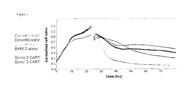
Figure 1 is a graph showing the increased killing of target cells in the
presence of CAR-T
cells. RTCA using BXPC3 (CEACAM6 positive) target cells. Effector to target
cell ratio was
10:1. Following introduction of effector cells, RTCA data collection proceeded
for an additional
56 hours.
Figure 2 is a graph showing that CEACAM6-negative cells are not killed in the
present of
CAR-T cells. RTCA using Pan02a (negative for CEACAM6) target cells. Effector
to target cell
ratio was 10:1. Following introduction of effector cells, RTCA data collection
proceeded for an
additional 36 hours.
Figure 3A is a graph showing interferon gamma secretion levels are increased
in a
significant and CAR-T specific manner. 3B shows a standard-curve generated for
the LPN-
gamma secretion assay.
Figure 4A is a graph showing IL-2 secretion levels are increased in a
significant CAR-T
specific manner. 4B is a graph showing a standard-curve generated for the IL-2
secretion assay.
Figure 5 shows anti-Fab antibody binding to CAR-T cells indicating a
transduction
efficiency of >26%.
Figure 6 shows the RTCA Indicating an Enhanced Cytotoxic Effect of CEACAM-6
CAR-T cells versus target BxPC-3 cells. Ratio of Effector:Target cells=10:1.
A. CAR-T cells for
injection #1 at Day 1; B. CAR-T cells used for injection #2, Day 8. C. CAR-T
cells used for
injection #3, Day 15.
Figure 7 is a graph showing CEACAM-6 CAR-T cells significantly decreased BxPC3
xenograft tumor growth in vivo. N=5 mice per group. Arrow indicate day of
injection (days 1,8,
13
CA 03031289 2019-01-18
WO 2018/014122 PCT/CA2017/050860
15). Mean+/- standard errors are shown. The p-values were calculated and at
day 29, the
difference was significant with p<0.001 in CAR-T group versus PBS and Control
Mock T cell
group.
Figure 8 shows the body weight of mice after injection of CEACAM-6-CAR-T and T
cells.
Figure 9 shows tumors collected at day 30 after BxPC3 cell injection. CEACAM-6-
treated mice had significantly decreased tumor sizes and one tumor was
completely eliminated.
Figure 10 shows flow cytometry analysis indicating effective transduction of T
cells with
lentiviral CAR and expression of CEACAM-6 scFv. CD3-APC staining detected a
high percent
of T cells.
Figure 11 shows the results of RTCA using pancreatic cancer BXPC3 (CEACAM6
positive) target cells. Effector to target cell ratio was 10:1. Following
introduction of effector
cells, RTCA data collection proceeded for an additional 26 hours. Data show
increased killing of
target cells in the presence of CEACAM-6 CAR-T cells.
Figure 12 shows the results of RTCA using colon cancer 5174-T (CEACAM6
positive)
target cells. Effector to target cell ratio was 10:1. Following introduction
of effector cells, RTCA
data collection proceeded for an additional 26 hours. Data show increased
killing of target cells
in the presence of CEACAM-6 CAR-T cells.
Figure 13 shows the results of RTCA using breast ductal carcinoma HCC-1954
(CEACAM6 positive) target cells. Effector to target cell ratio was 10:1.
Following introduction
of effector cells, RTCA data collection proceeded for an additional 26 hours.
Data show
increased killing of target cells in the presence of CEACAM-6 CAR-T cells.
Figure 14 shows the results of RTCA using lung carcinoma A549 (CEACAM6
positive)
target cells. Effector to target cell ratio was 10:1. Following introduction
of effector cells, RTCA
data collection proceeded for an additional 26 hours. Data show increased
killing of target cells
in the presence of CEACAM-6 CAR-T cells.
Figure 15 shows the results of RTCA indicating an enhanced cytotoxic effect of
CEACAM-6 CAR-T cells versus target BxPC-3 cells. Ratio of effector:target
cells = 10:1. T
cells and CAR-T cells were added at day 13. A. CAR-T cells used for injection
#1. B. CAR-T
cells used for injection #2. C. CAR-T cells used for injection #3.
14
CA 03031289 2019-01-18
WO 2018/014122 PCT/CA2017/050860
Figure 16 shows that CEACAM-6 CAR-T cells significantly decreased established
BxPC3 xenograft tumor growth in vivo. N=5 mice per group. Arrows indicate day
of injection.
Mean +/- standard errors are shown.
Figure 17 shows the body weight of mice after injection of PBS, CEACAM-6-CAR-T
cells, and T cells.
Figure 18 shows images of tumors collected at day 34 after BxPC3 cell
injection.
CEACAM-6-treated mice had significantly decreased tumor sizes and two tumors
were
completely eliminated.
DETAILED DESCRIPTION
Definitions
Unless defined otherwise, all technical and scientific terms used herein have
the same
meaning as commonly understood by one of ordinary skill in the art to which
the invention
pertains. Although any methods and materials similar or equivalent to those
described herein can
be used in the practice for testing of the present invention, the typical
materials and methods are
described herein. In describing and claiming the present invention, the
following terminology
will be used.
It is also to be understood that the terminology used herein is for the
purpose of
describing particular aspects only, and is not intended to be limiting.
The articles "a" and "an" are used herein to refer to one or to more than one
(i.e., to at
least one) of the grammatical object of the article. By way of example, "an
element" means one
element or more than one element.
"About" as used herein when referring to a measurable value such as an amount,
a
temporal duration, and the like, is meant to encompass variations of 20% or
10%, more
typically 5%, even more typically 1%, and still more typically 0.1% from
the specified
value, as such variations are appropriate to perform the disclosed methods.
"Activation", as used herein, refers to the state of an immune cell, such as a
OK cell or T
cell, that has been sufficiently stimulated to induce detectable cellular
proliferation. Activation
can also be associated with induced cytokine production, and detectable
effector functions. The
term "activated T cells" refers to, among other things, T cells that are
undergoing cell division.
CA 03031289 2019-01-18
WO 2018/014122 PCT/CA2017/050860
The term "antibody", also referred to in the art as "immunoglobulin" (Ig),
used herein
refers to a protein constructed from paired heavy and light polypeptide
chains; various Ig
isotypes exist, including IgA, IgD, IgE, IgG, and IgM. When an antibody is
correctly folded,
each chain folds into a number of distinct globular domains joined by more
linear polypeptide
sequences. For example, the immunoglobulin light chain folds into a variable
(VI) and a constant
(CO domain, while the heavy chain folds into a variable (VII) and three
constant (CH, CH2, CH3)
domains. Interaction of the heavy and light chain variable domains (VII and
VI) results in the
formation of an antigen binding region (Fv). Each domain has a well-
established structure
familiar to those of skill in the art.
The light and heavy chain variable regions are responsible for binding the
target antigen
and can therefore show significant sequence diversity between antibodies. The
constant regions
show less sequence diversity, and are responsible for binding a number of
natural proteins to
elicit important immunological events. The variable region of an antibody
contains the antigen
binding determinants of the molecule, and thus determines the specificity of
an antibody for its
target antigen. The majority of sequence variability occurs in six
hypervariable regions, three
each per variable heavy and light chain; the hypervariable regions combine to
form the antigen-
binding site, and contribute to binding and recognition of an antigenic
determinant. The
specificity and affinity of an antibody for its antigen is determined by the
structure of the
hypervariable regions, as well as their size, shape and chemistry of the
surface they present to the
antigen. Various schemes exist for identification of the regions of
hypervariability, the two most
common being those of Kabat and of Chothia and Lesk. Kabat et al (1991a;
1991b) define the
"complementarity-determining regions" (CDR) based on sequence variability at
the antigen-
binding regions of the VH and VL domains. Chothia and Lesk (1987) define the
"hypervariable
loops" (H or L) based on the location of the structural loop regions in the VH
and VL domains.
As these individual schemes define CDR and hypervariable loop regions that are
adjacent or
overlapping, those of skill in the antibody art often utilize the terms "CDR"
and "hypervariable
loop" interchangeably, and they may be so used herein. For this reason, the
regions forming the
antigen-binding site are referred to as CDR Li, CDR L2, CDR L3, CDR H1, CDR
H2, CDR H3
in the case of antibodies comprising a VH and a VL domain; or as CDR1, CDR2,
CDR3 in the
case of the antigen-binding regions of either a heavy chain or a light chain.
The CDR/loops are
referred to herein according to the EVIGT numbering system (Lefranc et al.,
2003), which was
16
CA 03031289 2019-01-18
WO 2018/014122 PCT/CA2017/050860
developed to facilitate comparison of variable domains. In this system,
conserved amino acids
(such as Cys23, Trp41, Cys 104, Phe/Trp 118, and a hydrophobic residue at
position 89) always
have the same position. Additionally, a standardized delimitation of the
framework regions (FR1:
positions 1 to 26; FR2: 39 to 55; FR3: 66 to 104; and FR4: 118 to 128) and of
the CDR (CDR1:
27 to 38, CDR2: 56 to 65; and CDR3: 105 to 117) is provided.
An "antibody fragment" as referred to herein may include any suitable antigen-
binding
antibody fragment known in the art. The antibody fragment may be a naturally-
occurring
antibody fragment, or may be obtained by manipulation of a naturally-occurring
antibody or by
using recombinant methods. For example, an antibody fragment may include, but
is not limited
to a Fv, single-chain Fv (scFv; a molecule consisting of VL and VH connected
with a peptide
linker), Fab, F(a1:02, single domain antibody (sdAb; a fragment composed of a
single VL or VH),
and multivalent presentations of any of these.
By the term "synthetic antibody" as used herein, is meant an antibody which is
generated
using recombinant DNA technology, such as, for example, an antibody expressed
by a
bacteriophage as described herein. The term should also be construed to mean
an antibody which
has been generated by the synthesis of a DNA molecule encoding the antibody
and which DNA
molecule expresses an antibody protein, or an amino acid sequence specifying
the antibody,
wherein the DNA or amino acid sequence has been obtained using synthetic DNA
or amino acid
sequence technology which is available and well known in the art.
In a non-limiting example, the antibody fragment may be an sdAb derived from
naturally-occurring sources. Heavy chain antibodies of camelid origin (Hamers-
Casterman et al,
1993) lack light chains and thus their antigen binding sites consist of one
domain, termed VHF"
sdAb have also been observed in shark and are termed VNAR (Nuttall et al,
2003). Other sdAb
may be engineered based on human Ig heavy and light chain sequences (Jespers
et al, 2004; To
et al, 2005). As used herein, the term "sdAb" includes those sdAb directly
isolated from VH,
VHH, VL, or VNAR reservoir of any origin through phage display or other
technologies, sdAb
derived from the aforementioned sdAb, recombinantly produced sdAb, as well as
those sdAb
generated through further modification of such sdAb by humanization, affinity
maturation,
stabilization, solubilization, e.g., camelization, or other methods of
antibody engineering. Also
encompassed by the present invention are homologues, derivatives, or fragments
that retain the
antigen-binding function and specificity of the sdAb.
17
CA 03031289 2019-01-18
WO 2018/014122 PCT/CA2017/050860
SdAbs are excellent building blocks for novel antibody molecules due to their
high
thermostability, high detergent resistance, relatively high resistance to
proteases (Dumoulin et al,
2002) and high production yield (Arbabi-Ghahroudi et al, 1997); they can also
be engineered to
have very high affinity by isolation from an immune library (Li et al, 2009)
or by in vitro affinity
maturation (Davies & Riechmann, 1996).
A person of skill in the art would be well-acquainted with the structure of a
single-
domain antibody (see, for example, 3DWT, 2P42 in Protein Data Bank). A sdAb
comprises a
single immunoglobulin domain that retains the immunoglobulin fold; most
notably, only three
CDR form the antigen-binding site. However, and as would be understood by
those of skill in the
art, not all CDR may be required for binding the antigen. For example, and
without wishing to be
limiting, one, two, or three of the CDR may contribute to binding and
recognition of the antigen
by the sdAb of the present invention. The CDR of the sdAb or variable domain
are referred to
herein as CDR1, CDR2, and CDR3, and numbered as defined by Kabat et al
(1991b).
The term "antigen" or "Ag" as used herein is defined as a molecule that
provokes an
immune response. This immune response may involve either antibody production,
or the
activation of specific immunologically-competent cells, or both. The skilled
artisan will
understand that any macromolecule, including virtually all proteins or
peptides, can serve as an
antigen. Furthermore, antigens can be derived from recombinant or genomic DNA.
A skilled
artisan will understand that any DNA, which comprises a nucleotide sequences
or a partial
nucleotide sequence encoding a protein that elicits an immune response
therefore encodes an
"antigen" as that term is used herein. Furthermore, one skilled in the art
will understand that an
antigen need not be encoded solely by a full length nucleotide sequence of a
gene. It is readily
apparent that the present invention includes, but is not limited to, the use
of partial nucleotide
sequences of more than one gene and that these nucleotide sequences are
arranged in various
combinations to elicit the desired immune response. Moreover, a skilled
artisan will understand
that an antigen need not be encoded by a "gene" at all. It is readily apparent
that an antigen can
be synthesized or can be derived from a biological sample. Such a biological
sample can include,
but is not limited to a tissue sample, a tumor sample, a cell or a biological
fluid.
The term "anti-tumor effect" or "treatment of cancer" as used herein, refers
to a
biological effect which can be manifested by a decrease in tumor volume, a
decrease in the
number of tumor cells, a decrease in the rate of tumor growth, a decrease in
the number of
18
CA 03031289 2019-01-18
WO 2018/014122 PCT/CA2017/050860
metastases, stabilized disease, an increase in life expectancy, or
amelioration of various
physiological symptoms associated with the cancerous condition. An "anti-tumor
effect" can also
be manifested by the ability of the peptides, polynucleotides, cells and
antibodies described
herein in prevention of the occurrence of tumor in the first place.
The term "auto-antigen" means, in accordance with the present invention, any
self-
antigen which is mistakenly recognized by the immune system as being foreign.
Auto-antigens
comprise, but are not limited to, cellular proteins, phosphoproteins, cellular
surface proteins,
cellular lipids, nucleic acids, glycoproteins, including cell surface
receptors.
As used herein, the term "autologous" is meant to refer to any material
derived from the
same individual to which it is later to be re-introduced into the individual.
"Allogeneic" refers to a graft derived from a different animal of the same
species.
"Xenogeneic" refers to a graft derived from a different species.
"Syngeneic" refers to a graft derived from an identical individual.
The term "cancer" as used herein is defined as disease characterized by the
rapid and
uncontrolled growth of aberrant cells. Cancer cells can spread locally or
through the bloodstream
and lymphatic system to other parts of the body. Examples of various cancers
include but are not
limited to, breast cancer, prostate cancer, ovarian cancer, cervical cancer,
skin cancer, pancreatic
cancer, colorectal cancer, renal cancer, liver cancer, brain cancer, lymphoma,
leukemia, lung
cancer and the like.
"Co-stimulatory ligand," as the term is used herein, includes a molecule on an
antigen
presenting cell (e.g., an aAPC, dendritic cell, B cell, and the like) that
specifically binds a
cognate co-stimulatory molecule on a T cell, thereby providing a signal which,
in addition to the
primary signal provided by, for instance, binding of a TCR/CD3 complex with an
MHC
molecule loaded with peptide, mediates a T cell response, including, but not
limited to,
proliferation, activation, differentiation, and the like. A co-stimulatory
ligand can include, but is
not limited to, CD7, B7-1 (CD80), B7-2 (CD86), PD-L1, PD-L2, 4-1BBL, OX4OL,
inducible
costimulatory ligand (ICOS-L), intercellular adhesion molecule (ICAM), CD3OL,
CD40, CD70,
CD83, EILA-G, MICA, MICB, HVEM, lymphotoxin beta receptor, 3/TR6, 1LT3, 1LT4,
HVEM,
an agonist or antibody that binds Toll ligand receptor and a ligand that
specifically binds with
B7-H3. A co-stimulatory ligand also encompasses, inter alia, an antibody that
specifically binds
with a co-stimulatory molecule present on a T cell, such as, but not limited
to, CD27, CD28, 4-
19
CA 03031289 2019-01-18
WO 2018/014122 PCT/CA2017/050860
1BB, 0X40, CD30, CD40, PD-1, ICOS, lymphocyte function-associated antigen-1
(LFA-1),
CD2, CD7, LIGHT, NKG2C, B7-H3, and a ligand that specifically binds with CD83.
A "co-stimulatory molecule" refers to the cognate binding partner on a T cell
that
specifically binds with a co-stimulatory ligand, thereby mediating a co-
stimulatory response by
the T cell, such as, but not limited to, proliferation. Co-stimulatory
molecules include, but are not
limited to an MHC class I molecule, BTLA and a Toll ligand receptor.
A "co-stimulatory signal", as used herein, refers to a signal, which in
combination with a
primary signal, such as TCR/CD3 ligation, leads to T cell proliferation and/or
upregulation or
downregulation of key molecules.
An "effective amount" as used herein, means an amount which provides a
therapeutic or
prophylactic benefit.
"Encoding" refers to the inherent property of specific sequences of
nucleotides in a
polynucleotide, such as a gene, a cDNA, or an mRNA, to serve as templates for
synthesis of
other polymers and macromolecules in biological processes having either a
defined sequence of
nucleotides (e.g., rRNA, tRNA and mRNA) or a defined sequence of amino acids
and the
biological properties resulting therefrom. Thus, a gene encodes a protein if
transcription and
translation of mRNA corresponding to that gene produces the protein in a cell
or other biological
system. Both the coding strand, the nucleotide sequence of which is identical
to the mRNA
sequence and is usually provided in sequence listings, and the non-coding
strand, used as the
template for transcription of a gene or cDNA, can be referred to as encoding
the protein or other
product of that gene or cDNA.
As used herein "endogenous" refers to any material from or produced inside an
organism,
cell, tissue or system.
As used herein, the term "exogenous" refers to any material introduced from or
produced
outside an organism, cell, tissue or system.
The term "expression" as used herein is defined as the transcription and/or
translation of a
particular nucleotide sequence driven by its promoter.
"Expression vector" refers to a vector comprising a recombinant polynucleotide
comprising expression control sequences operatively linked to a nucleotide
sequence to be
expressed. An expression vector comprises sufficient cis-acting elements for
expression; other
elements for expression can be supplied by the host cell or in an in vitro
expression system.
CA 03031289 2019-01-18
WO 2018/014122 PCT/CA2017/050860
Expression vectors include all those known in the art, such as cosmids,
plasmids (e g, naked or
contained in liposomes) and viruses (e.g., lentiviruses, retroviruses,
adenoviruses, and adeno-
associated viruses) that incorporate the recombinant polynucleotide.
"Homologous" refers to the sequence similarity or sequence identity between
two
polypeptides or between two nucleic acid molecules. When a position in both of
the two
compared sequences is occupied by the same base or amino acid monomer subunit,
e.g., if a
position in each of two DNA molecules is occupied by adenine, then the
molecules are
homologous at that position. The percent of homology between two sequences is
a function of
the number of matching or homologous positions shared by the two sequences
divided by the
number of positions compared×100. For example, if 6 of 10 of the
positions in two
sequences are matched or homologous then the two sequences are 60% homologous.
By way of
example, the DNA sequences ATTGCC and TATGGC share 50% homology. Generally, a
comparison is made when two sequences are aligned to give maximum homology.
"Isolated" means altered or removed from the natural state. For example, a
nucleic acid or
a peptide naturally present in a living animal is not "isolated," but the same
nucleic acid or
peptide partially or completely separated from the coexisting materials of its
natural state is
"isolated." An isolated nucleic acid or protein can exist in substantially
purified form, or can
exist in a non-native environment such as, for example, a host cell.
In the context of the present invention, the following abbreviations for the
commonly
occurring nucleic acid bases are used. "A" refers to adenosine, "C" refers to
cytosine, "G" refers
to guanosine, "T" refers to thymidine, and "U" refers to uridine.
Unless otherwise specified, a "nucleotide sequence encoding an amino acid
sequence"
includes all nucleotide sequences that are degenerate versions of each other
and that encode the
same amino acid sequence. The phrase nucleotide sequence that encodes a
protein or an RNA
may also include introns to the extent that the nucleotide sequence encoding
the protein may in
some version contain an intron(s).
A "lentivirus" as used herein refers to a genus of the Retroviridae family.
Lentiviruses are
unique among the retroviruses in being able to infect non-dividing cells; they
can deliver a
significant amount of genetic information into the DNA of the host cell, so
they are one of the
most efficient methods of a gene delivery vector. HIV, Sly, and FIV are all
examples of
21
CA 03031289 2019-01-18
WO 2018/014122 PCT/CA2017/050860
lentiviruses. Vectors derived from lentiviruses offer the means to achieve
significant levels of
gene transfer in vivo.
A "transposon" or "transposable element" is a DNA sequence that can change its
position
within a genome, sometimes creating or reversing mutations and altering the
cell's genome size.
Transposition often results in duplication of the transposon. There are two
distinct types of
transposon: class II transposons, which consist of DNA that moves directly
from place to place;
and class I transposons, which are retrotransposons that first transcribe the
DNA into RNA and
then use reverse transcriptase to make a DNA copy of the RNA to insert in a
new location.
Transposons typically interact with a transposase, which mediates the movement
of the
transposon. Non-limiting examples of transposon/transposase systems include
Sleeping Beauty,
Piggybac, Frog Prince, and Prince Charming.
By the term "modulating," as used herein, is meant mediating a detectable
increase or
decrease in the level of a response in a subject compared with the level of a
response in the
subject in the absence of a treatment or compound, and/or compared with the
level of a response
in an otherwise identical but untreated subject. The term encompasses
perturbing and/or
affecting a native signal or response thereby mediating a beneficial
therapeutic response in a
subject, typically, a human.
The term "operably linked" refers to functional linkage between a regulatory
sequence
and a heterologous nucleic acid sequence resulting in expression of the
latter. For example, a
first nucleic acid sequence is operably linked with a second nucleic acid
sequence when the first
nucleic acid sequence is placed in a functional relationship with the second
nucleic acid
sequence. For instance, a promoter is operably linked to a coding sequence if
the promoter
affects the transcription or expression of the coding sequence. Generally,
operably linked DNA
sequences are contiguous and, where necessary to join two protein coding
regions, in the same
reading frame.
The term "overexpressed" tumor antigen or "overexpression" of the tumor
antigen is
intended to indicate an abnormal level of expression of the tumor antigen in a
cell from a disease
area like a solid tumor within a specific tissue or organ of the patient
relative to the level of
expression in a normal cell from that tissue or organ. Patients having solid
tumors or a
hematological malignancy characterized by overexpression of the tumor antigen
can be
determined by standard assays known in the art.
22
CA 03031289 2019-01-18
WO 2018/014122 PCT/CA2017/050860
"Parenteral" administration of an immunogenic composition includes, e.g.,
subcutaneous
(s.c.), intravenous (i.v.), intramuscular (i.m.), or intrasternal injection,
or infusion techniques.
The terms "patient," "subject," "individual," and the like are used
interchangeably herein,
and refer to any animal, or cells thereof whether in vitro or in situ,
amenable to the methods
described herein.
Moreover, the terms "patient", "subject" and "individual" includes living
organisms in
which an immune response can be elicited (e.g., mammals). In certain non-
limiting aspects, the
patient, subject or individual is a mammal and includes humans, dogs, cats,
mice, rats, and
transgenic species thereof.
The term "polynucleotide" as used herein is defined as a chain of nucleotides.
Furthermore, nucleic acids are polymers of nucleotides. Thus, nucleic acids
and polynucleotides
as used herein are interchangeable. One skilled in the art has the general
knowledge that nucleic
acids are polynucleotides, which can be hydrolyzed into the monomeric
"nucleotides." The
monomeric nucleotides can be hydrolyzed into nucleosides. As used herein
polynucleotides
include, but are not limited to, all nucleic acid sequences which are obtained
by any means
available in the art, including, without limitation, recombinant means, i.e.,
the cloning of nucleic
acid sequences from a recombinant library or a cell genome, using ordinary
cloning technology
and PCR, and the like, and by synthetic means.
As used herein, the terms "peptide," "polypeptide," and "protein" are used
interchangeably, and refer to a compound comprised of amino acid residues
covalently linked by
peptide bonds. A protein or peptide must contain at least two amino acids, and
no limitation is
placed on the maximum number of amino acids that can comprise a protein's or
peptide's
sequence. Polypeptides include any peptide or protein comprising two or more
amino acids
joined to each other by peptide bonds. As used herein, the term refers to both
short chains, which
also commonly are referred to in the art as peptides, oligopeptides and
oligomers, for example,
and to longer chains, which generally are referred to in the art as proteins,
of which there are
many types. "Polypeptides" include, for example, biologically active
fragments, substantially
homologous polypeptides, oligopeptides, homodimers, heterodimers, variants of
polypeptides,
modified polypeptides, derivatives, analogs, fusion proteins, among others.
The polypeptides
include natural peptides, recombinant peptides, synthetic peptides, or a
combination thereof.
23
CA 03031289 2019-01-18
WO 2018/014122 PCT/CA2017/050860
The term "promoter" as used herein is defined as a DNA sequence recognized by
the
synthetic machinery of the cell, or introduced synthetic machinery, required
to initiate the
specific transcription of a polynucleotide sequence.
As used herein, the term "promoter/regulatory sequence" means a nucleic acid
sequence
which is required for expression of a gene product operably linked to the
promoter/regulatory
sequence. In some instances, this sequence may be the core promoter sequence
and in other
instances, this sequence may also include an enhancer sequence and other
regulatory elements
which are required for expression of the gene product. The promoter/regulatory
sequence may,
for example, be one which expresses the gene product in a tissue specific
manner.
A "constitutive" promoter is a nucleotide sequence which, when operably linked
with a
polynucleotide which encodes or specifies a gene product, causes the gene
product to be
produced in a cell under most or all physiological conditions of the cell.
An "inducible" promoter is a nucleotide sequence which, when operably linked
with a
polynucleotide which encodes or specifies a gene product, causes the gene
product to be
produced in a cell substantially only when an inducer which corresponds to the
promoter is
present in the cell.
A "tissue-specific" promoter is a nucleotide sequence which, when operably
linked with a
polynucleotide encodes or specified by a gene, causes the gene product to be
produced in a cell
substantially only if the cell is a cell of the tissue type corresponding to
the promoter.
By the term "specifically binds," as used herein with respect to an antibody,
is meant an
antibody which recognizes a specific antigen, but does not substantially
recognize or bind other
molecules in a sample. For example, an antibody that specifically binds to an
antigen from one
species may also bind to that antigen from one or more species. But, such
cross-species reactivity
does not itself alter the classification of an antibody as specific. In
another example, an antibody
that specifically binds to an antigen may also bind to different allelic forms
of the antigen.
However, such cross reactivity does not itself alter the classification of an
antibody as specific.
In some instances, the terms "specific binding" or "specifically binding," can
be used in
reference to the interaction of an antibody, a protein, or a peptide with a
second chemical
species, to mean that the interaction is dependent upon the presence of a
particular structure (e.g.,
an antigenic determinant or epitope) on the chemical species; for example, an
antibody
recognizes and binds to a specific protein structure rather than to proteins
generally. If an
24
CA 03031289 2019-01-18
WO 2018/014122 PCT/CA2017/050860
antibody is specific for epitope "A", the presence of a molecule containing
epitope A (or free,
unlabeled A), in a reaction containing labeled "A" and the antibody, will
reduce the amount of
labeled A bound to the antibody.
The term "epitope" means a protein determinant capable of specific binding to
an
antibody. Epitopes usually consist of chemically active surface groupings of
molecules such as
amino acids or sugar side chains and usually have specific three dimensional
structural
characteristics, as well as specific charge characteristics. Conformational
and nonconformational
epitopes are distinguished in that the binding to the former but not the
latter is lost in the
presence of denaturing solvents.
By the term "stimulation," is meant a primary response induced by binding of a
stimulatory molecule (e.g., a TCR/CD3 complex) with its cognate ligand thereby
mediating a
signal transduction event, such as, but not limited to, signal transduction
via the TCR/CD3
complex. Stimulation can mediate altered expression of certain molecules, such
as
downregulation of TGF-I3, and/or reorganization of cytoskeletal structures,
and the like.
A "stimulatory molecule," as the term is used herein, means a molecule on a T
cell that
specifically binds with a cognate stimulatory ligand present on an antigen
presenting cell.
A "stimulatory ligand," as used herein, means a ligand that when present on an
antigen
presenting cell (e.g., an aAPC, a dendritic cell, a B-cell, and the like) can
specifically bind with a
cognate binding partner (referred to herein as a "stimulatory molecule") on a
T cell, thereby
mediating a primary response by the T cell, including, but not limited to,
activation, initiation of
an immune response, proliferation, and the like. Stimulatory ligands are well-
known in the art
and encompass, inter alia, an MEW Class I molecule loaded with a peptide, an
anti-CD3
antibody, a superagonist anti-CD28 antibody, and a superagonist anti-CD2
antibody.
As used herein, a "substantially purified" cell is a cell that is essentially
free of other cell
types. A substantially purified cell also refers to a cell which has been
separated from other cell
types with which it is normally associated in its naturally occurring state.
In some instances, a
population of substantially purified cells refers to a homogenous population
of cells. In other
instances, this term refers simply to cell that have been separated from the
cells with which they
are naturally associated in their natural state. In some aspects, the cells
are cultured in vitro. In
other aspects, the cells are not cultured in vitro.
CA 03031289 2019-01-18
WO 2018/014122 PCT/CA2017/050860
As used herein, "treatment" or "therapy" is an approach for obtaining
beneficial or
desired clinical results. For the purposes described herein, beneficial or
desired clinical results
include, but are not limited to, alleviation of symptoms, diminishment of
extent of disease,
stabilized (i.e., not worsening) state of disease, delay or slowing of disease
progression,
amelioration or palliation of the disease state, and remission (whether
partial or total), whether
detectable or undetectable. "Treatment" and "therapy" can also mean prolonging
survival as
compared to expected survival if not receiving treatment or therapy. Thus,
"treatment" or
"therapy" is an intervention performed with the intention of altering the
pathology of a disorder.
Specifically, the treatment or therapy may directly prevent, slow down or
otherwise decrease the
pathology of a disease or disorder such as cancer, or may render the cells
more susceptible to
treatment or therapy by other therapeutic agents.
The term "therapeutic" as used herein means a treatment and/or prophylaxis. A
therapeutic effect is obtained by suppression, remission, or eradication of a
disease state.
The term "therapeutically effective amount" refers to the amount of the
subject
compound that will elicit the biological or medical response of a tissue,
system, or subject that is
being sought by the researcher, veterinarian, medical doctor or other
clinician. The term
"therapeutically effective amount" includes that amount of a compound that,
when administered,
is sufficient to prevent development of, or alleviate to some extent, one or
more of the signs or
symptoms of the disorder or disease being treated. The therapeutically
effective amount will vary
depending on the compound, the disease and its severity and the age, weight,
etc., of the subject
to be treated.
To "treat" a disease as the term is used herein, means to reduce the frequency
or severity
of at least one sign or symptom of a disease or disorder experienced by a
subject.
Moreover, a treatment regime of a subject with a therapeutically effective
amount may
consist of a single administration, or alternatively comprise a series of
applications. The length of
the treatment period depends on a variety of factors, such as the severity of
the disease, the age
of the subject, the concentration of the agent, the responsiveness of the
patient to the agent, or a
combination thereof. It will also be appreciated that the effective dosage of
the agent used for the
treatment may increase or decrease over the course of a particular treatment
regime. Changes in
dosage may result and become apparent by standard diagnostic assays known in
the art. The
26
CA 03031289 2019-01-18
WO 2018/014122 PCT/CA2017/050860
antibodies described herein may, in aspects, be administered before, during or
after treatment
with conventional therapies for the disease or disorder in question, such as
cancer.
The term "transfected" or "transformed" or "transduced" as used herein refers
to a process
by which exogenous nucleic acid is transferred or introduced into the host
cell. A "transfected"
or "transformed" or "transduced" cell is one which has been transfected,
transformed or
transduced with exogenous nucleic acid. The cell includes the primary subject
cell and its
progeny.
The phrase "under transcriptional control" or "operatively linked" as used
herein means
that the promoter is in the correct location and orientation in relation to a
polynucleotide to
control the initiation of transcription by RNA polymerase and expression of
the polynucleotide.
A "vector" is a composition of matter which comprises an isolated nucleic acid
and which
can be used to deliver the isolated nucleic acid to the interior of a cell.
Numerous vectors are
known in the art including, but not limited to, linear polynucleotides,
polynucleotides associated
with ionic or amphiphilic compounds, plasmids, and viruses. Thus, the term
"vector" includes an
autonomously replicating plasmid or a virus. The term should also be construed
to include non-
plasmid and non-viral compounds which facilitate transfer of nucleic acid into
cells, such as, for
example, polylysine compounds, liposomes, and the like. Examples of viral
vectors include, but
are not limited to, adenoviral vectors, adeno-associated virus vectors,
retroviral vectors, and the
like.
Ranges: throughout this disclosure, various aspects described herein can be
presented in a
range format. It should be understood that the description in range format is
merely for
convenience and brevity and should not be construed as an inflexible
limitation on the scope
described herein. Accordingly, the description of a range should be considered
to have
specifically disclosed all the possible subranges as well as individual
numerical values within
that range. For example, description of a range such as from 1 to 6 should be
considered to have
specifically disclosed subranges such as from 1 to 3, from 1 to 4, from 1 to
5, from 2 to 4, from 2
to 6, from 3 to 6 etc., as well as individual numbers within that range, for
example, 1, 2, 2.7, 3, 4,
5, 5.3, and 6. This applies regardless of the breadth of the range.
It will be understood that any aspects described as "comprising" certain
components may
also "consist of' or "consist essentially of," wherein "consisting of' has a
closed-ended or
restrictive meaning and "consisting essentially of' means including the
components specified but
27
CA 03031289 2019-01-18
WO 2018/014122 PCT/CA2017/050860
excluding other components except for materials present as impurities,
unavoidable materials
present as a result of processes used to provide the components, and
components added for a
purpose other than achieving the technical effect described herein. For
example, a composition
defined using the phrase "consisting essentially of' encompasses any known
pharmaceutically
acceptable additive, excipient, diluent, carrier, and the like. Typically, a
composition consisting
essentially of a set of components will comprise less than 5% by weight,
typically less than 3%
by weight, more typically less than 1% by weight of non-specified components.
It will be understood that any component defined herein as being included may
be
explicitly excluded from the claimed invention by way of proviso or negative
limitation.
Administration "in combination with" one or more further therapeutic agents
includes
simultaneous (concurrent) and consecutive administration in any order.
The term "pharmaceutically acceptable" means that the compound or combination
of
compounds is compatible with the remaining ingredients of a formulation for
pharmaceutical
use, and that it is generally safe for administering to humans according to
established
governmental standards, including those promulgated by the United States Food
and Drug
Administration.
The term "pharmaceutically acceptable carrier" includes, but is not limited to
solvents,
dispersion media, coatings, antibacterial agents, antifungal agents, isotonic
and/or absorption
delaying agents and the like. The use of pharmaceutically acceptable carriers
is well known.
Isolated: An "isolated" biological component (such as a protein) has been
substantially
separated or purified away from other biological components in the cell of the
organism in which
the component naturally occurs, i.e., chromosomal and extra-chromosomal DNA
and RNA, other
proteins and organelles. Proteins and peptides that have been "isolated"
include proteins and
peptides purified by standard purification methods. The term also includes
proteins and peptides
prepared by recombinant expression in a host cell, as well as chemically
synthesized proteins and
peptides.
"Tumour", as used herein, refers to all neoplastic cell growth and
proliferation, whether
malignant or benign, and all pre-cancerous and cancerous cells and tissues.
The terms "cancer" and "cancerous" refer to or describe the physiological
condition in
mammals that is typically characterized by unregulated cell growth. The cancer
to be treated may
be any type of malignancy and, in an aspect, is lung cancer, including small
cell lung cancer and
28
CA 03031289 2019-01-18
WO 2018/014122 PCT/CA2017/050860
non-small cell lung cancer (e.g. adenocarcinoma), pancreatic cancer, colon
cancer (e.g.
colorectal carcinoma, such as, for example, colon adenocarcinoma and colon
adenoma),
oesophageal cancer, oral squamous carcinoma, tongue carcinoma, gastric
carcinoma, liver
cancer, nasopharyngeal cancer, hematopoietic tumours of lymphoid lineage (e.g.
acute
lymphocytic leukemia, B-cell lymphoma, Burkitt's lymphoma), non-Hodgkin's
lymphoma (e.g.
mantle cell lymphoma), Hodgkin's disease, myeloid leukemia (for example, acute
myelogenous
leukemia (AML) or chronic myelogenous leukemia (CML)), acute lymphoblastic
leukemia,
chronic lymphocytic leukemia (CLL), thyroid follicular cancer, myelodysplastic
syndrome
(MDS), tumours of mesenchymal origin, soft tissue sarcoma, liposarcoma,
gastrointestinal
stromal sarcoma, malignant peripheral nerve sheath tumour (lVfPNST), Ewing
sarcoma,
leiomyosarcoma, mesenchymal chondrosarcoma, lymphosarcoma, fibrosarcoma,
rhabdomyosarcoma, melanoma, teratocarcinoma, neuroblastoma, brain tumours,
medulloblastoma, glioma, benign tumour of the skin (e.g. keratoacanthoma),
breast carcinoma
(e.g. advanced breast cancer), kidney carcinoma, nephroblastoma, ovary
carcinoma, cervical
carcinoma, endometrial carcinoma, bladder carcinoma, prostate cancer,
including advanced
disease and hormone refractory prostate cancer, testicular cancer,
osteosarcoma, head and neck
cancer, epidermal carcinoma, multiple myeloma (e.g. refractory multiple
myeloma), or
mesothelioma. In an aspect, the cancer cells are derived from a solid tumour.
Typically, the
cancer cells are derived from a breast cancer, colorectal cancer, melanoma,
ovarian cancer,
pancreatic cancer, gastric cancer, lung cancer, or prostate cancer. More
typically, the cancer cells
are derived from a prostate cancer, a lung cancer, a breast cancer, or a
melanoma.
A "chemotherapeutic agent" is a chemical compound useful in the treatment of
cancer.
Examples of chemotherapeutic agents include alkylating agents such as
thiotepa, CYTOXANTm
cyclosphosphamide; alkyl sulfonates such as busulfan, improsulfan and
piposulfan; aziridines
such as benzodopa, carboquone, meturedopa, and uredopa; ethylenimines and
methylamelamines
including altretamine, triethylenemelamine, trietylenephosphoramide,
triethiylenethiophosphoramide and trimethylolomelamine; acetogenins such as
bullatacin and
bullatacinone; camptothecins such as topotecan; bryostatin; callystatin; CC-
1065 and its
adozelesin, carzelesin and bizelesin synthetic analogues; cryptophycins such
as cryptophycin 1
and cryptophycin 8; dolastatin; duocarmycins such as the synthetic analogues
KW-2189 and
CB1-TM1; eleutherobin; pancratistatin; sarcodictyins; spongistatin; nitrogen
mustards such as
29
CA 03031289 2019-01-18
WO 2018/014122 PCT/CA2017/050860
chlorambucil, chlornaphazine, cholophosphamide, estramustine, ifosfamide,
mechlorethamine,
mechlorethamine oxide hydrochloride, melphalan, novembichin, phenesterine,
prednimustine,
trofosfamide, uracil mustard; nitrosureas such as carmustine, chlorozotocin,
fotemustine,
lomustine, nimustine, and ranimustine; antibiotics such as the enediyne
antibiotics, for example
calicheamicin, especially calicheamicin gamma 11 and calicheamicin omegaIl,
dynemicin,
including dynemicin A, bisphosphonates, such as clodronate, esperamicins,
neocarzinostatin
chromophore and related chromoprotein enediyne antibiotic chromophores;
aclacinomysins;
actinomycin; authramycin; azaserine; bleomycins; cactinomycin; carabicin;
carminomycin;
carzinophilin; chromomycins; dactinomycin; daunorubicin; detorubicin; 6-diazo-
5-oxo-L-
norleucine; ADRIAMYCINTm doxorubicin, including morpholino-doxorubicin,
cyanomorpholino-doxorubicin, 2-pyrrolino-doxorubicin and deoxydoxorubicin;
epirubicin;
esorubicin; idarubicin; marcellomycin; mitomycins such as mitomycin C,
mycophenolic acid,
nogalamycin, olivomycins, peplomycin, potfiromycin, puromycin, quelamycin,
rodorubicin,
streptonigrin, streptozocin, tubercidin, ubenimex, zinostatin, and zorubicin;
anti-metabolites such
as methotrexate and 5-fluorouracil (5-FU); folic acid analogues such as
denopterin,
methotrexate, pteropterin, trimetrexate; purine analogs such as fludarabine, 6-
mercaptopurine,
thiamiprine, and thioguanine; pyrimidine analogs such as ancitabine,
azacitidine, 6-azauridine,
carmofur, cytarabine, dideoxyuridine, doxifluridine, enocitabine, and
floxuridine; androgens
such as calusterone, dromostanolone propionate, epitiostanol, mepitiostane,
and testolactone;
anti-adrenals such as aminoglutethimide, mitotane, and trilostane; folic acid
replenishers such as
frolinic acid; aceglatone; aldophosphamide glycoside; aminolevulinic acid;
eniluracil; amsacrine;
bestrabucil; bisantrene; edatraxate; defofamine; demecolcine; diaziquone;
elfornithine;
elliptinium acetate; epothilones; etoglucid; gallium nitrate; hydroxyurea;
lentinan; lonidainine;
maytansinoids such as maytansine and ansamitocins; mitoguazone; mitoxantrone;
mopidanmol;
nitraerine; pentostatin; phenamet; pirarubicin; losoxantrone; podophyllinic
acid; 2-
ethylhydrazide; procarbazine; PSKTM polysaccharide complex; razoxane;
rhizoxin; sizofiran;
spirogermanium; tenuazonic acid; triaziquone; 2,2',2"-trichlorotriethylamine;
trichothecenes such
as T-2 toxin, verracurin A, roridin A and anguidine; urethan; vindesine;
dacarbazine;
mannomustine; mitobronitol; mitolactol; pipobroman; gacytosine; arabinoside
("Ara-C");
taxoids, such as TAXOLTm paclitaxel, ABRAXANETM Cremophor-free, albumin-
engineered
nanoparticle formulation of paclitaxel, TAXOTERETM and doxetaxel;
chloranbucil;
CA 03031289 2019-01-18
WO 2018/014122 PCT/CA2017/050860
GEMZARTm gemcitabine; 6-thioguanine; mercaptopurine; methotrexate; platinum
coordination
complexes such as cisplatin, oxaliplatin and carboplatin; vinblastine;
platinum; etoposide (VP-
16); ifosfamide; vincristine; NAVELBINE TM vinorelbine; novantrone;
teniposide; edatrexate;
daunomycin; aminopterin; xeloda; ibandronate; irinotecans such as CPT-11;
topoisomerase
inhibitors such as RFS 2000; difluoromethylornithine (DMF0); retinoids such as
retinoic acid;
capecitabine; and pharmaceutically acceptable salts, acids or derivatives of
any of the above.
Also included in this definition are anti-hormonal agents that act to regulate
or inhibit
hormone action on tumours such as anti-estrogens and selective estrogen
receptor modulators
(SERMs), including, for example, tamoxifen (including NOLVADEXTM tamoxifen),
raloxifene,
droloxifene, 4-hydroxytamoxifen, trioxifene, keoxifene, LY1170 18,
onapristone, and
FARESTON toremifene; aromatase inhibitors that inhibit the enzyme aromatase,
which regulates
estrogen production in the adrenal glands, such as, for example, 4(5)-
imidazoles,
aminoglutethimide, MEGASETM megestrol acetate, AROMASINTm exemestane,
formestane,
fadrozole, RIVISORTM vorozole, FEMARATm letrozole, and AREVIIDEXTm
anastrozole; and
anti-androgens such as flutamide, nilutamide, bicalutamide, leuprolide, and
goserelin; as well as
troxacitabine (a 1,3-dioxolane nucleoside cytosine analog); anti sense
oligonucleotides,
particularly those that inhibit expression of genes in signalling pathways
implicated in aberrant
cell proliferation, such as, for example, PKC-alpha, Ralf and H-Ras; ribozymes
such as a VEGF
expression inhibitor (e.g., ANGIOZYIVIETM ribozyme) and a HER2 expression
inhibitor;
antibodies such as an anti-VEGF antibody (e.g., AVASTINTm antibody); vaccines
such as gene
therapy vaccines, for example, ALLOVECTINTm vaccine, LEUVECTINTm vaccine, and
VAXIDTM vaccine; PROLEUKINTm r1L-2; LURTOTECANTm topoisomerase 1 inhibitor;
ABARELIXTM rmRH; and pharmaceutically acceptable salts, acids or derivatives
of any of the
above.
In aspects, the antibodies described herein act additively or synergistically
with other
conventional anti-cancer treatments.
Many patent applications, patents, and publications are referred to herein to
assist in
understanding the aspects described. Each of these references are incorporated
herein by
reference in their entirety.
31
CA 03031289 2019-01-18
WO 2018/014122 PCT/CA2017/050860
The invention relates to compositions and methods for treating cancer, in
aspects solid
tumors. The present invention relates to a strategy of adoptive cell transfer
of immune cells
transduced to express a chimeric antigen receptor (CAR). CARs are molecules
that combine
antibody-based specificity for a desired antigen (e.g., tumor antigen) with a
T cell receptor-
activating intracellular domain to generate a chimeric protein that exhibits a
specific anti-tumor
cellular immune activity. The gene-modified T cell therapy comprises
introducing a nucleic acid
encoding a chimeric antigen receptor (CAR) into a T cell, wherein the CAR has
specificity for a
surface antigen of a tumor cell and ability to activate a T cell, growing ex
vivo the gene-
introduced T cell thus obtained, and then transfusing the cell into a patient.
The present invention relates generally to the use of such T cells genetically
modified to
stably express a CAR specific for solid tumor antigens, more specifically,
solid tumors
expressing the CEACAM6 antigen, fragments, and/or epitopes thereof and
variants of these. T
cells expressing a CAR are referred to herein as CAR-T cells or CAR modified T
cells. In
aspects of the present invention, the T cell is genetically modified to stably
express a CAR that
combines an antigen recognition domain of a sdAb specific for CEACAM6 with one
or more
intracellular costimulatory domains and signalling domains into a single
chimeric protein.
Chimeric Antigen Receptor (CAR)
In one aspect, engineered CARs are described herein. The CAR comprises a
CEACAM6
binding moiety that binds to CEACAM6, an epitope thereof, a fragment thereof,
or variants of
the aforementioned and further comprises an immune cell activation domain.
When expressed by
an immune cell, the CEACAM6 binding moiety is or is part of an extracellular
domain and the
immune cell activation domain is or is part of an intracellular signaling
domain, typically of the
T cell antigen receptor complex zeta chain (e.g., CD3 zeta). Co-stimulatory
signaling regions
may also be included in the intracellular domain and are cell surface
molecules other than
antigens receptors or their ligands that are required for an efficient
response of lymphocytes to
antigen. A spacer moiety (also referred to as a hinge moiety) is typically
included in the
extracellular domain to allow the CEACAM6 binding moiety to efficiently bind
to its epitope.
The intracellular and extracellular domains are linked through a transmembrane
domain that
crosses the cytoplasmic membrane.
32
CA 03031289 2019-01-18
WO 2018/014122 PCT/CA2017/050860
A representative non-limiting structure of the CAR of the invention comprises
a single
domain antibody recognizing a CEACAM6 surface antigen of a tumor cell, a
transmembrane
domain, and an intracellular domain of a TCR complex CD3 that activates a T
cell (called a first
generation CAR). The nucleic acid sequence of such a single domain antibody
may be obtained
by a variety of methods as is understood by one of skill in the art. A T cell
expressing a CAR
directly recognizes a surface antigen of a tumor cell independently of the
expression of major
histocompatibility antigen class I on the tumor cell, and at the same time,
activates the T cell, and
thereby the CAR-expressing T cell can efficiently kill the tumor cell.
For enhancing the ability of the first generation CAR to activate a T cell, a
second
generation CAR can be made whereby an intracellular domain of CD28 which is a
co-
stimulatory molecule of a T cell is linked to the first generation CAR. A
third generation CAR
may also be made whereby an intracellular domain derived from (for example)
CD137 (4-1BB)
or CD134 (0X40) which is a tumor necrosis factor (TNF) receptor superfamily is
tandemly
linked to a first generation CAR. Thus, many CAR molecules targeting CEACAM6
are included
in the present invention.
CEACAM6 Binding Moiety
In typical aspects, the CAR described herein is specific for carcinoembryonic
antigen
related cell adhesion molecule 6 (CEACAM6), fragments thereof, epitopes
thereof and variants
of any of the foregoing. CEACAM6 is also known in the art as non-specific
cross-reacting
antigen (NCA) or CD66c. The CEACAM6 binding moiety of the invention is such
that it binds
with a desired affinity to CEACAM6 harbored on a cell/tumor surface leading to
activation of
the immune cell in which it is provided, to trigger cytotoxic activity and
release cytokines within
the tumor microenvironment ad further proliferating.
The sequence of CEACAM6 may be, but is not limited to SEQ ID NO: 7, or a
sequence
substantially identical thereto.
SEQ ID NO: 7:
MGPPSAPPCRLHVPWKEVLLTASLLTFWNPPTTAKLTIESTPFNVAEGKEVLLLAHNLPQ
NRIGYSWYKGERVDGNSLIVGYVIGTQQATPGPAYSGRETIYPNASLLIQNVTQNDTGFY
TLQV1KSDLVNEEATGQFHVYPELPKPSISSNNSNPVEDKDAVAFTCEPEVQNTTYLWW
VNGQSLPVSPRLQLSNGNMTLTLLSVKRNDAGSYECEIQNPASANRSDPVTLNVLYGPD
33
CA 03031289 2019-01-18
WO 2018/014122 PCT/CA2017/050860
GPTISPSKANYRPGENLNLSCHAASNPPAQYSWFINGTFQQ STQELFIPNITVNNSGSYMC
QAHNSATGLNRTTVTMITVSGSAPVLSAVATVGITIGVLARVALI.
In particular aspects, the antibody and/or epitope may be that described in US
9,066,986,
which is incorporated herein by reference in its entirety. Specifically, the
CEACAM6 binding
domain may comprise the 2A3 anti-CEACAM6 antibody or a fragment or variant
thereof
Without wishing to be bound by theory, it is believed that this
antibody/epitope interaction has
an advantageous level of affinity (not too high and not too low), such that
the antibody can bind
the epitope on a first cell and activate cell killing, then move on to bind a
further epitope on a
second or further cell and activate further cell killing.
Thus, also described herein is a CAR comprising, within the CEACAM6 binding
moiety
a complementarity determining region (CDR) 1 comprising the sequence of
GRTNSVYTMG
(SEQ ID NO:1); a CDR2 comprising the sequence of IMWGAGTNTHYADSVKG (SEQ ID
NO:2); and a CDR3 comprising the sequence of AANROPIAGRQYDY (SEQ ID NO:3),
wherein the antibody or fragment thereof is specific for CEACAM6. The CEACAM6
binding
moiety as just described may recognize and bind to an epitope comprising or
consisting of the
sequence NRIGYSWYKG (SEQ ID NO:6).
In a specific, non-limiting example, the antibody or fragment thereof may
comprise the
sequence of SEQ ID NO: 4 or a sequence substantially identical thereto.
SEQ ID NO: 4:
QVKLEESGGGLVQAGGSLRLSCRTSGRTNSVYTMGWFRQAPGKEREFVAQIMWGAGT
NTHYADSVKGRFTISRDSAESTVYLQMNSLKPEDTAVYYCAANROPIAGRQYDYWGQ
GTQVTVSS.
The terms "antibody" and "antibody fragment" ("fragment thereof') are as
defined above.
As previously stated, the antibody or fragment thereof may be an sdAb. The
sdAb may be of
camelid origin (e.g., from the species Camelidae) or be derived from a camelid
VHH, and thus
may be based on camelid framework regions; alternatively, the CDR described
above may be
grafted onto VNAR, VHH or VL framework regions. In yet another alternative,
the hypervariable
loops described above may be grafted onto the framework regions of other types
of antibody
fragments (Fv, scFv, Fab).
The present aspect further encompasses an antibody fragment that is
"humanized" using
any suitable method known in the art, for example, but not limited to CDR
grafting and
34
CA 03031289 2019-01-18
WO 2018/014122 PCT/CA2017/050860
veneering. Humanization of an antibody or antibody fragment comprises
replacing an amino acid
in the sequence with its human counterpart, as found in the human consensus
sequence, without
loss of antigen-binding ability or specificity; this approach reduces
immunogenicity of the
antibody or fragment thereof when introduced into human subjects. In the
process of CDR
grafting, one or more than one of the heavy chain CDR defined herein may be
fused or grafted to
a human variable region (VII, or VI), or to other human antibody fragment
framework regions
(Fv, scFv, Fab). In such a case, the conformation of said one or more than one
hypervariable
loop is preserved, and the affinity and specificity of the sdAb for its target
(i.e., toxins A and B)
is also preserved.
CDR grafting is described in at least the following: U.S. Pat. No. 6,180,370,
U.S. Pat. No.
5,693,761, U.S. Pat. No. 6,054,297, U.S. Pat. No. 5,859,205, and European
Patent No. 626390
(the disclosures of which are hereby incorporated by reference in their
entirety). Veneering, also
referred to in the art as "variable region resurfacing", involves humanizing
solvent-exposed
positions of the antibody or fragment; thus, buried non-humanized residues,
which may be
important for CDR conformation, are preserved while the potential for
immunological reaction
against solvent-exposed regions is minimized. Veneering is described in at
least the following:
U.S. Pat. No. 5,869,619, U.S. Pat. No. 5,766,886, U.S. Pat. No. 5,821,123, and
European Patent
No. 519596 (the disclosures of which are hereby incorporated by reference in
their entirety).
Persons of skill in the art would be amply familiar with methods of preparing
such humanized
antibody fragments.
A substantially identical sequence may comprise one or more conservative amino
acid
mutations. It is known in the art that one or more conservative amino acid
mutations to a
reference sequence may yield a mutant peptide with no substantial change in
physiological,
chemical, or functional properties compared to the reference sequence; in such
a case, the
reference and mutant sequences would be considered "substantially identical"
polypeptides.
Conservative amino acid mutation may include addition, deletion, or
substitution of an amino
acid; a conservative amino acid substitution is defined herein as the
substitution of an amino acid
residue for another amino acid residue with similar chemical properties (e.g.
size, charge, or
polarity).
In a non-limiting example, a conservative mutation may be an amino acid
substitution.
Such a conservative amino acid substitution may substitute a basic, neutral,
hydrophobic, or
CA 03031289 2019-01-18
WO 2018/014122 PCT/CA2017/050860
acidic amino acid for another of the same group. By the term "basic amino
acid" it is meant
hydrophilic amino acids having a side chain pK value of greater than 7, which
are typically
positively charged at physiological pH. Basic amino acids include histidine
(His or H), arginine
(Arg or R), and lysine (Lys or K). By the term "neutral amino acid" (also
"polar amino acid"), it
is meant hydrophilic amino acids having a side chain that is uncharged at
physiological pH, but
which has at least one bond in which the pair of electrons shared in common by
two atoms is
held more closely by one of the atoms. Polar amino acids include serine (Ser
or S), threonine
(Thr or T), cysteine (Cys or C), tyrosine (Tyr or Y), asparagine (Asn or N),
and glutamine (Gln
or Q). The term "hydrophobic amino acid" (also "non-polar amino acid") is
meant to include
amino acids exhibiting a hydrophobicity of greater than zero according to the
normalized
consensus hydrophobicity scale of Eisenberg (1984). Hydrophobic amino acids
include proline
(Pro or P), isoleucine (Ile or I), phenylalanine (Phe or F), valine (Val or
V), leucine (Leu or L),
tryptophan (Trp or W), methionine (Met or M), alanine (Ala or A), and glycine
(Gly or G).
"Acidic amino acid" refers to hydrophilic amino acids having a side chain pK
value of less than
7, which are typically negatively charged at physiological pH. Acidic amino
acids include
glutamate (Glu or E), and aspartate (Asp or D).
Sequence identity is used to evaluate the similarity of two sequences; it is
determined by
calculating the percent of residues that are the same when the two sequences
are aligned for
maximum correspondence between residue positions. Any known method may be used
to
calculate sequence identity; for example, computer software is available to
calculate sequence
identity. Without wishing to be limiting, sequence identity can be calculated
by software such as
NCBI BLAST2 service maintained by the Swiss Institute of Bioinformatics (and
as found at
ca.expasy.org/tools/blast/), BLAST-P, Blast-N, or FASTA-N, or any other
appropriate software
that is known in the art.
The substantially identical sequences of the present invention may be at least
85%
identical; in another example, the substantially identical sequences may be at
least 70, 75, 80, 85,
90, 95, 96, 97, 98, 99, or 100% (or any percentage therebetween) identical at
the amino acid
level to sequences described herein. Importantly, the substantially identical
sequences retain the
activity and specificity of the reference sequence. In a non-limiting aspect,
the difference in
sequence identity may be due to conservative amino acid mutation(s).
36
CA 03031289 2019-01-18
WO 2018/014122 PCT/CA2017/050860
The single domain antibody or fragment thereof of the present invention may
also
comprise additional sequences to aid in expression, detection or purification
of a recombinant
antibody or fragment thereof Any such sequences or tags known to those of
skill in the art may
be used. For example, and without wishing to be limiting, the antibody or
fragment thereof may
comprise a targeting or signal sequence (for example, but not limited to
ompA), a detection tag,
exemplary tag cassettes include Strep tag, or any variant thereof; see, e.g.,
U.S. Patent No.
7,981,632, His tag, Flag tag having the sequence motif DYKDDDDK, Xpress tag,
Avi
tag,Calmodulin tag, Polyglutamate tag, HA tag, Myc tag, Nus tag, S tag, SBP
tag, Softag 1,
Softag 3, V5 tag, CREB-binding protein (CBP), glutathione S-transferase (GST),
maltose
binding protein (MBP), green fluorescent protein (GFP), Thioredoxin tag, or
any combination
thereof; a purification tag (for example, but not limited to a Hiss or His6),
or a combination
thereof
In another example, the additional sequence may be a biotin recognition site
such as that
described by Cronan et al in WO 95/04069 or Voges et al in WO/2004/076670. As
is also known
to those of skill in the art, linker sequences may be used in conjunction with
the additional
sequences or tags.
More specifically, a tag cassette may comprises an extracellular component
that can
specifically bind to an antibody with high affinity or avidity. Within a
single chain fusion protein
structure, a tag cassette may be located (a) immediately amino-terminal to a
connector region, (b)
interposed between and connecting linker modules, (c) immediately carboxy-
terminal to a
binding domain, (d) interposed between and connecting a binding domain (e.g.,
scFv) to an
effector domain, (e) interposed between and connecting subunits of a binding
domain, or (f) at
the amino-terminus of a single chain fusion protein. In certain embodiments,
one or more
junction amino acids may be disposed between and connecting a tag cassette
with a hydrophobic
portion, or disposed between and connecting a tag cassette with a connector
region, or disposed
between and connecting a tag cassette with a linker module, or disposed
between and connecting
a tag cassette with a binding domain.
37
CA 03031289 2019-01-18
WO 2018/014122 PCT/CA2017/050860
Transmembrane Domain
In particular aspects, the CAR comprises a transmembrane domain that is fused
to the
extracellular domain and intracellular domain of the CAR. In one aspect, the
transmembrane
domain that naturally is associated with one of the domains in the CAR is
used. In some
instances, the transmembrane domain can be selected or modified by amino acid
substitution to
avoid binding of such domains to the transmembrane domains of the same or
different surface
membrane proteins to minimize interactions with other members of the receptor
complex.
The transmembrane domain may be derived either from a natural or from a
synthetic
source. Where the source is natural, the domain may be derived from any
membrane-bound or
transmembrane protein. Transmembrane regions of particular use in this
invention may be
derived from (i.e. comprise at least the transmembrane region(s) of) the
alpha, beta or zeta chain
of the T-cell receptor, CD28, CD3 epsilon, CD45, CD4, CD5, CD8, CD9, CD16,
CD22, CD33,
CD37, CD64, CD80, CD86, CD134, CD137, CD154. Typically, the transmembrane
domain in
the CAR described herein is the CD28 transmembrane domain.
Alternatively the transmembrane domain may be synthetic, in which case it will
comprise
predominantly hydrophobic residues such as leucine and valine. Typically a
triplet of
phenylalanine, tryptophan and valine will be found at each end of a synthetic
transmembrane
domain. Optionally, a short oligo- or polypeptide linker, typically between 2
and 10 amino acids
in length may form the linkage between the transmembrane domain and the
cytoplasmic
signaling domain of the CAR. A glycine-serine doublet provides a particularly
suitable linker.
Spacer Domain
Between the extracellular domain and the transmembrane domain of the CAR, or
between the cytoplasmic domain and the transmembrane domain of the CAR, there
may be
incorporated a spacer domain, also referred to as a hinge domain. As used
herein, the term
"spacer domain" generally means any oligo- or polypeptide that functions to
link the
transmembrane domain to, either the extracellular domain or, the cytoplasmic
domain in the
polypeptide chain and elevate the CEACAM6 binding domain from the cell
surface. A spacer
domain may comprise up to 300 amino acids, typically 10 to 100 amino acids and
most typically
25 to 50 amino acids. The spacer may comprise one of the following, for
example: a human an
38
CA 03031289 2019-01-18
WO 2018/014122 PCT/CA2017/050860
IgG1 Fe domain; an IgG1 hinge; an IgG1 hinge-CD8 stalk; a CD8 stalk; IgG1
hinge-CD28 stalk;
and a CD28 stalk.
Cytoplasmic Domain
The cytoplasmic domain or otherwise the intracellular signaling domain of the
CAR
described herein is responsible for activation of at least one of the normal
effector functions of
the immune cell in which the CAR has been placed in. The term "effector
function" refers to a
specialized function of a cell. Effector function of a T cell, for example,
may be cytolytic activity
or helper activity including the secretion of cytokines. Thus the term
"intracellular signaling
domain" refers to the portion of a protein which transduces the effector
function signal and
directs the cell to perform a specialized function. While usually the entire
intracellular signaling
domain can be employed, in many cases it is not necessary to use the entire
chain. To the extent
that a truncated portion of the intracellular signaling domain is used, such
truncated portion may
be used in place of the intact chain as long as it transduces the effector
function signal. The term
intracellular signaling domain is thus meant to include any truncated portion
of the intracellular
signaling domain sufficient to transduce the effector function signal.
Typical examples of intracellular signaling domains for use in the CAR
described herein
include the cytoplasmic sequences of the T cell receptor (TCR) and co-
receptors that act in
concert to initiate signal transduction following antigen receptor engagement,
as well as any
derivative or variant of these sequences and any synthetic sequence that has
the same functional
capability.
Primary cytoplasmic signaling sequences regulate primary activation of the TCR
complex either in a stimulatory way, or in an inhibitory way. Primary
cytoplasmic signaling
sequences that act in a stimulatory manner may contain signaling motifs which
are known as
immunoreceptor tyrosine-based activation motifs or ITAMs.
Examples of ITAM containing primary cytoplasmic signaling sequences that are
of
particular use in the invention include those derived from TCR zeta, FcR
gamma, FcR beta, CD3
gamma, CD3 delta, CD3 epsilon, CD5, CD22, CD79a, CD79b, and CD66d. It is
particularly
typical that cytoplasmic signaling molecule in the CAR described herein
comprises a
cytoplasmic signaling sequence derived from CD3 zeta.
39
CA 03031289 2019-01-18
WO 2018/014122 PCT/CA2017/050860
In a typical aspect, the cytoplasmic domain of the CAR can be designed to
comprise the
CD3-zeta signaling domain by itself or combined with any other desired
cytoplasmic domain(s)
useful in the context of the CAR described herein. For example, the
cytoplasmic domain of the
CAR can comprise a CD3 zeta chain portion and one or more costimulatory
signaling regions.
The costimulatory signaling region refers to a portion of the CAR comprising
the intracellular
domain of a costimulatory molecule. A costimulatory molecule is a cell surface
molecule other
than an antigen receptor or their ligands that is required for an efficient
response of lymphocytes
to an antigen. Examples of such molecules include CD27, CD28, 4-1BB (CD137),
0X40, CD30,
CD40, PD-1, ICOS, lymphocyte function-associated antigen-1 (LFA-1), CD2, CD7,
LIGHT,
NKG2C, B7-H3, and a ligand that specifically binds with CD83, and the like.
The cytoplasmic signaling sequences within the cytoplasmic signaling portion
of the
CAR described herein may be linked to each other in a random or specified
order. Optionally, a
short oligo- or polypeptide linker, typically between 2 and 10 amino acids in
length may form
the linkage. A glycine-serine doublet provides a particularly suitable linker.
In one aspect, the cytoplasmic domain is designed to comprise the signaling
domain of
CD3-zeta and the signaling domain of CD28. In another aspect, the cytoplasmic
domain is
designed to comprise the signaling domain of CD3-zeta and the signaling domain
of 4-1BB. In
yet another aspect, the cytoplasmic domain is designed to comprise the
signaling domain of
CD3-zeta and the signaling domain of CD28 and 4-1BB.
In one aspect, the cytoplasmic domain in the CAR described herein is designed
to
comprise the signaling domain of CD28 and/or 4-1BB and the signaling domain of
CD3-zeta.
Vectors
Described herein are vectors in which a DNA of the present invention is
inserted. Vectors
derived from retroviruses such as the lentivirus are suitable tools to achieve
long-term gene
transfer since they allow long-term, stable integration of a transgene and its
propagation in
daughter cells. Lentiviral vectors have the added advantage over vectors
derived from onco-
retroviruses such as murine leukemia viruses in that they can transduce non-
proliferating cells,
such as hepatocytes. They also have the added advantage of low immunogenicity.
In brief summary, the expression of natural or synthetic nucleic acids
encoding CARs is
typically achieved by operably linking a nucleic acid encoding the CAR
polypeptide or portions
CA 03031289 2019-01-18
WO 2018/014122 PCT/CA2017/050860
thereof to a promoter, and incorporating the construct into an expression
vector. The vectors can
be suitable for replication and integration eukaryotes. Typical cloning
vectors contain
transcription and translation terminators, initiation sequences, and promoters
useful for
regulation of the expression of the desired nucleic acid sequence.
The expression constructs of the present invention may also be used for
nucleic acid
immunization and gene therapy, using standard gene delivery protocols. Methods
for gene
delivery are known in the art. See, e.g., U.S. Pat. Nos. 5,399,346, 5,580,859,
5,589,466,
incorporated by reference herein in their entireties. In another aspect, the
invention provides a
gene therapy vector.
The nucleic acid can be cloned into a number of types of vectors. For example,
the
nucleic acid can be cloned into a vector including, but not limited to a
plasmid, a phagemid, a
phage derivative, an animal virus, and a cosmid. Vectors of particular
interest include expression
vectors, replication vectors, probe generation vectors, and sequencing
vectors.
Further, the expression vector may be provided to a cell in the form of a
viral vector.
Viral vector technology is well known in the art and is described, for
example, in Sambrook et al.
(2001, Molecular Cloning: A Laboratory Manual, Cold Spring Harbor Laboratory,
New York),
and in other virology and molecular biology manuals. Viruses, which are useful
as vectors
include, but are not limited to, retroviruses, adenoviruses, adeno-associated
viruses, herpes
viruses, and lentiviruses. In general, a suitable vector contains an origin of
replication functional
in at least one organism, a promoter sequence, convenient restriction
endonuclease sites, and one
or more selectable markers, (e.g., WO 01/96584; WO 01/29058; and U.S. Pat. No.
6,326,193 the
disclosures of which are hereby incorporated by reference in their entirety).
A number of viral based systems have been developed for gene transfer into
mammalian
cells. For example, retroviruses provide a convenient platform for gene
delivery systems. A
selected gene can be inserted into a vector and packaged in retroviral
particles using techniques
known in the art. The recombinant virus can then be isolated and delivered to
cells of the subject
either in vivo or ex vivo. A number of retroviral systems are known in the
art. In some aspects,
adenovirus vectors are used. A number of adenovirus vectors are known in the
art. In one aspect,
lentivirus vectors are used.
Additional promoter elements, e g, enhancers, regulate the frequency of
transcriptional
initiation. Typically, these are located in the region 30-110 bp upstream of
the start site, although
41
CA 03031289 2019-01-18
WO 2018/014122 PCT/CA2017/050860
a number of promoters have recently been shown to contain functional elements
downstream of
the start site as well. The spacing between promoter elements frequently is
flexible, so that
promoter function is preserved when elements are inverted or moved relative to
one another. In
the thymidine kinase (tk) promoter, the spacing between promoter elements can
be increased to
50 bp apart before activity begins to decline. Depending on the promoter, it
appears that
individual elements can function either cooperatively or independently to
activate transcription.
One example of a suitable promoter is the immediate early cytomegalovirus
(CMV)
promoter sequence. This promoter sequence is a strong constitutive promoter
sequence capable
of driving high levels of expression of any polynucleotide sequence
operatively linked thereto.
Another example of a suitable promoter is Elongation Growth Factor-1a (EF-1a).
However,
other constitutive promoter sequences may also be used, including, but not
limited to the simian
virus 40 (SV40) early promoter, mouse mammary tumor virus (MMTV), human
immunodeficiency virus (HIV) long terminal repeat (LTR) promoter, MoMuLV
promoter, an
avian leukemia virus promoter, an Epstein-Barr virus immediate early promoter,
a Rous sarcoma
virus promoter, as well as human gene promoters such as, but not limited to,
the actin promoter,
the myosin promoter, the hemoglobin promoter, and the creatine kinase
promoter. Further, the
invention should not be limited to the use of constitutive promoters.
Inducible promoters are also
contemplated as part described herein. The use of an inducible promoter
provides a molecular
switch capable of turning on expression of the polynucleotide sequence which
it is operatively
linked when such expression is desired, or turning off the expression when
expression is not
desired. Examples of inducible promoters include, but are not limited to a
metallothionine
promoter, a glucocorticoid promoter, a progesterone promoter, and a
tetracycline promoter.
In order to assess the expression of a CAR polypeptide or portions thereof,
the expression
vector to be introduced into a cell can also contain either a selectable
marker gene or a reporter
gene or both to facilitate identification and selection of expressing cells
from the population of
cells sought to be transfected or infected through viral vectors. In other
aspects, the selectable
marker may be carried on a separate piece of DNA and used in a co-transfection
procedure. Both
selectable markers and reporter genes may be flanked with appropriate
regulatory sequences to
enable expression in the host cells. Useful selectable markers include, for
example, antibiotic-
resistance genes, such as neo and the like.
42
CA 03031289 2019-01-18
WO 2018/014122 PCT/CA2017/050860
Reporter genes are used for identifying potentially transfected cells and for
evaluating the
functionality of regulatory sequences. In general, a reporter gene is a gene
that is not present in
or expressed by the recipient organism or tissue and that encodes a
polypeptide whose expression
is manifested by some easily detectable property, e.g., enzymatic activity.
Expression of the
reporter gene is assayed at a suitable time after the DNA has been introduced
into the recipient
cells. Suitable reporter genes may include genes encoding luciferase, beta-
galactosidase,
chloramphenicol acetyl transferase, secreted alkaline phosphatase, or the
green fluorescent
protein gene (e.g., Ui-Tei et al., 2000 FEBS Letters 479: 79-82). Suitable
expression systems are
well known and may be prepared using known techniques or obtained
commercially. In general,
the construct with the minimal 5' flanking region showing the highest level of
expression of
reporter gene is identified as the promoter. Such promoter regions may be
linked to a reporter
gene and used to evaluate agents for the ability to modulate promoter-driven
transcription.
Methods of introducing and expressing genes into a cell are known in the art.
In the
context of an expression vector, the vector can be readily introduced into a
host cell, e.g.,
mammalian, bacterial, yeast, or insect cell by any method in the art. For
example, the expression
vector can be transferred into a host cell by physical, chemical, or
biological means.
Physical methods for introducing a polynucleotide into a host cell include
calcium
phosphate precipitation, lipofection, particle bombardment, microinjection,
electroporation, and
the like. Methods for producing cells comprising vectors and/or exogenous
nucleic acids are
well-known in the art. See, for example, Sambrook et al. (2001, Molecular
Cloning: A
Laboratory Manual, Cold Spring Harbor Laboratory, New York). A typical method
for the
introduction of a polynucleotide into a host cell is calcium phosphate
transfection.
Biological methods for introducing a polynucleotide of interest into a host
cell include
the use of DNA and RNA vectors. Viral vectors, and especially retroviral
vectors, have become
the most widely used method for inserting genes into mammalian, e.g., human
cells. Other viral
vectors can be derived from lentivirus, poxviruses, herpes simplex virus I,
adenoviruses and
adeno-associated viruses, and the like. See, for example, U.S. Pat. Nos.
5,350,674 and 5,585,362.
Chemical means for introducing a polynucleotide into a host cell include
colloidal
dispersion systems, such as macromolecule complexes, nanocapsules,
microspheres, beads, and
lipid-based systems including oil-in-water emulsions, micelles, mixed
micelles, and liposomes.
43
CA 03031289 2019-01-18
WO 2018/014122 PCT/CA2017/050860
An exemplary colloidal system for use as a delivery vehicle in vitro and in
vivo is a liposome
(e.g., an artificial membrane vesicle).
In the case where a non-viral delivery system is utilized, an exemplary
delivery vehicle is
a liposome. The use of lipid formulations is contemplated for the introduction
of the nucleic
acids into a host cell (in vitro, ex vivo or in vivo). In another aspect, the
nucleic acid may be
associated with a lipid. The nucleic acid associated with a lipid may be
encapsulated in the
aqueous interior of a liposome, interspersed within the lipid bilayer of a
liposome, attached to a
liposome via a linking molecule that is associated with both the liposome and
the
oligonucleotide, entrapped in a liposome, complexed with a liposome, dispersed
in a solution
containing a lipid, mixed with a lipid, combined with a lipid, contained as a
suspension in a lipid,
contained or complexed with a micelle, or otherwise associated with a lipid.
Lipid, lipid/DNA or
lipid/expression vector associated compositions are not limited to any
particular structure in
solution. For example, they may be present in a bilayer structure, as
micelles, or with a
"collapsed" structure. They may also simply be interspersed in a solution,
possibly forming
aggregates that are not uniform in size or shape. Lipids are fatty substances
which may be
naturally occurring or synthetic lipids. For example, lipids include the fatty
droplets that
naturally occur in the cytoplasm as well as the class of compounds which
contain long-chain
aliphatic hydrocarbons and their derivatives, such as fatty acids, alcohols,
amines, amino
alcohols, and aldehydes.
Lipids suitable for use can be obtained from commercial sources. For example,
dimyristyl
phosphatidylcholine ("Dl\SPC") can be obtained from Sigma, St. Louis, Mo.;
dicetyl phosphate
("DCP") can be obtained from K & K Laboratories (Plainview, N.Y.); cholesterol
("Choi") can
be obtained from Calbiochem-Behring; dimyristyl phosphatidylglycerol ("DMPG")
and other
lipids may be obtained from Avanti Polar Lipids, Inc. (Birmingham, Ala.).
Stock solutions of
lipids in chloroform or chloroform/methanol can be stored at about -20°
C. Chloroform is
used as the only solvent since it is more readily evaporated than methanol.
"Liposome" is a
generic term encompassing a variety of single and multilamellar lipid vehicles
formed by the
generation of enclosed lipid bilayers or aggregates. Liposomes can be
characterized as having
vesicular structures with a phospholipid bilayer membrane and an inner aqueous
medium.
Multilamellar liposomes have multiple lipid layers separated by aqueous
medium. They form
spontaneously when phospholipids are suspended in an excess of aqueous
solution. The lipid
44
CA 03031289 2019-01-18
WO 2018/014122 PCT/CA2017/050860
components undergo self-rearrangement before the formation of closed
structures and entrap
water and dissolved solutes between the lipid bilayers (Ghosh et al., 1991
Glycobiology 5: 505-
10). However, compositions that have different structures in solution than the
normal vesicular
structure are also encompassed. For example, the lipids may assume a micellar
structure or
merely exist as nonuniform aggregates of lipid molecules. Also contemplated
are lipofectamine-
nucleic acid complexes.
Regardless of the method used to introduce exogenous nucleic acids into a host
cell or
otherwise expose a cell to the inhibitor of the present invention, in order to
confirm the presence
of the recombinant DNA sequence in the host cell, a variety of assays may be
performed. Such
assays include, for example, "molecular biological" assays well known to those
of skill in the art,
such as Southern and Northern blotting, RT-PCR and PCR; "biochemical" assays,
such as
detecting the presence or absence of a particular peptide, e.g., by
immunological means (ELISAs
and Western blots) or by assays described herein to identify agents falling
within the scope
described herein.
Transposon/Transposase System
Typical methods for introducing DNA into a cell include DNA condensing
reagents such
as calcium phosphate, polyethylene glycol, and the like, lipid-containing
reagents, such as
liposomes, multi-lamellar vesicles, and the like, as well as virus-mediated
strategies.
However, all of these methods have their limitations. For example, there are
size
constraints associated with DNA condensing reagents and virus-mediated
strategies. Further, the
amount of nucleic acid that can be transfected into a cell is limited in virus
strategies. Not all
methods facilitate insertion of the delivered nucleic acid into cellular
nucleic acid and while
DNA condensing methods and lipid-containing reagents are relatively easy to
prepare, the
insertion of nucleic acid into viral vectors can be labor intensive. Moreover,
virus-mediated
strategies can be cell-type or tissue-type specific and the use of virus-
mediated strategies can
create immunologic problems when used in vivo.
One suitable tool in order to overcome these problems are transposons.
Transposons or
transposable elements include a (short) nucleic acid sequence with terminal
repeat sequences
upstream and downstream thereof. Active transposons encode enzymes that
facilitate the
excision and insertion of the nucleic acid into target DNA sequences.
CA 03031289 2019-01-18
WO 2018/014122 PCT/CA2017/050860
At present, two classes of transposons are known, i.e. class I and class II
transposons.
Class I transposons, also called retrotransposons or retroposons, include
retroviral-like
retrotransposons and non-retroviral-like retrotransposons. They work by
copying themselves and
pasting copies back into the genome in multiple places. Initially,
retrotransposons copy
themselves to RNA (transcription) but, instead of being translated, the RNA is
copied into DNA
by a reverse transcriptase (often coded by the transposon itself) and inserted
back into the
genome. Typical representatives of class I transposons include e.g. Copia
(Drosophila), Tyl
(yeast), THE-1 (human), Bsl (maize), the F-element, Li (human) or Cin4
(maize).
As a first step Class II transposons have to be transfected to the cells using
standard
methods like virus infection etc. Following that Class 11 transposons, also
called "DNA-only
transposons", move by a cut and paste mechanism, rather than by copy and
paste, and use the
transposase enzyme in this mechanism. Different types of transposases may work
in different
ways. Some can bind to any part of the DNA molecule, and the target site can
be located at any
position, while others bind to specific sequences. The transposase then cuts
the target site to
produce sticky ends, releases the transposon and ligates it into the target
site. Typical class 11
representatives include the P element (Drosophila), Ac-Ds (maize), TN3 and IS1
( E. coli), Tam3
(snapdragon) etc.
Particularly, with class II transposons, the element-encoded transposase
catalyzes the
excision of the transposon from its original location and promotes its
insertion elsewhere in the
genome (Plasterk, 1996 Curr. Top. Microbiol. Immunol. 204, 125-143).
Autonomous members
of a transposon family can express an active transposase, the transacting
factor for transposition,
and thus are capable of transposing on their own. Non-autonomous elements have
mutated
transposase genes but may retain cis-acting DNA sequences. These cis-acting
DNA sequences
are also referred to as inverted terminal repeats (IR). Some inverted repeat
sequences may
include one or more direct repeat sequences. These sequences usually are
embedded in the
terminal inverted repeats (IRs) of the elements, which are required for
mobilization in the
presence of a complementary transposase from another element. Not a single
autonomous
element has been isolated from vertebrates so far with the exception of To12
(see below); all
transposon-like sequences are defective, apparently as a result of a process
called "vertical
inactivation" (Lohe et al., 1995 Mol. Biol. Evol. 12, 62-72). According to one
phylogenetic
model (Hartl et al., 1997 Trends Genet. 13, 197-201 ), the ratio of non-
autonomous to
46
CA 03031289 2019-01-18
WO 2018/014122 PCT/CA2017/050860
autonomous elements in eukaryotic genomes increases as a result of the trans-
complementary
nature of transposition. This process leads to a state where the ultimate
disappearance of active,
transposase-producing copies in a genome is inevitable. Consequently, DNA-
transposons can be
viewed as transitory components of genomes which, in order to avoid
extinction, must find ways
to establish themselves in a new host. Indeed, horizontal gene transmission
between species is
thought to be one of the important processes in the evolution of transposons
(Lohe et al., 1995
supra and Kidwell, 1992. Curr. Opin. Genet Dev. 2, 868-873).
The natural process of horizontal gene transfer can be mimicked under
laboratory
conditions. In plants, transposons of the Ac/Ds and Spm families have been
routinely transfected
into heterologous species (Osborne and Baker, 1995 Curr. Opin. Cell Biol. 7,
406-413). In
animals, however, a major obstacle to the transfer of an active transposon
system from one
species to another has been that of species-specificity of transposition due
to the requirement for
factors produced by the natural host.
Transposon systems as discussed above may occur in vertebrate and invertebrate
systems.
In vertebrates, the discovery of DNA-transposons, mobile elements that move
via a DNA
intermediate, is relatively recent (Radice, A. D., et al., 1994. Mol. Gen.
Genet. 244, 606-612).
Since then, inactive, highly mutated members of the Td /mariner as well as the
hAT
(hobo/Ac/Tam) superfamilies of eukaryotic transposons have been isolated from
different fish
species, Xenopus and human genomes (Oosumi et al., 1995. Nature 378, 873;
Ivies et al. 1995.
MoI. Gen. Genet. 247, 312-322; Koga et al., 1996. Nature 383, 30; Lam et al.,
1996. J. MoI.
Biol. 257, 359-366 and Lam, W. L., et al. Proc. Natl. Acad Sci. USA 93, 10870-
10875).
Both invertebrate and vertebrate transposons hold potential for transgenesis
and
insertional mutagenesis in model organisms. Particularly, the availability of
alternative
transposon systems in the same species opens up new possibilities for genetic
analyses. For
example, piggyl3ac transposons can be mobilized in Drosophila in the presence
of stably inserted
P elements (Hacker et al., (2003), Proc Natl Acad Sci U S A 100, 7720-5.).
Because P element-
and piggyBac-based systems show different insertion site preferences
(Spradling et al. (1995),
Proc Natl Acad Sci U S A 92, 10824-30, Hacker et al., (2003), Proc Natl Acad
Sci U S A 100,
7720-5), the number of fly genes that can be insertionally inactivated by
transposons can greatly
be increased. P element vectors have also been used to insert components of
the mariner
transposon into the D. melanogaster genome by stable germline transformation.
In these
47
CA 03031289 2019-01-18
WO 2018/014122 PCT/CA2017/050860
transgenic flies, mariner transposition can be studied without accidental
mobilization of P
elements (Lohe and Hartl, (2002), Genetics 160, 519-26).
In vertebrates, three active transposons are currently known and used: the
To12 element in
medaka, and the reconstructed transposons Sleeping Beauty (SB) and Frog Prince
(FP). A further
interesting transposon system in vertebrates is the PiggyBac transposon system
(Ding et al., Cell,
2005).
The To12 element is an active member of the hAT transposon family in medaka.
It was
discovered by a recessive mutation causing an albino phenotype of the Japanese
medaka
(Oryzias latipes), a small freshwater fish of East Asia. It was found that the
mutation is due to a
4.7-kb long TE insertion into the fifth exon of the tyrosinase gene. The DNA
sequence of the
element, named To/2, is similar to transposons of the hAT family, including
hobo of Drosophila,
Acoi maize and Tam3 of snapdragon.
Sleeping Beauty (SB) is a Tcl/mariner-like element from fish and exhibits high
transpositional activity in a variety of vertebrate cultured cell lines,
embryonic stem cells and in
both somatic and germ line cells of the mouse in vivo.
Also Frog Prince (FP) is a Tc I/mariner-like element that was recently
reactivated from
genomic transposon copies of the Northern Leopard Frog (Rana pip/ens). An open
reading frame
trapping method was used to identify uninterrupted transposase coding regions,
and the majority
rule consensus of these sequences revealed an active transposase gene. Thus,
in contrast to the
"resurrection" procedure of SB, the relatively young state of genomic elements
in Rana pipiens
made it possible to ground the majority rule consensus on transposon copies
derived from a
single species. The SB and FP transposons are clearly distinct, sharing only -
50% identity in
their transposase sequences.
Transposons as the above, particularly To12, SB and FP, as well as piggyback
(Ding et
al., Cell 2005), do not interact and thus may be used as a genetic tool in the
presence of others,
which considerably broadens the utility of these elements. The preferences of
these transposons
to insert into expressed genes versus non-coding DNA, and preferences for
insertion sites within
genes may be substantially different. If so, different patterns of insertion
of these transposon
systems can be exploited in a complementary fashion. For instance, one could
use different
transposon systems to transfect several transgenes into cells sequentially,
without accidental and
unwanted mobilization of already inserted transgenes. In addition, the number
of target loci that
48
CA 03031289 2019-01-18
WO 2018/014122 PCT/CA2017/050860
can be mutagenized by transposon vectors could dramatically increase by
combining different
transposon systems in genome-wide screens.
In addition to the variation in transpositional activity in hosts, and
differences in target
site specificity, distinct structural properties of various elements could
also be advantageous in
certain applications. For example, transposon insertions can be utilized to
misexpress genes and
to look for gain-of-function phenotypes Rorth, P. (1996, A modular
misexpression screen in
Drosophila detecting tissue-specific phenotypes. Proc Natl Acad Sci U S A 93,
12418-22.) used
a modified P element transposon that carried an inducible promoter directed
out from the
element to force expression of host genes near to transposon insertion sites
and detected tissue
specific phenotypes. A prerequisite of such an experimental setup is that the
transposon "Rs
allow read through transcription/translation across the lits.
As was already explained above DNA transposons have been developed as gene
transfer
vectors in invertebrate model organisms and more recently, in vertebrates too.
They also rose to
be strong rivals of the retroviral systems in human gene therapy. As said
before the most useful
transposable elements (TEs) for genetic analyses and for therapeutic
approaches are the Class II
TEs moving in the host genome via a "cut-and-paste" mechanism, due to their
easy laboratory
handling and controllable nature. Sleeping Beauty (hereinafter abbreviated as
"SB") belongs to
the TcI /mariner family of the "cut-and-paste" transposons. These mobile DNA
elements are
simply organized, encoding a transposase protein in their genome flanked by
the inverted
terminal repeats (ITR). The ITRs carry the transposase binding sites necessary
for transposition.
Their activities can easily be controlled by separating the transposase source
from the
transposable DNA harboring the ITRs, thereby creating a non-autonomous TE. In
such a two-
component system, the transposon can only move by fr3/75supplementing the
transposase
protein. Practically any sequence of interest can be positioned between the
ITR elements
according to experimental needs. The transposition will result in excision of
the element from the
vector DNA and subsequent single copy integration into a new sequence
environment.
In general the transposon mediated chromosomal entry seems to be advantageous
over
viral approaches because on one hand transposons if compared to viral systems
do not favour so
much the active genes and 5' regulatory regions and thus are not so prone to
mutagenesis, and on
the other hand due to their special mechanism of chromosomal entry into of the
gene of interest
are more physiologically controlled.
49
CA 03031289 2019-01-18
WO 2018/014122 PCT/CA2017/050860
SB already proved to be a valuable tool for functional genomics in several
vertebrate
model organisms (Miskey, C, Izsvak, Z., Kawakami, K. and Ivies, Z. (2005); DNA
transposons
in vertebrate functional genomics. Cell MoI. Life. Sci. 62: 629-641 ) and
shows promise for
human gene therapeutic applications (Ivies, Z. and Izsvak, Z. (2006).
Transposons for gene
therapy; Curr. Gene Ther. 6: 593-607). However for all of these applications
the transpositional
activity of the system is a key issue of usability and efficiency. Even though
functional and
valuable as commonly known and described as of today the transposase activity
is likely to be
one of the factors that still causes the SB system to reach its limits. Thus,
a remarkable
improvement of transpositional activity could breach current experimental
barriers in both
directions.
In aspects, a hyperactive variant of the SB10 transposase is used in the
methods described
herein. In particular aspects, the hyperactive variant may be that described
by WO 2009/003671,
incorporated herein by reference in its entirety. For example, a polypeptide
selected from
variants of SB10 transposase comprising an amino acid sequence differing from
the sequence of
native SB10 transposase according to SEQ ID No. 8 by 1 to 20 amino acids
including at least
one of the following mutations or groups of mutations selected from K14R,
K13D, K13A, K3OR,
K33A, T83A, IlOOL, R115H, R143L, R147E, A205K/H207V/K208R/D210E;
H207V/K208R/D210E; R214D/K215A/E216VN217Q; M243H; M243Q; E267D; T314N; and
G317E.
SEQ ID NO: 8 is as follows:
MGKSKEISQDLRKKIVDLHKSGSSLGAISKRLKVPRSSVQTIVRKYKHHGTTQPSYRSGR
RRVLSPRDERTLVRKVQINPRTTAKDLVKMLEETGTKVSISTVKRVLYRHNLKGRSARK
KPLLQNRHKKARLRFATAHGDKDRTFWRNVLWSDETKIELFGHNDHRYVWRKKGEAC
KPKNTIPTVKHGGGSIMLWGCFAAGGTGALHKIDGIIVIRKENYVDILKQHLKTSVRKLKL
GRKWVFQMDNDPKHT SKVVAKWLKDNKVKVLEWP SQSPDLNPIENLWAELKKRVRA
RRPTNLTQLHQLCQEEWAKIHPTYCGKLVEGYPKRLTQVKQFKGNATKY
"Mutation" or "mutations" is defined herein as the exchange of 1 or more amino
acids of
a known amino acid sequence by 1 or more other amino acids, respectively, and
might - if
specifically indicated - also a "group of mutations" or "groups of mutations".
A "group of
mutations" or "groups of mutations" are defined herein as the exchange of
groups, e.g. 3 or 4, of
amino acids from the original sequence by 3 or 4 other amino acids at the
indicated positions,
CA 03031289 2019-01-18
WO 2018/014122 PCT/CA2017/050860
respectively. As a definition the following code is used to identify the above
mutations. "XNo.
Z" means that the amino acid "X" of the original amino acid sequence at
position "No." is
exchanged for amino acid "Z", whereas "XNo.Y/X'No.'Z'/X"No."Z" is intended to
mean that in
this mutation amino acids "X" at position "No.", "X" in position "No." and "X"
in position
"No." are simultaneously exchanged for amino acid "Z", "Z" and "Z"
respectively. If a
"combination of mutations" is defined "II" is used to separate and indicate
"simultaneous
mutations" in this combination but otherwise is identical to a single slash
"/".
In another typical aspect, the variants differ by at least 2, or by at least 1
to 8, typically by
2 to 7 of the above-listed mutations or groups of mutations, even more
typically by at least 4 to 7
of the above-listed mutations or groups of mutations.
In another the variants of SB10 transposase are selected from variants
comprising the
following combination of mutations:
= Variant 1: K14R//R214D/K215A/E216V/N217Q;
= Variant 2: K33A/R115H//R214D/K215A/E216V/N217Q//M243H;
= Variant 3: Kl4R/K3ORHA205K/H207V/K208R/D210E//R214D/K215A/E216V/N217Q//
M243H;
= Variant 4: Kl3D/K33A/T83A//H207V/K208R/D210E//M243Q;
= Variant 5: K13A/K33A//R214D/K215A/E216V/N217Q;
= Variant 6: K33A/T83A//R214D/K215A/E216VN217Q//G317E;
= Variant 7: K14R/T83A/M243Q;
= Variant 8: K14R/T83 A/1100L/M243Q;
= Variant 9: K14R/T83A/R143L/M243Q;
= Variant 10: K14R/T83A/R147E/M243Q;
= Variant 11: K14R/T83A/M243Q/E267D;
= Variant 12: K14R/T83A/M243Q/T314N;
= Variant 13: Kl4R/K3OR/I100I7/A205K/H207V/K208R/D2100/R214D/K215A/E216V/
N217Q//M243H;
= Variant 14: Kl4R/K3OR/R143I7/A205K/H207V/K208R/D2100/R214D/K215A/E216V/
N217Q//M243H;
= Variant 15: Kl4R/K3OR/R147E//A205K/H207V/K208R/D210E//R214D/K215A/E216V/
N217Q//M243H;
51
CA 03031289 2019-01-18
WO 2018/014122 PCT/CA2017/050860
= Variant 16: Kl4R/K3010A205K/H207V/K208R/D210E//R214D/K215A/E216V/N217Q//
M243H/E267D;
= Variant 17: Kl4R/K3010A205K/H207V/K208R/D210E//R214D/K215A/E216V/N217Q//
M243 H/T314N;
= Variant 18: Kl4R/K3010A205K/H207V/K208R/D210E//R214D/K215A/E216V/N217Q//
M243H/G317E;
= Variant 19: K14R/K33A/R1 15H//R214D/K215A/E216V/N217Q//M243H;
= Variant 20: Kl4R/K3OR/R147E//A205K/H207V/K208R/D2100/R214D/K215A/E216V/
N217Q//M243H/T314N;
= Variant 21: Kl4R/K3OR/R143U/A205K/H207V/K208R/D210E//R214D/K215A/E216V/
N217Q//M243H/E267D;
= Variant 22: Kl4R/K3OR/R143U/A205K/H207V/K208R/D210E//R214D/K215A/E216V/
N217Q//M243H/T314N;
= Variant 23: K14R/K3OR/R143U/A205K/H207V/K208R/D2100/R214D/ K215A/E216V/
N217Q//M243H/G317E;
= Variant 24: K14R/K33A/R1 15H/R143L//R214D/K215A/E216V/N217Q// M243H;
= Variant 25: K14R/K33A/R1 15H/R147E//R214D/K215A/E216V/N217Q// M243H;
= Variant 26: Kl4R/K33A/R115H//R214D/K215A/E216V/N217Q//M243H/ E267D;
= Variant 27: Kl4R/K33A/R115H//R214D/K215A/E216V/N217Q//M243H/T314N;
= Variant 28: K14R/K33A/R1 15H//R214D/K215A/E216V/N217Q//M243H/ G317E;
= Variant 29: K14R/T83A/M243Q/G317E;
= Variant 30: K13A/K33A/T83A//R214D/K215A/E216V/N217Q
In a typical aspect, the transposase is the SB100X transposase, which is noted
as Variant
27 in the list above.
Immune Cells
Prior to expansion and genetic modification of the immune cells described
herein, a
source of the immune cells is obtained from a subject. It will be understood
that any source of
immune cells may be used, and they may be autologous, allogeneic, syngeneic,
or xenogeneic. In
typical aspects, the immune cells are autologous or allogeneic.
52
CA 03031289 2019-01-18
WO 2018/014122 PCT/CA2017/050860
For example, PBMCs can be obtained by any known method and then stimulated to
become OK cells, as described in, for example, WO 2016/071513, which is
incorporated by
reference in its entirety. The OK cells can then be made into OK CAR cells.
Alternatively, T
cells can be obtained by any known method and subsequently used to produce CAR-
T cells.
Whether prior to or after genetic modification of the immune cells to express
a CAR as
described herein, the immune cells can be activated and expanded generally
using methods as
described, for example, in U.S. Pat. Nos. 6,352,694; 6,534,055; 6,905,680;
6,692,964; 5,858,358;
6,887,466; 6,905,681; 7,144,575; 7,067,318; 7,172,869; 7,232,566; 7,175,843;
5,883,223;
6,905,874; 6,797,514; 6,867,041; U.S. Patent Application Publication No.
20060121005; and
WO 2016/071513, incorporated herein by reference in their entirety.
Therapeutic Application
The CAR described herein and immune cells expressing the CAR can block the
CEACAM6 antigen and decrease its invasiveness. Binding also activates the CAR-
T cell or OK
CAR cell and stimulate immune-cell killing of the cancer cells. An advantage
of these antibodies
over drugs used for chemotherapy is that they are more specific for tumors
that over-express
CEACAM6 antigen. Therefore, this might result in reduced general cell toxicity
and cancer cell
chemo-resistance. Additionally, the CAR described herein has tissue
penetration ability due to
their small size.
The present invention encompasses a cell (e.g., T cell) transduced with a
lentiviral vector
(LV) or transfected with a transposon. For example, the LV or transposon
encodes a CAR that
combines an antigen recognition domain of a specific antibody with an
intracellular domain of
CD3-zeta, CD28, 4-1BB, or any combinations thereof Therefore the transduced T
cell elicits a
CAR-mediated T-cell response, thus may aid in reducing tumor growth and
inducing cell killing.
The invention provides the use of a CAR to redirect the specificity of a
primary T cell to
a tumor antigen. Thus, the present invention also provides a method for
stimulating a T cell-
mediated immune response to a target cell population or tissue in a mammal
comprising the step
of administering to the mammal a T cell that expresses a CAR, wherein the CAR
comprises a
binding moiety that specifically interacts with a predetermined target, a zeta
chain portion
comprising for example the intracellular domain of human CD3zeta, and a co-
stimulatory
signaling region.
53
CA 03031289 2019-01-18
WO 2018/014122 PCT/CA2017/050860
In one aspect, the present invention includes a type of cellular therapy where
T cells or
OK cells are genetically modified to express a CAR and the CAR-T or OK-CAR
cell is infused
to a recipient in need thereof. The infused cell is able to kill tumor cells
in the recipient. Unlike
antibody therapies, CAR-T and OK-CAR cells are able to replicate in vivo
resulting in long-
term persistence that can lead to sustained tumor control.
In one aspect, the CAR-T or OK-CAR cells described herein can undergo robust
in vivo
cell expansion and can persist for an extended amount of time. In another
aspect, the CAR-T
cells described herein evolve into specific memory T cells that can be
reactivated to inhibit any
additional tumor formation or growth. Without wishing to be bound by any
particular theory,
CAR-T cells may differentiate in vivo into a central memory-like state upon
encounter and
subsequent elimination of target cells expressing the surrogate antigen.
Further, the anti-tumor immunity response elicited by the CAR-modified T or OK
cells
may be an active or a passive immune response. In addition, the CAR mediated
immune
response may be part of an adoptive immunotherapy approach in which CAR-
modified T cells
induce an immune response specific to the antigen binding moiety in the CAR.
For example,
CEACAM6-specific CAR-T or OK cells elicit an immune response specific against
cells
expressing CEACAM6.
In one aspect, the antigen binding moiety portion of the CAR described herein
is
designed to treat a particular cancer expressing a particular antigen. For
example, the CAR
described herein is typically specific for CEACAM6 and can be used to treat
cancers and
disorders associated with CEACAM6, such as pancreatic cancer, ovarian cancer,
bladder cancer,
breast cancer, lung cancer, hepatocellular cancer, and colon cancer
The CAR-modified T cells described herein may also serve as a type of vaccine
for ex
vivo immunization and/or in vivo therapy in a mammal. Typically, the mammal is
a human.
With respect to ex vivo immunization, at least one of the following occurs in
vitro prior to
administering the cell into a mammal: i) expansion of the cells, ii)
introducing a nucleic acid
encoding a CAR to the cells, and/or iii) cryopreservation of the cells.
Ex vivo procedures are well known in the art and are discussed more fully
below. Briefly,
cells are isolated from a mammal (typically a human) and genetically modified
(i.e., transduced
or transfected in vitro) with a vector expressing a CAR disclosed herein. The
CAR-modified cell
can be administered to a mammalian recipient to provide a therapeutic benefit.
The mammalian
54
CA 03031289 2019-01-18
WO 2018/014122 PCT/CA2017/050860
recipient may be a human and the CAR-modified cell can be autologous with
respect to the
recipient. Alternatively, the cells can be allogeneic, syngeneic or xenogeneic
with respect to the
recipient.
The procedure for ex vivo expansion of hematopoietic stem and progenitor cells
is
described in U.S. Pat. No. 5,199,942, incorporated herein by reference, can be
applied to the
cells described herein. Other suitable methods are known in the art, therefore
the present
invention is not limited to any particular method of ex vivo expansion of the
cells. Briefly, ex
vivo culture and expansion of T cells comprises: (1) collecting CD34+
hematopoietic stem and
progenitor cells from a mammal from peripheral blood harvest or bone marrow
explants; and (2)
expanding such cells ex vivo. In addition to the cellular growth factors
described in U.S. Pat. No.
5,199,942, other factors such as flt3-L, IL-1, IL-3 and c-kit ligand, can be
used for culturing and
expansion of the cells.
In addition to using a cell-based vaccine in terms of ex vivo immunization,
the present
invention also provides compositions and methods for in vivo immunization to
elicit an immune
response directed against an antigen in a patient.
In particular, the CAR-modified T cells described herein are used in the
treatment of a
CEACAM6 expressing cancer. In certain aspects, the cells described herein are
used in the
treatment of patients at risk for developing a CEACAM6 expressing cancer.
Thus, the present
invention provides methods for the treatment or prevention of a CEACAM6
expressing cancer
comprising administering to a subject in need thereof, a therapeutically
effective amount of the
CAR-modified T cells described herein.
The CAR-modified T cells of the present invention may be administered either
alone, or
as a pharmaceutical composition in combination with diluents and/or with other
components
such as IL-2 or other cytokines or cell populations. Briefly, pharmaceutical
compositions of the
present invention may comprise a target cell population as described herein,
in combination with
one or more pharmaceutically or physiologically acceptable carriers, diluents
or excipients. Such
compositions may comprise buffers such as neutral buffered saline, phosphate
buffered saline
and the like; carbohydrates such as glucose, mannose, sucrose or dextrans,
mannitol; proteins;
polypeptides or amino acids such as glycine; antioxidants; chelating agents
such as EDTA or
glutathione; adjuvants (e.g., aluminum hydroxide); and preservatives.
Compositions of the
present invention are typically formulated for intravenous administration.
CA 03031289 2019-01-18
WO 2018/014122 PCT/CA2017/050860
Pharmaceutical compositions of the present invention may be administered in a
manner
appropriate to the disease to be treated (or prevented). The quantity and
frequency of
administration will be determined by such factors as the condition of the
patient, and the type
and severity of the patient's disease, although appropriate dosages may be
determined by clinical
trials.
When "an immunologically effective amount", "an anti-tumor effective amount",
"an
tumor-inhibiting effective amount", or "therapeutic amount" is indicated, the
precise amount of
the compositions of the present invention to be administered can be determined
by a physician
with consideration of individual differences in age, weight, tumor size,
extent of infection or
metastasis, and condition of the patient (subject). It can generally be stated
that a pharmaceutical
composition comprising the T cells described herein may be administered at a
dosage of 104 to
109 cells/kg body weight, typically 105 to 106 cells/kg body weight, including
all integer values
within those ranges. T cell compositions may also be administered multiple
times at these
dosages. The cells can be administered by using infusion techniques that are
commonly known
in immunotherapy (see, e.g., Rosenberg et al., New Eng. J. of Med. 319:1676,
1988). The
optimal dosage and treatment regime for a particular patient can readily be
determined by one
skilled in the art of medicine by monitoring the patient for signs of disease
and adjusting the
treatment accordingly.
In certain aspects, it may be desired to administer activated T cells to a
subject and then
subsequently redraw blood (or have an apheresis performed), activate T cells
therefrom
according to the present invention, and reinfuse the patient with these
activated and expanded T
cells. This process can be carried out multiple times every few weeks. In
certain aspects, T cells
can be activated from blood draws of from 10 cc to 400 cc. In certain aspects,
T cells are
activated from blood draws of 20 cc, 30 cc, 40 cc, 50 cc, 60 cc, 70 cc, 80 cc,
90 cc, or 100 cc.
Not to be bound by theory, using this multiple blood draw/multiple reinfusion
protocol may
serve to select out certain populations of T cells.
The administration of the subject compositions may be carried out in any
convenient
manner, including by aerosol inhalation, injection, ingestion, transfusion,
implantation or
transplantation. The compositions described herein may be administered to a
patient
subcutaneously, intradermally, intratumorally, intranodally, intramedullary,
intramuscularly, by
intravenous (i.v.) injection, or intraperitoneally. In one aspect, the T cell
compositions of the
56
CA 03031289 2019-01-18
WO 2018/014122 PCT/CA2017/050860
present invention are administered to a patient by intradermal or subcutaneous
injection. In
another aspect, the T cell compositions of the present invention are typically
administered by i.v.
injection. The compositions of T cells may be injected directly into a tumor,
lymph node, or site
of infection.
In certain aspects of the present invention, cells activated and expanded
using the
methods described herein, or other methods known in the art where T cells are
expanded to
therapeutic levels, are administered to a patient in conjunction with (e.g.,
before, simultaneously
or following) any number of relevant treatment modalities, including but not
limited to
chemotherapy, radiation, immunosuppressive agents, such as cyclosporin,
azathioprine,
methotrexate, mycophenolate, and FK506, antibodies, or other immunoablative
agents such as
CAM PATH, anti-CD3 antibodies or other antibody therapies, cytoxin,
fludaribine, cyclosporin,
FK506, rapamycin, mycophenolic acid, steroids, FR901228, cytokines, and
irradiation. These
drugs inhibit either the calcium dependent phosphatase calcineurin
(cyclosporine and FK506) or
inhibit the p70S6 kinase that is important for growth factor induced signaling
(rapamycin) (Liu et
al., Cell 66:807-815, 1991; Henderson et al., Immun 73:316-321, 1991; Bierer
et al., Curr. Opin.
Immun 5:763-773, 1993). In a further aspect, the cell compositions of the
present invention are
administered to a patient in conjunction with (e.g., before, simultaneously or
following) bone
marrow transplantation, T cell ablative therapy using either chemotherapy
agents such as,
fludarabine, external-beam radiation therapy (XRT), cyclophosphamide, or
antibodies such as
OKT3 or CAMPATH. In another aspect, the cell compositions of the present
invention are
administered following B-cell ablative therapy such as agents that react with
CD20, e.g.,
Rituxan. For example, in one aspect, subjects may undergo standard treatment
with high dose
chemotherapy followed by peripheral blood stem cell transplantation. In
certain aspects,
following the transplant, subjects receive an infusion of the expanded immune
cells described
herein. In an additional aspect, expanded cells are administered before or
following surgery.
The dosage of the above treatments to be administered to a patient will vary
with the
precise nature of the condition being treated and the recipient of the
treatment. The scaling of
dosages for human administration can be performed according to art-accepted
practices. The
dose for CAMPATH, for example, will generally be in the range 1 to about 100
mg for an adult
patient, usually administered daily for a period between 1 and 30 days. The
typical daily dose is
57
CA 03031289 2019-01-18
WO 2018/014122 PCT/CA2017/050860
1 to 10 mg per day although in some instances larger doses of up to 40 mg per
day may be used
(described in U.S. Pat. No. 6,120,766).
In aspects of the invention the compositions comprising the CAR-T of the
invention may
be stored refrigerated until use or frozen (then thawed) until required for
use. The compositions
may be formulated and provided as a "bank" of CAR-T cells for therapeutic
treatment of
CEACAM6 cancers.
In embodiments of the invention, cells that are to be used for cell therapy
are provided in
a kit, and in some cases the cells are essentially the sole component of the
kit. The kit may
comprise reagents and materials to make the desired cell. In specific
embodiments, the reagents
and materials include primers for amplifying desired sequences, nucleotides,
suitable buffers or
buffer reagents, salt, and so forth, and in some cases the reagents include
vectors and/or DNA
that encodes a CAR as described herein and/or regulatory elements therefor.
In particular embodiments, there are one or more apparatuses in the kit
suitable for
extracting one or more samples from an individual. The apparatus may be a
syringe, scalpel, and
so forth.
In some cases of the invention, the kit, in addition to cell therapy
embodiments, also
includes a second cancer therapy, such as chemotherapy, hormone therapy,
and/or
immunotherapy, for example. The kit(s) may be tailored to a particular cancer
for an individual
and comprise respective second cancer therapies for the individual.
EXPERIMENTAL EXAMPLES
The invention is further described in detail by reference to the following
experimental
examples. These examples are provided for purposes of illustration only, and
are not intended to
be limiting unless otherwise specified. Thus, the invention should in no way
be construed as
being limited to the following examples, but rather, should be construed to
encompass any and
all variations which become evident as a result of the teaching provided
herein.
Without further description, it is believed that one of ordinary skill in the
art can, using
the preceding description and the following illustrative examples, make and
utilize the
compounds of the present invention and practice the claimed methods. The
following working
examples therefore, specifically point out the typical aspects of the present
invention, and are not
to be construed as limiting in any way the remainder of the disclosure.
58
CA 03031289 2019-01-18
WO 2018/014122 PCT/CA2017/050860
Summary of Findings
CEACAM6 is overexpressed in many types of human cancers such as breast,
pancreatic,
colorectal, and non-small-cell lung adenocarcinoma, and is an independent
predictor of overall
survival and disease free survival. Targeting this molecule by antibodies has
slowed tumor
progression in certain animal models. 2A3 is a camelid single domain antibody
isolated from a
whole cancer cell immunized llama library. The antibody binds specifically to
the CEACAM6
antigen with high affinity (5nM as measured by SPR) and inhibits the
proliferation of
CEACAM6-expressing cancer cells in vitro. In this study, Chimeric-Antigen
Receptor (CAR) T
cells were engineered to target human CEACAM6 antigen by transducing the 2A3
antibody
sequence to generate a modified chimeric CD28 signaling domain fused to
chimeric CD3-zeta.
Transduction efficiency and expression of 2A3 antibody were verified by flow
cytometry. Co-
incubation of CEACAM6-specific CAR-T (CEACAM6-CAR-T) cells with the CEACAM6-
expressing pancreatic cell line BxPC-3 resulted in augmented cytotoxicity and
cytokines (IL-2
and IFN-y) release, suggesting potential anti-cancer activity of the CAR-T
cells. Data from real-
time cell analysis showed a significant increase in BxPC-3 cell cytotoxicity
by CEACAM6-
CAR-T cells, as compared to native T cells. However, CEACAM6-CAR-T cells
showed much
lower cytotoxic activity on negative control cell lines. The efficacy of
CEACAM6-CAR-T cells
in xenograft model was examined in vivo. BxPC3 cells were inoculated
subcutaneously into the
hind flank of CIEA NOG mice. Three groups of mice then received intravenous
injection of
either PBS, native T cells, or CEACAM6-CAR-T cells, respectively, at day 1, 8,
and 15. The
data demonstrated very high efficacy of CEACAM6-CAR-T cells against the
pancreatic cancer
xenograft. CEACAM6-CAR-T cells significantly decreased the growth of the BxPC-
3 xenograft
as compared to that of native T cells (p-value=0.00025) and PBS (p-
value=7.91x10⁶). No toxic
effect was observed based on body weight measurement. The results strongly
support that
CEACAM6-CAR-T cells can be used as an effective immunotherapy agent against
CEACAM6-
expressing cancers, and that camelid single domain antibodies can be easily
adopted for CAR-T
type therapies.
Example 1
The CAR was cloned into the XbaI and EcoRI cloning sites of the lentiviral
plasmid
Lenti CMV-MCS-EFla-puro (Alstem, Richmond, CA). All plasmid inserts were
sequence
59
CA 03031289 2019-01-18
WO 2018/014122 PCT/CA2017/050860
verified following synthesis and cloning. Each CAR plasmid was transformed
into E. coli BL21
cells for production and isolation of transfection-ready DNA prior to
generating lentiviral stock.
The viral titer was detected by the qRT-PCR analysis of genomic lentiviral
RNA. The source of
human T cells to be transduced were peripheral blood mononuclear cells (PBMC),
isolated from
whole blood by Ficoll-Paque gradient
Real-Time Cell Assay (RTCA) Protocol
Day 1:
A. Preparing the (adherent) target cells:
1. Aspirate the old medium from the flask
2. Add 5-10 mL of regular medium (without serum) to the flask to rinse off
any
serum residue. Then, aspirate the medium.
3. Add 0.5 mL-1 mL of 0.05% Trypsin-EDTA to the flask to detach the cells.
4. Add 4.5 mL - 9.5 mL of medium (with FBS) to the flask and pipet the
medium up
and down to the bottom of the flask to disrupt the cells into single cell
solution.
5. Transfer the suspended cell suspension into a 15 mL tube.
6. Take out 10 uL of the cell suspension and count, using a hemacytometer,
to
determine the cell concentration.
7. Centrifuge the cell suspension at 1000 rpm (or 300x g) for 5 mins at 25
C.
8. After centrifugation, aspirate the supernatant and re-suspend the cell
pellet in
medium (with FBS) until concentration is lx105 cells/mL
B. Preparing the RTCA plate:
1. Add 50uL of medium only (the same medium used for the cell suspension)
to the
wells that will be measured.
2. Place the E-Plate (ACEA Biosciences, Inc, Catalog#: JL-10-156010-1A)
back on
the station.
3. Start the RTCA 2.0 software
a. Enter Layout information:
i. At the very least, select all wells and click "Apply". This activates the
wells
b. Enter Schedule:
CA 03031289 2019-01-18
WO 2018/014122 PCT/CA2017/050860
1. Step 1 = Background measurement. Do not change.
Step 2 = Monitoring of cells
Step 3 = Short term cytotoxicity
iv. Step 4 = Long term cytotoxicity (check the "Auto" box)
** You can change the amount of cycles for each step and the interval between
each cycle **
4. Click "Step 1" and press Start to name the experiment and measure the
background from media
5. After "Step 1" is complete, remove the E-Plate from the station.
6. Add 100uL of cells suspension from Part A to the wells (Do not remove
the prior
media. There should be a final of 150uL total in each well)
7. Let the cells settle for 5 minutes in the hood
8. Place the E-Plate back on the station
9. Click "Step 2" and press Start to resume the recordings
Day 2 ¨ Treatment:
A. Preparing the CAR-T effector cells:
1. Take all the suspended CAR-T cells from the 6-well plate and mix well.
2. Take 10 uL of the cell suspension and determine the cell concentration
using a
hematocytometer
3. Centrifuge the cell suspension at 1000 rpm (or 300x g) for 5 mins @25 C
4. Aspirate the supernatant and resuspend the cell pellet in 2 mL of
Cytotoxicity
Buffer (RPMI 1640 Phenol Free (Invitrogen) + 1% P/S (Invitrogen) + 5% Human AB
Serum
(Gemini Bioproducts; 100-318)
5. Repeat step 3
6. Aspirate the supernatant and resuspend the cell pellet in Cytotoxicity
media
(Phenol red-free RPMI1640 (Invitrogen) plus 5% AB serum (Gemini Bioproducts;
100-318)) to
obtain a final concentration at lx106 cells/mL
B. Preparing the RTCA Plate:
1. After Part A is complete, press fast-forward to step 3.
2. Remove the E-Plate from the station
3. Aspirate the supernatant carefully from each well
4. Add 100 uL of Cytotoxicity media to each well
61
CA 03031289 2019-01-18
WO 2018/014122 PCT/CA2017/050860
5. Repeat step 3
6. Add 50 uL of Cytotoxicity media to each well
7. Dispense 100 uL of the CAR-T cell suspension (1x10⁵ cells) from Part A
to the
desired wells per design.
8. Return the E-Plate to the station
9. Click "Step 3" and press Start to resume the recordings
Sequence of CAR: second generation CEACAM-6 scFv-CD28-CD3zeta (SEQ ID NO: 5):
Secretion signal 2A3
L LVT S LLLCLPE ?A LL I PA S Q7.7?: LE E S SGELVQAGG S LR Ls c RTN SVY
TEGMFaQA?
2A3
G HE RE FVAQ I ITKCLAG Tr: H YAD GaFz-
Is R D SAE S: LQMITS L ?: PE TAV Y akkl,IRG I PA
2A3 CD28 (hinge region).
acv? YieKLQG WV:VS S LE I EVMY ? ? LE11 I.H S'N T EVF.CLE.F_LCPS'
?LFPG?S?:PEWT.IL VVV
CD28 TM CD28 stim domain
rvTLA CY.= TiT VA 27 I IF.::irriRS HRS. RLLHSC YENMT PRR ?G RK E YQ PY AP P
F.LkY RS (VHF
CD3 z eta stint, domain
saSACAPAYQQGQNQL YNELNLGRRE DV1 DKRRG DPEMGGE.?RaHN?Q -ICL LTA -I LQK HMAE
CD3zdata stun, domain
AY S' E I CAKCLE RaRCLK G DCLL 'QC LS TA1- HE T DAL EAQAL ?PR- -
Protocol for Flow Cytometry
Cells are washed and suspended in FACS buffer (Phosphate-buffered saline (PBS)
plus
0.1% sodium azide and 0.4% BSA). Cells are then divided them lx106 aliquots.
Fc receptors are blocked with normal goat IgG (LifeTechnologies). Add 100 11.1
of 1:1000
diluted normal goat lgG to each tube and incubate in ice for 10 min.
Add 1.0 ml FACs buffer to each tube, mix well and spin down at 300xg for 5
min.
Add biotin-labeled polyclonal goat anti-mouse-F(ab)2 antibodies (Life
Technologies) to
detect CD19 scFv; biotin-labeled normal polyclonal goat IgG antibodies (Life
Technologies) is
added to serve as an isotype control. (1:200 dilution, reaction volume of 100
1).
Cells are incubated at 4 C for 25 minutes and washed once with FACS buffer.
62
CA 03031289 2019-01-18
WO 2018/014122 PCT/CA2017/050860
Suspend cells in FACs buffer and block with normal mouse IgG (Invitrogen) by
adding
100 11.1 1:1000 diluted normal mouse lgG to each tube. Incubate in ice for 10
min. Wash cells
with FACS buffer and re-suspend in 100 1FACS buffer.
The cells are then stained with phycoerythrin (PE)-labeled streptavidin (BD
Pharmingen,
San Diego, CA) and allophycocyanin (APC)-labeled CD3 (eBiocience, San Diego,
CA). Add 1.0
11.1 PE and APC each to tube 2 and 3.
Flow cytometry acquisition was performed with a BD FacsCalibur (BD
Biosciences), and
analysis was performed with FlowJo (Treestar, Inc. Ashland, OR).
G1 PBS G2 Mock G3 CAR T
DAYS MEAN SD N MEAN SD N MEAN SD N
0 0 0 5 0 0 5 0 0 5
2 3t84 19.04 5 49.00 32.07 5 45.17
7.09 5
6 73.45 30.43 5 95.01 48.69 5 42.48
13.83 5
9 9337 27.30 5 114.74 29.56 5 68.11
18.75 5
13 107.78 26.14 5 112.59 24.73 5 62.95
12.48 5
SEQ ID NO: 9 and SEQ ID NO: 11 below.
63
179
ID5DIDDDDD5IDDD.5
5.../...2DDVDD5'.2D'.2,L5'.2DIDI5 5'.2DD..2,L
,LIDD:J5I...25Dr../D5 5 5 5'.27./D.5
.15,2 ^
..z5.5 5 5 55.I....25....2.5,LD
DD',25 5DD
5I5D...25',25'.2.../D'.255,LIII5I'.25D..2....2.55,25,25,2,25D..215,2.IDE:.2'.2D
ID.5...25D'.2'.2,LVIDID
JDDDDD5Dr..25'.2D5D5'.25
:Ø2D,L,L5'..2..j),L5'..:25...ZDJ,L D5D.F.ZIDD5'..2
D5D.I,LD...25D5DVDDVDDDD.5.IVIDDD5'../DDVIIVD5'..27.2D5DDD'IDDD9
5.5DDDD5DD5DDDDID.../5I...ZDV
5...2515 5 5IDILL ILL LL'.2LLIDD JLJ...Z.D.,27-2,L
5,LI5 5,LILLIDDD5VVID,LIDDr.j:J5DDDI
IDIDDIDI5DOV 55IDDDI5 55 51D 05 555IDII
D V.-ILY Vv. ) DO DDIO5IIVIDOV ,L Vv. 5 5 5.3 Vv. 53 5 VD 5 I 5 IVLLVJIII53 55
DV) 5-7- ID ari
-7-727-11. aril 5 I JY135 5
a35 572-3-i 5 wiD .3 I 3 IV 0:DI:DILL -v7) DO 5 5 5704.5 I
.13,3 D .5%. I II :YVILD I 55 3 3 I5555I5IIII ID 5VIDIII5 VD 5 5
r.-) 5 5
VaD
.5-ia..Y=f30 I I 5 5,L .3 5 5 =JI-.4-3 DVIVI D ID I 574-3Ve DOVO 5 :DV5JLZU
DaTi 5 VI 5 I a3 L) I .3 1T.-Yi5
I L) I .3 .55 5 5J135 5 L 5 I L 5 JJLD I 5 V5 5 5 5 I
D 5 ...e3 D
011
UOP% (upuop
eiazeco pue (ulewop kmeinuins gueuquiusum Etzup leu!iwai-D qv%
eig
:,,78Zn4-ENT-E.931NA-0190od. '1=111-4stua VVD 1-104Plaua put Via
-)IDdS'ISTS).1011 ..HNI-TrIVEHJAIASOSSANIDOOMAS
)IC[AI rINSX rI,LISDa SCErrAd d I IN muNna s basivizAvi s
rISANNINI rIZCPIS dd r1 ACI,;33
'd an DYTAS I Ii).13I
aVaTunilN.S.ANDNTADN'IcvlanEFIALTASPAE7LISNA.OHEdd)II)ParNHASADCEA.71.1vind
)IA3 aCE3 HSACIAAAD IA3a S ITAFT ICDEN a..37.3.,A,S aD
3aVa 3 aa H .. :.-355 5 5 FFAIA
I5 2t.0 .:.1574. I d I 5'..11,77-ia7, d
>11F rwYu.AI2-7-FzrS' I I 5NAF 3-77, -71 I IDV5LvJWIari
5Vri':2-I5 5 5 .':73 DIAr:.:(I I :=3
v:_::.--t-tv000zozalooazazaavart-tvoovavaavoovvovoalozovai
00.1.00V500.10.n.1Ø15000.1.000VOTtarfrtart.3,0\4-tart-
tartOOTtODDD.I.VVOTtartD.I.DVDD.1.00
a.i.vavovaoalv.i.alzaavvvozaloozaavaioaartazvoovvvvaaval..avalvooazvoaa
ooal000vava.lvavoowv0000voan-P.vaoak7k-P.voozozvaarvaiv00000v000z000
vvvovvoozazartvoalvvava.vvvortvaazavvaavazooloartazaazovas..1.1.
oov.i.ovovowavavo vvo00aµ-7µ-7kFO=VakrV00a.\-7k7.1.kralaVDa. 0 0.kPD
Okra. owa
a^
'z'aVVOOOVVVV300030.1.10.1.00.1.10.15V0.1530V5D5a1.00.10VV5.1.00V3aV030a100V000
a1V0V0V
- - - -
55,1. :AU :17i5IVIVi055:1)055,L05,L
aDVIV4.55553,L -v.-v.505105i 5,L :AU IVII 53=355074.7i55
^
5,L ar_riVe5 I a=1 a ri V-1L ^ 05 I 3 DV I 5 I 55 Dv 3.5ri 574.5 .3 D 5 -7.3 V
^ IDIV ar_riaL I V 50 D 5
55174'5 L 5 I 07i51 5 Irv- I DV 35 3 Vv. 3 I 5 574-3 5I5555I5IIII
Dri VI5III 515 I 5 35i 5
Vi 5 5 .5 VD I .55 ID 50 .3 I I 55 I .3 5 55 IV DOI IVIDI5I5
0:1/4-3 5 DV) 5I:DID DVi5riI5IDDID
I3V5V5I313I OD 5 13 -^ 0 5155,1. LI.V DV 5 5 Ji13 I 574. V55 57 fi V DV
. . .
(DA leup.wal-D)
uar5n4 Ddnq e se Jolo8A uomaidxe ueileiuweu.J e ow. pauop 'Evz
0980SO/LIOZVD/13c1 ZZItI0/8I0Z OM
81-T0-610Z 68ZTEIDEO VD
CA 03031289 2019-01-18
WO 2018/014122 PCT/CA2017/050860
SEQ ID NO: 10
Example 2
Chimeric-Antigen Receptor T-cells (CAR-T cells) were generated using an Anti-
camelid
specific antibody against human CEACAM6. These CAR-T were initially shown to
demonstrate
potent cytotoxic activity against cells displaying CEACAM6 target antigen, and
additional
studies were initiated to observe the variability of the cytotoxic effect
against multiple PBMC-
donors.
Data suggest that the anti-CEACAM6 CAR-T is effective across a range of human
donors of T cells.
Anti-CEACAM6 CAR lent/virus Production:
Viral Stock CAR Construct Viral Titer (IFU/mL)
A Anti-CEACAM6 2.86 0.19x10⁸ (for donor 1)
Anti-CEACAM6 3.08 0.13x10⁸ (for donor 2 and 3)
Excess viral stocks made to accommodate the transduction of multiple
independent T cell
populations, and virus not immediately used were stored at -80 C. Transduction
occurred with a
multiplicity of infection (MOI) of 5:1 (Virus to cell ratio).
Donor 1 PBMC were used in generating initial anti-CEACAM6 data. Three
additional
donors were examined, but PBMC from one of the additional donors had repeated
difficulties in
expanding following activation and transduction. Only two of the three
additional donors were
therefore used for subsequent experiments, and include "Donor 2" and "Donor 3"
discussed
within this report. Viral stock A therefore was used to transduce donor 1
PBMC, and viral stock
B used for the transduction of PBMC from donor 2 and donor 3 respectively.
Real-Time Cell Assay (RTCA), CAR-T Cell Cytotoxicity
Cells used for the cytotoxicity assays in Figures 1 and 2 were expanded to 14-
15 days
before use (Cells from Donor 1 were expanded to day 14 while cells from Donors
2 and 3 were
expanded to day 15). BXPC3 was used as the positive target cell line while
Pan02a cells were
used as the negative target cell line. Target cells were plated at 1 x
104ce11s per well (of a 96 well
CA 03031289 2019-01-18
WO 2018/014122 PCT/CA2017/050860
plate), and incubated for 24 hr. Anti-CEACAM6 CAR-T cells were used as
effector cells, along
with non-transduced CAR-T cells as a negative control. Anti-CEACAM6 CAR-T
cells were
introduced from frozen stocks following transduction with Anti-CEACAM6 CAR and
expansion. CAR-T cells were added to appropriate wells, containing target
cells, at a ratio of
10:1.
BXPC3 data suggest a specific, and virtually identical cytotoxic profile as
displayed by
RTCA Cell Index values, and confirm initial anti-CEACAM6 CAR-T cytotoxicity
data.
Pan02a cells, a mouse pancreatic line that is negative for human CEACAM 6
expression, are
not affected by CEACAM6 CAR-T cells.
Cytokine Secretion Assay
Cells used for the cytokine secretion assay were expanded to 15 days. With lx
iO4 target
cells plated per well (of a 96 well plate) effector cells were added at an
effector to target cell
ratio of 1:1, and incubated together for approximately 18 hr before removal of
supernatant for
assay for IFN-gamma, and IL-2 concentrations, Figures 3A and 4A respectively.
Standard curves
are shown in Figures 3B and 4B.
Example 3
The efficacy of CEACAM-6-CAR-T cells in pancreatic cancer cell, BxPC3
xenografts
was tested in vivo. In a previous experiment high cytotoxicity of CEACAM-6-CAR-
T cells
against BxPC3 cells in vitro has been demonstrated. In this example CIEA-NOG
mice (Taconic)
were injected subcutaneously with BxPC3 cells into the hind flank (2x10⁶
cells/mice). The three
groups of mice: PBS, Mock (T cells) and CEACAM-6 CAR-T cells (5 mice per
group) received
at day 1, 8 and 15 intravenous (i.v) injection of either PBS or 1x10⁷ T cells
or CAR-T cells.
The data demonstrate very high efficacy of CEACAM-6 CAR-T cells using
xenograft
mice model. CEACAM-6 CAR-T cells significantly decreased pancreatic cell BcPC3
xenograft
tumor growth versus Mock T cells (p-value=0.00025) and versus PBS group (p-
value=7.90984E-
06). The data suggest high therapeutic potential of CEACAM-6 CAR-T cells
against pancreatic
cancer.
66
CA 03031289 2019-01-18
WO 2018/014122 PCT/CA2017/050860
Verification of Transduction Efficiency Following T-Cell Activation,
Transduction, and
Expansion of CEACAM-6 CAR-T Cells in vitro.
The source of human T cells to be transduced were peripheral blood mononuclear
cells
(PBMC), isolated from whole blood by Ficoll-Paque gradient (see Appendix).
Cells were suspended at lx106 cells/mL with IL-2 (300 IU/mL), activated with
CD28/CD3 microbeads, and transduced with respective stocks of recombinant
lentivirus 24hr
following activation. Cells were then passaged at 2-3 day intervals with huIL-
2 concentrations
maintained at 300 IU/m. The fresh CEACAM-6 CAR-T cells were prepared each week
with new
lentivirus transduction. Fresh lentivirus was prepared from the same DNA
(titer was determined
with PCR and equal to 3.40 +/- 0.31*108 IFU/mL) and used for transduction each
week during 3
weeks to generate CAR-T cells for 3 intravenous (i.v) injections into mice at
concentration 1x10⁷
cells per mice at days 1, 8 and 15 after BxPC3 pancreatic cancer cell
injection (2x10⁶ cells/mice
subcutaneously). The control T cells were also cultivated for Mock control
injection into mice at
the same concentration at the same days of injection.
Expression of CEACAM-6 in CAR-T cells was detected by flow cytometry, using an
anti-mouse Fab antibody to detect the CEACAM-6 scFv. The non-transduced T
cells were used
as a negative control. Anti-CD3 antibodies were also used to examine the
overall distribution of
CD3-positive cells within the population. The flow cytometry data are shown in
Figure 5 and
demonstrate transduction efficiency of > 26%.
High CEACA7vI-6 CAR-T Cytotoxicity against BxPC3 cells
RTCA assay
Cytotoxicity of CEACAM-6-CAR-T cells against BxPC3 target cells was measured
by
Real-time cell analysis (RTCA) for each batch of CAR-T cells (#1, 2 and 3)
(Figure 6 A,B,C).
The RTCA to date provides reliable data when using adherent cell lines. Our
target cells
were BxPC3 cells and effector cells were CAR-T cells at a ratio of 1:10.
RTCA experimental samples include the following:
(i) Target BxPC3
(ii) Normal, non-transduced CAR-T cells (negative effector cell control)
67
CA 03031289 2019-01-18
WO 2018/014122 PCT/CA2017/050860
(iii) CEACAM-6-CAR-T cells, human T-cells transduced with CEACAM-6 CAR
lentivirus (Positive Effector cells).
In brief, cells were plated at lx iO4 cells per well 24 hr prior to the
introduction of
CEACAM-6 CAR-T effector cells. Impedance values across the E-plate were
recorded from this
point on. Once cells were confluent, wells were washed and appropriate number
of CAR-T cells
were added, depending on the effector:target cell ratio desired.
Data (Figure 6) clearly show a significant increase in BxPC3 cell cytotoxicity
by
CEACAM-6-CAR-T, as compared to the negative control T cells. T cells have some
cytotoxic
activity that was seen previously in some donors.
CEACAM-6 CAR-T cells significantly decreased BxPC3 xenograft tumor growth in
vivo.
CIEA NOG (NOD .Cg-Prkdcscid Il2rgtm1SugaicTac) female mice 5 weeks of age were
obtained from Taconic Bioscience (Hudson, NY). Pancreatic BxPC3 cancer cells,
(2x10⁶
cells/mice) were injected subcutaneously into the hind flank of NOG mice
subcutaneously. The
next day after BxPC3 cell injection PBS, T cells or CEACAM-6-CAR-T cells were
injected into
mice (1x10⁷)/mice) intravenously into mice vein tail. The tumor size and mice
body weight was
measured twice a week with calipers, and tumor volume was calculated using the
following
formula: length x width2)/2.
CEACAM-6 CAR-T cells significantly decreased xenograft BxPC3 tumor growth in
vivo
(p-value versus PBS =7.9E-06; p-value versus Mock control T cells=0.00025,
Student's t-test)
(Figure 7).
CEACAM-6 CAR-T cells did not affect mice body weight.
Figure 8 demonstrates no toxic effect of CEACAM-6-CAR-T cells on mice body
weight
and all mice were alive after 3 injections of CEACAM-6-CAR-T cells (total
3x10⁷ cells injected
during 3 injections that is equal to 3x101 human dose).
At the end of experiment xenograft tumors were collected for biochemical and
genetic
analyses.
Figure 9 demonstrates decreased tumor weight and elimination of one tumor
completely
in CEACAM-6-treated mice.
68
CA 03031289 2019-01-18
WO 2018/014122 PCT/CA2017/050860
Conclusion
CEACAM-6-CAR-T cells significantly decrease BxPC3 pancreatic cancer cell
growth in
vitro and significantly decrease BxPC3 pancreatic cancer xenograft tumor
growth in vivo.
The results strongly support that CEACAM-6 can be used as a novel and
effective
immunotherapy treatment against pancreatic tumors.
Appendix A
Al. Xenograft tumor growth in NOG mice.
Table: Study design
Cell line Treatment
Gp # # mice Evaluation
Day 0 Day 1, 8 and 15
BxPC3
PBS
1 2x10⁶ cells in 5
2004 s.c. 2004 by IV, Daily cage side
,
observations, Tumor
BxPC3 Mock T-cell measurement and
2 2x10⁶ cells in 5 1x10⁷ cells in Body Weights twice a
2004, s.c. 2004 by IV week. Tumor burden
threshold at 600-
BxPC3 CAR-T1 cells 1000mm3.
3 2x10⁶ cells in 5 1x10⁷ cells in
2004, s.c. 2004 by IV
Procedure
1) Female, CIEA NOG mouse (NOD.Cg-Prkdcscid Il2rgtm 1 SugaicTac) mice 5
weeks
of age were obtained from Taconic Bioscience (Hudson, NY).
2) Upon receipt, the mice will be acclimated in the animal facility for 5
days.
3) The BxPC3 cell line is used for injection.
= On the day of implantation (Day 0), BxPC3 cells will be spun down, washed
one
time with PBS and resuspended in PBS at 2x10⁷ cells per mL.
= Two million cells (2x10⁶) in 0.10 mL volume are injected s.c.
4) On the day following implantation (Day 1, 8, and 15), PBS, Mock-T, and
CAR-T cells
are injected vein tail by iv.
= On the day of treatment, Mock-T and CAR-T cells are spun down, washed one
time, and resuspended in PBS at 5x10⁷ cells per mL.
69
CA 03031289 2019-01-18
WO 2018/014122 PCT/CA2017/050860
= Ten million cells (1x10⁷) in 0.20 mL volume are injected intravenously
via the tail
vein.
i. Group 1 receives PBS.
ii. Group 2 receives Mock T cell
iii. Group 3 receives CAR-T cell
5) All animals are observed daily for general activity levels and clinical
symptoms of
morbidity and ambulatory discomfort. Body weights and tumor measurement are
recorded twice a week.
= Tumor volume is calculated in mm3 using the following formula:
= (length x width2)/2
6) When tumor volume reaches to the endpoint tumor volume (600-1000 mm3),
animals
are euthanized and tumor is harvested. Upon euthanasia, whole tumor is
harvested,
stored into tissue cassette, flash frozen, and stored at -80 C.
A2. Statistical analysis.
The Students t-test was done to measure significant differences between
groups. P-
value<0.05 was considered significant. At the end of experiment at day 29 of
xenograft tumor
growth, the significant difference was considered with p-value was <0.001.
Example 4
CEACAM6 CAR-T cells were tested against the breast ductal carcinoma HCC1954
cell
line, the colon adenocarcinoma LS-174T cell line, and the lung carcinoma A549
cell line in a
RTCA (real-time cytotoxic assay) analysis. The BxPC3 pancreatic cancer cell
line was used as a
positive control. Data show that the anti-CEACAM6 CAR-T is highly effective in
all three cell
lines analyzed.
CAR-T cell generation, expression of CAR by flow cytometry
Freshly prepared CEACAM6 CAR-T cells were expanded for 13 days and used for
flow
cytometry with anti-llama-antibody, and secondary anti-rabbit-FITC antibody.
As shown in
Figure 10, flow cytometry analysis with primary rabbit anti-llama-antibody and
isotype IgG
negative control, followed by anti-rabbit-FITC antibody staining, showed
effective transduction
of T cells with lentiviral CAR and expression of CEACAM-6 scFv. CD3-APC
staining detected
high percent of T cells.
CA 03031289 2019-01-18
WO 2018/014122 PCT/CA2017/050860
Real-Time Cell Assay (RTCA), CAR-T Cell Cytotoxicity
The freshly prepared CAR-T cells expanded for 13 days were used in the RTCA
assay.
BXPC3 was used as the positive target cell line. Target cells were plated at 1
x 104 cells per well
(of a 96 well plate), and incubated for 24 hr. Anti-CEACAM6 CAR-T cells were
used as effector
cells, along with non-transduced CAR-T cells as a negative control. CAR-T
cells and non-
transduced T cells were added to appropriate wells, containing target cells,
at a ratio of 10:1
(effector to target cells).
The results for BxPC-3 cells are shown in Figure 11, the results for LS-174-T
cells are
shown in Figure 12, the results for HCC1954 cells are shown in Figure 13, and
the results for
A549 cells are shown in Figure 14. In each instance, it is evident that the
CEACAM6 CAR-T
cells had a high cytotoxic activity against the cancer cells.
These data show specific and high CEACAM-CAR-T cytotoxic activity against all
four
cell lines: pancreatic cancer BxPC3; breast cancer HCC1954; colon cancer
LS174T and lung
cancer A549 cells. All these cancer cell lines can be used as model cells for
in vivo studies with
CEACAM-CAR-T cells. These data suggest that CEACAM6 CAR-T cells would have
cytotoxic
activity against any CEACAM6-expressing cancer.
Example 5
The efficacy of CEACAM-6-CAR-T cells in established pancreatic cancer BxPC3
xenografts was tested in vivo. In the examples above, high cytotoxicity of
CEACAM-6-CAR-T
cells against BxPC3 cells in vitro and in vivo was demonstrated when CAR-T
cells were injected
for the first time the day after tumor cell injections. In this example, CIEA-
NOG mice (Taconic)
were used and injected subcutaneously with BxPC3 cells into the hind flank
(2x10⁶ cells/mice).
The three groups of mice: PBS; Mock (T cells); and CEACAM-6 CAR-T cells (5
mice per
group) were treated at days 12 (when xenograft tumors reached volume 100 mm3),
20, and 26
with intravenous (i.v) injections of either 1xPBS or 1x10⁷ T cells or CAR-T
cells.
The data demonstrate very high efficacy of CEACAM-6 CAR-T cells using an
established xenograft mouse model. CEACAM-6 CAR-T cells significantly
decreased pancreatic
cancer cell BxPC3 xenograft tumor growth versus PBS control group (p-
value<0.002). The data
show high therapeutic potential of CEACAM-6 CAR-T cells against pancreatic
cancer and
71
CA 03031289 2019-01-18
WO 2018/014122 PCT/CA2017/050860
confirm and reproduce the above in vivo study result. These results suggest
that the CEACAM-6-
CAR-T cells would be useful in future clinical trials and treatment
modalities.
RTCA assay
Cytotoxicity of CEACAM-6-CAR-T cells against BxPC3 target cells was measured
by
real-time cell analysis (RTCA) for each batch of CAR-T cells (#1, 2 and 3)
(Figures 15, 16, and
17). The RTCA to date provides reliable data when using adherent cell lines.
The target cells
were BxPC3 cells and effector cells were CAR-T cells at a ratio of 1:10.
RTCA experimental samples included the following:
(i) Target BxPC3 cells;
(ii) Normal, non-transduced CAR-T cells (negative effector cell control); and
(iii) CEACAM-6-CAR-T cells, human T-cells transduced with CEACAM-6 CAR
lentivirus (positive effector cells).
In brief, cells were plated at lx iO4 cells per well 24 hr prior to the
introduction of
CEACAM-6 CAR-T effector cells. Impedance values across the E-plate were
recorded from this
point on. Once cells were confluent, wells were washed and appropriate number
of CAR-T cells
were added at a 10:1 target cell ratio.
Data (Figures 15A, B, and C) clearly show a significant increase in BxPC3 cell
cytotoxicity by CEACAM-6-CAR-T cells, as compared to the negative control T
cells. The
control T cells had some cytotoxic activity that was seen previously in some
donors.
CEACAM-6 CAR-T cells significantly decreased established BxPC3 xenograft tumor
growth in
vivo
CIEA NOG (NOD .Cg-Prkdcscid Il2rgtm1SugaicTac) female mice 5 weeks of age were
obtained from Taconic Bioscience (Hudson, NY). Pancreatic BxPC3 cancer cells,
(2x10⁶
cells/mice) were injected subcutaneously into the hind flank of NOG mice. When
tumor volume
reached 100 mm3, PBS, T cells or CEACAM-6-CAR-T cells were injected
(1x10⁷/mouse)
intravenously into the mouse tail vein at days 12, 20, and 26. The tumor size
and mouse body
weight were measured twice a week with calipers, and tumor volume was
calculated using the
following formula: (length x width2)/2.
72
CA 03031289 2019-01-18
WO 2018/014122 PCT/CA2017/050860
CEACAM-6 CAR-T cells significantly decreased established xenograft BxPC3 tumor
growth in vivo (p-value versus PBS = 0.0386, p-value vs mock T cells = 0.0005,
day 3, 1 way
ANOVA with Tukey's multiple comparisons test) (Figure 16).
CEACAM-6 CAR-T cells decreased tumor sizes
No toxic effects of CEACAM-6-CAR-T cells on mouse body weight were observed
(Figure 17) and all mice were alive after 3 injections of CEACAM-6-CAR-T cells
(total 3x10⁷
cells injected during 3 injections, which is approximately equivalent to a
3x101 human dose).
There were no abnormal gross observations in the CEACAM-CAR-T group, while
there were in
control groups due to tumor growth. As shown in Figure 18, CEACAM-6 CAR-T
treated mice
had decreased tumor sizes and elimination of two tumors completely.
CEACAM-6-CAR-T cells significantly decreased BxPC3 pancreatic cancer cell
growth
in vitro and significantly decreased established BxPC3 pancreatic cancer
xenograft tumor growth
in vivo. The results, in combination with those above, strongly support that
CEACAM-6 can be
used as a novel and effective immunotherapy treatment against CEACAM-6-
expressing tumors.
73