Note : Les descriptions sont présentées dans la langue officielle dans laquelle elles ont été soumises.
DEMANDE OU BREVET VOLUMINEUX
LA PRESENTE PARTIE DE CETTE DEMANDE OU CE BREVET COMPREND
PLUS D'UN TOME.
CECI EST LE TOME 1 DE 2
CONTENANT LES PAGES 1 A 228
NOTE : Pour les tomes additionels, veuillez contacter le Bureau canadien des
brevets
JUMBO APPLICATIONS/PATENTS
THIS SECTION OF THE APPLICATION/PATENT CONTAINS MORE THAN ONE
VOLUME
THIS IS VOLUME 1 OF 2
CONTAINING PAGES 1 TO 228
NOTE: For additional volumes, please contact the Canadian Patent Office
NOM DU FICHIER / FILE NAME:
NOTE POUR LE TOME / VOLUME NOTE:
CA 03045307 2019-05-28
WO 2018/112240
PCT/US2017/066485
TREATMENT OF A DISEASE OF THE GASTROINTESTINAL TRACT WITH A TNF
INHIBITOR
CROSS-REFERENCE TO RELATED APPLICATIONS
This application claims the benefit of the following U.S. Provisional
Applications:
62/434,363 filed December 14, 2016; 62/479,118 filed March 30, 2017;
62/545,240 filed August
14, 2017; and 62/583,768 filed November 9, 2017. This disclosure of the prior
applications are
considered part of (and are incorporated by reference in its entirety) the
disclosure of this
application.
TECHNICAL FIELD
This disclosure features methods and compositions for treating diseases of the
gastrointestinal tract with a TNF inhibitor.
BACKGROUND
Tumor necrosis factor alpha (also variously known as TNF-alpha, TNF-a,
cachexin, and
cachectin) is a cell signaling pro-inflammatory cytokine that is primarily
produced by activated
macrophages and T lymphocytes, although it can also be produced by other cell
types such as
CD4+ lymphocytes, NK cells, neutrophils, mast cells, eosinophils, and neurons.
TNF-alpha
maps to chromosome 6p21.3, and contains 4 exons that span about 3 kilobases.
TNF-alpha
mediates multiple proinflammatory signals that play a central role in the
pathogenesis of
gastrointestinal disease, including recruitment of neutrophils and T cells to
local sites of
inflammation, activation of coagulation and fibrinolysis, and induction of
granuloma formation.
TNF-alpha is one of the central cytokines in the underlying pathogenesis of
gastrointestinal
diseases including, for example, mucosal inflammation in inflammatory bowel
disease (IBD),
.. Crohn's disease, ulcerative colitis, indeterminate colitis, infectious
colitis, drug or chemical-
induced colitis, diverticulitis, and ischemic colitis.
The gastrointestinal (GI) tract generally provides a therapeutic medium for an
individual's body. At times, therapeutic drugs may need to be dispensed to
specified locations
within the small intestine or large intestine, which is more effective than
oral administration of
the therapeutic drugs to cure or alleviate the symptoms of some medical
conditions. For
example, therapeutic drugs dispensed directly within the small intestine would
not be
contaminated, digested or otherwise compromised in the stomach, and thus allow
a higher dose
to be delivered at a specific location within the small intestine. However,
dispensing therapeutic
1
CA 03045307 2019-05-28
WO 2018/112240
PCT/US2017/066485
drugs directly within the small intestine inside a human body (e.g., the
cecum, the ascending
colon) can be difficult, because a device or mechanism (e.g., special
formulation) would be
needed to transport a therapeutically effective dose of drug to a desired
location within the small
intestine and then automatically deliver the therapeutic drug at the desired
location. Dispensing
therapeutic drugs directly within other locations in the GI tract of the human
body can be
similarly difficult. Such a device or mechanism also would also need to be
operated in a safe
manner in that the device or mechanism needs to physically enter the human
body.
In sum, there remains a significant unmet medical need for improved treatment
regimens
for gastrointestinal diseases, such as inflammatory bowel disease (MD),
including a need for
regimens which can dispense therapeutics to specific locations within the GI
tract, thereby
reducing or avoiding the drawbacks of oral or other forms of systemic
administration.
SUMMARY
The present disclosure provides novel treatment paradigms for inflammatory
conditions
of the gastrointestinal tract. The methods and compositions described herein
allow for the regio-
specific release of therapeutic drugs at or near the site of disease in the
gastrointestinal tract. By
releasing a therapeutic drug locally instead of systemically, the
bioavailability of the drug can be
increased at the site of injury and/or decreased in the systemic circulation,
thereby resulting in
improved overall safety and/or efficacy and fewer adverse side effects.
Advantages may include
one or more of increased drug engagement at the target, leading to new and
more efficacious
treatment regimens, and/or lower systemic drug levels, which can translate to
reduced toxicity
and reduced immunogenicity, e.g., in the case of biologics. In some instances,
releasing a
therapeutic drug locally also provides for new modes of action that may be
unique to local
delivery in the GI tract as opposed to systemic administration. For patients,
clinicians and
payors, this can mean an easier or simpler route of administration, fewer co-
medicaments (e.g.,
immunomodulators), fewer side effects, and/or better outcomes.
Accordingly, described herein are methods for treating disorders of the
gastrointestinal
(GI) tract. The methods can include one or more of:
- diagnosing a GI disease in a subject; and/or
- mapping, sampling, and/or assessing the site, severity, pathology, and
extent of a GI
disease in the GI tract of a subject and/or mapping, sampling, and/or
assessing a
patient response to a therapeutic agent, e.g., in the patient's GI tract;
and/or
2
CA 03045307 2019-05-28
WO 2018/112240
PCT/US2017/066485
- identifying, quantifying, and/or monitoring one or more markers of a GI
disease in the
GI tract of the subject and/or one or more markers of patient response to a
therapeutic
agent, e.g., in the patient's GI tract;-and/or
- releasing a therapeutic agent, e.g., proximate to the site of a GI
disease.
The present disclosure accordingly provides patients and physicians more
personalized
treatment options for GI disorders by facilitating regimens which can release
a therapeutic agent
according to desired (e.g., customized or optimized) dosage, timing, and/or
location parameters.
In some cases, the treatment methods can employ one or more ingestible devices
to achieve the
benefits disclosed herein.
In some embodiments, provided herein is a method of treating a disease of the
gastrointestinal tract in a subject, comprising:
administering to the subject a pharmaceutical formulation that comprises a TNF
inhibitor,
wherein the pharmaceutical formulation is released at a location in the
gastrointestinal
tract of the subject that is proximate to one or more sites of disease.
In some embodiments, provided herein the pharmaceutical formulation is
administered in
an ingestible device. In some embodiments, the pharmaceutical formulation is
released from an
ingestible device. In some embodiments, the ingestible device comprises a
housing, a reservoir
containing the pharmaceutical formulation, and a release mechanism for
releasing the
pharmaceutical formulation from the device,
wherein the reservoir is releasably or permanently attached to the exterior of
the housing
or internal to the housing.
In some embodiments, provided herein is a method of treating a disease of the
gastrointestinal tract in a subject, comprising:
administering to the subject an ingestible device comprising a housing, a
reservoir
containing a pharmaceutical formulation, and a release mechanism for releasing
the
pharmaceutical formulation from the device.
wherein the reservoir is releasably or permanently attached to the exterior of
the housing
or internal to the housing;
wherein the pharmaceutical formulation comprises a TNF inhibitor, and
the ingestible device releases the pharmaceutical formulation at a location in
the
gastrointestinal tract of the subject that is proximate to one or more sites
of disease.
In some embodiments, the housing is non-biodegradable in the GI tract.
In some embodiments, the release of the formulation is triggered autonomously.
In some
embodiments, the device is programmed to release the formulation with one or
more release
3
CA 03045307 2019-05-28
WO 2018/112240
PCT/US2017/066485
profiles that may be the same or different at one or more locations. In some
embodiments, the
device is programmed to release the formulation at a location proximate to one
or more sites of
disease. In some embodiments, the location of one or more sites of disease is
predetermined.
In some embodiments, the reservoir is made of a material that allows the
formulation to
leave the reservoir, such as a biodegradable material.
In some embodiments, the release of the formulation is triggered by a pre-
programmed
algorithm. In some embodiments, the release of the formulation is triggered by
data from a
sensor or detector to identify the location of the device. In some more
particular embodiments,
the data is not based solely on a physiological parameter (such as pH,
temperature, and/or transit
time).
In some embodiments, the device comprises a detector configured to detect
light
reflectance from an environment external to the housing. In some more
particular embodiments,
the release is triggered autonomously or based on the detected reflectance.
In some embodiments, the device releases the formulation at substantially the
same time
as one or more sites of disease are detected. In some embodiments, the one or
more sites of
disease are detected by the device (e.g., by imaging the GI tract).
In some embodiments, the release mechanism is an actuation system. In some
embodiments, the release mechanism is a chemical actuation system. In some
embodiments, the
release mechanism is a mechanical actuation system. In some embodiments, the
release
mechanism is an electrical actuation system. In some embodiments, the
actuation system
comprises a pump and releasing the formulation comprises pumping the
formulation out of the
reservoir. In some embodiments, the actuation system comprises a gas
generating cell.
In some embodiments, the device further comprises an anchoring mechanism.In
some
embodiments, the formulation comprises a therapeutically effective amount of
the TNF inhibitor.
In some embodiments, the formulation comprises a human equivalent dose (HED)
of the TNF
inhibitor.
In some embodiments, the device is a device capable of releasing a solid TNF
inhibitor or
a solid formulation comprising the TNF inhibitor. In some embodiments, the
device is a device
capable of releasing a liquid TNF inhibitor or a liquid formulation comprising
the TNF inhibitor.
Accordingly, in some embodiments of the methods herein, the pharmaceutical
formulation
release from the device is a solid formulation. Accordingly, in some
embodiments of the
methods herein, the pharmaceutical formulation release from the device is a
liquid formulation.
The devices disclosed herein are capable of releasing a TNF inhibitor or a
formulation
comprising the TNF inhibitor irrespective of the particular type of TNF
inhibitor. For example,
4
CA 03045307 2019-05-28
WO 2018/112240
PCT/US2017/066485
the TNF inhibitor may be a small molecule, a biological, a nucleic acid, an
antibody, a fusion
protein, and so on.
In some embodiments, provided herein is a method of releasing a TNF inhibitor
into the
gastrointestinal tract of a subject for treating one or more sites of disease
within the
gastrointestinal tract, the method comprising:
administering to the subject a therapeutically effective amount of the TNF
inhibitor
housed in an ingestible device, wherein the ingestible device comprises
a detector configured to detect the presence of the one or more sites of
disease, and
a controller or processor configured to trigger the release of the TNF
inhibitor proximate
to the one or more sites of disease in response to the detector detecting the
presence of the one or
more sites of disease.
In some embodiments, provided herein is a method of releasing a TNF inhibitor
into the
gastrointestinal tract of a subject for treating one or more pre-determined
sites of disease within
the gastrointestinal tract, the method comprising:
administering to the subject a therapeutically effective amount of the TNF
inhibitor
contained in an ingestible device, wherein the ingestible device comprises
a detector configured to detect the location of the device within the
gastrointestinal tract,
and
a controller or processor configured to trigger the release of the TNF
inhibitor proximate
to the one or more predetermined sites of disease in response to the detector
detecting a location
of the device that corresponds to the location of the one or more pre-
determined sites of disease.
In some embodiments, provided herein is a method of releasing a TNF inhibitor
into the
gastrointestinal tract of a subject for treating one or more sites of disease
within the
gastrointestinal tract, the method comprising:
administering to the subject a therapeutically effective amount of the TNF
inhibitor
contained in an ingestible device;
receiving at an external receiver from the device a signal transmitting
environmental data;
assessing the environmental data to confirm the presence of the one or more
sites of
disease; and
when the presence of the one or more sites of disease is confirmed, sending
from an
external transmitter to the device a signal triggering the release of the TNF
inhibitor proximate to
the one or more sites of disease.
5
CA 03045307 2019-05-28
WO 2018/112240
PCT/US2017/066485
In some embodiments, provided herein is a method of releasing a TNF inhibitor
into the
gastrointestinal tract of a subject for treating one or more sites of disease
within the
gastrointestinal tract, the method comprising:
administering to the subject a therapeutically effective amount of the TNF
inhibitor
contained in an ingestible device;
receiving at an external receiver from the device a signal transmitting
environmental or
optical data;
assessing the environmental or optical data to confirm the location of the
device within
the gastrointestinal tract; and
when the location of the device is confirmed, sending from an external
transmitter to the
device a signal triggering the release of the TNF inhibitor proximate to the
one or more sites of
disease.
Provided herein in one embodiment is a method of treating a disease of the
gastrointestinal tract in a subject, comprising:
delivering a TNF inhibitor at a location in the gastrointestinal tract of the
subject,
wherein the method comprises administering to the subject a pharmaceutical
composition
comprising a therapeutically effective amount of the TNF inhibitor.
Provided herein in one embodiment is a method of treating a disease of the
large intestine
in a subject, comprising:
delivering a TNF inhibitor at a location in the proximal portion of the large
intestine of
the subject,
wherein the method comprises administering endoscopically to the subject a
therapeutically effective amount of the TNF inhibitor.
Provided herein in one embodiment is a method of treating a disease of the
gastrointestinal tract in a subject, comprising:
releasing a TNF inhibitor at a location in the gastrointestinal tract of the
subject that is
proximate to one or more sites of disease,
wherein the method comprises administering to the subject a pharmaceutical
composition
comprising a therapeutically effective amount of the TNF inhibitor.
Provided herein in one embodiment is a method of treating a disease of the
gastrointestinal tract in a subject, comprising:
releasing a TNF inhibitor at a location in the gastrointestinal tract of the
subject that is
proximate to one or more sites of disease,
6
CA 03045307 2019-05-28
WO 2018/112240
PCT/US2017/066485
wherein the method comprises administering to the subject a pharmaceutical
composition
comprising a therapeutically effective amount of the TNF inhibitor, wherein
the pharmaceutical
composition is an ingestible device, and the method comprises administering
orally to the subject
the pharmaceutical composition.
Provided herein in one embodiment is a method of treating a disease of the
gastrointestinal tract in a subject, comprising:
releasing a TNF inhibitor at a location in the gastrointestinal tract of the
subject that is
proximate to one or more sites of disease, wherein the method comprises
administering to the
subject a pharmaceutical composition comprising a therapeutically effective
amount of the TNF
inhibitor, wherein the method provides a concentration of the TNF inhibitor in
the plasma of the
subject that is less than 3 ug/ml.
Provided herein in one embodiment is a method of treating a disease of the
large intestine
in a subject, comprising:
releasing a TNF inhibitor at a location in the proximal portion of the large
intestine of the
subject that is proximate to one or more sites of disease,
wherein the method comprises administering endoscopically to the subject a
therapeutically effective amount of the TNF inhibitor.
In another aspect of the present invention, there is provided a TNF inhibitor
for use in a
method of treating a disease of the gastrointestinal tract in a subject,
wherein the method
comprises orally administering to the subject an ingestible device loaded with
the TNF inhibitor,
wherein the TNF inhibitor is released by the device at a location in the
gastrointestinal tract of
the subject that is proximate to one or more sites of disease.
In another aspect, the present invention provides a composition comprising or
consisting
of an ingestible device loaded with a therapeutically effective amount of a
TNF inhibitor, for use
in a method of treatment, wherein the method comprises orally administering
the composition to
the subject, wherein the TNF inhibitor is released by the device at a location
in the
gastrointestinal tract of the subject that is proximate to one or more sites
of disease.
In another aspect, the present invention provides an ingestible device loaded
with a
therapeutically effective amount of a TNF inhibitor, wherein the device is
controllable to release
the TNF inhibitor at a location in the gastrointestinal tract of the subject
that is proximate to one
or more sites of disease. The device may be for use in a method of treatment
of the human or
animal body, for example, any method as described herein.
In still another aspect, the present invention provides an ingestible device
for use in a
method of treating a disease of the gastrointestinal tract in a subject,
wherein the method
7
CA 03045307 2019-05-28
WO 2018/112240
PCT/US2017/066485
comprises orally administering to the subject the ingestible device loaded
with a therapeutically
effective amount of a TNF inhibitor, wherein the TNF inhibitor is released by
the device at a
location in the gastrointestinal tract of the subject that is proximate to one
or more sites of
disease.
An ingestible device as used in the present invention may comprise one or more
mechanical and/or electrical mechanisms which actively control release of the
TNF inhibitor.
For example, in any of the above aspects and embodiments, the ingestible
device as used in the
present invention may comprise a release mechanism for release of the TNF
inhibitor (e.g., from
a reservoir comprising the TNF inhibitor) and an actuator controlling the
release mechanism.
In one embodiment, the ingestible device comprises:
an ingestible housing comprising a reservoir having a therapeutically
effective amount of the
TNF inhibitor stored therein;
a release mechanism having a closed state which retains the TNF inhibitor in
the reservoir and an
open state which releases the TNF inhibitor from the reservoir to the exterior
of the device; and
an actuator which changes the state of the release mechanism from the closed
to the open state.
In one embodiment, the ingestible device comprises:
a housing defined by a first end, a second end substantially opposite from the
first end;
a reservoir located within the housing and containing the TNF inhibitor
wherein a first
end of the reservoir is attached to the first end of the housing;
a mechanism for releasing the TNF inhibitor from the reservoir;
and
an exit valve configured to allow the TNF inhibitor to be released out of the
housing from
the reservoir.
Here, the exit valve can be considered as the release mechanism having a
closed state
which retains the TNF inhibitor in the reservoir and an open state which
releases the TNF
inhibitor from the reservoir to the exterior of the device, and the mechanism
for releasing the
TNF inhibitor from the reservoir can be considered as the actuator.
In some embodiments of methods of treatment as described herein, the one or
more
disease sites may have been pre-determined (e.g., determined in a step
preceding the
administration of the composition of the present invention). The disease
site(s) may have been
determined by imaging the gastrointestinal tract. For example, the disease
site(s) may have been
pre-determined by endoscopy (e.g., a step of colonoscopy, enteroscopy, or
using a capsule
endoscope). Determination that the device is proximate to the disease site may
therefore
8
CA 03045307 2019-05-28
WO 2018/112240
PCT/US2017/066485
comprise a determining that the device is in a location corresponding to this
previously-
determined disease site.
In some embodiments, the location of the device in the gut may be detected by
tracking
the device. For example, the device may comprise a localization mechanism
which may be a
communication system for transmitting localization data, e.g., by
radiofrequency transmission.
The device may additionally or alternatively comprise a communication system
for receiving a
signal remotely triggering the actuator and thus causing release of the TNF
inhibitor. The signal
may be sent when it is determined that the device is in the correct location
in the gut.
Thus, the ingestible device may comprise:
an ingestible housing comprising a reservoir having a therapeutically
effective
amount of the TNF inhibitor stored therein;
a release mechanism having a closed state which retains the TNF inhibitor in
the
reservoir and an open state which releases the TNF inhibitor from the
reservoir to the exterior of
the device;
a communication system for transmitting localization data to an external
receiver
and for receiving a signal from an external transmitter; and
an actuator which changes the state of the release mechanism from the closed
to
the open state and which can be triggered by the signal.
In other embodiments, the ingestible device as used in the present invention
may
comprise an environmental sensor for detecting the location of the device in
the gut and/or for
detecting the presence of disease in the GI tract. For example, the
environment sensor may be
an image sensor for obtaining images in vivo.
Detecting the presence of disease may comprise, for example, detecting the
presence of
inflamed tissue, and/or lesions such as ulceration e.g., aphthoid ulcerations,
"punched-out ulcers"
and/or superficial ulcers of the mucosa, cobblestoning, stenosis, granulomas,
crypt abscesses,
fissures, e.g., extensive linear fissures, villous atrophy, fibrosis, and/or
bleeding.
Detecting the presence of disease may also comprise molecular sensing, such as
detecting
the amount of an inflammatory cytokine or other marker of inflammation. Such a
marker can be
measured locally from a biopsy or systemically in the serum.
Where the ingestible device comprises an environmental sensor, actuation of
the release
mechanism may be triggered by a processor or controller communicably coupled
to the
environmental sensor. Thus, in some embodiments, the device may not require
any external
signal or control in order to release the drug.
In one embodiment, the ingestible device may comprise:
9
CA 03045307 2019-05-28
WO 2018/112240
PCT/US2017/066485
an ingestible housing comprising a reservoir having a therapeutically
effective
amount of the TNF inhibitor stored therein;
a release mechanism having a closed state which retains the TNF inhibitor in
the
reservoir and an open state which releases the TNF inhibitor from the
reservoir to the exterior of
the device;
an actuator which controls the transition of the release mechanism from the
closed
to the open state;
a detector for detecting the location of the device in the gut and/or the
presence of
diseased tissue; and
a processor or controller which is coupled to the detector and to the actuator
and
which triggers the actuator to cause the release mechanism to transition from
its closed state to its
open state when it is determined that the device is in the presence of
diseased tissue and/or in a
location in the gut that has been predetermined to be proximal to diseased
tissue.
In another embodiment, there is provided:
an ingestible housing comprising a reservoir having a therapeutically
effective
amount of the TNF inhibitor stored therein;
a detector coupled to the ingestible housing, the detector configured to
detect
when the ingestible housing is proximate to a respective disease site of the
one of the one or more
sites of disease;
a valve system in fluid communication with the reservoir system; and
a controller communicably coupled to the valve system and the detector, the
controller configured to cause the valve system to open in response to the
detector detecting that
the ingestible housing is proximate to the respective disease site so as to
release the
therapeutically effective amount of the TNF inhibitor at the respective
disease site.
As above, detection that the ingestible housing is proximate to the respective
disease site
may be based on environmental data indicating the location of the device in
the GI tract (and
reference to a pre-determined disease site) or on environmental data directly
indicating the
presence of diseased tissue.
Additionally, or alternatively, the device may further comprise a
communication system
adapted to transmit the environment data to an external receiver (e.g.,
outside of the body). This
data may be used, for example, for diagnostic purposes. The external receiver
may comprise
means for displaying the data.
In some embodiments, this data may be analyzed externally to the device and
used to
determine when the drug should be released: an external signal may then be
sent to the device to
CA 03045307 2019-05-28
WO 2018/112240
PCT/US2017/066485
trigger release of the drug. Thus, the communication system may further be
adapted to receive a
signal remotely triggering the actuator and thus causing release of the TNF
inhibitor. The signal
may be sent from an external transmitter in response to receipt/analysis
and/or assessment of the
environmental data, e.g., data indicating that the device has reached the
desired location of the
.. gut (where the location of the diseased tissue has been pre-determined)
and/or data indicating the
presence of diseased tissue. "External" may be "outside of the body".
Thus, in another embodiment, the ingestible device may comprise:
an ingestible housing comprising a reservoir having a therapeutically
effective
amount of the TNF inhibitor stored therein;
a release mechanism having a closed state which retains the TNF inhibitor in
the
reservoir and an open state which releases the TNF inhibitor from the
reservoir to the exterior of
the device;
an environmental detector for detecting environmental data indicating the
location
of the device in the gut and/or the presence of diseased tissue;
a communication system for transmitting the environmental data to an external
receiver and for receiving a signal from an external transmitter; and
an actuator which controls the transition of the release mechanism from the
closed
to the open state in response to the signal.
It will be understood from the above that when the device comprises one or
more
environmental detectors, e.g., comprises an image detector, the compositions
may be used both
for disease detection and for disease treatment.
Accordingly, in a further embodiment, there is provided a TNF inhibitor for
use in a
method of detecting and treating a disease of the gastrointestinal tract in a
subject, wherein the
method comprises orally administering to the subject an ingestible device
loaded with the TNF
.. inhibitor, wherein the ingestible device comprises an environmental sensor
for determining the
presence of diseased tissue in the GI tract, and wherein the TNF inhibitor is
released by the
device at a location in the gastrointestinal tract of the subject that is
proximate to one or more
sites of disease, as detected by the environmental sensor. The device may be
according to any of
the embodiments described herein.
In another embodiment, there is provided a composition for use in a method of
detecting
and treating a disease of the gastrointestinal tract in a subject, wherein the
composition comprises
or consists of an ingestible device loaded with a therapeutically effective
amount of a TNF
inhibitor, wherein the ingestible device comprises an environmental sensor for
determining the
presence of diseased tissue in the GI tract, and wherein the TNF inhibitor is
released by the
11
CA 03045307 2019-05-28
WO 2018/112240
PCT/US2017/066485
device at a location in the gastrointestinal tract of the subject that is
proximate to one or more
sites of disease, as detected by the environmental sensor. Again, the device
may be according to
any of the embodiments described herein.
In some embodiments, where the ingestible device as used in the present
invention
comprises an environmental sensor for detecting the presence of disease in the
GI tract and a
communication system as described above, the method of treatment may comprise:
i) receiving at an external receiver from the ingestible device a signal
transmitting the
environmental data;
ii) assessing the environmental data to confirm the presence of the disease;
and
iii) when the presence of the disease is confirmed, sending from an external
transmitter to
the ingestible device a signal triggering release of the TNF inhibitor.
For example, the presence of disease may be confirmed based on the presence of
inflamed tissue and/or lesions associated with any of the disease states
referred to herein. For
example, the presence of disease may be confirmed based on the presence of
inflammation,
ulceration e.g., aphthoid ulcerations, "punched-out ulcers" and/or superficial
ulcers of the
mucosa, cobblestoning, stenosis, granulomas, crypt abscesses, fissures, e.g.,
extensive linear
fissures, villous atrophy, fibrosis, and/or bleeding.
In some embodiments, the present invention may relate to a system comprising:
an ingestible device loaded with a therapeutically effective amount of a TNF
inhibitor, a
release mechanism for release of the TNF inhibitor (e.g., from a reservoir
comprising the TNF
inhibitor), an actuator controlling the release mechanism, an environmental
sensor for
determining the location of the device in the gut and/or for detecting the
presence of diseased
tissue and a communication system adapted to transmit the environment data and
receive a signal
triggering the actuator;
a receiver and display module for receiving and displaying outside of the body
the
environment data from the ingestible device;
a transmitter for sending to the ingestible device a signal triggering the
actuator.
In any of the above embodiments, the ingestible device may further comprise an
anchoring system for anchoring the device or a portion thereof in a location
and an actuator for
the anchoring system. This may be triggered in response to a determination
that the device is at a
location in the gastrointestinal tract of the subject proximate to one or more
sites of disease. For
instance, this may be detected by the environmental sensor. The triggering may
be controlled by
a processor in the device, that is, autonomously. A device where the
triggering is controlled by a
12
CA 03045307 2019-05-28
WO 2018/112240
PCT/US2017/066485
processor in the device is said to be an autonomous device. Alternatively, it
may be controlled
by a signal sent from outside of the body, as described above.
In any of the above aspects and embodiments, disease of the GI tract may be an
inflammatory bowel disease.
In some embodiments, the disease of the GI tract is ulcerative colitis.
In some embodiments, the disease of the GI tract is Crohn's disease.
In general, apparatuses, compositions, and methods disclosed herein are useful
in the
treatment of diseases of the gastrointestinal tract. Exemplary
gastrointestinal tract diseases that
can be treated include, without limitation, inflammatory bowel disease (IBD),
Crohn's disease
(e.g., active Crohn's disease, refractory Crohn's disease, or fistulizing
Crohn's disease),
ulcerative colitis, indeterminate colitis, microscopic colitis, infectious
colitis, drug or chemical-
induced colitis, diverticulitis, and ischemic colitis, gastritis, peptic
ulcers, stress ulcers, bleeding
ulcers, gastric hyperacidity, dyspepsia, gastroparesis, Zollinger-Elli son
syndrome,
gastroesophageal reflux disease, short-bowel (anastomosis) syndrome, a
hypersecretory state
associated with systemic mastocytosis or basophilic leukemia or
hyperhistaminemia, Celiac
disease (e.g., nontropical Sprue), enteropathy associated with seronegative
arthropathies,
microscopic colitis, collagenous colitis, eosinophilic gastroenteritis,
colitis associated with
radiotherapy or chemotherapy, colitis associated with disorders of innate
immunity as in
leukocyte adhesion deficiency-1, chronic granulomatous disease, food
allergies, gastritis,
infectious gastritis or enterocolitis (e.g., Helicobacter pylori-infected
chronic active gastritis),
other forms of gastrointestinal inflammation caused by an infectious agent,
pseudomembranous
colitis, hemorrhagic colitis, hemolytic-uremic syndrome colitis, diversion
colitis, irritable bowel
syndrome, irritable colon syndrome, and pouchitis.
In some embodiments, apparatuses, compositions, and methods disclosed herein
are used
to treat one gastrointestinal disease. In some embodiments, apparatuses,
compositions, and
methods disclosed herein are used to treat more than one gastrointestinal
disease. In some
embodiments, apparatuses, compositions, and methods disclosed herein are used
to treat multiple
gastrointestinal diseases that occur in the same area of the gastrointestinal
tract (e.g., each disease
can occur in the small intestine, large intestine, colon, or any sub-region
thereof). In some
embodiments, apparatuses, compositions, and methods disclosed herein are used
to treat multiple
gastrointestinal diseases that occur in different areas of the
gastrointestinal tract. In some
embodiments, administration (e.g., local administration to the
gastrointestinal tract) of TNF
inhibitor is useful in the treatment of gastrointestinal diseases including,
but not limited to,
13
CA 03045307 2019-05-28
WO 2018/112240
PCT/US2017/066485
inflammatory bowel disease (IBD), ulcerative colitis, Crohn's disease, or any
of the other
gastrointestinal diseases described herein.
Aspects and embodiments as described herein are intended to be freely
combinable. For
example, any details or embodiments described herein for methods of treatment
apply equally to
a TNF inhibitor, composition or ingestible device for use in said treatment.
Any details or
embodiments described for a device apply equally to methods of treatment using
the device, or to
a TNF inhibitor or composition for use in a method of treatment involving the
device.
BRIEF DESCRIPTION OF THE DRAWINGS
FIG. 1 is a view of an example embodiment of an ingestible device, in
accordance with
some embodiments of the disclosure;
FIG. 2 is an exploded view of the ingestible device of FIG. 1, in accordance
with some
embodiments of the disclosure;
FIG. 3 is a diagram of an ingestible device during an example transit through
a GI tract,
in accordance with some embodiments of the disclosure;
FIG. 4 is a diagram of an ingestible device during an example transit through
a jejunum,
in accordance with some embodiments of the disclosure;
FIG. 5 is a flowchart of illustrative steps for determining a location of an
ingestible device
as it transits through a GI tract, in accordance with some embodiments of the
disclosure;
FIG. 6 is a flowchart of illustrative steps for detecting transitions from a
stomach to a
duodenum and from a duodenum back to a stomach, which may be used when
determining a
location of an ingestible device as it transits through a GI tract, in
accordance with some
embodiments of the disclosure;
FIG. 7 is a plot illustrating data collected during an example operation of an
ingestible
device, which may be used when determining a location of an ingestible device
as it transits
through a GI tract, in accordance with some embodiments of the disclosure;
FIG. 8 is another plot illustrating data collected during an example operation
of an
ingestible device, which may be used when determining a location of an
ingestible device as it
transits through a GI tract, in accordance with some embodiments of the
disclosure;
FIG. 9 is a flowchart of illustrative steps for detecting a transition from a
duodenum to a
jejunum, which may be used when determining a location of an ingestible device
as it transits
through a GI tract, in accordance with some embodiments of the disclosure;
14
CA 03045307 2019-05-28
WO 2018/112240
PCT/US2017/066485
FIG. 10 is a plot illustrating data collected during an example operation of
an ingestible
device, which may be used when detecting a transition from a duodenum to a
jejunum, in
accordance with some embodiments of the disclosure;
FIG. 11 is a plot illustrating muscle contractions detected by an ingestible
device over
time, which may be used when determining a location of an ingestible device as
it transits
through a GI tract, in accordance with some embodiments of the disclosure;
FIG. 12 is a flowchart of illustrative steps for detecting a transition from a
jejenum to an
ileum, which may be used when determining a location of an ingestible device
as it transits
through a GI tract, in accordance with some embodiments of the disclosure;
FIG. 13 is a flowchart of illustrative steps for detecting a transition from a
jejenum to an
ileum, which may be used when determining a location of an ingestible device
as it transits
through a GI tract, in accordance with some embodiments of the disclosure;
FIG. 14 is a flowchart of illustrative steps for detecting a transition from
an ileum to a
cecum, which may be used when determining a location of an ingestible device
as it transits
through a GI tract, in accordance with some embodiments of the disclosure;
FIG. 15 is a flowchart of illustrative steps for detecting a transition from a
cecum to a
colon, which may be used when determining a location of an ingestible device
as it transits
through a GI tract, in accordance with some embodiments of the disclosure;
FIG. 16 illustrates an ingestible device for delivering a substance in the GI
tract;
FIG. 17 illustrates aspects of a mechanism for an ingestible device with a gas
generating
cell configured to generate a gas to dispense a substance;
FIG. 18 illustrates an ingestible device having a piston to push for drug
delivery;
FIG. 19 illustrates an ingestible device having a bellow structure for a
storage reservoir of
dispensable substances;
FIG. 20 illustrates an ingestible device having a flexible diaphragm to deform
for drug
delivery;
FIG. 21 shows an illustrative embodiment of an ingestible device with multiple
openings
in the housing;
FIG. 22 shows a highly cross-section of an ingestible device including a valve
system and
a sampling system;
FIG. 23 illustrates a valve system;
FIGs. 24A and 24B illustrate a portion of a two-stage valve system in its
first and second
stages, respectively;
CA 03045307 2019-05-28
WO 2018/112240
PCT/US2017/066485
FIGs. 25A and 25B illustrate a portion of a two-stage valve system in its
first and second
stages, respectively;
FIGs. 26A and 26B illustrate a portion of a two-stage valve system in its
first and second
stages, respectively;
FIG. 27 illustrates a more detailed view of an ingestible device including a
valve system
and a sampling system;
FIG. 28 illustrates a portion of an ingestible device including a sampling
system and a
two-stage valve system in its second stage; and
FIG. 29 is a highly schematic illustrate of an ingestible device.
FIG. 30 is a graph showing the percentage (%) change in body weight at day 14
( SEM)
for DSS mice treated with anti-IL-12 p40 antibody intraperitoneally (10 mg/kg)
every third day
(Q3D) or intracecally (10 mg/kg or 1 mg/kg) daily (QD), when compared to mice
treated with
anti-IL-12 p40 antibody intraperitoneally (10 mg/kg) every third day (Q3D) and
vehicle control
(Vehicle). Mann-Whitney's U-- test and Student's t-test were used for
statistical analysis on
non-Gaussian and Gaussian data respectively. A value of p < 0.05 was
considered significant
(Graph Pad Software, Inc.).
FIG. 31 is a graph showing the concentration of anti-IL-12 p40 rat IgG2A (
g/mL) in
plasma of anti-IL-12 p40 intraperitoneally (10 mg/kg) and intracecally (10
mg/kg and 1 mg/kg)
administered treatment groups given daily (QD) or every third day (Q3D) when
compared to
vehicle control (Vehicle) and when IP is compared to IC. ELISA analysis was
used to determine
the concentration of anti-IL-12 p40 (IgG2A). Data presented as mean SEM.
Mann-Whitney's
U-- test and Student's t-test were used for statistical analysis on non-
Gaussian and Gaussian data
respectively. A value of p < 0.05 was considered significant (Graph Pad
Software, Inc.).
FIG. 32 is a graph showing the concentration of anti-IL-12 p40 antibody
(IgG2A)
( g/mL) in the cecum and colon content of anti-IL-12 p40 antibody
intraperitoneally (10 mg/kg)
and intracecally (10 mg/kg and 1 mg/kg) administered treatment groups given
daily (QD) or
every third day (Q3D), when compared to vehicle control (Vehicle) and when IP
is compared to
IC. ELISA analysis was used to determine the concentration of rat IgG2A. Data
presented as
mean SEM. Mann-Whitney's U- test and Student's t-test were used for
statistical analysis on
non-Gaussian and Gaussian data respectively. A value of p < 0.05 was
considered significant
(Graph Pad Software, Inc.).
FIG. 33 is a graph showing the mean overall tissue immunolabel scores
(intensity and
extent) in acute DSS colitis mouse colon of anti-IL-12 p40 antibody
intracecally-treated versus
vehicle control-treated DSS mice. Data presented as mean SEM.
16
CA 03045307 2019-05-28
WO 2018/112240
PCT/US2017/066485
FIG. 34 is a graph showing the mean location-specific immunolabel scores in
acute DSS
colitis mouse colon of anti-IL-12 p40 intracecally-treated versus vehicle
control-treated DSS
mice. Data presented as mean SEM. Mann-Whitney's U- test and Student's t-
test were used
for statistical analysis on non-Gaussian and Gaussian data respectively. A
value of p < 0.05 was
.. considered significant (Graph Pad Software, Inc.).
FIG. 35 is a graph showing the ratio of anti-IL-12 p40 antibody in the colon
tissue to the
plasma concentration of the anti-IL-12 p40 antibody in mice treated with the
anti-IL-12 p40
antibody on day 0 (QO) or day 3 (Q3D) of the study, when measured at the same
time point after
the initial dosing. An outlier animal was removed from Group 5.
FIG. 36 is a graph showing the concentration of I1-113 (ug/mL) in colon tissue
lysate of
acute DSS colitis mice treated with anti-IL-12 p40 intraperitoneally (10
mg/kg) every third day
(Q3D) or intracecally (10 mg/kg or 1 mg/kg) adminitsered daily (QD), when
compared to vehicle
control (Vehicle). Data presented as mean SEM. Mann-Whitney's U- test and
Student's t-test
were used for statistical analysis on non-Gaussian and Gaussian data
respectively. A value of p <
0.05 was considered significant (Graph Pad Software, Inc.).
FIG. 37 is a graph showing the concentration of 11-6 ( g/mL) in colon tissue
lysate of
acute DSS colitis mice treated with anti-IL-12 p40 intraperitoneally (10
mg/kg) every third day
(Q3D) or intracecally (10 mg/kg or 1 mg/kg) administered daily (QD), when
compared to vehicle
control (Vehicle). Data presented as mean SEM. Mann-Whitney's U- test and
Student's t-test
were used for statistical analysis on non-Gaussian and Gaussian data
respectively. A value of p <
0.05 was considered significant (Graph Pad Software, Inc.
FIG. 38 is a graph showing the concentration of I1-17A ( g/mL) in colon tissue
lysate of
acute DSS colitis mice treated with anti-IL-12 p40 intraperitoneally (10
mg/kg) every third day
(Q3D) or intracecally (10 mg/kg and 1 mg/kg) administered daily (QD), when
compared to
vehicle control (Vehicle). Data presented as mean SEM. Mann-Whitney's U-
test and
Student's t-test were used for statistical analysis on non-Gaussian and
Gaussian data respectively.
A value of p < 0.05 was considered significant (Graph Pad Software, Inc.).
FIG. 39 is a graph showing the percentage (%) change in body weight at day 14
( SEM)
for DSS mice treated with DATK32 (anti-a4137) antibody intraperitoneally (25
mg/kg) every
third day (Q3D) or intracecally (25 mg/kg or 5 mg/kg) administered daily (QD),
when compared
to vehicle control (Vehicle) and when IC is compared to IP. Data presented as
mean SEM.
Mann-Whitney's U- test and Student's t-test were used for statistical analysis
on non-Gaussian
and Gaussian data respectively. A value of p < 0.05 was considered significant
(Graph Pad
Software, Inc.).
17
CA 03045307 2019-05-28
WO 2018/112240
PCT/US2017/066485
FIG. 40 is a graph showing the plasma concentration of DATK32 rat IgG2A (
g/mL) of
intraperitoneally (25mg/kg) and intracecally (25 mg/kg and 5 mg/kg)
administered treatment
groups given daily (QD) or every third day (Q3D), where IP is compared to IC.
Data presented
as mean SEM. Mann-Whitney's U- test and Student's t-test were used for
statistical analysis
on non-Gaussian and Gaussian data respectively. A value of p < 0.05 was
considered significant
(Graph Pad Software, Inc.).
FIG. 41 is a graph showing the concentration of DATK32 rat IgG2A antibody (
g/mL) in
cecum and colon content of intraperitoneally (25mg/kg) or intracecally (25
mg/kg and 5 mg/kg)
administered treatment groups given daily (QD) or every third day (Q3D), where
IP is compared
to IC. Data presented as mean SEM. Mann-Whitney's U- test and Student's t-
test were used
for statistical analysis on non-Gaussian and Gaussian data respectively. A
value of p < 0.05 was
considered significant (Graph Pad Software, Inc.).
FIG. 42 is a graph showing the concentration of DATK32 rat IgG2A ( g/mL) in
the
colon content of intraperitoneally (25mg/kg) or intracecally (25 mg/kg and 5
mg/kg)
administered treatment groups given daily (QD), and concentration over time
(1, 2 ,4, 24, and 48
hours), where IP is compared to IC. Data presented as mean SEM. Mann-
Whitney's U- test
and Student's t-test were used for statistical analysis on non-Gaussian and
Gaussian data
respectively. A value ofp<0.05 was considered significant (Graph Pad Software,
Inc.).
FIG. 43 is a graph showing the concentration of DATK32 rat IgG2A ( g/g) in
colon
tissue of intraperitoneally (25mg/kg) or intracecally (25 mg/kg and 5 mg/kg)
administered
treatment groups given daily (QD) or every third day (Q3D), where IP is
compared to IC. Data
presented as mean SEM. Mann-Whitney's U- test and Student's t-test were used
for statistical
analysis on non-Gaussian and Gaussian data respectively. A value ofp<0.05 was
considered
significant (Graph Pad Software, Inc.).
FIG. 44 is a graph showing the concentration of DATK32 rat IgG2A ( g/g) in the
colon
tissue of intraperitoneally (25mg/kg) or intracecally (25 mg/kg and 5 mg/kg)
administered
treatment groups given daily (QD), and the concentration over time (1, 2, 4,
24, and 48 hours)
was determined, where IP is compared to IC. Data presented as mean SEM. Mann-
Whitney's
U- test and Student's t-test were used for statistical analysis on non-
Gaussian and Gaussian data
respectively. A value of p < 0.05 was considered significant (Graph Pad
Software, Inc.).
FIG. 45 is a graph showing the mean overall tissue immunolabel scores
(intensity and
extent) in acute DSS colitis mouse colon of DATK32 (anti-a4137) antibody
treated versus vehicle
control (Vehicle) treated DSS mice. The data are presented as mean SEM.
18
CA 03045307 2019-05-28
WO 2018/112240
PCT/US2017/066485
FIG. 46 is a graph showing the mean location-specific immunolabel scores in
acute DSS
colitis mouse colon of DATK32 (anti-a4137) antibody-treated versus vehicle
control (Vehicle)-
treated DSS mice. Data presented as mean SEM. Mann-Whitney's U- test and
Student's t-test
were used for statistical analysis on non-Gaussian and Gaussian data
respectively. A value of p <
0.05 was considered significant (Graph Pad Software, Inc.).
FIG. 47 is a graph showing the ratio of the DATK-32 antibody in the colon
tissue to the
plasma concentration of the DATK-32 antibody in mice treated with the DATK-32
antibody on
day 0 (QO) or day 3 (Q3D) of the study (Groups 9-12), when measured after
initial dosing.
FIG. 48 is a graph showing the mean percentage of Th memory cells (mean SEM)
in
blood for DATK32 (anti-a4137) antibody intraperitoneally (25mg/kg) or
intracecally (25 mg/kg or
5 mg/kg) administered treatment groups given daily (QD) or every third day
(Q3D), when
compared to vehicle control (Vehicle) and when IP is compared to IC. Mean
percentage Th
memory cells were measured using FACS analysis. Data presented as mean SEM.
Mann-
Whitney's U- test and Student's t-test were used for statistical analysis on
non-Gaussian and
Gaussian data respectively. A value of p < 0.05 was considered significant
(Graph Pad Software,
Inc.).
FIG. 49 is an exemplary image of a histological section of a distal transverse
colon of
Animal 1501 showing no significant lesions (i.e., normal colon).
FIG. 50 is an exemplary image of a histological section of a distal transverse
colon of
Animal 2501 (treated with TNBS) showing areas of necrosis and inflammation.
FIG. 51 is a representative graph of plasma adalimumab concentrations over
time
following a single subcutaneous (SQ) or topical administration of adalimumab.
The plasma
concentrations of adalimumab were determined 6, 12, 24, and 48 hours after
administration of
adalimumab. N/D = not detectable.
FIG. 52 is a representative table of the plasma adalimumab concentrations
(1.tg/mL) as
shown in Figure 4.6.
FIG. 53 is a graph showing the concentration of TNFa (pg/mL per mg of total
protein) in
non-inflamed and inflamed colon tissue after intracecal administration of
adalimumab, as
measured 6, 12, 24, and 24 hours after the initial dosing.
FIG. 54 is a graph showing the concentration of TNFa (pg/mL per mg of total
protein) in
colon tissue after subcutaneous or intracecal (topical) administration of
adalimumab, as measured
48 hours after the initial dosing.
FIG. 55 is a graph showing the percentage (%) change in body weight at day 14
( SEM)
in acute DSS colitis mice treated with cyclosporine A orally (10 mg/kg) every
third day (Q3D) or
19
CA 03045307 2019-05-28
WO 2018/112240
PCT/US2017/066485
intracecally (10 mg/kg or 3 mg/kg) daily (QD), when compared to vehicle
control (Vehicle).
Data presented as mean SEM. Mann-Whitney's U- test and Student's t-test were
used for
statistical analysis on non-Gaussian and Gaussian data respectively. A value
of p <0.05 was
considered significant (Graph Pad Software, Inc.).
FIG. 56 is a graph showing the plasma cyclosporine A (CsA) (ng/mL)
concentration over
time (1 h, 2 h, 4 h, and 24 h) in acute DSS colitis mice treated daily (QD)
with orally (PO) (10
mg/kg) or intracecally (IC) (10 mg/kg or 3 mg/kg) administered CsA. Data
presented as mean
SEM.
FIG. 57 is a graph showing the colon tissue cyclosporine A (CsA) (ng/g)
concentration
over time (1 h, 2 h ,4 h and 24 h) in acute DSS colitis mice treated daily
(QD) with orally (PO)
(10 mg/kg) or intracecally (IC) (10 mg/kg or 3 mg/kg) administered CsA. Data
presented as
mean SEM.
FIG. 58 is a graph showing the peak colon tissue cyclosporine A (CsA) (ng/g)
concentration in acute DSS colitis mice treated daily (QD) with orally (PO)
(10 mg/kg) or
intracecally (IC) (10 mg/kg or 3 mg/kg) administered CsA. Data presented as
mean SEM.
FIG. 59 is a graph showing the trough tissue concentration of cyclosporine
(CsA) (ng/g)
in colon of acute DSS colitis mice treated daily (QD) with orally (PO) (10
mg/kg) or intracecally
(IC) (10 mg/kg or 3 mg/kg) administered CsA. Data presented as mean SEM.
FIG. 60 is a graph showing the interleukin-2 (I1-2) concentration ( g/mL) in
colon tissue
of acute DSS colitis mice treated daily (QD) with orally (PO) (10 mg/kg) or
intracecally (IC) (10
mg/kg or 3 mg/kg) administered CsA, where PO is compared to IC. Data presented
as mean
SEM. Mann-Whitney's U- test and Student's t-test were used for statistical
analysis on non-
Gaussian and Gaussian data respectively. A value of p < 0.05 was considered
significant (Graph
Pad Software, Inc.).
FIG. 61 is a graph showing the interleukin-6 (I1-6) concentration ( g/mL) in
colon tissue
of acute DSS colitis mice treated daily (QD) with orally (PO) (10 mg/kg) or
intracecally (IC) (10
mg/kg or 3 mg/kg) administered CsA. Data presented as mean SEM.
FIG. 62 illustrates a nonlimiting example of a system for collecting,
communicating
and/or analyzing data about a subject, using an ingestible device.
FIGs. 63A-F are graphs showing rat IgG2A concentration as measured in (A)
colon
homogenate, (B) mLN homogenate, (C) small intestine homogenate, (D) cecum
contents, (E)
colon contents, and (F) plasma by ELISA. Standards were prepared with plasma
matrix.
Samples were diluted 1:50 before analysis. Sample 20 was removed from cecum
contents
CA 03045307 2019-05-28
WO 2018/112240
PCT/US2017/066485
analysis graph (outlier). *p<0.05; **p<0.01; ****p<0.001 were determined using
the unpaired t
test.
FIG. 64 illustrates a tapered silicon bellows.
FIG. 65 illustrates a tapered silicone bellows in the simulated device jig.
FIG. 66 illustrates a smooth PVC bellows.
FIG. 67 illustrates a smooth PVC bellows in the simulated device jig.
FIG. 68 demonstrates a principle of a competition assay performed in an
experiment.
FIG. 69 shows AlphaLISA data.
FIG. 70 shows AlphaLISA data.
FIG. 71 shows AlphaLISA data.
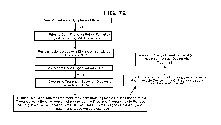
FIG. 72 is a flowchart of illustrative steps of a clinical protocol, in
accordance with some
embodiments of the disclosure.
FIG. 73 is a graph showing the level of FAM-SMAD7-AS oligonucleotide in the
cecum
tissue of DSS-induced colitis mice at 12-hours. The bars represent from left
to right, Groups 2
through 5 in the experiment described in Example 9.
FIG. 74 is a graph showing the level of FAM-SMAD7-AS oligonucleotide in the
colon
tissue of DSS-induced colitis mice at 12-hours. The bars represent from left
to right, Groups 2
through 5 in the experiment described in Example 9.
FIG. 75 is a graph showing the level of FAM-SMAD7-AS oligonucleotide in the
cecum
contents of DSS-induced colitis mice at 12-hours. The bars represent from left
to right, Groups 2
through 5 in the experiment described in Example 9.
FIG. 76 is a graph showing the mean concentration of tacrolimus in the cecum
tissue and
the proximal colon tissue 12 hours after intra-cecal or oral administration of
tacrolimus to swine
as described in Example 10.
DETAILED DESCRIPTION
The present disclosure is directed to various methods and formulations for
treating
diseases of the gastrointestinal tract with an TNF inhibitor. For example, in
an embodiment, a
method of treating a disease of the gastrointestinal tract in a subject
comprises administering to
the subject a pharmaceutical formulation comprising an TNF inhibitor wherein
the
pharmaceutical formulation is released in the subject's gastrointestinal tract
proximate to one or
more sites of disease. For example, in an embodiment, the pharmaceutical
formulation
comprises a therapeutically effective amount of an TNF inhibitor.
21
CA 03045307 2019-05-28
WO 2018/112240
PCT/US2017/066485
In some embodiments, the formulation is contained in an ingestible device, and
the device
releases the formulation at a location proximate to the site of disease. The
location of the site of
disease may be predetermined. For example, an ingestible device, the location
of which within
the GI tract can be accurately determined as disclosed herein, may be used to
sample one or more
locations in the GI tract and to detect one or more analytes, including
markers of the disease, in
the GI tract of the subject. A pharmaceutical formulation may be then
administered via an
ingestible device and released at a location proximate to the predetermined
site of disease. The
release of the formulation may be triggered autonomously, as further described
herein.
The following disclosure illustrates aspects of the formulations and methods
embodied in
the claims.
Formulations, including Pharmaceutical Formulations
As used herein, a "formulation" of an TNF inhibitor may refer to either the
TNF inhibitor
in pure form, such as, for example, a lyophilized TNF inhibitor, or a mixture
of the TNF inhibitor
with one or more physiologically acceptable carriers, excipients or
stabilizers. Thus, therapeutic
formulations or medicaments can be prepared by mixing the TNF inhibitor having
the desired
degree of purity with optional physiologically acceptable carriers, excipients
or stabilizers
(Remington's Pharmaceutical Sciences 16th edition, Osol, A. Ed. (1980)), in
the form of
lyophilized formulations or aqueous solutions. Acceptable carriers,
excipients, or stabilizers are
nontoxic to recipients at the dosages and concentrations employed, and include
buffers such as
phosphate, citrate, and other organic acids; antioxidants including ascorbic
acid and methionine;
preservatives (such as octadecyldimethylbenzyl ammonium chloride;
hexamethonium chloride;
benzalkonium chloride, benzethonium chloride; phenol, butyl or benzyl alcohol;
alkyl parabens
such as methyl or propyl paraben; catechol; resorcinol; cyclohexanol; 3-
pentanol; and m-cresol);
low molecular weight (less than about 10 residues) antibody; proteins, such as
serum albumin,
gelatin, or immunoglobulins; hydrophilic polymers such as
polyvinylpyrrolidone; amino acids
such as glycine, glutamine, asparagine, histidine, arginine, or lysine;
monosaccharides,
disaccharides, and other carbohydrates including glucose, mannose, or
dextrins; chelating agents
such as EDTA; sugars such as sucrose, mannitol, trehalose or sorbitol; salt-
forming counter-ions
such as sodium; metal complexes (e.g., Zn- protein complexes); and/or non-
ionic surfactants
such as TWEENTm, PLURONICSTM or polyethylene glycol (PEG). Exemplary
pharmaceutically
acceptable carriers herein further include insterstitial drug dispersion
agents such as soluble
neutral-active hyaluronidase glycoproteins (sHASEGP), for example, human
soluble PH-20
22
CA 03045307 2019-05-28
WO 2018/112240
PCT/US2017/066485
hyaluronidase glycoproteins, such as rHuPH20 (HYLENEX< >, Baxter
International, Inc.).
Certain exemplary sHASEGPs and methods of use, including rHuPH20, are
described in US
Patent Publication Nos. 2005/0260186 and 2006/0104968. In one aspect, a
sHASEGP is
combined with one or more additional glycosaminoglycanases such as
chondroitinases.
Exemplary lyophilized formulations are described in US Patent No. 6,267,958.
Aqueous
formulations include those described in US Patent No. 6,171,586 and
W02006/044908, the latter
formulations including a histidine-acetate buffer.
A formulation of an TNF inhibitor as disclosed herein, e.g., sustained-release
formulations, can further include a mucoadhesive agent, e.g., one or more of
polyvinyl
pyrolidine, methyl cellulose, sodium carboxyl methyl cellulose, hydroxyl
propyl cellulose,
carbopol, a polyacrylate, chitosan, a eudragit analogue, a polymer, and a
thiomer. Additional
examples of mucoadhesive agents that can be included in a formulation with an
TNF inhibitor
are described in, e.g., Peppas et al., Biomaterials 17(16):1553-1561, 1996;
Kharenko et al.,
Pharmaceutical Chemistry I 43(4):200-208, 2009; Salamat-Miller et al., Adv.
Drug Deliv.
Reviews 57(11):1666-1691, 2005; Bernkop-Schnurch, Adv. Drug Deliv. Rev.
57(11):1569-1582,
2005; and Harding et al., Biotechnol. Genet. Eng. News 16(1):41-86, 1999.
In some embodiments, components of a formulation may include any one of the
following components, or any combination thereof:
Acacia, Alginate, Alginic Acid, Aluminum Acetate, an antiseptic, Benzyl
Alcohol, Butyl
Paraben, Butylated Hydroxy Toluene, an antioxidant. Citric acid, Calcium
carbonate, Candelilla
wax, a binder, Croscarmellose sodium, Confectioner sugar, Colloidal silicone
dioxide, Cellulose,
Carnuba wax, Corn starch, Carboxymethylcellulose calcium, Calcium stearate,
Calcium
disodium EDTA, Chelation agents, Copolyvidone, Castor oil hydrogenated,
Calcium hydrogen
phosphate dehydrate, Cetylpyridine chloride, Cysteine HC1, Crosspovidone,
Dibasic Calcium
Phosphate, Disodium hydrogen phosphate, Dimethicone, Erythrosine Sodium, Ethyl
Cellulose,
Gelatin, Glyceryl monooleate, Glycerin, Glycine, Glyceryl monostearate,
Glyceryl behenate,
Hydroxy propyl cellulose, Hydroxyl propyl methyl cellulose, Hypromellose, HPMC
Pthalate,
Iron oxides or ferric oxide, Iron oxide yellow, Iron oxide red or ferric
oxide, Lactose (hydrous or
anhydrous or monohydrate or spray dried), Magnesium stearate, Microcrystalline
cellulose,
Mannitol, Methyl celluloseõ Magnesium carbonate, Mineral oil, Methacrylic acid
copolymer,
Magnesium oxide, Methyl paraben, PEG, Polysorbate 80, Propylene glycol,
Polyethylene oxide,
Propylene paraben, Polaxamer 407 or 188 or plain, Potassium bicarbonate,
Potassium sorbate,
Potato starch, Phosphoric acid, Polyoxy140 stearate, Sodium starch glycolate,
Starch
pregelatinized, Sodium crossmellose, Sodium lauryl sulfate, Starch, Silicon
dioxide, Sodium
23
CA 03045307 2019-05-28
WO 2018/112240
PCT/US2017/066485
benzoateõ Stearic acid, Sucrose base for medicated confectionery, a
granulating agent, Sorbic
acid, Sodium carbonate, Saccharin sodium, Sodium alginate, Silica gel,
Sorbiton monooleate,
Sodium stearyl fumarate, Sodium chloride, Sodium metabisulfite, Sodium citrate
dehydrate,
Sodium starch, Sodium carboxy methyl cellulose, Succinic acid, Sodium
propionate, Titanium
dioxide, Talc, Triacetin, Triethyl citrate.
Accordingly, in some embodiments of the method of treating a disease as
disclosed
herein, the method comprises administering to the subject a pharmaceutical
composition that is a
formulation as disclosed herein. In some embodiments the formulation is a
dosage form, which
may be, as an example, a solid form such as, for example, a capsule, a tablet,
a sachet, or a
lozenge; or which may be, as an example, a liquid form such as, for example, a
solution, a
suspension, an emulsion, or a syrup.
In some embodiments, the formulation is not comprised in an ingestible device.
In some
embodiments wherein the formulation is not comprised in an ingestible device,
the formulation
may be suitable for oral administration. The formulation may be, for example,
a solid dosage
form or a liquid dosage form as disclosed herein. In some embodiments wherein
the formulation
is not comprised in an ingestible device, the formulation may be suitable for
rectal
administration. The formulation may be, for example, a dosage form such as a
suppository or an
enema. In embodiments where the formulation is not comprised in an ingestible
device, the
formulation releases the TNF inhibitor at a location in the gastrointestinal
tract of the subject that
is proximate to one or more sites of disease. Such localized release may be
achieved, for
example, with a formulation comprising an enteric coating. Such localized
release may be
achieved, an another example, with a formulation comprising a core comprising
one or more
polymers suitable for controlled release of an active substance. A non-
limiting list of such
polymers includes: poly(2-(diethylamino)ethyl methacrylate, 2-
(dimethylamino)ethyl
methacrylate, poly(ethylene glycol), poly(2-aminoethyl methacrylate), (2-
hydroxypropyl)methacrylamide, poly(f3-benzy1-1-aspartate), poly(N-
isopropylacrylamide), and
cellulose derivatives.
In some embodiments, the formulation is comprised in an ingestible device as
disclosed
herein. In some embodiments wherein the formulation is comprised in an
ingestible device, the
formulation may be suitable for oral administration. The formulation may be,
for example, a
solid dosage form or a liquid dosage form as disclosed herein. In some
embodiments the
formulation is suitable for introduction and optionally for storage in the
device. In some
embodiments the formulation is suitable for introduction and optionally for
storage in a reservoir
comprised in the device. In some embodiments the formulation is suitable for
introduction and
24
CA 03045307 2019-05-28
WO 2018/112240
PCT/US2017/066485
optionally for storage in a reservoir comprised in the device. Thus, in some
embodiments,
provided herein is a reservoir comprising a therapeutically effective amount
of an TNF inhibitor,
wherein the reservoir is configured to fit into an ingestible device. In some
embodiments, the
reservoir comprising a therapeutically effective amount of an TNF inhibitor is
attachable to an
ingestible device. In some embodiments, the reservoir comprising a
therapeutically effective
amount of an TNF inhibitor is capable of anchoring itself to the subject's
tissue. As an example,
the reservoir capable of anchoring itself to the subject's tissue comprises
silicone. As an
example, the reservoir capable of anchoring itself to the subject's tissue
comprises polyvinyl
chloride.
In some embodiments the formulation is suitable for introduction in a spray
catheter, as
disclosed herein.
The formulation herein may also contain more than one active compound as
necessary for
the particular indication being treated, for example, those with complementary
activities that do
not adversely affect each other. For instance, the formulation may further
comprise another TNF
inhibitor or a chemotherapeutic agent. Such molecules are suitably present in
combination in
amounts that are effective for the purpose intended.
The active ingredients may also be entrapped in microcapsules prepared, for
example, by
coacervation techniques or by interfacial polymerization, for example,
hydroxymethylcellulose
or gelatin-microcapsule and poly-(methylmethacylate) microcapsule,
respectively, in colloidal
drug delivery systems (for example, liposomes, albumin microspheres,
microemulsions, nano-
particles and nanocapsules) or in macroemulsions. Such techniques are
disclosed in Remington's
Pharmaceutical Sciences 16th edition, Osol, A. Ed. (1980).
The formulations to be used for in vivo administration must be sterile. This
is readily
accomplished by filtration through sterile filtration membranes.
Sustained-release preparations may be prepared. Suitable examples of sustained-
release
preparations include semipermeable matrices of solid hydrophobic polymers
containing the TNF
inhibitor, which matrices are in the form of shaped articles, e.g., films, or
microcapsule.
Examples of sustained-release matrices include polyesters, hydrogels (for
example, poly(2-
hydroxyethyl-methacrylate), or poly(vinylalcohol)), polylactides (U.S. Pat.
No. 3,773,919),
copolymers of L-glutamic acid and y ethyl-L-glutamate, non-degradable ethylene-
vinyl acetate,
degradable lactic acid-glycolic acid copolymers such as the LUPRON DEPOTTm
(injectable
microspheres composed of lactic acid-glycolic acid copolymer and leuprolide
acetate), and poly-
D-(-)-3-hydroxybutyric acid. While polymers such as ethylene-vinyl acetate and
lactic acid-
glycolic acid enable release of molecules for over 100 days, certain hydrogels
release proteins for
CA 03045307 2019-05-28
WO 2018/112240
PCT/US2017/066485
shorter time periods. When encapsulated TNF inhibitors remain in the body for
a long time, they
may denature or aggregate as a result of exposure to moisture at 37 C,
resulting in a loss of
biological activity and possible changes in immunogenicity. Rational
strategies can be devised
for stabilization depending on the mechanism involved. For example, if the
aggregation
mechanism is discovered to be intermolecular S-S bond formation through thio-
disulfide
interchange, stabilization may be achieved by modifying sulfhydryl residues,
lyophilizing from
acidic solutions, controlling moisture content, using appropriate additives,
and developing
specific polymer matrix compositions.
Pharmaceutical formulations may contain one or more TNF inhibitors. The
pharmaceutical formulations may be formulated in any manner known in the art.
In some
embodiments the formulations include one or more of the following components:
a sterile diluent
(e.g., sterile water or saline), a fixed oil, polyethylene glycol, glycerin,
propylene glycol, or other
synthetic solvents, antibacterial or antifungal agents, such as benzyl alcohol
or methyl parabens,
chlorobutanol, phenol, ascorbic acid, thimerosal, and the like, antioxidants,
such as ascorbic acid
or sodium bisulfite, chelating agents, such as ethylenediaminetetraacetic
acid, buffers, such as
acetates, citrates, or phosphates, and isotonic agents, such as sugars (e.g.,
dextrose), polyalcohols
(e.g., mannitol or sorbitol), or salts (e.g., sodium chloride), or any
combination thereof.
Liposomal suspensions can also be used as pharmaceutically acceptable carriers
(see, e.g., U.S.
Patent No. 4,522,811, incorporated by reference herein in its entirety). The
formulations can be
formulated and enclosed in ampules, disposable syringes, or multiple dose
vials. Where
required, proper fluidity can be maintained by, for example, the use of a
coating, such as lecithin,
or a surfactant. Controlled release of the TNF inhibitor can be achieved by
implants and
microencapsulated delivery systems, which can include biodegradable,
biocompatible polymers
(e.g., ethylene vinyl acetate, polyanhydrides, polyglycolic acid, collagen,
polyorthoesters, and
polylactic acid; Alza Corporation and Nova Pharmaceutical, Inc.).
In some embodiments, the TNF inhibitor is present in a pharmaceutical
formulation
within the device.
In some embodiments, the TNF inhibitor is present in solution within the
device.
In some embodiments, the TNF inhibitor is present in a suspension in a liquid
medium
within the device.
In some embodiments, the TNF inhibitor is present as a pure, powder (e.g.,
lyophilized)
form of the TNF inhibitor.
26
CA 03045307 2019-05-28
WO 2018/112240
PCT/US2017/066485
Definitions:
By "ingestible", it is meant that the device can be swallowed whole.
"Gastrointestinal inflammatory disorders" are a group of chronic disorders
that cause
inflammation and/or ulceration in the mucous membrane. These disorders
include, for example,
inflammatory bowel disease (e.g., Crohn's disease, ulcerative colitis,
indeterminate colitis and
infectious colitis), mucositis (e.g., oral mucositis, gastrointestinal
mucositis, nasal mucositis and
proctitis), necrotizing enterocolitis and esophagitis.
"Inflammatory Bowel Disease" or "IBD" is a chronic inflammatory autoimmune
condition of the gastrointestinal (GI) tract. The GI tract can be divided into
four main different
sections, the oesophagus, stomach, small intestine and large intestine or
colon. The small
intestine possesses three main subcompartments: the duodenum, jejunum and
ileum. Similarly,
the large intestine consists of six sections: the cecum, ascending colon,
transverse colon,
ascending colon, sigmoid colon, and the rectum. The small intestine is about 6
m long, its
diameter is 2.5 to 3 cm and the transit time through it is typically 3 hours.
The duodenum has a
C-shape, and is 30 cm long. Due to its direct connection with the stomach, it
is physically more
stable than the jejunum and ileum, which are sections that can freely move.
The jejunum is 2.4
m in length and the ileum is 3.6 m in length and their surface areas are 180
m2 and 280 m2
respectively. The large intestine is 1.5 m long, its diameter is between 6.3
and 6.5 cm, the transit
time though this section is 20 hours and has a reduced surface area of
approximately 150 m2. The
higher surface area of the small intestine enhances its capacity for systemic
drug absorption.
The etiology of IBD is complex, and many aspects of the pathogenesis remain
unclear.
The treatment of moderate to severe IBD poses significant challenges to
treating physicians,
because conventional therapy with corticosteroids and immunomodulatory therapy
(e.g.,
azathioprine, 6 mercaptopurine, and methotrexate administered via traditional
routes such as
tablet form, oral suspension, or intravenously) is associated with side
effects and intolerance and
has not shown proven benefit in maintenance therapy (steroids). Monoclonal
antibodies targeting
tumor necrosis factor alpha (TNF-a), such as infliximab (a chimeric antibody)
and adalimumab (a
fully human antibody), are currently used in the management of CD. Infliximab
has also shown
efficacy and has been approved for use in UC. However, approximately 10%-20%
of patients
with CD are primary nonresponders to anti TNF therapy, and another ¨20%-30% of
CD patients
lose response over time (Schnitzler et al., Gut 58:492-500 (2009)). Other
adverse events (AEs)
associated with anti TNFs include elevated rates of bacterial infection,
including tuberculosis,
and, more rarely, lymphoma and demyelination (Chang et al., Nat Clin Pract
Gastroenterol
Hepatology 3:220 (2006); Hoentj en et al., World J. Gastroenterol. 15(17):2067
(2009)). No
27
CA 03045307 2019-05-28
WO 2018/112240
PCT/US2017/066485
currently available therapy achieves sustained remission in more than 20%-30%
of IBD patients
with chronic disease (Hanauer et al, Lancet 359: 1541-49 (2002); Sandborn et
al, N Engl J Med
353: 1912-25 (2005)). In addition, most patients do not achieve sustained
steroid-free remission
and mucosal healing, clinical outcomes that correlate with true disease
modification.
Although the cause of IBD remains unknown, several factors such as genetic,
infectious
and immunologic susceptibility have been implicated. IBD is much more common
in Caucasians,
especially those of Jewish descent. The chronic inflammatory nature of the
condition has
prompted an intense search for a possible infectious cause. Although agents
have been found
which stimulate acute inflammation, none has been found to cause the chronic
inflammation
associated with IBD. The hypothesis that IBD is an autoimmune disease is
supported by the
previously mentioned extraintestinal manifestation of IBD as joint arthritis,
and the known
positive response to IBD by treatment with therapeutic agents such as adrenal
glucocorticoids,
cyclosporine and azathioprine, which are known to suppress immune response. In
addition, the
GI tract, more than any other organ of the body, is continuously exposed to
potential antigenic
substances such as proteins from food, bacterial byproducts (LPS), etc.
A chronic inflammatory autoimmune condition of the gastrointestinal (GI) tract
presents
clinically as either ulcerative colitis (UC) or Crohn's disease (CD). Both IBD
conditions are
associated with an increased risk for malignancy of the GI tract.
"Crohn's disease" ("CD") is a chronic transmural inflammatory disease with the
potential
to affect any part of the entire GI tract, and UC is a mucosal inflammation of
the colon. Both
conditions are characterized clinically by frequent bowel motions,
malnutrition, and dehydration,
with disruption in the activities of daily living.
CD is frequently complicated by the development of malabsorption, strictures,
and
fistulae and may require repeated surgery. UC, less frequently, may be
complicated by severe
bloody diarrhea and toxic megacolon, also requiring surgery. The most
prominent feature
Crohn's disease is the granular, reddish-purple edematous thickening of the
bowel wall. With the
development of inflammation, these granulomas often lose their circumscribed
borders and
integrate with the surrounding tissue. Diarrhea and obstruction of the bowel
are the predominant
clinical features. As with ulcerative colitis, the course of Crohn's disease
may be continuous or
relapsing, mild or severe, but unlike ulcerative colitis, Crohn's disease is
not curable by resection
of the involved segment of bowel. Most patients with Crohn's disease require
surgery at some
point, but subsequent relapse is common and continuous medical treatment is
usual. Crohn's
disease may involve any part of the alimentary tract from the mouth to the
anus, although
typically it appears in the ileocolic, small-intestinal or colonic-anorectal
regions.
28
CA 03045307 2019-05-28
WO 2018/112240
PCT/US2017/066485
Histopathologically, the disease manifests by discontinuous granulomatomas,
crypt abscesses,
fissures and aphthous ulcers. The inflammatory infiltrate is mixed, consisting
of lymphocytes
(both T and B cells), plasma cells, macrophages, and neutrophils. There is a
disproportionate
increase in IgM- and IgG-secreting plasma cells, macrophages and neutrophils.
To date, the primary outcome measure in Crohn's Disease clinical trials is the
Crohn's
Disease Activity Index (CDAI), which has served as the basis for approval of
multiple drug
treatments, including for example, vedolizumab and natalizumab. The CDAI was
developed by
regressing clinician global assessment of disease activity on eighteen
potential items representing
patient reported outcomes (PROs) (i.e. abdominal pain, pain awakening patient
from sleep,
appetite), physical signs (i.e. average daily temperature, abdominal mass),
medication use (i.e.
loperamide or opiate use for diarrhea) and a laboratory test (i.e.
hematocrit). Backward stepwise
regression analysis identified eight independent predictors which are the
number of liquid or soft
stools, severity of abdominal pain, general well-being, occurrence of extra-
intestinal symptoms,
need for anti-diarrheal drugs, presence of an abdominal mass, hematocrit, and
body
weight. The final score is a composite of these eight items, adjusted using
regression coefficients
and standardization to construct an overall CDAI score, ranging from 0 to 600
with higher score
indicating greater disease activity. Widely used benchmarks are: CDAI <150 is
defined as
clinical remission, 150 to 219 is defined as mildly active disease, 220 to 450
is defined as
moderately active disease, and above 450 is defined as very severe disease
(Best WR, et al.,
Gastroenterology 77:843-6, 1979). Vedolizumab and natalizumab have been
approved on the basis of demonstrated clinical remission, i.e. CDAI < 150.
Although the CDAI has been in use for over 40 years, and has served as the
basis for drug
approval, it has several limitations as an outcome measure for clinical
trials. For example,
most of the overall score comes from the patient diary card items (pain,
number of liquid bowel
movements, and general well-being), which are vaguely defined and not
standardized terms
(Sandler et al., J. Clin. Epidemiol 41:451-8, 1988; Thia et al., Inflamm Bowel
Dis 17: 105-11,
2011). In addition, measurement of pain is based on a four-point scale rather
than an updated
seven-point scale. The remaining 5 index items contribute very little to
identifying an efficacy
signal and may be a source of measurement noise. Furthermore, concerns have
been raised about
poor criterion validity for the CDAI, a reported lack of correlation between
the CDAI and
endoscopic measures of inflammation (which may render the CDAI as a poor
discriminator of active CD and irritable bowel syndrome) and high reported
placebo rates
(Korzenik et al., N Engl J Med. 352:2193-201, 2005; Sandborn WJ, et al., N
Engl J Med 353 :
29
CA 03045307 2019-05-28
WO 2018/112240
PCT/US2017/066485
1912-25, 2005; Sandborn WJ, et al., Ann Intern 19; 146:829-38, 2007, Epub 2007
Apr 30; Kim
et al., Gastroenterology 146: (5 supplement 1) S-368, 2014).
It is, thus, generally recognized that additional or alternative measures of
CD symptoms
are needed, such as new PRO tools or adaptations of the CDAI to derive a new
PRO. The PRO2
and PRO3 tools are such adaptations of the CDAI and have been recently
described in Khanna et
al., Aliment Pharmacol. Ther. 41:77-86, 2015. The PRO2 evaluates the frequency
of loose/liquid
stools and abdominal pain {Id). These items are derived and weighted
accordingly
from the CDAI and are the CDAI diary card items, along with general well-
being, that contribute
most to the observed clinical benefit measured by CDAI (Sandler et al., J.
Clin. Epidemiol 41
:451-8, 1988; Thia et al., Inflamm Bowel Dis 17: 105-11, 2011; Kim et al.,
Gastroenterology
146: (5 supplement 1) S-368, 2014). The remission score of < 11 is the CDAI-
weighted
sum of the average stool frequency and pain scores in a 7-day period, which
yielded optimum
sensitivity and specificity for identification of CDAI remission (score of <
150) in a retrospective
data analysis of ustekinumab induction treatment for moderate to severe CD in
a Phase II clinical
study (Gasink C, et al., abstract, ACG Annual Meeting 2014). The PRO2 was
shown to be
sensitive and responsive when used as a continuous outcome measure in a
retrospective data
analysis of MTX treatment in active CD (Khanna R, et al., Inflamm Bowel Dis
20: 1850-61,
2014) measured by CDAI. Additional outcome measures include the Mayo Clinic
Score, the
Crohn disease endoscopic index of severity (CDEIS), and the Ulcerative colitis
endoscopic index
of severity (UCEIS). Additional outcome measures include Clinical remission,
Mucosal healing,
Histological healing (transmural), MM or ultrasound for measurement or
evaluation of bowel
wall thickness, abscesses, fistula and histology.
An additional means of assessing the extent and severity of Crohn's Disease is
endoscopy.
Endoscopic lesions typical of Crohn's disease have been described in numerous
studies and
include, e.g., aphthoid ulcerations, "punched-out ulcers," cobblestoning and
stenosis. Endoscopic
evaluation of such lesions was used to develop the first validated endoscopic
score, the Crohn's
Disease Endoscopic Index of Severity (CDEIS) (Mary et al., Gut 39:983-9,
1989). More recently,
because the CDEIS is time-consuming, complicated and impractical for routine
use, a Simplified
Endoscopic Activity Score for Crohn's Disease (SES- CD) was developed and
validated
(Daperno et al., Gastrointest. Endosc. 60(4):505-12, 2004). The SES-CD
consists of four
endoscopic variables (size of ulcers, proportion of surface covered by ulcers,
proportion of surface with any other lesions (e.g., inflammation), and
presence of narrowings
[stenosis]) that are scored in five ileocolonic segments, with each variable,
or assessment, rated
from 0 to 3.
CA 03045307 2019-05-28
WO 2018/112240
PCT/US2017/066485
To date, there is no cure for CD. Accordingly, the current treatment goals for
CD are to
induce and maintain symptom improvement, induce mucosal healing, avoid
surgery, and improve
quality of life (Lichtenstein GR, et al., Am J Gastroenterol 104:465-83, 2009;
Van Assche G, et
al., J Crohns Colitis. 4:63-101, 2010). The current therapy of MD usually
involves the
administration of antiinflammatory or immunosuppressive agents, such as
sulfasalazine,
corticosteroids, 6- mercaptopurine/azathioprine, or cyclosporine, all of which
are not typically
delivered by localized release of a drug at the site or location of disease.
More recently, biologics
like TNF-alpha inhibitors and IL-12/IL-23 blockers, are used to treat MD. If
anti-
inflammatory/immunosuppressive/biologic therapies fail, colectomies are the
last line of defense.
The typical operation for CD not involving the rectum is resection (removal of
a diseased
segment of bowel) and anastomosis (reconnection) without an ostomy. Sections
of the small or
large intestine may be removed. About 30% of CD patients will need surgery
within the first year
after diagnosis. In the subsequent years, the rate is about 5% per year.
Unfortunately, CD is
characterized by a high rate of recurrence; about 5% of patients need a second
surgery each year
after initial surgery.
Refining a diagnosis of inflammatory bowel disease involves evaluating the
progression
status of the diseases using standard classification criteria. The
classification systems used in IBD
include the Truelove and Witts Index (Truelove S. C. and Witts, L.J. Br Med J.
1955;2: 1041-
1048), which classifies colitis as mild, moderate, or severe, as well as
Lennard- Jones. (Lennard-
Jones JE. Scand J Gastroenterol Suppl 1989; 170:2-6) and the simple clinical
colitis activity
index (SCCAI). (Walmsley et. al. Gut. 1998; 43:29-32) These systems track such
variables as
daily bowel movements, rectal bleeding, temperature, heart rate, hemoglobin
levels, erythrocyte
sedimentation rate, weight, hematocrit score, and the level of serum albumin.
There is sufficient overlap in the diagnostic criteria for UC and CD that it
is sometimes
impossible to say which a given patient has; however, the type of lesion
typically seen is
different, as is the localization. UC mostly appears in the colon, proximal to
the rectum, and the
characteristic lesion is a superficial ulcer of the mucosa; CD can appear
anywhere in the bowel,
with occasional involvement of stomach, esophagus and duodenum, and the
lesions are usually
described as extensive linear fissures.
In approximately 10-15% of cases, a definitive diagnosis of ulcerative colitis
or Crohn's
disease cannot be made and such cases are often referred to as "indeterminate
colitis." Two
antibody detection tests are available that can help the diagnosis, each of
which assays for
antibodies in the blood. The antibodies are "perinuclear anti-neutrophil
antibody" (pANCA) and
"anti-Saccharomyces cervisiae antibody" (ASCA). Most patients with ulcerative
colitis have the
31
CA 03045307 2019-05-28
WO 2018/112240
PCT/US2017/066485
pANCA antibody but not the ASCA antibody, while most patients with Crohn's
disease have the
ASCA antibody but not the pANCA antibody. However, these two tests have
shortcomings as
some patients have neither antibody and some Crohn's disease patients may have
only the
pANCA antibody. A third test, which measures the presence and accumulation of
circulating
anti-microbial antibodies ¨ particularly flagellin antibodies, has proven to
be useful for detecting
susceptibility to Crohn's Disease before disease development. See Choung, R.
S., et al.
"Serologic microbial associated markers can predict Crohn's disease behaviour
years before
disease diagnosis." Alimentary pharmacology & therapeutics 43.12 (2016): 1300-
1310.
"Ulcerative colitis (UC)" afflicts the large intestine. The course of the
disease may be
continuous or relapsing, mild or severe. The earliest lesion is an
inflammatory infiltration with
abscess formation at the base of the crypts of Lieberkuhn. Coalescence of
these distended and
ruptured crypts tends to separate the overlying mucosa from its blood supply,
leading to
ulceration. Symptoms of the disease include cramping, lower abdominal pain,
rectal bleeding,
and frequent, loose discharges consisting mainly of blood, pus and mucus with
scanty fecal
particles. A total colectomy may be required for acute, severe or chronic,
unremitting ulcerative
colitis.
The clinical features of UC are highly variable, and the onset may be
insidious or abrupt,
and may include diarrhea, tenesmus and relapsing rectal bleeding. With
fulminant involvement of
the entire colon, toxic megacolon, a life-threatening emergency, may occur.
Extraintestinal
manifestations include arthritis, pyoderma gangrenoum, uveitis, and erythema
nodosum.
The terms "antibody" and "immunoglobulin" are used interchangeably in the
broadest
sense and include monoclonal antibodies (for example, full length or intact
monoclonal
antibodies), polyclonal antibodies, multivalent antibodies, multi specific
antibodies (e.g.,
bispecific, trispecific etc. antibodies so long as they exhibit the desired
biological activity) and
may also include certain antibody fragments (as described in greater detail
herein). An antibody
can be human, humanized and/or affinity matured.
"Antibody fragments" comprise only a portion of an intact antibody, where in
certain
embodiments, the portion retains at least one, and typically most or all, of
the functions normally
associated with that portion when present in an intact antibody. In one
embodiment, an antibody
fragment comprises an antigen binding site of the intact antibody and thus
retains the ability to
bind antigen. In another embodiment, an antibody fragment, for example one
that comprises the
Fc region, retains at least one of the biological functions normally
associated with the Fc region
when present in an intact antibody, such as FcRn binding, antibody half-life
modulation, ADCC
function and complement binding. In one embodiment, an antibody fragment is a
monovalent
32
CA 03045307 2019-05-28
WO 2018/112240
PCT/US2017/066485
antibody that has an in vivo half-life substantially similar to an intact
antibody. For example,
such an antibody fragment may comprise on antigen binding arm linked to an Fc
sequence
capable of conferring in vivo stability to the fragment.
The term "monoclonal antibody" as used herein refers to an antibody obtained
from a
.. population of substantially homogeneous antibodies, i.e., the individual
antibodies comprising
the population are identical except for possible naturally occurring mutations
that may be present
in minor amounts. Monoclonal antibodies are highly specific, being directed
against a single
antigen. Furthermore, in contrast to polyclonal antibody preparations that
typically include
different antibodies directed against different determinants (epitopes), each
monoclonal antibody
is directed against a single determinant on the antigen.
The monoclonal antibodies herein specifically include "chimeric" antibodies in
which a
portion of the heavy and/or light chain is identical with or homologous to
corresponding
sequences in antibodies derived from a particular species or belonging to a
particular antibody
class or subclass, while the remainder of the chain(s) is identical with or
homologous to
corresponding sequences in antibodies derived from another species or
belonging to another
antibody class or subclass, as well as fragments of such antibodies, so long
as they exhibit the
desired biological activity (U.S. Patent No. 4,816,567; and Morrison et al,
Proc. Natl. Acad. Sci.
USA 81:6851-6855 (1984)).
"Treatment regimen" refers to a combination of dosage, frequency of
administration, or
duration of treatment, with or without addition of a second medication.
"Effective treatment regimen" refers to a treatment regimen that will offer
beneficial
response to a patient receiving the treatment.
"Effective amount" refers to an amount of drug that offers beneficial response
to a patient
receiving the treatment. For example, an effective amount may be a human
Equivalent Dose
(HED).
"Dispensable", with reference to any substance, refers to any substance that
may be
released from an ingestible device as disclosed herein, or from a component of
the device such as
a reservoir. For example, a dispensable substance may be a TNF inhibitor,
and/or a formulation
comprising a TNF inhibitor.
"Patient response" or "patient responsiveness" can be assessed using any
endpoint
indicating a benefit to the patient, including, without limitation, (1)
inhibition, to some extent, of
disease progression, including slowing down and complete arrest; (2) reduction
in the number of
disease episodes and/or symptoms; (3) reduction in lesional size; (4)
inhibition (i.e., reduction,
slowing down or complete stopping) of disease cell infiltration into adjacent
peripheral organs
33
CA 03045307 2019-05-28
WO 2018/112240
PCT/US2017/066485
and/or tissues; (5) inhibition (i.e., reduction, slowing down or complete
stopping) of disease
spread; (6) decrease of auto-immune response, which may, but does not have to,
result in the
regression or ablation of the disease lesion; (7) relief, to some extent, of
one or more symptoms
associated with the disorder; (8) increase in the length of disease-free
presentation following
treatment; and/or (9) decreased mortality at a given point of time following
treatment. The term
"responsiveness" refers to a measurable response, including complete response
(CR) and partial
response (PR).
As used herein, "complete response" or "CR" means the disappearance of all
signs of
inflammation or remission in response to treatment. This does not necessarily
mean the disease
has been cured.
"Partial response" or "PR" refers to a decrease of at least 50% in the
severity of
inflammation, in response to treatment.
A "beneficial response" of a patient to treatment with a therapeutic agent and
similar
wording refers to the clinical or therapeutic benefit imparted to a patient at
risk for or suffering
from a gastrointestinal inflammatory disorder from or as a result of the
treatment with the agent.
Such benefit includes cellular or biological responses, a complete response, a
partial response, a
stable disease (without progression or relapse), or a response with a later
relapse of the patient
from or as a result of the treatment with the agent.
As used herein, "non-response" or "lack of response" or similar wording means
an
absence of a complete response, a partial response, or a beneficial response
to treatment with a
therapeutic agent.
"A patient maintains responsiveness to a treatment" when the patient' s
responsiveness
does not decrease with time during the course of a treatment.
A "symptom" of a disease or disorder (e.g., inflammatory bowel disease, e.g.,
ulcerative
colitis or Crohn's disease) is any morbid phenomenon or departure from the
normal in structure,
function, or sensation, experienced by a subject and indicative of disease.
TNFa Inhibitors
The term "TNFa inhibitor" refers to an agent which directly or indirectly
inhibits,
impairs, reduces, down-regulates, or blocks TNFa activity and/or expression.
In some
embodiments, a TNFa inhibitor is an inhibitory nucleic acid, an antibody or an
antigen-binding
fragment thereof, a fusion protein, a soluble TNFa receptor (a soluble TNFR1
or a soluble
TNFR2), or a small molecule TNFa antagonist. In some embodiments, the
inhibitory nucleic
34
CA 03045307 2019-05-28
WO 2018/112240
PCT/US2017/066485
acid is a ribozyme, small hairpin RNA, a small interfering RNA, an antisense
nucleic acid, or an
aptamer.
Exemplary TNFa inhibitors that directly inhibit, impair, reduce, down-
regulate, or block
TNFa activity and/or expression can, e.g., inhibit or reduce binding of TNFa
to its receptor
(TNFR1 and/or TNFR2) and/or inhibit or decrease the expression level of TNFa
or a receptor of
TNFa (TNFR1 or TNFR2) in a cell (e.g., a mammalian cell). Non-limiting
examples of TNFa
inhibitors that directly inhibit, impair, reduce, down-regulate, or block TNFa
activity and/or
expression include inhibitory nucleic acids (e.g., any of the examples of
inhibitory nucleic acids
described herein), an antibody or fragment thereof, a fusion protein, a
soluble TNFa receptor
(e.g., a soluble TNFR1 or soluble TNFR2), and a small molecule TNFa
antagonist.
Exemplary TNFa inhibitors that can indirectly inhibit, impair, reduce, down-
regulate, or
block TNFa activity and/or expression can, e.g., inhibit or decrease the level
of downstream
signaling of a TNFa receptor (e.g., TNFR1 or TNFR2) in a mammalian cell (e.g.,
decrease the
level and/or activity of one or more of the following signaling proteins:
TRADD, TRAF2,
MEKK1/4, MEKK4/7, INK, AP-1, ASK1, RIP, MEKK 3/6, MAPK, NIK, IKK, and NF-KB in
a
mammalian cell), and/or decrease the level of TNFa-induced gene expression in
a mammalian
cell (e.g., decrease the transcription of genes regulated by, e.g., one or
more transcription factors
selected from the group of NF--03, c-Jun, and ATF2). A description of
downstream signaling of
a TNFa receptor is provided in Waj ant et al., Cell Death Differentiation
10:45-65, 2003
.. (incorporated herein by reference). For example, such indirect TNFa
inhibitors can be an
inhibitory nucleic acid that targets (decreases the expression) a signaling
component downstream
of a TNFa receptor (e.g., any one or more of the signaling components
downstream of a TNFa
receptor described herein or known in the art), a TNFa-induced gene (e.g., any
TNFa-induced
gene known in the art), or a transcription factor selected from the group of
NF--03, c-Jun, and
ATF2.
In other examples, such indirect TNFa inhibitors can be a small molecule
inhibitor of a
signaling component downstream of a TNFa receptor (e.g., any of the signaling
components
downstream of a TNFa receptor described herein or known in the art), a small
molecule inhibitor
of a protein encoded by a TNFa-induced gene (e.g., any protein encoded by a
TNFa-induced
gene known in the art), and a small molecule inhibitor of a transcription
factor selected from the
group of NF--03, c-Jun, and ATF2.
In other embodiments, TNFa inhibitors that can indirectly inhibit, impair,
reduce, down-
regulate, or block one or more components in a mammalian cell (e.g., a
macrophage, a CD4+
lymphocyte, a NK cell, a neutrophil, a mast cell, a eosinophil, or a neuron)
that are involved in
CA 03045307 2019-05-28
WO 2018/112240
PCT/US2017/066485
the signaling pathway that results in TNFa mRNA transcription, TNFa mRNA
stabilization, and
TNFa mRNA translation (e.g., one or more components selected from the group of
CD14,
MyD88, IRAK, lipopolysaccharide binding protein (LBP), TRAF6, ras, raf,
MEK1/2, ERK1/2,
NIK, IKK, IKB, NF-KB, rac, MEK4/7, JNK, c-jun, MEK3/6, p38, PKR, TTP, and
MK2). For
example, such indirect TNFa inhibitors can be an inhibitory nucleic acid that
targets (decreases
the expression) of a component in a mammalian cell that is involved in the
signaling pathway
that results in TNFa mRNA transcription, TNFa mRNA stabilization, and TNFa
mRNA
translation (e.g., a component selected from the group of CD14, MyD88, IRAK,
lipopolysaccharide binding protein (LBP), TRAF6, ras, raf, MEK1/2, ERK1/2,
NIK, IKK, IKB,
NF-KB, rac, MEK4/7, JNK, c-jun, MEK3/6, p38, PKR, TTP, and MK2). In other
examples, an
indirect TNFa inhibitors is a small molecule inhibitor of a component in a
mammalian cell that is
involved in the signaling pathway that results in TNFa mRNA transcription,
TNFa mRNA
stabilization, and TNFa mRNA translation (e.g., a component selected from the
group of CD14,
MyD88, IRAK, lipopolysaccharide binding protein (LBP), TRAF6, ras, raf,
MEK1/2, ERK1/2,
NIK, IKK, IKB, NF-KB, rac, MEK4/7, JNK, c-jun, MEK3/6, p38, PKR, TTP, and
MK2).
Inhibitory Nucleic Acids
Inhibitory nucleic acids that can decrease the expression of TNFa, TNFR1,
TNFR2,
TRADD, TRAF2, MEKK1/4, MEKK4/7, JNK, AP-1, ASK1, RIP, MEKK 3/6, MAPK, NIK,
IKK, NF-KB, CD14, MyD88, IRAK, lipopolysaccharide binding protein (LBP),
TRAF6, ras, raf,
MEK1/2, ERK1/2, NIK, IKK, IKB, NF-KB, rac, MEK4/7, JNK, c-jun, MEK3/6, p38,
PKR, TTP,
or MK2 mRNA expression in a mammalian cell include antisense nucleic acid
molecules, i.e.,
nucleic acid molecules whose nucleotide sequence is complementary to all or
part of a TNFa,
TNFR1, TNFR2, TRADD, TRAF2, MEKK1/4, MEKK4/7, JNK, AP-1, ASK1, RIP, MEKK 3/6,
MAPK, NIK, IKK, NF-KB, CD14, MyD88, IRAK, lipopolysaccharide binding protein
(LBP),
TRAF6, ras, raf, MEK1/2, ERK1/2, NIK, IKK, IKB, NF-KB, rac, MEK4/7, INK, c-
jun, MEK3/6,
p38, PKR, TTP, or MK2 mRNA (e.g., complementary to all or a part of any one of
SEQ ID NOs:
1-37).
Human TNFa CDS (SEQ ID NO: 1)
ATGAGCACTGAAAGCATGATCCGGGACGTGGAGCTGGCCGAGGAGGCGCTCCCCAA
GAAGACAGGGGGGCCCCAGGGCTCCAGGCGGTGCTTGTTCCTCAGCCTCTTCTCCTT
CCTGATCGTGGCAGGCGCCACCACGCTCTTCTGCCTGCTGCACTTTGGAGTGATCGG
CCCCCAGAGGGAAGAGTTCCCCAGGGACCTCTCTCTAATCAGCCCTCTGGCCCAGGC
AGTCAGATCATCTTCTCGAACCCCGAGTGACAAGCCTGTAGCCCATGTTGTAGCAAA
36
CA 03045307 2019-05-28
WO 2018/112240
PCT/US2017/066485
CCCTCAAGCTGAGGGGCAGCTCCAGTGGCTGAACCGCCGGGCCAATGCCCTCCTGGC
CAATGGCGTGGAGCTGAGAGATAACCAGCTGGTGGTGCCATCAGAGGGCCTGTACC
TCATCTACTCCCAGGTCCTCTTCAAGGGCCAAGGCTGCCCCTCCACCCATGTGCTCCT
CACCCACACCATCAGCCGCATCGCCGTCTCCTACCAGACCAAGGTCAACCTCCTCTC
TGCCATCAAGAGCCCCTGCCAGAGGGAGACCCCAGAGGGGGCTGAGGCCAAGCCCT
GGTATGAGCCCATCTATCTGGGAGGGGTCTTCCAGCTGGAGAAGGGTGACCGACTC
AGCGCTGAGATCAATCGGCCCGACTATCTCGACTTTGCCGAGTCTGGGCAGGTCTAC
TTTGGGATCATTGCCCTGTGA
Human TNFR1 CDS (SEQ ID NO: 2)
ATGGGCCTCTCCACCGTGCCTGACCTGCTGCTGCCGCTGGTGCTCCTGGAGCTGTTG
GTGGGAATATACCCCTCAGGGGTTATTGGACTGGTCCCTCACCTAGGGGACAGGGA
GAAGAGAGATAGTGTGTGTCCCCAAGGAAAATATATCCACCCTCAAAATAATTCGA
TTTGCTGTACCAAGTGCCACAAAGGAACCTACTTGTACAATGACTGTCCAGGCCCGG
GGCAGGATACGGACTGCAGGGAGTGTGAGAGCGGCTCCTTCACCGCTTCAGAAAAC
CACCTCAGACACTGCCTCAGCTGCTCCAAATGCCGAAAGGAAATGGGTCAGGTGGA
GATCTCTTCTTGCACAGTGGACCGGGACACCGTGTGTGGCTGCAGGAAGAACCAGTA
CCGGCATTATTGGAGTGAAAACCTTTTCCAGTGCTTCAATTGCAGCCTCTGCCTCAAT
GGGACCGTGCACCTCTCCTGCCAGGAGAAACAGAACACCGTGTGCACCTGCCATGC
AGGTTTCTTTCTAAGAGAAAACGAGTGTGTCTCCTGTAGTAACTGTAAGAAAAGCCT
GGAGTGCACGAAGTTGTGCCTACCCCAGATTGAGAATGTTAAGGGCACTGAGGACT
CAGGCACCACAGTGCTGTTGCCCCTGGTCATTTTCTTTGGTCTTTGCCTTTTATCCCTC
CTCTTCATTGGTTTAATGTATCGCTACCAACGGTGGAAGTCCAAGCTCTACTCCATTG
TTTGTGGGAAATCGACACCTGAAAAAGAGGGGGAGCTTGAAGGAACTACTACTAAG
CCCCTGGCCCCAAACCCAAGCTTCAGTCCCACTCCAGGCTTCACCCCCACCCTGGGC
TTCAGTCCCGTGCCCAGTTCCACCTTCACCTCCAGCTCCACCTATACCCCCGGTGACT
GTCCCAACTTTGCGGCTCCCCGCAGAGAGGTGGCACCACCCTATCAGGGGGCTGACC
CCATCCTTGCGACAGCCCTCGCCTCCGACCCCATCCCCAACCCCCTTCAGAAGTGGG
AGGACAGCGCCCACAAGCCACAGAGCCTAGACACTGATGACCCCGCGACGCTGTAC
GCCGTGGTGGAGAACGTGCCCCCGTTGCGCTGGAAGGAATTCGTGCGGCGCCTAGG
GCTGAGCGACCACGAGATCGATCGGCTGGAGCTGCAGAACGGGCGCTGCCTGCGCG
AGGCGCAATACAGCATGCTGGCGACCTGGAGGCGGCGCACGCCGCGGCGCGAGGCC
ACGCTGGAGCTGCTGGGACGCGTGCTCCGCGACATGGACCTGCTGGGCTGCCTGGA
GGACATCGAGGAGGCGCTTTGCGGCCCCGCCGCCCTCCCGCCCGCGCCCAGTCTTCT
CAGATGA
Human TNFR2 CDS (SEQ ID NO: 3)
ATGGCGCCCGTCGCCGTCTGGGCCGCGCTGGCCGTCGGACTGGAGCTCTGGGCTGCG
GCGCACGCCTTGCCCGCCCAGGTGGCATTTACACCCTACGCCCCGGAGCCCGGGAGC
ACATGCCGGCTCAGAGAATACTATGACCAGACAGCTCAGATGTGCTGCAGCAAATG
CTCGCCGGGCCAACATGCAAAAGTCTTCTGTACCAAGACCTCGGACACCGTGTGTGA
CTCCTGTGAGGACAGCACATACACCCAGCTCTGGAACTGGGTTCCCGAGTGCTTGAG
CTGTGGCTCCCGCTGTAGCTCTGACCAGGTGGAAACTCAAGCCTGCACTCGGGAACA
GAACCGCATCTGCACCTGCAGGCCCGGCTGGTACTGCGCGCTGAGCAAGCAGGAGG
GGTGCCGGCTGTGCGCGCCGCTGCGCAAGTGCCGCCCGGGCTTCGGCGTGGCCAGA
CCAGGAACTGAAACATCAGACGTGGTGTGCAAGCCCTGTGCCCCGGGGACGTTCTCC
AACACGACTTCATCCACGGATATTTGCAGGCCCCACCAGATCTGTAACGTGGTGGCC
ATCCCTGGGAATGCAAGCATGGATGCAGTCTGCACGTCCACGTCCCCCACCCGGAGT
37
CA 03045307 2019-05-28
WO 2018/112240
PCT/US2017/066485
ATGGCCCCAGGGGCAGTACACTTACCCCAGCCAGTGTCCACACGATCCCAACACAC
GCAGCCAACTCCAGAACCCAGCACTGCTCCAAGCACCTCCTTCCTGCTCCCAATGGG
CCCCAGCCCCCCAGCTGAAGGGAGCACTGGCGACTTCGCTCTTCCAGTTGGACTGAT
TGTGGGTGTGACAGCCTTGGGTCTACTAATAATAGGAGTGGTGAACTGTGTCATCAT
GACCCAGGTGAAAAAGAAGCCCTTGTGCCTGCAGAGAGAAGCCAAGGTGCCTCACT
TGCCTGCCGATAAGGCCCGGGGTACACAGGGCCCCGAGCAGCAGCACCTGCTGATC
ACAGCGCCGAGC TC CAGCAGCAGC TC CC TGGAGAGCTCGGCCAGTGC GTTGGACAG
AAGGGCGCCCACTCGGAACCAGCCACAGGCACCAGGCGTGGAGGCCAGTGGGGCCG
GGGAGGCCCGGGCCAGCACCGGGAGCTCAGATTCTTCCCCTGGTGGCCATGGGACC
CAGGTCAATGTCACCTGCATCGTGAACGTCTGTAGCAGCTCTGACCACAGCTCACAG
TGCTCCTCCCAAGCCAGCTCCACAATGGGAGACACAGATTCCAGCCCCTCGGAGTCC
CCGAAGGACGAGCAGGTC CC CTTCTCCAAGGAGGAATGTGCCTTTCGGTCACAGCTG
GAGACGCCAGAGACCCTGCTGGGGAGCACCGAAGAGAAGCCCCTGCCCCTTGGAGT
GCCTGATGCTGGGATGAAGCCCAGTTAA
Human TRADD CDS (SEQ ID NO: 4)
ATGGCAGCTGGGCAAAATGGGCACGAAGAGTGGGTGGGCAGCGCATACCTGTTTGT
GGAGTCCTCGCTGGACAAGGTGGTCCTGTCGGATGCCTACGCGCACCCCCAGCAGA
AGGTGGCAGTGTACAGGGCTCTGCAGGCTGCCTTGGCAGAGAGCGGCGGGAGCCCG
GACGTGCTGCAGATGCTGAAGATCCACCGCAGCGACCCGCAGCTGATCGTGCAGCT
GCGATTCTGCGGGCGGCAGCCCTGTGGCCGCTTCCTCCGCGCCTACCGCGAGGGGGC
GCTGCGCGCCGCGCTGCAGAGGAGCCTGGCGGCCGCGCTCGCCCAGCACTCGGTGC
CGCTGCAACTGGAGCTGCGCGCCGGCGCCGAGCGGCTGGACGCTTTGCTGGCGGAC
GAGGAGCGCTGTTTGAGTTGCATCCTAGCCCAGCAGCCCGACCGGCTCCGGGATGA
AGAACTGGCTGAGCTGGAGGATGCGCTGCGAAATCTGAAGTGCGGCTCGGGGGCCC
GGGGTGGCGACGGGGAGGTCGCTTCGGCCCCCTTGCAGCCCCCGGTGCCCTCTCTGT
CGGAGGTGAAGCCGCCGCCGCCGCCGCCACCTGCCCAGACTTTTCTGTTCCAGGGTC
AGCCTGTAGTGAATCGGCCGCTGAGCCTGAAGGACCAACAGACGTTCGCGCGCTCT
GTGGGTCTCAAATGGCGCAAGGTGGGGCGCTCACTGCAGCGAGGCTGCCGGGCGCT
GCGGGACCCGGCGCTGGACTCGCTGGCCTACGAGTACGAGCGCGAGGGACTGTACG
AGCAGGCCTTCCAGCTGCTGCGGCGCTTCGTGCAGGCCGAGGGCCGCCGCGCCACG
CTGCAGCGCCTGGTGGAGGCACTCGAGGAGAACGAGCTCACCAGCCTGGCAGAGGA
CTTGCTGGGCCTGACCGATCCCAATGGCGGCCTGGCCTAG
Human TRAF2 CDS (SEQ ID NO: 5)
ATGGCTGCAGCTAGCGTGACCCCCCCTGGCTCCCTGGAGTTGCTACAGCCCGGCTTC
TCCAAGACCCTCCTGGGGACCAAGCTGGAAGCCAAGTACCTGTGCTCCGCCTGCAGA
AACGTCCTCCGCAGGCCCTTCCAGGCGCAGTGTGGCCACCGGTACTGCTCCTTCTGC
CTGGCCAGCATCCTCAGCTCTGGGCCTCAGAACTGTGCTGCCTGTGTTCACGAGGGC
ATATATGAAGAAGGCATTTCTATTTTAGAAAGCAGTTCGGCCTTCCCAGATAATGCT
GCCCGCAGGGAGGTGGAGAGCCTGCCGGCCGTCTGTCCCAGTGATGGATGCACCTG
GAAGGGGACC CTGAAAGAATAC GAGAGC TGC CAC GAAGGCC GCTGCC CGCTCATGC
TGACCGAATGTCCCGCGTGCAAAGGCCTGGTCCGCCTTGGTGAAAAGGAGCGCCAC
CTGGAGCACGAGTGCCCGGAGAGAAGCCTGAGCTGCCGGCATTGCCGGGCACCCTG
CTGCGGAGCAGACGTGAAGGCGCACCACGAGGTCTGCCCCAAGTTCCCCTTAACTTG
TGACGGCTGCGGCAAGAAGAAGATCCCCCGGGAGAAGTTTCAGGACCACGTCAAGA
CTTGTGGCAAGTGTCGAGTCCCTTGCAGATTCCACGCCATCGGCTGCCTCGAGACGG
TAGAGGGTGAGAAACAGCAGGAGCACGAGGTGCAGTGGCTGCGGGAGCACCTGGC
38
CA 03045307 2019-05-28
WO 2018/112240
PCT/US2017/066485
CATGCTACTGAGCTCGGTGCTGGAGGCAAAGCCCCTCTTGGGAGACCAGAGCCACG
CGGGGTCAGAGCTCCTGCAGAGGTGCGAGAGCCTGGAGAAGAAGACGGCCACTTTT
GAGAACATTGTCTGCGTCCTGAACCGGGAGGTGGAGAGGGTGGCCATGACTGCCGA
GGCCTGCAGCCGGCAGCACCGGCTGGACCAAGACAAGATTGAAGCCCTGAGTAGCA
AGGTGCAGCAGCTGGAGAGGAGCATTGGCCTCAAGGACCTGGCGATGGCTGACTTG
GAGCAGAAGGTCTTGGAGATGGAGGCATCCACCTACGATGGGGTCTTCATCTGGAA
GATCTCAGACTTCGCCAGGAAGCGCCAGGAAGCTGTGGCTGGCCGCATACCCGCCA
TCTTCTCCCCAGCCTTCTACACCAGCAGGTACGGCTACAAGATGTGTCTGCGTATCTA
CCTGAACGGCGACGGCACCGGGCGAGGAACACACCTGTCCCTCTTCTTTGTGGTGAT
GAAGGGCCCGAATGACGCCCTGCTGCGGTGGCCCTTCAACCAGAAGGTGACCTTAA
TGCTGCTCGACCAGAATAACCGGGAGCACGTGATTGACGCCTTCAGGCCCGACGTG
ACTTCATCCTCTTTTCAGAGGCCAGTCAACGACATGAACATCGCAAGCGGCTGCCCC
CTCTTCTGCCCCGTCTCCAAGATGGAGGCAAAGAATTCCTACGTGCGGGACGATGCC
ATCTTCATCAAGGCCATTGTGGACCTGACAGGGCTCTAA
Human MEKK1 CDS (SEQ ID NO: 6)
ATGGCGGCGGCGGCGGGGAATCGCGCCTCGTCGTCGGGATTCCCGGGCGCCAGGGC
TACGAGCCCTGAGGCAGGCGGCGGCGGAGGAGCCCTCAAGGCGAGCAGCGCGCCCG
CGGCTGCCGCGGGACTGCTGCGGGAGGCGGGCAGCGGGGGCCGCGAGCGGGCGGA
CTGGCGGCGGCGGCAGCTGCGCAAAGTGCGGAGTGTGGAGCTGGACCAGCTGCCTG
AGCAGCCGCTCTTCCTTGCCGCCTCACCGCCGGCCTCCTCGACTTCCCCGTCGCCGGA
GCCCGCGGACGCAGCGGGGAGTGGGACCGGCTTCCAGCCTGTGGCGGTGCCGCCGC
CCCACGGAGCCGCGAGCCGCGGCGGCGCCCACCTTACCGAGTCGGTGGCGGCGCCG
GACAGCGGCGCCTCGAGTCCCGCAGCGGCCGAGCCCGGGGAGAAGCGGGCGCCCGC
CGCCGAGCCGTCTCCTGCAGCGGCCCCCGCCGGTCGTGAGATGGAGAATAAAGAAA
CTCTCAAAGGGTTGCACAAGATGGATGATCGTCCAGAGGAACGAATGATCAGGGAG
AAACTGAAGGCAACCTGTATGCCAGCCTGGAAGCACGAATGGTTGGAAAGGAGAAA
TAGGCGAGGGCCTGTGGTGGTAAAACCAATCCCAGTTAAAGGAGATGGATCTGAAA
TGAATCACTTAGCAGCTGAGTCTCCAGGAGAGGTCCAGGCAAGTGCGGCTTCACCA
GCTTCCAAAGGCCGACGCAGTCCTTCTCCTGGCAACTCCCCATCAGGTCGCACAGTG
AAATCAGAATCTCCAGGAGTAAGGAGAAAAAGAGTTTCCCCAGTGCCTTTTCAGAG
TGGCAGAATCACACCACCCCGAAGAGCCCCTTCACCAGATGGCTTCTCACCATATAG
CCCTGAGGAAACAAACCGCCGTGTTAACAAAGTGATGCGGGCCAGACTGTACTTAC
TGCAGCAGATAGGGCCTAACTCTTTCCTGATTGGAGGAGACAGCCCAGACAATAAA
TACCGGGTGTTTATTGGGCCTCAGAACTGCAGCTGTGCACGTGGAACATTCTGTATT
CATCTGCTATTTGTGATGCTCCGGGTGTTTCAACTAGAACCTTCAGACCCAATGTTAT
GGAGAAAAACTTTAAAGAATTTTGAGGTTGAGAGTTTGTTCCAGAAATATCACAGTA
GGCGTAGCTCAAGGATCAAAGCTCCATCTCGTAACACCATCCAGAAGTTTGTTTCAC
GCATGTCAAATTCTCATACATTGTCATCATCTAGTACTTCTACGTCTAGTTCAGAAAA
CAGCATAAAGGATGAAGAGGAACAGATGTGTCCTATTTGCTTGTTGGGCATGCTTGA
TGAAGAAAGTCTTACAGTGTGTGAAGACGGCTGCAGGAACAAGCTGCACCACCACT
GCATGTCAATTTGGGCAGAAGAGTGTAGAAGAAATAGAGAACCTTTAATATGTCCC
CTTTGTAGATCTAAGTGGAGATCTCATGATTTCTACAGCCACGAGTTGTCAAGTCCT
GTGGATTCCCCTTCTTCCCTCAGAGCTGCACAGCAGCAAACCGTACAGCAGCAGCCT
TTGGCTGGATCACGAAGGAATCAAGAGAGCAATTTTAACCTTACTCATTATGGAACT
CAGCAAATCCCTCCTGCTTACAAAGATTTAGCTGAGCCATGGATTCAGGTGTTTGGA
ATGGAACTCGTTGGCTGCTTATTTTCTAGAAACTGGAATGTGAGAGAGATGGCCCTC
AGGCGTCTTTCCCATGATGTCAGTGGGGCCCTGCTGTTGGCAAATGGGGAGAGCACT
GGAAATTCTGGGGGCAGCAGTGGAAGCAGCCCGAGTGGGGGAGCCACCAGTGGGTC
39
CA 03045307 2019-05-28
WO 2018/112240
PCT/US2017/066485
TTCCCAGACCAGTATCTCAGGAGATGTGGTGGAGGCATGCTGCAGCGTTCTGTCAAT
GGTCTGTGCTGACCCTGTCTACAAAGTGTACGTTGCTGCTTTAAAAACATTGAGAGC
CATGCTGGTATATACTCCTTGCCACAGTTTAGCGGAAAGAATCAAACTTCAGAGACT
TCTCCAGCCAGTTGTAGACACCATCCTAGTCAAATGTGCAGATGCCAATAGCCGCAC
AAGTCAGCTGTCCATATCAACACTGTTGGAACTGTGCAAAGGCCAAGCAGGAGAGT
TGGCAGTTGGCAGAGAAATACTAAAAGCTGGATCCATTGGTATTGGTGGTGTTGATT
ATGTCTTAAATTGTATTCTTGGAAACCAAACTGAATCAAACAATTGGCAAGAACTTC
TTGGCCGCCTTTGTCTTATAGATAGACTGTTGTTGGAATTTCCTGCTGAATTTTATCCT
CATATTGTCAGTACTGATGTTTCACAAGCTGAGCCTGTTGAAATCAGGTATAAGAAG
CTGCTGTCCCTCTTAACCTTTGCTTTGCAGTCCATTGATAATTCCCACTCAATGGTTG
GCAAACTTTCCAGAAGGATCTACTTGAGTTCTGCAAGAATGGTTACTACAGTACCCC
ATGTGTTTTCAAAACTGTTAGAAATGCTGAGTGTTTCCAGTTCCACTCACTTCACCAG
GATGCGTCGCCGTTTGATGGCTATTGCAGATGAGGTGGAAATTGCCGAAGCCATCCA
GTTGGGCGTAGAAGACACTTTGGATGGTCAACAGGACAGCTTCTTGCAGGCATCTGT
TCCCAACAACTATCTGGAAACCACAGAGAACAGTTCCCCTGAGTGCACAGTCCATTT
AGAGAAAACTGGAAAAGGATTATGTGCTACAAAATTGAGTGCCAGTTCAGAGGACA
TTTCTGAGAGACTGGCCAGCATTTCAGTAGGACCTTCTAGTTCAACAACAACAACAA
CAACAACAACAGAGCAACCAAAGCCAATGGTTCAAACAAAAGGCAGACCCCACAGT
CAGTGTTTGAACTCCTCTCCTTTATCTCATCATTCCCAATTAATGTTTCCAGCCTTGTC
AACCCCTTCTTCTTCTACCCCATCTGTACCAGCTGGCACTGCAACAGATGTCTCTAAG
CATAGACTTCAGGGATTCATTCCCTGCAGAATACCTTCTGCATCTCCTCAAACACAG
CGCAAGTTTTCTCTACAATTCCACAGAAACTGTCCTGAAAACAAAGACTCAGATAAA
CTTTCCCCAGTCTTTACTCAGTCAAGACCCTTGCCCTCCAGTAACATACACAGGCCA
AAGCCATCTAGACCTACCCCAGGTAATACAAGTAAACAGGGAGATCCCTCAAAAAA
TAGCATGACACTTGATCTGAACAGTAGTTCCAAATGTGATGACAGCTTTGGCTGTAG
CAGCAATAGTAGTAATGCTGTTATACCCAGTGACGAGACAGTGTTCACCCCAGTAGA
GGAGAAATGCAGATTAGATGTCAATACAGAGCTCAACTCCAGTATTGAGGACCTTCT
TGAAGCATCTATGCCTTCAAGTGATACAACAGTAACTTTTAAGTCAGAAGTTGCTGT
CCTGTCTCCTGAAAAGGCTGAAAATGATGATACCTACAAAGATGATGTGAATCATAA
TCAAAAGTGCAAAGAGAAGATGGAAGCTGAAGAAGAAGAAGCTTTAGCAATTGCCA
TGGCAATGTCAGCGTCTCAGGATGCCCTCCCCATAGTTCCTCAGCTGCAGGTTGAAA
ATGGAGAAGATATCATCATTATTCAACAGGATACACCAGAGACTCTACCAGGACAT
ACCAAAGCAAAACAACCGTATAGAGAAGACACTGAATGGCTGAAAGGTCAACAGAT
AGGCCTTGGAGCATTTTCTTCTTGTTATCAGGCTCAAGATGTGGGAACTGGAACTTTA
ATGGCTGTTAAACAGGTGACTTATGTCAGAAACACATCTTCTGAGCAAGAAGAAGT
AGTAGAAGCACTAAGAGAAGAGATAAGAATGATGAGCCATCTGAATCATCCAAACA
TCATTAGGATGTTGGGAGCCACGTGTGAGAAGAGCAATTACAATCTCTTCATTGAAT
GGATGGCAGGGGGATCGGTGGCTCATTTGCTGAGTAAATATGGAGCCTTCAAAGAA
TCAGTAGTTATTAACTACACTGAACAGTTACTCCGTGGCCTTTCGTATCTCCATGAAA
ACCAAATCATTCACAGAGATGTCAAAGGTGCCAATTTGCTAATTGACAGCACTGGTC
AGAGACTAAGAATTGCAGATTTTGGAGCTGCAGCCAGGTTGGCATCAAAAGGAACT
GGTGCAGGAGAGTTTCAGGGACAATTACTGGGGACAATTGCATTTATGGCACCTGA
GGTACTAAGAGGTCAACAGTATGGAAGGAGCTGTGATGTATGGAGTGTTGGCTGTG
CTATTATAGAAATGGCTTGTGCAAAACCACCATGGAATGCAGAAAAACACTCCAAT
CATCTTGCTTTGATATTTAAGATTGCTAGTGCAACTACTGCTCCATCGATCCCTTCAC
ATTTGTCTCCTGGTTTACGAGATGTGGCTCTTCGTTGTTTAGAACTTCAACCTCAGGA
CAGACCTCCATCAAGAGAGCTACTGAAGCATCCAGTCTTTCGTACTACATGGTAG
CA 03045307 2019-05-28
WO 2018/112240
PCT/US2017/066485
Human MEKK4 CDS (SEQ ID NO: 7)
ATGAGAGAAGCCGCTGCCGCGCTGGTCCCTCCTCCCGCCTTTGCCGTCACGCCTGCC
GCCGCCATGGAGGAGCCGCCGCCACCGCCGCCGCCGCCACCACCGCCACCGGAACC
CGAGACCGAGTCAGAACCCGAGTGCTGCTTGGCGGCGAGGCAAGAGGGCACATTGG
GAGATTCAGCTTGCAAGAGTCCTGAATCTGATCTAGAAGACTTCTCCGATGAAACAA
ATACAGAGAATCTTTATGGTACCTCTCCCCCCAGCACACCTCGACAGATGAAACGCA
TGTCAACCAAACATCAGAGGAATAATGTGGGGAGGCCAGCCAGTCGGTCTAATTTG
AAAGAAAAAATGAATGCACCAAATCAGCCTCCACATAAAGACACTGGAAAAACAGT
GGAGAATGTGGAAGAATACAGCTATAAGCAGGAGAAAAAGATCCGAGCAGCTCTTA
GAACAACAGAGCGTGATCATAAAAAAAATGTACAGTGCTCATTCATGTTAGACTCA
GTGGGTGGATCTTTGCCAAAAAAATCAATTCCAGATGTGGATCTCAATAAGCCTTAC
CTCAGCCTTGGCTGTAGCAATGCTAAGCTTCCAGTATCTGTGCCCATGCCTATAGCC
AGACCTGCACGCCAGACTTCTAGGACTGACTGTCCAGCAGATCGTTTAAAGTTTTTT
GAAACTTTACGACTTTTGCTAAAGCTTACCTCAGTCTCAAAGAAAAAAGACAGGGA
GCAAAGAGGACAAGAAAATACGTCTGGTTTCTGGCTTAACCGATCTAACGAACTGA
TCTGGTTAGAGCTACAAGCCTGGCATGCAGGACGGACAATTAACGACCAGGACTTCT
TTTTATATACAGCCCGTCAAGCCATCCCAGATATTATTAATGAAATCCTTACTTTCAA
AGTCGACTATGGGAGCTTCGCCTTTGTTAGAGATAGAGCTGGTTTTAATGGTACTTC
AGTAGAAGGGCAGTGCAAAGCCACTCCTGGAACAAAGATTGTAGGTTACTCAACAC
ATCATGAGCATCTCCAACGCCAGAGGGTCTCATTTGAGCAGGTAAAACGGATAATG
GAGCTGCTAGAGTACATAGAAGCACTTTATCCATCATTGCAGGCTCTTCAGAAGGAC
TATGAAAAATATGCTGCAAAAGACTTCCAGGACAGGGTGCAGGCACTCTGTTTGTGG
TTAAACATCACAAAAGACTTAAATCAGAAATTAAGGATTATGGGCACTGTTTTGGGC
ATCAAGAATTTATCAGACATTGGCTGGCCAGTGTTTGAAATCCCTTCCCCTCGACCA
TCCAAAGGTAATGAGCCGGAGTATGAGGGTGATGACACAGAAGGAGAATTAAAGG
AGTTGGAAAGTAGTACGGATGAGAGTGAAGAAGAACAAATCTCTGATCCTAGGGTA
CCGGAAATCAGACAGCCCATAGATAACAGCTTCGACATCCAGTCGCGGGACTGCAT
ATCCAAGAAGCTTGAGAGGCTCGAATCTGAGGATGATTCTCTTGGCTGGGGAGCACC
AGACTGGAGCACAGAAGCAGGCTTTAGTAGACATTGTCTGACTTCTATTTATAGACC
ATTTGTAGACAAAGCACTGAAGCAGATGGGGTTAAGAAAGTTAATTTTAAGACTTCA
CAAGCTAATGGATGGTTCCTTGCAAAGGGCACGTATAGCATTGGTAAAGAACGATC
GTCCAGTGGAGTTTTCTGAATTTCCAGATCCCATGTGGGGTTCAGATTATGTGCAGTT
GTCAAGGACACCACCTTCATCTGAGGAGAAATGCAGTGCTGTGTCGTGGGAGGAGC
TGAAGGCCATGGATTTACCTTCATTCGAACCTGCCTTCCTAGTTCTCTGCCGAGTCCT
TCTGAATGTCATACATGAGTGTCTGAAGTTAAGATTGGAGCAGAGACCTGCTGGAGA
ACCATCTCTCTTGAGTATTAAGCAGCTGGTGAGAGAGTGTAAGGAGGTCCTGAAGG
GCGGCCTGCTGATGAAGCAGTACTACCAGTTCATGCTGCAGGAGGTTCTGGAGGACT
TGGAGAAGCCCGACTGCAACATTGACGCTTTTGAAGAGGATCTACATAAAATGCTTA
TGGTGTATTTTGATTACATGAGAAGCTGGATCCAAATGCTACAGCAATTACCTCAAG
CATCGCATAGTTTAAAAAATCTGTTAGAAGAAGAATGGAATTTCACCAAAGAAATA
ACTCATTACATACGGGGAGGAGAAGCACAGGCCGGGAAGCTTTTCTGTGACATTGC
AGGAATGCTGCTGAAATCTACAGGAAGTTTTTTAGAATTTGGCTTACAGGAGAGCTG
TGCTGAATTTTGGACTAGTGCGGATGACAGCAGTGCTTCCGACGAAATCAGGAGGTC
TGTTATAGAGATCAGTCGAGCCCTGAAGGAGCTCTTCCATGAAGCCAGAGAAAGGG
CTTCCAAAGCACTTGGATTTGCTAAAATGTTGAGAAAGGACCTGGAAATAGCAGCA
GAATTCAGGCTTTCAGCCCCAGTTAGAGACCTCCTGGATGTTCTGAAATCAAAACAG
TATGTCAAGGTGCAAATTCCTGGGTTAGAAAACTTGCAAATGTTTGTTCCAGACACT
CTTGCTGAGGAGAAGAGTATTATTTTGCAGTTACTCAATGCAGCTGCAGGAAAGGAC
TGTTCAAAAGATTCAGATGACGTACTCATCGATGCCTATCTGCTTCTGACCAAGCAC
GGTGATCGAGCCCGTGATTCAGAGGACAGCTGGGGCACCTGGGAGGCACAGCCTGT
41
CA 03045307 2019-05-28
WO 2018/112240
PCT/US2017/066485
CAAAGTCGTGCCTCAGGTGGAGACTGTTGACACCCTGAGAAGCATGCAGGTGGATA
ATCTTTTACTAGTTGTCATGCAGTCTGCGCATCTCACAATTCAGAGAAAAGCTTTCCA
GCAGTCCATTGAGGGACTTATGACTCTGTGCCAGGAGCAGACATCCAGTCAGCCGGT
CATCGCCAAAGCTTTGCAGCAGCTGAAGAATGATGCATTGGAGCTATGCAACAGGA
TAAGCAATGCCATTGACCGCGTGGACCACATGTTCACATCAGAATTTGATGCTGAGG
TTGATGAATCTGAATCTGTCACCTTGCAACAGTACTACCGAGAAGCAATGATTCAGG
GGTACAATTTTGGATTTGAGTATCATAAAGAAGTTGTTCGTTTGATGTCTGGGGAGTT
TAGACAGAAGATAGGAGACAAATATATAAGCTTTGCCCGGAAGTGGATGAATTATG
TCCTGACTAAATGTGAGAGTGGTAGAGGTACAAGACCCAGGTGGGCGACTCAAGGA
TTTGATTTTCTACAAGCAATTGAACCTGCCTTTATTTCAGCTTTACCAGAAGATGACT
TCTTGAGTTTACAAGCCTTGATGAATGAATGCATTGGCCATGTCATAGGAAAACCAC
ACAGTCCTGTTACAGGTTTGTACCTTGCCATTCATCGGAACAGCCCCCGTCCTATGA
AGGTACCTCGATGCCATAGTGACCCTCCTAACCCACACCTCATTATCCCCACTCCAG
AGGGATTCAGCACTCGGAGCATGCCTTCCGACGCGCGGAGCCATGGCAGCCCTGCT
GCTGCTGCTGCTGCTGCTGCTGCTGCTGTTGCTGCCAGTCGGCCCAGCCCCTCTGGTG
GTGACTCTGTGCTGCCCAAATCCATCAGCAGTGCCCATGATACCAGGGGTTCCAGCG
TTCCTGAAAATGATCGATTGGCTTCCATAGCTGCTGAATTGCAGTTTAGGTCCCTGA
GTCGTCACTCAAGCCCCACGGAGGAGCGAGATGAACCAGCATATCCAAGAGGAGAT
TCAAGTGGGTCCACAAGAAGAAGTTGGGAACTTCGGACACTAATCAGCCAGAGTAA
AGATACTGCTTCTAAACTAGGACCCATAGAAGCTATCCAGAAGTCAGTCCGATTGTT
TGAAGAAAAGAGGTACCGAGAAATGAGGAGAAAGAATATCATTGGTCAAGTTTGTG
ATACGCCTAAGTCCTATGATAATGTTATGCACGTTGGCTTGAGGAAGGTGACCTTCA
AATGGCAAAGAGGAAACAAAATTGGAGAAGGCCAGTATGGGAAGGTGTACACCTG
CATCAGCGTCGACACCGGGGAGCTGATGGCCATGAAAGAGATTCGATTTCAACCTA
ATGACCATAAGACTATCAAGGAAACTGCAGACGAATTGAAAATATTCGAAGGCATC
AAACACCCCAATCTGGTTCGGTATTTTGGTGTGGAGCTCCATAGAGAAGAAATGTAC
ATCTTCATGGAGTACTGCGATGAGGGGACTTTAGAAGAGGTGTCAAGGCTGGGACTT
CAGGAACATGTGATTAGGCTGTATTCAAAGCAGATCACCATTGCGATCAACGTCCTC
CATGAGCATGGCATAGTCCACCGTGACATTAAAGGTGCCAATATCTTCCTTACCTCA
TCTGGATTAATCAAACTGGGAGATTTTGGATGTTCAGTAAAGCTCAAAAACAATGCC
CAGACCATGCCTGGTGAAGTGAACAGCACCCTGGGGACAGCAGCATACATGGCACC
TGAAGTCATCACTCGTGCCAAAGGAGAGGGCCATGGGCGTGCGGCCGACATCTGGA
GTCTGGGGTGTGTTGTCATAGAGATGGTGACTGGCAAGAGGCCTTGGCATGAGTATG
AGCACAACTTTCAAATTATGTATAAAGTGGGGATGGGACATAAGCCACCAATCCCTG
AAAGATTAAGCCCTGAAGGAAAGGACTTCCTTTCTCACTGCCTTGAGAGTGACCCAA
AGATGAGATGGACCGCCAGCCAGCTCCTCGACCATTCGTTTGTCAAGGTTTGCACAG
ATGAAGAATGA
Human MEKK7 CDS (SEQ ID NO: 8)
ATGTCTACAGCCTCTGCCGCCTCCTCCTCCTCCTCGTCTTCGGCCGGTGAGATGATCG
AAGCCCCTTCCCAGGTCCTCAACTTTGAAGAGATCGACTACAAGGAGATCGAGGTG
GAAGAGGTTGTTGGAAGAGGAGCCTTTGGAGTTGTTTGCAAAGCTAAGTGGAGAGC
AAAAGATGTTGCTATTAAACAAATAGAAAGTGAATCTGAGAGGAAAGCGTTTATTG
TAGAGCTTCGGCAGTTATCCCGTGTGAACCATCCTAATATTGTAAAGCTTTATGGAG
CCTGCTTGAATCCAGTGTGTCTTGTGATGGAATATGCTGAAGGGGGCTCTTTATATA
ATGTGCTGCATGGTGCTGAACCATTGCCATATTATACTGCTGCCCACGCAATGAGTT
GGTGTTTACAGTGTTCCCAAGGAGTGGCTTATCTTCACAGCATGCAACCCAAAGCGC
TAATTCACAGGGACCTGAAACCACCAAACTTACTGCTGGTTGCAGGGGGGACAGTTC
TAAAAATTTGTGATTTTGGTACAGCCTGTGACATTCAGACACACATGACCAATAACA
42
CA 03045307 2019-05-28
WO 2018/112240
PCT/US2017/066485
AGGGGAGTGCTGCTTGGATGGCACCTGAAGTTTTTGAAGGTAGTAATTACAGTGAAA
AATGTGACGTCTTCAGCTGGGGTATTATTCTTTGGGAAGTGATAACGCGTCGGAAAC
CCTTTGATGAGATTGGTGGCCCAGCTTTCCGAATCATGTGGGCTGTTCATAATGGTA
CTCGACCACCACTGATAAAAAATTTACCTAAGCCCATTGAGAGCCTGATGACTCGTT
GTTGGTCTAAAGATCCTTCCCAGCGCCCTTCAATGGAGGAAATTGTGAAAATAATGA
CTCACTTGATGCGGTACTTTCCAGGAGCAGATGAGCCATTACAGTATCCTTGTCAGT
ATTCAGATGAAGGACAGAGCAACTCTGCCACCAGTACAGGCTCATTCATGGACATTG
CTTCTACAAATACGAGTAACAAAAGTGACACTAATATGGAGCAAGTTCCTGCCACA
AATGATACTATTAAGCGCTTAGAATCAAAATTGTTGAAAAATCAGGCAAAGCAACA
GAGTGAATCTGGACGTTTAAGCTTGGGAGCCTCCCGTGGGAGCAGTGTGGAGAGCTT
GCCCCCAACCTCTGAGGGCAAGAGGATGAGTGCTGACATGTCTGAAATAGAAGCTA
GGATCGCCGCAACCACAGGCAACGGACAGCCAAGACGTAGATCCATCCAAGACTTG
ACTGTAACTGGAACAGAACCTGGTCAGGTGAGCAGTAGGTCATCCAGTCCCAGTGTC
AGAATGATTACTACCTCAGGACCAACCTCAGAAAAGCCAACTCGAAGTCATCCATG
GACCCCTGATGATTCCACAGATACCAATGGATCAGATAACTCCATCCCAATGGCTTA
TCTTACACTGGATCACCAACTACAGCCTCTAGCACCGTGCCCAAACTCCAAAGAATC
TATGGCAGTGTTTGAACAGCATTGTAAAATGGCACAAGAATATATGAAAGTTCAAAC
AGAAATTGCATTGTTATTACAGAGAAAGCAAGAACTAGTTGCAGAACTGGACCAGG
ATGAAAAGGACCAGCAAAATACATCTCGCCTGGTACAGGAACATAAAAAGCTTTTA
GATGAAAACAAAAGCCTTTCTACTTACTACCAGCAATGCAAAAAACAACTAGAGGT
CATCAGAAGTCAGCAGCAGAAACGACAAGGCACTTCATGA
Human JNK CDS (SEQ ID NO: 9)
ATGAGCAGAAGCAAGCGTGACAACAATTTTTATAGTGTAGAGATTGGAGATTCTACA
TTCACAGTCCTGAAACGATATCAGAATTTAAAACCTATAGGCTCAGGAGCTCAAGGA
ATAGTATGCGCAGCTTATGATGCCATTCTTGAAAGAAATGTTGCAATCAAGAAGCTA
AGCCGACCATTTCAGAATCAGACTCATGCCAAGCGGGCCTACAGAGAGCTAGTTCTT
ATGAAATGTGTTAATCACAAAAATATAATTGGCCTTTTGAATGTTTTCACACCACAG
AAATCCCTAGAAGAATTTCAAGATGTTTACATAGTCATGGAGCTCATGGATGCAAAT
CTTTGCCAAGTGATTCAGATGGAGCTAGATCATGAAAGAATGTCCTACCTTCTCTAT
CAGATGCTGTGTGGAATCAAGCACCTTCATTCTGCTGGAATTATTCATCGGGACTTA
AAGCCCAGTAATATAGTAGTAAAATCTGATTGCACTTTGAAGATTCTTGACTTCGGT
CTGGCCAGGACTGCAGGAACGAGTTTTATGATGACGCCTTATGTAGTGACTCGCTAC
TACAGAGCACCCGAGGTCATCCTTGGCATGGGCTACAAGGAAAACGTTGACATTTG
GTCAGTTGGGTGCATCATGGGAGAAATGATCAAAGGTGGTGTTTTGTTCCCAGGTAC
AGATCATATTGATCAGTGGAATAAAGTTATTGAACAGCTTGGAACACCATGTCCTGA
ATTCATGAAGAAACTGCAACCAACAGTAAGGACTTACGTTGAAAACAGACCTAAAT
ATGCTGGATATAGCTTTGAGAAACTCTTCCCTGATGTCCTTTTCCCAGCTGACTCAGA
ACACAACAAACTTAAAGCCAGTCAGGCAAGGGATTTGTTATCCAAAATGCTGGTAA
TAGATGCATCTAAAAGGATCTCTGTAGATGAAGCTCTCCAACACCCGTACATCAATG
TCTGGTATGATCCTTCTGAAGCAGAAGCTCCACCACCAAAGATCCCTGACAAGCAGT
TAGATGAAAGGGAACACACAATAGAAGAGTGGAAAGAATTGATATATAAGGAAGTT
ATGGACTTGGAGGAGAGAACCAAGAATGGAGTTATACGGGGGCAGCCCTCTCCTTT
AGGTGCAGCAGTGATCAATGGCTCTCAGCATCCATCATCATCGTCGTCTGTCAATGA
TGTGTCTTCAATGTCAACAGATCCGACTTTGGCCTCTGATACAGACAGCAGTCTAGA
AGCAGCAGCTGGGCCTCTGGGCTGCTGTAGATGA
43
CA 03045307 2019-05-28
WO 2018/112240
PCT/US2017/066485
Human AP-1 CDS (SEQ ID NO: 10)
ATGACTGCAAAGATGGAAACGACCTTCTATGACGATGCCCTCAACGCCTCGTTCCTC
CCGTCCGAGAGCGGACCTTATGGCTACAGTAACCCCAAGATCCTGAAACAGAGCAT
GACCCTGAACCTGGCCGACCCAGTGGGGAGCCTGAAGCCGCACCTCCGCGCCAAGA
ACTCGGACCTCCTCACCTCGCCCGACGTGGGGCTGCTCAAGCTGGCGTCGCCCGAGC
TGGAGCGCCTGATAATCCAGTCCAGCAACGGGCACATCACCACCACGCCGACCCCC
ACCCAGTTCCTGTGCCCCAAGAACGTGACAGATGAGCAGGAGGGCTTCGCCGAGGG
CTTCGTGCGCGCCCTGGCCGAACTGCACAGCCAGAACACGCTGCCCAGCGTCACGTC
GGCGGCGCAGCCGGTCAACGGGGCAGGCATGGTGGCTCCCGCGGTAGCCTCGGTGG
CAGGGGGCAGCGGCAGCGGCGGCTTCAGCGCCAGCCTGCACAGCGAGCCGCCGGTC
TACGCAAACCTCAGCAACTTCAACCCAGGCGCGCTGAGCAGCGGCGGCGGGGCGCC
CTCCTACGGCGCGGCCGGCCTGGCCTTTCCCGCGCAACCCCAGCAGCAGCAGCAGCC
GCCGCACCACCTGCCCCAGCAGATGCCCGTGCAGCACCCGCGGCTGCAGGCCCTGA
AGGAGGAGCCTCAGACAGTGCCCGAGATGCCCGGCGAGACACCGCCCCTGTCCCCC
ATCGACATGGAGTCCCAGGAGCGGATCAAGGCGGAGAGGAAGCGCATGAGGAACC
GCATCGCTGCCTCCAAGTGCCGAAAAAGGAAGCTGGAGAGAATCGCCCGGCTGGAG
GAAAAAGTGAAAACCTTGAAAGCTCAGAACTCGGAGCTGGCGTCCACGGCCAACAT
GCTCAGGGAACAGGTGGCACAGCTTAAACAGAAAGTCATGAACCACGTTAACAGTG
GGTGCCAACTCATGCTAACGCAGCAGTTGCAAACATTTTGA
Human ASK! CDS (SEQ ID NO: 11)
ATGAGCACGGAGGCGGACGAGGGCATCACTTTCTCTGTGCCACCCTTCGCCCCCTCG
GGCTTCTGCACCATCCCCGAGGGCGGCATCTGCAGGAGGGGAGGAGCGGCGGCGGT
GGGCGAGGGCGAGGAGCACCAGCTGCCACCGCCGCCGCCGGGCAGCTTCTGGAACG
TGGAGAGCGCCGCTGCCCCTGGCATCGGTTGTCCGGCGGCCACCTCCTCGAGCAGTG
CCACCCGAGGCCGGGGCAGCTCTGTTGGCGGGGGCAGCCGACGGACCACGGTGGCA
TATGTGATCAACGAAGCGAGCCAAGGGCAACTGGTGGTGGCCGAGAGCGAGGCCCT
GCAGAGCTTGCGGGAGGCGTGCGAGACAGTGGGCGCCACCCTGGAAACCCTGCATT
TTGGGAAACTCGACTTTGGAGAAACCACCGTGCTGGACCGCTTTTACAATGCAGATA
TTGCGGTGGTGGAGATGAGCGATGCCTTCCGGCAGCCGTCCTTGTTTTACCACCTTG
GGGTGAGAGAAAGTTTCAGCATGGCCAACAACATCATCCTCTACTGTGATACTAACT
CGGACTCTCTGCAGTCACTGAAGGAAATAATTTGCCAGAAGAATACTATGTGCACTG
GGAACTACACCTTTGTTCCTTACATGATAACTCCACATAACAAAGTCTACTGCTGTG
ACAGCAGCTTCATGAAGGGGTTGACAGAGCTCATGCAACCGAACTTCGAGCTGCTTC
TTGGACCCATCTGCTTACCTCTTGTGGATCGTTTTATTCAACTTTTGAAGGTGGCACA
AGCAAGTTCTAGCCAGTACTTCCGGGAATCTATACTCAATGACATCAGGAAAGCTCG
TAATTTATACACTGGTAAAGAATTGGCAGCTGAGTTGGCAAGAATTCGGCAGCGAGT
AGATAATATCGAAGTCTTGACAGCAGATATTGTCATAAATCTGTTACTTTCCTACAG
AGATATCCAGGACTATGATTCTATTGTGAAGCTGGTAGAGACTTTAGAAAAACTGCC
AACCTTTGATTTGGCCTCCCATCACCATGTGAAGTTTCATTATGCATTTGCACTGAAT
AGGAGAAATCTCCCTGGTGACAGAGCAAAAGCTCTTGATATTATGATTCCCATGGTG
CAAAGCGAAGGACAAGTTGCTTCAGATATGTATTGCCTAGTTGGTCGAATCTACAAA
GATATGTTTTTGGACTCTAATTTCACGGACACTGAAAGCAGAGACCATGGAGCTTCT
TGGTTCAAAAAGGCATTTGAATCTGAGCCAACACTACAGTCAGGAATTAATTATGCG
GTCCTCCTCCTGGCAGCTGGACACCAGTTTGAATCTTCCTTTGAGCTCCGGAAAGTTG
GGGTGAAGCTAAGTAGTCTTCTTGGTAAAAAGGGAAACTTGGAAAAACTCCAGAGC
TACTGGGAAGTTGGATTTTTTCTGGGGGCCAGCGTCCTAGCCAATGACCACATGAGA
GTCATTCAAGCATCTGAAAAGCTTTTTAAACTGAAGACACCAGCATGGTACCTCAAG
TCTATTGTAGAGACAATTTTAATATATAAGCATTTTGTGAAACTGACCACAGAACAG
44
CA 03045307 2019-05-28
WO 2018/112240
PCT/US2017/066485
CCTGTGGCCAAGCAAGAACTTGTGGACTTTTGGATGGATTTCCTGGTCGAGGCCACA
AAGACAGATGTTACTGTGGTTAGGTTTCCAGTATTAATATTAGAACCAACCAAAATC
TATCAACCTTCTTATTTGTCTATCAACAATGAAGTTGAGGAAAAGACAATCTCTATTT
GGCACGTGCTTCCTGATGACAAGAAAGGTATACATGAGTGGAATTTTAGTGCCTCTT
CTGTCAGGGGAGTGAGTATTTCTAAATTTGAAGAAAGATGCTGCTTTCTTTATGTGCT
TCACAATTCTGATGATTTCCAAATCTATTTCTGTACAGAACTTCATTGTAAAAAGTTT
TTTGAGATGGTGAACACCATTACCGAAGAGAAGGGGAGAAGCACAGAGGAAGGAG
ACTGTGAAAGTGACTTGCTGGAGTATGACTATGAATATGATGAAAATGGTGACAGA
GTCGTTTTAGGAAAAGGCACTTATGGGATAGTCTACGCAGGTCGGGACTTGAGCAAC
CAAGTCAGAATTGCTATTAAGGAAATCCCAGAGAGAGACAGCAGATACTCTCAGCC
CCTGCATGAAGAAATAGCATTGCATAAACACCTGAAGCACAAAAATATTGTCCAGT
ATCTGGGCTCTTTCAGTGAGAATGGTTTCATTAAAATCTTCATGGAGCAGGTCCCTG
GAGGAAGTCTTTCTGCTCTCCTTCGTTCCAAATGGGGTCCATTAAAAGACAATGAGC
AAACAATTGGCTTTTATACAAAGCAAATACTGGAAGGATTAAAATATCTCCATGACA
ATCAGATAGTTCACCGGGACATAAAGGGTGACAATGTGTTGATTAATACCTACAGTG
GTGTTCTCAAGATCTCTGACTTCGGAACATCAAAGAGGCTTGCTGGCATAAACCCCT
GTACTGAAACTTTTACTGGTACCCTCCAGTATATGGCACCAGAAATAATAGATAAAG
GACCAAGAGGCTACGGAAAAGCAGCAGACATCTGGTCTCTGGGCTGTACAATCATT
GAAATGGCCACAGGAAAACCCCCATTTTATGAACTGGGAGAACCACAAGCAGCTAT
GTTCAAGGTGGGAATGTTTAAAGTCCACCCTGAGATCCCAGAGTCCATGTCTGCAGA
GGCCAAGGCATTCATACTGAAATGTTTTGAACCAGATCCTGACAAGAGAGCCTGTGC
TAACGACTTGCTTGTTGATGAGTTTTTAAAAGTTTCAAGCAAAAAGAAAAAGACACA
ACCTAAGCTTTCAGCTCTTTCAGCTGGATCAAATGAATATCTCAGGAGTATATCCTTG
CCGGTACCTGTGCTGGTGGAGGACACCAGCAGCAGCAGTGAGTACGGCTCAGTTTC
ACCCGACACGGAGTTGAAAGTGGACCCCTTCTCTTTCAAAACAAGAGCCAAGTCCTG
CGGAGAAAGAGATGTCAAGGGAATTCGGACACTCTTTTTGGGCATTCCAGATGAGA
ATTTTGAAGATCACAGTGCTCCTCCTTCCCCTGAAGAAAAAGATTCTGGATTCTTCAT
GCTGAGGAAGGACAGTGAGAGGCGAGCTACCCTTCACAGGATCCTGACGGAAGACC
AAGACAAAATTGTGAGAAACCTAATGGAATCTTTAGCTCAGGGGGCTGAAGAACCG
AAACTAAAATGGGAACACATCACAACCCTCATTGCAAGCCTCAGAGAATTTGTGAG
ATCCACTGACCGAAAAATCATAGCCACCACACTGTCAAAGCTGAAACTGGAGCTGG
ACTTCGACAGCCATGGCATTAGCCAAGTCCAGGTGGTACTCTTTGGTTTTCAAGATG
CTGTCAATAAAGTTCTTCGGAATCATAACATCAAGCCGCACTGGATGTTTGCCTTAG
ACAGTATCATTCGGAAGGCGGTACAGACAGCCATTACCATCCTGGTTCCAGAACTAA
GGCCACATTTCAGCCTTGCATCTGAGAGTGATACTGCTGATCAAGAAGACTTGGATG
TAGAAGATGACCATGAGGAACAGCCTTCAAATCAAACTGTCCGAAGACCTCAGGCT
GTCATTGAAGATGCTGTGGCTACCTCAGGCGTGAGCACGCTCAGTTCTACTGTGTCT
CATGATTCCCAGAGTGCTCACCGGTCACTGAATGTACAGCTTGGAAGGATGAAAATA
GAAACCAATAGATTACTGGAAGAATTGGTTCGGAAAGAGAAAGAATTACAAGCACT
CCTTCATCGAGCTATTGAAGAAAAAGACCAAGAAATTAAACACCTGAAGCTTAAGT
CCCAACCCATAGAAATTCCTGAATTGCCTGTATTTCATCTAAATTCTTCTGGCACAAA
TACTGAAGATTCTGAACTTACCGACTGGCTGAGAGTGAATGGAGCTGATGAAGACA
CTATAAGCCGGTTTTTGGCTGAAGATTATACACTATTGGATGTTCTCTACTATGTTAC
ACGTGATGACTTAAAATGCTTGAGACTAAGGGGAGGGATGCTGTGCACACTGTGGA
AGGCTATCATTGACTTTCGAAACAAACAGACTTGA
Human RIP CDS (SEQ ID NO: 12)
ATGTGGAGCAAACTGAATAATGAAGAGCACAATGAGCTGAGGGAAGTGGACGGCA
CCGCTAAGAAGAATGGCGGCACCCTCTACTACATGGCGCCCGAGCACCTGAATGAC
CA 03045307 2019-05-28
WO 2018/112240
PCT/US2017/066485
GTCAACGCAAAGCCCACAGAGAAGTCGGATGTGTACAGCTTTGCTGTAGTACTCTGG
GCGATATTTGCAAATAAGGAGCCATATGAAAATGCTATCTGTGAGCAGCAGTTGATA
ATGTGCATAAAATCTGGGAACAGGCCAGATGTGGATGACATCACTGAGTACTGCCC
AAGAGAAATTATCAGTCTCATGAAGCTCTGCTGGGAAGCGAATCCGGAAGCTCGGC
CGACATTTCCTGGCATTGAAGAAAAATTTAGGCCTTTTTATTTAAGTCAATTAGAAG
AAAGTGTAGAAGAGGACGTGAAGAGTTTAAAGAAAGAGTATTCAAACGAAAATGCA
GTTGTGAAGAGAATGCAGTCTCTTCAACTTGATTGTGTGGCAGTACCTTCAAGCCGG
TCAAATTCAGCCACAGAACAGCCTGGTTCACTGCACAGTTCCCAGGGACTTGGGATG
GGTCCTGTGGAGGAGTCCTGGTTTGCTCCTTCCCTGGAGCACCCACAAGAAGAGAAT
GAGCCCAGCCTGCAGAGTAAACTCCAAGACGAAGCCAACTACCATCTTTATGGCAG
CCGCATGGACAGGCAGACGAAACAGCAGCCCAGACAGAATGTGGCTTACAACAGAG
AGGAGGAAAGGAGACGCAGGGTCTCCCATGACCCTTTTGCACAGCAAAGACCTTAC
GAGAATTTTCAGAATACAGAGGGAAAAGGCACTGCTTATTCCAGTGCAGCCAGTCA
TGGTAATGCAGTGCACCAGCCCTCAGGGCTCACCAGCCAACCTCAAGTACTGTATCA
GAACAATGGATTATATAGCTCACATGGCTTTGGAACAAGACCACTGGATCCAGGAA
CAGCAGGTCCCAGAGTTTGGTACAGGCCAATTCCAAGTCATATGCCTAGTCTGCATA
ATATCCCAGTGCCTGAGACCAACTATCTAGGAAATACACCCACCATGCCATTCAGCT
CCTTGCCACCAACAGATGAATCTATAAAATATACCATATACAATAGTACTGGCATTC
AGATTGGAGCCTACAATTATATGGAGATTGGTGGGACGAGTTCATCACTACTAGACA
GCACAAATACGAACTTCAAAGAAGAGCCAGCTGCTAAGTACCAAGCTATCTTTGAT
AATACCACTAGTCTGACGGATAAACACCTGGACCCAATCAGGGAAAATCTGGGAAA
GCACTGGAAAAACTGTGCCCGTAAACTGGGCTTCACACAGTCTCAGATTGATGAAAT
TGACCATGACTATGAGCGAGATGGACTGAAAGAAAAGGTTTACCAGATGCTCCAAA
AGTGGGTGATGAGGGAAGGCATAAAGGGAGCCACGGTGGGGAAGCTGGCCCAGGC
GCTCCACCAGTGTTCCAGGATCGACCTTCTGAGCAGCTTGATTTACGTCAGCCAGAA
CTAA
Human MEKK 3 CDS (SEQ ID NO: 13)
ATGGACGAACAGGAGGCATTGAACTCAATCATGAACGATCTGGTGGCCCTCCAGAT
GAACCGACGTCACCGGATGCCTGGATATGAGACCATGAAGAACAAAGACACAGGTC
ACTCAAATAGGCAGAAAAAACACAACAGCAGCAGCTCAGCCCTTCTGAACAGCCCC
ACAGTAACAACAAGCTCATGTGCAGGGGCCAGTGAGAAAAAGAAATTTTTGAGTGA
CGTCAGAATCAAGTTCGAGCACAACGGGGAGAGGCGAATTATAGCGTTCAGCCGGC
CTGTGAAATATGAAGATGTGGAGCACAAGGTGACAACAGTATTTGGACAACCTCTT
GATCTACATTACATGAACAATGAGCTCTCCATCCTGCTGAAAAACCAAGATGATCTT
GATAAAGCAATTGACATTTTAGATAGAAGCTCAAGCATGAAAAGCCTTAGGATATTG
CTGTTGTCCCAGGACAGAAACCATAACAGTTCCTCTCCCCACTCTGGGGTGTCCAGA
CAGGTGCGGATCAAGGCTTCCCAGTCCGCAGGGGATATAAATACTATCTACCAGCCC
CCCGAGCCCAGAAGCAGGCACCTCTCTGTCAGCTCCCAGAACCCTGGCCGAAGCTCA
CCTCCCCCTGGCTATGTTCCTGAGCGGCAGCAGCACATTGCCCGGCAGGGGTCCTAC
ACCAGCATCAACAGTGAGGGGGAGTTCATCCCAGAGACCAGCGAGCAGTGCATGCT
GGATCCCCTGAGCAGTGCAGAAAATTCCTTGTCTGGAAGCTGCCAATCCTTGGACAG
GTCAGCAGACAGCCCATCCTTCCGGAAATCACGAATGTCCCGTGCCCAGAGCTTCCC
TGACAACAGACAGGAATACTCAGATCGGGAAACTCAGCTTTATGACAAAGGGGTCA
AAGGTGGAACCTACCCCCGGCGCTACCACGTGTCTGTGCACCACAAGGACTACAGT
GATGGCAGAAGAACATTTCCCCGAATACGGCGTCATCAAGGCAACTTGTTCACCCTG
GTGCCCTCCAGCCGCTCCCTGAGCACAAATGGCGAGAACATGGGTCTGGCTGTGCAA
TACCTGGACCCCCGTGGGCGCCTGCGGAGTGCGGACAGCGAGAATGCCCTCTCTGTG
CAGGAGAGGAATGTGCCAACCAAGTCTCCCAGTGCCCCCATCAACTGGCGCCGGGG
46
CA 03045307 2019-05-28
WO 2018/112240
PCT/US2017/066485
AAAGCTCCTGGGCCAGGGTGCCTTCGGCAGGGTCTATTTGTGCTATGACGTGGACAC
GGGACGTGAACTTGCTTCCAAGCAGGTCCAATTTGATCCAGACAGTCCTGAGACAAG
CAAGGAGGTGAGTGCTCTGGAGTGCGAGATCCAGTTGCTAAAGAACTTGCAGCATG
AGCGCATCGTGCAGTACTATGGCTGTCTGCGGGACCGCGCTGAGAAGACCCTGACC
ATCTTCATGGAGTACATGCCAGGGGGCTCGGTGAAAGACCAGTTGAAGGCTTACGG
TGCTCTGACAGAGAGCGTGACCCGAAAGTACACGCGGCAGATCCTGGAGGGCATGT
CCTACCTGCACAGCAACATGATTGTTCACCGGGACATTAAGGGAGCCAACATCCTCC
GAGACTCTGCTGGGAATGTAAAGCTGGGGGACTTTGGGGCCAGCAAACGCCTGCAG
ACGATCTGTATGTCGGGGACGGGCATGCGCTCCGTCACTGGCACACCCTACTGGATG
AGCCCTGAGGTGATCAGCGGCGAGGGCTATGGAAGGAAAGCAGACGTGTGGAGCCT
GGGCTGCACTGTGGTGGAGATGCTGACAGAGAAACCACCGTGGGCAGAGTATGAAG
CTATGGCCGCCATCTTCAAGATTGCCACCCAGCCCACCAATCCTCAGCTGCCCTCCC
ACATCTCTGAACATGGCCGGGACTTCCTGAGGCGCATTTTTGTGGAGGCTCGCCAGA
GACCTTCAGCTGAGGAGCTGCTCACACACCACTTTGCACAGCTCATGTACTGA
Human MEKK 6 CDS (SEQ ID NO: 14)
ATGGCGGGGCCGTGTCCCCGGTCCGGGGCGGAGCGCGCCGGCAGCTGCTGGCAGGA
CCCGCTGGCCGTGGCGCTGAGCCGGGGCCGGCAGCTCGCGGCGCCCCCGGGCCGGG
GCTGCGCGCGGAGCCGGCCGCTCAGCGTGGTCTACGTGCTGACCCGGGAGCCGCAG
CCCGGGCTCGAGCCTCGGGAGGGAACCGAGGCGGAGCCGCTGCCCCTGCGCTGCCT
GCGCGAGGCTTGCGCGCAGGTCCCCCGGCCGCGGCCGCCCCCGCAGCTGCGCAGCC
TGCCCTTCGGGACGCTGGAGCTAGGCGACACCGCGGCTCTGGATGCCTTCTACAACG
CGGATGTGGTGGTGCTGGAGGTGAGCAGCTCGCTGGTACAGCCCTCCCTGTTCTACC
ACCTTGGTGTGCGTGAGAGCTTCAGCATGACCAACAATGTGCTCCTCTGCTCCCAGG
CCGACCTCCCTGACCTGCAGGCCCTGCGGGAGGATGTTTTCCAGAAGAACTCGGATT
GCGTTGGCAGCTACACACTGATCCCCTATGTGGTGACGGCCACTGGTCGGGTGCTGT
GTGGTGATGCAGGCCTTCTGCGGGGCCTGGCTGATGGGCTGGTACAGGCTGGAGTG
GGGACCGAGGCCCTGCTCACTCCCCTGGTGGGCCGGCTTGCCCGCCTGCTGGAGGCC
ACACCCACAGACTCTTGTGGCTATTTCCGGGAGACCATTCGGCGGGACATCCGGCAG
GCGCGGGAGCGGTTCAGTGGGCCACAGCTGCGGCAGGAGCTGGCTCGCCTGCAGCG
GAGACTGGACAGCGTGGAGCTGCTGAGCCCCGACATCATCATGAACTTGCTGCTCTC
CTACCGCGATGTGCAGGACTACTCGGCCATCATTGAGCTGGTGGAGACGCTGCAGGC
CTTGCCCACCTGTGATGTGGCCGAGCAGCATAATGTCTGCTTCCACTACACTTTTGCC
CTCAACCGGAGGAACAGGCCTGGGGACCGGGCGAAGGCCCTGTCTGTGCTGCTGCC
GCTGGTACAGCTTGAGGGCTCTGTGGCGCCCGATCTGTACTGCATGTGTGGCCGTAT
CTACAAGGACATGTTCTTCAGCTCGGGTTTCCAGGATGCTGGGCACCGGGAGCAGGC
CTATCACTGGTATCGCAAGGCTTTTGACGTAGAGCCCAGCCTTCACTCAGGCATCAA
TGCAGCTGTGCTCCTCATTGCTGCCGGGCAGCACTTTGAGGATTCCAAAGAGCTCCG
GCTAATAGGCATGAAGCTGGGCTGCCTGCTGGCCCGCAAAGGCTGCGTGGAGAAGA
TGCAGTATTACTGGGATGTGGGTTTCTACCTGGGAGCCCAGATCCTCGCCAATGACC
CCACCCAGGTGGTGCTGGCTGCAGAGCAGCTGTATAAGCTCAATGCCCCCATATGGT
ACCTGGTGTCCGTGATGGAGACCTTCCTGCTCTACCAGCACTTCAGGCCCACGCCAG
AGCCCCCTGGAGGGCCACCACGCCGTGCCCACTTCTGGCTCCACTTCTTGCTACAGT
CCTGCCAACCATTCAAGACAGCCTGTGCCCAGGGCGACCAGTGCTTGGTGCTGGTCC
TGGAGATGAACAAGGTGCTGCTGCCTGCAAAGCTCGAGGTTCGGGGTACTGACCCA
GTAAGCACAGTGACCCTGAGCCTGCTGGAGCCTGAGACCCAGGACATTCCCTCCAGC
TGGACCTTCCCAGTCGCCTCCATATGCGGAGTCAGCGCCTCAAAGCGCGACGAGCGC
TGCTGCTTCCTCTATGCACTCCCCCCGGCTCAGGACGTCCAGCTGTGCTTCCCCAGCG
TAGGGCACTGCCAGTGGTTCTGCGGCCTGATCCAGGCCTGGGTGACGAACCCGGATT
47
CA 03045307 2019-05-28
WO 2018/112240
PCT/US2017/066485
CCACGGCGCCCGCGGAGGAGGCGGAGGGCGCGGGGGAGATGTTGGAGTTTGATTAT
GAGTACACGGAGACGGGCGAGCGGCTGGTGCTGGGCAAGGGCACGTATGGGGTGGT
GTACGCGGGCCGCGATCGCCACACGAGGGTGCGCATCGCCATCAAGGAGATCCCGG
AGCGGGACAGCAGGTTCTCTCAGCCCCTGCATGAAGAGATCGCTCTTCACAGACGCC
TGCGCCACAAGAACATAGTGCGCTATCTGGGCTCAGCTAGCCAGGGCGGCTACCTTA
AGATCTTCATGGAGGAAGTGCCTGGAGGCAGCCTGTCCTCCTTGCTGCGGTCGGTGT
GGGGACCCCTGAAGGACAACGAGAGCACCATCAGTTTCTACACCCGCCAGATCCTG
CAGGGACTTGGCTACTTGCACGACAACCACATCGTGCACAGGGACATAAAAGGGGA
CAATGTGCTGATCAACACCTTCAGTGGGCTGCTCAAGATTTCTGACTTCGGCACCTC
CAAGCGGCTGGCAGGCATCACACCTTGCACTGAGACCTTCACAGGAACTCTGCAGTA
TATGGCCCCAGAAATCATTGACCAGGGCCCACGCGGGTATGGGAAAGCAGCTGACA
TCTGGTCACTGGGCTGCACTGTCATTGAGATGGCCACAGGTCGCCCCCCCTTCCACG
AGCTCGGGAGCCCACAGGCTGCCATGTTTCAGGTGGGTATGTACAAGGTCCATCCGC
CAATGCCCAGCTCTCTGTCGGCCGAGGCCCAAGCCTTTCTCCTCCGAACTTTTGAGCC
AGACCCCCGCCTCCGAGCCAGCGCCCAGACACTGCTGGGGGACCCCTTCCTGCAGCC
TGGGAAAAGGAGCCGCAGCCCCAGCTCCCCACGACATGCTCCACGGCCCTCAGATG
CCCCTTCTGCCAGTCCCACTCCTTCAGCCAACTCAACCACCCAGTCTCAGACATTCCC
GTGCCCTCAGGCACCCTCTCAGCACCCACCCAGCCCCCCGAAGCGCTGCCTCAGTTA
TGGGGGCACCAGCCAGCTCCGGGTGCCCGAGGAGCCTGCGGCCGAGGAGCCTGCGT
CTCCGGAGGAGAGTTCGGGGCTGAGCCTGCTGCACCAGGAGAGCAAGCGTCGGGCC
ATGCTGGCCGCAGTATTGGAGCAGGAGCTGCCAGCGCTGGCGGAGAATCTGCACCA
GGAGCAGAAGCAAGAGCAGGGGGCCCGTCTGGGCAGAAACCATGTGGAAGAGCTG
CTGCGCTGCCTCGGGGCACACATCCACACTCCCAACCGCCGGCAGCTCGCCCAGGAG
CTGCGGGCGCTGCAAGGACGGCTGAGGGCCCAGGGCCTTGGGCCTGCGCTTCTGCA
CAGACCGCTGTTTGCCTTCCCGGATGCGGTGAAGCAGATCCTCCGCAAGCGCCAGAT
CCGTCCACACTGGATGTTCGTTCTGGACTCACTGCTCAGCCGTGCTGTGCGGGCAGC
CCTGGGTGTGCTAGGACCGGAGGTGGAGAAGGAGGCGGTCTCACCGAGGTCAGAGG
AGCTGAGTAATGAAGGGGACTCCCAGCAGAGCCCAGGCCAGCAGAGCCCGCTTCCG
GTGGAGCCCGAGCAGGGCCCCGCTCCTCTGATGGTGCAGCTGAGCCTCTTGAGGGCA
GAGACTGATCGGCTGCGCGAAATCCTGGCGGGGAAGGAACGGGAGTACCAGGCCCT
GGTGCAGCGGGCTCTACAGCGGCTGAATGAGGAAGCCCGGACCTATGTCCTGGCCC
CAGAGCCTCCAACTGCTCTTTCAACGGACCAGGGCCTGGTGCAGTGGCTACAGGAAC
TGAATGTGGATTCAGGCACCATCCAAATGCTGTTGAACCATAGCTTCACCCTCCACA
CTCTGCTCACCTATGCCACTCGAGATGACCTCATCTACACCCGCATCAGGGGAGGGA
TGGTATGCCGCATCTGGAGGGCCATCTTGGCACAGCGAGCAGGATCCACACCAGTC
ACCTCTGGACCCTGA
Human NIK CDS (SEQ ID NO: 15)
ATGGCAGTGATGGAAATGGCCTGCCCAGGTGCCCCTGGCTCAGCAGTGGGGCAGCA
GAAGGAACTCCCCAAAGCCAAGGAGAAGACGCCGCCACTGGGGAAGAAACAGAGC
TCCGTCTACAAGCTTGAGGCCGTGGAGAAGAGCCCTGTGTTCTGCGGAAAGTGGGA
GATCCTGAATGACGTGATTACCAAGGGCACAGCCAAGGAAGGCTCCGAGGCAGGGC
CAGCTGCCATCTCTATCATCGCCCAGGCTGAGTGTGAGAATAGCCAAGAGTTCAGCC
CCACCTTTTCAGAACGCATTTTCATCGCTGGGTCCAAACAGTACAGCCAGTCCGAGA
GTCTTGATCAGATCCCCAACAATGTGGCCCATGCTACAGAGGGCAAAATGGCCCGTG
TGTGTTGGAAGGGAAAGCGTCGCAGCAAAGCCCGGAAGAAACGGAAGAAGAAGAG
CTCAAAGTCCCTGGCTCATGCAGGAGTGGCCTTGGCCAAACCCCTCCCCAGGACCCC
TGAGCAGGAGAGCTGCACCATCCCAGTGCAGGAGGATGAGTCTCCACTCGGCGCCC
CATATGTTAGAAACACCCCGCAGTTCACCAAGCCTCTGAAGGAACCAGGCCTTGGGC
48
CA 03045307 2019-05-28
WO 2018/112240
PCT/US2017/066485
AACTCTGTTTTAAGCAGCTTGGCGAGGGCCTACGGCCGGCTCTGCCTCGATCAGAAC
TCCACAAACTGATCAGCCCCTTGCAATGTCTGAACCACGTGTGGAAACTGCACCACC
CCCAGGACGGAGGCCCCCTGCCCCTGCCCACGCACCCCTTCCCCTATAGCAGACTGC
CTCATCCCTTCCCATTCCACCCTCTCCAGCCCTGGAAACCTCACCCTCTGGAGTCCTT
CCTGGGCAAACTGGCCTGTGTAGACAGCCAGAAACCCTTGCCTGACCCACACCTGAG
CAAACTGGCCTGTGTAGACAGTCCAAAGCCCCTGCCTGGCCCACACCTGGAGCCCAG
CTGCCTGTCTCGTGGTGCCCATGAGAAGTTTTCTGTGGAGGAATACCTAGTGCATGC
TCTGCAAGGCAGCGTGAGCTCAGGCCAGGCCCACAGCCTGACCAGCCTGGCCAAGA
CCTGGGCAGCAAGGGGCTCCAGATCCCGGGAGCCCAGCCCCAAAACTGAGGACAAC
GAGGGTGTCCTGCTCACTGAGAAACTCAAGCCAGTGGATTATGAGTACCGAGAAGA
AGTCCACTGGGCCACGCACCAGCTCCGCCTGGGCAGAGGCTCCTTCGGAGAGGTGC
ACAGGATGGAGGACAAGCAGACTGGCTTCCAGTGCGCTGTCAAAAAGGTGCGGCTG
GAAGTATTTCGGGCAGAGGAGCTGATGGCATGTGCAGGATTGACCTCACCCAGAAT
TGTCCCTTTGTATGGAGCTGTGAGAGAAGGGCCTTGGGTCAACATCTTCATGGAGCT
GCTGGAAGGTGGCTCCCTGGGCCAGCTGGTCAAGGAGCAGGGCTGTCTCCCAGAGG
ACCGGGCCCTGTACTACCTGGGCCAGGCCCTGGAGGGTCTGGAATACCTCCACTCAC
GAAGGATTCTGCATGGGGACGTCAAAGCTGACAACGTGCTCCTGTCCAGCGATGGG
AGCCACGCAGCCCTCTGTGACTTTGGCCATGCTGTGTGTCTTCAACCTGATGGCCTG
GGAAAGTCCTTGCTCACAGGGGACTACATCCCTGGCACAGAGACCCACATGGCTCC
GGAGGTGGTGCTGGGCAGGAGCTGCGACGCCAAGGTGGATGTCTGGAGCAGCTGCT
GTATGATGCTGCACATGCTCAACGGCTGCCACCCCTGGACTCAGTTCTTCCGAGGGC
CGCTCTGCCTCAAGATTGCCAGCGAGCCTCCGCCTGTGAGGGAGATCCCACCCTCCT
GCGCCCCTCTCACAGCCCAGGCCATCCAAGAGGGGCTGAGGAAAGAGCCCATCCAC
CGCGTGTCTGCAGCGGAGCTGGGAGGGAAGGTGAACCGGGCACTACAGCAAGTGGG
AGGTCTGAAGAGCCCTTGGAGGGGAGAATATAAAGAACCAAGACATCCACCGCCAA
ATCAAGCCAATTACCACCAGACCCTCCATGCCCAGCCGAGAGAGCTTTCGCCAAGG
GCCCCAGGGCCCCGGCCAGCTGAGGAGACAACAGGCAGAGCCCCTAAGCTCCAGCC
TCCTCTCCCACCAGAGCCCCCAGAGCCAAACAAGTCTCCTCCCTTGACTTTGAGCAA
GGAGGAGTCTGGGATGTGGGAACCCTTACCTCTGTCCTCCCTGGAGCCAGCCCCTGC
CAGAAACCCCAGCTCACCAGAGCGGAAAGCAACCGTCCCGGAGCAGGAACTGCAGC
AGCTGGAAATAGAATTATTCCTCAACAGCCTGTCCCAGCCATTTTCTCTGGAGGAGC
AGGAGCAAATTCTCTCGTGCCTCAGCATCGACAGCCTCTCCCTGTCGGATGACAGTG
AGAAGAACCCATCAAAGGCCTCTCAAAGCTCGCGGGACACCCTGAGCTCAGGCGTA
CACTCCTGGAGCAGCCAGGCCGAGGCTCGAAGCTCCAGCTGGAACATGGTGCTGGC
CCGGGGGCGGCCCACCGACACCCCAAGCTATTTCAATGGTGTGAAAGTCCAAATAC
AGTCTCTTAATGGTGAACACCTGCACATCCGGGAGTTCCACCGGGTCAAAGTGGGAG
ACATCGCCACTGGCATCAGCAGCCAGATCCCAGCTGCAGCCTTCAGCTTGGTCACCA
AAGACGGGCAGCCTGTTCGCTACGACATGGAGGTGCCAGACTCGGGCATCGACCTG
CAGTGCACACTGGCCCCTGATGGCAGCTTCGCCTGGAGCTGGAGGGTCAAGCATGG
CCAGCTGGAGAACAGGCCCTAA
Human IKK CDS (SEQ ID NO: 16)
ATGTTTTCAGGGGGGTGTCATAGCCCCGGGTTTGGCCGCCCCAGCCCCGCCTTCCCC
GCCCCGGGGAGCCCGCCCCCTGCCCCGCGTCCCTGCCGACAGGAAACAGGTGAGCA
GATTGCCATCAAGCAGTGCCGGCAGGAGCTCAGCCCCCGGAACCGAGAGCGGTGGT
GCCTGGAGATCCAGATCATGAGAAGGCTGACCCACCCCAATGTGGTGGCTGCCCGA
GATGTCCCTGAGGGGATGCAGAACTTGGCGCCCAATGACCTGCCCCTGCTGGCCATG
GAGTACTGCCAAGGAGGAGATCTCCGGAAGTACCTGAACCAGTTTGAGAACTGCTG
TGGTCTGCGGGAAGGTGCCATCCTCACCTTGCTGAGTGACATTGCCTCTGCGCTTAG
49
CA 03045307 2019-05-28
WO 2018/112240
PCT/US2017/066485
ATACCTTCATGAAAACAGAATCATCCATCGGGATCTAAAGCCAGAAAACATCGTCCT
GCAGCAAGGAGAACAGAGGTTAATACACAAAATTATTGACCTAGGATATGCCAAGG
AGCTGGATCAGGGCAGTCTTTGCACATCATTCGTGGGGACCCTGCAGTACCTGGCCC
CAGAGCTACTGGAGCAGCAGAAGTACACAGTGACCGTCGACTACTGGAGCTTCGGC
ACCCTGGCCTTTGAGTGCATCACGGGCTTCCGGCCCTTCCTCCCCAACTGGCAGCCC
GTGCAGTGGCATTCAAAAGTGCGGCAGAAGAGTGAGGTGGACATTGTTGTTAGCGA
AGACTTGAATGGAACGGTGAAGTTTTCAAGCTCTTTACCCTACCCCAATAATCTTAA
CAGTGTCCTGGCTGAGCGACTGGAGAAGTGGCTGCAACTGATGCTGATGTGGCACCC
CCGACAGAGGGGCACGGATCCCACGTATGGGCCCAATGGCTGCTTCAAGGCCCTGG
ATGACATCTTAAACTTAAAGCTGGTTCATATCTTGAACATGGTCACGGGCACCATCC
ACACCTACCCTGTGACAGAGGATGAGAGTCTGCAGAGCTTGAAGGCCAGAATCCAA
CAGGACACGGGCATCCCAGAGGAGGACCAGGAGCTGCTGCAGGAAGCGGGCCTGG
CGTTGATCCCCGATAAGCCTGCCACTCAGTGTATTTCAGACGGCAAGTTAAATGAGG
GCCACACATTGGACATGGATCTTGTTTTTCTCTTTGACAACAGTAAAATCACCTATGA
GACTCAGATCTCCCCACGGCCCCAACCTGAAAGTGTCAGCTGTATCCTTCAAGAGCC
CAAGAGGAATCTCGCCTTCTTCCAGCTGAGGAAGGTGTGGGGCCAGGTCTGGCACA
GCATCCAGACCCTGAAGGAAGATTGCAACCGGCTGCAGCAGGGACAGCGAGCCGCC
ATGATGAATCTCCTCCGAAACAACAGCTGCCTCTCCAAAATGAAGAATTCCATGGCT
TCCATGTCTCAGCAGCTCAAGGCCAAGTTGGATTTCTTCAAAACCAGCATCCAGATT
GACCTGGAGAAGTACAGCGAGCAAACCGAGTTTGGGATCACATCAGATAAACTGCT
GCTGGCCTGGAGGGAAATGGAGCAGGCTGTGGAGCTCTGTGGGCGGGAGAACGAAG
TGAAACTCCTGGTAGAACGGATGATGGCTCTGCAGACCGACATTGTGGACTTACAGA
GGAGCCCCATGGGCCGGAAGCAGGGGGGAACGCTGGACGACCTAGAGGAGCAAGC
AAGGGAGCTGTACAGGAGACTAAGGGAAAAACCTCGAGACCAGCGAACTGAGGGT
GACAGTCAGGAAATGGTACGGCTGCTGCTTCAGGCAATTCAGAGCTTCGAGAAGAA
AGTGCGAGTGATCTATACGCAGCTCAGTAAAACTGTGGTTTGCAAGCAGAAGGCGC
TGGAACTGTTGCCCAAGGTGGAAGAGGTGGTGAGCTTAATGAATGAGGATGAGAAG
ACTGTTGTCCGGCTGCAGGAGAAGCGGCAGAAGGAGCTCTGGAATCTCCTGAAGAT
TGCTTGTAGCAAGGTCCGTGGTCCTGTCAGTGGAAGCCCGGATAGCATGAATGCCTC
TCGACTTAGCCAGCCTGGGCAGCTGATGTCTCAGCCCTCCACGGCCTCCAACAGCTT
ACCTGAGCCAGCCAAGAAGAGTGAAGAACTGGTGGCTGAAGCACATAACCTCTGCA
CCCTGCTAGAAAATGCCATACAGGACACTGTGAGGGAACAAGACCAGAGTTTCACG
GCCCTAGACTGGAGCTGGTTACAGACGGAAGAAGAAGAGCACAGCTGCCTGGAGCA
GGCCTCATGA
Human NF-K13 CDS (SEQ ID NO: 17)
ATGGCAGAAGATGATCCATATTTGGGAAGGCCTGAACAAATGTTTCATTTGGATCCT
TCTTTGACTCATACAATATTTAATCCAGAAGTATTTCAACCACAGATGGCACTGCCA
ACAGATGGCCCATACCTTCAAATATTAGAGCAACCTAAACAGAGAGGATTTCGTTTC
CGTTATGTATGTGAAGGCCCATCCCATGGTGGACTACCTGGTGCCTCTAGTGAAAAG
AACAAGAAGTCTTACCCTCAGGTCAAAATCTGCAACTATGTGGGACCAGCAAAGGT
TATTGTTCAGTTGGTCACAAATGGAAAAAATATCCACCTGCATGCCCACAGCCTGGT
GGGAAAACACTGTGAGGATGGGATCTGCACTGTAACTGCTGGACCCAAGGACATGG
TGGTCGGCTTCGCAAACCTGGGTATACTTCATGTGACAAAGAAAAAAGTATTTGAAA
CACTGGAAGCACGAATGACAGAGGCGTGTATAAGGGGCTATAATCCTGGACTCTTG
GTGCACCCTGACCTTGCCTATTTGCAAGCAGAAGGTGGAGGGGACCGGCAGCTGGG
AGATCGGGAAAAAGAGCTAATCCGCCAAGCAGCTCTGCAGCAGACCAAGGAGATGG
ACCTCAGCGTGGTGCGGCTCATGTTTACAGCTTTTCTTCCGGATAGCACTGGCAGCTT
CACAAGGCGCCTGGAACCCGTGGTATCAGACGCCATCTATGACAGTAAAGCCCCCA
CA 03045307 2019-05-28
WO 2018/112240
PCT/US2017/066485
ATGCATCCAACTTGAAAATTGTAAGAATGGACAGGACAGCTGGATGTGTGACTGGA
GGGGAGGAAATTTATCTTCTTTGTGACAAAGTTCAGAAAGATGACATCCAGATTCGA
TTTTATGAAGAGGAAGAAAATGGTGGAGTCTGGGAAGGATTTGGAGATTTTTCCCCC
ACAGATGTTCATAGACAATTTGCCATTGTCTTCAAAACTCCAAAGTATAAAGATATT
AATATTACAAAACCAGCCTCTGTGTTTGTCCAGCTTCGGAGGAAATCTGACTTGGAA
ACTAGTGAACCAAAACCTTTCCTCTACTATCCTGAAATCAAAGATAAAGAAGAAGTG
CAGAGGAAACGTCAGAAGCTCATGCCCAATTTTTCGGATAGTTTCGGCGGTGGTAGT
GGTGCTGGAGCTGGAGGCGGAGGCATGTTTGGTAGTGGCGGTGGAGGAGGGGGCAC
TGGAAGTACAGGTCCAGGGTATAGCTTCCCACACTATGGATTTCCTACTTATGGTGG
GATTACTTTCCATCCTGGAACTACTAAATCTAATGCTGGGATGAAGCATGGAACCAT
GGACACTGAATCTAAAAAGGACCCTGAAGGTTGTGACAAAAGTGATGACAAAAACA
CTGTAAACCTCTTTGGGAAAGTTATTGAAACCACAGAGCAAGATCAGGAGCCCAGC
GAGGCCACCGTTGGGAATGGTGAGGTCACTCTAACGTATGCAACAGGAACAAAAGA
AGAGAGTGCTGGAGTTCAGGATAACCTCTTTCTAGAGAAGGCTATGCAGCTTGCAAA
GAGGCATGCCAATGCCCTTTTCGACTACGCGGTGACAGGAGACGTGAAGATGCTGCT
GGCCGTCCAGCGCCATCTCACTGCTGTGCAGGATGAGAATGGGGACAGTGTCTTACA
CTTAGCAATCATCCACCTTCATTCTCAACTTGTGAGGGATCTACTAGAAGTCACATCT
GGTTTGATTTCTGATGACATTATCAACATGAGAAATGATCTGTACCAGACGCCCTTG
CACTTGGCAGTGATCACTAAGCAGGAAGATGTGGTGGAGGATTTGCTGAGGGCTGG
GGCCGACCTGAGCCTTCTGGACCGCTTGGGTAACTCTGTTTTGCACCTAGCTGCCAA
AGAAGGACATGATAAAGTTCTCAGTATCTTACTCAAGCACAAAAAGGCAGCACTAC
TTCTTGACCACCCCAACGGGGACGGTCTGAATGCCATTCATCTAGCCATGATGAGCA
ATAGCCTGCCATGTTTGCTGCTGCTGGTGGCCGCTGGGGCTGACGTCAATGCTCAGG
AGCAGAAGTCCGGGCGCACAGCACTGCACCTGGCTGTGGAGCACGACAACATCTCA
TTGGCAGGCTGCCTGCTCCTGGAGGGTGATGCCCATGTGGACAGTACTACCTACGAT
GGAACCACACCCCTGCATATAGCAGCTGGGAGAGGGTCCACCAGGCTGGCAGCTCT
TCTCAAAGCAGCAGGAGCAGATCCCCTGGTGGAGAACTTTGAGCCTCTCTATGACCT
GGATGACTCTTGGGAAAATGCAGGAGAGGATGAAGGAGTTGTGCCTGGAACCACGC
CTCTAGATATGGCCACCAGCTGGCAGGTATTTGACATATTAAATGGGAAACCATATG
AGCCAGAGTTTACATCTGATGATTTACTAGCACAAGGAGACATGAAACAGCTGGCT
GAAGATGTGAAGCTGCAGCTGTATAAGTTACTAGAAATTCCTGATCCAGACAAAAA
CTGGGCTACTCTGGCGCAGAAATTAGGTCTGGGGATACTTAATAATGCCTTCCGGCT
GAGTCCTGCTCCTTCCAAAACACTTATGGACAACTATGAGGTCTCTGGGGGTACAGT
CAGAGAGCTGGTGGAGGCCCTGAGACAAATGGGCTACACCGAAGCAATTGAAGTGA
TCCAGGCAGCCTCCAGCCCAGTGAAGACCACCTCTCAGGCCCACTCGCTGCCTCTCT
CGCCTGCCTCCACAAGGCAGCAAATAGACGAGCTCCGAGACAGTGACAGTGTCTGC
GACAGCGGCGTGGAGACATCCTTCCGCAAACTCAGCTTTACCGAGTCTCTGACCAGT
GGTGCCTCACTGCTAACTCTCAACAAAATGCCCCATGATTATGGGCAGGAAGGACCT
CTAGAAGGCAAAATTTAG
Human CD14 CDS (SEQ ID NO: 18)
ATGGAGCGCGCGTCCTGCTTGTTGCTGCTGCTGCTGCCGCTGGTGCACGTCTCTGCG
ACCACGCCAGAACCTTGTGAGCTGGACGATGAAGATTTCCGCTGCGTCTGCAACTTC
TCCGAACCTCAGCCCGACTGGTCCGAAGCCTTCCAGTGTGTGTCTGCAGTAGAGGTG
GAGATCCATGCCGGCGGTCTCAACCTAGAGCCGTTTCTAAAGCGCGTCGATGCGGAC
GCCGACCCGCGGCAGTATGCTGACACGGTCAAGGCTCTCCGCGTGCGGCGGCTCAC
AGTGGGAGCCGCACAGGTTCCTGCTCAGCTACTGGTAGGCGCCCTGCGTGTGCTAGC
GTACTCCCGCCTCAAGGAACTGACGCTCGAGGACCTAAAGATAACCGGCACCATGC
CTCCGCTGCCTCTGGAAGCCACAGGACTTGCACTTTCCAGCTTGCGCCTACGCAACG
51
CA 03045307 2019-05-28
WO 2018/112240
PCT/US2017/066485
TGTCGTGGGCGACAGGGCGTTCTTGGCTCGCCGAGCTGCAGCAGTGGCTCAAGCCAG
GCCTCAAGGTACTGAGCATTGCCCAAGCACACTCGCCTGCCTTTTCCTGCGAACAGG
TTCGCGCCTTCCCGGCCCTTACCAGCCTAGACCTGTCTGACAATCCTGGACTGGGCG
AACGCGGACTGATGGCGGCTCTCTGTCCCCACAAGTTCCCGGCCATCCAGAATCTAG
CGCTGCGCAACACAGGAATGGAGACGCCCACAGGCGTGTGCGCCGCACTGGCGGCG
GCAGGTGTGCAGCCCCACAGCCTAGACCTCAGCCACAACTCGCTGCGCGCCACCGTA
AACCCTAGCGCTCCGAGATGCATGTGGTCCAGCGCCCTGAACTCCCTCAATCTGTCG
TTCGCTGGGCTGGAACAGGTGCCTAAAGGACTGCCAGCCAAGCTCAGAGTGCTCGA
TCTCAGCTGCAACAGACTGAACAGGGCGCCGCAGCCTGACGAGCTGCCCGAGGTGG
ATAACCTGACACTGGACGGGAATCCCTTCCTGGTCCCTGGAACTGCCCTCCCCCACG
AGGGCTCAATGAACTCCGGCGTGGTCCCAGCCTGTGCACGTTCGACCCTGTCGGTGG
GGGTGTCGGGAACCCTGGTGCTGCTCCAAGGGGCCCGGGGCTTTGCCTAA
Human MyD88 CDS (SEQ ID NO: 19)
ATGCGACCCGACCGCGCTGAGGCTCCAGGACCGCCCGCCATGGCTGCAGGAGGTCC
CGGCGCGGGGTCTGCGGCCCCGGTCTCCTCCACATCCTCCCTTCCCCTGGCTGCTCTC
AACATGCGAGTGCGGCGCCGCCTGTCTCTGTTCTTGAACGTGCGGACACAGGTGGCG
GCCGACTGGACCGCGCTGGCGGAGGAGATGGACTTTGAGTACTTGGAGATCCGGCA
ACTGGAGACACAAGCGGACCCCACTGGCAGGCTGCTGGACGCCTGGCAGGGACGCC
CTGGCGCCTCTGTAGGCCGACTGCTCGAGCTGCTTACCAAGCTGGGCCGCGACGACG
TGCTGCTGGAGCTGGGACCCAGCATTGGTGCCGCCGGATGGTGGTGGTTGTCTCTGA
TGATTACCTGCAGAGCAAGGAATGTGACTTCCAGACCAAATTTGCACTCAGCCTCTC
TCCAGGTGCCCATCAGAAGCGACTGA
Human IRAK CDS (SEQ ID NO: 20)
ATGGCCGGGGGGCCGGGCCCGGGGGAGCCCGCAGCCCCCGGCGCCCAGCACTTCTT
GTACGAGGTGCCGCCCTGGGTCATGTGCCGCTTCTACAAAGTGATGGACGCCCTGGA
GCCCGCCGACTGGTGCCAGTTCGCCGCCCTGATCGTGCGCGACCAGACCGAGCTGCG
GCTGTGCGAGCGCTCCGGGCAGCGCACGGCCAGCGTCCTGTGGCCCTGGATCAACC
GCAACGCCCGTGTGGCCGACCTCGTGCACATCCTCACGCACCTGCAGCTGCTCCGTG
CGCGGGACATCATCACAGCCTGGCACCCTCCCGCCCCGCTTCCGTCCCCAGGCACCA
CTGCCCCGAGGCCCAGCAGCATCCCTGCACCCGCCGAGGCCGAGGCCTGGAGCCCC
CGGAAGTTGCCATCCTCAGCCTCCACCTTCCTCTCCCCAGCTTTTCCAGGCTCCCAGA
CCCATTCAGGGCCTGAGCTCGGCCTGGTCCCAAGCCCTGCTTCCCTGTGGCCTCCAC
CGCCATCTCCAGCCCCTTCTTCTACCAAGCCAGGCCCAGAGAGCTCAGTGTCCCTCC
TGCAGGGAGCCCGCCCCTTTCCGTTTTGCTGGCCCCTCTGTGAGATTTCCCGGGGCAC
CCACAACTTCTCGGAGGAGCTCAAGATCGGGGAGGGTGGCTTTGGGTGCGTGTACC
GGGCGGTGATGAGGAACACGGTGTATGCTGTGAAGAGGCTGAAGGAGAACGCTGAC
CTGGAGTGGACTGCAGTGAAGCAGAGCTTCCTGACCGAGGTGGAGCAGCTGTCCAG
GTTTCGTCACCCAAACATTGTGGACTTTGCTGGCTACTGTGCTCAGAACGGCTTCTAC
TGCCTGGTGTACGGCTTCCTGCCCAACGGCTCCCTGGAGGACCGTCTCCACTGCCAG
ACCCAGGCCTGCCCACCTCTCTCCTGGCCTCAGCGACTGGACATCCTTCTGGGTACA
GCCCGGGCAATTCAGTTTCTACATCAGGACAGCCCCAGCCTCATCCATGGAGACATC
AAGAGTTCCAACGTCCTTCTGGATGAGAGGCTGACACCCAAGCTGGGAGACTTTGGC
CTGGCCCGGTTCAGCCGCTTTGCCGGGTCCAGCCCCAGCCAGAGCAGCATGGTGGCC
CGGACACAGACAGTGCGGGGCACCCTGGCCTACCTGCCCGAGGAGTACATCAAGAC
GGGAAGGCTGGCTGTGGACACGGACACCTTCAGCTTTGGGGTGGTAGTGCTAGAGA
CCTTGGCTGGTCAGAGGGCTGTGAAGACGCACGGTGCCAGGACCAAGTATCTGAAA
52
CA 03045307 2019-05-28
WO 2018/112240
PCT/US2017/066485
GACCTGGTGGAAGAGGAGGCTGAGGAGGCTGGAGTGGCTTTGAGAAGCACCCAGAG
CACACTGCAAGCAGGTCTGGCTGCAGATGCCTGGGCTGCTCCCATCGCCATGCAGAT
CTACAAGAAGCACCTGGACCCCAGGCCCGGGCCCTGCCCACCTGAGCTGGGCCTGG
GCCTGGGCCAGCTGGCCTGCTGCTGCCTGCACCGCCGGGCCAAAAGGAGGCCTCCTA
TGACCCAGGAGAACTCCTACGTGTCCAGCACTGGCAGAGCCCACAGTGGGGCTGCT
CCATGGCAGCCCCTGGCAGCGCCATCAGGAGCCAGTGCCCAGGCAGCAGAGCAGCT
GCAGAGAGGCCCCAACCAGCCCGTGGAGAGTGACGAGAGCCTAGGCGGCCTCTCTG
CTGCCCTGCGCTCCTGGCACTTGACTCCAAGCTGCCCTCTGGACCCAGCACCCCTCA
GGGAGGCCGGCTGTCCTCAGGGGGACACGGCAGGAGAATCGAGCTGGGGGAGTGG
CCCAGGATCCCGGCCCACAGCCGTGGAAGGACTGGCCCTTGGCAGCTCTGCATCATC
GTCGTCAGAGCCACCGCAGATTATCATCAACCCTGCCCGACAGAAGATGGTCCAGA
AGCTGGCCCTGTACGAGGATGGGGCCCTGGACAGCCTGCAGCTGCTGTCGTCCAGCT
CCCTCCCAGGCTTGGGCCTGGAACAGGACAGGCAGGGGCCCGAAGAAAGTGATGAA
TTTCAGAGCTGA
Human LBP CDS (SEQ ID NO: 21)
ATGGGGGCCTTGGCCAGAGCCCTGCCGTCCATACTGCTGGCATTGCTGCTTACGTCC
ACCCCAGAGGCTCTGGGTGCCAACCCCGGCTTGGTCGCCAGGATCACCGACAAGGG
ACTGCAGTATGCGGCCCAGGAGGGGCTATTAGCTCTGCAGAGTGAGCTGCTCAGGA
TCACGCTGCCTGACTTCACCGGGGACTTGAGGATCCCCCACGTCGGCCGTGGGCGCT
ATGAGTTCCACAGCCTGAACATCCACAGCTGTGAGCTGCTTCACTCTGCGCTGAGGC
CTGTCCCTGGCCAGGGCCTGAGTCTCAGCATCTCCGACTCCTCCATCCGGGTCCAGG
GCAGGTGGAAGGTGCGCAAGTCATTCTTCAAACTACAGGGCTCCTTTGATGTCAGTG
TCAAGGGCATCAGCATTTCGGTCAACCTCCTGTTGGGCAGCGAGTCCTCCGGGAGGC
CCACAGTTACTGCCTCCAGCTGCAGCAGTGACATCGCTGACGTGGAGGTGGACATGT
CGGGAGACTTGGGGTGGCTGTTGAACCTCTTCCACAACCAGATTGAGTCCAAGTTCC
AGAAAGTACTGGAGAGCAGGATTTGCGAAATGATCCAGAAATCAGTGTCCTCCGAT
CTACAGCCTTATCTCCAAACTCTGCCAGTTACAACAGAGATTGACAGTTTCGCCGAC
ATTGATTATAGCTTAGTGGAAGCCCCTCGGGCAACAGCCCAGATGCTGGAGGTGATG
TTTAAGGGTGAAATCTTTCATCGTAACCACCGTTCTCCAGTTACCCTCCTTGCTGCAG
TCATGAGCCTTCCTGAGGAACACAACAAAATGGTCTACTTTGCCATCTCGGATTATG
TCTTCAACACGGCCAGCCTGGTTTATCATGAGGAAGGATATCTGAACTTCTCCATCA
CAGATGACATGATACCGCCTGACTCTAATATCCGACTGACCACCAAGTCCTTCCGAC
CCTTCGTCCCACGGTTAGCCAGGCTCTACCCCAACATGAACCTGGAACTCCAGGGAT
CAGTGCCCTCTGCTCCGCTCCTGAACTTCAGCCCTGGGAATCTGTCTGTGGACCCCTA
TATGGAGATAGATGCCTTTGTGCTCCTGCCCAGCTCCAGCAAGGAGCCTGTCTTCCG
GCTCAGTGTGGCCACTAATGTGTCCGCCACCTTGACCTTCAATACCAGCAAGATCAC
TGGGTTCCTGAAGCCAGGAAAGGTAAAAGTGGAACTGAAAGAATCCAAAGTTGGAC
TATTCAATGCAGAGCTGTTGGAAGCGCTCCTCAACTATTACATCCTTAACACCTTCTA
CCCCAAGTTCAATGATAAGTTGGCCGAAGGCTTCCCCCTTCCTCTGCTGAAGCGTGT
TCAGCTCTACGACCTTGGGCTGCAGATCCATAAGGACTTCCTGTTCTTGGGTGCCAA
TGTCCAATACATGAGAGTTTGA
Human TRAF6 CDS (SEQ ID NO: 22)
ATGAGTCTGCTAAACTGTGAAAACAGCTGTGGATCCAGCCAGTCTGAAAGTGACTGC
TGTGTGGCCATGGCCAGCTCCTGTAGCGCTGTAACAAAAGATGATAGTGTGGGTGGA
ACTGCCAGCACGGGGAACCTCTCCAGCTCATTTATGGAGGAGATCCAGGGATATGAT
GTAGAGTTTGACCCACCCCTGGAAAGCAAGTATGAATGCCCCATCTGCTTGATGGCA
53
CA 03045307 2019-05-28
WO 2018/112240
PCT/US2017/066485
TTAC GAGAAGC AGT GCAAAC GCCAT GC GGCCATAGGTTC TGC AAAGCC T GCATC AT
AAAATCAATAAGGGATGCAGGTCACAAATGTCCAGTTGACAATGAAATACTGCTGG
AAAATCAACTATTTCCAGACAATTTTGCAAAACGTGAGATTCTTTCTCTGATGGTGA
AAT GT CCAAAT GAAGGTT GTT T GC ACAAGATGGAAC T GAGAC ATC T TGAGGATC ATC
AAGCACATTGTGAGTTTGCTCTTATGGATTGTC CC CAATGCCAGCGTCCC TTCCAAA
AATTCCATATTAATATTCACATTCTGAAGGATTGTCCAAGGAGACAGGTTTCTTGTG
ACAAC T GT GC T GCAT CAAT GGCAT TT GAAGATAAAGAGAT CC ATGACCAGAAC T GTC
C TT TGGC AAAT GTC ATC TGT GAATAC TGCAATAC TATAC T CAT CAGAGAACAGATGC
CTAATCATTATGATCTAGACTGCCCTACAGCCCCAATTCCATGCACATTCAGTACTTT
TGGT TGC CATGAAAAGAT GCAGAGGAAT CAC T TGGC AC GC CAC C TAC AAGAGAACA
CCCAGTCACACATGAGAATGTTGGCCCAGGCTGTTCATAGTTTGAGCGTTATACCCG
ACTCTGGGTATATCTCAGAGGTCCGGAATTTCCAGGAAACTATTCACCAGTTAGAGG
GTC GC C T TGTAAGACAAGACC ATCAAATC C GGGAGC TGAC TGC TAAAAT GGAAAC T
CAGAGTATGTAT GTAAGT GAGC TCAAAC GAAC CAT TC GAACCC TT GAGGAC AAAGT
TGC T GAAAT C GAAGCACAGCAGT GCAAT GGAAT TTATAT TT GGAAGAT T GGCAAC TT
TGGAAT GCAT TT GAAAT GTC AAGAAGAGGAGAAAC C TGT T GTGAT TC ATAGC CC T GG
ATTCTACACTGGCAAACCCGGGTACAAACTGTGCATGCGCTTGCACCTTCAGTTACC
GACTGCTCAGCGCTGTGCAAACTATATATCCCTTTTTGTCCACACAATGCAAGGAGA
ATATGACAGCCACCTCCCTTGGCCCTTCCAGGGTACAATACGCCTTACAATTCTTGAT
CAGTC TGAAGCACC TGTAAGGC AAAACC AC GAAGAGATAAT GGAT GCC AAACC AGA
GCTGCTTGCTTTCCAGCGACCCACAATCCCACGGAACCCAAAAGGTTTTGGCTATGT
AAC TT TTAT GCAT C T GGAAGC CC TAAGACAAAGAAC TTT CAT TAAGGAT GACAC ATT
ATTAGTGCGCTGTGAGGTCTCCACCCGCTTTGACATGGGTAGCCTTCGGAGGGAGGG
TT TTC AGC CAC GAAGTAC TGAT GCAGGGGTATAG
Human K-Ras CDS (SEQ ID NO: 23)
ATGAC TGAATATAAAC TT GT GGTAGTT GGAGC T GGT GGC GTAGGCAAGAGT GC C T TG
AC GATACAGC TAAT TC AGAAT CATT TT GT GGAC GAATAT GAT CCAAC AATAGAGGAT
TCC TACAGGAAGCAAGTAGTAAT TGAT GGAGAAACC T GTC TC TT GGATAT TC TC GAC
ACAGCAGGTCAAGAGGAGTACAGTGCAATGAGGGACCAGTACATGAGGACTGGGG
AGGGCTTTCTTTGTGTATTTGCCATAAATAATACTAAATCATTTGAAGATATTCACCA
TTATAGAGAACAAATTAAAAGAGTTAAGGACTCTGAAGATGTACCTATGGTCCTAGT
AGGAAATAAATGT GAT TT GCCTTCTAGAACAGTAGAC ACAAAACAGGCTCAGGAC T
TAGCAAGAAGT TAT GGAAT TC C T TT TAT T GAAACAT CAGC AAAGAC AAGACAGGGT
GTT GATGAT GCC TT C TATAC ATTAGT TC GAGAAAT TC GAAAACATAAAGAAAAGAT G
AGCAAAGATGGTAAAAAGAAGAAAAAGAAGTCAAAGACAAAGTGTGTAATTATGT
AA
Human N-Ras CDS (SEQ ID NO: 24)
ATGACTGAGTACAAACTGGTGGTGGTTGGAGCAGGTGGTGTTGGGAAAAGCGCACT
GAC AAT CCAGC TAATC CAGAACCAC TT TGTAGAT GAATATGAT CCC ACC ATAGAGGA
TT C TTACAGAAAACAAGT GGT TATAGAT GGTGAAACC TGT TT GTT GGAC ATAC TGGA
TACAGCTGGACAAGAAGAGTACAGTGCCATGAGAGACCAATACATGAGGACAGGCG
AAGGCTTCC TC TGTGTATTTGC CATCAATAATAGCAAGTCATTTGCGGATATTAAC CT
C TAC AGGGAGC AGAT TAAGC GAGTAAAAGAC T C GGATGAT GTACC TATGGT GC TAG
TGGGAAACAAGT GTGATTT GC CAACAAGGACAGT TGATACAAAACAAGCCC AC GAA
CTGGCCAAGAGTTACGGGATTCCATTCATTGAAACCTCAGCCAAGACCAGACAGGG
TGT TGAAGATGC T T TT TAC ACAC T GGTAAGAGAAATAC GCCAGTACC GAAT GAAAA
54
CA 03045307 2019-05-28
WO 2018/112240
PCT/US2017/066485
AACTCAACAGCAGTGATGATGGGACTCAGGGTTGTATGGGATTGCCATGTGTGGTGA
TGTAA
Human Raf CDS (SEQ ID NO: 25)
ATGGCTAGCAAACGAAAATCTACAACTCCATGCATGGTTCGGACATCACAAGTAGT
AGAACAAGATGTGCCCGAGGAAGTAGACAGGGCCAAAGAGAAAGGAATCGGCACA
CCACAGCCTGACGTGGCCAAGGACAGTTGGGCAGCAGAACTTGAAAACTCTTCCAA
AGAAAACGAAGTGATAGAGGTGAAATCTATGGGGGAAAGCCAGTCCAAAAAACTCC
AAGGTGGTTATGAGTGCAAATACTGCCCCTACTCCACGCAAAACCTGAACGAGTTCA
CGGAGCATGTCGACATGCAGCATCCCAACGTGATTCTCAACCCCCTCTACGTGTGTG
CAGAATGTAACTTCACAACCAAAAAGTACGACTCCCTATCCGACCACAACTCCAAGT
TCCATCCCGGGGAGGCCAACTTCAAGCTGAAGTTAATTAAACGCAATAATCAAACTG
TCTTGGAACAGTCCATCGAAACCACCAACCATGTCGTGTCCATCACCACCAGTGGCC
CTGGAACTGGTGACAGTGATTCTGGGATCTCGGTGAGTAAAACCCCCATCATGAAGC
CTGGAAAACCAAAAGCGGATGCCAAGAAGGTGCCCAAGAAGCCCGAGGAGATCAC
CCCCGAGAACCACGTGGAAGGGACCGCCCGCCTGGTGACAGACACAGCTGAGATCC
TCTCGAGACTCGGCGGGGTGGAGCTCCTCCAAGACACATTAGGACACGTCATGCCTT
CTGTACAGCTGCCACCAAATATCAACCTTGTGCCCAAGGTCCCTGTCCCACTAAATA
CTACCAAATACAACTCTGCCCTGGATACAAATGCCACGATGATCAACTCTTTCAACA
AGTTTCCTTACCCGACCCAGGCTGAGTTGTCCTGGCTGACAGCTGCCTCCAAACACC
CAGAGGAGCACATCAGAATCTGGTTTGCCACCCAGCGCTTAAAGCATGGCATCAGCT
GGTCCCCAGAAGAGGTGGAGGAGGCCCGGAAGAAGATGTTCAACGGCACCATCCAG
TCAGTACCCCCGACCATCACTGTGCTGCCCGCCCAGTTGGCCCCCACAAAGGTGACG
CAGCCCATCCTCCAGACGGCTCTACCGTGCCAGATCCTCGGCCAGACTAGCCTGGTG
CTGACTCAGGTGACCAGCGGGTCAACAACCGTCTCTTGCTCCCCCATCACACTTGCC
GTGGCAGGAGTCACCAACCATGGCCAGAAGAGACCCTTGGTGACTCCCCAAGCTGC
CCCCGAACCCAAGCGTCCACACATCGCTCAGGTGCCAGAGCCCCCACCCAAGGTGG
CCAACCCCCCGCTCACACCAGCCAGTGACCGCAAGAAGACAAAGGAGCAGATAGCA
CATCTCAAGGCCAGCTTTCTCCAGAGCCAGTTCCCTGACGATGCCGAGGTTTACCGG
CTCATCGAGGTGACTGGCCTTGCCAGGAGCGAGATCAAGAAGTGGTTCAGTGACCA
CCGATATCGGTGTCAAAGGGGCATCGTCCACATCACCAGCGAATCCCTTGCCAAAGA
CCAGTTGGCCATCGCGGCCTCCCGACACGGTCGCACGTATCATGCGTACCCAGACTT
TGCCCCCCAGAAGTTCAAAGAGAAAACACAGGGTCAGGTTAAAATCTTGGAAGACA
GCTTTTTGAAAAGTTCTTTTCCTACCCAAGCAGAACTGGATCGGCTAAGGGTGGAGA
CCAAGCTGAGCAGGAGAGAGATCGACTCCTGGTTCTCGGAGAGGCGGAAGCTTCGA
GACAGCATGGAACAAGCTGTCTTGGATTCCATGGGGTCTGGCAAAAAAGGCCAAGA
TGTGGGAGCCCCCAATGGTGCTCTGTCTCGACTCGACCAGCTCTCCGGTGCCCAGTT
AACAAGTTCTCTGCCCAGCCCTTCGCCAGCAATTGCAAAAAGTCAAGAACAGGTTCA
TCTCCTGAGGAGCACGTTTGCAAGAACCCAGTGGCCTACTCCCCAGGAGTACGACCA
GTTAGCGGCCAAGACTGGCCTGGTCCGAACTGAGATTGTGCGTTGGTTCAAGGAGA
ACAGATGCTTGCTGAAAACGGGAACCGTGAAGTGGATGGAGCAGTACCAGCACCAG
CCCATGGCAGATGATCACGGCTACGATGCCGTAGCAAGGAAAGCAACAAAACCCAT
GGCCGAGAGCCCAAAGAACGGGGGTGATGTGGTTCCACAATATTACAAGGACCCCA
AAAAGCTCTGCGAAGAGGACTTGGAGAAGTTGGTGACCAGGGTAAAAGTAGGCAGC
GAGCCAGCAAAAGACTGTTTGCCAGCAAAGCCCTCAGAGGCCACCTCAGACCGGTC
AGAGGGCAGCAGCCGGGACGGCCAGGGTAGCGACGAGAACGAGGAGTCGAGCGTT
GTGGATTACGTGGAGGTGACGGTCGGGGAGGAGGATGCGATCTCAGATAGATCAGA
TAGCTGGAGTCAGGCTGCGGCAGAAGGTGTGTCGGAACTGGCTGAATCAGACTCCG
ACTGCGTCCCTGCAGAGGCTGGCCAGGCCTAG
CA 03045307 2019-05-28
WO 2018/112240
PCT/US2017/066485
Human MEK1 CDS (SEQ ID NO: 26)
ATGCCCAAGAAGAAGCCGACGCCCATCCAGCTGAACCCGGCCCCCGACGGCTCTGC
AGTTAACGGGACCAGCTCTGCGGAGACCAACTTGGAGGCCTTGCAGAAGAAGCTGG
AGGAGCTAGAGCTTGATGAGCAGCAGCGAAAGCGCCTTGAGGCCTTTCTTACCCAG
AAGCAGAAGGTGGGAGAACTGAAGGATGACGACTTTGAGAAGATCAGTGAGCTGG
GGGCTGGCAATGGCGGTGTGGTGTTCAAGGTCTCCCACAAGCCTTCTGGCCTGGTCA
TGGCCAGAAAGCTAATTCATCTGGAGATCAAACCCGCAATCCGGAACCAGATCATA
AGGGAGCTGCAGGTTCTGCATGAGTGCAACTCTCCGTACATCGTGGGCTTCTATGGT
GCGTTCTACAGCGATGGCGAGATCAGTATCTGCATGGAGCACATGGATGGAGGTTCT
CTGGATCAAGTCCTGAAGAAAGCTGGAAGAATTCCTGAACAAATTTTAGGAAAAGT
TAGCATTGCTGTAATAAAAGGCCTGACATATCTGAGGGAGAAGCACAAGATCATGC
ACAGAGATGTCAAGCCCTCCAACATCCTAGTCAACTCCCGTGGGGAGATCAAGCTCT
GTGACTTTGGGGTCAGCGGGCAGCTCATCGACTCCATGGCCAACTCCTTCGTGGGCA
CAAGGTCCTACATGTCGCCAGAAAGACTCCAGGGGACTCATTACTCTGTGCAGTCAG
ACATCTGGAGCATGGGACTGTCTCTGGTAGAGATGGCGGTTGGGAGGTATCCCATCC
CTCCTCCAGATGCCAAGGAGCTGGAGCTGATGTTTGGGTGCCAGGTGGAAGGAGAT
GCGGCTGAGACCCCACCCAGGCCAAGGACCCCCGGGAGGCCCCTTAGCTCATACGG
AATGGACAGCCGACCTCCCATGGCAATTTTTGAGTTGTTGGATTACATAGTCAACGA
GCCTCCTCCAAAACTGCCCAGTGGAGTGTTCAGTCTGGAATTTCAAGATTTTGTGAA
TAAATGCTTAATAAAAAACCCCGCAGAGAGAGCAGATTTGAAGCAACTCATGGTTC
ATGCTTTTATCAAGAGATCTGATGCTGAGGAAGTGGATTTTGCAGGTTGGCTCTGCT
CCACCATCGGCCTTAACCAGCCCAGCACACCAACCCATGCTGCTGGCGTCTAA
Human MEK2 CDS (SEQ ID NO: 27)
ATGCTGGCCCGGAGGAAGCCGGTGCTGCCGGCGCTCACCATCAACCCTACCATCGCC
GAGGGCCCATCCCCTACCAGCGAGGGCGCCTCCGAGGCAAACCTGGTGGACCTGCA
GAAGAAGCTGGAGGAGCTGGAACTTGACGAGCAGCAGAAGAAGCGGCTGGAAGCC
TTTCTCACCCAGAAAGCCAAGGTCGGCGAACTCAAAGACGATGACTTCGAAAGGAT
CTCAGAGCTGGGCGCGGGCAACGGCGGGGTGGTCACCAAAGTCCAGCACAGACCCT
CGGGCCTCATCATGGCCAGGAAGCTGATCCACCTTGAGATCAAGCCGGCCATCCGG
AACCAGATCATCCGCGAGCTGCAGGTCCTGCACGAATGCAACTCGCCGTACATCGTG
GGCTTCTACGGGGCCTTCTACAGTGACGGGGAGATCAGCATTTGCATGGAACACATG
GACGGCGGCTCCCTGGACCAGGTGCTGAAAGAGGCCAAGAGGATTCCCGAGGAGAT
CCTGGGGAAAGTCAGCATCGCGGTTCTCCGGGGCTTGGCGTACCTCCGAGAGAAGC
ACCAGATCATGCACCGAGATGTGAAGCCCTCCAACATCCTCGTGAACTCTAGAGGG
GAGATCAAGCTGTGTGACTTCGGGGTGAGCGGCCAGCTCATCGACTCCATGGCCAAC
TCCTTCGTGGGCACGCGCTCCTACATGGCTCCGGAGCGGTTGCAGGGCACACATTAC
TCGGTGCAGTCGGACATCTGGAGCATGGGCCTGTCCCTGGTGGAGCTGGCCGTCGGA
AGGTACCCCATCCCCCCGCCCGACGCCAAAGAGCTGGAGGCCATCTTTGGCCGGCCC
GTGGTCGACGGGGAAGAAGGAGAGCCTCACAGCATCTCGCCTCGGCCGAGGCCCCC
CGGGCGCCCCGTCAGCGGTCACGGGATGGATAGCCGGCCTGCCATGGCCATCTTTGA
ACTCCTGGACTATATTGTGAACGAGCCACCTCCTAAGCTGCCCAACGGTGTGTTCAC
CCCCGACTTCCAGGAGTTTGTCAATAAATGCCTCATCAAGAACCCAGCGGAGCGGGC
GGACCTGAAGATGCTCACAAACCACACCTTCATCAAGCGGTCCGAGGTGGAAGAAG
TGGATTTTGCCGGCTGGTTGTGTAAAACCCTGCGGCTGAACCAGCCCGGCACACCCA
CGCGCACCGCCGTGTGA
56
CA 03045307 2019-05-28
WO 2018/112240
PCT/US2017/066485
Human ERKI CDS (SEQ ID NO: 28)
ATGGC GGCGGC GGCGGC TCAGGGGGGC GGGGGC GGGGAGCC CC GTAGAACCGAGG
GGGTC GGC C C GGGGGT C C C GGGGGAGGTGGAGATGGTGAAGGGGCAGC C GT TC GAC
GTGGGC CC GC GC TACAC GCAGTT GC AGTACAT C GGC GAGGGC GC GTAC GGCAT GGT
CAGC T C GGC C TATGAC CAC GTGC GCAAGAC TC GC GT GGC C ATC AAGAAGAT CAGC C
CCTTCGAACATCAGACCTACTGCCAGCGCACGCTCCGGGAGATCCAGATCCTGCTGC
GCTTCCGCCATGAGAATGTCATCGGCATCCGAGACATTCTGCGGGCGTCCACCCTGG
AAGC CAT GAGAGATGT C TACAT TGTGC AGGAC C TGATGGAGAC T GAC C T GTACAAG
TTGCTGAAAAGCCAGCAGCTGAGCAATGACCATATCTGCTACTTCCTCTACCAGATC
CTGCGGGGCCTCAAGTACATCCACTCCGCCAACGTGCTCCACCGAGATCTAAAGCCC
TCCAACCTGCTCATCAACACCACCTGCGACCTTAAGATTTGTGATTTCGGCCTGGCCC
GGATTGCCGATCCTGAGCATGACCACACCGGCTTCCTGACGGAGTATGTGGCTACGC
GCTGGTACCGGGCCCCAGAGATCATGCTGAACTCCAAGGGCTATACCAAGTCCATCG
ACATCTGGTCTGTGGGCTGCATTCTGGCTGAGATGCTCTCTAACCGGCCCATCTTCCC
TGGCAAGCACTACCTGGATCAGCTCAACCACATTCTGGGCATCCTGGGCTCCCCATC
C CAGGAGGAC C T GAAT TGTAT CAT CAAC ATGAAGGC C C GAAAC TAC C TAC AGTC TC T
GCCCTCCAAGACCAAGGTGGCTTGGGCCAAGCTTTTCCCCAAGTCAGACTCCAAAGC
CCTTGACCTGCTGGACCGGATGTTAACCTTTAACCCCAATAAACGGATCACAGTGGA
GGAAGC GC TGGC TC AC C C C TAC C T GGAGC AGTAC TAT GAC C C GAC GGAT GAGGTGG
GC CAGT C C C CAGC AGCAGT GGGGC TGGGGGCAGGGGAGCAGGGGGGCAC GTAG
Human ERK2 CDS (SEQ ID NO: 29)
ATGGC GGCGGC GGCGGC GGC GGGC GC GGGCC C GGAGAT GGT C C GC GGGC AGGTGT T
C GAC GT GGGGC C GC GC TACAC CAAC C TC TC GTACAT C GGC GAGGGC GC C TAC GGCA
TGGT GTGC TC TGC T TATGATAAT GTC AACAAAGT T C GAGTAGC TATC AAGAAAAT CA
GC C C C TT TGAGCAC CAGAC C TAC TGC CAGAGAAC C C T GAGGGAGATAAAAAT C T TA
CTGCGCTTCAGACATGAGAACATCATTGGAATCAATGACATTATTCGAGCACCAACC
ATCGAGCAAATGAAAGATGTATATATAGTACAGGACCTCATGGAAACAGATCTTTA
CAAGCTCTTGAAGACACAACAC CTCAGCAATGAC CATATC TGC TATTTTC TC TAC CA
GATCCTCAGAGGGTTAAAATATATCCATTCAGCTAACGTTCTGCACCGTGACCTCAA
GCCTTCCAACCTGCTGCTCAACACCACCTGTGATCTCAAGATCTGTGACTTTGGCCTG
GCCCGTGTTGCAGATCCAGACCATGATCACACAGGGTTCCTGACAGAATATGTGGCC
ACAC GT TGGTACAGGGC T C C AGAAAT TAT GTT GAAT TC C AAGGGC TAC AC CAAGTC C
ATT GATAT TT GGT C T GTAGGC TGC ATT C T GGCAGAAATGC TT TC TAAC AGGC C CAT C T
TTCCAGGGAAGCATTATCTTGACCAGCTGAACCACATTTTGGGTATTCTTGGATCCCC
ATCAC AAGAAGAC C T GAAT TGTATAATAAAT TTAAAAGC TAGGAAC TATT TGC T TT C
TCTTCCACACAAAAATAAGGTGCCATGGAACAGGCTGTTCCCAAATGCTGACTCCAA
AGCTCTGGACTTATTGGACAAAATGTTGACATTCAACCCACACAAGAGGATTGAAGT
AGAACAGGC TC TGGC C CAC C C ATAT C T GGAGCAGTAT TAC GAC C C GAGT GAC GAGC
C CAT C GC C GAAGC AC CAT T CAAGT TC GACAT GGAAT TGGAT GAC T T GC C TAAGGAAA
AGC TC AAAGAAC TAATT TT TGAAGAGAC TGC TAGATT C CAGC CAGGATACAGATC T T
AA
Human IKB CDS (SEQ ID NO: 30)
ATGTTCCAGGC GGCCGAGC GCCCCCAGGAGT GGGC CATGGAGGGCCCCC GC GAC GG
GC TGAAGAAGGAGC GGC TAC T GGAC GAC C GC CAC GACAGC GGC C T GGAC TC CATGA
AAGAC GAGGAGTAC GAGCAGATGGT CAAGGAGC T GCAGGAGATC C GC C TC GAGC C G
CAGGAGGT GC C GC GC GGC T C GGAGC C C TGGAAGCAGC AGC T CAC C GAGGAC GGGGA
57
CA 03045307 2019-05-28
WO 2018/112240
PCT/US2017/066485
CTCGTTCCTGCACTTGGCCATCATCCATGAAGAAAAGGCACTGACCATGGAAGTGAT
CCGCCAGGTGAAGGGAGACCTGGCCTTCCTCAACTTCCAGAACAACCTGCAGCAGA
CTCCACTCCACTTGGCTGTGATCACCAACCAGCCAGAAATTGCTGAGGCACTTCTGG
GAGCTGGCTGTGATCCTGAGCTCCGAGACTTTCGAGGAAATACCCCCCTACACCTTG
CCTGTGAGCAGGGCTGCCTGGCCAGCGTGGGAGTCCTGACTCAGTCCTGCACCACCC
CGCACCTCCACTCCATCCTGAAGGCTACCAACTACAATGGCCACACGTGTCTACACT
TAGCCTCTATCCATGGCTACCTGGGCATCGTGGAGCTTTTGGTGTCCTTGGGTGCTGA
TGTCAATGCTCAGGAGCCCTGTAATGGCCGGACTGCCCTTCACCTCGCAGTGGACCT
GCAAAATCCTGACCTGGTGTCACTCCTGTTGAAGTGTGGGGCTGATGTCAACAGAGT
TACCTACCAGGGCTATTCTCCCTACCAGCTCACCTGGGGCCGCCCAAGCACCCGGAT
ACAGCAGCAGCTGGGCCAGCTGACACTAGAAAACCTTCAGATGCTGCCAGAGAGTG
AGGATGAGGAGAGCTATGACACAGAGTCAGAGTTCACGGAGTTCACAGAGGACGAG
CTGCCCTATGATGACTGTGTGTTTGGAGGCCAGCGTCTGACGTT ATGA
Human Rae CDS (SEQ ID NO: 31)
ATGAGCGACGTGGCTATTGTGAAGGAGGGTTGGCTGCACAAACGAGGGGAGTACAT
CAAGACCTGGCGGCCACGCTACTTCCTCCTCAAGAATGATGGCACCTTCATTGGCTA
CAAGGAGCGGCCGCAGGATGTGGACCAACGTGAGGCTCCCCTCAACAACTTCTCTGT
GGCGCAGTGCCAGCTGATGAAGACGGAGCGGCCCCGGCCCAACACCTTCATCATCC
GCTGCCTGCAGTGGACCACTGTCATCGAACGCACCTTCCATGTGGAGACTCCTGAGG
AGCGGGAGGAGTGGACAACCGCCATCCAGACTGTGGCTGACGGCCTCAAGAAGCAG
GAGGAGGAGGAGATGGACTTCCGGTCGGGCTCACCCAGTGACAACTCAGGGGCTGA
AGAGATGGAGGTGTCCCTGGCCAAGCCCAAGCACCGCGTGACCATGAACGAGTTTG
AGTACCTGAAGCTGCTGGGCAAGGGCACTTTCGGCAAGGTGATCCTGGTGAAGGAG
AAGGCCACAGGCCGCTACTACGCCATGAAGATCCTCAAGAAGGAAGTCATCGTGGC
CAAGGACGAGGTGGCCCACACACTCACCGAGAACCGCGTCCTGCAGAACTCCAGGC
ACCCCTTCCTCACAGCCCTGAAGTACTCTTTCCAGACCCACGACCGCCTCTGCTTTGT
CATGGAGTACGCCAACGGGGGCGAGCTGTTCTTCCACCTGTCCCGGGAGCGTGTGTT
CTCCGAGGACCGGGCCCGCTTCTATGGCGCTGAGATTGTGTCAGCCCTGGACTACCT
GCACTCGGAGAAGAACGTGGTGTACCGGGACCTCAAGCTGGAGAACCTCATGCTGG
ACAAGGACGGGCACATTAAGATCACAGACTTCGGGCTGTGCAAGGAGGGGATCAAG
GACGGTGCCACCATGAAGACCTTTTGCGGCACACCTGAGTACCTGGCCCCCGAGGTG
CTGGAGGACAATGACTACGGCCGTGCAGTGGACTGGTGGGGGCTGGGCGTGGTCAT
GTACGAGATGATGTGCGGTCGCCTGCCCTTCTACAACCAGGACCATGAGAAGCTTTT
TGAGCTCATCCTCATGGAGGAGATCCGCTTCCCGCGCACGCTTGGTCCCGAGGCCAA
GTCCTTGCTTTCAGGGCTGCTCAAGAAGGACCCCAAGCAGAGGCTTGGCGGGGGCTC
CGAGGACGCCAAGGAGATCATGCAGCATCGCTTCTTTGCCGGTATCGTGTGGCAGCA
CGTGTACGAGAAGAAGCTCAGCCCACCCTTCAAGCCCCAGGTCACGTCGGAGACTG
ACACCAGGTATTTTGATGAGGAGTTCACGGCCCAGATGATCACCATCACACCACCTG
ACCAAGATGACAGCATGGAGTGTGTGGACAGCGAGCGCAGGCCCCACTTCCCCCAG
TTCTCCTACTCGGCCAGCGGCACGGCCTGA
Human MEK3 CDS (SEQ ID NO: 32)
ATGTCCAAGCCACCCGCACCCAACCCCACACCCCCCCGGAACCTGGACTCCCGGACC
TTCATCACCATTGGAGACAGAAACTTTGAGGTGGAGGCTGATGACTTGGTGACCATC
TCAGAACTGGGCCGTGGAGCCTATGGGGTGGTAGAGAAGGTGCGGCACGCCCAGAG
CGGCACCATCATGGCCGTGAAGCGGATCCGGGCCACCGTGAACTCACAGGAGCAGA
AGCGGCTGCTCATGGACCTGGACATCAACATGCGCACGGTCGACTGTTTCTACACTG
TCACCTTCTACGGGGCACTATTCAGAGAGGGAGACGTGTGGATCTGCATGGAGCTCA
58
CA 03045307 2019-05-28
WO 2018/112240
PCT/US2017/066485
TGGACACATCCTTGGACAAGTTCTACCGGAAGGTGCTGGATAAAAACATGACAATTC
CAGAGGACATCCTTGGGGAGATTGCTGTGTCTATCGTGCGGGCCCTGGAGCATCTGC
ACAGCAAGCTGTCGGTGATCCACAGAGATGTGAAGCCCTCCAATGTCCTTATCAACA
AGGAGGGCCATGTGAAGATGTGTGACTTTGGCATCAGTGGCTACTTGGTGGACTCTG
TGGCCAAGACGATGGATGCCGGCTGCAAGCCCTACATGGCCCCTGAGAGGATCAAC
CCAGAGCTGAACCAGAAGGGCTACAATGTCAAGTCCGACGTCTGGAGCCTGGGCAT
CACCATGATTGAGATGGCCATCCTGCGGTTCCCTTACGAGTCCTGGGGGACCCCGTT
CCAGCAGCTGAAGCAGGTGGTGGAGGAGCCGTCCCCCCAGCTCCCAGCCGACCGTT
TCTCCCCCGAGTTTGTGGACTTCACTGCTCAGTGCCTGAGGAAGAACCCCGCAGAGC
GTATGAGCTACCTGGAGCTGATGGAGCACCCCTTCTTCACCTTGCACAAAACCAAGA
AGACGGACATTGCTGCCTTCGTGAAGGAGATCCTGGGAGAAGACTCATAG
Human MEK6 CDS (SEQ ID NO: 33)
ATGGAACTGGGACGAGGTGCGTACGGGGTGGTGGAGAAGATGCGGCACGTGCCCAG
CGGGCAGATCATGGCAGTGAAGCGGATCCGAGCCACAGTAAATAGCCAGGAACAGA
AACGGCTACTGATGGATTTGGATATTTCCATGAGGACGGTGGACTGTCCATTCACTG
TCACCTTTTATGGCGCACTGTTTCGGGAGGGTGATGTGTGGATCTGCATGGAGCTCA
TGGATACATCACTAGATAAATTCTACAAACAAGTTATTGATAAAGGCCAGACAATTC
CAGAGGACATCTTAGGGAAAATAGCAGTTTCTATTGTAAAAGCATTAGAACATTTAC
ATAGTAAGCTGTCTGTCATTCACAGAGACGTCAAGCCTTCTAATGTACTCATCAATG
CTCTCGGTCAAGTGAAGATGTGCGATTTTGGAATCAGTGGCTACTTGGTGGACTCTG
TTGCTAAAACAATTGATGCAGGTTGCAAACCATACATGGCCCCTGAAAGAATAAAC
CCAGAGCTCAACCAGAAGGGATACAGTGTGAAGTCTGACATTTGGAGTCTGGGCAT
CACGATGATTGAGTTGGCCATCCTTCGATTTCCCTATGATTCATGGGGAACTCCATTT
CAGCAGCTCAAACAGGTGGTAGAGGAGCCATCGCCACAACTCCCAGCAGACAAGTT
CTCTGCAGAGTTTGTTGACTTTACCTCACAGTGCTTAAAGAAGAATTCCAAAGAACG
GCCTACATACCCAGAGCTAATGCAACATCCATTTTTCACCCTACATGAATCCAAAGG
AACAGATGTGGCATCTTTTGTAAAACTGATTCTTGGAGACTAA
Human p38 CDS (SEQ ID NO: 34)
ATGTCTCAGGAGAGGCCCACGTTCTACCGGCAGGAGCTGAACAAGACAATCTGGGA
GGTGCCCGAGCGTTACCAGAACCTGTCTCCAGTGGGCTCTGGCGCCTATGGCTCTGT
GTGTGCTGCTTTTGACACAAAAACGGGGTTACGTGTGGCAGTGAAGAAGCTCTCCAG
ACCATTTCAGTCCATCATTCATGCGAAAAGAACCTACAGAGAACTGCGGTTACTTAA
ACATATGAAACATGAAAATGTGATTGGTCTGTTGGACGTTTTTACACCTGCAAGGTC
TCTGGAGGAATTCAATGATGTGTATCTGGTGACCCATCTCATGGGGGCAGATCTGAA
CAACATTGTGAAATGTCAGAAGCTTACAGATGACCATGTTCAGTTCCTTATCTACCA
AATTCTCCGAGGTCTAAAGTATATACATTCAGCTGACATAATTCACAGGGACCTAAA
ACCTAGTAATCTAGCTGTGAATGAAGACTGTGAGCTGAAGATTCTGGATTTTGGACT
GGCTCGGCACACAGATGATGAAATGACAGGCTACGTGGCCACTAGGTGGTACAGGG
CTCCTGAGATCATGCTGAACTGGATGCATTACAACCAGACAGTTGATATTTGGTCAG
TGGGATGCATAATGGCCGAGCTGTTGACTGGAAGAACATTGTTTCCTGGTACAGACC
ATATTAACCAGCTTCAGCAGATTATGCGTCTGACAGGAACACCCCCCGCTTATCTCA
TTAACAGGATGCCAAGCCATGAGGCAAGAAACTATATTCAGTCTTTGACTCAGATGC
CGAAGATGAACTTTGCGAATGTATTTATTGGTGCCAATCCCCTGGCTGTCGACTTGCT
GGAGAAGATGCTTGTATTGGACTCAGATAAGAGAATTACAGCGGCCCAAGCCCTTG
CACATGCCTACTTTGCTCAGTACCACGATCCTGATGATGAACCAGTGGCCGATCCTT
ATGATCAGTCCTTTGAAAGCAGGGACCTCCTTATAGATGAGTGGAAAAGCCTGACCT
59
CA 03045307 2019-05-28
WO 2018/112240
PCT/US2017/066485
ATGATGAAGTCATCAGCTTTGTGCCACCACCCCTTGACCAAGAAGAGATGGAGTCCT
GA
Human PKR CDS (SEQ ID NO: 35)
ATGGCTGGTGATCTTTCAGCAGGTTTCTTCATGGAGGAACTTAATACATACCGTCAG
AAGCAGGGAGTAGTACTTAAATATCAAGAACTGCCTAATTCAGGACCTCCACATGAT
AGGAGGTTTACATTTCAAGTTATAATAGATGGAAGAGAATTTCCAGAAGGTGAAGG
TAGATCAAAGAAGGAAGCAAAAAATGCCGCAGCCAAATTAGCTGTTGAGATACTTA
ATAAGGAAAAGAAGGCAGTTAGTCCTTTATTATTGACAACAACGAATTCTTCAGAAG
GATTATCCATGGGGAATTACATAGGCCTTATCAATAGAATTGCCCAGAAGAAAAGA
CTAACTGTAAATTATGAACAGTGTGCATCGGGGGTGCATGGGCCAGAAGGATTTCAT
TATAAATGCAAAATGGGACAGAAAGAATATAGTATTGGTACAGGTTCTACTAAACA
GGAAGCAAAACAATTGGCCGCTAAACTTGCATATCTTCAGATATTATCAGAAGAAA
CCTCAGTGAAATCTGACTACCTGTCCTCTGGTTCTTTTGCTACTACGTGTGAGTCCCA
AAGCAACTCTTTAGTGACCAGCACACTCGCTTCTGAATCATCATCTGAAGGTGACTT
CTCAGCAGATACATCAGAGATAAATTCTAACAGTGACAGTTTAAACAGTTCTTCGTT
GCTTATGAATGGTCTCAGAAATAATCAAAGGAAGGCAAAAAGATCTTTGGCACCCA
GATTTGACCTTCCTGACATGAAAGAAACAAAGTATACTGTGGACAAGAGGTTTGGC
ATGGATTTTAAAGAAATAGAATTAATTGGCTCAGGTGGATTTGGCCAAGTTTTCAAA
GCAAAACACAGAATTGACGGAAAGACTTACGTTATTAAACGTGTTAAATATAATAA
CGAGAAGGCGGAGCGTGAAGTAAAAGCATTGGCAAAACTTGATCATGTAAATATTG
TTCACTACAATGGCTGTTGGGATGGATTTGATTATGATCCTGAGACCAGTGATGATT
CTCTTGAGAGCAGTGATTATGATCCTGAGAACAGCAAAAATAGTTCAAGGTCAAAG
ACTAAGTGCCTTTTCATCCAAATGGAATTCTGTGATAAAGGGACCTTGGAACAATGG
ATTGAAAAAAGAAGAGGCGAGAAACTAGACAAAGTTTTGGCTTTGGAACTCTTTGA
ACAAATAACAAAAGGGGTGGATTATATACATTCAAAAAAATTAATTCATAGAGATC
TTAAGCCAAGTAATATATTCTTAGTAGATACAAAACAAGTAAAGATTGGAGACTTTG
GACTTGTAACATCTCTGAAAAATGATGGAAAGCGAACAAGGAGTAAGGGAACTTTG
CGATACATGAGCCCAGAACAGATTTCTTCGCAAGACTATGGAAAGGAAGTGGACCT
CTACGCTTTGGGGCTAATTCTTGCTGAACTTCTTCATGTATGTGACACTGCTTTTGAA
ACATCAAAGTTTTTCACAGACCTACGGGATGGCATCATCTCAGATATATTTGATAAA
AAAGAAAAAACTCTTCTACAGAAATTACTCTCAAAGAAACCTGAGGATCGACCTAA
CACATCTGAAATACTAAGGACCTTGACTGTGTGGAAGAAAAGCCCAGAGAAAAATG
AACGACACACATGTTAG
Human TTP CDS (SEQ ID NO: 36)
ATGGCGGCTCAGCGGATCCGAGCGGCCAACTCCAATGGCCTCCCTCGCTGCAAGTCA
GAGGGGACCCTGATTGACCTGAGCGAAGGGTTTTCAGAGACGAGCTTTAATGACAT
CAAAGTGCCTTCTCCCAGTGCCTTGCTCGTAGACAACCCCACACCTTTCGGAAATGC
AAAGGAAGTGATTGCGATCAAGGACTATTGCCCCACCAACTTCACCACACTGAAGTT
CTCCAAGGGCGACCATCTCTACGTCTTGGACACATCTGGCGGTGAGTGGTGGTACGC
ACACAACACCACCGAAATGGGCTACATCCCCTCCTCCTATGTGCAGCCCTTGAACTA
CCGGAACTCAACACTGAGTGACAGCGGTATGATTGATAATCTTCCAGACAGCCCAG
ACGAGGTAGCCAAGGAGCTGGAGCTGCTCGGGGGATGGACAGATGACAAAAAAGT
ACCAGGCAGAATGTACAGTAATAACCCTTTCTGGAATGGGGTCCAGACCAATCCATT
TCTGAATGGGAACGTGCCCGTCATGCCCAGCCTGGATGAGCTGAATCCCAAAAGTAC
TGTGGATTTGCTCCTTTTTGACGCAGGTACATCCTCCTTCACCGAATCCAGCTCAGCC
ACCACGAATAGCACTGGCAACATCTTCGATGAGCTTCCAGTCACAAACGGACTCCAC
CA 03045307 2019-05-28
WO 2018/112240
PCT/US2017/066485
GCAGAGCCGCCGGTCAGGCGGGACAACCCCTTCTTCAGAAGCAAGCGCTCCTACAG
TCTCTCGGAACTCTCCGTCCTCCAAGCCAAGTCCGATGCTCCCACATCGTCGAGTTTC
TTCACCGGCTTGAAATCACCTGCCCCCGAGCAATTTCAGAGCCGGGAGGATTTTCGA
ACTGCCTGGCTAAACCACAGGAAGCTGGCCCGGTCTTGCCACGACCTGGACTTGCTT
GGCCAAAGCCCTGGTTGGGGCCAGACCCAAGCCGTGGAGACAAACATCGTGTGCAA
GCTGGATAGCTCCGGGGGTGCTGTCCAGCTTCCTGACACCAGCATCAGCATCCACGT
GCCCGAGGGCCACGTCGCCCCTGGGGAGACCCAGCAGATCTCCATGAAAGCCCTGC
TGGACCCCCCGCTGGAGCTCAACAGTGACAGGTCCTGCAGCATCAGCCCTGTGCTGG
AGGTCAAGCTGAGCAACCTGGAGGTGAAAACCTCTATCATCTTGGAGATGAAAGTG
TCAGCCGAGATAAAAAATGACCTTTTTAGCAAAAGCACAGTGGGCCTCCAGTGCCTG
AGGAGCGACTCGAAGGAAGGGCCATATGTCTCCGTCCCGCTCAACTGCAGCTGTGG
GGACACGGTCCAGGCACAGCTGCACAACCTGGAGCCCTGTATGTACGTGGCTGTCGT
GGCCCATGGCCCAAGCATCCTCTACCCTTCCACCGTGTGGGACTTCATCAATAAAAA
AGTCACAGTGGGTCTCTACGGCCCTAAACACATCCACCCATCCTTCAAGACGGTAGT
GACCATTTTTGGGCATGACTGTGCCCCAAAGACGCTCCTGGTCAGCGAGGTCACACG
CCAGGCACCCAACCCTGCCCCGGTGGCCCTGCAGCTGTGGGGGAAGCACCAGTTCGT
TTTGTCCAGGCCCCAGGATCTCAAGGTCTGTATGTTTTCCAATATGACGAATTACGA
GGTCAAAGCCAGCGAGCAGGCCAAAGTGGTGCGAGGATTCCAGCTGAAGCTGGGCA
AGGTGAGCCGCCTGATCTTCCCCATCACCTCCCAGAACCCCAACGAGCTCTCTGACT
TCACGCTGCGGGTTCAGGTGAAGGACGACCAGGAGGCCATCCTCACCCAGTTTTGTG
TCCAGACTCCTCAGCCACCCCCTAAAAGTGCCATCAAGCCTTCCGGGCAAAGGAGGT
TTCTCAAGAAGAACGAAGTCGGGAAAATCATCCTGTCCCCGTTTGCCACCACTACAA
AGTACCCGACTTTCCAGGACCGCCCGGTGTCCAGCCTCAAGTTTGGTAAGTTGCTCA
AGACTGTGGTGCGGCAGAACAAGAACCACTACCTGCTGGAGTACAAGAAGGGCGAC
GGGATCGCCCTGCTCAGCGAGGAGCGGGTCAGGCTCCGGGGCCAGCTGTGGACCAA
GGAGTGGTACATCGGCTACTACCAGGGCAGGGTGGGCCTCGTGCACACCAAGAACG
TGCTGGTGGTCGGCAGGGCCCGGCCCAGCCTGTGCTCGGGCCCCGAGCTGAGCACCT
CGGTGCTGCTGGAGCAGATCCTGCGGCCCTGCAAATTCCTCACGTACATCTATGCCT
CCGTGAGGACCCTGCTCATGGAGAACATCAGCAGCTGGCGCTCCTTCGCTGACGCCC
TGGGCTACGTGAACCTGCCGCTCACCTTTTTCTGCCGGGCAGAGCTGGATAGTGAGC
CCGAGCGGGTGGCGTCCGTCCTAGAAAAGCTGAAGGAGGACTGTAACAACACTGAG
AACAAAGAACGGAAGTCCTTCCAGAAGGAGCTTGTGATGGCCCTACTGAAGATGGA
CTGCCAGGGCCTGGTGGTCAGACTCATCCAGGACTTTGTGCTCCTGACCACGGCTGT
AGAGGTGGCCCAGCGCTGGCGGGAGCTGGCTGAGAAGCTGGCCAAGGTCTCCAAGC
AGCAGATGGACGCCTACGAGTCTCCCCACCGGGACAGGAACGGGGTTGTGGACAGC
GAGGCCATGTGGAAGCCTGCGTATGACTTCTTACTCACCTGGAGCCATCAGATCGGG
GACAGCTACCGGGATGTCATCCAGGAGCTGCACCTGGGCCTGGACAAGATGAAAAA
CCCCATCACCAAGCGCTGGAAGCACCTCACTGGGACTCTGATCTTGGTGAACTCCCT
GGACGTTCTGAGAGCAGCCGCCTTCAGCCCTGCGGACCAGGACGACTTCGTGATTTG
A
Human MK2 CDS (SEQ ID NO: 37)
ATGCTGTCCAACTCCCAGGGCCAGAGCCCGCCGGTGCCGTTCCCCGCCCCGGCCCCG
CCGCCGCAGCCCCCCACCCCTGCCCTGCCGCACCCCCCGGCGCAGCCGCCGCCGCCG
CCCCCGCAGCAGTTCCCGCAGTTCCACGTCAAGTCCGGCCTGCAGATCAAGAAGAAC
GCCATCATCGATGACTACAAGGTCACCAGCCAGGTCCTGGGGCTGGGCATCAACGG
CAAAGTTTTGCAGATCTTCAACAAGAGGACCCAGGAGAAATTCGCCCTCAAAATGCT
TCAGGACTGCCCCAAGGCCCGCAGGGAGGTGGAGCTGCACTGGCGGGCCTCCCAGT
GCCCGCACATCGTACGGATCGTGGATGTGTACGAGAATCTGTACGCAGGGAGGAAG
61
CA 03045307 2019-05-28
WO 2018/112240
PCT/US2017/066485
TGCCTGCTGATTGTCATGGAATGTTTGGACGGTGGAGAACTCTTTAGCCGAATCCAG
GATCGAGGAGACCAGGCATTCACAGAAAGAGAAGCATCCGAAATCATGAAGAGCAT
CGGTGAGGCCATCCAGTATCTGCATTCAATCAACATTGCCCATCGGGATGTCAAGCC
TGAGAATCTCTTATACACCTCCAAAAGGCCCAACGCCATCCTGAAACTCACTGACTT
TGGCTTTGCCAAGGAAACCACCAGCCACAACTCTTTGACCACTCCTTGTTATACACC
GTACTATGTGGCTCCAGAAGTGCTGGGTCCAGAGAAGTATGACAAGTCCTGTGACAT
GTGGTCCCTGGGTGTCATCATGTACATCCTGCTGTGTGGGTATCCCCCCTTCTACTCC
AACCACGGCCTTGCCATCTCTCCGGGCATGAAGACTCGCATCCGAATGGGCCAGTAT
GAATTTCCCAACCCAGAATGGTCAGAAGTATCAGAGGAAGTGAAGATGCTCATTCG
GAATCTGCTGAAAACAGAGCCCACCCAGAGAATGACCATCACCGAGTTTATGAACC
ACCCTTGGATCATGCAATCAACAAAGGTCCCTCAAACCCCACTGCACACCAGCCGGG
TCCTGAAGGAGGACAAGGAGCGGTGGGAGGATGTCAAGGGGTGTCTTCATGACAAG
AACAGCGACCAGGCCACTTGGCTGACCAGGTTGTGA
An antisense nucleic acid molecule can be complementary to all or part of a
non-coding
region of the coding strand of a nucleotide sequence encoding a TNFa, TNFR1,
TNFR2,
TRADD, TRAF2, MEKK1/4, MEKK4/7, JNK, AP-1, ASK1, RIP, MEKK 3/6, MAPK, NIK,
IKK, NF-KB, CD14, MyD88, IRAK, lipopolysaccharide binding protein (LBP),
TRAF6, ras, raf,
MEK1/2, ERK1/2, NIK, IKK, IKB, NF-KB, rac, MEK4/7, JNK, c-jun, MEK3/6, p38,
PKR, TTP,
or MK2 protein. Non-coding regions (5' and 3' untranslated regions) are the 5'
and 3' sequences
that flank the coding region in a gene and are not translated into amino
acids.
Based upon the sequences disclosed herein, one of skill in the art can easily
choose and
synthesize any of a number of appropriate antisense nucleic acids to target a
nucleic acid
encoding a TNFa, TNFR1, TNFR2, TRADD, TRAF2, MEKK1/4, MEKK4/7, JNK, AP-1,
ASK1, RIP, MEKK 3/6, MAPK, NIK, IKK, NF-KB, CD14, MyD88, IRAK,
lipopolysaccharide
binding protein (LBP), TRAF6, ras, raf, MEK1/2, ERK1/2, NIK, IKK, IKB, NF-KB,
rac,
MEK4/7, JNK, c-jun, MEK3/6, p38, PKR, TTP, or MK2 protein described herein.
Antisense
nucleic acids targeting a nucleic acid encoding a TNFa, TNFR1, TNFR2, TRADD,
TRAF2,
MEKK1/4, MEKK4/7, JNK, AP-1, ASK1, RIP, MEKK 3/6, MAPK, NIK, IKK, NF-KB, CD14,
MyD88, IRAK, lipopolysaccharide binding protein (LBP), TRAF6, ras, raf,
MEK1/2, ERK1/2,
NIK, IKK, IKB, NF-KB, rac, MEK4/7, JNK, c-jun, MEK3/6, p38, PKR, TTP, or MK2
protein can
be designed using the software available at the Integrated DNA Technologies
website.
An antisense nucleic acid can be, for example, about 5, 10, 15, 20, 25, 30,
35, 40, 45, or
50 nucleotides or more in length. An antisense oligonucleotide can be
constructed using
chemical synthesis and enzymatic ligation reactions using procedures known in
the art. For
example, an antisense nucleic acid can be chemically synthesized using
naturally occurring
nucleotides or variously modified nucleotides designed to increase the
biological stability of the
molecules or to increase the physical stability of the duplex formed between
the antisense and
62
CA 03045307 2019-05-28
WO 2018/112240
PCT/US2017/066485
sense nucleic acids, e.g., phosphorothioate derivatives and acridine
substituted nucleotides can be
used.
Examples of modified nucleotides which can be used to generate an antisense
nucleic
acid include 5-fluorouracil, 5-bromouracil, 5-chlorouracil, 5-iodouracil,
hypoxanthine, xanthine,
4-acetylcytosine, 5-(carboxyhydroxylmethyl) uracil, 5-carboxymethylaminomethy1-
2-thiouridine,
5-carboxymethylaminomethyluracil, dihydrouracil, beta-D-galactosylqueosine,
inosine, N6-
isopentenyladenine, 1-methylguanine, 1-methylinosine, 2,2-dimethylguanine, 2-
methyladenine,
2-methylguanine, 3-methylcytosine, 5-methylcytosine, N6-adenine, 7-
methylguanine, 5-
methylaminomethyluracil, 5-methoxyaminomethy1-2-thiouracil, beta-D-
mannosylqueosine, 5'-
methoxycarboxymethyluracil, 5-methoxyuracil, 2-methylthio-N6-
isopentenyladenine, uracil-5-
oxyacetic acid (v), wybutoxosine, pseudouracil, queosine, 2-thiocytosine, 5-
methyl-2-thiouracil,
2-thiouracil, 4-thiouracil, 5-methyluracil, uracil-5-oxyacetic acid methyl
ester, uracil-5-oxyacetic
acid (v), 5-methy1-2-thiouracil, 3-(3-amino-3-N-2-carboxypropyl) uracil,
(acp3)w, and 2,6-
diaminopurine. Alternatively, the antisense nucleic acid can be produced
biologically using an
expression vector into which a nucleic acid has been subcloned in an antisense
orientation (i.e.,
RNA transcribed from the inserted nucleic acid will be of an antisense
orientation to a target
nucleic acid of interest).
The antisense nucleic acid molecules described herein can be prepared in vitro
and
administered to a mammal, e.g., a human. Alternatively, they can be generated
in situ such that
they hybridize with or bind to cellular mRNA and/or genomic DNA encoding a
TNFa, TNFR1,
TNFR2, TRADD, TRAF2, MEKK1/4, MEKK4/7, JNK, AP-1, ASK1, RIP, MEKK 3/6, MAPK,
NIK, IKK, NF-KB, CD14, MyD88, IRAK, LBP, TRAF6, ras, raf, MEK1/2, ERK1/2, NIK,
IKK,
IKB, NF-KB, rac, MEK4/7, JNK, c-jun, MEK3/6, p38, PKR, TTP, or MK2 protein to
thereby
inhibit expression, e.g., by inhibiting transcription and/or translation. The
hybridization can be
by conventional nucleotide complementarities to form a stable duplex, or, for
example, in the
case of an antisense nucleic acid molecule that binds to DNA duplexes, through
specific
interactions in the major groove of the double helix. The antisense nucleic
acid molecules can be
delivered to a mammalian cell using a vector (e.g., a lentivirus, a
retrovirus, or an adenovirus
vector).
An antisense nucleic acid can be an a-anomeric nucleic acid molecule. An a-
anomeric
nucleic acid molecule forms specific double-stranded hybrids with
complementary RNA in
which, contrary to the usual, 0-units, the strands run parallel to each other
(Gaultier et al., Nucleic
Acids Res. 15:6625-6641, 1987). The antisense nucleic acid can also comprise a
2-0-
63
CA 03045307 2019-05-28
WO 2018/112240
PCT/US2017/066485
methylribonucleotide (Inoue etal., Nucleic Acids Res. 15:6131-6148, 1987) or a
chimeric RNA-
DNA analog (Inoue et al., FEBS Lett. 215:327-330, 1987).
Another example of an inhibitory nucleic acid is a ribozyme that has
specificity for a
nucleic acid encoding a TNFa, TNFR1, TNFR2, TRADD, TRAF2, MEKK1/4, MEKK4/7,
JNK,
AP-1, ASK1, RIP, MEKK 3/6, MAPK, NIK, IKK, NF-KB, CD14, MyD88, IRAK,
lipopolysaccharide binding protein (LBP), TRAF6, ras, raf, MEK1/2, ERK1/2,
NIK, IKK, IKB,
NF-KB, rac, MEK4/7, JNK, c-jun, MEK3/6, p38, PKR, TTP, or MK2 protein (e.g.,
specificity for
a TNFa, TNFR1, TNFR2, TRADD, TRAF2, MEKK1/4, MEKK4/7, JNK, AP-1, ASK1, RIP,
MEKK 3/6, MAPK, NIK, IKK, NF-KB, CD14, MyD88, IRAK, lipopolysaccharide binding
protein (LBP), TRAF6, ras, raf, MEK1/2, ERK1/2, NIK, IKK, IKB, NF-KB, rac,
MEK4/7, JNK,
c-jun, MEK3/6, p38, PKR, TTP, or MK2 mRNA, e.g., specificity for any one of
SEQ ID
NOs: 1-37). Ribozymes are catalytic RNA molecules with ribonuclease activity
that are capable
of cleaving a single-stranded nucleic acid, such as an mRNA, to which they
have a
complementary region. Thus, ribozymes (e.g., hammerhead ribozymes (described
in Haselhoff
and Gerlach, Nature 334:585-591, 1988)) can be used to catalytically cleave
mRNA transcripts
to thereby inhibit translation of the protein encoded by the mRNA. A ribozyme
having
specificity for a TNFa, TNFR1, TNFR2, TRADD, TRAF2, MEKK1/4, MEKK4/7, JNK, AP-
1,
ASK1, RIP, MEKK 3/6, MAPK, NIK, IKK, NF-KB, CD14, MyD88, IRAK,
lipopolysaccharide
binding protein (LBP), TRAF6, ras, raf, MEK1/2, ERK1/2, NIK, IKK, IKB, NF-KB,
rac,
MEK4/7, JNK, c-jun, MEK3/6, p38, PKR, TTP, or MK2 mRNA can be designed based
upon the
nucleotide sequence of any of the TNFa, TNFR1, TNFR2, TRADD, TRAF2, MEKK1/4,
MEKK4/7, JNK, AP-1, ASK1, RIP, MEKK 3/6, MAPK, NIK, IKK, NF-KB, CD14, MyD88,
IRAK, lipopolysaccharide binding protein (LBP), TRAF6, ras, raf, MEK1/2,
ERK1/2, NIK, IKK,
IKB, NF-KB, rac, MEK4/7, JNK, c-jun, MEK3/6, p38, PKR, TTP, or MK2 mRNA
sequences
disclosed herein. For example, a derivative of a Tetrahymena L-19 IVS RNA can
be constructed
in which the nucleotide sequence of the active site is complementary to the
nucleotide sequence
to be cleaved in a TNFa, TNFR1, TNFR2, TRADD, TRAF2, MEKK1/4, MEKK4/7, JNK, AP-
1,
ASK1, RIP, MEKK 3/6, MAPK, NIK, IKK, NF-KB, CD14, MyD88, IRAK,
lipopolysaccharide
binding protein (LBP), TRAF6, ras, raf, MEK1/2, ERK1/2, NIK, IKK, IKB, NF-KB,
rac,
MEK4/7, JNK, c-jun, MEK3/6, p38, PKR, TTP, or MK2 mRNA (see, e.g., U.S.
Patent. Nos.
4,987,071 and 5,116,742). Alternatively, a TNFa, TNFR1, TNFR2, TRADD, TRAF2,
MEKK1/4, MEKK4/7, JNK, AP-1, ASK1, RIP, MEKK 3/6, MAPK, NIK, IKK, NF-KB, CD14,
MyD88, IRAK, lipopolysaccharide binding protein (LBP), TRAF6, ras, raf,
MEK1/2, ERK1/2,
NIK, IKK, IKB, NF-KB, rac, MEK4/7, JNK, c-jun, MEK3/6, p38, PKR, TTP, or MK2
mRNA
64
CA 03045307 2019-05-28
WO 2018/112240
PCT/US2017/066485
can be used to select a catalytic RNA having a specific ribonuclease activity
from a pool of RNA
molecules. See, e.g., Bartel et al., Science 261:1411-1418, 1993.
An inhibitory nucleic acid can also be a nucleic acid molecule that forms
triple helical
structures. For example, expression of a TNFa, TNFR1, TNFR2, TRADD, TRAF2,
MEKK1/4,
MEKK4/7, JNK, AP-1, ASK1, RIP, MEKK 3/6, MAPK, NIK, IKK, NF-KB, CD14, MyD88,
IRAK, lipopolysaccharide binding protein (LBP), TRAF6, ras, raf, MEK1/2,
ERK1/2, NIK, IKK,
IKB, rac, MEK4/7, JNK, c-jun, MEK3/6, p38, PKR, TTP, or MK2
polypeptide can be
inhibited by targeting nucleotide sequences complementary to the regulatory
region of the gene
encoding the TNFa, TNFR1, TNFR2, TRADD, TRAF2, MEKK1/4, MEKK4/7, JNK, AP-1,
ASK1, RIP, MEKK 3/6, MAPK, NIK, IKK, NF-KB, CD14, MyD88, IRAK,
lipopolysaccharide
binding protein (LBP), TRAF6, ras, raf, MEK1/2, ERK1/2, NIK, IKK, IKB, NF-KB,
rac,
MEK4/7, JNK, c-jun, MEK3/6, p38, PKR, TTP, or MK2 polypeptide (e.g., the
promoter and/or
enhancer, e.g., a sequence that is at least 1 kb, 2 kb, 3 kb, 4 kb, or 5 kb
upstream of the
transcription initiation start state) to form triple helical structures that
prevent transcription of the
gene in target cells. See generally Helene, Anticancer Drug Des. 6(6):569-84,
1991; Helene,
Ann. N.Y. Acad. Sci. 660:27-36, 1992; and Maher, Bioassays 14(12):807-15,
1992.
In various embodiments, inhibitory nucleic acids can be modified at the base
moiety,
sugar moiety, or phosphate backbone to improve, e.g., the stability,
hybridization, or solubility of
the molecule. For example, the deoxyribose phosphate backbone of the nucleic
acids can be
modified to generate peptide nucleic acids (see, e.g., Hyrup et al.,
Bioorganic Medicinal Chem.
4(1):5-23, 1996). Peptide nucleic acids (PNAs) are nucleic acid mimics, e.g.,
DNA mimics, in
which the deoxyribose phosphate backbone is replaced by a pseudopeptide
backbone and only
the four natural nucleobases are retained. The neutral backbone of PNAs allows
for specific
hybridization to DNA and RNA under conditions of low ionic strength. The
synthesis of PNA
oligomers can be performed using standard solid phase peptide synthesis
protocols (see, e.g.,
Perry-O'Keefe et al., Proc. Natl. Acad. Sci. U.S.A. 93:14670-675, 1996). PNAs
can be used as
antisense or antigene agents for sequence-specific modulation of gene
expression by, e.g.,
inducing transcription or translation arrest or inhibiting replication.
PNAs can be modified, e.g., to enhance their stability or cellular uptake, by
attaching
lipophilic or other helper groups to PNA, by the formation of PNA-DNA
chimeras, or by the use
of liposomes or other techniques of drug delivery known in the art. For
example, PNA-DNA
chimeras can be generated which may combine the advantageous properties of PNA
and DNA.
Such chimeras allow DNA recognition enzymes, e.g., RNAse H and DNA
polymerases, to
interact with the DNA portion while the PNA portion would provide high binding
affinity and
CA 03045307 2019-05-28
WO 2018/112240
PCT/US2017/066485
specificity. PNA-DNA chimeras can be linked using linkers of appropriate
lengths selected in
terms of base stacking, number of bonds between the nucleobases, and
orientation.
The synthesis of PNA-DNA chimeras can be performed as described in Finn et
al.,
Nucleic Acids Res. 24:3357-63, 1996. For example, a DNA chain can be
synthesized on a solid
support using standard phosphoramidite coupling chemistry and modified
nucleoside analogs.
Compounds such as 5'-(4-methoxytrityl)amino-5'-deoxy-thymidine phosphoramidite
can be used
as a link between the PNA and the 5' end of DNA (Mag et al., Nucleic Acids
Res. 17:5973-88,
1989). PNA monomers are then coupled in a stepwise manner to produce a
chimeric molecule
with a 5' PNA segment and a 3' DNA segment (Finn et al., Nucleic Acids Res.
24:3357-63, 1996).
Alternatively, chimeric molecules can be synthesized with a 5' DNA segment and
a 3' PNA
segment (Peterser et al., Bioorganic Med. Chem. Lett. 5:1119-11124, 1975).
In some embodiments, the inhibitory nucleic acids can include other appended
groups
such as peptides, or agents facilitating transport across the cell membrane
(see, Letsinger et al.,
Proc. Natl. Acad. Sci. U.S.A. 86:6553-6556, 1989; Lemaitre et al., Proc. Natl.
Acad. Sci. U.S.A.
84:648-652, 1989; and WO 88/09810). In addition, the inhibitory nucleic acids
can be modified
with hybridization-triggered cleavage agents (see, e.g., Krol et al.,
Bio/Techniques 6:958-976,
1988) or intercalating agents (see, e.g., Zon, Pharm. Res., 5:539-549, 1988).
To this end, the
oligonucleotide may be conjugated to another molecule, e.g., a peptide,
hybridization triggered
cross-linking agent, transport agent, hybridization-triggered cleavage agent,
etc.
Another means by which expression of a TNFa, TNFR1, TNFR2, TRADD, TRAF2,
MEKK1/4, MEKK4/7, JNK, AP-1, ASK1, RIP, MEKK 3/6, MAPK, NIK, IKK, NF-KB, CD14,
MyD88, IRAK, lipopolysaccharide binding protein (LBP), TRAF6, ras, raf,
MEK1/2, ERK1/2,
NIK, IKK, IKB, NF-KB, rac, MEK4/7, JNK, c-jun, MEK3/6, p38, PKR, TTP, or MK2
mRNA
can be decreased in a mammalian cell is by RNA interference (RNAi). RNAi is a
process in
which mRNA is degraded in host cells. To inhibit an mRNA, double-stranded RNA
(dsRNA)
corresponding to a portion of the gene to be silenced (e.g., a gene encoding a
TNFa, TNFR1,
TNFR2, TRADD, TRAF2, MEKK1/4, MEKK4/7, JNK, AP-1, ASK1, RIP, MEKK 3/6, MAPK,
NIK, IKK, NF-KB, CD14, MyD88, IRAK, lipopolysaccharide binding protein (LBP),
TRAF6,
ras, raf, MEK1/2, ERK1/2, NIK, IKK, IKB, NF-KB, rac, MEK4/7, INK, c-jun,
MEK3/6, p38,
PKR, TTP, or MK2 polypeptide) is introduced into a mammalian cell. The dsRNA
is digested
into 21-23 nucleotide-long duplexes called short interfering RNAs (or siRNAs),
which bind to a
nuclease complex to form what is known as the RNA-induced silencing complex
(or RISC). The
RISC targets the homologous transcript by base pairing interactions between
one of the siRNA
strands and the endogenous mRNA. It then cleaves the mRNA about 12 nucleotides
from the 3'
66
CA 03045307 2019-05-28
WO 2018/112240
PCT/US2017/066485
terminus of the siRNA (see Sharp etal., Genes Dev. 15:485-490, 2001, and
Hammond etal.,
Nature Rev. Gen. 2:110-119, 2001).
RNA-mediated gene silencing can be induced in a mammalian cell in many ways,
e.g., by
enforcing endogenous expression of RNA hairpins (see, Paddison et al., Proc.
Natl. Acad. Sci.
U.S.A. 99:1443-1448, 2002) or, as noted above, by transfection of small (21-23
nt) dsRNA
(reviewed in Caplen, Trends Biotech. 20:49-51, 2002). Methods for modulating
gene expression
with RNAi are described, e.g., in U.S. Patent No. 6,506,559 and US
2003/0056235, which are
hereby incorporated by reference.
Standard molecular biology techniques can be used to generate siRNAs. Short
interfering
RNAs can be chemically synthesized, recombinantly produced, e.g., by
expressing RNA from a
template DNA, such as a plasmid, or obtained from commercial vendors, such as
Dharmacon.
The RNA used to mediate RNAi can include synthetic or modified nucleotides,
such as
phosphorothioate nucleotides. Methods of transfecting cells with siRNA or with
plasmids
engineered to make siRNA are routine in the art.
The siRNA molecules used to decrease expression of a TNFa, TNFR1, TNFR2,
TRADD,
TRAF2, MEKK1/4, MEKK4/7, JNK, AP-1, ASK1, RIP, MEKK 3/6, MAPK, NIK, IKK, NF-
KB,
CD14, MyD88, IRAK, lipopolysaccharide binding protein (LBP), TRAF6, ras, raf,
MEK1/2,
ERK1/2, NIK, IKK, fkB, NF-KB, rac, MEK4/7, JNK, c-jun, MEK3/6, p38, PKR, TTP,
or MK2
mRNA can vary in a number of ways. For example, they can include a 3' hydroxyl
group and
strands of 21, 22, or 23 consecutive nucleotides. They can be blunt ended or
include an
overhanging end at either the 3' end, the 5' end, or both ends. For example,
at least one strand of
the RNA molecule can have a 3' overhang from about 1 to about 6 nucleotides
(e.g., 1-5, 1-3, 2-
4, or 3-5 nucleotides (whether pyrimidine or purine nucleotides) in length.
Where both strands
include an overhang, the length of the overhangs may be the same or different
for each strand.
To further enhance the stability of the RNA duplexes, the 3' overhangs can be
stabilized
against degradation (by, e.g., including purine nucleotides, such as adenosine
or guanosine
nucleotides or replacing pyrimidine nucleotides by modified analogues (e.g.,
substitution of
uridine 2-nucleotide 3' overhangs by 2'-deoxythymidine is tolerated and does
not affect the
efficiency of RNAi). Any siRNA can be used in the methods of decreasing a
TNFa, TNFR1,
TNFR2, TRADD, TRAF2, MEKK1/4, MEKK4/7, INK, AP-1, ASK1, RIP, MEKK 3/6, MAPK,
NIK, IKK, NF-KB, CD14, MyD88, IRAK, lipopolysaccharide binding protein (LBP),
TRAF6,
ras, raf, MEK1/2, ERK1/2, NIK, IKK, fkB, NF-KB, rac, MEK4/7, INK, c-jun,
MEK3/6, p38,
PKR, TTP, or MK2 mRNA, provided it has sufficient homology to the target of
interest (e.g., a
sequence present in any one of SEQ ID NOs: 1-37, e.g., a target sequence
encompassing the
67
CA 03045307 2019-05-28
WO 2018/112240
PCT/US2017/066485
translation start site or the first exon of the mRNA). There is no upper limit
on the length of the
siRNA that can be used (e.g., the siRNA can range from about 21 base pairs of
the gene to the
full length of the gene or more (e.g., about 20 to about 30 base pairs, about
50 to about 60 base
pairs, about 60 to about 70 base pairs, about 70 to about 80 base pairs, about
80 to about 90 base
pairs, or about 90 to about 100 base pairs).
Exemplary TNFa inhibitors that are inhibitory nucleic acids targeting TNFa
include, e.g.,
antisense DNA (e.g., Myers et al., J Pharmacol Exp Ther. 304(1):411-424, 2003;
Wasmuth et al.,
Invest. Opthalmol. Vis. Sci, 2003; Dong et al., J. Orthop. Res. 26(8):1114-
1120, 2008; U.S.
Patent Application Serial Nos. 2003/0083275, 2003/0022848, and 2004/0770970;
ISIS 104838;
U.S. Patent Nos. 6,180,403, 6,080,580, and 6,228,642; Kobzik et al.,
Inhibition of TNF Synthesis
by Antisense Oligonucleotides, in Manual of Antisense Methodology, Kluwer
Academic
Publishers, Vol. 4, pp.107-123, 1999; Taylor et al., Antisense Nucleic Acid
Drug Develop.
8(3):199-205, 1998; Mayne et al., Stroke 32:240-248, 2001; Mochizuki et al.,
J. Controlled
Release 151(2):155-161, 2011; Dong et al., J. Orthopaedic Res. 26(8):1114-
1120, 2008; Dong et
al., Pharm. Res. 28(6):1349-1356, 2011; and Pampfer et al., Biol. Reproduction
52(6):1316-1326,
1995), antisense RNA, short interfering RNA (siRNA) (e.g., Taishi et al.,
Brain Research
1156:125-132, 2007; Presumey et al., Eur. I Pharm. Biopharm. 82(3):457-467,
2012; Laroui et
al., J. Controlled Release 186:41-53, 2014; D'Amore et al., Int. J.
Immunopathology Pharmacol.
21:1045-1047, 2008; Choi et al., J. Dermatol. Sci. 52:87-97, 2008; Qin et al.,
Artificial Organs
35:706-714, 2011; McCarthy et al., J. Controlled Release 168: 28-34, 2013;
Khoury et al.,
Current Opin. Mot. Therapeutics 9(5):483-489, 2007; Lu et al., RNA
Interference Technology
From Basic Science to Drug Development 303, 2005; Xie et al., PharmaGenomics
4(6):28-34,
2004; Aldawsari et al., Current Pharmaceutical Design 21(31):4594-4605, 2015;
Zheng et al.,
Arch. Med. Sci. 11:1296-1302, 2015; Peng et al., Chinese J. Surgery 47(5):377-
380, 2009;
Aldayel et al., Molecular Therapy. Nucleic Acids 5(7):e340, 2016; Bai et al.,
Current Drug
Targets 16:1531-1539, 2015; U.S. Patent Application Publications Nos.
2008/0097091,
2009/0306356, and 2005/0227935; and WO 14/168264), short hairpin RNA (shRNA)
(e.g.,
Jakobsen et al., Mol. Ther. 17(10): 1743-1753, 2009; Ogawa et al., PLoS One
9(3): e92073,
2014; Ding et al., Bone Joint 94-6(Suppl. 11):44, 2014; and Hernandez-
Alejandro et al., J.
Surgical Res. 176(2):614-620, 2012), and microRNAs (see, e.g., WO 15/26249).
In some
embodiments, the inhibitory nucleic acid blocks pre-mRNA splicing of TNFa
(e.g., Chiu et al.,
Mol. Pharmacol. 71(6): 1640-1645, 2007).
68
CA 03045307 2019-05-28
WO 2018/112240
PCT/US2017/066485
In some embodiments, the inhibitory nucleic acid, e.g., an aptamer (e.g.,
Orava et al., ACS
Chem Biol. 2013; 8(1): 170-178, 2013), can block the binding of a TNFa protein
with its receptor
(TNFR1 and/or TNFR2).
In some embodiments, the inhibitory nucleic acid can down-regulate the
expression of a
TNFa-induced downstream mediator (e.g., TRADD, TRAF2, MEKK1/4, MEKK4/7, INK,
AP-1,
ASK1, RIP, MEKK 3/6, MAPK, NIK, IKK, NF-KB, p38, INK, IKB-a, or CCL2). Further
teachings of downstream TNFa-induced mediators can be found in, e.g.,
Schwamborn et al.,
BMC Genomics 4:46, 2003; and Zhou et al., Oncogene 22: 2034-2044, 2003,
incorporated by
reference herein. Additional aspects of inhibitory nucleic acids are described
in Aagaard et al.,
Adv. Drug Delivery Rev. 59(2):75-86, 2007, and Burnett et al., Biotechnol. 1
6(9):1130-1146,
2011.
In certain embodiments, a therapeutically effective amount of an inhibitory
nucleic acid
targeting a nucleic acid encoding a TNFa, TNFR1, TNFR2, TRADD, TRAF2, MEKK1/4,
MEKK4/7, INK, AP-1, ASK1, RIP, MEKK 3/6, MAPK, NIK, IKK, NF-KB, CD14, MyD88,
IRAK, lipopolysaccharide binding protein (LBP), TRAF6, ras, raf, MEK1/2,
ERK1/2, NIK, IKK,
IKB, NF-KB, rac, MEK4/7, JNK, c-jun, MEK3/6, p38, PKR, TTP, or MK2 protein can
be
administered to a subject (e.g., a human subject) in need thereof.
In some embodiments, the inhibitory nucleic acid can be about 10 nucleotides
to about
40 nucleotides (e.g., about 10 to about 30 nucleotides, about 10 to about 25
nucleotides, about 10
to about 20 nucleotides, about 10 to about 15 nucleotides, 10 nucleotides, 11
nucleotides, 12
nucleotides, 13 nucleotides, 14 nucleotides, 15 nucleotides, 16 nucleotides,
17 nucleotides, 18
nucleotides, 19 nucleotides, 20 nucleotides, 21 nucleotides, 22 nucleotides,
23 nucleotides, 24
nucleotides, 25 nucleotides, 26 nucleotides, 27 nucleotides, 28 nucleotides,
29 nucleotides, 30
nucleotides, 31 nucleotides, 32 nucleotides, 33 nucleotides, 34 nucleotides,
35 nucleotides, 36
nucleotides, 37 nucleotides, 38 nucleotides, 39 nucleotides, or 40
nucleotides) in length. One
skilled in the art will appreciate that inhibitory nucleic acids may comprise
at least one modified
nucleic acid at either the 5' or 3'end of DNA or RNA.
As is known in the art, the term "thermal melting point (Tm)" refers to the
temperature,
under defined ionic strength, pH, and inhibitory nucleic acid concentration,
at which 50% of the
inhibitory nucleic acids complementary to the target sequence hybridize to the
target sequence at
equilibrium. In some embodiments, an inhibitory nucleic acid can bind
specifically to a target
nucleic acid under stingent conditions, e.g., those in which the salt
concentration is at least about
0.01 to 1.0 M Na ion concentration (or other salts) at pH 7.0 to 8.3 and the
temperature is at least
69
CA 03045307 2019-05-28
WO 2018/112240
PCT/US2017/066485
about 30 C. for short oligonucleotides (e.g., 10 to 50 nucleotide). Stringent
conditions can also
be achieved with the addition of destabilizing agents such as formamide.
In some embodiments of any of the inhibitory nucleic acids described herein,
the
inhibitory nucleic acid binds to a target nucleic acid (e.g., a nucleic acid
encoding any one of
TNFa, TNFR1, TNFR2, TRADD, TRAF2, MEKK1/4, MEKK4/7, JNK, AP-1, ASK1, RIP,
MEKK 3/6, MAPK, NIK, IKK, NF-KB, CD14, MyD88, IRAK, lipopolysaccharide binding
protein (LBP), TRAF6, ras, raf, MEK1/2, ERK1/2, NIK, IKK, IKB, NF-KB, rac,
MEK4/7, JNK,
c-jun, MEK3/6, p38, PKR, TTP, or MK2) with a Tm of greater than 20 C, greater
than 22 C,
greater than 24 C, greater than 26 C, greater than 28 C, greater than 30
C, greater than 32 C,
greater than 34 C, greater than 36 C, greater than 38 C, greater than 40
C, greater than 42 C,
greater than 44 C, greater than 46 C, greater than 48 C, greater than 50
C, greater than 52 C,
greater than 54 C, greater than 56 C, greater than 58 C, greater than 60
C, greater than 62 C,
greater than 64 C, greater than 66 C, greater than 68 C, greater than 70
C, greater than 72 C,
greater than 74 C, greater than 76 C, greater than 78 C, or greater than 80
C, e.g., as
measured in phosphate buffered saline using a UV spectrophotometer.
In some embodiments of any of the inhibitor nucleic acids described herein,
the
inhibitory nucleic acid binds to a target nucleic acid (e.g., a nucleic acid
encoding any one of
TNFa, TNFR1, TNFR2, TRADD, TRAF2, MEKK1/4, MEKK4/7, JNK, AP-1, ASK1, RIP,
MEKK 3/6, MAPK, NIK, IKK, NF-KB, CD14, MyD88, IRAK, lipopolysaccharide binding
protein (LBP), TRAF6, ras, raf, MEK1/2, ERK1/2, NIK, IKK, 1KB, NF-KB, rac,
MEK4/7, JNK,
c-jun, MEK3/6, p38, PKR, TTP, or MK2) with a Tm of about 20 C to about 80 C,
about 78 C,
about 76 C, about 74 C, about 72 C, about 70 C, about 68 C, about 66 C,
about 64 C,
about 62 C, about 60 C, about 58 C, about 56 C, about 54 C, about 52 C,
about 50 C,
about 48 C, about 46 C, about 44 C, about 42 C, about 40 C, about 38 C,
about 36 C,
about 34 C, about 32 C, about 30 C, about 28 C, about 26 C, about 24 C,
or about 22 C
(inclusive); about 22 C to about 80 C, about 78 C, about 76 C, about 74
C, about 72 C,
about 70 C, about 68 C, about 66 C, about 64 C, about 62 C, about 60 C,
about 58 C,
about 56 C, about 54 C, about 52 C, about 50 C, about 48 C, about 46 C,
about 44 C,
about 42 C, about 40 C, about 38 C, about 36 C, about 34 C, about 32 C,
about 30 C,
about 28 C, about 26 C, or about 24 C (inclusive); about 24 C to about 80
C, about 78 C,
about 76 C, about 74 C, about 72 C, about 70 C, about 68 C, about 66 C,
about 64 C,
about 62 C, about 60 C, about 58 C, about 56 C, about 54 C, about 52 C,
about 50 C,
CA 03045307 2019-05-28
WO 2018/112240
PCT/US2017/066485
about 48 C, about 46 C, about 44 C, about 42 C, about 40 C, about 38 C,
about 36 C,
about 34 C, about 32 C, about 30 C, about 28 C, or about 26 C
(inclusive); about 26 C to
about 80 C, about 78 C, about 76 C, about 74 C, about 72 C, about 70 C,
about 68 C,
about 66 C, about 64 C, about 62 C, about 60 C, about 58 C, about 56 C,
about 54 C,
about 52 C, about 50 C, about 48 C, about 46 C, about 44 C, about 42 C,
about 40 C,
about 38 C, about 36 C, about 34 C, about 32 C, about 30 C, or about 28
C (inclusive);
about 28 C to about 80 C, about 78 C, about 76 C, about 74 C, about 72
C, about 70 C,
about 68 C, about 66 C, about 64 C, about 62 C, about 60 C, about 58 C,
about 56 C,
about 54 C, about 52 C, about 50 C, about 48 C, about 46 C, about 44 C,
about 42 C,
about 40 C, about 38 C, about 36 C, about 34 C, about 32 C, or about 30
C (inclusive);
about 30 C to about 80 C, about 78 C, about 76 C, about 74 C, about 72
C, about 70 C,
about 68 C, about 66 C, about 64 C, about 62 C, about 60 C, about 58 C,
about 56 C,
about 54 C, about 52 C, about 50 C, about 48 C, about 46 C, about 44 C,
about 42 C,
about 40 C, about 38 C, about 36 C, about 34 C, or about 32 C
(inclusive); about 32 C to
about 80 C, about 78 C, about 76 C, about 74 C, about 72 C, about 70 C,
about 68 C,
about 66 C, about 64 C, about 62 C, about 60 C, about 58 C, about 56 C,
about 54 C,
about 52 C, about 50 C, about 48 C, about 46 C, about 44 C, about 42 C,
about 40 C,
about 38 C, about 36 C, or about 34 C (inclusive); about 34 C to about 80
C, about 78 C,
about 76 C, about 74 C, about 72 C, about 70 C, about 68 C, about 66 C,
about 64 C,
about 62 C, about 60 C, about 58 C, about 56 C, about 54 C, about 52 C,
about 50 C,
about 48 C, about 46 C, about 44 C, about 42 C, about 40 C, about 38 C,
or about 36 C
(inclusive); about 36 C to about 80 C, about 78 C, about 76 C, about 74
C, about 72 C,
about 70 C, about 68 C, about 66 C, about 64 C, about 62 C, about 60 C,
about 58 C,
about 56 C, about 54 C, about 52 C, about 50 C, about 48 C, about 46 C,
about 44 C,
about 42 C, about 40 C, or about 38 C (inclusive); about 38 C to about 80
C, about 78 C,
about 76 C, about 74 C, about 72 C, about 70 C, about 68 C, about 66 C,
about 64 C,
about 62 C, about 60 C, about 58 C, about 56 C, about 54 C, about 52 C,
about 50 C,
about 48 C, about 46 C, about 44 C, about 42 C, or about 40 C
(inclusive); about 40 C to
about 80 C, about 78 C, about 76 C, about 74 C, about 72 C, about 70 C,
about 68 C,
about 66 C, about 64 C, about 62 C, about 60 C, about 58 C, about 56 C,
about 54 C,
about 52 C, about 50 C, about 48 C, about 46 C, about 44 C, or about 42
C (inclusive);
71
CA 03045307 2019-05-28
WO 2018/112240
PCT/US2017/066485
about 42 C to about 80 C, about 78 C, about 76 C, about 74 C, about 72
C, about 70 C,
about 68 C, about 66 C, about 64 C, about 62 C, about 60 C, about 58 C,
about 56 C,
about 54 C, about 52 C, about 50 C, about 48 C, about 46 C, or about 44
C (inclusive);
about 44 C to about 80 C, about 78 C, about 76 C, about 74 C, about 72
C, about 70 C,
about 68 C, about 66 C, about 64 C, about 62 C, about 60 C, about 58 C,
about 56 C,
about 54 C, about 52 C, about 50 C, about 48 C, or about 46 C
(inclusive); about 46 C to
about 80 C, about 78 C, about 76 C, about 74 C, about 72 C, about 70 C,
about 68 C,
about 66 C, about 64 C, about 62 C, about 60 C, about 58 C, about 56 C,
about 54 C,
about 52 C, about 50 C, or about 48 C (inclusive); about 48 C to about 80
C, about 78 C,
about 76 C, about 74 C, about 72 C, about 70 C, about 68 C, about 66 C,
about 64 C,
about 62 C, about 60 C, about 58 C, about 56 C, about 54 C, about 52 C,
or about 50 C
(inclusive); about 50 C to about 80 C, about 78 C, about 76 C, about 74
C, about 72 C,
about 70 C, about 68 C, about 66 C, about 64 C, about 62 C, about 60 C,
about 58 C,
about 56 C, about 54 C, or about 52 C (inclusive); about 52 C to about 80
C, about 78 C,
about 76 C, about 74 C, about 72 C, about 70 C, about 68 C, about 66 C,
about 64 C,
about 62 C, about 60 C, about 58 C, about 56 C, or about 54 C
(inclusive); about 54 C to
about 80 C, about 78 C, about 76 C, about 74 C, about 72 C, about 70 C,
about 68 C,
about 66 C, about 64 C, about 62 C, about 60 C, about 58 C, or about 56
C (inclusive);
about 56 C to about 80 C, about 78 C, about 76 C, about 74 C, about 72
C, about 70 C,
about 68 C, about 66 C, about 64 C, about 62 C, about 60 C, or about 58
C (inclusive);
about 58 C to about 80 C, about 78 C, about 76 C, about 74 C, about 72
C, about 70 C,
about 68 C, about 66 C, about 64 C, about 62 C, or about 60 C
(inclusive); about 60 C to
about 80 C, about 78 C, about 76 C, about 74 C, about 72 C, about 70 C,
about 68 C,
about 66 C, about 64 C, or about 62 C (inclusive); about 62 C to about 80
C, about 78 C,
about 76 C, about 74 C, about 72 C, about 70 C, about 68 C, about 66 C,
or about 64 C
(inclusive); about 64 C to about 80 C, about 78 C, about 76 C, about 74
C, about 72 C,
about 70 C, about 68 C, or about 66 C (inclusive); about 66 C to about 80
C, about 78 C,
about 76 C, about 74 C, about 72 C, about 70 C, or about 68 C
(inclusive); about 68 C to
about 80 C, about 78 C, about 76 C, about 74 C, about 72 C, or about 70
C (inclusive);
about 70 C to about 80 C, about 78 C, about 76 C, about 74 C, or about 72
C (inclusive);
about 72 C to about 80 C, about 78 C, about 76 C, or about 74 C
(inclusive); about 74 C to
72
CA 03045307 2019-05-28
WO 2018/112240
PCT/US2017/066485
about 80 C, about 78 C, or about 76 C (inclusive); about 76 C to about 80
C or about 78 C
(inclusive); or about 78 C to about 80 C (inclusive),
In some embodiments, the inhibitory nucleic acid can be formulated in a
nanoparticle
(e.g., a nanoparticle including one or more synthetic polymers, e.g., Patil et
al., Pharmaceutical
Nanotechnol. 367:195-203, 2009; Yang et al., ACS Appl. Mater. Interfaces, doi:
10.1021/acsami.6b16556, 2017; Perepelyuk et al., Mol. Ther. Nucleic Acids
6:259-268, 2017). In
some embodiments, the nanoparticle can be a mucoadhesive particle (e.g.,
nanoparticles having a
positively-charged exterior surface) (Andersen et al., Methods Mol. Biol.
555:77-86, 2009). In
some embodiments, the nanoparticle can have a neutrally-charged exterior
surface.
In some embodiments, the inhibitory nucleic acid can be formulated, e.g., as a
liposome
(Buyens et al., I Control Release 158(3): 362-370, 2012; Scarabel et al.,
Expert Op/n. Drug
Del/v. 17:1-14, 2017), a micelle (e.g., a mixed micelle) (Tangsangasaksri et
al.,
BioMacromolecules 17:246-255, 2016; Wu et al., Nanotechnology, doi:
10.1088/1361-
6528/aa6519, 2017), a microemulsion (WO 11/004395), a nanoemulsion, or a solid
lipid
nanoparticle (Sahay et al., Nature Biotechnol. 31:653-658, 2013; and Lin et
al., Nanomedicine
9(1):105-120, 2014). Additional exemplary structural features of inhibitory
nucleic acids and
formulations of inhibitory nucleic acids are described in US 2016/0090598.
In some embodiments, a pharmaceutical composition can include a sterile saline
solution and one or more inhibitory nucleic acid (e.g., any of the inhibitory
nucleic acids
described herein). In some examples, a pharmaceutical composition consists of
a sterile saline
solution and one or more inhibitory nucleic acid (e.g., any of the inhibitory
nucleic acids
described herein). In certain embodiments, the sterile saline is a
pharmaceutical grade saline. In
certain embodiments, a pharmaceutical composition can include one or more
inhibitory nucleic
acid (e.g., any of the inhibitory nucleic acids described herein) and sterile
water. In certain
embodiments, a pharmaceutical composition consists of one or more inhibitory
nucleic acid (e.g.,
any of the inhibitory nucleic acids described herein) and sterile water. In
certain embodiments, a
pharmaceutical composition includes one or more inhibitory nucleic acid (e.g.,
any of the
inhibitory nucleic acids described herein) and phosphate-buffered saline
(PBS). In certain
embodiments, a pharmaceutical composition consists of one or more inhibitory
nucleic acids
(e.g., any of the inhibitory nucleic acids described herein) and sterile
phosphate-buffered saline
(PBS). In some examples, the sterile saline is a pharmaceutical grade PBS.
In certain embodiments, one or more inhibitory nucleic acids (e.g., any of the
inhibitory
nucleic acids described herein) may be admixed with pharmaceutically
acceptable active and/or
73
CA 03045307 2019-05-28
WO 2018/112240
PCT/US2017/066485
inert substances for the preparation of pharmaceutical compositions or
formulations.
Compositions and methods for the formulation of pharmaceutical compositions
depend on a
number of criteria, including, but not limited to, route of administration,
extent of disease, or
dose to be administered.
Pharmaceutical compositions including one or more inhibitory nucleic acids
encompass
any pharmaceutically acceptable salts, esters, or salts of such esters. Non-
limiting examples of
pharmaceutical compositions include pharmaceutically acceptable salts of
inhibitory nucleic
acids. Suitable pharmaceutically acceptable salts include, but are not limited
to, sodium and
potassium salts.
Also provided herein are prodrugs that can include additional nucleosides at
one or both
ends of an inhibitory nucleic acid which are cleaved by endogenous nucleases
within the body, to
form the active inhibitory nucleic acid.
Lipid moieties can be used to formulate an inhibitory nucleic acid. In certain
such
methods, the inhibitory nucleic acid is introduced into preformed liposomes or
lipoplexes made
of mixtures of cationic lipids and neutral lipids. In certain methods,
inhibitory nucleic acid
complexes with mono- or poly-cationic lipids are formed without the presence
of a neutral lipid.
In certain embodiments, a lipid moiety is selected to increase distribution of
an inhibitory nucleic
acid to a particular cell or tissue in a mammal. In some examples, a lipid
moiety is selected to
increase distribution of an inhibitory nucleic acid to fat tissue in a mammal.
In certain
embodiments, a lipid moiety is selected to increase distribution of an
inhibitory nucleic acid to
muscle tissue.
In certain embodiments, pharmaceutical compositions provided herein comprise
one or
more inhibitory nucleic acid and one or more excipients. In certain such
embodiments,
excipients are selected from water, salt solutions, alcohol, polyethylene
glycols, gelatin, lactose,
amylase, magnesium stearate, talc, silicic acid, viscous paraffin,
hydroxymethylcellulose and
polyvinylpyrrolidone.
In some examples, a pharmaceutical composition provided herein includes
liposomes and
emulsions. Liposomes and emulsions can be used to formulate hydrophobic
compounds. In
some examples, certain organic solvents such as dimethylsulfoxide are used.
In some examples, a pharmaceutical composition provided herein includes one or
more
tissue-specific delivery molecules designed to deliver one or more inhibitory
nucleic acids to
specific tissues or cell types in a mammal. For example, a pharmaceutical
composition can
include liposomes coated with a tissue-specific antibody.
74
CA 03045307 2019-05-28
WO 2018/112240
PCT/US2017/066485
In some embodiments, a pharmaceutical composition provided herein can include
a co-
solvent system. Examples of such co-solvent systems include benzyl alcohol, a
nonpolar
surfactant, a water-miscible organic polymer, and an aqueous phase. A non-
limiting example of
such a co-solvent system is the VPD co-solvent system, which is a solution of
absolute ethanol
comprising 3% w/v benzyl alcohol, 8% w/v of the nonpolar surfactant
Polysorbate 80" and 65%
w/v polyethylene glycol 300. As can be appreciated, other surfactants may be
used instead of
Polysorbate 80"; the fraction size of polyethylene glycol may be varied; other
biocompatible
polymers may replace polyethylene glycol, e.g., polyvinyl pyrrolidone; and
other sugars or
polysaccharides may substitute for dextrose.
In some examples, a pharmaceutical composition can be formulated for oral
administration. In some examples, pharmaceutical compositions are formulated
for buccal
administration.
In some examples, a pharmaceutical composition is formulated for
administration by
injection (e.g., intravenous, subcutaneous, intramuscular, etc.). In some of
these embodiments, a
pharmaceutical composition includes a carrier and is formulated in aqueous
solution, such as
water or physiologically compatible buffers such as Hanks's solution, Ringer's
solution, or
physiological saline buffer. In some examples, other ingredients are included
(e.g., ingredients
that aid in solubility or serve as preservatives). In some examples,
injectable suspensions are
prepared using appropriate liquid carriers, suspending agents, and the like.
Some pharmaceutical
compositions for injection are formulated in unit dosage form, e.g., in
ampoules or in multi-dose
containers. Some pharmaceutical compositions for injection are suspensions,
solutions, or
emulsions in oily or aqueous vehicles, and may contain formulatory agents such
as suspending,
stabilizing, and/or dispersing agents. Solvents suitable for use in
pharmaceutical compositions
for injection include, but are not limited to, lipophilic solvents and fatty
oils, such as sesame oil,
synthetic fatty acid esters, such as ethyl oleate or triglycerides, and
liposomes.
Antibodies
In some embodiments, the TNFa inhibitor is an antibody or an antigen-binding
fragment thereof (e.g., a Fab or a scFv). In some embodiments, an antibody or
antigen-binding
fragment described herein binds specifically to any one of TNFa, TNFR1, or
TNFR2. In some
embodiments, an antibody or antigen-binding fragment of an antibody described
herein can bind
specifically to TNFa. In some embodiments, an antibody or antigen-binding
fragment of an
antibody described herein can bind specifically to an TNFa receptor (TNFR1 or
TNFR2).
CA 03045307 2019-05-28
WO 2018/112240
PCT/US2017/066485
In some embodiments, the antibody can be a humanized antibody, a chimeric
antibody,
a multivalent antibody, or a fragment thereof. In some embodiments, an
antibody can be a scFv-
Fc, a VHEI domain, a VNAR domain, a (scFv)2, a minibody, or a BiTE. In some
embodiments,
an antibody can be a DVD-Ig, and a dual-affinity re-targeting antibody (DART),
a triomab, kih
IgG with a common LC, a crossmab, an ortho-Fab IgG, a 2-in-1-IgG, IgG-ScFv,
scFv2-Fc, a bi-
nanobody, tanden antibody, a DART-Fc, a scFv-HAS-scFv, DNL-Fab3, DAF (two-in-
one or
four-in-one), DutaMab, DT-IgG, knobs-in-holes common LC, knobs-in-holes
assembly, charge
pair antibody, Fab-arm exchange antibody, SEEDbody, Triomab, LUZ-Y, Fcab, la-
body,
orthogonal Fab, DVD-IgG, IgG(H)-scFv, scFv-(H)IgG, IgG(L)-scFv, scFv-(L)-IgG,
IgG (L,H)-
Fc, IgG(H)-V, V(H)-IgG, IgG(L)-V, V(L)-IgG, KIH IgG-scFab, 2scFv-IgG, IgG-
2scFv, scFv4-
Ig, Zybody, DVI-IgG, nanobody, nanobody-HSA, a diabody, a TandAb, scDiabody,
scDiabody-
CH3, Diabody-CH3, Triple Body, miniantibody, minibody, TriBi minibody, scFv-
CH3 KIH,
Fab-scFv, scFv-CH-CL-scFv, F(ab')2-scFV2, scFv-KIH, Fab-scFv-Fc, tetravalent
HCAb,
scDiabody-Fc, diabody-Fc, tandem scFv-Fc, intrabody, dock and lock bispecific
antibody,
ImmTAC, HSAbody, scDiabody-HAS, tandem scFv, IgG-IgG, Cov-X-Body, and scFv1-
PEG-
scFv2.
Non-limiting examples of an antigen-binding fragment of an antibody include an
Fv
fragment, a Fab fragment, a F(ab')2 fragment, and a Fab' fragment. Additional
examples of an
antigen-binding fragment of an antibody is an antigen-binding fragment of an
IgG (e.g., an
antigen-binding fragment of IgG 1, IgG2, IgG3, or IgG4) (e.g., an antigen-
binding fragment of a
human or humanized IgG, e.g., human or humanized IgGl, IgG2, IgG3, or IgG4);
an antigen-
binding fragment of an IgA (e.g., an antigen-binding fragment of IgAl or IgA2)
(e.g., an antigen-
binding fragment of a human or humanized IgA, e.g., a human or humanized IgAl
or IgA2); an
antigen-binding fragment of an IgD (e.g., an antigen-binding fragment of a
human or humanized
IgD); an antigen-binding fragment of an IgE (e.g., an antigen-binding fragment
of a human or
humanized IgE); or an antigen-binding fragment of an IgM (e.g., an antigen-
binding fragment of
a human or humanized IgM).
Non-limiting examples of TNF inhibitors that are antibodies that specifically
bind to
TNFa are described in Elliott et al., Lancet 1994; 344: 1125-1127, 1994;
Rankin et al., Br.
Rheumatol. 2:334-342, 1995; Butler et al., Eur. Cytokine Network 6(4):225-230,
1994; Lorenz et
al., I Immunol. 156(4):1646-1653, 1996; Hinshaw et al., Circulatory Shock
30(3):279-292,
1990; Wanner et al., Shock 11(6):391-395, 1999; Bongartz et al., AMA
295(19):2275-2285,
2006; Knight et al., Molecular Immunol. 30(16):1443-1453, 1993; Feldman,
Nature Reviews
Immunol. 2(5):364-371, 2002; Taylor et al., Nature Reviews Rheumatol.
5(10):578-582, 2009;
76
CA 03045307 2019-05-28
WO 2018/112240
PCT/US2017/066485
Garces etal., Annals Rheumatic Dis. 72(12):1947-1955, 2013; Palladino etal.,
Nature Rev. Drug
Discovery 2(9):736-746, 2003; Sandborn etal., Inflammatory Bowel Diseases
5(2):119-133,
1999; Atzeni et al., Autoimmunity Reviews 12(7):703-708, 2013; Maini et al.,
Immunol. Rev.
144(1):195-223, 1995; Ordas etal., Clin. Pharmacol. Therapeutics 91(4):635-
646, 2012; Cohen
et al., Canadian I Gastroenterol. Hepatol. 15(6):376-384, 2001; Feldmann et
al., Ann. Rev.
Immunol. 19(1):163-196, 2001; Ben-Horin et al., Autoimmunity Rev. 13(1):24-30,
2014; and U.S.
Patent Nos. 6,090,382; 6,258,562; and 6,509,015).
In certain embodiments, the TNFa inhibitor can include or is infliximab
(RemicadeTm),
CDP571, CDP 870, golimumab (golimumabTM), adalimumab (HumiraTm), or
certolizumab
pegol (CimziaTm). In certain embodiments, the TNFa inhibitor can be a TNFa
inhibitor
biosimilar. Examples of approved and late-phase TNFa inhibitor biosimilars
include, but are not
limited to, infliximab biosimilars such as RemsimaTM and Inflectra (CT-P13)
from
Celltrion/Pfizer, G5071 from Aprogen, F!ixabiTM (5B2) from Samsung Bioepis, PF-
06438179
from Pfizer/Sandoz, NI-071 from Nichi-Iko Pharmaceutical Co., and ABP 710 from
Amgen;
adalimumab biosimilars such as ExemptiaTM (ZRC3197) from Zydus Cadila, India,
Solymbic
and Amgevita (ABP 501) from Amgen, Imraldi (5B5) from Samsung Bioepis, GP-
2017 from
Sandoz, Switzerland, ONS-3010 from Oncobiologics/Viropro, U.S.A., M923 from
Momenta
Pharmaceuticals/Baxalta (Baxter spinoff USA), PF-06410293 from Pfizer, BMO-2
or MYL-
1401-A from Biocon/Mylan, CHS-1420 from Coherus, FKB327 from Fujifilm/Kyowa
Hakko
Kirin (Fujifilm Kyowa Kirin Biologics), Cyltezo (BI 695501) from Boehringer
Ingelheim, CT-
P17 from Celltrion, BAX 923 from Baxalta (now a part of Shire), MSB11022 from
Fresenius
Kabi (bought from Merck kGaA (Merck Group) in 2017), LBAL from LG Life
Sciences/Mochida Pharmaceutical, South Korea/Japan, PBP1502 from Prestige
Biopharma,
Adfrar from Torrent Pharmaceuticals, India, a biosimilar of adalimumab in
development by
Adello Biologics, a biosimilar of adalimumab in development by AET
Biotech/BioXpress
Therapeutics, Germany/Switzerland, a biosimilar of adalimumab from mAbxience,
Spain, a
biosimilar of adalimumab in development by PlantForm, Canada; and etanercept
biosimilars such
as Ere!ziTM from Sandoz/Novartis, BrenzySTM (5B4) from Samsung Bioepis, GP2015
from
Sandoz, TuNEX from Mycenax, LBEC0101 from LG Life, and CHS-0214 from Coherus.
In some embodiments, a biosimilar is an antibody or antigen-binding fragment
thereof
that has a light chain that has the same primary amino acid sequence as
compared to a reference
antibody (e.g., adalimumab) and a heavy chain that has the same primary amino
acid sequence as
compared to the reference antibody. In some examples, a biosimilar is an
antibody or antigen-
binding fragment thereof that has a light chain that includes the same light
chain variable domain
77
CA 03045307 2019-05-28
WO 2018/112240
PCT/US2017/066485
sequence as a reference antibody (e.g., adalimumab) and a heavy chain that
includes the same
heavy chain variable domain sequence as a reference antibody. In some
embodiments, a
biosimilar can have a similar glycosylation pattern as compared to the
reference antibody (e.g.,
adalimumab). In other embodiments, a biosimilar can have a different
glycosylation pattern as
compared to the reference antibody (e.g., adalimumab).
Changes in the N-linked glycosylation profile of a biosimilar as compared to a
reference antibody (e.g., adalimumab) can be detected using 2-anthranilic acid
(AA)-
derivatization and normal phase liquid chromatography with fluorescence
detection, as generally
described in Kamoda et al., I Chromatography 1 1133:332-339, 2006. For
example, a
biosimilar can have changes in one or more (e.g., two, three, four, five, six,
seven, eight, nine,
ten, or eleven) of the following types of N-glycosylation as compared to the
reference antibody
(e.g., adalimumab): neutrally-charged oligosaccharides; monosialylated fucose-
containing
oligosaccharides; monosialylated oligosaccharides; bisialylated fucose-
containing
oligosaccharide; bisialylated oligosaccharides; triantennary, trisiaylated
oligosaccharides of form
1; triantennary, trisialylated oligosaccharides of form 2; mannose-6-phosphate
oligosaccharides;
monophosphorylated oligosaccharides; tetrasialylated oligosaccharides;
monosialylated and
monophosphorylated oligosaccharides; and bis-mannose-6-phosphate
oligosaccharides.
In some embodiments, the biosimilar can have a change in one, two, or three
of: the
percentage of species having one C-terminal lysine, the percentage of species
having two C-
terminal lysines, and the percentage of species having three C-terminal
lysines as compared to
the reference antibody (e.g., adalimumab).
In some embodiments, the biosimilar can have a change in the level of one,
two, or three
of acidic species, neutral species, and basic species in the composition as
compared to the
reference antibody (e.g., adalimumab).
In some embodiments, the biosimilar can have a change in the level of
sulfation as
compared to the reference antibody.
In some embodiments, the TNFa inhibitor can be SAR252067 (e.g., a monoclonal
antibody that specifically binds to TNFSF14, described in U.S. Patent
Application Publication
No. 2013/0315913) or MDGN-002 (described in U.S. Patent Application
Publication No.
2015/0337046). In some embodiments, the TNFa inhibitor can be PF-06480605,
which binds
specifically to TNF SF15 (e.g., described in U.S. Patent Application
Publication No.
2015/0132311). Additional examples of TNFa inhibitors include DLCX105
(described in
Tsianakas et al., Exp. Dermatol. 25:428-433, 2016) and PF-06480605, which
binds specifically
to TNFSF15 (described in U.S. Patent Application Publication No.
2015/0132311). Further
78
CA 03045307 2019-05-28
WO 2018/112240 PCT/US2017/066485
examples of TNFa inhibitors that are antibodies or antigen-binding antibody
fragments are
described in, e.g., WO 17/158097, EP 3219727, WO 16/156465, and WO 17/167997.
In some embodiments, the TNF inhibitor is tulinercept (Protalix
Biotherapeutics) In
some embodiments, the TNF inhibitor is DLX-105 (gel formulation) (Cell
Medica). In some
embodiments the TNF inhibitor is an inhibitor disclosed in W02017158097A1 or
EP3219727A1
(Tillotts Pharma). In some embodiments the TNF inhibitor is an inhibitor
disclosed in
W02016156465A1 and W02017167997A1 (Vhsquared Ltd.).
In some embodiments, the TNF inhibitor is one of the inhibitors in the table
below:
Common Brand
Company
Name name
adalimumab Humira AbbVie
biosimilar TBD Progenity
Solymbic
and
biosimilar Amgen
Amgevita
(ABP 501)
PF-
biosimilar Pfizer
06410293
biosimilar CHS-1420 Coherus
biosimilar M923 Momenta
biosimilar GP-2017 Novartis (Sandoz)
biosimilar Exemptia Zydus Cadilla
biosimilar Adfrar Torrent Pharmaceuticals
BMO-2 or
biosimilar MYL-1401- Biocon/Mylan
A
biosimilar BI 695501 Boehringer Ingelheim
biosimilar Imraldi Samsung Bioepis (UK)
biosimilar ONS-3010 Oncobiologics
biosimilar TBD Axxo GmbH
79
CA 03045307 2019-05-28
WO 2018/112240
PCT/US2017/066485
infliximab Remicade Janssen, Merck & Co.,
Mitsubishi Tanabe
Remsim aTM
and Celltrion & Pfizer
biosimilar
Inflectra (Hospira)
(CT-P13)
Samsung Bioepis
FlixabiTM
biosimilar (Biogen) + Merck
(SB2)
(commercial)
biosimilar ABP 710 Amgen
GS071 (NI- Nichiiko
biosimilar
071) Pharmaceutical/Aprogen
PF-
biosimilar Novartis (Sandoz)
06438179
biosimilar TBD Axxo GmbH
Amgen (Immunex),
etanercept Enbrel
Pfizer, Takeda
biosimilar ErelZiTM Novartis (Sandoz)
biosimilar BrenzysTMSamsung Bioepis
(SB4)
biosimilar TuNEX Mycenax (Taiwan)
biosimilar LBEC0101 LG Life
Coherus (partnered with
biosimilar CHS-0214
Daiichi Sankyo)
biosimilar TBD Axxo GmbH
certolizumab
Cimzia UCB
pegol
biosimilar PF688 PFEnex (San Diego)
golimumab Simponi Janssen
CA 03045307 2019-05-28
WO 2018/112240
PCT/US2017/066485
TNFSF's
Common Brand
Company
Name name
PF-06480605 Pfizer
SAR252067 Sanofi
MD GN-002 Kyowa Hakko Kirin Co
Ltd
Anti-TNF fragments
Common Brand
Company
Name name
Protalix
tulinercept
BioTherapeutics Inc
DLX-105
Cell Medica
(gel
Switzerland
formulation)
In some embodiments, the inhibitor is:
C87 (Ma et al., J. Biol. Chem. 289(18):12457-66, 2014);
LMP-420 (e.g., Haraguchi et al., AIDS Res. Ther. 3:8, 2006); or a tumor
necrosis factor-
converting enzyme (TACE) inhibitor (e.g., TMI-005 or BMS-561392) (see, Moss et
al., Nature
Clinical Practice Rheumatology 4: 300-309, 2008).
Additional examples of small molecule inhibitors are described in, e.g., He et
al., Science
310(5750):1022-1025, 2005
In some embodiments, any of the antibodies or antigen-binding fragments
described
herein has a dissociation constant (KD) of less than 1 x 10⁻⁵M (e.g., less
than 0.5 x 10⁻⁵M, less
than 1 x 10' M, less than 0.5 x 10' M, less than 1 x 10⁷M, less than 0.5 x 10-
7M, less than 1 x
10-8M, less than 0.5 x 10⁻⁸M, less than 1 x 10⁻⁹M, less than 0.5 x 10⁻⁹M, less
than 1 x 10' M,
less than 0.5 x 10' M, less than 1 x 10-11M, less than 0.5 x 10-"M, or less
than 1 x 10-12M),
e.g., as measured in phosphate buffered saline using surface plasmon resonance
(SPR).
In some embodiments, any of the antibodies or antigen-binding fragments
described
herein has a KD of about 1 x 10-12M to about 1 x 10⁻⁵M, about 0.5x 10⁻⁵M,
about 1 x 10⁶M,
about 0.5 x 10' M, about 1 x 10⁻⁷M, about 0.5 x 10⁻⁷M, about 1 x 10⁻⁸M, about
0.5 x 10⁻⁸M,
81
CA 03045307 2019-05-28
WO 2018/112240
PCT/US2017/066485
about 1 x 10⁻⁹M, about 0.5 x 10⁻⁹M, about 1 x 10⁻¹⁰M, about 0.5 x 10⁻¹ M,
about 1 x 10-11M, or
about 0.5 x 10⁻¹¹M (inclusive); about 0.5 x 10⁻¹¹M to about 1 x 10⁻⁵M, about
0.5 x 10⁻⁵M, about
1 x 10' M, about 0.5 x 10' M, about 1 x 10⁷M, about 0.5 x 10' M, about 1 x 10-
8M, about 0.5
x 10-8M, about 1 x 10⁻⁹M, about 0.5 x 10⁻⁹M, about 1 x 10' M, about 0.5 x 10⁻¹
M, or about 1
x 1u -41
M (inclusive); about 1 x 10-11M to about 1 x 10⁻⁵M, about 0.5 x 10⁻⁵M, about 1
x 10' M,
about 0.5 x 10' M, about 1 x 10⁷M, about 0.5 x 10' M, about 1 x 10⁻⁸M, about
0.5 x 10⁻⁸M,
about 1 x 10⁻⁹M, about 0.5 x 10⁻⁹M, about 1 x 10⁻¹ M, or about 0.5 x 10' M
(inclusive); about
0.5 x 10¹⁰ M to about 1 x 10⁻⁵M, about 0.5 x 10⁻⁵M, about 1 x 10' M, about 0.5
x 10' M, about
1 x 10⁷M, about 0.5 x 10⁻⁷M, about 1 x 10⁻⁸M, about 0.5 x 10⁻⁸M, about 1 x 10-
9M, about 0.5
x 10-9M, or about 1 x 10⁻¹ M (inclusive); about 1 x 10⁻¹ M to about 1 x 10⁻⁵M,
about 0.5 x 10⁻⁵
M, about 1 x 10' M, about 0.5 x 10' M, about 1 x 10⁷M, about 0.5 x 10' M,
about 1 x 10⁻⁸M,
about 0.5 x 10⁻⁸M, about 1 x 10⁻⁹M, or about 0.5 x 10⁻⁹M (inclusive); about
0.5 x 10⁻⁹M to
about 1 x 10⁻⁵M, about 0.5 x 10⁻⁵M, about 1 x 10' M, about 0.5 x 10' M, about
1 x 10⁷M,
about 0.5 x 10' M, about 1 x 10⁻⁸M, about 0.5 x 10⁻⁸M, or about 1 x 10⁻⁹M
(inclusive); about 1
x 10-9M to about 1 x 10⁻⁵M, about 0.5x 10⁻⁵M, about 1 x 10⁶M, about 0.5x 10⁶M,
about 1 x
107M, about 0.5 x 10' M, about 1 x 10⁻⁸M, or about 0.5 x 10⁻⁸M (inclusive);
about 0.5 x 10⁻⁸
M to about lx 10-5M, about 0.5 x 10⁻⁵M, about lx 10' M, about 0.5 x 10' M,
about lx 107M,
about 0.5 x 10' M, or about 1 x 10⁻⁸M (inclusive); about 1 x 10⁻⁸M to about 1
x 10-5M, about
0.5 x 10⁻⁵M, about 1 x 10' M, about 0.5 x 10' M, about 1 x 10⁷M, or about 0.5
x 10' M
(inclusive); about 0.5 x 10' M to about 1 x 10⁻⁵M, about 0.5 x 10⁻⁵M, about 1
x 10' M, about
0.5 x 10⁶M, or about lx 10-7M (inclusive); about lx 10-7M to about lx 10-5M,
about 0.5 x
10-5M, about 1 x 10' M, or about 0.5 x 10' M (inclusive); about 0.5 x 10' M to
about 1 x 10⁻⁵
M, about 0.5 x 10⁻⁵M, or about 1 x 10' M (inclusive); about 1 x 10' M to about
1 x 10⁻⁵M or
about 0.5 x 10⁻⁵M (inclusive); or about 0.5 x 10⁻⁵M to about 1 x 10⁻⁵M
(inclusive), e.g., as
measured in phosphate buffered saline using surface plasmon resonance (SPR).
In some embodiments, any of the antibodies or antigen-binding fragments
described
herein has a Koff of about 1 x 10' s-1 to about 1 x 10⁻³ s-1, about 0.5 x 10⁻³
s-1, about 1 x 10⁻⁴ s-1,
about 0.5 x 10⁻⁴ s-1, about 1 x 10⁻⁵ s-1, or about 0.5 x 10⁻⁵ s-1 (inclusive);
about 0.5 x 10⁻⁵ s-1 to
about 1 x 10⁻³ s-1, about 0.5 x 10⁻³ s-1, about 1 x 10⁻⁴ s-1, about 0.5 x 10⁻⁴
s-1, or about 1 x 10⁻⁵ s-1
.. (inclusive); about 1 x 10⁻⁵ s-1 to about 1 x 10⁻³ s-1, about 0.5 x 10⁻³ s-
1, about 1 x 10⁻⁴ s-1, or about
0.5 x 10⁻⁴ s-1 (inclusive); about 0.5 x 10⁻⁴ s-1 to about 1 x 10⁻³ s-1, about
0.5 x 10⁻³ s-1, or about 1 x
10-4 s-1 (inclusive); about 1 x 10⁻⁴ s-lto about 1 x 10⁻³ s-1, or about 0.5 x
10-3 s-1 (inclusive); or
about 0.5 x 10⁻⁵ s-lto about 1 x 10⁻³ s-1 (inclusive), e.g., as measured in
phosphate buffered saline
using surface plasmon resonance (SPR).
82
CA 03045307 2019-05-28
WO 2018/112240
PCT/US2017/066485
In some embodiments, any of the antibodies or antigen-binding fragments
described
herein has a Kon of about 1 x 10² M's' to about 1 x 10⁶M1s1, about 0.5 x 10⁶
M's', about 1 x
105M1s1, about 0.5 x 10⁵ M's', about 1 x 10⁴ M's', about 0.5 x 10⁴ M's', about
1 x 10³
M's', or about 0.5 x 10³ M's' (inclusive); about 0.5 x 10³ M's' to about 1 x
106M1s1, about
0.5 x 10⁶ M's', about 1 x 10⁵M1s1, about 0.5 x 10⁵ M's', about 1 x 10⁴ M's',
about 0.5 x 10⁴
M's', or about 1 x 10³ M's' (inclusive); about 1 x 10³ M's' to about 1 x
106M1s1, about 0.5 x
106 M's', about 1 x 10⁵M1s1, about 0.5 x 10⁵ M's', about 1 x 10⁴ M's', or
about 0.5 x 10⁴
M's' (inclusive); about 0.5 x 10⁴ M's' to about 1 x 10⁶M1s1, about 0.5 x 10⁶
M's', about 1 x
105M1s1, about 0.5 x 10⁵ M's', or about 1 x 10⁴ M's' (inclusive); about 1 x
104 M's' to about
1 x 10⁶M1s1, about 0.5 x 10⁶ M's', about 1 x 10⁵M1s1, or about 0.5 x 10⁵ M's'
(inclusive);
about 0.5 x 10⁵M1s1 to about 1 x 10⁶M1s1, about 0.5 x 10⁶ M's', or about 1 x
105M1s1
(inclusive); about 1 x 10⁵M1s1 to about 1 x 10⁶M1s1, or about 0.5 x 10⁶ M's'
(inclusive); or
about 0.5 x 10⁶ M's' to about 1 x 10⁶M-1s1 (inclusive), e.g., as measured in
phosphate buffered
saline using surface plasmon resonance (SPR).
Fusion Proteins
In some embodiments, the TNFa inhibitory agent is a fusion protein (e.g., an
extracellular
domain of a TNFR fused to a partner peptide, e.g., an Fc region of an
immunoglobulin, e.g.,
human IgG) (see, e.g., Peppel et al., I Exp. Med. 174(6):1483-1489, 1991; Deeg
et al., Leukemia
16(2):162, 2002) or a soluble TNFR (e.g., TNFR1 or TNFR2) that binds
specifically to TNFa. In
some embodiments, the TNFa inhibitor includes or is etanercept (EnbrelTm)
(see, e.g., WO
91/03553 and WO 09/406,476, incorporated by reference herein). In some
embodiments, the
TNFa inhibitor includes or is r-TBP-I (e.g., Gradstein et al., I Acquir.
Immune Defic. Syndr.
26(2): 111-117, 2001). In some embodiments, the TNFa inhibitor includes or is
a soluble TNFa
receptor (e.g., Watt et al., J Leukoc Biol. 66(6):1005-1013, 1999; Tsao et
al., Eur Respir I
14(3):490-495, 1999; Kozak et al., Am. I Physiol. Reg. Integrative Comparative
Physiol.
269(1):R23-R29, 1995; Mohler et al., 1 Immunol. 151(3):1548-1561, 1993; Nophar
et al., EMBO
I 9(1 0): 3269, 1990; Nornberg et al., Lymphokine Cytokine Res. 13(3):203-211,
1994; Piguet et
al., Eur. Respiratory 1 7(3):515-518, 1994; and Gray et al., Proc. Natl. Acad.
Sci. U.S.A.
87(19):7380-7384, 1990).
Small Molecules
In some embodiments, the TNFa inhibitor is a small molecule. In some
embodiments,
the TNFa inhibitor is C87 (Ma et al., I Biol. Chem. 289(18):12457-66, 2014).
In some
83
CA 03045307 2019-05-28
WO 2018/112240
PCT/US2017/066485
embodiments, the small molecule is LMP-420 (e.g., Haraguchi et al., AIDS Res.
Ther. 3:8, 2006).
In some embodiments, the small molecule is a tumor necrosis factor-converting
enzyme (TACE)
inhibitor (e.g., Moss et al., Nature Clinical Practice Rheumatology 4: 300-
309, 2008). In some
embodiments, the TACE inhibitor is TMI-005 and BMS-561392. Additional examples
of small
molecule inhibitors are described in, e.g., He et al., Science 310(5750):1022-
1025, 2005.
In some examples, the TNFa inhibitor is a small molecule that inhibits the
activity of one
of TRADD, TRAF2, MEKK1/4, MEKK4/7, JNK, AP-1, ASK1, RIP, MEKK 3/6, MAPK, NIK,
IKK, and NF-KB, in a mammalian cell.
In some examples, the TNFa inhibitor is a small molecule that inhibits the
activity of one
of CD14, MyD88 (see, e.g., Olson et al., Scientific Reports 5:14246, 2015),
IRAK (Chaudhary et
al., I Med. Chem. 58(1):96-110, 2015), lipopolysaccharide binding protein
(LBP) (see, e.g., U.S.
Patent No. 5,705,398), TRAF6 (e.g., 3-[(2,5-Dimethylphenyl)amino]-1-pheny1-2-
propen-1-one),
ras (e.g., Baker et al., Nature 497:577-578, 2013), raf (e.g., vemurafenib
(PLX4032, RG7204),
sorafenib tosylate, PLX-4720, dabrafenib (GSK2118436), GDC-0879, RAF265 (CHIR-
265), AZ
628, NVP-BHG712, SB590885, ZM 336372, sorafenib, GW5074, TAK-632, CEP-32496,
encorafenib (LGX818), CCT196969, LY3009120, R05126766 (CH5126766), PLX7904,
and
M1LN2480), MEK1/2 (e.g., Facciorusso et al., Expert Review Gastroentrol.
Hepatol. 9:993-1003,
2015), ERK1/2 (e.g., Mandal et al., Oncogene 35:2547-2561, 2016), NIK (e.g.,
Mortier et al.,
Bioorg. Med. Chem. Lett. 20:4515-4520, 2010), IKK (e.g., Reilly et al., Nature
Med. 19:313-321,
2013), IKB (e.g., Suzuki et al., Expert. Opin. Invest. Drugs 20:395-405,
2011), NF-KB (e.g.,
Gupta et al., Biochim. Biophys. Acta 1799(10-12):775-787, 2010), rac (e.g.,
U.S. Patent No.
9,278,956), MEK4/7, INK (e.g., AEG 3482, BI 78D3, CEP 1347, c-JUN peptide, IQ
1S, JIP-1
(153-163), 5P600125, SU 3327, and TCS JNK6o), c-jun (e.g., AEG 3482, BI 78D3,
CEP 1347,
c-JUN peptide, IQ 1S, JIP-1 (153-163), 5P600125, SU 3327, and TCS JNK6o),
MEK3/6 (e.g.,
Akinleye et al., I Hematol. Oncol. 6:27, 2013), p38 (e.g., AL 8697, AMG 548,
BIRB 796,
CMPD-1, DBM 1285 dihydrochloride, EO 1428, JX 401, ML 3403, Org 48762-0, PH
797804,
RWJ 67657, SB 202190, SB 203580, SB 239063, SB 706504, SCIO 469, SKF 86002, SX
011,
TA 01, TA 02, TAK 715, VX 702, and VX 745), PKR (e.g., 2-aminopurine or CAS
608512-97-
6), TTP (e.g., CAS 329907-28-0), and MK2 (PF 3644022 and PHA 767491).
Endoscopes, Ingestible Devices, and Reservoirs
As discussed herein, in some embodiments, a method of treating a disease of
the
gastrointestinal tract comprises administering to the subject a pharmaceutical
formulation
wherein the pharmaceutical formulation is delivered proximate to one or more
sites of disease by
84
CA 03045307 2019-05-28
WO 2018/112240
PCT/US2017/066485
one of various methods. For example, the pharmaceutical formulation may be
delivered via a
medical device such as an endoscope, ingestible device, or reservoir; the
pharmaceutical
formulation may be a solid dosage form, a liquid dosage form, a suppository or
an enema for
rectal administration with different types of release such as sustained or
delayed release.
In one embodiment, the pharmaceutical formulation is delivered proximate to
one or
more sites of disease by an endoscope, ingestible device, or reservoir
containing the
pharmaceutical formulation.
The GI tract can be imaged using endoscopes, or more recently, by ingestible
devices that
are swallowed. Direct visualization of the GI mucosa is useful to detect
subtle mucosal
alterations, as in inflammatory bowel diseases, as well as any flat or sessile
lesions.
As discussed herein, in some embodiments, the method of treating a disease of
the
gastrointestinal tract comprises administering to the subject a pharmaceutical
formulation. In
some embodiments, the pharmaceutical formulation is delivered proximate to one
or more sites
of disease by one of various methods. For example, the pharmaceutical
formulation may be
delivered via a medical device such as an endoscope, ingestible device, or
reservoir; the
pharmaceutical formulation may be a solid dosage form, a liquid dosage form, a
suppository or
an enema for rectal administration with different types of release such as
sustained or delayed
release.
In one embodiment, the pharmaceutical formulation is delivered proximate to
one or
more sites of disease by an endoscope, ingestible device, or reservoir
containing the
pharmaceutical formulation.
Endoscopes may comprise a catheter. As an example, the catheter may be a spray
catheter. As an example, a spray catheter may be used to deliver dyes for
diagnostic purposes.
As an example, a spray catheter may be used to deliver a therapeutic agent at
the site of disease
in the GI tract. For example, the Olypmus PW-205V is a ready-to-use spray
catheter that enables
efficient spraying for maximal differentiation of tissue structures during
endoscopy, but may also
be used to deliver drugs diseased tissue.
In a review of robotic endoscopic capsules, Journal of Micro-Bio Robotics 11.1-
4 (2016):
1-18, Ciuti et al. state that progress in micro-electromechanical systems
(MEMS) technologies
have led to the development of new endoscopic capsules with enhanced
diagnostic capabilities,
in addition to traditional visualization of mucosa (embedding, e.g. pressure,
pH, blood detection
and temperature sensors).
CA 03045307 2019-05-28
WO 2018/112240
PCT/US2017/066485
Endoscopic capsules, however, do not have the capability of accurately
locating a site
autonomously. They require doctor oversight over a period of hours in order to
manually
determine the location. Autonomous ingestible devices are advantageous in that
regard.
Ingestible devices are also advantageous over spray catheters in that they are
less
invasive, thereby allowing for regular dosing more frequently than spray
catheters. Another
advantage of ingestible devices is the greater ease with which they can
access, relative to a
catheter, certain sections of the GI tract such as the ascending colon, the
cecum, and all portions
of the small intestine.
Methods and Mechanisms for Localization
In addition to, or as an alternative, to directly visualizing the GI tract,
one or more
different mechanisms can be used to determine the location of an ingestible
device within the GI
tract. Various implementations may be used for localization of ingestible
devices within the GI
tract. For example, certain implementations can include one or more
electromagnetic sensor
coils, magnetic fields, electromagnetic waves, electric potential values,
ultrasound positioning
systems, gamma scintigraphy techniques or other radio-tracker technology have
been described
by others. Alternatively, imaging can be used to localize, for example, using
anatomical
landmarks or more complex algorithms for 3D reconstruction based on multiple
images. Other
technologies rely on radio frequency, which relies on sensors placed
externally on the body to
receive the strength of signals emitted by the capsule. Ingestible devices may
also be localized
based on reflected light in the medium surrounding the device; pH;
temperature; time following
ingestion; and/or acoustic signals.
The disclosure provides an ingestible device, as well as related systems and
methods that
provide for determining the position of the ingestible device within the GI
tract of a subject with
very high accuracy. In some embodiments, the ingestible device can
autonomously determine its
position within the GI tract of the subject.
Typically, the ingestible device includes one or more processing devices, and
one more
machine readable hardware storage devices. In some embodiments, the one or
more machine
readable hardware storage devices store instructions that are executable by
the one or more
processing devices to determine the location of the ingestible device in a
portion of a GI tract of
the subject. In certain embodiments, the one or more machine readable hardware
storage
devices store instructions that are executable by the one or more processing
devices to transmit
data to an external device (e.g., a base station external to the subject, such
as a base station
86
CA 03045307 2019-05-28
WO 2018/112240
PCT/US2017/066485
carried on an article worn by the subject) capable of implementing the data to
determine the
location of the device within the GI tract of the subject.
In some embodiments, the location of the ingestible device within the GI tract
of the
subject can be determined to an accuracy of at least 85%, e.g., at least 90%,
at least 95%, at least
97%, at least 98%, at least 99%, 100%. In some embodiments, the location of
the ingestible
device within the GI tract of the subject can be determined to an accuracy of
at least 85%, e.g., at
least 90%, at least 95%, at least 97%, at least 98%, at least 99%, 100%. In
such embodiments,
the portion of the GI tract of the subject can include, for example, the
esophagus, the stomach,
duodenum, the jejunum, and/or the terminal ileum, cecum and colon. An
exemplary and non-
limiting embodiment is provided below in Example 13.
In certain embodiments, the location of the ingestible device within the
esophagus of the
subject can be determined to an accuracy of at least 85%, e.g., at least 90%,
at least 95%, at least
97%, at least 98%, at least 99%, 100%. An exemplary and non-limiting
embodiment is provided
below in Example 13.
In some embodiments, the location of the ingestible device within the stomach
of the
subject can be determined to an accuracy of at least 85%, e.g., at least 90%,
at least 95%, at least
97%, at least 98%, at least 99%, 100%. An exemplary and non-limiting
embodiment is provided
below in Example 13.
In certain embodiments, the location of the ingestible device within the
duodenum of the
subject can be determined to an accuracy of at least 85%, e.g., at least 90%,
at least 95%, at least
97%, at least 98%, at least 99%, 100%. An exemplary and non-limiting
embodiment is provided
below in Example 13.
In some embodiments, the location of the ingestible device within the jejunum
of the
subject can be determined to an accuracy of at least 85%, e.g., at least 90%,
at least 95%, at least
97%, at least 98%, at least 99%, 100%. An exemplary and non-limiting
embodiment is provided
below in Example 13.
In certain embodiments, the location of the ingestible device within the
terminal ileum,
cecum and colon of the subject can be determined to an accuracy of at least
85%, e.g., at least
90%, at least 95%, at least 97%, at least 98%, at least 99%, 100%.
In some embodiments, the location of the ingestible device within the cecum of
the
subject can be determined to an accuracy of at least 85%, e.g., at least 90%,
at least 95%, at least
97%, at least 98%, at least 99%, 100%. An exemplary and non-limiting
embodiment is provided
below in Example 13. In such embodiments, the portion of the portion of the GI
tract of the
87
CA 03045307 2019-05-28
WO 2018/112240
PCT/US2017/066485
subject can include, for example, the esophagus, the stomach, duodenum, the
jejunum, and/or the
terminal ileum, cecum and colon.
In certain embodiments, the location of the ingestible device within the
esophagus of the
subject can be determined to an accuracy of at least 85%, e.g., at least 90%,
at least 95%, at least
97%, at least 98%, at least 99%, 100%.
In some embodiments, the location of the ingestible device within the stomach
of the
subject can be determined to an accuracy of at least 85%, e.g., at least 90%,
at least 95%, at least
97%, at least 98%, at least 99%, 100%.
In certain embodiments, the location of the ingestible device within the
duodenum of the
subject can be determined to an accuracy of at least 85%, e.g., at least 90%,
at least 95%, at least
97%, at least 98%, at least 99%, 100%.
In some embodiments, the location of the ingestible device within the jejunum
of the
subject can be determined to an accuracy of at least 85%, e.g., at least 90%,
at least 95%, at least
97%, at least 98%, at least 99%, 100%.
In certain embodiments, the location of the ingestible device within the
terminal ileum,
cecum and colon of the subject can be determined to an accuracy of at least
85%, e.g., at least
90%, at least 95%, at least 97%, at least 98%, at least 99%, 100%.
In some embodiments, the location of the ingestible device within the cecum of
the
subject can be determined to an accuracy of at least 85%, e.g., at least 90%,
at least 95%, at least
97%, at least 98%, at least 99%, 100%.
As used herein, the term "reflectance" refers to a value derived from light
emitted by the
device, reflected back to the device, and received by a detector in or on the
device. For example,
in some embodiments this refers to light emitted by the device, wherein a
portion of the light is
reflected by a surface external to the device, and the light is received by a
detector located in or
on the device.
As used herein, the term "illumination" refers to any electromagnetic
emission. In some
embodiments, an illumination may be within the range of Infrared Light (IR),
the visible
spectrum and ultraviolet light (UV), and an illumination may have a majority
of its power
centered at a particular wavelength in the range of 100nm to 1000nm. In some
embodiments, it
may be advantageous to use an illumination with a majority of its power
limited to one of the
infrared (750nm-1000nm), red (600nm-750nm), green (495nm-600nm), blue (400nm-
495nm), or
ultraviolet (100nm-400nm) spectrums. In some embodiments a plurality of
illuminations with
different wavelengths may be used. For illustrative purposes, the embodiments
described herein
may refer to the use of green or blue spectrums of light. However, it is
understood that these
88
CA 03045307 2019-05-28
WO 2018/112240
PCT/US2017/066485
embodiments may use any suitable light having a wavelength that is
substantially or
approximately within the green or blue spectra defined above, and the
localization systems and
methods described herein may use any suitable spectra of light.
Referring now to FIG. 1, shown therein is a view of an example embodiment of
an
ingestible device 100, which may be used to identify a location within a
gastrointestinal (GI)
tract. In some embodiments, ingestible device 100 may be configured to
autonomously
determine whether it is located in the stomach, a particular portion of the
small intestine such as a
duodenum, jejunum, or ileum, or the large intestine by utilizing sensors
operating with different
wavelengths of light. Additionally, ingestible device 100 may be configured to
autonomously
determine whether it is located within certain portions of the small intestine
or large intestine,
such as the duodenum, the jejunum, the cecum, or the colon.
Ingestible device 100 may have a housing 102 shaped similar to a pill or
capsule. The
housing 102 of ingestible device 100 may have a first end portion 104, and a
second end portion
106. The first end portion 104 may include a first wall portion 108, and
second end portion 106
may include a second wall portion 110. In some embodiments, first end portion
104 and second
end portion 106 of ingestible device 100 may be manufactured separately, and
may be affixed
together by a connecting portion 112.
In some embodiments, ingestible device 100 may include an optically
transparent window
114. Optically transparent window 114 may be transparent to various types of
illumination in the
visible spectrum, infrared spectrum, or ultraviolet light spectrum, and
ingestible device 100 may
have various sensors and illuminators located within the housing 102, and
behind the transparent
window 114. This may allow ingestible device 100 to be configured to transmit
illumination at
different wavelengths through transparent window 114 to an environment
external to housing
102 of ingestible device 100, and to detect a reflectance from a portion of
the illumination that is
reflected back through transparent window 114 from the environment external to
housing 102.
Ingestible device 100 may then use the detected level of reflectance in order
to determine a
location of ingestible device 100 within a GI tract. In some embodiments,
optically transparent
window 114 may be of any shape and size, and may wrap around the circumference
of ingestible
device 100. In this case, ingestible device 100 may have multiple sets of
sensors and illuminators
positioned at different locations azimuthally behind window 114.
In some embodiments, ingestible device 100 may optionally include an opening
116 in
the second wall portion 110. In some embodiments, the second wall portion 110
may be
configured to rotate around the longitudinal axis of ingestible device 100
(e.g., by means of a
suitable motor or other actuator housed within ingestible device 100). This
may allow ingestible
89
CA 03045307 2019-05-28
WO 2018/112240
PCT/US2017/066485
device 100 to obtain a fluid sample from the GI tract, or release a substance
into the GI tract,
through opening 116.
FIG. 2 shows an exploded view of ingestible device 100. In some embodiments,
ingestible device 100 may optionally include a rotation assembly 118. Optional
rotation
assembly 118 may include a motor 118-1 driven by a microcontroller (e.g., a
microcontroller
coupled to printed circuit board 120), a rotation position sensing ring 118-2,
and a storage sub-
unit 118-3 configured to fit snugly within the second end portion 104. In some
embodiments,
rotation assembly 118 may cause second end portion 104, and opening 116, to
rotate relative to
the storage sub-unit 118-3. In some embodiments, there may be cavities on the
side of storage
sub-unit 118-3 that function as storage chambers. When the opening 116 is
aligned with a cavity
on the side of the storage sub-unit 118-3, the cavity on the side of the
storage sub-unit 118-3 may
be exposed to the environment external to the housing 102 of ingestible device
100. In some
embodiments, the storage sub-unit 118-3 may be loaded with a medicament or
other substance
prior to the ingestible device 100 being administered to a subject. In this
case, the medicament or
other substance may be released from the ingestible device 100 by aligning
opening 116 with the
cavity within storage sub-unit 118-3. In some embodiments, the storage sub-
unit 118-3 may be
configured to hold a fluid sample obtained from the GI tract. For example,
ingestible device 100
may be configured to align opening 116 with the cavity within storage sub-unit
118-3, thus
allowing a fluid sample from the GI tract to enter the cavity within storage
sub-unit 118-3.
Afterwards, ingestible device 100 may be configured to seal the fluid sample
within storage sub-
unit 118-3 by further rotating the second end portion 106 relative to storage
sub-unit 118-3. In
some embodiments, storage sub-unit 118-3 may also contain a hydrophilic
sponge, which may
enable ingestible device 100 to better draw certain types of fluid samples
into ingestible device
100. In some embodiments, ingestible device 100 may be configured to either
obtain a sample
from within the GI tract, or to release a substance into the GI tract, in
response to determining
that ingestible device 100 has reached a predetermined location within the GI
tract. For example,
ingestible device 100 may be configured to obtain a fluid sample from the GI
tract in response to
determining that the ingestible device has entered the jejunum portion of the
small intestine (e.g.,
as determined by process 900 discussed in relation to FIG. 9). Other
ingestible devices capable
of obtaining samples or releasing substances are discussed in commonly-
assigned PCT
Application No. PCT/CA2013/000133 filed February 15, 2013, commonly-assigned
U.S.
Provisional Application No. 62/385,553, and commonly-assigned U.S. Provisional
Application
No. 62/376,688, which each are hereby incorporated by reference herein in
their entirety. It is
understood that any suitable method of obtaining samples or releasing
substances may be
CA 03045307 2019-05-28
WO 2018/112240
PCT/US2017/066485
incorporated into some of the embodiments of the ingestible devices disclosed
herein, and that
the systems and methods for determining a location of an ingestible device may
be incorporated
into any suitable type of ingestible device.
Ingestible device 100 may include a printed circuit board (PCB) 120, and a
battery 128
configured to power PCB 120. PCB 120 may include a programmable
microcontroller, and
control and memory circuitry for holding and executing firmware or software
for coordinating
the operation of ingestible device 100, and the various components of
ingestible device 100. For
example, PCB 120 may include memory circuitry for storing data, such as data
sets of
measurements collected by sensing sub-unit 126, or instructions to be executed
by control
circuitry to implement a localization process, such as, for example, one or
more of the processes,
discussed herein, including those discussed below in connection with one or
more of the
associated flow charts. PCB 120 may include a detector 122 and an illuminator
124, which
together form sensing sub-unit 126. In some embodiments, control circuitry
within PCB 120
may include processing units, communication circuitry, or any other suitable
type of circuitry for
operating ingestible device 100. For illustrative purposes, only a single
detector 122 and a single
illuminator 124 forming a single sensing sub-unit 126 are shown. However, it
is understood that
in some embodiments there may be multiple sensing sub-units, each with a
separate illuminator
and detector, within ingestible device 100. For example, there may be several
sensing sub-units
spaced azimuthally around the circumference of the PCB 120, which may enable
ingestible
device 100 to transmit illumination and detect reflectances or ambient light
in all directions
around the circumference of the device. In some embodiments, sensing sub-unit
126 may be
configured to generate an illumination using illuminator 124, which is
directed through the
window 114 in a radial direction away from ingestible device 100. This
illumination may reflect
off of the environment external to ingestible device 100, and the reflected
light coming back into
ingestible device 100 through window 114 may be detected as a reflectance by
detector 122.
In some embodiments, window 114 may be of any suitable shape and size. For
example,
window 114 may extend around a full circumference of ingestible device 100. In
some
embodiments there may be a plurality of sensing sub-units (e.g., similar to
sensing sub-unit 126)
located at different positions behind the window. For example, three sensing
sub-units may be
positioned behind the window at the same longitudinal location, but spaced 120
degrees apart
azimuthally. This may enable ingestible device 100 to transmit illuminations
in all directions
radially around ingestible device 100, and to measure each of the
corresponding reflectances.
In some embodiments, illuminator 124 may be capable of producing illumination
at a
variety of different wavelengths in the ultraviolet, infrared, or visible
spectrum. For example,
91
CA 03045307 2019-05-28
WO 2018/112240
PCT/US2017/066485
illuminator 124 may be implemented by using Red-Green-Blue Light-Emitting
diode packages
(RGB-LED). These types of RGB-LED packages are able to transmit red, blue, or
green
illumination, or combinations of red, blue, or green illumination. Similarly,
detector 122 may be
configured to sense reflected light of the same wavelengths as the
illumination produced by
illuminator 124. For example, if illuminator 124 is configured to produce red,
blue, or green
illumination, detector 122 may be configured to detect different reflectances
produced by red,
blue, or green illumination (e.g., through the use of an appropriately
configured photodiode).
These detected reflectances may be stored by ingestible device 100 (e.g.,
within memory
circuitry of PCB 120), and may then be used by ingestible device 100 in
determining a location
of ingestible device 100 within the GI tract (e.g., through the use of process
500 (FIG. 5), process
600 (FIG. 6), or process 900 (FIG. 9)).
It is understood that ingestible device 100 is intended to be illustrative,
and not limiting.
It will be understood that modifications to the general shape and structure of
the various devices
and mechanisms described in relation to FIG. 1 and FIG. 2 may be made without
significantly
changing the functions and operations of the devices and mechanisms. For
example, ingestible
device 100 may have a housing formed from a single piece of molded plastic,
rather than being
divided into a first end portion 104 and a second end portion 106. As an
alternate example, the
location of window 114 within ingestible device 100 may be moved to some other
location, such
as the center of ingestible device 100, or to one of the ends of ingestible
device 100. Moreover,
the systems and methods discussed in relation to FIGS. 1-10 may be implemented
on any suitable
type of ingestible device, provided that the ingestible device is capable of
detecting reflectances
or levels of illumination in some capacity. For example, in some embodiments
ingestible device
100 may be modified to replace detector 122 with an image sensor, and the
ingestible device may
be configured to measure relative levels of red, blue, or green light by
decomposing a recorded
image into its individual spectral components. Other examples of ingestible
devices with
localization capabilities, which may be utilized in order to implement the
systems and methods
discussed in relation to FIG. 1-11, are discussed in co-owned PCT Application
No.
PCT/U52015/052500 filed on September 25, 2015, which is hereby incorporated by
reference
herein in its entirety. Furthermore, it should be noted that the features and
limitations described
in any one embodiment may be applied to any other embodiment herein, and the
descriptions and
examples relating to one embodiment may be combined with any other embodiment
in a suitable
manner.
FIG. 3 is a diagram of an ingestible device during an example transit through
a
gastrointestinal (GI) tract, in accordance with some embodiments of the
disclosure. Ingestible
92
CA 03045307 2019-05-28
WO 2018/112240
PCT/US2017/066485
device 300 may include any portion of any other ingestible device discussed in
this disclosure
(e.g., ingestible device 100 (FIG. 1)), and may be any suitable type of
ingestible device with
localization capabilities. For example, ingestible device 300 may be one
embodiment of
ingestible device 100 without the optional opening 116 (FIG. 1) or optional
rotation assembly
118 (FIG. 2)). In some embodiments, ingestible device 300 may be ingested by a
subject, and as
ingestible device 300 traverses the GI tract, ingestible device 300 may be
configured to
determine its location within the GI tract. For example, the movement of
ingestible device 300
and the amount of light detected by ingestible device 300 (e.g., via detector
122 (FIG. 2)) may
vary substantially depending on the location of ingestible device 300 within
the GI tract, and
ingestible device 300 may be configured to use this information to determine a
location of
ingestible device 300 within the GI tract. For instance, ingestible device 300
may detect ambient
light from the surrounding environment, or reflectances based on illumination
generated by
ingestible device 300 (e.g., generated by illuminator 124 (FIG. 1)), and use
this information to
determine a location of ingestible device 300 through processes, such as
described herein. The
current location of ingestible device 300, and the time that ingestible device
300 detected each
transition between the various portions of the GI tract, may then be stored by
ingestible device
300 (e.g., in memory circuitry of PCB 120 (FIG. 2)), and may be used for any
suitable purpose.
Shortly after ingestible device 300 is ingested, ingestible device will
traverse the
esophagus 302, which may connect the subject's mouth to a stomach 306. In some
embodiments, ingestible device 300 may be configured to determine that it has
entered the
esophagus portion GI tract by measuring the amount and type of light (e.g.,
via detector 122
(FIG. 2)) in the environment surrounding the ingestible device 300. For
instance, ingestible
device 300 may detect higher levels of light in the visible spectrum (e.g.,
via detector 122 (FIG.
2)) while outside the subject's body, as compared to the levels of light
detected while within the
GI tract. In some embodiments, ingestible device 300 may have previously
stored data (e.g., on
memory circuitry of PCB 120 (FIG. 2)) indicating a typical level of light
detected when outside
of the body, and the ingestible device 300 may be configured to determine that
entry to the body
has occurred when a detected level of light (e.g., detected via detector 122
(FIG. 2)) has been
reduced beyond a threshold level (e.g., at least a 20-30% reduction) for a
sufficient period of time
(e.g., 5.0 seconds).
In some embodiments, ingestible device 300 may be configured to detect a
transition
from esophagus 302 to stomach 306 by passing through sphincter 304. In some
embodiments,
ingestible device 300 may be configured to determine whether it has entered
stomach 306 based
at least in part on a plurality of parameters, such as but not limited to the
use of light or
93
CA 03045307 2019-05-28
WO 2018/112240
PCT/US2017/066485
temperature measurements (e.g., via detector 122 (FIG. 2) or via a thermometer
within ingestible
device 300), pH measurements (e.g., via a pH meter within ingestible device
300), time
measurements (e.g., as detected through the use of clock circuitry included
within PCB 120 (FIG.
2)), or any other suitable information. For instance, ingestible device 300
may be configured to
determine that ingestible device 300 has entered stomach 306 after detecting
that a measured
temperature of ingestible device 300 exceeds 31 degrees Celsius. Additionally,
or alternately,
ingestible device 300 may be configured to automatically determine it has
entered stomach 306
after one minute (or another pre-set time duration parameter, 80 seconds, 90
seconds, etc.) has
elapsed from the time that ingestible device 300 was ingested, or one minute
(or another pre-set
time duration parameter, 80 seconds, 90 seconds, etc.) from the time that
ingestible device 300
detected that it has entered the GI tract.
Stomach 306 is a relatively large, open, and cavernous organ, and therefore
ingestible
device 300 may have a relatively large range of motion. By comparison, the
motion of ingestible
device 300 is relatively restricted within the tube-like structure of the
duodenum 310, the
jejunum 314, and the ileum (not shown), all of which collectively form the
small intestine.
Additionally, the interior of stomach 306 has distinct optical properties from
duodenum 310 and
jejunum 314, which may enable ingestible device 300 to detect a transition
from stomach 306 to
duodenum 310 through the appropriate use of measured reflectances (e.g.,
through the use of
reflectances measured by detector 122 (FIG. 2)), as used in conjunction with
process 600 (FIG.
6)).
In some embodiments, ingestible device 300 may be configured to detect a
pyloric
transition from stomach 306 to duodenum 310 through the pylorus 308. For
instance, in some
embodiments, ingestible device 300 may be configured to periodically generate
illumination in
the green and blue wavelengths (e.g., via illuminator 124 (FIG. 2)), and
measure the resulting
reflectances (e.g., via detector 122 (FIG. 2)). Ingestible device 300 may be
configured to then
use a ratio of the detected green reflectance to the detected blue reflectance
to determine whether
ingestible device 300 is located within the stomach 306, or duodenum 310
(e.g., via process 600
(FIG. 6)). In turn, this may enable ingestible device 300 to detect a pyloric
transition from
stomach 306 to duodenum 310, an example of which is discussed in relation to
FIG. 6.
Similarly, in some embodiments, ingestible device 300 may be configured to
detect a
reverse pyloric transition from duodenum 310 to stomach 306. Ingestible device
300 will
typically transition naturally from stomach 306 to duodenum 310, and onward to
jejunum 314
and the remainder of the GI tract. However, similar to other ingested
substances, ingestible
device 300 may occasionally transition from duodenum 310 back to stomach 306
as a result of
94
CA 03045307 2019-05-28
WO 2018/112240
PCT/US2017/066485
motion of the subject, or due to the natural behavior of the organs with the
GI tract. To
accommodate this possibility, ingestible device 300 may be configured to
continue to
periodically generate illumination in the green and blue wavelengths (e.g.,
via illuminator 124
(FIG. 2)), and measure the resulting reflectances (e.g., via detector 122
(FIG. 2)) to detect
whether or not ingestible device 300 has returned to stomach 306. An exemplary
detection
process is described in additional detail in relation to FIG. 6.
After entering duodenum 310, ingestible device 300 may be configured to detect
a
transition to the jejunum 314 through the duodenojejunal flexure 312. For
example, ingestible
device 300 may be configured to use reflectances to detect peristaltic waves
within the jejunum
314, caused by the contraction of the smooth muscle tissue lining the walls of
the jejunum 314.
In particular, ingestible device 300 may be configured to begin periodically
transmitting
illumination (and measuring the resulting reflectances (e.g., via detector 122
and illuminator 124
of sensing sub-unit 126 (FIG. 2)) at a sufficiently high frequency in order to
detect muscle
contractions within the jejunum 314. Ingestible device 300 may then determine
that it has
entered the jejunum 314 in response to having detected either a first muscle
contraction, or a
predetermined number of muscle contractions (e.g., after having detected three
muscle
contractions in sequence). The interaction of ingestible device 300 with the
walls of jejunum 314
is also discussed in relation to FIG. 4, and an example of this detection
process is described in
additional detail in relation to FIG. 9.
FIG. 4 is a diagram of an ingestible device during an example transit through
a jejunum,
in accordance with some embodiments of the disclosure. Diagrams 410, 420, 430,
and 440
depict ingestible device 400 as it traverses through a jejunum (e.g., jejunum
314), and how
ingestible device 400 interacts with peristaltic waves formed by walls 406A
and 406B
(collectively, walls 406) of the jejunum. In some implementations, ingestible
device 400 may
include any portion of any other ingestible device discussed in this
disclosure (e.g., ingestible
device 100 (FIG. 1) or ingestible device 300 (FIG. 3)), and may be any
suitable type of ingestible
device with localization capabilities. For example, ingestible device 400 may
be substantially
similar to the ingestible device 300 (FIG. 3) or ingestible device 100 (FIG.
1), with window 404
being the same as window 114 (FIG. 1), and sensing sub-unit 402 being the same
as sensing sub-
unit 126 (FIG. 2).
Diagram 410 depicts ingestible device 400 within the jejunum, when the walls
406 of the
jejunum are relaxed. In some embodiments, the confined tube-like structure of
the jejunum
naturally causes ingestible device 400 to be oriented longitudinally along the
length of the
jejunum, with window 404 facing walls 406. In this orientation, ingestible
device 400 may use
CA 03045307 2019-05-28
WO 2018/112240
PCT/US2017/066485
sensing sub-unit 402 to generate illumination (e.g., via illuminator 124 (FIG.
2)) oriented towards
walls 406, and to detect the resulting reflectances (e.g., via detector 122
(FIG. 2)) from the
portion of the illumination reflected off of walls 406 and back through window
404. In some
embodiments, ingestible device 400 may be configured to use sensing sub-unit
402 to generate
illumination and measure the resulting reflectance with sufficient frequency
to detect peristaltic
waves within the jejunum. For instance, in a healthy human subject,
peristaltic waves may occur
at a rate of approximately 0.1 Hz to 0.2 Hz. Therefore, the ingestible device
400 may be
configured to generate illumination and measure the resulting reflectance at
least once every 2.5
seconds (i.e., the minimum rate necessary to detect a 0.2 Hz signal), and
preferably at a higher
rate, such as once every 0.5 seconds, which may improve the overall
reliability of the detection
process due to more data points being available. It is understood that the
ingestible device 400
need not gather measurements at precise intervals, and in some embodiments the
ingestible
device 400 may be adapted to analyze data gathered at more irregular
intervals, provided that
there are still a sufficient number of appropriately spaced data points to
detect 0.1 Hz to 0.2 Hz
signals.
Diagram 420 depicts ingestible device 400 within the jejunum, when the walls
406 of the
jejunum begin to contract and form a peristaltic wave. Diagram 420 depicts
contracting portion
408A of wall 406A and contracting portion 408B of wall 406B (collectively,
contracting portion
408 of wall 406) that form a peristaltic wave within the jejunum. The
peristaltic wave proceeds
along the length of the jejunum as different portions of wall 406 contract and
relax, causing it to
appear as if contracting portions 408 of wall 406 proceed along the length of
the jejunum (i.e., as
depicted by contracting portions 408 proceeding from left to right in diagrams
410-430). While
in this position, ingestible device 400 may detect a similar level of
reflectance (e.g., through the
use of illuminator 124 and detector 122 of sensing sub-unit 126 (FIG. 2)) as
detected when there
is no peristaltic wave occurring (e.g., as detected when ingestible device 400
is in the position
indicated in diagram 410).
Diagram 430 depicts ingestible device 400 within the jejunum, when the walls
406 of the
jejunum continue to contract, squeezing around ingestible device 400. As the
peristaltic wave
proceeds along the length of the jejunum, contracting portions 408 of wall 406
may squeeze
tightly around ingestible device 400, bringing the inner surface of wall 406
into contact with
window 404. While in this position, ingestible device 400 may detect a change
in a reflectance
detected as a result of illumination produced by sensing sub-unit 402. The
absolute value of the
change in the measured reflectance may depend on several factors, such as the
optical properties
of the window 404, the spectral components of the illumination, and the
optical properties of the
96
CA 03045307 2019-05-28
WO 2018/112240
PCT/US2017/066485
walls 406. However, ingestible device 400 may be configured to store a data
set with the
reflectance values over time, and search for periodic changes in the data set
consistent with the
frequency of the peristaltic waves (e.g., by analyzing the data set in the
frequency domain, and
searching for peaks between 0.1 Hz to 0.2 Hz). This may enable ingestible
device 400 to detect
muscle contractions due to peristaltic waves without foreknowledge of the
exact changes in
reflectance signal amplitude that may occur as a result of detecting the
muscle contractions of the
peristaltic wave. An example procedure for detecting muscle contractions is
discussed further in
relation to FIG. 9, and an example of a reflectance data set gathered while
ingestible device 400
is located within the jejunum is discussed in relation to FIG. 10.
Diagram 440 depicts ingestible device 400 within the jejunum, when the
peristaltic wave
has moved past ingestible device 400. Diagram 440 depicts contracting portions
408 that form
the peristaltic wave within the jejunum having moved past the end of
ingestible device 400. The
peristaltic wave proceeds along the length of the jejunum as different
portions of wall 406
contract and relax, causing it to appear as if contracting portions 408 of
wall 406 proceed along
the length of the jejunum (i.e., as depicted by contracting portions 408
proceeding from left to
right in diagrams 410-430). While in this position, ingestible device 400 may
detect a similar
level of reflectance (e.g., through the use of illuminator 124 and detector
122 of sensing sub-unit
126 (FIG. 2)) as detected when there is no peristaltic wave occurring (e.g.,
as detected when
ingestible device 400 is in the position indicated in diagram 410, or diagram
420).
Depending on the species of the subject, peristaltic waves may occur with
relatively
predictable regularity. After the peristaltic wave has passed over ingestible
device 400 (e.g., as
depicted in diagram 440), the walls 406 of the jejunum may relax again (e.g.,
as depicted in
diagram 410), until the next peristaltic wave begins to form. In some
embodiments, ingestible
device 400 may be configured to continue to gather reflectance value data
while it is within the
GI tract, and may store a data set with the reflectance values over time. This
may allow
ingestible device 400 to detect each of the muscle contractions as the
peristaltic wave passes over
ingestible device 400 (e.g., as depicted in diagram 430), and may enable
ingestible device 400 to
both count the number of muscle contractions that occur, and to determine that
a current location
of the ingestible device 400 is within the jejunum. For example, ingestible
device 400 may be
configured to monitor for possible muscle contractions while is inside either
the stomach or the
duodenum, and may determine that ingestible device 400 has moved to the
jejunum in response
to detecting a muscle contraction consistent with a peristaltic wave.
FIG. 5 is a flowchart illustrating some aspects of a localization process used
by the
ingestible device. Although FIG. 5 may be described in connection with the
ingestible device
97
CA 03045307 2019-05-28
WO 2018/112240
PCT/US2017/066485
100 for illustrative purposes, this is not intended to be limiting, and either
portions or the entirety
of the localization procedure 500 described in FIG. 5 may be applied to any
device discussed in
this application (e.g., the ingestible devices 100, 300, and 400), and any of
the ingestible devices
may be used to perform one or more parts of the process described in FIG. 5.
Furthermore, the
features of FIG. 5 may be combined with any other systems, methods or
processes described in
this application. For example, portions of the process in FIG. 5 may be
integrated into or
combined with the pyloric transition detection procedure described by FIG. 6,
or the jejunum
detection process described by FIG. 9.
At 502, the ingestible device (e.g., ingestible device 100, 300, or 400)
gathers
measurements (e.g., through detector 122 (FIG. 2)) of ambient light. For
example, ingestible
device 100 may be configured to periodically measure (e.g., through detector
122 (FIG. 2)) the
level of ambient light in the environment surrounding ingestible device 100.
In some
embodiments, the type of ambient light being measured may depend on the
configuration of
detector 122 within ingestible device 100. For example, if detector 122 is
configured to measure
red, green, and blue wavelengths of light, ingestible device 100 may be
configured to measure
the ambient amount of red, green, and blue light from the surrounding
environment. In some
embodiments, the amount of ambient light measured by ingestible device 100
will be larger in
the area external to the body (e.g., a well-lit room where ingestible device
100 is being
administered to a subject) and in the oral cavity of the subject, as compared
to the ambient level
of light measured by ingestible device 100 when inside of an esophagus,
stomach, or other
portion of the GI tract (e.g., esophagus 302, stomach 306, duodenum 310, or
jejunum 314 (FIG.
3)).
At 504, the ingestible device (e.g., ingestible device 100, 300, or 400)
determines (e.g.,
via control circuitry within PCB 120 (FIG. 2)) whether the ingestible device
has detected entry
into the GI tract. For example, ingestible device 100 may be configured to
determine when the
most recent measurement of ambient light (e.g., the measurement gathered at
502) indicates that
the ingestible device has entered the GI tract. For instance, the first time
that ingestible device
100 gatherers a measurement of ambient light at 502, ingestible device 100 may
store that
measurement (e.g., via storage circuitry within PCB 120 (FIG. 2)) as a typical
level of ambient
light external to the body. Ingestible device 100 may be configured to then
compare the most
recent measurement of ambient light to the typical level of ambient light
external to the body
(e.g., via control circuitry within PCB 120 (FIG. 2)), and determine that
ingestible device 100 has
entered the GI tract when the most recent measurement of ambient light is
substantially smaller
than the typical level of ambient light external to the body. For example,
ingestible device 100
98
CA 03045307 2019-05-28
WO 2018/112240
PCT/US2017/066485
may be configured to detect that it has entered the GI tract in response to
determining that the
most recent measurement of ambient light is less than or equal to 20% of the
typical level of
ambient light external to the body. If ingestible device 100 determines that
it has detected entry
into the GI tract (e.g., that ingestible device 100 has entered at least the
esophagus 302 (FIG. 3)),
process 500 proceeds to 506. Alternately, if ingestible device 100 determines
that it has not
detected entry into the GI tract (e.g., as a result of the most recent
measurement being similar to
the typical level of ambient light external to the body), process 500 proceeds
back to 502 where
the ingestible device 100 gathers further measurements. For instance,
ingestible device 100 may
be configured to wait a predetermined amount of time (e.g., five seconds, ten
seconds, etc.), and
then gather another measurement of the level of ambient light from the
environment surrounding
ingestible device 100.
At 506, the ingestible device (e.g., ingestible device 100, 300, or 400) waits
for a
transition from the esophagus to the stomach (e.g., from esophagus 302 to
stomach 306 (FIG. 3)).
For example, ingestible device 100 may be configured to determine that it has
entered the
stomach (e.g., stomach 306 (FIG. 3)) after waiting a predetermined period of
time after having
entered the GI tract. For instance, a typical esophageal transit time in a
human patient may be on
the order of 15-30 seconds. In this case, after having detected that
ingestible device 100 has
entered the GI tract at 504 (i.e., after detecting that ingestible device 100
has reached at least
esophagus 302 (FIG. 3)), ingestible device 100 may be configured to wait one
minute, or a
similar amount of time longer than the typical esophageal transmit time (e.g.,
ninety-seconds),
before automatically determining that ingestible device 100 has entered at
least the stomach (e.g.,
stomach 306 (FIG. 3)).
In some embodiments, the ingestible device (e.g., ingestible device 100, 300,
or 400) may
also determine it has entered the stomach based on measurements of pH or
temperature. For
.. example, ingestible device 100 may be configured to determine that it has
entered the stomach if
a temperature of ingestible device has increased to at least 31 degrees
Celsius (i.e., consistent
with the temperature inside the stomach), or if a measured pH of the
environment surrounding
ingestible device 100 is sufficiently acidic (i.e., consistent with the acidic
nature of gastric juices
that may be found inside the stomach).
At 508, the ingestible device (e.g., ingestible device 100, 300, or 400)
stores data
indicating the ingestible device has entered the stomach (e.g., stomach 306
(FIG. 3)). For
example, after having waited a sufficient amount of time at 506, ingestible
device 100 may store
data (e.g., within storage circuitry of PCB 120 (FIG. 2)) indicative of
ingestible device 100
having entered at least the stomach. Once ingestible device 100 reaches at
least the stomach,
99
CA 03045307 2019-05-28
WO 2018/112240
PCT/US2017/066485
process 500 proceeds to 510 where ingestible device 100 may be configured to
gather data to
detect entry into the duodenum (e.g., duodenum 310 (FIG. 3)).
In some embodiments, process 500 may also simultaneously proceed from 508 to
520,
where ingestible device 100 may be configured to gather data in order to
detect muscle
contractions and detect entry into the jejunum (e.g., jejunum 314 (FIG. 3)).
In some
embodiments, ingestible device 100 may be configured to simultaneously monitor
for entry into
the duodenum at 516-518, as well as detect for entry into the jejunum at 520-
524. This may
allow ingestible device 100 to determine when it has entered the jejunum
(e.g., as a result of
detecting muscle contractions), even when it fails to first detect entry into
the duodenum (e.g., as
a result of very quick transit times of the ingestible device through the
duodenum).
At 510, the ingestible device (e.g., ingestible device 100, 300, or 400)
gathers
measurements of green and blue reflectance levels (e.g., through the use of
illuminator 124 and
detector 122 of sensing sub-unit 126 (FIG. 2)) while in the stomach (e.g.,
stomach 306 (FIG. 3)).
For example, ingestible device 100 may be configured to periodically gather
measurements of
green and blue reflectance levels while in the stomach. For instance,
ingestible device 100 may
be configured to transmit a green illumination and a blue illumination (e.g.,
via illuminator 124
(FIG. 2)) every five to fifteen seconds, and measure the resulting reflectance
(e.g., via detector
122 (FIG. 2)). Every time that ingestible device 100 gathers a new set of
measurements, the
measurements may be added to a stored data set (e.g., stored within memory
circuitry of PCB
120 (FIG. 2)). The ingestible device 100 may then use this data set to
determine whether or not
ingestible device 100 is still within a stomach (e.g., stomach 306 (FIG. 3)),
or a duodenum (e.g.,
duodenum 310 (FIG. 3)).
In some embodiments, the ingestible device (e.g., ingestible device 100, 300,
or 400) may
be configured to detect a first reflectance based on generating an
illumination of a first
wavelength in approximately the green spectrum of light (between 495-600 nm),
and detecting a
second reflectance based on generating an illumination of the second
wavelength in
approximately the blue spectrum of light (between 400-495 nm). In some
embodiments, the
ingestible device may ensure that the illumination in the green spectrum and
the illumination in
the blue spectrum have wavelengths separated by at least 50 nm. This may
enable ingestible
device 100 to sufficiently distinguish between the two wavelengths when
detecting the
reflectances (e.g., via detector 122 (FIG. 2)). It is understood that the
separation of 50 nm is
intended to be illustrative, and not limiting, and depending on the accuracy
of the detectors
within ingestible device 100, smaller separations may be possible to be used.
100
CA 03045307 2019-05-28
WO 2018/112240
PCT/US2017/066485
At 512, the ingestible device (e.g., ingestible device 100, 300, or 400)
determines (e.g.,
using control circuitry within PCB 120 (FIG. 2)) whether the ingestible device
has detected a
transition from the stomach (e.g., stomach 306 (FIG. 3)) to a duodenum (e.g.,
duodenum 310
(FIG. 3)) based on a ratio of green and blue (G/B) reflectance levels. For
example, ingestible
device 100 may obtain (e.g., from memory circuitry of PCB 120 (FIG. 2)) a data
set containing
historical data for the respective ratio of the green reflectance to the blue
reflectance as measured
at a respective time. Generally speaking, a duodenum (e.g., duodenum 310 (FIG.
3)) of a human
subject reflects a higher ratio of green light to blue light, as compared to
the ratio of green light to
blue light that is reflected by a stomach (e.g., stomach 306 (FIG. 3)). Based
on this, ingestible
device 100 may be configured to take a first set of ratios from the data set,
representing the result
of recent measurements, and compare them to a second set of ratios from the
data set,
representing the results of past measurements. When the ingestible device 100
determines that
the mean value of the first set of ratios is substantially larger than the
mean value of the second
set of ratios (i.e., that the ratio of reflected green light to reflected blue
light has increased), the
ingestible device 100 may determine that it has entered the duodenum (e.g.,
duodenum 310 (FIG.
3)) from the stomach (e.g., stomach 306 (FIG. 3)). If the ingestible device
100 detects a
transition from the stomach (e.g., stomach 306 (FIG. 3)) to a duodenum (e.g.,
duodenum 310
(FIG. 3)), process 500 proceeds to 514, where ingestible device 100 stores
data indicating that the
ingestible device 100 has entered the duodenum (e.g., duodenum 310 (FIG. 3)).
Alternatively, if
the ingestible device determines that the ingestible device has not
transitioned from the stomach
(e.g., stomach 306 (FIG. 3)) to the duodenum (e.g., duodenum 310 (FIG. 3)),
process 500
proceeds back to 510 to gather more measurements of green and blue reflectance
levels while
still in the stomach (e.g., stomach 306 (FIG. 3)). An example procedure for
using measurements
of green and blue reflectances to monitor for transitions between the stomach
and the duodenum
is discussed in greater detail in relation to FIG. 6.
In some embodiments, the first time that ingestible device 100 detects a
transition from
the stomach (e.g., stomach 306 (FIG. 3)) to the duodenum (e.g., duodenum 310
(FIG. 3)),
ingestible device 100 may be configured to take a mean of the second set of
data, (e.g., the set of
data previously recorded while in stomach 306 (FIG. 3)) and store this as a
typical ratio of green
light to blue light detected within the stomach (e.g., stomach 306 (FIG. 3))
(e.g., within memory
circuitry of PCB 120 (FIG. 2)). This stored information may later be used by
ingestible device
100 to determine when ingestible device 100 re-enters the stomach (e.g.,
stomach 306 (FIG. 3))
from the duodenum (e.g., duodenum 310 (FIG. 3)) as a result of a reverse
pyloric transition.
101
CA 03045307 2019-05-28
WO 2018/112240
PCT/US2017/066485
At 514, the ingestible device (e.g., ingestible device 100, 300, or 400)
stores data
indicating that the ingestible device has entered the duodenum (e.g., duodenum
310 (FIG. 3)).
For example, ingestible device 100 may store a flag within local memory (e.g.,
memory circuitry
of PCB 120) indicating that the ingestible device 100 is currently in the
duodenum. In some
embodiments, the ingestible device 100 may also store a timestamp indicating
the time when
ingestible device 100 entered the duodenum. Once ingestible device 100 reaches
the duodenum,
process 500 proceeds to 520 where ingestible device 100 may be configured to
gather data in
order to detect muscle contractions and detect entry into the jejunum (e.g.,
jejunum 314 (FIG. 3)).
Process 500 also proceeds from 514 to 516, where ingestible device 100 may be
configured to
gather data additional data in order to detect re-entry into the stomach
(e.g., stomach 306 (FIG.
3)) from the duodenum (e.g., duodenum 310 (FIG. 3)).
At 516, the ingestible device (e.g., ingestible device 100, 300, or 400)
gathers
measurements (e.g., via sensing sub-unit 126 (FIG. 2)) of green and blue
reflectance levels while
in the duodenum (e.g., duodenum 310 (FIG. 3)). For example, ingestible device
100 may be
configured to periodically gather measurements (e.g., via sensing sub-unit 126
(FIG. 2)) of green
and blue reflectance levels while in the duodenum, similar to the measurements
made at 510
while in the stomach. For instance, ingestible device 100 may be configured to
transmit a green
illumination and a blue illumination (e.g., via illuminator 124 (FIG. 2))
every five to fifteen
seconds, and measure the resulting reflectance (e.g., via detector 122 (FIG.
2)). Every time that
ingestible device 100 gathers a new set of measurements, the measurements may
be added to a
stored data set (e.g., stored within memory circuitry of PCB 120 (FIG. 2)).
The ingestible device
100 may then use this data set to determine whether or not ingestible device
100 is still within the
duodenum (e.g., duodenum 310 (FIG. 3)), or if the ingestible device 100 has
transitioned back
into the stomach (e.g., stomach 306 (FIG. 3)).
At 518, the ingestible device (e.g., ingestible device 100, 300, or 400)
determines a
transition from the duodenum (e.g., duodenum 310 (FIG. 3)) to the stomach
(e.g., stomach 306
(FIG. 3)) based on a ratio of the measured green reflectance levels to the
measured blue
reflectance levels. In some embodiments, ingestible device 100 may compare the
ratio of the
measured green reflectance levels to the measured blue reflectance levels
recently gathered by
ingestible device 100 (e.g., measurements gathered at 516), and determine
whether or not the
ratio of the measured green reflectance levels to the measured blue
reflectance levels is similar to
the average ratio of the measured green reflectance levels to the measured
blue reflectance levels
seen in the stomach (e.g., stomach 306 (FIG. 3)). For instance, ingestible
device 100 may
retrieve data (e.g., from memory circuitry of PCB 120 (FIG. 2)) indicative of
the average ratio of
102
CA 03045307 2019-05-28
WO 2018/112240
PCT/US2017/066485
the measured green reflectance levels to the measured blue reflectance levels
seen in the
stomach, and determine that ingestible device 100 has transitioned back to the
stomach if the
recently measured ratio of the measured green reflectance levels to the
measured blue reflectance
levels is sufficiently similar to the average level in the stomach (e.g.,
within 20% of the average
ratio of the measured green reflectance levels to the measured blue
reflectance levels seen in the
stomach, or within any other suitable threshold level). If the ingestible
device detects a transition
from the duodenum (e.g., duodenum 310 (FIG. 3)) to the stomach (e.g., stomach
306 (FIG. 3)),
process 500 proceeds to 508 to store data indicating the ingestible device has
entered the stomach
(e.g., stomach 306 (FIG. 3)), and continues to monitor for further
transitions. Alternatively, if the
ingestible device does not detect a transition from the duodenum (e.g.,
duodenum 310 (FIG. 3))
to the stomach (e.g., stomach 306 (FIG. 3)), process 500 proceeds to 516 to
gather additional
measurements of green and blue reflectance levels while in the duodenum (e.g.,
duodenum 310
(FIG. 3)), which may be used to continuously monitor for possible transitions
back into the
stomach. An example procedure for using measurements of green and blue
reflectances to
monitor for transitions between the stomach and the duodenum is discussed in
greater detail in
relation to FIG. 6.
At 520, the ingestible device (e.g., ingestible device 100, 300, or 400)
gathers periodic
measurements of the reflectance levels (e.g., via sensing sub-unit 126 (FIG.
2)) while in the
duodenum (e.g., duodenum 310 (FIG. 3)). In some embodiments, the ingestible
device (e.g.,
ingestible device 100, 300, or 400) may gather similar periodic measurements
while in the
stomach as well. In some embodiments, these periodic measurements may enable
ingestible
device 100 to detect muscle contractions (e.g., muscle contractions due to a
peristaltic wave as
discussed in relation to FIG. 4), which may be indicative of entry into a
jejunum (e.g., jejunum
314 (FIG. 3)). Ingestible device 100 may be configured to gather periodic
measurements using
any suitable wavelength of illumination (e.g., by generating illumination
using illuminator 124,
and detecting the resulting reflectance using detector 122 (FIG. 2)), or
combinations of
wavelengths of illumination. For example, in some embodiments, ingestible
device 100 may be
configured to generate red, green, and blue illumination, store separate data
sets indicative of red,
green, and blue illumination, and analyze each of the data sets separately to
search for frequency
components in the recorded data indicative of detected muscle contractions. In
some
embodiments, the measurements gathered by ingestible device 100 at 520 may be
sufficiently
fast as to detect peristaltic waves in a subject. For instance, in a healthy
human subject,
peristaltic waves may occur at a rate of approximately 0.1 Hz to 0.2 Hz.
Therefore, the ingestible
device 400 may be configured to generate illumination and measure the
resulting reflectance at
103
CA 03045307 2019-05-28
WO 2018/112240
PCT/US2017/066485
least once every 2.5 seconds (i.e., the minimum rate necessary to detect a 0.2
Hz signal), and
preferably at a higher rate, such as once every 0.5 seconds or faster, and
store values indicative of
the resulting reflectances in a data set (e.g., within memory circuitry of PCB
120 (FIG. 2)). After
gathering additional data (e.g., after gathering one new data point, or a
predetermined number of
new data points), process 500 proceeds to 522, where ingestible device 100
determines whether
or not a muscle contraction has been detected.
At 522, the ingestible device (e.g., ingestible device 100, 300, or 400)
determines (e.g.,
via control circuitry within PCB 120 (FIG .2)) whether the ingestible device
detects a muscle
contraction based on the measurements of reflectance levels (e.g., as gathered
by sensing sub-unit
126 (FIG. 2)). For example, ingestible device 100 may obtain a fixed amount of
data stored as a
result of measurements made at 520 (e.g., retrieve the past minute of data
from memory circuitry
within PCB 120 (FIG. 2)). Ingestible device 100 may then convert the obtained
data into the
frequency domain, and search for peaks in a frequency range that would be
consistent with
peristaltic waves. For example, in a healthy human subject, peristaltic waves
may occur at a rate
of approximately 0.1 Hz to 0.2 Hz, and an ingestible device 100 may be
configured to search for
peaks in the frequency domain representation of the data between 0.1 Hz and
0.2 Hz above a
threshold value. If the ingestible device 100 detects a contraction based on
the reflectance levels
(e.g., based on detecting peaks in the frequency domain representation of the
data between 0.1
Hz and 0.2 Hz), process 500 proceeds to 524 to store data indicating that the
device has entered
the jejunum. Alternatively, if the ingestible device 100 does not detect a
muscle contraction,
process 500 proceeds to 520 to gather periodic measurements of the reflectance
levels while in
the duodenum (e.g., duodenum 310 (FIG. 3)). In some embodiments, the
ingestible device (e.g.,
ingestible device 100, 300, or 400) may store data (e.g., within memory
circuitry of PCB 120
(FIG. 2)) indicating that a muscle contraction was detected, and process 500
will not proceed
from 522 to 524 until a sufficient number of muscle contractions have been
detected.
At 524, the ingestible device (e.g., ingestible device 100, 300, or 400)
stores data (e.g.,
within memory circuitry of PCB 120 (FIG. 2)) indicating that the device has
entered the jejunum
(e.g., jejunum 314 (FIG. 3)). For example, in response to detecting that
muscle contraction has
occurred at 522, ingestible device 100 may determine that it has entered the
jejunum 314, and is
no longer inside of the duodenum (e.g., duodenum 310 (FIG. 3)) or the stomach
(e.g., stomach
306 (FIG. 3)). In some embodiments, the ingestible device 100 may continue to
measure muscle
contractions while in the jejunum, and may store data indicative of the
frequency, number, or
strength of the muscle contractions over time (e.g., within memory circuitry
of PCB 120 (FIG.
2)). In some embodiments, the ingestible device 100 may also be configured to
monitor for one
104
CA 03045307 2019-05-28
WO 2018/112240
PCT/US2017/066485
or more transitions. Such transitions can include a transition from the
jejunum to the ileum, an
ileoceacal transition from the ileum to the cecum, a transition from the cecum
to the colon, or
detect exit from the body (e.g., by measuring reflectances, temperature, or
levels of ambient
light).
In some embodiments, the ingestible device (e.g., ingestible device 100, 300,
or 400) may
also determine that it has entered the jejunum (e.g., jejunum 314 (FIG. 3))
after a pre-determined
amount of time has passed after having detected entry into the duodenum (e.g.,
duodenum 310
(FIG. 3)). For example, barring a reverse pyloric transition from the duodenum
(e.g., duodenum
310 (FIG. 3)) back to the stomach (e.g., stomach 306 (FIG. 3)), the typical
transit time for an
ingestible device to reach the jejunum from the duodenum in a healthy human
subject is less than
three minutes. In some embodiments, the ingestible device (e.g., ingestible
device 100, 300, or
400) may therefore be configured to automatically determine that it has
entered the jejunum after
spending at least three minutes within the duodenum. This determination may be
made
separately from the determination made based on measured muscle contractions
(e.g., the
determination made at 522), and in some embodiments, ingestible device 100 may
determine that
it has entered the jejunum in response to either detecting muscle
contractions, or after three
minutes has elapsed from having entered the duodenum (e.g., as determined by
storing data at
514 indicative of the time that ingestible device entered the duodenum).
For illustrative purposes, 512-518 of process 500 describe the ingestible
device (e.g.,
ingestible device 100, 300, or 400) measuring green reflectances and blue
reflectances,
calculating a ratio of the two reflectances, and using this information to
determine when the
ingestible device has transitioned between the duodenum and stomach. However,
in some
embodiments, other wavelengths of light may be used other than green and blue,
provided that
the wavelengths of light chosen have different reflective properties within
the stomach and the
duodenum (e.g., as a result of different reflection coefficients of the
stomach tissue and the tissue
of the duodenum).
It will be understood that the steps and descriptions of the flowcharts of
this disclosure,
including FIG. 5, are merely illustrative. Any of the steps and descriptions
of the flowcharts,
including FIG. 5, may be modified, omitted, rearranged, and performed in
alternate orders or in
parallel, two or more of the steps may be combined, or any additional steps
may be added,
without departing from the scope of the present disclosure. For example, the
ingestible device
100 may calculate the mean and the standard deviation of multiple data sets in
parallel in order to
speed up the overall computation time. As another example, ingestible device
100 may gather
data periodic measurements and detect possible muscle contractions (e.g., at
520-522) while
105
CA 03045307 2019-05-28
WO 2018/112240
PCT/US2017/066485
simultaneously gathering green and blue reflectance levels to determine
transitions to and from
the stomach and duodenum (e.g., at 510-518). Furthermore, it should be noted
that the steps and
descriptions of FIG. 5 may be combined with any other system, device, or
method described in
this application, including processes 600 (FIG. 6) and 900 (FIG. 9), and any
of the ingestible
devices or systems discussed in this application (e.g., ingestible devices
100, 300, or 400) could
be used to perform one or more of the steps in FIG. 5.
FIG. 6 is a flowchart illustrating some aspects of a process for detecting
transitions from a
stomach to a duodenum and from a duodenum back to a stomach, which may be used
when
determining a location of an ingestible device as it transits through a
gastrointestinal (GI) tract, in
accordance with some embodiments of the disclosure. In some embodiments,
process 600 may
begin when an ingestible device first detects that it has entered the stomach,
and will continue as
long as the ingestible device determines that it is within the stomach or the
duodenum. In some
embodiments, process 600 may only be terminated when an ingestible device
determines that it
has entered the jejunum, or otherwise progressed past the duodenum and the
stomach. Although
FIG. 6 may be described in connection with the ingestible device 100 for
illustrative purposes,
this is not intended to be limiting, and either portions or the entirety of
the duodenum detection
process 600 described in FIG. 6 may be applied to any device discussed in this
application (e.g.,
the ingestible devices 100, 300, or 400), and any of the ingestible devices
may be used to perform
one or more parts of the process described in FIG. 6. Furthermore, the
features of FIG. 6 may be
combined with any other systems, methods or processes described in this
application. For
example, portions of the process described by the process in FIG. 6 may be
integrated into
process 500 discussed in relation to FIG. 5.
At 602, the ingestible device (e.g., ingestible device 100, 300, or 400)
retrieves a data set
(e.g., from memory circuitry within PCB 120 (FIG. 2)) with ratios of the
measured green
reflectance levels to the measured blue reflectance levels over time. For
example, ingestible
device 100 may retrieve a data set from PCB 120 containing recently recorded
ratios of the
measured green reflectance levels to the measured blue reflectance levels
(e.g., as recorded at
510 or 516 of process 500 (FIG. 5)). In some embodiments, the retrieved data
set may include
the ratios of the measured green reflectance levels to the measured blue
reflectance levels over
time. Example plots of data sets of ratios of the measured green reflectance
levels to the
measured blue reflectance levels are discussed further in relation to FIG. 7
and FIG. 8.
At 604, the ingestible device (e.g., ingestible device 100, 300, or 400)
includes a new
measurement (e.g., as made with sensing sub-unit 126 (FIG. 2)) of a ratio of
the measured green
reflectance level to the measured blue reflectance level in the data set. For
example, ingestible
106
CA 03045307 2019-05-28
WO 2018/112240
PCT/US2017/066485
device 100 may be configured to occasionally record new data by transmitting
green and blue
illumination (e.g., via illuminator 124 (FIG. 2)), detecting the amount of
reflectance received due
to the green and blue illumination (e.g., via detector 122 (FIG. 2)), and
storing data indicative of
the amount of the received reflectance (e.g., in memory circuitry of PCB 120
(FIG. 2)). The
ingestible device 100 may be configured to record new data every five to
fifteen seconds, or at
any other convenient interval of time. For illustrative purposes, ingestible
device 100 is
described as storing and retrieving the ratio of the measured green
reflectance levels to the
measured blue reflectance levels (e.g., if the amount of detected green
reflectance was identical
to the amount of detected blue reflectance at a given time, the ratio of the
green and blue
reflectances would be "1.0" at that given time); however, it is understood
that the green
reflectance data and the blue reflectance data may be stored separately within
the memory of
ingestible device 100 (e.g., stored as two separate data sets within memory
circuitry of PCB 120
(FIG. 2)).
At 606, the ingestible device (e.g., ingestible device 100, 300, or 400)
retrieves a first
subset of recent data by applying a first sliding window filter to the data
set. For example,
ingestible device 100 may use a sliding window filter to obtain a
predetermined amount of the
most recent data within the data set, which may include any new values of the
ratio of the
measured green reflectance level to the measured blue reflectance level
obtained at 604. For
instance, the ingestible device may be configured to select between ten and
forty data points from
the data set, or ingestible device 100 may be configured to select a
predetermined range of data
values between fifteen seconds of data and five minutes of data. In some
embodiments, other
ranges of data may be selected, depending on how frequently measurements are
recorded, and the
particular application at hand. For instance, any suitable amount of data may
be selected in the
sliding window, provided that it is sufficient to detect statistically
significant differences between
the data selected in a second sliding window (e.g., the second subset of data
selected at 614).
In some embodiments, the ingestible device (e.g., ingestible device 100, 300,
or 400) may
also be configured to remove outliers from the data set, or to smooth out
unwanted noise in the
data set. For example, ingestible device 100 may select the first subset of
data, or any other
subset of data, by first obtaining a raw set of values by applying a window
filter to the data set
(e.g., selecting a particular range of data to be included). Ingestible device
100 may then be
configured to identify outliers in the raw set of values; for instance, by
identifying data points
that are over three standard deviations away from the mean value of the raw
set of values, or any
other suitable threshold. Ingestible device 100 may then determine the subset
of data by
removing outliers from the raw set of values. This may enable ingestible
device 100 to avoid
107
CA 03045307 2019-05-28
WO 2018/112240
PCT/US2017/066485
spurious information when determining whether or not it is located within the
stomach or the
duodenum.
At 608, the ingestible device (e.g., ingestible device 100, 300, or 400)
determines whether
the most recently detected location was the duodenum (e.g., duodenum 310 (FIG.
3)). In some
.. embodiments, ingestible device 100 may store a data flag (e.g., within
memory circuitry of PCB
120 (FIG. 2)) indicating the most recent portion of the GI tract that the
ingestible device 100
detected itself to be within. For instance, every time ingestible device 100
detects entry to the
stomach (e.g., detects entry into stomach 306 (FIG. 3) as a result of the
decision made at 610), a
flag is stored in memory indicating the ingestible device 100 is in the
stomach (e.g., as part of
storing data at 612). If ingestible device 100 subsequently detects entry into
the duodenum (e.g.,
detects entry into duodenum 310 (FIG. 3) as a result of a decision made at
624), another different
flag is stored in memory indicating that the ingestible device 100 is in the
duodenum (e.g., as part
of storing data at 624). In this case, ingestible device 100 may retrieve the
most recently stored
flag at 608, and determine whether or not the flag indicates that the
ingestible device 100 was
most recently within the duodenum. If ingestible device 100 detects that it
was most recently in
the duodenum, process 600 proceeds to 610 where the ingestible device compares
the recent
measurements of the ratios of the measured green reflectance levels to the
measured blue
reflectance levels (e.g., measurements that include the recent measurement
made at 606) to the
typical ratios measured within the stomach, and uses this information to
determine whether a
reverse pyloric transition from the duodenum back to the stomach has occurred.
Alternately, if
ingestible device 100 detects that it was not most recently in the duodenum
(e.g., because it was
in the stomach instead), process 600 proceeds to 614 where the ingestible
device compares the
recent measurements of the ratios of the measured green reflectance levels to
the measured blue
reflectance levels (e.g., measurements that include the recent measurement
made at 606) to past
.. measurements, and uses this information to determine whether a pyloric
transition from the
stomach to the duodenum has occurred.
Process 600 proceeds from 608 to 610 when the ingestible device determined
that it
was most recently in the duodenum. At 610, the ingestible device (e.g.,
ingestible device 100,
300, or 400) determines (e.g., via control circuitry within PCB 120 (FIG. 2))
whether the current
G/B signal is similar to a recorded average G/B signal in the stomach. For
example, ingestible
device 100 may be configured to have previously stored data (e.g., within
memory circuitry of
PCB 120 (FIG. 2)) indicative of the average ratio of the measured green
reflectance levels to the
measured blue reflectance levels measured in the stomach. Ingestible device
100 may then
retrieve this stored data indicative of the average ratio of the measured
green reflectance levels to
108
CA 03045307 2019-05-28
WO 2018/112240
PCT/US2017/066485
the measured blue reflectance levels in the stomach, and compare this against
the recent
measurements in order to determine whether or not ingestible device 100 has
returned back to the
stomach from the duodenum. For instance, ingestible device 100 may determine
if the mean
value of the first subset of recent data (i.e., the average value of the
recently measured ratios of
the measured green reflectance levels to the measured blue reflectance levels)
is less than the
average ratio of the measured green reflectance levels to the measured blue
reflectance levels
within the stomach, or less that the average ratio measured within the stomach
plus a
predetermined number times the standard deviation of the ratios measured
within the stomach.
For instance, if the average ratio of the measured green reflectance levels to
the measured blue
reflectance levels in the stomach was "1," with a standard deviation of "0.2,"
ingestible device
100 may determine whether or not the mean value of the first subset of data is
less than "1.0 +
k*0.2," where "k" is a number between zero and five. It is understood that, in
some
embodiments, the ingestible device 100 may be configured to use a different
threshold level to
determine whether or not the mean value of the first subset of recent data is
sufficiently similar to
the average ratio of the measured green reflectance levels to the measured
blue reflectance levels
within the stomach. In response to determining that the recent ratio of the
measured green
reflectance levels to the measured blue reflectance levels is similar to the
average ratio of
measured green and blue reflectance levels seen in the stomach, process 600
proceeds to 612
where ingestible device 100 stores data indicating that it has re-entered the
stomach from the
duodenum. Alternately, in response to determining that the recent ratio of
measured green and
blue reflectance levels is sufficiently different from the average ratio of
measured green and blue
reflectance levels seen in the stomach, ingestible device 100 proceeds
directly to 604, and
continues to obtain new data on an ongoing basis.
At 612, the ingestible device (e.g., ingestible device 100, 300, or 400)
stores data
indicating a reverse pyloric transition from the duodenum to the stomach was
detected. For
example ingestible device 100 may store a data flag (e.g., within memory
circuitry of PCB 120
(FIG. 2)) indicating that the ingestible device 100 most recently detected
itself to be within the
stomach portion of the GI tract (e.g., stomach 306 (FIG. 3)). In some
embodiments, ingestible
device 100 may also store data (e.g., within memory circuitry of PCB 120 (FIG.
2)) indicating a
time that ingestible device 100 detected the reverse pyloric transition from
the duodenum to the
stomach. This information may be used by ingestible device 100 at 608, and as
a result process
600 may proceed from 608 to 614, rather than proceeding from 618 to 610. After
ingestible
device 100 stores the data indicating a reverse pyloric transition from the
duodenum to the
stomach was detected, process 600 proceeds to 604 where ingestible device 100
continues to
109
CA 03045307 2019-05-28
WO 2018/112240
PCT/US2017/066485
gather additional measurements, and continues to monitor for further
transitions between the
stomach and the duodenum.
Process 600 proceeds from 608 to 614 when the ingestible device determined
that it was
not most recently in the duodenum (e.g., as a result of having most recently
been in the stomach
instead). At 614, the ingestible device (e.g., ingestible device 100, 300, or
400) retrieves a
second subset of previous data by applying a second sliding window filter to
the data set. For
example, ingestible device 100 may use a sliding window filter to obtain a
predetermined amount
of older data from a past time range, which may be separated from recent time
range used to
select the first subset of data gathered at 606 by a predetermined period of
time. In some
embodiments, any suitable amount of data may be selected by the first and
second window
filters, and the first and second window filters may be separated by any
appropriate
predetermined amount of time. For example, in some embodiments, the first
window filter and
the second window filter may each be configured to select a predetermined
range of data values
from the data set, the predetermined range being between fifteen seconds of
data and five
minutes of data. In some embodiments, the recent measurements and the past
measurements may
then be separated by a predetermined period of time that is between one to
five times the
predetermined range of data values. For instance, ingestible device 100 may
select the first
subset of data and the second subset of data to each be one minute of data
selected from the
dataset (i.e., selected to have a predetermined range of one minute), and the
first subset of data
and the second subset of data are selected from recorded measurements that are
at least two
minutes apart (i.e., the predetermined period of time is two minutes, which is
twice the range
used to select the subsets of data using the window filters). As another
example, ingestible
device 100 may select the first subset of data and the second subset of data
to each be five
minutes of data selected from the dataset (i.e., selected to have a
predetermined range of five
minutes), and the first subset of data and the second subset of data are
selected from recorded
measurements that are at least 10 minutes apart (i.e., the predetermined
period of time is two
minutes, which is twice the range used to select the subsets of data using the
window filters).
In some embodiments, if ingestible device 100 recently transitioned to the
stomach from
the duodenum (e.g., as determined by checking for recent data stored within
ingestible device
100 at 612), ingestible device 100 may select the second subset of data at 614
from a time frame
when ingestible device 100 is known to be within the stomach. In some
embodiments, ingestible
device 100 may alternately select a previously recorded average and standard
deviation for ratios
of green reflectances and blue reflectances within the stomach (e.g., an
average and standard
deviation typical of data recorded within the stomach, as previously recorded
within memory
110
CA 03045307 2019-05-28
WO 2018/112240
PCT/US2017/066485
circuitry of PCB 120 at 620) in place of the second subset of data. In this
case, ingestible device
100 may simply use the previously recorded average and previously recorded
standard deviation
when making a determination at 616, rather than expending resources to
calculate the mean and
standard deviation of the second subset.
At 616, the ingestible device (e.g., ingestible device 100, 300, or 400)
determines
whether the difference between the mean of the second subset and the mean of
the first subset is
greater than a predetermined multiple of the standard deviation of the first
subset. For example,
ingestible device 100 may compute a difference between a mean of the first
subset of recent data
and a mean of a second subset of past data, and determine whether this
difference is greater than
three times the standard deviation of the second subset of past data. In some
embodiments, it is
understood that any convenient threshold level may be used other than three
times the standard
deviation, such as any value between one and five times the standard
deviation. Also, in some
embodiments, the ingestible device may instead set the threshold level based
on the standard
deviation of the second subset instead of the first subset. In response to
determining that the
difference between the mean of the first subset and the mean of the second
subset is greater than
a predetermined multiple of the standard deviation of the second subset,
process 600 proceeds to
618. Otherwise, process 600 proceeds back to 604, where the ingestible device
604 continues to
gather new data to be used in monitoring for transitions between the stomach
(e.g., stomach 306
(FIG. 3)) and the duodenum (e.g., duodenum 310 (FIG. 3)).
At 618, the ingestible device (e.g., ingestible device 100, 300, or 400)
determines (e.g.,
via control circuitry within PCB 120 (FIG. 2)) whether the determination made
at 616 is the first
time that the difference between the mean of the first subset of recent data
and the mean of the
second subset of past data is calculated to be greater than the standard
deviation of the second
subset. If the ingestible device determines that this is the first time that
the difference between
the mean of the first subset and the mean of the second subset is calculated
to be greater than the
standard deviation of the second subset, process 600 proceeds to 620 to store
the mean of the
second subset of past data as an average G/B signal in the stomach.
Alternatively, if the
ingestible device determines that the immediately preceding determination made
at 616 is not the
first time that the difference between the mean of the first subset of recent
data and the mean of
the second subset of past data is calculated to be greater than the standard
deviation of the second
subset, process 600 proceeds directly to 622.
At 620, the ingestible device (e.g., ingestible device 100, 300, or 400)
stores the mean of
the second subset as an average G/B signal in the stomach. For example,
ingestible device 100
may be configured to store the mean of the second subset of past data (e.g.,
store within memory
111
CA 03045307 2019-05-28
WO 2018/112240
PCT/US2017/066485
circuitry of PCB 120 (FIG. 2)) as the average ratio of the measured green
reflectance levels to the
measured blue reflectance levels measured in the stomach. In some embodiments,
ingestible
device 100 may also store the standard deviation of the second subset of past
data as a typical
standard deviation of the ratios of the measured green reflectance levels to
the measured blue
.. reflectance levels detected within the stomach. This stored information may
be used by the
ingestible device later on (e.g., at 610) to compare against future data,
which may enable the
ingestible device to detect reverse pyloric transitions from the duodenum
(e.g., duodenum 310
(FIG. 3)) back to the stomach (e.g., stomach 306 (FIG. 3)), and may generally
be used in place of
other experimental data gathered from the stomach (e.g., in place of the
second subset of data at
616). After storing the mean of the second subset as an average G/B signal in
the stomach,
process 600 proceeds to 622.
At 622, the ingestible device (e.g., ingestible device 100, 300, or 400)
determines whether
a difference of the mean of the first subset of recent data to the mean of the
second subset of past
data is greater than a predetermined threshold, "M". In some embodiments, the
predetermined
threshold, "M," will be sufficiently large to ensure that the mean of the
first subset is
substantially larger than the mean of the second subset, and may enable
ingestible device 100 to
ensure that it detected an actual transition to the duodenum. This may be
particularly
advantageous when the determination made at 616 is potentially unreliable due
to the standard
deviation of the second subset of past data being abnormally small. For
example, a typical value
of the predetermined threshold "M," may be on the order of 0.1 to 0.5. If
ingestible device 100
determines that the difference of the mean of the first subset of recent data
to the second subset of
past data is greater than a predetermined threshold, process 600 proceeds to
624 to store data
indicating that a pyloric transition from the stomach to the duodenum (e.g.,
from stomach 306 to
duodenum 310 (FIG. 3)) was detected. Alternatively, if the ingestible device
determines that the
.. ratio of the mean of the first subset to the second subset is less than or
equal to the predetermined
threshold, "M" (i.e., determines that a transition to the duodenum has not
occurred), process 600
proceeds directly to 604 where ingestible device 100 continues to make new
measurements and
monitor for possible transitions between the stomach and the duodenum.
In some embodiments, instead of using a difference of the mean of the first
subset of
recent data to the mean of the second subset of past data, the ingestible
device (e.g., ingestible
device 100, 300, or 400) determines whether the ratio of the mean of the first
subset of recent
data to the mean of the second subset of past data is greater than a
predetermined threshold, "M".
In some embodiments, the predetermined threshold, "M," will be sufficiently
large to ensure that
the mean of the first subset is substantially larger than the mean of the
second subset, and may
112
CA 03045307 2019-05-28
WO 2018/112240
PCT/US2017/066485
enable ingestible device 100 to ensure that it detected an actual transition
to the duodenum. This
may be particularly advantageous when the determination made at 616 is
potentially unreliable
due to the standard deviation of the second subset of past data being
abnormally small. For
example, a typical value of the predetermined threshold "M," may be on the
order of 1.2 to 2Ø
It is understood any convenient type of threshold or calculation may be used
to determine
whether or not the first subset of data and the second subset of data are both
statistically distinct
from one another, and also substantially different from one another in terms
of overall average
value.
At 624, the ingestible device (e.g., ingestible device 100, 300, or 400)
stores data
indicating a pyloric transition from the stomach to the duodenum was detected.
For example
ingestible device 100 may store a data flag (e.g., within memory circuitry of
PCB 120 (FIG. 2))
indicating that the ingestible device 100 most recently detected itself to be
within the duodenum
portion of the GI tract (e.g., duodenum 310 (FIG. 3)). In some embodiments,
ingestible device
100 may also store data (e.g., within memory circuitry of PCB 120 (FIG. 2))
indicating a time
that ingestible device 100 detected the pyloric transition from the stomach to
the duodenum.
This information may be used by ingestible device 100 at 608, and as a result
process 600 may
proceed from 608 to 610, rather than proceeding from 618 to 614. After
ingestible device 100
stores the data indicating a pyloric transition from the stomach to the
duodenum was detected,
process 600 proceeds to 604 where ingestible device 100 continues to gather
additional
measurements, and continues to monitor for further transitions between the
stomach and the
duodenum.
It will be understood that the steps and descriptions of the flowcharts of
this disclosure,
including FIG. 6, are merely illustrative. Any of the steps and descriptions
of the flowcharts,
including FIG. 6, may be modified, omitted, rearranged, and performed in
alternate orders or in
parallel, two or more of the steps may be combined, or any additional steps
may be added,
without departing from the scope of the present disclosure. For example, the
ingestible device
100 may calculate the mean and the standard deviation of multiple data sets in
parallel in order to
speed up the overall computation time. Furthermore, it should be noted that
the steps and
descriptions of FIG. 6 may be combined with any other system, device, or
method described in
this application, and any of the ingestible devices or systems discussed in
this application could
be used to perform one or more of the steps in FIG. 6. For example, portions
of process 600 may
be incorporated into 508-516 of process 500 (FIG. 5), and may be part of a
more general process
for determining a location of the ingestible device. As another example, the
ratio of detected
blue and green light (e.g., as measured and added to the data set at 604) may
continue even
113
CA 03045307 2019-05-28
WO 2018/112240
PCT/US2017/066485
outside of the stomach or duodenum, and similar information may be recorded by
the ingestible
device throughout its transit in the GI tract. Example plots of data sets of
ratios of measured
green and blue reflectance levels, which may be gathered throughout the GI
tract, are discussed
further in relation to FIG. 7 and FIG. 8 below.
FIG. 7 is a plot illustrating data collected during an example operation of an
ingestible
device (e.g., ingestible device 100, 300, or 400), which may be used when
determining a location
of an ingestible device as it transits through a gastrointestinal (GI) tract,
in accordance with some
embodiments of the disclosure.
Although FIG. 7 may be described in connection with ingestible device 100 for
illustrative
purposes, this is not intended to be limiting, and plot 700 and data set 702
may be typical of data
gathered by any device discussed in this application. Plot 700 depicts the
ratios of the measured
green reflectance levels to the measured blue reflectance levels over time.
For example,
ingestible device 100 may have computed the value for each point in the data
set 702 by
transmitting green and blue illumination at a given time (e.g., via
illuminator 124 (FIG. 2)),
measuring the resulting green and blue reflectances (e.g., via detector 122
(FIG. 2)), calculating
the ratio of the resulting reflectances, and storing the ratio in the data set
along with a timestamp
indicating the time that the reflectances were gathered.
At 704, shortly after ingestible device 100 begins operation, ingestible
device 100
determines that it has reached at least the stomach (e.g., as a result of
making a determination
similar to the determination discussed in relation to 506 in process 500 (FIG.
5)). Ingestible
device 100 continues to gather additional measurements of green and blue
reflectance levels, and
at 706 ingestible device 100 determines that a pyloric transition has occurred
from the stomach to
the duodenum (e.g., as a result of making a determination similar to the
determinations discussed
in relation to 616-624 of process 600 (FIG. 6)). Notably, the values in data
set 702 around 706
jump up precipitously, which is indicative of the higher ratios of measured
green reflectance
levels to measured blue reflectance levels typical of the duodenum.
The remainder of the data set 702 depicts the ratios of the measured green
reflectance
levels to the measured blue reflectance levels throughout the remainder of the
GI tract. At 708,
ingestible device 100 has reached the jejunum (e.g., as determined through
measurements of
muscle contractions, as discussed in relation to FIG. 9), and by 710,
ingestible device 100 has
reached the cecum. It is understood that, in some embodiments, the overall
character and
appearance of data set 702 changes within the small intestine (i.e., the
duodenum, jejunum, and
ileum) versus the cecum. Within the jejunum and ileum, there may typically be
a wide variation
in the ratios of the measured green reflectance levels to the measured blue
reflectance levels,
114
CA 03045307 2019-05-28
WO 2018/112240
PCT/US2017/066485
resulting in relatively noisy data with a high standard deviation. By
comparison, within the
cecum ingestible device 100 may measure a relatively stable ratio of the
measured green
reflectance levels to the measured blue reflectance levels. In some
embodiments, ingestible
device 100 may be configured to determine transitions from the small intestine
to the cecum
based on these differences. For example, ingestible device 100 may compare
recent windows of
data to past windows of data, and detect a transition to the cecum in response
to determining that
the standard deviation of the ratios in the recent window of data is
substantially less than the
standard deviation of the ratios in the past window of data.
FIG. 8 is another plot illustrating data collected during an example operation
of an
ingestible device, which may be used when determining a location of an
ingestible device as it
transits through a gastrointestinal (GI) tract, in accordance with some
embodiments of the
disclosure. Similar to FIG. 7, FIG. 8 may be described in connection with the
ingestible device
100 for illustrative purposes. However, this is not intended to be limiting,
and plot 800 and data
set 802 may be typical of data gathered by any device discussed in this
application.
At 804, shortly after ingestible device 100 begins operation, ingestible
device 100
determines that it has reached at least the stomach (e.g., as a result of
making a determination
similar to the determination discussed in relation to 506 in process 500 (FIG.
5)). Ingestible
device 100 continues to gather additional measurements of green and blue
reflectance levels
(e.g., via sensing sub-unit 126 (FIG. 2)), and at 806 ingestible device 100
determines that a
.. pyloric transition has occurred from the stomach to the duodenum (e.g., as
a result of making a
determination similar to the determinations discussed in relation to 616-624
of process 600 (FIG.
6)). Notably, the values in data set 802 around 806 jump up precipitously,
which is indicative of
the higher ratios of measured green reflectance levels to measured blue
reflectance levels typical
of the duodenum, before falling shortly thereafter. As a result of the reduced
values in data set
802, ingestible device 100 determines that a reverse pyloric transition has
occurred from the
duodenum back to the stomach at 808 (e.g., as a result of making a
determination similar to the
determinations discussed in relation to 610-612 of process 600 (FIG. 6)). At
810, as a result of
the values in data set 802 increasing again, ingestible device 100 determines
that another pyloric
transition has occurred from the stomach to the duodenum, and shortly
thereafter ingestible
device 100 proceeds onwards to the jejunum, ileum, and cecum.
The remainder of the data set 802 depicts the ratios of the measured green
reflectance
levels to the measured blue reflectance levels throughout the remainder of the
GI tract. Notably,
at 812, ingestible device reaches the transition point between the ileum and
the cecum. As
discussed above in relation to FIG. 7, the transition to the cecum is marked
by a reduced standard
115
CA 03045307 2019-05-28
WO 2018/112240
PCT/US2017/066485
deviation in the ratios of measured green reflectances and measured blue
reflectances over time,
and ingestible device 100 may be configured to detect a transition to the
cecum based on
determining that the standard deviation of a recent set of measurements is
substantially smaller
than the standard deviation of past measurements taken from the jejunum or
ileum.
FIG. 9 is a flowchart of illustrative steps for detecting a transition from a
duodenum to a
jejunum, which may be used when determining a location of an ingestible device
as it transits
through a gastrointestinal (GI) tract, in accordance with some embodiments of
the disclosure.
Although FIG. 9 may be described in connection with the ingestible device 100
for illustrative
purposes, this is not intended to be limiting, and either portions or the
entirety of process 900
described in FIG. 9 may be applied to any device discussed in this application
(e.g., the ingestible
devices 100, 300, and 400), and any of these ingestible devices may be used to
perform one or
more parts of the process described in FIG. 9. Furthermore, the features of
FIG. 9 may be
combined with any other systems, methods or processes described in this
application. For
example, portions of the process described by the process in FIG. 9 may be
integrated into the
localization process described by FIG. 5 (e.g., as part of 520-524 of process
500 (FIG. 5)). In
some embodiments, an ingestible device 100 may perform process 900 while in
the duodenum,
or in response to detecting entry to the duodenum. In other embodiments, an
ingestible device
100 may perform process 900 while in the stomach, or in response to detecting
entry into the GI
tract. It is also understood that process 900 may be performed in parallel
with any other process
described in this disclosure (e.g., process 600 (FIG. 6)), which may enable
ingestible device 100
to detect entry into various portions of the GI tract, without necessarily
detecting entry into a
preceding portion of the GI tract.
For illustrative purposes, FIG. 9 may be discussed in terms of ingestible
device 100
generating and making determinations based on a single set of reflectance
levels generated at a
single wavelength by a single sensing sub-unit (e.g., sensing sub-unit 126
(FIG. 2)). However, it
is understood that ingestible device 100 may generate multiple wavelengths of
illumination from
multiple different sensing sub-units positioned around the circumference of
ingestible device
(e.g., multiple sensing sub-units positioned at different locations behind
window 114 of
ingestible device 100 (FIG. 1), and each of the resulting reflectances may be
stored as a separate
data set. Moreover, each of these sets of reflectance levels may be used to
detect muscle
contractions by running multiple versions of process 900, each one of which
processes data for a
different set of reflectances corresponding to data sets obtained from
measurements of different
wavelengths or measurements made by different sensing sub-units.
116
CA 03045307 2019-05-28
WO 2018/112240
PCT/US2017/066485
At 902, the ingestible device (e.g., ingestible device 100, 300, or 400)
retrieves a set of
reflectance levels. For example, ingestible device 100 may retrieve a data set
of previously
recorded reflectance levels from memory (e.g., from memory circuitry of PCB
120 (FIG. 2)).
Each of the reflectance levels may correspond to reflectances previously
detected by ingestible
device 100 (e.g., via detector 122 (FIG. 2)) from illumination generated by
ingestible device 100
(e.g., via illuminator 124 (FIG. 2)), and may represent a value indicative of
an amount of light
detected in a given reflectance. However, it is understood that any suitable
frequency of light
may be used, such as light in the infrared, visible, or ultraviolet spectrums.
In some
embodiments, the reflectance levels may correspond to reflectances previously
detected by
ingestible device 100 at periodic intervals.
At 904, the ingestible device (e.g., ingestible device 100, 300, or 400)
includes new
measurements of reflectance levels in the data set. For example, ingestible
device 100 may be
configured to detect a new reflectance (e.g., transmit illumination and detect
the resulting
reflectance using sensing sub-unit 126 (FIG. 2)) at regular intervals, or with
sufficient speed as to
detect peristaltic waves. For example, ingestible device 100 may be configured
to generate
illumination and measure the resulting reflectance once every three seconds
(i.e., the minimum
rate necessary to detect a 0.17 Hz signal), and preferably at a higher rate,
as fast at 0.1 second or
even faster. It is understood that the periodic interval between measurements
may be adapted as
needed based on the species of the subject, and the expected frequency of the
peristaltic waves to
be measured. Every time ingestible device 100 makes a new reflectance level
measurement at
904, the new data is included to the data set (e.g., a data set stored within
memory circuitry of
PCB 120 (FIG. 2)).
At 906, the ingestible device (e.g., ingestible device 100, 300, or 400)
obtains a first
subset of recent data by applying a sliding window filter to the data set. For
example, ingestible
device 100 may retrieve a one-minute worth of data from the data set. If the
data set includes
values for reflectances measured every second, this would be approximately 60
data points worth
of data. Any suitable type of window size may be used, provided that the size
of the window is
sufficiently large to detect peristaltic waves (e.g., fluctuations on the
order of 0.1 Hz to 0.2 Hz for
healthy human subjects). In some embodiments, ingestible device 100 may also
clean the data,
for example, by removing outliers from the first subset of data obtained
through the use of the
sliding window filter.
At 908, the ingestible device (e.g., ingestible device 100, 300, or 400)
obtains a second
subset of recent data by interpolating the first subset of recent data. For
example, ingestible
device 100 may interpolate the first subset of data in order to generate a
second subset of data
117
CA 03045307 2019-05-28
WO 2018/112240
PCT/US2017/066485
with a sufficient number of data points (e.g., data points spaced every 0.5
seconds or greater). In
some embodiments, this may enable ingestible device 100 to also replace any
outlier data points
that may have been removed as part of applying the window filter at 906.
At 910, the ingestible device (e.g., ingestible device 100, 300, or 400)
calculates a
normalized frequency spectrum from the second subset of data. For example,
ingestible device
100 may be configured to perform a fast Fourier transform to convert the
second subset of data
from a time domain representation into a frequency domain representation. It
is understood that
depending on the application being used, and the nature of the subset of data,
any number of
suitable procedures (e.g., Fourier transform procedures) may be used to
determine a frequency
spectrum for the second subset of data. For example, the sampling frequency
and size of the
second subset of data may be known in advance, and ingestible device 100 may
be configured to
have pre-stored values of a normalized discreet Fourier transform (DFT)
matrix, or the rows of
the DFT matrix corresponding to the 0.1 Hz to 0.2 Hz frequency components of
interest, within
memory (e.g., memory circuitry of PCB 120 (FIG. 2)). In this case, the
ingestible device may
use matrix multiplication between the DFT matrix and the data set to generate
an appropriate
frequency spectrum. An example data set and corresponding frequency spectrum
that may be
obtained by the ingestible device is discussed in greater detail in relation
to FIG. 10.
At 912, the ingestible device (e.g., ingestible device 100, 300, or 400)
determines whether
at least a portion of the normalized frequency spectrum is between 0.1 Hz and
0.2 Hz above a
threshold value of 0.5 Hz. Peristaltic waves in a healthy human subject occur
at a rate between
0.1 Hz and 0.2 Hz, and an ingestible device experiencing peristaltic waves
(e.g., ingestible device
400 detecting contractions in walls 406 of the jejunum (FIG. 4)) may detect
sinusoidal variations
in the amplitude of detected reflectances levels that follow a similar 0.1 Hz
to 0.2 Hz frequency.
If the ingestible device determines that a portion of the normalized frequency
spectrum between
0.1 Hz and 0.2 Hz is above a threshold value of 0.5, this measurement may be
consistent with
peristaltic waves in a healthy human subject, and process 900 proceeds to 914
where ingestible
device 100 stores data indicating a muscle contraction was detected.
Alternatively, if the
ingestible device determines that no portion of the normalized frequency
spectrum between 0.1
Hz and 0.2 Hz above a threshold value of 0.5, process 900 proceeds directly to
904 to make new
measurements and to continue to monitor for new muscle contractions. It is
understood that a
threshold value other than 0.5 may be used, and that the exact threshold may
depend on the
sampling frequency and type of frequency spectrum used by ingestible device
100.
At 914, the ingestible device (e.g., ingestible device 100, 300, or 400)
stores data
indicating a muscle contraction was detected. For example, ingestible device
100 may store data
118
CA 03045307 2019-05-28
WO 2018/112240
PCT/US2017/066485
in memory (e.g., memory circuitry of PCB 120 (FIG. 2)) indicating that a
muscle contraction was
detected, and indicating the time that the muscle contraction was detected. In
some
embodiments, ingestible device 100 may also monitor the total number of muscle
contractions
detected, or the number of muscle contractions detected in a given time frame.
In some
embodiments, detecting a particular number of muscle contractions may be
consistent with
ingestible device 100 being within the jejunum (e.g., jejunum 314 (FIG. 3)) of
a healthy human
subject. After detecting a muscle contraction, process 900 proceeds to 916.
At 916, the ingestible device (e.g., ingestible device 100, 300, or 400)
determines whether
a total number of muscle contractions exceeds a predetermined threshold
number. For example,
ingestible device 100 may retrieve the total number of muscle contractions
detected from
memory (e.g., from memory circuitry of PCB 120 (FIG. 2)), and compare the
total number to a
threshold value. In some embodiments, the threshold value may be one, or any
number larger
than one. The larger the threshold value, the more muscle contractions need to
be detected
before ingestible device 100 stores data indicating that it has entered the
jejunum. In practice,
setting the threshold value as three or higher may prevent the ingestible
device from detecting
false positives (e.g., due to natural movement of the GI tract organs, or due
to movement of the
subject). If the total number of contractions exceeds the predetermined
threshold number,
process 900 proceeds to 918 to store data indicating detection of a transition
from the duodenum
to the jejunum. Alternatively, if the total number of contractions does not
exceed a
predetermined threshold number, process 900 proceeds to 904 to include new
measurements of
reflectance levels in the data set. An example plot of the muscle contractions
detected over time
is discussed in greater detail in relation to FIG. 11.
At 918, the ingestible device (e.g., ingestible device 100, 300, or 400)
stores data
indicating detection of a transition from the duodenum to the jejunum. For
example, ingestible
device 100 may store data in memory (e.g., from memory circuitry of PCB 120
(FIG. 2))
indicating that the jejunum has been reached. In some embodiments, if
ingestible device 100 is
configured to perform all or part of process 900 while in the stomach,
ingestible device 100 may
store data at 918 indicating detection of a transition from the stomach
directly to the jejunum
(e.g., as a result of transitioning too quickly through the duodenum for the
pyloric transition to be
detected using process 600 (FIG. 6)).
In some embodiments, the ingestible device (e.g., ingestible device 100, 300,
or 400) may
be configured to obtain a fluid sample from the environment external to a
housing of the
ingestible device in response to identifying a change in the location of the
ingestible device. For
example, ingestible device 100 may be configured to obtain a fluid sample from
the environment
119
CA 03045307 2019-05-28
WO 2018/112240
PCT/US2017/066485
external to the housing of ingestible device 100 (e.g., through the use of
optional opening 116
and optional rotating assembly 118 (FIG. 2)) in response to determining that
the ingestible device
is located within the jejunum (e.g., jejunum 314 (FIG. 3)). In some
embodiments, ingestible
device 100 may also be equipped with appropriate diagnostics to detect certain
medical
conditions based on the retrieved fluid sample, such as small intestinal
bacterial overgrowth
(SIB 0).
In some embodiments, the ingestible device (e.g., ingestible device 100, 300,
or 400) may
be configured to deliver a dispensable substance that is pre-stored within the
ingestible device
from the ingestible device into the gastrointestinal tract in response to
identifying the change in
the location of the ingestible device. For example, ingestible device 100 may
have a dispensable
substance pre-stored within the ingestible device 100 (e.g., within a storage
chamber or cavity on
optional storage sub-unit 118-3 (FIG. 2)), and ingestible device 100 may be
configured to
dispense the substance into the gastrointestinal tract (e.g., through the use
of optional opening
116 and optional rotating assembly 118 (FIG. 2)) when the ingestible device
100 detects that the
ingestible device 100 is located within the jejunum (e.g., jejunum 314 (FIG.
3)). In some
embodiments, this may enable ingestible device 100 to deliver substances
(e.g., therapeutics and
medicaments) at targeted locations within the GI tract.
In some embodiments, the ingestible device (e.g., ingestible device 100, 300,
or 400) may
be configured to perform an action based on the total number of detected
muscle contractions.
For example, ingestible device 100 may be configured to retrieve data
indicative of the total
number of muscle contractions (e.g., from memory circuitry of PCB 120 (FIG.
2)), and compare
that to an expected number of muscle contractions in a healthy individual. In
response, the
ingestible device may either dispense a substance into the gastrointestinal
tract (e.g., through the
use of optional opening 116 and optional rotating assembly 118 (FIG. 2)), or
may obtain a fluid
sample from the environment external to the housing of ingestible device 100
(e.g., through the
use of optional opening 116 and optional rotating assembly 118 (FIG. 2)). For
instance,
ingestible device 100 may be configured to obtain a sample in response to
determining that a
number of detected muscle contractions is abnormal, and differs greatly from
the expected
number. As another example, ingestible device 100 may be configured to deliver
a substance
into the GI tract (such as a medicament), in response to determining that the
detected muscle
contractions are consistent with a functioning GI tract in a healthy
individual.
It will be understood that the steps and descriptions of the flowcharts of
this disclosure,
including FIG. 9, are merely illustrative. Any of the steps and descriptions
of the flowcharts,
including FIG. 9, may be modified, omitted, rearranged, performed in alternate
orders or in
120
CA 03045307 2019-05-28
WO 2018/112240
PCT/US2017/066485
parallel, two or more of the steps may be combined, or any additional steps
may be added,
without departing from the scope of the present disclosure. For example, the
ingestible device
100 may calculate the mean and the standard deviation of multiple data sets in
parallel (e.g.,
multiple data sets, each one corresponding to a different wavelength of
reflectance or different
sensing sub-unit used to detect the reflectance) in order to speed up the
overall computation time.
Furthermore, it should be noted that the steps and descriptions of FIG. 9 may
be combined with
any other system, device, or method described in this application, and any of
the ingestible
devices or systems discussed in this application could be used to perform one
or more of the
steps in FIG. 9.
FIG. 10 is a plot illustrating data collected during an example operation of
an ingestible
device, which may be used when detecting a transition from a duodenum to a
jejunum, in
accordance with some embodiments of the disclosure. Diagram 1000 depicts a
time domain plot
1002 of a data set of reflectance levels measured by an ingestible device
(e.g., the second subset
of data discussed in relation to 908 of FIG. 9). In some embodiments,
ingestible device 100 may
be configured to gather data points at semi-regular intervals approximately
0.5 seconds apart. By
comparison, diagram 1050 depicts a frequency domain plot 1004 of the same data
set of
reflectance levels measured by an ingestible device (e.g., as a result of
ingestible device 100
calculating a frequency spectrum at 910 of FIG. 9). In some embodiments,
ingestible device 100
may be configured to calculate the frequency spectrum through any convenient
means.
In diagram 1050, the range of frequencies 1006 between 0.1 Hz and 0.2 Hz may
be the
range of frequencies that ingestible device 100 searches in order to detect
muscle contractions.
As shown in diagram 1050, there is a strong peak in the frequency domain plot
1004 around 0.14
Hz, which is consistent with the frequency of peristaltic motion in a healthy
human individual.
In this case, an ingestible device 100 analyzing frequency domain plot 1004
may be configured
to determine that the data is consistent with a detected muscle contraction
(e.g., using a process
similar to 912 of process 900 (FIG. 9)), and may store data (e.g., in memory
circuitry of PCB 120
(FIG. 2)) indicating that a muscle contraction has been detected. Because the
muscle contraction
was detected from the one-minute window of data ending at 118 minutes,
ingestible device 100
may also store data indicating that the muscle contraction was detected at the
118-minute mark
(i.e., which may indicate that the ingestible device 100 was turned on and
ingested by the subject
118 minutes ago).
FIG. 11 is a plot illustrating muscle contractions detected by an ingestible
device over
time, which may be used when determining a location of an ingestible device as
it transits
through a gastrointestinal (GI) tract, in accordance with some embodiments of
the disclosure. In
121
CA 03045307 2019-05-28
WO 2018/112240
PCT/US2017/066485
some embodiments, ingestible device 100 may be configured to detect muscle
contractions, and
store data indicative of when each muscle contraction is detected (e.g., as
part of 914 of process
900 (FIG. 9)). Plot 1100 depicts the detected muscle contractions 1106 over
time, with each
muscle contraction being represented by a vertical line reaching from "0" to
"1" on the y-axis.
At 1102, around the 10-minute mark, ingestible device 100 first enters the
duodenum
(e.g., as determined by ingestible device 100 performing process 600 (FIG.
6)). Shortly
thereafter, at 1108, ingestible device 100 begins to detect several muscle
contractions 1106 in
quick succession, which may be indicative of the strong peristaltic waves that
form in the
jejunum (e.g., jejunum 314 (FIG. 3)). Later, around 1110, ingestible device
100 continues to
detect intermittent muscle contractions, which may be consistent with an
ingestible device 100
within the ileum. Finally, at 1104, ingestible device 100 transitions out of
the small intestine, and
into the cecum. Notably, ingestible device 100 detects more frequent muscle
contractions in the
jejunum portion of the small intestine as compared to the ileum portion of the
small intestine, and
ingestible device 100 does not measure any muscle contractions after having
exited the small
intestine. In some embodiments, ingestible device 100 may incorporate this
information into a
localization process. For example, ingestible device 100 may be configured to
detect a transition
from a jejunum to an ileum in response to determining that a frequency of
detected muscle
contractions (e.g., the number of muscle contractions measured in a given 10-
minute window)
has fallen below a threshold number. As another example, ingestible device 100
may be
configured to detect a transition from an ileum to a cecum in response to
determining that no
muscle contractions have been detected for a threshold period of time. It is
understood that these
examples are intended to be illustrative, and not limiting, and that
measurements of muscle
contractions may be combined with any of the other processes, systems, or
methods discussed in
this disclosure.
FIG. 12 is a flowchart 1200 for certain embodiments for determining a
transition of the
device from the jejunum to the ileum. It is to be noted that, in general, the
jejunum is redder and
more vascular than the ileum. Moreover, generally, in comparison to the ileum,
the jejunum has
a thicker intestine wall with more messentary fat. These differences between
the jejunum and the
ileum are expected to result in differences in optical responses in the
jejunum relative to the
ileum. Optionally, one or more optical signals may be used to investigate the
differences in
optical responses. For example, the process can include monitoring a change in
optical response
in reflected red light, blue light, green light, ratio of red light to green
light, ratio of red light to
blue light, and/or ratio of green light to blue light. In some embodiments,
reflected red light is
detected in the process.
122
CA 03045307 2019-05-28
WO 2018/112240
PCT/US2017/066485
Flowchart 1200 represents a single sliding window process. In step 1210, the
jejenum
reference signal is determined based on optical reflection. Typically, this
signal is as the average
signal (e.g., reflected red light) over a period of time since the device was
determined to enter the
jejenum. The period of time can be, for example, from five minutes to 40
minutes (e.g., from 10
minutes to 30 minutes, from 15 minutes to 25 minutes). In step 1220, the
detected signal (e.g.,
reflected red light) just after the period of time used in step 1210 is
normalized to the reference
signal determined in step 1210. In step 1230, the signal (e.g., reflected red
light) is detected. In
step 1240, the mean signal detected based on the single sliding window is
compared to a signal
threshold. The signal threshold in step 1240 is generally a fraction of the
reference signal of the
jejenum reference signal determined in step 1210. For example, the signal
threshold can be from
60% to 90% (e.g., from 70% to 80%) of the jejenum reference signal. If the
mean signal exceeds
the signal threshold, then the process determines that the device has entered
the ileum at step
1250. If the mean signal does not exceed the signal threshold, then the
process returns to step
1230.
FIG. 13 is a flowchart 1200 for certain embodiments for determining a
transition of the
device from the jejunum to the ileum using a two sliding window process. In
step 1310, the
jejenum reference signal is determined based on optical reflection. Typically,
this signal is as the
average signal (e.g., reflected red light) over a period of time since the
device was determined to
enter the jejenum. The period of time can be, for example, from five minutes
to 40 minutes (e.g.,
from 10 minutes to 30 minutes, from 15 minutes to 25 minutes). In step 1320,
the detected signal
(e.g., reflected red light) just after the period of time used in step 1310 is
normalized to the
reference signal determined in step 1310. In step 1330, the signal (e.g.,
reflected red light) is
detected. In step 1340, the mean difference in the signal detected based on
the two sliding
windows is compared to a signal threshold. The signal threshold in step 1340
is based on
whether the mean difference in the detected signal exceeds a multiple (e.g.,
from 1.5 times to five
times, from two times to four times) of the detected signal of the first
window. If signal
threshold is exceeded, then the process determines that the device has entered
the ileum at step
1350. If the signal threshold is not exceeded, then the process returns to
step 1330.
FIG. 14 is a flowchart 1400 for a process for certain embodiments for
determining a
transition of the device from the ileum to the cecum. In general, the process
involves detecting
changes in the reflected optical signal (e.g., red light, blue light, green
light, ratio of red light to
green light, ratio of red light to blue light, and/or ratio of green light to
blue light). In some
embodiments, the process includes detecting changes in the ratio of reflected
red light to
reflected green light, and also detecting changes in the ratio of reflected
green light to reflected
123
CA 03045307 2019-05-28
WO 2018/112240
PCT/US2017/066485
blue light. Generally, in the process 1400, the sliding window analysis (first
and second
windows) discussed with respect to process 600 is continued.
Step 1410 includes setting a first threshold in a detected signal, e.g., ratio
of detected red
light to detected green light, and setting a second threshold for the
coefficient of variation for a
detected signal, e.g., the coefficient of variation for the ratio of detected
green light to detected
blue light. The first threshold can be set to a fraction (e.g., from 0.5 to
0.9, from 0.6 to 0.8) of the
average signal (e.g., ratio of detected red light to detected green light) in
the first window, or a
fraction (e.g., from 0.4 to 0.8, from 0.5 to 0.7) of the mean difference
between the detected signal
(e.g., ratio of detected red light to detected green light) in the two
windows. The second
threshold can be set to 0.1 (e.g., 0.05, 0.02).
Step 1420 includes detecting the signals in the first and second windows that
are to be
used for comparing to the first and second thresholds.
Step 1430 includes comparing the detected signals to the first and second
thresholds. If
the corresponding value is not below the first threshold or the corresponding
value is not below
the second threshold, then it is determined that the device has not left the
ileum and entered the
cecum, and the process returns to step 1420. If the corresponding value is
below the first
threshold and the corresponding value is below the second threshold, then it
is determined that
the device has left the ileum and entered the cecum, and the proceeds to step
1440.
Step 1450 includes determining whether it is the first time that that the
device was
determined to leave the ileum and enter the cecum. If it is the first time
that the device was
determined to leave the ileum and enter the cecum, then the process proceeds
to step 1460. If it
is not the first time that the device has left the ileum and entered the
cecum, then the process
proceeds to step 1470.
Step 1460 includes setting a reference signal. In this step the optical signal
(e.g., ratio of
detected red light to detected green light) as a reference signal.
Step 1470 includes determining whether the device may have left the cecum and
returned
to the ileum. The device is determined to have left the cecum and returned to
the ileum if the
corresponding detected signal (e.g., ratio of detected red light to detected
green light) is
statistically comparable to the reference signal (determined in step 1460) and
the coefficient of
variation for the corresponding detected signal (e.g., ratio of detected green
light to detected blue
light) exceeds the second threshold. If it is determined that the device may
have left the cecum
and returned to the ileum, the process proceeds to step 1480.
Step 1480 includes continuing to detect the relevant optical signals for a
period of time
(e.g., at least one minute, from five minutes to 15 minutes).
124
CA 03045307 2019-05-28
WO 2018/112240
PCT/US2017/066485
Step 1490 includes determining whether the signals determined in step 1480
indicate
(using the methodology discussed in step 1470) that the device re-entered the
ileum. If the
signals indicate that the device re-entered the ileum, the process proceeds to
step 1420. If the
signals indicate that the device is in the cecum, the process proceeds to step
1492.
Step 1492 includes continuing to monitor the relevant optical signals for a
period of time
(e.g., at least 30 minutes, at least one hour, at least two hours).
Step 1494 includes determining whether the signals determined in step 1492
indicate
(using the methodology discussed in step 1470) that the device re-entered the
ileum. If the
signals indicate that the device re-entered the ileum, the process proceeds to
step 1420. If the
signals indicate that the device is in the cecum, the process proceeds to step
1496.
At step 1496, the process determines that the device is in the cecum.
FIG. 15 is a flowchart 1500 for a process for certain embodiments for
determining a
transition of the device from the cecum to the colon. In general, the process
involves detecting
changes in the reflected optical signal (e.g., red light, blue light, green
light, ratio of red light to
green light, ratio of red light to blue light, and/or ratio of green light to
blue light). In some
embodiments, the process includes detecting changes in the ratio of reflected
red light to
reflected green light, and also detecting changes in the ratio of reflected
blue light. Generally, in
the process 1500, the sliding window analysis (first and second windows)
discussed with respect
to process 1400 is continued.
In step 1510, optical signals (e.g., the ratio of reflected red signal to
reflected green
signal, and reflected blue signal) are collected for a period of time (e.g.,
at least one minute, at
least five minutes, at least 10 minutes) while the device is in the cecum
(e.g., during step 1480).
The average values for the recorded optical signals (e.g., the ratio of
reflected red signal to
reflected green signal, and reflected blue signal) establish the cecum
reference signals.
In step 1520, the optical signals are detected after it has been determined
that the device
entered the cecum (e.g., at step 1440). The optical signals are normalized to
the cecum reference
signals.
Step 1530 involves determining whether the device has entered the colon. This
includes
determining whether any of three different criteria are satisfied. The first
criterion is satisfied if
the mean difference in the ratio of a detected optical signal (e.g., ratio of
detected red signal to
the detected green) is a multiple greater than one (e.g., 2X, 3X, 4X) the
standard deviation of the
corresponding signal (e.g., ratio of detected red signal to the detected
green) in the second
window. The second criterion is satisfied if the mean of a detected optical
signal (e.g., a ratio of
detected red light to detected green light) exceeds a given value (e.g.,
exceeds one). The third
125
CA 03045307 2019-05-28
WO 2018/112240
PCT/US2017/066485
criterion is satisfied if the coefficient of variation of an optical signal
(e.g., detected blue light) in
the first window exceeds a given value (e.g., exceeds 0.2). If any of the
three criteria are
satisfied, then the process proceeds to step 1540. Otherwise, none of the
three criteria are
satisfied, the process returns to step 1520.
For illustrative purposes the disclosure focuses primarily on a number of
different
example embodiments of an ingestible device, and example embodiments of
methods for
determining a location of an ingestible device within a GI tract. However, the
possible ingestible
devices that may be constructed are not limited to these embodiments, and
variations in the shape
and design may be made without significantly changing the functions and
operations of the
device. Similarly, the possible procedures for determining a location of the
ingestible device
within the GI tract are not limited to the specific procedures and embodiments
discussed (e.g.,
process 500 (FIG. 5), process 600 (FIG. 6), process 900 (FIG. 9), process 1200
(FIG. 12), process
1300 (FIG. 13), process 1400 (FIG. 14) and process 1500 (FIG. 15)). Also, the
applications of
the ingestible devices described herein are not limited merely to gathering
data, sampling and
testing portions of the gastrointestinal tract, or delivering medicament. For
example, in some
embodiments the ingestible device may be adapted to include a number of
chemical, electrical, or
optical diagnostics for diagnosing a number of diseases. Similarly, a number
of different sensors
for measuring bodily phenomenon or other physiological qualities may be
included on the
ingestible device. For example, the ingestible device may be adapted to
measure elevated levels
of certain chemical compounds or impurities in the gastrointestinal tract, or
the combination of
localization, sampling, and appropriate diagnostic and assay techniques
incorporated into a
sampling chamber may be particularly well suited to determine the presence of
small intestinal
bacterial overgrowth (SIBO).
At least some of the elements of the various embodiments of the ingestible
device
described herein that are implemented via software (e.g., software executed by
control circuitry
within PCB 120 (FIG. 2)) may be written in a high-level procedural language
such as object
oriented programming, a scripting language or both. Accordingly, the program
code may be
written in C, C++ or any other suitable programming language and may comprise
modules or
classes, as is known to those skilled in object oriented programming.
Alternatively, or in
addition, at least some of the elements of the embodiments of the ingestible
device described
herein that are implemented via software may be written in assembly language,
machine
language or firmware as needed. In either case, the language may be a compiled
or an interpreted
language.
126
CA 03045307 2019-05-28
WO 2018/112240
PCT/US2017/066485
At least some of the program code used to implement the ingestible device can
be stored
on a storage media or on a computer readable medium that is readable by a
general or special
purpose programmable computing device having a processor, an operating system
and the
associated hardware and software that is necessary to implement the
functionality of at least one
of the embodiments described herein. The program code, when read by the
computing device,
configures the computing device to operate in a new, specific and predefined
manner in order to
perform at least one of the methods described herein.
Furthermore, at least some of the programs associated with the systems,
devices, and
methods of the example embodiments described herein are capable of being
distributed in a
computer program product comprising a computer readable medium that bears
computer usable
instructions for one or more processors. The medium may be provided in various
forms,
including non-transitory forms such as, but not limited to, one or more
diskettes, compact disks,
tapes, chips, and magnetic and electronic storage. In some embodiments, the
medium may be
transitory in nature such as, but not limited to, wire-line transmissions,
satellite transmissions,
internet transmissions (e.g. downloads), media, digital and analog signals,
and the like. The
computer useable instructions may also be in various formats, including
compiled and non-
compiled code.
The techniques described above can be implemented using software for execution
on a
computer. For instance, the software forms procedures in one or more computer
programs that
execute on one or more programmed or programmable computer systems (which may
be of
various architectures such as distributed, client/server, or grid) each
including at least one
processor, at least one data storage system (including volatile and non-
volatile memory and/or
storage elements), at least one input device or port, and at least one output
device or port.
The software may be provided on a storage medium, such as a CD-ROM, readable
by a
general or special purpose programmable computer or delivered (encoded in a
propagated signal)
over a communication medium of a network to the computer where it is executed.
All of the
functions may be performed on a special purpose computer, or using special-
purpose hardware,
such as coprocessors. The software may be implemented in a distributed manner
in which
different parts of the computation specified by the software are performed by
different
computers. Each such computer program is preferably stored on or downloaded to
a storage
media or device (e.g., solid state memory or media, or magnetic or optical
media) readable by a
general or special purpose programmable computer, for configuring and
operating the computer
when the storage media or device is read by the computer system to perform the
procedures
described herein. The inventive system may also be considered to be
implemented as a
127
CA 03045307 2019-05-28
WO 2018/112240
PCT/US2017/066485
computer-readable storage medium, configured with a computer program, where
the storage
medium so configured causes a computer system to operate in a specific and
predefined manner
to perform the functions described herein.
Methods and Mechanisms of Delivery
FIG. 16 provides an example mock-up diagram illustrating aspects of a
structure of an
ingestible device 1600 for delivering a dispensable substance, such as a
formulation of a
therapeutic agent described herein, according to some embodiments described
herein. In some
embodiments, the ingestible device 1600 may generally be in the shape of a
capsule, a pill or any
swallowable form that may be orally consumed by an individual. In this way,
the ingestible
device 1600 may be ingested by a patient and may be prescribed by healthcare
practitioners and
patients.
The ingestible device 1600 includes a housing 1601 that may take a shape
similar to a
capsule, a pill, and/or the like, which may include two ends 1602a-b. The
housing 1601 may be
designed to withstand the chemical and mechanical environment of the GI tract
(e.g., effects of
muscle contractile forces and concentrated hydrochloric acid in the stomach).
A broad range of
materials that may be used for the housing 1601. Examples of these materials
include, but are
not limited to, thermoplastics, fluoropolymers, elastomers, stainless steel
and glass complying
with ISO 10993 and USP Class VI specifications for biocompatibility; and any
other suitable
materials and combinations thereof.
In some embodiment, the wall of the housing 1601 may have a thickness of 0.5mm-
lmm,
which is sufficient to sustain an internal explosion (e.g., caused by hydrogen
ignition or over
pressure inside the housing).
The housing 1601 may or may not have a pH-sensitive enteric coating to detect
or
otherwise be sensitive to a pH level of the environment external to the
ingestible device. As
discussed elsewhere in the application in more detail, the ingestible device
1600 may additionally
or alternatively include one more sensors, e.g., temperature sensor, optical
sense.
The housing 1601 may be formed by coupling two enclosure portions together.
The
ingestible device 1600 may include an electronic component within the housing
1600. The
electronic component may be placed proximally to an end 1602b of the housing,
and includes a
printed circuit board (PCB), a battery, an optical sensing unit, and/or the
like.
The ingestible device 1600 further includes a gas generating cell 1603 that is
configured
to generate gas and thus cause an internal pressure within the housing 1601.
In some
embodiments, the gas generating cell may include or be connected to a separate
channel or valve
128
CA 03045307 2019-05-28
WO 2018/112240
PCT/US2017/066485
of the ingestible device such that gas may be release through the channel or
valve to create a
motion to alter the position of the ingestible device within the GI tract.
Such gas release can also
be used to position the ingestible device relative to the intestinal lining.
In another embodiment,
gas may be released through the separate channel or valve to alter the surface
orientation of the
intestinal tissue prior to delivery of the dispensable substance.
A traveling plunger 1604 may be placed on top of the gas generating cell 1603
within the
housing 1601. The traveling plunger 1604 is a membrane that separates the gas
generating cell
1603 and a storage reservoir that stores the dispensable substance 1605. In
some embodiments,
the traveling plunger 1604 may be a movable piston. In some embodiments, the
traveling
plunger 1604 may instead be a flexible membrane such as but not limited to a
diaphragm. In
some embodiments, the traveling plunger 1604, which may have the form of a
flexible
diaphragm, may be placed along an axial direction of the housing 1601, instead
of being placed
on top of the gas generating cell 1603. The traveling plunger or the membrane
1604 may move
(when the membrane 1604 is a piston) or deform (when the membrane 1604 is a
diaphragm)
towards a direction of the end 1602a of the housing, when the gas generating
cell 1603 generates
gas to create an internal pressure that pushes the membrane 1604. In this way,
the membrane or
traveling plunger 1604 may push the dispensable substance 1605 out of the
housing via a
dispensing outlet 1607.
The housing 1601 may include a storage reservoir storing one or more
dispensable
.. substances 1605 adjacent to the traveling plunger 1604. The dispensable
substance 1605 may be
a therapeutic or medical agent that may take a form of a powder, a compressed
powder, a fluid, a
semi-liquid gel, or any other dispensable or deliverable form. The delivery of
the dispensable
substance 1605 may take a form such as but not limited to bolus, semi-bolus,
continuous,
systemic, burst drug delivery, and/or the like. In some embodiments, a
single bolus is
delivered proximate to the disease location. In some embodiments, more than
one bolus is
released at one location or more than one location. In some embodiments the
release of more
than one bolus is triggered according to a pre-programmed algorithm. In some
embodiments the
release profile is continuous. In some embodiments the release profile is time-
based. In some
embodiments the release profile is location-based. In some embodiments, the
amount delivered
is based on the severity and/or extent of the disease in the following manner.
In some
embodiments, the bolus is delivered in one or more of the following locations:
stomach;
duodenum; proximal jejunum; ileum; cecum; ascending colon; transverse colon;
descending
colon. In some embodiments, the TNF inhibitor is adalimumab.
129
CA 03045307 2019-05-28
WO 2018/112240
PCT/US2017/066485
In some embodiments the dispensable substance is a small molecule therapeutic
that is
released in the cecum and/or other parts of the large intestine. Small
molecules that are
administerered by typical oral routes are primarily absorbed in the small
intestine, with much
lower absorption taking place in the large intestine (outside of the rectum).
Accordingly, an
ingestible device that is capable of releasing a small molecule selectively in
the large intestine
(e.g., the cecum) with resulting low systemic levels (even when high doses are
used) is attractive
for subjects with inflammatory bowel disease in the large intestine.
In some embodiments, the storage reservoir may include multiple chambers, and
each
chamber stores a different dispensable substance. For example, the different
dispensable
substances can be released at the same time via the dispensing outlet 1607.
Alternatively, the
multiple chambers may take a form of different layers within the storage
reservoir such that the
different dispensable substance from each chamber is delivered sequentially in
an order. In one
example, each of the multiple chambers is controlled by a separate traveling
plunger, which may
be propelled by gas generation. The electronic component may control the gas
generating cell
1603 to generate gas to propel a specific traveling plunger, e.g., via a
separate gas generation
chamber, etc., to deliver the respective substance. In some embodiments, the
content of the
multiple chambers may be mixed or combined prior to release, for example, to
activate the drug.
The ingestible device 1600 may include a dispensing outlet 1607 at one end
1602a of the
housing 1601 to direct the dispensable substance 105 out of the housing. The
dispensing outlet
1607 may include an exit valve, a slit or a hole, a jet injection nozzle with
a syringe, and/or the
like. When the traveling plunger 1604 moves towards the end 1602a of the
housing 1601, an
internal pressure within the storage reservoir may increase and push the
dispensing outlet to be
open to let the dispensable substance 1605 be released out of the housing
1601.
In an embodiment, a pressure relief device 1606 may be placed within the
housing 1601,
e.g., at the end 1602a of the housing 1601.
In some embodiments, the housing 1601 may include small holes (e.g., with a
diameter
smaller than 2 mm), e.g., on the side of the housing 1601, or at the end 1602a
to facilitate loading
the dispensable substance into the storage reservoir.
In some embodiments, a feedback control circuit (e.g., a feedback resistor,
etc.) may be
added to send feedback from the gas generating cell 1603 to the electronic
component such that
when the internal pressure reaches a threshold level, the electronic component
may control the
gas generating cell 1603 to turn off gas generation, or to activate other
safety mechanism (e.g.,
feedback-controlled release valve, etc.). For example, an internal pressure
sensor may be used to
130
CA 03045307 2019-05-28
WO 2018/112240
PCT/US2017/066485
measure the internal pressure within the ingestible device and generate
feedback to the feedback
control circuit.
FIG. 17 provides an example diagram illustrating aspects of a mechanism for a
gas
generating cell 1603 configured to generate a gas to dispense a substance,
according to some
embodiments described herein. As shown in FIG. 17, the gas generating cell
1603 generates a
gas 1611 which can propel the dispensable substance 1605 out of the dispensing
outlet 1607. A
variable resistor 1608 may be connected to a circuit with the gas generating
cell 1603 such that
the variable resistor 1608 may be used to control an intensity and/or an
amount of gas 1611 (e.g.,
hydrogen) generated by the cell 1603. Specifically, the gas generating cell
1603 may be a battery
form factor cell that is capable of generating hydrogen when a resistor is
applied. In this way, as
the gas generating cell 1603 only needs the use of a resistor only without any
active power
requirements, the gas generating cell 1603 may be integrated into an
ingestible device such as a
capsule with limited energy/power available. For example, the gas generating
cell 1603 may be
compatible with a capsule at a size of 26mm x 13mm or smaller.
In some embodiments, based on the elution rate of gas from the cell, and an
internal
volume of the ingestible device, it may take time to generate sufficient gas
1611 to deliver the
substance 1605, and the time required may be 30 seconds or longer. For
example, the time to
generate a volume of hydrogen equivalent to 500 L of fluid would be
approximately 5 minutes.
A longer period of time may be needed based upon non-ideal conditions within
the ingestible
device, such as friction, etc. Thus, given that the production of gas (e.g.,
hydrogen) may take
time, gas generation may need to start prior to the ingestible device arriving
at the site of delivery
to build pressure up within the device. The ingestible device may then need to
know when it is
approaching the site of delivery. For example, the device may start producing
gas on an "entry
transition," which is determined by temperature, so as to produce enough gas
to be close to the
pressure high enough to deliver the dispensable substance. The ingestible
device may then only
start producing gas again when it arrives at the site of delivery, which will
cause the internal
pressure within the ingestible device to reach a level required by the
dispensing outlet to release
the dispensable substance. Also, for regio-specific delivery, the ingestible
device may estimate
the time it takes to build up enough pressure to deliver the dispensable
substance before the
ingestible device arrives at a specific location, to activate gas generation.
For example, for systemic delivery, when an internal volume of the ingestible
device is
around 500 L, a gas generation time of 2 hours, an initial pressure of
approximately 300 pound
per square inch absolute (psia) may be generated, with higher and lower
pressures possible. The
generated pressure may drop when air enters the storage reservoir which was
previously
131
CA 03045307 2019-05-28
WO 2018/112240
PCT/US2017/066485
occupied by the dispensable substance during the dispensing process. For
systemic drug
delivery, a force with a generated pressure of approximately 100 to 360 pound
per square inch
(psi) may be required for dermal penetration, e.g., to penetrate the mucosa or
epithelial layer.
The pressure may also vary depending on the nozzle design at the dispensing
outlet, fluid
viscosity, and surrounding tissue proximity and properties.
The gas 1611 that may be generated for a continuous delivery of drug (e.g.,
lcc H2 in 4
hours, 16 breaths per minute at 0.5L tidal volume) may equate to 1 cc hydrogen
in approximately
2000L of exhaled air, or approximately 0.5 ppm H2, which is below physiologic
values of
exhaled hydrogen. Reducing this time to 10 minutes equates to approximately 13
ppm hydrogen.
Thus, due to the length of intestine that may be covered during this time
period, the ingestible
device may possess a higher localized value than physiologic.
FIGs. 18 and 19, disclosed in US Provisional Application No. 62/385,553,
incorporated
by reference herein in its entirety, illustrates an example of an ingestible
device for localized
delivery of pharmaceutical compositions disclosed herein, in accordance with
particular
implementations. The ingestible device 1600 includes a piston or drive element
1634 to push for
drug delivery, in accordance with particular implementations described herein.
The ingestible
device 1600 may have one or more batteries 1631 placed at one end 1602a of a
housing 1601 to
provide power for the ingestible device 1600. A printed circuit board (PCB)
1632 may be placed
adjacent to a battery or other power source 1631, and a gas generating cell
1603 may be mounted
on or above the PCB 1632. The gas generating cell 1603 may be sealed from the
bottom
chamber (e.g., space including 1631 and 1632) of the ingestible device 1600. A
movable piston
1634 may be placed adjacent to the gas generating cell 1603. In this way, gas
generation from
the gas generating cell 1603 may propel a piston 1634 to move towards another
end 1602b of the
housing 1601 such that the dispensable substance in a reservoir compartment
1635 can be pushed
out of the housing through a dispensing outlet 1607, e.g., the movement is
shown at 1636, with
the piston 1634 at a position after dispensing the substance. The dispensing
outlet 1607 may
comprise a plug. The reservoir compartment 1635 can store the dispensable
substance (e.g., drug
substance), or alternatively the reservoir compartment can house a storage
reservoir 1661 which
comprises the dispensable substance. The reservoir compartment 1635 or storage
reservoir 1661
may have a volume of approximately 6004, or even more dispensable substance,
which may be
dispensed in a single bolus, or gradually over a period of time.
The battery cells 1631 may have a height of 1.65 mm each, and one to three
batteries may
be used. The height of the piston may be reduced with custom molded part for
around 1.5mm to
save space. If the gas generating cell 1603 is integrated with the piston
1634, the overall height
132
CA 03045307 2019-05-28
WO 2018/112240
PCT/US2017/066485
of the PCB, batteries and gas generating cell in total can be reduced to
around 5 mm, thus
providing more space for drug storage. For example, for an ingestible device
of 7.8 mm in length
(e.g., from end 1602a to the other end 1602b), a reservoir compartment 1635 or
a storage
reservoir 1661 of approximately 6004, may be used for drug delivery. For
another example, for
an ingestible device of 17.5 mm in length, a reservoir compartment 1635 or a
storage reservoir
1661 of approximately 13004, may be used for drug release.
In some implementations, at the reservoir 1635 or 1661 for storing a
therapeutically
effective amount of the TNF inhibitor forms at least a portion of the device
housing 1601. The
therapeutically effective amount of the TNF inhibitor can be stored in the
reservoir 1635 or 1661
at a particular pressure, for example, determined to be higher than a pressure
inside the GI tract
so that once the reservoir 1635 or 1661 is in fluid communication with the GI
tract, the TNF
inhibitor is automatically released. In certain implementations, the reservoir
compartment 1635
includes a plurality of chambers, and each of the plurality of the chambers
stores a different
dispensable substance or a different storage reservoir 1661.
In certain embodiments, the storage reservoir 1661 is a compressible component
or has
compressible side walls. In particular embodiments, the compressible component
can be
composed, at least in part, or coated (e.g., internally) with polyvinyl
chloride (PVC), silicone,
DEHP (di-2-ethylhexyl phthalate), Tyvek, polyester film, polyolefin,
polyethylene, polyurethane,
or other materials that inhibit the TNF inhibitor from sticking to the
reservoir and provide a
sterile reservoir environment for the TNF inhibitor. The storage reservoir
1661 can be
hermetically sealed. The reservoir compartment 1635 or storage reservoir 1661
can be
configured to store TNF inhibitor in quantities in the range of 0.01 mL ¨2 mL,
such as 0.05 mL
¨ 2 mL, such as 0.05 mL ¨ 2 mL, such as 0.6mL ¨ 2 mL. In some embodiments, the
storage
reservoir 1661 is attachable to the device housing 1601, for example, in the
reservoir
compartment. Accordingly, the storage reservoir 1635 can be loaded with the
TNF inhibitor
prior to being positioned in and/or coupled to the ingestible device housing
1601. The ingestible
device housing 1601 includes one or more openings configured as a loading port
to load the
dispensable substance into the reservoir compartment. In another embodiment,
the ingestible
device housing 1601 includes one or more openings configured as a vent.
As noted above, in some embodiments, a storage reservoir (optionally,
containing a TNF
inhibitor, such as a therapeutically effective amount of TNF inhibitor) is
attachable to an
ingestible device. In general, in such embodiments the storage reservoir and
ingestible device
can be designed in any appropriate fashion so that the storage reservoir can
attach to the
ingestible device when desired. Examples of designs include a storage
reservoir that fits entirely
133
CA 03045307 2019-05-28
WO 2018/112240
PCT/US2017/066485
within the ingestible device (e.g., in the ingestible device so that the
storage reservoir is sealed
within the device at the time the device is ingested by a subject), a storage
reservoir that fits
partially within the ingestible device, and a storage reservoir that is
carried by the housing of the
device. In some embodiments, the storage reservoir snap fits with the
ingestible device. In
certain embodiments, the storage reservoir is friction fit with the ingestible
device. In some
embodiments, the storage reservoir is held together with the ingestible device
via a biasing
mechanism, such as one or more springs, one or more latches, one or more
hooks, one or more
magnets, and/or electromagnetic radiation. In certain embodiments, the storage
reservoir can be
a piercable member. In some embodiments, the ingestible device has a sleeve
into which the
storage reservoir securely fits. In some embodiments, the storage reservoir is
disposed in/on a
slidable track/groove so that it can move onto a piercing needle when delivery
of the therapeutic
agent is desired. In certain embodiments, the storage reservoir is made of a
soft plastic coating,
which is contacted with a needle at any orientation to deliver the therapeutic
agent when desired.
Generally, the storage reservoir can be made of one or more appropriate
materials, such as, for
example, one or more plastics and/or one or more metals or alloys. Exemplary
materials include
silicone, polyvinyl chloride, polycarbonate and stainless steel. Optionally,
the design may be
such that the storage reservoir carries some or all of the electrical
componentry to be used by the
ingestible device. Although the foregoing discussion relates to one storage
reservoir, it is to be
understood that an ingestible device can be designed to carry any desired
number (e.g., two,
three, four, five) storage reservoirs. Different storage reservoirs can have
the same or different
designs. In some embodiments, the ingestible device (when fully assembled and
packaged)
satisfies the regulatory requirements for marketing a medical device in one or
more jurisdictions
selected from the United States of America, the European Union or any member
state thereof,
Japan, China, Brazil, Canada, Mexico, Colombia, Argentina, Chile, Peru,
Russia, the UK,
Switzerland, Norway, Turkey, Israel, any member state of the Gulf Cooperative
Council, South
Africa, India, Australia, New Zealand, South Korea, Singapore, Thailand, the
Philippines,
Malaysia, Viet Nam, Indonesia, Taiwan and Hong Kong.
In certain embodiments, the ingestible device housing 1601 includes one or
more
actuation systems (e.g., gas generating cell 1603) for pumping the TNF
inhibitor from the
reservoir 1635. In some embodiments, the actuation system can include a
mechanical, electrical,
electromechanical, hydraulic, and/or fluid actuation system. For example, a
chemical actuation
means may use chemical reaction of mixing one or more reagents to generate a
sufficient volume
of gas to propel the piston or drive element 1634 for drug release. The
actuation system can be
integrated into the reservoir compartment 1635 or can be an auxiliary system
acting on or outside
134
CA 03045307 2019-05-28
WO 2018/112240
PCT/US2017/066485
of the reservoir compartment 1635. For example, the actuation system can
include pumping
system for pushing/pulling the TNF inhibitor out of the reservoir compartment
1635 or the
actuation system can be configured to cause the reservoir compartment 1635 to
change
structurally so that the volume inside of the reservoir compartment 1635
changes, thereby
dispensing the TNF inhibitor from the reservoir compartment 1635. The
actuation system can
include an energy storage component such as a battery or a capacitor for
powering the actuation
system. The actuation system can be actuated via gas pressure or a system
storing potential
energy, such as energy from an elastic reservoir component being expanded
during loading of the
reservoir and after being positioned in the ingestible device housing 1601
being subsequently
released from the expanded state when the ingestible device housing is at the
location for release
within the GI tract. In certain embodiments, the reservoir compartment 1635
can include a
membrane portion, whereby the TNF inhibitor is dispensed from the reservoir
compartment 1635
or storage reservoir 1661 via osmotic pressure.
In particular embodiments the storage reservoir 1661 is in a form of a bellow
that is
configured to be compressed via a pressure from the gas generating cell. The
TNF inhibitor may
be loaded into the bellow, which may be compressed by gas generation from the
gas generating
cell or other actuation means to dispense the dispensable substance through
the dispensing outlet
1607 and out of the housing 1601. In some embodiments, the ingestible device
includes a
capillary plate placed between the gas generating cell and the first end of
the housing, and a wax
seal between the gas generating cell and the reservoir, wherein the wax seal
is configured to melt
and the dispensable substance is pushed through the capillary plate by a
pressure from the gas
generating cell. The shape of the bellow may aid in controlled delivery. The
reservoir
compartment 1635 includes a dispensing outlet, such as a valve or dome slit
1662 extending out
of an end of the housing 1601, in accordance with particular implementations.
Thus when the
bellow is being compressed, the dispensable substance may be propelled out of
the bellow
through the valve or the dome slit.
In certain embodiments, the reservoir compartment 1635 includes one or more
valves
(e.g. a valve in the dispensing outlet 1607) that are configured to move or
open to fluidly couple
the reservoir compartment 1635 to the GI tract. In certain embodiments, a
housing wall of the
housing 1601 can form a portion of the reservoir compartment 1635. In certain
embodiments,
the housing walls of the reservoir serve as a gasket. One or more of the one
or more valves are
positioned in the housing wall of the device housing 1601, in accordance with
particular
implementations. One or more conduits may extend from the reservoir 1635 to
the one or more
valves, in certain implementations.
135
CA 03045307 2019-05-28
WO 2018/112240
PCT/US2017/066485
In certain embodiments, a housing wall of the housing 1601 can be formed of a
material
that is configured to dissolve, for example, in response to contact at the
disease site. In certain
embodiments, a housing wall of the housing 1601 can be configured to dissolve
in response to a
chemical reaction or an electrical signal. The one or more valves and/or the
signals for causing
the housing wall of the housing 1601 to dissolve or dissipate can be
controlled by one or more
processors or controllers positioned on PCB 1632 in the device housing 1601.
The controller is
communicably coupled to one or more sensors or detectors configured to
determine when the
device housing 1601 is proximate to a disease site. The sensors or detectors
comprise a plurality
of electrodes comprising a coating, in certain implementations. Releasing of
the TNF inhibitor
from the reservoir compartment 1635 is triggered by an electric signal from
the electrodes
resulting from the interaction of the coating with the one or more sites of
disease site. The one or
more sensors can include a chemical sensor, an electrical sensor, an optical
sensor, an
electromagnetic sensor, a light sensor, and/or a radiofrequency sensor.
In particular embodiments, the device housing 1601 can include one or more
pumps
configured to pump the therapeutically effective amount of the TNF inhibitor
from the reservoir
compartment 1635. The pump is communicably coupled to the one or more
controllers. The
controller is configured to activate the pump in response to detection by the
one or more
detectors of the disease site and activation of the valves to allow the
reservoir 1635 to be in fluid
communication with the GI tract. The pump can include a fluid actuated pump,
an electrical
pump, or a mechanical pump.
In certain embodiments, the device housing 1601 comprises one or more anchor
systems
for anchoring the device housing 1601 or a portion thereof at a particular
location in the GI tract
adjacent the disease site. In some embodiments, a storage reservoir comprises
an anchor system,
and the storage reservoir comprising a releasable substance is anchored to the
GI tract. The
anchor system can be activated by the controller in response to detection by
the one or more
detectors of the disease site. In certain implementations, the anchor system
includes legs or
spikes configured to extend from the housing wall(s) of the device housing
1601. The spikes can
be configured to retract and/or can be configured to dissolve over time. An
example of an
attachable device that becomes fixed to the interior surface of the GI tract
is described in PCT
Patent Application PCT/US2015/012209, "Gastrointestinal Sensor Implantation
System", filed
January 21, 2015, which is hereby incorporated by reference herein in its
entirety.
FIG. 20 provides an example structural diagram having a flexible diaphragm
1665 that
may deform towards the dispensing outlet 1607 when the gas generating cell
1603 generates gas.
The dispensable substance may then be propelled by the deformed diaphragm out
of the housing
136
CA 03045307 2019-05-28
WO 2018/112240
PCT/US2017/066485
through the dispensing outlet 1607. The dispensing outlet 1607 shown at FIG.
20 is in the form
of a ring valve, however, any outlet design can be applied.
In some embodiments, an ingestible device can have an umbrella-shaped exit
valve
structure as a dispensing outlet of the ingestible device. Optionally, an
ingestible device can
have a flexible diaphragm to deform for drug delivery, and/or an integrated
piston and gas
generating cell such that the gas generating cell is movable with the piston
to push for drug
delivery.
In certain embodiments, an ingestible device can be anchored within the
intestine by
extending hooks from the ingestible device after it has entered the region of
interest. For
example, when the ingestible device determines it has arrived at a location
within the GI tract, the
hooks can be actuated to extend outside of the ingestible device to catch in
the intestinal wall and
hold the ingestible device in the respective location. In some embodiments,
the hook can pierce
into the intestinal wall to hold the ingestible device 100 in place. The hooks
can be hollow. A
hollow hook can be used to anchor the ingestible device and/or to dispense a
substance from the
dispensable substance, e.g., into the intestinal wall.
In some embodiments an ingestible device includes an intestinal gripper to
grip a portion
of the intestinal wall for delivering the dispensable substance. Such a
gripper can include two or
more arms configured to out of the device and close to grip a portion of the
intestinal wall.
An injecting needle can be used with the anchoring arms to inject dispensable
substance
into the intestinal wall after a portion of the intestinal wall is gripped.
In some embodiments, when the gas generating cell generates gas to propel the
piston to
move towards the nozzle such that the dispensable substance can be pushed
under the pressure to
break a burst disc to be injected via the nozzle.
In some embodiments, an ingestible device has a jet delivery mechanism with
enhanced
usable volume of dispensable substance. For example, the nozzle may be placed
at the center of
the ingestible device, and gas channels may be placed longitudinally along the
wall of the
ingestible device to transport gas from the gas generating cell to propel the
piston, which is
placed at an end of the ingestible device.
In some embodiments, the ingestible device can use osmotic pressure to adhere
a suction
device of the ingestible device to the intestinal wall. For example, the
ingestible device may
have an osmotic mechanism that has a chamber storing salt crystals. The
chamber can include a
mesh placed in proximate to a burst valve at one end of the chamber, and a
reverse osmosis (RO)
membrane placed in proximate to a valve on the other end of the chamber. A
suction device,
e.g., two or more suction fingers, is placed outside of the chamber with an
open outlet exposed to
137
CA 03045307 2019-05-28
WO 2018/112240
PCT/US2017/066485
luminal fluid in the GI tract. When the osmotic mechanism is inactivated,
e.g., the valve is
closed so that no luminal fluid is drawn into the osmotic chamber. When the
osmotic mechanism
is activated by opening the valve, luminal fluid enters the ingestible device
through an outlet of
the suction device and enters the osmotic chamber through the valve. The salt
in the chamber is
then dissolved into the fluid. The RO membrane prevents any fluid to flow in
the reverse
direction, e.g., from inside the chamber to the valve. The fluid continues to
flow until all the salt
contained in the chamber is dissolved or until intestinal tissue is drawn into
the suction device.
As luminal fluid keeps flowing into the chamber, the solution of the luminal
fluid with dissolved
salt in the chamber may reduce osmotic pressure such that the suction force at
may also be
reduced. In this way, suction of the intestinal tissue may stall before the
tissue is in contact with
the valve to avoid damage to the intestinal tissue.
An ingestible device employing an osmotic mechanism can also include a suction
device
as illustrated. The suction device can be two or more suction fingers 347a-b
disposed proximate
to the outlet. The outlet can be connected to a storage reservoir storing the
dispensable substance
(e.g., therapeutic agent). The storage reservoir can contact a piston (similar
to 104 in FIG. 16),
which can be propelled by pressure generated from the osmotic pump to move
towards the outlet.
The osmotic pump can be similar to the osmotic mechanism described in the
preceding
paragraph. A breakaway section can be placed in proximate to the other end
(opposite to the end
where the outlet 107 is disposed) of the ingestible device.
In some embodiments, tumbling suction by an ingestible device is used. Such an
ingestible device does not require any electronics or other actuation
elements. Such an ingestible
device may constantly, intermittently, or periodically tumble when travelling
through the
intestine. When the ingestible device tumbles to a position that the outlet is
in direct contact with
the intestinal wall, a suction process similar to that described in the
preceding paragraph may
occur. Additional structural elements such as fins, flutes or the like may be
added to the outer
wall of the ingestible device 100 to promote the tumbling motion.
In certain embodiments, the reservoir is an anchorable reservoir, which is a
reservoir
comprising one or more anchor systems for anchoring the reservoir at a
particular location in the
GI tract adjacent the disease site. In certain embodiments, the anchor system
includes legs or
spikes or other securing means such as a piercing element, a gripping element,
a magnetic-flux-
guiding element, or an adhesive material, configured to extend from the
anchorable reservoir of
the device housing. The spikes can be configured to retract and/or can be
configured to dissolve
over time. In some embodiments, the anchorable reservoir is suitable for
localizing,positioning
and/or anchoring. In some embodiments, the anchorable reservoir is suitable
for localizing, and
138
CA 03045307 2019-05-28
WO 2018/112240
PCT/US2017/066485
positioning and/or anchoring by an endoscope. In some embodiments, the
anchorable reservoir
is connected to the endoscope. In some embodiments, the anchorable reservoir
is connected to
the endoscope in a manner suitable for oral administration. In some
embodiments, the
anchorable reservoir is connected to the endoscope in a manner suitable for
rectal administration.
Accordingly, provided herein in some embodiments is an anchorable reservoir is
connected to an
endoscope wherein the anchorable reservoir comprises a therapeutically
effective amount of the
TNF inhibitor. In some embodiments the endoscope is fitted with a spray
catheter.
Exemplary embodiments of anchorable reservoirs are as follows. In more
particular
examples of the following exemplary embodiments the reservoir is connected to
an endoscope.
In one embodiment, the anchorable reservoir comprises an implant capsule for
insertion
into a body canal to apply radiation treatment to a selected portion of the
body canal. The
reservoir includes a body member defining at least one therapeutic treatment
material receiving
chamber and at least one resilient arm member associated with the body member
for removably
engaging the body canal when the device is positioned therein.
In one embodiment the anchorable reservoir has multiple suction ports and
permits
multiple folds of tissue to be captured in the suction ports with a single
positioning of the device
and attached together by a tissue securement mechanism such as a suture,
staple or other form of
tissue bonding. The suction ports may be arranged in a variety of
configurations on the reservoir
to best suit the desired resulting tissue orientation.
In some embodiments an anchorable reservoir comprises a tract stimulator
and/or monitor
IMD comprising a housing enclosing electrical stimulation and/or monitoring
circuitry and a
power source and an elongated flexible member extending from the housing to an
active fixation
mechanism adapted to be fixed into the GI tract wall is disclosed. After
fixation is effected, the
elongated flexible member bends into a preformed shape that presses the
housing against the
mucosa so that forces that would tend to dislodge the fixation mechanism are
minimized. The
IMD is fitted into an esophageal catheter lumen with the fixation mechanism
aimed toward the
catheter distal end opening whereby the bend in the flexible member is
straightened. The catheter
body is inserted through the esophagus into the GI tract cavity to direct the
catheter distal end to
the site of implantation and fix the fixation mechanism to the GI tract wall.
The IMD is ejected
from the lumen, and the flexible member assumes its bent configuration and
lodges the
hermetically sealed housing against the mucosa. A first stimulation/sense
electrode is preferably
an exposed conductive portion of the housing that is aligned with the bend of
the flexible
member so that it is pressed against the mucosa. A second stimulation/sense
electrode is located
at the fixation site.
139
CA 03045307 2019-05-28
WO 2018/112240
PCT/US2017/066485
In some embodiments a reservoir for sensing one or more parameters of a
patient is
anchored to a tissue at a specific site and is released from a device, using a
single actuator
operated during a single motion. As an example, a delivery device may anchor
the capsule to the
tissue site and release the reservoir from the delivery device during a single
motion of the
actuator.
In some embodiments a device is provided comprising: a reservoir configured to
contain
a fluid, the reservoir having at least one outlet through which the fluid may
exit the reservoir; a
fluid contained within the reservoir; a primary material contained within the
reservoir and having
a controllable effective concentration in the fluid; and at least one
electromagnetically responsive
control element located in the reservoir or in a wall of the reservoir and
adapted for modifying
the distribution of the primary material between a first active form carried
in the fluid and a
second form within the reservoir in response to an incident electromagnetic
control signal, the
effective concentration being the concentration of the first active form in
the fluid, whereby fluid
exiting the reservoir carries the primary material in the first active form at
the effective
concentration.
In some embodiments systems and methods are provided for implementing or
deploying
medical or veterinary devices or reservoirs (a) operable for anchoring at
least partly within a
digestive tract, (b) small enough to pass through the tract per vias naturales
and including a
wireless-control component, (c) having one or more protrusions positionable
adjacent to a
mucous membrane, (d) configured to facilitate redundant modes of anchoring,
(e) facilitating a
"primary" material supply deployable within a stomach for an extended and/or
controllable
period, (f) anchored by one or more adaptable extender modules supported by a
subject's head or
neck, and/or (g) configured to facilitate supporting at least a sensor within
a subject's body lumen
for up to a day or more.
In certain embodiments, the reservoir is attachable to an ingestible device.
In certain
embodiments, the ingestible device comprises a housing and the reservoir is
attachable to the
housing. In certain embodiments, the attachable reservoir is also an
anchorable reservoir, such as
an anchorable reservoir comprising one or more anchor systems for anchoring
the reservoir at a
particular location in the GI tract as disclosed hereinabove.
Accordingly, in certain embodiments, provided herein is a TNF inhibitor for
use in a
method of treating a disease of the gastrointestinal tract as disclosed
herein, wherein the TNF
inhibitor is contained in a reservoir suitable for attachment to a device
housing, and wherein the
method comprises attaching the reservoir to the device housing to form the
ingestible device,
prior to orally administering the ingestible device to the subject.
140
CA 03045307 2019-05-28
WO 2018/112240
PCT/US2017/066485
In certain embodiments, provided herein is an attachable reservoir containing
a TNF
inhibitor for use in a method of treating a disease of the gastrointestinal
tract, wherein the method
comprises attaching the reservoir to a device housing to form an ingestible
device and orally
administering the ingestible device to a subject, wherein the TNF inhibitor is
released by device
at a location in the gastrointestinal tract of the subject that is proximate
to one or more sites of
disease.
In certain embodiments, provided herein is an attachable reservoir containing
a TNF
inhibitor, wherein the reservoir is attachable to a device housing to form an
ingestible device that
is suitable for oral administration to a subject and that is capable of
releasing the TNF inhibitor at
a location in the gastrointestinal tract of the subject that is proximate to
one or more sites of
disease.
In particular implementation the ingestible device includes cameras (e.g.,
video cameras)
that affords inspection of the entire GI tract without discomfort or the need
for sedation, thus
avoiding many of the potential risks of conventional endoscopy. Video imaging
can be used to
help determine one or more characteristics of the GI tract, including the
location of disease (e.g.,
presence or location of inflamed tissue and/or lesions associated with
inflammatory bowel
disease). In some embodiments, the ingestible device 101 may comprise a camera
for generating
video imaging data of the GI tract which can be used to determine, among other
things, the
location of the device. Examples of video imaging capsules include Medtronic's
PillCamTM,
Olympus' Endocapsuleg, and IntroMedic's MicroCamTM. For a review of imaging
capsules, see
Basar et al. "Ingestible Wireless Capsule Technology: A Review of Development
and Future
Indication" International Journal of Antennas and Propagation (2012); 1-14).
Other imaging
technologies implemented with the device 101 can include thermal imaging
cameras, and those
that employ ultrasound or Doppler principles to generate different images (see
Chinese patent
application CN104473611: "Capsule endoscope system having ultrasonic
positioning function".
Ingestible devices can be equipped with sources for generating reflected
light, including
light in the Ultraviolet, Visible, Near-infrared and/or Mid-infrared spectrum,
and the
corresponding detectors for spectroscopy and hyperspectral imaging. Likewise,
autofluorescense
may be used to characterize GI tissue (e.g., subsurface vessel information),
or low-dose radiation
(see Check-CapTM) can be used to obtain 3D reconstructed images.
141
CA 03045307 2019-05-28
WO 2018/112240
PCT/US2017/066485
Device Components
An ingestible device in accordance with particular embodiments of the present
invention
may comprise a component made of a non-digestible material and contain the TNF
inhibitor. In
some embodiments, the material is plastic.
It is envisaged that the device is single-use. The device is loaded with a
drug prior to the
time of administration. In some embodiments, it may be preferred that there is
provided a
medicinal product comprising the device pre-filled with the drug.
Anchoring components
Several systems may actively actuate and control the capsule position and
orientation in
different sections of the GI tract. Examples include leg-like or anchor-like
mechanisms that can
be deployed by an ingestible device to resist peristaltic forces in narrowed
sections of the GI
tract, such as the intestine, and anchor the device to a location. Other
systems employ magnetic
shields of different shapes that can interact with external magnetic fields to
move the device.
.. These mechanisms may be particularly useful in areas outside of the small
intestine, like the
cecum and large intestine.
An anchoring mechanism may be a mechanical mechanism. For example, a device
may
be a capsule comprising a plurality of legs configured to steer the capsule.
The number of legs in
the capsule may be, for example, two, four, six, eight, ten or twelve. The
aperture between the
legs of the device may be up to about 35 mm; about 30 to about 35 mm; about 35
to about 75
mm; or about 70 to about 75 mm. The contact area of each leg may be varied to
reduce impact on
the tissue. One or more motors in the capsule may each actuate a set of legs
independently from
the other. The motors may be battery-powered motors.
An anchoring mechanism may be a non-mechanical mechanism. For example, a
device
may be a capsule comprising a permanent magnet located inside the capsule. The
capsule may
be anchored at the desired location of the GI tract by an external magnetic
field.
An anchoring mechanism may comprise a non-mechanical mechanism and a
mechanical
mechanism. For example, a device may be a capsule comprising one or more legs,
one or more
of which are coated with an adhesive material.
Locomotion components
Ingestible devices can be active or passive, depending on whether they have
controlled or
non-controlled locomotion. Passive (non-controlled) locomotion is more
commonly used among
ingestible devices given the challenges of implementing a locomotion module.
Active
142
CA 03045307 2019-05-28
WO 2018/112240
PCT/US2017/066485
(controlled) locomotion is more common in endoscopic ingestible capsules. For
example, a
capsule may comprise a miniaturized locomotion system (internal locomotion).
Internal
locomotion mechanisms may employ independent miniaturized propellers actuated
by DC
brushed motors, or the use of water jets. As an example, a mechanism may
comprise flagellar or
flap-based swimming mechanisms. As an example, a mechanism may comprise cyclic
compression/extension shape-memory alloy (SMA) spring actuators and anchoring
systems
based on directional micro-needles. As an example, a mechanism may comprise
six SMA
actuated units, each provided with two SMA actuators for enabling
bidirectional motion. As an
example, a mechanism may comprise a motor adapted to electrically stimulating
the GI muscles
to generate a temporary restriction in the bowel.
As an example, a capsule may comprise a magnet and motion of the capsule is
caused by
an external magnetic field. For example, a locomotion system may comprise an
ingestible
capsule and an external magnetic field source. For example, the system may
comprise an
ingestible capsule and magnetic guidance equipment such as, for example,
magnetic resonance
imaging and computer tomography, coupled to a dedicated control interface.
In some embodiments drug release mechanisms may also be triggered by an
external condition,
such as temperature, pH, movement, acoustics, or combinations thereof.
Sampling Components
Ingestible devices may comprise a mechanism adapted to permit the collection
of tissue
samples. In some examples, this is achieved using electro-mechanical solutions
to collect and
store the sample inside an ingestible device. As an example, a biopsy
mechanism may include a
rotational tissue cutting razor fixed to a torsional spring or the use of
microgrippers to fold and
collect small biopsies. As an example, Over-the-scope clips (OTSCg) may be
used to perform
endoscopic surgery and/or biopsy. As an example of the methods disclosed
herein, the method
may comprise releasing a TNF inhibitor and collecting a sample inside the
device. As an
example, the method may comprise releasing a TNF inhibitor and collecting a
sample inside the
device in a single procedure.
FIG. 21 illustrates an example ingestible device 2100 with multiple openings
in the
housing. The ingestible device 2100 has an outer housing with a first end
2102A, a second end
2102B, and a wall 2104 extending longitudinally from the first end 2102A to
the second end
2102B. Ingestible device 2100 has a first opening 2106 in the housing, which
is connected to a
second opening 2108 in the housing. The first opening 2106 of the ingestible
device 2100 is
oriented substantially perpendicular to the second opening 2108, and the
connection between the
143
CA 03045307 2019-05-28
WO 2018/112240
PCT/US2017/066485
first opening 2106 and the second opening 2108 forms a curved chamber 2110
within the
ingestible device 2100.
The overall shape of the ingestible device 2100, or any of the other
ingestible devices
discussed in this disclosure, may be similar to an elongated pill or capsule.
In some embodiments, a portion of the curved chamber 2110 may be used as a
sampling
chamber, which may hold samples obtained from the GI tract. In some
embodiments the curved
chamber 2110 is subdivided into sub-chambers, each of which may be separated
by a series of
one or more valves or interlocks.
In some embodiments, the first opening 2106, the second opening 2108, or the
curved
chamber 2110 include one or more of a hydrophilic or hydrophobic material, a
sponge, a valve,
or an air permeable membrane.
The use of a hydrophilic material or sponge may allow samples to be retained
within the
curved chamber 2110, and may reduce the amount of pressure needed for fluid to
enter through
the first opening 2106 and dislodge air or gas in the curved chamber 2110.
Examples of
hydrophilic materials that may be incorporated into the ingestible device 2100
include
hydrophilic polymers such as polyvinyl alcohol, polyvinyl pyrrolidone, and the
like. Similarly,
materials that have undergone various types of treatments, such as plasma
treatments, may have
suitable hydrophilic properties, and may be incorporated into the investible
device 2100.
Sponges may be made of any suitable material or combination of materials, such
as fibers of
cotton, rayon, glass, polyester, polyethylene, polyurethane, and the like.
Sponges generally may
be made from commercially available materials, such as those produced by Porex
.
As discussed in more detail below, in some embodiments, the sponges may be
treated in
order to change their absorbency or to help preserve samples.
In some embodiments, the sponges may be cut or abraded to change their
absorbency or
other physical properties.
Hydrophobic materials located near the second opening 2108 may repel liquids,
discouraging liquid samples from entering or exiting the curved chamber 2110
through the
second opening 2108. This may serve a similar function as an air permeable
membrane.
Examples of hydrophobic materials which may be incorporated into the
ingestible device 2100
include polycarbonate, acrylics, fluorocarbons, styrenes, certain forms of
vinyl, stainless steel,
silicone, and the like.
The various materials listed above are provided as examples, and are not
limiting. In
practice, any type of suitable hydrophilic, hydrophobic, or sample preserving
material may be
used in the ingestible device 2100.
144
CA 03045307 2019-05-28
WO 2018/112240
PCT/US2017/066485
In some embodiments, an ingestible device includes a moveable valve as a
diaphragm
valve, which uses a mechanical actuator to move a flexible diaphragm in order
to seal or unseal
an aperture in a second portion of an inlet region, which may effectively
block or unblock the
inlet region. However, it will be understood that, in some embodiments, the
moveable valve may
be a different type of valve. For example, in some embodiments the moveable
valve may be
replaced by a pumping mechanism. As another example, in some embodiments the
moveable
valve is replaced with an osmotic valve
A sampling chamber of an ingestible device can have an exit port to allow air
or gas to
exit the sampling chamber, while preventing at least a portion of the sample
obtained by the
ingestible device from exiting the sampling chamber. For example, the exit
port may include a
gas-permeable membrane. An ingestible device can include one-way valve as part
of its exit
port.
An ingestible device can include an outlet port connected to the volume within
housing of
the ingestible device. The outlet port may provide a path for the gas to exit
the ingestible device
and be released into the environment surrounding the ingestible device. This
may prevent
pressure from building up within the housing of the ingestible device. In some
embodiments, an
ingestible device does not include an outlet port, and the gas stays inside
the volume of the
ingestible device. In some embodiments, the outlet port may contain a gas
permeable membrane,
a one-way valve, a hydrophobic channel, or some other mechanism to avoid
unwanted material,
(e.g., fluids and solid particulates from within the GI tract), from entering
the ingestible device
through the outlet port.
In some embodiments, the ingestible device may include a sensor within or
proximate to
the sampling chamber. For example, this sensor may be used to detect various
properties of a
sample contained within the sampling chamber, or this sensor may be used to
detect the results of
.. an assay technique applied to the sample contained within the sampling
chamber.
In some embodiments, a hydrophilic sponge is located within the sampling
chamber, and
the hydrophilic sponge may be configured to absorb the sample as the sample
enters the sampling
chamber. In some embodiments, the hydrophilic sponge fills a substantial
portion of the
sampling chamber, and holds the sample for an extended period of time. This
may be
particularly advantageous if the sample is collected from the ingestible
device after the ingestible
device exits the body. In some embodiments, the hydrophilic sponge is placed
on only certain
surfaces or fills only certain portions of the sampling chamber. For example,
it may be possible
to line certain walls (or all walls) of the sampling chamber with a
hydrophilic sponge to assist in
drawing in the sample, while leaving some (or none) of the walls of the
sampling chamber
145
CA 03045307 2019-05-28
WO 2018/112240
PCT/US2017/066485
uncovered. Leaving walls uncovered may allow the use of diagnostics or assay
techniques that
require a relatively un-obscured optical path.
In some embodiments, the ingestible device may include a sealed vacuum chamber
connected to the exit port, or connected directly or indirectly to the
sampling chamber. In some
embodiments a pin valve may be used as a moveable valve (e.g., as moveable
valve of ingestible
device). In certain embodiments, a rotary valve may be used as a moveable
valve (e.g., as
moveable valve of ingestible device). In some embodiments, a flexible
diaphragm, or diaphragm
valve, may be used as a moveable valve (e.g., as moveable valve of ingestible
device). In certain
embodiments, a mechanism is near the diaphragm or in direct contact with the
diaphragm. The
spring mechanism may apply pressure to the diaphragm to oppose the pressure
applied by the
mechanical actuator, which may cause the flexible diaphragm to be moved into
an open position
when the mechanical actuator is not applying pressure to the flexible
diaphragm. Additionally,
this may ensure that the diaphragm valve remains open when the mechanical
actuator is not
applying pressure across the flexible diaphragm. In some embodiments, moving
the mechanical
actuator from a closed position to an open position causes a volume of the
inlet region within the
ingestible device to increase. This may cause the pressure within the inlet
region to be reduced,
generating suction to draw a sample into the inlet region. Similarly, moving
the mechanical
actuator from an open position to a closed position may cause the volume of
the inlet region to be
reduced. This may cause the pressure within the inlet region to be increased,
pushing the sample
out of the inlet region. Depending on the design of the inlet region, the
mechanical actuator, and
the moveable valve, this may push the sample into the sampling chamber rather
than pushing the
sample back through the opening in the ingestible device.
FIG. 22 depicts a cross-sectional view of a portion of the interior of
ingestible device
3000. As shown in FIG. 22, the interior of ingestible device 3000 includes a
valve system 3100
and a sampling system 3200. Valve system 3100 is depicted as having a portion
that is flush
with the opening 3018 so that valve system 3100 prevents fluid exterior to
ingestible device 2000
from entering sampling system 3200. However, as described in more detail below
with reference
to FIGs. 22-27, valve system 3100 can change position so that valve system
3100 allows fluid
exterior to ingestible device 3000 to enter sampling system 3200.
FIGs. 23 and 27 illustrate valve system 3100 in more detail. As shown in FIG.
23, valve
system 3100 includes an actuation mechanism 3110, a trigger 3120, and a gate
3130. In FIGs. 23
and 7, a leg 3132 of gate 3130 is flush against, and parallel with, housing
wall 3016 so that gate
leg 3132 covers opening 3018 to prevent fluid exterior to ingestible device
3000 (e.g., fluid in the
GI tract) from entering the interior of ingestible device 3000. A protrusion
3134 of gate 3130
146
CA 03045307 2019-05-28
WO 2018/112240
PCT/US2017/066485
engages a lip 3122 of trigger 3120. A peg 3124 of trigger 3120 engages a wax
pot 3112 of
actuation mechanism 3110. Referring to FIG. 27, a biasing mechanism 3140
includes a
compression spring 3142 that applies an upward force on gate 3130. Biasing
mechanism 3140
also includes a torsion spring 3144 that applies a force on trigger 3120 in
the counter-clockwise
direction. In FIGs. 23 and 27, the force applied by torsion spring 3144 is
counter-acted by the
solid wax in pot 3112, and the force applied by compression spring 3142 is
counter-acted by lip
3122.
FIGs. 24A and FIG 24B show an embodiment of the manner in which actuation
mechanism 3110 actuates movement of trigger 3120. Similar to FIGs. 23 and 27,
FIG. 24A
shows a configuration in which peg 3124 applies a force against solid wax pot
3112 due to
torsion spring 3144, and in which the solid nature of wax pot 3112 resists the
force applied by
peg 3124. A control unit 3150 is in signal communication with valve system
3100. During use
of ingestible device 3000, a control unit 3150 receives a signal, indicating
that the position of
valve system 3100 should change, e.g., so that ingestible device 3000 can take
a sample of a fluid
in the GI tract. Control unit 3150 sends a signal that causes a heating system
3114 of actuation
system 3100 to heat the wax in pot 3112 so that the wax melts. As shown in
FIG. 24B, the
melted wax is not able to resist the force applied by peg 3124 so that, under
the force of torsion
spring 3144, trigger 3120 moves in a counter-clockwise fashion.
FIGs. 25A and 25B illustrate the interaction of trigger 3120 and gate 3130
before and
after actuation. As shown in FIG 25A, when wax pot 3112 is solid
(corresponding to the
configuration shown in FIG. 24A), protrusion 3134 engages lip 3122, which
prevents the force of
compression spring 3142 from moving gate 3130 upward. As shown in FIG. 25B,
when the wax
in pot 3112 melts (FIG. 24B), trigger 3120 moves counter-clockwise, and lip
3122 disengages
from protrusion 3134. This allows the force of compression spring 3142 to move
gate 3130
upward. As seen by comparing FIG. 25A to FIG. 25B, the upward movement of gate
3130
results in an upward movement of an opening 3136 in gate leg 3132.
FIGs. 26A and 26B illustrate the impact of the upward movement of opening 3136
on the
ability of ingestible device 3000 to obtain a sample. As shown in FIG. 26A,
when the wax in pot
3112 is solid (FIGs. 24A and 25A), opening 3136 in is not aligned with opening
3018 in wall
3016 of ingestible device 3000. Instead, gate leg 3132 covers opening 3018 and
blocks fluid
from entering the interior of ingestible device 3000. As shown in FIG. 26B,
when the wax in pot
3112 is melted and trigger 3120 and gate 3130 have moved (FIGs. 24B and 42B),
opening 3136
in gate 3130 is aligned with opening 3018 in wall 3016. In this configuration,
fluid that is
147
CA 03045307 2019-05-28
WO 2018/112240
PCT/US2017/066485
exterior to ingestible device 3000 (e.g., in the GI tract) can enter the
interior of ingestible device
3000 via openings 3018 and 3036.
FIG. 27 illustrates a more detailed view of ingestible device 3000 including
valve system
3100 and sampling system 3200.
While the foregoing description is made with regard to a valve system having
one open
position and one closed position (e.g., a two-stage valve system), the
disclosure is not limited in
this sense. Rather, the concepts described above with regard to a two stage
valve system can be
implemented with a valve system have more than two stages (e.g., three stages,
four stages, five
stages, etc.).
As noted above in addition to a valve system, an ingestible device includes a
sampling
system. FIG. 28 illustrates a partial cross sectional view of ingestible
device 3000 with sampling
system 3200 and certain components of valve system 3100. Sampling system 3200
includes a
series of sponges configured to absorb fluid from an opening, move the fluid
to a location within
the housing, and prepare the fluid for testing. Preparation for testing may
include filtering the
fluid and combining the fluid with a chemical assay. The assay may be
configured to dye cells in
the filtered sample. The series of sponges includes a wicking sponge 3210, a
transfer sponge
3220, a volume sponge 3230, and an assay sponge 3240. Sampling system 3200
also includes a
membrane 3270 located between assay sponge 3240 and a vent 3280 for gases to
leave sampling
system 3200. A cell filter 3250 is located between distal end 3214 of wicking
sponge 3210 and a
first end 3222 of transfer sponge 3220. Membrane 3270 is configured to allow
one or more gases
to leave sampling system 3200 via an opening 3280, while maintaining liquid in
sampling system
3200.
FIG. 29 is a highly schematic illustration of an ingestible device 4000 that
contains
multiple different systems that cooperate for obtaining a sample and analyzing
a sample, e.g.,
within the GI tract of a subject. Ingestible device 4000 includes a power
system 4100 (e.g., one
or more batteries), configured to power an electronics system 4200 (e.g.,
including a control
system, optionally in signal communication with an external base station), a
valve system 4300, a
sampling system 4400, and an analytic system 4500. Exemplary analytical
systems include assay
systems, such as, for example, optical systems containing one or more sources
of radiation and/or
one more detectors.
Some or all of the sponges of the above-described sampling systems may contain
one or
more preservatives (see discussion above). Typically, the assay sponge and/or
the volume
sponge 3230 and/or the transfer sponge contain one or more preservatives.
Typically, the
148
CA 03045307 2019-05-28
WO 2018/112240
PCT/US2017/066485
preservative(s) are selected based on the analyte of interest, e.g., an
analyte (such as a protein
biomarker) for a GI disorder.
Communication systems
An ingestible device may be equipped with a communication system adapted to
transmit
and/or receive data, including imaging and/or localization data. As an
example, a communication
system may employ radiofrequency transmission. Ingestible devices using
radiofrequency
communication are attractive because of their efficient transmission through
the layers of the
skin. This is especially true for low frequency transmission (UHF-433 ISM and
lower, including
the Medical Device Radio Communication Service band (MDRS) band 402-406MHz).
In
another embodiment, acoustics are used for communications, including the
transmission of data.
For example, an ingestible capsule may be able to transmit information by
applying one or more
base voltages to an electromechanical transducer or piezoelectric (e.g., PZT,
PVDF, etc.) device
to cause the piezoelectric device to ring at particular frequencies, resulting
in an acoustic
transmission. A multi-sensor array for receiving the acoustic transmission may
include a plurality
of acoustic transducers that receive the acoustic transmission from a movable
device such as an
ingestible capsule as described in US Patent Application No. 11/851214 filed
September 6, 2007,
incorporated by reference herein in its entirety.
As an example, a communication system may employ human body communication
technology. Human body communication technology uses the human body as a
conductive
medium, which generally requires a large number of sensor electrodes on the
skin. As an
example, a communication system may integrate a data storage system.
Environmental Sensors
In some embodiments the device may comprise environmental sensors to measure
pH,
temperature, transit times, or combinations thereof. Other examples of
environmental sensors
include, but are not limited to a capacitance sensor, an impedance sensor, a
heart rate sensor,
acoustic sensor such as a microphone or hydrophone, image sensor, and/or a
movement sensor.
In one embodiment, the ingestible device comprises a plurality of different
environmental
sensors for generating different kinds of environmental data.
In order to avoid the problem of capsule retention, a thorough past medical
and surgical
history should be undertaken. In addition, several other steps have been
proposed, including
performing investigations such as barium follow-through. In cases where it is
suspected that there
is a high risk of retention, the patient is given a patency capsule a few days
before swallowing an
149
CA 03045307 2019-05-28
WO 2018/112240
PCT/US2017/066485
ingestible device. Any dissolvable non-endoscopic capsule may be used to
determine the
patency of the GI tract. The patency capsule is usually the same size as the
ingestible device and
can be made of cellophane. In some embodiments, the patency capsule contains a
mixture of
barium and lactose, which allows visualization by x-ray. The patency capsule
may also include a
radiotag or other label, which allows for it to be detected by radio-scanner
externally.
The patency capsule may comprise wax plugs, which allow for intestinal fluid
to enter and
dissolve the content, thereby dividing the capsule into small particles.
Accordingly, in some embodiments, the methods herein comprise (a) identifying
a subject
having a disease of the gastrointestinal tract and (b) evaluating the subject
for suitability to
treatment. In some embodiments, the methods herein comprise evaluating for
suitability to
treatment a subject identified as having a disease of the gastrointestinal
tract. In some
embodiments, evaluating the subject for suitability to treatment comprises
determining the
patency of the subject's GI tract.
In some embodiments, an ingestible device comprises a tissue anchoring
mechanism for
anchoring the ingestible device to a subject's tissue. For example, an
ingestible device could be
administered to a subject and once it reaches the desired location, the tissue
attachment
mechanism can be activated or deployed such that the ingestible device, or a
portion thereof, is
anchored to the desired location. In some embodiments, the tissue anchoring
mechanism is
reversible such that after initial anchoring, the tissue attachment device is
retracted, dissolved,
detached, inactivated or otherwise rendered incapable of anchoring the
ingestible device to the
subject's tissue. In some embodiments the attachment mechanism is placed
endoscopically.
In some embodiments, a tissue anchoring mechanism comprises an osmotically-
driven
sucker. In some embodiments, the osmotically-driven sucker comprises a first
valve on the near
side of the osmotically-driven sucker (e.g., near the subject's tissue) and a
second one-way valve
that is opened by osmotic pressure on the far side of the osmotically-driven
sucker, and an
internal osmotic pump system comprising salt crystals and semi-permeable
membranes
positioned between the two valves. In such embodiments, osmotic pressure is
used to adhere the
ingestible device to the subject's tissue without generating a vacuum within
the ingestible
capsule. After the osmotic system is activated by opening the first valve,
fluid is drawn in
through the sucker and expelled through the second burst valve. Fluid
continues to flow until all
the salt contained in the sucker is dissolved or until tissue is drawn into
the sucker. As liminal
fluid is drawn through the osmotic pump system, solutes build up between the
tissue and the first
valve, reducing osmotic pressure. In some embodiments, the solute buildup
stalls the pump
before the tissue contacts the valve, preventing tissue damage. In some
embodiments, a burst
150
CA 03045307 2019-05-28
WO 2018/112240
PCT/US2017/066485
valve is used on the far side of the osmotically-driven sucker rather than a
one-way valve, such
that luminal fluid eventually clears the saline chamber and the osmotic flow
reverses, actively
pushing the subject's tissue out of the sucker. In some embodiments, the
ingestible device may
be anchored to the interior surface of tissues forming the GI tract of a
subject. In one
embodiment, the ingestible device comprises a connector for anchoring the
device to the interior
surface of the GI tract. The connector may be operable to ingestible device to
the interior surface
of the GI tract using an adhesive, negative pressure and/or fastener.
In some embodiments a device comprises a tract stimulator and/or monitor IMD
comprising a housing enclosing electrical stimulation and/or monitoring
circuitry and a power
source and an elongated flexible member extending from the housing to an
active fixation
mechanism adapted to be fixed into the GI tract wall is disclosed. After
fixation is effected, the
elongated flexible member bends into a preformed shape that presses the
housing against the
mucosa so that forces that would tend to dislodge the fixation mechanism are
minimized. The
IMD is fitted into an esophageal catheter lumen with the fixation mechanism
aimed toward the
catheter distal end opening whereby the bend in the flexible member is
straightened. The catheter
body is inserted through the esophagus into the GI tract cavity to direct the
catheter distal end to
the site of implantation and fix the fixation mechanism to the GI tract wall.
The IMD is ejected
from the lumen, and the flexible member assumes its bent configuration and
lodges the
hermetically sealed housing against the mucosa. A first stimulation/sense
electrode is preferably
an exposed conductive portion of the housing that is aligned with the bend of
the flexible
member so that it is pressed against the mucosa. A second stimulation/sense
electrode is located
at the fixation site.
In some embodiments a device includes a fixation mechanism to anchor the
device to
tissue within a body lumen, and a mechanism to permit selective de-anchoring
of the device from
the tissue anchoring site without the need for endoscopic or surgical
intervention. An
electromagnetic device may be provided to mechanically actuate the de-
anchoring mechanism.
Alternatively, a fuse link may be electrically blown to de-anchor the device.
As a further
alternative, a rapidly degradable bonding agent may be exposed to a
degradation agent to de-
anchor the device from a bonding surface within the body lumen.
In some embodiments a device is as disclosed in patent publication
W02015112575A1,
incorporated by reference herein in its entirety. The patent publication is
directed to a
gastrointestinal sensor implantation system. In some embodiments an orally-
administrable
capsule comprises a tissue capture device or reservoir removably coupled to
the orally-
151
CA 03045307 2019-05-28
WO 2018/112240
PCT/US2017/066485
administrable capsule, where the tissue capture device including a plurality
of fasteners for
anchoring the tissue capture device to gastrointestinal tissue within a body
In some embodiments, the ingestible device contains an electric energy
emitting means, a
radio signal transmitting means, a medicament storage means and a remote
actuatable
medicament releasing means. The capsule signals a remote receiver as it
progresses through the
alimentary tract in a previously mapped route and upon reaching a specified
site is remotely
triggered to release a dosage of medicament. Accordingly, in some embodiments,
releasing the
TNF inhibitor is triggered by a remote electromagnetic signal.
In some embodiments, the ingestible device includes a housing introducible
into a body
cavity and of a material insoluble in the body cavity fluids, but formed with
an opening covered
by a material which is soluble in body cavity fluids. A diaphragm divides the
interior of the
housing into a medication chamber including the opening, and a control
chamber. An electrolytic
cell in the control chamber generates a gas when electrical current is passed
therethrough to
deliver medication from the medication chamber through the opening into the
body cavity at a
rate controlled by the electrical current. Accordingly, in some embodiments,
releasing the TNF
inhibitor is triggered by generation in the composition of a gas in an amount
sufficient to expel
the TNF inhibitor.
In some embodiments, the ingestible device includes an oral drug delivery
device having
a housing with walls of water permeable material and having at least two
chambers separated by
a displaceable membrane. The first chamber receives drug and has an orifice
through which the
drug is expelled under pressure. The second chamber contains at least one of
two spaced apart
electrodes forming part of an electric circuit which is closed by the ingress
of an aqueous ionic
solution into the second chamber. When current flows through the circuit, gas
is generated and
acts on the displaceable membrane to compress the first chamber and expel the
active ingredient
through the orifice for progressive delivery to the gastrointestinal tract.
In some embodiments, the ingestible device includes an ingestible device for
delivering a
substance to a chosen location in the GI tract of a mammal includes a receiver
of electromagnetic
radiation for powering an openable part of the device to an opened position
for dispensing of the
substance. The receiver includes a coiled wire that couples the energy field,
the wire having an
air or ferrite core. In a further embodiment the invention includes an
apparatus for generating the
electromagnetic radiation, the apparatus including one or more pairs of field
coils supported in a
housing. The device optionally includes a latch defined by a heating resistor
and a fusible
restraint. The device may also include a flexible member that may serve one or
both the functions
152
CA 03045307 2019-05-28
WO 2018/112240
PCT/US2017/066485
of activating a transmitter circuit to indicate dispensing of the substance;
and restraining of a
piston used for expelling the substance.
In some embodiments, the ingestible device includes an ingestible device for
delivering a
substance to a chosen location in the GI tract of a mammal includes a receiver
of electromagnetic
radiation for powering an openable part of the device to an opened position
for dispensing of the
substance. The receiver includes a coiled wire that couples the energy field,
the wire having an
air or ferrite core. In a further embodiment the invention includes an
apparatus for generating the
electromagnetic radiation, the apparatus including one or more pairs of field
coils supported in a
housing. The device optionally includes a latch defined by a heating resistor
and a fusible
.. restraint. The device may also include a flexible member that may serve one
or both the functions
of activating a transmitter circuit to indicate dispensing of the substance;
and restraining of a
piston used for expelling the substance.
In some embodiments, the ingestible device is a device a swallowable capsule.
A sensing
module is disposed in the capsule. A bioactive substance dispenser is disposed
in the capsule. A
.. memory and logic component is disposed in the capsule and in communication
with the sensing
module and the dispenser.
In some embodiments, localized administration is implemented via an electronic
probe
which is introduced into the intestinal tract of a living organism and which
operates
autonomously therein, adapted to deliver one or more therapy agents. In one
embodiment, the
method includes loading the probe with one or more therapy agents, and
selectively releasing the
agents from the probe at a desired location of the intestinal tract in order
to provide increased
efficacy over traditional oral ingestion or intravenous introduction of the
agent(s).
In some embodiments, the ingestible device includes electronic control means
for
dispensing the drug substantially to the diseased tissue sites of the GI
tract, according to a pre-
.. determined drug release profile obtained prior to administration from the
specific mammal.
Accordingly, in some embodiments, releasing the TNF inhibitor is triggered by
an
electromagnetic signal generated within the device. The releasing may occur
according to a pre-
determined drug release profile.
In some embodiments, the ingestible device can include at least one guide
tube, one or
more tissue penetrating members positioned in the guide tube, a delivery
member, an actuating
mechanism and a release element. The release element degrades upon exposure to
various
conditions in the intestine so as to release and actuate the actuating
mechanism. Embodiments of
the invention are particularly useful for the delivery of drugs which are
poorly absorbed,
tolerated and/or degraded within the GI tract.
153
CA 03045307 2019-05-28
WO 2018/112240
PCT/US2017/066485
In some embodiments, the ingestible device includes an electronic pill
comprising at least
one reservoir with a solid powder or granulate medicament or formulation, a
discharge opening
and an actuator responsive to control circuitry for displacing medicine from
the reservoir to the
discharge opening. The medicament or formulation comprises a dispersion of one
or more active
ingredients--e.g., solids in powder or granulate form--in an inert carrier
matrix. Optionally, the
active ingredients are dispersed using intestinal moisture absorbed into the
pill via a semi-
permeable wall section.
In some embodiments, the ingestible device includes a sensor comprising a
plurality of
electrodes having a miniature size and a lower power consumption and a coating
exterior to the
electrodes, wherein the coating interacts with a target condition thereby
producing a change in an
electrical property of the electrodes, wherein the change is transduced into
an electrical signal by
the electrodes. Accordingly, in some embodiments, releasing the TNF inhibitor
is triggered by
an electric signal by the electrodes resulting from the interaction of the
coating with the one or
more sites of disease. Further provided herein is a system for medication
delivery comprising
such sensor and a pill.
In some embodiments, the ingestible device includes an electronic pill
comprising a
plurality of reservoirs, each of the reservoirs comprising a discharge opening
covered by a
removable cover. The pill comprises at least one actuator responsive to
control circuitry for
removing the cover from the discharge opening. The actuator can for example be
a spring loaded
piston breaking a foil cover when dispensing the medicament. Alternatively,
the cover can be a
rotatable disk or cylinder with an opening which can be brought in line with
the discharge
opening of a reservoir under the action of the actuator.
In some embodiments, the ingestible device includes an electronically and
remotely
controlled pill or medicament delivery system. The pill includes a housing; a
reservoir for storing
a medicament; an electronically controlled release valve or hatch for
dispensing one or more
medicaments stored in the reservoir while traversing the gastrointestinal
tract; control and timing
circuitry for opening and closing the valve; and a battery. The control and
timing circuitry opens
and closes the valve throughout a dispensing time period in accordance with a
preset dispensing
timing pattern which is programmed within the control and timing circuitry. RF
communication
circuitry receives control signals for remotely overriding the preset
dispensing timing pattern,
reprogramming the control and timing circuitry or terminating the dispensing
of the medicament
within the body. The pill includes an RFID tag for tracking, identification,
inventory and other
purposes.
154
CA 03045307 2019-05-28
WO 2018/112240
PCT/US2017/066485
In some embodiments, the ingestible device includes an electronic capsule
which has a
discrete drive element comprising: a housing, electronics for making the
electronic capsule
operable, a pumping mechanism for dosing and displacing a substance, a power
source for
powering the electronic capsule and enabling the electronics and the pumping
mechanism to
operate, and a locking mechanism; and a discrete payload element comprising: a
housing, a
reservoir for storing the substance, one or more openings in the housing for
releasing the
substance from the reservoir and a locking mechanism for engaging the drive
element locking
mechanism. Engagement of the drive element locking mechanism with the payload
element
locking mechanism secures the drive element to the payload element, thereby
making the
electronic capsule operable and specific.
In some embodiments, the ingestible device may be a mucoadhesive device
configured
for release of an active agent.
In some embodiments, the ingestible device includes an apparatus that includes
an
ingestible medical treatment device, which is configured to initially assume a
contracted state
having a volume of less than 4 cm'. The device includes a gastric anchor,
which initially assumes
a contracted size, and which is configured to, upon coming in contact with a
liquid, expand
sufficiently to prevent passage of the anchor through a round opening having a
diameter of
between 1 cm and 3 cm. The device also includes a duodenal unit, which is
configured to pass
through the opening, and which is coupled to the gastric anchor such that the
duodenal unit is
held between 1 cm and 20 cm from the gastric anchor.
In some embodiments, the ingestible device includes a medical robotic system
and
method of operating such comprises taking intraoperative external image data
of a patient
anatomy, and using that image data to generate a modeling adjustment for a
control system of the
medical robotic system (e.g., updating anatomic model and/or refining
instrument registration),
and/or adjust a procedure control aspect (e.g., regulating substance or
therapy delivery,
improving targeting, and/or tracking performance).
In one embodiment the ingestible device may also include one or more
environmental
sensors. Environmental sensor may be used to generate environmental data for
the environment
external to device in the gastrointestinal (GI) tract of the subject. In some
embodiments,
environmental data is generated at or near the location within the GI tract of
the subject where a
drug is delivered. Examples of environmental sensor include, but are not
limited to a capacitance
sensor, a temperature sensor, an impedance sensor, a pH sensor, a heart rate
sensor, acoustic
sensor, image sensor (e.g., a hydrophone), and/or a movement sensor (e.g., an
accelerometer). In
155
CA 03045307 2019-05-28
WO 2018/112240
PCT/US2017/066485
one embodiment, the ingestible device comprises a plurality of different
environmental sensors
for generating different kinds of environmental data.
In one embodiment, the image sensor is a video camera suitable for obtaining
images in
vivo of the tissues forming the GI tract of the subject. In one embodiment,
the environmental data
is used to help determine one or more characteristics of the GI tract,
including the location of
disease (e.g., presence or location of inflamed tissue and/or lesions
associated with inflammatory
bowel disease). In some embodiments, the ingestible device may comprise a
camera for
generating video imaging data of the GI tract which can be used to determine,
among other
things, the location of the device.
In another embodiment, the ingestible device described herein may be localized
using a
gamma scintigraphy technique or other radio-tracker technology as employed by
Phaeton
Research's EnterionTM capsule (See Teng, Renli, and Juan Maya. "Absolute
bioavailability and
regional absorption of ticagrelor in healthy volunteers." Journal of Drug
Assessment 3.1(2014):
43-50), or monitoring the magnetic field strength of permanent magnet in the
ingestible device
.. (see T. D. Than, et al., "A review of localization systems for robotic
endoscopic capsules," IEEE
Trans. Biomed. Eng., vol. 59, no. 9, pp. 2387-2399, Sep. 2012).
In one embodiment, drug delivery is triggered when it encounters the site of
disease in the
GI tract.
In one embodiment, the one or more environmental sensors measure pH,
temperature,
transit times, or combinations thereof
In some embodiments, releasing the TNF inhibitor is dependent on the pH at or
in the
vicinity of the location. In some embodiments the pH in the jejunum is from
6.1 to 7.2, such as
6.6. In some embodiments the pH in the mid small bowel is from 7.0 to 7.8,
such as 7.4. In
some embodiments the pH in the ileum is from 7.0 to 8.0, such as 7.5. In some
embodiments the
pH in the right colon is from 5.7 to 7.0, such as 6.4. In some embodiments the
pH in the mid
colon is from 5.7 to 7.4, such as 6.6. In some embodiments the pH in the left
colon is from 6.3 to
7.7, such as 7Ø In some embodiments, the gastric pH in fasting subjects is
from about 1.1 to
2.1, such as from 1.4 to 2.1, such as from 1.1 to 1.6, such as from 1.4 to
1.6. In some
embodiments, the gastric pH in fed subjects is from 3.9 to 7.0, such as from
3.9 to 6.7, such as
from 3.9 to 6.4, such as from 3.9 to 5.8, such as from 3.9 to 5.5, such as
from 3.9 to 5.4, such as
from 4.3 to 7.0, such as from 4.3 to 6.7, such as from 4.3 to 6.4, such as
from 4.3 to 5.8, such as
from 4.3 to 5.5, such as from 4.3 to 5.4. In some embodiments, the pH in the
duodenum is from
5.8 to 6.8, such as from 6.0 to 6.8, such as from 6.1 to 6.8, such as from 6.2
to 6.8, such as from
5.8 to 6.7, such as from 6.0 to 6.7, such as from 6.1 to 6.7, such as from 6.2
to 6.7, such as from
156
CA 03045307 2019-05-28
WO 2018/112240
PCT/US2017/066485
5.8 to 6.6, such as from 6.0 to 6.6, such as from 6.1 to 6.6, such as from 6.2
to 6.6, such as from
5.8 to 6.5, such as from 6.0 to 6.5, such as from 6.1 to 6.5, such as from 6.2
to 6.5.
In some embodiments, releasing the TNF inhibitor is not dependent on the pH at
or in the
vicinity of the location. In some embodiments, releasing the TNF inhibitor is
triggered by
degradation of a release component located in the capsule. In some
embodiments, the TNF
inhibitor is not triggered by degradation of a release component located in
the capsule. In some
embodiments, wherein releasing the TNF inhibitor is not dependent on enzymatic
activity at or in
the vicinity of the location. In some embodiments, releasing the TNF inhibitor
is not dependent
on bacterial activity at or in the vicinity of the location.
In some embodiments, the pharmaceutical composition is an ingestible device,
comprising:
a housing defined by a first end, a second end substantially opposite from the
first end,
and a wall extending longitudinally from the first end to the second end;
a reservoir located within the housing and containing the TNF inhibitor,
wherein a first end of the reservoir is attached to the first end of the
housing;
a mechanism for releasing the TNF inhibitor from the reservoir;
and;
an exit valve configured to allow the TNF inhibitor to be released out of the
housing from
the reservoir.
In some embodiments, the ingestible device further comprises:
an electronic component located within the housing; and
a gas generating cell located within the housing and adjacent to the
electronic component,
wherein the electronic component is configured to activate the gas generating
cell to
generate gas.
In some embodiments, the ingestible device further comprises:
a safety device placed within or attached to the housing,
wherein the safety device is configured to relieve an internal pressure within
the
housing when the internal pressure exceeds a threshold level.
In some embodiments, the pharmaceutical composition is an ingestible device,
comprising:
a housing defined by a first end, a second end substantially opposite from the
first end,
and a wall extending longitudinally from the first end to the second end;
an electronic component located within the housing;
a gas generating cell located within the housing and adjacent to the
electronic component,
157
CA 03045307 2019-05-28
WO 2018/112240
PCT/US2017/066485
wherein the electronic component is configured to activate the gas generating
cell
to generate gas;
a reservoir located within the housing,
wherein the reservoir stores a dispensable substance and a first end of the
reservoir is attached to the first end of the housing;
an exit valve located at the first end of the housing,
wherein the exit valve is configured to allow the dispensable substance to be
released out of the first end of the housing from the reservoir; and
a safety device placed within or attached to the housing,
wherein the safety device is configured to relieve an internal pressure within
the
housing when the internal pressure exceeds a threshold level.
In some embodiments, the pharmaceutical composition is an ingestible device,
comprising:
a housing defined by a first end, a second end substantially opposite from the
first end,
and a wall extending longitudinally from the first end to the second end;
an electronic component located within the housing,
a gas generating cell located within the housing and adjacent to the
electronic component,
wherein the electronic component is configured to activate the gas generating
cell
to generate gas;
a reservoir located within the housing,
wherein the reservoir stores a dispensable substance and a first end of the
reservoir is attached to the first end of the housing;
an injection device located at the first end of the housing,
wherein the jet injection device is configured to inject the dispensable
substance
out of the housing from the reservoir; and
a safety device placed within or attached to the housing,
wherein the safety device is configured to relieve an internal pressure within
the
housing.
In some embodiments, the pharmaceutical composition is an ingestible device,
comprising:
a housing defined by a first end, a second end substantially opposite from the
first end,
and a wall extending longitudinally from the first end to the second end;
an optical sensing unit located on a side of the housing,
158
CA 03045307 2019-05-28
WO 2018/112240
PCT/US2017/066485
wherein the optical sensing unit is configured to detect a reflectance from an
environment external to the housing;
an electronic component located within the housing;
a gas generating cell located within the housing and adjacent to the
electronic component,
wherein the electronic component is configured to activate the gas generating
cell
to generate gas in response to identifying a location of the ingestible device
based on the
reflectance;
a reservoir located within the housing,
wherein the reservoir stores a dispensable substance and a first end of the
.. reservoir is attached to the first end of the housing;
a membrane in contact with the gas generating cell and configured to move or
deform
into the reservoir by a pressure generated by the gas generating cell; and
a dispensing outlet placed at the first end of the housing,
wherein the dispensing outlet is configured to deliver the dispensable
substance
.. out of the housing from the reservoir.
In one embodiment, drug delivery is triggered when it encounters the site of
disease in the
GI tract.
In one embodiment, the one or more environmental sensors measure pH,
temperature,
transit times, or combinations thereof
In some embodiments, releasing the TNF inhibitor is dependent on the pH at or
in the
vicinity of the location. In some embodiments the pH in the jejunum is from
6.1 to 7.2, such as
6.6. In some embodiments the pH in the mid small bowel is from 7.0 to 7.8,
such as 7.4. In
some embodiments the pH in the ileum is from 7.0 to 8.0, such as 7.5. In some
embodiments the
pH in the right colon is from 5.7 to 7.0, such as 6.4. In some embodiments the
pH in the mid
colon is from 5.7 to 7.4, such as 6.6. In some embodiments the pH in the left
colon is from 6.3 to
7.7, such as 7Ø In some embodiments, the gastric pH in fasting subjects is
from about 1.1 to
2.1, such as from 1.4 to 2.1, such as from 1.1 to 1.6, such as from 1.4 to
1.6. In some
embodiments, the gastric pH in fed subjects is from 3.9 to 7.0, such as from
3.9 to 6.7, such as
from 3.9 to 6.4, such as from 3.9 to 5.8, such as from 3.9 to 5.5, such as
from 3.9 to 5.4, such as
.. from 4.3 to 7.0, such as from 4.3 to 6.7, such as from 4.3 to 6.4, such as
from 4.3 to 5.8, such as
from 4.3 to 5.5, such as from 4.3 to 5.4. In some embodiments, the pH in the
duodenum is from
5.8 to 6.8, such as from 6.0 to 6.8, such as from 6.1 to 6.8, such as from 6.2
to 6.8, such as from
5.8 to 6.7, such as from 6.0 to 6.7, such as from 6.1 to 6.7, such as from 6.2
to 6.7, such as from
159
CA 03045307 2019-05-28
WO 2018/112240
PCT/US2017/066485
5.8 to 6.6, such as from 6.0 to 6.6, such as from 6.1 to 6.6, such as from 6.2
to 6.6, such as from
5.8 to 6.5, such as from 6.0 to 6.5, such as from 6.1 to 6.5, such as from 6.2
to 6.5.
In some embodiments, releasing the TNF inhibitor is not dependent on the pH at
or in the
vicinity of the location. In some embodiments, releasing the TNF inhibitor is
triggered by
.. degradation of a release component located in the capsule. In some
embodiments, the TNF
inhibitor is not triggered by degradation of a release component located in
the capsule. In some
embodiments, wherein releasing the TNF inhibitor is not dependent on enzymatic
activity at or in
the vicinity of the location. In some embodiments, releasing the TNF inhibitor
is not dependent
on bacterial activity at or in the vicinity of the location.
In some embodiments, the pharmaceutical composition is an ingestible device,
comprising:
a housing defined by a first end, a second end substantially opposite from the
first end,
and a wall extending longitudinally from the first end to the second end;
a reservoir located within the housing and containing the TNF inhibitor,
wherein a first end of the reservoir is attached to the first end of the
housing;
a mechanism for releasing the TNF inhibitor from the reservoir;
and;
an exit valve configured to allow the TNF inhibitor to be released out of the
housing from
the reservoir.
In some embodiments, the ingestible device further comprises:
an electronic component located within the housing; and
a gas generating cell located within the housing and adjacent to the
electronic component,
wherein the electronic component is configured to activate the gas generating
cell to
generate gas.
In some embodiments, the ingestible device further comprises:
a safety device placed within or attached to the housing,
wherein the safety device is configured to relieve an internal pressure within
the
housing when the internal pressure exceeds a threshold level.
In some embodiments, the pharmaceutical composition is an ingestible device,
comprising:
a housing defined by a first end, a second end substantially opposite from the
first end,
and a wall extending longitudinally from the first end to the second end;
an electronic component located within the housing;
a gas generating cell located within the housing and adjacent to the
electronic component,
160
CA 03045307 2019-05-28
WO 2018/112240
PCT/US2017/066485
wherein the electronic component is configured to activate the gas generating
cell
to generate gas;
a reservoir located within the housing,
wherein the reservoir stores a dispensable substance and a first end of the
reservoir is attached to the first end of the housing;
an exit valve located at the first end of the housing,
wherein the exit valve is configured to allow the dispensable substance to be
released out of the first end of the housing from the reservoir; and
a safety device placed within or attached to the housing,
wherein the safety device is configured to relieve an internal pressure within
the
housing when the internal pressure exceeds a threshold level.
In some embodiments, the pharmaceutical composition is an ingestible device,
comprising:
a housing defined by a first end, a second end substantially opposite from the
first end,
and a wall extending longitudinally from the first end to the second end;
an electronic component located within the housing,
a gas generating cell located within the housing and adjacent to the
electronic component,
wherein the electronic component is configured to activate the gas generating
cell
to generate gas;
a reservoir located within the housing,
wherein the reservoir stores a dispensable substance and a first end of the
reservoir is attached to the first end of the housing;
an injection device located at the first end of the housing,
wherein the jet injection device is configured to inject the dispensable
substance
out of the housing from the reservoir; and
a safety device placed within or attached to the housing,
wherein the safety device is configured to relieve an internal pressure within
the
housing.
In some embodiments, the pharmaceutical composition is an ingestible device,
comprising:
a housing defined by a first end, a second end substantially opposite from the
first end,
and a wall extending longitudinally from the first end to the second end;
an optical sensing unit located on a side of the housing,
161
CA 03045307 2019-05-28
WO 2018/112240
PCT/US2017/066485
wherein the optical sensing unit is configured to detect a reflectance from an
environment external to the housing;
an electronic component located within the housing;
a gas generating cell located within the housing and adjacent to the
electronic component,
wherein the electronic component is configured to activate the gas generating
cell
to generate gas in response to identifying a location of the ingestible device
based on the
reflectance;
a reservoir located within the housing,
wherein the reservoir stores a dispensable substance and a first end of the
reservoir is attached to the first end of the housing;
a membrane in contact with the gas generating cell and configured to move or
deform
into the reservoir by a pressure generated by the gas generating cell; and
a dispensing outlet placed at the first end of the housing,
wherein the dispensing outlet is configured to deliver the dispensable
substance
out of the housing from the reservoir.
In some embodiments, the pharmaceutical composition is an ingestible device as
disclosed in US
Patent Application Ser. No. 62/385,553, incorporated by reference herein in
its entirety.
In some embodiments, the pharmaceutical composition is an ingestible device as
disclosed in the following applications, each of which is incorporated by
reference herein in its
entirety:
USSNs 14/460,893; 15/514,413; 62/376,688; 62/385,344; 62/478,955; 62/434,188;
62/434,320;
62/431,297; 62/434,797; 62/480,187; 62/502,383; and 62/540,873.
In some embodiments, the pharmaceutical composition is an ingestible device
comprising
a localization mechanism as disclosed in international patent application
PCT/US2015/052500,
incorporated by reference herein in its entirety.
In some embodiments, the pharmaceutical composition is not a dart-like dosage
form.
In some embodiments of any ingestible device disclosed herein comprising a TNF
inhibitor, the TNF inhibitor is present in a therapeutically effective amount.
In case of conflict between the present specification and any subject matter
incorporated
by reference herein, the present specification, including definitions, will
control.
Devices and Methods for Detection of Analytes in GI tract
Detection of certain analytes in the GI tract may be useful in the
identification of the
nature and severity of the disease, in accurately locating the site(s) of
disease, and in assessing
162
CA 03045307 2019-05-28
WO 2018/112240
PCT/US2017/066485
patient response to a therapeutic agent. The appropriate therapeutic agent may
accordigly be
released at the correct locations(s), dosage, or timing for the disease. As
discussed further herein,
analytes may include biomarkers associated with a disease or associated with
patient response
and/or therapeutic agents previously administered to treat the disease.
In some embodiments, the disclosure provides an ingestible device for
detecting an
analyte in a sample, the ingestible device comprising a sampling chamber that
is configured to
hold a composition comprising: (1) a plurality of donor particles, each of the
plurality of donor
particles comprising a photosensitizer and having coupled thereto a first
antigen-binding agent
that binds to the analyte, wherein the photosensitizer, in its excited state,
is capable of generating
singlet oxygen; and (2) a plurality of acceptor particles, each of the
plurality of acceptor particles
comprising a chemiluminescent compound and having coupled thereto a second
antigen-binding
agent that binds to the analyte, wherein the chemiluminescent compound is
capable of reacting
with singlet oxygen to emit luminescence. In some embodiments, the first and
the second
analyte-binding agents are antigen-binding agents (e.g., antibodies). In some
embodiments, the
first and the second antigen-binding agents bind to the same epitope of the
analyte (e.g., a
protein). In some embodiments, the first and the second antigen-binding agents
bind to separate
epitopes of the analyte (e.g., a protein) that spatially overlap. In some
embodiments, the first and
the second antigen-binding agents bind to the separate epitopes of the analyte
(e.g., a protein) that
do not spatially overlap.
In some embodiments, this discicosure provides an ingestible device for
detecting an
analyte in a sample, the ingestible device comprising a sampling chamber that
is configured to
hold an absorbable material (e.g., an absorbable pad or sponge) having
absorbed therein a
composition comprising: (1) a plurality of donor particles, each of the
plurality of donor particles
comprising a photosensitizer and having coupled thereto a first antigen-
binding agent that binds
to the analyte, wherein the photosensitizer, in its excited state, is capable
of generating singlet
oxygen; and (2) a plurality of acceptor particles, each of the plurality of
acceptor particles
comprising a chemiluminescent compound and having coupled thereto a second
antigen-binding
agent that binds to the analyte, wherein the chemiluminescent compound is
capable of reacting
with singlet oxygen to emit luminescence. In some embodiments, the first and
the second
.. analyte-binding agents are antigen-binding agents (e.g., antibodies). In
some embodiments, the
first and the second antigen-binding agents bind to the same epitope of the
analyte (e.g., a
protein). In some embodiments, the first and the second antigen-binding agents
bind to separate
epitopes of the analyte (e.g., a protein) that spatially overlap. In some
embodiments, the first and
163
CA 03045307 2019-05-28
WO 2018/112240
PCT/US2017/066485
the second antigen-binding agents bind to the separate epitopes of the analyte
(e.g., a protein) that
do not spatially overlap.
In certain embodiments, the disclosure provides a kit comprising an ingestible
device as
described herein. In some embodiments, the kit further comprises instructions,
e.g., for detecting
or quantifying an analyte in a sample.
In some embodiments, the disclosure provides methods for determining an
analyte in a
sample. In certain embodiments, this disclosure provides a method of detecting
an analyte in a
fluid sample of a subject, comprising: (1) providing an ingestible device; (2)
transferring the fluid
sample of the subject into the sampling chamber of the ingestible device in
vivo; (3) irradiating
the composition held in the sampling chamber of the ingestible device with
light to excite the
photosensitizer; and (4) measuring total luminescence or rate of change of
luminescence emitted
from the composition held in the sampling chamber of the ingestible device as
a function of time,
thereby determining the level of the analyte in the fluid sample. In some
embodiments, the
method further comprises comparing the level of the analyte in the fluid
sample with the level of
analyte in a reference sample (e.g., a reference sample obtained from a
healthy subject). In some
embodiments, the level of the analyte in the sample is used to diagnose and/or
monitor a disease
or disorder in the subject.
In some embodiments, the disclosure provides a method of detecting an analyte
in a fluid
sample of a subject, comprising: (1) providing an ingestible device, the
device comprising a
sampling chamber that is configured to hold an absorbable material (e.g., an
absorbable pad or
sponge) having absorbed therein a composition, as described herein; (2)
transferring the fluid
sample of the subject into the sampling chamber of the ingestible device in
vivo; (3) fully or
partially saturating the absorbable material held in the sampling chamber of
the ingestible device
with the fluid sample; (4) irradiating the absorbable material held in the
sampling chamber of the
ingestible device with light to excite the photosensitizer; and (5) measuring
total luminescence or
rate of change of luminescence emitted from the composition held in the
sampling chamber of
the ingestible device as a function of time, thereby determining the level of
the analyte in the
fluid sample. In some embodiments, the method further comprises comparing the
level of the
analyte in the fluid sample with the level of analyte in a reference sample
(e.g., a reference
sample obtained from a healthy subject). In some embodiments, the level of the
analyte in the
sample is used to diagnose and/or monitor a disease or disorder in the
subject..
In some embodiments, the disclosure provides a method of assessing or
monitoring the
need to treat a subject suffering from or at risk of overgrowth of bacterial
cells in the
gastrointestinal (GI) tract, comprising: (1) providing an ingestible device
for detecting an analyte;
164
CA 03045307 2019-05-28
WO 2018/112240
PCT/US2017/066485
(2) transferring a fluid sample from the GI tract of the subject into the
sampling chamber of the
ingestible device in vivo; (3) irradiating the composition held in the
sampling chamber of the
ingestible device with light to excite the photosensitizer; (4) measuring
total luminescence or rate
of change of luminescence emitted from the composition held in the sampling
chamber of the
ingestible device as a function of time; (5) correlating the total
luminescence or the rate of
change of luminescence as a function of time measured in step (4) to the
amount of the analyte in
the fluid sample; and (6) correlating the amount of the analyte in the fluid
sample to the number
of viable bacterial cells in the fluid sample.. In some embodiments, a number
of viable bacterial
cells determined in step (6) greater than a control number of viable bacterial
cells, indicates a
need for treatment (e.g., with an antibiotic agent described herein). In some
embodiments, the
control number of viable bacterial cells is 103, 104, 105, 106, 107, 108, 109,
or more. For example,
in some embodiments, a number of viable bacterial cells determined in step (6)
greater that about
103 CFU/mL indicates a need for treatment. In some embodiments, a number of
viable bacterial
cells determined in step (6) greater that about 104 CFU/mL indicates a need
for treatment. In
some embodiments, a number of the viable bacterial cells determined in step
(6) greater than
about 105 CFU/mL indicates a need for treatment, e.g., with an antibiotic
agent as described
herein. In some embodiments, a number of viable bacterial cells determined in
step (6) greater
that about 106 or more CFU/mL indicates a need for treatment.
In some embodiments, the total luminescence or the rate of change of
luminescence as a
function of time of the sponge is measured over multiple time points for an
extended period of
time in step (4). For instance, in some embodiments, the total luminescence or
rate of change of
luminescence as a function of time of the sample is measured continuously for
a period of 0-1800
minutes, 0-1600 minutes, 0-1500 minutes, 0-1440 minutes, 0-1320 minutes, 0-
1000 minutes, 0-
900 minutes, 0-800 minutes, 0-700 minutes, 0-600 minutes, 0-500 minutes, 0-400
minutes, 0-350
minutes, 0-330 minutes, 0-300 minutes, 0-270 minutes, or 0-220 minutes. In
some embodiments,
the total luminescence or the rate of change of luminescence as a function of
time of said sample
is measured continuously for a period of 0-330 minutes. In some embodiments,
the method is
performed in vivo. In some embodiments, the method includes communicating the
results of the
onboard assay(s) to an ex vivo receiver. In some embodiments, the total
luminescence or the rate
of change of luminescence as a function of time of the sponge is measured over
multiple time
points for an extended period of time in step (5). For instance, in some
embodiments, the total
luminescence or rate of change of luminescence as a function of time of the
sample is measured
continuously for a period of 0-1800 minutes, 0-1600 minutes, 0-1500 minutes, 0-
1440 minutes,
0-1320 minutes, 0-1000 minutes, 0-900 minutes, 0-800 minutes, 0-700 minutes, 0-
600 minutes,
165
CA 03045307 2019-05-28
WO 2018/112240
PCT/US2017/066485
0-500 minutes, 0-400 minutes, 0-350 minutes, 0-330 minutes, 0-300 minutes, 0-
270 minutes, or
0-220 minutes. In some embodiments, the total luminescence or the rate of
change of
luminescence as a function of time of said sample is measured continuously for
a period of 0-330
minutes. In some embodiments, the method is performed in vivo. In some
embodiments, the
method includes communicating the results of the onboard assay(s) to an ex
vivo receiver.
In some embodiments, the disclosure provides a method of assessing or
monitoring the
need to treat a subject suffering from or at risk of overgrowth of bacterial
cells in the
gastrointestinal tract, comprising: (1) providing an ingestible device for
detecting an analyte, the
device comprising a sampling chamber that is configured to hold an absorbable
material (e.g., an
absorbable pad or sponge) having absorbed therein a composition, as described
herein; (2)
transferring a fluid sample from the GI tract of the subject into the sampling
chamber of the
ingestible device in vivo; (3) fully or partially saturating the absorbable
material held in the
sampling chamber of the ingestible device with the fluid sample; (4)
irradiating the absorbable
material held in the sampling chamber of the ingestible device with light to
excite the
photosensitizer; (5) measuring total luminescence or rate of change of
luminescence emitted from
the composition held in the sampling chamber of the ingestible device as a
function of time; (6)
correlating the total luminescence or the rate of change of luminescence as a
function of time
measured in step (5) to the amount of the analyte in the fluid sample; and (7)
correlating the
amount of the analyte in the fluid sample to the number of viable bacterial
cells in the fluid
sample. In some embodiments, a number of viable bacterial cells determined in
step (7) greater
than a control number of viable bacterial cells indicates a need for treatment
(e.g., with an
antibiotic agent described herein). In some embodiments, the control number of
viable bacterial
cells is 103, 104, 105, 106, 107, 108, 109, or more. For example, in some
embodiments, a number
of viable bacterial cells determined in step (7) greater that about 103 CFU/mL
indicates a need for
treatment. In some embodiments, a number of viable bacterial cells determined
in step (7)
greater that about 104 CFU/mL indicates a need for treatment. In some
embodiments, a number
of the viable bacterial cells determined in step (7) greater than about 105
CFU/mL indicates a
need for treatment, e.g., with an antibiotic agent as described herein. In
some embodiments, a
number of viable bacterial cells determined in step (7) greater that about 106
or more CFU/mL
indicates a need for treatment.
In some embodiments, the disclosure, provides a method of measuring the
presence,
absence or amount of one or more analytes from one or more samples in the
gastrointestinal tract.
In some embodiments the one or more analytes are measured multiple times, for
example, at
different time points or at different locations. In one embodiment, a single
device measures one
166
CA 03045307 2019-05-28
WO 2018/112240
PCT/US2017/066485
or more analytes or more time points or locations; thereby creating a
"molecular map" of a
physiological region. Measurements can be taken at any location in the
gastrointestinal tract. For
example, in one aspect, analytes from samples from one or more of the
duodenum, jejunum,
ileum, ascending colon, transverse colon or descending colon can be measured
to create a
molecular map of the small and large intestine. In one aspect, the sample is
from the duodenum.
In one aspect, In one aspect, the sample is from the jejunum. In one aspect,
the sample is from
the ileum. In one aspect, the sample is from the ascending colon. In one
aspect, the sample is
from the transverse colon. In one aspect, the sample is from the descending
colon.
In another aspect, a series of measurements can be taken over a shorter
distance of the
gastrointestinal tract (e.g., the ileum) to create a higher resolution
molecular map. In some
embodiments, previous endoscopic imaging may identify a diseased area for
molecular mapping.
For example, a gastroenterologist may use imaging (e.g., an endoscope equipped
with a camera)
to identify the presence of Crohn's Disease in the ileum and cecum of a
patient, and the methods
and techniques herein may be used to measure inflammation-associated analytes
in this diseased
area of the patient. In a related embodiment, the inflammation-associated
analytes, or any
analyte, may be measured every one or more days to monitor disease flare-ups,
or response to
therapeutics.
Analytes
The compositions and methods described herein can be used to detect, analyze,
and/or
quantitate a variety of analytes in a human subject. "Analyte" as used herein
refers to a
compound or composition to be detected in a sample. Exemplary analytes
suitable for use herein
include those described in U.S. Patent 6,251,581, which is incorporated by
reference herein in its
entirety. Broadly speaking, an analyte can be any substance (e.g., a substance
with one or more
antigens) capable of being detected. An exemplary and non-limiting list of
analytes includes
ligands, proteins, blood clotting factors, hormones, cytokines,
polysaccharides,
mucopolysaccharides, microorganisms (e.g., bacteria), microbial antigens, and
therapeutic agents
(including fragments and metabolites thereof).
For instance, the analyte may be a ligand, which is monovalent (monoepitopic)
or
polyvalent (polyepitopic), usually antigenic or haptenic, and is a single
compound or plurality of
compounds which share at least one common epitopic or determinant site. The
analyte can be a
part of a cell such as bacteria or a cell bearing a blood group antigen such
as A, B, D, etc., a
human leukocyte antigen (HLA), or other cell surface antigen, or a
microorganism, e.g.,
bacterium (e.g. a pathogenic bacterium), a fungus, protozoan, or a virus
(e.g., a protein, a nucleic
acid, a lipid, or a hormone). In some embodiments, the analyte can be a part
of an exosome (e.g.,
167
CA 03045307 2019-05-28
WO 2018/112240
PCT/US2017/066485
a bacterial exosome). In some embodiments, the analyte is derived from a
subject (e.g., a human
subject). In some embodiments, the analyte is derived from a microorganism
present in the
subject. In some embodiments, the analyte is a nucleic acid (e.g., a DNA
molecule or a RNA
molecule), a protein (e.g., a soluble protein, a cell surface protein), or a
fragment thereof, that can
be detected using any of the devices and methods provided herein.
The polyvalent ligand analytes will normally be poly(amino acids), i.e., a
polypeptide
(i.e., protein) or a peptide, polysaccharides, nucleic acids (e.g., DNA or
RNA), and combinations
thereof Such combinations include components of bacteria, viruses,
chromosomes, genes,
mitochondria, nuclei, cell membranes, and the like.
In some embodiments, the polyepitopic ligand analytes have a molecular weight
of at
least about 5,000 Da, more usually at least about 10,000 Da. In the poly(amino
acid) category,
the poly(amino acids) of interest may generally have a molecular weight from
about 5,000 Da to
about 5,000,000 Da, more usually from about 20,000 Da to 1,000,000 Da; among
the hormones
of interest, the molecular weights will usually range from about 5,000 Da to
60,000 Da.
In some embodiments, the monoepitopic ligand analytes generally have a
molecular
weight of from about 100 to 2,000 Da, more usually from 125 to 1,000 Da.
A wide variety of proteins may be considered as to the family of proteins
having similar
structural features, proteins having particular biological functions, proteins
related to specific
microorganisms, particularly disease causing microorganisms, etc. Such
proteins include, for
.. example, immunoglobulins, cytokines, enzymes, hormones, cancer antigens,
nutritional markers,
tissue specific antigens, etc.
In some embodiments, the analyte is a protein. In some embodiments, the
analyte is a
protein, e.g., an enzyme (e.g., a hemolysin, a protease, a phospholipase), a
soluble protein, an
exotoxin. In some embodiments, the analyte is a fragment of a protein, a
peptide, or an antigen.
In some embodiments, the analyte is a peptide of at least 5 amino acids (e.g.,
at least 6, at least 7,
at least 8, at least 9, at least 10, at least 25, at least, 50, or at least
100 amino acids). Exemplary
lengths include 6, 7, 8, 9, 10, 11, 12, 13, 14, 15, 16, 17, 18, 19, 20, 21,
22, 23, 24, 25, 26, 27, 28,
29, 30, 50, 75, or 100 amino acids. Exemplary classes of protein analytes
include, but are not
limited to: protamines, hi stones, albumins, globulins, scleroproteins,
phosphoproteins,
mucoproteins, chromoproteins, lipoproteins, nucleoproteins, glycoproteins, T-
cell receptors,
proteoglycans, cell surface receptors, membrane-anchored proteins,
transmembrane proteins,
secreted proteins, HLA, and unclassified proteins.
In some embodiments, the analyte is an affimer (see, e.g., Tiede et at. (2017)
eLife 6:
e24903, which is expressly incorporated herein by reference).
168
CA 03045307 2019-05-28
WO 2018/112240
PCT/US2017/066485
Exemplary analytes include: Prealbumin, Albumin, al-Lipoprotein, ai-
Antitrypsin, ai-
Glycoprotein, Transcortin, 4.6S-Postalbumin, ai-glycoprotein, aix-
Glycoprotein, Thyroxin-
binding globulin, Inter-a-trypsin-inhibitor, Gc-globulin (Gc 1-1, Gc 2-1, Gc 2-
2), Haptoglobin
(Hp 1-1, Hp 2-1, Hp 2-2), Ceruloplasmin, Cholinesterase, a2-Lipoprotein(s),
Myoglobin, C-
Reactive Protein, a2-Macroglobulin, a2-HS-glycoprotein, Zn-a2-glycoprotein, a2-
Neuramino-
glycoprotein, Erythropoietin, 3-lipoprotein, Transferrin, Hemopexin,
Fibrinogen, Plasminogen,
32-glycoprotein I, f32-glycoprotein II, Immunoglobulin G (IgG) or 7G-globulin,
Immunoglobulin
A (IgA) or 7A-globulin, Immunoglobulin M (IgM) or 7M-globulin, Immunoglobulin
D (IgD) or
7D-Globulin (7D), Immunoglobulin E (IgE) or 7E-Globulin (7E), Free lc and X,
light chains, and
.. Complement factors: C'1, (C'lq, C'lr, C'ls, C'2, C'3 (PA, a2D), C'4, C'5,
C'6, C'7, C'8, C'9.
Additional examples of analytes include tumor necrosis factor-a (TNFa),
interleukin-12
(IL-12), IL-23, IL-6, a2131 integrin, al f31 integrin, a4137 integrin,
integrin a4131 (VLA-4), E-
selectin, ICAM-1, a5131 integrin, a4131 integrin, VLA-4, a2131 integrin, a5(33
integrin, a5(35
integrin, allbf33 integrin, MAdCAM-1, SMAD7, JAK1, JAK2, JAK3, TYK-2, CHST15,
IL-1,
IL-la, IL-113, IL-18, IL-36a, IL-36I3, IL-367, IL-38, IL-33, IL-13, CD4OL,
CD40, CD37, CD3o,
CD3c, CD3c TCR, TCRa, TCRI3, TCRo, TCR7, CD14, CD20, CD25, IL-2, IL-2 (3
chain, IL-2 7
chain, CD28, CD80, CD86, CD49, MMP1, CD89, IgA, CXCL10, CCL11, an ELR
chemokine,
CCR2, CCR9, CXCR3, CCR3, CCR5, CCL2, CCL8, CCL16, CCL25, CXCR1m CXCR2m
CXCL1, CXCL2, CXCL3, CXCL4, CXCL5, CXCL6, CXCL7, and CXCL8, and a nucleic acid
(e.g., mRNA) encoding any of the same.
169
CA 03045307 2019-05-28
WO 2018/112240
PCT/US2017/066485
In some embodiments, the analyte is a blood clotting factor. Exemplary blood
clotting
factors include, but are not limited to:
International desi4rtation Name
Fibri:itlgetr
prothLoRthin
Ha
Thiroinbit
al risme tnboplasin
allci VE rostecelerin, at:col:Dm:or
VII PiotDonver;in
VIII Antihemophilic stobtriln
IX. (:hristms factor
plasma throathoplastirt
compo.rreat (FR)
.X Stria:I-Prow:Dr factor,
at:It:Trot arombin
Plasma t -srorcti-,=oplagtin
Imbecedimt (VIA)
XII Ragman:: ftictof
NH I Fibrirr-stabit7i1:2. factor
In some embodiments, the analyte is a hormone. Exemplary hormones include, but
are
not limited to: Peptide and Protein Hormones, Parathyroid hormone,
(parathromone),
Thyrocalcitonin, Insulin, Glucagon, Relaxin, Erythropoietin, Melanotropin
(melancyte-
stimulating hormone; intermedin), Somatotropin (growth hormone), Corticotropin
(adrenocorticotropic hormone), Thyrotropin, Follicle-stimulating hormone,
Luteinizing hormone
(interstitial cell-stimulating hormone), Luteomammotropic hormone
(luteotropin, prolactin),
Gonadotropin (chorionic gonadotropin), Secretin, Gastrin, Angiotensin I and
II, Bradykinin, and
Human placental lactogen, thyroxine, cortisol, triiodothyronine, testosterone,
estradiol, estrone,
progestrone, luteinizing hormone-releasing hormone (LHRH), and
immunosuppressants such as
cyclosporin, FK506, mycophenolic acid, and so forth.
In some embodiments, the analyte is a peptide hormone (e.g., a peptide hormone
from the
neurohypophysis). Exemplary peptide hormones from the neurohypophysis include,
but are not
limited to: Oxytocin, Vasopressin, and releasing factors (RF) (e.g.,
corticotropin releasing factor
(CRF), luteinizing hormone releasing factor (LRF), thyrotropin releasing
factor (TRF),
Somatotropin-RF, growth hormone releasing factor (GRF), follicle stimulating
hormone-
releasing factor (FSH-RF), prolactin inhibiting factor (PIF), and melanocyte
stimulating hormone
inhibiting factor (MIF)).
In some embodiments, the analyte is a cytokine or a chemokine. Exemplary
cytokines
include, but are not limited to: interleukin-1 (IL-1), interleukin-2 (IL-2),
interleukin-6 (IL-6),
170
CA 03045307 2019-05-28
WO 2018/112240
PCT/US2017/066485
epidermal growth factor (EGF), tumor necrosis factor (TNF, e.g., TNF-a or TNF-
I3), and nerve
growth factor (NGF).
In some embodiments, the analyte is a cancer antigen. Exemplary cancer
antigens
include, but are not limited to: prostate-specific antigen (PSA),
carcinoembryonic antigen (CEA),
a-fetoprotein, Acid phosphatase, CA19.9, and CA125.
In some embodiments, the analyte is a tissue-specific antigen. Exemplary
tissue specific
antigens include, but are not limited to: alkaline phosphatase, myoglobin, CPK-
MB, calcitonin,
and myelin basic protein.
In some embodiments, the analyte is a mucopolysaccharide or a polysaccharide.
In some embodiments, the analyte is a microorganism, or a molecule derived
from or
produced by a microorganism (e.g., a bacteria, a virus, prion, or a
protozoan). For example, in
some embodiments, the analyte is a molecule (e.g., an protein or a nucleic
acid) that is specific
for a particular microbial genus, species, or strain (e.g., a specific
bacterial genus, species, or
strain). In some embodiments, the microorganism is pathogenic (i.e., causes
disease). In some
embodiments, the microorganism is non-pathogenic (e.g., a commensal
microorganism).
Exemplary microorganisms include, but are not limited to:
Corynebacteria
Corynebacterium diphtheria
Pneumococci
Diplococcus pneumoniae
Streptococci
Streptococcus pyrogenes
Streptococcus salivarus
Staphylococci
Staphylococcus aureus
Staphylococcus albus
Neisseria
Neisseria meningitidis
Neisseria gonorrhea
Enterobacteriaciae
Escherichia coli
Aerobacter aerogenes The coliform
Klebsiella pneumoniae bacteria
Salmonella typhosa
Salmonella choleraesuis The Salmonellae
Salmonella typhimurium
Shigella dysenteria
171
CA 03045307 2019-05-28
WO 2018/112240
PCT/US2017/066485
Shigella schmitzii
Shigella arabinotarda
The Shigellae
Shigella flexneri
Shigella boydii
Shigella sonnei
Other enteric bacilli
Proteus vulgaris
Proteus mirabilis Proteus species
Proteus morgani
Pseudomonas aeruginosa
Akaligenes faecalis
Vibrio cholerae
Hemophilus-Bordetella group Rhizopus oryzae
Hemophilus influenza, H. ducryi Rhizopus arrhizua
Phycomycetes
Hemophilus hemophilus Rhizopus nigricans
Hemophilus aegypticus Sporotrichum schenkii
Hemophilus parainfluenza Flonsecaea pedrosoi
Bordetella pertussis Fonsecacea compact
Pasteurellae Fonsecacea dermatidis
Pasteurella pestis Cladosporium carrionii
Pasteurella tulareusis Phi alophora verrucosa
Brucellae Aspergillus nidulans
Brucella melltensis Madurella mycetomi
Brucella abortus Madurella grisea
Brucella suis Allescheria boydii
Aerobic Spore-forming Bacilli Phialophora jeanselmei
Bacillus anthracis Microsporum gypseum
Bacillus subtilis Trichophyton mentagrophytes
Bacillus megaterium Keratinomyces ajelloi
Bacillus cereus Microsporum canis
Anaerobic Spore-forming Bacilli Trichophyton rubrum
Clostridium botulinum Microsporum adouini
Clostridium tetani Viruses
Clostridium perfringens Adenoviruses
Clostridium novyi Herpes Viruses
Clostridium septicum Herpes simplex
Clostridium histoyticum Varicella (Chicken pox)
Clostridium tertium Herpes Zoster (Shingles)
Clostridium bifermentans Virus B
172
CA 03045307 2019-05-28
WO 2018/112240
PCT/US2017/066485
Clostridium sporogenes Cytomegalovirus
Mycobacteria Pox Viruses
Mycobacterium tuberculosis hominis Variola (smallpox)
Mycobacterium bovis Vaccinia
Mycobacterium avium Poxvirus bovis
Mycobacterium leprae Paravaccinia
Mycobacterium paratuberculosis Molluscum contagiosum
Actinomycetes (fungus-ike bacteria) Picornaviruses
Actinomyces Isaeli Poliovirus
Actinomyces bovis Coxsackievirus
Actinomyces naeslundii Echoviruses
Nocardia asteroides Rhinoviruses
Nocardia brasiliensis Myxoviruses
The Spirochetes Influenza(A, B, and C)
Treponema pallidum Parainfluenza (1-4)
Treponema pertenue Mumps Virus
Spirillum minus
Streptobacillus monoiliformis Newcastle Disease Virus
Treponema carateum Measles Virus
Borrelia recurrentis Rinderpest Virus
Leptospira icterohemorrhagiae Canine Distemper Virus
Leptospira canicola Respiratory Syncytial Virus
Trypanasomes Rubella Virus
Mycoplasmas Arboviruses
Mycoplasma pneumoniae
Other pathogens Eastern Equine Encephalitis Virus
Listeria monocytogenes Western Equine Encephalitis Virus
Erysipeothrix rhusiopathiae Sindbis Virus
Streptobacillus moniliformis Chikugunya Virus
Donvania granulomatis Semliki Forest Virus
Entamoeba histolytica Mayora Virus
Plasmodium falciparum St. Louis Encephalitis
Plasmodium japonicum California Encephalitis Virus
Bartonella bacilliformis Colorado Tick Fever Virus
Rickettsia (bacteria-like parasites) Yellow Fever Virus
Rickettsia prowazekii Dengue Virus
Rickettsia mooseri Reoviruses
Rickettsia rickettsii Reovirus Types 1-3
Rickettsia conori Retroviruses
Rickettsia australis Human Immunodeficiency
Rickettsia sibiricus Viruses I and II (HTLV)
173
CA 03045307 2019-05-28
WO 2018/112240
PCT/US2017/066485
Rickettsia akari Human T-cell Lymphotrophic
Rickettsia tsutsugamushi Virus I & II (HIV)
Rickettsia burnetti Hepatitis
Rickettsia quintana Hepatitis A Virus
Chlamydia (unclassifiable parasites Hepatitis B Virus
bacterial/viral) Hepatitis C Virus
Chlamydia agents (naming uncertain) Tumor Viruses
Chlamydia trachomatis
Fungi Rauscher Leukemia Virus
Cryptococcus neoformans Gross Virus
Blastomyces dermatidis Maloney Leukemia Virus
Histoplasma capsulatum
Coccidioides immitis Human Papilloma Virus
Paracoccidioides brasliensis
Candida albicans
Aspergillus fumigatus
Mucor corymbifer (Absidia corymbifera)
In some embodiments, the analyte is a bacterium. Exemplary bacteria include,
but are not
limited to: Escherichia coil (or E. coil), Bacillus anthracis, Bacillus
cereus, Clostridium
botulinum, Clostridium difficile, Yersinia pestis, Yersinia enterocolitica,
Francisella tularensis,
Brucella species, Clostridium perfringens, Burkholderia mallei, Burkholderia
pseudomallei,
Staphylococcus species, Mycobacterium species, Group A Streptococcus, Group B
Streptococcus, Streptococcus pneumoniae, Helicobacter pylori, Salmonella
enteritidis,
Mycoplasma hominis, Mycoplasma orate, Mycoplasma salivarium, Mycoplasma
fermentans,
Mycoplasma pneumoniae, Mycobacterium bovis, Mycobacterium tuberculosis,
Mycobacterium
avium, Mycobacterium leprae, Rickettsia rickettsii, Rickettsia akari,
Rickettsia prowazekii,
Rickettsia canada, Bacillus subtilis, Bacillus subtilis niger, Bacillus
thuringiensis, Coxiella
burnetti, Faecalibacterium prausnitzii (also known as Bacteroides
praussnitzii), Roseburia
hominis, Eubacterium rectale, Dialister invisus, Ruminococcus albus,
Ruminococcus callidus,
and Ruminococcus bromii. Additional exemplary bacteria include bacteria of the
phyla
Firmicutes (e.g., Clostridium clusters XIVa and IV), bacteria of the phyla
Bacteroidetes (e.g.,
Bacteroides fragilis or Bacteroides vulgatus), and bacteria of the phyla
Actinobacteria (e.g.,
Coriobacteriaceae spp. or Bifidobacterium adolescentis). Bacteria of the
Clostridium cluster
XIVa includes species belonging to, for example, the Clostridium,
Ruminococcus, Lachnospira,
Rose buria, Eubacterium, Coprococcus, Dorea, and Butyrivibrio genera. Bacteria
of the
174
CA 03045307 2019-05-28
WO 2018/112240
PCT/US2017/066485
Clostridium cluster IV includes species belonging to, for example, the
Clostridium,
Ruminococcus, Eubacterium and Anaerofilum genera. In some embodiments, the
analyte is
Candida, e.g., Candida albicans. In some embodiments, the analyte is a
byproduct from a
bacterium or other microorganism, e.g., helminth ova, enterotoxin (Clostridium
difficile toxin A;
TcdA) or cytotoxin (Clostridium difficile toxin B; TcdB).
In some embodiments, the bacterium is a pathogenic bacterium. Non-limiting
examples
of pathogenic bacteria belong to the genera Bacillus, Bordetella, Borrelia,
Brucella,
Campylobacter, Chlamydia, Chlamydophila, Clostridium, Corynebacterium,
Enterobacter,
Enterococcus, Escherichia, Francisella, Haemophilus, Helicobacter, Legionella,
Leptospira,
Listeria, Mycobacterium, Mycoplasma, Neisseria, Pseudomonas, Rickettsia,
Salmonella,
Shigella, Staphylococcus, Streptococcus, Treponema, Vibrio, and Yersinia. Non-
limiting
examples of specific pathogenic bacterial species include a strain of Bacillus
anthracis, a strain
of a strain of Bordetella pertussis, a strain of a strain of Borrelia
burgdorferi, a strain of a strain
of Brucella abortus, a strain of a strain of Brucella canis, a strain of a
strain of Brucella
melitensis, a strain of a strain of Brucella suis, a strain of a strain of
Campylobacter jejuni, a
strain of Chlamydia pneumoniae, a strain of Chlamydia trachomatis, a strain of
Chlamydophila
psittaci, a strain of Clostridium botulinum, a strain of Clostridium
difficile, a strain of
Clostridium perfringens, a strain of Clostridium tetani, a strain of
Corynebacterium diphtheria, a
strain of Enterobacter sakazakii, a strain of Enterococcus faecalis, a strain
of Enterococcus
faecium, a strain of Escherichia coli (e.g., E. coli 0157 H7), a strain of
Francisella tularensis, a
strain of Haemophilus influenza, a strain of Helicobacter pylori, a strain of
Legionella
pneumophila, a strain of Leptospira interrogans, a strain of Listeria
monocytogenes, a strain of
Mycobacterium leprae, a strain of Mycobacterium tuberculosis, a strain of
Mycobacterium
ulcerans, a strain of Mycoplasma pneumonia, a strain of Neisseria gonorrhoeae,
a strain of
Neisseria meningitides, a strain of P seudomonas aeruginosa, a strain of
Rickettsia rickettsia, a
strain of Salmonella typhi and Salmonella typhimurium, a strain of Shigella
sonnei, a strain of
Staphylococcus aureus, a strain of Staphylococcus epidermidis, a strain of
Staphylococcus
saprophyticus, a strain of Streptococcus agalactiae, a strain of Streptococcus
pneumonia, a strain
of Streptococcus pyogenes, a strain of Treponema pallidum, a strain of Vibrio
cholera, a strain of
Yersinia enterocolitica, and, a strain of Yersinia pestis.
In some embodiments, the bacterium is a commensal bacterium (e.g., a
probiotic). In
some embodiments, the bacterium has been previously administered to a subject,
e.g., as a live
biotherapeutic agent. Exemplary commensal bacteria include, but are not
limited to,
Faecalibacterium prausnitzii (also referred to as Bacteroides praussnitzii),
Roseburia hominis,
175
CA 03045307 2019-05-28
WO 2018/112240
PCT/US2017/066485
Eubacterium rectale, Dialister invisus, Ruminococcus albus, Ruminococcus
gnavus,
Ruminococcus torques, Ruminococcus callidus, and Ruminococcus bromii.
In some embodiments, the analyte is a virus. In some embodiments, the virus is
a
pathogenic virus. Non-limiting examples of pathogenic viruses belong to the
families
Adenoviridae, Picornaviridae, Herpesviridae, Hepadnaviridae, Flaviviridae,
Retroviridae,
Orthomyxoviridae, Paramyxoviridae, Papovaviridae, Polyomavirus, Rhabdoviridae,
and
Togaviridae.
In some embodiments, the analyte is a fungus. In some embodiments, the fungi
is a
pathogenic fungus. Non-limiting examples of pathogenic fungi belong to the
genera Asperfillus,
Canidia, Cryptococcus, Histoplasma, Pneumocystis, and Stachybotrys. Non-
limiting examples of
specific pathogenic fungi species include a strain of Aspergillus clavatus,
Aspergillus fumigatus,
Aspergillus flavus, Canidia albicans, Cryptococcus albidus, Cryptococcus
gattii, Cryptococcus
laurentii, Cryptococcus neoformans, Histoplasma capsulatum, Pneumocystis
jirovecii,
Pneumocystis carinii, and Stachybotrys chartarum.
In some embodiments, the analyte is a protozoan. In some embodiments, the
analyte is a
pathogenic protozoan. Non-limiting examples of pathogenic protozoa belong to
the genera
Acanthamoeba, Balamuthia, Cryptosporidium, Dientamoeba, Endolimax, Entamoeba,
Giardia,
lodamoeba, Leishmania, Naegleria, Plasmodium, Sappinia, Toxoplasma,
Trichomonas, and
Trypanosoma. Non-limiting examples of specific pathogenic protozoa species
include a strain of
Acanthamoeba spp., Balamuthia mandrillaris, Cryptosporidium can/s,
Cryptosporidium fells,
Cryptosporidium hominis, Cryptosporidium meleagridis, Cryptosporidium muris,
Cryptosporidium parvum, Dientamoeba fragilis, Endolimax nana, Entamoeba
dispar,
Entamoeba hartmanni, Entamoeba histolytica, Entamoeba coli, Entamoeba
moshkovskii, Giardia
lamblia, lodamoeba butschlii, Leishmania aethiopica, Leishmania brazil/ens/s,
Leishmania
chagasi, Leishmania donovani, Leishmania infantum, Leishmania major,
Leishmania mexicana,
Leishmania trop/ca, Naegleria fowleri, Plasmodium falciparum, Plasmodium
knowlesi,
Plasmodium malariae, Plasmodium ovale, Plasmodium vivax, Sappinia diploidea,
Toxoplasma
gondii, Trichomonas vaginal/s, Trypanosoma brucei, and Trypanosoma cruzi.
In some embodiments, the analyte is secreted by or expressed on the cell
surface of a
microorganism (e.g., a bacterium, a colonic bacterium, a viable bacterium, a
dead bacterium, a
parasite (e.g., Giardia lamblia, Cryptosporidium, Cystoisosporiasis bell/, and
Balantidium col/),
a virus (e.g., a herpes virus, a cytomegalovirus, a herpes simplex virus, an
Epstein-Barr virus, a
human papilloma virus, a rotavirus, a human herpesvirus-8; Goodgame (1999)
Curr.
Gastroenterol. Rep. 1(4): 292-300). In some embodiments, the analyte is
secreted by or
176
CA 03045307 2019-05-28
WO 2018/112240
PCT/US2017/066485
expressed on the cell surface of a Gram-negative bacterium (e.g., E. coil,
Helicobacter pylori). In
some embodiments, the analyte is secreted by or expressed on the cell surface
(e.g., a bacterial
surface epitope) of a Gram-positive bacterium (e.g., Staphylococcus aureus,
Clostridium
botulinum, Clostridium difficile).
In some embodiments, the analyte is a molecule expressed on the surface of a
bacterial
cell (e.g., a bacterial cell surface protein). In some embodiments, the
analyte is a bacterial toxin
(e.g., TcdA and/or TcdB from Clostridium difficile). In some embodiments, the
analyte is CFA/I
fimbriae, flagella, lipopolysaccharide (LPS), lipoteichoic acid, or a
peptidoglycan. Non-limiting
examples of bacterium that may express an analyte that can be detected using
any of the devices
and methods described herein include: Bacillus anthracis, Bacillus cereus,
Clostridium
botulinum, Clostridium difficile, Escherichia coil, Yersinia pestis, Yersinia
enterocolitica,
Francisella tularensis, Brucella species, Clostridium perfringens,
Burkholderia mallei,
Burkholderia pseudomallei, Helicobacter pylori, Staphylococcus species,
Mycobacterium
species, Group A Streptococcus, Group B Streptococcus, Streptococcus
pneumoniae, Francisella
tularensis, Salmonella enteritidis, Mycoplasma hominis, Mycoplasma orale,
Mycoplasma
salivarium, Mycoplasma fermentans, Mycoplasma pneumoniae, Mycobacterium bovis,
Mycobacterium tuberculosis, Mycobacterium avium, Mycobacterium leprae,
Rickettsia rickettsii,
Rickettsia akari, Rickettsia prowazekii, Rickettsia canada, Bacillus subtilis,
Bacillus subtilis
niger, Bacillus thuringiensis, Coxiella bumetti, Candida albicans, Bacteroides
fragilis,
Leptospira interrogans, Listeria monocytogenes, Pasteurella multocida,
Salmonella typhi,
Salmonella typhimurium, Shigella dysenteriae, Shigella flexneria, Shigella
sonnei, Vibrio
cholera, and Vibrio parahaemolyticus.
In some embodiments, the analyte is a byproduct from a bacterium or another
microorganism, e.g., helminth ova, enterotoxin (Clostridium difficile toxin A;
TcdA), cytotoxin
(Clostridium difficile toxin B; TcdB), ammonia. In some embodiments, the
analyte is an antigen
from a microorganism (e.g., a bacteria, virus, prion, fungus, protozoan or a
parasite).
In some embodiments, the analytes include drugs, metabolites, pesticides,
pollutants, and
the like. Included among drugs of interest are the alkaloids. Among the
alkaloids are morphine
alkaloids, which includes morphine, codeine, heroin, dextromethorphan, their
derivatives and
metabolites; cocaine alkaloids, which include cocaine and benzyl ecgonine,
their derivatives and
metabolites; ergot alkaloids, which include the diethylamide of lysergic acid;
steroid alkaloids;
iminazoyl alkaloids; quinazoline alkaloids; isoquinoline alkaloids; quinoline
alkaloids, which
include quinine and quinidine; diterpene alkaloids, their derivatives and
metabolites.
177
CA 03045307 2019-05-28
WO 2018/112240
PCT/US2017/066485
In some embodiments, the analyte is a steroid selected from the estrogens,
androgens,
andreocortical steroids, bile acids, cardiotonic glycosides and aglycones,
which includes digoxin
and digoxigenin, saponins and sapogenins, their derivatives and metabolites.
Also included are
the steroid mimetic substances, such as diethylstilbestrol.
In some embodiments, the analyte is a bile acid. In some embodiments, the
presence,
absence, and/or a specific level of one or more bile acids in the GI tract of
a subject is indicative
of a condition or disease state (e.g., a GI disorder and/or a non-GI disorder
(e.g., a systemic
disorder). For example, in some embodiments, the compositions and methods
described herein
may be used to detect and/or quantify a bile acid in the GI tract of the
subject to diagnose a
condition such as bile acid malabsorption (also known as bile acid diarrhea).
In some
embodiments, the analyte is a metabolite in the serotonin, tryptophan and/or
kynurenine
pathways, including but not limited to, serotonin (5-HT), 5-hydroxyindole
acetic acid (5-HIAA),
5-hydroxytryptophan (5-HTP), kynurenine (K), kynurenic acid (KA), 3-
hydroxykynurenine (3-
HK), 3-hydroxyanthranilic acid (3-HAA), quinolinic acid, anthranilic acid, and
combinations
thereof 5-HT is a molecule that plays a role in the regulation of
gastrointestinal motility,
secretion, and sensation. Imbalances in the levels of 5-HT are associated with
several diseases
including inflammatory bowel syndrome (IBS), autism, gastric ulcer formation,
non-cardiac
chest pain, and functional dyspepsia (see, e.g., Faure et al. (2010)
Gastroenterology 139(1): 249-
58 and Muller et at. (2016) Neuroscience 321: 24-41, and International
Publication No. WO
2014/188377, each of which are incorporated herein by reference). Conversion
of metabolites
within the serotonin, tryptophan and/or kynurenine pathways affects the levels
of 5-HT in a
subject. Therefore, measuring the levels of one or more of the metabolites in
this pathway may
be used for the diagnosis, management and treatment of a disease or disorder
associated with 5-
HT imbalance including but not limited to IBS, autism, carcinoid syndrome,
depression,
hypertension, Alzheimer's disease, constipation, migraine, and serotonin
syndrome. One or more
analytes in the serotonin, tryptophan and/or kynurenine pathways can be
detected and/or
quantitated using, for example, methods and analyte-binding agents that bind
to these metabolites
including, e.g., antibodies, known in the art (see, e.g., International
Publication No.
W02014/188377, the entire contents of which are expressly incorporated herein
by reference).
In some embodiments, the analyte is a lactam having from 5 to 6 annular
members
selected from barbituates, e.g., phenobarbital and secobarbital,
diphenylhydantonin, primidone,
ethosuximide, and metabolites thereof.
178
CA 03045307 2019-05-28
WO 2018/112240
PCT/US2017/066485
In some embodiments, the analyte is an aminoalkylbenzene, with alkyl of from 2
to 3
carbon atoms, selected from the amphetamines; catecholamines, which includes
ephedrine, L-
dopa, epinephrine; narceine; papaverine; and metabolites thereof
In some embodiments, the analyte is a benzheterocyclic selected from oxazepam,
chlorpromazine, tegretol, their derivatives and metabolites, the heterocyclic
rings being azepines,
diazepines and phenothiazines.
In some embodiments, the analyte is a purine selected from theophylline,
caffeine, their
metabolites and derivatives.
In some embodiments, the analyte is marijuana, cannabinol or
tetrahydrocannabinol.
In some embodiments, the analyte is a vitamin such as vitamin A, vitamin B,
e.g. vitamin
B12, vitamin C, vitamin D, vitamin E and vitamin K, folic acid, thiamine.
In some embodiments, the analyte is selected from prostaglandins, which differ
by the
degree and sites of hydroxylation and unsaturation.
In some embodiments, the analyte is a tricyclic antidepressant selected from
imipramine,
dismethylimipramine, amitriptyline, nortriptyline, protriptyline,
trimipramine, chlomipramine,
doxepine, and desmethyldoxepin.
In some embodiments, the analyte is selected from anti-neoplastics, including
methotrexate.
In some embodiments, the analyte is an antibiotic as described herein,
including, but not
limited to, penicillin, chloromycetin, actinomycetin, tetracycline,
terramycin, and metabolites and
derivatives.
In some embodiments, the analyte is a nucleoside and nucleotide selected from
ATP,
NAD, FMN, adenosine, guanosine, thymidine, and cytidine with their appropriate
sugar and
phosphate sub stituents.
In some embodiments, the analyte is selected from methadone, meprobamate,
serotonin,
meperidine, lidocaine, procainamide, acetylprocainamide, propranolol,
griseofulvin, valproic
acid, butyrophenones, antihistamines, chloramphenicol, anticholinergic drugs,
such as atropine,
their metabolites and derivatives.
In some embodiments, the analyte is a metabolite related to a diseased state.
Such
metabolites include, but are not limited to spermine, galactose, phenylpyruvic
acid, and
porphyrin Type 1.
In some embodiments, the analyte is an aminoglycoside, such as gentamicin,
kanamicin,
tobramycin, or amikacin.
179
CA 03045307 2019-05-28
WO 2018/112240
PCT/US2017/066485
In some embodiments, the analyte is a pesticide. Among pesticides of interest
are
polyhalogenated biphenyls, phosphate esters, thiophosphates, carbamates,
polyhalogenated
sulfenamides, their metabolites and derivatives.
In some embodiments, the analyte has a molecular weight of about 500 Da to
about
1,000,000 Da (e.g., about 500 to about 500,000 Da, about 1,000 to about
100,000 Da).
In some embodiments, the analyte is a receptor, with a molecular weight
ranging from
10,000 to 2 x 10⁸Da, more usually from 10,000 to 106 Da. For immunoglobulins,
IgA, IgG, IgE
and IgM, the molecular weights will generally vary from about 160,000 Da to
about 106 Da.
Enzymes will normally range in molecular weight from about 10,000 Da to about
1,000,000 Da.
Natural receptors vary widely, generally having a molecular weight of at least
about 25,000 Da
and may be 106 or higher Da, including such materials as avidin, DNA, RNA,
thyroxine binding
globulin, thyroxine binding prealbumin, transcortin, etc.
In some embodiments, the term "analyte" further includes polynucleotide
analytes such as
those polynucleotides defined below. These include m-RNA, r-RNA, t-RNA, DNA,
DNA-RNA
duplexes, etc. The term analyte also includes polynucleotide-binding agents,
such as, for
example, restriction enzymes, trascription factors, transcription activators,
transcription
repressors, nucleases, polymerases, histones, DNA repair enzymes,
intercalating gagents,
chemotherapeutic agents, and the like.
In some embodiments, the analyte may be a molecule found directly in a sample
such as a
body fluid from a host. The sample can be examined directly or may be
pretreated to render the
analyte more readily detectible. Furthermore, the analyte of interest may be
determined by
detecting an agent probative of the analyte of interest (i.e., an analyte-
binding agent), such as a
specific binding pair member complementary to the analyte of interest, whose
presence will be
detected only when the analyte of interest is present in a sample. Thus, the
agent probative of the
analyte becomes the analyte that is detected in an assay.
In some embodiments, the analyte a nucleic acid (e.g., a bacterial DNA
molecule or a
bacterial RNA molecule (e.g., a bacterial tRNA, a transfer-messenger RNA
(tmRNA)). See, e.g.,
Sjostrom et al. (2015) Scientific Reports 5: 15329; Ghosal (2017) Microbial
Pathogenesis 104:
161-163; Shen et al. (2012) Cell Host Microbe. 12(4): 509-520.
In some embodiments, the analyte is a component of an outer membrane vesicle
(OMV)
(e.g., an OmpU protein, Elluri et al. (2014) PloS One 9: e106731). See, e.g.,
Kulp and Kuehn
(2010) Annual Review of microbiology 64: 163-184; Berleman and Auer (2013)
Environmental
microbiology 15: 347-354; Wai et al. (1995) Microbiology and immunology 39:
451-456;
180
CA 03045307 2019-05-28
WO 2018/112240
PCT/US2017/066485
Lindmark et al. (2009) BMC microbiology 9: 220; Sjostrom et al. (2015)
Scientific Reports 5:
15329.
In some embodiments, the analyte is G-CSF, which can stimulate the bone marrow
to
produce granulocytes and stem cells and release them into the bloodstream.
In some embodiments, the analyte is an enzyme such as glutathione S-
transferase. For
example, the ingestible device can include P28GST, a 28 kDa helminth protein
from Schistosoma
with potent immunogenic and antioxidant properties. P28GST prevents intestinal
inflammation
in experimental colitis through a Th2-type response with mucosal eosinophils
and can be
recombinantly produced (e.g., in S. cerevisiae). See, for example, U.S. Patent
No. 9,593,313,
Driss et at., Mucosal Immunology, 2016 9, 322-335; and Capron et at.,
Gastroenterology,
146(5):S-638.
In some embodiments, the analyte is a metabolite in the serotonin, tryptophan
and/or
kynurenine pathways, including but not limited to, serotonin (5-HT), 5-
hydroxyindole acetic acid
(5-HIAA), 5-hydroxytryptophan (5-HTP), kynurenine (K), kynurenic acid (KA), 3-
hydroxykynurenine (3-111K), 3-hydroxyanthranilic acid (3-HAA), quinolinic
acid, anthranilic
acid, and combinations thereof.
In some embodiments, analytes are therapeutic agents or drugs. In some
embodiments,
analytes are biomarkers. The therapeutic agents disclosed herein are can also
be analytes.
Examples of biomarkers are provided herein.
In some embodiments, analytes are therapeutic agents, fragments thereof, and
metabolites
thereof (e.g., antibiotics). In some embodiments, the analytes are antibodies.
In some
embodiments, the analytes are antibiotics. Additional exemplary analytes
(e.g., antibodies and
antibiotics) are provided below.
a. Antibodies
In some embodiments, the analyte or the analyte-binding agent is an antibody.
An
"antibody" is an immunoglobulin molecule capable of specific binding to a
target, such as a
carbohydrate, polynucleotide, lipid, polypeptide, etc., through at least one
antigen recognition
site, located in the variable region of the immunoglobulin molecule. As used
herein, the term
encompasses not only intact polyclonal or monoclonal antibodies, but also
fragments thereof
(such as Fab, Fab', F(ab')2, Fv), single chain (ScFv) and domain antibodies),
and fusion proteins
including an antibody portion, and any other modified configuration of the
immunoglobulin
molecule that includes an antigen recognition site. The term antibody includes
antibody
fragments (e.g., antigen-binding fragments) such as an Fv fragment, a Fab
fragment, a F(ab')2
181
CA 03045307 2019-05-28
WO 2018/112240
PCT/US2017/066485
fragment, and a Fab' fragment. Additional examples of antigen-binding
fragments include an
antigen-binding fragment of an IgG (e.g., an antigen-binding fragment of IgGl,
IgG2, IgG3, or
IgG4) (e.g., an antigen-binding fragment of a human or humanized IgG, e.g.,
human or
humanized IgGl, IgG2, IgG3, or IgG4); an antigen-binding fragment of an IgA
(e.g., an antigen-
binding fragment of IgAl or IgA2) (e.g., an antigen-binding fragment of a
human or humanized
IgA, e.g., a human or humanized IgAl or IgA2); an antigen-binding fragment of
an IgD (e.g., an
antigen-binding fragment of a human or humanized IgD); an antigen-binding
fragment of an IgE
(e.g., an antigen-binding fragment of a human or humanized IgE); or an antigen-
binding
fragment of an IgM (e.g., an antigen-binding fragment of a human or humanized
IgM). An
antibody includes an antibody of any class, such as IgG, IgA, or IgM (or sub-
class thereof), and
the antibody need not be of any particular class. Depending on the antibody
amino acid sequence
of the constant domain of its heavy chains, immunoglobulins can be assigned to
different classes.
There are five major classes of immunoglobulins: IgA, IgD, IgE, IgG, and IgM,
and several of
these may be further divided into subclasses (isotypes), e.g., IgGl, IgG2,
IgG3, IgG4, IgAl and
IgA2. The heavy-chain constant domains that correspond to the different
classes of
immunoglobulins are called alpha, delta, epsilon, gamma, and mu, respectively.
The subunit
structures and three-dimensional configurations of different classes of
immunoglobulins are well
known.
As used herein, "monoclonal antibody" refers to an antibody obtained from a
population
of substantially homogeneous antibodies, i.e., the individual antibodies
including the population
are identical except for possible naturally-occurring mutations that may be
present in minor
amounts. Monoclonal antibodies are highly specific, being directed against a
single antigenic
site. Furthermore, in contrast to polyclonal antibody preparations, which
typically include
different antibodies directed against different determinants (epitopes), each
monoclonal antibody
is directed against a single determinant on the antigen. The modifier
"monoclonal" indicates the
character of the antibody as being obtained from a substantially homogeneous
population of
antibodies, and is not to be construed as requiring production of the antibody
by any particular
method. For example, the monoclonal antibodies to be used in accordance with
the present
invention may be made by the hybridoma method first described by Kohler and
Milstein, 1975,
Nature 256:495, or may be made by recombinant DNA methods such as described in
U.S. Patent
No. 4,816,567. The monoclonal antibodies may also be isolated from phage
libraries generated
using the techniques described in McCafferty et al., 1990, Nature 348:552-554,
for example.
A "variable region" of an antibody refers to the variable region of the
antibody light chain
or the variable region of the antibody heavy chain, either alone or in
combination. As known in
182
CA 03045307 2019-05-28
WO 2018/112240
PCT/US2017/066485
the art, the variable regions of the heavy and light chain each consist of
four framework regions
(FR) connected by three complementarity determining regions (CDRs) that
contain hypervariable
regions. The CDRs in each chain are held together in close proximity by the
FRs and, with the
CDRs from the other chain, contribute to the formation of the antigen-binding
site of antibodies.
There are at least two techniques for determining CDRs: (1) an approach based
on cross-species
sequence variability (i.e., Kabat et al. Sequences of Proteins of
Immunological Interest, (5th ed.,
1991, National Institutes of Health, Bethesda MD)); and (2) an approach based
on
crystallographic studies of antigen-antibody complexes (Al-Lazikani et al,
1997, J. Molec. Biol.
273:927-948). As used herein, a CDR may refer to CDRs defined by either
approach or by a
combination of both approaches.
As known in the art, a "constant region" of an antibody refers to the constant
region of the
antibody light chain or the constant region of the antibody heavy chain,
either alone or in
combination.
A "derivative" refers to any polypeptide (e.g., an antibody) having a
substantially
identical amino acid sequence to the naturally occurring polypeptide, in which
one or more
amino acids have been modified at side groups of the amino acids (e.g., an
biotinylated protein or
antibody). The term "derivative" shall also include any polypeptide (e.g., an
antibody) which has
one or more amino acids deleted from, added to, or substituted from the
natural polypeptide
sequence, but which retains a substantial amino acid sequence homology to the
natural sequence.
A substantial sequence homology is any homology greater than 50 percent.
In some embodiments, the antibody can be a humanized antibody, a chimeric
antibody, a
multivalent antibody, or a fragment thereof. In some embodiments, an antibody
can be a scFv-
Fc (Sokolowska-Wedzina et al., Mol. Cancer Res. 15(8):1040-1050, 2017), a VHH
domain (Li et
al., Immunol. Lett. 188:89-95, 2017), a VNAR domain (Hasler et al., Mol.
Immunol. 75:28-37,
2016), a (scFv)2, a minibody (Kim et al., PLoS One 10(1):e113442, 2014), or a
BiTE. In some
embodiments, an antibody can be a DVD-Ig (Wu et al., Nat. Biotechnol.
25(11):1290-1297,
2007; WO 08/024188; WO 07/024715), and a dual-affinity re-targeting antibody
(DART) (Tsai
et al., Mol. Ther. Oncolytics 3:15024, 2016), a triomab (Chelius et al., MAbs
2(3):309-319,
2010), kih IgG with a common LC (Kontermann et al., Drug Discovery Today
20(7):838-847,
2015), a crossmab (Regula et al., EMBO Mol. Med. 9(7):985, 2017), an ortho-Fab
IgG
(Kontermann et al., Drug Discovery Today 20(7):838-847, 2015), a 2-in-1-IgG
(Kontermann et
al., Drug Discovery Today 20(7):838-847, 2015), IgG-scFv (Cheal et al., Mol.
Cancer Ther.
13(7):1803-1812, 2014), scFv2-Fc (Natsume et al., I Biochem. 140(3):359-368,
2006), a bi-
nanobody (Kontermann et al., Drug Discovery Today 20(7):838-847, 2015), tanden
antibody
183
CA 03045307 2019-05-28
WO 2018/112240
PCT/US2017/066485
(Kontermann et al., Drug Discovery Today 20(7):838-847, 2015), a DART-Fe
(Kontermann et
al., Drug Discovery Today 20(7):838-847, 2015), a scFv-HSA-scFv (Kontermann et
al., Drug
Discovery Today 20(7):838-847, 2015), DNL-Fab3 (Kontermann et al., Drug
Discovery Today
20(7):838-847, 2015), DAF (two-in-one or four-in-one), DutaMab, DT-IgG, knobs-
in-holes
common LC, knobs-in-holes assembly, charge pair antibody, Fab-arm exchange
antibody,
SEEDbody, Triomab, LUZ-Y, Fcab, la-body, orthogonal Fab, DVD-IgG, IgG(H)-scFv,
scFv-
(H)IgG, IgG(L)-scFv, scFv-(L)-IgG, IgG (L,H)-Fc, IgG(H)-V, V(H)-IgG, IgG(L)-V,
V(L)-IgG,
KIH IgG-scFab, 2scFv-IgG, IgG-2scFv, scFv4-Ig, Zybody, DVI-IgG, nanobody
(e.g., antibodies
derived from Camelus bactriamus, Calelus dromaderius, or Lama paccos) (U.S.
Patent No.
5,759,808; Stijlemans et al., I Biol. Chem. 279:1256-1261, 2004; Dumoulin et
al., Nature
424:783-788, 2003; and Pleschberger et al., Bioconjugate Chem. 14:440-448,
2003), nanobody-
HSA, a diabody (e.g., Poljak, Structure 2(12):1121-1123, 1994; Hudson et al.,
I Immunol.
Methods 23(1-2):177-189, 1999), a TandAb (Reusch et al., mAbs 6(3):727-738,
2014),
scDiabody (Cuesta et al., Trends in Biotechnol. 28(7):355-362, 2010),
scDiabody-CH3 (Sanz et
al., Trends in Immunol. 25(2):85-91, 2004), Diabody-CH3 (Guo et al.), Triple
Body,
miniantibody, minibody, TriBi minibody, scFv-CH3 KIH, Fab-scFv, scFv-CH-CL-
scFv, F(ab')2-
scFV2, scFv-KIH, Fab-scFv-Fc, tetravalent HCAb, scDiabody-Fc, diabody-Fc,
tandem scFv-Fc,
intrabody (Huston et al., Human Antibodies 10(3-4):127-142, 2001; Wheeler et
al., Mol. Ther.
8(3):355-366, 2003; Stocks, Drug Discov. Today 9(22):960-966, 2004), dock and
lock bispecific
antibody, ImmTAC, HSAbody, scDiabody-HSA, tandem scFv, IgG-IgG, Cov-X-Body,
and
scFv1-PEG-scFv2.
In some embodiments, an antibody can be an IgNAR, a bispecific antibody
(Milstein and
Cuello, Nature 305:537-539, 1983; Suresh et al., Methods in Enzymology
121:210, 1986; WO
96/27011; Brennan etal., Science 229:81, 1985; Shalaby etal.,I Exp. Med.
175:217-225, 1992;
Kolstelny et al., I Immunol. 148(5):1547-1553, 1992; Hollinger et al., Proc.
Natl. Acad. Sci.
U.S.A. 90:6444-6448, 1993; Gruber et al., I Immunol. 152:5368, 1994; Tutt et
al., I Immunol.
147:60, 1991), a bispecific diabody, a triabody (Schoonooghe et al., BMC
Biotechnol. 9:70,
2009), a tetrabody, scFv-Fc knobs-into-holes, a scFv-Fc-scFv, a (Fab' scFv)2,
a V-IgG, a IvG-V, a
dual V domain IgG, a heavy chain immunoglobulin or a camelid (Holt et al.,
Trends Biotechnol.
21(11):484-490, 2003), an intrabody, a monoclonal antibody (e.g., a human or
humanized
monoclonal antibody), a heteroconjugate antibody (e.g., U.S. Patent No.
4,676,980), a linear
antibody (Zapata et al., Protein Eng. 8(10:1057-1062, 1995), a trispecific
antibody (Tutt et al.,
Immunol. 147:60, 1991), a Fabs-in-Tandem immunoglobulin (WO 15/103072), or a
humanized
camelid antibody.
184
CA 03045307 2019-05-28
WO 2018/112240
PCT/US2017/066485
In some embodiments, the antibody binds specifically to a metabolite in the
serotonin,
tryptophan and/or kynurenine pathways, including but not limited to, serotonin
(5-HT), 5-
hydroxyindole acetic acid (5-HIAA), 5-hydroxytryptophan (5-HTP), kynurenine
(K), kynurenic
acid (KA), 3-hydroxykynurenine (3-HK), 3-hydroxyanthranilic acid (3-HAA),
quinolinic acid,
anthranilic acid. Exemplary antibodies that bind to metabolites in these
pathways are disclosed,
for example, in International Publication No. W02014/188377, the entire
contents of which are
incorporated herein by reference.
In some embodiments, the antibody is specific for a particular genus, species,
or strain of
a microorganism, and may therefore be used for the detection, analysis and/or
quantitation of the
microorganism using the detection methods described below. In some
embodiments, the
antibody specifically binds to a surface-specific biomolecule (e.g., a pilus
subunit or a flagella
protein) present in a particular genus, species or strain of microorganism,
and does not cross-
react with other microorganisms. In some embodiments, these antibodies may be
used in the
methods described herein to diagnose a subject with a particular infection or
disease, or to
monitor an infection (e.g., during or after treatment). In some embodiments,
the antibody
specifically binds to an antigen present in a particular genera, species or
strain of a
microorganism. Exemplary antigens, the corresponding microorganism that can be
detected, and
the disease caused by the microorganism (in parentheticals) include: outer
membrane protein A
OmpA (Acinetobacter baumannii, Acinetobacter infections)); HIV p24 antigen,
HIV Eenvelope
proteins (Gp120, Gp41, Gp160) (HIV (Human immunodeficiency virus), AIDS
(Acquired
immunodeficiency syndrome)); galactose-inhibitable adherence protein GIAP, 29
kDa antigen
Eh29, GaVGaINAc lectin, protein CRT, 125 kDa immunodominant antigen, protein
M17,
adhesin ADH112, protein STIRP (Entamoeba histolytica, Amoebiasis); protective
Antigen PA,
edema factor EF, lethal facotor LF, the S-layer homology proteins SLH
(Bacillus anthracis,
Anthrax); nucleocapsid protein NP, glycoprotein precursor GPC, glycoprotein
GP1, glycoprotein
GP2 (Junin virus, Argentine hemorrhagic fever); 41 kDa allergen Asp v13,
allergen Asp f3,
major conidial surface protein rodlet A, protease Peplp, GPI-anchored protein
Gellp, GPI-
anchored protein Crflp (Aspergillus genus, Aspergillosis); outer surface
protein A OspA, outer
surface protein OspB, outer surface protein OspC, decorin binding protein A
DbpA, flagellar
filament 41 kDa core protein Fla, basic membrane protein A precursor BmpA
(Immunodominant
antigen P39), outer surface 22 kDa lipoprotein precursor (antigen IPLA7),
variable surface
lipoprotein vIsE (Borrelia genus, Borrelia infection); OmpA-like transmembrane
domain-
containing protein Omp31, immunogenic 39-kDa protein M5 P39, 25 kDa outer-
membrane
immunogenic protein precursor 0mp25, outer membrane protein MotY 0mp16,
conserved outer
185
CA 03045307 2019-05-28
WO 2018/112240
PCT/US2017/066485
membrane protein D15, malate dehydrogenase Mdh, component of the Type-IV
secretion system
(T4SS) VirJ, lipoprotein of unknown function BAB1_0187 (Brucella genus,
Brucellosis); major
outer membrane protein PorA, flagellin FIaA, surface antigen CjaA, fibronectin
binding protein
CadF, aspartate/glutamate-binding ABC transporter protein PeblA, protein
FspAl, protein
FspA2 (Campylobacter genus, Campylobacteriosis); glycolytic enzyme enolase,
secreted aspartyl
proteinases SAP1-10, glycophosphatidylinositol (GPI)-linked cell wall protein,
adhesin Als3p,
cell surface hydrophobicity protein CSH (usually Candida albicans and other
Candida species,
Candidiasis); envelope glycoproteins (gB, gC, gE, gH, gI, gK, gL) (Varicella
zoster virus (VZV),
Chickenpox); major outer membrane protein MOMP, probable outer membrane
protein PMPC,
outer membrane complex protein B OmcB (Chlamydia trachomatis, Chlamydia);
major outer
membrane protein MOMP, outer membrane protein 2 0mp2, (Chlamydophila
pneumoniae,
Chlamydophila pneumoniae infection); outer membrane protein U Porin ompU,
(Vibrio cholerae,
Cholera); surface layer proteins SLPs, Cell Wall Protein CwpV, flagellar
protein FliC, flagellar
protein FliD (Clostridium difficile, Clostridium difficile infection); acidic
ribosomal protein P2
CpP2, mucin antigens Mud, Muc2, Muc3 Muc4, Muc5, Muc6, Muc7, surface adherence
protein
CP20, surface adherence protein CP23, surface protein CP12, surface protein
CP21, surface
protein CP40, surface protein CP60, surface protein CP15, surface-associated
glycopeptides
gp40, surface-associated glycopeptides gp15, oocyst wall protein AB, profilin
PRF, apyrase
(Cryptosporidium genus, Cryptosporidiosis); membrane protein pp15, capsid-
proximal tegument
protein pp150 (Cytomegalovirus, Cytomegalovirus infection); prion protein
(vCJD prion, Variant
Creutzfeldt-Jakob disease (vCJD, nvCJD)); cyst wall proteins CWP1, CWP2, CWP3,
variant
surface protein VSP, VSP1, VSP2, VSP3, VSP4, VSP5, VSP6, 56 kDa antigen
(Giardia
intestinalis, Giardiasis); minor pilin-associated subunit pi1C, major pilin
subunit and variants
pilE, pilS (Neisseria gonorrhoeae, Gonorrhea); outer membrane protein A OmpA,
outer
.. membrane protein C OmpC, outer membrane protein K17 0mpK17 (Klebsiella
granulomatis,
Granuloma inguinale (Donovanosis)); fibronectin-binding protein Sfb
(Streptococcus pyogenes,
Group A streptococcal infection); outer membrane protein P6 (Haemophilus
influenzae,
Haemophilus influenzae infection); integral membrane proteins, aggregation-
prone proteins, 0-
antigen, toxin-antigens Stx2B, toxin-antigen Stx1B, adhesion-antigen fragment
Int28, protein
EspA, protein EspB, Intimin, protein Tir, protein IntC300, protein Eae
(Escherichia coil
0157:H7, 0111 and 0104:H4, Hemolytic-uremic syndrome (HUS)); hepatitis A
surface antigen
HBAg (Hepatitis A Virus, Hepatitis A); hepatitis B surface antigen HBsAg
(Hepatitis B Virus,
Hepatitis B); envelope glycoprotein El gp32 gp35, envelope glycoprotein E2 NS1
gp68 gp70,
capsid protein C, (Hepatitis C Virus, Hepatitis C); type IV pilin PilE, outer
membrane protein
186
CA 03045307 2019-05-28
WO 2018/112240
PCT/US2017/066485
MIP, major outer membrane protein MompS (Legionella pneumophila, Legionellosis
(Legionnaires' disease, Pontiac fever)); minor pilin-associated subunit pi1C,
major pilin subunit
and variants pilE, pilS (Neisseria meningitidis, Meningococcal disease);
adhesin P1, adhesion
P30 (Mycoplasma pneumoniae, Mycoplasma pneumonia); Fl capsule antigen, outer
membrane
protease Pla, (Yersinia pestis, Plague); surface adhesin PsaA, cell wall
surface anchored protein
psrP (Streptococcus pneumoniae, Pneumococcal infection); flagellin FliC,
invasion protein SipC,
glycoprotein gp43, outer membrane protein LamB, outer membrane protein PagC,
outer
membrane protein To1C, outer membrane protein NmpC, outer membrane protein
FadL,
transport protein SadA (Salmonella genus, Salmonellosis); collagen adhesin
Cna, fibronectin-
binding protein A FnbA, secretory antigen SssA (Staphylococcus genus,
Staphylococcal food
poisoning); collagen adhesin Can (Staphylococcus genus, Staphylococcal
infection); fibronectin-
binding protein A FbpA (Ag85A), fibronectin-binding protein D FbpD,
fibronectin-binding
protein C FbpC1, heat-shock protein HSP65, protein PST-S (Mycobacterium
tuberculosis,
Tuberculosis); and outer membrane protein FobA, outer membrane protein FobB,
type IV pili
glycosylation protein, outer membrane protein to1C, protein TolQ (Francisella
tularensis,
Tularemia). Additional exemplary microorganisms and corresponding antigens are
disclosed,
e.g., in U.S. Publication No. 2015/0118264, the entire contents of which are
expressly
incorporated herein by reference.
In some embodiments, a plurality of antibodies (e.g.,2, 3, 4, 5, 6, 7, 8, 9,
10, 15, 20, 25,
30, or more antibodies) are used as analyte-binding agents in any of the
methods described herein
(e.g., to detect the presence of one or more analytes in a sample). In some
embodiments, the
plurality of antibodies bind to the same analyte (e.g., an antigen). In some
embodiments, the
plurality of antibodes bind to the same epitope present on the analyte (e.g.,
an antigen). In some
embodiments, the plurality of antibodies bind to different epitopes present on
the same analyte.
In some embodiments, the plurality of antibodies bind to overlapping epitopes
present on the
same analyte. In some embodiments, the plurality of antibodies bind to non-
overlapping epitopes
present on the same analyte.
b. Antibiotics
In some embodiments, the analyte or analyte-binding agent is an antibiotic. An
"antibiotic" or "antibiotic agent" refers to a substance that has the capacity
to inhibit or slow
down the growth of, or to destroy bacteria and/or other microorganisms. In
some embodiments,
the antibiotic agent is a bacteriostatic antibiotic agent. In some
embodiments, the antibiotic is a
187
CA 03045307 2019-05-28
WO 2018/112240
PCT/US2017/066485
bacteriolytic antibiotic agent. Exemplary antibiotic agents are set forth in
the U.S. Patent
Publication US 2006/0269485, which is hereby incorporated by reference herein
in its entirety.
In some embodiments, the antibiotic agent is selected from the classes
consisting of beta-
lactam antibiotics, aminoglycosides, ansa-type antibiotics, anthraquinones,
antibiotic azoles,
antibiotic glycopeptides, macrolides, antibiotic nucleosides, antibiotic
peptides, antibiotic
polyenes, antibiotic polyethers, quinolones, antibiotic steroids,
sulfonamides, tetracycline,
dicarboxylic acids, antibiotic metals, oxidizing agents, substances that
release free radicals and/or
active oxygen, cationic antimicrobial agents, quaternary ammonium compounds,
biguanides,
triguanides, bisbiguanides and analogs and polymers thereof and naturally
occurring antibiotic
compounds. In some embodiments, the antibiotic is rifaximin.
Beta-lactam antibiotics include, but are not limited to, 2-(3-alanyl)clavam, 2-
hydroxymethylclavam, 8-epi-thienamycin, acetyl-thienamycin, amoxicillin,
amoxicillin sodium,
amoxicillin trihydrate, amoxicillin-potassium clavulanate combination,
ampicillin, ampicillin
sodium, ampicillin trihydrate, ampicillin-sulbactam, apalcillin, aspoxicillin,
azidocillin,
azlocillin, aztreonam, bacampicillin, biapenem, carbenicillin, carbenicillin
disodium, carfecillin,
carindacillin, carpetimycin, cefacetril, cefacl or, cefadroxil, cefalexin,
cefaloridine, cefalotin,
cefamandole, cefamandole, cefapirin, cefatrizine, cefatrizine propylene
glycol, cefazedone,
cefazolin, cefbuperazone, cefcapene, cefcapene pivoxil hydrochloride,
cefdinir, cefditoren,
cefditoren pivoxil, cefepime, cefetamet, cefetamet pivoxil, cefixime,
cefinenoxime, cefinetazole,
cefminox, cefminox, cefmolexin, cefodizime, cefonicid, cefoperazone,
ceforanide, cefoselis,
cefotaxime, cefotetan, cefotiam, cefoxitin, cefozopran, cefpiramide,
cefpirome, cefpodoxime,
cefpodoxime proxetil, cefprozil, cefquinome, cefradine, cefroxadine,
cefsulodin, ceftazidime,
cefteram, cefteram pivoxil, ceftezole, ceftibuten, ceftizoxime, ceftriaxone,
cefuroxime,
cefuroxime axetil, cephalosporin, cephamycin, chitinovorin, ciclacillin,
clavulanic acid,
clometocillin, cloxacillin, cycloserine, deoxy pluracidomycin, dicloxacillin,
dihydro
pluracidomycin, epicillin, epithienamycin, ertapenem, faropenem, flomoxef,
flucloxacillin,
hetacillin, imipenem, lenampicillin, loracarbef, mecillinam, meropenem,
metampicillin,
meticillin, mezlocillin, moxalactam, nafcillin, northienamycin, oxacillin,
panipenem,
penamecillin, penicillin, phenethicillin, piperacillin, tazobactam,
pivampicillin, pivcefalexin,
pivmecillinam, pivmecillinam hydrochloride, pluracidomycin, propicillin,
sarmoxicillin,
sulbactam, sulbenicillin, talampicillin, temocillin, terconazole, thienamycin,
ticarcillin and
analogs, salts and derivatives thereof.
Aminoglycosides include, but are not limited to, 1,2'-N-DL-isosery1-3',4'-
dideoxykanamycin B, 1,2'-N-DL-isoseryl-kanamycin B, 1881,2'-N-[(S)-4-amino-2-
hydroxybutyry1]-
CA 03045307 2019-05-28
WO 2018/112240
PCT/US2017/066485
3 ',4'-dideoxykanamycin B, 1,2'-N-[(S)-4-amino-2-hydroxybutyry1]-kanamycin B,
1-N-(2-
Aminobutanesulfonyl) kanamycin A, 1-N-(2-aminoethanesulfony1)31,4'-
dideoxyribostamycin, 1-
N-(2-Aminoethanesulfony1)3 '-deoxyribostamycin, 1 -N-(2-aminoethanesulfony1)3
'4 '-
dideoxykanamycin B, 1-N-(2-aminoethanesulfonyl)kanamycin A, 1-N-(2-
aminoethanesulfonyl)kanamycin B, 1-N-(2-aminoethanesulfonyl)ribostamycin, 1-N-
(2-
aminopropanesulfony1)3 '-deoxykanamycin B, 1 -N-(2-aminopropanesulfony1)3 '4'-
dideoxykanamycin B, 1-N-(2-aminopropanesulfonyl)kanamycin A, 1-N-(2-
aminopropanesulfonyl)kanamycin B, 1-N-(L-4-amino-2-hydroxy-butyry1)2, '3 '-
dideoxy-2'-
fluorokanamycin A, 1-N-(L-4-amino-2-hydroxy-propiony1)2,'3'-dideoxy-2'-
fluorokanamycin A,
1-N-DL-3',4'-dideoxy-isoserylkanamycin B, 1-N-DL-isoserylkanamycin, 1-N-DL-
isoserylkanamycin B, 1-N4L-(-)-(alpha-hydroxy-gamma-aminobutyryl)PCK-62-
2,21,3'-
dideoxy-21-fluorokanamycin A,2-hydroxygentamycin A3,2-hydroxygentamycin B, 2-
hydroxygentamycin Bl, 2-hydroxygentamycin JI-20A, 2-hydroxygentamycin JI-20B,
3"-N-
methy1-4"-C-methy1-3 ',4'-dodeoxy kanamycin A, 3"-N-methy1-4"-C-methy1-3',4'-
dodeoxy
kanamycin B, 3"-N-methyl-4"-C-methyl-3',4'-dodeoxy-6'-methyl kanamycin B, 3
',4'-Dideoxy-
3 '-eno-ribostamycin,3 ',4 '-dideoxyneamine, 3',4 '-dideoxyrib ostamycin, 3 '-
deoxy -6 '-N-methyl-
kanamycin B, 3 '-deoxyneamine,3 '-deoxyribostamycin, 3 '-oxy saccharocin, 3,3
'-
nep otrehal osadi amine, 3-demethoxy-2"-N-formimidoylistamycin B disulfate
tetrahydrate, 3-
demethoxyistamycin B,3-0-demethy1-2-N-formimidoylistamycin B, 3-0-
demethylistamycin
B,3-trehalosamine,4",6"-dideoxydibekacin, 4-N-glycyl-KA-6606V1, 5"-Amino-
3',4',5"-trideoxy-
butirosin A, 6"-deoxydibekacin,61-epifortimicin A, 6-deoxy-neomycin (structure
6-deoxy-
neomycin B),6-deoxy-neomycin B, 6-deoxy-neomycin C, 6-deoxy-paromomycin,
acmimycin,
AHB-3 ',4'-dideoxyribostamycin, AHB-3 '-deoxykanamycin B, AHB-3 '-
deoxyneamine, AHB-3 '-
deoxyribostamycin, AHB-4"-6"-dideoxydibekacin, AHB-6"-deoxydibekacin, AHB-
dideoxyneamine, AHB-kanamycin B, AHB-methyl-3'-deoxykanamycin B, amikacin,
amikacin
sulfate, apramycin, arbekacin, astromicin, astromicin sulfate, bekanamycin,
bluensomycin,
boholmycin, butirosin, butirosin B, catenulin, coumamidine gammal, coumamidine
gamma2,D,L-1-N-(alpha-hydroxy-beta-aminopropiony1)-XK-62-2, dactimicin, de-0-
methy1-4-
N-glycyl-KA-6606VI, de-0-methyl-KA-6606I, de-0-methyl-KA-7038I, destomycin A,
destomycin B, di-N6',03-demethylistamycin A, dibekacin, dibekacin sulfate,
dihydrostreptomycin, dihydrostreptomycin sulfate, epi-
formamidoylglycidylfortimicin B,
epihygromycin, formimidoyl-istamycin A, formimidoyl-istamycin B, fortimicin B,
fortimicin C,
fortimicin D, fortimicin KE, fortimicin KF, fortimicin KG, fortimicin KG1
(stereoisomer
KG1/KG2), fortimicin KG2 (stereoisomer KG1/KG2), fortimicin KG3, framycetin,
framycetin
189
CA 03045307 2019-05-28
WO 2018/112240
PCT/US2017/066485
sulphate, gentamicin, gentamycin sulfate, globeomycin, hybrimycin Al,
hybrimycin A2,
hybrimycin Bl, hybrimycin B2, hybrimycin Cl, hybrimycin C2,
hydroxystreptomycin,
hygromycin, hygromycin B, isepamicin, isepamicin sulfate, istamycin,
kanamycin, kanamycin
sulphate, kasugamycin, lividomycin, marcomycin, micronomicin, micronomicin
sulfate,
mutamicin, myomycin, N-demethy1-7-0-demethylcelesticetin,
demethylcelesticetin,
methanesulfonic acid derivative of istamycin, nebramycin, nebramycin,
neomycin, netilmicin,
oligostatin, paromomycin, quintomycin, ribostamycin, saccharocin, seldomycin,
sisomicin,
sorbistin, spectinomycin, streptomycin, tobramycin, trehalosmaine, trestatin,
validamycin,
verdamycin, xylostasin, zygomycin and analogs, salts and derivatives thereof.
Ansa-type antibiotics include, but are not limited to, 21-hydroxy-25-demethy1-
25-
methylth ioprotostreptovaricin, 3-methylth iorifamycin, ansamitocin,
atropisostreptovaricin,
awamycin, halomicin, maytansine, naphthomycin, rifabutin, rifamide,
rifampicin, rifamycin,
rifapentine, rifaximin (e.g., Xifaxang), rubradirin, streptovaricin,
tolypomycin and analogs, salts
and derivatives thereof.
Antibiotic anthraquinones include, but are not limited to, auramycin,
cinerubin,
ditrisarubicin, ditrisarubicin C, figaroic acid fragilomycin, minomycin,
rabelomycin,
rudolfomycin, sulfurmycin and analogs, salts and derivatives thereof
Antibiotic azoles include, but are not limited to, azanidazole, bifonazole,
butoconazol,
chlormidazole, chlormidazole hydrochloride, cloconazole, cloconazole
monohydrochloride,
clotrimazol, dimetridazole, econazole, econazole nitrate, enilconazole,
fenticonazole,
fenticonazole nitrate, fezatione, fluconazole, flutrimazole, isoconazole,
isoconazole nitrate,
itraconazole, ketoconazole, lanoconazole, metronidazole, metronidazole
benzoate, miconazole,
miconazole nitrate, neticonazole, nimorazole, niridazole, omoconazol,
ornidazole, oxiconazole,
oxiconazole nitrate, propenidazole, secnidazol, sertaconazole, sertaconazole
nitrate, sulconazole,
sulconazole nitrate, tinidazole, tioconazole, voriconazol and analogs, salts
and derivatives
thereof
Antibiotic glycopeptides include, but are not limited to, acanthomycin,
actaplanin,
avoparcin, balhimycin, bleomycin B (copper bleomycin), chloroorienticin,
chloropolysporin,
demethylvancomycin, enduracidin, galacardin, guanidylfungin, hachimycin,
demethylvancomycin, N-nonanoyl-teicoplanin, phleomycin, platomycin,
ristocetin,
staphylocidin, tali somycin, teicoplanin, vancomycin, victomycin, xylocandin,
zorbamycin and
analogs, salts and derivatives thereof.
Macrolides include, but are not limited to, acetylleucomycin,
acetylkitasamycin,
angolamycin, azithromycin, bafilomycin, brefeldin, carbomycin, chalcomycin,
cirramycin,
190
CA 03045307 2019-05-28
WO 2018/112240
PCT/US2017/066485
clarithromycin, concanamycin, deisovaleryl-niddamycin, demycinosyl-
mycinamycin, Di-0-
methyltiacumicidin, dirithromycin, erythromycin, erythromycin estolate,
erythromycin ethyl
succinate, erythromycin lactobionate, erythromycin stearate, flurithromycin,
focusin,
foromacidin, haterumalide, haterumalide, josamycin, josamycin ropionate,
juvenimycin,
juvenimycin, kitasamycin, ketotiacumicin, lankavacidin, lankavamycin,
leucomycin, machecin,
maridomycin, megalomicin, methylleucomycin, methymycin, midecamycin,
miocamycin,
mycaminosyltylactone, mycinomycin, neutramycin, niddamycin, nonactin,
oleandomycin,
phenylacetyideltamycin, pamamycin, picromycin, rokitamycin, rosaramicin,
roxithromycin,
sedecamycin, shincomycin, spiramycin, swalpamycin, tacrolimus, telithromycin,
tiacumicin,
tilmicosin, treponemycin, troleandomycin, tylosin, venturicidin and analogs,
salts and derivatives
thereof
Antibiotic nucleosides include, but are not limited to, amicetin, angustmycin,
azathymidine, blasticidin S, epiroprim, flucytosine, gougerotin, mildiomycin,
nikkomycin,
nucleocidin, oxanosine, oxanosine, puromycin, pyrazomycin, showdomycin,
sinefungin,
sparsogenin, spicamycin, tunicamycin, uracil polyoxin, vengicide and analogs,
salts and
derivatives thereof.
Antibiotic peptides include, but are not limited to, actinomycin, aculeacin,
alazopeptin,
amfomycin, amythiamycin, antifungal from Zalerion arbor/cola, antrimycin,
apid, apidaecin,
aspartocin, auromomycin, bacileucin, bacillomycin, bacillopeptin, bacitracin,
bagacidin,
beminamycin, beta-alanyl-L-tyrosine, bottromycin, capreomycin, caspofungine,
cepacidine,
cerexin, cilofungin, circulin, colistin, cyclodepsipeptide, cytophagin,
dactinomycin, daptomycin,
decapeptide, desoxymulundocandin, echanomycin, echinocandin B, echinomycin,
ecomycin,
enniatin, etamycin, fabatin, ferrimycin, ferrimycin, ficellomycin,
fluoronocathiacin, fusaricidin,
gardimycin, gatavalin, globopeptin, glyphomycin, gramicidin, herbicolin,
iomycin, iturin,
iyomycin, izupeptin, janiemycin, janthinocin, jolipeptin, katanosin,
killertoxin,
lipopeptide antibiotic, lipopeptide from Zalerion sp., lysobactin, lysozyme,
macromomycin,
magainin, melittin, mersacidin, mikamycin, mureidomycin, mycoplanecin,
mycosubtilin,
neopeptifluorin, neoviridogrisein, netropsin, nisin, nocathiacin, nocathiacin
6-deoxyglycoside,
nosiheptide, octapeptin, pacidamycin, pentadecapeptide, peptifluorin,
permetin, phytoactin,
phytostreptin, planothiocin, plusbacin, polcillin, polymyxin antibiotic
complex, polymyxin B,
polymyxin Bl, polymyxin F, preneocarzinostatin, quinomycin, quinupristin-
dalfopristin,
safracin, salmycin, salmycin, salmycin, sandramycin, saramycetin, siomycin,
sperabillin,
sporamycin, a Streptomyces compound, subtilin, teicoplanin aglycone,
telomycin, thermothiocin,
thiopeptin, thiostrepton, tridecaptin, tsushimycin, tuberactinomycin,
tuberactinomycin,
191
CA 03045307 2019-05-28
WO 2018/112240
PCT/US2017/066485
tyrothricin, valinomycin, viomycin, virginiamycin, zervacin and analogs, salts
and derivatives
thereof
In some embodiments, the antibiotic peptide is a naturally-occurring peptide
that
possesses an antibacterial and/or an antifungal activity. Such peptide can be
obtained from an
herbal or a vertebrate source.
Polyenes include, but are not limited to, amphotericin, amphotericin,
aureofungin,
ayfactin, azalomycin, blasticidin, candicidin, candicidin methyl ester,
candimycin, candimycin
methyl ester, chinopricin, filipin, flavofungin, fradicin, hamycin,
hydropricin, levorin,
lucensomycin, lucknomycin, mediocidin, mediocidin methyl ester, mepartricin,
methylamphotericin, natamycin, niphimycin, nystatin, nystatin methyl ester,
oxypricin, partricin,
pentamycin, perimycin, pimaricin, primycin, proticin, rimocidin, sistomycosin,
sorangicin,
trichomycin and analogs, salts and derivatives thereof
Polyethers include, but are not limited to, 20-deoxy-epi-narasin, 20-
deoxysalinomycin,
carriomycin, dianemycin, dihydrolonomycin, etheromycin, ionomycin, iso-
lasalocid, lasalocid,
lenoremycin, lonomycin, lysocellin, monensin, narasin, oxolonomycin, a
polycyclic
ether antibiotic, salinomycin and analogs, salts and derivatives thereof
Quinolones include, but are not limited to, an alkyl-methylendioxy-4(1H)-
oxocinnoline-
3-carboxylic acid, alatrofloxacin, cinoxacin, ciprofloxacin, ciprofloxacin
hydrochloride,
danofloxacin, dermofongin A, enoxacin, enrofloxacin, fleroxacin, flumequine,
gatifloxacin,
gemifloxacin, grepafloxacin, levofloxacin, lomefloxacin, lomefloxacin,
hydrochloride,
miloxacin, moxifloxacin, nadifloxacin, nalidixic acid, nifuroquine,
norfloxacin, ofloxacin,
orbifloxacin, oxolinic acid, pazufloxacine, pefloxacin, pefloxacin mesylate,
pipemidic acid,
piromidic acid, premafloxacin, rosoxacin, rufloxacin, sparfloxacin,
temafloxacin, tosufloxacin,
trovafloxacin and analogs, salts and derivatives thereof
Antibiotic steroids include, but are not limited to, aminosterol,
ascosteroside,
cladosporide A, dihydrofusidic acid, dehydro-dihydrofusidic acid,
dehydrofusidic acid, fusidic
acid, squalamine and analogs, salts and derivatives thereof
Sulfonamides include, but are not limited to, chloramine, dapsone, mafenide,
phthalylsulfathiazole, succinylsulfathiazole, sulfabenzamide, sulfacetamide,
sulfachlorpyridazine, sulfadiazine, sulfadiazine silver, sulfadicramide,
sulfadimethoxine,
sulfadoxine, sulfaguanidine, sulfalene, sulfamazone, sulfamerazine,
sulfamethazine,
sulfamethizole, sulfamethoxazole, sulfamethoxypyridazine, sulfamonomethoxine,
sulfamoxol,
sulfanilamide, sulfaperine, sulfaphenazol, sulfapyridine, sulfaquinoxaline,
sulfasuccinamide,
192
CA 03045307 2019-05-28
WO 2018/112240
PCT/US2017/066485
sulfathiazole, sulfathiourea, sulfatolamide, sulfatriazin, sulfisomidine,
sulfisoxazole,
sulfisoxazole acetyl, sulfacarbamide and analogs, salts and derivatives
thereof.
Tetracyclines include, but are not limited to, dihydrosteffimycin,
demethyltetracycline,
aclacinomycin, akrobomycin, baumycin, bromotetracycline, cetocyclin,
chlortetracycline,
clomocycline, daunorubicin, demeclocycline, doxorubicin, doxorubicin
hydrochloride,
doxycycline, lymecyclin, marcellomycin, meclocycline, meclocycline
sulfosalicylate,
methacycline, minocycline, minocycline hydrochloride, musettamycin,
oxytetracycline,
rhodirubin, rolitetracycline, rubomycin, serirubicin, steffimycin,
tetracycline and analogs, salts
and derivatives thereof.
Dicarboxylic acids, having between about 6 and about 14 carbon atoms in their
carbon
atom skeleton are particularly useful in the treatment of disorders of the
skin and mucosal
membranes that involve microbial. Suitable dicarboxylic acid moieties include,
but are not
limited to, adipic acid, pimelic acid, suberic acid, azelaic acid, sebacic
acid, 1,11-undecanedioic
acid, 1,12-dodecanedioic acid, 1,13-tridecanedioic acid and 1,14-
tetradecanedioic acid. Thus, in
one or more embodiments of the present disclosure, dicarboxylic acids, having
between about 6
and about 14 carbon atoms in their carbon atom skeleton, as well as their
salts and derivatives
(e.g., esters, amides, mercapto-derivatives, anhydraides), are useful
immunomodulators in the
treatment of disorders of the skin and mucosal membranes that involve
inflammation. Azelaic
acid and its salts and derivatives are preferred. It has antibacterial effects
on both aerobic and
anaerobic organisms, particularly Prop/on/bacterium acnes and Staphylococcus
epidermidis,
normalizes keratinization, and has a cytotoxic effect on malignant or
hyperactive melanocytes.
In a preferred embodiment, the dicarboxylic acid is azelaic acid in a
concentration greater than
10%. Preferably, the concentration of azelaic acid is between about 10% and
about 25%. In
such concentrates, azelaic acid is suitable for the treatment of a variety of
skin disorders, such as
acne, rosacea and hyperpigmentation.
In some embodiments, the antibiotic agent is an antibiotic metal. A number of
metals
ions have been shown to possess antibiotic activity, including silver, copper,
zinc, mercury, tin,
lead, bismutin, cadmium, chromium and ions thereof. It has been theorized that
these antibiotic metal ions exert their effects by disrupting respiration and
electron transport
systems upon absorption into bacterial or fungal cells. Anti-microbial metal
ions of silver,
copper, zinc, and gold, in particular, are considered safe for in vivo use.
Anti-microbial silver
and silver ions are particularly useful due to the fact that they are not
substantially absorbed into
the body. Thus, in one or more embodiment, the antibiotic metal consists of an
elemental metal,
selected from the group consisting of silver, copper, zinc, mercury, tin,
lead, bismutin, cadmium,
193
CA 03045307 2019-05-28
WO 2018/112240
PCT/US2017/066485
chromium and gold, which is suspended in the composition as particles,
microparticles,
nanoparticles or colloidal particles. The antibiotic metal can further be
intercalated in a chelating
substrate.
In further embodiments, the antibiotic metal is ionic. The ionic antibiotic
metal can be
presented as an inorganic or organic salt (coupled with a counterion), an
organometallic complex
or an intercalate. Non-binding examples of counter inorganic and organic ions
are sulfadiazine,
acetate, benzoate, carbonate, iodate, iodide, lactate, laurate, nitrate,
oxide, and palmitate, a
negatively charged protein. In preferred embodiments, the antibiotic metal
salt is a silver salt,
such as silver acetate, silver benzoate, silver carbonate, silver iodate,
silver iodide, silver lactate,
silver laurate, silver nitrate, silver oxide, silver palmitate, silver
protein, and silver sulfadiazine.
In one or more embodiments, the antibiotic metal or metal ion is embedded into
a
substrate, such as a polymer, or a mineral (such as zeolite, clay and silica).
In one or more embodiments, the antibiotic agent includes strong oxidants and
free
radical liberating compounds, such as oxygen, hydrogen peroxide, benzoyl
peroxide, elemental
halogen species, as well as oxygenated halogen species, bleaching agents
(e.g., sodium, calcium
or magnesium hypochloride and the like), perchlorite species, iodine, iodate,
and benzoyl
peroxide. Organic oxidizing agents, such as quinones, are also included. Such
agents possess a
potent broad-spectrum activity.
In one or more embodiments, the antibiotic agent is a cationic antimicrobial
agent. The
outermost surface of bacterial cells universally carries a net negative
charge, making them
sensitive to cationic substances. Examples of cationic antibiotic agents
include: quaternary
ammonium compounds (QAC's)¨QAC's are surfactants, generally containing one
quaternary
nitrogen associated with at least one major hydrophobic moiety; alkyltrimethyl
ammonium
bromides are mixtures of where the alkyl group is between 8 and 18 carbons
long, such as
cetrimide (tetradecyltrimethylammonium bromide); benzalkonium chloride, which
is a mixture
of n-alkyldimethylbenzyl ammonium chloride where the alkyl groups (the
hydrophobic moiety)
can be of variable length; dialkylmethyl ammonium halides; dialkylbenzyl
ammonium halides;
and QAC dimmers, which bear bi-polar positive charges in conjunction with
interstitial
hydrophobic regions.
In one or more embodiments, the cationic antimicrobial agent is a polymer.
Cationic
antimicrobial polymers include, for example, guanide polymers, biguanide
polymers, or
polymers having side chains containing biguanide moieties or other cationic
functional groups,
such as benzalkonium groups or quarternium groups (e.g., quaternary amine
groups). It is
understood that the term "polymer" as used herein includes any organic
material including three
194
CA 03045307 2019-05-28
WO 2018/112240
PCT/US2017/066485
or more repeating units, and includes oligomers, polymers, copolymers, block
copolymers,
terpolymers, etc. The polymer backbone may be, for example a polyethylene,
ploypropylene or
polysilane polymer.
In one or more embodiments, the cationic antimicrobial polymer is a polymeric
biguanide
compound. When applied to a substrate, such a polymer is known to form a
barrier film that can
engage and disrupt a microorganism. An exemplary polymeric biguanide compound
is
polyhexamethylene biguanide (PHMB) salts. Other exemplary biguanide polymers
include, but
are not limited to poly(hexamethylenebiguanide), poly(hexamethylenebiguanide)
hydrochloride,
poly(hexamethylenebiguanide) gluconate, poly(hexamethylenebiguanide) stearate,
or a derivative
thereof In one or more embodiments, the antimicrobial material is
substantially water-insoluble.
In some embodiments, the antibiotic agent is selected from the group of
biguanides,
triguanides, bisbiguanides and analogs thereof.
Guanides, biguanides, biguanidines and triguanides are unsaturated nitrogen
containing
molecules that readily obtain one or more positive charges, which make them
effective
antimicrobial agents. The basic structures a guanide, a biguanide, a
biguanidine and a triguanide
are provided below.
4 2 2
HN NH NH
4 6
NH NI-12
5 3 1 1
H 2N HN NH 2 H 2N HN
3
Biguanide NH
5
Biguanidine
6 4 2
NH NH NH
7
H2N HN HN NH2
Triguanide
In some embodiments, the guanide, biguanide, biguanidine or triguanide,
provide bi-polar
configurations of cationic and hydrophobic domains within a single molecule.
Examples of guanides, biguanides, biguanidines and triguanides that are
currently been
used as antibacterial agents include chlorhexidine and chlorohexidine salts,
analogs and
derivatives, such as chlorhexidine acetate, chlorhexidine gluconate and
chlorhexidine
hydrochloride, picloxydine, alexidine and polihexanide. Other examples of
guanides,
biguanides, biguanidines and triguanides that can conceivably be used
according to the present
disclosure are chlorproguanil hydrochloride, proguanil hydrochloride
(currently used as
195
CA 03045307 2019-05-28
WO 2018/112240
PCT/US2017/066485
antimalarial agents), mefformin hydrochloride, phenformin and buformin
hydrochloride
(currently used as antidiabetic agents).
Yet, in one or more embodiments, the antibiotic is a non-classified antibiotic
agent,
including, without limitation, aabomycin, acetomycin, acetoxycycloheximide,
acetylnanaomycin,
an Actinoplanes sp. compound, actinopyrone, aflastatin, albacarcin,
albacarcin, albofungin,
albofungin, alisamycin, alpha-R,S-methoxycarbonylbenzylmonate, altromycin,
amicetin, amycin,
amycin demanoyl compound, amycine, amycomycin, anandimycin, anisomycin,
anthramycin,
anti-syphilis immune substance, anti-tuberculosis immune substance, an
antibiotic
from Escherichia coil, an antibiotic from Streptomyces refuineus, anticapsin,
antimycin,
aplasmomycin, aranorosin, aranorosinol, arugomycin, ascofuranone, ascomycin,
ascosin, Aspergillus flavus antibiotic, asukamycin, aurantinin, an Aureolic
acid antibiotic
substance, aurodox, avilamycin, azidamfenicol, azidimycin, bacillaene, a
Bacillus
larvae antibiotic, bactobolin, benanomycin, benzanthrin, benzylmonate,
bicozamycin,
bravomicin, brodimoprim, butalactin, calcimycin, calvatic acid, candiplanecin,
carumonam,
carzinophilin, celesticetin, cepacin, cerulenin, cervinomycin, chartreusin,
chloramphenicol,
chloramphenicol palmitate, chloramphenicol succinate sodium, chlorflavonin,
chlorobiocin,
chlorocarcin, chromomycin, ciclopirox, ciclopirox olamine, citreamicin,
cladosporin, clazamycin,
clecarmycin, clindamycin, coliformin, collinomycin, copiamycin,
corallopyronin, corynecandin,
coumermycin, culpin, cuprimyxin, cyclamidomycin, cycloheximide, dactylomycin,
danomycin,
danubomycin, delaminomycin, demethoxyrapamycin, demethylscytophycin, dermadin,
desdamethine, dexylosyl-benanomycin, pseudoaglycone, dihydromocimycin,
dihydronancimycin, diumycin, dnacin, dorrigocin, dynemycin, dynemycin
triacetate,
ecteinascidin, efrotomycin, endomycin, ensanchomycin, equisetin, ericamycin,
esperamicin,
ethylmonate, everninomicin, feldamycin, flambamycin, flavensomycin,
florfenicol, fluvomycin,
fosfomycin, fosfonochlorin, fredericamycin, frenolicin, fumagillin,
fumifungin, funginon,
fusacandin, fusafungin, gelbecidine, glidobactin, grahamimycin, granaticin,
griseofulvin,
griseoviridin, grisonomycin, hayumicin, hayumicin, hazymicin, hedamycin,
heneicomycin,
heptelicid acid, holomycin, humidin, isohematinic acid, karnatakin,
kazusamycin, kristenin, L-
dihydrophenylalanine, a L-isoleucyl-L-2-amino-4-(41-amino-21,51-
cyclohexadienyl) derivative,
lanomycin, leinamycin, leptomycin, libanomycin, lincomycin, lomofungin,
lysolipin,
magnesidin, manumycin, melanomycin, methoxycarbonylmethylmonate,
methoxycarbonylethylmonate, methoxycarbonylphenylmonate, methyl pseudomonate,
methylmonate, microcin, mitomalcin, mocimycin, moenomycin, monoacetyl
cladosporin,
monomethyl cladosporin, mupirocin, mupirocin calcium, mycobacidin, myriocin,
myxopyronin,
196
CA 03045307 2019-05-28
WO 2018/112240
PCT/US2017/066485
pseudoaglycone, nanaomycin, nancimycin, nargenicin, neocarcinostatin,
neoenactin,
neothramycin, nifurtoinol, nocardicin, nogalamycin, novobiocin, octylmonate,
olivomycin,
orthosomycin, oudemansin, oxirapentyn, oxoglaucine methiodide, pactacin,
pactamycin,
papulacandin, paulomycin, phaeoramularia fungicide, phenelfamycin, phenyl,
cerulenin,
phenylmonate, pholipomycin, pirlimycin, pleuromutilin, a polylactone
derivative, polynitroxin,
polyoxin, porfiromycin, pradimicin, prenomycin, prop-2-enylmonate, protomycin,
Pseudomonas antibiotic, pseudomonic acid, purpuromycin, pyrinodemin,
pyrroInitrin,
pyrrolomycin, amino, chloro pentenedioic acid, rapamycin, rebeccamycin,
resistomycin, reuterin,
reveromycin, rhizocticin, roridin, rubiflavin, naphthyridinomycin, saframycin,
saphenamycin,
sarkomycin, sarkomycin, sclopularin, selenomycin, siccanin, spartanamicin,
spectinomycin,
spongistatin, stravidin, streptolydigin, Streptomyces arenae antibiotic
complex, streptonigrin,
streptothricins, streptovitacin, streptozotocine, a strobilurin derivative,
stubomycin,
sulfamethoxazol-trimethoprim, sakamycin, tejeramycin, terpentecin,
tetrocarcin, thermorubin,
thermozymocidin, thiamphenicol, thioaurin, thiolutin, thiomarinol,
thiomarinol, tirandamycin,
tolytoxin, trichodermin, trienomycin, trimethoprim, trioxacarcin,
tyrissamycin, umbrinomycin,
unphenelfamycin, urauchimycin, usnic acid, uredolysin, variotin, vermisporin,
verrucarin and
analogs, salts and derivatives thereof.
In one or more embodiments, the antibiotic agent is a naturally
occurring antibiotic compound. As used herein, the term "naturally-occurring
antibiotic agent"
includes all antibiotics that are obtained, derived or extracted from plant or
vertebrate sources.
Non-limiting examples of families of naturally-occurring antibiotic agents
include phenol,
resorcinol, antibiotic aminoglycosides, anamycin, quinines, anthraquinones,
antibiotic
glycopeptides, azoles, macrolides, avilamycin, agropyrene, cnicin, aucubin
antibioticsaponin
fractions, berberine (isoquinoline alkaloid), arctiopicrin (sesquiterpene
lactone), lupulone,
humulone (bitter acids), allicin, hyperforin, echinacoside, coniosetin,
tetramic acid, imanine and
novoimanine.
Ciclopirox and ciclopiroxolamine possess fungicidal, fungistatic and
sporicidal activity.
They are active against a broad spectrum of dermatophytes, yeasts, moulds and
other fungi, such
as Trichophytons species, Microsporum species, Epidermophyton species and
yeasts (Candida
albicans, Candida glabrata, other candida species and Cryptococcus
neoformans). Some
Aspergillus species are sensitive to ciclopirox as are some Penicillium.
Likewise, ciclopirox is
effective against many Gram-positive and Gram-negative bacteria (e.g.,
Escherichia coil, Proteus
Pseudomonas aeruginosa, Staphylococcus and Streptococcus species), as well as
Mycoplasma species, Trichomonas vaginalis and Actinomyces.
197
CA 03045307 2019-05-28
WO 2018/112240
PCT/US2017/066485
Plant oils and extracts which contain antibiotic agents are also useful. Non-
limiting
examples of plants that contain agents include thyme, Per/ha, lavender, tea
tree, Terfezia
clayeryi, Micromonospora, Putterlickia verrucosa, Putterlickia pyracantha,
Putterlickia
retrospinosa, Maytenus ilicifolia, Maytenus evonymoides, Maytenus aquifolia,
Faenia interjecta,
Cordyceps sinensis, couchgrass, holy thistle, plantain, burdock, hops,
echinacea, buchu,
chaparral, myrrh, red clover and yellow dock, garlic, and St. John's
wort.Mixtures of
the antibiotic agents as described herein may also be employed.
Combination Detection:
Any combination of the analytes disclosed herein can be detected using any of
the
methods described herein. In particular, any combination disclosed herein can
be detected using
any of the methods described herein.
A "photosensitizer" as used herein refers to a sensitizer for generation of
singlet oxygen
usually by excitation with light. Exemplary photosensitizers suitable for use
include those
described in U.S. Patent Nos. 6,251,581, 5,516,636, 8,907,081, 6,545,012,
6,331,530, 8,247,180,
5,763,602, 5,705,622, 5,516,636, 7,217,531, and U.S. Patent Publication No.
2007/0059316, all
of which are herein expressly incorporated by reference in their entireties.
The photosensitizer
can be photoactivatable (e.g., dyes and aromatic compounds) or chemiactivated
(e.g., enzymes
and metal salts). When excited by light the photosensitizer is usually a
compound comprised of
covalently bonded atoms, usually with multiple conjugated double or triple
bonds. The
compound should absorb light in the wavelength range of 200-1100 nm, usually
300-1000 nm,
e.g., 450-950 nm, with an extinction coefficient at its absorbance maximum
greater than 500
M-lcm-1, e.g., at least 5000 M-lcm-1, or at least 50,000 M-lcm-1 at the
excitation wavelength.
The lifetime of an excited state produced following absorption of light in the
absence of oxygen
will usually be at least 100 nsec, e.g., at least 1 psec. In general, the
lifetime must be sufficiently
long to permit energy transfer to oxygen, which will normally be present at
concentrations in the
range of 10 to 10313M depending on the medium. The sensitizer excited state
will usually have
a different spin quantum number (S) than its ground state and will usually be
a triplet (S=1) when,
as is usually the case, the ground state is a singlet (S=0). In some
embodiments, the sensitizer
will have a high intersystem crossing yield. That is, photoexcitation of a
sensitizer will produce
the long lived state (usually triplet) with an efficiency of at least 10%, at
least 40%, e.g., greater
than 80%. The photosensitizer will usually be at most weakly fluorescent under
the assay
conditions (quantum yield usually less that 0.5, or less that 0.1).
198
CA 03045307 2019-05-28
WO 2018/112240
PCT/US2017/066485
Photosensitizers that are to be excited by light will be relatively
photostable and will not
react efficiently with singlet oxygen. Several structural features are present
in most useful
sensitizers. Most sensitizers have at least one and frequently three or more
conjugated double or
triple bonds held in a rigid, frequently aromatic structure. They will
frequently contain at least
one group that accelerates intersystem crossing such as a carbonyl or imine
group or a heavy
atom selected from rows 3-6 of the periodic table, especially iodine or
bromine, or they may have
extended aromatic structures. Typical sensitizers include acetone,
benzophenone, 9-
thioxanthone, eosin, 9,10-dibromoanthracene, methylene blue, metallo-
porphyrins, such as
hematoporphyrin, phthalocyanines, chlorophylls, rose bengal,
buckminsterfullerene, etc., and
derivatives of these compounds having substituents of 1 to 50 atoms for
rendering such
compounds more lipophilic or more hydrophilic and/or as attaching groups for
attachment.
Examples of other photosensitizers that may be utilized are those that have
the above properties
and are enumerated in N. J. Turro, "Molecular Photochemistry," page 132, W. A.
Benjamin Inc.,
N.Y. 1965.
In some embodiments, the photosensitizers are relatively non-polar to assure
dissolution
into a lipophilic member when the photosensitizer is incorporated in an oil
droplet, liposome,
latex particle, etc.
In some embodiments, the photosensitizers suitable for use herein include
other
substances and compositions that can produce singlet oxygen with or without
activation by an
external light source. Thus, for example, molybdate (Mo04 -) salts and
chloroperoxidase and
myeloperoxidase plus bromide or chloride ion (Kanofsky, I Biol. Chem. (1983)
259 5596) have
been shown to catalyze the conversion of hydrogen peroxide to singlet oxygen
and water. Either
of these compositions can, for example, be included in particles and used in
the assay method
wherein hydrogen peroxide is included as an ancillary reagebly,
chloroperoxidase is bound to a
surface and molybdate is incorporated in the aqueous phase of a liposome. Also
included within
the scope of the invention as photosensitizers are compounds that are not true
sensitizers but
which on excitation by heat, light, or chemical activation will release a
molecule of singlet
oxygen. The best known members of this class of compounds includes the
endoperoxides such
as 1,4-biscarboxyethy1-1,4-naphthalene endoperoxide, 9,10-diphenylanthracene-
9,10-
endoperoxide and 5,6,11,12-tetraphenyl naphthalene 5,12-endoperoxide. Heating
or direct
absorption of light by these compounds releases singlet oxygen.
A "chemiluminescent compound" as used herein refers to a substance that
undergoes a
chemical reaction with singlet oxygen to form a metastable intermediate that
can decompose with
the simultaneous or subsequent emission of light within the wavelength range
of 250 to 1200 nm.
199
CA 03045307 2019-05-28
WO 2018/112240
PCT/US2017/066485
Exemplary chemiluminescent compounds suitable for use include those described
in U.S. Patent
Nos. 6,251,581 and 7,709,273, and Patent Cooperatio Treaty (PCT) International
Application
Publication No. W01999/042838. Examplery chemiluminescent compound includes
the
following:
Chemiluminescer Half-Life Emission Max
Thioxene + Diphenyl anthracence: 0.6 seconds 430 nm
Thioxene + Umbelliferone derivative 0.6 seconds 500 nm
Thioxene + Europium chelate 0.6 seconds 615 nm
Thioxene + Samarium Chelate 0.6 seconds 648 nm
Thioxene + terbium Chelate 0.6 seconds 540nm
N-Phenyl Oxazine + Umbelliferone derivative 30 seconds 500 nm
N-Phenyl Oxazine + Europium chelate 30 seconds 613nm
N-phenyl Oxazine + Samarium Chelate 30 seconds 648 nm
N-phenyl Oxazine + terbium Chelate 30 seconds 540nm
Dioxene + Umbelliferone derivative 300 seconds 500 nm
Dioxene + Europium chelate 300 seconds 613nm
Dioxene + Samarium Chelate 300 seconds 648 nm
N-phenyl Oxazine + terbium Chelate 300 seconds 540nm
All of the above mentioned applications are herey expressly incorporated by
reference
herein in their entireties. Emission will usually occur without the presence
of an energy acceptor
or catalyst to cause decomposition and light emission. In some embodiments,
the intermediate
decomposes spontaneously without heating or addition of ancillary reagents
following its
formation. However, addition of a reagent after formation of the intermediate
or the use of
elevated temperature to accelerate decomposition will be required for some
chemiluminescent
compounds. The chemiluminescent compounds are usually electron rich compounds
that react
with singlet oxygen, frequently with formation of dioxetanes or dioxetanones.
Exemplary of
such compounds are enol ethers, enamines, 9-alkylidenexanthans, 9-alkylidene-N-
alkylacridans,
aryl vinyl ethers, dioxenes, arylimidazoles and lucigenin. Other
chemiluminescent compounds
give intermediates upon reaction with singlet oxygen, which subsequently react
with another
reagent with light emission. Exemplary compounds are hydrazides such as
luminol and oxalate
esters.
The chemiluminescent compounds of interest will generally emit at wavelengths
above
300 nanometers and usually above 400 nm. Compounds that alone or together with
a fluorescent
molecule emit light at wavelengths beyond the region where serum components
absorb light will
200
CA 03045307 2019-05-28
WO 2018/112240
PCT/US2017/066485
be of particular use. The fluorescence of serum drops off rapidly above 500 nm
and becomes
relatively unimportant above 550 nm. Therefore, when the analyte is in serum,
chemiluminescent compounds that emit light above 550 nm, e.g., above 600 nm
may be suitable
for use. In order to avoid autosensitization of the chemiluminescent compound,
in some
embodiments, the chemiluminescent compounds do not absorb light used to excite
the
photosensitizer. In some embodiments, the sensitizer is excited with light
wavelengths longer
than 500 nm, it will therefore be desirable that light absorption by the
chemiluminescent
compound be very low above 500 nm.
Where long wave length emission from the chemiluminescent compound is desired,
a
long wavelength emitter such as a pyrene, bound to the chemiluminescent
compound can be
used. Alternatively, a fluorescent molecule can be included in the medium
containing the
chemiluminescent compound. In some embodiments, fluorescent molecules will be
excited by
the activated chemiluminescent compound and emit at a wavelength longer than
the emission
wavelength of the chemiluminescent compound, usually greater that 550 nm. It
is usually also
desirable that the fluorescent molecules do not absorb at the wavelengths of
light used to activate
the photosensitizer. Examples of useful dyes include rhodamine, ethidium,
dansyl, Eu(fod)3,
Eu(TTA)3, Ru(bpy)3 (wherein bpy=2,21-dipyridyl, etc. In general these dyes act
as acceptors
in energy transfer processes and in some embodiments, have high fluorescent
quantum yields and
do not react rapidly with singlet oxygen. They can be incorporated into
particles simultaneously
with the incorporation of the chemiluminescent compound into the particles.
In some embodiments, the disclosure provides diffractive optics detection
technology that
can be used with, for example, ingestible device technology. In certain
embodiments, an
ingestible device includes the diffractive optics technology (e.g.,
diffractive optics detection
system). In certain embodiments, the disclosure provides diffractive optics
technology (e.g.,
diffractive optics detection systems) that are used outside the body of
subject. As an example, an
ingestible device can be used to obtain one more samples in the body (e.g., in
the gastrointestinal
tract) of a subject, and the diffractive optics technology can be used to
analyze the sample(s).
Such analysis can be performed in vivo (e.g., when the ingestible device
contains the diffractive
optics).
Diffraction is a phenomenon that occurs due to the wave nature of light. When
light hits
an edge or passes through a small aperture, it is scattered in different
directions. But light waves
can interfere to add (constructively) and subtract (destructively) from each
other, so that if light
hits a non-random pattern of obstacles, the subsequent constructive and
destructive interference
will result in a clear and distinct diffraction pattern. A specific example is
that of a diffraction
201
CA 03045307 2019-05-28
WO 2018/112240
PCT/US2017/066485
grating, which is of uniformly spaced lines, typically prepared by ruling
straight, parallel grooves
on a surface. Light incident on such a surface produces a pattern of evenly
spaced spots of high
light intensity. This is called Bragg scattering, and the distance between
spots (or 'Bragg
scattering peaks') is a unique function of the diffraction pattern and the
wavelength of the light
source. Diffraction gratings, like focusing optics, can be operated in both
transmission and
reflection modes.
In general, the light used in the diffractive optics can be of any appropriate
wavelength.
Exemplary wavelengths include visible light, infrared red (IR) and ultraviolet
(UV). Optionally,
the light can be monochromatic or polychromatic. The light can be coherent or
incoherent. The
light can be collimated or non-collimated. In some embodiments, the light is
coherent and
collimated. Generally, any appropriate light source may be used, such as, for
example, a laser
(e.g., a laser diode) or a light emitting diode. In some embodiments, the
light source is a laser
diode operating at 670 nm wavelength, e.g., at 3 mWatts power. Optionally, an
operating
wavelength of a laser diode can be 780 nm, e.g., when larger grating periods
are used. In certain
embodiments, the light source is a laser, such as, for example, a He-Ne laser,
a Nd:YV04 laser,
or an argon-ion laser. In some embodiments, the light source is a low power,
continuous waver
laser.
The diffracted light can be detected using any appropriate light detector(s).
Examples of
light detectors include photodetectors, such as, for example, position
sensitive photodiodes,
photomultiplier tubes (PMTs), photodiodes (PDs), avalanche photodiodes (APDs),
charged-
coupled device (CCD) arrays, and CMOS detectors. In some embodiments, the
diffracted light is
detected via one or more individual photodiodes.
In general, the diffraction grating is made of a material that is transparent
in the
wavelength of the radiation used to illuminate the sensor. Any appropriate
material may be used
for the diffraction grating substrate, such as glass or a polymer. Exemplary
polymers include
polystyrene polymers (PSEs), cyclo-olefin polymers (COPs), polycarbonate
polymers,
polymethyl methacrylates, and methyl methacrylate styrene copolymers.
Exemplary COPs
include Zeonex (e.g., Zeonex E48R, Zeonex F52R).
The light may be incident on the diffraction grating any appropriate angle. In
some
embodiments, the light is incident on the diffraction grating with an angle of
incidence of from
30 to 80 (e.g., from 40 to 80 , from 50 to 70 , from 55 to 65 , 60 ).
Optionally, the system
is configured so that that diffractive grating and light source can move
relative to each other
In general, the light detector can be positioned with respect to the
diffractive grating so
that the diffraction grating can be illuminated at a desired angle of
incidence and/or so that
202
CA 03045307 2019-05-28
WO 2018/112240
PCT/US2017/066485
diffracted light can be detected at a desired angle and/or so that diffracted
light of a desired order
can be detected.
The period P of the diffraction grating can be selected as desired. In some
embodiments,
the period P is from 0.5 microns to 50 microns (e.g., from one micron to 15
microns, from one
micron to five microns). In some embodiments, the grating is a repeating
patter of 1.5 micron
and 4.5 micron lines with a period of 15 microns.
The height h of the diffraction grating can be selected as desired. In certain
embodiments, the height h is from one nanometer to about 1000 nanometers
(e.g., from about
five nanometers to about 250 nanometers, from five nanometers to 100
nanometers).
In general, the diffractive optics can be prepared using any appropriate
method, such as,
for example, surface ablation, photolithograph (e.g., UV photolithography),
laser etching,
electron beam etching, nano-imprint molding, or microcontact printing.
Optionally, the diffractive optics system can include one or more additional
optical
elements, such as, for example, one or more mirrors, filters and/or lenses.
Such optical elements
can, for example, be arranged between the light source and the diffractive
grating and/or between
the diffractive grating and the detector.
In some of the embodiments of the devices described herein, a primary binding
partner
specifically binds to a secondary binding partner through non-covalent
interactions (e.g.,
electrostatic, van der Waals, hydrophobic effect). In some embodiments, a
primary binding
partner specifically binds to a secondary binding partner via a covalent bond
(e.g., a polar
covalent bond or a non-polar covalent bond). In some embodiments of any of the
devices
described herein, the primary and the secondary binding partner can be
interchanged. For
example, the primary binding partner can be biotin, or a derivative thereof,
and the secondary
binding partner is avidin, or a derivative thereof. In other examples, the
primary binding partner
can be avidin, or a derivative thereof, and the secondary binding partner is
biotin.
In some embodiments, the binding of the primary and the secondary binding
partner is
essentially irreversible. In some embodiments, the binding of the primary and
the secondary
binding partner is reversible. In some embodiments, the primary binding
partner is
CaptAvidinTM biotin-binding protein and the secondary binding partner is
biotin, or vice versa.
In some embodiments, the primary binding partner is DSBXTM biotin and the
secondary binding
partner is avidin, or vice versa. In some embodiments, the primary binding
partner is
desthiobiotin and the secondary binding partner is avidin, or vice versa
(Hirsch et al., Anal
Biochem. 308(2):343-357, 2002). In some embodiments, the primary binding
partner is
203
CA 03045307 2019-05-28
WO 2018/112240
PCT/US2017/066485
glutathione (GSH) or a derivative thereof, and the secondary binding partner
is glutathione-S-
transferase (GST).
In some embodiments, the primary binding partner can bind to a target analyte
that is a
nucleic acid (e.g., a DNA molecule, a RNA molecule). In some embodiments, the
primary
binding partner comprises a portion of a nucleic acid that is complementary to
the nucleic acid
sequence of the target analyte.
In some embodiments of any of the devices described herein, the device can
include a
label that binds to the target analyte and does not prevent binding of the
target analyte to the
primary binding partner. In some embodiments, the label can amplify the
diffraction signal of
the target analyte.
In some embodiments, the label is from about 1 nm to 200 nm (e.g., about 50 nm
to about
200 nm).
In some embodiments, the label (e.g., any of the labels described herein)
includes one or
more antibodies (e.g., any of the antibodies and/or antibody fragments
described herein).
In some embodiments, the label is a nanoparticle (e.g., a gold nanoparticle)
that includes
the primary binding partner that has a nucleic acid sequence that is
complementary to the target
analyte, and is covalently linked to the nanoparticle.
One or more additional steps can be performed in any of the methods described
herein.
In some embodiments, the one or more additional steps are performed: prior to
the binding of the
primary binding partner to the secondary binding partner, after the binding of
the primary
binding partner to the secondary binding partner, prior to the binding of the
primary binding
partner to the target analyte, or after the binding of the primary binding
partner to the target
analyte.
In some embodiments of any of the methods described herein, the determining
step
(during which the primary binding partner binds to the target analyte is
detected) can occur in at
least 15 seconds. In some embodiments, the binding of the primary binding
partner to the target
analyte can occur during a period of time of, for example, five at least
seconds.
In some embodiments, the one or more additional steps can include: a blocking
of the
sensors step, at least one wash step, a capturing step, and/or a filtering
step. In some
embodiments, the blocking step can include blocking a sensor within the
ingestible device with a
solution comprising at least 1% bovine serum albumin (BSA) in a buffered
solution (e.g.,
phosphate buffered saline (PBS), Tris buffered saline (TB S)). In some
embodiments, the at least
one wash step can include washing with a buffered solution (e.g., phosphate
buffered saline
204
CA 03045307 2019-05-28
WO 2018/112240
PCT/US2017/066485
(PBS), Tris buffered saline (TBS)). In general, blocking is performed during
capsule
manufacture, rather than in vivo.
In some embodiments, the capturing step includes enriching the target analyte.
In some
embodiments, the capturing step includes physically separating the target
analyte from the
remaining sample using a filter, a pore, or a magnetic bead. In some
embodiments, the target
analyte is captured by size exclusion.
In some embodiments, the disclosure provides methods of obtaining, culturing,
and/or
detecting target cells and/or target analytes in vivo within the
gastrointestinal (GI) tract or
reproductive tract of a subject. Associated devices are also disclosed. The
methods and devices
described provide a number of advantages for obtaining and/or analyzing fluid
samples from a
subject. In some embodiments, diluting the fluid sample increases the dynamic
range of analyte
detection and/or reduces background signals or interference within the sample.
For example,
interference may be caused by the presence of non-target analytes or non-
specific binding of a
dye or label within the sample. In some embodiments, culturing the sample
increases the
concentration of target cells and/or target analytes produced by the target
cells thereby
facilitating their detection and/or characterization.
In certain embodiments, the methods and devices a described herein may be used
to
obtain information regarding bacteria populations in the GI tract of a
subject. This has a number
of advantages and is less invasive than surgical procedures such as intubation
or endoscopy to
obtain fluid samples from the GI tract. The use of an ingestible device as
described herein also
allows for fluid samples to be obtained and data to be generated on bacterial
populations from
specific regions of the GI tract.
In some embodiments, the methods and devices described herein may be used to
generate
data such as by analyzing the fluid sample, dilutions thereof or cultured
samples for one or more
target cells and/or target analytes. The data may include, but is not limited
to, the types of
bacteria present in the fluid sample or the concentration of bacteria in
specific regions of the GI
tract. Such data may be used to determine whether a subject has an infection,
such as Small
Intestinal Bacterial Overgrowth (SIBO), or to characterize bacterial
populations within the GI
tract for diagnostic or other purposes. Thus, in some embodiments, analytes
disclosed herein are
indicative of disorders of the gastrointestinal tract associated with
anomalous bacterial
populations.
For example, in one aspect, the data may include, but is not limited to, the
concentration
of bacteria in a specific region of the GI tract that is one or more of the
duodenum, jejunum,
ileum, ascending colon, transverse colon or descending colon. . In one aspect,
the specific region
205
CA 03045307 2019-05-28
WO 2018/112240
PCT/US2017/066485
of the GI tract is the duodenum. In one aspect, the specific region of the GI
tract is the jejunum.
In one aspect, the specific region of the GI tract is the ileum. In one
aspect, the specific region of
the GI tract is the ascending colon. In one aspect, the specific region of the
GI tract is the
transverse colon. In one aspect, the specific region of the GI tract is the
descending colon. In a
related embodiment, the data may be generated every one or more days to
monitor disease flare-
ups, or response to the therapeutic agents disclosed herein.
Data may be generated after the device has exited the subject, or the data may
be
generated in vivo and stored on the device and recovered ex vivo.
Alternatively, the data can be
transmitted wirelessly from the device while the device is passing through the
GI tract of the
subject or in place within the reproductive tract of the subject.
In some embodiments, a method comprises: providing a device comprising one or
more
dilution chambers and dilution fluid; transferring all or part of a fluid
sample obtained from the
GI tract or reproductive tract of the subject into the one or more dilution
chambers in vivo; and
combining the fluid sample and the dilution fluid to produce one or more
diluted samples in the
one or more dilution chambers.
In certain embodiments, a method comprises: providing an ingestible device
comprising
one or more dilution chambers; transferring all or part of a fluid sample
obtained from the GI
tract into the one or more dilution chambers comprising sterile media;
culturing the sample in
vivo within the one or more dilution chambers to produce one or more cultured
samples; and
detecting bacteria in the one or more cultured samples.
In some embodiments, a method comprises: providing a device comprising one or
more
dilution chambers; transferring all or part of a fluid sample obtained from
the GI tract or
reproductive tract into the one or more dilution chambers; combining all or
part of the fluid
sample with a dilution fluid in the one or more dilution chambers; and
detecting the target analyte
in the one or more diluted samples.
In certain embodiments, a device comprises: one or more dilution chambers for
diluting a
fluid sample obtained from the GI tract or reproductive tract; and dilution
fluid for diluting the
sample within the one or more dilution chambers.
In some embodiments, the device comprises: one or more dilution chambers for
culturing
a fluid sample obtained from the GI tract; sterile media for culturing the
sample within the one or
more dilution chambers; and a detection system for detecting bacteria.
In certain embodiments, a device comprises: one or more dilution chambers for
culturing
a fluid sample obtained from the GI tract; sterile media for culturing the
sample within the one or
more dilution chambers; and a detection system for detecting bacteria.
206
CA 03045307 2019-05-28
WO 2018/112240
PCT/US2017/066485
Also provided is the use of a device as described herein for diluting one or
more samples
obtained from the GI tract or reproductive tract of a subject. In one
embodiment, there is
provided the use of an ingestible device as described herein for detecting
target cells and/or target
analytes in vivo within the gastrointestinal (GI) tract of a subject.
Further provided is a system comprising a device as described herein and a
base station.
In one embodiment, the device transmits data to the base station, such as data
indicative of the
concentration and/or types of bacteria in the GI tract of the subject. In one
embodiment, the
device receives operating parameters from the base station. Some embodiments
described herein
provide an ingestible device for obtaining one or more samples from the GI
tract or reproductive
tract of a subject and diluting and/or culturing all or part of the one or
more samples. The
ingestible device includes a cylindrical rotatable element having a port on
the wall of the
cylindrical rotatable element. The ingestible device further includes a shell
element wrapping
around the cylindrical rotatable element to form a first dilution chamber
between the cylindrical
rotatable element and the shell element. The shell element has an aperture
that exposes a portion
of the wall of the cylindrical rotatable element to an exterior of the
ingestible device.
In certain embodiments, the medical device comprises one or more dilution
chambers for
receiving a fluid sample from the GI tract or reproductive tract of a subject
or a dilution thereof.
In some embodiments, one or more dilutions of the fluid sample are cultured in
one or more
dilution chambers. In certain embodiments, the dilution chambers each define a
known volume,
optionally the same volume or different volumes. In some embodiments, the
dilution chambers
define a fluid volume ranging from about 10 pL to about 1 mL. The dilution
chambers may
define a fluid volume less than or equal to about 500 pL, less than or equal
to about 250 pL, less
than or equal to about 100 pL, or less than or equal to about 50 pL. In
certain embodiments, the
dilution chambers define a fluid volume of greater than or equal to about 10
pL, greater than or
equal to about 20 pL, greater than or equal to about 30 pL, or greater than or
equal to about 50
[EL. In some embodiments, the dilution chambers define a fluid volume between
about 10 pL and
500 pL, between about 20 pL and 250 pL, between about 30 pL and 100 pL or
about 50 pL.
In some embodiments, dilution fluid in the device is combined with all or part
of the fluid
sample, or dilution thereof, to produce one or more dilutions. In certain
embodiments, the
dilution fluid is sterile media suitable for culturing one or more target
cells within the dilution
chambers.
In certain embodiments, the one or more dilution chambers may be filled with
the dilution
fluid prior to a patient ingesting the ingestible device. In some embodiments,
the dilution fluid
may be added into the one or more dilution chambers in vivo from a reservoir
of the ingestible
207
CA 03045307 2019-05-28
WO 2018/112240
PCT/US2017/066485
device. Sampling and dilution of the GI fluid sample may take place in vivo.
For example, an
actuator of the ingestible device may pump the dilution fluid from the
reservoir into a dilution
chamber when it is determined that the ingestible device is located at a
predetermined location
within the GI tract. In some embodiments, the dilution chambers each contain a
volume of sterile
media suitable for culturing a fluid sample from the GI tract or reproductive
tract. In certain
embodiments, the dilution chambers are at least 95%, at least 97%, at least
98%, or at least 99%
full of sterile media. In some embodiments, the dilution chambers each contain
oxygen to
facilitate aerobic bacteria growth. In certain embodiments, a non-dilution
chamber comprises
oxygen and is added to one or more of the dilution chambers to facilitate
aerobic bacteria growth.
In some embodiments, the culturing may take place in vivo immediately after
the GI fluid
sample has been diluted. Or alternatively, the culturing may take place ex
vivo, e.g., when the
ingestible device has been evacuated and recovered such that the dilution
chamber containing the
diluted GI fluid sample may be extracted and the culturing may be performed in
a laboratory.
The recovery of the ingestible device may be performed in a similar manner as
embodiments
described in U.S. Provisional Application No. 62/434,188, filed on December
14, 2016, which is
herein expressly incorporated by reference in its entirety.
As used herein "culturing" refers to maintaining target cells in an
environment that allows
a population of one or more target cells to increase in number through cell
division. For example,
in some embodiments, "culturing" may include combining the cells with media in
an dilution
chamber at a temperature that permits cell growth, optionally a temperature
found in vivo within
the GI tract or reproductive tract of a subject. In certain embodiments, the
cells are cultured at a
temperature between about 35 C and 42 C.
As used herein "dilution fluid" refers to a fluid within the device for
diluting a fluid
sample from the GI tract or reproductive tract. In some embodiments, the
dilution fluid is an
aqueous solution. In certain embodiments, the dilution fluid comprises one or
more agents that
promote or inhibit the growth of an organism, such as a fungus or bacteria. In
some
embodiments, the dilution fluid comprises one or more agents that facilitate
the detection of a
target analyte, such as dyes or binding agents for target analytes.
In some embodiments, the dilution fluid is a sterile media. As used herein,
"sterile media"
refers to media that does not contain any viable bacteria or other cells that
would grow and
increase in number through cell division. Media may be rendered sterile by
various techniques
known in the art such as, but not limited to, autoclaving and/or preparing the
media using
asceptic techniques. In certain embodiments, the media is a liquid media.
Examples of media
suitable for culturing bacteria include nutrient broth, Lysogeny Broth (LB)
(also known as Luria
208
CA 03045307 2019-05-28
WO 2018/112240
PCT/US2017/066485
Broth), Wilkins chalgren, and Tryptic Soy Broth (TSB), Other growth or culture
media known in
the art may also be used in the methods and devices described herein. In some
embodiments, the
media has a carbon source, such as glucose or glycerol, a nitrogen source such
as ammonium
salts or nitrates or amino acids, as well as salts and/or trace elements and
vitamins required for
microbial growth. In certain embodiments, the media is suitable for
maintaining eukaryotic cells.
In some embodiments, the media comprises one or more agents that promote or
inhibit the
growth of bacteria, optionally agents that promote or inhibit the growth of
specific types of
bacteria.
In certain embodiments, the media is a selective media. As used herein,
"selective
media" refers to a media that allows certain types of target cells to grow and
inhibits the growth
of other organisms. Accordingly, the growth of cells in a selective media
indicates the presence
of certain types of cells within the cultured sample. For example, in some
embodiments, the
media is selective for gram-positive or gram-negative bacteria. In certain
embodiments, the
media contains crystal violet and bile salts (such as found in MacConkey agar)
that inhibit the
growth of gram-positive organisms and allows for the selection and isolation
of gram-negative
bacteria. In some embodiments, the media contains a high concentration of salt
(NaCl) (such as
found in Mannitol salt agar) and is selective for Gram-positive bacteria. In
some embodiments,
the media selectively kills eukaryotic cells or only grows prokaryotic cells,
for example, using a
media comprising TritonTm X-100. In certain embodiments, the media selectively
kills
prokaryotic cells (or alternatively only grows eukaryotic cells), for example,
using a media that
comprises antibiotics.
In some embodiments, the media is an indicator media. As used herein,
"indicator media"
refers to a media that contains specific nutrients or indicators (such as, but
not limited to neutral
red, phenol red, eosin y, or methylene blue) that produce a detectable signal
when a certain type
of cells are cultured in the indicator media.
In some embodiments, the disclosure provides a composition comprising a dye
and
optionally a reagent for selective lysis of eukaryotic cells. In certain
embodiments, the
composition comprises both a dye and a reagent for selective lysis of
eukaryotic cells. In some
embodiments, the composition further comprises one or more reagents
independently selected
from the group consisting of: a second reagent for selective lysis of
eukaryotic cells (e.g., Triton
X-100), an electrolyte (e.g., MgCl2), an anti-fungi reagent (e.g.,
amphotericin-B), and an
antibiotic. In some embodiments, the composition comprises water and is in the
form of an
aqueous solution. In some embodiments, the composition is a solid or semi-
solid. In some
embodiments, the compositions described here are suitable for use in a kit or
device for detecting
209
CA 03045307 2019-05-28
WO 2018/112240
PCT/US2017/066485
or quantifying viable bacterial cells in a sample. In some embodiments, such a
device is an
ingestible device for detecting or quantifying viable bacterial cells in vivo
(e.g., in the GI tract).
In some embodiments, viable bacterial cells in a sample are detected or
quantified in the presence
of one or more antibiotics to determine antibiotic resistance of the bacteria
in the sample. In
some embodiments, anomalous bacterial populations in a sample may be detected
or quantified,
for example through the use of one a composition comprising a dye as disclosed
herein, to
determine whether a subject has an infection, such as Small Intestinal
Bacterial Overgrowth
(SIBO), or to characterize bacterial populations within the GI tract for
diagnostic or other
purposes.
In some embodiments, a method comprises: (a) contacting the sample with a
composition
as described herein; and (b) measuring total fluorescence or rate of change of
fluorescence as a
function of time of said sample, thereby detecting viable bacterial cells in
said sample. In some
embodiments, a control as described herein may be employed in the method. In
some
embodiments, the total fluorescence or the rate of change of fluorescence as a
function of time of
the sample is measured over multiple time points for an extended period of
time in step (b),
thereby detecting viable bacterial cells in said sample. In some embodiments,
the method further
comprises correlating the total fluorescence or the rate of change of
fluorescence as a function of
time determined in step (b) to the number of viable bacterial cells in the
sample. In some
embodiments, the rate of change of fluorescence as a function of time of the
sample measured
over multiple time points is determined and compared to the rate of change of
fluorescence as a
function of time of a control measured over the same time points to determine
the number of
viable bacterial cells in the sample. In some embodiments, the method does not
require ex vivo
plating or culturing. In some embodiments, the method does not require
aspiration. In some
embodiments, the method is performed in vivo (e.g., in an ingestible device in
vivo). In some
embodiments, the method comprises communicating the results of the onboard
assay(s) to an ex
vivo receiver.
In certain embodiments, a kit comprises a composition as described herein and
instructions, e.g., for detecting or quantifying viable bacterial cells in a
sample. In some
embodiments, a device comprises a composition as described herein, e.g., for
detecting or
quantifying viable bacterial cells in a sample. The detection of live cells,
as opposed to the
detection of bacterial components (such as endotoxins) which can be present in
the sample
environment and lead to conflicting results, is the gold standard of viable
plate counting and
represents one of the advantages of the compositions and methods described
herein.
210
CA 03045307 2019-05-28
WO 2018/112240
PCT/US2017/066485
The systems employ methods, compositions and detection systems found to
accurately
and reliably correlate fluorescence to total bacteria count (TBC) in an
autonomous, ingestible
device, or other similarly-sized device. The compositions include novel
combinations of dyes,
buffers and detergents that allow for the selective staining of viable
bacterial cells in samples that
comprise non-bacterial cells and other components that otherwise make
detecting or quantifying
live bacterial cells challenging. In some embodiments, the systems allow for
bacteria to be
quantified in near real-time and the results to be shared telemetrically
outside of the device.
In certain embodiments, the disclosure provides a method of assessing or
monitoring the
need to treat a subject suffering from or at risk of overgrowth of bacterial
cells in the
gastrointestinal tract, which comprises: (a) obtaining a sample from the
gastrointestinal tract of
said subject; (b) contacting the sample with a composition as described
herein; (c) measuring
total fluorescence or rate of change of fluorescence as a function of time of
said sample; and (d)
correlating the total fluorescence or the rate of change of fluorescence as a
function of time
measured in step (c) to the number of viable bacterial cells in the sample,
wherein the number of
the viable bacterial cells determined in step (e) greater than about 105
CFU/mL indicates a need
for treatment, e.g., with an antibiotic agent as described herein. In some
embodiments, a control
as described herein may be employed in the method. In some embodiments, the
total
fluorescence or the rate of change of fluorescence as a function of time of
the sample is measured
over multiple time points for an extended period of time in step (c). In some
embodiments, the
rate of change of fluorescence as a function of time of the sample measured
over multiple time
points is determined and compared to the rate of change of fluorescence as a
function of time of a
control measured over the same time points to determine the number of viable
bacterial cells in
the sample. In some embodiments, the method does not require ex vivo plating
or culturing. In
some embodiments, the method does not require aspiration. In some embodiments,
the method is
performed in vivo (e.g., in an ingestible device in vivo). In some
embodiments, the method
comprises communicating the results of the onboard assay(s) to an ex vivo
receiver. In some
embodiments, the method may be further used to monitor the subject after the
treatment (e.g.,
with an antibiotic). In some embodiments, the method may be used to assess the
efficacy of the
treatment. For example, efficacious treatment may be indicated by the decrease
of the number of
viable bacterial cells in a sample from the GI tract of the subject post-
treatment. Efficacy of the
treatment may be evaluated by the rate of decrease of the number of viable
bacterial cells in a
sample from the GI tract of the subject post-treatment. In some embodiments,
the method may
be used to detect infection with antibiotic-resistant strains of bacteria in a
subject. For instance,
211
CA 03045307 2019-05-28
WO 2018/112240
PCT/US2017/066485
such infection may be indicated where the number of viable bacterial cells in
a sample from the
GI tract of the subject does not substantially decrease after antibiotic
treatment.
In some embodiments, the disclosure provides an absorbable material, (e.g.,
absorbable
sponge), having absorbed therein a composition as described herein. In some
embodiments, the
absorbable sponge is Ahlstrom Grade 6613H (Lot 150191) or Porex PSU-567,
having absorbed
therein a composition as described herein. In some embodiments, the absorbable
sponge may be
prepared by injecting into the absorbable sponge an aqueous solution
comprising a composition
as described herein, and optionally further comprising a step of drying the
resulting absorbable
sponge.
In certain embodiments, the disclosure provides a method for detecting the
presence of
viable bacterial cells in a sample, which comprises: (a) fully or partially
saturating an absorbable
sponge as described herein, or an absorbable sponge prepared as described
herein, with the
sample; and (b) measuring total fluorescence or rate of change of fluorescence
as a function of
time of the fully or partially saturated sponge prepared in step (a), thereby
detecting viable
bacterial cells. In some embodiments, a control as described herein may be
employed in the
method. In some embodiments, the total fluorescence or the rate of change of
fluorescence as a
function of time of the fully or partially saturated sponge is measured over
multiple time points
for an extended period of time in step (b), thereby detecting viable bacterial
cells in said sample.
In some embodiments, the method further comprises correlating the total
fluorescence or the rate
of change of fluorescence as a function of time measured in step (b) to the
number of viable
bacterial cells in the sample. In some embodiments, the rate of change of
fluorescence as a
function of time of the fully or partially saturated sponge measured over
multiple time points is
determined and compared to the rate of change of fluorescence as a function of
time of a control
measured over the same time points to determine the number of viable bacterial
cells in the
sample. In some embodiments, the method does not require ex vivo plating or
culturing. In some
embodiments, the method does not require aspiration. In some embodiments, the
method is
performed in vivo (e.g., in an ingestible device in vivo). In some
embodiments, the method
comprises communicating the results of the onboard assay(s) to an ex vivo
receiver.
In one aspect, provided herein is a kit comprising an absorbable sponge as
described herein and
instructions, e.g., for detecting or quantifying viable bacterial cells in a
sample. In another
aspect, provided herein is a device comprising an absorbable sponge as
described herein, e.g., for
detecting or quantifying viable bacterial cells in a sample.
In certain embodiments, the disclosure provides a method of assessing or
monitoring the
need to treat a subject suffering from or at risk of overgrowth of bacterial
cells in the
212
CA 03045307 2019-05-28
WO 2018/112240
PCT/US2017/066485
gastrointestinal tract, which comprises: (a) obtaining a sample from the
gastrointestinal tract of
said subject; (b) fully or partially saturating an absorbable sponge described
herein, or an
absorbable sponge prepared as described herein, with the sample; (c) measuring
total
fluorescence or rate of change of fluorescence as a function of time of the
fully or partially
saturated sponge prepared in step (b); (d) correlating the total fluorescence
or the rate of change
of fluorescence as a function of time measured in step (c) to the number of
viable bacterial cells
in the sample, wherein the number of the viable bacterial cells as determined
in step (e) greater
than about 105 CFU/mL indicates a need for treatment, e.g., with an antibiotic
agent as described
herein. In some embodiments, a control as described herein may be employed in
the method. In
some embodiments, the total fluorescence or the rate of change of fluorescence
as a function of
time of the fully or partially saturated sponge is measured over multiple time
points for an
extended period of time in step (c). In some embodiments, the rate of change
of fluorescence as
a function of time of the fully or partially saturated sponge measured over
multiple time points is
determined and compared to the rate of change of fluorescence as a function of
time of a control
measured over the same time points to determine the number of viable bacterial
cells in the
sample. In some embodiments, the method does not require ex vivo plating or
culturing. In some
embodiments, the method does not require aspiration. In some embodiments, the
method is
performed in vivo (e.g., in an ingestible device in vivo). In some
embodiments, the method
comprises communicating the results of the onboard assay(s) to an ex vivo
receiver. In some
embodiments, the method may be further used to monitor the subject after the
treatment (e.g.,
with an antibiotic). In some embodiments, the method may be used to assess the
efficacy of the
treatment. For example, efficacious treatment may be indicated by the decrease
of the number of
viable bacterial cells in a sample from the GI tract of the subject post-
treatment. Efficacy of the
treatment may be evaluated by the rate of decrease of the number of viable
bacterial cells in a
sample from the GI tract of the subject post-treatment. In some embodiments,
the method may
be used to detect infection with antibiotic-resistant strains of bacteria in a
subject. For instance,
such infection may be indicated where the number of viable bacterial cells in
a sample from the
GI tract of the subject does not substantially decrease after antibiotic
treatment
In certain embodiments, the disclosure provides and ingestible device
comprising a
housing; a first opening in the wall of the housing; a second opening in the
first end of the
housing; and a chamber connecting the first opening and the second opening,
wherein at least a
portion of the chamber forms a sampling chamber within the ingestible device.
In some
embodiments, the sampling chamber is configured to hold an absorbable sponge
described
herein. In some embodiments, the sampling chamber is configured to hold a
sample obtained
213
CA 03045307 2019-05-28
WO 2018/112240
PCT/US2017/066485
from a gastrointestinal (GI) tract of a body. In some embodiments, the
ingestible device is
individually calibrated (for example, by comparing to a positive or negative
control as described
herein), wherein the fluorescent properties of the absorbable sponge held in
the sampling
chamber of the device are determined prior to the introduction of the sample.
The ingestible
device as described herein is useful for detecting or quantifying viable
bacterial cells in vivo. In
some embodiments, provided herein is a method for detecting or quantifying
viable bacterial
cells in a GI tract sample in vivo using an ingestible device as described
herein. In some
embodiments, provided herein is a method of assessing or monitoring the need
to treat a subject
suffering from or at risk of overgrowth of bacterial cells in the GI tract in
vivo using an ingestible
device as described herein. In some embodiments, provided herein is a method
of altering the
treatment regimen of a subject suffering from or at risk of overgrowth of
bacterial cells in the GI
tract in vivo using an ingestible device as described herein. In one aspect,
the subject is a subject
suffering from or at risk of overgrowth of bacterial cells in the duodenum. In
one aspect, the
subject is a subject suffering from or at risk of overgrowth of bacterial
cells in the jejunum. In
one aspect, the subject is a subject suffering from or at risk of overgrowth
of bacterial cells in the
ileum. In one aspect, the subject is a subject suffering from or at risk of
overgrowth of bacterial
cells in the ascending colon. In one aspect, the subject is a subject
suffering from or at risk of
overgrowth of bacterial cells in the transverse colon. In one aspect, the
subject is a subject
suffering from or at risk of overgrowth of bacterial cells in the descending
colon. In some
embodiments, the method may be further used to monitor the subject after the
treatment (e.g.,
with an antibiotic). In some embodiments, the method may be used to assess the
efficacy of the
treatment. For example, efficacious treatment may be indicated by the decrease
of the number of
viable bacterial cells in a sample from the GI tract of the subject post-
treatment. Efficacy of the
treatment may be evaluated by the rate of decrease of the number of viable
bacterial cells in a
sample from the GI tract of the subject post-treatment. In some embodiments,
the method may
be used to detect infection with antibiotic-resistant strains of bacteria in a
subject. For instance,
such infection may be indicated where the number of viable bacterial cells in
a sample from the
GI tract of the subject does not substantially decrease after antibiotic
treatment. In some
embodiments, the method is performed autonomously and does not require
instructions, triggers
or other inputs from outside the body after the device has been ingested.
"Eukaryotic" as recited herein relates to any type of eukaryotic organism
excluding fungi,
such as animals, in particular animals containing blood, and comprises
invertebrate animals such
as crustaceans and vertebrates. Vertebrates comprise both cold-blooded (fish,
reptiles,
214
CA 03045307 2019-05-28
WO 2018/112240
PCT/US2017/066485
amphibians) and warm blooded animal (birds and mammals). Mammals comprise in
particular
primates and more particularly humans
"Selective lysis" as used herein is obtained in a sample when the percentage
of bacterial
cells in that sample that remain intact is significantly higher (e.g. 2, 5,
10, 20, 50, 100, 250, 500,
or 1,000 times more) than the percentage of the eukaryotic cells in that
sample that remain intact,
upon treatment of or contact with a composition or device as described herein.
In some embodiments, the dye suitable for use herein is a dye that is capable
of being
internalized by a viable cell, binding to or reacting with a target component
of the viable cell, and
having fluorescence properties that are measurably altered when the dye is
bound to or reacted
with the target component of the viable cell. In some embodiments, the dye
herein is actively
internalized by penetrating viable cells through a process other than passible
diffusion across cell
membranes. Such internalization includes, but is not limited to,
internalization through cell
receptors on cell surfaces or through channels in cell membranes. In some
embodiments, the
target component of a viable cell to which the dye is bound to or reacted with
is selected from the
group consisting of: nucleic acids, actin, tubulin, enzymes, nucleotide-
binding proteins, ion-
transport proteins, mitochondria, cytoplasmic components, and membrane
components. In some
embodiments, the dye suitable for use herein is a fluorogenic dye that is
capable of being
internalized and metabolized by a viable cell, and wherein said dye fluoresces
when metabolized
by the viable cell. In some embodiments, the dye is a chemiluminescent dye
that is capable of
being internalized and metabolized by a viable cell, and wherein said dye
becomes
chemiluminescent when metabolized by the viable cell.
In some embodiments, the composition comprises a dye that fluoresces when bond
to
nucleic acids. Examples of such dyes include, but are not limited to, acridine
orange (U.S. Pat.
No. 4,190,328); calcein-AM (U.S. Pat. No. 5,314,805); DAPI; Hoechst 33342;
Hoechst 33258;
PicoGreenTM; SYTO 16; SYBR Green I; Texas Red , Redmond RedTM; Bodipy Dyes;
Oregon GreenTM; ethidium bromide; and propidium iodide.
In some embodiments, the composition comprises a lipophilic dye that
fluoresces when
metabolized by a cell. In some embodiments, the dye fluoresces when reduced by
a cell or a cell
component. Examples of dyes that fluoresce when reduced include, but are not
limited to,
resazurin; Cu-resazurin; 7-hydroxy-9H-(1,3 dichloro-9,9-dimethylacridin-2-ol)
N-oxide; 6-
chloro-9-nitro-5-oxo-5H-benzo[a]phenoxazine; and tetrazolium salts. In some
embodiment, the
dye fluoresces when oxidized by a cell or a cell component. Examples of such
dyes include, but
are not limited to, dihydrocalcein AM; dihydrorhodamine 123; dihydroethidium;
2,3,4,5,6-
pentafluorotetramethyldihydrorosamine; and 3'-(p-aminophenyl) fluorescein.
215
CA 03045307 2019-05-28
WO 2018/112240
PCT/US2017/066485
In some embodiments, the composition comprises a dye that becomes
chemiluminescent
when oxidized by a cell or a cell component, such as luminol.
In some embodiments, the composition comprises a dye that fluoresces when de-
acetylated and/or oxidized by a cell or a cell component. Examples of such
dyes include, but are
not limited to, dihydrorhodamines; dihydrofluoresceins; 2',7'-
dichlorodihydrofluorescein
diacetate; 5-(and 6-)carboxy-2',7'-dichlorodihydrofluorescein diacetate; and
chloromethy1-2',7'-
dichlorodihydrofluorescein diacetate acetyl ester.
In some embodiments, the composition comprises a dye that fluoresces when
reacted with
a peptidase. Examples of such dyes include, but are not limited to, (CBZ-Ala-
Ala-Ala-Ala)2-
R110 elastase 2; (CBZ-Ala-Ala-Asp)2-R110 granzyme B; and 7-amino-4-
methylcoumarin, N-
CBZ-L-aspartyl-L-glutamyl-L-valyl-L-aspartic acid amide.
In some embodiments, the composition comprises a dye selected from the group
consisting of resazurin, FDA, Calcein AM, and SYTO 9. In some embodiments,
the dye is
FDA or SYTO 9.
SYTO 9, when used alone, labels the nucleic acid of bacteria cells. The
excitation/emission wavelengths for SYTO 9 is 480/500 nm, with the background
remaining
non-fluorescent. See, e.g., J. Appl. Bacteriol. 72, 410 (1992); Lett. Appl.
Microbiol. 13, 58
(1991); Curr. Microbiol. 4, 321 (1980); J. Microbiol. Methods 13, 87 (1991);
and Microbiol. Rev.
51, 365 (1987); and J. Med. Microbiol. 39, 147 (1993).
FDA is a non-polar, non-fluorescent compound that can cross the membranes of
mammalian and bacterial cells. The acetyl esterases (present only within
viable cells) hydrolyze
the FDA into the fluorescent compound fluorescein. Fluorescein is a
fluorescent polar compound
that is retained within these cells. Living cells can be visualized in a
photospectrometer when
assayed with an excitation wavelength of 494 nm and an emission wavelength of
518 nm. See,
.. e.g., Brunius, G. (1980). Technical aspects of the use of 3', 6' ¨ Diacetyl
fluorescein for vital
fluorescent staining of bacteria. Current Microbiol. 4: 321-323; Jones, K. H.
and Senft, J. A.
(1985). An improved method to determine cellviability by simultaneous staining
with fluorescein
diacetate - propidium iodide. J. Histochem. Cytochem. 33: 77-79; Ross, R. D. ,
Joneckis, C. C.,
Ordonez, J. V., Sisk, A. M., Wu, R. K., Hamburger, A. W., and Nora, R. E.
(1989). Estimation of
cell survival by flow cytometric quantification of fluorescein
diacetate/propidium iodide viable
cell number. Cancer Research. 49: 3776 - 3782.
Calcein-AM, which is an acetoxylmethyl ester of calcein, is highly lipophilic
and cell
permeable. Calcein-AM in itself is not fluorescent, but the calcein generated
by esterase in a
viable cell emits a green fluorescence with an excitation wavelength of 490 nm
and an emission
216
CA 03045307 2019-05-28
WO 2018/112240
PCT/US2017/066485
of 515 nm. Therefore, Calcein-AM can only stain viable cells. See, e.g.,
Kimura, K., et al.,
Neurosci. Lett., 208, 53 (1998); Shimokawa, I., et al., I Geronto., 51a, b49
(1998); Yoshida, S.,
et al., Cl/n. Nephrol., 49, 273 (1998); and Tominaga, H., et al., Anal.
Commun., 36, 47 (1999).
Resazuirn (also known as Alamar Blue) is a blue compound that can be reduced
to pink
resorufin which is fluorescent. This dye is mainly used in viability assays
for mammalian cells.
C12 ¨resazurin has better cell permeability than resazurin. When lipohilic C12
¨resazurin crosses
the cell membranes, it is subsequently reduced by living cells to make a red
fluorescent resorufin.
The adsorption/emission of ¨resazurin is 563/587 nm. See, e.g., Appl
Environ Microbiol 56,
3785 (1990); J Dairy Res 57, 239 (1990); J Neurosci Methods 70, 195 (1996); J
Immunol
Methods 210, 25 (1997); J Immunol Methods 213, 157 (1998); Antimicrob Agents
Chemother
41, 1004 (1997).
In some embodiments, the composition optionally further comprises a reagent
for
selective lysis of eukaryotic cells. In some embodiments, the composition
comprises a dye as
described herein and a reagent for selective lysis of eukaryotic cells. In
some embodiments, the
reagent for selective lysis of eukaryotic cells is a detergent, such as a non-
ionic or an ionic
detergent. Examples of the reagent for selective lysis of eukaryotic cells
include, but are not
limited to, alkylglycosides, Brij 35 (C12E23 Polyoxyethyleneglycol dodecyl
ether), Brij 58
(C16E20 Polyoxyethyleneglycol dodecyl ether), Genapol, glucanids such as MEGA-
8, -9, -10,
octylglucoside, Pluronic F127, Triton X-100 (Ci4H220(C2H40)n), Triton X-114
(C24H4206),
Tween 20 (Polysorbate 20) and Tween 80 (Polysorbate 80), Nonidet P40,
deoxycholate, reduced
Triton X-100 and/or Igepal CA 630. In some embodiments, the composition
comprises a dye as
described herein and deoxycholate (e.g., sodium deoxycholate) as a reagent for
selective lysis of
eukaryotic cells. In some embodiments, the composition comprises deoxycholate
at a
concentration selected from 0.0001% to 1 wt%. In some embodiments, the
composition
comprises deoxycholate at a concentration of 0.005 wt%. In some embodiments,
the
composition may comprise more than one reagent for selective lysis of
eukaryotic cells.
In some embodiments, the composition may comprise two different reagents for
selective
lysis of eukaryotic cells. In some instances, when more than one selective
lysis reagents are
used, more effective and/or complete selective lysis of eukaryotic cells in a
sample may be
achieved. For example, the composition may comprise deoxycholate (e.g., sodium
deoxycholate)
and Triton X-100 as two different reagents for selective lysis of eukaryotic
cells. In some
embodiments, the composition comprises deoxycholate (e.g., sodium
deoxycholate) at a
concentration selected from 0.0001% to 1 wt% (e.g., 0.005 wt%) and Triton X-
100 at a
concentration selected from 0.1 to 0.05 wt%.
217
CA 03045307 2019-05-28
WO 2018/112240
PCT/US2017/066485
In some embodiments, after a sample (e.g., a biological sample) is treated or
contacted
with a composition comprising a dye and one or more reagents for selective
lysis of eukaryotic
cells as described herein, the eukaryotic cells (e.g., animal cells) in the
sample are selectively
lysed whereby a substantial percentage (e.g., more than 20%, 40%, 60%, 80%,
90% or even more
that 95%) of the bacterial cells in the same sample remains intact or alive.
In some embodiments, the composition does not comprise a reagent for selective
lysis of
eukaryotic cells, and such a composition is useful for detecting or
quantifying viable bacterial
cells in a sample (e.g., an environmental sample such as a water sample) that
does not contain
any eukaryotic cells.
In some embodiments, the composition further comprises an electrolyte, such as
a
divalent electrolyte (e.g., MgCl2). In some embodiments, the composition
comprises MgCl2 at a
concentration selected from 0.1 mM to 100 mM (e.g., a concentration selected
from 0.5 mM to
50 mM).
In some embodiments, the composition further comprises water and is in a form
of an
aqueous solution. In some embodiments, the composition has a pH selected from
5-8 (e.g., a pH
selected from 6-7.8, such as pH being 6.0). In some embodiments, the
composition is a solid or a
semi-solid.
In some embodiments, the composition further comprises an anti-fungal agent.
Suitable
anti-fungal agents for use herein include, but are not limited to, fungicidal
and fungistatic agents
including terbinafine, itraconazole, micronazole nitrate, thiapendazole,
tolnaftate, clotrimazole
and griseofulvin. In some embodiments, the anti-fungal agent is a polyene anti-
fungal agent,
such as amphotericin-B, nystatin, and pimaricin.
In some embodiments, the composition does not contain any anti-fungal agent.
In some
embodiments, the composition contains broad spectrum antibiotics but not any
anti-fungal agent.
Such compositions that do not contain anti-fungal agents but contain broad
spectrum antibiotics
may be useful in detecting or quantifying fungi (e.g., yeast) in a sample.
In some embodiments, the composition does not contain any anti-fungal agent,
any
antibiotics or any anti-mammalian agent. Such compositions that do not
selectively lyse
mammalian cells may be useful in detecting or quantifying mammalian cells
(e.g., cells from the
GI tract) in a sample since many dyes have a higher affinity for mammalian as
compared to
bacteria or fungi cells. In some embodiments, the composition contains broad
spectrum
antibiotics and one or more anti-fungal agents. Such compositions that contain
anti-fungal agents
and broad spectrum antibiotics may be useful in detecting or quantifying
mammalian cells (e.g.,
cells from the GI tract) in a sample. The detection or quantification of
mammalian cells may be
218
CA 03045307 2019-05-28
WO 2018/112240
PCT/US2017/066485
useful for determining cell turnover in a subject. High cell turnover is
sometimes associated with
a GI injury (e.g., lesion), the presence of a tumor(s), or radiation-induced
colitis or radiation
enteropathy.
In some embodiments, the composition further comprises an antibiotic agent as
described
herein. Such a composition may be useful in detecting or quantifying
antibiotic-resistant strains
of bacteria in a sample.
In certain embodiments, the composition comprises Triton X-100, deoxycholate,
resazurin, and MgCl2. In some embodiments, the composition comprises Triton X-
100,
deoxycholate, resazurin, amphotericin-B and MgCl2. In some embodiments, the
composition
comprises 0.1 wt% or 0.05 wt% Triton X-100; 0.005 wt% deoxycholate; 10 mM
resazurin; 2.5
mg/L amphotericin-B and 50 mM MgCl2. In some embodiments, the composition has
a pH of
6Ø
In certain embodiments, the compositions are suitable for use in a kit or
device, e.g., for
detecting or quantifying viable bacterial cells in a sample. In some
embodiments, such a device
is an ingestible device for detecting or quantifying viable bacterial cells in
vivo (e.g., in the GI
tract).
FIG. 62 illustrates a nonlimiting example of a system for collecting,
communicating
and/or analyzing data about a subject, using an ingestible device as disclosed
herein. For
example, an ingestible device may be configured to communicate with an
external base station.
As an example, an ingestible device can have a communications unit that
communicates with an
external base station which itself has a communications unit. FIG. 62
illustrates exemplary
implementation of such an ingestible device. As shown in FIG. 62, a subject
ingests an
ingestible device as disclosed herein. Certain data about the subject (e.g.,
based on a collected
sample) and/or the location of the ingestible device in the GI tract of the
subject is collected or
otherwise available and provided to a mobile device, which then forwards the
data via the
internet and a server/data store to a physician's office computer. The
information collected by
the ingestible device is communicated to a receiver, such as, for example, a
watch or other object
worn by the subject. The information is then communicated from the receiver to
the mobile
device which then forwards the data via the internet and a server/data store
to a physician's office
computer. The physician is then able to analyze some or all of the data about
the subject to
provide recommendations, such as, for example, delivery a therapeutic agent.
While FIG. 62
shows a particular approach to collecting and transferring data about a
subject, the disclosure is
not limited. As an example, one or more of the receiver, mobile device,
internet, and/or
server/data store can be excluded from the data communication channel. For
example, a mobile
219
CA 03045307 2019-05-28
WO 2018/112240
PCT/US2017/066485
device can be used as the receiver of the device data, e.g., by using a
dongle. In such
embodiments, the item worn by the subject need not be part of the
communication chain. As
another example, one or more of the items in the data communication channel
can be replaced
with an alternative item. For example, rather than be provided to a
physician's office computer,
data may be provided to a service provider network, such as a hospital
network, an HMO
network, or the like. In some embodiments, subject data may be collected
and/or stored in one
location (e.g., a server/data store) while device data may be collected and/or
stored in a different
location (e.g., a different server/data store).
Locations of treatment
In some embodiments, the TNF inhibitor is delivered at a location in the large
intestine of
the subject. In some embodiments, the location is in the proximal portion of
the large intestine.
In some embodiments, the location is in the distal portion of the large
intestine.
In some embodiments, the TNF inhibitor is delivered at a location in the
ascending colon
of the subject. In some embodiments, the location is in the proximal portion
of the ascending
colon. In some embodiments, the location is in the distal portion of the
ascending colon.
In some embodiments, the TNF inhibitor is delivered at a location in the cecum
of the
subject. In some embodiments, the location is in the proximal portion of the
cecum. In some
embodiments, the location is in the distal portion of the cecum.
In some embodiments, the TNF inhibitor is delivered at a location in the
sigmoid colon of
the subject. In some embodiments, the location is in the proximal portion of
the sigmoid colon.
In some embodiments, the location is in the distal portion of the sigmoid
colon.
In some embodiments, the TNF inhibitor is delivered at a location in the
transverse colon
of the subject. In some embodiments, the location is in the proximal portion
of the transverse
colon. In some embodiments, the location is in the distal portion of the
transverse colon.
In some embodiments, the TNF inhibitor is delivered at a location in the
descending
colon of the subject. In some embodiments, the location is in the proximal
portion of the
descending colon. In some embodiments, the location is in the distal portion
of the descending
colon.
In some embodiments, the TNF inhibitor is delivered at a location in the small
intestine of
the subject. In some embodiments, the location is in the proximal portion of
the small intestine.
In some embodiments, the location is in the distal portion of the small
intestine.
220
CA 03045307 2019-05-28
WO 2018/112240
PCT/US2017/066485
In some embodiments, the TNF inhibitor is delivered at a location in the
duodenum of the
subject. In some embodiments, the location is in the proximal portion of the
duodenum. In some
embodiments, the location is in the distal portion of the duodenum.
In some embodiments, the TNF inhibitor is delivered at a location in the
jejunum of the
subject. In some embodiments, the location is in the proximal portion of the
jejunum. In some
embodiments, the location is in the distal portion of the jejunum.
In some embodiments, the TNF inhibitor is delivered at a location in the
duodenum of the
subject and is not delivered at other locations in the gastrointestinal tract.
In some embodiments,
the TNF inhibitor is delivered at a location in the duodenum of the subject
and is not delivered at
other locations in the gastrointestinal tract, wherein a site of disease is in
the duodenum and no
site of disease is present at other locations in the gastrointestinal tract.
In some embodiments, the
TNF inhibitor is delivered at a location in the duodenum of the subject and is
not delivered at
other locations in the gastrointestinal tract, wherein a first site of disease
is in the duodenum and
a second site of disease is in the stomach and no site of disease is present
at other locations in the
gastrointestinal tract.
In some embodiments, the TNF inhibitor is delivered at a location in the
proximal
duodenum of the subject and is not delivered at other locations in the
gastrointestinal tract. In
some embodiments, the TNF inhibitor is delivered at a location in the proximal
duodenum of the
subject and is not delivered at other locations in the gastrointestinal tract,
wherein a site of
disease is in the duodenum and no site of disease is present at other
locations in the
gastrointestinal tract. In some embodiments, the TNF inhibitor is delivered at
a location in the
proximal duodenum of the subject and is not delivered at other locations in
the gastrointestinal
tract, wherein a first site of disease is in the duodenum and a second site of
disease is in the
stomach and no site of disease is present at other locations in the
gastrointestinal tract.
In some embodiments, the TNF inhibitor is delivered at a location in the
jejunum of the
subject and is not delivered at other locations in the gastrointestinal tract.
In some embodiments,
the TNF inhibitor is delivered at a location in the jejunum of the subject and
is not delivered at
other locations in the gastrointestinal tract, wherein a site of disease is in
the jejunum and no site
of disease is present at other locations in the gastrointestinal tract. In
some embodiments, the
TNF inhibitor is delivered at a location in the jejunum of the subject and is
not delivered at other
locations in the gastrointestinal tract, wherein a first site of disease is in
the jejunum and a second
site of disease is in the ileum and no site of disease is present at other
locations in the
gastrointestinal tract.
221
CA 03045307 2019-05-28
WO 2018/112240
PCT/US2017/066485
In some embodiments, the TNF inhibitor is delivered at a location in the
proximal portion
of the jejunum of the subject and is not delivered at other locations in the
gastrointestinal tract. In
some embodiments, the TNF inhibitor is delivered at a location in the proximal
portion of the
jejunum of the subject and is not delivered at other locations in the
gastrointestinal tract, wherein
a site of disease is in the jejunum and no site of disease is present at other
locations in the
gastrointestinal tract. In some embodiments, the TNF inhibitor is delivered at
a location in the
proximal portion of the jejunum of the subject and is not delivered at other
locations in the
gastrointestinal tract, wherein a first site of disease is in the jejunum and
a second site of disease
is in the ileum and no site of disease is present at other locations in the
gastrointestinal tract.
In some embodiments, the TNF inhibitor is delivered at a location in the
distal portion of
the jejunum of the subject and is not delivered at other locations in the
gastrointestinal tract. In
some embodiments, the TNF inhibitor is delivered at a location in the distal
portion of the
jejunum of the subject and is not delivered at other locations in the
gastrointestinal tract, wherein
a site of disease is in the jejunum and no site of disease is present at other
locations in the
gastrointestinal tract. In some embodiments, the TNF inhibitor is delivered at
a location in the
distal portion of the jejunum of the subject and is not delivered at other
locations in the
gastrointestinal tract, wherein a first site of disease is in the jejunum and
a second site of disease
is in the ileum and no site of disease is present at other locations in the
gastrointestinal tract.
In some embodiments, the TNF inhibitor is delivered at a location in the ileum
of the
subject. In some embodiments, the location is in the proximal portion of the
ileum. In some
embodiments, the location is in the distal portion of the ileum.
In some embodiments, the TNF inhibitor is delivered at a location in the ileum
of the
subject and is not delivered at other locations in the gastrointestinal tract.
In some embodiments,
the TNF inhibitor is delivered at a location in the ileum of the subject and
is not delivered at
other locations in the gastrointestinal tract, wherein a site of disease is in
the ileum and no site of
disease is present at other locations in the gastrointestinal tract. In some
embodiments, the TNF
inhibitor is delivered at a location in the ileum of the subject and is not
delivered at other
locations in the gastrointestinal tract, wherein a first site of disease is in
the ileum and a second
site of disease is in the cecum and no site of disease is present at other
locations in the
gastrointestinal tract. In some embodiments, the TNF inhibitor is delivered at
a location in the
ileum of the subject and is not delivered at other locations in the
gastrointestinal tract, wherein a
first site of disease is in the ileum and a second site of disease is in the
cecum and/or ascending
colon, and no site of disease is present at other locations in the
gastrointestinal tract.
222
CA 03045307 2019-05-28
WO 2018/112240
PCT/US2017/066485
In some embodiments, the TNF inhibitor is delivered at a location in the
proximal portion
of the ileum of the subject and is not delivered at other locations in the
gastrointestinal tract. In
some embodiments, the TNF inhibitor is delivered at a location in the proximal
portion of the
ileum of the subject and is not delivered at other locations in the
gastrointestinal tract, wherein a
site of disease is in the ileum and no site of disease is present at other
locations in the
gastrointestinal tract. In some embodiments, the TNF inhibitor is delivered at
a location in the
proximal portion of the ileum of the subject and is not delivered at other
locations in the
gastrointestinal tract, wherein a first site of disease is in the ileum and a
second site of disease is
in the cecum and no site of disease is present at other locations in the
gastrointestinal tract. In
some embodiments, the TNF inhibitor is delivered at a location in the proximal
portion of the
ileum of the subject and is not delivered at other locations in the
gastrointestinal tract, wherein a
first site of disease is in the ileum and a second site of disease is in the
cecum and/or ascending
colon, and no site of disease is present at other locations in the
gastrointestinal tract.
In some embodiments, the TNF inhibitor is delivered at a location in the
distal portion of
the ileum of the subject and is not delivered at other locations in the
gastrointestinal tract. In
some embodiments, the TNF inhibitor is delivered at a location in the distal
portion of the ileum
of the subject and is not delivered at other locations in the gastrointestinal
tract, wherein a site of
disease is in the ileum and no site of disease is present at other locations
in the gastrointestinal
tract. In some embodiments, the TNF inhibitor is delivered at a location in
the distal portion of
the ileum of the subject and is not delivered at other locations in the
gastrointestinal tract,
wherein a first site of disease is in the ileum and a second site of disease
is in the cecum and no
site of disease is present at other locations in the gastrointestinal tract.
In some embodiments, the
TNF inhibitor is delivered at a location in the distal portion of the ileum of
the subject and is not
delivered at other locations in the gastrointestinal tract, wherein a first
site of disease is in the
ileum and a second site of disease is in the cecum and/or ascending colon, and
no site of disease
is present at other locations in the gastrointestinal tract.
In some embodiments, the TNF inhibitor is delivered at a location in the cecum
of the
subject and is not delivered at other locations in the gastrointestinal tract.
In some embodiments,
the TNF inhibitor is delivered at a location in the distal portion of the
cecum of the subject and is
not delivered at other locations in the gastrointestinal tract, wherein a site
of disease is in the
cecum and/or ascending colon, and no site of disease is present at other
locations in the
gastrointestinal tract. In some embodiments, the TNF inhibitor is delivered at
a location in the
distal portion of the ileum or the proximal portion of the ascending colon of
the subject and is not
delivered at other locations in the gastrointestinal tract, wherein a first
site of disease is in the
223
CA 03045307 2019-05-28
WO 2018/112240
PCT/US2017/066485
cecum and a second site of disease is in the ascending colon, and no site of
disease is present at
other locations in the gastrointestinal tract.
In some embodiments, a site of disease is in the colon and the TNF inhibitor
is released in
the colon, such as in the cecum. In some embodiments, a site of disease is in
the ascending colon
and the TNF inhibitor is released in the ascending colon, such as in the
cecum. In some
embodiments, a site of disease is in the ileum and the TNF inhibitor is
released in the ileum.
In some embodiments the subject is diagnosed with ileal colonic Crohn's
disease and the
TNF inhibitor is released in both the ileum and the colon. In some more
particular
embodiments, the TNF inhibitor is released in both the ileum and the colon
from the same
ingestble device. In some more particular embodiments, the TNF inhibitor is
released in the
ileum from a first ingestble device and in the colon from a second ingestible
device, wherein the
first ingestble device and the second ingestible device are ingested at
substantially the same time
or at different times.
In some embodiments the subject is diagnosed with colitis throughout the colon
and the
TNF inhibitor is released (a) in the cecum, (b) in the cecum and in the
transverse colon, and/or
(c) in the descending colon.
In some embodiments the subject is diagnosed with right sided colitis and the
TNF
inhibitor is released in the transverse colon or in the descending colon.
In some embodiments the subject is diagnosed with rectosigmoidal colitis and
the TNF
inhibitor is released in the descending colon.
In some embodiments, the location at which the TNF inhibitor is delivered is
proximate
to a site of disease. The site of disease may be, for example, an injury,
inflamed tissue, or one or
more lesions. In some embodiments, the location at which the TNF inhibitor is
delivered is
proximate to one or more sites of disease. In some embodiments, the TNF
inhibitor is delivered
150 cm or less from the one or more sites of disease. In some embodiments, the
TNF inhibitor is
delivered 125 cm or less from the one or more sites of disease. In some
embodiments, the TNF
inhibitor is delivered 100 cm or less from the one or more sites of disease.
In some
embodiments, the TNF inhibitor is delivered 50 cm or less from the one or more
sites of disease.
In some embodiments, the TNF inhibitor is delivered 40 cm or less from the one
or more sites of
disease. In some embodiments, the TNF inhibitor is delivered 30 cm or less
from the one or
more sites of disease. In some embodiments, the TNF inhibitor is delivered 20
cm or less from
the one or more sites of disease. In some embodiments, the TNF inhibitor is
delivered 10 cm or
less from the one or more sites of disease. In some embodiments, the TNF
inhibitor is delivered
5 cm or less from the one or more sites of disease. In some embodiments, the
TNF inhibitor is
224
CA 03045307 2019-05-28
WO 2018/112240
PCT/US2017/066485
delivered 2 cm or less from the one or more sites of disease. In some
embodiments, the method
further comprises using an ingestible device to deliver the TNF inhibitor and
using localization
methods disclosed herein (e.g., such as discussed in Example 13 below) to
determine the location
of the ingestible device within the GI tract (e.g., relative to the site of
disease). In some
.. embodiments, the method further comprises using an ingestible device to
deliver the TNF
inhibitor and determining the period of time since the ingestible device was
ingested to determine
the location of the ingestible device within the GI tract (e.g., relative to
the site of disease). In
some embodiments, the method further comprises identifying the one or more
sites of disease by
a method comprising imaging of the gastrointestinal tract. In some
embodiments, imaging of the
.. gastrointestinal tract comprises video imaging. In some embodiments,
imaging of the
gastrointestinal tract comprises thermal imaging. In some embodiments, imaging
of the
gastrointestinal tract comprises ultrasound imaging. In some embodiments,
imaging of the
gastrointestinal tract comprises Doppler imaging.
In some embodiments the method does not comprise releasing more than 20 % of
the
TNF inhibitor at a location that is not proximate to a site of disease. In
some embodiments the
method does not comprise releasing more than 10 % of the TNF inhibitor at a
location that is not
proximate to a site of disease. In some embodiments the method does not
comprise releasing
more than 5 % of the TNF inhibitor at a location that is not proximate to a
site of disease. In
some embodiments the method does not comprise releasing more than 4 % of the
TNF inhibitor
at a location that is not proximate to a site of disease. In some embodiments
the method does not
comprise releasing more than 3 % of the TNF inhibitor at a location that is
not proximate to a site
of disease. In some embodiments the method does not comprise releasing more
than 2 % of the
TNF inhibitor at a location that is not proximate to a site of disease.
In some embodiments the method comprises releasing at least 80% of the TNF
inhibitor
at a location proximate to a site of disease. In some embodiments the method
comprise releasing
at least 90 % of the TNF inhibitor at a location proximate to a site of
disease. In some
embodiments the method comprises releasing at least 95 % of the TNF inhibitor
at a location
proximate to a site of disease. In some embodiments the method comprises
releasing at least
96% of the TNF inhibitor at a location proximate to a site of disease. In some
embodiments the
method comprises releasing at least 97 % of the TNF inhibitor at a location
proximate to a site of
disease. In some embodiments the method comprises releasing at least 98% of
the TNF inhibitor
at a location proximate to a site of disease. In some embodiments, the at
least 80%, at least 90%,
at least 95%, at least 96%, at least 97%, or at least 98% of the TNF inhibitor
is delivered 150 cm
or less from the one or more sites of disease. In some embodiments, the at
least 80%, at least
225
CA 03045307 2019-05-28
WO 2018/112240
PCT/US2017/066485
90%, at least 95%, at least 96%, at least 97%, or at least 98% of the TNF
inhibitor is delivered
125 cm or less from the one or more sites of disease. In some embodiments, the
at least 80%, at
least 90%, at least 95%, at least 96%, at least 97%, or at least 98% of the
TNF inhibitor is
delivered 100 cm or less from the one or more sites of disease. In some
embodiments, the at
least 80%, at least 90%, at least 95%, at least 96%, at least 97%, or at least
98% of the TNF
inhibitor is delivered 50 cm or less from the one or more sites of disease. In
some embodiments,
the at least 80%, at least 90%, at least 95%, at least 96%, at least 97%, or
at least 98% of the TNF
inhibitor is delivered 40 cm or less from the one or more sites of disease. In
some embodiments,
the at least 80%, at least 90%, at least 95%, at least 96%, at least 97%, or
at least 98% of the TNF
inhibitor is delivered 30 cm or less from the one or more sites of disease. In
some embodiments,
the at least 80%, at least 90%, at least 95%, at least 96%, at least 97%, or
at least 98% of the TNF
inhibitor is delivered 20 cm or less from the one or more sites of disease. In
some embodiments,
the at least 80%, at least 90%, at least 95%, at least 96%, at least 97%, or
at least 98% of the TNF
inhibitor is delivered 10 cm or less from the one or more sites of disease. In
some embodiments,
the at least 80%, at least 90%, at least 95%, at least 96%, at least 97%, or
at least 98% of the TNF
inhibitor is delivered 5 cm or less from the one or more sites of disease. In
some embodiments,
the at least 80%, at least 90%, at least 95%, at least 96%, at least 97%, or
at least 98% of the TNF
inhibitor is delivered 2 cm or less from the one or more sites of disease. In
some embodiments,
the method further comprises using an ingestible device to deliver the TNF
inhibitor and using
localization methods disclosed herein (e.g., such as discussed in Example 13
below) to determine
the location of the ingestible device within the GI tract (e.g., relative to
the site of disease). In
some embodiments, the method further comprises using an ingestible device to
deliver the TNF
inhibitor and determining the period of time since the ingestible device was
ingested to determine
the location of the ingestible device within the GI tract (e.g., relative to
the site of disease).
In some embodiments, the amount of TNF inhibitor that is delivered is a Human
Equivalent Dose.
In some embodiments the method comprises releasing the TNF inhibitor at a
location that
is proximate to a site of disease, wherein the TNF inhibitor and, if
applicable, any carriers,
excipients or stabilizers admixed with the TNF inhibitor, are substantially
unchanged, at the time
of release of the TNF inhibitor at the location, relatively to the time of
administration of the
composition to the subject.
In some embodiments the method comprises releasing the TNF inhibitor at a
location that
is proximate to a site of disease, wherein the TNF inhibitor and, if
applicable, any carriers,
excipients or stabilizers admixed with the TNF inhibitor, are substantially
unchanged by any
226
CA 03045307 2019-05-28
WO 2018/112240
PCT/US2017/066485
physiological process (such as, but not limited to, degradation in the
stomach), at the time of
release of the TNF inhibitor at the location, relatively to the time of
administration of the
composition to the subject.
In some embodiments, the TNF inhibitor is delivered to the location by mucosal
contact.
In some embodiments, a method of treatment disclosed herein includes
determining the
level of TNF inhibitor at a site of disease or a location in the
gastrointestinal tract of the subject
that is proximate to one or more sites of disease. In some examples, a method
of treatment as
described herein can include determining the level of TNF inhibitor at a site
of disease or a
location in the gastrointestinal tract of the subject that is proximate to one
or more sites of disease
within a time period of about 10 minutes to about 10 hours following
administration of the
device.
In some examples, a method of treatment disclosed herein includes determining
the level
of the TNF inhibitor at a site of disease or a location in the
gastrointestinal tract of the subject
that is proximate to one or more sites of disease at a time point following
administration of the
.. device that is elevated as compared to a level of the TNF inhibitor at the
same site of disease or
location at substantially the same time point in a subject following systemic
administration of an
equal amount of the TNF inhibitor.
In some examples, a method of treatment disclosed herein includes determining
the level
of the TNF inhibitor in plasma in a subject at a time point following
administration of the device
that is decreased as compared to a level of the TNF inhibitor in plasma in a
subject at
substantially the same time point following systemic administration of an
equal amount of the
TNF inhibitor.
In some examples where the TNF inhibitor is an antibody or an antigen-binding
fragment
thereof (e.g., any of the antibodies or antigen-binding antibody fragments
described herein) are
administered to a subject using any of the compositions or devices described
herein, the antibody
or antigen-binding antibody fragment can penetrate the GI tissue of the
subject. As used herein,
"GI tissue" refers to tissue in the gastrointestinal (GI) tract, such as
tissue in one or more of
duodenum, jejunum, ileum, cecum, ascending colon, transverse colon, descending
colon, sigmoid
colon, and rectum. In one particular embodiment, GI tissue refers to tissue in
the proximal
.. portion of one or more of duodenum, jejunum, ileum, cecum, ascending colon,
transverse colon,
descending colon, and sigmoid colon. In one particular embodiment, GI tissue
refers to tissue in
the distal portion of one or more of duodenum, jejunum, ileum, cecum,
ascending colon,
transverse colon, descending colon, and sigmoid colon. The GI tissue may be,
for example, GI
tissue proximate to one or more sites of disease. Accordingly, in some
embodiments the
227
CA 03045307 2019-05-28
WO 2018/112240
PCT/US2017/066485
antibody or antigen-binding antibody fragment can penetrate the dudodenum
tissue proximate to
one or more sites of disease. In some embodiments the antibody or antigen-
binding antibody
fragment can penetrate the jejunum tissue proximate to one or more sites of
disease. In some
embodiments the antibody or antigen-binding antibody fragment can penetrate
the ileum tissue
proximate to one or more sites of disease. In some embodiments the antibody or
antigen-binding
antibody fragment can penetrate the cecum tissue proximate to one or more
sites of disease. In
some embodiments the antibody or antigen-binding antibody fragment can
penetrate the
ascending colon tissue proximate to one or more sites of disease. In some
embodiments the
antibody or antigen-binding antibody fragment can penetrate the transverse
colon tissue
proximate to one or more sites of disease. In some embodiments the antibody or
antigen-binding
antibody fragment can penetrate the descending colon tissue proximate to one
or more sites of
disease. In some embodiments the antibody or antigen-binding antibody fragment
can penetrate
the sigmoid colon tissue proximate to one or more sites of disease. For
example, an antibody or
antigen-binding fragment thereof (e.g., a F(ab')2, a Fv, or a scFv) can
penetrate one or more (e.g.,
two, three, or four) of the lumen/superficial mucosa, the lamina propria, the
submucosa, and the
tunica muscularis/serosa. In some embodiments, any of the devices or
compositions described
herein can release a recombinant antibody (e.g., a humanized or fully human
antibody, e.g.,
human or humanized IgGl, human or humanized IgG2, human or humanized IgG3,
human or
humanized IgG4, human or humanized IgAl, human or humanized IgA2, human or
humanized
.. IgD, human or humanized IgE, or human or humanized IgM), which is degraded
into an antigen-
binding antibody fragment (e.g., a Fab, a Fv, or a F(a1302), which in turn is
able to penetrate GI
tissue (e.g., one or more (e.g., two, three, or four) of the lumen/superficial
mucosa, the lamina
propria, the submucosa, and the tunica muscularis/serosa) of the subject. In
some embodiments,
the device releases an antigen-binding antibody fragment (e.g., any of the
antigen-binding
antibody fragments described herein).
In some examples, administration of an antibody or an antigen-binding fragment
thereof
using any of the compositions or devices described herein results in
penetration (e.g., a detectable
level of penetration) of GI tissue (e.g., one or more (e.g., two, three, or
four) of the
lumen/superficial mucosa, the lamina propria, the submucosa, and the tunica
muscularis/serosa)
within a time period of about 10 minutes to about 10 hours, about 10 minutes
to about 9 hours,
about 10 minutes to about 8 hours, about 10 minutes to about 7 hours, about 10
minutes to about
6 hours, about 10 minutes to about 5 hours, about 10 minutes to about 4.5
hours, about 10
minutes to about 4 hours, about 10 minutes to about 3.5 hours, about 10
minutes to about 3 hours,
about 10 minutes to about 2.5 hours, about 10 minutes to about 2 hours, about
10 minutes to
228
DEMANDE OU BREVET VOLUMINEUX
LA PRESENTE PARTIE DE CETTE DEMANDE OU CE BREVET COMPREND
PLUS D'UN TOME.
CECI EST LE TOME 1 DE 2
CONTENANT LES PAGES 1 A 228
NOTE : Pour les tomes additionels, veuillez contacter le Bureau canadien des
brevets
JUMBO APPLICATIONS/PATENTS
THIS SECTION OF THE APPLICATION/PATENT CONTAINS MORE THAN ONE
VOLUME
THIS IS VOLUME 1 OF 2
CONTAINING PAGES 1 TO 228
NOTE: For additional volumes, please contact the Canadian Patent Office
NOM DU FICHIER / FILE NAME:
NOTE POUR LE TOME / VOLUME NOTE: