Note : Les descriptions sont présentées dans la langue officielle dans laquelle elles ont été soumises.
CA 03054304 2019-08-21
WO 2018/156802 PCT/US2018/019281
1
COMPOSITIONS AND METHODS FOR TREATMENT OF CANCER
Cross Reference To Related Applications
[0001] This application claims priority to each of U.S. Provisional
Patent Application
Nos. 62/462,098 filed February 22, 2017; and 62/541,439 filed August 4, 2017,
the entire
contents of each of which are hereby incorporated by reference.
Background
[0002] Adoptive cell therapy (ACT) is a treatment method in which cells
are removed
from a donor, cultured and/or manipulated in vitro, and then administered to a
patient for the
treatment of a disease. A variety of cell types have been used in ACT in an
attempt to treat
several classes of disorders. For the treatment of cancer, ACT generally
involves the transfer of
lymphocytes, such as chimeric antigen receptor (CAR) T cells. Use of such CAR
T cells
involves identifying an antigen on a tumor cell to which a CAR T cell can
bind, but tumor
heterogeneity can make antigen identification challenging. Accordingly, there
remains a need
for improved methods for treating cancer using adoptive cell therapy.
Summary
[0003] The present invention provides methods and compositions useful for
treatment of
cancer and/or for initiating or modulating immune responses. In some
embodiments, the present
invention provides cellular therapeutics (e.g., immune cells) comprising a
constitutive expression
construct, which comprises a promoter operably linked to a gene of interest.
In some
embodiments, the present invention provides cellular therapeutics (e.g.,
immune cells)
comprising (i) an antigen binding receptor, wherein the antigen binding
receptor comprises an
antigen-binding domain, a transmembrane domain, and a cytosolic signaling
domain, and (ii) an
inducible expression construct, which comprises a promoter operably linked to
a gene of interest.
Among other things, the present invention encompasses the recognition that a
combination of a
CA 03054304 2019-08-21
WO 2018/156802 PCT/US2018/019281
2
cellular therapeutic described herein and one or more additional therapies
(e.g., one or more
additional cellular therapeutics (e.g., CAR-T cell, CAR-NK cell, TCR-T cell,
TIL cell, allogenic
NK cell, and autologous NK cell), antibody-drug conjugate, an antibody, and/or
a polypeptide
described herein), can lead to improved induction of beneficial immune
responses, for example a
cellular response (e.g., T-cell activation).
[0004] In some embodiments, the present disclosure provides methods of
treating a
subject having a tumor, comprising administering to the subject a cellular
therapeutic described
herein and/or a protein therapeutic described herein. In some embodiments,
methods further
comprise administration of one or more additional therapies (e.g., a second
cellular therapeutic
(e.g., CAR-T cell, CAR-NK cell, TCR-T cell, TIL cell, allogenic NK cell, and
autologous NK
cell), an antibody-drug conjugate, an antibody, and/or a polypeptide described
herein).
[0005] Other features, objects, and advantages of the present invention
are apparent in
the detailed description that follows. It should be understood, however, that
the detailed
description, while indicating embodiments of the present invention, is given
by way of
illustration only, not limitation. Various changes and modifications within
the scope of the
invention will become apparent to those skilled in the art from the detailed
description.
Brief Description of the Drawings
[0006] The figures of the drawing are for illustration purposes only, not
for limitation.
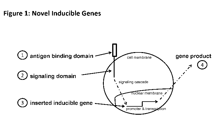
[0007] Figure 1 is a schematic depicting an exemplary cellular
therapeutic.
[0008] Figure 2 is a schematic depicting an exemplary cellular
therapeutic encoding an
inducible scFv-CD19 fusion protein.
[0009] Figure 3 is a schematic depicting an exemplary cellular
therapeutic encoding an
inducible scFv-EGFR fusion protein.
CA 03054304 2019-08-21
WO 2018/156802 PCT/US2018/019281
3
[0010] Figure 4 is a schematic depicting an exemplary "self amplifying"
cellular
therapeutic encoding an inducible scFv-CD19 fusion protein and an inducible
CAR that targets
CD19.
[0011] Figure 5 is a schematic depicting an exemplary "self amplifying"
cellular
therapeutic encoding an inducible scFv-CD19 fusion protein and a
constitutively expressed CAR
that targets CD19.
[0012] Figure 6 is a schematic depicting an exemplary "self amplifying"
cellular
therapeutic expressing an antigen binding receptor that does not include a
signaling domain
leading to induction of killing, and does include a signaling domain
sufficient to induce gene
transcription, and also encoding an inducible scFv-CD19 fusion protein and an
inducible CAR
(left) or a constitutively expressed CAR (right) that targets CD19.
[0013] Figure 7 is a schematic depicting an exemplary cellular
therapeutic encoding
various inducible genes.
[0014] Figure 8 is a schematic depicting an exemplary cellular
therapeutic encoding an
inducible cytokine.
[0015] Figure 9 is a schematic depicting an exemplary cellular
therapeutic encoding an
inducible scFv-CD30 fusion protein.
[0016] Figure 10 is a schematic depicting an exemplary cellular
therapeutic encoding an
inducible toxin.
[0017] Figure 11 is a schematic depicting an exemplary cellular
therapeutic encoding
various inducible genes.
[0018] Figures 12A, 12B, and 12C are schematics depicting exemplary CD19
variants.
[0019] Figure 13 is a schematic depicting exemplary antibody fusion
proteins in which a
polypeptide antigen is fused to the C terminus of a light chain (LC) of an
antibody, a polypeptide
antigen is fused to the N terminus of a LC of an antibody, a polypeptide
antigen is fused to the C
CA 03054304 2019-08-21
WO 2018/156802 PCT/US2018/019281
4
terminus of a heavy chain (HC) of an antibody, or a polypeptide antigen is
fused to the N
terminus of a HC of an antibody.
[0020] Figure 14A and 14B show expression levels of various polypeptide
antigen-
antibody fusion constructs.
[0021] Figure 15 is a schematic depicting exemplary antibody fusion
proteins in which a
polypeptide antigen is fused in various orientations to an scFv.
[0022] Figure 16 shows expression levels of various polypeptide antigen-
scFv fusion
constructs.
[0023] Figures 17A, 17B, 17C, and 17D show binding of panitumumab-CD19
fusion
proteins to an anti-CD19 antibody (FMC63).
[0024] Figure 18 shows binding of panitumumab-CD19 fusion proteins to an
anti-CD19
antibody (FMC63) relative to negative controls.
[0025] Figures 19A, 19B, 19C, and 19D show binding of LY2875358-CD19
fusion
proteins to an anti-CD19 antibody (FMC63).
[0026] Figure 20 shows binding of LY2875358-CD19 fusion proteins to an
anti-CD19
antibody (FMC63) relative to negative controls.
[0027] Figure 21 shows a summary of expression of, and FMC63 binding to,
various
antibody-CD19 fusion proteins.
[0028] Figure 22 shows binding of trastuzumab scFv-CD19 fusion proteins
to an anti-
CD19 antibody (FMC63).
[0029] Figures 23A, 23B, and 23C show binding of LY2875358-CD19 fusion
proteins to
c-Met expressing cells and to an anti-CD19 antibody (FMC63).
[0030] Figures 24A and 24B show binding of trastuzumab scFv-CD19 fusion
proteins to
an anti-CD19 antibody (FMC63) and to Her-2 protein.
CA 03054304 2019-08-21
WO 2018/156802 PCT/US2018/019281
[0031] Figures 25A and 25B show binding of trastuzumab scFv-CD19 fusion
proteins to
an anti-CD19 antibody (FMC63) relative to negative controls.
[0032] Figure 26 shows binding of CD19-scFv fusion proteins captured on
anti-His
antibody-coated ELISA plates.
[0033] Figure 27 shows binding of CD19-scFv fusion proteins captured on
anti-His
antibody-coated ELISA plates.
[0034] Figure 28 shows binding of CD19-scFv fusion proteins captured on
anti-FMC63
(anti-CD19)-coated plates, then detected with anti-His-HRP.
[0035] Figure 29 shows detection of CD19-anti-Her2 trastuzumab scFv-human
Fc fusion
proteins in a "sandwich ELISA" format.
[0036] Figure 30 shows the capture of multiple fusion proteins by anti-
CD19 monoclonal
antibody FMC63 and their detection by anti-His antibody coupled to HRP.
[0037] Figure 31 shows the capture of CD19 full-length extracellular
domain-anti-CD20
Leu16 scFv VH-VL-His fusion protein by the C-terminal His tag and then
detected by mouse
monoclonal antibody FMC63 anti-CD19 and then anti-mouse IgG-HRP.
[0038] Figure 32 shows results for fusion proteins that incorporate CD22
protein
domains, or anti-EGFRvIII scFv (#64: CD22-FMC63 scFv-His; #65: CD22-anti-CD20
scFv-His;
#67: CD19 full ECD-anti-EGFRvIII scFv-his; #68: CD22-anti-EGFRvIII scFv-His).
[0039] Figure 33 shows results for protein-antibody fusion proteins and
protein-scFv
fusion proteins derived from the same antibody, panitumumab (#57: Her2
extracellular domain-
Panitumumab scFv VH-VL-His; #58 Her2 extracellular D4- Panitumumab scFv VH-VL-
His;
#33+4 (cotransfection of heavy and light chains; one chain carries the CD19
fusion): CD19
extracellular D1+2 Panitumumab antibody ¨ His).
[0040] Figure 34 shows binding affinity of purified CD19-anti-Her2 scFv-
His fusion
protein for the FMC63 antibody.
CA 03054304 2019-08-21
WO 2018/156802 PCT/US2018/019281
6
[0041] Figure 35 shows the binding affinity of the FMC63-bound CD19-anti-
Her2 scFv-
His fusion protein to Her2.
[0042] Figure 36 shows the binding affinity of the FMC63-bound CD19-anti-
Her2 scFv-
His fusion protein to anti-Her2 scFv.
[0043] Figure 37 shows a flow cytometry profile of fusion protein CD19-
ECD-Leu16
scFv (VH/VL)(#63) bound to CD20 expressing 293 cells and labeled with anti-
CD19
monoclonal antibody FMC63-PE-conjugated.
[0044] Figure 38 shows a flow cytometry profile of fusion protein CD19-
D1+2-Leu16
scFv (VH/VL) (#83) bound to CD20 expressing 293 cells and labeled with anti-
CD19
monoclonal antibody FMC63-PE-conjugated.
[0045] Figure 39 shows a flow cytometry profile of fusion protein CD19-
D1+2-Leu16
scFv (VL/VH) (#85) bound to CD20 expressing 293 cells and labeled with anti-
CD19
monoclonal antibody FMC63-PE-conjugated.
[0046] Figure 40 shows a flow cytometry profile of fusion protein CD19-
D1+2-Leu16
scFv (VH/VL)-huIgGFc (#82) bound to CD20 expressing 293 cells + a-huIgG-FITC.
[0047] Figure 41 shows analysis of anti-huIgG-FITC negative control: 293-
CD20 + a-
huIgG-FITC.
[0048] Figure 42 shows a flow cytometry profile of fusion protein CD19-
D1+2-Leu16
scFv (VL/VH)-huIgGFc (#84) bound to CD20 expressing 293 cells + a-huIgG-FITC.
[0049] Figure 43 shows a flow cytometry profile of fusion protein CD22-
D123-Leu16
scFv (VH/VL) (#65) bound to CD20 expressing 293 cells + cc-His-PE.
[0050] Figure 44 shows detection control for Her2 - A431 cells +
Trastuzumab-PE,
showing the background level of binding (A431 cells are Her2-negative).
[0051] Figure 45 shows analysis of A431 + fusion protein Her2-ECD-
Panitumumab scFv
(VH/VL) (#57) + Trastuzumab-PE-conjugated.
CA 03054304 2019-08-21
WO 2018/156802 PCT/US2018/019281
7
[0052] Figure 46 shows analysis of A431 + fusion protein Her2-D4-
Panitumumab scFv
(VH/VL) (#58) + Trastuzumab-PE-conjugated.
[0053] Figure 47 shows IFNy ELISA results for BT474 cells coated with
indicated
peptide and incubated with CD19 specific CAR-T at effector target ratio of
10:1.
[0054] Figure 48 shows IFNy ELISA results for BT474 cells coated with
indicated
peptide and incubated with CD19 specific CAR-T at effector target ratio of
1:1.
[0055] Figure 49 shows summary XTT-cytotoxicity results for BT474 cells
coated with
indicated peptide and incubated with CD19 specific CAR-T at effector target
ratio of 10:1.
[0056] Figure 50 shows IFNy ELISA results for BT474 cells coated with
indicated
peptide and incubated with CD19 specific CAR-T at effector target ratio of
10:1.
[0057] Figure 51 shows IFNy ELISA results for BT474 cells coated with
indicated
peptide and incubated with CD19 specific CAR-T at effector target ratio of
1:1.
[0058] Figures 52A-52C show exemplary Fc-based constructs.
[0059] Figures 53A-53C show exemplary Fc-based bi-specific constructs.
[0060] Figures 54A and 54B show exemplary Fc-based constructs that
include an Fc Ig
"swap".
[0061] Figures 55A and 55B show exemplary constructs in which a loops in
one or both
Fc CH3 domains is replaced.
[0062] Figure 56 shows an exemplary construct with fusion of a masking
moiety to
constructs described in Figures 52B and 52C with a masking moiety fused to the
N-terminus of
the scFv.
[0063] Figure 57 shows an exemplary construct with fusion of a masking
moiety to
constructs described in Figures 53B and 53C with the masking moiety fused to
the N-terminus of
the VH and/or VL on the VH/VL arm.
CA 03054304 2019-08-21
WO 2018/156802 PCT/US2018/019281
8
[0064] Figure 58 shows an exemplary construct with fusion of a masking
moiety to
construct described in Figure 54B with a masking moiety fused to the N-
terminus of each heavy
chain.
[0065] Figure 59 shows an exemplary construct with fusion of a masking
moiety to
constructs described in Figures 55A and 55B with a masking moiety fused to the
N-terminus of a
heavy chain and/or scFv VH.
[0066] Figures 60A-60D show analysis of GFP expression from the CMV
promoter-
tGFP construct (#66) under resting or activated conditions.
[0067] Figures 61A-61D show analysis of GFP expression from the human
CD69
promoter-tGFP (#46) under resting or activated conditions.
[0068] Figures 62A-62D show analysis of GFP expression from the human
TNFalpha
promoter-tGFP (#47) under resting or activated conditions.
[0069] Figures 63A-63D show analysis of GFP expression from the human
NFAT
element x 6 promoter-tGFP (#49) under resting or activated conditions.
[0070] Figures 64A-64B show analysis of expression of CD69 on the surface
of cells
under resting or activated conditions.
[0071] Figures 65A-65C depict binding of CD19-containing fusion proteins
(#42, #43,
#56, #82, #83, #91, #92, #93, #94) to an FMC63-coated plate. Figure 65D shows
titer
determiniations for fusion proteins #82, #83, #91, and #92.
[0072] Figures 66A- 66D show the capture of multiple fusion proteins by
plate bound
antigen and their detection by anti-His antibody coupled to HRP.
[0073] Figures 67A and 67B show flow cytometry results of fusion protein
CD19-D1+2-
Leu16 scFv (VH/VL) (#83) bound to CD20 expressing 293 cells and labeled with
anti-His-PE
(67A) or anti-CD19 monoclonal antibody FMC63-PE (67B).
CA 03054304 2019-08-21
WO 2018/156802 PCT/US2018/019281
9
[0074] Figures 68A and 68B show flow cytometry results of fusion protein
CD19-D1+2-
Leu16 scFv (VH/VL)-huIgGFc (#82) bound to CD20 expressing 293 cells and
labeled with a-
huIgG-FITC (68A) or FMC63-PE or anti-CD19 monoclonal antibody FMC63-PE (68B).
[0075] Figures 69A-69D show results of IFNy ELISA for construct #83
fusion protein.
Figure 69A: 24 hrs, 10:1 effector:target ratio; Figure 69B: 24 hrs, 2:1
effector:target ratio; Figure
69C: 48 hrs, 10:1 effector:target ratio; Figure 69D: 48 hrs, 2:1
effector:target ratio.
[0076] Figure 70 show results of IFNy ELISA for fusion protein derived
from the
cotransfection of construct #33+ construct #4 at 24 hrs, 2:1 effector:target
ratio.
[0077] Figures 71A and 71B show summary XTT-cytotoxicity results for
fusion protein
#83 and 293-CD20 cells. Figure 71A: 48 hrs, 10:1 effector:target ratio; Figure
71B 48 hrs, 2:1
effector:target ratio.
[0078] Figures 72A and 72B show summary XTT-cytotoxicity results for
fusion protein
derived from the cotransfection of construct #33 + construct #4 and A4321
cells. Figure72A: 24
hrs, 10:1 effector:target ratio. Figure 72B: 24 hrs, 2:1 effector:target
ratio.
[0079] Figures 73A and 73B show expression of HER2 and EGFR in
transiently
transfected 293T cells.
[0080] Figures 74A-74D show fusion protein #43 binding to 293T-Her2
expressing cells.
[0081] Figures 75A-75D show binding of fusion proteins #94, and #95 to
293T-Her2
expressing cells.
[0082] Figures 76A and 76B show binding of fusion protein #94 to 293T-
EGFR
expressing cells.
[0083] Figures 77A and 77B show CAR19-mediated cytotoxicity redirected to
HER2+
cells by CAR19 T cell secretion of fusion protein encoded by construct #42.
[0084] Figure 78 shows binding of a heteromeric fusion protein comprised
of fusion
proteins #29 and #103 to anti-CD19 antibody FMC63 detected by HRP-conjugated
mouse IgG
antibody.
CA 03054304 2019-08-21
WO 2018/156802 PCT/US2018/019281
[0085] Figures 79A and 79B shows yeast surface display of wild-type CD19
extracellular
domain.
[0086] Figure 80 shows antibody binding to yeast-displayed CD19
extracellular domain.
[0087] Figure 81 shows diversified regions of the extracellular domain.
[0088] Figure 82 demonstrates combinatorial CD19 libraries are
effectively displayed on
yeast surface and maintain antibody binding.
[0089] Figures 83A and 83B demonstrate combinatorial CD19 libraries can
be enriched
for binding ligands to EGFR and HER2.
[0090] Figure 84A shows an exemplary Fc-based construct that includes an
anti-tumor
antigen scFv, an anti-idiotype scFv, and CH2 and CH3 Fc domains. Figure 84B
shows an
exemplary Fc-based construct that includes an anti-tumor antigen scFv, an anti-
idiotype scFv,
and CH2 Fc domains. Figure 84C shows an exemplary masked scFv/anti-idiotype
scFv
construct.
[0091] Figure 85 demonstrates secretion of anti- FMC63 (anti-Id) antibody
from
transfected 293T cells.
[0092] Figures 86A and 86B demonstrate expression of CAR19 (construct
#140) with an
FMC63 domain as detected by a Flag tag (86A) and detection of the CAR19 by
anti- FMC63
antibody (86B).
[0093] Figures 87A-87C demonstrate Trastuzumab scFv/anti-Id scFv fusion
proteins
bind both FMC63 and Her2. Figure 87A demonstrates binding of a Trastuzumab
scFv/anti-Id
scFv fusion protein to FMC63. Figure 87B demonstrates binding of a Trastuzumab
scFv/anti-Id
scFv fusion protein to Her2. Figure 87C demonstrates binding of a CD19
expressing construct
(#42) with the FMC63 coated plate as a control.
[0094] Figures 88A and 88B demonstrate recognition of Her2 by Trastuzumab
scFv/anti-
Id scFv fusion proteins. Figure 88A demonstrates Her2 expression on SKOV3
cells. Figure 88B
CA 03054304 2019-08-21
WO 2018/156802 PCT/US2018/019281
11
demonstrates binding to the SKOV3-Her2 cells by the Trastuzumab scFv/anti-Id
scFv fusion
protein.
[0095] Figure 89 shows CAR19-mediated cytotoxicity redirected to HER2+
SKOV3
cells by a Trastuzumab scFv/anti-Id scFv fusion protein.
[0096] Figures 90A and 90B summarize the calculated cytotoxicity of CAR19-
mediated
killing as redirected by a Trastuzumab scFv/anti-Id scFv fusion protein.
Figure 90A shows the
calculated cytotoxicity. Figure 90B shows the calculated EC50 of construct
#171.
[0097] Figure 91 shows results of IFNy ELISA for CAR19 killing redirected
by construct
#171.
[0098] Figures 92A and 92B demonstrate specificity of CAR19 redirected
killing using
Trastuzumab scFv/anti-Id scFv fusion proteins. Figure 92A demonstrates results
of CAR19-
mediated cytotoxicity redirected to HER2+ SKOV3 cells by Trastuzumab scFv/anti-
Id scFv
construct #171 relative to a construct expressing an anti-Her2 protein ( #16).
Figure 92B
summaraizes the calculated cytotoxicity of CAR19-mediated killing as
redirected by construct
#171 or #16.
[0099] Figure 93 demonstrates the lack of CAR19 redirected killing using
Trastuzumab
scFv/anti-Id scFv fusion proteins when the target cell (H929) lacks Her2.
Definitions
[0100] In order for the present invention to be more readily understood,
certain terms are
first defined below. Additional definitions for the following terms and other
terms are set forth
throughout the specification.
[0101] Administration: As used herein, the term "administration" refers
to the
administration of a composition to a subject or system. Administration to an
animal subject
(e.g., to a human) may be by any appropriate route. For example, in some
embodiments,
administration may be bronchial (including by bronchial instillation), buccal,
enteral,
interdermal, intra-arterial, intradermal, intragastric, intramedullary,
intramuscular, intranasal,
CA 03054304 2019-08-21
WO 2018/156802 PCT/US2018/019281
12
intraperitoneal, intrathecal, intravenous, intraventricular, within a specific
organ (e.g.,
intrahepatic), mucosal, nasal, oral, rectal, subcutaneous, sublingual,
topical, tracheal (including
by intratracheal instillation), transdermal, vaginal and vitreal. In some
embodiments,
administration may be intratumoral or peritumoral. In some embodiments,
administration may
involve intermittent dosing. In some embodiments, administration may involve
continuous
dosing (e.g., perfusion) for at least a selected period of time.
[0102] Adoptive cell therapy: As used herein, "adoptive cell therapy" or
"ACT" involves
the transfer of immune cells with antitumour activity into cancer patients. In
some embodiments,
ACT is a treatment approach that involves the use of lymphocytes with
antitumour activity, the
in vitro expansion of these cells to large numbers and their infusion into a
cancer-bearing host.
[0103] Agent: The term "agent" as used herein may refer to a compound or
entity of any
chemical class including, for example, polypeptides, nucleic acids,
saccharides, lipids, small
molecules, metals, or combinations thereof As will be clear from context, in
some
embodiments, an agent can be or comprise a cell or organism, or a fraction,
extract, or
component thereof In some embodiments, an agent is or comprises a natural
product in that it is
found in and/or is obtained from nature. In some embodiments, an agent is or
comprises one or
more entities that is man-made in that it is designed, engineered, and/or
produced through action
of the hand of man and/or is not found in nature. In some embodiments, an
agent may be
utilized in isolated or pure form; in some embodiments, an agent may be
utilized in crude form.
In some embodiments, potential agents are provided as collections or
libraries, for example that
may be screened to identify or characterize active agents within them. Some
particular
embodiments of agents that may be utilized in accordance with the present
invention include
small molecules, antibodies, antibody fragments, aptamers, nucleic acids
(e.g., siRNAs, shRNAs,
DNA/RNA hybrids, antisense oligonucleotides, ribozymes), peptides, peptide
mimetics, etc. In
some embodiments, an agent is or comprises a polymer. In some embodiments, an
agent is not a
polymer and/or is substantially free of any polymer. In some embodiments, an
agent contains at
least one polymeric moiety. In some embodiments, an agent lacks or is
substantially free of any
polymeric moiety.
CA 03054304 2019-08-21
WO 2018/156802 PCT/US2018/019281
13
[0104] Amelioration: As used herein, "amelioration" refers to prevention,
reduction
and/or palliation of a state, or improvement of the state of a subject.
Amelioration includes, but
does not require, complete recovery or complete prevention of a disease,
disorder or condition.
[0105] Amino acid: As used herein, term "amino acid," in its broadest
sense, refers to any
compound and/or substance that can be incorporated into a polypeptide chain.
In some
embodiments, an amino acid has the general structure H2N¨C(H)(R)¨COOH. In some
embodiments, an amino acid is a naturally occurring amino acid. In some
embodiments, an
amino acid is a synthetic amino acid; in some embodiments, an amino acid is a
d-amino acid; in
some embodiments, an amino acid is an 1-amino acid. "Standard amino acid"
refers to any of the
twenty standard 1-amino acids commonly found in naturally occurring peptides.
"Nonstandard
amino acid" refers to any amino acid, other than the standard amino acids,
regardless of whether
it is prepared synthetically or obtained from a natural source. As used
herein, "synthetic amino
acid" encompasses chemically modified amino acids, including but not limited
to salts, amino
acid derivatives (such as amides), and/or substitutions. Amino acids,
including carboxy- and/or
amino-terminal amino acids in peptides, can be modified by methylation,
amidation, acetylation,
protecting groups, and/or substitution with other chemical groups that can
change the peptide's
circulating half-life without adversely affecting their activity. Amino acids
may participate in a
disulfide bond. Amino acids may comprise one or posttranslational
modifications, such as
association with one or more chemical entities (e.g., methyl groups, acetate
groups, acetyl
groups, phosphate groups, formyl moieties, isoprenoid groups, sulfate groups,
polyethylene
glycol moieties, lipid moieties, carbohydrate moieties, biotin moieties,
etc.). The term "amino
acid" is used interchangeably with "amino acid residue," and may refer to a
free amino acid
and/or to an amino acid residue of a peptide. It will be apparent from the
context in which the
term is used whether it refers to a free amino acid or a residue of a peptide.
[0106] Antibody: As used herein, the term "antibody" refers to a
polypeptide that
includes canonical immunoglobulin sequence elements sufficient to confer
specific binding to a
particular target antigen. As is known in the art, intact antibodies as
produced in nature are
approximately 150 kD tetrameric agents comprised of two identical heavy chain
polypeptides
CA 03054304 2019-08-21
WO 2018/156802 PCT/US2018/019281
14
(about 50 kD each) and two identical light chain polypeptides (about 25 kD
each) that associate
with each other into what is commonly referred to as a "Y-shaped" structure.
Each heavy chain
is comprised of at least four domains (each about 110 amino acids long)¨ an
amino-terminal
variable (VH) domain (located at the tips of the Y structure), followed by
three constant
domains: CHL CH2, and the carboxy-terminal CH3 (located at the base of the Y's
stem). A
short region, known as the "switch", connects the heavy chain variable and
constant regions.
The "hinge" connects CH2 and CH3 domains to the rest of the antibody. Two
disulfide bonds in
this hinge region connect the two heavy chain polypeptides to one another in
an intact antibody.
Each light chain is comprised of two domains ¨ an amino-terminal variable (VL)
domain,
followed by a carboxy-terminal constant (CL) domain, separated from one
another by another
"switch". Intact antibody tetramers are composed of two heavy chain-light
chain dimers in
which the heavy and light chains are linked to one another by a single
disulfide bond; two other
disulfide bonds connect the heavy chain hinge regions to one another, so that
the dimers are
connected to one another and the tetramer is formed. Naturally-produced
antibodies are also
glycosylated, typically on the CH2 domain. Each domain in a natural antibody
has a structure
characterized by an "immunoglobulin fold" formed from two beta sheets (e.g., 3-
, 4-, or 5-
stranded sheets) packed against each other in a compressed antiparallel beta
barrel. Each
variable domain contains three hypervariable loops known as "complement
determining regions"
(CDR1, CDR2, and CDR3) and four somewhat invariant "framework" regions (FR1,
FR2, FR3,
and FR4). When natural antibodies fold, the FR regions form the beta sheets
that provide the
structural framework for the domains, and the CDR loop regions from both the
heavy and light
chains are brought together in three-dimensional space so that they create a
single hypervariable
antigen binding site located at the tip of the Y structure. The Fc region of
naturally-occurring
antibodies binds to elements of the complement system, and also to receptors
on effector cells,
including for example effector cells that mediate cytotoxicity. As is known in
the art, affinity
and/or other binding attributes of Fc regions for Fc receptors can be
modulated through
glycosylation or other modification. In some embodiments, antibodies produced
and/or utilized
in accordance with the present disclosure include glycosylated Fc domains,
including Fc
domains with modified or engineered such glycosylation. For purposes of the
present disclosure,
CA 03054304 2019-08-21
WO 2018/156802 PCT/US2018/019281
in certain embodiments, any polypeptide or complex of polypeptides that
includes sufficient
immunoglobulin domain sequences as found in natural antibodies can be referred
to and/or used
as an "antibody", whether such polypeptide is naturally produced (e.g.,
generated by an organism
reacting to an antigen), or produced by recombinant engineering, chemical
synthesis, or other
artificial system or methodology. In some embodiments, an antibody is
polyclonal; in some
embodiments, an antibody is monoclonal. In some embodiments, an antibody has
constant
region sequences that are characteristic of mouse, rabbit, primate, or human
antibodies. In some
embodiments, antibody sequence elements are fully human, or are humanized,
primatized,
chimeric, etc, as is known in the art. Moreover, the term "antibody" as used
herein, can refer in
appropriate embodiments (unless otherwise stated or clear from context) to any
of the art-known
or developed constructs or formats for utilizing antibody structural and
functional features in
alternative presentation. For example, in some embodiments, an antibody
utilized in accordance
with the present disclosure is in a format selected from, but not limited to,
intact IgG, IgE and
IgM, bi- or multi- specific antibodies (e.g., Zybodies , etc), single chain
Fvs, polypeptide-Fc
fusions, Fabs, cameloid antibodies, masked antibodies (e.g., Probodies ),
Small Modular
ImmunoPharmaceuticals ("SMIPsTM"), single chain or Tandem diabodies (TandAbg),
Anticalins , Nanobodies , minibodies, BiTEgs, ankyrin repeat proteins or
DARPINs ,
Avimers , a DART, a TCR-like antibody, Adnectins , Affilins , Trans-bodies ,
Affibodies ,
a TrimerX , MicroProteins, Fynomers , Centyrins , and a KALBITOR . In some
embodiments, an antibody may lack a covalent modification (e.g., attachment of
a glycan) that it
would have if produced naturally. In some embodiments, an antibody may contain
a covalent
modification (e.g., attachment of a glycan, a payload (e.g., a detectable
moiety, a therapeutic
moiety, a catalytic moiety, etc.), or other pendant group (e.g., poly-ethylene
glycol, etc.)).
[0107] Antibody-Dependent Cellular Cytotoxicity: As used herein, the term
"antibody-
dependent cellular cytotoxicity" or "ADCC" refers to a phenomenon in which
target cells bound
by antibody are killed by immune effector cells. Without wishing to be bound
by any particular
theory, ADCC is typically understood to involve Fc receptor (FcR)-bearing
effector cells can
recognizing and subsequently killing antibody-coated target cells (e.g., cells
that express on their
CA 03054304 2019-08-21
WO 2018/156802 PCT/US2018/019281
16
surface specific antigens to which an antibody is bound). Effector cells that
mediate ADCC can
include immune cells, including but not limited to one or more of natural
killer (NK) cells,
macrophage, neutrophils, eosinophils.
[0108] Antibody Fragment: As used herein, an "antibody fragment" includes
a portion of
an intact antibody, such as, for example, the antigen-binding or variable
region of an antibody.
Examples of antibody fragments include Fab, Fab', F(ab')2, and Fv fragments;
triabodies;
tetrabodies; linear antibodies; single-chain antibody molecules; and multi-
specific antibodies
formed from antibody fragments. For example, antibody fragments include
isolated fragments,
"Fv" fragments (consisting of the variable regions of the heavy and light
chains), recombinant
single chain polypeptide molecules in which light and heavy chain variable
regions are
connected by a peptide linker ("scFv proteins"), recombinant single domain
antibodies consisting
of a variable region of an antibody heavy chain (e.g., VHH), and minimal
recognition units
consisting of the amino acid residues that mimic a hypervariable region (e.g.,
a hypervariable
region of a heavy chain variable region (VH), a hypervariable region of a
light chain variable
region (VL), one or more CDR domains within the VH, and/or one or more CDR
domains within
the VL). In many embodiments, an antibody fragment contains sufficient
sequence of the parent
antibody of which it is a fragment that it binds to the same antigen as does
the parent antibody; in
some embodiments, a fragment binds to the antigen with a comparable affinity
to that of the
parent antibody and/or competes with the parent antibody for binding to the
antigen. Examples
of antigen binding fragments of an antibody include, but are not limited to,
Fab fragment, Fab'
fragment, F(ab')2 fragment, scFv fragment, Fv fragment, dsFy diabody, dAb
fragment, Fd'
fragment, Fd fragment, heavy chain variable region, and an isolated
complementarity
determining region (CDR) region. An antigen binding fragment of an antibody
may be produced
by any means. For example, an antigen binding fragment of an antibody may be
enzymatically
or chemically produced by fragmentation of an intact antibody and/or it may be
recombinantly
produced from a gene encoding the partial antibody sequence. Alternatively or
additionally,
antigen binding fragment of an antibody may be wholly or partially
synthetically produced. An
antigen binding fragment of an antibody may optionally comprise a single chain
antibody
CA 03054304 2019-08-21
WO 2018/156802 PCT/US2018/019281
17
fragment. Alternatively or additionally, an antigen binding fragment of an
antibody may
comprise multiple chains which are linked together, for example, by disulfide
linkages. An
antigen binding fragment of an antibody may optionally comprise a
multimolecular complex. A
functional antibody fragment typically comprises at least about 50 amino acids
and more
typically comprises at least about 200 amino acids.
[0109] Antigen: The term "antigen", as used herein, refers to an agent
that elicits an
immune response; and/or an agent that binds to a T cell receptor (e.g., when
presented by an
WIC molecule) or to an antibody or antibody fragment. In some embodiments, an
antigen
elicits a humoral response (e.g., including production of antigen-specific
antibodies); in some
embodiments, an antigen elicits a cellular response (e.g., involving T-cells
whose receptors
specifically interact with the antigen). In some embodiments, an antigen binds
to an antibody
and may or may not induce a particular physiological response in an organism.
In general, an
antigen may be or include any chemical entity such as, for example, a small
molecule, a nucleic
acid, a polypeptide, a carbohydrate, a lipid, a polymer (in some embodiments
other than a
biologic polymer (e.g., other than a nucleic acid or amino acid polymer)) etc.
In some
embodiments, an antigen is or comprises a polypeptide. In some embodiments, an
antigen is or
comprises a glycan. Those of ordinary skill in the art will appreciate that,
in general, an antigen
may be provided in isolated or pure form, or alternatively may be provided in
crude form (e.g.,
together with other materials, for example in an extract such as a cellular
extract or other
relatively crude preparation of an antigen-containing source), or
alternatively may exist on or in
a cell. In some embodiments, an antigen is a recombinant antigen.
[0110] Antigen presenting cell: The phrase "antigen presenting cell" or
"APC," as used
herein, has its art understood meaning referring to cells that process and
present antigens to T-
cells. Exemplary APC include dendritic cells, macrophages, B cells, certain
activated epithelial
cells, and other cell types capable of TCR stimulation and appropriate T cell
costimulation.
[0111] Approximately or about: As used herein, the term "approximately" or
"about," as
applied to one or more values of interest, refers to a value that is similar
to a stated reference
value. In certain embodiments, the term "approximately" or "about" refers to a
range of values
CA 03054304 2019-08-21
WO 2018/156802 PCT/US2018/019281
18
that fall within 25%, 20%, 19%, 18%, 17%, 16%, 15%, 14%, 13%, 12%, 11%, 1000,
9%, 8%,
700, 60o, 5%, 4%, 3%, 2%, 100, or less in either direction (greater than or
less than) of the stated
reference value unless otherwise stated or otherwise evident from the context
(except where such
number would exceed 100% of a possible value).
[0112] Binding: It will be understood that the term "binding", as used
herein, typically
refers to a non-covalent association between or among two or more entities.
"Direct" binding
involves physical contact between entities or moieties; indirect binding
involves physical
interaction by way of physical contact with one or more intermediate entities.
Binding between
two or more entities can typically be assessed in any of a variety of contexts
¨ including where
interacting entities or moieties are studied in isolation or in the context of
more complex systems
(e.g., while covalently or otherwise associated with a carrier entity and/or
in a biological system
or cell).
[0113] Cancer: The terms "cancer", "malignancy", "neoplasm", "tumor", and
"carcinoma", are used interchangeably herein to refer to cells that exhibit
relatively abnormal,
uncontrolled, and/or autonomous growth, so that they exhibit an aberrant
growth phenotype
characterized by a significant loss of control of cell proliferation. In
general, cells of interest for
detection or treatment in the present application include precancerous (e.g.,
benign), malignant,
pre-metastatic, metastatic, and non-metastatic cells. The teachings of the
present disclosure may
be relevant to any and all cancers. To give but a few, non-limiting examples,
in some
embodiments, teachings of the present disclosure are applied to one or more
cancers such as, for
example, hematopoietic cancers including leukemias, lymphomas (Hodgkins and
non-
Hodgkins), myelomas and myeloproliferative disorders; sarcomas, melanomas,
adenomas,
carcinomas of solid tissue, squamous cell carcinomas of the mouth, throat,
larynx, and lung, liver
cancer, genitourinary cancers such as prostate, cervical, bladder, uterine,
and endometrial cancer
and renal cell carcinomas, bone cancer, pancreatic cancer, skin cancer,
cutaneous or intraocular
melanoma, cancer of the endocrine system, cancer of the thyroid gland, cancer
of the parathyroid
gland, head and neck cancers, breast cancer, gastro-intestinal cancers and
nervous system
cancers, benign lesions such as papillomas, and the like.
CA 03054304 2019-08-21
WO 2018/156802 PCT/US2018/019281
19
[0114] Chimeric antigen receptor: "Chimeric antigen receptor" or "CAR" or
"CARs"
as used herein refers to engineered receptors, which graft an antigen
specificity onto cells (for
example T cells such as naive T cells, central memory T cells, effector memory
T cells or
combination thereof). CARs are also known as artificial T-cell receptors,
chimeric T-cell
receptors or chimeric immunoreceptors. In some embodiments, CARs comprise an
antigen-
specific targeting regions, an extracellular domain, a transmembrane domain,
one or more co-
stimulatory domains, and an intracellular signaling domain.
[0115] Combination Therapy: As used herein, the term "combination
therapy" refers to
those situations in which a subject is simultaneously exposed to two or more
therapeutic
regimens (e.g., two or more therapeutic agents). In some embodiments, two or
more agents may
be administered simultaneously; in some embodiments, such agents may be
administered
sequentially; in some embodiments, such agents are administered in overlapping
dosing
regimens.
[0116] Domain: The term "domain" is used herein to refer to a section or
portion of an
entity. In some embodiments, a "domain" is associated with a particular
structural and/or
functional feature of the entity so that, when the domain is physically
separated from the rest of
its parent entity, it substantially or entirely retains the particular
structural and/or functional
feature. Alternatively or additionally, a domain may be or include a portion
of an entity that,
when separated from that (parent) entity and linked with a different
(recipient) entity,
substantially retains and/or imparts on the recipient entity one or more
structural and/or
functional features that characterized it in the parent entity. In some
embodiments, a domain is a
section or portion of a molecular (e.g., a small molecule, carbohydrate, a
lipid, a nucleic acid, or
a polypeptide). In some embodiments, a domain is a section of a polypeptide;
in some such
embodiments, a domain is characterized by a particular structural element
(e.g., a particular
amino acid sequence or sequence motif, a-helix character, 13-sheet character,
coiled-coil
character, random coil character, etc), and/or by a particular functional
feature (e.g., binding
activity, enzymatic activity, folding activity, signaling activity, etc).
CA 03054304 2019-08-21
WO 2018/156802 PCT/US2018/019281
[0117] Dosage form: As used herein, the terms "dosage form" and "unit
dosage form"
refer to a physically discrete unit of a therapeutic agent for the patient to
be treated. Each unit
contains a predetermined quantity of active material calculated to produce the
desired therapeutic
effect. It will be understood, however, that the total dosage of the
composition will be decided
by the attending physician within the scope of sound medical judgment.
[0118] Dosing regimen: As used herein, the term "dosing regimen" refers to
a set of unit
doses (typically more than one) that are administered individually to a
subject, typically
separated by periods of time. In some embodiments, a given therapeutic agent
has a
recommended dosing regimen, which may involve one or more doses. In some
embodiments, a
dosing regimen comprises a plurality of doses each of which are separated from
one another by a
time period of the same length; in some embodiments, a dosing regimen
comprises a plurality of
doses and at least two different time periods separating individual doses. In
some embodiments,
all doses within a dosing regimen are of the same unit dose amount. In some
embodiments,
different doses within a dosing regimen are of different amounts. In some
embodiments, a
dosing regimen comprises a first dose in a first dose amount, followed by one
or more additional
doses in a second dose amount different from the first dose amount. In some
embodiments, a
dosing regimen comprises a first dose in a first dose amount, followed by one
or more additional
doses in a second dose amount same as the first dose amount. In some
embodiments, a dosing
regimen is correlated with a desired or beneficial outcome when administered
across a relevant
population (i.e., is a therapeutic dosing regimen).
[0119] Effector Function: As used herein, "effector function" refers a
biochemical event
that results from the interaction of an antibody Fc region with an Fc receptor
or ligand. Effector
functions include but are not limited to antibody-dependent cell-mediated
cytotoxicity (ADCC),
antibody-dependent cell-mediated phagocytosis (ADCP), and complement-mediated
cytotoxicity
(CMC). In some embodiments, an effector function is one that operates after
the binding of an
antigen, one that operates independent of antigen binding, or both.
[0120] Effector Cell: As used herein, "effector cell" refers to a cell of
the immune
system that expresses one or more Fc receptors and mediates one or more
effector functions. In
CA 03054304 2019-08-21
WO 2018/156802 PCT/US2018/019281
21
some embodiments, effector cells may include, but may not be limited to, one
or more of
monocytes, macrophages, neutrophils, dendritic cells, eosinophils, mast cells,
platelets, large
granular lymphocytes, Langerhans' cells, natural killer (NK) cells, T-
lymphocytes, B-
lymphocytes and may be from any organism including but not limited to humans,
mice, rats,
rabbits, and monkeys.
[0121] Expression: As used herein, "expression" of a nucleic acid sequence
refers to one
or more of the following events: (1) production of an RNA template from a DNA
sequence (e.g.,
by transcription); (2) processing of an RNA transcript (e.g., by splicing,
editing, 5' cap
formation, and/or 3' end formation); (3) translation of an RNA into a
polypeptide or protein;
and/or (4) post-translational modification of a polypeptide or protein.
[0122] Extracellular domain: As used herein, "extracellular domain" (or
"ECD") refers
to a portion of a polypeptide that extends beyond the transmembrane domain
into extracellular
space.
[0123] Fusion protein: As used herein, the term "fusion protein" generally
refers to a
polypeptide including at least two segments, each of which shows a high degree
of amino acid
identity to a peptide moiety that (1) occurs in nature, and/or (2) represents
a functional domain of
a polypeptide. Typically, a polypeptide containing at least two such segments
is considered to be
a fusion protein if the two segments are moieties that (1) are not included in
nature in the same
peptide, and/or (2) have not previously been linked to one another in a single
polypeptide, and/or
(3) have been linked to one another through action of the hand of man.
[0124] Gene: As used herein, the term "gene" has its meaning as understood
in the art.
It will be appreciated by those of ordinary skill in the art that the term
"gene" may include gene
regulatory sequences (e.g., promoters, enhancers, etc.) and/or intron
sequences. It will further be
appreciated that definitions of gene include references to nucleic acids that
do not encode
proteins but rather encode functional RNA molecules such as tRNAs, RNAi-
inducing agents,
etc. For the purpose of clarity we note that, as used in the present
application, the term "gene"
generally refers to a portion of a nucleic acid that encodes a protein; the
term may optionally
encompass regulatory sequences, as will be clear from context to those of
ordinary skill in the
CA 03054304 2019-08-21
WO 2018/156802 PCT/US2018/019281
22
art. This definition is not intended to exclude application of the term "gene"
to non-protein¨
coding expression units but rather to clarify that, in most cases, the term as
used in this document
refers to a protein-coding nucleic acid.
[0125] Gene product or expression product: As used herein, the term "gene
product" or
"expression product" generally refers to an RNA transcribed from the gene (pre-
and/or post-
processing) or a polypeptide (pre- and/or post-modification) encoded by an RNA
transcribed
from the gene.
[0126] Idiotope: As used herein, the term "idiotope" refers to a unique
antigenic
determinant (epitope) of a variable region of an antibody, or antigen binding
portion.
[0127] Idiotype: As used herein, the term "idiotype" refers to a set of
idiotopes of a
particular antibody, or antigen binding portion.
[0128] Immune response: As used herein, the term "immune response" refers
to a
response elicited in an animal. An immune response may refer to cellular
immunity, humoral
immunity or may involve both. An immune response may also be limited to a part
of the
immune system. For example, in certain embodiments, an immunogenic composition
may
induce an increased IFNy response. In certain embodiments, an immunogenic
composition may
induce a mucosal IgA response (e.g., as measured in nasal and/or rectal
washes). In certain
embodiments, an immunogenic composition may induce a systemic IgG response
(e.g., as
measured in serum). In certain embodiments, an immunogenic composition may
induce virus-
neutralizing antibodies or a neutralizing antibody response. In certain
embodiments, an
immunogenic composition may induce a cytolytic (CTL) response by T cells.
[0129] Improve, increase, or reduce: As used herein, the terms "improve,"
"increase" or
"reduce," or grammatical equivalents, indicate values that are relative to a
baseline measurement,
such as a measurement in the same individual prior to initiation of the
treatment described
herein, or a measurement in a control individual (or multiple control
individuals) in the absence
of the treatment described herein.
CA 03054304 2019-08-21
WO 2018/156802 PCT/US2018/019281
23
[0130] Individual, subject, patient: As used herein, the terms "subject,"
"individual" or
"patient" refer to a human or a non-human mammalian subject. The individual
(also referred to
as "patient" or "subject") being treated is an individual (fetus, infant,
child, adolescent, or adult)
suffering from a disease, for example, cancer. In some embodiments, the
subject is a human.
[0131] Linker: As used herein, the term "linker" refers to, e.g., in a
fusion protein, an
amino acid sequence of an appropriate length other than that appearing at a
particular position in
the natural protein and is generally designed to be flexible and/or to
interpose a structure, such as
an a-helix, between two protein moieties. In general, a linker allows two or
more domains of a
fusion protein to retain 50%, 55%, 60%, 65%, 70%, 75%, 80%, 85%, 90%, 95% or
more of the
biological activity of each of the domains. A linker may also referred to as a
spacer.
[0132] Masking moiety: As used herein, "masking moiety" refers to a
molecular moiety
that, when linked to an antigen-binding protein described herein, is capable
of masking the
binding of such antigen-binding moiety to its target antigen. An antigen-
binding protein
comprising such a masking moiety is referred to herein as a "masked" antigen-
binding protein.
[0133] Nucleic acid: As used herein, "nucleic acid", in its broadest
sense, refers to any
compound and/or substance that is or can be incorporated into an
oligonucleotide chain. In some
embodiments, a nucleic acid is a compound and/or substance that is or can be
incorporated into
an oligonucleotide chain via a phosphodiester linkage. As will be clear from
context, in some
embodiments, "nucleic acid" refers to individual nucleic acid residues (e.g.,
nucleotides and/or
nucleosides); in some embodiments, "nucleic acid" refers to an oligonucleotide
chain comprising
individual nucleic acid residues. In some embodiments, a "nucleic acid" is or
comprises RNA;
in some embodiments, a "nucleic acid" is or comprises DNA. In some
embodiments, a nucleic
acid is, comprises, or consists of one or more natural nucleic acid residues.
In some
embodiments, a nucleic acid is, comprises, or consists of one or more nucleic
acid analogs. In
some embodiments, a nucleic acid analog differs from a nucleic acid in that it
does not utilize a
phosphodiester backbone. For example, in some embodiments, a nucleic acid is,
comprises, or
consists of one or more "peptide nucleic acids", which are known in the art
and have peptide
bonds instead of phosphodiester bonds in the backbone, are considered within
the scope of the
CA 03054304 2019-08-21
WO 2018/156802 PCT/US2018/019281
24
present invention. Alternatively or additionally, in some embodiments, a
nucleic acid has one or
more phosphorothioate and/or 5'-N-phosphoramidite linkages rather than
phosphodiester bonds.
In some embodiments, a nucleic acid is, comprises, or consists of one or more
natural
nucleosides (e.g., adenosine, thymidine, guanosine, cytidine, uridine,
deoxyadenosine,
deoxythymidine, deoxy guanosine, and deoxycytidine). In some embodiments, a
nucleic acid is,
comprises, or consists of one or more nucleoside analogs (e.g., 2-
aminoadenosine, 2-
thiothymidine, inosine, pyrrolo-pyrimidine, 3 -methyl adenosine, 5-
methylcytidine, C-5
propynyl-cytidine, C-5 propynyl-uridine, 2-aminoadenosine, C5-bromouridine, C5-
fluorouridine,
C5-iodouridine, C5-propynyl-uridine, C5 -propynyl-cytidine, C5-methylcytidine,
2-
aminoadenosine, 7-deazaadenosine, 7-deazaguanosine, 8-oxoadenosine, 8-
oxoguanosine, 0(6)-
methylguanine, 2-thiocytidine, methylated bases, intercalated bases, and
combinations thereof).
In some embodiments, a nucleic acid comprises one or more modified sugars
(e.g., 2'-
fluororibose, ribose, 2'-deoxyribose, arabinose, and hexose) as compared with
those in natural
nucleic acids. In some embodiments, a nucleic acid has a nucleotide sequence
that encodes a
functional gene product such as an RNA or protein. In some embodiments, a
nucleic acid
includes one or more introns. In some embodiments, nucleic acids are prepared
by one or more
of isolation from a natural source, enzymatic synthesis by polymerization
based on a
complementary template (in vivo or in vitro), reproduction in a recombinant
cell or system, and
chemical synthesis. In some embodiments, a nucleic acid is at least 3, 4, 5,
6, 7, 8, 9, 10, 15, 20,
25, 30, 35, 40, 45, 50, 55, 60, 65, 70, 75, 80, 85, 90, 95, 100, 110, 120,
130, 140, 150, 160, 170,
180, 190, 20, 225, 250, 275, 300, 325, 350, 375, 400, 425, 450, 475, 500, 600,
700, 800, 900,
1000, 1500, 2000, 2500, 3000, 3500, 4000, 4500, 5000 or more residues long. In
some
embodiments, a nucleic acid is single stranded; in some embodiments, a nucleic
acid is double
stranded. In some embodiments a nucleic acid has a nucleotide sequence
comprising at least one
element that encodes, or is the complement of a sequence that encodes, a
polypeptide. In some
embodiments, a nucleic acid has enzymatic activity.
[0134] Operably linked: As used herein, "operably linked" refers to a
juxtaposition
wherein the components described are in a relationship permitting them to
function in their
CA 03054304 2019-08-21
WO 2018/156802 PCT/US2018/019281
intended manner. A control sequence "operably linked" to one or more coding
sequence(s) is
ligated in such a way that expression of the one or more coding sequence(s) is
achieved under
conditions compatible with the control sequences. "Operably linked" sequences
include both
expression control sequences that are contiguous with the gene(s) of interest
and expression
control sequences that act in trans or at a distance to control the gene(s) of
interest. The term
"expression control sequence" as used herein refers to polynucleotide
sequences that are
necessary to effect the expression and processing of coding sequences to which
they are ligated.
Expression control sequences include appropriate transcription initiation,
termination, promoter
and enhancer sequences; efficient RNA processing signals such as splicing and
polyadenylation
signals; sequences that stabilize cytoplasmic mRNA; sequences that enhance
translation
efficiency (i.e., Kozak consensus sequence); sequences that enhance protein
stability; and when
desired, sequences that enhance protein secretion. The nature of such control
sequences differs
depending upon the host organism. For example, in prokaryotes, such control
sequences
generally include promoter, ribosomal binding site, and transcription
termination sequence,
while in eukaryotes, typically, such control sequences include promoters and
transcription
termination sequence. The term "control sequences" is intended to include
components whose
presence is essential for expression and processing, and can also include
additional components
whose presence is advantageous, for example, leader sequences and fusion
partner sequences.
[0135] Patient: As used herein, the term "patient" refers to any organism
to which a
provided composition is or may be administered, e.g., for experimental,
diagnostic, prophylactic,
cosmetic, and/or therapeutic purposes. Typical patients include animals (e.g.,
mammals such as
mice, rats, rabbits, non-human primates, and/or humans). In some embodiments,
a patient is a
human. In some embodiments, a patient is suffering from or susceptible to one
or more disorders
or conditions. In some embodiments, a patient displays one or more symptoms of
a disorder or
condition. In some embodiments, a patient has been diagnosed with one or more
disorders or
conditions. In some embodiments, the disorder or condition is or includes
cancer, or presence of
one or more tumors. In some embodiments, the patient is receiving or has
received certain
therapy to diagnose and/or to treat a disease, disorder, or condition.
CA 03054304 2019-08-21
WO 2018/156802 PCT/US2018/019281
26
[0136] Peptide: The term "peptide" as used herein refers to a polypeptide
that is
typically relatively short, for example having a length of less than about 100
amino acids, less
than about 50 amino acids, less than 20 amino acids, or less than 10 amino
acids.
[0137] Pharmaceutically acceptable: The term "pharmaceutically acceptable"
as used
herein, refers to substances that, within the scope of sound medical judgment,
are suitable for use
in contact with the tissues of human beings and animals without excessive
toxicity, irritation,
allergic response, or other problem or complication, commensurate with a
reasonable benefit/risk
ratio.
[0138] Polypeptide: As used herein, a "polypeptide", generally speaking,
is a string of at
least two amino acids attached to one another by a peptide bond. In some
embodiments, a
polypeptide may include at least 3-5 amino acids, each of which is attached to
others by way of
at least one peptide bond. Those of ordinary skill in the art will appreciate
that polypeptides
sometimes include "non-natural" amino acids or other entities that nonetheless
are capable of
integrating into a polypeptide chain, optionally.
[0139] Promoter: As used herein, a "promoter" is a DNA sequence recognized
by the
synthetic machinery of the cell, or introduced synthetic machinery, required
to initiate the
specific transcription of a polynucleotide sequence. A "constitutive" promoter
is a nucleotide
sequence which, when operably linked with a polynucleotide that encodes or
specifies a gene
product, causes the gene product to be produced in a cell under most or all
physiological
conditions of the cell. An "inducible" promoter is a nucleotide sequence that,
when operably
linked with a polynucleotide that encodes or specifies a gene product, causes
the gene product to
be produced in a cell substantially only when a promoter-specific inducer is
present in the cell.
[0140] Protein: As used herein, the term "protein", refers to a
polypeptide (i.e., a string
of at least two amino acids linked to one another by peptide bonds). Proteins
may include
moieties other than amino acids (e.g., may be glycoproteins, proteoglycans,
etc.) and/or may be
otherwise processed or modified. Those of ordinary skill in the art will
appreciate that a
"protein" can be a complete polypeptide chain as produced by a cell (with or
without a signal
sequence), or can be a portion thereof. Those of ordinary skill will
appreciate that a protein can
CA 03054304 2019-08-21
WO 2018/156802 PCT/US2018/019281
27
sometimes include more than one polypeptide chain, for example linked by one
or more disulfide
bonds or associated by other means. Polypeptides may contain L-amino acids, D-
amino acids, or
both and may contain any of a variety of amino acid modifications or analogs
known in the art.
Useful modifications include, e.g., terminal acetylation, amidation,
methylation, etc. In some
embodiments, proteins may comprise natural amino acids, non-natural amino
acids, synthetic
amino acids, and combinations thereof.
[0141] Reference: As used herein, "reference" describes a standard or
control relative to
which a comparison is performed. For example, in some embodiments, an agent,
animal,
individual, population, sample, sequence or value of interest is compared with
a reference or
control agent, animal, individual, population, sample, sequence or value. In
some embodiments,
a reference or control is tested and/or determined substantially
simultaneously with the testing or
determination of interest. In some embodiments, a reference or control is a
historical reference
or control, optionally embodied in a tangible medium. Typically, as would be
understood by
those skilled in the art, a reference or control is determined or
characterized under comparable
conditions or circumstances to those under assessment. Those skilled in the
art will appreciate
when sufficient similarities are present to justify reliance on and/or
comparison to a particular
possible reference or control.
[0142] Solid tumor: As used herein, the term "solid tumor" refers to an
abnormal mass
of tissue that usually does not contain cysts or liquid areas. Solid tumors
may be benign or
malignant. Different types of solid tumors are named for the type of cells
that form them.
Examples of solid tumors are sarcomas, carcinomas, lymphomas, mesothelioma,
neuroblastoma,
retinoblastoma, etc.
[0143] Stage of cancer: As used herein, the term "stage of cancer" refers
to a qualitative
or quantitative assessment of the level of advancement of a cancer. Criteria
used to determine
the stage of a cancer include, but are not limited to, the size of the tumor
and the extent of
metastases (e.g., localized or distant).
[0144] Subject: By "subject" is meant a mammal (e.g., a human, in some
embodiments
including prenatal human forms). In some embodiments, a subject is suffering
from a relevant
CA 03054304 2019-08-21
WO 2018/156802 PCT/US2018/019281
28
disease, disorder or condition. In some embodiments, a subject is susceptible
to a disease,
disorder, or condition. In some embodiments, a subject displays one or more
symptoms or
characteristics of a disease, disorder or condition. In some embodiments, a
subject does not
display any symptom or characteristic of a disease, disorder, or condition. In
some
embodiments, a subject is someone with one or more features characteristic of
susceptibility to
or risk of a disease, disorder, or condition. In some embodiments, a subject
is a patient. In some
embodiments, a subject is an individual to whom diagnosis and/or therapy is
and/or has been
administered.
[0145] Suffering from: An individual who is "suffering from" a disease,
disorder, or
condition (e.g., cancer) has been diagnosed with and/or exhibits one or more
symptoms of the
disease, disorder, or condition.
[0146] Symptoms are reduced: According to the present invention, "symptoms
are
reduced" when one or more symptoms of a particular disease, disorder or
condition is reduced in
magnitude (e.g., intensity, severity, etc.) or frequency. For purposes of
clarity, a delay in the
onset of a particular symptom is considered one form of reducing the frequency
of that symptom.
It is not intended that the present invention be limited only to cases where
the symptoms are
eliminated. The present invention specifically contemplates treatment such
that one or more
symptoms is/are reduced (and the condition of the subject is thereby
"improved"), albeit not
completely eliminated.
[0147] T cell receptor: As used herein, a "T cell receptor" or "TCR"
refers to the
antigen-recognition molecules present on the surface of T-cells. During normal
T-cell
development, each of the four TCR genes, a, (3, y, and 6, can rearrange
leading to highly diverse
TCR proteins.
[0148] Therapeutic agent: As used herein, the phrase "therapeutic agent"
in general
refers to any agent that elicits a desired pharmacological effect when
administered to an
organism. In some embodiments, an agent is considered to be a therapeutic
agent if it
demonstrates a statistically significant effect across an appropriate
population. In some
embodiments, the appropriate population may be a population of model
organisms. In some
CA 03054304 2019-08-21
WO 2018/156802 PCT/US2018/019281
29
embodiments, an appropriate population may be defined by various criteria,
such as a certain age
group, gender, genetic background, preexisting clinical conditions, etc. In
some embodiments, a
therapeutic agent is a substance that can be used to alleviate, ameliorate,
relieve, inhibit, prevent,
delay onset of, reduce severity of, and/or reduce incidence of one or more
symptoms or features
of a disease, disorder, and/or condition. In some embodiments, a "therapeutic
agent" is an agent
that has been or is required to be approved by a government agency before it
can be marketed for
administration to humans. In some embodiments, a "therapeutic agent" is an
agent for which a
medical prescription is required for administration to humans.
[0149] Therapeutically effective amount: As used herein, the term
"therapeutically
effective amount" means an amount that is sufficient, when administered to a
population
suffering from or susceptible to a disease, disorder, and/or condition in
accordance with a
therapeutic dosing regimen, to treat the disease, disorder, and/or condition.
In some
embodiments, a therapeutically effective amount is one that reduces the
incidence and/or severity
of, stabilizes one or more characteristics of, and/or delays onset of, one or
more symptoms of the
disease, disorder, and/or condition. Those of ordinary skill in the art will
appreciate that the term
"therapeutically effective amount" does not in fact require successful
treatment be achieved in a
particular individual. Rather, a therapeutically effective amount may be that
amount that
provides a particular desired pharmacological response in a significant number
of subjects when
administered to patients in need of such treatment. For example, in some
embodiments,
"therapeutically effective amount" refers to an amount which, when
administered to an
individual in need thereof in the context of inventive therapy, will block,
stabilize, attenuate, or
reverse a cancer-supportive process occurring in said individual, or will
enhance or increase a
cancer-suppressive process in said individual. In the context of cancer
treatment, a
"therapeutically effective amount" is an amount which, when administered to an
individual
diagnosed with a cancer, will prevent, stabilize, inhibit, or reduce the
further development of
cancer in the individual. A particularly preferred "therapeutically effective
amount" of a
composition described herein reverses (in a therapeutic treatment) the
development of a
malignancy such as a pancreatic carcinoma or helps achieve or prolong
remission of a
CA 03054304 2019-08-21
WO 2018/156802 PCT/US2018/019281
malignancy. A therapeutically effective amount administered to an individual
to treat a cancer in
that individual may be the same or different from a therapeutically effective
amount
administered to promote remission or inhibit metastasis. As with most cancer
therapies, the
therapeutic methods described herein are not to be interpreted as, restricted
to, or otherwise
limited to a "cure" for cancer; rather the methods of treatment are directed
to the use of the
described compositions to "treat" a cancer, i.e., to effect a desirable or
beneficial change in the
health of an individual who has cancer. Such benefits are recognized by
skilled healthcare
providers in the field of oncology and include, but are not limited to, a
stabilization of patient
condition, a decrease in tumor size (tumor regression), an improvement in
vital functions (e.g.,
improved function of cancerous tissues or organs), a decrease or inhibition of
further metastasis,
a decrease in opportunistic infections, an increased survivability, a decrease
in pain, improved
motor function, improved cognitive function, improved feeling of energy
(vitality, decreased
malaise), improved feeling of well-being, restoration of normal appetite,
restoration of healthy
weight gain, and combinations thereof In addition, regression of a particular
tumor in an
individual (e.g., as the result of treatments described herein) may also be
assessed by taking
samples of cancer cells from the site of a tumor such as a pancreatic
adenocarcinoma (e.g., over
the course of treatment) and testing the cancer cells for the level of
metabolic and signaling
markers to monitor the status of the cancer cells to verify at the molecular
level the regression of
the cancer cells to a less malignant phenotype. For example, tumor regression
induced by
employing the methods of this invention would be indicated by finding a
decrease in one or more
pro-angiogenic markers, an increase in anti-angiogenic markers, the
normalization (i.e.,
alteration toward a state found in normal individuals not suffering from
cancer) of metabolic
pathways, intercellular signaling pathways, or intracellular signaling
pathways that exhibit
abnormal activity in individuals diagnosed with cancer. Those of ordinary
skill in the art will
appreciate that, in some embodiments, a therapeutically effective amount may
be formulated
and/or administered in a single dose. In some embodiments, a therapeutically
effective amount
may be formulated and/or administered in a plurality of doses, for example, as
part of a dosing
regimen.
CA 03054304 2019-08-21
WO 2018/156802 PCT/US2018/019281
31
[0150] Transformation: As used herein, "transformation" refers to any
process by which
exogenous DNA is introduced into a host cell. Transformation may occur under
natural or
artificial conditions using various methods well known in the art.
Transformation may rely on
any known method for the insertion of foreign nucleic acid sequences into a
prokaryotic or
eukaryotic host cell. In some embodiments, a particular transformation
methodology is selected
based on the host cell being transformed and may include, but is not limited
to, viral infection,
electroporation, mating, lipofection. In some embodiments, a "transformed"
cell is stably
transformed in that the inserted DNA is capable of replication either as an
autonomously
replicating plasmid or as part of the host chromosome. In some embodiments, a
transformed cell
transiently expresses introduced nucleic acid for limited periods of time.
[0151] Treatment: As used herein, the term "treatment" (also "treat" or
"treating") refers
to any administration of a substance that partially or completely alleviates,
ameliorates, relives,
inhibits, delays onset of, reduces severity of, and/or reduces incidence of
one or more symptoms,
features, and/or causes of a particular disease, disorder, and/or condition
(e.g., cancer). Such
treatment may be of a subject who does not exhibit signs of the relevant
disease, disorder and/or
condition and/or of a subject who exhibits only early signs of the disease,
disorder, and/or
condition. Alternatively or additionally, such treatment may be of a subject
who exhibits one or
more established signs of the relevant disease, disorder and/or condition. In
some embodiments,
treatment may be of a subject who has been diagnosed as suffering from the
relevant disease,
disorder, and/or condition. In some embodiments, treatment may be of a subject
known to have
one or more susceptibility factors that are statistically correlated with
increased risk of
development of the relevant disease, disorder, and/or condition.
[0152] Tumor infiltrating lymphocyte: As used herein, the term "tumor-
infiltrating
lymphocytes" refers to white blood cells of a subject afflicted with a cancer
(such as melanoma),
that have left the blood stream and have migrated into a tumor. In some
embodiments, tumor-
infiltrating lymphocytes have tumor specificity.
[0153] Vector: As used herein, "vector" refers to a nucleic acid molecule
capable of
transporting another nucleic acid to which it is associated. In some
embodiments, vectors are
CA 03054304 2019-08-21
WO 2018/156802 PCT/US2018/019281
32
capable of extra-chromosomal replication and/or expression of nucleic acids to
which they are
linked in a host cell such as a eukaryotic and/or prokaryotic cell. Vectors
capable of directing
the expression of operatively linked genes are referred to herein as
"expression vectors."
Detailed Description
[0154] Among other things, the present invention provides methods and
compositions
useful for treatment of cancer. Specifically, the present disclosure provides
cellular therapeutics,
e.g., immune cells, genetically modified with an integrated gene, e.g., a
nucleotide sequence of
interest (e.g., a constitutive expression construct and/or an inducible
expression construct that
includes such nucleotide sequence). In some embodiments, expression of a
nucleotide sequence
of interest can be designed to be constitutive or inducible by appropriate
selection, construction
and/or design of an expressed promoter sequence operably linked to such
nucleotide sequence of
interest, as described herein. In the case of a constitutive expression
construct, a gene in the
construct is constitutively expressed. In the case of an inducible expression
construct, a cellular
therapeutic can be genetically modified with a nucleic acid encoding an
antigen binding receptor
and with an inducible expression construct. Upon binding of a target antigen,
an antigen binding
receptor of a cellular therapeutic induces expression of a gene included in an
inducible
expression construct, e.g., as depicted in Figure 1. In certain embodiments,
expression of such
gene facilitates and/or improves treatment of cancer, e.g., by one or more
cellular therapies. The
invention also specifically discloses protein therapeutics that include
proteins encoded by such
genes (e.g., soluble forms of such gene products, e.g., pharmaceutical
compositions that include
such proteins for administration), and nucleic acids encoding such proteins,
such as for gene
therapy.
Constitutive Expression Constructs
[0155] In some embodiments, the disclosure includes constitutive
expression constructs.
In some embodiments, a constitutive expression construct comprises a nucleic
acid sequence that
includes at least a promoter operably linked to a nucleotide sequence of
interest, e.g., a gene
CA 03054304 2019-08-21
WO 2018/156802 PCT/US2018/019281
33
described herein. A constitutive expression construct can comprise regulatory
sequences, such
as transcription and translation initiation and termination codons. In some
embodiments, such
regulatory sequences are specific to the type of cell into which the non-
inducible expression
construct is to be introduced, as appropriate. A constitutive expression
construct can comprise a
native or non-native promoter operably linked to a nucleotide sequence of
interest. Preferably,
the promoter is functional in immune cells. Exemplary promoters include, e.g.,
CMV, ElF,
VAV, TCRvbeta, MCSV, and PGK promoter. Operably linking of a nucleotide
sequence with a
promoter is within the skill of the artisan. In some embodiments, a
constitutive expression
construct is or includes a recombinant expression vector described herein.
Inducible Expression Constructs and Inducible Expression
[0156] For inducible expression, a cellular therapeutic of the present
disclosure can
include (i) one or more types of antigen binding receptors comprising an
extracellular domain, a
transmembrane domain, and an intracellular (or cytoplasmic) domain, and (ii)
an inducible
expression construct.
Antigen Binding Receptors
[0157] The extracellular domain of an antigen binding receptor comprises
a target-
specific antigen binding domain. The intracellular domain (or cytoplasmic
domain) of an
antigen binding receptor comprises a signaling domain. The signaling domain
includes an amino
acid sequence that, upon binding of target antigen to the antigen binding
domain, initiates and/or
mediates an intracellular signaling pathway that can activate, among other
things, an inducible
expression construct described herein, such that an inducible gene is
expressed. In some
embodiments, a signaling domain further includes one or more additional
signaling regions (e.g.,
costimulatory signaling regions) that activate one or more immune cell
effector functions (e.g.,
native immune cell effector functions). In some embodiments, the signaling
domain activates T
cell activation, proliferation, survival, or other T cell function, but does
not induce cytotoxic
activity. In some embodiments, an antigen binding receptor includes all or
part of a chimeric
antigen receptor (CAR). Such CARs are known in the art (see, e.g., Gill et
al., Immunol. Rev.
263:68-89 (2015); Stauss et al., Curr. Opin. Pharmacol. 24:113-118 (2015)).
CA 03054304 2019-08-21
WO 2018/156802 PCT/US2018/019281
34
Antigen Binding Domain
[0158] An antigen binding domain can be or include any polypeptide that
specifically
binds to a target antigen, e.g., a tumor antigen described herein. For
example, in some
embodiments, an antigen binding domain includes an antibody or antigen-binding
fragment
described herein (e.g., an Fab fragment, Fab' fragment, F(ab')2 fragment, scFv
fragment, Fv
fragment, dsFv diabody, dAb fragment, Fd' fragment, Fd fragment, an isolated
complementarity
determining region (CDR), a cameloid antibody, a masked antibody (e.g.,
Probodyg), a single
chain or Tandem diabody (TandAbg), a VHH, an Anticalin , a single-domain
antibody (e.g.,
Nanobodyg), an ankyrin repeat protein or DARPIN , an Avimer , an Adnecting, an
Affilin ,
an Affibody , a Fynomer , or a Centyring). In some embodiments, an antigen
binding domain
is or includes a T cell receptor (TCR) or antigen-binding portion thereof. In
some embodiments,
an antigen binding domain is a pH sensitive domain (see, e.g., Schroter et
al., MAbs 7:138-51
(2015)).
[0159] Antigen binding domains can be selected based on, e.g., type and
number of
target antigens present on or near a surface of a target cell. For example, an
antigen binding
domain can be chosen to recognize an antigen that acts as a cell surface
marker on a target cell
associated with a particular disease state. In some embodiments, an antigen
binding domain is
selected to specifically bind to an antigen on a tumor cell. Tumor antigens
are proteins that are
produced by tumor cells and, in some embodiments, that elicit an immune
response, particularly
T-cell mediated immune responses. Selection of an antigen binding domain can
depend on, e.g.,
a particular type of cancer to be treated.
Transmembrane Domain
[0160] In general, a "transmembrane domain", as used herein, refers to a
domain having
an attribute of being present in the membrane (e.g., spanning a portion or all
of a cellular
membrane). As will be appreciated, it is not required that every amino acid in
a transmembrane
domain be present in the membrane. For example, in some embodiments, a
transmembrane
domain is characterized in that a designated stretch or portion of a protein
is substantially located
in the membrane. As is well known in the art, amino acid or nucleic acid
sequences may be
CA 03054304 2019-08-21
WO 2018/156802 PCT/US2018/019281
analyzed using a variety of algorithms to predict protein subcellular
localization (e.g.,
transmembrane localization). Exemplary such programs include psort
(PSORT.org), Prosite
(prosite.expasy.org), among others.
[0161] The type of transmembrane domain included in an antigen binding
receptor
described herein is not limited to any particular type. In some embodiments, a
transmembrane
domain is selected that is naturally associated with an antigen binding domain
and/or
intracellular domain. In some instances, a transmembrane domain includes a
modification of one
or more amino acids (e.g., deletion, insertion, and/or substitution), e.g., to
avoid binding of such
domains to a transmembrane domain of the same or different surface membrane
proteins to
minimize interactions with other members of the receptor complex.
[0162] A transmembrane domain can be derived either from a natural or
from a synthetic
source. Where the source is natural, a domain may be derived from any membrane-
bound or
transmembrane protein. Exemplary transmembrane regions can be derived from
(e.g., can
comprise at least a transmembrane region(s) of) an alpha, beta or zeta chain
of a T-cell receptor,
CD28, CD3 epsilon, CD45, CD4, CD5, CD8, CD9, CD16, CD22, CD27, CD33, CD37,
CD64,
CD80, CD86, CD134, CD137, TNFSFR25, or CD154. Alternatively, a transmembrane
domain
can be synthetic (and can, e.g., comprise predominantly hydrophobic residues
such as leucine
and valine). In some embodiments, a triplet of phenylalanine, tryptophan and
valine are included
at each end of a synthetic transmembrane domain. In some embodiments, a
transmembrane
domain is directly linked to a cytoplasmic domain. In some embodiments, a
short oligo- or
polypeptide linker (e.g., between 2 and 10 amino acids in length) may form a
linkage between a
transmembrane domain and an intracellular domain. In some embodiments, a
linker is a glycine-
serine doublet.
Cytoplasmic Domain
[0163] The intracellular domain (or cytoplasmic domain) comprises a
signaling domain
that, upon binding of target antigen to the antigen binding domain, initiates
and/or mediates an
intracellular signaling pathway that induces expression of an inducible
expression construct
described herein.
CA 03054304 2019-08-21
WO 2018/156802 PCT/US2018/019281
36
[0164] Intracellular signaling domains that can transduce a signal upon
binding of an
antigen to an immune cell are known, any of which can be used herein. For
example,
cytoplasmic sequences of a T cell receptor (TCR) are known to initiate signal
transduction
following TCR binding to an antigen (see, e.g., Brownlie et al., Nature Rev.
Immunol. 13:257-
269 (2013)). In some embodiments, a signaling domain includes an
immunoreceptor tyrosine-
based activation motif (ITAM). Examples of ITAM containing cytoplasmic
signaling sequences
include those derived from TCR zeta, FcR gamma, FcR beta, CD3 zeta, CD3 gamma,
CD3 delta,
CD3 epsilon, CD5, CD22, CD79a, CD79b, and CD66d (see, e.g., Love et al., Cold
Spring Harb.
Perspect. Biol. 2:a002485 (2010); Smith-Garvin et al., Annu. Rev. Immunol.
27:591-619
(2009)).
[0165] In some embodiments, an intracellular signaling domain does not
include a
sequence that transduces a signal leading to killing by T cells (e.g., CD8+ T
cells). For example,
TCR cytoplasmic sequences are known to activate a number of signaling
pathways, some of
which lead to killing (see, e.g., Smith-Garvin et al., Annu. Rev. Immunol.
27:591-619 (2009)).
In some embodiments, an intracellular domain includes a signaling domain that
leads to signal
transduction that mediates expression of an inducible expression construct,
but not induction of
killing (e.g., as exemplified in Figure 6). For example, the cytoplasmic
domain can include a
cytoplasmic portion of a PDGF receptor and, upon antigen binding by the
antigen binding
domain, can lead to an intracellular signal that induces a promoter of the
inducible expression
construct. One of skill in the art, based on knowledge in the art, can select
an intracellular
domain and a cognate promoter to be included within an inducible expression
construct.
[0166] It is known that signals generated through a TCR alone are
insufficient for full
activation of a T cell and that a secondary or co-stimulatory signal is also
required. Thus, in
some embodiments, a signaling domain further includes one or more additional
signaling regions
(e.g., costimulatory signaling regions) that activate one or more immune cell
effector functions
(e.g., a native immune cell effector function described herein). In some
embodiments, a portion
of such costimulatory signaling regions can be used, as long as the portion
transduces the
effector function signal. In some embodiments, a cytoplasmic domain described
herein includes
CA 03054304 2019-08-21
WO 2018/156802 PCT/US2018/019281
37
one or more cytoplasmic sequences of a T cell co-receptor (or fragment
thereof). Non-limiting
examples of such T cell co-receptors include CD27, CD28, 4-1BB (CD137), 0X40,
CD30,
CD40, PD-1, ICOS, lymphocyte function-associated antigen-1 (LFA-1), MYD88,
CD2, CD7,
LIGHT, NKG2C, B7-H3, and a ligand that specifically binds with CD83.
[0167] In some embodiments, two or more signaling domains are linked to
each other in
a random or specified order. Optionally, a short oligo- or polypeptide linker,
(e.g., between 2
and 10 amino acids in length) may form the linkage. In some embodiments, such
linker is a
glycine-serine doublet.
Exemplary Antigen Binding Receptors
[0168] In some embodiments, a transmembrane and/or cytoplasmic domain is
derived
from a receptor tyrosine kinase (RTK). RTKs are a large and diverse family of
cell surface
receptors that transmit signals that trigger various physiologic responses
depending on cell type
and signal integration from the cell surface. Many RTKs are suitable to
transmit signals in T
cells, as the downstream components for signaling widely shared across cell
types (Schlessinger,
J. 2000. Cell Signaling by Receptor Review Tyrosine Kinases Cell 103, 211-
225). The example
given below is directed to PDGF receptors. These receptors are exemplary, and
other receptor
pairs, e.g., SCF-R and c-kit, and other heterodimeric and homodimeric
receptors, can also be
used.
[0169] RTKs are divided into subfamilies based on the manner in which the
receptors
signal in response to ligand binding. One example is the PDGFR family (Type
III RTKs) that
contains the two PDGF receptors (PDGFR-alpha (a) and PDGFR-beta (I3)), CSF1R,
KIT, RK2
and FLT3. These receptors signal upon dimerization that is induced by ligand
binding - the
ligands being members of the PDGF family. The receptors can signal as
homodimers (aa and
1313) and as the heterodimer (4) (Wu E, Palmer N, Tian Z, Moseman AP,
Galdzicki M, et al.
(2008) Comprehensive Dissection of PDGF-PDGFR Signaling Pathways in PDGFR
Genetically
Defined Cells. PLoS ONE 3: e3794. doi: 10.1371/j ournal.pone.0003794). PDGFRs
and several
other TYPE III RTKs are dysregulated in some T cell malignancies, and other
hematologic
CA 03054304 2019-08-21
WO 2018/156802 PCT/US2018/019281
38
malignancies, illustrating their potential to signal proliferation and
survival without triggering
cytotoxic activity (Wadleigh M, DeAngelo DJ, Griffin JD, Stone RM. 2005. After
chronic
myelogenous leukemia: tyrosine kinase inhibitors in other hematologic
malignancies. Blood.
105, 22-30; Blood. 2010 Jan 7; 115(1): 51-60; Yang, J. et al. Platelet-derived
growth factor
mediates survival of leukemic large granular lymphocytes via an autocrine
regulatory pathway.
doi: 10.1182/blood-2009-06-223719). Importantly, mutations in PDGFRs can cause
the
receptors to signal in an autocrine manner, that is, independently of
dimerization induced by
ligand binding. This autocrine signaling is caused by mutations in the protein
sequence, and has
been shown to require only the transmembrane (TM) and cytoplasmic domains of
the PDGFR.
Thus, the PDGFR receptors are one example of RTKs useful for designing CAR-T
signaling
domains.
[0170] In some embodiments, a TM and/or cytoplasmic domain of PDGFRa
and/or
PDGFRI3, can be used as signaling domains. In one embodiment, a T cell is
tranfected with
nucleotide sequences encoding an scFv directed to CD19 (e.g., as can be
derived from antibody
FMC63) cloned in frame with nucleotide sequence encoding a TM and cytoplasmic
domain of a
PDGFR, e.g. PDGFRI3, with suitable linker sequences inserted between the
components. The
resulting CAR-T cell expresses anti-CD19 scFv as an antigen binding domain,
and recognition of
CD19 on cells (e.g., normal B cells or malignant B cells) induces CAR-T cell
activation and
proliferation, and supports cells survival, but does not induce cytotoxicity.
These qualities of
PDGFRI3 signaling are known in T cell malignancies, and other hematologic
malignancies, in
which PDGFRI3 is dysregulated, e.g., Chronic Myelogenous Leukemia (CIVIL) and
T cell
leukemia. The binding of antigen to the antigen binding domain (scFv) induces
PDGFR
dimerization. In some embodiments, scFv is assessed for ability to
specifically induce PDGFR
dimerization, an can be determined by known signaling assays and functional
assays.
[0171] In some embodiments, a consequence of CAR-T cell activation and
proliferation
is stimulation of specific promoters, e.g., a promoter described herein, e.g.,
the CD69 promoter,
the CD25 promoter, the TNF promoter, the VLA1 promoter, the LFA1 promoter, and
many
others described herein (see, e.g., Example 9), and can lead to expression of
an inducible
CA 03054304 2019-08-21
WO 2018/156802 PCT/US2018/019281
39
expression construct described herein. In some embodiments, upon binding of
antigen (e.g.,
CD19) to a first antigen binding receptor (e.g., that includes an anti-CD19
scFv as an antigen
binding domain and a transmembrane and/or cytoplasmic domain of PDGFR) an
inducible
expression construct encoding a second antigen binding receptor is induced to
be expressed. This
second, induced, antigen binding receptor can bind to a tumor antigen of
interest, and can include
a canonical CAR-T signaling domain described herein, e.g., CD3/CD28 or CD3/4-
1BB or
CD3/CD28/4-1BB. Thus, such an exemplary CAR-T cell has two activities: the
first is T cell
activation, proliferation and survival, as induced by signaling through the
first antigen binding
receptor (that includes an anti-CD19 scFv as an antigen binding domain and a
transmembrane
and/or cytoplasmic domain of PDGFR); and the second is canonical T cell
activation,
proliferation, survival and anti-tumor cell cytotoxic activity, where the
tumor cell is identified by
the target of the induced antigen binding receptor.
[0172] In another embodiment, PDGFRa TM and cytoplasmic domains are used
in place
of PDGFRI3 TM and cytoplasmic domains. In yet another embodiment, nucleic acid
sequences
encoding an anti-CD19 scFv linked to PDGFRa TM and/or cytoplasmic domains, and
anti-
CD19 scFv linked to PDGFRI3 TM and/or cytoplasmic domains, are expressed in T
cells such
that a T cell expresses heterodimeric CAR constructs consisting of both the
PDGFRa and
PDGFRI3 TM and cytoplasmic domains. Empirical analyses of CAR-mediated
signaling and T
cell function in response to antigen (e.g. CD19) can be used to identify
appropriate PDGFR TM
and cytoplasmic domains representing PDGFRa and PDGFRI3 (e.g., domains that
induce T cell
proliferation and survival, but not cytotoxic activity, in response to
antigen, e.g., as displayed on
antigen-positive cells).
[0173] In another embodiment, a cytoplasmic domain of PDGFRa and/or
PDGFRI3 is
mutagenized to enhance or reduce one or more components of downstream
signaling in order to
induce T cell activation, proliferation and survival, but not cytotoxic
activity, in response to
antigen, e.g. as displayed on antigen-positive cells. Techniques for
mutagenesis and subsequent
analyses are well-known and readily apparent to one skilled in the art. In
another embodiment, a
cytoplasmic domain of PDGFRa and/or PDGFRI3 is mutagenized to enhance or
reduce one or
CA 03054304 2019-08-21
WO 2018/156802 PCT/US2018/019281
more components of downstream signaling in order to optimize induction of a
specific promoter,
e.g., a promoter described herein, e.g., CD69 promoter, CD25 promoter, and/or
as described in
Example 9.
[0174] In another embodiment, a T cell (i) expresses a first antigen
binding receptor (e.g.,
that includes an scFv as an antigen binding domain and a transmembrane and/or
cytoplasmic
domain of PDGFR), where the scFv is directed to a first tumor antigen
expressed on a tumor
type, and (ii) upon binding of the first antigen binding receptor to the first
tumor antigen, the T
cell is induced to express a second antigen binding receptor that includes an
scFv directed to a
second tumor antigen expressed on the same tumor type. In some embodiments,
the first antigen
binding receptor signals T cell activation, proliferation and survival, but
not cytotoxic activity,
and the induced antigen binding receptor (i.e., the second antigen binding
receptor) triggers
cytoxicity. In some such embodiments, a T cell allows 'antigen-gating',
whereby cytoxicity is
induced only when both antigens are successfully encountered, while still
promoting CAR T cell
expansion and persistence. Such embodiments can be useful, e.g., where
engagement of a single
antigen provides an insufficient therapeutic window over normal cell (i.e.,
non-malignant cell)
destruction and on-target toxicity. Examples of such 'antigen pairs' to which
a first and second
antigen binding receptor can be directed include, but are not limited to, CD56
and CD138, CD56
and BCMA, CD138 and BCMA (Multiple Myeloma), IL-3R (CD123) and CD33, CD123 and
CLEC12A, CD33 and CLEC12A (Acute Myeloid Leukemia), CD56 and c-KIT (e.g. Small
Cell
Lung Cancer), CEA and PSMA, PSCA and PSMA, CEA and PSCA (Pancreatic Cancer),
CA-IX
and CD70 (Renal Cell Carcinoma), HER2 and EGFR, Epcam and c-MET, EGFR and IGFR
(e.g.
for Breast Cancer), MUC16 and Folate Receptor alpha, Mesothelin and Folate
Receptor alpha
(e.g. Ovarian Cancer, Mesothelioma), and many others. In some examples one
might choose to
target the tumor microenvironment (TME), e.g. tumor-associated macrophages
(TAM) or
myeloid-derived suppressor cells (MDSC) or tumor-associated fibroblasts.
Examples of relevant
targeting antigen pairs include but are not limited to: FAP and CD45, FAP and
CSFR1, and
CD45 and CSFR1.
CA 03054304 2019-08-21
WO 2018/156802 PCT/US2018/019281
41
[0175] In another embodiment, a T cell (i) expresses a first antigen
binding receptor (e.g.,
that includes a bispecific antibody (or portion) as an antigen binding domain
and a
transmembrane and/or cytoplasmic domain of PDGFR), where the bispecific
antibody (or
portion) binds a B cell antigen (e.g., CD19) and to a tumor antigen expressed
on a tumor of
interest, and (ii) upon binding of the first antigen binding receptor to the
first tumor antigen, the
T cell is induced to express a second antigen binding receptor that includes
an scFv directed to a
second tumor antigen expressed on the same tumor type. In some embodiments,
the first antigen
binding receptor utilizes both CD19 recognition (to facilitate expansion
and/or persistence) and
'antigen-pair' recognition to facilitate expansion, persistence and/or
cytotoxicity. Examples of
such 'antigen pairs' include but are not limited to, CD56 and CD138, CD56 and
BCMA, CD138
and BCMA (Multiple Myeloma), IL-3R (CD123) and CD33 (Acute Myeloid Leukemia),
CD56
and c-KIT (e.g. Small Cell Lung Cancer), CEA and PSMA, PSCA and PSMA, CEA and
PSCA
(Pancreatic Cancer), CA-IX and CD70 (Renal Cell Carcinoma), HER2 and EGFR,
Epcam and c-
MET, EGFR and IGFR (e.g. for Breast Cancer), MUC16 and Folate Receptor alpha,
Mesothelin
and Folate Receptor alpha (e.g. Ovarian Cancer, Mesothelioma), and many
others. In some
examples one might choose to target the tumor microenvironment (TME), e.g.
tumor-associated
macrophages (TAM) or myeloid-derived suppressor cells (MDSC) or tumor-
associated
fibroblasts. Examples of relevant targeting antigen pairs include but are not
limited to: FAP and
CD45, FAP and CSFR1, and CD45 and CSFR1.
[0176] Domains of other receptors in the Type III RTK family, e.g.,
CSF1R, KIT, RK2
and FLT3, can be included in antigen binding receptors described herein. The
disclosure is not
limited to the Type III RTK family, but is readily applied to the TM and
cytoplasmic domains of
other RTK families and receptors, e.g. the Epidermal growth factor receptor
family, the
Fibroblast growth factor receptor (FGFR) family, the Vascular endothelial
growth factor receptor
(VEGFR) family, the RET receptor family, the Eph receptor family, or the
Discoidin domain
receptor (DDR) family and many other as comprise receptors and families within
the RTK
families I - XVII. Constructs described herein can be modified to account for
the different
physiological means used within the different RTK families to trigger receptor
signaling.
CA 03054304 2019-08-21
WO 2018/156802 PCT/US2018/019281
42
[0177] In some embodiments, a transmembrane and/or cytoplasmic domain is
derived
from one or more components of a JAK/STAT pathway. The JAK family of signaling
proteins
consists of JAK1, JAK3, JAK3 and TYK2. JAK proteins homodimerize and
heterodimerize in
order to phosphorylate STAT proteins. The STAT proteins thus propagate
signaling. The STAT
family consists of STATs 1 - 6. A regulatory form of STAT5, called STAT5b, has
also been
identified. Nearly all JAK/STAT combinations may be possible, although
specific cell surface
receptors are known to utilize subsets of JAKs and STATs when signaling.
[0178] Hematologic malignancies provide several examples of dysregulated
JAK/STAT
signaling cascades that can support cell proliferation and survival. The
myeloid cell disorders,
polycythemia vera (PV), essential thrombocythemia (ET) and primary
myelofibrosis (PMF)
demonstrate mutations in JAK2 signaling, which can lead to constitutive STAT3
and/or STAT5
activation. The mutations most often appear in the pseudokinase domain
impacting JAK
signaling and its regulation. The genotype/phenotype relationship is complex
and demonstrates a
gene dosage effect such that a single allele genotype generally has a
different outcome that a dual
allele genotype (e.g. development of ET vs PV). Both JAK2 and JAK1 have been
identified as
driver mutations in T cell leukemias, and activation of STAT proteins has been
implicated in a
variety of T cell leukemias and lymphomas. Somatic mutations in the JAK3 gene
are seen in
acute lymphoblastic and acute myelogenous leukemia, and in multiple myeloma
and non¨
Hodgkin lymphoma. Oncogenic mutations in various regulatory and negative
feedback pathways
that control JAK/STAT signaling have also been described. These examples
provide evidence of
proliferative T cell activation driven by JAK/STAT pathways, albeit pathogenic
activation when
subjected to malignant mutations.
[0179] Many receptors are known signal through JAK/STAT complexes. Among
the
RTKs, the IGF-Rs, the EGFR/ErbB receptors, SCFR/cKit, BDNF, EphA4, VEGFR/Flt-1
and
HGFR/c-Met preferentially utilize JAK 1 and/or 2 and various combinations of
STATs 1, 3 and
5. The RTKs also induce many other signaling cascades. The hormone receptors
(GHR, TpoR,
EpoR, Prolactin-R) also preferentially utilize JAK 1 and/or 2 (homodimers and
heterodimers)
and various combinations of STATs 1, 3 and 5. The TpoR can also signal through
TYK2 via a
CA 03054304 2019-08-21
WO 2018/156802 PCT/US2018/019281
43
JAK2/TYK2 complex). The principal signaling pathway activated by the Prolactin-
Receptor
pathway is the JAK/STAT pathway. The ligand (Prolactin) binds and induces
receptor
dimerization and JAK2 activation. JAK2 is constitutively associated with the
Prolactin receptor.
JAK2 phosphorylates receptor cytoplasmic domain tyrosine residues and enables
STAT protein
binding and phosphorylation. Phosphorylated STAT5 dissociates from the
receptor, dimerizes,
undergoes nuclear translocation and target gene promoter activation. The
prolactin receptor also
signals through ZAP70, Tec, PTK2, Fyn, NF-KB and MAPK. The prolactin receptor
is active in
lymphocytes and this activity is associated with lymphocyte survival during
activation.
[0180] Cytokine receptors of the common beta chain and common gamma chain
receptor
families singularly use the JAK/STAT pathways to transduce signals upon ligand
(i.e. cytokine)
binding. In all cases, ligand binding and receptor signaling requires the
formation of a
heteromeric complex between and specific alpha chain and the common (beta or
gamma) chain.
Within the common beta chain family (IL-3, IL-5, GM-CSF) the IL-5Ralpha/common
beta chain
complex signals through JAKs 1 and 2 and STATs 3 and 5, while the GM-CSF-
Ralpha/common
beta chain complex utilizes JAKs 1 and 2 to signal through STATs 1, 3, 5 and
6. Within the
common gamma chain family (IL-2, IL-4, IL-7, IL-9, IL-13, IL-21) JAK3 is
typically engaged,
along with JAK1 and/or 2 and/or TYK2. As a consequence STAT signaling is
varied. The
related cytokine TSLP shows restricted JAK utilization, as it signals through
an IL-
7Ralpha/TSLP-R complex to JAKs 1 and 2, and STATs 1, 3 and 5.
[0181] The IL-6 receptor family, the IL-10 receptor family and the IL-12
receptor family
all share similar features. The receptors form heteromeric complexes
consisting of variously
shared alpha chains (e.g. IL-20R alpha), beta chains (e.g. IL-10R beta),
lambda chains (e.g. IFN-
lambda-R1), or a receptor-specific chain and the gp130 coreceptor. This
modularity allows for
considerable variety in ligand/receptor interactions and JAK/STAT signaling.
All of the receptor
complexes within these three cytokine receptor families utilize JAK1 and JAK2
and TYK2, or a
subset thereof, and in most cases STATs 1, 3, and 5 are the phosphorylated
targets of the JAK
activity, with a few exceptions. The utilization of TYK2 often engages
additional STAT proteins,
CA 03054304 2019-08-21
WO 2018/156802 PCT/US2018/019281
44
such as STATs 4 and 6. A very similar pattern is seen within the G-protein
coupled receptors that
signal through a JAK/STAT pathway (e.g. 5-HT2A, AGTR-1, various chemokine
receptors).
[0182] The IL-6 receptor (IL-6R alpha/gp130) engages JAK complexes
containing
JAK1, JAK2, and TYK2. These in turn signal through STAT1 and STAT5. In T cells
IL-6
receptor signaling fosters cell proliferation, survival, differentiation and
protection from T-
regulatory cell mediated suppression. The leptin receptor signals primarily
through JAK2 and
STAT3 and STAT5 to induce both proliferative and anti-apoptotic signaling. The
leptin receptor
is expressed on T cells and in that cell type it is also associated with
decreased T regulatory
activity. The IL-12 receptor (IL-12R-betal/IL-12beta2 ) is expressed on T
cells and is critical for
the establishment of the Thl phenotype of CD4+ and CD8+ T cells. The IL-12
receptor activates
JAK2 and TYK2. Specifically, IL-12RB1 associates with TYK2 and IL-12RB2
associates with
JAK3. Upon activation JAK2 phosphorylates the tyrosine residues of STAT3 and
STAT4 that
then translocate to the nucleus and bind to the IFN-gamma promoter, thereby
driving Thl
activity and differentiation.
[0183] In some embodiments, a TM and/or cytoplasmic domain of JAK/STAT
engaging
receptors are included in an antigen binding receptor described herein. In one
embodiment, an
scFv directed to CD19 (e.g. as can be derived from antibody FMC63) is cloned
in frame with the
TM and cytoplasmic domains of homodimerizing or heterodimerizing receptors
having
JAK/STAT engaging activities, with suitable linker sequences inserted between
these
components. The resulting CAR-T cell expresses anti-CD19 scFv and recognition
of CD19 on
cells (e.g. normal B cells or malignant B cells) induces CAR-T cell activation
and proliferation,
and supports cells survival, but does not induce cytotoxicity. These qualities
of JAK/STAT
signaling are seen in hematologic malignancies, including T cell malignancies
in which
JAK/STAT signaling is dysregulated. The binding of antigen to the scFv will be
sufficient to
induce receptor dimerization. In related embodiments, scFv will be assessed
for their ability to
specifically induce receptor dimerization, as monitored by signaling assays
and functional
assays.
CA 03054304 2019-08-21
WO 2018/156802 PCT/US2018/019281
[0184] In one embodiment, a TM and/or cytoplasmic domain is derived from
the IL-12
receptor chains (IL-12R-betal/IL-12beta2). In another embodiment, a TM and/or
cytoplasmic
domain is derived from the IL-6 receptor alpha chain. In another embodiment a
TM and/or
cytoplasmic domain is derived from the leptin receptor. In another embodiment
a TM and/or
cytoplasmic domain is derived from the prolactin receptor. In another
embodiment a TM and/or
cytoplasmic domain is derived from a G-protein coupled receptor that engages
the JAK/STAT
pathway (e.g. AGTR-1. 5-HT2A, PAR, PAR3, PAR4, Bradykinin-RB2, PAFR, alpha
adrenergic
receptors, CXCR4, CCR2, CCR5, CCR1). In another embodiment a TM and/or
cytoplasmic
domain is derived from the IL-12 receptor family (e.g. IL-23R, IL-27R but not
IL-35R). In
another embodiment a TM and/or cytoplasmic domain is derived from the IL-10
receptor family.
In another embodiment a TM and/or cytoplasmic domain is derived the IL-6
receptor family (IL-
11R, CNTFR, LIFR, OSMR, GCSFR, IL-31R, CTNFR). In another embodiment a TM
and/or
cytoplasmic domain is derived from the gamma chain receptor family (e.g. IL-
2R, IL-4R, IL-7R,
IL-9R, IL-13R, IL-15R, IL-21R and the related receptor TSLPR). In another
embodiment a TM
and/or cytoplasmic domain is derived are derived from the beta chain receptor
family e.g. (IL-3,
IL-5R, GM-CSFR). In another embodiment a TM and/or cytoplasmic domain is
derived from the
homodimeric hormone receptor family (e.g. GHR, TpoR, EpoR). In another
embodiment a TM
and/or cytoplasmic domain is derived from the RTK family (e.g. Insulin-R,
EGFR/ERbB
receptors, PDGF receptors, SCF-R/c-Kit, M-CSFR, the FGF receptors 1-4, EphA4,
TrkB, Tie2,
the VEGF receptors, Mer, HGFR/c-MET). In another embodiment a TM and/or
cytoplasmic
domain is derived from the Type I/II interferon receptors.
[0185] It is understood that for some receptors, it may be desirable to
remove one or
more signaling components of receptor complex signaling while leaving
interaction with
JAK/STAT pathways intact. It is understood that methods to make such altered
or mutated
receptor chains are well-understood and readily available to one skilled in
the art.
[0186] In some embodiments, a TM and/or cytoplasmic domain of a receptor
that
engages a JAK/STAT pathway can be used as signaling domains. In one
embodiment, a T cell is
tranfected with nucleotide sequences encoding an scFv directed to CD19 (e.g.,
as can be derived
CA 03054304 2019-08-21
WO 2018/156802 PCT/US2018/019281
46
from antibody FMC63) cloned in frame with nucleotide sequence encoding a TM
and
cytoplasmic domain of a receptor that engages a JAK/STAT pathway, optionally
with suitable
linker sequences inserted between the components. The resulting CAR-T cell
expresses anti-
CD19 scFv as an antigen binding domain, and recognition of CD19 on cells
(e.g., normal B cells
or malignant B cells) induces CAR-T cell activation and proliferation, and
supports cells
survival, but does not induce cytotoxicity. In some embodiments, a consequence
of CAR-T cell
activation and proliferation is stimulation of specific promoters, e.g., the
CD69 promoter, the
CD25 promoter, the TNF promoter, the VLA1 promoter, the LFA1 promoter, and
many others
described herein (see, e.g., Example 9), and can lead to expression of an
inducible expression
construct described herein. In some embodiments, upon binding of antigen
(e.g., CD19) to a first
antigen binding receptor (e.g., that includes an anti-CD19 scFv as an antigen
binding domain and
a transmembrane and/or cytoplasmic domain of a receptor that engages a
JAK/STAT pathway)
an inducible expression construct encoding a second antigen binding receptor
is induced to be
expressed. This second, induced, antigen binding receptor can bind to a tumor
antigen of interest,
and can include a canonical CAR-T signaling domain described herein, e.g.
CD3/CD28 or
CD3/4-1BB or CD3/CD28/4-1BB. Thus, such an exemplary CAR-T cell has two
activities: the
first is T cell activation, proliferation and survival, as induced by
signaling through the first
antigen binding receptor (that includes an anti-CD19 scFv as an antigen
binding domain and a
transmembrane and/or cytoplasmic domain of a receptor that engages a JAK/STAT
pathway);
and the second is canonical T cell activation, proliferation, survival and
anti-tumor cell cytotoxic
activity, where the tumor cell is identified by the target of the induced
antigen binding receptor.
[0187] In another embodiment, TM and/or cytoplasmic domains of both
receptor chains
(e.g., classes of alpha/beta, gamma/gamma, alpha/alpha, alpha/lambda, common
beta, common
gamma, gp130, and specific receptors within the families recited) are used.
For example, nucleic
acid sequences encoding an anti-CD19 scFv linked to such TM and/or cytoplasmic
domains of
different receptor chains are expressed in T cells such that a T cell
expresses heterodimeric CAR
constructs consisting of both receptor chains TM and cytoplasmic domains.
Empirical analyses
of CAR-mediated signaling and T cell function in response to antigen (e.g.
CD19) can be used to
CA 03054304 2019-08-21
WO 2018/156802 PCT/US2018/019281
47
identify appropriate receptor TM and cytoplasmic domains representing
different receptor chains
(e.g. of distinct common beta partners, or distinct gp130 partners) (e.g.,
domains that induce T
cell proliferation and/or survival, but not cytotoxic activity, in response to
antigen, e.g. as
displayed on antigen-positive cells.
[0188] In another embodiment, a cytoplasmic domain of specific receptors
or classes of
receptor chains are mutagenized to enhance or reduce one or more components of
downstream
signaling in order to induce T cell activation, proliferation and/or survival,
but not cytotoxic
activity, in response to antigen, e.g. as displayed on antigen-positive cells.
Techniques for
mutagenesis and subsequent analyses are well-known and readily apparent to one
skilled in the
art. In another embodiment, a cytoplasmic domain of specific receptors or
classes of receptor
chains is mutagenized to enhance or reduce one or more components of
downstream signaling in
order to further optimize the induction of a specific promoter, e.g. CD69
promoter, CD25
promoter, et alia, and/or as described in Example 9.
[0189] In another embodiment, a T cell (i) expresses a first antigen
binding receptor (e.g.,
that includes an scFv as an antigen binding domain and a transmembrane and/or
cytoplasmic
domain of a receptor that engages JAK/STAT), where the scFv is directed to a
first tumor
antigen expressed on a tumor type, and (ii) upon binding of the first antigen
binding receptor to
the first tumor antigen, the T cell is induced to express a second antigen
binding receptor that
includes an scFv directed to a second tumor antigen expressed on the same
tumor type. In some
embodiments, the first antigen binding receptor signals T cell activation,
proliferation and/or
survival, but not cytotoxic activity, and the induced antigen binding receptor
(i.e., the second
antigen binding receptor) triggers cytoxicity. In some such embodiments, a T
cell allows
'antigen-gating', as described herein. This will be useful is cases where
engagement of a single
antigen provides an insufficient therapeutic window over normal cell (i.e. non-
malignant cell)
destruction and on-target toxicity. Examples of such 'antigen pairs' include
but are not limited to,
CD56 and CD138, CD56 and BCMA, CD138 and BCMA (Multiple Myeloma), IL-3R
(CD123)
and CD33, CD123 and CLEC12A, CD33 and CLEC12A (Acute Myeloid Leukemia), CD56
and
c-KIT (e.g. Small Cell Lung Cancer), CEA and PSMA, PSCA and PSMA, CEA and PSCA
CA 03054304 2019-08-21
WO 2018/156802 PCT/US2018/019281
48
(Pancreatic Cancer), CA-IX and CD70 (Renal Cell Carcinoma), HER2 and EGFR,
Epcam and c-
MET, EGFR and IGFR (e.g. for Breast Cancer), MUC16 and Folate Receptor alpha,
Mesothelin
and Folate Receptor alpha (e.g. Ovarian Cancer, Mesothelioma), and many
others. In some
examples one might choose to target the tumor microenvironment (TME), e.g.
tumor-associated
macrophages (TAM) or myeloid-derived suppressor cells (MDSC) or tumor-
associated
fibroblasts. Examples of relevant targeting antigen pairs include but are not
limited to: FAP and
CD45, FAP and CSFR1, and CD45 and CSFR1. It is understood that selection of
scFv and the
epitope of the scFv can be critical for successful recognition of some target
antigens distinct
from recognition of the CAR-T cell, in cases where the CAR-scFv-receptor for
JAK/STAT
construct and the antigen target overlap (e.g. ERbB/EGFR receptors). Since use
of extracellular
residues in the CAR-scFv-receptor for JAK/STAT construct can be limited by
design, this is
readily accomplished.
[0190] In another embodiment, a T cell (i) expresses a first antigen
binding receptor (e.g.,
that includes a bispecific antibody (or portion) as an antigen binding domain
and a
transmembrane and/or cytoplasmic domain of a receptor that engages JAK/STAT),
where the
bispecific antibody (or portion) binds a B cell antigen, e.g. CD19, and to a
tumor antigen
expressed on a tumor of interest, and (ii) upon binding of the first antigen
binding receptor to the
first tumor antigen, the T cell is induced to express a second antigen binding
receptor that
includes an scFv directed to a second tumor antigen expressed on the same
tumor type. In some
embodiments, the first antigen binding receptor utilizes both CD19 recognition
(to facilitate
expansion and/or persistence) and 'antigen-pair' recognition to facilitate
expansion and/or
persistence and cytotoxicity. Examples of such 'antigen pairs' include but are
not limited to,
CD56 and CD138, CD56 and BCMA, CD138 and BCMA (Multiple Myeloma), IL-3R
(CD123)
and CD33, CD123 and CLEC12A, CD33 and CLEC12A (Acute Myeloid Leukemia), CD56
and
c-KIT (e.g. Small Cell Lung Cancer), CEA and PSMA, PSCA and PSMA, CEA and PSCA
(Pancreatic Cancer), CA-IX and CD70 (Renal Cell Carcinoma), HER2 and EGFR,
Epcam and c-
MET, EGFR and IGFR (e.g. for Breast Cancer), MUC16 and Folate Receptor alpha,
Mesothelin
and Folate Receptor alpha (e.g. Ovarian Cancer, Mesothelioma), and many
others. In some
CA 03054304 2019-08-21
WO 2018/156802 PCT/US2018/019281
49
examples one might choose to target the tumor microenvironment (TME), e.g.
tumor-associated
macrophages (TAM) or myeloid-derived suppressor cells (MDSC) or tumor-
associated
fibroblasts. Examples of relevant targeting antigen pairs include but are not
limited to: FAP and
CD45, FAP and CSFR1, and CD45 and CSFR1. It is understood that selection of
scFv and the
epitope of the scFv may be critical for successful recognition of some target
antigens distinct
from recognition of the CAR-T cell, in cases where the CAR-scFv-receptor for
JAK/STAT
construct and the antigen target overlap (e.g. ERbB/EGFR receptors). Since use
of extracellular
residues in the CAR-scFv-receptor for JAK/STAT construct can be limited by
design, this is
readily accomplished.
Inducible Expression Constructs
[0191] In some embodiments, an "inducible expression construct" as used
herein may be
or comprises a nucleic acid sequence that includes at least a promoter
operably linked to a
nucleotide sequence of interest, e.g., a gene described herein. An inducible
expression construct
can comprise regulatory sequences, such as transcription and translation
initiation and
termination codons. In some embodiments, such regulatory sequences are
specific to the type of
cell into which an inducible expression construct is to be introduced, as
appropriate. In some
embodiments, such regulatory sequences are specific to a signaling pathway
induced by a
signaling domain described herein.
[0192] An inducible expression construct can comprise a native or non-
native promoter
operably linked to the nucleic acid encoding the gene of interest. Preferably,
the promoter is
functional in immune cells. Operably linking of a nucleotide sequence with a
promoter is within
the skill of the artisan. The promoter can be a non-viral promoter or a viral
promoter, e.g., a
cytomegalovirus (CMV) promoter, an 5V40 promoter, an RSV promoter, or a
promoter found in
the long-terminal repeat of the murine stem cell virus. In some embodiments, a
promoter
includes an NFAT, NF-KB, AP-1 or other recognition sequence, as examples.
[0193] In some embodiments, a promoter included in an inducible
expression construct
described herein is an IL-2 promoter, a cell surface protein promoter (e.g.,
CD69 promoter), a
cytokine promoter (e.g., TNF promoter), a cellular activation promoter (e.g.,
CTLA4, 0X40,
CA 03054304 2019-08-21
WO 2018/156802 PCT/US2018/019281
CD4OL), or a cell surface adhesion protein promoter (e.g., VLA-1 promoter).
The selection of a
promoter, e.g., strong, weak, inducible, tissue-specific, developmental-
specific, having specific
kinetics of activation (e.g., early and/or late activation), and/or having
specific kinetics of
expression of an induced gene (e.g., short or long expression) is within the
ordinary skill of the
artisan. In some embodiments, a promoter mediates rapid, sustained expression,
measured in
days (e.g., CD69). In some embodiments, a promoter mediates delayed
expression, late-
inducible (e.g., VLA1). In some embodiments, a promoter mediates rapid,
transient expression
(e.g., TNF, immediate early response genes and many others).
[0194] Upon antigen binding by an antigen binding receptor, a signal can
be transduced
from a signaling domain of an antigen binding receptor described herein to an
inducible
expression construct, e.g., using a known pathway (see, e.g., Chow et al.,
Mol. Cell. Biol.
19:2300-2307 (1999); Castellanos et al., J. Immunol. 159:5463-73 (1997);
Kramer et al., JBC
270:6577-6583 (1995); Gibson et al., J. Immunol. 179:3831-40 (2007));
Tsytsykova et al., J.
Biol. Chem. 271:3763-70 (1996); Goldstein et al., J. Immunol. 178:201-10
(2007)). Thus, upon
binding of an antigen, an antigen binding receptor activates a signal
transduction pathway that
leads to induction of expression (e.g., by binding of a transcription factor
to a promoter described
herein).
Genes for Expression Constructs
[0195] Any gene can be included in an expression construct described
herein (e.g., a
constitutive expression construct or inducible expression construct), and the
present disclosure is
not limited to any particular gene. Exemplary, non-limiting types of genes
that can be included
in an expression construct include, e.g., genes encoding polypeptides (e.g.,
polypeptide antigens
and/or therapeutic peptides), antibodies (e.g., antigen-binding fragments of
antibodies and/or
fusion proteins comprising an antibody or antigen-binding fragment(s)),
cytokines, chemokines,
cytokine receptors, chemokine receptors, toxins, agents targeting tumor
microenvironment, and
agents supporting immune cell growth/proliferation. In some examples a gene
sequence
included in an expression construct is transcribed, and then translated. In
other cases, transcribed
CA 03054304 2019-08-21
WO 2018/156802 PCT/US2018/019281
51
therapeutics have utility as genes, as is known for RNAi, miRNA, shRNA and
other classes of
regulatory RNAs, without limitation.
1. Expressed Polypeptides
[0196] In some embodiments, a cellular therapeutic described herein can
include an
expression construct (e.g., a constitutive expression construct or inducible
expression construct)
that encodes a polypeptide antigen (or a fragment thereof, e.g., a fragment
that includes an
epitope). In some embodiments, an expression construct includes a nucleotide
sequence
encoding a tumor antigen. Tumor antigens are known in the art and include, for
example, a
glioma-associated antigen, carcinoembryonic antigen (CEA), 13-human chorionic
gonadotropin,
alphafetoprotein (AFP), lectin-reactive AFP, thyroglobulin, RAGE-1, MN-CA IX,
human
telomerase reverse transcriptase, RU1, RU2 (AS), intestinal carboxyl esterase,
mut hsp70-2, M-
CSF, prostase, prostate-specific antigen (PSA), PAP, NY-ESO-1, LAGE-la, p53,
prostein,
PSMA, Her2/neu, survivin and telomerase, prostate-carcinoma tumor antigen-1
(PCTA-1),
MAGE, ELF2M, neutrophil elastase, ephrinB2, CD22, insulin growth factor (IGF)-
I, IGF-II,
IGF-I receptor, and mesothelin.
[0197] In some embodiments, a tumor antigen is or comprises one or more
antigenic
cancer epitopes associated with a malignant tumor. Malignant tumor antigens
that include such
epitopes include, e.g., tissue-specific antigens such as MART-1, tyrosinase
and GP 100 in
melanoma and prostatic acid phosphatase (PAP) and prostate-specific antigen
(PSA) in prostate
cancer. Other tumor antigens belong to the group of transformation-related
molecules such as
the oncogene HER-2/Neu/ErbB-2. Yet another group of tumor antigens are onco-
fetal antigens
such as carcinoembryonic antigen (CEA). In B-cell lymphoma the tumor-specific
idiotype
immunoglobulin constitutes a tumor-specific immunoglobulin antigen that is
unique to the
individual tumor. B-cell differentiation antigens such as CD19, CD20 and CD37
are other tumor
antigens in B-cell lymphoma. Some of these antigens (e.g., CEA, HER-2, CD19,
CD20,
idiotype) have been used as targets for passive immunotherapy with monoclonal
antibodies with
limited success.
CA 03054304 2019-08-21
WO 2018/156802 PCT/US2018/019281
52
[0198] A tumor antigen described herein can be a tumor-specific antigen
(TSA) or a
tumor-associated antigen (TAA). A TSA is (or is believed to be) unique to
tumor cells and does
not occur on other cells in the body (e.g., does not occur to a significant
extent on other cells). A
TAA is not unique to a tumor cell and instead is also expressed on a normal
cell (e.g., expressed
under conditions that fail to induce a state of immunologic tolerance to the
antigen). For
example, TAAs can be antigens that are expressed on normal cells during fetal
development
when the immune system is immature and unable to respond, or they can be
antigens that are
normally present at extremely low levels on normal cells but that are
expressed at higher levels
on tumor cells.
[0199] Non-limiting examples of TSA or TAA antigens include
differentiation antigens
such as MART-1/MelanA (MART-I), gp100 (Pmel 17), tyrosinase, TRP-1, TRP-2 and
tumor-
specific multilineage antigens such as MAGE-1, MAGE-3, BAGE, GAGE-1, GAGE-2,
p15;
overexpressed embryonic antigens such as CEA; overexpressed oncogenes and
mutated tumor-
suppressor genes such as p53, Ras, HER-2/neu; unique tumor antigens resulting
from
chromosomal translocations such as BCR-ABL, E2A-PRL, H4-RET, IGH-IGK, MYL-RAR;
and
viral antigens, such as the Epstein Barr virus antigens EBVA and the human
papillomavirus
(HPV) antigens E6 and E7. Other tumor antigens include TSP-180, MAGE-4, MAGE-
5,
MAGE-6, RAGE, NY-ESO, erbB, p185erbB2, p180erbB-3, c-met, nm-23H1, PSA, TAG-
72,
CA 19-9, CA 72-4, CAM 17.1, NuMa, K-ras, beta-Catenin, CDK4, Mum-1, p 15, p
16, 43-9F,
5T4, 791Tgp72, alpha-fetoprotein, beta-HCG, BCA225, BTAA, CA 125, CA 15-3\CA
27.29\BCAA, CA 195, CA 242, CA-50, CAM43, CD68\Pl, CO-029, FGF-5, G250,
Ga733\EpCAM, HTgp-175, M344, MA-50, MG7-Ag, MOV18, NB/70K, NY-CO-1, RCAS1,
SDCCAG16, TA-90\Mac-2 binding protein\cyclophilin C-associated protein, TAAL6,
TAG72,
TLP, MUC16, IL13Ra2, FRa, VEGFR2, Lewis Y, FAP, EphA2, CEACAM5, EGFR, CA6,
CA9, GPNMB, EGP1, FOLR1, endothelial receptor, STEAP1, SLC44A4, Nectin-4, AGS-
16,
guanalyl cyclase C, MUC-1, CFC1B, integrin alpha 3 chain (of a3b1, a laminin
receptor chain),
and TPS.
CA 03054304 2019-08-21
WO 2018/156802 PCT/US2018/019281
53
[0200] In some embodiments, a tumor antigen is CD19, CD20, CD22, CD30,
CD72,
CD180, CD171 (L1CAM), CD123, CD133, CD138, CD37, CD70, CD79a, CD79b, CD56,
CD74, CD166, CD71, CLL-1/CLECK12A, ROR1, Glypican 3 (GPC3), Mesothelin,
CD33/IL3Ra, c-Met, PSCA, PSMA, Glycolipid F77, EGFRvIII, GD-2, MY-ESO-1, or
MAGE
A3. Additional tumor antigens can be identified, e.g., by sequencing tumor
genomes and
exomes, and/or by high-sensitivity mass spectrometry analysis of the tumor
proteome, any of
which can be used in methods described herein.
[0201] In some embodiments, a tumor antigen is a generic or
"housekeeping" membrane
protein, e.g., found on every cell. In some embodiments, a tumor antigen is a
tumor stem cell
marker. In some embodiments, a tumor antigen is a neoantigen (i.e., an antigen
that arises in a
tumor itself, e.g., because of aberrant proliferation).
[0202] In some embodiments, an expressed polypeptide is included as part
of a fusion
protein, e.g., a fusion protein that includes the polypeptide antigen and an
antibody or antibody
fragment described herein. In some embodiments, a fusion protein is or
includes a polypeptide
antigen fused to the amino (N) terminus of another protein, for example, a
polypeptide antigen
fused to the amino (N) terminus of an antigen binding protein (e.g., antibody
or antibody
fragment described herein, or a scaffold protein described herein (e.g.,
Kunitz-like domain,
ankyrin repeat domain, lipoclains, Type III fibronectin domain, CD19 variant
protein, or B cell
specific marker variant described herein)). In some embodiments, a fusion
protein is or includes
a polypeptide antigen fused to the amino terminus of a light chain of an
antibody, or a fragment
thereof. In some embodiments, a fusion protein is or includes a polypeptide
antigen fused to the
amino terminus of a heavy chain of an antibody, or portion thereof
[0203] In some embodiments, a fusion protein is or includes a polypeptide
antigen fused
to the carboxyl (C) terminus of another protein, for example, a polypeptide
antigen fused to the
carboxyl (C) terminus of an antigen binding protein (e.g., antibody or
antibody fragment
described herein, or a scaffold protein described herein (e.g., Type III
fibronectin domain, CD19
variant protein, or B cell specific marker variant described herein)). In some
embodiments, a
fusion protein is or includes a polypeptide antigen fused to the carboxyl
terminus of a light chain
CA 03054304 2019-08-21
WO 2018/156802 PCT/US2018/019281
54
of an antibody, or a fragment thereof In some embodiments, a fusion protein is
or includes a
polypeptide antigen fused to the carboxyl terminus of a heavy chain of an
antibody, or portion
thereof.
[0204] In some embodiments, an expressed polypeptide antigen (or a
fragment thereof) is
expressed on the surface of the cellular therapeutic and/or is secreted by the
cellular therapeutic
and/or binds to the surface of a tumor cell. While any polypeptide can be
expressed from an
expression construct described herein, in particular embodiments, a
polypeptide is selected that is
a target of (e.g., binds to) an antigen-binding protein described herein
(e.g., an antibody (e.g., a
bispecific antibody or multi-specific antibody or fragment thereof), an
antibody fusion protein or
an antibody-drug conjugate). In some embodiments, the antibody or antibody
fusion protein can
be, e.g., a known therapeutic antibody (e.g., one that exhibits ADCC or CDC),
a therapeutic
fusion protein, or a therapeutic antibody-drug conjugate.
[0205] In some embodiments, a nucleic acid encoding a polypeptide antigen
that binds to
one or more known antibodies or antibody-drug conjugates can be included in an
expression
construct described herein. Various review articles have been published that
describe useful
anti-tumor antibodies (see, for example, Adler et al., Hematol. Oncol. Clin.
North Am. 26:447-
81(2012); Li et al., Drug Discov. Ther. 7:178-84 (2013); Scott et al., Cancer
Immun. 12:14
(2012); and Sliwkowski et al., Science 341:1192-1198 (2013)). Table 1 presents
a non-
comprehensive list of certain human polypeptide antigens targeted by known,
available antibody
agents, and notes certain cancer indications for which the antibody agents
have been proposed to
be useful:
Table 1:
Human Antigen Antibody (commercial or Cancer indication
scientific name)
CD2 Siplizumab Non-Hodgkin's Lymphoma
CD3 UCHT1 Peripheral or Cutaneous T-cell
Lymphoma
CD4 HuMax-CD4
CA 03054304 2019-08-21
WO 2018/156802
PCT/US2018/019281
CD19 SAR3419, MEDI-551 Diffuse Large B-cell Lymphoma
CD19 and CD3 or Bispecific antibodies such as Non-Hodgkin's Lymphoma
0D22 Blinatumomab, DT2219ARL
CD20 Rituximab, Veltuzumab, B cell malignancies (Non-Hodgkin's
Tositumomab, Ofatumumab, lymphoma, Chronic lymphocytic
lbritumomab, Obinutuzumab, leukemia)
CD22 (SIGLEC2) lnotuzumab, tetraxetan,CAT- Chemotherapy-resistant hairy
cell
8015, DCDT2980S, leukemia, Hodgkin's lymphoma
Bectumomab
CD30 Brentuximab vedotin
CD33 Gemtuzumab ozogamicin Acute myeloid leukemia
(Mylotarg)
CD37 TRU-016 Chronic lymphocytic leukemia
CD38 Daratumumab Multiple myeloma, hematological
tumors
CD40 Lucatumumab Non-Hodgkin's lymphoma
CD52 Alemtuzumab (Campath) Chronic lymphocytic leukemia
CD56 (NCAM1) Lorvotuzumab Small Cell Lung Cancer
CD66e (CEA) Labetuzumab Breast, colon and lung tumors
CD70 SGN-75 Non-Hodgkin's lymphoma
CD74 Milatuzumab Non-Hodgkin's lymphoma
CD138 (SYND1) BT062 Multiple Myeloma
CD152 (CTLA-4) 1pilimumab Metastatic melanoma
CD221 (IGF1R) AVE1642, IMC-Al2, MK-0646, Glioma, lung, breast, head and
neck,
R150, CP 751871 prostate and thyroid cancer
CD254 (RANKL) Denosumab Breast and prostate carcinoma
CD261 (TRAILR1) Mapatumumab Colon, lung and pancreas tumors
and
CA 03054304 2019-08-21
WO 2018/156802 PCT/US2018/019281
56
0D262 (TRAILR2) HGS-ETR2, CS-1008 haematological malignancies
0D326 (Epcam) Edrecolomab, 17-1A, IGN101, Colon and rectal cancer,
malignant
Catumaxomab, ascites, epithelial tumors
(breast, colon,
Adecatumumab lung)
CD309 (VEGFR2) IM-2C6, CDP791 Epithelium-derived solid tumors
CD319 (SLAMF7) HuLuc63 Multiple myeloma
CD340 (HER2) Trastuzumab, Pertuzumab, Breast cancer
Ado-trastuzumab emtansine
CAIX (CA9) cG250 Renal cell carcinoma
EGFR (c-erbB) Cetuximab, Panitumumab, Solid tumors including glioma,
lung,
breast, colon, and head and neck
nimotuzumab and 806 tumors
EPHA3 (HEK) KB004,111A4 Lung, kidney and colon tumors,
melanoma, glioma and haematological
malignancies
Episialin Epitumomab Epithelial ovarian tumors
FAP Sibrotuzumab and F19 Colon, breast, lung, pancreas, and
head
and neck tumors
HLA-DR beta Apolizumab Chronic lymphocytic leukemia, non-
Hodkin's lymphoma
FOLR-1 Farletuzumab Ovarian tumors
5T4 Anatumomab Non-small cell lung cancer
G D3/G D2 3F8, ch14.18, KW-2871 Neuroectodermal and epithelial
tumors
gpA33 huA33 Colorectal carcinoma
GPNMB Glembatumumab Breast cancer
HER3 (ERBB3) MM-121 Breast, colon, lung, ovarian, and
prostate tumors
lntegrin a\/133 Etaracizumab Tumor vasculature
CA 03054304 2019-08-21
WO 2018/156802 PCT/US2018/019281
57
lntegrin a581 Volociximab Tumor vasculature
Lewis-Y antigen hu3S193, IgN311 Breast, colon, lung and prostate
tumors
MET (HGFR) AMG 102, METMAB, Breast, ovary and lung tumors
SCH900105
Mucin-1/CanAg Pemtumomab, oregovomab, Breast, colon, lung and ovarian
tumors
Cantuzumab
PSMA ADC, J591 Prostate Cancer
Phosphatidylserine Bavituximab Solid tumors
TAG-72 Minretumomab Breast, colon and lung tumors
Tenascin 8106 Glioma, breast and prostate
tumours
VEGF Bevacizumab Tumour vasculature
[0206] In some embodiments, a cellular therapeutic that includes an
expression construct
(e.g., a constitutive expression construct or inducible expression construct)
encoding one or more
such polypeptide antigens is administered to a subject in combination with one
or more of these
(or other) known antibodies.
[0207] Antibody-drug conjugates are known and include, e.g., brentuximab
vedotin
(ADCETRIS , Seattle Genetics); ado-trastuzumab emtansine (KADCYLA , Roche);
Gemtuzumab ozogamicin (Wyeth); CMC-544; 5AR3419; CDX-011; PSMA-ADC; BT-062;
and
IMGN901 (see, e.g., Sassoon et al., Methods Mol. Biol. 1045:1-27 (2013);
Bouchard et al.,
Bioorganic Med. Chem. Lett. 24: 5357-5363 (2014)). In some embodiments, a
nucleic acid
encoding a polypeptide antigen that binds to one or more of such known
antibody-drug
conjugates can be included in an expression construct described herein. In
some such
embodiments, a cellular therapeutic that includes an expression construct
encoding one or more
such polypeptide antigens is administered to a subject in combination with one
or more of these
(or other) known antibody-drug conjugates.
CA 03054304 2019-08-21
WO 2018/156802 PCT/US2018/019281
58
[0208] In some embodiments, an expressed polypeptide is included as part
of a fusion
protein. For example, an expression construct can encode a fusion protein
comprising an
expressed polypeptide described herein (e.g., a polypeptide target for an
antibody, an antibody
fusion protein, and/or antibody drug conjugate) and a second polypeptide
(e.g., a scaffold protein
described herein (e.g., Type III fibronectin domain, CD19 variant protein, or
B cell specific
marker variant described herein), an antibody or fragment thereof, e.g., Fab
fragment, Fab'
fragment, F(ab')2 fragment, scFv fragment, Fv fragment, dsFv diabody, dAb
fragment, Fd'
fragment, Fd fragment, CDR region, a cameloid antibody, a masked antibody
(e.g., Probodyg), a
single chain or Tandem diabody (TandAbg), a VHH, an Anticalin , a single-
domain antibody
(e.g., Nanobodyg), an ankyrin repeat protein or DARPIN , an Avimer , an
Adnecting, an
Affilin , an Affibody , a Fynomer , or a Centyring) that targets (e.g., binds
to) a tumor
antigen such as a tumor antigen described herein.
[0209] One exemplary cellular therapeutic is depicted in Figure 9. As
shown in Figure 9,
an exemplary cellular therapeutic includes an antigen binding receptor, which
includes an
antigen binding domain (e.g., an antigen binding domain described herein) and
a signaling
domain (e.g., a signaling domain described herein). The cellular therapeutic
also includes an
inducible expression construct (e.g., an inducible expression construct
described herein), which
encodes an scFv-CD30 fusion protein. Upon binding of the antigen binding
domain to an
antigen on a tumor cell (e.g., after administration to a subject), the
signaling domain induces
expression of the scFv-CD30 fusion protein. The scFv portion of the fusion
protein binds to a
second antigen on the tumor cell, localizing CD30 (i.e., the scFv fusion
partner) to the tumor
cell. In this exemplary embodiment, ADCETRIS (brentuximab vedotin; Seattle
Genetics) is
subsequently administered to target CD30. Upon binding to CD30 of the scFv-
CD30 fusion
protein (which is bound to the tumor cell), ADCETRIS leads to killing of
proliferating tumor
cells.
[0210] In another embodiment, a cellular therapeutic includes a chimeric
antigen receptor
(CAR) on its surface, which includes an antigen binding domain (e.g., an
antigen binding domain
described herein) and a signaling domain (e.g., a signaling domain described
herein). The
CA 03054304 2019-08-21
WO 2018/156802 PCT/US2018/019281
59
cellular therapeutic also includes an inducible expression construct (e.g., an
inducible expression
construct described herein), which encodes CD30. Upon binding of the antigen
binding domain
to an antigen on a tumor cell (e.g., after administration to a subject), the
signaling domain
induces expression of CD30 on its surface. In this exemplary embodiment,
ADCETRIS is used
(e.g., administered to the subject) to target CD30 on the cellular therapeutic
and, upon binding to
CD30 on a surface of the cellular therapeutic, results in local killing of
proliferating tumor cells.
[0211] These are a few exemplary cellular therapeutics, and do not limit
the present
disclosure. For example, any of the listed antigens in Table 1 can be encoded
by an expression
construct, either alone or as part of a fusion protein (e.g., a fusion protein
that includes a
polypeptide that targets a tumor antigen). Any such cellular therapeutic can
be used alone or in
combination with a corresponding antibody or antibody drug conjugate listed in
Table 1.
[0212] In some embodiments, an expression construct (e.g., a constitutive
expression
construct or inducible expression construct) can encode a fusion protein
comprising a
polypeptide that is a target for (e.g., binds to) one or more known
radioactive antibodies (e.g., a
radioactive antibody used in radio-immunotherapy (RIT)) and a second
polypeptide (e.g., a
scaffold protein described herein (e.g., Type III fibronectin domain, CD19
variant protein, or B
cell specific marker variant described herein), an antibody or fragment
thereof, e.g., Fab
fragment, Fab' fragment, F(ab')2 fragment, scFv fragment, Fv fragment, dsFy
diabody, dAb
fragment, Fd' fragment, Fd fragment, or CDR region) that targets (e.g., binds
to) a tumor antigen
such as a tumor antigen described herein. Radioactive antibodies are known
(e.g., BEXXAR
(Corixa), ZEVALIN (Spectrum Pharmaceuticals), Actimab-A (anti-CD33 antibody
lintuzumab
linked to actinium-225; Actinium Pharmaceuticals), and monoclonal antibodies
with beta
emitters, e.g., Lu177 (see, e.g., Nordic Nano). In addition, any antibody
described herein can be
linked, directly or indirectly, to a radioisotope including, e.g., beta-
emitters, Auger-emitters,
conversion electron-emitters, alpha-emitters, and low photon energy-emitters.
Exemplary
radioisotopes may include long-range beta-emitters, such as 90y, 32p,
186Re/188Re; 166H0,
76As/77As, 895r, 1535m; medium range beta-emitters, such as 131k 177 67
161 105 Lu, Cu, Tb, Rh; low-
energy beta-emitters, such as 45Ca or 35; conversion or Auger-emitters, such
as 51Cr, 67Ga,
CA 03054304 2019-08-21
WO 2018/156802
PCT/US2018/019281
99mTe, 1111n, 114m1n, 1231, 125-% 201T1; and alpha-emitters, such as 212Bi,
213Bi, 223Ac, 225Ac, 212pb,
255Fm, 223Ra, 149Tb and 221At. Suitable linkers are known in the art and
include, for example,
prosthetic groups, non-phenolic linkers (derivatives of N-succimidyl-
benzoates; dodecaborate),
chelating moieties of both macrocyclics and acyclic chelators, such as
derivatives of 1,4,7,10-
tetraazacyclododecane-1,4,7,10,tetraacetic acid (DOTA), derivatives of
diethylenetriaminepentaacetic avid (DTPA), derivatives of S-2-(4-
Isothiocyanatobenzy1)-1,4,7-
triazacyclononane-1,4,7-triacetic acid (NOTA) and derivatives of 1,4,8,11-
tetraazacyclodocedan-
1,4,8,11-tetraacetic acid (TETA) and other chelating moieties. Radiolabeling
of such antibodies
is known in the art (see, e.g., Barbet et al., Methods Mol. Biol. 907:681-97
(2014); Steiner et al.,
Clin. Cancer Res. 17:6406 (2011); Goldenberg, J. Nucl. Med. 43:693-713
(2002)).
[0213] In
some embodiments, an expression construct (e.g., a constitutive expression
construct or inducible expression construct) includes a gene encoding a
polypeptide antigen that
is a target for one or more additional cellular therapeutics, e.g., CAR-T
cells. CAR-T cells are
known in the art and include CAR-T cells targeting, e.g., CD19, CD20, CD22,
CD30, CD33,
CD171, CD133, EphA2, estrogen receptor, progesterone receptor, EGF receptor
(EGFR), EGFR
mutants (e.g., EGFRvIII), CEA, GPC3, HER-2, GD2, alpha-fetoprotein (AFP), CA19-
9, prostate
specific antigen (PSA), and BCMA (see, e.g., Juno Therapeutics; Bellicum; Kite
Pharma;
Cellectis; Hillerdal et al., BioDrugs 29:75-89 (2015); Magee et al., Discov.
Med. 18:265-71
(2014); Kakarla et al., Cancer J. 20:151-155 (2014)). CAR-T cells generally
kill only cells
expressing a particular antigen recognized by a particular type of CAR-T cell.
One known
problem with use of CAR-T cells involves tumor heterogeneity. Solid tumors,
e.g., are
characterized by heterogeneous antigen distribution. In some embodiments,
methods and
compositions of the disclosure increase the number and/or types of tumors that
can be recognized
by a particular CAR-T cell. For example, in some embodiments, an expression
construct
described herein expresses a target antigen for one or more known CAR-T cells.
In some such
embodiments, after expression of a target antigen, such target antigen is
secreted from a cellular
therapeutic and can bind on or near a tumor cell. Upon subsequent treatment
with a CAR-T cell
that targets the target antigen, such CAR-T cell binds to the expressed target
antigen on or near
CA 03054304 2019-08-21
WO 2018/156802 PCT/US2018/019281
61
the tumor cell. Some such methods thus allow the use of a specific CAR-T cell
to target a tumor
cell that it would not otherwise target (i.e., a tumor cell that does not
express a relevant target
antigen).
[0214] In some embodiments, a cellular therapeutic described herein can
include an
expression construct (e.g., a constitutive expression construct or inducible
expression construct)
that encodes a polypeptide target (e.g., a CAR target) for one or more
additional cellular
therapeutics (e.g., CAR-T). Without wishing to be bound by theory, it is
believed that such an
expressed polypeptide target (e.g., CAR target) can provide a targeting and/or
killing advantage
and/or can provide a proliferative and/or survival advantage to TIL and/or TCR
T cells (e.g.,
resulting in differentiation of a memory T cell subset and/or a long-lived NK
cell subset). A
polypeptide antigen to be expressed by an expression construct described
herein is not limited to
any particular polypeptide or portion thereof, provided that an additional
cellular therapeutic
(e.g., CAR-T cell) is available and/or can be engineered to recognize and bind
to such
polypeptide target. In some embodiments, a polypeptide target is a polypeptide
that is not a
tumor-associated antigen. In some embodiments, the target is a tumor antigen
described herein,
e.g., CD19, CD20, CD22, ROR1, Glypican 3 (GPC3), mesothelin, CD33/IL3Ra, c-
Met, PSMA,
Glycolipid F77, EGFRvIII, GD-2, NY-ESO-1, or MAGE A3. In some embodiments,
such a
polypeptide target can be encoded by an expression construct, either alone or
as part of a fusion
protein (e.g., a fusion protein that includes a polypeptide that targets a
tumor antigen as described
herein). Any such cellular therapeutic can be used alone or in combination
with a corresponding
additional cellular therapeutic (e.g., CAR-T cell).
[0215] In some embodiments, an expression construct described herein
encodes a fusion
protein comprising (i) an antibody or antigen-binding fragment thereof that
binds to a tumor
antigen described herein and (ii) an "anti-idiotype" peptide that binds an
antigen binding receptor
of one or more additional cellular therapeutics (e.g., an scFv of a CAR-T
cell). In some
embodiments, an anti-idiotype peptide that binds an antigen binding receptor
of one or more
additional cellular therapeutics binds one or more CDRs of an antigen binding
receptor (e.g., an
scFv of a CAR-T cell). In some embodiments, a fusion protein includes (i) an
scFv that binds a
CA 03054304 2019-08-21
WO 2018/156802 PCT/US2018/019281
62
tumor antigen (as described herein) at the N-terminus and (ii) an anti-
idiotype peptide that binds
to an antigen binding receptor (described herein) at the C-terminus. In some
embodiments, a
fusion protein includes (i) an anti-idiotype peptide that binds to an antigen
binding receptor
(described herein) at the N-terminus, and (ii) an scFv that binds a tumor
antigen (as described
herein) at the C-terminus.
[0216] One of skill in the art will recognize that several methods can be
used to identify
peptides that bind to antibodies or fragments thereof (e.g., scFvs or CDRs).
Exemplary methods
include screening or panning peptide libraries. For example, peptides that
bind rituximab, an
anti-CD20 antibody, have been identified (Klein et al. mABs 5:1, 22-33
January/February 2013;
Philip et al. Blood. 2014 Aug 21;124(8):1277-87; Perosa et al. J Immunol 2007;
179: 7967-7974;
Perosa et al. Blood. 2006 Feb 1;107(3):1070-7). In some embodiments, peptides
that bind
antibodies can be identified through the use of phage display libraries (see,
e.g., Smith Science.
1985 Jun 14;228(4705):1315-7; Scott et al. Science. 1990 Jul 27;249(4967):386-
90; Mintz et al.
Nat Biotechnol. 2003 Jan;21(1):57-63; Spatola et al. Anal Chem. 2013; Rojas et
al. MAbs.
2014;6(6):1368-76; Wang et al. Oncotarget. 2016 Nov 15;7(46):75293-75306; He
et al. Virology
Journal 2012, 9:217; Li et al. PLoS One. 2016 May 18;11(5):e0147361; de
Oliveira-Junior et al.
Biomed Res Int. 2015;2015:267989). In some embodiments, peptides that bind
antibodies can
be identified through screens of peptide libraries displayed on organisms
other than phage (for
example bacteria, see, e.g., US Pat. 9,309,510). In some embodiments, peptides
that bind
antibodies can be identified through other peptide libraries, for example,
soluble peptide libraries
(e.g., positional scanning libraries; see, for example, Pinilla et al. Biochem
J. (1994) 301, 847-
853), DNA-encoded cyclic libraries, etc. Any of such peptides can be used as
an "anti-idiotype"
peptide in methods and constructs described herein.
[0217] In some embodiments, after being expressed, such fusion protein is
secreted from
a cellular therapeutic and can bind on or near a tumor cell via its anti-tumor
antibody or fragment
(e.g., scFv). Upon subsequent treatment with an additional cellular
therapeutic (e.g., CAR-T
cell), the fusion protein (bound to a tumor antigen) binds to such additional
cellular therapeutic
via its anti-idiotype peptide (e.g., that recognizes an antigen binding
receptor of a CAR-T cell).
CA 03054304 2019-08-21
WO 2018/156802 PCT/US2018/019281
63
For example, a fusion protein can include (i) an scFv that binds to a tumor
antigen and (ii) an
anti-idiotype peptide that binds to a B-cell specific marker binding domain
(e.g., a CAR that
binds CD19, CD20, CD21, CD22, CD24, CD79a, CD79b, ROR1, or BCMA) of a CAR-T
cell.
In some embodiments, a fusion protein can include (i) an scFv that binds to a
tumor antigen and
(ii) an anti-idiotype peptide that binds to an anti-CD19 scFv on a CD19 CAR-T
cell.
[0218] In some embodiments, an expression construct encodes a therapeutic
peptide. For
example, a therapeutic peptide can block interaction of TGFP with a TGFP
receptor, and/or
block interaction of PD-1 with PD-Li. Additional therapeutic peptides are
known in the art.
[0219] In some embodiments, an expression construct encodes a TLR
agonist, an NK
ligand, and/or an NKT ligand.
[0220] In some embodiments, an expressed polypeptide includes a signal
sequence, e.g.,
to lead to secretion of the polypeptide from a cellular therapeutic. Signal
sequences and their
uses are known in the art.
[0221] In some embodiments, a constitutive expression construct encodes
one or more
polypeptides described herein. In some embodiments, an induced expression
construct encodes
one or more polypeptides described herein. In some embodiments, a polypeptide
described
herein can additionally or alternatively be produced and/or purified using
known methods. In
some embodiments, such produced and/or purified polypeptide can be used, as
described herein,
as a protein therapeutic.
2. Expressed Antibodies
[0222] In some embodiments, a cellular therapeutic includes an expression
construct
(e.g., a constitutive expression construct or inducible expression construct)
that encodes an
antibody (or fragment thereof), and/or a fusion protein comprising one or more
antibodies or
fragments thereof. Antibodies include, e.g., intact IgG, IgE and IgM, anti-
idiotype antibodies,
bi- or multi- specific antibodies (e.g., Zybodiesg, etc), single chain Fvs,
polypeptide-Fc fusions,
Fabs, cameloid antibodies, masked antibodies (e.g., Probodiesg), Small Modular
ImmunoPharmaceuticals ("SMIPsTM"), single chain or Tandem diabodies (TandAbg),
VEIHs,
CA 03054304 2019-08-21
WO 2018/156802 PCT/US2018/019281
64
Anticalins , Nanobodies , minibodies, BiTEgs, ankyrin repeat proteins or
DARPINs ,
Avimers , a DART, a TCR-like antibody, Adnectins , Affilins , Trans-bodies ,
Affibodies ,
a TrimerX , MicroProteins, Fynomers , Centyrins , and a KALBITOR . Exemplary
antibodies are listed in Table 1. In some embodiments, an antibody targets PD-
1, TIM-3, LAG-
3, IDO, A2AR, TGFbeta, CD47, or another protein involved in an
immunosuppressive pathway.
For example, an inducible expression construct can encode an antibody fragment
(e.g., anti-PD1
scFv; anti-PD-Li scFv; anti-CD39 scFv; or anti-CD73 scFv).
[0223] In some embodiments, an expression construct described herein
encodes a fusion
protein comprising (i) an antibody or antigen-binding fragment thereof that
binds to a tumor
antigen described herein and (ii) an anti-idiotype antibody or fragment that
binds an antigen
binding receptor of one or more additional cellular therapeutics (e.g., an
scFv of a CAR-T cell).
In some embodiments, a fusion protein is an "scFv/anti-idiotype scFv" fusion
protein that
includes (i) an scFv that binds a tumor antigen (as described herein) at the N-
terminus and (ii) an
anti-idiotype scFv that binds to an antigen binding receptor (described
herein) at the C-terminus.
In some embodiments, a fusion protein is an "anti-idiotype scFv/scFv" fusion
protein that
includes (i) an anti-idiotype scFv that binds to an antigen binding receptor
(described herein) at
the N-terminus, and (ii) an scFv that binds a tumor antigen (as described
herein) at the C-
terminus.
[0224] In some such embodiments, after being expressed, such fusion
protein is secreted
from a cellular therapeutic and can bind on or near a tumor cell via its anti-
tumor antibody or
fragment (e.g., scFv). Upon subsequent treatment with an additional cellular
therapeutic (e.g.,
CAR-T cell), the fusion protein (bound to a tumor antigen) binds to such
additional cellular
therapeutic via its idiotope-binding protein (e.g., via its anti-idiotype
antibody that recognizes an
antigen binding receptor of a CAR-T cell). For example, a fusion protein can
include (i) an scFv
that binds to a tumor antigen and (ii) an anti-idiotype antibody (e.g., anti-
idiotype scFv) that
binds to a B-cell specific marker binding domain (e.g., a CAR that binds CD19,
CD20, CD21,
CD22, CD24, CD79a, CD79b, ROR1, or BCMA) of a CAR-T cell. In some embodiments,
a
CA 03054304 2019-08-21
WO 2018/156802 PCT/US2018/019281
fusion protein can include (i) an scFv that binds to a tumor antigen and (ii)
an anti-idiotype
antibody (e.g., anti-idiotype scFv) that binds to an anti-CD19 scFv on a CD19
CAR-T cell.
[0225] Anti-idiotype antibodies are specific antibodies that can bind to
the CDR
sequences within a specific antibody or an antibody's scFv. Anti-idiotype
antibodies can be
characterized by their binding. Type 1 anti-idiotype antibodies bind to the
CDRs of a target
antibody variable domain in such a manner as to inhibit, disrupt or neutralize
the activity of the
target antibody, i.e., its ability to bind antigen. Type 2 anti-idiotype
antibodies bind to the CDRs
of the target antibody variable domains in such a manner as to be able to bind
even when the
antibody is bound to antigen. Thus Type 2 antibodies are not defined by their
ability to inhibit or
neutralize antigen binding. A Type 3 anti-idiotype antibody only binds a
target antibody when is
bound to antigen.
[0226] Anti-idiotype antibodies are known in the art, and any such
antibody is useful in
compositions and methods described herein. One example of a specific anti-
idiotype antibody
specific for an antibody scFv is antibody 136.20.1, which recognizes the scFv
domain of the
mouse anti-human antibody FMC63 (see, e.g., Jena B, et al. (2013) Chimeric
Antigen Receptor
(CAR)-Specific Monoclonal Antibody to Detect CD19-Specific T Cells in Clinical
Trials. PLoS
ONE 8(3): e57838; US 2016/0096902). The 136.20.1 antibody and its domains
(e.g., the scFv
domain), have been used to detect the FMC63 VH/VL pair, or scFv, e.g., as
displayed on the
surface of a CAR T cell. However, the 136.20.1 antibody has not previously
been presented to
an FMC63-based CAR T cell as a means of triggering CAR T activity. Indeed, in
the scFv or
similar monovalent format, 136.20.1 antibody triggering CAR T activity would
not be expected.
It has been shown that 136.20.1 binds to the antigen (CD19) recognition site
of FMC63, since at
concentrations above 5 g/m1 136.20.1 inhibits binding of the FMC63 CART cell
to CD19.
[0227] Another example is an anti-idiotype antibody that recognizes an
anti-human
CD22 scFv (as described in, e.g., Zhoa et al. 2014. Generation of Anti-
Idiotype scFv for
Pharmacokinetic Measurement in Lymphoma Patients Treated with Chimera Anti-
CD22
Antibody 5M03. PLoS ONE 9(5): e96697; US 2015/0175711). One such antibody is
an anti-
idiotype single-chain Fv (scFv) antibody specific for the murine (RFB4),
chimeric (5M03) and
CA 03054304 2019-08-21
WO 2018/156802 PCT/US2018/019281
66
humanized (SM06) versions of an anti-CD22 antibody that has the
characteristics of a Type 1
anti-idiotypic antibody, that is, it binds specifically to the CDRs of the
named anti-CD22
antibodies and inhibits the binding of the named antibodies to human CD22
protein. A Type 2
idiotypic antibody that specifically recognizes rituximab (a mouse-derived
antibody to human
CD20) has also been described (see Cragg et al. (2004) An anti-idiotype
antibody capable of
binding rituximab on the surface of lymphoma cells. Blood 104: 2540-2542).
[0228] Other examples include anti-idiotypic antibodies described by Dunn
& Kehry in
US 2013/0330323 Al. Other examples include myriad anti-idiotypic antibodies
published and
described. Other examples include novel anti-idiotypic antibodies as
discovered in directed
screening campaigns using the target antibody or scFv protein as immunogen or
screening
reagent.
[0229] In some embodiments, a cellular therapeutic includes an expression
construct that
encodes a fusion protein comprising an antibody (or fragment thereof) and an
additional
polypeptide described herein. In some embodiments, an expression construct
described herein
encodes a fusion protein comprising an antibody (or antigen-binding fragment
thereof) and a
target for one or more additional cellular therapeutics (e.g., a CAR-T
target). An antibody (or
fragment) can be selected to bind, e.g., to a tumor antigen (e.g., a TAA or
TSA described herein),
and its fusion partner can include a target for one or more additional
cellular therapeutics. Such
antibodies (or antigen-binding fragments) include, e.g., a monoclonal antibody
(mAb), Fv, scFv,
a VHI-1 domain, a diabody, a nanobody, etc. In one example, an expression
construct encodes a
fusion protein of a mAb (e.g., an anti-tumor associated antigen mAb or antigen-
binding
fragment) and CD19 or a fragment thereof (e.g., a CD19 Ig domain). In some
embodiments, an
antibody (or fragment) binds to an antigen expressed on several types of
cells. In some
embodiments, an antibody (or fragment) binds to a tumor-selective antigen. In
some
embodiments, an antibody (or fragment) binds to a tumor-selective, but not
specific, antigen. In
some embodiments, an antibody (or fragment) binds to a tumor antigen
associated with a
hematologic malignancy. In some embodiments, an antibody (or fragment) binds
to a tumor
antigen associated with a solid tumor. In some embodiments, an antibody (or
fragment) binds to
CA 03054304 2019-08-21
WO 2018/156802 PCT/US2018/019281
67
one or more of CD3, CD16, CD19, CD20, CD22, CD72, CD180, ROR1, CCL-1, Glypican
3
(GPC3), mesothelin, CD33/IL3Ra, c-Met, PSMA, Glycolipid F77, EGFRvIII, GD-2,
NY-ESO-1,
and MAGE A3.
[0230] In some embodiments, an antibody (or fragment) binds to a B cell
specific
marker. In some embodiments, a B cell specific marker is a B cell antigen. In
some
embodiments, a B cell specific marker is a neoantigen and/or an antigen
expressed by a B cell
lineage cancer cell. For example, B cell specific markers include CD19, CD20,
CD21, CD22,
CD24, CD79a, CD79b, ROR1, and BCMA. In some embodiments, an antibody (or
fragment)
binds to a fragment or portion of a B cell specific marker. For example, in
some embodiments,
an antibody (or fragment) binds to a large extracellular loop (e.g., at least
a portion of amino
acids 163-187) of CD20 (see Du et al. JBC Vol. 282, NO. 20,2007, pp. 15073-
15080).
[0231] Some such embodiments can be used, e.g., in combination with a
cellular
therapeutic, e.g., a CAR-T cell that targets a B cell specific marker (e.g.,
to treat a B cell tumor).
Upon administration of a cellular therapeutic (e.g., a CAR-T cell) to a
subject, expansion of the
CAR-T cell can mediate efficacy, which in certain instances can require
continuous antigen
stimulation. For a CAR-T cell that targets a B cell specific marker, normal B
cells in a subject
can provide the antigen target for the CAR-T cell, providing CAR-T cell
stimulation and
expansion. However, B cells (expressing the B cell specific marker) are
destroyed by the CAR-
T cell along with B cell tumors expressing the same B cell specific marker.
Thus, in some
embodiments, an expression construct encodes a fusion protein comprising an
antibody (or
antigen-binding fragment thereof) and a B cell specific marker. An antibody
(or fragment) can
be selected to bind, e.g., to a tumor antigen (e.g., a TAA or TSA described
herein), and the B cell
specific marker can be a target for an additional cellular therapeutic, e.g.,
CAR-T cell. In some
such embodiments, a fusion protein binds to a tumor antigen, and a B cell
specific marker (bound
to the tumor antigen) provides cell stimulation and expansion for an
additional cellular
therapeutic, e.g., CAR-T cell, administered to a subject.
[0232] One exemplary embodiment of a cellular therapeutic is depicted in
Figure 2. As
shown in Figure 2, a cellular therapeutic includes an antigen binding receptor
on its surface,
CA 03054304 2019-08-21
WO 2018/156802 PCT/US2018/019281
68
which includes an antigen binding domain (e.g., an antigen binding domain
described herein)
and a signaling domain (e.g., a signaling domain described herein). The
cellular therapeutic also
includes an inducible expression construct (e.g., an inducible expression
construct described
herein), which encodes an scFv-CD19 IgC domain fusion protein. Upon binding of
the antigen
binding domain to a first antigen on a tumor cell, the signaling domain
induces expression of the
scFv-CD19 IgC domain fusion protein. The scFv portion of the fusion protein
binds to a second
antigen on the tumor cell (e.g., a tumor-associated antigen, TAA), localizing
CD19 (i.e., the scFv
fusion partner) to the tumor cell. The tumor cell is thus "decorated" with
CD19. An additional
cellular therapeutic (e.g., a CAR-T that includes an antigen binding domain
that binds to CD19)
binds to CD19 of the scFv-CD19 fusion protein (which is bound to the tumor
cell), and
subsequently kills the CD19-"decorated" tumor cell. As depicted in Figure 2,
the induced scFv-
CD19 fusion protein can also target a second tumor cell, which does not
express the first antigen,
allowing the CAR-T cell to bind to and kill the second tumor cell. Figure 2
illustrates an
exemplary method to overcome tumor heterogeneity with respect to expressed
antigens.
[0233] In another embodiment, a cellular therapeutic includes an antigen
binding
receptor on its surface, which includes an antigen binding domain (e.g., an
antigen binding
domain described herein) and a signaling domain (e.g., a signaling domain
described herein).
The cellular therapeutic also includes an inducible expression construct
(e.g., an inducible
expression construct described herein), which encodes an scFv-scFv fusion
protein. Upon
binding of the antigen binding domain to a first antigen on a tumor cell, the
signaling domain
induces expression of the scFv-scFv fusion protein. One scFv of the fusion
protein is an anti-
tumor antigen scFv, and the second scFv of the fusion protein is an anti-
idiotype scFv. The anti-
tumor antigen scFv portion of the fusion protein binds to a second antigen on
the tumor cell (e.g.,
a tumor-associated antigen, TAA), localizing the anti-idiotype scFv to the
tumor cell. The tumor
cell is thus "decorated" with the anti-idiotype scFv. An additional cellular
therapeutic (e.g., a
CD19 CAR-T that includes an anti-CD19 scFv) is bound by the anti-idiotype scFv
portion of the
fusion protein (which is bound to the tumor cell by the anti-tumor antigen
scFv), and
subsequently kills the anti-idiotype scFv-"decorated" tumor cell.
CA 03054304 2019-08-21
WO 2018/156802 PCT/US2018/019281
69
[0234] Another exemplary embodiment of a cellular therapeutic is depicted
in Figure 3.
As shown in Figure 3, a cellular therapeutic includes an antigen binding
receptor on its surface,
which includes an antigen binding domain (e.g., an antigen binding domain
described herein)
and a signaling domain (e.g., a signaling domain described herein). The
cellular therapeutic
additionally includes an inducible expression construct (e.g., an inducible
expression construct
described herein), which encodes an scFv-EGFR fusion protein. Upon binding of
the antigen
binding domain to a first antigen on a tumor cell, the signaling domain
induces expression of the
scFv-EGFR fusion protein. The scFv portion of the fusion protein binds to a
second antigen on
the tumor cell, localizing EGFR (i.e., the scFv fusion partner) to the tumor
cell. The tumor cell
is thus "decorated" with EGFR. An additional cellular therapeutic (e.g., a CAR-
T that includes
an antigen binding domain that binds to EGFR) can be used to bind to EGFR of
the scFv-EGFR
fusion protein (which is bound to the tumor cell), and subsequently kill the
EGFR-"decorated"
tumor cell.
[0235] Another exemplary cellular therapeutic is depicted in Figure 4. As
shown in
Figure 4, a cellular therapeutic includes a first antigen binding receptor on
its surface, which
includes a first antigen binding domain (e.g., an antigen binding domain
described herein) and a
signaling domain (e.g., a signaling domain described herein). The cellular
therapeutic
additionally includes an inducible expression construct (e.g., an inducible
expression construct
described herein), which encodes two proteins: (i) an scFv-CD19 fusion
protein; and (ii) a CAR
that includes a second antigen-binding domain (which binds CD19). Upon binding
of the first
antigen binding domain to a first antigen on a tumor cell, the signaling
domain induces
expression of the scFv-CD19 fusion protein and of the CAR. The scFv portion of
the scFv-
CD19 fusion protein binds to a second antigen on the tumor cell, localizing
CD19 (i.e., the scFv
fusion partner) to the tumor cell. The tumor cell is thus "decorated" with
CD19. The cellular
therapeutic subsequently binds to the CD19 of the scFv-CD19 fusion protein
(which is bound to
the tumor cell), mediated by expression of the CAR. Alternatively or
additionally, an additional
cellular therapeutic (i.e., a CAR-T that includes an antigen binding domain
that binds to CD19)
CA 03054304 2019-08-21
WO 2018/156802 PCT/US2018/019281
can be used to bind to CD19 of the scFv-CD19 fusion protein (which is bound to
the tumor cell),
and kill the CD19-"decorated" tumor cell.
[0236] In some embodiments, the scFv-CD19 fusion protein and the CAR can
be
expressed at the same time (e.g., using the same or separate promoters), or
can be expressed at
different times. In some embodiments, an inducible expression construct
includes a first
promoter to express the scFv-CD19 fusion protein, and includes a second
promoter to express a
second CAR. For example, a first promoter can mediate rapid expression of the
scFv-CD19
fusion protein, and a second promoter can mediate delayed expression of the
second CAR.
[0237] In some embodiments, a CAR includes a second signaling domain that
can lead to
constitutive or inducible expression of the scFv-CD19 fusion protein and/or
the CAR (e.g., to
"self-amplify" the cellular therapeutic). Figure 5 depicts an exemplary
cellular therapeutic that
encodes a constitutively expressed CAR. As shown in Figure 5, a cellular
therapeutic includes a
first antigen binding receptor on its surface, which includes a first antigen
binding domain (e.g.,
an antigen binding domain described herein) and a signaling domain (e.g., a
signaling domain
described herein). The cellular therapeutic additionally constitutively
expresses a CAR that
includes a second antigen-binding domain (which binds CD19). The cellular
therapeutic also
includes an inducible expression construct (e.g., an inducible expression
construct described
herein), which encodes an scFv-CD19 fusion protein. Upon binding of the first
antigen binding
domain to a first antigen on a tumor cell, the signaling domain induces
expression of the scFv-
CD19 fusion protein. The scFv portion of the scFv-CD19 fusion protein binds to
a second
antigen on the tumor cell, localizing CD19 (i.e., the scFv fusion partner) to
the tumor cell. The
tumor cell is thus "decorated" with CD19. The cellular therapeutic
subsequently binds to the
CD19 of the scFv-CD19 fusion protein (which is bound to the tumor cell),
mediated by the
constitutively expressed CAR. In this embodiment, the cellular therapeutic is
self-amplifying
because the CAR targeting CD19 triggers release of more scFv-CD19 fusion
protein.
Alternatively or additionally, an additional cellular therapeutic (i.e., a CAR-
T that includes an
antigen binding domain that binds to CD19) can be used to bind to CD19 of the
scFv-CD19
fusion protein (which is bound to the tumor cell), and kill the CD19-
"decorated" tumor cell.
CA 03054304 2019-08-21
WO 2018/156802 PCT/US2018/019281
71
[0238] Another exemplary cellular therapeutic is depicted in Figure 6. As
shown in
Figure 6, a cellular therapeutic includes a first antigen binding receptor on
its surface, which
includes a first antigen binding domain (e.g., an antigen binding domain
described herein) and a
signaling domain that does not induce killing (e.g., the antigen binding
receptor is not a CAR).
The cellular therapeutic shown in Figure 6 (left) additionally includes an
inducible expression
construct (e.g., an inducible expression construct described herein), which
encodes two proteins:
(i) an scFv-CD19 fusion protein; and (ii) a CAR that includes a second antigen-
binding domain
(which binds CD19). Upon binding of the first antigen binding domain to a
first antigen on a
tumor cell, the signaling domain induces expression of the scFv-CD19 fusion
protein and of the
CAR. The scFv portion of the scFv-CD19 fusion protein binds to a second
antigen on the tumor
cell, localizing CD19 (i.e., the scFv fusion partner) to the tumor cell. The
tumor cell is thus
"decorated" with CD19. The cellular therapeutic subsequently binds to the CD19
of the scFv-
CD19 fusion protein (which is bound to the tumor cell), mediated by expression
of the CAR.
[0239] The cellular therapeutic shown in Figure 6 (right) additionally
constitutively
expresses a CAR that includes a second antigen-binding domain (which binds
CD19) and also
includes an inducible expression construct (e.g., an inducible expression
construct described
herein), which encodes an scFv-CD19 fusion protein. Upon binding of the first
antigen binding
domain to a first antigen on a tumor cell, the signaling domain induces
expression of the scFv-
CD19 fusion protein. The scFv portion of the scFv-CD19 fusion protein binds to
a second
antigen on the tumor cell, localizing CD19 (i.e., the scFv fusion partner) to
the tumor cell. The
tumor cell is thus "decorated" with CD19. The cellular therapeutic
subsequently binds to the
CD19 of the scFv-CD19 fusion protein (which is bound to the tumor cell),
mediated by the
constitutively expressed CAR.
[0240] Figure 7 depicts additional exemplary cellular therapeutics that
include inducible
expression constructs including various genes.
[0241] Another exemplary cellular therapeutic includes an antigen binding
receptor
described herein and also includes an inducible expression construct (e.g., an
inducible
expression construct described herein), which encodes an scFv-CD19 fusion
protein. The scFv
CA 03054304 2019-08-21
WO 2018/156802 PCT/US2018/019281
72
portion of the fusion protein binds to a tumor antigen. Upon binding of the
antigen binding
domain to an antigen on a tumor cell (e.g., after administration to a
subject), the signaling
domain induces expression of the scFv-CD19 fusion protein. The scFv portion of
the fusion
protein binds to a second antigen on the tumor cell, localizing CD19 (i.e.,
the scFv fusion
partner) to the tumor cell. In this exemplary embodiment, BLINCYTO
(blinatumomab;
Amgen) is subsequently administered to target T cells to CD19 (which is bound
to the tumor
cell).
[0242] In some embodiments, a constitutive expression construct encodes a
fusion
protein or Fc-based construct described herein that includes an antigen-
binding protein (that
targets a B cell specific marker) fused to CD19, or a portion. In some
embodiments, a
constitutive expression construct encodes a B cell specific marker antibody
(or portion
thereof)/CD19 fusion protein, or a CD19/B-cell specific marker antibody (or
portion) fusion
protein. An antigen-binding protein (e.g., B-cell specific marker antibody)
can bind to any
known B cell specific marker, e.g., a B cell specific marker described herein
(e.g., CD19, CD20,
CD21, CD22, CD72, CD79a, CD79b, BCMA, or CD180). In some embodiments, a
constitutive
expression construct encodes an scFv/CD19 fusion protein, e.g., an anti-CD20
scFv / CD19
fusion protein or an anti-CD20 scFv/CD19 fragment fusion protein. In some
embodiments, a
constitutive expression construct encodes a CD19/scFv fusion protein, e.g., a
CD19/anti-CD20
scFv fusion protein, or a CD19 fragment/anti-CD20 scFv fusion protein.
[0243] In some embodiments, a constitutive expression construct encodes a
fusion
protein or Fc-based construct described herein that includes an antigen-
binding protein (that
targets a B cell specific marker) fused to a B cell specific marker or portion
thereof. In some
embodiments, a constitutive expression construct encodes a B cell specific
marker antibody (or
portion thereof)/B cell specific marker (or portion) fusion protein, or a B
cell specific marker (or
portion)/B-cell specific marker antibody (or portion) fusion protein. In some
embodiments, a
constitutive expression construct encodes a fusion protein that includes (i)
CD22 or portion (e.g.,
one or more of domains 1-3), CD79 or portion (e.g., CD79a or CD79b), and (ii)
a B cell specific
CA 03054304 2019-08-21
WO 2018/156802 PCT/US2018/019281
73
marker antibody or portion (e.g., an anti-CD19, CD20, CD21, CD22, CD72, CD79a,
CD79b,
BCMA, or CD180 scFv).
[0244] In some embodiments, a constitutive expression construct encodes a
fusion
protein or Fc-based construct described herein that includes an antigen-
binding protein (that
targets a B cell specific marker) fused to CD20 (or portion). In some
embodiments, a
constitutive expression construct encodes a fusion protein that includes a B
cell specific marker
antibody (or portion thereof) and CD20 (or portion). In some embodiments, a
constitutive
expression construct encodes a fusion protein that includes a B cell specific
marker antibody (or
portion thereof) and a portion of CD20 that is or includes an epitope of CD20
(as described in,
e.g., Nataraj an et al., Clin. Cancer Res. 19:6820-9 (2013)).
[0245] In some embodiments, a constitutive expression construct encodes a
fusion
protein or Fc-based construct described herein that includes an antigen-
binding protein (that
targets a TSA or TAA) and CD19, or portion. In some embodiments, a
constitutive expression
construct encodes an anti-TSA antibody (or portion thereof)/CD19 fusion
protein, or a
CD19/anti-TSA antibody (or portion) fusion protein. An anti-TSA antibody can
bind to any
known TSA, e.g., any TSA described herein. In some embodiments, a TSA is
EGFRvIII splice
variant. In some embodiments, a constitutive expression construct encodes an
scFv/CD19 fusion
protein, e.g., an anti-EGFRvIII scFv / CD19 fusion protein or an anti-EGFRvIII
scFv/CD19
fragment fusion protein. In some embodiments, a constitutive expression
construct encodes a
CD19/scFv fusion protein, e.g., a CD19/anti-EGFRvIII scFv fusion protein, or a
CD19
fragment/anti-EGFRvIII scFv fusion protein.
[0246] In some embodiments, a constitutive expression construct encodes a
fusion
protein or Fc-based construct described herein that includes an antigen-
binding protein (that
targets a TSA or TAA) and a B cell specific marker or portion. In some
embodiments, a
constitutive expression construct encodes an anti-TSA antibody (or portion
thereof)/B cell
specific marker fusion protein, or a B cell specific marker/anti-TSA antibody.
An antigen-
binding protein (e.g., anti-TSA antibody) can bind to any known TSA, e.g., any
TSA described
herein. In some embodiments, a TSA is EGFRvIII splice variant. In some
embodiments, a
CA 03054304 2019-08-21
WO 2018/156802 PCT/US2018/019281
74
constitutive expression construct encodes a fusion protein that includes (i)
an anti-EGFRvIII
scFv and (ii) a B cell specific marker or portion (e.g., CD20 or portion
(e.g., an epitope as
described in, e.g., Natarajan et al., Clin. Cancer Res. 19:6820-9 (2013), CD22
or portion (e.g.,
one or more of domains 1-3), CD79 or portion (e.g., CD79a or CD79b)).
[0247] In some embodiments, a constitutive expression construct encodes
one or more
antibodies (or fragments) described herein. In some embodiments, an inducible
expression
construct encodes one or more antibodies (or fragments) described herein. In
some
embodiments, an antibody described herein as encoded by an expression
construct can
additionally or alternatively be produced and/or purified using known methods.
In some
embodiments, such produced and/or purified antibody can be used, as described
herein, as a
protein therapeutic.
3. Expressed Cytokines
[0248] In some embodiments, an expression construct described herein
(e.g., a
constitutive expression construct or inducible expression construct) encodes
one or more
cytokines, e.g., one or more cytokines known in the art, e.g., used in cancer
therapy. In some
embodiments, an expression construct that encodes one or more cytokines is an
inducible
expression construct. In some embodiments, an expression construct that
encodes one or more
cytokines is a constitutive expression construct. Nonlimiting, exemplary
cytokines that can be
included in an expression construct include, e.g., IFNa, IFNO, IFNy, IL-1, IL-
2, IL-7, IL-12, IL-
15, IL-21, IL-36, TNF, LTa, GM-CSF, and G-CSF. Cytokines participate in immune
responses
by acting through various mechanisms, including recruitment of T cells toward
a tumor.
Nucleotide sequences encoding cytokines are known, and such nucleotide
sequence can be from
any animal, such as human, ape, rat, mouse, hamster, dog, or cat.
[0249] Known problems associated with cytokine therapy include, e.g.,
high dose
requirements, toxicity, and limited efficacy. Thus, in some embodiments, an
expression
construct described herein is used to deliver one or more cytokines at a
specific site and/or at a
specific dose (e.g., to reduce or eliminate one or more risks associated with
cytokine therapy). In
some embodiments, an expression construct includes a promoter operably linked
to a gene
CA 03054304 2019-08-21
WO 2018/156802 PCT/US2018/019281
encoding a cytokine, and the promoter mediates rapid, sustained expression. In
some
embodiments, an expression construct includes a promoter operably linked to a
gene encoding a
cytokine, and the promoter mediates delayed, late-inducible expression. In
some embodiments,
an expression construct includes a promoter operably linked to a gene encoding
a cytokine, and
the promoter mediates rapid, transient expression.
[0250] In some embodiments, expression of a cytokine (e.g., an
immunostimulatory
cytokine) at or near a surface of a tumor induces an immune response to the
tumor. In some
embodiments, an expressed cytokine can be a target for one or more additional
cellular
therapeutics (e.g., one or more additional CAR-T cells). In some embodiments,
expression of a
cytokine near a surface of a tumor induces an immune response to the tumor and
is also used as a
target for one or more additional cellular therapeutics (e.g., one or more
additional CAR-T cells).
[0251] For example, release of IL-21 can be used to induce expansion
and/or effector
differentiation of CD8+ T cells and/or support NK cell activation and
cytolytic activity. In one
exemplary method, a cellular therapeutic includes an expression construct that
includes a CD69
promoter and a nucleic acid encoding IL-21. In some embodiments, upon binding
of an antigen
on a tumor cell, a cellular therapeutic described herein exhibits prolonged
release of IL-21. In
some embodiments, IL-21 is constitutively expressed by the cellular
therapeutic after
administration of the cellular therapeutic to a subject. Exemplary cellular
therapeutics include,
e.g., CAR-T cells, CAR-NK cells, TCR-T cells, TIL cells, allogenic NK cells,
and autologous
NK cells.
[0252] In another exemplary method, release of IL-15 can be used to
support NK cell
expansion and/or to recruit NK cells to promulgate an anti-tumor response.
Figure 8 depicts an
exemplary cellular therapeutic that includes an inducible expression construct
that includes a
TNF promoter and a nucleic acid encoding IL-15. Upon binding of an antigen on
a tumor cell,
the cellular therapeutic exhibits secretion (e.g., rapid secretion) of IL-15.
Exemplary cellular
therapeutics include, e.g., CAR-T cells, CAR-NK cells, TCR-T cells, TIL cells,
allogenic NK
cells, and autologous NK cells.
CA 03054304 2019-08-21
WO 2018/156802 PCT/US2018/019281
76
[0253] In some embodiments, one or more cytokines encoded by an
expression construct
bind to cells at high affinity (e.g., KD of about 10-7, 10-8, 10-9, 10-10,
1011,
or less) and/or have
low internalization rates (e.g., less than about 10, 102, 103, 104, or 105
cytokine molecules per
cell per day). Binding affinity and internalization rates of various cytokines
are known in the art
and/or can be measured using known methods.
[0254] In some embodiments, an expression construct described herein
(e.g., a
constitutive expression construct or inducible expression construct) encodes a
cytokine fusion
protein, e.g., a fusion protein of a cytokine (e.g., an anti-tumor cytokine)
and a target for one or
more additional cellular therapeutics described herein (e.g., a CAR-T target).
Such an
expression construct can provide both a target for one or more additional
cellular therapeutics
(e.g., a CAR-T target) and a stimulatory cytokine at a tumor surface. For
example, an expression
construct can encode a cytokine-CD19 fusion protein, or a fusion of a cytokine
and a CD19
fragment, e.g., a CD19 fragment to which a CD19- CAR-T cell binds. In some
embodiments, a
CD19 fragment is a CD19 IgC domain. Without wishing to be bound by theory, a
single
expression construct encoding such a fusion protein advantageously allows a
cellular therapeutic
to be genetically engineered using a minimal (e.g., a single) transgene.
[0255] In some embodiments, a non-inducible expression construct encodes
one or more
cytokines or cytokine fusion proteins described herein. In some embodiments,
an inducible
expression construct encodes one or more cytokines or cytokine fusion proteins
described herein.
In some embodiments, a cytokine fusion protein described herein as encoded by
an expression
construct can additionally or alternatively be produced and/or purified using
known methods. In
some embodiments, such produced and/or purified fusion protein can be used, as
described
herein, as a protein therapeutic.
4. Expressed Scaffold Fusion Proteins
[0256] In some embodiments, an expression construct described herein
(e.g., a
constitutive expression construct or inducible expression construct) encodes a
fusion protein
comprising one or more scaffold polypeptides (or fragments thereof). In some
embodiments, an
expression construct described herein (e.g., a constitutive expression
construct or inducible
CA 03054304 2019-08-21
WO 2018/156802 PCT/US2018/019281
77
expression construct) encodes a fusion protein comprising a scaffold
polypeptide and a target for
one or more additional cellular therapeutics described herein (e.g., a CAR-T
target). In some
embodiments, an expression construct described herein encodes a fusion protein
comprising a
scaffold polypeptide and an anti-idiotype antibody or fragment. In some
embodiments, an
expression construct described herein encodes a fusion protein comprising a
scaffold polypeptide
and an anti-idiotype peptide that binds an antigen binding receptor of one or
more additional
cellular therapeutics (e.g., an scFv of a CAR-T cell).
[0257] A scaffold polypeptide (or fragment) can be selected to bind,
e.g., to a tumor
antigen (e.g., a tumor antigen described herein). Such scaffold polypeptides
(or fragments)
include, e.g., fibronectin domain (e.g., a Type III fibronectin domain), a
DARPin, an adhiron, a
lipocalin/anticalin, protein A, an affibody, thioredoxin, etc. For example, an
expression
construct can encode a Type III fibronectin domain-CD19 fusion protein, or a
fusion of a Type
III fibronectin domain and a CD19 fragment, e.g., a CD19 fragment to which a
CD19- CAR-T
cell binds. In some embodiments, a CD19 fragment is a CD19 IgC domain. In some
embodiments, an expression construct can encode a Type III fibronectin domain-
anti-idiotype
scFv fusion protein, where the anti-idiotype scFv binds to a CAR-T cell (e.g.,
an anti-CD19 scFv
on the CAR-T cell). In some embodiments, an expression construct can encode a
Type III
fibronectin domain-anti-idiotype peptide fusion protein, where the anti-
idiotype peptide binds to
a CAR-T cell (e.g., an anti-CD19 scFv on the CAR-T cell).
[0258] In some embodiments, a constitutive expression construct encodes
one or more
scaffold fusion proteins described herein. In some embodiments, an inducible
expression
construct encodes one or more scaffold fusion proteins described herein. In
some embodiments,
a scaffold fusion protein described herein can additionally or alternatively
be produced and/or
purified using known methods. In some embodiments, such produced and/or
purified scaffold
fusion protein can be used, as described herein, as a protein therapeutic.
5. CD19 as a Scaffold for Expressed CD19 Variant Proteins and CD19 Variant
Fusion Proteins
[0259] CD19 is a 95 kd transmembrane glycoprotein belonging to the Ig
superfamily and
includes two extracellular C2-type Ig domains (see, e.g., Tedder Nature Rev.
Rheum. 5:572-577
CA 03054304 2019-08-21
WO 2018/156802 PCT/US2018/019281
78
(2009); Wang etal., Exp. Hematol. Oncol. 2012 Nov 29;1(1):36. doi:
10.1186/2162-3619-1-
36.)). In some embodiments, the extracellular domain (ECD) of CD19, and/or one
or both of the
C2-type Ig domains are used as scaffolds for mutagenesis, and CD19 variants
(e.g., CD19 or a
portion thereof that include one or more mutations within the ECD and/or one
or both C2-type Ig
domains) can be screened and selected for binding to a target antigen (e.g., a
TAA or
TSA)described herein.
[0260] The nucleotide sequence of human CD19 is known (see Genbank
Accession No.
M84371.1). To provide variant nucleic acid sequences that encode CD19 variants
that bind a
particular antigen, a number of methods known in the art may be utilized. In
some
embodiments, a screening procedure is used that enables identification and/or
isolation of nucleic
acids that encode CD19 variants that bind a particular antigen. Exemplary
methods include a so-
called biopanning step, known from technologies such as phage display (Kang,
A. S. etal. 1991.
Proc Nat! Acad Sci USA 88, 4363-4366), ribosome display (Schaffitzel, C. et
al. 1999. J.
Immunol. Methods 231, 119-135), DNA display (Cu!!, M. G. et al. 1992. Proc
Nat! Acad Sci
USA 89, 1865-1869), RNA-peptide display (Roberts, R. W., Szostak, J. W., 1997.
Proc Nat!
Acad Sci USA 94, 12297-12302), covalent display (WO 98/37186), bacterial
surface display
(Fuchs, P. et al. 1991. Biotechnology 9, 1369-1372), yeast surface display
(Boder, E. T., Wittrup,
K. D., 1997. Nat Biotechnol 15, 553-557) and eukaryotic virus display
(Grabherr, R., Ernst, W.,
2001. Comb. Chem. High Throughput. Screen. 4, 185-192). FACS and magnetic bead
sorting
are also applicable for enrichment (panning) purposes using labeled antigen.
Immunodetection
assays such as ELISA (Dreher, M. L. et al. 1991. J. Immunol. Methods 139, 197-
205) and
ELISPOT (Czerkinsky, C. C. et. al. 1983. J Immunol Methods. 65, 109-21) can
also be used
either following a biopanning step or alone.
[0261] Thus, in some embodiments, an expression construct described
herein (e.g., a
constitutive expression construct or inducible expression construct) encodes a
CD19 variant (or
fragment), either alone or as part of a fusion protein described herein. For
example, an
expression construct described herein can encode a CD19 variant (or fragment)
selected to bind
to a tumor antigen and which, upon expression, can bind to the tumor antigen
and that itself can
CA 03054304 2019-08-21
WO 2018/156802 PCT/US2018/019281
79
be a target for an additional cellular therapeutic (e.g., a CAR-T cell that
binds CD19). In some
embodiments, a CD19 variant (or fragment) can comprise one or more mutations,
relative to
wildtype CD19, within the ECD and/or one or both Ig domains. In some
embodiments, an
expression construct described herein encodes a CD19 variant that includes an
ECD variant or a
C2-type Ig domain variant selected to bind a tumor antigen. Upon expression of
the CD19
variant, the ECD or C2-type Ig domain binds to the tumor antigen on a tumor
cell.
Subsequently, treatment with (e.g., administration to a subject of) a CAR-T
cell that recognizes
CD19 kills the tumor cell to which the CD19 variant is bound. An example of
such a CD19
variant is depicted in Figure 12A.
[0262] In some embodiments, an expression construct described herein
encodes a CD19
variant that includes variants of both C2-type Ig domains, each of which is
selected to bind a
tumor antigen (e.g., different epitopes of the tumor antigen). Upon expression
of the CD19
variant, the C2-type Ig domains bind to the tumor antigen on a tumor cell.
Subsequently,
treatment with (e.g., administration to a subject of) a CAR-T cell that
recognizes CD19 kills the
tumor cell to which the CD19 variant is bound. An example of such a CD19
variant is depicted
in Figure 12B.
[0263] In some embodiments, a CD19 variant selected for binding to a
target antigen is
included in a fusion protein. For example, a CD19 variant that includes an ECD
variant or C2-
type Ig domain variant selected to bind a tumor antigen can be fused to an
antibody or fragment
thereof that also binds to the tumor antigen (e.g., to a different epitope on
the tumor antigen).
Exemplary fusion proteins include, e.g., CD19 variant / scFv fusion proteins
and CD19 variant /
VEIR fusion proteins. An expression construct described herein can encode such
a CD19 variant
/ antibody fusion protein and upon expression, the CD19 variant and the
antibody of the fusion
protein bind to the tumor antigen on a tumor cell. Subsequently, treatment
with (e.g.,
administration to a subject of) a CAR-T cell that recognizes CD19 kills the
tumor cell to which
the CD19 variant / antibody fusion protein is bound. An example of such a CD19
variant is
depicted in Figure 12C.
CA 03054304 2019-08-21
WO 2018/156802 PCT/US2018/019281
[0264] In some embodiments, a CD19 variant selected for binding to a
target antigen is
included in a fusion protein with an anti-idiotype antibody or fragment
described herein. For
example, a CD19 variant that includes an ECD variant or C2-type Ig domain
variant selected to
bind a tumor antigen can be fused to an anti-idiotype antibody or fragment
thereof that binds to
an antibody or portion on a cellular therapeutic, e.g., CAR-T cell. An
expression construct
described herein can encode such a CD19 variant / anti-idiotype antibody
fusion protein and
upon expression, the CD19 variant of the fusion protein binds to the tumor
antigen on a tumor
cell. Subsequently, treatment with (e.g., administration to a subject of) a
CAR-T cell that
expresses an antibody or fragment recognized by the anti-idiotype antibody or
fragment kills the
tumor cell to which the CD19 variant / anti-idiotype antibody fusion protein
is bound. In some
embodiments, an expression construct described herein can encode one or more
CD19 variants.
For example, in some embodiments, a first CD19 variant that includes an ECD
variant or C2-
type Ig domain variant selected to bind a tumor antigen can be fused to a
second CD19 variant
that includes an ECD variant or C2-type Ig domain variant selected to bind an
antibody or
fragment expressed on a cellular therapeutic (e.g., CAR-T cell).
[0265] In some embodiments, a CD19 variant selected for binding to a
target antigen is
included in a fusion protein with an anti-idiotype peptide that binds an
antigen binding receptor
of one or more additional cellular therapeutics as described herein. For
example, a CD19 variant
that includes an ECD variant or C2-type Ig domain variant selected to bind a
tumor antigen can
be fused to an anti-idiotype peptide that binds to an antibody or portion on a
cellular therapeutic,
e.g., CAR-T cell. An expression construct described herein can encode such a
CD19 variant /
anti-idiotype peptide fusion protein and upon expression, the CD19 variant of
the fusion protein
binds to the tumor antigen on a tumor cell. Subsequently, treatment with
(e.g., administration to
a subject of) a CAR-T cell that expresses an antibody or fragment recognized
by the anti-
idiotype peptide kills the tumor cell to which the CD19 variant / anti-
idiotype peptide fusion
protein is bound.
[0266] In some embodiments, a constitutive expression construct encodes
one or more
CD19 variant proteins or CD19 variant fusion proteins described herein. In
some embodiments,
CA 03054304 2019-08-21
WO 2018/156802 PCT/US2018/019281
81
an inducible expression construct encodes one or more CD19 variant proteins or
CD19 variant
fusion proteins described herein. In some embodiments, a CD19 variant protein
or CD19 variant
fusion protein described herein can additionally or alternatively be produced
and/or purified
using known methods. In some embodiments, such produced and/or purified CD19
variant
protein or CD19 variant fusion protein can be used, as described herein, as a
protein therapeutic.
[0267] Additional, non-limiting examples of fusion proteins that include
CD19 variants
(or fragment) as a scaffold include, e.g., CD19 variant /cytokine fusion
proteins and CD19
variant / TLR agonist fusion proteins.
6. B Cell-Specific Markers and Additional Proteins as Scaffolds
[0268] In addition to CD19, other B cell specific markers belonging to
the Ig superfamily
can also be used as scaffolds for mutagenesis, and B cell specific marker
variants can be
screened and selected for binding to a target antigen described herein. In
some embodiments, a
B cell specific marker is CD19, CD20, CD21, CD22, CD23, CD24, CD40, CD72,
CD180,
ROR1, BCMA, CD79a, or CD79b (see, e.g., LeBien et al., Blood 112:1570-1580
(2008)).
[0269] For example, CD22 contains 7 Ig domains, each of which can be
mutated
individually or in combination with one or more other CD22 Ig domains and
screened using
methods described herein to bind to a tumor antigen. In some embodiments, a
CD22 variant or
fragment includes the first 1, 2, 3, 4, 5, 6, or all 7 Ig domains (e.g.,
domains 1-3). In some
embodiments, a CD22 variant (or fragment) can comprise one or more mutations,
relative to
wildtype CD22, within each of one or more CD22 Ig domains (e.g., CD22 domains
1 and 2, or
CD22 domains 1 thru 3, etc.). Thus, in some embodiments, an expression
construct described
herein encodes a CD22 variant (or fragment), either alone or as part of a
fusion protein described
herein. For example, an expression construct described herein can encode a
CD22 variant (or
fragment) selected to bind to a tumor agent and which, upon expression, can
bind to the tumor
antigen and that itself can be a target for an additional cellular therapeutic
(e.g., a CAR-T cell
that binds CD22). Similarly, CD79a and CD79b each consist of a single Ig
domain, each of
which can be mutated and screened using methods described herein to bind to a
tumor antigen.
Thus, in some embodiments, an expression construct described herein encodes a
CD79a or
CA 03054304 2019-08-21
WO 2018/156802 PCT/US2018/019281
82
CD79b variant, either alone or as part of a fusion protein described herein.
For example, an
expression construct described herein can encode a CD79 variant selected to
bind to a tumor
agent and which, upon expression, can bind to the tumor antigen and that
itself can be a target for
an additional cellular therapeutic (e.g., a CAR-T cell that binds CD79a or
CD79b).
[0270] Additional B cell specific markers or proteins that can be used as
a scaffold as
described herein include the C-type lectins CD23 and CD72 (see, e.g., LeBien
et al., Blood
112:1570-1580 (2008)). As a precedent, another C-type lectin tetranectin (see,
e.g., Byla et al.,
JBC 285:12096-12100 (2010)) has been used successfully as a scaffold protein.
Accordingly, in
some embodiments, an expression construct described herein encodes a CD23 or
CD72 variant
(or fragment), either alone or as part of a fusion protein described herein.
For example, an
expression construct described herein can encode a fusion protein comprising a
CD23 or CD72
variant (or fragment) selected to bind to a tumor antigen and which, upon
expression, can bind to
the tumor antigen. In some embodiments, the fusion protein can further
comprise a polypeptide
target for an additional cellular therapeutic (e.g., a CAR-T cell that binds
the polypeptide target)
or an anti-idiotype antibody or peptide that binds the antigen binding domain
of a cellular
therapeutic.
7. Expressed Toxins
[0271] In some embodiments, an expression construct described herein
(e.g., a
constitutive expression construct or inducible expression construct) encodes
one or more toxins.
In some such embodiments, an expression construct is designed such that timing
of expression of
the encoded toxin is controlled (e.g., producing a "smart bomb" cellular
therapeutic). For
example, an expression construct can include an appropriate promoter to
mediate delayed
expression of an encoded toxin (e.g., a VLA1 promoter), or an expression
construct can include
an appropriate promoter to mediate rapid and/or transient expression (e.g., a
TNF promoter)).
[0272] A nucleotide sequence encoding any known protein toxin can be
included in an
inducible expression construct, e.g., bacterial toxins such as diphtheria
toxin and plant toxins
such as ricin. Additional enzymatically active toxins and fragments thereof
that can be used
include diphtheria A chain, nonbinding active fragments of diphtheria toxin,
anthrax toxin, shiga
CA 03054304 2019-08-21
WO 2018/156802 PCT/US2018/019281
83
toxin, exotoxin A chain (from Pseudomonas aeruginosa), ricin A chain, abrin A
chain, modeccin
A chain, alpha-sarcin, Aleurites fordii proteins, dianthin proteins, Phytolaca
americana proteins
(PAPI, PAPII, and PAP-S), momordica charantia inhibitor, curcin, crotin,
sapaonaria officinalis
inhibitor, gelonin, mitogellin, restrictocin, phenomycin, enomycin and the
tricothecenes. See, for
example, WO 93/21232.
[0273] In some embodiments, expression and/or delivery of a toxin to a
target cell is
controlled by administering or contacting a target cell with a defined number
of cellular
therapeutic cells that include an expression construct encoding a toxin. For
example, a
population of cellular therapeutic cells can be administered to a subject and
/or contacted with a
target cell. In some embodiments, such population includes a ratio of cellular
therapeutic cells
that include an expression construct and cellular therapeutic cells that do
not include an
expression construct. For example, a population having a ratio of expression
construct ¨
containing cellular therapeutic cells and cellular therapeutic cells lacking
an expression construct
of about 1:10, 1:100, 1:1000, 1:10000, 1:100000, or more, can be administered.
[0274] In some embodiments, delivery of a toxin by a cellular therapeutic
induced to
express a toxin can kill, e.g., 10, 50, 100, 250, 500, 750, 1000, 1500, 2000,
or more cells near the
vicinity of the target cell.
[0275] In some embodiments, an expression construct can include a "kill
switch" in
tandem with the nucleic acid encoding a toxin, to thereby stop expression of
the toxin by the
cellular therapeutic after a defined period of time (e.g., after 1, 2, 4, 8,
12 hours, or more). Safety
"switches" can be used to turn off cellular therapeutics, e.g., when they
cause life-threatening
inflammation or attack normal healthy tissue. For example, such a "switch" can
induce caspase
9-dependent apoptosis when a CAR T cell is exposed to rimiducid (a pill that
can be given to
patients if they develop life-threatening side effects; Bellicum
Pharmaceuticals Inc.). Many such
switches are known and in preclinical and clinical development and can be used
in the context of
the present disclosure (see for example Tey, 2014. Adoptive T-cell therapy:
adverse events and
safety switches. Clinical & Translational Immunology 3, e17;
doi:10.1038/cti.2014.11).
CA 03054304 2019-08-21
WO 2018/156802 PCT/US2018/019281
84
[0276] Figure 10 depicts an exemplary cellular therapeutic that encodes
an inducibly
expressed toxin (e.g., diphtheria toxin, anthrax toxin, shiga toxin). As shown
in Figure 10, a
cellular therapeutic includes an antigen binding receptor on its surface,
which includes an antigen
binding domain (e.g., an antigen binding domain described herein) and a
signaling domain (e.g.,
a signaling domain described herein). The cellular therapeutic also includes
an inducible
expression construct (e.g., an inducible expression construct described
herein), which encodes
diphtheria toxin. Upon binding of the antigen binding domain to an antigen on
a tumor cell, the
signaling domain induces expression of the diphtheria toxin, leading to cell
death.
8. Other Expressed Genes
[0277] In some embodiments, an expression construct (e.g., a constitutive
expression
construct or inducible expression construct) encodes an agent that targets a
tumor
microenvironment. The microenvironment of certain cancers and/or tumors are
known to
provide protection to the tumor against cellular therapeutic attack. For
example, such protective
microenvironments can include an extracellular matrix (ECM) that prevents or
reduces
effectiveness of cellular attack, can include hypoxic and/or acidic pH
conditions, and/or can
include immunosuppressive signals. In some embodiments, an expression
construct encodes a
protein that targets and/or mediates degradation of a tumor microenvironment.
Such proteins are
known in the art. For example, an expression construct can encode a
hyaluronidase, a
heparinase, a matrix metalloproteinase (MMP), and/or an ADAM (a disintegrin
and
metalloproteinase, e.g., ADAMs1-20, e.g., ADAM8, ADAM10, ADAM17) (see, e.g.,
Edwards
et al., Mol. Aspects Med. 29:258-89 (2008); Decock et al., J. Cell. Mol. Med.
15:1254-65
(2011); McAtee et al., Adv. Cancer Res. 123:1-34 (2014); Stanton et al.,
Biochim. Biophys. Acta
1812:1616-1629 (2011)).
[0278] Figure 11 depicts an exemplary cellular therapeutic that encodes
inducibly
expressed genes. As shown in Figure 11, a cellular therapeutic includes an
antigen binding
receptor on its surface, which includes an antigen binding domain (e.g., an
antigen binding
domain described herein) and a signaling domain (e.g., a signaling domain
described herein).
The cellular therapeutic also includes an inducible expression construct
(e.g., an inducible
CA 03054304 2019-08-21
WO 2018/156802 PCT/US2018/019281
expression construct described herein), which encodes a gene (e.g., a gene
depicted in Figure
11). Upon binding of the antigen binding domain to an antigen on a tumor cell,
the signaling
domain induces expression of gene.
[0279] In some embodiments, an inducible expression construct encodes
factors for T
cell and/or NK cell function and/or survival (e.g., lymphocyte expansion
molecule (LEM); see,
e.g., Leavy, Nat. Rev. Immunol. 15:334 (2015)).
9. Expressed Fusion Proteins with Cleavable Linkers
[0280] In some embodiments, any of the fusion proteins described herein
(e.g., an scFv-
CD19 fusion protein, or an scFv-scFy fusion protein) can include a linker
between the fusion
partners. A variety of suitable linkers and methods for preparing fusion
proteins including
linkers are known in the art. The linker can be cleavable, e.g., under
physiological conditions.,
e.g., under intracellular conditions, such that cleavage of the linker
releases the fusion partners.
The linker can be, e.g., a peptidyl linker that is cleaved by, e.g., a plasma
peptidase or protease
enzyme, including, but not limited to, aminopeptidase, plasmin, and kinin-
kallikrein. In some
embodiments, the linker can be cleaved by a tumor associated protease, e.g.,
matriptase,
Cathepsin B. In some embodiments, cleavage by a tumor-associated protease
induces a
conformational change in CD19 allowing for binding and/or expression of the
CAR epitope to
allow killing. In some embodiments, the peptidyl linker is at least two amino
acids long or at
least three amino acids long.
10. Expressed Fc-based Constructs
[0281] In some embodiments, an expression construct described herein
(e.g., a
constitutive expression construct or inducible expression construct) encodes
an Fc-based
construct. In some embodiments, an Fc-based construct is a CD19-Fc fusion
protein, e.g., a
construct depicted in Figure 52A. As shown in Figure 52A, a CD19-Fc fusion
protein can be a
dimer of two monomers, each of which includes all or part of a heavy chain Fc
region of an
antibody fused to an extracellular C2-type Ig domain-containing form of CD19.
In some
embodiments, a CD19-Fc fusion protein includes an ECD variant or one or two C2-
type Ig
CA 03054304 2019-08-21
WO 2018/156802 PCT/US2018/019281
86
domain variants described herein. In some embodiments, one or both of the
extracellular C2-
type Ig domains of CD19 are C2-type Ig domain variants described herein. In
the exemplary
embodiment depicted in Figure 52A, both C2-type Ig domains are C2-type Ig
domain variants
(depicted with "**"). In some embodiments, such a construct both binds a tumor
antigen (e.g., a
TSA or TAA described herein) via one or both C2-type Ig domain variants (or
ECD variant), and
presents CD19 as a target for one or more additional therapeutics described
herein (e.g., CART,
ADC, etc.).
[0282] In some embodiments, an Fc-based construct is one schematically
depicted in
Figure 52B, in which the construct is a CD19-scFv-Fc fusion protein. As shown
in Figure 52B,
an exemplary construct is a heterodimer, where one monomer includes all or
part of a heavy
chain Fc region of an antibody fused to an scFv (e.g., an scFv described
herein), and one
monomer includes all or part of a heavy chain Fc region of an antibody fused
to all or part of
CD19. In some embodiments, such a construct binds to a tumor antigen (e.g., a
TSA or TAA
described herein) via the scFv, and presents CD19 as a target for one or more
additional
therapeutics described herein (e.g., CART, ADC, etc.).
[0283] In some embodiments, an Fc-based construct is one schematically
depicted in
Figure 52C, in which the construct is a CD19-scFv-Fc fusion protein. As shown
in Figure 52C,
an exemplary construct is a heterodimer, where one monomer includes all or
part of a heavy
chain Fc region of an antibody fused to an scFv (e.g., an scFv described
herein), and one
monomer includes all or part of a heavy chain Fc region of an antibody fused
to an extracellular
C2-type Ig domain variant described herein (depicted with "**"). In some
embodiments, such a
construct can be bivalent, in which the scFv and C2-type Ig domain variant (or
ECD variant)
bind the same target (e.g., a TSA or TAA described herein), or can be
bispecific, in which the
scFv and C2-type Ig domain variant bind different targets (e.g., a TSA or TAA
described herein).
In addition, such construct presents CD19 as a target for one or more
additional therapeutics
described herein (e.g., CART, ADC, etc.).
[0284] In some embodiments, an Fc-based construct is a heterodimer, where
one
monomer includes all or part of a heavy chain Fc region of an antibody fused
to an scFv (e.g., an
CA 03054304 2019-08-21
WO 2018/156802 PCT/US2018/019281
87
scFv described herein), and one monomer includes all or part of a heavy chain
Fc region of an
antibody fused to a second scFv (e.g., an scFv described herein). In some
embodiments, an Fc-
based construct is a heterodimer, where one monomer includes CH2 and CH3
regions of a heavy
chain Fc region of an antibody fused to an scFv (e.g., an scFv described
herein), and one
monomer includes CH2 and CH3 regions of a heavy chain Fc region of an antibody
fused to a
second scFv (e.g., an scFv described herein). In some embodiments, an Fc-based
construct is a
heterodimer, where one monomer includes a CH2 region of a heavy chain Fc
region of an
antibody fused to an scFv (e.g., an scFv described herein), and one monomer
includes a CH2
region of a heavy chain Fc region of an antibody fused to a second scFv (e.g.,
an scFv described
herein).
[0285] In some embodiments, an Fc-based construct is a heterodimer, where
one
monomer includes all or part of a heavy chain Fc region of an antibody (e.g.,
CH2 and CH3
regions, or only CH2 region) fused to an scFv (e.g., an scFv described
herein), and one monomer
includes all or part of a heavy chain Fc region of an antibody (e.g., CH2 and
CH3 regions, or
only CH2 region) fused to an anti-idiotype scFv described herein (e.g., an
anti-idiotype scFv that
binds to a B-cell specific marker binding domain of an anti-B-cell specific
marker antibody or
fragment). In some embodiments, an Fc-based construct is a heterodimer, where
one monomer
includes all or part of a heavy chain Fc region of an antibody (e.g., CH2 and
CH3 regions, or
only CH2 region) fused to an scFv (e.g., an scFv described herein), and one
monomer includes
all or part of a heavy chain Fc region of an antibody (e.g., CH2 and CH3
regions, or only CH2
region) fused to an anti-idiotype peptide described herein (e.g., an anti-
idiotype peptide that
binds to a B-cell specific marker binding domain of an anti-B-cell specific
marker antibody or
fragment). In some embodiments, such a construct binds to a tumor antigen
(e.g., a TSA or TAA
described herein) via the scFv, and binds to an anti-B-cell specific marker
antibody or fragment
(e.g., a CAR of a CAR-T cell that binds CD19, CD20, CD21, CD22, CD24, CD79a,
CD79b,
ROR1, or BCMA) via the anti-idiotype scFv or the anti-idiotype peptide. An
exemplary
construct is depicted in Figure 84A, which includes CH2 and CH3 Fc domains.
Another
CA 03054304 2019-08-21
WO 2018/156802 PCT/US2018/019281
88
exemplary construct is depicted in Figure 84B, which includes CH2 Fc domains,
and lacks CH3
Fc domains.
[0286] In some embodiments, an Fc-based construct is or includes a
bispecific antibody
or portion thereof, which binds different targets (e.g., a TSA or TAA
described herein). Various
bispecific antibodies are known in the art (see, e.g., Kontermann et al., Drug
Disc. Today
20:838-847 (2015); Spiess et al., Mol. Immunol. 67:95-106 (2015)), and can be
used in a
construct described herein. Exemplary bispecific antibodies include, e.g.,
triomab, knobs into
holes (kih) IgG, crossMab, ortho-Fab IgG, dual variable domain immunoglobulins
(DVD-Ig), 2
in 1-IgG, IgG-scFv, tandem scFv, scFv2-Fc, bi-nanobody, BiTE, tandAbs, DART,
DART-Fc,
scFv-HAS-scFv, dock-and-lock (DNL)-Fab3, ImmTAC, DAF, HAS body, IgG-fynomer,
and
ART-Ig. Additional examples include XmAb5574, XmAb5871, XmAb7195, Xtend-TNF,
XmAb14045, XmAb13676, XmAb13551 (Xencor). One exemplary construct is depicted
in
Figure 53A, which includes heterodimeric heavy chains, and where one arm of
the construct
includes a VH/VL and the other arm includes an scFv fused to the Fc region. In
some
embodiments, a construct depicted in Figure 53A is monovalent, where the VH/VL
arm binds a
tumor antigen (e.g., a TSA or TAA described herein), and the scFv binds a T
cell antigen
described herein (e.g., CD3). Another exemplary construct is depicted in
Figure 53B, in which
the scFv of the construct depicted in Figure 53A is replaced with one or two
extracellular C2-
type Ig domains of CD19. In addition, the construct depicted in Figure 53B
presents CD19 as a
target for one or more additional therapeutics described herein (e.g., CART,
ADC, etc.).
Another exemplary construct is depicted in Figure 53C, in which one or both
extracellular C2-
type Ig domains of CD19 are C2-type Ig domain variants described herein
(depicted with
In some embodiments, such a construct can be bivalent, in which the VH/VL and
C2-type Ig
domain variant (or ECD variant) bind the same target (e.g., a TSA or TAA
described herein), or
can be bispecific, in which the VH/VL and C2-type Ig domain variant (or ECD
variant) bind
different targets (e.g., a TSA or TAA described herein). In addition, such
construct depicted in
Figure 53C presents CD19 as a target for one or more additional therapeutics
described herein
(e.g., CART, ADC, etc.). In some embodiments, an Fc-based construct is or
includes
CA 03054304 2019-08-21
WO 2018/156802 PCT/US2018/019281
89
heterodimeric heavy chains, and where one arm of the construct includes a VHNL
that binds a
tumor antigen (e.g., a TSA or TAA described herein) and the other arm includes
an anti-idiotype
scFv described herein fused to the Fc region. In some embodiments, an Fc-based
construct is or
includes heterodimeric heavy chains, and where one arm of the construct
includes a VHNL that
binds a tumor antigen (e.g., a TSA or TAA described herein) and the other arm
includes an anti-
idiotype peptide described herein fused to the Fc region.
[0287] In some embodiments, an Fc-based construct is or includes
heterodimeric heavy
chains, and where one arm of the construct includes an scFv (e.g., an scFv
described herein) and
the other arm includes a second scFv (e.g., an scFv described herein).
[0288] In some embodiments, an Fc-based construct includes an Fc Ig
"swap". Figure
54A schematically depicts an antibody in which each Fc heavy chain includes
two Ig constant
domains, one called CH2 (blue) and the other called CH3 (red). In some
embodiments, an Fc-
based construct includes an antibody as depicted in Figure 54B, which includes
one or two heavy
chains that include CH2 (blue) fused to one or more extracellular C2-type Ig
domains of CD19
described herein, one or more Ig domains of CD22 described herein, and/or one
or more Ig
domains of CD79a or CD79b described herein (depicted green in Figure 54B).
[0289] In some embodiments, an Fc-based construct includes a fusion
protein (as
described herein) and that includes an Ig constant domain, or a Type III
fibronectin domain, and
one or more "loops" of an extracellular C2-type Ig domains of CD19 described
herein. The
structure of extracellular C2-type Ig domains of CD19 are known to include
three "loops". One
exemplary construct is depicted in Figure 55A, in which a loop in one or both
Fc CH3 domains
is replaced with a loop of extracellular C2-type Ig domain of CD19. Another
exemplary
construct is depicted in Figure 55B, in which 1, 2, or 3 loops of
extracellular C2-type Ig domain
of CD19 are grafted onto VH, Type III fibronectin domain, or scFv.
[0290] In some embodiments, a constitutive expression construct encodes
one or more
Fc-based constructs described herein. In some embodiments, an inducible
expression construct
encodes one or more Fc-based constructs described herein. In some embodiments,
an Fc-based
construct described herein can additionally or alternatively be produced
and/or purified using
CA 03054304 2019-08-21
WO 2018/156802 PCT/US2018/019281
known methods. In some embodiments, such produced and/or purified Fc-based
constructs can
be used, as described herein, as a protein therapeutic.
11. Expressed Polypeptides with Inducible Function
[0291] In some embodiments, an expression construct described herein
(e.g., a
constitutive expression construct or inducible expression construct) encodes
one or more
polypeptides, which exhibit one or more inducible functions. In some
embodiments, a
polypeptide is or comprises, e.g., an antibody or enzyme, of which one or more
functions is
reversibly reduced, blocked or inhibited, and whose function can be induced,
e.g., by unblocking
or disinhibition. A variety of polypeptides with inducible function are known
in the art and
include, e.g., polypeptides that include ligand binding sites (e.g., hormone
binding domain
inducible function (see, for example, Eilers et al. Nature 340, 66-68 1989) or
masked
polypeptides (e.g., antibodies, enzymes). In some embodiments, an inducible
function is
inducible binding of a target antigen (e.g., a TAA or TSA described herein).
Masked Constructs
[0292] In some embodiments, an expressed polypeptide is or includes a
masked version
of an antigen-binding protein described herein (e.g., antibody or antibody
fragment described
herein, or a scaffold protein described herein (e.g., Type III fibronectin
domain, CD19 variant
protein, or B cell specific marker variant described herein)). In some
embodiments, an expressed
polypeptide includes a masked version of an antibody or antibody fragment
described herein
(e.g., a Probody as described in, e.g., Sandersjoo et al. Cell. Mol. Life
Sci. (2015) 72:1405-
1415; US 2015/0183875; US 8,513,390; and US 9,120,853). In some embodiments, a
masked
construct comprises an antibody, or fragment thereof, or a scaffold protein
described herein (e.g.,
Type III fibronectin domain, CD19 variant protein, or B cell specific marker
variant described
herein), a masking moiety, a cleavable moiety, and/or a linker. In some
embodiments, a masked
construct includes an antigen-binding protein that targets one or more TSA
described herein. In
some embodiments, a masked construct includes an antigen-binding protein that
targets one or
more TAA described herein. In some embodiments, a masked construct includes an
antigen-
binding protein that targets one or more TSA and one or more TAA described
herein. In some
CA 03054304 2019-08-21
WO 2018/156802 PCT/US2018/019281
91
embodiments, an induced expression construct encodes one or more masked
constructs. In some
embodiments, a constitutive expression construct encodes one or more masked
constructs.
[0293] In some embodiments, a masked construct comprises an antigen-
binding protein
(e.g., antibody, or fragment thereof, or a scaffold protein described herein
(e.g., Type III
fibronectin domain, CD19 variant protein, or B cell specific marker variant
described herein)),
and a masking moiety. In some embodiments, a masking moiety is an amino acid
sequence
coupled to the antigen-binding protein, and positioned such that it reduces
the protein's ability to
specifically bind its target ("masking" the antigen-binding protein). In some
embodiments, a
masking moiety is coupled to the antigen-binding protein by way of a linker.
In some
embodiments, specific binding of a masked antigen-binding protein, to its
target is reduced or
inhibited, as compared to the specific binding of an "unmasked" antigen-
binding protein, or as
compared to the specific binding of the parental antigen-binding protein, to
the target. In some
embodiments, a masked antigen-binding protein demonstrates no measurable
binding or
substantially no measurable binding to the target, and/or demonstrates no more
than 0.001%,
0.01%, 0.1%, 1%, 2%, 3%, 4%, 5%, 6%, 7%, 8%, 9%, 10%, 15%, 20%, 25%, 30%, 35%,
40%,
or 50% binding to the target, as compared to the binding of an unmasked
antigen-binding
protein, or as compared to the binding of the parental antigen-binding protein
to the target, e.g.,
for at least 2, 4, 6, 8, 12, 28, 24, 30, 36, 48, 60, 72, 84, 96 hours, or 5,
10, 15, 30, 45, 60, 90, 120,
150, 180 days, or 1, 2, 3, 4, 5, 6, 7, 8, 9, 10, 11, 12 months or greater,
e.g., when measured in
vivo or in a Target Displacement in vitro immunoabsorbent assay (described in
US 8,513,390).
[0294] In some embodiments, specific binding of a masked antigen-binding
protein to its
target is reduced or inhibited, as compared to specific binding of the
unmasked antigen-binding
protein, or as compared to the specific binding of the parental antigen-
binding protein to the
target. The Kd of the masked antigen-binding protein towards the target can be
at least 5, 10, 25,
50, 100, 250, 500, 1,000, 2,500, 5,000, 10,000, 50,000, 100,000, 500,000,
1,000,000, 5,000,000,
10,000,000, 50,000,000 or greater, or between 5-10, 10-100, 10-1,000, 10-
10,000, 10-100,000,
10-1,000,000, 10-10,000,000, 100-1,000, 100-10,000, 100-100,000, 100-
1,000,000, 100-
10,000,000, 1,000-10,000, 1,000-100,000, 1,000-1,000,000, 1000-10,000,000,
10,000-100,000,
CA 03054304 2019-08-21
WO 2018/156802 PCT/US2018/019281
92
10,000-1,000,000, 10,000-10,000,000, 100,000-1,000,000, or 100,000-10,000,000
times greater
than that of the unmasked antigen-binding protein, or than that of the
parental antigen-binding
protein. Conversely, the binding affinity of the masked antigen-binding
protein towards the
target can be at least 5, 10, 25, 50, 100, 250, 500, 1,000, 2,500, 5,000,
10,000, 50,000, 100,000,
500,000, 1,000,000, 5,000,000, 10,000,000, 50,000,000 or greater, or between 5-
10, 10-100, 10-
1,000, 10-10,000, 10-100,000, 10-1,000,000, 10-10,000,000, 100-1,000, 100-
10,000, 100-
100,000, 100-1,000,000, 100-10,000,000, 1,000-10,000, 1,000-100,000, 1,000-
1,000,000, 1000-
10,000,000, 10,000-100,000, 10,000-1,000,000, 10,000-10,000,000, 100,000-
1,000,000, or
100,000-10,000,000 times lower than that of the unmasked antigen-binding
protein, or than that
of the parental antigen-binding protein.
[0295] Masking moieties are known in the art and include, e.g., known
binding partners
of antibodies, or fragments thereof. In some embodiments, a masking moiety is
an amino acid
sequence at the N-terminus, at the C-terminus, and/or within an internal site
(e.g., an antigen
binding loop) of the antigen-binding protein. In some embodiments, a masking
moiety is or
includes one or more pairs of cysteine residues, e.g., resulting in formation
of a disulfide bond
between cysteine pairs. In some such embodiments, disulfide bonds result in a
conformationally
constrained structure, which can be "unmasked" by cleavage of the disulfide
bond by, e.g., a
reducing agent. Exemplary masking moieties are described in, e.g., Sandersjoo
et al. Cell. Mol.
Life Sci. (2015) 72:1405-1415; US 2015/0183875; US 8,513,390; and US
9,120,853.
[0296] In some embodiments, an expressed polypeptide is an antibody
fusion protein
described herein that includes a masking moiety. For example, an expressed
polypeptide can be
an antibody fusion protein that includes (i) an antibody or fragment (e.g.,
scFv) that binds to a
tumor antigen, where the antibody or fragment (e.g., scFv) includes a masking
moiety, and (ii) a
B-cell specific marker (e.g., CD19, CD20, CD21, CD22, CD24, CD79a, CD79b,
ROR1, or
BCMA).
[0297] In some embodiments, an expressed polypeptide is an antibody
fusion protein
described herein that includes a masking moiety, e.g., a masked scFv-CD19 or
masked CD19-
scFv fusion protein described herein. In some embodiments, a masked scFv-CD19
fusion
CA 03054304 2019-08-21
WO 2018/156802 PCT/US2018/019281
93
protein includes a masking moiety at the N-terminus of the fusion protein. In
some
embodiments, a masked scFv-CD19 fusion protein includes a masking moiety at
the C-terminus
of the fusion protein. In some embodiments, a masked CD19-scFv fusion protein
includes a
masking moiety at the N-terminus of the fusion protein. In some embodiments, a
masked CD19-
scFv fusion protein includes a masking moiety at the C-terminus of the fusion
protein.
[0298] In some embodiments, an expressed polypeptide is a masked fusion
protein that
includes an scFv described herein at the N-terminus and a fragment of CD19 at
the C-terminus
(an scFv-CD19 fragment fusion protein), or a masked fusion protein that
includes a fragment of
CD19 at the N-terminus and an scFv described herein at the C-terminus (a CD19
fragment-scFv
fusion protein). In some embodiments, a masked scFv-CD19 fragment fusion
protein includes a
masking moiety at the N-terminus of the fusion protein. In some embodiments, a
masked scFv-
CD19 fragment fusion protein includes a masking moiety at the C-terminus of
the fusion protein.
In some embodiments, a masked CD19 fragment-scFv fusion protein includes a
masking moiety
at the N-terminus of the fusion protein. In some embodiments, a masked CD19
fragment-scFv
fusion protein includes a masking moiety at the C-terminus of the fusion
protein.
[0299] In some embodiments, an expressed polypeptide is an antibody
fusion protein that
includes one or more masking moieties and also includes (i) an antibody or
fragment (e.g., scFv)
that binds to a tumor antigen, and (ii) an anti-idiotype antibody (e.g., anti-
idiotype scFv) that
binds to a B-cell specific marker binding domain of an anti-B-cell specific
marker antibody
(e.g., a CAR of a CAR-T cell that binds CD19, CD20, CD21, CD22, CD24, CD79a,
CD79b,
ROR1, or BCMA). In some embodiments, such fusion protein includes a masking
moiety that
masks binding of an scFv to a tumor antigen. In some embodiments, such fusion
protein
includes a masking moiety that masks binding of an anti-idiotype scFv to an
anti-B cell specific
marker antibody or fragment described herein. In some embodiments, such fusion
protein
includes a masking moiety that masks binding of an scFv to a tumor antigen and
includes a
masking moiety that masks binding of an anti-idiotype scFc to an anti-B cell
specific marker
antibody or fragment described herein.
CA 03054304 2019-08-21
WO 2018/156802
PCT/US2018/019281
94
[0300] In some embodiments, a "masked scFv/anti-idiotype scFv" includes a
masking
moiety at the N-terminus of an scFv/anti-idiotype scFv fusion protein
described herein. In some
embodiments, a "masked scFv/anti-idiotype scFv" includes a masking moiety at
the C-terminus
of an scFv/anti-idiotype scFv fusion protein described herein. In some
embodiments, a "masked
anti-idiotype scFv/scFv" includes a masking moiety at the N-terminus of an
anti-idiotype
scFv/scFv fusion protein described herein. In some embodiments, a "masked anti-
idiotype
scFv/scFv" includes a masking moiety at the C-terminus of an anti-idiotype
scFv/scFv fusion
protein described herein. In some embodiments, a "masked scFv/masked anti-
idiotype scFv"
includes a masking moiety at the N-terminus of an scFv/anti-idiotype scFv
fusion protein
described herein and includes a masking moiety at the C-terminus of an
scFv/anti-idiotype scFv
fusion protein described herein. In some embodiments, a "masked anti-idiotype
scFv/masked
scFv" includes a masking moiety at the N-terminus of an anti-idiotype
scFv/scFv fusion protein
described herein and includes a masking moiety at the C-terminus of an anti-
idiotype scFv/scFv
fusion protein described herein. One exemplary construct is depicted in Figure
84C, where the
masking moiety is present on the N-terminus of the scFv.
[0301] In
some embodiments, an expressed polypeptide is an antibody fusion protein
described herein that includes (i) a masking moiety, (ii) an scFv that binds a
tumor antigen
described herein, and (iii) an anti-idiotype scFv that binds to an anti-CD19
antibody or fragment
(e.g., an anti-CD19 antibody or fragment of a CAR, e.g., an anti-CD19 scFv).
In some
embodiments, an expressed polypeptide is a masked scFv/anti-idiotype scFv
fusion protein that
includes (i) an scFv that binds a tumor antigen (as described herein) at the N-
terminus and (ii) an
anti-idiotype scFv that binds to an anti-CD19 antibody or fragment at the C-
terminus. In some
embodiments, a masked scFv/anti-idiotype scFv fusion protein includes a
masking moiety at the
N-terminus of the fusion protein. In some embodiments, a masked scFv/anti-
idiotype scFv
fusion protein includes a masking moiety at the C-terminus of the fusion
protein. In some
embodiments, an expressed polypeptide is a masked anti-idiotype scFv/scFv
fusion protein that
includes (i) an anti-idiotype scFv that binds to an anti-CD19 antibody or
fragment at the N-
terminus and (ii) an scFv that binds a tumor antigen at the C-terminus. In
some embodiments, a
CA 03054304 2019-08-21
WO 2018/156802 PCT/US2018/019281
masked anti-idiotype scFv/scFv fusion protein includes a masking moiety at the
N-terminus of
the fusion protein. In some embodiments, a masked anti-idiotype scFv/scFv
fusion protein
includes a masking moiety at the C-terminus of the fusion protein.
[0302] In some embodiments, an expressed polypeptide is or includes a
masked antibody
(or fragment thereof) known in the art, including but not limited to, a masked
version of
cetuximab, panitumumab, infliximab, adalimumab, efalizumab, ipilimumab,
tremelimumab,
adecatumumab, Hu5c8, alemtuzumab, ranibizumab, tositumomab, ibritumomab
tiuxetan,
rituximab, infliximab, bevacizumab, or figitumumab, or a fragment thereof
(e.g., a masked scFv
fragment). Additional antibodies that can be masked are described in, e.g., US
8,513,390, US
9,120,853, US 9,127,053, US 20150183875, US 20140363430, US 20140045195,
U520130101555, and US 20100189651.
[0303] In some embodiments, an expressed polypeptide is an antibody
fusion protein that
includes one or more masking moieties and also includes (i) an antibody or
fragment (e.g., scFv)
that binds to a tumor antigen, and (ii) an anti-idiotype peptide that binds to
a B-cell specific
marker binding domain of an anti-B-cell specific marker antibody (e.g., a CAR
of a CAR-T cell
that binds CD19, CD20, CD21, CD22, CD24, CD79a, CD79b, ROR1, or BCMA). In some
embodiments, such fusion protein includes a masking moiety that masks binding
of an scFv to a
tumor antigen. In some embodiments, a "masked scFv/anti-idiotype peptide"
includes a masking
moiety at the N-terminus of an scFv/anti-idiotype peptide fusion protein
described herein. In
some embodiments, a "masked scFv/anti-idiotype peptide" includes a masking
moiety at the C-
terminus of an scFv/anti-idiotype peptide fusion protein described herein.
[0304] In some embodiments, an expressed polypeptide is an antibody
fusion protein
described herein that includes (i) a masking moiety, (ii) an scFv that binds a
tumor antigen
described herein, and (iii) an anti-idiotype peptide that binds to an anti-
CD19 antibody or
fragment (e.g., an anti-CD19 antibody or fragment of a CAR, e.g., an anti-CD19
scFv). In some
embodiments, an expressed polypeptide is a masked scFv/anti-idiotype peptide
fusion protein
that includes (i) an scFv that binds a tumor antigen (as described herein) at
the N-terminus and
(ii) an anti-idiotype peptide that binds to an anti-CD19 antibody or fragment
at the C-terminus.
CA 03054304 2019-08-21
WO 2018/156802 PCT/US2018/019281
96
In some embodiments, a masked scFv/anti-idiotype peptide fusion protein
includes a masking
moiety at the N-terminus of the fusion protein. In some embodiments, a masked
scFv/anti-
idiotype peptide fusion protein includes a masking moiety at the C-terminus of
the fusion
protein.
[0305] In some embodiments, a masked antibody or fusion protein
additionally includes
one or more cleavable moieties. In some embodiments, a cleavable moiety is or
includes, e.g.,
one or more amino acid sequences that can serve as a substrate for one or more
proteases, such
as one or more extracellular proteases. In some embodiments, a cleavable
moiety is or includes a
cysteine-cysteine pair capable of forming a disulfide bond, which can be
cleaved by action of a
reducing agent. In other embodiments, a cleavable moiety is or includes a
substrate capable of
being cleaved upon photolysis.
[0306] In some embodiments, a cleavable moiety is selected based on
presence of a
protease in or in proximity to tissue with a desired target of an antibody, or
fragment thereof In
some embodiments, target tissue is a cancerous tissue. Proteases having
substrates in a number
of cancers, e.g., solid tumors, are known in the art (see, e.g., La Rocca et
al, (2004) British J. of
Cancer 90(7): 1414-1421). In some embodiments, a cleavable moiety is or
includes a target for,
e.g., legumain, plasmin, TMPRSS-3/4, MMP-9, MT1-MMP, ADAM (a disintegrin and
metalloproteinase, e.g., ADAMs1-20, e.g., ADAM8, ADAM10, ADAM17), cathepsin
(e.g.,
cathepsin A, B, C, D, E, F, G, H, L, K, 0, S, V, or W (Tan et al., World J.
Biol. Chem. 4:91-101
(2013)), caspase, human neutrophil elastase, beta-secretase, matriptase, uPA,
or PSA.
[0307] In some embodiments, a masked construct described herein includes
a linker, e.g.,
C-terminal and/or N-terminal to a masking moiety and/or cleavage moiety. In
some
embodiments, a linker may provide flexibility for the masking moiety to
reversibly inhibit
binding of the antigen-binding protein to its target. Suitable linkers can be
readily selected and
can be of any of a suitable of different lengths, such as from 1 amino acid
(e.g., Gly) to 20 amino
acids, from 2 amino acids to 15 amino acids, from 3 amino acids to 12 amino
acids, including 4
amino acids to 10 amino acids, 5 amino acids to 9 amino acids, 6 amino acids
to 8 amino acids,
or 7 amino acids to 8 amino acids, and may be 1, 2, 3, 4, 5, 6, or 7 amino
acids. In some
CA 03054304 2019-08-21
WO 2018/156802 PCT/US2018/019281
97
embodiments, a masking moiety is fused to an antigen-binding protein through a
polypeptide
linker. In some embodiments, a linker used to fuse a masking moiety to an
antigen-binding
protein is a cleavable moiety described herein. In some embodiments a masking
moiety is fused,
directly or by linker, to the N-terminus of an antigen-binding protein. In
some embodiments a
masking moiety is fused, directly or by linker, to the C-terminus of an
antigen-binding protein.
[0308] A masked construct can include any expressed polypeptide described
herein. One
set of exemplary masked constructs is depicted in Figure 56, which shows
fusion of a masking
moiety to constructs described in Figures 52B and 52C. In some embodiments, as
depicted in
Figure 56, a masking moiety can be fused to the N-terminus of the scFv.
Another exemplary set
of masked constructs is depicted in Figure 57, which shows fusion of a masking
moiety to
constructs described in Figures 53B and 53C. In some embodiments, as depicted
in Figure 57, a
masking moiety can be fused to the N-terminus of the VH and/or VL on the VH/VL
arm.
Another exemplary set of masked constructs is depicted in Figure 58, which
shows fusion of a
masking moiety to construct described in Figure 54B. In some embodiments, as
depicted in
Figure 58, a masking moiety can be fused to the N-terminus of each heavy
chain, which each
includes CH2 (blue) fused to one or more extracellular C2-type Ig domains of
CD19 described
herein, one or more Ig domains of CD22 described herein, and/or an Ig domain
of CD79a or
CD79b described herein (depicted green). In some embodiments, a masking moiety
can be fused
to the N-terminus of one or both heavy chains. Additionally or alternatively,
in some
embodiments, a masking moiety can be fused to the N-terminus of one or both
light chains. An
additional exemplary masked construct is depicted in Figure 59, which shows
fusion of a
masking moiety to constructs described in Figures 55A and 55B. In some
embodiments, as
depicted in Figure 59, a masking moiety can be fused to the N-terminus of a
heavy chain and/or
scFv VH.
[0309] In some embodiments, a constitutive expression construct encodes
one or more
masked constructs described herein. In some embodiments, an inducible
expression construct
encodes one or more masked constructs described herein.
CA 03054304 2019-08-21
WO 2018/156802 PCT/US2018/019281
98
[0310] In some embodiments, a constitutive expression construct encodes a
masked
construct described herein (e.g., a masked construct depicted in Figure 56,
57, 58, or 59). In
some embodiments, a constitutive expression construct encodes a masked fusion
protein or
masked Fc-based construct described herein that includes an antigen-binding
protein (that targets
a TSA or TAA) fused to a target for a CART cell, an ADC, etc., or fused to an
anti-idiotype scFv
or anti-idiotype peptide that binds an antigen binding receptor of one or more
additional cellular
therapeutics as described herein. In some embodiments, a constitutive
expression construct
encodes a masked fusion protein or masked Fc-based construct described herein
that includes an
antigen-binding protein (that targets a TSA or TAA) fused to a B cell specific
marker or portion
described herein. In some embodiments, a constitutive expression construct
encodes a fusion
protein that includes a masked anti-TAA and/or anti-TSA antibody (or portion
thereof) and
CD19 or fragment. In some embodiments, a constitutive expression construct
encodes a masked
fusion protein or masked Fc-based construct described herein that includes an
antigen-binding
protein (that targets a TSA or TAA) fused to an anti-idiotype antibody or
portion (e.g., scFv) or
anti-idiotype peptide that binds to a B-cell specific marker binding domain of
an anti-B-cell
specific marker antibody (e.g., a CAR of a CAR-T cell that binds CD19, CD20,
CD21, CD22,
CD24, CD79a, CD79b, ROR1, or BCMA). In some embodiments, a constitutive
expression
construct encodes a masked fusion protein or masked Fc-based construct
described herein that
includes an antigen-binding protein (that targets a TSA or TAA) fused to an
anti-idiotype
antibody or portion (e.g., scFv) or anti-idiotype peptide that binds to a CD19-
binding domain of
an anti-CD19 antibody (e.g., an anti-CD19 CAR of a CD19 CAR-T cell). A masked
antigen-
binding protein (when unmasked) can bind to any known TAA and/or TSA, e.g.,
any TAA
and/or TSA described herein.
[0311] In some embodiments, a masked construct described herein can
additionally or
alternatively be produced and/or purified using known methods. In some
embodiments, such
produced and/or purified masked construct can be used, as described herein, as
a protein
therapeutic.
Methods of Producing Cellular Therapeutics
CA 03054304 2019-08-21
WO 2018/156802 PCT/US2018/019281
99
[0312] In general, a cellular therapeutic described herein can be
produced from an
immune cell, e.g., a cell useful in or capable of use in adoptive cell
therapy. In some
embodiments, a cellular therapeutic is produced from a cell type selected from
a group consisting
of TILs, T-cells, CD8+ cells, CD4+ cells, NK-cells, delta-gamma T-cells,
regulatory T-cells or
peripheral blood mononuclear cells. As used herein "tumor-infiltrating
lymphocytes" or TILs
refer to white blood cells that have left the bloodstream and migrated into a
tumor. Lymphocytes
can be divided into three groups including B cells, T cells and natural killer
cells. As used herein
"T-cells" refers to CD3+ cells, including CD4+ helper cells, CD8+ cytotoxic T-
cells and delta-
gamma T cells.
[0313] In certain embodiments a cellular therapeutic is produced by
genetically
modifying (e.g., transforming) a cell, e.g., an immune cell, with a nucleic
acid encoding an
antigen binding receptor and/or an expression construct described herein
(e.g., (i) a first
recombinant expression vector that includes a nucleic acid encoding an antigen
binding receptor
and a second recombinant expression vector that includes an inducible
expression construct, (ii)
a single recombinant expression vector that includes both a nucleic acid
encoding an antigen
binding receptor and an inducible expression construct; or (iii) a recombinant
expression vector
that includes a constitutive expression construct). The recombinant expression
vector can
comprise any type of nucleotides, including, but not limited to DNA and RNA,
which can be
single-stranded or double-stranded, synthesized or obtained in part from
natural sources, and
which can contain natural, non-natural or altered nucleotides. A recombinant
expression vector
can comprise naturally-occurring or non-naturally-occurring internucleotide
linkages, or both
types of linkages.
[0314] A recombinant expression vector can be any suitable recombinant
expression
vector. Suitable vectors include those designed for propagation and expansion
or for expression
or both, such as plasmids and viruses. For example, a vector can be selected
from the pUC series
(Fermentas Life Sciences, Glen Burnie, Md.), the pBluescript series
(Stratagene, LaJolla, Calif.),
the pET series (Novagen, Madison, Wis.), the pGEX series (Pharmacia Biotech,
Uppsala,
Sweden), and the pEX series (Clontech, Palo Alto, Calif). Bacteriophage
vectors, such as
CA 03054304 2019-08-21
WO 2018/156802 PCT/US2018/019281
100
XGT10, XGT11, kZapII (Stratagene), XEMBL4, and XNM1149, also can be used.
Examples of
plant expression vectors useful in the context of the disclosure include
pBI01, pBI101.2,
pBI101.3, pBI121 and pBIN19 (Clontech). Examples of animal expression vectors
useful in the
context of the disclosure include pcDNA, pEUK-C1, pMAM, and pMAMneo
(Clontech). In
some embodiments, a bicistronic IRES vector (e.g., from Clontech) is used to
include both a
nucleic acid encoding an antigen binding receptor and an inducible expression
construct
described herein.
[0315] In some embodiments, a recombinant expression vector is a viral
vector. Suitable
viral vectors include, without limitation, retroviral vectors, alphaviral,
vaccinial, adenoviral,
adeno-associated viral, herpes viral, and fowl pox viral vectors, and
preferably have a native or
engineered capacity to transform an immune cell (e.g., T cell).
[0316] Recombinant expression vectors can be prepared using standard
recombinant
DNA techniques described in, for example, Sambrook et al., Molecular Cloning:
A Laboratory
Manual, 3rd ed., Cold Spring Harbor Press, Cold Spring Harbor, N.Y. 2001; and
Ausubel et al.,
Current Protocols in Molecular Biology, Greene Publishing Associates and John
Wiley & Sons,
NY, 1994. Constructs of expression vectors, which are circular or linear, can
be prepared to
contain a replication system functional in a prokaryotic or eukaryotic host
cell. Replication
systems can be derived, e.g., from ColE1, 211. plasmid, 5V40, bovine papilloma
virus, and the
like.
[0317] A recombinant expression vector can include one or more marker
genes, which
allow for selection of transformed or transfected hosts. Marker genes include
biocide resistance,
e.g., resistance to antibiotics, heavy metals, etc., complementation in an
auxotrophic host to
provide prototrophy, and the like. Suitable marker genes for the recombinant
expression vectors
include, for instance, neomycin/G418 resistance genes, puromycin resistance
genes, hygromycin
resistance genes, histidinol resistance genes, tetracycline resistance genes,
and ampicillin
resistance genes.
[0318] Vectors useful in the context of the disclosure can be "naked"
nucleic acid vectors
(i.e., vectors having little or no proteins, sugars, and/or lipids
encapsulating them), or vectors
CA 03054304 2019-08-21
WO 2018/156802 PCT/US2018/019281
101
complexed with other molecules. Other molecules that can be suitably combined
with the
vectors include without limitation viral coats, cationic lipids, liposomes,
polyamines, gold
particles, and targeting moieties such as ligands, receptors, or antibodies
that target cellular
molecules.
Vector DNA can be introduced into a cell, e.g., an immune cell, via
conventional transformation
or transfection techniques. As used herein, the terms "transformation" and
"transfection" are
intended to refer to a variety of art-recognized techniques for introducing
foreign nucleic acid
(e.g., DNA) into a cell, including calcium phosphate or calcium chloride co-
precipitation,
DEAE-dextran-mediated transfection, lipofection, gene gun, or electroporation.
Protein Therapeutics
[0319] In some aspects, polypeptides encoded by genes that can be
included in an
expression construct described herein can be produced and used as therapeutics
instead of, or in
addition to, being produced by a cellular therapeutic described herein. Such
polypeptides can be
included in a composition, e.g., a pharmaceutical composition, and used as a
protein therapeutic.
For example, a protein therapeutic that includes a polypeptide that is or
comprises a target for a
cellular therapeutic, e.g., a CAR-T cell or ADC, can be administered in
combination with such
cellular therapeutic, e.g., CAR-T cell or ADC.
[0320] In one example, a protein therapeutic includes an antibody fusion
protein that
contains an antigen binding fragment of an antibody (e.g., one or more of the
types described
herein) that binds to an antigen (e.g., one or more of the types described
herein). In another
example, an antibody fusion protein includes a bispecific antibody (or
fragment) that binds two
antigens. In some embodiments, such a bispecific antibody binds one or more
TAA and/or TSA
targets, e.g., that together define a specific tumor type. Examples of such
combinations of TAA
and/or TSA targets that allow for the specific recognition of a tumor type
include, e.g., CD70 and
carbonic anhydrase IX (renal cell carcinoma), MUC16 and mesothelin (ovarian
cancer), and
many others. Such antigen binding fragments (e.g., bispecific) are in turn
fused to a polypeptide
antigen recognized by a cellular therapeutic, e.g. a CAR T cell. One exemplary
polypeptide
antigen is an Ig domain of CD19 that is recognized by CAR-CD19 T cells. The
modular
CA 03054304 2019-08-21
WO 2018/156802 PCT/US2018/019281
102
characteristics of antibody antigen recognition domains allow consideration of
many
combinations of antigen recognition domains fused to target polypeptides for a
cellular
therapeutic.
[0321] In some embodiments, a polypeptide antigen, e.g., one recognized
by a cellular
therapeutic, is fused to the amino (N) terminus of an antigen binding
fragment. In some
embodiments, a polypeptide antigen is fused to the carboxyl (C) terminus of an
antigen binding
fragment. In some embodiments, an anti-idiotype antibody or fragment described
herein is fused
to the amino (N) terminus of an antigen binding fragment that binds a tumor
antigen. In some
embodiments, an anti-idiotype antibody or fragment described herein is fused
to the carboxyl (C)
terminus of an antigen binding fragment that binds a tumor antigen. In some
embodiments, an
anti-idiotype peptide described herein is fused to the amino (N) terminus of
an antigen binding
fragment that binds a tumor antigen. In some embodiments, an anti-idiotype
peptide described
herein is fused to the carboxyl (C) terminus of an antigen binding fragment
that binds a tumor
antigen. In particular embodiments, a protein therapeutic is or includes an Fc-
based construct
described herein.
[0322] A variety of methods of making polypeptides are known in the art
and can be
used to make a polypeptide to be included in a protein therapeutic. For
example, a polypeptide
can be recombinantly produced by utilizing a host cell system engineered to
express a nucleic
acid encoding the polypeptide. Recombinant expression of a gene can include
construction of an
expression vector containing a polynucleotide that encodes the polypeptide.
Once a
polynucleotide has been obtained, a vector for the production of the
polypeptide can be produced
by recombinant DNA technology using techniques known in the art. Known methods
can be
used to construct expression vectors containing polypeptide coding sequences
and appropriate
transcriptional and translational control signals. These methods include, for
example, in vitro
recombinant DNA techniques, synthetic techniques, and in vivo genetic
recombination.
[0323] An expression vector can be transferred to a host cell by
conventional techniques,
and transfected cells can then be cultured by conventional techniques to
produce polypeptide.
CA 03054304 2019-08-21
WO 2018/156802 PCT/US2018/019281
103
[0324] A variety of host expression vector systems can be used (see,
e.g., U.S. Pat. No.
5,807,715). Such host-expression systems can be used to produce polypeptides
and, where
desired, subsequently purified. Such host expression systems include
microorganisms such as
bacteria (e.g., E. coli and B. subtilis) transformed with recombinant
bacteriophage DNA, plasmid
DNA or cosmid DNA expression vectors containing polypeptide coding sequences;
yeast (e.g.,
Saccharomyces and Pichia) transformed with recombinant yeast expression
vectors containing
polypeptide coding sequences; insect cell systems infected with recombinant
virus expression
vectors (e.g., baculovirus) containing polypeptide coding sequences; plant
cell systems infected
with recombinant virus expression vectors (e.g., cauliflower mosaic virus,
CaMV; tobacco
mosaic virus, TMV) or transformed with recombinant plasmid expression vectors
(e.g., Ti
plasmid) containing polypeptide coding sequences; or mammalian cell systems
(e.g., COS, CHO,
BHK, 293, NSO, and 3T3 cells) harboring recombinant expression constructs
containing
promoters derived from the genome of mammalian cells (e.g., metallothionein
promoter) or from
mammalian viruses (e.g., the adenovirus late promoter; the vaccinia virus 7.5K
promoter).
[0325] For bacterial systems, a number of expression vectors can be used,
including, but
not limited to, the E. coli expression vector pUR278 (Ruther et al., 1983,
EMBO 12:1791); pIN
vectors (Inouye & Inouye, 1985, Nucleic Acids Res. 13:3101-3109; Van Heeke &
Schuster,
1989, J. Biol. Chem. 24:5503-5509); and the like. pGEX vectors can also be
used to express
foreign polypeptides as fusion proteins with glutathione 5-transferase (GST).
[0326] For expression in mammalian host cells, viral-based expression
systems can be
utilized (see, e.g., Logan & Shenk, 1984, Proc. Natl. Acad. Sci. USA 8 1:355-
359). The
efficiency of expression can be enhanced by inclusion of appropriate
transcription enhancer
elements, transcription terminators, etc. (see, e.g., Bittner et al., 1987,
Methods in Enzymol.
153 :516-544).
[0327] In addition, a host cell strain can be chosen that modulates
expression of inserted
sequences, or modifies and processes the gene product in the specific fashion
desired. Different
host cells have characteristic and specific mechanisms for post-translational
processing and
modification of proteins and gene products. Appropriate cell lines or host
systems can be chosen
CA 03054304 2019-08-21
WO 2018/156802 PCT/US2018/019281
104
to ensure the correct modification and processing of the polypeptide
expressed. Such cells
include, for example, established mammalian cell lines and insect cell lines,
animal cells, fungal
cells, and yeast cells. Mammalian host cells include, e.g., BALB/c mouse
myeloma line (NS0/1,
ECACC No: 85110503); human retinoblasts (PER.C6, CruCell, Leiden, The
Netherlands);
monkey kidney CV1 line transformed by 5V40 (COS-7, ATCC CRL 1651); human
embryonic
kidney line (293 or 293 cells subcloned for growth in suspension culture,
Graham et al., J. Gen
Virol., 36:59,1977); human fibrosarcoma cell line (e.g., HT1080); baby hamster
kidney cells
(BHK, ATCC CCL 10); Chinese hamster ovary cells +/-DHFR (CHO, Urlaub and
Chasin, Proc.
Natl. Acad. Sci. USA, 77:4216, 1980); mouse sertoli cells (TM4, Mather, Biol.
Reprod., 23:243-
251, 1980); monkey kidney cells (CV1 ATCC CCL 70); African green monkey kidney
cells
(VERO-76, ATCC CRL-1 587); human cervical carcinoma cells (HeLa, ATCC CCL 2);
canine
kidney cells (MDCK, ATCC CCL 34); buffalo rat liver cells (BRL 3A, ATCC CRL
1442);
human lung cells (W138, ATCC CCL 75); human liver cells (Hep G2, HB 8065);
mouse
mammary tumor (MNIT 060562, ATCC CCL51); TM cells (Mather et al., Annals N.Y.
Acad.
Sci., 383:44-68, 1982); MRC 5 cells; F54 cells; and a human hepatoma line (Hep
G2).
[0328] For long-term, high-yield production of recombinant proteins, host
cells are
engineered to stably express a polypeptide. Host cells can be transformed with
DNA controlled
by appropriate expression control elements known in the art, including
promoter, enhancer,
sequences, transcription terminators, polyadenylation sites, and selectable
markers. Methods
commonly known in the art of recombinant DNA technology can be used to select
a desired
recombinant clone.
[0329] Once a protein described herein has been produced by recombinant
expression, it
may be purified by any method known in the art for purification, for example,
by
chromatography (e.g., ion exchange, affinity, and sizing column
chromatography),
centrifugation, differential solubility, or by any other standard technique
for purification of
proteins. For example, an antibody can be isolated and purified by
appropriately selecting and
combining affinity columns such as Protein A column with chromatography
columns, filtration,
ultra filtration, salting-out and dialysis procedures (see Antibodies: A
Laboratory Manual, Ed
CA 03054304 2019-08-21
WO 2018/156802 PCT/US2018/019281
105
Harlow, David Lane, Cold Spring Harbor Laboratory, 1988). Further, as
described herein, a
polypeptide can be fused to heterologous polypeptide sequences to facilitate
purification.
Alternatively or additionally, a polypeptide can be partially or fully
prepared by chemical
synthesis. Alternatively or additionally, a polypeptide can be purified from
natural sources.
Administration
[0330] Certain embodiments of the disclosure include methods of
administering to a
subject a cellular therapeutic described herein (or a population thereof), a
protein therapeutic
described herein, a composition comprising a cellular therapeutic, and/or a
composition
comprising a protein therapeutic, e.g., in an amount effective to treat a
subject. In some
embodiments, the method effectively treats cancer in the subject.
[0331] In some embodiments, an immune cell is obtained from a subject and
is
transformed, e.g., transduced, with inducible expression construct or a
constitutive expression
construct described herein, e.g., an expression vector comprising an inducible
expression
construct or a constitutive expression construct described herein, to obtain a
cellular therapeutic.
Thus, in some embodiments, a cellular therapeutic comprises an autologous cell
that is
administered into the same subject from which an immune cell was obtained.
Alternatively, an
immune cell is obtained from a subject and is transformed, e.g., transduced,
with an inducible
expression construct or a constitutive expression construct described herein,
e.g., an expression
vector comprising an inducible expression construct or a constitutive
expression construct
described herein, to obtain a cellular therapeutic that is allogenically
transferred into another
subject.
[0332] In some embodiments, a cellular therapeutic is autologous to a
subject, and the
subject can be immunologically naive, immunized, diseased, or in another
condition prior to
isolation of an immune cell from the subject.
[0333] In some embodiments, additional steps can be performed prior to
administration
to a subject. For instance, a cellular therapeutic can be expanded in vitro
after contacting (e.g.,
transducing or transfecting) an immune cell with an inducible expression
construct or a
CA 03054304 2019-08-21
WO 2018/156802 PCT/US2018/019281
106
constitutive expression construct described herein (e.g., an expression vector
comprising an
inducible expression construct or a constitutive expression construct), but
prior to the
administration to a subject. In vitro expansion can proceed for 1 day or more,
e.g., 2 days or
more, 3 days or more, 4 days or more, 6 days or more, or 8 days or more, prior
to the
administration to a subject. Alternatively, or in addition, in vitro expansion
can proceed for 21
days or less, e.g., 18 days or less, 16 days or less, 14 days or less, 10 days
or less, 7 days or less,
or 5 days or less, prior to administration to a subject. For example, in vitro
expansion can
proceed for 1-7 days, 2-10 days, 3-5 days, or 8-14 days prior to the
administration to a subject.
[0334] In some embodiments, during in vitro expansion, a cellular
therapeutic can be
stimulated with an antigen (e.g., a TCR antigen). Antigen specific expansion
optionally can be
supplemented with expansion under conditions that non-specifically stimulate
lymphocyte
proliferation such as, for example, anti-CD3 antibody, anti-Tac antibody, anti-
CD28 antibody, or
phytohemagglutinin (PHA). The expanded cellular therapeutic can be directly
administered into
a subject or can be frozen for future use, i.e., for subsequent
administrations to a subject.
[0335] In some embodiments, a cellular therapeutic is treated ex vivo
with interleukin-2
(IL-2) prior to infusion into a cancer patient, and the cancer patient is
treated with IL-2 after
infusion. Furthermore, in some embodiments, a cancer patient can undergo
preparative
lymphodepletion--the temporary ablation of the immune system--prior to
administration of a
cellular therapeutic. A combination of IL-2 treatment and preparative
lymphodepletion can
enhance persistence of a cellular therapeutic.
[0336] In some embodiments, a cellular therapeutic is transduced or
transfected with a
nucleic acid encoding a cytokine, which nucleic acid can be engineered to
provide for
constitutive, regulatable, or temporally-controlled expression of the
cytokine. Suitable cytokines
include, for example, cytokines which act to enhance the survival of T
lymphocytes during the
contraction phase, which can facilitate the formation and survival of memory T
lymphocytes.
[0337] In certain embodiments, a cellular therapeutic is administered
prior to,
substantially simultaneously with, or after the administration of another
therapeutic agent, such
as a cancer therapeutic agent. The cancer therapeutic agent can be, e.g., a
chemotherapeutic
CA 03054304 2019-08-21
WO 2018/156802 PCT/US2018/019281
107
agent, a biological agent, or radiation treatment. In some embodiments, a
subject receiving a
cellular therapeutic is not administered a treatment which is sufficient to
cause a depletion of
immune cells, such as lymphodepleting chemotherapy or radiation therapy.
[0338] A cellular therapeutic described herein can be formed as a
composition, e.g., a
cellular therapeutic and a pharmaceutically acceptable carrier. In certain
embodiments, a
composition is a pharmaceutical composition comprising at least one cellular
therapeutic
described herein and a pharmaceutically acceptable carrier, diluent, and/or
excipient.
Pharmaceutically acceptable carriers described herein, for example, vehicles,
adjuvants,
excipients, and diluents, are well-known and readily available to those
skilled in the art.
Preferably, the pharmaceutically acceptable carrier is chemically inert to the
active agent(s), e.g.,
a cellular therapeutic, and does not elicit any detrimental side effects or
toxicity under the
conditions of use.
[0339] A composition can be formulated for administration by any suitable
route, such
as, for example, intravenous, intratumoral, intraarterial, intramuscular,
intraperitoneal,
intrathecal, epidural, and/or subcutaneous administration routes. Preferably,
the composition is
formulated for a parenteral route of administration.
[0340] A composition suitable for parenteral administration can be an
aqueous or
nonaqueous, isotonic sterile injection solution, which can contain anti-
oxidants, buffers,
bacteriostats, and solutes, for example, that render the composition isotonic
with the blood of the
intended recipient. An aqueous or nonaqueous sterile suspension can contain
one or more
suspending agents, solubilizers, thickening agents, stabilizers, and
preservatives.
[0341] Dosage administered to a subject, particularly a human, will vary
with the
particular embodiment, the composition employed, the method of administration,
and the
particular site and subject being treated. However, a dose should be
sufficient to provide a
therapeutic response. A clinician skilled in the art can determine the
therapeutically effective
amount of a composition to be administered to a human or other subject in
order to treat or
prevent a particular medical condition. The precise amount of the composition
required to be
therapeutically effective will depend upon numerous factors, e.g., such as the
specific activity of
CA 03054304 2019-08-21
WO 2018/156802 PCT/US2018/019281
108
the cellular therapeutic, and the route of administration, in addition to many
subject-specific
considerations, which are within those of skill in the art.
[0342] Any suitable number cellular therapeutic cells can be administered
to a subject.
While a single cellular therapeutic cell described herein is capable of
expanding and providing a
therapeutic benefit, in some embodiments, 102 or more, e.g., 103 or more, 104
or more, 105 or
more, or 108 or more, cellular therapeutic cells are administered.
Alternatively, or additionally
1012 or less, e.g., 1011 or less, 109 or less, 107 or less, or 105 or less,
cellular therapeutic cells
described herein are administered to a subject. In some embodiments, 102-105,
104-107, 103-109,
or 105-1010 cellular therapeutic cells described herein are administered.
[0343] A dose of a cellular therapeutic described herein can be
administered to a
mammal at one time or in a series of subdoses administered over a suitable
period of time, e.g.,
on a daily, semi-weekly, weekly, bi-weekly, semi-monthly, bi-monthly, semi-
annual, or annual
basis, as needed. A dosage unit comprising an effective amount of a cellular
therapeutic may be
administered in a single daily dose, or the total daily dosage may be
administered in two, three,
four, or more divided doses administered daily, as needed.
[0344] A polypeptide described herein can be incorporated into a
pharmaceutical
composition (e.g., for use as a protein therapeutic). Pharmaceutical
compositions comprising a
polypeptide can be formulated by methods known to those skilled in the art
(see, e.g.,
Remington's Pharmaceutical Sciences pp. 1447-1676 (Alfonso R. Gennaro, ed.,
19th ed. 1995)).
Pharmaceutical composition can be administered parenterally in the form of an
injectable
formulation comprising a sterile solution or suspension in water or another
pharmaceutically
acceptable liquid. For example, a pharmaceutical composition can be formulated
by suitably
combining a polypeptide with pharmaceutically acceptable vehicles or media,
such as sterile
water and physiological saline, vegetable oil, emulsifier, suspension agent,
surfactant, stabilizer,
flavoring excipient, diluent, vehicle, preservative, binder, followed by
mixing in a unit dose form
required for generally accepted pharmaceutical practices. The amount of active
ingredient
included in pharmaceutical preparations is such that a suitable dose within
the designated range
is provided.
CA 03054304 2019-08-21
WO 2018/156802 PCT/US2018/019281
109
[0345] The sterile composition for injection can be formulated in
accordance with
conventional pharmaceutical practices using distilled water for injection as a
vehicle. For
example, physiological saline or an isotonic solution containing glucose and
other supplements
such as D-sorbitol, D-mannose, D-mannitol, and sodium chloride may be used as
an aqueous
solution for injection, optionally in combination with a suitable solubilizing
agent, for example,
alcohol such as ethanol and polyalcohol such as propylene glycol or
polyethylene glycol, and a
nonionic surfactant such as polysorbate 8OTM, HCO-50 and the like.
[0346] Nonlimiting examples of oily liquid include sesame oil and soybean
oil, and it
may be combined with benzyl benzoate or benzyl alcohol as a solubilizing
agent. Other items
that may be included are a buffer such as a phosphate buffer, or sodium
acetate buffer, a soothing
agent such as procaine hydrochloride, a stabilizer such as benzyl alcohol or
phenol, and an
antioxidant. The formulated injection can be packaged in a suitable ampule.
[0347] Route of administration can be parenteral, for example,
administration by
injection, transnasal administration, transpulmonary administration, or
transcutaneous
administration. Administration can be systemic or local by intravenous
injection, intramuscular
injection, intraperitoneal injection, subcutaneous injection.
[0348] A suitable means of administration can be selected based on the
age and condition
of the subject. A single dose of a pharmaceutical composition containing a
polypeptide can be
selected from a range of 0.001 to 1000 mg/kg of body weight. On the other
hand, a dose can be
selected in the range of 0.001 to 100000 mg/body weight, but the present
disclosure is not
limited to such ranges. Dose and method of administration can vary depending
on the weight,
age, condition, and the like of the subject, and can be suitably selected as
needed by those skilled
in the art.
Tumors
[0349] The present disclosure provides technologies useful in the
treatment of any tumor.
In some embodiments, a tumor is or comprises a hematologic malignancy,
including but not
limited to, acute lymphoblastic leukemia, acute myeloid leukemia, chronic
lymphocytic
CA 03054304 2019-08-21
WO 2018/156802 PCT/US2018/019281
110
leukemia, chronic myelogenous leukemia, hairy cell leukemia, AIDS-related
lymphoma,
Hodgkin lymphoma, non-Hodgkin lymphoma, Langerhans cell histiocytosis,
multiple myeloma,
or myeloproliferative neoplasms.
[0350] In some embodiments, a tumor is or comprises a solid tumor,
including but not
limited to breast carcinoma, a squamous cell carcinoma, a colon cancer, a head
and neck cancer,
ovarian cancer, a lung cancer, mesothelioma, a genitourinary cancer, a rectal
cancer, a gastric
cancer, or an esophageal cancer.
[0351] In some particular embodiments, a tumor is or comprises an
advanced tumor,
and/or a refractory tumor. In some embodiments, a tumor is characterized as
advanced when
certain pathologies are observed in a tumor (e.g., in a tissue sample, such as
a biopsy sample,
obtained from a tumor) and/or when cancer patients with such tumors are
typically considered
not to be candidates for conventional chemotherapy. In some embodiments,
pathologies
characterizing tumors as advanced can include tumor size, altered expression
of genetic markers,
invasion of adjacent organs and/ or lymph nodes by tumor cells. In some
embodiments, a tumor
is characterized as refractory when patients having such a tumor are resistant
to one or more
known therapeutic modalities (e.g., one or more conventional chemotherapy
regimens) and/or
when a particular patient has demonstrated resistance (e.g., lack of
responsiveness) to one or
more such known therapeutic modalities.
Melanoma
[0352] Melanoma is the fifth most common type of new cancer diagnosis in
American
men and the seventh most common type in American women. The incidence and
mortality rates
for invasive melanoma are highest in whites, who have a much higher risk of
developing
melanoma than African Americans. Among people younger than 45 years, incidence
rates are
higher in women than in men. By age 60 years, melanoma incidence rates in men
are more than
twice those of women; by age 80 years, men are nearly three times more likely
to develop
melanoma than women. The annual incidence rate of melanoma among whites
increased by
more than 60 percent from 1991 to 2011. The incidence of melanoma has been
increasing more
rapidly among whites aged 65 and older than among any other group.
CA 03054304 2019-08-21
WO 2018/156802 PCT/US2018/019281
111
[0353] Risk factors for melanoma include having fair skin that burns
easily, high lifetime
exposure to natural or artificial sunlight, a history of blistering sunburns
(particularly at a young
age), many common moles, a personal or family history of dysplastic nevi or
melanoma, and
being white. Standard treatments for melanoma include surgery, chemotherapy,
radiation
therapy, targeted therapy, and biological therapy.
Lung Cancer
[0354] Lung cancer is the second most common cancer and the primary cause
of cancer-
related death in both men and women in the United States. The overall
mortality rate for lung
and bronchus cancers rose steadily through the 1980s, peaked in the early
1990s, and has been
slowly declining since 2001. Trends in lung cancer incidence and mortality
rates have closely
mirrored historical patterns of smoking prevalence, after accounting for a lag
period. Because
the prevalence of smoking peaked later in women than in men, lung cancer
incidence and
mortality rates began declining later for women than men. The incidence rate
has been declining
since the mid-1980s in men but only since the mid-2000s in women; the
mortality rate began
declining in 1991 in men and but not until 2003 in women. Incidence and
mortality rates are
highest among African American men, followed by white men.
[0355] Although smoking is the main cause of lung cancer, lung cancer
risk also is
increased by exposure to secondhand smoke; environmental exposures, such as
radon, workplace
toxins (e.g., asbestos, arsenic), and air pollution. Standard treatments for
lung cancer include
surgery, radiation therapy, chemotherapy, targeted therapy, laser therapy,
photodynamic therapy,
cryosurgery, endoscopic stent placement, and electrocautery.
Head and Neck Cancer
[0356] Head and neck cancers, which include cancers of the oral cavity,
larynx, pharynx,
salivary glands, and nose/nasal passages, account for approximately three
percent of all
malignancies in the United States. Alcohol and tobacco are the two most
prominent risk factors
for head and neck cancers with at least 75 percent of head and neck cancers
caused by alcohol
and tobacco use. Other risk factors can include infection with human
papillomavirus especially
CA 03054304 2019-08-21
WO 2018/156802 PCT/US2018/019281
112
HPV-16; consumption of Pann (betel quid), Mate and certain preserved or salted
foods; poor oral
health, occupational or radiation exposure; Epstein-Barr virus infection; and
ancestry.
Colorectal Cancer
[0357] Colorectal cancer is the third most common non-skin cancer in both
men and
women. It is the second leading cause of cancer-related mortality in the
United States. Over the
past decade, colorectal cancer incidence and mortality rates have decreased in
all racial/ethnic
populations except American Indians/Alaska Natives. Men and women have similar
incidence
rates through age 39; at and above age 40, rates are higher in men.
[0358] Differences exist between racial/ethnic groups in both incidence
and mortality.
African Americans have higher mortality rates than all other racial/ethnic
groups and higher
incidence rates than all except American Indians/Alaska Natives. Incidence and
mortality rates
are lowest among Hispanics and Asians/Pacific Islanders. Overall colorectal
cancer incidence
and mortality rates have been declining over the past two decades; these
declines have been
attributed largely to increased use of screening tests.
[0359] Risk factors for colorectal cancer include increasing age,
colorectal polyps, a
family history of colorectal cancer, certain genetic mutations, excessive
alcohol use, obesity,
being physically inactive, cigarette smoking, and a history of inflammatory
bowel disease.
Standard treatments for colorectal cancer include surgery, chemotherapy,
radiation therapy,
cryosurgery, radiofrequency ablation, and targeted therapy.
Lymphoma
[0360] Lymphoma, including Hodgkin lymphoma and non-Hodgkin lymphoma
(NHL),
is the most common blood cancer in the United States and is estimated to
represent
approximately 5 percent of all new cancers diagnosed in the United States in
2014. Nearly
71,000 new cases of NHL and nearly 9,200 new cases of Hodgkin lymphoma are
estimated for
2014. Incidence rates for Hodgkin lymphoma are highest for whites and African
Americans;
mortality rates are highest for whites, Hispanics, and African Americans.
CA 03054304 2019-08-21
WO 2018/156802 PCT/US2018/019281
113
[0361] Risk factors for both Hodgkin lymphoma and NHL include being male,
having a
weakened immune system, or being infected with human immunodeficiency virus
(HIV) or
Epstein-Barr virus. Infection with Helicobacter pylori or human T-cell
leukemia/lymphoma
virus type 1 (HTLV-1) increases the risk for certain types of NHL. The risk of
NHL increases
with age, whereas the risk of Hodgkin lymphoma is higher in both early
adulthood and later life.
Standard treatments for both types of lymphoma are chemotherapy, radiation
therapy, and stem
cell transplant. Additional standard therapies include surgery for Hodgkin
lymphoma and
targeted therapy, plasmapheresis, watchful waiting and biological therapy for
NHL.
B Cell Tumors
[0362] In some embodiments, a B cell specific marker antibody (or portion
thereof)/CD19 fusion protein, or a CD19/B-cell specific marker antibody (or
portion) fusion
protein described herein is used to treat a subject having a B cell tumor. In
some embodiments,
an scFv/CD19 fusion protein, e.g., an anti-CD20 scFv / CD19 fusion protein or
an anti-CD20
scFv/CD19 fragment fusion protein is used to treat a subject having a B cell
tumor. In some
embodiments, a CD19/scFv fusion protein, e.g., a CD19/anti-CD20 scFv fusion
protein, or a
CD19 fragment/anti-CD20 scFv fusion protein, is used to treat a subject having
a B cell tumor.
In some embodiments, an scFv/scFv fusion protein, e.g., a fusion protein that
includes (i) an anti-
CD20 scFv and (ii) an anti-idiotype antibody or portion that recognizes an
anti-CD19 antibody
(e.g., anti-CD19 scFv), is used to treat a subject having a B cell tumor. In
some embodiments, an
scFv/anti-idiotype peptide fusion protein, e.g., a fusion protein that
includes (i) an anti-CD20
scFv and (ii) an anti-idiotype peptide that recognizes an anti-CD19 antibody
(e.g., anti-CD19
scFv), is used to treat a subject having a B cell tumor. In some embodiments,
a B cell specific
marker antibody (or portion thereof)/B cell specific marker (or portion)
fusion protein, or a B cell
specific marker (or portion)/B-cell specific marker antibody (or portion)
fusion protein is used to
treat a subject having a B cell tumor. In some embodiments, a fusion protein
that includes (i)
CD22 or portion (e.g., one or more of domains 1-3), CD79 or portion (e.g.,
CD79a or CD79b),
and (ii) a B cell specific marker antibody or portion (e.g., an anti-CD19,
CD20, CD21, CD22,
CD72, or CD180 scFv) is used to treat a subject having a B cell tumor.
CA 03054304 2019-08-21
WO 2018/156802 PCT/US2018/019281
114
[0363] In some embodiments, a fusion protein that includes a B cell
specific marker
antibody (or portion thereof) and CD20 (or portion) is used to treat a subject
having a B cell
tumor. In some embodiments, a fusion protein that includes a B cell specific
marker antibody (or
portion thereof) and a portion of CD20 that is or includes an epitope of CD20
(as described in,
e.g., Nataraj an et al., Clin. Cancer Res. 19:6820-9 (2013)) is used to treat
a subject having a B
cell tumor.
[0364] In some embodiments, a fusion protein that includes (i) an
antibody or fragment
(e.g., scFv) that binds to a B cell specific marker, and (ii) an anti-idiotype
antibody (e.g., anti-
idiotype scFv) that binds to a B-cell specific marker binding domain of an
anti-B-cell specific
marker antibody (e.g., a CAR of a CAR-T cell that binds CD19, CD20, CD21,
CD22, CD24,
CD79a, CD79b, ROR1, or BCMA) described herein is used to treat a subject
having a B cell
tumor. In some embodiments, a fusion protein that includes (i) an antibody or
fragment (e.g.,
scFv) that binds to a B cell specific marker, and (ii) an anti-idiotype
peptide that binds to a B-cell
specific marker binding domain of an anti-B-cell specific marker antibody
(e.g., a CAR of a
CAR-T cell that binds CD19, CD20, CD21, CD22, CD24, CD79a, CD79b, ROR1, or
BCMA)
described herein is used to treat a subject having a B cell tumor.
[0365] In some embodiments, a subject having a B cell tumor is treated
with one or more
of these fusion proteins as a protein therapeutic. In some embodiments, a
subject having a B cell
tumor is treated with a cellular therapeutic that includes a constitutive
expression construct
described herein that encodes one or more of these fusion proteins. In some
embodiments, a
subject having a B cell tumor is treated with a naked nucleic acid encoding
one or more of these
fusion proteins, or is treated with a viral vector described herein that
includes a nucleic acid
encoding such fusion protein.
Hematological Malignancies
[0366] In some embodiments, a fusion protein described herein that
includes (i) an
antigen-binding protein that binds to a TSA and (ii) CD19 or portion thereof
is used to treat a
subject having a hematological malignancy. In some embodiments, a fusion
protein described
herein that includes (i) an antigen-binding protein that binds to a TSA and
(ii) an anti-idiotype
CA 03054304 2019-08-21
WO 2018/156802 PCT/US2018/019281
115
antibody or portion that recognizes an anti-CD19 antibody (e.g., anti-CD19
scFv) is used to treat
a subject having a hematological malignancy. In some embodiments, a fusion
protein described
herein that includes (i) an antigen-binding protein that binds to a TSA and
(ii) an anti-idiotype
peptide that recognizes an anti-CD19 antibody (e.g., anti-CD19 scFv) is used
to treat a subject
having a hematological malignancy. In some embodiments, a TSA binding protein
(e.g., an anti-
TSA antibody (or portion thereof)/CD19 fusion protein, or a CD19/TSA binding
protein (e.g.,
anti-TSA antibody) fusion protein is used to treat a subject having a
hematological malignancy.
In some embodiments, a hematological malignancy is a malignancy of
hematological cells not
defined by CD19 expression. In some embodiments, a hematological malignancy
may be a non-
B cell lineage malignancy. In some embodiments, a hematological malignancy may
include, for
example, a myeloid malignancy (e.g., acute myeloid malignancy), plasma cell
malignancy, and
myelodysplatic malignancy. In some embodiments, A TSA-binding protein (e.g.,
an anti-TSA
antibody) can bind to any known TSA, e.g., any TSA described herein. In some
embodiments, a
TSA is ROR1, BCMA, CS1, CD33, CD123, CD38, CD138, or CLL-1/CLECK12A.
[0367] In some embodiments, a fusion protein described herein that
includes (i) an
antigen-binding protein that binds to a TSA and (ii) a B cell specific marker
or portion thereof is
used to treat a subject having a hematological malignancy. In some
embodiments, a TSA
binding protein (e.g., an anti-TSA antibody (or portion thereof)) /B cell
specific marker fusion
protein, or a B cell specific marker/ TSA binding protein (e.g., anti-TSA
antibody) fusion protein
is used to treat a hematological malignancy. In some embodiments, a fusion
protein includes a B
cell specific marker or portion (e.g., CD20 or portion (e.g., an epitope as
described in, e.g.,
Nataraj an et al., Clin. Cancer Res. 19:6820-9 (2013), CD22 or portion (e.g.,
one or more of
domains 1-3), or CD79 or portion (e.g., CD79a or CD79b)). In some embodiments,
a fusion
protein that includes (i) an antibody or fragment (e.g., scFv) that binds to a
TSA, and (ii) an anti-
idiotype antibody (e.g., anti-idiotype scFv) that binds to a B-cell specific
marker binding domain
of an anti-B-cell specific marker antibody (e.g., a CAR of a CAR-T cell that
binds CD19, CD20,
CD21, CD22, CD24, CD79a, CD79b, ROR1, or BCMA) described herein is used to
treat a
subject having a hematological malignancy. In some embodiments, a fusion
protein that
CA 03054304 2019-08-21
WO 2018/156802 PCT/US2018/019281
116
includes (i) an antibody or fragment (e.g., scFv) that binds to a TSA, and
(ii) an anti-idiotype
peptide that binds to a B-cell specific marker binding domain of an anti-B-
cell specific marker
antibody (e.g., a CAR of a CAR-T cell that binds CD19, CD20, CD21, CD22, CD24,
CD79a,
CD79b, ROR1, or BCMA) described herein is used to treat a subject having a
hematological
malignancy.
[0368] In some embodiments, a subject having a hematological malignancy
is treated
with one or more of these fusion proteins as a protein therapeutic. In some
embodiments, a
subject having a hematological malignancy is treated with a cellular
therapeutic that includes a
constitutive expression construct described herein that encodes one or more of
these fusion
proteins. In some embodiments, a subject having a hematological malignancy is
treated with a
naked nucleic acid encoding one or more of these fusion proteins, or is
treated with a viral vector
described herein that includes a nucleic acid encoding such fusion protein.
Solid Tumors
[0369] In some embodiments, a cellular therapeutic described herein that
includes a
constitutive expression construct can be used to treat a subject having a
solid tumor. In some
embodiments, the constitutive expression construct encodes a fusion protein
described herein
that includes (i) an antigen binding protein that targets a TSA, and (ii) a
target for a second
cellular therapeutic, antibody, or antibody-drug conjugate. In some
embodiments, a constitutive
expression construct encodes a fusion protein described herein that includes
(i) an antibody or
fragment (e.g., scFv) that binds to a TSA, and (ii) an anti-idiotype antibody
(e.g., anti-idiotype
scFv) that binds to a B-cell specific marker binding domain of an anti-B-cell
specific marker
antibody (e.g., a CAR of a CAR-T cell that binds CD19, CD20, CD21, CD22, CD24,
CD79a,
CD79b, ROR1, or BCMA). In some embodiments, a constitutive expression
construct encodes a
fusion protein described herein that includes (i) an antibody or fragment
(e.g., scFv) that binds to
a TSA, and (ii) an anti-idiotype peptide that binds to a B-cell specific
marker binding domain of
an anti-B-cell specific marker antibody (e.g., a CAR of a CAR-T cell that
binds CD19, CD20,
CD21, CD22, CD24, CD79a, CD79b, ROR1, or BCMA). In some embodiments, a
cellular
therapeutic described herein that includes an inducible expression construct
can be used to treat a
CA 03054304 2019-08-21
WO 2018/156802 PCT/US2018/019281
117
subject having a solid tumor. In some embodiments, the inducible expression
construct encodes
a fusion protein described herein that includes (i) an antigen binding protein
that targets a TSA or
TAA, and (ii) a target for a second cellular therapeutic, antibody, or
antibody-drug conjugate. In
some embodiments, the inducible expression construct encodes a fusion protein
described herein
that includes (i) an antigen binding protein that targets a TSA or TAA, and
(ii) an anti-idiotype
antibody or portion that binds an anti-CD19 antibody (e.g., anti-CD19 scFv).
In some
embodiments, the inducible expression construct encodes a fusion protein
described herein that
includes (i) an antigen binding protein that targets a TSA or TAA, and (ii) an
anti-idiotype
peptide that binds an anti-CD19 antibody (e.g., anti-CD19 scFv). In some
embodiments, a
fusion protein that is or includes a masked construct or portion thereof
(described herein) is used
to treat a subject having a solid tumor. In some embodiments, a fusion protein
that includes a
masked antigen-binding protein (that, when unmasked, binds a TAA described
herein) and CD19
or fragment is used to treat a subject having a solid tumor.
[0370] In some embodiments, a subject having a solid tumor is treated
with one or more
of these fusion proteins as a protein therapeutic. In some embodiments, a
subject having a solid
tumor is treated with a cellular therapeutic that includes a constitutive
expression construct
described herein that encodes one or more of these fusion proteins. In some
embodiments, a
subject having a solid tumor is treated with a naked nucleic acid encoding one
or more of these
fusion proteins, or is treated with a viral vector described herein that
includes a nucleic acid
encoding such fusion protein.
Combination Therapy
[0371] As described herein, in some embodiments, a cellular therapeutic
and/or a protein
therapeutic is administered in combination with a second cellular therapeutic,
an antibody-drug
conjugate, an antibody, and/or a polypeptide. In some embodiments, the extent
of tumor
targeting and/or killing by a second cellular therapeutic (e.g., CAR-T cell)
is higher than a level
observed or measured in the absence of combined therapy with a cellular
therapeutic or a protein
therapeutic described herein.
CA 03054304 2019-08-21
WO 2018/156802 PCT/US2018/019281
118
[0372] A pharmaceutical composition comprising a cellular therapeutic
and/or a protein
therapeutic described herein can optionally contain, and/or be administered in
combination with,
one or more additional therapeutic agents, such as a cancer therapeutic agent,
e.g., a
chemotherapeutic agent or a biological agent. Examples of chemotherapeutic
agents that can be
used in combination with a cellular therapeutic described herein include
platinum compounds
(e.g., cisplatin, carboplatin, and oxaliplatin), alkylating agents (e.g.,
cyclophosphamide,
ifosfamide, chlorambucil, nitrogen mustard, thiotepa, melphalan, busulfan,
procarbazine,
streptozocin, temozolomide, dacarbazine, and bendamustine), antitumor
antibiotics (e.g.,
daunorubicin, doxorubicin, idarubicin, epirubicin, mitoxantrone, bleomycin,
mytomycin C,
plicamycin, and dactinomycin), taxanes (e.g., paclitaxel and docetaxel),
antimetabolites (e.g., 5-
fluorouracil, cytarabine, premetrexed, thioguanine, floxuridine, capecitabine,
and methotrexate),
nucleoside analogues (e.g., fludarabine, clofarabine, cladribine, pentostatin,
and nelarabine),
topoisomerase inhibitors (e.g., topotecan and irinotecan), hypomethylating
agents (e.g.,
azacitidine and decitabine), proteosome inhibitors (e.g., bortezomib),
epipodophyllotoxins (e.g.,
etoposide and teniposide), DNA synthesis inhibitors (e.g., hydroxyurea), vinca
alkaloids (e.g.,
vicristine, vindesine, vinorelbine, and vinblastine), tyrosine kinase
inhibitors (e.g., imatinib,
dasatinib, nilotinib, sorafenib, and sunitinib), nitrosoureas (e.g.,
carmustine, fotemustine, and
lomustine), hexamethylmelamine, mitotane, angiogenesis inhibitors (e.g.,
thalidomide and
lenalidomide), steroids (e.g., prednisone, dexamethasone, and prednisolone),
hormonal agents
(e.g., tamoxifen, raloxifene, leuprolide, bicaluatmide, granisetron, and
flutamide), aromatase
inhibitors (e.g., letrozole and anastrozole), arsenic trioxide, tretinoin,
nonselective
cyclooxygenase inhibitors (e.g., nonsteroidal anti-inflammatory agents,
salicylates, aspirin,
piroxicam, ibuprofen, indomethacin, naprosyn, diclofenac, tolmetin,
ketoprofen, nabumetone,
and oxaprozin), selective cyclooxygenase-2 (COX-2) inhibitors, or any
combination thereof.
[0373] Examples of biological agents that can be used in the compositions
and methods
described herein include monoclonal antibodies (e.g., rituximab, cetuximab,
panetumumab,
tositumomab, trastuzumab, alemtuzumab, gemtuzumab ozogamicin, bevacizumab,
catumaxomab, denosumab, obinutuzumab, ofatumumab, ramucirumab, pertuzumab,
ipilimumab,
CA 03054304 2019-08-21
WO 2018/156802 PCT/US2018/019281
119
nivolumab, nimotuzumab, lambrolizumab, pidilizumab, siltuximab, BMS-936559,
RG7446/MPDL3280A, MEDI4736, tremelimumab, or others listed in Table 1 herein),
enzymes
(e.g., L-asparaginase), cytokines (e.g., interferons and interleukins), growth
factors (e.g., colony
stimulating factors and erythropoietin), cancer vaccines, gene therapy
vectors, or any
combination thereof.
[0374] In some embodiments, treatment methods described herein are
performed on
subjects for which other treatments of the medical condition have failed or
have had less success
in treatment through other means. Additionally, the treatment methods
described herein can be
performed in conjunction with one or more additional treatments of the medical
condition. For
instance, the method can comprise administering a cancer regimen, e.g.,
nonmyeloablative
chemotherapy, surgery, hormone therapy, and/or radiation, prior to,
substantially simultaneously
with, or after the administration of a cellular therapeutic and/or a protein
therapeutic described
herein, or composition thereof. In certain embodiments, a subject to which a
cellular therapeutic
and/or a protein therapeutic described herein is administered can also be
treated with antibiotics
and/or one or more additional pharmaceutical agents.
[0375] Exemplary amino acid and nucleotide sequences of the disclosure
are listed in the
following Table:
Amino acid SEQ ID NO: Nucleotide SEQ ID NO: Name
1 201 Panitumumab Heavy Chain (HC)
2 202 CD19-D1-Panitumumab Light Chain
(LC)
3 203 CD19-D1-Panitumumab HC
4 204 Panitumumab LC
205 Panitumumab LC-CD19-D1
6 206 Panitumumab HC-CD19-D1
7 207 LY2875358 HC
8 208 CD19-D1-LY2875358 LC
9 209 CD19-D1-LY2875358 HC
210 LY2875358 LC
11 211 LY2875358 LC-CD19-D1
12 212 LY2875358 HC-CD19-D1
CA 03054304 2019-08-21
WO 2018/156802
PCT/US2018/019281
120
13 213 FMC63 CAR-19 construct
14 214 CD19-D1
15 215 CD19-D1-hulgGFc
16 216 Trastuzumab scFv (VH/VL)
17 217 MOC31 scFv (VH/VL)
18 218 MOC31 scFv (VL/VH)
19 219 LY2875358 scFv (VH/VL)
20 220 LY2875358 scFv (VL/VH)
21 221 Panitumumab scFv (VH/VL)
22 222 Panitumumab scFv (VL/VH)
23 223 CD19-D1+2-soluble TNF
24 224 Trastuzumab scFv (VH/VL)-CD19-D1
25 225 Trastuzumab scFv (VH/VL)-CD19-D1-
hulgGFc
26 226 CD19-D1-Trastuzumab scFv (VH/VL)
27 227 CD19-D1-Trastuzumab scFv (VH/VL)-
hulgGFc
28 228 CD19-D1+2
29 229 CD19-D1+D2-hulgGFc
30 230 CD19-D2
31 231 CD19-D2-hulgGFc
32 232 CD19-D1+2-Panitumumab LC
33 233 CD19-D1+2-Panitumumab HC
34 234 Panitumumab LC-CD19-D1+2
35 235 Panitumumab HC-CD19-D1+2
36 236 CD19-D1+2-LY2875358 LC
37 237 CD19-D1+2-LY2875358 HC
38 238 LY2875358 LC-CD19-D1+2
39 239 LY2875358 HC-CD19-D1+2
40 240 Trastuzumab scFv (VH/VL)-CD19-D1+2
41 241 Trastuzumab scFv (VH/VL)-CD19-D1+2-
hulgGFc
42 242 CD19-D1+2-Trastuzumab scFv (VH/VL)
43 243 CD19-D1+2-Trastuzumab scFv (VH/VL)-
hulgGFc
46 246 human CD69 promoter-tGFP
47 247 human TNFalpha promoter-tGFP
CA 03054304 2019-08-21
WO 2018/156802 PCT/US2018/019281
121
48 248 mouse CD25 promoter -tGFP
49 249 NFAT element x 6 promoter-tGFP
50 250 CD19-ECD-Panitumumab HC
51 251 CD19-ECD-LY2875358 HC
52 252 CD19-ECD-MOC31 scFv (VH/VL)
53 253 CD19-ECD-LY2875358-scFv (VH/VL)
54 254 CD19-ECD-Panitumumab scFv (VH/VL)
55 255 CD19-ECD-Trastuzumab scFv (VH/VL)
56 256 CD19-ECD-hulgGFc-Trastuzumab scFv
(VH/VL)
57 257 Her2-ECD-Panitumumab scFv (VH/VL)
58 258 Her2-D4-Panitumumab scFv (VH/VL)
63 263 CD19-ECD-Leu16 scFv (VH/VL)
64 264 CD22-D123-FMC63 scFv (VH/VL)
65 265 CD22-D123-Leu16 scFv (VH/VL)
66 266 CMV promote r-tGFP
67 267 CD19-ECD-anti-EGFRvIll scFv (VL/VH)
68 268 CD22-D123-anti-EGFRvIll scFv (VH/VL)
71 271 FMC63 CAR-19 construct Flag-tagged-1
72 272 FMC63 CAR-19 construct Flag-tagged-2
73 273 CD19 FMC63 CAR and CMV-#42
74 274 CD19 FMC63 CAR and CD25 promoter-
#42
75 275 CD19 FMC63 CAR and CD69 promoter-
#42
76 276 CD19 FMC63 CAR and TNF promoter-#42
77 277 CD19 FMC63 CAR and NFAT promoter-
#42
78 278 Leu16 scFv (VH/VL)-hulgGFc
79 279 Leu16 scFv (VH/VL)
80 280 Leu16 scFv (VL/VH)-hulgGFc
81 281 Leu16 scFv (VL/VH)
82 282 CD19-D1+2-Leu16 scFv (VH/VL)-hulgGFc
83 283 CD19-D1+2-Leu16 scFv (VH/VL)
84 284 CD19-D1+2-Leu16 scFv (VL/VH)-hulgGFc
85 285 CD19-D1+2-Leu16 scFv (VL/VH)
86 286 CD19-D1+2-M0C31 scFv (VH/VL)
CA 03054304 2019-08-21
WO 2018/156802 PCT/US2018/019281
122
87 287 CD19-D1+2-Ly2875358 scFy (VH/VL)
88 288 CD19 D1+2-Panitumumab scFy (VH/VL)
89 289 C11D5.3 scFy (VL/VH)
90 290 C11D5.3 scFy (VH/VL)
91 291 CD19-D1+2-C11D5.3 scFy (VL/VH)
92 292 CD19-D1+2-C11D5.3 scFy (VH/VL)
93 293 CD19-D1+2-hulgGFc-Trastuzumab
(VH/VL)
94 294 Bispecific CD19-D1+D2-Trastuzumab scFy
(VH/VL)-Panitumumab scFy (VH/VL)
95 295 Bispecific CD19-D1+D2-Trastuzumab scFy
(VH/VL)-Panitumumab scFy (VL/VH)
96 296 Bispecific Trastuzumab scFv-
Panitumumab scFy (VH/VL)
97 297 Bispecific Trastuzumab scFv-
Panitumumab scFy (VL/VH)
98 298 lentiviral CMV promoter- #42
99 299 lentiviral CD25 promoter - #42
100 300 lentiviral CD69 promoter - #42
101 301 lentiviral TNFa promoter - #42
102 302 lentiviral NFATx6 promoter - #42
103 303 Trastuzumab scFy (VH/VL)-hulgGFc
104 304 CD19-D1+2-extended linker-Leu16 scFy
(VH/VL)-hulgGFc
105 305 CD19-D1+2-extended linker-Leu16 scFy
(VH/VL)
106 306 CD19-D1+2-hulgGFc-Leu16 scFy (VH/VL)
107 307 Leu16 scFy (VH/VL)-CD19-D1+2-hulgGFc
108 308 EF1a - #72 - T2A - #42
109 309 EF1a - #42 - T2A - #72
110 310 #98 EF1a promoter and CMV (variant)
111 311 #42 EF1a promoter (pCDH-EF1a)
112 312 CD19 full ECD
113 313 FMC63 CAR-Flag
114 314 FMC63 CAR
115 315 anti-CD19 FMC63 CAR heavy chain
116 316 anti-CD19 FMC63 CAR light chain
117 317 anti-FMC63 (anti-Id) scFy VH-VL-
CA 03054304 2019-08-21
WO 2018/156802 PCT/US2018/019281
123
Trastuzumab scFv-His
118 318 anti-FMC63 (anti-Id) scFv VL-VH-
Trastuzumab scFv-His
[0376] In any of the embodiments described herein, a protein and/or
construct described
herein has an amino acid sequence having at least 80%, 85%, 90%, 91%, 92%,
93%, 94%, 95%,
96%, 97%, 98%, or 99% identity to a disclosed amino acid sequence, and/or is
encoded by a
nucleotide sequence having at least 80%, 85%, 90%, 91%, 92%, 93%, 94%, 95%,
96%, 97%,
98%, or 99% identity to a disclosed nucleotide sequence herein.
[0377] All publications, including GenBank sequences, cited herein are
expressly
incorporated by reference herein.
Exemplification
Example 1. Construction and expression of antibody-CD19 fusion proteins
[0378] Fusion proteins containing CD19 and either full-length antibodies
or scFvs were
produced using anti-EGFR monoclonal antibody panitumumab, humanized anti-c-MET
monoclonal antibody LY2875358 (emibetuzumab), or anti-HER2 monoclonal antibody
trastuzumab. The extracellular domain of CD19 lacking 13 amino acids at the C-
terminus and
including the two C2-type Ig domains of CD19 ("CD19-D1 + D2", which includes
the non-
coding and coding sequences of exons 1 - 4 in the CD19 gene) were fused to the
full length
antibodies in various orientations, as depicted schematically in Figure 13. In
some constructs
only CD19 domain 1 ("CD19-D1") or domain 2 ("CD19-D2") was used for the fusion
protein. In
some constructs the full-length extracellular domain of CD19 ("CD19-ECD"; SEQ
ID NO: 112)
was used.
[0379] Panitumumab-CD19 fusion proteins were produced in 293T cells by
expressing
vectors containing nucleic acids encoding panitumumab-CD19 fusion proteins.
Coding
sequences for the heavy and light chains of panitumumab described herein were
used to design
synthetic gene sequences in pcDNA-1 derived vectors. The synthetic gene
sequences encoded
CA 03054304 2019-08-21
WO 2018/156802 PCT/US2018/019281
124
panitumumab antibody sequences in which the CD19 D1+D2 domain was fused, in
frame, at
either the N-terminus of the heavy chain, or at the C-terminus of the heavy
chain, or at the N-
terminus of the light chain, or at the C-terminus of the light chain.
[0380] LY2875358-CD19 fusion proteins were produced by expressing vectors
containing nucleic acids encoding LY2875358-CD19 fusion proteins in 293T
cells. Coding
sequences for the heavy and light chains of LY2875358 (described herein) were
used to design
synthetic gene sequences in pcDNA-1 derived vectors. The synthetic gene
sequences encoded
LY2875358 antibody sequences in which the CD19 D1+D2 domain was fused, in
frame, at
either the N-terminus of the heavy chain, or at the C-terminus of the heavy
chain, or at the N-
terminus of the light chain, or at the C-terminus of the light chain. In some
constructs only
CD19-D1 or CD19-D2 was used for the fusion protein. In some constructs CD19-
ECD was used.
[0381] The 293T cells were cultured to be at 90-95% confluence at the
time of
transfection. At Day 0, cells were seeded at lx10e6 in 2 ml/well (6 well per
plate), and cultured
overnight. The cells reached ¨90% confluence on day 1. Vector DNAs encoding
heavy and light
chains were mixed with the transfection reagent. On day 1, 150 IA serum-free
OptiMEM"
(Gibco) was mixed with 10 1 Lipofectamine 2000" (Invitrogen) and incubated at
room
temperature for 5 minutes (Part A). In another tube, 2.5 j_tg of each vector
DNA (heavy and light
chains) were mixed (Part B) then 150 1 serum-free OptiMEM" was added. Parts A
and B were
then gently mixed and incubated at room temperature for 20 minutes. The
transfection reagent
was then added directly into the well with cells in 2 ml cell culture medium.
The cell culture
supernatant was harvested after 48 hours.
[0382] Expression levels of panitumumab-CD19 and LY2875358-CD19 fusion
proteins
were determined by Western blot analysis from cultures of cells expressing the
fusion proteins. 1
ml of supernatant was taken from the cell cultures 48 hours after
transfection. The cell culture
media was mixed with 20 1 50% rProtein A Sepharose Fast Flow slush in PBS (GE
Healthcare)
for 3 hours at room temperature, with gentle rocking. The protein-A beads,
with captured
antibodies bound, were spun down by centrifugation and washed with PBS. The
wash step was
repeated. Then, 20 1 2x Laemmli Sample buffer (Bio-Rad), including DTT as a
reducing
CA 03054304 2019-08-21
WO 2018/156802 PCT/US2018/019281
125
reagent, was added to remove any antibodies that had been captured by the
beads. The lysates
(101_11) were loaded onto a 4-20% polyacrylamide gel from Bio-Rad in order to
separate the
proteins under reducing conditions. The heavy and light chains were identified
by using a
peroxidase-coupled anti-human IgG polyclonal antibody. The peroxidase signal
was detected
enzymatically using SuperSignal West Femto Maximum Sensitivity Substrate
(Thermo Fisher),
and the resulting bands were imaged using Chemi Doc MP Imaging System (Bio-
Rad) and
Image Lab software. Expression levels are depicted in Figures 14A and 14B. In
Figures 14A
and 14B, expression of the following constructs is shown: panitumumab heavy
chain (SEQ ID
NO:1) and panitumumab light chain (SEQ ID NO:4) ( construct "1+4"); CD19-D1+2-
panitumumab LC (SEQ ID NO:32) and panitumumab HC (SEQ ID NO:1) (construct
"32+1");
CD19-D1+2-Panitumumab HC (SEQ ID NO:33) and panitumumab LC (SEQ ID NO:4)
(construct "33+4"); Panitumumab LC-CD19-D1+2 (SEQ ID NO:34) and panitumumab HC
(SEQ ID NO:1) (construct "34+1"); Panitumumab HC-CD19-D1+2 (SEQ ID NO:35) and
panitumumab LC (SEQ ID NO:4) (construct "35+4"); CD19-D1+2-LY2875358 LC (SEQ
ID
NO:36) and LY2875358 HC (SEQ ID NO:7) (construct "36+7"); CD19-D1+2-LY2875358
HC
(SEQ ID NO:37) and LY2875358 LC (SEQ ID NO:10) (construct "37+10"), LY2875358
LC-
CD19-D1+2 (SEQ ID NO:38) and LY2875358 HC (SEQ ID NO:7) (construct "38+7");
LY2875358 HC-CD19-D1+2 (SEQ ID NO:39) and LY2875358 LC (SEQ ID NO:10)
(construct
"39+10"), LY2875358 HC (SEQ ID NO:7) and LY2875358 LC (SEQ ID NO:10)
(construct
"7+10").
[0383] As shown in Figure 14, CD19-containing heavy and light chains were
detectible
and ran at a higher molecular weight than the unmodified heavy and light
chains (compare, e.g.,
lanes 1 and 3 on Figure 14A (panituzumab) and lanes 7 and 10 on Figure 14B
(LY2875358)).
[0384] scFv-CD19 fusion proteins were produced using scEv from anti-HER2
antibody
trastuzumab and CD19 fused to the N-terminus or the C-terminus of the scEv
(i.e., the linked VH
and VL sequences of the parental antibody), as depicted schematically in
Figure 15. The scFv-
CD19 fusion proteins were designed to include a C-terminal HIS tag (e.g.,
constructs #40 and
42) or a hinge-CH2-CH3 from human IgG ("huIgGFc") (e.g., constructs #41 and
43). In some
CA 03054304 2019-08-21
WO 2018/156802 PCT/US2018/019281
126
constructs only CD19-D1 or CD19-D2 was used for the fusion protein. In some
constructs
CD19-ECD was used.
[0385] The scFv-CD19 fusion proteins were expressed in 293T cells using
the same
methods as described above, except that only one vector encoding the sequences
to be expressed
was used, as the construct is linear. The expression levels of the scFv-fusion
proteins were
determined by Western blot analysis. The Fc-tagged scFv fusion proteins were
immunoprecipitated using protein-A coated beads, run on a reducing gel, and
detected via anti-
human IgG peroxidase staining and enzymatic detection. The HIS-tagged scFv
fusion proteins
were immunoprecipitated using anti-HIS resin (R&D Systems) and detected with
an anti-HIS
polyclonal antibody-peroxidase conjugate and enzymatic detection. The
expression of
trastuzumab scFv-CD19 fusion proteins is shown in Figure 16.
Example 2. Antibody-CD19 fusion proteins are recognized by anti-CD19 antibody
[0386] The ability of an anti-CD19 antibody (FMC63) to bind to the
various antibody-
CD19 fusion proteins described in Example 1 was determined using a variety of
methods to
demonstrate specific binding.
[0387] Figures 17A-17D depict binding of panitumumab-CD19 fusion proteins
described
in Example 1 to FMC63. ELISA plates (Pierce) were coated with 1 g/m1FMC63
anti-human
CD19 antibody (Millipore) at 4 C overnight. The plate was blocked with 0.3% NF
dry milk in
TBS for 1 hour at room temperature. Cell culture supernatants were added
directly to the wells in
ELISA buffer followed with a 1:3 to 1:2187 dilution, in series. The ELISA
plates were gently
washed with TBST (50 mM Tris, 150 mM NaCl, 0.05% Tween20) ELISA buffer, three
times,
and then peroxidase-conjugated polyclonal anti-human IgG was added to detect
the bound
human antibodies. In this assay format, the human antibodies are retained via
binding of CD19 to
the FMC63 coated on the plate surface. Additional controls were run to
demonstrate specificity
(depicted in Figure 18). "mAb only" indicates addition of the parental
antibody that does not
carry a CD19 fusion, and "mock Tfx" indicates the addition of media from wells
that were
CA 03054304 2019-08-21
WO 2018/156802 PCT/US2018/019281
127
treated with the transfection protocol, but without any added vectors. The
binding was
demonstrated to be above background for all four fusion proteins tested
(corresponding to N and
C terminal fusions of CD19 to the heavy and light chains; Figure 18). The
intensity of binding
appeared to reflect the amount of protein expression as was demonstrated on
the Western blots
(Figure 14A).
[0388] Figures 19A-19D depict binding of LY2875358-CD19 fusion proteins
described
in Example 1 to FMC63 using the same methods as described for the panitumumab-
CD19 fusion
proteins. As shown in Figure 20, binding of FMC63 to LY2875358-CD19 fusion
proteins was
specific when compared to the "mAb only" and "mock Tfx" controls, as described
for the
panitumumab-CD19 fusion protein example.
[0389] This example demonstrates that an anti-CD19 antibody is able to
recognize
antibody-CD19 fusion proteins. Figure 21 summarizes expression of, and FMC63
binding to,
the antibody-CD19 fusion proteins described in Example 1.
[0390] Figure 22 depicts binding of the CD19-D1+2-Trastuzumab scFv
(VH/VL) fusion
protein (construct #42 described in Example 1) to an FMC63-coated ELISA plate.
The bound
scFv-fusion protein was detected with a peroxidase coupled anti-HIS antibody.
Note that 3B10,
an anti-CD19 mAb that binds to the C-terminus of CD19 (which is lacking in
construct #42), did
not bind.
Example 3. LY2875358-CD19 fusion proteins bind to A549 carcinoma cells and
bind to
anti-CD19 antibody
[0391] The ability of a LY2875358-CD19 fusion protein (construct "37+10"
described in
Example 1) to bind to A549 carcinoma cells and to FMC63 (anti-CD19 antibody)
was tested by
Fluorescence Activated Cell Sorting (aka "FACS" or "Flow Cytometry"). A549
cells express the
cancer cell-associated protein c-MET that is specifically recognized by
LY2875358. The
LY2875358 HC (SEQ ID NO:7) and LY2875358 LC (SEQ ID NO:10) were expressed in
293T
cells and the cell culture supernatant was incubated with A549 cells. After a
30 minute
CA 03054304 2019-08-21
WO 2018/156802 PCT/US2018/019281
128
incubation on ice, the cells were washed with FACS buffer (PBS with 1% BSA and
0.1% sodium
azide). The bound antibody was then detected by incubating the cells with an
anti-human IgG-
Fluorescein Isothiocyanate (FITC) conjugate, which gives off a fluorescent
signal when activated
by a specific laser in the flow cytometer. The resulting FACS signal can be
seen as an increase in
mean fluorescence intensity (MFI) detected by the instrument, causing the
signal to shift higher
(a right-hand shift as depicted in Figure 23A). A similar shift was detected
when the supernatant
containing construct "37+10" fusion protein was incubated with A549 cells.
Importantly,
construct "37+10" fusion protein bound to A549 cells could be detected with
the anti-CD19
antibody FMC63, either as a PE-conjugate, where the phycoerythrin (PE) is
activated by the
Flow Cytometer, or as a purified antibody subsequently bound by an anti-human
IgG FITC
(Figure 23B-C). These results show that the LY2875358-CD19 fusion protein
bound to the A549
cells by recognition of c-MET by the antibody binding domains, and was
recognized in turn by
FMC63 anti-CD19 antibody. Thus both the antibody binding domain and CD19 were
intact. This
example demonstrates that a LY2875358-CD19 fusion protein was able to bind to
cells
expressing c-MET and to an anti-CD19 antibody.
Example 4. Trastuzumab scFv-CD19 fusion proteins bind to Her2 and to anti-CD19
antibody
[0392] The ability of two trastuzumab scFv-CD19 fusion proteins (CD19-
D1+2-
Trastuzumab scFv (VH/VL); construct #42 described in Example 1; and CD19-D1+2-
Trastuzumab scFv (VH/VL)-huIgGFc; construct #43 described in Example 1) to
bind to Her2
and to anti-CD19 antibody (FMC63) was determined. ELISA plates were coated
with 1 g/m1
FMC63 anti-CD19 antibody and the cell culture supernatant containing HIS
tagged construct #42
or Fc-fusion construct #43 was added in different dilutions. After incubation
for 1 hour at room
temperature and washing, purified biotinylated HER2 protein (ACRO Biosystems)
was added at
a concentration of 1 g/m1 for an additional hour at room temperature, then
the plate was washed
again and streptavidin-peroxidase was added to detect the bound biotin
conjugated to HER2
(Figure 24A, B). In a further set of experiments to demonstrate specificity,
irrelevant biotinylated
CA 03054304 2019-08-21
WO 2018/156802 PCT/US2018/019281
129
proteins were substituted for biotinylated HER2 ("bio-EpCAM", "bio-EGFR"), or
the
unconjugated form of trastuzumab scFv was used in place of a trastuzumab scFv-
CD19 fusion
protein ("#17-bio-HER2"), or media alone was added in place of the trastuzumab
scFv-CD19
fusion proteins ("medium-bio-HER2"). The results demonstrated the specificity
of the binding of
biotinylated HER2 to the trastuzumab scFv-CD19 fusion proteins captured on the
ELISA plate
by anti-CD19 (Figure 25A, B). As shown in Figure 24A, construct #42 bound to
both FMC63
and to Her2 antigen. Figure 24B demonstrates that construct #43 fusion protein
also bound to
both FMC63 and to Her2 antigen. This example demonstrates that trastuzumab
scFv-CD19
fusion proteins were able to bind to Her2 antigen and to an anti-CD19
antibody.
Example 5. ELISA Analysis of Various Fusion Proteins
Methods
[0393] ELISA was performed on various fusion proteins described below.
Briefly, 96
well plates (Pierce, Cat# 15041) were coated with 1.0 g/m1 reagent in 0.1 M
carbonate, pH 9.5
for 0/N at 4C. The plates were then blocked with 0.3% nonfat dry milk (NFD) in
TB S (200
l/well) for 1 hr at RT. Plates were then washed 3x with wash buffer (lx TBST:
0.1 M Tris, 0.5
M NaCl, 0.05% Tween20). Titrations were performed from undiluted cell culture
supernatant or
purified protein at 1.0 g/m1 with serial 3x dilutions, 100 1 per well and
incubate for lh at RT.
Dilution buffer is 1% BSA in lx TBS (0.1 M Tris, 0.5 M NaCl) followed by
washing 3x with
wash buffer. Secondary reagents were added (if needed) such as Biotinylated-
reagents at 1
g/m1 concentration at RT for 1 hour. HRP-conjugated reagents were added at
1:2000, applied
100 1 per well, incubated at RT in dark for 1 hr. 100 1 1-Step Ultra TMB-
ELISA (Thermo
Fisher, Prod#34028) was added per well. Plates were read at 405 nm when color
had developed.
[0394] The following reagents were used:
= Human CD19 (20-278) protein, Fc Tag: ACRO Biosystems, Cat# CD9-H5255
= Human CD19 (20-291) protein, His Tag: ACRO Biosystems, Cat# CD9-H5226
CA 03054304 2019-08-21
WO 2018/156802
PCT/US2018/019281
130
= Anti-CD19 (3B10): NOVUS, Cat# NBP2-46116
= Anti-CD19 (MFC63): Millipore, Cat# MAB1794
= Human Her2/ErbB2 Protein, Fe Tag: ACRO Biosystems, Cat# HE2-H5253
= Human EGF R Protein, Fe Tag: ACRO Biosystems, Cat# EGR-H5252
= Human BCMA Protein, Fe Tag : R&D Systems. Cat#193-BC-050
= Goat anti-human IgG (H+L) secondary antibody: Thermo Fisher, Cat# 31130
= 6x-His Epitope tag antibody: Thermo Fisher, Cat# PA1-983B
= 6x-His epitope tag antibody, HRP conjugate: Thermo fisher, Cat# MA 1-
21315-HRP
= Pierce high Sensitivity Streptavidin-HRP: Thermo Fisher, Cat# 21130
= Goat anti-Mouse IgG (H+L), HRP conjugated: Jackson ImmunoResearch, Cat#
115-
035-062
= Goat anti-Human IgG (H+L), HRP conjugated: Jackson ImmunoResearch, Cat#
109-
035-088
The following table lists the various fusion proteins assayed in this Example:
Construct # Description Amino Acid SEQ ID Nucleotide SEQ ID
NO: NO:
28 CD19-D1+2 28 228
29 CD19-D1+D2-hulgGFc 29 229
33+4 CD19-D1+2-Panitumumab 33/4 233/204
HC and Panitumumab LC
CA 03054304 2019-08-21
WO 2018/156802
PCT/US2018/019281
131
42 CD19-D1+2-Trastuzumab 42 242
scFv (VH/VL)
43 CD19-D1+2-Trastuzumab 43 243
scFv (VH/VL)-hulgGFc
52 CD19-ECD-MOC31 scFv 52 252
(VH/VL)
53 CD19-ECD-LY2875358- 53 253
scFv (VH/VL)
54 CD19-ECD-Panitumumab 54 254
scFv (VH/VL)
55 CD19-ECD-Trastuzumab 55 255
scFv (VH/VL)
56 CD19-ECD-hulgGFc- 56 256
Trastuzumab scFv
(VH/VL)
57 Her2-ECD-Panitumumab 57 257
scFv (VH/VL)
58 Her2-D4-Panitumumab 58 258
scFv (VH/VL)
63 CD19-ECD-Leu16 scFv 63 263
(VH/VL)
CA 03054304 2019-08-21
WO 2018/156802
PCT/US2018/019281
132
64 CD22-D123-FMC63 scFv 64 264
(VH/VL)
65 CD22-D123-Leu16 scFv 65 265
(VH/VL)
67 CD19-ECD-anti-EGFRvill 67 267
scFv (VL/VH)
68 CD22-D123-anti-EGFRvill 68 268
scFv (VH/VL)
82 CD19-D1+2-Leu16 scFv 82 282
(VH/VL)-hulgGFc
83 CD19-D1+2-Leu16 scFv 83 283
(VH/VL)
89 C11D5.3 scFv (VL/VH) 89 289
90 C11D5.3 scFv (VH/VL) 90 290
91 CD19-D1+2-C11D5.3 scFv 91 291
(VL/VH)
92 CD19-D1+2-C11D5.3 scFv 92 292
(VH/VL)
93 CD19-D1+2-hulgGFc- 93 293
Trastuzumab (VH/VL)
94 Bispecific CD19-D1+D2- 94 294
CA 03054304 2019-08-21
WO 2018/156802
PCT/US2018/019281
133
Trastuzumab scFv
(VH/VL)-Panitumumab
scFv (VH/VL)
95 Bispecific CD19-D1+D2- 95 295
Trastuzumab scFv
(VH/VL)-Panitumumab
scFv (VL/VH)
96 Bispecific Trastuzumab 96 296
scFv-Panitumumab scFv
(VH/VL)
97 Bispecific Trastuzumab 97 297
scFv-Panitumumab scFv
(VL/VH)
Results
[0395] Figure 26
shows fusion proteins captured on anti-His antibody-coated ELISA
plates. As shown in Figure 26, the binding capabilities of C-terminal-His-
tagged CD19-scFv
fusion proteins were demonstrated (Figure 26, arrows). CD19-ECD-MOC31 scFv
(VH/VL)
(construct #52) and CD19-ECD-Leu16 scFv (VH/VL) (construct #63) were captured
onto the
ELISA plate via an antibody directed to the C-terminal His tag. Once bound,
the fusion proteins
were detected using an HRP-coupled anti-CD19 monoclonal antibody 3B10 that
recognizes the
CD19 protein. Thus, a positive signal demonstrated that both the C-terminus
and N-terminus of
the fusion protein were intact and capable of binding.
[0396] Figure 27
shows fusion proteins captured on anti-His antibody-coated ELISA
plates. As shown in Figure 27, the binding capabilities of C-terminal-His-
tagged CD19-scFv
fusion proteins were demonstrated. A His-tagged CD19 protein (D1+ D2;
construct #28) was
CA 03054304 2019-08-21
WO 2018/156802 PCT/US2018/019281
134
created as a positive control for CD19 recognition. The fusion proteins were
captured onto the
ELISA plate via an antibody directed to the C-terminal His tag. Once bound,
the fusion proteins
were detected using the anti-CD19 mouse monoclonal antibody FMC63. Bound FMC63
was
detected using an HRP-coupled polyclonal antibody to the murine IgG Fc domain.
Thus, a
positive signal demonstrated that both the C-terminus and N-terminus of the
fusion protein were
intact and capable of binding.
[0397] Figure 28 shows fusion proteins captured on anti-FMC63 (anti-CD19)-
coated
plates, then detected with anti-His-HRP. As shown in Figure 28, the binding
capabilities of C-
terminal-His-tagged CD19-scFv fusion proteins were demonstrated. The fusion
proteins were
captured onto the ELISA plate coated with the anti-CD19 mouse monoclonal
antibody FMC63.
FMC63 captures the fusion proteins by binding the N-terminal CD19 protein.
Once bound, the
fusion proteins were detected using the HRP-coupled anti-His antibody that
recognizes the c-
terminal His tag on the fusion proteins. Thus, a positive signal demonstrated
that both the C-
terminus and N-terminus of the fusion protein were intact and capable of
binding.
[0398] Figures 65A, 65B, and 65C depict binding of additional CD19-
containing fusion
proteins (constructs #42, #43, #56, #82, #83, #91, #92, #93, #94) to an FMC63-
coated ELISA
plate as described for Figure 28. The bound fusion proteins were detected with
a peroxidase
coupled anti-HIS antibody or a peroxidase coupled anti-hIgG antibody. Figure
65D demonstrates
estimates of the fusion protein construct titers by titration against purified
construct #42.
[0399] Figure 65C demonstrates that the CD19 bispecific fusion protein
(construct #94)
was expressed, bound to the anti-CD19 antibody FMC63, and was detected by an
anti-His
antibody binding to the C-terminal His tag. This indicates that the protein
was intact and that the
N- and C- termini were present. The controls show strong binding by the fusion
protein
containing the Trastuzumab scFv only, (construct #42), and by the Trastuzumab
scFv-huIgGFc
fusion protein (construct #43). Moving the huIgGFc to a position just C-
terminal to the CD19
protein domain resulted in fusion proteins with reduced binding to FMC63 mAb
(constructs #56
and #93).
CA 03054304 2019-08-21
WO 2018/156802 PCT/US2018/019281
135
[0400] Figure 29 shows detection of CD19-anti-Her2 trastuzumab scFv-human
Fc fusion
proteins (constructs #29, 43, 56) in a "sandwich ELISA" format. The human Fc
domain was
bound to the plate using a polyclonal anti-human IgGFc polyclonal antibody.
Once bound the
fusion proteins were detected using a different anti-human IgGFc polyclonal
antibody coupled to
HRP. The results demonstrated that these fusion proteins were expressed and
that the human Fc
domain was present.
[0401] Figures 30 and 31 show fusion proteins detected by various ELISA
formats.
Figure 30 shows the capture by anti-CD19 monoclonal antibody FMC63 of multiple
fusion
proteins (constructs #52, 53, 54, 63) and their detection by anti-His antibody
coupled to HRP. In
addition, Figure 31 shows that the CD19-ECD-Leu16 scFv (VH/VL) fusion protein
(construct
#63) was detected using the reverse format, in which the protein was captured
via the C-terminal
His tag and then detected by mouse monoclonal antibody FMC63 anti-CD19 and
then anti-
mouse IgG-HRP. These results demonstrate that the desired binding properties
of these fusion
proteins were maintained.
[0402] Figure 32 shows results for fusion proteins that incorporate CD22
protein
domains, or anti-EGFRvIII scFv. Figure 32 demonstrates that two formats in
which a CD22
protein, a truncated and optimized form encoding the first three N-terminal
domains of the
extracellular portion of the protein and specific mutations, can be fused to
scFvs successfully.
Constructs #64 and #65 were captured via the C-terminal His tag, and CD22 at
the N-terminus
was detected. In contrast an identical CD22 protein fused to an anti-EGFRvIII
protein was not
successfully detected (#68) and neither was a CD19 protein fused to anti-
EGFRvIII scFv (#67).
[0403] Figure 33 shows results for a protein-antibody fusion protein
(construct "33+4")
and protein-scFv fusion proteins (constructs #57 and 58) derived from the same
antibody,
panitumumab The plate was coated with anti-His antibody, and bound protein was
detected with
biotinylated-EGFR protein and streptavidin-HRP. Figure 33 demonstrates that
panitumumab
and panitumumab-derived scFv fusion proteins were competent to bind their
antigenic ligand,
EGFR, when bound to the plate by the C-terminal His tag. Figure 33 also shows
that Her2
extracellular domains (full-length or domain 4 (D4)-only) do not disrupt
panitumumab and
CA 03054304 2019-08-21
WO 2018/156802 PCT/US2018/019281
136
panitumumab-derived scFv binding function. Her2 fusion proteins were similar
in this respect to
the CD19 fusion proteins.
[0404] Figures 66A ¨ 66D show the capture of various fusion proteins
(construct #42,
#52, #89, #90, #91, #92, #94, #95, #96, #97) by plate bound antigen and their
detection by anti-
His antibody coupled to HRP. Human BCMA-Fc (Fig.66A), Her2-Fc (#42, #94) or
EGFR-Fc
(#94, #57) (Fig. 66B), and Her-2-Fc or EGFR-Fc, as indicated (Fig. -66D), were
bound to the
plate using a polyclonal anti-human IgGFc polyclonal antibody. Supernatants
generated from
transfections with the indicated fusion proteins (purified or expressed) were
added to the coated
plate and allowed to incubate. After washing, bound protein was detected using
a HRP-
conjugated anti-HIS antibody. This demonstrates the fusion proteins maintain
the ability to bind
their respective antigens. Furthermore, fusion protein #94 (Figs. 66B, 66C)
and fusion proteins
#95, #96, and #97 (Figs. 66C-66D), were captured via the encoded scFvs to Her2
and to EGFR,
demonstrating that both scFv were functional in the produced fusion protein.
Example 6. Analysis of Target Affinities of Various Fusion Proteins
[0405] The binding affinities of the CD19-D1+2-Trastuzumab scFv (VH/VL)
fusion
protein (construct #42) for binding of the CD19 protein to an anti-CD19
monoclonal antibody
and for binding of the Trastuzumab scFv to purified Her2 protein were
assessed.
Methods
[0406] A 96-well ELISA plate was coated with 2 g/m1 of anti-CD19
monoclonal
antibody FMC63 in PBS. The plate was left to incubate overnight at 4 C. The
coated plate was
washed with PBS then blocked with PBS/0.3% nonfat dry milk (NFD) for 30
minutes at 37 C.
The purified CD19-D1+2-Trastuzumab scFv (VH/VL) fusion protein was diluted in
PBS/NFD
and added at varying amounts from 0.005 g/m1 to 1 g/ml, covering more than
three logs of
final concentration. The fusion protein was allowed to incubate for 1 hour at
37 C, then the plate
was washed and the HRP-coupled anti-His antibody was added for 30 minutes at
37 C, then used
CA 03054304 2019-08-21
WO 2018/156802 PCT/US2018/019281
137
for enzymatic detection, following the manufacturer's directions. The apparent
EC50 was
calculated using the 4-parameter curve fitting function of Softmax software.
[0407] The binding affinity of the FMC63-bound CD19-D1+2-Trastuzumab scFv
(VH/VL) fusion protein to Her2 was then assessed. The ELISA plate was coated,
washed and
incubated with the fusion protein as described above. Then, a titration of
purified Her2-Fc was
added to the wells and allowed to incubate for 1 hour at 37 C. After a wash
with PBS, HRP-
coupled anti-hIgGFc antibody was added and incubated for 30 minutes at 37 C.
The HRP was
detected by enzymatic reaction following the manufacturer's instructions.
[0408] The binding affinity for Her2 of the CD19-D1+2-Trastuzumab scFv
(VH/VL)
fusion protein was also compared to the binding affinity for Her2 of the
parental (trastuzumab)
scFv (construct #16). The ELISA plate was coated with 2 g/m1HER2-hFc in PBS
overnight at
4 C. The plate was washed with PBS, then blocked with PBS/NFD for 1 hour at 37
C. After
another wash with PBS, the proteins or supernatants were added in a titration
to the plate and
allowed to bind for 1 hour at 37 C. The plate was washed again with PBS, and
HRP-coupled
anti-His antibody was added for 30 minutes at 37 C, then developed using the
manufacturer's
instructions. The apparent EC50s were calculated as described above.
Results
[0409] The purified CD19-D1+2-Trastuzumab scFv (VH/VL) fusion protein
bound to the
FMC63 antibody with an apparent EC50 of 0.14 nM (Figure 34), which was very
similar to the
EC50 of 0.4 nM described for purified CD19 binding to the FMD63-derived CAR
construct scFv
(Nicholson, I.C. et al. 1998. Mol. Immunol., 34: 1157-1165). The binding
affinity of the
FMC63-bound CD19-D1+2-Trastuzumab scFv (VH/VL) fusion protein to Her2 was
assessed in
the ELISA format. As shown in Figure 35, the apparent affinity in this format
was 0.18 nM (the
circles show the purified CD19 protein control, which did not bind Her2 and
was therefore not
detected). The affinity of the scFv in the fusion protein to the expressed
anti-Her2 scFv was
compared. The apparent affinities of the protein supernatants were very
similar for Her2, with
the expressed fusion protein binding with an apparent affinity of 0.33 nM
compared to the
CA 03054304 2019-08-21
WO 2018/156802 PCT/US2018/019281
138
expressed scFv apparent affinity of 0.77 nM (Figure 36). The affinity of the
purified fusion
protein was 0.4 nM, showing that purification did not impact the binding
capacity of the fusion
protein for Her2 (Figure 36). These affinities are very similar to that
published for the
trastuzumab scFv (0.3 nM, Zhao et al. 2009 J. Immunol. 183:5563-5574).
Example 7. Analysis of Various Fusion Proteins by Flow Cytometry
Methods
[0410] If necessary, cells to be analyzed were detached with 0.5 mM EDTA
in PBS
followed by washing 2x with ice cold FACS buffer (1% BSA + 0.1% Sodium Azide
in PBS).
Cells were resuspended in FACS buffer (5x10⁵/ 100 l/test). Purified protein
(up to 10 ng/ml as
the final concentration), or 200 n1 supernatant, was added to cells suspended
in 100 n1 FACS
buffer followed by incubation at 4 C for 30 minutes. After washing 2x with ice
cold FACS
buffer, cells were resuspended in FACS buffer (5x10⁵/ 100 nl/test) and
incubated with detection
antibody in FACS buffer at 4 C for 30 minutes. If a secondary antibody was
needed, cells were
washed and the secondary antibody added at the desired concentration for 30
minutes at 4 C, for
the detection step. Samples were then washed 2x with ice cold FACS buffer,
cells were fixed
with 2% paraformadehyde in PBS, and analyzed on the Accuri Flow Cytometer (BD
Biosciences).
[0411] Several constructs described in Example 5 were assayed. Additional
constructs
are listed in the following table:
Construct # Description Amino Acid SEQ Nucleotide SEQ
ID
ID NO: NO:
84 CD19-D1+2-Leu16 scFv 84 284
(VL/VH)-hulgGFc
85 CD19-D1+2-Leu16 scFv 85 285
(VL/VH)
CA 03054304 2019-08-21
WO 2018/156802 PCT/US2018/019281
139
Results
[0412] Stable transfectant line 293-CD20 was incubated with 200 1 fusion
protein
CD19-ECD-Leu16 scFy (VH/VL) (construct #63) then anti-CD19 monoclonal antibody
FMC63-
PE-conjugated (aka "293-CD20 + #63 + FMC63-PE"). As shown in Figure 37, a
small positive
shift was observed in the Flow Cytometry (FACS) profile relative to controls.
[0413] Figure 38 shows analysis of 293-CD20 + 200 1 fusion protein CD19-
D1+2-
Leu16 scFy (VH/VL) (construct #83). FMC63-PE was used to detect the fusion
protein bound to
the 293-CD20 cells. The results showed a better shift in FACS profile than did
#63. This is
because the truncated CD19 protein (D1 + D2, i.e., exons 1 through 4 encoded,
and lacking the
last 13 amino acids of the extracellular domain) binds more effectively to
FMC63 in this fusion
protein format (comparing #63 and #83) than does the full length extracellular
domain.
Additionally, Figure 67A shows analysis of varying concentrations of construct
#83 bound to
293-CD20 cells detected by a-HIS-PE. Figure 67B shows analysis of varying
concentrations of
construct #83 bound to 293-CD20 cells detected by FMC63-PE. These results
further support the
conclusion that the fusion protein successfully bound to CD20 on the cell
surface and presented
the CD19 domains to be recognized by the detection antibody.
[0414] Figure 39 shows analysis of 293-CD20 + 200 1 fusion protein CD19-
D1+2-
Leu16 scFy (VL/VH) (construct #85). FMC63-PE was used to detect the fusion
protein bound to
the 293-CD20 cells. This result showed a fusion protein in which the leul6
scFy was encoded in
reverse of #83: thus #85 encodes VL and then VH whereas #83 encodes VH and
then VL. The
VL-VH leul6 scFy did not bind to the cell, and therefore CD19 (the N-terminal
component of
the fusion protein) was not detected.
[0415] Figure 40 shows analysis of 293-CD20 + 200 1 fusion protein CD19-
D1+2-
Leu16 scFv (VH/VL)-huIgGFc (construct #82). Anti-huIgG-FITC antibody was used
to detect
the fusion protein bound to the 293-CD20 cells. Figure 41 shows analysis of
anti-huIgG-FITC
negative control: 293-CD20 cells + anti-huIgG-FITC antibody (2 1). This
experiment
CA 03054304 2019-08-21
WO 2018/156802 PCT/US2018/019281
140
demonstrated that CD19-D1+2-Leu16 scFv (VH/VL)-huIgGFc (Figure 40) bound at
least 1-log
over the negative control (Figure 41) by mean fluorescence intensity (MFI) and
that 91.7% of the
293-CD20 cells stained positively in the FACS profile. This demonstrates that
the fusion protein
that was linked to a human IgG Fc (hinge-CH2-CH3) successfully bound to CD20
on the cell
surface and presented the C-terminal human IgGFc domain to be recognized by
the detection
antibody (anti-human IgG-FITC-conjugated). Additionally, Figure 68A shows
analysis of
varying concentrations of construct #82 bound to 293-CD20 cells detected by a-
hIgG-FITC.
Figure 68B shows analysis of varying concentrations of construct #82 bound to
293-CD20 cells
detected by FMC63-PE. This results further support the result that the fusion
protein that was
linked to a human IgG Fc (hinge-CH2-CH3) successfully bound to CD20 on the
cell surface and
presented the C-terminal IgGFc domain to be recognized by the detection
antibody.
[0416] Figure 42 shows analysis of 293-CD20 + 200 1 fusion protein CD19-
D1+2-
Leu16 scFv (VL/VH)-huIgGFc (construct #84). Anti-huIgG-FITC antibody was used
to detect
the fusion protein bound to the 293-CD20 cells. This experiment showed that,
as with the fusion
protein of construct #85, the scFv could not be successfully encoded as VL-VH
in this fusion
protein format.
[0417] Figure 43 shows analysis of 293-CD20 + 200 1 fusion protein CD22-
D123-
Leul6 scFv (VH/VL) (construct #65) + anti-His-PE antibody. Figure 43
demonstrates that
fusion protein CD22-D123-Leu16 scFv (VH/VL), in which the first three domains
of CD22
(which were further mutated) were fused to the Leu16 scFv, was detected on the
surface of 293-
CD20 cells via an antibody to the C-terminal His tag.
[0418] Figures 44-46 demonstrate that a fusion protein bridges
trastuzumab to Her2-
negative/EGFR-positive cells via EGFR binding by the panitumumab scFv. Figure
44 shows
detection control for Her2 - A431 cells + Trastuzumab-PE, showing the
background level of
binding (A431 cells are Her2-low/negative). Figure 45 shows analysis of A431 +
fusion protein
Her2-ECD-Panitumumab scFv (VH/VL) (construct #57) + PE-conjugated Trastuzumab.
Figure
46 shows analysis of A431 + fusion protein Her2-D4-Panitumumab scFv (VH/VL)
(construct
#58) + PE-conjugated Trastuzumab. These results demonstrate that the Her2-anti-
EGFR scFv
CA 03054304 2019-08-21
WO 2018/156802 PCT/US2018/019281
141
fusion proteins bound to an EGFR-positive cell and presented Her2 such that it
was, in turn,
bound by the anti-Her2 monoclonal antibody trastuzumab.
[0419] Figures 73-76 further demonstrate that fusion proteins can bridge
antigen binding
domains with other antigen binding domains. 293T cells were transiently
transfected with HER2
or EGFR cDNA expression constructs (Genscript) using the lipofectamine 2000
reagent
(ThermoFisher), following the manufacturer's instructions. 48 hours post
transfection the cells
were gently removed from the tissue culture plate using an EDTA solution.
After being washed
with FACs buffer, the transfected cells were incubated with supernatants
containing indicated
expressed fusion proteins. All incubations were performed at 4 C. After
washing the cells in cold
FACs buffer, 2ug/m1 of either HER2-huIgGFc or EGFR-huIgGFc were added and
incubated
with the cells. The bound fusion proteins were detected with anti-huIgGFc-FITC
conjugated
antibody (Jackson immunoResearch Laboratories, cat #: 100-096-098) on a flow
cytometer
(Accuri, BD Biosciences).
[0420] Figures 73A and 73B show control samples that were stained with
anti-EGFR or
anti-HER2 antibodies to confirm expression. Figures 74A-74D show the fusion
protein
expressed by construct #43 binding to 293T-Her2 expressing cells. An increase
in signal was
noted when either a fluorescently labeled anti-CD19 antibody (FMC63-PE) was
present (Fig.
74A vs. 74C) or a fluorescently labeled anti-human IgG-Fc (anti-huIgG- Fc-
FITC) (Figure 74B
vs. 74D). Figures 75A-75D show binding of construct #94, and #95 to 293T-Her2
expressing
cells. An increase in fluorescent signal was noted for fusion protein #94 when
recombinant Fc
tagged EGFR (EGFR-Fc) was incubated with cells bound to fusion protein #94
demonstrating
that both anti-HER2 and anti-EGFR scFv were functional in the expressed fusion
protein. In
contrast, construct #95, in which the anti-EGFR scFv is included as VL/VH
instead of VH/VL,
appeared to bind poorly if at all to the HER2-positive cells. Figure 76A and
76B demonstrate
binding of fusion protein #94 to 293T-EGFR expressing cells as detected via
purified soluble
HER2-Fc and detection of the huIgG-Fc.
Example 8. CAR19 T cell targeting and activation by fusion proteins
CA 03054304 2019-08-21
WO 2018/156802 PCT/US2018/019281
142
[0421] The activation and cytotoxicity of CAR19 T cells in the presence
of various
fusion proteins derived from the expression of specific constructs (described
in Example 5) and
target cell lines described below was assessed.
Methods
1. CAR19 T cell targeting BT474 cells by fusion proteins binding Her2
[0422] BT474 cells were used as Her2 expressing target cells. The
following samples,
expressed by the indicated constructs, were run in duplicate including:
= BT474 + construct #42 fusion protein + CAR-T
= BT474 + construct #28 protein + CAR-T
= BT474 + CAR-T
= BT474 + construct #42 fusion protein or construct #28 protein
= BT474 only
= CAR-T + construct #42 fusion protein or construct #28 protein
= CAR-T only
[0423] On day 1 the tumor cell line BT474 was seeded at lx104 per well of
a flat-bottom
96 well plate (Thermo Fisher, Cat# 130188) in cell culture media (RPMI 1640,
10% FBS). One
plate was seeded for 24 hour culture and analysis and a second plate for 48
hour culture and
analysis. On day 2, the fusion protein of construct #42 (described in Example
5) or control
protein (construct #28 described in Example 5) were added at 0.5 g/well where
indicated, then
left to incubate at 37 C for 1 hour using the cell culture incubator.
[0424] CAR-CD19-directed-T cells (from Promab) were freshly thawed from
pre-
aliquoted vials kept in liquid nitrogen and washed once with medium to remove
DMSO. The
CAR19 T cells were then added to the 96 well plate where indicated, using a T
cell:target cell
(aka effector:target) cell ratio of 10:1 or 1:1, where the target was the
BT474 cells.
CA 03054304 2019-08-21
WO 2018/156802 PCT/US2018/019281
143
[0425] On day 3, the 24 hour culture plate was harvested for analysis.
The cell culture
supernatant was removed and frozen at -20 C for later Interferon gamma
measurement. The
plates were gently washed x2 with RPMI 1640, then 100 1 media was added to
each well before
performing the XTT cytotoxicity assay. On day 4, the 48 hour culture plate was
harvested for
analysis using the exact same procedure as used for the 24 hour plate.
XTT cell proliferation assay (ATCC, Cat# 30-1011K)
[0426] An aliquot of the XTT reagent and the activation reagent was
rapidly thawed at
37oC prior to use. 0.1 ml of activation reagent was then added to 5.0 ml of
the XTT reagent. 50
1 of the activated ¨XTT solution was then added to each well. The plate was
placed in the cell
culture incubator for 2-4 hours and monitored for color development. The
absorbance of the
plate was read at wavelength 450 nm. The % cell death (aka cytotoxicity) was
calculated as
follows:
% killing 11-0D(experimental wells-corresponding number of T cells)/OD (tumor
cells without
T cell-medium)]X100
Interferon gamma concentration assay by ELISA
[0427] A 96 well plate (Pierce, product #15041) was coated with 1.0 g/m1
mouse anti-
human IFNy (BD Pharmingen, Cat# 551221) in 0.1 M carbonate buffer, pH 9.5,
overnight at
4 C. The plate was blocked with 0.3% non-fat dry milk solution in tris-
buffered saline (TBS)
using 200 l/well for 1 hour at room temperature. The plate was washed x3 with
wash buffer (lx
TBS/Tween: 0.1 M Tris, 0.5 M NaCl, 0.05% Tween20). 100 1 culture supernatant
from the 24
hour or 48 hour culture plates (see above) were added to the ELISA plate. A
titration of
recombinant human IFNy (Thermo Fisher, Cat# RIFNG100) was also performed in
the same
plate from 300 ng/ml with serial 3x dilutions to 2 pg/ml to generate a
standard curve. The plate
was then incubated for 1 hour at room temperature. The dilution buffer was lx
TBS (0.1 M Tris,
0.5 M NaCl) plus 1% BSA. The plate was washed x3 with wash buffer.
Biotinylated mouse anti-
human IFNy (BD Pharmingen, Cat# 554550) was added at 1 g/m1 concentration and
the plate
was incubated at room temperature for 1 hour. The plate was washed again x3
with wash buffer.
CA 03054304 2019-08-21
WO 2018/156802 PCT/US2018/019281
144
HRP-conjugated Streptavidin (Thermo Fisher, Cat# 21130) was added at a 1:2000
dilution from
the stock, with 100 1 added per well. The plate was then incubated at room
temperature for 1
hour in the dark. The plate was washed again x3 with wash buffer. 100 1 per
well of 1-Step
Ultra TMB-ELISA development solution (Thermo Fisher, Cat #34028) was added per
well. The
plate was read at wavelength 405 nm when color developed sufficiently.
2. Analysis of CAR19 T cell targeting 293-CD20 cells by fusion proteins
binding CD20
[0428] 293 cells expressing CD20 were used as target cells and were
assayed using the
same XTT assay described above.
3. Analysis of CAR19 T cell targeting A431 cells by fusion proteins binding
EGFR
[0429] A431 cells were used as EGFR expressing target cells and were
assayed using the
same XTT assay described above.
Results
[0430] Summary results of the IFNy ELISA at 24 hours for construct #42
fusion protein
are shown in Figure 47 (10:1 effector:target ratio) and Figure 48 (1:1
effector:target ratio). The
increase in IFNy concentration in both cases was >2-fold over background.
Summary results of
the IFNy ELISA at 24 hours for construct #83 are shown in Figure 69A (10:1
effector:target
ratio) and Figure 69B (2:1 effector:target ratio). Summary results of the IFNy
ELISA at 48 hours
for construct #83 fusion protein are shown in Figure 69C (10:1 effector:target
ratio) and Figure
69D (2:1 effector:target ratio). Summary results of the IFNy ELISA at 24 hours
for construct
#33-4 are shown in Figure 70 (2:1 effector:target ratio)
[0431] Figure 49 shows summary XTT-cytotoxicity results for the 10:1
effector:target
ratio after 48 hours with construct #42 fusion protein and BT474 cells,
showing an increase in
cytotoxicity >3-fold over background. These results demonstrate that the
addition of the fusion
protein of construct #42 successfully redirected the targeting activity of the
CAR19 T cell to kill
a Her2-positive (and CD19-negative) cell. Additional IFNy concentration
controls are provided
in Figures 50 and 51.
CA 03054304 2019-08-21
WO 2018/156802 PCT/US2018/019281
145
[0432] Figure 71A shows summary XTT-cytotoxicity results for the 10:1
effector:target
ratio after 48 hours with construct #83 fusion protein and 293-CD20 cells.
Figure 71B shows
summary XTT-cytotoxicity results for the 2:1 effector:target ratio after 48
hours with construct
#83 fusion protein and 293-CD20 cells. A negative value indicates active cell
growth over the
course of the assay. These results demonstrate that the addition of the fusion
protein (#83)
successfully redirected the targeting activity of the CAR19 T cell to kill a
CD20-positive (and
CD19-negative) cell via the anti-CD20 scFv-CD19 protein fusion.
[0433] Figure 72A shows summary XTT-cytotoxicity results for the 10:1
effector:target
ratio after 24 hours with fusion protein from constructs #33+#4 and A4321
cells. Figure 72B
shows summary XTT-cytotoxicity results for the 2:1 effector:target ratio after
24 hours with
fusion protein from constructs #33 + #4 and A4321 cells. These results
demonstrate that the
addition of the fusion protein (from construct #33+ #4, coexpressed)
successfully redirected the
targeting activity of the CAR19 T cell to kill a EGFR-positive (and CD19-
negative) cell via the
anti-EGFR-CD19 protein fusion. Figure 77A shows expression and secretion of
construct #42
fusion protein secreted from transfected Jurkat cells stably expressing a CD19
CAR construct
(SEQ ID NO. 71: FMC63 CAR-19 construct Flag-tagged-1). Detection of secretion
of the fusion
protein was performed by ELISA procedures described herein using antibody
FMC63 to capture
and HRP-confugated anti-HIS antibody to detect. Figure 77B shows CAR19-
mediated
cytotoxicity redirected to HER2+ cells by CAR19 T cell secretion of fusion
protein encoded by
construct #42. 1x10⁴ HER2+ BT474 cells were plated in each well of a 12 well
cell culture plate.
The Jurkat-#71 stable line with or without #42 inserted was added to the wells
containing BT474
cells at a ratio of 2:1 for cytotoxicity analysis using the XTT assay. The
assay was performed
after 24 hours of coculture of the T cells with the BT474 cells. Positive
controls for the assay
were the use of purified fusion protein from construct #42 fusion protein
bridging with Jurkat-71,
and use of purified fusion protein from construct #42 fusion protein bridging
with CAR19 T cells
(Promab). Jurkat cells stably transfected with the CAR19 construct #71 and
then transiently
transfected with construct #42 were able to secrete the encoded #42 fusion
protein and mediate
redirected killing of HER2+ BT474 cells.
CA 03054304 2019-08-21
WO 2018/156802 PCT/US2018/019281
146
Example 9. Analysis of Constitutive and Inducible Promoters in Jurkat Cells
Methods
[0434] Jurkat cells were grown in RPMI media containing 10% Fetal Bovine
Serum
(Gibco) and were transfected using the Invitrogen Neon electroporation system
as follows. All
steps were done at room temperature. Approximately 1.4 x 10⁷ cells were
centrifuged at 1000
rpm for 3 minutes. The supernatant was removed and the cells washed two times
with PBS
without calcium or magnesium (Gibco) then centrifuged as above. The cells were
resuspended in
1.3 ml of the R Resuspension buffer, provided in the Neon transfection system
100 1 kit (cat.
#MPK10096). 100 1 of the cell suspension containing approximately 106 Jurkat
cells was used
for each electroporation. A maximum volume of 10 1 for each DNA construct
(minimum DNA
concentration 0.73 g/ 1; maximum DNA concentration 1.48 g/ 1) was added to a
1.5 ml tube
prior to distribution of the cells. The mixture was mixed gently and pulled up
into a Neon tip.
The cells plus DNA mixture were electroporated on the setting 1600 volts, 10
ms and 3 pulses in
the Neon electroporation tubes filled with 3 ml of Electrolytic Buffer E2,
provided in the Neon
transfection system kit. The cells were then put into 2 ml RPMI/10% FBS in a 6
well dish and
incubated overnight at 37 C and 5% CO2. On day 2, the cells from each well
were pipetted up
and down and transferred into 2 wells of a 12 well dish (1 ml each). One well
remained
unstimulated and the other well was stimulated with PMA (50 ng/ml) and
Ionomycin (1 g/m1)
for various lengths of time. Expression of the GFP reporter was read in the
FL1 channel by a
Flow Cytometer (Accuri, BD Biosystems) at 6 hrs, 18 hrs or 48 hrs. The
activation state of the
cells was determined using anti-human CD69 staining (Browning, J.L et al.
1997. J. Immunol.
159:3288-3298).
[0435] The following constructs were evaluated: CMV promoter-tGFP (SEQ ID
NO:266); human CD69 promoter-tGFP (SEQ ID NO:246); human TNFalpha promoter-
tGFP
(SEQ ID NO:247); and NFAT element x 6 promoter-tGFP (SEQ ID NO:249).
Electroporation
without DNA was used as a control.
CA 03054304 2019-08-21
WO 2018/156802 PCT/US2018/019281
147
[0436] Flow cytometry was performed using 5x10⁵ cells/test, gated on FL1
to detect
tGFP. The anti-CD69-PE conjugated antibody was used at 10 1/test in 1001_11
(BD Biosystems).
The PE (phycoerythrin) fluorescent dye-conjugated antibody to CD69 was read in
the FL2
channel. The FACS buffer was PBS containing 1% BSA and 0.1% Sodium Azide.
After a final
wash the cells were fixed in 2% paraformaldehyde.
Results
[0437] As shown in Figures 61B and 61D, the constitutive CMV promoter was
modestly
impacted by Jurkat cell activation, with approximately 4% more cells present
in the tGFP gate at
increased MFI. The constitutive activation was sufficient however, as shown in
the unactivated
samples having 14.7 - 17.9% cells in the positive gate (see Figures 60A and
60C).
[0438] For the inducible promoters, cells were activated using PMA and
Ionomycin to
mimic canonical T cell activation. Under these activation conditions ("P+I")
the TNF promoter
had a marked impact on MFI at 6 hours (see Figures 62A-D), and the CD69
promoter has a
dramatic impact on both % positive cells and MFI at 48 hours (see Figures 61A-
61D). These
findings were consistent with the known kinetics of TNF and CD69 upregulation
following T
cell activation, where TNF has rapid but short-lived activation, while CD69
comes up gradually
and then remains elevated (Sareneva, T. et al. 1998. Immunology 93: 350-357;
Browning, J.L et
al. 1997. J. Immunol. 159:3288-3298). Expression of CD69 on the cell surface
shows
upregulation at 18 hours continuing to 48 hours (see Figures 64A-64D), in
support of the CD69-
tGFP promoter data. NFATx6 had a modest impact at 6 hours only and appeared to
be the
weakest promoter of those shown here (see Figures 63A-D). The results are
summarized in the
following Table:
18hr % 48h r `)/0
Promoter activation 6hr % pos 6hr MF1
18hr MF1 48hr MF1
pos pos
constitutive
CMV 14.7 1.3 17.9 0.5
CMV 19 1.6 22 1.1
inducible
CA 03054304 2019-08-21
WO 2018/156802 PCT/US2018/019281
148
TNF 33.8 1.5 0.1 0.02
TNF 39.3 2.2 0.1 0.03
NFATx6 4 0.2 0 0.02
NFATx6 5.6 0.7 0.1 0.025
CD69 20.5 0.6 7.6 1.3
CD69 24 1 16.3 3.4
% Pos refers to the percentage of cells in the R2 (tGFP-positive) gate in the
FACS plots; MFI is
the mean fluorescence of the cells within the R2 gate (cell number x 106). The
no-DNA negative
control cell cultures had on average a "% Pos" value of less than 0.5 and an
"MFI" of less than
0.03.
Example 10. Analysis of Heteromeric Fusion Proteins
Methods
[0439] Co-expression of CD19-D1+D2-huIgGFc (construct #29 described in
Example 5)
and Trastuzumab scFv (VH/VL)-huIgGFc (amino acid SEQ ID NO:103; nucleotide SEQ
ID
NO:303; construct #103) were analyzed in 293T cells. 293T cells were
transfected using
Lipofectamine 2000 with nucleotide sequences encoding construct #29 only or
#29 plus #103;
supernatants were harvested after 3 days. An ELISA plate was coated with mAb
FMC63 for
detection of construct #29 homodimers and with HER2-huIgGFc for detection of
construct #29 +
#103 heterodimers. The supernatants were added to the coated plate and allowed
to incubate for
1 hour. After washing, bound protein was detected using an HRP-conjugated anti-
huIgG
antibody for homodimer of #29. The heterodimer of #29 + #103 was detected via
the binding of
mAb FMC63 followed by HRP-conjugated mouseIgG antibody.
[0440] Figure 78 shows co-transfecting construct #29 (expressing CD19-
D1+D2-
huIgGFc) together with construct #103 (expressing Trastuzumab scFv (VH/VL)-
huIgGFc)
results in the formation of homodimers and a heterodimer, where one arm is
CD19-D1+D2-
huIgGFc and the other arm is Trastuzumab scFv (VH/VL)-huIgGFc. Formation of
the
heterodimer was detected by capturing the complex using the ligand for
Trastuzumab (Her2-Fc)
and detecting using the anti-CD19 mAb, FMC63.
CA 03054304 2019-08-21
WO 2018/156802 PCT/US2018/019281
149
Example 11 ¨ Yeast Display of CD19 and Variants
[0441] As discussed in the disclosure, in some embodiments, CD19 can be
used as a
scaffold to produce CD19 variants that can bind to targets of interest. This
Example
demonstrates the production of yeast display libraries to screen for such CD19
variants.
Yeast Display of Wild-type CD19 Extracellular Domain
[0442] The extracellular domain of human wild-type CD19 (amino acids 1-
272) was
genetically fused either C-terminal or N-terminal to Aga2p, via a polypeptide
linker. The fusion
constructs, with C-terminal c-myc epitope tags, were expressed within EBY100
Saccharomyces
cerevisiae yeast. CD19 expression per yeast was evaluated by flow cytometry
following labeling
with fluorescein-conjugated mouse-anti-c-myc epitope antibody (Bethyl).
Experiments were
performed as described in Chao et al., Isolating and engineering human
antibodies using yeast
surface display. Nat. Protoc. 1, 755-768 (2006). As shown in Figure 79, wild-
type CD19
extracellular domain was effectively displayed on the yeast surface as a
fusion to Aga2p in either
Aga2p-linker-CD19 (Figure 79A) or CD19-linker-Aga2p (Figure 79B) format.
The yeast-displayed CD19 ECD effectively binds to anti-CD19 monoclonal
antibodies (mAbs)
[0443] The fusion constructs, with C-terminal c-myc epitope tags, were
expressed within
EBY100 Saccharomyces cerevisiae yeast. CD19 expression per yeast and antibody
binding were
evaluated by flow cytometry following labeling with fluorescein-conjugated
goat-anti-c-myc
epitope antibody as well as the indicated mouse monoclonal antibody followed
by
AlexaFluor647-conjugated anti-mouse antibody. Experiments performed as in Chao
et al.,
Isolating and engineering human antibodies using yeast surface display. Nat.
Protoc. 1, 755-768
(2006). As shown in Figure 80, yeast-displayed CD19 extracellular domain
effectively bound to
commercially available anti-CD19 mAbs UltramAb103 (Origene) and 3B10 (Novus).
Generation and Initial Analysis of Combinatorial Ligand Libraries
[0444] The CD19 ECD can be diversified to generate new binding
functionality to a
variety of molecular targets (see Woldring et al., High-Throughput Ligand
Discovery Reveals a
Sitewise Gradient of Diversity in Broadly Evolved Hydrophilic Fibronectin
Domains. PLoS One
CA 03054304 2019-08-21
WO 2018/156802 PCT/US2018/019281
150
10, e0138956 (2015)). To exemplify this, the solvent-exposed loops in Ig
domain 1, or Ig
domain 2, or the beta sheet surface in Ig domain 2, were varied. Example
diversity designs are
indicated in Figure 81. The homology model was determined as follows. The 258
residue amino
acid sequence of CD19 comprised of the N-terminal domain, domain linker, and C-
terminal
domain was submitted to HHPred3 using the default parameters. HHPpred
makemodel was then
used to make a model for MODELLER4 using the automatically pick best template
option. The
optimal single template (lqz1) was selected for MODELLER (Note: the option for
selecting the
multiple optimal templates also output a structure similar to lqz1). The
output structure was then
refined in Foldit5 standalone by side-chain repacking, and full-structure
minimization.
[0445] These example libraries were constructed at the genetic level
(>1x10⁸ yeast
transformants) as described in Woldring et al., High-Throughput Ligand
Discovery Reveals a
Sitewise Gradient of Diversity in Broadly Evolved Hydrophilic Fibronectin
Domains. PLoS One
10, e0138956 (2015). CD19 expression per yeast and antibody binding were
evaluated by flow
cytometry following labeling with fluorescein-conjugated goat-anti-c-myc
epitope antibody as
well as the indicated mouse monoclonal antibody followed by AlexaFluor647-
conjugated anti-
mouse antibody as described in Chao et al., Isolating and engineering human
antibodies using
yeast surface display. Nat. Protoc. 1, 755-768 (2006). Variants were
effectively displayed on the
yeast cell surface and maintained binding to mAbs UltramAb103 and 3B10 (Figure
82),
suggesting that the mutated CD19 ECD retained its overall structure.
Ligand discovery from combinatorial libraries can effectively yield novel
binding molecules.
[0446] The example libraries were sorted for binders to biotinylated
epidermal growth
factor receptor (EGFR) and biotinylated human epidermal growth factor receptor
2 (HER2)
using magnetic bead selections (as described in Woldring et al., High-
Throughput Ligand
Discovery Reveals a Sitewise Gradient of Diversity in Broadly Evolved
Hydrophilic Fibronectin
Domains. PLoS One 10, e0138956 (2015); Ackerman et al., Highly avid magnetic
bead capture:
an efficient selection method for de novo protein engineering utilizing yeast
surface display.
Biotechnol. Prog. 25, 774-783 (2009); Hackel et al., Stability and CDR
Composition Biases
Enrich Binder Functionality Landscapes. J. Mol. Biol. 401, 84-96 (2010)).
Selective enrichment
CA 03054304 2019-08-21
WO 2018/156802 PCT/US2018/019281
151
of binders to EGFR and HER2 were revealed from all three libraries (Figure
83). Figure 83A
depicts results of resultant ligand populations evaluated for binding to
avidin (Ctl A and Ct1B) or
the desired target (EGFR or HER2). Substantial preference for the desired
target was observed.
Figure 83B depicts results from analysis of domain 2 sheet library, which was
sorted twice for
binding to HER2, labeled with 50 nM biotinylated IgG (left panel) or
biotinylated HER2 (right
panel), followed by streptavidin-AlexaFluor647. Yeast were also labeled with
mouse anti-c-myc
antibody followed by anti-mouse-AlexaFluor488. Select variants exhibit strong
HER2-specific
binding (right panel, upper right quadrant).
Example 12 Anti-idiotype (anti-Id) scFv/scFv fusion protein
Construction and expression of Trastuzumab scFv-anti-Id fusion proteins
[0447] This example illustrates that an anti-idiotype scFv (136.20.1
scFv, which
recognizes the scFv domain of the mouse anti-human antibody FMC63 (see, e.g.,
Jena B, et al.
(2013) Chimeric Antigen Receptor (CAR)-Specific Monoclonal Antibody to Detect
CD19-
Specific T Cells in Clinical Trials. PLoS ONE 8(3): e57838; US 2016/0096902)
can be fused to
scFvs that bind to HER2, an antigen expressed on solid tumors and their
metastases, e.g., the
anti-Her2 scFv that is disclosed within SEQ ID NO: 16, SEQ ID NO: 24, SEQ ID
NO: 25, SEQ
ID NO: 26, SEQ ID NO: 27, SEQ ID NO: 40, SEQ ID NO: 41, SEQ ID NO: 42, SEQ ID
NO:
43, SEQ ID NO: 55, SEQ ID NO: 56, SEQ ID NO: 93, SEQ ID NO: 94, SEQ ID NO: 95,
SEQ
ID NO: 96, SEQ ID NO: 97, SEQ ID NO: 103, or other variations of the anti-Her2
scFv or of
other anti-Her2 scFvs whether known in the art or newly discovered.
[0448] Trastuzumab scFv/anti-Id scFv fusion proteins containing 136.20.1
anti-idiotype
scFv and trastuzumab scFv is created using the coding sequences for each scFv,
with an
appropriate signal sequence, linked together using G45 or other robust linker
sequences as
needed to allow each VHNL pair to fold, and also to retain the structural
integrity of each scFv
by preventing interaction between the two scFv, using methods well known in
the art.
[0449] The trastuzumab scFv-anti-Id scFv fusion proteins are produced in
a variety of
configurations, where each scFv is provided in tandem as a VH/VL pair, and in
the N- or C-
CA 03054304 2019-08-21
WO 2018/156802 PCT/US2018/019281
152
terminal position. For example, trastuzumab scFv-anti-Id scFv fusion proteins
include N-
terminal 136.20.1 scFv (VH/VL or VL/VH) and C-terminal trastuzumab scFv (VHNL
or VL-
VH) and also N-terminal trastuzumab scFv (VH/VL or VL/VH) and C-terminal
136.20.1 scFv
(VH/VL or VLNH).
[0450] A His-tag is used to monitor protein expression. The His-tag is N-
terminal or C-
terminal in placement. A FLAG-tag is used as needed. A biotin label is used as
needed. Other
tags and labels are used as needed.
[0451] The assembled sequences are cloned into expression systems for
analysis. For
example, the sequences are cloned into the pcDNA-1 vector. The cloned
sequences are
expressed in mammalian cells. For example, 293T cells are transfected using
vector DNA and
Lipofectamine 2000 (Invitrogen). Some transfections are for temporary protein
production
(transient) while some are for cell line development (stable). Optimized
sequences are cloned
into retroviral, lentiviral or mRNA systems suitable for large scale
transduction of human T cells.
Protein expression level and quality is determined by Western blot analyses,
immunoprecipitation, ELISA analyses, chromatography and/or additional methods
as needed.
Trastuzumab scFv-anti-Id fusion proteins are recognized by FMC63 and by HER2
[0452] The ability of the trastuzumab scFv-anti-Id scFv fusion proteins
to bind to the
distinct ligands is determined using a variety of methods to demonstrate
specific binding.
ELISA plates are coated with FMC63 antibody or with streptavidin/biotinylated-
HER2 to bind to
136.20.1 scFv and trastuzumab scFv, respectively. After binding is allowed to
occur, the plates
are gently washed to remove unbound materials. Anti-HIS-antibody coupled to
horseradish
peroxidase (HRP) is used to detect the bound fusion protein. In another
iteration, an ELISA
plate is coated with anti-HIS antibody to capture the fusion protein, and
biotinylated
HER2/streptavidin-HRP is used to detect. In another iteration, the ELISA
plates is coated with
FMC63 antibody and biotinylated HER2/streptavidin-HRP is used to detect. Other
iterations are
utilized as needed. The ELISAs are used to monitor expression of transient
transfections, stable
transfections, and cell transductions.
CA 03054304 2019-08-21
WO 2018/156802 PCT/US2018/019281
153
Trastuzumab scFv-anti-Id fusion proteins bind to HER2 positive BT474 cells
[0453] The trastuzumab scFv-anti-Id scFv fusion proteins are shown to
bind to target
(HER2 positive) tumor cells using standard techniques known in the art, e.g.
flow cytometry,
ELISA, etc. Trastuzumab scFv-anti-Id scFv fusion proteins are incubated with
BT474 cells or
other human tumor cells or cell lines that are HER2-positive. After incubation
the cells are
gently washed to remove unbound materials. The bound trastuzumab scFv-anti-Id
scFv fusion
proteins are detected using fluorescently labeled anti-HIS antibody or FMC63
antibody.
CAR19 T cells are redirected to HER2 positive tumor cells via the Trastuzumab
scFv-anti-Id
fusion protein, and in a manner that successfully activates the CAR19 T cells
so that they lyse the
tumor cells
[0454] Trastuzumab scFv-anti-Id scFv fusion proteins are shown to induce
CAR T cell
activity using cytokine release and cytotoxicity assays. Trastuzumab scFv-anti-
Id scFv fusion
proteins are incubated with BT474 cells or other human tumor cells or cell
lines that are HER2-
positive. Trastuzumab scFv-anti-Id scFv fusion proteins are in the form of a
soluble purified
protein, or are in a cell culture supernatant, or are secreted from a cell in
the culture, for example
a CAR T cell that has an FMC63-based CAR domain. FMC63-based CAR T cells are
added to
the culture if they are not already present. The coculture is allowed to
incubate, for example
between 4 hours and 72 hours. At an optimal time, supernatants are collected
for ELISA
analyses, for example, for IL-2 and IFN-gamma. At an optimal time the cells
are analyzed using
a cytotoxicity assay, for example an XTT assay. The assays demonstrate that
trastuzumab scFv-
anti-Id scFv fusion proteins redirect FMC63 based CAR T cells to lyse target
HER2-positive
tumors cells, causing their cytotoxicity.
Construction and expression of various scFv-anti-Id fusion proteins
[0455] The scFv from the 136.20.1 anti-idiotype antibody recognizing
FMC63 can be
fused to many other scFv directed to diverse tumor antigens and investigated
for functionality in
the same manner as the trastuzumab scFv fusion. In another example, the
136.20.1 scFv is fused
to a tumor targeting scFv, for example an scFv that targets CD20, an antigen
that is expressed on
CA 03054304 2019-08-21
WO 2018/156802 PCT/US2018/019281
154
B cell malignancies, e.g., the anti-CD20 scFv as is disclosed as a component
of SEQ ID NO: 65,
SEQ ID NO: 78, SEQ ID NO: 79, SEQ ID NO: 80, SEQ ID NO: 81, SEQ ID NO: 82, SEQ
ID
NO: 83, SEQ ID NO: 84, SEQ ID NO: 85, SEQ ID NO: 125, or SEQ ID NO: 126 or
other
variations of the anti-CD20 scFv or of other anti-CD20 scFvs whether known in
the art or newly
discovered. In a further example, the 136.20.1 anti-idiotype scFv is fused to
an scFv that targets
BCMA, an antigen that is expressed on plasma cell malignancies including
multiple myeloma,
e.g. the anti-BCMA scFv as is disclosed as a component of SEQ ID NO: 89, SEQ
ID NO: 90,
SEQ ID NO: 91, SEQ ID NO: 92, SEQ ID NO: 119, or SEQ ID NO: 120 or other
variations of
this anti-BCMA scFv or of other anti-BCMA scFvs whether known in the art or
newly
discovered. In another example, the 136.20.1 anti-idiotype scFv is fused to a
tumor targeting
scFv, for example, an scFv that targets EGFR, an antigen expressed on solid
tumors and their
metastases, e.g., the anti-EGFR scFv that is disclosed within SEQ ID NO: 21,
SEQ ID NO: 22,
SEQ ID NO: 54, SEQ ID NO: 57, SEQ ID NO: 58, SEQ ID NO: 88, SEQ ID NO: 94, SEQ
ID
NO: 95, SEQ ID NO: 96, SEQ ID NO: 97, or other variations of the anti-EGFR
scFv or of other
anti-EGFR scFvs whether known in the art or newly discovered.
Example 13 Construction and expression of Trastuzumab scFv fused to an anti-Id
to an
scFv to CD22
[0456] In further exemplification, the anti-idiotype single-chain Fv
(scFv) antibody
specific for the murine (RFB4), chimeric (5M03) and/or humanized (5M06)
versions of an anti-
CD22 antibody is used. The fusion protein is constructed using similar methods
as described in
Example 12.
Example 14 Bispecific antibodies including Anti-Id
[0457] Diverse formats of bispecific antibodies are known in the art
(see, e.g.,
Kontermann et al., Drug Disc. Today 20:838-847 (2015); Spiess et al., Mol.
Immunol. 67:95-106
(2015)), and can be used in a construct described herein that contains an anti-
idiotypic antibody
or antibody domain, e.g., an scFv. Exemplary bispecific antibodies include,
e.g., triomab, knobs
into holes (kih) IgG, crossMab, ortho-Fab IgG, dual variable domain
immunoglobulins (DVD-
Ig), 2 in 1-IgG, IgG-scFv, tandem scFv, scFv2-Fc, bi-nanobody, BiTE, tandAbs,
DART, DART-
CA 03054304 2019-08-21
WO 2018/156802 PCT/US2018/019281
155
Fe, scFv-HAS-scFv, dock-and-lock (DNL)-Fab3, ImmTAC, DAF, HAS body, IgG-
fynomer, and
ART-Ig. Additional examples include XmAb5574, XmAb5871, XmAb7195, Xtend-TNF,
XmAb14045, XmAb13676, XmAb13551 (Xencor). In some embodiments, a bispecific
construct is monovalent, where the VH/VL arm binds a tumor antigen (e.g., a
TSA or TAA
described herein), and the other arm is an anti-idiotype specific scFv (e.g.,
derived from
136.20.1). In some embodiments, such a construct can be bivalent, in which the
individual
VH/VL domains (e.g. as constitute a Fab or an scFv) are bispecific, in which
one VH/VL pair
binds one target (e.g., a TSA or TAA described herein) and the other VH/VL
pair consists of an
anti-idiotype antibody domain. In one example, the TSA-targeting antibody
domains
specifically recognize Her2 and EGFR. In one example, the TSA-targeting
antibody domains
specifically recognize CD19 and CD20. In one example the TSA-targeting
antibody domains
specifically recognize CD123 and ROR1. In another embodiment, the construct
includes an anti-
idiotypic domain (e.g., an anti-idiotype scFv) fused to two or more antibody
domains (e.g.,
scFvs) that target tumor antigens (e.g., TAA and/or TSA). In another
embodiment, the construct
includes an anti-idiotypic domain (e.g., an anti-idiotype scFv) fused to two
or more antibody
domains (e.g., scFvs) one of which targets a tumor antigen (e.g., TAA or TSA)
and the other of
which targets a functional moiety (e.g., CTLA4, PD-1, PD-L1, PD-L2, TIM3,
A2AR, LAG3,
CD39, CD73, IDO, a TNF receptor superfamily protein, an innate pathway protein
or receptor,
an NK cell protein or receptor, a stromal cell protein or receptor, a myeloid
cell protein or
receptor, a tumor cell protein or receptor, a glycoprotein, or another moiety
that is relevant to the
biology of anti-tumor responses). In one example, one antibody domain
specifically recognizes
ROR1 and the other antibody domain specifically recognizes PD-Li. In one
example, one
antibody domain specifically recognizes BCMA and the other antibody domain
specifically
recognizes PD-Li. In one example, one antibody domain specifically recognizes
ROR1 and the
other antibody domain specifically recognizes CTLA4. In one example, one
antibody domain
specifically recognizes Her2 and the other antibody domain specifically
recognizes PD-Li.
[0458] In one example, a full length anti-idiotype antibody is used in a
bispecific
construct with the second antibody domain being added (e.g., to the N-terminus
or the C-
CA 03054304 2019-08-21
WO 2018/156802 PCT/US2018/019281
156
terminus). In another example, an anti-idiotype antibody domain used is a Fab
fragment, Fab'
fragment, F(ab')2 fragment, scFv fragment, Fv fragment, dsFv diabody, dAb
fragment, Fd'
fragment, Fd fragment, CDR region, a cameloid antibody, a masked antibody
(e.g., Probodyg), a
single chain or Tandem diabody (TandAbg), a VHH, an Anticalin , a single-
domain antibody
(e.g., Nanobodyg), an ankyrin repeat protein or DARPIN , an Avimer , an
Adnecting, an
Affilin , an Affibody , a Fynomer , or a Centyrin or a Type III fibronectin
domain
derivative.
[0459] In one embodiment, a bispecific construct containing the anti-
idiotypic antibody
sequences is a purified soluble protein. In another embodiment, a bispecific
construct is encoded
in a lentiviral vector under a constitutively active promoter. In another
embodiment, a bispecific
construct is encoded in a lentiviral vector under a promoter whose activity is
induced by
engagement of the CAR domain and cellular activation.
Example 15 Masked scFv's fused to scFv's anti-idiotypic to a CAR
Construction and expression of an scFv to EGFR
[0460] An scFv from the anti-EGFR mAb Cetuximab, also called C225,
designated
M1503, is made essentially as described in Kim et al., PLoS ONE 9(12):
e113442.
doi:10.1371/journal.pone.0113442 (2014) , with a Histidine tag (His tag) added
at the C-
terminus. This scFv has the orientation VL/VH, leaving the VL N-terminus
unblocked. The
scFv is expressed in HEK293 cells under a CMV promoter as described herein,
and supernatants
assayed for binding to EGFR by a 'sandwich' ELISA. Two orientations are
evaluated: an anti-
HIS antibody on the plate, with biotinylated EGFR for detection; and the
reverse, with EGFR
immobilized on the plate, and detection with anti-HIS antibody.
Construction and expression of a masked scFv to EGFR, and demonstration of
proteolytic
activation
[0461] Numerous 'masks' for mAb C225 to EGFR are known (described in,
e.g., US
8,513,390). Examples of C225 masks are attached to the N-terminus of the C225
scFv, and each
CA 03054304 2019-08-21
WO 2018/156802 PCT/US2018/019281
157
expressed in HEK293 cells as described. A HIS tag at the C-terminus is
retained in each case for
detection.
[0462] The masked C225 shows reduced binding to EGFR in one or both
sandwich
ELISA formats, demonstrating the efficacy of the mask in blocking binding of
the scFv to its
target. However, when treated with Matriptase or other appropriate proteases
(e.g., described in
US 8,513,390), the mask is released and the scFv "activated", as measured by
binding to EGFR
in the ELISA format.
Construction of a masked EGFR scFv ¨ Anti-Id scFv Fusion Protein, and
demonstration of
binding after proteolytic activation
[0463] An anti-idiotype scFv to FMC63 (as described in Example 12) is
fused C-terminal
to the masked anti-EGFR scFv using a standard (G45)4 linker. A HIS tag is
placed at the C-
terminus, i.e., at the C-terminus of the anti-Id scFv. Constructs are made
that include different
lengths and variations of linkers. The molecule is secreted from HEK293 cells
as described
herein. The secretion of the intact fusion protein is verified by a sandwich
ELISA, as described
above, for HIS expression and FMC63 binding. Little binding to EGFR is
detected using
biotinylated EGFR for detection. However, when treated with Matriptase or
other appropriate
proteases (e.g., described in US 8,513,390), the mask is released and the scFv
"activated", as
measured by binding to EGFR in the ELISA format, in parallel with FMC63
binding, showing
that upon activation, both halves of the fusion protein are functional.
Example 16 Masked scFv's linked to scFv's anti-idiotypic to a CAR via an Fc
fusion
protein
Construction and expression of a masked scFv-Fc fusion protein, and
demonstration of its
binding to its target following proteolytic activation
[0464] The placement of a masked scFv fused to an Fc from a mAb heavy
chain has been
described in detail (e.g., US 8,513,390), resulting in a 'mini-antibody'
format in which the
masked scFv-Fc fusion protein is secreted in a bispecific format [see Figure
84]. A masked C225
scFv-Fc fusion is constructed using the linkers and sequences as described for
the `minibody
CA 03054304 2019-08-21
WO 2018/156802 PCT/US2018/019281
158
format' (i.e., with CH3 domains only) in Kim et al., PLoS ONE 9(12): e113442.
doi:10.1371/journal.pone.0113442 (2014), except that the full Fc domain (CH3-
CH2) is used, as
described herein.
[0465] The construct is expressed in HEK293 cells as described herein,
and supernatants
evaluated by Sandwich ELISA. Secreted masked scFv-Fc is found in the
supernatants, and
measured by binding of anti-Ig antibodies, but little or no binding to EGFR is
detected.
However, when treated with Matriptase or other appropriate proteases (e.g.,
described in US
8,513,390), the mask is released and the scFv "activated", as measured by
binding to EGFR in
the ELISA format, in parallel with binding to anti-Ig, showing that upon
activation, both halves
of the fusion protein are functional.
Construction and expression of an anti-idiotypic scFv-Fc fusion protein, and
demonstration of
its binding to mAb FMC63
[0466] An anti-idiotype scFv to FMC63 is fused to a heavy chain Fc domain
as described
herein. The construct is expressed in HEK293 cells as described herein, and
supernatants
evaluated by Sandwich ELISA. Secreted anti-Id scFv-Fc is found in the
supernatants, as
measured by binding of anti-Ig antibodies and of FMC63, showing that the
fusion protein is full
length and the scFv functional.
Construction and expression of a heterodimeic Fc fusion protein containing a
masked scFv to
EGFR on one arm, and an scFv anti-idiotypic for FMC63 on the other arm, and
demonstration
of proteolytic activation of the masked scFv.
[0467] Example 10 describes construction and expression of a Her2-
directed scFv-Fc
fusion protein, and a CD19-Fc fusion protein, which were shown to be
coexpressed in HEK293
cells as heterodimers. These heterodimers express CD19 on one arm, and the
scFv to Her2 on
the other, and are functional, as assessed by sandwich ELISA for CD19 (using
FMC63 detection)
and Her2 scFv (using Her2 detection).
[0468] Similarly, masked scFv-Fc fusion protein and the anti-Id scFv-Fc
fusion protein
are co-expressed in HEK293 cells. To detect heterodimers, a sandwich ELISA is
performed,
CA 03054304 2019-08-21
WO 2018/156802 PCT/US2018/019281
159
using FMC63 to detect the anti-Id scFv, and EGFR to detect the anti-EGFR scFv.
The anti-Id
scFv binds FMC63 (as does the homodimer), but little EGFR binding is observed.
However,
when treated with Matriptase or other appropriate proteases (e.g., described
in US 8,513,390),
the mask is released and the scFv "activated", as measured by binding to EGFR
in the ELISA
format, in parallel with binding to FMC63, showing that the heterodimer is
indeed formed, and
that upon activation, both halves of the fusion protein are functional.
Example 17 Expression and Functional Testing of anti-FMC63 anti-Id scFv
The following table lists the various fusion proteins assayed in this and
subsequent Example:
Construct # Description Amino Acid SEQ ID Nucleotide SEQ ID
NO: NO:
140 FMC63 CAR-Flag 113 313
150 FMC63 CAR 114 314
151 anti-CD19 FMC63 115 315
CAR heavy chain
152 anti-CD19 FMC63 116 316
CAR light chain
171 anti-FMC63 (anti-Id) 117 317
scFv VH-VL-
Trastuzumab scFv-
His
172 anti-FMC63 (anti-Id) 118 318
scFv VL-VH-
Trastuzumab scFv-
CA 03054304 2019-08-21
WO 2018/156802 PCT/US2018/019281
160
His
Cloning and Expression of the anti-FMC63 antibody
[0469] The amino sequence for the variable heavy and light chains for the
anti-FMC63
antibody, 136.20.1, was obtained from Cooper et al. patent W02014190273 Al.
The sequences
were back translated and used to generate the full antibody chains. For the
heavy chain, the
sequence of the leader and constant domains were obtained from a murine IgG2a
antibody
(UniProt P01863). For the kappa light chain, the signal sequence and constant
domains were
obtained from Uniprot P01863. The nucleotide sequence of the anti-CD19 FMC63
CAR heavy
chain ( SEQ ID NO: 315; construct #151) and light chain (SEQ ID NO: 316;
construct #152)
were chemically synthesized by GenScript and cloned into the vector
pcDNA3.1(+) (#151) or
pcDNA3.1(+)hygro (#152). Equal amounts of the plasmids were co-transfected
into 293T cells
using lipofectamine 2000 (Invitrogen/Thermo Fisher Prod#11668019) following
the
manufacturers instructions; supernatants were harvested after 48hr. For the
large-scale
transfections, 293T cells were seeded into T175 flasks and transfected with
lipofectamine 2000,
as above, when the cells reached about 80% confluency. Cells were cultured in
bovine FBS with
low serum IgG (VWR). Supernatants were harvested every 3-4 days.
ELISA Method
[0470] A 96 well plate (Pierce, Cat# 15041) was coated with 1.0 ug/ml
goat anti-mIgG in
0.1 M carbonate, pH 9.5 for 0/N at 4C. The plate was blocked with 0.3% non-fat
milk in Tris
buffered saline (TB S 0.1 M Tris, 0.5 M NaCl) (200 ul/well) for 1 hr at RT.
Then washed 3x with
wash buffer (lx TBST: 0.1 M Tris, 0.5 M NaCl, 0.05% Tween20). The cell culture
sup was
titrated from 50x dilution down with 3x dilutions,100 ul was added per well
and incubated for
lh at RT. Dilution buffer is 1% BSA in lx TBS. The plate was washed 3x with
wash buffer then
100 ul per well HRP-goat anti-mIgG at 1:2000 was applied and incubated at RT
in dark for 1 hr.
Then 100 ul of 1-Step Ultra TMB-ELISA from Thermo Fisher, Prod#34028 was added
per well
and the plate was read at 405 nm when color developed.
CA 03054304 2019-08-21
WO 2018/156802 PCT/US2018/019281
161
CAR expression
[0471] 293T cells were transfected with an anti-CD19 FMC63 CAR vector
(SEQ ID NO:
313; construct #140). The CAR sequence (FMC63 VL-VH-Flag-CD28
linker/transmembrane/intracellular domain (ICD)-4-1BB ICD-CD3z ICD) was
synthesized by
ProMab Biotechnologies. The CAR insert was then cloned into a modified form of
the System
Biosciences vector pCDH-EFla to generate construct #140 (SEQ ID NO: 313). The
vector were
transiently transfected into 293T cells using 2.5ug of DNA and 10 ul of
lipofectamine 2000
(Invitrogen/Thermo Fisher). After ¨48hrs, the cells were harvested and
resuspended in FACS
buffer (1%BSA, 0.1% sodium azide in PBS). CAR transfected cells (2.5x10^5)
were incubated
with anti-Flag (lug/test) for 30min at 4 C, spun and washed twice with FACS
buffer, then
followed by incubation with anti-rabbit IgG-APC for 30min at 4 C. Cells were
spun and washed
as above, then fixed with 1% PFA in PBS. Fixed cells were analyzed on Accuri 6
for CAR
expression (Flag positive).
Cell binding
[0472] Cells transfected with construct #140 (SEQ ID NO: 313 ) (2.5x10^5
in 50u1) were
incubated with 50u1 sup or purified (5ug/m1 as final conc.) protein of
constructs#151/#152 (SEQ
ID NO: 315/ SEQ ID NO: 316) for 30min at 4 C, spun and washed twice with FACS
buffer.
This was followed by another incubation with anti-mouse Fc gamma-PE for 30min
at 4 C. Cells
were spun and washed as above, and fixed with 1% PFA in PBS. Fixed cells were
analyzed on
Accuri 6 for CAR expression binding (PE positive).
[0473] Figure 85 confirms the secretion of anti-FMC63 antibody by
detection of the
presence of anti-FMC63 heavy and light chain in supernatnats of transfected
293T cells. Further,
the secreted anti-FMC63 antibody binds CAR19 containing an FMC63 domain.
Figure 86A and
demonstrates that the CD19 CAR construct is expressed on the surface of
transfected 293T cells
(#140). Figure 86B demonstrates binding of the anti-FMC63 anti-Id scFv to the
CD19 CAR
construst.
CA 03054304 2019-08-21
WO 2018/156802 PCT/US2018/019281
162
Example 18 Expression and Functional Testing of trastuzumab scFv-anti-Id scFv
fusion
proteins
Cloning and expression of constructs #171 and #172 (SEQ ID NO: 317 and SEQ ID
NO: 318,
respectively)
[0474] Constructs were generated to express the anti-FMC63 scFv-
Trastuzumab scFv
fusion proteins with two orientations of the heavy and light variable domains
of anti-FMC63.
Construct #171 (SEQ ID NO: 117) contains anti-FMC63 VH-linker-VL-linker-
Trastuzumab
scFv-His and construct #172 (SEQ ID NO: 118) contains the anti-FMC63 in the VL-
linker-VH
arrangement. The sequences were chemically synthesized by GenScript and cloned
into
pcDNA3.1(+) hygro. Supernatants containing the bispecific scFvs were produced
by
transfecting 293T cells using lipofectamine 2000 (Invitrogen/Thermo Fisher).
The supernatants
were harvested after 72hrs by spinning for 3 min at 12k rpm at 4C.
ELISA Method
[0475] A 96 well plate (Pierce, Cat# 15041) was coated with 1.0 ug/ml
Her2-hFc
(plate#1) (Acrobiosystems, Cat#HE2-H5253) or FMC63 (plate#2) (NOVUS, Cat#NBP2-
527160) in 0.1 M carbonate, pH 9.5 for 0/N at 4C. The plates were blocked with
0.3% non-fat
milk in TBS (200 ul/well) for 1 hr at RT. The plate was washed 3x with wash
buffer (lx TBST:
0.1 M Tris, 0.5 M NaCl, 0.05% Tween20). The cell culture sup was titrated from
no dilution
down with 3 fold dilutions; the purified construct #42 protein (LakePharma)
was started with 1
ug/ml down with 3 fold dilutions. Next, 100 ul was added per well and
incubated for lh at RT.
Dilution buffer is 1% BSA in lx TBS (0.1 M Tris, 0.5 M NaCl). The plates were
washed 3x with
wash buffer. For the plate#1, 100 ul of 1 ug/ml FMC63 was added to each well
for 1 hr at RT
and followed by 100 ul HRP-anti-mIgG at 1:2000. For plate#2, 100 ul HRP-anti-
his at 1:2000
was applied per well and incubated for 1 hr at RT. For the final step, 100 ul
1-Step Ultra TMB-
ELISA from Thermo Fisher, Prod#34028 was added per well and plates were read
at 405 nm
when color developed.
Detection of binding to Her2-positive target cells
CA 03054304 2019-08-21
WO 2018/156802 PCT/US2018/019281
163
[0476] Supernatant (100u1) from 293T cells, transfected with constructs
#171 or #172,
was incubated with 50u1 (2x10e5) SKOV-3 luciferase cells (Cell Biolabs, Inc.
#AKR232) on ice
for 30 min. The samples were spun, washed 2x with FACS buffer (1%B SA, 0.1%
sodium azide
in PBS) and then incubated with anti-His tag-PE (Sul/sample, R&D systems
#IC050P) on ice for
30 min. The cells were spun, washed 2x with FACS buffer and fixed with 1% PFA
in PBS.
Fixed cells were analyzed on Accuri 6 for anti-id-Trastuzumab scFv-His
binding. The presence
of Her2 on the SKOV-3 cells was determined by staining 2x10e5 cells with
lul/sample anti-Her2
(Novus #NBP2-33064PE) for 30 min on ice, spun, washed 2x with FACS buffer,
fixed and read
as above. An anti-murine IgG2a-PE antibody lul/test (Thermo Fisher #SA1-120-
82) was used
as a control.
[0477] Figures 87A-C further demonstrate Trastuzumab scFv/anti-Id scFv
fusion proteins
containing 136.20.1 anti-idiotype scFv and trastuzumab scFv secreted from
transfected 293T
cells bind both FMC63 (Figure 87A) and Her2 (Figure 87B). Additionally,
Trastuzumab
scFv/anti-Id scFv fusion proteins, constructs #171 and #172, are capable of
recognizing Her2
expressed on SKOV3 cells. Figure 87C demonstrates binding of a CD19 expressing
construct
(#42) with the FMC63 coated plate as a control.
[0478] Figure 88A demonstrates Her2 expression on SKOV3 cells and Figure
88B
demonstrates binding to the SKOV3-Her2 cells by the Trastuzumab scFv/anti-Id
scFv fusion
protein.
Example 19 CAR19 T Cell targeting and activation by trastuzumab scFv-anti-Id
scFv
fusion proteins
SKOV3-Her2-Luc Killing Assay
[0479] Supernatant from cells transfected with #171 (concentration=
0.15ug/m1), starting
at 0.075ug/m1 was titrated down in 3x serial dilution in RPMI + 10%FBS (no
antibiotics)
medium for 8 points. Five replicates were done per dilution. SKOV3-Her2-Luc
cells were
seeded at lx10e4/100u1RPMI +10%FBS (no antibiotics)/well in a solid white
plate. The cells
were allowed to settle for ¨2hrs and then spun out and the supernatant
removed. Next 50u1 of
CA 03054304 2019-08-21
WO 2018/156802 PCT/US2018/019281
164
the 3x serial diluted #171 containing supernatant was added to SKOV3 cells.
Then, 50u1 of CAR
T cells #150 (SEQ ID NO:314) (5x10e4) was added (5 CART: 1 SKOV3 ratio) and
the plate
was incubated at 37 C for 48hr. The sup was collected and the cells washed 2x
with PBS. 20 ul
lx lysis buffer from Luciferase assay system kit, Promega, Fisher CAT# PR-
E1500 was added
into plate. The plate was placed into the luminometer with injector (Glomax
Multi Detection
System form Promega). The injector adds 100 ul of Luciferase assay reagent per
well, then the
well is read immediately. The plate is advanced to the next well for a repeat
of the inject-then-
read process.The % killing was determined for each concentration by dividing
it by the RLU
(relative luciferase units) of the target cells only using the equation 1-RLU
sample/RLU target
cell x 100%.
IFN gamma ELISA method
[0480] A 96 well plate (Pierce, Cat# 15041) was coated with 2.0 ug/ml
anti-INFg NIB42
(BD Pharmingen, cat#551221 through Fisher) at 100 ul in 0.1 M carbonate, pH9.5
for 0/N at 4C.
The plate was then blocked with 0.3% NF milk in TBS (200 ul/well) for 1 hr at
RT and wash 3x
with wash buffer 200 ul/well (lx TBST: 0.1 M Tris, 0.5 M NaCl, 0.05% Tween20).
Next, 100 ul
cell culture sup were transferred on the plate from the killing assay post 24
h and 48 h on to the
plate and incubated for 1 h at RT. The interferon gamma (INFg) standard
(recombinant human
interferon gamma from Thermo Fisher, cat#RIFNG100) was prepared at starting
concentration at
0.1 ug/ml and 3x series dilution to 1pg/ml and then 100 ul was added per well
and incubated for
lh at RT. The dilution buffer is 1% BSA in lx TBS (0.1 M Tris, 0.5 M NaCl).
The plate was
then washed 3x with wash buffer and biotinylated-mouse anti-human INFg (BD
Pharmingen,
cat# 554550 through Fisher) was added at 1 ug/ml concentration at RT for 1
hour. The plate was
washed 3x with wash buffer and HRP-conjugated SA (Pierce high sensitivity
Streptavidin-HRP:
Thermo Fisher, Cat# 21130) was added at 1:2000; it was applied at 100 ul per
well and incubated
at RT in the dark for 1 hr. The plate was washed again 3x with wash buffer and
100 ul 1-Step
Ultra TMB-ELISA from Thermo Fisher, Prod#34028 was added per well. The plate
was read at
405 nm when the color developed.
CA 03054304 2019-08-21
WO 2018/156802 PCT/US2018/019281
165
[0481] The results of the luciferase release killing assay are shown in
Figure 89. The
results demonstrate that Trastuzumab scFv/anti-Id scFv fusion proteins
successfully redirected
the targeting activity of the CAR19 T cell to kill a Her2-positive (and CD19-
negative) cell in a
fusion protein dose dependent manner. Figures 90A and 90B demonstrate the
calculated
cytotoxicity and EC50, respectively, of the CAR19 T cell killing as redirected
by the
Trastuzumab scFv/anti-Id scFv fusion proteins. A summary of the IFNg ELISA
results is shown
in Figure 91.
[0482] The specificity of redirected killing by Trastuzumab scFv/anti-Id
scFv fusion
proteins is further demonstrated by comparison to control protiens lacking an
anti-Id scFv
portion. Figures 92A and 92B demonstrate that incubation of CAR19 T cells with
a a Her2-
positive (and CD19-negative) cell and an anti-Her2 protein (construct #16)
using assays as
described above does not result in killing whereas a Trastuzumab scFv/anti-Id
scFv fusion
protein does redirect killing of the Her2 positive cell by a CAR19 T cell.
Further, Figure 93
demonstrates that when the target cell (H929) lacks Her2 no redirected killing
by the CAR19 T
cell occurs.
Equivalents
[0483] Those skilled in the art will recognize, or be able to ascertain
using no more than
routine experimentation, many equivalents to the specific embodiments of the
invention
described herein. The scope of the present invention is not intended to be
limited to the above
Description, but rather is as set forth in the following claims: