Note : Les descriptions sont présentées dans la langue officielle dans laquelle elles ont été soumises.
CA 03055810 2019-09-06
WO 2018/165328 PCT/US2018/021385
COMPOSITIONS, METHODS, AND SYSTEMS FOR AFFINITY-BASED
PROTEIN IDENTIFICATION AND PURIFICATION
CROSS-REFERENCE TO RELATED APPLICATIONS
[0001] This application claims benefit of priority pursuant to 35 U.S.C.
119(e) of U.S.
provisional patent application No. 62/468,323 filed on Mar 7, 2017, U.S.
provisional patent
application No. 62/559,143, filed on September 15, 2017, and U.S. provisional
patent
application No. 62/627,349, filed on February 7, 2018, all of which are hereby
incorporated
by reference in their entirety.
SEQUENCE LISTING
[0002] A sequence listing submitted in computer readable format is
hereby incorporated
by reference. The computer readable file is named P265260wo01_5T25.TXT, was
created
on March 7, 2018, and contains 50 kilobytes.
FIELD
[0003] The disclosed processes, methods, and systems are directed to
peptide
sequences useful in expression, identification, and isolation of recombinant
proteins and
peptides.
BACKGROUND
[0004] Much of bio-medical research relies on the ability to identify,
express, engineer,
isolate, and analyze proteins in a clinical or research laboratory setting. In
some cases, this
requires a large array of different methods, kits, and reagents. While
recombinant proteins
are useful in analyzing a protein's function by making mutations in its
sequence, it must be
isolated and purified in order to test that function. There are a variety of
reagents and
systems for purifying proteins, but existing methods have important
disadvantages. To
minimize these disadvantages researchers are required to use multiple
techniques, which
result in increased costs and time.
[0005] There is a need for improved compositions, methods, systems, and
kits for
enhancing the expression, isolation, and identification of proteins,
especially
recombinant/engineered proteins.
1
CA 03055810 2019-09-06
WO 2018/165328 PCT/US2018/021385
SUMMARY
[0006] The present disclosure is directed to compositions, proteins,
nucleic acids,
methods, and systems for purification and/or detection of recombinant
proteins. In many
embodiments, a Ribose Binding Protein is separated at or near its carboxyl end
to generate
two fragments that bind specifically, and with high affinity. When one or the
other fragment is
immobilized to a solid support, this specific interaction is robust and is
able to withstand
exposure to a wide range of pH environments. The disclosed interaction is also
stable in a
variety of denaturing conditions. The interaction may be further stabilized by
addition of D-
ribose. Also disclosed is a system that enhances recombinant protein
expression and
solubility.
[0007] The disclosed compositions, proteins, nucleic acids, methods, and
systems are
novel, non-obvious, and have great and varied utility. For example, the
disclosed
compositions may be useful in creating a variety of affinity purification
resins, as well as
various applications involving the expression, purification, or isolation of
tagged recombinant
proteins, including without limitation western blots, ELISAs,
immunocytochemistry, etc.
BRIEF DESCRIPTION OF THE DRAWINGS
[0008] FIG. 1 is a graph depicting the interaction of one embodiment of
the disclosed
system, used to determine an affinity between one embodiment of RP-Tag Small
and one
embodiment of RP-Tag Large.
[0009] FIG. 2 shows a column comprising one embodiment of the disclosed
compositions.
[0010] FIG. 3 is a nucleotide sequence of SEQ ID NO: 93, one embodiment
of the RP
Tag Large protein including tag, linkers, and engineered Cys residue.
[0011] FIG. 4 amino acid sequences of various embodiments of the RP Tag
proteins
including SEQ ID NO:94 (Sequence 1), SEQ ID NO: 3 (Sequence 2), and SEQ ID
NO:13
with a glyicine serine tail (Sequence 3).
[0012] FIG. 5 shows Kd titrations for RP-tag (large) and RP-tag (small)
and anti-6xHis
for a 6xHis peptide as measured by fluorescence anisotropy.
[0013] FIG. 6 shows fraction binding component after sequential boiling
trials.
[0014] FIG. 7 shows results of autoclave trial for RPtag and antibody.
[0015] FIG. 8 shows schematic of the fusion proteins and the columns
(left), along with
photographs of the actual columns (right).
2
CA 03055810 2019-09-06
WO 2018/165328 PCT/US2018/021385
[0016] FIG. 9 shows studies of the binding mechanism of RPtag large
(denoted as L)
and small (denoted as S)
[0017] FIG. 10 shows pH profiles of representative sequences.
[0018] FIG. 11 shows results from sequential pulldown trials.
[0019] FIG. 12 shows results of ELISA trials, with data for pN PP
substrate shown at left,
and CSPD at right.
[0020] FIG. 13 shows superimposed x-ray crystal structures of
periplasmic sugar
binding proteins from the protein data bank.
[0021] FIG. 14 shows specificity alteration in engineered RPtag (small)
construct.
[0022] FIG. 15 shows an X-ray crystal structure of mature PDGF-6 dimer
(left) (SEQ ID
NO: 11) and N-terminal sequence alignment with RPtag (small) (right) (SEQ ID
NO: 13).
[0023] FIG. 16 shows direct binding (left) and competition (right)
curves for RPtag
(large) and PDGF-6 n-terminal peptide.
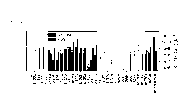
[0024] FIG. 17 shows a mutational screen for enhancing specific binding
to PDGF-6 n-
terminal peptide.
[0025] FIG. 18 shows energy minimized modeled crystal structures of native
RPtag
(large) and RPtag (large) h122I,a253r (blue) with PDGF-6 peptide (orange).
[0026] FIG. 19 shows Kd determination between FITC labeled RPtag (large)
h122I,a253r
and unlabeled PDGF-6 dimer.
DETAILED DESCRIPTION
[0027] Disclosed herein are compositions, methods, systems, and kits useful
in the
expression, identification, and isolation/purification of engineered proteins.
In some
embodiments, a two-part peptide tag system is disclosed that is useful for
affinity purification
and/or specifically identifying tagged proteins. The system is also useful in
aiding solubility
and expression of recombinant proteins while also providing a tag for
identifying and
isolating/purifying the recombinant protein. The disclosed system is also
useful in
performing protein interaction studies.
[0028] The disclosed two parts of the tag system are derived from
bacterial ribose
binding (RB) protein. In some embodiments, the disclosed ribose binding
protein (RP-Tag,
RPtag, Tag protein, Tag peptide, RPtag protein, RPtag peptide may be used to
describe the
presently disclosed proteins and peptides) is from the thermophilic bacterium
Thermoanaerobacter tengcongensis (also referred to as C. subterraneous), and
may be
more stable than other RB proteins. However, other sources of RB proteins, for
use with the
3
CA 03055810 2019-09-06
WO 2018/165328 PCT/US2018/021385
disclosed RB-Tag system, are appropriate. In many embodiments, the disclosed
ribose
binding protein sequence may be altered/mutated to remove a putative N-
terminal
periplasmic localization sequence. In most embodiments, the disclosed RB-Tag
sequences
may also be altered to change naturally-occurring cysteine residues (Cys; for
example, Cys
102) to serine residues (Ser; sequence of the intact protein below, Seq. 1).
Full length sequence of RB Protein from Thermoanaerobacter tengcongensis
lacking the putative periplasmic localization sequence and including a C102S
mutation is shown blow. A break, //, identifies, generally, the separation
between the two fragments (RPtag(large) and RPtag(small) ¨ SEQ ID NO:l.
MKEGKTIGLVISTLNNPFFVTLKNGAEEKAKELGYKIIVEDSQNDSSKELSNV
EDLIQQKVDVLLINPVDSDAVVTAIKEANSKNIPVITIDRSANGGDVVSHIASD
NVKGGEMAAEFIAKALKGKGNVVELEGIPGASAARDRGKGFDEAIAKYPDIK
IVAKQAADFDRSKGLSVMENILQAQPKIDAVFAQNDEMALGAIKAIEAANRQ
GIIVVGFDGTEDALKAIKEGKMAATIAQQPALMGSLGVEMADKYLK //
GEKIPNFIPAELKLITKENVQ
PRtaq proteins
[0029] The disclosed RB protein, from thermophilic bacteria, is very
stable. In many
cases, the disclosed RB protein has a melting temperature of over 100 C. The
disclosed
protein is also highly resistant to denaturants like guanidine hydrochloride
and urea.
Applicants have identified a peptide at the C-teminus of the RB protein that
binds with very
high affinity. Specifically, Applicants truncate the RB protein sequence at
position 257,
generating two RP-Tag fragments. The two fragments are referred to as RP-Tag
Large (a.a.
1-257) and RP-Tag Small (a.a. 258-279; GEKIPNFIPAELKLITKENVQ; SEQ ID NO: 13).
When expressed independently, the two fragments may be engineered to have
short linker
sequences at the C- and/or N-termini. The disclosed fragments may include any
number of
additional amino acids from the RB sequence (i.e. RP-Tag Large may comprise
a.a. 1-260;
and RP-Tag small may comprise a.a. 250-279), or amino acids from some other
source, at
the N- and/or C-termini. Additionally, the disclosed RP-Tag proteins may
include fewer RB
residues (i.e. RP-Tag Large may include a.a. 5-250, instead of a.a. 1-257).
[0030] Various embodiments of the disclosed proteins and peptides may
include one or
more changes selected from one or more of natural amino acid, synthetic amino
acid,
fusion, conjugation, derivatization, mutation, substitution, addition, or
deletion. In many
embodiments, the sequence of the disclosed RP-Tag proteins and peptides may
possess
4
CA 03055810 2019-09-06
WO 2018/165328 PCT/US2018/021385
less than 100% identity to the sequence of tte RB protein, for example less
than 90%, 85%,
80%, 75%, 70%, 65%, 60%, 55%, or 50%, and greater than about 50%, 60%, 70%,
80%,
90%, or 95%. In some embodiments, the disclosed proteins and peptides may
comprise
one or more synthetic amino acids or residues.
[0031] The disclosed proteins and peptides may include one or more
deletions. In some
embodiments, the deletions may be truncations at one or both termini of the
protein or
peptide. In some embodiments, such deletions may aid in enhancing affinity or
reducing
affinity. The disclosed deletions may include from about 1 to about 20
contiguous, or non-
contiguous residues, for example more than about 2 aa, 3 aa, 4 aa, 5 aa, 6 aa,
7 aa, 8 aa, 9
aa, 10 aa, 11 aa, 12 aa, 13 aa, 14 aa, 15 aa, 16 aa, 17 aa, 18 aa, or 19 aa,
and less than
about 20 aa, 19 aa, 18 aa, 17 aa, 16 aa, 15 aa, 14 aa, 13 aa, 12 aa, 11 aa, 10
aa, 9 aa, 8
aa, 7 aa, 6 aa, 5 aa, 4 aa, 3 aa, or 2 aa.
[0032] The disclosed proteins and peptides may have one or more amino
acid changes
in one or more functional and/or structural domains. For example, RPtag(small)
peptide
may include a domain that may aid in binding with another protein or peptide,
such as
RPtag(large), and another domain for stabilizing a bi-molecular complex (for
example
RPtag(large): RPtag(small)) or for stabilizing or destabilizing an
intermediate form.
Binding affinity
[0033] The disclosed RP-Tag proteins and peptides bind with specificity
and with high
affinity to each other. In many embodiments the equilibrium binding constant,
Kd, is in the
nanomolar range, for example less than about 100 nM, 10 nM, 1.0 nM, 0.1 nM,
0.01 nM. In
many embodiments, the Kd is less than about 10 nM. As demonstrated below, in
Figure 1,
Applicants have measured a Kd of about 8 nM for one embodiment, and 2 nM for
another
embodiment. In many embodiments, one or more changes in the amino acid
sequence may
aid in enhancing or reducing binding affinity. Binding affinity may be altered
by adjusting the
kinetics and/or equilibria of the binding reaction. This adjustment may be
accomplished by
modifying the amino acid sequence of one or both RPtag proteins and/or
modifying the
composition of a buffer system. The interaction of these two proteins is
specific and there is
no detectable binding of the RP-Tag proteins to BSA.
[0034] Amino acid substitutions in the sequence of RPtag(small) peptide
are useful in
modulating the affinity for RPtag(large). In some embodiments, amino acid
substitutions in
the sequence of RPtag(large) peptide may be useful in modulating the affinity
for
RPtag(small). For example, amino acid substitutions at positions 2E, 18E, and
21Q may aid
in increasing the affinity of RPtag(small) for RPtag(large). In some
embodiments, the
5
CA 03055810 2019-09-06
WO 2018/165328 PCT/US2018/021385
substitutions may be alanine, while in other embodiments enhancing mutations
may be
other than alanine, and at positions other than 2, 18, and 21.
Buffer systems
[0035] Affinity and specificity may be changed depending upon the
surrounding
environment, for example the solution wherein binding occurs. In many
embodiments,
affinity may be affected by adding one or more organic solvents, alcohols,
disulfide
reducers, aromatics, sugars, salts, denaturants, detergents, etc. In some
embodiments, the
buffer system for the disclosed proteins and peptides may include one or more
of DMSO,
Et0H, Me0H, acetone, glycerol, BME, DTT, PG, imidazole, ribose, sorbitol,
NaCI, KCI,
NH4SO4, MgCl2, CaCl2, NiCl2, MnSO4, Gdn-HCI, urea, Tween20, TritonX-100, SDS.
In
some embodiments, salts may enhance or lessen binding affinity. In one
embodiment,
kosmotropic salts may aid in enhancing binding affinity, while chaotropic
salts may decrease
binding affinity. In many embodiments, NaCI and KCI may aid in stabilizing the
interaction of
RPtag(large) and RPtag(small). In these embodiments, the buffer may include a
salt
concentration of between about 5 mM and 5 M. In many embodiments, the effect
on affinity
may be similar for all peptides and protein, or may be different depending
upon the
sequence of the protein and/or peptide. In other embodiments, one or more
compounds or
molecules may be used to disrupt and/or lessen the disclosed interactions. In
one
embodiment, a pH buffer, denaturant, polyion, or imidazole may be used to
disrupt binding.
In these cases, the solution may help elute a target protein or target peptide
from a solid
support.
[0036] Disclosed herein are buffer systems for promoting and for
disrupting interaction
between the disclosed RPtag proteins. In some embodiments, buffers that
promote binding
may have pH between about 4 and 10, and a kosmotropic salt between about 10 mm
and 5
M. In some embodiments, preferred buffers include about 0.1 M tris or
phosphate pH 8.0, 3
M NaCI for binding. In some embodiments, buffers that may disrupt a RPtag
complex may
have a pH greater than about 10 and less than about 4, may comprise a
chaotropic salt,
may comprise imidazole, and combinations thereof. In some embodiments,
preferred
buffers include about 0.1 M tris or phosphate pH 8.0, 3 M imidazole for
elution.
Protein expression
[0037] The large RP-Tag protein is also useful in aiding the stability and
expression of
other protein sequences to which it is fused. In many embodiments, fusion
proteins, having
the sequence of the Large RP-Tag protein may express to greater than about 400
mg/L
when expressed in bacteria (for example BL21(DE3) E. coli). In some
embodiments, high
6
CA 03055810 2019-09-06
WO 2018/165328 PCT/US2018/021385
expression of stable, functional, fusion proteins may be achieved with pH-stat
fed-batch
bioreactor and methods of using the stated bioreactors.
[0038] The disclosed RP-Tag proteins may be expressed in or from a
variety of
prokaryotic and eukaryotic cell and systems. In some embodiments, the RP-Tag
protein is
expressed from a yeast cell, bacterial cell, mammalian cell, insect cell,
plant cell, etc., such
as Saccharomyces cerevisiae, Pichia pastoris, Human Embryonic Kidney cell,
Chinese
Hamster Ovary Cell, Spodoptera frugiperda, etc. or extracts thereof. In some
embodiments,
the disclosed proteins and peptides may be chemically synthesized.
[0039] The disclosed RP-Tag interaction may be stabilized in the
presence of ribose.
Ribose is bound by the large RP-Tag protein, and its interaction with RP-Tag
Large may
help to stabilize the structure of this fragment and may also help to
stabilize interaction
between the two RP-Tag fragments.
Solid supports
[0040] The disclosed Tag proteins may be affixed to a solid support to
aid in isolating
the complement Tag protein. For example, in some embodiments, the Large RP-Tag
protein
may be affixed to a matrix for a column, and a fusion protein comprising the
Small RP-Tag
protein may be combined with the matrix (either in solution [or batch
processing], or by
adding the RP-Tag fusion protein to a column comprising the solid matrix/RP-
Tag protein, as
in Example 1, below) to isolate and purify the fusion protein. In other
embodiments, the
Small Tag protein is affixed to the column matrix to aid in binding a fusion
protein comprising
the Large Tag protein. Thus, a target protein may be fused to either the Small
or Large Tag
protein, and may be fused to either the C- or N-terminus of either protein. In
some
embodiments, the fusion protein may include a linker sequence between the Tag
sequence
and that target protein sequence. In many embodiments, this linker sequence
may be from
about 1 a.a. to about 30 a.a. in length. In some embodiments, this linker
sequence may add
functionality to the fusion protein, for example by introducing a labelling
sequence, cleavage
sequence, or recognition sequence.
[0041] Suitable resins for immobilization may comprise a bead of
polymeric matrix (for
example but not exclusive to: agarose, Sepharose, dextrans, acrylamide,
bisacrylamide,
silica, methacrylate, and various mixtures and cross linking formulations
thereof), along with
a chemistry for coupling to the peptide or protein (e.g. an aldehyde,
maleimide, N-
Hydroxysuccinimidyl ester, halo-acetyl group, sulfhydryl (activated or free),
hydrazide,
hydrazine, amine, alkyne, azide, carboxyl group, or other moiety commonly
known in the
art), that may or may not be on the end of a spacer which is attached to the
polymer matrix.
7
CA 03055810 2019-09-06
WO 2018/165328 PCT/US2018/021385
[0042] A variety of methods may be used to affix an RP-Tag protein to a
solid support.
In some embodiments, it may be useful to add one or more amino acids to the
RPtag
protein to aid in linking the RPtag protein to the solid support. In other
embodiments, the
linkage may be chemical, for example via cysteine, di-sulfide bond, primary
amines, amide
bonds, or other covalent chemistry. In one embodiment, a Cys residue may be
engineered
in the RPtag protein to allow the protein to link a solid support via a
thioether bond (e.g.
using SULFOLINKTM technology from ThermoFisher Scientific). By another method,
the
RPtag protein or peptide might be immobilized via free amine groups to
aldehyde resin, thus
forming an imine, and then reduced via sodium cyanoborohydride to form a
stable
secondary amine.
Modifications ¨ tags, linkers, reporters, etc.
[0043] The disclosed RPtag proteins may be labeled to aid in visualizing
or locating one
or both proteins. Suitable label and methods of labeling proteins are well
known in the art.
In some embodiments, specific amino acid residues may be targeted for
attaching one or
more labels. In other embodiments, target sequences (for example the linker
sequences
described above) may be added to the RPtag proteins to facilitate labeling. In
some
embodiments the label is visible (e.g. dyes or fluorescent labels), or the
label may be
visualized with detector equipment (e.g. radioactive labels, fluorophore,
radioactive isotopes,
chromophores, metals for electron microscopy like gold and iron, quantum dots,
etc.), or
other labeling techniques well known to those skilled in the art. In one
embodiment, the
RPtag protein is labeled with rhodamine.
[0044] Mutations may be introduced in the Tag protein using a variety of
methods well
known to those of skill in the art. In some embodiments, as discussed above,
additional
amino acids may be added to the Tag protein sequence to create linker
sequences that may
be useful in adding a label, tag, or other adduct to the protein. In other
embodiments, the
amino acid sequence of the Tag protein may be mutated to change one or more
amino acid
residues. In these embodiments, it may be useful to create specific amino acid
substitutions
to help increase or decrease affinity between the two Tag proteins. As one
example, a
Small mutant Tag protein may be engineered to have greater affinity for the
Large Tag
protein to aid in displacing, or competing away the disclosed Small Tag
protein. In other
embodiments, amino acid mutations may help to lower the affinity of the Large
Tag protein
for the Small Tag protein.
8
CA 03055810 2019-09-06
WO 2018/165328 PCT/US2018/021385
Protein stability
[0045] The disclosed Tag protein affinity system is resistant to
conditions that normally
disrupt protein-protein interactions. Typically, protein-protein interactions
are sensitive to
disruption by changes in pH, ion concentrations, temperature, and denaturant
concentration.
For example, typical protein-protein concentrations may be disrupted by
increasing or
decreasing the pH of a solution containing a protein-protein interaction above
about 8.0 pH
or below about 6.5 pH. In many embodiments, the disclosed protein-protein
interaction is
stable in pH above 8.0 pH and below 6.5 pH. In some embodiments, the disclosed
interaction is stable in high concentrations of one or more denaturant
compounds (e.g. urea,
guanidine, etc.), wherein the concentration of denaturant is greater than
about 1M.
Definitions
[0046] "Polypeptide," "protein," and "peptide" are used interchangeably
to refer to or
describe a linear or branched chain of amino acid monomers linked by peptide
bonds.
Individual positions within those chains may be referred to as a "residue," or
"amino acid."
The disclosed polypeptides, proteins, and peptides may be of any length and
comprise any
.. number of natural or synthetic amino acids.
[0047] "Homology," "homologous," "identity," "identical," "similar," and
"similarity" as
used herein refer to a degree of nucleic acid and/or amino acid sequence
similarity between
two optimally aligned nucleic acid or peptide molecules. Percent homology and
identity are
determined by comparing positions in two or more sequences, aligned for
purposes of such
.. a comparison. In many cases, one of skill in the art can use one or more
computer
applications to determine such values, for example BLAST. Comparing equivalent
positions
in different sequences may identify the same residue or nucleotide ¨ this is
referred to as
identity. In contrast, were the equivalent positions have amino acid residues
with similar
characteristics or properties (e.g. size, polarity, charge, etc.) the amino
acids may be
homologous but not identical.
[0048] Non-covalent interactions refer to interactions based on non-
covalent forces,
such as ionic, hydrophobic and hydrogen bond-based interactions. Non-covalent
interactions do not include interactions based upon two atoms sharing
electrons.
[0049] Affinity may be expressed in terms of the equilibrium binding
constant Ka, or
dissociation constant, Kd or KD. Kd is expressed as a concentration and can be
determined
by measuring the association rate constant, Ica, and dissociation rate
constant, kd, and
determining their ratio (kd/ka). One of skill in the art is readily able to
determine affinities
9
CA 03055810 2019-09-06
WO 2018/165328 PCT/US2018/021385
.. using a variety of techniques and methods. Typically, one of skill in the
art may determine
an equilibrium constant or Kd, by varying input concentrations of one
component (here, [RP-
Tag Large] or [RP-Tag Small]) to achieve equilibrium, and measuring the
relative
concentration of the complex (here [RP-Tag Large:RP-Tag Small]). Other
techniques are
able to monitor such interactions in real-time to determine on-rates and off-
rates.
EXAMPLES
Example 1 ¨ Column-based interaction
[0050] One embodiment of the disclosed RP-Tag system was tested by
creating a
column with one component bound to a solid, agarose-based matrix. In these
experiments,
a SulfoLinkTM Immobilization kit (ThermoFisher scientific) was used to affix
RP-Tag Large to
.. a solid support, according to the manufacturer's instructions. For these
experiments, an N-
terminal linker was added to RP-Large that included a Cys residue. One of
skill in the art is
able to select various techniques and chemistries to aid in affixing either RP-
Tag protein to a
solid support matrix.
[0051] The amount of protein linked to the column was determined using a
BOA assay
with Bovine Serum Albumin (BSA) as a standard. This showed that 4.5 mg RP-Tag
Large
was immobilized onto 2 mL of the SulfoLinkTM resin to create a RP-Tag Large-
linked resin.
The linked resin was poured into an included column and the column capped with
a frit
included in the kit (see photos in Figure 2). The column was equilibrated with
several
volumes of 50 mM Tris pH 8.5, 150 mM NaCI, 5 mM EDTA.
Methods
[0052] An E. coli codon-optimized gene encoding tteRP-Tag Large was
synthesized
using solid-state methods. This gene was then cloned into a pET-28a(+)
expression vector.
[0053] Labeled and unlabeled RP-Tag Small proteins were synthesized
solid state,
resuspended in DMSO (1-10 mM final peptide concentration) and stored at -20 C
until
needed. Prior to use, the proteins were thawed and diluted into an appropriate
buffer.
[0054] RP-Tag Large expression and purification
[0055] Chemically competent BL21(DE3) E. coli were transformed with 50
ng
expression plasmid, streaked onto Luria Broth (LB) + 50 mg/L kanamycin agar
plates and
grown at 37 C overnight. Single colonies were then picked and grown in
Fernbach flasks in
LB + 50 mg/L kanamycin at 37 C with continuous shaking at 225 RPM until 0D600
= 0.6.
CA 03055810 2019-09-06
WO 2018/165328 PCT/US2018/021385
The temperature was then dropped to 25 C and the cultures induced with 20
mg/L
Isopropyl 6-D-1-thiogalactopyranoside (IPTG) and grown for an additional 18 h.
[0056] Cultures were submitted to centrifugation to pellet bacterial
cells. Supernatant
was discarded and cell pellets resuspended in 20 mM sodium phosphate pH 8.0,
300 mM
NaCI, 10 mM 2-mercaptoethanol, and 10 mM imidazole. Cells were lysed
enzymatically
(Lysozyme, DNAasel, 5 mM MgSO4 1 hour on ice), cell debris pelleted by
centrifugation,
and the clarified supernatant loaded onto a NiNTA column equilibrated with the
lysis buffer.
Protein was then eluted with a step gradient of imidazole (10 mM - 250 mM),
and protein-
containing fractions pooled and dialyzed against 20 mM sodium phosphate 8.0,
150 mM
NaCI, and 10 mM 2-mercaptoethanol.
[0057] Dialyzed protein samples were flash frozen in liquid nitrogen and
stored at -80 C
until use. Concentrations were determined either using a BCA assay using
Bovine Serum
Albumin as a standard, or by A280 nm using a calculated 280 = 4,470 M-1cm-1.
For pH-stated
fed-batch bioreactor protocols, an identical protocol was used except cultures
were grown in
a 10-L New Brunswick Bioreactor, LB was additionally supplemented with 20 g/L
glucose
and 0.6 g/L magnesium sulfate, and pH was maintained between 6.85 and 6.95,
adding
50% glucose and 1.5% MgSO4 mixture if the pH increased over 6.95 by
peristaltic feed
pump, and 30% ammonium hydroxide if pH dropped below 6.85 by peristaltic feed
pump.
Fed-batch cultures were induced at 0D600 = 6 with 1 mM IPTG. Purified protein
was >95%
pure as judged by SDS-PAGE stained with coomassie brilliant blue R-250.
Example 2 ¨ Solution state affinity.
[0058] 50 nM of RP-Tag Small (see Sequence 3, below) with an N-terminal
Rhodamine
B label was incubated with increasing concentrations of either RP-Tag Large
(see Sequence
2) or Bovine Serum Albumin (BSA) in 50 mM Tris pH 8.0, 150 mM NaCI, 10 mM 2-
mercaptoethanol, and 0.005% Tween 20 for 5 min in black 96-well plates at room
temperature.
[0059] Anisotropy was then measured, and a Kd calculated by fitting the
data to the
equation f = yo + (ymax - yo)*(Ptot + x + Kd - sqrt((Ptot + x + Kd)A2 - 4 *
Rot* x))/(2 * Ptot), where
yo is the baseline anisotropy, ymax is the maximum anisotropy, Rot is the
fixed concentration
of labeled peptide used, x is the variable concentration of protein used, and
Kd is the
measured Kd. Fitting of the data resulted in a calculated Kd of 8 nM for this
interaction, and
detected no binding to BSA (see Fig. 1).
11
CA 03055810 2019-09-06
WO 2018/165328 PCT/US2018/021385
Binding of RP-Tag Small to immobilized RP-Tag Large
[0060] 25 mL of 1 pM Rhodamine-6B labeled RP-Tag Small, in 50 mM Tris pH
8.5, 150
mM NaCI, 5 mM EDTA, was flowed over the column of immobilized RP-Tag Large
protein.
RP-Tag Small bound the column and formed a visibly bright red band (rhodamine
B) at the
top of the resin (see Figure 2). This red band did not appreciably diffuse
even after
extensive washing (>20 column volumes), and was stable for >1 week at room
temperature.
Example 3- Stability of RP-Tag Small:RP-Tag Large interaction
[0061] Table 1 summarizes results from elution tests using a variety of
conditions.
Briefly, after binding, the RP-Tag Small red band was not observed to
appreciably diffuse
and/or elute after washing the column (and band) with various solutions. For
these tests, 10
mL (5 column volumes) of various buffers were added to the column. The tested
buffers
ranged from about pH 1.5-13.7 and about 1 M sodium hydroxide. These results
(see Fig.2)
suggest that this interaction (between RP-Tag Small and Large) possesses among
the
widest compatible pH ranges known for affinity resins.
[0062] Table 1
Elution Buffer (5 column volumes) Result
1 M sodium hydroxide No band diffusion/elution
0.1 M sodium phosphate pH 13.7 No band diffusion/elution
0.1 M sodium phosphate pH 13.0 No band diffusion/elution
0.1 M sodium phosphate pH 12.0 No band diffusion/elution
0.1 M Tris pH 8.5 No band diffusion/elution
0.1 M Tris pH 8.0 No band diffusion/elution
0.1 M Tris pH 7.5 No band diffusion/elution
0.05 M sodium acetate pH 4.6 No band diffusion/elution
0.1 M glycine pH 3.5 No band diffusion/elution
0.1 M glycine pH 2.5 No band diffusion/elution
0.1 M glycine pH 1.5 Complete band elution
0.1 M Tris pH 7.5 + 6 M guanidine
hydrochloride Complete band elution
0.1 M Tris pH 7.5 + 6 M guanidine partial band elution, significant
diffusion
hydrochloride + 10 mM D-ribose within column
0.1 M Tris pH 7.5 + 6 M guanidine no band elution, significant
diffusion within
hydrochloride + 100 mM D-ribose column
0.1 M Tris pH 7.5 + 6 M guanidine No band elution, minor diffusion
within the
hydrochloride + 1 M D-ribose column
0.1 M Tris pH 7.5 + 100 pM unlabeled Partial band elution, slight
diffusion
RP tag peptide throughout the column
[0063] Table 1. Summary of elution trial data. The test system used was a
column
equilibrated with 50 mM Tris pH 8.5, 150 mM NaCI, 5 mM EDTA, and applied 25 mL
1 pM
Rhodamine-6B labeled RP-small peptide in the same buffer. In cases where D-
ribose was
used, the indicated concentration was also included in the equilibration and
loading buffer.
12
CA 03055810 2019-09-06
WO 2018/165328 PCT/US2018/021385
[0064] Diffusion and/or elution of the rhodamine-labelled band required
subjecting the
column to very strong buffers. For example, elution was seen with a buffer
comprising 100
mM glycine and pH 1.5, as well as a buffer comprising 0.1 M Tris pH 7.5 + 6 M
Guanidine-
HCI. Even after elution with these strong buffers, the column was able to be
re-equilibrated
with neutral buffer (specifically 50 mM Tris pH 7.5), and its ability to bind
RP-Tag Small was
restored. These results indicate that the RP-Tag resin can be effectively
washed with high
concentrations of hydroxide (e.g. 1 M sodium hydroxide), low pH buffer (e.g.
100 mM glycine
pH 1.5) and high concentrations of denaturants (e.g. 6 M guanidine
hydrochloride), and still
be regenerated to a functional state.
Example 4 ¨ Binding under denaturing conditions
[0065] Conditions were investigated in which the RP-Tag Large resin would
bind RP-
Tag Small proteins under strongly denaturing conditions (e.g. 6 M Gdn-HCI). RP-
Tag
Large's ability to bind ribose was investigated. RP-Tag Small was bound as
described
above with 10 mM, 100 mM, and 1 M D-ribose. All concentrations of D-ribose
significantly
slowed diffusion of the bound rhodamine band with 5 column volumes (CVs)
washing.
About 100 mM D-ribose stopped virtually all peptide elution from the column,
while at 1 M D-
ribose even diffusion within the column was reduced to modest levels.
[0066] It should be noted that no tested concentration of D-ribose was
able to stop
diffusion of the rhodamine band entirely. In 6 M guanidine the capacity of the
column is
likely reduced and extensive washing would almost certainly cause the target
to leach to
some extent. Nonetheless, these results indicate that the inclusion of
increasing
concentrations of D-ribose can stabilize RP-tag under denaturing conditions,
and make it an
effective purification tool even with high concentrations of denaturants.
Example 5 - RP-Tag Small:Large competition
[0067] Conditions under which the labeled RP-Tag Small could be eluted
at neutral pH
were investigated. These experiments were directed to eluting bound RP-Tag
Small using
unlabeled RP-Tag Small ¨ that is, disrupting the complex by competition. For
these
experiments, 100 pM of unlabeled RP-Tag Small in 0.1 mM Tris pH 7.5 was used.
Slight
diffusion of the rhodamine band within the column, was observed. In addition,
these
competition experiments successfully eluted a small amount of labeled RP-Tag
(small)
protein from the column.
[0068] These results demonstrate that bound RP-Tag proteins may be
competed off the
column under neutral conditions. In some embodiments, higher affinity RP-Tag
proteins may
13
CA 03055810 2019-09-06
WO 2018/165328 PCT/US2018/021385
be engineered to help compete with one or more of the existing RP-Tag
proteins. In some
embodiments, the affinity of the interaction may be modulated by mutating one
or more
residues to raise or lower the interaction's strength/affinity. In some
embodiments, a closely
related protein or peptide may be used for completion and/or a multimeric
peptide used.
Example 6¨ Equilibrium binding
[0069] Equilibrium binding affinities (Kd) of the native
RPtag(large)/RPtag(small)
interaction was compared to a commonly used, commercially available epitope
tag antibody
and its corresponding tag by fluorescence anisotropy (mouse monoclonal
antibody
purchased from ThermoFisher Scientific (4E3D10H2/E3)). In these studies, the
tag
sequence was GHHHHHH (SEQ ID NO: 1) with an N-terminal rhodamine B.
[0070] The indicated concentrations of RPtag (large) and an anti-His tag
antibody
(4E3D10H2/E3 purchased from ThermoFisher Scientific) were incubated with 1 nM
rhodamine labeled native RPtag (small) peptide and 6xHis peptide (Rhodamine-
GHHHHHH), respectively, and fluorescence anisotropy measured. BSA incubated
with
native rhodamine labeled RPtag(small) is included as a control for non-
specific binding. Kd's
were calculated according to the equation f = y0 + (ymax - y0)*(Ptot + x + Kd -
sqrt((Ptot + x
+ Kd)A2 - 4 * Ptot*x))/(2 * Ptot), where y0 is the baseline anisotropy, ymax
is the maximum
anisotropy, Ptot is the fixed concentration of labeled peptide used, x is the
variable
concentration of protein used, and Kd is the measured Kd. We measured a Kd of
0.2 0.1
nM for RPtag(large) binding RPtag(small), and a Kd of 6 1 nM for the
antibody/6xHis tag
pair. There was no detectable binding of RPtag (small) to BSA up to the
indicated
concentrations.
[0071] Results presented in Fig. 5 demonstrated that the Kd of presently
disclosed
RPtag system was about 30 times better than that of the commercially available
system.
Specifically, the calculated Kd for the interaction of
RPtag(large)/RPtag(small) was about 0.2
nM, while the Kd for anti-6xHis and 6xHis is about 6 nM. Further, the
interaction between
the native RPtag (small) and Bovine Serum Albumin (BSA) was also tested. Here
again, no
interaction was detected up to 10 pM BSA. This indicated a >50,000 fold
selectivity for
RPtag (large)/RPtag(small) over non-specific (BSA) interaction.
Example 7 ¨ Thermal stability
[0072] The ability of the disclosed proteins and peptides to function after
being
subjected to thermal stress was also tested. Here again the RPtag (large) and
anti-6xHis
antibody were selected for analysis. Briefly, the RPtag(large) or the anti-
6xHis antibody was
14
CA 03055810 2019-09-06
WO 2018/165328 PCT/US2018/021385
subjected to sequential rounds of boiling and recovery. Specifically, the
proteins were
subjected to sequential rounds of: 5 min boiling in buffer, followed by
recovery for 1 min on
ice. After each round of boiling/recovery, the proteins were assayed for
binding to their
corresponding epitopes and the results plotted.
[0073] RPtag (large) and an the anti-His tag antibody (4E3D10H2/E3
purchased from
ThermoFisher Scientific) were placed in a solution at about 0.1-1 pM [final]
in a buffer of 50
mM Tris pH 8.0, 0.005% Tween20. The protein solutions were repeatedly heated
for 5 min
at 95 C and then recovered on ice for 1 min. After each round of
heating/cooling, an aliquot
was taken and diluted to 100 nM, and incubated with 100 nM either rhodamine
labeled
RPtag (small) or rhodamine labeled 6x-His peptide (sequence: Rhodamine-
GHHHHHH),
and the fluorescence anisotropy measured. Fraction binding was calculated via
the equation
F = (r - rni,n) / (ro - rni,n) where F is the fraction binding, r is the
measured anisotropy, rni,n is the
anisotropy in the absence of binding protein, and ro is the anisotropy before
any boiling
trials.
[0074] As shown in Fig. 6, the anti-6xHis antibody lost greater than 90%
of its binding
capacity after a single round of boiling/recovery. In contrast, Fig. 6 shows
that even after 24
rounds, the RPtag (large) possessed more binding capacity than did the anti-6x
His antibody
after a single round. This indicates that the RPtag (large) system is at least
about 10-fold
more stable than commercially available products.
[0075] A second stress test was performed on the proteins by subjecting
them to a 15
.. min 121 C autoclave cycle, after which the proteins' function was assayed.
[0076] Specifically, RP-tag (large) and the anti-6xHis antibody
(4E3D10H2/E3
purchased from ThermoFisher Scientific) at 5 pM were subjected to a 15 min 121
C
autoclave cycle with slow exhaust to prevent boiling (total time >100 C -60
min) in 50 mM
Tris pH 8.0, 150 mM NaCI, 1 mM EDTA, 10 mM 13-ME, and the Kd measured as
above.
[0077] As expected, the antibody was completely destroyed by this
treatment, losing all
detectable binding to its target peptide (see Fig. 7). By contrast, RP-tag
(large) survived, and
maintained a measurable, albeit impaired, binding affinity (Kd = 53 24 nM).
These results
indicate that the RP-tag (large) protein is extremely stable, and that
alterations to the protein
structure and/or sequence may allow the protein to survive and function in
harsh biological
or medical environments, for example, where existing systems may be
inactivated
completely.
CA 03055810 2019-09-06
WO 2018/165328 PCT/US2018/021385
Example 8- RP-tagged fusion proteins
[0078] To examine the efficacy and specificity of the disclosed
proteins, peptides,
systems, and methods, fusion proteins were created comprising the disclosed
proteins and
peptides, and several biomolecules of interest. In one example, RPtag (large)
or RPtag
(small) were conjugated to a resin of agarose beads. In these experiments, the
RPtag
sequences were engineered to include N-terminal cysteines, which could be used
to
covalently bind activated agarose (via the manufacturer's instructions;
SulfoLinkTM resin
purchased from ThermoFisher scientific). Immobilization efficiencies were
about 2 mg RPtag
(small)/mL resin, and -38 mg RPtag(large)/mL resin).
Protein Purification
[0079] Briefly, codon-optimized DNA coding sequence of each protein was
synthesized
solid state and then sub-cloned into the pET-28a(+) bacterial expression
plasmid.
Chemically competent BL21 (DE3) were transformed with the expression plasmids
and
grown on LB agar + 50 pg/mL kanamycin sulfate at 37 C overnight. Colonies
were picked
and grown in shaker flasks in LB + 50 pg/mL kanamycin sulfate (200 RPM) at 37
C until
0D600 = 0.6. The temperature was then dropped to 25 C and expression induced
with 1
mM isopropyl 3-D-1-thiogalactopyranoside for 16 hrs. Cells were then harvested
by
centrifugation and lysed enzymatically with lysozyme and DNAase in 20 mM Tris
pH 8.0,
300 mM NaCI, 10 mM imidazole, 5 mM MgCl2. Cell debris was pelleted by
centrifugation,
and proteins purified by single-step NiNTA chromatography (10 mM - 500 mM
imidazole
step gradient). Concentrations were determined by absorbance using 595 =
100,000 M-1cm-
1.
Column Production
[0080] SulfoLinkTM resin was purchased from ThermoFisher Scientific. For
immobilization of RPtag (large), 200 mg protein/mL resin was incubated at room
temperature for 1 hour in Tris pH 8.5, 1 mM EDTA. The RPtag(large)-resin was
then washed
and incubated in the same buffer + 10 mM cysteine. Next, the RPtag(large)-
resin was
packed into 1 mL FPLC- columns (Gold Biotechnology, Inc., St. Louis, MO; see
Fig. 8). 38
mg RPtag (large; about 1.3 pmol) and 2 mg RPtag(small; about 0.84 pmol) was
immobilized
per ml of resin. Note that the RPtag(large) was immobilized to the column via
an engineered
N-terminal cysteine. The RPtag (small) peptide was immobilized to the resin
via an
engineered cysteine on its C-terminus (sequence GGC).
16
CA 03055810 2019-09-06
WO 2018/165328 PCT/US2018/021385
Binding Experiments
[0081] After purification of the tagged proteins, each was applied to
its complementary
column to evaluate binding. Fig. 8 provides a schematic of the experiment's
setup. Briefly,
columns were first equilibrated in 20 mM Tris pH 8.0, 150 mM NaCI, 1 mM EDTA
(TNE
buffer), then 25 mL of 1 pM tagged protein in TNE buffer was applied to the
column at a flow
rate of 1 ml/min, then washed with 10 mL of TNE buffer. Columns were cleaned
with 10 mL
100 mM Glycine pH 1.5 between uses. As shown in photos of the columns at right
of Fig. 8,
both tags were effective whether attached to the N- or C- termini of the test
proteins. These
results demonstrate the versatility of the presently disclosed proteins,
peptides, methods,
and systems.
[0082] In some embodiments, immobilization of the RP-tag(small) peptide may
allow for
the use of a high-solubility, expression and solubility enhancing tag on
either terminus of the
protein of interest. In other embodiments, immobilization of the RP-tag(large)
protein may
allow for the use of a small, minimally perturbing tag on the protein of
interest, again at
either terminus.
[0083] As a test for specificity, non-complementary proteins were applied
to each
column using the same procedure described above. In these experiments, only a
small
amount of non-specific binding was observed. This background binding is not
uncommon
and may, in some cases be expected with agarose-based chromatography resins.
In some
cases, color in the photographs was enhanced to aid in visualization. Where
such
enhancement was performed, each panel received identical enhancements.
[0084] Next, N-terminal and C-terminal fusions of both RPtag (large) and
RPtag(small)
with a red fluorescent protein (tagRFP) were constructed. TagRFP allows
visualization of
the proteins (proteins also had an 8Xhis tag on the opposite terminus to aid
in rapid
purification). After purification of the tagged proteins, each was applied,
separately to its
complementary column to evaluate binding.
[0085] Briefly, columns were equilibrated in 20 mM Tris pH 8.0, 150 mM
NaCI, 1 mM
EDTA (TEN buffer), then 25 mL of 1 pM tagged protein in TEN buffer was applied
to the
column at a flow rate of 1 ml/min. Thereafter, the column was washed with 10
mL of TEN
buffer, and the results recorded by photograph (Fig. 8). Columns were cleaned
with 10 mL
100 mM Glycine pH 1.5 between uses. Both RPtags(large and small) were
effective when
attached to either the N- or C- termini of RFP.
17
CA 03055810 2019-09-06
WO 2018/165328 PCT/US2018/021385
Example 9 - Mechanistic Studies:
[0086] To define the mechanism of RPtag (large) and (small) binding, the
reaction order
of the rate-limiting step was determined.
Results
[0087] For these experiments, 1000 nM RPtag(large) was incubated with
increasing
concentrations of native RPtag (small) (from about 0.6 to 10 nM). These
experiments
identified a linear increase in the initial rate of formation of the complex
(RPtag(large):RPtag(small)). This linear increase indicated that the rate
limiting step is first
order with respect to RPtag(small) (Fig. 9 panels a and c).
[0088] Panel a depicts representative association rate kinetics traces
varying native
RPtag small. Rhodamine labeled RPtag small at the indicated concentration was
incubated
with unlabeled RPtag large and the association measured by fluorescence
anisotropy. L-S
complex concentration was calculated by the equation (r - rmin) / (rmax -
rmin)* [S] where r
is the measured anisotropy, rmin is the anisotropy in the absence of any RPtag
large, rmax
is the anisotropy measured in the presence of at saturating RPtag large, and
[S] is the total
concentration of RPtag small used in the experiment. As shown in Fig. 9, black
circles are
data, red lines are fits to the single exponential equation y = y0 + A*(1-exp(-
b*t)) where y0 is
a baseline offset, A is the amplitude of the curve, and b is the first order
rate constant, and t
is time. Dashed cyan lines are simulations of the mechanism shown in f using
the rate
constants detailed herein. The concentrations in the simulations were
multiplied by a
correction factor of 0.925, which is well within the 90% purity specification
of the purchased
peptide.
[0089] Panel c shows initial velocities of traces represented in panels
a (black) and b
(red). The first 5 min of data were fit with a line, and plotted as a function
of concentration.
S(total) is plotted as a function of the total concentration of RPtag small
used in the reaction,
S (corrected) is plotted as a function of the concentration of S corrected for
Keq as detailed
herein. The slope of the line using S (total) is 0.035 min-1, in much worse
agreement with the
data and single exponential fit rate constant than the 0.091 min-1 slope of
the S(corrected)
line. Data shown are mean SE (n = 3).
[0090] Next, 1000 nM RPtag (small) peptide was incubated with increasing
concentrations of RPtag (large) protein (from about 0 to 10,000 nM). These
experiments
resulted in no change in the rate (except at 0 nM RPtag(large)). These results
indicated that
the reaction is 0th order with respect to RPtag (large) (Fig. 9 panels b and
c).
18
CA 03055810 2019-09-06
WO 2018/165328 PCT/US2018/021385
[0091] Panel b presents the representative kinetics traces varying native
RPtag large.
Indicated concentrations of RPtag large were incubated with 1000 nM RPtag
small and
anisotropy measured. All calculations were as in panel a.
[0092] Taken together, these studies demonstrated that the rate liming
step of complex
(RPtag(large):RPtag(small)) formation is first order only with respect to
RPtag (small),
.. consistent with a unimolecular process. VVithout wishing to be limited,
this suggests the
possibility of a conformational change in the RPtag(small) peptide prior to
binding. The
conformational change being from a "non-binding" (S) to "binding" (S*) state.
This
conformation change is then followed by a much faster bi-molecular binding
event (to
RPtag(large)). Again, without wishing to be limited, the data suggest a
missing amplitude in
the binding kinetics. Specifically, this may represent the proportion of the
binding reaction
that occurred in the dead time of the instrument (-1 min), and therefore the
amount of RPtag
(small) that was already in the S*, binding state at the start of the
experiment. From the
missing amplitude and total amplitude, an equilibrium constant for S and S*
can be
calculated (Keq -1.6).
[0093] These values can then be used to correct for the true concentration
of S in the
initial rate plots (Fig. 9 panel c). Using the linear fit from the initial
rate plot, a rate constant
was calculated for the conversion of S to S* of 0.091 min-1. Fitting the
association curves to
a first-order rate law resulted in a rate constant of 0.092 0.001 min-1 (n =
15), which agreed
well with the calculated value.
[0094] Knowing the Keq and kf (forward rate constant) for the reaction, the
reverse rate
constant (kr) of 0.056 min-1 was calculated.
[0095] Resulting kinetics observed in these studies are shown in Fig. 9
panel d. Panel d
shows representative dissociation kinetics of the LS complex. LS complex was
performed by
incubating 1000 nM RPtag large with the indicated concentrations of rhodamine
labeled
.. RPtag small. At the start of the experiment, 0.1 mM unlabeled native RPtag
small was
added and fluorescence anisotropy monitored. Data processing was done as in
panel a,
except the red fits were to the single exponential decay equation y = y0 + A *
exp(-b*t). As
expected, the initial dissociation rates were linear with respect to the LS
complex.
[0096] Fitting to a line yields a rate constant (koff) = 0.0091 min-1
(Fig 9 panel e). In
good agreement, when the kinetics are fit with a first order rate law, a rate
constant of 0.012
0.002 min-1 (n = 15) was observed. Knowing the koff and Kd, the association
rate of the S*
to LS binding interaction can be calculated to be about 4.6 x 10"7 M-1 min-1.
Panel e is a
19
CA 03055810 2019-09-06
WO 2018/165328 PCT/US2018/021385
graph of the initial rates of the traces represented in d. Slope of the fit
line is 0.0091 min*
Data are mean SE (n = 3).
[0097] These rate constants were used to simulate the mechanism shown in
Fig. 9
panel f. When the first 1 min of the kinetics were eliminated, to account for
the 1 min of
dead time the experimental set up, near perfect agreement between the
simulation results
.. and the observed data, and the theoretical fits. Again, without wishing to
be limited, this
agreement indicated that the model accurately represents the mechanism of
RPtag (large)
and (small) binding. Panel f is a schematic representation of one embodiment
of a proposed
mechanism for RPtag binding. S is RPtag (small) in a non-binding conformation,
S* is RPtag
(small) in the binding conformation, L is free RPtag large, LS is the complex
of RPtag large
and small.
Example 10 - Mutagenesis Studies on RPtag(small)
[0098] Mutagenesis studies were carried out on the native RPtag(small)
peptide
sequence to attempt to identify the region required for binding to
RPtag(large), and to
identify specific mutations responsible for improving binding affinity or
improving binding
kinetics. Additionally, these studies might identify a minimal binding domain
in RPtag(small)
leading to a decrease in the size/number of amino acids in the RPtag(small)
peptide.
Effect on binding equilibrium
[0099] First alanine scanning mutagenesis was performed along the
sequence of
RPtag(small). Specifically, each amino acid position in RPtag(small) was
changed to
alanine and the Kd of the resulting peptide measured. The Kds of these alanine
mutants is
shown in Table 2. These studies identified a cluster of amino acids from about
F7 to about
K17 that, when changed to alanine, significantly impaired binding affinity.
This indicated that
the RPtag(large) binding region lies within these about 11 amino acids.
Structurally, this
region corresponds roughly to the 13-sheets on the crystal structure of the
tteRBP (PDB
2I0Y). Interestingly, despite being in the middle of the 13-sheet region, P9
did not impair
affinity when changed to alanine. Surprisingly, 3 mutations significantly
increased binding
affinity to RPtag(large) - E2A, E18A, and Q21A. These positions all lie
outside of the
putative binding region, and should therefore be amenable to incorporation
into the
sequences without disrupting the necessary interactions for binding.
.. Table 2 This table provides thermodynamic and kinetic constants measured
for RPtag small
sequences. All data are mean SE, (n= 3-15). All constants are for the
transitions
diagramed in Fig. 9e. ND indicates that the parameter could not be determined.
All
CA 03055810 2019-09-06
WO 2018/165328
PCT/US2018/021385
measurements were made by fluorescence anisotropy using N-terminal rhodamine
tagged
peptides with unlabeled RPtag (large) as above in 50 mM Tris pH 8.0, 0.005%
Tween 20.
Peptide Ka (nM) Icon (M-1 koff (min-1) Keg
kf (min-1) kr (min-1)
min-1) (SS*)
(x107)
native 0.21 0.03 4.3 0.1 0.0091 1.6
0.092 0.056
0.002 0.1 0.001 0.001
g1a 0.29 0.03 1.6 0.1 0.0047 2.3
0.086 0.037
0.0003 0.1 0.004 0.001
e2a 0.10 0.02 5.1 0.7 0.0053 1.9
0.084 0.044
0.0007 0.1 0.009 0.004
k3a 0.49 0.02 1.1 0.1 0.0053 ND
ND ND
0.0007
i4a 0.26 0.01 2.4 0.1 0.0060 0.9
0.086 0.098
0.0002 0.1 0.004 0.016
p5a 0.23 0.02 11 1 0.024 1.8
0.093 0.052
0.002 0.1 0.003 0.001
n6a 0.34 0.04 0.45 0.0015 1.3
0.082 0.068
0.09 0.0003 0.2 0.003 0.010
f7a 2.0 0.2 3.1 0.1 0.063 2.2 0.080
0.036
0.003 0.1 0.001 0.002
i8a 1.1 0.1 8.6 0.9 0.090 0.8 0.078
0.046
0.009 0.2 0.001 0.012
p9a 0.18 0.01 1.4 0.2 0.0024 2.7
0.092 0.040
0.0004 0.5 0.011 0.010
el 1a 0.80 0.07 0.46 0.0067 1.9
0.080 0.042
0.06 0.0005 0.1 0.004 0.004
112a 4.7 0.2 4.3 1.2 0.20 0.06 1.1
0.089 0.081
0.1 0.004 0.006
k13a 1.1 0.1 1.7 0.1 0.018 ND
ND ND
0.001
114a 8.9 0.5 ND >2 1.8 0.075
0.044
0.1 0.005 0.005
115a 4.5 0.4 25 6 1.1 0.3 1.4 0.077
0.053
0.1 0.006 0.002
t16a 1.2 0.1 7.5 0.3 0.089 2.5
0.071 0.038
0.003 0.8 0.006 0.011
k1 7a 0.54 0.03 1.0 0.1 0.0057 ND
ND ND
0.0002
e18a 0.056 0.013 16 0.8 0.0087 1.1
0.054 0.048
0.0004 0.1 0.005 0.006
n19a 0.26 0.02 14 1 0.037 1.6
0.089 0.057
0.003 0.1 0.003 0.008
v20a 0.18 0.03 6.8 0.5 0.012 1.5
0.073 0.050
0.001 0.1 0.003 0.002
g21a 0.10 0.01 3.5 0.4 0.0034 0.8
0.087 0.11 0.01
0.0003 0.1 0.005
Nd1 0.25 0.03 3.4 0.1 0.0088 2.7
0.064 0.024
0.0001 0.2 0.008 0.004
Nd2 0.067 0.017 5.3 1.3 0.0035 0.7
0.063 0.086
0.0008 0.1 0.003 0.002
Nd3 0.14 0.01 8.8 0.3 0.012 ND
ND ND
0.001
Nd4 0.30 0.02 9.4 0.4 0.028 ND
ND ND
0.001
Nd5 0.59 0.08 9.6 0.1 0.056 ND
ND ND
0.001
Nd6 0.54 0.05 12 1 0.065 ND
ND ND
0.003
21
CA 03055810 2019-09-06
WO 2018/165328
PCT/US2018/021385
Peptide Ka (nM) ken (M-1 keff (min-1) Keg kf
(min-1) kr (min-1)
min-1) (SS*)
(x107)
Nd7 2.4 0.1 1.8 0.4 0.042 ND ND ND
0.010
Nd8 0.59 0.04 9.3 0.3 0.055 ND ND ND
0.002
Nd9 4.9 0.4 8.2 0.9 0.40 0.04 ND ND ND
Nd10 5.9 0.2 4.3 0.7 0.25 0.04 ND ND ND
Nd11 16 1 7.1 3.8 1.2 0.6 ND ND ND
Nd12 520 49 ND >2 ND ND ND
Nd13 84,000 9,600 ND >2 ND ND ND
Nd14 >100,000 ND ND ND ND ND
Nd15 >100,000 ND ND ND ND ND
Nd16 >100,000 ND ND ND ND ND
Nd17 >100,000 ND ND ND ND ND
Cd1 0.55 0.04 1.1 0.1 0.0061 2.5 0.10
0.01 0.042
0.0006 0.3 0.007
Cd2 1.1 0.1 9.8 0.3 0.11 0.01 3.5
0.15 0.02 0.043
0.3 0.005
Cd3 1.2 0.1 11 2 0.12 0.02 3.1
0.014 0.045
0.3 0.01 0.007
Cd4 1.0 0.1 22 2 0.22 0.02 1.3
0.014 0.11 0.01
0.1 0.01
Cd5 3.6 0.2 11 7 0.39 0.23 ND ND ND
Cd6 200 11 ND >2 ND ND ND
Cd7 30,000 2,700 ND >2 ND ND ND
Cd8 >100,000 ND ND ND ND ND
Cd9 >100,000 ND ND ND ND ND
Cd10 >100,000 ND ND ND ND ND
Cd11 >100,000 ND ND ND ND ND
Cd12 >100,000 ND ND ND ND ND
Cd13 >100,000 ND ND ND ND ND
Cd14 >100,000 ND ND ND ND ND
Cd15 >100,000 ND ND ND ND ND
Cd16 >100,000 ND ND ND ND ND
Nd10,Cd3 57 2 ND >2 ND 0.32 0.13 0.050
0.012
Nd10,Cd3,e18a 71 1 ND >2 ND ND ND
Nd10,Cd5 250 10 ND >2 ND ND ND
Nd8,Cd3 11 1 ND >2 6.5 0.095 0.015
0.5 0.013 0.003
Nd8,Cd3,e18a 13 1 ND >2 ND ND ND
Nd8,Cd4 14 1 ND >2 ND ND ND
Nd6,Cd3 7.3 0.3 ND >2 ND ND ND
Nd6,Cd3,e18a 8.2 0.3 ND >2 ND ND ND
Nd6,Cd4 9.9 1.1 ND >2 ND ND ND
Nd3,p5a,e18a 0.63 0.03 9.2 0.4 0.058 ND ND ND
0.002
Nd2,Cd3 0.56 0.04 8.2 0.3 0.046 0.6 0.081
0.14 0.01
0.002 0.1 0.001
Nd2,Cd3,e18a 0.13 0.02 40 2 0.053 0.3 0.084
0.26 0.04
0.002 0.1 0.006
Nd2,Cd4 0.41 0.01 20 2 0.079 0.7 0.093
0.13 0.01
0.006 0.1 0.001
Nd2,Cd3,p5a 0.64 0.07 26 1 0.16 0.01 0.6
0.093 0.16 0.03
0.1 0.007
Nd2,Cd3,p5a,e1 0.56 0.05 33 2 0.18 0.01 0.7
0.086 0.13 0.01
8a 0.1 0.002
Nd2,Cd4,p5a 0.82 0.02 22 1 0.18 0.01 0.8
0.11 0.01 0.14 0.01
22
CA 03055810 2019-09-06
WO 2018/165328 PCT/US2018/021385
Peptide Ka (nM) ken (M-1 koff (mini Keg kf
(min-1) kr (min-1)
min-1) (SS*)
(x107)
0.1
Nd2,e18a 0.13 0.03 6.5 0.7 0.0083 0.4
0.044 0.13 0.02
0.0010 0.1 0.001
Nd2,p5a,e18a 0.047 0.018 28 2 0.013 0.3
0.051 0.19 0.02
0.001 0.1 0.001
Nd2,k3r,p5a,e18 0.26 0.02 3.4 0.1 0.013 ND ND
ND
a 0.001
Nd2,k3a,e18a 0.88 0.01 2.6 0.1 0.032 ND ND
ND
0.001
Nd2,k3a,p5a 0.55 0.01 2.4 0.1 0.0090 ND ND
ND
0.0004
Nd2,e18a,v20a,q 0.36 0.02 3.6 0.3 0.023 0.8
0.045 0.060
21a 0.001 0.1 0.002 0.005
Nd2,p5a,e18a,v2 0.16 0.03 20 1 0.013 0.4
0.050 0.14 0.03
0a,q21a 0.001 0.1 0.003
[00100] Next, sequential truncation mutagenesis from both the N- and C-
termini of the
peptide was performed (see Table 2). Beginning at about position Nd7 (deletion
of 7 amino
acids from N-terminus) impairments of -10 fold or greater were identified.
This level of Kd
reduction was also seen with Cd5 mutations (removal of 5 amino acids from C-
terminus).
These truncations, respectively, correspond to the F7 and K17 identified above
in the
alanine scanning studies.
[00101] A second region of the irregular 13-sheets was found between about L12
and K17.
This region appears to also be involved in binding the RPtag(large) protein,
like the first
identified region between about F7 and El 1. In this second region, Kd
impairments of
>1000-fold resulted from truncating into the region from either the N or C
terminus.
Surprisingly, Nd8 (removing the first 8 amino acid positions while leaving P9
as the N-
terminal amino acid) resulted in improving the binding affinity for
RPtag(large), relative to the
truncations to positions 7 or 9. This data aligns well with the observation
that mutation P9A
did not significantly impair the binding affinity, but A mutations at
positions 8 and 11 did.
Effect of binding rates
[00102] The library of mutant RPtag(small) peptides, described above, was next
analyzed
to measure off rates (koff). The on rate was also calculated from measured
koff and Kd (S*
LS transition), except instead of using unlabeled native peptide as the
competitor, we used
unlabeled Nd2,P5A,E18A (the tightest binding RPtag(small) peptide identified).
These
studies showed that kon and koff roughly tracked with Kd. However, there were
notable
exceptions. For example, although mutation P5A (proline at position 5 of
RPtag(small)
changed to alanine) did not have a significant effect on Kd, the mutation
increased the kon
and koff by a factor of -7. This indicates, without wishing to be limited,
that P5 may
23
CA 03055810 2019-09-06
WO 2018/165328 PCT/US2018/021385
constrain the structure of the peptide. The increased flexibility imparted to
the peptide by
the P5A mutation may decrease the energy barrier required for
binding/unbinding. N6A had
the opposite effect, decreasing the on and off rates without having a
significant effect on Kd,
indicating that this mutation may increases the energy barrier.
[00103] Forward and reverse rate constants for the SS* transition
revealed additional
notable mutants. Measured Keq's and kf's as well as the calculated kr's for
the alanine
scanning and truncation library peptides were all similar, with the exception
of K3A, K13A,
K17A, Nd3 truncations and further, and Cd5 truncations and further. A Keq or
kf (and
correspondingly kr) could not be determined for these mutants as the
association reaction
appeared to be completed within the dead time of the instrument (-1 min). Of
note, the Nd3
deletion corresponds to deletion through K3, and Cd5 corresponds to deletion
through K17.
This indicated that lysines in the peptide may play a significant role in
limiting the rate of LS
complex formation. Moreover, again without wishing to be limited, the rate
limiting
structure/interaction may be alleviated by the mutation or removal of either
lysine.
[00104] The kinetics data described above has at least two possible
interpretations.
Either these mutations cause a substantial increase in the rate of the SS*
transition, or they
shift the Keq of SS* such that it heavily favors S* (or some combination of
the two). VVithout
wishing to be limited, the end result may be a substantial increase in the
rate of formation of
the LS complex. Of the 3 lysines, only K3 falls outside the proposed
RPtag(large)-binding
region, which may allow the making mutations at or truncations of K3 much more
readily
than at either K12 or K17.
Mutant RPtaq(small) peptides with one or more alanine substitutions and/or
truncations
[00105] Several of the mutations identified above, in the alanine
scanning mutagenesis
and truncation mutagenesis, were combined and analyzed. These experiments were
intended to potentially identify two peptides: the smallest peptide with
antibody-like binding
affinity to RPtag (large) (Kd 108 M) and fastest kinetics, and the peptide
with the tightest
binding regardless of size or kinetics.
[00106] These studies identified the Nd8 truncation with the Cd4 truncation
(Nd8Cd4) as
having favorable characteristics in terms of size, kinetics, and Kd. This
peptide has a size of
9 amino acids, a Kd of - 14 nM, and binding/unbinding kinetics completed
within the dead
time (see Table 1; apparently meeting the criteria of the first desired
peptide). Next, Nd2,
p5a, and e18a were combined and analyzed. This peptide possessed a Kd of -47
pM,
which appeared to meet the criteria for the second desired peptides.
24
CA 03055810 2019-09-06
WO 2018/165328 PCT/US2018/021385
[00107] These two identified RPtag(small) mutant peptide sequences were
subjected to
additional testing, described below. Their sequences are:
PAELKLITK "RPtag(small) (fast)" or "Nd8Cd4)"
KIANFIPAELKLITKANVQ "RPtag(small) (tight)"or "Nd2p5ae18a"
Example 11 -Analysis of pH profile
[00108] The Kds of Nd8,Cd4 for RPtag(large) was measured in the presence of
buffers of
different pHs ranging from 1.5 to 13. These studies found that native, Nd8Cd4,
and
Nd2p5ae18a all showed good stability over a wide pH range, and possessing a
maximum
affinity between pH 4-10, with a relative maximum at -pH 8 (Fig. 10).
[00109] For these experiments, Kds were measured by fluorescence anisotropy as
described above in the following buffers, all at 100 mM with 0.005% Tween 20:
glycine pH
1.5, glycine pH 2.0, glycine pH 3.0, acetate pH 4.5, 2-(N-
morpholino)ethanesulfonic acid pH
6.0, tris pH 7.0, tris pH 7.5, tris pH 8.0, tris pH 8.5, borate pH 10.0,
phosphate pH 11.5,
phosphate pH 13Ø
Reagent Screening
[00110] To determine reagent compatibility with the disclosed system, an
additive screen
was performed, wherein the Kd between RPtag (large) and 3 RPtag (small)
peptides -
native, Nd8Cd4, and Nd2p5ae18a - were tested in the presence of several common
buffer
additives (Table 3).
Table 3. Buffer additive screen. Kd's were measured as above in 100 mM Tris pH
8.0,
0.005% Tween 20, and the indicated buffer additives. Shown are Kd's in nM.
Buffer Additive native Nd8Cd4
Nd2,p5a,e18a
No Additive 0.21 14 0.037
DMSO (20%) 0.17 140 0.023
Et0H (20%) 0.17 733 0.10
Me0H (20%) 0.11 34 0.056
acetone (20%) 0.15 22 .019
glycerol (20%) 0.034 27 <0.001
BME (10%) 5.3 740 6.7
DTT (100 mM) 0.51 33 0.073
PG (50%) 19 4600 49
imidazole (3M) >10000 >10000 >10000
ribose (3M) 0.023 42 <0.001
sorbitol (3M) 2.5 16 <0.001
NaCI (3M) 0.0027 0.15 <0.001
KCI (3M) <0.001 0.066 0.011
NI-14SO4 (3M) 1.5 2.8 44
MgCl2 (3M) 124 240 350
CA 03055810 2019-09-06
WO 2018/165328 PCT/US2018/021385
Buffer Additive native Nd8Cd4
Nd2,p5a,e18a
CaCl2 (3M) 330 >10000 150
NiCl2 (100mM) 3.2 42 2.2
MnSat (10mM) 160 160 22
Gdn-HCI (6M) >10000 >10000 >10000
urea (8M) >10000 >10000 >10000
Tween20 (2%) 0.49 45 4.7
TritonX-100 (2%) 0.64 180 0.046
SDS (2`)/0) >10000 >10000 >10000
[00111] These experiments demonstrated that the disclosed proteins,
peptides, and
systems were robust in the presence of a number of organic compounds (DMSO,
methanol,
ethanol, glycerol, and acetone), reducing agents (DTT), and detergents
(Tween20 and
TritonX-100). Interestingly, kosmotropic salts such as NaCI and KCI
significantly stabilized
the interaction between RPtag(large) and RPtag(small). In particular, the
RPtag(small)
mutant peptide Nd8Cd4 showed an increased affinity of about >100-fold. In
contrast,
addition of chaotropic salts, such as MgCl2 and CaCl2, resulted in a
destabilized LS
complex. Surprisingly, addition of imidazole significantly destabilized the
interaction of
RPtag(small) and RPtag(large). Under the conditions of this experiment all
apparent binding
affinity between the two RPtags was removed.
[00112] Next the affinity of RPtag(small) mutant peptide Nd8Cd4 was analyzed
as a
function of NaCI and imidazole. These studies identified a dose-dependent
enhancement of
affinity in the presence of NaCI, and impairment of affinity with imidazole
(not shown).
Example 12 - Native Elution Condition
[00113] Because of the observed stabilizing character of NaCI and
destabilizing character
of imidazole on Nd8Cd4 binding, these reagents, among others, were
incorporated into a
novel buffering system for use with the disclosed proteins, peptides, systems,
and methods.
In particular, the effect of this novel buffer on binding of RPtag (small)
Nd8Cd4 to an
immobilized RPtag(large) column was studied. These experiments included
eluting both (1)
rhodamine labeled peptide and (2) N-terminally RPtag(small) Nd8Cd4 tagged with
tagRFP
from a column under native conditions. Generally, native conditions is taken
to mean at or
near biological conditions under which many proteins are folded, e.g. pH 4 - 9
at moderate
temperature (4 C - 30 C) in the absence of denaturants (e.g. guanidine
hydrochloride,
urea, SDS).,This is in contrast to denaturing conditions, which generally rely
on extreme pH
(e.g. denaturants, or high temperature to unfold proteins.
26
CA 03055810 2019-09-06
WO 2018/165328 PCT/US2018/021385
[00114] Briefly, columns, as described above, were first equilibrated with
10 mL 100 mM
Tris pH 8.0, 3 M NaCI, 0.005% Tween20. Next, 1 mL of 10 uM rhodamine-tagged
Nd8Cd4
or tagRFP containing an N-terminal RPtag (small) Nd8Cd4 tag was applied in the
same
buffer. The column was then washed with 10 mL of the same buffer. Finally,
bound target
molecules were eluted with 10 mL of 100 mM Tris pH 8.0, 3 M imidazole, 0.005%
Tween20.
[00115] These experiments demonstrated that both the protein and the peptide
could be
eluted as a tight band. This demonstrated the efficacy of the disclosed
buffering system for
use in binding and elution of peptides and proteins at neutral pH. These same
conditions
caused native and Nd2p5ae18a peptides to bind in a tight band. Although these
peptides
were able to be eluted, instead of eluting as a tight band, the eluted band
spread through
the resin, creating a diffuse band. This suggested that modification of the
disclosed
buffering system for these sequences may be beneficial.
Example 13 ¨ Affinity-Based Precipitation/Pull-Down
[00116] The disclosed proteins and peptides were used in
precipitation/pull-down assays.
Specifically, rhodamine was tagged with the RPtag(small) Nd8Cd4 peptide and
RPtag(large)
was immobilized on a resin, as described above.
[00117] Briefly, 100 pL 1000 nM rhodamine labeled RPtag(small) peptide
was incubated
with 2.5 uL wet settled RPtag (large) resin generated as (as described above)
in phosphate
buffered saline (PBS; Gibco) + 1% BSA + 0.005% Tween 20 at 4 C for 60 min
with
continuous orbital shaking. Samples were then washed 3x with 500 pL of the
same buffer
with no peptide to remove unbound peptide. Bound RPtag(small) peptides were
then eluted
with 100 pL 100 mM glycine pH 1.5 + 0.005% Tween20, and the pH raised by
adding 1 pL 1
M borate pH 10Ø The amount eluted was calculated from the blank subtracted
fluorescence of the elution versus the load. After each elution, resin was
washed once with
500 pL 6M GdnHCI and once with 500 pL phosphate buffered saline (Gibco) + 1%
BSA +
0.005% Tween 20, before repeating the assay. Data shown in Fig. 11 are mean
SE (n=3).
[00118] Sequential precipitation/pulldown trials were performed with the
rhodamine
labelled RPtag(small) Nd8Cd4 peptide, and the RPtag(large)-resin. After
binding and pull-
down, the rhodamine-peptide was eluted in 100 mM glycine pH 1.5, the column
was washed
with water and 6 M guanidine hydrochloride, and then re-equilibrated with
buffer (50 mM
Tris pH 8.0, 10 mM EDTA). RPtag(small) Cd12 peptide as used as a negative
control due
to its similar size (9 amino acids) and markedly lower binding affinity for
RPtag(large).
27
CA 03055810 2019-09-06
WO 2018/165328
PCT/US2018/021385
[00119] Fig. 11 shows that the rhodamine labeled RPtag(small) Nd8Cd4
peptide was
repeatedly precipitated in these assays with little to no loss of binding
capacity. Surprisingly,
washing with 6 M guanidine hydrochloride did not appear to affect binding
capacity, even
after 7 sequential pulldowns, at which point the experiment was stopped (Fig.
11). As
expected, no binding of the control peptide could be detected. These
experiments show
that the disclosed proteins, peptides, systems and methods are very specific.
Moreover,
binding capacities identified in these experiments were within the range of
existing
commercial affinity/pulldown kits (e.g. anti-myc agarose resin capacity from
ThermoFisher
-102 nmol/mL RPtag resin capacity -200 nmol/mL)
Example 14- Affinity-Based Detection
[00120] The disclosed proteins and peptides were used for detection assays. In
these
experiments, RPtag (small) was recombinantly fused to an alkaline phosphatase.
Specifically, the RPtag(small) peptide Nd8Cd4 was fused to the N-terminus of a
highly
active monomeric alkaline phosphatase (PhoX class) from Pasteurella multocida.
RPtag
(large) with an engineered cysteine was immobilized to a white, polystyrene 96-
well plate via
activated maleimide (ThermoFisher) according to the manufacturer's
instructions. Control
wells were blocked with cysteine. 100 pL of 12.4 pM RPtag(small) Nd8Cd4-
labeled alkaline
phosphatase was added to both the experimental and control wells and then
incubated for
1.5 hrs at room temperature. Wells were washed 3x in 50 mM Tris pH 8.0, 150 mM
NaCI,
1% BSA, 0.05% Tween 20, and then either assayed with 0.25 mM CSPD purchased
from
ThermoFisher (a luminescent alkaline phosphatase activity probe) and measured
immediately at room temperature, or 2 mM p-nitrophenyl phosphate (an
absorbance alkaline
phosphatase reagent) in 50 mM Tris pH 9.5, 150 mM NaCI, 100 pM CaCl2, 0.2 %
Tween 20)
and measured after 60 minutes at room temperature. Measurements were taken on
a
SpectraMax i3 (Molecular Devices). For luminescence, the plate was placed into
the
instrument, mixed for 60 seconds in the dark, and then all wavelengths were
collected from
each well. For absorbance, after 60 minutes the samples were transferred to a
transparent
96 well plate and the absorbance measured at 405 nm.
[00121] As shown in Fig. 12, both the CSPD and p-nitrophenyl phosphate
reagents
resulted in a significant difference between experimental wells(containing
RPtag(large)
immobilized) and the control wells (no RPtag(large)) with their respective
signals, indicating
that this system is useful for detection, specifically in ELISA (Fig. 12).
28
CA 03055810 2019-09-06
WO 2018/165328 PCT/US2018/021385
Example 15 - Mutagenesis Studies to Generalize Binding
[00122] Additional mutagenesis studies were carried out on the RPtag(large)
and
RPtag(small) sequences to further study binding. First, the ability to
engineer compensatory
mutations in each protein was investigated. Second, native RPtag(large)
peptide sequence
was modified to study this protein's ability for expanded
recognition/specificity.
Compensatory mutations in RPtad(larde) and RPtad(small)
[00123] 11 PDB crystal structures of periplasmic sugar-binding proteins,
from diverse
species, were aligned to further investigate residues involved in binding
specificity (Fig. 13).
Residues indicated in Fig. 13 are those that correspond in in RPtag(small) and
RPtag(large)
after aligning in 3D space. Specifically, shown are position 14 on RPtag small
and 137 on
RPtag large. Numbering is according to the full sequence of the native RPtag
large
construct used in this document including all tags. Alignments and renderings
were done
with PyMol.
[00124] Of the 11 RPtag(small) PDB crystal structures investigated, 8
sequences showed
strong conservation of leucine (L) or valine (V) at position 14. 7 of the 11
RPtag(large) PDB
crystal structures, possess an aromatic residue at position 137.
[00125] The possibility of creating compensatory mutations in the two
sequences was
investigated by modifying position 14 of RPtag(small) and position 137 of
RPtag(large).
First, a positively charged lysine (K) was introduced at position L14 of
RPtag(small) (Fig.
14). This substitution significantly impaired binding of the mutant
RPtag(small) to native
RPtag(large), decreasing its affinity even more than the L14A, mutation (Table
2), which
was the most destabilizing mutant identified in the alanine scanning
mutagenesis study.
Next, position F137 of RPtag(large) was mutated to aspartic acid (D), which is
a negatively
charged residue under physiologic conditions. This substitution was introduced
into
RPtag(large) to balance the charge with the L14K substitution, and help
restore interaction
between the two residues. As shown at Fig. 14, this substitution substantially
restored the
binding affinity of L14K RPtag(small) for F137D RPtag(large). In other
embodiments,
different compensatory mutations may be introduced at F137 and L14, as well as
other
positions in the two RPtag proteins.
[00126] These experiments demonstrate that applicants have identified the
pocket/cleft in
the RPtag (large) protein useful for interacting with the RPtag(small)
peptide. Further, as
one of skill in the art would understand, additional positions, mutations, and
substitutions in
the RPtag protein sequences may aid in producing additional binding
enhancements.
29
CA 03055810 2019-09-06
WO 2018/165328 PCT/US2018/021385
Modifyino specificity of RPtao(laroe) for non RPtacffsmall) tamet proteins
(PDGF-13)
[00127] PDGF-13 was selected as a target protein for these studies
because of its clinical
importance to diseases such as cancer and macular degeneration. In addition,
the
physically accessible N-terminus of PDGF-13 possesses some homology to
RPtag(small)
(Fig. 15; showing PDB ID: 1PDG, with N-termini highlighted in magenta; the
structure was
rendered in PyMol, and alignments shown at right were done in Clustal Omega).
[00128] An initial Kd was measured between RPtag(large) and the PDGF-13 N-
terminal
peptide (Fig. 16). These experiments determined a value of 7.3 pM for the
initial Kd. Next,
competition experiments were performed using the mutant RPtag(small) peptide
Nd2,P5A,E18A. This study determined that binding of RPtag(large) to the PDGF-
13 N-
terminal peptide could be disrupted by unlabeled Nd2,P5A,E18A (tight) peptide,
suggesting
a common binding site on RPtag(large) for the two peptides.
[00129] Mutation studies were next performed to identify residues in
RPtag(large) that
could enhance binding to PDGF-13 peptide. For these studies, residues within 5
A of the
putative RPtag(small) binding cleft were identified - H122, D126, K129, G130,
F137, A253,
L233, and M270. Other positions within that area are 1108, V120, S121, H122,
1123, A124,
S125, D126, K129, G130, M133, F137, F237, E241, L244,1247, K248, G250, A253,
A254,
T255, 1256, A257, Q258, Q259, M263, L266, M270, K273, Y274, L275, and K276.
Numbering for RPtag(large) for these studies is relative to
"NativeRPtag(large)_with_Tags_and_Cys" provided below. This protein includes a
19 aa
leader sequence (MGSSCHHHHHHSQDPNSSS). Next, 31 mutants of RPtag(large) were
generated at a subset of 32 amino acids (H122, D126, K129, G130, F137, A253,
L233, and
M270) proximal to non-conserved residues between RPtag(small) and PDGF-13 when
modeled in the crystal structure. The Kd of each mutant was then measured to
identify
mutations that improved RPtag(large) affinity for PDGF-13 peptide, but not for
the a control
RPtag(small) peptide (Nd2Cd4) (Fig. 17). Briefly, the data presented in Fig.
17 are aligned
such that the wt affinities are in-line with the gray dashed line, so enhanced
affinity and
specificity is seen when the blue (PDGF-13) bars are above the line and the
red bars
(Nd2Cd4 control) are below the line.
[00130] These studies identified two positions where such mutations (with
enhanced
binding for PDGF-13 peptide, but decreased binding for Nd2Cd4) occurred, H122
and A253.
Two substitutions at these positions that provided the best affinity for PDGF-
13 peptide were
H122L and A253R. This double mutant RPtag(large) exhibited a enhanced binding
affinity
for PDGF-13 peptide (Kd = 1.5 pM versus a Kd of 7.3 pM for binding to native
RPtag(large))
CA 03055810 2019-09-06
WO 2018/165328 PCT/US2018/021385
and a reduced affinity for RPtag(small) (Nd2Cd4) (Kd = 3.1 nM versus a Kd of
0.41 nM for
binding to native RPtag(large)).
[00131] The modeled energy-minimized structure of the RPtag(large) H122L and
A253R
double mutant suggested a new hydrophobic interaction between PDGF-13 peptide
L5 and
RPtag(large) peptide L122, and a new ion pair between PDGF-13 peptide E15 and
RPtag(large) peptide R253 (Fig. 18). Briefly, Fig. 18 is a structural
rendering using the PDB
2i0y structure as a basis, the aligned corresponding residues in the RPtag
small portion of
the protein to those of the PDGF-13 peptide (top) were mutated and the energy
minimized in
PyMol. This was repeated with the h122I and a253r mutations (bottom)
introduced in the
RPtag large portion of the protein. In the double mutant energy minimized
structure, a new
hydrophobic contact between L122 and L5 was identified along with a new ion
pair between
R253 and E15 in the double mutant not seen in the single mutant.
[00132] This provides a plausible structural explanation for this tighter
binding. Notably,
the double mutant RPtag(large) bound purified PDGF-13 dimer even tighter, with
a Kd = 75
nM. This further-enhanced binding is likely because of the proximity and
corresponding high
local concentration of two N-termini in the PDGF-13 structure (Fig. 19).
Briefly, the indicated
concentration of recombinant purified, and refolded PDGF-13 dimer (see
protocol below) was
incubated with 100 nM RPtag large labeled with FITC. After 1 h incubation at
room
temperature, fluorescence anisotropy was measured, and the resultant curves
were fit to a
standard binding equation.
[00133] These studies show that RPtag(large) can be modified to select for
binding to
proteins other than RPtag(small) and its use as a monobody-type protein. One
of skill in the
art would understand that additional techniques may provide for further
enhancements in
both binding specificity and affinity of RPtag (large) for PDGF-13. For
example, library based
selection techniques such as phage and yeast display may be used to evolve
RPtag (large)
protein sequences with enhanced affinity for PDGF-13 as well as other target
proteins.
Potential target proteins may include, without limitation, other PDGF
isoforms, vascular
endothelial growth factor (VEGF), VEGF receptor, erbB-2, HER2/neu, a-tubulin,
amyloid-p
(1-38, 1-40, 1-42 and other fragments), Estrogen receptors, TNF-a, Lupus
anticoagulant
antibodies, among others.
[00134] Materials and Methods
[00135] > N ative R Ptag (I arg e)_with_Tag s_a nd_Cys (RPtagLarge sequence
proper starts
at MKEGKT...., all mutant numbering in here is done based off of this sequence
including
the tags and N-terminal Met.
31
CA 03055810 2019-09-06
WO 2018/165328 PCT/US2018/021385
MGSSCHHHHHHSQDPNSSSMKEGKTIGLVISTLNNPFFVTLKNGAEEKAKELGYKIIVEDSQ
NDSSKELSNVEDLIQQKVDVLLINPVDSDAVVTAIKEANSKNIPVITIDRSANGGDVVSHIASD
NVKGGEMAAEFIAKALKGKGNVVELEGIPGASAARDRGKGFDEAIAKYPDIKIVAKQAADFD
RSKGLSVMENILQAQPKIDAVFAQNDEMALGAIKAIEAANRQGIIVVGFDGTEDALKAIKEGK
MAATIAQQPALMGSLGVEMADKYLK.
[00136] Regarding the studies shown in Fig. 14, increasing concentrations
of unlabeled
RPtag large with the indicated mutations (native, f137d, and f137e) were
incubated for 1 h
with 1 nM rhodamine labeled RPtag small peptide and the fluorescence
anisotropy was
measured. Resultant curves were fit with the equation f = y0 + (ymax -
y0)*(Ptot + x + Kd -
sqrt((Ptot + x + Kd)A2 - 4 * Ptot* x))/(2 * Ptot), where y0 is the baseline
anisotropy, ymax is
the maximum anisotropy, Ptot is the fixed concentration of labeled peptide
used, x is the
variable concentration of protein used, and Kd is the measured Kd. Data are
presented here
as Ka = 1/Kd. Log transformed Ka's were then compared by one-way ANOVA with
Holm-
Sidak post hoc correction. ***p<0.001, **p<0.01, *p<0.05. data are mean SEM
(n = 3-4).
Buffer was 50 mM Tris pH 8.0, 0.005% tween20. Proteins were expressed and
purified by
single step NiNTA chromatography as before. Peptides were synthesized solid-
state and
handled as previous.
[00137] The direct binding (left) and competition studies (right)
presented in Fig. 16 were
performed as follows. The indicated concentration of RPtag large was incubated
with 20 nM
rhodamine labeled PDGF-13 n-terminal peptide for 1 h at room temperature, and
the
fluorescence anisotropy was measured. The curves were fit with the equation f
= y0 + (ymax
- y0)*(Ptot + x + Kd - sqrt((Ptot + x + Kd)A2 - 4 * Ptot*x))/(2 * Ptot), where
y0 is the baseline
anisotropy, ymax is the maximum anisotropy, Ptot is the fixed concentration of
labeled
peptide used, x is the variable concentration of protein used, and Kd is the
measured Kd.
For competition binding, 1 uM RPtag large and 1 uM rhodamine labeled PDGF-13 n-
terminal
peptide were mixed with the indicated concentration of unlabeled competitor
peptide,
Nd2,p5a,e18a, (this engineered peptide demonstrated the tightest binding
identified thus far)
for 1 h at room temperature and the fluorescence anisotropy was measured. The
curves
were fit empirically to a 4 parameter logistic regression. In both cases, the
buffer was 50 mM
Tris pH 8.0, 0.005% Tween20. Curves were fit in 4 independent trials each, and
the data
was averaged for display. Points are mean SEM (n=3).
[00138] As shown in Fig. 17, the indicated mutations were made to RPtag large,
and the
resultant proteins expressed and purified by single-step NiNTA chromatography
as before.
Kds were measured by incubating increasing concentrations of unlabeled RPtag
large
32
CA 03055810 2019-09-06
WO 2018/165328 PCT/US2018/021385
mutant with either 20 nM rhodamine labeled PDGF-13 n-terminal peptide or 2 nM
rhodamine
labeled Nd2Cd4 variant of RPtag small, as it has the same number of amino
acids as the
PDGF-13 peptide but contains the native binding sequence. After the
incubation,
fluorescence anisotropy was measured, and the resultant curves were fit with
the equation f
= y0 + (ymax - y0)*(Ptot + x + Kd - sqrt((Ptot + x + Kd)A2 - 4 * Ptot*x))/(2 *
Ptot), where y0
is the baseline anisotropy, ymax is the maximum anisotropy, Ptot is the fixed
concentration
of labeled peptide used, x is the variable concentration of protein used, and
Kd is the
measured Kd. Data are presented here as Ka = 1/Kd. Log transformed Ka's were
then
compared by one-way ANOVA relative to wt control with Holm-Sidak post hoc
correction.
***p<0.001, **p<0.01, *p<0.05. data are mean SEM (n = 3-4). Buffer was 50 mM
Tris pH
8.0, 0.005% Tween20. For the sake of comparison, the double mutant h122I,a253a
is
shown on the same axes, but was not included in the statistical analysis as
the experiment
was done after the initial results identified the two single mutants.
[00139] A standard binding equation was used to generate the plot in Fig.
18, specifically
f = y0 + (ymax - y0)*(Ptot + x + Kd - sqrt((Ptot + x + Kd)A2 - 4 * Ptot*
x))/(2 * Ptot), where y0
is the baseline anisotropy, ymax is the maximum anisotropy, Ptot is the fixed
concentration
of labeled peptide used, x is the variable concentration of protein used, and
Kd is the
measured Kd. The displayed result is a representative trace. Kd is mean SEM
(n=3).
Purifying and refolding recombinant PDGF-13 was performed as follows. Mature
PDGF-13
sequence lacking signaling peptide was cloned downstream of an N-terminal
8XHis tag and
TEV-protease site with a terminal serine such that, after cleavage, only the
native PDGF-13
sequence remains. The coding sequence was then subcloned into a pET 28 (+)
expression
plasmid and transformed into E. coli BL21(DE3) as previous and grown on LB
agar with 50
pg/mL kanamycin. After overnight growth at 37 C, colonies were picked and
grown in 10 L
benchtop bioreactors in LB + 50 pg/mL kanamycin until 0D600 = 0.6 (800 rpm
agitation, 8
SLPM air, 37 C) and induced with 100 pM I PTG for 18 h overnight. Cells were
harvested by
centrifugation and re-suspended in 50 mM Tris pH 8.0, 300 mM NaCI, 10 mM beta
mercaptoethanol, 10 mM imidazole and frozen at 20 C. Cells were then thawed
and lysed
enzymatically with a few crystals of lysozyme and DNAasel + 5 mM MgSO4 for 1 h
at room
temperature. Solid guanidine hydrochloride was added to the mixture to a final
concentration
of 6 M and the pH re-adjusted 8.0 with NaOH. Remaining cell debris was
pelleted by
centrifugation, and the resulting supernatant incubated with NiNTA resin
(Qiagen) pre-
equilibrated with the same buffer. After incubation, resin was allowed to
settle, the
supernatant was poured off, and the resin was washed in 20 column volumes of
the same
33
CA 03055810 2019-09-06
WO 2018/165328 PCT/US2018/021385
buffer. After washing, protein was eluted with the same buffer + 0.5 M
imidazole. The protein
was then spiked with 10 mM dithiothreitol and incubated at room temperature
for 1 h to help
reduce any disulfide bonds. The protein was then refolded by rapid dilution
into 50 mM
phosphate, 150 mM NaCI pH 7.4 (10-fold dilution), and exhaustively dialyzed
against the
same buffer for 48 h at room temperature in the presence of 0.1 mg/mL TEV
protease to
cleave off the tags. A mild precipitate was removed by centrifugation and 0.2
pm filtering,
and the protein further purified by a 0.15-1M NaCI gradient on a SP-sepharose
column
(GE), and size exclusion chromatography on a superdex S200 (prep grade column)
(GE).
The resultant protein was a homogenous band on SDS-PAGE, and migrated at the
expected weight of the dimer when boiled without reducing agent, and monomer
when
.. boiled with 10% beta-mercaptoethanol.
[00140] Other methods are described in the text or in the figure legends.
[00141] While multiple embodiments are disclosed, still other embodiments
of the present
invention will become apparent to those skilled in the art from the following
detailed
description. As will be apparent, the invention is capable of modifications in
various obvious
aspects, all without departing from the spirit and scope of the present
invention. For
example, the experiments presented herein should not be construed to limit the
mutations
that can be introduced into RPtag(small) or RPtag(large) to alter their
binding affinity or
specificity. Accordingly, the detailed description is to be regarded as
illustrative in nature
and not restrictive.
[00142] Below is a Table, Table 4 showing some of the sequences used in these
and
other examples.
34
CA 03055810 2019-09-06
WO 2018/165328 PCT/US2018/021385
SEQ ID NO:6>
Cterminally tagged NativeRPtag(sm
TABLE 4 all) tagRFP
SEQ ID NO:3 > MGSSHHHHHHHHGSMVSKGEELIKENMHMKLYM
NativeRPtag(large) with Tags and E=NNHHEKCISEGEGKPYEGTQTMRIKVVEG
Cys (RPtag sequence proper starts GPLPFAFDILATSFMYGSRTFINHTQGIPDFFK
at MKEGKT...., all mutant QSFPEGFTWERVTTYEDGGVLTATQDTSLQDGC
numbering in this manuscript is LIYNVKIRGVNFPSNGPVMQKKTLGWEANTEML
done based off of this sequence YPADGGLEGRSDMALKLVGGGHLICNFKITYRS
including the tags and N-terminal KKPAKNLKMPGVYYVDHRLERIKEADKETYVEQ
Met. HEVAVARYCDLPSKLGHKLNGSSGENLYFQGGS
MGSSCHHHHHHSQDPNSSSMKEGKTIGLVISTL GEKIPNFIPAELKLITKENVQGS
NNPFFVTLKNGAEEKAKELGYKIIVEDSQNDSS SEQ ID NO:7>
KELSNVEDLIQQKVDVLLINPVDSDAVVTAIKE Cterminally tagged NativeRPtag(la
ANSKNIPVITIDRSANGGDVVSHIASDNVKGGE rge) tagRFP
MAAEFIAKALKGKGNVVELEGIPGASAARDRGK
MGSSHHHHHHHHGSMVSKGEELIKENMHMKLYM
GFDEAIAKYPDIKIVAKQAADFDRSKGLSVMEN
E=NNHHEKCISEGEGKPYEGTQTMRIKVVEG
ILQAQPKIDAVFAQNDEMALGAIKAIEAANRQG
GPLPFAFDILATSFMYGSRTFINHTQGIPDFFK
IIVVGFDGTEDALKAIKEGKMAATIAQQPALMG
QSFPEGFTWERVTTYEDGGVLTATQDTSLQDGC
SLGVEMADKYLK
LIYNVKIRGVNFPSNGPVMQKKTLGWEANTEML
SEQ ID NO:4> YPADGGLEGRSDMALKLVGGGHLICNFKITYRS
Nterminally tagged NativeRPtag(sm KKPAKNLKMPGVYYVDHRLERIKEADKETYVEQ
all) tagRFP HEVAVARYCDLPSKLGHKLNGSSGENLYFQGGS
MGSSGEKIPNFIPAELKLITKENVQGSENLYFQ SQDPNSSSMKEGKTIGLVISTLNNPFFVTLKNG
GGSMVSKGEELIKENMHMKLYME=NNHHEKC AEEKAKELGYKIIVEDSQNDSSKELSNVEDLIQ
TSEGEGKPYEGTQTMRIKVVEGGPLPFAFDILA QKVDVLLINPVDSDAVVTAIKEANSKNIPVITI
TSFMYGSRTFINHTQGIPDFFKQSFPEGFTWER DRSANGGDVVSHIASDNVKGGEMAAEFIAKALK
VITYEDGGVLTATQDTSLQDGCLIYNVKIRGVN GKGNVVELEGIPGASAARDRGKGFDEAIAKYPD
FPSNGPVMQKKTLGWEANTEMLYPADGGLEGRS IKIVAKQAADFDRSKGLSVMENILQAQPKIDAV
DMALKLVGGGHLICNFKITYRSKKPAKNLKMPG FAQNDEMALGAIKAIEAANRQGIIVVGFDGTED
VYYVDHRLERIKEADKETYVEQHEVAVARYCDL ALKAIKEGKMAATIAQQPALMGSLGVEMADKYL
PSKLGHKLNGSSGHHHHHHHH
SEQ ID NO:5> SEQ ID NO:8>
Nterminally tagged NativeRPtag(la Nterminally tagged RPtag(small)Nd
rge) tagRFP 8,Cd4(fast) tagRFP
MGSSSQDPNSSSMKEGKTIGLVISTLNNPFFVT MGSSPAELKLITKGSENLYFQGGSMVSKGEELI
KENMHMKLYME=NNHHEKCISEGEGKPYEGT
LKNGAEEKAKELGYKIIVEDSQNDSSKELSNVE
Q
DLIQQKVDVLLINPVDSDAVVTAIKEANSKNIP
TMRIKVVEGGPLPFAFDILATSFMYGSRTFIN
H
VITIDRSANGGDVVSHIASDNVKGGEMAAEFIA
TQGIPDFFKQSFPEGFTWERVITYEDGGVLIA
T
KALKGKGNVVELEGIPGASAARDRGKGFDEAIA
QDTSLQDGCLIYNVKIRGVNFPSNGPVMQKKT
KYPDIKIVAKQAADFDRSKGLSVMENILQAQPK LGWEANTEMLYPADGGLEGRSDMALKLVGGGHL
IDAVFAQNDEMALGAIKAIEAANRQGIIVVGFD ICNFKITYRSKKPAKNLKMPGVYYVDHRLERIK
E
GTEDALKAIKEGKMAATIAQQPALMGSLGVEMA
ADKETYVEQHEVAVARYCDLPSKLGHKLNGSS
DKYLKGSENLYFQGGSMVSKGEELIKENMHMKL GHHHHHHHH
YMEGIVNNHHFKCISEGEGKPYEGTQTMRIKVV
EGGPLPFAFDILATSFMYGSRTFINHTQGIPDF
FKQSFPEGFTWERVTTYEDGGVLTATQDTSLQD
GCLIYNVKIRGVNFPSNGPVMQKKTLGWEANTE
MLYPADGGLEGRSDMALKLVGGGHLICNFKITY
RSKKPAKNLKMPGVYYVDHRLERIKEADKETYV
EQHEVAVARYCDLPSKLGHKLNGSSGHHHHHHH
484
CA 03055810 2019-09-06
WO 2018/165328
PCT/US2018/021385
GAKIPNFIPAELKLITKENVQ
TABLE 4 Continued SEQ ID NO:16> k3a
SEQ ID NO:9> GEAIPNFIPAELKLITKENVQ
Nterminally tagged RPtag(small)Nd SEQ ID NO:17> i4a
2,p5a,e18a(tight) tagRFP
GEKAPNFIPAELKLITKENVQ
MGSSKIANFIPAELKLITKANVQGSENLYFQGG
SEQ ID NO:18> p5a
SMVSKGEELIKENMHMKLYMEGIVNNHHEKCTS
EGEGKPYEGTQTMRIKVVEGGPLPFAFDILATS GEKIANFIPAELKLITKENVQ
FMYGSRTFINHTQGIPDFFKQSFPEGFTWERVT SEQ ID NO:19> n6a
TYEDGGVLTATQDTSLQDGCLIYNVKIRGVNFP
GEKIPAFIPAELKLITKENVQ
SNGPVMQKKTLGWEANTEMLYPADGGLEGRSDM
ALKLVGGGHLICNFKITYRSKKPAKNLKMPGVY SEQ ID NO:20> f7a
YVDHRLERIKEADKETYVEQHEVAVARYCDLPS GEKIPNAIPAELKLITKENVQ
KLGHKLNGSSGHHHHHHHH
SEQ ID NO:21> i8a
SEQ ID NO:10>
DualRPtagNd8,Cd4tagged alphaTubul GEKIPNFAPAELKLITKENVQ
in
SEQ ID NO:22> p9a
MGSPAELKLITKGGSEQKLISEEDLGGSMRECI
SIHVGQAGVQIGNACWELYCLEHGIQPDGQMPS GEKIPNFIAAELKLITKENVQ
DKTIGGGDDSFNIFFSETGAGKHVPRAVFVDLE SEQ ID NO:23> ella
PTVIDEVRIGTYRQLFHPEQLITGKEDAANNYA
GEKIPNFIPAALKLITKENVQ
RGHYTIGKEIIDLVLDRIRKLADQCTGLQGFLV
FHSFGGGTGSGFTSLLMERLSVDYGKKSKLEFS SEQ ID NO:24> 112a
IYPAPQVSTAVVEPYNSILITHTTLEHSDCAFM GEKIPNFIPAEAKLITKENVQ
VDNEAIYDICRRNLDIERPTYTNLNRLIGQIVS
SEQ ID NO:25> k13a
SITASLRFDGALNVDLTEFQINLVPYPRIHFPL
ATYAPVISAEKAYHEQLSVAEITNACFEPANQM GEKIPNFIPAELALITKENVQ
VKCDPRHGKYMACCLLYRGDVVPKDVNAAIATI SEQ ID NO:26> 114a
KTKRTIQFVDWCPTGEKVGINYQPPTVVPGGDL
GEKIPNFIPAELKAITKENVQ
AKVQRAVCMLSNTTAIAEAWARLDHKFDLMYAK
RAFVHWYVGEGMEEGEFSEAREDMAALEKDYEE SEQ ID NO:27> i15a
VGVDSVEGEGEEEGEEY
GEKIPNFIPAELKLATKENVQ
SEQ ID NO:11> PDGF-p
SEQ ID NO:28> t16a
MGSSHHHHHHHHENLYFQSLGSLTIAEPAMIAE
CKTRIEVFEISRRLIDRINANFLVWPPCVEVQR GEKIPNFIPAELKLIAKENVQ
CSGCCNNRNVQCRPTQVQLRPVQVRKIEIVRKK SEQ ID NO:29> k17a
PIFKKATVTLEDHLACKCETVAAARPVT
GEKIPNFIPAELKLITAENVQ
PEPTIDES
SEQ ID NO:30> e18a
SEQ ID NO:12> MaturePDGF-
GEKIPNFIPAELKLITKANVQ
p NterminalPeptide
Note: N-terminal G was to provide SEQ ID NO:31> n19a
a flexible attachment for the N- GEKIPNFIPAELKLITKENVQ
terminal rhodamine to avoid
interference by the fluorophore, SEQ ID NO:32> v20a
and is excluded from the GEKIPNFIPAELKLITKENVQ
numbering scheme.
SEQ ID NO:33> g21a
GSLGSLTIAEPAMIAE
GEKIPNFIPAELKLITKENVQ
SEQ ID NO:13> native
SEQ ID NO:34> Ndl
GEKIPNFIPAELKLITKENVQ
EKIPNFIPAELKLITKENVQ
SEQ ID NO:14> gla
SEQ ID NO:35> Nd2
AEKIPNFIPAELKLITKENVQ
KIPNFIPAELKLITKENVQ
SEQ ID NO:15> e2a
36
484
CA 03055810 2019-09-06
WO 2018/165328
PCT/US2018/021385
GEKIPNFIPAELKLIT
TABLE 4 Continued SEQ ID NO:56> Cd6
SEQ ID NO:36> Nd3 GEKIPNFIPAELKLI
IPNFIPAELKLITKENVQ SEQ ID NO:57> Cd7
SEQ ID NO:37> Nd4 GEKIPNFIPAELKL
PNFIPAELKLITKENVQ SEQ ID NO:58> Cd8
SEQ ID NO:38> Nd5 GEKIPNFIPAELK
NFIPAELKLITKENVQ SEQ ID NO:59> Cd9
SEQ ID NO:39> Nd6 GEKIPNFIPAEL
FIPAELKLITKENVQ SEQ ID NO:60> Cd10
SEQ ID NO:40> Nd7 GEKIPNFIPAE
IPAELKLITKENVQ SEQ ID NO:61> Cd11
SEQ ID NO:41> Nd8 GEKIPNFIPA
PAELKLITKENVQ SEQ ID NO:62> Cd12
SEQ ID NO:42> Nd9 GEKIPNFIP
AELKLITKENVQ SEQ ID NO:63> Cd13
SEQ ID NO:43> Nd10 GEKIPNFI
ELKLITKENVQ SEQ ID NO:64> Cd14
SEQ ID NO:44> Nd11 GEKIPNF
LKLITKENVQ SEQ ID NO:65> Cd15
SEQ ID NO:45> Nd12 GEKIPN
KLITKENVQ SEQ ID NO:66> Cd16
SEQ ID NO:46> Nd13 GEKIP
LITKENVQ SEQ ID NO:67> Nd10Cd3
SEQ ID NO:47> Nd14 ELKLITKE
ITKENVQ SEQ ID NO:68> Nd10,Cd3,e18a
SEQ ID NO:48> Nd15 ELKLITKA
TKENVQ SEQ ID NO:69> Nd10,Cd5
SEQ ID NO:49> Nd16 ELKLIT
KENVQ SEQ ID NO:70> Nd8,Cd3
SEQ ID NO:50> Nd17 PAELKLITKE
ENVQ SEQ ID NO:71> Nd8,Cd3,e18a
SEQ ID NO:51> Cd1 PAELKLITKA
GEKIPNFIPAELKLITKENV SEQ ID NO:72> Nd8,Cd4
SEQ ID NO:52> Cd2 PAELKLITK
GEKIPNFIPAELKLITKEN SEQ ID NO:73> Nd6,Cd3
SEQ ID NO:53> Cd3 FIPAELKLITKE
GEKIPNFIPAELKLITKE SEQ ID NO:74> Nd6,Cd3,e18a
SEQ ID NO:54> Cd4 FIPAELKLITKA
GEKIPNFIPAELKLITK SEQ ID NO:75> Nd6,Cd4
SEQ ID NO:55> Cd5 FIPAELKLITK
37
484
CA 03055810 2019-09-06
WO 2018/165328
PCT/US2018/021385
TABLE 4 Continued SEQ ID NO:85> Nd2,k3r,p5a,e18a
SEQ ID NO:76> Nd3,p5a,e18a RIANFIPAELKLITKANVQ
IANFIPAELKLITKANVQ SEQ ID NO:86> Nd2,k3a,p5a,e18a
SEQ ID NO:77> Nd2,Cd3 AIANFIPAELKLITKANVQ
KIPNFIPAELKLITKE SEQ ID NO:87> Nd2,k3a,p5a
AIANFIPAELKLITKENVQ
SEQ ID NO:78> Nd2,Cd3,e18a
KIPNFIPAELKLITKA SEQ ID NO:88>
Nd2,e18a,v20a,v2la
SEQ ID NO:79> Nd2,Cd4 KIPNFIPAELKLITKANAA
SEQ ID NO:89>
KIPNFIPAELKLITK
Nd2,p5a,e18a,v20a,q2la
SEQ ID NO:80> Nd2,Cd3,p5a
KIANFIPAELKLITKANAA
KIANFIPAELKLITKE SEQ ID NO:90> 114k
SEQ ID NO:81> Nd2,Cd3,p5a,e18a
GEKIPNFIPAELKKITKENVQ
KIANFIPAELKLITKA
SEQ ID NO:91NativeRPtag small for
SEQ ID NO:82> Nd2,Cd4,p5a immobilization
KIANFIPAELKLITK GEKIPNFIPAELKLITKENVQGGC
SEQ ID NO:83> Nd2,e18a SEQ ID NO:92>
Nd2,p5a,e18a(tight)
KIPNFIPAELKLITKANVQ peptide for immobilization
SEQ ID NO:84> Nd2,p5a,e18a KIANFIPAELKLITKANVQGGC
KIANFIPAELKLITKANVQ
[00143] All references disclosed herein, whether patent or non-patent, are
hereby
incorporated by reference as if each was included at its citation, in its
entirety. In case of
conflict between reference and specification, the present specification,
including definitions, will
control.
[00144] Although the present disclosure has been described with a certain
degree of
particularity, it is understood the disclosure has been made by way of
example, and changes in
detail or structure may be made without departing from the spirit of the
disclosure as defined in
the appended claims.
38
484