Note : Les descriptions sont présentées dans la langue officielle dans laquelle elles ont été soumises.
CA 03076027 2020-03-17
WO 2019/086500
PCT/EP2018/079785
-1-
Bispecific 2+1 Contorsbodies
FIELD OF THE INVENTION
The invention relates to novel bispecific antibodies (contorsbodies)
consisting of two
fusion polypeptides comprising two antigen binding domains capable of specific
binding to a
first target and one antigen binding domain capable of specific binding to a
second target, and to
methods of producing these molecules and to methods of using the same.
BACKGROUND
Since the development of the first monoclonal antibodies by Koehler and
Milstein in 1974
a lot of efforts have been dedicated to the development of antibodies which
are appropriate for
therapy in humans. The first monoclonal antibodies which became available had
been developed
in mice and rats. These antibodies when used for therapy of a human being
caused unwanted side
effects due to anti-rodent antibodies. A lot of efforts have been dedicated to
the reduction or even
elimination of such unwanted side effects. In the past years an ever growing
number of human
monoclonal antibodies or humanized monoclonal antibodies have reached the
market.
Bispecific antibodies have become of increasing interest for diagnostic and
therapeutic
applications. While natural antibodies are monospecific, bispecific antibodies
recognize two
different epitopes either on the same or on different antigens. Over the past
years, a plethora of
new antibody formats have been developed. The application of sophisticated
molecular design
and genetic engineering has solved many of the technical problems associated
with the formation
of bispecific antibodies such as stability, solubility and other parameters
that confer drug
properties and that are summarized under the term "developability". In
addition, different desired
features of the bispecific antibody to be generated (i.e. target product
profiles) make it necessary
to have access to a diverse panel of antibody formats. These formats may vary
in size, geometry
of their binding modules, valencies, flexibility as well as in their
pharmacokinetic properties
(Brinkmann U. and Kontermann R.E., MABS 2017, 9(2), 182-212).
However, it seems that there is not one best format for all needs and that
there is still
potential to optimize antibody formats derived from the wild-type four chain Y-
shaped antibody
format. For the use as pharmaceutical product, bispecific antibodies need to
be produced in large
amounts in a reproducible manner, preferably at high yields. The more complex
composition
(e.g. 3-4 chains in contrast to 2-chain IgGs) does often require more
extensive optimization of
DK / 23.10.2018
CA 03076027 2020-03-17
WO 2019/086500
PCT/EP2018/079785
-2-
expression systems. Furthermore, the presence or absence of undesired side
products can be of
high importance.
SUMMARY OF THE INVENTION
The present invention refers to bispecific antibodies that particularly
consist of two chains
although they comprise three antigen binding domains. The antibodies of the
present invention
differ in the spatial orientation of the antigen binding domain from classical
antibodies in the IgG
format.
The present invention provides a bispecific antibody consisting of two fusion
polypeptides
and comprising two antigen binding domains capable of specific binding to a
first target and one
antigen binding domain capable of specific binding to a second target, wherein
(a) the first fusion polypeptide comprises a first part of a first antigen
binding domain
capable of specific binding to the first target, a spacer domain, a second
part of a first
antigen binding domain capable of specific binding to the first target and a
first part
of an antigen binding domain capable of specific binding to the second target,
wherein
- the spacer domain is a polypeptide and comprises at least 25 amino acid
residues,
- the first part of the first antigen binding domain capable of specific
binding to the
first target is fused either directly or via a first peptide linker to the N-
terminus of the
spacer domain,
- the second part of the first antigen binding domain capable of specific
binding to
the first target is fused either directly or via a second peptide linker to
the C-terminus
of the spacer domain, and
- the first part of an antigen binding domain capable of specific binding
to a second
target is fused either directly or via a third peptide linker to the C-
terminus of the
second part of the first antigen binding domain capable of specific binding to
the first
target or is fused either directly or via a third peptide linker to the N-
terminus of the
first part of the first antigen binding domain capable of specific binding to
the first
target, and
(b) the second fusion polypeptide comprising a first part of a second antigen
binding
domain capable of specific binding to a first target, a spacer domain, a
second part of
the second antigen binding domain capable of specific binding to a first
target and
the second part of an antigen binding domain capable of specific binding to a
second
target, wherein
- the spacer domain is a polypeptide and comprises at least 25 amino acid
residues,
CA 03076027 2020-03-17
WO 2019/086500
PCT/EP2018/079785
-3-
- the first part of the second antigen binding domain capable of specific
binding to a
first target is fused either directly or via a first peptide linker to the N-
terminus of the
spacer domain,
- the second part of the second antigen binding domain capable of specific
binding to
a first target is fused either directly or via a second peptide linker to the
C-terminus
of the spacer domain, and
- the second part of an antigen binding domain capable of specific binding
to a
second target is fused either directly or via a third peptide linker to the C-
terminus of
the second part of the second antigen binding domain capable of specific
binding to a
first target or is fused either directly or via a third peptide linker to the
N-terminus of
the first part of the second antigen binding domain capable of specific
binding to a
first target,
wherein the first part and the second part of the antigen binding domain
capable of specific
binding to the second target are associated with each other to form the
antigen binding
domain capable of specific binding to the second target and wherein the first
part and the
second part of the first and second antigen binding domain capable of specific
binding to
the first target are associated with each other to form a circular fusion
polypeptide, and
wherein the spacer domain of the first fusion polypeptide and the spacer
domain of the
second fusion polypeptide are associated covalently to each other by a
disulfide bond and
comprise modifications promoting the association of the first and second
fusion
polypeptide.
In one aspect, provided is a bispecific antibody as defined herein before,
wherein in the
first fusion polypeptide the first part of an antigen binding domain capable
of specific binding to
a second target is fused either directly or via a third peptide linker to the
C-terminus of the
second part of the first antigen binding domain capable of specific binding to
the first target and
wherein in the second fusion polypeptide the second part of an antigen binding
domain capable
of specific binding to a second target is fused either directly or via a third
peptide linker to the C-
terminus of the second part of the first antigen binding domain capable of
specific binding to the
first target.
In another aspect, provided is a bispecific antibody as defined herein before,
wherein in the
first fusion polypeptide the first part of an antigen binding domain capable
of specific binding to
a second target is fused either directly or via a third peptide linker to the
N-terminus of the first
part of the first antigen binding domain capable of specific binding to the
first target and wherein
in the second fusion polypeptide the second part of an antigen binding domain
capable of
specific binding to a second target is fused either directly or via a third
peptide linker to the N-
CA 03076027 2020-03-17
WO 2019/086500
PCT/EP2018/079785
-4-
terminus of the first part of the first antigen binding domain capable of
specific binding to the
first target.
In one aspect, provided is a bispecific antibody as defined herein before,
wherein the third
peptide linker connecting the first part or the second part of an antigen
binding domain capable
of specific binding to a second target comprises at least 15 amino acids. In
one aspect, the third
peptide linker connecting the first part of an antigen binding domain capable
of specific binding
to a second target and the third peptide linker connecting the second part of
an antigen binding
domain capable of specific binding to a second target are identical. In one
aspect, the third
peptide linker comprises 15 to 25 amino acids. In one particular aspect, the
third peptide linker
comprises the amino acid sequence of SEQ ID NO:84.
In one aspect, the invention provides a bispecific antibody as defined herein
before,
wherein the first fusion polypeptide comprises the heavy chain variable domain
of the antigen
binding domain capable of specific binding to a second target and the second
fusion polypeptide
comprises the antibody light chain variable domain of the antigen binding
domain capable of
specific binding to a second target or vice versa. In one particular aspect,
the first part of the
antigen binding domain is an antibody heavy chain Fab fragment and the second
part of the
antigen binding domain is an antibody light chain Fab fragment or vice versa.
In one aspect, the
first part of the antigen binding domain and the second part of the antigen
binding domain are
associated covalently to each other by a disulfide bond.
Thus, in one aspect, the first part of the antigen binding domain is an
antibody heavy chain
Fab fragment (VH-CH1) and the second part of the antigen binding domain is an
antibody light
chain Fab fragment (VL-Ckappa). In another aspect, the first part of the
antigen binding domain
is an antibody light chain Fab fragment and the second part of the antigen
binding domain is an
antibody heavy chain Fab fragment. In another aspect, the first part of the
antigen binding
domain is an antibody cross Fab fragment comprising VH-Ckappa and the second
part of the
antigen binding domain is an antibody cross Fab fragment comprising VL-CH1. In
a further
aspect, the first part of the antigen binding domain is an antibody cross Fab
fragment comprising
VL-CH1 and the second part of the antigen binding domain is an antibody cross
Fab fragment
comprising VH-Ckappa.
As described above, the bispecific antibody consists of a first and a second
fusion
polypeptide, both comprising a spacer domain, the spacer domain of the first
fusion polypeptide
and the spacer domain of the second fusion polypeptide are associated
covalently to each other
by a disulfide bond and comprise modifications promoting the association of
the first and second
fusion polypeptide. The spacer domain comprises at least 25 amino acids.
CA 03076027 2020-03-17
WO 2019/086500
PCT/EP2018/079785
-5-
In one aspect of the invention, the spacer domain comprises an antibody hinge
region or a
(C-terminal) fragment thereof and an antibody CH2 domain or a (N-terminal)
fragment thereof.
In another aspect, the spacer domain comprises an antibody hinge region or a
fragment thereof,
an antibody CH2 domain, and an antibody CH3 domain or a fragment thereof.
Furthermore, the
spacer domain of the first fusion polypeptide and the spacer domain of the
second fusion
polypeptide comprise modifications promoting the association of the first and
second fusion
polypeptide. In a particular aspect, the spacer domain of the first fusion
polypeptide comprises
holes and the spacer domain of the second fusion polypeptide comprises knobs
according to the
knobs into hole method. In a further aspect, the invention comprises a
bispecific antibody,
wherein the spacer domain comprises an antibody hinge region or a fragment
thereof and an
IgG1 Fc domain. Particularly, the IgG1 Fc domain comprises one or more amino
acid
substitution that reduces binding to an Fc receptor, in particular towards Fcy
receptor. More
particularly, the IgG1 Fc domain comprises the amino acid substitutions L234A,
L235A and
P329G (numbering according to Kabat EU index).
In some aspects, provided is a bispecific antibody wherein the one antigen
binding domain
capable of specific binding to a second target is an antigen binding domain
capable of specific
binding to a tumor associated antigen (TAA). In particular, the tumor
associated antigen is
Fibroblast Activation Protein (FAP). In one aspect, provided is a bispecific
antibody, wherein the
antigen binding domain capable of specific binding to a second target is an
antigen binding
domain capable of specific binding to Fibroblast Activation Protein (FAP).
In some aspects, the antigen binding domain capable of specific binding to FAP
comprises
(a) a heavy chain variable region (VHFAP) comprising (i) CDR-H1 comprising the
amino acid
sequence of SEQ ID NO:1, (ii) CDR-H2 comprising the amino acid sequence of SEQ
ID NO:2,
and (iii) CDR-H3 comprising the amino acid sequence of SEQ ID NO:3, and a
light chain
variable region (VLFAP) comprising (iv) CDR-L1 comprising the amino acid
sequence of SEQ
ID NO:4, (v) CDR-L2 comprising the amino acid sequence of SEQ ID NO:5, and
(vi) CDR-L3
comprising the amino acid sequence of SEQ ID NO:6, or
(b) a heavy chain variable region (VHFAP) comprising (i) CDR-H1 comprising the
amino acid
sequence of SEQ ID NO:9, (ii) CDR-H2 comprising the amino acid sequence of SEQ
ID NO:10,
and (iii) CDR-H3 comprising the amino acid sequence of SEQ ID NO:11, and a a
light chain
variable region (VLFAP) comprising (iv) CDR-L1 comprising the amino acid
sequence of SEQ
ID NO:12, (v) CDR-L2 comprising the amino acid sequence of SEQ ID NO:13, and
(vi) CDR-
L3 comprising the amino acid sequence of SEQ ID NO:14.
More particularly, the antigen binding domain capable of specific binding to
FAP
comprises
(a) a heavy chain variable region (VHFAP) comprising an amino acid sequence
that is at least
CA 03076027 2020-03-17
WO 2019/086500
PCT/EP2018/079785
-6-
about 95%, 96%, 97%, 98%, 99% or 100% identical to the amino acid sequence of
SEQ ID
NO:7, and a light chain variable region (VLFAP) comprising an amino acid
sequence that is at
least about 95%, 96%, 97%, 98%, 99% or 100% identical to the amino acid
sequence of SEQ ID
NO:8, or
(b) a heavy chain variable region (VHFAP) comprising an amino acid sequence
that is at least
about 95%, 96%, 97%, 98%, 99% or 100% identical to the amino acid sequence of
SEQ ID
NO:15, and a light chain variable region (VLFAP) comprising an amino acid
sequence that is at
least about 95%, 96%, 97%, 98%, 99% or 100% identical to the amino acid
sequence of SEQ ID
NO:16.
In some aspects, provided is a bispecific antibody wherein the antigen binding
domain
capable of specific binding to a first target is an antigen binding domain
capable of specific
binding to a TNF receptor, in particular a costimulatory TNF receptor.
Particularly, the
costimulatory TNF receptor is 0X40. In one aspect, provided is a bispecific
antibody, wherein
the antigen binding domain capable of specific binding to a first target is an
antigen binding
domain capable of specific binding to 0X40. Particularly, the bispecific
antibody of the
invention comprises two antigen binding domains capable of specific binding to
0X40.
In some aspects, the antigen binding domain capable of specific binding to
0X40
comprises
(a) a heavy chain variable region (VHOX40) comprising (i) CDR-H1 comprising
the amino acid
sequence of SEQ ID NO:17, (ii) CDR-H2 comprising the amino acid sequence of
SEQ ID
NO:19, and (iii) CDR-H3 comprising the amino acid sequence of SEQ ID NO:22,
and a light
chain variable region (VLOX40) comprising (iv) CDR-L1 comprising the amino
acid sequence
of SEQ ID NO:28, (v) CDR-L2 comprising the amino acid sequence of SEQ ID
NO:31, and (vi)
CDR-L3 comprising the amino acid sequence of SEQ ID NO:35, or
(b) a heavy chain variable region (VHOX40) comprising (i) CDR-H1 comprising
the amino acid
sequence of SEQ ID NO:17, (ii) CDR-H2 comprising the amino acid sequence of
SEQ ID
NO:19, and (iii) CDR-H3 comprising the amino acid sequence of SEQ ID NO:21,
and a light
chain variable region (VLOX40) comprising (iv) CDR-L1 comprising the amino
acid sequence
of SEQ ID NO:28, (v) CDR-L2 comprising the amino acid sequence of SEQ ID
NO:31, and (vi)
CDR-L3 comprising the amino acid sequence of SEQ ID NO:34, or
(c) a heavy chain variable region (VHOX40) comprising (i) CDR-H1 comprising
the amino acid
sequence of SEQ ID NO:17, (ii) CDR-H2 comprising the amino acid sequence of
SEQ ID
NO:19, and (iii) CDR-H3 comprising the amino acid sequence of SEQ ID NO:23,
and a light
chain variable region (VLOX40) comprising (iv) CDR-L1 comprising the amino
acid sequence
of SEQ ID NO:28, (v) CDR-L2 comprising the amino acid sequence of SEQ ID
NO:31, and (vi)
CDR-L3 comprising the amino acid sequence of SEQ ID NO:36, or
(d) a heavy chain variable region (VHOX40) comprising (i) CDR-H1 comprising
the amino acid
CA 03076027 2020-03-17
WO 2019/086500
PCT/EP2018/079785
-7-
sequence of SEQ ID NO:17, (ii) CDR-H2 comprising the amino acid sequence of
SEQ ID
NO:19, and (iii) CDR-H3 comprising the amino acid sequence of SEQ ID NO:24,
and a light
chain variable region (VLOX40) comprising (iv) CDR-L1 comprising the amino
acid sequence
of SEQ ID NO:28, (v) CDR-L2 comprising the amino acid sequence of SEQ ID
NO:31, and (vi)
CDR-L3 comprising the amino acid sequence of SEQ ID NO:37, or
(e) a heavy chain variable region (VHOX40) comprising (i) CDR-H1 comprising
the amino acid
sequence of SEQ ID NO:18, (ii) CDR-H2 comprising the amino acid sequence of
SEQ ID
NO:20, and (iii) CDR-H3 comprising the amino acid sequence of SEQ ID NO:25,
and a light
chain variable region (VLOX40) comprising (iv) CDR-L1 comprising the amino
acid sequence
of SEQ ID NO:29, (v) CDR-L2 comprising the amino acid sequence of SEQ ID
NO:32, and (vi)
CDR-L3 comprising the amino acid sequence of SEQ ID NO:38, or
(f) a heavy chain variable region (VHOX40) comprising (i) CDR-H1 comprising
the amino acid
sequence of SEQ ID NO:18, (ii) CDR-H2 comprising the amino acid sequence of
SEQ ID
NO:20, and (iii) CDR-H3 comprising the amino acid sequence of SEQ ID NO:26,
and a light
chain variable region (VLOX40) comprising (iv) CDR-L1 comprising the amino
acid sequence
of SEQ ID NO:29, (v) CDR-L2 comprising the amino acid sequence of SEQ ID
NO:32, and (vi)
CDR-L3 comprising the amino acid sequence of SEQ ID NO:38, or
(g) a heavy chain variable region (VHOX40) comprising (i) CDR-H1 comprising
the amino acid
sequence of SEQ ID NO:18, (ii) CDR-H2 comprising the amino acid sequence of
SEQ ID
NO:20, and (iii) CDR-H3 comprising the amino acid sequence of SEQ ID NO:27,
and a light
chain variable region (VLOX40) comprising (iv) CDR-L1 comprising the amino
acid sequence
of SEQ ID NO:30, (v) CDR-L2 comprising the amino acid sequence of SEQ ID
NO:33, and (vi)
CDR-L3 comprising the amino acid sequence of SEQ ID NO:39.
In particular, the antigen binding domain capable of specific binding to 0X40
comprises a
heavy chain variable region (VHOX40) comprising (i) CDR-H1 comprising the
amino acid
sequence of SEQ ID NO:17, (ii) CDR-H2 comprising the amino acid sequence of
SEQ ID
NO:19, and (iii) CDR-H3 comprising the amino acid sequence of SEQ ID NO:22,
and a light
chain variable region (VLOX40) comprising (iv) CDR-L1 comprising the amino
acid sequence
of SEQ ID NO:28, (v) CDR-L2 comprising the amino acid sequence of SEQ ID
NO:31, and (vi)
CDR-L3 comprising the amino acid sequence of SEQ ID NO:35.
In some aspects, the antigen binding domain capable of specific binding to
0X40
comprises
(a) a heavy chain variable region (VHOX40) comprising an amino acid sequence
of SEQ ID
NO:40 and a light chain variable region (VLOX40) comprising an amino acid
sequence of SEQ
ID NO:41, or
(b) a heavy chain variable region (VHOX40) comprising an amino acid sequence
of SEQ ID
NO:42 and a light chain variable region (VLOX40) comprising an amino acid
sequence of SEQ
CA 03076027 2020-03-17
WO 2019/086500
PCT/EP2018/079785
-8-
ID NO:43, or
(c) a heavy chain variable region (VHOX40) comprising an amino acid sequence
of SEQ ID
NO:44 and a light chain variable region (VLOX40) comprising an amino acid
sequence of SEQ
ID NO:45, or
(d) a heavy chain variable region (VHOX40) comprising an amino acid sequence
of SEQ ID
NO:46 and a light chain variable region (VLOX40) comprising an amino acid
sequence of SEQ
ID NO:47, or
(a) a heavy chain variable region (VHOX40) comprising an amino acid sequence
of SEQ ID
NO:48 and a light chain variable region (VLOX40) comprising an amino acid
sequence of SEQ
ID NO:49, or
(a) a heavy chain variable region (VHOX40) comprising an amino acid sequence
of SEQ ID
NO:50 and a light chain variable region (VLOX40) comprising an amino acid
sequence of SEQ
ID NO:51, or
(a) a heavy chain variable region (VHOX40) comprising an amino acid sequence
of SEQ ID
NO:52 and a light chain variable region (VLOX40) comprising an amino acid
sequence of SEQ
ID NO:53.
In one particular aspect, the antigen binding domain capable of specific
binding to 0X40
comprises (a) a heavy chain variable region (VHOX40) comprising an amino acid
sequence that
is at least about 95%, 96%, 97%, 98%, 99% or 100% identical to the amino acid
sequence of
SEQ ID NO:40, and a light chain variable region (VLOX40) comprising an amino
acid sequence
that is at least about 95%, 96%, 97%, 98%, 99% or 100% identical to the amino
acid sequence of
SEQ ID NO:41.
Particularly, the present invention provides a bispecific antibody, wherein
the bispecific
antibody comprises
(a) a fusion polypeptide comprising an amino acid sequence that is at least
about 95%,
96%, 97%, 98%, 99% or 100% identical to the amino acid sequence of SEQ ID
NO:54, and a
fusion polypeptide comprising an amino acid sequence that is at least about
95%, 96%, 97%,
98%, 99% or 100% identical to the amino acid sequence of SEQ ID NO:55,
(b) a fusion polypeptide comprising an amino acid sequence that is at least
about 95%,
96%, 97%, 98%, 99% or 100% identical to the amino acid sequence of SEQ ID
NO:56, and a
fusion polypeptide comprising an amino acid sequence that is at least about
95%, 96%, 97%,
98%, 99% or 100% identical to the amino acid sequence of SEQ ID NO:57,
(c) a fusion polypeptide comprising an amino acid sequence that is at least
about 95%,
96%, 97%, 98%, 99% or 100% identical to the amino acid sequence of SEQ ID
NO:58, and a
fusion polypeptide comprising an amino acid sequence that is at least about
95%, 96%, 97%,
98%, 99% or 100% identical to the amino acid sequence of SEQ ID NO:59,
CA 03076027 2020-03-17
WO 2019/086500
PCT/EP2018/079785
-9-
(d) a fusion polypeptide comprising an amino acid sequence that is at least
about 95%,
96%, 97%, 98%, 99% or 100% identical to the amino acid sequence of SEQ ID
NO:60, and a
fusion polypeptide comprising an amino acid sequence that is at least about
95%, 96%, 97%,
98%, 99% or 100% identical to the amino acid sequence of SEQ ID NO:61,
(e) a fusion polypeptide comprising an amino acid sequence that is at least
about 95%,
96%, 97%, 98%, 99% or 100% identical to the amino acid sequence of SEQ ID
NO:62, and a
fusion polypeptide comprising an amino acid sequence that is at least about
95%, 96%, 97%,
98%, 99% or 100% identical to the amino acid sequence of SEQ ID NO:63,
(f) a fusion polypeptide comprising an amino acid sequence that is at least
about 95%, 96%,
97%, 98%, 99% or 100% identical to the amino acid sequence of SEQ ID NO:64,
and a fusion
polypeptide comprising an amino acid sequence that is at least about 95%, 96%,
97%, 98%, 99%
or 100% identical to the amino acid sequence of SEQ ID NO:65, or
(g) a fusion polypeptide comprising an amino acid sequence that is at least
about 95%,
96%, 97%, 98%, 99% or 100% identical to the amino acid sequence of SEQ ID
NO:66, and a
fusion polypeptide comprising an amino acid sequence that is at least about
95%, 96%, 97%,
98%, 99% or 100% identical to the amino acid sequence of SEQ ID NO:67.
Furthermore, the present invention provides a bispecific antibody, wherein the
bispecific
antibody comprises
(a) a fusion polypeptide comprising an amino acid sequence that is at least
about 95%,
96%, 97%, 98%, 99% or 100% identical to the amino acid sequence of SEQ ID
NO:116, and a
fusion polypeptide comprising an amino acid sequence that is at least about
95%, 96%, 97%,
98%, 99% or 100% identical to the amino acid sequence of SEQ ID NO:117,
(b) a fusion polypeptide comprising an amino acid sequence that is at least
about 95%,
96%, 97%, 98%, 99% or 100% identical to the amino acid sequence of SEQ ID
NO:118, and a
fusion polypeptide comprising an amino acid sequence that is at least about
95%, 96%, 97%,
98%, 99% or 100% identical to the amino acid sequence of SEQ ID NO:119,
(c) a fusion polypeptide comprising an amino acid sequence that is at least
about 95%,
96%, 97%, 98%, 99% or 100% identical to the amino acid sequence of SEQ ID
NO:120, and a
fusion polypeptide comprising an amino acid sequence that is at least about
95%, 96%, 97%,
98%, 99% or 100% identical to the amino acid sequence of SEQ ID NO:121,
(d) a fusion polypeptide comprising an amino acid sequence that is at least
about 95%,
96%, 97%, 98%, 99% or 100% identical to the amino acid sequence of SEQ ID
NO:122, and a
fusion polypeptide comprising an amino acid sequence that is at least about
95%, 96%, 97%,
98%, 99% or 100% identical to the amino acid sequence of SEQ ID NO:123,
(e) a fusion polypeptide comprising an amino acid sequence that is at least
about 95%,
96%, 97%, 98%, 99% or 100% identical to the amino acid sequence of SEQ ID
NO:124, and a
CA 03076027 2020-03-17
WO 2019/086500
PCT/EP2018/079785
-10-
fusion polypeptide comprising an amino acid sequence that is at least about
95%, 96%, 97%,
98%, 99% or 100% identical to the amino acid sequence of SEQ ID NO:125,
(f) a fusion polypeptide comprising an amino acid sequence that is at least
about 95%, 96%,
97%, 98%, 99% or 100% identical to the amino acid sequence of SEQ ID NO:126, a
fusion
polypeptide comprising an amino acid sequence that is at least about 95%, 96%,
97%, 98%, 99%
or 100% identical to the amino acid sequence of SEQ ID NO:127, and a light
chain that is at
least about 95%, 96%, 97%, 98%, 99% or 100% identical to the amino acid
sequence of SEQ ID
NO:128,
(g) a fusion polypeptide comprising an amino acid sequence that is at least
about 95%,
96%, 97%, 98%, 99% or 100% identical to the amino acid sequence of SEQ ID
NO:129, and a
fusion polypeptide comprising an amino acid sequence that is at least about
95%, 96%, 97%,
98%, 99% or 100% identical to the amino acid sequence of SEQ ID NO:130,
(h) a fusion polypeptide comprising an amino acid sequence that is at least
about 95%,
96%, 97%, 98%, 99% or 100% identical to the amino acid sequence of SEQ ID
NO:131, and a
fusion polypeptide comprising an amino acid sequence that is at least about
95%, 96%, 97%,
98%, 99% or 100% identical to the amino acid sequence of SEQ ID NO:132, or
(i) a fusion polypeptide comprising an amino acid sequence that is at least
about 95%, 96%,
97%, 98%, 99% or 100% identical to the amino acid sequence of SEQ ID NO:133,
and a fusion
polypeptide comprising an amino acid sequence that is at least about 95%, 96%,
97%, 98%, 99%
or 100% identical to the amino acid sequence of SEQ ID NO:134.
In some aspects, provided is a bispecific antibody wherein the costimulatory
TNF receptor
is 4-1BB. In one aspect, provided is a bispecific antibody, wherein the
antigen binding domain
capable of specific binding to a first target is an antigen binding domain
capable of specific
binding to 4-1BB. Particularly, the bispecific antibody of the invention
comprises two antigen
binding domains capable of specific binding to 4-1BB.
In some aspects, the antigen binding domain capable of specific binding to 4-
1BB a heavy
chain variable region (VH4-1BB) comprising (i) CDR-H1 comprising the amino
acid sequence
of SEQ ID NO:135, (ii) CDR-H2 comprising the amino acid sequence of SEQ ID
NO:136, and
(iii) CDR-H3 comprising the amino acid sequence of SEQ ID NO:137, and a light
chain variable
region (VL4-1BB) comprising (iv) CDR-L1 comprising the amino acid sequence of
SEQ ID
NO:138, (v) CDR-L2 comprising the amino acid sequence of SEQ ID NO:139, and
(vi) CDR-L3
comprising the amino acid sequence of SEQ ID NO:140. In one aspect, the
antigen binding
domain capable of specific binding to 4-1BB comprises a heavy chain variable
region (VH4-
1BB) comprising an amino acid sequence that is at least about 95%, 96%, 97%,
98%, 99% or
100% identical to the amino acid sequence of SEQ ID NO:141, and a light chain
variable region
(VL4-1BB) comprising an amino acid sequence that is at least about 95%, 96%,
97%, 98%, 99%
or 100% identical to the amino acid sequence of SEQ ID NO:142.
CA 03076027 2020-03-17
WO 2019/086500
PCT/EP2018/079785
-11-
Particularly, the present invention provides a bispecific antibody, wherein
the bispecific
antibody comprises
(a) a fusion polypeptide comprising an amino acid sequence that is at least
about 95%,
96%, 97%, 98%, 99% or 100% identical to the amino acid sequence of SEQ ID
NO:143, and a
fusion polypeptide comprising an amino acid sequence that is at least about
95%, 96%, 97%,
98%, 99% or 100% identical to the amino acid sequence of SEQ ID NO:144, or
(b) a fusion polypeptide comprising an amino acid sequence that is at least
about 95%,
96%, 97%, 98%, 99% or 100% identical to the amino acid sequence of SEQ ID
NO:145, and a
fusion polypeptide comprising an amino acid sequence that is at least about
95%, 96%, 97%,
98%, 99% or 100% identical to the amino acid sequence of SEQ ID NO:146.
In some aspects, provided is a bispecific antibody wherein the costimulatory
TNF receptor
is CD40. In one aspect, provided is a bispecific antibody, wherein the antigen
binding domain
capable of specific binding to a first target is an antigen binding domain
capable of specific
binding to CD40. Particularly, the bispecific antibody of the invention
comprises two antigen
binding domains capable of specific binding to CD40.
In some aspects, the antigen binding domain capable of specific binding to
CD40
comprises a heavy chain variable region (VHCD40) comprising (i) CDR-H1
comprising the
amino acid sequence of SEQ ID NO:147, (ii) CDR-H2 comprising the amino acid
sequence of
SEQ ID NO:148, and (iii) CDR-H3 comprising the amino acid sequence of SEQ ID
NO:149, and
a light chain variable region (VLCD40) comprising (iv) CDR-L1 comprising the
amino acid
sequence of SEQ ID NO:150, (v) CDR-L2 comprising the amino acid sequence of
SEQ ID
NO:151, and (vi) CDR-L3 comprising the amino acid sequence of SEQ ID NO:152.
In one
aspect, the antigen binding domain capable of specific binding to CD40
comprises a heavy chain
variable region (VHCD40) comprising an amino acid sequence that is at least
about 95%, 96%,
97%, 98%, 99% or 100% identical to the amino acid sequence of SEQ ID NO:153,
and a light
chain variable region (VLCD40) comprising an amino acid sequence that is at
least about 95%,
96%, 97%, 98%, 99% or 100% identical to the amino acid sequence of SEQ ID
NO:154.
In another aspect, the antigen binding domain capable of specific binding to a
first target is
an antigen binding domain capable of specific binding to CD40 comprises
(i) a heavy chain variable region (VHCD40) comprising an amino acid sequence
selected from
the group consisting of SEQ ID NO:167, SEQ ID NO:168, SEQ ID NO:169 and SEQ ID
NO:170, and a light chain variable region (VLCD40) comprising the amino acid
sequence
selected from the group consisting of SEQ ID NO:171, SEQ ID NO:172, SEQ ID
NO:173, and
SEQ ID NO:174, or
(ii) a heavy chain variable region (VHCD40) comprising an amino acid sequence
selected from
the group consisting of SEQ ID NO:175, SEQ ID NO:176, SEQ ID NO:177, SEQ ID
NO:178,
CA 03076027 2020-03-17
WO 2019/086500
PCT/EP2018/079785
-12-
SEQ ID NO:179 and SEQ ID NO:180, and a light chain variable region (VLCD40)
comprising
the amino acid sequence selected from the group consisting of SEQ ID NO:181,
SEQ ID
NO:182, SEQ ID NO:183, and SEQ ID NO:184.
Particularly, the present invention provides a bispecific antibody, wherein
the bispecific
antibody comprises
(a) a fusion polypeptide comprising an amino acid sequence that is at least
about 95%,
96%, 97%, 98%, 99% or 100% identical to the amino acid sequence of SEQ ID
NO:155, and a
fusion polypeptide comprising an amino acid sequence that is at least about
95%, 96%, 97%,
98%, 99% or 100% identical to the amino acid sequence of SEQ ID NO:156,
(b) a fusion polypeptide comprising an amino acid sequence that is at least
about 95%,
96%, 97%, 98%, 99% or 100% identical to the amino acid sequence of SEQ ID
NO:157, and a
fusion polypeptide comprising an amino acid sequence that is at least about
95%, 96%, 97%,
98%, 99% or 100% identical to the amino acid sequence of SEQ ID NO:158,
(c) a fusion polypeptide comprising an amino acid sequence that is at least
about 95%,
96%, 97%, 98%, 99% or 100% identical to the amino acid sequence of SEQ ID
NO:159, and a
fusion polypeptide comprising an amino acid sequence that is at least about
95%, 96%, 97%,
98%, 99% or 100% identical to the amino acid sequence of SEQ ID NO:160,
(d) a fusion polypeptide comprising an amino acid sequence that is at least
about 95%,
96%, 97%, 98%, 99% or 100% identical to the amino acid sequence of SEQ ID
NO:161, and a
fusion polypeptide comprising an amino acid sequence that is at least about
95%, 96%, 97%,
98%, 99% or 100% identical to the amino acid sequence of SEQ ID NO:162,
(e) a fusion polypeptide comprising an amino acid sequence that is at least
about 95%,
96%, 97%, 98%, 99% or 100% identical to the amino acid sequence of SEQ ID
NO:163, and a
fusion polypeptide comprising an amino acid sequence that is at least about
95%, 96%, 97%,
98%, 99% or 100% identical to the amino acid sequence of SEQ ID NO:164, or
(f) a fusion polypeptide comprising an amino acid sequence that is at least
about 95%, 96%,
97%, 98%, 99% or 100% identical to the amino acid sequence of SEQ ID NO:165,
and a fusion
polypeptide comprising an amino acid sequence that is at least about 95%, 96%,
97%, 98%, 99%
or 100% identical to the amino acid sequence of SEQ ID NO:166.
The present invention also provides isolated nucleic acid encoding the
bispecific antibody
of the present invention. Provided is also an expression vector comprising the
nucleic acid of the
present invention and and furthermore a host cell comprising the isolated
nucleic acid or the
expression vector of the present invention is provided. Also included is a
method of producing a
bispecific antibody, comprising culturing the host cell of the present
invention under conditions
suitable for the expression of the bispecific antibody. The method may also
include the step of
isolating the bispecific antibody.
CA 03076027 2020-03-17
WO 2019/086500
PCT/EP2018/079785
-13-
The present invention also provides a pharmaceutical composition comprising
the
bispecific antibody of the present invention and at least one pharmaceutically
acceptable
excipient.
The present invention also provides the bispecific antibody of the present
invention, or
.. the pharmaceutical composition of the present invention, for use as a
medicament. More
particularly, provided is also the bispecific antibody of the invention for
use in treating cancer or
infectious diseases. In particular, the bispecific antibody of the invention
for use in treating
cancer is provided.
In a further aspect, provided is the use of bispecific antibody of the present
invention, or
the pharmaceutical composition of the present invention, in the manufacture of
a medicament for
use
(i) in stimulating T cell response,
(ii) in supporting survival of activated T cells,
(iii) in the treatment of infections,
(iv) in the treatment of cancer,
(v) in delaying progression of cancer, or
(vi) in prolonging the survival of a patient suffering from cancer.
The present invention also provides a method of treating an individual having
cancer or
infectious diseases comprising administering to the individual an effective
amount of the
bispecific antibody of the present invention, or the pharmaceutical
composition of the present
invention.
The present invention also provides the use of the bispecific antibody of the
present
invention, or the pharmaceutical composition of the present invention, in the
manufacture of a
medicament for up-regulating or prolonging cytotoxic T cell activity. Also
provided is a method
of up-regulating or prolonging cytotoxic T cell activity in an individual
having cancer,
comprising administering to the individual an effective amount of the
bispecific antibody of the
present invention, or the pharmaceutical composition of the present invention.
In some
embodiments in accordance with various aspects of the present invention the
individual is a
mammal, particularly a human.
BRIEF DESCRIPTION OF THE DRAWINGS
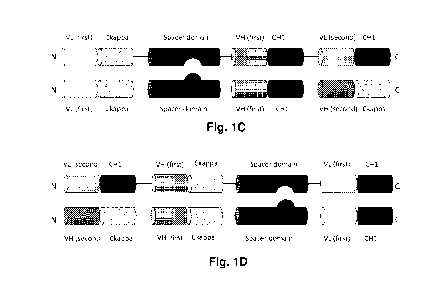
Figures IA, IB, IC and ID show examples, how the contorsbodies of the
invention can
be assembled. In Figure IA, the contorsbody consists of a first fusion
polypeptide comprising
the first half of a fab capable of specific binding to the first target (heavy
chain fab) linked via a
peptide linker (black line) to a spacer domain linked via peptide linker
(black line) to the second
CA 03076027 2020-03-17
WO 2019/086500
PCT/EP2018/079785
-14-
half of the fab capable of specific binding to the first target (light chain
fab) which is further
linked to a first half of a cross fab capable of specific binding to the
second target (VH-Ckappa)
(from N to C) and of a second fusion polypeptide comprising the first half of
a fab capable of
specific binding to the first target (heavy chain fab) linked via a peptide
linker (black line) to a
spacer domain linked via peptide linker (black line) to the second half of the
fab capable of
specific binding to the first target (light chain fab) which is further linked
to second half of a
cross fab capable of specific binding to the second target (VL-CH1) (from N to
C). The two
spacer domains are different from each other and comprise modifications
promoting the
association of the first and second fusion polypeptide. An example for this
type of contorsbody is
CD134-0093 (see Example 2.1). In Figure 1B, the contorsbody consists of a
first fusion
polypeptide comprising the first half of a cross fab capable of specific
binding to the first target
(VH-Ckappa) linked via a peptide linker (black line) to a spacer domain linked
via peptide linker
(black line) to the second half of the cross fab capable of specific binding
to the first target (VL-
CH1) which is further linked to a first half of a fab capable of specific
binding to the second
target (light chain fab) (from N to C) and of a second fusion polypeptide
comprising the first half
of a cross fab capable of specific binding to the first target (VH-Ckappa)
linked via a peptide
linker (black line) to a spacer domain linked via peptide linker (black line)
to the second half of
the cross fab capable of specific binding to the first target (VL-CH1) which
is further linked to
second half of the fab capable of specific binding to the second target (heavy
chain fab) (from N
to C). The two spacer domains are different from each other and comprise
modifications
promoting the association of the first and second fusion polypeptide. An
example for this type of
contorsbody is CD134-0094 (see Example 2.2). In Figure 1C, the contorsbody
consists of a first
fusion polypeptide comprising the first half of a fab capable of specific
binding to the first target
(VL-Ckappa) linked via a peptide linker (black line) to a spacer domain linked
via peptide linker
(black line) to the second half of the fab capable of specific binding to the
first target (VH-CH1)
which is further linked to a first half of a cross fab capable of specific
binding to the second
target (VH-Ckappa) (from N to C) and of a second fusion polypeptide comprising
the first half
of a fab capable of specific binding to the first target (VL-Ckappa) linked
via a peptide linker
(black line) to a spacer domain linked via peptide linker (black line) to the
second half of the fab
capable of specific binding to the first target (VH-CH1) which is further
linked to second half of
the cross fab capable of specific binding to the second target (VL-CH1) (from
N to C). The two
spacer domains are different from each other and comprise modifications
promoting the
association of the first and second fusion polypeptide. An example for this
type of contorsbody is
P1AE0821 (see Example 2.7). In Figure 1D, the contorsbody consists of a first
fusion
polypeptide comprising the first half of a cross fab capable of specific
binding to the second
target (VH-Ckappa) linked via a peptide linker (black line) to a first half of
a cross fab capable of
specific binding to the first target (VH-Ckappa) which is further linked via a
peptide linker
(black line) to a spacer domain linked via peptide linker (black line) to the
second half of the
CA 03076027 2020-03-17
WO 2019/086500
PCT/EP2018/079785
-15-
cross fab capable of specific binding to the first target (VL-CH1) (from N to
C) and of a second
fusion polypeptide comprising the first half of a cross fab capable of
specific binding to the
second target (VL-CH1) linked via a peptide linker (black line) to a first
half of a cross fab
capable of specific binding to the first target (VH-Ckappa) which is further
linked via a peptide
linker (black line) to a spacer domain linked via peptide linker (black line)
to the second half of
the cross fab capable of specific binding to the first target (VH-CH1) (from N
to C). The two
spacer domains are different from each other and comprise modifications
promoting the
association of the first and second fusion polypeptide. An example for this
type of contorsbody is
P1AE2735 (see Example 2.14).
Figure 1E is a schematic drawing of the assembled structure of Contorsbody
CD134-0093
(Example 2.1).
In Figure 1F a schematic drawing of the assembled structure of Contorsbody
CD134-0094
(Example 2.2) is shown.
Figure 1G is a schematic drawing of the assembled structure of Contorsbodies
P1AE0085
.. and P1AE0086 (Examples 2.3 and 2.4). The antigen binding domain capable of
specific binding
to the second target (cross fab) is connected via a longer peptide linker that
changes the
geometry of the molecule.
Figure 1H is a schematic drawing of the assembled structure of Contorsbody
P1AE0087
(Example 2.5) and Contorsbody P1AE0839 (Example 2.6). In this case the antigen
binding
domain capable of specific binding to the second target is a fab and both
antigen binding
domains capable of specific binding to the first target are cross fabs.
In Figure 11 a schematic drawing of the assembled structure of Contorsbody
P1AE0821
(Contorsbody 11, Example 2.7) is shown.
Figure 1J is a schematic drawing of the assembled structure of Contorsbody
PlAE1122
(Contorsbody 1, Example 2.8).
In Figure 1K a schematic drawing of the assembled structure of Contorsbody
P1AE1887
(Contorsbody 3, Example 2.10) is shown.
In Figure 1L a schematic drawing of the assembled structure of Contorsbody
P1AE2254
(Contorsbody 5, Example 2.12) is shown. In the CH and Ckappa fused to the VL
and VH of
0X40, respectively, amino acid mutations (so-called charged residues) were
introduced to
prevent the generation of Bence Jones proteins and to further facilitate the
correct pairing.
Figure 1M is a schematic drawing of the assembled structure of Contorsbody
P1AE2340
(Contorsbody 6, Example 2.13). In this case the molecule is composed of two
fusion proteins
and a light chain.
Figure 1N is a schematic drawing of the assembled structure of Contorsbody
P1AE2735
(Contorsbody 8, Example 2.14).
CA 03076027 2020-03-17
WO 2019/086500
PCT/EP2018/079785
-16-
Figure 10 is a schematic drawing of the 2+1 0X40 x FAP bispecific antibody
with
bivalent binding for 0X40 and monovalent binding for FAP (positive control
molecule). A
negative control molecule with the same structure was made wherein the FAP
binding domain
was replaced by DP47 germline (2+1 0X40 x DP47 antibody).
Figure 1P is a schematic drawing of the 4+1 0X40 x FAP bispecific antibody
with
tetravalent binding for 0X40 and monovalent binding for FAP. These control
molecules are
described in more detail in Example 2.18.
In Figures 2A, 2B, 2C and 2D is shown the binding of 0X40 x FAP bispecific
antibodies
to activated CD4+ and CD8+ T-cells (Figures 2A and 2C, respectively) and
resting CD4+ and
CD8+ T-cells (Figures 2B and 2D, respectively). The tetravalent 4+1 0X40 x FAP
(4B9)
bispecific antibody showed the strongest binding to CD4+ and CD8+ T-cells. The
2+1 formats
2+1 0X40 (49B4) x FAP (28H1) and 2+1 0X40 (49B4) x DP47 showed intermediate
binding.
The Contorsbody CD134-0093 indicated stronger binding than the 2+1 formats,
whereas the
Contorsbody CD134-0094 bound less strong. Binding to CD4+ T-cells was much
stronger than
that to CD8+ T cells. The negative control DP47 hu IgG1 P329G LALA did not
bind to CD4+ or
CD8+ T-cells. None of the molecules bound to resting CD4+ or CD8+ T-cells
(Figures 2B and
2D).
Figures 2E, 2F, 2G and 2H show the results as obtained with Contorsbodies P1
AE0085,
P1AE0086 and P1AE0087. All 2+1 formats 0X40 (49B4) x FAP (28H1), 0X40 (49B4) x
FAP
(4B9) and 0X40 (49B4) x DP47 showed a similarly good binding to activated CD4+
and CD8+
T-cells (Figure 2E and 2G, respectively) whereas the binding to 0X40 was
partially impaired
for the Contorsbody molecules, especially for the Contorsbody P1AE0087. DP47
hu IgG1
P329G LALA did not bind to CD4+ or CD8+ T-cells as expected. No binding was
observed to
resting CD4+ or CD8+ T-cells (Figure 2F and 2H, respectively) for any of the
tested molecules.
The binding to human FAP-expressing tumor cells is shown in Figures 3A, 3B, 3C
and
3D. In a first experiment, Contorsbodies CD134-0093 and CD134-0094 were
compared with the
control molecules. Binding to WM266-4 (FAP -F positive) and A549NLR (FAP
negative) tumor
cells is shown in Figures 3A and 3B, respectively. All 0X40 x FAP bispecific
antibodies bound
efficiently to human FAP expressing target cells. The tetravalent 4+1 0X40 x
FAP (4B9, high
affinity to FAP) bispecific antibody bound strongest to FAP -F cells, followed
by the Contorsbody
CD134-0093, Contorsbody CD1334-0094 and the 2+1 0X40 (49B4) x FAP (28H1)
bispecific
antibody. The non-targeted 2+1 0X40 (49B4) x DP47 and the negative control
(DP47 hu IgG1
P329G LALA) did not bind to any FARE cells. Figures 3C and 3D show the binding
of
Contorsbodies P1AE0085, P1AE0086 and P1AE0087 to NIH/3T3huFAP clone 19 (FAR')
(Figure 3C) and A549NLR (FAP-) tumor cells (Figure 3D). All FAP-targeted anti
0X40
antibodies bound efficiently to human FAP expressing target cells. The binding
of the
CA 03076027 2020-03-17
WO 2019/086500
PCT/EP2018/079785
-17-
Contorsbody molecules was slightly impaired as compared to their respective
controls. Again,
the non-targeted 2+1 0X40 (49B4) x DP47 and the negative control (DP47 hu IgG1
P329G
LALA) showed no binding to FAR cells.
In Figures 4A, 4B, 4C, 4D, 4E and 4F the NFKB activation with different types
of cross-
linking is shown. Using FAP expressing cells (NIH/3T3 huFAP clone 19) as
crosslinkers, the
FAP targeted 2+1 0X40 (49B4) x FAP (28H1) construct induced the strongest NFKB
activation,
followed by Contorsbodies CD134-0093 and CD134-0094, whereas CD134-0094 showed
a
slightly stronger activation than CD134-0093. The non-targeted and therefore
not crosslinked 2
non-targeted 2+1 0X40 (49B4) x DP47 induced weak NFKB activation (Figure 4A).
When
using a secondary, anti hu IgG1 Fcy-specific antibody as crosslinker, the two
2+1 constructs
0X40 (49B4) x FAP (28H1) and 0X40 (49B4) x DP47 behaved similar. The
Contorsbodies
CD134-0093 and CD134-0094 ran similar as well, but lower than the 2+1
constructs (Figure
4B). The least NFKB activation was obtained by not using crosslinkers. The 2+1
constructs
0X40 (49B4) x FAP (28H1) and 0X40 (49B4) x DP47showed a moderate NFKB
activation,
followed by the even less potent Contorsbodies CD134-0093 and CD134-0094. DP47
hu IgG1
P329G LALA did not induce any NFKB activation (Figure 4C). The ability of
Contorsbodies
P1AE0085, P1AE0086 and P1AE0087 to induce NFKB activation with different types
of cross-
linking is shown in Figures 4D to 4F. In the absence of cross-linking, only a
weak signal could
be detected at the highest antibody concentration (Figure 4D). The three
Contorsbody molecules
induced a very similar NFKB activation to that of FAP-targeted 2+1 formats
0X40 (49B4) x
FAP (28H1) and 0X40 (49B4) x FAP (4B9) when cross-linked by human FAP-
expressing cells
(Figure 4E). In the presence of secondary antibody cross-linking, all three
Contorsbody
molecules displayed slightly lower NFKB activation than the 2+1 control
molecules (Figure 4F).
DP47 hu IgG1 P329G LALA did not induce any NFKB activation.
0x40 mediated co-stimulation of sub-optimally TCR triggered resting human
PBMCs and
hyper-crosslinking by cell surface FAP is shown in Figures 5A, 5B, 5C, 5D, 5E
and 5F.
Figures 5A and 5B show the FSC-A ("area" of a Forward Side Scatter (FSC)
pulse),
respectively the size of CD4 and CD8 T-cells, respectively, after suboptimal
CD3 stimulation as
measured as intensity of light scattered at small angles by FACS analysis. The
FAP targeted 2+1
construct 0X40 (49B4) x FAP (28H1) showed an intermediate increase in size,
whereas the
Contorsbody CD134-0093 indicated a stronger increase. The untargeted 2+1 0X40
(49B4) x
DP47, the Contorsbody CD134-0094 and the negative control (DP47 hu IgG1 P329G
LALA)
did not change the size of neither CD4 nor CD8 T-cells. Figures 5C and 5D show
the activation
of CD4 and CD8 T-cells using the surface marker CD25. For the CD4 T-cells
(Figure 5C)
contorsbodies CD134-0093 and CD134-0094 demonstrated the highest activation,
followed by
the somehow less potent targeted 2+1 0X40 (49B4) x FAP (28H1). The untargeted
0X40 (49B4)
x DP47and the negative control did not show any activation after baseline-
correction. For the
CA 03076027 2020-03-17
WO 2019/086500
PCT/EP2018/079785
-18-
CD8 T-cells (Figure 5D), the Contorsbody CD134-0094 demonstrated a stronger
activation
compared to the 2+1 0X40 (49B4) x FAP (28H1) and CD134-0093. Figures 5E and 5F
show
the upon activation downregulated IL-7Ra (CD127). The Contorsbody CD134-0093
showed the
strongest CD127 downregulation for CD4 and CD8 T-cells, followed by the
targeted 2+1 0X40
(49B4) x FAP (28H1) and the Contorsbody CD134-0094.
Figure 6 illustrates the normalized area under the curve (AUC) values for FSC-
A and
CD25 on CD4 and CD8 T-cells. The filled symbols represent CD4 T-cells, the
empty one CD8 T
cells. All values were normalized to the top AUC values of the Contorsbody
CD134-0093 (=
100%). The untargeted 2+1 construct 0X40 (49B4) x DP47showed only minimal
activation on
CD4 and CD8 T cells, whereas the FAP targeted 2+1 construct 0X40 (49B4) x FAP
(28H1)
demonstrated a higher potency (normalized values between 60 and 100) regarding
FSC-A and
CD25 on CD4 and CD8 T cells, but less potency compared to CD134-0093. For the
Contorsbody
CD134-0094, only the CD25 MFI AUC values of CD4 and CD8 T-cells showed similar
activation compared to CD134-0093.
The effects of Contorsbodies P1AE0085, P1AE0086 and P1AE0087 on 0X40 mediated
co-stimulation of sub-optimally TCR triggered PBMCs and hyper-crosslinking by
cell surface
FAP are shown in Figures 7A, 7B, 7C and 7D. Figures 7A and 7B show the FSC-A,
respectively the size of CD4 and CD8 T cells, respectively, after suboptimal
CD3 stimulation.
Figures 7C and 7D show the activation of CD4 and CD8 T-cells, respectively,
using the
.. expression of the surface marker CD25. All FAP-targeted molecules (controls
and all three
contorsbody molecules) induced a dose-dependent increase in the Forward Side
Scatter Area and
CD25 expression on both CD4 and CD8 T cells. The untargeted 2+1 construct 0X40
(49B4) x
DP47 and the negative control (DP47 hu IgG1 P329G LALA) did not show any
activation after
baseline-correction.
In Figures 8A, 8B, 8C and 8D is shown the binding of different 0X40 x FAP
contorsbodies to activated CD4 + T-cells. The untargeted 2+1 construct 0X40
(49B4) x DP47
was used as positive control and DP47 hu IgG1 P329G LALA as negative control.
Fig. 8A
shows the binding of Contorsbody 1 (PlAE1122), Contorsbody 2 (P1AE1942) and
Contorsbody
3 (P1AE1887) and Fig. 8B shows the binding of Contorsbody 4 (P1AE1888),
Contorsbody 5
(P1AE2254) and Contorsbody 6 (P1AE2340). The binding of Contorsbody 7
(P1AE0086) and
Contorsbody 8 (P1AE2735) is shown in Fig. 8C and the binding of Contorsbody 9
(P1AE2743)
and Contorsbody 10 (P1AE2762) is shown in Fig. 8D. In Figures 9A, 9B, 9C and
9D,
respectively, it is shown that none of the tested Contorsbodies bound to
resting CD4 T cells. In
Figures 10A, 10B, 10C and 10D is shown the binding of the same 0X40 x FAP
contorsbodies
to activated CD8 + T-cells. The untargeted 2+1 construct 0X40 (49B4) x DP47
was used as
positive control and DP47 hu IgG1 P329G LALA as negative control. In Figures
11A, 11B, 11C
CA 03076027 2020-03-17
WO 2019/086500
PCT/EP2018/079785
-19-
and 11D, respectively, it is shown that none of the tested Contorsbodies bound
to resting CD8 T
cells. A summary of the area under the curve values for the binding to
activated or resting CD4+
T-cells is shown in Figure 12A and for the binding to activated or resting
CD8+ T cell in Figure
12B.
The binding to human FAP-expressing tumor cells of Contorsbodies 1 to 10 is
shown in
Figures 13A, 13B, 13C and 13D and Figures 14A, 14B, 14C and 14D, respectively.
Binding to
NIH/3T3-huFAP clone 19 (FAR') tumor cells (FAR positive) is shown in Figures
12A to 12D,
respectively. Fig. 13A shows the binding of Contorsbody 1 (PlAE1122),
Contorsbody 2
(P1AE1942) and Contorsbody 3 (P1AE1887) and Fig. 13B shows the binding of
Contorsbody 4
.. (P1AE1888), Contorsbody 5 (P1AE2254) and Contorsbody 6 (P1AE2340). The
binding of
Contorsbody 7 (P1AE0086) and Contorsbody 8 (P1AE2735) is shown in Fig. 13C and
the
binding of Contorsbody 9 (P1AE2743) and Contorsbody 10 (P1AE2762) is shown in
Fig. 13D.
All 0X40 x FAP bispecific contorsbodies bound efficiently to human FAP
expressing target
cells. Contorbody 8 (Fig. 12C) bound strongest to FAR' cells, followed by
Contorbody 10 (Fig.
.. 12D) and Contorbody 6 (Fig. 12B), much stronger than the 2+1 bispecific
antibody 0X40 (49B4)
x FAP (4B9). The untargeted 2+1 construct 0X40 (49B4) x DP47 and the negative
control
(DP47 hu IgG1 P329G LALA) did not bind to any FAR' cells. The corresponding
binding of
Contorsbodies 1 to 10 to A549NLR (FAP- negative) tumor cells is shown in
Figures 14A to 14D.
None of the FAP-targeted molecules (Contorsbodies 1 to 10 or 2+1 bispecific
antibody 0X40
.. (49B4) x FAP (4B9)) were able to bind to FAP- target cells.
In Figures 15A, 15B, 15C and 15D the NFKB activation of Contorsbodies 1 to 11
with
cross-linking is shown. Using FAP expressing cells (NIH/3T3 huFAP clone 19) as
crosslinkers,
the NFKB activation was comparable for all of the contorbodies, with
Contorbody 11
(P1AE0821) being the least potent. The non-targeted and therefore not
crosslinked 2 non-
.. targeted 2+1 0X40 (49B4) x DP47 antibody (7718) induced weak NFKB
activation, whereas
bispecific 0X40 (49B4) x FAP (4B9) antibody (7719) caused the highest NFKB
activation.
DP47 hu IgG1 P329G LALA (8105) did not induce any NFKB activation. NFKB
activation
without cross-linking by FAP is shown in Figures 16A, 16B, 16C and 16D. In the
absence of
cross-linking, only a weak signal could be detected at the higher antibody
concentrations. The
.. non-targeted 2+1 0X40 (49B4) x DP47 antibody (7718) showed a moderate NFKB
activation,
followed by the Contorbody 7 (Fig 16C). The other contorbodies induced minor
or zero levels.
The negative control (8105) did not induce any NFKB activation. Normalized
Area under the
curve values for NFKB activation in HeLa cells with and without crosslinking
with FAR' cells
are summarized in Figure 17. The negative control (8105) did not induce any
NFKB activation
.. in both cases, while non-targeted 2+1 0X40 (49B4) x DP47 antibody (7718)
showed only
minimal activation in both cases. Bispecific 0X40 (49B4) x FAP (4B9) antibody
(7719)
CA 03076027 2020-03-17
WO 2019/086500
PCT/EP2018/079785
-20-
demonstrated the higher levels of activation, followed by Contorbodies 8, 9
and 10. Lower levels
were observed for Contorbody 11 (P1AE0821).
0X40 mediated co-stimulation of sub-optimally TCR triggered resting human
PBMCs and
hyper-crosslinking by cell surface FAP of Contorsbodies 7, 8, 9 and 10 is
shown in Figures 18A,
18B, 18C and 18D. Figures 18A and 18B show the activation of CD4 T cells and
Figures 18C
and 18D show the activation of CD8 T cells using the surface marker CD25 after
suboptimal
CD3 stimulation. For the CD4 T-cells (Figures 18A nad 18B), the bispecific
0X40 (49B4) x
FAP (4B9) antibody (7719) demonstrated the highest activation, followed by the
minimally less
potent Contorbody 8. The non-targeted 2+1 0X40 (49B4) x DP47 antibody (7718)
and the
negative control (8105) did not show any activation after baseline-correction.
For the CD8 T-
cells (Figures 18C and 18D), the Contorbodies 7, 8 and 9 as well as the
bispecific 0X40 (49B4)
x FAP (4B9) antibody (7719) demonstrated a stronger activation compared to
Contorbody 10.
Figures 19A, 19B, 19C and 19D show the FSC-A, respectively the size of CD4
(Figures 19A
and 19B) and CD8 T-cells (Figures 19C and 19D) after suboptimal CD3
stimulation. All
contorbodies indicated a comparable intermediate increase. Addition of the
untargeted 2+1 anti
0X40 molecule (7718) and the negative control (8105) did not change the size
of neither CD4
nor CD8 T-cells. Figures 20A, 20B, 20C and 20D show the eFluor 670 levels,
respectively the
proliferation of CD4 (Figures 20A and 20B) and CD8 T-cells (Figures 20C and
20D) after
suboptimal CD3 stimulation. In CD4 subpopulations, Contorbodies 8 and 10
showed a
considerable decrease in eFluor 670 levels, indicating stronger proliferation.
In CD8
subpopulations Contorbodies 7 and 9 showed bigger decrease in eFluor 670
levels. The
untargeted 2+1 anti 0X40 molecule (7718) and the negative control (8105)
showed only a minor
decrease in all subpopulations. Figures 21A, 21B, 21C and 21D show the upon
activation
downregulated IL-7Ra (CD127). The the bispecific 0X40 (49B4) x FAP (4B9)
antibody (7719)
showed the strongest downregulation for CD4 and CD8 T-cells, followed by
Contorbody 10.
Greater downregulation of CD127 was observed in CD4 cells than CD8
subpopulations.
Normalized Area under the curve values for CD25 (Fig. 22A), FSC-A (Fig. 22B),
eFluor 670
(Fig. 22C) and CD127 (FIG. 22D) on CD4 and CD8 T-cells are summarized in
Figures 22A,
22B, 22C and 22D. The untargeted 2+1 anti 0X40 molecule (7718) showed only
minimal
activation on CD4 and CD8 T cells, whereas the bispecific 0X40 (49B4) x FAP
(4B9) antibody
(7719) demonstrated a higher potency regarding eFluor 679 and CD127 on CD4 and
CD8 T-
cells compared to the contorbodies. CD25 and FSC-A indicated similar
activation levels in both
the 7719 molecule and contorbodies in both CD4 and CD8 subpopulations.
Figures 23A and 23B show the NFKB-mediated luciferase expression activity in 4-
1BB
.. expressing reporter cell line Jurkat-hu4-1BB-NFKB-1uc2. To test the
functionality of 2+1 anti-4-
1BB (20H4.9) x anti-FAP(4B9) contorsbodies versus 2+1 anti-4-1BB (20H4.9) x
anti-FAP (4B9)
antigen binding molecules versus controls, molecules were incubated. The
concentration of
CA 03076027 2020-03-17
WO 2019/086500
PCT/EP2018/079785
-21-
antigen binding molecules or its controls are blotted against the units of
released light (RLU)
measured after 6 h of incubation. All values are baseline corrected by
subtracting the baseline
values of the blank control (e.g. no antibodies added). In Figure 23A FAP-
target-independent 4-
1BB activation is shown, whereby 4-1BB-binding induces NFKB-controlled
luciferase
expression in the reporter cell line without any FAP-mediated crosslinking. In
Figure 23B high
FAP-expressing cell line NIH/3T3-huFAP clone 19 (human-FAP-transgenic mouse
fibroblast
cell line) was added. The FAP-expressing tumor cells lead to cros slinking of
the bispecific 4-
1BB (20H4.9) x FAP (4B9) antigen binding molecules and and a strong increase
of its potential
to induce NFKB-induced /luciferase activation in the 4-1BB-expressing reporter
cell line. The
.. bispecific 2+1 anti-4-1BB (20H4.9) x anti-FAP(4B9) antigen binding
molecules (black filled star
and line) showed a slightly better activation (lower EC50 values) than the
contorsbodies.
However, the activation caused by the contorsbodies was much higher the
activation shown by
the untargeted 4-1BB antibodies.
Figures 24A, 24B, 24C, 24D, 24E, 24F, 24G and 24H show the in vitro activation
of
human B cells by bivalent human anti-CD40 x FAP contorsbodies in the presence
of FAP-coated
(Fig. 24A, 24C, 24E and 24G) or uncoated Dynabeads (Fig. 24B, 24D, 24F and
24H) after 2
days incubation. Compared to the FAP-independent upregulation of CD69, CD80,
CD86 and
HLA-DR induced by cross-linked SGN40, upregulation of these activation markers
induced by
FAP-dependent bispecific antigen binding molecules in the presence of FAP-
coated beads was
slightly lower. In the absence of FAP (uncoated beads) no increase of CD69,
CD80, CD86 or
HLA-DR expression could be observed with the FAP-targeted anti-CD40
contorsbodies, while
the cross-linked positive control antibody SGN40 induced an upregulation of
these activation
markers. Shown is the percentage of CD69, CD80, CD86 or HLA-DR positive vital
B cells after
2 days of incubation with the indicated titrated contorsbodies or control
antibody. XL stands for
cross-linking with F(abt)2 Fragment Goat anti-human IgG Fcy fragment specific.
The x-axis
shows the concentration of contorsbody constructs or the control antibody.
DETAILED DESCRIPTION OF THE INVENTION
Definitions
Unless defined otherwise, technical and scientific terms used herein have the
same
meaning as generally used in the art to which this invention belongs. For
purposes of interpreting
this specification, the following definitions will apply and whenever
appropriate, terms used in
the singular will also include the plural and vice versa.
CA 03076027 2020-03-17
WO 2019/086500
PCT/EP2018/079785
-22-
As used herein, the term "antigen binding molecule" refers in its broadest
sense to a
molecule that specifically binds an antigenic determinant. Examples of antigen
binding
molecules are antibodies, antibody fragments and scaffold antigen binding
proteins.
The term "antigen binding domain" refers to the part of an antigen binding
molecule that
.. specifically binds to an antigenic determinant. In one aspect, the antigen
binding domain is able
to activate signaling through its target cell antigen. Antigen binding domains
include the area or
fragment of an antibody which specifically binds to and is complementary to
part or all of an
antigen. In addition, antigen binding domains include scaffold antigen binding
proteins as further
defined herein, e.g. binding domains which are based on designed repeat
proteins or designed
repeat domains (see e.g. WO 2002/020565). In particular, an antigen binding
domain is
comprised of a first part and a second part, wherein the first part comprises
an antibody light
chain variable region (VL) and the second part comprises an antibody heavy
chain variable
region (VH) or vice versa.
The term "antibody" herein is used in the broadest sense and encompasses
various
antibody structures, including but not limited to monoclonal antibodies,
polyclonal antibodies,
monospecific and multispecific antibodies (e.g., bispecific antibodies), and
antibody fragments
so long as they exhibit the desired antigen-binding activity.
The term "monoclonal antibody" as used herein refers to an antibody obtained
from a
population of substantially homogeneous antibodies, i.e., the individual
antibodies comprising
the population are identical and/or bind the same epitope, except for possible
variant antibodies,
e.g. containing naturally occurring mutations or arising during production of
a monoclonal
antibody preparation, such variants generally being present in minor amounts.
In contrast to
polyclonal antibody preparations, which typically include different antibodies
directed against
different determinants (epitopes), each monoclonal antibody of a monoclonal
antibody
preparation is directed against a single determinant on an antigen.
The term "monospecific" antibody as used herein denotes an antibody that has
one or
more binding sites each of which bind to the same epitope of the same antigen.
The term
"bispecific" means that the antigen binding molecule is able to specifically
bind to at least two
distinct antigenic determinants. A bispecific antigen binding molecule
comprises at least two
antigen binding sites, each of which is specific for a different antigenic
determinant. In certain
embodiments the bispecific antigen binding molecule is capable of
simultaneously binding two
antigenic determinants, particularly two antigenic determinants expressed on
two distinct cells.
For example, the antigen binding molecules of the present invention are
bispecific, comprising
an antigen binding domain capable of specific binding to a first target, and
an antigen binding
domain capable of specific binding to a second target. In one particular
aspect, the antibody of
CA 03076027 2020-03-17
WO 2019/086500
PCT/EP2018/079785
-23-
the present invention comprises an antigen binding domain capable of specific
binding to 0X40
and an antigen binding domain capable of specific binding to FAP. In another
particular aspect,
the antibody of the present invention comprises an antigen binding domain
capable of specific
binding to 4-1BB and an antigen binding domain capable of specific binding to
FAP. In one
further aspect, the antibody of the present invention comprises an antigen
binding domain
capable of specific binding to CD40 and an antigen binding domain capable of
specific binding
to FAP.
The term "valent" as used within the current application denotes the presence
of a
specified number of binding sites in an antigen binding molecule. As such, the
terms "bivalent",
"tetravalent", and "hexavalent" denote the presence of two binding sites, four
binding sites, and
six binding sites, respectively, in an antigen binding molecule. Valency of an
antigen binding
molecule may also be expressed in relation to the number of binding sites for
a given antigenic
determinant. For example, the bispecific antibodies of the present invention
are bivalent with
respect to a first target, and monovalent with respect to a second target.
The terms "full length antibody", "intact antibody", and "whole antibody" are
used herein
interchangeably to refer to an antibody having a structure substantially
similar to a native
antibody structure. "Native antibodies" refer to naturally occurring
immunoglobulin molecules
with varying structures. For example, native IgG-class antibodies are
heterotetrameric
glycoproteins of about 150,000 daltons, composed of two light chains and two
heavy chains that
.. are disulfide-bonded. From N- to C-terminus, each heavy chain has a
variable region (VH), also
called a variable heavy domain or a heavy chain variable domain, followed by
three constant
domains (CH1, CH2, and CH3), also called a heavy chain constant region.
Similarly, from N- to
C-terminus, each light chain has a variable region (VL), also called a
variable light domain or a
light chain variable domain, followed by a light chain constant domain (CL),
also called a light
chain constant region. The heavy chain of an antibody may be assigned to one
of five types,
called a (IgA), 6 (IgD), 8 (IgE), y (IgG), or IA (IgM), some of which may be
further divided into
subtypes, e.g. yl (IgG1), y2 (IgG2), y3 (IgG3), y4 (IgG4), al (IgA 1) and a2
(IgA2). The light
chain of an antibody may be assigned to one of two types, called kappa (lc)
and lambda (X), based
on the amino acid sequence of its constant domain.
An "antibody fragment" refers to a molecule other than an intact antibody that
comprises
a portion of an intact antibody that binds the antigen to which the intact
antibody binds.
Examples of antibody fragments include but are not limited to Fv, Fab, Fab',
Fab'-SH, F(abt)2;
diabodies, triabodies, tetrabodies, cross-Fab fragments; linear antibodies;
single-chain antibody
molecules (e.g. scFv); and single domain antibodies. For a review of certain
antibody fragments,
see Hudson et al., Nat Med 9, 129-134 (2003). For a review of scFv fragments,
see e.g.
Pliickthun, in The Pharmacology of Monoclonal Antibodies, vol. 113, Rosenburg
and Moore
CA 03076027 2020-03-17
WO 2019/086500
PCT/EP2018/079785
-24-
eds., Springer-Verlag, New York, pp. 269-315 (1994); see also WO 93/16185; and
U.S. Patent
Nos. 5,571,894 and 5,587,458. For discussion of Fab and F(ab')2 fragments
comprising salvage
receptor binding epitope residues and having increased in vivo half-life, see
U.S. Patent No.
5,869,046. Diabodies are antibody fragments with two antigen-binding sites
that may be bivalent
or bispecific, see, for example, EP 404,097; WO 1993/01161; Hudson et al., Nat
Med 9, 129-134
(2003); and Hollinger et al., Proc Natl Acad Sci USA 90, 6444-6448 (1993).
Triabodies and
tetrabodies are also described in Hudson et al., Nat Med 9, 129-134 (2003).
Single-domain
antibodies are antibody fragments comprising all or a portion of the heavy
chain variable domain
or all or a portion of the light chain variable domain of an antibody. In
certain embodiments, a
single-domain antibody is a human single-domain antibody (Domantis, Inc.,
Waltham, MA; see
e.g. U.S. Patent No. 6,248,516 B1). Antibody fragments can be made by various
techniques,
including but not limited to proteolytic digestion of an intact antibody as
well as production by
recombinant host cells (e.g. E. coli or phage), as described herein.
Papain digestion of intact antibodies produces two identical antigen-binding
fragments,
called "Fab" fragments containing each the heavy- and light-chain variable
domains and also the
constant domain of the light chain and the first constant domain (CH1) of the
heavy chain. As
used herein, Thus, the term "Fab fragment" refers to an antibody fragment
comprising a light
chain fragment comprising a VL and a constant domain of a light chain (CL),
and a VH and a
first constant domain (CH1) of a heavy chain. Fab' fragments differ from Fab
fragments by the
addition of a few residues at the carboxy terminus of the heavy chain CH1
domain including one
or more cysteins from the antibody hinge region. Fab'-SH are Fab' fragments
wherein the
cysteine residue(s) of the constant domains bear a free thiol group. Pepsin
treatment yields an
F(abt)2fragment that has two antigen-combining sites (two Fab fragments) and a
part of the Fc
region. According to the present invention, the term "Fab fragment" also
includes "cross-Fab
fragments" or "crossover Fab fragments" as defined below.
The term "cross-Fab fragment" or "xFab fragment" or "crossover Fab fragment"
refers to
a Fab fragment, wherein either the variable regions or the constant regions of
the heavy and light
chain are exchanged. Two different chain compositions of a cross-Fab molecule
are possible and
comprised in the bispecific antibodies of the invention: On the one hand, the
variable regions of
the Fab heavy and light chain are exchanged, i.e. the crossover Fab molecule
comprises a peptide
chain composed of the light chain variable region (VL) and the heavy chain
constant region
(CH1), and a peptide chain composed of the heavy chain variable region (VH)
and the light
chain constant region (CL). This crossover Fab molecule is also referred to as
CrossFab (vLvf).
On the other hand, when the constant regions of the Fab heavy and light chain
are exchanged, the
crossover Fab molecule comprises a peptide chain composed of the heavy chain
variable region
(VH) and the light chain constant region (CL), and a peptide chain composed of
the light chain
CA 03076027 2020-03-17
WO 2019/086500
PCT/EP2018/079785
-25-
variable region (VL) and the heavy chain constant region (CH1). This crossover
Fab molecule is
also referred to as CrossFab (cLcm).
A "single chain Fab fragment" or "scFab" is a polypeptide consisting of an
antibody heavy
chain variable domain (VH), an antibody constant domain 1 (CH1), an antibody
light chain
variable domain (VL), an antibody light chain constant domain (CL) and a
linker, wherein said
antibody domains and said linker have one of the following orders in N-
terminal to C-terminal
direction: a) VH-CH1-linker-VL-CL, b) VL-CL-linker-VH-CH1, c) VH-CL-linker-VL-
CH1 or
d) VL-CH1-linker-VH-CL; and wherein said linker is a polypeptide of at least
30 amino acids,
preferably between 32 and 50 amino acids. Said single chain Fab fragments are
stabilized via the
natural disulfide bond between the CL domain and the CH1 domain. In addition,
these single
chain Fab molecules might be further stabilized by generation of interchain
disulfide bonds via
insertion of cysteine residues (e.g. position 44 in the variable heavy chain
and position 100 in the
variable light chain according to Kabat numbering).
A "crossover single chain Fab fragment" or "x-scFab" is a is a polypeptide
consisting of
an antibody heavy chain variable domain (VH), an antibody constant domain 1
(CH1), an
antibody light chain variable domain (VL), an antibody light chain constant
domain (CL) and a
linker, wherein said antibody domains and said linker have one of the
following orders in N-
terminal to C-terminal direction: a) VH-CL-linker-VL-CH1 and b) VL-CH1-linker-
VH-CL;
wherein VH and VL form together an antigen-binding site which binds
specifically to an antigen
and wherein said linker is a polypeptide of at least 30 amino acids. In
addition, these x-scFab
molecules might be further stabilized by generation of interchain disulfide
bonds via insertion of
cysteine residues (e.g. position 44 in the variable heavy chain and position
100 in the variable
light chain according to Kabat numbering).
A "single-chain variable fragment (scFv)" is a fusion protein of the variable
regions of
the heavy (VII) and light chains (VL) of an antibody, connected with a short
linker peptide of ten
to about 25 amino acids. The linker is usually rich in glycine for
flexibility, as well as serine or
threonine for solubility, and can either connect the N-terminus of the VH with
the C-terminus of
the VL, or vice versa. This protein retains the specificity of the original
antibody, despite removal
of the constant regions and the introduction of the linker. scFv antibodies
are, e.g. described in
Houston, J.S., Methods in Enzymol. 203 (1991) 46-96). In addition, antibody
fragments
comprise single chain polypeptides having the characteristics of a VH, namely
being able to
assemble together with a VL, or of a VL, namely being able to assemble
together with a VH to a
functional antigen binding site and thereby providing the antigen binding
property of full length
antibodies.
CA 03076027 2020-03-17
WO 2019/086500
PCT/EP2018/079785
-26-
"Scaffold antigen binding proteins" are known in the art, for example,
fibronectin and
designed ankyrin repeat proteins (DARPins) have been used as alternative
scaffolds for antigen-
binding domains, see, e.g., Gebauer and Skerra, Engineered protein scaffolds
as next-generation
antibody therapeutics. Curr Opin Chem Biol 13:245-255 (2009) and Stumpp et
al., Darpins: A
new generation of protein therapeutics. Drug Discovery Today 13: 695-701
(2008). In one aspect
of the invention, a scaffold antigen binding protein is selected from the
group consisting of
CTLA-4 (Evibody), Lipocalins (Anticalin), a Protein A-derived molecule such as
Z-domain of
Protein A (Affibody), an A-domain (Avimer/Maxibody), a serum transferrin
(trans-body); a
designed ankyrin repeat protein (DARPin), a variable domain of antibody light
chain or heavy
chain (single-domain antibody, sdAb), a variable domain of antibody heavy
chain (nanobody,
aVH), VNAR fragments, a fibronectin (AdNectin), a C-type lectin domain
(Tetranectin); a
variable domain of a new antigen receptor beta-lactamase (VNAR fragments), a
human gamma-
crystallin or ubiquitin (Affilin molecules); a kunitz type domain of human
protease inhibitors,
microbodies such as the proteins from the knottin family, peptide aptamers and
fibronectin
(adnectin). CTLA-4 (Cytotoxic T Lymphocyte-associated Antigen 4) is a CD28-
family receptor
expressed on mainly CD4+ T-cells. Its extracellular domain has a variable
domain- like Ig fold.
Loops corresponding to CDRs of antibodies can be substituted with heterologous
sequence to
confer different binding properties. CTLA-4 molecules engineered to have
different binding
specificities are also known as Evibodies (e.g. US7166697B1). Evibodies are
around the same
size as the isolated variable region of an antibody (e.g. a domain antibody).
For further details
see Journal of Immunological Methods 248 (1-2), 31-45 (2001). Lipocalins are a
family of
extracellular proteins which transport small hydrophobic molecules such as
steroids, bilins,
retinoids and lipids. They have a rigid beta-sheet secondary structure with a
number of loops at
the open end of the conical structure which can be engineered to bind to
different target antigens.
Anticalins are between 160-180 amino acids in size, and are derived from
lipocalins. For further
details see Biochim Biophys Acta 1482: 337-350 (2000), US7250297B1 and
US20070224633.
An affibody is a scaffold derived from Protein A of Staphylococcus aureus
which can be
engineered to bind to antigen. The domain consists of a three-helical bundle
of approximately 58
amino acids. Libraries have been generated by randomization of surface
residues. For further
details see Protein Eng. Des. Sel. 2004, 17, 455-462 and EP 1641818A1. Avimers
are
multidomain proteins derived from the A-domain scaffold family. The native
domains of
approximately 35 amino acids adopt a defined disulfide bonded structure.
Diversity is generated
by shuffling of the natural variation exhibited by the family of A-domains.
For further details see
Nature Biotechnology 23(12), 1556 - 1561 (2005) and Expert Opinion on
Investigational Drugs
16(6), 909-917 (June 2007). A transferrin is a monomeric serum transport
glycoprotein.
Transferrins can be engineered to bind different target antigens by insertion
of peptide sequences
in a permissive surface loop. Examples of engineered transferrin scaffolds
include the Trans-
body. For further details see J. Biol. Chem 274, 24066-24073 (1999). Designed
Ankyrin Repeat
CA 03076027 2020-03-17
WO 2019/086500
PCT/EP2018/079785
-27-
Proteins (DARPins) are derived from Ankyrin which is a family of proteins that
mediate
attachment of integral membrane proteins to the cytoskeleton. A single ankyrin
repeat is a 33
residue motif consisting of two alpha-helices and a beta-turn. They can be
engineered to bind
different target antigens by randomizing residues in the first alpha-helix and
a beta-turn of each
repeat. Their binding interface can be increased by increasing the number of
modules (a method
of affinity maturation). For further details see J. Mol. Biol. 332, 489-503
(2003), PNAS 100(4),
1700-1705 (2003) and J. Mol. Biol. 369, 1015-1028 (2007) and US20040132028A1.
A single-
domain antibody is an antibody fragment consisting of a single monomeric
variable antibody
domain. The first single domains were derived from the variable domain of the
antibody heavy
chain from camelids (nanobodies or VHFI fragments). Furthermore, the term
single-domain
antibody includes an autonomous human heavy chain variable domain (aVH) or
VNAR fragments
derived from sharks. Fibronectin is a scaffold which can be engineered to bind
to antigen.
Adnectins consists of a backbone of the natural amino acid sequence of the
10th domain of the
repeating units of human fibronectin type III (FN3). Three loops at one end of
the .beta.-
15 sandwich can be engineered to enable an Adnectin to specifically
recognize a therapeutic target
of interest. For further details see Protein Eng. Des. Sel. 18, 435- 444
(2005), US20080139791,
W02005056764 and US6818418B1. Peptide aptamers are combinatorial recognition
molecules
that consist of a constant scaffold protein, typically thioredoxin (TrxA)
which contains a
constrained variable peptide loop inserted at the active site. For further
details see Expert Opin.
Biol. Ther. 5, 783-797 (2005). Microbodies are derived from naturally
occurring microproteins
of 25-50 amino acids in length which contain 3-4 cysteine bridges - examples
of microproteins
include KalataBI and conotoxin and knottins. The microproteins have a loop
which can
beengineered to include upto 25 amino acids without affecting the overall fold
of the
microprotein. For further details of engineered knottin domains, see
W02008098796.
An "antigen binding molecule that binds to the same epitope" as a reference
molecule
refers to an antigen binding molecule that blocks binding of the reference
molecule to its antigen
in a competition assay by 50% or more, and conversely, the reference molecule
blocks binding
of the antigen binding molecule to its antigen in a competition assay by 50%
or more.
As used herein, the term "antigenic determinant" is synonymous with "antigen"
and
"epitope," and refers to a site (e.g. a contiguous stretch of amino acids or a
conformational
configuration made up of different regions of non-contiguous amino acids) on a
polypeptide
macromolecule to which an antigen binding moiety binds, forming an antigen
binding moiety-
antigen complex. Useful antigenic determinants can be found, for example, on
the surfaces of
tumor cells, on the surfaces of virus-infected cells, on the surfaces of other
diseased cells, on the
surface of immune cells, free in blood serum, and/or in the extracellular
matrix (ECM). The
proteins useful as antigens herein can be any native form the proteins from
any vertebrate source,
including mammals such as primates (e.g. humans) and rodents (e.g. mice and
rats), unless
CA 03076027 2020-03-17
WO 2019/086500
PCT/EP2018/079785
-28-
otherwise indicated. In a particular embodiment the antigen is a human
protein. Where reference
is made to a specific protein herein, the term encompasses the "full-length",
unprocessed protein
as well as any form of the protein that results from processing in the cell.
The term also
encompasses naturally occurring variants of the protein, e.g. splice variants
or allelic variants.
The term "paratope" refers to that part of a given antibody molecule that is
required for
specific binding between a target and a binding site. A paratope may be
continuous, i.e. formed
by adjacent amino acid residues present in the binding site, or discontinuous,
i.e. formed by
amino acid residues that are at different positions in the primary sequence of
the amino acid
residues, such as in the amino acid sequence of the CDRs of the amino acid
residues, but in close
proximity in the three-dimensional structure, which the binding site adopts.
By "specific binding" is meant that the binding is selective for the antigen
and can be
discriminated from unwanted or non-specific interactions. The ability of an
antigen binding
molecule to bind to a specific antigen can be measured either through an
enzyme-linked
immunosorbent assay (ELISA) or other techniques familiar to one of skill in
the art, e.g. Surface
Plasmon Resonance (SPR) technique (analyzed on a BIAcore instrument)
(Liljeblad et al., Glyco
J 17, 323-329 (2000)), and traditional binding assays (Heeley, Endocr Res 28,
217-229 (2002)).
In one embodiment, the extent of binding of an antigen binding molecule to an
unrelated protein
is less than about 10% of the binding of the antigen binding molecule to the
antigen as measured,
e.g. by SPR. In certain embodiments, a molecule that binds to the antigen has
a dissociation
constant (Kd) of < li.tM, < 100 nM, < 10 nM, < 1 nM, < 0.1 nM, < 0.01 nM, or <
0.001 nM (e.g.
10-8 M or less, e.g. from 10-8 M to 10-13 M, e.g. from 10-9 M to 10-13 M).
"Affinity" or "binding affinity" refers to the strength of the sum total of
non-covalent
interactions between a single binding site of a molecule (e.g. an antibody)
and its binding partner
(e.g. an antigen). Unless indicated otherwise, as used herein, "binding
affinity" refers to intrinsic
binding affinity which reflects a 1:1 interaction between members of a binding
pair (e.g.
antibody and antigen). The affinity of a molecule X for its partner Y can
generally be represented
by the dissociation constant (Kd), which is the ratio of dissociation and
association rate constants
(koff and kon, respectively). Thus, equivalent affinities may comprise
different rate constants, as
long as the ratio of the rate constants remains the same. Affinity can be
measured by common
methods known in the art, including those described herein. A particular
method for measuring
affinity is Surface Plasmon Resonance (SPR).
An "affinity matured" antibody refers to an antibody with one or more
alterations in one
or more hypervariable regions (HVRs), compared to a parent antibody which does
not possess
such alterations, such alterations resulting in an improvement in the affinity
of the antibody for
antigen.
CA 03076027 2020-03-17
WO 2019/086500
PCT/EP2018/079785
-29-
A "tumor associated antigen" as used herein refers to an antigenic determinant
presented
on the surface of a target cell, which is a cell in a tumor such as a cancer
cell, a cell of the tumor
stroma or a B cell. In certain aspects, the tumor associated antigen is
Fibroblast Activation
Protein (FAP).
The term "capable of specific binding to Fibroblast activation protein (FAP)"
refers to
an antigen binding molecule that is capable of binding FAP with sufficient
affinity such that the
antigen binding molecule is useful as a diagnostic and/or therapeutic agent in
targeting FAP. The
antigen binding molecule includes but is not limited to, antibodies, Fab
molecules, crossover Fab
molecules, single chain Fab molecules, Fv molecules, scFv molecules, single
domain antibodies,
and VH and scaffold antigen binding protein. In one aspect, the extent of
binding of an anti-FAP
antigen binding molecule to an unrelated, non-FAP protein is less than about
10% of the binding
of the antigen binding molecule to FAP as measured, e.g., by Surface Plasmon
Resonance (SPR).
In particular, an antigen binding molecule that is capable of specific binding
to FAP has a
dissociation constant (Ka) of < li.tM, < 100 nM, < 10 nM, < 1 nM, < 0.1 nM, <
0.01 nM, or
< 0.001 nM (e.g. 10-8M or less, e.g. from 10-8M to 10-13M, e.g., from 10-9M to
10-13 M). In
certain embodiments, an anti-FAP antigen binding molecule binds to FAP from
different species.
In particular, the anti-FAP antigen binding molecule binds to human,
cynomolgus and mouse
FAP.
The term "Fibroblast activation protein (FAP)", also known as Prolyl
endopeptidase
FAP or Seprase (EC 3.4.21), refers to any native FAP from any vertebrate
source, including
mammals such as primates (e.g. humans) non-human primates (e.g. cynomolgus
monkeys) and
rodents (e.g. mice and rats), unless otherwise indicated. The term encompasses
"full-length,"
unprocessed FAP as well as any form of FAP which results from processing in
the cell. The term
also encompasses naturally occurring variants of FAP, e.g., splice variants or
allelic variants. In
one embodiment, the antigen binding molecule of the invention is capable of
specific binding to
human, mouse and/or cynomolgus FAP. The amino acid sequence of human FAP is
shown in
UniProt (www.uniprot.org) accession no. Q12884 (version 149, SEQ ID NO:97), or
NCBI
(www.ncbi.nlm.nih.gov/) RefSeq NP_004451.2. The extracellular domain (ECD) of
human FAP
extends from amino acid position 26 to 760. The amino acid sequence of mouse
FAP is shown in
UniProt accession no. P97321 (version 126, SEQ ID NO:98), or NCBI RefSeq
NP_032012.1.
The extracellular domain (ECD) of mouse FAP extends from amino acid position
26 to 761.
Preferably, an anti-FAP binding molecule of the invention binds to the
extracellular domain of
FAP. Exemplary anti-FAP binding molecules are described in International
Patent Application
No. WO 2012/020006 A2.
The term "variable region" or "variable domain" refers to the domain of an
antibody
heavy or light chain that is involved in binding the antigen binding molecule
to antigen. The
CA 03076027 2020-03-17
WO 2019/086500
PCT/EP2018/079785
-30-
variable domains of the heavy chain and light chain (VH and VL, respectively)
of a native
antibody generally have similar structures, with each domain comprising four
conserved
framework regions (FRs) and three hypervariable regions (HVRs). See, e.g.,
Kindt et al., Kuby
Immunology, 6th ed., W.H. Freeman and Co., page 91 (2007). A single VH or VL
may be
sufficient to confer antigen-binding specificity.
The term "hypervariable region" or "HVR" as used herein refers to each of the
regions of
an antibody variable domain which are hypervariable in sequence
("complementarity
determining regions" or "CDRs") and/or form structurally defined loops
("hypervariable loops")
and/or contain the antigen-contacting residues ("antigen contacts").
Generally, antibodies
comprise six HVRs: three in the VH (H1, H2, H3), and three in the VL (L1, L2,
L3). Exemplary
HVRs herein include:
(a) hypervariable loops occurring at amino acid residues 26-32 (L1), 50-52
(L2), 91-96
(L3), 26-32 (H1), 53-55 (H2), and 96-101 (H3) (Chothia and Lesk, J. Mol. Biol.
196:901-917
(1987));
(b) CDRs occurring at amino acid residues 24-34 (L1), 50-56 (L2), 89-97 (L3),
31-35b
(H1), 50-65 (H2), and 95-102 (H3) (Kabat et al., Sequences of Proteins of
Immunological
Interest, 5th Ed. Public Health Service, National Institutes of Health,
Bethesda, MD (1991));
(c) antigen contacts occurring at amino acid residues 27c-36 (L1), 46-55 (L2),
89-96 (L3),
30-35b (H1), 47-58 (H2), and 93-101 (H3) (MacCallum et al. J. Mol. Biol. 262:
732-745 (1996));
and
(d) combinations of (a), (b), and/or (c), including HVR amino acid residues 46-
56 (L2), 47-
56 (L2), 48-56 (L2), 49-56 (L2), 26-35 (H1), 26-35b (H1), 49-65 (H2), 93-102
(H3), and 94-102
(H3).
Unless otherwise indicated, HVR (e.g. CDR) residues and other residues in the
variable
domain (e.g., FR residues) are numbered herein according to Kabat et al.,
supra.
Kabat et al. also defined a numbering system for variable region sequences
that is
applicable to any antibody. One of ordinary skill in the art can unambiguously
assign this system
of "Kabat numbering" to any variable region sequence, without reliance on any
experimental
data beyond the sequence itself. As used herein, "Kabat numbering" refers to
the numbering
system set forth by Kabat et al., U.S. Dept. of Health and Human Services,
"Sequence of
Proteins of Immunological Interest" (1983). Unless otherwise specified,
references to the
numbering of specific amino acid residue positions in an antibody variable
region are according
to the Kabat numbering system.
CA 03076027 2020-03-17
WO 2019/086500
PCT/EP2018/079785
-31-
As used herein, the term "affinity matured" in the context of antigen binding
molecules
(e.g., antibodies) refers to an antigen binding molecule that is derived from
a reference antigen
binding molecule, e.g., by mutation, binds to the same antigen, preferably
binds to the same
epitope, as the reference antibody; and has a higher affinity for the antigen
than that of the
reference antigen binding molecule. Affinity maturation generally involves
modification of one
or more amino acid residues in one or more CDRs of the antigen binding
molecule. Typically,
the affinity matured antigen binding molecule binds to the same epitope as the
initial reference
antigen binding molecule.
"Framework" or "FR" refers to variable domain residues other than
hypervariable region
(HVR) residues. The FR of a variable domain generally consists of four FR
domains: FR1, FR2,
FR3, and FR4. Accordingly, the HVR and FR sequences generally appear in the
following
sequence in VH (or VL): FR1-H1(L1)-FR2-H2(L2)-FR3-H3(L3)-FR4.
An "acceptor human framework" for the purposes herein is a framework
comprising
the amino acid sequence of a light chain variable domain (VL) framework or a
heavy chain
variable domain (VH) framework derived from a human immunoglobulin framework
or a human
consensus framework, as defined below. An acceptor human framework "derived
from" a human
immunoglobulin framework or a human consensus framework may comprise the same
amino
acid sequence thereof, or it may contain amino acid sequence changes. In some
embodiments,
the number of amino acid changes are 10 or less, 9 or less, 8 or less, 7 or
less, 6 or less, 5 or less,
4 or less, 3 or less, or 2 or less. In some embodiments, the VL acceptor human
framework is
identical in sequence to the VL human immunoglobulin framework sequence or
human
consensus framework sequence.
The term "chimeric" antibody refers to an antibody in which a portion of the
heavy and/or
light chain is derived from a particular source or species, while the
remainder of the heavy and/or
light chain is derived from a different source or species.
The "class" of an antibody refers to the type of constant domain or constant
region
possessed by its heavy chain. There are five major classes of antibodies: IgA,
IgD, IgE, IgG, and
IgM, and several of these may be further divided into subclasses (isotypes),
e.g. IgGi, IgG2, IgG3,
IgG4, IgAi, and IgA2. The heavy chain constant domains that correspond to the
different classes
of immunoglobulins are called cc, 8, E, 7, and p. respectively.
A "humanized" antibody refers to a chimeric antibody comprising amino acid
residues
from non-human HVRs and amino acid residues from human FRs. In certain
embodiments, a
humanized antibody will comprise substantially all of at least one, and
typically two, variable
domains, in which all or substantially all of the HVRs (e.g., CDRs) correspond
to those of a non-
human antibody, and all or substantially all of the FRs correspond to those of
a human antibody.
CA 03076027 2020-03-17
WO 2019/086500
PCT/EP2018/079785
-32-
A humanized antibody optionally may comprise at least a portion of an antibody
constant region
derived from a human antibody. A "humanized form" of an antibody, e.g., a non-
human
antibody, refers to an antibody that has undergone humanization. Other forms
of "humanized
antibodies" encompassed by the present invention are those in which the
constant region has
been additionally modified or changed from that of the original antibody to
generate the
properties according to the invention, especially in regard to Clq binding
and/or Fc receptor
(FcR) binding.
A "human" antibody is one which possesses an amino acid sequence which
corresponds to
that of an antibody produced by a human or a human cell or derived from a non-
human source
that utilizes human antibody repertoires or other human antibody-encoding
sequences. This
definition of a human antibody specifically excludes a humanized antibody
comprising non-
human antigen-binding residues.
The term "CH1 domain" denotes the part of an antibody heavy chain polypeptide
that
extends approximately from EU position 118 to EU position 215 (EU numbering
system
according to Kabat). In one aspect, a CH1 domain has the amino acid sequence
of
ASTKGPSVFP LAPSSKSTSG GTAALGCLVK DYFPEPVTVS WNSGALTSGV
HTFPAVLQSS GLYSLSSVVT VPSSSLGTQT YICNVNHKPS NTKVDKKV (SEQ ID NO: 94).
Usually, a segment having the amino acid sequence of EPKSC (SEQ ID NO:99) is
following to
link the CH1 domain to the hinge region,
The term "hinge region" denotes the part of an antibody heavy chain
polypeptide that joins
in a wild-type antibody heavy chain the CH1 domain and the CH2 domain, e. g.
from about
position 216 to about position 230 according to the EU number system of Kabat,
or from about
position 226 to about position 230 according to the EU number system of Kabat.
The hinge
regions of other IgG subclasses can be determined by aligning with the hinge-
region cysteine
residues of the IgG1 subclass sequence. The hinge region is normally a dimeric
molecule
consisting of two polypeptides with identical amino acid sequence. The hinge
region generally
comprises up to 25 amino acid residues and is flexible allowing the associated
target binding
sites to move independently. The hinge region can be subdivided into three
domains: the upper,
the middle, and the lower hinge domain (see e.g. Roux, et al., J. Immunol. 161
(1998) 4083).
In one aspect, the hinge region has the amino acid sequence DKTHTCPXCP (SEQ ID
NO:
100), wherein X is either S or P. In one aspect, the hinge region has the
amino acid sequence
HTCPXCP (SEQ ID NO: 101), wherein X is either S or P. In one aspect, the hinge
region has
the amino acid sequence CPXCP (SEQ ID NO: 102), wherein X is either S or P.
The term "Fc domain" or "Fc region" herein is used to define a C-terminal
region of an
antibody heavy chain that contains at least a portion of the constant region.
The term includes
CA 03076027 2020-03-17
WO 2019/086500
PCT/EP2018/079785
-33-
native sequence Fc regions and variant Fc regions. In one aspect, a human IgG
heavy chain Fc-
domain extends from Cys226, or from Pro230, or from Ala231 to the carboxyl-
terminus of the
heavy chain. However, the C-terminal lysine (Lys447) of the Fc-region may or
may not be
present. An IgG Fc region comprises an IgG CH2 and an IgG CH3 domain.
The "CH2 domain" of a human IgG Fc region usually extends from an amino acid
residue
at about EU position 231 to an amino acid residue at about EU position 340 (EU
numbering
system according to Kabat). In one aspect, a CH2 domain has the amino acid
sequence of
APELLGGPSV FLFPPKPKDT LMISRTPEVT CVWDVSHEDP EVKFNWYVDG
VEVHNAKTKP REEQESTYRW SVLTVLHQDW LNGKEYKCKV SNKALPAPIE KTISKAK
(SEQ ID NO: 95). The CH2 domain is unique in that it is not closely paired
with another domain.
Rather, two N-linked branched carbohydrate chains are interposed between the
two CH2
domains of an intact native Fc-region. It has been speculated that the
carbohydrate may provide a
substitute for the domain-domain pairing and help stabilize the CH2 domain.
Burton, Mol.
Immunol. 22 (1985) 161-206. In one embodiment, a carbohydrate chain is
attached to the CH2
domain. The CH2 domain herein may be a native sequence CH2 domain or variant
CH2 domain.
The "CH3 domain" comprises the stretch of residues C-terminal to a CH2 domain
in an Fc
region denotes the part of an antibody heavy chain polypeptide that extends
approximately from
EU position 341 to EU position 446 (EU numbering system according to Kabat).
In one aspect,
the CH3 domain has the amino acid sequence of GQPREPQVYT LPPSRDELTK NQVSLTCLVK
GFYPSDIAVE WESNGQPENN YKTTPPVLDS DGSFFLYSKL TVDKSRWQQG
NVFSCSVMHE ALHNHYTQKS L SL SP G (SEQ ID NO: 96). The CH3 region herein may be
a
native sequence CH3 domain or a variant CH3 domain (e.g. a CH3 domain with an
introduced
"protuberance" ("knob") in one chain thereof and a corresponding introduced
"cavity" ("hole")
in the other chain thereof; see US Patent No. 5,821,333, expressly
incorporated herein by
reference). Such variant CH3 domains may be used to promote heterodimerization
of two non-
identical antibody heavy chains as herein described. In one embodiment, a
human IgG heavy
chain Fc region extends from Cys226, or from Pro230, to the carboxyl-terminus
of the heavy
chain. However, the C-terminal lysine (Lys447) of the Fc region may or may not
be present.
Unless otherwise specified herein, numbering of amino acid residues in the Fc
region or constant
region is according to the EU numbering system, also called the EU index, as
described in Kabat
et al., Sequences of Proteins of Immunological Interest, 5th Ed. Public Health
Service, National
Institutes of Health, Bethesda, MD, 1991.
The "knob-into-hole" technology is described e.g. in US 5,731,168; US
7,695,936;
Ridgway et al., Prot Eng 9, 617-621 (1996) and Carter, J Immunol Meth 248, 7-
15 (2001).
Generally, the method involves introducing a protuberance ("knob") at the
interface of a first
polypeptide and a corresponding cavity ("hole") in the interface of a second
polypeptide, such
CA 03076027 2020-03-17
WO 2019/086500
PCT/EP2018/079785
-34-
that the protuberance can be positioned in the cavity so as to promote
heterodimer formation and
hinder homodimer formation. Protuberances are constructed by replacing small
amino acid side
chains from the interface of the first polypeptide with larger side chains
(e.g. tyrosine or
tryptophan). Compensatory cavities of identical or similar size to the
protuberances are created
in the interface of the second polypeptide by replacing large amino acid side
chains with smaller
ones (e.g. alanine or threonine). The protuberance and cavity can be made by
altering the nucleic
acid encoding the polypeptides, e.g. by site-specific mutagenesis, or by
peptide synthesis. In a
specific embodiment a knob modification comprises the amino acid substitution
T366W in one
of the two subunits of the Fc domain, and the hole modification comprises the
amino acid
substitutions T366S, L368A and Y407V in the other one of the two subunits of
the Fc domain. In
a further specific embodiment, the subunit of the Fc domain comprising the
knob modification
additionally comprises the amino acid substitution S354C, and the subunit of
the Fc domain
comprising the hole modification additionally comprises the amino acid
substitution Y349C.
Introduction of these two cysteine residues results in the formation of a
disulfide bridge between
the two subunits of the Fc region, thus further stabilizing the dimer (Carter,
J Immunol Methods
248, 7-15 (2001)). The numbering is according to EU index of Kabat et al,
Sequences of Proteins
of Immunological Interest, 5th Ed. Public Health Service, National Institutes
of Health, Bethesda,
MD, 1991.
A "region equivalent to the Fc region of an immunoglobulin" is intended to
include
naturally occurring allelic variants of the Fc region of an immunoglobulin as
well as variants
having alterations which produce substitutions, additions, or deletions but
which do not decrease
substantially the ability of the immunoglobulin to mediate effector functions
(such as antibody-
dependent cellular cytotoxicity). For example, one or more amino acids can be
deleted from the
N-terminus or C-terminus of the Fc region of an immunoglobulin without
substantial loss of
biological function. Such variants can be selected according to general rules
known in the art so
as to have minimal effect on activity (see, e.g., Bowie, J. U. et al., Science
247:1306-10 (1990)).
The term "wild-type Fc domain" denotes an amino acid sequence identical to the
amino
acid sequence of an Fc domain found in nature. Wild-type human Fc domains
include a native
human IgG1 Fc-region (non-A and A allotypes), native human IgG2 Fc-region,
native human
IgG3 Fc-region, and native human IgG4 Fc-region as well as naturally occurring
variants thereof.
Wild-type Fc-regions are denoted in SEQ ID NO: 102 (IgG 1, caucasian
allotype), SEQ ID NO:
103 (IgG 1, afroamerican allotype), SEQ ID NO: 104 (IgG2), SEQ ID NO: 105
(IgG3) and SEQ
ID NO: 106 (IgG4).
The term "variant (human) Fc domain" denotes an amino acid sequence which
differs
from that of a "wild-type" (human) Fc domain amino acid sequence by virtue of
at least one
"amino acid mutation". In one aspect, the variant Fc-region has at least one
amino acid mutation
CA 03076027 2020-03-17
WO 2019/086500
PCT/EP2018/079785
-35-
compared to a native Fc-region, e.g. from about one to about ten amino acid
mutations, and in
one aspect from about one to about five amino acid mutations in a native Fc-
region. In one
aspect, the (variant) Fc-region has at least about 95 % homology with a wild-
type Fc-region.
The term "effector functions" refers to those biological activities
attributable to the Fc
region of an antibody, which vary with the antibody isotype. Examples of
antibody effector
functions include: Clq binding and complement dependent cytotoxicity (CDC), Fc
receptor
binding, antibody-dependent cell-mediated cytotoxicity (ADCC), antibody-
dependent cellular
phagocytosis (ADCP), cytokine secretion, immune complex-mediated antigen
uptake by antigen
presenting cells, down regulation of cell surface receptors (e.g. B cell
receptor), and B cell
activation.
Fc receptor binding dependent effector functions can be mediated by the
interaction of the
Fc-region of an antibody with Fc receptors (FcRs), which are specialized cell
surface receptors
on hematopoietic cells. Fc receptors belong to the immunoglobulin superfamily,
and have been
shown to mediate both the removal of antibody-coated pathogens by phagocytosis
of immune
complexes, and the lysis of erythrocytes and various other cellular targets
(e.g. tumor cells)
coated with the corresponding antibody, via antibody dependent cell mediated
cytotoxicity
(ADCC) (see e.g. Van de Winkel, J.G. anderson, C.L., J. Leukoc. Biol. 49
(1991) 511-524).
FcRs are defined by their specificity for immunoglobulin isotypes: Fc
receptors for IgG
antibodies are referred to as FcyR. Fc receptor binding is described e.g. in
Ravetch, J.V. and
Kinet, J.P., Annu. Rev. Immunol. 9 (1991) 457-492; Capel, P.J., et al.,
Immunomethods 4 (1994)
25-34; de Haas, M., et al., J. Lab. Clin. Med. 126 (1995) 330-341; and
Gessner, J.E., et al., Ann.
Hematol. 76 (1998) 231-248.
Cross-linking of receptors for the Fc-region of IgG antibodies (FcyR) triggers
a wide
variety of effector functions including phagocytosis, antibody-dependent
cellular cytotoxicity,
and release of inflammatory mediators, as well as immune complex clearance and
regulation of
antibody production. In humans, three classes of FcyR have been characterized,
which are:
- FcyRI (CD64) binds monomeric IgG with high affinity and is expressed on
macrophages,
monocytes, neutrophils and eosinophils. Modification in the Fc-region IgG at
least at one of the
amino acid residues E233-G236, P238, D265, N297, A327 and P329 (numbering
according to
EU index of Kabat) reduce binding to FcyRI. IgG2 residues at positions 233-
236, substituted
into IgG1 and IgG4, reduced binding to FcyRI by 103-fold and eliminated the
human monocyte
response to antibody-sensitized red blood cells (Armour, K.L., et al., Eur. J.
Immunol. 29 (1999)
2613-2624).
-Fc7RII (CD32) binds complexed IgG with medium to low affinity and is widely
expressed. This receptor can be divided into two sub-types, Fc7RIIA and
Fc7RIIB. Fc7RIIA is
found on many cells involved in killing (e.g. macrophages, monocytes,
neutrophils) and seems
CA 03076027 2020-03-17
WO 2019/086500
PCT/EP2018/079785
-36-
able to activate the killing process. FcyRIIB seems to play a role in
inhibitory processes and is
found on B cells, macrophages and on mast cells and eosinophils. On B-cells it
seems to function
to suppress further immunoglobulin production and isotype switching to, for
example, the IgE
class. On macrophages, FcyRIIB acts to inhibit phagocytosis as mediated
through FcyRIIA. On
eosinophils and mast cells the B-form may help to suppress activation of these
cells through IgE
binding to its separate receptor. Reduced binding for FcyRIIA is found e.g.
for antibodies
comprising an IgG Fc-region with mutations at least at one of the amino acid
residues E233-
G236, P238, D265, N297, A327, P329, D270, Q295, A327, R292, and K414
(numbering
according to EU index of Kabat).
- Fc7RIII (CD16) binds IgG with medium to low affinity and exists as two
types. Fc7RIIIA
is found on NK cells, macrophages, eosinophils and some monocytes and T cells
and mediates
ADCC. Fc y RIIIB is highly expressed on neutrophils. Reduced binding to
Fc7RIIIA is found
e.g. for antibodies comprising an IgG Fc-region with mutation at least at one
of the amino acid
residues E233-G236, P238, D265, N297, A327, P329, D270, Q295, A327, S239,
E269, E293,
Y296, V303, A327, K338 and D376 (numbering according to EU index of Kabat).
Mapping of the binding sites on human IgG1 for Fc receptors, the above
mentioned
mutation sites and methods for measuring binding to FcyRI and FcyRIIA are
described in
Shields, R.L., et al. J. Biol. Chem. 276 (2001) 6591-6604.
The term "ADCC" or "antibody-dependent cellular cytotoxicity" is a function
mediated by
Fc receptor binding and refers to lysis of target cells by an antibody as
reported herein in the
presence of effector cells. The capacity of the antibody to induce the initial
steps mediating
ADCC is investigated by measuring their binding to Fcy receptors expressing
cells, such as cells,
recombinantly expressing FcyRI and/or FcyRIIA or NK cells (expressing
essentially FcyRIIIA).
In particular, binding to FcyR on NK cells is measured.
An "activating Fc receptor" is an Fc receptor that following engagement by an
Fc region
of an antibody elicits signaling events that stimulate the receptor-bearing
cell to perform effector
functions. Activating Fc receptors include FcyRIIIa (CD16a), FcyRI (CD64),
FcyRIIa (CD32),
and FcaRI (CD89). A particular activating Fc receptor is human FcyRIIIa (see
UniProt accession
no. P08637, version 141).
The "Tumor Necrosis factor receptor superfamily" or "TNF receptor superfamily"
currently consists of 27 receptors. It is a group of cytokine receptors
characterized by the ability
to bind tumor necrosis factors (TNFs) via an extracellular cysteine-rich
domain (CRD). These
pseudorepeats are defined by intrachain disulphides generated by highly
conserved cysteine
residues within the receptor chains. With the exception of nerve growth factor
(NGF), all TNFs
are homologous to the archetypal TNF-alpha. In their active form, the majority
of TNF receptors
form trimeric complexes in the plasma membrame. Accordingly, most TNF
receptors contain
CA 03076027 2020-03-17
WO 2019/086500
PCT/EP2018/079785
-37-
transmembrane domains (TMDs). Several of these receptors also contain
intracellular death
domains (DDs) that recruit caspase-interacting proteins following ligand
binding to initiate the
extrinsic pathway of caspase activation. Other TNF superfamily receptors that
lack death
domains bind TNF receptor-associated factors and activate intracellular
signaling pathways that
can lead to proliferation or differentiation. These receptors can also
initiate apoptosis, but they
do so via indirect mechanisms. In addition to regulating apoptosis, several
TNF superfamily
receptors are involved in regulating immune cell functions such as B cell
homeostasis and
activation, natural killer cell activation, and T cell co-stimulation. Several
others regulate cell
type-specific responses such as hair follicle development and osteoclast
development. Members
of the TNF receptor superfamily include the following: Tumor necrosis factor
receptor 1 (1A)
(TNFRSF1A, CD120a), Tumor necrosis factor receptor 2 (1B) (TNFRSF1B, CD120b),
Lymphotoxin beta receptor (LTBR, CD18), 0X40 (TNFRSF4, CD134), CD40 (Bp50),
Fas
receptor (Apo-1, CD95, FAS), Decoy receptor 3 (TR6, M68, TNFRSF6B), CD27
(S152, Tp55),
CD30 (Ki-1, TNFRSF8), 4-1BB (CD137, TNFRSF9), DR4 (TRAILR1, Apo-2, CD261,
TNFRSF10A), DRS (TRAILR2, CD262, TNFRSF10B), Decoy Receptor 1 (TRAILR3, CD263,
TNFRSF10C), Decoy Receptor 2 (TRAILR4, CD264, TNFRSF10D), RANK (CD265,
TNFRSF11A), Osteoprotegerin (OCIF, TR1, TNFRSF11B), TWEAK receptor (Fn14,
CD266,
TNFRSF12A), TACT (CD267, TNFRSF13B), BAFF receptor (CD268, TNFRSF13C),
Herpesvirus entry mediator (HVEM, TR2, CD270, TNFRSF14), Nerve growth factor
receptor
(p75NTR, CD271, NGFR), B-cell maturation antigen (CD269, TNFRSF17),
Glucocorticoid-
induced TNFR-related (GITR, AITR, CD357, TNFRSF18), TROY (TNFRSF19), DR6
(CD358,
TNFRSF21), DR3 (Apo-3, TRAMP, WS-1, TNFRSF25) and Ectodysplasin A2 receptor
(XEDAR, EDA2R).
Several members of the tumor necrosis factor receptor (TNFR) family function
after initial
T cell activation to sustain T cell responses. The term "costimulatory TNF
receptor family
member" or "costimulatory TNF family receptor" refers to a subgroup of TNF
receptor family
members, which are able to costimulate proliferation and cytokine production
of T-cells. The
term refers to any native TNF family receptor from any vertebrate source,
including mammals
such as primates (e.g. humans), non-human primates (e.g. cynomolgus monkeys)
and rodents
(e.g. mice and rats), unless otherwise indicated. In specific embodiments of
the invention,
costimulatory TNF receptor family members are selected from the group
consisting of 0X40
(CD134), 4-1BB (CD137), CD40, CD27, HVEM (CD270), CD30, and GITR, all of which
can
have costimulatory effects on T cells. More particularly, the antigen binding
molecule of the
present invention comprises at least moiety capable of specific binding to the
costimulatory TNF
receptor family member 0X40.
Further information, in particular sequences, of the TNF receptor family
members may be
obtained from publically accessible databases such as Uniprot
(www.uniprot.org). For instance,
CA 03076027 2020-03-17
WO 2019/086500
PCT/EP2018/079785
-38-
the human costimulatory TNF receptors have the following amino acid sequences:
human 0X40
(UniProt accession no. P43489, SEQ ID NO:108), human 4-1BB (UniProt accession
no.
Q07011, SEQ ID NO:109), human CD27 (UniProt accession no. P26842, SEQ ID
NO:110),
human HVEM (UniProt accession no. Q92956, SEQ ID NO:111), human CD30 (UniProt
accession no. P28908, SEQ ID NO:112), human GITR (UniProt accession no.
Q9Y5U5, SEQ ID
NO:113) and human CD40 (UniProt accession no. P25942, SEQ ID NO.115).
The term "0X40", as used herein, refers to any native 0X40 from any vertebrate
source,
including mammals such as primates (e.g. humans) and rodents (e.g., mice and
rats), unless
otherwise indicated. The term encompasses "full-length," unprocessed 0X40 as
well as any form
of 0X40 that results from processing in the cell. The term also encompasses
naturally occurring
variants of 0X40, e.g., splice variants or allelic variants. The amino acid
sequence of an
exemplary human 0X40 is shown in SEQ ID NO: 107 (Uniprot P43489, version 112)
and the
amino acid sequence of an exemplary murine 0X40 is shown in SEQ ID NO: 113
(Uniprot
P47741, version 101).
Among several costimulatory molecules, the tumor necrosis factor (TNF)
receptor family
member 0X40 (CD134) plays a key role in the survival and homeostasis of
effector and memory
T cells (Croft M. et al. (2009), Immunological Reviews 229, 173-191). 0X40
(CD134) is
expressed in several types of cells and regulates immune responses against
infections, tumors
and self-antigens and its expression has been demonstrated on the surface of T-
cells, NKT-cells
and NK-cells as well as neutrophils (Baumann R. et al. (2004), Eur. J.
Immunol. 34, 2268-2275)
and shown to be strictly inducible or strongly upregulated in response to
various stimulatory
signals. Functional activity of the molecule has been demonstrated in every
0X40-expressing
cell type suggesting complex regulation of 0X40-mediated activity in vivo.
Combined with T-
cell receptor triggering, 0X40 engagement on T-cells by its natural ligand or
agonistic
antibodies leads to synergistic activation of the PI3K and NFKB signalling
pathways (Song J. et
al. (2008) J. Immunology 180(11), 7240-7248). In turn, this results in
enhanced proliferation,
increased cytokine receptor and cytokine production and better survival of
activated T-cells. In
addition to its co-stimulatory activity in effector CD4+ or CD8+ T-cells, 0X40
triggering has
been recently shown to inhibit the development and immunosuppressive function
of T regulatory
cells. This effect is likely to be responsible, at least in part, for the
enhancing activity of 0X40
on anti-tumor or anti-microbial immune responses. Given that 0X40 engagement
can expand T-
cell populations, promote cytokine secretion, and support T-cell memory,
agonists including
antibodies and soluble forms of the ligand OX4OL have been used successfully
in a variety of
preclinical tumor models (Weinberg et al. (2000), J. Immunol. 164, 2160-2169).
The terms "anti-0X40 antibody", "anti-0X40", "0X40 antibody and "an antibody
that
specifically binds to 0X40" refer to an antibody that is capable of binding
0X40 with sufficient
CA 03076027 2020-03-17
WO 2019/086500
PCT/EP2018/079785
-39-
affinity such that the antibody is useful as a diagnostic and/or therapeutic
agent in targeting
0X40. In one embodiment, the extent of binding of an anti-0X40 antibody to an
unrelated, non-
0X40 protein is less than about 10% of the binding of the antibody to 0X40 as
measured, e.g.,
by flow cytometry (FACS). In certain embodiments, an antibody that binds to
0X40 has a
dissociation constant (KD) of < 1 1AM, < 100 nM, < 10 nM, < 1 nM, < 0.1 nM, <
0.01 nM, or
< 0.001 nM (e.g. 10-6M or less, e.g. from 10-68M to 10-13M, e.g., from 10-8M
to 10-10 M).
The term "4-1BB" or "CD137", as used herein, refers to any native 4-1BB from
any
vertebrate source, including mammals such as primates (e.g. humans) and
rodents (e.g., mice and
rats), unless otherwise indicated. The term encompasses "full-length,"
unprocessed 4-1BB as
well as any form of 4-1BB that results from processing in the cell. The term
also encompasses
naturally occurring variants of 4-1BB, e.g., splice variants or allelic
variants. The amino acid
sequence of an exemplary human 4-1BB is shown in SEQ ID NO:109 (Uniprot
accession no.
Q07011).
The terms "anti-4-1BB antibody", "anti-4-1BB", "4-1BB antibody and "an
antibody that
specifically binds to 4-1BB" refer to an antibody that is capable of binding 4-
1BB with sufficient
affinity such that the antibody is useful as a diagnostic and/or therapeutic
agent in targeting 4-
1BB. In one embodiment, the extent of binding of an anti-4-1BB antibody to an
unrelated, non-
4-1BB protein is less than about 10% of the binding of the antibody to 4-1BB
as measured, e.g.,
by a radioimmunoassay (RIA) or flow cytometry (FACS). In certain embodiments,
an antibody
that binds to 4-1BB has a dissociation constant (KD) of < li.tM, < 100 nM, <
10 nM, < 1 nM,
< 0.1 nM, < 0.01 nM, or < 0.001 nM (e.g. 10-6M or less, e.g. from 10-68M to 10-
13M, e.g., from
10-8M to 10-10 M). In particular, the anti-4-1BB antibody is clone 20H4.9 as
disclosed in US
Patent No. 7,288,638.
The term "CD40", as used herein, refers to any native CD40 from any vertebrate
source,
including mammals such as primates (e.g. humans) and rodents (e.g., mice and
rats), unless
otherwise indicated. The term encompasses "full-length," unprocessed CD40 as
well as any form
of CD40 that results from processing in the cell. The term also encompasses
naturally occurring
variants of CD40, e.g., splice variants or allelic variants. The amino acid
sequence of an
exemplary human CD40 is shown in SEQ ID NO:115 (UniProt no. P25942, version
200). The
CD40 antigen is a 50 kDa cell surface glycoprotein which belongs to the Tumor
Necrosis Factor
Receptor (TNF-R) family. (Stamenkovic et al. (1989), EMBO J. 8: 1403-10). CD40
is expressed
in many normal and tumor cell types, including B lymphocytes, dendritic cells,
monocytes,
macrophages, thymus epithelium, endothelial cells, fibroblasts, and smooth
muscle cells. CD40
is expressed in all B-lymphomas and in 70% of all solid tumors and is up-
regulated in antigen
presenting cells (APCs) by maturation signals, such as IFN-gamma and GM-CSF.
CD40
activation also induces differentiation of monocytes into functional dendritic
cells (DCs) and
CA 03076027 2020-03-17
WO 2019/086500
PCT/EP2018/079785
-40-
enhances cytolytic activity of NK cells through APC-CD40 induced cytokines.
Thus CD40 plays
an essential role in the initiation and enhancement of immune responses by
inducing maturation
of APCs, secretion of helper cytokines, upregulation of costimulatory
molecules, and
enhancement of effector functions.
The term "CD40 agonist" as used herein includes any moiety that agonizes the
CD40/CD4OL interaction. CD40 as used in this context refers preferably to
human CD40, thus
the CD40 agonist is preferably an agonist of human CD40. Typically, the moiety
will be an
agonistic CD40 antibody or antibody fragment.
The terms "anti-CD40 antibody", "anti-CD40", "CD40 antibody" and "an antibody
that
specifically binds to CD40" refer to an antibody that is capable of binding
CD40 with sufficient
affinity such that the antibody is useful as a diagnostic and/or therapeutic
agent in targeting
CD40. In one aspect, the extent of binding of an anti-CD40 antibody to an
unrelated, non-CD40
protein is less than about 10% of the binding of the antibody to CD40 as
measured, e.g., by a
radioimmunoassay (RIA) or flow cytometry (FACS). In certain embodiments, an
antibody that
binds to CD40 has a dissociation constant (KD) of < 1 [tM, < 100 nM, < 10 nM,
< 1 nM, < 0.1 nM,
<0.01 nM, or < 0.001 nM (e.g. 10-6M or less, e.g. from 10-68M to 10-13M, e.g.,
from 10-8M to
10-1 M).
The term "peptide linker" refers to a peptide comprising one or more amino
acids,
typically about 2 to 20 amino acids. Peptide linkers are known in the art or
are described herein.
Suitable, non-immunogenic linker peptides are, for example, (G45)., (Sat)11 or
G4(5G4). peptide
linkers, wherein "n" is generally a number between 1 and 10, typically between
1 and 4, in
particular 2, i.e. the peptides selected from the group consisting of GGGGS
(SEQ ID NO:77),
GGGGSGGGGS (SEQ ID NO:78), SGGGGSGGGG (SEQ ID NO:79),
GGGGGSGGGGSSGGGGS (SEQ ID NO:80), (G45)3 or GGGGSGGGGSGGGGS (SEQ ID
NO:81), GGGGSGGGGSGGGG or G4(5G4)2 (SEQ ID NO:82), (G45)4 or
GGGGSGGGGSGGGGSGGGGS (SEQ ID NO:83), and GGGGSGGGGSGGGSGGGGS (SEQ
ID NO:84), but also include the sequences GSPGSSSSGS (SEQ ID NO:85), GSGSGSGS
(SEQ
ID NO:86), GSGSGNGS (SEQ ID NO:87), GGSGSGSG (SEQ ID NO:88), GGSGSG (SEQ ID
NO:89), GGSG (SEQ ID NO:90), GGSGNGSG (SEQ ID NO:91), GGNGSGSG (SEQ ID
NO:92) and GGNGSG (SEQ ID NO:93). Peptide linkers of particular interest are
qG45)2 or
GGGGSGGGGS (SEQ ID NO:78) and GGGGSGGGGSGGGSGGGGS (SEQ ID NO:84).
A "spacer domain" according to the present invention is a polypeptide forming
a
structural domain after folding. Thus, the spacer domain can be smaller than
100 amino acid
residues, but needs to be structurally confined to fix the binding motifs.
Exemplary spacer
domains are pentameric coil-coils, antibody hinge regions or antibody Fc
regions or fragments
CA 03076027 2020-03-17
WO 2019/086500
PCT/EP2018/079785
-41-
thereof. The spacer domain is a dimerization domain, i.e. the the spacer
domain comprises amino
acids that are able to provide the dimerization functionality.
The term "amino acid" as used within this application denotes the group of
naturally
occurring carboxy a-amino acids comprising alanine (three letter code: ala,
one letter code: A),
arginine (arg, R), asparagine (asn, N), aspartic acid (asp, D), cysteine (cys,
C), glutamine (gln,
Q), glutamic acid (glu, E), glycine (gly, G), histidine (his, H), isoleucine
(ile, I), leucine (leu, L),
lysine (lys, K), methionine (met, M), phenylalanine (phe, F), proline (pro,
P), serine (ser, S),
threonine (thr, T), tryptophan (trp, W), tyrosine (tyr, Y), and valine (val,
V).
By "fused" or "connected" is meant that the components (e.g. a heavy chain of
an
antibody and a Fab fragment) are linked by peptide bonds, either directly or
via one or more
peptide linkers.
A "fusion polypeptide" or "single fusion polypeptide" as used herein refers to
a single
chain polypeptide composed of different components such as the ectodomain of a
TNF ligand
family member that are fused to each either directly or via a peptide linker.
By "fused" or
"connected" is meant that the components (e.g. a polypeptide and an ectodomain
of said TNF
ligand family member) are linked by peptide bonds, either directly or via one
or more peptide
linkers.
"Percent (%) amino acid sequence identity" with respect to a reference
polypeptide
(protein) sequence is defined as the percentage of amino acid residues in a
candidate sequence
.. that are identical with the amino acid residues in the reference
polypeptide sequence, after
aligning the sequences and introducing gaps, if necessary, to achieve the
maximum percent
sequence identity, and not considering any conservative substitutions as part
of the sequence
identity. Alignment for purposes of determining percent amino acid sequence
identity can be
achieved in various ways that are within the skill in the art, for instance,
using publicly available
computer software such as BLAST, BLAST-2, ALIGN. SAWI or Megalign (DNASTAR)
software. Those skilled in the art can determine appropriate parameters for
aligning sequences,
including any algorithms needed to achieve maximal alignment over the full
length of the
sequences being compared. For purposes herein, however, % amino acid sequence
identity
values are generated using the sequence comparison computer program ALIGN-2.
The ALIGN-
2 sequence comparison computer program was authored by Genentech, Inc., and
the source code
has been filed with user documentation in the U.S. Copyright Office,
Washington D.C., 20559,
where it is registered under U.S. Copyright Registration No. TXU510087. The
ALIGN-2
program is publicly available from Genentech, Inc., South San Francisco,
California, or may be
compiled from the source code. The ALIGN-2 program should be compiled for use
on a UNIX
operating system, including digital UNIX V4.0D. All sequence comparison
parameters are set by
CA 03076027 2020-03-17
WO 2019/086500
PCT/EP2018/079785
-42-
the ALIGN-2 program and do not vary. In situations where ALIGN-2 is employed
for amino
acid sequence comparisons, the % amino acid sequence identity of a given amino
acid sequence
A to, with, or against a given amino acid sequence B (which can alternatively
be phrased as a
given amino acid sequence A that has or comprises a certain % amino acid
sequence identity to,
with, or against a given amino acid sequence B) is calculated as follows:
100 times the fraction X/Y
where X is the number of amino acid residues scored as identical matches by
the sequence
alignment program ALIGN-2 in that program's alignment of A and B, and where Y
is the total
number of amino acid residues in B. It will be appreciated that where the
length of amino acid
sequence A is not equal to the length of amino acid sequence B, the % amino
acid sequence
identity of A to B will not equal the % amino acid sequence identity of B to
A. Unless
specifically stated otherwise, all % amino acid sequence identity values used
herein are obtained
as described in the immediately preceding paragraph using the ALIGN-2 computer
program.
In certain embodiments, amino acid sequence variants of the antigen binding
molecules
provided herein are contemplated. For example, it may be desirable to improve
the binding
affinity and/or other biological properties of the antigen binding molecules.
Amino acid
sequence variants of the antigen binding molecules may be prepared by
introducing appropriate
modifications into the nucleotide sequence encoding the molecules, or by
peptide synthesis.
Such modifications include, for example, deletions from, and/or insertions
into and/or
substitutions of residues within the amino acid sequences of the antibody. Any
combination of
deletion, insertion, and substitution can be made to arrive at the final
construct, provided that the
final construct possesses the desired characteristics, e.g., antigen-binding.
Sites of interest for
substitutional mutagenesis include the HVRs and Framework (FRs). Conservative
substitutions
are provided in Table B under the heading "Preferred Substitutions" and
further described below
in reference to amino acid side chain classes (1) to (6). Amino acid
substitutions may be
introduced into the molecule of interest and the products screened for a
desired activity, e.g.,
retained/improved antigen binding, decreased immunogenicity, or improved ADCC
or CDC.
TABLE A
Original Residue Exemplary Substitutions Preferred
Substitutions
Ala (A) Val; Leu; Ile Val
Arg (R) Lys; Gin; Asn Lys
Asn (N) Gin; His; Asp, Lys; Arg Gin
Asp (D) Glu; Asn Glu
Cys (C) Ser; Ala Ser
CA 03076027 2020-03-17
WO 2019/086500
PCT/EP2018/079785
-43-
Original Residue Exemplary Substitutions Preferred
Substitutions
Gin (Q) Asn; Glu Asn
Glu (E) Asp; Gin Asp
Gly (G) Ala Ala
His (H) Asn; Gin; Lys; Arg Arg
Ile (I) Leu; Val; Met; Ala; Phe; Norleucine Leu
Leu (L) Norleucine; Ile; Val; Met; Ala; Phe Ile
Lys (K) Arg; Gin; Asn Arg
Met (M) Leu; Phe; Ile Leu
Phe (F) Trp; Leu; Val; Ile; Ala; Tyr Tyr
Pro (P) Ala Ala
Ser (S) Thr Thr
Thr (T) Val; Ser Ser
Trp (W) Tyr; Phe Tyr
Tyr (Y) Trp; Phe; Thr; Ser Phe
Val (V) Ile; Leu; Met; Phe; Ala; Norleucine Leu
Amino acids may be grouped according to common side-chain properties:
(1) hydrophobic: Norleucine, Met, Ala, Val, Leu, Ile;
(2) neutral hydrophilic: Cys, Ser, Thr, Asn, Gln;
(3) acidic: Asp, Glu;
(4) basic: His, Lys, Arg;
(5) residues that influence chain orientation: Gly, Pro;
(6) aromatic: Trp, Tyr, Phe.
Non-conservative substitutions will entail exchanging a member of one of these
classes
for another class.
The term "amino acid sequence variants" includes substantial variants wherein
there
are amino acid substitutions in one or more hypervariable region residues of a
parent antigen
binding molecule (e.g. a humanized or human antibody). Generally, the
resulting variant(s)
selected for further study will have modifications (e.g., improvements) in
certain biological
properties (e.g., increased affinity, reduced immunogenicity) relative to the
parent antigen
binding molecule and/or will have substantially retained certain biological
properties of the
parent antigen binding molecule. An exemplary substitutional variant is an
affinity matured
CA 03076027 2020-03-17
WO 2019/086500
PCT/EP2018/079785
-44-
antibody, which may be conveniently generated, e.g., using phage display-based
affinity
maturation techniques such as those described herein. Briefly, one or more HVR
residues are
mutated and the variant antigen binding molecules displayed on phage and
screened for a
particular biological activity (e.g. binding affinity). In certain
embodiments, substitutions,
insertions, or deletions may occur within one or more HVRs so long as such
alterations do not
substantially reduce the ability of the antigen binding molecule to bind
antigen. For example,
conservative alterations (e.g., conservative substitutions as provided herein)
that do not
substantially reduce binding affinity may be made in HVRs. A useful method for
identification
of residues or regions of an antibody that may be targeted for mutagenesis is
called "alanine
scanning mutagenesis" as described by Cunningham and Wells (1989) Science,
244:1081-1085.
In this method, a residue or group of target residues (e.g., charged residues
such as Arg, Asp,
His, Lys, and Glu) are identified and replaced by a neutral or negatively
charged amino acid
(e.g., alanine or polyalanine) to determine whether the interaction of the
antibody with antigen is
affected. Further substitutions may be introduced at the amino acid locations
demonstrating
functional sensitivity to the initial substitutions. Alternatively, or
additionally, a crystal structure
of an antigen-antigen binding molecule complex to identify contact points
between the antibody
and antigen. Such contact residues and neighboring residues may be targeted or
eliminated as
candidates for substitution. Variants may be screened to determine whether
they contain the
desired properties.
Amino acid sequence insertions include amino- and/or carboxyl-terminal fusions
ranging
in length from one residue to polypeptides containing a hundred or more
residues, as well as
intrasequence insertions of single or multiple amino acid residues. Examples
of terminal
insertions include bispecific antigen binding molecules of the invention with
an N-terminal
methionyl residue. Other insertional variants of the molecule include the
fusion to the N- or C-
terminus to a polypeptide which increases the serum half-life of the
bispecific antigen binding
molecules.
In certain embodiments, the bispecific antigen binding molecules provided
herein are
altered to increase or decrease the extent to which the antibody is
glycosylated. Glycosylation
variants of the molecules may be conveniently obtained by altering the amino
acid sequence
such that one or more glycosylation sites is created or removed. Where the
bispecific antigen
binding molecule comprises an Fc region, the carbohydrate attached thereto may
be altered.
Native antibodies produced by mammalian cells typically comprise a branched,
biantennary
oligosaccharide that is generally attached by an N-linkage to Asn297 of the
CH2 domain of the
Fc region. See, e.g., Wright et al. TIB TECH 15:26-32 (1997). The
oligosaccharide may include
various carbohydrates, e.g., mannose, N-acetyl glucosamine (G1cNAc),
galactose, and sialic
acid, as well as a fucose attached to a GlcNAc in the "stem" of the
biantennary oligosaccharide
structure. In some embodiments, modifications of the oligosaccharide in the
antigen binding
CA 03076027 2020-03-17
WO 2019/086500
PCT/EP2018/079785
-45-
molecule may be made in order to create variants with certain improved
properties. In one
aspect, variants of bispecific antigen binding molecules or antibodies of the
invention are
provided having a carbohydrate structure that lacks fucose attached (directly
or indirectly) to an
Fc region. Such fucosylation variants may have improved ADCC function, see
e.g. US Patent
Publication Nos. US 2003/0157108 (Presta, L.) or US 2004/0093621 (Kyowa Hakko
Kogyo Co.,
Ltd). In another aspect, variants of the bispecific antigen binding molecules
or antibodies of the
invention are provided with bisected oligosaccharides, e.g., in which a
biantennary
oligosaccharide attached to the Fc region is bisected by GlcNAc. Such variants
may have
reduced fucosylation and/or improved ADCC function., see for example WO
2003/011878
(Jean-Mairet et al.); US Patent No. 6,602,684 (Umana et al.); and US
2005/0123546 (Umana et
al.). Variants with at least one galactose residue in the oligosaccharide
attached to the Fc region
are also provided. Such antibody variants may have improved CDC function and
are described,
e.g., in WO 1997/30087 (Patel et al.); WO 1998/58964 (Raju, S.); and WO
1999/22764 (Raju,
S.).
In certain aspects, it may be desirable to create cysteine engineered variants
of the
bispecific antigen binding molecules of the invention, e.g., "thioMAbs," in
which one or more
residues of the molecule are substituted with cysteine residues. In particular
aspects, the
substituted residues occur at accessible sites of the molecule. By
substituting those residues with
cysteine, reactive thiol groups are thereby positioned at accessible sites of
the antibody and may
be used to conjugate the antibody to other moieties, such as drug moieties or
linker-drug
moieties, to create an immunoconjugate. In certain aspects, any one or more of
the following
residues may be substituted with cysteine: V205 (Kabat numbering) of the light
chain; A118 (EU
numbering) of the heavy chain; and S400 (EU numbering) of the heavy chain Fc
region.
Cysteine engineered antigen binding molecules may be generated as described,
e.g., in U.S.
Patent No. 7,521,541.
In certain aspects, the bispecific antibody provided herein may be further
modified to
contain additional non-proteinaceous moieties that are known in the art and
readily available.
The moieties suitable for derivatization of the antibody include but are not
limited to water
soluble polymers. Non-limiting examples of water soluble polymers include, but
are not limited
to, polyethylene glycol (PEG), copolymers of ethylene glycol/propylene glycol,
carboxymethylcellulose, dextran, polyvinyl alcohol, polyvinyl pyrrolidone,
poly-1, 3-dioxolane,
poly-1,3,6-trioxane, ethylene/maleic anhydride copolymer, polyaminoacids
(either
homopolymers or random copolymers), and dextran or poly(n-vinyl
pyrrolidone)polyethylene
glycol, propropylene glycol homopolymers, prolypropylene oxide/ethylene oxide
co-polymers,
polyoxyethylated polyols (e.g., glycerol), polyvinyl alcohol, and mixtures
thereof. Polyethylene
glycol propionaldehyde may have advantages in manufacturing due to its
stability in water. The
polymer may be of any molecular weight, and may be branched or unbranched. The
number of
CA 03076027 2020-03-17
WO 2019/086500
PCT/EP2018/079785
-46-
polymers attached to the antibody may vary, and if more than one polymer is
attached, they can
be the same or different molecules. In general, the number and/or type of
polymers used for
derivatization can be determined based on considerations including, but not
limited to, the
particular properties or functions of the antibody to be improved, whether the
bispecific antibody
derivative will be used in a therapy under defined conditions, etc. In another
aspect, conjugates
of an antibody and non-proteinaceous moiety that may be selectively heated by
exposure to
radiation are provided. In one embodiment, the non-proteinaceous moiety is a
carbon nanotube
(Kam, N.W. et al., Proc. Natl. Acad. Sci. USA 102 (2005) 11600-11605). The
radiation may be
of any wavelength, and includes, but is not limited to, wavelengths that do
not harm ordinary
cells, but which heat the non-proteinaceous moiety to a temperature at which
cells proximal to
the antibody-non-proteinaceous moiety are killed.
In another aspect, immunoconjugates of the bispecific antibodies provided
herein maybe
obtained. An "immunoconjugate" is an antibody conjugated to one or more
heterologous
molecule(s), including but not limited to a cytotoxic agent.
The term "nucleic acid" refers to an isolated nucleic acid molecule or
construct, e.g.
messenger RNA (mRNA), virally-derived RNA, or plasmid DNA (pDNA). A
polynucleotide
may comprise a conventional phosphodiester bond or a non-conventional bond
(e.g. an amide
bond, such as found in peptide nucleic acids (PNA). The term "nucleic acid
molecule" refers to
any one or more nucleic acid segments, e.g. DNA or RNA fragments, present in a
polynucleotide.
By "isolated" nucleic acid molecule or polynucleotide is intended a nucleic
acid molecule,
DNA or RNA, which has been removed from its native environment. For example, a
recombinant polynucleotide encoding a polypeptide contained in a vector is
considered isolated
for the purposes of the present invention. Further examples of an isolated
polynucleotide include
recombinant polynucleotides maintained in heterologous host cells or purified
(partially or
substantially) polynucleotides in solution. An isolated polynucleotide
includes a polynucleotide
molecule contained in cells that ordinarily contain the polynucleotide
molecule, but the
polynucleotide molecule is present extrachromosomally or at a chromosomal
location that is
different from its natural chromosomal location. Isolated RNA molecules
include in vivo or in
vitro RNA transcripts of the present invention, as well as positive and
negative strand forms, and
double-stranded forms. Isolated polynucleotides or nucleic acids according to
the present
invention further include such molecules produced synthetically. In addition,
a polynucleotide or
a nucleic acid may be or may include a regulatory element such as a promoter,
ribosome binding
site, or a transcription terminator.
By a nucleic acid or polynucleotide having a nucleotide sequence at least, for
example,
95% "identical" to a reference nucleotide sequence of the present invention,
it is intended that the
CA 03076027 2020-03-17
WO 2019/086500
PCT/EP2018/079785
-47-
nucleotide sequence of the polynucleotide is identical to the reference
sequence except that the
polynucleotide sequence may include up to five point mutations per each 100
nucleotides of the
reference nucleotide sequence. In other words, to obtain a polynucleotide
having a nucleotide
sequence at least 95% identical to a reference nucleotide sequence, up to 5%
of the nucleotides
in the reference sequence may be deleted or substituted with another
nucleotide, or a number of
nucleotides up to 5% of the total nucleotides in the reference sequence may be
inserted into the
reference sequence. These alterations of the reference sequence may occur at
the 5' or 3'
terminal positions of the reference nucleotide sequence or anywhere between
those terminal
positions, interspersed either individually among residues in the reference
sequence or in one or
more contiguous groups within the reference sequence. As a practical matter,
whether any
particular polynucleotide sequence is at least 80%, 85%, 90%, 95%, 96%, 97%,
98% or 99%
identical to a nucleotide sequence of the present invention can be determined
conventionally
using known computer programs, such as the ones discussed above for
polypeptides (e.g.
ALIGN-2).
The term "expression cassette" refers to a polynucleotide generated
recombinantly or
synthetically, with a series of specified nucleic acid elements that permit
transcription of a
particular nucleic acid in a target cell. The recombinant expression cassette
can be incorporated
into a plasmid, chromosome, mitochondrial DNA, plastid DNA, virus, or nucleic
acid fragment.
Typically, the recombinant expression cassette portion of an expression vector
includes, among
other sequences, a nucleic acid sequence to be transcribed and a promoter. In
certain
embodiments, the expression cassette of the invention comprises polynucleotide
sequences that
encode bispecific antigen binding molecules of the invention or fragments
thereof.
The term "vector" or "expression vector" is synonymous with "expression
construct" and
refers to a DNA molecule that is used to introduce and direct the expression
of a specific gene to
which it is operably associated in a target cell. The term includes the vector
as a self-replicating
nucleic acid structure as well as the vector incorporated into the genome of a
host cell into which
it has been introduced. The expression vector of the present invention
comprises an expression
cassette. Expression vectors allow transcription of large amounts of stable
mRNA. Once the
expression vector is inside the target cell, the ribonucleic acid molecule or
protein that is
encoded by the gene is produced by the cellular transcription and/or
translation machinery. In
one embodiment, the expression vector of the invention comprises an expression
cassette that
comprises polynucleotide sequences that encode bispecific antigen binding
molecules of the
invention or fragments thereof.
The terms "host cell", "host cell line," and "host cell culture" are used
interchangeably and
refer to cells into which exogenous nucleic acid has been introduced,
including the progeny of
such cells. Host cells include "transformants" and "transformed cells," which
include the primary
CA 03076027 2020-03-17
WO 2019/086500
PCT/EP2018/079785
-48-
transformed cell and progeny derived therefrom without regard to the number of
passages.
Progeny may not be completely identical in nucleic acid content to a parent
cell, but may contain
mutations. Mutant progeny that have the same function or biological activity
as screened or
selected for in the originally transformed cell are included herein. A host
cell is any type of
cellular system that can be used to generate the bispecific antigen binding
molecules of the
present invention. Host cells include cultured cells, e.g. mammalian cultured
cells, such as CHO
cells, BHK cells, NSO cells, SP2/0 cells, YO myeloma cells, P3X63 mouse
myeloma cells, PER
cells, PER.C6 cells or hybridoma cells, yeast cells, insect cells, and plant
cells, to name only a
few, but also cells comprised within a transgenic animal, transgenic plant or
cultured plant or
animal tissue.
An "effective amount" of an agent refers to the amount that is necessary to
result in a
physiological change in the cell or tissue to which it is administered.
A "therapeutically effective amount" of an agent, e.g. a pharmaceutical
composition,
refers to an amount effective, at dosages and for periods of time necessary,
to achieve the desired
therapeutic or prophylactic result. A therapeutically effective amount of an
agent for example
eliminates, decreases, delays, minimizes or prevents adverse effects of a
disease.
An "individual" or "subject" is a mammal. Mammals include, but are not limited
to,
domesticated animals (e.g. cows, sheep, cats, dogs, and horses), primates
(e.g. humans and non-
human primates such as monkeys), rabbits, and rodents (e.g. mice and rats).
Particularly, the
individual or subject is a human.
The term "pharmaceutical composition" refers to a preparation which is in such
form as
to permit the biological activity of an active ingredient contained therein to
be effective, and
which contains no additional components which are unacceptably toxic to a
subject to which the
formulation would be administered.
A "pharmaceutically acceptable excipient" refers to an ingredient in a
pharmaceutical
composition, other than an active ingredient, which is nontoxic to a subject.
A pharmaceutically
acceptable excipient includes, but is not limited to, a buffer, a stabilizer,
or a preservative.
The term "package insert" is used to refer to instructions customarily
included in
commercial packages of therapeutic products, that contain information about
the indications,
usage, dosage, administration, combination therapy, contraindications and/or
warnings
concerning the use of such therapeutic products.
As used herein, "treatment" (and grammatical variations thereof such as
"treat" or
"treating") refers to clinical intervention in an attempt to alter the natural
course of the individual
CA 03076027 2020-03-17
WO 2019/086500
PCT/EP2018/079785
-49-
being treated, and can be performed either for prophylaxis or during the
course of clinical
pathology. Desirable effects of treatment include, but are not limited to,
preventing occurrence or
recurrence of disease, alleviation of symptoms, diminishment of any direct or
indirect
pathological consequences of the disease, preventing metastasis, decreasing
the rate of disease
progression, amelioration or palliation of the disease state, and remission or
improved prognosis.
In some embodiments, the molecules of the invention are used to delay
development of a disease
or to slow the progression of a disease.
The term "cancer" as used herein refers to proliferative diseases, such as
lymphomas,
carcinoma, lymphoma, blastoma, sarcoma, leukemia, lymphocytic leukemias, lung
cancer, non-
small cell lung (NSCL) cancer, bronchioloalviolar cell lung cancer, bone
cancer, pancreatic
cancer, skin cancer, cancer of the head or neck, cutaneous or intraocular
melanoma, uterine
cancer, ovarian cancer, rectal cancer, cancer of the anal region, stomach
cancer, gastric cancer,
colorectal cancer (CRC), pancreatic cancer, breast cancer, triple-negative
breast cancer , uterine
cancer, carcinoma of the fallopian tubes, carcinoma of the endometrium,
carcinoma of the cervix,
carcinoma of the vagina, carcinoma of the vulva, Hodgkin's Disease, cancer of
the esophagus,
cancer of the small intestine, cancer of the endocrine system, cancer of the
thyroid gland, cancer
of the parathyroid gland, cancer of the adrenal gland, sarcoma of soft tissue,
cancer of the urethra,
cancer of the penis, prostate cancer, cancer of the bladder, cancer of the
kidney or ureter, renal
cell carcinoma, carcinoma of the renal pelvis, mesothelioma, hepatocellular
cancer, biliary
cancer, neoplasms of the central nervous system (CNS), spinal axis tumors,
brain stem glioma,
glioblastoma multiforme, astrocytomas, schwanomas, ependymonas,
medulloblastomas,
meningiomas, squamous cell carcinomas, pituitary adenoma and Ewings sarcoma,
melanoma,
multiple myeloma, B-cell cancer (lymphoma), chronic lymphocytic leukemia
(CLL), acute
lymphoblastic leukemia (ALL), hairy cell leukemia, chronic myeloblastic
leukemia, including
refractory versions of any of the above cancers, or a combination of one or
more of the above
cancers..
Bispecific antigen binding molecules of the invention
The invention provides novel bispecific antibodies with particularly
advantageous
properties such as producibility, stability, binding affinity, biological
activity, targeting
efficiency and reduced toxicity. The novel bispecific antibodies consist of
two fusion
polypeptides comprising two antigen binding domains capable of specific
binding to a first target
and one antigen binding domain capable of specific binding to a second target.
Surprisingly,
these two fusion polypeptides are engineered in a way that the three antigen
binding domains can
assemble correctly and that the bispecific binding is fully functional.
CA 03076027 2020-03-17
WO 2019/086500
PCT/EP2018/079785
-50-
For a molecule intended to be developed towards clinical application,
aggregates of
functionally active molecules have to be avoided, meaning the purity and
stability of the
assembly of the different fusion polypeptides forming the antigen binding
molecule is very
critical. Importantly, in the bispecific antibodies of the invention, all
three antigen binding
.. domains are fused in a way that enables the correct assembly of the three
antigen binding
domains. And all three antigen binding domains are positioned in a way that
every antigen
binding domain can bind to their respective targets. The new bispecific
antigen binding
molecules of the invention are furthermore comprised only of two fusion
polypeptides and do
not comprise any light chains. Thus, the problem of correct pairing between
heavy and light
.. chains can be avoided. Important is also that the constructs are
expressable with reasonably good
titers and produce a good ratio of the wished product. The antibody-like
architecture comprising
a spacer domain for dimerization is stabile compared to other proteins; their
expression is also
very robust using different cell lines.
The novel bispecific antigen binding molecules of the invention are called 2+1
Contorsbodies.
Thus, a 2+1 Contorsbody is a bispecific antibody consisting of two fusion
polypeptides
and comprising two antigen binding domains capable of specific binding to a
first target and one
antigen binding domain capable of specific binding to a second target, wherein
(a) the first fusion polypeptide comprises a first part of a first antigen
binding domain
capable of specific binding to the first target, a spacer domain, a second
part of a first
antigen binding domain capable of specific binding to the first target and a
first part
of an antigen binding domain capable of specific binding to the second target,
wherein
- the spacer domain is a polypeptide and comprises at least 25 amino acid
residues,
- the first part of the first antigen binding domain capable of specific
binding to the
first target is fused either directly or via a first peptide linker to the N-
terminus of the
spacer domain,
- the second part of the first antigen binding domain capable of specific
binding to
the first target is fused either directly or via a second peptide linker to
the C-terminus
of the spacer domain, and
- the first part of an antigen binding domain capable of specific binding
to a second
target is fused either directly or via a third peptide linker to the C-
terminus of the
second part of the first antigen binding domain capable of specific binding to
the first
target or is fused either directly or via a third peptide linker to the N-
terminus of the
CA 03076027 2020-03-17
WO 2019/086500
PCT/EP2018/079785
-51-
first part of the first antigen binding domain capable of specific binding to
the first
target, and
(b) the second fusion polypeptide comprising a first part of a second antigen
binding
domain capable of specific binding to a first target, a spacer domain, a
second part of
the second antigen binding domain capable of specific binding to a first
target and
the second part of an antigen binding domain capable of specific binding to a
second
target, wherein
- the spacer domain is a polypeptide and comprises at least 25 amino acid
residues,
- the first part of the second antigen binding domain capable of specific
binding to a
first target is fused either directly or via a first peptide linker to the N-
terminus of the
spacer domain,
- the second part of the second antigen binding domain capable of specific
binding to
a first target is fused either directly or via a second peptide linker to the
C-terminus
of the spacer domain, and
- the second part of an antigen binding domain capable of specific binding to
a
second target is fused either directly or via a third peptide linker to the C-
terminus of
the second part of the second antigen binding domain capable of specific
binding to a
first target or is fused either directly or via a third peptide linker to the
N-terminus of
the first part of the second antigen binding domain capable of specific
binding to a
first target,
wherein the first part and the second part of the antigen binding domain
capable of specific
binding to the second target are associated with each other to form the
antigen binding
domain capable of specific binding to the second target and wherein the first
part and the
second part of the first and second antigen binding domain capable of specific
binding to
the first target are associated with each other to form a circular fusion
polypeptide, and
wherein the spacer domain of the first fusion polypeptide and the spacer
domain of the
second fusion polypeptide are associated covalently to each other by a
disulfide bond and
comprise modifications promoting the association of the first and second
fusion
polypeptide.
In one aspect, provided is a bispecific antibody as defined herein before,
wherein in the
first fusion polypeptide the first part of an antigen binding domain capable
of specific binding to
a second target is fused either directly or via a third peptide linker to the
C-terminus of the
second part of the first antigen binding domain capable of specific binding to
the first target and
wherein in the second fusion polypeptide the second part of an antigen binding
domain capable
-- of specific binding to a second target is fused either directly or via a
third peptide linker to the C-
CA 03076027 2020-03-17
WO 2019/086500
PCT/EP2018/079785
-52-
terminus of the second part of the first antigen binding domain capable of
specific binding to the
first target.
In another aspect, provided is a bispecific antibody as defined herein before,
wherein in the
first fusion polypeptide the first part of an antigen binding domain capable
of specific binding to
a second target is fused either directly or via a third peptide linker to the
N-terminus of the first
part of the first antigen binding domain capable of specific binding to the
first target and wherein
in the second fusion polypeptide the second part of an antigen binding domain
capable of
specific binding to a second target is fused either directly or via a third
peptide linker to the N-
terminus of the first part of the first antigen binding domain capable of
specific binding to the
.. first target.
Thus, in the bispecific antibody as defined herein before, the original
antibody domains are
fused by flexible peptide linkers. These linkers enable the correct domain
association within the
Contorsbody molecule, as well as proper folding of the antibody. This new
chain topology
results in a spatial orientation of Fab arms and Fc part that differs from the
classical IgG1 format.
Due to the parallel orientation of its antigen binding sites, the Contorsbody
is a very suitable
antibody format for agonistic mechanisms.
In one aspect, provided is a bispecific antibody as defined herein before,
wherein the third
peptide linker connecting the first part or the second part of an antigen
binding domain capable
of specific binding to a second target comprises at least 15 amino acids. In
one aspect, the third
peptide linker connecting the first part of an antigen binding domain capable
of specific binding
to a second target and the third peptide linker connecting the second part of
an antigen binding
domain capable of specific binding to a second target are identical. In one
aspect, the third
peptide linker comprises 15 to 25 amino acids. In one particular aspect, the
third peptide linker
comprises the amino acid sequence of SEQ ID NO:83 or SEQ ID NO:84. More
particularly, the
.. the third peptide linker comprises the amino acid sequence of SEQ ID NO:84.
In a further aspect,
the third peptide linker (in both fusion polypeptides) comprises the amino
acid sequence of SEQ
ID NO:83 or SEQ ID NO:84 and the first and second peptide linker comprises the
amino acid
sequence of SEQ ID NO:78.
In one aspect, the invention provides a bispecific antibody as defined herein
before,
.. wherein the first fusion polypeptide comprises the heavy chain variable
domain of the antigen
binding domain capable of specific binding to a second target and the second
fusion polypeptide
comprises the antibody light chain variable domain of the antigen binding
domain capable of
specific binding to a second target or vice versa.
In one aspect, the invention provides a bispecific antibody as defined herein
before,
wherein the first fusion polypeptide comprises the heavy chain variable domain
of the antigen
CA 03076027 2020-03-17
WO 2019/086500
PCT/EP2018/079785
-53-
binding domain capable of specific binding to a second target and the second
fusion polypeptide
comprises the antibody light chain variable domain of the antigen binding
domain capable of
specific binding to a second target or vice versa. In one particular aspect,
the first part of the
antigen binding domain is an antibody heavy chain Fab fragment and the second
part of the
antigen binding domain is an antibody light chain Fab fragment or vice versa.
In one aspect, the
first part of the antigen binding domain and the second part of the antigen
binding domain are
associated covalently to each other by a disulfide bond.
In an alternative aspect, a bispecific antibody consisting of two fusion
polypeptides and
comprising two antigen binding domains capable of specific binding to a first
target and one
antigen binding domain capable of specific binding to a second target, wherein
(a) the first fusion polypeptide comprises a first part of a first antigen
binding domain
capable of specific binding to the first target, a spacer domain, a second
part of a first
antigen binding domain capable of specific binding to the first target and a
first part
of an antigen binding domain capable of specific binding to the second target,
wherein
- the spacer domain is a polypeptide and comprises at least 25 amino acid
residues,
- the first part of the first antigen binding domain capable of specific
binding to the
first target is fused either directly or via a first peptide linker to the N-
terminus of the
spacer domain,
- the second part of the first antigen binding domain capable of specific
binding to
the first target is fused either directly or via a second peptide linker to
the C-terminus
of the spacer domain, and
- the first part of an antigen binding domain capable of specific binding
to a second
target is fused either directly or via a third peptide linker to the C-
terminus of the
second part of the first antigen binding domain capable of specific binding to
the first
target or is fused either directly or via a third peptide linker to the N-
terminus of the
first part of the first antigen binding domain capable of specific binding to
the first
target, and
(b) the second fusion polypeptide comprising a first part of a second antigen
binding
domain capable of specific binding to a first target, a spacer domain, a
second part of
the second antigen binding domain capable of specific binding to a first
target and
the second part of an antigen binding domain capable of specific binding to a
second
target, wherein
- the spacer domain is a polypeptide and comprises at least 25 amino acid
residues,
CA 03076027 2020-03-17
WO 2019/086500
PCT/EP2018/079785
-54-
- the first part of the second antigen binding domain capable of specific
binding to a
first target is fused either directly or via a first peptide linker to the N-
terminus of the
spacer domain,
- the second part of the second antigen binding domain capable of specific
binding to
a first target is fused either directly or via a second peptide linker to the
C-terminus
of the spacer domain, and
(c) a light chain comprising the the second part of an antigen binding domain
capable of
specific binding to a second target,
wherein the first part and the second part of the antigen binding domain
capable of specific
binding to the second target are associated with each other to form the
antigen binding
domain capable of specific binding to the second target and wherein the first
part and the
second part of the first and second antigen binding domain capable of specific
binding to
the first target are associated with each other to form a circular fusion
polypeptide, and
wherein the spacer domain of the first fusion polypeptide and the spacer
domain of the
second fusion polypeptide are associated covalently to each other by a
disulfide bond and
comprise modifications promoting the association of the first and second
fusion
polypeptide.
In some aspects, the first part of the antigen binding domain is an antibody
heavy chain
Fab fragment and the second part of the antigen binding domain is an antibody
light chain Fab
fragment or vice versa. In one aspect, the first part of the antigen binding
domain and the second
part of the antigen binding domain are associated covalently to each other by
a disulfide bond.
In one aspect, provided is a bispecific antibody as defined herein before,
wherein in both
the first fusion polypeptide and the second fusion polypeptide the first part
of the antigen binding
domain capable of specific binding to the first target is an antibody heavy
chain Fab fragment
and the second part of the antigen binding domain capable of specific binding
to the first target is
an antibody light chain Fab fragment.
If the antigen binding domain is a Fab fragment, then the Fab can be a
conventional Fab, a
cross-Fab or a DutaFab.
In case of a conventional Fab, a first part of the antigen binding domain
comprises an
antibody heavy chain variable domain (VH) and at least an N-terminal fragment
of a (or a
complete) first antibody heavy chain constant domain (CH1) and the respective
second part of
the antigen binding domain comprises an antibody light chain variable domain
(VL) and at least
an N-terminal fragment of a (or a complete) antibody light chain constant
domain (CL or
Ckappa). The order of these domains may be any as long as association thereof
and forming of a
CA 03076027 2020-03-17
WO 2019/086500
PCT/EP2018/079785
-55-
(functional) antigen binding domain is possible (i.e. not prevented). In one
aspect, one part of the
antigen binding domain comprises in N- to C-terminal direction VH-CH1 and the
other part of
the antigen binding domain comprises in N- to C-terminal direction VL-CL
(Ckappa).
In case of a cross-Fab both parts of the antigen binding domain comprise each
an antibody
variable domain and at least an N-terminal fragment of a (or a complete)
antibody constant
domain whereby the pairs of variable domain and constant domain are not
naturally associated
with each other and are obtained by a domain cross-over/exchange of a heavy
chain domain and
a light chain domain. This can be the exchange of VH with VL or CH1 with CL.
The order of
these domains may be any as long as association thereof and forming of a
(functional) binding
site is possible (i.e. not prevented). In one aspect, the first part of the
antigen binding domain
comprises in N- to C-terminal direction VL-CH1 and the second part of the
binding domain
comprises in N- to C-terminal direction VH-CL (Ckappa). In another aspect, the
first part of the
antigen binding domain comprises in N- to C-terminal direction VH-CL and the
second part of
the binding domain comprises in N- to C-terminal direction VL-CH1.
In case of a DutaFab, a first part of the antigen binding domain comprises an
antibody
heavy chain variable domain (VH) and at least an N-terminal fragment of a (or
a complete) first
antibody heavy chain constant domain (CH1) and the respective second antigen
binding domain
comprises an antibody light chain variable domain (VL) and at least an N-
terminal fragment of a
(or a complete) antibody light chain constant domain (CL), wherein said
antigen binding domain
comprises two non-overlapping paratopes in the complementary pair of a heavy
chain variable
domain (VH) and a light chain variable domain (VL), wherein the first paratope
comprises
residues from CDR1 and CDR3 of the VL domain and CDR2 of the VH domain, and
the second
paratope comprises residues from CDR1 and CDR3 of the VH domain and CDR2 of
the VL
domain.
Thus, in one aspect, the first part of the antigen binding domain is an
antibody heavy chain
Fab fragment (VH-CH1) and the second part of the antigen binding domain is an
antibody light
chain Fab fragment (VL-Ckappa). In another aspect, the first part of the
antigen binding domain
is an antibody light chain Fab fragment and the second part of the antigen
binding domain is an
antibody heavy chain Fab fragment. In another aspect, the first part of the
antigen binding
domain is an antibody cross Fab fragment comprising VH-Ckappa and the second
part of the
antigen binding domain is an antibody cross Fab fragment comprising VL-CH1. In
a further
aspect, the first part of the antigen binding domain is an antibody cross Fab
fragment comprising
VL-CH1 and the second part of the antigen binding domain is an antibody cross
Fab fragment
comprising VH-Ckappa.
CA 03076027 2020-03-17
WO 2019/086500
PCT/EP2018/079785
-56-
In a particular aspect, the invention provides a bispecific antibody, wherein
the antigen
binding domain capable of specific binding to a second target is a cross-Fab
and wherein both
antigen binding domains capable of specific binding to the first target are
conventional Fabs.
In another aspect, the invention provides a bispecific antibody, wherein the
antigen binding
domain capable of specific binding to a second target is a conventional Fab
and wherein both
antigen binding domains capable of specific binding to the first target are
cross-Fabs.
As described above, the bispecific antibody consists of a first and a second
fusion
polypeptide, both comprising a spacer domain, the spacer domain of the first
fusion polypeptide
and the spacer domain of the second fusion polypeptide are associated
covalently to each other
by a disulfide bond and comprise modifications promoting the association of
the first and second
fusion polypeptide. The spacer domain comprises at least 25 amino acids.
In one aspect of the invention, the spacer domain comprises an antibody hinge
region or a
(C-terminal) fragment thereof and an antibody CH2 domain or a (N-terminal)
fragment thereof.
In another aspect, the spacer domain comprises an antibody hinge region or a
fragment thereof,
an antibody CH2 domain, and an antibody CH3 domain or a fragment thereof. In
one aspect, the
spacer domain of the fusion polypeptide as described herein is an antibody Fc
domain, in
particular of IgGl, IgG2 or IgG4 subclass, more particularly of IgG1 subclass.
In one aspect, the spacer domain comprises a Fc domain with an amino acid
sequence
selected from the group consisting of SEQ ID NO:103, Seq ID NO:104, SEQ ID
NO:105, SEQ
.. ID NO:106 and SEQ ID NO:107, or a variant of 95% homology thereof.
Furthermore, the spacer domain of the first fusion polypeptide and the spacer
domain of
the second fusion polypeptide comprise modifications promoting the association
of the first and
second fusion polypeptide. In a particular aspect, the spacer domain of the
first fusion
polypeptide comprises holes and the spacer domain of the second fusion
polypeptide comprises
knobs according to the knobs into hole method. In a further aspect, the
invention comprises a
bispecific antibody, wherein the spacer domain comprises an antibody hinge
region or a
fragment thereof and an IgG1 Fc domain. Particularly, the IgG1 Fc domain
comprises one or
more amino acid substitution that reduces binding to an Fc receptor, in
particular towards Fcy
receptor. In a particular aspect, the IgG1 Fc domain comprises the amino acid
substitutions
L234A and L235A. In another aspect, the IgG1 Fc domain comprises the mutation
P329G.More
particularly, the IgG1 Fc domain comprises the amino acid substitutions L234A,
L235A and
P329G (numbering according to Kabat EU index).
In another aspect, the Fc domain as reported herein is of IgG1 or IgG2
subclass and
comprises the mutations PVA236, GLPSS331, and/or L234A/L235A/P329G (numbering
CA 03076027 2020-03-17
WO 2019/086500
PCT/EP2018/079785
-57-
according to Kabat EU index). In a further aspect, the Fc domain reported
herein is of IgG4
subclass and comprises the mutation L235E. In one aspect, the Fc domain
further comprises the
mutation S228P. In one aspect, the Fc domain of IgG4 subclass comprises the
mutation P329G.
In one aspect, the Fc domain as reported herein is of IgG4 subclass and
comprises the mutations
.. S228P/L235E/P329G (all numbering according to EU index of Kabat).
In some aspects, provided is a bispecific antibody wherein the one antigen
binding domain
capable of specific binding to a second target is an antigen binding domain
capable of specific
binding to a tumor associated antigen (TAA). In particular, the tumor
associated antigen is
Fibroblast Activation Protein (FAP). In one aspect, provided is a bispecific
antibody, wherein the
antigen binding domain capable of specific binding to a second target is an
antigen binding
domain capable of specific binding to Fibroblast Activation Protein (FAP).
Fc domain modifications promoting heterodimerization
In one aspect, the bispecific antibodies of the invention may comprise (a) a
first fusion
polypeptide as defined herein before and a second fusion polypeptide as
defined herein before,
.. wherein the first and second fusion polypeptide comprise modifications
promoting the
association of the first and second fusion polypeptide. Typically, these
modifications are
introduced in the Fc domains. Recombinant co-expression of the two
structurally different fusion
polypeptides and subsequent dimerization would lead to several possible
combinations of the
two polypeptides. In order to improve the yield and purity of the bispecific
antibodies in
.. recombinant production, it will thus be advantageous to introduce in the Fc
domain of the
bispecific antigen binding moleculesof the invention modifications promoting
the association of
the desired polypeptides.
The site of most extensive protein-protein interaction between the two
subunits of a human
IgG Fc domain is in the CH3 domain of the Fc domain. Thus, said modification
is particularly in
the CH3 domain of the Fc domain.
In a specific aspect, said modification is a so-called "knob-into-hole"
modification,
comprising a "knob" modification in one of the two subunits of the Fc domain
and a "hole"
modification in the other one of the two subunits of the Fc domain. Thus, in a
particular aspect,
the invention relates to a bispecific antigen binding molecule as described
herein before which
comprises an IgG molecule, wherein the Fc part of the first heavy chain
comprises a first
dimerization module and the Fc part of the second heavy chain comprises a
second dimerization
module allowing a heterodimerization of the two heavy chains of the IgG
molecule and the first
dimerization module comprises knobs and the second dimerization module
comprises holes
according to the knob into hole technology.
CA 03076027 2020-03-17
WO 2019/086500
PCT/EP2018/079785
-58-
The knob-into-hole technology is described e.g. in US 5,731,168; US 7,695,936;
Ridgway
et al., Prot Eng 9, 617-621 (1996) and Carter, J Immunol Meth 248, 7-15
(2001). Generally, the
method involves introducing a protuberance ("knob") at the interface of a
first polypeptide and a
corresponding cavity ("hole") in the interface of a second polypeptide, such
that the
protuberance can be positioned in the cavity so as to promote heterodimer
formation and hinder
homodimer formation. Protuberances are constructed by replacing small amino
acid side chains
from the interface of the first polypeptide with larger side chains (e.g.
tyrosine or tryptophan).
Compensatory cavities of identical or similar size to the protuberances are
created in the
interface of the second polypeptide by replacing large amino acid side chains
with smaller ones
(e.g. alanine or threonine).
The CH3 domains in the first and second fusion polypeptide as reported herein
can be
altered by the "knob-into-holes" technology which is described in detail with
several examples in
e.g. WO 96/027011, Ridgway, J.B., et al., Protein Eng. 9 (1996) 617-621; and
Merchant, A.M.,
et al., Nat. Biotechnol. 16 (1998) 677-681. In this method the interaction
surfaces of the two
CH3 domains are altered to increase the heterodimerization of both heavy
chains containing
these two CH3 domains. Each of the two CH3 domains (of the two heavy chains)
can be the
"knob", while the other is the "hole". The introduction of a disulfide bridge
further stabilizes the
heterodimers (Merchant, A.M., et al., Nature Biotech. 16 (1998) 677-681;
Atwell, S., et al., J.
Mol. Biol. 270 (1997) 26-35) and increases the yield.
Accordingly, in a particular aspect, in the CH3 domain of the first subunit of
the Fc domain
of the bispecificantigen binding molecules of the invention an amino acid
residue is replaced
with an amino acid residue having a larger side chain volume, thereby
generating a protuberance
within the CH3 domain of the first subunit which is positionable in a cavity
within the CH3
domain of the second subunit, and in the CH3 domain of the second subunit of
the Fc domain an
amino acid residue is replaced with an amino acid residue having a smaller
side chain volume,
thereby generating a cavity within the CH3 domain of the second subunit within
which the
protuberance within the CH3 domain of the first subunit is positionable.
In a specific aspect, in the CH3 domain of the first subunit of the Fc domain
("knobs
chain") the threonine residue at position 366 is replaced with a tryptophan
residue (T366W), and
in the CH3 domain of the second subunit of the Fc domain the tyrosine residue
at position 407 is
replaced with a valine residue (Y407V). More particularly, in the second
subunit of the Fc
domain ("hole chain") additionally the threonine residue at position 366 is
replaced with a serine
residue (T3665) and the leucine residue at position 368 is replaced with an
alanine residue
(L368A). More particularly, in the first subunit of the Fc domain additionally
the serine residue
at position 354 is replaced with a cysteine residue (5354C), and in the second
subunit of the Fc
domain additionally the tyrosine residue at position 349 is replaced by a
cysteine residue
CA 03076027 2020-03-17
WO 2019/086500
PCT/EP2018/079785
-59-
(Y349C). The introduction of these two cysteine residues results in the
formation of a disulfide
bridge between the two subunits of the Fc domain. The disulfide bridge further
stabilizes the
dimer (Carter, J Immunol Methods 248, 7-15 (2001)).
But also other knobs-in-holes technologies as described by EP 1870459 Al, can
be used
alternatively or additionally. In one embodiment the multicircular fusion
polypeptide as reported
herein comprises the R409D and K370E mutations in the CH3 domain of the "knobs
chain" and
the D399K and E357K mutations in the CH3 domain of the "hole-chain" (numbering
according
to Kabat EU index).
In a further aspect, the bispecificantigen binding molecule may comprises the
Y349C and
T366W mutations in one of the two CH3 domains and the S354C, T366S, L368A and
Y407V
mutations in the other of the two CH3 domains, or the bispecificantigen
binding molecule as
reported herein comprises the Y349C and T366W mutations in one of the two CH3
domains and
the S354C, T366S, L368A and Y407V mutations in the other of the two CH3
domains and
additionally the R409D and K370E mutations in the CH3 domain of the "knobs
chain" and the
D399K and E357K mutations in the CH3 domain of the "hole chain" (numbering
according to
the Kabat EU index).
In an alternative aspect, a modification promoting association of the first
and the second
subunit of the Fc domain comprises a modification mediating electrostatic
steering effects, e.g.
as described in PCT publication WO 2009/089004. Generally, this method
involves replacement
of one or more amino acid residues at the interface of the two Fc domain
subunits by charged
amino acid residues so that homodimer formation becomes electrostatically
unfavorable but
heterodimerization electrostatically favorable.
Apart from the "knob-into-hole technology" other techniques for modifying the
CH3
domains of the heavy chains to enforce heterodimerization are known in the
art. These
technologies, especially the ones described in WO 96/27011, WO 98/050431, EP
1870459, WO
2007/110205, WO 2007/147901, WO 2010/129304, WO 2011/90754, WO 2011/143545, WO
2012/058768, WO 2013/157954 and WO 2013/096291 are contemplated herein as
alternatives to
the "knob-into-hole technology" in combination with a bispecific antigen
binding molecule as
described herein.
In one aspect, charged amino acids with opposite charges at specific amino
acid positions
in the CH3/CH3-domain-interface between both, the first and the second heavy
chain are
introduced to further promote the association of the desired polypeptides.
Accordingly, this
aspect relates to bispecific antigen binding molecules as disclosed herein,
wherein in the tertiary
structure of the antibody the CH3 domain of the first heavy chain and the CH3
domain of the
second heavy chain an interface is formed that is located between the
respective antibody CH3
CA 03076027 2020-03-17
WO 2019/086500
PCT/EP2018/079785
-60-
domains, wherein the respective amino acid sequences of the CH3 domain of the
first heavy
chain and the CH3 domain of the second heavy chain each comprise a set of
amino acids that is
located within said interface in the tertiary structure of the circular fusion
polypeptide, and
wherein from the set of amino acids that is located in the interface in the
CH3 domain of one
heavy chain a first amino acid is substituted by a positively charged amino
acid and from the set
of amino acids that is located in the interface in the CH3 domain of the other
heavy chain a
second amino acid is substituted by a negatively charged amino acid. The
bispecific antigen
binding molecule according to this aspect is herein also referred to as "CH3
(+/-)-engineered TNF
family ligand trimer-containing antigen binding molecule" (wherein the
abbreviation "+/-"
stands for the oppositely charged amino acids that were introduced in the
respective CH3
domains). In one aspect of said CH3(+/-)-engineered bispecific antigen binding
molecule as
reported herein the positively charged amino acid is selected from K, R and H,
and the
negatively charged amino acid is selected from E or D. In another aspect, in
said CH3(+/-)-
engineered bispecific antigen binding molecule as reported herein the
positively charged amino
acid is selected from K and R, and the negatively charged amino acid is
selected from E or D. In
a further aspect, in said CH3(+/-)-engineered bispecific antigen binding
molecule as reported
herein the positively charged amino acid is K, and the negatively charged
amino acid is E. In one
aspect, in said CH3(+/-)-engineered bispecific antigen binding molecule as
reported herein in the
CH3 domain of one heavy chain the amino acid R at position 409 is substituted
by D and the
amino acid K at position is substituted by E, and in the CH3 domain of the
other heavy chain the
amino acid D at position 399 is substituted by K and the amino acid E at
position 357 is
substituted by K (numbering according to Kabat EU index).
In a further aspect of the invention, the IgG1 Fc domain comprises one or more
amino acid
substitution that reduces binding to an Fc receptor, in particular towards Fcy
receptor.
Fc domain modifications reducing Fc receptor binding and/or effector function
The bispecific antibodies of the invention may comprise as a spacer domain the
heavy
chain domains of an immunoglobulin molecule. For example, the Fc domain of an
immunoglobulin G (IgG) molecule is a dimer, each subunit of which comprises
the CH2 and
CH3 IgG heavy chain constant domains. The two subunits of the Fc domain are
capable of stable
association with each other. The Fc region confers favorable pharmacokinetic
properties to the
bispecific antibodies of the invention, including a long serum half-life which
contributes to good
accumulation in the target tissue and a favorable tissue-blood distribution
ratio. At the same time
it may, however, lead to undesirable targeting of the bispecific antibodies of
the invention to
cells expressing Fc receptors rather than to the preferred antigen-bearing
cells. Accordingly, in
particular embodiments the Fc region of the bispecific antibodies of the
invention exhibits
reduced binding affinity to an Fc receptor and/or reduced effector function,
as compared to a
CA 03076027 2020-03-17
WO 2019/086500
PCT/EP2018/079785
-61-
native IgG Fc region, in particular an IgG1 Fc region or an IgG4 Fc region.
More particularly,
the Fc region is an IgG1 Fc region.
In one such aspect the Fc region (or the bispecific antigen binding molecule
of the
invention comprising said Fc region) exhibits less than 50%, preferably less
than 20%, more
preferably less than 10% and most preferably less than 5% of the binding
affinity to an Fc
receptor, as compared to a native IgG1 Fc region (or the bispecific antigen
binding molecule of
the invention comprising a native IgG1 Fc region), and/or less than 50%,
preferably less than
20%, more preferably less than 10% and most preferably less than 5% of the
effector function, as
compared to a native IgG1 Fc region (or the bispecific antigen binding
molecule of the invention
comprising a native IgG1 Fc region). In one aspect, the Fc region (or the
bispecific antigen
binding molecule of the invention comprising said Fc region) does not
substantially bind to an Fc
receptor and/or induce effector function. In a particular aspect the Fc
receptor is an Fcy receptor.
In one aspect, the Fc receptor is a human Fc receptor. In one aspect, the Fc
receptor is an
activating Fc receptor. In a specific aspect, the Fc receptor is an activating
human Fcy receptor,
more specifically human FcyRIIIa, FcyRI or FcyRIIa, most specifically human
FcyRIIIa. In one
aspect, the Fc receptor is an inhibitory Fc receptor. In a specific aspect,
the Fc receptor is an
inhibitory human Fcy receptor, more specifically human Fc7RIIB. In one aspect
the effector
function is one or more of CDC, ADCC, ADCP, and cytokine secretion. In a
particular aspect,
the effector function is ADCC. In one aspect, the Fc region domain exhibits
substantially similar
binding affinity to neonatal Fc receptor (FcRn), as compared to a native IgG1
Fc region.
Substantially similar binding to FcRn is achieved when the Fc region (or the
bispecific antigen
binding molecule of the invention comprising said Fc region) exhibits greater
than about 70%,
particularly greater than about 80%, more particularly greater than about 90%
of the binding
affinity of a native IgG1 Fc region (or the bispecific antigen binding
molecule of the invention
comprising a native IgG1 Fc region) to FcRn.
In a particular aspect, the Fc region is engineered to have reduced binding
affinity to an Fc
receptor and/or reduced effector function, as compared to a non-engineered Fc
region. In a
particular aspect, the Fc region of the bispecific antigen binding molecule of
the invention
comprises one or more amino acid mutation that reduces the binding affinity of
the Fc region to
an Fc receptor and/or effector function. Typically, the same one or more amino
acid mutation is
present in each of the two subunits of the Fc region. In one aspect, the amino
acid mutation
reduces the binding affinity of the Fc region to an Fc receptor. In another
aspect, the amino acid
mutation reduces the binding affinity of the Fc region to an Fc receptor by at
least 2-fold, at least
5-fold, or at least 10-fold. In one aspect, the bispecific antigen binding
molecule of the invention
comprising an engineered Fc region exhibits less than 20%, particularly less
than 10%, more
particularly less than 5% of the binding affinity to an Fc receptor as
compared to bispecific
antibodies of the invention comprising a non-engineered Fc region. In a
particular aspect, the Fc
CA 03076027 2020-03-17
WO 2019/086500
PCT/EP2018/079785
-62-
receptor is an Fcy receptor. In other aspects, the Fc receptor is a human Fc
receptor. In one
aspect, the Fc receptor is an inhibitory Fc receptor. In a specific aspect,
the Fc receptor is an
inhibitory human Fcy receptor, more specifically human Fc7RIIB. In some
aspects the Fc
receptor is an activating Fc receptor. In a specific aspect, the Fc receptor
is an activating human
Fcy receptor, more specifically human FcyRIIIa, FcyRI or FcyRIIa, most
specifically human
FcyRIIIa. Preferably, binding to each of these receptors is reduced. In some
aspects, binding
affinity to a complement component, specifically binding affinity to Clq, is
also reduced. In one
aspect, binding affinity to neonatal Fc receptor (FcRn) is not reduced.
Substantially similar
binding to FcRn, i.e. preservation of the binding affinity of the Fc region to
said receptor, is
achieved when the Fc region (or the bispecific antigen binding molecule of the
invention
comprising said Fc region) exhibits greater than about 70% of the binding
affinity of a non-
engineered form of the Fc region (or the bispecific antigen binding molecule
of the invention
comprising said non-engineered form of the Fc region) to FcRn. The Fc region,
or the bispecific
antigen binding molecule of the invention comprising said Fc region, may
exhibit greater than
.. about 80% and even greater than about 90% of such affinity. In certain
embodiments the Fc
region of the bispecific antigen binding molecule of the invention is
engineered to have reduced
effector function, as compared to a non-engineered Fc region. The reduced
effector function can
include, but is not limited to, one or more of the following: reduced
complement dependent
cytotoxicity (CDC), reduced antibody-dependent cell-mediated cytotoxicity
(ADCC), reduced
antibody-dependent cellular phagocytosis (ADCP), reduced cytokine secretion,
reduced immune
complex-mediated antigen uptake by antigen-presenting cells, reduced binding
to NK cells,
reduced binding to macrophages, reduced binding to monocytes, reduced binding
to
polymorphonuclear cells, reduced direct signaling inducing apoptosis, reduced
dendritic cell
maturation, or reduced T cell priming.
In certain aspects, provided is a bispecific antibody that possesses some but
not all effector
functions, which make it a desirable candidate for applications in which the
half-life in vivo is
important yet certain effector functions (such as complement and ADCC) are
unnecessary or
deleterious. In vitro and/or in vivo cytotoxicity assays can be conducted to
confirm the
reduction/depletion of CDC and/or ADCC activities. For example, Fc receptor
(FcR) binding
assays can be conducted to ensure that the circular fusion polypeptide lacks
Fc7R binding (hence
likely lacking ADCC activity), but retains FcRn binding ability.
Accordingly, in particular aspects, the Fc domain of the bispecific antibody
of the
invention exhibits reduced binding affinity to an Fc receptor and/or reduced
effector function, as
compared to a native IgG1 Fc domain. In one aspect, the Fc does not
substantially bind to an Fc
receptor and/or does not induce effector function. In a particular aspect the
Fc receptor is an Fcy
receptor. In one aspect, the Fc receptor is a human Fc receptor. In a specific
aspect, the Fc
receptor is an activating human Fcy receptor, more specifically human
FcyRIIIa, FcyRI or
CA 03076027 2020-03-17
WO 2019/086500
PCT/EP2018/079785
-63-
FcyRIIa, most specifically human FcyRIIIa. In one aspect, the Fc domain does
not induce
effector function. The reduced effector function can include, but is not
limited to, one or more of
the following: reduced complement dependent cytotoxicity (CDC), reduced
antibody-dependent
cell-mediated cytotoxicity (ADCC), reduced antibody-dependent cellular
phagocytosis (ADCP),
reduced cytokine secretion, reduced immune complex-mediated antigen uptake by
antigen-
presenting cells, reduced binding to NK cells, reduced binding to macrophages,
reduced binding
to monocytes, reduced binding to polymorphonuclear cells, reduced direct
signaling inducing
apoptosis, reduced dendritic cell maturation, or reduced T cell priming.
In certain aspects, one or more amino acid modifications may be introduced
into the Fc
region of the bispecific antigen binding molecule provided herein, thereby
generating an Fc
region variant. The Fc region variant may comprise a human Fc region sequence
(e.g., a human
IgGl, IgG2, IgG3 or IgG4 Fc region) comprising an amino acid modification
(e.g. a substitution)
at one or more amino acid positions.
In a particular aspect, the invention provides a bispecificantigen binding
molecule, wherein
the spacer domain comprises Fc domain that comprises one or more amino acid
substitution that
reduces binding to an Fc receptor, in particular towards Fcy receptor.
In one aspect, the Fc domain of the bispecificantigen binding molecule of the
invention
comprises one or more amino acid mutation that reduces the binding affinity of
the Fc domain to
an Fc receptor and/or effector function. Typically, the same one or more amino
acid mutation is
present in each of the two subunits of the Fc domain. In particular, the Fc
domain comprises an
amino acid substitution at a position of E233, L234, L235, N297, P331 and P329
(EU
numbering). In particular, the Fc domain comprises amino acid substitutions at
positions 234 and
235 (EU numbering) and/or 329 (EU numbering) of the IgG heavy chains. More
particularly,
provided is a bispecificantigen binding molecule according to the invention
which comprises an
Fc domain with the amino acid substitutions L234A, L235A and P329G ("P329G
LALA", EU
numbering according to Kabat) in the IgG heavy chains. The amino acid
substitutions L234A
and L235A refer to the so-called LALA mutation. The "P329G LALA" combination
of amino
acid substitutions almost completely abolishes Fcy receptor binding of a human
IgG1 Fc domain
and is described in International Patent Appl. Publ. No. WO 2012/130831 Al
which also
describes methods of preparing such mutant Fc domains and methods for
determining its
properties such as Fc receptor binding or effector functions. "EU numbering"
refers to the
numbering according to EU index of Kabat et al., Sequences of Proteins of
Immunological
Interest, 5th Ed. Public Health Service, National Institutes of Health,
Bethesda, MD, 1991.
Fc domains with reduced Fc receptor binding and/or effector function also
include those
with substitution of one or more of Fc domain residues 238, 265, 269, 270,
297, 327 and 329
CA 03076027 2020-03-17
WO 2019/086500
PCT/EP2018/079785
-64-
(U.S. Patent No. 6,737,056). Such Fc mutants include Fc mutants with
substitutions at two or
more of amino acid positions 265, 269, 270, 297 and 327, including the so-
called "DANA" Fc
mutant with substitution of residues 265 and 297 to alanine (US Patent No.
7,332,581).
In another aspect, the Fc domain is an IgG4 Fc domain. IgG4 antibodies exhibit
reduced
binding affinity to Fc receptors and reduced effector functions as compared to
IgG1 antibodies.
In a more specific aspect, the Fc domain is an IgG4 Fc domain comprising an
amino acid
substitution at position S228 (Kabat numbering), particularly the amino acid
substitution 5228P.
In a more specific aspect, the Fc domain is an IgG4 Fc domain comprising amino
acid
substitutions L235E and 5228P and P329G (EU numbering). Such IgG4 Fc domain
mutants and
their Fcy receptor binding properties are also described in WO 2012/130831.
Mutant Fc domains can be prepared by amino acid deletion, substitution,
insertion or
modification using genetic or chemical methods well known in the art. Genetic
methods may
include site-specific mutagenesis of the encoding DNA sequence, PCR, gene
synthesis, and the
like. The correct nucleotide changes can be verified for example by
sequencing.
Binding to Fc receptors can be easily determined e.g. by ELISA, or by Surface
Plasmon
Resonance (SPR) using standard instrumentation such as a BIAcore instrument
(GE Healthcare),
and Fc receptors such as may be obtained by recombinant expression. A suitable
such binding
assay is described herein. Alternatively, binding affinity of Fc domains or
cell activating
bispecific antigen binding molecules comprising an Fc domain for Fc receptors
may be evaluated
using cell lines known to express particular Fc receptors, such as human NK
cells expressing
FcyllIa receptor.
Effector function of an Fc domain, or bispecific antibodies of the invention
comprising an
Fc domain, can be measured by methods known in the art. A suitable assay for
measuring ADCC
is described herein. Other examples of in vitro assays to assess ADCC activity
of a molecule of
interest are described in U.S. Patent No. 5,500,362; Hellstrom et al. Proc
Natl Acad Sci USA 83,
7059-7063 (1986) and Hellstrom et al., Proc Natl Acad Sci USA 82, 1499-1502
(1985); U.S.
Patent No. 5,821,337; Bruggemann et al., J Exp Med 166, 1351-1361 (1987).
Alternatively, non-
radioactive assays methods may be employed (see, for example, ACTITm non-
radioactive
cytotoxicity assay for flow cytometry (CellTechnology, Inc. Mountain View,
CA); and CytoTox
96 non-radioactive cytotoxicity assay (Promega, Madison, WI)). Useful
effector cells for such
assays include peripheral blood mononuclear cells (PBMC) and Natural Killer
(NK) cells.
Alternatively, or additionally, ADCC activity of the molecule of interest may
be assessed in vivo,
e.g. in a animal model such as that disclosed in Clynes et al., Proc Natl Acad
Sci USA 95, 652-
656 (1998).
CA 03076027 2020-03-17
WO 2019/086500
PCT/EP2018/079785
-65-
In some embodiments, binding of the Fc domain to a complement component,
specifically
to Clq, is reduced. Accordingly, in some embodiments wherein the Fc domain is
engineered to
have reduced effector function, said reduced effector function includes
reduced CDC. Clq
binding assays may be carried out to determine whether the bispecific
antibodies of the invention
is able to bind Clq and hence has CDC activity. See e.g., Clq and C3c binding
ELISA in WO
2006/029879 and WO 2005/100402. To assess complement activation, a CDC assay
may be
performed (see, for example, Gazzano-Santoro et al., J Immunol Methods 202,
163 (1996);
Cragg et al., Blood 101, 1045-1052 (2003); and Cragg and Glennie, Blood 103,
2738-2743
(2004)).
In particular aspects, the bispecificantigen binding molecule comprises all
positions
according to EU index of Kabat)
i) a homodimeric Fc-region of the human IgG1 subclass optionally with
the mutations
P329G, L234A and L235A, or
ii) a homodimeric Fc-region of the human IgG4 subclass optionally with the
mutations
P329G, 5228P and L235E, or
iii) a homodimeric Fc-region of the human IgG1 subclass optionally with the
mutations
P329G, L234A, L235A, I253A, H310A, and H435A, or optionally with the mutations
P329G,
L234A, L235A, H310A, H433A, and Y436A, or
iv) a heterodimeric Fc-region whereof
a) one Fc-region polypeptide comprises the mutation T366W, and the other Fc-
region
polypeptide comprises the mutations T3665, L368A and Y407V, or
b) one Fc-region polypeptide comprises the mutations T366W and Y349C, and the
other
Fc-region polypeptide comprises the mutations T3665, L368A, Y407V, and 5354C,
or
c) one Fc-region polypeptide comprises the mutations T366W and 5354C, and the
other
Fc-region polypeptide comprises the mutations T3665, L368A, Y407V and Y349C,
or
v) a heterodimeric Fc-region of the human IgG1 subclass whereof both
Fc-region
polypeptides comprise the mutations P329G, L234A and L235A and
a) one Fc-region polypeptide comprises the mutation T366W, and the other Fc-
region
polypeptide comprises the mutations T3665, L368A and Y407V, or
b) one Fc-region polypeptide comprises the mutations T366W and Y349C, and the
other
Fc-region polypeptide comprises the mutations T3665, L368A, Y407V, and 5354C,
or
c) one Fc-region polypeptide comprises the mutations T366W and 5354C, and the
other
Fc-region polypeptide comprises the mutations T3665, L368A, Y407V and Y349C,
or
vi) a heterodimeric Fc-region of the human IgG4 subclass whereof both Fc-
region
polypeptides comprise the mutations P329G, 5228P and L235E and
CA 03076027 2020-03-17
WO 2019/086500
PCT/EP2018/079785
-66-
a) one Fc-region polypeptide comprises the mutation T366W, and the other Fc-
region
polypeptide comprises the mutations T366S, L368A and Y407V, or
b) one Fc-region polypeptide comprises the mutations T366W and Y349C, and the
other
Fc-region polypeptide comprises the mutations T366S, L368A, Y407V, and S354C,
or
c) one Fc-region polypeptide comprises the mutations T366W and S354C, and the
other
Fc-region polypeptide comprises the mutations T366S, L368A, Y407V and Y349C,
or
vii) a combination of one of i), ii), and iii) with one of vi), v) and vi).
The C-terminus of the fusion polypeptides comprised in the bispecific
antibodies as
reported herein can be a complete C-terminus ending with the amino acid
residues PGK. The C-
terminus can be a shortened C-terminus in which one or two of the C-terminal
amino acid
residues have been removed. In one preferred embodiment the C-terminus is a
shortened C-
terminus ending with the amino acid residues PG.
In some aspects, provided is a bispecific antibody wherein the one antigen
binding domain
capable of specific binding to a second target is an antigen binding domain
capable of specific
binding to a tumor associated antigen (TAA). In particular, the tumor
associated antigen is
Fibroblast Activation Protein (FAP). In one aspect, provided is a bispecific
antibody, wherein the
antigen binding domain capable of specific binding to a second target is an
antigen binding
domain capable of specific binding to Fibroblast Activation Protein (FAP).
Bispecific antibodies binding to a TNF receptor and FAP
In some aspects, the antigen binding domain capable of specific binding to FAP
comprises
(a) a heavy chain variable region (VHFAP) comprising (i) CDR-H1 comprising the
amino acid
sequence of SEQ ID NO:1, (ii) CDR-H2 comprising the amino acid sequence of SEQ
ID NO:2,
and (iii) CDR-H3 comprising the amino acid sequence of SEQ ID NO:3, and a
light chain
variable region (VLFAP) comprising (iv) CDR-L1 comprising the amino acid
sequence of SEQ
ID NO:4, (v) CDR-L2 comprising the amino acid sequence of SEQ ID NO:5, and
(vi) CDR-L3
comprising the amino acid sequence of SEQ ID NO:6, or
(b) a heavy chain variable region (VHFAP) comprising (i) CDR-H1 comprising the
amino acid
sequence of SEQ ID NO:9, (ii) CDR-H2 comprising the amino acid sequence of SEQ
ID NO:10,
and (iii) CDR-H3 comprising the amino acid sequence of SEQ ID NO:11, and a a
light chain
variable region (VLFAP) comprising (iv) CDR-L1 comprising the amino acid
sequence of SEQ
ID NO:12, (v) CDR-L2 comprising the amino acid sequence of SEQ ID NO:13, and
(vi) CDR-
L3 comprising the amino acid sequence of SEQ ID NO:14.
CA 03076027 2020-03-17
WO 2019/086500
PCT/EP2018/079785
-67-
More particularly, the antigen binding domain capable of specific binding to
FAP
comprises
(a) a heavy chain variable region (VHFAP) comprising an amino acid sequence
that is at least
about 95%, 96%, 97%, 98%, 99% or 100% identical to the amino acid sequence of
SEQ ID
NO:7, and a light chain variable region (VLFAP) comprising an amino acid
sequence that is at
least about 95%, 96%, 97%, 98%, 99% or 100% identical to the amino acid
sequence of SEQ ID
NO:8, or
(b) a heavy chain variable region (VHFAP) comprising an amino acid sequence
that is at least
about 95%, 96%, 97%, 98%, 99% or 100% identical to the amino acid sequence of
SEQ ID
NO:15, and a light chain variable region (VLFAP) comprising an amino acid
sequence that is at
least about 95%, 96%, 97%, 98%, 99% or 100% identical to the amino acid
sequence of SEQ ID
NO:16.
In one particular aspect, the antigen binding domain capable of specific
binding to FAP
comprises a heavy chain variable region (VHFAP) comprising an amino acid
sequence of SEQ
ID NO:7 and a light chain variable region (VLFAP) comprising an amino acid
sequence of SEQ
ID NO:8. In one aspect, the antigen binding domain capable of specific binding
to FAP consists
of a heavy chain variable region (VHFAP) comprising an amino acid sequence of
SEQ ID NO:7
and a light chain variable region (VLFAP) comprising an amino acid sequence of
SEQ ID NO:8.
In another aspect, the antigen binding domain capable of specific binding to
FAP
comprises a heavy chain variable region (VHFAP) comprising an amino acid
sequence of SEQ
ID NO:15 and a light chain variable region (VLFAP) comprising an amino acid
sequence of SEQ
ID NO:16. In one aspect, the antigen binding domain capable of specific
binding to FAP consists
of a heavy chain variable region (VHFAP) comprising an amino acid sequence of
SEQ ID NO:15
and a light chain variable region (VLFAP) comprising an amino acid sequence of
SEQ ID
NO:16.
In one aspect, the bispecific antibodies provided herein bind monovalent to
FAP.
Bispecific antibodies binding to 0X40 and FAP
In some aspects, provided is a bispecific antibody wherein the antigen binding
domain
capable of specific binding to a first target is an antigen binding domain
capable of specific
binding to a TNF receptor, in particular a costimulatory TNF receptor.
Particularly, the
costimulatory TNF receptor is 0X40. In one aspect, provided is a bispecific
antibody, wherein
the antigen binding domain capable of specific binding to a first target is an
antigen binding
domain capable of specific binding to 0X40. Particularly, the bispecific
antibody of the
invention comprises two antigen binding domains capable of specific binding to
0X40.
CA 03076027 2020-03-17
WO 2019/086500
PCT/EP2018/079785
-68-
In some aspects, the antigen binding domain capable of specific binding to
0X40
comprises
(a) a heavy chain variable region (VHOX40) comprising (i) CDR-H1 comprising
the amino acid
sequence of SEQ ID NO:17, (ii) CDR-H2 comprising the amino acid sequence of
SEQ ID
NO:19, and (iii) CDR-H3 comprising the amino acid sequence of SEQ ID NO:22,
and a light
chain variable region (VLOX40) comprising (iv) CDR-L1 comprising the amino
acid sequence
of SEQ ID NO:28, (v) CDR-L2 comprising the amino acid sequence of SEQ ID
NO:31, and (vi)
CDR-L3 comprising the amino acid sequence of SEQ ID NO:35, or
(b) a heavy chain variable region (VHOX40) comprising (i) CDR-H1 comprising
the amino acid
sequence of SEQ ID NO:17, (ii) CDR-H2 comprising the amino acid sequence of
SEQ ID
NO:19, and (iii) CDR-H3 comprising the amino acid sequence of SEQ ID NO:21,
and a light
chain variable region (VLOX40) comprising (iv) CDR-L1 comprising the amino
acid sequence
of SEQ ID NO:28, (v) CDR-L2 comprising the amino acid sequence of SEQ ID
NO:31, and (vi)
CDR-L3 comprising the amino acid sequence of SEQ ID NO:34, or
(c) a heavy chain variable region (VHOX40) comprising (i) CDR-H1 comprising
the amino acid
sequence of SEQ ID NO:17, (ii) CDR-H2 comprising the amino acid sequence of
SEQ ID
NO:19, and (iii) CDR-H3 comprising the amino acid sequence of SEQ ID NO:23,
and a light
chain variable region (VLOX40) comprising (iv) CDR-L1 comprising the amino
acid sequence
of SEQ ID NO:28, (v) CDR-L2 comprising the amino acid sequence of SEQ ID
NO:31, and (vi)
CDR-L3 comprising the amino acid sequence of SEQ ID NO:36, or
(d) a heavy chain variable region (VHOX40) comprising (i) CDR-H1 comprising
the amino acid
sequence of SEQ ID NO:17, (ii) CDR-H2 comprising the amino acid sequence of
SEQ ID
NO:19, and (iii) CDR-H3 comprising the amino acid sequence of SEQ ID NO:24,
and a light
chain variable region (VLOX40) comprising (iv) CDR-L1 comprising the amino
acid sequence
of SEQ ID NO:28, (v) CDR-L2 comprising the amino acid sequence of SEQ ID
NO:31, and (vi)
CDR-L3 comprising the amino acid sequence of SEQ ID NO:37, or
(e) a heavy chain variable region (VHOX40) comprising (i) CDR-H1 comprising
the amino acid
sequence of SEQ ID NO:18, (ii) CDR-H2 comprising the amino acid sequence of
SEQ ID
NO:20, and (iii) CDR-H3 comprising the amino acid sequence of SEQ ID NO:25,
and a light
chain variable region (VLOX40) comprising (iv) CDR-L1 comprising the amino
acid sequence
of SEQ ID NO:29, (v) CDR-L2 comprising the amino acid sequence of SEQ ID
NO:32, and (vi)
CDR-L3 comprising the amino acid sequence of SEQ ID NO:38, or
(f) a heavy chain variable region (VHOX40) comprising (i) CDR-H1 comprising
the amino acid
sequence of SEQ ID NO:18, (ii) CDR-H2 comprising the amino acid sequence of
SEQ ID
.. NO:20, and (iii) CDR-H3 comprising the amino acid sequence of SEQ ID NO:26,
and a light
chain variable region (VLOX40) comprising (iv) CDR-L1 comprising the amino
acid sequence
of SEQ ID NO:29, (v) CDR-L2 comprising the amino acid sequence of SEQ ID
NO:32, and (vi)
CDR-L3 comprising the amino acid sequence of SEQ ID NO:38, or
CA 03076027 2020-03-17
WO 2019/086500
PCT/EP2018/079785
-69-
(g) a heavy chain variable region (VHOX40) comprising (i) CDR-H1 comprising
the amino acid
sequence of SEQ ID NO:18, (ii) CDR-H2 comprising the amino acid sequence of
SEQ ID
NO:20, and (iii) CDR-H3 comprising the amino acid sequence of SEQ ID NO:27,
and a light
chain variable region (VLOX40) comprising (iv) CDR-L1 comprising the amino
acid sequence
of SEQ ID NO:30, (v) CDR-L2 comprising the amino acid sequence of SEQ ID
NO:33, and (vi)
CDR-L3 comprising the amino acid sequence of SEQ ID NO:39.
In particular, the antigen binding domain capable of specific binding to 0X40
comprises a
heavy chain variable region (VHOX40) comprising (i) CDR-H1 comprising the
amino acid
sequence of SEQ ID NO:17, (ii) CDR-H2 comprising the amino acid sequence of
SEQ ID
.. NO:19, and (iii) CDR-H3 comprising the amino acid sequence of SEQ ID NO:22,
and a light
chain variable region (VLOX40) comprising (iv) CDR-L1 comprising the amino
acid sequence
of SEQ ID NO:28, (v) CDR-L2 comprising the amino acid sequence of SEQ ID
NO:31, and (vi)
CDR-L3 comprising the amino acid sequence of SEQ ID NO:35.
In some aspects, the antigen binding domain capable of specific binding to
0X40
comprises
(a) a heavy chain variable region (VHOX40) comprising an amino acid sequence
of SEQ ID
NO:40 and a light chain variable region (VLOX40) comprising an amino acid
sequence of SEQ
ID NO:41, or
(b) a heavy chain variable region (VHOX40) comprising an amino acid sequence
of SEQ ID
NO:42 and a light chain variable region (VLOX40) comprising an amino acid
sequence of SEQ
ID NO:43, or
(c) a heavy chain variable region (VHOX40) comprising an amino acid sequence
of SEQ ID
NO:44 and a light chain variable region (VLOX40) comprising an amino acid
sequence of SEQ
ID NO:45, or
(d) a heavy chain variable region (VHOX40) comprising an amino acid sequence
of SEQ ID
NO:46 and a light chain variable region (VLOX40) comprising an amino acid
sequence of SEQ
ID NO:47, or
(a) a heavy chain variable region (VHOX40) comprising an amino acid sequence
of SEQ ID
NO:48 and a light chain variable region (VLOX40) comprising an amino acid
sequence of SEQ
ID NO:49, or
(a) a heavy chain variable region (VHOX40) comprising an amino acid sequence
of SEQ ID
NO:50 and a light chain variable region (VLOX40) comprising an amino acid
sequence of SEQ
ID NO:51, or
(a) a heavy chain variable region (VHOX40) comprising an amino acid sequence
of SEQ ID
NO:52 and a light chain variable region (VLOX40) comprising an amino acid
sequence of SEQ
ID NO:53.
CA 03076027 2020-03-17
WO 2019/086500
PCT/EP2018/079785
-70-
In a particular aspect, the the antigen binding domain capable of specific
binding to 0X40
comprises (a) a heavy chain variable region (VHOX40) comprising an amino acid
sequence that
is at least about 95%, 96%, 97%, 98%, 99% or 100% identical to the amino acid
sequence of
SEQ ID NO:40, and a light chain variable region (VLOX40) comprising an amino
acid sequence
that is at least about 95%, 96%, 97%, 98%, 99% or 100% identical to the amino
acid sequence of
SEQ ID NO:41.
In one particular aspect, the antigen binding domain capable of specific
binding to 0X40
comprises a heavy chain variable region (VHOX40) comprising an amino acid
sequence of SEQ
ID NO:40 and a light chain variable region (VLOX40) comprising an amino acid
sequence of
SEQ ID NO:41. In one aspect, the antigen binding domain capable of specific
binding to 0X40
consists of a heavy chain variable region (VHOX40) comprising an amino acid
sequence of SEQ
ID NO:40 and a light chain variable region (VLOX40) comprising an amino acid
sequence of
SEQ ID NO:41.
In one aspect, bispecific antibodies are provided that comprise two antigen
binding
.. domains capable of specific binding to 0X40 comprises a heavy chain
variable region (VHOX40)
comprising an amino acid sequence of SEQ ID NO:40 and a light chain variable
region
(VLOX40) comprising an amino acid sequence of SEQ ID NO:41.
More particularly, the present invention provides a bispecific antibody,
wherein the
bispecific antibody comprises
(a) a first fusion polypeptide comprising an amino acid sequence that is at
least about 95%,
96%, 97%, 98%, 99% or 100% identical to the amino acid sequence of SEQ ID
NO:54, and a
second fusion polypeptide comprising an amino acid sequence that is at least
about 95%, 96%,
97%, 98%, 99% or 100% identical to the amino acid sequence of SEQ ID NO:55,
(b) a first fusion polypeptide comprising an amino acid sequence that is at
least about 95%,
96%, 97%, 98%, 99% or 100% identical to the amino acid sequence of SEQ ID
NO:56, and a
second fusion polypeptide comprising an amino acid sequence that is at least
about 95%, 96%,
97%, 98%, 99% or 100% identical to the amino acid sequence of SEQ ID NO:57,
(c) a first fusion polypeptide comprising an amino acid sequence that is at
least about 95%,
96%, 97%, 98%, 99% or 100% identical to the amino acid sequence of SEQ ID
NO:58, and a
second fusion polypeptide comprising an amino acid sequence that is at least
about 95%, 96%,
97%, 98%, 99% or 100% identical to the amino acid sequence of SEQ ID NO:59,
(d) a first fusion polypeptide comprising an amino acid sequence that is at
least about 95%,
96%, 97%, 98%, 99% or 100% identical to the amino acid sequence of SEQ ID
NO:60, and a
second fusion polypeptide comprising an amino acid sequence that is at least
about 95%, 96%,
97%, 98%, 99% or 100% identical to the amino acid sequence of SEQ ID NO:61,
CA 03076027 2020-03-17
WO 2019/086500
PCT/EP2018/079785
-71-
(e) a first fusion polypeptide comprising an amino acid sequence that is at
least about 95%,
96%, 97%, 98%, 99% or 100% identical to the amino acid sequence of SEQ ID
NO:62, and a
second fusion polypeptide comprising an amino acid sequence that is at least
about 95%, 96%,
97%, 98%, 99% or 100% identical to the amino acid sequence of SEQ ID NO:63,
(f) a first fusion polypeptide comprising an amino acid sequence that is at
least about 95%,
96%, 97%, 98%, 99% or 100% identical to the amino acid sequence of SEQ ID
NO:64, and a
second fusion polypeptide comprising an amino acid sequence that is at least
about 95%, 96%,
97%, 98%, 99% or 100% identical to the amino acid sequence of SEQ ID NO:65, or
(g) a first fusion polypeptide comprising an amino acid sequence that is at
least about 95%,
96%, 97%, 98%, 99% or 100% identical to the amino acid sequence of SEQ ID
NO:66, and a
second fusion polypeptide comprising an amino acid sequence that is at least
about 95%, 96%,
97%, 98%, 99% or 100% identical to the amino acid sequence of SEQ ID NO:67.
In one aspect, the bispecific antibody comprises
(a) a first fusion polypeptide comprising an amino acid sequence of SEQ ID
NO:54, and a
second fusion polypeptide comprising an amino acid sequence of SEQ ID NO:55,
(b) a first fusion polypeptide comprising an amino acid sequence of SEQ ID
NO:56, and a
second fusion polypeptide comprising an amino acid sequence of SEQ ID NO:57,
(c) a first fusion polypeptide comprising an amino acid sequence of SEQ ID
NO:58, and a
second fusion polypeptide comprising an amino acid sequence of SEQ ID NO:59,
(d) a first fusion polypeptide comprising an amino acid sequence of SEQ ID
NO:60, and a
second fusion polypeptide comprising an amino acid sequence of SEQ ID NO:61,
(e) a first fusion polypeptide comprising an amino acid sequence of SEQ ID
NO:62, and a
second fusion polypeptide comprising an amino acid sequence of SEQ ID NO:63,
(f) a first fusion polypeptide comprising an amino acid sequence of SEQ ID
NO:64, and a
second fusion polypeptide comprising an amino acid sequence of SEQ ID NO:65,
or
(g) a first fusion polypeptide comprising an amino acid sequence of SEQ ID
NO:66, and a
second fusion polypeptide comprising an amino acid sequence of SEQ ID NO:67.
Furthermore, the present invention provides a bispecific antibody, wherein the
bispecific
antibody comprises
(a) a first fusion polypeptide comprising an amino acid sequence that is at
least about 95%,
96%, 97%, 98%, 99% or 100% identical to the amino acid sequence of SEQ ID
NO:116, and a
second fusion polypeptide comprising an amino acid sequence that is at least
about 95%, 96%,
97%, 98%, 99% or 100% identical to the amino acid sequence of SEQ ID NO:117,
(b) a first fusion polypeptide comprising an amino acid sequence that is at
least about 95%,
96%, 97%, 98%, 99% or 100% identical to the amino acid sequence of SEQ ID
NO:118, and a
CA 03076027 2020-03-17
WO 2019/086500
PCT/EP2018/079785
-72-
second fusion polypeptide comprising an amino acid sequence that is at least
about 95%, 96%,
97%, 98%, 99% or 100% identical to the amino acid sequence of SEQ ID NO:119,
(c) a first fusion polypeptide comprising an amino acid sequence that is at
least about 95%,
96%, 97%, 98%, 99% or 100% identical to the amino acid sequence of SEQ ID
NO:120, and a
second fusion polypeptide comprising an amino acid sequence that is at least
about 95%, 96%,
97%, 98%, 99% or 100% identical to the amino acid sequence of SEQ ID NO:121,
(d) a first fusion polypeptide comprising an amino acid sequence that is at
least about 95%,
96%, 97%, 98%, 99% or 100% identical to the amino acid sequence of SEQ ID
NO:122, and a
second fusion polypeptide comprising an amino acid sequence that is at least
about 95%, 96%,
97%, 98%, 99% or 100% identical to the amino acid sequence of SEQ ID NO:123,
(e) a first fusion polypeptide comprising an amino acid sequence that is at
least about 95%,
96%, 97%, 98%, 99% or 100% identical to the amino acid sequence of SEQ ID
NO:124, and a
second fusion polypeptide comprising an amino acid sequence that is at least
about 95%, 96%,
97%, 98%, 99% or 100% identical to the amino acid sequence of SEQ ID NO:125,
(f) a first fusion polypeptide comprising an amino acid sequence that is at
least about 95%,
96%, 97%, 98%, 99% or 100% identical to the amino acid sequence of SEQ ID
NO:126, a
second fusion polypeptide comprising an amino acid sequence that is at least
about 95%, 96%,
97%, 98%, 99% or 100% identical to the amino acid sequence of SEQ ID NO:127,
and a light
chain that is at least about 95%, 96%, 97%, 98%, 99% or 100% identical to the
amino acid
sequence of SEQ ID NO:128,
(g) a first fusion polypeptide comprising an amino acid sequence that is at
least about 95%,
96%, 97%, 98%, 99% or 100% identical to the amino acid sequence of SEQ ID
NO:129, and a
second fusion polypeptide comprising an amino acid sequence that is at least
about 95%, 96%,
97%, 98%, 99% or 100% identical to the amino acid sequence of SEQ ID NO:130,
(h) a first fusion polypeptide comprising an amino acid sequence that is at
least about 95%,
96%, 97%, 98%, 99% or 100% identical to the amino acid sequence of SEQ ID
NO:131, and a
second fusion polypeptide comprising an amino acid sequence that is at least
about 95%, 96%,
97%, 98%, 99% or 100% identical to the amino acid sequence of SEQ ID NO:132,
or
(i) a first fusion polypeptide comprising an amino acid sequence that is at
least about 95%,
96%, 97%, 98%, 99% or 100% identical to the amino acid sequence of SEQ ID
NO:133, and a
second fusion polypeptide comprising an amino acid sequence that is at least
about 95%, 96%,
97%, 98%, 99% or 100% identical to the amino acid sequence of SEQ ID NO:134.
In one aspect, the bispecific antibody comprises
(a) a first fusion polypeptide comprising an amino acid sequence of SEQ ID
NO:116, and a
second fusion polypeptide comprising an amino acid sequence of SEQ ID NO:117,
(b) a first fusion polypeptide comprising an amino acid sequence of SEQ ID
NO:118, and a
second fusion polypeptide comprising an amino acid sequence of SEQ ID NO:119,
CA 03076027 2020-03-17
WO 2019/086500
PCT/EP2018/079785
-73-
(c) a first fusion polypeptide comprising an amino acid sequence of SEQ ID
NO:120, and a
second fusion polypeptide comprising an amino acid sequence of SEQ ID NO:121,
(d) a first fusion polypeptide comprising an amino acid sequence of SEQ ID
NO:122, and a
second fusion polypeptide comprising an amino acid sequence of SEQ ID NO:123,
(e) a first fusion polypeptide comprising an amino acid sequence of SEQ ID
NO:124, and a
second fusion polypeptide comprising an amino acid sequence of SEQ ID NO:125,
(f) a first fusion polypeptide comprising an amino acid sequence of SEQ ID
NO:126, a
second fusion polypeptide comprising an amino acid sequence of SEQ ID NO:127,
and a light
chain comprising the amino acid sequence of SEQ ID NO:128,
(g) a first fusion polypeptide comprising an amino acid sequence of SEQ ID
NO:129, and a
second fusion polypeptide comprising an amino acid sequence of SEQ ID NO:130,
(h) a first fusion polypeptide comprising an amino acid sequence of SEQ ID
NO:131, and a
second fusion polypeptide comprising an amino acid sequence of SEQ ID NO:132,
or
(i) a first fusion polypeptide comprising an amino acid sequence of SEQ ID
NO:133, and a
second fusion polypeptide comprising an amino acid sequence of SEQ ID NO:134.
Bispecific antibodies binding to 4-1BB and FAP
In some aspects, provided is a bispecific antibody wherein the antigen binding
domain
capable of specific binding to a first target is an antigen binding domain
capable of specific
binding to a TNF receptor, wherein the costimulatory TNF receptor is 4-1BB. In
one aspect,
provided is a bispecific antibody, wherein the antigen binding domain capable
of specific
binding to a first target is an antigen binding domain capable of specific
binding to 4-1BB.
Particularly, the bispecific antibody of the invention comprises two antigen
binding domains
capable of specific binding to 4-1BB.
In some aspects, the antigen binding domain capable of specific binding to 4-
1BB a heavy
.. chain variable region (VH4-1BB) comprising (i) CDR-H1 comprising the amino
acid sequence of
SEQ ID NO:135, (ii) CDR-H2 comprising the amino acid sequence of SEQ ID
NO:136, and (iii)
CDR-H3 comprising the amino acid sequence of SEQ ID NO:137, and a light chain
variable
region (VL4-1BB) comprising (iv) CDR-L1 comprising the amino acid sequence of
SEQ ID
NO:138, (v) CDR-L2 comprising the amino acid sequence of SEQ ID NO:139, and
(vi) CDR-L3
.. comprising the amino acid sequence of SEQ ID NO:140. In one aspect, the
antigen binding
domain capable of specific binding to 4-1BB comprises a heavy chain variable
region (VH4-1BB)
comprising an amino acid sequence that is at least about 95%, 96%, 97%, 98%,
99% or 100%
identical to the amino acid sequence of SEQ ID NO:141, and a light chain
variable region (VL4-
1BB) comprising an amino acid sequence that is at least about 95%, 96%, 97%,
98%, 99% or
100% identical to the amino acid sequence of SEQ ID NO:142.
CA 03076027 2020-03-17
WO 2019/086500
PCT/EP2018/079785
-74-
In one particular aspect, the antigen binding domain capable of specific
binding to 4-1BB
comprises a heavy chain variable region (VH4-1BB) comprising an amino acid
sequence of SEQ
ID NO:141 and a light chain variable region (VL4-1BB) comprising an amino acid
sequence of
SEQ ID NO:142. In one aspect, the antigen binding domain capable of specific
binding to 4-1BB
consists of a heavy chain variable region (VH4-1BB) comprising an amino acid
sequence of SEQ
ID NO:141 and a light chain variable region (VL4-1BB) comprising an amino acid
sequence of
SEQ ID NO:142.
Particularly, the present invention provides a bispecific antibody, wherein
the bispecific
antibody comprises
(a) a first fusion polypeptide comprising an amino acid sequence that is at
least about 95%,
96%, 97%, 98%, 99% or 100% identical to the amino acid sequence of SEQ ID
NO:143, and a
second fusion polypeptide comprising an amino acid sequence that is at least
about 95%, 96%,
97%, 98%, 99% or 100% identical to the amino acid sequence of SEQ ID NO:144,
or
(b) a first fusion polypeptide comprising an amino acid sequence that is at
least about 95%,
96%, 97%, 98%, 99% or 100% identical to the amino acid sequence of SEQ ID
NO:145, and a
second fusion polypeptide comprising an amino acid sequence that is at least
about 95%, 96%,
97%, 98%, 99% or 100% identical to the amino acid sequence of SEQ ID NO:146.
In one aspect, the bispecific antibody comprises
(a) a first fusion polypeptide comprising an amino acid sequence of SEQ ID
NO:143, and a
second fusion polypeptide comprising an amino acid sequence of SEQ ID NO:144,
or
(b) a first fusion polypeptide comprising an amino acid sequence of SEQ ID
NO:145, and a
second fusion polypeptide comprising an amino acid sequence of SEQ ID NO:146.
Bispecific antibodies binding to CD40 and FAP
In some aspects, provided is a bispecific antibody wherein the antigen binding
domain
capable of specific binding to a first target is an antigen binding domain
capable of specific
binding to a TNF receptor, wherein the costimulatory TNF receptor is CD40. In
one aspect,
provided is a bispecific antibody, wherein the antigen binding domain capable
of specific
binding to a first target is an antigen binding domain capable of specific
binding to CD40.
Particularly, the bispecific antibody of the invention comprises two antigen
binding domains
capable of specific binding to CD40.
In some aspects, the antigen binding domain capable of specific binding to
CD40
comprises a heavy chain variable region (VHCD40) comprising (i) CDR-H1
comprising the
amino acid sequence of SEQ ID NO:147, (ii) CDR-H2 comprising the amino acid
sequence of
SEQ ID NO:148, and (iii) CDR-H3 comprising the amino acid sequence of SEQ ID
NO:149, and
a light chain variable region (VLCD40) comprising (iv) CDR-L1 comprising the
amino acid
CA 03076027 2020-03-17
WO 2019/086500
PCT/EP2018/079785
-75-
sequence of SEQ ID NO:150, (v) CDR-L2 comprising the amino acid sequence of
SEQ ID
NO:151, and (vi) CDR-L3 comprising the amino acid sequence of SEQ ID NO:152.
In one
aspect, the antigen binding domain capable of specific binding to CD40
comprises a heavy chain
variable region (VHCD40) comprising an amino acid sequence that is at least
about 95%, 96%,
97%, 98%, 99% or 100% identical to the amino acid sequence of SEQ ID NO:153,
and a light
chain variable region (VLCD40) comprising an amino acid sequence that is at
least about 95%,
96%, 97%, 98%, 99% or 100% identical to the amino acid sequence of SEQ ID
NO:154.
In another aspect, the antigen binding domain capable of specific binding to a
first target is
an antigen binding domain capable of specific binding to CD40 comprises
(i) a heavy chain variable region (VHCD40) comprising an amino acid sequence
selected from
the group consisting of SEQ ID NO:167, SEQ ID NO:168, SEQ ID NO:169 and SEQ ID
NO:170, and a light chain variable region (VLCD40) comprising the amino acid
sequence
selected from the group consisting of SEQ ID NO:171, SEQ ID NO:172, SEQ ID
NO:173, and
SEQ ID NO:174, or
(ii) a heavy chain variable region (VHCD40) comprising an amino acid sequence
selected from
the group consisting of SEQ ID NO:175, SEQ ID NO:176, SEQ ID NO:177, SEQ ID
NO:178,
SEQ ID NO:179 and SEQ ID NO:180, and a light chain variable region (VLCD40)
comprising
the amino acid sequence selected from the group consisting of SEQ ID NO:181,
SEQ ID
NO:182, SEQ ID NO:183, and SEQ ID NO:184.
In one aspect, the antigen binding domain capable of specific binding to a
first target is an
antigen binding domain capable of specific binding to CD40 comprises
(a) a heavy chain variable region (VHCD40) comprising an amino acid sequence
of SEQ
ID NO:153, and a light chain variable region (VLCD40) comprising an amino acid
sequence of
SEQ ID NO:154, or
(b) a heavy chain variable region (VHCD40) comprising an amino acid sequence
of SEQ
ID NO:167, and a light chain variable region (VLCD40) comprising an amino acid
sequence of
SEQ ID NO:171.
Particularly, the present invention provides a bispecific antibody, wherein
the bispecific
antibody comprises
(a) a first fusion polypeptide comprising an amino acid sequence that is at
least about 95%,
96%, 97%, 98%, 99% or 100% identical to the amino acid sequence of SEQ ID
NO:155, and a
second fusion polypeptide comprising an amino acid sequence that is at least
about 95%, 96%,
97%, 98%, 99% or 100% identical to the amino acid sequence of SEQ ID NO:156,
(b) a first fusion polypeptide comprising an amino acid sequence that is at
least about 95%,
96%, 97%, 98%, 99% or 100% identical to the amino acid sequence of SEQ ID
NO:157, and a
CA 03076027 2020-03-17
WO 2019/086500
PCT/EP2018/079785
-76-
second fusion polypeptide comprising an amino acid sequence that is at least
about 95%, 96%,
97%, 98%, 99% or 100% identical to the amino acid sequence of SEQ ID NO:158,
(c) a first fusion polypeptide comprising an amino acid sequence that is at
least about 95%,
96%, 97%, 98%, 99% or 100% identical to the amino acid sequence of SEQ ID
NO:159, and a
second fusion polypeptide comprising an amino acid sequence that is at least
about 95%, 96%,
97%, 98%, 99% or 100% identical to the amino acid sequence of SEQ ID NO:160,
(d) a first fusion polypeptide comprising an amino acid sequence that is at
least about 95%,
96%, 97%, 98%, 99% or 100% identical to the amino acid sequence of SEQ ID
NO:161, and a
second fusion polypeptide comprising an amino acid sequence that is at least
about 95%, 96%,
97%, 98%, 99% or 100% identical to the amino acid sequence of SEQ ID NO:162,
(e) a first fusion polypeptide comprising an amino acid sequence that is at
least about 95%,
96%, 97%, 98%, 99% or 100% identical to the amino acid sequence of SEQ ID
NO:163, and a
second fusion polypeptide comprising an amino acid sequence that is at least
about 95%, 96%,
97%, 98%, 99% or 100% identical to the amino acid sequence of SEQ ID NO:164,
or
(f) a first fusion polypeptide comprising an amino acid sequence that is at
least about 95%,
96%, 97%, 98%, 99% or 100% identical to the amino acid sequence of SEQ ID
NO:165, and a
second fusion polypeptide comprising an amino acid sequence that is at least
about 95%, 96%,
97%, 98%, 99% or 100% identical to the amino acid sequence of SEQ ID NO:166.
In one aspect, the the bispecific antibody comprises
(a) a first fusion polypeptide comprising an amino acid sequence of SEQ ID
NO:155, and a
second fusion polypeptide comprising an amino acid sequence of SEQ ID NO:156,
(b) a first fusion polypeptide comprising an amino acid sequence of SEQ ID
NO:157, and a
second fusion polypeptide comprising an amino acid sequence of SEQ ID NO:158,
(c) a first fusion polypeptide comprising an amino acid sequence of SEQ ID
NO:159, and a
second fusion polypeptide comprising an amino acid sequence of SEQ ID NO:160,
(d) a first fusion polypeptide comprising an amino acid sequence of SEQ ID
NO:161, and a
second fusion polypeptide comprising an amino acid sequence of SEQ ID NO:162,
(e) a first fusion polypeptide comprising an amino acid sequence of SEQ ID
NO:163, and a
second fusion polypeptide comprising an amino acid sequence of SEQ ID NO:164,
or
(f) a first fusion polypeptide comprising an amino acid sequence of SEQ ID
NO:165, and a
second fusion polypeptide comprising an amino acid sequence of SEQ ID NO:166.
Modifications in the Fab domains
In one aspect, the invention relates to a bispecific antibody comprising (a)
two Fab
fragments capable of specific binding to 0X40 and (b) one Fab fragment capable
of specific
binding to FAP, wherein in one of (a) and (b) in the Fab fragments either the
variable domains
CA 03076027 2020-03-17
WO 2019/086500
PCT/EP2018/079785
-77-
VH and VL or the constant domains CH1 and CL are exchanged. The bispecific
antibodies are
prepared according to the Crossmab technology.
Multispecific antibodies with a domain replacement/exchange in one binding arm
(CrossMabVH-VL or CrossMabCH-CL) are described in detail in W02009/080252 and
Schaefer, W. et al, PNAS, 108 (2011) 11187-1191. They clearly reduce the
byproducts caused
by the mismatch of a light chain against a first antigen with the wrong heavy
chain against the
second antigen (compared to approaches without such domain exchange).
In one aspect, the invention relates to a bispecific antibody comprising (a)
two Fab
fragments capable of specific binding to 0X40 and (b) a cross-Fab fragment
capable of specific
binding to FAP, wherein the constant domains CL (Ckappa) and CH1 are replaced
by each other
so that the CH1 domain is fused to the VL domain and the CL domain is fused to
the VH domain.
In another aspect, the invention relates to a bispecific antibody, comprising
(a) two cross-
Fab fragments capable of specific binding to 0X40, wherein the VH domain is
fused to the CL
(Ckappa) domain and the VL domain is fused to the CH1 domain and (b) a Fab
fragment capable
of specific binding to FAP.
In another aspect, and to further improve correct pairing, the bispecific
antibody can
contain different charged amino acid substitutions (so-called "charged
residues"). These
modifications are introduced in the crossed or non-crossed CH1 and CL domains.
In a particular
aspect, the invention relates to a bispecific antibody, wherein in one of CL
domains the amino
acid at position 123 (EU numbering) has been replaced by arginine (R) and the
amino acid at
position 124 (EU numbering) has been substituted by lysine (K) (positive
charges) and wherein
in one of the CH1 domains the amino acids at position 147 (EU numbering) and
at position 213
(EU numbering) have been substituted by glutamic acid (E) (negative charges).
More particularly, the invention relates to a bispecific antigen binding
molecule
comprising a Fab, wherein in the CL domain the amino acid at position 123 (EU
numbering) has
been replaced by arginine (R) and the amino acid at position 124 (EU
numbering) has been
substituted by lysine (K), and wherein in the CH1 domain the amino acids at
position 147 (EU
numbering) and at position 213 (EU numbering) have been substituted by
glutamic acid (E).
Accordingly, in some embodiments one or more of the Fab fragments (e.g. Fab
fragments
capable of specific binding to 0X40) of the bispecific antigen binding
molecule of the present
invention comprise a CL domain comprising an arginine (R) at amino acid at
position 123 (EU
numbering) and a lysine (K) at amino acid at position 124 (EU numbering), and
a CH1 domain
comprising a glutamic acid (E) at amino acid at position 147 (EU numbering)
and a glutamic
acid (E) at amino acid at position 213 (EU numbering).
CA 03076027 2020-03-17
WO 2019/086500
PCT/EP2018/079785
-78-
Polynucleotides
The invention further provides isolated nucleic acid encoding a bispecific
antibody of the
invention as described herein, or a fragment thereof.
The isolated nucleic acid encoding a bispecific antibody of the invention may
be expressed
as a single polynucleotide that encodes the entire antigen binding molecule or
as multiple (e.g.,
two or more) polynucleotides that are co-expressed. Polypeptides encoded by
polynucleotides
that are co-expressed may associate through, e.g., disulfide bonds or other
means to form a
functional antigen binding molecule. When co-expressed, the fusion
polypeptides will associate
to form the antigen binding domain capable of specific binding to the second
target (e.g. FAP).
The antigen binding domains capable of specific binding to the first target
(e.g. 0X40) may be
encoded by one polynucleotide. When co-expressed, the fusion polypeptides will
associate to
form the bispecific antibody.
In certain embodiments the polynucleotide or nucleic acid is DNA. In other
embodiments,
a polynucleotide of the present invention is RNA, for example, in the form of
messenger RNA
(mRNA). RNA of the present invention may be single stranded or double
stranded.
According to another aspect of the invention, there is provided an isolated
polynucleotide
encoding a fusion polypeptide as described herein before. The invention
further provides a
vector, particularly an expression vector, comprising the isolated
polynucleotide of the invention
and a host cell comprising the isolated polynucleotide or the vector of the
invention. In some
embodiments the host cell is a eukaryotic cell, particularly a mammalian cell.
In another aspect, provided is a method for producing the bispecific antibody
of the
invention, comprising the steps of (i) culturing the host cell of the
invention under conditions
suitable for expression of said antibody, and (ii) isolating said bispecific
antibody. The invention
also encompasses a bispecific antibody produced by the method of the
invention.
Recombinant Methods
Bispecific antigen binding molecules of the invention may be obtained, for
example, by
solid-state peptide synthesis (e.g. Merrifield solid phase synthesis) or
recombinant production.
For recombinant production one or more polynucleotide encoding the antigen
binding molecule
or polypeptide fragments thereof, e.g., as described above, is isolated and
inserted into one or
more vectors for further cloning and/or expression in a host cell. Such
polynucleotide may be
readily isolated and sequenced using conventional procedures. In one aspect of
the invention, a
vector, preferably an expression vector, comprising one or more of the
polynucleotides of the
invention is provided. Methods which are well known to those skilled in the
art can be used to
CA 03076027 2020-03-17
WO 2019/086500
PCT/EP2018/079785
-79-
construct expression vectors containing the coding sequence of the bispecific
antigen binding
molecule (fragment) along with appropriate transcriptional/translational
control signals. These
methods include in vitro recombinant DNA techniques, synthetic techniques and
in vivo
recombination/genetic recombination. See, for example, the techniques
described in Maniatis et
.. al., MOLECULAR CLONING: A LABORATORY MANUAL, Cold Spring Harbor Laboratory,
N.Y. (1989); and Ausubel et al., CURRENT PROTOCOLS IN MOLECULAR BIOLOGY,
Greene Publishing Associates and Wiley Interscience, N.Y. (1989). The
expression vector can be
part of a plasmid, virus, or may be a nucleic acid fragment. The expression
vector includes an
expression cassette into which the polynucleotide encoding the bispecific
antigen binding
molecule or polypeptide fragments thereof (i.e. the coding region) is cloned
in operable
association with a promoter and/or other transcription or translation control
elements. As used
herein, a "coding region" is a portion of nucleic acid which consists of
codons translated into
amino acids. Although a "stop codon" (TAG, TGA, or TAA) is not translated into
an amino acid,
it may be considered to be part of a coding region, if present, but any
flanking sequences, for
example promoters, ribosome binding sites, transcriptional terminators,
introns, 5' and 3'
untranslated regions, and the like, are not part of a coding region. Two or
more coding regions
can be present in a single polynucleotide construct, e.g. on a single vector,
or in separate
polynucleotide constructs, e.g. on separate (different) vectors. Furthermore,
any vector may
contain a single coding region, or may comprise two or more coding regions,
e.g. a vector of the
present invention may encode one or more polypeptides, which are post- or co-
translationally
separated into the final proteins via proteolytic cleavage. In addition, a
vector, polynucleotide, or
nucleic acid of the invention may encode heterologous coding regions, either
fused or unfused to
a polynucleotide encoding the bispecific antigen binding molecule of the
invention or
polypeptide fragments thereof, or variants or derivatives thereof.
Heterologous coding regions
include without limitation specialized elements or motifs, such as a secretory
signal peptide or a
heterologous functional domain. An operable association is when a coding
region for a gene
product, e.g. a polypeptide, is associated with one or more regulatory
sequences in such a way as
to place expression of the gene product under the influence or control of the
regulatory
sequence(s). Two DNA fragments (such as a polypeptide coding region and a
promoter
.. associated therewith) are "operably associated" if induction of promoter
function results in the
transcription of mRNA encoding the desired gene product and if the nature of
the linkage
between the two DNA fragments does not interfere with the ability of the
expression regulatory
sequences to direct the expression of the gene product or interfere with the
ability of the DNA
template to be transcribed. Thus, a promoter region would be operably
associated with a nucleic
acid encoding a polypeptide if the promoter was capable of effecting
transcription of that nucleic
acid. The promoter may be a cell-specific promoter that directs substantial
transcription of the
DNA only in predetermined cells. Other transcription control elements, besides
a promoter, for
CA 03076027 2020-03-17
WO 2019/086500
PCT/EP2018/079785
-80-
example enhancers, operators, repressors, and transcription termination
signals, can be operably
associated with the polynucleotide to direct cell-specific transcription.
Suitable promoters and other transcription control regions are disclosed
herein. A variety
of transcription control regions are known to those skilled in the art. These
include, without
.. limitation, transcription control regions, which function in vertebrate
cells, such as, but not
limited to, promoter and enhancer segments from cytomegaloviruses (e.g. the
immediate early
promoter, in conjunction with intron-A), simian virus 40 (e.g. the early
promoter), and
retroviruses (such as, e.g. Rous sarcoma virus). Other transcription control
regions include those
derived from vertebrate genes such as actin, heat shock protein, bovine growth
hormone and
rabbit a-globin, as well as other sequences capable of controlling gene
expression in eukaryotic
cells. Additional suitable transcription control regions include tissue-
specific promoters and
enhancers as well as inducible promoters (e.g. promoters inducible
tetracyclins). Similarly, a
variety of translation control elements are known to those of ordinary skill
in the art. These
include, but are not limited to ribosome binding sites, translation initiation
and termination
codons, and elements derived from viral systems (particularly an internal
ribosome entry site, or
IRES, also referred to as a CITE sequence). The expression cassette may also
include other
features such as an origin of replication, and/or chromosome integration
elements such as
retroviral long terminal repeats (LTRs), or adeno-associated viral (AAV)
inverted terminal
repeats (ITRs).
Polynucleotide and nucleic acid coding regions of the present invention may be
associated
with additional coding regions which encode secretory or signal peptides,
which direct the
secretion of a polypeptide encoded by a polynucleotide of the present
invention. For example, if
secretion of the bispecific antigen binding molecule or polypeptide fragments
thereof is desired,
DNA encoding a signal sequence may be placed upstream of the nucleic acid
encoding a
bispecific antigen binding molecule of the invention or polypeptide fragments
thereof.
According to the signal hypothesis, proteins secreted by mammalian cells have
a signal peptide
or secretory leader sequence which is cleaved from the mature protein once
export of the
growing protein chain across the rough endoplasmic reticulum has been
initiated. Those of
ordinary skill in the art are aware that polypeptides secreted by vertebrate
cells generally have a
signal peptide fused to the N-terminus of the polypeptide, which is cleaved
from the translated
polypeptide to produce a secreted or "mature" form of the polypeptide. In
certain embodiments,
the native signal peptide, e.g. an immunoglobulin heavy chain or light chain
signal peptide is
used, or a functional derivative of that sequence that retains the ability to
direct the secretion of
the polypeptide that is operably associated with it. Alternatively, a
heterologous mammalian
signal peptide, or a functional derivative thereof, may be used. For example,
the wild-type leader
sequence may be substituted with the leader sequence of human tissue
plasminogen activator
(TPA) or mouse 13-glucuronidase.
CA 03076027 2020-03-17
WO 2019/086500
PCT/EP2018/079785
-81-
DNA encoding a short protein sequence that could be used to facilitate later
purification
(e.g. a histidine tag) or assist in labeling the fusion protein may be
included within or at the ends
of the polynucleotide encoding a bispecific antigen binding molecule of the
invention or
polypeptide fragments thereof.
In a further aspect of the invention, a host cell comprising one or more
polynucleotides of
the invention is provided. In certain embodiments a host cell comprising one
or more vectors of
the invention is provided. The polynucleotides and vectors may incorporate any
of the features,
singly or in combination, described herein in relation to polynucleotides and
vectors,
respectively. In one aspect, a host cell comprises (e.g. has been transformed
or transfected with)
a vector comprising a polynucleotide that encodes (part of) a bispecific
antigen binding molecule
of the invention. As used herein, the term "host cell" refers to any kind of
cellular system which
can be engineered to generate the fusion proteins of the invention or
fragments thereof. Host
cells suitable for replicating and for supporting expression of antigen
binding molecules are well
known in the art. Such cells may be transfected or transduced as appropriate
with the particular
expression vector and large quantities of vector containing cells can be grown
for seeding large
scale fermenters to obtain sufficient quantities of the antigen binding
molecule for clinical
applications. Suitable host cells include prokaryotic microorganisms, such as
E. coli, or various
eukaryotic cells, such as Chinese hamster ovary cells (CHO), human embryonic
kidney (HEK)
cells, insect cells, or the like. For example, polypeptides may be produced in
bacteria in
particular when glycosylation is not needed. After expression, the polypeptide
may be isolated
from the bacterial cell paste in a soluble fraction and can be further
purified. In addition to
prokaryotes, eukaryotic microbes such as filamentous fungi or yeast are
suitable cloning or
expression hosts for polypeptide-encoding vectors, including fungi and yeast
strains whose
glycosylation pathways have been "humanized", resulting in the production of a
polypeptide
with a partially or fully human glycosylation pattern. See Gerngross, Nat
Biotech 22, 1409-1414
(2004), and Li et al., Nat Biotech 24, 210-215 (2006).
Suitable host cells for the expression of (glycosylated) polypeptides are also
derived from
multicellular organisms (invertebrates and vertebrates). Examples of
invertebrate cells include
plant and insect cells. Numerous baculoviral strains have been identified
which may be used in
conjunction with insect cells, particularly for transfection of Spodoptera
frugiperda cells. Plant
cell cultures can also be utilized as hosts. See e.g. US Patent Nos.
5,959,177, 6,040,498,
6,420,548, 7,125,978, and 6,417,429 (describing PLANTIBODIESTm technology for
producing
antibodies in transgenic plants). Vertebrate cells may also be used as hosts.
For example,
mammalian cell lines that are adapted to grow in suspension may be useful.
Other examples of
useful mammalian host cell lines are monkey kidney CV1 line transformed by
5V40 (COS-7);
human embryonic kidney line (293 or 293T cells as described, e.g., in Graham
et al., J Gen Virol
36, 59 (1977)), baby hamster kidney cells (BHK), mouse sertoli cells (TM4
cells as described,
CA 03076027 2020-03-17
WO 2019/086500
PCT/EP2018/079785
-82-
e.g., in Mather, Biol Reprod 23, 243-251 (1980)), monkey kidney cells (CV1),
African green
monkey kidney cells (VERO-76), human cervical carcinoma cells (HELA), canine
kidney cells
(MDCK), buffalo rat liver cells (BRL 3A), human lung cells (W138), human liver
cells (Hep
G2), mouse mammary tumor cells (MMT 060562), TRI cells (as described, e.g., in
Mather et al.,
Annals N.Y. Acad Sci 383, 44-68 (1982)), MRC 5 cells, and FS4 cells. Other
useful mammalian
host cell lines include Chinese hamster ovary (CHO) cells, including dhfr- CHO
cells (Urlaub et
al., Proc Natl Acad Sci USA 77, 4216 (1980)); and myeloma cell lines such as
YO, NSO, P3X63
and 5p2/0. For a review of certain mammalian host cell lines suitable for
protein production, see,
e.g., Yazaki and Wu, Methods in Molecular Biology, Vol. 248 (B.K.C. Lo, ed.,
Humana Press,
Totowa, NJ), pp. 255-268 (2003). Host cells include cultured cells, e.g.,
mammalian cultured
cells, yeast cells, insect cells, bacterial cells and plant cells, to name
only a few, but also cells
comprised within a transgenic animal, transgenic plant or cultured plant or
animal tissue. In one
embodiment, the host cell is a eukaryotic cell, preferably a mammalian cell,
such as a Chinese
Hamster Ovary (CHO) cell, a human embryonic kidney (HEK) cell or a lymphoid
cell (e.g., YO,
NSO, Sp20 cell). Standard technologies are known in the art to express foreign
genes in these
systems. Cells expressing a polypeptide comprising either the heavy or the
light chain of an
antigen binding domain, may be engineered so as to also express the other of
the
immunoglobulin chains such that the expressed product is an antigen binding
domain that has
both a heavy and a light chain.
In another aspect, provided is a method for producing the bispecific antibody
of the
invention, comprising the steps of (i) culturing the host cell of the
invention under conditions
suitable for expression of said bispecific antibody, and (ii) isolating said
bispecific antibody form
the host cell or host cell culture medium.
The components of the bispecific antibody are genetically fused to each other.
Bispecific
antigen binding molecules can be designed such that its components are fused
directly to each
other or indirectly through a linker sequence. The composition and length of
the linker may be
determined in accordance with methods well known in the art and may be tested
for efficacy.
Examples of linker sequences between different components of bispecific
antigen binding
molecules are found in the sequences provided herein. Additional sequences may
also be
included to incorporate a cleavage site to separate the individual components
of the fusion if
desired, for example an endopeptidase recognition sequence.
In certain aspects, the antigen binding domain capable of specific binding to
FAP (e.g. Fab
fragments or scFv) forming part of the antibody comprises at least an
immunoglobulin variable
region capable of binding to FAP. Similarly, in certain aspects, the moieties
capable of specific
binding to 0X40 (e.g. Fab fragments or scFv) forming part of the bispecific
antibody comprise at
least an immunoglobulin variable region capable of binding to 0X40. Variable
regions can form
CA 03076027 2020-03-17
WO 2019/086500
PCT/EP2018/079785
-83-
part of and be derived from naturally or non-naturally occurring antibodies
and fragments thereof.
Methods to produce polyclonal antibodies and monoclonal antibodies are well
known in the art
(see e.g. Harlow and Lane, "Antibodies, a laboratory manual", Cold Spring
Harbor Laboratory,
1988). Non-naturally occurring antibodies can be constructed using solid phase-
peptide synthesis,
can be produced recombinantly (e.g. as described in U.S. patent No. 4,186,567)
or can be
obtained, for example, by screening combinatorial libraries comprising
variable heavy chains
and variable light chains (see e.g. U.S. Patent. No. 5,969,108 to McCafferty).
In certain aspects, the antigen binding domains capable of specific binding to
the relevant
target (e.g. Fab fragments or scFv) comprised in the antigen binding molecules
of the present
invention are engineered to have enhanced binding affinity according to, for
example, the
methods disclosed in PCT publication WO 2012/020006 (see Examples relating to
affinity
maturation) or U.S. Pat. Appl. Publ. No. 2004/0132066. The ability of the
antigen binding
molecules of the invention to bind to a specific antigenic determinant can be
measured either
through an enzyme-linked immunosorbent assay (ELISA) or other techniques
familiar to one of
skill in the art, e.g. surface plasmon resonance technique (Liljeblad, et al.,
Glyco J 17, 323-329
(2000)), and traditional binding assays (Heeley, Endocr Res 28, 217-229
(2002)). Competition
assays may be used to identify an antigen binding molecule that competes with
a reference
antibody for binding to a particular antigen. In certain embodiments, such a
competing antigen
binding molecule binds to the same epitope (e.g. a linear or a conformational
epitope) that is
bound by the reference antigen binding molecule. Detailed exemplary methods
for mapping an
epitope to which an antigen binding molecule binds are provided in Morris
(1996) "Epitope
Mapping Protocols", in Methods in Molecular Biology vol. 66 (Humana Press,
Totowa, NJ). In
an exemplary competition assay, immobilized antigen is incubated in a solution
comprising a
first labeled antigen binding molecule that binds to the antigen and a second
unlabeled antigen
binding molecule that is being tested for its ability to compete with the
first antigen binding
molecule for binding to the antigen. The second antigen binding molecule may
be present in a
hybridoma supernatant. As a control, immobilized antigen is incubated in a
solution comprising
the first labeled antigen binding molecule but not the second unlabeled
antigen binding molecule.
After incubation under conditions permissive for binding of the first antibody
to the antigen,
excess unbound antibody is removed, and the amount of label associated with
immobilized
antigen is measured. If the amount of label associated with immobilized
antigen is substantially
reduced in the test sample relative to the control sample, then that indicates
that the second
antigen binding molecule is competing with the first antigen binding molecule
for binding to the
antigen. See Harlow and Lane (1988) Antibodies: A Laboratory Manual ch.14
(Cold Spring
Harbor Laboratory, Cold Spring Harbor, NY).
Bispecific antibodies of the invention prepared as described herein may be
purified by art-
known techniques such as high performance liquid chromatography, ion exchange
CA 03076027 2020-03-17
WO 2019/086500
PCT/EP2018/079785
-84-
chromatography, gel electrophoresis, affinity chromatography, size exclusion
chromatography,
and the like. The actual conditions used to purify a particular protein will
depend, in part, on
factors such as net charge, hydrophobicity, hydrophilicity etc., and will be
apparent to those
having skill in the art. For affinity chromatography purification an antibody,
ligand, receptor or
antigen can be used to which the bispecific antigen binding molecule binds.
For example, for
affinity chromatography purification of fusion proteins of the invention, a
matrix with protein A
or protein G may be used. Sequential Protein A or G affinity chromatography
and size exclusion
chromatography can be used to isolate an antigen binding molecule essentially
as described in
the Examples. The purity of the bispecific antigen binding molecule or
fragments thereof can be
determined by any of a variety of well-known analytical methods including gel
electrophoresis,
high pressure liquid chromatography, and the like. For example, the bispecific
antigen binding
molecules expressed as described in the Examples were shown to be intact and
properly
assembled as demonstrated by reducing and non-reducing SDS-PAGE.
The invention also encompasses a bispecific antibodies produced by the methods
of the
invention.
Assays
The bispecific antigen binding molecules provided herein may be identified,
screened for,
or characterized for their physical/chemical properties and/or biological
activities by various
assays known in the art.
1. Affinity assays
The affinity of the bispecific antigen binding molecule provided herein for
0X40 or FAP
can be determined in accordance with the methods set forth in the Examples by
surface plasmon
resonance (SPR), using standard instrumentation such as a BIAcore instrument
(GE Healthcare),
and receptors or target proteins such as may be obtained by recombinant
expression. A specific
illustrative and exemplary embodiment for measuring binding affinity is
described in Example
3. According to one aspect, KD is measured by surface plasmon resonance using
a BIACORE
T200 machine (GE Healthcare) at 25 C.
2. Binding assays and other assays
Binding of the bispecific antigen binding molecule provided herein to the
corresponding
0X40 and/or FAP expressing cells may be evaluated using cell lines expressing
the particular
receptor or target antigen, for example by flow cytometry (FACS). In one
aspect, fresh
peripheral blood mononuclear cells (PBMCs) expressing 0X40 are used in the
binding assay.
These cells are used directly after isolation (naïve PMBCs) or after
stimulation (activated
CA 03076027 2020-03-17
WO 2019/086500
PCT/EP2018/079785
-85-
PMBCs). A specific illustrative and exemplary embodiment for measuring binding
to 0X40 is
described in Example 4.1.
In a further aspect, cancer cell lines expressing FAP were used to demonstrate
the binding
of the bispecific antibodies to FAP (see Example 4.2).
In another aspect, competition assays may be used to identify an antigen
binding molecule
that competes with a specific antibody or antigen binding molecule for binding
to FAP or 0X40,
respectively. In certain embodiments, such a competing antibody binds to the
same epitope (e.g.,
a linear or a conformational epitope) that is bound by a specific anti-FAP
antibody or a specific
anti-0X40 antibody. Detailed exemplary methods for mapping an epitope to which
an antibody
binds are provided in Morris (1996) "Epitope Mapping Protocols," in Methods in
Molecular
Biology vol. 66 (Humana Press, Totowa, NJ).
3. Activity assays
In one aspect, assays are provided for identifying bispecific antigen binding
molecules that
bind to FAP and to 0X40 having biological activity. Biological activity may
include, e.g.,
agonistic signalling through 0X40 on cells expressing 0X40. Bispecific antigen
binding
molecules identified by the assays as having such biological activity in vitro
are also provided.
In particular, a reporter cell assay detecting NFKB activation in Hela cells
expressing human
0X40 and co-cultured with FAP-expressing tumor cells is provided (see e.g.
Example 5.1).
In another aspect, assays are provided for identifying bispecific antigen
binding molecules
that bind to FAP and to 4-1BB having biological activity. In particular, a
reporter cell assay
detecting NF-KB activation in human 4-1BB and NFKB-luciferase reporter gene
expressing
reporter cell line Jurkat-hu4-1BB-NFKB-luc2 is provided (see e.g. Example
7.2).
In another aspect, assays are provided for identifying bispecific antigen
binding molecules
that bind to FAP and to CD40 having biological activity. In particular, a
method of measuring
the activation of human B cells by FAP-targeted anti-human CD40 binding
molecules using
FAP-coated Dynabeads as source of antigen is provided (see e.g. Example
10.1).
In certain aspects, bispecific antigen binding molecules of the invention are
tested for such
biological activity. Assays for detecting the biological activity of the
molecules of the invention
are those described in Example 5. Furthermore, assays for detecting cell lysis
(e.g. by
measurement of LDH release), induced apoptosis kinetics (e.g. by measurement
of Caspase 3/7
activity) or apoptosis (e.g. using the TUNEL assay) are well known in the art.
In addition, the
biological activity of such complexes can be assessed by evaluating their
effects on survival,
proliferation and lymphokine secretion of various lymphocyte subsets such as
NK cells, NKT-
CA 03076027 2020-03-17
WO 2019/086500
PCT/EP2018/079785
-86-
cells or y6 T-cells or assessing their capacity to modulate phenotype and
function of antigen
presenting cells such as dendritic cells, monocytes/macrophages or B-cells.
Pharmaceutical Compositions, Formulations and Routes of Administation
In a further aspect, the invention provides pharmaceutical compositions
comprising any of
the bispecific antibodies provided herein, e.g., for use in any of the below
therapeutic methods.
In one embodiment, a pharmaceutical composition comprises any of the
bispecific antibodies
provided herein and at least one pharmaceutically acceptable excipient. In
another embodiment,
a pharmaceutical composition comprises any of the bispecific antibodies
provided herein and at
least one additional therapeutic agent, e.g., as described below.
Pharmaceutical compositions of the present invention comprise a
therapeutically effective
amount of one or more bispecific antibodies dissolved or dispersed in a
pharmaceutically
acceptable excipient. The phrases "pharmaceutical or pharmacologically
acceptable" refers to
molecular entities and compositions that are generally non-toxic to recipients
at the dosages and
concentrations employed, i.e. do not produce an adverse, allergic or other
untoward reaction
when administered to an animal, such as, for example, a human, as appropriate.
The preparation
of a pharmaceutical composition that contains at least one bispecific antibody
and optionally an
additional active ingredient will be known to those of skill in the art in
light of the present
disclosure, as exemplified by Remington's Pharmaceutical Sciences, 18th Ed.
Mack Printing
Company, 1990, incorporated herein by reference. In particular, the
compositions are lyophilized
formulations or aqueous solutions. As used herein, "pharmaceutically
acceptable excipient"
includes any and all solvents, buffers, dispersion media, coatings,
surfactants, antioxidants,
preservatives (e.g. antibacterial agents, antifungal agents), isotonic agents,
salts, stabilizers and
combinations thereof, as would be known to one of ordinary skill in the art.
Parenteral compositions include those designed for administration by
injection, e.g.
subcutaneous, intradermal, intralesional, intravenous, intraarterial
intramuscular, intrathecal or
intraperitoneal injection. For injection, the bispecific antibodies of the
invention may be
formulated in aqueous solutions, preferably in physiologically compatible
buffers such as Hanks'
solution, Ringer's solution, or physiological saline buffer. The solution may
contain formulatory
agents such as suspending, stabilizing and/or dispersing agents.
Alternatively, the fusion proteins
may be in powder form for constitution with a suitable vehicle, e.g., sterile
pyrogen-free water,
before use. Sterile injectable solutions are prepared by incorporating the
fusion proteins of the
invention in the required amount in the appropriate solvent with various of
the other ingredients
enumerated below, as required. Sterility may be readily accomplished, e.g., by
filtration through
sterile filtration membranes. Generally, dispersions are prepared by
incorporating the various
sterilized active ingredients into a sterile vehicle which contains the basic
dispersion medium
CA 03076027 2020-03-17
WO 2019/086500
PCT/EP2018/079785
-87-
and/or the other ingredients. In the case of sterile powders for the
preparation of sterile injectable
solutions, suspensions or emulsion, the preferred methods of preparation are
vacuum-drying or
freeze-drying techniques which yield a powder of the active ingredient plus
any additional
desired ingredient from a previously sterile-filtered liquid medium thereof.
The liquid medium
should be suitably buffered if necessary and the liquid diluent first rendered
isotonic prior to
injection with sufficient saline or glucose. The composition must be stable
under the conditions
of manufacture and storage, and preserved against the contaminating action of
microorganisms,
such as bacteria and fungi. It will be appreciated that endotoxin
contamination should be kept
minimally at a safe level, for example, less that 0.5 ng/mg protein. Suitable
pharmaceutically
__ acceptable excipients include, but are not limited to: buffers such as
phosphate, citrate, and other
organic acids; antioxidants including ascorbic acid and methionine;
preservatives (such as
octadecyldimethylbenzyl ammonium chloride; hexamethonium chloride;
benzalkonium chloride;
benzethonium chloride; phenol, butyl or benzyl alcohol; alkyl parabens such as
methyl or propyl
paraben; catechol; resorcinol; cyclohexanol; 3-pentanol; and m-cresol); low
molecular weight
(less than about 10 residues) polypeptides; proteins, such as serum albumin,
gelatin, or
immunoglobulins; hydrophilic polymers such as polyvinylpyrrolidone; amino
acids such as
glycine, glutamine, asparagine, histidine, arginine, or lysine;
monosaccharides, disaccharides,
and other carbohydrates including glucose, mannose, or dextrins; chelating
agents such as EDTA;
sugars such as sucrose, mannitol, trehalose or sorbitol; salt-forming counter-
ions such as sodium;
metal complexes (e.g. Zn-protein complexes); and/or non-ionic surfactants such
as polyethylene
glycol (PEG). Aqueous injection suspensions may contain compounds which
increase the
viscosity of the suspension, such as sodium carboxymethyl cellulose, sorbitol,
dextran, or the
like. Optionally, the suspension may also contain suitable stabilizers or
agents which increase the
solubility of the compounds to allow for the preparation of highly
concentrated solutions.
Additionally, suspensions of the active compounds may be prepared as
appropriate oily injection
suspensions. Suitable lipophilic solvents or vehicles include fatty oils such
as sesame oil, or
synthetic fatty acid esters, such as ethyl cleats or triglycerides, or
liposomes.
Active ingredients may be entrapped in microcapsules prepared, for example, by
coacervation techniques or by interfacial polymerization, for example,
hydroxymethylcellulose
or gelatin-microcapsules and poly-(methylmethacylate) microcapsules,
respectively, in colloidal
drug delivery systems (for example, liposomes, albumin microspheres,
microemulsions, nano-
particles and nanocapsules) or in macroemulsions. Such techniques are
disclosed in Remington's
Pharmaceutical Sciences (18th Ed. Mack Printing Company, 1990). Sustained-
release
preparations may be prepared. Suitable examples of sustained-release
preparations include
semipermeable matrices of solid hydrophobic polymers containing the
polypeptide, which
matrices are in the form of shaped articles, e.g. films, or microcapsules. In
particular
embodiments, prolonged absorption of an injectable composition can be brought
about by the
CA 03076027 2020-03-17
WO 2019/086500
PCT/EP2018/079785
-88-
use in the compositions of agents delaying absorption, such as, for example,
aluminum
monostearate, gelatin or combinations thereof.
Exemplary pharmaceutically acceptable excipients herein further include
insterstitial drug
dispersion agents such as soluble neutral-active hyaluronidase glycoproteins
(sHASEGP), for
example, human soluble PH-20 hyaluronidase glycoproteins, such as rHuPH20
(HYLENEX ,
Baxter International, Inc.). Certain exemplary sHASEGPs and methods of use,
including
rHuPH20, are described in US Patent Publication Nos. 2005/0260186 and
2006/0104968. In one
aspect, a sHASEGP is combined with one or more additional
glycosaminoglycanases such as
chondroitinases.
Exemplary lyophilized antibody formulations are described in US Patent No.
6,267,958.
Aqueous antibody formulations include those described in US Patent No.
6,171,586 and
W02006/044908, the latter formulations including a histidine-acetate buffer.
In addition to the compositions described previously, the fusion proteins may
also be
formulated as a depot preparation. Such long acting formulations may be
administered by
implantation (for example subcutaneously or intramuscularly) or by
intramuscular injection.
Thus, for example, the fusion proteins may be formulated with suitable
polymeric or
hydrophobic materials (for example as an emulsion in an acceptable oil) or ion
exchange resins,
or as sparingly soluble derivatives, for example, as a sparingly soluble salt.
Pharmaceutical compositions comprising the fusion proteins of the invention
may be
manufactured by means of conventional mixing, dissolving, emulsifying,
encapsulating,
entrapping or lyophilizing processes. Pharmaceutical compositions may be
formulated in
conventional manner using one or more physiologically acceptable carriers,
diluents, excipients
or auxiliaries which facilitate processing of the proteins into preparations
that can be used
pharmaceutically. Proper formulation is dependent upon the route of
administration chosen.
The bispecific antibodies may be formulated into a composition in a free acid
or base,
neutral or salt form. Pharmaceutically acceptable salts are salts that
substantially retain the
biological activity of the free acid or base. These include the acid addition
salts, e.g. those
formed with the free amino groups of a proteinaceous composition, or which are
formed with
inorganic acids such as for example, hydrochloric or phosphoric acids, or such
organic acids as
acetic, oxalic, tartaric or mandelic acid. Salts formed with the free carboxyl
groups can also be
derived from inorganic bases such as for example, sodium, potassium, ammonium,
calcium or
ferric hydroxides; or such organic bases as isopropylamine, trimethylamine,
histidine or procaine.
Pharmaceutical salts tend to be more soluble in aqueous and other protic
solvents than are the
corresponding free base forms.
CA 03076027 2020-03-17
WO 2019/086500
PCT/EP2018/079785
-89-
The composition herein may also contain more than one active ingredients as
necessary for
the particular indication being treated, preferably those with complementary
activities that do not
adversely affect each other. Such active ingredients are suitably present in
combination in
amounts that are effective for the purpose intended.
The formulations to be used for in vivo administration are generally sterile.
Sterility may
be readily accomplished, e.g., by filtration through sterile filtration
membranes.
Therapeutic methods and compositions
Any of the bispecific antibodies provided herein may be used in therapeutic
methods. For
use in therapeutic methods, the antigen binding molecules of the invention can
be formulated,
dosed, and administered in a fashion consistent with good medical practice.
Factors for
consideration in this context include the particular disorder being treated,
the particular mammal
being treated, the clinical condition of the individual patient, the cause of
the disorder, the site of
delivery of the agent, the method of administration, the scheduling of
administration, and other
factors known to medical practitioners.
In one aspect, the bispecific antibodies of the invention are provided for use
as a
medicament. In further aspects, the bispecific antigen binding molecules of
the invention are
provided for use in treating a disease, in particular for use in the treatment
of cancer. In certain
embodiments, the bispecific antibodies of the invention are provided for use
in a method of
treatment. In one embodiment, the invention provides a bispecific antibody as
described herein
for use in the treatment of a disease in an individual in need thereof. In
certain embodiments, the
invention provides a bispecific antibody for use in a method of treating an
individual having a
disease comprising administering to the individual a therapeutically effective
amount of the
bispecific antigen binding molecule. In certain embodiments the disease to be
treated is cancer.
In certain embodiments the disease to be treated is a proliferative disorder,
particularly cancer.
Examples of cancers include bladder cancer, brain cancer, head and neck
cancer, pancreatic
cancer, lung cancer, breast cancer, ovarian cancer, uterine cancer, cervical
cancer, endometrial
cancer, esophageal cancer, colon cancer, colorectal cancer, rectal cancer,
gastric cancer, prostate
cancer, blood cancer, skin cancer, squamous cell carcinoma, bone cancer, and
kidney cancer.
Other cell proliferation disorders that can be treated using a bispecific
antigen binding molecule
of the present invention include, but are not limited to neoplasms located in
the: abdomen, bone,
breast, digestive system, liver, pancreas, peritoneum, endocrine glands
(adrenal, parathyroid,
pituitary, testicles, ovary, thymus, thyroid), eye, head and neck, nervous
system (central and
peripheral), lymphatic system, pelvic, skin, soft tissue, spleen, thoracic
region, and urogenital
system. Also included are pre-cancerous conditions or lesions and cancer
metastases. In certain
embodiments the cancer is chosen from the group consisting of renal cell
cancer, skin cancer,
CA 03076027 2020-03-17
WO 2019/086500
PCT/EP2018/079785
-90-
lung cancer, colorectal cancer, breast cancer, brain cancer, head and neck
cancer. The subject,
patient, or "individual" in need of treatment is typically a mammal, more
specifically a human.
Also encompassed by the invention is the bispecific antibody of the invention,
or the
pharmaceutical composition of the invention, for use in up-regulating or
prolonging cytotoxic T
cell activity.
In a further aspect, the invention provides for the use of a bispecific
antibody of the
invention in the manufacture or preparation of a medicament for the treatment
of a disease in an
individual in need thereof. In one aspect, the medicament is for use in a
method of treating a
disease comprising administering to an individual having the disease a
therapeutically effective
amount of the medicament. In certain embodiments the disease to be treated is
a proliferative
disorder, particularly cancer. Examples of cancers include bladder cancer,
brain cancer, head and
neck cancer, pancreatic cancer, lung cancer, breast cancer, ovarian cancer,
uterine cancer,
cervical cancer, endometrial cancer, esophageal cancer, colon cancer,
colorectal cancer, rectal
cancer, gastric cancer, prostate cancer, blood cancer, skin cancer, squamous
cell carcinoma, bone
cancer, and kidney cancer. Other cell proliferation disorders that can be
treated using a bispecific
antigen binding molecule of the present invention include, but are not limited
to neoplasms
located in the: abdomen, bone, breast, digestive system, liver, pancreas,
peritoneum, endocrine
glands (adrenal, parathyroid, pituitary, testicles, ovary, thymus, thyroid),
eye, head and neck,
nervous system (central and peripheral), lymphatic system, pelvic, skin, soft
tissue, spleen,
thoracic region, and urogenital system. Also included are pre-cancerous
conditions or lesions and
cancer metastases. In certain embodiments the cancer is chosen from the group
consisting of
renal cell cancer, skin cancer, lung cancer, colorectal cancer, breast cancer,
brain cancer, head
and neck cancer. A skilled artisan readily recognizes that in many cases the
bispecific antigen
binding molecule may not provide a cure but may only provide partial benefit.
In some
embodiments, a physiological change having some benefit is also considered
therapeutically
beneficial. Thus, in some embodiments, an amount of bispecific antibody that
provides a
physiological change is considered an "effective amount" or a "therapeutically
effective amount".
In any of the above embodiments the individual is preferably a mammal,
particularly a human.
In a further aspect, the invention relates to the use of a bispecific antibody
as described
herein in the manufacture or preparation of a medicament for the treatment of
infectious diseases,
in particular for the treatment of viral infections or for the treatment of
autoimmune diseases, for
example Lupus disease.
In a further aspect, the invention provides a method for treating a disease in
an individual,
comprising administering to said individual a therapeutically effective amount
of a bispecific
antigen binding molecule of the invention. In one embodiment a composition is
administered to
CA 03076027 2020-03-17
WO 2019/086500
PCT/EP2018/079785
-91-
said individual, comprising a fusion protein of the invention in a
pharmaceutically acceptable
form. In certain embodiments the disease to be treated is a proliferative
disorder. In a particular
embodiment the disease is cancer. In certain embodiments the method further
comprises
administering to the individual a therapeutically effective amount of at least
one additional
therapeutic agent, e.g. an anti-cancer agent if the disease to be treated is
cancer. An "individual"
according to any of the above embodiments may be a mammal, preferably a human.
For the prevention or treatment of disease, the appropriate dosage of a
bispecific antibody
of the invention (when used alone or in combination with one or more other
additional
therapeutic agents) will depend on the type of disease to be treated, the
route of administration,
the body weight of the patient, the type of fusion protein, the severity and
course of the disease,
whether the fusion protein is administered for preventive or therapeutic
purposes, previous or
concurrent therapeutic interventions, the patient's clinical history and
response to the fusion
protein, and the discretion of the attending physician. The practitioner
responsible for
administration will, in any event, determine the concentration of active
ingredient(s) in a
composition and appropriate dose(s) for the individual subject. Various dosing
schedules
including but not limited to single or multiple administrations over various
time-points, bolus
administration, and pulse infusion are contemplated herein.
The bispecific antibody is suitably administered to the patient at one time or
over a series
of treatments. Depending on the type and severity of the disease, about 1
jig/kg to 15 mg/kg (e.g.
0.1 mg/kg ¨ 10 mg/kg) of the antigen binding molecule can be an initial
candidate dosage for
administration to the patient, whether, for example, by one or more separate
administrations, or
by continuous infusion. One typical daily dosage might range from about 1
jig/kg to 100 mg/kg
or more, depending on the factors mentioned above. For repeated
administrations over several
days or longer, depending on the condition, the treatment would generally be
sustained until a
desired suppression of disease symptoms occurs. One exemplary dosage of the
fusion protein
would be in the range from about 0.005 mg/kg to about 10 mg/kg. In other
examples, a dose may
also comprise from about 1 [tg/kg body weight, about 5 [tg/kg body weight,
about 10 [tg/kg body
weight, about 50 [tg/kg body weight, about 100 [tg/kg body weight, about 200
[tg/kg body
weight, about 350 [tg/kg body weight, about 500 [tg/kg body weight, about 1
mg/kg body weight,
about 5 mg/kg body weight, about 10 mg/kg body weight, about 50 mg/kg body
weight, about
100 mg/kg body weight, about 200 mg/kg body weight, about 350 mg/kg body
weight, about
500 mg/kg body weight, to about 1000 mg/kg body weight or more per
administration, and any
range derivable therein. In examples of a derivable range from the numbers
listed herein, a range
of about 5 mg/kg body weight to about 100 mg/kg body weight, about 5 [tg/kg
body weight to
about 500 mg/kg body weight etc., can be administered, based on the numbers
described above.
Thus, one or more doses of about 0.5 mg/kg, 2.0 mg/kg, 5.0 mg/kg or 10 mg/kg
(or any
combination thereof) may be administered to the patient. Such doses may be
administered
CA 03076027 2020-03-17
WO 2019/086500
PCT/EP2018/079785
-92-
intermittently, e.g. every week or every three weeks (e.g. such that the
patient receives from
about two to about twenty, or e.g. about six doses of the bispecific antigen
binding molecule).
An initial higher loading dose, followed by one or more lower doses may be
administered.
However, other dosage regimens may be useful. The progress of this therapy is
easily monitored
by conventional techniques and assays.
The bispecific antibody of the invention will generally be used in an amount
effective to
achieve the intended purpose. For use to treat or prevent a disease condition,
the bispecific
antigen binding molecules of the invention, or pharmaceutical compositions
thereof, are
administered or applied in a therapeutically effective amount. Determination
of a therapeutically
.. effective amount is well within the capabilities of those skilled in the
art, especially in light of
the detailed disclosure provided herein.
For systemic administration, a therapeutically effective dose can be estimated
initially from
in vitro assays, such as cell culture assays. A dose can then be formulated in
animal models to
achieve a circulating concentration range that includes the IC50 as determined
in cell culture.
Such information can be used to more accurately determine useful doses in
humans. Initial
dosages can also be estimated from in vivo data, e.g., animal models, using
techniques that are
well known in the art. One having ordinary skill in the art could readily
optimize administration
to humans based on animal data. Dosage amount and interval may be adjusted
individually to
provide plasma levels of the bispecific antigen binding molecules which are
sufficient to
maintain therapeutic effect. Usual patient dosages for administration by
injection range from
about 0.1 to 50 mg/kg/day, typically from about 0.5 to 1 mg/kg/day.
Therapeutically effective
plasma levels may be achieved by administering multiple doses each day. Levels
in plasma may
be measured, for example, by HPLC.
In cases of local administration or selective uptake, the effective local
concentration of the
bispecific antigen binding molecules may not be related to plasma
concentration. One skilled in
the art will be able to optimize therapeutically effective local dosages
without undue
experimentation.
A therapeutically effective dose of the bispecific antibody described herein
will generally
provide therapeutic benefit without causing substantial toxicity. Toxicity and
therapeutic efficacy
of a fusion protein can be determined by standard pharmaceutical procedures in
cell culture or
experimental animals. Cell culture assays and animal studies can be used to
determine the LD50
(the dose lethal to 50% of a population) and the ED50 (the dose
therapeutically effective in 50%
of a population). The dose ratio between toxic and therapeutic effects is the
therapeutic index,
which can be expressed as the ratio LD50/ED50. Bispecific antigen binding
molecules that exhibit
large therapeutic indices are preferred. In one embodiment, the bispecific
antigen binding
CA 03076027 2020-03-17
WO 2019/086500
PCT/EP2018/079785
-93-
molecule according to the present invention exhibits a high therapeutic index.
The data obtained
from cell culture assays and animal studies can be used in formulating a range
of dosages
suitable for use in humans. The dosage lies preferably within a range of
circulating
concentrations that include the ED50 with little or no toxicity. The dosage
may vary within this
range depending upon a variety of factors, e.g., the dosage form employed, the
route of
administration utilized, the condition of the subject, and the like. The exact
formulation, route of
administration and dosage can be chosen by the individual physician in view of
the patient's
condition (see, e.g., Fingl et al., 1975, in: The Pharmacological Basis of
Therapeutics, Ch. 1, p. 1,
incorporated herein by reference in its entirety).
The attending physician for patients treated with the bispecific antigen
binding molecules
of the invention will know how and when to terminate, interrupt, or adjust
administration due to
toxicity, organ dysfunction, and the like. Conversely, the attending physician
would also know to
adjust treatment to higher levels if the clinical response were not adequate
(precluding toxicity).
The magnitude of an administered dose in the management of the disorder of
interest will vary
with the severity of the condition to be treated, with the route of
administration, and the like. The
severity of the condition may, for example, be evaluated, in part, by standard
prognostic
evaluation methods. Further, the dose and perhaps dose frequency will also
vary according to the
age, body weight, and response of the individual patient.
Other agents and treatments
The bispecific antibodies of the invention may be administered in combination
with one or
more other agents in therapy. For instance, a bispecific antibody of the
invention may be co-
administered with at least one additional therapeutic agent. The term
"therapeutic agent"
encompasses any agent that can be administered for treating a symptom or
disease in an
individual in need of such treatment. Such additional therapeutic agent may
comprise any active
ingredients suitable for the particular indication being treated, preferably
those with
complementary activities that do not adversely affect each other. In certain
embodiments, an
additional therapeutic agent is another anti-cancer agent.
Such other agents are suitably present in combination in amounts that are
effective for the
purpose intended. The effective amount of such other agents depends on the
amount of fusion
protein used, the type of disorder or treatment, and other factors discussed
above. The bispecific
antigen binding molecules are generally used in the same dosages and with
administration routes
as described herein, or about from 1 to 99% of the dosages described herein,
or in any dosage
and by any route that is empirically/clinically determined to be appropriate.
Such combination therapies noted above encompass combined administration
(where two
or more therapeutic agents are included in the same or separate compositions),
and separate
CA 03076027 2020-03-17
WO 2019/086500
PCT/EP2018/079785
-94-
administration, in which case, administration of the bispecific antigen
binding molecule of the
invention can occur prior to, simultaneously, and/or following, administration
of the additional
therapeutic agent and/or adjuvant.
Articles of Manufacture
In another aspect of the invention, an article of manufacture containing
materials useful for
the treatment, prevention and/or diagnosis of the disorders described above is
provided. The
article of manufacture comprises a container and a label or package insert on
or associated with
the container. Suitable containers include, for example, bottles, vials,
syringes, IV solution bags,
etc. The containers may be formed from a variety of materials such as glass or
plastic. The
container holds a composition which is by itself or combined with another
composition effective
for treating, preventing and/or diagnosing the condition and may have a
sterile access port (for
example the container may be an intravenous solution bag or a vial having a
stopper that is
pierceable by a hypodermic injection needle). At least one active agent in the
composition is a
bispecific antibody as described herein.
The label or package insert indicates that the composition is used for
treating the condition
of choice. Moreover, the article of manufacture may comprise (a) a first
container with a
composition contained therein, wherein the composition comprises a bispecific
antibody of the
invention; and (b) a second container with a composition contained therein,
wherein the
composition comprises a further cytotoxic or otherwise therapeutic agent. The
article of
manufacture in this embodiment of the invention may further comprise a package
insert
indicating that the compositions can be used to treat a particular condition.
Alternatively, or additionally, the article of manufacture may further
comprise a second (or
third) container comprising a pharmaceutically-acceptable buffer, such as
bacteriostatic water for
injection (BWFI), phosphate-buffered saline, Ringer's solution and dextrose
solution. It may
further include other materials desirable from a commercial and user
standpoint, including other
buffers, diluents, filters, needles, and syringes.
Table B. Sequences
SEQ Name Sequence
ID
NO:
1 FAP(4B9) CDR-H1 SYAMS
2 FAP(4B9) CDR-H2 AIIGSGASTYYADSVKG
3 FAP(4B9) CDR-H3 GWFGGFNY
4 FAP(4B9) CDR-L1 RASQSVTSSYLA
5 FAP(4B9) CDR-L2 VGSRRAT
CA 03076027 2020-03-17
WO 2019/086500 PCT/EP2018/079785
-95-
SEQ Name Sequence
ID
NO:
6 FAP(4B9) CDR-L3 QQGIMLPPT
7 FAP(4B9) VH EVQLLESGGGLVQPGGSLRLSCAASGFTFSSYAMSW
VRQAPGKGLEWVSAIIGSGASTYYADSVKGRFTISRD
NSKNTLYLQMNSLRAEDTAVYYCAKGWFGGFNYW
GQGTLVTVSS
8 FAP(4B9) VL EIVLTQSPGTLSLSPGERATLSCRASQSVTSSYLAWY
QQKPGQAPRLLINVGSRRATGIPDRFSGSGSGTDFTL
TISRLEPEDFAVYYCQQGIMLPPTFGQGTKVEIK
9 FAP (28H1) CDR-H1 SHAMS
FAP (28H1) CDR-H2 AIWASGEQYYADSVKG
11 FAP (28H1) CDR-H3 GWLGNFDY
12 FAP (28H1) CDR-L1 RASQSVSRSYLA
13 FAP (28H1) CDR-L2 GASTRAT
14 FAP (28H1) CDR-L3 QQGQVIPPT
FAP(28H1) VH EVQLLESGGGLVQPGGSLRLSCAASGFTFSSHAMSW
VRQAPGKGLEWVSAIWASGEQYYADSVKGRFTISRD
NSKNTLYLQMNSLRAEDTAVYYCAKGWLGNFDYW
GQGTLVTVSS
16 FAP(28H1) VL EIVLTQSPGTLSLSPGERATLSCRASQSVSRSYLAWY
QQKPGQAPRLLIIGASTRATGIPDRFSGSGSGTDFTLTI
SRLEPEDFAVYYCQQGQVIPPTFGQGTKVEIK
17 0X40(8H9,49B4,1G4, SYAIS
20B7) CDR-H1
18 0X40(CLC-563, CLC- SYAMS
564, 17A9) CDR-H1
19 0X40(8H9,49B4,1G4, GIIPIFGTANYAQKFQG
20B7) CDR-H2
0X40(CLC-563, CLC- AISGSGGSTYYADSVKG
564, 17A9) CDR-H2
21 0X40(8H9) CDR-H3 EYGWMDY
22 0X40(49B4) CDR-H3 EYYRGPYDY
23 0X40(1G4) CDR-H3 EYGSMDY
24 0X40(20B7) CDR-H3 VNYPYSYWGDFDY
0X40(CLC-563) CDR-H3 DVGAFDY
26 0X40(CLC-564) CDR-H3 DVGPFDY
27 0X40(17A9)-CDR-H3 VFYRGGVSMDY
28 0X40(8H9,49B4,1G4, RASQSISSWLA
20B7) CDR-L1
29 0X40(CLC-563, CLC564) RASQSVSSSYLA
CDR-L1
0X40(17A9) CDR-L1 QGDSLRSYYAS
31 0X40(8H9,49B4,1G4, DASSLES
20B7) CDR-L2
32 0X40(CLC-563, CLC564) GASSRAT
CDR-L2
33 0X40(17A9) CDR-L2 GKNNRPS
34 0X40(8H9) CDR-L3 QQYLTYSRFT
CA 03076027 2020-03-17
WO 2019/086500 PCT/EP2018/079785
-96-
SEQ Name Sequence
ID
NO:
35 0X40(49B4) CDR-L3 QQYSSQPYT
36 0X40(1G4) CDR-L3 QQYISYSMLT
37 0X40(20B7) CDR-L3 QQYQAFSLT
38 0X40(CLC-563, CLC- QQYGSSPLT
564) CDR-L3
39 0X40(17A9) CDR-L3 NSRVMPHNRV
40 0X40(49B4) VH QVQLVQSGAEVKKPGSSVKVSCKASGGTFSSYAISW
VRQAPGQGLEWMGGIIPIFGTANYAQKFQGRVTITA
DKSTSTAYMELSSLRSEDTAVYYCAREYYRGPYDY
WGQGTTVTVSS
41 0X40(49B4) VL DIQMTQSPSTLSASVGDRVTITCRASQSISSWLAWYQ
QKPGKAPKLLIYDASSLESGVPSRFSGSGSGTEFTLTI
SSLQPDDFATYYCQQYSSQPYTFGQGTKVEIK
42 0X40( 8H9) VH QVQLVQSGAEVKKPGSSVKVSCKASGGTFSSYAISW
VRQAPGQGLEWMGGIIPIFGTANYAQKFQGRVTITA
DKSTSTAYMELSSLRSEDTAVYYCAREYGWMDYW
GQGTTVTVSS
43 0X40( 8H9) VL DIQMTQSPSTLSASVGDRVTITCRASQSISSWLAWYQ
QKPGKAPKLLIYDASSLESGVPSRFSGSGSGTEFTLTI
SSLQPDDFATYYCQQYLTYSRFTFGQGTKVEIK
44 OX40(1G4) VH QVQLVQSGAEVKKPGSSVKVSCKASGGTFSSYAISW
VRQAPGQGLEWMGGIIPIFGTANYAQKFQGRVTITA
DKSTSTAYMELSSLRSEDTAVYYCAREYGSMDYWG
QGTTVTVSS
45 OX40(1G4) VL DIQMTQSPSTLSASVGDRVTITCRASQSISSWLAWYQ
QKPGKAPKLLIYDASSLESGVPSRFSGSGSGTEFTLTI
SSLQPDDFATYYCQQYISYSMLTFGQGTKVEIK
46 0X40(20B7) VH QVQLVQSGAEVKKPGSSVKVSCKASGGTFSSYAISW
VRQAPGQGLEWMGGIIPIFGTANYAQKFQGRVTITA
DKSTSTAYMELSSLRSEDTAVYYCARVNYPYSYWG
DFDYWGQGTTVTVSS
47 0X40(20B7) VL DIQMTQSPSTLSASVGDRVTITCRASQSISSWLAWYQ
QKPGKAPKLLIYDASSLESGVPSRFSGSGSGTEFTLTI
SSLQPDDFATYYCQQYQAFSLTFGQGTKVEIK
48 0X40(CLC-563) VH EVQLLESGGGLVQPGGSLRLSCAASGFTFSSYAMSW
VRQAPGKGLEWVSAISGSGGSTYYADSVKGRFTISR
DNSKNTLYLQMNSLRAEDTAVYYCALDVGAFDYW
GQGALVTVSS
49 0X40(CLC-563) VL EIVLTQSPGTLSLSPGERATLSCRASQSVSSSYLAWY
QQKPGQAPRLLIYGASSRATGIPDRFSGSGSGTDFTLT
ISRLEPEDFAVYYCQQYGSSPLTFGQGTKVEIK
50 0X40(CLC-564) VH EVQLLESGGGLVQPGGSLRLSCAASGFTFSSYAMSW
VRQAPGKGLEWVSAISGSGGSTYYADSVKGRFTISR
DNSKNTLYLQMNSLRAEDTAVYYCAFDVGPFDYWG
QGTLVTVSS
51 0X40(CLC-564) VL EIVLTQSPGTLSLSPGERATLSCRASQSVSSSYLAWY
QQKPGQAPRLLIYGASSRATGIPDRFSGSGSGTDFTLT
ISRLEPEDFAVYYCQQYGSSPLTFGQGTKVEIK
52 OX40(17A9) VH EVQLLESGGGLVQPGGSLRLSCAASGFTFSSYAMSW
VRQAPGKGLEWVSAISGSGGSTYYADSVKGRFTISR
CA 03076027 2020-03-17
WO 2019/086500 PCT/EP2018/079785
-97-
SEQ Name Sequence
ID
NO:
DNSKNTLYLQMNSLRAEDTAVYYCARVFYRGGVSM
DYVVGQGTLVTVSS
53 OX40(17A9) VL SSELTQDPAVSVALGQTVRITCQGDSLRSYYASWYQ
QKPGQAPVLVIYGKNNRPSGIPDRFSGSSSGNTASLTI
TGAQAEDEADYYCNSRVMPHNRVFGGGTKLTV
54 Fc knob chain of CD134- Table 1
0093
55 Fc hole chain of CD134- Table 1
0093
56 Fc knob chain of CD134- Table 2
0094
57 Fc hole chain of CD134- Table 2
0094
58 Fc knob chain of Table 3
PlAE0085
59 Fc hole chain of Table 3
PlAE0085
60 Fc knob chain of Table 4
PlAE0086
61 Fc hole chain of Table 4
PlAE0086
62 Fc knob chain of Table 5
PlAE0087
63 Fc hole chain of Table 5
PlAE0087
64 Fc knob chain of Table 6
PlAE0839
65 Fc hole chain of Table 6
PlAE0839
66 Fc knob chain of Table 7
PlAE0821
67 Fc hole chain of Table 7
PlAE0821
68 (49B4) VHCH1 Fc knob QVQLVQSGAEVKKPGSSVKVSCKASGGTFSSYAISW
VH (28H1) (heavy chain 1) VRQAPGQGLEWMGGIIPIFGTANYAQKFQGRVTITA
DKSTSTAYMELSSLRSEDTAVYYCAREYYRGPYDY
WGQGTTVTVSSASTKGPSVFPLAPSSKSTSGGTAALG
CLVKDYFPEPVTVSWNSGALTSGVHTFPAVLQSSGL
YSLSSVVTVPSSSLGTQTYICNVNHKPSNTKVDKKVE
PKSCDKTHTCPPCPAPEAAGGPSVFLFPPKPKDTLMIS
RTPEVTCVVVDVSHEDPEVKFNVVYVDGVEVHNAKT
KPREEQYNSTYRVVSVLTVLHQDWLNGKEYKCKVS
NKALGAPIEKTISKAKGQPREPQVYTLPPCRDELTKN
QVSLWCLVKGFYPSDIAVEWESNGQPENNYKTTPPV
LDSDGSFFLYSKLTVDKSRWQQGNVFSCSVMHEALH
NHYTQKSLSLSPGGGGGSGGGGSGGGGSGGGGSEV
QLLESGGGLVQPGGSLRLSCAASGFTFSSHAMSWVR
QAPGKGLEWVSAIWASGEQYYADSVKGRFTISRDNS
CA 03076027 2020-03-17
WO 2019/086500 PCT/EP2018/079785
-98-
SEQ Name Sequence
ID
NO:
KNTLYLQMNSLRAEDTAVYYCAKGWLGNFDYWGQ
GTLVTVSS
69 (49B4) VHCH1 Fc hole QVQLVQSGAEVKKPGSSVKVSCKASGGTFSSYAISW
VL (28H1) VRQAPGQGLEWMGGIIPIFGTANYAQKFQGRVTITA
(heavy chain 2) DKSTSTAYMELSSLRSEDTAVYYCAREYYRGPYDY
WGQGTTVTVSSASTKGPSVFPLAPSSKSTSGGTAALG
CLVKDYFPEPVTVSWNSGALTSGVHTFPAVLQSSGL
YSLSSVVTVPSSSLGTQTYICNVNHKPSNTKVDKKVE
PKSCDKTHTCPPCPAPEAAGGPSVFLFPPKPKDTLMIS
RTPEVTCVVVDVSHEDPEVKFNVVYVDGVEVHNAKT
KPREEQYNSTYRVVSVLTVLHQDWLNGKEYKCKVS
NKALGAPIEKTISKAKGQPREPQVCTLPPSRDELTKN
QVSLSCAVKGFYPSDIAVEWESNGQPENNYKTTPPV
LDSDGSFFLVSKLTVDKSRWQQGNVFSCSVMHEALH
NHYTQKSLSLSPGGGGGSGGGGSGGGGSGGGGSEIV
LTQSPGTLSLSPGERATLSCRASQSVSRSYLAWYQQK
PGQAPRLLIIGASTRATGIPDRFSGSGSGTDFTLTISRL
EPEDFAVYYCQQGQVIPPTFGQGTKVEIK
70 (49B4) VLCL-light chain DIQMTQSPSTLSASVGDRVTITCRASQSISSWLAWYQ
QKPGKAPKLLIYDASSLESGVPSRFSGSGSGTEFTLTI
SSLQPDDFATYYCQQYSSQPYTFGQGTKVEIKRTVA
APSVFIFPPSDEQLKSGTASVVCLLNNFYPREAKVQW
KVDNALQSGNSQESVTEQDSKDSTYSLSSTLTLSKAD
YEKHKVYACEVTHQGLSSPVTKSFNRGEC
71 (49B4) VHCH1 Fc knob QVQLVQSGAEVKKPGSSVKVSCKASGGTFSSYAISW
VH (4B9) VRQAPGQGLEWMGGIIPIFGTANYAQKFQGRVTITA
(heavy chain 1) DKSTSTAYMELSSLRSEDTAVYYCAREYYRGPYDY
WGQGTTVTVSSASTKGPSVFPLAPSSKSTSGGTAALG
CLVKDYFPEPVTVSWNSGALTSGVHTFPAVLQSSGL
YSLSSVVTVPSSSLGTQTYICNVNHKPSNTKVDKKVE
PKSCDKTHTCPPCPAPEAAGGPSVFLFPPKPKDTLMIS
RTPEVTCVVVDVSHEDPEVKFNVVYVDGVEVHNAKT
KPREEQYNSTYRVVSVLTVLHQDWLNGKEYKCKVS
NKALGAPIEKTISKAKGQPREPQVYTLPPCRDELTKN
QVSLWCLVKGFYPSDIAVEWESNGQPENNYKTTPPV
LDSDGSFFLYSKLTVDKSRWQQGNVFSCSVMHEALH
NHYTQKSLSLSPGGGGGSGGGGSGGGGSGGGGSEV
QLLESGGGLVQPGGSLRLSCAASGFTFSSYAMSWVR
QAPGKGLEWVSAIIGSGASTYYADSVKGRFTISRDNS
KNTLYLQMNSLRAEDTAVYYCAKGWFGGFNYWGQ
GTLVTVSS
72 (49B4) VHCH1 Fc hole QVQLVQSGAEVKKPGSSVKVSCKASGGTFSSYAISW
VL (4B9) VRQAPGQGLEWMGGIIPIFGTANYAQKFQGRVTITA
(heavy chain 2) DKSTSTAYMELSSLRSEDTAVYYCAREYYRGPYDY
WGQGTTVTVSSASTKGPSVFPLAPSSKSTSGGTAALG
CLVKDYFPEPVTVSWNSGALTSGVHTFPAVLQSSGL
YSLSSVVTVPSSSLGTQTYICNVNHKPSNTKVDKKVE
PKSCDKTHTCPPCPAPEAAGGPSVFLFPPKPKDTLMIS
RTPEVTCVVVDVSHEDPEVKFNVVYVDGVEVHNAKT
KPREEQYNSTYRVVSVLTVLHQDWLNGKEYKCKVS
NKALGAPIEKTISKAKGQPREPQVCTLPPSRDELTKN
CA 03076027 2020-03-17
WO 2019/086500 PCT/EP2018/079785
-99-
SEQ Name Sequence
ID
NO:
QVSLSCAVKGFYPSDIAVEWESNGQPENNYKTTPPV
LDSDGSFFLVSKLTVDKSRWQQGNVFSCSVMHEALH
NHYTQKSLSLSPGGGGGSGGGGSGGGGSGGGGSEIV
LTQSPGTLSLSPGERATLSCRASQSVTSSYLAWYQQK
PGQAPRLLINVGSRRATGIPDRFSGSGSGTDFTLTISR
LEPEDFAVYYCQQGIMLPPTFGQGTKVEIK
73 (49B4) VHCH1 Fc knob QVQLVQSGAEVKKPGSSVKVSCKASGGTFSSYAISW
VH (DP47) (heavy chain VRQAPGQGLEWMGGIIPIFGTANYAQKFQGRVTITA
1) DKSTSTAYMELSSLRSEDTAVYYCAREYYRGPYDY
WGQGTTVTVSSASTKGPSVFPLAPSSKSTSGGTAALG
CLVKDYFPEPVTVSWNSGALTSGVHTFPAVLQSSGL
YSLSSVVTVPSSSLGTQTYICNVNHKPSNTKVDKKVE
PKSCDKTHTCPPCPAPEAAGGPSVFLFPPKPKDTLMIS
RTPEVTCVVVDVSHEDPEVKFNVVYVDGVEVHNAKT
KPREEQYNSTYRVVSVLTVLHQDWLNGKEYKCKVS
NKALGAPIEKTISKAKGQPREPQVYTLPPCRDELTKN
QVSLWCLVKGFYPSDIAVEWESNGQPENNYKTTPPV
LDSDGSFFLYSKLTVDKSRWQQGNVFSCSVMHEALH
NHYTQKSLSLSPGGGGGSGGGGSGGGGSGGGGSEV
QLLESGGGLVQPGGSLRLSCAASGFTFSSYAMSWVR
QAPGKGLEWVSAISGSGGSTYYADSVKGRFTISRDNS
KNTLYLQMNSLRAEDTAVYYCAKGSGFDYWGQGT
LVTVSS
74 (49B4) VHCH1 Fc hole QVQLVQSGAEVKKPGSSVKVSCKASGGTFSSYAISW
VL (DP47) VRQAPGQGLEWMGGIIPIFGTANYAQKFQGRVTITA
(heavy chain 2) DKSTSTAYMELSSLRSEDTAVYYCAREYYRGPYDY
WGQGTTVTVSSASTKGPSVFPLAPSSKSTSGGTAALG
CLVKDYFPEPVTVSWNSGALTSGVHTFPAVLQSSGL
YSLSSVVTVPSSSLGTQTYICNVNHKPSNTKVDKKVE
PKSCDKTHTCPPCPAPEAAGGPSVFLFPPKPKDTLMIS
RTPEVTCVVVDVSHEDPEVKFNVVYVDGVEVHNAKT
KPREEQYNSTYRVVSVLTVLHQDWLNGKEYKCKVS
NKALGAPIEKTISKAKGQPREPQVCTLPPSRDELTKN
QVSLSCAVKGFYPSDIAVEWESNGQPENNYKTTPPV
LDSDGSFFLVSKLTVDKSRWQQGNVFSCSVMHEALH
NHYTQKSLSLSPGGGGGSGGGGSGGGGSGGGGSEIV
LTQSPGTLSLSPGERATLSCRASQSVSSSYLAWYQQK
PGQAPRLLIYGAS SRATGIPDRFS GS GS GTDFTLTISRL
EPEDFAVYYCQQYGSSPLTFGQGTKVEIK
75 HC 1 QVQLVQSGAEVKKPGSSVKVSCKASGGTFSSYAISW
(49B4) VHCHl_VHCH1 VRQAPGQGLEWMGGIIPIFGTANYAQKFQGRVTITA
Fc knob VH (4B9) DKSTSTAYMELSSLRSEDTAVYYCAREYYRGPYDY
WGQGTTVTVSSASTKGPSVFPLAPSSKSTSGGTAALG
CLVKDYFPEPVTVSWNSGALTSGVHTFPAVLQSSGL
YSLSSVVTVPSSSLGTQTYICNVNHKPSNTKVDKKVE
PKSCDGGGGSGGGGSQVQLVQSGAEVKKPGSSVKV
SCKASGGTFSSYAISWVRQAPGQGLEWMGGIIPIFGT
ANYAQKFQGRVTITADKSTSTAYMELSSLRSEDTAV
YYCAREYYRGPYDYWGQGTTVTVSSASTKGPSVFPL
APSSKSTSGGTAALGCLVKDYFPEPVTVSWNSGALT
SGVHTFPAVLQSSGLYSLSSVVTVPSSSLGTQTYICNV
CA 03076027 2020-03-17
WO 2019/086500 PCT/EP2018/079785
-100-
SEQ Name Sequence
ID
NO:
NHKPSNTKVDKKVEPKSCDKTHTCPPCPAPEAAGGP
SVFLFPPKPKDTLMISRTPEVTCVVVDVSHEDPEVKF
NVVYVDGVEVHNAKTKPREEQYNSTYRVVSVLTVLH
QDWLNGKEYKCKVSNKALGAPIEKTISKAKGQPREP
QVYTLPPCRDELTKNQVSLWCLVKGFYPSDIAVEWE
SNGQPENNYKTTPPVLDSDGSFFLYSKLTVDKSRWQ
QGNVFSCSVMHEALHNHYTQKSLSLSPGGGGGSGG
GGS GGGGS GGGGSEV QLLES GGGLV QPGGS LRL SCA
AS GFTFS SYAMSWVRQAPGKGLEWVSAIIGS GAS TY
YADSVKGRFTISRDNSKNTLYLQMNSLRAEDTAVYY
CAKGWFGGFNYWGQGTLVTVSS
76 HC 2 QVQLVQSGAEVKKPGSSVKVSCKASGGTFSSYAISW
(49B4) VHCHl_VHCH1 VRQAPGQGLEWMGGIIPIFGTANYAQKFQGRVTITA
Fc hole VL (4B9) DKSTSTAYMELSSLRSEDTAVYYCAREYYRGPYDY
WGQGTTVTVSSASTKGPSVFPLAPSSKSTS GGTAALG
CLVKDYFPEPVTVSWNSGALTSGVHTFPAVLQSSGL
YSLSSVVTVPS SSLGTQTYICNVNHKPSNTKVDKKVE
PKS CD GGGGS GGGGS QVQLV QS GAEVKKPGS SVKV
SCKASGGTFSSYAISWVRQAPGQGLEWMGGIIPIFGT
ANYAQKFQGRVTITADKSTSTAYMELSSLRSEDTAV
YYCAREYYRGPYDYWGQGTTVTVSSASTKGPSVFPL
APS S KST S GGTAALGCLVKDYFPEPVTVSWNS GALT
SGVHTFPAVLQSSGLYSLSSVVTVPSSSLGTQTYICNV
NHKPSNTKVDKKVEPKSCDKTHTCPPCPAPEAAGGP
SVFLFPPKPKDTLMISRTPEVTCVVVDVSHEDPEVKF
NVVYVDGVEVHNAKTKPREEQYNSTYRVVSVLTVLH
QDWLNGKEYKCKVSNKALGAPIEKTISKAKGQPREP
QVCTLPPSRDELTKNQVSLSCAVKGFYPSDIAVEWES
NGQPENNYKTTPPVLDSDGSFFLVSKLTVDKSRWQQ
GNVFSCSVMHEALHNHYTQKSLSLSPGGGGGSGGG
GSGGGGSGGGGSEIVLTQSPGTLSLSPGERATLSCRA
SQSVTSSYLAWYQQKPGQAPRLLINVGSRRATGIPDR
FSGSGSGTDFTLTISRLEPEDFAVYYCQQGIMLPPTFG
QGTKVEIK
77 Peptide linker G4S GGGGS
78 Peptide linker (G4S)2 GGGGS GGGGS
79 Peptide linker (S G4)2 SGGGGSGGGG
80 Peptide linker GGGGGS GGGGS SGGGGS
81 Peptide linker (G4S)3 GGGGS GGGGS GGGGS
82 Peptide linker G4(S G4)2 GGGGS GGGGS GGGG
83 Peptide linker (G4S)4 GGGGS GGGGS GGGGS GGGGS
84 Peptide linker GGGGS GGGGS GGGSGGGGS
85 Peptide linker GSPGS SS SGS
86 Peptide linker GS GS GS GS
87 Peptide linker GS GS GNGS
88 Peptide linker GGS GS GS G
89 Peptide linker GGS GS G
90 Peptide linker GGS G
91 Peptide linker GGS GNGSG
92 Peptide linker GGNGS GS G
CA 03076027 2020-03-17
WO 2019/086500 PCT/EP2018/079785
-101-
SEQ Name Sequence
ID
NO:
93 Peptide linker GGNGSG
94 IgG CH1 domain ASTKGPSVFPLAPSSKSTSGGTAALGCLVKDYFPEPV
TV SWNS GALTS GVHTFPAVLQS S GLYS LS S VVTVPS S
SLGTQTYICNVNHKPSNTKVDKKV
95 IgG CH2 domain APELLGGPSVFLFPPKPKDTLMISRTPEVTCVWDVSH
EDPEVKFNVVYVD GVEVHNAKTKPREEQESTYRWS V
LTVLHQDWLNGKEYKCKVSNKALPAPIEKTISKAK
96 IgG CH3 domain GQPREPQVYTLPPSRDELTKNQVSLTCLVKGFYPSDI
AVEWESNGQPENNYKTTPPVLDSDGSFFLYSKLTVD
KSRWQQGNVFS CSVMHEALHNHYT QKS LSLS PG
97 human FAP Uniprot accession no. Q12884
98 mouse FAP UniProt accession no. P97321
99 CH1 connector EPKSC
100 hinge DKTHTCPXCP with X being S or P
101 hinge HTCPXCP with X being S or P
102 hinge CPXCP with X being S or P
103 IgG 1 , c auc as ian allotype ASTKGPSVFPLAPSSKSTSGGTAALGCLVKDYFPEPV
TV SWNS GALTS GVHTFPAVLQS S GLYS LS S VVTVPS S
SLGTQTYICNVNHKPSNTKVDKKVEPKSCDKTHTCP
PCPAPELLGGPSVFLFPPKPKDTLMISRTPEVTCVVVD
VSHEDPEVKFNVVYVDGVEVHNAKTKPREEQYNSTY
RVVSVLTVLHQDWLNGKEYKCKVSNKALPAPIEKTI
SKAKGQPREPQVYTLPPSRDELTKNQVSLTCLVKGF
YPSD IAVEWES NGQPENNYKTTPPVLD SD GS FFLYS K
LTVDKSRWQQGNVFSCSVMHEALHNHYTQKSLSLS
PGK
104 IgG 1, afroamerican ASTKGPSVFPLAPSSKSTSGGTAALGCLVKDYFPEPV
allotype TV SWNS GALTS GVHTFPAVLQS S GLYS LS S VVTVPS S
SLGTQTYICNVNHKPSNTKVDKKVEPKSCDKTHTCP
PCPAPELLGGPSVFLFPPKPKDTLMISRTPEVTCVVVD
VSHEDPEVKFNVVYVDGVEVHNAKTKPREEQYNSTY
RVVSVLTVLHQDWLNGKEYKCKVSNKALPAPIEKTI
SKAKGQPREPQVYTLPPSREEMTKNQVSLTCLVKGF
YPSD IAVEWES NGQPENNYKTTPPVLD SD GS FFLYS K
LTVDKSRWQQGNVFSCSVMHEALHNHYTQKSLSLS
PGK
105 IgG2 ASTKGPSVFPLAPCSRSTSESTAALGCLVKDYFPEPVT
VSWNSGALTS GVHTFPAVLQS S GLYS LS S VVTVPS S N
FGTQTYTCNVDHKPSNTKVDKTVERKCCVECPPCPA
PPVAGPS VFLFPPKPKDTLMIS RTPEVTCVVVDVS HE
DPEVQFNVVYVDGVEVHNAKTKPREEQFNSTFRVVS
VLTVVHQDWLNGKEYKCKVSNKGLPAPIEKTISKTK
GQPREPQVYTLPPSREEMTKNQVSLTCLVKGFYPSDI
SVEWESNGQPENNYKTTPPMLDSDGSFFLYSKLTVD
KSRWQQGNVFSCSVMHEALHNHYTQKSLSLSPGK
106 IgG3 ASTKGPSVFPLAPCSRSTS GGTAALGCLVKDYFPEPV
TV SWNS GALTS GVHTFPAVLQS S GLYS LS S VVTVPS S
SLGTQTYTCNVNHKPSNTKVDKRVELKTPLGDTTHT
CPRCPEPKSCDTPPPCPRCPEPKSCDTPPPCPRCPEPKS
CDTPPPCPRCPAPELLGGPSVFLFPPKPKDTLMISRTP
CA 03076027 2020-03-17
WO 2019/086500 PCT/EP2018/079785
-102-
SEQ Name Sequence
ID
NO:
EVTCVVVDVSHEDPEVQFKWYVDGVEVHNAKTKPR
EEQYNSTFRVVSVLTVLHQDWLNGKEYKCKVSNKA
LPAPIEKTISKTKGQPREPQVYTLPPSREEMTKNQVSL
TCLVKGFYPSDIAVEWESSGQPENNYNTTPPMLDSD
GSFFLYSKLTVDKSRWQQGNIFSCSVMHEALHNRFT
QKSLSLSPGK
107 IgG4 ASTKGPSVFPLAPCSRSTSESTAALGCLVKDYFPEPVT
VSWNSGALTSGVHTFPAVLQSSGLYSLSSVVTVPSSS
LGTKTYTCNVDHKPSNTKVDKRVESKYGPPCPSCPA
PEFLGGPSVFLFPPKPKDTLMISRTPEVTCVVVDVSQE
DPEVQFNVVYVDGVEVHNAKTKPREEQFNSTYRVVS
VLTVLHQDWLNGKEYKCKVSNKGLPSSIEKTISKAK
GQPREPQVYTLPPSQEEMTKNQVSLTCLVKGFYPSDI
AVEWESNGQPENNYKTTPPVLDSDGSFFLYSRLTVD
KSRWQEGNVFSCSVMHEALHNHYTQKSLSLSLGK
108 human 0X40 UniProt no. P43489
109 human 4-1BB UniProt no. Q07011
110 human CD27 UniProt no. P26842
111 human HVEM UniProt no. Q92956
112 human CD30 UniProt no. P28908
113 human GITR UniProt no. Q9Y5U5
114 murine 0X40 UniProt no. P47741
115 human CD40 UniProt no. P25942
116 Fc knob chain of See Table 8
PlAE1122
117 Fc hole chain of See Table 8
PlAE1122
118 Fc knob chain of See Table 9
PlAE1942
119 Fc hole chain of See Table 9
PlAE1942
120 Fc knob chain of See Table 10
PlAE1887
121 Fc hole chain of See Table 10
PlAE1887
122 Fc knob chain of See Table 11
PlAE1888
123 Fc hole chain of See Table 11
PlAE1888
124 Fc knob chain of See Table 12
PlAE2254
125 Fc hole chain of See Table 12
PlAE2254
126 Fc knob chain of See Table 13
PlAE2340
127 Fc hole chain of See Table 13
PlAE2340
CA 03076027 2020-03-17
WO 2019/086500 PCT/EP2018/079785
-103-
SEQ Name Sequence
ID
NO:
128 Light chain of P1AE2340 See Table 13
129 Fc knob chain of See Table 14
PlAE2735
130 Fc hole chain of See Table 14
PlAE2735
131 Fc knob chain of See Table 15
PlAE2743
132 Fc hole chain of See Table 15
PlAE2743
133 Fc knob chain of See Table 16
PlAE2762
134 Fc hole chain of See Table 16
PlAE2762
135 4-1BB (20H4.9) CDR-H1 GYYWS
136 4-1BB (20H4.9) CDR-H2 EINHGGYVTYNPSLES
137 4-1BB (20H4.9) CDR-H3 DYGPGNYDWYFDL
138 4-1BB (20H4.9) CDR-L1 RASQSVSSYLA
139 4-1BB (20H4.9) CDR-L2 DASNRAT
140 4-1BB (20H4.9) CDR-L3 QQRSNVVPPALT
141 4-1BB (20H4.9) VH QVQLQQWGAGLLKPSETLSLTCAVYGGSFSGYYWS
WIRQSPEKGLEWIGEINHGGYVTYNPSLESRVTISVD
TSKNQFSLKLSSVTAADTAVYYCARDYGPGNYDWY
FDLWGRGTLVTVSS
142 4-1BB (20H4.9) VL EIVLTQSPATLSLSPGERATLSCRASQSVSSYLAWYQ
QKPGQAPRLLIYDASNRATGIPARFSGSGSGTDFTLTI
SSLEPEDFAVYYCQQRSNVVPPALTFGGGTKVEIK
143 Fc knob chain of See Table 26
PlAE1899
144 Fc hole chain of See Table 26
PlAE1899
145 Fc knob chain of See Table 27
PlAE2051
146 Fc hole chain of See Table 27
PlAE2051
147 hu CD40 CDR-H1 GYYIH
148 hu CD40 CDR-H2 RVIPNAGGTSYNQKFKG
149 hu CD40 CDR-H3 EGIYW
150 hu CD40 CDR-L1 RSSQSLVHSNGNTFLH
151 hu CD40 CDR-L2 TVSNRFS
152 hu CD40 CDR-L3 SQTTHVPWT
153 hu CD40 VH EVQLVESGGGLVQPGGSLRLSCAASGYSFTGYYIHW
VRQAPGKGLEWVARVIPNAGGTSYNQKFKGRFTLSV
DNSKNTAYLQMNSLRAEDTAVYYCAREGIYWWGQ
GTLVTVSS
154 hu CD40 VL DIQMTQSPSSLSASVGDRVTITCRSSQSLVHSNGNTFL
HWYQQKPGKAPKLLIYTVSNRFSGVPSRFSGSGSGT
DFTLTISSLQPEDFATYFCSQTTHVPWTFGQGTKVEIK
CA 03076027 2020-03-17
WO 2019/086500 PCT/EP2018/079785
-104-
SEQ Name Sequence
ID
NO:
155 Fc knob chain of See Table 32
PlAE1799
156 Fc hole chain of See Table 32
PlAE1799
157 Fc knob chain of See Table 33
PlAE1902
158 Fc hole chain of See Table 33
PlAE1902
159 Fc knob chain of See Table 34
PlAE1800
160 Fc hole chain of See Table 34
PlAE1800
161 Fc knob chain of See Table 35
PlAE2052
162 Fc hole chain of See Table 35
PlAE2052
163 Fc knob chain of See Table 36
PlAE1901
164 Fc hole chain of See Table 36
PlAE1901
165 Fc knob chain of See Table 37
PlAE2255
166 Fc hole chain of See Table 37
PlAE2255
167 VH1a (CD40) QVQLVQSGAEVKKPGASVKVSCKASGYSFTGYYIH
WVRQAPGQSLEWMGRVIPNAGGTSYNQKFKGRVTL
TVDKSISTAYMELSRLRSDDTAVYYCAREGIYWWG
QGTTVTVSS
168 VH lb (CD40) QVQLVQSGAEVKKPGASVKVSCKASGYSFTGYYIH
WVRQAPGKSLEWMGRVIPNAGGTSYNQKFKGRVTL
TVDKSISTAYMELSRLRSDDTAVYYCAREGIYWWG
QGTTVTVSS
169 VH lc (CD40) QVQLVQSGAEVKKPGASVKVSCKASGYSFTGYYIH
WVRQAPGQSLEWMGRVIPNAGGTSYNQKFKGRVTL
TVDKSISTAYMELSRLRSDDTAVYYCAREGIYWWG
HGTTVTVSS
170 VH 1d (CD40) QVQLVQSGAEVKKPGASVKVSCKASGYSFTGYYIH
WVRQAPGQSLEWMGRVIPNAGGTSYNQKFKGRVTL
SVDKSISTAYMELSRLRSDDTAVYYCAREGIYWWGQ
GTTVTVSS
171 VLla (CD40) DIVMTQTPLSLSVTPGQPASISCRSSQSLVHSNGNTFL
HWYLQKPGQSPQLLIYTVSNRFSGVPDRFSGSGSGTD
FTLKISRVEAEDVGVYFCSQTTHVPWTFGGGTKVEIK
172 Vilb (CD40) DIVVTQTPLSLSVTPGQPASISCRSSQSLVHSNGNTFL
HWYLQKPGQSPQLLIYTVSNRFSGVPDRFSGSGSGTD
FTLKISRVEAEDVGVYFCSQTTHVPWTFGGGTKVEIK
173 VL1c (CD40) DVVVTQTPLSLSVTPGQPASISCRSSQSLVHSNGNTFL
HWYLQKPGQSPQLLIYTVSNRFSGVPDRFSGSGSGTD
CA 03076027 2020-03-17
WO 2019/086500 PCT/EP2018/079785
-105-
SEQ Name Sequence
ID
NO:
FTLKISRVEAEDVGVYFCSQTTHVPWTFGGGTKVEIK
174 VIA d (CD40) DVVVTQTPLSLSVTPGQPASISCRSSQSLVHSNGNTFL
HWYLQKPGQSPQLLIYTV SNRFS GVPDRFS GS GS GTD
FTLKISRVEAEDVGVYFCSQTTHVPWTFGGGTKLEIK
175 VH2a (CD40) EVQLLESGGGLVQPGGSLRLSCAASGYSFTGYYIHW
VRQAPGKGLEWVGRVIPNAGGTSYNQKFKGRFTISV
DNSKNTAYLQMNSLRAEDTAVYYCAREGIYWWGQ
GTTVTVSS
176 VH2b (CD40) EVQLLESGGGLVQPGGSLRLSCAASGYSFTGYYIHW
VRQAPGKSLEWVGRVIPNAGGTSYNQKFKGRFTISV
DNSKNTAYLQMNSLRAEDTAVYYCAREGIYWWGQ
GTTVTVSS
177 VH2c (CD40) EVQLLESGGGLVQPGGSLRLSCAASGYSFTGYYIHW
VRQAPGKGLEWVGRVIPNAGGTSYNQKFKGRFTISV
DNSKNTAYLQMNSLRAEDTAVYYCAREGIYWWGH
GTTVTVSS
178 VH2d (CD40) EVQLLESGGGLVQPGGSLRLSCAASGYSFTGYYIHW
VRQAPGKGLEWVGRVIPNAGGTSYGDSVKGRFTISV
DNSKNTAYLQMNSLRAEDTAVYYCAREGIYWWGQ
GTTVTVSS
179 VH2ab (CD40) EVQLLESGGGLVQPGGSLRLSCAASGYSFTGYYMH
WVRQAPGKGLEWVGRVIPNAGGTSYNQKFKGRFTIS
VDNSKNTAYLQMNSLRAEDTAVYYCAREGIYWWG
QGTTVTVSS
180 VH2ac (CD40) EVQLLESGGGLVQPGGSLRLSCAASGYSFTGYYIHW
VRQAPGKGLEWVGRVIPNAGGTSYNQKVKGRFTISV
DNSKNTAYLQMNSLRAEDTAVYYCAREGIYWWGQ
GTTVTVSS
181 VL2a (CD40) DIQMTQSPSSLSASVGDRVTITCRSSQSLVHSNGNTFL
HWYQQKPGKAPKLLIYTVSNRFS GVPSRFS GS GS GT
DFTLTISSLQPEDFATYFCSQTTHVPWTFGGGTKVEIK
182 VL2b (CD40) DIQMTQSPSSLSASVGDRVTITCRSSQSLVHSNGNTFL
HWYQQKPGQSPKLLIYTV SNRFS GVPSRFS GS GS GTD
FTLTISSLQPEDFATYFCSQTTHVPWTFGGGTKVEIK
183 VL2ab (CD40) DIQMTQSPSSLSASVGDRVTITCRASQSLVHSNGNTF
LHWYQQKPGKAPKLLIYTVSNRFSGVPSRFSGSGSGT
DFTLTISSLQPEDFATYFCSQTTHVPWTFGGGTKVEIK
184 VL2ac (CD40) DIQMTQSPSSLSASVGDRVTITCRSSQSIVHSNGNTFL
HWYQQKPGKAPKLLIYTVSNRFS GVPSRFS GS GS GT
DFTLTISSLQPEDFATYFCSQTTHVPWTFGGGTKVEIK
General information regarding the nucleotide sequences of human
immunoglobulins light
and heavy chains is given in: Kabat, E.A., et al., Sequences of Proteins of
Immunological
Interest, 5th ed., Public Health Service, National Institutes of Health,
Bethesda, MD (1991).
Amino acids of antibody chains are numbered and referred to according to the
EU numbering
systems according to Kabat (Kabat, E.A., et al., Sequences of Proteins of
Immunological Interest,
CA 03076027 2020-03-17
WO 2019/086500
PCT/EP2018/079785
-106-
5th ed., Public Health Service, National Institutes of Health, Bethesda, MD
(1991)) as defined
above.
***
EXAMPLES
The following are examples of methods and compositions of the invention. It is
understood
that various other embodiments may be practiced, given the general description
provided above.
Recombinant DNA techniques
Standard methods were used to manipulate DNA as described in Sambrook et al.,
Molecular cloning: A laboratory manual; Cold Spring Harbor Laboratory Press,
Cold Spring
Harbor, New York, 1989. The molecular biological reagents were used according
to the
manufacturer's instructions. General information regarding the nucleotide
sequences of human
immunoglobulin light and heavy chains is given in: Kabat, E.A. et al., (1991)
Sequences of
Proteins of Immunological Interest, Fifth Ed., NIH Publication No 91-3242.
DNA sequencing
DNA sequences were determined by double strand sequencing.
Gene synthesis
Desired gene segments were prepared by chemical synthesis at Geneart AG
(Regensburg,
Germany) from synthetic oligonucleotides by automated gene synthesis. The
synthesized gene
fragments were cloned into an E. coli plasmid for propagation/amplification.
The DNA
sequences of subcloned gene fragments were verified by DNA sequencing.
Alternatively, short
synthetic DNA fragments were assembled by annealing chemically synthesized
oligonucleotides
or via PCR. The respective oligonucleotides were prepared by metabion GmbH
(Planegg-
Martinsried, Germany).
Cell culture techniques
Standard cell culture techniques were used as described in Current Protocols
in Cell
Biology (2000), Bonifacino, J.S., Dasso, M., Harford, J.B., Lippincott-
Schwartz, J. and Yamada,
K.M. (eds.), John Wiley & Sons, Inc.
CA 03076027 2020-03-17
WO 2019/086500
PCT/EP2018/079785
-107-
Reagents
All commercial chemicals, antibodies and kits were used as provided according
to the
manufacturer's protocol if not stated otherwise.
Example 1
Generation of bispecific contorsbodies
1.1 Construction of the expression plasmids for the bispecific contorsbodies
For the expression of bispecific contorsbodies as reported herein a
transcription unit
comprising the following functional elements was used:
- the immediate early enhancer and promoter from the human cytomegalovirus
(P-CMV)
including intron A,
- a human heavy chain immunoglobulin 5'-untranslated region (5'UTR),
- a murine immunoglobulin heavy chain signal sequence,
- a nucleic acid encoding the respective circular fusion polypeptide, and
- the bovine growth hormone polyadenylation sequence (BGH pA).
Beside the expression unit/cassette including the desired gene to be expressed
the
basic/standard mammalian expression plasmid contains
- an origin of replication from the vector pUC18 which allows replication
of this plasmid
in E. coli, and
- a beta-lactamase gene which confers ampicillin resistance in E. coli.
1.2 Expression of the bispecific contorsbodies
Transient expression of the bispecificantigen binding molecules was performed
in
suspension-adapted HEK293F (FreeStyle 293-F cells; Invitrogen) cells with
Transfection
Reagent 293-free (Novagen).
Cells were passaged, by dilution, at least four times (volume 30 ml) after
thawing in a 125
ml shake flask (Incubate/Shake at 37 C, 7 % CO2, 85 % humidity, 135 rpm). The
cells were
expanded to 3x10⁵ cells/ml in 250 ml volume. Three days later, cells were
split and new seeded
with a density of 7*105 cells/ml in a 250 ml volume in a 1 liter shake flask.
Transfection will be
24 hours later at a cell density around 1.4 - 2.0x10⁶ cells/ml.
CA 03076027 2020-03-17
WO 2019/086500
PCT/EP2018/079785
-108-
Before transfection 250 jig plasmid-DNA were diluted in a final volume of 10
ml with pre-
heated (water bath; 37 C) Opti-MEM (Gibco). The solution was gently mixed and
incubated at
room temperature for not longer than 5 min. Then 333.3 jul of the 293-free
transfection reagent
were added to the DNA-OptiMEM-solution. Thereafter the solution was gently
mixed and
incubated at room temperature for 15-20 minutes. The whole volume of mixture
was added to 1
L shake flask with 250 ml HEK-cell-culture-volume.
Incubate/Shake at 37 C, 7 % CO2, 85 % humidity, 135 rpm for 6 or 7 days.
The supernatant was harvested by a first centrifugation-step at 2,000 rpm, 4
C, for 10
minutes. Then the supernatant was transferred into a new centrifugation-flask
for a second
centrifuge at 4,000 rpm, 4 C, for 20 minutes. Thereafter the cell-free-
supernatant was filtered
through a 0.22 gm bottle-top-filter and stored in a freezer (-20 C).
1.3 Purification of the bispecific contorsbodies
The antigen binding molecule-containing culture supernatants were filtered and
purified by
two chromatographic steps. The antibodies were captured by affinity
chromatography using
HiTrap MabSelectSuRe (GE Healthcare) equilibrated with PBS (1 mM KH2PO4, 10 mM
Na2HPO4, 137 mM NaCl, 2.7 mM KC1), pH 7.4. Unbound proteins were removed by
washing
with equilibration buffer, and the antigen binding molecule was recovered with
50 mM citrate
buffer, pH 2.8, and immediately after elution neutralized to pH 6.0 with 1 M
Tris-base, pH 9Ø
Size exclusion chromatography on Superdex 200Tm (GE Healthcare) was used as
second
purification step. The size exclusion chromatography was performed in 20 mM
histidine buffer,
0.14 M NaCl, pH 6Ø The bispecificantigen binding molecules containing
solutions were
concentrated with an Ultrafree -CL centrifugal filter unit equipped with a
Biomax-SK membrane
(Millipore, Billerica, MA) and stored at -80 C.
1.4 Mass spectrometric analysis of the bispecific contorsbodies
PNGase F was obtained from Roche Diagnostics GmbH (14.3 U / I; solution in
sodium
phosphate, EDTA and glycerol). A protease specifically cleaving in the hinge
region of an IgG
antibody was freshly reconstituted from a lyophilisate prior to digestion.
Enzymatic deglycosylation of with PNGase F
50 jig of antigen binding molecule was diluted to a final concentration of 0.6
mg/ml with
10 mM sodium phosphate buffer, pH 7.1, and deglycosylated with 1 1 PNGase F at
37 C for 16
hours.
Enzymatic cleavage
CA 03076027 2020-03-17
WO 2019/086500
PCT/EP2018/079785
-109-
The deglycosylated sample was diluted to a final concentration of 0.5 mg/ml
with 200 mM
Tris buffer, pH 8.0, and subsequently digested with the IgG specific protease
at 37 C for 1 hour.
ESI-QTOF mass spectrometry
The sample was desalted by HPLC on a Sephadex G25 column (Kronlab, 5x250mm,
TAC05/250G0-SR) using 40% acetonitrile with 2% formic acid (v/v). The total
mass was
determined via ESI-QTOF MS on the maXis 4G UHR-QTOF MS system (Bruker
Daltonik)
equipped with a TriVersa NanoMate source (Advion). Calibration was performed
with sodium
iodide (Waters ToF G2-Sample Kit 2 Part: 700008892-1). For the digested
antigen binding
molecule, data acquisition was done at 1000-4000 m/z (ISCID: 30 eV). The raw
mass spectra
were evaluated and transformed into individual relative molar masses. For
visualization of the
results proprietary software was used to generate deconvoluted mass spectra.
Example 2
Preparation of bispecific antibodies with two antigen binding domains binding
to 0X40
and one antigen binding domain binding to FAP (FAP-0X40 contorsbodies)
The generation and preparation of the FAP binders is described in WO
2012/020006 A2,
which is incorporated herein by reference. The 0X40 binder is described in WO
2017/055398
A2.
2.1 Preparation of FAP (4B9)-0X40 (49B4) contorsbody CD134-0093
A bispecific antibody comprising two fusion polypeptides was cloned as
depicted in
Figure 1A:
- first fusion polypeptide (from N- to C-terminus): VH (0X40)-CH1, (G45)2
connector,
IgG1 hinge, Fc knob, (G45)2 connector, VL(0X40)-Ckappa, (G45)2 connector,
VH(FAP)-
Ckappa
- second fusion polypeptide (from N- to C-terminus): VH (0X40)-CH1, (G45)2
connector,
IgG1 hinge, Fc hole, (G45)2 connector, VL(0X40)-Ckappa, (G45)2 connector,
VL(FAP)-CH1.
The Pro329Gly, Leu234Ala and Leu235Ala mutations have been introduced in the
constant region of the knob and hole heavy chains to abrogate binding to Fc
gamma receptors
according to the method described in International Patent Appl. Publ. No. WO
2012/130831. The
knobs into hole heterodimerization technology was used with the 5354C/T366W
mutations in
the CH3 domain of the knob chain and the corresponding Y349C/T3665/L368A/Y407V
mutations in the CH3 domain of the hole chain (Carter, J Immunol Methods 248,
7-15 (2001)).
Table 1 shows the amino acid sequences of the bispecific antibody CD134-0093.
CA 03076027 2020-03-17
WO 2019/086500 PCT/EP2018/079785
-110-
Table 1: Sequences of CD134-0093
SEQ ID Description Sequence
NO:
54 first fusion QVQLVQSGAEVKKPGSSVKVSCKASGGTFSSYAISWVRQA
polypeptide (Fc PGQGLEWMGGIIPIFGTANYAQKFQGRVTITADKSTSTAY
knob) MELS SLRSEDTAVYYCAREYYRGPYDYWGQGTTVTV SS A
STKGPSVFPLAPSSKSTSGGTAALGCLVKDYFPEPVTVSWN
SGALTSGVHTFPAVLQSSGLYSLSSVVTVPSSSLGTQTYICN
VNHKPSNTKVDKKVEPKSCGGGGSGGGGSDKTHTCPPCP
APEAAGGPSVFLFPPKPKDTLMISRTPEVTCVVVDVSHEDP
EVKFNVVYVDGVEVHNAKTKPREEQYNSTYRVVSVLTVLH
QDWLNGKEYKCKVSNKALGAPIEKTISKAKGQPREPQVYT
LPPCRDELTKNQVSLWCLVKGFYPSDIAVEWESNGQPENN
YKTTPPVLDSDGSFFLYSKLTVDKSRWQQGNVFSCSVMHE
ALHNHYT QKSLSLSPGKGGGGS GGGGSDIQMTQSPS TLS A
SVGDRVTITCRASQSISSWLAWYQQKPGKAPKLLIYDASSL
ES GVPSRFS GS GS GTEFTLTIS SLQPDDFATYYC QQYSS QPY
TFGQGTKVEIKRTVAAPSVFIFPPSDEQLKSGTASVVCLLN
NFYPREAKVQWKVDNALQSGNSQESVTEQDSKDSTYSLSS
TLTLSKADYEKHKVYACEVTHQGLSSPVTKSFNRGECGGG
GS GGGGSEV QLLES GGGLVQPGGSLRLSCAAS GFTFS SYA
MSWVRQAPGKGLEWVSAIIGS GAS TYYAD S VKGRFTISRD
NSKNTLYLQMNSLRAEDTAVYYCAKGWFGGFNYWGQGT
LVTVSSASVAAPSVFIFPPSDEQLKSGTASVVCLLNNFYPRE
AKVQWKVDNALQSGNSQESVTEQDSKDSTYSLSSTLTLSK
ADYEKHKVYACEVTHQGLSSPVTKSFNRGEC
55 second fusion QVQLVQSGAEVKKPGSSVKVSCKASGGTFSSYAISWVRQA
polypeptide (Fc PGQGLEWMGGIIPIFGTANYAQKFQGRVTITADKSTSTAY
hole) MELS SLRSEDTAVYYCAREYYRGPYDYWGQGTTVTV SS A
STKGPSVFPLAPSSKSTSGGTAALGCLVKDYFPEPVTVSWN
SGALTSGVHTFPAVLQSSGLYSLSSVVTVPSSSLGTQTYICN
VNHKPSNTKVDKKVEPKSCGGGGSGGGGSDKTHTCPPCP
APEAAGGPSVFLFPPKPKDTLMISRTPEVTCVVVDVSHEDP
EVKFNVVYVDGVEVHNAKTKPREEQYNSTYRVVSVLTVLH
QDWLNGKEYKCKVSNKALGAPIEKTISKAKGQPREPQVCT
LPPSRDELTKNQVSLSCAVKGFYPSDIAVEWESNGQPENN
YKTTPPVLDSDGSFFLVSKLTVDKSRWQQGNVFSCSVMHE
ALHNHYT QKSLSLSPGKGGGGS GGGGSDIQMTQSPS TLS A
SVGDRVTITCRASQSISSWLAWYQQKPGKAPKLLIYDASSL
ES GVPSRFS GS GS GTEFTLTIS SLQPDDFATYYC QQYSS QPY
TFGQGTKVEIKRTVAAPSVFIFPPSDEQLKSGTASVVCLLN
NFYPREAKVQWKVDNALQSGNSQESVTEQDSKDSTYSLSS
TLTLSKADYEKHKVYACEVTHQGLSSPVTKSFNRGECGGG
GSGGGGSEIVLTQSPGTLSLSPGERATLSCRASQSVTSSYLA
WYQQKPGQAPRLLINVGSRRATGIPDRFS GS GS GTDFTLTI
SRLEPEDFAVYYCQQGIMLPPTFGQGTKVEIKSSASTKGPS
VFPLAPSSKSTSGGTAALGCLVKDYFPEPVTVSWNSGALTS
GVHTFPAVLQSSGLYSLSSVVTVPSSSLGTQTYICNVNHKP
SNTKVDKKVEPKSC
A schematic scheme of the assembled structure is shown in Figure 1E.
CA 03076027 2020-03-17
WO 2019/086500
PCT/EP2018/079785
-111-
2.2 Preparation of FAP (4B9)-0X40 (49B4) contorsbody CD134-0094
A bispecific antibody comprising two fusion polypeptides was cloned as
depicted in
Figure 1B:
- first fusion polypeptide (from N- to C-terminus): VH (0X40)-Ckappa,
(G4S)2 connector,
IgG1 hinge, Fc knob, (G4S)2 connector, VL(0X40)-CH1, (G4S)2 connector, VL(FAP)-
Ckappa
- second fusion polypeptide (from N- to C-terminus): VH (0X40)-Ckappa,
(G4S)2
connector, IgG1 hinge, Fc hole, (G4S)2 connector, VL(0X40)-CH1, (G4S)2
connector,
VH(FAP)-CH1.
The Pro329Gly, Leu234Ala and Leu235Ala mutations have been introduced in the
constant region of the knob and hole heavy chains to abrogate binding to Fc
gamma receptors
according to the method described in International Patent Appl. Publ. No. WO
2012/130831. The
knobs into hole heterodimerization technology was used with the S354C/T366W
mutations in
the CH3 domain of the knob chain and the corresponding Y349C/T366S/L368A/Y407V
mutations in the CH3 domain of the hole chain (Carter, J Immunol Methods 248,
7-15 (2001)).
Table 2 shows the amino acid sequences of the bispecific antibody CD134-0094.
Table 2: Sequences of CD134-0094
SEQ ID Description Sequence
NO:
56 first fusion QVQLVQSGAEVKKPGSSVKVSCKASGGTFSSYAISWVRQA
polypeptide (Fc PGQGLEWMGGIIPIFGTANYAQKFQGRVTITADKSTSTAY
knob) MELSSLRSEDTAVYYCAREYYRGPYDYWGQGTTVTVSSA
SVAAPSVFIFPPSDEQLKSGTASVVCLLNNFYPREAKVQWK
VDNALQSGNSQESVTEQDSKDSTYSLSSTLTLSKADYEKH
KVYACEVTHQGLSSPVTKSFNRGECDGGGGSGGGGSDKT
HTCPPCPAPEAAGGPSVFLFPPKPKDTLMISRTPEVTCVVV
DVSHEDPEVKFNVVYVDGVEVHNAKTKPREEQYNSTYRVV
SVLTVLHQDWLNGKEYKCKVSNKALGAPIEKTISKAKGQP
REPQVYTLPPCRDELTKNQVSLWCLVKGFYPSDIAVEWES
NGQPENNYKTTPPVLDSDGSFFLYSKLTVDKSRWQQGNVF
SCSVMHEALHNHYTQKSLSLSPGKGGGGSGGGGSDIQMT
QSPSTLSASVGDRVTITCRASQSISSWLAWYQQKPGKAPKL
LIYDASSLESGVPSRFSGSGSGTEFTLTISSLQPDDFATYYC
QQYSSQPYTFGQGTKVEIKSSASTKGPSVFPLAPSSKSTSGG
TAALGCLVKDYFPEPVTVSWNSGALTSGVHTFPAVLQSSG
LYSLSSVVTVPSSSLGTQTYICNVNHKPSNTKVDKKVEPKS
CGGGGSGGGGSEIVLTQSPGTLSLSPGERATLSCRASQSVT
SSYLAWYQQKPGQAPRLLINVGSRRATGIPDRFSGSGSGTD
FTLTISRLEPEDFAVYYCQQGIMLPPTFGQGTKVEIKRTVA
APSVFIFPPSDEQLKSGTASVVCLLNNFYPREAKVQWKVD
NALQSGNSQESVTEQDSKDSTYSLSSTLTLSKADYEKHKV
YACEVTHQGLSSPVTKSFNRGEC
CA 03076027 2020-03-17
WO 2019/086500
PCT/EP2018/079785
-112-
57 second fusion QVQLVQSGAEVKKPGSSVKVSCKASGGTFSSYAISWVRQA
polypeptide (Fc PGQGLEWMGGIIPIFGTANYAQKFQGRVTITADKSTSTAY
hole) MELSSLRSEDTAVYYCAREYYRGPYDYWGQGTTVTVSSA
SVAAPSVFIFPPSDEQLKSGTASVVCLLNNFYPREAKVQWK
VDNALQSGNSQESVTEQDSKDSTYSLSSTLTLSKADYEKH
KVYACEVTHQGLSSPVTKSFNRGECDGGGGSGGGGSDKT
HTCPPCPAPEAAGGPSVFLFPPKPKDTLMISRTPEVTCVVV
DVSHEDPEVKFNVVYVDGVEVHNAKTKPREEQYNSTYRVV
SVLTVLHQDWLNGKEYKCKVSNKALGAPIEKTISKAKGQP
REPQVCTLPPSRDELTKNQVSLSCAVKGFYPSDIAVEWESN
GQPENNYKTTPPVLDSDGSFFLVSKLTVDKSRWQQGNVFS
CSVMHEALHNHYTQKSLSLSPGKGGGGSGGGGSDIQMTQ
SPSTLSASVGDRVTITCRASQSISSWLAWYQQKPGKAPKLL
IYDASSLESGVPSRFSGSGSGTEFTLTISSLQPDDFATYYCQ
QYSSQPYTFGQGTKVEIKSSASTKGPSVFPLAPSSKSTSGGT
AALGCLVKDYFPEPVTVSWNSGALTSGVHTFPAVLQSSGL
YSLSSVVTVPSSSLGTQTYICNVNHKPSNTKVDKKVEPKSC
GGGGSGGGGSEVQLLESGGGLVQPGGSLRLSCAASGFTFS
SYAMSWVRQAPGKGLEWVSAIIGSGASTYYADSVKGRFTI
SRDNSKNTLYLQMNSLRAEDTAVYYCAKGWFGGFNYWG
QGTLVTVSSASTKGPSVFPLAPSSKSTSGGTAALGCLVKDY
FPEPVTVSWNSGALTSGVHTFPAVLQSSGLYSLSSVVTVPS
SSLGTQTYICNVNHKPSNTKVDKKVEPKSC
A schematic scheme of the assembled structure is shown in Figure 1F.
2.3 Preparation of FAP (28H1)-0X40 (49B4) contorsbody P1AE0085
A bispecific antibody comprising two fusion polypeptides was cloned as
depicted in
Figure 1A:
- first fusion polypeptide (from N- to C-terminus): VH (0X40)-CH1, (G4S)2
connector,
IgG1 hinge, Fc knob, (G4S)2 connector, VL(0X40)-Ckappa, GGGGSGGGGSGGGSGGGGS
(SEQ ID NO:84) connector, VH(FAP)-Ckappa
- second fusion polypeptide (from N- to C-terminus): VH (0X40)-CH1, (G45)2
connector,
IgG1 hinge, Fc hole, (G45)2 connector, VL(0X40)-Ckappa, GGGGSGGGGSGGGSGGGGS
(SEQ ID NO:84) connector, VL(FAP)-CH1.
The Pro329Gly, Leu234Ala and Leu235Ala mutations have been introduced in the
constant region of the knob and hole heavy chains to abrogate binding to Fc
gamma receptors
according to the method described in International Patent Appl. Publ. No. WO
2012/130831. The
knobs into hole heterodimerization technology was used with the 5354C/T366W
mutations in
the CH3 domain of the knob chain and the corresponding Y349C/T3665/L368A/Y407V
mutations in the CH3 domain of the hole chain (Carter, J Immunol Methods 248,
7-15 (2001)).
Table 3 shows the amino acid sequences of the bispecific antibody P1AE0085.
CA 03076027 2020-03-17
WO 2019/086500 PCT/EP2018/079785
- 1 13 -
Table 3: Sequences of P1AE0085
SEQ ID Description Sequence
NO:
58 first fusion QVQLVQSGAEVKKPGSSVKVSCKASGGTFSSYAISWVRQA
polypeptide (Fc PGQGLEWMGGIIPIFGTANYAQKFQGRVTITADKSTSTAY
knob) MELS SLRSEDTAVYYCAREYYRGPYDYWGQGTTVTV SS A
STKGPSVFPLAPSSKSTSGGTAALGCLVKDYFPEPVTVSWN
SGALTSGVHTFPAVLQSSGLYSLSSVVTVPSSSLGTQTYICN
VNHKPSNTKVDKKVEPKSCGGGGSGGGGSDKTHTCPPCP
APEAAGGPSVFLFPPKPKDTLMISRTPEVTCVVVDVSHEDP
EVKFNVVYVDGVEVHNAKTKPREEQYNSTYRVVSVLTVLH
QDWLNGKEYKCKVSNKALGAPIEKTISKAKGQPREPQVYT
LPPCRDELTKNQVSLWCLVKGFYPSDIAVEWESNGQPENN
YKTTPPVLDSDGSFFLYSKLTVDKSRWQQGNVFSCSVMHE
ALHNHYT QKSLSLSPGKGGGGS GGGGSDIQMTQSPS TLS A
SVGDRVTITCRASQSISSWLAWYQQKPGKAPKLLIYDASSL
ES GVPSRFS GS GS GTEFTLTIS SLQPDDFATYYC QQYSS QPY
TFGQGTKVEIKRTVAAPSVFIFPPSDEQLKSGTASVVCLLN
NFYPREAKVQWKVDNALQSGNSQESVTEQDSKDSTYSLSS
TLTLSKADYEKHKVYACEVTHQGLSSPVTKSFNRGECGGG
GSGGGGSGGGSGGGGSEVQLLESGGGLVQPGGSLRLSCAA
S GFTFS SHAMSWVRQAPGKGLEWV SAIWAS GEQYYAD S V
KGRFTISRDNSKNTLYLQMNSLRAEDTAVYYCAKGWLGN
FDYWGQGTLVTVSSASVAAPSVFIFPPSDEQLKSGTASVVC
LLNNFYPREAKVQWKVDNALQSGNS QESVTEQD S KD S TY
SLSSTLTLSKADYEKHKVYACEVTHQGLSSPVTKSFNRGE
C
59 second fusion QVQLVQSGAEVKKPGSSVKVSCKASGGTFSSYAISWVRQA
polypeptide (Fc PGQGLEWMGGIIPIFGTANYAQKFQGRVTITADKSTSTAY
hole) MELS SLRSEDTAVYYCAREYYRGPYDYWGQGTTVTV SS A
STKGPSVFPLAPSSKSTSGGTAALGCLVKDYFPEPVTVSWN
SGALTSGVHTFPAVLQSSGLYSLSSVVTVPSSSLGTQTYICN
VNHKPSNTKVDKKVEPKSCGGGGSGGGGSDKTHTCPPCP
APEAAGGPSVFLFPPKPKDTLMISRTPEVTCVVVDVSHEDP
EVKFNVVYVDGVEVHNAKTKPREEQYNSTYRVVSVLTVLH
QDWLNGKEYKCKVSNKALGAPIEKTISKAKGQPREPQVCT
LPPSRDELTKNQVSLSCAVKGFYPSDIAVEWESNGQPENN
YKTTPPVLDSDGSFFLVSKLTVDKSRWQQGNVFSCSVMHE
ALHNHYT QKSLSLSPGKGGGGS GGGGSDIQMTQSPS TLS A
SVGDRVTITCRASQSISSWLAWYQQKPGKAPKLLIYDASSL
ES GVPSRFS GS GS GTEFTLTIS SLQPDDFATYYC QQYSS QPY
TFGQGTKVEIKRTVAAPSVFIFPPSDEQLKSGTASVVCLLN
NFYPREAKVQWKVDNALQSGNSQESVTEQDSKDSTYSLSS
TLTLSKADYEKHKVYACEVTHQGLSSPVTKSFNRGECGGG
GSGGGGSGGGSGGGGSEIVLTQSPGTLSLSPGERATLSCRA
S QSV SRSYLAWYQQKPGQAPRLLIIGASTRAT GIPDRFS GS
GSGTDFTLTISRLEPEDFAVYYCQQGQVIPPTFGQGTKVEIK
SSASTKGPSVFPLAPSSKSTSGGTAALGCLVKDYFPEPVTV
SWNSGALTSGVHTFPAVLQSSGLYSLSSVVTVPSSSLGTQT
YICNVNHKPSNTKVDKKVEPKSC
CA 03076027 2020-03-17
WO 2019/086500
PCT/EP2018/079785
-114-
A schematic scheme of the assembled structure is shown in Figure 1G.
2.4 Preparation of FAP (4B9)-0X40 (49B4) contorsbody P1AE0086
A bispecific antibody comprising two fusion polypeptides was cloned as
depicted in
Figure 1A:
- first fusion polypeptide (from N- to C-terminus): VH (0X40)-CH1, (G4S)2
connector,
IgG1 hinge, Fc knob, (G4S)2 connector, VL(0X40)-Ckappa, GGGGSGGGGSGGGSGGGGS
(SEQ ID NO:84) connector, VH(FAP)-Ckappa
- second fusion polypeptide (from N- to C-terminus): VH (0X40)-CH1, (G45)2
connector,
IgG1 hinge, Fc hole, (G45)2 connector, VL(0X40)-Ckappa, GGGGSGGGGSGGGSGGGGS
(SEQ ID NO:84) connector, VL(FAP)-CH1.
The Pro329Gly, Leu234Ala and Leu235Ala mutations have been introduced in the
constant region of the knob and hole heavy chains to abrogate binding to Fc
gamma receptors
according to the method described in International Patent Appl. Publ. No. WO
2012/130831. The
knobs into hole heterodimerization technology was used with the 5354C/T366W
mutations in
the CH3 domain of the knob chain and the corresponding Y349C/T3665/L368A/Y407V
mutations in the CH3 domain of the hole chain (Carter, J Immunol Methods 248,
7-15 (2001)).
Table 4 shows the amino acid sequences of the bispecific antibody P1AE0086
(Contorsbody 7).
Table 4: Sequences of P1AE0086
SEQ ID Description Sequence
NO:
60 first fusion QVQLVQSGAEVKKPGSSVKVSCKASGGTFSSYAISWVRQA
polypeptide (Fc PGQGLEWMGGIIPIFGTANYAQKFQGRVTITADKSTSTAY
knob) MELSSLRSEDTAVYYCAREYYRGPYDYWGQGTTVTVSSA
STKGPSVFPLAPSSKSTSGGTAALGCLVKDYFPEPVTVSWN
SGALTSGVHTFPAVLQSSGLYSLSSVVTVPSSSLGTQTYICN
VNHKPSNTKVDKKVEPKSCGGGGSGGGGSDKTHTCPPCP
APEAAGGPSVFLFPPKPKDTLMISRTPEVTCVVVDVSHEDP
EVKFNVVYVDGVEVHNAKTKPREEQYNSTYRVVSVLTVLH
QDWLNGKEYKCKVSNKALGAPIEKTISKAKGQPREPQVYT
LPPCRDELTKNQVSLWCLVKGFYPSDIAVEWESNGQPENN
YKTTPPVLDSDGSFFLYSKLTVDKSRWQQGNVFSCSVMHE
ALHNHYTQKSLSLSPGKGGGGSGGGGSDIQMTQSPSTLSA
SVGDRVTITCRASQSISSWLAWYQQKPGKAPKLLIYDASSL
ES GVPSRFS GSGS GTEFTLTISSLQPDDFATYYCQQYSS QPY
TFGQGTKVEIKRTVAAPSVFIFPPSDEQLKSGTASVVCLLN
NFYPREAKVQWKVDNALQSGNSQESVTEQDSKDSTYSLSS
TLTLSKADYEKHKVYACEVTHQGLSSPVTKSFNRGECGGG
GSGGGGSGGGSGGGGSEVQLLESGGGLVQPGGSLRLSCAA
SGFTFSSYAMSWVRQAPGKGLEWVSAIIGSGASTYYADSV
KGRFTISRDNSKNTLYLQMNSLRAEDTAVYYCAKGWFGG
CA 03076027 2020-03-17
WO 2019/086500
PCT/EP2018/079785
-115-
FNYWGQGTLVTVSSASVAAPSVFIFPPSDEQLKSGTASVVC
LLNNFYPREAKVQWKVDNALQSGNSQESVTEQDSKDSTY
SLSSTLTLSKADYEKHKVYACEVTHQGLSSPVTKSFNRGE
C
61 second fusion QVQLVQSGAEVKKPGSSVKVSCKASGGTFSSYAISWVRQA
polypeptide (Fc PGQGLEWMGGIIPIFGTANYAQKFQGRVTITADKSTSTAY
hole) MELSSLRSEDTAVYYCAREYYRGPYDYWGQGTTVTVSSA
STKGPSVFPLAPSSKSTSGGTAALGCLVKDYFPEPVTVSWN
SGALTSGVHTFPAVLQSSGLYSLSSVVTVPSSSLGTQTYICN
VNHKPSNTKVDKKVEPKSCGGGGSGGGGSDKTHTCPPCP
APEAAGGPSVFLFPPKPKDTLMISRTPEVTCVVVDVSHEDP
EVKFNVVYVDGVEVHNAKTKPREEQYNSTYRVVSVLTVLH
QDWLNGKEYKCKVSNKALGAPIEKTISKAKGQPREPQVCT
LPPSRDELTKNQVSLSCAVKGFYPSDIAVEWESNGQPENN
YKTTPPVLDSDGSFFLVSKLTVDKSRWQQGNVFSCSVMHE
ALHNHYTQKSLSLSPGKGGGGSGGGGSDIQMTQSPSTLSA
SVGDRVTITCRASQSISSWLAWYQQKPGKAPKLLIYDASSL
ESGVPSRFSGSGSGTEFTLTISSLQPDDFATYYCQQYSSQPY
TFGQGTKVEIKRTVAAPSVFIFPPSDEQLKSGTASVVCLLN
NFYPREAKVQWKVDNALQSGNSQESVTEQDSKDSTYSLSS
TLTLSKADYEKHKVYACEVTHQGLSSPVTKSFNRGECGGG
GSGGGGSGGGSGGGGSEIVLTQSPGTLSLSPGERATLSCRA
SQSVTSSYLAWYQQKPGQAPRLLINVGSRRATGIPDRFSGS
GSGTDFTLTISRLEPEDFAVYYCQQGIMLPPTFGQGTKVEIK
SSASTKGPSVFPLAPSSKSTSGGTAALGCLVKDYFPEPVTV
SWNSGALTSGVHTFPAVLQSSGLYSLSSVVTVPSSSLGTQT
YICNVNHKPSNTKVDKKVEPKSC
A schematic scheme of the assembled structure is shown in Figure 1G.
2.5 Preparation of FAP (28H1)-0X40 (49B4) contorsbody P1AE0087
A bispecific antibody comprising two fusion polypeptides was cloned as
depicted in
Figure 1B:
- first fusion polypeptide (from N- to C-terminus): VH (0X40)-Ckappa,
(G4S)2 connector,
IgG1 hinge, Fc knob, (G4S)2 connector, VL(0X40)-CH1, GGGGSGGGGSGGGSGGGGS
(SEQ ID NO:84) connector, VL(FAP)-Ckappa.
- second fusion polypeptide (from N- to C-terminus): VH (0X40)-Ckappa,
(G45)2
connector, IgG1 hinge, Fc hole, (G45)2 connector, VL(0X40)-CH1,
GGGGSGGGGSGGGSGGGGS (SEQ ID NO:84) connector, VH(FAP)-CH1.
The Pro329Gly, Leu234Ala and Leu235Ala mutations have been introduced in the
constant region of the knob and hole heavy chains to abrogate binding to Fc
gamma receptors
according to the method described in International Patent Appl. Publ. No. WO
2012/130831. The
knobs into hole heterodimerization technology was used with the 5354C/T366W
mutations in
CA 03076027 2020-03-17
WO 2019/086500 PCT/EP2018/079785
-116-
the CH3 domain of the knob chain and the corresponding Y349C/T366S/L368A/Y407V
mutations in the CH3 domain of the hole chain (Carter, J Immunol Methods 248,
7-15 (2001)).
Table 5 shows the amino acid sequences of the bispecific antibody P1AE0087.
Table 5: Sequences of P1AE0087
SEQ ID Description Sequence
NO:
62 first fusion QVQLVQSGAEVKKPGSSVKVSCKASGGTFSSYAISWVRQA
polypeptide (Fc PGQGLEWMGGIIPIFGTANYAQKFQGRVTITADKSTSTAY
knob) MELSSLRSEDTAVYYCAREYYRGPYDYWGQGTTVTVSSA
SVAAPSVFIFPPSDEQLKSGTASVVCLLNNFYPREAKVQWK
VDNALQSGNSQESVTEQDSKDSTYSLSSTLTLSKADYEKH
KVYACEVTHQGLSSPVTKSFNRGECDGGGGSGGGGSDKT
HTCPPCPAPEAAGGPSVFLFPPKPKDTLMISRTPEVTCVVV
DVSHEDPEVKFNVVYVDGVEVHNAKTKPREEQYNSTYRVV
SVLTVLHQDWLNGKEYKCKVSNKALGAPIEKTISKAKGQP
REPQVYTLPPCRDELTKNQVSLWCLVKGFYPSDIAVEWES
NGQPENNYKTTPPVLDSDGSFFLYSKLTVDKSRWQQGNVF
SCSVMHEALHNHYTQKSLSLSPGKGGGGSGGGGSDIQMT
QSPSTLSASVGDRVTITCRASQSISSWLAWYQQKPGKAPKL
LIYDASSLES GVPSRFS GS GSGTEFTLTISSLQPDDFATYYC
QQYSSQPYTFGQGTKVEIKSSASTKGPSVFPLAPSSKSTSGG
TAALGCLVKDYFPEPVTVSWNSGALTSGVHTFPAVLQSSG
LYSLSSVVTVPSSSLGTQTYICNVNHKPSNTKVDKKVEPKS
CGGGGSGGGGSGGGSGGGGSEIVLTQSPGTLSLSPGERATL
SCRASQSVSRSYLAWYQQKPGQAPRLLIIGASTRATGIPDR
FSGSGSGTDFTLTISRLEPEDFAVYYCQQGQVIPPTFGQGTK
VEIKRTVAAPSVFIFPPSDEQLKSGTASVVCLLNNFYPREAK
VQWKVDNALQSGNSQESVTEQDSKDSTYSLSSTLTLSKAD
YEKHKVYACEVTHQGLSSPVTKSFNRGEC
63 second fusion QVQLVQSGAEVKKPGSSVKVSCKASGGTFSSYAISWVRQA
polypeptide (Fc PGQGLEWMGGIIPIFGTANYAQKFQGRVTITADKSTSTAY
hole) MELSSLRSEDTAVYYCAREYYRGPYDYWGQGTTVTVSSA
SVAAPSVFIFPPSDEQLKSGTASVVCLLNNFYPREAKVQWK
VDNALQSGNSQESVTEQDSKDSTYSLSSTLTLSKADYEKH
KVYACEVTHQGLSSPVTKSFNRGECDGGGGSGGGGSDKT
HTCPPCPAPEAAGGPSVFLFPPKPKDTLMISRTPEVTCVVV
DVSHEDPEVKFNVVYVDGVEVHNAKTKPREEQYNSTYRVV
SVLTVLHQDWLNGKEYKCKVSNKALGAPIEKTISKAKGQP
REPQVCTLPPSRDELTKNQVSLSCAVKGFYPSDIAVEWESN
GQPENNYKTTPPVLDSDGSFFLVSKLTVDKSRWQQGNVFS
CSVMHEALHNHYTQKSLSLSPGKGGGGSGGGGSDIQMTQ
SPSTLSASVGDRVTITCRASQSISSWLAWYQQKPGKAPKLL
IYDASSLES GVPSRFS GS GSGTEFTLTISSLQPDDFATYYCQ
QYSSQPYTFGQGTKVEIKSSASTKGPSVFPLAPSSKSTSGGT
AALGCLVKDYFPEPVTVSWNSGALTSGVHTFPAVLQSSGL
YSLSSVVTVPSSSLGTQTYICNVNHKPSNTKVDKKVEPKSC
GGGGSGGGGSGGGSGGGGSEVQLLESGGGLVQPGGSLRL
SCAASGFTFSSHAMSWVRQAPGKGLEWVSAIWASGEQYY
ADS VKGRFTISRDNSKNTLYLQMNSLRAEDTAVYYCAKG
CA 03076027 2020-03-17
WO 2019/086500
PCT/EP2018/079785
-117 -
WLGNFDYWGQGTLVTV SS ASTKGPSVFPLAPS SKST S GGT
AALGCLVKDYFPEPVTVSWNSGALTSGVHTFPAVLQSSGL
YSLSSVVTVPSSSLGTQTYICNVNHKPSNTKVDKKVEPKSC
A schematic scheme of the assembled structure is shown in Figure 1H.
2.6 Preparation of FAP (4B9)-0X40 (49B4) contorsbody P1AE0839
A bispecific antibody comprising two fusion polypeptides was cloned as
depicted in
Figure 1B:
- first fusion polypeptide (from N- to C-terminus): VH (0X40)-Ckappa,
(G4S)2 connector,
IgG1 hinge, Fc knob, (G4S)2 connector, VL(0X40)-CH1, GGGGSGGGGSGGGSGGGGS
(SEQ ID NO:84) connector, VL(FAP)-Ckappa.
- second fusion polypeptide (from N- to C-terminus): VH (0X40)-Ckappa,
(G45)2
connector, IgG1 hinge, Fc hole, (G45)2 connector, VL(0X40)-CH1,
GGGGSGGGGSGGGSGGGGS (SEQ ID NO:84) connector, VH(FAP)-CH1.
The Pro329Gly, Leu234Ala and Leu235Ala mutations have been introduced in the
constant region of the knob and hole heavy chains to abrogate binding to Fc
gamma receptors
according to the method described in International Patent Appl. Publ. No. WO
2012/130831. The
knobs into hole heterodimerization technology was used with the 5354C/T366W
mutations in
the CH3 domain of the knob chain and the corresponding Y349C/T3665/L368A/Y407V
mutations in the CH3 domain of the hole chain (Carter, J Immunol Methods 248,
7-15 (2001)).
Table 6 shows the amino acid sequences of the bispecific antibody P1AE0839.
Table 6: Sequences of P1AE0839
SEQ ID Description Sequence
NO:
64 first fusion QVQLVQSGAEVKKPGSSVKVSCKASGGTFSSYAISWVRQA
polypeptide (Fc PGQGLEWMGGIIPIFGTANYAQKFQGRVTITADKST STAY
knob) MELS SLRSEDTAVYYCAREYYRGPYDYWGQGTTVTV SS A
SVAAPSVFIFPPSDEQLKSGTASVVCLLNNFYPREAKVQWK
VDNALQSGNSQESVTEQDSKDSTYSLSSTLTLSKADYEKH
KVYACEVTHQGLSSPVTKSFNRGECDGGGGSGGGGSDKT
HTCPPCPAPEAAGGPSVFLFPPKPKDTLMISRTPEVTCVVV
DVSHEDPEVKFNVVYVDGVEVHNAKTKPREEQYNSTYRVV
SVLTVLHQDWLNGKEYKCKVSNKALGAPIEKTISKAKGQP
REPQVYTLPPCRDELTKNQVSLWCLVKGFYPSDIAVEWES
NGQPENNYKTTPPVLD SD GSFFLYSKLTVDKSRWQQGNVF
SCSVMHEALHNHYTQKSLSLSPGKGGGGSGGGGSDIQMT
QSPS TLS ASV GDRVTITCRAS QSISSWLAWYQQKPGKAPKL
LIYDAS SLES GVPSRFS GS GS GTEFTLTISSLQPDDFATYYC
QQYSSQPYTFGQGTKVEIKSSASTKGPSVFPLAPSSKSTSGG
CA 03076027 2020-03-17
WO 2019/086500
PCT/EP2018/079785
-118-
TAALGCLVKDYFPEPVTVSWNSGALTSGVHTFPAVLQSSG
LYSLSSVVTVPSSSLGTQTYICNVNHKPSNTKVDKKVEPKS
CGGGGSGGGGSGGGSGGGGSEIVLTQSPGTLSLSPGERATL
SCRASQSVTSSYLAWYQQKPGQAPRLLINVGSRRATGIPDR
FSGSGSGTDFTLTISRLEPEDFAVYYCQQGIMLPPTFGQGTK
VEIKRTVAAPSVFIFPPSDEQLKSGTASVVCLLNNFYPREAK
VQWKVDNALQSGNSQESVTEQDSKDSTYSLSSTLTLSKAD
YEKHKVYACEVTHQGLSSPVTKSFNRGEC
65 second fusion QVQLVQSGAEVKKPGSSVKVSCKASGGTFSSYAISWVRQA
polypeptide (Fc PGQGLEWMGGIIPIFGTANYAQKFQGRVTITADKSTSTAY
hole) MELSSLRSEDTAVYYCAREYYRGPYDYWGQGTTVTVSSA
SVAAPSVFIFPPSDEQLKSGTASVVCLLNNFYPREAKVQWK
VDNALQSGNSQESVTEQDSKDSTYSLSSTLTLSKADYEKH
KVYACEVTHQGLSSPVTKSFNRGECDGGGGSGGGGSDKT
HTCPPCPAPEAAGGPSVFLFPPKPKDTLMISRTPEVTCVVV
DVSHEDPEVKFNVVYVDGVEVHNAKTKPREEQYNSTYRVV
SVLTVLHQDWLNGKEYKCKVSNKALGAPIEKTISKAKGQP
REPQVCTLPPSRDELTKNQVSLSCAVKGFYPSDIAVEWESN
GQPENNYKTTPPVLDSDGSFFLVSKLTVDKSRWQQGNVFS
CSVMHEALHNHYTQKSLSLSPGKGGGGSGGGGSDIQMTQ
SPSTLSASVGDRVTITCRASQSISSWLAWYQQKPGKAPKLL
IYDASSLESGVPSRFSGSGSGTEFTLTISSLQPDDFATYYCQ
QYSSQPYTFGQGTKVEIKSSASTKGPSVFPLAPSSKSTSGGT
AALGCLVKDYFPEPVTVSWNSGALTSGVHTFPAVLQSSGL
YSLSSVVTVPSSSLGTQTYICNVNHKPSNTKVDKKVEPKSC
GGGGSGGGGSGGGSGGGGSEVQLLESGGGLVQPGGSLRL
SCAASGFTFSSYAMSWVRQAPGKGLEWVSAIIGSGASTYY
ADS VKGRFTISRDNSKNTLYLQMNSLRAEDTAVYYCAKG
WFGGFNYWGQGTLVTVSSASTKGPSVFPLAPSSKSTSGGT
AALGCLVKDYFPEPVTVSWNSGALTSGVHTFPAVLQSSGL
YSLSSVVTVPSSSLGTQTYICNVNHKPSNTKVDKKVEPKSC
A schematic scheme of the assembled structure is shown in Figure 1H.
2.7 Preparation of FAP (4B9)-0X40 (49B4) contorsbody P1AE0821
A bispecific antibody comprising two fusion polypeptides was cloned as
depicted in
Figure 1C:
- first fusion polypeptide (from N- to C-terminus): VL (0X40)-Ckappa,
(G4S)2 connector,
IgG1 hinge, Fc knob, (G4S)2 connector, VH(0X40)-CH1, GGGGSGGGGSGGGSGGGGS
(SEQ ID NO:84) connector, VH(FAP)-Ckappa.
- second fusion polypeptide (from N- to C-terminus): VL (0X40)-Ckappa,
(G45)2
connector, IgG1 hinge, Fc hole, (G45)2 connector, VH(0X40)-CH1,
GGGGSGGGGSGGGSGGGGS (SEQ ID NO:84) connector, VL(FAP)-CH1.
The Pro329Gly, Leu234Ala and Leu235Ala mutations have been introduced in the
constant region of the knob and hole heavy chains to abrogate binding to Fc
gamma receptors
CA 03076027 2020-03-17
WO 2019/086500 PCT/EP2018/079785
-119-
according to the method described in International Patent Appl. Publ. No. WO
2012/130831. The
knobs into hole heterodimerization technology was used with the S354C/T366W
mutations in
the CH3 domain of the knob chain and the corresponding Y349C/T366S/L368A/Y407V
mutations in the CH3 domain of the hole chain (Carter, J Immunol Methods 248,
7-15 (2001)).
Table 7 shows the amino acid sequences of the bispecific antibody P 1AE0821
(Contorsbody 11).
Table 7: Sequences of P1AE0821
SEQ ID Description Sequence
NO:
66 first fusion DIQMTQSPSTLSASVGDRVTITCRASQSISSWLAWYQQKPG
polypeptide (Fc KAPKLLIYDASSLESGVPSRFSGSGSGTEFTLTISSLQPDDFA
knob) TYYCQQYSSQPYTFGQGTKVEIKRTVAAPSVFIFPPSDEQL
KSGTASVVCLLNNFYPREAKVQWKVDNALQSGNSQESVT
EQDSKDSTYSLSSTLTLSKADYEKHKVYACEVTHQGLSSP
VTKSFNRGECGGGGSGGGGSDKTHTCPPCPAPEAAGGPSV
FLFPPKPKDTLMISRTPEVTCVVVDVSHEDPEVKFNVVYVD
GVEVHNAKTKPREEQYNSTYRVVSVLTVLHQDWLNGKEY
KCKVSNKALGAPIEKTISKAKGQPREPQVYTLPPCRDELTK
NQVSLWCLVKGFYPSDIAVEWESNGQPENNYKTTPPVLDS
DGSFFLYSKLTVDKSRWQQGNVFSCSVMHEALHNHYTQK
SLSLSPGKGGGGSGGGGSQVQLVQSGAEVKKPGSSVKVSC
KASGGTFSSYAISWVRQAPGQGLEWMGGIIPIFGTANYAQ
KFQGRVTITADKSTSTAYMELSSLRSEDTAVYYCAREYYR
GPYDYWGQGTTVTVSSASTKGPSVFPLAPSSKSTSGGTAA
LGCLVKDYFPEPVTVSWNSGALTSGVHTFPAVLQSSGLYS
LSSVVTVPSSSLGTQTYICNVNHKPSNTKVDKKVEPKSCGG
GGSGGGGSGGGSGGGGSEVQLLESGGGLVQPGGSLRLSCA
ASGFTFSSYAMSWVRQAPGKGLEWVSAIIGSGASTYYADS
VKGRFTISRDNSKNTLYLQMNSLRAEDTAVYYCAKGWFG
GFNYWGQGTLVTVSSASVAAPSVFIFPPSDEQLKSGTASVV
CLLNNFYPREAKVQWKVDNALQSGNSQESVTEQDSKDST
YSLSSTLTLSKADYEKHKVYACEVTHQGLSSPVTKSFNRG
EC
67 second fusion DIQMTQSPSTLSASVGDRVTITCRASQSISSWLAWYQQKPG
polypeptide (Fc KAPKLLIYDASSLESGVPSRFSGSGSGTEFTLTISSLQPDDFA
hole) TYYCQQYSSQPYTFGQGTKVEIKRTVAAPSVFIFPPSDEQL
KSGTASVVCLLNNFYPREAKVQWKVDNALQSGNSQESVT
EQDSKDSTYSLSSTLTLSKADYEKHKVYACEVTHQGLSSP
VTKSFNRGECGGGGSGGGGSDKTHTCPPCPAPEAAGGPSV
FLFPPKPKDTLMISRTPEVTCVVVDVSHEDPEVKFNVVYVD
GVEVHNAKTKPREEQYNSTYRVVSVLTVLHQDWLNGKEY
KCKVSNKALGAPIEKTISKAKGQPREPQVCTLPPSRDELTK
NQVSLSCAVKGFYPSDIAVEWESNGQPENNYKTTPPVLDS
DGSFFLVSKLTVDKSRWQQGNVFSCSVMHEALHNHYTQK
SLSLSPGKGGGGSGGGGSQVQLVQSGAEVKKPGSSVKVSC
KASGGTFSSYAISWVRQAPGQGLEWMGGIIPIFGTANYAQ
KFQGRVTITADKSTSTAYMELSSLRSEDTAVYYCAREYYR
GPYDYWGQGTTVTVSSASTKGPSVFPLAPSSKSTSGGTAA
CA 03076027 2020-03-17
WO 2019/086500
PCT/EP2018/079785
-120-
LGCLVKDYFPEPVTVSWNS GALTS GVHTFPAVLQSS GLYS
LSSVVTVPSSSLGTQTYICNVNHKPSNTKVDKKVEPKSCGG
GGSGGGGSGGGSGGGGSEIVLTQSPGTLSLSPGERATLSCR
ASQSVTSSYLAWYQQKPGQAPRLLINVGSRRATGIPDRFSG
SGSGTDFTLTISRLEPEDFAVYYCQQGIMLPPTFGQGTKVEI
KSSASTKGPSVFPLAPSSKSTSGGTAALGCLVKDYFPEPVT
VSWNSGALTSGVHTFPAVLQSSGLYSLSSVVTVPSSSLGTQ
TYICNVNHKPSNTKVDKKVEPKSC
A schematic scheme of the assembled structure is shown in Figure 11.
2.8 Preparation of FAP (4B9)-0X40 (49B4) contorsbody P1AE1122 (Contorsbody 1)
A bispecific antibody comprising two fusion polypeptides was cloned as
depicted in
Figure 1D:
- first fusion polypeptide (from N- to C-terminus): VH (0X40)-CH1, (G4S)2
connector,
IgG1 hinge, Fc knob, (G4S)2 connector, VL(0X40)-Ckappa, GGGGSGGGGSGGGSGGGGS
(SEQ ID NO:84) connector, VL(FAP)-CH1.
- second fusion polypeptide (from N- to C-terminus): VH (0X40)-CH1, (G45)2
connector,
IgG1 hinge, Fc hole, (G45)2 connector, VL(0X40)-Ckappa, GGGGSGGGGSGGGSGGGGS
(SEQ ID NO:84) connector, VH(FAP)-Ckappa.
The Pro329Gly, Leu234Ala and Leu235Ala mutations have been introduced in the
constant region of the knob and hole heavy chains to abrogate binding to Fc
gamma receptors
according to the method described in International Patent Appl. Publ. No. WO
2012/130831. The
knobs into hole heterodimerization technology was used with the 5354C/T366W
mutations in
the CH3 domain of the knob chain and the corresponding Y349C/T3665/L368A/Y407V
mutations in the CH3 domain of the hole chain (Carter, J Immunol Methods 248,
7-15 (2001)).
Table 8 shows the amino acid sequences of the bispecific antibody PlAE1122.
Table 8: Sequences of P1AE1122:
SEQ ID Description Sequence
NO:
116 first fusion QVQLVQSGAEVKKPGSSVKVSCKASGGTFSSYAISWVRQA
polypeptide (Fc PGQGLEWMGGIIPIFGTANYAQKFQGRVTITADKSTSTAY
knob) MELSSLRSEDTAVYYCAREYYRGPYDYWGQGTTVTVSSA
STKGPSVFPLAPSSKSTSGGTAALGCLVKDYFPEPVTVSWN
SGALTSGVHTFPAVLQSSGLYSLSSVVTVPSSSLGTQTYICN
VNHKPSNTKVDKKVEPKSCGGGGSGGGGSDKTHTCPPCP
APEAAGGPSVFLFPPKPKDTLMISRTPEVTCVVVDVSHEDP
EVKFNVVYVDGVEVHNAKTKPREEQYNSTYRVVSVLTVLH
QDWLNGKEYKCKVSNKALGAPIEKTISKAKGQPREPQVYT
LPPCRDELTKNQVSLWCLVKGFYPSDIAVEWESNGQPENN
CA 03076027 2020-03-17
WO 2019/086500
PCT/EP2018/079785
-121-
YKTTPPVLDSDGSFFLYSKLTVDKSRWQQGNVFSCSVMHE
ALHNHYTQKSLSLSPGKGGGGSGGGGSDIQMTQSPSTLSA
SVGDRVTITCRASQSISSWLAWYQQKPGKAPKLLIYDASSL
ESGVPSRFSGSGSGTEFTLTISSLQPDDFATYYCQQYSSQPY
TFGQGTKVEIKRTVAAPSVFIFPPSDEQLKSGTASVVCLLN
NFYPREAKVQWKVDNALQSGNSQESVTEQDSKDSTYSLSS
TLTLSKADYEKHKVYACEVTHQGLSSPVTKSFNRGECGGG
GSGGGGSGGGSGGGGSEIVLTQSPGTLSLSPGERATLSCRA
SQSVTSSYLAWYQQKPGQAPRLLINVGSRRATGIPDRFSGS
GSGTDFTLTISRLEPEDFAVYYCQQGIMLPPTFGQGTKVEIK
SSASTKGPSVFPLAPSSKSTSGGTAALGCLVKDYFPEPVTV
SWNSGALTSGVHTFPAVLQSSGLYSLSSVVTVPSSSLGTQT
YICNVNHKPSNTKVDKKVEPKSC
117 second fusion QVQLVQSGAEVKKPGSSVKVSCKASGGTFSSYAISWVRQA
polypeptide (Fc PGQGLEWMGGIIPIFGTANYAQKFQGRVTITADKSTSTAY
hole) MELSSLRSEDTAVYYCAREYYRGPYDYWGQGTTVTVSSA
STKGPSVFPLAPSSKSTSGGTAALGCLVKDYFPEPVTVSWN
SGALTSGVHTFPAVLQSSGLYSLSSVVTVPSSSLGTQTYICN
VNHKPSNTKVDKKVEPKSCGGGGSGGGGSDKTHTCPPCP
APEAAGGPSVFLFPPKPKDTLMISRTPEVTCVVVDVSHEDP
EVKFNVVYVDGVEVHNAKTKPREEQYNSTYRVVSVLTVLH
QDWLNGKEYKCKVSNKALGAPIEKTISKAKGQPREPQVCT
LPPSRDELTKNQVSLSCAVKGFYPSDIAVEWESNGQPENN
YKTTPPVLDSDGSFFLVSKLTVDKSRWQQGNVFSCSVMHE
ALHNHYTQKSLSLSPGKGGGGSGGGGSDIQMTQSPSTLSA
SVGDRVTITCRASQSISSWLAWYQQKPGKAPKLLIYDASSL
ESGVPSRFSGSGSGTEFTLTISSLQPDDFATYYCQQYSSQPY
TFGQGTKVEIKRTVAAPSVFIFPPSDEQLKSGTASVVCLLN
NFYPREAKVQWKVDNALQSGNSQESVTEQDSKDSTYSLSS
TLTLSKADYEKHKVYACEVTHQGLSSPVTKSFNRGECGGG
GSGGGGSGGGSGGGGSEVQLLESGGGLVQPGGSLRLSCAA
SGFTFSSYAMSWVRQAPGKGLEWVSAIIGSGASTYYADSV
KGRFTISRDNSKNTLYLQMNSLRAEDTAVYYCAKGWFGG
FNYWGQGTLVTVSSASVAAPSVFIFPPSDEQLKSGTASVVC
LLNNFYPREAKVQWKVDNALQSGNSQESVTEQDSKDSTY
SLSSTLTLSKADYEKHKVYACEVTHQGLSSPVTKSFNRGE
C
A schematic scheme of the assembled structure is shown in Figure li.
2.9 Preparation of FAP (4B9)-0X40 (49B4) contorsbody P1AE1942 (Contorsbody 2)
A bispecific antibody comprising two fusion polypeptides was cloned as
follows:
- first fusion polypeptide (from N- to C-terminus): VL (0X40)-Ckappa, (G4S)2
connector,
IgG1 hinge, Fc knob, (G4S)2 connector, VH(0X40)-CH1, GGGGSGGGGSGGGSGGGGS
(SEQ ID NO:84) connector, VL(FAP)-CH1.
- second fusion polypeptide (from N- to C-terminus): VL (0X40)-Ckappa, (G45)2
connector, IgG1 hinge, Fc hole, (G45)2 connector, VH(0X40)-CH1,
GGGGSGGGGSGGGSGGGGS (SEQ ID NO:84) connector, VH(FAP)-Ckappa.
CA 03076027 2020-03-17
WO 2019/086500 PCT/EP2018/079785
-122-
The Pro329Gly, Leu234Ala and Leu235Ala mutations have been introduced in the
constant region of the knob and hole heavy chains to abrogate binding to Fc
gamma receptors
according to the method described in International Patent Appl. Publ. No. WO
2012/130831. The
knobs into hole heterodimerization technology was used with the S354C/T366W
mutations in
the CH3 domain of the knob chain and the corresponding Y349C/T366S/L368A/Y407V
mutations in the CH3 domain of the hole chain (Carter, J Immunol Methods 248,
7-15 (2001)).
Table 9 shows the amino acid sequences of the bispecific antibody P 1AE1942.
Table 9: Sequences of P1AE1942:
SEQ ID Description Sequence
NO:
118 first fusion DIQMTQSPSTLSASVGDRVTITCRASQSISSWLAWYQQKPG
polypeptide (Fc KAPKLLIYDASSLESGVPSRFSGSGSGTEFTLTISSLQPDDFA
knob) TYYCQQYSSQPYTFGQGTKVEIKRTVAAPSVFIFPPSDEQL
KSGTASVVCLLNNFYPREAKVQWKVDNALQSGNSQESVT
EQDSKDSTYSLSSTLTLSKADYEKHKVYACEVTHQGLSSP
VTKSFNRGECGGGGSGGGGSDKTHTCPPCPAPEAAGGPSV
FLFPPKPKDTLMISRTPEVTCVVVDVSHEDPEVKFNVVYVD
GVEVHNAKTKPREEQYNSTYRVVSVLTVLHQDWLNGKEY
KCKVSNKALGAPIEKTISKAKGQPREPQVYTLPPCRDELTK
NQVSLWCLVKGFYPSDIAVEWESNGQPENNYKTTPPVLDS
DGSFFLYSKLTVDKSRWQQGNVFSCSVMHEALHNHYTQK
SLSLSPGKGGGGSGGGGSQVQLVQSGAEVKKPGSSVKVSC
KASGGTFSSYAISWVRQAPGQGLEWMGGIIPIFGTANYAQ
KFQGRVTITADKSTSTAYMELSSLRSEDTAVYYCAREYYR
GPYDYWGQGTTVTVSSASTKGPSVFPLAPSSKSTSGGTAA
LGCLVKDYFPEPVTVSWNSGALTSGVHTFPAVLQSSGLYS
LSSVVTVPSSSLGTQTYICNVNHKPSNTKVDKKVEPKSCGG
GGSGGGGSGGGSGGGGSEIVLTQSPGTLSLSPGERATLSCR
ASQSVTSSYLAWYQQKPGQAPRLLINVGSRRATGIPDRFSG
SGSGTDFTLTISRLEPEDFAVYYCQQGIMLPPTFGQGTKVEI
KSSASTKGPSVFPLAPSSKSTSGGTAALGCLVKDYFPEPVT
VSWNSGALTSGVHTFPAVLQSSGLYSLSSVVTVPSSSLGTQ
TYICNVNHKPSNTKVDKKVEPKSC
119 second fusion DIQMTQSPSTLSASVGDRVTITCRASQSISSWLAWYQQKPG
polypeptide (Fc KAPKLLIYDASSLESGVPSRFSGSGSGTEFTLTISSLQPDDFA
hole) TYYCQQYSSQPYTFGQGTKVEIKRTVAAPSVFIFPPSDEQL
KSGTASVVCLLNNFYPREAKVQWKVDNALQSGNSQESVT
EQDSKDSTYSLSSTLTLSKADYEKHKVYACEVTHQGLSSP
VTKSFNRGECGGGGSGGGGSDKTHTCPPCPAPEAAGGPSV
FLFPPKPKDTLMISRTPEVTCVVVDVSHEDPEVKFNVVYVD
GVEVHNAKTKPREEQYNSTYRVVSVLTVLHQDWLNGKEY
KCKVSNKALGAPIEKTISKAKGQPREPQVCTLPPSRDELTK
NQVSLSCAVKGFYPSDIAVEWESNGQPENNYKTTPPVLDS
DGSFFLVSKLTVDKSRWQQGNVFSCSVMHEALHNHYTQK
SLSLSPGKGGGGSGGGGSQVQLVQSGAEVKKPGSSVKVSC
KASGGTFSSYAISWVRQAPGQGLEWMGGIIPIFGTANYAQ
KFQGRVTITADKSTSTAYMELSSLRSEDTAVYYCAREYYR
CA 03076027 2020-03-17
WO 2019/086500
PCT/EP2018/079785
-123-
GPYDYWGQGTTVTVSSASTKGPSVFPLAPSSKSTS GGTAA
LGCLVKDYFPEPVTVSWNSGALTSGVHTFPAVLQSSGLYS
LSSVVTVPSSSLGTQTYICNVNHKPSNTKVDKKVEPKSCGG
GGSGGGGSGGGSGGGGSEVQLLESGGGLVQPGGSLRLSCA
ASGFTFSSYAMSWVRQAPGKGLEWVSAIIGSGASTYYADS
VKGRFTISRDNSKNTLYLQMNSLRAEDTAVYYCAKGWFG
GFNYWGQGTLVTVSSASVAAPSVFIFPPSDEQLKSGTASVV
CLLNNFYPREAKVQWKVDNALQSGNSQESVTEQDSKDST
YSLSSTLTLSKADYEKHKVYACEVTHQGLSSPVTKSFNRG
EC
2.10 Preparation of FAP (4B9)-0X40 (49B4) contorsbody P1AE1887 (Contorsbody 3)
A bispecific antibody comprising two fusion polypeptides was cloned as
follows:
- first fusion polypeptide (from N- to C-terminus): VL (0X40)-Ckappa,
(G4S)2 connector,
IgG1 hinge, Fc knob, (G4S)2 connector, VH(0X40)-CH1, GGGGSGGGGSGGGSGGGGS
(SEQ ID NO:84) connector, VH(FAP)-Ckappa.
- second fusion polypeptide (from N- to C-terminus): VH (0X40)-CH1, (G45)2
connector,
IgG1 hinge, Fc hole, (G45)2 connector, VL(0X40)-Ckappa, GGGGSGGGGSGGGSGGGGS
(SEQ ID NO:84) connector, VL(FAP)-CH1.
The Pro329Gly, Leu234Ala and Leu235Ala mutations have been introduced in the
constant region of the knob and hole heavy chains to abrogate binding to Fc
gamma receptors
according to the method described in International Patent Appl. Publ. No. WO
2012/130831. The
knobs into hole heterodimerization technology was used with the 5354C/T366W
mutations in
the CH3 domain of the knob chain and the corresponding Y349C/T3665/L368A/Y407V
mutations in the CH3 domain of the hole chain (Carter, J Immunol Methods 248,
7-15 (2001)).
Table 10 shows the amino acid sequences of the bispecific antibody P1AE1887.
Table 10: Sequences of P1AE1887:
SEQ ID Description Sequence
NO:
120 first fusion DIQMTQSPSTLSASVGDRVTITCRASQSISSWLAWYQQKPG
polypeptide (Fc KAPKLLIYDASSLESGVPSRFSGSGSGTEFTLTISSLQPDDFA
knob) TYYCQQYSSQPYTFGQGTKVEIKRTVAAPSVFIFPPSDEQL
KSGTASVVCLLNNFYPREAKVQWKVDNALQSGNSQESVT
EQDSKDSTYSLSSTLTLSKADYEKHKVYACEVTHQGLSSP
VTKSFNRGECGGGGSGGGGSDKTHTCPPCPAPEAAGGPSV
FLFPPKPKDTLMISRTPEVTCVVVDVSHEDPEVKFNVVYVD
GVEVHNAKTKPREEQYNSTYRVVSVLTVLHQDWLNGKEY
KCKVSNKALGAPIEKTISKAKGQPREPQVYTLPPCRDELTK
NQVSLWCLVKGFYPSDIAVEWESNGQPENNYKTTPPVLDS
DGSFFLYSKLTVDKSRWQQGNVFSCSVMHEALHNHYTQK
SLSLSPGKGGGGSGGGGSQVQLVQSGAEVKKPGSSVKVSC
CA 03076027 2020-03-17
WO 2019/086500
PCT/EP2018/079785
-124-
KASGGTFSSYAISWVRQAPGQGLEWMGGIIPIFGTANYAQ
KFQGRVTITADKSTSTAYMELSSLRSEDTAVYYCAREYYR
GPYDYWGQGTTVTVSSASTKGPSVFPLAPSSKSTSGGTAA
LGCLVKDYFPEPVTVSWNSGALTSGVHTFPAVLQSSGLYS
LSSVVTVPSSSLGTQTYICNVNHKPSNTKVDKKVEPKSCGG
GGSGGGGSGGGSGGGGSEVQLLESGGGLVQPGGSLRLSCA
ASGFTFSSYAMSWVRQAPGKGLEWVSAIIGSGASTYYADS
VKGRFTISRDNSKNTLYLQMNSLRAEDTAVYYCAKGWFG
GFNYWGQGTLVTVSSASVAAPSVFIFPPSDEQLKSGTASVV
CLLNNFYPREAKVQWKVDNALQSGNSQESVTEQDSKDST
YSLSSTLTLSKADYEKHKVYACEVTHQGLSSPVTKSFNRG
EC
121 second fusion QVQLVQSGAEVKKPGSSVKVSCKASGGTFSSYAISWVRQA
polypeptide (Fc PGQGLEWMGGIIPIFGTANYAQKFQGRVTITADKSTSTAY
hole) MELSSLRSEDTAVYYCAREYYRGPYDYWGQGTTVTVSSA
STKGPSVFPLAPSSKSTSGGTAALGCLVKDYFPEPVTVSWN
SGALTSGVHTFPAVLQSSGLYSLSSVVTVPSSSLGTQTYICN
VNHKPSNTKVDKKVEPKSCGGGGSGGGGSDKTHTCPPCP
APEAAGGPSVFLFPPKPKDTLMISRTPEVTCVVVDVSHEDP
EVKFNVVYVDGVEVHNAKTKPREEQYNSTYRVVSVLTVLH
QDWLNGKEYKCKVSNKALGAPIEKTISKAKGQPREPQVCT
LPPSRDELTKNQVSLSCAVKGFYPSDIAVEWESNGQPENN
YKTTPPVLDSDGSFFLVSKLTVDKSRWQQGNVFSCSVMHE
ALHNHYTQKSLSLSPGKGGGGSGGGGSDIQMTQSPSTLSA
SVGDRVTITCRASQSISSWLAWYQQKPGKAPKLLIYDASSL
ESGVPSRFSGSGSGTEFTLTISSLQPDDFATYYCQQYSSQPY
TFGQGTKVEIKRTVAAPSVFIFPPSDEQLKSGTASVVCLLN
NFYPREAKVQWKVDNALQSGNSQESVTEQDSKDSTYSLSS
TLTLSKADYEKHKVYACEVTHQGLSSPVTKSFNRGECGGG
GSGGGGSGGGSGGGGSEIVLTQSPGTLSLSPGERATLSCRA
SQSVTSSYLAWYQQKPGQAPRLLINVGSRRATGIPDRFSGS
GSGTDFTLTISRLEPEDFAVYYCQQGIMLPPTFGQGTKVEIK
SSASTKGPSVFPLAPSSKSTSGGTAALGCLVKDYFPEPVTV
SWNSGALTSGVHTFPAVLQSSGLYSLSSVVTVPSSSLGTQT
YICNVNHKPSNTKVDKKVEPKSC
A schematic scheme of the assembled structure is shown in Figure 1K.
2.11 Preparation of FAP (4B9)-0X40 (49B4) contorsbody P1AE1888 (Contorsbody 4)
A bispecific antibody comprising two fusion polypeptides was cloned as
follows:
- first fusion polypeptide (from N- to C-terminus): VH(0X40)-CH1, (G4S)2
connector,
IgG1 hinge, Fc knob, (G4S)2 connector, VL(0X40)-Ckappa, GGGGSGGGGSGGGSGGGGS
(SEQ ID NO:84) connector, VH(FAP)-Ckappa.
- second fusion polypeptide (from N- to C-terminus): VL(0X40)-Ckappa, (G45)2
connector, IgG1 hinge, Fc hole, (G45)2 connector, VH(0X40)-CH1,
GGGGSGGGGSGGGSGGGGS (SEQ ID NO:84) connector, VL(FAP)-CH1.
CA 03076027 2020-03-17
WO 2019/086500 PCT/EP2018/079785
-125-
The Pro329Gly, Leu234Ala and Leu235Ala mutations have been introduced in the
constant region of the knob and hole heavy chains to abrogate binding to Fc
gamma receptors
according to the method described in International Patent Appl. Publ. No. WO
2012/130831. The
knobs into hole heterodimerization technology was used with the S354C/T366W
mutations in
the CH3 domain of the knob chain and the corresponding Y349C/T366S/L368A/Y407V
mutations in the CH3 domain of the hole chain (Carter, J Immunol Methods 248,
7-15 (2001)).
Table 11 shows the amino acid sequences of the bispecific antibody P1AE1888.
Table 11: Sequences of P1AE1888:
SEQ ID Description Sequence
NO:
122 first fusion QVQLVQSGAEVKKPGSSVKVSCKASGGTFSSYAISWVRQA
polypeptide (Fc PGQGLEWMGGIIPIFGTANYAQKFQGRVTITADKSTSTAY
knob) MELSSLRSEDTAVYYCAREYYRGPYDYWGQGTTVTVSSA
STKGPSVFPLAPSSKSTSGGTAALGCLVKDYFPEPVTVSWN
SGALTSGVHTFPAVLQSSGLYSLSSVVTVPSSSLGTQTYICN
VNHKPSNTKVDKKVEPKSCGGGGSGGGGSDKTHTCPPCP
APEAAGGPSVFLFPPKPKDTLMISRTPEVTCVVVDVSHEDP
EVKFNVVYVDGVEVHNAKTKPREEQYNSTYRVVSVLTVLH
QDWLNGKEYKCKVSNKALGAPIEKTISKAKGQPREPQVYT
LPPCRDELTKNQVSLWCLVKGFYPSDIAVEWESNGQPENN
YKTTPPVLDSDGSFFLYSKLTVDKSRWQQGNVFSCSVMHE
ALHNHYTQKSLSLSPGKGGGGSGGGGSDIQMTQSPSTLSA
SVGDRVTITCRASQSISSWLAWYQQKPGKAPKLLIYDASSL
ES GVPSRFS GSGS GTEFTLTISSLQPDDFATYYCQQYSS QPY
TFGQGTKVEIKRTVAAPSVFIFPPSDEQLKSGTASVVCLLN
NFYPREAKVQWKVDNALQSGNSQESVTEQDSKDSTYSLSS
TLTLSKADYEKHKVYACEVTHQGLSSPVTKSFNRGECGGG
GSGGGGSGGGSGGGGSEVQLLESGGGLVQPGGSLRLSCAA
SGFTFSSYAMSWVRQAPGKGLEWVSAIIGSGASTYYADSV
KGRFTISRDNSKNTLYLQMNSLRAEDTAVYYCAKGWFGG
FNYWGQGTLVTVSSASVAAPSVFIFPPSDEQLKSGTASVVC
LLNNFYPREAKVQWKVDNALQSGNSQESVTEQDSKDSTY
SLSSTLTLSKADYEKHKVYACEVTHQGLSSPVTKSFNRGE
C
123 second fusion DIQMTQSPSTLSASVGDRVTITCRASQSISSWLAWYQQKPG
polypeptide (Fc KAPKLLIYDASSLESGVPSRFSGSGSGTEFTLTISSLQPDDFA
hole) TYYCQQYSSQPYTFGQGTKVEIKRTVAAPSVFIFPPSDEQL
KSGTASVVCLLNNFYPREAKVQWKVDNALQSGNSQESVT
EQDSKDSTYSLSSTLTLSKADYEKHKVYACEVTHQGLSSP
VTKSFNRGECGGGGSGGGGSDKTHTCPPCPAPEAAGGPSV
FLFPPKPKDTLMISRTPEVTCVVVDVSHEDPEVKFNVVYVD
GVEVHNAKTKPREEQYNSTYRVVSVLTVLHQDWLNGKEY
KCKVSNKALGAPIEKTISKAKGQPREPQVCTLPPSRDELTK
NQVSLSCAVKGFYPSDIAVEWESNGQPENNYKTTPPVLDS
DGSFFLVSKLTVDKSRWQQGNVFSCSVMHEALHNHYTQK
SLSLSPGKGGGGSGGGGSQVQLVQSGAEVKKPGSSVKVSC
KASGGTFSSYAISWVRQAPGQGLEWMGGIIPIFGTANYAQ
CA 03076027 2020-03-17
WO 2019/086500
PCT/EP2018/079785
-126-
KFQGRVTITADKSTSTAYMELSSLRSEDTAVYYCAREYYR
GPYDYWGQGTTVTVSSASTKGPSVFPLAPSSKSTSGGTAA
LGCLVKDYFPEPVTVSWNSGALTSGVHTFPAVLQSSGLYS
LSSVVTVPSSSLGTQTYICNVNHKPSNTKVDKKVEPKSCGG
GGSGGGGSGGGSGGGGSEIVLTQSPGTLSLSPGERATLSCR
ASQSVTSSYLAWYQQKPGQAPRLLINVGSRRATGIPDRFSG
SGSGTDFTLTISRLEPEDFAVYYCQQGIMLPPTFGQGTKVEI
KSSASTKGPSVFPLAPSSKSTSGGTAALGCLVKDYFPEPVT
VSWNSGALTSGVHTFPAVLQSSGLYSLSSVVTVPSSSLGTQ
TYICNVNHKPSNTKVDKKVEPKSC
2.12 Preparation of FAP (4B9)-0X40 (49B4) contorsbody P1AE2254 (Contorsbody 5)
A bispecific antibody comprising two fusion polypeptides was cloned as
follows:
- first fusion polypeptide (from N- to C-terminus): VH(0X40)-CH1_EE (K147E,
K213E),
(G4S)2 connector, IgG1 hinge, Fc knob, (G4S)2 connector, VL(0X40)-Ckappa_RK
(E123R,
Q124K), GGGGSGGGGSGGGSGGGGS (SEQ ID NO: 84) connector, VH(FAP)-Ckappa.
- second fusion polypeptide (from N- to C-terminus): VH(0X40)-CH1_EE
(K147E,
K213E), (G45)2 connector, IgG1 hinge, Fc hole, (G45)2 connector, VL(0X40)-
Ckappa_RK
(E123R, Q124K), GGGGSGGGGSGGGSGGGGS (SEQ ID NO:84) connector, VL(FAP)-CH1.
The Pro329Gly, Leu234Ala and Leu235Ala mutations have been introduced in the
constant region of the knob and hole heavy chains to abrogate binding to Fc
gamma receptors
according to the method described in International Patent Appl. Publ. No. WO
2012/130831. The
knobs into hole heterodimerization technology was used with the 5354C/T366W
mutations in
the CH3 domain of the knob chain and the corresponding Y349C/T3665/L368A/Y407V
mutations in the CH3 domain of the hole chain (Carter, J Immunol Methods 248,
7-15 (2001)).
Furthermore, in the CH and Ckappa fused to the VL and VH of 0X40,
respectively, amino acid
mutations (so-called charged residues) were introduced to prevent the
generation of Bence Jones
proteins and to further facilitate the correct pairing, i.e negative charges
in the CH1 domain
(K147E, K213E, numbering according Kabat EU index) and positive charges in the
CL domain
of the anti-0X40 binder 49B4 (E123R and Q124K, numbering according to Kabat EU
index).
Table 12 shows the amino acid sequences of the bispecific antibody P1AE2254.
Table 12: Sequences of P1AE2254:
SEQ ID Description Sequence
NO:
124 first fusion QVQLVQSGAEVKKPGSSVKVSCKASGGTFSSYAISWVRQA
polypeptide (Fc PGQGLEWMGGIIPIFGTANYAQKFQGRVTITADKSTSTAY
knob) MELSSLRSEDTAVYYCAREYYRGPYDYWGQGTTVTVSSA
STKGPSVFPLAPSSKSTSGGTAALGCLVEDYFPEPVTVSWN
SGALTSGVHTFPAVLQSSGLYSLSSVVTVPSSSLGTQTYICN
CA 03076027 2020-03-17
WO 2019/086500 PCT/EP2018/079785
-127-
VNHKPSNTKVDEKVEPKSCGGGGSGGGGSDKTHTCPPCPA
PEAAGGPSVFLFPPKPKDTLMISRTPEVTCVVVDVSHEDPE
VKFNVVYVDGVEVHNAKTKPREEQYNSTYRVVSVLTVLHQ
DWLNGKEYKCKVSNKALGAPIEKTISKAKGQPREPQVYTL
PPCRDELTKNQVSLWCLVKGFYPSDIAVEWESNGQPENNY
KTTPPVLDSDGSFFLYSKLTVDKSRWQQGNVFSCSVMHEA
LHNHYTQKSLSLSPGKGGGGS GGGGSDIQMT QSPS TLS AS
VGDRVTITCRASQSISSWLAWYQQKPGKAPKLLIYDASSLE
SGVPSRFS GS GS GTEFTLTISSLQPDDFATYYCQQYSS QPYT
FGQGTKVEIKRTVAAPSVFIFPPSDRKLKSGTASVVCLLNN
FYPREAKVQWKVDNALQSGNSQESVTEQDSKDSTYSLSST
LTLSKADYEKHKVYACEVTHQGLSSPVTKSFNRGECGGG
GSGGGGSGGGSGGGGSEVQLLESGGGLVQPGGSLRLSCAA
SGFTFSSYAMSWVRQAPGKGLEWVSAIIGSGASTYYADSV
KGRFTISRDNSKNTLYLQMNSLRAEDTAVYYCAKGWFGG
FNYWGQGTLVTVSSASVAAPSVFIFPPSDEQLKSGTASVVC
LLNNFYPREAKVQWKVDNALQSGNSQESVTEQDSKDSTY
SLSSTLTLSKADYEKHKVYACEVTHQGLSSPVTKSFNRGE
C
125 second fusion QVQLVQSGAEVKKPGSSVKVSCKASGGTFSSYAISWVRQA
polypeptide (Fc PGQGLEWMGGIIPIFGTANYAQKFQGRVTITADKSTSTAY
hole) MELSSLRSEDTAVYYCAREYYRGPYDYWGQGTTVTVSSA
STKGPSVFPLAPSSKSTSGGTAALGCLVEDYFPEPVTVSWN
SGALTSGVHTFPAVLQSSGLYSLSSVVTVPSSSLGTQTYICN
VNHKPSNTKVDEKVEPKSCGGGGSGGGGSDKTHTCPPCPA
PEAAGGPSVFLFPPKPKDTLMISRTPEVTCVVVDVSHEDPE
VKFNVVYVDGVEVHNAKTKPREEQYNSTYRVVSVLTVLHQ
DWLNGKEYKCKVSNKALGAPIEKTISKAKGQPREPQVCTL
PPSRDELTKNQVSLSCAVKGFYPSDIAVEWESNGQPENNY
KTTPPVLDSDGSFFLVSKLTVDKSRWQQGNVFSCSVMHEA
LHNHYTQKSLSLSPGKGGGGS GGGGSDIQMT QSPS TLS AS
VGDRVTITCRASQSISSWLAWYQQKPGKAPKLLIYDASSLE
SGVPSRFS GS GS GTEFTLTISSLQPDDFATYYCQQYSS QPYT
FGQGTKVEIKRTVAAPSVFIFPPSDRKLKSGTASVVCLLNN
FYPREAKVQWKVDNALQSGNSQESVTEQDSKDSTYSLSST
LTLSKADYEKHKVYACEVTHQGLSSPVTKSFNRGECGGG
GSGGGGSGGGSGGGGSEIVLTQSPGTLSLSPGERATLSCRA
SQSVTSSYLAWYQQKPGQAPRLLINVGSRRATGIPDRFSGS
GSGTDFTLTISRLEPEDFAVYYCQQGIMLPPTFGQGTKVEIK
SSASTKGPSVFPLAPSSKSTSGGTAALGCLVKDYFPEPVTV
SWNSGALTSGVHTFPAVLQSSGLYSLSSVVTVPSSSLGTQT
YICNVNHKPSNTKVDKKVEPKSC
A schematic scheme of the assembled structure is shown in Figure 1L.
2.13 Preparation of FAP (4B9)-0X40 (49B4) contorsbody P1AE2340 (Contorsbody 6)
A bispecific antibody comprising two fusion polypeptides and a light chain was
cloned as
follows:
CA 03076027 2020-03-17
WO 2019/086500
PCT/EP2018/079785
-128-
- first fusion polypeptide (from N- to C-terminus): VH(0X40)-CH1_EE (K147E,
K213E),
(G4S)2 connector, IgG1 hinge, Fc knob, (G4S)2 connector, VL(0X40)-Ckappa_RK
(E123R,
Q124K), GGGGSGGGGSGGGSGGGGS (SEQ ID NO: 84) connector, VH(FAP)-Ckappa.
- second fusion polypeptide (from N- to C-terminus): VH(0X40)-CH1_EE
(K147E,
K213E), (G45)2 connector, IgG1 hinge, Fc hole, (G45)2 connector, VL(0X40)-
Ckappa_RK
(E123R, Q124K).
- light chain: VL(FAP)-CH1.
The Pro329Gly, Leu234Ala and Leu235Ala mutations have been introduced in the
constant region of the knob and hole heavy chains to abrogate binding to Fc
gamma receptors
according to the method described in International Patent Appl. Publ. No. WO
2012/130831. The
knobs into hole heterodimerization technology was used with the 5354C/T366W
mutations in
the CH3 domain of the knob chain and the corresponding Y349C/T3665/L368A/Y407V
mutations in the CH3 domain of the hole chain (Carter, J Immunol Methods 248,
7-15 (2001)).
Furthermore, the CH and Ckappa fused to the VL and VH of 0X40, respectively,
amino acid
.. mutations (so-called charged residues) were introduced to prevent the
generation of Bence Jones
proteins and to further facilitate the correct pairing, i.e negative charges
in the CH1 domain
(K147E, K213E, numbering according Kabat EU index) and positive charges in the
CL domain
of the anti-0X40 binder 49B4 (E123R and Q124K, numbering according to Kabat EU
index).
Table 13 shows the amino acid sequences of the bispecific antibody P1AE2340.
Table 13: Sequences of P1AE2340:
SEQ ID Description Sequence
NO:
126 first fusion QVQLVQSGAEVKKPGSSVKVSCKASGGTFSSYAISWVRQA
polypeptide (Fc PGQGLEWMGGIIPIFGTANYAQKFQGRVTITADKSTSTAY
knob) MELSSLRSEDTAVYYCAREYYRGPYDYWGQGTTVTVSSA
STKGPSVFPLAPSSKSTSGGTAALGCLVEDYFPEPVTVSWN
SGALTSGVHTFPAVLQSSGLYSLSSVVTVPSSSLGTQTYICN
VNHKPSNTKVDEKVEPKSCGGGGSGGGGSDKTHTCPPCPA
PEAAGGPSVFLFPPKPKDTLMISRTPEVTCVVVDVSHEDPE
VKFNVVYVDGVEVHNAKTKPREEQYNSTYRVVSVLTVLHQ
DWLNGKEYKCKVSNKALGAPIEKTISKAKGQPREPQVYTL
PPCRDELTKNQVSLWCLVKGFYPSDIAVEWESNGQPENNY
KTTPPVLDSDGSFFLYSKLTVDKSRWQQGNVFSCSVMHEA
LHNHYTQKSLSLSPGKGGGGSGGGGSDIQMTQSPSTLSAS
VGDRVTITCRASQSISSWLAWYQQKPGKAPKLLIYDASSLE
SGVPSRFSGSGSGTEFTLTISSLQPDDFATYYCQQYSSQPYT
FGQGTKVEIKRTVAAPSVFIFPPSDRKLKSGTASVVCLLNN
FYPREAKVQWKVDNALQSGNSQESVTEQDSKDSTYSLSST
LTLSKADYEKHKVYACEVTHQGLSSPVTKSFNRGECGGG
GSGGGGSGGGSGGGGSEVQLLESGGGLVQPGGSLRLSCAA
SGFTFSSYAMSWVRQAPGKGLEWVSAIIGSGASTYYADSV
KGRFTISRDNSKNTLYLQMNSLRAEDTAVYYCAKGWFGG
CA 03076027 2020-03-17
WO 2019/086500
PCT/EP2018/079785
-129-
FNYWGQGTLVTVSSASVAAPSVFIFPPSDEQLKSGTASVVC
LLNNFYPREAKVQWKVDNALQSGNSQESVTEQDSKDSTY
SLSSTLTLSKADYEKHKVYACEVTHQGLSSPVTKSFNRGE
C
127 second fusion QVQLVQSGAEVKKPGSSVKVSCKASGGTFSSYAISWVRQA
polypeptide (Fc PGQGLEWMGGIIPIFGTANYAQKFQGRVTITADKSTSTAY
hole) MELSSLRSEDTAVYYCAREYYRGPYDYWGQGTTVTVSSA
STKGPSVFPLAPSSKSTSGGTAALGCLVEDYFPEPVTVSWN
SGALTSGVHTFPAVLQSSGLYSLSSVVTVPSSSLGTQTYICN
VNHKPSNTKVDEKVEPKSCGGGGSGGGGSDKTHTCPPCPA
PEAAGGPSVFLFPPKPKDTLMISRTPEVTCVVVDVSHEDPE
VKFNVVYVDGVEVHNAKTKPREEQYNSTYRVVSVLTVLHQ
DWLNGKEYKCKVSNKALGAPIEKTISKAKGQPREPQVCTL
PPSRDELTKNQVSLSCAVKGFYPSDIAVEWESNGQPENNY
KTTPPVLDSDGSFFLVSKLTVDKSRWQQGNVFSCSVMHEA
LHNHYTQKSLSLSPGKGGGGSGGGGSDIQMTQSPSTLSAS
VGDRVTITCRASQSISSWLAWYQQKPGKAPKLLIYDASSLE
SGVPSRFSGSGSGTEFTLTISSLQPDDFATYYCQQYSSQPYT
FGQGTKVEIKRTVAAPSVFIFPPSDRKLKSGTASVVCLLNN
FYPREAKVQWKVDNALQSGNSQESVTEQDSVTEQDSKDS
TYSLSSTLTLSKADYEKHKVYACEVTHQGLSSPVTKSFNR
GEC
128 Light Chain EIVLTQSPGTLSLSPGERATLSCRASQSVTSSYLAWYQQKP
GQAPRLLINVGSRRATGIPDRFSGSGSGTDFTLTISRLEPEDF
AVYYCQQGIMLPPTFGQGTKVEIKSSASTKGPSVFPLAPSS
KSTSGGTAALGCLVKDYFPEPVTVSWNSGALTSGVHTFPA
VLQSSGLYSLSSVVTVPSSSLGTQTYICNVNHKPSNTKVDK
KVEPKSC
A schematic scheme of the assembled structure is shown in Figure 1M.
2.14 Preparation of FAP (4B9)-0X40 (49B4) contorsbody P1AE2735 (Contorsbody 8)
A bispecific antibody comprising two fusion polypeptides was cloned as
follows:
- first fusion polypeptide (from N- to C-terminus): VH(FAP)-Ckappa,
GGGGSGGGGSGGGSGGGGS (SEQ ID NO:84) connector, VH(0X40)-CH1, (G45)2
connector, IgG1 hinge, Fc knob, (G45)2 connector, VL(0X40)-Ckappa.
- second fusion polypeptide (from N- to C-terminus): VL(FAP)-CH1,
GGGGSGGGGSGGGSGGGGS (SEQ ID NO:84) connector, VH(0X40)-CH1, (G45)2
connector, IgG1 hinge, Fc hole, (G45)2 connector, VL(0X40)-Ckappa.
The Pro329Gly, Leu234Ala and Leu235Ala mutations have been introduced in the
constant region of the knob and hole heavy chains to abrogate binding to Fc
gamma receptors
according to the method described in International Patent Appl. Publ. No. WO
2012/130831. The
knobs into hole heterodimerization technology was used with the 5354C/T366W
mutations in
CA 03076027 2020-03-17
WO 2019/086500 PCT/EP2018/079785
-130-
the CH3 domain of the knob chain and the corresponding Y349C/T366S/L368A/Y407V
mutations in the CH3 domain of the hole chain (Carter, J Immunol Methods 248,
7-15 (2001)).
Table 14 shows the amino acid sequences of the bispecific antibody P1AE2735.
Table 14: Sequences of P1AE2735:
SEQ ID Description Sequence
NO:
129 first fusion EVQLLESGGGLVQPGGSLRLSCAASGFTFSSYAMSWVRQA
polypeptide (Fc PGKGLEWVSAIIGSGASTYYADSVKGRFTISRDNSKNTLYL
knob) QMNSLRAEDTAVYYCAKGWFGGFNYWGQGTLVTVSSAS
VAAPSVFIFPPSDEQLKSGTASVVCLLNNFYPREAKVQWK
VDNALQSGNSQESVTEQDSKDSTYSLSSTLTLSKADYEKH
KVYACEVTHQGLSSPVTKSFNRGECGGGGSGGGGSGGGS
GGGGSQVQLVQSGAEVKKPGSSVKVSCKASGGTFSSYAIS
WVRQAPGQGLEWMGGIIPIFGTANYAQKFQGRVTITADKS
TSTAYMELSSLRSEDTAVYYCAREYYRGPYDYWGQGTTV
TVSSASTKGPSVFPLAPSSKSTSGGTAALGCLVKDYFPEPV
TVSWNSGALTSGVHTFPAVLQSSGLYSLSSVVTVPSSSLGT
QTYICNVNHKPSNTKVDKKVEPKSCGGGGSGGGGSDKTH
TCPPCPAPEAAGGPSVFLFPPKPKDTLMISRTPEVTCVVVD
VSHEDPEVKFNVVYVDGVEVHNAKTKPREEQYNSTYRVVS
VLTVLHQDWLNGKEYKCKVSNKALGAPIEKTISKAKGQPR
EPQVYTLPPCRDELTKNQVSLWCLVKGFYPSDIAVEWESN
GQPENNYKTTPPVLDSDGSFFLYSKLTVDKSRWQQGNVFS
CSVMHEALHNHYTQKSLSLSPGKGGGGSGGGGSDIQMTQ
SPSTLSASVGDRVTITCRASQSISSWLAWYQQKPGKAPKLL
IYDASSLES GVPSRFS GS GSGTEFTLTISSLQPDDFATYYCQ
QYSSQPYTFGQGTKVEIKRTVAAPSVFIFPPSDEQLKSGTAS
VVCLLNNFYPREAKVQWKVDNALQSGNSQESVTEQDSKD
STYSLSSTLTLSKADYEKHKVYACEVTHQGLSSPVTKSFNR
GEC
130 second fusion EIVLTQSPGTLSLSPGERATLSCRASQSVTSSYLAWYQQKP
polypeptide (Fc GQAPRLLINVGSRRATGIPDRFSGSGSGTDFTLTISRLEPEDF
hole) AVYYCQQGIMLPPTFGQGTKVEIKSSASTKGPSVFPLAPSS
KSTSGGTAALGCLVKDYFPEPVTVSWNSGALTSGVHTFPA
VLQSSGLYSLSSVVTVPSSSLGTQTYICNVNHKPSNTKVDK
KVEPKSCGGGGSGGGGSGGGSGGGGSQVQLVQSGAEVKK
PGSSVKVSCKASGGTFSSYAISWVRQAPGQGLEWMGGIIPI
FGTANYAQKFQGRVTITADKSTSTAYMELSSLRSEDTAVY
YCAREYYRGPYDYWGQGTTVTVSSASTKGPSVFPLAPSSK
STSGGTAALGCLVKDYFPEPVTVSWNSGALTSGVHTFPAV
LQSSGLYSLSSVVTVPSSSLGTQTYICNVNHKPSNTKVDKK
VEPKSCGGGGSGGGGSDKTHTCPPCPAPEAAGGPSVFLFPP
KPKDTLMISRTPEVTCVVVDVSHEDPEVKFNVVYVDGVEV
HNAKTKPREEQYNSTYRVVSVLTVLHQDWLNGKEYKCKV
SNKALGAPIEKTISKAKGQPREPQVCTLPPSRDELTKNQVS
LSCAVKGFYPSDIAVEWESNGQPENNYKTTPPVLDSDGSFF
LVSKLTVDKSRWQQGNVFSCSVMHEALHNHYTQKSLSLS
PGKGGGGSGGGGSDIQMTQSPSTLSASVGDRVTITCRASQS
ISSWLAWYQQKPGKAPKLLIYDASSLESGVPSRFS GSGS GT
CA 03076027 2020-03-17
WO 2019/086500
PCT/EP2018/079785
-13 1 -
EFTLTISSLQPDDFATYYCQQYSSQPYTFGQGTKVEIKRTV
AAPSVFIFPPSDEQLKSGTASVVCLLNNFYPREAKVQWKV
DNALQSGNSQESVTEQDSKDSTYSLSSTLTLSKADYEKHK
VYACEVTHQGLSSPVTKSFNRGEC
A schematic scheme of the assembled structure is shown in Figure 1N.
2.15 Preparation of FAP (4B9)-0X40 (49B4) contorsbody P1AE2743 (Contorsbody 9)
A bispecific antibody comprising two fusion polypeptides was cloned as
follows:
- first fusion polypeptide (from N- to C-terminus): VH(FAP)-Ckappa,
GGGGSGGGGSGGGSGGGGS (SEQ ID NO:84) connector, VL(0X40)-Ckappa, (G45)2
connector, IgG1 hinge, Fc knob, (G45)2 connector, VH(0X40)-CH1.
- second fusion polypeptide (from N- to C-terminus): VL(FAP)-CH1,
GGGGSGGGGSGGGSGGGGS (SEQ ID NO:84) connector, VL(0X40)-Ckappa, (G45)2
connector, IgG1 hinge, Fc hole, (G45)2 connector, VH(0X40)-CH1.
The Pro329Gly, Leu234Ala and Leu235Ala mutations have been introduced in the
constant region of the knob and hole heavy chains to abrogate binding to Fc
gamma receptors
according to the method described in International Patent Appl. Publ. No. WO
2012/130831. The
knobs into hole heterodimerization technology was used with the 5354C/T366W
mutations in
the CH3 domain of the knob chain and the corresponding Y349C/T3665/L368A/Y407V
mutations in the CH3 domain of the hole chain (Carter, J Immunol Methods 248,
7-15 (2001)).
Table 15 shows the amino acid sequences of the bispecific antibody P1AE2743.
Table 15: Sequences of P1AE2743:
SEQ ID Description Sequence
NO:
131 first fusion EVQLLESGGGLVQPGGSLRLSCAASGFTFSSYAMSWVRQA
polypeptide (Fc PGKGLEWVSAIIGSGASTYYADSVKGRFTISRDNSKNTLYL
knob) QMNSLRAEDTAVYYCAKGWFGGFNYWGQGTLVTVSSAS
VAAPSVFIFPPSDEQLKSGTASVVCLLNNFYPREAKVQWK
VDNALQSGNSQESVTEQDSKDSTYSLSSTLTLSKADYEKH
KVYACEVTHQGLSSPVTKSFNRGECGGGGSGGGGSGGGS
GGGGSDIQMTQSPSTLSASVGDRVTITCRASQSISSWLAWY
QQKPGKAPKLLIYDASSLESGVPSRFSGSGSGTEFTLTISSL
QPDDFATYYCQQYSSQPYTFGQGTKVEIKRTVAAPSVFIFP
PSDEQLKSGTASVVCLLNNFYPREAKVQWKVDNALQSGN
SQESVTEQDSKDSTYSLSSTLTLSKADYEKHKVYACEVTH
QGLSSPVTKSFNRGECGGGGSGGGGSDKTHTCPPCPAPEA
AGGPSVFLFPPKPKDTLMISRTPEVTCVVVDVSHEDPEVKF
NVVYVDGVEVHNAKTKPREEQYNSTYRVVSVLTVLHQDW
LNGKEYKCKVSNKALGAPIEKTISKAKGQPREPQVYTLPPC
CA 03076027 2020-03-17
WO 2019/086500
PCT/EP2018/079785
-132-
RDELTKNQVSLWCLVKGFYPSDIAVEWESNGQPENNYKTT
PPVLDSDGSFFLYSKLTVDKSRWQQGNVFSCSVMHEALHN
HYTQKSLSLSPGKGGGGSGGGGSQVQLVQSGAEVKKPGSS
VKVSCKASGGTFSSYAISWVRQAPGQGLEWMGGIIPIFGTA
NYAQKFQGRVTITADKSTSTAYMELSSLRSEDTAVYYCAR
EYYRGPYDYWGQGTTVTVSSASTKGPSVFPLAPSSKSTSG
GTAALGCLVKDYFPEPVTVSWNSGALTSGVHTFPAVLQSS
GLYSLSSVVTVPSSSLGTQTYICNVNHKPSNTKVDKKVEPK
Sc
132 second fusion EIVLTQSPGTLSLSPGERATLSCRASQSVTSSYLAWYQQKP
polypeptide (Fc GQAPRLLINVGSRRATGIPDRFSGSGSGTDFTLTISRLEPEDF
hole) AVYYCQQGIMLPPTFGQGTKVEIKSSASTKGPSVFPLAPSS
KSTSGGTAALGCLVKDYFPEPVTVSWNSGALTSGVHTFPA
VLQSSGLYSLSSVVTVPSSSLGTQTYICNVNHKPSNTKVDK
KVEPKSCGGGGSGGGGSGGGSGGGGSDIQMTQSPSTLSAS
VGDRVTITCRASQSISSWLAWYQQKPGKAPKLLIYDASSLE
SGVPSRFSGSGSGTEFTLTISSLQPDDFATYYCQQYSSQPYT
FGQGTKVEIKRTVAAPSVFIFPPSDEQLKSGTASVVCLLNN
FYPREAKVQWKVDNALQSGNSQESVTEQDSKDSTYSLSST
LTLSKADYEKHKVYACEVTHQGLSSPVTKSFNRGECGGG
GSGGGGSDKTHTCPPCPAPEAAGGPSVFLFPPKPKDTLMIS
RTPEVTCVVVDVSHEDPEVKFNVVYVDGVEVHNAKTKPRE
EQYNSTYRVVSVLTVLHQDWLNGKEYKCKVSNKALGAPI
EKTISKAKGQPREPQVCTLPPSRDELTKNQVSLSCAVKGFY
PSDIAVEWESNGQPENNYKTTPPVLDSDGSFFLVSKLTVDK
SRWQQGNVFSCSVMHEALHNHYTQKSLSLSPGKGGGGSG
GGGSQVQLVQSGAEVKKPGSSVKVSCKASGGTFSSYAISW
VRQAPGQGLEWMGGIIPIFGTANYAQKFQGRVTITADKSTS
TAYMELSSLRSEDTAVYYCAREYYRGPYDYWGQGTTVTV
SSASTKGPSVFPLAPSSKSTSGGTAALGCLVKDYFPEPVTV
SWNSGALTSGVHTFPAVLQSSGLYSLSSVVTVPSSSLGTQT
YICNVNHKPSNTKVDKKVEPKSC
2.16 Preparation of FAP (4B9)-0X40 (49B4) contorsbody P1AE2762 (Contorsbody
10)
A bispecific antibody comprising two fusion polypeptides was cloned as
follows:
- first fusion polypeptide (from N- to C-terminus): VH(FAP)-Ckappa,
GGGGSGGGGSGGGSGGGGS (SEQ ID NO:84) connector, VH(0X40)-Ckappa, (G45)2
connector, IgG1 hinge, Fc knob, (G45)2 connector, VL(0X40)-CH1.
- second fusion polypeptide (from N- to C-terminus): VH(FAP)-Ckappa,
GGGGSGGGGSGGGSGGGGS (SEQ ID NO:84) connector, VH(0X40)-Ckappa, (G45)2
connector, IgG1 hinge, Fc hole, (G45)2 connector, VL(0X40)-CH1.
The Pro329Gly, Leu234Ala and Leu235Ala mutations have been introduced in the
constant region of the knob and hole heavy chains to abrogate binding to Fc
gamma receptors
according to the method described in International Patent Appl. Publ. No. WO
2012/130831. The
knobs into hole heterodimerization technology was used with the 5354C/T366W
mutations in
CA 03076027 2020-03-17
WO 2019/086500 PCT/EP2018/079785
-133-
the CH3 domain of the knob chain and the corresponding Y349C/T366S/L368A/Y407V
mutations in the CH3 domain of the hole chain (Carter, J Immunol Methods 248,
7-15 (2001)).
Table 16 shows the amino acid sequences of the bispecific antibody P 1AE2762.
Table 16: Sequences of P1AE2762:
SEQ ID Description Sequence
NO:
133 first fusion EIVLTQSPGTLSLSPGERATLSCRASQSVTSSYLAWYQQKP
polypeptide (Fc GQAPRLLINVGSRRATGIPDRFSGSGSGTDFTLTISRLEPEDF
knob) AVYYCQQGIMLPPTFGQGTKVEIKRTVAAPSVFIFPPSDEQ
LKSGTASVVCLLNNFYPREAKVQWKVDNALQSGNSQESV
TEQDSKDSTYSLSSTLTLSKADYEKHKVYACEVTHQGLSS
PVTKSFNRGECGGGGSGGGGSGGGSGGGGSQVQLVQSGA
EVKKPGSSVKVSCKASGGTFSSYAISWVRQAPGQGLEWM
GGIIPIFGTANYAQKFQGRVTITADKSTSTAYMELSSLRSED
TAVYYCAREYYRGPYDYWGQGTTVTVSSASVAAPSVFIFP
PSDEQLKSGTASVVCLLNNFYPREAKVQWKVDNALQSGN
SQESVTEQDSKDSTYSLSSTLTLSKADYEKHKVYACEVTH
QGLSSPVTKSFNRGECDGGGGSGGGGSDKTHTCPPCPAPE
AAGGPSVFLFPPKPKDTLMISRTPEVTCVVVDVSHEDPEVK
FNVVYVDGVEVHNAKTKPREEQYNSTYRVVSVLTVLHQD
WLNGKEYKCKVSNKALGAPIEKTISKAKGQPREPQVYTLP
PCRDELTKNQVSLWCLVKGFYPSDIAVEWESNGQPENNYK
TTPPVLDSDGSFFLYSKLTVDKSRWQQGNVFSCSVMHEAL
HNHYTQKSLSLSPGKGGGGSGGGGSDIQMTQSPSTLSASV
GDRVTITCRASQSISSWLAWYQQKPGKAPKLLIYDASSLES
GVPSRFSGSGSGTEFTLTISSLQPDDFATYYCQQYSSQPYTF
GQGTKVEIKSSASTKGPSVFPLAPSSKSTSGGTAALGCLVK
DYFPEPVTVSWNSGALTSGVHTFPAVLQSSGLYSLSSVVTV
PSSSLGTQTYICNVNHKPSNTKVDKKVEPKSC
134 second fusion EVQLLESGGGLVQPGGSLRLSCAASGFTFSSYAMSWVRQA
polypeptide (Fc PGKGLEWVSAIIGSGASTYYADSVKGRFTISRDNSKNTLYL
hole) QMNSLRAEDTAVYYCAKGWFGGFNYWGQGTLVTVSSAS
TKGPSVFPLAPSSKSTSGGTAALGCLVKDYFPEPVTVSWNS
GALTSGVHTFPAVLQSSGLYSLSSVVTVPSSSLGTQTYICN
VNHKPSNTKVDKKVEPKSCGGGGSGGGGSGGGSGGGGSQ
VQLVQSGAEVKKPGSSVKVSCKASGGTFSSYAISWVRQAP
GQGLEWMGGIIPIFGTANYAQKFQGRVTITADKSTSTAYM
ELSSLRSEDTAVYYCAREYYRGPYDYWGQGTTVTVSSAS
VAAPSVFIFPPSDEQLKSGTASVVCLLNNFYPREAKVQWK
VDNALQSGNSQESVTEQDSKDSTYSLSSTLTLSKADYEKH
KVYACEVTHQGLSSPVTKSFNRGECDGGGGSGGGGSDKT
HTCPPCPAPEAAGGPSVFLFPPKPKDTLMISRTPEVTCVVV
DVSHEDPEVKFNVVYVDGVEVHNAKTKPREEQYNSTYRVV
SVLTVLHQDWLNGKEYKCKVSNKALGAPIEKTISKAKGQP
REPQVCTLPPSRDELTKNQVSLSCAVKGFYPSDIAVEWESN
GQPENNYKTTPPVLDSDGSFFLVSKLTVDKSRWQQGNVFS
CSVMHEALHNHYTQKSLSLSPGKGGGGSGGGGSDIQMTQ
SPSTLSASVGDRVTITCRASQSISSWLAWYQQKPGKAPKLL
IYDASSLESGVPSRFSGSGSGTEFTLTISSLQPDDFATYYCQ
CA 03076027 2020-03-17
WO 2019/086500 PCT/EP2018/079785
-134-
QYSSQPYTFGQGTKVEIKSSASTKGPSVFPLAPSSKSTSGGT
AALGCLVKDYFPEPVTVSWNSGALTSGVHTFPAVLQSSGL
YSLSSVVTVPSSSLGTQTYICNVNHKPSNTKVDKKVEPKSC
2.17 Biochemical analysis of the molecules after purification
Table 17 summarizes the yield and final monomer content of the FAP 0X40
contorsbodies.
Table 17 Biochemical analysis of the FAP 0X40 contorsbodies
MW [kD] Monomer
Yield
Construct Fel
[mg/11
(SEC)
contorsbody CD134-0093 194.5 100 1.0
contorsbody CD134-0094 194.8 100 0.5
contorsbody P1AE0085 195.7 97.0 16.4
contorsbody PlAE0086 195.6 96.4 11.2
contorsbody P1AE0087 196.0 96.5 2.0
contorsbody PlAE0839 100 1.2
contorsbody PlAE0821 100 0.12
contorsbody PlAE1122 98.6 4.0
contorsbody P1AE1942 100 1.32
contorsbody P1AE1887 100 1.44
contorsbody PlAE1888 100 1.24
contorsbody P1AE2254 100 3.52
contorsbody P1AE2340 100 6.44
contorsbody PlAE2735 100 1.52
contorsbody P1AE2743 100 1.96
contorsbody P1AE2762 100 2.72
2.18 Preparation of bispecific 0X40 antibodies as control molecules
As control the following bispecific anti-0X40 antibodies were prepared:
CA 03076027 2020-03-17
WO 2019/086500
PCT/EP2018/079785
-135-
a) a bispecific antibody with bivalent binding for 0X40 and monovalent binding
for FAP
was prepared in analogy with example 4.4 of WO 2017/055398 A2 (2+1 format). In
this
molecule, the first heavy chain (HC 1) was comprised of one Fab unit (VHCH1)
of the anti-
0X40 binder 49B4 followed by Fc knob chain fused by a (G4S) linker to a VH
domain of the
anti-FAP binder 28H1 or 4B9. The second heavy chain (HC 2) of the construct
was comprised of
one Fab units (VHCH1) of the anti-0X40 binder 49B4 followed Fc hole chain
fused by a (G4S)
linker to a VL domain of the anti-FAP binder 28H1 or 4B9. A schematic scheme
of the
molecules is shown in Figure 10.
b) an antibody with bivalent binding for 0X40 as above, wherein the the VH and
VL
.. domain of the anti-FAP binder were replaced by a germline control, termed
DP47, not binding to
the antigen. This molecule is used as negative, "untargeted" control.
c) a bispecific antibody with bivalent binding for 0X40 and monovalent binding
for FAP
was prepared in analogy with example 4.4 of WO 2017/060144 Al (4+1 format). In
this
molecule, the first heavy chain (HC 1) was comprised of two Fab units
(VHCH1_VHCH1) of
the anti-0X40 binder 49B4 followed by Fc knob chain fused by a (G4S) linker to
a VH domain
of the anti-FAP binder 4B9. The second heavy chain (HC 2) of the construct was
comprised of
two Fab units (VHCH1_VHCH1) of the anti-0X40 binder 49B4 followed Fc hole
chain fused by
a (G4S) linker to a VL domain of the anti-FAP binder 4B9. A schematic scheme
of the molecule
is shown in Figure 1P.
Bispecific agonistic 0x40 antibodies with tetravalent binding for 0x40 and
monovalent
binding for FAP were prepared by applying the knob-into-hole technology to
allow the
assembling of two different heavy chains. The Pro329Gly, Leu234Ala and
Leu235Ala mutations
were introduced in the constant region of the heavy chains to abrogate binding
to Fc gamma
receptors according to the method described in International Patent Appl.
Publ. No. WO
2012/130831 Al.
Table 18: Control molecules used in the experiments
disclosed in composed of
0X40 (49B4) FAP (28H1) Example 4.4 of SEQ ID NO:68, SEQ ID NO:69
2+1 construct WO 2017/055398 2 x SEQ ID NO:70
(P 1 AD4356)
0X40 (49B4) FAP (4B9) Example 4.4 of SEQ ID NO:71, SEQ ID NO:72
2+1 construct WO 2017/055398 2 x SEQ ID NO:70
(P1AD4353, 7719)
CA 03076027 2020-03-17
WO 2019/086500
PCT/EP2018/079785
-136-
OX40 (49B4) DP47 Example 4.4 of SEQ ID NO:73, SEQ ID NO:74
2+1 untargeted construct WO 2017/055398 2 x SEQ ID NO:70
(P1AD4352, 7718)
0X40 (49B4) FAP (4B9) Example 4.4 of WO SEQ ID NO:75, SEQ ID NO:76
4+1 construct 2017/060144A1 4x SEQ ID NO:70
Example 3
Characterization of FAP OX antibodies
3.1 Binding on human 0X40 (kinetic affinity)
Binding of bispecific FAP-0X40 antibodies to human 0X40 was investigated by
surface
plasmon resonance using a BIACORE T100 instrument (GE Healthcare). Around 8000
resonance units (RU) of the capturing system (20 jug/m1 anti-human IgG (Fc);
Order Code:
BR100839; GE Healthcare Bio-Sciences AB, Sweden) were coupled on a CMS chip
(GE
Healthcare BR-1005-30) at pH 5.0 by using an amine coupling kit supplied by
the GE
Healthcare. Running buffer was PBS-P pH 7.4 (20 mM phosphate buffer, 2.7 mM
KC1, 137 mM
NaCl, 0.05% Surfactant P20). The flow cell was set to 25 C - and the sample
block set to 12 C
- and primed with running buffer twice. The bispecific antibody was captured
by injecting a 2
jug/m1 solution for 60 s at a flow rate of 5 1/min. Association was measured
by injection of
human 0X40 for 120 s at a flow rate of 30 1/min starting with 600 nM in 1:3
dilution. The
dissociation phase was monitored for up to 720 s and triggered by switching
from the sample
solution to running buffer. The surface was regenerated by washing with 3 M
MgCl2 for 60 s at a
flow rate of 10 1/min. Bulk refractive index differences were corrected by
subtracting the
response obtained from an anti-human IgG (Fc) surface. Blank injections are
also subtracted (=
double referencing). For calculation of KD and kinetic parameters the Langmuir
1:1 model was
used.
Table 19: Binding of anti-FAP/anti-0X40 antibodies to recombinant human 0X40
Molecule ka (1/Ms) kd (1/s) KD (M) Rmax (RU) t
1/2 (min)
Contorsbody
CD134-0093 6.82E+05 0.1853 2.72E-07 15.68 0.06
Contorsbody
CD134-0094 6.33E+05 0.2051 3.24E-07 15.09 0.06
Control 4+1 5.29E+05 0.1856 3.51E-07 29.41 0.06
CA 03076027 2020-03-17
WO 2019/086500 PCT/EP2018/079785
-137-
Both contorsbodies have similar KD values compared to a "4+1" IgG-like format
for a
tetravalent anti-0X40 antibody; affinity is comparable between the molecules
and the Rmax is
indicative of the valency of the various molecules tested.
Further FAP-0X40 contorsbodies were tested and showed KD values as listed in
Table 20
below.
Table 20: Binding of anti-FAP/anti-0X40 antibodies to recombinant human 0X40
Molecule ka (1/Ms) kd (1/s) KD (M) Rmax (RU) t
1/2 (ses)
Contorsbody 1
(P1AE1122) 1.66E+06 2.12E-01 1.27E-07 11.7
3.3
Contorsbody 2
(P1AE1942) 8.74E+05 2.72E-01 3.11E-07 9.5
2.5
Contorsbody 3
(P1AE1887) 1.16E+06 3.12E-01 2.68E-07 18.9
2.2
Contorsbody 6
(P1AE2340) 1.66E+06 2.90E-01 1.75E-07 9.8
2.4
Contorsbody
P1AE0839 2.20E+06 3.62E-01 1.65E-07 6.5 1.9
Contorsbody 4
(P1AE1888) 1.03E+06 2.84E-01 2.76E-07 16.9
2.4
Contorsbody 5
(P1AE2254) 1.83E+06 2.39E-01 1.30E-07 8.7
2.9
Contorsbody 11
(P1AE0821) 8.94E+05 3.30E-01 3.69E-07 9.8
2.1
Contorsbody 8
(P1AE2735) 1.26E+06 2.27E-01 1.80E-07 8.2
3.1
Contorsbody 9
(P1AE2743) 7.30E+05 2.89E-01 3.95E-07 11.5
2.4
Contorsbody 10
(P1AE2762) 1.00E+06 3.30E-01 3.30E-07 9.3
2.1
3.2 Binding on human FAP (kinetic affinity)
Binding of bispecific FAP-0X40 antibodies to human FAP was investigated by
surface
plasmon resonance using a BIACORE T100 instrument (GE Healthcare). Around
12000
resonance units (RU) of the capturing system (15 jug/m1 anti- histidine
antibody; Order Code:
28995056; GE Healthcare Bio-Sciences AB, Sweden) were coupled on a CMS chip
(GE
Healthcare BR-1005-30) at pH 4.5 by using an amine coupling kit supplied by
the GE
Healthcare. Running buffer for Immobilization was HBS-N pH 7.4 (10 mM HEPES,
150 mM
NaCl, pH 7.4, GE Healthcare). For the following kinetic characterization
running buffer was
PBS-P pH 7.4 (20 mM phosphate buffer, 2.7 mM KC1, 137 mM NaCl, 0.05%
surfactant P20).
CA 03076027 2020-03-17
WO 2019/086500 PCT/EP2018/079785
-138-
The flow cell was set to 25 C - and the sample block set to 12 C - and
primed with running
buffer twice. The recombinant human FAP was captured by injecting a 25 jug/m1
solution for 60
s at a flow rate of 5 1/min. Association was measured by injection of the
bispecific antibody for
120 s at a flow rate of 30 1/min starting with 300 nM in 1:2 dilution. The
dissociation phase was
monitored for up to 720 s and triggered by switching from the sample solution
to running buffer.
The surface was regenerated by washing with 10 mM Glycine pH 1.5 for 60 s at a
flow rate of 30
I/min. Bulk refractive index differences were corrected by subtracting the
response obtained
from an anti-histidine surface. Blank injections are also subtracted (= double
referencing). For
calculation of KD and kinetic parameters the Langmuir 1:1 model was used.
Table 21: Binding of anti-FAP/anti-0X40 antibodies to recombinant human FAP
Molecule ka (1/Ms) kd (1/s) KD (M)
Rmax (RU) t 1/2 (min)
Contorsbody
CD134-0093 3.54E+04 7.08E-04 2.00E-08 21.01 -- 16.32
Contorsbody
CD134-0094 1.23E+04 4.62E-04 3.75E-08 28.54 -- 24.98
Control 4+1 4.04E+05 5.84E-04 1.45E-09 28.62 19.80
Both molecules have similar KD values. The association to the FAP ECD was less
optimal
in the Contorsbody format compared to a standard control molecule. However, in
the control
molecule, two 4xG4S peptide linker were used to link the anti-FAP moiety C-
terminally to the Fc
part, whereas in the contorsbodies two 2xG4S peptide linker were used to link
the anti-FAP
moiety to the contorsbody. Further FAP-0X40 contorsbodies were tested and
showed KD values
as listed in Table 22 below.
Table 22: Binding of anti-FAP/anti-0X40 antibodies to recombinant human FAP
Molecule ka (1/Ms) kd (1/s) KD (M) Rmax (RU) t 1/2 (sec)
Contorsbody 4
(P1AE1888) 5.49E+04 1.95E-04 3.55E-09
29.9 3557.8
Contorsbody
P1AE0839 4.74E+04 1.83E-04 3.87E-09 17.2 3778.1
Contorsbody 1
(P1AE1122) 5.29E+04 2.02E-04 3.81E-09
26.1 3432.9
Contorsbody 2
(P1AE1942) 4.71E+04 2.54E-04 5.39E-09
18.5 2727.1
Contorsbody 5
(P1AE2254) 4.79E+04 2.66E-04 5.54E-09
20.6 2610.2
Contorsbody 6
(P1AE2340) 1.96E+05 2.18E-04 1.11E-09
28.1 3176
Contorsbody 3
(P1AE1887) 5.34E+04 2.06E-04 3.86E-09
35.4 3362.1
Contorsbody 11 5.18E+04 3.03E-04 5.86E-09
16.9 2285
CA 03076027 2020-03-17
WO 2019/086500 PCT/EP2018/079785
-139-
(PlAE0821)
Contorsbody 8
(P1AE2735) 2.91E+05 5.13E-04 1.76E-09 33.4 1352.2
Contorsbody 9
(P1AE2743) 3.01E+05 4.86E-04 1.62E-09 36.9 1425.6
Contorsbody 10
(P1AE2762) 3.01E+05 3.59E-04 1.19E-09 40.9 1932.4
3.3 Simultaneous Binding on human 0X40 and human FAP (kinetic affinity)
The capacity of binding simultaneously human 0X40 and human FAP was also
assessed
by surface plasmon resonance (SPR) using a BIACORE T100 instrument (GE
Healthcare).
Around 8000 resonance units (RU) of the capturing system (20 jug/m1 anti-human
IgG (Fc);
Order Code: BR100839; GE Healthcare Bio-Sciences AB, Sweden) were coupled on a
CMS
chip (GE Healthcare BR-1005-30) at pH 5.0 by using an amine coupling kit
supplied by the GE
Healthcare. Running buffer was PBS-P pH 7.4 (20 mM phosphate buffer, 2.7 mM
KC1, 137 mM
NaCl, 0.05% Surfactant P20). The flow cell was set to 25 C - and the sample
block set to 12 C
- and primed with running buffer twice. The bispecific antibody was captured
by injecting a 2
jug/m1 solution for 60 seconds at a flow rate of 5 1/min. Association was
measured by injection
of the first analyte (human 0X40 or human FAP, respectively) for 120 seconds
at a flow rate of
30 1/min. Then the second analyte (human FAP or human 0X40, respectively) was
injected
with a flow rate of 30 1/min for 120 seconds. The dissociation phase was
monitored for up to
720 seconds and triggered by switching from the sample solution to running
buffer. The surface
was regenerated by washing with 3 M MgCl2 for 60 seconds at a flow rate of 10
1/min. Bulk
refractive index differences were corrected by subtracting the response
obtained from an anti-
human IgG (Fc) surface. Blank injections are also subtracted (= double
referencing). For
calculation of KD and kinetic parameters the Langmuir 1:1 model was used. All
FAP-0X40
contorsbodies were able to bind simultaneously and independently to both
antigens.
Example 4
Binding on cells
4.1 Binding to naive versus activated human PBMCs
Buffy coats were obtained from the Ziirich blood donation center. Human PBMC
were
isolated by ficoll density gradient centrifugation. To isolate fresh
peripheral blood mononuclear
cells (PBMCs) the buffy coat was diluted with the same volume of DPBS (Gibco
by Life
Technologies, Cat. No. 14190 326). 50 mL polypropylene centrifuge tubes (TPP,
Cat. No. 91050)
were supplied with 15 mL Histopaque 1077 (SIGMA Life Science, Cat. No. 10771,
polysucrose
CA 03076027 2020-03-17
WO 2019/086500
PCT/EP2018/079785
-140-
and sodium diatrizoate, adjusted to a density of 1.077 g/mL) and the buffy
coat solution was
layered above the Histopaque 1077. The tubes were centrifuged for 30 min at
400 x g, room
temperature and with low acceleration and no break. Afterwards the PBMCs were
collected from
the interface, washed three times with DPBS and resuspended in T cell medium
consisting of
RPMI 1640 medium (Gibco by Life Technology, Cat. No. 42401-042) supplied with
10 % Fetal
Bovine Serum (FBS, Gibco by Life Technology, Cat. No.16000-044, Lot 941273,
gamma-
irradiated, mycoplasma-free and heat inactivated at 56 C for 35 min), 1 %
(v/v) GlutaMAX I
(GIBCO by Life Technologies, Cat. No. 35050 038), 1 mM Sodium Pyruvate (SIGMA,
Cat. No.
S8636), 1 % (v/v) MEM non-essential amino acids (SIGMA, Cat.-No. M7145) and 50
ILEM 13-
Mercaptoethanol (SIGMA, M3148). PBMCs were frozen in FBS containing 10% (v/v)
dimethyl
sulfoxide.
Frozen PBMCs were thawed in T cell medium and PBMCs were used directly after
isolation (binding on resting human PBMCs) or they were stimulated to receive
a strong human
0X40 expression on the cell surface of T cells (binding on activated human
PBMCs). Therefore
naïve PBMCs were cultured for two days in T cell medium supplied with 200 U/mL
Proleukin
and 2 i.tg/mL PHA-L in 6-well tissue culture plate and then 1 day on pre-
coated 6-well tissue
culture plates [4 i.tg/mL anti-human CD3 (clone OKT3) and 2 i.tg/mL anti-human
CD28 (clone
CD28.2)] in T cell medium.
For detection of 0X40 naïve human PBMC and activated human PBMC were mixed. To
enable distinction of naïve from activated human PBMC naïve cells were labeled
prior to the
binding assay using the eFluor670 cell proliferation dye (eBioscience, Cat.-
No.65-0840-85). A 1
to 1 mixture of 1 x 10⁵ naïve, eFluor670 labeled human PBMC and unlabeled
activated human
PBMC were then added to each well of a round-bottom suspension cell 96-well
plates (greiner
bio-one, cellstar, Cat. No. 650185) and the binding assay was performed.
Cells were stained for 120 minutes at 4 C in the dark in 50 uL/well 4 C cold
FACS
buffer containing titrated anti-0x40 antibody constructs. After three times of
washing with
excess FACS buffer, cells were stained for 45 minutes at 4 C in the dark in
25 uL/well 4 C
cold FACS buffer containing a mixture of fluorescently labeled anti-human CD4
(clone RPA-T4,
mouse IgG1 k, BioLegend, Cat.-No. 300532), anti-human CD8 (clone RPA-T8, mouse
IgGlk,
BioLegend, Cat.-No. 3010441) and Fluorescein isothiocyanate (FITC)-conjugated
AffiniPure
anti-human IgG Fcy-fragment-specific goat IgG F(ab')2 fragment (Jackson
ImmunoResearch,
Cat.-No. 109-096-098). Plates were finally resuspended in 85 LEL/well FACS-
buffer containing
0.21..tg/mL DAPI (Santa Cruz Biotec, Cat. No. Sc-3598) and acquired the same
day using 5-laser
LSR-Fortessa (BD Bioscience with DIVA software).
As shown in Figures 2B and 2D, no antigen binding molecule specific for 0X40
bound to
resting human CD4 T-cells or CD8 T-cells. In contrast, all antigen binding
molecules (0X40
(49B4) FAP (28H1) 2+1 bispecific antibody, 0X40 (49B4) DP47 2+1 bispecific
antibody, 0X40
(49B4) FAP (4B9) 4+1 bispecific antibody, Contorsbodies CD134-0093 and CD134-
0094)
CA 03076027 2020-03-17
WO 2019/086500
PCT/EP2018/079785
-141-
bound to activated CD8 + or CD4 + T-cells (Figures 2A and 2C). Binding to CD4
+ T-cells was
much stronger than that to CD8 + T cells. All formats of a 2+1 design bound
with similar strength
to 0X40 expressing (positive) cells, independently of the binding moiety of
the second
specificity. Additionally, the 4+1 construct showed the strongest binding. The
Contorsbody
CD134-0093 showed an intermediate binding between the 2+1 and 4+1 formats, the
second
Contorsbody CD134-0094 bound less strong to CD4 + and CD8 + T-cells than the
2+1 formats.
The negative control DP47 hu IgG1 antibody (P329G LALA) did not bind to
activated nor to
resting T-cells. Since 0X40 is not upregulated on resting CD4 or CD8 T-cells,
none of the tested
molecules bound to resting cells. Moreover, binding of all constructs (except
the negative control)
was stronger on CD4 T-cells, since 0X40 expression is higher on these cells
than on CD8 T-
cells.
The results of a second experiment are shown in Figures 2E to 2H. As shown in
these
figures, none of the molecules tested (0X40 (49B4) FAP (28H1) 2+1 bispecific
antibody, 0X40
(49B4) FAP (4B9) 2+1 bispecific antibody, 0X40 (49B4) DP47 2+1 bispecific
antibody,
Contorsbodies P1AE0085, P1AE0086 and P1AE0087) bound to resting human CD4 + T-
cells or
CD8 + T-cells as expected since 0X40 is not expressed on resting cells
(Figures 2F and 2H). In
contrast, all antigen binding molecules displayed binding to activated CD8 +
and CD4 + T-cells
(Figures 2E and 2G). The signal amplitude was lower on CD8 + T cells than on
CD4 T cells
which correlated with 0X40 expression level (high on CD4 + T cells). However,
binding pattern
of each tested molecule was comparable between CD4 + and CD8 + T cells. All
three contorsbody
molecules did bind to 0X40 expressing cells, however they showed reduced
binding capacity to
0X40 as compared to the 2+1 control molecules, irrespective of the FAP binding
clone. In
particular, the binding of contorsbody P1AE0087 was slightly more impaired
than that of
contorsbody P1AE0085.
The binding of further contorsbodies (Contorsbodies 1 to 11) to activated CD4
+ T-cells is
shown in Figures 8A to 8D and binding to activated CD8 + T-cells is shown in
Figures 10A to
10D. In Figures 9A to 9D and Figures 11A to 11D, respectively, it is shown
that none of
Contorsbodies 1 to 11 bound to resting human CD4 T-cells or CD8 T-cells as
expected. In
contrast, all antigen binding molecules bound to activated CD8 + or CD4 + T-
cells. Binding to
CD4 + T-cells was much stronger than that to CD8 + T cells. All formats of a
2+1 design bound
with similar strength to 0X40 positive cells, independently of the binding
moiety of the second
specificity. The negative control (DP47 hu IgG1 P329G LALA) did not bind to
activated nor
resting T-cells. Since 0X40 is not upregulated on resting CD4 or CD8 T-cells,
none of the tested
molecules bound to resting cells. Moreover, binding of all constructs (except
the negative control)
was stronger on CD4 + T-cells, since 0X40 expression is higher on these cells
than on CD8 + T-
cells.
CA 03076027 2020-03-17
WO 2019/086500
PCT/EP2018/079785
-142-
4.2 Binding to human FAP-expressing tumor cells
The binding to cell surface FAP was tested using human fibroblast activating
protein
(huFAP) expressing WM266-4 cells (ATCC CRL-1676). The lack of binding to 0X40
negative
FAP negative tumor cells was tested using A549 NucLightTM Red Cells (Essen
Bioscience, Cat.
No. 4491) expressing the NucLight Red fluorescent protein restricted to the
nucleus to allow
separation from unlabeled human FAP positive WM266-4 cells. Parental A549
(ATCC CCL-185)
were transduced with the Essen CellPlayer NucLight Red Lentivirus (Essen
Bioscience, Cat. No.
4476; EF la, puromycin) at an MOI of 3 (TU/cell) in the presence of 8
jug/m1polybrene
following the standard Essen protocol. This resulted in >70% transduction
efficiency.
A mixture of 5 x 10⁴ unlabeled WM266-4 cells and unlabeled A549 NucLightTM Red
Cells
in FACS buffer were added to each well of a round-bottom suspension cell 96-
well plates
(greiner bio-one, cellstar, Cat. No. 650185) and the binding assay was
performed. Plates were
centrifuged 4 minutes, 400 x g at 4 C and supernatants were flicked off.
Cells were washed once
with 200 ILEL DPBS and pellets were resuspended by a short and gentle vortex.
All samples were
resuspended in 50 uL/well of 4 C cold FACS buffer containing the bispecific
antigen binding
molecules (primary antibody) at the indicated range of concentrations
(titrated) and incubated for
120 minutes at 4 C. Afterwards the cells were washed four times with 200 ILEL
4 C FACS buffer
and resuspended by a short vortex. Cells were further stained with 25 uL/well
of 4 C cold
secondary antibody solution containing Fluorescein isothiocyanate (FITC)-
conjugated
.. AffiniPure anti-human IgG Fcy-fragment-specific goat IgG F(ab')2 fragment
(Jackson
ImmunoResearch, Cat. No. 109-096-098) and incubated for 60 minutes at 4 C in
the dark.
Plates were finally resuspended in 90 uL/well FACS-buffer containing 0.2
[tg/mL DAPI (Santa
Cruz Biotec, Cat. No. Sc-3598) and acquired the same day using 5-laser LSR-
Fortessa (BD
Bioscience with DIVA software).
As shown in Figures 3A and 3B, the FAP-targeted anti-0X40 bispecific
antibodies bound
efficiently to human FAP-expressing target cells. Therefore, only FAP-
targeted anti-0X40
antigen binding molecules show direct tumor-targeting properties. The FAP 4B9
has a high
affinity to human FAP, whereas the 28H1 has a low affinity to human FAP. The
FAP-targeted
(0X40 (49B4) FAP (4B9) 4+1 bispecific antibody indicated the strongest binding
to FAR cells,
followed by the 2+1 Contorsbody CD134-0093, Contorsbody CD134-0094 and then
the 0X40
(49B4) FAP (28H1) 2+1 bispecific antibody. It has to be noted that FAP 4B9 has
a higher
affinity to human FAP compared to 28H1. The binding to FAP cells of both
contorsbodies was
slightly improved compared to a VHVL containing targeted 2+1 antibody
construct. The non-
targeted 0X40 (49B4) DP47 2+1 bispecific antibody and the negative control
(DP47 hu IgG1
antibody (P329G LALA)) did not bind to any FAR' cells. EC50 values of binding
to activated
human CD4 T cells and FAP positive tumor cells are summarized in Table 23.
CA 03076027 2020-03-17
WO 2019/086500
PCT/EP2018/079785
-143-
Table 23: EC50 values for binding of FAP targeted 0X40 (49B4) bispecific
antibodies in
different formats to cell surface human FAP and human 0X40 (on CD4+ T-cells)
Format FAP cell OX40+ cell
EC50 [nM] EC50 [nM]
0X40 (49B4) FAP (4B9) 11.342 0.127
4+1 construct
Contorsbody 1 (CD134-0093) 61.231 1.693
Contorsbody 2 (CD134-0094) 227.281 4.988
0X40 (49B4) FAP (28H1) 78.873 7.023
2+1 construct
0X40 (49B4) DP47
2+1 construct
control
In a further experiment, Contorsbodies P 1AE0085, P 1AE0086 and P 1AE0087 were
tested
in comparison with 0X40 (49B4) FAP (28H1) 2+1 bispecific antibody, 0X40 (49B4)
FAP (4B9)
2+1 bispecific antibody and 0X40 (49B4) DP47 2+1 bispecific antibody (negative
control). The
results are shown in Figures 3C and 3D. All FAP-targeted anti-0X40 antigen
binding
molecules bound to human FAP-expressing cells. The FAP 4B9 clone has a higher
affinity to
human FAP as the 28H1. Consequently, the best binding properties were observed
with 0X40
(49B4) FAP (4B9) 2+1 bispecific antibody and 0X40 (49B4) FAP (28H1) 2+1
bispecific
antibody showed reduced binding as expected. All three contorsbodies showed
binding to FAP,
although the binding to FAR cells was slightly impaired compared to the 0X40
(49B4) FAP
2+1 bispecific antibodies for both FAP clones (4B9 and 28H1). The linker
length did not seem to
affect the binding to huFAP since contorsbody P1AE0085 and P1AE0087 displayed
very similar
binding properties. The non-targeted 2 0X40 (49B4) DP47 2+1 bispecific
antibody and the
negative control (DP47 hu IgG1 antibody (P329G LALA)) did not bind to any FAR'
cells. In
addition, none of the molecules showed binding to FAP- A549NLR cells,
indicating that the
binding was specific to human FAP. EC50 values of binding to activated human
CD4 and CD8 T
cells and FAP positive cells are summarized in Table 24.
Table 24: EC50 values for binding of FAP targeted 0X40 (49B4) bispecific
antibodies in
different formats to cell surface human FAP and human 0x40 (on CD4+ T-cells)
OX40+ CD4+ OX40+ CD8+
FAR' cell
Format T cell T cell
EC50 [nM]
EC50 [nM] EC50 [nM]
CA 03076027 2020-03-17
WO 2019/086500
PCT/EP2018/079785
-144-
Contorsbody P1AE0085 (28H1) 102.34 n.c n.c
Contorsbody P1AE0086 (4B9) 5.48 n.c n.c
Contorsbody P1AE0087 (28H1) 42.26 63.89 7.30
0X40 (49B4) FAP (28H1) 2+1
14.94 0.20 0.30
bispecific antibody
0X40 (49B4) FAP (4B9) 2+1
0.75 0.22 0.31
bispecific antibody
0X40 (49B4) DP47 2+1
n.a 0.47 1.79
bispecific antibody
n.c. No curve fit. EC50 calculation not possible
n.a. Not applicable
The binding results for Contorsbodies 1 to 10 to FAP cells (NIH/3T3-huFAP
tumor cells)
are shown in Figures 13A to 13D. All FAP-targeted anti-0X40 antigen binding
molecules
bound to human FAP-expressing cells. As shown in Figures 14A to 14D, none of
the FAP-
targeted anti-0X40 antigen binding molecules were able to bind to A549NLR (FAP
negative)
tumor cells. Contorbody 8 bound strongest to FAP cells, followed by
Contorbody 10 and
Contorbody 6. The binding to FAP cells of both contorbodies was slightly
improved compared
to a VHVL containing targeted 2+1 antibody construct. The non-targeted 2+1
anti 0X40
construct (7718) and the negative control (8105) did not bind to any FAP
cells. EC50 values of
binding to activated human CD4+ T cells and FAP positive tumor cells are
summarized in Table
25.
Table 25: EC50 values for binding of FAP targeted 0X40 (49B4) bispecific
antibodies in
different formats to cell surface human FAP and human 0x40 (on CD4+ T-cells)
FAP cell OX40+ CD4+ T cell
Format
EC50 [nM] EC50 [nM]
Contorsbody 1 (P1AE1122) 4.45 0.257
Contorsbody 2 (P1AE1942) 6.86 1.02
Contorsbody 3 (P1AE1887) 7.68 25.39
Contorsbody 4 (P1AE1888) 6.74 31.82
Contorsbody 5 (P1AE2254) 11.74 0.677
Contorsbody 6 (P1AE2340) 0.74 n.c.
CA 03076027 2020-03-17
WO 2019/086500
PCT/EP2018/079785
-145-
Contorsbody 7 (P1AE0086) 2.47 0.126
Contorsbody 8 (P1AE2735) 0.736 7.02
Contorsbody 9 (P1AE2743) 0.398 2.56
Contorsbody 10 (P1AE2762) 0.464 1.22
0X40 (49B4) DP47 2+1 bispecific
0.086
antibody (P1AD4352) n.d.
0X40 (49B4) FAP (4B9) 2+1
4.46 n.d.
bispecific antibody (P1AD4353)
0X40 (49B4) DP47 4+1 bispecific
3.98 0.0001
antibody (P1AD4524)
n.c. No curve fit. EC50 calculation not possible
n.a. Not applicable
Example 5
Functional properties of bispecific anti-human 0X40 binding molecules
5.1 HeLa cells expressing human 0X40 and reporter gene NFKB-luciferase
Agonistic binding of 0X40 to its ligand induces downstream signaling via
activation of
nuclear factor kappa B (NFkB) (A. D. Weinberg et al., J. Leukoc. Biol. 2004,
75(6), 962-972).
The recombinant reporter cell line HeLa_h0x40_NFKB_Luc1 was generated to
express human
0x40 on its surface. Additionally, it harbors a reporter plasmid containing
the luciferase gene
under the control of an NFKB-sensitive enhancer segment. 0x40 triggering
induces dose-
dependent activation of NFKB, which translocates in the nucleus, where it
binds on the NFKB
sensitive enhancer of the reporter plasmid to increase expression of the
luciferase protein.
Luciferase catalyzes luciferin-oxidation resulting in oxyluciferin which emits
light. This can be
quantified by a luminometer.
Thus, the capacity of the various anti-0X40 molecules to induce NFKB
activation in
HeLa_h0x40_NFKB_Luc1 reporter cells was analyzed as a measure for bioactivity.
The NFKB activating capacity of selected bispecific 0X40(49B4) antibodies (in
a bivalent
FAP-targeted VHVL or Contorsbody format alone and with hyper-crosslinking of
the constructs
by either a secondary antibody or a FAP+ tumor cell line was tested. The cros
slinking of FAP-
binding antibodies by cell surface FAP was tested using human fibroblast
activating protein
(huFAP) expressing NIH/3T3-huFAP clone 19. This cell line was generated by the
transfection
of the mouse embryonic fibroblast NIH/3T3 cell line (ATCC CRL-1658) with the
expression
vector pETR4921 to express huFAP under 1.5)..tg/mL Puromycin selection.
CA 03076027 2020-03-17
WO 2019/086500
PCT/EP2018/079785
-146-
Adherent HeLa_h0X40_NFKB_Luc1 cells were cultured over night at a cell density
of
0.2* 105 cells per well and were stimulated for 5 hours with assay medium
containing titrated
bispecific anti-0X40 (49B4) antibodies (0X40 (49B4) FAP (28H1) 2+1 bispecific
antibody,
0X40 (49B4) DP47 2+1 bispecific antibody, Contorsbodies CD134-0093 and CD134-
0094). For
testing the effect of hyper-crosslinking by secondary antibodies, 25 pt/well
of medium
containing secondary antibody anti-human IgG Fcy-fragment-specific goat IgG
F(ab')2 fragment
(Jackson ImmunoResearch, 109-006-098) was added in a 1:2 ratio (primary to
secondary
antibodies). To test the effect of hyper-cros slinking by cell surface FAP
binding, 25 pt/well of
medium containing FAP+ tumor cells (NIH/3T3-huFAP clone 19) were co-cultured
in a 4 to 1
ratio (four time as much FAP+ tumor cells than reporter cells per well).
After incubation, assay supernatant was aspirated and plates washed two times
with DPBS.
Quantification of light emission was done using the luciferase 100 assay
system and the reporter
lysis buffer (both Promega, Cat.-No. E4550 and Cat-No: E3971) according to
manufacturer
instructions. Briefly, cells were lysed for 10 minutes at -20 C by addition
of 30 L per well lx
lysis buffer. Cells were thawed for 20 minutes at 37 C before 90 pt per well
provided luciferase
assay reagent was added. Light emission was quantified immediately with a
SpectraMax
M5/M5e microplate reader (Molecular Devices, USA) using 500ms integration
time, without any
filter to collect all wavelengths. Emitted relative light units (URL) were
corrected by basal
luminescence of HeLa_h0x40_NFKB_Luc1 cells and were blotted against the
logarithmic
primary antibody concentration using Prism4 (GraphPad Software, USA). Curves
were fitted
using the inbuilt sigmoidal dose response.
As shown in Figures 4A to 4C, the presence of all anti-0X40 constructs induced
NFKB
activation. Hyper-crosslinking via secondary anti-huIgG Fcy-specific antibody
increased NFKB
activation for all binders in a FAP independent manner. The two bispecific
antibodies, 2+1
constructs 0X40 (49B4) FAP (28H1) and 0X40 (49B4) DP47, ran similar because
the FAP
targeting seemed not to impact the NFKB induction. The Contorsbodies CD134-
0093 and
CD134-0094 performed comparable, but showed a less strong NFKB activation
under
crosslinking (their effect seems to be intrinsic and not crosslinking
dependant). FAP-expressing
tumor cells strongly increased induction of NFKB-mediated luciferase-
activation in a
concentration-dependent manner when FAP targeted molecules (filled triangle,
semi-filled circle,
filled circle) were used. No such effect was seen with the 0X40 (49B4) DP47
2+1 bispecific
antibody (open circle) as the construct could not be further hyper-crosslinked
by FAR tumor
cells. Additionally, the FAP targeted 0X40 (49B4) FAP (28H1) 2+1 bispecific
antibody induced
a stronger NFKB activation than Contorsbody CD134-0093 and Contorsbody CD134-
0094,
whereas the CD134-0094 showed stronger activation than the CD134-0093 (Figure
4A).
Altogether, the contorsbody constructs were able to induce 0X40 mediated NFKB
activation
however they were less active than FAP targeted 0X40 (49B4) FAP (28H1) 2+1
bispecific
CA 03076027 2020-03-17
WO 2019/086500
PCT/EP2018/079785
-147-
antibody, either crosslinked by secondary antibody or cell surface expressed
FAP. However,
there was also less activity when added w/o secondary oligomerization.
The results of a second experiment are shown in Figures 4D to 4F. As shown in
these
figures, the presence of all anti-0X40 molecules tested (0X40 (49B4) FAP
(28H1) 2+1
bispecific antibody, 0X40 (49B4) FAP (4B9) 2+1 bispecific antibody, 0X40
(49B4) DP47 2+1
bispecific antibody, Contorsbodies P 1AE0085, P 1AE0086 and P 1AE0087) induced
a very
minimal NFKB activation in the absence of cross-linking (Figure 4D). All three
2+1 control
molecules (0X40 (49B4) FAP (28H1) 2+1 bispecific antibody, 0X40 (49B4) FAP
(4B9) 2+1
bispecific antibody and 0X40 (49B4) DP47 2+1 bispecific antibody) showed very
similar NFKB
activation when hyper-crosslinking was provided by a secondary antibody
(Figure 4F). The three
contorsbody molecules showed a slightly reduced activity as compared to the
controls. This
difference in NFKB activation between the tested contorsbody and control
molecules was not
observed in the presence of FAP-expressing cells. The curves corresponding to
the contorsbody
molecules and their respective FAP-targeted control molecules were analyzed
with GraphPad
Prism software as sharing one global curve fit, indicating that there was no
significant difference
in potency. Even though there is a different binding affinity for FAP between
the two clones
tested (4B9 and 28H1), it did not translate into a noticeable difference in
potency in this assay.
DP47-targeted 2+1 control molecule displayed only minimal activity since it
does not have an
FAP binding moiety. The negative control (DP47 hu IgG1 antibody (P329G LALA))
showed no
activity, independent of cross-linking.
We further tested the NFKB activating capacity of Contorsbodies 1 to 11 alone
and with
hyper-cros slinking of the bispecific antibody constructs by the FAP tumor
cell line. In Figures
15A to 15D is shown the NFKB activation in the presence of FAP expressing
tumor cells. The
NFKB activation without crosslinking by FAP can be seen in Figures 16A to 16D.
A summary
of the area under the curve values for NFKB activation in HeLa cells with and
without
crosslinking with FAP cells is shown in Figure 17. The presence of all anti-
0X40 constructs
induced NFKB activation. Hyper-crosslinking via FAR tumor cells increased NFKB
activation
for all tested bispecific antibodies. FAP-expres sing tumor cell strongly
increased induction of
NFKB-mediated luciferase-activation in a concentration-dependent manner when
FAP targeted
molecules were used. No such effect was seen when in the 2+1 format the FAP
binding moiety
was replaced by a non- binding DP47 unit (open triangle) as the construct
could not be further
hyper-crosslinked by FAP tumor cells. All of the contorsbodies performed
comparably to the
0X40 (49B4) FAP (4B9) 2+1 bispecific antibody.
CA 03076027 2020-03-17
WO 2019/086500
PCT/EP2018/079785
-148-
5.2 0x40 mediated co-stimulation of sub-optimally TCR triggered resting human
PBMC
and hyper-crosslinking by cell surface FAP
It was shown in section 5.1 that addition of FAR tumor cells can strongly
increase the
NFKB activity induced by FAP targeted bivalent anti-0X40 antibodies in human
0X40 positive
reporter cell lines by providing strong oligomerization of 0X40 receptors.
Likewise, we tested
FAP-targeted bivalent anti-0X40 antibodies in the presence of NIH/3T3-huFAP
clone 19 cells
for their ability to rescue suboptimal TCR stimulation of resting human PBMC
cells.
Human PBMC preparations contain (1) resting 0X40 negative CD4+ and CD8+ T
cells
and (2) antigen presenting cells with various Fcy receptor molecules on their
cell surface e.g. B
cells and monocytes. Anti-human CD3 antibody of human IgG1 isotype can bind
with its Fc part
to the present Fcy receptor molecules and mediate a prolonged TCR activation
on resting 0X40
negative CD4+ and CD8+ T cells. These cells then start to express 0X40 within
several hours.
Functional agonistic compounds against 0X40 can signal via the 0X40 receptor
present on
activated CD8+ and CD4+ T cells and support TCR-mediated stimulation.
Resting human PBMC were stimulated for five days with a suboptimal
concentration of
anti-CD3 antibody in the presence of irradiated FAR' NIH/3T3-huFAP clone 19
cells and titrated
anti-0X40 constructs. Effects on T-cell survival and proliferation were
analyzed through
monitoring of total cell counts and co-staining with fluorescently-labeled
antibodies against T-
cell activation and maturation markers (CD25 / CD127) by flow cytometry.
Mouse embryonic fibroblast NIH/3T3-huFAP clone 19 cells were harvested using
cell
dissociation buffer (Invitrogen, Cat.-No. 13151-014) for 10 minutes at 37 C.
Cells were washed
once with DPBS. NIH/3T3-huFAP clone 19 cells were cultured at a density of
0.2*105 cells per
well in T cell media in a sterile 96-well round bottom adhesion tissue culture
plate (TPP, Cat. No
92097) over night at 37 C and 5% CO2 in an incubator (Hera Cell 150). The
next day they were
irradiated in an xRay irradiator using a dose of 4500 RAD to prevent later
overgrowth of human
PBMC by the tumor cell line.
Human PBMCs were isolated by ficoll density centrifugation. Cells were added
to each
well at a density of 0.6*105 cells per well. Anti-human CD3 antibody (clone
V9, human IgG1) at
a final concentration of [10 nM] and FAP targeted bivalent anti-0X40 antigen
binding molecules
and Contorsbodies were added at the indicated concentrations. Cells were
activated for four days
at 37 C and 5% CO2 in an incubator (Hera Cell 150).
Then, cells were surface-stained with fluorescent dye-conjugated antibodies
anti-human
CD4 (clone RPA-T4, BioLegend, Cat.-No. 300532), CD8 (clone RPa-T8, BioLegend,
Cat.-No.
3010441), CD25 (clone M-A251, BioLegend, Cat.-No. 356112) and CD127 (clone
A019D5,
BioLegend, Cat.-No. 351324) for 20 min at 4 C. Cell pellets were washed once
with FACS
buffer. Plates were finally resuspended in 85 iuL/well FACS-buffer containing
0.2 [tg/mL DAPI
CA 03076027 2020-03-17
WO 2019/086500
PCT/EP2018/079785
-149-
(Santa Cruz Biotec, Cat. No. Sc-3598) and acquired the same day using 5-laser
LSR-Fortessa
(BD Bioscience with DIVA software).
As shown in Figures 5A to 5F, co-stimulation with non-targeted 0X40 (49B4)
DP47 2+1
bispecific antibody (open triangle) did not rescue sub-optimally TCR
stimulated CD4 and CD8 T
.. cells. Hyper-crosslinking of the FAP targeted 0X40 (49B4) FAP (28H1) 2+1
bispecific antibody
(filled triangle) and the 2+1 Contorsbodies CD134-0093 and CD134-0094 by the
presence of
NIH/3T3-huFAP clone 19 cells strongly promoted survival and induced an
enhanced activated
phenotype in human CD4 and CD8 T cells. Furthermore, especially the
Contorsbody CD134-
0093 (filled circle) seems to show the most similar properties compared to the
targeted 0X40
(49B4) FAP (28H1) 2+1 bispecific antibody regarding activation. Moreover, the
Contorsbody
CD134-0094 (half filled circles) showed a stronger activation for CD8 T-cells
than for CD4 T-
cells (Figure 5D), whereas it demonstrated comparable activation to the
Contorsbody CD134-
0093 and the FAP targeted 0X40 (49B4) FAP (28H1) 2+1 bispecific antibody on
CD4 T-cells.
Altogether, results of the bioactivity and T-cell activation were normalized
and summarized in
Figure 6, where the agonistic capacity of each construct was quantified for
the analyzed markers
as area under the curve and plotted against each other. As shown in Figure 6,
the Contorsbody
CD134-0093 showed the strongest activation of both CD4 and CD8 T-cells
regarding FSC-A
and CD25. This molecule seems to be more potent than the FAP targeted 0X40
(49B4) FAP
(28H1) 2+1 bispecific antibody, which is roughly about 75% as strong as CD134-
0093. The
Contorsbody CD134-0094 seemed to be a bit less potent, since only CD25 got
upregulated, but
the size (FSC-A) of CD4 and CD8 T cells did not increase. Furthermore, the
untargeted 2+1
molecule and the negative control did not show any activation (very low
normalized AUC
values).
In a further experiment, co-stimulation with non-targeted 0X40 (49B4) DP47 2+1
bispecific antibody did not rescue sub-optimally TCR stimulated CD4 + and CD8
+ T-cells.
Hyper-cros slinking of the FAP targeted bivalent anti-0X40 antibodies 0X40
(49B4) FAP (28H1)
2+1 bispecific antibody and 0X40 (49B4) FAP (4B9) bispecific antibody, and the
three
contorsbody molecules (P1AE0085, P1AE0086 and PlAE 0087) by the presence of
NIH/3T3-
huFAP clone 19 cells strongly promoted an enhanced activation of primary human
CD4 and
CD8 T cells. P1AE0086 triggered a very similar activation of T cells than the
corresponding 2+1
4B9 control antibody 0X40 (49B4) FAP (4B9) bispecific antibody. They led to a
more activated
phenotype than the molecules with 28H1 FAP binder that is known to have a
lower affinity for
human FAP. Contorsbodies P1AE0085 and P1AE0087showed a similar CD25 expression
on
CD4 and CD8 T-cells and a slightly higher FSC-A MFI as compared to 2+1 control
molecule
.. 0X40 (49B4) FAP (28H1) 2+1 bispecific antibody. This assay with primary
PBMCs showed
that the potency of FAP-targeted molecules containing the high affinity binder
(4B9) is higher
CA 03076027 2020-03-17
WO 2019/086500
PCT/EP2018/079785
-150-
than that of the molecules with low affinity FAP binder (28H1). The results of
the bioactivity
and T-cell activation are shown in Figures 7A to 7D.
The ability of Contorsbodies 7, 8, 9 and 10 to rescue sub-optimally TCR
stimulated CD4+
and CD8 + T-cells was also tested. Figures 18A and 18B show the activation of
CD4 T cells
-- indicated by the surface marker CD25 after suboptimal CD3 stimulation. In
Figures 18C and
18D the activation of CD8 T cells indicated by the surface marker CD25 is
shown. Figures 19A
and 19B show the FSC-A MFI of CD4 T cells and Figures 19C and 19D the the FSC-
A MFI of
CD8 T cells. eFluor 670 is a proliferation dye for the measurement of cell
divisions. The eFluor
670 levels, respectively the proliferation of CD4 T cells is shown in Figures
20A and 20B and
-- the proliferation of CD8 T cells is shown in Figures 20C and 20D. The
downregulation of IL-
7Ra (CD127) is shown for CD4 T cells in Figures 21A and 21B and for CD8 T
cells in Figures
21C and 21D. All results demonstrate that hyper-crosslinking of the
contorsbodies and 0X40
(49B4) FAP (4B9) bispecific antibody (filled triangle) in the presence of
NIH/3T3-huFAP clone
19 cells strongly promoted survival and induced an enhanced activated
phenotype in human CD4
-- and in a smaller degree in CD8 T cells. In Figures 22A to 22D the results
of the various
bioactivity assays are summarized as area under the curve.
5.3 Summary of results
Altogether, the 2+1 Contorsbody CD134-0093 performed better than the 2+1
Contorsbody
CD134-0094. The binding of CD134-0093 on activated CD4 T-cells was stronger
than the one of
-- the FAP targeted 0X40 (49B4) FAP (28H1) 2+1 bispecific antibody and of
CD134-0094, which
both showed similar binding properties (Fig. 2A). Both Contorsbodies
demonstrated good
binding to human surface FAP on WM266-4 cells (Fig. 3A), the binding strength
was between
the ones from 0X40 (49B4) FAP (4B9) 4+1 bispecific antibody (comprising the
high affinity
binder 4B9) and 0X40 (49B4) FAP (28H1) 2+1 bispecific antibody (compising the
low affinity
-- binder 28H1). Even if the format of the contorsbodies is different from the
0X40 (49B4) FAP
(28H1) 2+1 bispecific antibody as describe din Example 2.9, the binding
properties seem to be as
good or even better. Nevertheless, they do not reach the MFIs of the 0X40
(49B4) FAP (4B9)
4+1 bispecific antibody (Example 2.9). Looking at NFKB activation with
crosslinking NIH 3T3
huFAP cells, the Contorsbodies showed an intermediate activation of NFKB
compared to the
-- (49B4) FAP (28H1) 2+1 bispecific antibody, but still stronger than the
untargeted 0X40 (49B4)
DP47 2+1 bispecific antibody (Fig 4A). The FAP domain on both Contorsbodies
seems to be
able to act as crosslinkers and activate 0X40 signaling. Using a secondary Fcy
specific antibody
as crosslinker, both Contorsbodies demonstrated similar NFKB activation, but
performed less
good than the targeted control molecules (Fig. 4B). The bioactivity testing of
the Contorsbodies
-- revealed that CD134-0093 seemed to induce a more pronounced activated
phenotype on CD4
CA 03076027 2020-03-17
WO 2019/086500
PCT/EP2018/079785
-151-
and CD8 T-cells than CD134-0094 (Fig. 5A-D). The Contorsbody CD134-0093 even
demonstrated a stronger activation of T-cells than the bivalent 2+1 format
(Fig. 6).
Contorsbodies P1AE0085, P1AE0086 and PlAE 0087 showed a slightly reduced
capacity
to bind to 0X40 expressed on the surface of activated CD4+ and CD8+ T-cells,
as compared to
.. bivalent anti 0X40 control molecules (Figures 2E-2H). P1AE0085, P1AE0086
and P1AE0087
also displayed a partially impaired binding to human FAP expressed on the
surface of NIH-3T3
huFAP clone 19 cells (Figures 3C and 3D) in comparison to their respective
controls (2+1 4B9
and 28H1 FAP binders). The molecules containing the high affinity FAP binder
4B9
demonstrated a superior binding capacity to human FAP than that of the
molecules with the
.. lower affinity 28H1 binder. The NFKB activation assay revealed that the
three contorsbody
molecules P1AE0085, P1AE0086 and PlAE 0087 induced a similar activation of
NFKB than the
control FAP-targeted molecules when cross-linked via NIH-3T3 huFAP clone 19
cells (Figure
4F). There was no discrimination in the potency of the low and high affinity
FAP-targeted anti-
0X40 molecules in the HeLa NFKB reporter assay. The bioactivity of the
contorsbody molecules
was also tested using primary human PBMCs and NIH-3T3 huFAP clone 19 cells as
cross-
linking cells in a five-day activation assay. Molecules with 4B9 FAP binder
induced a more
activated phenotype in CD4+ and CD8+ T-cells as compared to 28H1 binder. The
contorsbody
molecules performed as well as their respective control molecules. Taken
together, these data
indicated that the contorsbody molecules P1AE0085, P1AE0086 and P1AE0087 have
a similar
potency than that of the control molecules, despite a slightly reduced binding
capacity to 0X40
and huFAP.
All in all, despite the different format, Contorsbodies (and especially CD134-
0093) seem
to have comparable, if not better properties regarding binding and T-cell
activation, than the FAP
targeted 2+1 anti 0X40 antibodies described in Example 2.18.
Example 6
Preparation of bispecific antibodies with two antigen binding domains binding
to 4-1BB
and one antigen binding domain binding to FAP (FAP-4-1BB contorsbodies)
The generation and preparation of the FAP binders is described in WO
2012/020006 A2,
which is incorporated herein by reference. For the 4-1BB binder, the VH and VL
sequences of
clone 20H4.9 were obtained in accordance with US 7,288,638 B2 or US 7,659,384
B2.
6.1 Preparation of FAP (4B9)-4-1BB (20H4.9) contorsbody P1AE1899
A bispecific antibody comprising two fusion polypeptides was cloned as
depicted in
Figure 1A:
CA 03076027 2020-03-17
WO 2019/086500
PCT/EP2018/079785
-152-
- first fusion polypeptide (from N- to C-terminus): VH (4-1BB)-CH1, (G4S)2
connector,
IgG1 hinge, Fc knob, (G4S)2 connector, VL(4-1BB)-Ckappa, (GGGGSGGGGSGGGSGGGGS
(SEQ ID NO:84) connector, VH(FAP)-Ckappa
- second fusion polypeptide (from N- to C-terminus): VH (4-1BB)-CH1, (G45)2
connector,
IgG1 hinge, Fc hole, (G45)2 connector, VL(4-1BB)-Ckappa, GGGGSGGGGSGGGSGGGGS
(SEQ ID NO:84) connector, VL(FAP)-CH1.
The Pro329Gly, Leu234Ala and Leu235Ala mutations have been introduced in the
constant region of the knob and hole heavy chains to abrogate binding to Fc
gamma receptors
according to the method described in International Patent Appl. Publ. No. WO
2012/130831. The
knobs into hole heterodimerization technology was used with the 5354C/T366W
mutations in
the CH3 domain of the knob chain and the corresponding Y349C/T3665/L368A/Y407V
mutations in the CH3 domain of the hole chain (Carter, J Immunol Methods 248,
7-15 (2001)).
Table 26 shows the amino acid sequences of the bispecific antibody P1AE1899.
Table 26: Sequences of P1AE1899
SEQ ID Description Sequence
NO:
143 first fusion QVQLQQWGAGLLKPSETLSLTCAVYGGSFSGYYWSWIRQ
polypeptide (Fc SPEKGLEWIGEINHGGYVTYNPSLESRVTISVDTSKNQFSLK
knob) LSSVTAADTAVYYCARDYGPGNYDWYFDLWGRGTLVTV
SSASTKGPSVFPLAPSSKSTSGGTAALGCLVKDYFPEPVTV
SWNSGALTSGVHTFPAVLQSSGLYSLSSVVTVPSSSLGTQT
YICNVNHKPSNTKVDKKVEPKSCGGGGSGGGGSDKTHTC
PPCPAPEAAGGPSVFLFPPKPKDTLMISRTPEVTCVVVDVS
HEDPEVKFNVVYVDGVEVHNAKTKPREEQYNSTYRVVSVL
TVLHQDWLNGKEYKCKVSNKALGAPIEKTISKAKGQPREP
QVYTLPPCRDELTKNQVSLWCLVKGFYPSDIAVEWESNGQ
PENNYKTTPPVLDSDGSFFLYSKLTVDKSRWQQGNVFSCS
VMHEALHNHYTQKSLSLSPGKGGGGSGGGGSEIVLTQSPA
TLSLSPGERATLSCRASQSVSSYLAWYQQKPGQAPRLLIYD
ASNRATGIPARFSGSGSGTDFTLTISSLEPEDFAVYYCQQRS
NVVPPALTFGGGTKVEIKRTVAAPSVFIFPPSDEQLKSGTAS
VVCLLNNFYPREAKVQWKVDNALQSGNSQESVTEQDSKD
STYSLSSTLTLSKADYEKHKVYACEVTHQGLSSPVTKSFNR
GECGGGGSGGGGSGGGSGGGGSEVQLLESGGGLVQPGGS
LRLSCAASGFTFSSYAMSWVRQAPGKGLEWVSAIIGS GAS
TYYADSVKGRFTISRDNSKNTLYLQMNSLRAEDTAVYYC
AKGWFGGFNYWGQGTLVTVSSASVAAPSVFIFPPSDEQLK
SGTASVVCLLNNFYPREAKVQWKVDNALQSGNSQESVTE
QDSKDSTYSLSSTLTLSKADYEKHKVYACEVTHQGLSSPV
TKSFNRGEC
144 second fusion QVQLQQWGAGLLKPSETLSLTCAVYGGSFSGYYWSWIRQ
polypeptide (Fc SPEKGLEWIGEINHGGYVTYNPSLESRVTISVDTSKNQFSLK
hole) LSSVTAADTAVYYCARDYGPGNYDWYFDLWGRGTLVTV
SSASTKGPSVFPLAPSSKSTSGGTAALGCLVKDYFPEPVTV
CA 03076027 2020-03-17
WO 2019/086500
PCT/EP2018/079785
-153-
SWNS GALTS GVHTFPAVLQSSGLYSLSSVVTVPSSSLGTQT
YICNVNHKPSNTKVDKKVEPKSCGGGGSGGGGSDKTHTC
PPCPAPEAAGGPSVFLFPPKPKDTLMISRTPEVTCVVVDVS
HEDPEVKFNVVYVDGVEVHNAKTKPREEQYNSTYRVVSVL
TVLHQDWLNGKEYKCKVSNKALGAPIEKTISKAKGQPREP
QVCTLPPSRDELTKNQVSLSCAVKGFYPSDIAVEWESNGQ
PENNYKTTPPVLDSDGSFFLVSKLTVDKSRWQQGNVFSCS
VMHEALHNHYTQKSLSLSPGKGGGGSGGGGSEIVLTQSPA
TLSLSPGERATLSCRASQSVSSYLAWYQQKPGQAPRLLIYD
ASNRATGIPARFSGSGSGTDFTLTISSLEPEDFAVYYCQQRS
NVVPPALTFGGGTKVEIKRTVAAPSVFIFPPSDEQLKSGTAS
VVCLLNNFYPREAKVQWKVDNALQSGNSQESVTEQDSKD
STYSLSSTLTLSKADYEKHKVYACEVTHQGLSSPVTKSFNR
GECGGGGSGGGGSGGGSGGGGSEIVLTQSPGTLSLSPGER
ATLSCRASQSVTSSYLAWYQQKPGQAPRLLINVGSRRATGI
PDRFSGSGSGTDFTLTISRLEPEDFAVYYCQQGIMLPPTFGQ
GTKVEIKSSASTKGPSVFPLAPSSKSTSGGTAALGCLVKDY
FPEPVTVSWNSGALTSGVHTFPAVLQSSGLYSLSSVVTVPS
SSLGTQTYICNVNHKPSNTKVDKKVEPKSC
A schematic scheme of the assembled structure is shown in Figure 1E (0X40 to
be
replaced by 4-1BB).
6.2 Preparation of FAP (4B9)-4-1BB (20H4.9) contorsbody P1AE2051
A bispecific antibody comprising two fusion polypeptides was cloned as
follows:
- first fusion polypeptide (from N- to C-terminus): VH(4-1BB)-CH1_EE
(K147E, K213E),
(G4S)2 connector, IgG1 hinge, Fc knob, (G4S)2 connector, VL(4-1BB)-Ckappa_RK
(E123R,
Q124K), GGGGSGGGGSGGGSGGGGS (SEQ ID NO: 84) connector, VH(FAP)-Ckappa.
- second fusion polypeptide (from N- to C-terminus): VH(4-1BB)-CH1_EE
(K147E,
K213E), (G45)2 connector, IgG1 hinge, Fc hole, (G45)2 connector, VL(4-1BB)-
Ckappa_RK
(E123R, Q124K), GGGGSGGGGSGGGSGGGGS (SEQ ID NO:84) connector, VL(FAP)-CH1.
The Pro329Gly, Leu234Ala and Leu235Ala mutations have been introduced in the
constant region of the knob and hole heavy chains to abrogate binding to Fc
gamma receptors
according to the method described in International Patent Appl. Publ. No. WO
2012/130831. The
knobs into hole heterodimerization technology was used with the 5354C/T366W
mutations in
the CH3 domain of the knob chain and the corresponding Y349C/T3665/L368A/Y407V
mutations in the CH3 domain of the hole chain (Carter, J Immunol Methods 248,
7-15 (2001)).
Furthermore, in the CH and Ckappa fused to the VL and VH of 0X40,
respectively, amino acid
mutations (so-called charged residues) were introduced to prevent the
generation of Bence Jones
proteins and to further facilitate the correct pairing, i.e negative charges
in the CH1 domain
(K147E, K213E, numbering according Kabat EU index) and positive charges in the
CL domain
of the anti-0X40 binder 49B4 (E123R and Q124K, numbering according to Kabat EU
index).
CA 03076027 2020-03-17
WO 2019/086500 PCT/EP2018/079785
-154-
Table 27 shows the amino acid sequences of the bispecific antibody PlAE2051.
Table 27: Sequences of P1AE2051:
SEQ ID Description Sequence
NO:
145 first fusion QVQLQQWGAGLLKPSETLSLTCAVYGGSFSGYYWSWIRQ
polypepticle (Fc SPEKGLEWIGEINHGGYVTYNPSLESRVTISVDTSKNQFSLK
knob) LSSVTAADTAVYYCARDYGPGNYDWYFDLWGRGTLVTV
SSASTKGPSVFPLAPSSKSTSGGTAALGCLVEDYFPEPVTVS
WNSGALTSGVHTFPAVLQSSGLYSLSSVVTVPSSSLGTQTY
ICNVNHKPSNTKVDEKVEPKSCGGGGSGGGGSDKTHTCPP
CPAPEAAGGPSVFLFPPKPKDTLMISRTPEVTCVVVDVSHE
DPEVKFNVVYVDGVEVHNAKTKPREEQYNSTYRVVSVLTV
LHQDWLNGKEYKCKVSNKALGAPIEKTISKAKGQPREPQV
YTLPPCRDELTKNQVSLWCLVKGFYPSDIAVEWESNGQPE
NNYKTTPPVLDSDGSFFLYSKLTVDKSRWQQGNVFSCSVM
HEALHNHYTQKSLSLSPGKGGGGSGGGGSEIVLTQSPATLS
LSPGERATLSCRASQSVSSYLAWYQQKPGQAPRLLIYDAS
NRATGIPARFSGSGSGTDFTLTISSLEPEDFAVYYCQQRSN
WPPALTFGGGTKVEIKRTVAAPSVFIFPPSDRKLKSGTASV
VCLLNNFYPREAKVQWKVDNALQSGNSQESVTEQDSKDS
TYSLSSTLTLSKADYEKHKVYACEVTHQGLSSPVTKSFNR
GECGGGGSGGGGSGGGSGGGGSEVQLLESGGGLVQPGGS
LRLSCAASGFTFSSYAMSWVRQAPGKGLEWVSAIIGS GAS
TYYADSVKGRFTISRDNSKNTLYLQMNSLRAEDTAVYYC
AKGWFGGFNYWGQGTLVTVSSASVAAPSVFIFPPSDEQLK
SGTASVVCLLNNFYPREAKVQWKVDNALQSGNSQESVTE
QDSKDSTYSLSSTLTLSKADYEKHKVYACEVTHQGLSSPV
TKSFNRGEC
146 second fusion QVQLQQWGAGLLKPSETLSLTCAVYGGSFSGYYWSWIRQ
polypepticle (Fc SPEKGLEWIGEINHGGYVTYNPSLESRVTISVDTSKNQFSLK
hole) LSSVTAADTAVYYCARDYGPGNYDWYFDLWGRGTLVTV
SSASTKGPSVFPLAPSSKSTSGGTAALGCLVEDYFPEPVTVS
WNSGALTSGVHTFPAVLQSSGLYSLSSVVTVPSSSLGTQTY
ICNVNHKPSNTKVDEKVEPKSCGGGGSGGGGSDKTHTCPP
CPAPEAAGGPSVFLFPPKPKDTLMISRTPEVTCVVVDVSHE
DPEVKFNVVYVDGVEVHNAKTKPREEQYNSTYRVVSVLTV
LHQDWLNGKEYKCKVSNKALGAPIEKTISKAKGQPREPQV
CTLPPSRDELTKNQVSLSCAVKGFYPSDIAVEWESNGQPEN
NYKTTPPVLDSDGSFFLVSKLTVDKSRWQQGNVFSCSVMH
EALHNHYTQKSLSLSPGKGGGGSGGGGSEIVLTQSPATLSL
SPGERATLSCRASQSVSSYLAWYQQKPGQAPRLLIYDASN
RATGIPARFS GS GSGTDFTLTISSLEPEDFAVYYCQQRSNVVP
PALTFGGGTKVEIKRTVAAPSVFIFPPSDRKLKSGTASVVCL
LNNFYPREAKVQWKVDNALQSGNSQESVTEQDSKDSTYS
LSSTLTLSKADYEKHKVYACEVTHQGLSSPVTKSFNRGEC
GGGGSGGGGSGGGSGGGGSEIVLTQSPGTLSLSPGERATLS
CRASQSVTSSYLAWYQQKPGQAPRLLINVGSRRATGIPDRF
SGSGSGTDFTLTISRLEPEDFAVYYCQQGIMLPPTFGQGTK
VEIKSSASTKGPSVFPLAPSSKSTSGGTAALGCLVKDYFPEP
VTVSWNSGALTSGVHTFPAVLQSSGLYSLSSVVTVPSSSLG
TQTYICNVNHKPSNTKVDKKVEPKSC
CA 03076027 2020-03-17
WO 2019/086500
PCT/EP2018/079785
-155-
A schematic scheme of the assembled structure is shown in Figure 1L (0X40 to
be
replaced by 4-1BB).
6.3 Biochemical analysis of the molecules after purification
Table 28 summarizes the yield and final monomer content of the FAP-4-1BB
contorsbodies.
Table 28 Biochemical analysis of the FAP 4-1BB contorsbodies
Monomer Yield after
Construct Fel ProtA
(SEC) [mg/11
contorsbody P1AE1899 100 0.72
contorsbody P1AE2051 100 0.06
Example 7
Characterization of FAP-4-BB contorsbodies
7.1 Binding on human FAP (kinetic affinity)
Binding of bispecific FAP-4-1BB antibodies to human FAP was investigated by
surface
plasmon resonance using a BIACORE T100 instrument (GE Healthcare) as described
in
Example 3.2. For calculation of KD and kinetic parameters the Langmuir 1:1
model was used.
Table 29: Binding of anti-FAP/anti-4-1BB antibodies to recombinant human FAP
Molecule ka (1/Ms) kd (1/s) KD (M)
Rmax (RU) t 1/2 (s)
Contorsbody
P1AE1899 5.41E+04 2.80E-04 5.17E-09
18.8 2475.7
Contorsbody
P1AE2051 4.40E+04 3.67E-04 8.32E-09
6.7 1891.1
Both molecules have similar KD values.
CA 03076027 2020-03-17
WO 2019/086500
PCT/EP2018/079785
-156-
7.2 NF-KB activation in human 4-1BB and NFKB-luciferase reporter gene
expressing
reporter cell line Jurkat-hu4-1BB-NFKB-luc2
Agonistic binding of the 4-1BB (CD137) receptor to its ligand (4-1BBL) induces
4-1BB-
downstream signaling via activation of nuclear factor kappa B (NFkB) and
promotes survival
and activity of CD8 T cells (Lee HW, Park SJ, Choi BK, Kim HH, Nam KO, Kwon
BS. 4-1BB
promotes the survival of CD8 (+) T lymphocytes by increasing expression of Bc1-
x(L) and Bfl-1.
J Immunol 2002; 169:4882-4888). To monitor this NFKB-activation mediated by
2+1 H2H anti-
4-1BB, anti-FAP huIgG1 PGLALA bispecific antibody, Jurkat-hu4-1BB-NFKB-1uc2
reporter
cell line was purchased from Promega (Germany). The cells were cultured as
suspension cells in
RPMI 1640 medium (GIBCO by Life Technologies, Cat No 42401-042) supplied with
10% (v/v)
fetal bovine serum (FBS, GIBCO by Life Technologies, Cat.-No. 16000-044, Lot
941273,
gamma irradiated mycoplasma free, heat inactivated), 2 mM L-alanyl-L-glutamine
dipeptide
(Glutqa-MAX-I, GIBCO by Life Technologies, Cat.-No. 35050-038), 1 mM Sodium
Pyruvate
(SIGMA-Aldrich Cat.-No. S8636), 1% (v/v) MEM-Non essential Aminoacid Solution
100x
(SIGMA-Aldrich, Cat.-No. M7145), 600 [tg/m1 G-418 (Roche, Cat.-No.
04727894001), 400
[tg/m1Hygromycin B (Roche, Cat.-No.: 10843555001) and 25 mM HEPES (Sigma Life
Sience,
Cat.-No.: H0887-100 mL. For the assay, cells were harvested and resuspended in
assay medium
RPMI 1640 medium supplied with 10 % (v/v) FBS and 1 % (v/v) GlutaMAX-I. 10 p1
containing
2 x 10³ Jurkat-hu4-1BB-NFKB-1uc2 reporter cells were transferred to each well
of a sterile white
384-well flat bottom tissue culture plate with lid (Corning, Cat.-No.:3826).
101AL of assay
medium containing titrated concentrations of the contorsbodies, 2+1 bispecific
agonistic anti-4-
1BB (20H4.9) x anti-FAP (4B9) huIgG1 P329GLALA antibody, anti-4-1BB (20H4.9)
huIgG1
P329GLALA antibody, anti-4-1BB (20H4.9) huIgG4 and isotype control (DP47 hu
IgG1
P329GLALA antibody) were added. Finally, 101AL of assay medium alone or
containing 1 x 10⁴
cells FAP-expressing cells, NIH/3T3-huFAP clone 19 (as described above) was
supplied and
plates were incubated for 6 hours at 37 C and 5 % CO2 in a cell incubator. 6
jul freshly thawed
One-Glo Luciferase assay detection solution (Promega, Cat.-No.: E6110) were
added to each
well and Luminescence light emission were measured immediately using Tecan
microplate
reader (500 ms integration time, no filter collecting all wavelength).
As shown in Figure 23A, in the absence of FAP expressing cells none of the
molecules
was able to induce strong human 4-1BB receptor activation in the Jurkat-hu4-
1BB-NFkB-luc2
reporter cell line, leading to NFkB-activation and therefore Luciferase
expression. In the
presence of FAP-expressing cells like NIH/3T3-huFAP clone 19 (human-FAP-
transgenic mouse
fibroblast cell line) (see Figure 23B) cros slinking of bispecific 2+1 FAP 4-
1BB contorsbodies as
well as 2+1 bispecific agonistic anti-4-1BB (20H4.9) x anti-FAP (4B9) huIgG1
P329GLALA
antibody (black filled star) led to a strong increase of NFkB-activated
Luciferase activity in the
Jurkat-hu4-1BB-NFkB-luc2 reporter cell line, which was above the activation
mediated by the
CA 03076027 2020-03-17
WO 2019/086500
PCT/EP2018/079785
-157-
untargeted control 4-1BB antibodies. EC50 values and area under the curve
(AUC) of activation
curves are listed in Table 30 and Table 31.
Table 30: ECso values of activation curves shown in Figure 23B
NIH/3T3-huFAP
EC50 [nM]
clone 19
Contorsbody 0.070
PlAE1899
Contorsbody 0.055
PlAE2052
2+1 bispecific agonistic anti-4-1BB (20H4.9) x 0.020
anti-FAP (4B9) huIgG1 P329GLALA antibody
4-1BB (20H4.9) huIgG1 P329GLALA 0.321
4-1BB (20H4.9) huIgG4 0.701
Untargeted (DP47) huIgG1 P329G LALA n.d.
Table 31: Values of area under the curve (AUC) of activation curves shown in
Figure 23B
AUC NIH/3T3-huFAP
clone 19
Contorsbody 36079
PlAE1899
Contorsbody 36239
PlAE2052
2+1 bispecific agonistic anti-4-1BB (20H4.9) x 53529
anti-FAP (4B9) huIgG1 P329GLALA antibody
4-1BB (20H4.9) huIgG1 P329GLALA 15458
4-1BB (20H4.9) huIgG4 13032
Untargeted (DP47) huIgG1 P329G LALA n.d.
Example 8
Preparation of bispecific antibodies with two antigen binding domains binding
to CD40
and one antigen binding domain binding to FAP (FAP-CD40 contorsbodies)
The generation and preparation of the FAP binders is described in WO
2012/020006 A2,
which is incorporated herein by reference. For the CD40 binder, the VH and VL
sequences of
clone 20H4.9 were obtained in accordance with SEQ ID NO:10 and SEQ ID NO:16 of
WO
2006/128103.
CA 03076027 2020-03-17
WO 2019/086500
PCT/EP2018/079785
-158-
8.1 Preparation of FAP (4B9)-CD40 contorsbody P1AE1799
A bispecific antibody comprising two fusion polypeptides was cloned as
depicted in
Figure 1A:
- first fusion polypeptide (from N- to C-terminus): VH (CD40)-CH1, (G4S)2
connector,
IgG1 hinge, Fc knob, (G4S)2 connector, VL(CD40)-Ckappa, (GGGGSGGGGSGGGSGGGGS
(SEQ ID NO:84) connector, VH(FAP)-Ckappa
- second fusion polypeptide (from N- to C-terminus): VH (CD40)-CH1, (G45)2
connector,
IgG1 hinge, Fc hole, (G45)2 connector, VL(CD40)-Ckappa, GGGGSGGGGSGGGSGGGGS
(SEQ ID NO:84) connector, VL(FAP)-CH1.
The Pro329Gly, Leu234Ala and Leu235Ala mutations have been introduced in the
constant region of the knob and hole heavy chains to abrogate binding to Fc
gamma receptors
according to the method described in International Patent Appl. Publ. No. WO
2012/130831. The
knobs into hole heterodimerization technology was used with the 5354C/T366W
mutations in
the CH3 domain of the knob chain and the corresponding Y349C/T3665/L368A/Y407V
mutations in the CH3 domain of the hole chain (Carter, J Immunol Methods 248,
7-15 (2001)).
Table 32 shows the amino acid sequences of the bispecific antibody P1AE1799.
Table 32: Sequences of P1AE1799
SEQ ID Description Sequence
NO:
155 first fusion EVQLLESGGGLVQPGGSLRLSCAASGYSFTGYYIHWVRQA
polypeptide (Fc PGKGLEWVGRVIPNAGGTSYNQKFKGRFTISVDNSKNTAY
knob) LQMNSLRAEDTAVYYCAREGIYWWGQGTTVTVSSASTKG
PSVFPLAPSSKSTSGGTAALGCLVKDYFPEPVTVSWNSGAL
TSGVHTFPAVLQSSGLYSLSSVVTVPSSSLGTQTYICNVNH
KPSNTKVDKKVEPKSCGGGGSGGGGSDKTHTCPPCPAPEA
AGGPSVFLFPPKPKDTLMISRTPEVTCVVVDVSHEDPEVKF
NVVYVDGVEVHNAKTKPREEQYNSTYRVVSVLTVLHQDW
LNGKEYKCKVSNKALGAPIEKTISKAKGQPREPQVYTLPPC
RDELTKNQVSLWCLVKGFYPSDIAVEWESNGQPENNYKTT
PPVLDSDGSFFLYSKLTVDKSRWQQGNVFSCSVMHEALHN
HYTQKSLSLSPGKGGGGSGGGGSDIQMTQSPSSLSASVGD
RVTITCRSSQSLVHSNGNTFLHWYQQKPGKAPKLLIYTVSN
RFS GVPSRFS GSGS GTDFTLTISSLQPEDFATYFCSQTTHVP
WTFGGGTKVEIKRTVAAPSVFIFPPSDEQLKSGTASVVCLL
NNFYPREAKVQWKVDNALQSGNSQESVTEQDSKDSTYSL
SSTLTLSKADYEKHKVYACEVTHQGLSSPVTKSFNRGECG
GGGSGGGGSGGGSGGGGSEVQLLESGGGLVQPGGSLRLSC
AASGFTFSSYAMSWVRQAPGKGLEWVSAIIGSGASTYYAD
SVKGRFTISRDNSKNTLYLQMNSLRAEDTAVYYCAKGWF
GGFNYWGQGTLVTVSSASVAAPSVFIFPPSDEQLKSGTASV
VCLLNNFYPREAKVQWKVDNALQSGNSQESVTEQDSKDS
TYSLSSTLTLSKADYEKHKVYACEVTHQGLSSPVTKSFNR
CA 03076027 2020-03-17
WO 2019/086500
PCT/EP2018/079785
-159-
GEC
156 second fusion EVQLLESGGGLVQPGGSLRLSCAASGYSFTGYYIHWVRQA
polypeptide (Fc PGKGLEWVGRVIPNAGGTSYNQKFKGRFTISVDNSKNTAY
hole) LQMNSLRAEDTAVYYCAREGIYWWGQGTTVTVSSASTKG
PSVFPLAPSSKSTSGGTAALGCLVKDYFPEPVTVSWNSGAL
TSGVHTFPAVLQSSGLYSLSSVVTVPSSSLGTQTYICNVNH
KPSNTKVDKKVEPKSCGGGGSGGGGSDKTHTCPPCPAPEA
AGGPSVFLFPPKPKDTLMISRTPEVTCVVVDVSHEDPEVKF
NVVYVDGVEVHNAKTKPREEQYNSTYRVVSVLTVLHQDW
LNGKEYKCKVSNKALGAPIEKTISKAKGQPREPQVCTLPPS
RDELTKNQVSLSCAVKGFYPSDIAVEWESNGQPENNYKTT
PPVLDSDGSFFLVSKLTVDKSRWQQGNVFSCSVMHEALHN
HYTQKSLSLSPGKGGGGSGGGGSDIQMTQSPSSLSASVGD
RVTITCRSSQSLVHSNGNTFLHWYQQKPGKAPKLLIYTVSN
RFSGVPSRFSGSGSGTDFTLTISSLQPEDFATYFCSQTTHVP
WTFGGGTKVEIKRTVAAPSVFIFPPSDEQLKSGTASVVCLL
NNFYPREAKVQWKVDNALQSGNSQESVTEQDSKDSTYSL
SSTLTLSKADYEKHKVYACEVTHQGLSSPVTKSFNRGECG
GGGSGGGGSGGGSGGGGSEIVLTQSPGTLSLSPGERATLSC
RASQSVTSSYLAWYQQKPGQAPRLLINVGSRRATGIPDRFS
GSGSGTDFTLTISRLEPEDFAVYYCQQGIMLPPTFGQGTKV
EIKSSASTKGPSVFPLAPSSKSTSGGTAALGCLVKDYFPEPV
TVSWNSGALTSGVHTFPAVLQSSGLYSLSSVVTVPSSSLGT
QTYICNVNHKPSNTKVDKKVEPKSC
A schematic scheme of the assembled structure is shown in Figure 1E (0X40 to
be
replaced by CD40).
8.2 Preparation of FAP (4B9)-CD40 contorsbody P1AE1902
A bispecific antibody comprising two fusion polypeptides was cloned as
follows:
- first fusion polypeptide (from N- to C-terminus): VH (CD40)-CH1, (G4S)2
connector,
IgG1 hinge, Fc knob, (G4S)2 connector, VL(CD40)-Ckappa, (GGGGSGGGGSGGGSGGGGS
(SEQ ID NO:84) connector, VL(FAP)-CH1
- second fusion polypeptide (from N- to C-terminus): VH (CD40)-CH1, (G45)2
connector,
IgG1 hinge, Fc hole, (G45)2 connector, VL(CD40)-Ckappa, GGGGSGGGGSGGGSGGGGS
(SEQ ID NO:84) connector, VH(FAP)-Ckappa.
The Pro329Gly, Leu234Ala and Leu235Ala mutations have been introduced in the
constant region of the knob and hole heavy chains to abrogate binding to Fc
gamma receptors
according to the method described in International Patent Appl. Publ. No. WO
2012/130831. The
knobs into hole heterodimerization technology was used with the 5354C/T366W
mutations in
the CH3 domain of the knob chain and the corresponding Y349C/T3665/L368A/Y407V
mutations in the CH3 domain of the hole chain (Carter, J Immunol Methods 248,
7-15 (2001)).
Table 33 shows the amino acid sequences of the bispecific antibody P1AE1902.
CA 03076027 2020-03-17
WO 2019/086500 PCT/EP2018/079785
-160-
Table 33: Sequences of PlAE1902:
SEQ ID Description Sequence
NO:
157 first fusion EVQLLESGGGLVQPGGSLRLSCAASGYSFTGYYIHWVRQA
polypeptide (Fc PGKGLEWVGRVIPNAGGTSYNQKFKGRFTISVDNSKNTAY
knob) LQMNSLRAEDTAVYYCAREGIYWWGQGTTVTVSSASTKG
PSVFPLAPSSKSTSGGTAALGCLVKDYFPEPVTVSWNSGAL
TSGVHTFPAVLQSSGLYSLSSVVTVPSSSLGTQTYICNVNH
KPSNTKVDKKVEPKSCGGGGSGGGGSDKTHTCPPCPAPEA
AGGPSVFLFPPKPKDTLMISRTPEVTCVVVDVSHEDPEVKF
NVVYVDGVEVHNAKTKPREEQYNSTYRVVSVLTVLHQDW
LNGKEYKCKVSNKALGAPIEKTISKAKGQPREPQVYTLPPC
RDELTKNQVSLWCLVKGFYPSDIAVEWESNGQPENNYKTT
PPVLDSDGSFFLYSKLTVDKSRWQQGNVFSCSVMHEALHN
HYTQKSLSLSPGKGGGGSGGGGSDIQMTQSPSSLSASVGD
RVTITCRSSQSLVHSNGNTFLHWYQQKPGKAPKLLIYTVSN
RFS GVPSRFS GSGS GTDFTLTISSLQPEDFATYFCSQTTHVP
WTFGGGTKVEIKRTVAAPSVFIFPPSDEQLKSGTASVVCLL
NNFYPREAKVQWKVDNALQSGNSQESVTEQDSKDSTYSL
SSTLTLSKADYEKHKVYACEVTHQGLSSPVTKSFNRGECG
GGGSGGGGSGGGSGGGGSEIVLTQSPGTLSLSPGERATLSC
RASQSVTSSYLAWYQQKPGQAPRLLINVGSRRATGIPDRFS
GSGSGTDFTLTISRLEPEDFAVYYCQQGIMLPPTFGQGTKV
EIKSSASTKGPSVFPLAPSSKSTSGGTAALGCLVKDYFPEPV
TVSWNSGALTSGVHTFPAVLQSSGLYSLSSVVTVPSSSLGT
QTYICNVNHKPSNTKVDKKVEPKSC
158 second fusion EVQLLESGGGLVQPGGSLRLSCAASGYSFTGYYIHWVRQA
polypeptide (Fc PGKGLEWVGRVIPNAGGTSYNQKFKGRFTISVDNSKNTAY
hole) LQMNSLRAEDTAVYYCAREGIYWWGQGTTVTVSSASTKG
PSVFPLAPSSKSTSGGTAALGCLVKDYFPEPVTVSWNSGAL
TSGVHTFPAVLQSSGLYSLSSVVTVPSSSLGTQTYICNVNH
KPSNTKVDKKVEPKSCGGGGSGGGGSDKTHTCPPCPAPEA
AGGPSVFLFPPKPKDTLMISRTPEVTCVVVDVSHEDPEVKF
NVVYVDGVEVHNAKTKPREEQYNSTYRVVSVLTVLHQDW
LNGKEYKCKVSNKALGAPIEKTISKAKGQPREPQVCTLPPS
RDELTKNQVSLSCAVKGFYPSDIAVEWESNGQPENNYKTT
PPVLDSDGSFFLVSKLTVDKSRWQQGNVFSCSVMHEALHN
HYTQKSLSLSPGKGGGGSGGGGSDIQMTQSPSSLSASVGD
RVTITCRSSQSLVHSNGNTFLHWYQQKPGKAPKLLIYTVSN
RFS GVPSRFS GSGS GTDFTLTISSLQPEDFATYFCSQTTHVP
WTFGGGTKVEIKRTVAAPSVFIFPPSDEQLKSGTASVVCLL
NNFYPREAKVQWKVDNALQSGNSQESVTEQDSKDSTYSL
SSTLTLSKADYEKHKVYACEVTHQGLSSPVTKSFNRGECG
GGGSGGGGSGGGSGGGGSEVQLLESGGGLVQPGGSLRLSC
AASGFTFSSYAMSWVRQAPGKGLEWVSAIIGSGASTYYAD
SVKGRFTISRDNSKNTLYLQMNSLRAEDTAVYYCAKGWF
GGFNYWGQGTLVTVSSASVAAPSVFIFPPSDEQLKSGTASV
VCLLNNFYPREAKVQWKVDNALQSGNSQESVTEQDSKDS
TYSLSSTLTLSKADYEKHKVYACEVTHQGLSSPVTKSFNR
GEC
CA 03076027 2020-03-17
WO 2019/086500
PCT/EP2018/079785
-16 1 -
8.3 Preparation of FAP (4B9)-CD40 contorsbody P1AE1800
A bispecific antibody comprising two fusion polypeptides was cloned as
follows:
- first fusion polypeptide (from N- to C-terminus): VH (CD40)-CH1, (G4S)2
connector,
IgG1 hinge, Fc knob, (G4S)2 connector, VL(CD40)-Ckappa, GGGGSGGGGSGGGSGGGGS
(SEQ ID NO:84) connector, VH(FAP)-Ckappa.
- second fusion polypeptide (from N- to C-terminus): VL(CD40)-Ckappa,
(G45)2
connector, IgG1 hinge, Fc hole, (G45)2 connector, VH(CD40)-CH1,
GGGGSGGGGSGGGSGGGGS (SEQ ID NO:84) connector, VL(FAP)-CH1.
The Pro329Gly, Leu234Ala and Leu235Ala mutations have been introduced in the
constant region of the knob and hole heavy chains to abrogate binding to Fc
gamma receptors
according to the method described in International Patent Appl. Publ. No. WO
2012/130831. The
knobs into hole heterodimerization technology was used with the 5354C/T366W
mutations in
the CH3 domain of the knob chain and the corresponding Y349C/T3665/L368A/Y407V
mutations in the CH3 domain of the hole chain (Carter, J Immunol Methods 248,
7-15 (2001)).
Table 34 shows the amino acid sequences of the bispecific antibody P1AE1800.
Table 34: Sequences of P1AE1800
SEQ ID Description Sequence
NO:
159 first fusion EVQLLESGGGLVQPGGSLRLSCAASGYSFTGYYIHWVRQA
polypeptide (Fc PGKGLEWVGRVIPNAGGTSYNQKFKGRFTISVDNSKNTAY
knob) LQMNSLRAEDTAVYYCAREGIYWWGQGTTVTVSSASTKG
PSVFPLAPSSKSTSGGTAALGCLVKDYFPEPVTVSWNSGAL
TSGVHTFPAVLQSSGLYSLSSVVTVPSSSLGTQTYICNVNH
KPSNTKVDKKVEPKSCGGGGSGGGGSDKTHTCPPCPAPEA
AGGPSVFLFPPKPKDTLMISRTPEVTCVVVDVSHEDPEVKF
NVVYVDGVEVHNAKTKPREEQYNSTYRVVSVLTVLHQDW
LNGKEYKCKVSNKALGAPIEKTISKAKGQPREPQVYTLPPC
RDELTKNQVSLWCLVKGFYPSDIAVEWESNGQPENNYKTT
PPVLDSDGSFFLYSKLTVDKSRWQQGNVFSCSVMHEALHN
HYTQKSLSLSPGKGGGGSGGGGSDIQMTQSPSSLSASVGD
RVTITCRSSQSLVHSNGNTFLHWYQQKPGKAPKLLIYTVSN
RFS GVPSRFS GSGS GTDFTLTISSLQPEDFATYFCSQTTHVP
WTFGGGTKVEIKRTVAAPSVFIFPPSDEQLKSGTASVVCLL
NNFYPREAKVQWKVDNALQSGNSQESVTEQDSKDSTYSL
SSTLTLSKADYEKHKVYACEVTHQGLSSPVTKSFNRGECG
GGGSGGGGSGGGSGGGGSEVQLLESGGGLVQPGGSLRLSC
AASGFTFSSYAMSWVRQAPGKGLEWVSAIIGSGASTYYAD
SVKGRFTISRDNSKNTLYLQMNSLRAEDTAVYYCAKGWF
GGFNYWGQGTLVTVSSASVAAPSVFIFPPSDEQLKSGTASV
VCLLNNFYPREAKVQWKVDNALQSGNSQESVTEQDSKDS
TYSLSSTLTLSKADYEKHKVYACEVTHQGLSSPVTKSFNR
GEC
CA 03076027 2020-03-17
WO 2019/086500
PCT/EP2018/079785
-162-
160 second fusion DIQMTQSPSSLSASVGDRVTITCRSSQSLVHSNGNTFLHWY
polypeptide (Fc QQKPGKAPKLLIYTVSNRFSGVPSRFSGSGSGTDFTLTISSL
hole) QPEDFATYFCSQTTHVPWTFGGGTKVEIKRTVAAPSVFIFP
PSDEQLKSGTASVVCLLNNFYPREAKVQWKVDNALQSGN
SQESVTEQDSKDSTYSLSSTLTLSKADYEKHKVYACEVTH
QGLSSPVTKSFNRGECGGGGSGGGGSDKTHTCPPCPAPEA
AGGPSVFLFPPKPKDTLMISRTPEVTCVVVDVSHEDPEVKF
NVVYVDGVEVHNAKTKPREEQYNSTYRVVSVLTVLHQDW
LNGKEYKCKVSNKALGAPIEKTISKAKGQPREPQVCTLPPS
RDELTKNQVSLSCAVKGFYPSDIAVEWESNGQPENNYKTT
PPVLDSDGSFFLVSKLTVDKSRWQQGNVFSCSVMHEALHN
HYTQKSLSLSPGKGGGGSGGGGSEVQLLESGGGLVQPGGS
LRLSCAASGYSFTGYYIHWVRQAPGKGLEWVGRVIPNAG
GTSYNQKFKGRFTISVDNSKNTAYLQMNSLRAEDTAVYY
CAREGIYWWGQGTTVTVSSASTKGPSVFPLAPSSKSTSGGT
AALGCLVKDYFPEPVTVSWNSGALTSGVHTFPAVLQSSGL
YSLSSVVTVPSSSLGTQTYICNVNHKPSNTKVDKKVEPKSC
GGGGSGGGGSGGGSGGGGSEIVLTQSPGTLSLSPGERATLS
CRASQSVTSSYLAWYQQKPGQAPRLLINVGSRRATGIPDRF
SGSGSGTDFTLTISRLEPEDFAVYYCQQGIMLPPTFGQGTK
VEIKSSASTKGPSVFPLAPSSKSTSGGTAALGCLVKDYFPEP
VTVSWNSGALTSGVHTFPAVLQSSGLYSLSSVVTVPSSSLG
TQTYICNVNHKPSNTKVDKKVEPKSC
8.4 Preparation of FAP (4B9)-CD40 contorsbody P1AE2052
A bispecific antibody comprising two fusion polypeptides was cloned as
follows:
- first fusion polypeptide (from N- to C-terminus): VL(CD40)-Ckappa, (G4S)2
connector,
IgG1 hinge, Fc knob, (G4S)2 connector, VH(CD40)-CH1, GGGGSGGGGSGGGSGGGGS
(SEQ ID NO:84) connector, VH(FAP)-Ckappa.
- second fusion polypeptide (from N- to C-terminus): VL(CD40)-Ckappa,
(G45)2
connector, IgG1 hinge, Fc hole, (G45)2 connector, VH(CD40)-CH1,
GGGGSGGGGSGGGSGGGGS (SEQ ID NO:84) connector, VL(FAP)-CH1.
The Pro329Gly, Leu234Ala and Leu235Ala mutations have been introduced in the
constant region of the knob and hole heavy chains to abrogate binding to Fc
gamma receptors
according to the method described in International Patent Appl. Publ. No. WO
2012/130831. The
knobs into hole heterodimerization technology was used with the 5354C/T366W
mutations in
the CH3 domain of the knob chain and the corresponding Y349C/T3665/L368A/Y407V
mutations in the CH3 domain of the hole chain (Carter, J Immunol Methods 248,
7-15 (2001)).
Table 35 shows the amino acid sequences of the bispecific antibody P1AE2052.
Table 35: Sequences of P1AE2052
SEQ ID Description Sequence
CA 03076027 2020-03-17
WO 2019/086500 PCT/EP2018/079785
-163-
NO:
161 first fusion DIQMTQSPSSLSASVGDRVTITCRSSQSLVHSNGNTFLHWY
polypeptide (Fc QQKPGKAPKLLIYTVSNRFSGVPSRFSGSGSGTDFTLTISSL
knob) QPEDFATYFCSQTTHVPWTFGGGTKVEIKRTVAAPSVFIFP
PSDEQLKSGTASVVCLLNNFYPREAKVQWKVDNALQSGN
SQESVTEQDSKDSTYSLSSTLTLSKADYEKHKVYACEVTH
QGLSSPVTKSFNRGECGGGGSGGGGSDKTHTCPPCPAPEA
AGGPSVFLFPPKPKDTLMISRTPEVTCVVVDVSHEDPEVKF
NVVYVDGVEVHNAKTKPREEQYNSTYRVVSVLTVLHQDW
LNGKEYKCKVSNKALGAPIEKTISKAKGQPREPQVYTLPPC
RDELTKNQVSLWCLVKGFYPSDIAVEWESNGQPENNYKTT
PPVLDSDGSFFLYSKLTVDKSRWQQGNVFSCSVMHEALHN
HYTQKSLSLSPGKGGGGSGGGGSEVQLLESGGGLVQPGGS
LRLSCAASGYSFTGYYIHWVRQAPGKGLEWVGRVIPNAG
GTSYNQKFKGRFTISVDNSKNTAYLQMNSLRAEDTAVYY
CAREGIYWWGQGTTVTVSSASTKGPSVFPLAPSSKSTSGGT
AALGCLVKDYFPEPVTVSWNSGALTSGVHTFPAVLQSSGL
YSLSSVVTVPSSSLGTQTYICNVNHKPSNTKVDKKVEPKSC
GGGGSGGGGSGGGSGGGGSEVQLLESGGGLVQPGGSLRL
SCAASGFTFSSYAMSWVRQAPGKGLEWVSAIIGSGASTYY
ADS VKGRFTISRDNSKNTLYLQMNSLRAEDTAVYYCAKG
WFGGFNYWGQGTLVTVSSASVAAPSVFIFPPSDEQLKSGT
ASVVCLLNNFYPREAKVQWKVDNALQSGNSQESVTEQDS
KDSTYSLSSTLTLSKADYEKHKVYACEVTHQGLSSPVTKSF
NRGEC
162 second fusion DIQMTQSPSSLSASVGDRVTITCRSSQSLVHSNGNTFLHWY
polypeptide (Fc QQKPGKAPKLLIYTVSNRFSGVPSRFSGSGSGTDFTLTISSL
hole) QPEDFATYFCSQTTHVPWTFGGGTKVEIKRTVAAPSVFIFP
PSDEQLKSGTASVVCLLNNFYPREAKVQWKVDNALQSGN
SQESVTEQDSKDSTYSLSSTLTLSKADYEKHKVYACEVTH
QGLSSPVTKSFNRGECGGGGSGGGGSDKTHTCPPCPAPEA
AGGPSVFLFPPKPKDTLMISRTPEVTCVVVDVSHEDPEVKF
NVVYVDGVEVHNAKTKPREEQYNSTYRVVSVLTVLHQDW
LNGKEYKCKVSNKALGAPIEKTISKAKGQPREPQVCTLPPS
RDELTKNQVSLSCAVKGFYPSDIAVEWESNGQPENNYKTT
PPVLDSDGSFFLVSKLTVDKSRWQQGNVFSCSVMHEALHN
HYTQKSLSLSPGKGGGGSGGGGSEVQLLESGGGLVQPGGS
LRLSCAASGYSFTGYYIHWVRQAPGKGLEWVGRVIPNAG
GTSYNQKFKGRFTISVDNSKNTAYLQMNSLRAEDTAVYY
CAREGIYWWGQGTTVTVSSASTKGPSVFPLAPSSKSTSGGT
AALGCLVKDYFPEPVTVSWNSGALTSGVHTFPAVLQSSGL
YSLSSVVTVPSSSLGTQTYICNVNHKPSNTKVDKKVEPKSC
GGGGSGGGGSGGGSGGGGSEIVLTQSPGTLSLSPGERATLS
CRASQSVTSSYLAWYQQKPGQAPRLLINVGSRRATGIPDRF
SGSGSGTDFTLTISRLEPEDFAVYYCQQGIMLPPTFGQGTK
VEIKSSASTKGPSVFPLAPSSKSTSGGTAALGCLVKDYFPEP
VTVSWNSGALTSGVHTFPAVLQSSGLYSLSSVVTVPSSSLG
TQTYICNVNHKPSNTKVDKKVEPKSC
CA 03076027 2020-03-17
WO 2019/086500
PCT/EP2018/079785
-164-
8.5 Preparation of FAP (4B9)-CD40 contorsbody P1AE1901
A bispecific antibody comprising two fusion polypeptides was cloned as
follows:
- first fusion polypeptide (from N- to C-terminus): VL(CD40)-Ckappa, (G4S)2
connector,
IgG1 hinge, Fc knob, (G4S)2 connector, VH(CD40)-CH1, GGGGSGGGGSGGGSGGGGS
(SEQ ID NO:84) connector, VL(FAP)-CH1.
- second fusion polypeptide (from N- to C-terminus): VL(CD40)-Ckappa,
(G45)2
connector, IgG1 hinge, Fc hole, (G45)2 connector, VH(CD40)-CH1,
GGGGSGGGGSGGGSGGGGS (SEQ ID NO:84) connector, VH(FAP)-Ckappa.
The Pro329Gly, Leu234Ala and Leu235Ala mutations have been introduced in the
constant region of the knob and hole heavy chains to abrogate binding to Fc
gamma receptors
according to the method described in International Patent Appl. Publ. No. WO
2012/130831. The
knobs into hole heterodimerization technology was used with the 5354C/T366W
mutations in
the CH3 domain of the knob chain and the corresponding Y349C/T3665/L368A/Y407V
mutations in the CH3 domain of the hole chain (Carter, J Immunol Methods 248,
7-15 (2001)).
Table 36 shows the amino acid sequences of the bispecific antibody P1AE1901.
Table 36: Sequences of P1AE1901
SEQ ID Description Sequence
NO:
163 first fusion DIQMTQSPSSLSASVGDRVTITCRSSQSLVHSNGNTFLHWY
polypeptide (Fc QQKPGKAPKLLIYTVSNRFSGVPSRFSGSGSGTDFTLTISSL
knob) QPEDFATYFCSQTTHVPWTFGGGTKVEIKRTVAAPSVFIFP
PSDEQLKSGTASVVCLLNNFYPREAKVQWKVDNALQSGN
SQESVTEQDSKDSTYSLSSTLTLSKADYEKHKVYACEVTH
QGLSSPVTKSFNRGECGGGGSGGGGSDKTHTCPPCPAPEA
AGGPSVFLFPPKPKDTLMISRTPEVTCVVVDVSHEDPEVKF
NVVYVDGVEVHNAKTKPREEQYNSTYRVVSVLTVLHQDW
LNGKEYKCKVSNKALGAPIEKTISKAKGQPREPQVYTLPPC
RDELTKNQVSLWCLVKGFYPSDIAVEWESNGQPENNYKTT
PPVLDSDGSFFLYSKLTVDKSRWQQGNVFSCSVMHEALHN
HYTQKSLSLSPGKGGGGSGGGGSEVQLLESGGGLVQPGGS
LRLSCAASGYSFTGYYIHWVRQAPGKGLEWVGRVIPNAG
GTSYNQKFKGRFTISVDNSKNTAYLQMNSLRAEDTAVYY
CAREGIYWWGQGTTVTVSSASTKGPSVFPLAPSSKSTSGGT
AALGCLVKDYFPEPVTVSWNSGALTSGVHTFPAVLQSSGL
YSLSSVVTVPSSSLGTQTYICNVNHKPSNTKVDKKVEPKSC
GGGGSGGGGSGGGSGGGGSEIVLTQSPGTLSLSPGERATLS
CRASQSVTSSYLAWYQQKPGQAPRLLINVGSRRATGIPDRF
SGSGSGTDFTLTISRLEPEDFAVYYCQQGIMLPPTFGQGTK
VEIKSSASTKGPSVFPLAPSSKSTSGGTAALGCLVKDYFPEP
VTVSWNSGALTSGVHTFPAVLQSSGLYSLSSVVTVPSSSLG
TQTYICNVNHKPSNTKVDKKVEPKSC
CA 03076027 2020-03-17
WO 2019/086500
PCT/EP2018/079785
-165-
164 second fusion DIQMTQSPSSLSASVGDRVTITCRSSQSLVHSNGNTFLHWY
polypeptide (Fc QQKPGKAPKLLIYTVSNRFSGVPSRFSGSGSGTDFTLTISSL
hole) QPEDFATYFCSQTTHVPWTFGGGTKVEIKRTVAAPSVFIFP
PSDEQLKSGTASVVCLLNNFYPREAKVQWKVDNALQSGN
SQESVTEQDSKDSTYSLSSTLTLSKADYEKHKVYACEVTH
QGLSSPVTKSFNRGECGGGGSGGGGSDKTHTCPPCPAPEA
AGGPSVFLFPPKPKDTLMISRTPEVTCVVVDVSHEDPEVKF
NVVYVDGVEVHNAKTKPREEQYNSTYRVVSVLTVLHQDW
LNGKEYKCKVSNKALGAPIEKTISKAKGQPREPQVCTLPPS
RDELTKNQVSLSCAVKGFYPSDIAVEWESNGQPENNYKTT
PPVLDSDGSFFLVSKLTVDKSRWQQGNVFSCSVMHEALHN
HYTQKSLSLSPGKGGGGSGGGGSEVQLLESGGGLVQPGGS
LRLSCAASGYSFTGYYIHWVRQAPGKGLEWVGRVIPNAG
GTSYNQKFKGRFTISVDNSKNTAYLQMNSLRAEDTAVYY
CAREGIYWWGQGTTVTVSSASTKGPSVFPLAPSSKSTSGGT
AALGCLVKDYFPEPVTVSWNSGALTSGVHTFPAVLQSSGL
YSLSSVVTVPSSSLGTQTYICNVNHKPSNTKVDKKVEPKSC
GGGGSGGGGSGGGSGGGGSEVQLLESGGGLVQPGGSLRL
SCAASGFTFSSYAMSWVRQAPGKGLEWVSAIIGSGASTYY
ADS VKGRFTISRDNSKNTLYLQMNSLRAEDTAVYYCAKG
WFGGFNYWGQGTLVTVSSASVAAPSVFIFPPSDEQLKSGT
ASVVCLLNNFYPREAKVQWKVDNALQSGNSQESVTEQDS
KDSTYSLSSTLTLSKADYEKHKVYACEVTHQGLSSPVTKSF
NRGEC
8.6 Preparation of FAP (4B9)-CD40 contorsbody P1AE2255
A bispecific antibody comprising two fusion polypeptides was cloned as
follows:
- first fusion polypeptide (from N- to C-terminus): VH(CD40)-CH1_EE (K147E,
K213E),
(G4S)2 connector, IgG1 hinge, Fc knob, (G4S)2 connector, VL(CD40)-Ckappa_RK
(E123R,
Q124K), GGGGSGGGGSGGGSGGGGS (SEQ ID NO: 84) connector, VH(FAP)-Ckappa.
- second fusion polypeptide (from N- to C-terminus): VH(CD40)-CH1_EE
(K147E,
K213E), (G45)2 connector, IgG1 hinge, Fc hole, (G45)2 connector, VL(CD40)-
Ckappa_RK
(E123R, Q124K), GGGGSGGGGSGGGSGGGGS (SEQ ID NO:84) connector, VL(FAP)-CH1.
The Pro329Gly, Leu234Ala and Leu235Ala mutations have been introduced in the
constant region of the knob and hole heavy chains to abrogate binding to Fc
gamma receptors
according to the method described in International Patent Appl. Publ. No. WO
2012/130831. The
knobs into hole heterodimerization technology was used with the 5354C/T366W
mutations in
the CH3 domain of the knob chain and the corresponding Y349C/T3665/L368A/Y407V
mutations in the CH3 domain of the hole chain (Carter, J Immunol Methods 248,
7-15 (2001)).
Furthermore, in the CH and Ckappa fused to the VL and VH of 0X40,
respectively, amino acid
mutations (so-called charged residues) were introduced to prevent the
generation of Bence Jones
proteins and to further facilitate the correct pairing, i.e negative charges
in the CH1 domain
CA 03076027 2020-03-17
WO 2019/086500 PCT/EP2018/079785
-166-
(K147E, K213E, numbering according Kabat EU index) and positive charges in the
CL domain
of the anti-0X40 binder 49B4 (E123R and Q124K, numbering according to Kabat EU
index).
Table 37 shows the amino acid sequences of the bispecific antibody P1AE2255.
Table 37: Sequences of P1AE2255:
SEQ ID Description Sequence
NO:
165 first fusion EVQLLESGGGLVQPGGSLRLSCAASGYSFTGYYIHWVRQA
polypeptide (Fc PGKGLEWVGRVIPNAGGTSYNQKFKGRFTISVDNSKNTAY
knob) LQMNSLRAEDTAVYYCAREGIYWWGQGTTVTVSSASTKG
PSVFPLAPSSKSTSGGTAALGCLVEDYFPEPVTVSWNSGAL
TSGVHTFPAVLQSSGLYSLSSVVTVPSSSLGTQTYICNVNH
KPSNTKVDEKVEPKSCGGGGSGGGGSDKTHTCPPCPAPEA
AGGPSVFLFPPKPKDTLMISRTPEVTCVVVDVSHEDPEVKF
NVVYVDGVEVHNAKTKPREEQYNSTYRVVSVLTVLHQDW
LNGKEYKCKVSNKALGAPIEKTISKAKGQPREPQVYTLPPC
RDELTKNQVSLWCLVKGFYPSDIAVEWESNGQPENNYKTT
PPVLDSDGSFFLYSKLTVDKSRWQQGNVFSCSVMHEALHN
HYTQKSLSLSPGKGGGGSGGGGSDIQMTQSPSSLSASVGD
RVTITCRSSQSLVHSNGNTFLHWYQQKPGKAPKLLIYTVSN
RFS GVPSRFS GSGS GTDFTLTISSLQPEDFATYFCSQTTHVP
WTFGGGTKVEIKRTVAAPSVFIFPPSDRKLKSGTASVVCLL
NNFYPREAKVQWKVDNALQSGNSQESVTEQDSKDSTYSL
SSTLTLSKADYEKHKVYACEVTHQGLSSPVTKSFNRGECG
GGGSGGGGSGGGSGGGGSEVQLLESGGGLVQPGGSLRLSC
AASGFTFSSYAMSWVRQAPGKGLEWVSAIIGSGASTYYAD
SVKGRFTISRDNSKNTLYLQMNSLRAEDTAVYYCAKGWF
GGFNYWGQGTLVTVSSASVAAPSVFIFPPSDEQLKSGTASV
VCLLNNFYPREAKVQWKVDNALQSGNSQESVTEQDSKDS
TYSLSSTLTLSKADYEKHKVYACEVTHQGLSSPVTKSFNR
GEC
166 second fusion EVQLLESGGGLVQPGGSLRLSCAASGYSFTGYYIHWVRQA
polypeptide (Fc PGKGLEWVGRVIPNAGGTSYNQKFKGRFTISVDNSKNTAY
hole) LQMNSLRAEDTAVYYCAREGIYWWGQGTTVTVSSASTKG
PSVFPLAPSSKSTSGGTAALGCLVEDYFPEPVTVSWNSGAL
TSGVHTFPAVLQSSGLYSLSSVVTVPSSSLGTQTYICNVNH
KPSNTKVDEKVEPKSCGGGGSGGGGSDKTHTCPPCPAPEA
AGGPSVFLFPPKPKDTLMISRTPEVTCVVVDVSHEDPEVKF
NVVYVDGVEVHNAKTKPREEQYNSTYRVVSVLTVLHQDW
LNGKEYKCKVSNKALGAPIEKTISKAKGQPREPQVCTLPPS
RDELTKNQVSLSCAVKGFYPSDIAVEWESNGQPENNYKTT
PPVLDSDGSFFLVSKLTVDKSRWQQGNVFSCSVMHEALHN
HYTQKSLSLSPGKGGGGSGGGGSDIQMTQSPSSLSASVGD
RVTITCRSSQSLVHSNGNTFLHWYQQKPGKAPKLLIYTVSN
RFS GVPSRFS GSGS GTDFTLTISSLQPEDFATYFCSQTTHVP
WTFGGGTKVEIKRTVAAPSVFIFPPSDRKLKSGTASVVCLL
NNFYPREAKVQWKVDNALQSGNSQESVTEQDSKDSTYSL
SSTLTLSKADYEKHKVYACEVTHQGLSSPVTKSFNRGECG
GGGSGGGGSGGGSGGGGSEIVLTQSPGTLSLSPGERATLSC
RASQSVTSSYLAWYQQKPGQAPRLLINVGSRRATGIPDRFS
CA 03076027 2020-03-17
WO 2019/086500
PCT/EP2018/079785
-167-
GSGSGTDFTLTISRLEPEDFAVYYCQQGIMLPPTFGQGTKV
EIKSSASTKGPSVFPLAPSSKSTSGGTAALGCLVKDYFPEPV
TVSWNSGALTSGVHTFPAVLQSSGLYSLSSVVTVPSSSLGT
QTYICNVNHKPSNTKVDKKVEPKSC
A schematic scheme of the assembled structure is shown in Figure 1L (0X40 to
be
replaced by CD40).
8.7 Biochemical analysis of the molecules after purification
Table 38 summarizes the yield and final monomer content of the FAP-4-1BB
contorsbodies.
Table 38 Biochemical analysis of the FAP 4-1BB contorsbodies
Monomer Titer
Construct Fel [mg/11
(SEC)
contorsbody P1AE1799 100 3.40
contorsbody P1AE1902 100 4.42
contorsbody P1AE1800 98.8 2.44
contorsbody P1AE2052 100 4.06
contorsbody PlAE1901 100 3.60
contorsbody P1AE2255 100 1.82
Example 9
Characterization of FAP CD40 antibodies
9.1 Binding to human CD40
The capacity of the bispecific constructs to bind human CD40 was assessed by
surface
plasmon resonance (SPR). All SPR experiments were performed on a Biacore T200
(Biacore) at
25 C with HBS-EP as running buffer (0.01 M HEPES pH 7.4, 0.15 M NaCl, 3 mM
EDTA,
0.005% Surfactant P20, (Biacore). Association was measured by injection of
human CD40 extra
cellular domain in various concentrations in solution for 300 sec at a flow of
30 1/min starting
with 300 nM in 1:3 dilutions. The dissociation phase was monitored for up to
1200 sec and
triggered by switching from the sample solution to running buffer. The surface
was regenerated
CA 03076027 2020-03-17
WO 2019/086500
PCT/EP2018/079785
-168-
by 60 sec washing with a Glycine pH 2.1 solution at a flow rate of 30 1/min.
Bulk refractive
index differences were corrected by subtracting the response obtained from a
goat anti human
F(ab')2 surface. Blank injections are also subtracted (= double referencing).
For calculation of
apparent KD and other kinetic parameters the Langmuir 1:1 model was used. The
apparent Kd
was calculated using the BiacoreTM B4000 evaluation software (version 1.1).
Table 39: Binding of bispecific CD40 x FAP Contorsbodies to recombinant human
CD4O_ECD
(Biacore)
Molecule ka (1/Ms) kd (1/s) KD (M) Rmax (RU) t 1/2
(s)
contorsbody
1.35E+06 1.27E-02 9.43E-09 5.2 55
PlAE1800
contorsbody
9.57E+05 1.51E-02 1.57E-08 10.7 46
PlAE1799
contorsbody
9.91E+05 6.92E-03 6.98E-09 12.3 100
PlAE1901
contorsbody
7.76E+05 1.45E-02 1.87E-08 9.9 48
PlAE1902
contorsbody
9.45E+05 6.81E-03 7.20E-09 10.9 102
PlAE2052
contorsbody
9.18E+05 1.59E-02 1.73E-08 14.2 44
PlAE2255
9.2 Binding on human FAP (kinetic affinity)
Binding of bispecific FAP-0X40 antibodies to human FAP was investigated by
surface
plasmon resonance using a BIACORE T100 instrument (GE Healthcare). Around
12000
resonance units (RU) of the capturing system (15 jug/m1 anti- histidine
antibody; Order Code:
28995056; GE Healthcare Bio-Sciences AB, Sweden) were coupled on a CMS chip
(GE
Healthcare BR-1005-30) at pH 4.5 by using an amine coupling kit supplied by
the GE
Healthcare. Running buffer for Immobilization was HBS-N pH 7.4 (10 mM HEPES,
150 mM
NaCl, pH 7.4, GE Healthcare). For the following kinetic characterization
running buffer was
PBS-P pH 7.4 (20 mM phosphate buffer, 2.7 mM KC1, 137 mM NaCl, 0.05%
surfactant P20).
The flow cell was set to 25 C - and the sample block set to 12 C - and
primed with running
buffer twice. The recombinant human FAP was captured by injecting a 25 jug/m1
solution for 60
s at a flow rate of 5 1/min. Association was measured by injection of the
bispecific antibody for
.. 120 s at a flow rate of 30 1/min starting with 300 nM in 1:2 dilution. The
dissociation phase was
monitored for up to 720 s and triggered by switching from the sample solution
to running buffer.
The surface was regenerated by washing with 10 mM Glycine pH 1.5 for 60 s at a
flow rate of 30
1/min. Bulk refractive index differences were corrected by subtracting the
response obtained
from an anti-histidine surface. Blank injections are also subtracted (= double
referencing). For
calculation of KD and kinetic parameters the Langmuir 1:1 model was used.
CA 03076027 2020-03-17
WO 2019/086500
PCT/EP2018/079785
-169-
Table 40: Binding of anti-FAP/anti-CD40 antibodies to recombinant human FAP
Molecule ka (1/Ms) kd (1/s) KD (M) Rmax (RU) t 1/2
(sec)
contorsbody
8.57E+04 7.78E-04 9.07E-09 18.4
891
PlAE1800
contorsbody
5.53E+04 2.53E-04 4.58E-09 28.9
2741
PlAE1799
contorsbody
3.82E+04 2.69E-04 7.05E-09 32.0
2578
PlAE1901
contorsbody
5.41E+04 2.88E-04 5.33E-09 26.0
2405
PlAE1902
contorsbody
3.56E+04 2.96E-04 8.31E-09 28.2
2345
PlAE2052
contorsbody
5.28E+04 2.35E-04 4.44E-09 36.7
2955
PlAE2255
9.3 Simultaneous Binding on human CD40 and human FAP (kinetic affinity)
The capacity of binding simultaneously human CD40 and human FAP was also
assessed
by surface plasmon resonance (SPR) using a BIACORE T100 instrument (GE
Healthcare).
Around 8000 resonance units (RU) of the capturing system (20 jug/m1 anti-human
IgG (Fc);
Order Code: BR100839; GE Healthcare Bio-Sciences AB, Sweden) were coupled on a
CMS
chip (GE Healthcare BR-1005-30) at pH 5.0 by using an amine coupling kit
supplied by the GE
Healthcare. Running buffer was PBS-P pH 7.4 (20 mM phosphate buffer, 2.7 mM
KC1, 137 mM
NaCl, 0.05% Surfactant P20). The flow cell was set to 25 C - and the sample
block set to 12 C
- and primed with running buffer twice. The bispecific antibody was captured
by injecting a 2
jug/m1 solution for 60 seconds at a flow rate of 5 1/min. Association was
measured by injection
of the first analyte (human CD40 or human FAP, respectively) for 120 seconds
at a flow rate of
30 1/min. Then the second analyte (human FAP or human CD40, respectively) was
injected
with a flow rate of 30 1/min for 120 seconds. The dissociation phase was
monitored for up to
720 seconds and triggered by switching from the sample solution to running
buffer. The surface
was regenerated by washing with 3 M MgCl2 for 60 seconds at a flow rate of 10
1/min. Bulk
refractive index differences were corrected by subtracting the response
obtained from an anti-
human IgG (Fc) surface. Blank injections are also subtracted (= double
referencing). For
.. calculation of KD and kinetic parameters the Langmuir 1:1 model was used.
All FAP-CD40
contorsbodies were able to bind simultaneously and independently to both
antigens.
CA 03076027 2020-03-17
WO 2019/086500
PCT/EP2018/079785
-170-
Example 10
Functional properties of FAP-targeted anti-human CD40 binding molecules
10.1 Activation of human B cells by FAP-targeted anti-human CD40 binding
molecules
using FAP-coated Dynabeads as source of antigen
B cells were isolated from buffy coats obtained from the Stiftung Ziircher
Blutspendedienst
SRK. In order to isolate peripheral blood mononuclear cells (PBMCs), a buffy
coat of 50 mL
was diluted in the same volume of PBS (gibco, Cat. No. 10010023). 50 mL
polypropylene
centrifuge tubes (TPP, Cat. No. 91050) were supplied with 15 mL of
LymphoprepTm
(STEMCELL Technologies, Cat. No. 07851) and 25 mL of the buffy coat solution
per tube were
carefully layered above the LymphorepTm. The tubes were centrifuged at 2000
rpm for 24
minutes at room temperature with low acceleration and without break.
Afterwards, the PBMCs
were collected from the interface, washed three times with PBS, resuspended in
10 mL of PBS
and cells were analyzed for cell type and number with a Beckman Coulter cell
counter Ac=TTm
5diff OV (Beckman Coulter, Cat. No. 6605580). Prior to the B cell isolation
from the PBMCs,
the CD14 positive fraction was removed by magnetic labeling of the CD14
positive cells with
CD14 microbeads (Miltenyi, Cat. No. 130-050-201) and subsequent isolation with
the
autoMACS Pro Separator (Miltenyi, Cat. No. 130-092-545). The CD14 negative
fraction was
used for subsequent B cell isolation with the Miltenyi B cell isolation kit II
(Cat. No. 130-091-
151) and autoMACS separation. 1x10⁵B cells in 100 jul of R10 medium
consisting of Roswell
Park Memorial Institute (RPMI) medium 1640 (gibco, Cat. No. 31870-025)
supplied with 10 %
(v/v) Fetal Bovine Serum (FBS) (life technologies, Cat. No. 16140, Lot No.
1797306A), 1% (v/v)
Penicillin/Streptomycin (gibco, Cat. No. 15070-063), 1% (v/v) L-Glutamine
(gibco, Cat.
No.25030-024), 1 % (v/v) Sodium-Pyruvate (gibco, Cat. No. 11360-039), 1 %
(v/v) MEM non-
essential amino acids (gibco, Cat. No. 11140-035) and 50 ILIMP-Mercaptoethanol
(gibco, Cat.
No. 31350-010) were added per well of a 96-well flat-bottom plate.
Streptavidin Dynabeads
(ThermoFisher Scientific, Cat. No.: 11205D) were coated with biotinylated
human FAP
(produced in-house, binding capacity of 6.5x10⁴ beads: 0.01 jig of protein)
according to the
manufacturer's protocol and added to the B cells in a bead to cell ratio of
2:1 in 50 jul of R10
medium. As control non-coated beads were added to the B cells.
FAP-targeted anti-human CD40 contorsbodies were added in 50 jul of R10 medium
to the
B cells at concentrations ranging from 6.7 to 0.003 nM (3x dilution series).
As a positive FAP-
independent control the agonistic anti-human CD40 antibody SGN40 (IgGl, INN:
Dacetuzumab)
was used. Since it is described in the literature that the SGN40 antibody
requires Fc receptor
cross-linking for biological activity (C. Law et al., Cancer Res 2005, 65,
8331-8338), the
antibody was incubated with a cross-linking goat anti-human IgG Fcy fragment
specific F(ab')2
CA 03076027 2020-03-17
WO 2019/086500
PCT/EP2018/079785
-171-
fragment (Jackson ImmunoResearch, Cat. No. 109-006-008) for 30 minutes before
addition to
the B cells. After 48 hours, cells were transferred into a 96-well round-
bottom plate, washed
once with PBS and incubated with 50 jul of 3 jug/mL of Fc receptor blocking
Mouse IgG Isotype
Control (ThermoFisher Scientific, Cat. No.10400C) in PBS. After 15 minutes of
incubation at
4 C, cells were washed with PBS and 50 jul of a mixture of fluorescently
labelled antibodies in
PBS was added to the cells. The following fluorescently labelled antibodies
were used: anti-
human CD80 BV605 (BD Biosciences, clone L307.4, Cat. No. 563315), anti-human
CD69
Alexa Fluor 488 (Biolegend, clone FN50, Cat. No. 310916), anti-human CD14
PerCP-Cy5.5
(Biolegend, clone HCD14, Cat. No. 325622), anti-human CD3 PerCP-Cy5.5
(Biolegend, clone
UCHT1, Cat. No. 300430), anti-human CD86 PE-CF594 (BD Biosciences, clone FUN-
1, Cat.
No. 562390), anti-HLA-DR BUV395 (BD Biosciences, clone G46-6, Cat. No. 564040)
and anti-
human CD19 APC-H7 (BD Biosciences, clone 5J25C1, Cat. No. 560177). In order to
distinguish
between live and dead cells, the viability dye Zombie Aqua Tm (Biolegend, Cat.
No. 423102) was
added to the antibody mixture. After 30 minutes of incubation at 4 C, cells
were washed twice
with PBS and resuspended in 200 jul of PBS. Cells were analyzed the same day
using a 5-laser
LSR-Fortessa (BD Bioscience with DIVA software). Data analysis was performed
using the
FlowJo version 10 software (FlowJo LLC). Live (aqua negative) cells, negative
for CD14 and
CD3 and positive for CD19 were analyzed for CD69, CD80, CD86 and HLA-DR
expression.
B cells analyzed after 2 days of incubation with agonistic anti-CD40
contorsbodies or
cross-linked SGN40 antibody showed an increase in CD69, CD80, CD86 and HLA-DR
expression for all tested constructs (see Figures 24A to 24H). Upregulation of
these expression
markers was dependent on FAP in case of the different FAP-targeted
contorsbodies and increase
of expression induced by these FAP-dependent antibodies was comparable or
slightly lower to
the increase induced by the cross-linked SGN40 antibody.
***