Note : Les descriptions sont présentées dans la langue officielle dans laquelle elles ont été soumises.
CA 03120178 2021-05-14
WO 2020/115124 PCT/EP2019/083662
1
Functionalized enzyme-powered nanomotors
This application claims the benefit of European Patent Application EP18382896
filed on
December 5th, 2018.
Technical Field
The present invention belongs to the field of nanotechnology. In particular,
the invention
relates to enzyme-powered nanomotors externally functionalized. The nanomotors
of the
invention are particularly useful for therapy and biosensing.
Background Art
Catalytic microswimmers are artificial systems able to self-propel thanks to
the conversion
of chemical energy into a mechanical force which ultimately translates into
active motion.
While chemically powered micro and nanomotors have shown promising
applicability in
many fields such as environmental remediation, cargo transport and delivery,
tissue and
cell penetration, and active drug delivery to the stomach in vivo, their
implementation in
biomedicine is often restricted by either the inherent toxicity of the fuel or
its limited
availability within the organism.
Recently, the use of enzyme catalysis has emerged as an attractive alternative
to replace
commonly used toxic fuels since it offers unique features including
biocompatibility,
versatility and fuel bioavailability. In this regard, the use of urease,
catalase, and glucose
oxidase has shown to increase the diffusion of nano-sized particles at
physiologically
relevant concentrations of the enzyme substrate.
In addition, a directional propulsion can be achieved when using urease to
power hollow
silica Janus particles ¨i.e. particles with two hemispheres in which only one
of them is
coupled to the enzyme. Their motion can be switched on and off by the addition
of
enzyme inhibiting salts and the trajectories can be modified on-demand by the
application
of a magnetic field, allowing a high degree of controllability (Xing MA et
al., "Motion
Control of Urea-Powered Biocompatible Hollow Microcapsules", ACS Nano., 2016,
vol.
10(3), pp. 3597-605).
It has also been described the use of enzyme-propelled nanomotors to increase
the
delivery efficiency of doxorubicin to cancer cells in vitro (Ana C. et al.,
"Enzyme-Powered
Nanobots Enhance Anticancer Drug Delivery", Advanced Functional Materials,
2017, vol.
CA 03120178 2021-05-14
WO 2020/115124 PCT/EP2019/083662
2
28(25)).
However, production of spherical Janus particles involves expensive and time-
consuming techniques that may compromise their scalability and, therefore,
their
applicability.
More recently, and despite the fact that an asymmetric structure and
distribution of the
catalyst has traditionally been claimed to be essential for the generation of
active motion,
it was shown the self-propulsion of non-Janus spherical motors powered by
enzyme
catalysis located over the whole particle surface (Patin T. et al.,
"Influence of Enzyme
Quantity and Distribution on the Self-Propulsion of Non-Janus Urease-Powered
Micromotors", J. Am. Chem. Soc., 2018, vol. 140(25), pp. 7896-7903). However,
the
movement of this type of nanomotors was shown to be extremely sensitive to the
enzyme
coverage. In fact, it was found that a large number of enzymes molecules per
nanomotor
was necessary to achieve the desired movement. This has strongly hindered the
use and
applicability of these nanomotors due to the limitations it imposes on
external
functionalization.
Therefore, despite of the efforts made so far, there is a still a need for
enzyme-powered
nanomotors that are easy to produce and to adapt to various applications while
maintaining a high movement capacity.
Summary of Invention
The present inventors have developed novel enzyme-propelled functionalized
nanomotors
that are useful in a variety of biomedical, chemical and environmental
applications.
Surprisingly, the inventors found that by externally attaching a molecule to
enzyme-
powered non-Janus nanomotors, they could maintain or even increase the
velocity and
movement patterns of the particles (see Figure 2D and Figure 7B).
This was highly unexpected since it was previously shown that nanomotors in
which the
propulsion enzymes are attached over the whole surface of the particle have a
movement
highly dependent on enzyme coverage. Therefore, when the number of enzymes
drops
below a given threshold, it was shown that the movement of the particle was
completely
abolished (Patin T. et al., supra). Hence, it was evident that any
modification performed
on the surface of these particles, which necessarily reduces the available
surface for
enzyme attachment, was expected to reduce the nanomotor movement capacity, and
therefore, its applicability.
CA 03120178 2021-05-14
WO 2020/115124 PCT/EP2019/083662
3
As shown in the examples below, the inventors have found that the external
attachment of
different types of voluminous molecules, such as antibodies or DNA structures,
not only
does not affect the movement of nanomotors, but it also increases their cell-
penetration
capacity, stability and avoids their aggregation. Figure 4, shows the higher
capacity of
nanomotors functionalized with an antibody to penetrate tumoral cells despite
of lacking
any cell penetration peptides.
Additionally, the inventors surprisingly found that the functionalized enzyme-
powered
nanomotors provided herein present such a strong activity that they are able
to increase
cancer cell death even when they are not loaded with any cytotoxic drug (see
Figure 4D).
This constitutes a great advantage because it allows the development of
anticancer
treatments with higher specificity and lower secondary effects.
An important advantage of the nanomotors of the invention is their
versatility¨they can be
engineered with different enzymes to make them active only in the locations
where the
substrate is present. This further provides the advantage of allowing the
development of
treatments with high specific and low secondary effects.
In view of the above, the nanomotors of the invention provide a very valuable
tool useful in
a variety of fields such as disease treatment and biosensing.
Thus, in a first aspect, the invention provides an enzyme-powered nanomotor,
comprising
a particle with a surface; an enzyme; and a heterologous molecule;
characterized in that
the enzyme and the heterologous molecule are discontinuously attached over the
whole
surface of the particle. The invention also provides an enzyme-powered
nanomotor,
comprising a particle with a surface; an enzyme; and a heterologous molecule;
wherein
the enzyme and the heterologous molecule are discontinuously attached over the
whole
surface of the particle.
In a second aspect, the invention provides, a pharmaceutical composition
comprising a
therapeutically effective amount of the nanomotor as defined in the first
aspect, and a
pharmaceutically acceptable excipient and/or carrier.
In a third aspect, the invention provides, the nanomotor as defined in the
first aspect or
the pharmaceutical composition as defined in the second aspect, for use in
therapy,
diagnosis or prognosis.
CA 03120178 2021-05-14
WO 2020/115124 PCT/EP2019/083662
4
In a fourth aspect, the invention provides a kit of parts comprising a
nanomotor as defined
in the first aspect or the pharmaceutical composition as defined in the second
aspect, and
optionally, instructions for its use. The invention also provides a kit of
parts comprising a
nanomotor as defined in the first aspect or the pharmaceutical composition as
defined in
the second aspect and instructions for its use. The kit of the invention may
further
comprise a buffer suitable to dilute the nanomotors of the invention, or a
buffer to
resuspend the dried or lyophilized nanomotors of the invention.
In a fifth aspect, the invention provides an in vitro method of detecting an
analyte in an
isolated sample, which comprises contacting the nanomotor as defined the first
aspect
with the sample.
In a sixth aspect, the invention provides the use of the nanomotor as defined
in the first
aspect in an in vitro method for detecting an analyte in an isolated sample.
Brief Description of Drawings
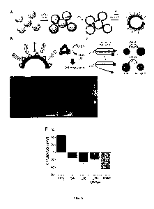
Fig. 1, related to Example 1, shows the fabrication and characterization of
urease/PEG
nanomotors (MSNP-Ur/PEG) and antibody-modified urease nanomotors (MSNP-Ur/PEG-
Ab). A) Scheme illustrating the stepwise fabrication process to obtain the
nanomotors.
Fig. 2, related to Example 1, shows motion analysis of MSNP-Ur/PEG and MSNP-
Ur/PEG-Ab. Representative tracked trajectories of A) MSNP-Ur/PEG nanomotors
and B)
MSNP-Ur/PEG-Ab nanomotors at 0 mM, 50 mM and 100 mM urea and C) Representative
mean-squared displacements (MSD) of both types of nanomotors at 0 mM, 50 mM
and
100 mM. D) Effective diffusion coefficients obtained by MSD analysis at
different urea
concentrations (N=20, error bars represent SE, p < 0.001).
Fig. 3, related to Example 1, shows the effect of nanomotors with and without
antibody on
spheroids' viability in the presence of different concentrations of urea.
Quantification of
spheroids' viability after 4-hour incubation with MSNP-Ur/PEG (originally in
blue) and
MSNP-Ur/PEG-Ab (originally in red), at different urea concentrations (N=3,
error bars
represent SE).
Fig. 4, related to Example 1, shows the targeting and penetration abilities of
antibody-
modified nanomotors into bladder cancer spheroids. Quantification of the
internalization of
antibody-modified nanomotors into bladder cancer spheroids in the presence (40
mM) and
absence of urea after 4-hour incubation, and quantification of the
proliferation of spheroids
incubated with MSNP-Ur/PEG and MSNP-Ur/PEG-Ab for 4 hours, in the presence (40
CA 03120178 2021-05-14
WO 2020/115124 PCT/EP2019/083662
mM) and absence of urea at after measured after a 48-hour resting period.
Fig. 5, related to Example 2, shows the fabrication approach and
characterization of DNA
micromotors. A) Schematic representation of the micromotors fabrication, where
a silicon
5 dioxide layer is grown onto a commercial polystyrene template by adding
APTES and
TEOS silica precursors. The polystyrene core is then removed by DMF and the
microcapsules are functionalized with urease and DNA scaffold through the use
of
glutaraldehyde (GA) linker. B) The pH-responsive DNA nanoswitch hybridizes to
the
complementary DNA scaffold that is covalently linked on the micromotor. Self-
propulsion
is achieved by the conversion of urea into ammonia and carbon dioxide,
mediated by
urease enzyme. C) The pH-dependent triplex-to-duplex transition of the
unimolecular DNA
nanoswitch results in change of FRET efficiency. D) Scanning electron
micrograph of SiO2
microcapsules. Inset shows a magnification of the selected area. Scale bar=2
pm. E)
Topographical image obtained by transmission electron microscopy. Calibration
bar
indicates the height in pm. F) Z-potential measurements of the microparticle
surface along
the functionalization process (NH2 = amine-coated particles resulting from the
synthesis;
GA=microparticles after incubation with glutaraldehyde, UR=urease-
functionalized
microparticles; UR+DNAss = microparticles functionalized with both urease and
DNA
scaffold; Switch = urease and DNA scaffold functionalized microparticles,
after their
hybridization with the DNA switch for 30 min.)
Fig. 6, related to Example 2, shows that triplex-based pH-responsive DNA
nanoswitch are
able to detect pH changes in solution and conjugated to the micromotor
structure. A)
Triplex DNA nanoswitch forms an intramolecular double hairpin structure
through the
.. formation of pH-insensitive Watson-Crick interactions (dashed line) and pH-
sensitive
Hoogsteen interactions (dots). Triplex nanoswitch containing CGC and TAT
triplets
unfolds into a duplex conformation by increasing the pH of the solution.
Ratiometric FRET
emission (left) showing the triplex-to-duplex transition of the DNA nanoswitch
as a
function of pH changes in solution. B) CSLM analysis of FRET effect of DNA
nanoswitch
functionalized microparticles, showing from right to left the Cy3 channel,
FRET channel
and the FRET/Cy3 ratio value, indicated in the calibration bar. Scale bar= 2
pm. The white
arrows indicate the functionalized microparticles (originally in red for the
Cy3 channel, in
green for the FRET channel, and yellow for the Cy3/FRET merge). Quantitative
pH
measurement by DNA-functionalized micromotors for pH-sensitive (C) and non-pH
specific (D), shown as the mean FRET/Cy3 emission values, shown as the mean
standard error of the mean.
Fig 7, related to Example 2. A) MSDs of DNA-switch micromotors. Results are
shown as
the mean standard error of the mean. B) Speed calculated from the MSDs.
Results are
CA 03120178 2021-05-14
WO 2020/115124 PCT/EP2019/083662
6
shown as the mean standard error of the mean.
Detailed description of the invention
All terms as used herein in this application, unless otherwise stated, shall
be understood
in their ordinary meaning as known in the art. Other more specific definitions
for certain
terms as used in the present application are as set forth below and are
intended to apply
uniformly through-out the specification and claims unless an otherwise
expressly set out
definition provides a broader definition.
As used herein, the indefinite articles "a" and "an" are synonymous with "at
least one" or
"one or more." Unless indicated otherwise, definite articles used herein, such
as "the" also
include the plural of the noun.
The term "enzyme-propelled nanomotor" or "enzyme-powered nanomotor" refers to
a
molecular device, on a micro or nano scale, capable of converting chemical
energy into
movement through the action of an enzyme located on the surface of the device.
In other
words, a nanomotor is a nanoparticle or a microparticle externally
functionalized with
enzymes. Without being bound by the theory, the enzymes generate movement
through
the asymmetric release of products involved in the catalytic reaction creating
interfacial
forces depending on osmotic gradients, charges, or other properties. The terms
"nanomotor" and "micromotor" are used interchangeably in the present
application.
As use herein, "heterologous molecule" refers to any molecule different from
the
enzyme(s), said enzyme(s) in charge of the propulsion of the nanomotor, and
that is
discontinuously attached over the whole surface of the particle. The
embodiments thereby
enable basically any type of molecule that can be linked to the particle to be
immobilized
onto a surface through its direct or indirect connection to the particle.
The below provided list of heterologous molecules should merely be seen as an
illustrative and non-limiting list of molecules that could be used in the
nanomotors of the
invention. The embodiments are, however, not limited thereto and encompasses
any
heterologous molecule that can be linked directly or indirectly to a nanomotor
of the
embodiments.
The heterologous molecule of interest could be selected among markers, such as
fluorescent markers, i.e. a fluorophore, e.g. fluorescein isothiocyanate
(FITC),
tetramethylrhodamine isothiocyanate (TRITC) and other isothiocyanates; N-
hydroxysuccinimide (NHS) fluorescein and other succinimidyl esters;
fluorescein-5-
CA 03120178 2021-05-14
WO 2020/115124 PCT/EP2019/083662
7
maleimide and other maleimide activated fluorophores; cyanine fluorophores;
fluroescein
fluorophores; rhodamine fluorophores; ATTO dyes; DyLight Fluor dyes; Alexa
Fluor dyes;
and boron-dipyrromethene (BODIPY) dyes. Further examples include isotope
labels or
markers, chemiluminescent markers, radiopaque markers, etc. In such a case,
the
nanomotor can be used as a test molecule to enable detection, using the
marker, of the
nanomotor on a surface.
Further examples of heterologous molecules include cell adhesion and cell
attachment
molecules, such cell adhesion molecules (CAMs), including immunoglobulin (Ig)
superfamily, integrins, cadhereins and selectins.
A further example of a heterologous molecule is extracellular matrix (ECM)
molecules
including, for instance, proteoglycans (PGs), glycosaminoglycans (GAGs),
heparan
sulfate (HS), chondroitin sulfates, keratin sulfates, collagen, elastins, etc.
A related type of molecular of interest is basal lamina molecules that include
molecules of
the basal lamina, which is a layer of ECM secreted by epithelial cells. Non-
limiting
examples of such basal lamina molecules include laminin, type IV collagen,
entactin and
perlecan.
Yet another example of a heterologous molecule of interest is an anti-
inflammatory
molecule, such as corticosteroids; glucocorticoids; non-steroidal anti-
inflammatory drugs
(NSAIDs), such as acetylsalicylic acid, iso-butyl-propanoic-phenolic acid and
naproxen
sodium (INN); lipoxins; interleukin-1 receptor antagonist (IL-1 RA); etc.
Antibiotics can also be used as heterologous molecules of interest in order to
inhibit
bacterial growth or kill bacteria. Non-limiting examples of antibiotics
include penicillins;
cephalosporins; polymyxins; rifamycins; lipiarmycins; quinolones;
sulfonamides;
macrolides; lincosamides; tetracylines; bactericidal aminoglycosides; cyclic
lipopeptides,
such as daptomycin; glycylcylines, such as tigecycline; oxazolidones, such as
linezolid;
and lipiarmycins, such as fidaxomicin.
In a similar way molecules targeting other types of microbes, such as anti-
fungal
molecules, e.g. polyene anti-fungals, such as amphotericin B, candicidin,
filipin, hamycin,
natamycin, nystatin and rimocidin; azole anti-fungals, such as imidazoles,
e.g. bifonazole,
butoconazole, clotrimazole, econazole, fenticonazole, isoconazole, miconazole,
omoconazole, oxiconazole, sertaconazole, sulconazole and tioconazole;
triazoles, e.g.
albaconazole, fluconazole, isavuconazole, itraconazole, posaconazole,
ravuconazole,
terconazole and voriconazole; and thiazoles, e.g. abafungin; allylamines, such
as
CA 03120178 2021-05-14
WO 2020/115124 PCT/EP2019/083662
8
amorolfin, butenafine, naftifine and terbinafine; echinocandins, such as
anidulafungin,
caspofungin and micafungin; benzoic acid; ciclopirox olamine; flucytosine;
griseofulvin;
tolnaftate and undecylenic acid. Also anti-viral molecules, e.g. virus-
assisted protein
(VAP) anti-idiotypic antibodies; amantadine; rimantadine; pleconaril;
acyclovir; zidovudine
(AZT); lamivudine; integrase; fomivirsen; rifampicin; zanamivir and
oseltamivir, and anti-
parasitic molecules, such as mebendazole; pyrantel pamoate; thiabendazole;
diethylcarbamazine; ivermectrin; niclosamide; praziquantel; albendazole;
praziquantel;
rifampin; amphotericin B; melarosprol; elfornithine; metronidazole; tinidazole
and
miltefosine, could be used as heterologous molecule of interest.
A further example of heterologous molecules include growth factors, such as
adenomedullin (AM), angiopoietin (Ang), autocrine motility factor, bone
morphogenetic
proteins (BMPs), brain-derived neutrophic factor (BDNF), epidermal growth
factor (EGF),
erythropoietin (EPO), fibroblast growth factor (FGF), glial cell line-derived
neutrophic
factor (GDNF), granulocyte colony-stimulating factor (G-CSF), granulocyte
macrophage
colony-stimulating factor (GM-CSF), growth differentiation factor-9 (GDF9),
hepatocyte
growth factor (HGF), hepatoma-derived growth factor (HDGF), insulin-like
growth factor
(IGF), mystatin (GDF-8), nerve growth factor (NGF), platelet-derived growth
factor
(PDGF), thrombopoietin (TP0), transforming growth factor alpha (TGF-a),
transforming
growth factor beta (TGF-B), tumor necrosis factor alpha (TNF-a), vascular
endothelial
growth factor (VEGF), placental growth factor (PIGF), etc. A nanomotor with a
growth
factor linked to a surface-binding peptide can be used to provide a surface
with, for
instance, capability of stimulating cellular growth, proliferation and/or
cellular
differentiation.
Further examples of heterologous molecules of interest include cell growth
inhibitors and
chemotherapeutic agents. Such a type of heterologous molecules will, when
included in
the nanomotor, provide a local cell growth inhibiting effect. Non-limiting
examples of such
heterologous molecules of interest include farnesyl transferase inhibitors;
alkylating
agents, such as nitrogen mustards, e.g. mechlorethamine, cyclophosphamide,
melphalan,
chlorambucil, ifosfamide and busulfan; nitrosoureas, e.g. N-nitroso-N-
methylurea (MNU),
carmustine (BCNU), lomustine (CCNU), semustine (MeCCNU), fotemustine and
streptozotocin; tetrazines, e.g. dacarbazine, mitozolomide and temozolomide
and
aziridines, e.g. thiotepa, mytomycin, diaziquone (AZQ); and cisplatines, e.g.
cisplatine,
carboplatin and oxaplatin; antimetabolites, such as anti-folates, e.g.
methotrexate and
pemetrexed; fluropyrimidines, e.g. fluorouracil and capecitabine;
deocynucleoside
analogues, such as cytarabine, gemcitabine, decitabine, Vidaza, fludarabine,
nelarabine,
cladribine, clofarabine and pentostatine; and thiopurines, e.g. thiguanine and
mercaptopurine; anti-microtubule agents, such as vinca alkaloids, e.g.
vincristine,
CA 03120178 2021-05-14
WO 2020/115124 PCT/EP2019/083662
9
vinblastine, vinorelbine, vindesine and vinflunine; and taxanes, e.g.
paclitaxel and
docetaxel; and podophyllotxin; topoisomerase inhibitors, such as irinotecan,
topotecan,
captothecin, etoposide, doxorubicin, mitoxantrone, teniposide, novobiocine,
merbarone
and aclarubicin; cytotoxic antibiotics, such as antracyclines, e.g.
doxorubicin,
daumorubicin, epirubicin, idarubicin, pirarubicin, aclarubicin, mitoxantrone,
actinomycin,
bleomycin, plicamycin, and mitomycin.
Other groups of heterologous molecules of interest include polynucleotides
such as DNA
or RNA molecules. The heterologous molecule can also be a nanosensor or a
molecular
gate.
"Discontinuously attached over the whole surface" refers to a discrete
distribution that is
not restricted to a single face or hemisphere of the particle, that is, it
refers to a nonpolar
distribution. It does not mean, however, that the molecule is covering the
whole surface of
the particle in a homogenous manner. The particles of the invention may
present the
molecules externally attached forming discrete patches over the whole surface
of the
particle, or which is the same, presenting gaps wherein no molecules are
attached.
As used herein, "targeting molecule" refers to a molecule having specificity
for a particular
cell, tissue, or organ. Preferred examples of targeting molecules include but
are not
limited to antibodies, growth factors, and polysaccharides.
As used herein, "nanosensor" refers to any nano or micro scale sensing device.
The
nanosensors of the invention are able to detect and respond to changes in the
environment where they are located. In particular, the nanosensors of the
invention can
be nanoswitches, which are nanosensors able to switch between two distinct
forms. DNA-
nanoswitches contain a single strand DNA molecule with a conformation that
changes in
response to an environmental change, for instance a pH change. The DNA
molecule may
be coupled to fluorescent molecules that allow the detection of the
conformational change.
As used herein, "labelling molecule" refers a molecule which can be chemically
bound to
the nanomotor and which emits a detectable signal enabling the nanomotor to be
detected. Particularly preferred examples of labelling molecules include but
are not limited
to chemiluminescent molecules, fluorescent molecules and isotopes.
As used herein, "molecular gate" or "nanovalve" refers to a molecular system
on a nano or
micro scale that switches between a first closed form and a second open form
in response
to a selected trigger, such as light, temperature, magnetic fields and pH. The
closed form
is design to block the release of the cargo contained in the particle to which
de molecular
CA 03120178 2021-05-14
WO 2020/115124 PCT/EP2019/083662
gate is attached. Upon application of the trigger, the gate changes to the
open form
allowing the release of the cargo.
An "anticancer antibody" refers to an antibody with the capacity to arrest or
eliminate
5 cancer cells.
As mentioned above, the invention provides in a first aspect an enzyme-powered
nanomotor externally functionalized with a heterologous molecule.
10 In a particular embodiment of the first aspect, optionally in
combination with any of the
embodiments provided above or below, the particle is a nanoparticle or a
microparticle.
The term "nanoparticle" as used herein, refers to a particle with at least two
dimensions at
the nanoscale, particularly with all three dimensions at the nanoscale. For
analogy, the
term "microparticle" as used herein, refers to a particle with at least two
dimensions at the
microscale, particularly with all three dimensions at the microscale In a
particular
embodiment, the particle is from 1 nm to 100 pm. In particular, from 30 nm to
2 pm. More
in particular, from 100 nm to 1 pm. Even more in particular, from 400 nm to
600 nm.
As regards the shape of the nanoparticles or microparticles described herein,
there are
included spheres, polyhedral and rod-shape. Particularly, when the
nanoparticle or
microparticle is substantially rod-shaped with a substantially circular cross-
section, such
as a nanowire or a nanotube, microwire or microtube, the "nanoparticle" or
"microparticle"
refers to a particle with at least two dimensions at the nanoscale or
microscale, these two
dimensions being the cross-section of the nanoparticle or the microparticle.
In a particular embodiment of the first aspect, optionally in combination with
any of the
embodiments provided above or below, the particle is spherical
As used herein, the term "size" refers to a characteristic physical dimension.
For example,
.. in the case of a nanoparticle/microparticle that is substantially
spherical, the size of the
nanoparticle/microparticle corresponds to the diameter of the
nanoparticle/microparticle.
When referring to a set of nanoparticles/microparticles as being of a
particular size, it is
contemplated that the set can have a distribution of sizes around the
specified size. Thus,
as used herein, a size of a set of nanoparticles/microparticles can refer to a
mode of a
distribution of sizes, such as a peak size of the distribution of sizes. In
addition, when not
perfectly spherical, the diameter is the equivalent diameter of the spherical
body including
the object. This diameter is generally referred as the "hydrodynamic
diameter", which
measurements can be performed using a Wyatt Mobius coupled with an Atlas cell
pressurization system or Malvern. Transmission Electron Microscopy (TEM) or
Scanning
CA 03120178 2021-05-14
WO 2020/115124 PCT/EP2019/083662
11
Electron Microscopy (SEM) images do also give information regarding diameters.
A wide variety of particle materials are available to the skilled person, and
he would
understand that the material chosen would depend on the intended application
of the
nanomotor, for instance, nanomotors for therapeutic uses should be made with
biocompatible particles.
Thus, in a particular embodiment of the first aspect, optionally in
combination with any of
the embodiments provided above or below, the particle is made of a material
selected
from the group consisting of metal, metal oxide, polymer, lipid, protein, cell
membrane,
cell body, carbonaceous material, and mixtures thereof. In a particular
embodiment, the
metal is aluminum (Al), platinum (Pt), palladium (Pd) or magnesium (Mg). In a
particular
embodiment, the metal oxide is selected from silica (SiO2), manganese oxide
(Mn02) and
titanium oxide (TiO2). In a particular embodiment the polymer is polystyrene
or metallic
organic frameworks. In a particular embodiment, the carbonaceous material is
selected
from carbon, graphene, and fullerene. In a particle embodiment, the material
of the
particle is a polymersome. In a particle embodiment, the particle is a protein-
based
particle (proteinsome). In a particular embodiment, the cell body is a
platelet or a red
blood cell (RBC).
The term "metallic organic framework" or "MOF" refers to compounds comprising
metal
ions or clusters coordinated to organic ligands. The central metallic element
may be at
least one selected from the group consisting of zinc (Zn), cobalt (Co),
cadmium (Cd),
nickel (Ni), manganese (Mn), chromium (Cr), copper (Cu), lanthanum (La), iron
(Fe),
platinum (Pt), palladium (Pd), silver (Ag), gold (Au), rhodium (Rh), iridium
(Ir), ruthenium
(Ru), lead (Pb), tin (Sn), aluminum (Al), titanium (Ti), molybdenum (Mo),
tungsten (W),
vanadium (V), niobium (Nb), tantalum (Ta), scandium (Sc), yttrium (Y), gallium
(Ga),
germanium (Ge), indium (In), bismuth (Bi), selenium (Se), and antimony (Sb).
The organic
ligand may include a functional group that is linkable to at least two
metallic ions.
"Cell membrane" refers to the lipid bilayer that forms a continuous barrier
around cells.
The particles of the invention can be formed of prokaryotic or eukaryotic cell
membranes.
As used herein, "cell body" refers to the part of a cell that contains the
genetic material
surrounded by the cytoplasm and the plasma membrane.
The term "polymersome" refers to artificial vesicles made of amphiphilic
synthetic block
copolymers,
CA 03120178 2021-05-14
WO 2020/115124 PCT/EP2019/083662
12
The term "proteinosome" refers to particles composed of self-assembled
proteins or
protein-polymer conjugates.
In a particular embodiment, optionally in combination with any of the
embodiments
provided above or below, the particle is made of mesoporous silica. As used
herein,
"mesoporous silica" refers to porous silica having medium-sized pores
regularly arranged,
specifically, the pores are from 2 nm to 50 nm, and more specifically, from 4
nm to about
40 nm.
In a particular embodiment of the first aspect, optionally in combination with
any of the
embodiments provided above or below, the enzyme is selected from the group
consisting
of oxidoreductase, hydrolase and lyase. In a more particular embodiment, the
enzyme is
selected from the group consisting of glucose oxidase, urease, catalase,
glutamate
oxidase, xanthine oxidase, peroxidase, bilirubin oxidase, lipase, protease and
combinations thereof. In a more particular embodiment, the enzyme is urease.
The term
urease (EC 3.5.1.5) refers to the group of enzymes that catalyze the
hydrolysis of urea
into carbon dioxide and ammonia. In a particular embodiment of the invention,
the urease
is from Canavalia ensiformis (CAS Number 9002-13-5). In a more particular
embodiment,
it is a Type IX urease from Canavalia ensiformis (CAS Number 9002-13-5). The
sequence
of the enzyme can be found in various databases, such as Uniprot (P07374
UREA_CANEN, 01/02/1994 update)
In a particular embodiment of the first aspect, optionally in combination with
any of the
embodiments provided above or below, the heterologous molecule is selected
from the
group consisting of a targeting molecule, a labelling molecule, a nanosensor
and a
molecular gate.
In a particular embodiment of the first aspect, optionally in combination with
any of the
embodiments provided above or below, the heterologous molecule is an antibody.
In a
more particular embodiment, the antibody is an anticancer antibody. In a more
particular
embodiment, the anticancer antibody binds a membrane receptor. In a more
particular
embodiment, the membrane receptor is a FGFR (fibroblast growth factor
receptor)
selected from the group consisting of FGFR1, FGFR2, FGFR3, and FGFR4. In a
more
particular embodiment, the FGFR is FGFR3.
In a particular embodiment of the first aspect, optionally in combination with
any of the
embodiments provided above or below, the heterologous molecule is a
nanosensor.
There are numerous nanosensors available for the skilled in the art (Campuzano
S. et al.,
"Motion-driven sensing and biosensing using electrochemically propelled
nanomotors",
CA 03120178 2021-05-14
WO 2020/115124 PCT/EP2019/083662
13
Analyst. 2011, vol. 36(22), pp. 4621-30). In a particular embodiment, the
nanosensor is a
DNA-nanoswitch. DNA-nanoswitches are molecular complexes comprising a single
strand
DNA molecule coupled to a fluorophore-quencher pair. In a particular
embodiment, the
sequence of the DNA molecule of the DNA-nanoswitch has at least 85%, 86%, 87%,
88%,
89%, 90%, 91%, 92%, 93%, 94%, 95%, 96%, 97%, 98%, 99% or 100% sequence
identity
with SEQ ID NO: 1.
In a particular embodiment, the nanosensor is a nanoparticle. In a more
particular
embodiment, the nanosensor is a metal nanoparticle. More particularly, the
metal
nanoparticle is a gold (Au) nanoparticle. The skilled in the art would
understand that the
nanoparticle forming the nanosensor has to be smaller than the nanoparticle or
microparticle forming the nanomotor.
In the present invention the term "identity" refers to the percentage of
residues that are
identical in the two sequences when the sequences are optimally aligned. If,
in the optimal
alignment, a position in a first sequence is occupied by the same amino acid
residue as
the corresponding position in the second sequence, the sequences exhibit
identity with
respect to that position. The level of identity between two sequences (or
"percent
sequence identity") is measured as a ratio of the number of identical
positions shared by
the sequences with respect to the size of the sequences (i.e., percent
sequence identity =
(number of identical positions/total number of positions) x 100).
A number of mathematical algorithms for rapidly obtaining the optimal
alignment and
calculating identity between two or more sequences are known and incorporated
into a
number of available software programs. Examples of such programs include the
MATCH-
BOX, MULTAIN, GCG, FASTA, and ROBUST programs for amino acid sequence
analysis, among others. Preferred software analysis programs include the
ALIGN,
CLUSTAL W, and BLAST programs (e.g., BLAST 2.1, BL2SEQ, and later versions
thereof).
For amino acid sequence analysis, a weight matrix, such as the BLOSUM matrixes
(e.g.,
the BLOSUM45, BLOSUM50, BLOSUM62, and BLOSUM80 matrixes), Gonnet matrixes,
or PAM matrixes (e.g., the PAM30, PAM70, PAM120, PAM160, PAM250, and PAM350
matrixes), are used in determining identity.
The BLAST programs provide analysis of at least two amino acid sequences,
either by
aligning a selected sequence against multiple sequences in a database (e.g.,
GenSeq),
or, with BL2SEQ, between two selected sequences. BLAST programs are preferably
modified by low complexity filtering programs such as the DUST or SEG
programs, which
CA 03120178 2021-05-14
WO 2020/115124 PCT/EP2019/083662
14
are preferably integrated into the BLAST program operations. If gap existence
costs (or
gap scores) are used, the gap existence cost preferably is set between about -
5 and -15.
Similar gap parameters can be used with other programs as appropriate. The
BLAST
programs and principles underlying them are further described in, e.g.,
Altschul et al.,
"Basic local alignment search tool", 1990, J. Mol. Biol, v. 215, pages 403-
410.
For multiple sequence analysis, the CLUSTAL W program can be used. The CLUSTAL
W
program desirably is run using "dynamic" (versus "fast") settings. Amino acid
sequences
are evaluated using a variable set of BLOSUM matrixes depending on the level
of identity
between the sequences. The CLUSTAL W program and underlying principles of
operation
are further described in, e.g., Higgins et al., "CLUSTAL V: improved software
for multiple
sequence alignment", 1992, CABIOS, 8(2), pages 189-191.
In a particular embodiment of the first aspect, optionally in combination with
any of the
embodiments provided above or below, the nanomotor further comprises a cargo.
In a
particular embodiment, the cargo is selected from the list consisting of a
drug, wherein the
drug is selected from the group consisting of a small molecule, a nucleic
acid, a
therapeutic enzyme, a peptide, a protein or a hormone. For "cargo" it is
understood any
molecule transported within the nanomotor to be delivered at the desired
target.
Depending on the surface material and porosity of the particle, the cargo can
be inside the
particle or adsorbed to its surface.
In a particular embodiment, the drug is a cytotoxic drug. More particularly,
it is an
anticancer drug.
In a particular embodiment of the first aspect, optionally in combination with
any of the
embodiments provided above or below, the enzyme and the heterologous molecule
are
attached to the surface of the particle directly or through a linker. In a
more particular
embodiment, the linker is selected from the group consisting of anhydrides,
alcohols,
acids, amines, epoxies, isocyanates, silanes, halogenated groups, and
polymerizable
groups, preferably 3-aminopropyltriethoxysilane (APTES). In an even more
particular
embodiment, the linker is glutaraldehyde.
In such an aspect, one part of the linker is bound to the surface of the
particle, and
another part of the linker is bound to the enzyme or heterologous molecule,
thereby
forming a covalent bond between the enzyme or heterologous molecule and the
surface.
The bond between the linker and said surface and said enzyme or heterologous
molecule
is effected by chemical reactions occurring between the linker and the enzyme
or
heterologous molecule thereby securing a covalent bond between the surface and
said
CA 03120178 2021-05-14
WO 2020/115124 PCT/EP2019/083662
enzyme or heterologous molecule. In one embodiment, the enzyme and/or the
heterologous molecule are attached to the surface of the particle after a
chemical pre-
treatment of said surface of the particle.
5 As mentioned above, in a second aspect the invention provides a
pharmaceutical
composition comprising a therapeutically effective amount of the nanomotor of
the first
aspect and a pharmaceutically acceptable excipient and/or carrier.
The expression "therapeutically effective amount" as used herein, refers to
the amount of
10 the nanomotor that, when administered, is sufficient to prevent
development of, or
alleviate to some extent, one or more of the symptoms of the disease or
disorder which is
addressed. The particular dose of agent administered according to this
invention will of
course be determined by the particular circumstances surrounding the case,
including the
nanomotor administered, the route of administration, the particular condition
being treated,
15 and the similar considerations.
The expression "pharmaceutical composition" encompasses both compositions
intended
for human as well as for non-human animals (i.e. veterinarian compositions).
The expression "pharmaceutically acceptable carriers or excipients" refers to
pharmaceutically acceptable materials, compositions or vehicles. Each
component must
be pharmaceutically acceptable in the sense of being compatible with the other
ingredients of the pharmaceutical composition. It must also be suitable for
use in contact
with the tissue or organ of humans and non-human animals without excessive
toxicity,
irritation, allergic response, immunogenicity or other problems or
complications
commensurate with a reasonable benefit/risk ratio.
Examples of suitable pharmaceutically acceptable excipients are solvents,
dispersion
media, diluents, or other liquid vehicles, dispersion or suspension aids,
surface active
agents, isotonic agents, thickening or emulsifying agents, preservatives,
solid binders,
lubricants and the like. Except insofar as any conventional excipient medium
is
incompatible with a substance or its derivatives, such as by producing any
undesirable
biological effect or otherwise interacting in a deleterious manner with any
other
component(s) of the pharmaceutical composition, its use is contemplated to be
within the
scope of this invention.
The relative amounts of the nanomotor, the pharmaceutically acceptable
excipients,
and/or any additional ingredients in a pharmaceutical composition of the
invention will
CA 03120178 2021-05-14
WO 2020/115124 PCT/EP2019/083662
16
vary, depending upon the identity, size, and/or condition of the subject
treated and further
depending upon the route by which the composition is to be administered.
Pharmaceutically acceptable excipients used in the manufacture of
pharmaceutical
compositions include, but are not limited to, inert diluents, dispersing
and/or granulating
agents, surface active agents and/or emulsifiers, disintegrating agents,
binding agents,
preservatives, buffering agents, lubricating agents, and/or oils. Excipients
such as coloring
agents, coating agents, sweetening, and flavoring agents can be present in the
composition, according to the judgment of the formulator.
The pharmaceutical compositions containing the nanomotor of the invention can
be
presented in any dosage form, for example, solid or liquid, and can be
administered by
any suitable route, for example, oral, parenteral, rectal, topical, intranasal
or sublingual
route, for which they will include the pharmaceutically acceptable excipients
necessary for
the formulation of the desired dosage form, for example, topical formulations
(ointment,
creams, lipogel, hydrogel, etc.), eye drops, aerosol sprays, injectable
solutions, osmotic
pumps, etc.
Exemplary diluents include, but are not limited to, calcium carbonate, sodium
carbonate,
calcium phosphate, dicalcium phosphate, calcium sulfate, calcium hydrogen
phosphate,
sodium phosphate lactose, sucrose, cellulose, microcrystalline cellulose,
kaolin, mannitol,
sorbitol, inositol, sodium chloride, dry starch, corn-starch, powdered sugar,
and
combinations thereof.
Exemplary granulating and/or dispersing agents include, but are not limited
to, potato
starch, corn starch, tapioca starch, sodium starch glycolate, clays, alginic
acid, guar gum,
citrus pulp, agar, bentonite, cellulose and wood products, natural sponge,
cation-
exchange resins, calcium carbonate, silicates, sodium carbonate, cross-linked
polyvinylpyrrolidone) (crospovidone), sodium carboxymethyl starch (sodium
starch
glycolate), carboxymethyl cellulose, cross-linked sodium carboxymethyl
cellulose
(croscarmellose), methylcellulose, pregelatinized starch (starch 1500),
microcrystalline
starch, water insoluble starch, calcium carboxymethyl cellulose, magnesium
aluminum
silicate (Veegum), sodium lauryl sulfate, quaternary ammonium compounds, and
combinations thereof.
Exemplary binding excipients include, but are not limited to, starch (e.g.,
corn-starch and
starch paste); gelatin; sugars (e.g., sucrose, glucose, dextrose, dextrin,
molasses, lactose,
lactitol, mannitol); natural and synthetic gums (e.g., acacia, sodium
alginate, extract of
Irish moss, panwar gum, ghatti gum, mucilage of isapol husks,
carboxymethylcellulose,
CA 03120178 2021-05-14
WO 2020/115124 PCT/EP2019/083662
17
methylcellulose, ethylcellulose, hydroxyethylcellulose, hydroxypropyl
cellulose,
hydroxypropyl methylcellulose, microcrystalline cellulose, cellulose acetate,
polyvinylpyrrolidone), magnesium aluminium silicate (Veegum), and larch
arabogalactan);
alginates; polyethylene oxide; polyethylene glycol; inorganic calcium salts;
silicic acid;
polymethacrylates; waxes; water; alcohol; and combinations thereof.
Exemplary preservatives may include antioxidants, chelating agents,
antimicrobial
preservatives, antifungal preservatives, alcohol preservatives, acidic
preservatives, and
other preservatives. Exemplary antioxidants include, but are not limited to,
alpha
tocopherol, ascorbic acid, ascorbyl palmitate, ascorbyl stearate, ascorbyl
oleate, butylated
hydroxyanisole, butylated hydroxytoluene, monothioglycerol, potassium
metabisulfite,
propionic acid, propyl gallate, sodium ascorbate, sodium bisulfite, sodium
metabisulfite,
and sodium sulfite. Exemplary chelating agents include
ethylenediaminetetraacetic acid
(EDTA), citric acid monohydrate, disodium edetate, dipotassium edetate, edetic
acid,
fumaric acid, malic acid, phosphoric acid, sodium edetate, tartaric acid, and
trisodium
edetate.
Exemplary buffering agents include, but are not limited to, citrate buffer
solutions, acetate
buffer solutions, phosphate buffer solutions, ammonium chloride, calcium
carbonate,
calcium chloride, calcium citrate, calcium glubionate, calcium gluceptate,
calcium
gluconate, D-gluconic acid, calcium glycerophosphate, calcium lactate,
propanoic acid,
calcium levulinate, pentanoic acid, dibasic calcium phosphate, phosphoric
acid, tribasic
calcium phosphate, calcium hydroxide phosphate, potassium acetate, potassium
chloride,
potassium gluconate, potassium mixtures, dibasic potassium phosphate,
monobasic
potassium phosphate, potassium phosphate mixtures, sodium acetate, sodium
bicarbonate, sodium chloride, sodium citrate, sodium lactate, dibasic sodium
phosphate,
monobasic sodium phosphate, sodium phosphate mixtures, tromethamine, magnesium
hydroxide, aluminum hydroxide, alginic acid, pyrogen-free water, isotonic
saline, Ringer's
solution, ethyl alcohol, and combinations thereof.
Exemplary lubricating agents include, but are not limited to, magnesium
stearate, calcium
stearate, stearic acid, silica, talc, malt, glyceryl behanate, hydrogenated
vegetable oils,
polyethylene glycol, sodium benzoate, sodium acetate, sodium chloride,
leucine,
magnesium lauryl sulfate, sodium lauryl sulfate, and combinations thereof.
As mentioned before, the invention also provides in a third aspect the
nanomotor or the
pharmaceutical composition of the invention for use in therapy, diagnosis or
prognosis.
CA 03120178 2021-05-14
WO 2020/115124 PCT/EP2019/083662
18
Urease-propelled nanomotors are capable of moving not only in liquid media,
such as
urine, but also in viscous media such as hyaluronic acid. Moreover, they are
also active in
mucous secretions. Therefore, the nanomotors of the invention can be used both
in liquid
and viscous tissues, such as in the aqueous humour of the eye.
In a particular embodiment of the third aspect, optionally in combination with
any of the
embodiments provided above or below, the nanomotor of the first aspect or the
pharmaceutical composition of the second aspect is for use in the treatment of
cancer.
This embodiment can also be formulated as the use of the nanomotor of the
first aspect,
or the pharmaceutical composition of the second aspect for the manufacture of
a
medicament for the treatment and/or prevention of cancer. This aspect can also
be
formulated as a method for treating and/or preventing cancer, the method
comprising
administering a therapeutically effective amount of the nanomotor of the first
aspect or the
pharmaceutical composition of the second aspect, to a subject in need thereof.
Illustrative non-limiting examples of cancer which can be treated with the
nanomotor or
the pharmaceutical composition of the invention include, although they are not
limited to,
papillomas, adenomas, lipomas, osteomas, myomas, angiomas, nevi, mature
teratomas,
carcinomas, sarcomas.immature teratomas, melanoma, myeloma, leukaemia,
Hodgkin's
lymphoma, basalioma, spinalioma, breast cancer, ovarian cancer, uterine
cancer, bladder
cancer, lung cancer, bronchial cancer, prostate cancer, colon cancer,
pancreatic cancer,
kidney cancer, esophageal cancer, hepatocarcinoma, head and neck cancer, etc.
In a
particular embodiment of the third aspect, the cancer is bladder cancer.
From the data herein provided, the skilled in the art would understand that
the nanomotors
and pharmaceutical compositions of the invention may also be useful in the
treatment of
other diseases such as metabolic, neurologic and inflammatory diseases.
As mentioned above, in a fifth aspect the invention provides an in vitro
method of
detecting an analyte in an isolated sample, which comprises contacting the
nanomotor as
defined in the first aspect with the sample. The skilled in the art would
understand that the
nanomotors of the invention can be adapted to detect different analytes
through their
functionalization with sensors of said analytes.
In a particular embodiment of the fifth aspect, optionally in combination with
any of the
embodiments provided above or below, the sample is a biological isolated
sample. More
particularly, the biological isolated sample is blood, plasma or serum.
CA 03120178 2021-05-14
WO 2020/115124 PCT/EP2019/083662
19
As mentioned above, in a sixth aspect the invention provides the use of the
nanomotor as
defined in the first aspect in an in vitro method for detecting an analyte in
an isolated
sample.
In a particular embodiment of the fifth or sixth aspects, optionally in
combination with any
of the embodiments provided above or below, the sample is a liquid sample. As
mentioned before, the nanomotors of the inventors are capable of moving in
liquids with
various degrees of viscosity. For instance, they can move in urine, which has
a kinematic
viscosity of: 1.0700 cSt at 20 C (measured by Inman et al., "The impact of
temperature
and urinary constituents on urine viscosity and its relevance to bladder
hyperthermia
treatment", Int J Hyperthermia, 2013, vol. 29(3), pp. 206-10), and Hyaluronic
acid at the
concentration found in the synovial fluids (1 mg/ml, around 10-2 Pa*s for a
range of 1 to
10 Hz of shear rate, measured in a rheometer).
The nanomotors of the invention are particularly useful for the detection of a
wide variety
of analytes, such as pollutants or biomarkers,
Throughout the description and claims the word "comprise" and variations of
the word, are
not intended to exclude other technical features, additives, components, or
steps.
Furthermore, the word "comprise" encompasses the case of "consisting of".
Additional
objects, advantages and features of the invention will become apparent to
those skilled in
the art upon examination of the description or may be learned by practice of
the invention.
The following examples and drawings are provided by way of illustration, and
they are not
intended to be limiting of the present invention. Reference signs related to
drawings and
placed in parentheses in a claim, are solely for attempting to increase the
intelligibility of
the claim, and shall not be construed as limiting the scope of the claim.
Furthermore, the
present invention covers all possible combinations of particular and preferred
embodiments described herein.
Examples
1. Targeting 3D Bladder Cancer Spheroids with Urease-Powered Nanomotors
1.1. Methods
Materials
Ethanol (Et0H, 99%), methanol (Me0H, 99%), hydrochloric acid (37% in water),
ammonium hydroxide (25% in water), tetraethylorthosilicate (TEOS, 99%),
triethanolamine
(TEOA, 99%), cetyltrimethylammonium bromide (CTAB, 99%), 3-
CA 03120178 2021-05-14
WO 2020/115124 PCT/EP2019/083662
aminopropyltriethoxysilane (APTES, 99%), glutaraldehyde (GA, 25% in water),
urease
(from Canavalia ensiformis, Type IX, powder, 50 000-100 000 units/g solid),
Urease
Activity Assay Kit (Sigma-Aldrich), urea (99.9%), glycerol (99%), sodium
borohydride
powder (NaBH4, 98.0%), formaldehyde solution (37% in water), bovine serum
albumin
5 .. (lyophilized powder), 4-nitrophenol solution (10 mM), sodium chloride
puriss. (NaCI),
potassium chloride anhydrous (KCI), sodium phosphate monobasic (NaH2PO4),
sodium
bicarbonate BioXtra (99.5-100.5%, NaHCO3), dimethyl sulfoxide (DMSO, 99.9%),
and
HS-PEG5K-NH2 (HCI salt) were purchased from Sigma-Aldrich. Piercenn. BOA
Protein
Assay Kit, Wheat Germ Agglutinin (WGA AlexaFluorTm 647 conjugate), Goat anti-
Mouse
10 .. IgG (H+L) Alexa FluorTM 488 conjugate, 3-(4,5-dimethylthiazol-2-y1)-2,5-
diphenyltetrazolium bromide (MTT), and phosphate buffer saline (PBS) were
purchased
from Thermo Fisher Scientific. MatrigelTM basement matrix was purchased from
Corning.
Anti-FGFR3 antibody (ab89660) was purchased from Abcam. Hoechst 33342 was
purchased from Life Sciences. Spectra/Por 7 Standard RC pre-treated Dialysis
Tubing
15 .. (3.5 kD) was purchased from Spectrum. Cyanine3 NHS ester was purchased
from
Lumiprobe. McCoy's 5A (modified) medium, Penicillin Streptomycin solution,
Fetal Bovine
Serum (FBS) and Trypsin 0.5% EDTA were purchased from Gibco. LIVE/DEADTM
Viability/Cytotoxicity Kit was purchased from lnvitrogen. Human urinary
bladder
transitional cell papilloma RT4 cells were obtained from ATCC (Rockville, MA).
Instruments
Transmission Electron Microscopy (TEM) images were captured using a JEOL JEM-
2100
microscope. Scanning Electron Microscopy (SEM) images were captured using a
FEI
NOVA NanoSEM 230 at 10 kV. Hydrodynamic radii and electrophoretic mobility
measurements were performed using a Wyatt Mobius coupled with an Atlas cell
pressurization system. The Brunauer¨Emmett¨Teller (BET) analysis was carried
out using
a Micromeritics Tristar II Plus automated analyzer. Optical videos as well as
cell culture
imaging were performed using an inverted optical microscope (Leica DMi8)
equipped with
a 63x water objective, a galvo stage and filter cubes for FITC, Rhodamine,
DAPI and
CY5. Protein quantification and enzymatic activity assays were carried out
using an
Infinite M200 PRO Multimode Microplate Reader. The confocal microscopy
analysis was
performed using a LSM 800 ¨ Zeiss equipped with a 63x oil objective.
Synthesis of Mesoporous Silica Nanoparticles (MSNPs): The MSNPs were prepared
.. using a sol-gel method. Briefly, a solution containing CTAB (570 mg), TEOA
(35 g), and
water (20 mL) was heated to 95 C in a silicon oil bath. This mixture was
stirred for 30
minutes, and subsequently TEOS (1.5 mL) was added dropwise. The mixture was
further
stirred at 95 C, for 2 hours. The produced particles were collected by
centrifugation and
washed with ethanol (3 times, 1350 g, 10 minutes). Then, the particles were
suspended in
CA 03120178 2021-05-14
WO 2020/115124 PCT/EP2019/083662
21
a MeOH:HCI mixture (30 mL, 10:0.6) and refluxed at 80 C for 24 hours, for
removal of
CTAB from the MSNPs' pores. Finally, the particles are collected by
centrifugation and
washed in ethanol (3 times, 1350 g, 10 minutes), sonicating 20 minutes between
each
centrifugation. Aliquots (0.5 mL) were collected, centrifuged and air-dried to
determine the
concentration of the MSNPs suspension.
Amine Functionalization of MSNPs (MSNP-NH2): The previously synthesized MSNPs
were suspended in Et0H (2 mg/mL). Then, APTES was added to the suspension (10
pL/mg of MSNP) and it was shaken for 24 hours at room temperature, using an
end-to-
end rotary shaker. Finally, the particles were collected by centrifugation and
washed in
ethanol (3 times, 1350 g 10 minutes) and in water (3 times, 1928 g 10
minutes),
sonicating 20 minutes between each centrifugation. Aliquots (0.5 mL) were
collected,
centrifuged and air-dried to determine the concentration of the MSNPs
suspension.
Functionalization of MSNP-NH2 with Urease and Heterobifunctional H2N-PEG-SH
(MSNP-
Ur/PEG): MSNP-NH2 were centrifuged at 1340 g for 10 minutes, suspended in 900
pL of
PBS (2 mg/mL) and sonicated for 20 minutes. After this, 100 pL of
glutaraldehyde were
added and the mixture was vortexed for 30 seconds to obtain a good dispersion.
The
mixture was placed on an end-to-end rotary shaker for 2.5 hours, at room
temperature.
The nanoparticles were then collected and washed three times with PBS (1340 g,
10
minutes) and sonicated for 20 minutes between each wash. Next, the GA-
activated
nanoparticles were suspended in solution of PBS containing urease (3 mg/mL)
and H2N-
PEG-SH (1 pg/mg of MSNP-NH2). The mixture was then placed on an end-to-end
rotary
shaker overnight, at room temperature. The resulting nanomotors were washed
three
times with PBS by centrifugation (1340 g, 10 minutes), intercalating the
washes with 3
minutes of sonication.
Functionalization of PEGylated Urease Nanomotors with anti-FGFR3 antibody
(MSNP-
Ur/PEG-Ab): The nanomotors were suspended in PBS (2 mg/mL) and anti-FGFR3
antibody (30 pg of antibody per mg of nanomotors) was added. The mixture was
then
incubated overnight in the rotary shaker, at room temperature. Finally, the
antibody-
modified nanomotors were collected by centrifugation (1340 g, 10 minutes) and
washed
three times with PBS, intercalating the washes with 3 minutes of sonication.
Hydrodynamic Radii and Surface Charge Analysis: A Wyatt Mobius DLS, coupled to
an
ATLAS pressurizer was used to characterize the size distribution and surface
charge of
MSNP, MSNP-NH2, MSNP-Ur/PEG and MSNP-Ur/PEG-Ab. The equipment uses a 532
nm wavelength laser and a detector angle of 163.5 . The samples analyzed were
diluted
to a concentration 0.3 mg/mL and analyzed for light scattering and
electrophoretic mobility
simultaneously, with an acquisition time of 5 seconds, performing 3 runs per
measurement. A total of 9 measurements were performed to obtain statistical
relevant
CA 03120178 2021-05-14
WO 2020/115124 PCT/EP2019/083662
22
data.
Quantification of Urease and Antibody Amounts on MSNP: The concentration of
urease
present on MSNP-Ur/PEG was measured using the BOA Protein Assay Kit from
Thermo
Fisher Scientific, according to manufacturer's instructions. This kit
correlates the quantity
of proteins with the reduction of copper by peptide bonds. The same procedure
was
repeated for MSNP-Ur/PEG-Ab, in order to quantify the amount of antibody bound
to the
nanomotors.
Urease Activity Assay: enzymatic activity of urease while bound to MSNPs was
evaluated
using a commercial kit that determines the concentration of ammonia generated
by
Berthelot's method (Patton, C. J. et al., "Spectrophotometric and Kinetics
Investigation of
the Berthelot Reaction for the Determination of Ammonia", Anal. Chem., 1977,
vol. 49, pp.
464-469). The nanomotors were at a concentration of 0.5 mg/mL and the
experiment was
performed according to manufacturer's instructions.
Urease Labeling with 0y3: Urease (22 mg) was dissolved in 1 mL of sodium
bicarbonate
buffer (100 mM). Next, 7 pL of a Cy 3 solution in DMSO (5 mM) were added to
the urease
solution, and the mixture was incubated for 4 hours, at room temperature and
shaking in
the dark. The solution of labelled urease was then dialyzed (3.5 kD pore
membrane) for
24 hours to eliminate non-reacted 0y3 molecules.
Quantification of Ammonia Production by MSNP-Ur/PEG: The ammonia produced by
nanomotors was quantified using a titration method. For this, the nanomotors
were
incubated with different urea concentrations (12.5, 25, 50, 100, 200, and 300
mM) and the
samples were analyzed at different time points (5, 15, 60, 120, 240 minutes
and at 24
hours). At each time point, the suspensions of nanomotors was centrifuged and
the
supernatant was titrated with HCI (10 mM), using p-nitrophenol as indicator.
.. Optical Video Recording of Nanomotors and MSD Analysis: An inverted
microscope
equipped with a 63x water objective was used to observe and record videos of
the
nanomotors movement. Samples of aqueous solutions of simulated urine
containing
nanomotors were placed in a glass slide and mixed well with simulated urine at
a range of
urea concentrations (12.5, 25, 50, 100, 200, and 300 mM). The samples were
then
covered with a glass slide to avoid artifacts caused by drifting and videos of
30 seconds
were recorded. The videos were acquired using a Hamamatsu camera, at a frame
rate of
50 fps, in bright field. At least 20 nanomotors are analyzed per condition.
The videos were
analyzed using a python-based code to obtain the trajectories of the
nanomotors, and
compute the mean-squared displacement (MSD) following:
CA 03120178 2021-05-14
WO 2020/115124 PCT/EP2019/083662
23
MSD (At) = ((x_i (t+At)-x_i (t))^2 ), i = 2, for 2D analysis
After this, the diffusion coefficient (De) was obtained by fitting the MSD
data to equation 2,
which is valid at short time intervals for small particles, with low
rotational diffusion. 3D
Cell Culture: Human urinary bladder transitional cell papilloma RT4 cells were
cultured in
McCoy's 5A (Modified) Medium, supplemented with FBS (10%) and penicillin-
streptomycin solution (1%), in a 37 C and 5% CO2 atmosphere. The cells were
split
every 4 days at a 1:2 ratio. To obtain 3D RT4 cell cultures, 8-well ibidi
dishes were pre-
coated with 23 pL of MatrigelTM (5 mg/mL) and incubated at 37 C for 30
minutes, allowing
the gel to form. Next, 30 pL of a suspension of RT4 cells at a density of
5x10⁶ cells/mL
was spread evenly in each well and the dishes were incubated for 30 minutes at
37 C.
Finally, 150 pL of RT4 McCoy's medium containing 10% of MatrigelTM was added.
The
cultures were allowed to grow for 7 days before the experiments, changing the
medium
every 2 days.
lmmunostaining of FGFR3 Transmembrane Protein in 3D RT4 Cell Cultures: the 3D
cultures described above were washed 3 times with PBS lx. Then, the surface of
the
wells was gently scratched with a pipette tip and the culture was suspended in
McCoy's
medium in a tube. The tubes were briefly spinned and the supernatant was
removed.
Next, the cells were suspended in formaldehyde (3.7 A), placed in an 8-well
dish and
incubated for 15 minutes at room temperature. Following, the culture was
washed with
PBS lx, a solution of PBS-BSA (5 A) was added and the dish was incubated for
40
minutes at room temperature. Then, the anti-FGFR3 was added to the culture at
a
proportion of 1:50, and the dish was incubated for 24 hours, at 37 C, in a 5
A CO2
atmosphere. After, the culture was washed 3 times with PBS lx, the secondary
antibody
(labeled with AlexaFluor 488) was added in a proportion of 1:500 and the dish
was
incubated for 40 minutes, at room temperature in the dark. Finally, the
culture was
washed 3 times with PBS lx, the nuclei were labeled with Hoescht and a
solution of
glycerol in PBS (70 A) was added. The culture was observed using confocal
microscopy.
Cytotoxicity Assays: The viability of RT4 3D cultures was quantified using the
Alamar Blue
assay and visualized using the LIVE/DEAD viability kit following
manufacturer's
instructions. For this, RT4 cells were culture as mentioned above and at day 7
were
incubated with each treatment¨Ammonia (1 mM, 1.5 mM, 3 mM, 5 mM, 10 mM and 20
mM, for 24 hours), Urea (25 mM, 30 mM, 40 mM and 50 mM, for 24 hours), MSNP-
Ur/PEG (12.5 pg/mL, at a range of urea concentrations ¨25 mM, 30 mM, 40 mM and
50
mM, for 1, 2 and 4 hours). After, the cultures were washed with medium, kept
resting for
24 hours and viability was investigated according to manufacturer's
instructions.
CA 03120178 2021-05-14
WO 2020/115124 PCT/EP2019/083662
24
Furthermore, viability was also assessed at 48 h time point.
Imaging of RT4 3D Cultures and Nanomotors: At day 7, the 3D cell cultures were
incubated with each treatment (MSNP-Ur/PEG or MSNP-Ur/PEG-Ab, 12.5 pg/mL) for
4
hours. At each time point, the cultures were washed and kept in a 37 C and 5%
CO2
atmosphere for 24 hours. Then, the cultures were labeled WGA 647 (membranes)
and
imaged using an inverted fluorescence microscope equipped with a 63x objective
and a
galvo stage, as well as filter cubes for Rhodamine, FITC, DAPI and Cy5.
1.2. Results and discussion
Fully mesoporous silica nanoparticles (MSNPs) were prepared using sol-gel
chemistry,
where cetyltrimethylammonium bromide (CTAB) was used as porogenic agent and
triethanolamine (TEOA) was used as base catalyst. The prepared MSNPs were
characterized by scanning electron microscopy (SEM). SEM analysis revealed
good
monodispersity of the sample (polydispersity index = 0.114) and a mean
diameter of 481
2 nm (N = 150, average size standard error of the mean (SE)). Furthermore,
the porous
structure of the MSNPs was evaluated by transmission electron microscopy. A
clear radial
porosity was evidenced by the TEM. This crystalline configuration was further
confirmed
by the Fast Fourier Transform, which indicated the periodicity of the porous
pattern. The
surface area of the nanoparticles was studied by performing nitrogen
adsorption/desorption, using Brunauer¨Emmett¨Teller analysis (BET) method. The
MSNPs showed a type IV isotherm, typical of mesoporous silica structures, and
a BET
specific surface area of 1184.8 m2/g, with an average pore size of 2 nm.
The produced particles were then functionalized with amine groups by using
amynopropyltriethoxysilane (APTES). The amine groups on the surface of the
MSNP are
later activated with glutaraldehyde (GA), that acts as a linker between the
particle and the
urease and heterobifunctional polyethylene glycol (PEG) molecules (Figure 1A).
The
terminal thiol group of PEG allows for the coupling of the targeting moiety,
anti-fibroblast
growth factor 3 (anti-FGFR3).
The functionalization steps were followed by dynamic light scattering (DLS)
and
electrophoretic mobility analysis to obtain the hydrodynamic radii and surface
charge,
respectively. The DLS analysis of the as-synthesized MSNPs showed a broad peak
suggesting the presence of aggregates in the suspension. Electrophoretic
mobility
analysis of MSNPs indicated a surface charge of -26.81 0.35 mV (N = 9,
average SE),
typical for silica nanoparticles. The successful functionalization with amines
was
evidenced by the pronounced change in surface charge to a strongly positive
value (33.6
CA 03120178 2021-05-14
WO 2020/115124 PCT/EP2019/083662
1.0 mM, N = 9, average SE), characteristic of the presence of free amine
groups on
the surface. The hydrodynamic radii of amine functionalized MSNPs (MSNP-NH2)
indicated a sharper peak that can be due to a stabilization of the particles
by both surface
charge and surface chemistry.
5
The subsequent functionalization step concerned the coupling of both urease
enzyme and
heterobifunctional PEG. Typically, PEG is used as a spacer or as a means of
preventing
aggregation in suspension by providing steric hindrance between particles. We
confirmed
this effect on the colloidal stability of MSNP-Ur/PEG by DLS analysis, where a
sharp
10 single population peak was observed. Furthermore, the PEG molecules
allowed for the
conjugation of the antibodies to the nanoparticles, by linking the free thiol
group at the
outer end of the PEG to the antibodies' cysteine residues. This approach
provides more
specificity on the binding of IgG antibodies, due to the high content of
cysteine residues
present on the constant region of the heavy chain (Figure 1A). The conjugation
of MSNP-
15 Ur/PEG with anti-FGFR3 antibody (MSNP-Ur/PEG-Ab) was also analyzed by
DLS, and
the observed single peak showed that the presence of the antibody did not
affect the
stability of the particles in solution. We have confirmed the presence of
urease, as well as
the antibody on the surface of the MSNPs, using a kit that quantifies proteins
based on
the reduction of copper by proteins' peptide bonds and evaluated the urease
enzymatic
20 activity while bound to the nanomotors.
The urease present on the surface of the MSNP-Ur/PEG and MSNP-Ur/PEG-Ab allows
for the biocatalytic conversion of urea into ammonia and carbon dioxide,
following the
equation:
25 (NH2)200 + H20 ¨> CO2 + 2 NH3
Typically, a geometrical asymmetry is induced on the micro-/nanostructures
(e.g. Janus
particles) in order to achieve an asymmetrical generation of forces, which is
an important
requirement to produce motion at low Reynolds number. However, recently, it
has been
reported that for the motors propelled via biocatalytic conversion, a
molecular unbalanced
distribution of enzymes is sufficient for the generation of the asymmetry
necessary to
generate net motion. Yet, that previous study was reported for micron-sized
motors. The
MSNP-Ur/PEG and MSNP-Ur/PEG-Ab nanomotors reported in this work rely on such
inherent asymmetries for self-propulsion in nano-scaled motors. The motion
profiles of
MSNP-Ur/PEG and MSNP-Ur/PEG-Ab were evaluated in the presence of a range of
urea
concentrations (0 mM, 12.5 mM, 25 mM, 50 mM, 100 mM, 200 mM and 300 mM), in
simulated urine. It was used optical tracking technique to obtain the tracked
trajectories of
the nanomotors (Figure 2A&B), which were then used to calculate the mean-
squared
displacement (MSD). Figure 20 displays the typical MSD of urease/PEG
nanomotors and
CA 03120178 2021-05-14
WO 2020/115124 PCT/EP2019/083662
26
antibody-modified nanomotors in simulated urine. We observed that the MSD
increases
linearly with time, which is characteristic of diffusive motion, and obtained
the effective
diffusion coefficient for each given condition by fitting the MSDs to the
following equation:
MSD (At) = 4 De At,
where De represents the effective diffusion coefficient and At represents the
time interval.
Figure 2D shows the calculated effective diffusion coefficients for both MSNP-
Ur/PEG and
MSNP-Ur/PEG-Ab, evidencing that a significant increase in diffusion was
achieved at 50
mM urea concentration (p < 0.001). The diffusion coefficient further increased
with
increasing urea concentrations in simulated urine, reaching a plateau. The
increase in
diffusion with respect to increasing urea concentrations can be related with
urease
enzyme Michaelis-Menten kinetics, which obeys the following equation:
VirtuA. [S]
=
V
Kill+ [Si
where Vniax represents the maximum reaction rate, S represents substrate
concentration
and Km represents the Michaelis-Menten constant. As displayed in Figure 2D, no
significant differences were found between the motion profiles of MSNP-Ur/PEG
and
MSNP-Ur/PEG-Ab, indicating that the presence of this targeting moiety does not
hinder
the motion abilities of the nanomotors.
We studied the in vitro biocompatibility of the substrate required for
nanomotors' motion
(urea) and the by-product of the bio-catalysis (ammonia) by using 3D cultures
(spheroids)
of human urinary bladder transitional cell papilloma RT4 cells. The spheroids
were
obtained by seeding RT4 cells in dishes coated with MatrigelTM which resembles
the
extracellular matrix and provides a 3D environment for cell growth. Then, the
cultures
were allowed to proliferate for 7 days and spheroid growth was monitored every
day. It
was then investigated the effect of a range of concentrations of urea (0 mM,
25 mM, 50
mM and 100 mM) and ammonia (0 mM, 20 mM, 30 mM, 40 mM and 50 mM), by
incubating the spheroids for 24 hours at each condition. After that, the
cultures were
washed with medium and cell viability and proliferation was assessed using the
Alamar
Blue assay. This assay is based on the reduction of resazurin into the
fluorescent
compound resorufin by metabolically active cells.
Urea exhibited good biocompatibility, not affecting spheroid viability even at
the highest
concentration we studied while ammonia revealed an increased cytotoxic trend
with
increasing concentrations. Nevertheless the spheroids remained viable ( > 70 %
viability)
at all ammonia concentrations tested.
CA 03120178 2021-05-14
WO 2020/115124 PCT/EP2019/083662
27
It was further investigated the viability of the spheroids when exposed to the
nanomotors
(MSNP-Ur/PEG), under a range of concentrations of urea and at different
incubation
periods. The spheroids were incubated with 12.5 ,g/mL of bare nanomotors or
antibody-
modified nanomotors at 0 mM, 25 mM, 30 mM and 40 mM of urea, for 1, 2 and 4
hours.
Next, the cultures were thoroughly washed with medium to remove nanomotors and
uncatalyzed urea and kept for 24 hours before analysis. The effect of the
nanomotors on
bladder cancer spheroids' viability was visualized using the LIVE/DEAD
viability kit and
quantified using the Alamar Blue assay (Figure 3). It was observed that the
nanomotors
were not toxic in the absence of urea, which indicates the good
biocompatibility of the
nanomotors' chassis (mesoporous silica, type MCM-41), as well as the PEG and
enzyme.
Upon the presence of increasing concentrations of urea, a cytotoxic effect is
denoted for
both nanomotors, being more pronounced on antibody-modified nanomotors (Figure
3).
The toxicity observed for bare nanomotors is due to the production of ammonia
originated
from the biocatalytic conversion of urea. However, the higher cytotoxic effect
verified for
nanomotors carrying the antibody can arise from the interaction between the
anti-FGFR3
and the antigen present on the spheroids' membranes. The interaction between
these
moieties has been reported to block the FGF signaling pathway, which is
involved on cell
growth and proliferation.
To better understand the contribution of ammonia to the cytotoxic effect
observed on the
spheroids, it was studied the effective concentration of ammonia produced by
the
nanomotors for defined periods at different concentrations of urea. 12.5 ug/mL
of
nanomotors were incubated with a range of concentrations of urea (0 mM, 12.5
mM, 25
mM, 50 mM, 100 mM, 200 mM and 300 mM) and used p-nitrophenol as an indicator
for
pH. Since the conversion of urea into ammonia and carbon dioxide by nanomotors
generates a sharp rise in pH, the solution containing nanomotors turns yellow
due to the
presence of p-nitrophenol and can be titrated with HCI to quantify the amount
of ammonia
present, according to the following equation:
NH3 + HCI ¨> NH40I
It was found that at this concentration of nanomotors, the maximum ammonia
output
reached was 17 mM, which was found to be biocompatible towards bladder cancer
spheroids (> 70 % viability for 20 mM ammonia). Nevertheless, upon incubation
with
nanomotors and urea, the cytotoxic effect observed is stronger than with free
ammonia.
This outcome may emerge from the production of a locally higher concentration
of
ammonia by the nanomotors in the vicinity of the spheroids, thus leading to
higher
cytotoxicity.
CA 03120178 2021-05-14
WO 2020/115124 PCT/EP2019/083662
28
Taking in consideration the nanomotors' enhanced diffusion capabilities and
biocompatibility, it was subsequently investigated their potential to target
and penetrate
into bladder cancer spheroids (Figure 4). Firstly, it was verified the
expression of the
targeted antigen (FGFR3) on the surface of the bladder cancer spheroids by
immunocytochemistry, a technique used to visually detect the location of
specific proteins
on a sample by means of fluorescently labeled antibodies. An
immunocytochemistry of a
bladder cancer spheroid showed green fluorescence on the cell membranes
confirming
the presence of the transmembrane protein FGFR3, and blue represents the cell
nuclei
labeled with Hoechst.
It was then investigated the ability of nanomotors to penetrate the bladder
cancer
spheroids, and the effect of the presence of the targeting moiety on
internalization
efficiency. Furthermore, it was evaluated the influence of active motion in
internalization
efficiency, by incubating the spheroids with bare nanomotors or antibody-
modified
nanomotors in the presence of 40 mM urea. For this, urease was labeled with
the
fluorescent marker Cyanine3 (Cy3) prior to its functionalization onto the MSNP-
NH2, to
precisely localize the nanomotors using fluorescence microscopy. Then, the
nanomotors
were functionalized with both pure urease and labeled urease (5 %) and
verified that the
motion capabilities were retained despite of the presence of labeled enzyme.
Next, the 3D
cultures were incubated with 12.5 pg/mL of MSNP-Ur/PEG-Ab, or MSNP-Ur/PEG as
negative control for targeting, for 4 hours, in the absence and presence of
urea (40 mM).
Afterwards, the cultures were washed, cell membranes were labeled with wheat
germ
agglutinin (WGA). Quantification of fluorescence intensity of Cy3 within
spheroids (50-100
pm in diameter) revealed that active motors present a three-fold higher
internalization
efficiency than in the absence of urea, which can be due to the propulsive
force generated
by active motion. Furthermore, in the presence of urea, antibody-modified
nanomotors
present four times higher internalization efficiency than nanomotors without
antibody
(Figure 4). This might be because when a nanomotor is actively moving, the
probability of
the antibody interacting with the target antigen is higher than when only
Brownian
diffusion is taking place. In our case, a nanomotor propelling at 40 mM urea
covers 53 %
more area in one second than a nanomotor merely experiencing Brownian
diffusion, as
evidenced by the MSDs, which improves the chances of the antibody to contact
with the
antigen and penetrate into the spheroid.
Considering that the antibody used blocks the cells' FGF signaling pathway
when bound
to the antigen, it was further investigated the potential therapeutic effect
of antibody-
modified nanomotors by analyzing cell proliferation (inset Figure 4). For
this, the bladder
cancer spheroids were incubated with MSNP-Ur/PEG-Ab for 4 hours, with and
without
CA 03120178 2021-05-14
WO 2020/115124 PCT/EP2019/083662
29
urea, using nanomotors without antibody as a control. After, the spheroids
were washed
to remove uncatalyzed urea and non-internalized nanomotors, and proliferation
was
measured after a 48-hour resting period using the Alamar Blue assay. It was
observed
that spheroids incubated with bare nanomotors (without antibody) maintained
the viability
levels observed at 24 hours, whereas spheroids incubated with antibody-
modified
nanomotors decreased the viability, indicating that cell proliferation was
arrested. These
results point towards the applicability of nanomotors carrying the anti-FGFR3
antibody as
tools for targeted bladder cancer therapy.
1.3. Conclusions
Urease-powered nanomotors comprising PEG, where the PEG acts both as a steric
impediment to prevent aggregation and a linker to connect a specific bladder
cancer
antibody on the nanomotors' surface (anti-FGFR3) have been developed. The
nanomotors, with and without antibody, present enhanced diffusion in simulated
urine at a
range of concentrations of urea found in bladder, which can enable their use
in biomedical
applications in this organ. I has been demonstrated the substrate-dependent
induced
toxicity of these enzymatic nanomotors using spheroids derived from human
bladder
cancer cells (3D cultures), which are considered to better mimic tumor
environments when
compared to conventional 2D cultures. Internalization phenomena was monitored
at a
time period similar to bladder voiding intervals and observed that active
motion enhances
nanomotors penetration by 3-fold. Furthermore, active antibody-modified
nanomotors
exhibited 4-fold higher internalization efficiency than active nanomotors
without the
antibody, reflecting the influence of self-propulsion and targeting on the
ability of active
particles to penetrate spheroids. Cell proliferation studies on spheroids
indicated that,
targeted nanomotors induce higher loss of viability than bare nanomotors
(without
antibody), indicating the therapeutic effect of the anti-FGFR3 that could
arise from both
suppression of cell proliferation and higher nanomotor internalization rates.
These results
point towards the potentials of such antibody-modified nanomotors as tools in
targeted
bladder cancer therapy, since the targeting capabilities of the particles are
enhanced with
active motion, resulting in the improvement of the therapeutic effect of the
anti-FGFR3
antibody.
2. Enzyme-powered micromotors modified with DNA nanoswitches for Local pH
Monitoring
2.1. Materials and methods
Chemicals
CA 03120178 2021-05-14
WO 2020/115124 PCT/EP2019/083662
Unmodified and fluorophore-tagged DNA oligonucleotides were synthesized and
purified
(H PLC purification) by IBA GmBH (Gottingen, Germany) and used without further
purification. The sequences of the DNA constructs are reported below.
5 DNA Sequences
pH-responsive DNA nanoswitch
5'- TCCTTGTCTGTCTGTCTGTC TTTTTT GAAGAAGGAA TTT (Cy3) A TTCCTTCTTC
GTTTG CTTCTTCCTT (Cy5) -3'
10 Amino-modified DNA-Scaffold
5'- GACAGACAGACAGACAAGGA ¨ NH2 -3'
Control Switch
5'- TCC TTG TCT GTC TGT CTG TC T (Cy3) GAACG TTTTT CGTTC (Cy5)
15 ______________________________________________________________________
Name SEQ ID Sequence
nanoswitch SEQ ID NO: 1 TCCTTGTCTGTCTGTCTGTCTTTTTTGAAGAAGGAATT
TATTCCTTCTTCGTTTGCTTCTTCCTT
scaffold SEQ ID NO: 2 GACAGACAGACAGACAAGGA ¨ NH2
Control SEQ ID NO: 3 TCCTTGTCTGTCTGTCTGTCTGAACGTTTTTCGTTC
switch
For all the sequences above the bases in bold represent the loop for the
duplex portion
and the underlined bases represent the loop for the parallel triplex region.
Both the pH-
responsive DNA nanoswitch and the control switch have a portion (here in
italics) that is
20 fully complementary (20-bases) to the amino-modified DNA-scaffold.
Buffer conditions
All DNA oligomers were stored (100 pM) in lx PBS.
25 Fluorescence measurements
Fluorescence measurements were carried out on a Cary Eclipse Fluorimeter
(Varian),
setting excitation wavelength to Aex = 530 nm (slit.= 5 nm) and acquisition
between 540
and 700 nm (slit. = 5 nm) using quartz cuvettes of reduced volume (100 pL).
All
measurements were performed at T = 25 C in 10 mM HEPES. Switches were first
diluted
30 in HEPES 10 mM at the concentration of 1 pM. This stock solution was
then diluted to 20
nM in the same buffer whose pH was adjusted to the desired value (pH between
5.0 to
9.0).
CA 03120178 2021-05-14
WO 2020/115124 PCT/EP2019/083662
31
Fluorescence data analysis
The ratiometric FRET has been calculated as following:
Fcys
Rat. FRET = _____________________________________
Fc3,3 + Fcys
Where Fcys is the maximum fluorescence emission of Cy5 (A. = 670 nm) and Fc3,3
is the
maximum fluorescence emission of Cy3 (Ae, = 570 nm). The pH titration curves
were
obtained by plotting Rat. FRET vs hydronium ions concentration, and fitting
the data with
the following Langmuir-type equation:
[Hi] * (Rat. FRETTRIPLEX ¨ Rat. FRETDUPLEX)
RAT IOM ET RIC FRET = Rat. FRETTRIPLEX + (
)
[H+ app 1 * KA
Where Rat. FRETTRIPLEX and Rat. FRETDUPLEX represent the FRET signal of the
switch in
the triplex state (closed) and duplex state (open), respectively and where
[Hi] represents
the total concentration of hydrogen ions and KAaPP is the observed acid
constant for the
switch.
Microcapsule fabrication
Commercial 2pm particles based on polystyrene (PS) (Sigma-Aldrich cat. no.
78452),
were used to a silicon dioxide shell by a previously reported co-condensation
method
(Ref. Ma Xing ACS Nano 2016). Briefly, 250 pL of polystyrene particles (stock
solution,
10% solids) were mixed with 0.5 mL of 99% ethanol (Panreac Applichem cat. no.
131086-
1214), 0.4 mL of Milli-Q water and 25 ul of ammonium hydroxide (Sigma-Aldrich
cat. no.
221228). The mixture was stirred at room temperature (RT) for 5 min. Then, 2.5
pL of 3-
aminopropyltriethoxysilane (APTES) 99% (Sigma-Aldrich cat. no. 440140) were
added to
the solution, which was incubated for 6h, under stirring and at RT. Next, 7.5
pl of
tetraethylorthosilicate (TEOS) 99% (Sigma-Aldrich cat. no. 86578) were added
and the
resulting mixture was let reacting overnight at RT under magnetic stirring.
The resulting
microparticles consisting of polystyrene coated with a silicon dioxide shell
were washed in
ethanol three times, by centrifuging them at 3500 rpm for 3.5 min. Then, the
polystyrene
core was removing by incubating the particles by performing 4 washes in
dimethylformamide (DMF) 99.8`)/0 (Acros Organics cat. no. 423640010) during 15
min.
The resulting microcapsules were washed thrice in ethanol and stored at room
temperature until their use. To characterize the size and morphology of
microcapsules,
Scanning Electron Microscopy (SEM) (FEI NOVA NanoSEM 230) and Transmission
Electron Microscopy were performed.
CA 03120178 2021-05-14
WO 2020/115124 PCT/EP2019/083662
32
Urease and DNA nanoswitch functionalization
Hollow silica microcapsules were functionalized with urease to provide them
with self-
propulsion. For this, SiO2 microcapsules were washed thrice with Milli-Q by
centrifuguing
them at 3500 rpm for 3.5 min. After that, three more washes in Phosphate-
buffered saline
(PBS, pH=7.4) (Thermo Fischer Scientific cat. no. 70011-036) were performed.
Microcapsules were then suspended in a 2.5 A (wt) glutaraldehyde solution in
PBS
(Sigma-Aldrich cat. no. G6257) and left at RT for 3h under end-to-end mixing.
The GA-
functionalized particles were washed 3 times in PBS lx and suspended in a
solution
containing 200 pg/ml urease from Canavalia ensiformis (Jack bean) (Sigma-
Aldrich cat.
no. U4002), and 1 pM DNA-scaffold, in PBS. The resulting solution was kept
under end-
to-end mixing for 2 h. Then, three washes were performed in PBS and
functionalized
particles were kept at 4 C until their use.
Motion analysis
Micromotors were recorded for 20 s at a 25 frames per second rate under an
inverted
optical microscope (Leica DMi8) equipped with a 63x water immersion objective
and a
hammamatsu camera. For each condition, at least 15 particles were recorded.
Micromotors trajectories were analyzed by a custom-made Phyton-based code,
which
allowed to calculate the MSD and speed of the motors, by applying the
following equation:
MSD(At) =< (x,(t + At) ¨ x1(t))2 > (1)
By fitting the MSD to equation 1, the speed was obtained.
Urease activity measurement
The enzymatic activity was measured by using the Urease Activity Kit (Sigma-
Aldrich),
which is based on the Barthelot method, a colorimetric assay to measure
ammonia
production by urease activity, following manufacturer's instructions. First,
the micromotors
were incubated with 100 mM urea. Then, at different time points the enzymatic
reaction
was stopped following the manufacturer's instructions. Subsequently, to avoid
the
interference of the particles with the measurements, samples were centrifuged
for 3.5 min
at 3500 rpm. Supernatants from each sample were collected and the absorbance
was
measured at 670 nm to determine the urease activity.
2.2. Results and discussion
Hollow silica microcapsules with amine groups on the surface were synthesized
according
to a previously reported co-condensation method (Ma, X. et al., "Motion
Control of Urea-
CA 03120178 2021-05-14
WO 2020/115124 PCT/EP2019/083662
33
Powered Biocompatible Hollow Microcapsules", ACS Nano, 2016, vol. 10(3), pp.
3597-
3605), based on the growth of a SiO2 shell onto 2pm 0 commercial polystyrene
microparticles using 3-aminopropyltriethoxysilane (APTES) and
tetraethylorthosilicate
(TEOS) as silica precursors, followed by the removal of the polystyrene core
by
dimethylformamide, as depicted in Figure 5A.
Urease was covalently conjugated to the micromotor surface using
Glutaraldehyde (GA)
as a linker as described in Example 1. During this step an amino-modified
single-stranded
DNA (DNAss, 20 bases) was also conjugated, that served as the anchoring moiety
for the
pH-responsive DNA nanoswitch (Figure 5B). Figure 50 shows a schematic
representation
of the pH sensing strategy based on the open/closed states of the DNA-
nanoswitch, which
causes low or high FRET efficiency, respectively. The resulting hollow
microcapsules
were studied by both scanning and transmission electron microscopy (SEM and
TEM,
respectively). Figure 5D shows a SEM micrograph where microcapsules with a
very
monodispersed size (2.04 0.06 pm, mean standard error of the mean) and a
rough
surface can be observed. The microcapsules displayed a hole on their surface,
probably
due to the proximity of particles during the growth of the silica shell, as
reported before,
which provides a structural asymmetry. Figure 5E shows a topographical image
obtained
by TEM, where the different pseudo-colors indicate the height, in pm.
The functionalization process was characterized by measuring the 4-potential
of
microparticles after each step (Figure 5F). First, the microcapsules displayed
a positively
charged surface due to the presence of amine groups, which was then shifted to
negatively charged due to the modification with GA. After urease addition,
surface
charges were slightly reduced. The functionalization with both urease and
DNAss
(UR+DNAss) also resulted in a decrease of 4 -potential with respect to GA.
Finally, microparticles were incubated in phosphate buffered saline (PBS)
containing the
DNA nanoswitch (i.e. 1 pM). A 15-minutes incubation of the nanoswitch with the
enzyme/anchoring strand conjugated motors was sufficient to functionalize
silica particles
with the pH responsive nanoswitch. Of note, the switch presents a 20-bases
long flanking
tail at the 5'-end of the sequence complementary with DNAss covalently
conjugated onto
the silica microcapsule. As a result of the conjugation, a further decrease of
the surface
charges has been measured and confirms the effective functionalization of the
motor with
the switch. It is also noteworthy that the pH-responsive DNA nanoswitch here
employed is
a triplex-forming single stranded DNA containing an intramolecular DNA hairpin
stabilized
with both Watson¨Crick and parallel Hoogsteen interactions. While the
Watson¨Crick (W-
C) interactions are effectively insensitive to pH, the Hoogsteen interactions
show strong
and programmable pH-dependence (Figure 6A). By labeling with a FRET pair the
CA 03120178 2021-05-14
WO 2020/115124 PCT/EP2019/083662
34
nanoswitch we can monitor the pH-dependent triplex to duplex transition which
can be
used to determine the pH of the solution in the vicinity of the micromotors.
More
specifically, a Cyanine-3 fluorophore (Cy3) is internally conjugated in the
loop of the
hairpin duplex DNA and a Cyanine-5 fluorophore (Cy5) is linked at the 3'- end
of the
triplex-forming DNA portion.
Fluorescence assays performed at a fixed concentration (i.e. 50 nM) of the DNA
switch by
varying the pH of the buffer solution in a fluorescence microcuvette (100 pL
solution)
clearly shows changes in the FRET efficiency as a function of pH. As expected,
at acidic
pH values, the intramolecular triplex structure is favored, and a high FRET
efficiency (Cy3
and Cy5 are brought in close proximity) is observed. As the pH of the solution
is
increased, the triplex structure is destabilized, and a gradual decrease of
the FRET signal
due to the triplex-to-duplex transition (unfolding) is observed (Figure 6A).
As a note,
fluorophores employed here are not sensitive to pH in the range of pHs
investigated (from
pH 5.0 to pH 9.5) to avoid any interferences in the signal. It is important to
note that this
class of triplex-based switches show opening/closing kinetics sufficiently
fast to allow the
real time monitoring of pH variation (average time constant ¨100 ms).
To test the functionality of the switch once conjugated to the micromotors,
FRET efficiency
was monitored through a Leica-SP5 confocal laser scanning microscope (CSLM)
equipped with a 63x oil immersion objective (Figure 6B). For this, micromotors
were
suspended in PBS either at pH 5 or pH 9 and placed in a 8-well glass bottom
dish for their
analysis under CSLM. The emission of the donor (Cy3) was recorded using a 564
nm
diode laser. The FRET image was obtained by exciting the Cy3 fluorophore and
detecting
.. the acceptor (Cy5) emission. Using a custom-made ImageJ plug-in,
quantification of the
FRET efficiency was achieved by calculating the FRET/Cy3 ratio.
These results indicated that the DNA-nanoswitch modified micromotors were able
to
detect pH changes in their surrounding environment. To demonstrate the
specificity of pH
detection and discard any effect of the pH in the fluorescence intensity, the
micromotors
were modified with a control switch, which did not respond to pH changes.
Figure 60 and
6D show the quantification of FRET/Cy3 emission from micromotors modified with
either a
pH responsive DNA-nanoswitch (Figure 60) or a non-pH responsive DNA nanoswitch
(Figure 6D). Specifically, as a non-pH responsive probe a single stranded DNA
containing
the same intramolecular DNA hairpin stabilized through WC-interactions and a
scramble
DNA tail that does not allow for the triplex folding was selected. As
expected, no
significant differences were found when using the non-pH responsive nanoswitch
presenting high FRET efficiency at all pH evaluated.
CA 03120178 2021-05-14
WO 2020/115124 PCT/EP2019/083662
The motion dynamics of hollow micromotors double functionalized with urease
and DNA-
nanoswitch was analyzed by optical recording either in the absence or presence
of 100
mM urea acting as fuel. For this, Leica DMi8 inverted fluorescence microscope
equipped
with a 63x water immersion objective and a Hamamatsu camera was used. At least
15
5 microparticles per condition were recorded during 20 s at a rate of 25
frames per second
(FPS). Using a Python-based code, the trajectories of the micromotors were
tracked and,
from the trajectories, the MSDs were calculated according to the following
equation:
MSD(At) =< (x,(t + At) ¨ x1(t))2 > (1)
where i=2 for 2D analysis. Upon addition of urea substrate, the MSD shows a
parabolic
shape, which corresponds to a propulsive regime of an active micro-particle.
However, in the absence of fuel, only Brownian motion is observed resulting in
a liner fit,
indicating that the motion arises from the catalytic reaction on the surface
of the
micromotors. The speed of propulsive particles was found to be 6.4 0.6 pm/s
(mean
standard error of the mean), calculated by applying the following equation:
MSD(t) = 4Dtt + v2 t2 ,
(2)
where Dr= diffusion coefficient, v=velocity and t=time.
Surprisingly, this velocity is comparable to asymmetric Janus enzyme-powered
microcapsules reported before (Ma X. et al., supra) and slightly higher than
non-Janus
microparticles (Patin T. et al., supra). Without being bound by any theory,
this effect
could be caused by the asymmetry provided by co-immobilizing DNA-nanoswitch
and
enzymes around the particles in a stochastic fashion.
The capability of micromotors to simultaneously record local pH changes
produced while
they are self-propelling was assessed by combining both optical tracking and
FRET
imaging using CSLM, where 25s videos were recorded at 3FPS. In the absence of
fuel, a
mean FRET/Cy3 ratio of 1.8 0.09 (mean standard error of the mean) was
observed.
When urea was added to the solution, the FRET/Cy3 ratio immediately decreased
to 1.5
0.05, indicating a pH increase due to micromotors activity. No significant
differences on
the FRET/Cy3 ratio were observed during the 25s of recording. In the absence
of fuel, the
micromotors only displayed Brownian motion and FRET/Cy3 ratio close to 2. By
contrast,
in the case of micromotors exposed to urea, the FRET/Cy3 ratio was already
decreased
at the moment of analysis (0 s), indicating that the pH had already changed as
the urease-
based enzymatic reaction immediately takes place after adding urea substrate,
inducing a
CA 03120178 2021-05-14
WO 2020/115124 PCT/EP2019/083662
36
local pH change around the particles, which is detected instantaneously.
These results demonstrate the capabilities of active DNA-modified micromotors
to sense
the microenvironment around them while producing a continuous chemical
reaction for
self-propulsion.
To gain insights into the instantaneous pH change around the micromotors from
the initial
moment of the reaction, micromotors were immobilized onto a glass surface
using APTES
as a coating agent to provide positive surface charges and stabilize the
electrostatic
interactions with the negatively charged micromotors. Immobilizing micromotors
allowed
to visualize the same micromotors prior and after the addition of fuel, as
well as the
analysis at longer time periods (2 min and 10 min) before they would leave out
the region
of interest.
.. Figures 7A and 7B show the average MSD and speed, respectively, obtained
from the
optical tracking of the motors. A continuous decrease of the MSD and speed can
be
clearly observed. Interestingly, while the speed decreased over time, the pH
continued
raising up to 10 min., when the speed was found to be close to 0.
These results suggest that the decrease in speed was not directly attributed
to a decrease
in enzyme activity and other factors such as the generation of ionic products
upon urea
decomposition or the high pH not ideal for the enzymatic reaction could be
affecting the
motion dynamics.
The use of DNA-switch nanotechnology allows the sensing of pH in the
microenvironment
of the motors and also can be used to monitor their own activity when using
enzymes that
induce pH changes such as urease. Thus, the integration of biosensing tools
into enzyme-
powered motors provides new insights to not only their application as sensors
but also to
monitor their intrinsic activity of the micromotors to understand their motion
dynamics and
mechanism. In addition, the high versatility of DNA and enzymes allows the
tunning of
micromotors properties for a wide range of applications.
2.3. Conclusions
The data herein provided demonstrates the potential of combining DNA
technology with
biocatalytic microswimmers to generate active and smart systems able to
simultaneously
self-propel while detecting their surrounding environment. Precise and
quantitative
analysis of pH changes around the surface of micromotors upon their activation
in the
presence of fuel were achieved through the use of a pH sensitive DNA-
nanoswitch and
CA 03120178 2021-05-14
WO 2020/115124 PCT/EP2019/083662
37
FRET imaging by confocal microscopy. The local pH changes and motion dynamics
of
micromotors were simultaneously analyzed in the presence of fuel at 30s, 2
min. and 10
min. The pH continuously increased while the speed was exponentially reduced,
indicating that other factors rather than enzyme activity could be affecting
the self-
propulsion of micromotors.
These results highlight the relevance of simultaneous sensing by micromotors
in a precise
and quantitative manner not only to monitor microenvironment changes but also
as an
activity indicator. Future directions will lead to the detection of
intracellular or localized
tissue changes in pH, and other analytes. Further, this synergistic technology
will open the
field to multifunctional micromotors where pH changes will trigger the release
of cargoes
by sense-act platforms.
Citation List
Patin T. et al., "Influence of Enzyme Quantity and Distribution on the Self-
Propulsion of
Non-Janus Urease-Powered Micromotors", J. Am. Chem. Soc., 2018, vol. 140(25),
pp.
7896-7903.
Ma, X. et al., "Motion Control of Urea-Powered Biocompatible Hollow
Microcapsules",
ACS Nano, 2016, vol. 10(3), pp. 3597-3605.
Patton, C. J. et al., "Spectrophotometric and Kinetics Investigation of the
Berthelot
Reaction for the Determination of Ammonia", Anal. Chem., 1977, vol. 49, pp.
464-469.
Campuzano S. et al., "Motion-driven sensing and biosensing using
electrochemically
propelled nanomotors", Analyst. 2011, vol. 36(22), pp. 4621-30
Altschul et al., "Basic local alignment search tool", 1990, J. Mol. Biol, v.
215, pages 403-
410
Higgins et al., "CLUSTAL V: improved software for multiple sequence
alignment", 1992,
CABIOS, vol. 8(2), pp. 189-191
Inman et al., "The impact of temperature and urinary constituents on urine
viscosity and its
relevance to bladder hyperthermia treatment", Int J Hyperthermia, 2013, vol.
29(3), pp.
206-10
Xing MA et al., "Motion Control of Urea-Powered Biocompatible Hollow
Microcapsules",
CA 03120178 2021-05-14
WO 2020/115124 PCT/EP2019/083662
38
ACS Nano., 2016, vol. 10(3), pp. 3597-605.
Ana C. et al., "Enzyme-Powered Nanobots Enhance Anticancer Drug Delivery",
Advanced
Functional Materials, 2017, vol. 28(25).