Note : Les descriptions sont présentées dans la langue officielle dans laquelle elles ont été soumises.
CA 03131879 2021-08-27
WO 2020/172328
PCT/US2020/018897
EXPANSION OF NATURAL KILLER AND CHIMERIC ANTIGEN RECEPTOR-MODIFIED
CELLS
CROSS REFERENCE TO RELATED APPLICATIONS
This application claims the benefit of U.S. Provisional Application No.
62/808,031, filed
February 20, 2019, which is incorporated herein in its entirety.
FIELD
This disclosure relates to methods of producing modified feeder cells,
compositions comprising
the modified feeder cells, and methods of their use.
ACKNOWLEDGMENT OF GOVERNMENT SUPPORT
This invention was made with government support under 1R01AI130197-01A1,
HL125018,
.. AI124769-01, and AI129594, awarded by the National Institutes of Health.
The government has certain
rights in the invention.
BACKGROUND
NK cells are an important subset of lymphocytes that provide the body's first
line of defense.
.. NK cells were originally described for their capacity to spontaneously kill
tumor cells (Rosenberg et al., J
Natl Cancer Inst 52: 345-52 (1974); Kiessling etal., Eur J Immunol 5:117-21
(1975); Kiessling etal.,
Eur J Immuol 5:112-7 (1975); Herberman etal., Int J Cancer 16:230-9 (1975);
Herberman etal., Int J
Cancer 16: 216-29 (1975)) and differ from T cells, which require prior
sensitization. NK cells kill tumor
cells or virus-infected cells via several pathways (Liu etal., Immunity 31:99-
109 (2012); Liu etal.,
Immunity 36:600-11 (2012); Long etal., Annu Rev Immunol 31:227-58 (2013)),
which include direct
cytotoxicity (natural cytotoxicity and ADCC) and indirect effects (e.g.,
cytokine production and
interacting with adaptive immunity). Among these functions, one important
application of NK cells is
use of primary ex vivo expanded NK cells or genetically modified NK cells to
treat a variety of cancers.
A number of clinical trials showed that NK cell infusion has less severe graft-
versus-host disease
(GvHD) than does T cell infusion.
There are two major clinical applications of NK cells. The first is to use the
primary ex vivo
expanded NK with genetic modification to treat cancers. Specifically, NK cells
are used to treat ALL
and AML in clinic (Miller et al., Blood 105:3051-7 (2005); Rubnitz et al., J
Clin Oncol 28:955-9 (2010)).
Second, genetically modified NK cells, such as chimeric antigen receptor (CAR)-
modified NK cells,
have become an emerging tool for cancer immunotherapy (Liu etal., Leukemia
32:520-31(2018); Liu et
al., Protein Cell 9:902 (2018)). Clinical investigation of CAR-modified NK
cell-based immunotherapy
has been intensively conducted for several types of cancer (Rezvani K and
Rouce RH, Front Immunol
- 1 -
CA 03131879 2021-08-27
WO 2020/172328
PCT/US2020/018897
6:578 (2015)). Similar to CAR-T cell-based immunotherapy, genetically modified
NK cells using
various CAR molecules to redirect different antigen specificity has been
investigated by different groups
(Rezvani K and Rouce RH, Front Immunol 6:578 (2015); Hermanson DL and Kaufman
DS, Front
Immunol 6:195 (2015); Glienke etal., Front Pharmacol 6:21 (2015)).
CAR-modified T cell therapy has become a promising immunotherapeutic strategy
for the
treatment of blood cancers (Porter etal., N Engl J Med 365: 725-33 (2011); Kim
etal., Arch Pharm Res
39:437-52 (2016); Maude S and Barrett DM, Br J Haematol 172:11-22 (2016)) and
has gained significant
attention from researchers in both academia and industry (Glienke etal., Front
Pharmacol 6:21 (2015).
Adoptive transfer of CAR-modified immune cells (including CAR-T, CAR-NK, and
CAR-NKT cells)
into patients has shown remarkable success in treating multiple blood cancers.
Clinical trials treating
multiple myeloma (Garfall etal., N Engl J Med 373:1040-7 (2015); Atanackovic
etal., Br J Haematol
172:685-98 (2016)), leukemia (Porter etal., N Engl J Med 365: 725-33 (2011);
Maude etal., N Engl J
Med 371:1507-17 (2014); Lee etal., Lancet 385:517-28 (2015)), sarcoma (Ahmed
etal., J Clin Oncol
33:1688-96 (2015)), and neuroblastoma (Pule etal., Nat Med 14:1264-70 (2008);
Louis etal., Blood
118:6050-6 (2011)) using CAR products have shown promising results. Scientists
and pharmaceutical
companies worldwide have invested considerable effort and funds into CAR
development and
optimization (Casucci et al., Cancer Immunol Immunother 64:123-30 (2015);
Gottschalk et al., Ernst
Schering Found Symp Proc 69-82 (2006); Ramos et al, Cancer J 20:112-8 (2014);
Savoldo B and Dotti
G, Cancer J 20:112-8 (2014)).
Adoptive CAR T cell therapy combines tumor antigen specificity with immune
cell activation in
a single receptor, which includes isolating a patient's own T-cells,
engineering them to express chimeric
antigen receptors (CAR) that recognize tumor proteins, and re-infusing them
back into the patient. One
potential problem with adoptive CART cell therapy is use of autologous T cells
isolated from patients.
Autologous T cells isolated from patients face two major issues. 1) T cells
directly isolated from
immune-compromised cancer patients usually have poor cytotoxicity and
functionality, precluding their
use. 2) Autologous T cells cannot be used for other patients due to the
potential for GVHD.
SUMMARY
There remains a need for improved cytotoxic cell-mediated immunotherapies, for
example, to
mitigate the disadvantages of CAR-modified cell immunotherapy, such as poor
cytotoxicity. Disclosed
herein are methods and compositions for expanding cells for immunotherapies,
such as NK and T cells,
with improved cytotoxicity and capacity for cell expansion.
Disclosed herein are modified 721.221 cells. In some examples, the modified
721.221 cells
express at least one of membrane-bound IL-21 (mIL-21), IL-2, IL-12, IL-33, IL-
27, IL-18, IL-7, mIL-7,
IL-15, membrane-bound IL-15 (mIL-15), a TLR ligand, UL16 -binding protein
(ULBP)-1, ULPB-2,
and/or major histocompatibility complex (MHC) class I chain-related protein A
(MIC-A). In specific,
non-limiting examples, the modified 721.221 cells express mIL-21, such as
including an amino acid
- 2 -
CA 03131879 2021-08-27
WO 2020/172328
PCT/US2020/018897
sequence with 90% or 95% sequence identity to SEQ ID NO: 2 (and/or as encoded
by a nucleic acid
sequence with 90% or 95% sequence identity to SEQ ID NO: 1), for example,
using a viral (such as
retroviral) vector (e.g., a lentivirus, such as a Moloney murine leukemia
virus (MoMLV) vector, such as
an SFG retroviral vector). Additional heterologous cytokines, including
activating receptor ligands, TRL
ligands, or receptors thereof, can be included in the modified 721.221 cell
(e.g., IL-15 receptor alpha (IL-
15Ra)). In some examples, the modified 721.221 cells include a heterologous
nucleic acid encoding at
least one of mIL-21, IL-2, IL-12, IL-33, IL-27, IL-18, IL-7, mIL-7, IL-15, mIL-
15, a TLR ligand, ULBP-
1, ULPB-2, and/or MIC-A. In particular examples, the modified 721.221 cells
express mIL-21 or mIL-
21 and IL-15Ra.
Further disclosed herein are methods of producing modified 721.221 cells, for
example,
including transducing or transfecting a population of 721.221 cells with a
nucleic acid encoding mIL-21,
IL-2, IL-12, IL-33, IL-27, IL-18, IL-7, mIL-7, IL-15, membrane-bound IL-15
(mIL-15), a TLR ligand,
ULBP-1, ULPB-2, and/or MIC-A; isolating the cells that express mIL-21, IL-2,
IL-12, IL-33, IL-27, IL-
18, IL-7, mIL-7, IL-15, membrane-bound IL-15 (mIL-15), a TLR ligand, ULBP-1,
ULPB-2, and/or MIC-
A; and irradiating the isolated cells, thereby producing the modified 721.221
cells. In some examples,
the cells are modified through transduction (e.g., using a viral vector such
as a retrovirus or a lentivirus).
In specific, non-limiting examples, the modified 721.221 cells express mIL-21,
such as including an
amino acid sequence with 90% or 95% sequence identity to SEQ ID NO: 2 (and/or
as encoded by a
nucleic acid sequence with 90% or 95% sequence identity to SEQ ID NO: 1), for
example, using a
retroviral vector (e.g., a Moloney murine leukemia virus (MoMLV) vector, such
as a SFG retroviral
vector). The methods can further include modifying the 721.221 cells to
express one or more than one
additional heterologous cytokine, activating receptor ligand, TRL ligand, or
receptor thereof (e.g., IL-
15Ra).
Also disclosed herein are methods of expanding a population of natural killer
(NK) cells or T
cells, for example, by contacting a population of lymphocytes with a modified
721.221 cell disclosed
herein and at least one cytokine (e.g., an interleukin, such as IL-15 or IL-2)
for 1-40 (e.g., 14-21 days)
days under conditions sufficient for cell expansion. The population of
lymphocytes can be from any
sample type, such as peripheral blood, cord blood, ascites, menstrual blood,
or bone marrow, and can, for
example, include peripheral blood mononuclear cells (PBMCs). The population of
cells contacted with
the modified 721.221 cells can further include modified cells for
immunotherapies, such as chimeric
antigen receptor (CAR)-modified cells (e.g., CAR-NK or CAR-T cells, such as
CD19 CAR-modified NK
cells). In some examples, the NK or T cell population is increased by at least
5000- to 90,000-fold (e.g.,
after contacting with the modified 721.221 for at least 14-21 days under
conditions sufficient for cell
expansion).
Additionally disclosed herein are methods of treating a cancer or an
infectious or immune
disease, for example, by administering the NK cells or T cells (e.g., CAR-
modified NK or T cells, such
as CD19 CAR-modified NK cells) produced using the methods disclosed herein to
a subject with cancer
- 3 -
CA 03131879 2021-08-27
WO 2020/172328
PCT/US2020/018897
or an infectious or immune disease, thereby treating the cancer or immune
disease. In some examples,
the cancer or immune or infectious disease includes an autoimmune disease, a
transplant rejection, a solid
tumor (such as lymphoma, breast cancer, hepatocellular carcinoma (HCC), and
pancreatic cancer), a
sarcoma, a neuroblastoma, blood cancer (e.g., multiple myeloma; lymphoma, such
as non-Hodgkin's
lymphoma; or leukemia; such as acute lymphocytic leukemia (ALL) or acute
myeloid leukemia (AML)),
HIV, hepatitis B virus (HBV), hepatitis C virus (HCV), tuberculosis (TB), or
malaria.
The foregoing and other features of the disclosure will become more apparent
from the following
detailed description, which proceeds with reference to the accompanying
figures.
BRIEF DESCRIPTION OF THE DRAWINGS
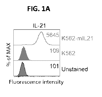
FIGS. 1A-1F. Characterization of K562 and 721.221 cells expressing membrane IL-
21. (FIG.
1A), Representative histograms show the expression of IL-21 and 4-1BBL by K562
(green) and K562
transduced with IL-21 (K562-mIL21, red) detected using flow cytometry. The
mean fluorescence
intensity (MFI) is noted in the respective histograms. (FIG. 1B),
Representative histograms show the
expression of IL-21 and 4-1BBL on 721.221 (green) and 721.221 transduced with
IL-21 (721.221-
mIL21, red) detected using flow cytometry. The MFI is noted in the respective
histograms. (FIG. 1C)
Confocal images of the expression of IL-21 on K562 cells transduced with IL-21
(K562-mIL21). (FIG.
1D) Confocal images of the expression of IL-21 on 221 cells transduced with IL-
21 (221-mIL21). (FIG.
1E), representative histograms show the expression of ICAM-1, PD-L1, HLA-E,
and MICB on K562
(green) and K562-mIL21 (red) cells detected using flow cytometry. The MFI is
noted in the respective
histograms. (FIG. 1F), Representative histograms show the expression of ICAM-
1, PD-L1, HLA-E, and
MICB on 721.221 (green) and 721.221-mIL21 (red) cells detected using flow
cytometry. The MFI is
noted in the respective histograms.
FIGS. 2A-2E. Primary human NK cell expansion with four different types of
feeder cells. (FIG.
2A), Representative dot plots show the purity of NK cells expanded with
different types of feeder cells
on the indicated day post expansion detected using flow cytometry. PBMCs were
stimulated with
irradiated K562, K562-mIL21, 721.221, and 721.221-mIL21 on day 0,
respectively. The purities of NK
cells were examined on day 7 and then every 3 to 5 days. (FIGS. 2B and 2C),
Quantitative data show
fold-expansion (FIG. 2B) and purity (FIG. 2C) of NK cells from 11 donors
expanded with irradiated
K562, K562-mIL21, 721.221, and 721.221-mIL21, respectively, for 21 days.
(FIGS. 2D and 2E),
Quantitative data show fold-expansion (FIG. 2D) and purity (FIG. 2E) of NK
cells from 11 donors
expanded with the indicated feeder cells on day 21. Mean (solid lines) with
95% CI (gray band) are
showed in (FIG. 2B) and (FIG. 2C). * p <0.05. ** p <0.01, *** p < 0.001.
FIGS. 3A-3E. Phenotypes of NK cells expanded by different feeder cells. (FIG.
3A),
Representative histograms show the expression of CD16, NKG2D, NKp46, 2B4, and
DNAM-1 on NK
cells expanded using K562, K562-mIL21, 721.221, and 721.221-mIL21. (FIG. 3B),
Representative
histograms show the expression of CD69, CD94, CD8a, and NKG2C on NK cells
expanded using K562,
- 4 -
CA 03131879 2021-08-27
WO 2020/172328
PCT/US2020/018897
K562-mIL21, 721.221, and 721.221-mIL21. (FIG. 3C), Representative histograms
show the expression
of NKG2A, CTLA-4, KLRG1, and PD-1 on NK cells expanded using K562, K562-mIL21,
721.221, and
721.221-mIL21. (FIG. 3D), Representative histograms show the expression of
LIR1, TIM-3, TIGIT, and
LAG-3 on NK cells expanded using K562, K562-mIL21, 721.221, and 721.221-mIL21.
(FIG. 3E),
Representative histograms show the expression of KIR, KIR2DL1, KIR2DL2/L3,
KIR3DL1, and
KIR3DL2 on NK cells expanded using K562, K562-mIL21, 721.221, and 721.221-
mIL21. The MFIs are
indicated in the respective histograms.
FIGS. 4A-4H. Functional comparison of NK cells against susceptible target
cells. (FIG. 4A),
Quantitative data show cytotoxic activity of expanded NK cells against K562
cells using the CFSE/7-
AAD cytotoxicity assay. K562 cells were labeled with CFSE and then incubated
with expanded NK
cells for E:T ratios ranging from 1:4 to 4:1 for 4 hours. Next, 7-AAD was used
to determine the lysis of
K562 cells. (FIG. 4B), Quantitative data show the percentage of expanded NK
cells expressing CD107a
following no stimulation, stimulation with K562, and stimulation with
PMA/Ionomycin, for 2 hours.
(FIG. 4C), Quantitative data show the cytotoxic activity of expanded NK cells
against 721.221 cells
using CFSE/7-AAD cytotoxicity assay. 721.221 cells were labeled with CFSE and
then incubated with
expanded NK cells for E:T ratios ranging from 1:4 to 4:1 for 4 hours. Next, 7-
AAD was used to
determine the lysis of 721.221 cells. (FIG. D), Quantitative data show the
percentage of expanded NK
cells expressing CD107a following no stimulation, stimulation with 721.221,
and stimulation with
PMA/Ionomycin, respectively, for 2 hours. The means SD are shown in (FIG.
4A) and (FIG. 4C), and
means + SD are shown in (FIG. 4B) and (FIG. 4D). (FIG. 4E) Gating strategies
for NK cell mediated
cytotoxicity using the CFSE/7-AAD approach. After incubation of NK cells with
CFSE-labeled target
cells for 4 hours, dead cells were gated on 7-AAD positive subsets. (FIG. 4F)
Representative flow
cytometry dot plots of the percent of 7-AAD positive cells in CFSE labeled
K562 cells following
incubation with expanded NK cells at different effector:target (E:T) cell
ratios. (FIG. 4G) Gating
strategies for cell surface CD107a assays. (FIG. 4H) Representative dot plots
of the percent of expanded
NK cells expressing CD107a following no stimulation (NK cell only, negative
control group),
stimulation with K562, and PMA/Ionomycin (positive control group). *** p <
0.001, ns p > 0.05.
FIGS. 5A-5F. An exemplary method of expansion of CD19-CAR NK cells with
721.221-mIL21
is schematically illustrated in FIG. 5A. Briefly, 221.mIL21 cells were
irradiated with a dose of 100 Gray
(10000 Rad). PBMCs were then co-cultured with irradiated feeder cells in the
presence of IL-2 and IL-
15. In parallel, CD19-CAR retrovirus was produced by transfecting 293T cells.
The expanded NK cells
were transduced with CD19-CAR retrovirus at Day 7. Cells were cultured for 21
days. (FIG. 5B),
Representative dot plots show the percentage of expanded NK cells in CD19-CAR-
positive cells on the
indicated day post expansion. PBMCs were stimulated with irradiated 721.221-
mIL21 on day 0 and
transduced with CD19-CAR retrovirus on day 7. Purities of NK cells in CD19-CAR-
positive cells were
examined every 3 to 4 days. (FIG. 5C) Dynamic time-lapsed expansion data of
the fold expansion of
CD19-CAR NK cells from 3 donors. CD19-CAR-modifed NK cells were expanded with
irradiated
- 5 -
CA 03131879 2021-08-27
WO 2020/172328
PCT/US2020/018897
K562, K562-mIL21, 221, and 221-mIL21 feeder cells for 21 days. (FIG. 5D)
Quantitative data of the
fold expansion of CD19-CARNK cells from 3 donors on day 21 of expansion. (FIG.
5E) Dynamic
time-lapsed expansion data of the purity of NK cells
544 within CD19-CAR positive cells from 3 donors. NK cells were expanded with
irradiated
.. 545 K562, K562-mIL21, 221, and 221-mIL21 feeder cells, respectively. (FIG.
5F) Quantitative data of
the percent of NK cells within CD19-CAR positive cells from 3 donors on day 21
post expansion. The
means (solid lines) with 95% CI (gray band) are shown in (FIG. 5C) and (FIG.
5D).
FIGS. 6A-6D. Expansion of Cord Blood (CB) derived NK and CAR-NK cells with
721.221-
mIL21. (FIG. 6A) Representative flow cytometry dot plots of the percent of
CD19-CAR positive cells in
NK cells at the indicated days. CBMCs were stimulated with irradiated feeder
cells on day 0 and
transduced with CD19-CAR retrovirus on day 7. (FIG. 6B) Quantitative data for
the percent of CD19-
CAR positive cells in NK cells expanded from CBMCs (n = 3). (FIG. 6C)
Quantitative data for the
cytotoxic activity of expanded CD19-CAR CB-NK cells against Raji cells using
the CFSE/7-AAD
cytotoxicity assay. Target cells were labeled with CFSE and then incubated
with expanded CD19-CAR
CB-NK cells at E:T ratios ranging from 5:1 to 0.3125:1 for 4 hours. Next, 7-
AAD was used to detect the
lysis of target cells. (FIG. 6D) Quantitative data for the cytotoxic activity
of expanded CD19-CAR CB-
NK cells against Daudi cells using the CFSE/7-AAD cytotoxicity assay.
FIGS 7A-7I. Superior anti-tumor activity from 221-mIL21 expanded CD19-CARNK
cells in a
lymphoma xenograft model. (FIG. 7A) Diagram of the experimental design of the
Daudi lymphoma
xenograft model. Male and female NSG mice (n = 5) were i.v. injected with 2
x106 Daudi-FFLuc cells in
100 1_, of PBS via tail vein on day -4. Beginning on day 0, mice were
injected (i.v.) with 1x10⁷ 221-
mIL21 expanded- or K562-mIL21 expanded-CD19-CARNK cells in 100 1_, of PBS and
injected (i.p.)
with IL-2 (50,000 Unit/mouse) and IL-15 (10 ng/mouse) in 150 uL of PBS at days
0, 3, 7, and 10.
Animals were imaged using the IVIS system twice a week for tumor cell
tracking. (FIG. 7B)
Representative images of tumor burden at indicated time points. The range of
fluorescence intensity is
from 1 x 10⁵ to 2 x 10⁶ units of photons/sec/cm2/sr. (FIG. 7C) Quantitative
data of tumor burden at
indicated time points. Mice were imaged at the indicated days to evaluate
tumor burden expressed as
quantified bioluminescence (average light intensity), which represents tumor
growth. (FIG. 7D)
Quantitative data of mice body weights at the indicated days. (FIG. 7E)
Diagram of the experimental
design of the Raji lymphoma xenograft model. Male and female NSG mice (n = 10)
were i.v. injected
with 2x10⁶Raji-FFLuc-GFP cells in 100 1_, of PBS via tail vein on day 0. On
day 2 and day 4, mice
were injected (i.v.) with 1x10⁷K562-m1L21 expanded-CD19-CARNK cells, 221-mIL21
expanded-
CD19-CAR NK cells, and 221-mIL21 expanded-CD19-CAR-IL15 NK cells,
respectively, in 100 uL of
PBS and injected (i.p.) with IL-2 (50,000 Unit/mouse) and IL-15 (10 ng/mouse)
in 150 1_, of PBS.
Animals were imaged using the IVIS system once a week for tumor cell tracking.
(FIG. 7F)
Representative images of tumor burden at indicated time points. The range of
fluorescence intensity is
from 5 x 10⁵ to 1 x 10⁷ units of photons/sec/cm2/sr for day 7 and from 2 x 10⁷
to 5 x 10⁸ units of
- 6 -
CA 03131879 2021-08-27
WO 2020/172328
PCT/US2020/018897
photonsisec/cm2/sr for day 14 and day 21. (FIG. 7G) Kaplan¨Meier survival
curves of tumor-bearing
mice after treatment with PBS, K562-mIL21 expanded-CD19-CAR NK cells, 221-
mIL21 expanded-
CD19-CAR NK cells, and 221-mIL21 expanded-CD19-CAR-IL15 NK cells,
respectively. The p-value
was analyzed by log-rank (Mantel-Cox) Test. (FIG. 7H) Quantitative data of
tumor burden at indicated
time points. Mice were imaged at the indicated days to evaluate tumor burden
expressed as quantified,
which represent tumor growth. (FIG. 71) Quantitative data of mice body weights
at the indicated days.
FIGS. 8A-8B. Schematic representation of exemplary recombinant retroviral
vectors encoding
human IL-21 and an exemplary method for NK cell expansion with 721.221.mIL-21
feeder cells. (FIG.
8A), the IL-21 construct contains the human IgG1 Fab' domain, CD28
transmembrane domain,
intracellular domain of 4-1BB, and intracellular domain of CD3 zeta. (FIG. 8B)
Feeder cells were
irradiated with a dose of 100 Gray (10000 Rad), and then PBMCs were co-
cultured with irradiated feeder
cells with IL-2 and IL-15 for NK cell expansion.
FIG. 9. Human primary NK cells express cell surface IL-21 receptors.
Representative
histograms show the expression of IL-21R on primary NK cells from PBMCs. The
MFI is noted in the
respective histograms.
FIGS. 10A-10C. Primary human NK cell expansion with 721.221 cell expressing
membrane IL-
15 receptor alpha (221-mIL-15Ra). (FIG. 10A), Representative dot plots show
the purity of NK cell
expanded with two different types of feeder cell on indicated day post
expansion detected by flow
cytometry. PBMCs were stimulated with irradiated wild-type 721.221 (top panel)
and 721.221-mIL-
.. 15Ra on day 0, respectively. The purities of NK cell were checked on day 7,
day 14, and day 21.
(FIGS. 10B and 10C), Quantitative data show fold expansion (FIG. 10B) and
purity (FIG. 10C) of NK
cells from 7 donors expanded with irradiated wild-type 721.221 and 721.221-mIL-
15Ra for 21 days,
respectively.
FIGS. 11A-11C. Primary human T cell expansion with 721.221 cell expressing
membrane IL-
.. 21. (FIG. 11A) Representative dot plots show the purity of T cell expanded
with two different types of
feeder cell on indicated day post expansion detected by flow cytometry. PBMCs
were stimulated with
irradiated K562-mIL21 (top panel) and 721.221-mIL21(low panel) on day 0,
respectively. The purities
of NK cell were checked on day 7, day 14, and day 21. Quantitative data show
fold expansion (left
panel) and purity (right panel) of T cells from 11 donors expanded with
irradiated K562-mIL21 and
721.221-mIL21for 21 days, respectively. (FIG. 11B) Representative dot plots
show the purity of T cell
expanded with two different types of feeder cell on indicated day post
expansion detected by flow
cytometry. Cord blood monocytes were stimulated with irradiated K562-mIL21
(top panel) and 721.221-
mIL21(low panel) on day 0, respectively. The purities of NK cell were checked
on day 7, day 14, and
day 21. Quantitative data show fold expansion (left panel) and purity (right
panel) of T cells from 11
donors expanded with irradiated K562-mIL21 and 721.221-mIL21for 21 days,
respectively. (FIG. 11C)
Representative dot plots show the purity of T cell expanded with two different
types of feeder cell on
indicated day post expansion detected by flow cytometry. PBMCs from patients
with anaplastic large
- 7 -
CA 03131879 2021-08-27
WO 2020/172328
PCT/US2020/018897
cell lymphoma were stimulated with irradiated 721.221-mIL21 feeder cells. The
purities of T cells were
checked on day 7, day 20, and day 28, respectively.
FIG. 12. Primary human NK cell expansion with four different types of feeder
cells. PBMCs
were stimulated with irradiated K562, K562-mIL21, 721.221, and 721.221-mIL21,
quantitative data
show fold-expansion of NK cells.
FIGS. 13A-13N. 221-mIL21 expanded NK cells show enriched metabolic pathways
and
immature phenotypes. (FIG. 13A) PBMCs were stimulated with irradiated K562-
mIL21 and 221-mIL21
feeder cells. NK cells were purified from expanded cells using flow cytometry
on day 7 and day 14 for
RNA sequencing (RNA-Seq). Principal component analysis (PCA) plots of sample-
to-sample distances
of NK cells expanded with K562-mIL21 or 221-mIL21 feeder cells on day 7 and
day 14. (FIG. 13B)
Mean-average (MA) plots of differentially expressed genes (DEGs) in NK cells
expanded with 221-
mIL21 feeder cells compared to those that were expanded with K562-mIL21 feeder
cells on day 7; p-
values calculated using DESeq2. Top 15 significant DEGs are labeled on the MA-
plot. Up, up-regulated
DEGs, adjusted p <0.05 and 1og2 fold change? 1; Down, down-regulated DEGs,
adjusted p <0.05 and
1og2 fold change < -1; NS, not significant. (FIG. 13C) MA plots of DEGs in NK
cells that were
expanded with 221-mIL21 feeder cells compared to those that were expanded with
K562-mIL21 feeder
cells on day 14. Top 15 significant DEGs are labeled on the MA-plot. Up, up-
regulated DEGs, adjusted
p <0.05 and 1og2 fold change >= 1; Down, down-regulated DEGs, adjusted p <
0.05 and 1og2 fold
change <= -1; NS, not significant. (FIG. 13D) Gene set enrichment analysis
(GSEA) of cellular amino
acid metabolic processes in NK cells that were expanded with 221-mIL21 feeder
cells compared to those
that were expanded with K562-mIL21 feeder cells on day 7 using gene ontology
(GO) biological process
(BP) datasets in the Molecular Signatures Database (MSigDB). NES, normalized
enrichment score;
p.adjust, false discovery rate (FDR)-adjusted p-value. (FIG. 13E) GSEA of
glycolysis in NK cells that
were expanded with 221-mIL21 feeder cells compared to those that were expanded
with K562-mIL21
feeder cells on day 7 using Hallmark datasets in the MSigDB. NES, normalized
enrichment score;
p.adjust, FDR-adjusted p-value. (FIG. 13F) Dynamic level of glucose in the
media during NK cell
expansion using either K562-mIL21 and 221-mIL21 as feeder cells. Arrows
indicate the time points for
media change. (FIG. 13G) Quantitative glucose uptake comparison of NK cells
expanded with K562-
mIL21 or with 221-mIL21 feeder cells on day 7 and day 14. (FIG. 13H) GSEA of
lymphocyte
activation in NK cells that were expanded with 221-mIL21 feeder cells compared
to those that were
expanded with K562-mIL21 feeder cells on day 7 using GO_BP datasets in the
MSigDB. NES,
normalized enrichment score; p.adjust, FDR-adjusted p-value. (FIG. 131) GSEA
of lymphocyte
differentiation in NK cells that were expanded with 221-mIL21 feeder cells
compared to those that were
expanded with K562-mIL21 feeder cells on day 7 using GO_BP datasets in the
MSigDB. NES,
normalized enrichment score; p. adjust, FDR-adjusted p-value. (FIG. 13J) GSEA
of cell-cell adhesion in
NK cells that were expanded with 221-mIL21 feeder cells compared to those that
were expanded with
K562-mIL21 feeder cells on day 7 using GO_BP datasets in the MSigDB. NES,
normalized enrichment
- 8 -
CA 03131879 2021-08-27
WO 2020/172328
PCT/US2020/018897
score; p. adjust, FDR-adjusted p-value. (FIG. 13K) Heat map of inhibitory
receptor of NK cells. (FIG.
13L) Heat map of activating receptor of NK cells. (FIG. 13M) Heat map of genes
associated with
cytotoxic function of NK cells. (FIG. 13N) Heat map of genes associated with
development and
maturation of NK cells. Heat maps were generated using z-scores derived from
transformed RNA-seq
counts using regularized-logarithm transformation (rlog). Each column
represents a biological replicate.
FIGS. 14A-14F. Dynamics of different cell population expansions among
different types of
feeder cell expansion systems. (FIG. 14A) Dynamic time-lapsed expansion data
for the percent of T
cells (CD3+CD56-) from PBMCs (n= 11) expanded with irradiated K562, K562-
mIL21, 221, and 221-
mIL21 feeder cells for 21 days. (FIG. 14B) Quantitative data for the percent
of T cells (CD3+CD56-)
from PBMCs (n= 11) expanded with the indicated feeder cells on day 21. (FIG.
14C) Dynamic time-
lapsed expansion data for the percent of CD3+CD56+ from PBMCs (n= 11) expanded
with irradiated
K562, K562-mIL21, 221, and 221-mIL21 feeder cells for 21 days. (FIG. 14D)
Quantitative data for the
percent of CD3+CD56+ from PBMCs (n= 11) expanded with indicated feeder cells
on day 21. (FIG.
14E) Dynamic time-lapsed expansion data for the percent of CD3-CD56- from
PBMCs (n= 11) expanded
with irradiated K562, K562-mIL21, 221, and 221-mIL21 feeder cells for 21 days.
(FIG. 14F)
Quantitative data for the percent of CD3-CD56- from PBMCs (n= 11) expanded
with indicated feeder
cells on day 21. Mean (solid lines) with 95% CI (gray band) are shown in
(FIGS. 14A, 14C, and 14E).
* p < 0.05, ** p <0.01, *** p <0.001, ns p >0.05.
FIGS. 15A-15K. Figure S7. Improved cord blood derived NK cell expansion using
221-mIL21
cells. (FIG. 15A) Representative flow cytometry dot plots of the purity of NK
cells expanded with
different feeder cells at indicated days post expansion. Cord blood
mononuclear cells (CBMCs) were
either stimulated with irradiated K562-mIL21 or 221-mIL21 on day 0, and the
purities of NK cells were
checked on day 7 and then subsequently checked every 3 to 4 days. (FIG. 15B)
Dynamic time-lapsed
expansion data for the fold expansion of NK cells from CBMCs from 9 donors
expanded with either
irradiated K562-mIL21 or 221-mIL21 feeder cells for 21 days. (FIG. 15C)
Quantitative data for the fold
expansion of NK cells from CBMCs from 9 donors on 21 days. (FIG. 15D) Dynamic
time-lapsed
expansion data for the purity of NK cells from CBMCs from 9 donors expanded
with irradiated K562-
mIL21 and 221-mIL21 feeder cells for 21 days. (FIG. 15E) Quantitative data for
the purity of NK cells
from CBMCs from 9 donors on 21 days. (FIG. 15F) Dynamic time-lapsed expansion
data for the
percent of T cells (CD3+CD56-) from CBMCs (n= 9) expanded with irradiated
K562, K562-mIL21, 221,
and 221-mIL21 feeder cells for 21 days. (FIG. 15G) Quantitative data for the
percent of T cells
(CD3+CD56-) from CBMCs (n= 9) expanded with indicated feeder cells on day 21.
(FIG. 15H)
Dynamic time-lapsed expansion data for the percent of CD3+CD56+ from CBMCs (n=
9) expanded with
irradiated K562, K562-mIL21, 221, and 221-mIL21 feeder cells for 21 days.
(FIG. 151) Quantitative
data for the percent of CD3+CD56+ from CBMCs (n= 9) expanded with indicated
feeder cells on day 21.
(FIG. 15J) Dynamic time-lapsed expansion data for the percent of CD3-CD56-
from CBMCs (n= 9)
expanded with irradiated K562, K562-mIL21, 221, and 221-mIL21 feeder cells for
21 days. (FIG. 15K)
- 9 -
CA 03131879 2021-08-27
WO 2020/172328
PCT/US2020/018897
Quantitative data for the percent of CD3-CD56- from CBMCs (n= 9) expanded with
indicated feeder
cells on day 21. Mean (solid lines) with 95% CI (gray band) are shown in
(FIGS. 15B, 15D, 15F, 13H,
and 15J). ** p < 0.01, ns p > 0.05.
FIGS. 16A-16D. Phenotype and function of NK cells expanded from cord blood
mononuclear
cells using different feeder cell systems. (FIG. 16A) Representative
histograms of the expression of
NKG2D, NKp46, 2B4, and CD226 on NK cells expanded from cord blood mononuclear
cells using 221-
mIL21 (red) and K562-mIL21 (green) feeder cells. NK cells from freshly
isolated cord blood
mononuclear cells from the same donor is also shown (blue). (FIG. 16B)
Representative histograms of
the expression of CD69, CD94, CD8a, and CD16 on NK cells expanded from cord
blood mononuclear
cells using 221-mIL21 (red) and K562-mIL21 (green) feeder cells. NK from
freshly isolated cord blood
mononuclear cells from the same donor is also shown (blue). (FIG. 16C)
Representative histograms of
the expression of NKG2A, NKG2C, KIR, and KIR3DL1 on NK cells expanded from
cord blood
mononuclear cells using 221-mIL21 (red) and K562-mIL21 (green) feeder cells.
NK from freshly
isolated cord blood mononuclear cells from the same donor is also shown
(blue). (FIG. 16D)
Quantitative data for the cytotoxic activity of expanded CB-NK cells against
K562 cells using the
CFSE/7-AAD cytotoxicity assay. K562 cells were labeled with CFSE and then
incubated with expanded
CB-NK cells at E:T ratios ranging from 5:1 to 0.3125:1 for 4 hours. Next, 7-
AAD was used to detect the
lysis of K562 cells.
FIGS. 17A-17H. Expansion of CD19-CAR NK cells from PBMCs with different feeder
cell
systems. (FIG. 17A) Representative flow cytometry dot plots of the percent of
CD19-CAR positive cells
in NK cells at the indicated time points. PBMCs were stimulated with
irradiated feeder cells on day 0
and transduced with CD19-CAR retrovirus on day 7. (FIG. 17B) Quantitative data
for the percent of
CD19-CAR positive cells in NK cells expanded from PBMCs (n = 3). (FIG. 17C)
Dynamic time-lapsed
expansion data for the percent of T cell (CD3+CD56-) in CD19-CAR positive
cells (n = 3). (FIG. 17D)
Quantitative data for the percent of T cell (CD3+CD56-) in CD19-CAR positive
cells (n = 3) on day 21.
(FIG. 17E) Dynamic time-lapsed expansion data for the percent of CD3+CD56+ in
CD19-CAR positive
cells (n = 3). (FIG. 17F) Quantitative data for the percent of CD3+CD56+ in
CD19-CAR positive cells
(n = 3) on day 21. (FIG. 17G) Dynamic time-lapsed expansion data for the
percent of CD3-CD56- in
CD19-CAR positive cells (n = 3). (FIG. 17H) Quantitative data for the percent
of CD3-CD56- in CD19-
CAR positive cells (n = 3) on day 21. Mean (solid lines) with 95% CI (gray
band) are shown in (FIGS.
17C, 17E, and 17G).
FIGS. 18A-18F. Enriched Metabolic pathways and immune Phenotypes of 221-
expanded NK
cells. (FIGS. 18A) Dot plots of the GSEA for genes between NK cells expanded
with 221-mIL21 and
K562-mIL21 feeder cells on day 7 (left) and day 14 (right) using gene ontology
(GO) biological process
(BP) datasets in the Molecular Signatures Database (MSigDB). (FIG. 18B) Gene
set enrichment
analysis (GSEA) of cellular amino acid metabolic processes in NK cells that
were expanded with 221-
mIL21 feeder cells compared to those that were expanded with K562-mIL21 feeder
cells on day 14.
- 10 -
CA 03131879 2021-08-27
WO 2020/172328
PCT/US2020/018897
NES, normalized enrichment score; p.adjust, FDR-adjusted p-value. (FIG. 18C)
GSEA of glycolysis in
NK cells that were expanded with 221-mIL21 feeder cells compared to those that
were expanded with
K562-mIL21 feeder cells on day 14 using Hallmark datasets in the Molecular
Signatures Database
(MSigDB). NES, normalized enrichment score; p.adjust, FDR-adjusted p-value.
(FIG. 18D) GSEA of
lymphocyte activation in NK cells that were expanded with 221-mIL21 feeder
cells compared to those
that were expanded with K562-mIL21 feeder cells on day 14. NES, normalized
enrichment score;
p.adjust, FDR-adjusted p-value. (FIG. 18E) GSEA of lymphocyte differentiation
in NK cells that were
expanded with 221-mIL21 feeder cells compared to those that were expanded with
K562-mIL21 feeder
cells on day 14. NES, normalized enrichment score; p.adjust, FDR-adjusted p-
value. (FIG. 18F) GSEA
of cell-cell adhesion in NK cells that were expanded with 221-mIL21 feeder
cells compared to those that
were expanded with K562-mIL21 feeder cells on day 14. NES, normalized
enrichment score; p.adjust,
FDR-adjusted p-value.
FIGS. 19A-19I. Heat maps of enriched metabolic pathways and immune phenotypes
of
expanded NK cells. (FIGS. 19A-19B) Heat map of GSEA-identified genes of
cellular amino acid
metabolic processes. (FIGS. 19C) Heat map of GSEA-identified genes of
glycolysis. (FIGS. 19D-19E)
Heat map of GSEA-identified genes of lymphocyte activation. (FIGS. 19E-19F)
Heat map of GSEA-
identified genes of lymphocyte differentiation. (FIGS. 19G-19I) Heat map of
GSEA-identified genes of
cell-cell adhesion. Heat maps were generated using z-scores derived from
transformed RNA-seq counts
using regularized-logarithm transformation (rlog). Each column represents a
biological replicate.
SEQUENCES
Any nucleic acid and amino acid sequences provided herein are shown using
standard letter
abbreviations for nucleotide bases and amino acids, as defined in 37 C.F.R.
1.822. In at least some
cases, only one strand of each nucleic acid sequence is shown, but the
complementary strand is
understood as included by any reference to the displayed strand.
SEQ ID NO: 1 is an exemplary nucleic acid sequence of the extracellular domain
from
interleukin (IL)-21.
SEQ ID NO: 2 is an exemplary amino acid sequence of the extracellular domain
from IL-21.
SEQ ID NO: 3 is an exemplary nucleic acid sequence of a construct for
transducing cells with
membrane-bound (m)IL-21.
SEQ ID NO: 4 is an exemplary nucleic acid sequence of IL-15Ra.
SEQ ID NO: 5 is an exemplary amino acid sequence of IL-15Ra.
SEQ ID NO: 6 is an exemplary nucleic acid sequence of IL-15.
SEQ ID NO: 7 is an exemplary amino acid sequence of IL-15.
SEQ ID NO: 8 is an exemplary nucleic acid sequence of IL-2.
SEQ ID NO: 9 is an exemplary amino acid sequence of IL-2.
SEQ ID NO: 10 is an exemplary nucleic acid sequence of IL-27.
- 11-
CA 03131879 2021-08-27
WO 2020/172328
PCT/US2020/018897
SEQ ID NO: 11 is an exemplary amino acid sequence of IL-27.
SEQ ID NO: 12 is an exemplary nucleic acid sequence of IL-12B.
SEQ ID NO: 13 is an exemplary amino acid sequence of IL-12B.
SEQ ID NO: 14 is an exemplary nucleic acid sequence of IL-12 p35.
SEQ ID NO: 15 is an exemplary amino acid sequence of IL-12 p35.
SEQ ID NO: 16 is an exemplary nucleic acid sequence of IL-12 p40.
SEQ ID NO: 17 is an exemplary amino acid sequence of IL-12 p40.
SEQ ID NO: 18 is an exemplary nucleic acid sequence of IL-18.
SEQ ID NO: 19 is an exemplary amino acid sequence of IL-18.
SEQ ID NO: 20 is an exemplary nucleic acid sequence of IL-18.
SEQ ID NO: 21 is an exemplary amino acid sequence of IL-18.
SEQ ID NO: 22 is an exemplary nucleic acid sequence of IL-33.
SEQ ID NO: 23 is an exemplary amino acid sequence of IL-33.
SEQ ID NO: 24 is an exemplary nucleic acid sequence of IL-7.
SEQ ID NO: 25 is an exemplary amino acid sequence of IL-7.
SEQ ID NO: 26 is an exemplary nucleic acid sequence of MICA.
SEQ ID NO: 27 is an exemplary amino acid sequence of MICA.
DETAILED DESCRIPTION
Described herein are modified 721.221 cells that express one or more cytokines
or cytokine
receptors (e.g., IL-15 receptor alpha (IL-15Ra) and/or membrane-bound IL-21)
and methods of
expanding immune cells using the modified 721.221ce11s. The modified 721.221
cells can be used to
effectively expand NK cells or T cells (including CAR-modified NK cells or T
cells), as shown herein.
In combination with recombinant IL-15 and IL-2 and the modified 721.221 cells,
primary NK
cells were expanded by about 39,663-fold after three weeks of expansion.
Furthermore, transduction
with a retrovirus coding for a CAR molecule specific for CD19 protein resulted
in the expansion of
primary NK cells from both peripheral blood and cord blood. Therefore, a
platform for the expansion of
human primary NK cells and genetically modified CAR-NK cells is described.
Compared with previous NK expansion systems (Denman etal., PLoS One 7:e30264
(2012);
Fujisaki etal., Cancer Res 69:4010-7 (2009)), the 721.221-mIL-21 cells used
for NK expansion
described herein include three distinct advantages. The number of expanded NK
cells is significantly
higher using the technique described herein (about a 39,663-fold increase in
721.221- mIL-21 cells vs. a
3588-fold increase using K562-mIL-21 cells) with the combination of the
membrane form of IL-21 with
two soluble cytokines in the cell culture, in which NK cells were efficiently
propagated in vitro. The
721.221-mIL-21 expanded NK cells further feature higher purity with enhanced
cytotoxicity compared
with K562-mIL-21 expanded NK cells. Moreover, herein CAR-NK cells are derived
from cord blood
- 12 -
CA 03131879 2021-08-27
WO 2020/172328
PCT/US2020/018897
(CB) using the 721.221-mIL-21 NK expansion ready availability of CB from a CB
bank and 2) use of
CB-derived CAR-NK cells as an off-the-shelf CAR product.
I. Terms
Unless otherwise noted, technical terms are used according to conventional
usage. Definitions of
common terms in molecular biology may be found in Benjamin Lewin, Genes VII,
published by Oxford
University Press, 2000 (ISBN 019879276X); Kendrew et al. (eds.), The
Encyclopedia ofMolecular
Biology, published by Blackwell Publishers, 1994 (ISBN 0632021829); Robert A.
Meyers (ed.),
Molecular Biology and Biotechnology: a Comprehensive Desk Reference, published
by Wiley, John &
Sons, Inc., 1995 (ISBN 0471186341); and George P. Redei, Encyclopedic
Dictionary of Genetics,
Genomics, and Proteomics, 2nd Edition, 2003 (ISBN: 0-471-26821-6).
The singular forms "a," "an," and "the" refer to one or more than one, unless
the context clearly
dictates otherwise. For example, the term "comprising an interleukin" includes
single or plural
interleukins and is considered equivalent to the phrase "comprising at least
one interleukin." The term
"or" refers to a single element of stated alternative elements or a
combination of two or more elements,
unless the context clearly indicates otherwise. As used herein, "comprises"
means "includes." Thus,
"comprising A or B," means "including A, B, or A and B," without excluding
additional elements.
It is further to be understood that all base sizes or amino acid sizes, and
all molecular weight or
molecular mass values, given for nucleic acids or polypeptides are
approximate, and are provided for
description. Although methods and materials similar or equivalent to those
described herein can be used
in the practice or testing of the present disclosure, suitable methods and
materials are described below.
All publications, patent applications, patents, and other references mentioned
herein are incorporated by
reference in their entirety, as are the GenBank0 Accession numbers (for the
sequence present on
February 20, 2019). In case of conflict, the present specification, including
explanations of terms, will
control. In addition, the materials, methods, and examples are illustrative
only and not intended to be
limiting.
To facilitate review of the various embodiments of this disclosure, the
following explanations of
specific terms are provided.
721.221 cells: Also referred to as LCL 721.221 or ATCCO CRL1855TM cells,
721.221 cells are
B lymphocytes derived from a human Epstein-Barr virus-transformed cell line.
721.221 cells do not
express class I histocompatibility antigens (also known as major
histocompatibility complex (MHC) class
I molecules). Methods of producing 721.221 cells are known in the art (see,
e.g., Shimiz et al., Proc Natl
Acad Sci U S A., 85(1):227-31, 1988, incorporated by reference in its
entirety).
Activating receptor ligand: Ligands that bind receptors of natural killer (NK)
or T cells,
thereby activating the NK or T cell. Examples of activating receptor ligands
include UL16-binding
protein (ULBP)-1, ULPB-2, and/or major histocompatibility complex (MHC) class
I chain-related
protein A (MIC-A).
- 13 -
CA 03131879 2021-08-27
WO 2020/172328
PCT/US2020/018897
Autoimmune disorder: A disorder in which the immune system produces an immune
response
(e. g. , a B cell or a T cell response) against an endogenous antigen, with
consequent injury to tissues. The
injury may be localized to certain organs, such as thyroiditis, or may involve
a particular tissue at
different locations, such as Goodpasture's disease, or may be systemic, such
as lupus erythematosus.
In some examples, autoimmune diseases include systemic lupus erythematosus,
Sjogren's
syndrome, rheumatoid arthritis, type I diabetes mellitus, Wegener's
granulomatosis, inflammatory bowel
disease, polymyositis, dermatomyositis, multiple endocrine failure, Schmidt's
syndrome, autoimmune
uveitis, Addison's disease, adrenalitis, Graves' disease, thyroiditis,
Hashimoto's thyroiditis, autoimmune
thyroid disease, pernicious anemia, gastric atrophy, chronic hepatitis, lupoid
hepatitis, atherosclerosis,
presenile dementia, demyelinating diseases, multiple sclerosis, subacute
cutaneous lupus erythematosus,
hypoparathyroidism, Dressler's syndrome, myasthenia gravis, autoimmune
thrombocytopenia, idiopathic
thrombocytopenic purpura, hemolytic anemia, pemphigus vulgaris, pemphigus,
dermatitis herpetiformis,
alopecia arcata, pemphigoid, scleroderma, progressive systemic sclerosis,
CREST syndrome (calcinosis,
Raynaud's phenomenon, esophageal dysmotility, sclerodactyly, and
telangiectasia), adult onset diabetes
mellitus (Type II diabetes), male and female autoimmune infertility,
ankylosing spondylitis, ulcerative
colitis, Crohn's disease, mixed connective tissue disease, polyarteritis
nedosa, systemic necrotizing
vasculitis, juvenile onset rheumatoid arthritis, glomerulonephritis, atopic
dermatitis, atopic rhinitis,
Goodpasture's syndrome, Chagas' disease, sarcoidosis, rheumatic fever, asthma,
recurrent abortion, anti-
phospholipid syndrome, farmer's lung, erythema multiforme, post cardiotomy
syndrome, Cushing's
syndrome, autoimmune chronic active hepatitis, bird-fancier's lung, allergic
disease, allergic
encephalomyelitis, toxic epidermal necrolysis, alopecia, Alport's syndrome,
alveolitis, allergic alveolitis,
fibrosing alveolitis, interstitial lung disease, erythema nodosum, pyoderma
gangrenosum, transfusion
reaction, leprosy, malaria, leishmaniasis, trypanosomiasis, Takayasu's
arteritis, polymyalgia rheumatica,
temporal arteritis, schistosomiasis, giant cell arteritis, ascariasis,
aspergillosis, Sampter's syndrome,
eczema, lymphomatoid granulomatosis, Behcet's disease, Caplan's syndrome,
Kawasaki's disease,
dengue, encephalomyelitis, endocarditis, endomyocardial fibrosis,
endophthalmitis, erythema elevatum et
diutinum, psoriasis, erythroblastosis fetalis, eosinophilic faciitis,
Shulman's syndrome, Felty's syndrome,
filariasis, cyclitis, chronic cyclitis, heterochronic cyclitis, Fuch's
cyclitis, IgA nephropathy, Henoch-
Schonlein purpura, glomerulonephritis, graft versus host disease,
transplantation rejection, human
immunodeficiency virus infection, echovirus infection, cardiomyopathy,
Alzheimer's disease, parvovirus
infection, rubella virus infection, post vaccination syndromes, congenital
rubella infection, Hodgkin's
and Non-Hodgkin's lymphoma, renal cell carcinoma, multiple myeloma, Eaton-
Lambert syndrome,
relapsing polychondritis, malignant melanoma, cryoglobulinemia, Waldenstrom's
macroglobulemia,
Epstein-Barr virus infection, rubulavirus, and Evan's syndrome.
Cancer: Also referred to as a "malignant tumor" or "malignant neoplasm,"
cancer refers to any
of a number of diseases characterized by uncontrolled, abnormal proliferation
of cells. Cancer cells have
the potential to spread locally or through the bloodstream and lymphatic
system to other parts of the body
- 14 -
CA 03131879 2021-08-27
WO 2020/172328
PCT/US2020/018897
(e.g., metastasize) with any of a number of characteristic structural and/or
molecular features. A "cancer
cell" is a cell having specific structural properties, lacking
differentiation, and being capable of invasion
and metastasis. Indolent and high grade forms are included. In some examples,
the cancer is a solid
cancer (such as sarcomas (e.g., rhabdomyosarcoma, osteogenic sarcoma, Ewing's
sarcoma,
chondrosarcoma, and alveolar soft part sarcoma); carcinomas (e.g., colorectal
carcinoma and
hepatocellular carcinoma (HCC), ); and lymphomas, such as Hodgkin's or non-
Hodgkin's lymphoma, for
example, diffuse large B-cell, follicular, chronic lymphocytic, small
lymphocytic, mantle cell, Burkitt's,
cutaneous T-cell, AIDS-related, or central nervous system lymphoma);
neuroblastoma; gynecological
cancer (such as ovarian cancer); breast cancer; liver cancer (e.g.,
hepatocellular carcinoma (HCC), ); lung
cancer; prostate cancer; skin cancer; bone cancer; pancreatic cancer; brain
cancer (neuroblastoma); head
or neck cancer; kidney cancer (such as Wilms' tumor); retinoblastoma;
adrenocortical tumor; desmoid
tumors; desmoplastic small round cell tumor; endocrine tumors; and/or blood
cancer (such as myeloma,
such as multiple myeloma; lymphoma, such as Hodgkin's or non-Hodgkin's
lymphoma, for example,
diffuse large B-cell, follicular, chronic lymphocytic, small lymphocytic,
mantle cell, Burkitt's, cutaneous
T-cell, AIDS-related, or central nervous system lymphoma; or leukemia, such as
acute lymphocytic
leukemia (ALL) or acute myeloid leukemia (AML)).
Chimeric antigen receptor (CAR): A chimeric fusion protein having an
extracellular domain
that is fused via a transmembrane domain to an intracellular signaling domain
capable of activating a T
cell. CAR molecules can include an extracellular domain (ectodomain) with two
(or more) targeting
domains that are functionally different from each other (multispecific CAR)
and that bind to two
different sites on a target (multi-targeted). For example, one targeting
domain of a multispecific CAR
can be a cell surface receptor, such as CD19 (e.g., a multispecific CD19-based
CAR). In another
example, one targeting domain of a multispecific CAR can be a cell surface
receptor, such as CD19, and
the second targeting domain can be an antibody or a fragment thereof, such as
a scFv (i.e. a multispecific
CD19-scFv CAR). In some embodiments, the CD19-scFv CAR binds two different
target sites (i.e. a
multi-targeted CD19-scFv). A monofunctional CAR contains only a single
functional element in the
targeting extracellular domain. In some particular embodiments, a portion of
the CAR's extracellular
binding domain is derived from a murine or humanized monoclonal antibody.
The intracellular signaling domain of CAR molecules include two different
cytoplasmic
signaling domains. For example, one signaling domain can be a cytoplasmic
effector function signaling
domain and the second signaling domain can be a cytoplasmic co-stimulatory
signaling domain. Linkers
can connect domains to each other (for example, the two targeting domains) or
they can connect one
domain to another domain (for example, the ligand-binding domain to the
transmembrane domain).
CARS are also known as chimeric immune receptors, zetakines, and universal T
cell receptors.
Methods of making CARS are available (see, e.g., Park et al., Trends
Biotechnol., 29:550-557,
2011; Grupp etal., N Engl J Med., 368:1509-1518, 2013; Han etal., J. Hematol
Oncol., 6:47, 2013; PCT
- 15 -
CA 03131879 2021-08-27
WO 2020/172328
PCT/US2020/018897
Pubs. WO 2012/079000, WO 2013/059593; and U.S. Pub. 2012/0213783, each of
which is incorporated
by reference herein in its entirety.)
Contacting: Placement in direct physical association, including both a solid
and liquid form. In
one example, contacting includes association between a substance or cell (such
as a cytokine or feeder
cells) in a liquid medium and one or more other cells (such as NK cells or T
cells in culture). Contacting
can occur in vitro with isolated cells or tissue or in vivo by administering
to a subject.
Culturing or Cell culture: Growth of a population of cells in a defined set of
conditions (such
as culture medium, extracellular matrix, temperature, and/or time of culture)
in vitro. In some examples,
a cell culture includes a substantially pure culture (for example, isolated
721.221 cells or isolated NK
cells). In additional examples a cell culture includes a mixed culture, such
as co-culture of two or more
types of cells (for example a culture of NK cells with feeder cells). In
further examples, a cell culture
includes cells grown in contact with an extracellular matrix.
Culture Medium: A synthetic set of culture conditions with the nutrients
necessary to support
the viability, function, and/or growth of a specific population of cells, such
as 721.221 cells. Culture
media generally include components such as a carbon source, a nitrogen source,
and a buffer to maintain
pH. Additional components in culture media also may include one or more of
serum, cytokines,
hormones, growth factors, protease inhibitors, protein hydrolysates, shear
force protectors, proteins,
vitamins, glutamine, trace elements, inorganic salts, minerals, lipids, and/or
attachment factors.
Cytokine: Proteins made by cells that affect the behavior of other cells, such
as lymphocytes.
In one embodiment, a cytokine is an interleukin, a molecule that regulates
cell growth, differentiation,
and motility (e.g., to stimulate immune responses, such as inflammation). In
other embodiments, the
cytokine can be an activating receptor ligand, TRL ligand, or receptors
thereof. In some examples, the
cytokine includes molecules known to stimulate or co-stimulate cell expansion
(e.g., NK or T cell
expansion). The term "cytokine" is used as a generic name for a diverse group
of soluble proteins and
peptides that act as humoral regulators at nanomolar to picomolar
concentrations and which, either under
normal or pathological conditions, modulate the functional activities of
individual cells and tissues.
These proteins also mediate interactions between cells directly and regulate
processes taking place in the
extracellular environment. Examples of cytokines include, but are not limited
to, tumor necrosis factor a
(TNF-a), interleukin (IL)-2, IL-7, IL-15, IL-21, including membrane-bound IL-
21 (mIL-21), interferon
(IFN)y, IFNa, IFN13, IL-12, IL-33, IL-27, IL-18, IL-1 family molecules (e.g.,
IL-la, IL-113, IL-1Ra, IL-
18, IL-36Ra, IL36a, IL3613, IL-36y, IL-37, IL-38, IL-33, toll receptor (TLR)
ligands, activating receptor
ligands (e.g., UL16 binding protein (ULBP)-1, ULPB-2, major histocompatibility
complex (MHC) class
I chain-related protein A (MIC-A)), IL-I family molecules, Fc receptors,
intercellular adhesion molecule
1 (ICAM-1), CD8a, 2B4 (also known as cluster of differentiation 244 (CD244)),
intercellular adhesion
molecule 1 (ICAM-1), CD8a, CD40, CD28, 4-1BB ligand (4-1BBL), OX4OL, TRX518,
CD3 antibody,
and CD28 antibody.
- 16 -
CA 03131879 2021-08-27
WO 2020/172328
PCT/US2020/018897
Effective amount: A quantity of a specified agent sufficient to achieve a
desired effect, for
example, in a subject being treated with that agent. In some examples, an
effective amount of an
expanded NK cell or T cell (e.g., a chimeric antigen receptor (CAR)-NK cell or
CAR-T cell) disclosed
herein is an amount sufficient to treat or inhibit a disease or disorder in a
subject (such as a tumor, viral
.. infection, autoimmune disease, or transplant rejection). In other examples,
an effective amount is an
amount of an expanded NK cell or T cell (e.g., a chimeric antigen receptor
(CAR)-NK cell or CAR-T
cell) sufficient to reduce or ameliorate one or more symptoms of a disease or
disorder in a subject. The
effective amount (for example, an amount ameliorating, inhibiting, and/or
treating a disorder in a subject)
will be dependent on, for example, the particular disorder being treated, the
subject being treated, the
manner of administration of the composition, and other factors.
Expression: The process by which the coded information of a gene is converted
into an
operational, non-operational, or structural part of a cell, such as the
synthesis of a protein. Gene
expression can be influenced by external signals. For instance, exposure of a
cell to a hormone may
stimulate expression of a hormone-induced gene. Different types of cells can
respond differently to an
identical signal. Expression of a gene also can be regulated anywhere in the
pathway from DNA to RNA
to protein. Regulation can include controls on transcription, translation, RNA
transport and processing,
degradation of intermediary molecules such as mRNA, or through activation,
inactivation,
compartmentalization or degradation of specific protein molecules after they
are produced.
Feeder cells: Cells that provide support for another cell type in ex vivo or
in vitro culture.
Feeder cells may provide one or more factors required for survival, growth,
and/or differentiation (or
inhibiting differentiation) of the cells cultured with the feeder cells.
Typically feeder cells are irradiated
or otherwise treated to prevent their proliferation in culture. In some
examples disclosed herein, NK cells
are cultured with feeder cells, such as irradiated modified 721.221 cells
(e.g., mIL-21-expressing 721.221
cells).
Heterologous nucleic acid: A nucleic acid introduced into a cell, for example,
by transduction
or transfection. A `heterologous' nucleic acid or protein refers to a nucleic
acid or protein originating
from a different genetic source. For example, a nucleic acid or protein that
is heterologous to a cell
originates from an organism or individual other than the cell in which it is
expressed and includes
synthesized nucleic acids (e.g., mRNA). In other examples, a heterologous
nucleic acid or protein
originates from a cell type other than the cell in which it is expressed (for
example, a nucleic acid or
protein not normally present in 721.221 cells is heterologous to 721.221
cells). In further examples, a
heterologous nucleic acid includes a recombinant nucleic acid, such as a
protein-encoding nucleic acid
operably linked to a promoter from another gene and/or two or more operably
linked nucleic acids from
different sources.
Immune system disorder: A disease or disorder that is associated with a
pathological immune
response in a subject (see Intl. Patent Pub. No. WO 2013/192294 and U.S.
Patent Pub. No.
2011/00811323, both of which are incorporated herein by reference). Examples
include
- 17 -
CA 03131879 2021-08-27
WO 2020/172328
PCT/US2020/018897
immunodeficiency (e.g., primary or hereditary immunodeficiency and
immunodeficiencies associated
with other conditions, such as immunosuppression associated with, for example,
HIV, old age, and
cancer), cytokine storm, allergies, asthma, various types of inflammation, and
autoimmune disorders.
Infectious disease: Also known as transmissible disease or communicable
disease, infection
.. disease are illnesses resulting from an infection. Infections are caused by
infectious agents, including
viruses, viroids, prions, bacteria; nematodes, such as parasitic roundworms
and pinworms; arthropods,
such as ticks, mites, fleas, and lice; fungi, such as ringworm; and other
macroparasites, such as
tapeworms and other helminths. Hosts fight infections using the immune system,
such as the innate
response (e.g., in mammals), which involves inflammation, followed by an
adaptive response.
Medications used to treat infections include antibiotics, antivirals,
antifungals, antiprotozoals, and
antihelminthics. Specific examples of infectious diseases include human
immunodeficiency syndrome
(HIV), hepatitis B virus (HBV), tuberculosis (TB), and malaria.
Inhibiting or treating a condition: "Inhibiting" a condition refers to
inhibiting the full
development of a condition or disease, for example, a tumor. Inhibition of a
condition can span the
spectrum from partial inhibition to substantially complete inhibition (e.g.,
including, but not limited to
prevention) of a disease (such as a tumor, viral infection, autoimmune
disease, or transplant rejection). In
some examples, the term "inhibiting" refers to reducing or delaying the onset
or progression of a
condition. "Treatment" refers to a therapeutic intervention that ameliorates a
sign or symptom of a
disease or condition after it has begun to develop. A subject to be
administered an effective amount of
the disclosed NK cells or T cells (e.g., CAR-NK cells or CAR-T cells) can be
identified by standard
diagnosing techniques for such a disorder, for example, presence of the
disease or disorder or risk factors
to develop the disease or disorder.
Isolated: An "isolated" or "purified" biological component (such as a cell,
nucleic acid, peptide,
protein, protein complex, or virus-like particle) has been substantially
separated, produced apart from, or
purified away from other components (for example, other biological components
in the cell or the
organism in which the component naturally occurs). Cells, nucleic acids,
peptides and proteins that have
been "isolated" or "purified" thus include cells, nucleic acids, and proteins
purified by standard
purification methods.
The term "isolated" or "purified" does not require absolute purity; rather, it
is intended as a
.. relative term. Thus, for example, an isolated biological component is one
in which the biological
component is more enriched than the biological component is in its natural
environment within a cell,
organism, sample, or production vessel (for example, a cell culture system).
Preferably, a preparation is
purified such that the biological component represents at least 50%, such as
at least 70%, at least 80%, at
least 90%, at least 95%, or greater, of the total biological component content
of the preparation.
Natural Killer (NK) cells: Cells of the immune system that kill target cells
in the absence of a
specific antigenic stimulus and without restriction according to MHC class.
Target cells can be tumor
cells or cells harboring viruses. NK cells are characterized by the presence
of CD56 and the absence of
- 18 -
CA 03131879 2021-08-27
WO 2020/172328
PCT/US2020/018897
CD3 surface markers. NK cells typically comprise approximately 10 to 15% of
the mononuclear cell
fraction in normal peripheral blood. Historically, NK cells were first
identified by their ability to lyse
certain tumor cells without prior immunization or activation. NK cells are
thought to provide a "back
up" protective mechanism against viruses and tumors that might escape the
cytotoxic T lymphocyte
(CTL) response by down-regulating MHC class I presentation. In addition to
being involved in direct
cytotoxic killing, NK cells also serve a role in cytokine production, which
can be important to control
cancer and infection. Tissue-resident memory NK cells are included.
In some examples, a "CAR-NK cell" is an NK cell transduced with a heterologous
nucleic acid
encoding or expressing a CAR.
Pharmaceutically acceptable carrier: The pharmaceutically acceptable carriers
(vehicles)
useful in this disclosure are conventional. Remington: The Science and
Practice of Pharmacy, The
University of the Sciences in Philadelphia, Editor, Lippincott, Williams, &
Wilkins, Philadelphia, PA,
21" Edition (2005), describes compositions and formulations suitable for
pharmaceutical delivery of one
or more therapeutic compositions, such as one or more modified NK cells and/or
additional
pharmaceutical agents.
In general, the nature of the carrier will depend on the particular mode of
administration being
employed. For instance, parenteral formulations usually comprise injectable
fluids that include
pharmaceutically and physiologically acceptable fluids such as water,
physiological saline, balanced salt
solutions, aqueous dextrose, glycerol or the like as a vehicle. For solid
compositions (for example,
powder, pill, tablet, or capsule forms), conventional non-toxic solid carriers
can include, for example,
pharmaceutical grades of mannitol, lactose, starch, or magnesium stearate. In
addition to biologically-
neutral carriers, pharmaceutical compositions to be administered can contain
minor amounts of non-toxic
auxiliary substances, such as wetting or emulsifying agents, preservatives,
and pH buffering agents and
the like, for example, sodium acetate or sorbitan monolaurate.
Subject: A living multi-cellular vertebrate organism, a category that includes
both human and
non-human mammals (such as veterinary animals, including dogs and cats, as
well as mice, rats, rabbits,
sheep, horses, cows, and non-human primates).
T Cell: A white blood cell critical to the immune response. T cells include,
but are not limited
to, CD4+ T cells and CD8+ T cells. A CD4+ T lymphocyte is an immune cell that
expresses CD4 on its
surface. These cells, also known as helper T cells, help orchestrate the
immune response, including
antibody responses as well as killer T cell responses. Thl and Th2 cells are
functional subsets of helper
T cells. Thl cells secrete a set of cytokines, including interferon-gamma, and
whose principal function is
to stimulate phagocyte-mediated defense against infections, especially related
to intracellular microbes.
Th2 cells secrete a set of cytokines, including interleukin (IL)-4 and IL-5,
and whose principal functions
are to stimulate IgE and eosinophil/mast cell-mediated immune reactions and to
downregulate Thl
responses. In further examples, T cells can include regulatory T cells
(Tregs), NKT cells, tumor
infiltrating lymphocytes (TIL), other unconventional T cells (e.g., MAIT, y6 T
cells, and CD8aa+ IELs),
- 19 -
CA 03131879 2021-08-27
WO 2020/172328
PCT/US2020/018897
innate lymphoid cells (ILCs), tissue-resident memory T cells, or any vaccine-
primed T cells. Similar to
CD4+ T cells, Tregs also express CD4 but are distinguished by expression of
TG93. Tregs can aid in
treating immune disorders, such as autoimmune disease, chronic graft versus
host disease (GVHD),
diabetes, systemic lupus erythematosus, obesity, and encephalitis, as well as
facilitate organ transplant
acceptance. NKT cells coexpress an c43 T-cell receptor as well as a variety of
molecular markers that are
typically associated with NK cells, such as CD161. NKT cells can recognize
lipids and glycolipids
presented by CD 1d molecules, and, thus, NKT cells can be used to recognize
glycolipids from organisms
such as mycobacterium, which causes tuberculosis.
In some examples, the T cell can be genetically modified, such as a "CAR-T
cell", which is a T
cell transduced with a heterologous nucleic acid encoding or expressing a CAR,
or can be a chimeric
cytokine receptor (CCR)-expressing T cell, which is a T cell transduced with a
heterologous nucleic acid
encoding a CCR (see, e.g., PCT Pat. Pub. No. WO 2017/029512, incorporated
herein by reference in its
entirety).
Toll-like receptor (TRL) ligands: TLR ligands are evolutionarily conserved,
and include
pathogen-associated molecules, such as bacterial cell-surface
lipopolysaccharides (LPS), lipoproteins,
lipopeptides, and lipoarabinomannan; proteins, such as flagellin from
bacterial flagella; double-stranded
RNA of viruses; unmethylated CpG islands of bacterial and viral DNA; CpG
islands in the eukaryotic
DNA promoters; as well as other RNA and DNA molecules.
Transformed: A transformed cell is a cell into which has been introduced a
nucleic acid
molecule by molecular biology techniques. As used herein, the term
transformation encompasses all
techniques by which a nucleic acid molecule might be introduced into such a
cell, including transduction
with viral vectors, transformation with plasmid vectors, and introduction of
naked DNA by
electroporation, lipofection, and particle gun acceleration (e.g.,
`transfection').
Vector: A nucleic acid molecule allowing insertion of foreign or heterologous
nucleic acid into
.. a cell without disrupting the ability of the vector to replicate and/or
integrate in a host cell. A vector can
include nucleic acid sequences that permit it to replicate in a host cell,
such as an origin of replication. A
vector can also include one or more selectable marker genes and other genetic
elements. An expression
vector is a vector that contains the necessary regulatory sequences to allow
transcription and/or
translation of an inserted gene or genes. In some non-limiting examples, the
vector is a viral vector, such
as a retroviral vector or lentiviral vector.
Overview of Several Embodiments
Described herein are modified (e.g., genetically-engineered) 721.221 cells and
methods of
expanding immune cells (such as NK cells, T cells, or genetically modified NK
cells or T cells) using an
irradiated modified 721.221 cell line (a B cell line derived by mutagenesis
that does not express MHC
class I molecules or expresses a low level of MHC class I molecules; (Shimizu
etal., Proc Natl Acad Sci
U S A 85:227-31 (1988)), expressing at least one of membrane-bound IL-21 (mIL-
21), IL-2, IL-12, IL-
- 20 -
CA 03131879 2021-08-27
WO 2020/172328
PCT/US2020/018897
33, IL-27, IL-18, IL-7, mIL-7, IL-15, mIL-15, an IL-1 family cytokine, a TLR
ligand, ULBP-1, ULPB-2,
Fc receptors, 2B4 (also known as CD244), intercellular adhesion molecule 1
(ICAM-1), CD8a, and/or
MIC-A. Also disclosed are methods of producing the modified 721.221 cells, for
example by
transducing or transfecting the cells with a nucleic acid encoding the
membrane-bound IL-21 (mIL-21),
IL-2, IL-12, IL-33, IL-27, IL-18, IL-7, mIL-7, IL-15, mIL-15, IL-1 family
cytokine, a TLR ligand,
ULBP-1, ULPB-2, Fc receptors, 2B4 (also known as CD244), intercellular
adhesion molecule 1 (ICAM-
1), CD8a, and/or MIC-A. The modified 721.221 cells are used in methods of
expanding primary NK
cells or T cells, or modified NK cells or T cells (such as CAR-NK or CAR-T
cells). Finally, the
expanded cells are used in methods of treating a disease or disorder, such as
cancer, infectious disease, or
.. immune disease.
Recent studies have shown that the 4-1BB (also known as CD137) ligand (4-
1BBL/CD137L)-
and IL-21-expressing K562 cells as feeder cells can be used to rapidly expand
NK cells in vitro (Denman
et al., PLoS One 7:e30264 (2012)). However, characterization and application
of these cells for the
treatment of patients is essential to ensure that the cells are functional and
healthy. In addition, specific
NK cell expansion is also needed to advance NK cell immunotherapy in vivo. One
potential issue
regarding NK cell expansion in vitro using irradiated feeder cells in the
presence of cytokine IL-2 is that
naïve immune cells become exhausted or senescent after rapid proliferation and
differentiation (Keir et
al., Annu Rev Immunol 26:677-704 (2008)). Indeed, CAR-modified immune cells
express exhaustion
markers such as PD-1 (John etal., Oncoimmunology 2:e26286 (2013); Cherkassky
etal., J Clin Invest
126:3130-44 (2016); Chong etal., Blood (2016); Gargett etal., Mol Ther 24:1135-
49 (2016)). To solve
the problem of immune cell exhaustion, one approach is to block PD-1 signaling
in CAR-modified T
cells (Cherkassky etal., J Clin Invest 126:3130-44 (2016). Another potential
strategy is to alter the
metabolic pathway in CAR-modified T cells (Ping et al., Protein Cell (2017))
or reinforce lymphocyte
metabolism (Lim WA and June CH, Cell 168:724-40 (2017)), given the essential
metabolic signaling in
T cells (Buck et al., J Exp Med 212:1345-60 (2015)).
In previous techniques, expansion of CAR-modified T and NK cells requires in
vitro stimulation
of genetically modified T and NK cells using antibodies and cytokines. Such
antibody- and cytokine-
driven activation and expansion may negatively alter CAR-T/NK cell functions.
For example, CAR-
modified immune cell exhaustion can be induced by the end of an extensive
expansion program, which is
evident by up-regulation of PD-1, TIM-3, and LAG-3 in CAR T cells (Long etal.,
Nat Med 21:581-90
(2015)). Therefore, new modification and expansion strategies without
induction of exhaustion may be
developed in vivo, given that immune cell exhaustion is a major factor for
compromised immune
responses against tumor and virus during chronic antigen stimulation (Wherry
EJ, Nat Immunol 12:492-9
(2011); Virgin etal., Cell 138:30-50 (2009)). Additionally, expansion of CAR-
modified immune cells
.. for clinical applications takes at least 2-3 weeks, which is a significant
hurdle for some patients. The
"sleeping beauty transposon", or piggBac system, which is capable of
delivering large (9.1-14.3 kb),
transposable elements without a significant reduction in T cell efficacy
(Guerrero et al., Chin J Cancer
-21 -
CA 03131879 2021-08-27
WO 2020/172328
PCT/US2020/018897
33:421-33 (2014); Singh etal., Immunol Rev 257:181-90 (2014); Maiti etal., J
Immunother 36:112-23
(2013)), in combination with genetically engineered artificial cells
expressing membrane-bound IL-15
and 4-1BB ligands has been used for CAR-modified T cell immunotherapy.
III. Modified 721.221 Cells and Methods of Production
Disclosed herein are modified (e.g., genetically-engineered) 721.221 cells
that express one or
more of a cytokine (e.g., membrane-bound interleukin-21 (mIL-21), IL-21, IL-2,
IL-12, IL-33, IL-27, IL-
18, IL-7, mIL-7, IL-15, mIL-15, a toll receptor (TLR) ligand, or an activating
receptor ligand (e.g., UL16
binding protein (ULBP)-1, ULPB-2, major histocompatibility complex (MHC) class
I chain-related
.. protein A (MIC-A)), IL-1 family molecules, Fc receptors, intercellular
adhesion molecule 1 (ICAM-1),
CD8a, 2B4 (also known as cluster of differentiation 244 (CD244)),
intercellular adhesion molecule 1
(ICAM-1), and CD8a), including CD40, CD28, 4-1BB ligand (4-1BBL), OX4OL,
TRX518, CD3
antibody, and CD28 antibody. Herein, these cells are referred to as 'modified
721.221 cells.' In some
embodiments, the modified 721.221 cells further express IL-15 receptor a (IL-
15Ra). In one example,
the modified 721.221 cells express mIL-21. In other examples, the modified
721.221 cells express mIL-
21 and IL-15Ra.
721.221 cells are B lymphocytes characterized by transformation with human
Epstein-Barr virus
and do not express class I histocompatibility antigens (also known as major
histocompatibility complex
(MHC) class I molecules), or express low levels of MHC I molecules. 721.221
cells are also referred to
.. as LCL 721.221 (also previously referred to as ATCCO CRL18SSTM cells).
721.221 cells can be
produced by any method used in the art. An exemplary method of producing
721.221 cells is described
in Shimiz etal., Proc Natl Acad Sci U S A., 85(1):227-31, 1988 (incorporated
by reference in its
entirety).
In some embodiments, the modified 721.221 cells include a heterologous nucleic
acid encoding
one of more of mIL-21, IL-2, IL-12, IL-33, IL-27, IL-18, IL-7, mIL-7, IL-15,
mIL-15, a TLR ligand,
ULBP-1, ULPB-2, MIC-A, IL-1 family molecules, Fc receptors, 2B4 (also known as
CD244),
intercellular adhesion molecule 1 (ICAM-1), and/or CD8a. In some embodiments,
the nucleic acid
encodes a protein that facilitates expansion of immune cells, such as natural
killer (NK) cells or T cells.
In some examples, the nucleic acid encodes a cytokine or cytokine receptor
(e.g., an interleukin or
interleukin receptor), such as mIL-21, mIL-15, IL-7, IL-2, IL-12, IL-33, IL-
27, IL-18, IFNa, IFNI3, IFNy,
IL-1 family molecules, or a receptor therefor (e.g., IL-15Ra; see, e.g., Wu et
al., Front Immunol, 8:930,
2017, incorporated herein by reference in its entirety), toll-like receptor
(TLR) ligands, activating
receptor ligands (such as ULBP-1, ULPB-2, MIC-A, Fc receptors, 2B4 (also known
as CD244),
intercellular adhesion molecule 1 (ICAM-1), and/or CD8a), CD40, CD28, 4-1BB
ligand (4-1BBL),
OX4OL, TRX518, CD3 antibody, and CD28 antibody. In further examples, the
cytokine or cytokine
receptor is membrane-bound (e.g., membrane-bound IL-21 or membrane-bound IL-
15). In specific, non-
limiting examples, the modified 721.221 cells include a nucleic acid encoding
mIL-21. In other non-
- 22 -
CA 03131879 2021-08-27
WO 2020/172328
PCT/US2020/018897
limiting examples, the modified 721.221 cells include heterologous nucleic
acids encoding mIL-21 and
IL-15Ra. In further, non-limiting examples, the modified 721.221 cells include
heterologous nucleic
acids encoding membrane-bound ICAM-1, Fc receptor, CD8a, ULBP-1, ULPB-2, or
MIC-A.
In some examples, the nucleic acid encoding mIL-21 includes or consists of a
nucleic acid with
at least 90% identity (such as at least 90%, 91%, 92%, 93%, 94%, 95%, 96%,
97%, 98%, 99%, or 100%
identity) to SEQ ID NO: 1 and/or encodes a protein including or consisting of
an amino acid sequence
with at least 95% identity (such as at least 95%, 96%, 97%, 98%, 99%, or 100%
identity) to SEQ ID
NO: 2. In some examples, the nucleic acid encoding IL-15Ra includes or
consists of a nucleic acid with
at least 90% identity (such as at least 90%, 91%, 92%, 93%, 94%, 95%, 96%,
97%, 98%, 99%, or 100%
identity) to SEQ ID NO: 4 and/or encodes a protein including or consisting of
an amino acid sequence
with at least 95% identity (such as at least 95%, 96%, 97%, 98%, 99%, or 100%
identity) to SEQ ID
NO: 5.
Further disclosed herein are methods of producing the modified 721.221 cells
described herein.
Modified or recombinant 721.221 cells can be produced by transducing or
transfecting 721.221 cells with
at least one heterologous nucleic acid (such as a nucleic acid encoding one or
more of mIL-21, IL-2, IL-
12, IL-33, IL-27, IL-18, IL-7, mIL-7, IL-15, mIL-15, a TLR ligand, ULBP-1,
ULPB-2, MIC-A, IL-1
family molecules, Fc receptors, 2B4, ICAM-1, CD8a, CD40, CD28, 4-1BB ligand (4-
1BBL), OX4OL,
TRX518, CD3 antibody, or CD28 antibody), and, in some examples, also IL-15Ra.
In specific, non-
limiting examples, the modified 721.221 cells include a heterologous nucleic
acid encoding mIL-21. In
other non-limiting examples, the modified 721.221 cells include heterologous
nucleic acids encoding
mIL-21 and IL-15Ra. In further, non-limiting examples, the modified 721.221
cells include heterologous
nucleic acids encoding membrane-bound ICAM-1, Fc receptor, CD8a, ULBP-1, ULPB-
2, or MIC-A.
In some examples, the 721.221 cells are transduced or transformed with a
vector (such as a
lentivirus or retrovirus vector) that includes the at least one heterologous
nucleic acid. For example, the
721.221 cells can be transduced or transfected with at least 1, at least 2, at
least 3, at least 4, at least 5, at
least 6, at least 7, at least 8, at least 9, at least 10, or more heterologous
nucleic acids, or about 1-2, 1-3,
1-5, 1-7, or 1-10 heterologous nucleic acids, or about 1, 2, or 3 heterologous
nucleic acids.
Any method of transduction or transfection can be used, such as viral
transduction (e.g., using a
retrovirus, such as MoMLV or lentivirus) or non-viral transduction, mRNA
transfection, or nanoscale
nucleic acid delivery (e.g., chemical dendrimers, DNA dendrimers, nanospheres,
nanolayers, nanorods,
and nanotubes).
In some embodiments, the disclosed methods utilize a viral vectors for
delivery of the at least
one heterologous nucleic acid to 721.221 cells. Examples of suitable virus
vectors include retrovirus
(e.g., MoMLV or lentivirus), adenovirus, adeno-associated virus, vaccinia
virus, and fowlpox vectors. In
specific examples, a retroviral system is used to introduce one or more
heterologous nucleic acids into
721.221 cells. In some examples, a MoMLV vector can be used, such as an SFG
retroviral vector. The
SFG vector is derived from a murine leukemia virus (MLV) backbone. This type
of Murine leukemia
- 23 -
CA 03131879 2021-08-27
WO 2020/172328
PCT/US2020/018897
virus (MLV)-based retroviral vector is frequently used gene delivery vehicles
and has been widely used
in clinical trials. Current SFG vectors are fully optimized for gene
expression for lymphocyte genetical
modification, protein expression, and viral titer.
In some examples, the SFG vector is a gamma retroviral vector that is
pseudotyped with the
RD114 envelope. RD114 pseudotyped transient retroviral supes can be generated
by triple transfection
of Peq-Pam plasmid (Moloney GagPol; e.g., at about 4.69 Kg), RDF plasmid
(RD114 envelope; e.g., at
about 3.125 Kg), and SFG-VRCO1 plasmid (e.g., at about 4.69 Kg) into cells
(e.g., 293T cells, for
example, using GeneJuice (Novagen). Supernatant can be harvested (e.g., after
about 48 and 72 hours).
High-titer producer lines were generated by multiple transduction of Monkey
and Human lymphocytes.
The heterologous nucleic acid introduced can be a nucleic acid encoding any
cytokine, activating
receptor ligand, or receptor or fragment thereof, such as IL-21 (e.g., to
produce mIL-21), IL-15Ra, IL-2,
IL-12, IL-33, IL-27, IL-18, IL-7, TLR ligands, ULBP-1, ULBP-2, MIC-A, IL-1
family molecules, Fc
receptors, 2B4, ICAM-1, CD8a, CD40, CD28, 4-1BB ligand (4-1BBL), OX4OL,
TRX518, CD3
antibody, and/or CD28 antibody.. In specific examples, the nucleic acid
encodes mIL-21, IL-15Ra, or a
combination thereof. In other non-limiting examples, the nucleic acid encodes
membrane-bound ICAM-
1, Fc receptor, CD8a, ULBP-1, ULPB-2, or MIC-A.
In embodiments where at least one heterologous nucleic acid comprises a
nucleic acid that
encodes a membrane-bound cytokine, the at least one heterologous nucleic acid
can comprise cytokine of
interest and additional heterologous nucleic acid sequences (e.g., in the same
or separate vector), for
example, to form a membrane-bound cytokine. For example, the at least one
heterologous nucleic acid
can comprise at least one extracellular sequence, at least one transmembrane
sequence, and/or at least
one intracellular sequence can be used (e.g., in the same vector).
In some examples, at least one heterologous nucleic acid comprises at least
two extracellular
sequences, at least three extracellular sequences, at least four extracellular
sequences, or at least five
extracellular sequences or about 1-2, 1-3, or 1-5 extracellular sequences. The
at least one extracellular
sequence can include the cytokine of interest for membrane, such as an
interleukin. In specific examples,
the interleukin is IL-21. In some examples, at least one extracellular
sequence can include an
extracellular fragment from an IgG sequence. In some examples, at least one
extracellular sequence can
include an extracellular fragment from a CD8a sequence. In some examples, at
least one heterologous
nucleic acid comprises at least two extracellular sequences. In specific
examples, the at least two
extracellular sequences include a cytokine of interest, such as IL-21, and an
extracellular fragment from
an IgG sequence.
In some examples, at least one heterologous nucleic acid comprises at least
two transmembrane
sequences, or at least three transmembrane sequences or about 1-2 or 1-
transmembrane sequences. In
some examples, at least one transmembrane sequence can include a transmembrane
fragment from a
CD28 sequence. Other transmembrane sequences can also be used, such as a
transmembrane sequence
from CD4OL or 2B4. In some examples, at least one heterologous nucleic acid
comprises at least two
- 24 -
CA 03131879 2021-08-27
WO 2020/172328
PCT/US2020/018897
intracellular sequences, at least three intracellular sequences, at least four
intracellular sequences, at least
five intracellular sequences, or at least six intracellular sequences, or
about 1-2, 1-3, or 1-6 intracellular
sequences. In some examples, at least one intracellular sequence can include
an intracellular fragment
from a CD28 sequence, an intracellular fragment from a 4-1BB sequence, and/or
an intracellular
fragment from a CD3 sequence. In some examples, the nucleic acid construct
includes or consists of a
nucleic acid with at least 90% identity (such as at least 90%, 91%, 92%, 93%,
94%, 95%, 96%, 97%,
98%, 99%, or 100% identity) to SEQ ID NO: 3.
Techniques for the in vitro isolation and enrichment of 721.221 cells are
described herein. In
some example, bulk 721.221 cells or 721.221 cell subsets can be isolated for
by enriching procedures,
such as through the use of immuno-magnetic beads or flow sorting. The isolated
721.221 cells may be
grown in cell culture medium. In one example, the medium is RPMI-1640 (CORNING
) containing
10% (v/v) fetal bovine serum (FBS) and 100 U/mL Penicillin-Streptomycin
(CORNING ). The isolated
721.221 cells can be analyzed by flow cytometry for the expression of the at
least one transgene, such as
mIL-21 and/or IL-15Ra. In some examples, the methods include arresting
proliferation of 721.221 cells,
such as by contact with arresting reagents or conditions. In some examples,
721.221 cell proliferation is
arrested by irradiation (e.g., y-irradiation, such as at a dose of at least
1,000, at least 2,000, at least 3,000,
at least 5,000, at least 7,000, at least 8,000, at least 9,000, at least
10,000, at least 11,000, at least 12,000,
or at least 15,000 or about 1,000-15,000, 2,000-12,000, 1,000-5,000, 5,000-
10,000, or 8,000-12,000, or
about 10,000 Rad) or by contact with mitomycin-C (MC).
Modified 721.221 cells can be identified using various techniques known to one
skilled in the art.
In some examples, the modified 721.221 cells are identified using flow
cytometry or immuno-magnetic
methods. For example, detectable antibodies (e.g., by fluorescent or metal
labeling) can be used to bind
modified 721.221, which express, for example, a surface-expressed cytokine,
TRL ligand, or activating
receptor ligand of interest. In some examples, flow cytometry or magnetic
beads can then be used to
identify modified 721.221 cells.
IV. Methods of Expanding Immune Cells Utilizing the Modified 721.221
Cells
Disclosed herein are methods of expanding NK or T cells using the modified
721.221 cells
disclosed herein. In particular examples, the methods disclosed herein are
utilized to expand CAR-
modified NK or T cells.
Techniques for the in vitro or ex vivo isolation and enrichment of NK or T
cells are described
herein. Exemplary procedures are described in US Pat. App. Publ. No.
2014/0086890, WO Pat. Pub. No.
2017/127729, and US Pat. Pub. No. 2013/0315884 incorporated herein by
reference in their entireties.
One of ordinary skill in the art can identify additional methods for expanding
NK or T cells, for example,
as described in Childs etal., Hematol. The Education Program 2013:234-246,
2013; U.S. Pat. Nos.
6,352,694; 6,534,055; 6,905,680; 6,692,964; 5,858,358; 6,887,466; 6,905,681;
7,144,575; 7,067,318;
- 25 -
CA 03131879 2021-08-27
WO 2020/172328
PCT/US2020/018897
7,172,869; 7,232,566; 7,175,843; 5,883,223; 6,905,874; 6,797,514; 6,867,041;
and U.S. Patent
Application Publication No. 2006/0121005 incorporated herein by reference in
their entireties.
Mononuclear cells are collected from a subject (such as a healthy subject, a
donor subject, or a
subject with a cancer, immune disorder, or infectious disease) or from a donor
HLA-matched to the
subject to be treated. In some examples, mononuclear cells are collected by an
apheresis procedure. The
mononuclear cells are enriched for NK or T cells, for example, by negative
depletion using an immuno-
magnetic bead strategy. In other examples, the mononuclear cells comprise
PMBCs , for example,
isolated using a polysaccharide technology, such as a Fico110-based separation
method (GE
Healthcare).
In some examples, NK cells are optionally enriched by depleting the
mononuclear cell sample of
T cells, B cells, monocytes, dendritic cells, platelets, macrophages, and
erythrocytes utilizing a mixture
of biotinylated monoclonal antibodies. In some examples, The non-NK cells in
the sample are removed
with magnetic beads coupled to streptavidin, resulting in an enriched
preparation of NK cells. An
exemplary commercially available kit for this method is Dynabeads0 UntouchedTM
Human NK Cells kit
(ThermoFisher Scientific, Waltham, MA).
In some examples, T cells are enriched by depleting the mononuclear cell
sample of NK cells, B
cells, monocytes, dendritic cells, platelets, macrophages, and erythrocytes
utilizing a mixture of
biotinylated monoclonal antibodies. In some examples, The non-NK cells in the
sample are removed
with magnetic beads coupled to streptavidin, resulting in an enriched
preparation of NK cells. An
exemplary commercially available kit for this method is EASYSEPTM Human T Cell
Isolation Kit
(STEMCELLTm technologies, Cambridge, MA). In some examples, The non-NK cells
in the sample are
removed with magnetic beads coupled to streptavidin, resulting in an enriched
preparation of NK cells.
In some examples, NK or T cells are enriched by positive selection. In some
examples, the
methods include enriching for NK cells, such as by positive selection of CD56
NK cells, for example
utilizing magnetic beads conjugated to an anti-CD56 antibody (such as CD56
MicroBeads, Miltenyi
Biotec, Inc., Auburn, CA). In other examples, a two-step method including
negative depletion (such as T
cell depletion) followed by positive selection of CD56 + NK cells is used for
enriching NK cells. In other
examples, the methods include enriching for T cells, such as by positive
selection of CD4 + T cells or
CD8 + T cells, for example utilizing magnetic beads conjugated to an anti-CD4
or anti-CD8 antibody
(such as CD4 or CD8 MicroBeads, Miltenyi Biotec, Inc., Auburn, CA). In other
examples, a two-step
method including negative depletion (such as NK cell depletion) followed by
positive selection of CD4+
T cells or CD8 + T cells is used for enriching T cells. One of ordinary skill
in the art can identify other
methods that can be used to prepare an enriched population of NK or T cells.
The isolated NK or T cells can be analyzed by flow cytometry for the
expression of markers. In
some examples, the markers can be used to assay for purity of the isolated
cells. In some examples,
CD56 can be used as a marker, for example, to analyze NK cells. In some
examples, CD8 or CD4 can be
used as a marker, for example, to analyze T cells.
- 26 -
CA 03131879 2021-08-27
WO 2020/172328
PCT/US2020/018897
In some embodiments, NK cells or T cells are expanded in vitro. In some
examples, enriched
NK cells or T cells can be used for expansion. In other examples, NK cells or
T cells are expanded using
a heterogeneous pool of cells, such as a population of cells derived from a
sample, such as a tissue, fluid,
or blood sample. In some examples, the population of cells comprises
peripheral blood mononuclear
cells (PMBCs). The population of cells (e.g., PMBCs) can be generated from any
tissue, fluid, or blood
sample can be used, for example, peripheral blood, cord blood, ascites,
menstrual blood, or bone marrow.
In specific examples, the population of cells comprises PBMCs from healthy
donors, cord blood
mononuclear cells from healthy donors, or PBMCs from non-Hodgkin lymphoma
(NHL) patients.
In some examples, to enhance expansion, the NK cells or T cells are expanded
with the modified
721.221 cells disclosed herein (e.g., 721.221 cells expressing mIL-21). The
modified 721.221 cells
disclosed herein are utilized as feeder cells for the NK or T cells. Any
amount of cells for expansion and
feeders cells can be used. In some examples, the amount of cells for expansion
(e.g., PMBCs) can
include at least about 101, at least about 102, at least about 103, at least
about 104, at least about 105, at
least about 106, at least about 107, at least about 108, at least about 109,
or at least about 1010, about 101-
1010, 104-108, or about 106, such as 5x10⁶ cells. In some examples, the cells
for expansion (e.g., a
population of cells comprising NK cells or T cells, such as PMBCs) can be
contacted with at least about
101, at least about 102, at least about 103, at least about 104, at least
about 105, at least about 106, at least
about 107, at least about 108, at least about 109, or at least about 1010,
about 101-1010, 105-109, or about
106, such as 1x10⁷ cells feeder cells (e.g., modified 721.221 cells, for
example, 721.221 cells expressing
mIL-21). In some examples, the ratio of cells for expansion (e.g., PMBCs) to
the feeder cells can be at
least about 1:1 to about 1:50, for example, at least about 1:1, at least about
1:2, at least about 1:5, at least
about 1:6, at least about 1:7, at least about 1:8, at least about 1:9, at
least about 1:10, at least about 1:15,
at least about 1:20, at least about 1:25, at least about 1:30, at least about
1:35, at least about 1:40, at least
about 1:45, or at least about 1:50 or about 1:2, about 1:7, about 3:20, or
about 1:20. In some examples,
further reagents are used to enhance expansion, such as additional cytokines,
for example, IL-2, IL-5, IL-
7, IL-8, and/or IL-12.
The cells for expansion (e.g., a population of cells comprising NK cells or T
cells, such as
PMBCs) are contacted with feeder cells and/or other expansion-enhancing
reagents (e.g., IL-2, IL-5, IL-
7, IL-8, and/or IL-12) for at least about 1-40 days, such as at least about 1,
at least about 3, at least about
5, at least about 7, at least about 10, at least about 14, at least about 21,
at least about 28, at least about
35, about 10-30, 10-20, 20-30, or 15-25, or about 14 days (e.g., for T cell
expansion) or about 21 days
(e.g., for NK cell expansion, such as CAR-NK cells).
The expanded NK cells or T cells (e.g., enriched or in a heterogeneous
population of cells, such
as PMBCS) produced using the techniques disclosed herein (e.g., by contacting
the NK cells or T cells
with feeder cells, such as modified 721.221 cells, for example, expressing mIL-
21) can be superior to
control expansion techniques, where feeder cells, such as the modified 721.221
cells (e.g., expressing
mIL-21), are not used. In some examples, expansion using the techniques
disclosed herein can enhance
- 27 -
CA 03131879 2021-08-27
WO 2020/172328
PCT/US2020/018897
expansion by at least about 1-fold, at least about 2-fold, at least about 3-
fold, at least about 4-fold, at least
about 5-fold, at least about 6-fold, at least about 7-fold, at least about 8-
fold, at least about 9-fold, at least
about 10-fold, at least about 15-fold, at least about 20-fold, 1-20-fold, 5-15-
fold, 1-5-fold, 5-10-fold, 10-
15-fold, or about 10-fold.
In some examples, cytotoxicity of the expanded NK cells or T cells can be
evaluated.
Cytotoxicity can be evaluated at any time, such as after the expanded NK cells
or T cells are expanded
or, optionally, the expanded NK cells or T cells can be transduced (for
example, to express chimeric
antigen receptor (CAR)). In some examples, to evaluate cytotoxicity against
tumor cells, animal models
can be used, such as animal models expressing a detectable tumor marker (e.g.,
a bioluminescent tumor
marker, such as luciferase, for example, ffluc.Daudi tumor cells). In specific
examples, the NK or T cells
exhibit superior cytotoxicity, for example, against tumor cells, compared with
control NK or T cells
produced without the methods disclosed herein. For example, the NK or T cells
produced using the
disclosed methods can exhibit greater cytotoxicity, for example, against tumor
cells, by at least about
0.5-fold, at least about 1-fold, at least about 2-fold, at least about 3-fold,
at least about 4-fold, at least
about 5-fold, at least about 6-fold, at least about 7-fold, at least about 8-
fold, at least about 9-fold, or at
least about 10-fold, about 0.5-10-fold, 1-5-fold, or 5-10-fold, or about 3-
fold greater toxicity. In some
examples, chromium release assays can be used to assess NK cell cytotoxicity
against cell targets. One
of ordinary skill in the art can identify other methods to assess the isolated
NK cell population (for
example, purity, viability, and/or activity).
In some embodiments, the NK or T cells can be further transduced to express a
protein of
interest. In specific examples, the NK or T cells can be transduced to express
a CAR. The modified NK
or T cells are then expanded using the modified 721.221 cells and methods
disclosed herein. The NK or
T cells can be transduced at any time throughout the methods described herein,
such as before expansion
or during expansion. In specific examples, the NK or T cells can be transduced
with CAR during
expansion, for example, at least about 1/4, 1/3, 1/2, or 3/4 of the duration
of the expansion process. In a
specific, non-limiting example, the NK or T cells can be transduced with CAR
at about 1/3 of the
duration of expansion, for example, where the expansion process occurs over 21
days, the NK or T cells
can be transduced with CAR at about day 7. In other examples, NK or T cells
expanded using the
modified 721.221 cells disclosed herein are subsequently modified to express a
CAR.
In specific examples, the NK or T cells can be transduced with viral vectors
comprising the CAR
of interest for delivery therein. Examples of suitable virus vectors include
retrovirus (e.g., MoMLV or
lentivirus), adenovirus, adeno-associated virus, vaccinia virus, and fowlpox
vectors. In specific
examples, a retroviral system is used to introduce the CAR into NK or T cells.
In some examples, a
MoMLV vector can be used, such as an SFG retroviral vector. In some examples,
the CAR can comprise
proteins or fragments thereof from at least one heterologous nucleic acid can
comprise at least one
extracellular sequence, at least one transmembrane sequence, and/or at least
one intracellular sequence
can be used (e.g., in the same or different vectors).
- 28 -
CA 03131879 2021-08-27
WO 2020/172328
PCT/US2020/018897
In some examples, at least one heterologous nucleic acid comprises at least
two extracellular
sequences, at least three extracellular sequences, at least four extracellular
sequences, or at least five
extracellular sequences or about 1-2, 1-3, or 1-5 extracellular sequences. The
at least one extracellular
sequence can include any CAR of interest, such as a CD19 or kappa light chain
sequence. In some
examples, at least one extracellular sequence can include an extracellular
fragment from an IgG
sequence. Other extracellular sequences can be used, including extracellular
sequences from CD8a or
CD28. In some examples, at least one heterologous nucleic acid comprises at
least two extracellular
sequences. In specific examples, the at least two extracellular sequences
include a CAR of interest, such
as CD19 or kappa, and an extracellular fragment from an IgG sequence.
In some examples, at least one heterologous nucleic acid comprises at least
two transmembrane
sequences, or at least three transmembrane sequences or about 1-2 or 1-
transmembrane sequences. In
some examples, at least one transmembrane sequence can include a transmembrane
fragment from a
CD28 sequence. Other transmembrane sequences can be used, such as a 4-1BB
sequence. In some
examples, at least one heterologous nucleic acid comprises at least two
intracellular sequences, at least
three intracellular sequences, at least four intracellular sequences, at least
five intracellular sequences, or
at least six intracellular sequences, or about 1-2, 1-3, or 1-6 intracellular
sequences. In some examples, at
least one intracellular sequence can include an intracellular fragment from a
CD28 sequence, an
intracellular fragment from a 4-1BB sequence, and/or an intracellular fragment
from a CD3 sequence.
Additional CARs can be used, for example, LL1 (anti-CD74), GD2 antigen, CDS
antigen, CD57
antigen, LL2 or RFB4 (anti-CD22), veltuzumab (hA20, anti-CD20), rituxumab
(anti-CD20),
obinutuzumab (GA101, anti-CD20), lambrolizumab (anti-PD1), nivolumab (anti-
PD1), MK-3475 (anti-
PD1), AMP-224 (anti-PD1), pidilizumab (anti-PD1), MDX-1105 (anti-PD-LI),
MEDI4736 (anti-PD-L1),
MPDL3280A (anti-PD-LI), BMS-936559 (anti-PD-L1), ipilimumab (anti-CTLA4),
trevilizumab (anti-
CTL4A), RS7 (anti-epithelial glycoprotein-1 (EGP-1, also known as TROP-2)),
PAM4 or KC4 (both
.. anti-mucin), MN-14 (anti-carcinoembryonic antigen (CEA, also known as CD66e
or CEACAM-5), MN-
15 or MN-3 (anti-CEACAM-6), Mu-9 (anti-colon-specific antigen-p), Immu 31 (an
anti-
alpha-fetoprotein), R1 (anti-IGF-1R), A19 (anti-CD19), TAG-72 (e.g., CC49),
Tn, 1591 or HuJ591 (anti-
PSMA (prostate-specific membrane antigen)), AB-PG1-XG1-026 (anti-PSMA dimer),
D2/B (anti-
PSMA), G250 (an anti-carbonic anhydrase IX MAb), L243 (anti-HLA-DR)
alemtuzumab (anti-CD52),
bevacizumab (anti-VEGF), cetuximab (anti-EGFR), gemtuzumab (anti-CD3 3),
ibritumomab tiuxetan
(anti-CD20); panitumumab (anti-EGFR); tositumomab (anti-CD20); PAM4 (aka
clivatuzumab, anti-
mucin), BWA-3 (anti-histone H2A/H4), LG2-1 (anti-histone H3), MRA12 (anti-
histone PRI-1 (anti-
histone H2B), LG11-2 (anti-histone H2B), LG2-2 (anti-histone H2B), and
trastuzumab (anti-ErbB2),
carbonic anhydrase IX, B7, CCL19, CCL21, CSAp, HER-2/neu, BrE3, CDI, CD la,
CD2, CD3, CD4,
CDS, CDS, CD11A, CD14, CD15, CD16, CD18, CD19, CD20 (e.g., C2B8, hA20, 1F5
MAbs), CD21,
CD22, CD23, CD25, CD29, CD30, CD32b, CD33, CD37, CD38, CD40, CD4OL, CD44,
CD45, CD46,
CD47, CD52, CD54, CD55, CD59, CD64, CD67, CD70, CD72, GPC-3, CD74, CD79a,
CDSO, CD83,
- 29 -
CA 03131879 2021-08-27
WO 2020/172328
PCT/US2020/018897
CD95, CD126, CD133, CD137, D138, CD147, CD154, CD127 (also known as B7-H3),
CEACAM-5,
CEACAM-6, CTLA4, alpha-fetoprotein (AFP), VEGF (e.g., AVASTINO, fibronectin
splice variant),
ED-B fibronectin (e.g., L19), EGP-1 (TROP-2), EGP-2 (e.g., 17-1A), EGF
receptor (ErbB1) (e.g.,
ERBITUXO), ErbB2, ErbB3, Factor H, FHL-1, Flt-3, folate receptor, Ga 733, GRO
family proteins,
HMGB-1, hypoxia inducible factor (HIF), HM1 .24, HER-2/neu, insulin-like
growth factor (ILGF), IFN
family proteins, IL-2R, IL-4R, IL-6R, IL-13R, IL-15R, IL-17R, IL-18R, IL-2, IL-
6, IL-8, IL-12, IL-15,
IL-17, IL-18, IL-25, IP-10, IGF-1R, Ia, HM1.24, ganglio-sides, Fas-L, HCG, the
1-11A-DR antigen to
which L243 binds, CD66 antigens (i.e., CD66a-d or a combination thereof),
MAGE, mCRP, MCP-1,
MIP-1A, MIP-18, macrophage migration-inhibitory factor (MIF), MUC1, MUC2,
MUC3, MUC4,
MUC5ac, placental growth factor (P1GF), PSA (prostate-specific antigen), PSMA,
PAM4 antigen, PD1
receptor, NCA-95, NCA-90, A3, A33, Ep-CAM, KS-1, Le(y), mesothelin, 5100,
tenascin, TAC, Tn
antigen, Thomas-Friedenreich antigens, tumor necrosis antigens, tumor
angiogenesis antigens, TNF-a,
TRAIL receptor (R1 and R2), TROP-2, VEGFR, RANTES, and TI01. Other CARs are
possible, such as
multispecific CARs (e.g., bispecific or trispecific CARs, such as including
one or more CAR disclosed
herein)
IV. Methods and Compositions for Treating or Inhibiting a Condition
Disclosed herein are methods of treating a subject with a disease or disorder
by administering
NK or T cells produced by the methods described herein to the subject (e.g.,
CAR-modified NK or T
cells). In specific, non-limiting examples, NK cells are administered. The non-
modified NK or T cells
or modified (e.g., CAR-modified) NK or T cells described herein can be
administered either to animals
or to human subjects. In particular examples, the NK or T cells (or CAR-NK or
CAR-T cells) are from a
non-HLA matched donor, including an unrelated individual. In other examples,
the NK or T cells (or
CAR-NK or CAR-T cells) are from the subject being treated (e.g., are
autologous). In some
embodiments, the disease or disorder is a cancer (e.g., solid cancer (such as
sarcomas (e.g.,
rhabdomyosarcoma, osteogenic sarcoma, Ewing's sarcoma, chondrosarcoma, and
alveolar soft part
sarcoma); carcinomas (e.g., colorectal carcinoma); and lymphomas, such as
Hodgkin's or non-Hodgkin's
lymphoma, for example, diffuse large B-cell, follicular, chronic lymphocytic,
small lymphocytic, mantle
cell, Burkitt's, cutaneous T-cell, AIDS-related, or central nervous system
lymphoma); neuroblastoma;
gynecological cancer (such as ovarian cancer); breast cancer; liver cancer;
lung cancer; prostate cancer;
skin cancer; bone cancer; pancreatic cancer; brain cancer (neuroblastoma);
head or neck cancer; kidney
cancer (such as Wilms' tumor); retinoblastoma; adrenocortical tumor; desmoid
tumors; desmoplastic
small round cell tumor; endocrine tumors; and/or blood cancer (such as
myeloma, such as multiple
myeloma; lymphoma, such as Hodgkin's or non-Hodgkin's lymphoma, for example,
diffuse large B-cell,
follicular, chronic lymphocytic, small lymphocytic, mantle cell, Burkitt's,
cutaneous T-cell, AIDS-
related, or central nervous system lymphoma; or leukemia, such as acute
lymphocytic leukemia (ALL) or
acute myeloid leukemia (AML))), immune disorder (e.g., an autoimmune disorder
or transplant
- 30 -
CA 03131879 2021-08-27
WO 2020/172328
PCT/US2020/018897
rejection), or infectious disease (for example, cytomegalovirus, adenovirus,
respiratory syncytial virus,
Epstein-Barr virus, or HIV infection).
The expanded NK or T cells produced as described herein can be incorporated
into
pharmaceutical compositions. Such compositions typically include a population
of NK or T cells (such
as modified NK or T cells) and a pharmaceutically acceptable carrier. A
"pharmaceutically acceptable
carrier" includes any and all solvents, dispersion media, coatings,
antibacterial and antifungal agents,
isotonic and absorption delaying agents, and the like, compatible with
pharmaceutical administration
(see, e.g., Remington: The Science and Practice ofPharmacy, The University of
the Sciences in
Philadelphia, Editor, Lippincott, Williams, & Wilkins, Philadelphia, PA, 21"
Edition, 2005). Examples
of such carriers or diluents include, but are not limited to, water, saline,
Ringer's solutions, dextrose
solution, balanced salt solutions, and 5% human serum albumin. Liposomes and
non-aqueous vehicles
such as fixed oils may also be used. Supplementary active compounds can also
be incorporated into the
compositions. Actual methods for preparing administrable compositions are
known or apparent to those
skilled in the art and are described in more detail in such publications as
Remington: The Science and
Practice ofPharmacy, The University of the Sciences in Philadelphia, Editor,
Lippincott, Williams, &
Wilkins, Philadelphia, PA, 21" Edition (2005). In one non-limiting example,
the transduced NK cells are
suspended in PLASMA-LYTETm multiple electrolyte solution.
In some examples, the composition includes about 104 to 1012 of the NK or T
cells (for example,
about 104-108 cells, about 106-108 cells, or about 106-1012 cells). For
example, the composition may be
prepared such that about 104 to 1010 NK or T cells cells/kg (such as about
104, 105, 106, 107, or 108
cells/kg) are administered to a subject. In specific examples, the composition
includes at least 104, 105,
106, or 107 NK cells. The population of NK or T cells is typically
administered parenterally, for example
intravenously; however, injection or infusion to a tumor or close to a tumor
(local administration) or
administration to the peritoneal cavity can also be used. One of skill in the
art can determine appropriate
routes of administration.
Multiple doses of the population of NK or T cells can be administered to a
subject. For example,
the population of NK or T cells can be administered daily, every other day,
twice per week, weekly,
every other week, every three weeks, monthly, or less frequently. A skilled
clinician can select an
administration schedule based on the subject, the condition being treated, the
previous treatment history,
and other factors.
In additional examples, the subject is also administered at least one, at
least one, at least two, at
least three, or at least four cytokine(s) (such as IL-2, IL-15, IL-21, and/or
IL-12) to support survival
and/or growth of the NK or T cells. In specific, non-limiting examples, at
least one cytokine includes IL-
2 and IL-15 (e.g., to support survival and/or growth of NK cells). The
cytokine(s) are administered
before, after, or substantially simultaneously with the NK or T cells. In
specific examples, at least one
(e.g., IL-2 and/or IL-2) is administered simultaneously, for example, with NK
cells.
-31-
CA 03131879 2021-08-27
WO 2020/172328
PCT/US2020/018897
In some examples, the methods include treating or inhibiting a
hyperproliferative disorder, such
as a hematological malignancy or a solid tumor. Examples of hematological
malignancies include
leukemias, including acute leukemias (such as 11q23-positive acute leukemia,
acute lymphocytic
leukemia, acute myelocytic leukemia, acute myelogenous leukemia and
myeloblastic, promyelocytic,
myelomonocytic, monocytic and erythroleukemia), chronic leukemias (such as
chronic myelocytic
(granulocytic) leukemia, chronic myelogenous leukemia, and chronic lymphocytic
leukemia), T-cell
large granular lymphocyte leukemia, polycythemia vera, lymphoma, Hodgkin's
lymphoma, non-
Hodgkin's lymphoma (indolent and high grade forms; includes diffuse large B-
cell, follicular, chronic
lymphocytic, small lymphocytic, mantle cell, Burkitt's, cutaneous T-cell, AIDS-
related, or central
nervous system lymphoma), multiple myeloma, Waldenstrom's macroglobulinemia,
heavy chain disease,
myelodysplastic syndrome, hairy cell leukemia, and myelodysplasia. Unmodified
or modified (e.g.,
CAR-modified) NK or T cells can be administered. In specific examples,
unmodified NK or T cells
expanded using the methods herein can be administered to treat or inhibit
lymphoma, such as B cell
lymphoma; gynecological cancer, such as ovarian cancer; breast cancer; liver
cancer; lung cancer; or
blood cancer, such as myeloma or leukemia, for example, multiple myeloma, ALL,
or AML).
Examples of solid tumors, such as sarcomas and carcinomas, include
fibrosarcoma,
myxosarcoma, liposarcoma, chondrosarcoma, osteogenic sarcoma, and other
sarcomas, synovioma,
mesothelioma, Ewing's tumor, leiomyosarcoma, rhabdomyosarcoma, colon
carcinoma, lymphoma
(includes indolent and high grade forms; Hodgkin's lymphoma; and non-Hodgkin's
lymphoma, such as
diffuse large B-cell, follicular, chronic lymphocytic, small lymphocytic,
mantle cell, Burkitt's, cutaneous
T-cell, AIDS-related, or central nervous system lymphoma), pancreatic cancer,
breast cancer (including
basal breast carcinoma, ductal carcinoma and lobular breast carcinoma), lung
cancers, ovarian cancer,
prostate cancer, hepatocellular carcinoma, squamous cell carcinoma, basal cell
carcinoma,
adenocarcinoma, sweat gland carcinoma, medullary thyroid carcinoma, papillary
thyroid carcinoma,
pheochromocytoma, sebaceous gland carcinoma, papillary carcinoma, papillary
adenocarcinoma,
medullary carcinoma, bronchogenic carcinoma, renal cell carcinoma, hepatoma,
bile duct carcinoma,
choriocarcinoma, Wilms' tumor, cervical cancer, testicular tumor, seminoma,
bladder carcinoma, and
CNS tumors (such as a glioma, astrocytoma, medulloblastoma, craniopharyrgioma,
ependymoma,
pinealoma, hemangioblastoma, acoustic neuroma, oligodendroglioma, meningioma,
melanoma,
neuroblastoma, and retinoblastoma).
In particular examples, hematological malignancies that can be inhibited or
treated by the
methods disclosed herein include but are not limited to multiple myeloma,
chronic lymphocytic
leukemia, acute lymphocytic leukemia, acute myeloid leukemia, chronic
myelogenous leukemia, pro-
lymphocytic/myelocytic leukemia, plasma cell leukemia, NK cell leukemia,
Waldenstrom
macroglobulinemia, Hodgkin's lymphoma, and non-Hodgkin's lymphoma (indolent
and high grade
forms; includes diffuse large B-cell, follicular, chronic lymphocytic, small
lymphocytic, mantle cell,
Burkitt's, cutaneous T-cell, AIDS-related, or central nervous system
lymphoma). In additional particular
- 32 -
CA 03131879 2021-08-27
WO 2020/172328
PCT/US2020/018897
examples, solid tumors that can be treated or inhibited by the methods
disclosed herein include lung
carcinoma, prostate cancer, pancreatic cancer (for example, insulinoma),
breast cancer, colorectal
adenocarcinoma or squamous cell carcinoma, neuroblastoma, testicular cancer
(such as seminoma), and
ovarian cancer. In specific, non-limiting examples, the subject has chronic
myelogenous leukemia, acute
monocytic leukemia, or non-Hodgkin's lymphoma (indolent and high grade forms;
includes diffuse large
B-cell, follicular, chronic lymphocytic, small lymphocytic, mantle cell,
Burkitt's, cutaneous T-cell,
AIDS-related, or central nervous system lymphoma). One of ordinary skill in
the art can select NK cells
or T cells expressing an appropriate transgene for treating a subject with
particular tumors or other
disorders.
In some examples, the subject (such as a subject with a tumor or
hyperproliferative disorder) is
also administered one or more chemotherapeutic agents and/or radiation
therapy. Such agents include
alkylating agents, such as nitrogen mustards (such as mechlorethamine,
cyclophosphamide, melphalan,
uracil mustard or chlorambucil), alkyl sulfonates (such as busulfan),
nitrosoureas (such as carmustine,
lomustine, semustine, streptozocin, or dacarbazine); antimetabolites such as
folic acid analogs (such as
methotrexate), pyrimidine analogs (such as 5-FU or cytarabine), and purine
analogs, such as
mercaptopurine or thioguanine; or natural products, for example vinca
alkaloids (such as vinblastine,
vincristine, or vindesine), epipodophyllotoxins (such as etoposide or
teniposide), antibiotics (such as
dactinomycin, daunorubicin, doxorubicin, bleomycin, plicamycin, or mitocycin
C), and enzymes (such as
L-asparaginase). Additional agents include platinum coordination complexes
(such as cis-diamine-
dichloroplatinum II, also known as cisplatin), substituted ureas (such as
hydroxyurea), methyl hydrazine
derivatives (such as procarbazine), and adrenocrotical suppressants (such as
mitotane and
aminoglutethimide); hormones and antagonists, such as adrenocorticosteroids
(such as prednisone),
progestins (such as hydroxyprogesterone caproate, medroxyprogesterone acetate,
and magestrol acetate),
estrogens (such as diethylstilbestrol and ethinyl estradiol), antiestrogens
(such as tamoxifen), and
androgens (such as testosterone proprionate and fluoxymesterone). Examples of
the most commonly
used chemotherapy drugs include adriamycin, melphalan (Alkeran0) Ara-C
(cytarabine), carmustine,
busulfan, lomustine, carboplatinum, cisplatinum, cyclophosphamide (Cytoxan0),
daunorubicin,
dacarbazine, 5-fluorouracil, fludarabine, hydroxyurea, idarubicin, ifosfamide,
methotrexate,
mithramycin, mitomycin, mitoxantrone, nitrogen mustard, paclitaxel (or other
taxanes, such as
docetaxel), vinblastine, vincristine, VP-16, while newer drugs include
gemcitabine (Gemzar0),
trastuzumab (Herceptin0), irinotecan (CPT-11), leustatin, navelbine, rituximab
(Rituxan0) imatinib
(STI-571), Topotecan (Hycamtin0), capecitabine, ibritumomab (Zevalin0), and
calcitriol.
In some examples, the methods include treating or inhibiting a blood cancer
(includes indolent
and high grade forms; includes such as myeloma, such as multiple myeloma;
lymphoma, such as
Hodgkin's or non-Hodgkin's lymphoma, for example, diffuse large B-cell,
follicular, chronic
lymphocytic, small lymphocytic, mantle cell, Burkitt's, cutaneous T-cell, AIDS-
related, or central
nervous system lymphoma; or leukemia, such as acute lymphocytic leukemia (ALL)
or acute myeloid
- 33 -
CA 03131879 2021-08-27
WO 2020/172328
PCT/US2020/018897
leukemia (AML)). For example, the methods can include selecting a subject with
a blood cancer. The
methods can also include administering any of the CAR-modified lymphocytes
disclosed using the
methods disclosed herein, thereby treating the blood cancer. For example, CD19-
CAR-modified NK
cells produced using 721.221 cells. In some examples, modified 721.221 cells
expressing mIL-21 and/or
IL-15Ra can be used to produce the CD19-CAR-modified NK cells administered.
In specific, non-limiting examples, the methods include treating or inhibiting
leukemia (such as
acute lymphocytic leukemia (ALL) or acute myeloid leukemia (AML)). For
example, the methods can
include selecting a subject with leukemia. The methods can also include
administering any of the CAR-
modified lymphocytes disclosed using the methods disclosed herein, thereby
treating the leukemia, for
example, CD19-CAR-modified NK cells produced using 721.221 cells. In some
examples, modified
721.221 cells expressing mIL-21 and/or IL-15Ra can be used to produce the CD19-
CAR-modified NK
cells administered.
In some examples, the methods include treating or inhibiting solid tumors
(indolent and high
grade forms; includes sarcomas, carcinomas, and lymphomas (such as Hodgkin's
or non-Hodgkin's)).
For example, the methods can include selecting a subject with a solid tumor.
The methods can also
include administering any of the CAR-modified lymphocytes disclosed using the
methods disclosed
herein, thereby treating the solid tumor. For example, CD19-CAR-modified NK
cells produced using
721.221 cells. In some examples, modified 721.221 cells expressing mIL-21
and/or IL-15Ra can be used
to produce the CD19-CAR-modified NK cells administered.
In specific, non-limiting examples, the methods include treating or inhibiting
lymphoma
(includes indolent and high grade forms; includes Hodgkin's and non-Hodgkin's
lymphoma). For
example, the methods can include selecting a subject with lymphoma. The
methods can also include
administering any of the CAR-modified lymphocytes disclosed using the methods
disclosed herein,
thereby treating the lymphoma, for example, CD19-CAR-modified NK cells
produced using 721.221
cells. In some examples, modified 721.221 cells expressing mIL-21 and/or IL-
15Ra can be used to
produce the CD19-CAR-modified NK cells administered.
In specific, non-limiting examples, the methods include treating or inhibiting
non-Hodgkin's
lymphoma (includes indolent and high grade forms; includes diffuse large B-
cell, follicular, chronic
lymphocytic, small lymphocytic, mantle cell, Burkitt's, cutaneous T-cell, AIDS-
related, or central
nervous system lymphoma). For example, the methods can include selecting a
subject with non-
Hodgkin's lymphoma. The methods can also include administering any of the CAR-
modified
lymphocytes disclosed using the methods disclosed herein, thereby treating the
non-Hodgkin's
lymphoma. For example, CD19-CAR-modified NK cells produced using 721.221
cells. In some
examples, modified 721.221 cells expressing mIL-21 and/or IL-15Ra can be used
to produce the CD19-
CAR-modified NK cells administered.
In some examples, the methods include treating or inhibiting an immune system
condition. The
immune system condition can be any type of immune system condition, such as a
cytokine storm, an
- 34 -
CA 03131879 2021-08-27
WO 2020/172328
PCT/US2020/018897
immune system disorder (e.g., an inflammatory or autoimmune disorder) or can
be immune system
conditions associated with another condition and/or disease (e.g., human
immunodeficiency virus
infection or exposure to microgravity). In some non-limiting examples, the
immune system condition is
an inflammatory disorder. In specific embodiments, the inflammatory disorder
can be rheumatoid
arthritis, chronic obstructive pulmonary lung disease, inflammatory bowel
disease, or systemic lupus
erythematosus. In other examples, the immune system condition is an autoimmune
disorder. In certain
embodiments, the autoimmune disorder is type I diabetes, multiple sclerosis,
lupus erythematosus,
myasthenia gravis, ankylosing spondylitis, celiac disease, Crohn's disease,
Graves' disease, Hashimoto's
thyroiditis, transplant rejection, or autoimmune uveitis. Modified or
unmodified NK or T cells expanded
using the methods disclosed herein can be used. In specific examples, modified
(e.g., CAR-modified)
NK or T cells can be used, for example, to treat or inhibit rheumatoid
arthritis, Crohn's disease, or
transplant rejection.
In some examples, the subject (e.g., a subject with an immune disorder, such
as an autoimmune
disease, transplant rejection, or inflammatory disease) is also administered
one or more
immunomodulatory therapies (e.g., immunomodulatory biologics, such as
muromonab, ipilimumab,
abatacept, belatacept, tremelimumab, BMS-936558, CT-011, MK-3475, AMP224, BMS-
936559,
MPDL3280A, MEDI4736, MGA271, IMP321, BMS-663513, PF-05082566, CDX-1127, anti-
0X40,
huMAb, OX4OL, and TRX518, e.g., Yao etal., Nat Rev Drug Discov, 12(2): 130-
146, 2013, and
Kamphorst etal., Vaccine, 33(0 2): B21¨B28, 2015, both of which are
incorporated herein by reference
in their entireties; modulatory cytokines, such as IL-7; mTOR modulatory
agents, such as rapamycin;
antimicrobial therapy, such as vaccination, antifungals, and/or antibiotics),
anti-inflammatory agents
(NSAIDS; antileukotrines; immune selective anti-inflammatory derivatives,
ImSAIDs; bioactive
compounds with anti-inflammatory activities, such as plumbagin and plumericin;
and/or steroids),
disease-modifying antirheumatic drugs (DMARDs, such as methotrexate,
sulfasalazine, leflunomide,
hydroxychloroquine, tofacitinib, infliximab, etanercept, adalimumab,
certolizumab, golimumab,
tocilizumab, anakinra, abatacept, and/or rituximab), antimalarial drugs (e.g.,
chloroquine and
hydroxychloroquine), medical procedures (including surgery and stem cell
transplantation);
immunosuppressive agents (e.g., for preventing rejection of transplanted
organs or tissues, treating
autoimmune diseases, and/or inflammatory diseases; e.g., glucocorticoids, such
as prednisone,
dexamethasone, and hydrocortisone; cytostatics, such as alkylating agents and
antimetabolites;
antibodies, such as Atgam, thymoglobuline, and T-cell receptor- and IL-2
receptor-directed antibodies;
immunophilin-targeting agents, such as cyclosporin, tacrolimus, sirolimus, and
everolimus; interferons
(IFNs), such as IFT\a and IFNI3; opioids; TNF binding proteins, such as
infliximab, etanercept, and
adalimumab; mycophenolate; and small biological agents, such as fingolimod and
myriocin), immune
tolerance therapy (e.g., for treating subjects at risk for tissue or organ
transplantation rejection, subjects
with allergies, and/or subjects with autoimmune disease; e.g., T or B cell-
targeting or T or B cell-
suppressing drugs, such as CAMPATH-1H, calcineurin inhibitors, rituximab,
epratuzumab, belimumab,
- 35 -
CA 03131879 2021-08-27
WO 2020/172328
PCT/US2020/018897
and atacicept; anti-cluster of differentiation (CD)3 antibodies; abatacept;
induction of hematopoietic
chimerism, such as mixed hematopoietic chimerism, in which the bone marrow of
an organ or a tissue
recipient is replaced with the donor's bone marrow or a mixture of the donor
and recipient bone marrow
to reduce organ or tissue transplant rejection; antigen desensitization; see
Nepom etal., Immunol Rev;
241(1): 49-62, 2011, incorporated herein by reference), antihistamines,
helminthic therapies (e.g.,
deliberate infestation of the subject with a helminth or with the ova of a
helminth for treating immune
disorders).
In some examples, the methods include treating or inhibiting an infectious
disease by
administering a therapeutically effective amount of a composition disclosed
herein to a subject. In some
aspects, the infectious disease is selected from among arboviral infections,
botulism, brucellosis,
candidiasis, campylobacteriosis, chickenpox, chlamydia, cholera, coronovirus
infections, staphylococcus
infections, coxsackie virus infections, Creutzfeldt-Jakob disease,
cryptosporidiosis, cyclospora infection,
cytomegalovirus infections, Epstein-Barr virus infection, dengue fever,
diphtheria, ear infections,
encephalitis, influenza virus infections, parainfluenza virus infections
giardiasis, gonorrhea, Haemophilus
influenzae infections, hantavirus infections, viral hepatitis, herpes simplex
virus infections, HIV/AIDS,
helicobacter infection, human papillomavirus (HPV) infections, infectious
mononucleosis, legionellosis,
leprosy, leptospirosis, listeriosis, lyme disease, lymphocytic
choriomeningitis, malaria, measles, marburg
hemorrhagic fever, meningitis, monkeypox, mumps, mycobacteria infection,
mycoplasma infection,
norwalk virus infection, pertussis, pinworm infection, pneumococcal disease,
Streptococcus pneumonia
infection, Mycoplasma pneumoniae infection, Moraxella catarrhalis infection,
Pseudomonas aeruginosa
infection, rotavirus infection, psittacosis, rabies, respiratory syncytial
virus infection (RSV), ringworm,
rocky mountain spotted fever, rubella, salmonellosis, SARS, scabies, sexually
transmitted diseases,
shigellosis, shingles, sporotrichosis, streptococcal infections, syphilis,
tetanus, trichinosis, tuberculosis,
tularemia, typhoid fever, viral meningitis, bacterial meningitis, west Nile
virus infection, yellow fever,
adenovirus-mediated infections and diseases, retrovirus-mediated infectious
diseases and yersiniosis
zoonoses. For example, the infectious disease can be influenza, parainfluenza,
respiratory syncytial
virus.
Unmodified or modified (e.g., CAR-modified) NK or T cells expanded using the
methods
disclosed herein can be used to treat or inhibit infectious disease. In
specific examples, CAR-modified
NK or T cells expanded using the disclosed methods can be used to treat or
inhibit HIV, such as using
CARS based on HIV antibodies VRC01, 2G12, 2F5, 4E10, 3BNC117, 10-1074,
VRCOlLS, VRC07-
532L5, 3BC176, PG16, NIH45-46G54W, PG9, PG16, PGT145, PGDM1400, PGT121,
PGT124,
PGT128, PGT135, 8ANC195, 10E8, and/or PD-1. In specific examples, CAR-modified
NK or T cells
expanded using the disclosed methods can be used to treat or inhibit HBV, such
as using CARS targeting
HBsAg (e.g., GENBANKO nos. KP972453.1 or KP972454.1) and/or HB1.
In some examples, the subject (e.g., a subject with an infectious disease,
such as HIV) is also
administered one or more anti-infection agents (e.g., antibodies, antifungals,
antivirals, and/or
- 36 -
CA 03131879 2021-08-27
WO 2020/172328
PCT/US2020/018897
antiparasitics). In specific examples, the infectious disease is HIV, and the
subject is also administered
antiretroviral agents, such as nucleoside and nucleotide reverse transcriptase
inhibitors (nRTI), non-
nucleoside reverse transcriptase inhibitors (NNRTI), protease inhibitors,
entry inhibitors (or fusion
inhibitors), maturation inhibitors, or a broad spectrum inhibitors, such as
natural antivirals. Exemplary
agents include lopinavir, ritonavir, zidovudine, lamivudine, tenofovir,
emtricitabine, and efavirenz.
EXAMPLES
The following examples are provided to illustrate certain particular features
and/or embodiments.
These examples should not be construed to limit the disclosure to the
particular features or embodiments
described.
The clinical success of chimeric antigen receptor (CAR)-modified T cells
requires engineering of
autologous T cells harvested from patients, which limits the broader
implementation of CAR cell therapy.
Development of allogeneic, universal cell products will significantly broaden
their application and reduce
costs. The unique biology of NK cells allows them to serve as a safe and
effective alternative
immunotherapeutic strategy to CAR-modified T cells in the clinic.
Described herein is an approach for expansion of NK and CAR-modified NK cells
from both
peripheral and cord blood. Herein, 721.221-based artificial antigen-presenting
cells (APC) with
membrane-bound interleukin (IL)-21 (mIL-21) were developed to propagate
clinical-grade NK and
CAR-modified NK cells. In contrast to K562-based APC with mIL-21, by day 21,
the capability of
propagating NK cells with 721.221-expressed mIL-21 feeder cells (ranging from
a 5335- to 94170-fold
expansion; e.g., FIG. 12) was superior to K562 with mIL-21 feeder cells
(ranging from 662- to 7743-fold
expansion). The 721.221-mIL-21-expanded NK cells and K562-mIL-21-expanded NK
cells were similar
in phenotype. However, a superior cytotoxicity from 721.221-mIL-21-expanded NK
cells was observed.
In conclusion, development of off-the-shelf NK cell products derived from cord
blood or peripheral
blood with superior functionalities, persistence, and proliferation will
support their clinical use for
adoptive immunotherapy. This approach provides the immunotherapy field with a
powerful tool to
expand primary NK and CAR-modified NK cells for clinical application.
Example 1- METHODS AND MATERIALS
Antibodies and Reagents. PE and APC anti-human CD3 antibody (clone OKT3,
BIOLEGENDO), FITC, BV605, PE/Cy7, and BV 510 anti-human CD56 antibody (clone
HCD56,
BIOLEGENDO), PE anti-human CD69 antibody (clone FN50, BIOLEGENDO), PE/Cy7 anti-
human
CD8a antibody (clone HIT8a, BIOLEGENDO), AF647 anti-human IL-21 antibody
(clone 3A3-N2,
BIOLEGENDO), APC/Fire 750 anti-human CD226 antibody (DNAM-1) (clone 11A8,
BIOLEGENDO),
APC/Fire 750 anti-human KLRG1 (MAFA) antibody (clone SA231A2, BIOLEGENDO), BV
421 anti-
human CD335 (NKp46) antibody (clone 9E2, BIOLEGENDO), PE/Cy7 anti-human CD158b
(KIR2DL2/L3, BIOLEGENDO) antibody (clone DX27, BIOLEGENDO), PE/Cy7 anti-human
CD244
- 37 -
CA 03131879 2021-08-27
WO 2020/172328
PCT/US2020/018897
(2B4) antibody (clone C1.7, BIOLEGENDO), PE anti-human CD152 (CTLA-4) antibody
(clone BNI3),
APC anti-human CD366 (Tim-3) antibody (clone F38-2E2), PerCP/Cy5.5 anti-human
TIGIT (VSTM3)
antibody (clone A15153G), FITC anti-human CD223 (LAG-3) antibody (clone
11C3C65,
BIOLEGENDO), and PerCP/Cy5.5 anti-human CD94 (clone DX22, BIOLEGENDO) were
purchased
from BIOLEGENDO (San Diego, CA, USA). APC anti-human CD16 antibody (clone
B73.1, BDTM
Biosciences), FITC anti-human CD3 antibody (clone UCHT1, BDTM Biosciences),
BV480 anti-human
CD85j antibody (LIR-1) antibody (clone GHI/75, BD Tm Biosciences), BV711 anti-
human CD314
(NKG2D) antibody (clone 1D11, BDTM Biosciences), and FITC anti-human CD107a
antibody (clone
H4A3, BDTM Biosciences) were purchased from BDTM Biosciences (San Jose, CA,
USA). FITC anti-
human KIR/CD158 antibody (clone 180704, R&D SYSTEMS ), PE anti-human
KIR2DL1/KIR2DS5
antibody (clone 143211, R&D SYSTEMS ), APC anti-human KIR3DL1 antibody (clone
DX9, R&D
SYSTEMS ), AF405 anti-human KIR3DL2/CD158k antibody (clone 539304, R&D SYSTEMS
),
APC anti-human NKG2A/CD159a antibody (clone 131411, R&D SYSTEMS ), and PE anti-
human
NKG2C/CD159c antibody (clone 134591, R&D SYSTEMS ) were purchased from R&D
SYSTEMS .
AF647 goat anti-human IgG F(ab')2 fragment antibody was purchased from Jackson
ImmunoResearch
(West Grove, PA, USA).
Cell lines. The 721.221 cell line was a gift. The 293T, K562, and Daudi cell
lines were
purchased from AMERICAN TYPE CULTURE COLLECTION (ATCCO). To establish K562-
mIL21
and 721.221-mIL21 cells, K562 and 721.221 cells were each transduced with IL-
21 retrovirus, and
membrane IL-21-positive cells were then sorted using a FACS ARIATM II cell
sorter (BDTM Biosciences)
by AF647 mouse IgG1 anti-human IL-21 (clone 3A3-N2). To establish the Daudi-
FFluc cell, CD19-
positive Daudi cells were transduced with a lentiviral vector encoding FFLuc,
as previously described
(Xiong et al., Mol Ther 26:963-75 (2018)). The K562, 721.221, K562-mIL21,
721.221-mIL21, Daudi,
and Daudi-FFluc cells were cultured in RPMI-1640 (CORNING ) supplemented with
10% (v/v) fetal
bovine serum (FBS) and 100 U/mL Penicillin-Streptomycin (CORNING ) at 37 C
under 5% (v/v) CO2.
For NK cell expansion, K562, 721.221, K562-mIL21, and 721.221-mIL21 cells were
irradiated at a dose
of 10,000 Rad, washed with PBS, and then used as the feeder cells. 293T was
cultured in DMEM
(CORNING ) supplemented with 10% (v/v) fetal bovine serum (FBS) and 100 U/mL
penicillin-
streptomycin (CORNING ) at 37 C under 5% (v/v) CO2.
Primary NK cell expansion. PBMCs were isolated from buffy coats (Gulf Coast
Regional Blood
Center) using Lymphocyte Separation Medium (CORNING ). For NK cell expansion,
5 x 10⁶ PBMCs
were cultured with 1 x 10⁷ 10000 Rad-irradiated feeder cells in 35 ml complete
RPMI-1640 media with
200 U/ml IL-2 (PEPROTECHO) and 5 ng/ml IL-15 (PEPROTECHO) in G-REXO 6 multi-
well cell
culture plate (Wilson Wolf). Media were changed every 3-4 days, and 2 x 10⁷
cells were kept in each
well for continued culture. Total cell numbers were counted using trypan blue.
To determine the
percentage of NK cells, cells were stained for CD3 and CD56 followed by flow
cytometry analysis.
- 38 -
CA 03131879 2021-08-27
WO 2020/172328
PCT/US2020/018897
Transduction of expanded NK cells with CD19-CAR. To produce CD19-CAR
retrovirus, 293T
cells were transfected with a combination of plasmid containing CD19-specific
scFv, RDF, and
PegPam3, as previously described (Xiong et al., Mol Ther 26:963-75 (2018)). NK
cells were harvested
on day 7 of expansion and transduced with CD19-CAR retrovirus (using an SFG
backbone) in plates
coated with RETRONECTIONO. Two days later, cells were transferred to G-REXO 6
multi-well cell
culture plate and maintained in 35 ml complete RPMI-1640 media with 200 Um' IL-
2 (PeproTech) and
5 ng/ml IL-15 (PeproTech). The media were changed every 3-4 days and 2 x 10⁷
cells were kept in each
well for continued culture. The total cell numbers were counted using trypan
blue. To determine the
percentage of NK cells and expression of CAR, cells were stained for CD3,
CD56, and an anti-human
IgG(H+L) F(ab')2 fragment and then analyzed by flow cytometry.
Flow Cytometry Analysis. PBMCs and expanded NK cells were stained with
fluorescence-
conjugated antibodies in FACS staining buffer (PBS with 1% FBS) on ice for 30
minutes, washed with
PBS, and analyzed on a FACS LSRII or an LSRFORTESSAO flow cytometer (BDTm).
The PMT
voltages were adjusted and compensation values were calculated before data
collection. Data were
acquired using FACS DIVATM software (BDTM) and analyzed using FLOWJ00 software
(Tree Star).
Flow Cytometry-based NK Cytotoxicity Assays. K562 and 721.221 cells were each
used as target
cells to determine NK cell cytotoxicity. The target cells were harvested and
stained with 5 uM
CELLTRACE' CFSE (INVITROGENO) in PBS for 20 minutes. The staining was stopped
by adding
complete RPMI-1640 media and then washed using PBS twice. Expanded NK cells
were harvested and
cocultured with 2 x 10⁵ CFSE-labeled target cells at 5 different E:T ratios
(effector:target; 4:1, 2:1, 1:1,
0.5:1, and 0.25:1, respectively) in V-bottomed 96-well plates in complete RPMI-
1640 media. After 4
hours of incubation at 37 C and 5% CO2, cells were stained with 7-AAD
(EBIOSCIENCETM) and then
analyzed by flow cytometry. Target cells (CFSE ) were gated, and the percent
of 7-AAD was then used
to calculate NK cell cytotoxicity using (Experimental ¨ Spontaneous Dead)/(100
- Spontaneous Dead) x
100%.
NK Degranulation assay (CD107a). Expanded NK cells (5 x 10⁵) were incubated
with 1.5 x 10⁵
K562 cells in V-bottomed 96-well plates in complete RPMI-1640 media at 37 C
for 2 hours. Afterward,
cells were harvested; washed; stained for CD3, CD56, and CD107a with
GOLGISTOPTm for 30 minutes;
and analyzed by flow cytometry. (See, e.g., Zheng et al., J Allergy Clin
Immunol 135, 1293-1302,
(2015), incorporated herein by reference).
Animal Studies. All animal experiments were approved by the Houston Methodist
Research
Institute Institutional Animal Care and Use Committee (IACUC). NOD.Cg-
Prkdcscid Il2rgtm1Wjl/SzJ
(NSG) mice from Jackson Laboratory were used for all in vivo experiments. To
establish a human
lymphoma xenograft model, both male and female NSG mice (8-weeks-old) were iv.
injected with
2 x10⁶ FFLuc-Daudi cells in 100 ,1_, of PBS via tail vein. Beginning on day
0, the mice were injected
(iv.) with 1x10⁷ 721.221-mIL21 expanded- or K562-mIL21-expanded CD19-CAR NK
cells in 100 ,1_,
of PBS and then injected (i.p.) with IL-2 (50,000 Unit/mouse) and IL-15 (10
ng/mouse) in 150 ,1_, of
- 39 -
CA 03131879 2021-08-27
WO 2020/172328
PCT/US2020/018897
PBS at days 0, 3, 7, and 10. Isoflurane-anesthetized animals were imaged using
the IVISO system
(IVISO-200, PERKINELMERO, Waltham, MA, USA) 10 min after 150 mg/kg D-luciferin
(GOLD
BIOTECHNOLOGY , St. Louis, MO, USA) per mouse administered intraperitoneally
(i.p.). The
photons emitted from the luciferase-expressing tumor cells were quantified
using LIVING IMAGE
software 64 (CALIPER Life Sciences, Hopkinton, MA, USA). A pseudo-color image
representing
light intensity (blue least intense and red most intense) was generated and
superimposed over the
grayscale reference image. A constant region of interest (ROI) was drawn over
the whole animal,
excluding the tail, and the intensity of the signal was measured as total
photons per second. After
effector CD19-CAR NK cell injections, animals were imaged twice a week for
tumor cell tracking at the
preclinical imaging core of the Houston Methodist Research Institute.
RNA -seq sample preparation, sequencing, and data analysis. NK cells were
expanded among
PBMCs with irradiated 221-mIL21 and K562-mIL21 cells as described before. On
day 7 and day 14 of
expansion, cells were collected and stained with PE-anti-CD3 and PE/Cy7-anti-
CD56 antibodies on ice
for 30 minutes. After washing with FACS staining buffer (PBS with 2% FBS)
twice, cells were
resuspended in FACS staining buffer and then CD3- CD56+ cells were sorted to a
purity of > 98% for
each replicate using FACS Aria II cell sorter (BD Biosciences). Purified NK
cells were directly lysed in
Trizol reagent (Thermo Fisher Scientific) for RNA extraction using the
manufacturer's protocol. RNA
sequencing (RNA-seq) was performed on a BGISEQ-500 platform by BGI Group
(Shenzhen,
Guangdong, China). Clean reads in FASTQ format were obtained after filtering
low quality reads (reads
where more than 50% of the base's qualities are lower than 15), reads with
adaptors, and reads with more
than 10% unknown bases (N). FASTQ files were aligned to the hg38 human
reference genome using
STAR2.6.1d. The aligned files were processed using the GenomicAlignments
package (v.1.20.0) to get
count matrices. Genes with less than 10 reads median were pre-filtered in all
comparisons as an initial
step. Differentially expressed genes were identified using the DESeq2 package
(v.1.24.0) and were
defined as having an adjusted p-value <0.05 and a 1og2 fold change >1 or < -1.
The 1og2 fold changes
were shrunken using the lfcShrinkfunction and were then used to make MA-plots
using ggpubr package
(vØ2.1). GSEAs were performed using MSigDB (Broad Institute) and
clusterProfiler package
(v.3.12.0). Heat maps were generated using z-scores derived from log
transformed counts. All of the
data analysis was performed using R (v.3.6.0).
Statistical Analysis. Data were represented as means SEM. The statistical
significance was
determined using a two-tailed unpaired Student t test, a two-tailed paired
Student t test, or a two-way
ANOVA, where indicated. P < 0.05 was considered statistically significant.
Example 2 - Generation of membrane form of IL-21 on artificial antigen
presenting cell lines.
Previous studies showed that IL-21 plays critical roles in NK cell
proliferation (e.g., Denman et
al., PLoS One, 7(1):e30264, 2012). An artificial antigen presenting cell line
was developed using
- 40 -
CA 03131879 2021-08-27
WO 2020/172328
PCT/US2020/018897
721.221 cells expressing a membrane form of IL-21 without noticeable phenotype
changes (FIG. 8A).
The expression of the IL-21 receptor on human primary cells was also examined
(FIG. 9).
To expand human primary NK cells, PMBCs were isolated from peripheral blood or
cord blood.
The freshly isolated PBMCs were co-cultured with 721.221ce11s expressing
membrane IL-21 (221-mIL-
21) in the presence of 200 U/mL IL-2 and 5 ng/mL IL-15 (FIG. 8B). As shown in
FIGS. 8A-8B, IL-21
was cloned into the SFG vector that contains a human IgGl, CD28-transmembrane
(TM) domain, CD28
intracellular domain, 4-1BB-Ligand, and CD3zeta (FIGS. 8A-8B). As a control,
wild-type (WT) K562,
K562-mIL-21, and WT 721.221 were included in the assays. K562 and 721.221
cells were transduced
with IL-21 retrovirus and sorted using FACS by staining with anti-human IL-21
antibody. After 2 weeks
of culture, the expression of IL-21 on K562-mIL21 and 721.221-mIL21 was
examined using FACS.
High levels of IL-21 were expressed on both K562-mIL21 (FIG. 1A) and 721.221-
mIL21 cells (FIG.
1B). Both K562-mIL21 and 721.221-mIL21 cells were also stained with anti-IL21
antibody and mIL21
cells were also stained with anti-IL-21 antibody and evaluated for proper
plasma membrane localization
of the IL-21 protein by confocal microscopy (FIGS. 1C-1D). The membrane form
of IL-21 molecules
was expressed on the cell surface of K562-mIL21 (FIG. 1C) and 721.221-mIL21
cells (FIG. 1D).
Human primary NK cell expression of IL-21 receptor (IL-21R) was verified (FIG.
9). To determine
whether transduction of IL-21 molecules on the K562 and 721.221 cells alters
expression of activating
and inhibitory NK cell ligands. ICAM-1 (a ligand of LFA-1), PD-Li (a ligand of
PD-1), HLA-E (a
ligand for CD94/NKG2A/C), and MICB (a ligand of NKG2D) were examined using
flow cytometry.
The level of expression was comparable between pre-transduction and after
transduction (FIGS. lE and
1F). In conclusion, membrane expression of IL-21 in K562 and 721.221 cells
with comparable surface
ligands was established.
Example 3 - Superior propagation of NK cells by 721.221-mIL21 cells among
different types of
feeder cells.
After establishment of the K562-, K562-mIL21, 721.221-, and 721.221-mIL21 cell
lines as
feeder cells, the best feeder cell line for expanding human NK cells was
investigated. To expand primary
human NK cells, PBMCs were isolated from buffy coat from healthy donors and
cultured with feeder
cells plus 200 U/ml IL-2 and 5 ng/ml IL-15, as described above. To compare the
capacities for NK cell
expansion, both K562-mIL21 and 721.221-mIL21 were compared directly. The WT-
K562 and WT-221
cell lines were used as control groups. The initial number of NK cells and
proportion of NK cells were 5
million and 10-20%, respectively. A representative NK expansion profile (FIG.
2A) at different time
points (Day 0, Day 7, Day12, Day17, and Day 21) gated with CD3 and CD56 using
flow cytometry data
is shown. The dynamic number (FIG. 2B) and the proportion (FIG. 2C) of NK
cells were significantly
increased after 3 weeks of expansion by co-culturing PBMCs with different
feeder cells. However, the
fold-expanded NK cells with 721.221-mIL21 feeder cells (FIG. 2D) and purity
(FIG. 2E) of expanded
NK cells was significantly higher than for K562-mIL21 feeder cells.
Additionally, the non-NK cells
-41 -
CA 03131879 2021-08-27
WO 2020/172328
PCT/US2020/018897
(including CD3+CD56-, CD3+CD56+, and CD3-CD56- cells) are decreased in the
presence of mIL21-
expressing feeder cells (FIGS. 14A-F). Thus, 721.221-mIL-21 cells are superior
to K562-mIL21 cells as
feeder cells for expanding human primary NK cells.
Example 4 - Characteristics of expanded NK cells derived from peripheral
blood.
To determine the immunophenotying of expanded NK cells, K562-, K562-mIL21-,
721.221-, and
721.221-mIL21-expanded NK cells were examined using flow cytometry with
antibodies against
activating and inhibitory receptors. The activating receptors included CD16,
NKG2D, NKP46, 2B4,
DNAM-1, CD69, CD94, CD8a, and NKG2C (FIGS. 3A and 3B). The inhibitory
receptors included
NKG2A, CTLA-4, KIRG1, PD-1, LIR1, TIM-3, TIGIT, LAG-3, total KIR, KIR2DL1,
KIR2DL2/L3,
KIR3DL1, and KIR3DL2 (FIGS. 3C, 3D, and 3E). The expression of these
activating and inhibitory
receptors on the expanded NK cells is comparable. CD69, an activation marker
of NK cells, was
decreased in the 221-mIL21 expanded NK cells.
To examine similarities in function for the ex vivo expanded NK cells, the
expanded NK cell
cytotoxicity was investigated by co-culturing with NK-susceptible target cells
721.221 (FIGS. 4A and
4B) and K562 (FIGS. 4C and 4D). Interestingly, the NK cells expanded with
721.221-mIL-21 cells show
superior cytotoxicity compared with NK cells expanded with K562-mIL-21 cells
(FIG. 4A). To further
confirm this observation, a CD107a assay was used to examine the surface level
of CD107a molecules
after NK degranulation. The comparable degranulation between K562-mIL-21- and
721.221-mIL-21-
expanded NK cells was observation (FIG. 4B). No significant difference in
cytotoxicity and
degranulation was observed when co-culturing these expanded NK cells with WT
K562 cells (FIGS. 4C
and 4D).
To further examine whether NK cells isolated from cord blood (CB) can be
expanded by this
system, the expansion of NK number and purity of K562-mIL21 and 221-mIL21 was
compared. Similar
results were obtained (FIGS. 15A-15K). The immunophenotyping of expanded NK
cells by K562-
mIL2 land 221-mIL21 was also examined using by flow cytometry. CD69 expression
was dramatically
decreased in 221-mIL21 expanded NK cells with comparable cytotoxicity (FIGS.
16A-16D). In
summary, expanded NK cells show similar phenotyping with freshly isolated
human primary NK cells,
and superior cytotoxicity.
Example 5 - Improved peripheral blood-derived CAR-NK expansion using 721.221-
mIL-21 cells.
Whether the 721.221-mIL-21 feeder cells could expand CAR-NK cells in a similar
way as non-
genetically modified primary NK cells was also examined. To expand CAR-NK
cells ex vivo,
unfractionated PBMCs were stimulated for 7 days with 721.221-mIL-21 feeder
cells in the presence of
soluble IL-2 and IL-15, inducing rapid proliferation of NK cells and, in some
cases, non-specific
expansion of T cells (FIG. 5A). At day 7, expanded NK cells were transduced
with CD19-CAR
retrovirus. The dynamics of NK cell number and purity were examined at day 0,
day 7, day 11, day 14,
- 42 -
CA 03131879 2021-08-27
WO 2020/172328
PCT/US2020/018897
day 18, and day 21. A representative profile of ex vivo expanded NK cells from
one donor shows
superior NK number and purity (FIG. 5B). A quantitative analysis of NK cell
number (FIG. 5C) and
purity (FIG. 5D) from 5 donors shows that 721.221-mIL-21 feeder cells provide
superior CD19-CAR-
NK cell expansion. A quantitative analysis of NK cell number (FIGS. 5C-5D) and
purity (FIGS. 5E-5F)
from 3 donors shows that 221-mIL21 feeder cells provide superior CD19-CAR-NK
cell expansion.
However, the percentage of CD19-CAR positive NK cells stimulated by K562-mIL21
was
comparable to that of NK cells stimulated by 221-mIL21 cells (FIGS. 17A-17H).
In contrast, non-NK
cells (including CD3+CD56-, CD3+CD56+, and CD3-CD56- populations) were
decreased in the
presence of mIL21-expressing feeder cells (FIGS. 17A-17H). In conclusion, the
721.221-mIL-21 feeder
cells show superior CAR-NK cell expansion capability compared with CAR-NK
cells expanded with
K562-mIL-21 feeder cells.
Example 6 - 721.221-mIL-21 feeder cells exhibit a superior capacity to expand
cord blood-derived
primary NK and CAR-NK expansion
Given multiple advantages of cord blood-derived NK and CAR-NK cells (Liu et
al., Leukemia
32:520-31(2018); Nahm et al. , J Immunother 41:64-72 (2018); Balassa K and
Rocha V, Expert Opin Bio
Ther 18:121-34 (2018)), whether the 721.221-mIL-21 feeder cells could expand
cord blood-derived NK
cells and CAR-NK cells was examined. To expand CB-NK cells ex vivo,
unfractionated CB-
lymphocytes were stimulated for 7 days with 721.221-mIL-21 feeder cells in the
presence of soluble IL-2
and IL-15, inducing rapid proliferation of NK cells (FIGS. 6A-6D). As a
control, unfractionated CB-
lymphocytes were stimulated with K562-mIL21 feeder cells in the presence of
soluble IL-2 and IL-15 in
a separate group. At day 7, expanded NK cells were transduced with CD19-CAR
retrovirus. The
dynamics of NK cell number and purity were examined at day 0, day 7, day 11,
day 14, day 18, and day
21. A representative profile of ex vivo expanded NK cells from one donor shows
superior NK number
and purity (FIG. 6A). A quantitative analysis of NK cell number purity (FIG.
6B) from 3 donors shows
that 721.221-mIL-21 feeder cells provide superior CD19-NK cell expansion.
To examine whether these ex vivo expanded CD19-CAR.CB-NK cells exhibit similar
functions,
the expanded NK cell cytotoxicity was examined by co-culturing them with the
NK
susceptible Raji and Daudi cells. CD19-CAR.CB-NK cells expanded with 221-mIL21
cells show
superior cytotoxicity compared with CD19-CAR.CB-NK cells expanded with K562-
mIL21 cells (FIGS.
6C-6D). Therefore, the NK expansion approach with improved cord blood derived
NK and CAR-NK
expansion using 721.221-mIL-21 cells was successful.
Example 7 - Effectiveness and side effects of expanded CAR-NK cells in vivo.
To further evaluate whether the expanded CD19-CAR-NK cells can kill tumor
cells in vivo, the
anti-tumor activities of 721.221-mIL-21 expanded CD19-CAR-NK cells and K562-
mIL-21 expanded
CD19-CAR-NK cells were compared using a lymphoma xenograft model in NSG mice
(FIGS. 7A-7D).
- 43 -
CA 03131879 2021-08-27
WO 2020/172328
PCT/US2020/018897
Luciferase-tagged Daudi (FFluc-Daudi) was implanted using intravenous (i.v.)
tail vein injection. The
tumor burden was assessed at the indicated points by measuring tumor-derived
bioluminescence followed
by CD19-CAR-NK infusion (FIG. 7A). Mice treated with 721.221-mIL-21 expanded
CD19-CAR-NK
cells show superior anti-tumor activities than K562-mIL-21 expanded CD19-CAR-
NK after treatment
(FIGS. 7B and 7C). Similar results for tumor growth inhibition (TGI) were
obtained. To further evaluate
the toxicity of the in vivo expanded CD19-CAR-NK cells, the body weight of the
mice was quantified.
No significant difference in body weight was observed (FIG. 7D), indicating
the minimal side effects
from the ex vivo expanded CD19-CAR-NK cells. In conclusion, 721.221-mIL-21
expanded CD19-CAR-
NK cells show superior anti-tumor activities in vivo with minimal side
effects.
NK cell expansion capability was compared between wild-type 221 and 221 cell
expressing
trans-membrane presentation of IL-15. However, no significant difference in
fold-NK expansion and
purity of NK expansion was observed (FIGS. 10A-10C).
Example 8 - Effectiveness of 221-mIL21 Cell Expansion of T cells
Expansion of T cells from different sources using 721.221-mIL21 was further
tested. The CD3-
positive T cell subsets from PBMCs and cord blood were examined. Both K562-
mIL21 cells and
721.221-mIL21 cells can expand T cells. However, expansion using 721.221-mIL21
cells yielded a
superior T cell fold increase from both PBMC and cord blood samples compared
with K652-mIL21 cells
(FIGS. 11A-11C). The percentage of T cells expanded from PBMCs by 721.221-
mIL21 cells was lower
than the percentage expanded using K562-mIL21 cells (FIG. 11A). No difference
in T cell purity from
cord blood between K652-mIL21 cells and 721.221-mIL21 cells was observed (FIG.
11B). Further,
721.221-mIL21 feeder cells preferably expanded CD4+ T cells from PBMCs of
patient with anaplastic
large cell lymphoma (ALCL, a rare type of non-Hodgkin lymphoma). After two-
weeks of expansion by
721.221-mIL21 feeder cells, more than 90% of cells were CD4+, CD3+, and CD56-
subsets (FIG. 11C).
Example 9 - 221-mIL21 feeder cell expansion system promotes less-
differentiated, memory-like NK
development
To further investigate 221-mIL21 feeder cell expansion system inducing
superior NK cell
expansion capability and functions, RNA sequencing (RNA-Seq) experiments were
performed using NK
cells expanded by different feeder cell systems and at various time points.
Briefly, PBMCs were
stimulated with irradiated K562-mIL21 or 221-mIL21 feeder cells. Expanded NK
cells from these two
different expansion systems were sorted using flow cytometry on day 7 and day
14 for RNA-Seq.
Principal component analysis (PCA) plots of sample-to-sample distances of NK
cells expanded with
K562-mIL21 or 221-mIL21 cells show a significant difference at day 7, compared
with NK cells
expanded at day 14 (FIG. 13A).
Therefore, the following RNA-Seq data analysis was focused on data at day 7.
The numbers of
differentially expressed genes (DEGs) in NK cells that were expanded with 221-
mIL21 feeder cells on
- 44 -
CA 03131879 2021-08-27
WO 2020/172328
PCT/US2020/018897
day 7 by mean average (MA) plots were significantly increased compared with
those of NK cells
expanded with K562-mIL21 on day 14 (FIGS. 13B and 13C). Gene set enrichment
analysis (GSEA)
using gene ontology (GO) biological process (BP) datasets and hallmark
datasets in the Molecular
Signatures Database (MSigDB) showed that gene signatures associated with
cellular amino acid
metabolic process and glycolysis were upregulated in NK cells that were
expanded with the 221-mIL21
feeder cell expansion system on day 7 compared to NK cells expanded with the
K562-mIL21 feeder cell
expansion system, which has been further verified by glucose uptake assays
(FIG. 13D-13H; FIGS. 18A-
18C; FIGS. 19A-19C).
Unexpectedly, gene signatures of lymphocyte activation, lymphocyte
differentiation, and cell-
cell adhesion in NK cells expanded with the 221-mIL21 feeder cell expansion
system on day 7 were
significantly down-regulated compared to NK cells expanded with the K562-mIL21
feeder cell
expansion system (FIG. 13H-13J; FIGS. 18A and 18D-18F), which can be further
illustrated by heatmaps
of NK cell development & maturation, inhibitory receptors, activating
receptors, and cytotoxic function
(FIG. 13K-13N; FIGS. 19D-191). In conclusion, the 221-mIL21 feeder cell
expansion system promotes a
less-differentiated, memory-like, NK cell development.
In view of the many possible embodiments to which the principles of the
disclosure may be
applied, it should be recognized that the illustrated embodiments are only
examples and should not be
taken as limiting in scope. Rather, the scope is defined by the following
claims. We, therefore, claim all
.. that comes within the scope and spirit of these claims.
- 45 -