Note : Les descriptions sont présentées dans la langue officielle dans laquelle elles ont été soumises.
CA 03133941 2021-09-15
WO 2020/208177
PCT/EP2020/060240
ANTI-la E ANTIBODIES
FIELD OF THE INVENTION
The present invention relates to antibodies that bind to IgE and their use in
the treatment of
autoimmune diseases, particularly Bullous Pemphigoid (BP) and Chronic
Spontaneous Urticaria
(CSU). The anti-IgE antibodies comprise a variant Fc domain that binds to the
Fc receptor FcRn
with increased affinity relative to a wild-type Fc domain. The anti-IgE
antibodies may comprise a
variant Fc domain incorporating ABDEGTM technology wherein the variant ABDEGTM
Fc domain
binds to FcRn with increased affinity relative to a wild-type Fc domain. FcRn
is important for the
plasma recycling of IgG antibodies, including IgG autoantibodies. The anti-IgE
antibodies of the
invention thus provide dual targeting of IgE and IgG autoantibodies in the
treatment of
autoimmune diseases.
BACKGROUND TO THE INVENTION
Immunoglobulin E (IgE) was first discovered in 1966 and is the least abundant
of the
immunoglobulin classes or isotypes. IgE molecules play a central role in human
allergy, primarily
by virtue of their high-affinity association with receptors on mast cells and
basophils, specifically
FcERI receptors. Allergen binding to IgE molecules causes FcERI receptor cross-
linking, which
triggers the release of histamine and other inflammatory mediators from the
effector cells in a
process termed "degranulation". IgE-mediated stimulation also leads to the
synthesis of
numerous cytokines and other factors that produce an inflammatory response.
IgE also
associates with a low-affinity receptor (FcERII or CD23) located on cell types
including B cells,
macrophages and platelets.
Given the central role played by IgE molecules in diseases such as asthma,
allergic rhinitis and
other allergic disorders, IgE has long been an attractive therapeutic target
for these diseases.
The challenge in developing an agent, for example an antibody, to target IgE
has been to
produce an agent that does not itself cross-link IgE-receptor complexes i.e.
the agent must be
non-anaphylactogenic. In diseases such as asthma and allergic disorders, the
triggers for mast
cell and basophil degranulation are exogenous ligands of specific IgE
antibodies. More recently,
it has become apparent that IgE antibodies recognizing autoantigens can also
trigger
degranulation in response to their cognate ligands. Thus IgEs can play a role
in autoimmune
diseases such as some forms of Chronic Urticaria (including CSU and ClndU),
and Bullous
Pemphigoid. Numerous other autoimmune diseases may also involve IgE antibodies
recognizing
1
CA 03133941 2021-09-15
WO 2020/208177
PCT/EP2020/060240
self-antigens (see Maurer et al. Frontiers in Immunology (2018)9: 1-17; and
Sanjuan et al. JACI
137(6): 1651-1661).
Omalizumab is a humanized monoclonal anti-IgE antibody with a high binding
affinity for IgE (for
reviews, see Kawaki et al. J. Immunol. (2016) 197(11): 4187-9192; and Schulman
E.S. Am J
Respir Crit Care Med. (2001) 164: 56-511). Omalizumab inhibits allergic
responses by binding
to serum IgE molecules, thereby preventing the interaction of IgE with IgE
receptors. Unlike
other anti-IgE antibodies that can cross-link FcERI-bound IgE, omalizumab does
not cause an
anaphylactic effect. Omalizumab binds to the CE3 (or CH3) domain of free IgE
preventing it from
binding to FcERI. By depleting serum IgE, omalizumab also down-regulates the
expression of
FcERI on mast cells and basophils as well as antigen-presenting cells. This,
in turn, makes them
less sensitive to degranulation and thus limits the activation of mast cells
and basophils. In
addition to the depletion of free IgE and downregulation of FcERI on mast
cells and basophils, it
has been suggested that omalizumab may exert its therapeutic effects via a
variety of other
mechanisms.
Omalizumab was first approved in the US and the EU for the treatment of
allergic asthma. In
2014, it was approved for use in patients with Chronic Spontaneous Urticaria
(CSU).
CSU is a highly debilitating skin disease. It is characterized by the presence
of itchy wheal-and-
flare skin reactions, angioedema, or both, for a period of greater than 6
weeks. The wheal and
angioedema observed in CSU appear to involve the degranulation of skin mast
cells, which
release histamine, proteases, and cytokines together with generation of
platelet-activating factor
and other arachidonic metabolites. These mediators induce vasodilatation,
increase vascular
permeability, and stimulate sensory nerve endings that lead to swelling,
redness and itch. A
lesion site or wheal is characterised by edema, mast cell degranulation, and a
perivascular
infiltrate of cells ¨ CD4+ lymphocytes, monocytes, neutrophils, eosinophils,
and basophils.
Around half of patients with CSU can be successfully treated with
antihistamines. However, in
those for which antihistamines fail, omalizumab is approved as second-line
therapy (for reviews,
see Ferrer M. Clin Transl Allergy (2015) 5:30; Kolkhir et al. J Allergy Clin
Immunol. (2017) 139:
1772-81; Kaplan A.P. Allergy Asthma Immunol Res. (2017) 9(6): 477-482).
A great deal of work has been carried out to elucidate the mechanisms by which
omalizumab
exerts its therapeutic effect in patients having CSU (see Chang et al. J
Allergy Clin Immunol.
(2015) 135: 337-42; and Kaplan et al. Allergy (2017) 72(4): 519-533). IgE
clearly plays an
important role in the pathogenesis of CSU and accumulating evidence has shown
that IgE, by
binding to FcERI on mast cells, can promote the proliferation and survival of
these cells thereby
expanding the mast cell pool. IgE and FcERI engagement can also decrease the
release
2
CA 03133941 2021-09-15
WO 2020/208177
PCT/EP2020/060240
threshold of mast cells and increase their sensitivity to various stimuli. The
reversal of these
effects by omalizumab is likely to account, at least in part, for its efficacy
in treating CSU.
In addition to the above, it has been observed that CSU has an important
autoimmune
.. component. It has in fact been suggested that autoimmune processes might be
the primary
cause of most cases of CSU. CSU patients frequently exhibit increased total
IgE levels and have
associated autoimmune conditions, especially thyroid autoimmune disorders such
as Hashimoto
thyroiditis. Studies have reported the presence in CSU patient sera of
autoreactive IgE
molecules directed against thyroperoxidase (TPO) and against dsDNA. It is
likely therefore, that
omalizumab exerts its therapeutic effect, at least in part, by inhibiting
autoreactive IgE antibodies.
In addition to CSU, a pathophysiological role of autoreactive IgEs has been
observed in several
other autoimmune diseases including systemic conditions such as SLE and also
tissue-specific
diseases such as Grave's disease. One disease in which IgE autoantibodies are
thought to play
a key role is Bullous Pemphigoid (BP). BP is the most common antibody-mediated
autoimmune
blistering disease of the skin. The disease occurs mainly in the elderly
(median age of
presentation in the UK is 80 years) and is characterised by tense bullae and
urticarial type
plaques. Studies on BP patients have revealed that about 50% of patients have
blood
eosinophilia and about 70% have elevated serum IgE. In addition, more than 70%
of patients
have serum IgE against the antigen BP180 (or BPAg2), a type XVII collagen
(COL17) protein,
which acts as the adhesion molecule between the epidermis and the basement
membrane of the
dermis. A second autoantigen has also been identified as the target of
autoreactive IgE in BP
patients. This autoantigen is BP230 (or BP antigen 1 or BPAG1/BPAG1e), a cell
adhesion
junction plaque protein which localises to the hemidesmosome (see, Hammers et
al. Annu. Rev.
Pathol. Mech. Dis. (2016) 11: 175-197; Saniklidou et al. Arch Dermatol Res.
(2018) 310(1): 11-
28). Although not yet authorised for the treatment of BP, omalizumab has
proven to be effective
in treating the symptoms of BP in some human subjects (Fairley et al. J.
Allergy Clin Immunol.
(2009) 123: 704-705; Dufour et al. Br J. Dermatol. (2012) 166: 1140-1142; Yu
et al. J. Am. Acad.
Dermatol. (2014) 71(3): 468-474).
SUMMARY OF THE INVENTION
Given the importance of IgE immunoglobulins in both allergic and autoimmune
diseases, there is
.. a need to develop improved agents, for example antibodies, that target IgE.
The present
invention addresses this problem by the provision of novel anti-IgE
antibodies.
3
CA 03133941 2021-09-15
WO 2020/208177
PCT/EP2020/060240
Furthermore, the present invention seeks to provide anti-IgE antibodies that
are particularly
suited to the treatment of autoimmune diseases caused by both autoreactive IgE
antibodies and
autoreactive IgG antibodies. As noted above, CSU and BP are two examples of
autoimmune
diseases in which autoreactive IgE antibodies play a key role in the
pathophysiology. In both of
these diseases, autoreactive IgG antibodies against self-antigens have also
been identified in
some patients.
In CSU, IgG autoantibodies that bind to the high-affinity IgE receptor, FcERI,
have been observed
in 35%-40% patients. IgG autoantibodies that bind to IgE itself have also been
observed in 5%-
10% patients. The cross-linking of FcERI receptors on mast cells and basophils
by the direct
binding of anti-FcERI IgG autoantibodies or via the indirect binding of anti-
IgE IgG autoantibodies
is likely to play an important role in the pathogenesis of this disease.
BP is also characterised by the presence of IgG autoantibodies, for example
IgG autoantibodies
that bind to the BP180 antigen described above. IgE autoantibodies against the
NC16A domain
of BP180 were found in 77% of sera tested and were equivalent to the frequency
of anti-BP180
NC16A IgG autoantibodies. Together with the autoreactive anti-BP180 IgE
autoantibodies, the
anti-BP180 IgG autoantibodies identified in patients having BP are thought to
play a causative
role in disease progression. IgG autoantibodies bind to BP180 at the basement
membrane zone
and induce complement activation and recruitment of neutrophils. Neutrophils
induce the
cleavage of BP180 and cleaved BP180 is linked by IgE autoantibodies leading to
the activation
of eosinophils and mast cells and worsening of the disease.
Taking into account the above, the present inventors considered the
possibility of dual targeting
.. of IgE and IgG autoantibodies as an effective strategy to treat diseases
having both an
autoreactive IgE and IgG pathogenic component. As reported herein, the
antibodies of the
invention exhibit binding specificity for IgE and have the ability to deplete
IgG levels by binding to
the Fc receptor FcRn with higher affinity than native IgG molecules. These
antibodies provide a
two-pronged approach to the treatment of autoimmune diseases such as BP and
CSU.
In a first aspect, the present invention provides an antibody that binds to
IgE, wherein the
antibody comprises a variant Fc domain or a FcRn binding fragment thereof that
binds to FcRn
with increased affinity relative to a wild-type Fc domain.
.. In certain embodiments, the variant Fc domain or FcRn binding fragment
thereof binds to FcRn
with increased affinity relative to a wild-type IgG Fc domain. In certain
embodiments, the variant
Fc domain or FcRn binding fragment thereof binds to human FcRn with increased
affinity relative
to a wild-type human IgG Fc domain. In preferred embodiments, the variant Fc
domain or FcRn
4
CA 03133941 2021-09-15
WO 2020/208177
PCT/EP2020/060240
binding fragment thereof binds to human FcRn with increased affinity relative
to a wild-type
human IgG1 Fc domain.
In certain embodiments, the variant Fc domain or FcRn binding fragment thereof
binds to human
FcRn with increased affinity at pH 6Ø In certain embodiments, the variant Fc
domain or FcRn
binding fragment thereof binds to human FcRn with increased affinity at pH
7.4. In preferred
embodiments, the variant Fc domain or FcRn binding fragment thereof binds to
human FcRn with
increased affinity at pH 6.0 and pH 7.4.
In certain embodiments, the variant Fc domain or FcRn binding fragment thereof
binds to human
FcRn at pH 6.0 with a binding affinity that is increased by at least 20x as
compared with a wild-
type human IgG1 Fc domain. In preferred embodiments, the variant Fc domain or
FcRn binding
fragment thereof binds to human FcRn at pH 6.0 with a binding affinity that is
increased by at
least 30x as compared with a wild-type human IgG1 Fc domain.
In certain embodiments, the binding affinity of the variant Fc domain or FcRn
binding fragment
for human FcRn at pH 6.0 is stronger than KD 15 nM. In certain embodiments,
the binding
affinity of the variant Fc domain or FcRn binding fragment for human FcRn at
pH 7.4 is stronger
than KD 320 nM.
In certain embodiments, the variant Fc domain or FcRn binding fragment thereof
comprises at
least one amino acid substitution, at least two amino acid substitutions, at
least three amino acid
substitutions as compared with the corresponding wild-type Fc domain. The
variant Fc domain
or FcRn binding fragment thereof may comprise at least one amino acid, at
least two amino acids
or at least three amino acids selected from the following: 237M; 238A; 239K;
2481; 250A; 250F;
2501; 250M; 2500; 250S; 250V; 250W; 250Y; 252F; 252W; 252Y; 254T; 255E; 256D;
256E;
2560; 257A; 257G; 2571; 257L; 257M; 257N; 257S; 257T; 257V; 258H; 265A; 270F;
286A; 286E;
289H; 297A; 298G; 303A; 305A; 307A; 307D; 307F; 307G; 307H; 3071; 307K; 307L;
307M;
307N; 307P; 3070; 307R; 307S; 307V; 307W; 307Y; 308A; 308F; 3081; 308L; 308M;
308P;
3080; 308T; 309A; 309D; 309E; 309P; 309R; 311A; 311H; 3111; 312A; 312H; 314K;
314R;
315A; 315H; 317A; 325G; 332V; 334L; 360H; 376A; 378V; 380A; 382A; 384A; 385D;
385H;
386P; 387E; 389A; 389S; 424A; 428A; 428D; 428F; 428G; 428H; 4281; 428K; 428L;
428N; 428P;
4280; 428S; 428T; 428V; 428W; 428Y; 433K; 434A; 434F; 434H; 434S; 434W; 434Y;
436H;
4361 and 436F, wherein the positions are defined in accordance with EU
numbering.
In preferred embodiments, the variant Fc domain or FcRn binding fragment
thereof comprises
the amino acids Y, T, E, K, F and Y at EU positions 252, 254, 256, 433, 434
and 436,
respectively.
5
CA 03133941 2021-09-15
WO 2020/208177
PCT/EP2020/060240
The variant Fc domain or FcRn binding fragment thereof may comprise at least
one, at least two
or at least three amino acid substitution(s) selected from: G237M; P238A;
S239K; K248I; T250A;
T250F; T2501; T250M; T2500; T250S; T250V; T250W; T250Y; M252F; M252W; M252Y;
S254T;
R255E; T256D; T256E; T2560; P257A; P257G; P257I; P257L; P257M; P257N; P257S;
P257T;
P257V; E258H; D265A; D270F; N286A; N286E; T289H; N297A; S298G; V303A; V305A;
T307A;
T307D; T307F; T307G; T307H; T3071; T307K; T307L; T307M; T307N; T307P; T3070;
T307R;
T307S; T307V; T307W; T307Y; V308A; V308F; V3081; V308L; V308M; V308P; V3080;
V308T;
V309A; V309D; V309E; V309P; V309R; 0311A; 0311 H; 0311I; D312A; D312H; L314K;
L314R;
N315A; N315H; K317A; N325G; I332V; K334L; K360H; D376A; A378V; E380A; E382A;
N384A;
G385D; G385H; 0386P; P387E; N389A; N389S; S424A; M428A; M428D; M428F; M428G;
M428H; M428I; M428K; M428L; M428N; M428P; M4280; M428S; M428T; M428V; M428W;
M428Y; H433K; N434A; N434F; N434H; N434S; N434W; N434Y; Y436H; Y436I and
Y436F,
wherein the positions are defined in accordance with EU numbering.
In preferred embodiments, the variant Fc domain or FcRn binding fragment
thereof comprises
the amino acid substitutions M252Y, S254T, T256E, H433K and N434F.
In certain embodiments, the variant Fc domain or FcRn binding fragment thereof
does not
comprise the combination of amino acids Y, P and Y at EU positions 252, 308
and 434,
respectively. In certain embodiments, the variant Fc domain or FcRn binding
fragment does not
comprise the combination of amino acid substitutions: M252Y, V308P and N434Y.
Also provided herein is an antibody that binds to IgE, wherein the antibody
comprises a variant
Fc domain or a FcRn binding fragment thereof, said variant Fc domain or FcRn
binding fragment
comprising the amino acids Y, T, E, K, F and Y at EU positions 252, 254, 256,
433, 434 and 436,
respectively.
In certain embodiments relating to all anti-IgE antibodies described herein,
the variant Fc domain
or FcRn binding fragment thereof is a variant human Fc domain or FcRn binding
fragment
thereof. The variant Fc domain or FcRn binding fragment thereof may be a
variant IgG Fc
domain or FcRn binding fragment thereof. The variant Fc domain or FcRn binding
fragment
thereof may be a variant IgG1 Fc domain or FcRn binding fragment thereof,
preferably a variant
human IgG1 Fc domain or FcRn binding fragment thereof.
In certain embodiments relating to all anti-IgE antibodies described herein,
the variant Fc domain
or FcRn binding fragment thereof consists of no more than 20, no more than 10
or no more than
5 amino acid substitutions as compared with the corresponding wild-type Fc
domain.
6
CA 03133941 2021-09-15
WO 2020/208177
PCT/EP2020/060240
In certain preferred embodiments, the variant Fc domain comprises or consists
of the amino acid
sequence set forth in SEQ ID NO: 1, SEQ ID NO: 2 or SEQ ID NO: 3. In further
preferred
embodiments, the variant Fc domain comprises or consists of the amino acid
sequence set forth
in SEQ ID NO: 5, SEQ ID NO: 6 or SEQ ID NO: 7.
In certain embodiments, the variant Fc domain or FcRn binding fragment thereof
is comprised
within a variant Fc region, said variant Fc region consisting of two Fc
domains or FcRn binding
fragments thereof. The two Fc domains or FcRn binding fragments of the variant
Fc region may
be identical. In such embodiments, the two Fc domains of the variant Fc region
may each
comprise or consist of the amino acid sequence set forth in SEQ ID NO: 1, SEQ
ID NO: 2 or
SEQ ID NO: 3. Alternatively, the two Fc domains of the variant Fc region may
each comprise or
consist of the amino acid sequence set forth in SEQ ID NO: 5, SEQ ID NO: 6 or
SEQ ID NO: 7.
For embodiments wherein the anti-IgE antibody comprises a variant Fc region,
the variant Fc
region may have increased affinity for CD16a. In certain embodiments, the Fc
domains of the
variant Fc region do not comprise an N-linked glycan at EU position 297.
Alternatively, the Fc
domains of the variant Fc region comprise an afucosylated N-linked glycan at
EU position 297.
Alternatively, the Fc domains of the variant Fc region comprise an N-linked
glycan having a
bisecting GIcNac at EU position 297 of the Fc domains.
The anti-IgE antibodies provided herein may bind to the CH3 domain of IgE.
Binding to IgE may
inhibit binding of IgE to FcERI and/or inhibit mast cell or basophil
degranulation. In preferred
embodiments, the anti-IgE antibodies are not anaphylactic.
In certain preferred embodiments, the anti-IgE antibodies exhibit pH-dependent
target binding
such that the antibody exhibits lower antigen-binding activity at acidic pH
than at neutral pH. The
ratio of antigen-binding activity at acidic pH and at neutral pH may be at
least 2, at least 3, at
least 5, at least 10, as measured by KD(at acidic pH)/KD(at neutral pH). In
certain embodiments,
the pH-dependent anti-IgE antibodies comprise one or more CDRs comprising one
or more His
substitutions.
The anti-IgE antibodies provided herein may be IgG antibodies, preferably IgG1
antibodies. In
certain embodiments, the anti-IgE antibodies are humanised or germlined
variants of non-human
antibodies, for example camelid-derived antibodies. In certain embodiments,
the anti-IgE
antibodies comprise the CDR, VH and/or VL sequences of the exemplary anti-IgE
antibodies
described herein.
7
CA 03133941 2021-09-15
WO 2020/208177
PCT/EP2020/060240
Further provided herein are polynucleotides encoding the anti-IgE antibodies,
and expression
vectors comprising said polynucleotides operably linked to regulatory
sequences which permit
expression of the antibody. Also provided are host cells or cell-free
expression systems
containing the expression vectors. Further provided are methods of producing
recombinant
.. antibodies, the methods comprising culturing the host cells or cell free
expression systems under
conditions which permit expression of the antibody and recovering the
expressed antibody.
In a further aspect, the present invention provides pharmaceutical
compositions comprising an
anti-IgE antibody of the invention and at least one pharmaceutically
acceptable carrier or
excipient. The anti-IgE antibodies and pharmaceutical compositions comprising
the same may
be for use as medicaments.
In still further aspects, the present invention provides methods of treating
antibody-mediated
disorders in subjects, preferably human subjects. The methods comprise
administering to a
patient in need thereof a therapeutically effective amount of an anti-IgE
antibody or a
pharmaceutical composition according to the aspects of the invention described
above.
The antibody-mediated disorder may be an IgE-mediated disorder. Alternatively
or in addition,
the antibody-mediated disorder may be an autoimmune disease. The autoimmune
disease may
be selected from the group consisting of allogenic islet graft rejection,
alopecia areata,
ankylosing spondylitis, antiphospholipid syndrome, autoimmune Addison's
disease,
Alzheimer's disease, antineutrophil cytoplasmic autoantibodies (ANCA),
autoimmune diseases
of the adrenal gland, autoimmune hemolytic anemia, autoimmune hepatitis,
autoimmune
myocarditis, autoimmune neutropenia, autoimmune oophoritis and orchitis,
autoimmune
.. thrombocytopenia, autoimmune urticaria, Behcet's disease, bullous
pemphigoid,
cardiomyopathy, Castleman's syndrome, celiac spruce-dermatitis, chronic
fatigue immune
disfunction syndrome, chronic inflammatory demyelinating polyneuropathy
(CIDP), chronic
inducible urticaria, chronic spontaneous urticaria, Chu rg-Strauss syndrome,
cicatrical
pemphigoid, CREST syndrome, cold agglutinin disease, Crohn's disease,
dermatomyositis,
dilated cardiomyopathy, discoid lupus, epidermolysis bullosa acquisita,
essential mixed
cryoglobulinemia, factor VIII deficiency, fibromyalgia-fibromyositis,
glomerulonephritis, Grave's
disease, Guillain-Barre, Goodpasture's syndrome, graft-versus-host disease
(GVHD),
Hashimoto's thyroiditis, hemophilia A, idiopathic membranous neuropathy,
idiopathic
pulmonary fibrosis, idiopathic thrombocytopenia purpura (ITP), IgA neuropathy,
IgM
polyneuropathies, immune mediated thrombocytopenia, juvenile arthritis,
Kawasaki's disease,
lichen plantus, lichen sclerosus, systemic lupus erythematosis, lupus
nephritis, Meniere's
disease, mixed connective tissue disease, mucous membrane pemphigoid, multiple
sclerosis,
type 1 diabetes mellitus, Multifocal motor neuropathy (MMN), myasthenia
gravis,
8
CA 03133941 2021-09-15
WO 2020/208177
PCT/EP2020/060240
paraneoplastic bullous pemphigoid, pemphigoid gestationis, pemphigus vulgaris,
pemphigus
foliaceus, pernicious anemia, polyarteritis nodosa, polychrondritis,
polyglandular syndromes,
polymyalgia rheumatica, polymyositis and dermatomyositis, primary
agammaglobinulinemia,
primary biliary cirrhosis, psoriasis, psoriatic arthritis, relapsing
polychondritis, Reynauld's
phenomenon, Reiter's syndrome, rheumatoid arthritis, sarcoidosis, scleroderma,
Sjorgen's
syndrome, solid organ transplant rejection, stiff-man syndrome, systemic lupus
erythematosus,
takayasu arteritis, toxic epidermal necrolysis (TEN), Stevens Johnson syndrome
(SJS),
temporal arteristis/giant cell arteritis, thrombotic thrombocytopenia purpura,
ulcerative colitis,
uveitis, dermatitis herpetiformis vasculitis, anti-neutrophil cytoplasmic
antibody-associated
vasculitides, vitiligo, and Wegener's granulomatosis.
In preferred embodiments, the autoimmune disease is chronic spontaneous
urticaria or bullous
pemphigoid. Thus, provided herein is an anti-IgE antibody or pharmaceutical
composition of the
invention for use in the treatment of chronic spontaneous urticaria or bullous
pemphigoid.
In certain embodiments, the anti-IgE antibody or pharmaceutical composition
may be
administered to the subject simultaneously or sequentially with an additional
therapeutic agent.
BRIEF DESCRIPTION OF DRAWINGS
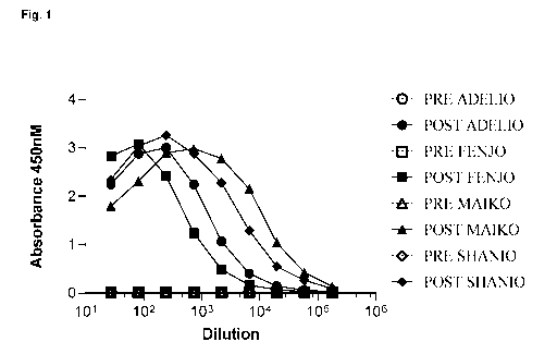
Figure 1 shows the results of testing the pre-immune (PRE) and post-immune
(POST) serum of
immunized llamas for binding to human IgE.
Figure 2 shows the binding of anti-IgE mAbs to human IgE, as measured by
ELISA. Binding
was measured at pH 5.5 and pH 7.4. (A) clone 3D6; (B) clone 16E4; (C) clone
3A1; (D) clone
3D1; (E) clone 13E4; (F) clone 1869; (G) clone 20D5; (H) clone 18E2.
Figure 3 shows the ability of anti-IgE mAbs to inhibit hIgE binding to
hFcERIa, as measured by
ELISA. Binding was measured at pH 6 and pH 7.4. (A) clone 3D6; (B) clone 16E4;
(C) clone
3A1; (D) clone 3D1; (E) clone 13E4; (F) clone 1869; (G) clone 20D5; (H) clone
18E2.
Figure 4 shows the ability of anti-IgE mAbs to inhibit hIgE binding to
hFcERIa, as determined by
SPR analysis. Binding was measured at pH 6 and pH 7.4. (A) clone 3D6; (B)
clone 16E4; (C)
clone 3A1; (D) clone 3D1; (E) clone 13E4; (F) clone 1869; (G) clone 20D5.
Figure 5 shows the binding of anti-IgE mAbs to cynomolgus IgE, as measured by
ELISA.
Binding was measured at pH 5.5 and pH 7.4. (A) clone 3D6; (B) clone 16E4; (C)
clone 3A1; (D)
clone 3D1; (E) clone 13E4; (F) clone 1869; (G) clone 20D5; (H) clone 18E2.
Figure 6 shows the binding of anti-IgE ABDEGTM mAbs to human IgE, as measured
by ELISA.
Binding was measured at pH 5.5 and pH 7.4. (A) clone 18B9His; (B) clone
18E2His2; (C) clone
13E4.
9
CA 03133941 2021-09-15
WO 2020/208177
PCT/EP2020/060240
Figure 7 shows the ability of anti-IgE ABDEGTM mAbs to bind to FcRn with
higher affinity as
compared with the corresponding anti-IgE mAbs lacking the ABDEGTM technology.
Efgartigimod
(an isolated variant Fc molecule incorporating the ABDEGTM technology) was
included for
comparison. (A) Binding of clone 18B9His at pH 6.0; (B) Binding of clone
18B9His at pH 7.0; (C)
Binding of clone 18E2His2 at pH 6.0; (D) Binding of clone 18E2His2 at pH 7.0;
(E) Binding of
clone 13E4 at pH 6.0; (F) Binding of clone 13E4 at pH 7Ø
Figure 8 shows the ability of anti-IgE ABDEGTM mAbs to compete with native
IgG3 for binding to
FcRn, as measured by competition ELISA. (A) clone 18B9His; (B) clone 18E2His2;
(C) clone
13E4.
Figure 9 shows the ability of anti-IgE mAbs (both with and without ABDEGTM) to
inhibit IgE
binding to hFcERla expressing mast cells. (A) clone 18B9His; (B) clone
18E2His2; (C) clone
13E4.
Figure 10 shows the ability of anti-IgE mAbs (both with and without ABDEGTM)
to bind to hIgE
pre-bound to hFcERla on mast cells, as measured by ELISA. (A) clone 13E4; (B)
clone 18B9His;
(C) clone 18E2His2.
Figure 11 shows the ability of an anti-IgE ABDEGTM mAb to deplete both IgG (A)
and IgE (B)
levels in vivo. The controls used were: omalizumab (an anti-IgE antibody
without ABDEGTM
substitutions in the Fc domain) and HEL-hIgG1-ABDEG (an IgG1 antibody
incorporating
ABDEGTM substitutions but without binding specificity for IgE).
Figure 12 shows a schematic of the method used to engineer pH-dependent
variants of the anti-
IgE antibody clone CL-2C (ligelizumab).
Figure 13 shows the distribution of histidine residues at the various CDR
positions of the VK (A)
and VH (B) domains post-screening for CL-2C variant clones exhibiting pH-
dependent binding to
IgE.
Figure 14 shows the ability of anti-IgE ABDEGTM mAbs to inhibit IgE binding to
hFcsRla expressing mast cells.
Figure 15 shows the results of testing various anti-IgE antibodies in a mast
cell activation assay.
Bone marrow-derived mast cells were sensitized with IgE so as to load the
FcERla receptor. The
mast cells were subsequently incubated with various anti-IgE antibodies so as
to test for the
ability of these antibodies to cross-link the FcERIa-bound IgE and trigger
mast cell activation. (A)
shows mast cell challenge with 20 g/ml antibody; (B) shows mast cell
challenge with 200 g/ml
antibody; (C) shows mast cell challenge with increasing concentrations of the
clones 13E4-
hIgG1-ABDEGTm; 18139-hIgG1-ABDEGTm; and 18E2His2-MG-ABDEGTm.
Figure 16 shows the results of testing various anti-IgE antibodies for the
induction of an
anaphylactic reaction in vivo. Mice sensitized with recombinant human IgE were
challenged with
CA 03133941 2021-09-15
WO 2020/208177
PCT/EP2020/060240
various anti-IgE antibodies and the temperature of the mice post-challenge was
recorded at 15
minute intervals over a period of 2 hours. (A) and (B) show show temperature
changes over
the time course of the experiment for antibodies administered at a dose of 15
mg/kg; (C) shows
temperature changes over the time course of the experiment for antibodies
administered at a
dose of 50 mg/kg.
Figure 17 shows the results of testing an ABDEGTM antibody in an in vivo model
of Bullous
Pemphigoid. Knock-in human NC16A mice were injected with either anti-hNC16A
IgG or anti-
hNC16A IgE in the presence or absence of an anti-lgE-ABDEGTM antibody. (A)
shows the
effect on skin disease score in mice injected with anti-hNC16A IgG and (B)
shows the effect on
the anti-hNC16A IgG levels in mice treated with or without a HELABDEGTM
antibody. (C)
shows the effect on skin disease score in mice injected with anti-hNC16A IgE
and (D) shows
the effect on eosinophil peroxidase (EPO) activity in mice treated with or
without an anti-IgE-
ABDEGTM antibody. *p<0.001.
DETAILED DESCRIPTION
A. Definitions
Unless defined otherwise, all technical and scientific terms used herein have
the same meaning
as is commonly understood by one skilled in the art in the technical field of
the invention.
"Antibody" - As used herein, the term "antibody" is intended to encompass full-
length
antibodies and variants thereof, including but not limited to modified
antibodies, humanised
antibodies, germlined antibodies (see definitions below). The term "antibody"
is typically used
herein to refer to immunoglobulin polypeptides having a combination of two
heavy and two light
chains wherein the polypeptide has significant specific immunoreactive
activity to an antigen of
interest (herein IgE). For antibodies of the IgG class, the antibodies
comprise two identical light
polypeptide chains of molecular weight approximately 23,000 Da!tons, and two
identical heavy
chains of molecular weight 53,000-70,000. The four chains are joined by
disulfide bonds in a
"Y" configuration wherein the light chains bracket the heavy chains starting
at the mouth of the
"Y" and continuing through the variable region. The light chains of an
antibody are classified as
either kappa or lambda (K,X). Each heavy chain class may be bound with either
a kappa or
lambda light chain. In general, the light and heavy chains are covalently
bonded to each other,
and the "tail" portions of the two heavy chains are bonded to each other by
covalent disulfide
linkages or non-covalent linkages when the immunoglobulins are generated
either by
hybridomas, B cells or genetically engineered host cells. In the heavy chain,
the amino acid
sequences run from an N-terminus at the forked ends of the Y configuration to
the C-terminus
at the bottom of each chain.
11
RECTIFIED SHEET (RULE 91) ISA/EP
CA 03133941 2021-09-15
WO 2020/208177
PCT/EP2020/060240
Those skilled in the art will appreciate that heavy chains are classified as
gamma, mu, alpha,
delta, or epsilon, (y, , a, 6, e) with some subclasses among them (e.g., y1-
y4). It is the nature of
this chain that determines the "class" of the antibody as IgG, IgM, IgA, IgD
or IgE, respectively.
The immunoglobulin subclasses (isotypes) e.g., IgG1, IgG2, IgG3, IgG4, IgA1,
etc. are well
characterized and are known to confer functional specialization. The term
"antibody" as used
herein encompasses antibodies from any class or subclass of antibody.
"Variable region" or "variable domain" - The terms "variable region" and
"variable domain"
are used herein interchangeably and are intended to have equivalent meaning.
The term
"variable" refers to the fact that certain portions of the variable domains VH
and VL differ
extensively in sequence among antibodies and are used in the binding and
specificity of each
particular antibody for its target antigen. However, the variability is not
evenly distributed
throughout the variable domains of antibodies. It is concentrated in three
segments called
"hypervariable loops" in each of the VL domain and the VH domain which form
part of the
antigen binding site. The first, second and third hypervariable loops of the
VLambda light chain
domain are referred to herein as L1(A), L2(A) and L3(A) and may be defined as
comprising
residues 24-33 (Li (A), consisting of 9, 10 or 11 amino acid residues), 49-53
(L2(A), consisting of
3 residues) and 90-96 (L3(A), consisting of 5 residues) in the VL domain
(Morea etal., Methods
.. 20:267-279 (2000)). The first, second and third hypervariable loops of the
VKappa light chain
domain are referred to herein as Li (K), L2(k) and L3(k) and may be defined as
comprising
residues 25-33 (Li (K), consisting of 6, 7, 8, 11, 12 or 13 residues), 49-53
(L2(k), consisting of 3
residues) and 90-97 (L3(k), consisting of 6 residues) in the VL domain (Morea
etal., Methods
20:267-279 (2000)). The first, second and third hypervariable loops of the VH
domain are
.. referred to herein as H1, H2 and H3 and may be defined as comprising
residues 25-33 (H1,
consisting of 7, 8 or 9 residues), 52-56 (H2, consisting of 3 or 4 residues)
and 91-105 (H3, highly
variable in length) in the VH domain (Morea etal., Methods 20:267-279 (2000)).
Unless otherwise indicated, the terms L1, L2 and L3 respectively refer to the
first, second and
third hypervariable loops of a VL domain, and encompass hypervariable loops
obtained from
both Vkappa and Vlambda isotypes. The terms H1, H2 and H3 respectively refer
to the first,
second and third hypervariable loops of the VH domain, and encompass
hypervariable loops
obtained from any of the known heavy chain isotypes, including y, E, 6, a or
p.
.. The hypervariable loops L1, L2, L3, H1, H2 and H3 may each comprise part of
a
"complementarity determining region" or "CDR", as defined below. The terms
"hypervariable
loop" and "complementarity determining region" are not strictly synonymous,
since the
hypervariable loops (HVs) are defined on the basis of structure, whereas
complementarity
12
CA 03133941 2021-09-15
WO 2020/208177
PCT/EP2020/060240
determining regions (CDRs) are defined based on sequence variability (Kabat
etal., Sequences
of Proteins of Immunological Interest, 5th Ed. Public Health Service, National
Institutes of Health,
Bethesda, MD., 1983) and the limits of the HVs and the CDRs may be different
in some VH and
VL domains.
The CDRs of the VL and VH domains can typically be defined as comprising the
following amino
acids: residues 24-34 (LCDR1), 50-56 (LCDR2) and 89-97 (LCDR3) in the light
chain variable
domain, and residues 31-35 or 31-35b (HCDR1), 50-65 (HCDR2) and 95-102 (HCDR3)
in the
heavy chain variable domain; (Kabat etal., Sequences of Proteins of
Immunological Interest, 5th
Ed. Public Health Service, National Institutes of Health, Bethesda, MD.
(1991)). Thus, the HVs
may be comprised within the corresponding CDRs and references herein to the
"hypervariable
loops" of VH and VL domains should be interpreted as also encompassing the
corresponding
CDRs, and vice versa, unless otherwise indicated.
The more highly conserved portions of variable domains are called the
framework region (FR),
as defined below. The variable domains of native heavy and light chains each
comprise four FRs
(FR1, FR2, FR3 and FR4, respectively), largely adopting a p-sheet
configuration, connected by
the three hypervariable loops. The hypervariable loops in each chain are held
together in close
proximity by the FRs and, with the hypervariable loops from the other chain,
contribute to the
formation of the antigen-binding site of antibodies. Structural analysis of
antibodies revealed the
relationship between the sequence and the shape of the binding site formed by
the
complementarity determining regions (Chothia etal., J. Mol. Biol. 227: 799-817
(1992));
Tramontano etal., J. Mol. Biol, 215:175-182 (1990)). Despite their high
sequence variability, five
of the six loops adopt just a small repertoire of main-chain conformations,
called "canonical
structures". These conformations are first of all determined by the length of
the loops and
secondly by the presence of key residues at certain positions in the loops and
in the framework
regions that determine the conformation through their packing, hydrogen
bonding or the ability to
assume unusual main-chain conformations.
"CDR" - As used herein, the term "CDR" or "complementarity determining region"
means the
non-contiguous antigen binding sites found within the variable region of both
heavy and light
chain polypeptides. These particular regions have been described by Kabat
etal., J. Biol. Chem.
252, 6609-6616 (1977) and Kabat etal., Sequences of protein of immunological
interest. (1991),
and by Chothia etal., J. Mol. Biol. 196:901-917 (1987) and by MacCallum etal.,
J. Mol. Biol.
262:732-745 (1996) where the definitions include overlapping or subsets of
amino acid residues
when compared against each other. The amino acid residues which encompass the
CDRs as
defined by each of the above cited references are set forth for comparison.
Preferably, the term
"CDR" is a CDR as defined by Kabat based on sequence comparisons.
13
CA 03133941 2021-09-15
WO 2020/208177
PCT/EP2020/060240
Table 1: CDR definitions
CDR Definitions
Kabat1 Chothia2 MacCallum3
VH CDR1 31-35 26-32 30-35
VH CDR2 50-65 53-55 47-58
VH CDR3 95-102 96-101 93-101
VLCDR1 24-34 26-32 30-36
VI_ CDR2 50-56 50-52 46-55
VI_ CDR3 89-97 91-96 89-96
1Residue numbering follows the nomenclature of Kabat etal., supra
2Residue numbering follows the nomenclature of Chothia etal., supra
3Residue numbering follows the nomenclature of MacCallum etal., supra
"Framework region" - The term "framework region" or "FR region" as used
herein, includes the
amino acid residues that are part of the variable region, but are not part of
the CDRs (e.g., using
the Kabat definition of CDRs). Therefore, a variable region framework is
between about 100-120
amino acids in length but includes only those amino acids outside of the CDRs.
For the specific
example of a heavy chain variable domain and for the CDRs as defined by Kabat
etal.,
framework region 1 corresponds to the domain of the variable region
encompassing amino acids
1-30; framework region 2 corresponds to the domain of the variable region
encompassing amino
acids 36-49; framework region 3 corresponds to the domain of the variable
region encompassing
amino acids 66-94, and framework region 4 corresponds to the domain of the
variable region
from amino acids 103 to the end of the variable region. The framework regions
for the light chain
are similarly separated by each of the light chain variable region CDRs.
Similarly, using the
definition of CDRs by Chothia etal. or McCallum etal. the framework region
boundaries are
separated by the respective CDR termini as described above. In preferred
embodiments the
.. CDRs are as defined by Kabat.
In naturally occurring antibodies, the six CDRs present on each monomeric
antibody are short,
non-contiguous sequences of amino acids that are specifically positioned to
form the antigen
binding site as the antibody assumes its three dimensional configuration in an
aqueous
environment. The remainder of the heavy and light variable domains show less
inter-molecular
variability in amino acid sequence and are termed the framework regions. The
framework
regions largely adopt a p-sheet conformation and the CDRs form loops which
connect, and in
some cases form part of, the p-sheet structure. Thus, these framework regions
act to form a
scaffold that provides for positioning the six CDRs in correct orientation by
inter-chain, non-
covalent interactions. The antigen binding site formed by the positioned CDRs
defines a surface
14
CA 03133941 2021-09-15
WO 2020/208177
PCT/EP2020/060240
complementary to the epitope on the immunoreactive antigen. This complementary
surface
promotes the non-covalent binding of the antibody to the immunoreactive
antigen epitope. The
position of CDRs can be readily identified by one of ordinary skill in the
art.
"Constant region" ¨ As used herein, the term "constant region" refers to the
portion of the
antibody molecule outside of the variable domains or variable regions.
Immunoglobulin light
chains have a single domain "constant region", typically referred to as the
"CL" or "CL1 domain".
This domain lies C terminal to the VL domain. Immunoglobulin heavy chains
differ in their
constant region depending on the class of immunoglobulin (y, , a, 6, e).
Heavy chains y, a and 6
have a constant region consisting of three immunoglobulin domains (referred to
as CH1, CH2
and CH3) with a flexible hinge region separating the CH1 and CH2 domains.
Heavy chains p
and E have a constant region consisting of four domains (CH1-CH4). The
constant domains of
the heavy chain are positioned C terminal to the VH domain.
The numbering of the amino acids in the heavy and light immunoglobulin chains
run from the N-
terminus at the forked ends of the Y configuration to the C-terminus at the
bottom of each chain.
Different numbering schemes are used to define the constant domains of the
immunoglobulin
heavy and light chains. In accordance with the EU numbering scheme, the heavy
chain constant
domains of an IgG molecule are identified as follows: CH1 ¨ amino acid
residues 118-215; CH2
¨ amino acid residues 231-340; CH3 ¨ amino acid residues 341-446. In
accordance with the
Kabat numbering scheme, the heavy chain constant domains of an IgG molecule
are identified
as follows: CH1 ¨ amino acid residues 114-223; CH2 ¨ amino acid residues 244-
360; CH3 ¨
amino acid residues 361-477.
"Fc domain" ¨ As used herein, the "Fc domain" defines the portion of the
constant region of an
immunoglobulin heavy chain including the CH2 and CH3 domains. It typically
defines the portion
of a single immunoglobulin heavy chain beginning in the hinge region just
upstream of the papain
cleavage site and ending at the C-terminus of the antibody. The Fc domain
typically includes
some residues from the hinge region. Accordingly, a complete Fc domain
typically comprises at
least a portion of a hinge (e.g., upper, middle, and/or lower hinge region)
domain, a CH2 domain,
and a CH3 domain.
The "hinge region" includes the portion of a heavy chain molecule that joins
the CH1 domain to
the CH2 domain. This hinge region comprises approximately 25 residues and is
flexible, thus
allowing the two N-terminal antigen binding regions to move independently.
Hinge regions can
be subdivided into three distinct domains: upper, middle, and lower hinge
domains (Roux K.H. et
al. J. Immunol. 161:4083-90 1998). Antibodies of the invention comprising a
"fully human"
hinge region may contain one of the hinge region sequences shown in Table 2
below.
CA 03133941 2021-09-15
WO 2020/208177
PCT/EP2020/060240
Table 2: Human hinge sequences
IgG Upper hinge Middle hinge Lower hinge
IgG1 EPKSCDKTHT CPPCP APELLGGP
(SEQ ID NO: 159) (SEQ ID NO: 160) (SEQ ID NO: 161)
IgG3 ELKTPLGDTTHT CPRCP (EPKSCDTPPPCPRCP)3 APELLGGP
(SEQ ID NO: 162) (SEQ ID NO: 163) (SEQ ID NO: 164)
IgG4 ESKYGPP CPSCP APEFLGGP
(SEQ ID NO: 165) (SEQ ID NO: 166) (SEQ ID NO: 167)
IgG2 ERK CCVECPPPCP APPVAGP
(SEQ ID NO: 168) (SEQ ID NO: 169) (SEQ ID NO: 170)
"Variant Fc domain" - As used herein, the term "variant Fc domain" refers to
an Fc domain with
one or more alterations relative to a wild-type Fc domain, for example the Fc
domain of a
naturally-occurring or "wild-type" human IgG. Alterations can include amino
acid substitutions,
additions and/or deletions, linkage of additional moieties, and/or alteration
of the native glycans.
"Fc region" ¨ As used herein, the term "Fc region" refers to the portion of a
native
immunoglobulin formed by the Fc domains of the two heavy chains. A native or
wild-type Fc
region is typically homodimeric.
"Variant Fc region" ¨ As used herein the term "variant Fc region" refers to an
Fc region wherein
at least one of the Fc domains has one or more alterations relative to the
wild-type domains of
the wild-type Fc region, for example the Fc region of a naturally-occurring
human IgG. In certain
embodiments the term encompasses homodimeric Fc regions wherein each of the
constituent Fc
domains is the same. In certain embodiments the term encompasses heterodimeric
Fc regions
wherein each of the constituent Fc domains is different. For heterodimeric
embodiments, one or
both of the Fc domains may be variant Fc domains.
"FcRn binding fragment" - As used herein the term "FcRn binding fragment"
refers to a portion
of an Fc domain or Fc region that is sufficient to confer FcRn binding.
"Specificity" and "Multispecific antibodies"¨ The antibodies described herein
bind to a
particular target antigen, IgE. It is preferred that the antibodies
"specifically bind" to their target
antigen, wherein the term "specifically bind" refers to the ability of any
antibody to preferentially
immunoreact with a given target e.g. IgE. The antibodies of the present
invention may be
monospecific and contain one or more binding sites which specifically bind a
particular target.
The antibodies may be incorporated into "multispecific antibody" formats, for
example bispecific
16
CA 03133941 2021-09-15
WO 2020/208177
PCT/EP2020/060240
antibodies, wherein the multispecific antibody binds to two or more target
antigens. In order to
achieve multiple specificities, "multispecific antibodies" are typically
engineered to include
different combinations or pairings of heavy and light chain polypeptides with
different VH-VL
pairs. Multispecific, notably bispecific antibodies, may be engineered so as
to adopt the overall
conformation of a native antibody, for example a Y-shaped antibody having Fab
arms of different
specificities conjugated to an Fc region. Alternatively multispecific
antibodies, for example
bispecific antibodies, may be engineered so as to adopt a non-native
conformation, for example
wherein the variable domains or variable domain pairs having different
specificities are
positioned at opposite ends of the Fc region.
"Modified antibody" - As used herein, the term "modified antibody" includes
synthetic forms of
antibodies which are altered such that they are not naturally occurring.
Examples include but are
not limited to antibodies that comprise at least two heavy chain portions but
not two complete
heavy chains (such as, domain deleted antibodies or minibodies); multispecific
forms of
antibodies (e.g., bispecific, trispecific, etc.) altered to bind to two or
more different antigens or to
different epitopes on a single antigen); heavy chain molecules joined to scFv
molecules and the
like. scFv molecules are known in the art and are described, e.g., in US
patent 5,892,019. In
addition, the term "modified antibody" includes multivalent forms of
antibodies (e.g., trivalent,
tetravalent, etc., antibodies that bind to three or more copies of the same
antigen).
The term "modified antibody" may also be used herein to refer to amino acid
sequence variants
of the antibodies of the invention as structurally defined herein. It will be
understood by one of
ordinary skill in the art that an antibody may be modified to produce a
variant antibody which
varies in amino acid sequence in comparison to the antibody from which it was
derived. For
example, nucleotide or amino acid substitutions leading to conservative
substitutions or changes
at "non-essential" amino acid residues may be made (e.g., in CDR and/or
framework residues).
Amino acid substitutions can include replacement of one or more amino acids
with a naturally
occurring or non-natural amino acid.
Modified antibodies in accordance with the present invention may comprise any
suitable antigen-
binding fragment as defined herein linked to a variant Fc domain or FcRn
binding fragment
thereof as defined in accordance with the invention.
"Antigen binding fragment" ¨ The term "antigen binding fragment" as used
herein refers to
fragments that are parts or portions of a full-length antibody or antibody
chain comprising fewer
amino acid residues than an intact or complete antibody whilst retaining
antigen binding activity.
An antigen-binding fragment of an antibody includes peptide fragments that
exhibit specific
immuno-reactive activity to the same antigen as the antibody (e.g. IgE). The
term "antigen
17
CA 03133941 2021-09-15
WO 2020/208177
PCT/EP2020/060240
binding fragment" as used herein is intended to encompass antibody fragments
selected from: an
antibody light chain variable domain (VL); an antibody heavy chain variable
domain (VH); a VH-
VL domain pairing; a single chain antibody (scFv); a F(ab')2 fragment; a Fab
fragment; an Fd
fragment; an Fv fragment; a one-armed (monovalent) antibody; diabodies,
triabodies, tetrabodies
or any antigen-binding molecule formed by combination, assembly or conjugation
of such antigen
binding fragments. The term "antigen binding fragment" as used herein may also
encompass
antibody fragments selected from the group consisting of: unibodies; domain
antibodies; and
nanobodies. Fragments can be obtained, for example, via chemical or enzymatic
treatment of an
intact or complete antibody or antibody chain or by recombinant means.
"Humanising substitutions" - As used herein, the term "humanising
substitutions" refers to
amino acid substitutions in which the amino acid residue present at a
particular position in the VH
or VL domain of an antibody is replaced with an amino acid residue which
occurs at an
equivalent position in a reference human VH or VL domain. The reference human
VH or VL
domain may be a VH or VL domain encoded by the human germline. Humanising
substitutions
may be made in the framework regions and/or the CDRs of the antibodies,
defined herein.
"Humanised variants" - As used herein the term "humanised variant" or
"humanised antibody"
refers to a variant antibody which contains one or more "humanising
substitutions" compared to a
reference antibody, wherein a portion of the reference antibody (e.g. the VH
domain and/or the
VL domain or parts thereof containing at least one CDR) has an amino acid
derived from a non-
human species, and the "humanising substitutions" occur within the amino acid
sequence
derived from a non-human species.
"Germlined variants" - The term "germlined variant" or "germlined antibody" is
used herein to
refer specifically to "humanised variants" in which the "humanising
substitutions" result in
replacement of one or more amino acid residues present at (a) particular
position(s) in the VH or
VL domain of an antibody with an amino acid residue which occurs at an
equivalent position in a
reference human VH or VL domain encoded by the human germline. It is typical
that for any
given "germlined variant", the replacement amino acid residues substituted
into the germlined
variant are taken exclusively, or predominantly, from a single human germline-
encoded VH or VL
domain. The terms "humanised variant" and "germlined variant" are often used
interchangeably.
Introduction of one or more "humanising substitutions" into a camelid-derived
(e.g. llama derived)
VH or VL domain results in production of a "humanised variant" of the camelid
(llama)-derived
.. VH or VL domain. If the amino acid residues substituted in are derived
predominantly or
exclusively from a single human germline-encoded VH or VL domain sequence,
then the result
may be a "human germlined variant" of the camelid (llama)-derived VH or VL
domain.
18
CA 03133941 2021-09-15
WO 2020/208177
PCT/EP2020/060240
"Affinity variants" - As used herein, the term "affinity variant" refers to a
variant antibody which
exhibits one or more changes in amino acid sequence compared to a reference
antibody,
wherein the affinity variant exhibits an altered affinity for the target
antigen in comparison to the
reference antibody. For example, affinity variants will exhibit a changed
affinity for a target, for
example IgE, as compared to the reference IgE antibody. Preferably the
affinity variant will
exhibit improved affinity for the target antigen, as compared to the reference
antibody. Affinity
variants typically exhibit one or more changes in amino acid sequence in the
CDRs, as
compared to the reference antibody. Such substitutions may result in
replacement of the original
amino acid present at a given position in the CDRs with a different amino acid
residue, which
may be a naturally occurring amino acid residue or a non-naturally occurring
amino acid residue.
The amino acid substitutions may be conservative or non-conservative.
"Engineered" - As used herein the term "engineered" includes manipulation of
nucleic acid or
polypeptide molecules by synthetic means (e.g. by recombinant techniques, in
vitro peptide
synthesis, by enzymatic or chemical coupling of peptides or some combination
of these
techniques). Preferably, the antibodies of the invention are engineered,
including for example,
humanized antibodies which have been engineered to improve one or more
properties, such as
antigen binding, stability/half-life or effector function.
"FcRn" - As used herein, the term "FcRn" refers to a neonatal Fc receptor.
Exemplary FcRn
molecules include human FcRn encoded by the FCGRT gene as set forth in Ref Seq
NM 004107.
"CD16" - As used herein, the term "CD16" refers to FcyRIII Fc receptors that
are required for
Antibody-Dependent Cell-mediated Cytotoxicity (ADCC). Exemplary CD16 molecules
include
human CD16a as set forth in RefSeq NM 000569.
"N-linked glycan" - As used herein the term "N-linked glycan" refers to the N-
linked glycan
attached to the nitrogen (N) in the side chain of asparagine in the sequence
(i.e., Asn-X-Ser or
Asn-X-Thr sequence, where X is any amino acid except proline) present in the
CH2 domain of an
Fc region. Such N-glycans are fully described in, for example, Drickamer K and
Taylor ME (2006)
Introduction to Glycobiology, 2nd ed., incorporated herein by reference in its
entirety.
"Afucosylated" - As used herein the term "afucosylated" refers to an N-linked
glycan which
lacks a core fucose molecule as described in US Pat No. 8067232, incorporated
herein by
reference in its entirety.
19
CA 03133941 2021-09-15
WO 2020/208177
PCT/EP2020/060240
"Bisecting GIcNAc" - As used herein the term "bisecting GIcNAc" refers to an N-
linked glycan
having an N-acetylglucosamine (GIcNAc) molecule linked to a core mannose
molecule, as
described in US Pat. No. 8021856, incorporated herein by reference in its
entirety.
"IgE" ¨As used herein, the term "IgE" refers to "immunoglobulin E" molecules
or "class E
immunoglobulins". IgE is the least abundant immunoglobulin isotype in human
serum. IgE
immunoglobulins adopt the tetrameric structure common to other classes or
isotypes of
immunoglobulin. However, IgE is characterised by its e heavy chains, which
comprise four
constant regions: CE1, CE2, CE3 and CE4 (also referred to herein as CH1, CH2,
CH3 and CH4).
As explained elsewhere herein, IgE plays an important role in allergy and
hypersensitivity by
binding to the high-affinity Fc receptors on mast cells and basophils. This
high-affinity receptor,
FcERI, has a multisubunit structure including one IgE-binding a subunit, one p
subunit and a
dimer of disulphide-linked y subunits. A low-affinity IgE receptor, FcaRII
(also known as CD23),
is constitutively expressed on B cells and can be expressed on macrophages,
eosinophils,
platelets and some T cells in response to IL-4.
Omalizumab ¨ Omalizumab is a recombinant humanized monoclonal antibody that
binds to IgE.
It contains 5% murine sequence and 95% human sequence. It is marketed by
Novartis as
Xolair , and is approved for the treatment of allergic asthma and Chronic
Spontaneous Urticaria
(CSU). The CDR, VH and VL sequences of omalizumab are shown in table 3 below.
Table 3 CDR, VH and VL sequences for omalizumab
Sequence
SEQ ID NO.
VH CDR1 SGYSWN 143
VH CDR2 SITYDGSTNYNPSVKG 144
VH CDR3 GSHYFGHWHFAV 145
VH EVQLVESGGGLVQPGGSLRLSCAVSGYSITSGYSWNWIRQAP 146
GKGLEWVASITYDGSTNYNPSVKGRITISRDDSKNTFYLQMNSL
RAEDTAVYYCARGSHYFGHWHFAVWGQGTLVTVSS
VL CDR1 RASQSVDYDGDSYMN 147
VL CDR2 AASYLES 148
VL CDR3 QQSHEDPYT 149
VL DIQLTQSPSSLSASVGDRVTITCRASQSVDYDGDSYMNWYQQK 150
PGKAPKLLIYAASYLESGVPSRFSGSGSGTDFTLTISSLQPEDFA
TYYCQQSHEDPYTFGQGTKVEIK
Omalizumab binds to the receptor-binding portion of IgE i.e. a region within
the CH3 or CE3
domain. Since the epitope that is recognized by omalizumab encompasses binding
regions for
both the high-affinity and low-affinity IgE receptors, omalizumab eliminates
the ability of IgE to
bind to both types of receptor. Importantly, omalizumab is not able to cross-
link IgE molecules
CA 03133941 2021-09-15
WO 2020/208177
PCT/EP2020/060240
that are already bound on the cell surface i.e. it is non-anaphylactogenic.
The binding of FcERI to
one CH3 domain of one IgE heavy chain inhibits or prevents the binding of
omalizumab to the
CH3 region of the other IgE heavy chain. Thus, omalizumab can only bind to IgE
that is in
circulation. In the circulation, each molecule of IgE can be simultaneously
bound by two
molecules of omalizumab.
Ligelizumab ¨ Ligelizumab is a second humanized monoclonal antibody that binds
to IgE. It
binds to the same region of IgE as omalizumab but binds to IgE with higher
affinity. The CDR,
VH and VL sequences of ligelizumab are shown in table 4 below.
Table 4 CDR, VH and VL sequences for ligelizumab
Sequence SEQ ID
NO.
VH CDR1 WYWLE 151
VH CDR2 EIDPGTFTTNYNEKFKA 152
VH CDR3 FSHFSGSNYDYFDY 153
VH QVQLVQSGAEVMKPGSSVKVSCKASGYTFSWYWLEWVRQAP 154
GHGLEWMGEIDPGTFTTNYNEKFKARVTFTADTSTSTAYMELS
SLRSEDTAVYYCARFSHFSGSNYDYFDYWGQGTLVTVSS
VL CDR1 RASQSIGTNIH 155
VL CDR2 YASESIS 156
VL CDR3 QQSWSWPTT 157
VL EIVMTQSPATLSVSPGERATLSCRASQSIGTNIHWYQQKPGQAP 158
RLLIYYASESISGIPARFSGSGSGTEFTLTISSLQSEDFAVYYCQQ
SWSWPTTFGGGTKVEIK
"Antibody-mediated disorder" - As used herein, the term "antibody-mediated
disorder" refers
to any disease or disorder caused or exacerbated by the presence of an
antibody in a subject.
"Treat, treating and treatment" - As used herein, the terms "treat,"
"treating," and "treatment"
refer to therapeutic or preventative measures described herein. The methods of
"treatment"
employ administration to a subject, for example, a subject having an antibody-
mediated disease
or disorder (e.g. autoimmune disease) or predisposed to having such a disease
or disorder, an
antibody in accordance with the present invention, in order to prevent, cure,
delay, reduce the
severity of, or ameliorate one or more symptoms of the disease or disorder or
recurring disease
or disorder, or in order to prolong the survival of a subject beyond that
expected in the absence
of such treatment.
"Subject" - As used herein, the term "subject" refers to any human or non-
human animal. In
certain embodiments, the term "subject" refers to any human or non-human
mammal. In
21
CA 03133941 2021-09-15
WO 2020/208177
PCT/EP2020/060240
preferred embodiments, the subject is a human. In certain embodiments the
subject is an adult
human. As used herein, an "adult human" is a human who is at least 18 years of
age.
B. Anti-laE antibodies havina variant Fc domains
(i) Variant Fc domains and FcRn bindinci fraciments thereof
In a first aspect, the present invention provides antibodies that bind to IgE
(i.e. anti-IgE
antibodies) wherein the antibodies comprise at least one variant Fc domain or
FcRn binding
fragment thereof. This variant Fc domain or FcRn binding fragment thereof is
characterised by
the ability to bind to the neonatal Fc receptor, FcRn, with increased affinity
relative to a wild-type
Fc domain. Put another way, the binding affinity between the variant Fc domain
or FcRn binding
fragment of the anti-IgE antibodies described herein and FcRn is higher as
compared with the
binding affinity between a wild-type Fc domain and FcRn.
The FcRn receptor plays an important role in regulating IgG concentrations in
the plasma by
means of the salvage receptor pathway. The model for FcRn function is as
follows. IgGs in the
circulation are taken up into cells, most likely by fluid-phase pinocytosis,
as the near-neutral pH
of the extracellular milieu is generally not permissive for FcRn-IgG
interactions. IgGs that bind to
FcRn in early, acidic endosomes following uptake are recycled (or
transcytosed) and released at
the cell surface by exocytosis. In contrast, IgGs that do not bind FcRn, enter
the lysosomal
pathway and are degraded.
By virtue of binding with higher affinity to FcRn, the anti-IgE antibodies of
the invention interfere
with the recycling of endogenous IgG molecules and thus can reduce the levels
of endogenous
IgG antibodies, for example IgG autoantibodies. It follows, that the anti-IgE
antibodies of the
invention target both endogenous IgE (by virtue of antigen binding via the
variable region) and
endogenous IgG (by competing for binding to FcRn via the variant Fc domain).
The variant Fc domains or FcRn binding fragments thereof bind to FcRn with
increased affinity
relative to a wild-type Fc domain. In certain embodiments, the wild-type Fc
domain against
which the binding affinity of the variant Fc domain is compared may be the
wild-type Fc domain
from which the variant Fc domain derives. As described above, a variant Fc
domain in the
context of the present invention refers to an Fc domain with one or more
alterations relative to a
wild-type Fc domain, for example the Fc domain of a naturally-occurring or
"wild-type" human
IgG. Alterations can include amino acid substitutions, additions and/or
deletions, linkage of
additional moieties, and/or alteration of the native glycans. If the naturally-
occurring or wild-type
22
CA 03133941 2021-09-15
WO 2020/208177
PCT/EP2020/060240
Fc domain from which the variant Fc domain derives is a human IgG1 Fc domain,
the variant Fc
domain may bind to FcRn with higher affinity than the wild-type human IgG1 Fc
domain.
The increased affinity for FcRn exhibited by the variant Fc domain or FcRn
binding fragment may
be relative to a wild-type Fc domain that is not necessarily the Fc domain
from which the variant
Fc domain or FcRn binding fragment derives. For example, the variant Fc domain
or FcRn
binding fragment thereof may bind to FcRn with increased affinity relative to
a wild-type human
IgG Fc domain. The wild-type human IgG may be an IgG1, IgG2, IgG3 or IgG4. In
preferred
embodiments, the variant Fc domain or FcRn binding fragment thereof of the
anti-IgE antibodies
described herein binds to FcRn with increased affinity relative to a wild-type
human IgG1 Fc
domain or a wild-type human IgG3 Fc domain. In a preferred embodiment, the
variant Fc
domain or FcRn binding fragment thereof of the anti-IgE antibodies described
herein binds to
FcRn with increased affinity relative to a wild-type human IgG1 Fc domain.
Since the anti-IgE antibodies of the present invention are intended for use in
the treatment of
human disease, particularly the depletion of IgG autoantibodies from patients
having
autoimmune diseases, the variant Fc region or FcRn binding fragment thereof
will typically bind
with higher affinity to human FcRn. In other words, the variant Fc region or
FcRn binding
fragment of the anti-IgE antibodies described herein will compete with native
or endogenous
patient IgG antibodies for binding to human FcRn.
The interaction between IgG Fc domains and FcRn is pH-dependent. The binding
affinity is
typically stronger at acidic pH (i.e. at the pH found in the early endosomal
compartment) and
weaker at neutral pH (i.e. plasma pH). The variant Fc domains or FcRn binding
fragments
described herein may bind to FcRn with increased affinity at acidic pH, for
example pH 6Ø
Alternatively or in addition, the variant Fc domains or FcRn binding fragments
described herein
may bind to FcRn with increased affinity at neutral pH, for example pH 7.4. In
preferred
embodiments, the variant Fc domains or FcRn binding fragments of the anti-IgE
antibodies
described herein bind to FcRn with increased affinity at both pH 6.0 and pH
7.4. In certain
embodiments, the variant Fc domains and/or FcRn binding fragments bind to FcRn
with reduced
pH-dependence as compared with a wild-type Fc domain, particularly a wild-type
human IgG1 Fc
domain. For embodiments where the variant Fc domain or FcRn binding fragment
binds to FcRn
with reduced pH-dependence, it is still preferred that the binding affinity is
increased at pH 6.0
and pH 7.4.
As explained herein, the binding affinity between the variant Fc domains or
FcRn binding
fragments described herein and FcRn is increased such that the antibodies of
the present
invention compete with endogenous IgGs, particularly IgG autoantibodies, for
binding to FcRn.
23
CA 03133941 2021-09-15
WO 2020/208177
PCT/EP2020/060240
As reported in Vaccaro et al. (Engineering the Fc region of immunoglobulin G
to modulate in vivo
antibody levels. Nature Biotechnology (2005) 23(10): 1283-1288), Ulrichts et
al. (Neonatal Fc
receptor antagonist efgartigimod safely and sustainably reduces IgGs in
humans. J. Clinical
Investigation. (2018) 128(10): 4372-4386), and also reported herein, a variant
Fc region
comprising variant Fc domains having ABDEGTM mutations
(M252Y/S254T/T256E/H433K/N434F) can bind to human FcRn with increased affinity
and
thereby reduce endogenous IgG levels. Vaccaro et al. (incorporated herein by
reference) reports
a binding affinity for human FcRn at pH 6.0 for the variant ABDEGTM Fc region
of KD 15.5 nM as
compared with a binding affinity of KD 370 nM for wild-type human IgG1 (as
measured by surface
plasmon resonance analysis). Thus, in certain embodiments, the variant Fc
domain or FcRn
binding fragments described herein bind to human FcRn at pH 6.0 with an
affinity that is
increased by at least 20x as compared with a wild-type human IgG1 Fc domain.
In certain
embodiments, the variant Fc domain or FcRn binding fragments described herein
bind to human
FcRn at pH 6.0 with an affinity that is increased by at least 25x, preferably
at least 30x, as
compared with a wild-type human IgG1 Fc domain. The binding affinity of the
variant Fc domain
or FcRn binding fragment may be compared with the binding affinity of the wild-
type human IgG1
Fc domain when the affinity of the Fc domains (or fragment) is tested in the
context of a full-
length IgG molecule.
As reported in Ulrichts et al. the FcRn antagonist, Efgartigimod, has
equilibrium dissociation
constants (KD) for human FcRn of 14.2 nM and 320 nM at pH 6.0 and pH 7.4,
respectively.
Thus, in certain embodiments, the variant Fc domain or FcRn binding fragments
described
herein bind to human FcRn at pH 6.0 with a binding affinity stronger than KD
15 nM. Alternatively
or in addition, the variant Fc domain or FcRn binding fragments described
herein may bind to
human FcRn at pH 7.4 with a binding affinity stronger than KD 320 nM. The
binding affinity of the
variant Fc domain or FcRn binding fragment thereof may be determined when the
variant Fc
domain or FcRn binding fragment thereof is tested in the context of a variant
Fc region (i.e.
including two Fc domains).
The variant Fc domains or FcRn binding fragments comprise one or more
alterations relative to a
wild-type Fc domain. In certain embodiments, the variant Fc domains or FcRn
binding fragments
comprise at least one amino acid substitution relative to a wild-type Fc
domain. The variant Fc
domains or FcRn binding fragments may comprise, in certain embodiments, at
least two, at least
three, at least four or at least five amino acid substitutions relative to a
wild-type Fc domain.
The number of alterations in the variant Fc domain or FcRn binding fragment
thereof may be
limited relative to the corresponding wild-type Fc domain or FcRn binding
fragment. For
example, the total number of amino acid substitutions in the variant Fc domain
or FcRn binding
24
CA 03133941 2021-09-15
WO 2020/208177
PCT/EP2020/060240
fragment may be limited relative to the corresponding wild-type Fc domain or
FcRn binding
fragment. In certain embodiments, the variant Fc domain or FcRn binding
fragment thereof
consists of no more than 5, no more than 6, no more than 7, no more than 8, no
more than 9, no
more than 10, no more than 11, no more than 12, no more than 15, no more than
20 alterations
as compared with the corresponding wild-type Fc domain. The alterations may be
selected from
amino acid substitutions, additions and/or deletions, linkage of additional
moieties, and/or
alteration of the native glycans. In certain embodiments, the variant Fc
domain or FcRn binding
fragment thereof consists of no more than 5, no more than 6, no more than 7,
no more than 8, no
more than 9, no more than 10, no more than 11, no more than 12, no more than
15, no more
than 20 amino acid substitutions as compared with the corresponding wild-type
Fc domain.
In certain embodiments, the variant Fc domain or FcRn binding fragment thereof
comprises or
consists of at least one amino acid substitution but no more than 20 amino
acid substitutions in
total. In certain embodiments, the variant Fc domain or FcRn binding fragment
thereof
comprises or consists of at least two amino acid substitutions but no more
than 20 amino acid
substitutions in total. In certain embodiments, the variant Fc domain or FcRn
binding fragment
thereof comprises or consists of at least one amino acid substitution but no
more than 10 amino
acid substitutions in total. In certain embodiments, the variant Fc domain or
FcRn binding
fragment thereof comprises or consists of at least two amino acid
substitutions but no more than
.. 10 amino acid substitutions in total. In certain embodiments, the variant
Fc domain or FcRn
binding fragment thereof comprises or consists of at least one amino acid
substitution but no
more than 5 amino acid substitutions in total. In certain embodiments, the
variant Fc domain or
FcRn binding fragment thereof comprises or consists of at least two amino acid
substitutions but
no more than 5 amino acid substitutions in total.
The wild-type Fc domain from which the variant Fc domains of the anti-IgE
antibodies described
herein derive may be an IgG Fc domain. In such embodiments, the variant Fc
domain is a
variant IgG Fc domain. In preferred embodiments, the variant Fc domain is a
variant IgG1 Fc
domain i.e. the variant Fc domain possesses one or more alterations relative
to a wild-type IgG1
domain.
Since the anti-IgE antibodies of the present invention may be for use in human
patients, the
variant Fc domains or FcRn binding fragments thereof will preferably be
variant forms of human
Fc domains i.e. the variant Fc domain or FcRn binding fragment thereof will be
a variant human
Fc domain or FcRn binding fragment thereof. Since the purpose of the variant
Fc domain is to
compete with native IgG antibodies for binding to FcRn, it is preferred that
the variant Fc domain
is a human variant IgG domain, for example a human variant IgG domain selected
from IgG1,
CA 03133941 2021-09-15
WO 2020/208177
PCT/EP2020/060240
IgG2, IgG3 or IgG4. In particularly preferred embodiments, the variant Fc
domain is a variant
IgG1 Fc domain or FcRn binding fragment thereof.
The variant Fc domains or FcRn binding fragments of the anti-IgE antibodies of
the present
invention may comprise any non-native amino acid residues, provided that the
variant Fc domain
or FcRn binding fragment exhibits the requisite increased binding affinity for
FcRn, preferably
human FcRn. As used herein, the term "non-native amino acid" means an amino
acid that does
not occur naturally at the position at which it is located in the variant Fc
domain or FcRn binding
fragment thereof.
Antibodies having variant Fc domains and exhibiting increased binding affinity
for FcRn have
been reported in the literature. These variant Fc domains have been reported
as having various
non-native amino acids at specific positions within the Fc domain. The variant
Fc domains and
FcRn binding fragments of the anti-IgE antibodies described herein may
comprise any of the
non-native amino acids and/or amino acid substitutions described in the
literature as capable of
increasing Fc domain binding affinity for FcRn. The variant Fc domains and
FcRn binding
fragments of the anti-IgE antibodies described herein may also comprise any
combinations of
non-native amino acids and/or amino acid substitutions described in the
literature as capable of
increasing Fc domain binding affinity for FcRn. Non-limiting examples of amino
acid
substitutions that may be included in the variant Fc domains or FcRn binding
fragments
described herein are reported in Yeung et al. (Engineering Human IgG1 Affinity
to Human
Neonatal Fc Receptor: Impact of Affinity Improvement on Pharmacokinetics in
Primates. J.
Immunol. (2009) 182: 7663-7671), and also International patent application no.
W02011/122011,
the entire contents of which are incorporated herein by reference.
In certain embodiments, the variant Fc domains or FcRn binding fragments
described herein
comprise at least one amino acid selected from the following: 237M; 238A;
239K; 2481; 250A;
250F; 2501; 250M; 2500; 250S; 250V; 250W; 250Y; 252F; 252W; 252Y; 254T; 255E;
256D;
256E; 2560; 257A; 257G; 2571; 257L; 257M; 257N; 257S; 257T; 257V; 258H; 265A;
270F; 286A;
286E; 289H; 297A; 298G; 303A; 305A; 307A; 307D; 307F; 307G; 307H; 3071; 307K;
307L;
307M; 307N; 307P; 3070; 307R; 307S; 307V; 307W; 307Y; 308A; 308F; 3081; 308L;
308M;
308P; 3080; 308T; 309A; 309D; 309E; 309P; 309R; 311A; 311H; 3111; 312A; 312H;
314K;
314R; 315A; 315H; 317A; 325G; 332V; 334L; 360H; 376A; 378V; 380A; 382A; 384A;
385D;
385H; 386P; 387E; 389A; 389S; 424A; 428A; 428D; 428F; 428G; 428H; 4281; 428K;
428L; 428N;
428P; 4280; 428S; 428T; 428V; 428W; 428Y; 433K; 434A; 434F; 434H; 434S; 434W;
434Y;
436H; 4361 and 436F, wherein the positions are defined in accordance with EU
numbering. EU
numbering refers to the convention for the Fc region described in Edelman,
G.M. et al., Proc.
Natl. Acad. Sci. USA, 63: 78-85 (1969); and Kabat et al., in "Sequences of
Proteins of
26
CA 03133941 2021-09-15
WO 2020/208177
PCT/EP2020/060240
Immunological Interest", U.S. Dept. Health and Human Services, 5th edition,
1991. The variant
Fc domains or FcRn binding fragments described herein may comprise 2, 3, 4 or
5 amino acids
selected from the following: 237M; 238A; 239K; 2481; 250A; 250F; 2501; 250M;
2500; 250S;
250V; 250W; 250Y; 252F; 252W; 252Y; 254T; 255E; 256D; 256E; 2560; 257A; 257G;
2571;
257L; 257M; 257N; 257S; 257T; 257V; 258H; 265A; 270F; 286A; 286E; 289H; 297A;
298G;
303A; 305A; 307A; 307D; 307F; 307G; 307H; 3071; 307K; 307L; 307M; 307N; 307P;
3070;
307R; 307S; 307V; 307W; 307Y; 308A; 308F; 3081; 308L; 308M; 308P; 3080; 308T;
309A;
309D; 309E; 309P; 309R; 311A; 311H; 3111; 312A; 312H; 314K; 314R; 315A; 315H;
317A;
325G; 332V; 334L; 360H; 376A; 378V; 380A; 382A; 384A; 385D; 385H; 386P; 387E;
389A;
389S; 424A; 428A; 428D; 428F; 428G; 428H; 4281; 428K; 428L; 428N; 428P; 4280;
428S; 428T;
428V; 428W; 428Y; 433K; 434A; 434F; 434H; 434S; 434W; 434Y; 436H; 4361 and
436F, wherein
the positions are defined in accordance with EU numbering and wherein any
combinations are
contemplated.
In certain embodiments, the variant Fc domains or FcRn binding fragments
described herein
comprise a combination of amino acids selected from the following:
(i) Y, T, E, K, F and Y at EU positions 252, 254, 256, 433, 434 and 436,
respectively;
(ii) Q and L at EU positions 250 and 428, respectively;
(iii) P and A at EU positions 308 and 434, respectively;
(iv) P and Y at EU positions 308 and 434, respectively; or
(v) Y, E and Y at EU positions 252, 286 and 434, respectively.
In certain embodiments, the variant Fc domains or FcRn binding fragments
described herein
comprise at least one amino acid substitution selected from: G237M; P238A;
S239K; K248I;
T250A; T250F; T2501; T250M; T2500; T2505; T250V; T250W; T250Y; M252F; M252W;
M252Y;
5254T; R255E; T256D; T256E; T2560; P257A; P257G; P257I; P257L; P257M; P257N;
P257S;
P257T; P257V; E258H; D265A; D270F; N286A; N286E; T289H; N297A; 5298G; V303A;
V305A;
T307A; T307D; T307F; T307G; T307H; T3071; T307K; T307L; T307M; T307N; T307P;
T3070;
T307R; T3075; T307V; T307W; T307Y; V308A; V308F; V3081; V308L; V308M; V308P;
V3080;
V308T; V309A; V309D; V309E; V309P; V309R; 0311A; 0311 H; 0311I; D312A; D312H;
L314K;
L314R; N315A; N315H; K317A; N325G; I332V; K334L; K360H; D376A; A378V; E380A;
E382A;
N384A; G385D; G385H; 0386P; P387E; N389A; N3895; 5424A; M428A; M428D; M428F;
M428G; M428H; M428I; M428K; M428L; M428N; M428P; M4280; M4285; M428T; M428V;
M428W; M428Y; H433K; N434A; N434F; N434H; N4345; N434W; N434Y; Y436H; Y436I
and
Y436F, wherein the positions are defined in accordance with EU numbering. The
variant Fc
domains or FcRn binding fragments described herein may comprise 2, 3, 4 or 5
amino acid
substitutions selected from the following: G237M; P238A; S239K; K248I; T250A;
T250F; T250I;
T250M; T2500; T2505; T250V; T250W; T250Y; M252F; M252W; M252Y; 5254T; R255E;
27
CA 03133941 2021-09-15
WO 2020/208177
PCT/EP2020/060240
T256D; T256E; T2560; P257A; P257G; P257I; P257L; P257M; P257N; P257S; P257T;
P257V;
E258H; D265A; D270F; N286A; N286E; T289H; N297A; S298G; V303A; V305A; T307A;
T307D;
1307F; 1307G; 1307H; 13071; 1307K; 1307L; 1307M; 1307N; 1307P; 13070; 1307R;
1307S;
1307V; 1307W; 1307Y; V308A; V308F; V3081; V308L; V308M; V308P; V3080; V3081;
V309A;
V309D; V309E; V309P; V309R; 0311A; 0311H; 03111; D312A; D312H; L314K; L314R;
N315A;
N315H; K317A; N325G; I332V; K334L; K360H; D376A; A378V; E380A; E382A; N384A;
G385D;
G385H; 0386P; P387E; N389A; N389S; S424A; M428A; M428D; M428F; M428G; M428H;
M428I; M428K; M428L; M428N; M428P; M4280; M428S; M428T; M428V; M428W; M428Y;
H433K; N434A; N434F; N434H; N434S; N434W; N434Y; Y436H; Y436I and Y436F,
wherein the
positions are defined in accordance with EU numbering, and wherein any
combinations of
substitutions are contemplated.
In certain embodiments, the variant Fc domains or FcRn binding fragments
described herein
comprise a combination of amino acid substitutions selected from the
following:
(i) M252Y, S2541, 1256E, H433K and N434F;
(ii) 12500 and M428L;
(iii) V308P and N434A;
(iv) V308P and N434Y; or
(v) M252Y, N286E and N434Y.
In certain embodiments, the variant Fc domains or FcRn binding fragments do
not comprise
the combination of amino acids Y, P and Y at EU positions 252, 308 and 434,
respectively. In
certain embodiments, the variant Fc domains or FcRn binding fragments do not
comprise the
combination of amino acid substitutions: M252Y, V308P and N434Y.
In certain embodiments, the anti-IgE antibodies of the invention comprise a
variant Fc region
consisting of two Fc domains or FcRn binding fragments thereof, wherein at
least one of the Fc
domains or FcRn binding fragments is a variant Fc domain or FcRn binding
fragment as
described herein. In certain embodiments, the two variant Fc domains of the
variant Fc region
are different and form a heterodimer. For heterodimeric embodiments, one or
both of the Fc
domains or FcRn binding fragments thereof may be a variant Fc domain or FcRn
binding
fragment thereof. In certain embodiments, the two variant Fc domains of the
variant Fc region
are identical and form a homodimer.
(ii) Variant Fc domains and FcRn bindinci fraciments thereof incorporatinci
ABDEGTM
In preferred embodiments, the present invention provides antibodies that bind
to IgE (i.e. anti-IgE
antibodies) wherein the antibodies comprise at least one variant Fc domain
incorporating
ABDEGTM technology. As reported in Vaccaro etal. (Nat. Biotechnology (2005)
23(10):1283-8),
28
CA 03133941 2021-09-15
WO 2020/208177
PCT/EP2020/060240
ABDEGTM antibodies (meaning "antibodies that enhance IgG degradation")
comprise an
engineered or variant Fc region. This engineered or variant Fc region can bind
to the neonatal
Fc receptor, FcRn, with higher affinity and reduced pH dependence as compared
with the Fc
region of wild-type antibodies.
As explained above, the FcRn receptor plays an important role in regulating
IgG concentrations
in the plasma by means of the salvage receptor pathway. By virtue of binding
with higher affinity
to FcRn, ABDEGTM antibodies interfere with the recycling of endogenous
immunoglobulins and
thus can reduce the levels of endogenous immunoglobulins, for example
autoantibodies.
ABDEGTM antibodies and FcRn antagonists incorporating ABDEGTM technology have
been
described for the treatment of antibody-mediated diseases such as autoimmune
diseases (see
W02006/130834 and W02015/100299, incorporated herein by reference).
The Fc domain amino acid "signature" of ABDEGTM antibodies is well-
characterised. Therefore,
in preferred embodiments, the present invention provides an antibody that
binds to IgE, wherein
the antibody comprises a variant Fc domain or a FcRn binding fragment thereof,
said variant Fc
domain or FcRn binding fragment thereof comprising the amino acids Y, T, E, K,
F and Y at EU
positions 252, 254, 256, 433, 434 and 436, respectively. This variant Fc
domain is referred to
herein as a variant ABDEGTM Fc domain.
As described above, the variant Fc domain of ABDEGTM antibodies is engineered
so as to
increase the binding affinity for the Fc receptor FcRn, particularly human
FcRn. The variant
ABDEGTM Fc domain or FcRn binding fragment thereof binds to FcRn with
increased affinity
relative to a wild-type Fc domain. In such embodiments, the wild-type Fc
domain may be the
wild-type Fc domain from which the variant Fc domain derives. For example, if
the variant
ABDEGTM Fc domain is derived from a human IgG1 Fc domain, the variant Fc
domain may bind
to FcRn with higher affinity than the human IgG1 Fc domain.
In certain embodiments, the variant ABDEGTM Fc domain or FcRn binding fragment
thereof binds
to FcRn, preferably human FcRn, with increased affinity relative to a wild-
type IgG Fc domain,
preferably a wild-type human IgG Fc domain. In a preferred embodiment, the
variant ABDEGTM
Fc domain or FcRn binding fragment thereof binds to FcRn, preferably human
FcRn, with
increased affinity relative to a wild-type human IgG1 Fc domain or a wild-type
human IgG3 Fc
domain. For anti-IgE antibodies of the invention that are intended for use in
depleting human IgG
autoantibodies, it is preferred that the variant ABDEGTM Fc domain or FcRn
binding fragment
thereof (irrespective of its origin) binds to human FcRn with increased
affinity relative to the wild-
type human IgG1 Fc domain.
29
CA 03133941 2021-09-15
WO 2020/208177
PCT/EP2020/060240
The variant ABDEGTM Fc domain or FcRn binding fragment thereof of the anti-IgE
antibodies
described herein may be a variant Fc domain or FcRn binding fragment derived
from any
suitable wild-type immunoglobulin Fc domain. In certain embodiments, the
variant ABDEGTM Fc
domain or FcRn binding fragment thereof is a variant IgG Fc domain or FcRn
binding fragment
thereof. The wild-type IgG domain may be an IgG of any sub-class including
IgG1, IgG2, IgG3
and IgG4. The wild-type IgG domain is preferably human.
In preferred embodiments, the variant ABDEGTM Fc domain or FcRn binding
fragment thereof is
a variant IgG1 Fc domain or FcRn binding fragment thereof. In such
embodiments, the variant
ABDEGTM Fc domain has the amino acid sequence of a wild-type IgG1 domain
comprising or
consisting of the ABDEGTM amino acid signature described herein, specifically
amino acids Y, T,
E, K, F and Y at EU positions 252, 254, 256, 433, 434 and 436, respectively.
The wild-type IgG1
domain is preferably human.
In certain embodiments, the variant ABDEGTM Fc domain or FcRn binding fragment
thereof
consists of no more than 5, no more than 6, no more than 7, no more than 8, no
more than 9, no
more than 10, no more than 11, no more than 12, no more than 15, no more than
20 alterations
as compared with the corresponding wild-type Fc domain. The alterations may be
selected from
amino acid substitutions, additions and/or deletions, linkage of additional
moieties, and/or
alteration of the native glycans. In certain embodiments, the variant ABDEGTM
Fc domain or
FcRn binding fragment thereof consists of no more than 5, no more than 6, no
more than 7, no
more than 8, no more than 9, no more than 10, no more than 11, no more than
12, no more than
15, no more than 20 amino acid substitutions as compared with the
corresponding wild-type Fc
domain.
In certain embodiments, the variant ABDEGTM Fc domain or FcRn binding fragment
thereof
comprises or consists of at least five amino acid substitutions but no more
than 20 amino acid
substitutions in total. In certain embodiments, the variant ABDEGTM Fc domain
or FcRn binding
fragment thereof comprises or consists of at least five amino acid
substitutions but no more than
10 amino acid substitutions in total.
In certain embodiments, the variant Fc domain or FcRn binding fragment is
identical to the
corresponding wild-type Fc domain or FcRn binding fragment but for the amino
acids Y, T, E, K,
F and Y at EU positions 252, 254, 256, 433, 434 and 436, respectively.
Non-limiting examples of variant Fc domains for inclusion in the anti-IgE
antibodies described
herein are set forth in Table 5 below. In certain embodiments, the variant Fc
domain comprises
or consists of the amino acid sequence set forth in SEQ ID NO: 1. In certain
embodiments, the
CA 03133941 2021-09-15
WO 2020/208177 PCT/EP2020/060240
variant Fc domain comprises or consists of the amino acid sequence set forth
in SEQ ID NO: 2.
In certain embodiments, the variant Fc domain comprises or consists of the
amino acid sequence
set forth in SEQ ID NO: 3. In certain embodiments, the variant Fc domain is
linked to a heavy
chain CH1 domain and the heavy chain constant region comprises or consists of
the amino acid
sequence set forth in SEQ ID NO: 4.
Table 5. Amino acid sequences of non-limiting examples of variant Fc domains
and heavy
chain constant regions incorporating variant Fc domains
SEQ ID NO Amino Acid Sequence
1 CPPCPAPELLGGPSVFLFPPKPKDTLYITREPEVTCVVVDVSHEDPEV
KFNWYVDGVEVHNAKTKPREEQYNSTYRVVSVLTVLHQDWLNGKEY
KCKVSNKALPAPIEKTISKAKGQPREPQVYTLPPSRDELTKNQVSLTCL
VKGFYPSDIAVEWESNGQPENNYKTTPPVLDSDGSFFLYSKLTVDKS
RWQQGNVFSCSVMHEALKFHYTQKSLSLSPG
2 DKTHTCPPCPAPELLGGPSVFLFPPKPKDTLYITREPEVTCVVVDVSH
EDPEVKFNWYVDGVEVHNAKTKPREEQYNSTYRVVSVLTVLHQDWL
NGKEYKCKVSNKALPAPIEKTISKAKGQPREPQVYTLPPSRDELTKNQ
VSLTCLVKGFYPSDIAVEWESNGQPENNYKTTPPVLDSDGSFFLYSKL
TVDKSRWQQGNVFSCSVMHEALKFHYTQKSLSLSPGK
3 DKTHTCPPCPAPELLGGPSVFLFPPKPKDTLYITREPEVTCVVVDVSH
EDPEVKFNWYVDGVEVHNAKTKPREEQYNSTYRVVSVLTVLHQDWL
NGKEYKCKVSNKALPAPIEKTISKAKGQPREPQVYTLPPSRDELTKNQ
VSLTCLVKGFYPSDIAVEWESNGQPENNYKTTPPVLDSDGSFFLYSKL
TVDKSRWQQGNVFSCSVMHEALKFHYTQKSLSLSPG
4 ASTKGPSVFPLAPSSKSTSGGTAALGCLVKDYFPEPVTVSWNSGALT
SGVHTFPAVLQSSGLYSLSSVVTVPSSSLGTQTYICNVNHKPSNTKVD
KKVEPKSCDKTHTCPPCPAPELLGGPSVFLFPPKPKDTLYITREPEVT
CVVVDVSHEDPEVKFNWYVDGVEVHNAKTKPREEQYNSTYRVVSVL
TVLHQDWLNGKEYKCKVSNKALPAPIEKTISKAKGQPREPQVYTLPPS
RDELTKNQVSLTCLVKGFYPSDIAVEWESNGQPENNYKTTPPVLDSD
GSFFLYSKLTVDKSRWQQGNVFSCSVMHEALKFHYTQKSLSLSPGK
5 CPPCPAPEAAGGPSVFLFPPKPKDTLYITREPEVTCVVVDVSHEDPEV
KFNWYVDGVEVHNAKTKPREEQYNSTYRVVSVLTVLHQDWLNGKEY
KCKVSNKALPAPIEKTISKAKGQPREPQVYTLPPSRDELTKNQVSLTCL
31
CA 03133941 2021-09-15
WO 2020/208177
PCT/EP2020/060240
VKGFYPSDIAVEWESNGQPENNYKTTPPVLDSDGSFFLYSKLTVDKS
RWQQGNVFSCSVMHEALKFHYTQKSLSLSPG
6 DKTHTCPPCPAPEAAGGPSVFLFPPKPKDTLYITREPEVTCVVVDVSH
EDPEVKFNWYVDGVEVHNAKTKPREEQYNSTYRVVSVLTVLHQDWL
NGKEYKCKVSNKALPAPIEKTISKAKGQPREPQVYTLPPSRDELTKNQ
VSLTCLVKGFYPSDIAVEWESNGQPENNYKTTPPVLDSDGSFFLYSKL
TVDKSRWQQGNVFSCSVMHEALKFHYTQKSLSLSPGK
7 DKTHTCPPCPAPEAAGGPSVFLFPPKPKDTLYITREPEVTCVVVDVSH
EDPEVKFNWYVDGVEVHNAKTKPREEQYNSTYRVVSVLTVLHQDWL
NGKEYKCKVSNKALPAPIEKTISKAKGQPREPQVYTLPPSRDELTKNQ
VSLTCLVKGFYPSDIAVEWESNGQPENNYKTTPPVLDSDGSFFLYSKL
TVDKSRWQQGNVFSCSVMHEALKFHYTQKSLSLSPG
8 ASTKGPSVFPLAPSSKSTSGGTAALGCLVKDYFPEPVTVSWNSGALT
SGVHTFPAVLQSSGLYSLSSVVTVPSSSLGTQTYICNVNHKPSNTKVD
KKVEPKSCDKTHTCPPCPAPEAAGGPSVFLFPPKPKDTLYITREPEVT
CVVVDVSHEDPEVKFNWYVDGVEVHNAKTKPREEQYNSTYRVVSVL
TVLHQDWLNGKEYKCKVSNKALPAPIEKTISKAKGQPREPQVYTLPPS
RDELTKNQVSLTCLVKGFYPSDIAVEWESNGQPENNYKTTPPVLDSD
GSFFLYSKLTVDKSRWQQGNVFSCSVMHEALKFHYTQKSLSLSPGK
ABDEGTM Amino acids at EU positions 252, 254, 256, 433, and 434 are shown in
bold
and underlined. Amino acids at positions 234 and 235 are underlined.
NB SEQ ID NOs: 1-3 and 5-7 represent variant Fc domains; SEQ ID NOs: 4 and 8
incorporate the CH1 domain sequence in addition to the variant Fc domain.
For embodiments wherein the variant Fc domain comprises one or more non-
naturally occurring
amino acid residues in addition to the ABDEGTM mutations, the variant Fc
domain or FcRn
binding fragment thereof may comprise the amino acids A, A at EU positions 234
and 235,
respectively.
In certain embodiments, the variant Fc domain comprises or consists of the
amino acid sequence
set forth in SEQ ID NO: 5. In certain embodiments, the variant Fc domain
comprises or consists
of the amino acid sequence set forth in SEQ ID NO: 6. In certain embodiments,
the variant Fc
domain comprises or consists of the amino acid sequence set forth in SEQ ID
NO: 7. In certain
32
CA 03133941 2021-09-15
WO 2020/208177
PCT/EP2020/060240
embodiments, the variant Fc domain is linked to a heavy chain CH1 domain and
the heavy chain
constant region comprises or consists of the amino acid sequence set forth in
SEQ ID NO: 8.
As noted above, in certain embodiments, the anti-IgE antibodies of the
invention comprise a
variant Fc region consisting of two Fc domains or FcRn binding fragments
thereof, wherein at
least one of the Fc domains or FcRn binding fragments is a variant Fc domain
or FcRn binding
fragment as described herein. In certain embodiments, each of the two variant
Fc domains or
FcRn binding fragments of the variant Fc region comprise the amino acids Y, T,
E, K, F and Y at
EU positions 252, 254, 256, 433, 434 and 436, respectively. In certain
embodiments, the two
variant Fc domains of the variant Fc region are different and form a
heterodimer. For
heterodimeric embodiments, one or both of the Fc domains or FcRn binding
fragments thereof
may be a variant Fc domain or FcRn binding fragment. In alternative
embodiments, the two
variant Fc domains of the variant Fc region are identical and form a
homodimer. In certain
embodiments, the amino acid sequence of each of the variant Fc domains in the
variant Fc
region comprises or consists of the amino acid sequence set forth in SEQ ID
NO: 1, 2 or 3. In
certain embodiments, the amino acid sequence of each of the variant Fc domains
in the variant
Fc region comprises or consists of the amino acid sequence set forth in SEQ ID
NO: 5, 6 or 7.
For embodiments wherein the variant Fc domain comprises one or more non-
naturally occurring
amino acid residues in addition to the ABDEGTM mutations, the variant Fc
domain or FcRn
binding fragment thereof may comprise one or more additional Fc substitutions
that have been
reported to increase FcRn binding and thereby improve antibody
pharmacokinetics. Such
substitutions are reported in, for example, Zalevsky et al. (2010) Nat.
Biotechnol. 28(2):157-9;
Hinton et al. (2006) J Immunol. 176:346-356; Yeung et al. (2009) J Immunol.
182:7663-7671;
.. Presta LG. (2008) Curr. Op. Immunol. 20:460-470; and Vaccaro et al. (2005)
Nat. Biotechnol.
23(10):1283-88, the contents of which are incorporated herein in their
entirety.
For embodiments wherein the variant Fc domain comprises one or more non-
naturally occurring
amino acid residues in addition to the ABDEGTM mutations, the variant Fc
domain or FcRn
binding fragment thereof may comprise a non-naturally occurring amino acid
residue at one or
more positions selected from the group consisting of 234, 235, 236, 239, 240,
241, 243, 244,
245, 247, 262, 263, 264, 265, 266, 267, 269, 296, 297, 298, 299, 313, 325,
326, 327, 328, 329,
330, 332, 333, and 334 as numbered by the EU index as set forth in Kabat.
Optionally, the
variant Fc domain may comprise a non-naturally occurring amino acid residue at
additional
and/or alternative positions known to one skilled in the art (see, e.g., U.S.
Pat. Nos. 5,624,821;
6,277,375; 6,737,056; PCT Patent Publications WO 01/58957; WO 02/06919; WO
04/016750;
WO 04/029207; WO 04/035752 and WO 05/040217, the contents of which are
incorporated by
reference herein in their entirety).
33
CA 03133941 2021-09-15
WO 2020/208177
PCT/EP2020/060240
In certain embodiments, the variant Fc domain or FcRn binding fragment
comprises at least one
additional non-naturally occurring amino acid residue selected from the group
consisting of 234D,
234E, 234N, 2340, 234T, 234H, 234Y, 2341, 234V, 234F, 235A, 235D, 235R, 235W,
235P,
235S, 235N, 2350, 235T, 235H, 235Y, 2351, 235V, 235F, 236E, 239D, 239E, 239N,
2390, 239F,
239T, 239H, 239Y, 2401, 240A, 240T, 240M, 241W, 241 L, 241Y, 241E, 241R. 243W,
243L
243Y, 243R, 2430, 244H, 245A, 247V, 247G, 2621, 262A, 262T, 262E, 2631, 263A,
263T, 263M,
264L, 2641, 264W, 264T, 264R, 264F, 264M, 264Y, 264E, 265G, 265N, 2650, 265Y,
265F,
265V, 2651, 265L, 265H, 265T, 2661, 266A, 266T, 266M, 2670, 267L, 269H, 269Y,
269F, 269R,
296E, 2960, 296D, 296N, 296S, 296T, 296L, 2961, 296H, 269G, 297S, 297D, 297E,
298H, 2981,
298T, 298F, 2991, 299L, 299A, 299S, 299V, 299H, 299F, 299E, 313F, 3250, 325L,
3251, 325D,
325E, 325A, 325T, 325V, 325H, 327G, 327W, 327N, 327L, 328S, 328M, 328D, 328E,
328N,
3280, 328F, 3281, 328V, 328T, 328H, 328A, 329F, 329H, 3290, 330K, 330G, 330T,
330C, 330L,
330Y, 330V, 3301, 330F, 330R, 330H, 332D, 332S, 332W, 332F, 332E, 332N, 3320,
332T,
332H, 332Y, and 332A as numbered by the EU index as set forth in Kabat.
Optionally, the Fc
domain or FcRn binding fragment thereof may comprise additional and/or
alternative non-
naturally occurring amino acid residues known to one skilled in the art (see,
e.g., U.S. Pat. Nos.
5,624,821; 6,277,375; 6,737,056; PCT Patent Publications WO 01/58957; WO
02/06919; WO
04/016750; WO 04/029207; WO 04/035752 and WO 05/040217, the contents of which
are
incorporated by reference herein in their entirety).
Additional Fc domain alterations that may be incorporated into the variant Fc
domains or FcRn
binding fragments also include without limitation those disclosed in Ghetie et
al., 1997, Nat.
Biotech. 15:637-40; Duncan et al, 1988, Nature 332:563-564; Lund et al., 1991,
J. Immunol.,
147:2657-2662; Lund et al, 1992, Mol. Immunol., 29:53-59; Alegre et al, 1994,
Transplantation
57:1537-1543; Hutchins et al., 1995, Proc Natl. Acad Sci USA, 92:11980-11984;
Jefferis et al,
1995, Immunol Lett., 44:111-117; Lund et al., 1995, Faseb J., 9:115-119;
Jefferis et al, 1996,
Immunol Lett., 54:101-104; Lund et al, 1996, J. Immunol., 157:4963-4969;
Armour et al., 1999,
Eur J Immunol. 29:2613-2624; Idusogie et al, 2000, J. Immunol., 164:4178-4184;
Reddy et al,
2000, J. Immunol., 164:1925-1933; Xu et al., 2000, Cell Immunol., 200:16-26;
Idusogie et al,
2001, J. Immunol., 166:2571-2575; Shields et al., 2001, J Biol. Chem.,
276:6591-6604; Jefferis et
al, 2002, Immunol Lett., 82:57-65; Presta et al., 2002, Biochem Soc Trans.,
30:487-490); U.S.
Pat. Nos. 5,624,821; 5,885,573; 5,677,425; 6,165,745; 6,277,375; 5,869,046;
6,121,022;
5,624,821; 5,648,260; 6,528,624; 6,194,551; 6,737,056; 6,821,505; 6,277,375;
U.S. Patent
Publication Nos. 2004/0002587 and PCT Publications WO 94/29351; WO 99/58572;
WO
00/42072; WO 02/060919; WO 04/029207; WO 04/099249; WO 04/063351, the contents
of
which are incorporated by reference herein in their entirety.
34
CA 03133941 2021-09-15
WO 2020/208177
PCT/EP2020/060240
As described herein, the variant Fc domains or FcRn binding fragments thereof
incorporated into
the anti-IgE antibodies of the present invention can aid in the clearance of
pathogenic IgG
autoantibodies from the body. This effect is mediated by the higher-affinity
binding of the anti-
IgE antibodies to the FcRn receptor as effected by the variant Fc domain(s) or
FcRn binding
fragments thereof. It is believed that pathogenic IgG antibodies observed in
autoimmune
diseases are either the pathogenic triggers for these diseases or contribute
to disease
progression and mediate disease through the inappropriate activation of
cellular Fc receptors.
Aggregated autoantibodies and/or autoantibodies complexed with self-antigens
(immune
complexes) bind to activating Fc receptors, causing numerous autoimmune
diseases (which
occur in part because of immunologically mediated inflammation against self-
tissues) (see e.g.,
Clarkson et al., NEJM 314(9), 1236-1239 (2013)); U520040010124A1;
U520040047862A1; and
U52004/0265321A1 , incorporated herein by reference in their entirety).
Accordingly, to treat antibody-mediated disorders (e.g. autoimmune diseases),
it would be
advantageous to both remove the deleterious autoantibodies and to block the
interaction of the
immune complexes of these antibodies with activating Fc receptors (e.g., Fcy
receptors, such as
CD16a). Accordingly, in certain embodiments, the variant Fc domain or variant
Fc region of the
anti-IgE antibody exhibits increased binding to CD16a (e.g., human CD16a).
This is particularly
advantageous in that it allows the anti-IgE antibody to additionally
antagonize the immune
complex-induced inflammatory response of autoantibodies being targeted for
removal by FcRn
inhibition. Any art recognized means of increasing affinity for CD16a (e.g.,
human CD16a) can
be employed. In certain embodiments, the anti-IgE antibody comprises a variant
Fc domain or
variant Fc-region comprising an N-linked glycan (e.g., at EU position 297). In
this case it is
possible to increase the binding affinity of the anti-IgE antibody for CD16a
by altering the glycan
structure. Alterations of the N-linked glycan of Fc regions are well known in
the art. For
example, afucosylated N-linked glycans or N-glycans having a bisecting GIcNac
structure have
been shown to exhibit increased affinity for CD16a. Accordingly, in certain
embodiments, the N-
linked glycan is afucosylated. Afucosylation can be achieved using any art
recognized means.
For example, an anti-IgE antibody can be expressed in cells lacking fucosyl
transferase, such
that fucose is not added to the N-linked glycan at EU position 297 of the
variant Fc domain or
variant Fc region (see e.g., US 8,067,232, the contents of which is
incorporated by reference
herein in its entirety). In certain embodiments, the N-linked glycan has a
bisecting GIcNac
structure. The bisecting GIcNac structure can be achieved using any art
recognized means. For
example, an anti-IgE antibody can be expressed in cells expressing betal -4-N-
acetylglucosaminyltransf erase III (GnTIII) , such that bisecting GIcNac is
added to the N-linked
glycan at EU position 297 of the variant Fc domain or variant Fc region (see
e.g., US 8021856,
the contents of which is incorporated by reference herein in its entirety).
Additionally or
CA 03133941 2021-09-15
WO 2020/208177
PCT/EP2020/060240
alternatively, alterations of the N-linked glycan structure can also be
achieved by enzymatic
means in vitro.
To enhance the manufacturability of the IgE antibodies of the present
invention disclosed herein,
it is preferable that the variant Fc domains or variant Fc regions do not
comprise any non-
disulphide bonded cysteine residues. Accordingly, in certain embodiments the
variant Fc
domains or variant Fc regions do not comprise a free cysteine residue.
In certain embodiments, the variant Fc domain or variant Fc region has altered
(e.g., increased
or decreased) binding affinity for an additional Fc receptor. The variant Fc
domain or variant Fc
region can have altered (e.g., increased or decreased) binding affinity for
one or more of Fcy
receptors e.g., FcyRI (CD64), FcyRIIA (CD32), FcyRIIB (CD32), FcyRIIIA
(CD16a), and FcyRIIIB
(CD16b). Any art recognized means of altering the affinity for an additional
Fc receptor can be
employed.
(iii) Antibodies that bind IciE
The anti-IgE antibodies of the present invention may adopt the format of any
suitable antibody
displaying immunoreactivity for IgE, provided that the antibody comprises at
least one variant Fc
domain or FcRn binding fragment as described above. In this regard, the term
"antibody" should
be construed broadly so as to encompass bivalent tetrameric antibodies,
including humanized
and germlined variants thereof, and also modified antibodies having a non-
native
immunoglobulin structure.
The anti-IgE antibodies of the invention may comprise, in addition to the
variant Fc domain or
FcRn binding fragment thereof described above, any antigen-binding fragment or
region. In
certain embodiments, said antigen-binding fragment or region comprises or
consists of a VH-VL
domain pairing, a scFy fragment, a Fab, a Fab', a F(ab')2. In certain
embodiments, the anti-IgE
antibody is a bivalent IgG having a variant Fc region or FcRn binding fragment
as defined herein.
In certain embodiments, the anti-IgE antibody is a monovalent IgG having a
variant Fc domain or
FcRn binding fragment as defined herein. Monovalent anti-IgE antibodies may be
advantageous
in that they may not have the ability to cross-link FcERI receptors.
The antibodies described herein are intended for human therapeutic use and
therefore, will
typically be of the IgA, IgD, IgE, IgG, IgM type, often of the IgG type, in
which case they can
.. belong to any of the four sub-classes IgG1, IgG2a and b, IgG3 or IgG4. In
preferred
embodiments, the anti-IgE antibodies of the invention are IgG antibodies,
optionally IgG1
antibodies. The antibodies may be monoclonal, polyclonal, multispecific (e.g.
bispecific
antibodies) antibodies, provided that they exhibit the appropriate
immunological specificity for
36
CA 03133941 2021-09-15
WO 2020/208177
PCT/EP2020/060240
their target. Monoclonal antibodies are preferred since they are highly
specific, being directed
against a single antigenic site.
The anti-IgE antibodies described herein may exhibit high human homology. Such
antibody
molecules having high human homology may include antibodies comprising VH and
VL domains
of native non-human antibodies which exhibit sufficiently high % sequence
identity to human
germline sequences. In certain embodiments, the antibody molecules are
humanised or
germlined variants of non-human antibodies.
The anti-IgE antibodies described herein preferably inhibit the binding of IgE
to its receptor,
FcERI. In certain embodiments, the anti-IgE antibodies inhibit binding of IgE
to both FcERI and
FcERII. The anti-IgE antibodies may bind to an epitope located within the CH3
domain of the IgE
heavy chain. The anti-IgE antibodies described herein preferably do not bind
to IgE that is
already associated with FcERI i.e. membrane-localised IgE. In preferred
embodiments, the anti-
IgE antibodies of the invention are not anaphylactic.
(iv) pH-dependent antibodies
Any of the anti-IgE antibodies described herein may exhibit pH-dependent
antigen binding i.e.
pH-dependent binding to IgE.
Antibodies that have bound antigen are taken up into cells and trafficked to
the endosomal-
lysosomal degradation pathway. Antibodies that are able to dissociate from
their antigen in the
early endosome can be recycled back to the cell surface. Antibodies that bind
with high affinity
to their antigen in the endosomal compartments are typically trafficked to the
lysosomes for
degradation. It has been shown previously that if an antibody has pH-dependent
antigen binding
activity, such that it has a lower binding affinity for its antigen at early
endosomal pH as
compared with plasma pH, the antibody will recycle to the cell surface more
efficiently. This can
extend the antibody plasma half-life and allow the same antibody to bind to
multiple antigens.
For this reason, it is advantageous for the anti-IgE antibodies described
herein to exhibit pH-
dependent antigen binding, pH-dependent anti-IgE antibodies in accordance with
the present
invention have the potential to eliminate serum IgE autoantibodies by binding
to these
autoantibodies in the circulation and internalising the IgE autoantibodies.
The IgE autoantibodies
may be released in the acidic endosomal compartment and trafficked to the
lysosomes for
degradation. The free anti-IgE antibodies of the invention may be recycled to
the cell surface
such that they can bind and internalise further IgE autoantibodies.
The anti-IgE antibodies of the invention may possess intrinsic pH-dependent
antigen binding
activity i.e. they may have been selected for this property. Alternatively or
in addition, the anti-
37
CA 03133941 2021-09-15
WO 2020/208177
PCT/EP2020/060240
IgE antibodies described herein may be engineered so as to exhibit pH-
dependent target
binding. Methods of engineering pH-dependent antigen binding activity in
antibody molecules
are described in, for example, EP2275443, which is incorporated herein by
reference. Methods
of engineering pH-dependent antigen binding in antibody molecules are also
described in
W02018/206748, which is incorporated herein by reference. The antibodies
described herein
may be modified by any technique so as to achieve pH-dependent binding. For
example, the
antibodies may be modified in accordance with the methods described in
EP2275443 or
W02018/206748 such that they exhibit pH-dependent antigen binding.
For pH-dependent embodiments of the anti-IgE antibodies described herein, the
antigen-binding
activity is lower at endosomal pH as compared to the antigen-binding activity
at plasma pH. The
endosomal pH is typically acidic pH whereas the plasma pH is typically neutral
pH. Accordingly,
the antibodies described herein, may exhibit pH-dependent antigen binding such
that their
antigen-binding activity is lower at acidic pH as compared to the antigen-
binding activity at
neutral pH. Endosomal pH or "acidic pH" may be pH of from about pH 4.0 to
about pH 6.5,
preferably from about pH 5.5 to about pH 6.5, preferably from about pH 5.5 to
about pH 6.0,
preferably pH 5.5, pH 5.6, pH 5.7 or pH 5.8. Plasma pH or "neutral pH" may be
pH of from about
pH 6.9 to about pH 8.0, preferably from about pH 7.0 to about pH 8.0,
preferably from about pH
7.0 to about pH 7.4, preferably pH 7.0 or pH 7.4.
In certain embodiments, the anti-IgE antibodies exhibit pH-dependent binding
such that the
antigen-binding activity at pH 5.8 is lower as compared with the antigen-
binding activity at pH
7.4. The pH-dependent anti-IgE antibodies may be characterised in that the
dissociation
constant (KD) for the antibody-antigen interaction at acidic pH or pH 5.8 is
higher than the
dissociation constant (KD) for the antibody-antigen interaction at neutral pH
or at pH 7.4. In
certain embodiments, the anti-IgE antibodies exhibit pH-dependent binding such
that the ratio of
KD for the antigen at pH 5.8 and KD for the antigen at pH 7.4
(KD(pH5.8)/KD(pH7.4)) is 2 or
more, 4 or more, 6 or more, 8 or more, 10 or more, 12 or more.
The pH-dependent antigen-binding activity of an antibody molecule may be
engineered by
modifying an antibody molecule so as to impair the antigen-binding ability at
acidic pH and/or
increase the antigen-binding ability at neutral pH. For example, the antibody
molecule may be
modified by substituting at least one amino acid of the antibody molecule with
histidine, or by
inserting at least one histidine into the antibody molecule. Such histidine
mutation (substitution or
insertion) sites are not particularly limited, and any site is acceptable as
long as the antigen-
binding activity at endosomal pH (for example pH 5.8) is lower than that at
plasma pH (for
example pH 7.4) as compared to before the mutation or insertion.
38
CA 03133941 2021-09-15
WO 2020/208177
PCT/EP2020/060240
In certain embodiments, the anti-IgE antibodies may be engineered so as to
exhibit pH-
dependent antigen binding by the introduction of one or more substitutions
into the variable
domains. In preferred embodiments, the anti-IgE antibodies are engineered so
as to exhibit pH-
dependent antigen binding by introducing one or more substitutions into one or
more CDRs of
the antibody. The substitutions may introduce one or more His residues into
one or more sites of
the variable domains, preferably the heavy chain and/or light chain CDRs so as
to confer pH-
dependent antigen binding.
For embodiments of the invention wherein the antibody comprises three heavy
chain CDR
sequences and three light chain CDR sequences, the six CDRs combined may
consist of a total
of 1-10 His substitutions, optionally 1-5 His substitutions, optionally 1, 2,
3, 4, 5, 6, 7, 8, 9 or 10
His substitutions. The anti-IgE antibodies may be engineered in accordance
with the methods
described in W02018/206748, incorporated herein by reference. Non-histidine
substitutions
may also be incorporated into variable domains, particularly the CDRs, of the
pH-dependent
antibodies described herein.
In preferred embodiments, the exemplary anti-IgE antibodies having the
particular CDR, VH
and/or VL domain sequences recited herein are engineered such that they
exhibit pH-dependent
antigen binding. For example, the CDR sequences of the exemplary anti-IgE
antibodies
described herein may be modified by the introduction of one or more Histidine
substitutions so as
to produce antibodies exhibiting pH-dependent antigen binding.
(v) Camelid-derived anti-lciE antibodies
The anti-IgE antibodies of the present invention may be camelid-derived.
Camelid-derived
antibodies may be heavy-chain only antibodies i.e. VHH antibodies or may be
conventional
heterotetrameric antibodies. In preferred embodiments, the anti-IgE antibodies
of the invention
are derived from camelid heterotetrameric antibodies.
For example, the antibody molecules may be selected from immune libraries
obtained by a
method comprising the step of immunizing a camelid with IgE, preferably human
IgE. The
camelid may be immunized with IgE protein or a polypeptide fragment thereof,
or with an mRNA
molecule or cDNA molecule expressing the protein or polypeptide fragment
thereof. Methods for
producing antibodies in camelid species and selecting antibodies against
preferred targets from
camelid immune libraries are described in, for example, International patent
application no.
W02010/001251, incorporated herein by reference.
In certain embodiments, the antibody molecules may be camelid-derived in that
they comprise at
least one hypervariable loop or complementarity determining region obtained
from a VH domain
39
CA 03133941 2021-09-15
WO 2020/208177
PCT/EP2020/060240
or a VL domain of a species in the family Camelidae. In particular, the
antibody molecule may
comprise VH and/or VL domains, or CDRs thereof, obtained by active
immunisation of outbred
camelids, e.g. llamas, with IgE.
The term "obtained from" in this context implies a structural relationship, in
the sense that the
HVs or CDRs of the antibody molecule embody an amino acid sequence (or minor
variants
thereof) which was originally encoded by a Camelidae immunoglobulin gene.
However, this
does not necessarily imply a particular relationship in terms of the
production process used to
prepare the antibody molecule.
Camelid-derived antibody molecules may be derived from any camelid species,
including inter
alia, llama, dromedary, alpaca, vicuna, guanaco or camel.
Antibody molecules comprising camelid-derived VH and VL domains, or CDRs
thereof, are
typically recombinantly expressed polypeptides, and may be chimeric
polypeptides. The term
"chimeric polypeptide" refers to an artificial (non-naturally occurring)
polypeptide which is created
by juxtaposition of two or more peptide fragments which do not otherwise occur
contiguously.
Included within this definition are "species" chimeric polypeptides created by
juxtaposition of
peptide fragments encoded by two or more species, e.g. camelid and human.
In certain embodiments, the entire VH domain and/or the entire VL domain may
be obtained from
a species in the family Camelidae. The camelid-derived VH domain and/or the
camelid-derived
VL domain may then be subject to protein engineering, in which one or more
amino acid
substitutions, insertions or deletions are introduced into the camelid amino
acid sequence.
These engineered changes preferably include amino acid substitutions relative
to the camelid
sequence. Such changes include "humanisation" or "germlining" wherein one or
more amino
acid residues in a camelid-encoded VH or VL domain are replaced with
equivalent residues from
a homologous human-encoded VH or VL domain.
Isolated camelid VH and VL domains obtained by active immunisation of a
camelid (e.g. llama)
can be used as a basis for engineering antibody molecules in accordance with
the invention.
Starting from intact camelid VH and VL domains, it is possible to engineer one
or more amino
acid substitutions, insertions or deletions which depart from the starting
camelid sequence. In
certain embodiments, such substitutions, insertions or deletions may be
present in the framework
regions of the VH domain and/or the VL domain.
In other embodiments, there are provided "chimeric" antibody molecules
comprising camelid-
derived VH and VL domains (or engineered variants thereof) and one or more
constant domains
CA 03133941 2021-09-15
WO 2020/208177
PCT/EP2020/060240
from a non-camelid antibody, for example human-encoded constant domains (or
engineered
variants thereof). In such embodiments it is preferred that both the VH domain
and the VL
domain are obtained from the same species of camelid, for example both VH and
VL may be
from Lama glama or both VH and VL may be from Lama pacos (prior to
introduction of
engineered amino acid sequence variation). In such embodiments both the VH and
the VL
domain may be derived from a single animal, particularly a single animal which
has been actively
immunised with the antigen of interest.
As an alternative to engineering changes in the primary amino acid sequence of
Camelidae VH
and/or VL domains, individual camelid-derived hypervariable loops or CDRs, or
combinations
thereof, can be isolated from camelid VH/VL domains and transferred to an
alternative (i.e. non-
Camelidae) framework, e.g. a human VH/VL framework, by CDR grafting.
In non-limiting embodiments, the anti-IgE antibody molecules of the invention
may comprise CH1
domains and/or CL domains (from the heavy chain and light chain,
respectively), the amino acid
sequence of which is fully or substantially human. For antibody molecules
intended for human
therapeutic use, it is typical for the entire constant region of the antibody,
or at least a part
thereof, to have fully or substantially human amino acid sequence. As
described herein, the
variant Fc domains and/or variant Fc regions of the anti-IgE antibodies of the
invention may be
variant human Fc domains and/or variant human Fc regions. The CDRs or antigen-
binding
domains of camelid-derived IgE antibodies, including humanized and germlined
variants thereof,
may be combined with any of the variant human Fc domains or variant human Fc
regions as
described in sections (i) and (ii) above.
One or more or any combination of the CH1 domain, hinge region, CH2 domain,
CH3 domain
and CL domain (and CH4 domain if present) may be fully or substantially human
with respect to
its amino acid sequence. The CH1 domain, hinge region, CH2 domain, CH3 domain
and/or CL
domain (and/or CH4 domain if present) may be derived from a human antibody,
preferably a
human IgG antibody, more preferably a human IgG1 antibody of subtype IgG1,
IgG2, IgG3 or
IgG4. As described herein, the variant Fc domains and variant Fc regions of
the anti-IgE
antibodies of the invention may be variant human IgG Fc domains or variant
human IgG Fc
regions, for example variant human IgG1, IgG2, IgG3 or IgG4 Fc domains or Fc
regions. The
CDRs or antigen-binding domains of camelid-derived IgE antibodies, including
humanized and
germlined variants thereof, may be combined with any of the variant human Fc
IgG domains or
variant human IgG Fc regions as described in sections (i) and (ii) above.
Advantageously, the CH1 domain, hinge region, CH2 domain, CH3 domain and CL
domain (and
CH4 domain if present) may all have substantially human amino acid sequence.
In the context of
41
CA 03133941 2021-09-15
WO 2020/208177
PCT/EP2020/060240
the constant region of a humanised or chimeric antibody, or an antibody
fragment, the term
"substantially human" refers to an amino acid sequence identity of at least
90%, or at least 92%,
or at least 95%, or at least 97%, or at least 99% with a human constant
region. The term "human
amino acid sequence" in this context refers to an amino acid sequence which is
encoded by a
human immunoglobulin gene, which includes germline, rearranged and somatically
mutated
genes.
(vi) Exemplary camelid-derived anti-IpE antibodies
In certain embodiments, the anti-IgE antibodies of the invention are selected
from antibodies
comprising a combination of variable heavy chain CDR3 (HCDR3), variable heavy
chain CDR2
(HCDR2) and variable heavy chain CDR1 (HCDR1), variable light chain CDR3
(LCDR3), variable
light chain CDR2 (LCDR2) and variable light chain CDR1 (LCDR1) selected from
the following:
(i) HCDR3 comprising SEQ ID NO: 11; HCDR2 comprising SEQ ID NO: 10; HCDR1
comprising SEQ ID NO: 9; LCDR3 comprising SEQ ID NO: 56; LCDR2 comprising
SEQ ID NO: 55; and LCDR1 comprising SEQ ID NO: 54;
(ii) HCDR3 comprising SEQ ID NO: 14; HCDR2 comprising SEQ ID NO: 13; HCDR1
comprising SEQ ID NO: 12; LCDR3 comprising SEQ ID NO: 58; LCDR2 comprising
SEQ ID NO: 55; and LCDR1 comprising SEQ ID NO: 57;
(iii) HCDR3 comprising SEQ ID NO: 17; HCDR2 comprising SEQ ID NO: 16; HCDR1
comprising SEQ ID NO: 15; LCDR3 comprising SEQ ID NO: 61; LCDR2 comprising
SEQ ID NO: 60; and LCDR1 comprising SEQ ID NO: 59;
(iv) HCDR3 comprising SEQ ID NO: 19; HCDR2 comprising SEQ ID NO: 18; HCDR1
comprising SEQ ID NO: 12; LCDR3 comprising SEQ ID NO: 61; LCDR2 comprising
SEQ ID NO: 60; and LCDR1 comprising SEQ ID NO: 59;
(v) HCDR3 comprising SEQ ID NO: 22; HCDR2 comprising SEQ ID NO: 21; HCDR1
comprising SEQ ID NO: 20; LCDR3 comprising SEQ ID NO: 63; LCDR2 comprising
SEQ ID NO: 55; and LCDR1 comprising SEQ ID NO: 62;
(vi) HCDR3 comprising SEQ ID NO: 24; HCDR2 comprising SEQ ID NO: 23; HCDR1
comprising SEQ ID NO: 20; LCDR3 comprising SEQ ID NO: 66; LCDR2 comprising
SEQ ID NO: 65; and LCDR1 comprising SEQ ID NO: 64;
(vii) HCDR3 comprising SEQ ID NO: 27; HCDR2 comprising SEQ ID NO: 26; HCDR1
comprising SEQ ID NO: 25; LCDR3 comprising SEQ ID NO: 66; LCDR2 comprising
SEQ ID NO: 67; and LCDR1 comprising SEQ ID NO: 54;
(viii) HCDR3 comprising SEQ ID NO: 22; HCDR2 comprising SEQ ID NO: 21;
HCDR1
comprising SEQ ID NO: 20; LCDR3 comprising SEQ ID NO: 56; LCDR2 comprising
SEQ ID NO: 69; and LCDR1 comprising SEQ ID NO: 68;
42
CA 03133941 2021-09-15
WO 2020/208177
PCT/EP2020/060240
(ix) HCDR3 comprising SEQ ID NO: 30; HCDR2 comprising SEQ ID NO: 29; HCDR1
comprising SEQ ID NO: 28; LCDR3 comprising SEQ ID NO: 72; LCDR2 comprising
SEQ ID NO: 71; and LCDR1 comprising SEQ ID NO: 70;
(x) HCDR3 comprising SEQ ID NO: 33; HCDR2 comprising SEQ ID NO: 32; HCDR1
comprising SEQ ID NO: 31; LCDR3 comprising SEQ ID NO: 56; LCDR2 comprising
SEQ ID NO: 55; and LCDR1 comprising SEQ ID NO: 54;
(xi) HCDR3 comprising SEQ ID NO: 22; HCDR2 comprising SEQ ID NO: 23; HCDR1
comprising SEQ ID NO: 34; LCDR3 comprising SEQ ID NO: 63; LCDR2 comprising
SEQ ID NO: 55; and LCDR1 comprising SEQ ID NO: 62;
(xii) HCDR3 comprising SEQ ID NO: 37; HCDR2 comprising SEQ ID NO: 36; HCDR1
comprising SEQ ID NO: 35; LCDR3 comprising SEQ ID NO: 75; LCDR2 comprising
SEQ ID NO: 74; and LCDR1 comprising SEQ ID NO: 73;
(xiii) HCDR3 comprising SEQ ID NO: 38; HCDR2 comprising SEQ ID NO: 21;
HCDR1
comprising SEQ ID NO: 20; LCDR3 comprising SEQ ID NO: 63; LCDR2 comprising
SEQ ID NO: 55; and LCDR1 comprising SEQ ID NO: 62;
(xiv) HCDR3 comprising SEQ ID NO: 40; HCDR2 comprising SEQ ID NO: 39; HCDR1
comprising SEQ ID NO: 12; LCDR3 comprising SEQ ID NO: 78; LCDR2 comprising
SEQ ID NO: 77; and LCDR1 comprising SEQ ID NO: 76;
(xv) HCDR3 comprising SEQ ID NO: 43; HCDR2 comprising SEQ ID NO: 42; HCDR1
comprising SEQ ID NO: 41; LCDR3 comprising SEQ ID NO: 81; LCDR2 comprising
SEQ ID NO: 80; and LCDR1 comprising SEQ ID NO: 79;
(xvi) HCDR3 comprising SEQ ID NO: 14; HCDR2 comprising SEQ ID NO: 13; HCDR1
comprising SEQ ID NO: 12; LCDR3 comprising SEQ ID NO: 56; LCDR2 comprising
SEQ ID NO: 55; and LCDR1 comprising SEQ ID NO: 82;
(xvii) HCDR3 comprising SEQ ID NO: 45; HCDR2 comprising SEQ ID NO: 44; HCDR1
comprising SEQ ID NO: 12; LCDR3 comprising SEQ ID NO: 66; LCDR2 comprising
SEQ ID NO: 55; and LCDR1 comprising SEQ ID NO: 54;
(xviii) HCDR3 comprising SEQ ID NO: 48; HCDR2 comprising SEQ ID NO: 47; HCDR1
comprising SEQ ID NO: 46; LCDR3 comprising SEQ ID NO: 85; LCDR2 comprising
SEQ ID NO: 84; and LCDR1 comprising SEQ ID NO: 83;
(xix) HCDR3 comprising SEQ ID NO: 50; HCDR2 comprising SEQ ID NO: 49; HCDR1
comprising SEQ ID NO: 12; LCDR3 comprising SEQ ID NO: 88; LCDR2 comprising
SEQ ID NO: 87; and LCDR1 comprising SEQ ID NO: 86; and
(xx) HCDR3 comprising SEQ ID NO: 53; HCDR2 comprising SEQ ID NO: 52; HCDR1
comprising SEQ ID NO: 51; LCDR3 comprising SEQ ID NO: 91; LCDR2 comprising
SEQ ID NO: 90; and LCDR1 comprising SEQ ID NO: 89.
43
CA 03133941 2021-09-15
WO 2020/208177
PCT/EP2020/060240
In certain embodiments, the anti-IgE antibodies of the invention are selected
from antibodies
comprising a combination of variable heavy chain CDR3 (HCDR3), variable heavy
chain CDR2
(HCDR2) and variable heavy chain CDR1 (HCDR1), variable light chain CDR3
(LCDR3), variable
light chain CDR2 (LCDR2) and variable light chain CDR1 (LCDR1) selected from
the following:
(i) HCDR3 comprising SEQ ID NO: 22; HCDR2 comprising SEQ ID NO: 21; HCDR1
comprising SEQ ID NO: 132; LCDR3 comprising SEQ ID NO: 56; LCDR2 comprising
SEQ ID NO: 69; and LCDR1 comprising SEQ ID NO: 68;
(ii) HCDR3 comprising SEQ ID NO: 22; HCDR2 comprising SEQ ID NO: 21; HCDR1
comprising SEQ ID NO: 20; LCDR3 comprising SEQ ID NO: 56; LCDR2 comprising
SEQ ID NO: 69; and LCDR1 comprising SEQ ID NO: 135;
(iii) HCDR3 comprising SEQ ID NO: 22; HCDR2 comprising SEQ ID NO: 21; HCDR1
comprising SEQ ID NO: 132; LCDR3 comprising SEQ ID NO: 56; LCDR2 comprising
SEQ ID NO: 69; and LCDR1 comprising SEQ ID NO: 135;
(iv) HCDR3 comprising SEQ ID NO: 24; HCDR2 comprising SEQ ID NO: 23; HCDR1
comprising SEQ ID NO: 133; LCDR3 comprising SEQ ID NO: 66; LCDR2 comprising
SEQ ID NO: 65; and LCDR1 comprising SEQ ID NO: 64;
(v) HCDR3 comprising SEQ ID NO: 24; HCDR2 comprising SEQ ID NO: 23; HCDR1
comprising SEQ ID NO: 20; LCDR3 comprising SEQ ID NO: 66; LCDR2 comprising
SEQ ID NO: 65; and LCDR1 comprising SEQ ID NO: 64;
(vi) HCDR3 comprising SEQ ID NO: 19; HCDR2 comprising SEQ ID NO: 18; HCDR1
comprising SEQ ID NO: 134; LCDR3 comprising SEQ ID NO: 61; LCDR2 comprising
SEQ ID NO: 60; and LCDR1 comprising SEQ ID NO: 59; and
(vii) HCDR3 comprising SEQ ID NO: 19; HCDR2 comprising SEQ ID NO: 18;
HCDR1
comprising SEQ ID NO: 12; LCDR3 comprising SEQ ID NO: 136; LCDR2 comprising
SEQ ID NO: 60; and LCDR1 comprising SEQ ID NO: 59.
In preferred embodiments, the anti-IgE antibodies of the invention comprise:
- a variable heavy chain CDR3 comprising or consisting of SEQ ID NO: 22
[[GTSYSGSYYYTDPFFGS];
- a variable heavy chain CDR2 comprising or consisting of SEQ ID NO: 21
[SIYHDGSHTYYADFVKG];
- a variable heavy chain CDR1 comprising or consisting of SEQ ID NO: 132
[SYVMH];
- a variable light chain CDR3 comprising or consisting of SEQ ID NO: 56
[QSADSSGNPV];
- a variable light chain CDR2 comprising or consisting of SEQ ID NO: 69
[DDDRRPS]; and
- a variable light chain CDR1 comprising or consisting of SEQ ID NO: 135
[QGDRLGSRYIH].
In preferred embodiments, the anti-IgE antibodies of the invention comprise:
44
CA 03133941 2021-09-15
WO 2020/208177
PCT/EP2020/060240
a variable heavy chain CDR3 comprising SEQ ID NO: 22 [GTSYSGSYYYTDPFFGS];
a variable heavy chain CDR2 comprising SEQ ID NO: 21 [SIYHDGSHTYYADFVKG];
a variable heavy chain CDR1 comprising SEQ ID NO: 20 [SYVMS];
a variable light chain CDR3 comprising SEQ ID NO: 56 [QSADSSGNPV];
a variable light chain CDR2 comprising SEQ ID NO: 69 [DDDRRPS]; and
a variable light chain CDR1 comprising SEQ ID NO: 135 [QGDRLGSRYIH].
In certain embodiments, the anti-IgE antibodies are selected from antibodies
comprising a
variable heavy chain domain (VH) and a variable light chain domain (VL)
selected from the
following:
(i) a VH domain comprising or consisting of the amino acid sequence of SEQ
ID
NO: 92 or an amino acid sequence having at least 80%, 90%, 95%, 98% 99%
identity thereto, and a VL domain comprising or consisting of the amino acid
sequence of SEQ ID NO: 93 or an amino acid sequence having at least 80%,
90%, 95%, 98% 99% identity thereto;
(ii) a VH domain comprising or consisting of the amino acid sequence of SEQ
ID
NO: 94 or an amino acid sequence having at least 80%, 90%, 95%, 98% 99%
identity thereto, and a VL domain comprising or consisting of the amino acid
sequence of SEQ ID NO: 95 or an amino acid sequence having at least 80%,
90%, 95%, 98% 99% identity thereto;
(iii) a VH domain comprising or consisting of the amino acid sequence of
SEQ ID
NO: 96 or an amino acid sequence having at least 80%, 90%, 95%, 98% 99%
identity thereto, and a VL domain comprising or consisting of the amino acid
sequence of SEQ ID NO: 97 or an amino acid sequence having at least 80%,
90%, 95%, 98% 99% identity thereto;
(iv) a VH domain comprising or consisting of the amino acid sequence of SEQ
ID
NO: 98 or an amino acid sequence having at least 80%, 90%, 95%, 98% 99%
identity thereto, and a VL domain comprising or consisting of the amino acid
sequence of SEQ ID NO: 99 or an amino acid sequence having at least 80%,
90%, 95%, 98% 99% identity thereto;
(v) a VH domain comprising or consisting of the amino acid sequence of SEQ
ID
NO: 100 or an amino acid sequence having at least 80%, 90%, 95%, 98% 99%
identity thereto, and a VL domain comprising or consisting of the amino acid
sequence of SEQ ID NO: 101 or an amino acid sequence having at least 80%,
90%, 95%, 98% 99% identity thereto;
(vi) a VH domain comprising or consisting of the amino acid sequence of SEQ
ID
NO: 102 or an amino acid sequence having at least 80%, 90%, 95%, 98% 99%
identity thereto, and a VL domain comprising or consisting of the amino acid
CA 03133941 2021-09-15
WO 2020/208177
PCT/EP2020/060240
sequence of SEQ ID NO: 103 or an amino acid sequence having at least 80%,
90%, 95%, 98% 99% identity thereto;
(vii) a VH domain comprising or consisting of the amino acid sequence of
SEQ ID
NO: 104 or an amino acid sequence having at least 80%, 90%, 95%, 98% 99%
identity thereto, and a VL domain comprising or consisting of the amino acid
sequence of SEQ ID NO: 105 or an amino acid sequence having at least 80%,
90%, 95%, 98% 99% identity thereto;
(viii) a VH domain comprising or consisting of the amino acid sequence of
SEQ ID
NO: 106 or an amino acid sequence having at least 80%, 90%, 95%, 98% 99%
identity thereto, and a VL domain comprising or consisting of the amino acid
sequence of SEQ ID NO: 107 or an amino acid sequence having at least 80%,
90%, 95%, 98% 99% identity thereto;
(ix) a VH domain comprising or consisting of the amino acid sequence of SEQ
ID
NO: 108 or an amino acid sequence having at least 80%, 90%, 95%, 98% 99%
identity thereto, and a VL domain comprising or consisting of the amino acid
sequence of SEQ ID NO: 109 or an amino acid sequence having at least 80%,
90%, 95%, 98% 99% identity thereto;
(x) a VH domain comprising or consisting of the amino acid sequence of SEQ
ID
NO: 110 or an amino acid sequence having at least 80%, 90%, 95%, 98% 99%
identity thereto, and a VL domain comprising or consisting of the amino acid
sequence of SEQ ID NO: 111 or an amino acid sequence having at least 80%,
90%, 95%, 98% 99% identity thereto;
(xi) a VH domain comprising or consisting of the amino acid sequence of SEQ
ID
NO: 112 or an amino acid sequence having at least 80%, 90%, 95%, 98% 99%
identity thereto, and a VL domain comprising or consisting of the amino acid
sequence of SEQ ID NO: 113 or an amino acid sequence having at least 80%,
90%, 95%, 98% 99% identity thereto;
(xii) a VH domain comprising or consisting of the amino acid sequence of
SEQ ID
NO: 114 or an amino acid sequence having at least 80%, 90%, 95%, 98% 99%
identity thereto, and a VL domain comprising or consisting of the amino acid
sequence of SEQ ID NO: 115 or an amino acid sequence having at least 80%,
90%, 95%, 98% 99% identity thereto;
(xiii) a VH domain comprising or consisting of the amino acid sequence of
SEQ ID
NO: 116 or an amino acid sequence having at least 80%, 90%, 95%, 98% 99%
identity thereto, and a VL domain comprising or consisting of the amino acid
sequence of SEQ ID NO: 117 or an amino acid sequence having at least 80%,
90%, 95%, 98% 99% identity thereto;
46
CA 03133941 2021-09-15
WO 2020/208177
PCT/EP2020/060240
(xiv) a VH domain comprising or consisting of the amino acid sequence of
SEQ ID
NO: 118 or an amino acid sequence having at least 80%, 90%, 95%, 98% 99%
identity thereto, and a VL domain comprising or consisting of the amino acid
sequence of SEQ ID NO: 119 or an amino acid sequence having at least 80%,
90%, 95%, 98% 99% identity thereto;
(xv) a VH domain comprising or consisting of the amino acid sequence of SEQ
ID
NO: 120 or an amino acid sequence having at least 80%, 90%, 95%, 98% 99%
identity thereto, and a VL domain comprising or consisting of the amino acid
sequence of SEQ ID NO: 121 or an amino acid sequence having at least 80%,
90%, 95%, 98% 99% identity thereto;
(xvi) a VH domain comprising or consisting of the amino acid sequence of
SEQ ID
NO: 122 or an amino acid sequence having at least 80%, 90%, 95%, 98% 99%
identity thereto, and a VL domain comprising or consisting of the amino acid
sequence of SEQ ID NO: 123 or an amino acid sequence having at least 80%,
90%, 95%, 98% 99% identity thereto;
(xvii) a VH domain comprising or consisting of the amino acid sequence of SEQ
ID
NO: 124 or an amino acid sequence having at least 80%, 90%, 95%, 98% 99%
identity thereto, and a VL domain comprising or consisting of the amino acid
sequence of SEQ ID NO: 125 or an amino acid sequence having at least 80%,
90%, 95%, 98% 99% identity thereto;
(xviii) a VH domain comprising or consisting of the amino acid sequence of SEQ
ID
NO: 126 or an amino acid sequence having at least 80%, 90%, 95%, 98% 99%
identity thereto, and a VL domain comprising or consisting of the amino acid
sequence of SEQ ID NO: 127 or an amino acid sequence having at least 80%,
90%, 95%, 98% 99% identity thereto;
(xix) a VH domain comprising or consisting of the amino acid sequence of
SEQ ID
NO: 128 or an amino acid sequence having at least 80%, 90%, 95%, 98% 99%
identity thereto, and a VL domain comprising or consisting of the amino acid
sequence of SEQ ID NO: 129 or an amino acid sequence having at least 80%,
90%, 95%, 98% 99% identity thereto; and
(xx) a VH domain comprising or consisting of the amino acid sequence of SEQ
ID
NO: 130 or an amino acid sequence having at least 80%, 90%, 95%, 98% 99%
identity thereto, and a VL domain comprising or consisting of the amino acid
sequence of SEQ ID NO: 131 or an amino acid sequence having at least 80%,
90%, 95%, 98% 99% identity thereto.
47
CA 03133941 2021-09-15
WO 2020/208177
PCT/EP2020/060240
In certain embodiments, the anti-IgE antibodies are selected from antibodies
comprising a
variable heavy chain domain (VH) and a variable light chain domain (VL)
selected from the
following:
(i) a VH domain comprising or consisting of the amino acid sequence of SEQ
ID
NO: 137 or an amino acid sequence having at least 80%, 90%, 95%, 98% 99%
identity thereto, and a VL domain comprising or consisting of the amino acid
sequence of SEQ ID NO: 107 or an amino acid sequence having at least 80%,
90%, 95%, 98% 99% identity thereto;
(ii) a VH domain comprising or consisting of the amino acid sequence of SEQ
ID
NO: 106 or an amino acid sequence having at least 80%, 90%, 95%, 98% 99%
identity thereto, and a VL domain comprising or consisting of the amino acid
sequence of SEQ ID NO: 138 or an amino acid sequence having at least 80%,
90%, 95%, 98% 99% identity thereto;
(iii) a VH domain comprising or consisting of the amino acid sequence of
SEQ ID
NO: 137 or an amino acid sequence having at least 80%, 90%, 95%, 98% 99%
identity thereto, and a VL domain comprising or consisting of the amino acid
sequence of SEQ ID NO: 138 or an amino acid sequence having at least 80%,
90%, 95%, 98% 99% identity thereto;
(iv) a VH domain comprising or consisting of the amino acid sequence of SEQ
ID
NO: 139 or an amino acid sequence having at least 80%, 90%, 95%, 98% 99%
identity thereto, and a VL domain comprising or consisting of the amino acid
sequence of SEQ ID NO: 103 or an amino acid sequence having at least 80%,
90%, 95%, 98% 99% identity thereto;
(v) a VH domain comprising or consisting of the amino acid sequence of SEQ
ID
NO: 102 or an amino acid sequence having at least 80%, 90%, 95%, 98% 99%
identity thereto, and a VL domain comprising or consisting of the amino acid
sequence of SEQ ID NO: 140 or an amino acid sequence having at least 80%,
90%, 95%, 98% 99% identity thereto;
(vi) a VH domain comprising or consisting of the amino acid sequence of SEQ
ID
NO: 139 or an amino acid sequence having at least 80%, 90%, 95%, 98% 99%
identity thereto, and a VL domain comprising or consisting of the amino acid
sequence of SEQ ID NO: 140 or an amino acid sequence having at least 80%,
90%, 95%, 98% 99% identity thereto;
(vii) a VH domain comprising or consisting of the amino acid sequence of
SEQ ID
NO: 141 or an amino acid sequence having at least 80%, 90%, 95%, 98% 99%
identity thereto, and a VL domain comprising or consisting of the amino acid
sequence of SEQ ID NO: 99 or an amino acid sequence having at least 80%,
90%, 95%, 98% 99% identity thereto; and
48
CA 03133941 2021-09-15
WO 2020/208177
PCT/EP2020/060240
(viii) a VH domain comprising or consisting of the amino acid
sequence of SEQ ID
NO: 98 or an amino acid sequence having at least 80%, 90%, 95%, 98% 99%
identity thereto, and a VL domain comprising or consisting of the amino acid
sequence of SEQ ID NO: 142 or an amino acid sequence having at least 80%,
90%, 95%, 98% 99% identity thereto.
In preferred embodiments, the anti-IgE antibodies comprise or consist of a
variable heavy chain
domain (VH) comprising or consisting of the amino acid sequence of SEQ ID NO:
137 or an
amino acid sequence having at least 80%, 90%, 95%, 98% 99% identity thereto,
and a variable
light chain domain (VL) comprising or consisting of the amino acid sequence of
SEQ ID NO: 138
or an amino acid sequence having at least 80%, 90%, 95%, 98% 99% identity
thereto.
In preferred embodiments, the anti-IgE antibodies comprise or consist of a
variable heavy chain
domain (VH) comprising or consisting of the amino acid sequence of SEQ ID NO:
173 or an
amino acid sequence having at least 80%, 90%, 95%, 98% 99% identity thereto,
and a variable
light chain domain (VL) comprising or consisting of the amino acid sequence of
SEQ ID NO: 174
or an amino acid sequence having at least 80%, 90%, 95%, 98% 99% identity
thereto.
In preferred embodiments, the anti-IgE antibodies comprise or consist of a a
variable heavy
chain domain (VH) comprising or consisting of the amino acid sequence of SEQ
ID NO: 173 and
a variable light chain domain (VL) comprising or consisting of the amino acid
sequence of SEQ
ID NO: 174.
In preferred embodiments, the anti-IgE antibodies comprise or consist of a
variable heavy chain
domain (VH) comprising or consisting of the amino acid sequence of SEQ ID NO:
106 or an
amino acid sequence having at least 80%, 90%, 95%, 98% 99% identity thereto,
and a variable
light chain domain (VL) comprising or consisting of the amino acid sequence of
SEQ ID NO: 138
or an amino acid sequence having at least 80%, 90%, 95%, 98% 99% identity
thereto.
In preferred embodiments, the anti-IgE antibodies comprise or consist of a
variable heavy chain
domain (VH) comprising or consisting of the amino acid sequence of SEQ ID NO:
215 or an
amino acid sequence having at least 80%, 90%, 95%, 98% 99% identity thereto,
and a variable
light chain domain (VL) comprising or consisting of the amino acid sequence of
SEQ ID NO: 174
or an amino acid sequence having at least 80%, 90%, 95%, 98% 99% identity
thereto.
In preferred embodiments, the anti-IgE antibodies comprise or consist of a a
variable heavy
chain domain (VH) comprising or consisting of the amino acid sequence of SEQ
ID NO: 215 or
an amino acid sequence having at least 80%, 90%, 95%, 98% 99% identity thereto
and a
49
CA 03133941 2021-09-15
WO 2020/208177
PCT/EP2020/060240
variable light chain domain (VL) comprising or consisting of the amino acid
sequence of SEQ ID
NO: 174.
For embodiments wherein the domains of the antibodies or antigen binding
fragments are
defined by a particular percentage sequence identity to a reference sequence,
the VH and/or VL
domains may retain identical CDR sequences to those present in the reference
sequence such
that the variation is present only within the framework regions.
The exemplary camelid-derived anti-IgE antibodies having any of the specific
CDR, VH and/or
VL domains recited above may comprise any of the variant Fc domains or FcRn
binding
fragments thereof according to the embodiments described in sections (i) and
(ii) above. The
exemplary camelid-derived anti-IgE antibodies having any of the specific CDR,
VH and/or VL
domains recited above may comprise any of the variant Fc regions or FcRn
binding fragments
thereof according to the embodiments described in sections (i) and (ii) above.
In certain embodiments, the exemplary camelid-derived anti-IgE antibodies
described herein
comprise a variant IgG Fc domain or FcRn binding fragment thereof, preferably
a variant IgG1
domain or FcRn binding fragment thereof. In certain embodiments, the exemplary
camelid-
derived anti-IgE antibodies described herein comprise a variant human IgG Fc
domain or FcRn
binding fragment thereof, preferably a variant human IgG1 Fc domain or FcRn
binding fragment
thereof.
In certain embodiments, the exemplary camelid-derived anti-IgE antibodies
described herein
comprise a variant human IgG Fc domain or FcRn binding fragment thereof
comprising the
amino acids Y, T, E, K, F and Y at EU positions 252, 254, 256, 433, 434 and
436, respectively.
In certain embodiments, the exemplary camelid-derived anti-IgE antibodies
described herein
comprise a variant human IgG1 Fc domain or FcRn binding fragment thereof
comprising the
amino acids Y, T, E, K, F and Y at EU positions 252, 254, 256, 433, 434 and
436, respectively.
In certain embodiments, the exemplary camelid-derived anti-IgE antibodies
described herein
comprise a variant human IgG Fc region comprising or consisting of two
identical variant human
IgG Fc domains, wherein each variant Fc domain comprises the amino acids Y, T,
E, K, F and Y
at EU positions 252, 254, 256, 433, 434 and 436, respectively. In certain
embodiments, the
exemplary camelid-derived anti-IgE antibodies described herein comprise a
variant human IgG1
Fc region comprising or consisting of two identical variant human IgG1 Fc
domains, wherein
each variant Fc domain comprises the amino acids Y, T, E, K, F and Y at EU
positions 252, 254,
256, 433, 434 and 436, respectively.
CA 03133941 2021-09-15
WO 2020/208177
PCT/EP2020/060240
In certain embodiments, the exemplary camelid-derived anti-IgE antibodies
described herein
comprise a variant Fc domain comprising or consisting of the amino acid
sequence set forth in
any one of SEQ ID NOs: 1, 2 or 3. In certain embodiments, the exemplary
camelid-derived anti-
IgE antibodies described herein comprise a variant Fc region consisting of two
variant Fc
domains wherein each variant Fc domain comprises or consists of the amino acid
sequence set
forth in any one of SEQ ID NOs: 1, 2 or 3. In certain embodiments, the
exemplary camelid-
derived anti-IgE antibodies described herein comprise a variant Fc domain
comprising or
consisting of the amino acid sequence set forth in any one of SEQ ID NOs: 5, 6
or 7. In certain
embodiments, the exemplary camelid-derived anti-IgE antibodies described
herein comprise a
variant Fc region consisting of two variant Fc domains wherein each variant Fc
domain
comprises or consists of the amino acid sequence set forth in any one of SEQ
ID NOs: 5, 6 or 7.
In certain embodiments, the exemplary camelid-derived anti-IgE antibodies
described herein
comprise a heavy chain constant region comprising or consisting of the amino
acid sequence set
forth in SEQ ID NO: 4. In certain embodiments, the exemplary camelid-derived
anti-IgE
antibodies described herein comprise a heavy chain constant region comprising
or consisting of
the amino acid sequence set forth in SEQ ID NO: 8.
The exemplary camelid-derived anti-IgE antibodies described herein may exhibit
pH-dependent
antigen binding. In certain embodiments, the anti-IgE antibodies may be
engineered so as to
exhibit pH-dependent antigen binding by the introduction of one or more
substitutions into the
variable domains. In preferred embodiments, the anti-IgE antibodies are
engineered so as to
exhibit pH-dependent antigen binding by introducing one or more substitutions
into one or more
CDRs of the antibody. The substitutions may introduce one or more His residues
into one or
more sites of the variable domains, preferably the heavy chain and/or light
chain CDRs so as to
confer pH-dependent antigen binding. The six heavy chain and light chain CDRs
combined may
consist of a total of 1-10 His substitutions, optionally 1-5 His
substitutions, optionally 1, 2, 3, 4, 5,
6, 7, 8, 9 or 10 His substitutions. The anti-IgE antibodies may be engineered
in accordance with
the methods described in W02018/206748. Non-histidine substitutions may also
be
incorporated into variable domains, particularly the CDRs, of the pH-dependent
antibodies
described herein.
(vii) Exemplary anti-IgE antibodies
As described elsewhere herein, antibodies that bind to IgE are known in the
art. The anti-IgE
antibodies of the present invention may comprise the CDR, VH and/or VL domain
amino acid
sequences of any anti-IgE antibody known to exhibit binding specificity for
IgE, preferably human
IgE.
51
CA 03133941 2021-09-15
WO 2020/208177
PCT/EP2020/060240
Exemplary antibodies known to bind IgE include but are not limited to
omalizumab and
ligelizumab. The anti-IgE antibodies of the invention may comprise CDR, VH
and/or VL amino
acid sequences derived from omalizumab or ligelizumab.
Therefore, in certain embodiments, the anti-IgE antibodies are selected from
antibodies
comprising a combination of variable heavy chain CDR3 (HCDR3), variable heavy
chain CDR2
(HCDR2) and variable heavy chain CDR1 (HCDR1), variable light chain CDR3
(LCDR3), variable
light chain CDR2 (LCDR2) and variable light chain CDR1 (LCDR1) selected from
the following:
(i) HCDR3 comprising SEQ ID NO: 145; HCDR2 comprising SEQ ID NO: 144; HCDR1
comprising SEQ ID NO: 143; LCDR3 comprising SEQ ID NO: 149; LCDR2 comprising
SEQ ID NO: 148; and LCDR1 comprising SEQ ID NO: 147; and
(ii) HCDR3 comprising SEQ ID NO: 153; HCDR2 comprising SEQ ID NO: 152; HCDR1
comprising SEQ ID NO: 151; LCDR3 comprising SEQ ID NO: 157; LCDR2 comprising
SEQ ID NO: 156; and LCDR1 comprising SEQ ID NO: 155;
In certain embodiments, the anti-IgE antibodies are selected from antibodies
comprising a
variable heavy chain domain (VH) and a variable light chain domain (VL)
selected from the
following:
(i) a VH domain comprising or consisting of the amino acid sequence of SEQ
ID
NO: 146 or an amino acid sequence having at least 80%, 90%, 95%, 98% 99%
identity thereto, and a VL domain comprising or consisting of the amino acid
sequence of SEQ ID NO: 150 or an amino acid sequence having at least 80%,
90%, 95%, 98% 99% identity thereto; and
(ii) a VH domain comprising or consisting of the amino acid sequence of SEQ
ID
NO: 154 or an amino acid sequence having at least 80%, 90%, 95%, 98% 99%
identity thereto, and a VL domain comprising or consisting of the amino acid
sequence of SEQ ID NO: 158 or an amino acid sequence having at least 80%,
90%, 95%, 98% 99% identity thereto.
.. For embodiments wherein the domains of the antibodies or antigen binding
fragments are
defined by a particular percentage sequence identity to a reference sequence,
the VH and/or VL
domains may retain identical CDR sequences to those present in the reference
sequence such
that the variation is present only within the framework regions.
In certain embodiments, the anti-IgE antibodies comprise a variable heavy
chain domain (VH)
comprising or consisting of the amino acid sequence of SEQ ID NO: 146 and a VL
domain
comprising or consisting of the amino acid sequence of SEQ ID NO: 150.
52
CA 03133941 2021-09-15
WO 2020/208177
PCT/EP2020/060240
In certain embodiments, the anti-IgE antibodies comprise a variable heavy
chain domain (VH)
comprising or consisting of the amino acid sequence of SEQ ID NO: 154 and a VL
domain
comprising or consisting of the amino acid sequence of SEQ ID NO: 158.
The anti-IgE antibodies having the CDR, VH and/or VL amino acid sequences
recited above may
be engineered so as to be pH-dependent, as described in section (iii) above.
The exemplary
anti-IgE antibodies described herein may be engineered so as to exhibit pH-
dependent antigen
binding by the introduction of one or more substitutions into the variable
domains. In preferred
embodiments, the anti-IgE antibodies are engineered so as to exhibit pH-
dependent antigen
binding by introducing one or more substitutions into one or more CDRs of the
antibody. The
substitutions may introduce one or more His residues into one or more sites of
the variable
domains, preferably the heavy chain and/or light chain CDRs so as to confer pH-
dependent
antigen binding. The six heavy chain and light chain CDRs combined may consist
of a total of 1-
10 His substitutions, optionally 1-5 His substitutions, optionally 1, 2, 3, 4,
5, 6, 7, 8, 9 or 10 His
substitutions. The anti-IgE antibodies may be engineered in accordance with
the methods
described in W02018/206748. Non-histidine substitutions may also be
incorporated into
variable domains, particularly the CDRs, of the pH-dependent antibodies
described herein.
Exemplary pH-dependent anti-IgE antibodies in accordance with the invention
are described
below with reference to specific CDR, VH and/or VL sequences.
In certain embodiments, pH-dependent anti-IgE antibodies of the invention
comprise:
a variable heavy chain CDR3 comprising or consisting of SEQ ID NO: 197
[ATHYFGHWHFAV];
a variable heavy chain CDR2 comprising or consisting of SEQ ID NO: 198
[SIHYDHSTNYNPSVKG];
a variable heavy chain CDR1 comprising or consisting of SEQ ID NO: 195
[SGHRWE];
a variable light chain CDR3 comprising or consisting of SEQ ID NO: 201
[QQNAEDPYT];
a variable light chain CDR2 comprising or consisting of SEQ ID NO: 200
[WGSYLRS];
and
a variable light chain CDR1 comprising or consisting of SEQ ID NO: 203
[RASQSVDYDGDHYMN].
In certain embodiments, pH-dependent anti-IgE antibodies of the invention
comprise:
a variable heavy chain CDR3 comprising or consisting of SEQ ID NO: 199
[ATHYFGHHHFAV];
a variable heavy chain CDR2 comprising or consisting of SEQ ID NO: 196
[SIHYDGSTNYNPSVKG];
53
CA 03133941 2021-09-15
WO 2020/208177
PCT/EP2020/060240
a variable heavy chain CDR1 comprising or consisting of SEQ ID NO: 195
[SGHRWE];
a variable light chain CDR3 comprising or consisting of SEQ ID NO: 201
[QQNAEDPYT];
a variable light chain CDR2 comprising or consisting of SEQ ID NO: 200
[WGSYLRS];
and
a variable light chain CDR1 comprising or consisting of SEQ ID NO: 147
[RASQSVDYDGDSYMN].
In certain embodiments, pH-dependent anti-IgE antibodies of the invention
comprise:
a variable heavy chain CDR3 comprising or consisting of SEQ ID NO: 180
[FSHFSGSNHDYFDY];
a variable heavy chain CDR2 comprising or consisting of SEQ ID NO: 152
[EIDPGTFTTNYNEKFKA];
a variable heavy chain CDR1 comprising or consisting of SEQ ID NO: 179
[WYHLE];
a variable light chain CDR3 comprising or consisting of SEQ ID NO: 157
[QQSWSWPTT];
a variable light chain CDR2 comprising or consisting of SEQ ID NO: 156
[YASESIS]; and
a variable light chain CDR1 comprising or consisting of SEQ ID NO: 155
[RASQSIGTNIH].
In certain embodiments, the anti-IgE antibodies comprise or consist of a
variable heavy chain
domain (VH) comprising or consisting of the amino acid sequence of SEQ ID NO:
206 or an
amino acid sequence having at least 80%, 90%, 95%, 98% 99% identity thereto,
and a variable
light chain domain (VL) comprising or consisting of the amino acid sequence of
SEQ ID NO: 211
or an amino acid sequence having at least 80%, 90%, 95%, 98% 99% identity
thereto.
In preferred embodiment, the anti-IgE antibodies comprise or consist of a
variable heavy chain
domain (VH) comprising or consisting of the amino acid sequence of SEQ ID NO:
206, and a
variable light chain domain (VL) comprising or consisting of the amino acid
sequence of SEQ ID
NO: 211.
In certain embodiments, the anti-IgE antibodies comprise or consist of a
variable heavy chain
domain (VH) comprising or consisting of the amino acid sequence of SEQ ID NO:
207 or an
amino acid sequence having at least 80%, 90%, 95%, 98% 99% identity thereto,
and a variable
light chain domain (VL) comprising or consisting of the amino acid sequence of
SEQ ID NO: 209
or an amino acid sequence having at least 80%, 90%, 95%, 98% 99% identity
thereto.
In preferred embodiment, the anti-IgE antibodies comprise or consist of a
variable heavy chain
domain (VH) comprising or consisting of the amino acid sequence of SEQ ID NO:
207, and a
54
CA 03133941 2021-09-15
WO 2020/208177
PCT/EP2020/060240
variable light chain domain (VL) comprising or consisting of the amino acid
sequence of SEQ ID
NO: 209.
In certain embodiments, the anti-IgE antibodies comprise or consist of a
variable heavy chain
domain (VH) comprising or consisting of the amino acid sequence of SEQ ID NO:
186 or an
amino acid sequence having at least 80%, 90%, 95%, 98% 99% identity thereto,
and a variable
light chain domain (VL) comprising or consisting of the amino acid sequence of
SEQ ID NO: 158
or an amino acid sequence having at least 80%, 90%, 95%, 98% 99% identity
thereto.
In preferred embodiment, the anti-IgE antibodies comprise or consist of a
variable heavy chain
domain (VH) comprising or consisting of the amino acid sequence of SEQ ID NO:
186, and a
variable light chain domain (VL) comprising or consisting of the amino acid
sequence of SEQ ID
NO: 158.
The exemplary anti-IgE antibodies having any of the specific CDR, VH and/or VL
domains
recited above may comprise any of the variant Fc domains or FcRn binding
fragments thereof
according to the embodiments described in sections (i) and (ii) above. The
exemplary anti-IgE
antibodies having any of the specific CDR, VH and/or VL domains recited above
may comprise
any of the variant Fc regions or FcRn binding fragments thereof according to
the embodiments
described in sections (i) and (ii) above.
In certain embodiments, the exemplary anti-IgE antibodies described herein
comprise a variant
IgG Fc domain or FcRn binding fragment thereof, preferably a variant IgG1
domain or FcRn
binding fragment thereof. In certain embodiments, the exemplary anti-IgE
antibodies described
herein comprise a variant human IgG Fc domain or FcRn binding fragment
thereof, preferably a
variant human IgG1 domain or FcRn binding fragment thereof.
In certain embodiments, the exemplary anti-IgE antibodies described herein
comprise a variant
human IgG Fc domain or FcRn binding fragment thereof comprising the amino
acids Y, T, E, K, F
and Y at EU positions 252, 254, 256, 433, 434 and 436, respectively. In
certain embodiments,
the exemplary anti-IgE antibodies described herein comprise a variant human
IgG1 Fc domain or
FcRn binding fragment thereof comprising the amino acids Y, T, E, K, F and Y
at EU positions
252, 254, 256, 433, 434 and 436, respectively. In certain embodiments, the
exemplary anti-IgE
antibodies described herein comprise a variant human IgG Fc region comprising
or consisting of
two identical variant human IgG Fc domains, wherein each variant Fc domain
comprises the
amino acids Y, T, E, K, F and Y at EU positions 252, 254, 256, 433, 434 and
436, respectively.
In certain embodiments, the exemplary anti-IgE antibodies described herein
comprise a variant
human IgG1 Fc region comprising or consisting of two identical variant human
IgG1 Fc domains,
CA 03133941 2021-09-15
WO 2020/208177
PCT/EP2020/060240
wherein each variant Fc domain comprises the amino acids Y, T, E, K, F and Y
at EU positions
252, 254, 256, 433, 434 and 436, respectively.
In certain embodiments, the exemplary anti-IgE antibodies described herein
comprise a variant
Fc domain comprising or consisting of the amino acid sequence set forth in any
one of SEQ ID
NOs: 1, 2 or 3. In certain embodiments, the exemplary anti-IgE antibodies
described herein
comprise a variant Fc region consisting of two variant Fc domains wherein each
variant Fc
domain comprises or consists of the amino acid sequence set forth in any one
of SEQ ID NOs: 1,
2 or 3. In certain embodiments, the exemplary anti-IgE antibodies described
herein comprise a
variant Fc domain comprising or consisting of the amino acid sequence set
forth in any one of
SEQ ID NOs: 5, 6 or 7. In certain embodiments, the exemplary anti-IgE
antibodies described
herein comprise a variant Fc region consisting of two variant Fc domains
wherein each variant Fc
domain comprises or consists of the amino acid sequence set forth in any one
of SEQ ID NOs: 5,
6 or 7. In certain embodiments, the exemplary anti-IgE antibodies described
herein comprise a
heavy chain constant region comprising or consisting of the amino acid
sequence set forth in
SEQ ID NO: 4. In certain embodiments, the exemplary anti-IgE antibodies
described herein
comprise a heavy chain constant region comprising or consisting of the amino
acid sequence set
forth in SEQ ID NO: 8.
B. Polvnucleotides encodino anti-loE antibodies
The invention also provides polynucleotide molecules encoding the anti-IgE
antibodies of the
invention or fragments thereof, also expression vectors containing said
nucleotide sequences of
the invention operably linked to regulatory sequences which permit expression
of the antibodies
or fragments thereof in a host cell or cell-free expression system, and a host
cell or cell-free
expression system containing this expression vector.
Polynucleotide molecules encoding the antibodies of the invention include, for
example,
recombinant DNA molecules. The terms "nucleic acid", "polynucleotide" or a
"polynucleotide
molecule" as used herein interchangeably and refer to any DNA or RNA molecule,
either single-
or double-stranded and, if single-stranded, the molecule of its complementary
sequence. In
discussing nucleic acid molecules, a sequence or structure of a particular
nucleic acid molecule
may be described herein according to the normal convention of providing the
sequence in the 5'
to 3 direction. In some embodiments of the invention, nucleic acids or
polynucleotides are
"isolated." This term, when applied to a nucleic acid molecule, refers to a
nucleic acid molecule
that is separated from sequences with which it is immediately contiguous in
the naturally
occurring genome of the organism in which it originated. For example, an
"isolated nucleic acid"
may comprise a DNA molecule inserted into a vector, such as a plasmid or virus
vector, or
integrated into the genomic DNA of a prokaryotic or eukaryotic cell or non-
human host organism.
56
CA 03133941 2021-09-15
WO 2020/208177
PCT/EP2020/060240
When applied to RNA, the term "isolated polynucleotide" refers primarily to an
RNA molecule
encoded by an isolated DNA molecule as defined above. Alternatively, the term
may refer to an
RNA molecule that has been purified/separated from other nucleic acids with
which it would be
associated in its natural state (i.e., in cells or tissues). An isolated
polynucleotide (either DNA or
RNA) may further represent a molecule produced directly by biological or
synthetic means and
separated from other components present during its production.
For recombinant production of an antibody according to the invention, a
recombinant
polynucleotide encoding it may be prepared (using standard molecular biology
techniques) and
inserted into a replicable vector for expression in a chosen host cell, or a
cell-free expression
system. Suitable host cells may be prokaryote, yeast, or higher eukaryote
cells, specifically
mammalian cells. Examples of useful mammalian host cell lines are monkey
kidney CV1 line
transformed by 5V40 (COS-7, ATCC CRL 1651); human embryonic kidney line (293
or 293 cells
subcloned for growth in suspension culture, Graham et al., J. Gen. Virol.
36:59 (1977)); baby
hamster kidney cells (BHK, ATCC CCL 10); Chinese hamster ovary cells/-DHFR
(CHO, Urlaub et
al., Proc. Natl. Acad. Sci. USA 77:4216 (1980)); mouse sertoli cells (TM4,
Mather, Biol. Reprod.
23:243-251 (1980)); mouse myeloma cells 5P2/0-AG14 (ATCC CRL 1581; ATCC CRL
8287) or
NSO (HPA culture collections no. 85110503); monkey kidney cells (CV1 ATCC CCL
70); African
green monkey kidney cells (VERO-76, ATCC CRL-1587); human cervical carcinoma
cells
(HELA, ATCC CCL 2); canine kidney cells (MDCK, ATCC CCL 34); buffalo rat liver
cells (BRL
3A, ATCC CRL 1442); human lung cells (W138, ATCC CCL 75); human liver cells
(Hep G2, HB
8065); mouse mammary tumor (MMT 060562, ATCC CCL51); TRI cells (Mather et al.,
Annals
N.Y. Acad. Sci. 383:44-68 (1982)); MRC 5 cells; F54 cells; and a human
hepatoma line (Hep
G2), as well as DSM's PERC-6 cell line. Expression vectors suitable for use in
each of these
host cells are also generally known in the art.
It should be noted that the term "host cell" generally refers to a cultured
cell line. Whole human
beings into which an expression vector encoding an antibody according to the
invention has
been introduced are explicitly excluded from the definition of a "host cell".
C. Antibody production
In a further aspect, the invention also provides a method of producing anti-
IgE antibodies of the
invention which comprises culturing a host cell (or cell free expression
system) containing
polynucleotide (e.g. an expression vector) encoding the antibody under
conditions which permit
expression of the antibody, and recovering the expressed antibody. This
recombinant
expression process can be used for large scale production of anti-IgE
antibodies according to the
invention, including monoclonal antibodies intended for human therapeutic use.
Suitable vectors,
cell lines and production processes for large scale manufacture of recombinant
antibodies
57
CA 03133941 2021-09-15
WO 2020/208177
PCT/EP2020/060240
suitable for in vivo therapeutic use are generally available in the art and
will be well known to the
skilled person.
D. Pharmaceutical compositions
The scope of the invention includes pharmaceutical compositions, containing
one or a
combination of anti-IgE antibodies of the invention formulated with one or
more pharmaceutically
acceptable carriers or excipients. Such compositions may include one or a
combination of (e.g.,
two or more different) anti-IgE antibodies. Techniques for formulating
monoclonal antibodies for
human therapeutic use are well known in the art and are reviewed, for example,
in Wang etal.,
Journal of Pharmaceutical Sciences, Vol.96, pp1-26, 2007, the contents of
which are
incorporated herein in their entirety.
Pharmaceutically acceptable excipients that may be used to formulate the
compositions include,
but are not limited to, ion exchangers, alumina, aluminum stearate, lecithin,
serum proteins, such
as human serum albumin, buffer substances such as phosphates, glycine, sorbic
acid, potassium
sorbate, partial glyceride mixtures of saturated vegetable fatty acids, water,
salts or electrolytes,
such as protamine sulfate, disodium hydrogen phosphate, potassium hydrogen
phosphate,
sodium chloride, zinc salts, colloidal silica, magnesium trisilicate,
polyvinyl pyrrolidone, cellulose-
based substances (for example sodium carboxymethylcellulose), polyethylene
glycol,
polyacrylates, waxes, polyethylene- polyoxypropylene- block polymers,
polyethylene glycol and
wool fat.
In certain embodiments, the pharmaceutical compositions are formulated for
administration to a
subject via any suitable route of administration including but not limited to
intramuscular,
intravenous, intradermal, intraperitoneal injection, subcutaneous, epidural,
nasal, oral, rectal,
topical, inhalational, buccal (e.g., sublingual), and transdermal
administration. In preferred
embodiments, the composition is formulated for intravenous or subcutaneous
administration.
E. Methods of treatment
The anti-IgE antibodies and pharmaceutical compositions as described herein
are intended for
use in methods of treatment. The present invention thus provides anti-IgE
antibodies in
accordance with the first aspect of the invention or pharmaceutical
compositions comprising the
same for use as medicaments.
Further provided are methods of treating an antibody-mediated disorder in a
subject, the
methods comprising administering to a patient in need thereof a
therapeutically effective amount
of an anti-IgE antibody in accordance with the first aspect of the invention
or a pharmaceutical
composition comprising the same. The invention also provides anti-IgE
antibodies in accordance
58
CA 03133941 2021-09-15
WO 2020/208177
PCT/EP2020/060240
with the first aspect of the invention or pharmaceutical compositions
comprising the same for use
in the treatment of an antibody-mediated disorder in a subject in need
thereof. The subject is
preferably human. All embodiments described above in relation to the anti-IgE
antibodies and
pharmaceutical compositions of the invention are equally applicable to the
methods described
herein.
In certain embodiments, the antibody-mediated disorder treated in accordance
with the methods
described herein is an IgE-mediated disorder. In certain embodiments, the
antibody-mediated
disorder is an autoimmune disorder. Autoimmune disorders or diseases that may
be treated in
accordance with the methods described herein include but are not limited to
allogenic islet graft
rejection, alopecia areata, amyloidosis, ankylosing spondylitis,
antiphospholipid syndrome,
autoimmune Addison's disease, Alzheimer's disease, antineutrophil cytoplasmic
autoantibodies
(ANCA), autoimmunocytopenia, autoimmune diseases of the adrenal gland,
autoimmune
hemolytic anemia, autoimmune hepatitis, autoimmune myocarditis, autoimmune
neutropenia,
autoimmune oophoritis and orchitis, autoimmune thrombocytopenia, autoimmune
urticaria,
Behcet's disease, bullous pemphigoid, cardiomyopathy, Castleman's syndrome,
celiac spruce-
dermatitis, chronic fatigue immune disf unction syndrome, chronic inflammatory
demyelinating
polyneuropathy (CIDP), chronic inducible urticaria, chronic spontaneous
urticaria, Churg-Strauss
syndrome, cicatrical pemphigoid, CREST syndrome, cold agglutinin disease,
Crohn's disease,
dermatomyositis, discoid lupus, essential mixed cryoglobulinemia, factor VIII
deficiency,
fibromyalgia-fibromyositis, glomerulonephritis, Grave's disease, Guillain-
Barre Syndrome,
Goodpasture's syndrome, graft-versus-host disease (GVHD), Hashimoto's
thyroiditis,
hemophilia A, idiopathic pulmonary fibrosis, idiopathic thrombocytopenia
purpura (ITP), IgA
neuropathy, IgM polyneuropathies, immune mediated thrombocytopenia, juvenile
arthritis,
Kawasaki's disease, lichen plantus, systemic lupus erythematosis, lupus
nephritis, Meniere's
disease, mixed connective tissue disease, mycosis fungoides, multiple
sclerosis, type 1
diabetes mellitus, Multifocal motor neuropathy (MMN), myasthenia gravis,
bullous pemphigoid,
pemphigus vulgaris, pemphigus foliaceus, pernicious anemia, polyarteritis
nodosa,
polychrondritis, polyglandular syndromes, polymyalgia rheumatica, polymyositis
and
dermatomyositis, polyneuritis, primary agammaglobinulinemia, primary biliary
cirrhosis,
psoriasis, psoriatic arthritis, Reynauld's phenomenon, Reiter's syndrome,
rheumatoid arthritis,
sarcoidosis, scleroderma, Sharp syndrome, Sjorgen's syndrome, solid organ
transplant
rejection, stiff-man syndrome, systemic lupus erythematosus, takayasu
arteritis, toxic
epidermal necrolysis (TEN), Stevens Johnson syndrome (SJS), temporal
arteristis/giant cell
arteritis, thrombotic thrombocytopenia purpura, thrombocytopenia purpura,
ulcerative colitis,
uveitis, dermatitis herpetiformis vasculitis, anti-neutrophil cytoplasmic
antibody-associated
vasculitides, vitiligo, and Wegener's granulomatosis.
59
CA 03133941 2021-09-15
WO 2020/208177
PCT/EP2020/060240
In preferred embodiments, the methods described herein are for the treatment
of chronic
spontaneous urticaria or bullous pemphigoid. As explained elsewhere herein,
these disorders
are characterised by the presence of both autoreactive IgE antibodies and/or
autoreactive IgG
antibodies. The anti-IgE antibodies described herein are thus particularly
suited to the treatment
.. of these two autoimmune disorders since the anti-IgE antibodies of the
invention can target both
forms of autoreactive antibody thereby depleting both IgE and IgG autoantibody
levels in the
CSU or BP patient. Additional indications involving both IgE and/or IgG
autoantibodies include
systemic lupus erythematosus, lupus nephritis, autoimmune uveitis, allergic
bronchopulmonary
aspergillosis, Churg-Strauss syndrome, Wegener's granulomatosis, and thyroid
autoimmune
diseases such as Grave's disease and Hashimoto's thyroiditis.
The methods described herein may include administration of further therapeutic
agents.
For embodiments wherein the methods are for treating chronic spontaneous
urticaria, the
methods may comprise the administration of one or more further therapeutic
agents selected
from anti-histamines, cyclosporine, dapsone, hydroxychloroquine,
sulfasalazine, colchicine,
methotrexate, IVIG, corticosteroids, H2 receptor antagonists or leukotriene
antagonists. For
embodiments wherein the methods are for treating bullous pemphigoid, the
methods may
comprise the administration of one or more further therapeutic agents selected
from a
corticosteroid, rituximab, or immunosuppressants such as azathioprine,
mycophenolate,
dapsone, methotrexate, chlorambucil and cyclophosphamide.
Patients or subjects treated in accordance with the methods described herein
may already be
receiving treatment or may have failed on a previous treatment. For example,
patients or
subjects treated in accordance with the methods described herein may be
receiving or have
already received treatments such as corticosteroids, immunosuppressants, IViG,
anti-histamines
and/or Omalizumab
Incorporation by Reference
Various publications are cited in the foregoing description and throughout the
following
examples, each of which is incorporated by reference herein in its entirety.
EXAMPLES
The invention will be further understood with reference to the following non-
limiting examples.
CA 03133941 2021-09-15
WO 2020/208177
PCT/EP2020/060240
Example 1. Production of anti-loE antibodies in llama
A. Immunization of llamas
Four llamas were immunized with recombinant human immunoglobulin E (hIgE)
(protein L
purified IgE from Abcam; cat# ab65866) by intramuscular injection in the neck
after mixing with
Incomplete Freund's Adjuvant. The immunization scheme is summarized in Table
6.
Table 6: Summary of immunization schedule and tissue collection
Amount of Tissue collection
Week Date Day
antigen
0 1/11/2012 Preimmune serum
1 1/11/2012 1 100pg
2 8/11/2012 8 100pg
3 15/11/2012 15 50pg
4 22/11/2012 22 50pg
5 29/12/2012 28 50pg
6 6/11/2012 34 50pg
7 10/11/2012 38 400 ml immune blood
ml immune blood (plasma)
12(*) 17/01/2013 50pg
13(*) 24/01/2013 50pg
14(*) 29/01/2013 400 ml immune blood
+ immune serum
10 (*) Llama Adelio and Shanio were boosted with 2 injections of hIgE.
Four to five days after the last immunization, 400 mL of blood from the
immunized llamas was
collected to isolate the PBMC and allow RNA extraction. In order to determine
the immune
response of the immunized llamas, an enzyme-linked immunosorbent assay (ELISA)
set-up was
used.
To carry out the ELISA, a Maxisorp plate was coated with 1 pg/mlof hIgE 0/N at
4 C. Plates
were washed with PBS-Tween and blocked for 2 hours with PBS +1% casein. Serial
dilutions of
llama serum pre- and post-immunization were added to the wells of the plate
and incubated for
lh. Llama Immunoglobulin (Ig) bound to coated hIgE was detected with a mouse
anti-llama VH
specific antibody (27E10). Detection was realized with an anti-mouse IgG-HRP
(DAMPO).
Finally, after the addition of TMB, the reaction was stopped with 0.5M H2504
and absorbance
was measured at 450 nm (Tecan Sunrise, Magellan software). All immunized
llamas showed a
specific immune response against hIgE (see Fig. 1).
B. Library construction (Fab)
Fab libraries were constructed as follows: mRNA was purified from PBMCs
isolated from the
blood of the immunized llamas using the Rneasy Midi kit from Qiagen. RNA
integrity was verified
61
CA 03133941 2021-09-15
WO 2020/208177
PCT/EP2020/060240
via the Experion StdSens Analysis Kit. The mRNA was reverse transcribed with
random
hexamer primers to obtain cDNA. For construction of heavy and light chain
libraries, a two-step
FOR was used. First, non-tagged primers were used directly on the cDNA to
amplify the VH-
CH1, VL-CL and Vk-Ck. The FOR product was then purified and used in a second
FOR with
tagged primers to amplify the VH-CH, VL and Vk. The light chains (VA-CA or Vk-
Ck) were re-
cloned in the heavy chain (VH-CH) library derived from the same llama, to form
the Fab library.
Table 7: Size of the libraries generated (CFU)
VK-CK VA-CA VH-CH1 Fab library
Fab library
VH-CH/VK-CK VH-CH/VA-CA
ADELIO 3.0x10⁹ 6x10⁸ 1.2 x10⁸ 3.6x10⁹ 8.0x10⁸
FENJO 8.8x10⁸ 1.1x10⁹ 1.2x10⁹ 1.6x10⁹ 8.8x10⁸
MAIKO 5.6x10⁸ 1.1x10⁹ 1.2x10⁹ 7.6x10⁸ 1.2x10⁹
SHANIO 2.2x10⁸ 3.0x10⁹ 1.5x10⁹ 3.0x10⁹ 3.2x10⁹
Enrichment of phages expressing specific hIgE Fab fragments were performed by
three rounds
of selection on immobilized hIgE. Two different selection methods were used
differing only in the
type of elution after phage selection.
The initial selection of the appropriate Fab clones specific for hIgE was
carried out by a bio-
panning approach. Briefly, hIgE was immobilized on Maxisorp ELISA plate, then
the Fab phage
library (Input), in TBS pH7.4, was added. Unbound phages were removed via
multiple washing
steps. Finally, the bound phages were eluted with Trypsin or with TBS pH 5.5.
E. co//were
infected with the eluted material in order to amplify the selected phages.
This process resulted in
the enrichment of the phage population expressing Fab with high affinity to
hIgE. At the end of
the round of selection, the number of eluted phages was estimated by titration
of infected E. co//,
spotted (from 10-1 to 10-6) on Petri dishes containing solid LB medium with
ampicillin and
glucose. The first round of selection of the Lambda and Kappa library from
both llamas resulted
in a minor enrichment of specific phages to hIgE. The second and third rounds
of selection
resulted in an enrichment of phages expressing Fab with probably a higher
affinity for hIgE. Two
different selection campaigns were performed:
= Campaign 1: all third rounds of selection were done with hIgE purchased
at Abcam (cat#
ab65866). Clone starting with 1-9 were obtained in this campaign.
= Campaign 2: first round of selection was done with Abcam hIgE. Second and
third rounds of
selection were done with hIgE purchased at Kerafast: (cat # EX0011). Clone
starting with 10-
20 were obtained in this campaign.
Tables 3-5 shows the coating amount used for different rounds of selections.
Single clone
generation resulted in the creation of Master plates. From these Master
plates, periplasmic
62
CA 03133941 2021-09-15
WO 2020/208177 PCT/EP2020/060240
master plates (PMP) were produced. The antibody fragments in Fab format can be
secreted into
the periplasmic space of E. co//bacteria by induction with IPTG. For this
purpose, single clones
from the Master plates were first amplified in 96 well format (deep well), and
production of the
Fab was induced by an overnight incubation with IPTG. The next day, the
bacteria were lysed by
two cycles of freeze/thaw (-80 C and -20 C). After centrifugation, the
supernatant (periplasmic
extract) was collected and transferred in a separate 96 well plate in order to
test their binding
capacity (ELISA and Biacore).
Table 8: First round selection (R1)
Phage hIgE coating elution
(II1) (pg/m1)
R1a 10 50 Trypsin
Rib 10 5 Trypsin
Ric 10 50 pH5.5
Rid 10 5 pH5.5
Table 9: Second round of selection (R2)
Phage hIgE coating elution
( I) ( g/m1)
R2a 5 20 Trypsin
R2b 5 2 Trypsin
R2c 5 20 pH5.5
R2d 5 2 pH5.5
Table 10: Third round of selection (R3)
Phage Coating elution
(II1) (pg/m1)
R3a 1 2 Trypsin
R3b 1 0,2 Trypsin
R3c 1 2 pH5.5
R3d 1 0,2 pH5.5
C. Screening of the Fab periplasmic extracts by ELISA
In order to test the binding capacity of the Fab to hIgE, an ELISA binding
assay was established.
Briefly, a Maxisorp plate was coated with hIgE (1 g/mL), then blocked with PBS
1%Casein,
before being incubated with the periplasmic extract (dilution 1/4 in PBS)
containing the Fab-Myc.
Detection of the binders was carried out using an anti-Myc-HRP antibody.
Absorbance was
measured at 450 nm (reference at 620 nm) with Tecan instrument.
63
CA 03133941 2021-09-15
WO 2020/208177
PCT/EP2020/060240
D. Screenind of the Fab periplasmic extracts by competition ELISA
To identify Fab blocking the IgE-FceRla interaction, a competition ELISA
binding assay was
established. Briefly, a Maxisorp plate was coated with 1 pg/m1 of soluble
FceRla (R&D system,
cat #6678-FC), then blocked with PBS 1%Casein. Biotinylated hIgE was pre-
incubated with the
periplasmic extract (dilution 1/4 in PBS) before being added to the FceRla
coated well. hIgE
binding was detected using streptavidin-HRP reagent. Absorbance was measured
at 450 nm
(reference at 620 nm) with Tecan instrument.
E. Screening of the pH-dependent binding Fab periplasmic extracts by SPR
Binding capacity to hIgE of was analyzed on Biacore T3000 at pH 7.4 and pH
5.5. For this
purpose, a CM5 Chip was coated with hIgE at 2000 RU. Periplasmic extract
(dilution 1/10 in
HBSEP pH7.4 buffer or HBSEP pH5.5) were injected to the Chip coated with hIgE.
Raw data were
analyzed via BIA evaluation software with a blank subtraction.
The CDR, VH and VL sequences of pH-dependent IgE binding clones are shown in
Tables 11-13
below.
64
Table 11: Heavy chain CDR sequences of Fabs binding to IgE
Fab clone CDR1 SEQ ID NO. CDR2 SEQ ID
NO. CDR3 SEQ ID NO. 0
t..)
o
3D6 SYYMT 9 SIYSDGSNTYYADSVKG 10
DLKARYSGSYHDEGYDY 11 t..)
=
i-J
16E4 SYYMS 12 SIYSDGSYAYYADSVKG 13
DLKARYSGTYHDEGYDY 14
oe
-4
3A1 NYAMS 15 AISWNGGSTYYAESMKG 16
DLLVAARGGMDY 17 -4
3D1 SYYMS 12 SIYSDGRGSKTFYADSVKG 18
DLLVAARGSM 19
13E4 SYVMS 20 SIYHDGSHTYYADFVKG 21
GTSYSGSYYYTDPFFGS 22
18B9 SYVMS 20 SIYSDGSHTYYADSVKG 23
NLEHYSGSYYYTDPRYDY 24
20D5 SYVMT 25 SIYSDGSHTYYADSVKD 26
DAEYYSGSYYYTDTKYDY 27
18E2 SYVMS 20 SIYHDGSHTYYADFVKG 21
GTSYSGSYYYTDPFFGS 22
p
14F10 DYDMS 28 IISWNGGSTDYAESMKG 29
HSVGRNGYDY 30 ,
G)
w
()I 15C3 NYYMS 31 SIYSDGGYTYYADSVKG 32
DLKPRNSGTYHDEGYDD 33 .
,
15D12 TYVMS 34 SIYSDGSHTYYADSVKG 23
GTSYSGSYYYTDPFFGS 22
-
,
17A10 TS YYAW N 35 VIAYDGSTDYSPSLKS 36
DYRINSDYAGGYDY
'
,
17G12 SYVMS 20 SIYHDGSHTYYADFVKG 21
GTSYSASYYYTDPFFGS 38
17H2 SYYMS 12 SISSDGSNPYYADSVKG 39
DTLTGASYSDSLYDY 40
19H2 SYAMS 41 SIYSYSSNTYYADSVKG 42
TTLSRLTYSDYRYDY 43
20A1 SYYMS 12 SIYSDGSYAYYADSVKG 13
DLKARYSGTYHDEGYDY 14
20D2 SYYMS 12 SIYSDDSNTDYADSVKG 44
ATGTVGYYSDYFYDY 45 od
n
20G5 DYAMS 46 GISWKGGIIYYAESMEG 47
ALGTVASGQYDY 48
m
21A1 SYYMS 12 SISSDGSNTYYADSVKG 49
D DNSGS DYE FGYDY 50 od
t..)
o
4D8 SSYYDWT 51 VI HYDGSTYYS PSLKS 52
SYSSSPWDYDY 53 t..)
O-
o,
o
t..)
4,.
o
Table 12: Light chain CDR sequences of Fabs binding to IgE
0
t..)
o
Fab clone CDR1 SEQ ID NO. CDR2 SEQ ID
NO. CDR3 SEQ ID NO. t..)
o
i-J
3D6 QGGSLGSSYAH 54 DDDSRPS 55
QSADSSGNPV 56 =
oe
16E4 QGGSLGATYAY 57 DDDSRPS 55
QSAYSNGNAV 58 -4
-4
3A1 QGGTLGSYGAH 59 GDNSRPS 60
QSFDYSGNAV 61
3D1 QGGTLGSYGAH 59 GDNSRPS 60
QSFDYSGNAV 61
13E4 QGGSLGSNYAY 62 DDDSRPS 55
QSADSNGNAV 63
18B9 QGGSLGSSYVH 64 DGDSRPS 65
QSADSSGNAV 66
20D5 QGGSLGSSYAH 54 ADDSRPS 67
QSADSSGNAV 66
P
18E2 QGDRLGSRYIY 68 DDDRRPS 69
QSADSSGNPV 56 -
,
14F10 QGGSLGTSYAY 70 DDDNRPS 71
QSEDTSSNFV 72
,
G)
G) 54 55
56
15C3 QGGSLGSSYAH DDDSRPS
QSADSSGNPV
,
,
15D12 QGGSLGSNYAY 62 DDDSRPS 55
QSADSNGNAV 63 ,
,
17A10 TGSSSNIGGGYYLS 73 NANNRAS 74
GCYDSSLSTPV 75
17G12 QGGSLGSNYAY 62 DDDSRPS 55
QSADSNGNAV 63
17H2 QGGSLGGSYAH 76 DDTSRPS 77
QSSYSSGNPV 78
19H2 QGDNLGNNYVQ 79 DDNRRPS 80
QASDSSGNAV 81
20A1 QGGNLGSSYAH 82 DDDSRPS 55
QSADSSGNPV 56
od
20D2 QGGSLGSSYAH 54 DDDSRPS 55
QSADSSGNAV 66 n
1-i
m
20G5 AGTSNDVGYGNYVS 83 DVNKRAS 84
ASYRTNNNVV 85 od
t..)
o
21A1 QGDNFGSYYAS 86 KDSERPS 87
LSYDNNGAPV 88 t..)
=
O-
4D8 AGTSSDIGGYNSVS 89 EVNKRAS 90
ASYRNSNNVV 91 o,
t..)
4,.
o
Table 13: VH and VL sequences of Fabs binding to IgE
SEQ ID SEQ ID 0
Fab clone VH VL
t..)
o
NO.
NO. t..)
o
i-J
QVQLVESGGGLVQPGGSLRLSCAASGFTFSS
o
oe
YYMTWVRQAPGKGLEWVSSIYSDGSNTYYA
QSALTQPSALSVTLGQTAKITCQGGSLGSSYAHWY -4
-4
3D6 DSVKGRFTISRDNAKNTLHLQMNSLKSEDTA 92
QQKPGQAPVLVIYDDDSRPSGIPERFSGSSSGGRA 93
VYYCAKDLKARYSGSYHDEGYDYWGQGTQV
TLTISGAQAEDEGDYYCQSADSSGNPVFGGGTKLT
TVSS VL
EVQLVESGGGLVQPGGSLRLSCAASGFTFSS
YYMSWVRQAPGKGLEWVSSIYSDGSYAYYA
SSALTQPSALSVTLGQSAKITCQGGSLGATYAYWY
16E4 DSVKGRFTISRDNAKNTLYLQMNSLKSEDTAV 94
QQKPGQAPVLVIYDDDSRPSGIPERFSGSSSGGRA 95
YYCAKDLKARYSGTYHDEGYDYWGQGTQVT
TLTISGAQAEDEGDYYCQSAYSNGNAVFGGGTHLT P
VSS VL
,
EVQVQESGGGLVQPGGSLRLSCAASGFTFD
SSALTQPSAVSVSLEQTARITCQGGTLGSYGAHWY
G)
,,
-.I NYAMSWVRQAPGKGLEWVSAISWNGGSTYY
QQKPGQAPVLLIYGDNSRPSGIPERFSGTRSGGTA ,
3A1 AESMKGRFTISRDNAKNMLYLQMNSLKSEDT 96
TLTISGAQAEDEADYYCQSFDYSGNAVFGGGTHLT
97 -
,
0
AVYYCAKDLLVAARGGMDYWGKGTLVTVSS VL
' ,
,
QLQVVESGGGLVQPGGSLRLSCAASGFTFSS
NFMLTQPSAVSVSLEQTARITCQGGTLGSYGAHWY
YYMSWVRQAPGKGLEWVSSIYSDGRGSKTF
QQKPGQAPVLLIYGDNSRPSGIPERFSGTRSGGTA
3D1 YADSVKGRFTISRDNAKNTLYLQMNSLKSEDT 98
TLTISGAQAEDEADYYCQSFDYSGNAVFGGGTHLT
99
AVYFCAKDLLVAARGSMDYWGQGTQVTVSS VL
ELQLVESGGGLVQPGGSLRLSCAASGFTFSS
YVMSWVRQAPGKGLEWVSSIYHDGSHTYYA
HSAVTQPSALSVTLGQTAKITCQGGSLGSNYAYWY
od
13E4 DFVKGRFTISRDNAKNTLYLQMNSLKSEDTAV 100
QQKPGQAPVLVIYDDDSRPSGIPERFSGSSSGGTA 101 n
1-i
YYCASGTSYSGSYYYTDPFFGSWGQGTQVT
TLTISGAQAEDEGDYYCQSADSNGNAVFGGGTHLT m
od
VSS VL
t..)
o
t..)
QVQLVESGGGLVQPGGSLRLSCAASGFTFSS
SSALTQPSALSVTLGQTAKITCQGGSLGSSYVHWY =
'a
18B9 YVMSWVRQAPGKGLEWVSSIYSDGSHTYYA 102
QQKPGQAPVLVIYDGDSRPSGIPERFSGSSSGGTA 103 o,
o
t..)
DSVKGRFTISRDNAKNTLYLQMNSLKSEDTAV
TLTISGAQAEDEDDYYCQSADSSGNAVFGGGTHLT
o
YYCAKNLEHYSGSYYYTDPRYDYVVGQGTQV VL
TVSS
0
QLQLVESGGGLVQPGGSLRLSCAASGFAFSS
t..)
YVMTWVRQAPGKGLEWVSSIYSDGSHTYYA
SSALTQPSALSVTLGQTAKITCQGGSLGSSYAHWY 2
o
20D5 DSVKDRFTISRDNAKNTLFLQMNSLKSEDTAV 104
QQKPGQAPVLVIYADDSRPSGIPERFSGSSSGGTA 105 oe'
YYCAKDAEYYSGSYYYTDTKYDYVVGQGTQV TLTISGAQAE
DEGDYYCQSADSSGNAVFGGGTH LT
1
TVSS VL
QLQLVESGGGLVQPGGSLRLSCAASGFTFSS
YVMSWVRQAPGKGLEWVSSIYHDGSHTYYA N FM
LTQPSALSVTLGQTARITCQGD RLGSRYIYVVY
18E2 DFVKGRFTISRDNAKNTLYLQMNSLKSEDTAV 106
QQKPPQAPVLVIHDDDRRPSGIPERFSGSSSGGTA 107
YYCASGTSYSGSYYYTDPFFGSWGQGTQVT
TLTISGAQAEDDGDYYCQSADSSGNPVFGGGTH LT
VSS VL
EVQVQESGGGLVQPGGSLRLSCAASGFTFD N FM
LTQPSALSVTLGQTAKITCQGGS LGTSYAYVVY
P
DYDMSWVRQAPGKG LEWVS I ISWNGGSTDY
QQKAGQAPVVVIYDDDN RPSG I PERFSGSSSGGTA
108
109
AESMKG RFTISRDNAKNTLYLQMNSLKSE DT
TLTISGAQAEDEGDYYCQSEDTSSNFVFGGGTH LT
L.
0-)
14F10 AVYFCAKHSVGRNGYDYVVGQGTQVTVSS VL
,
0"
ELQLVESGGGLVRPGGSLRLSCAASGFTFSN
YYMSWVRQAPGKGLEWLSSIYSDGGYTYYA SSE
LTQASALSVTLGQTAKITCQGGS LGSSYAHWY
'.
,
DSVKG RFTISRDNAKNTLYLQM NS LKSEDTAV 110 QQKPGQAPVLVIYDD
DSRPSG IPERFSGSSSGG RA 111
YYCAKDLKPRNSGTYH DEGYDDWGQGTQVT
TLTISGAQAEDEGDYYCQSADSSGNPVFGGGTKLT
15C3 VSS VL
EVQLVESGGGLVQPGGSLRLSCAASGFTFST
YVMSWVRQAPGKGLEWVSSIYSDGSHTYYA
HSAVTQPSALSVTLGQTAKITCQGGSLGSNYAYVVY
DSVKG RFTISRDNAKNTLYLQM NS LKSEDTA 112
QQKPGQAPVLVIYDDDSRPSGIPERFSGSSSGGTA 113
MYYCTTGTSYSGSYYYTDPFFGSWGQGTQVI
TLTISGAQAEDEGDYYCQSADSNGNAVFGGGTH LT oio
n
15D12 VSS VM
1-3
t=1
QVQVQESGPG LVKPSQTLS LTCTVSGGS ITT
QPVLNQLSSMSGSPGQTVTITCTGSSSNIGGGYYL t..1
SYYAWNWIRQPPGKGLEWMGVIAYDGSTDY
SWYQQLPGTAPKLLIYNANNRASGVPNRFSGSKTG 2
o
'a
SPSLKSRTSISRDTSKNQFSLQLSSVTPEDTA 114
SLASLTITGLQAEDEADYYCGCYDSSLSTPVFGGGT 115 o
17A10 VYYCARDYRINSDYAGGYDYVVGQGTQVTVS KLIVL
2
o'
S
EVQLVESGGGLVQPGGSLRLSCATSGFTFSS
0
YVMSWVRQAPGKGLEWVSSIYHDGSHTYYA
SYELTQPSALSVTLGQTAKITCQGGSLGSNYAYWY t..)
o
DFVKGRFTISRDNAKNTLYLQMNSLKSEDTAV 116
QQKPGQAPVLVIYDDDSRPSGIPERFSGSSSGGTA 117 t..)
o
i-J
YYCASGTSYSASYYYTDPFFGSWGQGTQVT
TLTISGAQAEDEGDYYCQSADSNGNAVFGGGTH LT o
cio
17G12 VSS VL
-4
-4
QVQVEESGGGLVQPGGSLRLSCAASGFTFS
SYYMSWVRQAPGKGLEWVSSISSDGSNPYY
SSALTQPSALSVTLGQTADITCQGGSLGGSYAHWY
ADSVKGRFTISRDNAKNTLYLQMNSLKSEDTA 118
QQKPGQAPMLVIYDDTSRPSGIPERFSGSSSGDRV 119
VYYCAKDTLTGASYSDSLYDYWGQGTQVTV
TLTISGAQAEDGGDYYCQSSYSSGNPVFGGGTKLT
17H2 SS VL
QVQLVESGGGLVQPGGSLRLSCAASGFTFSS
SYELTQPSALSVTLRQTAKITCQGDNLGNNYVQWY
YAMSWVRQAPGKGLEMVSSIYSYSSNTYYAD
QQKPGQAPELVIYDDNRRPSGIPERFSGSSSGGTA
120
121 P
SVKGRFTISRDNAKNTLYLQMNSLKSEDTAVY TLTISGAQAD D EG
DYYCQASDSSGNAVVGGGTH LI I .
,
19H2 YCAKTTLSRLTYSDYRYDYWGQGTQVTVSS L
co QLQVVESGGGLVQPGGSLRLSCAASGFTFSS
,
"
0
YYMSWVRQAPGKGLEWVSSIYSDGSYAYYA
QSALTQPSALSVTLGQTAKITCQGGNLGSSYAHWY " ,
,
DSVKGRFTISRDNAKNTLYLQMNSLKSEDTAV 122
QQKPGQAPVLVIYDDDSRPSGIPERFSGSSSGGTA 123 .7
,
YYCAKDLKARYSGTYHDEGYDYWGQGTQVT
TLIISGAQAEDEGDYYCQSADSSGNPVFGGGTKLT
20A1 VSS VL
EVQLVESGGGLVQPGGSLRLSCAASGFTFSS
YYMSWVRQAPGKG LE RVSSIYSD DSNTDYA N FM
LTQPSALSVTLGQTAKITCQGGSLGSSYAHWY
DSVKGRFTISRDNAKNTLYLQMNSLKSEDTAV 124
QQKPGQAPVLVIYDDDSRPSGIPERFSGSSSGGTA 125
YYCAKATGTVGYYSDYFYDYWGQGTQVTVS TLTISGAQAE
DEGDYYCQSADSSGNAVFGGGTH LT
20D2 S VL
od
n
EVQLVESGGGLVQPGGSLRLSCAASGFTFDD
SSALTQPPSVSGSPGKTVTISCAGTSNDVGYGNYV
m
YAMSWVRQAPGKG LEWVSG ISWKGG I IYYAE
SWYQQLPGMAPKLLIYDVNKRASGITDRFSGSKSG od
t..)
126
127
SMEGRFTISRDNAKNTLYLQMNSLKSEDTAV
NTASLTISGLQSEDEADYYCASYRTNNNVVFGGGT t..)
o
O-
20G5 YYCAKALGTVASGQYDYWGQGTQVTVSS KVTVL
o,
o
t..)
21A1 QLQLVESGGGLVQPGGSLRLSCAASGFTFSS 128 SYELTQPSAVSVSLGQTARITCQGDNFGSYYASWY
129
o
YYMSWVRQAPGKGLEWVFSISSDGSNTYYA
QQKSGQAPVRVIYKDSERPSGIPERFSGSSSGDTA
DSVKGRFTISRDNAKNTLYLQMNSLKSEDTAV
TLTISGAQFEDEADYYCLSYDNNGAPVFGGGTKLT
0
YYCAKDDNSGSDYEFGYDYWGQGTQVTVSS VL
t..)
o
ELQLVESGPGLVKPSQTLSLTCTVSGASITSS
QAVLTQPPSVSGTLGKTLTISCAGTSSDIGGYNSVS t..)
o
YYDWTWIRQPPGKGLEWMGVIHYDGSTYYS 130 WYQQLPGTAPKLLIYEVNKRASGIPDRFSGSKSGN
o
PSLKSRTSISRDTSKNQFSLQLSSVTPEDTAV
TASLSISGLQSEDEADYYCASYRNSNNVVFGGGTH 131 -4
-4
4D8 YYCTQSYSSSPWDYDYWGQGTQVTVSS LTVL
P
0
,
,,
0
,,
'7
0
,
,
od
n
1-i
m
od
t..)
o
t..)
o
O-
o
o
t..)
4,.
o
CA 03133941 2021-09-15
WO 2020/208177 PCT/EP2020/060240
Example 2. Further characterization of anti-loE Fab clones
A. Sequencing and reformatting of Fab clones
The following 8 Fab clones were re-cloned into a human hIgG1 Fc for further
characterization:
3D6, 16E4, 3A1, 3D1, 13E4, 18B9, 20D5 and 18E2.
For this purpose, the VH and the VL of each clone were FOR amplified using
specific primers,
isolated by electrophoresis, purified and digested with restriction enzymes
(BsmBi). After
digestion and clean-up, ligation of the DNA (VH or VL) was performed into
BsmBi pre-digested
vectors containing the constant domains of the human lambda or kappa light
chain
(pUPEX116.08 for VK, pUPEX116.09 for VA) or of the human IgG1 heavy chain (CH1-
CH2-CH3,
pUPEX116.07). The transformation of each of the ligated products was done into
Top10 bacteria
by heat shock and transfer on agarose plate with Ampicillin (resistance gene
of the vectors). For
each clone (HC and LC), four to eight colonies were picked and sent for
sequencing. The clones
that showed the proper insert were selected and amplified in order to purify
the DNA sequence
(MidiPrep).
The production of the 8 human IgG1 antibodies was carried out by transfection
of HEK293E cells
(using the Polyethylenimine (PEI) with a mix containing the heavy and light
chain DNA
expression vectors in a 1/1 ratio. After allowing cells to express for 6 days,
human monoclonal
antibodies were purified from the cell supernatant using the protein-A
sepharose beads. Finally,
SDS-PAGE analysis was carried out to assess the purity and the integrity of
the antibodies.
B. Characterization of anti-hIgE monoclonal antibodies
ELISA and SPR with a T3000 Biacore were used to assess the binding properties
of the anti-
hIgE mAbs panel.
I. Binding ELISA
The sequence of hIgE was retrieved from the WGS database. DNA encoding the VH
of
Motavizumab antibody and constant heavy chain (CO -0e4) of hIgE was
synthesized and re-
cloned into an expression vector. Together with the Motavizumab light chain,
variable and
constant human kappa, the IgE vector was transfected into CHO K1 cells, and
recombinant
Motavizumab human IgE (rMota-hIgE) was produced. hIgE was purified using
MabSelectTM
SuReTM. rMota-hlg E was used to assess the relative binding properties of the
8 anti-hIgE mAbs
by ELISA. Briefly a Maxisorp plate was coated with recombinant human
respiratory syncytial
virus protein F (RSV-F) (0.5 g/mL), then blocked with PBS with 3% BSA and
0.05% Tween.
1 g/m1 of rMota-hIgE was captured before being incubated with a serial
dilution of the anti-hIgE
mAbs. After several washing steps at pH 7.4 or pH 5.5, detection of the bound
mAbs was carried
out with an anti-human Fc-HRP antibody. Absorbance was measured at 450 nM
(reference at
71
CA 03133941 2021-09-15
WO 2020/208177 PCT/EP2020/060240
620 nm) with Tecan instrument. All re-cloned antibodies were able to bind
human IgE (see Fig
2).
ii. Competition ELISA
In the exact set-up to that used during the initial screening, the inhibition
hIgE binding to hFcsRla
by 8 different anti-hIgE antibodies was analyzed by ELISA at pH 7.4 and pH 6.
The raw data (OD
values) were plotted on GraphPad Prism 7.01. The IC50 values of each compound
were
calculated with a non-linear regression (log(agonist) vs. response Variable
slope (four
parameters)). The results are shown in Fig. 3 and Table 14. The clone 13E4
showed the highest
affinity to hIgE at pH 7.4. Three clones showed the highest pH-dependent
differential affinity to
hIgE: 3D6, 16E4 and 1869.
Table 14: IC50 (nM) of inhibition of hIgE binding to FcERI by ELISA
Antibody IC50 (nM) IC50 (nM) Ratio pH 6/7.4
(pH 7.4) (pH 6)
3D6 75.43 214.4 2.8
16E4 57.98 156.3 2.7
3A1 152 148.4 1.0
3D1 90.34 118.4 1.3
13E4 10.32 12.89 1.2
1869 41.9 112.2 2.7
20D5 35.19 39.15 1.1
18E2 36.88 87.76 2.4
iii. SPR analysis: competition of IQE binding to FceRI
The binding capacity of the human anti-hIgE IgG1 mAbs was analyzed on Biacore
T3000. For
this purpose, a competition approach was set-up. A CMS Chip was coated with
hFcsRla at 1500
RU. A fixed concentration of hIgE (1 g/mL) was pre-incubated with serial
concentrations of the
human IgG1 antibody panel before being injected to the Chip coated with h
hFcsRla. The assay
was performed in HBS-EP pH7.4 or HBS-EP pH 5.5. Raw data were analyzed via BIA
evaluation software with a blank subtraction (2-1). The RU values were plotted
on GraphPad
Prism 7.01. The IC50 values of each compound were calculated with a non-linear
regression
(log(agonist) vs. response Variable slope (four parameters)). The results are
shown in Fig. 4 and
Table 15 below. As observed in competition ELISA, the antibody with the
highest potency is the
clone 13E4. In this approach, the clone 3D6 showed the highest pH dependency.
Table 15: IC50 (nM) of inhibition of hIgE binding to FcERI by Biacore
Antibody IC50 (nM) IC50 (nM) Ratio pH 6/7.4
(pH 7.4) (pH 6)
3D6 4.366 8.041 1.8
72
CA 03133941 2021-09-15
WO 2020/208177
PCT/EP2020/060240
16E4 4.251 5.167 1.2
3A1 4.759 6.912 1.5
3D1 5.308 4.442 0.8
13E4 1.621 1.543 1.0
18139 4.091 5.646 1.4
20D5 3.385 3.830 1.1
18E2 NA NA
C. Identification of clones cross-reactive to cynomolcius monkey IciE (ciciE).
The sequence of clgE was retrieved from the WGS database. The sequence showed
85%
identity on the full Fc (CO -CE4). DNA encoding the VH of Motavizumab antibody
and constant
heavy chain (CO -CE4) of clgE was synthesized and re-cloned into an expression
vector. The
DNA encoding the VL of Motavizumab was cloned into an expression vector
containing the
Vkappa constant region. The plasm ids were transfected into CHO K1 cells. clgE
was purified
using MabSelectTM SuReTM. ELISA and SPR with a T3000 Biacore were used to
assess the
cross reactivity of the anti-hlg E mAbs panel.
I. Binding ELISA
In a similar set-up to that used for hIgE, the relative binding properties of
the 8 anti-hlg E mAbs
were analyzed by ELISA. Briefly a Maxisorp plate was coated with RSV-F (0.5
g/mL), then
blocked with PBS with 3% BSA and 0.05% Tween. 1 g/m1 of rMota-clgE was
captured before
being incubated with a serial dilution of the anti-hIgE mAbs. After several
washing steps at pH
7.4 or pH 5.5, detection of the bound mAbs was done with an anti-human Fc-HRP
antibody.
Absorbance was measured at 450 nM (reference at 620 nm) with Tecan instrument.
Finally, the
raw data (OD values) were plotted on GraphPad Prism 7.01. The 8 clones are
able to bind to
clgE with various affinities (see Fig. 5). The clones 3D6 and 13E4 have a pH
dependent binding
affinity to clgE.
Example 3. Identification of pH-dependent anti-IgE antibodies blocking
IgE/FcERla
interaction by histidine engineering
A. Enciineerinci pH-dependent IciE binding
Three antibodies were selected in order to increase the pH dependency.
Histidine mutations
were introduced into the CDR sequences by rational selection of the position
to mutate as
described in W02018/206748, incorporated herein by reference.
The CDR, VH and VL sequences of mutated clones are shown in Tables 16, 17 and
18 below.
73
Table 16: Heavy chain CDR sequences of antibodies produced in order to
increase the pH dependent affinity to hIgE
Antibody SEQ
ID SEQ ID
CDR1 SEQ ID NO. CDR2
CDR3 o
clone NO.
NO. t..)
o
t..)
o
18E2 SYVMS 20 SIYHDGSHTYYADFVKG
21 GTSYSGSYYYTDPFFGS 22
o
oe
,-,
-4
VH18E2_535H SYVMH 132 SIYHDGSHTYYADFVKG
21 GTSYSGSYYYTDPFFGS 22 -4
18B9 SYVMS
20 SIYSDGSHTYYADSVKG 23 NLEHYSGSYYYTDPRYDY
24
VH18B9_535H SYVMH 133 SIYSDGSHTYYADSVKG
23 NLEHYSGSYYYTDPRYDY 24
3D1 SYYMS 12 SIYSDGRGSKTFYADSVKG
18 DLLVAARGSM 19
VH3D1_535H SYYMH 134 SIYSDGRGSKTFYADSVKG 18
DLLVAARGSM 19 P
,
Table 17: Light chain CDR sequences of antibodies produced in order to
increase the pH dependent affinity to hIgE
,
-p,
0
Antibody SEQ
ID SEQ ID ,
,
CDR1 SEQ ID NO. CDR2
CDR3 .
,
clone NO.
NO. ,
18E2 QGDRLGSRYIY 68 DDDRRPS
69 QSADSSGNPV 56
VL18E2_Y34H QGDRLGSRYIH 135 DDDRRPS
69 QSADSSGNPV 56
18B9 QGGSLGSSYVH 64 DGDSRPS
65 QSADSSGNAV 66
od
VL18B9_Y49H QGGSLGSSYVH 64 DGDSRPS
65 QSADSSGNAV 66 n
1-i
m
3D1 QGGTLGSYGAH 59 GDNSRPS
60
QSFDYSGNAV
61 od
t..)
=
t..)
o
VL3D1_Y49H QGGTLGSYGAH 59 GDNSRPS
60 HSFDYSGNAV 136 O-
o,
VL3D1_089H
o
t..)
4.
o
Table 18: VH and VL sequences of antibodies selected in order to increase the
pH dependent affinity to hIgE
0
Antibody
t..)
VH SEQ ID NO. VL
SEQ ID NO.
t..)
clone
=
i-J
o
QLQLVESGGGLVQPGGSLRLSCAA
oe
SGFTFSSYVMSWVRQAPGKGLEWV
NFMLTQPSALSVTLGQTARITCQGDRLG -4
-4
SSIYHDGSHTYYADFVKGRFTISRD
SRYIYWYQQKPPQAPVLVIHDDDRRPSGI
18E2 NAKNTLYLQMNSLKSEDTAVYYCAS 106
PERFSGSSSGGTATLTISGAQAEDDGDY 107
GTSYSGSYYYTDPFFGSWGQGTQV
YCQSADSSGNPVFGGGTHLTVL
TVSS
QLQLVESGGGLVQPGGSLRLSCAA
SGFTFSSYVMHWVRQAPGKGLEW
NFMLTQPSALSVTLGQTARITCQGDRLG
VH18E2 S35 VSSIYHDGSHTYYADFVKGRFTISRD
SRYIYWYQQKPPQAPVLVIHDDDRRPSGI
137
107
H NAKNTLYLQMNSLKSEDTAVYYCAS
PERFSGSSSGGTATLTISGAQAEDDGDY
P
GTSYSGSYYYTDPFFGSWGQGTQV
YCQSADSSGNPVFGGGTHLTVL .
TVSS
,
QLQLVESGGGLVQPGGSLRLSCAA
'
,
-.I SGFTFSSYVMSWVRQAPGKGLEWV
NFMLTQPSALSVTLGQTARITCQGDRLG
VL18E2 Y34 SSIYHDGSHTYYADFVKGRFTISRD
SRYIHWYQQKPPQAPVLVIHDDDRRPSG ,
' 106
138 .
H NAKNTLYLQMNSLKSEDTAVYYCAS
IPERFSGSSSGGTATLTISGAQAEDDGDY -
,
GTSYSGSYYYTDPFFGSWGQGTQV
YCQSADSSGNPVFGGGTHLTVL ,
TVSS
QLQLVESGGGLVQPGGSLRLSCAA
SGFTFSSYVMHWVRQAPGKGLEW
NFMLTQPSALSVTLGQTARITCQGDRLG
18E2 VH S3 VSSIYHDGSHTYYADFVKGRFTISRD
SRYIHWYQQKPPQAPVLVIHDDDRRPSG
5H_VL_Y34 NAKNTLYLQMNSLKSEDTAVYYCAS 137
IPERFSGSSSGGTATLTISGAQAEDDGDY 138
H GTSYSGSYYYTDPFFGSWGQGTQV
YCQSADSSGNPVFGGGTHLTVL
TVSS
od
QVQLVESGGGLVQPGGSLRLSCAA
n
1-i
SGFTFSSYVMSWVRQAPGKGLEWV
SSALTQPSALSVTLGQTAKITCQGGSLGS m
od
SSIYSDGSHTYYADSVKGRFTISRD
SYVHWYQQKPGQAPVLVIYDGDSRPSGI t..)
18B9 NAKNTLYLQMNSLKSEDTAVYYCAK 102
PERFSGSSSGGTATLTISGAQAED EDDY
103 =
t..)
o
NLEHYSGSYYYTDPRYDYWGQGTQ
YCQSADSSGNAVFGGGTHLTVL 'a
o,
VTVSS
o
t..)
4,.
o
QVQLVESGGGLVQPGGSLRLSCAA
SG FTFSSYVMHWVRQAPGKG LEW
SSALTQPSALSVTLGQTAKITCQGGSLGS o
VH18B9 S35 VSSIYSDGSHTYYADSVKGRFTISRD
139
SYVHWYQQKPGQAPVLVIYDG DSRPSG I
103 2w
H NAKNTLYLQMNSLKSEDTAVYYCAK P
ERFSGSSSGGTATLTISGAQAED EDDY o
N LEHYSGSYYYTDPRYDYVVGQGTQ
YCQSADSSGNAVFGGGTHLTVL t''J
a
VTVSS
QVQLVESGGGLVQPGGSLRLSCAA
1
SG FTFSSYVMSWVRQAPGKG LEWV
SSALTQPSALSVTLGQTAKITCQGGSLGS
VL18B9 Y49 SS IYSDGS HTYYADSVKG RFTISRD
102
SYVHWYQQKPGQAPVLVIH DGDSRPSG I
140
H NAKNTLYLQMNSLKSEDTAVYYCAK P
ERFSGSSSGGTATLTISGAQAED EDDY
N LEHYSGSYYYTDPRYDYVVGQGTQ
YCQSADSSGNAVFGGGTHLTVL
VTVSS
QVQLVESGGGLVQPGGSLRLSCAA
SG FTFSSYVMHWVRQAPGKG LEW
SSALTQPSALSVTLGQTAKITCQGGSLGS
18B9 VH S3 VSSIYSDGSHTYYADSVKGRFTISRD
SYVHWYQQKPGQAPVLVIH DGDSRPSG I P
5H_VL_Y49 NAKNTLYLQMNSLKSEDTAVYYCAK 139
P ERFSGSSSGGTATLTISGAQAED EDDY
140
2
H N LEHYSGSYYYTDPRYDYVVGQGTQ
YCQSADSSGNAVFGGGTHLTVL
L.
VTVSS
..'
,
-.I
cr) QLQVVESGGGLVQPGGSLRLSCAA
0"
N FM LTQPSAVSVSLEQTARITCQGGTLG
SG FTFSSYYMSWVRQAPGKG LEWV
,
SYGAHWYQQKPGQAPVLLIYGDNSRPS
3D1 SS IYSDGRGSKTFYADSVKGRFTISR 98
G I P ERFSGTRSGGTATLTISGAQAED EAD
99 ,
DNAKNTLYLQMNSLKSEDTAVYFCA
YYCQSFDYSGNAVFGGGTH LTVL
KDLLVAARGSMDYVVGQGTQVTVSS
QLQVVESGGGLVQPGGSLRLSCAA
SG FTFSSYYMHWVRQAPGKG LEW N FM
LTQPSAVSVSLEQTARITCQGGTLG
VH3D1 S35 VSSIYSDGRGSKTFYADSVKGRFTIS
141
SYGAHWYQQKPGQAPVLLIYGDNSRPS
99
H RDNAKNTLYLQMNSLKSEDTAVYFC G I P
ERFSGTRSGGTATLTISGAQAED EAD
AKDLLVAARGSMDYVVGQGTQVTVS
YYCQSFDYSGNAVFGGGTH LTVL
S
00
n
QLQVVESGGGLVQPGGSLRLSCAA
1-3
VL3D1_Y49 SG FTFSSYYMSWVRQAPGKG LEWV N FM
LTQPSAVSVSLEQTARITCQGGTLG
H SS IYSDGRGSKTFYADSVKGRFTISR 98
SYGAHWYQQKPGQAPVL LING DNSRPS
142 t..R
VL3D1_089 DNAKNTLYLQMNSLKSEDTAVYFCA G I P
ERFSGTRSGGTATLTISGAQAED EAD 2
=
H KDLLVAARGSMDYVVGQGTQVTVSS
YYCHSFDYSGNAVFGGGTHLTVL a
217'
o'''
CA 03133941 2021-09-15
WO 2020/208177
PCT/EP2020/060240
SPR with a T3000 Biacore was used to assess the binding capacity of the
mutated clones. The
previously described competition approach was used. Raw data were analyzed via
BIA
evaluation software with a blank subtraction (2-1). The RU values were plotted
on GraphPad
Prism 7.01. The IC50 values of each compound, calculated with a non-linear
regression
(log(agonist) vs. response Variable slope (four parameters)). The results are
shown in table 19
below.
Table 19: IC50 of pH dependent engineered anti-hIgE clones.
IC50 g/m1 IC50 g/m1
(pH 7.4) (pH 5.5)
18E2 0.628 0.484
VH18E2 S35H 0.811 3.729
VL18E2 Y34H 1.102 5.183
18E2 VH S35H VL Y34H 1.562 9.856
18139 0.662 1.073
VH1869 S35H 21.670 22.280
VL1869 Y49H 0.749 4.057
18139 VH S35H VL Y49H 26.260 24.630
3D1 1.363 11.660
VH3D1 S35H 15.540 23.040
VL3D1 Y49H
4.410 15.170
VL3D1 Q89H
The results can be summarised as follows:
= Mutation S35H does not affect IgE binding at pH 7.4 but increases pH
dependency in
clone 18E2. On the contrary, this mutation abrogates IgE binding of clones
18139 and
3D1.
= For clone 18E2, the best pH dependent binder is 18E2 VH S35H VL Y34H with a
ratio
of 6.3 between IC50 at pH5.5 and pH 7.4. However, the affinity at pH 7.4 is
reduced by
2.5-fold compare to the WT clone.
= For clone 1869, the best pH dependent binder is VL1869 Y49H with a ratio
of 5.4
between IC50 at pH5.5 and pH 7.4. The affinity at pH 7.4 is not affected.
= The clone WT 3D1 showed the best pH dependent affinity with a ratio of 8.6
between
IC50 at pH5.5 and pH 7.4. His mutation affects IgE binding at pH 7.4 and does
not
increase the ratio between IC50 at pH5.5 and pH 7.4 compared to the WT
antibody.
Example 4. Production and characterization of anti-hIpE-ABDEG antibodies
A. Reformattinci anti-lciE Fab into human IciG1 FcABDEGTM human IciG1 Fc -LALA
ABDEGTM
Three Fab clones: 13E4; 18E2_VH_S35H_VL_Y34H (18E2His2); and VL18B9_Y49H
(18B9His), were re-cloned into a human hIgG1 Fc containing ABDEGTM mutations.
For this
77
CA 03133941 2021-09-15
WO 2020/208177
PCT/EP2020/060240
purpose, DNA strings of the VH of each clone containing BsmBI restriction
sites were ordered.
After digestion and clean-up, ligation of the DNA was performed into BsmBI pre-
digested vectors
containing the constant domains of the human IgG1 heavy chain with ABDEGTM
mutation (CH1-
CH2-CH3, pUPEX32a), or human IgG1 heavy chain with LALA and ABDEGTM mutation
(CH1-
CH2-CH3, pUPEX94). The transformation of each of the ligated products was done
into Tool 0
bacteria by heat shock and transfer of the transformed bacteria on agarose
plate with Ampicillin
(resistance gene of the vectors). Per clones (HC and LC), four to eight
colonies were picked and
sent for sequencing. The clones that showed the proper insert were selected
and amplified in
order to purify the DNA sequence (MidiPrep).
The production of the 3 human IgG1-ABDEGTm antibodies was done by transfection
with a ratio
of 1 heavy chain for 1 light chain incorporated in HEK293E cells via the
Polyethylenimine (PEI).
After 6 days, human monoclonal antibodies were purified from the cell
supernatants using
protein-A sepharose beads. Finally, SDS-PAGE analysis was done to assess the
purity and the
integrity of the antibodies (150 kDa).
B. Characterization of anti-loE ABDEGTM antibodies
ELISA and SPR with a T3000 Biacore were used to assess the binding properties
of the anti-
hlg E-ABDEGTm mAbs.
I. IgE binding ELISA
In the exact set-up to that used above, the relative binding properties of the
3 anti-hIgE-
ABDEGTm antibodies were analyzed by ELISA. The raw data (OD values) were
plotted on
GraphPad Prism 7.01 (see Fig. 6).
= All 3 clones were able to bind hIgE and compete with FcERIA for binding to
hIgE
= Clone 13E4 had the highest affinity to hIgE.
= Clone 18E2His2 showed the highest pH dependency.
ii. IgE competition ELISA
In the exact set-up to that used above, the inhibition hIgE binding to hFcsRla
by anti-higE-
ABDEGTM antibodies was analyzed by ELISA at pH 7.4 and pH 6. The raw data (OD
values)
were plotted on GraphPad Prism 7.01. The IC50 values of each compound were
calculated with
a non-linear regression (log(agonist) vs. inhibition Variable slope (four
parameters)). The results
are shown in table 20 below.
Table 20: IC50 of IgE competition ELISA. ABDEGTM function does not affect IgE
binding
78
CA 03133941 2021-09-15
WO 2020/208177
PCT/EP2020/060240
IC50 (ng/ml) IC50 (ng/ml)
(pH 7.4) (pH 6)
18B9His-hIgG1-WT 190 3017
18B9His-hIgG1-ABDEG 130 2547
18E2His2-hIgG1-WT 2482 77189
18E2His2-hIgG1-ABDEG 1494 26592
13E4-hIgG1-WT 6.815 11.42
13E4-hIgG1-ABDEG 6.502 10.65
= The 3 clones were able to inhibit IgE:FccRla interaction
= Clone 13E4 was the most potent clone to inhibit IgE:FccRla interaction.
ill. SPR analysis for competition of IQE:FceRla interaction
As described above, the binding capacity of the human IgG1 mAbs anti-hIgE was
analyzed on
Biacore T3000 using a competition approach. The assay was performed in HBS-EP
pH7.4 or
HBS-EP pH 5.5. Raw data were analyzed via BIA evaluation software with a blank
subtraction
(2-1). The RU values were plotted on GraphPad Prism 7.01. The IC50 values of
each compound,
calculated with a non-linear regression (log(agonist) vs. response Variable
slope (four
parameters)). The results are shown in table 21 below. The results obtained
confirmed data
obtained with the competition ELISA.
Table 21: IC50 of IgE cornpetition, SPR analysis
IC50 (Wm!) IC50 (Wm!)
(pH 7.4) (pH 6)
18B9His-hIgG1-WT 2.7 58.5
18B9His-hIgG1-ABDEG 2.9 52.9
18E2His2-hIgG1-WT 6.0 6312
18E2His2-hIgG1-ABDEG 5.8 7323
13E4-hIgG1-WT 2.3 2.5
13E4-hIgG1-ABDEG 3.0 3.2
The 3 clones were able to inhibit IgE:FccRla interaction. The most potent
clone was the clone
13E4, whereas the clone with the highest binding pH-dependency was 18E2His2.
iv. FcRn binding ELISA
In order to test the binding capacity of the full antibodies equipped with
ABDEGTM to FcRn, an
ELISA binding assay was established. Briefly, a Maxisorp plate was coated with
neutravidin
(1 g/mL, ThermoFisher Cat# 31000), then was blocked with PBS1%Casein.
Biotinylated human
FcRn (0.5 g/ml, ImmuniTrack, cat# ITF01) was added, before incubation with
serial dilutions of
anti-hIgG1-ABDEG antibodies pre-incubated or not with hIgE. Detection of
binders was done
with a Goat F(ab')2 anti-Human IgG - Fc ¨ HRP (1/20,000, Abcam cat# ab98595).
The assay
79
CA 03133941 2021-09-15
WO 2020/208177
PCT/EP2020/060240
was performed at pH 6 and pH7. Absorbance was measured at 450 nm (reference at
620 nm)
with Tecan instrument. The results are show in Figure 7. Antibodies
reformatted in human IgG1
Fc equipped with ABDEGTM mutation had higher affinity to FcRn at pH 6 and pH 7
than human
IgG1 Fc WT.
v. 10G3 competition ELISA
In order to test the functionality of ABDEGTM in full antibodies equipped with
ABDEGTM in an in
vitro assay, a competition ELISA binding assay was established. In short, a
Maxisorp plate was
coated with neutravidin (1 g/mL, ThermoFisher Cat# 31000), then was blocked
with
PBS1%Casein. A mix of biotinylated human FcRn (0.5 g/ml, ImmuniTrack, cat#
ITF01),
recombinant hIgG3 (in house production) and serial dilutions of anti-hIgG1-
ABDEG antibodies,
pre-incubated or not with hIgE, was added to the plate. Detection of bound
IgG3 was done with a
mouse anti-human IgG3 (ThermoFisher Cat# MH1732) Goat F(ab')2 anti-Human IgG -
Fc ¨ HRP
(1/20,000, Abcam cat# ab98595). The assay was performed at pH 6. Absorbance
was measured
at 450 nm (reference at 620 nm) with Tecan instrument. The results are show in
Figure 8.
Example 5. Inhibition of IgE binding to FcERI+ cells
The ability of the anti-hlgE-ABDEGTM antibodies to inhibit IgE binding to
hFcERIa+ cells was
analysed. Bone marrow cells were isolated from Tg hIgE/hFcERla mice. These
cells were
differentiated in vitro into mast cells in the presence of murine IL-3 for 30
days. The bone-marrow
derived mast cells were incubated with human IgE in presence of serial
dilutions anti-IgE-
ABDEGTM mAbs. The residual hIgE binding was measured by flow cytometry. Median
fluorescence intensity, calculated using FlowJo software, were plotted on
Graph Pad Prism 7.01.
The IC50 values of each compound were calculated with a non-linear regression
(log(agonist) vs.
inhibition Variable slope (four parameters)). The results are shown in Figure
9.
= The 3 clones were able to inhibit hIgE binding to hFcERIa+ cells.
= Clone 13E4 displayed the highest potency.
= ABDEGTM mutations in the Fc fragment do not affect the anti-IgE function
of different
clones.
Example 6. Anti-IgE antibodies binding to IgE pre-bound on FcERI+ cells
A. IgE crosslinkinq ELISA
Antibody binding to human IgE associated with FcERla was analyzed by ELISA.
Briefly a
Maxisorp plate was coated with hFcERla (0.5 g/mL), then blocked with PBS with
1% BSA and
0.05% Tween. 3 g/m1 of rMota-hIgE was captured before being incubated with a
serial dilution of
the anti-hIgE mAbs. After several washing steps, detection of the bound mAbs
was done with an
anti-human Fc-HRP antibody. Absorbance was measured at 450 nM (reference at
620 nm) with
CA 03133941 2021-09-15
WO 2020/208177
PCT/EP2020/060240
Tecan instrument. Finally, the raw data (OD values) were plotted on GraphPad
Prism 7.01 (see
Figure 10).
= The clones 13E4 and 18B9His were able to bind IgE associated to hFcERIa+
= The clone 18E2His2 does not bind IgE associated to hFcERIa+
B. Basophil activation test
Antibody binding to human IgE pre-bound on human basophils was analyzed by
flow cytometry.
Blood was obtained from a house dust-mite allergic donor. Basophil activation
was measured
according to FLOW CAST Kit (BUHLMANN) in presence of anti-hlgE-ABDEGTM
antibodies.
The results were analyzed by flow cytometry and raw data were processed using
FlowJo
software. Basophil cells were identified as CCR3+ cells. Activated basophils
were defined as
CCR3+CD63+ cells. The percentage ( /0) of activated basophils is displayed in
the table 22
below.
= Clones 13E4 and 18B9His induce basophil activation
= Clone 18E2His2 does not induce basophil activation
Table 22: Basophil activation test
% of activated basophils
Irrelevant antibody 3
18B9His-hIgG1-WT 32
18B9His-hIgG1-ABDEG 29
18E2His2-hIgG1-WT 5
18E2His2-hIgG1-ABDEG 4
13E4-hIgG1-WT 18
13E4-hIgG1-ABDEG 17
Example 7. Clearance of IdE and laG in non-disease model by antilc1EABDEGTM
antibodies
The ability of anti-hIgE-ABDEGTm antibodies to increase IgE and IgG clearance
was analyzed in
vivo in mice. rMota-hIgE was injected in C75BL6 mice 2h prior injection of
anti-hIgE-ABDEG
mAb. Blood was collected from mice and hIgE and murine IgG levels were
measured by ELISA
(see Figure 11).
= A non-IgE binding clone equipped with ABDEGTM mutation (HEL-hIgG1-ABDEG)
induced
IgG but not IgE depletion
= Omalizumab was unable to induce IgE or IgG depletion
= The clone 18E2His2-hIgG1-ABDEG induced IgG depletion and IgE depletion.
81
CA 03133941 2021-09-15
WO 2020/208177
PCT/EP2020/060240
Example 8 Germlinina of llama anti-laE Fab clones
Selected anti-IgE Fab clones from Examples 2 and 3 were subjected to
germlining by grafting the
llama CDR sequences into human framework sequences. The Fab clones that were
germlined
were: 13E4; 18E2_VH_35H_VL_Y34H (18E2His2); VL18E2_Y34H; VH18E2_535H; and
VL18B9_Y49H (18B9His). The VH and VL sequences of the germlined clones are
shown in
Table 23 below.
Table 23 VH and VL sequences of the germlined anti-IgE Fab clones
Antibody clone VH SEQ VL
SEQ
ID NO.
ID NO.
13E4 MG EVQLLESGGGLVQPGGSL 171
SSELTQDPAVSVALGQTV 172
RLSCAASGFTFSSYVMSW RITCQGGSLGSNYAYWYQ
VRQAPGKGLEWVSSIYHD QKPGQAPVLVIYDDDSRP
GSHTYYADFVKGRFTISR SGIPDRFSGSSSGNTASL
DNSKNTLYLQMNSLRAED TITGAQAEDEADYYCQSA
TAVYYCAKGTSYSGSYYY DSNGNAVFGGGTQLTVL
TDPFFGSWGQGTLVTVSS
18E2His2 MG EVQLLESGGGLVQPGGSL 173 SSELTQDPAVSVALGQTV
174
RLSCAASGFTFSSYVMHW RITCQGDRLGSRYIHWYQ
VRQAPGKGLEWVSSIYHD QKPGQAPVLVIYDDDRRP
GSHTYYADFVKGRFTISR SGIPDRFSGSSSGNTASL
DNSKNTLYLQMNSLRAED TITGAQAEDEADYYCQSA
TAVYYCAKGTSYSGSYYY DSSGNPVFGGGTQLTVL
TDPFFGSWGQGTLVTVSS
18E2 VL QLLESGGGLVQPGGSLRL 215
SSELTQDPAVSVALGQTV 174
SCAASGFTFSSYVMSWVR RITCQGDRLGSRYIHWYQ
Y34H MG QAPGKGLEWVSSIYHDGS QKPGQAPVLVIYDDDRRP
HTYYADFVKGRFTISRDN SGIPDRFSGSSSGNTASL
SKNTLYLQMNSLRAEDTA TITGAQAEDEADYYCQSA
VYYCAKGTSYSGSYYYTD DSSGNPVFGGGTQLTVL
PFFGSWGQGTLVTVSS
18E2 VH
EVQLLESGGGLVQPGGSL 173 SSELTQDPAVSVALGQTV 216
RLSCAASGFTFSSYVMHW RITCQGDRLGSRYIYWYQ
535H _MG VRQAPGKGLEWVSSIYHD QKPGQAPVLVIYDDDRRP
GSHTYYADFVKGRFTISR SGIPDRFSGSSSGNTASL
DNSKNTLYLQMNSLRAED TITGAQAEDEADYYCQSA
TAVYYCAKGTSYSGSYYY DSSGNPVFGGGTQLTVL
TDPFFGSWGQGTLVTVSS
18B9His MG EVQLLESGGGLVQPGGSL 175 SSELTQDPAVSVALGQTV
176
RLSCAASGFTFSSYVMSW RITCQGGSLGSSYVHWYQ
VRQAPGKGLEWVSSIYSD QKPGQAPVLVIYDGDSRP
GSHTYYADSVKGRFTISR SGIPDRFSGSSSGNTASL
DNSKNTLYLQMNSLRAED TITGAQAEDEADYYCQSA
TAVYYCAKNLEHYSGSYY DSSGNAVFGGGTQLTVL
YTDPRYDYWGQGTLVTVS
S
MG denotes germlined variant
Competition ELISA
In the exact set-up to that used during the initial screening, the inhibition
of hIgE binding to
hFcaRla by the 5 different anti-hIgE germlined clones was analyzed by ELISA at
pH 7.4 and pH
6. The raw data (OD values) were plotted on GraphPad Prism 7.01. The IC50
values of each
82
CA 03133941 2021-09-15
WO 2020/208177
PCT/EP2020/060240
antibody were calculated with a non-linear regression (log(agonist) vs.
response Variable slope
(four parameters)). The results are shown in Table 24 below. All clones were
still able to bind to
IgE. As previously observed, clone 13E4 showed the highest affinity to hIgE at
pH 7.4. The
clone 18E2VHS35HVLY34HMG (germlined 18E2His2) showed the highest pH-dependent
differential affinity to hIgE.
Table 24 IC50 (ng/ml) of inhibition of hIgE binding to FcERI by ELISA
Germlined clone IC50 (ng/ml)
IC50 (ng/ml)
(pH 7.4) (pH 6)
13E4 MG 24.21 63.31
18E2 VHS35H VLY34H MG 3911 - 11280286
18E2 VLY34H MG 705.6 61380
18E2 VHS35H MG 65.68 671.3
VL18B9 Y49H MG 176.5 1739
MG denotes germlined variant
Example 9 Enaineerina pH-dependent variants of the anti-IpE antibody CL-2C
pH-dependent variants of the anti-IgE Fab of clone CL-2C were engineered
according to the
method depicted schematically in Figure 12. The different stages of the method
are described in
more detail below.
A. Desiqn of aene fraqments
Protein sequences for the VH and VL (VK) domains of clone CL-2C are described
in US Patent
U57531169, incorporated herein by reference. Starting from these VH and VL
domains, histidine
mutations were introduced at each position in the CDR regions (VH and VL)
according to the
approach depicted in the first step of the schematic shown in Figure 12. The
Kabat numbering
scheme was used to number the amino acid residues of the variable domains.
Gene fragments
were designed with the desired mutations in the CDRs of the VK and VH variable
regions
together with suitable cloning sites. Framework region 3 (FR3) was divided
into FR3a and FR3b
with a cloning site in-between (as shown in Fig. 12).
B. Library Construction
As a first step towards library construction, a VK mutant (Vkm) sub-library
was constructed by
cloning of the BsmBI-digested VK gene fragments into the ApaLl/Xhol pCB13-CK
phagemid
vector. Starting from this VK sub-library, final Fab libraries were generated
using two
approaches. (A and B explained below)
83
CA 03133941 2021-09-15
WO 2020/208177
PCT/EP2020/060240
In accordance with approach A, triple ligation was carried out by cloning of
the two
Sfil/BsmBI(Nhel compatible)-digested mutant VH (VHm) gene fragments into the
Sfil/Nhel-
digested VKM sub-library (contains the hCH1). In accordance with approach B,
ligation was
carried out by combined cloning of the Sfil/BsmBI-digested VHm- gene fragments
with hCH1
Nhel/Notl extracted from pCB13-CK into the Sfil/Notl-digested VKM sub-library
through a 4 points
ligation. Ligation was followed by transformation into TG1 E. Coli
electrocompetent cells. Final
Fab libraries contained up to 4 His mutations in the CDRs (0-1 in HCDR1 or
HCDR2 and 0-1 in
HCDR3, 0-1 in LCDR1 or LCDR2, 0-1 in LCDR3). Sequence analysis was performed
on 32
random clones out of the final VK (using M13R) and VH libraries (using PelB3)
from both
approaches A and B. Ligation was followed by transformation into TG1 E. Coli
electrocompetent
cells. VHm or VKM sub-libraries contained up to 2 His mutations (0-1 His in
CDR1 or CDR2 and
0-1 His in CDR3). Control Fabs were generated by separate cloning of 1Vklig01A
(WT VK) and
1VHlig01A (WT VH). From approach A, 25 out of 32 VK sequenced clones (78%)
showed
correct V-Regions sequences. From approach B, this was the case for 24 out of
the 32 clones
(75%). From approach A, 17 out of 32 VH sequenced clones (53%) showed correct
V-Regions
sequences. From approach B, this was the case for 16 out of the 32 clones
(50%).
C. Selection
Fab phage display was performed using the Fab libraries from both approaches
and selection
was performed with increased stringency, combining off-rate washing (washing
in the presence
of soluble target) and pH elution. Eluted phages were used for infection of E.
Coli TG1 cells.
Output of eluted phages from several selection rounds were plated to obtain
single colonies.
Individual clones were picked at random and six master plates were generated.
D. Screening by IqE binding ELISA
Periplasmic extracts (crude fraction containing the secreted monomeric Fabs
called PERI) were
produced from 1 ml E. Co//cultures (induced with IPTG) derived from all
generated master
plates. A hIgE binding ELISA was carried out precisely in accordance with the
protocol
described above in Example 2. Sequencing of clones exhibiting pH-dependent
binding to hIgE
.. revealed positions in VK and VH enriched in His mutations. These results
are depicted
schematically in Figure 13.
E. Reformatting
The 8 VK and 5 VH strings shown in Table 25 below were re-cloned into a
mammalian
expression vector containing the human constant domain (human hIgG1) for
further
characterization.
84
CA 03133941 2021-09-15
WO 2020/208177
PCT/EP2020/060240
Table 25
Selected VK Selected VH
HIS VK28H94H VH33H100CH
position VK50H92D89H VH33H100BH
VK5OH94H VH33H100AH
VK50H92D94H VH58H100BH
VK5OH
VK94H
VKWT VHWT
DNA String fragments were designed and ordered from Geneart for each VH and VL
and
subsequently digested with restriction enzymes (BsmBi). After digestion and
clean-up, ligation of
the DNA (VH or VL) was performed into BsmBi pre-digested vectors containing
the constant
domains of the human kappa light chain (pUPEX116.08 for Vk) or of the human
IgG1-ABDEGTm
heavy chain (CH1-CH2-CH3, pUPEX32a). The ligated products were transformed
into Top10
bacteria by heat shock and transferred onto agarose plates with Ampicillin
(resistance gene of
the vectors). For each clone (HC and LC), four to eight colonies were picked
and sent for
sequencing. The clones that showed the proper insert were selected and
amplified in order to
purify the DNA sequence (by MidiPrep).
Production of 35 human IgG1 antibodies, resulting from combination of all VKS
with all VHs, was
carried out by transfection of HEK293E cells (using the Polyethylenimine (PEI)
with a mix
.. containing the heavy and light chain DNA expression vectors in a 1/1
ratio). After allowing cells
to express protein for 6 days, human monoclonal antibodies were purified from
the cell
supernatant using protein-A sepharose beads. Finally, SDS-PAGE analysis was
carried out to
assess the purity and the integrity of the antibodies.
F. Characterization of pH-engineered anti-hldE antibodies
The hIgE binding properties of the engineered CL-20 antibody panel were
assessed by SPR
analysis (with a Biacore 3000) and by IgE binding ELISA, in accordance with
the protocols
described in Example 2.
I. SPR analysis
The results of the SPR analysis are shown in Table 26 below. The ratio of KD
measured at pH
5.5 versus KD measured at pH 7.4 was calculated for each of the CL-20
antibodies.
85
CA 03133941 2021-09-15
WO 2020/208177
PCT/EP2020/060240
Table 26 Binding of pH-engineered CL-2C antibodies to hIgE as measured by
Biacore
Antibody VK VH KD Ratio pH5.5/pH 7.4
CL-20 mAb 1 VK28H94H VH58H100BH 4.6
CL-20 mAb 2 VH33H100CH 8.5
CL-2C mAb 3 VH33H100BH 8.4
CL-2C mAb 4 VH33H100AH 3.3
CL-2C mAb 5 VHWT 0.3
CL-2C mAb 6 VK50H92D89H VH58H100BH 6.1
CL-2C mAb 7 VH33H100CH 7.9
CL-2C mAb 8 VH33H100BH 2.8
CL-2C mAb 9 VH33H100AH 2.5
CL-2C mAb 10 VHWT 0.7
CL-2C mAb 11 VK5OH94H VH58H100BH 6.0
CL-2C mAb 12 VH33H100CH 3.9
CL-2C mAb 13 VH33H100BH 17.3
CL-2C mAb 14 VH33H100AH 3.2
CL-2C mAb 15 VHWT 1.1
CL-2C mAb 16 VK50H92D94H VH58H100BH 4.7
CL-2C mAb 17 VH33H100CH 7.1
CL-2C mAb 18 VH33H100BH 0.2
CL-2C mAb 19 VH33H100AH 5.2
CL-2C mAb 20 VHWT 1.1
CL-2C mAb 26 VK5OH VH58H100BH 8.9
CL-2C mAb 27 VH33H100CH 7.4
CL-2C mAb 28 VH33H100BH 9.0
CL-2C mAb 29 VH33H100AH 3.8
CL-2C mAb 30 VHWT 1.0
CL-2C mAb 31 VK94H VH58H100BH 5.5
CL-2C mAb 32 VH33H100CH 6.2
CL-2C mAb 33 VH33H100BH 7.0
CL-2C mAb 34 VH33H100AH 3.0
CL-2C mAb 35 VHWT 0.6
CL-2C mAb 36 VKWT VH58H100BH 4.3
CL-2C mAb 37 VH33H100CH 11.1
CL-2C mAb 38 VH33H100BH 6.5
CL-2C mAb 39 VH33H100AH 4.6
CL-2C mAb 40 VHWT 0.6
86
CA 03133941 2021-09-15
WO 2020/208177
PCT/EP2020/060240
Not all clones were found to be pH-dependent. By SPR analysis, CL-2C mAb13
showed the
highest pH-dependency.
ii. IQE binding ELISA
The results of the hIgE binding ELISA are shown in Table 27 below. The ratio
of 0D450
measured at pH 7.4 versus 0D450 measured at pH 6 was calculated for each of
the CL-2C
antibodies.
Table 27 Binding of pH-engineered CL-2C antibodies to hIgE as measured by
ELISA
Antibody VK VH Ratio 0D450 pH7.4 / pH 6
CL-2C mAb 1 VK28H94H VH58H100BH 6.94
CL-2C mAb 2 VH33H100CH 7.11
CL-2C mAb 3 VH33H100BH 6.44
CL-2C mAb 4 VH33H100AH 5.26
CL-2C mAb 5 VHWT 0.73
CL-2C mAb 6 VK50H92D89H VH58H100BH 10.22
CL-2C mAb 7 VH33H100CH 1.43
CL-2C mAb 8 VH33H100BH 1.12
CL-2C mAb 9 VH33H100AH 1.45
CL-2C mAb 10 VHWT 2.77
CL-2C mAb 11 VK50H94H VH58H100BH 5.90
CL-2C mAb 12 VH33H100CH 1.54
CL-2C mAb 13 VH33H100BH 1.28
CL-2C mAb 14 VH33H100AH 1.53
CL-2C mAb 15 VHWT 4.07
CL-2C mAb 16 VK50H92D94H VH58H100BH 2.24
CL-2C mAb 17 VH33H100CH 1.03
CL-2C mAb 18 VH33H100BH 0.97
CL-2C mAb 19 VH33H100AH 1.04
CL-2C mAb 20 VHWT 5.46
CL-2C mAb 26 VK5OH VH58H100BH 44.56
CL-2C mAb 27 VH33H100CH 19.40
CL-2C mAb 28 VH33H100BH 5.54
CL-2C mAb 29 VH33H100AH 15.28
CL-2C mAb 30 VHWT 13.97
CL-2C mAb 31 VK94H VH58H100BH 10.94
CL-2C mAb 32 VH33H100CH 4.13
87
CA 03133941 2021-09-15
WO 2020/208177
PCT/EP2020/060240
CL-2C mAb 33 VH33H100BH 2.61
CL-2C mAb 34 VH33H100AH 5.50
CL-2C mAb 35 VHWT 0.69
CL-2C mAb 36 VKWT VH58H100BH 10.71
CL-2C mAb 37 VH33H100CH 10.34
CL-2C mAb 38 VH33H100BH 4.48
CL-2C mAb 39 VH33H100AH 7.59
CL-2C mAb 40 VHWT 1.00
Similar to the Biacore results, not all clones were found to be pH-dependent.
By ELISA analysis,
CL-2C mAb26 showed the highest pH-dependency.
The CDR, VH domain and VL domain sequences of the pH-engineered CL-2C antibody
variants
are shown in Tables 28, 29 and 30 below.
88
Table 28 Heavy chain CDR sequences of pH-engineered VH domains
0
Antibody VH SEQ ID SEQ
ID SEQ ID t..)
o
CDR1 CDR2
CDR3 t..)
domains NO. NO.
NO. o
i-J
o
oe
VH58H100BH
WYWLE 151 EIDPGTFTTHYNEKFKA 177
FSHFSGSHYDYFDY 178 -4
-4
VH33H100CH WYHLE 179 EIDPGTFTTNYNEKFKA 152
FSHFSGSNHDYFDY 180
VH33H100BH
WYHLE 179 EIDPGTFTTNYNEKFKA 152
FSHFSGSHYDYFDY 178
VH33H100AH
WYHLE 179 EIDPGTFTTNYNEKFKA 152
FSHFSGHNYDYFDY 181
P
Table 29 Light chain CDR sequences of pH-engineered VL domains
0
03
,õ
CD
r
w
w
SEQ ID SEQ ID SEQ ID '
Antibody CDR1 CDR2 CDR3 ,
NO.
NO. NO.
0
,,
,
,
VK28H94H
.
' RASQHIGTNIH 182 YASESIS
156 QQSWSHPTT 183 ,
VK50H92D89H RASQSIGTNIH 155 HASESIS
184 HQSDSWPTT 214
VK5OH94H
RASQSIGTNIH 155 HASESIS
184 QQSWSHPTT 183
VK50H92D94H
RASQSIGTNIH 155 HASESIS
184 QQSDSHPTT 213
VK5OH
od
RASQSIGTNIH 155 HASESIS
184 QQSWSWPTT 183 n
1-i
VK94H
m
RASQSIGTNIH 155 YASESIS
156 QQSWSHPTT 183 od
t..)
o
t..)
o
O-
o,
o
t..)
4,.
o
Table 30 VH and VL domain sequences of pH-engineered antibodies
SEQ
SEQ 0
t..)
o
VH Sequence ID VL Sequence
ID t..)
o
NO.
NO. o
oe
,-,
VH58H QVQLVQSGAEVMKPGSSVKVSCKASGYT VK28H
EIVMTQSPATLSVSPGERATLSCRASQH IGTN I HWYQQKP -4
-4
FSWYWLEWVRQAPGHGLEWMGEIDPGT 185
GQAPRLLIYYASESISGIPARFSGSGSGTEFTLTISSLQSE 189
100BH FTTHYNEKFKARVTFTADTSTSTAYMELS 94H
DFAVYYCQQSWSHPTTFGGGTKVEIK
SLRSEDTAVYYCARFSHFSGSHYDYFDY
WGQGTLVTVSS
VH33H QVQLVQSGAEVMKPGSSVKVSCKASGYT VK5OH
EIVMTQSPATLSVSPGERATLSCRASQSIGTN I HWYQQKP
FSWYHLEWVRQAPGHGLEWMGEIDPGT 186
GQAPRLLIYHASESISGIPARFSGSGSGTEFTLTISSLQSE 190
100CH FTTNYNEKFKARVTFTADTSTSTAYMELS 92D89H
DFAVYYCHQSDSWPTTFGGGTKVEIK
SLRSEDTAVYYCARFSHFSGSNHDYFDY
WGQGTLVTVSS
P
.
,
VH33H QVQLVQSGAEVMKPGSSVKVSCKASGYT VK5OH
EIVMTQSPATLSVSPGERATLSCRASQSIGTN I HWYQQKP
co FSWYHLEWVRQAPGHGLEWMGEIDPGT 187
GQAPRLLIYHASESISGIPARFSGSGSGTEFTLTISSLQSE 191 -
o 100BH
FTTNYNEKFKARVTFTADTSTSTAYMELS 94H DFAVYYCQQSWSHPTTFGGGTKVEIK
SLRSEDTAVYYCARFSHFSGSHYDYFDY
,
WGQGTLVTVSS
o
,
u,
VH33H QVQLVQSGAEVMKPGSSVKVSCKASGYT VK5OH
EIVMTQSPATLSVSPGERATLSCRASQSIGTN I HWYQQKP
FSWYHLEWVRQAPGHGLEWMGEIDPGT 188
GQAPRLLIYHASESISGIPARFSGSGSGTEFTLTISSLQSE 192
100AH FTTNYNEKFKARVTFTADTSTSTAYMELS 92D94H
DFAVYYCQQSDSHPTTFGGGTKVEIK
SLRSEDTAVYYCARFSHFSGHNYDYFDY
WGQGTLVTVSS
VK5OH EIVMTQSPATLSVSPGERATLSCRASQSIGTN I HWYQQKP
GQAPRLLIYHASESISGIPARFSGSGSGTEFTLTISSLQSE
193
DFAVYYCQQSWSWPTTFGGGTKVEIK
00
n
1-i
VK94H EIVMTQSPATLSVSPGERATLSCRASQSIGTN I HWYQQKP
M
GQAPRLLIYYASESISGIPARFSGSGSGTEFTLTISSLQSE
194 od
n.)
DFAVYYCQQSWSHPTTFGGGTKVEIK
o
n.)
o
'a
o
o
n.)
.6.
o
CA 03133941 2021-09-15
WO 2020/208177
PCT/EP2020/060240
Example 10 Enaineerina pH-dependent variants of the anti-laE antibody
omalizumab
The omalizumab antibody was subjected to a process of affinity maturation
prior to the
generation of pH-dependent variants. These methods are described in detail
below.
Omalizumab affinity maturation
A. Library generation
Key residues in the CDR regions of the omalizumab VH and VL domains were
subjected to
mutagenesis. The numbering of the CDR amino acid residues was performed
according to the
Kabat numbering scheme. A maximum of six residues were mutated per CDR sub-
library (giving
.. a theoretical diversity of 6.40x10⁷. Library design was based on solvent
exposed residues and/or
high variability based on natural antibody sequences.
B. Generation of parental Fab
2 lig omalizumab VHWT cDNA was digested with Ncol/Nhel and 2 lig of omalizumab
VkWT was
digested with Apall/Bswil. Samples were separated in a 1% agarose gel and
purified for further
ligation with pCB13.ck4. 43 ng of omalizumab VHWT and omalizumab VkWT DNA
fragments
were ligated with 200 ng of pCB 13 Ck4 vector digested with Ncol/Nhel and
Apall/BsiWI,
respectively. Transformation of purified 10 ill ligation was performed with 25
ill ECC TG1 cells
(Lucigen Cat nr 60502 2).
C. Variant Fab library generation
Vk and VH gene variants were generated via a FOR and gene assembly protocol
using eight
overlapping oligonucleotides. Libraries were generated by ligation of
Ncol/Nhel-digested VH into
Ncol/Nhel-digested VLWT pCB13 and Apall/BsiWI-digested VL into Apall Bsi WI-
digested
VHWTpCB13. Libraries were transformed into ECC TG1 cells (Lucigen Cat nr 60502
2).
D. Selection
Fab phage display was performed using the Fab libraries generated as described
above.
Selection was carried out with increased stringency and off-rate washing
(washing in the
.. presence of soluble target). Eluted phages were used for infection of E.
ColiTG1 cells. Output
of eluted phages from several selection rounds were plated to obtain single
colonies. Individual
clones were picked at random into master plates (MP).
E. Screening for hIQE binding
Periplasmic extracts (crude fraction containing the secreted monomeric Fabs
called PERI) were
produced from 1 ml E. co//cultures (induced with IPTG) derived from all
generated master plates.
The binding of Fab periplasmic extract to hIgE was assessed by SPR analysis,
as described in
Example 2. The results are shown in Table 31 below.
91
CA 03133941 2021-09-15
WO 2020/208177
PCT/EP2020/060240
Table 31 Binding of affinity-matured omalizumab antibodies to hIgE as measured
by
Biacore
VH ID VL ID Native IgE (Abcam) Recombinant IgE Off-rate improvement
ratio
(argenx)
pH 7.4 pH 7.4
kd (1/s) RO (RU) kd (1/s) RO (RU) IgE IgE
Average
(abcam) (argenx)
VH15 VL3 3.50E-04 235.19 2.69E-04 284.68 14.83 18.03 16.43
VH11 VL3 4.12E-04 190.02 3.21E-04 231.8 12.6 15.11 13.85
VH14 VL3 4.75E-04 235.86 3.66E-04 290.2 10.93 13.25 12.09
VH13 VL3 4.46E-04 235.08 3.45E-04 290.48 11.64 14.06 12.85
VH17 VL3 7.99E-04 262.74 6.16E-04 321.98 6.5 7.87 7.18
VH18 VL3 6.47E-04 227.47 5.34E-04 274.71 8.02 9.08 8.55
VH16 VL3 6.79E-04 261.73 5.26E-04 317.15 7.64 9.22 8.43
VH7 VLWT 1.46E-03 253.32 6.78E-04 260.04 3.55 7.15 5.35
VHS VL4 2.14E-03 212.18 1.49E-03 268.21 2.43 3.26 2.84
VH15 VLWT 2.01E-03 217.55 1.57E-03 268.12 2.58 3.09 2.84
VH3 VL3 4.81E-04 193.93 3.52E-04 229.27 10.79 13.78 12.28
VH13 VLWT 2.47E-03 208.11 1.97E-03 255.78 2.1 2.46 2.28
VHS VL3 5.58E-04 236.53 4.32E-04 284.16 9.3 11.23 10.26
VH14 VLWT 2.64E-03 224.11 2.16E-03 272.85 1.97 2.25 2.11
VHWT VLWT 5.19E-03 186.94 4.85E-03 204.67 1 1 1
One particular clone, VH15VL3, showed the highest affinity increase and was
selected for further
pH engineering.
Omalizumab pH engineering
A. Production of pH-dependent omalizumab antibody variants
In a similar approach to that described in Example 9 for CL-2C, histidine
mutations were
introduced in each position in the CDR regions of the VH and VL domains of the
omalizumab
parental antibody.
Using this approach, 2 mutations in the VH domain and 2 mutations in the VL
domain were
chosen based on their enrichment in pH-dependent clones:
VH: G55H in CDR2 and W100bH in CDR3
VK: D28H in CDR1 and S31H in CDR1
92
CA 03133941 2021-09-15
WO 2020/208177
PCT/EP2020/060240
The identified histidine "hotspots" noted above were inserted into the
affinity-matured variant of
omalizumab - VH15VL3. The production of 16 human IgG1 antibodies, resulting
from the
combination of all VKS with all VHs, was carried out as previously described.
B. Characterization of pH-engineered omalizumab antibody variants
The hIgE binding properties of the engineered omalizumab antibody panel were
assessed by
SPR analysis (with a Biacore 3000), IgE binding ELISA and by IgE competition
ELISA, according
to the protocols described in Example 2.
I. SPR analysis
The results of the SPR analysis are shown in Table 32 below.
Table 32 Binding of pH-engineered omalizumab antibodies to hIgE as measured by
Biacore
Antibody pH 6.0 pH 7.4
ka (1/Ms) kd (s) KD ka (1/Ms) kd (s)
KD (nM) .. Ratio
(nM) pH
7.4/pH6.0
VL3 2.81E+05 2.90E-04 1.03 1.30E+05 1.14E-08 0.0000874
VL3D25H 2.81E+05 7.36E-04 2.62 2.64E+05 4.41E-05 0.1675
15.6
U)
VL3S31H 2.61E+05 2.28E-03 8.93 3.10E+05 1.66E-04 0.534
16.7
2
VL3D28HS31H 3.10E+05 3.98E-03 12.85 3.02E+05 3.34E-04 1.1
11.7
VL3 2.91E+05 5.24E-04 1.805 3.23E+05 1.05E-04 0.3255
5.6
1.0 VL3D25H 2.65E+05 1.17E-03 4.395 2.83E+05 2.06E-04 0.7275
6.0
U)
VL3S31H 3.29E+05 2.97E-03 9.07 3.01E+05 4.02E-04 1.335
6.8
93
CA 03133941 2021-09-15
WO 2020/208177
PCT/EP2020/060240
VL3D28HS31H 3.26E+05 5.27E-03 16.05 2.86E+05 7.22E-04 2.52 6.4
VL3 2.94E+05
8.75E-03 29.75 3.51E+05 1.03E-03 2.93 10.2
VL3D25H 2.32E+05
1.48E-02 64.45 2.13E+05 1.82E-03 8.59 7.5
a
a
VL3S31H 2.06E+05
3.75E-02 190.5 3.98E+05 2.44E-03 6.125 31.1
as
0
VL3D28HS31H 4.53E+05 8.95E-02 206 2.90E+05 2.51E-03 8.78 23.5
VL3 1.70E+05
2.54E-02 149.5 2.61E+05 2.26E-03 8.685 17.2
_a VL3D25H 1.23E+05 6.48E-02 136.5
2.00E+05 3.03E-03 15.2 9.0
a
a
0
VL3S31H 2.43E+05 9.86E-02 312
2.53E+05 3.91E-03 15.4 20.3
8
0
VL3D28HS31H 0.00E+00 0.00E+0 0 1.09E+03 3.56E-03
0
18E2VLHis MG 4.30E+05 6.15E-02 205 3.92E+05 2.23E-03
5.91 34.7
hIgG1-ABDEG
Omalizumab 3.28E+05 3.54E-03 10.82 3.03E+05 1.35E-03 4.445 2.4
hIgG1-WT 5
The results revealed that not all clones are pH-dependent. OmaVH15W100b-
VL3S31H showed
the highest pH-dependency as measured by Biacore.
94
CA 03133941 2021-09-15
WO 2020/208177
PCT/EP2020/060240
loE binding EL ISA
The results of the hIgE binding ELISA are shown in Table 33 below.
Table 33 Binding of pH-engineered omalizumab antibodies to hIgE as measured by
ELISA
EC50 (ng/ml)
Antibody pH 7.4 pH 5.5 Ratio EC50
Ratio EC50
pH 5.5/7.4 pH
7.4/5.5
OMA VH15 VL3 20.2 21.74 1.1 0.93
VL3D28H 16.28 16.42 1.0 0.99
VL3S31H 20.04 17.86 0.9 1.12
VL3D28HS31H 15.34 17.77 1.2 0.86
Omalizumab 29.55 33.52 1.1 0.88
OmaVH15G55H VL3 25.51 22.02 0.9 1.16
VL3D28H 26.43 25.07 0.9 1.05
VL3S31H 15.3 22.96 1.5 0.67
VL3D28HS31H 15.41 22.96 1.5 0.67
Omalizumab 29.24 40.71 1.4 0.72
18E2VL 19.96 32.25 1.6 0.62
Oma VH15W100b VL3 17.42 32.65 1.9 0.53
VL3D28H 15.04 /
VL3S31H 15.25 /
VL3D28HS31H 15.61
Omalizumab 24.39 34.12 1.4 0.71
CL-2C mAb37 14.85 37.26 2.5 0.40
OmaVH15G55HW100b VL3 16.41 221 13.5 0.07
VL3D28H 13.53 130 9.6 0.10
VL3S31H 19.13 /
VL3D28HS31H
18E2VL 16.34 26.88 1.6 0.61
Similar to the results seen with the SPR analysis, not all clones were pH-
dependent.
OmaVH15W100b-VL3D28H, OmaVH15W100b-VL3531H, OmaVH15W100b-VL3D28H531H,
OmaVH15G55hW100b-VL3531H and OmaVH15G55hW100b-VL3D28H531H showed the
highest pH dependency as measured by hIgE binding ELISA.
loE competition EL ISA
The results of the hIgE competition ELISA are shown in Table 34 below.
CA 03133941 2021-09-15
WO 2020/208177
PCT/EP2020/060240
Table 34 Activity of pH-engineered omalizumab antibodies to hIgE as measured
by
competition ELISA
IC50 (nM)
OMA VH15 VL3 0.08025
VL3D28H 0.1366
VL3S31H 0.2063
VL3D28HS31H 0.7218
OmaVH15G55H VL3 0.07529
VL3D28H 0.1869
VL3S31H 0.4848
VL3D28HS31H 2.629
Oma VH15W100b VL3 1.402
VL3D28H 35.05
VL3S31H /
VL3D28HS31H 585.1
OmaVH15G55HW100b VL3 23.23
VL3D28H 171.6
VL3S31H 446.5
VL3D28HS31H 9.487
Omalizumab 1.133
18E2VL 6.057
CL-2C mAb37 0.4834
In this experiment, OMAVH15VL3 showed the best affinity. Histidine engineering
was found to
affect the capacity for OMAVH15VL3 to inhibit IgE binding to FcERIa.
The CDR, VH and VL sequences of clone VH15VL3 and the pH-engineered variants
thereof are
shown in Tables 35, 36 and 37 below.
96
Table 35 Heavy chain CDR sequences of engineered VH domains
o
t..)
o
SEQ ID
SEQ ID t..)
o
Antibody VH domains CDR1 SEQ ID NO. CDR2
CDR3
NO.
NO. o
oe
,-,
-4
OMA VH15
-4
SGHRWE 195 SIHYDGSTNYNPSVKG 196 ATHYFGHWHFAV
197
OmaVH15G55H
SGHRWE 195 SIHYDHSTNYNPSVKG 198 ATHYFGHWHFAV
197
Oma VH15W100b
SGHRWE 195 SIHYDGSTNYNPSVKG 196 ATHYFGHHHFAV
199
OmaVH15G55HW100b
SGHRWE 195 SIHYDHSTNYNPSVKG 198 ATHYFGHHHFAV
199
P
.
,
CD
w
w
Table 36 Light chain CDR sequences of engineered VL domains
.
,
,,
.
,,
,
SEQ ID
SEQ ID
Antibody CDR1 SEQ ID NO. CDR2
CDR3
NO.
NO. ,
VL3
RASQSVDYDGDSYMN 147 WGSYLRS
200 QQNAEDPYT 201
VL3D28H
RASQSVDYHGDSYMN 202 WGSYLRS
200 QQNAEDPYT 201
VL3S31H
RASQSVDYDGDHYMN 203 WGSYLRS
200 QQNAEDPYT 201
od
VL3D28HS31H
n
RASQSVDYHGDHYMN 204 WGSYLRS
200 QQNAEDPYT 201
m
od
t..)
o
t..)
o
O-
o,
o
t..)
4,.
o
Table 37 VH and VL domain sequences of engineered omalizumab antibodies
0
t..)
o
SEQ ID
SEQ ID t..)
o
VH Sequence VL
Sequence
NO.
NO. o
oe
,-,
-4
OMA VH15 EVQLVESGGGLVQPGGSLRLS OMA VL3
DIQLTQSPSSLSASVGDRVTITCRAS -4
CAVSGYSITSGHRWEWIRQAP 205
QSVDYDGDSYMNWYQQKPGKAPK 209
GKGLEWVASIHYDGSTNYNPS
LLIEWGSYLRSGVPSRFSGSGSGTD
VKGRITISRDDSKNTFYLQMNS
FTLTISSLQPEDFATYYCQQNAEDP
LRAEDTAVYYCARATHYFGHW
YTFGQGTKVEIK
HFAVWGQGTLVTVSS
OmaVH15G55H EVQLVESGGGLVQPGGSLRLS OmaVL3D28H
DIQLTQSPSSLSASVGDRVTITCRAS
CAVSGYSITSGHRWEWIRQAP 206
QSVDYHGDSYMNWYQQKPGKAPK 210
GKGLEWVASIHYDHSTNYNPS
LLIEWGSYLRSGVPSRFSGSGSGTD
VKGRITISRDDSKNTFYLQMNS
FTLTISSLQPEDFATYYCQQNAEDP P
co
.
03 LRAEDTAVYYCARATHYFGHW
YTFGQGTKVEIK
,
HFAVWGQGTLVTVSS
Oma EVQLVESGGGLVQPGGSLRLS OmaVL3531H
DIQLTQSPSSLSASVGDRVTITCRAS ,
CAVSGYSITSGHRWEWIRQAP 207
QSVDYDGDHYMNWYQQKPGKAPK 211 2
VH15W100b GKGLEWVASIHYDGSTNYNPS
LLIEWGSYLRSGVPSRFSGSGSGTD
VKGRITISRDDSKNTFYLQMNS
FTLTISSLQPEDFATYYCQQNAEDP ' ,
LRAEDTAVYYCARATHYFGHH
YTFGQGTKVEIK
HFAVWGQGTLVTVSS
OmaVH15G55H EVQLVESGGGLVQPGGSLRLS OmaVL3D28H
DIQLTQSPSSLSASVGDRVTITCRAS
CAVSGYSITSGHRWEWIRQAP 208
QSVDYHGDHYMNWYQQKPGKAPK 212
W100b S31H
GKGLEWVASIHYDHSTNYNPS
LLIEWGSYLRSGVPSRFSGSGSGTD
VKGRITISRDDSKNTFYLQMNS
FTLTISSLQPEDFATYYCQQNAEDP
LRAEDTAVYYCARATHYFGHH
YTFGQGTKVEIK
HFAVWGQGTLVTVSS
od
n
1-i
m
od
t..)
o
t..)
o
O-
o
o
t..)
4,.
o
CA 03133941 2021-09-15
WO 2020/208177
PCT/EP2020/060240
Example 11 Inhibition of IgE binding to FcERIa+ cells
Human mast cells were cultured from CD34+ blood progenitors (healthy donor)
with SCF, IL-6
and IL-3 for 12 weeks until they co-expressed KIT and FcERI and were able to
degranulate upon
crosslinking of FcERI. Chimeric NP-specific IgE (human constant region; mouse
variable
regions) was produced using JW8/5/13 cells (Sigma), and purified using
omalizumab-coupled
sepharose. NP-specific IgE was coupled to APC. Human mast cells were pre-
incubated with
various anti-IgE mAbs at a range of concentrations before addition of APC-hlg
E. APC
fluorescence was analysed after lh to determine the percentage ( /0) of IgE+
mast cells. The
results are shown in Figure 14. 18E2His2-MG-hIgG1-ABDEG showed the best
competition
capacity to human mast cells. OMAVH15G55H-VL3531H showed the lowest
competition
capacity to human mast cells.
Example 12 Safety testing of anti-lgE-ABDEGTM antibodies
As described elsewhere herein, anti-IgE antibodies can exhibit undesirable
properties such as
the cross-linking of IgE already bound to FcERla at the cell surface. This
cross-linking can lead
to downstream effects such as mast cell and basophil activation, and initate
unwanted
anaphylaxis. The ability of various anti-IgE antibodies described herein to
bind to receptor-bound
IgE and thus trigger downstream events was assessed as described below.
A. Mast cell activation assay
Bone marrow cells were isolated from hIgE/hFcERla mice. Cells were
differentiated in RPMI+
10%FBS + Glut + Pen/Strep + 30ng/mL IL-3 for 16 days. 3E+06/mL in 100 L in 96-
well bone
marrow mast cells were sensitized with IgE at 3 g/mL for 2.5 hours to load
the receptor FcERIa.
After removal of IgE excess, varying concentrations of anti-IgE antibodies
were added to
sensitized cells for 30 minutes. 100 L of cell suspension were transferred to
200 L ice-cold
FACS buffer to stop the degranulation reaction and CD63 recycling. Activated
Mast cells were
identified by looking at cKit+ CD49b- (mast cells) CD63+ cells (activation
marker). The results
are shown in Figure 15. Part A shows challenge with 20 g/mL antibody and part
B shows
challenge with 200 g/ml antibody. With the exception of antibody 13E4-hIgG1-
ABDEGTm and
.. 18139-hIgG1-ABDEG, the various anti-IgE antibodies tested showed
essentially no activation of
mast cells, even at the higher concentration tested.
B. Basophil activation test
Antibody binding to human IgE pre-bound on human basophils was analyzed by
flow cytometry.
Blood was obtained from a house dust-mite allergic donor. Basophil activation
was measured
according to FLOW CAST Kit (BUHLMANN) in the presence of various anti-hlgE-
ABDEGTM
antibodies. The results were analyzed by flow cytometry and raw data were
processed using
99
CA 03133941 2021-09-15
WO 2020/208177
PCT/EP2020/060240
FlowJo software. Basophil cells were identified as CCR3+ cells. Activated
basophils were
defined as CCR3+CD63+ cells. The percentage ( /0) of activated basophils and
Stimulation
Index (SI) is displayed in Table 38 below. An SI above 2 ( /0 activated after
challenge versus %
activated at basal conditions) and basophil activation above 5% is considered
positive.
Table 38 Basophil activation by anti-IgE antibodies
Antibody Challenge concentration (pg/mL) %CD63+ SI
18B9-His-WT 100 37,0 16,09
20 12,6 5,48
13E4- 100 68,6 27,44
ABDEGTM
20 61,3 24,52
18E2VLHis- 100 3,1 1,24
ABDEGTM
20 3,7 1,48
18E2VHHis- 100 4,8 1,92
ABDEGTM
20 3,5 1,40
18E2His2- 100 4,3 1,72
ABDEGTM
20 4,0 1,60
Similar to the results seen with the mast activation experiment, antibodies
based upon clones
18139 and 13E4 demonstrated a degree of basophil activation. The various other
anti-IgE
ABDEGTM antibodies tested did not activate basophils.
C. In vivo anaphylaxis
To assess the potential for an anaphylactic reaction in vivo, mice were
challenged with various
anti-IgE antibodies. On day -1, hFcERIa/hIgE mice were sensitized by i.p.
injection of
recombinant human IgE at 15mg/kg. One day later, mice were challenged i.v.
with anti-IgE
clones at 50 mg/kg or 15 mg/kg. Temperature was measured every 15 minutes for
2 hours. The
results are shown in Figure 16. Parts A and B show temperature changes over
the time course
of the experiment for antibodies administered at a dose of 15 mg/kg. Part C
shows temperature
changes over the time course of the experiment for antibodies administered at
a dose of 50
mg/kg.
100
CA 03133941 2021-09-15
WO 2020/208177
PCT/EP2020/060240
Example 13 Inhibition of a Bullous Pemphiaoid disease model induced by loG and
loE
autoantibodies
The ability of ABDEGTM antibodies to modify disease in vivo was assessed using
a murine
Bullous Pemphigoid BP disease model.
A. loG-mediated BP disease
Eight week old human NC16A knock-in mice were injected i.p. with anti-hNC16A
IgG (250 pg/g
body weight) in the absence or presence of HELABDEGTM (50 pg/g body weight)
and examined
48h post injection. The results are shown in Figure 17 parts (A) and (B).
HELABDEGTM
significantly reduced skin disease severity (see Fig. 17A) and this was
associated with a
significantly reduced level of anti-NC16A IgG in the circulation (see Fig.
17B). *p<0.001, n=6 for
each group.
B. loE-mediated BP disease
Eight week old hFcERI/hNC16A mice were injected at ear pinna with anti-hNC16A
IgE or control
IgE (100 ng/g body weight), and then injected i.p. with 18E2VLHis-ABDEGTm (50
pg/g body
weight). The mice were examined 48 h post IgE injection. The results are shown
in Figure 17
parts (C) and (D). Mice treated with 18E2VLHisABDEGTM exhibited significant
reduction in
clinical disease activity (see Fig. 17C), and this was associated with
significantly reduced levels
of eosinophil peroxidase (EPO) activity (indicative of a reduction in IgE) in
the skin protein
extracts (see Fig. 17D). *p<0.01, n=3-5 for each group.
101