Note : Les descriptions sont présentées dans la langue officielle dans laquelle elles ont été soumises.
CA 03135987 2021-10-02
WO 2020/201992
PCT/IB2020/053024
ANTIBODY VARIANTS WITH PH-DEPENDENT ANTIGEN BINDING FOR SELECTIVE TARGETING OF
SOLID
TUMORS
FIELD OF THE INVENTION
The technology consists of antibody variants and fragments thereof capable of
binding to the
human tumor target Her2 in a pH-dependent manner, and uses thereof. More
specifically, the
present invention relates to Her2 binding molecules with reduced affinities to
normal cells or
tissues at physiological pH relative to their affinities to tumor cells and
tissues under slightly
acidic pH, which can lead to high safety and low toxicity for therapeutic or
diagnostic uses in
humans.
BACKGROUND OF THE INVENTION
Antibody-based anti-cancer therapeutics are intended to target antigens
present on tumor cells.
Specific tumor targeting can be accomplished on those antigens exclusively
found on cancer
cells and not present at all on normal cells, like a splice variant of EGFR
(EGFRvIll) specific to
glioma cells for example [1]. In most cases, however, the target antigen
overexpressed by cancer
cells is also present at lower concentration in normal tissues. In order to
reduce antibody toxicity
in these cases, one strategy is to take advantage of the higher antigen
density on tumor cells
relative to normal cells [2-5] [W02012075581; W02012100346]. This approach
requires
modulation of antibody-antigen affinity, e.g., by mutagenesis of the
complementarity determining
region (CDR), to an optimal range where binding to the low-density antigen on
normal cells is
reduced while a reasonable level of binding to the high-density antigen
present on tumor cells is
retained. This results from the avidity of bridged binding that can be
achieved by typical bivalent
antibodies and related constructs. The optimal range of monovalent binding
selectivity is found
empirically and is system dependent; too little or too much affinity weakening
can lead to
maintained binding at low-density, or loss of binding at high-density,
respectively. The avidity-
based approach can only be applied when there is a significant antigen
overexpression on tumor
cells and their surrounding stroma.
A completely different optimization strategy for specific tumor targeting is
proposed herein, which
exploits the slightly higher acidity of the tumor relative to normal tissues
pH [6, 7]. Due to several
factors including poor vascular perfusion, regional hypoxia, and fermentative
glycolysis, [8] the
pH surrounding solid tumor cells is in the 6.0-6.8 range [9-14], whereas the
pH surrounding
normal cells is at physiological levels (7.2-7.4). In order to take advantage
of this differential pH
1
CA 03135987 2021-10-02
WO 2020/201992
PCT/IB2020/053024
for reducing antibody toxicity on normal cells, CDR mutagenesis can be aimed
at introducing a
certain level of pH dependence into the antibody binding affinity to the
antigen, such that binding
is significantly weakened at physiological pH relative to the acidic pH. Since
the ionization
constant of the histidine on the protein surface is -6.4 [15], histidine
scanning mutagenesis is
applied in this type of design.
De novo engineering of pH-dependent antibody binding had overwhelmingly
focused towards
weakening binding at acidic pH relative to the physiological pH. When antibody
CDRs were
mutated in order to generate so-called recycling or sweeping antibodies, the
motivation was
mainly to direct overexpressed antigens to lysosomal degradation following
dissociation in the
acidic endosomes from their antibody complexes [16-24]. A similar approach was
also employed
to engineer pH dependent dissociation into antibody sequences outside of the
CDR or into non-
antibody protein-protein complexes [25-27]. From a completely different
perspective, protein
domains were engineered against non-CDR antibody surfaces as binding reagents
at neutral
pH from which antibodies can be eluted at acidic pH [28-31]. Engineered
selectivity towards the
acidic pH was rarely reported, aimed at extending half-lives in blood.
Examples include de novo
engineering of an affibody protein for binding to the recycling neonatal
receptor (FcRn) at the
acidic pH of early endosomes [32], and modulating the already present pH-
dependent binding
of Fc to FcRn to further improve binding selectivity towards acidic pH [33].
Expectedly, histidine mutagenesis has been the workhorse for most of these pH-
dependent
binding engineering efforts, either by screening of recombinant variants or
selection from
combinatorial display libraries. While computational design has been
successfully applied to
antibody-antigen affinity maturation [34, 35], successfully predicting pH-
dependent antigen-
binding CDRs of antibodies has been limited thus far. To our knowledge, only
two previous
computational structure-based design studies reported successful prospective
engineering of
pH-dependent binding proteins, both aimed at weakening binding at acidic pH
[25, 30]. A
computational framework for structure-based design of pH-dependent binding was
also
proposed and used to retrospectively recapitulate previous Fc-FcRn pH-
dependent binding data
[36].
In this study, the anti-Her2 Fab called bH1 was selected as starting point for
structure-based de
novo engineering of pH-dependent antigen binding. In addition to its available
crystal structure
in complex with the antigen, bH1 binds Her2 with reduced affinity relative to
the related antibody
Herceptin [37] [W02008027236; W02010027981; W02010108127; W02015095539]. As
mentioned earlier, this is a desired characteristic that can be used to reduce
toxicity to normal
cells via avidity. Here, we first implemented dual-pH histidine-scanning
mutagenesis into the
Assisted Design of Antibody and Protein Therapeutics (ADAPT) platform
previously used for
2
CA 03135987 2021-10-02
WO 2020/201992
PCT/IB2020/053024
antibody-antigen affinity maturation at physiological pH [35, 38]. The
extended computational
platform was then applied to the structure of the bH1-Her2 complex aiming at
improved binding
selectivity towards acidic pH versus normal pH. Rational designs were first
tested as Fabs at
two pHs, for in vitro binding to the soluble recombinant Her2 ectodomain and
then for binding to
intact Her2 expressed at cell surface. Full-size antibody (FSA) versions of
successfully designed
mutants were then tested on Her2 expressing cells as a function of pH within
the 5.2-7.3 range.
Rationally designed FSA variants displayed marked selectivity towards the
extracellular pH of
solid tumors versus normal tissues.
SUMMARY OF THE INVENTION
Recent development of monoclonal antibodies as mainstream anticancer agents
demands
further optimization of their safety for use in humans. Potent targeting
and/or effector activities
on normal tissues is an obvious toxicity concern. Optimization of specific
tumor targeting could
be achieved by taking advantage of the extracellular acidity of the solid
tumors relative to normal
tissues. Here, a structure-based computational approach was applied to
engineer anti-Her2
antibodies with selective binding in the acidic tumor microenvironment. We
used an affinity
maturation platform in which dual-pH histidine-scanning mutagenesis was
implemented for pH
selectivity optimization. Testing of a small set of designs for binding to the
recombinant Her2
ectodomain led to the identification of Fab variants with the desired pH-
dependent binding
behavior. Binding selectivity towards acidic pH was improved by as much as 25-
fold relative to
the parental bH1-Fab. In vitro experiments on cells expressing intact Her2
confirmed that
designed variants formatted as IgG1/k full-size antibodies have high affinity
and inhibit the
growth of tumor spheroids at a level comparable to that of the benchmark anti-
Her2 antibody
Herceptin at acidic pH, whereas these effects were significantly reduced at
physiological pH. In
contrast, both Herceptin and the parental bH1 antibody exhibited strong cell
binding and growth
inhibition irrespective of pH. These acidic pH-selective variants are usefully
and advantageous
alternatives for tumour targeting antibodies and for development of novel CAR-
T cells,
bispecifics and ADCs with reduced toxicities.
The present invention provides an anti-Her2 antibody, antibody fragment, or
antigen-binding
fragment thereof comprising complementarity determining region (CDR)-H1
comprising
sequence GFNIKDTYIH (SEQ ID NO:1), CDR-H2 comprising sequence
RIYPTNGYTHYADSVKG (SEQ ID NO:2), CDR-H3 comprising sequence WGGDGFYAMDY
(SEQ ID NO: 3), CDR-L1 comprising sequence RASQDIPX1X2ISGYVA (SEQ ID NO:4),
CDR-
L2 comprising sequence WGSYLYS (SEQ ID NO:5) and CDR-L3 comprising sequence
QQHYTTPPT (SEQ ID NO:6) or a sequence substantially identical thereto;
wherein: Xi is S or
3
CA 03135987 2021-10-02
WO 2020/201992
PCT/IB2020/053024
H, and X2 is R or H. In preferred embodiments, SEQ ID NO: 4 may comprise a
sequence
selected from the group consisting of SEQ ID NO:7, SEQ ID NO:8 and SEQ ID
NO:9, or any
sequence substantially identical thereto.
The provided antibody, antibody fragment or antigen-binding fragment thereof
may comprise a
heavy-chain variable sequence comprising SEQ ID NO:10; and a light-chain
variable sequence
comprising a sequence selected from the group consisting of SEQ ID NO:11, SEQ
ID NO:12
and SEQ ID NO:13; or a sequence substantially identical thereto.
The provided antibody, antibody fragment, or antigen-binding fragment thereof
of the present
invention preferentially and selectively bind to Her2 or Her2-expressing cells
with increased
binding at an acidic pH (pH between 5.0 - 6.8) relative to a physiological pH
(pH between 7.2 -
7.4). The provided antibody, antibody fragment or antigen-binding fragment may
bind Her2 or
Her2-expressing cells with an at least 10-fold increase in binding affinity at
an acidic pH relative
to a physiological pH. The antibodies now provided preferentially bind to Her2
or Her2-
expressing cells in a slightly acidic pH (pH 5.0 to 6.8), and dissociate from
Her2 or Her2-
expressing cells when pH is increased (pH above 7.2). The at least 10-fold
increase in binding
affinity is defined as a ratio of apparent equilibrium dissociation constants,
with selectivity
towards the slightly acidic pH conditions relative to physiological pH
conditions. For example,
the provided antibody may bind to Her2-expressing cells with an apparent KD of
less than 50 nM
in an acidic environment. In accordance with the present invention, the term
"acidic pH" may be
any pH value between 5.0 - 6.8 (for example, a pH of 5.0, 5.7, 6.4, 6.5, 6.8
or any pH within said
range); whereas a "physiological pH" means any pH value between 7.2 - 7.4.
The provided antibody, antibody fragment, or antigen-binding fragment thereof
of the present
invention preferentially and selectively inhibits growth of Her2-expressing
cells at an acidic pH
(for example at pH 6.4) relative to a physiological pH (for example at pH
7.4).
The provided antibody, antibody fragment, or antigen-binding fragment thereof
of the present
invention preferentially and selectively internalizes into Her2-expressing
cells at an acidic pH (for
example at pH 6.4) relative to a physiological pH (for example at pH 7.4).
The provided antibody, antibody fragment, or antigen-binding may be full size
antibody (FSA),
bivalent full-size antibody, Fab fragment thereof, or any antibody fragment
comprising CDR-H1
comprising sequence GFNIKDTYIH (SEQ ID NO:1), CDR-H2 comprising sequence
RIYPTNGYTHYADSVKG (SEQ ID NO:2), CDR-H3 comprising sequence WGGDGFYAMDY
(SEQ ID NO: 3), CDR-L1 comprising sequence RA5QDIPX1X2I5GYVA (SEQ ID NO:4),
CDR-
L2 comprising sequence WGSYLYS (SEQ ID NO:5) and CDR-L3 comprising sequence
4
CA 03135987 2021-10-02
WO 2020/201992
PCT/IB2020/053024
QQHYTTPPT (SEQ ID NO:6) or a sequence substantially identical thereto;
wherein: Xi is S or
H, and X2 is R or H. For example, the provided antibody, antibody fragment or
antigen-binding
fragment may comprise a format that is a scFv, di-scFv, Fab, Fab', F(a13')2, a
multimer thereof,
a bi-specific T-cell engager (BiTE), or a bi/tri/multi-specific killer cell
engager.
The provided antibody, antibody fragment or antigen-binding fragment may
comprise a constant
region of human origin.
The provided antibody, antibody fragment or antigen-binding fragment may
comprise a
sequence selected from the group consisting of SEQ ID NO:14, SEQ ID NO:15, SEQ
ID NO:16,
SEQ ID NO:17 and SEQ ID NO:18; or a sequence substantially identical thereto.
The provided antibody, antibody fragment or antigen-binding fragment may be
comprised in a
protein fusion. One of skill in the present art would understand that said
fusion proteins may
comprise, but is not limited to, one or more than one components including a
linker sequences
(such as any linker sequence that would allow for the operable fusion of
antibody domains to
form an antibody or antigen-binding fragment thereof), targeting or signal
sequences, a
detection/purification tag or any additional sequence, or a combination
thereof.
The provided antibody, antibody fragment or antigen-binding fragment may be
comprised in a
chimeric antigen receptor (CAR). The CAR may further comprise a spacer, a
transmembrane
domain, and may optionally include at least one costimulatory domain (for
example, 0D28) or at
least one intracellular signalling domain (for example, CD3 zeta).
The provided antibody, antibody fragment or antigen-binding fragment may be in
a multivalent
or multispecific display format.
There are also provided nucleic acid molecules or vectors encoding any of the
provided antibody,
antibody fragment or antigen-binding fragments thereof or any of the fusion
proteins comprising
said antibodies, antibody fragments or antigen-binding fragments.
The provided antibody, antibody fragment or antigen-binding fragment thereof
may be
immobilized onto a surface, for example, but not limited to, a solid surface.
The provided antibody, antibody fragment or antigen-binding fragment thereof
may be linked to
a cargo molecule. The cargo molecule may be a detectable agent, a therapeutic,
a drug, a
peptide, a carbohydrate moiety, an enzyme, or a cytotoxic agent; one or more
liposomes loaded
with a detectable agent, a therapeutic, a drug, a peptide, an enzyme, or a
cytotoxic agent; or
one or more nanoparticle, nanowire, nanotube, or quantum dots.
5
CA 03135987 2021-10-02
WO 2020/201992
PCT/IB2020/053024
The antibodies or fragments thereof of the present invention, linked to a
cargo molecule such as
a cytotoxic drug, preferentially inhibit growth of Her2-expressing cells
selectively at an acidic pH
(for example at pH 6.4) relative to a physiological pH (for example pH 7.4).
The present invention provides a composition, for example a pharmaceutical
composition
comprising one or more than one antibody, antibody fragment or antigen-binding
fragment
thereof wherein said composition may additionally comprise a pharmaceutically-
acceptable
carrier, diluent, or excipient.
The present invention provides a cell comprising or expressing the provided
antibody, antibody
fragment or antigen-binding fragment. The provided cell may comprise a nucleic
acid or vector
encoding any of the provided antibody, antibody fragment or antigen-binding
fragments. The
present invention provides a kit comprising any cell expressing, any nucleic
acid sequence or
vector encoding, or any composition comprising any antibody, antibody fragment
or antigen-
binding fragment of the present invention.
The present invention provides a method of treating solid tumors, or any Her2-
producing tumours
comprising the use or administration of any antibody, antibody fragment or
antigen-binding
fragment of the present invention, to a subject in need thereof.
The present invention also provides a method of detecting solid tumors, or any
Her2-producing
tumours in a subject, comprising the use or administration of any antibody,
antibody fragment or
antigen-binding fragment of the present invention, or any composition
comprising the same, in
a subject, and detecting the bound antibody, antibody fragment or antigen-
binding fragment
using a suitable detection and/or imaging technology.
The present invention also provides a method of capturing the Her2 ectodomain,
comprising
contacting a sample with one, or more than one, antibody, antibody fragment or
antigen-binding
fragment of the present invention, and allowing the Her2 ectodomain to bind to
the antibody or
fragment thereof in slightly acidic pH (pH between 5.0 and 6.8), and releasing
the Her2
ectodomain from the antibody or fragment thereof by raising the pH to 7.2-7.4.
The present invention confirms and provides a method comprising a rational
structure-guided
affinity optimization of a parent anti-tumour antibody to modulate binding
selectivity at varying
pH levels, in this case the weakened binding of an anti-Her2 antibody in the
physiological
environment relative to the parent, while maintaining the strong binding
affinity (KD < 50 nM) in
the acidic environment. The pH sensitivity now provided to the novel anti-Her2
variants in the
present invention advantageously allow for the modulation of binding relative
to the pH
6
CA 03135987 2021-10-02
WO 2020/201992
PCT/IB2020/053024
environment. This is highly favourable for immunotherapeutics targeting
cancerous tumour cells,
for example but not limited to breast cancer cells.
The present invention relates to antibody variants and fragments thereof
capable of binding to
the human tumor target Her2 in a pH-dependent manner, and uses thereof. More
specifically,
the present invention relates to Her2 binding molecules with reduced
affinities to normal cells or
tissues at physiological pH relative to their affinities to tumor cells and
tissues under slightly
acidic pH, which can lead to high safety and low toxicity for therapeutic or
diagnostic uses in
humans.
The present invention provides an antibody or fragment thereof comprising a
sequence of: CDR-
H1 of GFNIKDTYIH (SEQ ID NO:1), CDR-H2 of RIYPTNGYTHYADSVKG (SEQ ID NO:2), CDR-
H3 of WGGDGFYAMDY (SEQ ID NO: 3), CDR-L1 of RASQDIPX1X2ISGYVA (SEQ ID NO:4),
CDR-L2 of WGSYLYS (SEQ ID NO:5) and CDR-L3 of QQHYTTPPT (SEQ ID NO:6);
wherein:
Xi is S or H, and X2 is R or H (SEQ ID NO:7, SEQ ID NO:8, SEQ ID NO:9).
The present invention provides an antibody, antibody fragment or antigen
binding fragment that
specifically binds Her2 with an increased binding affinity of at least 10-fold
in a pH range of 5.0-
6.8 relative to a physiological pH (i.e. pH of 7.2-7.4). The provided anti-
Her2 binding antibody
may be a full-size antibody, an antibody fragment or an antigen binding
fragment comprising
CDR-H1, -H2, -H3, -L1, -L2 and -L3 having sequences SEQ ID NO: 1, 2, 3, 4, 5
and 6
respectively, or any sequence substantially identical thereto.
The present invention also provides an antibody or fragment thereof that may
be selected from
the group consisting of a heavy-chain variable sequence of SEQ ID NO:10; and
the light-chain
variable sequence selected from the group consisting of SEQ ID NO:11, SEQ ID
NO:12 and
SEQ ID NO:13; or a sequence substantially identical thereto.
The antibodies or fragments thereof provided by the present invention bind to
Her2-expressing
cells selectively in slightly acidic conditions (e.g., pH in the 5.0-6.8
range) relative to the
physiological pH environment (pH of 7.2-7.4). In certain embodiments, the full-
size antibodies
and their corresponding Fab fragments bind to Her2-expressing cells with at
least 10-fold weaker
apparent affinity at pH 7.3 than in slightly acidic conditions.
The antibodies or fragments thereof provided by the present invention inhibits
growth of Her2-
expressing cells selectively in slightly acidic conditions (for example at pH
6.4) relative to the
physiological pH environment (for example pH at 7.4).
7
CA 03135987 2021-10-02
WO 2020/201992
PCT/IB2020/053024
The antibodies or fragments thereof provided by the present invention
internalize into Her2-
expressing cells selectively in slightly acidic conditions (for example at pH
6.4) relative to the
physiological pH environment (for example pH at 7.4).
In certain embodiments, the antibodies or fragments thereof comprise a
constant region of
human origin. Hence, these antibodies or fragments thereof are selected from
the group
consisting of SEQ ID NO:14, SEQ ID NO:15, SEQ ID NO:16, SEQ ID NO:17 and SEQ
ID NO:18;
or a sequence substantially identical thereto.
In other embodiments, the antibody fragments can be in the scFv (single-chain
variable domain)
format, in a di-scFv, Fab, F(ab) multimer, or a BiTE (bi-specific T-cell
engager). In yet other
embodiments, the antibodies or fragments thereof are protein fusions, for
example, in a CAR
(chimeric antigen receptor) format displayed on cell surface, or in a
multivalent display format,
or in a multispecific display format.
The antibodies and antibody fragments of the present invention may be produced
recombinantly.
The present invention further encompasses nucleic acid molecules encoding the
antibodies or
fragments thereof as described above. The present invention also includes
vectors comprising
said nucleic acid molecules.
The antibodies or fragments thereof as described herein may be immobilized
onto a surface.
The antibodies or fragments thereof of the present invention may be linked to
a cargo molecule;
the cargo molecule may be a detectable agent, a therapeutic, a drug, a
peptide, a carbohydrate
moiety, an enzyme, or a cytotoxic agent; one or more liposomes loaded with a
detectable agent,
a therapeutic, a drug, a peptide, an enzyme, or a cytotoxic agent; or one or
more nanoparticle,
nanowire, nanotube, or quantum dots.
The antibodies or fragments thereof of the present invention linked to a cargo
molecule such as
a cytotoxic drug inhibit growth of Her2-expressing cells selectively in
slightly acidic conditions
(for example at pH 6.4) relative to the physiological pH environment (for
example at pH 7.4).
Also provided is a composition comprising one or more than one antibody or
fragment thereof
of the present invention and a pharmaceutically-acceptable carrier, diluent,
or excipient.
The present invention further provides a method of selectively targeting Her2-
expressing cancer
cells and tumor tissues, and treating solid tumors with minimal unwanted off-
tumor host toxicity
and a wide therapeutic window, comprising administering the antibodies or
fragments thereof of
the present invention or the composition described above to a subject in need
thereof.
8
CA 03135987 2021-10-02
WO 2020/201992
PCT/IB2020/053024
The present invention also provides a method of detecting solid tumors,
comprising
administering the antibodies or fragments thereof of the present invention or
the composition
described above, and detecting the bound antibody or fragment thereof using a
suitable
detection and/or imaging technology.
.. The present invention further provides a method of capturing the Her2
ectodomain, comprising
contacting a sample with one or more than one surface-immobilized antibodies
or fragments
thereof of the present invention, and allowing the Her2 ectodomain to bind to
the antibodies or
fragments thereof in slightly acidic pH conditions, e.g., pH between 5.0 and
6.8, and releasing
the Her2 ectodomain from the antibody or fragment thereof by raising the pH to
7.2-7.4.
Additional aspects and advantages of the present invention will be apparent in
view of the
following description. The detailed description and examples, while indicating
preferred
embodiments of the invention, are given by way of illustration only, as
various changes and
modifications within the scope of the invention will become apparent to those
skilled in the art in
light of the teachings of this invention.
.. BRIEF DESCRIPTION OF THE DRAWINGS
These and other features of the invention will now be described by way of
example, with
reference to the appended drawings, wherein:
FIGURE 1. Definition of relative binding free energy function for
computational optimization of
pH dependence. The main property to be optimized is AAAGbinding, the binding
free energy gap
.. between the Acidic and Physiological environments of a Mutant relative to
the Parent, which has
to be as negative as possible. This is shown in the upper diagram as the
difference given by (1)
¨ (2). Computationally, we simulate (3) ¨ (4) instead, which from the
thermodynamic cycle yields
the same quantity. The bottom diagram provides an illustrative example for a
possible
distribution of free energies for the 4 states shown in the thermodynamic
cycle, and how
.. AAAGbinding can be calculated based on these free energies.
FIGURE 2. Structural location of selected histidine mutations. Shown is the
crystal structure of
the parental bH1-Her2 complex (PDB code 313E1) [37] as prepared for molecular
simulations.
Only the antigen-binding Fv domains of the antibody are shown. Selected
positions for mutation
to histidine are shown as Ca-sphere models and are labeled. Domain IV
(residues 0489-N607)
.. of the Her2 antigen including the epitope is rendered as a black ribbon
inside a translucent gray
molecular surface.
9
CA 03135987 2021-10-02
WO 2020/201992
PCT/IB2020/053024
FIGURE 3. !so-affinity plots from SPR data. Arrows indicate moving the data
point on the iso-
affinity plot from physiological to acidic environments for various variants
(labeled, mean data
from Table 2).
FIGURE 4. Representative SPR sensorgrams for select Fab variants. Interaction
of the parent
bH1-Fab, the lead single mutant bH1-P5 (H-R58H) (SEQ ID NOS:14, 15) and the
double mutant
bH1-P5P8 (H-R58H,L-S30bH) (SEQ ID NOS:14,17) with immobilized Her2 ectodomain.
FIGURE 5. pH dependence of Fab variants binding to Her2-expressing SKOV3
cells. Selected
anti-Her2 Fab variants at varying concentrations were analyzed for cell-based
binding by flow
cytometry under acidic and physiological pH conditions.
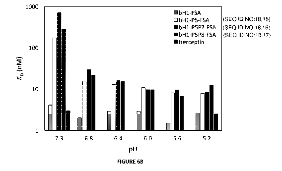
.. FIGURE 6A and 6B. pH dependence of FSA variants binding to cells expressing
Her2. (FIG 6A)
High-density Her2 cells (SKOV3) were tested in environments with varying pH
between 5.2 and
7.3 and cell binding was analyzed by fluorescence-activated cell sorting
(FACS) at varying
concentrations of the FSA variants. (FIG 6B) Apparent dissociation constants
(KD) of tested FSA
variants from binding experiments to high-density Her2 cells (SKOV3) at
various pHs.
FIGURE 7. Structural details of best single-point histidine mutants are
provided in panels (A),
(B) and (C) for mutants P5, P7 and P8 respectively. Antibody chains of the
parental bH1 antibody
are colored in dark gray, and the antigen is rendered in light gray. At each
mutated position, the
parental side chain and its histidine side chain substitutions in the acidic
and physiological pH
conditions are overlaid and rendered as dark-gray sticks model. Arrows
indicate histidine
mutation in the acidic and neutral states. The main interacting side chains of
the antigen are also
shown in dark-gray sticks models and labeled by residues numbers.
FIGURE 8. pH dependence of designed FSA variants binding to cells expressing
Her2 at low
density. Low-density Her2 expressing JIMT-1 cells were tested under acidic and
physiological
pH conditions and cell binding was analyzed by flow cytometry. The left panel
representing
binding to the normal cell model (low-density Her2 and physiological pH) is to
be compared with
binding of the same variants to the tumor cell model consisting of high-
density Her2 (SKOV3
cells) within a pH range of 6.0-6.8, as presented in Figure 6A.
FIGURE 9. Amino acid sequences of the Fv domains of the parental bH1 antibody
[37]. CDR
loops are shaded and labeled according to the Kabat definition, except for the
CDR-H1 which is
defined by the union of Kabat and Chothia definitions (the choice for the
combined definition in
the case of CDR-H1 was motivated by the fact that in this way a larger
sequence region is
subjected to histidine scanning mutagenesis so as to lead to more potentially
interesting hits).
Sequence numbering is according to Chothia scheme. Positions designed for
single-point
CA 03135987 2021-10-02
WO 2020/201992
PCT/IB2020/053024
histidine mutagenesis are indicated by arrows and labeled according to the
produced variants
(see Table 1). Positions used for double-point histidine mutants are labeled
in bold.
FIGURE 10. Viability test of SKOV3 and JIMT1 cells at pH 5 and pH 7. Cells
were incubated
during 3 h at 4 C in RPMI-1640 2% FBS 50 mM BES buffer adjusted to pH 7 or pH
5 (similar
conditions to the complete binding assay). Cells viability was evaluated by
flow cytometry using
P11% staining.
FIGURE 11. The effect of Herceptin, bH1-FSA and its pH selective antibody
variant bH1-P5P8-
FSA on BT474 spheroid growth under normal and low pH conditions. A. Dose-
response effect
of antibodies at concentrations ranging from 100 ig/m1 to 0.4 ig/m1 was
tested. Changes in
spheroid size are reported as size normalized to isotype control human IgG and
time zero for
each concentration after 8 days of treatment in corresponding conditions at pH
7.4, pH 6.4 and
pH 6.4-AA (acidosis adapted BT474 cells). B. Time course of change in spheroid
size in
response to 100 ig/m1 of each antibody. Data is reported as normalized to
control and time zero
over 200 h at physiological pH of 7.4; or acute exposure to acidic pH of 6.4
(panel B'); or cells
adapted to acidic pH of 6.4 (panel B").
FIGURE 12. Comparison of internalization capacity of Herceptin, bH1-FSA and
its pH selective
antibody variant bH1-P5P8-FSA in normal and low pH conditions. The
internalization of FITC
conjugated antibodies into BT474 cells was quantified under physiological pH
of 7.4 or acidic pH
of 6.4 conditions. A. Representative images for each pH condition. B.
Quantified fluorescent
intensity per cell area. Horizontal dashed lines indicate the level of
internalization of the negative
control antibody at each pH.
FIGURE 13. ADC efficacy of DM1-conjugated antibody variants in BT474 cells at
pH 7.4 versus
pH 6.4.
DETAILED DESCRIPTION OF THE INVENTION
The present invention relates to anti-Her2 antibodies and uses thereof. More
specifically, the
present invention relates to anti-Her2 antibodies and fragments thereof with
preferential binding
to cancer cells in slightly acidic environment relative to normal cells at
physiological pH
environment.
The present invention provides engineered recombinant full-size antibodies
(FSAs) and
fragments thereof capable of binding selectively to Her2 expressing cells at
slightly acidic pH in
the 5.0-6.8 range relative to the physiological pH of 7.2-7.4. Without wishing
to be bound by
theory, the environment surrounding solid tumors is slightly acidic relative
to normal cells.
11
CA 03135987 2021-10-02
WO 2020/201992
PCT/IB2020/053024
Achieving pH dependent binding selectivity towards acidic pH with reduced
binding at
physiological pH of normal cells represents a novel means of reducing off-
tumor host toxicities
of antibody-based therapeutics and widening their therapeutic windows.
The use of the terms "a" and "an" and "the" and similar referents in the
context of describing the
invention (especially in the context of the claims) are to be construed to
cover both the singular
and the plural, unless otherwise indicated herein or clearly contradicted by
context.
Unless specifically stated or obvious from context, as used herein the term
"or" is understood to
be inclusive and covers both "or" and "and". The term "and/or" where used
herein is to be taken
as specific disclosure of each of the specified features or components with or
without the other.
The terms "comprising", "having", "including", and "containing" are to be
construed as open-
ended terms (i.e., meaning "including, but not limited to") unless otherwise
noted. The term
"consisting of" is to be construed as close-ended. The term "consisting
essentially of" when used
in the context of CDR sequences means that the CDR sequence may be slightly
(e.g., +/- 1 or
2 aa) longer or shorter.
The term "antibody", also referred to in the art as "immunoglobulin" (Ig),
used herein refers to a
protein constructed from paired heavy and light polypeptide chains; various Ig
isotypes exist,
including IgA, IgD, IgE, IgG, and IgM. When an antibody is correctly folded,
each chain folds into
a number of distinct globular domains joined by more linear polypeptide
sequences. For
example, the immunoglobulin light chain folds into a variable (VL) and a
constant (CL) domain,
while the heavy chain folds into a variable (VH) and three constant (CHi , CH2
and CH3) domains.
Interaction of the heavy and light chain variable domains (VH and VL) results
in the formation of
an antigen binding region (Fv). Each domain has a well-established structure
familiar to those
of skill in the art. An antibody may be, but is not limited to, full length
antibodies, an antigen
binding fragment comprising a VL and VH domain, such as Fab, Fv, scFv, dsFv,
BiTE, or any
multimer of said antigen binding fragment.
The light and heavy chain variable regions are responsible for binding the
target antigen and
can therefore show significant sequence diversity between antibodies. The
constant regions
show less sequence diversity, and are responsible for binding a number of
natural proteins to
elicit important biochemical events. The variable region of an antibody
contains the antigen
binding determinants of the molecule, and thus determines the specificity of
an antibody for its
target antigen. The majority of sequence variability occurs in six
hypervariable regions, three
each per variable heavy (VH) and light (VL) chain; the hypervariable regions
combine to form the
antigen-binding site, and contribute to binding and recognition of an
antigenic determinant. The
12
CA 03135987 2021-10-02
WO 2020/201992
PCT/IB2020/053024
specificity and affinity of an antibody for its antigen is determined by the
structure of the
hypervariable regions, as well as their size, shape and chemistry of the
surface they present to
the antigen. Various schemes exist for identification of the regions of
hypervariability, the two
most common being those of Kabat and of Chothia. Kabat et al. define the
"complementarity-
determining regions" (CDR) based on sequence variability at the antigen-
binding regions of the
VH and VL domains [39]. Chothia and Lesk define the "hypervariable loops" (H
or L) based on
the location of the structural loop regions in the VH and VL domains [40]. As
these individual
schemes define CDR and hypervariable loop regions that are adjacent or
overlapping, those of
skill in the antibody art often utilize the terms "CDR" and "hypervariable
loop" interchangeably,
and they may be so used herein. For this reason, the regions forming the
antigen-binding site
are presently referred to herein as CDR-L1, CDR-L2, CDR-L3, CDR-H1, CDR-H2 and
CDR-H3,
and they follow the Kabat definition, except for CDR-H1 that is taken here as
the union of the
Kabat and Chothia definitions. The CDR/loops can also be referred to according
to the IMGT
numbering system [41], which was developed to facilitate comparison of
variable domains.
Additionally, standardized delimitations of the framework regions of the VH
and VL domains
outside of the CDR can be formulated according to each CDR definition scheme.
An "antibody fragment" as referred to herein may include any suitable antigen-
binding antibody
fragment known in the art. The antibody fragment may be a naturally-occurring
antibody
fragment, or may be obtained by manipulation of a naturally-occurring antibody
or by using
recombinant methods. For example, an antibody fragment may include, but is not
limited to a
Fv, a single-chain Fv (scFv; a molecule consisting of VL and VH connected with
a peptide linker),
a Fab, a single-chain Fab (scFab; a molecule consisting of VL and CH connected
with a peptide
linker, or of VH and CL connected with a peptide linker), F(a1:02, and
multivalent presentations of
any of these. For example, multiple Fv fragments can be linked, or multiple
Fab fragments can
be linked, via the heavy chain, or light chain, or both. Non-limiting examples
of antibody
fragments and possible multivalent assemblies are known in the art [42-44]. As
used herein the
term "antigen-binding domain" or "antigen-binding fragment" refers to the
domain of an antibody
or of an antigen-binding fragment which allows binding to an antigen.
Thus, present invention provides an antibody or fragment thereof comprising a
sequence of:
CDR-H1 of GFNIKDTYIH (SEQ ID NO:1), CDR-H2 of RIYPTNGYTHYADSVKG (SEQ ID NO:2),
CDR-H3 of WGGDGFYAMDY (SEQ ID NO: 3), CDR-L1 of RASQDIPX1X2ISGYVA (SEQ ID
NO:4), CDR-L2 of WGSYLYS (SEQ ID NO:5) and CDR-L3 of QQHYTTPPT (SEQ ID NO:6);
wherein: Xi is S or H, and X2 is R or H (SEQ ID NO:7, SEQ ID NO:8, SEQ ID
NO:9).
As would be understood by those of skill in the art, not all CDR may be
required for binding the
antigen. For example, and without wishing to be limiting, one, two, three,
four, five or all six CDR
13
CA 03135987 2021-10-02
WO 2020/201992
PCT/IB2020/053024
loops may contribute to binding and recognition of the antigen by the sdAb of
the present
invention.
In a non-limiting example, the antibody or fragment is recombinantly produced
and includes
modifications engineered by site-directed mutagenesis and affinity modulation
of a parental
antibody. Encompassed by the present invention are any homologues,
derivatives, or fragments
of the antibodies and fragments thereof disclosed here that retain the pH-
dependent binding
selectivity to Her2 of the antibodies and fragments thereof disclosed here.
As previously stated, the antibody or fragment may be recombinantly produced
and thus may
be based on chosen framework regions; alternatively, the CDR described above
may be grafted
onto other VH or VL framework regions. In yet another alternative, the
hypervariable loops
described above may be grafted onto the framework regions of other types of
antibody fragments
(e.g., Fv, scFv, Fab, scFab, and their combination and multivalent formats).
The present
embodiment further encompasses an antibody or an antibody fragment whereby the
CDR is
grafted into framework regions chosen from various species.
In a specific, non-limiting example, the antibody or fragment thereof that is
selective for Her2
binding at acidic pH comprise a sequence selected from the group consisting of
a heavy-chain
variable sequence of SEQ ID NO:10; and the light-chain variable sequence
selected from the
group consisting of SEQ ID NO:11, SEQ ID NO:12 and SEQ ID NO:13; or a sequence
substantially identical thereto.
In certain embodiments, the antibodies or fragments thereof comprise a
constant region of
human origin. Hence, in specific non-limiting examples, antibodies or
fragments thereof are
selected from the group consisting of SEQ ID NO:14, SEQ ID NO:15, SEQ ID
NO:16, SEQ ID
NO:17 and SEQ ID NO:18; or a sequence substantially identical thereto.
A "substantially identical" sequence may comprise one or more conservative
amino acid
mutations. It is known in the art that one or more conservative amino acid
mutations to a
reference sequence may yield a mutant peptide with no substantial change in
physiological,
chemical, physico-chemical or functional properties compared to the reference
sequence; in
such a case, the reference and mutant sequences would be considered
"substantially identical"
polypeptides. Conservative amino acid mutation may include addition, deletion,
or substitution
of an amino acid; a conservative amino acid substitution is defined herein as
the substitution of
an amino acid residue for another amino acid residue with similar chemical
properties (e.g. size,
charge, or polarity).
14
CA 03135987 2021-10-02
WO 2020/201992
PCT/IB2020/053024
In a non-limiting example, a conservative mutation may be an amino acid
substitution. Such a
conservative amino acid substitution may substitute a basic, neutral,
hydrophobic, or acidic
amino acid for another of the same group. By the term "basic amino acid" it is
meant hydrophilic
amino acids having a side chain pKa value of greater than 7, which are
typically positively
charged at physiological pH. Basic amino acids include arginine (Arg or R) and
lysine (Lys or K).
By the term "neutral amino acid" (also "polar amino acid"), it is meant
hydrophilic amino acids
having a side chain that is uncharged at physiological pH, but which has at
least one bond in
which the pair of electrons shared in common by two atoms is held more closely
by one of the
atoms. Polar amino acids include serine (Ser or S), threonine (Thr or T),
cysteine (Cys or C),
tyrosine (Tyr or Y), asparagine (Asn or N), and glutamine (Gin or Q). The term
"hydrophobic
amino acid" (also "non-polar amino acid") is meant to include amino acids
exhibiting a
hydrophobicity of greater than zero according to the normalized consensus
hydrophobicity scale
of [45]. Hydrophobic amino acids include proline (Pro or P), isoleucine (Ile
or l), phenylalanine
(Phe or F), valine (Val or V), leucine (Leu or L), tryptophan (Trp or W),
methionine (Met or M),
alanine (Ala or A), and glycine (Gly or G). "Acidic amino acid" refers to
hydrophilic amino acids
having a side chain pKa value of less than 7, which are typically negatively
charged at
physiological pH. Acidic amino acids include glutamate (Glu or E), and
aspartate (Asp or D).
Histidine (His or H) is a polar amino acid with a special ionization potential
due to its pKa around
7, and more precisely around 6.4 in case of histidine residues located at the
protein surface [15].
This results in histidine amino acid residues being a "polar" and
predominantly uncharged at
physiological pH of 7.2-7.4, and predominantly positively charged in acidic
environments (pH <
7).
Sequence identity is used to evaluate the similarity of two sequences; it is
determined by
calculating the percent of residues that are the same when the two sequences
are aligned for
maximum correspondence between residue positions. Any known method may be used
to
calculate sequence identity; for example, computer software is available to
calculate sequence
identity. Without wishing to be limiting, sequence identity can be calculated
by software such as
NCB! BLAST2 service maintained by the Swiss Institute of Bioinformatics (and
as found at
http://ca.expasy.org/tools/blast/), BLAST-P, Blast-N, or FASTA-N, or any other
appropriate
software that is known in the art.
The substantially identical sequences of the present invention may be at least
65% identical; in
another example, the substantially identical sequences may be at least 65, 70,
85, 90, 95, 96,
97, 98, 99, or 100% identical, or any percentage there between, at the amino
acid level to
sequences described herein. Importantly, the substantially identical sequences
retain the activity
and specificity of the reference sequence. In a non-limiting embodiment, the
difference in
CA 03135987 2021-10-02
WO 2020/201992
PCT/IB2020/053024
sequence identity may be due to conservative amino acid mutation(s). By way of
example only,
and without wishing to be limiting in any manner, the bH1 Fab fragment used as
parental
antibody in this invention and the Fab fragment of the anti-Her2 antibody
Herceptin share the
same heavy chain (100% identity) and have a light chain variable domains (W)
that differ at 11
positions, which is equivalent to 95.3% sequence identity in the combined
variable domains. By
way of other examples, and without wishing to be limiting in any manner,
affinity modulated and
affinity matured Herceptin variants have been described which differ only by 1
to 3 amino-acids,
equivalent to 98.9-99.6% sequence identity, in the combined variable domains
[35, 46]
[W02012075581], as well as affinity modulated and affinity matured bH1
variants that differ by
1 to 12 amino acids, equivalent to 94.2-99.6% sequence identity, in the
combined variable
domains [35, 37, 47]. Hence, the present invention also encompasses histidine
mutated variants
of Herceptin, as well as of aforementioned Herceptin and bH1 affinity
modulated and affinity
matured variants, which contain the same mutation(s) to histidine as the
one(s) exemplified here
for the parental bH1 sequence which lead(s) to pH-dependent binding
selectivity towards the
slightly acidic pH relative to physiological pH.
The antibody or fragment thereof of the present invention may also comprise
additional
sequences to aid in expression, detection or purification of a recombinant
antibody or fragment
thereof. Any such sequences or tags known to those of skill in the art may be
used. For example,
and without wishing to be limiting, the antibody or fragment thereof may
comprise a targeting or
signal sequence (for example, but not limited to ompA), a
detection/purification tag (for example,
but not limited to c-Myc or a His5 or His6), a linker sequence (wherein one of
skill in the present
art could use any suitable linker to allow for the operably biological
function and association of
the antibody domains that form the antigen-binding region; for example, a
linker may be
(GGGS)n, any multiple thereof, or any suitable linker in the art), or a
combination thereof. In
another example, the additional sequence may be a biotin recognition site such
as that described
in W01995004069 or W02004076670. As is also known to those of skill in the
art, linker
sequences may be used in conjunction with the additional sequences or tags, or
may serve as
a detection/purification tag.
In yet other embodiments, the antibodies or fragments thereof are protein
fusions, for example,
in a CAR (chimeric antigen receptor) format displayed on cell surface, or in a
multivalent display
format, or in a multispecific display format.
By way of example only, and without wishing to be limiting in any manner, the
antibodies and
fragments thereof of the present invention can be reformatting as single-chain
variable domains
(scFv) for generation of chimeric antigen receptors (CARs) [48]. To this end,
the pH-dependent
anti-Her2 antibodies and fragments thereof of the present invention have to be
formatted as
16
CA 03135987 2021-10-02
WO 2020/201992
PCT/IB2020/053024
scFv and then fused to a spacer poypeptide, a transmembrane domain (e.g., from
0D28) and
an endodomain (e.g., CD3-zeta) capable of transmitting an activation signal to
the T-cell after
the engagement of the Her2 antigen. The 2D-tethering of CARs on the T-cell
membrane may
also complement their pH-dependent binding with avidity selectivity towards
tumor cells.
The antibody or fragment thereof of the present invention may also be in a
multivalent display
format, also referred to herein as multivalent presentation. Multimerization
may be achieved by
any suitable method of known in the art. Multimerization may result in
homomeric or heteromeric
constructs. Homo-multimers can be used for introducing avidity effects, which
can improve
efficacy and reduce toxicity. For example, and without wishing to be limiting
in any manner,
homo-multimerization may be achieved using self-assembly molecules as
described in [49] and
W02003046560. The described method produces pentabodies by expressing a fusion
protein
comprising the antibody or fragment thereof of the present invention and the
pentamerization
domain of the B-subunit of an AB5 toxin family [50]; the pentamerization
domain assembles into
a pentamer, through which a multivalent display of the antibody or fragment
thereof is formed.
Each subunit of the pentamer may be the same or different, and may have the
same or different
specificity. Additionally, the pentamerization domain may be linked to the
antibody or antibody
fragment using a linker; such a linker should be of sufficient length and
appropriate composition
to provide flexible attachment of the two molecules, but should not hamper the
antigen-binding
properties of the antibody. Hetero-multimers can be used for generated multi-
specific molecules.
Bispecific and multispecific fusion proteins consisting of antibodies and
antibody fragments are
well known in the art. For example, and without wishing to be limiting, the
antibody or fragment
thereof may be presented as a dimer, a trimer, or any other suitable oligomer.
This may be
achieved by methods known in the art, for example direct linking connection, c-
jun/Fos
interaction [51], "knob into holes" interaction [52], and many other formats
known in the art, some
of which may employ carefully designed Fc domains [42]. Furthermore, enhanced
in vivo efficacy
of fragments lacking an Fc domain may be obtained using various techniques,
including
PEGylation, fusion to serum albumin, or fusion to serum albumin-specific
antibody fragments;
these approaches increase their blood circulation half-lives, size and
avidity.
The antibodies and antibody fragments of the present invention may be produced
recombinantly.
The present invention further encompasses nucleic acid molecules encoding the
antibodies or
fragments thereof as described above. The nucleic acid sequence may be codon-
optimized for
expression in various micro-organisms. The present invention also includes
vectors comprising
the nucleic acid molecules just described. Furthermore, the invention
encompasses cells
comprising the nucleic acid and/or vector as described.
17
CA 03135987 2021-10-02
WO 2020/201992
PCT/IB2020/053024
The present invention further encompasses the antibodies or fragments thereof
immobilized
onto a surface using various methodologies. For example, and without wishing
to be limiting, the
antibody or fragment may be linked or coupled to the surface via His-tag
coupling, biotin binding,
covalent binding, adsorption, and the like. Immobilization of the antibody or
fragment thereof of
the present invention may be useful in various applications for capturing,
purifying or isolating
proteins. The solid surface may be any suitable surface, for example, but not
limited to the well
surface of a microtiter plate, channels of surface plasmon resonance (SPR)
sensorchips,
membranes, beads (such as magnetic-based or sepharose-based beads or other
chromatography resin), glass, plastic, stainless steel, a film, or any other
useful surface such as
nanoparticles, nanowires and cantilever surfaces.
The present invention further provides a method of capturing the Her2
ectodomain, comprising
contacting a sample with one or more than one surface-immobilized antibodies
or fragments
thereof of the present invention, and allowing the Her2 ectodomain to bind to
the antibodies or
fragments thereof in slightly acidic pH conditions, e.g., pH between 5.0 and
6.8, and releasing
the Her2 ectodomain from the antibody or fragment thereof by raising the pH to
7.2-7.4. Thus,
the antibodies or fragments thereof of the present invention may provide
useful affinity
purification reagents for preparations of Her2 ectodomain samples.
The antibodies or fragments thereof of the present invention may be linked to
a cargo molecule.
The cargo molecule may be any suitable molecule. For example, and without
wishing to be
limiting in any manner, the cargo molecule may be a detectable agent, a
therapeutic agent, a
drug, a peptide, an enzyme, a protease, a carbohydrate moiety, a cytotoxic
agent, one or more
liposomes loaded with any of the previously recited types of cargo molecules,
or one or more
nanoparticle, nanowire, nanotube, or quantum dots. The cargo molecule may be
linked to the
antibody or fragment thereof by any suitable method known in the art. For
example, and without
wishing to be limiting, the cargo molecule may be linked to the peptide by a
covalent bond or
ionic interaction. The linkage may be achieved through a chemical cross-
linking reaction, or
through fusion using recombinant DNA methodology combined with any peptide
expression
system, such as bacteria, yeast or mammalian cell-based systems. Methods for
linking an
antibody or fragment thereof to a therapeutic agent or detectable agent would
be well-known to
a person of skill in the art.
The present invention also provides a method of detecting solid tumors,
comprising
administering the antibodies or fragments thereof of the present invention or
the composition
described above, and detecting the bound antibody or fragment thereof using a
suitable
detection and/or imaging technology. For example, the antibodies or fragments
thereof of the
present invention may be linked to a radioisotope, a paramagnetic label, a
fluorophore, an affinity
18
CA 03135987 2021-10-02
WO 2020/201992
PCT/IB2020/053024
label (for example biotin, avidin, etc), fused to a detectable protein-based
molecule, nucleotide,
quantum dot, nanoparticle, nanowire, or nanotube or any other suitable agent
that may be
detected by imaging methods. In a specific, non-limiting example, the antibody
or fragment
thereof may be linked to a fluorescent agent such as FITC or may genetically
be fused to the
Enhanced Green Fluorescent Protein (EGFP). The antibody or fragment thereof
may be linked
to the detectable agent using any method known in the art (recombinant
technology, chemical
conjugation, etc.).
Thus, the present invention further provides a method of detecting Her2 on
solid tumors at
slightly acidic pH, comprising contacting a sample (such as, but not limited
to biopsy sample, or
any other suitable sample) with one or more than one antibody or fragment
thereof of the present
invention. The antibodies or fragments thereof may be linked to a detectable
agent. The Her2
antigen can then be detected using detection and/or imaging technologies known
in the art, such
as, but not limited to mass spectrometric or immunoassay methods. For example,
and without
wishing to be limiting in any manner, the antibodies or fragments thereof
linked to a detectable
agent may be used in immunoassays (IA) including, but not limited to enzyme IA
(EIA), ELISA,
"rapid antigen capture", "rapid chromatographic IA", and "rapid EIA".
The present invention also encompasses a composition comprising one or more
than one
antibody or fragment thereof as described herein. The composition may comprise
a single
antibody or fragment as described above, or may be a mixture of antibodies or
fragments. The
composition may also comprise a pharmaceutically acceptable diluent,
excipient, or carrier. The
diluent, excipient, or carrier may be any suitable diluent, excipient, or
carrier known in the art,
and must be compatible with other ingredients in the composition, with the
method of delivery of
the composition, and is not deleterious to the recipient of the composition.
The composition may
be in any suitable form; for example, the composition may be provided in
suspension form,
powder form (for example, but limited to lyophilised or encapsulated), capsule
or tablet form.
For example, and without wishing to be limiting, when the composition is
provided in suspension
form, the carrier may comprise water, saline, a suitable buffer, or additives
to improve solubility
and/or stability; reconstitution to produce the suspension is effected in a
buffer at a suitable pH
to ensure the viability of the antibody or fragment thereof. Dry powders may
also include
additives to improve stability and/or carriers to increase bulk/volume; for
example, and without
wishing to be limiting, the dry powder composition may comprise sucrose or
trehalose. The
composition may comprise encapsulation, time-release, or other suitable
technologies for
delivery of the antibody or fragment thereof. It would be within the
competency of a person of
skill in the art to prepare suitable compositions comprising the present
compounds.
19
CA 03135987 2021-10-02
WO 2020/201992
PCT/IB2020/053024
The present invention further provides a method of selectively targeting Her2-
expressing cancer
cells and tumor tissues, and treating solid tumors with minimal unwanted off -
tumor host toxicity
and a wide therapeutic window, comprising administering the antibodies or
fragments thereof of
the present invention or the composition described above to a subject in need
thereof. Any
suitable method of delivery may be used. For example, and without wishing to
be limiting in any
manner, the antibody or fragment thereof, or the composition, may be delivered
systemically
(orally, nasally, intravenously, etc.). Those of skill in the art would be
familiar with such methods
of delivery.
The present invention will be further illustrated in the following examples.
However, it is to be
understood that these examples are for illustrative purposes only and should
not be used to limit
the scope of the present invention in any manner.
Example 1: Computational design of pH dependence
The Her2-bound crystal structure of bH1-Fab was retrieved from the Protein
Data Bank (entry
313E1) [37] and structurally prepared for molecular design as described
previously at
physiological pH [35]. At slightly acidic pH of 5-6, visual examination was
used in order to decide
whether a His side chain can be protonated based on its structural environment
protonation.
This led to protonation of three His residues in the antigen epitope: H497,
H537 and H567,
whereas the other two His residues in the antigen epitope (H490 and H542) as
well as the two
His residues of the Fv fragment (L-H95 and H-H35) were treated in the neutral
state in the slightly
acidic conditions.
Computational His scanning was carried out with the ADAPT protocol [35] in
order to generate
and score single-point mutations to histidine of non-proline, non-cysteine and
non-histidine
residues in the CDR region covering 3 loops in the light chain: CDR-L1 (R24-
A34), CDR-L2
(W50-S56), CDR-L3 (089-T97), and 3 loops in the heavy chain: CDR-H1 (G26-H35),
CDR-H2
(R50-G65) and CDR-H3 (R94-Y104) of bH1-Fv (Figure 9). Histidine mutations were
built and
evaluated energetically at these positions using the three programs, SCWRL [53-
55], FoldX [56,
57] and Rosetta [58, 59] currently implemented in ADAPT [35]. From an
operational perspective,
since scoring functions for computational mutagenesis provide meaningful
results when taken
as scores relative to the parent system for which the crystal structure has
been determined, the
implementation of these methods in ADAPT adopts a conservative approach that
limits
conformational flexibility of the mutants as much as possible and especially
at the level of the
protein backbone atoms [58, 60].
CA 03135987 2021-10-02
WO 2020/201992
PCT/IB2020/053024
Histidine mutations were generated at physiological pH and also in slightly
acidic conditions
(e.g., pH 6). For mutations at physiological pH, all programs were forced to
mutate to a neutral
histidine. For mutations at slightly acidic pH, each program had a different
implementation of a
protonated histidine. Rosetta had the simplest method in that mutations to
histidine were forced
to use the protonated form. In the case of FoldX, protonated histidine takes
two different forms,
protonated delta (called "0") and protonated epsilon (called "e"). Therefore,
the mutation with the
lowest FoldX stability score was retained and used for ranking. Lastly for
SCWRL, the option to
enable the protonated form to compete with neutral histidine tautomers was
chosen. While this
does not force mutation to the protonated form, if the resulting mutation is
neutral it is assumed
that the protonated form would be destabilizing, and vice-versa. Binding
scores of the
constructed His mutants relative to the parent were then calculated at each of
the two pHs,
A Givicant _ A Gxcarnct
di and AGA
uytsainoto
gical AGgyresinotlogical, with the three scoring functions in ADAPT:
SIE [54, 55], FoldX-FOLDEF [56] and Rosetta-Interface [58]. The double-
referenced binding free
energy scores, AAAGbinding (Figure 1), were then derived as difference in the
scores obtained in
acidic conditions relative to physiological pH conditions. A consensus ranking
was finally derived
from these individual AAAGbinding scores calculated with each scoring
functions. The FoldX-
FOLDEF energy function [56] was also used to estimate the effect of His
substitutions on the
internal stability of the Fv structure at each of the two pHs. Additional
technical and
implementation details of ADAPT and its component methods can be found in
earlier reports
[35, 38, 60].
The concept of free energy optimization of a parent antibody-antigen system
via mutagenesis
for improved binding at acidic pH (tumor microenvironment) relative to
physiological pH (normal
cells) is presented schematically in Figure 1. binding free energy gap,
AAAGbinding =
(AGrAvigitnt _ AGiyhuytsanotog (AGFA,cairdeicnt _ AGRyrseinotiogical\,
I ical/
) between the mutant and parent variants
in the acidic relative to physiological environments. This binding free energy
gap must be as
negative as possible in order to enhance the selectivity for binding at acidic
versus physiological
pH. At the same time, the mutant must maintain a reasonable level of binding
in the acidic
environment relative to the parent molecule. For example, a mutant that binds
1000-fold better
at acidic versus physiological pH, but has a 100 [tM affinity under acidic
conditions, has high pH
selectivity but is of limited practical use. Hence, a filter was applied to
step (3) in Figure 1 to
ensure that (AG)zicuitnt _ AGIA3caircfinctµ
) is not too overly positive (e.g., not more than 2.7 kcal/mol, or
100-fold increase in KD). Moreover, since the designed mutants must be stable
in the intended
acidic environment, another filter was applied to the folding free energy
associated with step (3).
This ensures that the stability of the mutant is comparable with that of the
parent, and hence the
predicted AAGfolding = AGfmoitcJitianngt _ AGfPoririenngt should typically be
less than 2.7 kcal/mol [35]. For
21
CA 03135987 2021-10-02
WO 2020/201992
PCT/IB2020/053024
manufacturing and in vivo delivery reasons, stability at physiological pH
should also be
maintained, and so AAGfdding at that pH associated with step (4) in Figure 1
should not be overly
positive either.
We have implemented this version of ADAPT [35, 38] capable of handling dual-pH
His-scanning
mutagenesis, and applied it to the bH1 Fab in complex with Her2 ectodomain
[37]
(W02008027236; W02010027981; W02010108127; W02015095539). A total of 68
positions
(non-His, non-Pro, non-Cys) forming the CDR loops of bH1 (Figure 9) were
screened for single
mutations to His with 3 protocols for mutant generation and relative binding
affinity scoring, and
with one protocol for relative stability scoring (Table 1). 21 mutants were
excluded based on
stability criteria on acidic conditions (>2.72 kcal/mol from parent). From the
remaining 47
mutations, the top 10 in average ranks in terms of AAAGb,nd,ng binding
affinity gap were retained.
Ranks 1 (L-R30a), 2 (H-R58), 6 (H-N28), 8 (H-Y100a) and 10 (H-Y56) were
pursued further. The
other 5 mutations were excluded after visual examination of the modeled 3D
structures, namely
rank 3 (H-G97) due to steric clashes, rank 4 (L-Y92) due to packing of
protonated His in a
hydrophobic environment, and ranks 5 (H-N54), 7 (L-S30d) and 9 (H-K64) for
which the
introduced His residues made no direct contacts with the antigen. Rank 26 (L-
S30b) was
included based on its second-best stability in acidic conditions. Rank 30 (H-
Y33) was also
included as another test of a non-highly-ranked mutant. These two mutants from
the bottom half
of the ranked list passed visual inspection in terms of good steric and
electrostatic interactions
in the complex. Lastly, H-R50 was included as another control, having the best
average rank for
binding affinity change if the stability filter under acidic conditions would
not be applied.
These selected residues for single-point mutations to histidine are spread out
over four of the
six CDR loops (Figure 9). Having widely separated mutations at the antibody-
antigen interface
(Figure 2) can be beneficial for combining them into higher-order mutants
using the ADAPT
protocol. This is based on reasonable additivity of contributions from
spatially separated
mutations to binding affinity [35, 38].
Example 2: Protein expression and purification
cDNA for the heavy and light chains of Fab and full-size human IgG1/k antibody
variants were
ordered from commercial vendors (ThermoFisher Scientific/Life Technologies
Inc., Burlington,
ON; GENEART, Regensburg, Germany). These contained signal peptide sequences,
but no
His-tags. Productions were carried out by co-transfection of CH0-3E7 cells as
described
previously [35] at 200-mL scale. Transfections were performed at a cell
density between 1.8
x 106 to 2.0 x 10⁶ cells/mL with viability greater than 98%. Cells were
distributed in 1.0 L to 2.8 L-
shaker flasks and transfected with 1 lig of total DNA per 1 mL of production
(50% of total DNA
22
CA 03135987 2021-10-02
WO 2020/201992
PCT/IB2020/053024
contained heavy chain and light chain constructs at ratios of 1:1 (w/w)) using
PEI MAXTM
(Polysciences, Inc., Warrington, PA). The final DNA : PEI MAXTM ratio was 1 :
4 (w/w). Cell
cultures were incubated for 24 h on an orbital shaking platform at an
agitation rate of 110 rpm at
37 C in a humidified 5% CO2 atmosphere. Twenty-four hours later, the cultures
were fed with
Tryptone Ni at 1% w/v final and Valproic acid sodium salt at 0.5 mM final
concentration and
transferred to 32 C for 6 days. Cell density and cell viability were
determined by direct counting
of cell samples with a Vi-CELL automated cell counting system (Beckman Coulter
Life Sciences,
Indianapolis, IN) using the trypan blue dye exclusion method.
Purifications from cell-culture supernatants were performed by protein-A
affinity chromatography
for Fab fragments and the IgG1/k full-size antibodies (FSAs). Purifications of
cell-culture
supernatants were performed by loading the Fabs onto 5-mL MabSelect or 1-mL
HiTrap
MabSelect columns (GE Healthcare Life Sciences, Mississauga, ON) and FSAs onto
MabSelect
SuRe columns (GE Healthcare) equilibrated in HyCloneTM Dulbecco's phosphate-
buffered saline
(DPBS; GE Healthcare Life Sciences). Columns were washed with PBS and Fabs or
FSAs were
eluted with 100 mM citrate buffer pH 3.0, respectively. Fractions containing
Fabs or FSAs were
pooled and the citrate buffer was exchanged against DPBS on CentriPure P100
columns (EMP
Biotech, Howell, NJ) or ZebaSpin TK MWCO columns (ThermoFisher Scientific,
Waltham, MA).
Purified Fabs and FSAs were sterilized by filtration through 0.2 pm filters.
UPLC-SEC was used
to assess the purity of all eluates. Variants with less than 95% purity (Fab
fragments bH1 -P4,
bH1-P6, bH1-P5P7 and bH1-P5P8) were further purified by preparative size
exclusion
chromatography (SEC) on Superdex-200 increase columns (GE Healthcare Life
Sciences).
Selected peak fractions were concentrated by ultrafiltration using Vivaspin
Turbo 4 or 15
(depending on the volume to concentrate) centrifugal concentrator with a
membrane molecular
weight cut off of 10 kDa (GE Healthcare Life Sciences) at room temperature
following the
manufacturer's instructions. During the process, the protein concentration was
monitored on a
NanoDropTM 2000 spectrophotometer (ThermoFisher Scientific) using absorbance
at 280 nm
and the calculated specific extinction coefficient of each variant.
Example 3: Surface plasmon resonance (SPR) binding affinity measurements of pH-
dependent
binding to recombinant Her2 ectodomain
Fab-Her2 interactions were analyzed using a BiacoreTM T200 (GE Healthcare,
Missisauga, ON)
surface plasmon resonance instrument. Samples were assayed at 25 C or 37 C
using PBS
containing 0.05% Tween 20 (Teknova, Hollister, CA) with added 3.4 mM EDTA as
running buffer
or 150 mM citrate-phosphate buffer pH 5 with added 3.4 mM EDTA, 135 mM NaCI
and 0.05%
Tween 20. Recombinant human Her2 extracellular domain (ThermoFisher
Scientific, Burlington,
ON) was immobilized onto a CM-5 sensorchip along with a mock-activated blank
control surface
23
CA 03135987 2021-10-02
WO 2020/201992
PCT/IB2020/053024
for referencing. Her2 was diluted to 10 [tg/mL in 10 mM Na0Ac immobilization
buffer pH 4.5
(Biacore) and immobilized to approximately 200 RUs using the Biacore control
software
Immobilization Wizard with standard NHS/EDC amine coupling. The Her2
interaction was
determined using single cycle kinetics analysis for each variant with five
concentrations using 3-
fold dilutions from the top concentration between 100 to 900 nM depending on
the affinity of the
variant. Fab samples were injected at 100 4/min with a contact time of 90 s
and a 1200-s
dissociation using either pH 5.0 or 7.4 running buffer. Sensorgrams were
double referenced to
the mock-activated blank sensor surface and analyzed for kinetic determination
using a 1:1
binding model in BiaEvaluation software v3.1 (GE Healthcare).
The parental and selected bH1-Fab variants were first tested for binding to
recombinant Her2
ectodomain by SPR at the physiological pH of 7.4 as well as at pH 5.0, whereby
the Fab samples
were flowed over the antigen ectodomain immobilized at a sensor-chip surface.
Interestingly,
parental bH1-Fab bound to the Her2 ectodomain about 4 times weaker at acidic
pH (KD = 13
4 nM) versus physiological pH (KD = 3 1 nM) due to a combination of a slower
on-rate and a
faster off-rate (Table 2). In contrast, our rank-2 computational structure-
based designed variant
bH1-P5 (SEQ ID NOS:14,15) having the H-R58 amino-acid residue mutated to
histidine showed
improved antigen binding at acidic pH (KD = 98 30 nM) versus physiological
pH (KD = 310 8
nM). In free energy terms, reversal by the new mutant of the undesired binding
phenotype of the
parental Fab to the desired pH dependence resulted from a negative value for
AAAGbinding of ¨
1.54 kcal/mol. This indicates a successful design in the desired direction for
pH dependence,
and is clearly apparent in the iso-affinity plot in Figure 3. Here, we see the
opposite directions
of binding free energy change by shifting the pH from physiological to acidic,
and comparing the
parental bH1-Fab to the bH1-P5 and other variants.
By the AAAGbinding objective metric, other successful designs were bH1-P7 (L-
R30aH) and bH1-
P8 (L-S30bH), although in these cases the undesired binding phenotype of the
parent was not
reversed but merely weakened. These are also apparent in the iso-affinity plot
(Figure 3).
Variant bH1-P1 (H-N28H), ranked 6 computationally, was similar to the parent,
whereas for bH1-
P6 (H-Y100aH, ranked 8) had complex binding and poor fit at acidic pH. For
variant bH1-P2 (H-
Y33H, ranked 30) the parental phenotype was actually accentuated and hence
this variant had
a positive value for AAAGbinding (Table 2). Finally, binding was so weak that
it could not be
detected at one or both pHs for variants bH1-P4 (H-Y56H, ranked 10) and bH1-P3
(H-R5OH,
control for testing a case with high destabilization predicted at acidic pH).
As this SPR binding
analysis was carried out at 25 C, we repeated experiments at 37 C and obtained
similar trends,
with almost identical equilibrium constants and slightly higher rate constants
as expected from
Arrhenius equation.
24
CA 03135987 2021-10-02
WO 2020/201992
PCT/IB2020/053024
Based on the SPR data for single mutants, three designed bH1 variants, bH1-P5,
bH1-P7 and
bH1-P8, were selected for generation of double mutants bH1-P5P7 (H-R58H,L-
R30aH) (SEQ
ID NOS:14,16) and bH1-P5P8 (H-R58H,L-S30bH) (SEQ ID NOS:14,17). These double
mutants
combine well-spaced mutations present on distinct chains, aiming at
introducing additivity of
mutation effects. The SPR-based AAAGbinding values listed in Table 2 confirm
that additivity has
been achieved. Both double mutants had approximately 6-fold stronger antigen
binding at acidic
pH than at physiological pH. This behavior was significantly driven by faster
dissociations (larger
koff) at physiological pH than at acidic pH, in sharp contrast to the parent,
as also apparent in the
iso-affinity plot (Figure 3). By comparing the variant bH1-P5P8 (SEQ ID
NOS:14, 17) with the
parental bH1-Fab, we see that antigen binding in the acidic environment has
only marginally
weakened (KD of 50 20 nM versus 13 4 nM, respectively) whereas binding
affinity in the
physiological pH environment has been weakened by 2 orders of magnitude (KD of
290 50 nM
versus 3 1 nM, respectively). This is also readily seen by visual inspection
of the corresponding
SPR sensorgrams shown in Figure 4.
Example 4: Flow cytometry measurements for pH-dependent binding to cells
expressing intact
Her2
SKOV3 and JIMT-1 cells (ATCC) were cultured in McCoy's 5A and DMEM media,
respectively,
supplemented with 10% fetal bovine serum (FBS).
Cells in T-75 flasks were washed twice with D-PBS and then dissociated using
Cell Dissociation
buffer (Sigma, C5914) at 372C. The cells were centrifuged and resuspended in
the appropriate
pH binding buffer; RPMI-1640 media, 2% FBS, 50 mM BES (Sigma), at the
indicated pH ranging
from pH 5.2 to 7.3, and then dispensed at 1 x 10⁵ cells/well in a 96-well
polypropylene (PP) v-
bottom plate (Costar) at 42C. Pre-diluted Fab or full size antibody samples
were then added to
cells to give concentrations ranging from 0.02 to 300 nM (8-point dilution
series) in a final volume
of 100 4/well, followed by incubation at 4 C, 2 h. The cells were then washed
twice by
centrifugation at 233xg, removal of supernatant by aspiration and resuspending
the cells in 200
[tL binding buffer at 4 C. Detection reagent, either anti-human Fab or Fc
AlexaFluor488-(Fab)2
(Jackson lmmunochemicals, West Groove, PA), was then added at a final
concentration of 10
g/mL and samples were incubated at 4 C, 1 h. The cells were washed twice in
200 [tL binding
buffer, followed by addition of 120 [tL 1.0% propidium iodide and samples were
then transferred
to Multiscreen 96-well plates (60 p.m Nylon Mesh, Millipore, Etobicoke, ON)
and filtered by
centrifugation. The filtrate samples were collected from the Multiscreen
receiver plate and
transferred to a new V-bottom polypropylene plate at 4 C. Flow cytometric
analysis was
performed on a BD LSR-Fortessa instrument (BD Biosciences, San Jose, CA). The
CA 03135987 2021-10-02
WO 2020/201992
PCT/IB2020/053024
AlexaFluor488 fluorescence was measured using a 488 nm laser as excitation
source and a
530/30 nm band pass filter. Median fluorescence intensity (MFI) was reported
by analyzing 3000
alive cells per sample with the gating strategy: all cells/singlets/alive
cells (P1 negative) using BD
FACS Diva software (BD Biosciences).
Encouraged by the SPR binding data on the recombinant Her2 ectodomain, we
examined the
most promising designed bH1-Fab variants bH1-P5 (SEQ ID NOS:14, 15), bH1-P5P7
(SEQ ID
NOS:14,16) and bH1-P5P8 (SEQ ID NOS:14,17) for pH dependence of binding to
cells
expressing intact Her2. At the high Her2 cell surface density of the SKOV3
cells, the parent bH1-
Fab binding was approximately 2-fold weaker at acidic pH (KD of -41 nM) than
at physiological
pH (KD of 21 nM) (Table 3). In contrast, while weak binding of the designed
Fab mutants to
SKOV3 cells could be detected at pH 5.2 (KD range -100-200 nM), their binding,
if any, was
weaker than the sensitivity of our detection method at pH 7.3 (Figure 5). The
double mutant
bH1-P5P8 Fab (SEQ ID NOS:14, 17) was found to have -2-fold stronger affinity
than bH1-P5
(SEQ ID NOS:14,15) and bH1-P5P7 (SEQ ID NOS:14,16) at acidic pH. Similar
results were
obtained on the JIMT-1 cell line expressing Her at lower density than SKOV3
cells (data not
shown). The viability of various cell lines under acidic (pH 5.2) and
physiological (pH 7.3)
conditions was tested and shown to be not affected by the conditions used in
the binding assay
(Figure 10).
The designed variants were then reformatted into human IgG1/k FSAs and re-
tested on the high-
density Her2 cells. A pH scan of SKOV3 cell binding within the 5.2-7.3 range
is shown in Figure
6A for designed FSA variants and control antibodies. The parental bH1-FSA
displayed low-nM
apparent affinity similar to the related antibody Herceptin at physiological
pH (apparent KD of 4
nM and 3 nM, respectively, Table 4), suggesting that the weaker cell-based
binding previously
seen with the bH1-Fab (KD -20 nM, Table 3, Figure 5) could be improved by
avidity binding.
Both parental bH1-FSA and Herceptin displayed practically no pH-dependent cell
binding, with
apparent KD values relatively stable within the 2-4 nM range between pH 5.2
and pH 7.3. The
designed FSA mutants also maintained significant cell-surface Her2 binding in
the acidic range
of pHs, from pH 5.2 up to pH 6.0 (KD range 8-12 nM), with some weakening of
affinity in the pH
6.4-6.8 range to 13-30 nM. Importantly, further increasing the pH to 7.3 led
to a marked drop of
binding capacity of the designed variants well above an apparent KD of 100 nM
(Table 4), which
is in sharp contrast to the pH-independent binding observed with the parental
bH1-FSA and
Herceptin. Hence, we were successful in designing a Her2 directed antibodies
that showed more
than 10-fold pH-selectivity for SKOV3 cell binding at pH 6.0 over pH 7.3
conditions, and -25-
fold selectivity at pH 6.8 over pH 7.3. The observed cell binding dependence
on pH for the
designed mutants (Figure 6B) is consistent with the histidine pK, value of -
6.4 [15].
26
CA 03135987 2021-10-02
WO 2020/201992
PCT/IB2020/053024
The ability of Herceptin and the related bH1-FSA to equally engage Her2-
expressing cells at
slightly acidic pH typical of solid tumors as well as at physiological pH
typical of normal tissues
(Figure 6) may result in unwanted systemic toxicity to the host. Histidine
mutants derived from
the bH1-FSA designed in this invention have more than 10-fold weaker binding
at pH 7.3 than
at pH 6.0 while still possessing fairly strong binding within the pH 6.0-6.8
range (KD below 30
nM) to high Her2-density tumor cells. To further determine the tumor
selectivity of these
antibodies, we evaluated their binding on Her2-expressing JIMT-1 cells (i.e., -
104 Her2
receptors per cell, representing a lower Her2 density similar to Her2-
expressing cardiac cells
[61]) at physiological pH. As it can be seen in the left panel of Figure 8,
Herceptin and bH1-FSA
bind very well to the surface of these cells at pH 7.3. In sharp contrast, the
designed antibodies,
and especially the double-point His mutants, advantageously completely lost
their binding in
these normal cell conditions even at the highest tested concentration of 300
nM (-45 lig/mL).
Here, the pH-dependent binding mechanism works in concert with weakened
avidity effects in
order to completely eliminate off-tumor targeting and widen the selectivity
for tumor versus
normal tissue. pH-dependent binding of the His-mutant bH1-FSAs was also
present on the low-
Her2 JIMT-1 cells, with estimated KDs greater than 50 nM at pH 6.0 (Figure 8,
right panel), albeit
it more closely mirrored the pH-dependent binding of Fabs on high-Her2 cells
due to lack of
avidity effects. This data also suggests reduced off-tumor targeting of normal
tissues living under
acidic pH (e.g., Her2 expressed on normal gastric epithelia) relative to
Herceptin and parental
bH1-FSA.
Anti-Her2 antibodies with pH-dependent binding selectivity towards slightly
acidic pH such as
those disclosed here can be adapted to other formats suitable for various
therapeutic modalities.
One of the most urgently needed is as antibody-drug conjugates (ADCs) since
those carry toxic
payloads with potential for widespread cytotoxicity [62, 63]. Another format
that would benefit
from pH-selective antibodies is that used in the radioimmunotherapy (RIT) of
solid tumors,
especially when a compartmental route of administration is not feasible and
systemic application
leads to radiation exposure to non-target organs. The fact that the Fab
fragments of the designed
variants possess monovalent binding in the Fab format at acidic pH with no
binding at all at
physiological pH (Figure 5) indicates that they may also be used as targeting
moieties in
bispecific antibodies. Furthermore, a heterodimeric IgG framework can be
augmented by
multivalent presentation of the pH-sensitive Fab arm for increased potency
through avidity (e.g.,
1Fab-IgG) [2, 42]. The Fab and FSA can be reformatted into other antibody
formats, for example,
but not limited to single-chain variable domains (scFv) [64], and scFv's for
generation of chimeric
antigen receptors (CARs) [48]. The 2D-tethering of CARs on the T-cell membrane
may also
complement their pH-dependent binding with avidity-driven selectivity towards
tumor cells. The
advantages provided by these novel anti-Her2 pH dependent variant antibodies
confirm that well
27
CA 03135987 2021-10-02
WO 2020/201992
PCT/IB2020/053024
established antibodies that target cancer cells can be improved, and the
improvements, as now
provided, can help to better target tumour cells and to reduce toxicity
associated with such
treatments. The promising results now provided confirm that a computational
structure-based
engineering of pH-selectivity can be successfully employed to other high-
profile cancer targets
with the aim of delivering safer immunotherapies in oncology.
Example 5: Structural interpretation of pH-dependent binding
One of the advantages of rational structure-guided affinity maturation is that
it helps to
understand the structural basis for improvement of binding affinity. For
designing the type of pH-
dependent binding pursued in this study, the ideal scenario is to weaken
binding in the
physiological pH environment (negative design) and strengthen binding in the
acidic
environment (positive design) relative to the parent, as exemplified in Figure
1. Scenarios based
only on positive designs or only on negative designs in both environments are
also viable. It is
likely that available optimization routes will be system dependent. In the bH1-
Her2 system
investigated here, the pH selectivity of the bH1-P5 mutant H-R58H arises from
negative designs
at both pHs, with the impact being marginal at acidic pH and large at
physiological pH (see
Figure 3, Tables 2-4). Molecular modeling suggests that replacing arginine for
histidine at
position H-58 may incur some modest cost in non-polar packing in both
environments due to a
relatively crowded location at the antibody-antigen interface. From an
electrostatic viewpoint,
this mutation may have minimal impact in the acidic environment where the
positive charge is
maintained and favorable interaction with the negatively charged E558 could be
established, but
it may incur a larger cost in the physiological environment that eliminates
that positive charge
(Figure 7A). A similar mechanism underlies the bH1-P7 mutant L-R30aH, but the
negative
design is attenuated relative to P5 due to its more exposed location. Hence,
for bH1-P7, the
small effect (see Figure 3) of negative design at physiological pH is
predicted to arise from
removal of positive charge and loss of electrostatic interactions with the
negatively charged E598
(Figure 7B). The case of bH1-P8 mutation L-S30bH seems to be different, as the
SPR and cell-
binding data for the bH1-P8 mutant versus parent, as well as between the bH1-
P5P8 and bH1-
P5 mutants (see Figures 3 and 5) appear to signal a small degree of positive
design under
acidic conditions. A charged His residue at this position can interact more
favorably with E598
than the parental neutral residue Ser (Figure 7C).
Example 6: Biophysical characterization of pH sensitive antibody variants
In order to verify that the designed His mutations do not introduce protein
folding instability or
aggregation relative to the parental bH1 antibody, differential scanning
calorimetry (DSC) and
sedimentation velocity (SV) analytical ultracentrifugation analyses were
carried out.
28
CA 03135987 2021-10-02
WO 2020/201992
PCT/IB2020/053024
DSC was used to determine the thermal transition midpoints (Tien) of bH1-FSA
variants using a
VP-Capillary DSC system (Malvern Instruments Ltd, Malvern, UK). Samples in
DPBS buffer
were diluted in the DPBS buffer to a final concentration of 0.4 mg/mL.
Aliquots of each variant
were buffer exchanged to 20 mM sodium acetate, 150 mM NaCI, pH 5.1 using 0.5
mL, 7 kDa
MWCO ZebaSpin columns (ThermoFisher Scientific) according to the
manufacturer's
instructions. Samples in acetate buffer were diluted in the acetate buffer to
concentrations of
0.15-0.25 mg/mL to accommodate sample availability. Thermal denaturation was
carried out by
increasing the temperature from 20 C to 100 C at a rate of 60 C/h, with
feedback mode/gain set
at "low", filtering period of 8 s, pre-scan time of 3 min, and under 70 psi of
nitrogen pressure. All
data were analyzed with Origin 7.0 software (OriginLab Corporation,
Northampton, MA).
Thermograms were corrected by subtraction of corresponding buffer blank scans
and
normalized to the protein molar concentration. The T, were determined using a
manual fit to
three transitions.
SV analytical ultracentrifugation experiments were performed on a Beckman XL-I
analytical
ultracentrifuge monitoring absorbance at 280 nm. Full-size antibodies, with
the exception of bH1-
FSA, were diluted to an A280 of 0.5 with a pathlength of 0.3 cm. Material
availability required that
bH1-FSA be diluted to an A280 of 0.3. Two sector charcoal-filled epon
centerpieces were used
with the appropriate buffer loaded into the reference sector. Samples were
sedimented at 40,000
rpm using an 8-hole rotor at 20 C with absorbance scans collected every four
minutes. The c(s)
distributions were obtained from scans 1-63 using SEDFIT software and were
integrated using
GUSSI software.
Data listed in Table 5 shows similar biophysical properties for all bH1-FSA
variants at both pH
5.1 and pH 7.2. All variants behave similarly in each buffer. The DSC data
shows that for each
variant 7-,1 (CH2-domain melting) occurs at -65 C in acetate buffer and at -70
C in DPBS.
Comparing results in acetate buffer and DPBS, 7-,2 (Fab melting) and 7-,3 (CH3-
domain melting)
differ by -2 degrees and 1 degree, respectively, for each variant. Since
binding of engineered
variants is decreased at physiological pH relative to acidic pH despite slight
thermal stabilization
of these variants at pH 7.2 compared to low pH, the binding differences cannot
be due to
molecular stability differences resulting from the engineered mutations. The
SV data shows that
for each variant in each buffer, the major peak accounting for 84-90% of the
total peak area is
at 6.4-6.5S, consistent with monomeric antibody. Each variant has a minor peak
at 8-10S,
consistent with dimeric antibody, accounting for 3-6% of the total peak area.
Given the similar
size distributions at pH 5.1 and at pH 7.2, the binding differences observed
for the engineered
variants between physiological and acidic pHs cannot be attributed to
differences in aggregation.
29
CA 03135987 2021-10-02
WO 2020/201992
PCT/IB2020/053024
Example 7: Spheroid growth inhibition functional testing
Human breast invasive ductal primary carcinoma B1474 cells purchased from
(ATCC) were
grown in McCoy's 5A medium supplemented with 10% FBS. Acidosis adapted cells
(B1474-AA)
were cultured and maintained in pH adjusted medium at pH 6.4 for 3 months. The
pH of the
medium was altered by adjusting the sodium bicarbonate concentration in the
base medium to
achieve the desired pH. Culture media were equilibrated at 37 C and 5% CO2 for
at least 12 h
prior to use. Spheroid growth inhibition in response to the Herceptin pH
selective and none
selective variants were then tested for cells grown in normal pH, cells grown
in normal pH but
exposed acutely to pH 6.4 conditions for the duration of the experiment and
low pH adapted cell
lines. The cells were seeded in 96-well PrimeSurface 3D Culture Spheroid
plates (5-B10,
Hudson, NH) at 1000 cells per well for 72 h prior to the addition of
antibodies. Spheroids were
then supplemented with adjusted concentrations of antibodies diluted in the
culture media at pH
7.4 or pH 6.4. Spheroid growth was monitored over 8 days and images were
captured every 6
h using IncuCyte S3 (Essen BioScience, Ann Arbor, MI). Spheroid segmentation
and size
measurements were conducted using the IncuCyte software, following instrument
guidelines.
Spheroid sizes were then normalized to time zero and to the human IgG1 isotype
control
antibody (BioXCell, West Lebanon, NH, Cat# 6E0297) treated spheroids.
We tested the pH-dependent function of the designed variant bH1-P5P8-FSA by
evaluating its
effect on the growth of the BT474 spheroids in vitro. We asked if the
spheroids grown under
normal or low-pH conditions respond differently to antibody treatment. We
first established that
the growth of the spheroids without antibody treatment in response to low pH
conditions was not
affected. To assess the pH selectivity of the designed antibody relative to
Herceptin, spheroids
were treated with different concentrations of antibodies either in
physiological pH or a lower pH
of 6.4 typical for solid tumor microenvironment (Figure 11). As expected,
Herceptin as well as
the parental bH1 antibody inhibited growth at both pHs. The antibody mutant
with engineered
pH-sensitive Her2 binding only inhibited the growth when spheroids at low-pH
conditions (Figure
11B' and 11B") but not at physiological pH (Figure 11B). This suggests that
the engineered pH
selectivity for Her2 binding is also manifested functionally. The tumor growth
inhibition efficacy
of the engineered bH1-P5P8-FSA antibody was statistically indistinguishable
from those of the
benchmark Herceptin when the extracellular pH was acidified artificially
(Figure 116') or when
cells where adapted at the acidic pH (Figure 11B").
Example 8: Cellular internalization
BT474 mammary carcinoma cells were seeded at 6000 cells per well on black 96-
well plate
(Corning 4580) on the day prior to the assay. The media was then exchanged
with the media
CA 03135987 2021-10-02
WO 2020/201992
PCT/IB2020/053024
containing 40 nM of the FITC conjugated antibodies and Hoechst for nuclei
labeling diluted in
pH 7.4 and pH 6.4 media followed by 4 h incubation at 37 C. The wells were
then washed 3
times with cold PBS (+Ca and Mg). Fluorescent signal from membrane bound
antibodies by
addition of 1 mM Brilliant Black to the wells was then imaged immediately
using ImageXpress
micro XS with a 10x objective. Dual color imaging for nuclei in blue and green
for the internalized
antibodies were performed. Images were analyzed using the MetaXpress plate
analysis platform
(Molecular Devices). Each channel was first calibrated and a mask was created
for the nuclei
and internalized puncta. Nuclear area and FITC integrated intensity of the
corresponding
masked regions were measured. Integrated fluorescent intensity per nuclear
area for each
condition were reported.
As shown in Figure 12, the internalization of the pH-sensitive variants bH1-
P5P8-FSA variant
was similar to the negative control antibody at physiological pH, whereas its
internalization was
increased twice relative to the negative control at the acidic pH of 6.4
(Figure 12B). This level
of internalization at acidic pH was not statistically different than the
internalization levels of
Herceptin and parental bH1-FSA antibodies (Figure 12B). In contrast to the pH
sensitive
antibody, increased levels of internalization relative to the negative control
antibody were
observed for Herceptin and the parental bH1-FSA antibodies at physiological pH
of 7.4 (Figure
12B). These results corroborate with the cell binding data described in
Example 4 and growth
inhibition functional data described in Example 7.
Example 9: Cellular efficacy as antibody-drug conjugates
Herein it is further demonstrated that treatment of BT474 cancer cells with pH-
dependent mutant
antibody, conjugated to the maytansine drug DM1 (bH1-P5P8-FSA-DM1), results in
improved
ADC efficacy under acidic conditions compared to physiological pH. BT474 cells
were seeded
onto 96-well culture plates and then treated with increasing doses of antibody-
drug conjugates
(ADCs): Herceptin-DM1, bH1-FSA-DM1, bH1-P5P8-FSA-DM1 or non-specific antibody
control,
Synagis-DM1, in either pH 7.4 or pH 6.4 buffered media at 37 C for 13.5 days.
In addition, BT474
cells that had been previously adapted to acidosis (pH 6.4 adapted), were
treated with these
ADCs in pH 6.4 medium. Growth inhibition, relative to growth of non-treated
cells, was measured
by monitoring cell confluency normalized to time zero, using lncucyte live
cell analysis. Figure
13 shows growth inhibition curves for cells treated with each ADC variant at
the different pH
conditions, compared to the Synagis-DM1 controls, and a table of derived IC50
values. The
Herceptin-DM1 and parent bH1-FSA-DM1 ADCs have similar potencies at both pH
7.4 and pH
6.4 (IC50 range 0.4-2.7 nM). In contrast, at pH 7.4, mutant bH1-P5P8-FSA-DM1
showed low
potency (IC50 - 93 nM), that was within the range of Synagis-DM1 background
(IC50 -90-100
31
CA 03135987 2021-10-02
WO 2020/201992
PCT/IB2020/053024
nM), but had -10-fold higher potency at pH 6.4 in acidosis-adapted and non-
adapted cells (1050
-8-9 nM).
Table 1. Computational predictions of relative binding affinities and
stabilities under acidic and
physiological conditions.
A Gir,,Icigcnt _ A GLairdeicnt AAAG
CDR
Variant Mutation loop Stability Binding Stability
Binding
FoldXs FoldXB SIE Rosetta FoldXs FoldXB SIE Rosetta Consensus
Rank
bH1-P1 H-N28H H1 0.91 0.00 -0.25 0.75
0.69 0.05 -0.38 -0.08 6
bH1-P2 H-Y33H H1 1.60 0.01 0.08 0.60
0.00 -0.72 0.01 0.05 30
bH1-P3 H-R5OH H2 3.56 1.30 1.35 2.15 -
0.65 -6.68 -1.88 0.00 NA
bH1-P4 H-Y56H H2 0.43 0.00 -0.37 0.75
0.00 -0.92 -0.23 0.25 10
bH1-P5 H-R58H H2 0.74 0.42 -0.40 -0.13 -
0.10 -0.06 -0.3.1 -0.48 2
bH1-P6 H-Y100aH H3 1.02 0.04 0.3.3. 0.76
0.00 -1.71 -0.61 0.90 8
bH1-P7 L-R30aH L1 1.62 -0.01 0.18 0.50
0.13 -0.26 -0.06 -0.24 1
bH1-P8 L-530bH L1 -0.07 0.00 0.15 0.21
0.03 0.49 0.00 -0.08 26
a Top ranked if the stability filter under acidic condition (FoldXs > 2.7
kcal/mol) is not applied.
32
CA 03135987 2021-10-02
WO 2020/201992
PCT/IB2020/053024
Table 2. SPR data for Fab binding to recombinant human Her2 ectodomain
Physiological pH (7.4) Acidic pH
(5.0)
KD (SD) lc. (SD) koff (SD) KD (SD) lc. (SD)
koff (SD) AAAGbinding
Variant Mutation n n
[10-9M] [105M4s4] [10-3s4] [10-9M]
[105M4s4] [10-3s4] [kcal/mol]
bH1 Parent 3.0 (1.0) 5.0 (2.0) 1.4 (0.07) 12 13 (4.0)
2.5 (0.5) 3.3 (0.6) 15 0.00
bH1-P1 H-N28H 3.5 (0.2) 3.7 (0.2) 1.3 (0.01) 5 16(2.0)
1.9 (0.4) 2.8 (0.4) 5 0.02
bH1-P2 H-Y33H 120 (3.0) 1.7 (0.3) 20 (3.0) 5 1200 (400)
0.4 (0.1) 48 (8.0) 5 0.49
bH1-P3 H-R5OH NB NB NB 1 NB NB NB
1 NB
bH1-P4 H-Y56H 170 7.8 130 1 NB NB NB
1 NB
bH1-P5 H-R58H 310 (8.0) 0.5 (0.01) 16(0.7) 6 98(30)
0.9 (0.3) 8.0 (1.0) 14 -1.54
bH1-P6 H-Y100aH 10(0.5) 4.8 (0.3) 4.8 (0.04) 2 ND ND
ND 2 ND
bH1-P7 L-R30aH 5.7 (0.3) 3.3 (0.3) 1.9 (0.09) 5 17(1.0)
1.6 (0.1) 2.8 (0.3) 5 -0.21
bH1-P8 L-530bH 3.4 (0.1) 3.4 (0.2) 1.2 (0.01) 5 9.9
(0.9) 1.6 (0.2) 1.5 (0.3) 5 -0.23
H-R58H' 530 (80) bH1-P5P7 0.4 (0.1) 21 (2.0) 6
90(20) 0.7 (0.1) 5.7 (0.3) 8 -1.93
L-R30aH
H-R58H' 290 (50) bH1-P5P8 0.4 (0.1) 11 (1.0) 8 50(20)
0.9 (0.4) 3.7 (0.3) 10 -1.91
L-530bH
SD: standard deviation. n: number of replicates. NB: no binding. ND: not
determined due to
poor fit.
33
CA 03135987 2021-10-02
WO 2020/201992
PCT/IB2020/053024
Table 3. Apparent binding affinities to SKOV3 cells for bH1 variants formatted
as Fab
fragments.
Physiological pH Acidic pH
(7.3) (5.2)
KD KD
Variants SEQ ID NO
[10-9M] [10-9M]
bH1-Fab 21.3 41a"
bH1-P5-Fab 14,15 NBD 199a"
bH1-P5P7-Fab 14, 16 NBD 178a"
bH1-P5P8-Fab 14, 17 NBD 93a-.,
a Approximate values since Bmax was not reached. NB: no binding detected.
As seen in Table 3, in SKOV3 cells, i.e., cells that express Her2 at a density
of -200,000
molecules/cell, the novel bH1 variants have a relatively weaker binding in an
acidic pH relative
to the parent bH1 Fab; however, these designed Fab variants have no binding at
a physiological
pH which would advantageously reduce binding to Her2-expressing cells in the
physiological pH
microenvironment of normal cells. In contrast, the parental bH1 Fab binds
potently to these Her2-
1 0 expressing cells at the physiological pH, which is indicative of
potential toxicity to normal tissues.
34
CA 03135987 2021-10-02
WO 2020/201992
PCT/IB2020/053024
Table 4. Apparent binding affinities to SKOV3 cells for bH1 variants formatted
as full-size
antibodies (FSAs).
pH 7.3 pH 6.8 pH 6.4 pH 6.0 pH 5.6
pH 5.2
KD KD KD KD KD KD
Variants SEQ ID NO
[109M] [10-9M] [10-9M] [10-9M] [10-9M]
[10-9M]
bH1-FSA 4.1 2.0 2.9 2.9 1.5 2.6
bH1-P5-FSA 18,15 176a" 15.7 12.9 10.9 8.0 7.8
bH1-P5P7-FSA 18,16 716a" 29.7 16.0 9.7 9.5 8.4
bH1-P5P8-FSA 18,17 290a" 21.7 15.1 9.7 6.6 12.3
Herceptin b 3.0 ND ND ND ND 2.5
a Approximate values since 13max was not reached. b Herceptin FSA included for
comparative
purposes at the extreme pH values tested only (ND: not determined).
As provided in Table 4, in SKOV3 cells, i.e., cells that express Her2 at a
density of -200,000
molecules/cell, the novel bH1 variants formatted as FSAs have a slightly
weaker binding in an
acidic pH in the range 5.2-6.8 relative to the parent bH1 FSA and the related
FSA Herceptin;
however, the designed FSA variants disclosed in this application have a much
weaker binding
at a physiological pH of 7.3, which would advantageously reduce binding to
Her2-expressing
cells in the physiological pH microenvironment of normal cells. In contrast,
the parental bH1
FSA and Herceptin bind potently to these Her2-expressing cells at the
physiological pH with a
similar affinity to that exhibited at acidic pH, which suggests potential
toxicity to normal tissues.
The antibody variants now provided, as shown above, provide an at least 10-
fold increase in
binding affinity/selectivity at an acidic pH, for example, KD in an acidic pH
environment is at least
10-fold lower than KD in a physiological pH physiological environment. As
exemplified above,
the KD of the provided novel FSA variants is lower in an acidic pH environment
typically
surrounding solid tumor cells (for example, KD below 50 nM) when compared to
the KD in a
physiological pH physiological environment typical for normal cells.
CA 03135987 2021-10-02
WO 2020/201992
PCT/IB2020/053024
Table 5. Summary of biophysical characterization of bH1-FSA variants by DSC
and SV.
Sedimentation velocity (SV)
Differential scanning calorimetry (DSC)
pH 7.2 (DPBS) pH 5.1 (Acetate)
pH 7.2 (DPBS) pH 5.1 (Acetate)
Variants SEQ
ID NO S peak % S peak % Tm C C
6.43 87.9 6.41 89.3 1 71.2
65.2
bH1-FSA 8.84 5.6 9.05 5.7 2 76.0
74.3
>10 3.6 >10 3.3 3 83.2
82.8
6.46 89.8 6.40 88.7 1 70.7
65.5
bH1-P5-FSA 18,15 9.27 6.0 8.63 6.0 2 77.7
75.4
>10 4.2 >10 2.3 3 84.0
83.1
6.51 86.9 6.50 87.5 1 70.4
65.5
bH1-P5P7-FSA 18,16 10.29 4.4 9.07 5.4 2 78.2
75.6
>11 8.5 >10 1.1 3 84.3
83.2
6.50 84.5 6.40 91.5 1 70.7
65.6
bH1-P5P8-FSA 18,17 9.65 5.0 8.89 4.4 2 77.4
74.9
>10 9.0 >10 1.2 3 83.9
83.0
As listed in Table 5, similar biophysical properties for all bH1-FSA variants
at both pH 5.1 and
pH 7.2. All variants behave similarly in each buffer. The SV data shows that
for each variant in
each buffer, the major peak accounting for 84-90% of the total peak area is at
6.4-6.5S,
consistent with monomeric antibody. Each variant has a minor peak at 8-10S,
consistent with
dimeric antibody, accounting for 3-6% of the total peak area. The DSC data
shows that for each
variant Tm1 (CH2-domain melting) occurs at -65 C in acetate buffer and at -70
C in DPBS.
Comparing results in acetate buffer and DPBS, Tm2 (Fab melting) and Tm3 (CH3-
domain melting)
differ by -2 degrees and 1 degree, respectively, for each variant. Since
generally the designed
antibody variants behave similarly to the parental bH1 -FSA antibody and they
appear more
stable at physiological pH than acidic pH, their observed decrease in antigen
binding at
physiological pH is not due to thermal destabilization or aggregation.
36
CA 03135987 2021-10-02
WO 2020/201992 PCT/IB2020/053024
Table 6. Sequence listing.
SEQ
Name Sequence
Chain ID
(CDR definition) NO: (illustrative examples of signal peptides are
underlined, if present)
CDR-H1
Heavy 1 GFNIKDTYIH
(Kabat+Chothia)
CDR-H2
Heavy 2 RI YPTNGYTHYADSVKG
(Kabat)
CDR-H3
Heavy 3 WGGDGFYAMDY
(Kabat)
CDR-L1-generic
Light 4 RASQDIPX1X2ISGYVA
(Kabat)
CDR-L2
Light 5 WGSYLYS
(Kabat)
CDR-L3
(Kabat) Light 6 QQHYTTPPT
CDR-L1-v1
Light 7 RASQDIPRSISGYVA
(Kabat)
CDR-L1-v2
Light 8 RASQDIPHSISGYVA
(Kabat)
CDR-L1-v3
Light 9 RASQDIPRHISGYVA
(Kabat)
EVQLVESGGGLVQPGGSLRLSCAASGFNIKDTYIHWVRQAPGKGLEWVARIYPTNGYTHYAD
Heavy 10
SVKGRFT I SADT SKNTAYLQMNSLRAEDTAVYYCSRWGGDGFYAMDYWGQGTLVTVS S
bH1-P5-Fy
L
DIQMTQSPSSLSASVGDRVTITCRASQDIPRSISGYVAWYQQKPGKAPKLLIYWGSYLYSGV
ight 11
PSRFSGSGSGTDFTLTISSLQPEDFATYYCQQHYTTPPTFGQGTKVEIK
EVQLVESGGGLVQPGGSLRLSCAASGFNIKDTYIHWVRQAPGKGLEWVARIYPTNGYTHYAD
Heavy 10
SVKGRFT I SADT SKNTAYLQMNSLRAEDTAVYYCSRWGGDGFYAMDYWGQGTLVTVS S
bH1-P5P7-Fy
L ht 12
DIQMTQSPSSLSASVGDRVTITCRASQDIPHSISGYVAWYQQKPGKAPKLLIYWGSYLYSGV
PSRFSGSGSGTDFTLTISSLQPEDFATYYCQQHYTTPPTFGQGTKVEIK
EVQLVESGGGLVQPGGSLRLSCAASGFNIKDTYIHWVRQAPGKGLEWVARIYPTNGYTHYAD
Heavy 10
SVKGRFT I SADT SKNTAYLQMNSLRAEDTAVYYCSRWGGDGFYAMDYWGQGTLVTVS S
bH1-P5P8-Fy
L ht 13
DIQMTQSPSSLSASVGDRVTITCRASQDIPRHISGYVAWYQQKPGKAPKLLIYWGSYLYSGV
PSRFSGSGSGTDFTLTISSLQPEDFATYYCQQHYTTPPTFGQGTKVEIK
MDWTWRILFLVAAATGTHAEVQLVESGGGLVQPGGSLRLSCAASGFNIKDTYIHWVRQAPGK
GLEWVARI YPTNGYTHYADSVKGRFT I SADT SKNTAYLQMNSLRAEDTAVYYCSRWGGDGFY
Heavy 14
AMDYWGQGTLVTVSSASTKGPSVFPLAPSSKSTSGGTAALGCLVKDYFPEPVTVSWNSGALT
SGVHTFPAVLQSSGLYSLSSVVTVPSSSLGTQTYICNVNHKPSNTKVDKKVEPKSCDKT
bH1-P5-Fab
MVLQTQVFISLLLWISGAYGDIQMTQSPSSLSASVGDRVTITCRASQDIPRSISGYVAWYQQ
KPGKAPKLLIYWGSYLYSGVPSRFSGSGSGTDFTLTI SSLQPEDFATYYCQQHYTTPPTFGQ
Light 15
GTKVEIKRTVAAP SVFI FP P SDEQLKSGTASVVCLLNNFYPREAKVQWKVDNALQSGNSQES
VTEQDSKDSTYSLSSTLTLSKADYEKHKVYACEVTHQGLSSPVTKSFNRGEC
MDWTWRILFLVAAATGTHAEVQLVESGGGLVQPGGSLRLSCAASGFNIKDTYIHWVRQAPGK
H 14 GLEWVARI YPTNGYTHYADSVKGRFT I SADT
SKNTAYLQMNSLRAEDTAVYYCSRWGGDGFY
eavy
AMDYWGQGTLVTVSSASTKGPSVFPLAPSSKSTSGGTAALGCLVKDYFPEPVTVSWNSGALT
SGVHTFPAVLQSSGLYSLSSVVTVPSSSLGTQTYICNVNHKPSNTKVDKKVEPKSCDKT
bH1-P5P7-Fab
MVLQTQVFISLLLWISGAYGDIQMTQSPSSLSASVGDRVTITCRASQDIPHSISGYVAWYQQ
KPGKAPKLLIYWGSYLYSGVPSRFSGSGSGTDFTLTI SSLQPEDFATYYCQQHYTTPPTFGQ
Light 16
GTKVEIKRTVAAP SVFI FP P SDEQLKSGTASVVCLLNNFYPREAKVQWKVDNALQSGNSQES
VTEQDSKDSTYSLSSTLTLSKADYEKHKVYACEVTHQGLSSPVTKSFNRGEC
MDWTWRILFLVAAATGTHAEVQLVESGGGLVQPGGSLRLSCAASGFNIKDTYIHWVRQAPGK
bH1-P5P8-Fab Heavy 14
GLEWVARI YPTNGYTHYADSVKGRFT I SADT SKNTAYLQMNSLRAEDTAVYYCSRWGGDGFY
37
CA 03135987 2021-10-02
WO 2020/201992
PCT/IB2020/053024
AMDYWGQGTLVTVSSASTKGPSVFPLAPSSKSTSGGTAALGCLVKDYFPEPVTVSWNSGALT
SGVHTFPAVLQSSGLYSLSSVVTVPSSSLGTQTYICNVNHKPSNTKVDKKVEPKSCDKT
MVLQTQVFISLLLWISGAYGDIQMTQSPSSLSASVGDRVTITCRASQDIPRHISGYVAWYQQ
KPGKAPKLLIYWGSYLYSGVPSRFSGSGSGTDFTLTI SSLQPEDFATYYCQQHYTTPPTFGQ
Light 17
GTKVEIKRTVAAPSVFIFPPSDEQLKSGTASVVCLLNNFYPREAKVQWKVDNALQSGNSQES
VTEQDSKDSTYSLSSTLTLSKADYEKHKVYACEVTHQGLSSPVTKSFNRGEC
MDWTWRILFLVAAATGTHAEVQLVESGGGLVQPGGSLRLSCAASGFNIKDTYTHWVRQAPGK
GLEWVARIYPTNGYTHYADSVKGRFT I SADT SKNTAYLQMNSLRAEDTAVYYCSRWGGDGFY
AMDYWGQGTLVTVSSASTKGPSVFPLAP SSKST SGGTAALGCLVKDYFPEPVTVSWNSGALT
SGVHTFPAVLQSSGLYSLSSVVTVPSSSLGTQTYICNVNHKPSNTKVDKKVEPKSCDKTHTC
Heavy 18
PPCPAPELLGGPSVFLFPPKPKDTLMISRTPEVTCVVVDVSHEDPEVKFNWYVDGVEVHNAK
TKPREEQYNSTYRVVSVLTVLHQDWLNGKEYKCKVSNKALPAPIEKT I SKAKGQPREPQVYT
bH1-P5-FSA
LPPSRDELTKNQVSLTCLVKGFYPSDIAVEWESNGQPENNYKTTPPVLDSDGSFFLYSKLTV
DKSRWQQGNVFSCSVMHEALHNHYTQKSLSLSPG
MVLQTQVFISLLLWISGAYGDIQMTQSPSSLSASVGDRVTITCRASQDIPRSISGYVAWYQQ
KPGKAPKLLIYWGSYLYSGVPSRFSGSGSGTDFTLTI SSLQPEDFATYYCQQHYTTPPTFGQ
Light 15
GTKVEIKRTVAAPSVFIFPPSDEQLKSGTASVVCLLNNFYPREAKVQWKVDNALQSGNSQES
VTEQDSKDSTYSLSSTLTLSKADYEKHKVYACEVTHQGLSSPVTKSFNRGEC
MDWTWRILFLVAAATGTHAEVQLVESGGGLVQPGGSLRLSCAASGFNIKDTYTHWVRQAPGK
GLEWVARIYPTNGYTHYADSVKGRFT I SADT SKNTAYLQMNSLRAEDTAVYYCSRWGGDGFY
AMDYWGQGTLVTVSSASTKGPSVFPLAP SSKST SGGTAALGCLVKDYFPEPVTVSWNSGALT
SGVHTFPAVLQSSGLYSLSSVVTVPSSSLGTQTYICNVNHKPSNTKVDKKVEPKSCDKTHTC
Heavy 18
PPCPAPELLGGPSVFLFPPKPKDTLMISRTPEVTCVVVDVSHEDPEVKFNWYVDGVEVHNAK
TKPREEQYNSTYRVVSVLTVLHQDWLNGKEYKCKVSNKALPAPIEKT I SKAKGQPREPQVYT
bH1-P5P7-FS
LPPSRDELTKNQVSLTCLVKGFYPSDIAVEWESNGQPENNYKTTPPVLDSDGSFFLYSKLTV
DKSRWQQGNVFSCSVMHEALHNHYTQKSLSLSPG
MVLQTQVFISLLLWISGAYGDIQMTQSPSSLSASVGDRVTITCRASQDIPHSISGYVAWYQQ
KPGKAPKLLIYWGSYLYSGVPSRFSGSGSGTDFTLTI SSLQPEDFATYYCQQHYTTPPTFGQ
Light 16
GTKVEIKRTVAAPSVFIFPPSDEQLKSGTASVVCLLNNFYPREAKVQWKVDNALQSGNSQES
VTEQDSKDSTYSLSSTLTLSKADYEKHKVYACEVTHQGLSSPVTKSFNRGEC
MDWTWRILFLVAAATGTHAEVQLVESGGGLVQPGGSLRLSCAASGFNIKDTYTHWVRQAPGK
GLEWVARIYPTNGYTHYADSVKGRFT I SADT SKNTAYLQMNSLRAEDTAVYYCSRWGGDGFY
AMDYWGQGTLVTVSSASTKGPSVFPLAPSSKST SGGTAALGCLVKDYFPEPVTVSWNSGALT
SGVHTFPAVLQSSGLYSLSSVVTVPSSSLGTQTYICNVNHKPSNTKVDKKVEPKSCDKTHTC
Heavy 18
PPCPAPELLGGPSVFLFPPKPKDTLMISRTPEVTCVVVDVSHEDPEVKFNWYVDGVEVHNAK
TKPREEQYNSTYRVVSVLTVLHQDWLNGKEYKCKVSNKALPAPIEKT I SKAKGQPREPQVYT
bH1-P5P8-FSA
LPPSRDELTKNQVSLTCLVKGFYPSDIAVEWESNGQPENNYKTTPPVLDSDGSFFLYSKLTV
DKSRWQQGNVFSCSVMHEALHNHYTQKSLSLSPG
MVLQTQVFISLLLWISGAYGDIQMTQSPSSLSASVGDRVTITCRASQDIPRHISGYVAWYQQ
KPGKAPKLLIYWGSYLYSGVPSRFSGSGSGTDFTLTI SSLQPEDFATYYCQQHYTTPPTFGQ
Light 17
GTKVEIKRTVAAPSVFIFPPSDEQLKSGTASVVCLLNNFYPREAKVQWKVDNALQSGNSQES
VTEQDSKDSTYSLSSTLTLSKADYEKHKVYACEVTHQGLSSPVTKSFNRGEC
GAGGTGCAGCTGGTGGAGAGCGGCGGCGGCCTGGTGCAGCCCGGCGGCAGCCTGAGACTGAG
CTGCGCCGCCAGCGGCT T CAACATCAAGGACACCTACATCCACTGGGTGAGACAGGCCCCCG
GCAAGGGCCTGGAGT GGGTGGCCAGAATCTACCCCACCAACGGCTACACCCACTACGCCGAC
Heavy 19
AGCGTGAAGGGCAGAT T CAC CAT CAGCGCC GACACCAGCAAGAACACC GC CTAC CT GCAGAT
GAACAGCCTGAGAGCCGAGGACACCGCCGTGTACTACTGCAGCAGATGGGGCGGCGACGGCT
bH1-P5-Fy TCTACGCCATGGACTACTGGGGCCAGGGCACCCTGGTGACCGTGAGCAGC
(DNA)
GACATCCAGATGACCCAGAGCCCCAGCAGCCTGAGCGCCAGCGTGGGCGACAGAGTGACCAT
CACCT GCAGAGCCAGCCAGGACAT CCCCAGAAGCAT CAGCGGCTAC GT GGCCT GGTACCAGC
AGAAGCCCGGCAAGGCCCCCAAGCT GCT GAT CTAC T GGGGCAGCTACCTGTACAGCGGCGT G
Light 20
cCCAGCAGATTCAGCGGCAGCGGCAGCGGCACCGACTTCACCCTGACCATCAGCAGCCTGCA
GCCCGAGGACT T CGCCAC CTACTACT GC CAGCAGCAC TACAC CACC CC CC CCAC CT TCGGCC
AGGGCACCAAGGTGGAGATCAAG
GAGGTGCAGCTGGTGGAGAGCGGCGGCGGCCTGGTGCAGCCCGGCGGCAGCCTGAGACTGAG
CTGCGCCGCCAGCGGCT TCAACATCAAGGACACCTACATCCACTGGGTGAGACAGGCCCCCG
GCAAGGGCCTGGAGT GGGT GGCCAGAATCTACCCCACCAACGGCTACACCCACTACGCCGAC
Heavy 19
AGCGTGAAGGGCAGAT T CAC CAT CAGCGCCGACACCAGCAAGAACACCGCCTACCT GCAGAT
GAACAGCCTGAGAGCCGAGGACACCGCCGTGTACTACTGCAGCAGATGGGGCGGCGACGGCT
bH1-P5P7-Fy TCTACGCCATGGACTACTGGGGCCAGGGCACCCTGGTGACCGTGAGCAGC
(DNA)
GACATCCAGATGACCCAGAGCCCCAGCAGCCTGAGCGCCAGCGTGGGCGACAGAGTGACCAT
CACCT GCAGAGCCAGCCAGGACAT CCCCCACAGCAT CAGCGGCTAC GT GGCCT GGTACCAGC
AGAAGCCCGGCAAGGCCCCCAAGCT GCT GAT CTAC T GGGGCAGCTACCTGTACAGCGGCGT G
Light 21
cCCAGCAGAT T CAGCGGCAGCGGCAGCGGCACCGACT T CACC CT GACCAT CAGCAGCCT GCA
GCCCGAGGACT T CGCCACCTACTACTGCCAGCAGCACTACACCACCCCCCCCACCT TCGGCC
AGGGCACCAAGGTGGAGATCAAG
38
CA 03135987 2021-10-02
WO 2020/201992
PCT/IB2020/053024
GAGGTGCAGCTGGTGGAGAGCGGCGGCGGCCTGGTGCAGCCCGGCGGCAGCCTGAGACTGAG
CTGCGCCGCCAGCGGCT T CAACATCAAGGACACCTACATCCACTGGGTGAGACAGGCCCCCG
GCAAGGGCCTGGAGT GGGT GGCCAGAAT CTAC CC CACCAACGGCTACACC CACTAC GC CGAC
Heavy 19
AGCGTGAAGGGCAGAT T CAC CAT CAGCGCCGACACCAGCAAGAACACCGCCTACCT GCAGAT
GAACAGCCTGAGAGCCGAGGACACCGCCGTGTACTACTGCAGCAGATGGGGCGGCGACGGCT
bH1-P5P8-Fy TCTACGCCATGGACTACTGGGGCCAGGGCACCCTGGTGACCGTGAGCAGC
(DNA)
GACATCCAGATGACCCAGAGCCCCAGCAGCCTGAGCGCCAGCGTGGGCGACAGAGTGAC CAT
CACCT GCAGAGCCAGCCAGGACAT CCCCAGACACAT CAGCGGCTAC GT GGCCT GGTACCAGC
AGAAGCCCGGCAAGGCCCCCAAGCT GCT GAT CTAC T GGGGCAGCTACCTGTACAGCGGCGT G
Light 22
cCCAGCAGATTCAGCGGCAGCGGCAGCGGCACCGACTTCACCCTGACCATCAGCAGCCTGCA
GCCCGAGGACT T CGCCACCTACTACTGCCAGCAGCACTACACCACCCCCCCCACCT TCGGCC
AGGGCACCAAGGTGGAGATCAAG
39
CA 03135987 2021-10-02
WO 2020/201992
PCT/IB2020/053024
REFERENCES
All patents, patent applications and publications referred to herein and
throughout the application
are hereby incorporated by reference.
References
1. Sampson, J.H., et al., Tumor-specific immunotherapy targeting the
EGFRvIll mutation in
patients with malignant glioma. Semin lmmunol, 2008. 20(5): p. 267-75.
2. Slaga, D., et al., Avidity-based binding to HER2 results in selective
killing of HER2-
overexpressing cells by anti-HER2/CD3. Sci Trans! Med, 2018. 10(463).
3. Rudnick, S.I., et al., Influence of affinity and antigen internalization
on the uptake and
penetration of Anti-HER2 antibodies in solid tumors. Cancer Res, 2011. 71(6):
p. 2250-
9.
4. Harms, B.D., et al., Optimizing properties of antireceptor
antibodies using kinetic
computational models and experiments. Methods Enzymol, 2012. 502: p. 67-87.
5. Zhou, Y., et al., Impact of intrinsic affinity on functional binding and
biological activity of
EGFR antibodies. Mol Cancer Ther, 2012. 11(7): p. 1467-76.
6. Tannock, I. F. and D. Rotin, Acid pH in tumors and its potential for
therapeutic exploitation.
Cancer Res, 1989. 49(16): p. 4373-84.
7. Stubbs, M., et al., Causes and consequences of tumour acidity and
implications for
treatment. Mol Med Today, 2000. 6(1): p. 15-9.
8. Kato, Y., et al., Acidic extracellular microenvironment and cancer.
Cancer Cell Int, 2013.
13(1): p. 89.
9. Zhang, X., Y. Lin, and R.J. Gillies, Tumor pH and its measurement. J
Nucl Med, 2010.
51(8):p. 1167-70.
10. Damaghi, M., J.W. Wojtkowiak, and R.J. Gillies, pH sensing and
regulation in cancer.
Front Physiol, 2013. 4: p. 370.
11. Hashim, A.I., et al., Imaging pH and metastasis. NMR Biomed, 2011.
24(6): p.582-91.
12. Gillies, R.J., et al., MRI of the tumor microenvironment. J Magn Reson
Imaging, 2002.
16(4): p. 430-50.
13. Estrella, V., et al., Acidity generated by the tumor microenvironment
drives local invasion.
Cancer Res, 2013. 73(5): p. 1524-35.
CA 03135987 2021-10-02
WO 2020/201992
PCT/IB2020/053024
14. Fukamachi, T., et al., Tumor specific low pH environments enhance the
cytotoxicity of
lovastatin and cantharidin. Cancer Lett, 2010. 297(2): p. 182-9.
15. Tanokura, M., 1 H-NMR study on the tautomerism of the imidazole ring of
histidine
residues. I. Microscopic pK values and molar ratios of tautomers in histidine-
containing
peptides. Biochim Biophys Acta, 1983. 742(3): p. 576-85.
16. lgawa, T., F. Mimoto, and K. Hattori, pH-dependent antigen-binding
antibodies as a novel
therapeutic modality. Biochim Biophys Acta, 2014.1844(11): p. 1943-1950.
17. Schroter, C., et al., A generic approach to engineer antibody pH-
switches using
combinatorial histidine scanning libraries and yeast display. MAbs, 2015.
7(1): p. 138-
51.
18. Konning, D., et al., Isolation of a pH-sensitive IgNAR variable domain
from a yeast-
displayed, histidine-doped master library. Mar Biotechnol (NY), 2016. 18(2):
p. 161-7.
19. Tillotson, B.J., et al., Engineering an anti-transferrin receptor scFv
for pH-sensitive
binding leads to increased intracellular accumulation. PLoS One, 2015. 10(12):
p.
e0145820.
20. lgawa, T., et al., Antibody recycling by engineered pH-dependent
antigen binding
improves the duration of antigen neutralization. Nat Biotechnol, 2010. 28(11):
p. 1203-7.
21. Murtaugh, M.L., et al., A combinatorial histidine scanning library
approach to engineer
highly pH-dependent protein switches. Protein Sci, 2011. 20(9): p. 1619-31.
22. Chaparro-Riggers, J., et al., Increasing serum half-life and extending
cholesterol lowering
in vivo by engineering antibody with pH-sensitive binding to PCSK9. J Biol
Chem, 2012.
287(14): p. 11090-7.
23. Devanaboyina, S.C., et al., The effect of pH dependence of antibody-
antigen interactions
on subcellular trafficking dynamics. MAbs, 2013. 5(6): p. 851-9.
24. Bonvin, P., et al., De novo isolation of antibodies with pH-dependent
binding properties.
MAbs, 2015. 7(2): p.294-302.
25. Sarkar, C.A., et al., Rational cytokine design for increased lifetime
and enhanced potency
using pH-activated "histidine switching". Nat Biotechnol, 2002. 20(9): p. 908-
13.
26. Heinzelman, P., et al., Engineering pH responsive fibronectin domains
for biomedical
applications. J Biol Eng, 2015. 9: p. 6.
27. Traxlmayr, M.W., et al., Construction of pH-sensitive Her2-binding IgG1-
Fc by directed
evolution. Biotechnol J, 2014. 9(8): p. 1013-22.
28. Bailey, L.J., et al., Applications for an engineered Protein-G variant
with a pH controllable
affinity to antibody fragments. J Immunol Methods, 2014. 415: p. 24-30.
41
CA 03135987 2021-10-02
WO 2020/201992
PCT/IB2020/053024
29. Gera, N., et al., Design of pH sensitive binding proteins from the
hyperthermophilic Sso7d
scaffold. PLoS One, 2012. 7(11): p. e48928.
30. Strauch, E.M., S.J. Fleishman, and D. Baker, Computational design of a
pH-sensitive
IgG binding protein. Proc Natl Acad Sci USA, 2014. 111(2): p. 675-80.
31. Tsukamoto, M., et al., Engineered protein A ligands, derived from a
histidine-scanning
library, facilitate the affinity purification of IgG under mild acidic
conditions. J Biol Eng,
2014. 8: p. 15.
32. Seijsing, J., et al., An engineered affibody molecule with pH-dependent
binding to FcRn
mediates extended circulatory half-life of a fusion protein. Proc Natl Acad
Sci U S A,
2014. 111(48): p. 17110-5.
33. Ghetie, V., et al., Increasing the serum persistence of an IgG fragment
by random
mutagenesis. Nat Biotechnol, 1997. 15(7): p. 637-40.
34. Lippow, S.M., K.D. Wittrup, and B. Tidor, Computational design of
antibody-affinity
improvement beyond in vivo maturation. Nat Biotech, 2007. 25(10): p. 1171-
1176.
35. Vivcharuk, V., et al., Assisted Design of Antibody and Protein
Therapeutics (ADAPT).
PloS one, 2017. 12(7): p. e0181490.
36. Spassov, V.Z. and L. Yan, pH-selective mutagenesis of protein-protein
interfaces: in
silico design of therapeutic antibodies with prolonged half-life. Proteins,
2013. 81(4): p.
704-14.
37. Bostrom, J., et al., Variants of the antibody herceptin that interact
with HER2 and VEGF
at the antigen binding site. Science, 2009. 323(5921): p. 1610-4.
38. Sulea, T., et al., Application of Assisted Design of Antibody and
Protein Therapeutics
(ADAPT) improves efficacy of a Clostridium difficile toxin A single-domain
antibody. Sci
Rep, 2018. 8(1): p. 2260.
39. Kabat, E.A. and T.T. Wu, Identical V region amino acid sequences and
segments of
sequences in antibodies of different specificities. Relative contributions of
VH and VL
genes, minigenes, and complementarity-determining regions to binding of
antibody-
combining sites. J lmmunol, 1991. 147(5): p. 1709-19.
40. Chothia, C. and A.M. Lesk, Canonical structures for the hypervariable
regions of
immunoglobulins. J Mob Biol, 1987. 196(4): p. 901-917.
41. Lefranc, M.P., et al., IMGT unique numbering for immunoglobulin and T
cell receptor
variable domains and Ig superfamily V-like domains. Dev Comp lmmunol, 2003.
27(1):
p. 55-77.
42. Brinkmann, U. and R.E. Kontermann, The making of bispecific antibodies.
MAbs, 2017.
9(2): p. 182-212.
42
CA 03135987 2021-10-02
WO 2020/201992
PCT/IB2020/053024
43. Spiess, C., Q. Zhai, and P.J. Carter, Alternative molecular formats and
therapeutic
applications for bispecific antibodies. Mol lmmunol, 2015. 67(2 Pt A): p.95-
106.
44. Kontermann, R.E., Dual targeting strategies with bispecific antibodies.
MAbs, 2012. 4(2):
p. 182-97.
45. Eisenberg, D., et al., Analysis of membrane and surface protein
sequences with the
hydrophobic moment plot. J Mol Biol, 1984. 179(1): p. 125-42.
46. Kelley, R.F. and M.P. O'Connell, Thermodynamic analysis of an antibody
functional
epitope. Biochemistry, 1993. 32(27): p. 6828-6835.
47. Bostrom, J., et al., High affinity antigen recognition of the dual
specific variants of
herceptin is entropy-driven in spite of structural plasticity. PLoS One, 2011.
6(4): p.
e17887.
48. Dotti, G., et al., Design and development of therapies using chimeric
antigen receptor-
expressing T cells. Immunol Rev, 2014. 257(1): p. 107-26.
49. Zhang, J., et al., Pentamerization of single-domain antibodies from
phage libraries: a
novel strategy for the rapid generation of high-avidity antibody reagents. J
Mol Biol, 2004.
335(1): p.49-56.
50. Merritt, E.A. and W.G. Hol, AB5 toxins. Curr Opin Struct Biol, 1995.
5(2): p. 165-71.
51. de Kruif, J. and T. Logtenberg, Leucine zipper dimerized bivalent and
bispecific scFv
antibodies from a semi-synthetic antibody phage display library. J Biol Chem,
1996.
271(13): p.7630-4.
52. Ridgway, J.B., L.G. Presta, and P. Carter, 'Knobs-into-holes'
engineering of antibody
CH3 domains for heavy chain heterodimerization. Protein Eng, 1996. 9(7): p.617-
21.
53. Krivov, G.G., M.V. Shapovalov, and R.L. Dunbrack, Jr., Improved
prediction of protein
side-chain conformations with SCWRL4. Proteins, 2009. 77(4): p. 778-95.
54. Naim, M., et al., Solvated interaction energy (SIE) for scoring protein-
ligand binding
affinities. 1. Exploring the parameter space. J Chem Inf Model, 2007. 47(1):
p. 122-133.
55. Sulea, T. and E.O. Purisima, The solvated interaction energy method for
scoring binding
affinities. Methods Mol Biol, 2012. 819: p. 295-303.
56. Guerois, R., J.E. Nielsen, and L. Serrano, Predicting changes in the
stability of proteins
and protein complexes: a study of more than 1000 mutations. J Mol Biol, 2002.
320(2):
p. 369-387.
57. Schymkowitz, J., et al., The FoldX web server: an online force field.
Nucleic Acids Res,
2005. 33(Web Server issue): p. W382-8.
43
CA 03135987 2021-10-02
WO 2020/201992
PCT/IB2020/053024
58. 0 Conchuir, S., et al., A web resource for standardized benchmark
datasets, metrics,
and Rosetta protocols for macromolecular modeling and design. PLoS One, 2015.
10(9):
p. e0130433.
59. Rohl, C.A., et al., Protein structure prediction using Rosetta. Methods
Enzymol, 2004.
383: p. 66-93.
60. Su lea, T., et al., Assessment of solvated interaction energy function
for ranking antibody-
antigen binding affinities. J Chem Inf Model, 2016. 56(7): p. 1292-303.
61. Onsum, M.D., et al., Single-cell quantitative HER2 measurement
identifies heterogeneity
and distinct subgroups within traditionally defined HER2-positive patients. Am
J Pathol,
2013. 183(5): p. 1446-1460.
62. Beck, A., et al., Strategies and challenges for the next generation of
antibody-drug
conjugates. Nat Rev Drug Discov, 2017. 16(5): p. 315-337.
63. Masters, J.C., et al., Clinical toxicity of antibody drug conjugates: a
meta-analysis of
payloads. Invest New Drugs, 2018. 36(1): p. 121-135.
64. Nelson, A.L., Antibody fragments: hope and hype. MAbs, 2010.2: p. 77-
83.
44
CA 03135987 2021-10-02
WO 2020/201992
PCT/IB2020/053024
W02012075581
W02012100346
W02008027236
W02010027981
W02010108127
W02015095539
W01995004069
W02004076670
W02003046560
15
45