Note : Les descriptions sont présentées dans la langue officielle dans laquelle elles ont été soumises.
CA 03139901 2021-11-10
WO 2020/229691 PCT/EP2020/063726
1
Method for selecting a patient for a reperfus ion therapy
This application claims the benefit of European Patent Application
EP19382384.6 filed on
16th May 2019.
Technical Field
The invention is related to the field of diagnostics or companion diagnostics,
in particular
to a method of differentiating ischemic stroke from hemorrhagic stroke, and to
the
selection of proper therapies depending on the type of stroke events.
Background Art
Stroke, also called cerebrovascular disease (CVD) remains one of the most
important
neurological affection. It represents the second leading cause of preventable
death
worldwide and a major cause of productivity impairment. Two main subtypes of
stroke are
ischemic stroke (IS) and intracerebral hemorrhage (ICH), also called
hemorrhagic stroke.
Over 80-85% of all strokes are IS caused by a brain artery occlusion, whereas
the
remaining 15-20% are ICH that appear due to an arterial rupture. A poorer
outcome with a
mortality after 30 days from symptoms onset of 37-38% is associated with
patients who
suffer ICH, in contrast with IS patients who have a 30-days mortality of 8-
12%.
An accurate differentiation of both subtypes is critical during acute phase to
prescribe the
most suitable treatment protocol, which is specific and widely different
between IS and
ICH. The primary therapy recommended for acute IS includes reperfusion, which
is the
restoration of blood flow by administration of drugs or by endovascular
procedures
(Thrombectomy). Main drugs used are thrombolytic agents such as recombinant
tissue
plasminogen activator (r-tPA), a serine protease that lysates the clot that
occludes the
brain artery or Tenecteplase (TN K, a recombinant fibrin-specific plasminogen
activator
that is derived from native t-PA by modifications at three sites of the
protein structure).
Thrombolysis has a narrow therapeutic time window of only 4.5h from symptoms
onset,
thus a rapid identification of IS might allow an early recanalization leading
to a recovery of
the tissue from the penumbra and therefore improving the clinical outcome. On
the other
side, patients with acute ICH are usually managed by reducing blood pressure
in order to
delay hematoma growth or to avoid edema appearance and rebleedings. Nowadays
stroke subtype diagnosis is mainly based on brain imaging data by computerized
tomography (CT) or magnetic resonance imaging (MRI). Therefore, patients with
suspicion of stroke have to be transferred to hospital to perform such
neuroimaging
techniques losing a precious time to obtain the CT scan or MRI. ,
Unfortunately, MRI and
CA 03139901 2021-11-10
WO 2020/229691 PCT/EP2020/063726
2
CT scans are not widely available, especially in undeveloped regions and they
cannot be
used repeatedly in primary hospital due to the lack of resources. Moreover,
some of these
techniques may have side effects mainly related with radiation or contrast
injections. In
addition, MR1 and CT may be subject to error or uncertainty if the medical
personnel
.. conducting them are not experienced or are inadequately trained.
Among the several documents that describe the use of biomarkers in order to
carry out a
rapid differentiation of stroke subtypes, international application with
publication number
W02016087611 discloses a method for differentiating ischemic stroke from
haemorrhagic
stroke in a patient and a method for selecting a patient suffering stroke for
a therapy with
an antithrombotic agent or with an agent capable of reducing blood pressure
based on the
determination of the level of glial fibrillary acid protein (GFAP) in a sample
of said patient
in combination with one or more biomarkers.
Another example of a study that analyzes biomarkers that can relate with the
acute IS,
can be extracted from the document by Reynolds et al., "Early Biomarkers of
Stroke",
Clinical Chemistry-2003, vol.: 49 (10), pp.: 1733-1739. This document shows
the results
for 5-100B molecules, B-type neurotrophic growth factor, Von VVillebrand
factor, matrix
metalloproteinase-9 (MMP-9), and chemocine ligand 2 (with C-C motif) (CCL-2),
also
known as chemotactic protein-1 (MCP-1) as possible biomarkers in plasma of
patients of
stroke. The authors concluded that only the MCP-1 protein had significant
value for the
diagnosis of acute ischemic stroke, extracting the sample from the
cerebrospinal fluid of
the patient, although serum concentrations did not differ from those of
control patients.
Thus, it supposed a low practical approach for the correct identification of
the disease.
Biomarkers for the particular diagnosis of cardioembolic stroke have also been
disclosed
in Montaner et al. "Etiologic Diagnosis of lschemic Stroke Subtypes With
Plasma
Biomarkers", Stroke 2008, vol. no. 39, pp.: 2280-2287. Brain natriuretic
peptide (BNP) and
D-dimer (DD) are there proposed to improve cardioembolic stroke diagnosis in
acute
phase of stroke.
Precisely due to severity of the disease, the correct diagnosis between stroke
subtypes is
critical, since administration of a reperfusion therapy in a non-IS patient
might be fatal.
This subtype diagnosis would be preferably done as soon as possible, in
particular once
the patient is found in the acute phase at home, at the street or at the
general practitioner
office. Therefore, a kit or point of care that could be easily implemented in
ambulances
and that could be further verified at hospital would be a plus.
Other teams are using strategies such as mobile stroke units that are
ambulances with an
CA 03139901 2021-11-10
WO 2020/229691 PCT/EP2020/063726
3
incorporated CT scan in order to be able to do the stroke diagnosis out of the
hospital and
administer reperfusion therapies as soon as possible to improve the
neurological outcome
of treated patients. However, this strategy is extremely expensive with
enormous costs for
those high tech ambulances, requiring specialized personnel.
Recent trials have shown that endovascular treatment for large vessel
occlusion (LVO)
reduces morbidity and mortality for patients experiencing this form of severe
acute
ischemic stroke. Nevertheless, a minority of patients experiencing LVO receive
endovascular treatment, often due to delays in reaching specialized hospitals
where
endovascular treatment can be performed (Rai AT et al. (2017). A population-
based
incidence of acute large vessel occlusions and thrombectomy eligible patients
indicates
significant potential for growth of endovascular stroke therapy in the USA. J
Neurointery
Surg. 9:722-6). Patients experiencing acute stroke are often first encountered
by
Emergency Medical Services (EMS) professionals and early recognition of LVO
stroke in
the prehospital setting by EMS professionals can improve timely transport to
endovascular
centers and lead to better patient outcomes (Crowe RP, Myers JB, Fernandez AR,
Bourn
S, McMullan JT. The Cincinnati Prehospital Stroke Scale Compared to Stroke
Severity
Tools for Large Vessel Occlusion Stroke Prediction. Prehosp Emerg Care. 2020
Feb 25:1-
9.). Different Scales used for the diagnosis of LVO were compared by Crowe et
al.
(supra). They showed that in 2,415 patients that experienced an acute ischemic
stroke, of
26% of the patients with ischemic stroke were 26% (n = 628) were diagnosed
with LVO.
A CPSS score of 2 or higher demonstrated a sensitivity of 69% and a
specificity of 78%
for LVO. A RACE score of 4 or higher demonstrated a sensitivity of 63%, and a
specificity of 73%. A LAMS score of 3 or higher demonstrated a sensitivity of
63%, a
specificity of 72% and a positive VAN score demonstrated a sensitivity of 86%,
and a
specificity of 65%. Comparing the area under the ROC curve for each scale
revealed no
statistically significant differences in discriminative ability for LVO
stroke. This make
evident the need of reliable markers of LVO.
Moreover, LVO is associated with unfavorable outcomes at 3 and 6 months in
patients
with acute ischemic stroke (AIS). (Gandhi CD, Al Mufti F, Singh iP, et al.
Neuroendovascular management of emergent large vessel occlusion: update on the
technical aspects and standards of practice by the Standards and Guidelines
Committee
of the Society of Neurointerventional Surgery. J Neurointery Surg 2018;10:315-
20).
Lakomkin et al found that 16 of the studies included in their systematic
review used nine
different definitions of LVO (different combinations of locations of arterial
occlusions) and
this might condition prevalence of LVO as shown by Waqas et al. (see Lakom kin
N,
Dhamoon M, Carroll K, et al. Prevalence of large vessel occlusion in patients
presenting
with acute ischemic stroke: a 10-year systematic review of the literature. J
Neurointery
CA 03139901 2021-11-10
WO 2020/229691 PCT/EP2020/063726
4
Surg 2019;11:241-5; and Waqas M, et al. Effect of definition and methods on
estimates of
prevalence of large vessel occlusion in acute ischemic stroke: a systematic
review and
meta-analysis. J Neurointery Surg. 2020 Mar;12(3):260-265).
Finally, also noteworthy in the field of stroke diagnosis and treatment is to
distinguish the
so-called stroke mimics from actual strokes. A stroke mimic is defined as a
disease or
condition that presents with a stroke-like clinical picture but without
neurologic tissue
infarction. Several clinical syndromes can present with symptoms or signs that
resemble
an acute ischemic stroke and, thus, differentiation between a stroke and a
stroke mimic is
.. difficult due to the wide variety of overlapping clinical presentations.
This is a real
challenge for physicians, because of the potential adverse effects of
interventional stroke
therapies. Few are nowadays the markers in isolated samples of patients that
allow
distinguishing actual strokes from mimics.
.. Thus, there is a need in the art of alternative tests using biomarkers to
overcome the
limitations of the methods disclosed in the art and that can reliably
discriminate between
stroke subtypes and discarding mimics, in order to decide the best therapeutic
approach
for the patients and in the shortest time period. Moreover, meanwhile a clear
definition of
LVO is stablished, there is also an unsatisfied need of reliable markers of
LVO, which is a
.. condition that requires a particular treatment (i.e. endovascular treatment
or
thrombectomy).
Summary of Invention
In a first aspect the invention relates to an in vitro method for selecting a
patient suffering
stroke for a reperfusion therapy, comprising determining the level of Retinol
binding
protein-4 (RBP4) and N-terminal fragment of B-type natriuretic peptide (NT-
proBNP) in an
isolated sample of said patient.
Therefore, this method is encompassed as a companion diagnostic method.
Inventors surprisingly found for the first time that by determining the level
of these two
proteins in the isolated sample a good classification among IS and ICH could
be done.
Thus, another aspect of the invention is an in vitro method for
differentiating IS from ICH
in a patient, comprising determining the level of RBP4 and NT-proBPN in an
isolated
sample of said patient.
CA 03139901 2021-11-10
WO 2020/229691 PCT/EP2020/063726
The levels of NT-proBNP and of RBP4 allow classification of patients within
two groups,
those that could receive reperfusion therapy, mainly with an antithrombotic
agent or by
means of thrombectomy, and those that should avoid a reperfusion therapy in
order to
avoid fatal outcomes. Among the later, ICH patients are then susceptible to be
treated
5 with a therapy reducing or optimizing blood pressure.
As depicted in examples below, the levels of NT-proBNP and of RBP4 in
combination
allow the classification of the patients with a specificity of 100% or near
100%. Therefore,
this combination of markers is highly accurate, and it supposes a genuine safe
mode of
selecting an adequate therapy (i.e. candidate patients for reperfusion
therapy). According
to the best of inventor's knowledge, this is the first time that markers
detectable in isolated
samples (i.e. biofluid samples) of patients achieve specificity values of or
near 100 %. In
addition, and highly advantageously, both markers allow discrimination between
different
stroke types even when measured within 6 hours or less, even 3 hours or less,
after
symptoms onset. In other words, correct discrimination is possible during
critic time
(hyperacute phase).
In addition, and as will be illustrated in Examples below, determining these
two proteins in
the isolated samples allows also to detect those stroke suffering patients
with a worse
prognosis or outcome in the sense that they have a higher mortality rate.
Therefore, those
patients would need to be treated as quick as possible to avoid their poor
outcome
evolution.
Therefore, the invention also relates to a method for the prognosis of a
patient suffering
stroke, in particular suffering ischemic stroke and thus candidates to
reperfusion therapy,
which method comprises determining the level of expression of RBP4, optionally
in
combination of the level of expression of NT-proBNP in an isolated sample of
the patient.
According to the best of inventor's knowledge, this is the first time this bad
outcome
association with RBP4 or RBP4 and NT-proBNP has been indicated. In a
particular
embodiment of the method for the prognosis, levels of both of the proteins are
determined
in the sample of the patients, which sample is, in another particular
embodiment, a biofluid
sample; more in particular blood (plasma or serum). In yet another particular
embodiment
of the method for the prognosis of a patient suffering stroke, the levels of
at least RBP4 or
the two proteins are compared with a reference value, wherein said reference
value is
selected from a value or range of values indicating that the subject is
suffering ischemic
stroke.
In yet another aspect, the invention relates to a kit comprising reagent means
for detecting
the level of RBP4 and NT-proBNP.
CA 03139901 2021-11-10
WO 2020/229691 PCT/EP2020/063726
6
The invention also discloses kits comprising a reagent for detecting the level
of a marker
selected from GFAP, RBP4, NT-proBNP or a combination thereof.
In yet another aspect, the invention aims also the use of means for detecting
the presence
of any of RBP4, NT-proBNP in a test sample, said means being selected from the
group
consisting of immunoassays, protein migration, chromatography, mass
spectrometry,
turbidimetry, nephelometry and polymerase chain reaction (PCR), for carrying
out method
for selecting a patient suffering stroke for a reperfusion therapy, as defined
in the first
aspect; or for differentiating ischemic stroke from hemorrhagic stroke in a
patient.
Brief Description of Drawings
FIG. 1 shows in (A) the cut-off (horizontal black line) level for NT-proBNP (Y-
axis in
pg/mL) for a 100% specificity of the two stroke subtypes; and in (B) the cut-
off (horizontal
black line) level for RBP4 (Y-axis in pg/mL). In panel C, log10(NT-proBNP) and
RBP4
levels were simultaneously plotted with the corresponding previously
determined cut-offs
for each protein. 100% specificity IS means that all patients with values in
that space of
the graphic (figure) were all ischemic stroke patients.
FIG. 2 shows in (A) the cut-off level for GFAP (log(GFAP)) of >325 pg/ml in a
different
cohort of patients. In panel (B), the log10(NT-proBNP) and RBP4 levels were
simultaneously plotted with the corresponding previously determined cut-offs
for each
protein in this cohort of patients. 100% specificity IS has the same meaning
as in FIG. 1.
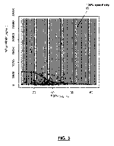
FIG. 3 is a graphic resulting from the analysis of retrieved data with a
Support Vector
Machine procedure (SVM). Values over the sigmoidal curve are 100% IS, and
values
under the curve correspond to patients with any of ICH or IS subtypes. Y-axis
levels of
NT-proBNP (pg/mL); X-axis levels of RBP4 (pg/mL). "can be introduced in a
support
vector machine procedure with a gaussian kernel". 100% specificity IS has the
same
meaning as in FIG. 1.
FIG. 4 is a graphic showing the classification of a cohort of subjects using a
logistic model
score including the logarithmic transformation of GFAP (pg/ml), NT-proBNP
(pg/ml) and
diastolic blood pressure (mmhg) as significative predictors of lschemic stroke
condition.
FIG. 5 is a graphic showing the classification of a cohort of subjects, in
which
determination of the levels of markers was performed within the first hour
after stroke
symptoms onset, and using a logistic model score including the logarithmic
transformation
CA 03139901 2021-11-10
WO 2020/229691 PCT/EP2020/063726
7
of GFAP (pg/ml), NT-proBNP (pg/ml) and diastolic blood pressure (mmhg) as
significative
predictors of lschemic stroke condition.
Detailed description of the invention
All terms as used herein in this application, unless otherwise stated, shall
be understood
in their ordinary meaning as known in the art. Other more specific definitions
for certain
terms as used in the present application are as set forth below and are
intended to apply
uniformly through-out the specification and claims unless an otherwise
expressly set out
definition provides a broader definition.
The term "patient" (or subject), as used herein, refers to any subject which
show one or
more signs or symptoms typically associated with stroke such as sudden-onset
face
weakness, arm drift, abnormal speech as well as combination thereof such as
the FAST
(face, arm, speech, and time), hemiplegia and muscle weakness of the face,
numbness,
reduction in sensory or vibratory sensation, initial flaccidity
(hypotonicity), replaced by
spasticity (hypertonicity), hyperreflexia, obligatory synergies and, in
particular, when they
appear in one side of the body (unilateral), altered smell, taste, hearing, or
vision (total or
partial), drooping of eyelid (ptosis) and weakness of ocular muscles,
decreased reflexes
(e.g. gag, swallow, pupil reactivity to light), decreased sensation and muscle
weakness of
the face, balance problems and nystagmus, altered breathing and heart rate,
weakness in
sternocleidomastoid muscle with inability to turn head to one side, weakness
in tongue
(inability to protrude and/or move from side to side), aphasia, dysarthria,
apraxia, visual
field defect, memory deficits, hemineglect, disorganized thinking, confusion,
hypersexual
gestures, lack of insight of his or her, usually stroke- related, disability,
altered walking
gait, altered movement coordination, vertigo, headache and or disequilibrium.
The term
"patient", as used herein, refers also to all animals classified as mammals
and includes,
but is not restricted to, domestic and farm animals, primates and humans,
e.g., human
beings, non-human primates, cows, horses, pigs, sheep, goats, dogs, cats, or
rodents.
Preferably, the patient is a male or female human of any age or race.
Preferably the
patient suffers stroke.
The term "selecting a patient for a therapy", as used herein, relates to the
identification of
a patient for a therapy designed to cure a disease or palliate the symptoms
associated
with one or more diseases or conditions. In the particular case of a stroke
therapy, it is
understood any therapy which abolishes, retards or reduces the symptoms
associated
with stroke and, more in particular, with ischemic stroke or alternatively
with hemorrhagic
stroke.
CA 03139901 2021-11-10
WO 2020/229691 PCT/EP2020/063726
8
The term "reperfusion therapy" relates to a medical treatment to restore blood
flow, either
through or around, blocked arteries. Reperfusion therapy includes drugs and
endovascular procedures. The drugs are thrombolytics (antithrombotic agents)
and
fibrinolytics used in a process called thrombolysis. Interventions performed
may be
minimally-invasive endovascular procedures for removing the thrombus
(thrombectomy)õ
with the possible use of one or more stent-retrievers, aspiration techniques
or alternatives
devices that combine both stent-retrievers and aspiration. Other surgeries
performed are
the more invasive bypass surgeries that graft arteries around blockages.
"Mechanical
thrombectomy", or simply thrombectomy, is the interventional procedure of
removing a
blood clot (thrombus) from a blood vessel. It is commonly performed in the
coronary
arteries (interventional cardiology), peripheral arteries (interventional
radiology) and
cerebral arteries (interventional neuroradiology).
The selection of a patient, although preferred to be, need not be adequate for
100% of the
subjects selected according to this first method of the invention. The term,
however,
requires that a statistically significant portion of subjects be correctly
selected. Whether
the selection of a patient in a population of subjects is statistically
significant can be
determined by the person skilled in the art using various well known statistic
evaluation
tools, e.g., determination of confidence intervals, p-value determination,
Student's t-test,
Mann- Whitney test, etc. Details are found in Dowdy and Wearden, Statistics
for
Research, John Wiley & Sons, New York 1983. Preferred confidence intervals are
at least
50%, at least 60%>, at least 70%>, at least 80%>, at least 90%>, or at least
95%. The p-
values are, preferably, 0.01, 0.05, 0.005, 0.001 or lower.
The term "ischemic stroke" (abbreviated IS) refers to the physical blockage of
blood flow
to an area of the brain, causing brain cells in the area to die. lschemic
strokes can further
be divided into thrombotic and embolic strokes. Thrombotic strokes occur when
a brain
artery is blocked by a blood clot formed in the brain. Embolic strokes are
caused by a
thrombus, which is formed in a peripheral artery or in the heart that travels
to the brain
where it produces ischemia. Another type of ischemic strokes are lacunar
strokes due to
the occlusion of a small cerebral artery.
The term "hemorrhagic stroke" (abbreviated ICH if intracerebral haemorrhage),
as used
herein refers to a bleeding into the brain tissue due to a blood vessel burst.
Inventors of present invention have identified RBP4 and BNP as new plasma
biomarkers
for accurately selecting a patient suffering from stroke for a reperfusion
therapy. Thus, the
markers can differentially diagnose acute IS from ICH. By means of several
data analysis
CA 03139901 2021-11-10
WO 2020/229691 PCT/EP2020/063726
9
approaches with these two markers, 100 % of specificity was achieved with a
sensitivity of
20%- 30%. In addition, if a third marker was added, in particular the levels
of GFAP,
specificity was maintained while sensitivity increased at 60%.
Thus, in a particular embodiment of the first aspect of selecting a patient
suffering stroke
for a reperfusion therapy, the method further comprises determining the level
of GFAP in
the isolated sample of said patient.
Although an increase of sensitivity is desirable, inventors have also
developed a simplified
kit comprising only means for detecting the levels of RBP4 and NT-proBNP. With
this
simplified kit, that can be used in ambulance when a stroke patient is being
attended, an
accurate discrimination between IS and ICH can be performed. This allows
administration
of the reperfusion therapy, if proper, as soon as possible (i.e antithrombotic
agent at
ambulance); and to avoid a bad outcome at least in IS because the patients are
treated
soon. In addition, in ICH a worsening of the symptoms is avoided if blood
pressure might
be optimized.
In yet a more particular embodiment of the first aspect, the method comprises
the step of
comparing said levels with a corresponding reference value or reference
interval for each
protein, said reference value or interval selected from a value or interval of
values from a
subject suffering from ischemic stroke, and wherein the subject is classified
as a
candidate for a reperfusion therapy when at least the level of RBP4 and of NT-
proBNP are
both within the value or interval of values from a subject suffering from IS.
In a more particular embodiment, the reference value or interval is selected
from a value
or interval of values from a subject suffering from IS or from a subject
suffering from ICH,
the subject is classified as a candidate for a reperfusion therapy when at
least the level of
RBP4 and of NT-proBNP are both within the value or interval of values from a
subject
suffering from IS. With the levels of both within the value or interval of
values from a
subject suffering from IS a meaningful clinical sensitivity (around 21 %) is
achieved for a
specificity over 98 %, in particular of the 100 %.
In another particular embodiment of the first aspect, the method comprises the
step of
comparing the levels of RBP4, NT-proBNP and optionally of GFAP if determined,
with a
corresponding reference value or reference interval for each protein, said
reference value
or interval selected from a value or interval of values from a subject
suffering from
ischemic stroke, and wherein the subject is classified:
- as a candidate for a reperfusion therapy, and
CA 03139901 2021-11-10
WO 2020/229691 PCT/EP2020/063726
- as having a prognosis defined by a dependency degree greater than 2
according to
modified ranking score (mRS), and determined within 1-5 months after stroke
onset
and/or as having a prognosis defined by a three-month after onset mortality
rate
comprised from 20% to 30%.
5 when at least the level of RBP4 and of NT-proBNP are both within the
value or interval of
values from a subject suffering from ischemic stroke.
In another more particular embodiment, the method comprises the step of
comparing the
levels of RBP4, NT-proBNP and of GFAP, and wherein if the subject is
classified as
10 candidate for a reperfusion therapy it is also classified as having a
prognosis defined by a
dependency degree greater than 2 according to modified ranking score (mRS),
and
determined within 1-5 months after stroke onset; and/or as having a prognosis
defined by
a three-month after onset mortality rate comprised from 20% to 30%.
The prognosis can also be defined as a dependency degree greater than 2
according to
modified ranking score (mRS), and determined at least three months after
stroke onset. In
a particular embodiment the three-month after onset mortality rate is of at
least 23%. In
another particular embodiment is of 25%.
In certain embodiments with only one of the levels of RBP4 and of NT-proBNP
are within
the value or interval of values from a subject suffering from IS, the subject
is also
classified as candidate for reperfusion.
In another particular embodiment of the first aspect, the in vitro method
further comprises
the step of comparing the levels of RBP4, NT-proBNP and if determined of GFAP,
with a
corresponding reference cut-off value for each protein, wherein:
- if only the levels of RBP4 and NT-proBNP are determined, a level of RBP4
and of NT-
proBNP simultaneously equal or higher than corresponding reference cut-off
values for
each of the proteins, Ref1 RBP4 and Ref1NT-proBNP, said cut-off values
discriminating between
ischemic stroke patients and intracerebral haemorrhage patients is indicative
that the
patient is a candidate for a reperfusion therapy; or
- if the levels of RBP4, NT-proBNP and additional GFAP are determined, the
patient is
selected as a candidate for a reperfusion therapy if in a first step the level
of GFAP is
equal or lower than a reference cut-off value RefGFAp; and in a second step
the level of
RBP4 and of NT-proBNP are simultaneously equal or higher than corresponding
references cut-off values Ref2RBp4 and Ref2NT-proBNP, said cut-off values
discriminating
between ischemic stroke patients and intracerebral haemorrhage patients.
CA 03139901 2021-11-10
WO 2020/229691 PC T/EP2020/063726
11
Indeed, the different alternative embodiments of the method of the first
aspect when
including the option of comparing tested levels with respective cut-off values
or reference
intervals are selected considering particular values of desired sensitivities
and
specificities. Thus, if a 100% specificity (correct classification between two
conditions) is
desired, sensitivity (detection of one condition among a cohort of subjects
with different
conditions) can be lowered. On the other hand, lowering the specificity (i.e.
around 94 %
or 98%) allows increasing sensitivity of a method. Therefore, reference values
can be
varied depending on the desired specificity and/or sensitivity desired.
In a more particular embodiment of this method including comparison with cut-
off values
of the two or the three protein levels, if the subject is classified as
candidate for a
reperfusion therapy, it is also classified as having a prognosis defined by a
dependency
degree greater than 2 according to modified ranking score (mRS), and
determined within
1-5 months after stroke onset; and/or as having a prognosis defined by a three-
month
after onset mortality rate comprised from 20% to 30%.
Also encompassed herewith is, as another particular embodiment of the method
of the
first aspect for selecting a patient suffering stroke for a reperfusion
therapy, that it further
includes a step of treating the patient with said reperfusion therapy if at
least the level of
RBP4 and of NT-proBNP are both within the value or interval of values from a
subject
suffering from IS; or in the alternative, if corresponding reference cut-off
values of RBP4
and of NT-proBNP, an optionally of GFAP, classify the patient as candidate for
reperfusion therapy.
As above indicated, there are several therapy protocols for the promotion of
reperfusion.
In a particular embodiment of the first aspect of the invention, the
reperfusion therapy is
selected from the group consisting of a therapy with an antithrombotic agent,
thrombectomy and a combination thereof.
In a more particular embodiment, the antithrombotic agent is a thrombolytic
agent. In yet a
more particular embodiment, the thrombolytic agent is a plasminogen activator.
More in
particular, the plasminogen activator is tissue plasminogen activator.
The term "antithrombotic agent", as used herein, refers to a drug that is able
to reduce clot
formation. Suitable antithrombotic agents for use in the present invention
include, without
limitation, thrombolytic agents, antiplatelet agents and anticoagulant
compounds.
The term "thrombolytic agent" as used herein refers to a drug that is able to
dissolve a
CA 03139901 2021-11-10
WO 2020/229691 PCT/EP2020/063726
12
clot. All thrombolytic agents are serine proteases and convert plasminogen to
plasmin
which breaks down the fibrinogen and fibrin and dissolves the clot. Currently
available
thrombolyic agents include reteplase (r-PA or Retavase), alteplase (t-PA or
Activase),
urokinase (Abbokinase), prourokinase, anisoylated purified streptokinase
activator
complex (APSAC), staphylokinase (Sak), tenecteplase (TNK-tPA), atenecteplase
(TNKasa), anistreplase (Eminase), streptoquinase (Kabikinase, Streptase) or
uroquinase
(Abokinase). Tenecteplase (TNK-tPA) is used in a particular embodiment, since
it can be
administered as a fast single bolus and can be used at ambulance level. TNK is
effective
after 1 minute post-administration (post-injection). Providers for TNK are
Boehringer
Ingelheim (European Union) and Genentech Inc (USA).
The term anticoagulant compounds, as used herein, refers to compounds that
prevent
coagulation and include, without limitation, vitamin K antagonists (warfarin,
acenocumarol,
fenprocoumon and fenidione), heparin and heparin derivatives such as low
molecular
weight heparins, factor Xa inhibitors such as synthetic pentasaccharides,
direct thrombin
inhibitors (argatroban, lepirudin, bivalirudin and ximelagatran) and
antiplatelet compounds
that act by inhibition of platelet aggregation and, therefore, thrombus
formation and
include, without limitation, cyclooxygenase inhibitors (aspirin), adenosine
diphosphate
receptor inhibitors (clopidrogrel and ticlopidine), phosphodiesterase
inhibitors (cilostazol),
glycoprotein IIB/IIIA inhibitors (Abciximab, Eptifibatide, Tirofiban and
Defibrotide) and
adenosine uptake inhibitors (dipiridamol). In a preferred embodiment, the
antithrombotic
agent is a thrombolytic agent. In a more preferred embodiment, the
thrombolytic agent is a
plaminogen activator. In a yet more preferred embodiment, the plasminogen
activator is
tPA (tissue plasminogen activator).
The term "tissue plasminogen activator (t-PA)" as used herein refers to a
serine protease
found on endothelial cells that catalyzes the conversion of plasminogen to
plasmin. The
complete protein sequence for human t-PA has the UniProt KB accession number
P00750
(July 11th, 2012), SEQ ID NO: 1. tPA may be manufactured using recombinant
biotechnology techniques, tPA created this way may be referred to as
recombinant tissue
plasminogen activator (rtPA). Recombinant tissue plasminogen activators (r-
tPAs) include
the thrombolytic agents alteplase, reteplase, and tenecteplase (TNKase, also
termed
TNK-tPA, SEQ ID NO: 2). In human t-PA, the amino acids at position 296-299 are
lysine,
histidine, and two arginines. In TNK-tPA, these amino acids have been replaced
by four
alanines. This mutation is responsible for increased resistance to plasminogen
activator
inhibitor 1 (PAI-1).
Doses of t-PA should be given within the first 3 hours of the onset of
symptoms or up to
4.5 hours from symptom onset. Recommended total dose: 0.9 mg/kg (maximum dose
CA 03139901 2021-11-10
WO 2020/229691 PCT/EP2020/063726
13
should not exceed 90 mg) infused over 60 minutes. Load with 0.09 mg/kg (10% of
the 0.9
mg/kg dose) as an intravenous bolus over 1 minute, followed by 0.81 mg/kg (90%
of the
0.9 mg/kg dose) as a continuous infusion over 60 minutes. Heparin should not
be started
for 24 hours or more after starting alteplase for stroke. Said t-PA is given
intravenously
and in some cases may be given directly into an artery and should be given
right away
after the first symptoms of stroke start. Said doses and administration routes
apply to any
of the embodiments of the first aspect. Also, in particular in embodiments
including step of
treating the patient.
.. Single dose of TNK-tPA should be given as soon as possible after
determining that the
subject suffering from stroke is a candidate to reperfusion therapy, and
within the first 3
hours of the onset of symptoms or up to 4.5 hours from symptom onset,
preferably within
the first hour after stroke onset.
As indicated above, the use of TNK-tPA is particularly useful, since due to
the particular
formulation as fast single application bolus, it can be administered at any
point of care,
even at ambulance level, being effective about one minute post-administration.
Those patients suffering stroke not selected for a reperfusion therapy, are in
a particular
embodiment, selected for a therapy reducing blood pressure. In particular,
said therapy is
performed with an agent capable of reducing blood pressure.
"Blood pressure" is herein to be understood as to refer to the blood pressure
at the site of
central arteries, such as the aorta and carotid artery. Central blood pressure
can suitably
be measured non-invasively (as set out below) at the carotids or radialis by
applanation
tonometry. "Blood pressure" as used herein thus encompasses aortic blood
pressure.
"Agent capable of reducing blood pressure", as used in the present invention,
relates to
any drug which lower blood pressure by different means. Among the most widely
agents
are the thiazide diuretics [such as furosemide, nitroprusside, hydralazine];
the ACE
inhibitors, the calcium channel blockers (such as nicardipine or nimodipine);
the
adrenergic receptor antagonist (such as alpha-adrenergic antagonist,
urapidil), or
combined alpha- and beta-blocker (labetalol and nitroglycerin); and the
angiotensin II
receptor antagonists (ARBs). Illustrative, non- !imitative example of agents
capable of
lowering or reducing blood pressure are alpha-methyl dopa (Aldomet),
11,17alpha-
d.imethoxy- 18[3- [(3,4,5 -trimethoxy- benzoyl)oxyl)]-3p,2a-yohimban-168-
carboxylic acid
methyl ester (Reserpine) or 2-(2,6-dichlorophenylamino) 2-imidazoline
hydrochloride
(Clonidine hydrochloride), lergotrile or viz. 2-chloro-6-methylergoline-88-
acetonitrile as
disclosed in EP0005074. Reference values that will be used to decrease blood
pressure
CA 03139901 2021-11-10
WO 2020/229691 PCT/EP2020/063726
14
in ischemic stroke, ischemic stroke treated with thrombolytics or hemorrhagic
stroke, will
be those recommended by clinical practice guidelines as these values could be
updated.
Nowadays, treatment modalities for blood pressure lowering are aimed to be
reduced if
systolic blood pressure to among 220-120 mm Hg was achieved in ischemic
patients and
if it achieved to among 180-100 mm Hg in haemorrhagic patients. In a preferred
embodiment, the blood pressure may be reduced by intravenous administration of
an
agent capable of reducing blood pressure and co- administration of oral
antihypertensive
agent(s). Reference values that will be used to decrease blood pressure in
ischemic
stroke, ischemic stroke treated with thrombolytics or hemorrhagic stroke, will
be those
recommended by clinical practice guidelines as these values could be updated.
Any method suitable for measure arterial pressure can be used for determining
if an agent
is capable of reducing blood pressure, wherein a reduction in arterial
pressure is detected
after administration of the agent. Illustrative, non- !imitative examples of
methods for
measurement arterial pressure are non-invasive techniques, such as by way of
illustrative
non- !imitative example palpitation, auscultatory, oscillometric and
continuous noninvasive
arterial pressure (CNAP).
The term "reference value", as used herein, relates to a predetermined
criteria used as a
.. reference for evaluating the values or data obtained from the samples
collected from a
subject. The reference value or reference level can be an absolute value; a
relative value;
a value that has an upper or a lower limit; a range of values; an average
value; a median
value, a mean value, or a value as compared to a particular control or
baseline value. A
reference value can be based on an individual sample value, such as for
example, a value
obtained from a sample from the subject being tested, but at an earlier point
in time. The
reference value can be based on a large number of samples, such as from
population of
subjects of the chronological age matched group, or based on a pool of samples
including
or excluding the sample to be tested. Reference values have been determined
for the
biomarkers of the invention. The reference value for each of RBP4, NT-proBNP
and
GFAP may be from a lower and an upper value as will be disclosed in view of
examples
below. Range of values of each biomarker (protein levels) and particular
combinations of
the values of the different biomarkers provide for correct classification of
subjects with
high sensitivity and specificity.
The levels of a bio marker (in this invention any of NT-proBNP, RBP4 or GFAP)
are
considered to be higher than its reference value when it is at least 1.5%, at
least 2%, at
least 5%, at least 10%, at least 15%, at least 20%, at least 25%, at least
30%, at least
35%, at least 40%, at least 45%, at least 50%, at least 55%, at least 60%, at
least 65%, at
least 70%, at least 75%, at least 80%: at least 85%, at least 90%, at least
95%, at least
CA 03139901 2021-11-10
WO 2020/229691 PCT/EP2020/063726
100%, at least 110%, at least 120%, at least 130%, at least 140%, at least
150% or more
higher than the reference value.
Likewise, in the context of the present invention, the level of a biomarker is
reduced when
5 the level of said biomarker in a sample is lower than a reference value.
The levels of a
biomarker are considered to be lower than its reference value when it is at
least 5%, at
least 10%, at least 15%, at least 20%, at least 25%, at least 30%, at least
35%, at least
40%, at least 45%, at least 50%, at least 55%, at least 60%, at least 65%, at
least 70%, at
least 75%, at least 80%: at least 85%, at least 90%, at least 95%, at least
100%, at least
10 .. 110%, at least 120%, at least 130%, at least 140%, at least 150% or more
lower than the
reference value.
In a particular embodiment of the first aspect, when only the levels of RBP4
and NT-
proBN P are determined in a biofluid sample and compared with corresponding
reference
15 cut-off values (herewith termed Ref1RBP4 and Ref1NT-p1oBNP), these
reference cut-off values
are for RBP4 52 pg/ml, for NT-proBNP 4062 pg/ml. These particular reference
cut-off
values are, in a particular embodiment, for isolated plasma samples and when
assayed
with an enzyme immunosorbent assay (ELISA)
.. In another particular embodiment of the first aspect, when the levels of
RBP4, NT-proBNP
and GFAP are determined and compared with corresponding reference cut-off
values
(herewith termed Ref2RBP4, Ref2NT-proBNP and --.GFAP Ref )
these reference cut-off values are: for
RBP4 38 pg/ml, for BNP 1305 pg/ml, and for GFAP 0.325 ng/ml. These particular
reference cut-off values are, in a more particular embodiment, for isolated
plasma
samples and when GFAP was assayed, the levels of this marker is determined
with a
more sensitive (picomolar level) single molecular assay (SIMOA) and the others
with an
ELISA assay. With these cut-off levels, a particular method is carried out in
two separate
steps or conditions. In a first step the level of GFAP is determined and if it
is equal or
lower than the reference cut-off value RefGFAp, patients are considered as
suffering from
.. ischemic stroke and candidates for reperfusion if further in a second step
the level of
RBP4 and of NT-proBNP are simultaneously equal or higher than corresponding
references cut-off values Ref2RBp4 and Ref2NT-proBNP=
As will be illustrated by means of the examples, ICH patients had higher GFAP
levels and
lower RBP4 and NT-proBNP than IS. The combination of RBP4>52 pg/mL and GFAP
>0.18ng/mL resulted in an accurate diagnosis of 6.5% of IS and 34.3% of ICH.
The
addition of the NT-proBNP kept 100% specificity and improved sensitivity for
IS up to 20%
(31/155) by using RBP4 > 52 pg/mL and the BNP cut-off > 4060 pg/mL.
CA 03139901 2021-11-10
WO 2020/229691 PCT/EP2020/063726
16
As above indicated, it is widely known that reference values can vary
depending on
exclusion criteria decisions of clinical protocols. In addition, depending on
the variables
that are considered to make a diagnosis, these values can also be modulated
always in
order to increase sensitivity while maintaining specificity, which specificity
is really critical
in stroke.
Moreover, proper classification of patients depending on detected levels of
the proteins
can be done using computational methods, by which of them the determined
values of
proteins are computed in a formula that gives a predictive factor, said
predictive factor
calculated based on the expression levels of the proteins, said expression
levels being
corrected by a particular coefficient. Other computation methods (such as one
of the
exemplified herewith and called support vector machine method), allow
disclosing a
function that properly classify the patients considering the levels of all
determined
proteins.
All these reference values indicated in the particular embodiments are the
ones
determined in isolated plasma samples of subjects suffering from stroke. The
skilled man
will know how to find the corresponding ones in serum and other bio fluids.
.. The term "sample" as used herein, relates to any sample which can be
obtained from the
patient. The present method can be applied to any type of biological sample
from a
patient, such as a biopsy sample, tissue, cell or biofluid (plasma, serum,
saliva, semen,
sputum, cerebral spinal fluid (CSF), tears, mucus, sweat, milk, brain extracts
and the like.
Therefore, in another particular embodiment, optionally in combination with
any
embodiment above or below, the isolated sample of the subject (i.e. patient
suffering
stroke) is a bio fluid. Illustrative non !imitative bio fluids are blood,
plasma, serum, saliva,
urine or cerebrospinal fluid. In a more preferred embodiment, the biofluid is
plasma or
serum.
In a preferred embodiment of the methods of the invention, the sample is
obtained at
baseline.
Different samples could be used for determining the level of different
markers. Thus, it is
not necessary that the levels of all the markers according to the methods of
the invention
are measured in the same type of sample. Thus, in another preferred
embodiment, the
levels of RBP4, NT-proBNP and GFAP are measured in serum. In another preferred
the
level of they are measured in plasma.
CA 03139901 2021-11-10
WO 2020/229691 PCT/EP2020/063726
17
"Baseline", as used in the present invention, is considered any time from
onset of
symptoms until the patient is explored for the first time. This is usually
within the first hours
after stroke, and it is usually the first attention in the ambulance or in the
hospital. In a
preferred embodiment, the baseline is within the first 4.5 hours from symptom
onset, or
less than 6 hours after stroke or in another preferred embodiment less than 24
hours
symptoms onset.
In another particular embodiment of this aspect, the step of determining the
level of RBP4
and of NT-proBNP, and if determined of GFAP, is carried out within the two
first hours
after the stroke onset. As will be depicted in examples below, the sooner the
determination of the markers in the isolated sample is done, the better the
accuracy and
sensitivity of the method. In another particular embodiment of this aspect,
the step of
determining the level of RBP4 and of NT-proBNP, and if determined of GFAP, is
carried
out within the first hour after the stroke onset.
In another embodiment of the first aspect of the method for selecting a
patient suffering
stroke for a reperfusion therapy, it further comprises determining one or more
clinical
parameters. Thus, the method of the invention comprises determining the levels
of RBP4
and BNP and one or more clinical parameters.
The term "clinical parameters" or clinical data, as used herein, refers to
person
demographics (age or date of birth, race and/or ethnicity), patient clinical
symptoms or
signs related to stroke related diseases/conditions. The term also includes
laboratory
parameters, such as the determination of d-dimer or of glycemia.
In a particular embodiment, the clinical parameter is hypertension and wherein
if the
patient has hypertension is indicative that the patient suffers ischemic
stroke or that the
patient is a candidate for a reperfusion therapy.
In another particular embodiment of the first aspect, the in vitro method
further comprises
determining a clinical parameters selected from the group consisting of blood
pressure,
including systolic blood pressure and/or diastolic blood pressure, glycemia,
age, scores
from systematic assessment tool stroke-related neurologic deficit, such as NI
HSS score,
gender, and combinations thereof. The values of all these parameters are, in a
particular
embodiment, used in combination with the levels of RBP4, NT-proBNP and
optionally of
GFAP in adequate algorithms for correctly classifying the patient as candidate
to
reperfusion therapy. For example, blood pressure within a certain interval in
combination
with certain levels of the two or three proteins are used in a decision
protocol for the
CA 03139901 2021-11-10
WO 2020/229691 PCT/EP2020/063726
18
correct classification. The values are, in another more particular embodiment,
introduced
in formulas of regression models to give a score or final value allowing such
classification.
For "hypertension", sometimes called arterial hypertension, is to be
understood a chronic
medical condition in which the blood pressure in the arteries is elevated.
Normal blood
pressure at rest is within the range of 100-140 mmHg systolic (top reading)
and 60-90
mmHg diastolic (bottom reading). High blood pressure is said to be present if
it is
persistently at or above 140/90 mmHg. As above indicated, reference values
that will be
used to decrease blood pressure in ischemic stroke, ischemic stroke treated
with
thrombolytics or haemorrhagic stroke, will be those recommended by clinical
practice
guidelines as these values could be updated, being nowadays accepted as
suitable
treatment modalities for blood pressure lowering when systolic blood pressure
amounts to
among 220-120 mm Hg in ischemic patients and amounts to among 180-100 mm Hg in
haemorrhagic patients.
The term "systematic assessment tool stroke-related neurologic deficit"
relates to tools
designed to measure and scale the neurological deficits most often seen with
stroke.
Several aspects or parameters are assessed, such as the level of
consciousness, visual
fields, facial weakness, motor performance of extremities, gaze, sensory
deficits,
coordination (ataxia), language (aphasia), speech (dysarthria), etc. For all
of them a value
is given, being 0 if normal. So, in most of these tools the higher the score,
the worse the
neurological deficit. The skilled man will know of the existence of different
tools for this
purpose, as the National Institutes of Health Stroke Scale (NI HSS) score, the
Rapid
Arteria occlusion evaluation scale for stroke (RACE), the Cincinnati
Prehospital Stroke
Scale Compared to Stroke Severity Tools for Large Vessel Occlusion Stroke
Prediction
(Cincinnati-score), Los Angeles Motor Scale (LAMS), or the modified Rankin
Scale or
Score (mRS). All these scales are designed to provide a rapid and standardized
assessment of the neurological function in the early periods after stroke. The
modified
Rankin Scale or Score (mRS) is also a scale for evaluating the degree of
disability after
the stroke onset. It is mostly applied at discharge and 3 months after onset.
In another preferred embodiment the clinical parameter is selected from age,
NI HSS
score, gender, systolic blood pressure and combinations thereof. The term "NI
HSS
score", as used in the present invention refers to The National Institutes of
Health Stroke
.. Scale (NI HSS) score, a systematic assessment tool that provides a
quantitative measure
of stroke-related neurologic deficit (Adams HP Jr Neurology. 1999 Jul
13;53(1): 126-31).
The NI HSS was originally designed as a research tool to measure baseline data
on
patients in acute stroke clinical trials. Now, the scale is also widely used
as a clinical
assessment tool to evaluate acuity of stroke patients, determine appropriate
treatment,
CA 03139901 2021-11-10
WO 2020/229691 PCT/EP2020/063726
19
and predict patient outcome. The NIHSS is a 15-item neurologic examination
stroke scale
used to evaluate the effect of acute cerebral infarction on the levels of
consciousness,
language, neglect, visual-field loss, extraocular movement, motor strength,
ataxia,
dysarthria, and sensory loss. A trained observer rates the patient's ability
to answer
questions and perform activities. Ratings for each item are scored with 3 to 5
grades with
0 as normal, and there is an allowance for untestable items. The level of
stroke severity as
measured by the NI H stroke scale scoring system: 0= no stroke, 1-4= minor
stroke, 5-15=
moderate stroke, 15-20= moderate/severe stroke, 21-42= severe stroke. In the
present
invention the term "higher score" refers to a score from 5 to 42 in the NI H
stroke scale
scoring system.
Also variations of the NI HSS such as Rapid Arteria occlusion evaluation scale
for stroke
(RACE) or other scores used to identify ischemic strokes with large vessel
occlusion may
be used.
Yet in another particular embodiment of the first aspect, optionally in
combination with any
of the embodiments above or below, if the subject is classified as candidate
for a
reperfusion therapy, it is also diagnosed of suffering from large vessel
occlusion.
As a second aspect the invention relates to an in vitro method for
differentiating IS from
ICH in a patient, comprising determining the level of RBP4 and NT-proBPN in an
isolated
sample of said patient.
In a particular embodiment of the second aspect, the method comprises
determining the
level of GFAP.
In another more particular embodiment of the second aspect, it further
comprises the step
of comparing the levels of RBP4, NT-proBNP and if determined of GFAP, with a
corresponding reference value, wherein:
- if only the levels of RBP4 and BNP are determined, a level of RBP4 and of
BNP
simultaneously higher than corresponding reference values Ref1RBp4 and Ref1NT-
promp is
indicative that the patient is an IS patient; or
- if the levels of RBP4, BNP and GFAP are determined, a level of RBP4 and of
BNP
simultaneously higher than corresponding references values Ref2RBp4 and Ref2NT-
proBNP,
and a level of GFAP lower than a reference value RefGFAp is indicative that
the patient is
an IS patient.
In another particular embodiment of the second aspect, if the subject is
classified as an
ischemic stroke when determining the levels of RBP4 and BNP and optionally of
GFAP, it
CA 03139901 2021-11-10
WO 2020/229691 PCT/EP2020/063726
is also classified as having a prognosis defined by a dependency degree
greater than 2
according to modified ranking score (mRS), and determined within 1-5 months
after stroke
onset; and/or as having a prognosis defined by a three-month after onset
mortality rate
comprised from 20% to 30%.
5
In another particular embodiment of the second aspect, it further comprises
the step of
selecting a therapy, in particular reperfusion therapy, as indicated in the
first aspect and
its particular embodiments.
10 Thus, after differential diagnosis is accomplished, in another
particular embodiment of the
second aspect it further comprises a step of recommending a reperfusion
therapy to a
patient diagnosed of IS and/or treating said patient diagnosed of IS with a
reperfusion
therapy, mainly with an antithrombotic agent or by means of thrombectomy. On
the
alternative, those patients diagnosed of ICH that should avoid a reperfusion
therapy in
15 order to avoid fatal outcomes are, in another particular embodiment,
recommended for or
treated with a therapy reducing or optimizing blood pressure.
This particular embodiment could be drafted as a method of treating a patient
suffering
stroke, said method comprising carrying out the in vitro method for
differentiating IS from
20 ICH in a patient according to the second aspect and treating a patient
diagnosed of IS
with a reperfusion therapy, mainly with an antithrombotic agent or by means of
thrombectomy; or treating a patients diagnosed of ICH with a therapy reducing
or
optimizing blood pressure. Advantageously, with this method patient is treated
or
recommended to be treated within first hours of the onset of symptoms and with
the most
appropriate therapy regimen.
All particular embodiments previously disclosed for the first aspect do also
apply to this
second aspect. In particular, those preferred reference values, the kind of
isolated sample
and the option of further determining one or more clinical parameters. Thus,
in another
particular embodiment of the second aspect, the in vitro method further
comprises
determining a clinical parameters selected from the group consisting of blood
pressure,
including systolic blood pressure and/or diastolic blood pressure, glycemia,
age, scores
from systematic assessment tool stroke-related neurologic deficit, such as NI
HSS score,
gender, and combinations thereof. As before, the values of all these
parameters are, in a
particular embodiment, used in combination with the levels of RBP4, NT-proBNP
and
optionally of GFAP in adequate algorithms for correctly classifying the
patient as
candidate to reperfusion therapy. For example, blood pressure within a certain
interval in
combination with certain levels of the two or three proteins are used in a
decision protocol
for the correct classification. The values are, in another more particular
embodiment,
CA 03139901 2021-11-10
WO 2020/229691 PCT/EP2020/063726
21
introduced in formulas of regression models to give a score or final value
allowing such
classification.
Also, in another particular embodiment of the second aspect, the determining
the level of
RBP4 and of NT-proBNP, and if determined of GFAP, is carried out within the
two first
hours after the stroke onset. More in particular within the first hour. This
implies the
advantage of increasing sensitivity of the method as indicated for the first
aspect.
Yet, in another particular embodiment of this second aspect, optionally in
combination with
.. any of the embodiments above or below, if the subject is classified an
ischemic stroke, it is
also diagnosed of suffering from large vessel occlusion.
Invention also generically encompasses a method of detecting, in an isolated
sample of a
subject suffering from stroke, the level of RBP4 and NT-proBPN, and optionally
in
combination with the level of GFAP, the method comprising:
(a) obtaining a sample from the subject; and
(b) detecting whether one or more of the proteins is present in the isolated
sample by: (i)
contacting said sample with means capable of binding the corresponding
expressed
proteins and detecting said binding; or (ii) contacting said sample with means
capable of
binding corresponding RNA going to be translated to the one or more of the
corresponding proteins and detecting said binding.
The term "differentiating", as used herein for the second aspect, relates to
the
determination of a different condition. As will be understood by those skilled
in the art,
differentiation, although preferred to be, need not be correct for 100% of the
subjects to be
diagnosed or evaluated. The term, however, requires that a statistically
significant portion
of subjects can be identified as having an increased probability of having one
of the two
types of stroke. Whether a subject is statistically significant can be
determined without
further ado by the person skilled in the art using various well known
statistic evaluation
tools, e.g., determination of confidence intervals, p-value determination,
Student's t-test,
Mann- Whitney test, etc. Details are found in Dowdy and Wearden, Statistics
for
Research, John Wiley & Sons, New York 1983. Preferred confidence intervals are
at least
50%, at least 60%, at least 70%, at least 80%, at least 90%, or at least 95%.
The p-
values are, preferably, 0.05, 0.01, 0.005 or lower.
Since the biomarkers identified in the present invention allow differentiating
IS from ICH in
a patient and considering that different therapies are applied to these two
types of patients
(antithrombotic agents in patients suffering ischemic stroke and an agent
capable of
reducing blood pressure in patients suffering haemorrhagic stroke) (see
Tsivgoulis G. et
CA 03139901 2021-11-10
WO 2020/229691 PCT/EP2020/063726
22
al, Neurology. 2014 Sep 19), the invention also provides the above-mentioned
first
method for the selection of a therapy for a patient having suffered stroke.
Thus, both
aspects are intimately and conceptually related.
When in present invention RBP4 is referred, it relates to refers to retinol
binding protein 4,
plasma that belongs to the lipocalin family and is the specific carrier for
retinol in the
blood. The complete sequence for human retinol binding protein 4 has the
UniProtKB
accession number P02753 (August 8th, 2013), SEQ ID NO: 3.
The term "GFAP" as used herein refers to glial fibrillary acidic protein, an
intermediate
filament protein that is expressed by numerous cell types of the central
nervous system.
The complete human sequence for glial fibrillary acidic protein has the
UniProtKB
accession number P14136 (August 8th, 2013), SEQ ID NO: 4.
The N-terminal fragment of B-type natriuretic peptide (NT-proBNP) (SEQ ID NO:
5) is the
76¨amino acid N-terminal fragment of the B-type natriuretic peptide
prohormone. Cleaving
of pro-BNP yields the NT-proBNP fragment and the active B-type natriuretic
peptide
(BNP). BNP is a hormone secreted by cardiomyocytes in the heart ventricles in
response
to stretching caused by increased ventricular blood volume. The complete human
sequence BNPhas the UniProt KB accession number P16860 (August 1st, 1990¨
version
1 of the sequence, and database release 187 of May 8th, 2019).
All these proteins do have homologues in other mammal species (cats, dogs,
mouse, rats,
etc.). The skilled man can retrieved the corresponding complete sequences in
public
databases.
As the person skilled in the art understands, the expression levels of NT-
proBNP, RBP4
and/or GFAP can be determined by measuring the levels of mRNA encoded by the
corresponding genes or by measuring the levels of proteins encoded by said
genes, and
the levels of variants thereof.
By way of a non-limiting illustration, the expression levels are determined by
means of the
quantification of the levels of mRNA encoded by said genes. The latter can be
quantified
by means of using conventional methods, for example, methods comprising the
amplification of mRNA and the quantification of the amplification product of
said mRNA,
such as electrophoresis and staining, or alternatively, by means of Northern
blot and the
use of suitable probes, Northern blot and use of specific probes of the mRNA
of the genes
of interest or of their corresponding cDNA/cRNA, mapping with the SI nuclease,
RT-PCR,
hybridization, microarrays, etc. Similarly, the levels of the cDNA/cRNA
corresponding to
CA 03139901 2021-11-10
WO 2020/229691 PCT/EP2020/063726
23
said mRNA encoded by the marker genes can also be quantified by means of using
conventional techniques; in this event, the method of the invention includes a
step of
synthesis of the corresponding cDNA by means of reverse transcription (RT) of
the
corresponding mRNA followed by the synthesis (RNA polymerase) and
amplification of
the cRNA complementary to said cDNA. Conventional methods of quantifying the
expression levels can be found in laboratory manuals.
In order to normalize the values of mRNA expression among the different
samples, it is
possible to compare the expression levels of the mRNA of interest in the test
samples with
the expression of a control RNA. A "control RNA" as used herein, relates to
RNA whose
expression levels do not change or change only in limited amounts. Preferably,
the control
RNA is mRNA derived from housekeeping genes and which code for proteins which
are
constitutively expressed and carry out essential cellular functions. Preferred
housekeeping
genes for use in the present invention include 18-S ribosomal protein, 8-2-
microglobulin,
ubiquitin, cyclophilin, GAPDH, PSMB4, tubulin and 13-actin.
Alternatively, it is also possible to determine the expression levels of the
marker genes by
means of the determination of the expression levels of the proteins encoded by
said
genes, since if the expression of genes is increased, an increase of the
amount of
corresponding protein should occur and if the expression of genes is
decreased, a
decrease of the amount of corresponding protein should occur.
The determination of the expression levels of the proteins can be carried out
by qualitative
and/or quantitative tests selected from the group consisting of an
immunological test,
bioluminescence, fluorescence, chemiluminescence, electrochemistry and mass
spectrometry. Particular tests that can be implemented in a point of care test
format
(POCT) are recommended to make easy and fast the determining of marker levels.
In a
particular embodiment, point of care tests include lateral flow tests, which
allow detecting
the presence (or absence) of a target analyte in liquid sample (matrix)
without the need for
specialized and costly equipment, though many lab-based applications exist
that are
supported by reading equipment.
As will be illustrated in examples, a particular POCT was developed and it was
tested in
ambulances and helicopters. By means of this POCT useful high sensitivity
rates at 100%
of specificity for ischemic strokes were achieved, that allowed initiating pre-
hospital
reperfusion therapies in selected cases much faster than using standard
technologies.
Independently of the test format, particular quantitative tests are selected
from the group
consisting of an immunological test, bioluminescence, fluorescence,
chemiluminescence,
CA 03139901 2021-11-10
WO 2020/229691 PCT/EP2020/063726
24
electrochemistry and mass spectrometry.
In one embodiment, the level of expression is determined by immunological
techniques
such as enzyme-linked immunosorbent assay (ELISA), enzyme immunodot assay,
agglutination assay, antibody-antigen-antibody sandwich assay, antigen-
antibody-antigen
sandwich assay, immunocromatography, or other immunoassay formats well-known
to the
ordinarily skilled artisan, such as radioimmunoassay, as well as protein
microarray
formats, such as single molecular assay (SIMOA), Western Blot or
immunofluorescence.
Western blot is based on the detection of proteins previously resolved by gel
electrophoreses under denaturing conditions and immobilized on a membrane,
generally
nitrocellulose by the incubation with an antibody specific and a developing
system (e.g.
chemoluminiscent). The analysis by immunofluorescence requires the use of an
antibody
specific for the target protein for the analysis of the expression. ELISA is
based on the use
of antigens or antibodies labelled with enzymes so that the conjugates formed
between
the target antigen and the labelled antibody results in the formation of
enzymatically-active
complexes. Since one of the components (the antigen or the labelled antibody)
are
immobilised on a support, the antibody-antigen complexes are immobilised on
the support
and thus, it can be detected by the addition of a substrate which is converted
by the
enzyme to a product which is detectable by, e.g. spectrophotometry
,fluorometry, mass
spectrometry or tandem mass tags (TMT). SIMOA is a type of assay more
sensitive than
an ELISA, since it uses arrays of femtoliter-sized reaction chambers, which
are termed
single-molecule arrays (SimoaTM) that can isolate and detect single enzyme
molecules.
Because the array volumes are approximately 2 billion times smaller than a
conventional
ELISA, a rapid build-up of fluorescent product is generated if a labeled
protein is present.
With diffusion defeated, this high local concentration of product can be
readily observed.
Only a single molecule is needed to reach the detection limit. Using the same
reagents as
a conventional ELISA, this method has been used to measure proteins in a
variety of
different matrices (serum, plasma, cerebrospinal fluid, urine, cell extracts,
etc.) at
femtomolar (fg/mL) concentrations, offering aroughly1000-fold improvement in
sensitivity.
On the other hand, the determination of the protein expression levels can be
carried out
by constructing a tissue microarray (TMA) containing the subject samples
assembled, and
determining the expression levels of the proteins by techniques well known in
the state of
the art.
In a preferred embodiment the determination of the levels of the markers are
determined
by immunological technique. In a more preferred embodiment, the immunological
technique is ELISA.
CA 03139901 2021-11-10
WO 2020/229691 PCT/EP2020/063726
When an immunological method is used, any antibody or reagent known to bind
with high
affinity to the target proteins can be used for detecting the amount of target
proteins. It is
preferred nevertheless the use of antibody, for example polyclonal sera,
hybridoma
5 supernatants or monoclonal antibodies, antibody fragments, Fv, Fab, Fab'
y F(ab')2,
ScFv, diabodies, triabodies, tetrabodies and humanised antibodies.
As previously cited, the expression levels of the NT-proBNP and/or RBP4 and/or
GFAP
can be determined by measuring both the levels of protein, and the levels of
variants
10 thereof, such as fragments, isoforms, analogues and/or derivatives.
The term "functionally equivalent variant" is understood to mean all those
proteins derived
from NT-proBNP and/or RBP4 and/or GFAP sequence by modification, insertion
and/or
deletion or one or more amino acids, whenever the function of said variants
are
15 substantially maintained. Preferably, variants of NT-proBNP and/or RBP4
and/or GFAP
are (i) polypeptides in which one or more amino acid residues are substituted
by a
preserved or non-preserved amino acid residue (preferably a preserved amino
acid
residue) and such substituted amino acid may be coded or not by the genetic
code, (ii)
polypeptides in which there is one or more modified amino acid residues, for
example,
20 residues modified by substituent bonding, (iii) polypeptides resulting
from alternative
processing of a similar mRNA, (iv) polypeptide fragments and/or (v)
polypeptides resulting
from NT-proBNP and/or RBP4 and/or GFAP fusion or the polypeptide defined in
(i) to (iii)
with another polypeptide, such as a secretory leader sequence or a sequence
being used
for purification (for example, His tag) or for detection (for example, Sv5
epitope tag). The
25 fragments include polypeptides generated through proteolytic cut
(including multisite
proteolysis) of an original sequence. The variants may be post-translationally
or
chemically modified. Such variants are supposed to be apparent to those
skilled in the art.
As known in the art the "similarity" between two proteins is determined by
comparing the
amino acid sequence and its conserved amino acid substitutes of one protein to
a
sequence of a second protein. The variants are defined to include polypeptide
sequences
different from the original sequence, preferably different from the original
sequence in less
than 40% of residues per segment concerned, more preferably different from the
original
sequence in less than 25% of residues per segment concerned, more preferably
different
from the original sequence in less than 10% of residues per segment concerned,
more
preferably different from the original sequence in only a few residues per
segment
concerned and, at the same time, sufficiently homologous to the original
sequence to
preserve functionality of the original sequence. Variants according to the
present invention
includes amino acid sequences that are at least 60%, 65%, 70%, 72%, 74%, 76%,
78%,
CA 03139901 2021-11-10
WO 2020/229691 PCT/EP2020/063726
26
80%, 90%, or 95% similar or identical to the original amino acid sequence. The
degree of
identity between two proteins is determined using computer algorithms and
methods that
are widely known for the persons skilled in the art. The identity between two
amino acid
sequences is preferably determined by using the BLASTP algorithm [BLASTManual,
Altschul, S., et al, NCB! NLM NIH Bethesda, Md. 20894, Altschul, S., et al, J.
Mol. Biol.
215: 403-410 (1990)].
The proteins can be post-translationally modified. For example, post-
translational
modifications that fall within the scope of the present invention include
signal peptide
cleavage, glycosylation, acetylation, isoprenylation, proteolysis
myristoylation, protein
folding and proteolytic processing, etc. Additionally, the proteins may
include unnatural
amino acids formed by post-translational modification or by introducing
unnatural amino
acids during translation.
In another particular embodiments of the in vitro methods of the invention
that provide a
differential diagnostic and information for selecting a therapy, they further
comprise the
steps of (i) collecting the diagnostic information, and (ii) saving the
information in a data
carrier.
In the sense of the invention a "data carrier" is to be understood as any
means that
contain meaningful information data for the differential diagnosis of IS and
ICH and/or for
the selection of a candidate to reperfusion therapy, such as paper. The
carrier may also
be any entity or device capable of carrying the differential diagnosis data or
information for
selecting a therapy. For example, the carrier may comprise a storage medium,
such as a
ROM, for example a CD ROM or a semiconductor ROM, or a magnetic recording
medium,
for example a floppy disc or hard disk. Further, the carrier may be a
transmissible carrier
such as an electrical or optical signal, which may be conveyed via electrical
or optical
cable or by radio or other means. When the diagnosis/therapy selection data
are
embodied in a signal that may be conveyed directly by a cable or other device
or means,
the carrier may be constituted by such cable or other device or means. Other
carriers
relate to USB devices and computer archives. Examples of suitable data carrier
are
paper, CDs, USB, computer archives in PCs, or sound registration with the same
information.
The invention also encompasses an in vitro method for the prognosis of a
patient suffering
ischemic stroke, comprising determining the level of retinol binding protein-4
(RBP4) and
N-terminal fragment of B-type natriuretic peptide (NT-proBNP) in an isolated
sample of
said patient.
CA 03139901 2021-11-10
WO 2020/229691 PCT/EP2020/063726
27
In a particular embodiment of the in vitro method for the prognosis, the
levels of at least
RBP4 or the two proteins are compared with a reference value, wherein said
reference
value is selected from a value or range of values indicating or confirming
that the subject
is suffering ischemic stroke. In another more particular embodiment, the
prognosis is
defined by a dependency degree greater than 2 according to modified ranking
score
(mRS), and determined within 1-5 months after stroke onset; and/or as having a
prognosis
defined by a three-month after onset mortality rate comprised from 20% to 30%.
As previously indicated, the invention also relates to kits comprising reagent
means for
detecting the level of a RBP4 and NT-proBN P.
In a particular embodiment of the kits of the invention, they further comprise
reagent
means for detecting the level of GFAP.
The term "kit", as used herein, refers to a product containing the different
reagents (or
reagent means) necessary for carrying out the methods of the invention packed
so as to
allow their transport and storage. Materials suitable for packing the
components of the kit
include crystal, plastic (e.g. polyethylene, polypropylene, polycarbonate),
bottles, vials,
paper, or envelopes.
Additionally, the kits of the invention can contain instructions for the
simultaneous,
sequential or separate use of the different components which are in the kit.
Said
instructions can be in the form of printed material or in the form of an
electronic support
capable of storing instructions susceptible of being read or understood, such
as, for
example, electronic storage media (e.g. magnetic disks, tapes), or optical
media (e.g. CD-
ROM, DVD), or audio materials. Additionally, or alternatively, the media can
contain
internet addresses that provide said instructions.
The reagent means (or simply reagents) of the kit include compounds that bind
specifically to the marker proteins. Preferably, said compounds are
antibodies, aptamers
or fragments thereof.
In a preferred embodiment, the reagent is an antibody or fragments thereof.
Thus, the
reagent means are one or more antibodies that specifically recognize the
proteins of
interest (i.e NT-proBNP, RBP4 and if determined GFAP). The antibodies of the
kit of the
invention can be used according to techniques known in art for determining the
protein
expression levels, such as, for example, flow cytometry, Western blot, ELISA,
RIA,
competitive EIA, DAS-ELISA, techniques based on the use of biochips, protein
microarrays, or assays of colloidal precipitation in reactive strips.
CA 03139901 2021-11-10
WO 2020/229691 PCT/EP2020/063726
28
The antibodies can be fixed to a solid support such as a membrane, a plastic
or a glass,
optionally treated to facilitate the fixation of said antibodies to the
support. Said solid
support comprises, at least, a set of antibodies which specifically recognize
the marker
(i.e. the protein of interest), and which can be used for detecting the levels
of expression
of said marker.
Additionally, the kits of the invention comprise reagents for detecting a
protein encoded by
a constitutive gene. The availability of said additional reagents allows
normalizing the
measurements performed in different samples (for example, the sample to be
analyzed
and the control sample) to rule out that the differences in the expression of
the biomarkers
are due to a different quantity of total protein amount in the sample more
than the real
differences in the relative levels of expression. The constitutive genes in
the present
invention are genes that are always active or being transcribed constantly and
which
encode for proteins that are expressed constitutively and carry out essential
cellular
functions. Proteins that are expressed constitutively and can be used in the
present
invention include, without limitation, [3-2-microglobulin (B2M), ubiquitin, 18-
S ribosomal
protein, cyclophilin, GAPDH, PSMB4, tubulin and actin.
.. In a preferred embodiment, the reagent means for assaying the levels of the
different
biomarkers comprise at least 10%, at least 20%, at least 30%, at least 40%, at
least 50%,
at least 60%, at least 70%, at least 80%, at least 90% or at least 100% of the
total amount
of reagents for assaying biomarkers forming the kit. Thus, in the particular
case of kits
comprising reagents for assaying the levels of RBP4, NT-proBNP and optionally
GFPA,
.. the reagents specific for said biomarkers (i.e. antibodies which bind
specifically to RBP4,
NT-proBNP and optionally GFPA) comprise at least 10%, at least 20%, at least
30%, at
least 40%, at least 50%, at least 60%, at least 70%, at least 80%, at least
90% or at least
100% of the antibodies present in the kit. These kits are, thus, simplified
kits including
mainly the reagent means for detecting the levels of the two (or three)
proteins.
In another particular embodiment, the kits of the invention are conceived as
point of care
tests. More in particular they are in form of lateral flow tests.
In another particular embodiment the kit according to the invention comprises
a support
and one or more sample inlet ports for deposition of a biofluid sample, in
particular whole
blood; a reaction area comprising the means /reagents that bind specifically
to the marker
proteins, in particular antibodies; and wherein the sample inlet port is
connected with the
reaction area. In another more particular embodiment, the kit comprises as
many sample
inlet ports as markers (one, two or three) to be detected and corresponding
reaction areas
CA 03139901 2021-11-10
WO 2020/229691 PCT/EP2020/063726
29
connected thereto. In another embodiment the kit comprises one single inlet
import and as
capillary tracks connecting to as many reactive areas, said capillary tracks
conducting part
of the sample to each corresponding connected reaction area. The kits
comprising more
than one reaction areas are multiplex kits.
In another aspect, the invention relates to the use of the kit of the
invention for
differentiating IS from ICH or for selecting a patient suffering stroke for a
reperfusion
therapy, said reperfusion therapy being, in a particular embodiment, selected
from the
group consisting of a therapy with an antithrombotic agent, thrombectomy and a
combination thereof.
Thus, in a particular embodiment, the invention relates to the use of the kit
of the invention
in any of the methods of the invention.
As illustrated in examples below, inventors surprisingly found that the
determining in
combination of the levels of RBP4 and of NT-proBNP, an optionally of those of
GFAP
and/or certain additional clinical parameters, allowed the diagnosis of those
stroke
suffering patients with a large vessel occlusion (LVO). LVO is likely in part
responsible of
the previously commented poor outcome or bad prognosis of the ischemic stroke
patients.
Thus, related with this selection and correct classification of the stroke
suffering patients,
invention also relates to an in vitro method for the diagnosis of LVO,
comprising
determining the level of RBP4 and of NT-proBNP in an isolated sample of a
subject. In a
particular embodiment, the subject is an ischemic stroke patient.
In a particular embodiment of the in vitro method for the diagnosis of LVO, it
further
comprises determining the level of GFAP. In another more particular
embodiment, the
method further comprises he step of comparing the levels of RBP4, NT-proBNP
and if
determined of GFAP, with a corresponding reference value, wherein said
reference value
or interval is selected from a value or interval of values from a subject
suffering from LVO,
and wherein the subject is diagnosed as LVO when at least the level of one of
RBP4, NT-
proBNP and GFAP are both within the value or interval of values from a subject
suffering
from LVO. Even in a more particular embodiment of of the in vitro method for
the
diagnosis of LVO, it comprises determining one or more clinical parameters.
These clinical
parameters are, in particular, selected from the group consisting of blood
pressure,
including systolic blood pressure and/or diastolic blood pressure, glycemia,
levels of blood
d-dimer, age, scores from systematic assessment tools of stroke-related
neurologic
deficits, gender, and combinations thereof. In a more particular embodiment,
the in vitro
method for the diagnosis of LVO, comprises determining the level of RBP4, NT-
proBNP
CA 03139901 2021-11-10
WO 2020/229691 PCT/EP2020/063726
and GFAP, glycemia, d-dimer in the isolated sample and further diastolic blood
pressure
and baseline score from systematic assessment tools of stroke-related
neurologic deficits,
such as NI HSS-score, RACE, Cincinnati, LAMS, etc. As in other aspects and
embodiments, the isolated sample is preferably a biofluid and more in
particular is
5 selected from plasma and serum. Also as disclosed for other aspects of
the invention, the
values of the clinical parameters can be used in combination with the levels
of RBP4, NT-
proBNP and optionally of GFAP in adequate algorithms for correctly classifying
the patient
as suffering from LVO. In another particular embodiment of the in vitro method
for the
diagnosis of LVO, the determining of the level of RBP4 and of NT-proBNP, and
if
10 determined of GFAP, is carried out within the two first hours after the
stroke onset, more
in particular within the first hour after the stroke onset.
According to inventor's knowledge, this is the first time a combination of
markers in serum
and/or plasma gives reliable information for the accurate diagnosis of LVO
(high sensitivity
15 among IS patients). This supposes another real contribution to the art,
since nowadays
clinical scores or protocols are used, which are far from values of 100%
sensitivities and
which do not have proper accuracy, reason of why they are not well implemented
in the
clinical practice.
20 The high sensitivity in the diagnosis of LVO in ischemic stroke
patients, associated to the
determination of the above-mentioned levels of RBP4, NT-proBNP and optionally
of
GFAP, allows deriving these patients to the nearest reference hospital in
which
mechanical thrombectomy can be applied.
25 Therefore, with a fast good classification of the patients manifesting
stroke symptoms
using the levels of RBP4, NT-proBNP and optionally of GFAP, for example at
ambulance
level and if possible within the first two hours after onset, even within the
first hour, the
patient can be classified as candidate for reperfusion to receive first an
antithrombotic
agent at ambulance, and be derived to the hospital with the facilities to
treat LVO.
It is also disclosed herewith an in vitro method for differentiating ischemic
stroke from
haemorrhagic stroke in a patient, or for selecting a patient suffering stroke
for a
reperfusion therapy, comprising determining the level of NT-proBNP and the
level of
GFAP in an isolated sample of the patient, optionally in combination with a
clinical
parameter selected from the group consisting of blood pressure, including
systolic blood
pressure and/or diastolic blood pressure, glycemia, age, NI HSS scores from
systematic
assessment tool stroke-related neurologic deficit, gender, and combinations
thereof. More
in particular, the method comprises determining the level of NT-proBNP and the
level of
GFAP in the isolated sample, in combination with blood pressure of the
patient. This
CA 03139901 2021-11-10
WO 2020/229691 PCT/EP2020/063726
31
particular combination allows a discrimination with high sensitivity and
specificity, as will
be depicted in Examples below. In another more particular embodiment, the
method
comprises determining also the level of RBP4 in the isolated sample. More in
particular,
isolated sample is selected from a biopsy sample, tissue, cell or biofluid
(plasma, serum,
saliva, semen, sputum, cerebral spinal fluid (CSF), tears, mucus, sweat, milk,
and brain
extracts. In particular is serum or plasma.
All particular embodiments previously disclosed for the first and second
aspects do also
apply to this method for differentiating ischemic stroke from haemorrhagic
stroke in a
patient, and/or for selecting a patient suffering stroke for a reperfusion
therapy, the
method comprises the step of comparing the levels of NT-proBNP and of GFAP.
In a more particular embodiment the of in vitro method for differentiating
ischemic stroke
from haemorrhagic stroke in a patient, and/or for selecting a patient
suffering stroke for a
reperfusion therapy, the method comprises the step of comparing the levels of
NT-
proBNP and of GFAP, with a corresponding reference value or reference interval
for each
protein, said reference value or interval selected from a value or interval of
values from a
subject suffering from ischemic stroke, and wherein the subject is classified
as a
candidate for a reperfusion therapy when at least the level of GFAP and of NT-
proBNP
are both within the value or interval of values from a subject suffering from
ischemic
stroke, and optionally blood pressure is also within the values of a subject
suffering from
ischemic stroke. In another particular embodiment, and as indicated before for
other
aspects of the invention, the values of the levels in the isolated sample of
NT-proBNP and
of GFAP, are used in combination with determined blood pressure in adequate
algorithms
for correctly classifying the patient as candidate to reperfusion therapy.
Blood pressure
within a certain interval in combination with certain levels of the two
proteins are used in a
decision protocol for the correct classification and selection of therapy. The
values are, in
another particular embodiment, introduced in formulas of regression models to
give a
score or final value allowing such classification. In order to increase
sensitivity of the
method, the determining of the levels of NT-proBNP and of GFAP is carried out
within the
two first hours after the stroke onset. More in particular within the first
hour.ln another
particular embodiment, high sensitivity for the detection of ischemic stroke
with large
vessel occlusion, thus as candidates to the particular thrombectomy
therapeutic approach,
is achieved when the levels of NT-proBNP and of GFAP are determined and values
are
used in combination with clinical variables such as baseline NIHSS score
and/or d-dimer
levels in blood, and/or blood pressure values.
In another particular embodiment of the in vitro method for differentiating
ischemic stroke
from haemorrhagic stroke in a patient, or for selecting a patient suffering
stroke for a
reperfusion therapy, comprising determining the level of NT-proBNP and the
level of
CA 03139901 2021-11-10
WO 2020/229691 PCT/EP2020/063726
32
GFAP in an isolated sample of the patient, the levels are measured with a POCT
including
(comprising) reagent means for the analysis of these two proteins in the
isolated sample.
More in particular, this kit comprises among the reagent means, only those
means for
detecting the level (either protein level or mRNA level) of one or both of NT-
proBNP and
GFAP.
Throughout the description and claims the word "comprise" and variations of
the word, are
not intended to exclude other technical features, additives, components, or
steps.
Furthermore, the word "comprise" encompasses the case of "consisting of'.
Additional
objects, advantages and features of the invention will become apparent to
those skilled in
the art upon examination of the description or may be learned by practice of
the invention.
The following examples are provided by way of illustration, and they are not
intended to
be limiting of the present invention. Furthermore, the present invention
covers all possible
combinations of particular and preferred embodiments described herein.
Examples
In order to provide a method for pre-hospital differentiation between ischemic
strokes (IS)
and intracerebral hemorrhage (ICH) using blood biomarkers, inventors carried
out an
extensive analysis of samples of stroke patients at hand. Main aim was to find
reliable
markers giving information to start a reperfusion therapy (mainly intravenous
thrombolysis) without the need of neuroimaging techniques.
Example 1. Classification of patients and selection of therapy in two
different cohorts
(Cohort 1 with of 190 patients; Cohort 2 with 67 patients)
Materials and methods
From December-2013 to July-2014, patients with suspected stroke admitted
within 4.5
hours after stroke onset were enrolled. Blood samples were collected at
admission
(baseline). Biomarkers were mainly measured by ELISA and SIMOA. Stroke subtype
was
confirmed by neuroimaging techniques. Biomarkers were dichotomized by cut-
offs,
selected as having the highest sensitivity with 100% specificity for IS in
order to minimize
any mistake by giving tPA to an ICH patient.
The patient cohorts were as follows:
Cohort 1 (ELISA cohort): 190 patients who have suffered stroke (155 ischemic,
35
hemorrhagic).
CA 03139901 2021-11-10
WO 2020/229691 PCT/EP2020/063726
33
Cohort 2 (SIMOA cohort): 67 patients who have suffered stroke (33 ischemic, 34
hemorrhagic).
Kits for analysis of markers were:
- for RBP4--> cat# DRB400, Quantikine R&D Systems;
- for GFAP (measured using a Simoa kit and giving the name of SIMOA cohort
to Cohort
2)--> cat# 102336 and consumables:
- Simoa Accelerator - 1 Plate Lab Service Fee, cat#100835
- Accelerator Consumables Kit, cat#ACC1001
- for NT-proBNP--> following reference catalogue reactives where used in an
automated
Roche system (4842464130-proBNP GEN.2 ELECSYS; 4917049922-Precicontrol
cardiac G4; 4842472190- CALSET proBNO GEN.2 ELECSYS)
These cohorts 1 and 2 were selected after exclusion of mimics, those patients
not
suffering from stroke but having clinical signs of stroke.
In all patients the level of expression at baseline of GFAP, NT-proBNP and
RBP4 was
measured using ELISA techniques (for NT-proBNP and RBP4; see above) or SIMOA
(for
GFAP, see above).
Different methods for the analysis of retrieved data were assayed: Basic cut-
offs (method
1), a Principal component analysis (PCA) (method 2), an improved (more rounds)
of the
PCA; and a support vector machine procedure (SVM) (method 4).
In all methods, the concern here was to find optimal cut-offs of these blood
biomarkers
values, which could indicate an exact value of each biomarker where it could
be assured
that patients were classified with 100% accuracy in order to avoid any
mistake, since the
offering of reperfusion therapies (i.e. tPA or TNK) to ICH patients might have
fatal side
effects.
Results
Method 1 (Basic cut-offs)
In this method the simplest cut-offs for a unique biomarker in training
cohorts was used,
and then all the cut-offs were combined to obtain a final classification.
CA 03139901 2021-11-10
WO 2020/229691 PCT/EP2020/063726
34
In Cohort 1 (ELISA cohort) lschemic patients could be detected by looking at
those who
had either high levels of NT-proBNP or RBP4. So, looking for 100% specificity,
patients
were classified as ischemic if they had:
- NT-proBNP > 4062 pg/mL. 100% specificity and 14.3% sensibility were
obtained
(22/155) ischemic patients were detected without risk) (see FIG. 1 (A)) or
- RBP4 > 52 pg/mL. 100% specificity and 6.5% sensibility were obtained (10/155
ischemic patients were detected without risk) (see FIG. 1(B))
Just one of these 2 conditions was needed to be fulfilled. At the end, 31/155
ischemic
patients were classified (20.1%) with 100% specificity if both conditions were
simultaneously fulfilled (see FIG. 1(C)).
Data are depicted in FIG. 1, wherein a cut-off (dark line in both panels A and
B) of 4062
pg/ml for NT-proBNP or a cut-off of 52 pg/mL for RBP4 allowed a reliable
discrimination
between IS and ICH. In panel C, log10(NT-proBNP) and RBP4 levels were
simultaneously
plotted with the corresponding previously determined cut-offs for each
protein.
With cohort 2 (67 patients) including determination of GFAP using SIMOA test
for this
protein (herewith termed also SIMOA cohort) a first approach was done to
combine GFAP
with the other biomarkers in order to perform a safe and better detection of
ischemic
patients.
The classification had 2 phases. First, those who had values of GFAP < 325
ng/mL as a
first condition were selected as possible ischemic candidates. 27 hemorrhagic
patients,
and only 6 ischemic patients from the potential ischemic patients were
discarded. Visual
presentation of this first discrimination step is plotted in FIG. 2 (A),
wherein the 3D graph
classifies patients according to detected levels of GFAP (as log(GFAP), the
levels of NT-
proBNP( as log(NT-proBNP) and the levels of RBP4. Values under the square
defining
values of GFAP < 325 ng/mL (3D space signalized by an arrow) correspond to
those of
selected patients as ischemic candidates in the first phase.
As a second condition patients as ischemic were those who had either:
- NT-proBNP > 1305 pg/mL. 100% specificity and 30.3% sensibility
were
obtained (10/33 ischemic patients detected), or
CA 03139901 2021-11-10
WO 2020/229691 PCT/EP2020/063726
- RBP4 > 38 pg/mL. 100% specificity and 30.3% sensibility were obtained (10/33
ischemic patients detected).
If GFAP was not considered anymore in possible ischemic candidates selected in
first
5 phase and those patients with NT-proBNP>1305 pg/mL and RBP4>38 pg/mL were
considered as candidates, then 17/33 ischemic patients (51.5%) without any
risk (100%
specificity) were detected. Data are depicted in FIG. 2 (B).
Several analysis were done at different specificity values (different from 100
%)
At 97% specificity ischemic patients were those who had values of GFAP <325
ng/mL and:
- NT-proBNP > 600 pg/mL. 100% specificity and 51.5% sensibility were
obtained
(17/33 ischemic patients were detected)
- RBP4 > 36.6 pg/mL. 97% specificity and 39.4% sensibility were obtained
(13/33
ischemic patients were detected)
This allowed detecting 23/33 ischemic patients (69.7%) with a 97% specificity.
At 94% specificity ischemic patients were those who had values of GFAP < 325
ng/mL and:
- NT-proBNP > 147 pg/mL. 94% specificity and 69.7% sensibility were
obtained
(23/33 ischemic patients were detected)
- RBP4 > 31 pg/mL. 94% specificity and 51.5% sensibility were obtained
(17/33
ischemic patients were detected)
This allowed detecting 26/33 ischemic patients (78.7%) with a 94% specificity.
Cut-off values imply always a deviation or certain variability due to
different factors. All
those herewith indicated relate to a particular fix predictive accuracy
(usually IC 95%), so
that they are within a range of also considered positive or discriminating
values.
Using method 1 the best cut-off for reliably classifying stroke patients were
the individual
cut-offs of ELISA cohort (RBP4> 52 pg/ml and NT-proBNP>4062.0 pg/ml)
Method 2 (Principal component analysis)
Using the PCA computation it was seen which of these 3 biomarkers explains the
most
variability in less principal components (Jolliffe, I.T. (2002). Principal
Component Analysis,
second edition (Springer), ISBN 0-387-95442-2). The procedure consisted on
performing
a Principal Component Analysis. After it was computed, it was seen which
variables
contributed the most with Principal Components, the results were that GFAP was
the
CA 03139901 2021-11-10
WO 2020/229691 PCT/EP2020/063726
36
variable which correlated the most with PC1 and RBP4 was the variable which
correlated
the most with P02. So, in order to classify the patients, the best order to
follow will be
GFAP>RBP4>NT-proBNP.
Briefly, Principal component analysis (PCA) is a statistical procedure that
uses an
orthogonal transformation to convert a set of observations of possibly
correlated variables
(entities each of which takes on various numerical values) into a set of
values of linearly
uncorrelated variables called principal components. If there are n
observations with p
variables, then the number of distinct principal components is min(n-1,p) .
This
transformation is defined in such a way that the first principal component has
the largest
possible variance (that is, accounts for as much of the variability in the
data as possible),
and each succeeding component in turn has the highest variance possible under
the
constraint that it is orthogonal to the preceding components. The resulting
vectors (each
being a linear combination of the variables and containing n observations) are
an
uncorrelated orthogonal basis set. PCA is sensitive to the relative scaling of
the original
variables. PCA is mostly used as a tool in exploratory data analysis and for
making
predictive models. PCA is mathematically defined as an orthogonal linear
transformation
that transforms the data to a new coordinate system such that the greatest
variance by
some projection of the data comes to lie on the first coordinate (called the
first principal
component), the second greatest variance on the second coordinate, and so on.
This order of cut-off biomarkers (GFAP>RBP4>NT-proBNP) was used to classify
patients.
First GFAP cut-off was used, classifying the maximum number of patients with
100%
sensitivity or 100% specificity. Then the RBP4 cut-off was used to classify
with 100%
sensitivity or 100% specificity the patients that couldn't be classified in
the previous cut-
off. Finally, the NT-proBNP cut-off was used to classify with 100% sensitivity
or 100%
specificity the patients that weren't classified yet. Once these 3 cut-offs
were applied it
was considered a completed 1st round of cut-offs.
The same analysis was done again using only 2 biomarkers (RBP4 and NT-proBNP).
In this case when using GFAP the cut-offs were illustrated in Table 1:
Table 1. Classification of patients with PCA analysis (1st round)
Hemorrhagic Ischemic
GFAP > 0.179 ng/mL
RBP4 <8.619 pg/mL RBP4 >51.988 pg/mL (ELISA cohort
or also named cohort 1)
RBP4 >60.199 ug/mL (SIMOA
cohort 2)
CA 03139901 2021-11-10
WO 2020/229691 PCT/EP2020/063726
37
NT-proBNP > 4076.5 pg/mL (ELISA
cohort)
NT-proBNP > 3984 pg/mL (SIMOA
cohort or also named cohort 2)
Cohort 1 (190 patients) cut-off was more robust than that which was found
using cohort 2,
in which besides the determination of NT-proBNP and RBP4 using ELISA test,
also GFAP
was determined using a SIMOA test (i.e. the SIMOA cohort (67 patients)).
Using cut-off of ELISA cohort (cohort 1) it was possible to classify with 100%
sensitivity or
100% specificity a 23.16% of the entire cohort (37.14% for Hemorrhagic
patients, 20% for
lschemic patients).
Using cut-off of SIMOA cohort (cohort 2) it was possible to classify with 100%
sensitivity
or 100% specificity a 31% of the entire cohort (38% Hemorrhagic patients, 24%
lschemic
patients).
Using few individuals in cohort 2 allowed differentiating them better
(classifying almost an
1% more of Hemorrhagic patients and a 4% more of lschemic ones).
Using only RBP4+NT-proBNP the cut-offs were those of Table 2:
Table 2: Classification of patients with PCA analysis (1st round)
Hemorrhagic Ischemic
RBP4 <8.619 pg/mL RBP4 >51.988 pg/mL (ELISA
cohort, also named cohort 1)
RBP4 > 60.199 pg/mL (SIMOA
cohort also named cohort 2)
NT-proBNP > 4076.5 pg/mL (ELISA
cohort)
NT-proBNP > 3984 pg/mL (SIMOA
cohort)
Cohort a (190 patients) was again more robust than cohort 2 (67 patients).
(The same cut-
offs as above)
Using cut-offs from ELISA cohort (cohort 1) it was possible to classify with
100%
sensitivity or 100% specificity a 16.8% of the entire cohort (3% for
Hemorrhagic patients,
20% for lschemic patients).
Using cut-offs from SIMOA cohort (cohort 2) it was possible to classify with
100%
sensitivity or 100% specificity a 13.4% of the entire cohort (3% Hemorrhagic
patients, 24%
lschemic patients).
CA 03139901 2021-11-10
WO 2020/229691 PCT/EP2020/063726
38
Using few individuals in the SIMOA cohort allowed differentiating them better
(classifying
a 4% more of lschemic patients).
Method 3 (Principal component analysis)
This method was an extension of method 2, where more rounds with non-
classified
individuals were computed.
After the 1st round completion a logistic regression was performed to check if
there was
still a tendency with some biomarker related to the outcome. After that cut-
offs were
computed again in the same order with the patients that were not classified
yet.
This method was performed till no tendency was observed or it was not possible
to
classify more individuals with 100% sensitivity or 100% specificity. It's
important to remark
that it has to be followed the order of the cut-offs from the 1st one to the
last once the
individual has been classified.
Method 3 was performed also with only 2 biomarkers (RBP4 and NT-proBNP).
With ELISA cohort (cohort 1) and using as biomarkers: GFAP, RBP4 and NT-
proBNP, the
following cut-offs were obtained:
Table 3: Classification of patients with PCA analysis (more than one rounds)
Hemorrhagic Ischemic
Round 1 GFAP > 0.179 ng/mL
RBP4 <8619 pg/mL RBP4 >51.988 pg/mL
NT-proBNP > 4076.5 pg/mL
Round 2 GFAP > 0.0995 ng/mL
RBP4 < 13.157 pg/mL
NT-proBNP > 1312.5 pg/mL
Round 3 GFAP > 0.0505 ng/mL
NT-proBNP > 1219.5 pg/mL a
RBP4 < 15.488 pg/mL
Round 4 NT-proBNP > 920.2 pg/mL
RBP4 < 19.262 pg/mL
With this method 51% of the individuals were classified with 100% sensitivity
or specificity
(60% for hemorrhagic and 49% for ischemic).
CA 03139901 2021-11-10
WO 2020/229691 PCT/EP2020/063726
39
With ELISA cohort (cohort 1), using as biomarkers RBP4 and NT-proBNP the
following
cut-offs were obtained:
Table 4: Classification of patients with PCA analysis (more than one rounds)
Hemorrhagic lschemic
Round 1 RBP4 <8.619 pg/mL RBP4 >51.988 pg/mL
NT-proBNP > 4076.5 pg/mL
Round 2 REP4>5L877pgrnL RBP4 < 13.157 pg/mL
NT-proBNP > 1468.5 pg/mL
Round 3 RBP4 > 46.19 pg/mL
36.5% of the individuals with 100% sensitivity or specificity (6% for
hemorrhagic and 44%
for ischemic).
Method 4 (SVM)
RBP4 and NT-proBNP levels were computed using a Support Vector Machines
procedure
(SVM) in order to maximize the number of IS patients well classified when
classifying with
100% specificity all the ICH patients (See "A User's Guide to Support Vector
Machines",
Article in Methods in Molecular Biology, (Clifton N.J), 2010, Asa Ben-Hur and
Jason
Weston). Radial kernel analysis was used, and the parameters were as follows:
c=100
and sigma=0.05. To obtain a classifier with 100% specificity in data for ICH
the decision
value should have to be increased in each point on 0.71 (the intuition behind
the decision
value is that the more positive value we have the less chances to classify an
ICH patient
as IS while losing chances to classify an IS patient as IS).
With this method a 29.7% sensibility was achieved for IS.
Data are depicted in FIG. 3, wherein values over the "sigmoidal curve" are
100% IS
(signalized with an arrow), and values under the curve correspond to patients
with any of
ICH or IS subtypes.
As can be derived from this FIG. 3, the combination of the levels of RBP4 and
NT-proBNP
allowed good classification of patients and the proper selection of therapy.
Said levels can
be introduced in a support vector machine procedure with a gaussian kernel,
giving a
graph as in FIG. 3 and, depending on the value of one of the proteins, correct
classification of a patient as IS or ICH will result from the value of the
other protein.
CA 03139901 2021-11-10
WO 2020/229691 PCT/EP2020/063726
According to SVM, once the determined values of a tested sample are obtained,
they are
introduced in the trained SVM that gives back a decision value that allows
classification of
the test sample to one category (IS) or another (ICH). In particular, said
decision value
usually is 0.5 and if needed it can be accommodated to improve correct
classification of
5 subjects. Over this value the patient is classified in one category and
under in the other,
depending on the trained machine. Decision value can be accommodated.
Conclusions:
Using method 1 (basic cut-offs) with ELISA cohort (cohort 1) and using RBP4
and NT-
proBNP separately allowed proper discrimination of a 6.5% and 14.2% of IS
respectively
with 100% specificity for ICH. When used together discrimination rose to a 20%
of IS
patients with 100% specificity for ICH, which is less than the sum of both
biomarkers
separately (the reason of that is that 1 individual is classified properly by
both biomarkers).
In SIMOA cohort (cohort 2): When GFAP cut-off was used to separate the maximum
number of ICH and IS patients and then trying to classify the IS patients
within the bottom
of the GFAP cut-off, sensitivity for IS patients was increased (51.5%) while
100%
specificity was maintained for ICH patients.
Using method 2 (also known as classify-remove) and Method 3 (also known
classify ¨
remove ¨ repeat), allowed observing that addition of GFAP biomarker just
helped to
classify properly ICH patients (60% when using GFAP compared to a 6% when
don't use
GFAP in ELISA cohort and 37% when using GFAP compared to a 3% when don't use
GFAP in values for SIMOA cohort). It's also important to remark that it was
not lost a high
amount of power to classify IS patients when GFAP was not used (49% vs 44% in
values
with ELISA cohort and 24% vs 24% in values with SIMOA cohort, when using or
not
GFAP biomarker).
The inclusion of GFAP doesn't influence the power of discrimination for IS
patients.
Finally, using method 4 (SVM) classifier, which is a more sophisticated method
it was
possible to discriminate properly a 29.7% of IS patients with 100% specificity
for ICH.
All these data make also clear that reference values for correct
classification of patients
are dynamic values, which can be a function of several parameters, such as the
number
of other markers that are simultaneously determined, or the technology used
for data
analysis from cohorts of patients. On the other hand, for a fixed amount or
level of one of
the proteins, the amount of other simultaneously markers determined with the
first and
CA 03139901 2021-11-10
WO 2020/229691
PCT/EP2020/063726
41
allowing a good classification can vary and be represented by several
mathematical
functions or models used (i.e. Basic cut-off, ROC curves, PCA, SVM etc.)
From the different data analysis provided with two different cohorts of
patients according
to Example 1 in this description, is made clear that the levels of RBP4 and NT-
proBNP,
and optionally of GFAP, allow good discrimination between IS and ICH and also
the safe
selection of a candidate for reperfusion therapy. Reference intervals or
values for correct
classification will be accommodated depending on several variables computed in
the
methods. In any case, these values will be indicated to personnel having to
carry out the
methods.
With these two or three markers a global image of patients that were well-
classified was
obtained, thus making them useful for correct classification of a test
patient.
Of particular interest is, moreover, that ischemic patients properly
classified by using
RBP4 and NT-proBNP biomarkers are those patients that will have a worse
outcome.
As shown in the Table 5 below, the ischemic strokes (IS) identified using
these two
biomarkers are the ones that will have more mortality and they will be less
independent at
three months after stroke.
This is another reason to treat those patients as soon as possible with
reperfusion
techniques in order to avoid the fatal outcome that they will have if treated
later following
nowadays standard pathways.
Table 5. Data from analysed IS patients.
CATEGORIE ALL (n=155) WELL NOT
CLASSIFIED CLASSIFIED
(n=31) (n=124)
Modified Asymptomati
0.0467
Ranking score c 10 (6.45%) 1 (3.23%) 9 (7.26%)
(mRS) at 3 No significant
Months after disability 31(20%) 4(12.9%) 27(21.77%)
stroke Mild disability 24 (15.48%) 2 (6.45%) 22
(17.74%)
Moderate
disability 17 (10.97%) 4 (12.9%) 13 (10.48%)
Moderate-
severe
disability 29 (18.71%) 4 (12.9%) 25 (20.16%)
Severe
disability 5 (3.23%) 1 (3.23%) 4 (3.23%)
Death 39 (25.16%) 15 (48.39%) 24 (19.35%)
mRS >2 90 (58.06%) 24 (77.42%) 66 (53.23%)
0.0252
(dependence <=2
vs
independence) 65 (41.94%) 7 (22.58%) 58 (46.77%)
CA 03139901 2021-11-10
WO 2020/229691
PCT/EP2020/063726
42
CATEGORIE ALL (n=155) WELL NOT
CLASSIFIED CLASSIFIED
(n=31) (n=124)
Mortality No 122 (78.71%) 18 (58.06%) 104 (83.87%)
0.0027
Yes 32(20.65%) 13(41.94%) 19(15.32%)
Hemorrhagic No 74 (47.74%) 18 (58.06%) 56 (45.16%)
1
Transformatio Yes
n (HT) 30 (19.35%) 7 (22.58%) 23 (18.55%)
Symptomatic No 144 (92.9%) 29(93.55%) 115 (92.74%) 1
HT Yes 9 (5.81%) 2 (6.45%) 7 (5.65%)
Sex Male 65(41.94%) 9(29.03%) 56(45.16%) 0.1544
Female 90 (58.06%) 22 (70.97%) 68 (54.84%)
Alcohol No 138 (89.03%) 28(90.32%) 110 (88.71%)
0.1668
Moderate 6 (3.87%) 3 (9.68%) 3 (2.42%)
Excessive 2(1.29%) 0(0%) 2(1.61%)
Tobacco No 131 (84.52%) 28 (90.32%) 103 (83.06%)
1
Yes 16 (10.32%) 3 (9.68%) 13 (10.48%)
Hypertension 39 (25.16%) 5 (16.13%) 34 (27.42%) 0.2773
115 (74.19%) 26(83.87%) 89(71.77%)
Dislipidemia No 86 (55.48%) 20 (64.52%) 66 (53.23%)
0.3527
Yes 69 (44.52%) 11(35.48%) 58 (46.77%)
Diabetes No 108 (69.68%) 17 (54.84%) 91(73.39%)
0.0733
Yes 47 (30.32%) 14 (45.16%) 33 (26.61%)
PERIPHERAL No 146 (94.19%) 29(93.55%) 117 (94.35%)
1
ARTERIAL Yes
DISEASE 8 (5.16%) 2 (6.45%) 6 (4.84%)
PREVIOUS No 122 (78.71%) 23 (74.19%) 99 (79.84%)
0.6589
STROKE Yes 33(21.29%) 8(25.81%) 25(20.16%)
CARDIOPHAT No 80(51.61%) 9(29.03%) 71(57.26%) 0.009
Yes 75 (48.39%) 22 (70.97%) 53 (42.74%)
Coronary No 131 (84.52%) 24 (77.42%) 107 (86.29%)
0.3453
artery disease Yes 24 (15.48%) 7 (22.58%) 17 (13.71%)
EMBOLIGEN No 93(60%) 11(35.48%) 82(66.13%) 0.0056
CARDIOPHAT Major 61(39.35%) 20 (64.52%) 41(33.06%)
Minor 1 (0.65%) 0 (0%) 1 (0.81%)
Atrial No 102 (65.81%) 13(41.94%) 89(71.77%)
0.0035
fibrillation Yes 53 (34.19%) 18 (58.06%) 35 (28.23%)
NEUROLOGIC No 131 (84.52%) 27 (87.1%) 104 (83.87%)
0.08677
AL DISEASE Yes 24 (15.48%) 4 (12.9%) 20 (16.13%)
PREVIOUS Asymptomatic 35(22.58%) 4(12.9%) 31(25%) 0.0211
mRS No significant
disability 56 (36.13%) 6 (19.35%) 50 (40.32%)
Mild disability 24 (15.48%) 8 (25.81%) 16 (12.9%)
Moderate
disability 24 (15.48%) 9 (29.03%) 15 (12.1%)
Moderate-
severe
disability 12 (7.74%) 4 (12.9%) 8 (6.45%)
Severe
disability 1 (0.65%) 0 (0%) 1 (0.81%)
PREVIOUS No 115 (74.19%) 18(58.06%) 97(78.23%)
0.0201
DISABILITY Yes 37(23.87%) 13(41.94%) 24(19.35%)
Treatment None 11(7.1%) 1(3.23%) 10(8.06%) 0.6046
Yes 142 (91.61%) 29(93.55%) 113 (91.13%)
Antiplatelets None 93(60%) 13(41.94%) 80(64.52%) 0.0483
Yes 60 (38.71%) 17 (54.84%) 43 (34.68%)
Anticoagulan None 118 (76.13%) 19(61.29%) 99(79.84%)
0.0779
ts Yes 35(22.58%) 11(35.48%) 24(19.35%)
Statins None 88 (56.77%) 15 (48.39%) 73 (58.87%)
0.4406
CA 03139901 2021-11-10
WO 2020/229691 PCT/EP2020/063726
43
CATEGORIE ALL (n=155) WELL NOT
CLASSIFIED CLASSIFIED
(n=31) (n=124)
Yes 64(41.29%) 15(48.39%) 49(39.52%)
Anti None 45 (29.03%) 3 (9.68%) 42 (33.87%)
0.0174
Hypertensive Yes
drugs 108 (69.68%) 27 (87.1%) 81(65.32%)
Stroke Atherotromboti 0.1339
Etiology c 21(13.55%) 1 (3.23%) 20 (16.13%)
(TOAST) Cardioembolic 83 (53.55%) 22 (70.97%) 61(49.19%)
Lacunar 3(1.94%) 1(3.23%) 2(1.61%)
Other
infrequent
cases 4 (2.58%) 0 (0%) 4 (3.23%)
Undetermined 44 (28.39%) 7 (22.58%) 37 (29.84%)
OCSP PACI 62 (40%) 8 (25.81%) 54 (43.55%)
0.3238
TACI 83 (53.55%) 20 (64.52%) 63 (50.81%)
POCI 7 (4.52%) 2 (6.45%) 5 (4.03%)
LACI 3(1.94%) 1(3.23%) 2(1.61%)
i.v. No 67 (43.23%) 18 (58.06%) 49 (39.52%)
0.1038
Thrombolysi Yes
87(56.13%) 13(41.94%) 74(59.68%)
NIHSS 13.86 5.89 15.19 7.51 13.53
5.39 0.31
atadmission
Age 76.17 12.63 80.58 7.56 75.07
13.41 0.064
Systolic BP 150.11 145.75 151.36 0.48
25.63 29.38 24.48
Dyastolic BP 77.86 15.67 73.54 13.48 79.11
16.1 0.07
Glycemia 140.95 151.29 138.32 0.093
51.54 51.67 51.39
ASPECTS 9.16 1.25 9.04 1.53 9.19 1.17
0.7
score
In lay terms this technology based on determination of both biomarkers
increases the
odds of being treated within the "golden hour" (<60min since ischemic stroke
onset) and
that fact almost double the odds of becoming asymptomatic, triples the odds of
being
independent and increases the odds of survival fourfold. (See Kunz et al.
"Effects of
Ultraearly Intravenous Thrombolysis on Outcomes in lschemic Stroke: The STEMO
(Stroke Emergency Mobile) Group", Circulation- 2017 May 2;135(18):1765-1767,
for more
information regarding "golden hour" and therapeutic action protocols).
Example 2. Classification of patients and selection of therapy in a different
cohort of 32
patients.
Data set consisted in 32 patients (18 hemorrhagic, 14 ischemic). The aim was
to separate
the maximum of patients in both classes without any risk of misclassification.
In order to do that, 4 different techniques for GFAP biomarkers were used. So,
the
procedure was computed for each different technique.
CA 03139901 2021-11-10
WO 2020/229691 PCT/EP2020/063726
44
The procedure consisted in finding the best cut-off for GFAP, removing the
patients that
were classified with 100% sensitivity or 100% specificity and, with the rest
of data (not
classified/removed with GFAP, so that not perfectly classified) next cut-off
with RBP4
biomarker data was computed, classifying again the patients with 100%
sensitivity or 100%
specificity and removing them. Finally, repeating the procedure with NT-proBNP
biomarker
data.
Below there are included cut-off values used for each of the techniques for
GFAP
determination.
GFAP DxSYS_CLIA (DxSYS Inc. chemiluminescence immunoassay): Using the cut-off
>80.6 pg/ml 15 hemorrhagic patients (83% of the total hemorrhagic patients)
were
correctly classified.
GFAP DxSYS_TMB (DxSYS Inc using 3,3',5,5'-tetramethylbenzidine (TMB)): Using
the
cut-off >88.685 pg/ml 14 hemorrhagic patients (78% of the total hemorrhagic
patients)
were correctly classified. Same result was obtained using a cut-off of 100
pg/ml.
GFAP Quanterix0: Using cut-offs of >2066.078 pg/ml and <166.67 pg/ml 14
hemorrhagic
patients and 3 ischemic patients (78% of the total hemorrhagic patients and
21.4% of
ischemic patients) were correctly classified.
GFAP Elisa (Elisa kit cat# RD192072200, BioVendor): Using the cut-off >50.5
pg/ml 11
.. hemorrhagic patients (61.1% of the total hemorrhagic patients) were
correctly classified.
After applying all these cut-offs and proceeding with RBP4 and NT-proBNP as
said, finally
17/18 hemorrhagic patients and 12/14 ischemic patients were correctly
classified.
Therefore, this procedure illustrated that determining the 3 biomarkers in a
serial steps
allows accurate classification of patients for deciding the appropriate
medical regimen
(reperfusion therapy in ischemic strokes). A summary of the procedure for each
analytical
technique is depicted in Table 6.
Table 6. Cut-offs of markers for correct classification
CA 03139901 2021-11-10
WO 2020/229691 PCT/EP2020/063726
GFAP technique `)/0 GFAP pg/ml RBP4 pg/ml NT-proBNP Numbed% of
Number of patients Cut-off Cut-off pg/ml Cut-off patients
NOT
(hemorrhagic/ischemi; classified
hem/isch) correctly using
the 3 biomakers
DxSYS-CLIA >80.6/- -/>38.148 <858.45/>168 1/2
N=32 15/0 0/7 2.5 5.55% hem
(18/14) 83% hem/ 0% 0% hem/ 50'Y 2/5 14.3% isch
isch isch 11% hem/36%
isch
DxSYS-TMB >88.685/- -/>38.148 <910.95/>168 1/2
N=32 14/0 0/7 2.5 5.55% hem
(18/14) 78% hem/ 0% 0% hem/ 50'Y 3/5 14.3% isch
isch isch 17% hem/36%
isch
DxSYS-TMB >100/- -/>38.148 <910.95/>168 1/2
N=32 14/0 0/7 2.5 5.55% hem
(18/14) 78% hem/ 0% 0% hem/ 50'Y 3/5 14.3% isch
isch isch 17% hem/36%
isch
Quanterix >2066.078/<166. -/>38.1747 <910.95/>304 1/2
N=32 67 0/5 3 5.55% hem
(18/14) 14/3 0% hem/ 3/4 14.3% isch
78% hem/ 21.4% 35.7% isch 16.7%
isch hem/28.6%
isch
Biovendor-ELISA >50.5/- -/>38.148 <910.95/>168 1/2
N=32 11/0 0/7 2.5 5.55% hem
(18/14) 61.1% hem/ 0% 0% hem/ 50'Y 6/7 14.3%
isch
isch isch 33.3%
hem/35.7%
isch
Example 3. The combination of biomarkers with clinical data improves the
accuracy of the
identification of ischemic strokes that require reperfusion therapies
5
An analysis of the data with the following patients and cutoffs was performed:
CA 03139901 2021-11-10
WO 2020/229691 PCT/EP2020/063726
46
Hemorrhagic n=35 and lschemic n=155
The three biomarkers in combination using the cutoffs GFAP (pg/ml) <97.03 and
NT-
proBNP (pg/ml) > 4076.50 and RBP-4 (pg/ml) >52.52 showed a sensitivity of
0.32,a
specificity of 1.00(100%), a positive predictive value (PPV) of 1.00 and a
negative
predictive value (NPV) of 0.25.
When clinical data were added on top of that (specially blood pressure and
glucose level),
the sensitivity was improved keeping the 100% specificity. In fact, GFAP
(pg/ml) <97.03
and NT-proBNP (pg/ml) >4076.50 and systolic blood pressure (SBP) mmhg <119.00,
and
diastolic blood pressure (DBP) (mmhg) <60.50 and RBP-4 (ug/ml) >52.52 and
glycemia
(mg/di) <83.50 had a sensitivity of 0.45, and a specificity of 1.00, with
PPV=1.00 and
NPV=0.29.
Logistic regression-based models:
In order to find a feasible transformation for the data that can enhance
classification
precision and robustness while limiting over-fitting, several Multiple
Logistic Regression
Models were tested on pooled data from both cohorts (original cohort 1 n=189
of Example
1 and a replication cohort n=300 with Hemorrhagic, n=51 and lschemic, n= 249).
Tested
models included the panel of markers and relevant clinical variables. A model
was
selected by Akaike Information Criteria and used to classify individuals
between lschemic
and Hemorrhagic strokes.
The chosen model contained the logarithmic transformation of GFAP (pg/ml), NT-
proBNP
(pg/ml) and diastolic blood pressure (mmhg) as significative predictors of
lschemic stroke
condition, in the following combination:
-1.56 log(GFAP(pg/mI)) + 0.0008 NT-proBNP (pg/ml) - 0.041 DBP (mmhg)
This linear combination yielded an estimated logarithmic odds ratio score that
could be
treated as a compound marker. A threshold could be located in this score that
maximized
the desired sensitivity and specificity requirements to classify individuals
between the two
groups. The weighting and aggregation of markers improved classification
sensitivity over
raw markers for both cohorts, when specificity was above 95 %.
In the original cohort the application of this model had a sensitivity=0.60,
specificity= 1.00
and accuracy =0.68.
CA 03139901 2021-11-10
WO 2020/229691
PCT/EP2020/063726
47
FIG. 4 illustrates graphically the classification of the subjects using this
logistic model
score.
Replication cohort (n=300) characteristics are listed below:
Blood samples were obtained within 3 hours of stroke onset in cases of stroke
suspicion
(ischemic or hemorrhagic strokes). Diagnostic and therapeutic workup was
similar to the
initial cohort 1 of Example 1 of studied patients used in these files in
several examples
(n=189).
Overall (N=300)
Stroke
Hemorrhagic 51(17.0%)
Ischemic 249 (83.0%)
Mimic 0 (0.0%)
Control 0 (0.0%)
Sex
Male 166 (55.3%)
Female 134 (44.7%)
Age
300
min 26.000
max 95.000
mean 74.187
median 77.000
sd 12.519
IQ range 67.000, 84.000
Alcohol abuse
no 282 (94.0%)
yes 18 (6.0%)
Smokers
no 260 (86.7%)
yes 40 (13.3%)
Dyslipidemia
no 143 (47.7%)
yes 157 (52.3%)
Diabetes Mellitus
no 225 (75.0%)
yes 75 (25.0%)
Coronariopathy
no 258 (86.0%)
yes 42 (14.0%)
Diastolic blood pressure (mmhg)
284
CA 03139901 2021-11-10
WO 2020/229691
PCT/EP2020/063726
48
Overall (N=300)
min 25.000
max 165.000
mean 84.764
median 84.000
sd 19.915
IQ range 71.750, 95.250
Systolic blood pressure (mmhg)
285
min 83.000
max 313.000
mean 158.340
median 156.000
sd 31.720
IQ range 138.000, 175.000
Glycemia (mg/di)
298
min 64.000
max 318.000
mean 135.628
median 124.000
sd 43.014
IQ range 107.250, 149.750
Death (at 3m)
missing 51
No 191 (76.7%)
Yes 58 (23.3%)
Time from stroke onset (h)
300
min 0.417
max 2.500
mean 1.586
median 1.575
sd 0.474
IQ range 1.233, 2.000
Previous Stroke
No 248 (82.7%)
Yes 52 (17.3%)
Vessel Occlusion
missing 90
No 87 (41.4%)
Yes 123 (58.6%)
Occlusion location
missing 90
ACA 1 (0.5%)
MCA_M1 65(31.0%)
CA 03139901 2021-11-10
WO 2020/229691
PCT/EP2020/063726
49
Overall (N=300)
MCA_M2 24 (11.4%)
BA 1 (0.5%)
ICA 7 (3.3%)
No 87 (41.4%)
Tandem 10 (4.8%)
TICA 12 (5.7%)
VA 3(1.4%)
Baseline NIHSS score
300
min 5.000
max 42.000
mean 14.403
median 14.000
sd 6.624
IQ range 8.000, 20.000
NIHSS score at 24 hours
273
min 0.000
max 42.000
mean 9.300
median 7.000
sd 7.961
IQ range 2.000, 16.000
Previous mRS
299
min 0.000
max 5.000
mean 0.997
median 0.000
sd 1.322
IQ range 0.000, 2.000
Discharge mRS
291
min 0.000
max 6.000
mean 3.330
median 4.000
sd 1.935
IQ range 2.000, 5.000
3 months mRS
239
min 0.000
max 6.000
mean 3.054
median 3.000
CA 03139901 2021-11-10
WO 2020/229691
PCT/EP2020/063726
Overall (N=300)
sd 2.148
IQ range 1.000, 5.000
ASPECTS
248
min 0.000
max 10.000
mean 9.234
median 10.000
sd 1.474
IQ range 9.000, 10.000
TOAST
missing 52
Atherotrombotic 32 (12.9%)
Cardioembolic 127 (51.2%)
Small vessel disease 7 (2.8%)
Other infrequent cases 5 (2.0%)
Undetermined 77 (31.0%)
OCSP
missing 65
TACI 144 (61.3%)
PACI 73 (31.1%)
LACI 9 (3.8%)
POCI 9 (3.8%)
tPA
missing 1
No 153 (51.2%)
Si 146 (48.8%)
Thrombectomy
missing 1
No 264 (88.3%)
Si 35 (11.7%)
NT-proBNP (pg/ml)
300
min 5.003
max 47572.018
mean 1424.552
median 445.880
sd 3889.609
IQ range 154.870, 1277.597
RBP4 (ug/ml)
300
min 8.566
max 87.304
mean 28.112
median 26.158
CA 03139901 2021-11-10
WO 2020/229691
PCT/EP2020/063726
51
Overall (N=300)
sd 10.867
IQ range 20.846, 32.397
GFAP (pg/ml)
289
min 34.782
max 6853.122
mean 682.851
median 219.673
sd 1503.459
IQ range 131.895, 370.360
ddi_ngml
296
min 4.540
max 8.230
mean 7.004
median 6.990
sd 0.797
IQ range 6.428, 7.500
Example 4. Improved accuracy to detect ischemic stroke patients candidates for
reperfusion therapies in ultra early time-points
In the original cohort 1 of Example 1 (n=189) the performance of the
biomarkers was
evaluated in relationship with the time of blood collection from symptoms
onset.
Surprisingly the sooner the test was done the better that accuracy of the
test:
(i) From 0 to 2 hours (Hemorrhagic n=11 and lschemic n=82)
GFAP (pg/ml) <175.85 and NT-proBNP (pg/ml) >3916.50 and RBP-4 (ug/ml) >38.15.
Sensitivity=0.70, Specificity= 1.00, PPV=1.00 and NPV 0.31.
(ii) From 2 to 3h (Hemorrhagic n=13 and lschemic n=35)
GFAP (pg/ml) <94.37 and NT-proBNP (pg/mI)>1289.50 and RBP-4 (ug/ml) >46.55.
Sensitivity=0.60, Specificity= 1.00, PPV=1.00 and NPV 0.48.
(iii) From 3 to 4.5 hours (Hemorrhagic n=11 and lschemic n=38)
GFAP (pg/mI)<98.96 and NT-proBNP (pg/mI)>4254.50 and RBP-4 (ug/mI)>53.34.
Sensitivity=0.34, Specificity= 1.00, PPV=1.00 and NPV 0.31.
CA 03139901 2021-11-10
WO 2020/229691 PCT/EP2020/063726
52
In the replication cohort (n=300, Hemorrhagic, n=51 and lschemic, n= 249),
those patients
arriving very early at the hospital were selected, with blood samples obtained
within the
first hour of stroke onset. There were Hemorrhagic n=8 and lschemic n= 33.
In that subcohort, GFAP (pg/mI)<300.03 and NT-proBNP (pg/mI)> 1033.01 and
RBP-4 (ug/mI)>32.93 had an excellent accuracy, with a sensitivity=0.91,
specificity= 1.00,
PPV=1.00 and NPV 0.73.
That was even improved and the test behaved perfect when adding some clinical
variables (clinical parameters): GFAP (pg/mI)<300.03 and DBP (mmhg) <82.00 and
SBP
(mmhg)<143.00 and NT-proBNP (pg/mI)>2741.31 and glycemia (mgdI)<108.00. This
combination gave a sensitivity=1.00, a specificity= 1.00, a PPV=1.00 and a NPV
1.00.
Logistic regression was also performed in this cohort of patients with samples
obtained in
the first hour of symptoms onset.
The chosen model contains the logarithmic transformation of GFAP (pg/ml), NT-
proBNP
(pg/ml) and diastolic blood pressure (mmhg) as significative predictors of
lschemic stroke
condition, in the following combination:
-1.56 log(GFAP(pg/mI)) + 0.0008 NT-proBNP (pg/ml) - 0.041 DBP (mmhg)
This linear combination yielded an estimated logarithmic odds ratio score that
could be
treated as a compound marker. The application of this model to those patients
of the
replication cohort attended within the first hour of stroke onset the model
had a
sensitivity=0.79, specificity= 1.00 and accuracy =0.83.
FIG. 5 illustrates graphically the classification of the subjects using this
logistic model
score, with data of isolated samples within the first hour after onset of
stroke.
All these data in Example 4 demonstrate that, selected biomarkers RBP4, NT-
proBNP
and GFAP allow high sensitivities if they can be measured soon after the onset
of stroke.
In particular if they can be determined within the first and second hour after
the onset.
That a better sensitivity can be achieved for a fixed specificity the sooner
the test is
performed is a surprising effect in stroke, where generally the biomarkers
give an
appropriate signal after a longer period after onset. Far from being a
disadvantage, this is
a goal in this pathology because a good and a reliable classification of
patients can be fast
CA 03139901 2021-11-10
WO 2020/229691 PCT/EP2020/063726
53
done in the critical period of the disease (for example at ambulance level).
This allows
taking the best and most adequate decision at those critical moments, which
can later be
verified with additional biomarkers (for example once the patient arrives to
hospital).
Example 5. A rapid point-of-care blood test performed at the ambulances to
select
ischemic stroke patients that deserve reperfusion therapies from any other
disease that
resembles an acute ischemic stroke (stroke mimicking conditions and
intracerebral
hemorrhages)
The panel of biomarkers included in this invention (RBP4, NT-proBNP and GFAP)
was
validated using a Point-of-Care test (POCT) for the ambulances which may
identify those
with an ischemic stroke using a blood sample in order to start reperfusion
therapies at the
ambulance (thrombolysis) or sending this patients to the right hospital in
order to get the
best reperfusion therapy (thrombolysis or mechanical thrombectomy).
Methods:
Patients with suspected stroke (<6 hours) were enrolled within the BIO-FAST
study
(Biomarkers for Initiating Onsite and Faster Ambulance Stroke Therapies) by a
network of
more than 20 ambulances and helicopters in the region of Seville at the south
of Spain.
Blood samples were collected by the ambulances that used a rapid POCT (10 to
15
minutes until the results is given) to measure RBP-4/NT-pro-BNP and GFAP was
measured by SIMOA-Quanterix technology.
Inclusion criteria: Patients > 18 years old; Stroke code activated by the
coordinator center
and <6 hours from symptoms onset. In the case of stroke with uncertain
chronology or
wake-up stroke, the initial time will be considered as the last moment the
patient was seen
fine.
Exclusion criteria: Prehospital diagnosis different of stroke; Impossibility
of getting a
prehospital blood sample and Refusal to provide the informed consent by the
patient/relative.
Type of samples
Extraction of one EDTA tube of blood sample (10 mL) for biobank + one EDTA
tube of
blood sample (2 mL) for POCs. The samples were included in the collection
placed in Vail
d'Hebron hospital in the registered collection with code C.0003176 according
to the
established requirements in the RD 1716/2011, until they are fully used for
biomarkers
discovery research.
CA 03139901 2021-11-10
WO 2020/229691 PCT/EP2020/063726
54
- RBP4 rapid test. RBP4 POC lateral flow dispositive.
- NT-proBNP rapid test. Nt-proBNP POC lateral flow dispositive.
- GFAP SIMOA-Quanterix technology
Results:
20 patients were included (10 were ischemic strokes, 3 intracerebral
hemorrhages and 7
stroke-mimicking conditions). To run the POCT at the ambulance is feasible and
even one
case was performed in the helicopter with no incidences.
The rapid POCT (RBP-4-NT-proBNP) with selected cutoffs for these biomarkers
identified
precisely 50% of IS without misclassifying any ICH or mimics (100 %
specificity, 50%
sensitivity). When wake-up-strokes were excluded the POCT identified precisely
62.5% of
IS without misclassifying any ICH or mimics (100 % specificity, 62.5%
sensitivity).
Using the blood samples stored from those patients, GFAP was measured using
SIMOA-
Quanterix technology in order to explore how that might improve the results
with a POCT
that might incorporate GFAP, RBP-4 and NT-proBNP. There were identified four
patients
with very elevated GFAP levels, two were ICH, one was an IS and one mimic.
Ruling out
those four cases allowed to increase sensitivity to more than 70% maintaining
the
specificity at 100%.
20% of IS might have been treated within the first 30 min from symptoms onset
using the
POCT. Moreover, one mimic that received tPA might have been avoided since the
POCT
was negative for IS. tPA treated patients might had get the drug 1h 30min
before by using
the test at the ambulance- 3 ischemic patients (30%) might had been into the
4,5h time
window for the test and were out of that time when the CT scan was performed
at the
hospital.
Conclusions:
The panel of biomarkers including RBP-4, NT-proBNP and GFAP provides useful
sensitivity rates at 100% specificity for ischemic strokes. This might change
standard
clinical practice by using a POCT that allows initiating pre-hospital
reperfusion therapies in
selected cases much faster than using standard technologies.
The data obtained with POCT allowing determining levels in blood of RBP4 and
NT-
proBNP, and further completed with the GFAP determination, supposed a first
test in real-
CA 03139901 2021-11-10
WO 2020/229691 PCT/EP2020/063726
life conditions, in which patients with IS and ICH should be screened within
the existing
presence of mimics (non-stroke patients with stroke-like symptomatology). In
this real
scenario, even sensitivity was higher (more than 50%) than in some of the
previous
Examples, while fixing a 100% of specificity. This is an unexpected advantage
of the
5 panel of biomarkers to be added to the one that they allow a good and
reliable
classification, which is translated to an appropriate selection of therapy
that can be
administered before getting a hospital.
Example 6. RBP-4, NT-proBNP and GFAP identify ischemic stroke patients with
large
10 vessel occlusion (LVO) that are the ones that require mechanical
thrombectomy and need
to be transferred to a reference center with this therapy available.
The identification of LVO in the two cohorts of stroke patients previously
disclosed (n=189
and n=300) was based on the presence and location of an occluded brain artery
in CT
15 angiography (CTA) performed at hospital arrival. A restrictive
definition of LVO was
followed that was described as occlusion of any of the following arteries or
arterial
segments: occlusion of the intracranial carotid (ICA), basilar (BA), and M1
segment of
middle cerebral artery occlusions.
(See. Vanacker P, Heldner MR, Amiguet M, et al. Prediction of large vessel
occlusions in
20 acute stroke: National institute of Health Stroke Scale is hard to beat.
Crit Care Med
2016;44:e336-43).
The same biomarkers showed below are also useful to identify LVO with less
strict criteria
that includes also occlusion in more distal parts of the middle cerebral
artery (MCA) such
25 as M2 (LVO defined as occlusion of the ICA, Ml, M2, or BA), just by
using different
cutoffs.
A.- INITIAL COHORT 1 of Example 1 (N=189):
30 Prediction of patients with LVO (134 no LVO vs 56 LVO):
GFAP (pg/ml) <694.48 and NT-proBNP (pg/ml) >1764.50 and RBP-4 (pg/mI)>35.22
had a
sensitivity=0.98, specificity= 0.18, PPV=0.33 and NPV 0.96; that was improved
when
adding clinical and laboratory variables such as baseline NI HSS score >11
points and d-
35 dimer (ng/mI)>1432.43 that had a sensitivity=1.00, a specificity= 0.46,
a PPV=0.43 and a
NPV 1.00.
CA 03139901 2021-11-10
WO 2020/229691 PCT/EP2020/063726
56
When blood samples were obtained within 2 hours of symptoms onset it was
baseline
NIHSS score >11 points and RBP-4 (ug/mI)>51.53 the markers that better
performed with
a sensitivity=1.00, a specificity= 0.40, a PPV=0.46 and a NPV 1.00.
Moreover, among those the ones that get a thrombectomy performed (165 no
thrombectomy and 22 thrombectomies) the biomarkers GFAP (pg/ml) >209.43 and NT-
proBNP (pg/ml) <848.15 had a sensitivity=1.00, a specificity= 0.23, a PPV=0.15
and NPV
1.00. This was improved when adding clinical variables such as baseline NIHSS
score
>11 points and GFAP (pg/ml) <54.84 having a sensitivity=1.00, specificity=
0.43,
PPV=0.19 and NPV 1.00.
When blood samples were obtained within 2 hours of symptoms onset it was NT-
proBNP
with d-dimer the ones that performed better with a sensitivity=1.00, a
specificity= 0.70, a
PPV=0.39 and a NPV 1.00.
B.- REPLICATION COHORT (n= 300)
LVO (restrictive definition) No LVO =215 vs LVO =85
The biomarkers GFAP (pg/ml) <153.18 and NT-proBNP (pg/mI)>692.60 and RBP-4
(pg/mI)<39.72 showed a sensitivity=1.00, a specificity= 0.09, a PPV=0.30 and a
NPV
1.00. Those results improved by adding clinical data baseline NIHSS score>11
points and
RBP-4 (ug/mI)<29.03 and glycemia(mg/dI)<71.50, that showed a sensitivity=1.00,
specificity= 0.20, PPV=0.33 and NPV 1.00.
.. Those results were even better for baseline NIHSS score and RBP-4 when
blood samples
were obtained within 2 hours after onset, having a sensitivity=1.00, a
specificity= 0.45, a
PPV=0.43 and a NPV 1.00.
Among those who got a thrombectomy (no thrombectomy n=264 and thrombectomy n
=35) the biomarkers GFAP (pg/mI)<153.18 and NT-proBNP (pg/ml) <2049.01 had a
sensitivity=1.00, a specificity= 0.14, a PPV=0.13 and a NPV 1.00 to correctly
identify
those patients. That improved by adding clinical and laboratory variables,
since then
GFAP (pg/mI)<153.18 and d-dimer (ng/mI)>7.29 and diastolic blood pressure
(mmhg)>89.50 were having a sensitivity=0.97, a specificity= 0.33, a PPV=0.16
and a NPV
0.99.
Citation List
Patent Literature
CA 03139901 2021-11-10
WO 2020/229691 PCT/EP2020/063726
57
- W02016087611
Non Patent Literature
- Reynolds et al., "Early Biomarkers of Stroke", Clinical Chemistry-2003,
vol.: 49
(10), pp.: 1733-1739
- Montaner et al. "Etiologic Diagnosis of lschemic Stroke Subtypes With
Plasma
Biomarkers", Stroke 2008, vol. no. 39, pp.: 2280-2287
- Adams HP Jr Neurology. 1999 Jul 13;53(1): 126-31.
- Tsivgoulis G. et al, Neurology. 2014 Sep 19
- BLASTManual, Altschul, S., et al, NCB! NLM NIH Bethesda, Md. 20894,
Altschul,
S., et al, J. Mol. Biol. 215: 403-410 (1990)
- Jolliffe, I.T. (2002). Principal Component Analysis, second edition
(Springer),
ISBN 0-387-95442-2.
- Kunz et al. "Effects of Ultraearly Intravenous Thrombolysis on Outcomes
in
lschemic Stroke: The STEMO (Stroke Emergency Mobile) Group", Circulation-
2017 May 2;135(18):1765-1767.
- Vanacker P, Heldner MR, Amiguet M, et al. Prediction of large vessel
occlusions in
acute stroke: National institute of Health Stroke Scale is hard to beat. Crit
Care
Med 2016;44:e336-43.
- Rai AT et al. (2017). A population-based incidence of acute large vessel
occlusions and thrombectomy eligible patients indicates significant potential
for
growth of endovascular stroke therapy in the USA. J Neurointery Surg. 9:722-6.
- Crowe RP, Myers JB, Fernandez AR, Bourn S, McMullan JT.. Prehosp Emerg
Care. 2020 Feb 25:1-9.
- Gandhi CD, Al Mufti F, Singh iP, et al. Neuroendovascular management of
emergent large vessel occlusion: update on the technical aspects and standards
of
practice by the Standards and Guidelines Committee of the Society of
Neurointerventional Surgery. J Neurointery Surg 2018;10:315-20).
- Lakomkin N, Dhamoon M, Carroll K, et al. Prevalence of large vessel
occlusion in
patients presenting with acute ischemic stroke: a 10-year systematic review of
the
literature. J Neurointery Surg 2019;11:241-5.
- Waqas M, et al. Effect of definition and methods on estimates of
prevalence of
large vessel occlusion in acute ischemic stroke: a systematic review and meta-
analysis. J Neurointery Surg. 2020 Mar;12(3):260-265