Note : Les descriptions sont présentées dans la langue officielle dans laquelle elles ont été soumises.
CA 03139989 2021-11-10
WO 2020/232433 PCT/US2020/033382
MESOTHELIN CARS AND USES THEREOF
CROSS-REFERENCE TO RELATED APPLICATION
This application claims priority to U.S. Provisional Application No.:
62/848,983
filed on May 16, 2019, and U.S. Provisional Application No.: 62/975,966 filed
on
February 13, 2020, the contents of each of which are incorporated by reference
in their
entireties, and to each of which priority is claimed.
SEQUENCE LISTING
This application contains a Sequence Listing, which was submitted in ASCII
.. format via EFS-Web, and is hereby incorporated by reference in its
entirety. The ASCII
copy, created on May 13, 2020, is named 07273410415L 5T25.TXT and is 144,318
bytes in size.
1. INTRODUCTION
The presently disclosed subject matter provides methods and compositions for
enhancing the immune response toward cancers and pathogens. It relates to
chimeric
antigen receptors (CARs) that specifically target human mesothelin, and
immunoresponsive cells comprising such CARs. The presently disclosed
mesothelin-
targeted CARs have enhanced immune-activating properties, including anti-tumor
activity, while possessing features to minimize CAR-induced toxicity and
immunogenicity.
2. BACKGROUND OF THE INVENTION
Cell-based immunotherapy is a therapy with curative potential for the
treatment of
cancer. T cells and other immune cells may be modified to target tumor
antigens through
the introduction of genetic material coding for artificial or synthetic
receptors for antigen,
termed Chimeric Antigen Receptors (CARs), specific to selected antigens.
Targeted T
cell therapy using CARs has shown recent clinical success in treating some
hematologic
malignancies. However, translating CAR-expressing T cell therapy to solid
tumors poses
several obstacles that must be overcome to achieve clinical benefit. Malignant
cells adapt
to generate an immunosuppressive microenvironment to protect themselves from
immune
recognition and elimination. This tumor microenvironment poses a challenge to
methods
of treatment involving stimulation of an immune response, such as targeted T
cell
therapies. Solid tumors may also be restricted within anatomical compartments
that
impede efficient T cell trafficking, lack expression of agonistic
costimulatory ligands
and/or express negative regulators of T cell function. The successful
elimination of solid
1
CA 03139989 2021-11-10
WO 2020/232433
PCT/US2020/033382
tumors thus requires effective tumor infiltration and overcoming tumor-induced
immunosuppression. In addition, solid tumors pose a challenge for selecting
optimal
immune targets ¨ antigens whose targeting would enable tumor eradication by
potent T
cells, with minimal or tolerable toxicity to non-tumor tissues.
Accordingly, there are needs for novel therapeutic strategies to design CARs
for
treating cancers, particularly, solid tumors, which strategies capable of
inducing potent
tumor eradication with minimal toxicity and immunogenicity.
3. SUMMARY OF THE INVENTION
The presently disclosed subject matter provides polypeptide compositions
comprising (a) a chimeric antigen receptor (CAR) that specifically targets
mesothelin
(e.g., human mesothelin); and (b) a dominant negative form of programmed death
1 (PD-
1 DN); immunoresponsive cells comprising such polypeptide compositions, and
uses of
these polypeptide compositions and immunoresponsive cells, e.g., for treating
cancers.
The presently disclosed subject matter provides polypeptide compositions. In
certain embodiments, the polypeptide composition comprises: i) a chimeric
antigen
receptor (CAR) and ii) a dominant negative form of programmed death 1 (PD-1
DN),
wherein the CAR comprises (a) an extracellular antigen-binding domain and (b)
an
intracellular signaling domain comprising a modified CD3t polypeptide
comprising an
ITAM2 variant and an ITAM3 variant, wherein each of the ITAM2 variant and an
ITAM3 variant comprises two loss-of-function mutations.
In certain embodiments, the extracellular antigen-binding domain comprises: a
heavy chain variable region comprising a CDR1 comprising the amino acid
sequence set
forth in SEQ ID NO:76, a CDR2 comprising the amino acid sequence set forth in
SEQ ID
NO:77, and a CDR3 comprising the amino acid sequence set forth in SEQ ID
NO:78; and
a light chain variable region comprising a CDR1 comprising the amino acid
sequence set
forth in SEQ ID NO:79, a CDR2 comprising the amino acid sequence set forth in
SEQ ID
NO:80, and a CDR3 comprising the amino acid sequence set forth in SEQ ID
NO:81.
In certain embodiments, the PD-1 DN comprises: (a) at least a portion of an
extracellular domain of programmed death 1 (PD-1) comprising a ligand binding
region,
and (b) a first transmembrane domain.
In certain embodiments, the first transmembrane domain of the PD-1 DN
comprises a CD8 polypeptide, a CD28 polypeptide, a CD3t polypeptide, a CD4
polypeptide, a 4-1BB polypeptide, an 0X40 polypeptide, a CD166 polypeptide, a
CD166
polypeptide, a CD8a polypeptide, a CD8b polypeptide, an ICOS polypeptide, an
ICAM-1
2
CA 03139989 2021-11-10
WO 2020/232433
PCT/US2020/033382
polypeptide, a CTLA-4 polypeptide, a CD27 polypeptide, a CD40/My88 peptide, a
NKGD2 peptide, or a combination thereof In certain embodiments, the first
transmembrane domain of the PD-1 DN comprises a CD8 polypeptide. In certain
embodiments, the CD8 polypeptide comprised in the first transmembrane domain
of the
PD-1 DN comprises amino acids 137 to 207 of SEQ ID NO: 86. In certain
embodiments,
the PD-1 DN lacks an intracellular domain. In certain embodiments, the PD-1 DN
comprises amino acids 21 to 165 of SEQ ID NO: 48 and amino acids 137 to 207 of
SEQ
ID NO: 86.
In certain embodiments, the extracellular antigen-binding domain of the CAR
specifically binds to human mesothelin with an EC50 value of from about 1 nM
to about
25 nM. In certain embodiments, the extracellular antigen-binding domain of the
CAR
specifically binds to human mesothelin with an EC50 value of about 20 nM.
In certain embodiments, the extracellular antigen-binding domain of the CAR
comprises a single-chain variable fragment (scFv), a Fab that is optionally
crosslinked, or
a F(ab)2. In certain embodiments, the extracellular antigen-binding domain of
the CAR
comprises a human scFv. In certain embodiments, the extracellular antigen-
binding
domain of the CAR recognizes human mesothelin with a mesothelin expression
level of
about 1,000 or more mesothelin binding sites/cell.
In certain embodiments, the heavy chain variable region comprises an amino
acid
sequence that is at least about 80%, at least about 81%, at least about 82%,
at least about
83%, at least about 84%, at least about 85%, at least about 86%, at least
about 87%, at
least about 88%, at least about 89%, at least about 90%, at least about 91%,
at least about
92%, at least about 93%, at least about 94%, at least about 95%, at least
about 96%, at
least about 97%, at least about 98%, at least about 99%, or at least about
100%
homologous to identical to the amino acid sequence set forth in SEQ ID NO:82.
In
certain embodiments, the heavy chain variable region comprises the amino acid
sequence
set forth in SEQ ID NO:82.
In certain embodiments, the light chain variable region comprises an amino
acid
sequence that is at least about 80%, at least about 81%, at least about 82%,
at least about
83%, at least about 84%, at least about 85%, at least about 86%, at least
about 87%, at
least about 88%, at least about 89%, at least about 90%, at least about 91%,
at least about
92%, at least about 93%, at least about 94%, at least about 95%, at least
about 96%, at
least about 97%, at least about 98%, at least about 99%, or at least about
100%
homologous to identical to the amino acid sequence set forth in SEQ ID NO: 83.
In
3
CA 03139989 2021-11-10
WO 2020/232433 PCT/US2020/033382
certain embodiments, the light chain variable region comprises the amino acid
sequence
set forth in SEQ ID NO: 83.
In certain embodiments, the heavy chain variable region comprises an amino
acid
sequence that is at least about 80%, at least about 81%, at least about 82%,
at least about
83%, at least about 84%, at least about 85%, at least about 86%, at least
about 87%, at
least about 88%, at least about 89%, at least about 90%, at least about 91%,
at least about
92%, at least about 93%, at least about 94%, at least about 95%, at least
about 96%, at
least about 97%, at least about 98%, at least about 99%, or at least about
100%
homologous to identical to the amino acid sequence set forth in SEQ ID NO:82,
and the
light chain variable region comprises an amino acid sequence that is at least
about 80%, at
least about 81%, at least about 82%, at least about 83%, at least about 84%,
at least about
85%, at least about 86%, at least about 87%, at least about 88%, at least
about 89%, at
least about 90%, at least about 91%, at least about 92%, at least about 93%,
at least about
94%, at least about 95%, at least about 96%, at least about 97%, at least
about 98%, at
least about 99%, or at least about 100% homologous to identical to the amino
acid
sequence set forth in SEQ ID NO: 83. In certain embodiments, the heavy chain
variable
region comprises the amino acid sequence set forth in SEQ ID NO:82, and the
light chain
variable region comprises the amino acid sequence set forth in SEQ ID NO: 83.
In certain embodiments, the extracellular antigen-binding domain of the CAR
comprises a linker between the heavy chain variable region and the light chain
variable
region.
In certain embodiments, a leader that is covalently joined to the N-terminus
of the
extracellular antigen-binding domain. In certain embodiments, the leader
comprises a
CD8 polypeptide. In certain embodiments, the CD8 polypeptide consists of the
amino
acid sequence set forth in SEQ ID NO: 71. In certain embodiments, the at least
a portion
of the extracellular domain of PD-1 comprises amino acids 21 to 165 of SEQ ID
NO: 48.
In certain embodiments, each of the loss-of-function mutations in the modified
CD3 polypeptide of the CAR is at a tyrosine amino acid residue. In certain
embodiments, the ITAM2 variant comprises or consists of the amino acid
sequence set
forth in SEQ ID NO: 29. In certain embodiments, the ITAM3 variant comprises or
consists of the amino acid sequence set forth in SEQ ID NO: 33. In certain
embodiments,
the modified CD3t polypeptide comprises a native ITAM1. In certain
embodiments, the
native ITAM1 comprises or consists of the amino acid sequence set forth in SEQ
ID NO:
4
CA 03139989 2021-11-10
WO 2020/232433 PCT/US2020/033382
23. In certain embodiments, the modified CD3t polypeptide comprises or
consists of the
amino acid sequence set forth in SEQ ID NO: 35.
In certain embodiments, the CAR comprises or consists of the amino acid
sequence set forth in SEQ ID NO: 56.
In certain embodiments, the CAR further comprises a second transmembrane
domain. In certain embodiments, the second transmembrane domain of the CAR
comprises a CD8 polypeptide, a CD28 polypeptide, a CD3t polypeptide, a CD4
polypeptide, a 4-1BB polypeptide, an 0X40 polypeptide, a CD166 polypeptide, a
CD166
polypeptide, a CD8a polypeptide, a CD8b polypeptide, an ICOS polypeptide, an
ICAM-1
.. polypeptide, a CTLA-4 polypeptide, a CD27 polypeptide, a CD40/My88 peptide,
a
NKGD2 peptide, or a combination thereof In certain embodiments, the second
transmembrane domain of the CAR comprises a CD28 polypeptide
In certain embodiments, the intracellular signaling domain of the CAR further
comprises a co-stimulatory signaling domain. In certain embodiments, the co-
stimulatory
signaling region comprises a CD28 polypeptide, a 4-1BB polypeptide, an 0X40
polypeptide, an ICOS polypeptide, a DAP-10 polypeptide, a CD27 polypeptide, a
CD40/My88 polypeptide, a NKGD2 polypeptide, or a combinations thereof In
certain
embodiments, the co-stimulatory signaling region comprises a CD28 polypeptide.
The presently disclosed subject matter provides immunoresponsive cells
.. comprising a polypeptide composition disclosed herein. In certain
embodiments, the PD-
1 DN and/or the CAR is recombinantly expressed. In certain embodiments, the PD-
1 DN
and/or the CAR is expressed from a vector. In certain embodiments, the
immunoresponsive cell is selected from the group consisting of a T cell, a
Natural Killer
(NK) cell, a pluripotent stem cell from which lymphoid cells may be
differentiated. In
certain embodiments, the pluripotent stem cell is an embryonic stem cell or an
induced
pluripotent stem cells. In certain embodiments, the immunoresponsive cell is a
T cell. In
certain embodiments, the T cell is selected from the group consisting of a
cytotoxic T
lymphocyte (CTL), a regulatory T cell, and a Natural Killer T (NKT) cell. In
certain
embodiments, the immunoresponsive cell is autologous. In certain embodiments,
the
immunoresponsive cell is allogenic.
The presently disclosed subject matter further provides compositions
comprising
an immunoresponsive cell disclosed herein. In certain embodiments, the
composition is a
pharmaceutical composition that further comprises a pharmaceutically
acceptable
excipient. In certain embodiments, the pharmaceutical composition comprises
between
5
CA 03139989 2021-11-10
WO 2020/232433 PCT/US2020/033382
about 104 and 106 of the immunoresponsive cells. In certain embodiments, the
pharmaceutical composition comprises at least about 105 of the
immunoresponsive cells.
In certain embodiments, the pharmaceutical composition comprises about 105 of
the
immunoresponsive cells. In certain embodiments, the pharmaceutical composition
is for
preventing and/or treating a neoplasm in a subject, treating a subject having
a relapse of a
neoplasm, reducing tumor burden in a subject, increasing or lengthening
survival of a
subject having a neoplasm, preventing and/or treating an inflammatory disease
in a
subject, and/or preventing graft rejection in a subject who is a recipient of
an organ
transplant.
In addition, the presently disclosed subject matter provides nucleic acid
compositions comprising a polynucleotide encoding a polypeptide composition
disclosed
herein. In certain embodiments, the polynucleotide comprises the nucleotide
sequence set
forth in SEQ ID NO: 123. In certain embodiments, the polynucleotide comprises
the
nucleotide sequence set forth in SEQ ID NO: 124. The presently disclosed
subject matter
further provides vectors comprising the presently disclosed nucleic acid
compositions. In
certain embodiments, the vector is a retroviral vector. In certain
embodiments, the
retroviral vector is a y-retroviral vector or a lentiviral vector.
The presently disclosed subject matter provides methods for producing an
immunoresponsive cell disclosed herein. In certain embodiments, the method
comprises
introducing into an immunoresponsive cell a presently disclosed polypeptide
composition, a presently disclosed nucleic acid composition, or a presently
disclosed
vector.
The presently disclosed subject matter provides kits comprising a presently
disclosed polypeptide composition, a presently disclosed nucleic acid
composition, a
presently disclosed vector, an presently disclosed immunoresponsive cell, or a
presently
disclosed pharmaceutical composition. In certain embodiments, the kit further
comprises
written instructions for treating and/or preventing a neoplasm.
Furthermore, the presently disclosed subject matter provides various methods
of
using the above-described immunoresponsive cell. For example, the presently
disclosed
subject matter provides methods of reducing tumor burden in a subject, wherein
the
method comprises administering to the subject an effective amount of the
immunoresponsive cells or the pharmaceutical composition disclosed herein. In
certain
embodiments, the method reduces the number of tumor cells, reduces tumor size,
and/or
eradicates the tumor in the subject.
6
CA 03139989 2021-11-10
WO 2020/232433 PCT/US2020/033382
The presently disclosed subject matter also provides methods of increasing or
lengthening survival of a subject having a neoplasm, where the method
comprises
administering to the subject an effective amount of the presently disclosed
immunoresponsive cell or a presently disclosed pharmaceutical composition.
In certain embodiments, the tumor or neoplasm is a solid tumor. In certain
embodiments, the solid tumor is selected from the group consisting of
mesothelioma, lung
cancer, pancreatic cancer, ovarian cancer, breast cancer, colon cancer,
pleural tumor,
glioblastoma, esophageal cancer, gastric cancer, synovial sarcoma, thymic
carcinoma,
endometrial carcinoma, stomach cancer, cholangiocarcinoma, cervical cancer,
salivary
gland cancer, and a combination thereof.
The presently disclosed subject matter provides methods of treating a subject
having a relapse of a neoplasm, the method comprising administering to the
subject an
effective amount of the immunoresponsive cells or the pharmaceutical
composition
disclosed herein. In certain embodiments, the subject received an
immunotherapy prior to
said administration of the immunoresponsive cells or the composition.
Additionally, the presently disclosed subject matter provides methods of
increasing immune-activating cytokine production in response to a cancer cell
or a
pathogen in a subject. In certain embodiments, the method comprises
administering to
the subject an effective amount of the immunoresponsive cells or the
pharmaceutical
composition disclosed herein. In certain embodiments, the immune-activating
cytokine is
selected from the group consisting of granulocyte macrophage colony
stimulating factor
(GM-CSF), IFN-a, IFN-f3, IFN-y, TNF-a, IL-2, IL-3, IL-6, IL-11, IL-7, IL-12,
IL-15, IL-
21, interferon regulatory factor 7 (IRF7), and combinations thereof
In accordance with the presently disclosed subject matter, the above-described
various methods can comprise administering at least one immunomodulatory
agent. In
certain embodiments, the at least one immunomodulatory agent is selected from
the group
consisting of immunostimulatory agents, checkpoint immune blockade agents,
radiation
therapy agents, chemotherapy agents, and combinations thereof. In some
embodiments,
the immunostimulatory agents are selected from the group consisting of IL-12,
an agonist
costimulatory monoclonal antibody, and combinations thereof. In certain
embodiments,
the immunostimulatory agent is IL-12. In some embodiments, the agonist
costimulatory
monoclonal antibody is selected from the group consisting of an anti-4-1BB
antibody, an
anti-0X40 antibody, an anti-ICOS antibody, and combinations thereof In certain
embodiments, the agonist costimulatory monoclonal antibody is an anti-4-1BB
antibody.
7
CA 03139989 2021-11-10
WO 2020/232433 PCT/US2020/033382
In certain embodiments, the checkpoint immune blockade agents are selected
from the
group consisting of anti-PD-Li antibodies, anti-CTLA-4 antibodies, anti-PD-1
antibodies,
anti-LAG3 antibodies, anti-B7-H3 antibodies, anti-TIM3 antibodies, and
combinations
thereof. In certain embodiments, the checkpoint immune blockade agent is an
anti-PD-Li
antibody or an anti-PD-1 antibody. In certain embodiments, the subject is a
human.
In certain embodiments, the immunoresponsive cell is pleurally or
intrapleurally
administered to the subject.
The presently disclosed subject matter further provides a method of preventing
and/or treating an inflammatory disease in a subject. In certain embodiments,
the method
comprises administering the presently disclosed immunoresponsive cell or the
pharmaceutical composition to the subject. In certain embodiments, the
immunoresponsive cell is an immunoinhibitory cell. In certain embodiments, the
immunoinhibitory cell is a regulatory T cell. In certain embodiments, the
inflammatory
disease is pancreatitis. In certain embodiments, the subject is a human. In
certain
embodiments, the subject is a recipient of an organ transplant. In certain
embodiments,
the subject is a recipient of a pancreas transplant.
The presently disclosed subject matter further provides a method of preventing
graft rejection in a subject who is a recipient of an organ transplant. In
certain
embodiments, the method comprises administering the presently disclosed
immunoresponsive cell or the pharmaceutical composition to the subject. In
certain
embodiments, the immunoresponsive cell is an immunoinhibitory cell. In certain
embodiments, the immunoinhibitory cell is a regulatory T cell. In certain
embodiments,
the subject is a human. In certain embodiments, the subject is a recipient of
an pancreas
transplant.
4. BRIEF DESCRIPTION OF THE FIGURES
The following Detailed Description, given by way of example, but not intended
to
limit the presently disclosed subject matter to specific embodiments
described, may be
understood in conjunction with the accompanying drawings.
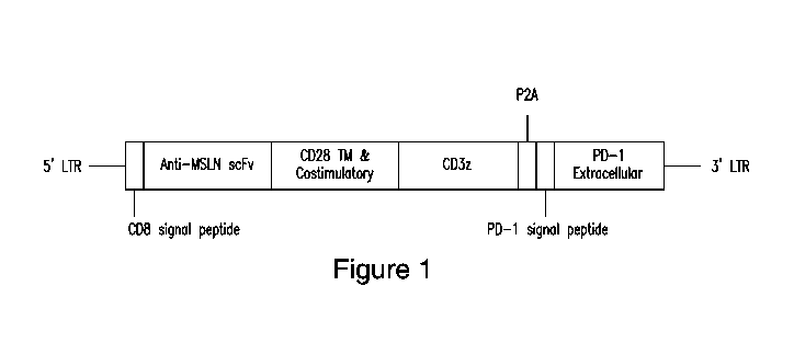
Figure 1 depicts a polypeptide composition in accordance with certain
embodiments of the presently disclosed subject matter. The polypeptide
composition
comprises a CAR comprising an anti-mesothelin (MSLN) scFv, a CD28
transmembrane
domain, a CD28 cytoplasmic signaling domain, a CD3zeta signaling domain (e.g.,
comprising an ITAM2 variant and an ITAM3 variant). The CAR is fused to the
PD1DNR
(and PD1 signaling domain) via a cleavable P2A peptide. SP: signaling peptide;
scFv:
8
CA 03139989 2021-11-10
WO 2020/232433 PCT/US2020/033382
single-chain variable fragment; TM: transmembrane domain; cyt: cytosolic
domain;
DNR: dominant negative receptor; LTR: long terminal repeat.
Figure 2 depicts various constructs disclosed in Example 2.
Figures 3A-3D depict virus production in producer cell line RD114. RD114 cells
were transduced with different dilutions of H29 viral supernatant (undiluted,
1:2, and 1:4)
and stained for CAR expression by flow cytometry using an anti-Fab antibody.
RD114
empty served as a negative control. Figure 3A shows RD114 empty (as a negative
control). Figure 3B shows undiluted; Figure 3C shows sup 1:2 diluted; and
Figure 3D
shows sup 1:4 diluted.
Figures 4A-4E depict transduction of human T cells with M28z1XX-P2A-
PD1DNR ¨ donor H116-2. PHA-activated T cells were transduced with different
concentrations of RD114 viral supernatant (Figure 4A shows 1:2, Figure 4B
shows 1:5,
Figure 4C shows 1:7, Figure 4D shows 1:15, and Figure 4E shows un-transduced
("UT")), and stained for CAR expression by anti-Fab staining and PD1DNR by
anti-PD1
staining using flow cytometry.
Figure 5A-5E depict transduction of human T cells with M28z1XX-P2A-
PD1DNR ¨ donor H18. PHA-activated T cells were transduced with different
concentrations of RD114 viral supernatant (Figure 5A shows 1:2, Figure 5B
shows 1:5,
Figure 5C shows 1:10, Figure 5D shows 1:15, and Figure 5E shows un-transduced
("UT")) and stained for CAR expression by anti-Fab staining and PD1DNR by anti-
PD1
staining using flow cytometry.
Figures 6A-6F depict transduction of human T cells with M28z1XX-P2A-
PD1DNR ¨ donor H19. PHA-activated T cells were transduced with different
concentrations of RD114 viral supernatant (Figure 6A shows 1:2, Figure 6B
shows 1:5,
Figure 6C shows 1:7; Figure 6D shows 1:10, Figure 6E shows 1:15, and Figure 6F
shows
un-transduced ("UT")) and stained for CAR expression by anti-Fab staining and
PD1DNR by anti-PD1 staining using flow cytometry.
Figures 7A-7C depict the correlation of vector copy number (VCN) with median
fluorescence intensity (MFI). PHA-activated T cells were transduced with
different
concentrations of RD114 viral supernatant and stained for CAR expression by
anti-Fab
staining and flow cytometry analysis. Genomic DNA of transduced T cells was
isolated
and vector copy number was determined as VCN/pg DNA using qPCR. The MFI of
CAR-positive cells was correlated with the VCN/pg DNA for three different
donors.
9
CA 03139989 2021-11-10
WO 2020/232433
PCT/US2020/033382
Figure 7A shows donor H19; Figure 7B shows donor H18, and Figure 7C shows
donor
H116-2.
Figure 8 depicts that cytotoxicity for transduced T cells from 3 different
donors.
MSLN high target cells (MGM) were co-cultured with M28z lxx-PD1DNR CAR T cells
from different donors at different E:T ratios using an impedance-based assay.
The
M28z lxx-PD1DNR CAR T-cell mediated cytolysis of MGM cells at E:T ratio 1:1.
M28z lxx-PD1DNR CAR T cells killed high MSLN target cells.
Figure 9 depicts an example of impedance-based cytotoxicity measurement
(eCTL).
Figure 10 depicts parameters of comparative analysis of various constructs
using
eCTL.
Figures 11A-11E depict MSLN and PD-Li expressions of target cell lines.
Mesothelioma (MGM (shown in Figure 11A), MGM-PDL1 (shown in Figure 11B) and
MSTOG (shown in Figure 11C)) and lung cancer (A549GM (shown in Figure 11D) and
A549G (shown in Figure 11e)) cell lines were assessed for MSLN and PD-Li
expressions
by flow cytometry. MGM, MGM-PDL1 and A549GM overexpressed MSLN; MGM-
PDL1 cells additionally overexpressed PD-Li.
Figures 12A-12E depict CAR and PD1 expression of transduced T cells. Human
T cells transduced with M28z (as shown in Figure 12A), M28z lxx (as shown in
Figure
12B), M28z-PD1DNR (as shown in Figure 12C) and M28z lxx-PD1DNR (as shown in
Figure 12D) were analyzed for CAR expression by anti-myc staining and
PD1/PD1DNR
expression by anti-PD1 staining using flow cytometry. Figure 12E shows the un-
transduced ("UT") T cells.
Figures 13A-13C depict comparative analysis of anti-tumor efficacy of CAR T
cells bearing the 1XX domain and PD1DNR for MSLN high tumor cells (MGM). MSLN
high target cells (MGM) were co-cultured with either M28z, M28z lxx, M28z-
PD1DNR,
M28z1XX-PD1DNR or untransduced T cells at the indicated E:T ratios. Anti-tumor
efficacy was assessed using an impedance-based assay. Figure 13A shows the E:T
ratio of
about 3:1. Figure 13B shows the E:T ratio of about 1:1. Figure 13C shows the
E:T ratio
of about 0.33:1.
Figure 14 depicts comparative analysis of cytotoxicity of CAR T cells bearing
the
lxx domain and PD1DNR for MSLN high tumor cells (MGM). MSLN high target cells
(MGM) labeled with chromium-51 were co-cultured with either M28z, M28z1xx,
M28z-
CA 03139989 2021-11-10
WO 2020/232433 PCT/US2020/033382
PD1DNR, M28z lxx-PD1DNR or untransduced T cells at the indicated E:T ratio for
18
hours. Cytotoxicity was determined by chromium-51 CTL.
Figures 15A-15C depict comparative analysis of anti-tumor efficacy of CART
cells bearing the lxx domain and PD1DNR for MSLN negative tumor cells (MSTOG).
MSLN negative target cells (MSTOG) were co-cultured with either M28z, M28z
lxx,
M28z-PD1DNR, M28z1xx-PD1DNR or untransduced T cells at the indicated E:T
ratios.
Anti-tumor efficacy was assessed using an impedance-based assay. Figure 15A
shows
the E:T ratio of about 3:1. Figure 15B shows the E:T ratio of about 1:1.
Figure 15C
shows the E:T ratio of about 0.33:1.
Figure 16 depicts comparative analysis of cytotoxicity of CAR T cells bearing
the
1XX domain and PD1DNR for MSLN negative tumor cells (MSTOG). MSLN negative
target cells (MSTOG) labeled with chromium-51 were co-cultured with either
M28z,
M28z1XX, M28z-PD1DNR, M28z1XX-PD1DNR or untransduced T cells at the
indicated E:T ratio for 18 hours. Cytotoxicity was determined by chromium-51
CTL.
Figures 17A-17C depict comparative analysis of anti-tumor efficacy of CAR T
cells bearing the 1XX domain and PD1DNR for MSLN high tumor cells
overexpressing
PDLl. MSLN high target cells overexpressing PDL1 (MGM-PDL1) were co-cultured
with either M28z, M28z1XX, M28z-PD1DNR, M28z1XX-PD1DNR or untransduced T
cells at the indicated E:T ratios. Anti-tumor efficacy was assessed using an
impedance-
based assay. Figure 17A shows the E:T ratio of about 3:1. Figure 17B shows the
E:T
ratio of about 1:1. Figure 17C shows the E:T ratio of about 0.33:1.
Figures 18A-18C depict comparative analysis of anti-tumor efficacy of CAR T
cells bearing the lxx domain and PD1DNR for MSLN high tumor cells (A549GM).
MSLN high target cells (A549GM) were co-cultured with either M28z, M28z lxx,
M28z-
PD1DNR, M28z lxx-PD1DNR or untransduced T cells at the indicated E:T ratios.
Anti-
tumor efficacy was assessed using an impedance-based assay. Figure 18A shows
the E:T
ratio of about 10:1. Figure 18B shows the E:T ratio of about 5:1. Figure 18C
shows the
E:T ratio of about 2:1.
Figures 19A-19C depict comparative analysis of anti-tumor efficacy of CAR T
cells bearing the lxx domain and PD1DNR: MSLN low tumor cells (A549G). MSLN
low
target cells (A549G) were co-cultured with either M28z, M28z lxx, M28z-PD1DNR,
M28z lxx-PD1DNR or untransduced T cells at the indicated E:T ratios. Anti-
tumor
efficacy was assessed using an impedance-based assay. Figure 19A shows the E:T
ratio
11
CA 03139989 2021-11-10
WO 2020/232433
PCT/US2020/033382
of about 10:1. Figure 19B shows the E:T ratio of about 5:1. Figure 19C shows
the E:T
ratio of about 2:1.
Figures 20A-20D depict the in vivo study results of various treatments. Figure
20A shows the comparative in vivo efficacy of CAR T cells ¨ M28z, M28z with
PD1
antibody, and M28z with PD1DNR.. Figure 20B shows comparative in vivo efficacy
of
M28z and M28z1XX+PD1DNR CAR T cells. Figure 20C shows tumor burden imaging
showing systemic anti-tumor immunity following tumor rechallenges. Figure 20D
shows
ex vivo immunofluorescence staining of orthotopic MPM tumors showing CAR T
cells
infiltration.
Figure 21 depicts structure and components of M28z1XXPD1DNR CAR. In
contrast to M28z, M28z1XXPD1DNR CAR T cells bear a mutated CD3 signaling
domain with a single functional ITAM and co-express a PD1DNR that consists of
CD8
transmembrane and hinge domains and lacks the intracellular PD1 signaling
domain
present in endogenous PD1.
Figure 22 depicts structures of CAR T cell vectors.
Figure 23 depicts expression of mesothelin (MSLN), PD-L1, and GFP on tumor
cell lines. MGM, MGM-PDL1, and MSTOG tumor cells were analyzed for their
expression of mesothelin (left panels), PD-Li (middle panels), and GFP (right
panels) by
flow cytometry. Shown are density plots depicting the relative expression
intensity
plotted against the side scatter area (Y-axis).
Figures 24A-24D depict orthotopic MPM mouse model. Figure 24A shows the
gross appearance of human MPM (left upper panel) is reproduced in the mouse
model of
MPM (right upper panel), with tumor encasing heart, lungs, and mediastinal
structures
and the tumor invading the chest wall (bottom panel). Figure 24B shows
extensive
vascularity of the tumor is demonstrated by the CD34 immunofluorescences.
Figure 24C
shows tumor burden progression monitored by BLI correlates with tumor volume
measurements by Mill at respective time points. Figure 24D shows tumor burden
progression monitored by serial BLI and MRI.
Figures 25A-25C depict expression of mesothelin in human tissues by
immunohistochemical analysis. Figure 25A shows expression of mesothelin in MPM
versus normal pleura and pericardium. Figure 25B shows expression of
mesothelin in
lung adenocarcinoma versus normal lung tissue. Figure 25C shows expression of
mesothelin in triple-negative breast cancer versus normal breast tissue.
12
CA 03139989 2021-11-10
WO 2020/232433 PCT/US2020/033382
Figure 26 depicts that M28z1XXPD1DNR expression can be titrated using
different dilutions of viral supernatant. Human T cells were transduced with
different
dilutions of viral supernatant encoding for M28z1XXPD1DNR (left panels) or
mycM28z1XXPD1DNR (middle panels). Expression of CAR (Y-axis) and PD1 (X-axis)
of viable CD3-positive cells was assessed by flow cytometry. Depicted results
are from 1
donor representative of 3 different donors.
Figure 27 depicts CAR expression measured by MFI correlates with VCN. Human
T cells derived from 3 different donors were transduced with different
dilutions of
retroviral supernatant encoding for either M28z1XXPD1DNR or
mycM28z1XXPD1DNR. The MFI of CAR-positive T cells (as determined by flow
cytometry) was plotted against the VCN (as determined by qPCR). The R2 value
was
derived from a linear regression analysis (black line).
Figures 28A-28D depict expression of PD1 and PD1DNR in
mycM28z1XXPD1DNR and mycM28z CAR T cells. Figure 28A shows percent CAR
surface expression of mycM28z and mycM28z1XXPD1DNR CAR T cells. Figure 28B
shows percent CD3-positive cells positive for PD1 surface expression. Figure
28C shows
MFI of PD1 surface expression of CD3-positive cells. Figure 28D shows relative
mRNA
expression of PD1 extracellular and PD1 intracellular domain shown as fold
change,
compared with that of untransduced T cells.
Figure 29 depicts M28z1XXPD1DNR-expressing T cells with or without myc-tag
exhibit identical antitumor efficacy in vitro. Human T cells derived from 3
different
donors were transduced with either M28z1XXPD1DNR (red) or mycM28z1XXPD1DNR
(green) (transduction range, 37%-63%) and cocultured with MGM cells (green;
arrow
indicates time of T-cell addition). The antitumor efficacy of both constructs
was
compared at the indicted E:T ratios using an impedance-based cytotoxicity
assay.
Figure 30 shows that mycM28z1XXPD1DNR CAR T cells mediate antigen-
specific, HLA-independent tumor lysis. Human T cells transduced with
mycM28z1XXPD1DNR (blue) or mycM28z (red) were co-cultured with either MGM,
MGM-PDL1, or MSTOG tumor cells at the indicated E:T ratios. The cytotoxicity
of CAR
T cells was assessed by 51Cr-release assay after 18 h of coculture.
Untransduced T cells
(orange) served as control.
Figure 31 depicts accumulation of mycM28z1XXPD1DNR CART cells upon
stimulation with mesothelin-expressing tumor cells. Human T cells transduced
with
mycM28z1XXPD1DNR (blue) or mycM28z (red) were repeatedly exposed to MGM or
13
CA 03139989 2021-11-10
WO 2020/232433 PCT/US2020/033382
MGM-PDL1 target cells for 48 h at an E:T ratio of 1:1. The accumulation of CAR
T cells
was quantified by absolute CAR T-cell count after each antigen stimulation.
Figure 32 shows that mycM28z1XXPD1DNR CAR T cells exhibited similar
cytotoxicity to mycM28z CAR T cells upon initial antigen stimulation. Human T
cells
transduced with mycM28z1XXPD1DNR (blue) or mycM28z (red) were cocultured with
51Cr-labeled MGM or MGM-PDL1 target cells at the indicated E:T ratios.
Cytotoxicity
was assessed after 18 h using a 51Cr-release assay. Untransduced T cells
(orange) served
as control.
Figure 33 shows that mycM28z1XXPD1DNR CAR T cells retained antitumor
efficacy upon repeated antigen stimulation. Human T cells transduced with
mycM28z1XXPD1DNR (blue) or mycM28z (red) were repeatedly exposed to MGM (left
panels) or MGM-PDL1 (right panels) target cells for 48 h at an E:T ratio of
3:1 for 4
stimulations followed by an E:T ratio of 1:1 for 2 additional stimulations.
The
cytotoxicity of CART cells was assessed by 51Cr-release assay upon the fourth
and
seventh antigen stimulations at the indicated E:T ratios after 18 h of
coculture.
Figure 34 shows that mycM28z1XXPD1DNR CAR T cells secreted effector
cytokines upon antigen stimulation. Human T cells transduced with
mycM28z1XXPD1DNR (blue) or mycM28z (red) were repeatedly exposed to MGM (top
row) or MGM-PDL1 (bottom row) target cells for 48 h at an E:T ratio of 1:1.
Cell-free
supernatant was collected 24 h after the first, third, and sixth antigen
exposures, and
effector cytokine secretion was assessed by Luminex assay.
Figure 35 depicts intrapleural administration of a single low dose of 3 x10⁴
to
mycM28z1XXPD1DNR CAR T cells demonstrates antitumor efficacy in vivo. Female
NSG mice bearing orthotopic MGM tumor were treated with a single intrapleural
dose of
either P28z CAR T cells (n=6, red bar) or mycM28z1XXPD1DNR CAR T cells (n=10,
blue bar). Tumor burden was measured by BLI. The indicated time point
represents day
15 after CAR T-cell administration, when P28z CAR T cell¨treated mice started
to
become moribund. Statistical significance was determined using an unpaired
Student's t
test (2-tailed). ***p<0.001.
Figures 36A-36D depict that intrapleurally administered mycM28z1XXPD1DNR
CAR T cells exhibited antitumor efficacy in vivo and increase survival. Figure
3A shows
serial tumor BLI of MGM-PDL1 tumor¨bearing female NSG mice treated with a
single
dose of mycM28z (1x10⁵) or mycM28z1XXPD1DNR (1x10⁵ or 5x10⁴) CART cells
(n=7-8). Shown are 4 mice per treatment group in the ventral position. Figure
36B shows
14
CA 03139989 2021-11-10
WO 2020/232433 PCT/US2020/033382
corresponding serial tumor BLI (average of dorsal and ventral) indicating
tumor burden
of each treated mouse. Figure 36C shows corresponding mice weights following
treatment. Figure 36D shows Kaplan-Meier survival analysis comparing the in
vivo
efficacy of mycM28z and mycM28z1XXPD1DNR CAR T cells. The survival curve was
analyzed using the log-rank test. *p<0.05, **p<0.01.
Figure 37 depicts detection of mycM28z1XXPD1DNR CAR T cells in the
primary tumor of intrapleurally treated mice. Pleural MGM tumor from mice
treated with
5x10⁵ untransduced T cells (left), mycM28z CART cells (middle), or
mycM28z1XXPD1DNR CAR T cells (right). Tumor tissue was collected 3 days after
intrapleural T-cell injection, fixed, and stained ex vivo for tumor mesothelin
(green),
human CD45-positive cells (red), and DAPI (cell nucleus, blue) by
immunofluorescence.
Figures 38A and 38B show that mycM28z1XXPD1DNR CAR T cells resisted
tumor reestablishment upon repeated tumor challenge in vivo. Figure 38A shows
scheme
illustrating the tumor rechallenge experiment: 68 days after intrapleural
administration of
mycM28z or mycM28z1XXPD1DNR CART cells (single dose of 1x10⁵ CART cells)
and after pleural eradication of MGM-PDL1 tumor cells (inoculation dose of
8x10⁵),
mice were rechallenged 10 times intraperitoneally with escalating doses (2x10⁶
to
11x106) of MGM tumor cells every 4-8 days. Figure 38B shows serial BLI
indicating
tumor burden following a single intrapleural dose of mycM28z (2 mice, red
lines) or
mycM28z1XXPD1DNR (3 mice, black lines) CAR T cells and tumor rechallenge
starting
at treatment day 68. Black arrows indicate time points of intraperitoneal
tumor
rechallenge with escalating doses.
Figures 39A-39C depict M28z1XXPD1DNR CAR T cells manufactured using the
vector stocks for the clinical trial possess antitumor efficacy in vivo and
prolong survival.
Figure 39A shows serial tumor BLI of MGM tumor-bearing female NSG mice treated
with of 6x10⁴ (n=8) or 2x10⁵ (n=10) M28z1XXPD1DNR CART cells manufactured by
CTCEF using the viral supernatant for the clinical trial. Figure 39B shows
corresponding
mice weights following treatment. Figure 39C shows Kaplan-Meier survival
analysis.
Figure 40 depicts average body weights at the interim sacrifice for male mice.
Groups 1 (nontumor control), 3 (control vehicle), and 5 (test article) are
shown.
Figure 41 depicts average body weights at the interim sacrifice for female
mice.
Groups 2 (nontumor control), 4 (control), and 6 (test article) are shown.
Figure 42 depicts average body weights at the final sacrifice for male mice.
Groups 7 (nontumor control), 9 (control vehicle), and 11 (test article) are
shown.
CA 03139989 2021-11-10
WO 2020/232433 PCT/US2020/033382
Figure 43 depicts average body weights at the final sacrifice for female mice.
Groups 8 (nontumor control), 10 (control vehicle), and 12 (test article) are
shown.
Figure 44 depicts identification of human T cells in the tumors of CART cell¨
treated and vehicle-treated mice. Intrapleural tumor tissue cells derived from
CAR T cell-
treated and vehicle-treated mice were stained with DAPI, anti-human CD45
APC/CY7,
and anti-human CD3 PE/CY7 antibodies to detect viable human T cells by flow
cytometry. Shown are density plots of human CD3 expression (X-axis) and human
CD45
expression (Y-axis) of DAPI-negative (alive) single cells. The gate indicates
cells stained
positive for human CD45 and human CD3, representing human T cells.
Figure 45 depicts identification of human T cells in the spleens of CART cell¨
treated and vehicle-treated mice. Spleen tissue cells derived from CAR T
cell¨treated and
vehicle-treated mice were stained with DAPI, anti-human CD45 APC/CY7, and anti-
human CD3 PE/CY7 antibodies to detect viable human T cells by flow cytometry.
Shown
are density plots of human CD3 expression (X-axis) and human CD45 expression
(Y-
axis) of DAPI-negative (alive) single cells. The gate indicates cells stained
positive for
human CD45 and human CD3, representing human T cells.
Figure 46 depicts BLI of male mice.
Figure 47 depicts BLI of female mice.
5. DETAILED DESCRIPTION OF THE INVENTION
The presently disclosed subject matter provides polypeptide compositions
comprising a chimeric antigen receptor (CAR) targeting mesothelin and a
dominant
negative form of programmed death 1 (PD-1 DN), and immunoresponsive cells
(e.g., T
cells or NK cells) comprising the polypeptide composition. The presently
disclosed
subject matter also provides methods of using such polypeptide composition for
inducing
and/or enhancing an immune response of an immunoresponsive cell to a target
antigen,
and/or treating and/or preventing neoplasms or other diseases/disorders where
an decrease
in immune cell exhaustion is desired.
Persistent antigen exposure of T cells, such as in cancer, leads to an altered
T-cell
differentiation state, termed exhaustion, that renders CAR T cells
dysfunctional
(Youngblood et al., Int Immunol. 2010;22(10):797-803; Wherry et al., Nat Rev
Immunol.
2015;15(8):486-499). Previous studies have shown that CAR activation potential
is
associated with three ITAMs (1-2-3) present in the CD3 cytoplasmic domain
(Acuto et
at., Nat Rev Immunol. 2003;3(12):939-951; Love et at., Cold Spring Harb
Perspect Biol.
2010;2(6):a002485). Recent studies demonstrated that this CAR activation
potential could
16
CA 03139989 2021-11-10
WO 2020/232433 PCT/US2020/033382
be calibrated by mutating ITAMs, thereby reducing their functionality.
Importantly, it
was shown that, by introducing point mutations in the second- and third-
position ITAMs
(1-X-X; herein designated as "1XX") of the CD3 domain, the fate of CART cells
was
changed from an exhaustive state to a balanced effector and memory state in
the presence
of high antigen exposure (Feucht et al ., Nat Med. 2019;25(1):82-88).
Another hurdle CAR T cells encounter in the solid tumor microenvironment is
inhibition of their cytolytic activity mediated through PD1, an inhibitory
receptor that is
expressed upon antigen-mediated T-cell activation. In addition, tumor cells
augment the
expression of coinhibitory ligands such as PD-Ll following exposure to T-cell-
secreted
.. proapoptotic cytokines (McGray et at., Mot Ther. 2014;22(1):206-218;
Spranger et at.,
Sci Transl Med. 2013;5(200):200ra116; Moon et at., Clin Cancer Res.
2014;20(16):4262-
4273). To overcome this hurdle, our group has combined mesothelin-targeted CAR
T
cells with a PD1 blocking antibody to rescue exhausted CAR T cells, restoring
the
antitumor efficacy of CAR T cells in our orthotopic mouse model (Cherkassky et
at., J
Clin Invest. 2016;126(8):3130-3144; Grosser et al., Cancer Cell.
2019;36(5):471-482).
To avoid repeated dosage of PD1 checkpoint blockade agents and associated
clinical
adverse effects, our group has demonstrated that the use of a cell-intrinsic
PD1
checkpoint blockade strategy¨wherein a PD1DNR is co-transduced into the T cell
along
with a second-generation CAR¨ultimately renders the transduced cells resistant
to tumor
PD-Li -mediated inhibition in the solid tumor microenvironment Cherkassky et
at., J Clin
Invest. 2016;126(8):3130-3144; Grosser et al., Cancer Cell. 2019;36(5):471-
482).
Hence, to develop CAR T cells with an enhanced therapeutic profile, functional
persistence, and resistance to tumor-mediated inhibition, the inventors
incorporated the
1XX and PD1DNR components into the second-generation CAR vector design, which
allows these cells to perform efficiently in the highly immunosuppressive
microenvironment of solid tumors.
For purposes of clarity of disclosure and not by way of limitation, the
detailed
description is divided into the following subsections:
5.1. Definitions;
5.2. Polypeptide Compositions;
5.2.1. PD-1 DN;
5.2.2. Mesothelin-targeted CARs; and
5.2.3. Exemplified Polypeptide Compositions;
5.3. Immunoresponsive Cells;
17
CA 03139989 2021-11-10
WO 2020/232433 PCT/US2020/033382
5.4. Nucleic Acid Compositions and Vectors;
5.5. Polypeptides and Analogs;
5.6. Pharmaceutical Compositions and Administration;
5.7. Formulations;
5.8. Methods of Treatments; and
5.9. Kits
5.1. Definitions
Unless defined otherwise, all technical and scientific terms used herein have
the
meaning commonly understood by a person skilled in the art.
As used herein, the term "about" or "approximately" means within an acceptable
error range for the particular value as determined by one of ordinary skill in
the art, which
will depend in part on how the value is measured or determined, i.e., the
limitations of the
measurement system. For example, "about" can mean within 3 or more than 3
standard
deviations, per the practice in the art Alternatively, "about" can mean a
range of up to
20%, e.g., up to 10%, up to 5%, or up to 1% of a given value. Alternatively,
particularly
with respect to biological systems or processes, the term can mean within an
order of
magnitude, e.g., within 5-fold or within 2-fold, of a value.
By "immunoresponsive cell" is meant a cell that functions in an immune
response
or a progenitor, or progeny thereof
By "activates an immunoresponsive cell" is meant induction of signal
transduction
or changes in protein expression in the cell resulting in initiation of an
immune response.
For example, when CD3 Chains cluster in response to ligand binding and
immunoreceptor tyrosine-based inhibition motifs (ITAMs), a signal transduction
cascade
is produced. In certain embodiments, when a chimeric antigen receptor (CAR)
binds to
an antigen, a formation of an immunological synapse occurs that includes
clustering of
many molecules near the bound receptor (e.g. CD4 or CD8, CD3y/o/c/C, etc.).
This
clustering of membrane bound signaling molecules allows for ITAM motifs
contained
within the CD3 chains to become phosphorylated. This phosphorylation in turn
initiates a
T cell activation pathway ultimately activating transcription factors, such as
NF-KB and
AP-1. These transcription factors induce global gene expression of the T cell
to increase
IL-2 production for proliferation and expression of master regulator T cell
proteins in
order to initiate a T cell mediated immune response.
18
CA 03139989 2021-11-10
WO 2020/232433 PCT/US2020/033382
By "stimulates an immunoresponsive cell" is meant a signal that results in a
robust and sustained immune response. In various embodiments, this occurs
after the
activation of an immunoresponsive cell (e.g., a T cell) or concomitantly
mediates through
receptors including, but not limited to, CD28, CD137 (4-1BB), 0X40, CD40 and
ICOS.
Receiving multiple co-stimulatory signals can be important to mount a robust
and long-
term T cell mediated immune response. T cells can quickly become inhibited and
unresponsive to antigen. While the effects of these co-stimulatory signals may
vary, they
generally result in increased gene expression in order to generate long lived,
proliferative,
and anti-apoptotic T cells that robustly respond to antigen for complete and
sustained
eradication.
As used herein, the term "antibody" means not only intact antibody molecules,
but
also fragments of antibody molecules that retain immunogen-binding ability.
Such
fragments are also well known in the art and are regularly employed both in
vitro and
in vivo. Accordingly, as used herein, the term "antibody" means not only
intact
immunoglobulin molecules but also the well-known active fragments F(a1302, and
Fab.
F(a1302, and Fab fragments that lack the Fe fragment of intact antibody, clear
more rapidly
from the circulation, and may have less non-specific tissue binding of an
intact antibody
(Wahl et al., I Nucl. Med. 24:316-325 (1983). As used herein, antibodies
include whole
native antibodies, bispecific antibodies; chimeric antibodies; Fab, Fab',
single chain V
region fragments (scFv), fusion polypeptides, and unconventional antibodies.
In certain
embodiments, an antibody is a glycoprotein comprising at least two heavy (H)
chains and
two light (L) chains inter-connected by disulfide bonds. Each heavy chain is
comprised
of a heavy chain variable region (abbreviated herein as VH) and a heavy chain
constant
(CH) region. The heavy chain constant region is comprised of three domains,
CHL CH2
and CH3. Each light chain is comprised of a light chain variable region
(abbreviated
herein as VL) and a light chain constant CL region. The light chain constant
region is
comprised of one domain, CL. The VH and VL regions can be further sub-divided
into
regions of hypervariability, termed complementarity determining regions (CDR),
interspersed with regions that are more conserved, termed framework regions
(FR). Each
VH and VL is composed of three CDRs and four FRs arranged from amino-terminus
to
carboxy-terminus in the following order: FR1, CDR1, FR2, CDR2, FR3, CDR3, FR4.
The variable regions of the heavy and light chains contain a binding domain
that interacts
with an antigen. The constant regions of the antibodies may mediate the
binding of the
19
CA 03139989 2021-11-10
WO 2020/232433
PCT/US2020/033382
immunoglobulin to host tissues or factors, including various cells of the
immune system
(e.g., effector cells) and the first component (Cl q) of the classical
complement system.
As used herein, "CDRs" are defined as the complementarity determining region
amino acid sequences of an antibody which are the hypervariable regions of
immunoglobulin heavy and light chains. See, e.g., Kabat et al., Sequences of
Proteins of
Immunological Interest, 4th U. S. Department of Health and Human Services,
National
Institutes of Health (1987). Generally, antibodies comprise three heavy chain
and three
light chain CDRs or CDR regions in the variable region. CDRs provide the
majority of
contact residues for the binding of the antibody to the antigen or epitope. In
certain
embodiments, the CDRs regions are delineated using the Kabat system (Kabat, E.
A., et
at. (1991) Sequences of Proteins of Immunological Interest, Fifth Edition,
U.S.
Department of Health and Human Services, NIH Publication No. 91-3242). In
certain
embodiments, the CDRs are identified according to the Kabat system.
As used herein, the term "single-chain variable fragment" or "scFv" is a
fusion
protein of the variable regions of the heavy (VH) and light chains (VL) of an
immunoglobulin covalently linked to form a VH: :VL heterodimer. The VH and VL
are
either joined directly or joined by a peptide-encoding linker (e.g., 10, 15,
20, 25 amino
acids), which connects the N-terminus of the VH with the C-terminus of the VL,
or the C-
terminus of the VH with the N-terminus of the VL. The linker is usually rich
in glycine for
flexibility, as well as serine or threonine for solubility. "Linker", as used
herein, shall
mean a functional group (e.g., chemical or polypeptide) that covalently
attaches two or
more polypeptides or nucleic acids so that they are connected to one another.
As used
herein, a "peptide linker" refers to one or more amino acids used to couple
two proteins
together (e.g., to couple VH and VL domains). In certain embodiments, the
linker
comprises or consists of the amino acid sequence set forth in SEQ ID NO: 66,
which is
provided below:
GGGGSGGGGSGGGGS [SEQ ID NO: 66].
An exemplary nucleotide sequence encoding the amino acid sequence of SEQ ID
NO: 66 is set forth in SEQ ID NO: 50, which is provided below:
GGAGGTGGAGGCTCAGGAGGAGGAGGCAGTGGAGGTGGTGGGTCA [SEQ ID NO:50].
An exemplary nucleotide sequence encoding the amino acid sequence f SEQ ID
NO:66 is set forth in SEQ ID NO: 51, which is provided below.
GGTGGAGGCGGTTCAGGCGGAGGTGGCTCTGGCGGTGGCGGATCA [SEQ ID NO: 51]
CA 03139989 2021-11-10
WO 2020/232433
PCT/US2020/033382
Despite removal of the constant regions and the introduction of a linker, scFv
proteins retain the specificity of the original immunoglobulin. Single chain
Fv
polypeptide antibodies can be expressed from a nucleic acid including VH - and
VL
-encoding sequences as described by Huston, et al. (Proc. Nat. Acad. Sci. USA,
85:5879-
5883, 1988). See, also, U.S. Patent Nos. 5,091,513, 5,132,405 and 4,956,778;
and U.S.
Patent Publication Nos. 20050196754 and 20050196754. Antagonistic scFvs having
inhibitory activity have been described (see, e.g., Zhao et al., Hyrbidoma
(Larchmt) 2008
27(6):455-51; Peter et al., J Cachexia Sarcopenia Muscle 2012 August 12; Shieh
et al., J
Imuno12009 183(4):2277-85; Giomarelli et al., Thromb Haemost 2007 97(6):955-
63; Fife
eta., J Clin Invst 2006 116(8):2252-61; Brocks et al., Immunotechnology 1997
3(3):173-
84; Moosmayer et al., Ther Immunol 1995 2(10:31-40). Agonistic scFvs having
stimulatory activity have been described (see, e.g., Peter et al., J Bioi Chem
2003
25278(38):36740-7; Xie et al., Nat Biotech 1997 15(8):768-71; Ledbetter et
al., Crit Rev
Immuno11997 17(5-6):427-55; Ho et al., BioChim Biophys Acta 2003 1638(3):257-
66).
As used herein, "F(ab)" refers to a fragment of an antibody structure that
binds to
an antigen but is monovalent and does not have a Fc portion, for example, an
antibody
digested by the enzyme papain yields two F(ab) fragments and an Fc fragment
(e.g., a
heavy (H) chain constant region; Fc region that does not bind to an antigen).
As used herein, "F(ab)2" refers to an antibody fragment generated by pepsin
digestion of whole IgG antibodies, wherein this fragment has two antigen
binding (ab')
(bivalent) regions, wherein each (ab') region comprises two separate amino
acid chains, a
part of a H chain and a light (L) chain linked by an S-S bond for binding an
antigen and
where the remaining H chain portions are linked together. A "F(a1302" fragment
can be
split into two individual Fab' fragments.
As used herein, the term "vector" refers to any genetic element, such as a
plasmid,
phage, transposon, cosmid, chromosome, virus, virion, etc., which is capable
of
replication when associated with the proper control elements and which can
transfer gene
sequences into cells. Thus, the term includes cloning and expression vehicles,
as well as
viral vectors and plasmid vectors.
As used herein, the term "expression vector" refers to a recombinant nucleic
acid
sequence, i.e. recombinant DNA molecule, containing a desired coding sequence
and
appropriate nucleic acid sequences necessary for the expression of the
operably linked
coding sequence in a particular host organism. Nucleic acid sequences
necessary for
expression in prokaryotes usually include a promoter, an operator (optional),
and a
21
CA 03139989 2021-11-10
WO 2020/232433 PCT/US2020/033382
ribosome binding site, often along with other sequences. Eukaryotic cells are
known to
utilize promoters, enhancers, and termination and polyadenylation signals.
As used herein, the term "affinity" is meant a measure of binding strength.
Affinity can depend on the closeness of stereochemical fit between antibody
combining
sites and antigen determinants, on the size of the area of contact between
them, and/or on
the distribution of charged and hydrophobic groups. Methods for calculating
the affinity
of an antibody for an antigen are known in the art, including, but not limited
to, various
antigen-binding experiments, e.g., functional assays (e.g., flow cytometry
assay).
The term "chimeric antigen receptor" or "CAR" as used herein refers to a
.. molecule comprising an extracellular antigen-binding domain that is fused
to an
intracellular signaling domain that is capable of activating or stimulating an
immunoresponsive cell, and a transmembrane domain. In certain embodiments, the
extracellular antigen-binding domain of a CAR comprises a scFv. The scFv can
be
derived from fusing the variable heavy and light regions of an antibody.
Alternatively or
additionally, the scFv may be derived from Fab's (instead of from an antibody,
e.g.,
obtained from Fab libraries). In certain embodiments, the scFv is fused to the
transmembrane domain and then to the intracellular signaling domain. In
certain
embodiments, the CAR is selected to have high binding affinity for the
antigen.
As used herein, the term "nucleic acid molecules" include any nucleic acid
molecule that encodes a polypeptide of interest or a fragment thereof Such
nucleic acid
molecules need not be 100% homologous or identical to an endogenous nucleic
acid
sequence, but may exhibit substantial identity.
By "substantially identical" or "substantially homologous" is meant an amino
acid
sequence or a nucleic acid molecule exhibiting at least about 50% homologous
or
identical to a reference amino acid sequence (for example, any one of the
amino acid
sequences described herein) or a reference nucleic acid sequence (for example,
any one of
the nucleic acid sequences described herein). In certain embodiments, such a
sequence is
at least about 60%, at least about 65%, at least about 70%, at least about
75%, at least
about 80%, at least about 85%, at least about 90%, at least about 95%, at
least about 99%,
or at least about 100% homologous or identical to the sequence of the
reference amino
acid or the reference nucleic acid used for comparison.
Sequence identity can be measured by using sequence analysis software (for
example, Sequence Analysis Software Package of the Genetics Computer Group,
University of Wisconsin Biotechnology Center, 1710 University Avenue, Madison,
Wis.
22
CA 03139989 2021-11-10
WO 2020/232433
PCT/US2020/033382
53705, BLAST, BESTFIT, GAP, or PILEUP/PRETTYBOX programs). Such software
matches identical or similar sequences by assigning degrees of homology to
various
substitutions, deletions, and/or other modifications. Conservative
substitutions typically
include substitutions within the following groups: glycine, alanine; valine,
isoleucine,
leucine; aspartic acid, glutamic acid, asparagine, glutamine; serine,
threonine; lysine,
arginine; and phenylalanine, tyrosine. In an exemplary approach to determining
the
degree of identity, a BLAST program may be used, with a probability score
between e-3
and e-100 indicating a closely related sequence.
As used herein, the percent homology between two amino acid sequences is
equivalent to the percent identity between the two sequences. The percent
identity
between the two sequences is a function of the number of identical positions
shared by
the sequences (i.e.,% homology = # of identical positions/total # of positions
x 100),
taking into account the number of gaps, and the length of each gap, which need
to be
introduced for optimal alignment of the two sequences. The comparison of
sequences
and determination of percent identity between two sequences can be
accomplished using
a mathematical algorithm.
The percent homology between two amino acid sequences can be determined
using the algorithm of E. Meyers and W. Miller (Comput. Appl. Biosci., 4:11-17
(1988))
which has been incorporated into the ALIGN program (version 2.0), using a
PAM120
weight residue table, a gap length penalty of 12 and a gap penalty of 4. In
addition, the
percent homology between two amino acid sequences can be determined using the
Needleman and Wunsch (J. Mol. Biol. 48:444-453 (1970)) algorithm which has
been
incorporated into the GAP program in the GCG software package (available at
www.gcg.com), using either a Blossum 62 matrix or a PAM250 matrix, and a gap
weight
of 16, 14, 12, 10, 8, 6, or 4 and a length weight of 1, 2, 3, 4, 5, or 6.
Additionally or alternatively, the amino acids sequences of the presently
disclosed
subject matter can further be used as a "query sequence" to perform a search
against
public databases to, for example, identify related sequences. Such searches
can be
performed using the )(BLAST program (version 2.0) of Altschul, et al. (1990)
J. Mol.
Biol. 215:403-10. BLAST protein searches can be performed with the )(BLAST
program, score = 50, wordlength = 3 to obtain amino acid sequences homologous
to the
specified sequences (e.g., heavy and light chain variable region sequences of
scEv m903,
m904, m905, m906, and m900) disclosed herein. To obtain gapped alignments for
comparison purposes, Gapped BLAST can be utilized as described in Altschul et
al.,
23
CA 03139989 2021-11-10
WO 2020/232433 PCT/US2020/033382
(1997) Nucleic Acids Res. 25(17):3389-3402. When utilizing BLAST and Gapped
BLAST programs, the default parameters of the respective programs (e.g.,
)(BLAST and
NBLAST) can be used. The term "constitutive expression" or "constitutively
expressed"
as used herein refers to expression or expressed under all physiological
conditions.
By "disease" is meant any condition, disease or disorder that damages or
interferes with the normal function of a cell, tissue, or organ, e.g.,
neoplasm, and
pathogen infection of cell.
By "effective amount" is meant an amount sufficient to have a therapeutic
effect.
In certain embodiments, an "effective amount" is an amount sufficient to
arrest,
ameliorate, or inhibit the continued proliferation, growth, or metastasis
(e.g., invasion, or
migration) of a neoplasm.
By "modulate" is meant positively or negatively alter. Exemplary modulations
include a about 1%, about 2%, about 5%, about 10%, about 25%, about 50%, about
75%,
or about 100% change.
By "increase" is meant to alter positively by at least about 5%. An alteration
may
be by about 5%, about 10%, about 25%, about 30%, about 50%, about 75%, about
100%
or more.
By "reduce" is meant to alter negatively by at least about 5%. An alteration
may
be by about 5%, about 10%, about 25%, about 30%, about 50%, about 75%, or even
by
about 100%.
By "isolated cell" is meant a cell that is separated from the molecular and/or
cellular components that naturally accompany the cell.
The terms "isolated," "purified," or "biologically pure" refer to material
that is
free to varying degrees from components which normally accompany it as found
in its
native state. "Isolate" denotes a degree of separation from original source or
surroundings. "Purify" denotes a degree of separation that is higher than
isolation. A
"purified" or "biologically pure" protein is sufficiently free of other
materials such that
any impurities do not materially affect the biological properties of the
protein or cause
other adverse consequences. That is, a nucleic acid or peptide is purified if
it is
substantially free of cellular material, viral material, or culture medium
when produced by
recombinant DNA techniques, or chemical precursors or other chemicals when
chemically synthesized. Purity and homogeneity are typically determined using
analytical chemistry techniques, for example, polyacrylamide gel
electrophoresis or high
performance liquid chromatography. The term "purified" can denote that a
nucleic acid
24
CA 03139989 2021-11-10
WO 2020/232433
PCT/US2020/033382
or protein gives rise to essentially one band in an electrophoretic gel. For a
protein that
can be subjected to modifications, for example, phosphorylation or
glycosylation,
different modifications may give rise to different isolated proteins, which
can be
separately purified.
By "neoplasm" is meant a disease characterized by the pathological
proliferation
of a cell or tissue and its subsequent migration to or invasion of other
tissues or organs.
Neoplastic growth is typically uncontrolled and progressive, and occurs under
conditions
that would not elicit, or would cause cessation of, multiplication of normal
cells.
Neoplasm can affect a variety of cell types, tissues, or organs, including but
not limited to
an organ selected from the group consisting of bladder, bone, brain, breast,
cartilage, glia,
esophagus, fallopian tube, gallbladder, heart, intestines, kidney, liver,
lung, lymph node,
nervous tissue, ovaries, pancreas, prostate, skeletal muscle, skin, spinal
cord, spleen,
stomach, testes, thymus, thyroid, trachea, urogenital tract, ureter, urethra,
uterus, and
vagina, or a tissue or cell type thereof Neoplasm include cancers, such as
sarcomas,
carcinomas, or plasmacytomas (malignant tumor of the plasma cells). In certain
embodiments, the neoplasm is a solid tumor. The neoplasm can a primary tumor
or
primary cancer. In addition, the neoplasm can be in metastatic status.
As used herein, the term "a conservative sequence modification" refers to an
amino acid modification that does not significantly affect or alter the
binding
characteristics of the presently disclosed mesothelin-targeted CAR (e.g., the
extracellular
antigen-binding domain of the CAR) comprising the amino acid sequence.
Conservative
modifications can include amino acid substitutions, additions and deletions.
Modifications can be introduced into the extracellular antigen-binding domain
of the
presently disclosed CAR by standard techniques known in the art, such as site-
directed
mutagenesis and PCR-mediated mutagenesis. Amino acids can be classified into
groups
according to their physicochemical properties such as charge and polarity.
Conservative
amino acid substitutions are ones in which the amino acid residue is replaced
with an
amino acid within the same group. For example, amino acids can be classified
by charge:
positively-charged amino acids include lysine, arginine, histidine, negatively-
charged
amino acids include aspartic acid, glutamic acid, neutral charge amino acids
include
alanine, asparagine, cysteine, glutamine, glycine, isoleucine, leucine,
methionine,
phenylalanine, proline, serine, threonine, tryptophan, tyrosine, and valine.
In addition,
amino acids can be classified by polarity: polar amino acids include arginine
(basic
CA 03139989 2021-11-10
WO 2020/232433
PCT/US2020/033382
polar), asparagine, aspartic acid (acidic polar), glutamic acid (acidic
polar), glutamine,
histidine (basic polar), lysine (basic polar), serine, threonine, and
tyrosine; non-polar
amino acids include alanine, cysteine, glycine, isoleucine, leucine,
methionine,
phenylalanine, proline, tryptophan, and valine. Thus, one or more amino acid
residues
within a CDR region can be replaced with other amino acid residues from the
same group
and the altered antibody can be tested for retained function (i.e., the
functions set forth in
(c) through (1) above) using the functional assays described herein. In
certain
embodiments, no more than one, no more than two, no more than three, no more
than
four, no more than five residues within a specified sequence or a CDR region
are altered.
By "signal sequence" or "leader sequence" is meant a peptide sequence (e.g.,
5,
10, 15, 20, 25 or 30 amino acids) present at the N-terminus of newly
synthesized proteins
that directs their entry to the secretory pathway. Exemplary leader sequences
include, but
is not limited to, a human IL-2 signal sequence (e.g. MYRMQLLSCIALSLALVTNS
[SEQ ID NO: 67]), a mouse IL-2 signal sequence (e.g., MYSMQLASCVTLTLVLLVNS
[SEQ ID NO: 68]); a human kappa leader sequence (e.g.,
METPAQLLFLLLLWLPDTTG [SEQ ID NO: 69]), a mouse kappa leader sequence (e.g.,
METDTLLLWVLLLWVPGSTG [SEQ ID NO: 70]); a human CD8 leader sequence
(e.g., MALPVTALLLPLALLLHAARP [SEQ ID NO: 71]); a truncated human CD8
signal peptide (e.g., MALPVTALLLPLALLLHA [SEQ ID NO: 72]); a human albumin
signal sequence (e.g., MKWVTFISLLFSSAYS [SEQ ID NO: 73]); and a human
prolactin signal sequence (e.g., MDSKGSSQKGSRLLLLLVVSNLLLCQGVVS [SEQ
ID NO: 74]).
In certain embodiments, the CAR comprises a CD8 signal peptide at the N-
terminus, e.g., the signal peptide is connected to the extracellular antigen-
binding domain
of the CAR. In certain embodiments, the CD8 signal peptide comprises or
consists of the
amino acid sequence set forth in SEQ ID NO: 71.
An exemplary nucleotide encoding the amino acid sequence of SEQ ID NO: 71 is
set forth in SEQ ID NO: 125. SEQ ID NO: 125 is provided below.
ATGGCCCTGCCAGTAACGGCTCTGCTGCTGCCACTTGCTCTGCTCCTCCATGCAGCCAGGCCT [ SEQ ID
NO: 125]
The terms "comprises", "comprising", and are intended to have the broad
meaning
ascribed to them in U.S. Patent Law and can mean "includes", "including" and
the like.
As used herein, "treatment" refers to clinical intervention in an attempt to
alter the
disease course of the individual or cell being treated, and can be performed
either for
26
CA 03139989 2021-11-10
WO 2020/232433 PCT/US2020/033382
prophylaxis or during the course of clinical pathology. Therapeutic effects of
treatment
include, without limitation, preventing occurrence or recurrence of disease,
alleviation of
symptoms, diminishment of any direct or indirect pathological consequences of
the
disease, preventing metastases, decreasing the rate of disease progression,
amelioration or
palliation of the disease state, and remission or improved prognosis. By
preventing
progression of a disease or disorder, a treatment can prevent deterioration
due to a
disorder in an affected or diagnosed subject or a subject suspected of having
the disorder,
but also a treatment may prevent the onset of the disorder or a symptom of the
disorder in
a subject at risk for the disorder or suspected of having the disorder.
An "individual" or "subject" herein is a vertebrate, such as a human or non-
human
animal, for example, a mammal. Mammals include, but are not limited to,
humans,
primates, farm animals, sport animals, rodents and pets. Non-limiting examples
of non-
human animal subjects include rodents such as mice, rats, hamsters, and guinea
pigs;
rabbits; dogs; cats; sheep; pigs; goats; cattle; horses; and non-human
primates such as
apes and monkeys. The term "immunocompromised" as used herein refers to a
subject
who has an immunodeficiency. The subject is very vulnerable to opportunistic
infections,
infections caused by organisms that usually do not cause disease in a person
with a
healthy immune system, but can affect people with a poorly functioning or
suppressed
immune system.
Other aspects of the presently disclosed subject matter are described in the
following disclosure and are within the ambit of the presently disclosed
subject matter.
5.2. Polypeptide Compositions
The presently disclosed subject matter provides polypeptide compositions
comprising a mesothelin-targeted chimeric antigen receptor (CAR) and a
dominant
negative form of programmed death 1 (PD-1 DN).
5.2.1. Dominant Negative Form of Programmed Death 1 (PD-1 DN)
The dominant negative form of programmed death 1 (referred to as "PD-1 DN")
can enhance the therapeutic efficacy of an immunoresponsive cell comprising a
CAR. In
certain embodiments, the PD-1 DN comprises (a) at least a portion of an
extracellular
domain of programmed death 1 (PD-1) comprising a ligand binding region, and
(b) a
transmembrane domain.
In certain embodiments, an immunoresponsive cell, such as a T cell, or a
precursor cell thereof, is engineered to express a dominant negative form (DN
form) of
PD-1.
27
CA 03139989 2021-11-10
WO 2020/232433 PCT/US2020/033382
Malignant cells adapt to generate an immunosuppressive microenvironment that
protects the cells from immune recognition and elimination (Sharpe et at.,
Dis. Model
Mech. 2015;8:337-350). The immunosuppressive microenvironment puts limitations
on
immunotherapy methods. The presently disclosed subject matter addresses this
limitation
by expressing in an immunoresponsive cell, or precursor cell thereof, a DN
form of an
inhibitor of a cell-mediated immune response. Details of DN forms of
inhibitors of a cell-
mediated immune response are disclosed in W02017/040945 and W02017/100428, the
contents of each of which are incorporated herein in their entireties.
Programmed cell death protein 1 (PD-1) is a negative immune regulator of
.. activated T cells upon engagement with its corresponding ligands, PD-Li and
PD-L2,
expressed on endogenous macrophages and dendritic cells. PD-1 is a type I
membrane
protein of 268 amino acids. PD-1 has two ligands, PD-Li and PD-L2, which are
members
of the B7 family. The protein's structure comprises an extracellular IgV
domain followed
by a transmembrane region and an intracellular tail. The intracellular tail
contains two
phosphorylation sites located in an immunoreceptor tyrosine-based inhibitory
motif and
an immunoreceptor tyrosine-based switch motif. PD-1 negatively regulates TCR
signals.
SHP-1 and SHP-2 phosphatases bind to the cytoplasmic tail of PD-1 upon ligand
binding.
Upregulation of PD-Li is one mechanism tumor cells use to evade the host
immune
system. In pre-clinical and clinical trials, PD-1 blockade by antagonistic
antibodies
induced anti-tumor responses mediated through the host endogenous immune
system.
In certain embodiments, a PD-1 polypeptide consists of the amino acid with a
GenBank No. NP 005009.2 (SEQ ID NO: 48), or a fragment thereof. In certain
embodiments, amino acids 1 to 20 of SEQ ID NO: 48 is the signal peptide (or
peptide
signal) of PD-1. In certain embodiments, amino acids 21 to 170 of SEQ ID NO:
48 is the
extracellular domain of PD-1. In certain embodiments, amino acids 171 to 191
of SEQ
ID NO: 48 is the transmembrane domain of PD-1. In certain embodiments, amino
acids
192 to 288 of SEQ ID NO: 48 is the intracellular domain of PD-1. SEQ ID NO:48
is
provided below:
MQIPQAPWPV VWAVLQLGWR PGWFLDSPDR PWNPPTFSPA LLVVTEGDNA TFTCSFSNTS
ESFVLNWYRM SPSNQTDKLA AFPEDRSQPG QDCRFRVTQL PNGRDFHMSV VRARRNDSGT
YLCGAISLAP KAQIKESLRA ELRVTERRAE VPTAHPSPSP RPAGQFQTLV VGVVGGLLGS
LVLLVWVLAV ICSRAARGTI GARRTGQPLK EDPSAVPVFS VDYGELDFQW REKTPEPPVP
CVPEQTEYAT IVFPSGMGTS SPARRGSADG PRSAQPLRPE DGHCSWPL [SEQ ID NO: 48]
In certain embodiments, the extracellular domain of PD-1 comprises a ligand
binding domain (referred to as "extracellular ligand binding domain"). In
certain
embodiments, the extracellular ligand binding domain of PD-1 is fused to one
or more
28
CA 03139989 2021-11-10
WO 2020/232433
PCT/US2020/033382
heterologous polypeptide sequences, that is, the PD-1 DN is a chimeric
sequence. For
example, the extracellular ligand binding domain of PD-1 can be fused at its N-
terminus
to a signal peptide that is optionally a heterologous signal peptide,
including various
signal peptides described herein. In addition, the PD-1 DN can comprise a
transmembrane
domain that is optionally a heterologous transmembrane domain, including any
of various
transmembrane domains described herein.
In certain embodiments, the PD-1 DN comprises the extracellular domain of a
PD-1 polypeptide (e.g., amino acids 21 to 170 of SEQ ID NO:48) or a ligand
binding
portion thereof (e.g., amino acids 21 to 165 of SEQ ID NO:48). A cell
expressing such a
PD-1 DN may lack the ability or have reduced ability to signal in a PD-1
immune
checkpoint pathway. In certain embodiments, the PD-1 DN is a deletion mutant
consisting of a deletion of the intracellular domain (e.g., the PD-1 DN lacks
amino acids
192 to 288 of SEQ ID NO:48) or a portion thereof A PD-1 consisting of a
deletion of the
intracellular domain may have reduced or inhibited immune checkpoint pathway
mediated by PD-1.
In certain embodiments, the PD-1 DN comprises the extracellular ligand binding
domain of PD-1. In certain embodiments, the PD-1 DN comprises the
extracellular
ligand binding domain of a PD-1 polypeptide, and the transmembrane domain of a
PD-1
polypeptide. In certain embodiments, the PD-1 DN comprises or consists of the
amino
.. acid sequence set forth in SEQ ID NO: 58 (or amino acids 21 to 165 of SEQ
ID NO: 48).
SEQ ID NO: 58 is provided below.
PGWFLDSPDR PWNPPTFSPA LLVVTEGDNA TFTCSFSNTS ESFVLNWYRM SPSNQTDKLA
AFPEDRSQPG QDCRFRVTQL PNGRDFHMSV VRARRNDSGT YLCGAISLAP KAQIKESLRA
ELRVTERRAE VPTAHPSPSP RPAGQ [SEQ ID NO: 58]
An exemplary nucleotide sequence encoding SEQ ID NO: 58 (or amino acids 21
to 165 of SEQ ID NO: 48) is set forth in SEQ ID NO: 59, which is provided
below.
CCAGGATGGTTCTTAGACTCCCCAGACAGGCCCTGGAACCCCCCCACCTTCTCCCCAGCCCTGCTCGTGGTG
ACCGAAGGGGACAACGCCACCTTCACCTGCAGCTTCTCCAACACATCGGAGAGCTTCGTGCTAAACTGGTAC
CGCATGAGCCCCAGCAACCAGACGGACAAGCTGGCCGCTTTCCCCGAGGACCGCAGCCAGCCCGGCCAGGAC
TGCCGCTTCCGTGTCACACAACTGCCCAACGGGCGTGACTTCCACATGAGCGTGGTCAGGGCCCGGCGCAAT
GACAGCGGCACCTACCTCTGTGGGGCCATCTCCCTGGCCCCCAAGGCGCAGATCAAAGAGAGCCTGCGGGCA
GAGCTCAGGGTGACAGAGAGAAGGGCAGAAGTGCCCACAGCCCACCCCAGCCCCTCACCCAGGCCAGCCGGC
CAG [ SEQ ID NO: 59]
In certain embodiments, the PD-1 DN further comprises a signal peptide, e.g.,
the
PD-1 DN comprises the extracellular ligand binding domain of a PD-1
polypeptide, the
transmembrane domain of a PD-1 polypeptide, and the signal peptide of a PD-1
polypeptide. In certain embodiments, the signal peptide comprises or consists
of amino
29
CA 03139989 2021-11-10
WO 2020/232433
PCT/US2020/033382
acids 1-20 of SEQ ID NO: 48. An exemplary nucleotide sequence encoding amino
acids
1-20 of SEQ ID NO: 48 is set forth in SEQ ID NO: 60, which is provided below.
AT GCAGAT CCCACAGGCGCCCT GGCCAGT CGT CT GGGCGGT GCTACAACT GGGCT GGCGG [ SEQ ID
NO:
6 0 ]
In certain embodiments, the PD-1 DN comprises or consists of amino acids 1 to
165 of SEQ ID NO: 48.
An exemplary nucleotide sequence encoding amino acids 1-165 of SEQ ID NO:
48 is set forth in SEQ ID NO: 61, which is provided below.
AT GCAGAT CCCACAGGCGCCCT GGCCAGT CGT CT GGGCGGT GCTACAACT GGGCT GGCGGCCAGGAT
GGTT C
TTAGACT CCCCAGACAGGCCCT GGAACCCCCCCACCTT CT CCCCAGCCCT GCT CGT GGT
GACCGAAGGGGAC
AACGCCACCTT CACCT GCAGCTT CT CCAACACAT CGGAGAGCTT CGT GCTAAACT GGTACCGCAT
GAGCCCC
AGCAACCAGACGGACAAGCTGGCCGCTTTCCCCGAGGACCGCAGCCAGCCCGGCCAGGACTGCCGCTTCCGT
GT CACACAACT GCCCAACGGGCGT GACTT CCACAT GAGCGT GGT CAGGGCCCGGCGCAAT
GACAGCGGCACC
TACCT CT GT GGGGCCAT CT CCCT GGCCCCCAAGGCGCAGAT CAAAGAGAGCCT GCGGGCAGAGCT
CAGGGT G
ACAGAGAGAAGGGCAGAAGTGCCCACAGCCCACCCCAGCCCCTCACCCAGGCCAGCCGGCCAG [ SEQ ID
NO: 61]
In certain embodiments, the PD-1 DN comprises or consists of amino acids 21 to
151 of SEQ ID NO:48. In certain embodiments, a PD-1 DN comprises or consists
of
amino acids 1 to 151 of SEQ ID NO:48. In certain embodiments, a PD-1 DN
comprises
.. or consists of amino acids 21 to 151 of SEQ ID NO:48. In certain
embodiments, thePD-1
DN comprises or consists of an amino acid sequence starting at amino acid 21
of SEQ ID
NO:48 through an amino acid between amino acids 151 to 165 of SEQ ID NO:48.
In certain embodiments, the PD-1 DN further comprises a CD8 polypeptide. In
certain embodiments, the PD-1 DN comprises the extracellular domain of PD-1 or
a
portion thereof (e.g., the extracellular ligand binding domain) fused to the
transmembrane
domain and/or the hinge domain of CD8. In certain embodiments, the PD-1 DN
comprises the transmembrane domain of CD8 (e.g., amino acids 183 to 203 of SEQ
ID
NO:86). Such embodiments are representative of a chimeric DN form comprising a
transmembrane domain from a different (heterologous) polypeptide. As described
above,
a PD-1 DN comprising a heterologous domain such as a transmembrane domain can
optionally include additional sequence from the heterologous polypeptide. In
certain
embodiments, the PD-1 DN comprises an additional sequence from the
heterologous
polypeptide N-terminal of the transmembrane domain. In certain embodiments,
the PD-1
DN comprises the hinge domain of CD8. In certain embodiments, the heterologous
sequence comprises an additional N-terminal sequence of a CD8 polypeptide
(e.g., amino
acids 137 to 182 (or optionally starting at amino acids 138 or 139) of SEQ ID
NO:86). In
certain embodiments, the PD-1 DN comprises an additional sequence from the
CA 03139989 2021-11-10
WO 2020/232433 PCT/US2020/033382
heterologous polypeptide C-terminal of the transmembrane domain of CD8. In
certain
embodiments, the additional C-terminal sequence is amino acids 204 to 209 of
SEQ ID
NO:86.
In certain embodiments, the PD-1 DN comprises the transmembrane domain of a
.. CD8 polypeptide (e.g., amino acids 183 to 203 of SEQ ID NO: 86), a hinge
domain of a
CD8 polypeptide (e.g., amino acids 137 to 182 of SEQ ID NO: 86), and an
additional C-
terminal sequence of a CD8 polypeptide (e.g., amino acids 204 to 207 of SEQ ID
NO: 86.
In certain embodiments, the PD-1 DN comprises a CD8 polypeptide consisting of
amino
acids 137 to 207 of SEQ ID NO:86.
An exemplary nucleotide sequence encoding amino acids 137 to 207 of SEQ ID
NO: 86 is set forth in SEQ ID NO: 62, which is provided below:
CCCACCACGACGCCAGCGCCGCGACCACCAACCCCGGCGCCCACGATCGCGTCGCAGCCCCTGTCCCTGCGC
CCAGAGGCGTGCCGGCCAGCGGCGGGGGGCGCAGTGCACACGAGGGGGCTGGACTTCGCCTGTGATATCTAC
ATCTGGGCGCCCCTGGCCGGGACTTGTGGGGTCCTTCTCCTGTCACTGGTTATCACCCTTTACTGCAAC
[SEQ ID NO: 62]
In certain embodiments, the PD-1 DN comprises the transmembrane domain of a
CD8 polypeptide (e.g., amino acids 183 to 203 of SEQ ID NO: 86), a hinge
domain of a
CD8 polypeptide (e.g., amino acids 137 to 182 of SEQ ID NO: 86), and an
additional C-
terminal sequence of a CD8 polypeptide (e.g., amino acids 204 to 209 of SEQ ID
NO: 86.
In certain embodiments, the PD-1 DN comprises a CD8 polypeptide consisting of
amino
acids 137 to 209 of SEQ ID NO:86.
An exemplary nucleotide sequence encoding amino acids 137 to 209 of SEQ ID
NO: 86 is set forth in SEQ ID NO: 63, which is provided below:
CCCACCACGACGCCAGCGCCGCGACCACCAACCCCGGCGCCCACGATCGCGTCGCAGCCCCTGTCCCTGCGC
CCAGAGGCGTGCCGGCCAGCGGCGGGGGGCGCAGTGCACACGAGGGGGCTGGACTTCGCCTGTGATATCTAC
ATCTGGGCGCCCCTGGCCGGGACTTGTGGGGTCCTTCTCCTGTCACTGGTTATCACCCTTTACTGCAACCAC
AGG [ SEQ ID NO: 63]
In certain embodiments, the PD-1 DN comprises the amino acid sequence set
forth in SEQ ID NO: 49, which is provided below.
MQIPQAPWPVVWAVLQLGWRPGWFLDSPDRPWNPPTFSPALLVVTEGDNA
TFTCSFSNTSESFVLNWYRMSPSNQTDKLAAFPEDRSQPGQDCRFRVTQL
PNGRDFHMSVVRARRNDSGTYLCGAISLAPKAQIKESLRAELRVTERRAE
VPTAHPSPSPRPAGQAAAPTTTPAPRPPTPAPTIASQPLSLRPEACRPAA
GGAVHTRGLDFACDIYIWAPLAGTCGVLLLSLVITLYCN [SEQ ID NO: 49]
An exemplary nucleotide sequence encoding the amino acid sequence set forth in
SEQ ID NO: 49 is set forth in SEQ ID NO: 64, which is provided below:
ATGCAGATCCCACAGGCGCCCTGGCCAGTCGTCTGGGCGGTGCTACAACTGGGCTGGCGGCCAGGATGGTTC
TTAGACTCCCCAGACAGGCCCTGGAACCCCCCCACCTTCTCCCCAGCCCTGCTCGTGGTGACCGAAGGGGAC
AACGCCACCTTCACCTGCAGCTTCTCCAACACATCGGAGAGCTTCGTGCTAAACTGGTACCGCATGAGCCCC
AGCAACCAGACGGACAAGCTGGCCGCTTTCCCCGAGGACCGCAGCCAGCCCGGCCAGGACTGCCGCTTCCGT
31
CA 03139989 2021-11-10
WO 2020/232433 PCT/US2020/033382
GTCACACAACTGCCCAACGGGCGTGACTTCCACATGAGCGTGGTCAGGGCCCGGCGCAATGACAGCGGCACC
TACCTCTGTGGGGCCATCTCCCTGGCCCCCAAGGCGCAGATCAAAGAGAGCCTGCGGGCAGAGCTCAGGGTG
ACAGAGAGAAGGGCAGAAGTGCCCACAGCCCACCCCAGCCCCTCACCCAGGCCAGCCGGCCAGGCGGCCGCA
CCCACCACGACGCCAGCGCCGCGACCACCAACCCCGGCGCCCACGATCGCGTCGCAGCCCCTGTCCCTGCGC
CCAGAGGCGTGCCGGCCAGCGGCGGGGGGCGCAGTGCACACGAGGGGGCTGGACTTCGCCTGTGATATCTAC
ATCTGGGCGCCCCTGGCCGGGACTTGTGGGGTCCTTCTCCTGTCACTGGTTATCACCCTTTACTGCAAC
[SEQ ID NO: 64]
In certain embodiments, the PD-1 DN comprises the amino acid sequence set
forth in SEQ ID NO:118, which is provided below.
MQIPQAPWPVVWAVLQLGWRPGWFLDSPDRPWNPPTFSPALLVVTEGDNA
TFTCSFSNTSESFVLNWYRMSPSNQTDKLAAFPEDRSQPGQDCRFRVTQL
PNGRDFHMSVVRARRNDSGTYLCGAISLAPKAQIKESLRAELRVTERRAE
VPTAHPSPSPRPAGQAAAPTTTPAPRPPTPAPTIASQPLSLRPEACRPAA
GGAVHTRGLDFACDIYIWAPLAGTCGVLLLSLVITLYCNHR [SEQ ID NO: 118]
An exemplary nucleotide sequence encoding the amino acid sequence set forth in
SEQ ID NO: 118 is set forth in SEQ ID NO: 119, which is provided below:
ATGCAGATCCCACAGGCGCCCTGGCCAGTCGTCTGGGCGGTGCTACAACTGGGCTGGCGGCCAGGATGGTTC
TTAGACTCCCCAGACAGGCCCTGGAACCCCCCCACCTTCTCCCCAGCCCTGCTCGTGGTGACCGAAGGGGAC
AACGCCACCTTCACCTGCAGCTTCTCCAACACATCGGAGAGCTTCGTGCTAAACTGGTACCGCATGAGCCCC
AGCAACCAGACGGACAAGCTGGCCGCTTTCCCCGAGGACCGCAGCCAGCCCGGCCAGGACTGCCGCTTCCGT
GTCACACAACTGCCCAACGGGCGTGACTTCCACATGAGCGTGGTCAGGGCCCGGCGCAATGACAGCGGCACC
TACCTCTGTGGGGCCATCTCCCTGGCCCCCAAGGCGCAGATCAAAGAGAGCCTGCGGGCAGAGCTCAGGGTG
ACAGAGAGAAGGGCAGAAGTGCCCACAGCCCACCCCAGCCCCTCACCCAGGCCAGCCGGCCAGGCGGCCGCA
CCCACCACGACGCCAGCGCCGCGACCACCAACCCCGGCGCCCACGATCGCGTCGCAGCCCCTGTCCCTGCGC
CCAGAGGCGTGCCGGCCAGCGGCGGGGGGCGCAGTGCACACGAGGGGGCTGGACTTCGCCTGTGATATCTAC
ATCTGGGCGCCCCTGGCCGGGACTTGTGGGGTCCTTCTCCTGTCACTGGTTATCACCCTTTACTGCAACCAC
AGG [ SEQ ID NO: 119]
In certain embodiments, the transmembrane domain of the PD-1 DN comprises a
hydrophobic alpha helix that spans at least a portion of the membrane.
Different
transmembrane domains result in different receptor stability. In accordance
with the
presently disclosed subject matter, the transmembrane domain of the PD-1 DN
can
comprise a native or modified transmembrane domain of any polypeptide disclose
herein,
e.g., any transmembrane domain that can be comprised in a chimeric antigen
receptor. In
certain embodiments, the transmembrane domain is a CD8 polypeptide, a CD28
polypeptide, a CD3t polypeptide, a CD40 polypeptide, a 4-1BB polypeptide, an
0X40
polypeptide, a CD84 polypeptide, a CD166 polypeptide, a CD8a polypeptide, a
CD8b
polypeptide, an ICOS polypeptide, an ICAM-1 polypeptide, a CTLA-4 polypeptide,
a
CD27 polypeptide, a CD40/My88 peptide, a NKGD2 peptide, a synthetic
polypeptide
(not based on a protein associated with the immune response), or a combination
thereof
In certain embodiments, the transmembrane domain is a CD8 polypeptide. Detail
of the
these transmembrane domains will be described in the section below.
5.2.2. Mesothelin-targeted Chimeric Antigen Receptor (CAR)
32
CA 03139989 2021-11-10
WO 2020/232433 PCT/US2020/033382
The presently disclosed polypeptide composition comprises a CAR that
specifically targets mesothelin, e.g., human mesothelin.
CARs are engineered receptors, which graft or confer a specificity of interest
onto
an immune effector cell. CARs can be used to graft the specificity of a
monoclonal
antibody onto a T cell; with transfer of their coding sequence facilitated by
retroviral
vectors.
There are three generations of CARs. "First generation" CARs are typically
composed of an extracellular antigen-binding domain (e.g., a scFv), which is
fused to a
transmembrane domain, which is fused to cytoplasmic/intracellular signaling
domain.
"First generation" CARs can provide de novo antigen recognition and cause
activation of
both CD4+ and CD8+ T cells through their CD3t chain signaling domain in a
single
fusion molecule, independent of HLA-mediated antigen presentation. "Second
generation" CARs add intracellular signaling domains from various co-
stimulatory
molecules (e.g., CD28, 4-1BB, ICOS, 0X40, CD27, CD40/My88 and NKGD2) to the
cytoplasmic tail of the CAR to provide additional signals to the T cell.
"Second
generation" CARs comprise those that provide both co-stimulation (e.g., CD28
or 4-1BB)
and activation (CD3). "Third generation" CARs comprise those that provide
multiple
co-stimulation (e.g., CD28 and 4-1BB) and activation (CD3). In certain
embodiments,
the CAR is a second-generation CAR. In certain embodiments, the CAR comprises
an
extracellular antigen-binding domain that binds to an antigen, a transmembrane
domain,
and an intracellular signaling domain, wherein the intracellular signaling
domain
comprises a co-stimulatory signaling domain. In certain embodiments, the CAR
further
comprises a hinger/spacer region.
5.2.2.1. Extracellular Antigen-Binding Domain of the CAR
The extracellular antigen-binding domain of the CAR specifically binds to
mesothelin, e.g., human mesothelin. In certain embodiments, the extracellular
antigen-
binding domain is an scFv. In certain embodiments, the scFv is a human scFv.
In certain
embodiments, the scFv is a humanized scFv. In certain embodiments, the scFv is
a
murine scFv. In certain embodiments, the extracellular antigen-binding domain
of the
CAR is a Fab, which is optionally crosslinked. In certain embodiments, the
extracellular
antigen-binding domain of the CAR is a F(ab)2. In certain embodiments, any of
the
foregoing molecules may be comprised in a fusion protein with a heterologous
sequence
to form the extracellular antigen-binding domain. In certain embodiments, the
scFv is
identified by screening scFv phage library with an antigen-Fc fusion protein.
The scFv
33
CA 03139989 2021-11-10
WO 2020/232433 PCT/US2020/033382
can be derived from a mouse bearing human VL and/or VH genes. The scFv can
also be
substituted with a camelid Heavy chain (e.g., VHH, from camel, lama, etc.) or
a partial
natural ligand for a cell surface receptor.
Mesothelin is an immunogenic cell surface antigen that is highly expressed in
.. solid cancers. Mesothelin is involved in cell proliferation, adhesion,
invasion, cell
signaling, and metastasis. Studies have demonstrated that serum soluble
mesothelin-
related peptide secreted by mesothelin-expressing tumors can be measured in
both
humans and mice, and has been shown to correlate with therapy response and
prognosis.
In normal tissues, mesothelin is expressed only in the pleura, pericardium,
and
peritoneum, at low levels. The anti-mesothelin recombinant immunotoxin SS1P
has
shown in vivo specificity and significant antitumor activity in patients. In a
pancreatic
cancer vaccine trial, patients with survival advantage had consistent CD8+ T
cell
responses to mesothelin associated with vaccine-induced delayed-type
hypersensitivity
response. Specific T cell epitopes derived from mesothelin were shown to
activate
human T cells to efficiently lyse human tumors expressing mesothelin. Thus,
there is
strong supportive evidence that adoptive immunotherapy targeting mesothelin
can target
mesothelin-expressing tumors.
In certain embodiments, the CAR binds to human mesothelin. In certain
embodiments, the human mesothelin comprises or consists of the amino acid
sequence
with a NCBI Reference No: AAV87530.1 (SEQ ID NO: 75) or a fragment thereof.
SEQ ID NO:75 is provided below:
MALPTARPLL GSCGTPALGS LLFLLFSLGW VQPSRTLAGE TGQEAAPLDG VLANPPNISS LSPRQLLGFP
CAEVSGLSTE RVRELAVALA QKNVKLSTEQ LRCLAHRLSE PPEDLDALPL DLLLFLNPDA FSGPQACTHF
FSRITKANVD LLPRGAPERQ RLLPAALACW GVRGSLLSEA DVRALGGLAC DLPGRFVAES AEVLLPRLVS
CPGPLDQDQQ EAARAALQGG GPPYGPPSTW SVSTMDALRG LLPVLGQPII RSIPQGIVAA WRQRSSRDPS
WRQPERTILR PRFRREVEKT ACPSGKKARE IDESLIFYKK WELEACVDAA LLATQMDRVN AIPFTYEQLD
VLKHKLDELY PQGYPESVIQ HLGYLFLKMS PEDIRKWNVT SLETLKALLE VNKGHEMSPQ VATLIDRFVK
GRGQLDKDTL DTLTAFYPGY LCSLSPEELS SVPPSSIWAV RPQDLDTCDP RQLDVLYPKA RLAFQNMNGS
EYFVKIQSFL GGAPTEDLKA LSQQNVSMDL ATFMKLRTDA VLPLTVAEVQ KLLGPHVEGL KAEERHRPVR
DWILRQRQDD LDTLGLGLQG GIPNGYLVLD LSVQEALSGT PCLLGPGPVL TVLALLLAST LA [SEQ ID
NO: 75]
In certain embodiments, the extracellular antigen-binding domain of the CAR
(embodied, for example, in a scFv or an analog thereof) binds to human
mesothelin with an
EC50 value of from about 1 nM to about 25 nM as measured by enzyme-linked
immunosorbent
assay (ELISA). In certain embodiments, the extracellular antigen-binding
domain of the CAR
has an EC50 value of about 20 nM as measured by ELISA. In certain embodiments,
the
extracellular antigen-binding domain of the CAR comprises an anti-mesothelin
antibody or an
antigen-binding portion thereof described in U.S. Patent No. 8,357,783, which
is herein
34
CA 03139989 2021-11-10
WO 2020/232433 PCT/US2020/033382
incorporated by reference in its entirety. In certain embodiments, the
extracellular antigen-
binding domain of the CAR is derived from a heavy chain variable region and a
light chain
variable region of an antibody that binds to human mesothelin, e.g., antibody
m912 as disclosed
in Feng et at., Mol. Cancer Therapy (2009);8(5):1113-1118, which is herein
incorporated by
reference in its entirety. Antibody m912 was isolated from a human Fab library
by panning
against recombinant mesothelin. In certain embodiments, the extracellular
antigen-binding
domain of the CAR is derived from Fab's (e.g., from human or mouse Fab
libraries).
Binding of the extracellular antigen-binding domain (embodiment, for example,
in
a scFv or an analog thereof) of the CAR can be confirmed by, for example,
enzyme-
linked immunosorbent assay (ELISA), radioimmunoassay (RIA), FACS analysis,
bioassay (e.g., growth inhibition), or Western Blot assay. Each of these
assays generally
detect the presence of protein-antibody complexes of particular interest by
employing a
labeled reagent (e.g., an antibody, or a scFv) specific for the complex of
interest. For
example, the scFv can be radioactively labeled and used in a radioimmunoassay
(RIA)
(see, for example, Weintraub, B., Principles of Radioimmunoassays, Seventh
Training
Course on Radioligand Assay Techniques, The Endocrine Society, March, 1986,
which is
incorporated by reference herein). The radioactive isotope can be detected by
such means
as the use of a y counter or a scintillation counter or by autoradiography. In
certain
embodiments, the mesothelin targeted extracellular antigen-binding domain is
labeled
with a fluorescent marker. Non-limiting examples of fluorescent markers
include green
fluorescent protein (GFP), blue fluorescent protein (e.g., EBFP, EBFP2,
Azurite, and
mKalamal), cyan fluorescent protein (e.g., ECFP, Cerulean, and CyPet), and
yellow
fluorescent protein (e.g., YFP, Citrine, Venus, and YPet). In certain
embodiments, the
mesothelin-targeted human scFv is labeled with GFP.
In certain embodiments, the extracellular antigen-binding domain of the CAR
binds to human mesothelin with a mesothelin level of about 1,000 or more
mesothelin
binding sites/cell. In certain embodiments, the extracellular antigen-binding
domain of
the CAR binds to human mesothelin with a mesothelin level of from about 1,000
to about
50,000 mesothelin binding sites/cell. In certain embodiments, the
extracellular antigen-
binding domain of the CAR does not bind to human mesothelin with a mesothelin
expression level of less than 1,000 mesothelin binding sites/cell, e.g., the
human
mesothelin expressed normal tissues, e.g., normal pleura, pericardium, and
peritoneum
tissues. In certain embodiments, the extracellular antigen-binding domain of
the CAR
does not bind to human mesothelin with a mesothelin expression level of more
than
CA 03139989 2021-11-10
WO 2020/232433 PCT/US2020/033382
50,000 mesothelin binding sites/cell. In certain embodiments, a human scFV
comprised
in the CAR binds to human mesothelin with a mesothelin expression level of
from about
1,000 to about 50,000 mesothelin binding sites/cell. In certain embodiments, a
human
scFV comprised in the CAR does not bind to human mesothelin with a mesothelin
expression level of more than 50,000 or less than 1,000 mesothelin binding
sites/cell.
In certain embodiments, the extracellular antigen-binding domain of the CAR
(e.g., a scFv) comprises a heavy chain variable region (VH) comprising a CDR1
comprising or consisting of the amino acid sequence set forth in SEQ ID NO:
76, or a
conservative modification thereof, a CDR2 comprising or consisting of the
amino acid
sequence set forth in SEQ ID NO: 77 or a conservative modification thereof,
and a CDR3
comprising or consisting of the amino acid sequence set forth in SEQ ID NO:
78, a
conservative modification thereof In certain embodiments, the VH comprises a
CDR1
comprising or consisting of the amino acid sequence set forth in SEQ ID NO:
76, a CDR2
comprising or consisting of the amino acid sequence set forth in SEQ ID NO:
77, and a
CDR3 comprising or consisting of the amino acid sequence set forth in SEQ ID
NO: 78.
In certain embodiments, the extracellular antigen-binding domain of the CAR
(e.g., a scFv) comprises a light chain variable region (VI) comprising a CDR1
comprising
or consisting of the amino acid sequence set forth in SEQ ID NO: 79 or a
conservative
modification thereof, a CDR2 comprising or consisting of the amino acid
sequence set
forth in SEQ ID NO: 80 or a conservative modification thereof, and a CDR3
comprising
or consisting of the amino acid sequence set forth in SEQ ID NO: 81 or a
conservative
modification thereof. In certain embodiments, the VL comprises a CDR1
comprising or
consisting of the amino acid sequence set forth in SEQ ID NO: 79, a CDR2
comprising or
consisting of the amino acid sequence set forth in SEQ ID NO: 80, and a CDR3
comprising or consisting of the amino acid sequence set forth in SEQ ID NO:
81.
In certain embodiments, the VH comprises a CDR1 comprising or consisting of
the amino acid sequence set forth in SEQ ID NO: 76 or a conservative
modification
thereof, a CDR2 comprising or consisting of the amino acid sequence set forth
in SEQ ID
NO: 77 or a conservative modification thereof, and a CDR3 comprising or
consisting of
the amino acid sequence set forth in SEQ ID NO: 78, a conservative
modification thereof;
and the VL comprises a CDR1 comprising or consisting of the amino acid
sequence set
forth in SEQ ID NO: 79 or a conservative modification thereof, a CDR2
comprising or
consisting of the amino acid sequence set forth in SEQ ID NO: 80 or a
conservative
modification thereof, and a CDR3 comprising or consisting of the amino acid
sequence
36
CA 03139989 2021-11-10
WO 2020/232433 PCT/US2020/033382
set forth in SEQ ID NO: 81 or a conservative modification thereof In certain
embodiments, the VH comprises a CDR1 comprising or consisting of the amino
acid
sequence set forth in SEQ ID NO: 76, a CDR2 comprising or consisting of the
amino acid
sequence set forth in SEQ ID NO: 77, and a CDR3 comprising or consisting of
the amino
acid sequence set forth in SEQ ID NO: 78; and the VL comprises a CDR1
comprising or
consisting of the amino acid sequence set forth in SEQ ID NO: 79, a CDR2
comprising or
consisting of the amino acid sequence set forth in SEQ ID NO: 80, and a CDR3
comprising or consisting of the amino acid sequence set forth in SEQ ID NO:
81. In
certain embodiments, the CDRs are identified according to the Kabat numbering
system.
In certain embodiments, the heavy chain variable region (VH) comprises the
amino acid sequence set forth in SEQ ID NO: 82. In certain embodiments, the
light
chain variable region (VL) comprises the amino acid sequence set forth in SEQ
ID NO:
83. In certain embodiments, the VH comprises the amino acid sequence set forth
in SEQ
ID NO: 82 and the VL comprises the amino acid sequence set forth in SEQ ID NO:
83,
optionally with (iii) a linker sequence, for example a linker peptide, between
the VH and
the VL. In certain embodiments, the linker comprises or consists of the amino
acid
sequence set forth in SEQ ID NO: 66. In certain embodiments, the VH comprises
an
amino acid sequence that is at least about 80% (e.g., at least about 85%, at
least about
90%, or at least about 95%) homologous or identical to the amino acid sequence
set forth
in SEQ ID NO: 82. For example, the VH comprises an amino acid sequence that is
about
80%, about 81%, about 82%, about 83%, about 84%, about 85%, about 86%, about
87%,
about 88%, about 89%, about 90%, about 91%, about 92%, about 93%, about 94%,
about
95%, about 96%, about 97%, about 98%, about 99% or about 100% homologous or
identical to the amino acid sequence set forth in SEQ ID NO: 82. In certain
embodiments, the VH comprises the amino sequence set forth in SEQ ID NO: 82.
In
certain embodiments, the VL comprises an amino acid sequence that is at least
about 80%
(e.g., at least about 85%, at least about 90%, or at least about 95%)
homologous or
identical to the amino acid sequence set forth in SEQ ID NO: 83. For example,
the VL
comprises an amino acid sequence that is about 80%, about 81%, about 82%,
about 83%,
about 84%, about 85%, about 86%, about 87%, about 88%, about 89%, about 90%,
about
91%, about 92%, about 93%, about 94%, about 95%, about 96%, about 97%, about
98%,
about 99% or about 100% homologous or identical to the amino acid sequence set
forth in
SEQ ID NO: 83. In certain embodiments, the VL comprises the amino acid
sequence set
forth in SEQ ID NO: 83. In certain embodiments, the VH comprises an amino acid
37
CA 03139989 2021-11-10
WO 2020/232433
PCT/US2020/033382
sequence that is at least about 80% (e.g., at least about 85%, at least about
90%, or at least
about 95%) homologous or identical to the amino acid sequence set forth in SEQ
ID NO:
82, and the VL comprises an amino acid sequence that is at least about 80%
(e.g., at least
about 85%, at least about 90%, or at least about 95%) homologous or identical
to the
amino acid sequence set forth in SEQ ID NO: 83. In certain embodiments, the VH
comprises the amino acid sequence set forth in SEQ ID NO: 82 and the VL
comprises the
amino acid sequence set forth in SEQ ID NO: 83.
An exemplary nucleic acid sequence encoding the amino acid sequence of SEQ
ID NO:82 is set forth in SEQ ID NO:52.
An exemplary nucleic acid sequence encoding the amino acid sequence of SEQ
ID NO:83 is set forth in SEQ ID NO:53.
In certain embodiments, the extracellular antigen-binding domain of the CAR
(e.g., a scFv) comprises an amino acid sequence that is at least about 80%, at
least about
80%, at least about 85%, at least about 90%, or at least about 95% (e.g.,
about 81%, about
82%, about 83%, about 84%, about 85%, about 86%, about 87%, about 88%, about
89%,
about 90%, about 91%, about 92%, about 93%, about 94%, about 95%, about 96%,
about
97%, about 98%, or about 99%) homologous or identical to the amino acid
sequence set
forth in SEQ ID NO:84. In certain embodiments, the extracellular antigen-
binding
domain of the CAR (e.g., a scFv) comprises or consists of the amino acid
sequence set
forth in SEQ ID NO: 84. In certain embodiments, the extracellular antigen-
binding
domain of the CAR (e.g., a scFv) specifically binds to a human mesothelin
polypeptide
(e.g., a human mesothelin polypeptide comprising the amino acid sequence set
forth in
SEQ ID NO: 75).
An exemplary nucleotide sequence encoding the amino acid sequence of SEQ ID
NO: 84 is set forth in SEQ ID NO: 85.
In certain embodiments, the scFv is a human scFv.
SEQ ID Nos: 52, 53, and 76-85 are provided below:
CAGGTTCAGCTTCAGGAGAGTGGCCCAGGCCTGGTGAAGCCAAGTGAGACTCTCAGCTTGACTTGCACAGTT
TCTGGAGGCAGTGTCTCCTCAGGCAGCTATTATTGGTCCTGGATTCGGCAGCCCCCTGGGAAAGGCCTGGAG
TGGATTGGGTACATATATTACAGTGGCAGCACAAATTACAATCCATCCCTGAAGTCTCGAGTAACTATCAGT
GTGGACACAAGCAAGAATCAGTTTTCACTCAAACTGTCTTCTGTGACTGCTGCTGACACTGCTGTTTATTAT
TGTGCCAGGGAGGGGAAAAATGGGGCATTTGATATTTGGGGTCAGGGCACAATGGTGACAGTCAGCTCT
[SEQ ID NO: 52]
CGCCATCAGATGACTCAGTCCCCCTCCAGTCTTTCTGCCTCAGTTGGGGATAGAGTGACCATCACATGCAGA
GCAAGTCAGAGCATATCATCCTATCTGAACTGGTACCAGCAGAAGCCAGGGAAAGCCCCCAAATTGCTGATT
TATGCAGCCTCAAGTCTCCAGAGTGGGGTGCCAAGCAGGTTCTCAGGCAGTGGCAGTGGGACAGATTTCACA
38
CA 03139989 2021-11-10
WO 2020/232433 PCT/US2020/033382
TT GACAAT CAGCT CCCT CCAACCT GAAGATTTT GCCACCTACTATT GCCAGCAAT
CCTACAGCACGCCCCT G
ACTTTTGGAGGTGGCACAAAGGTAGAGATCAAGAGGACT [ SEQ ID NO: 53]
GGSVSSGSYY [ SEQ ID NO: 76]
IYYSGST [SEQ ID NO: 77]
AREGKNGAFDIW [SEQ ID NO: 78]
QSISSY [SEQ ID NO: 79]
AASS [SEQ ID NO: 80]
QQSYSTPLTF [SEQ ID NO: 81]
QVQLQESGPGLVKPSETLSLTCTVSGGSVSSGSYYWSWIRQPPGKGLEWIGYIYYSGSTNYNPSLKSRVTIS
VDT S KNQ FS LKL S SVTAADTAVYYCAREGKNGAFDIWGQGTMVTVS S [ SEQ ID NO: 82]
RHQMTQ S P S SL SASVGDRVT I T CRASQ SI SS YLNWYQQKP GKAP KLL I YAAS S LQ S GVP
S RFS GS GS GT DFT
LTISSLQPEDFATYYCQQSYSTPLTEGGGTKVEIKRT [ SEQ ID NO: 83]
QVQLQES GP GLVKP S ET L S LT CTVS GGSVS S GS YYWSWI RQ P P GKGLEWI GYI YYS GS
TNYNP S LKS RVT I S
VDT S KNQ FS LKL S SVTAADTAVYYCAREGKNGAFDIWGQGTMVTVS
SGGGGSGGGGSGGGGSRHQMTQSPSS
L SASVGDRVT I T CRASQ SI SS YLNWYQQKP GKAP KLL I YAAS S LQ S GVP S RFS GS GS
GT DFT LT I S S LQ P ED
FATYYCQQ S YS T P LT FGGGT KVEI KRT [ SEQ ID NO: 84]
CAGGTT CAGCTT CAGGAGAGT GGCCCAGGCCT GGT GAAGCCAAGT GAGACT CT CAGCTT GACTT
GCACAGTT
T CT GGAGGCAGT GT CT CCT CAGGCAGCTATTATT GGT CCT GGATT CGGCAGCCCCCT
GGGAAAGGCCT GGAG
T GGATT GGGTACATATAT TACAGT GGCAGCACAAAT TACAAT CCAT CCCT GAAGT CT CGAGTAACTAT
CAGT
GT GGACACAAGCAAGAAT CAGTTTT CACT CAAACT GT CTT CT GT GACT GCT GCT GACACT GCT
GTTTATTAT
T GT GCCAGGGAGGGGAAAAAT GGGGCATTT GATATTT GGGGT CAGGGCACAAT GGT GACAGT CAGCT
CT GGA
GGTGGAGGCTCAGGAGGAGGAGGCAGTGGAGGTGGTGGGTCACGCCATCAGATGACTCAGTCCCCCTCCAGT
CTTT CT GCCT CAGTT GGGGATAGAGT GAC CAT CACAT GCAGAGCAAGT CAGAGCATAT CAT CCTAT
CT GAAC
T GGTACCAGCAGAAGCCAGGGAAAGCCCCCAAATT GCT GATTTAT GCAGCCT CAAGT CT CCAGAGT
GGGGT G
CCAAGCAGGTT CT CAGGCAGT GGCAGT GGGACAGATTT CACATT GACAAT CAGCT C C CT C CAAC
CT GAAGAT
TTTGCCACCTACTATTGCCAGCAATCCTACAGCACGCCCCTGACTTTTGGAGGTGGCACAAAGGTAGAGATC
AAGAGGACT [ SEQ ID NO: 85]
In certain embodiments, the heavy chain variable region comprises an amino
acid
sequence that is at least about 80%, at least about 80%, at least about 85%,
at least about
90%, or at least about 95% (e.g., about 81%, about 82%, about 83%, about 84%,
about
85%, about 86%, about 87%, about 88%, about 89%, about 90%, about 91%, about
92%,
about 93%, about 94%, about 95%, about 96%, about 97%, about 98%, or about
99%)
homologous or identical to the amino acid sequence set forth in SEQ ID NO:36,
which is
provided below.
QVQLQES GP GLVKP S ET L S LT CTVS GGSVS S GS YYWSWI RQ P P GKGLE
WI GYI YYS GS TNYNP S LKS RVT I SVDT S KNQ FS LKL S SVTAADTAVYY
CAREGKNGAFDIWGQGTMVTVS SAS T KGP SVFP LAP S S KS T S GGTAAL
GCLVKDYFP EPVTVSWNS GALT S GVHT FPAVLQ S SGLYSLS SVVTVPS
S SLGTQTYI CNVNHKP SNT KVDKKVEP KS CDKT S GQAG [ SEQ ID NO: 36]
39
CA 03139989 2021-11-10
WO 2020/232433
PCT/US2020/033382
An exemplary nucleic acid sequence encoding the amino acid sequence of SEQ
ID NO:36 is set forth in SEQ ID NO:37, which is provided below.
caggtgcagctgcaggagtccggcccaggactggtgaagccttcggagaccctgtccctc 60
acctgcactgtctctggtggctccgtcagcagtggtagttactactggagctggatccgg 120
cagcccccagggaagggactggagtggattgggtatatctattacagtgggagcaccaac 180
tacaacccctccctcaagagtcgagtcaccatatcagtagacacgtccaagaaccagttc 240
tccctgaagctgagctctgtgaccgctgcggacacggccgtgtattactgtgcgagagag 300
gggaagaatggggcttttgatatctggggccaagggacaatggtcaccgtctcttcagcc 360
tccaccaagggcccatcggtcttccccctggcaccctcctccaagagcacctctgggggc 420
acagcggccctgggctgcctggtcaaggactacttccccgaaccggtgacggtgtcgtgg 480
aactcaggcgccctgaccagcggcgtgcacaccttcccggctgtcctacagtcctcagga 540
ctctactccctcagcagcgtggtgaccgtgccctccagcagcttgggcacccagacctac 600
atctgcaacgtgaatcacaagcccagcaacaccaaggtggacaagaaagttgagcccaaa 660
tcttgtgacaaaactagtggccaggccggccac 693 [SEQ ID NO:37]
In certain embodiments, the light chain variable region comprises an amino
acid
sequence that is at least about 80%, at least about 80%, at least about 85%,
at least about
90%, or at least about 95% (e.g., about 81%, about 82%, about 83%, about 84%,
about
85%, about 86%, about 87%, about 88%, about 89%, about 90%, about 91%, about
92%,
about 93%, about 94%, about 95%, about 96%, about 97%, about 98%, or about
99%)
homologous or identical to the amino acid sequence set forth in SEQ ID NO:38,
which is
provided below.
DIQMTQSPSSLSASVGDRVTITCRASQSISSYLNWYQQKPGKAPKLLI
YAASSLQSGVPSGFSGSGSGTDFTLTISSLQPEDFATYYCQQSYSTPL
TEGGGTKVEIKRTVAAPSVFIFPPSDEQLKSGTASVVCLLNNEYPREA
KVQWKVDNALQSGNSQESVTEQDSKDSTYSLSSTLTLSKADYEKHKVY
ACEVTHQGLSSPVTKSFNRGEC [SEQ ID NO:38]
An exemplary nucleic acid sequence encoding the amino acid sequence of SEQ
ID NO:38 is set forth in SEQ ID NO:39, which is provided below.
gacatccagatgacccagtctccatcctccctgtctgcatctgtaggagacagagtcacc 60
atcacttgccgggcaagtcagagcattagcagctatttaaattggtatcagcagaaacca 120
gggaaagcccctaagctcctgatctatgctgcatccagtttgcaaagtggggtcccatca 180
gggttcagtggcagtggatctgggacagatttcactctcaccatcagcagtctgcaacct 240
gaagattttgcaacttactactgtcaacagagttacagtaccccgctcactttcggcgga 300
gggaccaaggtggagatcaaacgaactgtggctgcaccatctgtcttcatcttcccgcca 360
tctgatgagcagttgaaatctggaactgcctctgttgtgtgcctgctgaataacttctat 420
cccagagaggccaaagtacagtggaaggtggataacgccctccaatcgggtaactcccag 480
gagagtgtcacagagcaggacagcaaggacagcacctactgcctcagcagcaccctgacg 540
ctgagcaaagcagactacgagaaacacaaactctacgcctgcgaagtcacccatcagggc 600
ctgagctcgcccgtcacaaagagcttcaacaggggagagt [SEQ ID NO: 39]
In certain embodiments, the light chain variable region comprises an amino
acid
sequence that is at least about 80%, at least about 80%, at least about 85%,
at least about
90%, or at least about 95% (e.g., about 81%, about 82%, about 83%, about 84%,
about
85%, about 86%, about 87%, about 88%, about 89%, about 90%, about 91%, about
92%,
about 93%, about 94%, about 95%, about 96%, about 97%, about 98%, or about
99%)
CA 03139989 2021-11-10
WO 2020/232433 PCT/US2020/033382
homologous or identical to the amino acid sequence set forth in SEQ ID NO:40,
which is
provided below.
RHQMTQSPSSLSASVGDRVTITCRASQSI SSYLNWYQQKPGKAPKLLIYAASSL
QSGVPSGESGSGSGTDFTLTISSLQPEDFATYYCQQSYSTPLTEGGGTKVEIKR
TVAAPSVFIFPPSDEQLKSGTASVVCLLNNEYPREAKVQWKVDNALQSGNSQES
VTEQDSKDSTYSLSSTLTLSKADYEKHKVYACEVTHQGLSSPVTKSFNRGEC
[SEQ ID NO:40]
In certain embodiments, the heavy chain variable region comprises an amino
acid
sequence that is at least about 80%, at least about 80%, at least about 85%,
at least about
.. 90%, or at least about 95% (e.g., about 81%, about 82%, about 83%, about
84%, about
85%, about 86%, about 87%, about 88%, about 89%, about 90%, about 91%, about
92%,
about 93%, about 94%, about 95%, about 96%, about 97%, about 98%, or about
99%)
homologous or identical to the amino acid sequence set forth in SEQ ID NO:41,
which is
provided below.
QVQLQESGPGLVKPSETLSLTCTVSGGSVSSGSYYWSWIRQPPGKGLEWIGYI
YYSGSTNYNPSLKSRVTISVDTSKNQFSLKLSSVTAADTAVYYCAREGKNGAFD
IWGQGTMVTVSSS [SEQ ID NO:41]
In certain embodiments, the light chain variable region comprises amino acids
1-
107 of SEQ ID NO:38. In certain embodiments, the light chain variable region
comprises
.. amino acids 1-107 of SEQ ID NO:40.
In certain embodiments, the extracellular antigen-binding domain of the CAR
(e.g., a scFv) comprises an amino acid sequence that is at least about 80%, at
least about
80%, at least about 85%, at least about 90%, or at least about 95% (e.g.,
about 81%, about
82%, about 83%, about 84%, about 85%, about 86%, about 87%, about 88%, about
89%,
.. about 90%, about 91%, about 92%, about 93%, about 94%, about 95%, about
96%, about
97%, about 98%, or about 99%) homologous or identical to the amino acid
sequence set
forth in SEQ ID NO:42, which is provided below.
QVQLQESGPGLVKPSETLSLTCTVSGGSVSSGSYYW
SWIRQPPGKGLEWIGYIYYSGSTNYNPSLKSRVTIS
VDTSKNQFSLKLSSVTAADTAVYYCAREGKNGAFDI
WGQGTMVTVSSSGGGGSGGGGSGGGGSRHQMTQSPS
SLSASVGDRVTITCRASQSISSYLNWYQQKPGKAPK
LLIYAASSLQSGVPSRFSGSGSGTDFTLTISSLQPE
DFATYYCQQSYSTPLTFGGGTKVEIKGQAGHHHHHH
GDYKDDDDKG [SEQ ID NO:42]
An exemplary nucleic acid sequence encoding the amino acid sequence of SEQ
ID NO:42 is set forth in SEQ ID NO:45, which is provided below.
atggccttaccagtgaccgccttgctcctgccgctggccttgctgctccacgccgccaggccgcaggtgcag
ctgcaggagtccggcccaggactggtgaagccttcggagaccctgtccctcacctgcactgtctctggtggc
.. tccgtcagcagtggtagttactactggagctggatccggcagcccccagggaagggactggagtggattggg
tatatctattacagtgggagcaccaactacaacccctccctcaagagtcgagtcaccatatcagtagacacg
41
CA 03139989 2021-11-10
WO 2020/232433
PCT/US2020/033382
tccaagaaccagttctccctgaagctgagctctgtgaccgctgcggacacggccgtgtattactgtgcgaga
gaggggaagaatggggcttttgatatctggggccaagggacaatggtcaccgtctcttcaggtggaggcggt
tcaggcggaggtggctctggcggtggcggatcacgacatcagatgacccagtctccatcctccctgtctgca
tctgtaggagacagagtcaccatcacttgccgggcaagtcagagcattagcagctatttaaattggtatcag
cagaaaccagggaaagcccctaagctcctgatctatgctgcatccagtttgcaaagtggggtcccatcaagg
ttcagtggcagtggatctgggacagatttcactctcaccatcagcagtctgcaacctgaagattttgcaact
tactactgtcaacagagttacagtaccccgctcactttcggcggagggaccaaggtggagatcaaacggact
gcggccgca [SEQ ID NO:45]
An exemplary nucleic acid sequence encoding the amino acid sequence of SEQ
.. ID NO:42 is set forth in SEQ ID NO:46, which is provided below. The nucleic
acid
sequence as set forth in SEQ ID NO:46 is synthetically optimized for codon
usage, which
can increase the expression of the CAR.
ATGGCGCTGCCGGTGACCGCGCTGCTGCTGCCGCTGGCGCTGCTGCTGCATGCGGCGCGCCCGCAGGTGCAG
CTGCAGGAAAGCGGCCCGGGCCTGGTGAAACCGAGCGAAACCCTGAGCCTGACCTGCACCGTGAGCGGCGGC
AGCGTGAGCAGCGGCAGCTATTATTGGAGCTGGATTCGCCAGCCGCCGGGCAAAGGCCTGGAATGGATTGGC
TATATTTATTATAGCGGCAGCACCAACTATAACCCGAGCCTGAAAAGCCGCGTGACCATTAGCGTGGATACC
AGCAAAAACCAGTTTAGCCTGAAACTGAGCAGCGTGACCGCGGCGGATACCGCGGTGTATTATTGCGCGCGC
GAAGGCAAAAACGGCGCGTTTGATATTTGGGGCCAGGGCACCATGGTGACCGTGAGCAGCGGCGGCGGCGGC
AGCGGCGGCGGCGGCAGCGGCGGCGGCGGCAGCCGCCATCAGATGACCCAGAGCCCGAGCAGCCTGAGCGCG
AGCGTGGGCGATCGCGTGACCATTACCTGCCGCGCGAGCCAGAGCATTAGCAGCTATCTGAACTGGTATCAG
CAGAAACCGGGCAAAGCGCCGAAACTGCTGATTTATGCGGCGAGCAGCCTGCAGAGCGGCGTGCCGAGCCGC
TTTAGCGGCAGCGGCAGCGGCACCGATTTTACCCTGACCATTAGCAGCCTGCAGCCGGAAGATTTTGCGACC
TAT TAT T GCCAGCAGAGCTATAGCACCCCGCT GACCT T T GGCGGCGGCACCAAAGT GGAAAT
TAAACGCACC
GCGGCGGCG [ SEQ ID NO:46]
An exemplary nucleic acid sequence encoding the amino acid sequence of SEQ
ID NO:42 is set forth in SEQ ID NO:47, which is provided below. The nucleic
acid
sequence as set forth in SEQ ID NO:47 is synthetically optimized for codon
usage, which
can increase the expression of the CAR.
atggccCTCCCGGTAACGGCTCTGCTGCTTCCACTCGCACTGCTCTTGCATGCTGCCAGACCACAGGTCCAG
CTGCAGGAGAGTGGGCCTGGACTGGTTAAGCCGAGTGAGACACTTTCCTTGACGTGCACTGTGAGCGGGGGA
AGTGTGTCCTCAGGTAGTTATTACTGGTCCTGGATTCGCCAGCCACCAGGAAAGGGACTGGAGTGGATAGGT
TATATCTATTATTCTGGCAGCACTAATTACAATCCTTCTCTCAAAAGTAGGGTGACAATTTCAGTGGATACT
TCCAAAAATCAGTTTAGTCTGAAGCTCAGCTCTGTGACAGCTGCTGATACTGCAGTTTACTACTGCGCCAGG
GAGGGGAAGAATGGCGCCTTCGATATTTGGGGACAGGGCACTATGGTGACTGTATCAAGCGGAGGCGGTGGC
AGCGGCGGGGGAGGGAGTGGAGGCGGCGGGTCTCGACATCAGATGACACAGAGCCCATCATCACTTAGCGCC
AGCGTTGGCGACCGGGTTACGATAACATGCAGGGCTTCCCAATCTATCAGTTCTTATCTGAACTGGTATCAG
CAGAAACCAGGTAAGGCCCCCAAGCTGCTCATCTACGCAGCCTCATCCCTGCAGAGCGGCGTCCCTAGTCGA
TTTTCCGGTAGTGGGTCAGGGACAGATTTTACCCTGACTATCAGTTCACTGCAGCCCGAGGACTTCGCGACA
TACTATTGCCAACAGTCCTATAGTACACCCTTGACATTTGGCGGCGGGACTAAAGTAGAAATTAAACGCACC
gcggccgca [SEQ ID NO:47]
The VH and/or VL amino acid sequences consisting of at least about 80%, at
least
about 80%, at least about 85%, at least about 90%, or at least about 95%
(e.g., about 81%,
about 82%, about 83%, about 84%, about 85%, about 86%, about 87%, about 88%,
about
89%, about 90%, about 91%, about 92%, about 93%, about 94%, about 95%, about
96%,
about 97%, about 98%, or about 99%) homology or sequence identity to a
specific
sequence (e.g., SEQ ID NO: 82, SEQ ID NO: 83, SEQ ID NO: 36, SEQ ID NO: 38,
SEQ
ID NO: 40, SEQ ID NO: 41, or SEQ ID NO: 42) may comprise substitutions (e.g.,
42
CA 03139989 2021-11-10
WO 2020/232433 PCT/US2020/033382
conservative substitutions), insertions, or deletions relative to the
specified sequence(s),
but retain the ability to bind to a target antigen (e.g., mesothelin). In
certain
embodiments, a total of 1 to 10 amino acids are substituted, inserted and/or
deleted in a
specific sequence (e.g., SEQ ID NO: 82, SEQ ID NO: 83, SEQ ID NO: 36, SEQ ID
NO:
38, SEQ ID NO: 40, SEQ ID NO: 41, or SEQ ID NO: 42). In certain embodiments,
substitutions, insertions, or deletions occur in regions outside the CDRs
(e.g., in the FRs)
of the extracellular antigen-binding domain. In certain embodiments, the
extracellular
antigen-binding domain comprises VH and/or VL sequence selected from the group
consisting of SEQ ID NOs: 82, and 83, including post-translational
modifications of that
sequence (SEQ ID NO: 82 and 83).
5.2.2.2. Transmembrane Domain of the CAR
In certain embodiments, the CAR comprises a transmembrane domain. In certain
embodiments, the transmembrane domain of the CAR comprises a hydrophobic alpha
helix that spans at least a portion of the membrane. Different transmembrane
domains
result in different receptor stability. After antigen recognition, receptors
cluster and a
signal are transmitted to the cell. In accordance with the presently disclosed
subject
matter, the transmembrane domain of the CAR can comprise a native or modified
transmembrane domain of a CD8 polypeptide, a CD28 polypeptide, a CD3t
polypeptide,
a CD40 polypeptide, a 4-1BB polypeptide, an 0X40 polypeptide, a CD84
polypeptide, a
CD166 polypeptide, a CD8a polypeptide, a CD8b polypeptide, an ICOS
polypeptide, an
ICAM-1 polypeptide, a CTLA-4 polypeptide, a CD27 polypeptide, a CD40/My88
peptide, a NKGD2 peptide, a synthetic polypeptide (not based on a protein
associated
with the immune response), or a combination thereof.
CD8
In certain embodiments, the transmembrane domain comprises a CD8 polypeptide
(e.g., a transmembrane domain of CD8 or a portion thereof). In certain
embodiments, the
transmembrane domain comprises a transmembrane domain of human CD8 or a
portion
thereof. In certain embodiments, the CD8 polypeptide comprises or consists of
an amino
acid sequence that is at least about 80%, at least about 85%, at least about
90%, at least
about 95%, at least about 96%, at least about 97%, at least about 98%, at
least about 99%
or at least about 100% homologous or identical to the sequence with a NCBI
Reference
No: NP 001139345.1 (SEQ ID NO: 86) or a fragment thereof, and/or may
optionally
comprise up to one or up to two or up to three conservative amino acid
substitutions. In
certain embodiments, the CD8 polypeptide comprises or consists of an amino
acid
43
CA 03139989 2021-11-10
WO 2020/232433
PCT/US2020/033382
sequence that is a consecutive portion of SEQ ID NO: 86 which is at least
about 20, or at
least about 30, or at least about 40, or at least about 50, or at least about
60, or at least
about 70, and up to about 235 amino acids in length. In certain embodiments,
the CD8
polypeptide comprises or consists of an amino acid sequence of amino acids 1
to 235, 1 to
50, 50 to 100, 100 to 150, 150 to 200, 137 to 209, or 200 to 235 of SEQ ID NO:
86. In
certain embodiments, the CAR of the presently disclosed subject matter
comprises a
transmembrane domain comprising a CD8 polypeptide that comprises or consists
of an
amino acid sequence of amino acids 137 to 209 of SEQ ID NO: 86. In certain
embodiments, the transmembrane domain of the CAR comprises a CD8 polypeptide
comprising or consisting of amino acids 137 to 207 of SEQ ID NO: 86.
MALPVTALLLPLALLLHAARPSQFRVSPLDRIWNLGETVELKCQVLLSNPTSGCSWLFQPRGAAASPTELLY
LSQNKPKAAEGLDTQRFSGKRLGDIFVLILSDFRRENEGYYFCSALSNSIMYFSHFVPVFLPAKPITTPAPR
PPIPAPTIASQPLSLRPEACRPAAGGAVHIRGLDFACDIYIWAPLAGICGVLLLSLVITLYCNHRNRRRVCK
CPRPVVKSGDKPSLSARYV [SEQ ID NO: 86]
In certain embodiments, the transmembrane domain comprises a transmembrane
domain of mouse CD8 or a portion thereof In certain embodiments, the CD8
polypeptide
comprises or consists of an amino acid sequence that is at least about 80%, at
least about
85%, at least about 90%, at least about 95%, at least about 96%, at least
about 97%, at
least about 98%, at least about 99% or at least about 100% homologous or
identical to the
sequence with a NCBI Reference No: AAA92533.1 (SEQ ID NO: 87) or a fragment
thereof, and/or may optionally comprise up to one or up to two or up to three
conservative
amino acid substitutions. In certain embodiments, the CD8 polypeptide
comprises or
consists of an amino acid sequence that is a consecutive portion of SEQ ID NO:
87 which
is at least about 20, or at least about 30, or at least about 40, or at least
about 50, or at
least about 60, or at least about 70, or at least about 100, or at least about
200, and up to
247 amino acids in length. In certain embodiments, the CD8 polypeptide
comprises or
consists of an amino acid sequence of amino acids 1 to 247, 1 to 50, 50 to
100, 100 to
150, 150 to 200, 151 to 219, or 200 to 247 of SEQ ID NO: 87. In certain
embodiments,
the transmembrane domain of the CAR comprises a CD8 polypeptide that
comprising or
consisting of amino acids 151 to 219 of SEQ ID NO: 87.
1 MASPLTRFLS LNLLLMGESI ILGSGEAKPQ APELRIFPKK MDAELGQKVD LVCEVLGSVS
61 QGCSWLFQNS SSKLPQPTFV VYMASSHNKI TWDEKLNSSK LFSAVRDTNN KYVLTLNKFS
121 KENEGYYFCS VISNSVMYFS SVVPVLQKVN STTTKPVLRT PSPVHPTGTS QPQRPEDCRP
181 RGSVKGTGLD FACDIYIWAP LAGICVAPLL SLIITLICYH RSRKRVCKCP RPLVRQEGKP
241 RPSEKIV [SEQ ID NO: 87]
In certain embodiments, the CD8 polypeptide comprises or consists of the amino
acid sequence set forth in SEQ ID NO: 88, which is provided below:
44
CA 03139989 2021-11-10
WO 2020/232433 PCT/US2020/033382
STTTKPVLRTPSPVHPTGTSQPQRPEDCRPRGSVKGTGLDFACDIYIWAPLAGICVALLLSLIITLICY
[SEQ ID NO: 88]
An exemplary nucleotide sequence encoding the the amino acid sequence of SEQ
ID NO: 88 is set forth in SEQ ID NO: 89, whichis provided below.
TCTACTACTACCAAGCCAGTGCTGCGAACTCCCTCACCTGTGCACCCTACCGGGACATCTCAGCCCCAGAGA
CCAGAAGATTGTCGGCCCCGTGGCTCAGTGAAGGGGACCGGATTGGACTTCGCCTGTGATATTTACATCTGG
GCACCCTTGGCCGGAATCTGCGTGGCCCTTCTGCTGTCCTTGATCATCACTCTCATCTGCTAC [SEQ ID
NO: 89]
CD28
In certain embodiments, the transmembrane domain of the CAR comprises a
CD28 polypeptide (e.g., a transmembrane domain of CD28 or a portion thereof).
In
certain embodiments, the transmembrane domain comprises a transmembrane domain
of
human CD28 or a portion thereof In certain embodiments, the CD28 polypeptide
comprises or consists of an amino acid sequence that is at least about 80%, at
least about
85%, at least about 90%, at least about 95%, at least about 96%, at least
about 97%, at
least about 98%, at least about 99% or at least about 100% homologous or
identical to the
sequence with a NCBI Reference No: NP 006130 (SEQ ID NO: 90) or a fragment
thereof, and/or may optionally comprise up to one or up to two or up to three
conservative
amino acid substitutions. In certain embodiments, the CD28 polypeptide
comprises or
consists of an amino acid sequence that is a consecutive portion of SEQ ID NO:
90 which
is at least about 20, at least about 25, or at least about 30, or at least
about 40, or at least
about 50, and up to about 220 amino acids in length. In certain embodiments,
the CD28
polypeptide comprises or consists of an amino acid sequence of amino acids 1
to 220, 1 to
50, 50 to 100, 100 to 150, 114 to 220, 153 to 179, 150 to 200, or 200 to 220
of SEQ ID
NO: 90. In certain embodiments, the transmembrane domain of the CAR comprises
a
CD28 polypeptide comprising or consisting of SEQ ID NO: 92 (or amino acids 153
to
179 of SEQ ID NO: 90). An exemplary nucleic acid sequence encoding the amino
acid
sequence of SEQ ID NO: 92 or amino acids 153 to 179 of SEQ ID NO: 90 is set
forth in
SEQ ID NO: 93. In certain embodiments, the transmembrane domain of the CAR
comprises a CD28 polypeptide comprising or consisting of an amino acid
sequence of
amino acids 114 to 220 of SEQ ID NO: 90. An exemplary nucleotide sequence
encoding
the amino acid sequence of SEQ ID NO: 92 (or amino acids 153 to 179 of SEQ ID
NO:
90) is set forth in SEQ ID NO: 91. SEQ ID NOs: 90-93 are provided below:
1 MLRLLLALNL FPSIQVTGNK ILVKQSPMLV AYDNAVNLSC KYSYNLFSRE FRASLHKGLD
61 SAVEVCVVYG NYSQQLQVYS KTGFNCDGKL GNESVTFYLQ NLYVNQTDIY FCKIEVMYPP
121 PYLDNEKSNG TIIHVKGKHL CPSPLFPGPS KPFWVLVVVG GVLACYSLLV TVAFIIFWVR
181 SKRSRLLHSD YMNMTPRRPG PTRKHYQPYA PPRDFAAYRS [SEQ ID NO: 90]
CA 03139989 2021-11-10
WO 2020/232433
PCT/US2020/033382
ttttgggtgctggtggtggttggtggagtcctggcttgctatagcttgctagtaacagtggcctttattatt
ttctgggtg [SEQ ID NO: 91]
FWVLVVVGGV LACYSLLVTV AFIIFWV [SEQ ID NO: 92].
TTTTGGGTGCTGGTGGTGGTTGGTGGAGTCCTGGCTTGCTATAGCTTGCTAGTAACAGTGGCCTTTATTATT
TTCTGGGTG [SEQ ID NO: 93]
In certain embodiments, the transmembrane domain comprises a transmembrane
domain of mouse CD28 or a portion thereof. In certain embodiments, the CD28
polypeptide comprises or consists of an amino acid sequence that is at least
about 80%, at
least about 85%, at least about 90%, at least about 95%, at least about 96%,
at least about
97%, at least about 98%, at least about 99% or at least about 100% homologous
or
identical to the sequence with a NCBI Reference No: NP 031668.3 (SEQ ID NO:
97) or
a fragment thereof, and/or may optionally comprise up to one or up to two or
up to three
conservative amino acid substitutions. In certain embodiments, the CD28
polypeptide
comprises or consists of an amino acid sequence that is a consecutive portion
of SEQ ID
NO: 97 which is at least about 20, or at least about 30, or at least about 40,
or at least
about 50, and up to 218 amino acids in length. In certain embodiments, the
CD28
polypeptide comprises or consists of an amino acid sequence of amino acids 1
to 218, 1 to
50, 50 to 100, 100 to 150, 114 to 220, 150 to 200, 151 to 177, or 200 to 218
of SEQ ID
NO: 97. In certain embodiments, the transmembrane domain of the CAR comprises
a
CD28 polypeptide comprising or consisting of amino acids 151 to 177 of SEQ ID
NO:
97.
SEQ ID NO: 97 is provided below:
1 MTLRLLFLAL NFFSVQVTEN KILVKQSPLL VVDSNEVSLS CRYSYNLLAK EFRASLYKGV
61 NSDVEVCVGN GNFTYQPQFR SNAEFNCDGD FDNETVTFRL WNLHVNHTDI YFCKIEFMYP
121 PPYLDNERSN GTIIHIKEKH LCHTQSSPKL FWALVVVAGV LFCYGLLVTV ALCVIWTNSR
181 RNRLLQSDYM NMTPRRPGLT RKPYQPYAPA RDFAAYRP [SEQ ID NO: 97]
CD84
In certain embodiments, the transmembrane domain of the CAR comprises a
native or modified transmembrane domain of a CD84 polypeptide or a portion
thereof.
The CD84 polypeptide can have an amino acid sequence that is at least about
80%, at
least about 85%, at least about 90%, at least about 95%, at least about 96%,
at least about
97%, at least about 98%, at least about 99% or at least about 100% homologous
or
identical to the sequence with a NCBI Reference No: NP 001171808.1 (SEQ ID No:
1)
46
CA 03139989 2021-11-10
WO 2020/232433 PCT/US2020/033382
or a fragment thereof, and/or may optionally comprise up to one or up to two
or up to
three conservative amino acid substitutions. In certain embodiments, the CD84
polypeptide comprises or consists of an amino acid sequence that is a
consecutive portion
of SEQ ID NO: 1 which is at least about 20, or at least about 30, or at least
about 40, or at
least about 50, and up to about 345 amino acids in length. In certain
embodiments, the
CD84 polypeptide comprises or consists of an amino acid sequence of amino
acids 1 to
345, 1 to 50, 50 to 100, 100 to 150, 150 to 200, 226 to 250, 250 to 300, or
300 to 345 of
SEQ ID NO: 1. In certain embodiments, the transmembrane domain of the CAR
comprises or consists of a CD84 polypeptide comprising or consisting of amino
acids 226
to 250 of SEQ ID NO: 1.
SEQ ID NO: 1 is provided below:
1 maqhhlwill lclqtwpeaa gkdseiftvn gilgesvtfp vniqeprqvk iiawtsktsv
61 ayvtpgdset apvvtvthrn yyerihalgp nynlvisdlr medagdykad intqadpytt
121 tkrynlqiyr rlgkpkitqs lmasvnstcn vtltcsveke eknvtynwsp lgeegnvlqi
181 fqtpedgelt ytctagnpvs nnsdsisarq lcadiamgfr thhtgllsvl amffllvlil
241 ssvflfrlfk rrqgrifpeg sclntftknp yaaskktiyt yimasrntqp aesriydeil
301 qskvlpskee pvntvysevq fadkmgkast qdskppgtss yeivi [SEQ ID NO: 1]
An exemplary nucleotide sequence encoding amino acids 226 to 250 of SEQ ID
NO: 1 is set forth in SEQ ID NO: 2, which is provided below.
TTGCTGAGCGTGCTGGCTATGTTCTTTCTGCTTGTTCTCATTCTGTCTTCAGTGTTTTTGTTCCGTTTGTTC
AAG [SEQ ID NO: 2]
CD166
In certain embodiments, the transmembrane domain of the CAR comprises a
native or modified transmembrane domain of a CD166 polypeptide or a portion
thereof.
The CD166 polypeptide can have an amino acid sequence that is at least about
80%, at
least about 85%, at least about 90%, at least about 95%, at least about 96%,
at least about
97%, at least about 98%, at least about 99% or at least about 100% homologous
or
identical to the sequence with a NCBI Reference No: NP 001618.2 (SEQ ID NO: 3)
or a
fragment thereof, and/or may optionally comprise up to one or up to two or up
to three
conservative amino acid substitutions. In certain embodiments, the CD166
polypeptide
comprises or consists of an amino acid sequence that is a consecutive portion
of SEQ ID
NO: 3 which is at least about 20, or at least about 30, or at least about 40,
or at least about
50, at least about 100, and up to about 583 amino acids in length. In certain
embodiments, the CD166 polypeptide comprises or consists of an amino acid
sequence of
amino acids 1 to 583, 1 to 50, 50 to 100, 100 to 150, 150 to 200, 200 to 300,
300 to 400,
400 to 500, 528 to 549, or 500 to 583 of SEQ ID NO: 3. In certain embodiments,
the
CD166 polypeptide comprised in the transmembrane domain of a presently
disclosed
47
CA 03139989 2021-11-10
WO 2020/232433 PCT/US2020/033382
CAR comprises or consists of an amino acid sequence of amino acids 528 to 553
of SEQ
ID NO: 3. In certain embodiments, the CD166 polypeptide comprised in the
transmembrane domain of the CAR comprises or consists of an amino acid
sequence of
amino acids 528 to 549 of SEQ ID NO: 3.
SEQ ID NO: 3 is provided below:
1 MESKGASSCR LLFCLLISAT VFRPGLGWYT VNSAYGDTII IPCRLDVPQN LMFGKWKYEK
61 PDGSPVFIAF RSSTKKSVQY DDVPEYKDRL NLSENYTLSI SNARISDEKR FVCMLVTEDN
121 VFEAPTIVKV FKQPSKPEIV SKALFLETEQ LKKLGDCISE DSYPDGNITW YRNGKVLHPL
181 EGAVVIIFKK EMDPVTQLYT MTSTLEYKTT KADIQMPFTC SVTYYGPSGQ KTIHSEQAVF
241 DIYYPTEQVT IQVLPPKNAI KEGDNITLKC LGNGNPPPEE FLFYLPGQPE GIRSSNTYTL
301 TDVRRNATGD YKCSLIDKKS MIASTAITVH YLDLSLNPSG EVTRQIGDAL PVSCTISASR
361 NATVVWMKDN IRLRSSPSFS SLHYQDAGNY VCETALQEVE GLKKRESLTL IVEGKPQIKM
421 TKKTDPSGLS KTIICHVEGF PKPAIQWTIT GSGSVINQTE ESPYINGRYY SKIIISPEEN
481 VTLTCTAENQ LERTVNSLNV SAISIPEHDE ADEISDENRE KVNDQAKLIV GIVVGLLLAA
541 LVAGVVYWLY MKKSKTASKH VNKDLGNMEE NKKLEENNHK TEA [SEQ ID NO: 3]
An exemplary nucleotide sequence encoding amino acids 528 to 553 of SEQ ID
NO: 3 is set forth in SEQ ID NO: 4, which is provided below.
CTAATTGTGGGAATCGTTGTTGGTCTCCTCCTTGCTGCCCTTGTTGCTGGTGTCGTCTACTGGCTGTACATG
AAGAAG [SEQ ID NO: 4]
CD8a
In certain embodiments, the transmembrane domain of the CAR comprises a
native or modified transmembrane domain of a CD8a polypeptide or a portion
thereof
The CD8a polypeptide can have an amino acid sequence that is at least about
80%, at
least about 85%, at least about 90%, at least about 95%, at least about 96%,
at least about
97%, at least about 98%, at least about 99% or at least about 100% homologous
or
identical to the sequence consisting of a NCBI Reference No: NP 001139345.1
(SEQ ID
No: 5), or fragments thereof, and/or may optionally comprise up to one or up
to two or up
to three conservative amino acid substitutions. In certain embodiments, the
CD8a
polypeptide comprises or consists of an amino acid sequence that is a
consecutive portion
of SEQ ID NO: 5 which is at least about 20, or at least about 30, or at least
about 40, or at
least about 50, and up to about 235 amino acids in length. In certain
embodiments, the
CD8a polypeptide comprises or consists of an amino acid sequence of amino
acids 1 to
235, 1 to 50, 50 to 100, 100 to 150, 183 to 207, 150 to 200, or 200 to 235 of
SEQ ID NO:
5. In certain embodiments, the transmembrane domain of the CAR comprises a
CD8a
polypeptide comprising or consisting of amino acids 183 to 207 of SEQ ID NO:
5. SEQ
ID NO: 5 is provided below:
1 MALPVTALLL PLALLLHAAR PSQFRVSPLD RTWNLGETVE LKCQVLLSNP TSGCSWLFQP
61 RGAAASPTFL LYLSQNKPKA AEGLDTQRFS GKRLGDTFVL TLSDFRRENE GYYFCSALSN
121 SIMYFSHFVP VFLPAKPTTT PAPRPPTPAP TIASQPLSLR PEACRPAAGG AVHTRGLDFA
181 CDIYIWAPLA GTCGVLLLSL VITLYCNHRN RRRVCKCPRP VVKSGDKPSL SARYV
48
CA 03139989 2021-11-10
WO 2020/232433 PCT/US2020/033382
[SEQ ID NO: 5]
An exemplary nucleotide sequence encoding amino acids 183 to 207 of SEQ ID
NO: 5 is set forth in SEQ ID NO: 6, which is provided below.
atctacatctgggcgcccttggccgggacttgtggggtccttctcctgtcactggttatcaccctttactgc
aac [SEQ ID NO: 6]
CD8b
In certain embodiments, the transmembrane domain of the CAR comprises a
native or modified transmembrane domain of a CD8b polypeptide or a portion
thereof.
The CD8b polypeptide can have an amino acid sequence that is at least about
80%, at
least about 85%, at least about 90%, at least about 95%, at least about 96%,
at least about
97%, at least about 98%, at least about 99% or at least about 100% homologous
or
identical to the sequence with a NCBI Reference No: NP 742099.1 (SEQ ID No:
7), or
fragments thereof, and/or may optionally comprise up to one or up to two or up
to three
conservative amino acid substitutions. In certain embodiments, the CD8b
polypeptide
comprises or consists of an amino acid sequence that is a consecutive portion
of SEQ ID
NO: 7 which is at least about 20, or at least about 30, or at least about 40,
or at least about
50, and up to about 221 amino acids in length. In certain embodiments, the
CD8b
polypeptide comprises or consists of an amino acid sequence of amino acids 1
to 221, 1 to
50, 50 to 100, 100 to 150, 171 to 195, 150 to 200, or 200 to 221 of SEQ ID NO:
7. In
certain embodiments, the transmembrane domain of the CAR comprises a CD8b
polypeptide comprising or consisting of amino acids 171 to 195 of SEQ ID NO:
7. SEQ
ID NO: 7 is provided below:
1 MRPRLWLLLA AQLTVLHGNS VLQQTPAYIK VQTNKMVMLS CEAKISLSNM RIYWLRQRQA
61 PSSDSHHEFL ALWDSAKGTI HGEEVEQEKI AVFRDASRFI LNLTSVKPED SGIYFCMIVG
121 SPELTFGKGT QLSVVDFLPT TAQPTKKSTL KKRVCRLPRP ETQKGPLCSP ITLGLLVAGV
181 LVLLVSLGVA IHLCCRRRRA RLRFMKQLRL HPLEKCSRMD Y [SEQ ID NO: 7]
An exemplary nucleotide sequence encoding amino acids 171 to 195 of SEQ ID
NO: 7 is set forth in SEQ ID NO: 8, which is provided below.
ATCACCCTTGGCCTGCTGGTGGCTGGCGTCCTGGTTCTGCTGGTTTCCCTGGGAGTGGCCATCCACCTGTGC
TGC [SEQ ID NO: 8]
/COS
In certain embodiments, the transmembrane domain of the CAR comprises a
native or modified transmembrane domain of an ICOS polypeptide or a portion
thereof.
The ICOS polypeptide can have an amino acid sequence that is at least about
80%, at
least about 85%, at least about 90%, at least about 95%, at least about 96%,
at least about
97%, at least about 98%, at least about 99% or at least about 100% homologous
or
49
CA 03139989 2021-11-10
WO 2020/232433 PCT/US2020/033382
identical to the sequence with a NCBI Reference No: NP 036224.1 (SEQ ID No: 9)
or a
fragment thereof, and/or may optionally comprise up to one or up to two or up
to three
conservative amino acid substitutions. In certain embodiments, the ICOS
polypeptide
comprises or consists of an amino acid sequence that is a consecutive portion
of SEQ ID
NO: 9 which is at least about 20, or at least about 30, or at least about 40,
or at least about
50, and up to about 199 amino acids in length. In certain embodiments, the
ICOS
polypeptide comprises or consists of an amino acid sequence of amino acids 1
to 199, 1 to
50, 50 to 100, 100 to 150, 141 to 165, or 150 to 199 of SEQ ID NO: 9. In
certain
embodiments, the transmembrane domain of the CAR comprises a ICOS polypeptide
comprising or consisting of amino acids 141 to 165 of SEQ ID NO: 9. SEQ ID NO:
9 is
provided below:
1 MKSGLWYFFL FCLRIKVLTG EINGSANYEM FIFHNGGVQI LCKYPDIVQQ FKMQLLKGGQ
61 ILCDLTKTKG SGNTVSIKSL KFCHSQLSNN SVSFFLYNLD HSHANYYFCN LSIFDPPPFK
121 VTLTGGYLHI YESQLCCQLK FWLPIGCAAF VVVCILGCIL ICWLTKKKYS SSVHDPNGEY
181 MFMRAVNTAK KSRLTDVTL [SEQ ID NO: 9]
An exemplary nucleotide sequence encoding amino acids 141 to 165 of SEQ ID
NO: 9 is set forth in SEQ ID NO: 10, which is provided below.
TTCTGGTTACCCATAGGATGTGCAGCCTTTGTTGTAGTCTGCATTTTGGGATGCATACTTATTTGTTGGCTT
ACA [SEQ ID NO: 10]
CTLA-4
In certain embodiments, the transmembrane domain of the CAR comprises a
native or modified transmembrane domain of a CTLA-4 polypeptide or a portion
thereof.
The CTLA-4 polypeptide can have an amino acid sequence that is at least about
80%, at
least about 85%, at least about 90%, at least about 95%, at least about 96%,
at least about
97%, at least about 98%, at least about 99% or at least about 100% homologous
or
identical to the sequence with a NCBI Reference No: NP 005205.2 (SEQ ID No:
11) or a
fragment thereof, and/or may optionally comprise up to one or up to two or up
to three
conservative amino acid substitutions. In certain embodiments, the CTLA-4
polypeptide
comprises or consists of an amino acid sequence that is a consecutive portion
of SEQ ID
NO: 11 which is at least about 20, or at least about 30, or at least about 40,
or at least
about 50, and up to about 223 amino acids in length. In certain embodiments,
the CTLA-
4 polypeptide comprises or consists of an amino acid sequence of amino acids 1
to 223, 1
to 50, 50 to 100, 100 to 150, 162 to 186, 150 to 200, or 200 to 223 of SEQ ID
NO: 11. In
certain embodiments, the transmembrane domain of the CAR comprises a CTLA-4
polypeptide comprising or consisting of amino acids 162 to 186 of SEQ ID NO:
11. SEQ
ID NO: 11 is provided below:
CA 03139989 2021-11-10
WO 2020/232433 PCT/US2020/033382
1 MACLGFQRHK AQLNLATRTW PCTLLFFLLF IPVFCKAMHV AQPAVVLASS RGIASFVCEY
61 ASPGKATEVR VTVLRQADSQ VTEVCAATYM MGNELTFLDD SICTGTSSGN QVNLTIQGLR
121 AMDTGLYICK VELMYPPPYY LGIGNGTQIY VIDPEPCPDS DFLLWILAAV SSGLFFYSFL
181 LTAVSLSKML KKRSPLTTGV YVKMPPTEPE CEKQFQPYFI PIN [SEQ ID NO: 11]
An exemplary nucleotide sequence encoding amino acids 162 to 186 of SEQ ID
NO: 11 is set forth in SEQ ID NO: 12, which is provided below.
TTCCTCCTCTGGATCCTTGCAGCAGTTAGTTCGGGGTTGTTTTTTTATAGCTTTCTCCTCACAGCTGTTTCT
TTG [SEQ ID NO: 12]
/CAM-
In certain embodiments, the transmembrane domain of the CAR comprises a
native or modified transmembrane domain of an ICAM-1 polypeptide or a portion
thereof. The ICAM-1 polypeptide can have an amino acid sequence that is at
least about
80%, at least about 85%, at least about 90%, at least about 95%, at least
about 96%, at
least about 97%, at least about 98%, at least about 99% or at least about 100%
homologous or identical to the sequence with a NCBI Reference No: NP 000192.2
(SEQ
ID No: 13) or a fragment thereof, and/or may optionally comprise up to one or
up to two
or up to three conservative amino acid substitutions. In certain embodiments,
the ICAM-
1 polypeptide comprises or consists of an amino acid sequence that is a
consecutive
portion of SEQ ID NO: 13 which is at least about 20, or at least about 30, or
at least about
40, or at least about 50, and up to about 220 amino acids in length. In
certain
embodiments, the ICAM-1 polypeptide comprises or consists of an amino acid
sequence
of amino acids 1 to 532, 1 to 50, 50 to 100, 100 to 150, 150 to 200, 200 to
300, 300 to
400, 481 to 507, 400 to 500, or 500 to 532 of SEQ ID NO: 13. In certain
embodiments,
the transmembrane domain of the CAR comprises a ICAM-1 polypeptide comprising
or
consisting of amino acids 481 to 507 of SEQ ID NO: 13. SEQ ID NO: 13 is
provided
below:
1 MAPSSPRPAL PALLVLLGAL FPGPGNAQTS VSPSKVILPR GGSVLVTCST SCDQPKLLGI
61 ETPLPKKELL LPGNNRKVYE LSNVQEDSQP MCYSNCPDGQ STAKTFLTVY WTPERVELAP
121 LPSWQPVGKN LTLRCQVEGG APRANLTVVL LRGEKELKRE PAVGEPAEVT TTVLVRRDHH
181 GANFSCRTEL DLRPQGLELF ENTSAPYQLQ TFVLPATPPQ LVSPRVLEVD TQGTVVCSLD
241 GLFPVSEAQV HLALGDQRLN PTVTYGNDSF SAKASVSVTA EDEGTQRLTC AVILGNQSQE
301 TLQTVTIYSF PAPNVILTKP EVSEGTEVTV KCEAHPRAKV TLNGVPAQPL GPRAQLLLKA
361 TPEDNGRSFS CSATLEVAGQ LIHKNQTREL RVLYGPRLDE RDCPGNWTWP ENSQQTPMCQ
421 AWGNPLPELK CLKDGTFPLP IGESVTVTRD LEGTYLCRAR STQGEVTRKV TVNVLSPRYE
481 IVIITVVAAA VIMGTAGLST YLYNRQRKIK KYRLQQAQKG TPMKPNTQAT PP [SEQ ID
NO: 13]
An exemplary nucleotide sequence encoding amino acids 481 to 507 of SEQ ID
NO: 13 is set forth in SEQ ID NO: 14, which is provided below.
ATTGTCATCATCACTGTGGTAGCAGCCGCAGTCATAATGGGCACTGCAGGCCTCAGCACGTACCTCTATAAC
CGCCAGCGG [SEQ ID NO: 14]
51
CA 03139989 2021-11-10
WO 2020/232433 PCT/US2020/033382
5.2.2.3. Hinge/Spacer Region of the CAR
In certain embodiments, the CAR comprises a hinge/spacer region that links the
extracellular antigen-binding domain to the transmembrane domain. The
hinge/spacer
region can be flexible enough to allow the antigen binding domain to orient in
different
directions to facilitate antigen recognition. In certain embodiments, the
hinge/spacer
region of the CAR can comprise a native or modified hinge region of a CD8
polypeptide,
a CD28 polypeptide, a CD3t polypeptide, a CD40 polypeptide, a 4-1BB
polypeptide, an
0X40 polypeptide, a CD84 polypeptide, a CD166 polypeptide, a CD8a polypeptide,
a
CD8b polypeptide, an ICOS polypeptide, an ICAM-1 polypeptide, a CTLA-4
polypeptide, a CD27 polypeptide, a CD40/My88 peptide, a NKGD2 peptide, a
synthetic
polypeptide (not based on a protein associated with the immune response), or a
combination thereof The hinge/spacer region can be the hinge region from IgGl,
or the
CH2CH3 region of immunoglobulin and portions of CD3, a portion of a CD28
polypeptide (e.g., a portion of SEQ ID NO: 90), a portion of a CD8 polypeptide
(e.g., a
portion of SEQ ID NO: 86, or a portion of SEQ ID NO: 87), a variation of any
of the
foregoing which is at least about 80%, at least about 85%, at least about 90%,
at least
about 95%, or at least about 100% homologous or identical thereto, or a
synthetic spacer
sequence.
CD28
In certain embodiments, the hinge/spacer region of the CAR comprises a native
or
modified hinge region of a CD28 polypeptide or a portion thereof, as described
herein. In
certain embodiments, the hinge/spacer region of the CAR comprises a CD28
polypeptide
comprising or consisting of the amino acid sequence set forth in SEQ ID NO: 15
(or
amino acids 114 to 152 of SEQ ID NO: 90). SEQ ID NO: 15 is provided below.
IEVMYPPPYLDNEKSNGTIIHVKGKHLCPSPLFPGPSKP [SEQ ID NO: 15]
An exemplary nucleotide sequence encoding the amino acid sequence of SEQ ID
NO: 15 (or amino acids 114 to 152 of SEQ ID NO: 90) is set forth in SEQ ID NO:
54,
which is provided below.
attgaagttatgtatcctcctccttacctagacaatgagaagagcaatggaaccattatccatgtgaaaggg
aaacacctttgtccaagtcccctatttcccggaccttctaagccc [SEQ ID NO: 54]
CD84
In certain embodiments, the hinge/spacer region of the CAR comprises a native
or
modified hinge region of a CD84 polypeptide or a portion thereof, as described
herein. In
certain embodiments, the hinge/spacer region of the CAR comprises a CD84
polypeptide
52
CA 03139989 2021-11-10
WO 2020/232433 PCT/US2020/033382
comprising or consisting of amino acids 187 to 225 of SEQ ID NO: 1. An
exemplary
nucleotide sequence encoding amino acids 187 to 225 of SEQ ID NO: 1 is set
forth in
SEQ ID NO: 16, which is provided below.
CAAGAGCTGACTTACACGTGTACAGCCCAGAACCCTGTCAGCAACAATTCTGACTCCATCTCTGCCCGGCAG
CTCTGTGCAGACATCGCAATGGGCTTCCGTACTCACCACACCGGG [SEQ ID NO: 16]
CD166
In certain embodiments, the hinge/spacer region of the CAR comprises a native
or
modified hinge region of a CD166 polypeptide or a portion thereof, as
described herein.
In certain embodiments, the hinge/spacer region of the CAR comprises a CD166
polypeptide comprising or consisting of amino acids 489 to 527 of SEQ ID NO:3.
An
exemplary nucleotide sequence encoding amino acids 489 to 527 of SEQ ID NO: 3
is set
forth in SEQ ID NO: 17, which is provided below.
ACCAACTGGAGAGAACAGTAAACTCCTTGAATGTCTCTGCTATAAGTATTCCAGAACACGATGAGGCAGACG
AGATAAGTGATGAAAACAGAGAAAAGGTGAATGACCAGGCAAAA [SEQ ID NO: 17]
In certain embodiments, the hinge/spacer region of the CAR comprises a CD166
polypeptide comprising or consisting of amino acids 484 to 527 of SEQ ID NO:3.
In
certain embodiments, the hinge/spacer region of the CAR comprises a CD166
polypeptide comprising or consisting of amino acids 506 to 527 of SEQ ID NO:3.
In
certain embodiments, the hinge/spacer region of the CAR comprises a CD166
polypeptide comprising or consisting of amino acids 517 to 527 of SEQ ID NO:3.
In
certain embodiments, the hinge/spacer region of the CAR comprises a CD166
polypeptide comprising or consisting of the amino acid sequence set forth in
SEQ ID NO:
109 or SEQ ID NO: 110. SEQ ID Nos: 109 and 110 are provided below.
NQLERTVNSLNVPAISIPEHDEADEISDENREKVNDQAK [SEQ ID NO: 109]
AAANQLERTVNSLNVSAISIPEHDEADEISDENREKVNDQAK [SEQ ID NO: 110]
In certain embodiments, the CD166 polypeptide comprised in the hinge/spacer
region and the transmembrane domain of the CAR comprises or consists of the
amino
acid sequence set forth in SEQ ID NO: 111, SEQ ID NO: 112, SEQ ID NO: 113, SEQ
ID
NO: 114, SEQ ID NO: 115, SEQ ID NO: 116, or SEQ ID NO: 117. SEQ ID Nos: 111-
117 are provided below.
PEHDEADEISDENREKVNDQAKLIVGIVVGLLLAALVAGVVYWLYMKK [SEQ ID NO: 111]
ENREKVNDQAKLIVGIVVGLLLAALVAGVVYWLYMKK [SEQ ID NO: 112]
NQLERTVNSLNVPAISIPEHDEADEISDENREKVNDQAKLIVGIVVGLLLAALVAGVVYWLYMKK [SEQ
ID NO: 113]
TCTAENQLERTVNSLNVSAISIPEHDEADEISDENREKVNDQAKLIVGIVVGLLLAALVAGVVYWL [SEQ
ID NO: 114]
53
CA 03139989 2021-11-10
WO 2020/232433 PCT/US2020/033382
PEHDEADEISDENREKVNDQAKLIVGIVVGLLLAALVAGVVYWL [SEQ ID NO: 115]
NOLERTVNSLNVSAISIPEHDEADEISDENREKVNDOAKLIVGIVVGLLLAALVAGVVYWL [SEQ ID
NO: 116]
AAANOLERTVNSLNVSAISIPEHDEADEISDENREKVNDOAKLIVGIVVGLLLAALVAGVVYWLYMKK
[SEQ ID NO: 117]
CD8a
In certain embodiments, the hinge/spacer region of the CAR comprises a native
or
modified hinge region of a CD8a polypeptide or a portion thereof, as described
herein. In
certain embodiments, the hinge/spacer region of the CAR comprises a CD8a
polypeptide
comprising or consisting of amino acids 137 to 182 of SEQ ID NO: 5. An
exemplary
nucleotide sequence encoding amino acids 137 to 182 of SEQ ID NO: 5 is set
forth in
SEQ ID NO: 18, which is provided below.
cccaccacgacgccagcgccgcgaccaccaacaccggcgcccaccatcgcgtcgcagcccctgtccctgcgc
ccagaggcgtgccggccagcggcggggggcgcagtgcacacgagggggctggacttcgcctgtgat [SEQ
ID NO: 18]
CD8b
In certain embodiments, the hinge/spacer region of the CAR comprises a native
or
modified hinge region of a CD8b polypeptide as described herein. In certain
embodiments, the CD8b polypeptide comprised in the hinge/spacer region of the
CAR
comprises or consists of amino acids 132 to 170 of SEQ ID NO: 7. An exemplary
nucleotide sequence encoding amino acids 132 to 170 of SEQ ID NO: 7 is set
forth in
SEQ ID NO: 19, which is provided below.
CTGAGTGTGGTTGATTTCCTTCCCACCACTGCCCAGCCCACCAAGAAGTCCACCCTCAAGAAGAGAGTGTGC
CGGTTACCCAGGCCAGAGACCCAGAAGGGCCCACTTTGTAGCCCC [SEQ ID NO: 19]
ICOS
In certain embodiments, the hinge/spacer region of the CAR comprises a native
or
modified hinge region of an ICOS polypeptide or portion thereof, as described
herein. In
certain embodiments, the hinge/spacer region of the CAR comprises an ICOS
polypeptide
comprising or consisting of amino acids 102 to 140 of SEQ ID NO: 9. An
exemplary
nucleotide sequence encoding amino acids 102 to 140 of SEQ ID NO: 9 is set
forth in
SEQ ID NO: 20, which is provided below.
tctcatgccaactattacttctgcaacctatcaatttttgatcctcctccttttaaagtaactcttacagga
ggatatttgcatatttatgaatcacaactttgttgccagctgaag [SEQ ID NO: 20]
CTLA-4
In certain embodiments, the hinge/spacer region of the CAR comprises a native
or
modified hinge region of a CTLA-4 polypeptide or a portion thereof, as
described herein.
54
CA 03139989 2021-11-10
WO 2020/232433 PCT/US2020/033382
In certain embodiments, the hinge/spacer region of the CAR comprises a CTLA-4
polypeptide comprising or consisting of amino acids 123 to 161 of SEQ ID NO:
11. An
exemplary nucleotide sequence encoding amino acids 123 to 161 of SEQ ID NO: 11
is set
forth in SEQ ID NO: 21, which is provided below.
GACACGGGACTCTACATCTGCAAGGTGGAGCTCATGTACCCACCGCCATACTACCTGGGCATAGGCAACGGA
ACCCAGATTTATGTAATTGATCCAGAACCGTGCCCAGATTCTGAC [SEQ ID NO: 21]
/CAM-
In certain embodiments, the hinge/spacer region of the CAR comprises a native
or
modified hinge region of a ICAM-1 polypeptide or a portion thereof, as
described herein.
In certain embodiments, the hinge/spacer region of the CAR comprises an ICAM-1
polypeptide comprising or consisting of amino acids 442 to 480 of SEQ ID NO:
13. An
exemplary nucleotide sequence encoding amino acids 442 to 480 of SEQ ID NO: 13
is set
forth in SEQ ID NO: 22, which is provided below.
GGGGAATCAGTGACTGTCACTCGAGATCTTGAGGGCACCTACCTCTGTCGGGCCAGGAGCACTCAAGGGGAG
GTCACCCGCAAGGTGACCGTGAATGTGCTCTCCCCCCGGTATGAG [SEQ ID NO: 22]
In certain embodiments, the mesothelin-targeted CAR comprises a hinge/spacer
region. In certain embodiments, the hinge/spacer region is positioned between
the
extracellular antigen-binding domain and the transmembrane domain. In certain
embodiments, the hinge/spacer region comprises a CD8 polypeptide, a CD28
polypeptide, a CD3t polypeptide, a CD4 polypeptide, a 4-1BB polypeptide, an
0X40
polypeptide, a CD166 polypeptide, a CD8a polypeptide, a CD8b polypeptide, an
ICOS
polypeptide, an ICAM-1 polypeptide, a CTLA-4 polypeptide, a CD27 polypeptide,
a
CD40/My88 peptide, a NKGD2 peptide, a synthetic polypeptide (not based on a
protein
associated with the immune response), or a combination thereof In certain
embodiments,
the transmembrane domain comprises a CD8 polypeptide, a CD28 polypeptide, a
CD3
polypeptide, a CD4 polypeptide, a 4-1BB polypeptide, an 0X40 polypeptide, a
CD166
polypeptide, a CD8a polypeptide, a CD8b polypeptide, an ICOS polypeptide, an
ICAM-1
polypeptide, a CTLA-4 polypeptide, a CD27 polypeptide, a CD40/My88 peptide, a
NKGD2 peptide, a synthetic polypeptide (not based on a protein associated with
the
immune response), or a combination thereof.
In certain embodiments, the transmembrane domain and the hinge/spacer region
are derived from the same molecule. In certain embodiments, the transmembrane
domain
and the hinge/spacer region are derived from different molecules. In certain
embodiments, the hinge/spacer region of the CAR comprises a CD28 polypeptide
and the
CA 03139989 2021-11-10
WO 2020/232433 PCT/US2020/033382
transmembrane domain of the CAR comprises a CD28 polypeptide. In certain
embodiments, the hinge/spacer region of the CAR comprises a CD28 polypeptide
and the
transmembrane domain of the CAR comprises a CD28 polypeptide. In certain
embodiments, the hinge/spacer region of the CAR comprises a CD84 polypeptide
and the
transmembrane domain of the CAR comprises a CD84 polypeptide. In certain
embodiments, the hinge/spacer region of the CAR comprises a CD166 polypeptide
and
the transmembrane domain of the CAR comprises a CD166 polypeptide. In certain
embodiments, the hinge/spacer region of the CAR comprises a CD8a polypeptide
and the
transmembrane domain of the CAR comprises a CD8a polypeptide. In certain
embodiments, the hinge/spacer region of the CAR comprises a CD8b polypeptide
and the
transmembrane domain of the CAR comprises a CD8b polypeptide. In certain
embodiments, the hinge/spacer region of the CAR comprises a CD28 polypeptide
and the
transmembrane domain of the CAR comprises an ICOS polypeptide.
5.2.2.4. Intracellular Signaling Domain of the CAR
A. CD3
In certain embodiments, the CAR comprises an intracellular signaling domain.
In
certain embodiments, the intracellular signaling domain of the CAR comprises a
CD3
polypeptide, which can activate or stimulate a cell (e.g., a cell of the
lymphoid lineage,
e.g., a T cell). Wild type ("native") CD3 comprises three immunoreceptor
tyrosine-
based activation motifs ("ITAMs") (e.g., ITAM1, ITAM2 and ITAM3), three basic-
rich
stretch (BRS) regions (BRS1, BRS2 and BRS3), and transmits an activation
signal to the
cell (e.g., a cell of the lymphoid lineage, e.g., a T cell) after antigen is
bound. The
intracellular signaling domain of the native CD3-chain is the primary
transmitter of
signals from endogenous TCRs.
In certain embodiments, the intracellular signaling domain of the CAR
comprises
a native CD3t polypeptide. In certain embodiments, the native CD3t polypeptide
comprises or consists of an amino acid sequence that is at least about 80%, at
least about
85%, at least about 90%, at least about 95%, at least about 96%, at least
about 97%, at
least about 98%, or at least about 99%, at least about 100% homologous or
identical to
the sequence with a NCBI Reference No: NP 932170 (SEQ ID No: 94) or a fragment
thereof. In certain embodiments, the native CD3 polypeptide comprises or
consists of an
amino acid sequence that is a consecutive portion of SEQ ID NO: 94, which is
at least
about 20, or at least about 30, or at least about 40, or at least about 50, or
at least about
100, or at least about 110, and up to about 164 amino acids in length. In
certain
56
CA 03139989 2021-11-10
WO 2020/232433 PCT/US2020/033382
embodiments, a native CD3t polypeptide comprises or consists of an amino acid
sequence of amino acids 1 to 50, 50 to 100, 100 to 150, 50 to 164, 55 to 164,
or 150 to
164 of SEQ ID NO: 94. In certain embodiments, a native CD3t polypeptide
comprises or
consists of an amino acid sequence of amino acids 52 to 164 of SEQ ID NO: 94.
SEQ ID NO: 94 is provided below:
1 MKWKALFTAA ILQAQLPITE AQSFGLLDPK LCYLLDGILF IYGVILTALF LRVKFSRSAD
61 APAYQQGQNQ LYNELNLGRR EEYDVLDKRR GRDPEMGGKP QRRKNPQEGL YNELQKDKMA
121 EAYSEIGMKG ERRRGKGHDG LYQGLSTATK DTYDALHMQA LPPR [SEQ ID NO: 94]
In certain embodiments, a CD3t polypeptide comprises or consists of an amino
acid sequence that is at least about 80%, at least about 85%, at least about
90%, at least
about 95%, at least about 96%, at least about 97%, at least about 98%, or at
least about
99%, at least about 100% homologous or identical to the amino acid sequence
set forth in
SEQ ID NO: 95 or a fragment thereof, and/or may optionally comprise up to one
or up to
two or up to three conservative amino acid substitutions. SEQ ID NO: 95 is
provided
below:
RVKFSRSADA PAYQQGQNQL YNELNLGRRE EYDVLDKRRG RDPEMGGKPR RKNPQEGLYN
ELQKDKMAEA YSEIGMKGER RRGKGHDGLY QGLSTATKDT YDALHMQALP PR [SEQ ID NO:
95]
An exemplary nucleotide sequence encoding the amino acid sequence of SEQ ID
NO: 95 is set forth in SEQ ID NO: 96, which is provided below.
AGAGTGAAGTTCAGCAGGAGCGCAGACGCCCCCGCGTACCAGCAGGGCCAGAACCAGCTCTATAACGAGCTC
AATCTAGGACGAAGAGAGGAGTACGATGTTTTGGACAAGAGACGTGGCCGGGACCCTGAGATGGGGGGAAAG
CCGAGAAGGAAGAACCCTCAGGAAGGCCTGTACAATGAACTGCAGAAAGATAAGATGGCGGAGGCCTACAGT
GAGATTGGGATGAAAGGCGAGCGCCGGAGGGGCAAGGGGCACGATGGCCTTTACCAGGGTCTCAGTACAGCC
ACCAAGGACACCTACGACGCCCTTCACATGCAGGCCCTGCCCCCTCGC [SEQ ID NO: 96]
In certain embodiments, the intracellular signaling domain of the CAR
comprises
a modified CD3t polypeptide. In certain embodiments, the intracellular
signaling domain
of the CAR comprises a modified human CD3t polypeptide. In certain
embodiments, the
modified CD3 polypeptide comprises or consists of an amino acid sequence that
is at
least about 80%, at least about 85%, at least about 90%, at least about 95%,
at least about
96%, at least about 97%, at least about 98%, or at least about 99%, at least
about 100%
homologous or identical to the amino acid sequence set forth in SEQ ID NO: 35
or a
fragment thereof, and/or may optionally comprise up to one or up to two or up
to three
conservative amino acid substitutions. SEQ ID NO: 35 is provided below:
RVKFSRSADA PAYQQGQNQL YNELNLGRRE EYDVLDKRRG RDPEMGGKPR RKNPQEGLFN
57
CA 03139989 2021-11-10
WO 2020/232433 PCT/US2020/033382
ELQKDKMAEA FSEIGMKGER RRGKGHDGLF QGLSTATKDT FDALHMQALP PR [SEQ ID NO:
35]
An exemplary nucleotide sequence encoding the amino acid sequence of SEQ ID
NO: 35 is set forth in SEQ ID NO: 55, which is provided below.
agagtgaagttcagcaggagcgcagacgcccccgcgtaccagcagggccagaaccagctctataacgagctc
aatctaggacgaagagaggagtacgatgttttggacaagagacgtggccgggaccctgagatggggggaaag
ccgagaaggaagaaccctcaggaaggcctgtTcaatgaactgcagaaagataagatggcggaggcctTcagt
gagattgggatgaaaggcgagcgccggaggggcaaggggcacgatggcctttTccaggggctcagtacagcc
accaaggacacctIcgacgcccttcacatgcaggccctgccccctcgc [SEQ ID NO: 55]
In certain embodiments, the modified CD3t polypeptide comprises one, two or
three ITAM variants. In certain embodiments, the modified CD3t polypeptide
comprises
a native ITAM1. In certain embodiments, the native ITAM1 comprises or consist
of the
amino acid sequence set forth in SEQ ID NO: 23.
QNQLYNELNLGRREEYDVLDKR [SEQ ID NO: 23]
An exemplary nucleic acid sequence encoding the amino acid sequence of SEQ
ID NO: 23 is set forth in SEQ ID NO: 24, which is provided below.
cagaaccagctctataacgagctcaatctagga cgaagagaggagtacgatgttttggacaagaga [SEQ
ID NO: 24]
In certain embodiments, the modified CD3t polypeptide comprises an ITAM1
variant comprising one or more loss-of-function mutations. In certain
embodiments, the
ITAM1 variant comprises or consists of two loss-of-function mutations. In
certain
embodiments, each of the one or more (e.g., two) loss of function mutations
comprises or
consists of a mutation of a tyrosine residue in ITAM1. In certain embodiments,
the
ITAM1 variant (e.g., the variant consisting of two loss-of-function mutations)
comprises
or consists of the amino acid sequence set forth in SEQ ID NO: 25, which is
provided
below.
QNQLFNELNLGRREEFDVLDKR [SEQ ID NO: 25]
An exemplary nucleic acid sequence encoding the amino acid sequence of SEQ
ID NO: 25 is set forth in SEQ ID NO: 26, which is provided below.
cagaaccagctctTtaacgagctcaatctagga cgaagagaggagtTcgatgttttggacaagaga [SEQ
ID NO: 26]
In certain embodiments, the modified CD3t polypeptide comprises a native
ITAM2. In certain embodiments, the native ITAM2 comprises or consists of the
amino
acid sequence set forth in SEQ ID NO: 27, which is provided below.
QEGLYNELQKDKMAEAYSEIGMK [SEQ ID NO: 27]
58
CA 03139989 2021-11-10
WO 2020/232433
PCT/US2020/033382
An exemplary nucleic acid sequence encoding the amino acid sequence of SEQ
ID NO: 27 is set forth in SEQ ID NO: 28, which is provided below.
caggaaggcctgtacaatgaactgcagaaagataagatggcggaggcctacagtgagattgggatgaaa
[SEQ ID NO: 28]
In certain embodiments, the modified CD3t polypeptide comprises an ITAM2
variant comprising one or more loss-of-function mutations. In certain
embodiments, the
ITAM2 variant comprises or consists of two loss-of-function mutations. In
certain
embodiments, each of the one or more (e.g., two) the loss of function
mutations
comprises or consists of a mutation of a tyrosine residue in ITAM2. In certain
.. embodiments, the ITAM2 variant (e.g., a variant consisting of two loss-of-
function
mutations) comprises or consists of the amino acid sequence set forth in SEQ
ID NO: 29,
which is provided below.
QEGLFNELQKDKMAEAFSEIGMK [SEQ ID NO: 29]
An exemplary nucleic acid sequence encoding the amino acid sequence of SEQ
.. ID NO: 29 is set forth in SEQ ID NO: 30, which is provided below.
caggaaggcctgtTcaatgaactgcagaaagataagatggcggaggcctTcagtgagattgggatgaaa
[SEQ ID NO: 30]
In certain embodiments, the modified CD3t polypeptide comprises a native
ITAM3. In certain embodiments, the native ITAM3 comprises or consists of the
amino
acid sequence set forth in SEQ ID NO: 31, which is provided below.
HDGLYQGLSTATKDTYDALHMQ [SEQ ID NO: 31]
An exemplary nucleic acid sequence encoding the amino acid sequence of SEQ
ID NO: 31 is set forth in SEQ ID NO: 32, which is provided below.
cacgatggcctttaccagggtctcagtacagccaccaaggacacctacgacgcccttcacatgcag [SEQ
.. ID NO: 32]
In certain embodiments, the modified CD3t polypeptide comprises an ITAM3
variant comprising one or more loss-of-function mutations. In certain
embodiments, the
ITAM3 variant comprises or consists of two loss-of-function mutations. In
certain
embodiments, each of the one or more (e.g., two) the loss of function
mutations
.. comprises or consists of a mutation of a tyrosine residue in ITAM3. In
certain
embodiments, the ITAM3 variant (e.g., a variant consisting of two loss-of-
function
mutations) comprises or consists of the amino acid sequence set forth in SEQ
ID NO: 33,
which is provided below.
HDGLFQGLSTATKDTFDALHMQ [SEQ ID NO: 33]
59
CA 03139989 2021-11-10
WO 2020/232433 PCT/US2020/033382
An exemplary nucleic acid sequence encoding the amino acid sequence of SEQ
ID NO: 33 is set forth in SEQ ID NO: 34, which is provided below.
cacgatggcctttTccaggggctcagtacagccaccaaggacacctTcgacgcccttcacatgcag [ SEQ
ID NO: 34]
In certain embodiments, the intracellular signaling domain of the CAR
comprises
a modified CD3t polypeptide comprising an ITAM1 variant comprising or
consisting of
one or more loss-of-function mutations, an ITAM2 variant comprising or
consisting of
one or more loss-of-function mutations, and/or an ITAM3 variant comprising or
consisting of one or more loss-of-function mutations, or a combination thereof
In certain embodiments, the intracellular signaling domain of the CAR
comprises
a modified CD3t polypeptide comprising an ITAM2 variant comprising or
consisting of
one or more (e.g., two) loss-of-function mutations and an ITAM3 variant
comprising or
consisting of one or more (e.g., two) loss-of-function mutations. In certain
embodiments,
the intracellular signaling domain of the CAR comprises a modified CD3t
polypeptide
comprising a native ITAM1, an ITAM2 variant comprising or consisting of two
loss-of-
function mutations and an ITAM3 variant comprising or consisting of two loss-
of-
function mutations. In certain embodiments, the intracellular signaling domain
of the
CAR comprises a modified CD3t polypeptide comprising a native ITAM1 consisting
of
the amino acid sequence set forth in SEQ ID NO: 23, an ITAM2 variant
consisting of the
amino acid sequence set forth in SEQ ID NO: 29, and an ITAM3 variant
consisting of the
amino acid sequence set forth in SEQ ID NO: 33 (e.g., a construct designated
as "1XX").
In certain embodiments, the modified CD3t polypeptide comprises or consists of
the
amino acid sequence set forth in SEQ ID NO: 35.
In certain embodiments, the intracellular signaling domain of the CAR
comprises
a modified CD3t polypeptide comprising an ITAM1 variant comprising or
consisting of
one or more (e.g., two) loss-of-function mutations and an ITAM3 variant
comprising or
consisting of one or more (e.g., two) loss-of-function mutations. In certain
embodiments,
the intracellular signaling domain of the CAR comprises a modified CD3t
polypeptide
comprising an ITAM1 variant comprising or consisting of two loss-of-function
mutations,
a native ITAM2, and an ITAM3 variant comprising or consisting of two loss-of-
function
mutations. In certain embodiments, the intracellular signaling domain of the
CAR
comprises a modified CD3t polypeptide comprising an ITAM1 variant consisting
of the
amino acid sequence set forth in SEQ ID NO: 25, a native ITAM2 consisting of
the
CA 03139989 2021-11-10
WO 2020/232433 PCT/US2020/033382
amino acid sequence set forth in SEQ ID NO: 27, and an ITAM3 variant
consisting of the
amino acid sequence set forth in SEQ ID NO: 33 (e.g., a construct designated
as "X2X").
In certain embodiments, the intracellular signaling domain of the CAR
comprises
a modified CD3t polypeptide comprising an ITAM1 variant comprising or
consisting of
one or more (e.g., two) loss-of-function mutations and an ITAM2 variant
comprising or
consisting of one or more (e.g., two) loss-of-function mutations. In certain
embodiments,
the intracellular signaling domain of the CAR comprises a modified CD3t
polypeptide
comprising or consisting of an ITAM1 variant comprising two loss-of-function
mutations,
an ITAM2 variant comprising or consisting of two loss-of-function mutations,
and a
native ITAM3. In certain embodiments, the intracellular signaling domain of
the CAR
comprises a modified CD3t polypeptide comprising an ITAM1 variant consisting
of the
amino acid sequence set forth in SEQ ID NO: 25, an ITAM2 variant consisting of
the
amino acid sequence set forth in SEQ ID NO: 29, and a native ITAM3 consisting
of the
amino acid sequence set forth in SEQ ID NO: 31 (e.g., a construct designated
as "XX3").
In certain embodiments, the intracellular signaling domain of the CAR
comprises
a modified CD3t polypeptide comprising an ITAM1 variant comprising one or more
(e.g., two) loss-of-function mutations. In certain embodiments, the
intracellular signaling
domain of the CAR comprises a modified CD3t polypeptide comprising an ITAM1
variant comprising or consisting of two loss-of-function mutations, a native
ITAM2, and
a native ITAM3. In certain embodiments, the intracellular signaling domain of
the CAR
comprises a modified CD3t polypeptide comprising an ITAM1 variant consisting
of the
amino acid sequence set forth in SEQ ID NO: 25, a native ITAM2 consisting of
the
amino acid sequence set forth in SEQ ID NO: 27 and a native ITAM3 consisting
of the
amino acid sequence set forth in SEQ ID NO: 31 (e.g., a construct designated
as "X23").
In certain embodiments, the intracellular signaling domain of the CAR
comprises
a modified CD3t polypeptide comprising a native ITAM1, a native ITAM2, and an
ITAM3 variant comprising one or more (e.g., two) loss-of-function mutations.
In certain
embodiments, the intracellular signaling domain of the CAR comprises a
modified CD3
polypeptide comprising a native ITAM1, a native ITAM2, and an ITAM1 variant
comprising or consisting of two loss-of-function mutations. In certain
embodiments, the
intracellular signaling domain of the CAR comprises a modified CD3t
polypeptide
comprising a native ITAM1 consisting of the amino acid sequence set forth in
SEQ ID
NO: 23, a native ITAM2 consisting of the amino acid sequence set forth in SEQ
ID NO:
61
CA 03139989 2021-11-10
WO 2020/232433 PCT/US2020/033382
27 and an ITAM3 variant consisting of the amino acid sequence set forth in SEQ
ID NO:
33 (e.g., a construct designated as "12X").
In certain embodiments, the intracellular signaling domain of the CAR
comprises
a modified CD3t polypeptide comprising a native ITAM1, an ITAM2 variant
comprising
one or more (e.g., two) loss-of-function mutations, and a native ITAM3. In
certain
embodiments, the intracellular signaling domain of the CAR comprises a
modified CD3
polypeptide comprising a native ITAM1, an ITAM2 variant comprising or
consisting of
two loss-of-function mutations, and a native ITAM3. In certain embodiments,
the
intracellular signaling domain of the CAR comprises a modified CD3t
polypeptide
comprising a native ITAM1 consisting of the amino acid sequence set forth in
SEQ ID
NO: 23, an ITAM2 variant consisting of the amino acid sequence set forth in
SEQ ID
NO: 29 and a native ITAM3 variant consisting of the amino acid sequence set
forth in
SEQ ID NO: 31 (e.g., a construct designated as "1X3").
In certain embodiments, the intracellular signaling domain of the CAR
comprises
a modified CD3t polypeptide comprising a deletion of one or two ITAMs. In
certain
embodiments, the modified CD3t polypeptide comprises or consists of a deletion
of
ITAM1 and ITAM2, e.g., the modified CD3t polypeptide comprises a native ITAM3
or
an ITAM3 variant, and does not comprise an ITAM1 or an ITAM2. In certain
embodiments, the modified CD3t polypeptide comprises a native ITAM3 consisting
of
.. the amino acid sequence set forth in SEQ ID NO: 31, and does not comprise
an ITAM1
(native or modified), or an ITAM2 (native or modified) (e.g., a construct
designated as
"D12").
In certain embodiments, the modified CD3t polypeptide comprises or consists of
a deletion of ITAM2 and ITAM3, e.g., the modified CD3t polypeptide comprises a
native
ITAM1 or an ITAM1 variant, and does not comprise an ITAM2 or an ITAM3. In
certain
embodiments, the modified CD3t polypeptide comprises a native ITAM1 consisting
of
the amino acid sequence set forth in SEQ ID NO: 23, and does not comprise an
ITAM2
(native or modified), or an ITAM3 (native or modified) (e.g., a construct
designated as
"D23").
In certain embodiments, the modified CD3t polypeptide comprises or consists of
a deletion of ITAM1 and ITAM3, e.g., the modified CD3t polypeptide comprises a
native
ITAM2 or an ITAM2 variant, and does not comprise an ITAM1 or an ITAM3. In
certain
embodiments, the modified CD3t polypeptide comprises a native ITAM2 consisting
of
the amino acid sequence set forth in SEQ ID NO: 27, and does not comprise an
ITAM1
62
CA 03139989 2021-11-10
WO 2020/232433
PCT/US2020/033382
(native or modified), or an ITAM3 (native or modified) (e.g., a construct
designated as
"D13").
In certain embodiments, the modified CD3t polypeptide comprises or consists of
a deletion of ITAM1, e.g., the modified CD3t polypeptide comprises a native
ITAM2 or
an ITAM2 variant, and a native ITAM3 or an ITAM3 variant, and does not
comprise an
ITAM1 (native or modified).
In certain embodiments, the modified CD3t polypeptide comprises or consists of
a deletion of ITAM2, e.g., the modified CD3t polypeptide comprises a native
ITAM1 or
an ITAM1 variant, and a native ITAM3 or an ITAM3 variant, and does not
comprise an
ITAM2 (native or modified).
In certain embodiments, the modified CD3t polypeptide comprises or consists of
a deletion of ITAM3, e.g., the modified CD3t polypeptide comprises a native
ITAM1 or
an ITAM1 variant, and a native ITAM2 or an ITAM2 variant, and does not
comprise an
ITAM3 (native or modified).
B. Co-stimulatory Signaling Region
In certain embodiments, the intracellular signaling domain of the CAR further
comprises at least a co-stimulatory signaling region. In certain embodiments,
the co-
stimulatory signaling region comprises at least a portion of a co-stimulatory
molecule,
which can provide optimal lymphocyte activation.
As used herein, "co-stimulatory molecules" refer to cell surface molecules
other
than antigen receptors or their ligands that are required for an efficient
response of
lymphocytes to antigen. Non-limiting examples of co-stimulatory molecules
include
CD28, 4-1BB, 0X40, ICOS, DAP-10, CD27, CD40, and NKGD2. The co-stimulatory
molecule can bind to a co-stimulatory ligand, which is a protein expressed on
cell surface
that upon binding to its receptor produces a co-stimulatory response, i.e., an
intracellular
response that effects the stimulation provided when an antigen binds to its
CAR molecule.
Co-stimulatory ligands include, but are not limited to CD80, CD86, CD70,
OX4OL, and
4-1BBL. As one example, a 4-1BB ligand (i.e., 4-1BBL) may bind to 4-1BB (also
known
as "CD137") for providing an intracellular signal that in combination with a
CAR signal
induces an effector cell function of the CAR' T cell. CARs comprising an
intracellular
signaling domain that comprises a co-stimulatory signaling region comprising 4-
1BB,
ICOS or DAP-10 are disclosed in U.S. 7,446,190, which is herein incorporated
by
reference in its entirety.
63
CA 03139989 2021-11-10
WO 2020/232433
PCT/US2020/033382
In certain embodiments, the intracellular signaling domain of the CAR
comprises
a co-stimulatory signaling region that comprises a CD28 polypeptide (e.g., an
intracellular domain of CD28 or a portion thereof). In certain embodiments,
the co-
stimulatory signaling region comprises an intracellular domain of human CD28
or a
portion thereof. In certain embodiments, the co-stimulatory signaling region
comprises a
CD28 polypeptide comprising or consisting of amino acids 180 to 220 of SEQ ID
NO:
90.
In certain embodiments, the co-stimulatory signaling region comprises a CD28
polypeptide comprising or consisting of the amino acid sequence set forth in
SEQ ID NO:
101 (or amino acids 180 to 220 of SEQ ID NO: 90). SEQ ID NO: 101 is provided
below.
RSKRSRLLHS DYMNMTPRRP GPTRKHYQPY APPRDFAAYR S [SEQ ID NO: 101]
An exemplary nucleotide sequence encoding the amino acid sequence of SEQ ID
NO: 101 (or amino acids 180 to 220 of SEQ ID NO: 90) is set forth in SEQ ID
NO: 102,
which is provided below.
AGGAGTAAGAGGAGCAGGCTCCTGCACAGTGACTACATGAACATGACTCCCCGCCGCCCCGGGCCC
ACCCGCAAGCATTACCAGCCCTATGCCCCACCACGCGACTTCGCAGCCTATCGCTCC [SEQ ID
NO: 102]
In certain embodiments, the co-stimulatory signaling region comprises a CD28
polypeptide comprising or consisting of the amino acid sequence set forth in
SEQ ID NO:
108 (or amino acids 180 to 219 of SEQ ID NO: 90). SEQ ID NO: 108 is provided
below.
RSKRSRLLHS DYMNMTPRRP GPTRKHYQPY APPRDFAAYR K [SEQ ID NO: 108]
In certain embodiments, the co-stimulatory signaling region comprises an
intracellular
domain of mouse CD28 or a portion thereof. In certain embodiments, the co-
stimulatory
signaling region comprises or consists of amino acids 178 to 218 of SEQ ID NO:
97.
An exemplary nucleotide sequence encoding amino acids 178 to 218 of SEQ ID
NO: 97 is set forth in SEQ ID NO: 98, which is provided below.
aat agtagaagga acagactcct tcaaagtgac tacatgaaca tgactccccg
gaggcctggg ctcactcgaa agccttacca gccctacgcc cctgccagag actttgcagc
gtaccgcccc [SEQ ID NO: 98]
In certain embodiments, the co-stimulatory signaling region comprises or
consists
of a CD28 polypeptide comprising or consisting of the amino acid sequence set
forth in
SEQ ID NO: 99. SEQ ID NO: 99 is provided below:
NSRRNRLLQS DYMNMTPRRP GLTRKPYQPY APARDFAAYR P [SEQ ID NO: 99].
An exemplary nucleic acid sequence encoding the amino acid sequence of SEQ
ID NO: 99 is set forth in SEQ ID NO: 100, which is provided below.
64
CA 03139989 2021-11-10
WO 2020/232433 PCT/US2020/033382
AATAGTAGAAGGAACAGACTCCTTCAAAGTGACTACATGAACATGACTCCCCGGAGGCCTGGGCTCACTCGA
AAGCCTTACCAGCCCTACGCCCCTGCCAGAGACTTTGCAGCGTACCGCCCC [SEQ ID NO: 100]
In certain embodiments, the co-stimulatory signaling region comprises a
portion
of a first co-stimulatory molecule and a portion of a second co-stimulatory
molecule, e.g.,
an intracellular domain of CD28 and an intracellular domain of 4-1BB or an
intracellular
domain of CD28 and an intracellular domain of 0X40.
In certain embodiments, the co-stimulatory signaling region comprises a 4-1BB
polypeptide (e.g., an intracellular domain of 4-1BB or a portion thereof). In
certain
embodiments, the co-stimulatory signaling region comprises an intracellular
domain of
human 4-1BB or a portion thereof. 4-1BB can act as a tumor necrosis factor
(TNF)
ligand and have stimulatory activity. In certain embodiments, the 4-1BB
polypeptide
comprises or consists of an amino acid sequence that is at least about 80%, at
least about
85%, at least about 90%, at least about 95%, at least about 96%, at least
about 97%, at
least about 98%, or at least about 99%, at least about 100% homologous or
identical to
the sequence with a NCBI Reference No: NP 001552.2 (SEQ ID NO: 103) or a
fragment
thereof, and/or may optionally comprise up to one or up to two or up to three
conservative
amino acid substitutions. In certain embodiments, the 4-1BB polypeptide
comprises or
consists of an amino acid sequence that is a consecutive portion of SEQ ID NO:
103
which is at least about 20, at least about 25, or at least about 30, or at
least about 40, or at
least about 50, and up to about 255 amino acids in length. In certain
embodiments, the 4-
1BB polypeptide comprises or consists of an amino acid sequence of amino acids
1 to
255, 1 to 50, 50 to 100, 100 to 150, 150 to 200, 214 to 255, or 200 to 255 of
SEQ ID NO:
103. In certain embodiments, the co-stimulatory signaling region comprises a 4-
1BB
polypeptide comprising or consisting of SEQ ID NO: 104 (or amino acids 214 to
255 of
SEQ ID NO: 103). SEQ ID NOs: 103 and 104 are provided below:
1 MGNSCYNIVA TLLLVLNFER TRSLQDPCSN CPAGTFCDNN RNQICSPCPP NSFSSAGGQR
61 TCDICRQCKG VFRTRKECSS TSNAECDCTP GFHCLGAGCS MCEQDCKQGQ ELTKKGCKDC
121 CFGTFNDQKR GICRPWTNCS LDGKSVLVNG TKERDVVCGP SPADLSPGAS SVTPPAPARE
181 PGHSPQIISF FLALTSTALL FLLFFLTLRF SVVKRGRKKL LYIFKQPFMR PVQTTQEEDG
241 CSCRFPEEEE GGCEL [SEQ ID NO: 103]
KRGRKKLLYI FKQPFMRPVQ TTQEEDGCSC RFPEEEEGGC EL [SEQ ID NO: 104].
An exemplary nucleotide sequence encoding the amino acid sequence of SEQ ID
NO: 104 (or amino acids 214 to 255 of SEQ ID NO: 103) is set forth in SEQ ID
NO: 105,
which is provided below.
CA 03139989 2021-11-10
WO 2020/232433
PCT/US2020/033382
AAACGGGGCAGAAAGAAACTCCTGTATATATTCAAACAACCATTTATGAGACCAGTACAAACTACTCA
AGAGGAAGATGGCTGTAGCTGCCGATTTCCAGAAGAAGAAGAAGGAGGATGTGAACTG [SEQ ID
NO: 105]
In certain embodiments, the co-stimulatory signaling region comprises an 0X40
polypeptide (e.g., an intracellular domain of 0X40 or a portion thereof). In
certain
embodiments, the co-stimulatory signaling region comprises an intracellular
domain of
human 0X40 or a portion thereof. In certain embodiments, the 0X40 polypeptide
comprises or consists of an amino acid sequence that is at least about 80%, at
least about
85%, at least about 90%, at least about 95%, at least about 96%, at least
about 97%, at
least about 98%, or at least about 99%, at least about 100% homologous or
identical to
the sequence with a NCBI Reference No: NP 003318.1 (SEQ ID NO: 106) or a
fragment
thereof, and/or may optionally comprise up to one or up to two or up to three
conservative
amino acid substitutions. In certain embodiments, the OX40 polypeptide
comprises or
consists of an amino acid sequence that is a consecutive portion of SEQ ID NO:
106
which is at least about 20, at least about 25, or at least about 30, or at
least about 40, or at
least about 50, and up to about 277 amino acids in length. In certain
embodiments, the
OX40 polypeptide comprises or consists of an amino acid sequence of amino
acids 1 to
277, 1 to 50, 50 to 100, 100 to 150, 150 to 200, or 200 to 277 of SEQ ID NO:
106. SEQ
ID NO: 106 is provided below.
1 MCVGARRLGR GPCAALLLLG LGLSTVTGLH CVGDTYPSND RCCHECRPGN GMVSRCSRSQ
61 NTVCRPCGPG FYNDVVSSKP CKPCTWCNLR SGSERKQLCT ATQDTVCRCR AGTQPLDSYK
121 PGVDCAPCPP GHFSPGDNQA CKPWTNCTLA GKHTLQPASN SSDAICEDRD PPATQPQETQ
181 GPPARPITVQ PTEAWPRTSQ GPSTRPVEVP GGRAVAAILG LGLVLGLLGP LAILLALYLL
241 RRDQRLPPDA HKPPGGGSFR TPIQEEQADA HSTLAKI [SEQ ID NO: 106]
In certain embodiments, the co-stimulatory signaling region comprises an ICOS
polypeptide (e.g., an intracellular domain of ICOS or a portion thereof). In
certain
embodiments, the co-stimulatory signaling region comprises an intracellular
domain of
human ICOS or a portion thereof. In certain embodiments, the ICOS polypeptide
comprises or consists of an amino acid sequence that is at least about 80%, at
least about
85%, at least about 90%, at least about 95%, at least about 96%, at least
about 97%, at
least about 98%, or at least about 99%, at least about 100% homologous or
identical
homologous to the sequence with a NCBI Reference No: NP 036224 (SEQ ID NO: 65)
or a fragment thereof, and/or may optionally comprise up to one or up to two
or up to
three conservative amino acid substitutions. In certain embodiments, the ICOS
polypeptide comprises or consists of an amino acid sequence that is a
consecutive portion
of SEQ ID NO: 65 which is at least about 20, at least about 25, or at least
about 30, or at
66
CA 03139989 2021-11-10
WO 2020/232433 PCT/US2020/033382
least about 40, or at least about 50, and up to about 199 amino acids in
length. In certain
embodiments, the ICOS polypeptide comprises or consists of an amino acid
sequence of
amino acids 1 to 199, 1 to 50, 50 to 100, 100 to 150, or 150 to 199 of SEQ ID
NO: 65.
SEQ ID NO: 65 is provided below.
1 MKSGLWYFFL FCLRIKVLTG EINGSANYEM FIFHNGGVQI LCKYPDIVQQ FKMQLLKGGQ
61 ILCDLIKTKG SGNTVSIKSL KFCHSQLSNN SVSFFLYNLD HSHANYYFCN LSIFDPPPFK
121 VTLIGGYLHI YESQLCCQLK FWLPIGCAAF VVVCILGCIL ICWLTKKKYS SSVHDPNGEY
181 MFMRAVNTAK KSRLTDVTL [SEQ ID NO: 65]
In certain embodiments, a presently disclosed mesothelin-targeted CAR further
comprises an inducible promoter, for expressing nucleic acid sequences in
human cells.
Promoters for use in expressing CAR genes can be a constitutive promoter, such
as
ubiquitin C (UbiC) promoter.
In certain embodiments, mutation sites and/or junction between
domains/motifs/regions of the CAR derived from different proteins are de-
immunized.
Immunogenicity of junctions between different CAR moieties can be predicted
using
NetMEIC 4.0 Server. For each peptide containing at least one amino acid from
next
moiety, binding affinity to HLA A, B and C, for all alleles, can be predicted.
A score of
immunogenicity of each peptide can be assigned for each peptide.
Immunogenicity score
can be calculated using the formula Immunogenicity score=[(50-binding
affinity)*HLA
frequency]. n is the number of prediction for each peptide.
5.2.2.5. Exemplified CARs
In certain embodiments, the mesothelin-targeted CAR comprises:
(a) an extracellular antigen-binding domain comprising a VH comprising a CDR1
consisting of the amino acid sequence set forth in SEQ ID NO: 76, a CDR2
consisting of
the amino acid sequence set forth in SEQ ID NO: 77, and a CDR3 consisting of
the amino
acid sequence set forth in SEQ ID NO: 78; and a VL comprising a CDR1
consisting of the
amino acid sequence set forth in SEQ ID NO: 79, a CDR2 consisting of the amino
acid
sequence set forth in SEQ ID NO: 80, and a CDR3 consisting of the amino acid
sequence
set forth in SEQ ID NO: 81;
(b) a transmembrane domain comprising a CD28 polypeptide (e.g., a
transmembrane domain of human CD28 or a portion thereof);
(c) a CD28 hinge/spacer region (e.g., a hinge/spacer region of human CD28 or a
portion thereof); and
(d) an intracellular signaling domain comprising (i) a modified CD3t
polypeptide
(e.g., a modified human CD3t polypeptide) comprising a native ITAM1, an ITAM2
67
CA 03139989 2021-11-10
WO 2020/232433 PCT/US2020/033382
variant consisting of two loss-of-function mutations, and an ITAM3 variant
consisting of
two loss-of-function mutations, and (ii) a co-stimulatory signaling region
comprising a
CD28 polypeptide (e.g., a human CD28 polypeptide, e.g., an intracellular
domain of a
human CD28 or a portion thereof).
In certain embodiments, the transmembrane domain comprises a CD28
polypeptide consisting of the amino acid sequence set forth in SEQ ID NO: 92
(or amino
acids 153 to 179 of SEQ ID NO: 90).
In certain embodiments, the CD28 hinge/spacer region consists of the amino
acid
sequence set forth in SEQ ID NO: 15 (or amino acids 114 to 152 of SEQ ID NO:
90).
In certain embodiments, the modified CD3t polypeptide consists of the amino
acid sequence set forth in SEQ ID NO: 35.
In certain embodiments, the co-stimulatory signaling region comprises a CD28
polypeptide consisting of the amino acid sequence set forth in SEQ ID NO: 101
(or amino
acids 180 to 220 of SEQ ID NO: 90).
In certain embodiments, the CAR comprises an amino acid sequence that is at
least about 80%, at least about 85%, at least about 90%, at least about 95%,
at least about
96%, at least about 97%, at least about 98%, or at least about 99%, at least
about 100%
homologous or identical to the amino acid sequence set forth in SEQ ID NO: 56.
In
certain embodiments, the CAR comprises an amino acid sequence set forth in SEQ
ID
NO: 56. SEQ ID NO: 56 is provided below.
QVQLQESGPGLVKPSETLSLTCTVSGGSVSSGSYYWSWIRQPPGKGLEWIGYIYYSGSTNYNPSLKSRVTIS
VDT S KNQ FS LKL S SVTAADTAVYYCAREGKNGAFDIWGQGTMVTVS
SGGGGSGGGGSGGGGSRHQMTQSPSS
LSASVGDRVTITCRASQS I S SYLNWYQQKPGKAPKLLIYAAS SLQSGVPSRFSGSGSGTDFTLTI S SLQPED
FATYYCQQ S YS T P LT FGGGT KVEI KRTAAAI EVMYP P PYLDNEKSNGT I I HVKGKHLCP S P
L FP GP SKPFWV
LVVVGGVLACYSLLVTVAFI I FWVRS KRS RLLHS DYMNMT P RRP GPT RKHYQ PYAP P RDFAAYRS
RVKFS RS
ADAPAYQQGQNQLYNELNLGRREEYDVLDKRRGRDPEMGGKPRRKNPQEGLFNELQKDKMAEAFSEI GMKGE
RRRGKGHDGL FQGL S TAT KDT FDALHMQAL P PR [SEQ ID NO: 5 6 ]
An exemplary nucleotide sequence encoding the amino acid sequence of SEQ ID
NO: 56 is set forth in SEQ ID NO: 57, which is provided below.
CAGGTT CAGCTT CAGGAGAGT GGCCCAGGCCT GGT GAAGCCAAGT GAGACT CT CAGCTT GACTT
GCACAGTT
T CT GGAGGCAGT GT CT CCT CAGGCAGCTATTATT GGT CCT GGATT CGGCAGCCCCCT
GGGAAAGGCCT GGAG
T GGATT GGGTACATATAT TACAGT GGCAGCACAAAT TACAAT CCAT CCCT GAAGT CT CGAGTAACTAT
CAGT
GT GGACACAAGCAAGAAT CAGTTTT CACT CAAACT GT CTT CT GT GACT GCT GCT GACACT GCT
GTTTATTAT
T GT GCCAGGGAGGGGAAAAAT GGGGCATTT GATATTT GGGGT CAGGGCACAAT GGT GACAGT CAGCT
CT GGA
GGTGGAGGCTCAGGAGGAGGAGGCAGTGGAGGTGGTGGGTCACGCCATCAGATGACTCAGTCCCCCTCCAGT
CTTT CT GCCT CAGTT GGGGATAGAGT GAC CAT CACAT GCAGAGCAAGT CAGAGCATAT CAT CCTAT
CT GAAC
T GGTACCAGCAGAAGCCAGGGAAAGCCCCCAAATT GCT GATTTAT GCAGCCT CAAGT CT CCAGAGT
GGGGT G
C CAAGCAGGTT CT CAGGCAGT GGCAGT GGGACAGATTT CACATT GACAAT CAGCT CCCT CCAACCT
GAAGAT
TTTGCCACCTACTATTGCCAGCAATCCTACAGCACGCCCCTGACTTTTGGAGGTGGCACAAAGGTAGAGATC
AAGAGGACT GCGGCCGCAATT GAAGT TAT GTAT CCT CCT CCTTACCTAGACAAT GAGAAGAGCAAT
GGAAC C
ATTAT CCAT GT GAAAGGGAAACACCTTT GT CCAAGT CCCCTATTT CCCGGACCTT CTAAGCCCTTTT
GGGT G
CT GGT GGT GGTT GGT GGAGT CCT GGCTT GCTATAGCTT GCTAGTAACAGT GGCCTTTATTATTTT CT
GGGT G
68
CA 03139989 2021-11-10
WO 2020/232433 PCT/US2020/033382
AGGAGTAAGAGGAGCAGGCTCCTGCACAGTGACTACATGAACATGACTCCCCGCCGCCCCGGGCCCACCCGC
AAGCATTACCAGCCCTATGCCCCACCACGCGACTTCGCAGCCTATCGCTCCAGAGTGAAGTTCAGCAGGAGC
GCAGACGCCCCCGCGTACCAGCAGGGCCAGAACCAGCTCTATAACGAGCTCAATCTAGGACGAAGAGAGGAG
TACGATGTTTTGGACAAGAGACGTGGCCGGGACCCTGAGATGGGGGGAAAGCCGAGAAGGAAGAACCCTCAG
GAAGGCCTGTTCAATGAACTGCAGAAAGATAAGATGGCGGAGGCCTTCAGTGAGATTGGGATGAAAGGCGAG
CGCCGGAGGGGCAAGGGGCACGATGGCCTTTTCCAGGGGCTCAGTACAGCCACCAAGGACACCTTCGACGCC
CTTCACATGCAGGCCCTGCCCCCTCGC [SEQ ID NO: 57]
In certain embodiments, the CAR further comprises a CD8 leader. In certain
embodiments, the CD8 leader comprises or consists of the amino acid sequence
set forth
in SEQ NO: 71.
An exemplary nucleotide sequence encoding the amino acid sequence of SEQ ID
NO: 71 is set forth in SEQ ID NO: 120, which is provided below.
ATGGCCCTGCCAGTAACGGCTCTGCTGCTGCCACTTGCTCTGCTCCTCCATGCAGCCAGGCCT [SEQ ID
NO: 120]
In certain embodiments, the CAR comprises an amino acid sequence that is at
least about 80%, at least about 85%, at least about 90%, at least about 95%,
at least about
96%, at least about 97%, at least about 98%, or at least about 99%, at least
about 100%
homologous or identical to the amino acid sequence set forth in SEQ ID NO: 43,
which is
provided below. In certain embodiments, the CAR comprises or consists of the
amino
acid sequence set forth in SEQ ID NO: 43. SEQ ID NO: 43 includes a CD8 leader
consists of the amino acid sequence set forth in SEQ ID NO: 71. SEQ ID NO: 43
is
provided below:
MALPVTALLLPLALLLHAARPQVQLQESGPGLVKPSETLSLTCTVSGGSVSSGSYYWSWI
RQPPGKGLEWIGYIYYSGSTNYNPSLKSRVTISVDTSKNQFSLKLSSVTAADTAVYYCAR
EGKNGAFDIWGQGTMVTVSSGGGGSGGGGSGGGGSRHQMTQSPSSLSASVGDRVTITCRA
SQSISSYLNWYQQKPGKAPKLLIYAASSLQSGVPSRFSGSGSGTDFTLTISSLQPEDFAT
YYCQQSYSTPLTFGGGTKVEIKRTAAAIEVMYPPPYLDNEKSNGTIIHVKGKHLCPSPLF
PGPSKPFWVLVVVGGVLACYSLLVTVAFIIFWVRSKRSRLLHSDYMNMTPRRPGPTRKHY
QPYAPPRDFAAYRSRVKFSRSADAPAYQQGQNQLYNELNLGRREEYDVLDKRRGRDPEMG
GKPRRKNPQEGLFNELQKDKMAEAFSEIGMKGERRRGKGHDGLFQGLSTATKDTFDALHM
QALPPR [SEQ ID NO: 43]
An exemplary nucleotide sequence encoding the amino acid sequence of SEQ ID
NO: 43 is set forth in SEQ ID NO: 44, which is provided below.
ATGGCCCTGCCAGTAACGGCTCTGCTGCTGCCACTTGCTCTGCTCCTCCATGCAGCCAGGCCTCAGGT
TCAGCTTCAGGAGAGTGGCCCAGGCCTGGTGAAGCCAAGTGAGACTCTCAGCTTGACTTGCACAGTTT
CTGGAGGCAGTGTCTCCTCAGGCAGCTATTATTGGTCCTGGATTCGGCAGCCCCCTGGGAAAGGCCTG
GAGTGGATTGGGTACATATATTACAGTGGCAGCACAAATTACAATCCATCCCTGAAGTCTCGAGTAAC
TATCAGTGTGGACACAAGCAAGAATCAGTTTTCACTCAAACTGTCTTCTGTGACTGCTGCTGACACTG
CTGTTTATTATTGTGCCAGGGAGGGGAAAAATGGGGCATTTGATATTTGGGGTCAGGGCACAATGGTG
ACAGTCAGCTCTGGAGGTGGAGGCTCAGGAGGAGGAGGCAGTGGAGGTGGTGGGTCACGCCATCAGAT
GACTCAGTCCCCCTCCAGTCTTTCTGCCTCAGTTGGGGATAGAGTGACCATCACATGCAGAGCAAGTC
AGAGCATATCATCCTATCTGAACTGGTACCAGCAGAAGCCAGGGAAAGCCCCCAAATTGCTGATTTAT
GCAGCCTCAAGTCTCCAGAGTGGGGTGCCAAGCAGGTTCTCAGGCAGTGGCAGTGGGACAGATTTCAC
ATTGACAATCAGCTCCCTCCAACCTGAAGATTTTGCCACCTACTATTGCCAGCAATCCTACAGCACGC
CCCTGACTTTTGGAGGTGGCACAAAGGTAGAGATCAAGAGGACTGCGGCCGCAATTGAAGTTATGTAT
CCTCCTCCTTACCTAGACAATGAGAAGAGCAATGGAACCATTATCCATGTGAAAGGGAAACACCTTTG
69
CA 03139989 2021-11-10
WO 2020/232433 PCT/US2020/033382
TCCAAGTCCCCTATTTCCCGGACCTTCTAAGCCCTTTTGGGTGCTGGTGGTGGTTGGTGGAGTCCTGG
CTTGCTATAGCTTGCTAGTAACAGTGGCCTTTATTATTTTCTGGGTGAGGAGTAAGAGGAGCAGGCTC
CTGCACAGTGACTACATGAACATGACTCCCCGCCGCCCCGGGCCCACCCGCAAGCATTACCAGCCCTA
TGCCCCACCACGCGACTTCGCAGCCTATCGCTCCAGAGTGAAGTTCAGCAGGAGCGCAGACGCCCCCG
CGTACCAGCAGGGCCAGAACCAGCTCTATAACGAGCTCAATCTAGGACGAAGAGAGGAGTACGATGTT
TTGGACAAGAGACGTGGCCGGGACCCTGAGATGGGGGGAAAGCCGAGAAGGAAGAACCCTCAGGAAGG
CCTGTTCAATGAACTGCAGAAAGATAAGATGGCGGAGGCCTTCAGTGAGATTGGGATGAAAGGCGAGC
GCCGGAGGGGCAAGGGGCACGATGGCCTTTTCCAGGGGCTCAGTACAGCCACCAAGGACACCTTCGAC
GCCCTTCACATGCAGGCCCTGCCCCCTCGC [SEQ ID NO: 44]
5.2.3. Exemplified Polypeptide Compositions
In certain embodiments, the polypeptide composition comprises a mesothelin-
targeted CAR comprising or consisting of the amino acid sequence set forth in
SEQ ID
NO: 56, and a PD-1 DN comprising or consisting of amino acids 21 to 165 of SEQ
ID
NO: 48.
In certain embodiments, the polypeptide composition comprises a mesothelin-
targeted CAR comprising or consisting of the amino acid sequence set forth in
SEQ ID
NO: 56, and a PD-1 DN comprising or consisting of amino acids 1 to 165 of SEQ
ID NO:
48.
In certain embodiments, the polypeptide composition comprises a mesothelin-
targeted CAR comprising or consisting of the amino acid sequence set forth in
SEQ ID
NO: 56, and a PD-1 DN comprising or consisting of the amino acid sequence set
forth in
SEQ ID NO: 49.
In certain embodiments, the polypeptide composition comprises a mesothelin-
targeted CAR comprising or consisting of the amino acid sequence set forth in
SEQ ID
NO: 56, and a PD-1 DN comprising or consisting of the amino acid sequence set
forth in
SEQ ID NO: 118.
In certain embodiments, the polypeptide composition comprises a mesothelin-
targeted CAR comprising or consisting of the amino acid sequence set forth in
SEQ ID
NO: 56 and a CD8 leader comprising or consisting of the amino acid sequence
set forth in
SEQ ID NO: 71, and a PD-1 DN comprising or consisting of amino acids 21 to 165
of
SEQ ID NO: 48.
In certain embodiments, the polypeptide composition comprises a mesothelin-
targeted CAR comprising or consisting of the amino acid sequence set forth in
SEQ ID
NO: 56 and a CD8 leader comprising or consisting of the amino acid sequence
set forth in
SEQ ID NO: 71, and a PD-1 DN comprising or consisting of amino acids 1 to 165
of
SEQ ID NO: 48.
CA 03139989 2021-11-10
WO 2020/232433 PCT/US2020/033382
In certain embodiments, the polypeptide composition comprises a mesothelin-
targeted CAR comprising or consisting of the amino acid sequence set forth in
SEQ ID
NO: 56 and a CD8 leader comprising or consisting of the amino acid sequence
set forth in
SEQ ID NO: 71, and a PD-1 DN comprising or consisting of the amino acid
sequence set
forth in SEQ ID NO: 49.
In certain embodiments, the polypeptide composition comprises a mesothelin-
targeted CAR comprising or consisting of the amino acid sequence set forth in
SEQ ID
NO: 56 and a CD8 leader comprising or consisting of the amino acid sequence
set forth in
SEQ ID NO: 71, and a PD-1 DN comprising or consisting of the amino acid
sequence set
forth in SEQ ID NO: 118.
5.3. Immunoresponsive Cells
The presently disclosed subject matter provides immunoresponsive cells
comprising a polypeptide composition disclosed herein. In certain embodiments,
the
CAR is capable of activating the immunoresponsive cell. In certain
embodiments, the
polypeptide composition is capable of promoting an anti-tumor effect of the
immunoresponsive cell. The immunoresponsive cells can be transduced with the
polypeptide composition such that the cells co-express the CAR and the PD-1
DN.
The immunoresponsive cells of the presently disclosed subject matter can be
cells
of the lymphoid lineage. The lymphoid lineage, comprising B, T and natural
killer (NK)
cells, provides for the production of antibodies, regulation of the cellular
immune system,
detection of foreign agents in the blood, detection of cells foreign to the
host, and the like.
Non-limiting examples of immunoresponsive cells of the lymphoid lineage
include T
cells, Natural Killer (NK) cells, embryonic stem cells, and pluripotent stem
cells (e.g.,
those from which lymphoid cells may be differentiated). T cells can be
lymphocytes that
mature in the thymus and are chiefly responsible for cell-mediated immunity. T
cells are
involved in the adaptive immune system. The T cells of the presently disclosed
subject
matter can be any type of T cells, including, but not limited to, helper T
cells, cytotoxic T
cells, memory T cells (including central memory T cells, stem-cell-like memory
T cells
(or stem-like memory T cells), and two types of effector memory T cells: e.g.,
TEm cells
and TEMRA cells, Regulatory T cells (also known as suppressor T cells),
Natural killer T
cells, Mucosal associated invariant T cells, and y6 T cells. Cytotoxic T cells
(CTL or
killer T cells) are a subset of T lymphocytes capable of inducing the death of
infected
somatic or tumor cells. A patient's own T cells may be genetically modified to
target
specific antigens through the introduction of an antigen-recognizing receptor,
e.g., a CAR
71
CA 03139989 2021-11-10
WO 2020/232433 PCT/US2020/033382
or a TCR. In certain embodiments, the immunoresponsive cell is a T cell. The T
cell can
be a CD4+ T cell or a CD8+ T cell. In certain embodiments, the T cell is a
CD4+ T cell.
In certain embodiments, the T cell is a CD8+ T cell.
Natural killer (NK) cells can be lymphocytes that are part of cell-mediated
immunity and act during the innate immune response. NK cells do not require
prior
activation in order to perform their cytotoxic effect on target cells.
Types of human lymphocytes of the presently disclosed subject matter include,
without limitation, peripheral donor lymphocytes, e.g., those disclosed in
Sadelain, M., et
at. 2003 Nat Rev Cancer 3:35-45 (disclosing peripheral donor lymphocytes
genetically
modified to express CARs), in Morgan, R.A., et al. 2006 Science 314:126-129
(disclosing
peripheral donor lymphocytes genetically modified to express a full-length
tumor antigen-
recognizing T cell receptor complex comprising the a and 0 heterodimer), in
Panelli,
MC., et at. 2000 J Immunol 164:495-504; Panelli, MC., et at. 2000 J Immunol
164:4382-4392 (disclosing lymphocyte cultures derived from tumor infiltrating
lymphocytes (TILs) in tumor biopsies), and in Dupont, J., et at. 2005 Cancer
Res
65:5417-5427; Papanicolaou, G.A., et al. 2003 Blood 102:2498-2505 (disclosing
selectively in vitro-expanded antigen-specific peripheral blood leukocytes
employing
artificial antigen-presenting cells (AAPCs) or pulsed dendritic cells). The
immunoresponsive cells (e.g., T cells) can be autologous, non-autologous
(e.g.,
.. allogeneic), or derived in vitro from engineered progenitor or stem cells.
In certain embodiments, the presently disclosed immunoresponsive cell
comprises
a mesothelin-targeted CAR comprising or consisting of the amino acid sequence
set forth
in SEQ ID NO: 56, and a PD-1 DN comprising or consisting of amino acids 1 to
165 of
SEQ ID NO: 48.
In certain embodiments, the presently disclosed immunoresponsive cells that
comprise one or more of the CAR and/or PD-1/DN polypeptides of the presently
disclosed subject matter are allogeneic or autologous EBV-sensitized cytotoxic
T
lymphocytes (CTLs). For example, generation of EBV-sensitized cytotoxic T
cells may
involve isolating PBMCs from of an EBV-seropositive autologous or allogenic
donor and
enriching them for T cells by depletion of monocytes and NK cells. EBV-
sensitized
cytotoxic T cells may also be produced by contacting donor PBMCs or purified
donor T
cells with "stimulator" cells that express one or more EBV antigen(s) and
present the
EBV antigen(s) to unstimulated T cells, thereby causing stimulation and
expansion of
72
CA 03139989 2021-11-10
WO 2020/232433 PCT/US2020/033382
EBV-sensitized CTLs. Notably, in certain embodiments, such methods comprise
obtaining a sample of cells (e.g., PBMC) from a subject comprising CD3+ cells
and
contacting said CD3+ cells with antigen and/or antigen-presenting stimulator
cells. In
certain embodiments, the CD3+ T cells are isolated from the sample prior to
contacting
the antigen by a method known in the art (e.g., positive selection of CD3+
cells from the
sample and/or negative selection by depletion of undesired cells or components
from the
sample). In certain embodiments, such methods comprise selection using
fluorescence
activated cell sorting (FACS), with anti-CD3 beads (e.g., magnetic beads),
plastic
adherence, depletion of NK cells using anti-CD56, elutriation, and/or
combinations
thereof. EBV antigens include, for example, latent membrane protein (LMP) and
EBV
nuclear antigen (EBNA) proteins, such as LMP-1, LMP-2A, and LMP-2B and EBNA-1,
EBNA-2, EBNA-3A, EBNA-3B, EBNA-3C and EBNA-LP. Cytotoxic T cells that
comprise T cell receptor(s) which recognize one or more EBV-specific antigens
are
deemed to have been "sensitized" to those EBV antigen(s) and are therefore
termed
"EBV-sensitized cytotoxic T cells" herein. Known methods for generating
allogeneic or
autologous EBV-specific cytotoxic T cell populations that may comprise one or
more of
the presently disclosed CAR polypeptides are described, for example, in Barker
et al.,
Blood 116(23):5045-49 (2010); Doubrovina et al., Blood 119(11):2644-56 (2012);
Koehne et al., Blood 99(5):1730-40 (2002); and Smith et al., Cancer Res.
72(5):1116-25
(2012), which are incorporated by reference for these teachings. Similarly,
cytotoxic T
cells may be "sensitized" to other viral antigens, including cytomegalovirus
(CMV),
papillomavirus (e.g., HPV), adenovirus, polyomavirus (e.g., BKV, JCV, and
Merkel cell
virus), retrovirus (e.g., HTLV-I, also including lentivirus such as HIV),
picomavirus (e.g.,
Hepatitis A virus), hepadnavirus (e.g., Hepatitis B virus), hepacivirus (e.g.,
Hepatitis C
virus), deltavirus (e.g., Hepatitis D virus), hepevirus (e.g., Hepatitis E
virus), and the like.
In certain embodiments, the target antigen is from an oncovirus. In certain
embodiments,
the T cells used for generating the presently disclosed CAR-T cells are
polyfunctional T-
cells, e.g., those T cells that are capable of inducing multiple immune
effector functions,
that provide a more effective immune response to a pathogen than do cells that
produce,
for example, only a single immune effector (e.g. a single biomarker such as a
cytokine or
CD107a). Less-polyfunctional, monofunctional, or even "exhausted" T cells may
dominate immune responses during chronic infections, thus negatively impacting
protection against virus-associated complications. In certain embodiments, the
presently
disclosed CAR-T cells are polyfunctional. In certain embodiments, at least
about 50% of
73
CA 03139989 2021-11-10
WO 2020/232433 PCT/US2020/033382
the T cells used for generating the presently disclosed CAR-T cells are CD4+ T
cells. In
certain such embodiments, the T cells are less than about 50% CD4+ T cells. In
certain
embodiments, the T cells are predominantly CD4+ T cells.
In certain embodiments, at least about 50% of the T cells used for generating
the
presently disclosed CAR-T cells are CD8+ T cells. In certain such embodiments,
the T
cells are less than about 50% CD8+ T cells. In certain embodiments, the T
cells are
predominantly CD8+ T cells. In certain embodiments, the T cells (e.g., the
sensitized T
cells and/or CAR-T cells described herein) are stored in a cell library or
bank before they
are administered to the subject.
A presently disclosed immunoresponsive cell can further comprise at least one
exogenous co-stimulatory ligand, such that the immunoresponsive cell co-
expresses or is
induced to co-express the mesothelin-specific CAR and the at least one
exogenous co-
stimulatory ligand. The interaction between the mesothelin-specific CAR and at
least one
co-stimulatory ligand provides a non-antigen-specific signal important for
full activation
of an immunoresponsive cell (e.g., T cell). Co-stimulatory ligands include,
without
limitation, members of the tumor necrosis factor (TNF) superfamily, and
immunoglobulin
(Ig) superfamily ligands. TNF is a cytokine involved in systemic inflammation
and
stimulates the acute phase reaction. Its primary role is in the regulation of
immune cells.
Members of TNF superfamily share a number of common features. The majority of
TNF
superfamily members are synthesized as type II transmembrane proteins
(extracellular C-
terminus) containing a short cytoplasmic segment and a relatively long
extracellular
region. TNF superfamily members include, without limitation, nerve growth
factor
(NGF), CD4OL (CD4OL)/CD154, CD137L/4-1BBL, TNF-a, CD134L/OX4OL/CD252,
CD27L/CD70, Fas ligand (FasL), CD3OL/CD153, tumor necrosis factor beta
(TNFP)/lymphotoxin-alpha (LTa), lymphotoxin-beta (LT(3), CD257/B cell-
activating
factor (BAFF)/Blys/THANK/Ta11-1, glucocorticoid-induced TNF Receptor ligand
(GITRL), and TNF-related apoptosis-inducing ligand (TRAIL), LIGHT (TNFSF14).
The
immunoglobulin (Ig) superfamily is a large group of cell surface and soluble
proteins that
are involved in the recognition, binding, or adhesion processes of cells.
These proteins
share structural features with immunoglobulins -- they possess an
immunoglobulin
domain (fold). Immunoglobulin superfamily ligands include, without limitation,
CD80
and CD86, both ligands for CD28, PD-L1/(B7-H1) that ligands for PD-1.
74
CA 03139989 2021-11-10
WO 2020/232433
PCT/US2020/033382
In certain embodiments, the at least one co-stimulatory ligand is selected
from the
group consisting of 4-1BBL, CD80, CD86, CD70, OX4OL, CD48, TNFRSF14, PD-L1,
and combinations thereof. In certain embodiments, the co-stimulatory ligand is
4-1BBL.
4-1BBL can be covalently joined to the 5' terminus of the extracellular
antigen-binding
domain of the mesothelin-targeted CAR. Alternatively, 4-1BBL can be covalently
joined
to the 3' terminus of the intracellular signaling domain of the mesothelin-
targeted CAR.
Furthermore, a presently disclosed immunoresponsive cell can further comprise
at
least one exogenous cytokine, such that the immunoresponsive cell co-expresses
or is
induced to co-express the mesothelin-specific CAR and the at least one
exogenous
cytokine. In certain embodiments, the at least one exogenous cytokine is
selected from
the group consisting of IL-2, IL-3, IL-6, IL-7, IL-11, IL-12, IL-15, IL-17,
and IL-21. In
certain embodiments, the at least one exogenous cytokine comprises IL-12. In
certain
embodiments, the immunoresponsive cell co-expresses the mesothelin-targeted
CAR and
an exogenous IL-12. IL-12 can be covalently joined to the 3' terminus of the
intracellular
signaling domain of the mesothelin-targeted CAR.
Additionally, the immunoresponsive cells can express a second CAR that binds
to
a second antigen that is mesothelin or an antigen other than mesothelin. CARs
that can
be used as a second CAR in combination with the mesothelin-specific CAR in the
presently disclosed subject matter include those described in Sadelain, et
al., "The Basic
Principles of Chimeric Antigen Receptor Design." Cancer Discovery, OF1-11,
(2013),
Chicaybam, et al., (2011), Brentjens et al. Nature Medicine 9:279- 286 (2003),
and U.S.
7,446,190, which are herein incorporated by reference in their entireties,
e.g., CD19-
targeted CARs (see U.S. 7,446,190; U.S. 2013/0071414,), HER2-targeted CARs
(see
Ahmed, et al., Clin Cancer Res., 2010), MUC16-targeted CARs (see Chekmasova,
et al.,
2011), prostate-specific membrane antigen (PSMA)-targeted CARs (for example,
Zhong,
et al., Molecular Therapy, 18(2):413-420 (2010), all of which are herein
incorporated by
reference in their entireties. Immunoresponsive cells expressing two or more
antigen
recognizing receptors (e.g., CARs) are described in WO 2014/055668, which is
herein
incorporated by reference in its entirety.
The second antigen can be a tumor antigen or a pathogen antigen. Any suitable
tumor antigen (antigenic peptide) is suitable for use in the tumor-related
embodiments
described herein. Sources of tumor antigen include, but are not limited to
cancer proteins.
The second antigen can be expressed as a peptide or as an intact protein or
portion
thereof. The intact protein or a portion thereof can be native or mutagenized.
Suitable
CA 03139989 2021-11-10
WO 2020/232433 PCT/US2020/033382
second antigens include, but are not limited to, prostate specific membrane
antigen
(PSMA) and prostate stem cell antigen (PCSA). In some embodiments, the tumor
antigen
can be carbonic anhydrase IX (CAIX), carcinoembryonic antigen (CEA), CD5, CD7,
CD10, CD19, CD20, CD22, CD30, CD33, CD34, CD38, CD41, CD44, CD49f, CD56,
CD74, CD123, CD133, CD138, an antigen of a cytomegalovirus (CMV) infected cell
(e.g., a cell surface antigen), epithelial glycoprotein2 (EGP 2), epithelial
glycoprotein-40
(EGP-40), epithelial cell adhesion molecule (EpCAM), receptor tyrosine-protein
kinases
Erb-B2, Erb-B3, Erb-B4, folate-binding protein (FBP), fetal acetylcholine
receptor
(AChR), folate receptor-a, Ganglioside G2 (GD2), Ganglioside G3 (GD3), human
Epidermal Growth Factor Receptor 2 (HER-2), human telomerase reverse
transcriptase
(hTERT), Interleukin-13 receptor subunit alpha-2 (IL-13Ra2), x-light chain,
kinase insert
domain receptor (KDR), Lewis A (CA19.9), Lewis Y (LeY), Li cell adhesion
molecule
(L1CAM), melanoma antigen family A, 1 (MAGE-AI), Mucin 16 (Muc-16), Mucin 1
(Muc-1), NKG2D ligands, cancer-testis antigen NY-ESO-1, oncofetal antigen
(h5T4),
prostate stem cell antigen (PSCA), prostate-specific membrane antigen (PSMA),
tumor-
associated glycoprotein 72 (TAG-72), vascular endothelial growth factor R2
(VEGF- R2),
Wilms tumor protein (WT-1), type 1 tyrosine-protein kinase transmembrane
receptor
(ROR1), or a combination thereof
Suitable pathogenic antigens for use in the treatment of pathogen infection or
other infectious disease, for example, in an immunocompromised subject
include, without
limitation, viral antigens present in Cytomegalovirus (CMV), Epstein Barr
Virus (EBV),
Human Immunodeficiency Virus (HIV), and influenza virus. The immunoresponsive
cells that include a second CAR targeting a viral antigen can be used for
treating viral
diseases. In certain embodiments, the mesothelin-targeted CAR and a second CAR
that
.. binds to a CMV antigen are co-expressed in the immunoresponsive cells
(e.g., cytotoxic T
lymphocytes) can be used for treating CMV.
The mesothelin-specific or mesothelin-targeted human lymphocytes that can be
used in the methods of the presently disclosed subject matter include, without
limitation,
peripheral donor lymphocytes, e.g., those disclosed in Sadelain, M., et at.
2003 Nat Rev
Cancer 3:35-45 (disclosing peripheral donor lymphocytes genetically modified
to express
CARs), in Morgan, R.A., et at. 2006 Science 314:126-129 (disclosing peripheral
donor
lymphocytes genetically modified to express a full-length tumor antigen-
recognizing T
cell receptor complex comprising the a and 0 heterodimer), in Panelli, M.C.,
et at. 2000 J
76
CA 03139989 2021-11-10
WO 2020/232433 PCT/US2020/033382
Immunol 164:495-504; Panelli, M.C., et at. 2000 J Immunol 164:4382-4392
(disclosing
lymphocyte cultures derived from tumor infiltrating lymphocytes (TILs) in
tumor
biopsies), and in Dupont, J., et al. 2005 Cancer Res 65:5417-5427;
Papanicolaou, G.A., et
at. 2003 Blood 102:2498-2505 (disclosing selectively in vitro-expanded antigen-
specific
peripheral blood leukocytes employing artificial antigen-presenting cells
(AAPCs) or
pulsed dendritic cells). The immunoresponsive cells (e.g., T cells) can be
autologous,
non-autologous (e.g., allogeneic), or derived in vitro from engineered
progenitor or stem
cells.
Assays may be used to compare the influence of co-stimulatory signaling on
enhancing mesothelin-targeted CAR-transduced T cell proliferation, effector
function,
and accumulation upon repeated (weekly) antigen stimulation. Peripheral blood
lymphocytes (PBL) may be harvested from healthy volunteers under an IRB-
approved
protocol and transduced. Gene transfer efficiency may be monitored by FACS
analysis to
quantify the fraction of GFP+ (transduced) T cells and/or by quantitative PCR.
Using a
well-established cocultivation system (Gade, T.P., et al. Cancer Res. 65, 9080-
9088
(2005); Gong, M.C., et al. Neoplasia. 1, 123-127 (1999); Latouche, J.B. &
Sadelain, M.
Nat.Biotechnol. 18, 405-409 (2000)), it may be determined whether fibroblast
AAPCs
expressing mesothelin (vs mesothelin-controls) direct cytokine release from
transduced T
cells (cell supernatant LUMINEX assay for IL-2, IL-4, IL-10, IFN-y, TNF-a, and
GM-
CSF), T cell proliferation (by CFSE labeling), and T cell survival (by Annexin
V
staining). The influence of CD80 and/or 4-1BBL on T cell survival,
proliferation, and
efficacy may be evaluated. T cells may be exposed to repeated stimulation by
mesothelin+ (MSLN+) target cells and determine whether T cell proliferation
and cytokine
response remained similar or diminished with repeated stimulation.
Cytotoxicity assays
with multiple E:T ratios may be conducted using chromium-release assays.
Statistical
analysis may be optionally performed with 2-way ANNOVA, followed by pairwise
multiple comparison procedures, where data may be expressed as mean SEM. The
CD4
and CD8 T cell subtypes (activated effector, central memory, effector memory)
may be
identified to determine what conditions favor maintenance or expansion of the
central
memory phenotype.
In certain embodiments, a presently disclosed immunoresponsive cell (e.g., T
cell)
expresses from about 1 to about 4, from about 2 to about 4, from about 3 to
about 4, from
about 1 to about 2, from about 1 to about 3, or from about 2 to about 3 vector
copy
numbers/cell of the mesothelin-targeted CAR. For example, a presently
disclosed
77
CA 03139989 2021-11-10
WO 2020/232433 PCT/US2020/033382
immunoresponsive cell (e.g., T cell) expresses about 1, about 2, about 3, or
about 4 vector
copy numbers/cell of the mesothelin-targeted CAR. In certain embodiments, a
presently
disclosed immunoresponsive cell (e.g., T cell) expresses from about 3 to about
4 vector
copy numbers/cell of the mesothelin-targeted CAR. In certain embodiments, the
cytotoxicity and cytokine production of the immunoresponsive cell (e.g., T
cell) are
proportional to the expression level of the mesothelin-specific CAR in the
cell. For
example, the higher the CAR expression level in an immunoresponsive cell, the
greater
cytotoxicity and cytokine production the immunoresponsive cell exhibits. An
immunoresponsive cell (e.g., T cell) having a high mesothelin-CAR expression
level can
induce antigen-specific cytokine production or secretion and/or exhibit
cytotoxicity to a
tissue or a cell having a low level of mesothelin expression, e.g., about
2,000 or less,
about 1,000 or less, about 900 or less, about 800 or less, about 700 or less,
about 600 or
less, about 500 or less, about 400 or less, about 300 or less, about 200 or
less, about 100
or less of mesothelin binding sites/cell. Additionally or alternatively, the
cytotoxicity and
cytokine production of a presently disclosed immunoresponsive cell (e.g., T
cell) are
proportional to the expression level of human mesothelin in a target tissue or
a target cell.
For example, the higher the expression level of human mesothelin in the
target, the
greater cytotoxicity and cytokine production the immunoresponsive cell
exhibits.
In certain embodiments, the target cells are heterogeneous MSLN-expressing
cells, which are a population of cells comprising low MSLN-expressing cells
and high
MSLN-expressing cells. The presently disclosed immunoresponsive cell can
exhibit
increased cytotoxicity and antitumor activity to low MSLN-expressing cells
(e.g., about
2,000 or less, about 1,000 or less, about 900 or less, about 800 or less,
about 700 or less,
about 600 or less, about 500 or less, about 400 or less, about 300 or less,
about 200 or
less, or about 100 or less MSLN binding sites/cell) in the presence of high
MSLN-
expressing cells. In certain embodiments, even in the presence of high MSLN-
expressing
cells, the immunoresponsive cell does not exhibit increased cytotoxicity or
nonspecific
kill to MSLN-negative cells. Thus, the immunoresponsive cell can exhibit
increased
cytotoxicity and antitumor activity to low MSLN-expressing cells in the
presence of high
MSLN-expressing cells while retain safety to MSLN-negative cells.
In certain embodiments, the immunoresponsive cell can express one or more
adhesion molecules, which can increase the avidity of the MSLN-specific CAR,
especially when the CAR is a low affinity CAR. Non-limiting examples of
adhesion
molecules include CD2 and VLA-4. CD2 expressed on the immunoresponsive cell
can
78
CA 03139989 2021-11-10
WO 2020/232433 PCT/US2020/033382
bind to CD58 expressed on a target cell (e.g., a cancerous cell). VLA-4
expressed on the
immunoresponsive cell can bind to VCAM-1 on a target cell (e.g., a cancerous
cell).
The unpurified source of CTLs may be any known in the art, such as the bone
marrow, fetal, neonate or adult or other hematopoietic cell source, e.g.,
fetal liver,
peripheral blood or umbilical cord blood. Various techniques can be employed
to separate
the cells. For instance, negative selection methods can remove non-CTLs
initially. mAbs
are particularly useful for identifying markers associated with particular
cell lineages
and/or stages of differentiation for both positive and negative selections.
A large proportion of terminally differentiated cells can be initially removed
by a
.. relatively crude separation. For example, magnetic bead separations can be
used initially
to remove large numbers of irrelevant cells. In certain embodiments, at least
about 80%,
usually at least 70% of the total hematopoietic cells will be removed prior to
cell
isolation.
Procedures for separation include, but are not limited to, density gradient
centrifugation; resetting; coupling to particles that modify cell density;
magnetic
separation with antibody-coated magnetic beads; affinity chromatography;
cytotoxic
agents joined to or used in conjunction with a mAb, including, but not limited
to,
complement and cytotoxins; and panning with antibody attached to a solid
matrix, e.g.
plate, chip, elutriation or any other convenient technique.
Techniques for separation and analysis include, but are not limited to, flow
cytometry, which can have varying degrees of sophistication, e.g., a plurality
of color
channels, low angle and obtuse light scattering detecting channels, impedance
channels.
The cells can be selected against dead cells, by employing dyes associated
with
dead cells such as propidium iodide (PI). In certain embodiments, the cells
are collected
in a medium comprising 2% fetal calf serum (FCS) or 0.2% bovine serum albumin
(BSA)
or any other suitable, e.g., sterile, isotonic medium.
5.4. Nucleic Acid Compositions and Vectors
The present discloses subject matter provides nucleic acid compositions
encoding
the polypeptide compositions disclosed herein (e.g., disclosed in Section
5.2). In certain
embodiments, the nucleic acid composition comprises a polynucleotide encoding
the a
polypeptide composition disclosed herein (e.g., one disclosed in Section 5.2).
Also
provided are vectors comprising such nucleic acid compositions, and cells
comprising
such nucleic acid compositions or vectors.
79
CA 03139989 2021-11-10
WO 2020/232433 PCT/US2020/033382
In certain embodiments, the nucleic acid composition further comprises a
promoter that is operably linked to the polypeptide composition. In certain
embodiments,
the promoter is endogenous or exogenous. In certain embodiments, the exogenous
promoter is selected from an elongation factor (EF)-1 promoter, a CMV
promoter, a
SV40 promoter, a PGK promoter, and a metallothionein promoter. In certain
embodiments, the promoter is an inducible promoter. In certain embodiment, the
inducible promoter is selected from a NFAT transcriptional response element
(TRE)
promoter, a CD69 promoter, a CD25 promoter, and an IL-2 promoter.
The nucleic acid compositions can be administered to subjects or and/delivered
into cells by art-known methods or as described herein.
Genetic modification of an immunoresponsive cell (e.g., a T cell or a NK cell)
can
be accomplished by transducing a substantially homogeneous cell composition
with a
recombinant DNA construct. In certain embodiments, a retroviral vector (either
gamma-retroviral or lentiviral) is employed for the introduction of the DNA
construct into
the cell. For example, a polynucleotide encoding an antigen-recognizing
receptor can be
cloned into a retroviral vector and expression can be driven from its
endogenous
promoter, from the retroviral long terminal repeat, or from a promoter
specific for a target
cell type of interest. Non-viral vectors may be used as well.
For initial genetic modification of an immunoresponsive cell to include an
antigen
recognizing receptor (e.g., a CAR or TCR), a retroviral vector is generally
employed for
transduction, however any other suitable viral vector or non-viral delivery
system can be
used. The CAR and the PD-1 DN can be constructed in a single, multicistronic
expression cassette, in multiple expression cassettes of a single vector, or
in multiple
vectors. Examples of elements that create polycistronic expression cassette
include, but is
not limited to, various viral and non-viral Internal Ribosome Entry Sites
(IRES, e.g.,
F GF -1 IRES, FGF-2 IRES, VEGF IRES, IGF-II IRES, NF-KB IRES, RUNX1 IRES, p53
IRES, hepatitis A IRES, hepatitis C IRES, pestivirus IRES, aphthovirus IRES,
picornavirus IRES, poliovirus IRES and encephalomyocarditis virus IRES) and
cleavable
linkers (e.g., 2A peptides , e.g., P2A, T2A, E2A and F2A peptides). In certain
embodiments, the P2A peptide comprises or consists of the amino acid sequence
set forth
in SEQ ID NO: 107, which is provided below:
GSGATNFSLLKQAGDVEENPGPM [SEQ ID NO: 107]
In certain embodiments, the P2A peptide comprises or consists of the amino
acid
sequence set forth in SEQ ID NO: 121, which is provided below:
CA 03139989 2021-11-10
WO 2020/232433
PCT/US2020/033382
ATNFSLLKQAGDVEENPGP [ SEQ ID NO: 121]
An exemplary nucleotide sequence encoding the amino acid sequence of SEQ ID
NO: 121 is set forth in 122, which is provided below:
GCAACAAACTTCTCACTACTCAAACAAGCAGGTGACGTGGAGGAGAATCCCGGCCCA [ SEQ ID NO:
122]
Combinations of retroviral vector and an appropriate packaging line are also
suitable, where the capsid proteins will be functional for infecting human
cells. Various
amphotropic virus-producing cell lines are known, including, but not limited
to, PA12
(Miller, et al. (1985)Mol. Cell. Biol. 5:431-437); PA317 (Miller, et al.
(1986) Mol. Cell.
Biol. 6:2895-2902); and CRIP (Danos, et al. (1988) Proc. Natl. Acad. Sci. USA
85:6460-
6464). Non-amphotropic particles are suitable too, e.g., particles pseudotyped
with
VSVG, RD114 or GALV envelope and any other known in the art.
Possible methods of transduction also include direct co-culture of the cells
with
producer cells, e.g., by the method of Bregni, et al. (1992) Blood 80:1418-
1422, or
culturing with viral supernatant alone or concentrated vector stocks with or
without
appropriate growth factors and polycations, e.g., by the method of Xu, et al.
(1994) Exp.
Hemat. 22:223-230; and Hughes, et al. (1992) Cl/n. Invest. 89:1817.
Other transducing viral vectors can be used to modify an immunoresponsive
cell.
In certain embodiments, the chosen vector exhibits high efficiency of
infection and stable
integration and expression (see, e.g., Cayouette et al., Human Gene Therapy
8:423-430,
1997; Kido et al., Current Eye Research 15:833-844, 1996; Bloomer et al.,
Journal of
Virology 71:6641-6649, 1997; Naldini et al., Science 272:263-267, 1996; and
Miyoshi et
al., Proc. Natl. Acad. Sci. U.S.A. 94:10319, 1997). Other viral vectors that
can be used
include, for example, adenoviral, lentiviral, and adena-associated viral
vectors, vaccinia
virus, a bovine papilloma virus, or a herpes virus, such as Epstein-Barr Virus
(also see,
for example, the vectors of Miller, Human Gene Therapy 15-14, 1990; Friedman,
Science
244:1275-1281, 1989; Eglitis et al., BioTechniques 6:608-614, 1988; Tolstoshev
et al.,
Current Opinion in Biotechnology 1:55-61, 1990; Sharp, The Lancet 337:1277-
1278,
1991; Cornetta et al., Nucleic Acid Research and Molecular Biology 36:311-322,
1987;
Anderson, Science 226:401-409, 1984; Moen, Blood Cells 17:407-416, 1991;
Miller et
al., Biotechnology 7:980-990, 1989; LeGal La Salle et al., Science 259:988-
990, 1993;
and Johnson, Chest 107:77S- 83S, 1995). Retroviral vectors are particularly
well
developed and have been used in clinical settings (Rosenberg et al., N. Engl.
J. Med
323:370, 1990; Anderson et al., U.S. Pat. No. 5,399,346).
81
CA 03139989 2021-11-10
WO 2020/232433 PCT/US2020/033382
In certain embodiments, a vector encoding a presently disclosed polypeptide
composition is a retroviral vector, e.g., a SGF y-retroviral vector, which can
be Moloney
murine leukemia-based retroviral vector. In certain embodiments, the vector
comprises
or consists of the nucleotide sequence set forth in SEQ ID NO: 123, which is
provided
below:
ATGGCCCTGCCAGTAACGGCTCTGCTGCTGCCACTTGCTCTGCTCCTCCATGCAGCCAGGCCTCAGGTTCAG
CTTCAGGAGAGTGGCCCAGGCCTGGTGAAGCCAAGTGAGACTCTCAGCTTGACTTGCACAGTTTCTGGAGGC
AGTGTCTCCTCAGGCAGCTATTATTGGTCCTGGATTCGGCAGCCCCCTGGGAAAGGCCTGGAGTGGATTGGG
TACATATAT TACAGT GGCAGCACAAAT TACAAT CCAT CCCT GAAGT CT CGAGTAACTAT CAGT GT
GGACACA
AGCAAGAATCAGTTTTCACTCAAACTGTCTTCTGTGACTGCTGCTGACACTGCTGTTTATTATTGTGCCAGG
GAGGGGAAAAATGGGGCATTTGATATTTGGGGTCAGGGCACAATGGTGACAGTCAGCTCTGGAGGTGGAGGC
TCAGGAGGAGGAGGCAGTGGAGGTGGTGGGTCACGCCATCAGATGACTCAGTCCCCCTCCAGTCTTTCTGCC
TCAGTTGGGGATAGAGTGACCATCACATGCAGAGCAAGTCAGAGCATATCATCCTATCTGAACTGGTACCAG
CAGAAGCCAGGGAAAGCCCCCAAATTGCTGATTTATGCAGCCTCAAGTCTCCAGAGTGGGGTGCCAAGCAGG
TTCTCAGGCAGTGGCAGTGGGACAGATTTCACATTGACAATCAGCTCCCTCCAACCTGAAGATTTTGCCACC
TACTATTGCCAGCAATCCTACAGCACGCCCCTGACTTTTGGAGGTGGCACAAAGGTAGAGATCAAGAGGACT
gcggccgcaattgaagttatgtatcctcctccttacctagacaatgagaagagcaatggaaccattatccat
gtgaaagggaaacacctttgtccaagtcccctatttcccggaccttctaagcccttttgggtgctggtggtg
gttggtggagtcctggcttgctatagcttgctagtaacagtggcctttattattttctgggtgaggagtaag
aggagcaggctcctgcacagtgactacatgaacatgactccccgccgccccgggcccacccgcaagcattac
cagccctatgccccaccacgcgacttcgcagcctatcgctccagagtgaagttcagcaggagcgcagacgcc
cccgcgtaccagcagggccagaaccagctctataacgagctcaatctaggacgaagagaggagtacgatgtt
ttggacaagagacgtggccgggaccctgagatggggggaaagccgagaaggaagaaccctcaggaaggcctg
tTcaatgaactgcagaaagataagatggcggaggcctTcagtgagattgggatgaaaggcgagcgccggagg
ggcaaggggcacgatggcctttTccaggggctcagtacagccaccaaggacacctTcgacgcccttcacatg
caggccctgccccctcgcGGATCTGGAGCAACAAACTTCTCACTACTCAAACAAGCAGGTGACGTGGAGGAG
AATCCCGGCCCAATGCAGATCCCACAGGCGCCCTGGCCAGTCGTCTGGGCGGTGCTACAACTGGGCTGGCGG
CCAGGATGGTTCTTAGACTCCCCAGACAGGCCCTGGAACCCCCCCACCTTCTCCCCAGCCCTGCTCGTGGTG
ACCGAAGGGGACAACGCCACCTTCACCTGCAGCTTCTCCAACACATCGGAGAGCTTCGTGCTAAACTGGTAC
CGCATGAGCCCCAGCAACCAGACGGACAAGCTGGCCGCTTTCCCCGAGGACCGCAGCCAGCCCGGCCAGGAC
TGCCGCTTCCGTGTCACACAACTGCCCAACGGGCGTGACTTCCACATGAGCGTGGTCAGGGCCCGGCGCAAT
GACAGCGGCACCTACCTCTGTGGGGCCATCTCCCTGGCCCCCAAGGCGCAGATCAAAGAGAGCCTGCGGGCA
GAGCTCAGGGTGACAGAGAGAAGGGCAGAAGTGCCCACAGCCCACCCCAGCCCCTCACCCAGGCCAGCCGGC
CAGGCGGCCGCACCCACCACGACGCCAGCGCCGCGACCACCAACCCCGGCGCCCACGATCGCGTCGCAGCCC
CTGTCCCTGCGCCCAGAGGCGTGCCGGCCAGCGGCGGGGGGCGCAGTGCACACGAGGGGGCTGGACTTCGCC
TGTGATATCTACATCTGGGCGCCCCTGGCCGGGACTTGTGGGGTCCTTCTCCTGTCACTGGTTATCACCCTT
TACTGCAACCACAGGCGGATCCAATAAcagccactcgaggatccGGATTAGTCCAATTTGTTAAAGACAGGA
TATCAGTGGTCCAGGCTCTAGTTTTGACTCAACAATATCACCAGCTGAAGCCTATAGAGTACGAGCCATAGA
TAAAATAAAAGATTTTATTTAGTCTCCAGAAAAAGGGGGGAATGAAAGACCCCACCTGTAGGTTTGGCAAGC
TAGCTTAAGTAACGCCATTTTGCAAGGCATGGAAAAATACATAACTGAGAATAGAGAAGTTCAGATCAAGGT
CAGGAACAGATGGAACAGCTGAATATGGGCCAAACAGGATATCTGTGGTAAGCAGTTCCTGCCCCGGCTCAG
GGCCAAGAACAGAT GGAACAGCT GAATAT GGGCCAAACAGGATAT CT GT GGTAAGCAGT T CCT
GCCCCGGCT
CAGGGCCAAGAACAGATGGTCCCCAGATGCGGTCCAGCCCTCAGCAGTTTCTAGAGAACCATCAGATGTTTC
CAGGGTGCCCCAAGGACCTGAAATGACCCTGTGCCTTATTTGAACTAACCAATCAGTTCGCTTCTCGCTTCT
GTTCGCGCGCTTCTGCTCCCCGAGCTCAATAAAAGAGCCCACAACCCCTCACTCGGGGCGCCAGTCCTCCGA
TTGACTGAGTCGCCCGGGTACCCGTGTATCCAATAAACCCTCTTGCAGTTGCATCCGACTTGTGGTCTCGCT
GTTCCTTGGGAGGGTCTCCTCTGAGTGATTGACTACCCGTCAGCGGGGGTCTTTCACACATGCAGCATGTAT
CAAAATTAATTTGGTTTTTTTTCTTAAGTATTTACATTAAATGGCCATAGTACTTAAAGTTACATTGGCTTC
CTTGAAATAAACATGGAGTATTCAGAATGTGTCATAAATATTTCTAATTTTAAGATAGTATCTCCATTGGCT
TTCTACTTTTTCTTTTATTTTTTTTTGTCCTCTGTCTTCCATTTGTTGTTGTTGTTGTTTGTTTGTTTGTTT
GTTGGTTGGTTGGTTAATTTTTTTTTAAAGATCCTACACTATAGTTCAAGCTAGACTATTAGCTACTCTGTA
ACCCAGGGTGACCTTGAAGTCATGGGTAGCCTGCTGTTTTAGCCTTCCCACATCTAAGATTACAGGTATGAG
CTATCATTTTTGGTATATTGATTGATTGATTGATTGATGTGTGTGTGTGTGATTGTGTTTGTGTGTGTGACT
GTGAAAATGTGTGTATGGGTGTGTGTGAATGTGTGTATGTATGTGTGTGTGTGAGTGTGTGTGTGTGTGTGT
GCATGTGTGTGTGTGTGACTGTGTCTATGTGTATGACTGTGTGTGTGTGTGTGTGTGTGTGTGTGTGTGTGT
GTGTGTGTGTGTTGTGAAAAAATATTCTATGGTAGTGAGAGCCAACGCTCCGGCTCAGGTGTCAGGTTGGTT
TTTGAGACAGAGTCTTTCACTTAGCTTGGAATTCactggccgtcgttttacaacgtcgtgactgggaaaacc
82
CA 03139989 2021-11-10
WO 2020/232433 PCT/US2020/033382
ctggcgttacccaacttaatcgccttgcagcacatccccctttcgccagctggcgtaatagcgaagaggccc
gcaccgatcgcccttcccaacagttgcgcagcctgaatggcgaatggcgcctgatgcggtattttctcctta
cgcatctgtgcggtatttcacaccgcatatggtgcactctcagtacaatctgctctgatgccgcatagttaa
gccagccccgacacccgccaacacccgctgacgcgccctgacgggcttgtctgctcccggcatccgcttaca
gacaagctgtgaccgtctccgggagctgcatgtgtcagaggttttcaccgtcatcaccgaaacgcgcgatga
cgaaagggcctcgtgatacgcctatttttataggttaatgtcatgataataatggtttcttagacgtcaggt
ggcacttttcggggaaatgtgcgcggaacccctatttgtttatttttctaaatacattcaaatatgtatccg
ctcatgagacaataaccctgataaatgcttcaataatattgaaaaaggaagagtatgagtattcaacatttc
cgtgtcgcccttattcccttttttgcggcattttgccttcctgtttttgctcacccagaaacgctggtgaaa
gtaaaagatgctgaagatcagttgggtgcacgagtgggttacatcgaactggatctcaacagcggtaagatc
cttgagagttttcgccccgaagaacgttttccaatgatgagcacttttaaagttctgctatgtggcgcggta
ttatcccgtattgacgccgggcaagagcaactcggtcgccgcatacactattctcagaatgacttggttgag
tactcaccagtcacagaaaagcatcttacggatggcatgacagtaagagaattatgcagtgctgccataacc
atgagtgataacactgcggccaacttacttctgacaacgatcggaggaccgaaggagctaaccgcttttttg
cacaacatgggggatcatgtaactcgccttgatcgttgggaaccggagctgaatgaagccataccaaacgac
gagcgtgacaccacgatgcctgtagcaatggcaacaacgttgcgcaaactattaactggcgaactacttact
ctagcttcccggcaacaattaatagactggatggaggcggataaagttgcaggaccacttctgcgctcggcc
cttccggctggctggtttattgctgataaatctggagccggtgagcgtgggtctcgcggtatcattgcagca
ctggggccagatggtaagccctcccgtatcgtagttatctacacgacggggagtcaggcaactatggatgaa
cgaaatagacagatcgctgagataggtgcctcactgattaagcattggtaactgtcagaccaagtttactca
tatatactttagattgatttaaaacttcatttttaatttaaaaggatctaggtgaagatcctttttgataat
ctcatgaccaaaatcccttaacgtgagttttcgttccactgagcgtcagaccccgtagaaaagatcaaagga
tcttcttgagatcctttttttctgcgcgtaatctgctgcttgcaaacaaaaaaaccaccgctaccagcggtg
gtttgtttgccggatcaagagctaccaactctttttccgaaggtaactggcttcagcagagcgcagatacca
aatactgtccttctagtgtagccgtagttaggccaccacttcaagaactctgtagcaccgcctacatacctc
gctctgctaatcctgttaccagtggctgctgccagtggcgataagtcgtgtcttaccgggttggactcaaga
cgatagttaccggataaggcgcagcggtcgggctgaacggggggttcgtgcacacagcccagcttggagcga
acgacctacaccgaactgagatacctacagcgtgagcattgagaaagcgccacgcttcccgaagggagaaag
gcggacaggtatccggtaagcggcagggtcggaacaggagagcgcacgagggagcttccagggggaaacgcc
tggtatctttatagtcctgtcgggtttcgccacctctgacttgagcgtcgatttttgtgatgctcgtcaggg
gggcggagcctatggaaaaacgccagcaacgcggcctttttacggttcctggccttttgctggccttttgct
cacatgttctttcctgcgttatcccctgattctgtggataaccgtattaccgcctttgagtgagctgatacc
gctcgccgcagccgaacgaccgagcgcagcgagtcagtgagcgaggaagcggaagagcgcccaatacgcaaa
ccgcctctccccgcgcgttggccgattcattaatgcagctggcacgacaggtttcccgactggaaagcgggc
agtgagcgcaacgcaattaatgtgagttagctcactcattaggcaccccaggctttacactttatgcttccg
gctcgtatgttgtgtggaattgtgagcggataacaatttcacacaggaaacagctatgaccatgattacgcc
AAGCTTTGCTCTTAGGAGTTTCCTAATACATCCCAAACTCAAATATATAAAGCATTTGACTTGTTCTATGCC
CTAGGGGGCGGGGGGAAGCTAAGCCAGCTTTTTTTAACATTTAAAATGTTAATTCCATTTTAAATGCACAGA
TGTTTTTATTTCATAAGGGTTTCAATGTGCATGAATGCTGCAATATTCCTGTTACCAAAGCTAGTATAAATA
AAAATAGATAAACGTGGAAATTACTTAGAGTTTCTGTCATTAACGTTTCCTTCCTCAGTTGACAACATAAAT
GCGCTGCTGAGCAAGCCAGTTTGCATCTGTCAGGATCAATTTCCCATTATGCCAGTCATATTAATTACTAGT
CAATTAGTTGATTTTTATTTTTGACATATACATGTGAATGAAAGACCCCACCTGTAGGTTTGGCAAGCTAGC
TTAAGTAACGCCATTTTGCAAGGCATGGAAAAATACATAACTGAGAATAGAAAAGTTCAGATCAAGGTCAGG
AACAGAT GGAACAGCT GAATAT GGGCCAAACAGGATAT CT GT GGTAAGCAGTT CCT GCCCCGGCT
CAGGGCC
AAGAACAGAT GGAACAGCT GAATAT GGGCCAAACAGGATAT CT GT GGTAAGCAGTT CCT GCCCCGGCT
CAGG
GCCAAGAACAGATGGTCCCCAGATGCGGTCCAGCCCTCAGCAGTTTCTAGAGAACCATCAGATGTTTCCAGG
GTGCCCCAAGGACCTGAAATGACCCTGTGCCTTATTTGAACTAACCAATCAGTTCGCTTCTCGCTTCTGTTC
GCGCGCTTATGCTCCCCGAGCTCAATAAAAGAGCCCACAACCCCTCACTCGGGGCGCCAGTCCTCCGATTGA
CTGAGTCGCCCGGGTACCCGTGTATCCAATAAACCCTCTTGCAGTTGCATCCGACTTGTGGTCTCGCTGTTC
CTTGGGAGGGTCTCCTCTGAGTGATTGACTACCCGTCAGCGGGGGTCTTTCATTTGGGGGCTCGTCCGGGAT
CGGGAGACCCCTGCCCAGGGACCACCGACCCACCACCGGGAGGTAAGCTGGCCAGCAACTTATCTGTGTCTG
TCCGATTGTCTAGTGTCTATGACTGATTTTATGCGCCTGCGTCGGTACTAGTTAGCTAACTAGCTCTGTATC
TGGCGGACCCGTGGTGGAACTGACGAGTTCGGAACACCCGGCCGCAACCCTGGGAGACGTCCCAGGGACTTC
GGGGGCCGTTTTTGTGGCCCGACCTGAGTCCTAAAATCCCGATCGTTTAGGACTCTTTGGTGCACCCCCCTT
AGAGGAGGGATATGTGGTTCTGGTAGGAGACGAGAACCTAAAACAGTTCCCGCCTCCGTCTGAATTTTTGCT
TTCGGTTTGGGACCGAAGCCGCGCCGCGCGTCTTGTCTGCTGCAGCATCGTTCTGTGTTGTCTCTGTCTGAC
TGTGTTTCTGTATTTGTCTGAAAATATGGGCCCGGGCTAGcctgttaccactCCCTTAAGTTTGACCTTAGG
T CACT GGAAAGAT GT CGAGCGGAT CGCT CACAACCAGT CGGTAGAT GT CAAGAAGAGACGT T GGGT
TACCT T
CTGCTCTGCAGAATGGCCAACCTTTAACGTCGGATGGCCGCGAGACGGCACCTTTAACCGAGACCTCATCAC
CCAGGTTAAGATCAAGGTCTTTTCACCTGGCCCGCATGGACACCCAGACCAGGTccccTACATCGTGACCTG
GGAAGCCTTGGCTTTTGACCCCCCTCCCTGGGTCAAGCCCTTTGTACACCCTAAGCCTCCGCCTCCTCTTCC
83
CA 03139989 2021-11-10
WO 2020/232433 PCT/US2020/033382
TCCATCCGCCCCGTCTCTCCCCCTTGAACCTCCTCGTTCGACCCCGCCTCGATCCTCCCTTTATCCAGCCCT
CACTCCTTCTCTAGGCGCCCCCATATGGCCATATGAGATCTTATATGGGGCACCCCCGCCCCTTGTAAACTT
CCCTGACCCTGACATGACAAGAGTTACTAACAGCCCCTCTCTCCAAGCTCACTTACAGGCTCTCTACTTAGT
CCAGCACGAAGTCTGGAGACCTCTGGCGGCAGCCTACCAAGAACAACTGGACCGACCGGTGGTACCTCACCC
T TACCGAGT CGGCGACACAGT GT GGGT CCGCCGACACCAGACTAAGAACCTAGAACCT CGCT
GGAAAGGACC
TTACACAGTCCTGCTGACCACCCCCACCGCCCTCAAAGTAGACGGCATCGCAGCTTGGATACACGCCGCCCA
CGTGAAGGCTGCCGACCCCGGGGGTGGACCATCCTCTAGACTGCC [ SEQ ID NO: 123]
In certain embodiments, the vector comprises or consists of the nucleotide
sequence set forth in SEQ ID NO: 124, which is provided below:
ATGGCCCTGCCAGTAACGGCTCTGCTGCTGCCACTTGCTCTGCTCCTCCATGCAGCCAGGCCTCAG
GTTCAGCTTCAGGAGAGTGGCCCAGGCCTGGTGAAGCCAAGTGAGACTCTCAGCTTGACTTGCACAGTTTCT
GGAGGCAGTGTCTCCTCAGGCAGCTATTATTGGTCCTGGATTCGGCAGCCCCCTGGGAAAGGCCTGGAGTGG
ATTGGGTACATATATTACAGTGGCAGCACAAATTACAATCCATCCCTGAAGTCTCGAGTAACTATCAGTGTG
GACACAAGCAAGAATCAGTTTTCACTCAAACTGTCTTCTGTGACTGCTGCTGACACTGCTGTTTATTATTGT
GCCAGGGAGGGGAAAAATGGGGCATTTGATATTTGGGGTCAGGGCACAATGGTGACAGTCAGCTCTGGAGGT
GGAGGCTCAGGAGGAGGAGGCAGTGGAGGTGGTGGGTCACGCCATCAGATGACTCAGTCCCCCTCCAGTCTT
T CT GCCT CAGTT GGGGATAGAGT GACCAT CACAT GCAGAGCAAGT CAGAGCATAT CAT CCTAT CT
GAACT GG
TACCAGCAGAAGCCAGGGAAAGCCCCCAAATTGCTGATTTATGCAGCCTCAAGTCTCCAGAGTGGGGTGCCA
AGCAGGTTCTCAGGCAGTGGCAGTGGGACAGATTTCACATTGACAATCAGCTCCCTCCAACCTGAAGATTTT
GCCACCTACTATTGCCAGCAATCCTACAGCACGCCCCTGACTTTTGGAGGTGGCACAAAGGTAGAGATCAAG
AGGACTgcggccgcaattgaagttatgtatcctcctccttacctagacaatgagaagagcaatggaaccatt
atccatgtgaaagggaaacacctttgtccaagtcccctatttcccggaccttctaagcccttttgggtgctg
gtggtggttggtggagtcctggcttgctatagcttgctagtaacagtggcctttattattttctgggtgagg
agtaagaggagcaggctcctgcacagtgactacatgaacatgactccccgccgccccgggcccacccgcaag
cattaccagccctatgccccaccacgcgacttcgcagcctatcgctccagagtgaagttcagcaggagcgca
gacgcccccgcgtaccagcagggccagaaccagctctataacgagctcaatctaggacgaagagaggagtac
gatgttttggacaagagacgtggccgggaccctgagatggggggaaagccgagaaggaagaaccctcaggaa
ggcctgtTcaatgaactgcagaaagataagatggcggaggcctTcagtgagattgggatgaaaggcgagcgc
cggaggggcaaggggcacgatggcctttTccaggggctcagtacagccaccaaggacacctTcgacgccctt
cacatgcaggccctgccccctcgcGGATCTGGAGCAACAAACTTCTCACTACTCAAACAAGCAGGTGACGTG
GAGGAGAATCCCGGCCCAATGCAGATCCCACAGGCGCCCTGGCCAGTCGTCTGGGCGGTGCTACAACTGGGC
TGGCGGCCAGGATGGTTCTTAGACTCCCCAGACAGGCCCTGGAACCCCCCCACCTTCTCCCCAGCCCTGCTC
GTGGTGACCGAAGGGGACAACGCCACCTTCACCTGCAGCTTCTCCAACACATCGGAGAGCTTCGTGCTAAAC
TGGTACCGCATGAGCCCCAGCAACCAGACGGACAAGCTGGCCGCTTTCCCCGAGGACCGCAGCCAGCCCGGC
CAGGACTGCCGCTTCCGTGTCACACAACTGCCCAACGGGCGTGACTTCCACATGAGCGTGGTCAGGGCCCGG
CGCAATGACAGCGGCACCTACCTCTGTGGGGCCATCTCCCTGGCCCCCAAGGCGCAGATCAAAGAGAGCCTG
CGGGCAGAGCTCAGGGTGACAGAGAGAAGGGCAGAAGTGCCCACAGCCCACCCCAGCCCCTCACCCAGGCCA
GCCGGCCAGGCGGCCGCACCCACCACGACGCCAGCGCCGCGACCACCAACCCCGGCGCCCACGATCGCGTCG
CAGCCCCTGTCCCTGCGCCCAGAGGCGTGCCGGCCAGCGGCGGGGGGCGCAGTGCACACGAGGGGGCTGGAC
TTCGCCTGTGATATCTACATCTGGGCGCCCCTGGCCGGGACTTGTGGGGTCCTTCTCCTGTCACTGGTTATC
ACCCTTTACTGCAACCACAGGCGGATCCAATAAcagccactcgaggatccGGATTAGTCCAATTTGTTAAAG
ACAGGATATCAGTGGTCCAGGCTCTAGTTTTGACTCAACAATATCACCAGCTGAAGCCTATAGAGTACGAGC
CATAGATAAAATAAAAGATTTTATTTAGTCTCCAGAAAAAGGGGGGAATGAAAGACCCCACCTGTAGGTTTG
GCAAGCTAGCTTAAGTAACGCCATTTTGCAAGGCATGGAAAAATACATAACTGAGAATAGAGAAGTTCAGAT
CAAGGTCAGGAACAGATGGAACAGCTGAATATGGGCCAAACAGGATATCTGTGGTAAGCAGTTCCTGCCCCG
GCTCAGGGCCAAGAACAGATGGAACAGCTGAATATGGGCCAAACAGGATATCTGTGGTAAGCAGTTCCTGCC
CCGGCTCAGGGCCAAGAACAGATGGTCCCCAGATGCGGTCCAGCCCTCAGCAGTTTCTAGAGAACCATCAGA
TGTTTCCAGGGTGCCCCAAGGACCTGAAATGACCCTGTGCCTTATTTGAACTAACCAATCAGTTCGCTTCTC
GCTTCTGTTCGCGCGCTTCTGCTCCCCGAGCTCAATAAAAGAGCCCACAACCCCTCACTCGGGGCGCCAGTC
CTCCGATTGACTGAGTCGCCCGGGTACCCGTGTATCCAATAAACCCTCTTGCAGTTGCATCCGACTTGTGGT
CTCGCTGTTCCTTGGGAGGGTCTCCTCTGAGTGATTGACTACCCGTCAGCGGGGGTCTTTCACATGCAGCAT
GTATCAAAATTAATTTGGTTTTTTTTCTTAAGTATTTACATTAAATGGCCATAGTACTTAAAGTTACATTGG
CTTCCTTGAAATAAACATGGAGTATTCAGAATGTGTCATAAATATTTCTAATTTTAAGATAGTATCTCCATT
GGCTTTCTACTTTTTCTTTTATTTTTTTTTGTCCTCTGTCTTCCATTTGTTGTTGTTGTTGTTTGTTTGTTT
GTTTGTTGGTTGGTTGGTTAATTTTTTTTTAAAGATCCTACACTATAGTTCAAGCTAGACTATTAGCTACTC
TGTAACCCAGGGTGACCTTGAAGTCATGGGTAGCCTGCTGTTTTAGCCTTCCCACATCTAAGATTACAGGTA
TGAGCTATCATTTTTGGTATATTGATTGATTGATTGATTGATGTGTGTGTGTGTGATTGTGTTTGTGTGTGT
GAtTGTGtAtATGTGTGTATGGtTGTGTGTGAtTGTGTGTATGTATGTtTGTGTGTGAtTGTGTGTGTGTGa
tTGTGCATGTGTGTGTGTGTGAtTGTGTtTATGTGTATGAtTGTGTGTGTGTGTGTGTGTGTGTGTGTGTGT
84
CA 03139989 2021-11-10
WO 2020/232433 PCT/US2020/033382
GTGTGTGTGTGTGTGTTGTGtAtAtATATTTATGGTAGTGAGAGgCAACGCTCCGGCTCAGGTGTCAGGTTG
GTTTTTGAGACAGAGTCTTTCACTTAGCTTGGAATTCactggccgtcgttttacaacgtcgtgactgggaaa
accctggcgttacccaacttaatcgccttgcagcacatccccctttcgccagctggcgtaatagcgaagagg
cccgcaccgatcgcccttcccaacagttgcgcagcctgaatggcgaatggcgcctgatgcggtattttctcc
ttacgcatctgtgcggtatttcacaccgcatatggtgcactctcagtacaatctgctctgatgccgcatagt
taagccagccccgacacccgccaacacccgctgacgcgccctgacgggcttgtctgctcccggcatccgctt
acagacaagctgtgaccgtctccgggagctgcatgtgtcagaggttttcaccgtcatcaccgaaacgcgcga
gacgaaagggcctcgtgatacgcctatttttataggttaatgtcatgataataatggtttcttagacgtcag
gtggcacttttcggggaaatgtgcgcggaacccctatttgtttatttttctaaatacattcaaatatgtatc
cgctcatgagacaataaccctgataaatgcttcaataatattgaaaaaggaagagtatgagtattcaacatt
tccgtgtcgcccttattcccttttttgcggcattttgccttcctgtttttgctcacccagaaacgctggtga
aagtaaaagatgctgaagatcagttgggtgcacgagtgggttacatcgaactggatctcaacagcggtaaga
tccttgagagttttcgccccgaagaacgttttccaatgatgagcacttttaaagttctgctatgtggcgcgg
tattatcccgtattgacgccgggcaagagcaactcggtcgccgcatacactattctcagaatgacttggttg
agtactcaccagtcacagaaaagcatcttacggatggcatgacagtaagagaattatgcagtgctgccataa
ccatgagtgataacactgcggccaacttacttctgacaacgatcggaggaccgaaggagctaaccgcttttt
tgcacaacatgggggatcatgtaactcgccttgatcgttgggaaccggagctgaatgaagccataccaaacg
acgagcgtgacaccacgatgcctgtagcaatggcaacaacgttgcgcaaactattaactggcgaactactta
ctctagcttcccggcaacaattaatagactggatggaggcggataaagttgcaggaccacttctgcgctcgg
cccttccggctggctggtttattgctgataaatctggagccggtgagcgtgggtctcgcggtatcattgcag
cactggggccagatggtaagccctoccgtatcgtagttatctacacgacggggagtcaggcaactatggatg
aacgaaatagacagatcgctgagataggtgcctcactgattaagcattggtaactgtcagaccaagtttact
catatatactttagattgatttaaaacttcatttttaatttaaaaggatctaggtgaagatcctttttgata
atctcatgaccaaaatcccttaacgtgagttttcgttccactgagcgtcagaccccgtagaaaagatcaaag
gatcttcttgagatcctttttttctgcgcgtaatctgctgcttgcaaacaaaaaaaccaccgctaccagcgg
tggtttgtttgccggatcaagagctaccaactctttttccgaaggtaactggcttcagcagagcgcagatac
caaatactgtTcttctagtgtagccgtagttaggccaccacttcaagaactctgtagcaccgcctacatacc
tcgctctgctaatcctgttaccagtggctgctgccagtggcgataagtcgtgtcttaccgggttggactcaa
gacgatagttaccggataaggcgcagcggtcgggctgaacggggggttcgtgcacacagcccagcttggagc
gaacgacctacaccgaactgagatacctacagcgtgagcTAtgagaaagcgccacgcttcccgaagggagaa
aggcggacaggtatccggtaagcggcagggtcggaacaggagagcgcacgagggagcttccagggggaaacg
cctggtatctttatagtcctgtcgggtttcgccacctctgacttgagcgtcgatttttgtgatgctcgtcag
gggggcggagcctatggaaaaacgccagcaacgcggcctttttacggttcctggccttttgctggccttttg
ctcacatgttctttcctgcgttatcccctgattctgtggataaccgtattaccgcctttgagtgagctgata
.. ccgctcgccgcagccgaacgaccgagcgcagcgagtcagtgagcgaggaagcggaagagcgcccaatacgca
aaccgcctctccccgcgcgttggccgattcattaatgcagctggcacgacaggtttcccgactggaaagcgg
gcagtgagcgcaacgcaattaatgtgagttagctcactcattaggcaccccaggctttacactttatgcttc
cggctcgtatgttgtgtggaattgtgagcggataacaatttcacacaggaaacagctatgaccatgattacg
ccAAGCTTTGCTCTTAGGAGTTTCCTAATACATCCCAAACTCAAATATATAAAGCATTTGACTTGTTCTATG
CCCTAGGGGGCGGGGGGAAGCTAAGCCAGCTTTTTTTAACATTTAAAATGTTAATTCCATTTTAAATGCACA
GATGTTTTTATTTCATAAGGGTTTCAATGTGCATGAATGCTGCAATATTCCTGTTACCAAAGCTAGTATAAA
TAAAAATAGATAAACGTGGAAATTACTTAGAGTTTCTGTCATTAACGTTTCCTTCCTCAGTTGACAACATAA
AT GCGCT GCT GAGAAGCCAGTTT GCATCT GTCAGGATCAATTTCCCATTAT GCCAGTCATATTAATTACTAG
TCAATTAGTTGATTTTTATTTTTGACATATACATGTGAAAGACCCCACCTGTAGGTTTGGCAAGCTAGCTTA
AGTAACGCCATTTTGCAAGGCATGGAAAAATACATAACTGAGAATAGAAAAGTTCAGATCAAGGTCAGGAAC
AGAT GGAACAGCT GAATAT GGGCCAAACAGGATAT CT GT GGTAAGCAGTT CCT GCCCCGGCT
CAGGGCCAAG
AACAGAT GGAACAGCT GAATAT GGGCCAAACAGGATAT CT GT GGTAAGCAGTT CCT GCCCCGGCT
CAGGGCC
AAGAACAGATGGTCCCCAGATGCGGTCCAGCCCTCAGCAGTTTCTAGAGAACCATCAGATGTTTCCAGGGTG
CCCCAAGGACCTGAAATGACCCTGTGCCTTATTTGAACTAACCAATCAGTTCGCTTCTCGCTTCTGTTCGCG
CGCTTcTGCTCCCCGAGCTCAATAAAAGAGCCCACAACCCCTCACTCGGcGCGCCAGTCCTCCGATTGACTG
AGTCGCCCGGGTACCCGTGTATCCAATAAACCCTCTTGCAGTTGCATCCGACTTGTGGTCTCGCTGTTCCTT
GGGAGGGTCTCCTCTGAGTGATTGACTACCCGTCAGCGGGGGTCTTTCATTTGGGGGCTCGTCCGGGATCGG
GAGACCCCTGCCCAGGGACCACCGACCCACCACCGGGAGGTAAGCTGGCCAGCAACTTATCTGTGTCTGTCC
GATTGTCTAGTGTCTATGACTGATTTTATGCGCCTGCGTCGGTACTAGTTAGCTAACTAGCTCTGTATCTGG
CGGACCCGTGGTGGAACTGACGAGTTCGGAACACCCGGCCGCAACCCTGGGAGACGTCCCAGGGACTTCGGG
GGCCGTTTTTGTGGCCCGACCTGAGTCCTAAAATCCCGATCGTTTAGGACTCTTTGGTGCACCCCCCTTAGA
GGAGGGATATGTGGTTCTGGTAGGAGACGAGAACCTAAAACAGTTCCCGCCTCCGTCTGAATTTTTGCTTTC
GGTTTGGGACCGAAGCCGCGCCGCGCGTCTTGTCTGCTGCAGCATCGTTCTGTGTTGTCTCTGTCTGACTGT
GTTTCTGTATTTGTCTGAAAATATGGGCCCGGGCTAGcctgttaccactCCCTTAAGTTTGACCTTAGGTCA
CT GGAAAGAT GT CGAGCGGAT CGCT CACAACCAGT CGGTAGAT GT CAAGAAGAGACGTT GGGTTACCTT
CT G
CTCTGCAGAATGGCCAACCTTTAACGTCGGATGGCCGCGAGACGGCACCTTTAACCGAGACCTCATCACCCA
CA 03139989 2021-11-10
WO 2020/232433 PCT/US2020/033382
GGTTAAGATCAAGGTCTTTTCACCTGGCCCGCATGGACACCCAGACCAGGTccccTACATCGTGACCTGGGA
AGCCTTGGCTTTTGACCCCCCTCCCTGGGTCAAGCCCTTTGTACACCCTAAGCCTCCGCCTCCTCTTCCTCC
ATCCGCCCCGTCTCTCCCCCTTGAACCTCCTCGTTCGACCCCGCCTCGATCCTCCCTTTATCCAGCCCTCAC
TCCTTCTCTAGGCGCCCCCATATGGCCATATGAGATCTTATATGGGGCACCCCCGCCCCTTGTAAACTTCCC
T GACCCT GACAT GACAAGAGTTACTAACAGCCCCT CT CT CCAAGCT CACTTACAGGCT CT CTACTTAGT
CCA
GCACGAAGTCTGGAGACCTCTGGCGGCAGCCTACCAAGAACAACTGGACCGACCGGTGGTACCTCACCCTTA
CCGAGT CGGCGACACAGT GT GGGT CCGCCGACACCAGACTAAGAACCTAGAACCT CGCT GGAAAGGACCTTA
CACAGTCCTGCTGACCACCCCCACCGCCCTCAAAGTAGACGGCATCGCAGCTTGGATACACGCCGCCCACGT
GAAGGCTGCCGACCCCGGGGGTGGACCATCCTCTAGACTGCC [ SEQ ID NO: 12 4 ]
Non-viral approaches can also be employed for genetic modification of an
immunoresponsive cell. For example, a nucleic acid molecule can be introduced
into an
immunoresponsive cell by administering the nucleic acid in the presence of
lipofection
(Feigner et al., Proc. Natl. Acad. Sci. U.S.A. 84:7413, 1987; Ono et al.,
Neuroscience
Letters 17:259, 1990; Brigham et al., Am. J. Med. Sci. 298:278, 1989;
Staubinger et al.,
Methods in Enzymology 101:512, 1983), asialoorosomucoid-polylysine conjugation
(Wu
et al., Journal of Biological Chemistry 263:14621, 1988; Wu et al., Journal of
Biological
Chemistry 264:16985, 1989), or by micro-injection under surgical conditions
(Wolff
et al., Science 247:1465, 1990). Other non-viral means for gene transfer
include
transfection in vitro using calcium phosphate, DEAE dextran, electroporation,
and
protoplast fusion. Liposomes can also be potentially beneficial for delivery
of DNA into a
cell. Transplantation of normal genes into the affected tissues of a subject
can also be
accomplished by transferring a normal nucleic acid into a cultivatable cell
type ex vivo
(e.g., an autologous or heterologous primary cell or progeny thereof), after
which the cell
(or its descendants) are injected into a targeted tissue or are injected
systemically.
Recombinant receptors can also be derived or obtained using transposases or
targeted
nucleases (e.g. Zinc finger nucleases, meganucleases, or TALE nucleases,
CRISPR).
Transient expression may be obtained by RNA electroporation.
Any targeted genome editing methods can be used to express the polypeptide
composition. In certain embodiments, a CRISPR system is used to express the
polypeptide composition disclosed herein. In certain embodiments, zinc-finger
nucleases
are used to express the polypeptide composition disclosed herein. In certain
embodiments, a TALEN system is used to express the polypeptide composition
disclosed
herein.
Clustered regularly-interspaced short palindromic repeats (CRISPR) system is a
genome editing tool discovered in prokaryotic cells. When utilized for genome
editing,
the system includes Cas9 (a protein able to modify DNA utilizing crRNA as its
guide),
CRISPR RNA (crRNA, contains the RNA used by Cas9 to guide it to the correct
section
86
CA 03139989 2021-11-10
WO 2020/232433 PCT/US2020/033382
of host DNA along with a region that binds to tracrRNA (generally in a hairpin
loop
form) forming an active complex with Cas9), trans-activating crRNA (tracrRNA,
binds to
crRNA and forms an active complex with Cas9), and an optional section of DNA
repair
template (DNA that guides the cellular repair process allowing insertion of a
specific
DNA sequence). CRISPR/Cas9 often employs a plasmid to transfect the target
cells. The
crRNA needs to be designed for each application as this is the sequence that
Cas9 uses to
identify and directly bind to the target DNA in a cell. The repair template
carrying CAR
expression cassette need also be designed for each application, as it must
overlap with the
sequences on either side of the cut and code for the insertion sequence.
Multiple crRNA's
and the tracrRNA can be packaged together to form a single-guide RNA (sgRNA).
This
sgRNA can be joined together with the Cas9 gene and made into a plasmid in
order to be
transfected into cells.
A zinc-finger nuclease (ZFN) is an artificial restriction enzyme, which is
generated by combining a zinc finger DNA-binding domain with a DNA-cleavage
domain. A zinc finger domain can be engineered to target specific DNA
sequences which
allows a zinc-finger nuclease to target desired sequences within genomes. The
DNA-
binding domains of individual ZFNs typically contain a plurality of individual
zinc finger
repeats and can each recognize a plurality of basepairs. The most common
method to
generate new zinc-finger domain is to combine smaller zinc-finger "modules" of
known
specificity. The most common cleavage domain in ZFNs is the non-specific
cleavage
domain from the type IIs restriction endonuclease FokI. Using the endogenous
homologous recombination (HR) machinery and a homologous DNA template carrying
CAR expression cassette, ZFNs can be used to insert the CAR expression
cassette into
genome. When the targeted sequence is cleaved by ZFNs, the HR machinery
searches for
homology between the damaged chromosome and the homologous DNA template, and
then copies the sequence of the template between the two broken ends of the
chromosome, whereby the homologous DNA template is integrated into the genome.
Transcription activator-like effector nucleases (TALEN) are restriction
enzymes
that can be engineered to cut specific sequences of DNA. TALEN system operates
on
almost the same principle as ZFNs. They are generated by combining a
transcription
activator-like effectors DNA-binding domain with a DNA cleavage domain.
Transcription activator-like effectors (TALEs) are composed of 33-34 amino
acid
repeating motifs with two variable positions that have a strong recognition
for specific
nucleotides. By assembling arrays of these TALEs, the TALE DNA-binding domain
can
87
CA 03139989 2021-11-10
WO 2020/232433 PCT/US2020/033382
be engineered to bind desired DNA sequence, and thereby guide the nuclease to
cut at
specific locations in genome. cDNA expression for use in polynucleotide
therapy
methods can be directed from any suitable promoter (e.g., the human
cytomegalovirus
(CMV), simian virus 40 (SV40), or metallothionein promoters), and regulated by
any
.. appropriate mammalian regulatory element or intron (e.g. the elongation
factor la
enhancer/promoter/intron structure). For example, if desired, enhancers known
to
preferentially direct gene expression in specific cell types can be used to
direct the
expression of a nucleic acid. The enhancers used can include, without
limitation, those
that are characterized as tissue- or cell-specific enhancers. Alternatively,
if a genomic
.. clone is used as a therapeutic construct, regulation can be mediated by the
cognate
regulatory sequences or, if desired, by regulatory sequences derived from a
heterologous
source, including any of the promoters or regulatory elements described above.
Methods for delivering the genome editing agents/systems can vary depending on
the need. In certain embodiments, the components of a selected genome editing
method
are delivered as DNA constructs in one or more plasmids. In certain
embodiments, the
components are delivered via viral vectors. Common delivery methods include
but is not
limited to, electroporation, microinjection, gene gun, impalefection,
hydrostatic pressure,
continuous infusion, sonication, magnetofection, adeno-associated viruses,
envelope
protein pseudotyping of viral vectors, replication-competent vectors cis and
trans-acting
elements, herpes simplex virus, and chemical vehicles (e.g., oligonucleotides,
lipoplexes,
polymersomes, polyplexes, dendrimers, inorganic Nanoparticles, and cell-
penetrating
peptides).
5.5. Polypeptides and Analogs
Also included in the presently disclosed subject matter are polypeptide
disclosed
herein (e.g., mesothelin, CD28, CD8, CD3C, and PD-1 DN, etc.) or fragments
thereof that
are modified in ways that enhance their anti-neoplastic activity when
expressed in an
immunoresponsive cell. The presently disclosed subject matter provides methods
for
optimizing an amino acid sequence or nucleic acid sequence by producing an
alteration in
the sequence. Such alterations may include certain mutations, deletions,
insertions, or
post-translational modifications. The presently disclosed subject matter
further includes
analogs of any polypeptide disclosed herein (including, but not limited to,
mesothelin,
CD28, CD8, CD3C and PD-1 DN). Analogs can differ from a polypeptide disclosed
herein by amino acid sequence differences, by post-translational
modifications, or by
88
CA 03139989 2021-11-10
WO 2020/232433 PCT/US2020/033382
both. Analogs can exhibit at least about 85%, about 90%, about 91%, about 92%,
about
93%, about 94%, about 95%, about 96%, about 97%, about 98%, about 99% or more
homologous to all or part of an amino, acid sequence of the presently
disclosed subject
matter. The length of sequence comparison is at least 5, 10, 15 or 20 amino
acid residues,
e.g., at least 25, 50, or 75 amino acid residues, or more than 100 amino acid
residues.
Again, in an exemplary approach to determining the degree of identity, a BLAST
program may be used, with a probability score between e-3 and Cm indicating a
closely
related sequence. Modifications include in vivo and in vitro chemical
derivatization of
polypeptides, e.g., acetylation, carboxylation, phosphorylation, or
glycosylation; such
modifications may occur during polypeptide synthesis or processing or
following
treatment with isolated modifying enzymes. Analogs can also differ from the
polypeptides by alterations in primary sequence. These include genetic
variants, both
natural and induced (for example, resulting from random mutagenesis by
irradiation or
exposure to ethanemethylsulfate or by site-specific mutagenesis as described
in
Sambrook, Fritsch and Maniatis, Molecular Cloning: A Laboratory Manual (2d
ed.), CSH
Press, 1989, or Ausubel et al., supra). Also included are cyclized peptides,
molecules, and
analogs which contain residues other than L-amino acids, e.g., D-amino acids
or non-
naturally occurring or synthetic amino acids, e.g., 0 or y amino acids.
In addition to full-length polypeptides, the presently disclosed subject
matter also
provides fragments of any one of the polypeptides or peptide domains disclosed
herein.
As used herein, the term "a fragment" means at least 5, 10, 13, or 15 amino
acids. In
certain embodiments, a fragment comprises at least 20 contiguous amino acids,
at least 30
contiguous amino acids, or at least 50 contiguous amino acids. In certain
embodiments, a
fragment comprises at least 60 to 80, 100, 200, 300 or more contiguous amino
acids.
Fragments can be generated by methods known to those skilled in the art or may
result
from normal protein processing (e.g., removal of amino acids from the nascent
polypeptide that are not required for biological activity or removal of amino
acids by
alternative mRNA splicing or alternative protein processing events).
Non-protein analogs have a chemical structure designed to mimic the functional
activity of a protein disclosed herein. Such analogs may exceed the
physiological activity
of the original polypeptide. Methods of analog design are well known in the
art, and
synthesis of analogs can be carried out according to such methods by modifying
the
chemical structures such that the resultant analogs increase the anti-
neoplastic activity of
89
CA 03139989 2021-11-10
WO 2020/232433 PCT/US2020/033382
the original polypeptide when expressed in an immunoresponsive cell. These
chemical
modifications include, but are not limited to, substituting alternative R
groups and varying
the degree of saturation at specific carbon atoms of a reference polypeptide.
In certain
embodiments, the protein analogs are relatively resistant to in vivo
degradation, resulting
in a more prolonged therapeutic effect upon administration. Assays for
measuring
functional activity include, but are not limited to, those described in the
Examples below.
In accordance with the presently disclosed subject matter, the polynucleotides
encoding an extracellular antigen-binding domain that specifically binds to
human
mesothelin (e.g., a scFv, a Fab, or a (Fab)2), CD3C, CD8, CD28, 4-1BB, 4-1BBL,
and IL-
12, can be modified by codon optimization. Codon optimization can alter both
naturally
occurring and recombinant gene sequences to achieve the highest possible
levels of
productivity in any given expression system. Factors that are involved in
different stages
of protein expression include codon adaptability, mRNA structure, and various
cis-
elements in transcription and translation. Any suitable codon optimization
methods or
technologies that are known to ones skilled in the art can be used to modify
the
polynucleotides of the presently disclosed subject matter, including, but not
limited to,
OptimumGeneTM, Encor optimization, and Blue Heron.
Codon optimization can be performed based on four different algorithms (e.g.,
Blue Heron and Encore algorithms). The codon optimization sequences obtained
from all
four algorithms are blended, and all CPGs and BAM-Hl are removed for optimal
cloning.
In certain embodiments, the codon optimized nucleotide sequence is about 70%
homologous to the original sequence prior to codon optimization. In order to
obtain
efficient expression in an immunoresponsive cell (e.g., human primary T
cells), the codon
optimized nucleotide sequence is ligated to a CD8 leader, e.g., a
polynucleotide encoding
SEQ ID NO:71. The CD8 leader provides optimal signal cleavage preceding scFv
heavy
chain (QVQL). Codon optimization optimize mesothelin CAR expression in an
immunoresponsive cell, e.g., multiple human donor primary T cells, with good
transduction efficiency. Multiple CAR vector copy numbers in multiple donors T
cells
are tested for functional efficiency, specificity and sensitivity against
multiple
hematological and solid cancer cells with varying mesothelin expression. The
codon
optimized mesothelin-targeted CAR with a vector copy number of 1-4 (more
specifically,
about 3-4) provides highly efficient cytotoxicity against high mesothelin
expressing
targets, yet minimal reactivity against low mesothelin expressing targets,
i.e. normal
CA 03139989 2021-11-10
WO 2020/232433 PCT/US2020/033382
tissue. The above-described genetic engineering in generating a specific
mesothelin CAR
that is reactive against cancer cells expressing high mesothelin while sparing
normal
tissue expressing low mesothelin is optimal for use as clinical vector for
cancer therapy
while assuring safety.
5.6. Pharmaceutical Compositions and Administration
The presently disclosed subject matte provides compositions comprising the
presently disclosed cells (e.g., as disclosed in Section 5.3). The amount of
cells
comprised in the compositions can vary depending on the purpose of the uses
for the
composition, and/or the size, age, sex, weight, and condition of the subject
who receives
the compositions. In certain embodiments, the composition comprises between
about 104
and about 1010, between about 104 and about 106, between about 105 and about
106,
between about 105 and about 107, between about 105 and about 109, or between
about 106
and about 108 of the presently disclosed immunoresponsive cells. In certain
embodiments, the composition comprises at least about lx 105, at least about
5x 10⁵, at
least about lx 106, at least about lx 107, at least about lx 108 of the
presently disclosed
immunoresponsive cells. In certain embodiments, the composition comprises
about
1 x 10⁵ of the presently disclosed cells.
Compositions comprising the presently disclosed immunoresponsive cells can be
provided systemically or directly to a subject for inducing and/or enhancing
an immune
response to an antigen and/or treating and/or preventing a neoplasm, pathogen
infection,
or infectious disease, inflammatory disease, or graft rejection. In certain
embodiments,
the presently disclosed immunoresponsive cells or compositions comprising
thereof are
directly injected into an organ of interest (e.g., an organ affected by a
neoplasm).
Alternatively, the presently disclosed immunoresponsive cells or compositions
comprising thereof are provided indirectly to the organ of interest, for
example, by
administration into the circulatory system (e.g., the tumor vasculature).
Expansion and
differentiation agents can be provided prior to, during or after
administration of the cells
or compositions to increase production of T cells, NK cells, or CTL cells in
vitro or in
vivo.
The presently disclosed immunoresponsive cells can be administered in any
physiologically acceptable vehicle, normally intravascularly, although they
may also be
introduced into bone or other convenient site where the cells may find an
appropriate site
for regeneration and differentiation (e.g., thymus). Usually, at least about 1
x 105 cells
will be administered, eventually reaching about 1 x 10¹⁰ or more. The
presently disclosed
91
CA 03139989 2021-11-10
WO 2020/232433 PCT/US2020/033382
immunoresponsive cells can comprise a purified population of cells. Those
skilled in the
art can readily determine the percentage of the presently disclosed
immunoresponsive
cells in a population using various well-known methods, such as fluorescence
activated
cell sorting (FACS). Suitable ranges of purity in populations comprising the
presently
disclosed immunoresponsive cells are about 50% to about 55%, about 5% to about
60%,
and about 65% to about 70%. In certain embodiments, the purity is about 70% to
about
75%, about 75% to about 80%, or about 80% to about 85%. In certain
embodiments, the
purity is about 85% to about 90%, about 90% to about 95%, and about 95% to
about
100%. Dosages can be readily adjusted by those skilled in the art (e.g., a
decrease in
purity may require an increase in dosage). The cells can be introduced by
injection,
catheter, or the like.
The presently disclosed compositions can be pharmaceutical compositions
comprising the presently disclosed immunoresponsive cells or their progenitors
and a
pharmaceutically acceptable carrier. Administration can be autologous or
heterologous.
For example, immunoresponsive cells, or progenitors can be obtained from one
subject,
and administered to the same subject or a different, compatible subject.
Peripheral blood
derived immunoresponsive cells or their progeny (e.g., in vivo, ex vivo or in
vitro derived)
can be administered via localized injection, including catheter
administration, systemic
injection, localized injection, intravenous injection, or parenteral
administration. When
administering a therapeutic composition of the presently disclosed subject
matter (e.g., a
pharmaceutical composition comprising a presently disclosed immunoresponsive
cell), it
can be formulated in a unit dosage injectable form (solution, suspension,
emulsion).
5.7. Formulations
Compositions comprising the presently disclosed immunoresponsive cells can be
conveniently provided as sterile liquid preparations, e.g., isotonic aqueous
solutions,
suspensions, emulsions, dispersions, or viscous compositions, which may be
buffered to a
selected pH. Liquid preparations are normally easier to prepare than gels,
other viscous
compositions, and solid compositions. Additionally, liquid compositions are
somewhat
more convenient to administer, especially by injection. Viscous compositions,
on the
other hand, can be formulated within the appropriate viscosity range to
provide longer
contact periods with specific tissues. Liquid or viscous compositions can
comprise
carriers, which can be a solvent or dispersing medium containing, for example,
water,
saline, phosphate buffered saline, polyol (for example, glycerol, propylene
glycol, liquid
polyethylene glycol, and the like) and suitable mixtures thereof.
92
CA 03139989 2021-11-10
WO 2020/232433 PCT/US2020/033382
Sterile injectable solutions can be prepared by incorporating the genetically
modified immunoresponsive cells in the required amount of the appropriate
solvent with
various amounts of the other ingredients, as desired. Such compositions may be
in
admixture with a suitable carrier, diluent, or excipient such as sterile
water, physiological
saline, glucose, dextrose, or the like. The compositions can also be
lyophilized. The
compositions can contain auxiliary substances such as wetting, dispersing, or
emulsifying
agents (e.g., methylcellulose), pH buffering agents, gelling or viscosity
enhancing
additives, preservatives, flavoring agents, colors, and the like, depending
upon the route
of administration and the preparation desired. Standard texts, such as
"REMINGTON'S
PHARMACEUTICAL SCIENCE", 17th edition, 1985, incorporated herein by reference,
may be consulted to prepare suitable preparations, without undue
experimentation.
Various additives which enhance the stability and sterility of the
compositions,
including antimicrobial preservatives, antioxidants, chelating agents, and
buffers, can be
added. Prevention of the action of microorganisms can be ensured by various
antibacterial
and antifungal agents, for example, parabens, chlorobutanol, phenol, sorbic
acid, and the
like. Prolonged absorption of the injectable pharmaceutical form can be
brought about by
the use of agents delaying absorption, for example, aluminum monostearate and
gelatin.
According to the presently disclosed subject matter, however, any vehicle,
diluent, or
additive used would have to be compatible with the genetically modified
immunoresponsive cells or their progenitors.
The compositions can be isotonic, i.e., they can have the same osmotic
pressure as
blood and lacrimal fluid. The desired isotonicity of the compositions may be
accomplished using sodium chloride, or other pharmaceutically acceptable
agents such as
dextrose, boric acid, sodium tartrate, propylene glycol or other inorganic or
organic
solutes. Sodium chloride can be particularly for buffers containing sodium
ions.
Viscosity of the compositions, if desired, can be maintained at the selected
level
using a pharmaceutically acceptable thickening agent. For example,
methylcellulose is
readily and economically available and is easy to work with. Other suitable
thickening
agents include, for example, xanthan gum, carboxymethyl cellulose,
hydroxypropyl
cellulose, carbomer, and the like. The concentration of the thickener can
depend upon the
agent selected. The important point is to use an amount that will achieve the
selected
viscosity. Obviously, the choice of suitable carriers and other additives will
depend on the
exact route of administration and the nature of the particular dosage form,
e.g., liquid
93
CA 03139989 2021-11-10
WO 2020/232433 PCT/US2020/033382
dosage form (e.g., whether the composition is to be formulated into a
solution, a
suspension, gel or another liquid form, such as a time release form or liquid-
filled form).
The quantity of cells to be administered will vary for the subject being
treated. In
certain embodiments, between about 104 and about 1010, between about 105 and
about
109, between about 104 and about 106, between about 105 and about 106, between
about
105 and about 107, or between about 106 and about 108 of the presently
disclosed
immunoresponsive cells are administered to a human subject. More effective
cells may
be administered in even smaller numbers. In certain embodiments, at least
about lx 105,
at least about lx 106, at least about lx 107, lx 108, at least about 2x 10⁸,
at least about
3x10⁸, at least about 4x10⁸, or at least about 5x10⁸ of the presently
disclosed
immunoresponsive cells are administered to a human subject. The precise
determination
of what would be considered an effective dose may be based on factors
individual to each
subject, including their size, age, sex, weight, and condition of the
particular subject.
Dosages can be readily ascertained by those skilled in the art from this
disclosure and the
knowledge in the art. In certain embodiments, about lx 105 of the presently
disclosed
cells are administered to a subject.
The skilled artisan can readily determine the amount of cells and optional
additives, vehicles, and/or carrier in compositions and to be administered in
methods.
Typically, any additives (in addition to the active cell(s) and/or agent(s))
are present in an
amount of 0.001 to 50% (weight) solution in phosphate buffered saline, and the
active
ingredient is present in the order of micrograms to milligrams, such as about
0.0001 to
about 5 wt %, about 0.0001 to about 1 wt %, about 0.0001 to about 0.05 wt% or
about
0.001 to about 20 wt %, about 0.01 to about 10 wt %, or about 0.05 to about 5
wt %. For
any composition to be administered to an animal or human, the followings can
be
determined: toxicity such as by determining the lethal dose (LD) and LD50 in a
suitable
animal model e.g., rodent such as mouse; the dosage of the composition(s),
concentration
of components therein and timing of administering the composition(s), which
elicit a
suitable response. Such determinations do not require undue experimentation
from the
knowledge of the skilled artisan, this disclosure and the documents cited
herein. And, the
time for sequential administrations can be ascertained without undue
experimentation.
5.8. Methods of Treatment
The immunoresponsive cells and compositions comprising thereof of the
presently
disclosed subject matter can be used for the treatment and/or prevention of a
neoplasm,
pathogen infection, infectious disease, inflammatory disease, or graft
rejection. Such
94
CA 03139989 2021-11-10
WO 2020/232433 PCT/US2020/033382
immunoresponsive cells can be administered to a subject (e.g., a human
subject) in need
thereof for the treatment or prevention of a solid tumor (e.g. mesothelioma,
lung cancer,
pancreatic cancer, ovarian cancer, breast cancer, colon cancer, pleural tumor,
glioblastoma, esophageal cancer, gastric cancer, synovial sarcoma, thymic
carcinoma,
endometrial carcinoma, stomach cancer, and/or cholangiocarcinoma). In certain
embodiments, the immunoresponsive cell is a T cell. The T cell can be a CD4+ T
cell or a
CDS+ T cell. In certain embodiments, the T cell is a CD4+ T cell.
The presently disclosed subject matter provides methods for inducing and/or
increasing an immune response in a subject in need thereof The presently
disclosed
immunoresponsive cells and compositions comprising thereof can be used for
treating
and/or preventing a neoplasm in a subject. The presently disclosed
immunoresponsive
cells and compositions comprising thereof can be used for prolonging the
survival of a
subject suffering from a neoplasm. The presently disclosed immunoresponsive
cells and
compositions comprising thereof can also be used for treating and/or
preventing a
pathogen infection or other infectious disease in a subject, such as an
immunocompromised human subject. Such methods comprise administering the
presently disclosed immunoresponsive cells in an amount effective or a
composition (e.g.,
pharmaceutical composition) comprising thereof to achieve the desired effect,
be it
palliation of an existing condition or prevention of recurrence. For
treatment, the amount
administered is an amount effective in producing the desired effect. An
effective amount
can be provided in one or a series of administrations. An effective amount can
be
provided in a bolus or by continuous perfusion.
An "effective amount" (or, "therapeutically effective amount") is an amount
sufficient to effect a beneficial or desired clinical result upon treatment.
An effective
amount can be administered to a subject in one or more doses. In terms of
treatment, an
effective amount is an amount that is sufficient to palliate, ameliorate,
stabilize, reverse or
slow the progression of the disease, or otherwise reduce the pathological
consequences of
the disease. The effective amount is generally determined by the physician on
a case-by-
case basis and is within the skill of one in the art. Several factors are
typically taken into
account when determining an appropriate dosage to achieve an effective amount.
These
factors include age, sex and weight of the subject, the condition being
treated, the severity
of the condition and the form and effective concentration of the
immunoresponsive cells
administered.
CA 03139989 2021-11-10
WO 2020/232433 PCT/US2020/033382
For adoptive immunotherapy using antigen-specific T cells, cell doses in the
range
of about 106 to about 1010 (e.g., about 109) are typically infused. However,
due to the
high efficiency of the presently disclosed polypeptide compositions, a lesser
amount of
the presently disclosed cells is required to achieve the desired effects. For
example, about
1 x 10⁵ of the presently disclosed cells are sufficient to achieve the desired
effects.
Upon administration of the immunoresponsive cells into the subject and
subsequent differentiation, the immunoresponsive cells are induced that are
specifically
directed against one specific antigen (e.g., human mesothelin). "Induction" of
T cells can
include inactivation of antigen-specific T cells such as by deletion or
anergy. Inactivation
is particularly useful to establish or reestablish tolerance such as in
autoimmune
disorders. The immunoresponsive cells of the presently disclosed subject
matter can be
administered by any methods known in the art, including, but not limited to,
pleural
administration, intravenous administration, subcutaneous administration,
intranodal
administration, intratumoral administration, intrathecal administration,
intrapleural
.. administration, intraperitoneal administration, and direct administration
to the thymus. In
certain embodiments, the immunoresponsive cells and/or the compositions
comprising
thereof are pleurally administered to the subject in need. In certain
embodiments, the
immunoresponsive cells and/or the compositions comprising thereof are
intrapleurally
administered to the subject in need.
The presently disclosed subject matter provides various methods of using the
immunoresponsive cells (e.g., T cells). For example, the presently disclosed
subject
matter provides methods of reducing tumor burden in a subject. In certain
embodiments,
the method of reducing tumor burden comprises administering an effective
amount of the
presently disclosed immunoresponsive cells or a composition comprising thereof
to the
.. subject. The presently disclosed immunoresponsive cell can reduce the
number of tumor
cells, reduce tumor size, and/or eradicate the tumor in the subject. The tumor
can be a
solid tumor. Non-limiting examples of solid tumor include mesothelioma, lung
cancer,
pancreatic cancer, ovarian cancer, breast cancer, colon cancer, pleural tumor,
glioblastoma, esophageal cancer, gastric cancer, synovial sarcoma, thymic
carcinoma,
endometrial carcinoma, stomach cancer, and cholangiocarcinoma.
The presently disclosed subject matter also provides methods of increasing or
lengthening survival of a subject having a neoplasm. In certain embodiments,
the method
of increasing or lengthening survival of a subject having neoplasia neoplasm
comprises
administering an effective amount of the presently disclosed immunoresponsive
cells or a
96
CA 03139989 2021-11-10
WO 2020/232433 PCT/US2020/033382
composition comprising thereof to the subject. The method can reduce or
eradicate tumor
burden in the subject. Additionally, the presently disclosed subject matter
provides
methods for increasing an immune response in a subject, comprising
administering the
presently disclosed immunoresponsive cell or a composition comprising thereof
to the
.. subject. The presently disclosed subject matter further provides methods
for treating
and/or preventing a neoplasm in a subject, comprising administering the
presently
disclosed immunoresponsive cell or a composition comprising thereof to the
subject.
In certain embodiments, the neoplasm is a solid tumor. The neoplasm can a
primary tumor or primary cancer. In addition, the neoplasm can be in
metastatic status.
Cancers whose growth may be inhibited using the immunoresponsive cells of the
presently disclosed subject matter comprise cancers typically responsive to
immunotherapy. Non-limiting examples of cancers for treatment include
mesothelioma,
lung cancer (e.g. non-small cell lung cancer), pancreatic cancer, ovarian
cancer, breast
cancer (e.g., metastatic breast cancer, metastatic triple-negative breast
cancer), colon
cancer, pleural tumor, glioblastoma, esophageal cancer, gastric cancer,
synovial sarcoma,
thymic carcinoma, endometrial carcinoma, stomach cancer, cholangiocarcinoma,
cervical
cancer, and salivary gland cancer. Additionally, the presently disclosed
subject matter
comprises refractory or recurrent malignancies whose growth may be inhibited
using the
immunoresponsive cells of the presently disclosed subject matter.
Examples of other neoplasms or cancers that may be treated using the methods
of
the presently disclosed subject matter include bone cancer, intestinal cancer,
liver cancer,
skin cancer, cancer of the head or neck, melanoma (cutaneous or intraocular
malignant
melanoma), renal cancer (e.g. clear cell carcinoma), throat cancer, prostate
cancer (e.g.
hormone refractory prostate adenocarcinoma), blood cancers (e.g. leukemias,
lymphomas,
and myelomas), uterine cancer, rectal cancer, cancer of the anal region,
bladder cancer,
brain cancer, stomach cancer, testicular cancer, carcinoma of the fallopian
tubes,
carcinoma of the endometrium, carcinoma of the cervix, carcinoma of the
vagina,
carcinoma of the vulva, leukemias (e.g., acute leukemia, acute lymphocytic
leukemia,
acute myelocytic leukemia, acute myeloblastic leukemia, acute promyelocytic
leukemia,
acute myelomonocytic leukemia, acute monocytic leukemia, acute
erythroleukemia,
chronic leukemia, chronic myelocytic leukemia, chronic lymphocytic leukemia),
polycythemia vera, lymphoma (Hodgkin's disease, non-Hodgkin's disease), cancer
of the
small intestine, cancer of the endocrine system, cancer of the thyroid gland,
cancer of the
parathyroid gland, cancer of the adrenal gland, sarcoma of soft tissue, cancer
of the
97
CA 03139989 2021-11-10
WO 2020/232433 PCT/US2020/033382
urethra, cancer of the penis, solid tumors of childhood, lymphocytic lymphoma,
cancer of
the bladder, cancer of the kidney or ureter, carcinoma of the renal pelvis,
neoplasm of the
central nervous system (CNS), primary CNS lymphoma, tumor angiogenesis, spinal
axis
tumor, brain stem glioma, pituitary adenoma, Kaposi's sarcoma, epidermoid
cancer,
.. squamous cell cancer, T-cell lymphoma, environmentally induced cancers
including those
induced by asbestos, include Waldenstrom's macroglobulinemia, heavy chain
disease,
and solid tumors such as sarcomas and carcinomas (e.g., fibrosarcoma,
myxosarcoma,
liposarcoma, chondrosarcoma, osteogenic sarcoma, chordoma, angiosarcoma,
endotheliosarcoma, lymphangiosarcoma, lymphangioendotheliosarcoma, synovioma,
mesothelioma, Ewing's tumor, leiomyosarcoma, rhabdomyosarcoma, squamous cell
carcinoma, basal cell carcinoma, adenocarcinoma, sweat gland carcinoma,
sebaceous
gland carcinoma, papillary carcinoma, papillary adenocarcinomas,
cystadenocarcinoma,
medullary carcinoma, bronchogenic carcinoma, hepatoma, nile duct carcinoma,
choriocarcinoma, seminoma, embryonal carcinoma, Wilm's tumor, cervical cancer,
salivary gland cancer, uterine cancer, testicular cancer, bladder carcinoma,
epithelial
carcinoma, glioma, astrocytoma, medulloblastoma, craniopharyngioma,
ependymoma,
pinealoma, hemangioblastoma, acoustic neuroma, oligodenroglioma, schwannoma,
meningioma, melanoma, neuroblastoma, and retinoblastoma).
Additionally, the presently disclosed subject matter provides methods of
increasing immune-activating cytokine production in response to a cancer cell
or a
pathogen in a subject. In certain embodiments, the method comprises
administering the
presently disclosed immunoresponsive cell or composition comprising thereof to
the
subject. The immune-activating cytokine can be granulocyte macrophage colony
stimulating factor (GM-CSF), IFN- a, IFN-0, IFN-y, TNF-a, IL-2, IL-3, IL-6, IL-
11, IL-
7, IL-12, IL-15, IL-21, interferon regulatory factor 7 (IRF7), and
combinations thereof
In certain embodiments, the immunoresponsive cells increase the production of
GM-C SF,
IFN-y, and/or TNF-a.
The presently disclosed subject matter provides therapies that are
particularly
useful for treating solid tumors (e.g., mesothelioma, lung cancer, pancreatic
cancer,
ovarian cancer, breast cancer, colon cancer, pleural tumor, glioblastoma,
esophageal
cancer, gastric cancer, synovial sarcoma, thymic carcinoma, endometrial
carcinoma,
stomach cancer, and cholangiocarcinoma). Solid tumors can be primary tumors or
tumors
in metastatic state. Certain solid tumors are heterogeneous MSLN expressing
tumors,
e.g., breast cancer (e.g., TNBC), lung cancer, ovarian cancer, pancreatic
cancer,
98
CA 03139989 2021-11-10
WO 2020/232433 PCT/US2020/033382
esophagus cancer, colon cancer, gastric cancer, and malignant pleural
mesothelioma
(MPM). Heterogeneous MSLN expressing cells (e.g., tumor cells) are a
population of
cells comprising low MSLN-expressing cells and high MSLN-expressing cells. The
presently disclosed immunoresponsive cell can exhibit increased cytotoxicity
and
antitumor activity to low MSLN-expressing cells (e.g., about 2,000 or less,
about 1,000 or
less, about 900 or less, about 800 or less, about 700 or less, about 600 or
less, about 500
or less, about 400 or less, about 300 or less, about 200 or less, or about 100
or less MSLN
binding sites/cell), in the presence of high MSLN-expressing cells. In certain
embodiments, even in the presence of high MSLN-expressing cells,
immunoresponsive
cell does not exhibit increased cytotoxicity or nonspecific kill to MSLN-
negative cells.
Thus, the immunoresponsive cell can exhibit increased cytotoxicity and
antitumor activity
to low MSLN-expressing cells in the presence of high MSLN-expressing cells
while
retain safety to MSLN-negative cells.
Furthermore, the presently disclosed subject matter provides methods for
treating
subjects with a pathogen infection (e.g., viral infection, bacterial
infection, fungal
infection, parasite infection, or protozoal infection). The presently
disclosed subject
matter is particularly useful for enhancing an immune response in an
immunocompromised subject. Exemplary viral infections susceptible to treatment
using
a method of the invention include, but are not limited to, Cytomegalovirus
(CMV),
Epstein Barr Virus (EBV), Human Immunodeficiency Virus (HIV), and influenza
virus
infections. Accordingly, the presently disclosed subject matter provides a
method of
treating or preventing a pathogen infection in a subject, the method
comprising
administering an effective amount of the presently disclosed immunoresponsive
cells or
composition comprising thereof
In accordance with the presently disclosed subject matter, the above-described
various methods can comprise administering at least one immunomodulatory
agent. Non-
limiting examples of immunomodulatory agents include immunostimulatory agents,
checkpoint immune blockade agents, radiation therapy agents, and chemotherapy
agents.
In certain embodiments, the immunomodulatory agent is an immunostimulatory
agent.
Non-limiting examples of immunostimulatory agents include IL-12, and agonist
costimulatory monoclonal antibodies. In certain embodiments, the
immunostimulatory
agent is IL-12. In certain embodiments, the presently disclosed
immunoresponsive cells
or composition comprising thereof in combination with anti-IL-12 antibody can
be used
to treat breast cancer (BC), e.g., metastatic triple-negative breast cancer
(TNBC). Non-
99
CA 03139989 2021-11-10
WO 2020/232433 PCT/US2020/033382
limiting examples of agonist costimulatory monoclonal antibodies include anti-
4-1BB
antibodies, anti-0X40 antibodies, and anti-ICOS antibodies. In certain
embodiments, the
agonist costimulatory monoclonal antibody is an anti-4-1BB antibody.
In certain embodiments, the presently disclosed immunoresponsive cells or
composition comprising thereof not only can exhibit tumor-targeted adoptive T-
cell
therapy but can enhance T cell function through the design of improved antigen
receptors
and through intervention in the host microenvironment by immunomodulation
using IL-
12. Among all immunotherapeutic approaches, IL-12, a multifunctional cytokine,
has
been considered to be one of the most promising approaches to treat BC
(Boggio, K., et
at., Cancer Res 60, 359-364 (2000); Czerniecki, B.J., et al., Cancer Res 67,
1842-1852
(2007); Nanni, P., et al., JExp Med 194, 1195-1205 (2001)). IL-12 is
considered a master
regulator of adaptive type 1 cell-mediated immunity, the critical pathway
involved in
antitumor responses (Del Vecchio, M., et at., Clin Cancer Res 13, 4677-4685
(2007)).
IL-12 modulates antitumor responses at various levels, including polarization
of CD4 T
cells toward a Thl phenotype (Wesa, et at., J Immunother 30, 75-82 (2007)),
boosting of
T cell and NK effector functions (Curtsinger et at., J Exp Med 197, 1141-
1151(2003).),
remodeling the innate immune response (Chmielewski et al., Cancer Res 71, 5697-
5706
(2011)), and regulating tumor angiogenesis (Voest et at., J Nall Cancer Inst
87, 581-586
(1995)). The immunomodulating and antiangiogenic functions of IL-12 have
provided the
rationale for using this cytokine in combination with the immunoresponsive
cell of the
presently disclosed subject matter for treating cancers, e.g., BC (e.g., TNBC)
. Among
148 clinical trials including administration of IL-12 to patients with cancer
(36 of which
were reported recently), successful phase II studies with intraperitoneal
(Lenzi et at.
Clin.Cancer Res. 8, 3686-3695 (2002)) or subcutaneous (Mahvi et at. Cancer
Gene Ther.
14, 717-723 (2007); Kang et al. Hum.Gene Ther. 12, 671-684 (2001)). IL-12 have
shown
that paracrine secretion of IL-12, generated by gene transfer, can induce
immunity against
the tumor locally and at a distant site. Although several studies have
documented the
anticancer effectiveness of IL-12 in preclinical models of breast cancer (BC)
(Boggio et
at. Cancer Res 60, 359-364 (2000); Nanni et al. J Exp Med 194, 1195-1205
(2001)), the
.. significant toxicity resulting from administration of recombinant human IL-
12 observed
in several clinical trials in advanced cancers precludes its clinical use. To
overcome this
limitation, a number of groups have demonstrated that intratumoral delivery of
IL-12,
using adenoviral vectors, induces tumor regression and T cell activation in
preclinical
models of BC (Gyorffy et at. J Immunol 166, 6212-6217 (2001); Bramson et at.
Hum
100
CA 03139989 2021-11-10
WO 2020/232433 PCT/US2020/033382
Gene Ther 7, 1995-2002 (1996)). More recently, Sabel et al. used polylactic
acid
microspheres to release IL-12 into the tumor and found that the antitumor
response was
mediated primarily by NK cells (Sabel et at. Breast Cancer Res Treat 122, 325-
336
(2010)). Others have used mesenchymal stromal cells to locally deliver IL-12
to mouse
BC (Eliopoulos et al. Cancer Res 68, 4810-4818 (2008)). A phase I trial of
paclitaxel and
trastuzumab, in combination with IL-12, in patients with HER2/neu-expressing
malignancies showed an impressive synergy between IL-12 and trastuzumab for
stimulation of NK-cell cytokine secretion (Bekaii-Saab et at. Molecular cancer
therapeutics 8, 2983-2991 (2009)). Therefore, IL-12 can have considerable
promise as an
anticancer agent, and its use as a co-stimulant in an adoptive T cell therapy
approach is
well-justified.
In certain embodiments, the immunomodulatory agent is a checkpoint immune
blockade agent. Non-limiting examples of checkpoint immune blockade agents
include
anti-PD-Li antibodies, anti-CTLA-4 antibodies, anti-PD-1 antibodies, anti-LAG3
antibodies, anti-B7-H3 antibodies, and anti-TIM3 antibodies. In certain
embodiments,
the checkpoint immune blockade agent is an anti-PD-Li antibody. In certain
embodiments, the immunoresponsive cell of the presently disclosed subject
matter or
composition comprising thereof in combination with anti-PD-Li antibody can be
used to
treat breast cancer (BC), e.g., TNBC.
Programmed cell death ligand 1 (PD-Ll/B7-H4/CD274) is an inhibitory signal
typically expressed in actively inflamed tissues, serving as a negative
feedback loop to
limit T cell activation. PD-Li expression is typically absent from uninflamed
normal
tissues (including breast (Dong et at. Nature medicine 8, 793-800 (2002))) and
is instead
most prevalent in cancer tissues, particularly in those with an inflammatory
infiltrate
(Spranger et at. Science translational medicine 5, 200ra116 (2013)). This
association
with inflammation is likely due to PD-Li upregulation upon tumor cell exposure
to T
cell¨secreted cytokines generated upon T cell activation. This pattern of
expression is
exhibited by BCs, with 50%-75% of BC specimens staining positive for PD-Li and
with
expression strongly associated with severe lymphocytic infiltrate (Brown et
at. Journal of
immunology 170, 1257-1266 (2003); Ghebeh et at. Neoplasia 8, 190-198 (2006);
Ghebeh
et at. BMC cancer 8, 57 (2008)). BC-infiltrating T cells also expressed PD-Li
in 54% of
patients (Ghebeh et at. BMC cancer 8, 57 (2008)). BCs may also innately
express PD-
Li secondary to oncogenic signaling. Activation of the PI(3)K pathway results
in PD-Li
protein upregulation in BC cells, and PI(3)K activation in patient tumors
significantly
101
CA 03139989 2021-11-10
WO 2020/232433
PCT/US2020/033382
correlates with PD-Li expression (Crane et at. Oncogene 28, 306-312 (2009)).
The
expression of PD-1 by activated T cells spatially and temporally links ligand
with
receptor expression within the immunosuppressive TME. Expression of PD-Li in
BC
tissues suggests it as an immunotherapeutic target for these patients.
Efficacy of PD-
Li/PD-1 blockade in multiple preclinical cancer models (including breast (Ge
et at.
Cancer letters 336, 253-259 (2013))) paved the way for phase I trials using PD-
L1¨ or
PD-1¨targeting antibodies for patients with advanced cancers. A phase I study
(using a
PD-1 antibody) demonstrated efficacy only in PD-L1+ patients (Topalian et at.
The New
England journal of medicine 366, 2443-2454 (2012)). Genetically engineered T
cells
offer unique advantages for overcoming co-inhibitory checkpoints and the
typical lack of
co-stimulation found within the TME. CAR-expressing T cells are indeed
engineered to
optimize their co-stimulatory requirements to support T cell expansion,
survival, and
function.
In some embodiments, the immunomodulatory agent is a radiation therapy agent.
The localized, radiation-induced immunological milieu not only can provide the
preconditions to enhance the engraftment of targeted T cells in the tumor
(thereby
eliminating the need for systemic lymphodepleting regimens), but that the
immunological
responses resulting from a combination of radiation therapy and adoptive T
cell therapy
also enhance abscopal antitumor efficacy. In radiation-resistant tumors, 4-1BB
co-
stimulatory signaling in CAR T cells can overcome immunoinhibition. In some
embodiments, the immunomodulatory agent is a chemotherapy agents, including,
but not
limited to, cisplatin. Cisplatin-induced secretion of chemokines and cytokines
can
promote MSLN-targeted and endogenous T-cell responses.
Studies have shown that patients with lung adenocarcinoma (LAC) and malignant
pleural mesothelioma (MPM) who present with high levels of cytotoxic tumor
infiltrating
lymphocytes (cTILs) and low levels of regulatory T cells (Tregs) have a better
prognosis
and longer progression-free survival (Servais, et at., Clin Cancer Res (May I,
2012),18.2478-24S9; Kachala et al., Clin Cancer Res (2013);20(4); 1020-8). An
adoptive T-cell therapy using a MSLN-targeted CAR can be used to promote cTILs
in
LAC and MPM. Servais (2012) and Kachala (2013) report that MSLN is over-
expressed
and promotes aggressiveness in LAC and MPM¨justifying the choice of MSLN as a
target for CAR T-cell therapy. The higher proportion of TILs following
cisplatin and
radiation therapy are associated with improved outcomes both in mouse models
and in
patients.
102
CA 03139989 2021-11-10
WO 2020/232433 PCT/US2020/033382
Tumor radiation¨ and cisplatin therapy¨induced tumoral and abscopal
immunomodulation can provide the preconditioning required for better
engraftment of
adoptively transferred T cells; T-cell co-stimulatory strategies to exploit
the tumor and
stromal immunomodulation can potentiate the antitumor efficacy of both
endogenous and
adoptively transferred T cells.
Additionally, the above-described various methods of using the
immunoresponsive cells (e.g., T cells) expressing a mesothelin-specific CAR,
e.g., for
treating cancer in a subject, or for reducing tumor burden in a subject, can
be combined
with cancer cell antigen modulation. Immunoresponsive cells (e.g., T cells)
expressing a
.. mesothelin-specific CAR can target and kill the MSLN expressed on the
membrane
(referred to as "cell membrane MSLN") of a tumor or cancerous cell but not
cytoplasmic
MSLN. Certain tumors or cancers (e.g., lung cancer, and mesothelioma) have low
cell
membrane MSLN, but high cytoplasmic MSLN. Cancer cell antigen modulation can
increase the expression of cell membrane MSLN in a tumor or cancerous cell,
which can
make the tumor or cancerous cell more likely be targeted by the CAR-expressing
immunoresponsive cell, and thus, more susceptible to the killing by the
immunoresponsive cell. In certain embodiments, the cancer cell antigen
modulation is
radiation.
Further modification can be introduced to the immunoresponsive cells (e.g., T
cells) to avert or minimize the risks of immunological complications (known as
"malignant T-cell transformation"), e.g., graft versus-host disease (GvHD), or
when
healthy tissues express the same target antigens as the tumor cells, leading
to outcomes
similar to GvHD. A potential solution to this problem is engineering a suicide
gene into
the CAR-expressing T cells. Suitable suicide genes include, but are not
limited to,
Herpes simplex virus thymidine kinase (hsv-tk), inducible Caspase 9 Suicide
gene (iCasp-
9), and a truncated human epidermal growth factor receptor (EGFRt)
polypeptide. In
certain embodiments, the suicide gene is an EGFRt polypeptide. The EGFRt
polypeptide
can enable T cell elimination by administering anti-EGFR monoclonal antibody
(e.g.,
cetuximab). EGFRt can be covalently joined to the 3' terminus of the
intracellular
signaling domain of the mesothelin-targeted CAR. The suicide gene can be
included
within the vector comprising nucleic acids encoding the presently disclosed
mesothelin-
specific CARs. In this way, administration of a prodrug designed to activate
the suicide
gene (e.g., a prodrug (e.g., AP1903 that can activate iCasp-9) during
malignant T-cell
103
CA 03139989 2021-11-10
WO 2020/232433 PCT/US2020/033382
transformation (e.g., GVHD) triggers apoptosis in the suicide gene-activated
CAR-
expressing T cells.
In addition, the presently disclosed subject matter provides a method of
preventing
and/or treating an inflammatory disease in a subject. In certain embodiments,
the method
comprises administering the presently disclosed immunoresponsive cell or
composition
comprising thereof to the subject. In certain embodiments, the
immunoresponsive cell is
an immunoinhibitory cell. In certain embodiments, the immunoinhibitory cell is
a
regulatory T cell. In certain embodiments, the inflammatory disease is
pancreatitis. In
certain embodiments, the subject is a human. In certain embodiments, the
subject is a
recipient of an organ transplant, e.g., a recipient of a pancreas transplant.
Furthermore, the presently disclosed subject matter provides a method of
preventing graft rejection in a subject who is a recipient of an organ
transplant. In certain
embodiments, the method comprises administering the presently disclosed
immunoresponsive cell or composition comprising thereof to the subject. In
certain
embodiments, the immunoresponsive cell is an immunoinhibitory cell. In certain
embodiments, the immunoinhibitory cell is a regulatory T cell. In certain
embodiments
the subject is a human. In a further embodiment, the subject is a recipient of
a pancreas
transplant.
A presently disclosed mesothelin-targeted CAR can be transduced into an
immunoinhibitory cell, e.g., a regulatory T cell. The transduced
immunoinhibitory cell
can be administered to a subject (e.g., a human) having inflammatory
conditions or an
inflammatory disease. In some embodiments, the inflamed site or the site of
the
inflammatory disease has a high expression level of mesothelin, which is
recognized by
the presently disclosed MSLN-CAR. The inflammatory condition can be extreme,
e.g.,
severe pancreatitis. In addition, the transduced immunoinhibitory cell can be
administered to a subject who is a recipient of an organ transplant.
Additionally, a presently disclosed mesothelin-targeted CAR as well as a
second
CAR targeting an MHC antigen can be co-transduced into an immunoinhibitory
cell (e.g.,
regulatory T cell) so that the immunoinhibitory cell can specifically collect
at the site of
the transplanted pancreas. In certain embodiments, a MEW class I subject
receives a
pancreas transplant from a MEW class II donor; the regulatory T cells of the
recipient are
transduced with the presently disclosed MSLN-specific CAR and a second CAR
targeting
a MEW class II antigen, and thus, the transduced regulatory T cells of the
recipient
collect/pool at the site of the transplanted pancreas and avoid graft or organ
rejection.
104
CA 03139989 2021-11-10
WO 2020/232433 PCT/US2020/033382
Suitable human subjects for therapy typically comprise two treatment groups
that
can be distinguished by clinical criteria. Subjects with "advanced disease" or
"high tumor
burden" are those who bear a clinically measurable tumor. A clinically
measurable tumor
is one that can be detected on the basis of tumor mass (e.g., by palpation,
CAT scan,
sonogram, mammogram or X-ray; positive biochemical or histopathologic markers
on
their own are insufficient to identify this population). A pharmaceutical
composition is
administered to these subjects to elicit an anti-tumor response, with the
objective of
palliating their condition. Ideally, reduction in tumor mass occurs as a
result, but any
clinical improvement constitutes a benefit. Clinical improvement includes
decreased risk
or rate of progression or reduction in pathological consequences of the tumor.
A second group of suitable subjects is known in the art as the "adjuvant
group."
These are individuals who have had a history of neoplasm, but have been
responsive to
another mode of therapy. The prior therapy can have included, but is not
restricted to,
surgical resection, radiotherapy, and traditional chemotherapy. As a result,
these
individuals have no clinically measurable tumor. However, they are suspected
of being at
risk for progression of the disease, either near the original tumor site, or
by metastases.
This group can be further subdivided into high-risk and low-risk individuals.
The
subdivision is made on the basis of features observed before or after the
initial treatment.
These features are known in the clinical arts, and are suitably defined for
each different
neoplasm. Features typical of high-risk subgroups are those in which the tumor
has
invaded neighboring tissues, or who show involvement of lymph nodes.
Another group have a genetic predisposition to neoplasm but have not yet
evidenced clinical signs of neoplasm. For instance, women testing positive for
a genetic
mutation associated with breast cancer, but still of childbearing age, can
wish to receive
one or more of the immunoresponsive cells described herein in treatment
prophylactically
to prevent the occurrence of neoplasm until it is suitable to perform
preventive surgery.
As a consequence of surface expression of an anti-mesothelin CAR and a PD-1
DN that enhances the anti-tumor effect of the immunoresponsive cell,
adoptively
transferred T or NK cells are endowed with augmented and selective cytolytic
activity at
the tumor site. Furthermore, subsequent to their localization to tumor or
viral infection
and their proliferation, the T cells turn the tumor or viral infection site
into a highly
conductive environment for a wide range of immune cells involved in the
physiological
anti-tumor or antiviral response (tumor infiltrating lymphocytes, NK-, NKT-
cells,
dendritic cells, and macrophages).
105
CA 03139989 2021-11-10
WO 2020/232433 PCT/US2020/033382
Additionally, the presently disclosed subject matter provides methods for
treating
and/or preventing a pathogen infection (e.g., viral infection, bacterial
infection, fungal
infection, parasite infection, or protozoal infection) in a subject, e.g., in
an
immunocompromised subject. The method can comprise administering an effective
amount of the presently disclosed immunoresponsive cells or a composition
comprising
thereof to a subject having a pathogen infection. Exemplary viral infections
susceptible
to treatment include, but are not limited to, Cytomegalovirus (CMV), Epstein
Barr Virus
(EBV), Human Immunodeficiency Virus (HIV), and influenza virus infections.
Further modification can be introduced to the presently disclosed
immunoresponsive cells (e.g., T cells) to avert or minimize the risks of
immunological
complications (known as "malignant T-cell transformation"), e.g., graft versus-
host
disease (GvHD), or when healthy tissues express the same target antigens as
the tumor
cells, leading to outcomes similar to GvHD. A potential solution to this
problem is
engineering a suicide gene into the presently disclosed immunoresponsive
cells. Suitable
suicide genes include, but are not limited to, Herpes simplex virus thymidine
kinase (hsv-
tk), inducible Caspase 9 Suicide gene (iCasp-9), and a truncated human
epidermal growth
factor receptor (EGFRt) polypeptide. In certain embodiments, the suicide gene
is an
EGFRt polypeptide. The EGFRt polypeptide can enable T cell elimination by
administering anti-EGFR monoclonal antibody (e.g., cetuximab). EGFRt can be
covalently joined to the upstream of the antigen-recognizing receptor of a
presently
disclosed CAR. The suicide gene can be included within the vector comprising
nucleic
acids encoding a presently disclosed CAR. In this way, administration of a
prodrug
designed to activate the suicide gene (e.g., a prodrug (e.g., AP1903 that can
activate
iCasp-9) during malignant T-cell transformation (e.g., GVHD) triggers
apoptosis in the
suicide gene-activated CAR-expressing T cells. The incorporation of a suicide
gene into
the a presently disclosed CAR gives an added level of safety with the ability
to eliminate
the majority of CART cells within a very short time period. A presently
disclosed
immunoresponsive cell (e.g., a T cell) incorporated with a suicide gene can be
pre-
emptively eliminated at a given timepoint post CAR T cell infusion, or
eradicated at the
earliest signs of toxicity.
5.9. Kits
The presently disclosed subject matter provides kits for inducing and/or
enhancing
an immune response and/or treating and/or preventing a neoplasm or a pathogen
infection
in a subject. In certain embodiments, the kit comprises an effective amount of
presently
106
CA 03139989 2021-11-10
WO 2020/232433
PCT/US2020/033382
disclosed immunoresponsive cells or a pharmaceutical composition comprising
thereof
In certain embodiments, the kit comprises a sterile container; such containers
can be
boxes, ampules, bottles, vials, tubes, bags, pouches, blister-packs, or other
suitable
container forms known in the art. Such containers can be made of plastic,
glass,
laminated paper, metal foil, or other materials suitable for holding
medicaments. In
certain embodiments, the kit includes an isolated nucleic acid molecule
encoding an anti-
mesothelin CAR and an isolated nucleic acid molecule encoding a PD-1 DN in
expressible form, which may optionally be comprised in the same or different
vectors.
If desired, the immunoresponsive cells and/or nucleic acid molecules are
provided
together with instructions for administering the cells or nucleic acid
molecules to a
subject having or at risk of developing a neoplasm or pathogen or immune
disorder. The
instructions generally include information about the use of the composition
for the
treatment and/or prevention of a neoplasm or a pathogen infection. In certain
embodiments, the instructions include at least one of the following:
description of the
therapeutic agent; dosage schedule and administration for treatment or
prevention of a
neoplasm, pathogen infection, or immune disorder or symptoms thereof;
precautions;
warnings; indications; counter-indications; over-dosage information; adverse
reactions;
animal pharmacology; clinical studies; and/or references. The instructions may
be printed
directly on the container (when present), or as a label applied to the
container, or as a
separate sheet, pamphlet, card, or folder supplied in or with the container.
6. EXAMPLES
The practice of the present disclosure employs, unless otherwise indicated,
conventional techniques of molecular biology (including recombinant
techniques),
microbiology, cell biology, biochemistry and immunology, which are well within
the
purview of the skilled artisan. Such techniques are explained fully in the
literature, such
as, "Molecular Cloning: A Laboratory Manual", second edition (Sambrook, 1989);
"Oligonucleotide Synthesis" (Gait, 1984); "Animal Cell Culture" (Freshney,
1987);
"Methods in Enzymology" "Handbook of Experimental Immunology" (Weir, 1996);
"Gene Transfer Vectors for Mammalian Cells" (Miller and Cabs, 1987); "Current
Protocols in Molecular Biology" (Ausubel, 1987); "PCR: The Polymerase Chain
Reaction", (Mullis, 1994); "Current Protocols in Immunology" (Coligan, 1991).
These
techniques are applicable to the production of the polynucleotides and
polypeptides
disclosed herein, and, as such, may be considered in making and practicing the
presently
107
CA 03139989 2021-11-10
WO 2020/232433 PCT/US2020/033382
disclosed subject matter. Particularly useful techniques for particular
embodiments will
be discussed in the sections that follow.
The following examples are put forth so as to provide those of ordinary skill
in the
art with a complete disclosure and description of how to make and use the
presently
disclosed cells and compositions, and are not intended to limit the scope of
what the
inventors regard as their invention.
Example 1
A presently disclosed polypeptide composition was generated. The polypeptide
composition comprises: (i) a CAR that binds to human mesothelin and (ii) a
dominant
negative form of programmed death 1 (PD-1 DN), as shown in Figure 1. The
mesothelin-
targeted CAR comprises (a) a CD8 signal peptide (e.g., a CD8 signal peptide
consisting
of the amino acid sequence set forth in SEQ ID NO: 71), (b) an extracellular
antigen-
binding domain that is a scFv comprising a VH comprising a CDR1 consisting of
the
amino acid sequence set forth in SEQ ID NO: 76, a CDR2 consisting of the amino
acid
sequence set forth in SEQ ID NO: 77, and a CDR3 having the amino acid sequence
set
forth in SEQ ID NO: 78; and a VL comprising a CDR1 consisting of the amino
acid
sequence set forth in SEQ ID NO: 79, a CDR2 consisting of the amino acid
sequence set
forth in SEQ ID NO: 80, and a CDR3 consisting of the amino acid sequence set
forth in
SEQ ID NO: 81, (c) a transmembrane domain that comprises a CD28 polypeptide
(e.g., a
CD28 polypeptide consisting of the amino acid sequence set forth in SEQ ID NO:
92 (or
amino acids 153 to 179 of SEQ ID NO: 90)), (d) a CD28 hinge/spacer region
(e.g., a
CD28 polypeptide consisting of the amino acid sequence set forth in SEQ ID NO:
15 (or
amino acids 114 to 152 of SEQ ID NO: 90)), and (e) an intracellular signaling
domain
comprising a modified CD3t polypeptide consisting of the amino acid sequence
set forth
in SEQ ID NO: 35, and a co-stimulatory signaling region that comprises a CD28
polypeptide (e.g., a CD28 polypeptide consisting of the amino acid sequence
set forth in
SEQ ID NO: 101 (or amino acids 180 to 220 of SEQ ID NO: 90)). The PD-1 DN
comprises a PD-1 signal peptide consisting of amino acids 1 to 20 of SEQ ID
NO: 48 , a
PD-1 extracellular domain consisting of amino acids 21 to 165 of SEQ ID NO:
48, and a
CD8 polypeptide consisting of amino acids 137 to 207 of SEQ ID NO: 86. The
polypeptide composition also comprises a P2A peptide having the amino acid
sequence
set forth in SEQ ID NO: 121, and is positioned between the CAR and the PD-1
DN, as
shown in Figure 1. The polypeptide composition is designed as "M28z1XXPD1DNR".
108
CA 03139989 2021-11-10
WO 2020/232433
PCT/US2020/033382
The CAR comprised in the polypeptide construct has the amino acid sequence set
forth in SEQ ID NO: 56. An exemplary nucleotide sequence encoding the
polypeptide
construct is set forth in SEQ ID NO: 123. Another exemplary nucleotide
sequence
encoding the polypeptide composition is set forth in SEQ ID NO: 124.
Example 2
The activities of M28z1XX-P2A-PD1DNR having the structure of the polypeptide
composition as described in Example 1 was studied. The structures of
alternative and
control constructs were compared to M28z1XX-P2A-PD1DNR as shown in Figure 2.
Viral vectors comprising the CAR constructs were generated in producer cell
line
RD114 as shown in Figures 3A-3D. RD114 cells were transduced with different
dilutions
of H29 viral supernatant (undiluted, 1:2, and 1:4) and stained for CAR
expression by flow
cytometry using an anti-Fab antibody. RD114 empty served as a negative
control.
Human T cells were successful transduced with M28z1XX-P2A-PD1DNR as shown in
Figures 4A-4E, 5A-5E, and 6A-6F. PHA-activated T cells were transduced with
different
concentrations of RD114 viral supernatant and stained for CAR expression by
anti-Fab
staining and PD1DNR by anti-PD1 staining using flow cytometry. Whether the
vector
copy number (VCN) was correlated with median fluorescence intensity (MFI) was
studied. PHA-activated T cells were transduced with different concentrations
of RD114
viral supernatant and stained for CAR expression by anti-Fab staining and flow
cytometry
analysis. Genomic DNA of transduced T cells was isolated and vector copy
number was
determined as VCN/[tg DNA using qPCR. As shown in Figures 7A-7C, the MFI of
CAR-
positive cells was correlated with the VCN/ g DNA for all three tested donors.
Transduction ratios of human CD4+ and CD8+ T cells are shown in Table 1.
Table 1
;=,µ = =.== 'z.ok s = =;:` = A\=N: \
i&\\
........................
,..........................................................................
............................................................................
MINONIMpitialliNNEEMnibeiliMANISSMINIMAMMEWN
..\\\\\\ ======= .. ============== = ==
========= =======-
I Ili.tH =41.**1
5410 8 4f 3 50
,UR =Aq45
Next, the cytolytic effects of M28z1xx-PD1DNR CAR T cells were studied.
MSLN high target cells (MGM) were co-cultured with M28z1XX-PD1DNR CAR T cells
from different donors at different E:T ratios using an impedance-based assay.
The results
109
CA 03139989 2021-11-10
WO 2020/232433
PCT/US2020/033382
are shown in Figure 8. As shown in Figure 8, M28z1XX-PD1DNR CAR transduced T
cells demonstrated effective cytotoxicity for all three tested different
donors. Effector
cytotoxicity was across multiple E:T ratios (data not shown).
Conclusions:
M28z lxx-PD1DNR vectors were successfully produced in RD114 cells. Stable
producer cell lines were successfully established for all constructs. Viral
vectors were
titrated to yield transduction of ¨40-60% in multiple donor T cells. CD4 and
CD8 T cells
were successfully transduced to express CAR and PD1DNR. A correlation was
observed
between vector copy number and transduction.
Example 3
This example describes the comparative analysis of various constructs
including
M28z1XX-PD1DNR. The cytotoxicity was measured by using impedance assay. The
principle of impedance-based cytotoxicity measurement (eCTL) is shown in
Figure 9.
The parameters of the comparative analysis are shown in Figure 10, including
the CAR
constructs, donors, CAR targets and E:T ratios. MSLN and PD-Li expressions in
target
cell lines were measured. Mesothelioma (MGM, MGM-PDL1 and MSTOG) and lung
cancer (A549GM and A549G) cell lines were assessed for MSLN and PD-Li
expressions
by flow cytometry. The results are shown in Figures 11A-11E. MGM, MGM-PDL1 and
A549GM overexpressed MSLN. MGM-PDL1 cells additionally overexpressed PD-Li.
CAR and PD1 expression of transduced T cells were also measured. Human T
cells transduced with M28z, M28z lxx, M28z-PD1DNR or M28z1xx-PD1DNR were
analyzed for CAR expression by anti-myc staining and PD1/PD1DNR expression by
anti-
PD1 staining using flow cytometry. The results are shown in Figures 12A-12E.
Comparative analysis of anti-tumor efficacy of CAR T cells expressing M28z,
M28z lxx, M28z-PD1DNR or M28z1xx-PD1DNR against MSLN high tumor cells
(MGM) was conducted. MSLN high target cells (MGM) were co-cultured with either
M28z, M28z1xx, M28z-PD1DNR, M28z1XX-PD1DNR or untransduced T cells at
various E:T ratios. Anti-tumor efficacy was assessed using an impedance-based
assay.
The results are shown in Figures 13A-13C. In addition, MSLN high target cells
(MGM)
labeled with chromium-51 were co-cultured with either M28z, M28z lxx, M28z-
PD1DNR, M28z1xx-PD1DNR or untransduced T cells at various E:T ratio for 18
hours.
Cytotoxicity was determined by chromium-51 CTL. The results are shown in
Figure 14.
Next, comparative analysis of anti-tumor efficacy of CAR T cells expressing
M28z, M28z1xx, M28z-PD1DNR or M28z lxx-PD1DNR against MSLN negative tumor
110
CA 03139989 2021-11-10
WO 2020/232433 PCT/US2020/033382
cells (MSTOG) was measured. MSLN negative target cells (MSTOG) were co-
cultured
with either M28z, M28z lxx, M28z-PD1DNR, M28z lxx-PD1DNR or untransduced T
cells at the indicated E:T ratios. Anti-tumor efficacy was assessed using an
impedance-
based assay. The results are shown in Figures 15A-15C. In addition, MSLN
negative
target cells (MSTOG) labeled with chromium-51 were co-cultured with either
M28z,
M28z1XX, M28z-PD1DNR, M28z1XX-PD1DNR or untransduced T cells at various E:T
ratio for 18 hours. Cytotoxicity was determined by chromium-51 CTL. The
results are
shown in Figure 16.
Furthermore, comparative analysis of anti-tumor efficacy of CAR T cells
expressing M28z, M28z lxx, M28z-PD1DNR or M28z lxx-PD1DNR against MSLN high
tumor cells overexpressing PDL1 was measured. MSLN high target cells
overexpressing
PDL1 (MGM-PDL1) were co-cultured with either M28z, M28z1XX, M28z-PD1DNR,
M28z1XX-PD1DNR or untransduced T cells at various E:T ratios. Anti-tumor
efficacy
was assessed using an impedance-based assay. The results are shown in Figures
19A-
19C. Similarly, comparative analysis of anti-tumor efficacy of CART cells
expressing
M28z, M28z lxx, M28z-PD1DNR or M28z lxx-PD1DNR against MSLN high tumor cells
(A549GM) was measured, and the results are shown in Figures 18A-18C; and
comparative analysis of anti-tumor efficacy of CART cells expressing M28z,
M28z lxx,
M28z-PD1DNR or M28z lxx-PD1DNR against MSLN low tumor cells (A549G) was
measured, and the results are shown in Figures 19A-19C.
Conclusions:
M28z lxx-PD1DNR constructs killed MSLN + target cells in an E:T ratio-
dependent manner, where the results were reproduced with different T cells
donors in
different cancers (lung cancer and mesothelioma cell line). The targeted
killing was
correlated with the levels of MSLN expression and was efficient against MSLN +
target
cells with high expression of PD-Ll.
Example 4¨ Regional delivery of clinical-grade mesothelin-targeted CAR T cells
with
cell-intrinsic PD-1 checkpoint blockade: Translation to a phase I trial
Summary: This example provides evidence of the preclinical safety and enhanced
antitumor efficacy of clinical-grade M28z1XXPD1DNR CART cells.
Methods: Comparative cytotoxicity, proliferation, and cytokine secretion of
human T cells engineered to express M28z or M28z1XXPD1DNR CAR were assessed by
chromium-release, accumulation, and Luminex assays, respectively. The
antitumor
efficacy of a single dose (1x10⁵ CART cells; E:T 1:1000) of intrapleurally
administered
111
CA 03139989 2021-11-10
WO 2020/232433 PCT/US2020/033382
M28z or M28z1XXPD1DNR CAR T cells was investigated in NSG mice with orthotopic
pleural mesothelioma by serial bioluminescence imaging and by comparing
survival.
Following tumor eradication, functional persistence of CAR T cells was tested
by
repeated tumor challenge (increasing doses of 2x10⁶ to 11x106 tumor cells).
Results: In vitro, both M28z and M28z1XXPD1DNR CAR T cells exhibited
antigen-specific cytotoxicity, accumulation, and effector cytokine secretion
(see Table 2).
In vivo, a single dose of M28z1XXPD1DNR CAR T cells led to tumor eradication,
enhanced survival, and resistance to tumor reestablishment upon 10 tumor
rechallenges
(see Table 2) versus a single dose of M28z CAR T cells.
Conclusion: The data on the safety, tumor eradication, and functional
persistence
of CART cells without the use of anti-PD1 antibody support the initiation of a
phase I
clinical trial of intrapleural administration of M28z1XXPD1DNR CAR T cells in
patients
with pleural mesothelioma.
Table 2 ¨ Comparison of M28z and M28z1XXPD1DNR CAR T-cell constructs
M28z M28z1,00D1DNR
Target Mesothelin
Mesothelin
Costimulatory domain CD28 CD28
CD3z No mutations 2 ITAMs mutated
(1XX)
T-cell intrinsic checkpoint blockade (PD1DNR) No Yes
In vitro results
Human T-cell transduction, range 25%-82% 30%-
87%
PD-1 extracellular domain mRNA expression
compared to untransduced, fold 4 157
Cytotoxicity, range
E:T 10:1 35%-45% 25%-
51%
E:T 5:1 28%-44% 20%-
38%
E:T 2:1 17%-32% 14%-
24%
Accumulation, range, fold 110-390 53-622
Effector cytokines (E:T 1:1,24 h), range
IL-2 18-23 ng/mL 9-19
ng/mL
TNF-a 545-977 pg/mL 380-852 pg/mL
IFN-y 8-11 ng/mL 6-15
ng/mL
In vivo results
Tumor eradication 26 days 19
days
Median survival 56 days Not
reached
Tumor progression as measured by
bioluminescence imaging following rechallenge
Rechallenged 3 times over 15 days +1 log +0.2
logs
Rechallenged 10 times over 52 days +1-2 logs +0.2
logs
Current status In transition to phase =
Clinical-grade vector
II clinical trial and viral supernatant
produced
112
CA 03139989 2021-11-10
WO 2020/232433 PCT/US2020/033382
Example 5 ¨ A next-ken CAR T-cell with cell-intrinsic PD-1 blockade: Clinical
rationale, preclinical and clinical trial protocol development
Malignant pleural mesothelioma (MPM) is a low mutational burden and low-
PDL1 expressing cancer with discouraging responses to anti-PD1 antibodies. In
an
ongoing phase I/II trial (NCT02414269, n=41), the safety and antitumor
efficacy of
intrapleurally administered mesothelin-targeted chimeric antigen receptor
(M28z CAR) T
cells followed by PD-1 antibody have been established. CAR T cells with cell-
intrinsic
anti-PD1 strategy are safe and can provide anti-tumor efficacy against both
low- and
high-PDL1 tumor without the need for repeated administration of anti-PD1
antibody.
Summary: This example provides evidence of the preclinical safety and enhanced
antitumor efficacy of clinical-grade M28z1XXPD1DNR CART cells.
Methods: Clinical-grade M28z and M28z1XXPD1DNR (modified CD3z domain
with PD-1 dominant negative receptor) CAR were transduced in multiple donor T
cells as
effectors, and MPM cells with low- and high-PDL1 were used as targets. At
varying E:T
ratios, comparative in vitro, and in vivo anti-tumor efficacy was assessed in
mice with
orthotopic MPM. Systemic anti-tumor immunity was tested by repeated tumor
challenges
at a distant site.
Results: In vitro, no significant differences were noted between M28z and
M28z1XXPD1DNR CARs (antigen-specific cytotoxicity, accumulation, and effector
cytokine secretion). In vivo, a single dose (1 x 10⁵ CAR T cells) of
intrapleurally
administered M28z CAR T cells either with repeated administration of anti-PD1
antibody
or with cell-intrinsic PD1DNR led to comparable tumor eradication, enhanced
survival
with weight gain. See Figure 20A and Table 3. In mice with orthotopic MPM, a
single,
low-dose (1 x 10⁵ CART cells) of M28z1XXPD1DNR CAR T cells eradicated pleural
tumor, demonstrated enhanced systemic immunity compared to M28z CAR T cells by
resisting tumor re-challenges at a distant peritoneal site without any
toxicity (PD1DNR
bind to mouse PDL1/2). See Figures 20B and 20C. Harvested tumors demonstrate
higher
and deeper infiltration of CAR T cells compared to un-transduced T cells. See
Figure
20D.
Conclusion: A single, low-dose of intrapleurally administered
M28z1XXPD1DNR CAR T cells demonstrate feasibility, safety, tumor eradication,
functional persistence and systemic an-ti-tumor immunity.
Table 3 ¨ Comparison of Characteristics of Therapy with PD-1 DNR CART Cells
vs.
Checkpoint Blockade Agents with CAR T cells
113
CA 03139989 2021-11-10
WO 2020/232433 PCT/US2020/033382
PD-1 DNR CAR T Checkpoint Blockade Agents
Characteristic cells with CAR T cells
Mechanism Cell intrinsic Cell extrinsic
Potential doses Single Multiple
Targeted therapy Yes No
Toxicity Localized to tumor Systemic
Limits to tumor penetration Unlikely Limits of antibody
penetration
Patient selection Not required Tumor PD-Li high
Antigen responsiveness Multiple Multiple
Example 6
1 . Summary
Malignant pleural mesothelioma (MPM) is a rare and lethal malignancy
associated
with asbestos exposure. MPM is a regionally aggressive primary malignancy of
the pleura
with invasion into vital organs or the chest wall as a characteristic (Carbone
et at., CA
Cancer J Clin. 2019;69(5):402-429). The majority of the patients (60%-70%) at
presentation have locoregionally advanced disease and are unresectable (Nelson
et at., J
Clin Oncol. 2017;35(29):3354-3362; Flores et al., J Thorac Oncol.
2007;2(10):957-965).
Even with successful completion of a combination of chemotherapy, aggressive
surgical
resection and radiation therapy, the median survival of treated patients is
only 9-17
months (Flores et at., J Thorac Oncol. 2007;2(10):957-965). There have been no
new
FDA-approved therapies for MPM since 2003 (Tsao et at., J Thorac Oncol.
2018;13(11):1655-1667). The present standard of care for first-line systemic
treatment of
patients with MPM is cisplatin plus pemetrexed, on the basis of a median
overall survival
of 12.1 months compared, with 9.3 months for cisplatin alone (Vogelzang et
al., J Clin
Oncol. 2003;21(14):2636-2644). As patients with MPM have a low tumor
mutational
burden and low programmed death-ligand 1 (PD-L1) expression, their response to
immune checkpoint inhibitors is limited, and a tremendous unmet need persists
(Yarchoan et at., JCI Insight. 2019;4(6); Forde et at., Curr Treat Options
Oncol.
2019;20(2):18). MPM's localized nature, potential accessibility, and relative
lack of
metastases at presentation make it a suitable candidate for regional targeted
therapies
(Nelson et at., J Clin Oncol. 2017;35(29):3354-3362).
This Example describes the nonclinical studies conducted to support the
clinical
use of an intrapleural dose of M28z1XXPD1DNR chimeric antigen receptor (CAR) T
cells, an investigational new drug for the treatment of patients with a
diagnosis
114
CA 03139989 2021-11-10
WO 2020/232433
PCT/US2020/033382
(histologically or cytologically documented) of MPM who have received at least
one
chemotherapeutic regimen and are documented to have tumor.
This is a single-center phase I study with a maximum of 36 participants
designed
to assess the safety, dose requirement, and targeting efficiency of
genetically directed
autologous M28z1XXPD1DNR CAR T cells following pretreatment with
cyclophosphamide. There are 5 planned dose levels in this study: lx106, 3x10⁶,
6x10⁶,
1x10⁷, and 3 x10⁷M28z1XXPD1DNR CART cells/kg, provided there are no dose-
limiting toxicities. M28z1XXPD1DNR CAR T cells are infused through an
indwelling
pleural catheter. Patients are screened for the expression of mesothelin by
immunohistochemical analysis of biopsied tumor and/or blood levels of soluble
mesothelin-related peptides before treatment.
M28z1XXPD1DNR CAR T cells are autologous T cells transduced ex vivo with a
gamma retroviral vector stock supernatant generated from a vector-producing
master cell
bank, 293VEC-GALV-SFG-M28z1XXPD1DNR. The main components of the CAR
encoded in the vector are:
1) Human anti-mesothelin scFv for targeting tumors expressing mesothelin,
2) Human CD28 costimulatory domain for signaling T-cell survival and
proliferation,
3) Point-mutated human CD3t with a single functional immunoreceptor tyrosine-
based activation motif (ITAM) for calibrated T-cell activation, and
4) Programmed cell death protein 1 (PD1) dominant negative receptor
(PD1DNR) for protecting T cells from going into a state of dysfunction or
exhaustion upon antigen exposure (see Figure 21).
Mesothelin is a cancer cell-surface antigen that is overexpressed in majority
of
MPM, lung cancers, triple-negative breast cancers, pancreatic cancers, and
ovarian
cancers and in some esophageal cancers (Pastan et at., Cancer Res.
2014;74(11):2907-
2912; Kachala et al., Clin Cancer Res. 2014;20(4):1020-1028; Tang et al.,
Anticancer
Agents Med Chem. 2013;13(2):276-280; Servais et al., Clin Cancer Res. 2012;
Kelly et
at., Mot Cancer Ther. 2012;11(3):517-525; Tchou et at., Breast Cancer Res
Treat.
2012;133(2):799-804). The inventors have previously demonstrated that
mesothelin
overexpression promotes aggressiveness in lung adenocarcinomas (n=1200)
(Kachala et
al., Clin Cancer Res. 2014;20(4):1020-1028), MPMs (n=250) (Servais et al.,
Clin
Cancer Res. 2012; Kelly et al. , Mot Cancer Ther. 2012;11(3):517-525), and
triple-
negative breast cancers (n=250) (Tozbikian et at., PLoS One.
2014;9(12):e114900). In
115
CA 03139989 2021-11-10
WO 2020/232433 PCT/US2020/033382
addition to mesothelin expression being relatively high in tumors, compared
with normal
tissues, it is also expressed at very low levels on normal peritoneal,
pleural, and
pericardial mesothelial surfaces (Villena-Vargas et at., Ann Cardiothorac
Surg.
2012;1(4):466-471, making it an ideal target for solid-tumor CAR T-cell
therapy. The
biologic function of mesothelin is not well-understood and is under
investigation.
Previously, mesothelin-targeted CAR T cells were given intravenously to humans
(3 x10⁸ cells/m2 or 4.8x10⁷ cells/dose) in a clinical study conducted at the
University of
Pennsylvania (NCT01355965), where the CAR comprises a murine scFv. The
anaphylactic reaction noted in 1 patient was reportedly caused by anti-murine
antibody
responses that were developed against the humanized mouse scFv portion of the
CAR
construct used in that study (Beatty et at., Cancer Immunol Res. 2014). In
contrast, in a
recent phase I study (NCT02414269) conducted by the inventors' laboratory,
mesothelin-
targeted CAR T cells (up to 6x i07 CAR T cells/kg) were administered
intrapleurally that
are composed of a fully human scFv derived from a human Fab library (Feng et
at., Mot
Cancer Ther. 2009). To date, 40 patients have been treated without the
observation of
dose-limiting toxicities. Preliminary efficacy has been observed in this study
in a cohort
of patients (n=18) who received CAR T-cell therapy followed by a minimum of 3
doses
of pembrolizumab (anti-PD1), with an additional 3-month period of follow-up.
Importantly, 83% of patients in this cohort did not require new or additional
treatment at
6 months, and half of patients did not receive additional treatments for 18
months.
Furthermore, in a majority of patients, CAR T cells were detected in the
peripheral blood
>100 days after intrapleural administration, indicating persistence of these
cells in the
patient's body.
In support of a first-in-human clinical trial, the pharmacology program for
M28z1XXPD1DNR CART cells consists of a series of orthogonal in vitro
specificity,
cytotoxicity, accumulation, and cytokine secretion studies and in vivo tumor
efficacy and
survival studies in mice, the results of which suggest an effective dose for
translation to
clinical use. Table 4 provides an integrated summary of the nonclinical
pharmacology and
toxicology assays performed along with their key findings.
Table 4. Integrated summary of pharmacology and toxicology assays and their
key
findings.
Experiment Purpose Key findings
Mesothelin Define distribution of - Mesothelin expressed in
solid
expression in human cell-surface mesothelin tumors
116
CA 03139989 2021-11-10
WO 2020/232433
PCT/US2020/033382
tumors in human tumors - No mesothelin expression in
normal lung and breast tissue
Transduction Determine viral - Viral supernatant concentration
supernatant is directly proportional to CAR
transduction efficiency and PD1DNR transduction
efficiency
Vector copy number Determine number of - Positive correlation between
(VCN) vector insertions in T- CAR expression and VCN
cell genome - CAR transduction optimized to
35%-70% to keep VCN <5
PD1DNR Distinctly quantify - PD1 extracellular domain
quantification endogenous PD1 and upregulated at protein level (>2-
PD1DNR levels fold) and mRNA level (>100-fold)
for mycM28z1XXPD1DNR CAR
T cells
Myc-tag interference Investigate impact of - M28z1XXPD1DNR CAR T cells
myc-tag on CAR with and without myc-tag both
function exhibit identical antitumor
efficacy in vitro
Cytotoxicity Determine antigen- - mycM28z1XXPD1DNR CAR T
specific cytotoxicity cells mediate antigen-specific,
HLA-independent tumor lysis
Accumulation Investigate CAR T-cell - mycM28z1XXPD1DNR CAR T
accumulation cells found to proliferate and
accumulate up to 622-fold over six
antigen stimulations
Repeated antigen Investigate - Initial antigen stimulation:
stimulation cytotoxicity under Similar cytotoxicity between
continuous antigen mycM28z1XXPD1DNR and
exposure mycM28z CAR T cells
- Repeated antigen stimulation:
Cytotoxicity retained for
mycM28z1XXPD1DNR but
decreased for mycM28z CAR T
cells
Cytokine secretion Quantify cytokine - mycM28z1XXPD1DNR CAR T
secretion upon antigen cells secrete effector cytokines
stimulation (IL-2, IFN-y, TNF-a) upon antigen
stimulation
- Effector cytokine secretion
decreased upon repeated antigen
stimulation
Antitumor efficacy Investigate antitumor - Efficacy and tumor
eradication
in vivo efficacy and survival observed at a single intrapleural
in vivo dose of 3 x10⁴
mycM28z1XXPD1DNR CAR T
cells/mouse
- Mice treated with
mycM28z1XXPD1DNR (dose
5x10⁴ and lxx105) CART cells
117
CA 03139989 2021-11-10
WO 2020/232433
PCT/US2020/033382
remained tumor-free until
termination of study (day 68;
median survival not reached vs. 50
days for mice treated with lx i05
mycM28z CAR T cells)
- Weight gain and no toxicities
observed in mice treated with
mycM28z1XXPD1DNR CAR T
cells
Immunofluorescence Detect CAR T cells in - Regionally administered
staining of ex vivo primary tumor of mycM28z1XXPD1DNR CAR T
tumor intrapleurally treated cells found infiltrating tumor
and
mice enriched in peritumoral areas
Repeated antigen Investigate functional - Single intrapleural dose of lx
i05
challenge in vivo persistence and long- mycM28z1XXPD1DNR CAR T
term antitumor cells demonstrated superior
efficacy functional persistence and
enhanced long-term antitumor
activity by resisting establishment
of rechallenged intraperitoneal
tumor
- mycM28z CAR T cells became
dysfunctional upon antigen
rechallenge
Antitumor efficacy Validate antitumor - Cryopreserved clinical-grade
of clinical-grade efficacy of M28z1XXPD1DNR CART
CAR T cells in vivo cryopreserved T cells demonstrated high viability after
transduced with vector thawing, exhibited antitumor
stock for clinical trial efficacy, and prolonged survival in
vivo
- No toxicities observed
Toxicity Investigate toxicity in - No mortality or morbidity
vivo - No significant clinical signs
- No significant difference in body
weight compared to nontumor
controls
- Histopathology: No microscopic
findings related to acute or
delayed toxicity
- Hematology: Female mice
sacrificed on study day 15 had a
high average % monocyte value;
no correlation with any
microscopic findings.
- Clinical chemistry: Male mice
sacrifice on study day 14 had a
low average total protein value; no
correlation with any microscopic
findings.
- Single orthotopic administration
118
CA 03139989 2021-11-10
WO 2020/232433
PCT/US2020/033382
of 1x10⁵ mycM28z1XXPD1DNR
CAR T cells in a mesothelioma
xenograft model is well tolerated.
To facilitate the detection of CART cells in preclinical studies, CART cells
comprising a myc-tag at the N-terminus of the mesothelin-specific scFv
(mycM28z1XXPD1DNR) were generated. To evaluate consistency between
__ mycM28z1XXPD1DNR and M28z1XXPD1DNR CAR T cells, the transduction
efficiencies of the viral supernatants were compared and a consistent,
concentration-
dependent expression of vector components between the two constructs was
observed. In
addition, both CAR and PD1DNR were expressed proportionally within each
transduced
cell due to the presence of P2A self-cleaving peptide, which efficiently
mediates
bicistronic transgene expression. Upon comparison of the percentage of
transduced cells
expressing CAR to the number of vector copy insertions in the T-cell genome, a
positive
linear association between the dilution of viral supernatant used for
transduction and the
resulting vector copy number (VCN) was observed. From these observations, it
was
determined that a range of 35%-70% of T cells expressing CAR ensures optimal
transduction efficiency, which (1) maintains a low VCN:cell ratio and (2)
lowers the risk
of insertional mutagenesis, which is usually higher with a high VCN:cell
ratio.
To confirm the expression of PD1DNR and distinguish it from its endogenous
counterpart, flow cytometry and qPCR assays were performed to measure and
compare
the cell-surface protein and intracellular mRNA expression of PD1,
respectively, in T
cells transduced with mycM28z1XXPD1DNR and mycM28z. At the cell-surface
protein
level, compared with mycM28z CAR T cells, mycM28z1XXPD1DNR CAR T cells
exhibited a 2-fold increase in the percent of cells stained positive for PD1
and a 3-fold
increase in the median fluorescence intensity (MFI) displayed by PD1-positive
cells. At
the mRNA level, relative to un-transduced T cells, mycM28z CAR T cells showed
a 4-
fold increase in both PD1 extracellular and intracellular domains, whereas
mycM28z1XXPD1DNR CAR T cells exhibited a 157-fold increase in PD1
extracellular
domain and only a 2-fold increase in PD1 intracellular domain. That expression
of PD1
extracellular domain higher by orders of magnitude indicates high expression
of
PD1DNR, which serves to combat checkpoint inhibition.
Finally, to rule out any potential interference of the myc-tag (used in
preclinical
studies) with CAR function, the antitumor efficacy of mycM28z1XXPD1DNR and
M28z1XXPD1DNR CAR T cells was compared using an impedance-based cytotoxicity
119
CA 03139989 2021-11-10
WO 2020/232433
PCT/US2020/033382
assay that revealed no difference in the kinetics and overall killing of
mesothelin-positive
tumor cells across 3 different donors, confirming that the myc-tag does not
interfere with
CAR function.
Next, the specificity, cytotoxicity, accumulation, and cytokine secretion of
mycM28z1XXPD1DNR CAR T cells were analyzed. In a 51Cr cytotoxicity assay,
mycM28z1XXPD1DNR CAR T cells exhibited antigen-specific and human leukocyte
antigen (HLA)¨independent cytotoxicity against mesothelin-positive tumor cells
with
both constitutive expression and overexpression of PD-Li. Nonspecific
cytotoxicity
against mesothelin-negative tumor cells was not observed. mycM28z1XXPD1DNR CAR
T cells did not express any cytotoxicity against PD-Li-overexpressing targets
in the
absence of mesothelin antigen expression. Upon conducting repeated antigen
stimulation
assay (antigen stress test), an up to 622-fold expansion in both
mycM28z1XXPD1DNR
and mycM28z CAR T cells over a period of 6 antigen stimulations was noticed.
At the
timepoints tested, the constructs demonstrated similar cytotoxicity following
the first
antigen stimulation and retained similar cytotoxicity up to the fourth antigen
stimulation.
When the effector-to-target (E:T) ratio was decreased further to increase
antigen stress,
mycM28z1XXPD1DNR CAR T cells retained cytotoxicity better than mycM28z CAR T
cells upon the seventh antigen stimulation. However, over the course of the
assay,
secretion of effector cytokines (IL-2, IFN-y, and TNF-a) was gradually reduced
for both
mycM28z1XXPD1DNR and mycM28z CAR T cells, indicating the safety profile of
both
constructs.
MPM tumor cells co-transduced with firefly luciferase (ffLuc) were
administered
intrapleurally to establish tumors representing an orthotopic cancer model of
MPM. To
monitor tumor growth non-invasively, tumor-bearing mice were injected
intraperitoneally
with a 150 mg/kg dose of luciferin and were visualized in an IVIS Spectrum
imaging
system (PerkinElmer, Waltham, MA) after 15 min using a protocol optimized for
quantitatively monitoring pleural tumor regression or progression that was
previously
published by our laboratory (Servais et at., Curr Protoc Pharmacol.
2011;Chapter
14:Unit14 21).
In an initial experiment conducted to study antitumor activity of CART cells
in
vivo, NSG mice bearing orthotopic tumors were treated with a single
intrapleural dose of
3 x10⁴mycM28z1XXPD1DNR CART cells and compared with mice treated with a
single intrapleural dose of control CAR T cells specific for prostate-specific
membrane
antigen (P28z). Fifteen days later, mice treated with mycM28z1XXPD1DNR CAR T
120
CA 03139989 2021-11-10
WO 2020/232433 PCT/US2020/033382
cells showed a significant reduction in tumor burden (p=0.0002), whereas mice
treated
with P28z CAR T cells started to become moribund due to high tumor burden.
In a second in vivo study, mice with orthotopic tumors were distributed into 3
groups, each receiving a single intrapleural dose of 1x10⁵ or 5x10⁴
mycM28z1XXPD1DNR CART cells or 1x10⁵ mycM28z CART cells. Serial tumor
imaging showed a decrease in tumor burden as early as 5 days after CAR T-cell
administration, with complete tumor eradication, determined by a decrease in
the
bioluminescence imaging (BLI) signal to baseline level, at approximately day
19 for mice
treated with lx i05 mycM28z1XXPD1DNR CAR T cells and approximately day 26 for
mice treated with lx i05 mycM28z CAR T cells. Mice treated with either dose of
mycM28z1XXPD1DNR CAR T cells remained tumor-free until termination of the
study
(68 days). Median survival was 50 days for mice treated with lx105 mycM28z CAR
T
cells; the median survival was not reached for mice treated with either dose
of
mycM28z1XXPD1DNR CAR T cells (>68 days; p=0.0085-0.0427). Ex vivo
immunofluorescence imaging of pleural tumors revealed that mycM28z1XXPD1DNR
CAR T cells surround the tumor periphery in a high density and invade the
tumor
parenchyma by 3 days after regional delivery. These results were reproduced by
use of T
cells from multiple donors with varying percentages of CD4 and CD8 T cells
transduced
with mycM28z1XXPD1DNR CAR.
To investigate the functional persistence of mycM28z1XXPD1DNR CAR T cells,
mice with orthotopic MPM that received a single intrapleural dose of either
1x10⁵
mycM28z1XXPD1DNR or mycM28z CAR T cells were rechallenged with escalating
doses (2x10⁶ to 11x106 cells/dose) of mesothelin-positive tumor cells
administered
intraperitoneally (repeated administration of cells is more feasible in the
peritoneal cavity
than in the pleural cavity) every 4-8 days up to 10 times. The BLI signal for
mice treated
with mycM28z1XXPD1DNR CAR T cells peaked shortly after each tumor rechallenge
and returned to baseline level at all of the rechallenge time points. In
contrast, mice
treated with mycM28z CAR T cells showed the same trend for up to 5 tumor
rechallenges, but failed to control tumor reestablishment following
administration of
higher tumor doses at the late tumor rechallenge time points (6 to 10),
leading to tumor
relapse and a moribund state. mycM28z1XXPD1DNR CAR T cells resisted
intraperitoneal tumor establishment for 10 repeated challenges, even >126 days
after a
single intrapleural dose of lx 105 CART cells, without any apparent signs of
toxicity. In
an environment of high antigen stress, mycM28z1XXPD1DNR CAR T cells exhibited
121
CA 03139989 2021-11-10
WO 2020/232433 PCT/US2020/033382
superior functional persistence and enhanced antitumor efficacy, compared with
mycM28z CAR T cells in vivo. To confirm that this enhanced efficacy was not
due to
graft-versus-host disease, which is commonly seen in NSG mice treated with CAR
T cells
at this time point, nonantigen-expressing targets were administered, resulting
in an
increase in tumor BLI with no antitumor response, confirming that the observed
antitumor efficacy was antigen-specific.
To validate the antitumor efficacy of cryopreserved T cells transduced with
the
M28z1XXPD1DNR CAR-encoding viral supernatant produced to be used in the
clinical
trial, M28z1XXPD1DNR CAR T cells generated by the MSK Cell Therapy and Cell
Engineering Facility (CTCEF) were thawed (viability: 88% after thawing) and
injected
intrapleurally into mice with orthotopic MPM at a dose of 6x10⁴ and 2x10⁵ CART
cells/mouse. Tumor regression and eradication was observed for both doses with
100% of
the mice surviving until the end of the observation period (day 70), whereas
tumor
progressed in untreated mice, causing death by day 19. Cryopreserved CAR T
cells
demonstrated high viability after thawing and were efficacious without any
signs of
toxicity.
In the conduct of the above efficacy experiments, no toxicities were observed
in
mice, and weights remained stable throughout.
Section 3 of this Example (entitled "Nonclinical toxicology") describes a
study
conducted in mice to specifically evaluate the potential toxicity of
mycM28z1XXPD1DNR CAR T cells in an orthotopic mouse model of MPM. Mortality,
morbidity, weights, clinical signs, hematology and clinical chemistry, gross
necropsy, and
histopathologic evaluations were assessed in 96 (48 male and 48 female) NSG
mice,
bearing 8-days-old orthotopic mesothelioma that were randomly assigned to
control and
treatment groups. A dose of lx 105 CART cells/mouse or control vehicle (5x10⁶
CART
cells/kg) were administered once via orthotopic injection. On day 2 and day 14
after CAR
T-cell or vehicle administration (interim and final sacrifice, respectively),
mice were
sedated for necropsy and assessment of hematology and clinical chemistry
parameters.
Day 14 was chosen as the time point for final sacrifice as the tumor had
either regressed
significantly or been eradicated at this time point (as evidenced by BLI or
necropsy from
prior experiments). Performing sacrifice and necropsy at this time point
allows
examination of any on-target, off-tumor effects (the scFv used in our CAR
reacts to
mouse mesothelin) (Feng et at., Mot Cancer Ther. 2009) on normal
tissue¨specifically
pleura, peritoneum, and pericardium¨with low levels of expression of
mesothelin,
122
CA 03139989 2021-11-10
WO 2020/232433 PCT/US2020/033382
following peak CAR T-cell expansion in the absence of tumor burden with high
antigen
expression.
No mortality or morbidity was observed in animals in this study, with the
exception of 2 animals from the control vehicle-treated group that underwent
selected
sacrifice due to morbidity and labored breathing 20-22 days after tumor
administration.
Previous work in our laboratory showed that control vehicle-treated animals
may become
moribund due to tumor burden at approximately 20-22 days after tumor
administration
(Servais et at., Clin Cancer Res. 2012; Servais et at., Curr Protoc Pharmacol.
2011;Chapter 14:Unit14 21; Adusumilli et al., Sci Transl Med.
2014;6(261):261ra151;
Cherkassky et at., J Clin Invest. 2016; 126(8):3130-3144; Servais et at., PLoS
One.
2011;6(10):e26722). Therefore, these sacrifices were unscheduled but not
unexpected. No
mortality or morbidity was traced to the CAR T cells. Animals that received
control
vehicle showed a progressive decrease in body weight during the study period
and a
significant difference in weight, compared with nontumor controls and mice
treated with
mycM28z1XXPD1DNR CAR T cells. This was attributed to the increasing tumor
burden
of the control vehicle-treated animals. No significant clinical signs were
observed for
mice treated with mycM28z1XXPD1DNR CAR T cells. One test article-treated mouse
was observed to have slight scabbing, which was attributed to irritation
caused by the
surgical clips, as no other animals were affected and animal activity was
normal. Mice
appeared normal throughout the monitoring period.
Female mice treated with mycM28z1XXPD1DNR CAR T cells that underwent
final sacrifice 14 days after CAR T-cell administration had a high average
percent
monocyte value (average, 18.44%, n=5) (p<0.0001), compared with tumor control
vehicle
mice (average, 3.34%, n=5). The reference range established for percent
monocytes is
0.9%-18%. However, this did not correlate with any microscopic findings. No
other
significant or abnormal results were observed for the hematology parameters
assessed.
Any differences between test article¨treated groups and the corresponding
vehicle-treated
groups were within normal reference ranges or were not biologically relevant
or
statistically significant.
Male mice treated with mycM28z1XXPD1DNR CAR T cells that underwent final
sacrifice 14 days after CAR T-cell administration had a low average total
protein value
(average, 3.83 g/dL, n=5) (p=0.0022), compared with tumor control vehicle mice
(average, 4.68 g/dL, n=4). The reference range established for total protein
is 4.1-6.4
g/dL. However, this did not correlate with any microscopic findings. No other
adverse
123
CA 03139989 2021-11-10
WO 2020/232433 PCT/US2020/033382
effects on clinical chemistry parameters were observed with test article
administration.
Any differences between test article¨treated groups and the corresponding
vehicle-treated
groups were within normal reference ranges or were not biologically relevant
or
statistically significant.
Histopathologic review showed that there were no microscopic findings at the
interim and final sacrifice days related to acute or delayed toxicity from
mycM28z1XXPD1DNR CAR T-cell administration. Microscopic findings for animals
in
the CAR T-cell interim sacrifice groups included the presence of mixed
cellular
infiltration within the xenograft tumors. This was considered to be related to
test article
administration but not to any test article toxicity. Any other observed
findings were
determined to occur sporadically, at a similar incidence as in controls, or
were common in
the species/strain utilized.
mycM28z1XXPD1DNR CAR T cells were found in the tumor and spleen 8 days
after intrapleural administration, and BLI revealed that, at the 2-week time
point, the
tumor burden was significantly decreased in CAR T cell-treated mice,
confirming both
successful administration and the pharmacologic activity of the test article.
Mouse plasma
cytokine levels obtained at the same time point showed slightly higher levels
of IL-4 in
mice treated with CAR T cells than in mice treated with vehicle control.
Levels of IL-10,
IL-6, KC/GRO, and TNF-a were generally low and were not significantly
different
between mice receiving CAR T cells and mice receiving vehicle control. IFN-y,
IL-
12p'70, IL-10, IL-2, and IL-5 were not detectable (below the limit of
quantitation).
In conclusion, the data indicate that M28z1XXPD1DNR CAR T cells are well-
tolerated. The dose of lx 105 cells/mouse is 5-fold higher than the starting
dose for
patients (lx 106 cells/kg on a body weight basis), which corresponds to 5 x10⁶
cells/kg.
M28z1XXPD1DNR CAR T cells possess the same antigen-targeting moiety as M28z
CAR T cells, for which patient-safety data (n=50) are already available. In
our clinical
trial (IND16354), we did not observe any dose-limiting toxicities and no on-
target, off-
tumor toxicity with up to 6x 10⁷ intrapleurally administered M28z CAR T
cells/kg in
combination with anti-PD1 checkpoint blockade antibody given 3 weeks after CAR
T-
cell administration. Four patients received a second dose of intrapleural M28z
CAR T
cells following multiple doses of anti-PD1 agent (washout period of 4 weeks)
with no
observed toxicity. Together, all of the preclinical and clinical data
available reasonably
indicate that the proposed starting dose and route of administration of
M28z1XXPD1DNR CAR T cells do not pose unacceptable risk to our patients.
124
CA 03139989 2021-11-10
WO 2020/232433
PCT/US2020/033382
2. Non-Clinical Pharmacology
A. Methods
CAR vectors. The vectors used in the nonclinical studies are summarized in
Table 5.
Table 5. Summary of vectors used in nonclinical studies.
Vector Protein expression
mycM28z Mesothelin-specific scFv (myc tag),
CD28, CD3
mycM28z1XXPD1DNR Mesothelin-specific scFv (myc tag),
CD28, CD3C(1XX), P2A, PD1DNR
M28z1XXPD1DNR (research-grade) Mesothelin-specific scFv, CD28,
CD3C(1XX), P2A, PD1DNR
M28z1XXPD1DNR (clinical-grade*) Mesothelin-specific scFv, CD28,
CD3C(1XX), P2A, PD1DNR
GFP-ffLuc (transduced into tumor GFP, firefly luciferase
cells)
P28z (negative control) Prostate-specific membrane antigen
(PSMA) scFv, CD28, CD3C
*Clinical grade: CAR T cells manufactured by the MSK Cell Therapy and Cell
Engineering Facility using
viral supernatant produced for the clinical trial.
Mesothelin-targeted CAR constructs contain the mesothelin-specific scFv (clone
m912) (Feng et at., Mot Cancer Ther. 2009) fused to a CD28 costimulatory
domain and a
CD3C signaling domain (M28z). The CD3C chain was mutated in two of its three
ITAMs,
resulting in a single functional ITAM (termed 1XX) (Feucht et at., Nat Med.
2019;25(1):82-88). The CAR is fused to PD1DNR through a P2A site derived from
porcine teschovirus-1. PD1DNR is composed of the PD1 signaling peptide and PD1
extracellular domain fused to the CD8 transmembrane and hinge domains
(Cherkassky et
at., J Clin Invest. 2016;126(8):3130-3144). This decoy receptor is depleted of
the PD1
signaling domain, thereby providing T-cell-intrinsic checkpoint blockade. To
facilitate
detection of CAR, a myc-tag (amino acid sequence EQKLISEEDL x2) was fused to
the
N-terminus of the scFv in the constructs mycM28z and mycM28z1XXPD1DNR. To
avoid any potential risk of immunogenicity in humans, the clinical-grade
construct
M28z1XXPD1DNR does not contain a myc-tag. In addition, the protein expression
is
codon-optimized to avoid any immunogenicity in the construction of the CAR and
PD1DNR. The detailed structure of the constructs used in nonclinical studies
is depicted
in Figure 22.
The expression of the CAR constructs is under the control of the Moloney
murine
leukemia virus long terminal repeat (LTR) of the retroviral SFG vector
(Riviere et at.,
125
CA 03139989 2021-11-10
WO 2020/232433 PCT/US2020/033382
Proc Natl Acad Sci USA. 1995;92(15):6733-6737). Expression of both CAR and
PD1DNR is driven by the retroviral LTR.
All CAR vectors were transfected into 293T H29 packaging cell lines, and the
viral supernatant produced by these cells was used to transduce and generate
stable 293T
RD114 cell lines.
CAR T cells. Human primary T lymphocytes were isolated from the blood of
healthy volunteer donors under an institutional review board¨approved
protocol.
Phytohemagglutinin-activated peripheral blood mononuclear cells (PBMCs) were
isolated
by low-density centrifugation on Lymphocyte Separation Medium (Corning, New
York,
NY). Two days after isolation, PBMCs were transduced with viral supernatant
containing
mycM28z, mycM28z1XXPD1DNR, or M28z1XXPD1DNR vectors via spinoculation at
1800 g for 60 min at 24 C on 6-well culture plates coated with 15 [tg/mL
RetroNectin
(Takara, Shiga, Japan). Following the day of spinoculation, transduced PBMCs
were
maintained in RPMI-1640 supplemented with 10% fetal bovine serum (FBS), 2 mM L-
glutamine, 100 units/mL penicillin, 100 [tg/mL streptomycin, and 20 units/mL
IL-2.
Transduction efficiencies were determined by flow cytometry analysis of myc-
tag
expression on the scFv of the tagged CARs or by staining with a F(ab')2
fragment¨
specific anti-human IgG antibody to detect the expression of the scFv of the
untagged
CAR of M28z1XXPD1DNR. CAR T cells were tested for viability >70%, T-cell
purity
>95% by anti-human CD3 staining, transduction efficiency of 35%-70% by flow
cytometry, and CD4/CD8 expression. The characteristics of T cells used in
nonclinical
studies are summarized in Table 6.
Table 6. Characteristics of T cells used in nonclinical studies.
PBMC % CD3 % CD3+ % CD3+ CAR % CD3+
donor CAR+ CD4+ CAR CD8+
H16 97-98 43-62 48-57 33-45
H18 97-99 38-57 42-45 45-52
H19 98 37-64 41-46 47-51
97-98 65-66 37-38 55-57
Cl 97 64-70 46 48-49
H1 97 56-60 38 Not
determined
126
CA 03139989 2021-11-10
WO 2020/232433 PCT/US2020/033382
Tumor cells. Cells from the MSTO-211H human pleural mesothelioma cell line
(ATCC CRL-2081)were genetically modified and used for in vitro and in vivo
studies
(Table 7).
Table 7. Summary of tumor cells used in nonclinical studies.
Tumor cell Protein expression
MSTOG GFP, ffLuc
MGM GFP, ffLuc, mesothelin
MGM-PDL1 GFP, ffLuc, mesothelin, PD-Li
MSTO-211H is a biphasic MPM cancer cell line that lacks expression of
endogenous
CD80/86 costimulatoryligands. MSTO-211H cells were retrovirally transduced to
express GFP and the ffLuc protein, termed MSTOG, allowing noninvasive in vivo
BLI
using SFG retroviral vectors constructed at MSK. Media containing filtered
virus was
added to cells permeabilized using 8 ug/mL Polybrene (Sigma-Aldrich, St.
Louis, MO).
Cells were re-infected with freshly collected virus 24 h later. These cells
were transduced
with the human mesothelin-variant 1 (isolated from a human ovarian cancer cell
line
[OVCAR-3]) subcloned into an SFG retroviral vector to generate mesothelin+
MSTO-
211H cells, termed MGM. Similarly, MGM cells were transduced with PD-Li
(OriGene
cDNA subcloned into SFG vector), resulting in MGM-PDLl. Tumor cells were
maintained in RPMI-1640 media with 10% FBS, 2mM L-glutamine, 100 units/mL
penicillin, and 100 ug/mL streptomycin in a 5% CO2 humidified incubator at 37
C. A
linear correlation between the number of luciferase-expressing tumor cells and
BLI
photon counts in vitro (Pearson r=0.999, p<0.0001, data not shown) was
observed. The
relative expression levels of transduced proteins is depicted in Figure 23.
Flow cytometry. Flow cytometry was performed using the Attune NxT Flow
Cytometer (ThermoScientific, Waltham, MA) or BD LSRFortessa (BD Biosciences,
San
Jose, CA). Human mesothelin cell-surface expression on tumor cells was
detected using a
phycoerythrin-conjugated anti-human mesothelin rat IgG2a (R&D Systems,
Minneapolis,
MN). Human PD-Li cell-surface expression on tumor cells was detected using a
phycoerythrin-cyanine 7¨conjugated anti-human PD-Li mouse IgG1 (BD
Biosciences).
Human T cells were analyzed for their cell-surface expression of human CD3
using an
allophycocyanin-cyanine 7¨conjugated anti-human CD3 mouse IgG2a or
phycoerythrin-
cyanine 7¨conjugated anti-human CD3 mouse IgG1 antibody (BioLegend, San Diego,
127
CA 03139989 2021-11-10
WO 2020/232433 PCT/US2020/033382
CA) and either human CD4 or human CD8 using a fluorescein
isothiocyanate¨conjugated
anti-human CD4 mouse IgG1 (BioLegend) or an Alexa Fluor 488¨conjugated anti-
human
CD8 mouse IgG1 (BioLegend), respectively. Cell-surface expression of CAR was
quantified using a phycoerythrin-conjugated anti¨myc-tag antibody (Cell
Signaling
Technology, Danvers, MA) or an Alexa Fluor 647¨conjugated F(ab')2
fragment¨specific
goat anti-human F(ab')2 fragment (Jackson ImmunoResearch, West Grove, PA).
Cell-
surface expression of PD1 on CAR T cells was analyzed with a Brilliant Violet
711¨
conjugated anti-human PD1 mouse IgG1 (BioLegend). For ex vivo detection of
human T
cells, processed mouse tissue was stained with a phycoerythrin-cyanine
7¨conjugated
anti-human CD3 mouse IgG1 antibody and with an allophycocyanin-cyanine 7¨
conjugated anti-human CD45 mouse IgG1 antibody (BioLegend). Discrimination of
live
cells from dead cells was performed by staining cells with either 4',6-
diamidino-2-
phenylindole (DAPI, ThermoFisher Scientific, Waltham, MA) or eFluor 506
(ThermoFisher Scientific). Data analysis was performed using FCS Express (De
Novo
Software, Pasadena, CA) and FlowJo (BD Biosciences) software. Table 8
summarizes the
antibodies used for flow cytometry.
Table 8. Flow cytometry antibodies for nonclinical pharmacology studies.
Marker Clone(s) Species Antibody Fluorophores Manufacturer
format
CD3 HIT3a Mouse IgG2a APC-CY7 BioLegend
UCHT1 Mouse IgG1 PE-CY7 BioLegend
CD45 2D1 Mouse IgG1 APC-CY7 BioLegend
CD4 RPA-T4 Mouse IgG1 FITC BioLegend
CD8 RPA-T8 Mouse IgG1 AF700 BioLegend
PD1 EH12.2H7 Mouse IgG1 BV711 BioLegend
Myc-tag 1B11 Mouse lgG2a PE Cell Signaling
Anti-F(ab')2 Polyclonal Goat F(ab')2 AF647 Jackson
ImmunoResearch
Mesothelin 420411 Rat IgG2a PE R&D Systems
PD-Li MIH1 Mouse IgG1 PE-CY7 BD Biosciences
Determination of VCN. Total genomic DNA from CAR T cells was isolated using
the
Miniprep Kit (Qiagen, Hilden, Germany). TaqMan PCR primers and probes were
used to
128
CA 03139989 2021-11-10
WO 2020/232433 PCT/US2020/033382
detect SFG and the housekeeping gene albumin (ALB). Human SFG probe and primer
sequences:
Probe sequence: 5'-VIC-AGGACCTTACACAGTCCTGCTGAC-TAMRA-3' [SEQ ID
NO: 126]
Forward primer sequence: 5'-AGAACCTAGAACCTCGCTGGA-3' [SEQ ID NO: 127]
Reverse primer sequence: 5'-CTGCGATGCCGTCTACTTTG-3' [SEQ ID NO: 128]
Human-ALB probe and primer sequences:
Probe sequence: 5'-VIC-TGCTGAAACATTCACCTTCCATGCAGA-TAMRA-3' [SEQ
ID NO: 129]
Forward primer sequence: 5'-TGAAACATACGTTCCCAAAGAGTTT-3' [SEQ ID NO:
130]
Reverse primer sequence: 5'-CTCTCCTTCTCAGAAAGTGTGCATAT-3' [SEQ ID NO:
131]
The amplification reaction (25 [IL) contained 5 [IL (150 pg) of genomic DNA
and
12.5 [IL of TaqMan Fast Advanced Master Mix (ThermoFisher Scientific), 0.8 [IL
of
primers (forward and reverse), 0.2 [IL of TaqMan probe, and 5.7 uL of
distilled water.
qPCR conditions were as follows: 50 C (2 min), 95 C (20 min), followed by 42
cycles of
95 C (15 sec) and 60 C (1 min) using a QuantStudio 7-Flex Real-Time PCR System
(ThermoFisher Scientific). All PCR measures were performed in triplicate. VCN
per cell
was calculated as the ratio of (mean quantity of SFG/mean quantity of ALB)*2.
Mean
quantities were extrapolated from SFG and ALB standard curves.
Determination of PD] mRNA expression. Total RNA from CAR T cells was
isolated using the Miniprep Kit (Qiagen) and subjected to reverse-
transcriptional reaction
using the High-Capacity cDNA Reverse Transcription Kit (ThermoFisher
Scientific). The
SYBR Green assay was used to detect extracellular and intracellular domains of
human
PDCD1. Human glyceraldehyde 3-phosphate dehydrogenase (GAPDH) was used for
normalization. The following primers were used.
GAPDH primer sequences:
Forward primer sequence: 5'-GAAGGTGAAGGTCGGAGT-3' [SEQ ID NO:
132]
Reverse primer sequence: 5'-CATGGGTGGAATCATATTGGAA-3' [SEQ ID
NO: 133]
PD1 extracellular domain primer sequences (Yoon et al., Science.
2015;349(6247):1261669):
129
CA 03139989 2021-11-10
WO 2020/232433 PCT/US2020/033382
Forward primer sequence: 5'-CCAGGATGGTTCTTAGACTCCC-3' [SEQ ID NO: 134]
Reverse primer sequence: 5'-TTTAGCACGAAGCTCTCCGAT-3' [SEQ ID NO: 135]
PD1 intracellular domain primer sequences (Hsu et al., J Immunol.
2016;197(5):1884-1892.:
Forward primer sequence: 5'-ACGAGGGACAATAGGAGCCA-3' [SEQ ID NO: 136]
Reverse primer sequence: 5'-GGCATACTCCGTCTGCTCAG-3' [SEQ ID NO: 137]
cDNA was diluted 5 times for subsequent qPCR assay. The amplification reaction
(20 [IL) contained cDNA from 200 ng of total RNA and 10 [IL of QuantiTect SYBR
Green PCR Mix (Qiagen), 4 [IL of primers (forward and reverse, each 200 nM),
and
distilled water. qPCR conditions were as follows: 95 C (15 min), 95 C (20
min),
followed by 45 cycles of 94 C (15 sec), 60 C (30 sec), 72 C (30 sec), and 50 C
(20 sec
for data collection) using a QuantStudio 7 Flex Real-Time PCR system
(ThermoFisher
Scientific). All PCR measures were performed in triplicate. All primers were
synthesized
by Integrated DNA Technologies (Coralville, IA), and amplification efficacy
(E) values
were calculated. Relative expression of target genes was normalized to the
reference
group as a ratio according to the Pfaffl formula (Pfaff, Nucleic Acids Res.
2001;29(9):e45):
Relative ratio = (Erarger)ACt target (control-sample) / (Eref )ACt. ref
(control-sample)
Target = PD1 extracellular/intracellular domain; Ref = GAPDH; control = un-
transduced
T cells.
5ICr cytotoxicity assay. The cytotoxicity of mycM28z1XXPD1DNR CART cells,
compared with mycM28z T cells, was determined by standard 51Cr-release assay.
In 96-
well round-bottom plates, 5x10⁵ to lx 106 total T cells in 200 tL of RPMI with
10% FBS,
2 mM L-glutamine, 100 units/mL penicillin, and 100 ug/mL streptomycin were
serially
diluted 1:2 in 100 [IL of media. Target cells were incubated with 75 pfi of
51Cr per lx106
cells for 2 hand were resuspended at a final concentration of 5x10³ cells/100
[IL. After 3
washes with media, 100 [IL of the target cells were added to the T cells in
triplicate and
incubated for 4-18 h in a 5% CO2 humidified incubator at 37 C. Supernatants
were
collected, plated on 96-well Lumina plates (PerkinElmer), and measured on a
PerkinElmer TopCount. Spontaneous 51cr release was evaluated in target cells
incubated
with medium alone, and maximal 51cr release was determined with target cells
incubated
in 100 [IL of 0.2% Triton X-100. The percentage of specific lysis was
calculated as
follows: [(experimental counts per minute (cpm) ¨ spontaneous release
cpm)/(total cpm ¨
spontaneous release cpm)] x 100. Data are reported as the mean of triplicate
130
CA 03139989 2021-11-10
WO 2020/232433 PCT/US2020/033382
measurements +/- the standard error of the mean and were analyzed using
Microsoft
Excel (Microsoft, Redmond, WA) or GraphPad Prism (GraphPad Software, La Jolla,
CA).
Impedance assay. CAR T cell¨induced killing of target cells in vitro was
assessed
in real time using the xCELLigence Real Time Cell Analysis instrument (ACEA
Biosciences, San Diego, CA). First, 50 tL of RPMI with 10% FBS, 2 mM L-
glutamine,
100 units/mL penicillin, and 100 ug/mL streptomycin as culture medium for
target and
effector cells was added in a 96-well microtiter plate coated with gold
microelectrodes
(ACEA Biosciences) to measure the background impedance. Second, 10,000 target
cells
in 100 tL of medium per well were plated, and target cell adherence was
monitored for
24-34 h before CAR T cells were added in 50 tL of medium in triplicate at E:T
ratios of
1:1 to 1:3. To assess changes in impedance as a result of CAR T cell¨induced
killing and
detachment of target cells, data were recorded every 15 min in a 5% CO2
humidified
incubator at 37 C for up to 4 days after addition of effector cells.
Repeated antigen stimulation. To investigate the antitumor efficacy of CAR T
cells upon repeated antigen stimulation in vitro, 3.3 x10⁵to 1x10⁶ T cells
transduced with
mycM28z1XXPD1DNR or mycM28z as control were cocultured with 3.3 x10⁵
irradiated
target cells (E:T ratio, 1:1 to 3:1) in 1 mL of RPMI with 10% FBS, 2 mM L-
glutamine,
100 units/mL penicillin, and 100 ug/mL streptomycin in 24-well cell-culture
plates. After
48 h of coculture, T cells were pooled, counted, analyzed for their expression
of CAR by
flow cytometry, and replated at the same E:T ratio with irradiated target
cells for up to 6
rounds of repeated antigen exposure. After 1, 3, and 6 rounds of antigen
exposure, the
cytotoxicity of CART cells was assessed using 51Cr-release and impedance-based
assays.
Accumulation. Accumulation was assessed by coculturing 3.3 x10⁵ T cells
transduced with mycM28z1XXPD1DNR or mycM28z as control with 3.3x10⁵ irradiated
target cells (E:T ratio, 1:1) in 1 mL of RPMI with 10% FBS, 2 mM L-glutamine,
100
units/mL penicillin, and 100 ug/mL streptomycin in 24-well cell-culture
plates. After 48 h
of coculture, T cells were pooled, counted, analyzed for their expression of
CAR by flow
cytometry, and replated at the same E:T ratio with irradiated target cells for
up to 6
rounds of repeated antigen exposure. The number of CAR T cells after each
antigen
stimulation cycle was used to determine the accumulation of CAR T cells over
time by
absolute T-cell count.
Cytokine quantification. Cytokine-release assays were performed by coculturing
3.3x10⁵ T cells transduced with mycM28z1XXPD1DNR or mycM28z as control with
131
CA 03139989 2021-11-10
WO 2020/232433 PCT/US2020/033382
3.3 x10³ target cells (E:T ratio, 1:1) in RPMI with 10% FBS, 2 mM L-glutamine,
100
units/mL penicillin, and 100 ug/mL streptomycin in 24-well cell-culture
plates. After 48 h
of coculture, T cells were pooled, counted, analyzed for their expression of
CAR by flow
cytometry, and replated at the same E:T ratio with irradiated target cells for
up to 6
rounds of repeated antigen exposure. For cytokine quantification, supernatants
were
collected 24 h after coculture for repeated antigen stimulations 1, 3, and 6
and were
centrifuged at 800 g for 10 min at room temperature to remove cells and
debris. Cytokine
levels were determined in duplicate using the Human Cytokine Magnetic 30-plex
Panel
(Invitrogen, Carlsbad, CA) and the MAGPIX system (Luminex, Austin, TX), in
accordance with the manufacturers' instructions.
Orthotopic mouse model. Orthotopic tumor models are considered more clinically
relevant and better at predicting drug efficacy than standard subcutaneous
models. Due to
the fact that tumor cells are implanted directly into the organ of origin,
these tumors
reflect the original situation (e.g., microenvironment) much better than
conventional
subcutaneous xenograft tumor models. The combination of luciferase gene-
transfected
tumor cells together with orthotopic implantation of these cells allows
noninvasive
visualization of tumor growth, tumor distribution, and growth of metastases.
Female and male NOD/SCID gamma mice at 6-10 weeks of age (The Jackson
Laboratory, Bar Harbor, ME) were used to generate the orthotopic model. All
procedures
were performed under approved Institutional Animal Care and Use Committee
(IACUC)
protocols. Mice were anesthetized using inhaled isoflurane and oxygen. To
establish
orthotopic MPM tumors, direct intrapleural injection of mesothelin-expressing
cells
(8x10⁵ tumor cells) in 200 of serum-free media was performed via a right
thoracic
incision. Tumor was established in >95% of mice following inoculation at 8-12
days
post-injection. Mice were sacrificed when moribund, in accordance with IACUC
guidelines.
This model recapitulates the human tumor microenvironment in that it reflects
the
gross appearance and histopathologic profile of MPM (see Figure 24A). In
addition, the
extensive lympho-vascularization of the MPM tumors in our mouse model (see
Figure
24B) is characteristic of human MPM.
BLI is a sensitive modality in vivo that is capable of detecting as few as 1
x103
tumor cells in the pleural space. Standardization and sensitivity are based on
our own
experiments (Kachala et al., Clin Cancer Res. 2014;20(4):1020-1028; Servais et
al., Clin
Cancer Res. 2012; Servais et al., Curr Protoc Pharmacol 2011;Chapter 14:Unit14
21;
132
CA 03139989 2021-11-10
WO 2020/232433 PCT/US2020/033382
Servais et al., PLoS One. 2011;6(10):e26722; Servais et al., J Mol Med (Berl).
2011;89(8):753-769). A strong correlation was observed between BLI tumor
signal and
pleural tumor volume determined by magnetic resonance imaging (MRI), a gold
standard
for tumor volume assessment (r=0.86, p<0.0001, adjusted for within-mouse
clustering;
Figure 24C and 24D). Our findings of quantitative BLI are attributable to the
fact that
tumor grows along the chest wall as a thickening of the pleural rind,
minimizing tumor
depth (Figure 24A). Thus, BLI in the orthotopic pleural cancer model can
provide an
accurate quantitative evaluation of tumor burden, thereby providing a
comparable
standard for noninvasive serial evaluation of biomarker performance in the
live mouse.
Using immunohistochemical and flow cytometry analysis, we observed that
mesothelin
expression is sustained in the orthotopic MPM model even at advanced stages of
disease
(data not shown).
Mouse tissue processing. Mice were euthanized with CO2, and pleural tumor and
spleen were collected in a 50 mL conical tube with ice-cold RPMI-1640. For
processing,
the tissue was ground through a 40 p.m cell strainer and centrifuged at 450 g
for 5 min at
4 C. If the cell pellet appeared bloody, it was resuspended in 2 mL of ACK
lysis buffer
(Lonza, Basel, Switzerland) and incubated for 5 min at room temperature. After
an
additional centrifugation step at 450 g for 5 min at 4 C, the cell pellet was
resuspended in
PBS with 5% bovine serum albumin for washing and antibody staining for
immediate use
in flow cytometry.
Immunofluorescence. Female NSG mice with established MGM pleural tumor
were injected with 5x10⁵ mycM28z1XXPD1DNR CART cells, mycM28z CART cells,
or untransduced T cells. Three days after injection, mice were sacrificed, and
pleural
tumors were isolated, fixed in 4% paraformaldehyde overnight at room
temperature, and
processed for paraffin embedding using a Leica A5P6025 tissue processor (Leica
Biosystems, Wetzlar, Germany). Freshly cut 51.tm paraffin sections were
stained for
sequential double immunofluorescence on a Leica Bond RX (Leica Biosystems)
with
1.25 ug/mL CD45 mouse monoclonal antibody clone 2B11 + PD7/26 (Dako) for 1 h
with
10 min of 1:200 Tyramide Alexa Fluor 594 detection (Life Technologies,
Carlsbad, CA)
on Leica Bond Protocol F, followed by 0.03 ug/mL Mesothelin Rabbit Monoclonal
Clone
D9R5G (Cell Signaling) for 1 h with 10 min of 1:200 Tyramide Alexa Fluor 488
detection (Life Technologies) on Leica Bond Protocol F. The sections were
pretreated
with Leica Bond ER2 Buffer (Leica Biosystems) for 20 min at 100 C before each
133
CA 03139989 2021-11-10
WO 2020/232433 PCT/US2020/033382
staining. After staining, the sections were mounted with Mowiol for digital
scanning with
a Vectra 3.0 multispectral microscope (Perkin Elmer) using a 20X objective.
Tumor histology and immunostaining. Histopathologic evaluation of tumors was
performed after hematoxylin and eosin staining of paraffin-embedded, 4%
paraformaldehyde¨fixed tissue samples. Immunohistochemical analysis for human
mesothelin was performed with a mouse anti-human mesothelin IgG (1:100; Vector
Labs,
Burlingame, CA) using the Ventana platform. Grading of mesothelin was
performed by a
pathologist who was blinded to the clinical data, as follows: 0 (absent
stain), 1 (weak
expression), 2 (moderate expression), and 3 (strong expression). The
distribution of
mesothelin-positive tumor cells versus all tumor cells found in a single core
was graded
as 0 (absent), 1 (1%-50%), and 2 (51%-100%).
Mill was performed using a Bruker 4.7T USR scanner (Bruker Biospin,
Ettlingen, Germany) equipped with a 400 mT/m gradient coil and a 32-mm ID
custom
built birdcage resonator. Thoracic axial Mill images were acquired using a
RARE fast
spin-echo sequence [repetition time (TR) = 1.7 sec, echo time (TE) = 40 msec,
and 12
averages], triggered by animal respiration, to reduce respiration-induced
motion artifacts.
The slice thickness was 0.7 mm, and the in-plane image resolution was 117 x
156 mm.
Tumor volumes (mm3) were measured by tracing tumor boundaries in each slice
using
Bruker ParaVision Xtip software (Bruker Biospin) and then calculated from the
areas of
tumor regions in each slice.
BLI. BLI was used to noninvasively image tumor burden. Mice were injected
intraperitoneally with D-Luciferin at a dose of 150 mg/kg. Tumor
bioluminescence was
measured after 15 min, with mice in the dorsal and ventral position, using an
IVIS
Spectrum in vivo imaging system (PerkinElmer). The average total flux of
dorsal and
ventral was reported as the BLI signal in photons per second.
Quantification of cytokines in mouse plasma samples. Cytokine levels in plasma
in a subset of mice from the toxicology study were quantified using the 10-
plex V-Plex
Mouse Proinflammatory Panel 1 assay (Meso Scale Diagnostics, Rockville, MD).
The
following cytokines were analyzed: IFN-y, IL-
2, IL-4, IL-5, IL-6, IL-10, IL-12p70,
KC/GRO, and TNF-a. Where available, 60 tL of plasma was diluted 2-fold and
plated
into duplicate wells. Results (n=2 per sample) were reported as picograms per
milliliter of
plasma. The cytokine quantification was performed by the Immune Monitoring
Facility at
MSK.
B. Human tissue mesothelin and in vitro CAR T-cell studies
134
CA 03139989 2021-11-10
WO 2020/232433 PCT/US2020/033382
B.1. Mesothelin expression in tumor and normal tissues
Published studies have observed very low mesothelin expression in normal
pleura,
peritoneum, and pericardium. Extensive studies in MPM, lung adenocarcinoma,
and
triple-negative breast cancer human tissues, along with normal tissue
controls, were
conducted using mesothelin immunohistochemical analysis (Kachala et al., Clin
Cancer
Res. 2014;20(4):1020-1028; Servais et al., Clin Cancer Res. 2012; Rizk et al.,
Cancer
Epidemiol Biomarkers Prey. 2012;21(3):482-486). As shown in Figures 25A-25C,
mesothelin was overexpressed in human MPM and lung carcinoma, compared with
normal tissues (see Figure 25A). Mesothelin was overexpressed in 69% of lung
adenocarcinomas, with no expression in normal lungs (see Figure 25B).
Similarly,
mesothelin was not expressed in normal breast tissue but is overexpressed in
triple-
negative breast cancers (see Figure 25C).
B.2. Transduction of human T cells with M28z1XXPD1DNR
Viral supernatant encoding M28z1XXPD1DNR or mycM28z1XXPD1DNR
obtained from stable 293T RD114 cell lines was titrated to assess the
transduction
efficacy of human T cells by flow cytometry. T cells were successfully
transduced with
CAR and PD1DNR, with donor-dependent surface expression levels ranging from
32% to
73% CAR and 22% to 64% PD1 (including PD1DNR and endogenous PD1 expression)
for the tested dilutions of viral supernatant (see Figure 26). CAR and PD1DNR
were
expressed proportionally due to the bicistronic transgene expression mediated
by the P2A
self-cleaving peptide.
B.3. Vector Copy Number (VCN)
Following successful transduction of human T cells with M28z1XXPD1DNR,
next the relationship between CAR expression and the integration of vector
copies in the
T-cell genome was analyzed. A close correlation between the MFI of CAR-
positive cells
and the VCN was found across multiple donors and dilutions of viral
supernatant
encoding M28z1XXPD1DNR or mycM28z1XXPD1DNR, yielding R2 values in the range
from 0.89 to 0.99 (see Figure 27). Transduction efficiency and VCN were found
to be
donor-dependent, but high VCNs (>5/cell) were observed in particular for
transduction
efficiencies >70%, and low VCNs (<1/cell) were observed in particular for
transduction
efficiencies <35% (data not shown). To avoid a high risk of insertional
mutagenesis and
to ensure efficient transduction of T cells, we titrated the viral
supernatants for all
135
CA 03139989 2021-11-10
WO 2020/232433 PCT/US2020/033382
constructs to yield transduction efficiencies in the 35%-70% range for all
subsequent in
vitro and in vivo experiments.
B.4. M28z1XXPD1DNR CAR T cells overexpress the PD] extracellular domain
Human T cells transduced with M28z1XXPD1DNR express PD1DNR, a decoy
receptor depleted of the intracellular PD1 signaling domain. The relative PD1
protein
surface and mRNA expression in mycM28z1XXPD1DNR CAR T cells and mycM28z
CAR T cells were investigated by flow cytometry and qPCR, respectively. At
comparable
CAR surface expression (64%-70%, see Figure 28A), cell-surface PD1 staining
led to
detection of higher PD1 levels, both in percent positive cells (2-fold) and
intensity (3.4-
fold), on mycM28z1XXPD1DNR CAR T cells, compared with mycM28z CAR T cells
(see Figures 28B and 28C). Detection of PD1 cell-surface expression was
limited to the
extracellular domain, which was present in PD1DNR as well as endogenous PD1.
Therefore, although higher surface expression of the PD1 extracellular domain
was
caused by and indicative of expression of PD1DNR, discrimination between
PD1DNR
and endogenous PD1 was not possible at the cell-surface level.
To further explore differences in expression of PD1DNR and endogenous PD1,
relative mRNA expression by qPCR was investigated. To distinguish between the
expression of PD1DNR and that of endogenous PD1, primers specific for the PD1
extracellular and intracellular domains were designed and mRNA expression of
both the
PD1 extracellular and intracellular domains of mycM28z1XXPD1DNR and mycM28z
CAR T cells relative to un-transduced T cells was measured. mycM28z CAR T
cells did
not express PD1DNR and hence express only endogenous PD1 at an equimolar ratio
of
extracellular to intracellular domain. While expression of the intracellular
domain was
exclusive to endogenous PD1, the extracellular domain was expressed by both
PD1DNR
and endogenous PD1. It was found that mycM28z CART cells expressed 4-fold
higher
levels of PD1 extracellular and intracellular domains, compared with un-
transduced T
cells. However, mycM28z1XXPD1DNR CAR T cells exhibited a 157-fold upregulation
of the PD1 extracellular domain; un-transduced T cells exhibited only a 2-fold
upregulation of the PD1 intracellular domain (see Figure 28D).
In summary, these data confirm that PD1DNR is overexpressed both at the mRNA
level and protein level, compared with endogenous PD1, in mycM28z1XXPD1DNR
CAR T cells. This finding provides the basis for T cell-intrinsic checkpoint
blockade.
B.5. Functional comparison of M28z1XXPD1DNR CAR T cells with and
without myc-tag
136
CA 03139989 2021-11-10
WO 2020/232433 PCT/US2020/033382
To facilitate detection of CAR expression, M28z1XXPD1DNR CART cells were
generated with a myc-tag fused to the N-terminus of the anti-mesothelin scFv,
resulting in
mycM28z1XXPD1DNR. To rule out any potential interference of the myc-tag with
CAR
function, a head-to-head comparison of antitumor efficacy between
M28z1XXPD1DNR
(without tag) and mycM28z1XXPD1DNR CAR T cells was performed. Human T cells
prepared from 3 independent donors transduced with both M28z1XXPD1DNR and
mycM28z1XXPD1DNR at similar transduction levels (37%-63%, as determined by
flow
cytometry of cells stained with an anti-human F(a1302 fragment¨specific goat
F(a1302
fragment) exhibited no difference in kinetics and overall killing of MGM
target cells at
different E:T ratios (see Figure 29).
B.6. mycM28z1XXPD1DNR CAR T cells mediate antigen-specific, HLA-
independent tumor lysis
The cytotoxic activity of mycM28z1XXPD1DNR versus mycM28z CART cells
was determined against a panel of tumor cell lines, including human
mesothelioma cells
(MSTO-211H) with (GM) and without (G) mesothelin expression as well as with
constitutive PD-Li expression, by 51Cr-release assay. mycM28z1XXPD1DNR CAR T
cells efficiently killed MGM and MGM-PDL1 targets, similarly to mycM28z CAR T
cells, upon 18 h coculture with mesothelin-expressing tumor cells at multiple
E:T ratios,
as shown in the cytotoxicity assay results below (see Figure 30). No
unspecific killing by
either mycM28z1XXPD1DNR or mycM28z CAR T cells towards MSTOG tumor cells
negative for mesothelin was observed. Untransduced T cells did not exhibit any
killing of
either target. These results confirm that M28z1XXPD1DNR CAR T cells kill
target cells
in a mesothelin-specific and HLA-independent manner.
B. 7. mycM28z1XXPD1DNR CAR T cells accumulate
To investigate whether mycM28z1XXPD1DNR CAR T cells sustain T-cell
accumulation on repeated antigen stimulation by mesothelin-expressing tumor
cells with
inducible PD-Li expression (MGM) or constitutive PD-Li expression (MGM-PDL1),
the
expansion of mycM28z1XXPD1DNR CAR T cells was quantified and compared with
that of mycM28z CAR T cells. Over the course of 6 repeated antigen
stimulations,
mycM28z1XXPD1DNR CAR T cells expanded up to 622-fold, similarly to mycM28z
CAR T cells (see Figure 31).
B.8. Antitumor efficacy of mycM28z1XXPD1DNR CAR T cells upon repeated
antigen stimulation
137
CA 03139989 2021-11-10
WO 2020/232433 PCT/US2020/033382
Upon the first antigen exposure, mycM28z1XXPD1DNR CAR T cells exhibited
E:T ratio¨dependent cytotoxicity toward MGM and MGM-PDL1 target cells but did
not
show differences in cytotoxicity, compared with mycM28z CAR T cells (see
Figure 32).
To investigate the cytotoxic ability of mycM28z1XXPD1DNR and mycM28z CAR T
cells during continuous antigen exposure (or high antigen stress), CAR T cells
were
repeatedly cocultured with irradiated MGM or MGM-PDL1 target cells every 48 h
at an
E:T ratio of 3:1 for 4 cycles. At the end of the third cycle, a sample of CAR
T cells was
subjected to a fourth antigen stimulation in a 51Cr cytotoxicity assay.
mycM28z1XXPD1DNR and mycM28z CAR T cells demonstrated comparable
cytotoxicity during the fourth antigen stimulation (see Figure 33).
To further increase antigen stress, mimicking high tumor burden in solid
tumors,
CAR T cells were then co-cultured with target cells at an E:T ratio of 1:1 in
the fifth and
sixth cycles of antigen stimulation. At the end of the sixth cycle, another
sample of CAR
T cells was collected and subjected to the seventh cycle of antigen
stimulation in a 51Cr
cytotoxicity assay. Cytotoxicity upon the seventh antigen stimulation was
substantially
reduced for mycM28z CAR T cells against both target cell lines. In contrast,
mycM28z1XXPD1DNR CAR T cells retained cytotoxicity on target cells.
mycM28z1XXPD1DNR CAR T cells, compared with mycM28z CAR T cells, exhibited
superior tumor cell kill on target cells, with high constitutive PD-Li
expression (see
Figure 33), although cytotoxicity was reduced compared with the initial and
fourth
antigen stimulations. Collectively, these data indicate that mycM28z CAR T
cells reach
an exhaustive state upon chronic antigen exposure, whereas mycM28z1XXPD1DNR
CAR T cells are capable of maintaining antitumor activity even in environments
of high
antigen stress. The observed effects were dependent on biologic parameters
such as
donor, E:T ratio, and coculture time between tumor and CAR T cells.
B.9. Target-stimulated cytokine release by mycM28z1XXPD1DNR CAR T cells
Secretion of effector cytokines, upon repeated antigen stimulation, by
mycM28z1XXPD1DNR and mycM28z CAR T cells was assessed by Luminex assay.
Both mycM28z1XXPD1DNR and mycM28z CART cells secreted high levels of IL-2,
IFN-y, and TNF-a after the first antigen stimulation. However, effector
cytokine secretion
decreased with both CAR T cells upon repeated antigen stimulation (see Figure
34).
138
CA 03139989 2021-11-10
WO 2020/232433 PCT/US2020/033382
C. In vivo studies
C./. Antitumor efficacy of mycM28z1XXPD1DNR CAR T cells visualized by
ffLuc tumor cells
Using an orthotopic model, a series of in vivo experiments in mice were
conducted
to investigate the specificity and potency of mycM28z1XXPD1DNR CART cells by
measuring tumor burden and animal survival. Serial BLI was used to confirm
establishment of tumor, to equalize tumor burden across intervention groups
before
initiation of T-cell therapy, and subsequently to measure response to therapy.
To investigate the antitumor efficacy of mycM28z1XXPD1DNR CAR T cells, a
single low dose of 3 x10⁴ CART cells was administered intrapleurally into
female NSG
mice 13 days after inoculation with orthotopic MGM tumor. The low dose was
purposefully chosen to mimic the high tumor antigen burden faced by CAR T
cells.
Compared with control mice that received P28z CAR T cells, mice treated with a
single
dose of mycM28z1XXPD1DNR CAR T cells showed substantial reduction in tumor
burden 15 days after T-cell administration (p=0.0002) (see Figure 35). By day
15 after
treatment, mice administered P28z CAR T cells started to become moribund, as
expected,
from high tumor burden, whereas no signs of toxicity were observed in mice
that received
mycM28z1XXPD1DNR CAR T cells.
Having observed efficacy and tumor eradication at a single dose of 3 x10⁴
mycM28z1XXPD1DNR CAR T cells/mouse (translated to 1.2x10⁶ to 1.5x10⁶ cells/kg;
mouse weight, 20-25g), the inventors chose to increase the dose level by a
factor of 3-4 to
study toxicity in mice (reported in Section 3 of this Example (entitled
"Nonclinical
toxicology")).
In a subsequent experiment, female NSG mice with established MGM-PDL1
pleural tumor were treated 11 days after tumor inoculation with a single
intrapleural
administration of 1x10⁵ or 5x10⁴mycM28z1XXPD1DNR CART cells per mouse (E:T
ratio, 1:1000 to 1:2000; 2.5x10⁶ to 5x10⁶ cells/kg; average mouse weight, 20g)
or lx i05
mycM28z CAR T cells/mouse as control. E:T ratios were estimated from tumor
burden
quantification, as described by us previously (Servais et al., Curr Protoc
Pharmacol.
2011;Chapter 14:Unit14 21; Servais et al., PLoS One. 2011;6(10):e26722).
Serial tumor imaging with D-Luciferin (luciferin for ffluc) showed a decrease
in
tumor burden by BLI as early as 5 days after CAR T-cell administration, with
complete
tumor eradication (background BLI signal) at approximately day 19 for mice
treated with
139
CA 03139989 2021-11-10
WO 2020/232433
PCT/US2020/033382
lx 105 mycM28z1XXPD1DNR CAR T cells and at approximately day 26 for mice
treated
with lx 105 mycM28z CAR T cells (see Figures 36A and 36B). Untreated mice
became
moribund 9-12 days after the start of treatment (see Figure 36B). Tumor
eradication was
maintained by mycM28z1XXPD1DNR CART cells (dose, 1 x 10⁵ and 5x10) until
termination of the study at day 68. However, in mice treated with mycM28z CAR
T cells
that were sacrificed before day 68 due to moribund status and loss of weight,
necropsy
showed presence of tumor. Mice treated with either dose of mycM28z1XXPD1DNR
CAR T cells gained weight in a linear fashion throughout the time of the
study, whereas
mice treated with mycM28z CAR T cells lost weight toward the end of the study
(see
Figure 36C). Median survival was 12 days for untreated mice and 50 days for
mice
treated with lx 105 mycM28z CAR T cells; median survival was not reached for
mice
treated with either dose of mycM28z1XXPD1DNR CAR T cells (see Figure 36D).
Survival was significantly prolonged for mice treated with mycM28z1XXPD1DNR
CAR
T cells compared to mice treated with mycM28z CART cells (p=0.0085 for 5x10
mycM28z1XXPD1DNR and p=0.0427 for 1 x10⁵ mycM28z1XXPD1DNR versus lx 105
mycM28z CAR T cells). No apparent clinical signs of toxicity were observed in
mice
treated with mycM28z1XXPD1DNR CAR T cells.
C.2. mycM28z1XXPD1DNR CAR T-cell detection in primary tumor
In a subgroup of mice, after interpleural injection of 5 x10⁵
mycM28z1XXPD1DNR CAR T cells, mycM28z CAR T cells, or un-transduced T cells
into MGM pleural tumor¨bearing NSG mice, pleural tumor was isolated for 3 days
post-
injection and analyzed for human T-cell infiltration by human CD45 and human
tumor
mesothelin staining using immunofluorescence.
Among mice treated with un-transduced T cells, only a few un-transduced T
cells
were found in the tumor periphery and parenchyma, whereas mycM28z1XXPD1DNR
CAR T cells and mycM28z CAR T cells were found in a higher density in the
peritumoral
area and at the interface between the tumor and the peritumoral area (see
Figure 37).
These data suggest that regionally (intrapleurally) administered mesothelin-
targeted T
cells enrich peritumorally and infiltrate from the tumor periphery into the
tumor.
C.3. Antitumor activity of mycM28z1XXPD1DNR CAR T cells upon tumor
rechallenge in vivo
To investigate the functional persistence of mycM28z1XXPD1DNR CAR T cells,
mice with pleural MGM-PDL1 tumor (n=5) treated with a single intrapleural dose
of
1 x 10⁵ mycM28z1XXPD1DNR or mycM28z CART cells were rechallenged with
140
CA 03139989 2021-11-10
WO 2020/232433 PCT/US2020/033382
escalating doses of MGM tumor cells intraperitoneally. Rechallenge started 68
days after
intrapleural T-cell administration and following eradication of pleural tumor
with a
starting rechallenge dose of 2x10⁶ tumor cells. The tumor dose was escalated
to 11x106
tumor cells over a total of 10 rechallenges every 4-8 days (see Figure 38A).
Tumor
rechallenge was associated with an increase in BLI signal shortly after each
tumor
administration, followed by a decrease in BLI, indicating T cell¨mediated
tumor
regression (see Figure 38B). BLI peak signals increased following subsequent
escalating
doses of tumor cells, and mice treated with mycM28z CAR T cells showed
substantially
higher increases in BLI signal, compared with mice treated with the same dose
of
mycM28z1XXPD1DNR CAR T cells. In particular, the BLI signal for mice treated
with
mycM28z1XXPD1DNR CAR T cells peaked shortly after each tumor rechallenge but
returned to baseline BLI signal even at the highest rechallenge dose (see
Figure 38B). In
contrast, mice treated with mycM28z CAR T cells showed a decrease in BLI
signal
following the initial increase in BLI for up to 5 tumor rechallenges, but they
eventually
lost their ability to control tumor reestablishment following higher tumor
doses, leading
to a continuous increase in BLI signal and tumor burden and a moribund state.
mycM28z1XXPD1DNR CAR T cells resisted intraperitoneal tumor establishment for
10
repeated challenges, even >126 days after a single intrapleural dose of ix i0
CAR T
cells, without any apparent clinical signs of toxicity. These data indicate
that
mycM28z1XXPD1DNR CART cells are capable of controlling tumor not only locally
in
the pleural space but also at distant locations within the body. In vivo,
mycM28z1XXPD1DNR CAR T cells remained functional upon chronic antigen exposure
via repeated antigen challenge and persisted long-term, whereas mycM28z CAR T
cells
became dysfunctional upon repeated antigen rechallenge, indicating superior
functional
persistence and enhanced long-term antitumor activity of mycM28z1XXPD1DNR CAR
T cells. We have confirmed that this enhanced efficacy is not due to graft-
versus-host
disease. We administered nonantigen-expressing targets and noticed an increase
in tumor
BLI, with no antitumor response, confirming that the antitumor efficacy is
antigen-
specific.
C.4. Antitumor efficacy of clinical-grade M28z1XXPD1DNR CAR T cells
The purpose of this study was to validate the antitumor efficacy of
cryopreserved
M28z1XXPD1DNR CAR T cells that were generated by the MSK Cell Therapy and Cell
Engineering Facility (CTCEF). Before intrapleural injection, cryopreserved
aliquots of
M28z1XXPD1DNR CAR T cells were thawed in RPMI-1640 with 10% FBS, washed
141
CA 03139989 2021-11-10
WO 2020/232433 PCT/US2020/033382
twice in RPMI-1640 without FBS and resuspended in RPMI-1640 without FBS as a
vehicle for injection. The viability of CAR T cells after thawing and before
injection was
determined to be 88% by acridine orange/propidium iodide staining. CAR T cells
were
intrapleurally injected into female NSG mice bearing pleural MGM tumors at a
dose of
6x10⁴ or 2x10⁵ viable CAR T cells/mouse (n=8-10). Serial tumor BLI revealed
tumor
eradication by day 16 for mice treated with 2x10⁵ CAR T cells (see Figure
39A). Mice
treated with 6x 10⁴ CAR T cells showed tumor regression and eradication by day
29 (see
Figure 39A). No weight loss was observed for either treatment group whereas
untreated
tumor-bearing mice lost weight and died within 19 days after start of
treatment (see
Figures 39B and 39C). No toxicities were observed for the CAR T cell-treated
mice and
100% of the treated mice were alive at the end of the observation period (day
70, see
Figure 39C). This study confirmed that cryopreserved M28z1XXPD1DNR CAR T cells
manufactured using the vector stocks produced for the proposed clinical trial
demonstrated high viability after thawing, exhibited antitumor efficacy in
vivo, and were
well tolerated in mice.
3. Nonclinical Toxicology
A. Summary of nonclinical safety studies
M28z1XXPD1DNR CAR T cell binding and activity are specific to human
mesothelin, and thus there is no ideal pharmacologically relevant species in
which to
conduct nonclinical safety studies. Additionally, variability in the
expression pattern of
target antigens and differences in the clearing mechanisms and immunogenicity
of human
polypeptides, such as the CAR in immunocompetent mice, hinder the usefulness
of
animals to predict the toxicity of CART cells in humans. Because M28z1XXPD1DNR
CAR T cells have a relevant pharmacodynamic effect (cytokines, accumulation,
tumor
regression) in an orthotopic, immune-deficient mouse model expressing human
mesothelin, we conducted a safety study in this xenogeneic model. This was a
Good
Laboratory Practice (GLP) study conducted at MSK by the Antitumor Assessment
Core
Facility. The design, methods, and results of this study are discussed in this
section.
The dose chosen of the CAR T cells for this study was lx105
mycM28z1XXPD1DNR CAR T cells. This dose was chosen because it is 3-4x higher
than the minimum effective dose tested (3 x10⁴ mycM28z1XXPD1DNR CAR T cells)
for
treatment of orthotopic mesothelioma tumors in our preclinical mouse model.
Importantly, the selected dose of the CART cells (lx 105 cells/mouse or 5x10⁶
cells/kg) is
5x higher than the starting dose proposed in the current study (lx 106
cells/kg). In
142
CA 03139989 2021-11-10
WO 2020/232433 PCT/US2020/033382
inventors' published studies, mesothelin-targeted CAR with PD1DNR (Cherkassky
et at.,
Clin Invest. 2016;126(8):3130-3144. at a dose of 40,000 to 50,000 CART cells,
was
shown to eradicate high pleural tumor burden in mice with orthotopic pleural
mesothelioma.
The CAR T cells were delivered once via intrapleural injection into NSG mice
harboring mesothelioma xenografts in the pleural cavity. Unlike other agents,
such as
antibodies, for which intrapleural administration is initially limited to the
pleural cavity, it
has been shown that, in both mice and humans, intrapleurally administered CAR
T cells
circulate systemically within a day or two and are not limited to the pleural
cavity
(Adusumilli et al., Sci Transl Med. 2014;6(261):261ra151; Cherkassky et al. ,
JCtin
Invest. 2016;126(8):3130-3144; Adusumilli et at., Cancer Res. 2019;79(13)). In
particular, the inventors have observed in both mice and humans that
intrapleurally
administered CAR T cells are antigen-activated and proliferate 5-10-fold
higher than the
initially administered dose within a short period (Adusumilli et at., Sci
Transl Med.
2014;6(261):261ra151; Cherkassky et al., J Clin Invest. 2016;126(8):3130-
3144), so the
actual number of CART cells tested in this study is 5-10-fold higher than the
initially
administered dose within a short period.
The following considerations are important in assessing the human safety of
M28z1XXPD1DNR CAR T cells.
1. The previous human experience with CAR T cells indicates that exaggerated
pharmacology is the main cause of adverse events, which are both monitorable
and
manageable. There have been no reports of adverse events linked to on-target,
off-tumor
toxicity of mesothelin-targeted CAR T cells in our phase I clinical study
(Adusumilli et
at., Cancer Res. 2019;79(13)).
2. In in vitro studies, it was observed that the pharmacologic activity of
mycM28z1XXPD1DNR CAR T cells correlated with the expression of mesothelin on
the
surface of the target cell.
3. In the inventors' orthotopic model of MPM, specific cytotoxicity and
increased
survival were observed after a single intrapleural injection of
mycM28z1XXPD1DNR
CAR T cells in mice bearing MPM tumors. Without treatment with
mycM28z1XXPD1DNR CAR T cells, these mice would have died within 20-22 days of
tumor implant. Within 7 days, tumor burden was substantially reduced, and
animals
remained cured beyond 68 days; in contrast, untreated controls had a median
survival of
12 days, and mycM28z-treated controls had a median survival of 50 days.
143
CA 03139989 2021-11-10
WO 2020/232433 PCT/US2020/033382
Mortality and morbidity, weight, clinical signs, hematology and clinical
chemistry, gross necropsy, and histopathologic data were assessed in 96 (48
male and 48
female) NSG mice (The Jackson Laboratory) with 8-day-old orthotopic MGM tumors
randomly assigned to control and treatments groups. A dose of lx i05
cells/mouse
(approximately 5x10⁶ cells/kg) in RPMI-1640 was selected, which corresponds to
at least
3-4x the approximate minimum effective dose determined in in vitro and in vivo
proof-of-
principle experiments in this model (3x10⁴ cells/mouse). The dose and necropsy
regimen
were chosen rationally: (a) although a higher initial test dose may allow
investigation of
any immediate untoward effects of dose administration (no such effects were
seen in the
ongoing clinical trials), the biology and pharmacology do not reflect the
functional
persistence and proliferation of mycM28z1XXPD1DNR CART cells (i.e., in the
solid-
tumor microenvironment, it is important to allow a relatively higher number of
CAR T
cells present in the tumor/body for a prolonged period of time than in flash
kinetics of
high dose¨induced tumor eradication), and (b) day 14 was chosen because the
tumor has
either regressed significantly or been eradicated at this time point (as
evidenced by BLI or
necropsy from prior experiments) (Adusumilli et at., Sci Transl Med.
2014;6(261):261ra151; Cherkassky et al., J Clin Invest. 2016;126(8):3130-
3144).
Sacrifice and necropsy at this time point allow examination of any on-target,
off-tumor
effects (the scFv used in our CAR reacts to mouse mesothelin) (Feng et at.,
Mot Cancer
Ther. 2009) on normal tissue, specifically pleura, peritoneum, and
pericardium, with low
levels of expression of mesothelin, following peak CAR T-cell expansion in the
absence
of tumor burden with high mesothelin expression.
mycM28z1XXPD1DNR CAR T cells or control vehicle were administered once
via orthotopic injection on study day 1 (male mice) or study day 2 (female
mice). All
animals were observed for mortality and morbidity twice per week before study
day -8,
followed by daily monitoring (weekdays) until study day 1. After dosing,
animals were
monitored daily until the end of the study (study day 15). Body weights were
recorded
twice per week before study day -8, followed by daily monitoring (weekdays)
until study
day 1. After dosing, body weights were recorded daily until the end of the
study (study
day 15). Clinical signs were recorded twice per week before study day 1,
followed by
daily monitoring from study day 1 to 15. On study day 2 (interim sacrifice of
male mice),
study day 3 (interim sacrifice of female mice), study day 14 (final sacrifice
of male mice),
and study day 15 (final sacrifice of female mice), mice were sedated with
isoflurane, and
blood was collected for hematology and clinical chemistry. For gross and
complete
144
CA 03139989 2021-11-10
WO 2020/232433 PCT/US2020/033382
necropsies, tissues were collected and fixed in formalin. No tissues were
discarded during
necropsy. At the time of necropsy, gross examinations of each animal were
performed by
members of the Antitumor Assessment Core Facility. Any macroscopic lesions or
other
abnormal findings were recorded using standard terminology and provided to the
pathologist for correlation with microscopic findings. For histopathologic
analysis, all
tissues of necropsied animals were preserved in formalin. After at least 24 h
in fixative,
the tissues were processed and embedded in paraffin. Paraffin blocks were then
sectioned
at 4 jim. The resulting unstained slides were then stained with hematoxylin
and eosin.
Slides were then shipped to HSRL. for examination by a board-certified
pathologist.
Lesions were recorded using morphologic diagnoses following standardized
nomenclature. On study days 9 (male mice) and 10 (female mice), plasma was
collected
from a subgroup of mice, and cytokine assessment was performed by the Immune
Monitoring Core Facility at MSK (non-GLP). In addition, spleen and tumor were
isolated
from a subgroup of mice for identification of mycM28z1XXPD1DNR CART cells via
flow cytometry (non-GLP). On study days -1/1, 7/8, and 14/15, a subgroup of
animals
were imaged using luciferin (dorsal/ventral) to assess tumor burden and test
article
efficacy.
No mortality or morbidity was observed for animals in this study, with the
exception of 2 control animals in the imaging cohort that underwent elected
sacrifice due
to morbidity and labored breathing on study days 12 and 14 (day 20-22 after
tumor
administration). Previous work in the inventors' laboratory has shown that
control
animals may become moribund due to tumor burden at approximately 20-22 days
after
tumor administration (Servais et at., Clin Cancer Res. 2012; Servais et at.,
Curr Protoc
Pharmacol. 2011;Chapter 14:Unit14 21; Adusumilli et al., Sci Transl Med.
2014;6(261):261ra151; Cherkassky et al., J Clin Invest. 2016;126(8):3130-3144;
Servais
et at., PLoS One. 2011;6(10):e26722).Therefore, these sacrifices were
unscheduled, but
not unexpected. No mortality or morbidity was traced to the test article.
Animals that received control vehicle showed a progressive decrease in body
weight during the study and a significant difference in weight compared with
nontumor
controls and mice treated with mycM28z1XXPD1DNR CAR T cells. This was
attributed
to the increasing tumor burden of the control animals.
No significant clinical signs were observed for mice treated with
mycM28z1XXPD1DNR CAR T cells. One test article¨treated mouse was observed to
have slight scabbing, which was attributed to irritation caused by the
surgical clips, as no
145
CA 03139989 2021-11-10
WO 2020/232433 PCT/US2020/033382
other animals were affected and animal activity was normal. Mice appeared
normal
throughout the monitoring period.
Female mice treated with mycM28z1XXPD1DNR CAR T cells in group 12 (final
sacrifice on study day 15) had a high average percent monocyte value (average,
18.44%,
n=5) (p<0.0001), compared with group 10 (tumor control vehicle) (average,
3.34%, n=5).
The reference range established for percent monocytes is 0.9%-18%. However,
this did
not correlate with any microscopic findings. No other significant or abnormal
results were
observed for the hematology parameters assessed. Any differences between test
article¨
treated groups and the corresponding vehicle-treated groups were within normal
reference
ranges or were not biologically relevant or statistically significant.
Male mice treated with mycM28z1XXPD1DNR CAR T cells in group 11 (final
sacrifice on study day 14) had a low average total protein value (average,
3.83 g/dL, n=5)
(p=0.0022), compared with group 9 (tumor control vehicle) (average, 4.68 g/dL,
n=4).
The reference range established for total protein is 4.1-6.4 g/dL. However,
this did not
correlate with any microscopic findings. No other adverse effects on clinical
chemistry
parameters were observed with test article administration. Any differences
between test
article¨treated groups and the corresponding vehicle-treated groups were
within normal
reference ranges or were not biologically relevant or statistically
significant.
At the time of necropsy, tumor tissue was not collected but was observed in
many
animals in the interim sacrifice, as anticipated. At the time of final
sacrifice, tumor burden
was evident in control vehicle animals, but for most cases minor to no tumor
foci were
observed in the test article¨treated animals. Gross observations at the time
of necropsy for
individual animals included spotted liver, small spleen, blue seminal vesicle,
dark green
gallbladder, blue gallbladder, oily kidney, bubble in liver, and white spot on
spleen. None
.. of these findings correlated to microscopic findings. A small spleen is
expected in the
NSG mouse phenotype due to lymphocyte depletion.
Histopathologic review determined that there were no microscopic findings at
the
interim (study day 2/3) and final (study day 14/15) sacrifice days related to
acute or
delayed toxicity from test article administration. Microscopic findings for
animals in
groups 5 and 6 (test article; interim sacrifice) included the presence of
mixed cellular
infiltration within the xenograft tumors in the lungs. This was considered to
be related to
test article administration but not related to any test article toxicity. This
finding was
confirmed in a separate study by immunofluorescence staining of
mycM28z1XXPD1DNR CAR T cells in the primary tumor of intrapleurally treated
mice
146
CA 03139989 2021-11-10
WO 2020/232433
PCT/US2020/033382
(see Section 2.C.2 of this Example). Any other observed findings were
determined to
occur sporadically, at a similar incidence as in controls, or were common in
the
species/strain utilized.
In vivo BLI on study day -1/1 indicated successful tumor administration for
all
animals in each group. Following control vehicle administration, tumor burden
for
animals in the male and female mice groups increased at each measured time
point, with
2 animals requiring early imaging due to morbidity. Following test article
administration,
tumor burden for animals in the male and female mice groups demonstrated an
increase 1
week after injection; however, at the 2-week time point, the burden was
substantially
decreased. Human T cells were detected in spleen tissue and tumor 8 days after
intrapleural administration in mycM28z1XXPD1DNR CAR T cell¨treated mice but
not
in vehicle¨treated mice. Mouse plasma cytokine levels obtained at the same
time point
showed slightly elevated levels of IL-4 in mice treated with CAR T cells,
compared with
those treated with vehicle control. Levels of IL-10, IL-6, KC/GRO, and TNF-a
were
generally low and were not significantly different between mice receiving CAR
T cells
and those receiving vehicle control. IFN-y, IL-12p70, IL-10, IL-2, and IL-5
were not
detectable (below the limit of quantitation).
The purpose of this study was to assess the acute and delayed toxicity of
mycM28z1XXPD1DNR CAR T cells in NSG mice following a single orthotopic
injection. Throughout the study, body weight, clinical signs, hematology,
clinical
chemistry, and histopathologic data were collected and analyzed. The results
of this study
indicate that single orthotopic administration of lx105mycM28z1XXPD1DNR CAR T
cells in a mesothelioma xenograft model is well-tolerated.
B. Methods
Test system. Experiments were conducted with 6-8-week-old male and female
NSG mice from The Jackson Laboratory. Mice in the tumor groups received
800,000
MGM cells/mouse via intrapleural injection on study day 1 (male mice) and
study day 2
(female mice). These mice are expected to develop symptoms of pleural disease
approximately 5-20 days after injection.
Test article. The test article (mycM28z1XXPD1DNR CAR T cells) was prepared
by the inventors at MSK. PBMCs from a healthy donor were thawed on 11/05/2019
and
transduced with retroviral particles encoding for mycM28z1XXPD1DNR on
11/07/2019.
Prior to injection, transduced PBMCs were maintained in RPMI-1640 media with
10%
FBS, 100 units/mL penicillin, 100 i.tg/mL streptomycin, and 20 units/mL IL-2.
On
147
CA 03139989 2021-11-10
WO 2020/232433 PCT/US2020/033382
injection days 11/13/2019 (male mice) and 11/14/2019 (female mice), transduced
cells
were pooled, analyzed by flow cytometry for CD3 and CAR expression, washed,
and
resuspended in vehicle (RPMI-1640 without FBS and without phenol red) and
stored on
ice until injection.
mycM28z1XXPD1DNR CAR T cells were resuspended in vehicle at a final
concentration of 5x10⁵ viable CART cells/mL. Cell viability was measured. The
prepared solution was then transferred to the Animal Facility on ice for
immediate use.
The test article, ready for use and stored on ice, was considered stable under
these
conditions throughout the study period. Formulation of the CAR T cells was
performed in
the inventors' Laboratory within a laminar flow hood at room temperature under
aseptic
conditions. The viability and CAR expression was determined pre and post-dose.
Vehicle. The vehicle used in preparation of the CAR T cells formulations and
for
administration to the control group was sterile RPMI-1640 media without FBS
and
without phenol red (GIBCO, cat# 32404, lot number 2099376). Vehicle was
transferred
to the Animal Facility on ice for immediate use. Vehicle, ready for use and
stored on ice,
was considered stable under these conditions throughout the study period, as
supported by
the certificate of analysis by the manufacturer. Preparation of the vehicle
was performed
in the inventors' Laboratory within a laminar flow hood at room temperature
under
aseptic conditions.
Study design. All cohorts were randomized into study groups before study day
1.
Upon arrival, animals were randomly selected from the animal crates and placed
in their
respective treatment groups as outlined in the Table 9 below. Mice were
identified by
using ear punches.
Mice in the tumor groups received 8x10⁵ MGM cells via orthotopic injection in
the pleura 8 days before test article administration. mycM28z1XXPD1DNR CAR T
cells,
or a control vehicle, were administered similarly via orthotopic injection
once on study
day 1 (male mice) or study day 2 (female mice). Tumor cells, vehicle, and test
article
were administered at an injection volume of 200 [IL/mouse.
Table 9. Group assignment.
Mice/ Mice
Group* Sex Blood/Tissue Collection
Necropsy
Group ID#
Interim Sacrifice (24 h) Day 2/3
1. No Tumor¨ Control Vehicle 3 M 1-3 CBC & Chem
Gross
2. No Tumor ¨ Control Vehicle 3 F 4-6 CBC & Chem
Gross
3. Tumor¨ Control Vehicle 10 M 7-16 CBC & Chem
Complete
4. Tumor ¨ Control Vehicle 10 F 17-26 CBC & Chem
Complete
5. Tumor ¨ Test Article 10 M 27-36 CBC & Chem
Complete
148
CA 03139989 2021-11-10
WO 2020/232433
PCT/US2020/033382
6. Tumor ¨ Test Article 10 F 37-46 CBC & Chem
Complete
Final Sacrifice Day 14/15
7. No Tumor ¨ Control Vehicle 3 M 47-49 CBC & Chem
Gross
8. No Tumor ¨ Control Vehicle 3 F 50-52 CBC & Chem
Gross
9. Tumor ¨ Control Vehicle 5 M 53-57 CBC & Chem
Complete
10. Tumor ¨ Control Vehicle 5 F 58-62 CBC & Chem
Complete
11. Tumor ¨ Test Article 5 M 63-67 CBC & Chem
Complete
12. Tumor ¨ Test Article 5 F 68-72 CBC & Chem
Complete
TK/Cytokines
13. Tumor ¨ Control Vehicle Tumor, Spleen, 200 uL
3 M 73-75 None
EDTA
14. Tumor ¨ Control Vehicle Tumor, Spleen, 200 uL
3 F 76-78 None
EDTA
15. Tumor ¨ Test Article Tumor, Spleen, 200 uL
3 M 79-81 None
EDTA
16. Tumor ¨ Test Article Tumor, Spleen, 200 uL
3 F 82-84 None
EDTA
Imaging
17. Tumor ¨ Control Vehicle 3 M 85-87 None
None
18. Tumor ¨ Control Vehicle 3 F 88-90 None
None
19. Tumor ¨ Test Article 3 M 91-93 None
None
20. Tumor ¨ Test Article 3 F 94-96 None
None
*Explanation of groups:
No Tumor ¨ Control Vehicle: Nontumor mice + RPMI1640
Tumor ¨ Control Vehicle: Mice with MGM tumor + RPMI1640
Tumor ¨ Test Article: Mice with MGM tumor + 1 x10⁵mycM28z1)0(13D1DNR CAR T
cells
Clinical signs and body weights were collected throughout the study to assess
morbidity, and acute and delayed toxicity were assessed 1 and 14 days after
dosing,
respectively. Hematology, clinical chemistry, and histopathologic data were
collected and
assessed at the acute and delayed time points to determine test article
tolerability.
Parameters evaluated.
Mortality and morbidity. All animals were observed for mortality and morbidity
twice per week before study day -8, followed by daily monitoring (weekdays)
until study
day 1. After dosing, animals were monitored daily until the end of the study
(study day
15).
Body weight. Body weights were recorded twice per week before study day -8,
followed by daily monitoring (weekdays) until study day 1. After dosing, body
weights
were recorded daily until the end of the study (study day 15).
Clinical signs. Clinical signs were recorded twice per week before study day
1,
followed by daily monitoring from study day 1 to 15.
Hematology. On study day 2 (male groups 1, 3, 5), study day 3 (female groups
2,
4,6), study day 14 (male groups 7,9, 11), and study day 15 (female groups 8,
10, 12),
mice were sedated with isoflurane, and whole blood was collected in an EDTA
tube for
the following measurements as shown in Table 10:
149
CA 03139989 2021-11-10
WO 2020/232433
PCT/US2020/033382
Table 10. Hematology parameters.
Neutrophils White blood cell count
Lymphocytes Red blood cell count
Monocytes Hemoglobin concentration
Eosinophils Hematocrit
Basophils Mean corpuscular volume
% Neutrophils Mean corpuscular hemoglobin
% Lymphocytes Mean corpuscular hemoglobin concentration
% Monocytes Red blood cell distribution width
% Eosinophils Platelet count
% Basophils Mean platelet volume
Clinical chemistry. On study day 2 (male groups 1, 3, 5), study day 3 (female
groups 2, 4, 6), study day 14 (male groups 7, 9, 11), and study day 15 (female
groups 8,
10, 12), mice were sedated with isoflurane. Whole blood was collected in a
serum
separator tube. Serum was separated and analyzed for the following
measurements as
shown in Table 11:
Table 11. Clinical chemistry parameters.
Blood urea nitrogen Cholesterol
Creatinine Alanine aminotransferase
Phosphorus Aspartate aminotransferase
Calcium Alkaline phosphatase
Total protein Total bilirubin
Albumin Sodium
Globulin Potassium
Albumin/globulin ratio Chloride
Glucose Sodium/potassium ratio
Necropsy. On study day 2 (male groups 1, 3, 5), study day 3 (female groups 2,
4,
6), study day 14 (male groups 7,9, 11), and study day 15 (female groups 8, 10,
12), mice
were euthanized via CO2 inhalation. Gross necropsies were performed on animals
in
groups 1, 2, 7, and 8, and complete necropsies were performed on animals in
groups 3-6
and 9-12. Tissues were collected and fixed in formalin. No tissues were
discarded during
necropsy.
Gross pathology observations. At the time of necropsy, gross examinations of
each animal were performed by members of the Antitumor Assessment Core
Facility.
Any macroscopic lesions or other abnormal findings were recorded using
standard
terminology and provided to the pathologist for correlation with microscopic
findings.
Histopathology. All tissues of necropsied animals were preserved in formalin.
After at least 24 h in fixative, the tissues listed in Table 12 were processed
and embedded
in paraffin (tissues denoted with an asterisk were decalcified before
embedding). Paraffin
150
CA 03139989 2021-11-10
WO 2020/232433
PCT/US2020/033382
blocks were then sectioned at 4 tm. The resulting unstained slides were then
stained with
hematoxylin and eosin. Slides were then shipped to HSRL for examination by a
board-
certified pathologist. Lesions were recorded using morphologic diagnoses
following
standardized nomenclature.
Table 12. Tissues examined microscopically.
Adrenals Pancreas
Bone (marrow)* Rectum
Brain (brainstem, cerebellum, and cerebrum) Salivary glands
Esophagus Skin
Eyes Spleen
Gallbladder Sternum*
Heart Stifle joint*
Inguinal lymph node Stomach
Intestines (duodenum, jejunum, ileum, Testes, epididymis, seminal
cecum, colon) vesicles, and prostate (males)
Kidneys Thymus
Liver Thyroid
Lungs Tongue
Mesenteric lymph node Trachea
Oral-nasal cavity (nasopharynx)* Urinary bladder
Ovaries, uterine horns, uterus, cervix, and Vertebral column*
vagina (females)
Cytokine analysis (non-GLP). In study days 9 (male mice) and 10 (female mice),
blood was collected from mice in groups 13, 15 (males), 14, and 16 (females)
in EDTA
tubes. Plasma was separated and frozen. Cytokine assessment was performed by
the
Immune Monitoring Core Facility at MSK (non-GLP).
Toxicokinetics: Identification of CART cells via flow cytometry (non-GLP). On
study days 9 (male mice) and 10 (female mice), blood was collected from mice
in groups
13, 15 (males), 14, and 16 (females). Gross necropsy was performed on all
animals, at
which time tumor tissue and spleens were placed in RPMI media. Samples were
immediately provided to the inventors for identification of mycM28z1XXPD1DNR
CAR
T cells via flow cytometry (non-GLP).
Bioluminescence imaging. On study days -1/1, 7/8, and 14/15, animals in groups
17-20 were imaged using luciferin (dorsal/ventral) to assess tumor burden and
test article
efficacy.
Statistical methods. Group means and standard deviations were calculated for
body weights, hematology, and clinical chemistry parameters. For hematology
and
clinical chemistries, at each time point, the percent difference between the
mean of the
test article¨treated groups and corresponding vehicle-treated groups was
calculated. The
151
CA 03139989 2021-11-10
WO 2020/232433 PCT/US2020/033382
statistical significance of these differences was analyzed using an unpaired t
test and
considered statistically significant at p<0.05. Statistical analyses were
performed using
Ascentos Version 1.3.4, a preclinical laboratory information systems software
by PDS
Life Sciences (Mt. Arlington, NJ). Review of clinical data was performed by
assessing
individual values for potential outliers using the 1.5 x IQR test and
parameter reference
ranges established in the laboratory. Any values determined to be outliers
were removed
from statistical analysis.
C. Results
C.1. Mortality and morbidity
Previous work by the inventors indicated that control animals may become
moribund due to tumor burden approximately 20-22 days after tumor
administration. Two
control animals in the imaging cohort (86 [group 17] and 88 [group 19])
underwent
elected sacrifice on study days 12 and 14 due to morbidity and labored
breathing. Gross
necropsy of each animal following sacrifice indicated significant tumor burden
in the
thoracic cavity, surrounding the lungs and heart. Both animals were treated
with control
vehicle and demonstrated morbidity within the expected window. Therefore,
while these
sacrifices were unscheduled, they were not unexpected. No other mortality or
morbidity
was observed for any animals in this study.
C.2. Body weights
A summary of average body weight by group is shown in Figures 40-43. Animals
in group 9 (control vehicle) showed a progressive decrease in body weight
during the
study, and a significant difference in weight, compared with the nontumor
controls (group
7) and test article animals (group 11), on study day 14. This is attributed to
the increasing
tumor burden of the control animals (see Figure 42). Similarly, animals in
group 10
.. (control vehicle) demonstrated a significant difference in body weight,
compared with
group 8 (nontumor control vehicle) and group 12 (test article), beginning on
study day 13
and continue through the end of the study (see Figure 43). This is also
attributed to tumor
burden. All animals showed a drop in body weight the day following surgery
(day 2 for
male mice and day 3 for female mice) which was attributed to the surgery. The
nontumor
.. control and mice treated with mycM28z1XXPD1DNR CAR T cells regained their
initial
weight before surgery within 2-3 days after surgery and progressively gained
weight
during the remainder of the study whereas mice treated with vehicle showed
little to no
recovery in body weight after surgery and progressively lost weight with the
concurrent
increase in tumor burden (see Figure 42 and 43).
152
CA 03139989 2021-11-10
WO 2020/232433
PCT/US2020/033382
C.3. Clinical signs
Animals in group 17 (control vehicle) demonstrated signs of hair loss (85, 87)
and
scabbing (87), which were attributed to an aggressor animal inside the cage.
Once the
animals were separated, signs lessened, indicating that observations were due
to animals
fighting. Animal 86 was observed to have labored breathing and reduced
activity on study
day 12, which was attributed to tumor burden and resulted in an elected
sacrifice. Animal
93 (group 19, test article) was observed to have slight scabbing; however,
this was
attributed to irritation caused by the surgical clips, as no other animal was
affected in the
cage and animal activity was normal. Animal 88 (group 18, control vehicle) was
observed
to have reddened eyes 2 days before elected sacrifice. This clinical sign may
have been an
early indication of animal discomfort before the elected sacrifice due to
tumor burden.
No other significant clinical signs were observed during this study¨mice
appeared normal throughout the monitoring period.
C.4. Hematology
Group 12 (female mice, test article) had a high average percent monocyte value
(average, 18.44%, n=5) (p=<0.0001), compared with group 10 (tumor control
vehicle)
(average, 3.34%, n=5). The reference range established for percent monocytes
is 0.9%-
18%. However, this did not correlate with any microscopic findings.
No other significant or abnormal results were observed for the hematology
parameters assessed. Any differences between test article¨treated groups and
the
corresponding vehicle-treated groups were within normal reference ranges or
were not
biologically relevant or statistically significant.
C.5. Clinical chemistry
Group 11 (male mice, test article) had a low average total protein value
(average,
3.83 g/dL, n=5) (p=0.0022), compared with group 9 (tumor control vehicle)
(average,
4.68 g/dL, n=4). The reference range established for total protein is 4.1-6.4
g/dL.
However, this did not correlate with any microscopic findings.
No other adverse effects on clinical chemistry parameters were observed with
test
article administration. Any differences between test article¨treated groups
and the
corresponding vehicle-treated groups were within normal reference ranges or
were not
biologically relevant or statistically significant.
C.6. Gross pathology observations
At the time of necropsy, tumor tissue was not collected but was observed in
many
animals at the interim sacrifice, as anticipated. At the time of final
sacrifice, tumor burden
153
CA 03139989 2021-11-10
WO 2020/232433
PCT/US2020/033382
was evident in control vehicle animals, but for most cases minor to no tumor
foci were
observed in the test article¨treated animals. Additional findings at the time
of necropsy
are detailed in Table 13 below.
Table 13. Macroscopic observations with correlations to microscopic findings.
Gross Observation Animal(s) Observed Correlated Finding Test
(Group Number) Article
Relate
d?
Spotted liver 11 (G3 ¨ Control Vehicle) Not correlated to No
32 (G5 ¨ Test Article) microscopic findings
59 (G10 ¨ Control Vehicle)
60 (G10 ¨ Control Vehicle)
71 (G12 ¨ Test Article)
Small spleen 11 (G3 ¨ Control Vehicle) Lymphocyte depletion ¨ No
16 (G3 ¨ Control Vehicle) expected component of
25 (G4 ¨ Control Vehicle) NSG mouse phenotype
30 (G5 ¨ Test Article)
34 (G5 ¨ Test Article)
40 (G6 ¨ Test Article)
45 (G6 ¨ Test Article)
56 (G9 ¨ Control Vehicle)
58 (G10 ¨ Control Vehicle)
59 (G10 ¨ Control Vehicle)
60 (G10 ¨ Control Vehicle)
65 (G11 ¨ Test Article)
71 (G12 ¨ Test Article)
Blue seminal 57 (G9 ¨ Control Vehicle) Not correlated to No
vesicle microscopic findings
Dark green 107 (G9 ¨ Control Vehicle) Not correlated to No
gallbladder microscopic findings
Blue gallbladder 62 (G10 ¨ Control Vehicle) Not correlated to No
microscopic findings
Oily kidney, spotted 21 (G4 ¨ Control Vehicle) Not correlated to No
71 (G12 ¨ Test Article) microscopic findings
Bubble in liver 40 (G6 ¨ Test Article) Not correlated to No
microscopic findings
White spot on 69 (G12 ¨ Test Article) Moderate metaplasia ¨ No
spleen considered spontaneous
finding
No other gross observations were noted during necropsy.
C. 7. Histopathology
Histopathologic review determined that there were no microscopic findings at
the
interim (day 2) and final (day 14) sacrifice days related to acute or delayed
toxicity from
test article administration.
154
CA 03139989 2021-11-10
WO 2020/232433
PCT/US2020/033382
Microscopic findings for animals in groups 5 and 6 (test article) included the
presence of mixed cellular infiltration within the xenograft tumors in the
lungs. This was
considered to be related to test article administration but not related to any
test article
toxicity. This finding was confirmed in a separate study by immunofluorescence
staining
of mycM28z1XXPD1DNR CAR T cells in the primary tumor of intrapleurally treated
mice (see Section 2.C.2 of this Example).
Any other observed findings were determined to occur sporadically, at a
similar
incidence as in controls, or were common in the species/strain utilized.
C.8. Cytokine analysis
Cytokine levels in plasma collected from groups 13-16 (8 days after test
article/vehicle administration) were measured to examine the toxicity of
mycM28z1XXPD1DNR CAR T cells versus vehicle control on peripheral cytokine
expression in treated mice. The following cytokines were analyzed: IFN-y, IL-
113, IL-2,
IL-4, IL-5, IL-6, IL-10, IL-12p70, KC/GRO, and TNF-a.
Results from this analysis demonstrated undetectable levels of IFN-y, IL-
12p70,
IL-113, IL-2, and IL-5 (below the limit of quantitation) for all animals
whether they
received CAR T cells or vehicle control. IL-4 was detectable at low levels and
was the
only cytokine that showed slightly higher levels in mice receiving CAR T cells
than in
those receiving vehicle control. Levels of all other cytokines that were
detected above the
limit of quantitation, such as IL-10, IL-6, KC/GRO, and TNF-a, were generally
low and
were not significantly different between mice receiving CAR T cells and those
receiving
vehicle control.
C.9. Identification of mycM28z1XXPD1DNR CAR T cells in tumor and spleen
To confirm injection of mycM28z1XXPD1DNR CAR T cells, groups 13-16 were
.. sacrificed 8 days after test/vehicle administration. Spleen and tumor were
isolated,
processed, and stained for human T cells (CD45+ CD3+) by flow cytometry. Human
T
cells were detected in tumor and spleen tissue from all mycM28z1XXPD1DNR CAR T
cell¨treated mice but not in those from vehicle-treated mice (see Figures 44
and 45).
C.10. Bioluminescence imaging
Whole-body optical BLI (IVIS) was performed on mice in groups 17-20 to
provide a qualitative assessment of MGM tumor burden. Images from study day -
1/1
indicate successful tumor administration for all animals in each group.
Following control
vehicle administration, the tumor burden for animals in groups 17 (male mice)
and 18
155
CA 03139989 2021-11-10
WO 2020/232433 PCT/US2020/033382
(female mice) increased at each measured time point, with 2 animals requiring
early
imaging due to morbidity. Following test article administration, the tumor
burden for
animals in groups 19 (male mice) and 20 (female mice) demonstrated an increase
1 week
after injection; however, at the 2-week time point, the burden was
significantly decreased.
The imaging results for male and female mice are shown in Figures 46 and 47,
respectively.
D. Discussion
Extensive pharmacology studies were conducted to determine the antitumor
efficacy of M28z1XXPD1DNR CAR T cells in vitro and in vivo. It was shown that
mycM28z1XXPD1DNR CAR T cells kill target cells in a mesothelin-dependent and
HLA-independent manner, accumulate, and secrete effector cytokines when
antigen-
stimulated. A sophisticated antigen stress test using repeated antigen
stimulation revealed
that mycM28z1XXPD1DNR CAR T cells were able to retain cytotoxicity longer than
mycM28z CAR T cells over the course of the assay. In vivo, repeated antigen
challenge
revealed superior functional persistence of mycM28z1XXPD1DNR CAR T cells,
compared with mycM28z CAR T cells, leading to consecutive tumor regression
over a
series of 10 tumor rechallenges with escalating tumor doses for
mycM28z1XXPD1DNR
CAR T cell¨treated mice but eventual tumor progression and relapse for mycM28z
CAR
T cells¨treated mice.
Animal pharmacology studies are more relevant to study the antitumor efficacy
of
CAR T cells, as they cover important aspects of pharmacology and
pharmacokinetics that
cannot be studied in in vitro assays. Factors such as route of administration,
trafficking to
the tumor site, homing to lymphoid organs, systemic circulation, in vivo
persistence, and,
despite the limitations of the immunocompromised mouse strain used,
interaction with the
tumor immune microenvironment ultimately modulate antitumor efficacy. The
inventors
have developed a translationally relevant orthotopic mouse model of pleural
mesothelioma that resembles human disease (Servais et al., Clin Cancer Res.
2012). The
inventors have shown that regional (intrapleural) administration of T cells is
vastly
superior against not only pleural disease but also disseminated tumor sites
(Adusumilli et
at., Sci Transl Med. 2014;6(261):261ra151). More importantly, results observed
in
ongoing clinical trials (IND 16354) mirror our original observations in the
orthotopic
MI'M mouse model, attesting to the validity of the model for clinical
relevance
(Adusumilli et at., Cancer Res. 2019;79(13)). In this study, complete tumor
eradication
was observed with a single low dose of 3 x10⁴ mycM28z1XXPD1DNR CART cells
156
CA 03139989 2021-11-10
WO 2020/232433
PCT/US2020/033382
delivered regionally and no signs of on-target, off-tumor toxicity were
observed. A
toxicity study of a single orthotopic dose of 1x10⁵mycM28z1XXPD1DNR CART cells
in the mesothelioma xenograft mouse model with a 2-week recovery period
confirmed
that mycM28z1XXPD1DNR CAR T cells were well-tolerated. No significant findings
were found for mortality, clinical observations, changes in body weight, and
gross/macroscopic examination. Microscopic examination revealed mixed cellular
infiltration within xenograft tumors that was not observed for vehicle-treated
mice but is
indicative of the pharmacology of mycM28z1XXPD1DNR CAR T cells. Male treated
mice had a low average total protein value, and female treated mice exhibited
a high
average percent monocyte value at the 2-week time point, but neither of these
correlated
with any microscopic findings.
CAR T cells, including mesothelin-targeted CAR T cells, have been evaluated in
humans and have been reported to be safe, with defined, manageable side
effects such as
fevers, chills, myalgias, hypersensitivities, and anaphylaxis due to
inflammation caused
by T cells. The risk of replication-competent retroviruses will be minimized
by extensive
testing of the product. Furthermore, patient T cells will be tested every 12
weeks for up to
2 years or until disease progression. Patients will be followed for survival
for up to 15
years following CAR T-cell administration.
The combined evaluation of these data has been used to establish a safe
starting
dose in humans and to support the dose-escalation scheme.
Embodiments of the presently disclosed subject matter
From the foregoing description, it will be apparent that variations and
modifications may be made to the presently disclosed subject matter to adopt
it to various
usages and conditions. Such embodiments are also within the scope of the
following
claims.
The recitation of a listing of elements in any definition of a variable herein
includes definitions of that variable as any single element or combination (or
sub-
combination) of listed elements. The recitation of an embodiment herein
includes that
embodiment as any single embodiment or in combination with any other
embodiments or
portions thereof.
157
CA 03139989 2021-11-10
WO 2020/232433 PCT/US2020/033382
All patents and publications mentioned in this specification are herein
incorporated by reference to the same extent as if each independent patent and
publication
was specifically and individually indicated to be incorporated by reference.
158