Note : Les descriptions sont présentées dans la langue officielle dans laquelle elles ont été soumises.
CA 03142272 2021-11-29
WO 2020/247536 PCT/US2020/035976
PREBIOTIC FORMULATIONS FOR PREVENTION OF SEPSIS AND
NECROENTEROCOLITIS INDUCED NEURODEVELOPMENTAL DEFICIENCIES
CROSS-REFERENCE TO RELEATED APPLICATIONS
[0001] This application claims priority under 35 U.S.C. 119(e) to U.S.
Provisional
Application Serial No. 62/856,704 filed June 3, 2019, the contents of which is
incorporated
by reference into the present disclosure.
STATEMENT OF GOVERNMENT SUPPORT
[0002] This invention was made with government support under the Grant No.
1R01GM123482091 awarded by the National Institute of General Medical Sciences
(NIH/NIGMS). The government has certain rights to the invention.
SEQUENCE LISTING
[0003] The instant application contains a Sequence Listing which has been
submitted
electronically in ASCII format and is hereby incorporated by reference in its
entirety. Said
ASCII copy, created on June 3, 2020 is attached hereto.
TECHNICAL FIELD
[0004] This disclosure relates to novel probiotic formulations and methods for
using same for
treating or preventing disease and neurodevelopmental deficiences in infants,
newborns and
fetal patients.
BACKGROUND
[0005] Probiotics, are live microbes that when ingested in high enough
quantities confer a
health benefit for the host (Food and Agriculture Organization of the United
Nations and
World Health Organization, "Health and Nutritional Properties of Probiotics in
Food
Including Powdered Milk with Live Bacteria" (2001)), are gaining traction as a
viable option
for treating enteric diseases (Hemarajata and Versalovic, (2013) Effects of
Probiotics on Gut
Microbiota: Mechanisms of Intestinal Immunomodulation and Neuromodulation,
Therap Adv
Gastroenterol, 6:39-51).
[0006] Under the right conditions, many probiotics can effectively prevent
pathogen
colonization due to either direct (e.g., production of antimicrobial defenses)
or indirect (e.g.,
stimulation of host defenses) mechanisms. Few probiotic species are able to
both prevent
1
CA 03142272 2021-11-29
WO 2020/247536 PCT/US2020/035976
pathogen colonization and limit excessive inflammatory responses. This is
important,
however, because excessive colonic inflammation in response to colonic
infection can lead to
the development of protracted illness, such as post-infectious irritable bowel
syndrome.
Thus, the development of probiotics that are able to prevent excessive immune
responses to
colonic pathogens, while still maintaining anti-bacterial immunity would have
the ability to
prevent both short-term and longer-term health effects of enteric infection.
This disclosure
provides formulations that address this unmet need and provides related
advantages as well.
SUMMARY
[0007] Aspects and embodiments of this technology combine the health benefits
of probiotic
bacteria with prebiotic substances to help stimulate the exclusive growth of
the probiotic
species and, in one aspect, provide the bacteria in the form of a biofilm on a
biocompatible
microsphere. Applicants have discovered that the use of a biofilm on the
surface of and/or
within a microsphere provides enhanced efficacy and duration of the
therapeutic response for
reducing neurodevelopmental deficiencies, which in one aspect, result from
sepsis caused by
various factors. It has been shown that probiotic biofilms can be grown on
surfaces as a
means to introduce bacteria into the site of wounds, where a formulation
comprising a plaster
or dressing based on a hydrocolloid that is a natural gelatin to treat wounds
(i.e., EP2450062).
However, there is an unmet need for fewer probiotic doses and greater efficacy
of probiotic
bacteria and its appropriate formulation. The compositions and methods as
disclosed herein
are provided to address this unmet need and, to the best of Applicant's
knowledge, have not
yet heretofore been disclosed.
[0008] In one aspect, a method of preventing, delaying or treating
neurodevelopmental
deficiencies or promoting neurodevelopment in a subject comprising, consisting
essentially
of, or consisting of administering to the subject an effective amount of a
composition, the
composition comprising, consisting essentially of, or consisting of: a
microsphere, a biofilm-
generating probiotic bacterium and a prebiotic, wherein the prebiotic
comprises, consists
essentially of, or consists of a nutritional supplementation for the probiotic
bacterium. In one
aspect, the subject is suffering from, susceptible to, or having suffered from
NEC or other
pathology or condition with similar effects on the brain, or sepsis and/or a
sepsis causing
condition.
[0009] In another aspect, a method of preventing, decreasing, or delaying
microglial
activation in a subject is provided, the method comprising, consisting
essentially of, or
2
CA 03142272 2021-11-29
WO 2020/247536 PCT/US2020/035976
consisting of administering to the subject an effective amount of a
composition, the
composition comprising, consisting essentially of, or consisting of: a
microsphere, a biofilm-
generating probiotic bacterium and a prebiotic, wherein the prebiotic
comprises, consists
essentially of, or consists of a nutritional supplementation for the probiotic
bacterium. In one
aspect, the subject is suffering from, susceptible to, or having suffered from
NEC or other
pathology or condition with similar effects on the brain, or sepsis and/or a
sepsis causing
condition.
[0010] In another aspect, a method of increasing expression of brain-derived
neurotrophic
factor (BDNF) in a subject is provided, the method comprising, consisting
essentially of, or
consisting of administering to the subject an effective amount of a
composition comprising,
consisting essentially of, or consisting of: a microsphere, a biofilm-
generating probiotic
bacterium and a prebiotic, wherein the prebiotic comprises, consists
essentially of, or consists
of a nutritional supplementation for the probiotic bacterium. In one aspect,
the subject is
suffering from, susceptible to, or having suffered from NEC or other pathology
or condition
with similar effects on the brain, or sepsis and/or a sepsis causing
condition.
[0011] In aspects of the above methods, the subject is an infant, a newborn or
a fetus, e.g., a
mammal or a human subject.
[0012] In some embodiments, the sepsis causing condition comprises necrotizing
enterocolitis (NEC) or other pathology or condition with similar effects on
the brain or
neurodevelopment.
100131 This technology also provides methods of formulation, which enhance the
efficiency
and durability of introducing probiotic strains at a site of action. It
specifically bypasses the
rate limiting step of biofilm formation. This technology is useful for
gastrointestinal gut
health and any aspect where probiotic bacteria need to establish, e.g., the
gastrointestinal
tract.
100141 In the context of gastrointestinal health specifically and the
environment in general,
probiotics are a natural way to protect and restore gut microbiota to a
healthy state.
Unfortunately, even under optimal conditions, probiotic bacteria (as typically
delivered) fail
to establish, or sufficiently persist, minimizing the magnitude and duration
of their healthful
effects. One of the rate limiting steps is the capacity of introduced bacteria
to form a lasting
biofilm. When bacteria are already in the form of a biofilm (a surface adhered
community)
as opposed to planktonic (free-living), they more readily establish and
persist. The positive
3
CA 03142272 2021-11-29
WO 2020/247536 PCT/US2020/035976
effects of probiotic bacteria can be enhanced by providing them in a biofilm
state; this can
readily be accomplished by growing the bacteria on the surface of a
biocompatible and non-
toxic microsphere and associated with a biofilm on the surface of the
microsphere.
Biocompatible microspheres can be biodegradable polymers, non-biodegradable
polymers, a
metal, or a combination thereof. When this surface is in the form of a
microsphere, prebiotic
and/or prebiofilmic substances can be added as cargo to facilitate
establishment and
maintenance of the probiotic bacterial biofilm.
[0015] In one aspect, the biofilm-generating probiotic bacterium adheres to
the surface of the
biocompatible microsphere and generates a biofilm, prior to or after
administration. In
another aspect, the bacterium is loaded into the core of the microsphere and
then diffuses
from the core to the surface of the microsphere. The biocompatible microsphere
is semi-
permeable or porous, and has either a solid or hollow core. The biocompatible
microsphere
can have a hollow core that can carry a prebiotic and any nutritional
supplementation for the
probiotic bacterium as a cargo whereby the bacterium gains access via
diffusion from the
lumen or core. The microsphere itself can also contain the necessary prebiotic
and any
nutritional supplementation for the probiotic bacterium. The microsphere can
also carry a
drug, or a compound, or an agent, which is selective against a pathogen that
in one aspect,
may compete with the health-inducing bacterium in the composition. In a
further aspect, the
microsphere can carry chemical reductants and/or molecules and or surfaces
that promote
adsorption (in the core or on the surface of the microsphere) and/or molecules
and/or surfaces
that promote absorption (in the core or on the surface of the microsphere). In
addition to a
biocompatible microsphere, biofilm-generating probiotic and prebiotic, a novel
probiotic
formulation can also contain a prebiofilmic, which is a substance that
supports biofilm
formation and/or durability, and in one aspect, the prebiofilmic is a DNA
binding polypeptide
or protein and/or a DNABII polypeptide or protein or a fragment thereof that
supports biofilm
formation and/or durability. The prebiotic is released from the hollow core
and to adhere to
the bacterium. This occurs because the surface of the microsphere is porous or
semi-
permeable, and the prebiotic releases by diffusion or the microsphere slowly
degrades
causing leaks and again diffusion from the microsphere. Release of the
prebiotic from the
hollow core can be regulated by varying microsphere size (smaller microspheres
release
faster), and/or by altering the viscosity of the prebiotic (i.e., the higher
the viscosity the
slower the release).
4
CA 03142272 2021-11-29
WO 2020/247536 PCT/US2020/035976
[0016] Microspheres have added value in ideally providing diffusible prebiotic
(nutritional
supplementation specific/exclusive to probiotic bacteria) cargo that can help
promote
probiotic bacterial establishment and survival while limiting pathogenic
bacterial challenge.
At least for the probiotic bacterium Lactobacillus reuteri, the biofilm state
is advantageous in
establishing in the gut over the same bacteria in planktonic form.
Furthermore, L. reuteri
introduced into mice as biofilms are shown to have a more robust and durable
prophylactic
effect on the pathogenesis of the enteropathogenic bacterium, Citrobacter
rodentium, than L.
reuteri in its planktonic form. Based on these results, highly integrated
examples are
provided that yield novel formulations of probiotics that provide greater and
more lasting
effects against dysbiosis preventing or even treating gut pathogenesis with a
far reduced need
for patient compliance.
[0017] In view of the above advantages, provided herein is a composition
comprising, or
alternatively consisting essentially of, or yet further consisting of, a
biocompatible
microsphere, a biofilm-generating probiotic bacterium and a prebiotic, wherein
the prebiotic
comprises, or alternatively consisting essentially of, or yet consisting of, a
nutritional
supplementation for the probiotic bacterium. In one aspect, the composition
further
comprises, or alternatively consists essentially of, or yet further consisting
of, a carrier, such
as a pharmaceutically acceptable carrier or a biocompatible scaffold.
[0018] The compositions are formulated for in vivo or ex vivo use. For use in
vivo, the
compositions are formulated for administration enterally, orally,
parenterally, vaginally,
nasally (inhalation), intravenously or intramuscularly (injectable),
topically, as a suppository,
as a spray (aerosol administration), dry application by admixing in the soil,
as a solute (for
admixing with an aqueous environment). In one aspect, they are formulated in a
dosage
form. Suitable dosage forms include, but are not limited to enteral
formulations, parenteral
formulations, suppository, a powder, a liquid, a capsule, a chewable tablet, a
swallowable
tablet, a buccal tablet, a troche, a lozenge, a soft chew, a solution, a
suspension, a spray, a
tincture, a decoction, an infusion, and combinations thereof.
[0019] This disclosure also provides a method for preparing the above-noted
composition, the
method comprising, or alternatively consisting essentially of, or yet further
consisting of,
admixing a biocompatible microsphere with a biofilm-generating probiotic
bacterium, a
prebiotic, and in one aspect, further admixing a prebiofilmic. In a further
aspect, the method
further comprises, or alternatively consists essentially of, or yet further
consists of, admixing
CA 03142272 2021-11-29
WO 2020/247536 PCT/US2020/035976
an effective amount of one or more of: a nutritional supplement for the
probiotic bacterium, a
drug active against a pathogen or invertebrate, or a chemical reductant and/or
molecule that
promote adsorption (in the core or on the surface of the microsphere) and/or
molecules that
promote absorption (in the core or on the surface of the microsphere).
[0020] This disclosure also provides therapeutic usesof the compositions as
disclosed herein.
[0021] In one aspect, a method of promoting neurodevelopment or preventing,
delaying or
treating neurodevelopmental deficiencies in an infant, fetus, or newborn
subject suffering
from, susceptible to, or having suffered from nectrotizing enterocolitis (NEC)
or other
pathology or condition with similar effects on the brain is provided, or
sepsis and/or a sepsis
causing condition, the method comprising, consisting essentially of, or
consisting of
administering to the subject an effective amount of a composition comprising,
consisting
essentially of, or consisting of a microsphere, a biofilm-generating probiotic
bacterium and a
prebiotic, wherein the prebiotic comprises, consists essentially of, or
consists of a nutritional
supplementation for the probiotic bacterium. The subject can be any subject
that is, has or is
susceptible to NEC or other pathology or condition, such as for example a
mammal or a
human patient.
[0022] In some embodiments of the methods as described herein, the microsphere
further
comprises, consists essentially of, or consists of a partial or complete
biofilm coating on the
external surface of the microsphere. In some embodiments, the microsphere
comprises,
consists essentially of, or consists of a material selected from the group of:
a biodegradable
polymer, a non-degradable polymer, a metal, and wherein the diameter of the
microsphere is
from about 0.5 microns to about 1000 microns.
[0023] In some embodiments of the methods as described herein, the composition
further
comprises, consists essentially of, or consists of one or more of: a
prebiofilmic, a therapeutic
drug or agent, a chemical reductant, a molecule that promotes adsorption, and
a molecule that
supports absorption.
[0024] In some embodiments of the methods as described herein, the
prebiofilmic comprises,
consists essentially of, or consists of an agent that supports biofilm
formation and durability.
[0025] In some embodiments of the methods as described herein, the
prebiofilmic comprises,
consists essentially of, or consists of a DNA binding polypeptide or protein
and/or a DNABII
polypeptide or protein or an equivalent of each thereof.
6
CA 03142272 2021-11-29
WO 2020/247536 PCT/US2020/035976
[0026] In some embodiments of the methods as described herein, the prebiotic
comprises,
consists essentially of, or consists of a water-soluble carbohydrate, inulin,
oligosaccharides,
oligofructo se, fructo-oligosaccharide, galacto-oligosaccharide, glucose,
starch, maltose,
maltodextrins, polydextro se, amylo se, sucrose, fructose, lactose, isomaltulo
se, polyols,
glycerol, carbonate, thiamine, choline, histidine, trehalos, nitrogen, sodium
nitrate,
ammonium nitrate, phosphorus, phosphate salts, hydroxyapatite, potassium,
potash, sulfur,
homopolysaccharide, heteropolysaccharide, cellulose, chitin, vitamins, and
combination
thereof.
[0027] In some embodiments of the methods as described herein, the composition
further
comprises, consists essentially of, or consists of a pharmaceutically
acceptable carrier or a
biocompatible scaffold.
[0028] In some embodiments of the methods as described herein, the probiotic
bacterium
comprises, consists essentially of, or consists of one or more of L.
acidophilus, L. crispatus,
L. gasseri, group L. delbrueckii, L. salivarius, L. casei, L. paracasei, L.
plantarum, L.
rhamnosus, L. reuteri , L. brevis, L. buchneri, L. fermentum, L. rhamnosus, B.
adolescentis,
B. angulation, B. bifidum, B. breve, B. catenulatum, B. infantis, B. lactis,
B. ion gum, B.
pseudocatenulatum, S. thermophiles, Pseudomonas fluorescens, P. protegens, P.
brassicacearum, P. aeruginosa; Azospirillum. brabrasilense, A. lipferum, A.
halopraeferens,
A. irakense; Acetobacter diazotrophicus; Herbaspirillum seropedicae; Bacillus
subtilis,
Pseudomonas stutzeri, fluorescens, P. putida, P. cepacian, P. vesicularis, P.
paucimobilis;
Bacillus cereus, B. thuringiensis, B. sphaericus; Shewanella oneidensis;
Geobacter
bemidjiensis, G. metallireducens, G. sulfurreducens, G. uraniireducens, G.
lovleyi; Serratia
marcescens, Desulfovibrio vulgaris, D. desulfuricans, Dechloromonas aromatic,
Deinococcus radiodurans, Methylibium petroleiphilum, Alcanivorax borkumensis,
Archaeglobus fulgidus, Haloferax sp., Halobacterium sp., and combinations
thereof.
[0029] In some embodiments of the methods as described herein, the probiotic
bacterium
provides one or more of supporting anti-bacterial immunity, correcting
dysbiosis, enhancing
or supporting the gastrointestinal barrier, supporting or enhancing
gastrointestinal motility,
localized release of antibiotic compositions, antagonizing disease-related
bacterial infections,
or prevents deficiencies in one or more of body strength, coordination,
righting mechanism,
auditory reflex tests, curiosity, learning ability, working memory, short term
memory, long-
term memory, visuo-spatial reasoning, cognition, object recognition, and
serotonin.
7
CA 03142272 2021-11-29
WO 2020/247536 PCT/US2020/035976
[0030] In some embodiments of the methods as described herein, the probiotic
bacterium
prevents pathogen colonization and/or limits excessive inflammatory responses
by down-
regulating cytokine and chemokine production. Methods of determining if these
have been
achieved are known in the art and briefly described herein.
[0031] In some embodiments of the methods as described herein, the micro
sphere comprises,
consists essentially of, or consists of a solid core or a hollow core.
[0032] In some embodiments of the methods as described herein, the prebiotic
is
encapsulated within the hollow core or comprises, consists essentially of, or
consists of the
core of the micro sphere.
[0033] In some embodiments of the methods as described herein, the composition
further
comprises, consists essentially of, or consists of an agent, wherein the agent
is selective
against a pathogen.
[0034] In some embodiments of the methods as described herein, a complimentary
agent is
coated on the surface of the microsphere and/or encapsulated within the core
or the hollow
core.
[0035] In some embodiments of the methods as described herein, the micro
sphere comprises,
consists essentially of, or consists of a metal selected from one or more of
cobalt, chromium,
gold, nickel, platinum, stainless steel, titanium, tantalum, nickel-titanium,
an alloy, and
combinations thereof.
[0036] In some embodiments of the methods as described herein, the micro
sphere comprises,
consists essentially of, or consists of a biodegradable polymer selected from
one or more of;
dextran; dextranomer; poly(lactic-co-glycolic acid) or PLGA; polycaprolactone
or PLC;
Chitosan; Gelatin; DNA hydrogen; acetalated dextran; poly(lactide);
poly(glycolide);
poly(lactide-co-glycolide); poly(lactic acid); poly(glycolic acid);
poly(lactic acid-co-glycolic
acid); poly(lactide)/poly(ethylene glycol) copolymers;
poly(glycolide)/poly(ethylene glycol)
copolymer; poly(lactide-co-glycolide)/poly(ethylene glycol) copolymers;
poly(lactic
acid)/poly(ethylene glycol) copolymer; poly(glycolic acid)/poly(ethylene
glycol) copolymer;
poly(lactic acid-co-glycolic acid)/poly(ethylene glycol) copolymer;
poly(caprolactone);
poly(caprolactone)/poly(ethylene glycol) copolymer; poly(orthoester);
poly(phosphazene);
poly(hydroxybutyrate); poly(hydroxybutyrate); poly(lactide-co-caprolactone);
polycarbonate;
polyesteramide; polyanhidride; poly(dioxanone); poly(alkylene alkylate);
polyethylene
glycol/polyorthoester copolymer; polyurethane; poly(amino acid);
polyetherester; polyacetal;
8
CA 03142272 2021-11-29
WO 2020/247536 PCT/US2020/035976
polycyanoacrylate; poly(oxyethylene)/poly(oxypropylene) copolymer; Sephadex
copolymers and/or a combination thereof.
[0037] In some embodiments of the methods as described herein, the micro
sphere comprises,
consists essentially of, or consists of a non-biodegradable polymer selected
from one or more
of poly(ethylene vinyl acetate), poly(vinyl acetate), silicone polymers,
polyurethanes,
polysaccharides such as a cellulosic polymers and cellulose derivatives, acyl
substituted
cellulose acetates and derivatives thereof, copolymers of poly(ethylene
glycol) and
poly(butylene terephthalate), polystyrenes, polyvinyl chloride, polyvinyl
fluoride, poly(vinyl
imidazole), chorosulphonated polyolefins, polyethylene oxide, and copolymers
and blends
thereof.
[0038] In some embodiments of the methods as described herein, the micro
sphere comprises,
consists essentially of, or consists of a polymer selected from: Sephadex,
Sephadex G-25,
poly(lactic-co-glycolic acid)(" PLGA"); polycaprolactone ("PLC"); chitosan;
gelatin; DNA
hydrogen; acetalated dextran; poly(lactide); poly(glycolide); poly(lactide-co-
glycolide);
poly(lactic acid); poly(glycolic acid); poly(lactic acid-co-glycolic acid);
poly(lactide)/poly(ethylene glycol) copolymers; poly(glycolide)/poly(ethylene
glycol)
copolymer; poly(lactide-co-glycolide)/poly(ethylene glycol) copolymers;
poly(lactic
acid)/poly(ethylene glycol) copolymer; poly(glycolic acid)/poly(ethylene
glycol) copolymer;
poly(lactic acid-co-glycolic acid)/poly(ethylene glycol) copolymer;
poly(caprolactone);
poly(caprolactone)/poly(ethylene glycol) copolymer; poly(orthoester);
poly(phosphazene);
poly(hydroxybutyrate); poly(hydroxybutyrate); poly(lactide-co-caprolactone);
polycarbonate;
polyesteramide; polyanhidride; poly(dioxanone); poly(alkylene alkylate);
polyethylene
glycol/polyorthoester copolymer; polyurethane; poly(amino acid);
polyetherester; polyacetal;
polycyanoacrylate; poly(oxyethylene)/poly(oxypropylene) copolymer; and a
combination
thereof.
[0039] In some embodiments of the methods as described herein, the micro
sphere comprises,
consists essentially of, or consists of a non-biodegradable polymer selected
from one or more
of: poly(ethylene vinyl acetate), poly(vinyl acetate), silicone polymers,
polyurethanes,
polysaccharides such as a cellulosic polymers and cellulose derivatives, acyl
substituted
cellulose acetates and derivatives thereof, copolymers of poly(ethylene
glycol), poly(butylene
terephthalate), polystyrenes, polyvinyl chloride, polyvinyl fluoride,
poly(vinyl imidazole),
chorosulphonated polyolefins, polyethylene oxide, and copolymers and blends
thereof.
9
CA 03142272 2021-11-29
WO 2020/247536 PCT/US2020/035976
[0040] In some embodiments of the methods as described herein, the probiotic
bacteria
comprises, consists essentially of, or consists of L. reuteri and the
prebiotic comprises
maltose, glycerol, or histadine.
[0041] In some embodiments of the methods as described herein, the L. reuteri
produces
glucosyltransferase (GFT).
[0042] In some embodiments of the methods as described herein, the micro
sphere comprises,
consists essentially of, or consists of dextran or dextranomer.
[0043] In some embodiments of the methods as described herein, the microphsere
comprises,
consists essentially of, or consists of dextranomer, the probiotic bacteria
comprises, consists
essentially of, or consists of L. reuteri, and the prebiotic comprises,
consists essentially of, or
consists maltose.
[0044] In some embodiments of the methods as described herein, the microphsere
comprises,
consists essentially of, or consists of a dextranomer, the probiotic bacteria
comprises, consists
essentially of, or consists of L. reuteri, and the prebiotic comprises,
consists essentially of, or
consists of maltose.
[0045] In some embodiments of the methods as described herein, the composition
comprises,
consists essentially of, or consists of microspheres that are substantially
identical.
[0046] In some embodiments of the methods as described herein, the composition
comprises,
consists essentially of, or consists of microspheres that are different in
composition from each
other.
[0047] In some embodiments of the methods as described herein, the composition
was
prepared by admixing a microsphere with a biofilm-generating probiotic
bacterium and a
prebiotic and optionally, in a culture comprising, consisting essentially of,
or consisting of a
biofilm.
[0048] In some embodiments of the methods as described herein, the composition
further
comprised, consisted essentially of, or consisted of admixing a prebiofilmic.
[0049] In some embodiments of the methods as described herein, the composition
was
prepared by admixing one or more of: a prebiofilmic, a therapeutic drug or
agent, a chemical
reductant, a molecule that promotes adsorption, and a molecule that supports
absorption
and/or wherein the prebiofilmic comprises, consists essentially of, or
consists of an agent
that supports biofilm formation and durability.
CA 03142272 2021-11-29
WO 2020/247536 PCT/US2020/035976
[0050] In some embodiments of the methods as described herein, the
prebiofilmic is a DNA
binding polypeptide or protein and/or a DNABII polypeptide or protein or an
equivalent of
each thereof.
[0051] In some embodiments of the methods as described herein, the composition
is
administered to provide from about 1 x 10⁷ to about 1 x 10⁹ CFU/ml of the
biofilm-
generating probiotic bacterium to the subject or mother when the subject is a
fetus.
[0052] In some embodiments of the methods as described herein, the composition
is
administered at about 6, 12, 18, 24, 36, 48, and 72 hours after birth of the
subject.
[0053] In some embodiments of the methods as described herein, the composition
is
administered in a single dose.
[0054] In some embodiments of the methods as described herein, the composition
is
formulated in a dosage form selected from the group comprising, consisting
essentially of, or
consisting of of: feeding tube, enterally, parenterally, suppository, within a
biocompatible
scaffold, powder, liquid, capsule, chewable tablet, swallowable tablet, buccal
tablet, troche,
lozenge, soft chew, solution, suspension, spray, tincture, decoction,
infusion, parenterally,
and combinations thereof.
[0055] In some embodiments of the methods as described herein, the compsotion
comprises,
consists essentially of, or consists of a PGLA or dextranomer biocompatible
microsphere, one
or more biofilm-generating probiotic bacterium comprising at least
Lactobacillus reuteri ("L.
reuteri "), and a nutritional supplementation comprising, consisting
essentially of, or
consisting of one or more of maltose, sucrose, glycerol or histadine, in an
amount to support
the growth of the probiotic bacterium, and optionally wherein the microsphere
is partially or
wholly coated with a bio film.
[0056] In some embodiments of the methods as described herein, the microsphere
has a
diameter in the range of from about 0.5 microns to 75 microns.
[0057] In some embodiments of the methods as described herein, release of the
prebiotic is
regulated by varying microsphere size (smaller microspheres release faster) or
by altering the
viscosity of the prebiotic (i.e. the higher the viscosity the slower the
release).
[0058] In some embodiments of the methods as described herein, a kit is
provided
comprising, consisting essentially of, or consisting of a composition
compring, consisting
essentially of, or consisting of a microsphere, a biofilm-generating probiotic
bacterium and a
11
CA 03142272 2021-11-29
WO 2020/247536 PCT/US2020/035976
prebiotic, the prebiotic comprising, consisting essentially of, or consisting
of a nutritional
supplementation for the probiotic bacterium, and instructions for preventing,
delaying or
treating neurodevelopmental deficiencies in an infant susceptible to or
suffering from sepsis
and/or a condidtion that causes sepsis (i.e, nectrotizing enterocolitis
(NEC)).
[0059] In some embodiments of the methods as described herein, preventing the
neurodevelopmental deficiency or promoting neurodevelopment in a subject
(e.g., wherein
the subject is in the autism spectrum) including for example preventing or
treating comprises,
consists essentially of, or consists of partial prevention of the deficiency.
Partial prevention
of the deficiency comprising, consisting essentially of, or consisting of the
subject scoring
about 5%, about 10%, about 15%, about 20%, about 25%, about 30%, about 35%,
about
40%, about 45%, about 50%, about 55%, about 60%, about 65%, about 70%, about
75%,
about 80%, about 85%, about 90%, or about 95% of what a control subject of the
same age
and species as the subject but not having suffered from the disease or
condition, e.g., sepsis
and/or sepsis causing condition scores on one or more of any clinically
recognized tests
selected from ear opening and eye opening, surface righting, air righting,
forelimb grasp,
auditory startle, surface righting, negative geotaxis, openfield traversal,
cliff aversion, Barnes
maze, elevated plus maze, Jamar dynamometer, handheld dynamometry, manual
muscle
testing (MMT), isokinetic dynamometry, trunk stability test (TST), unilateral
hip bridge
endurance test (UHBE), pronator sign, Barre sign, autism, autism spectrum
disorder,
Romberg test, Landau reflex, particle suspension, sensory reflex (pinprick,
light touch,
position, vibration, and charger), reflex (biceps, triceps, brachioradialis,
patellar, and ankle),
Moro reflex, tonic neck response, sucking reflex, palmer and planter grasp
reflex, parachute
response, neck on body righting reaction (NOB), body on body righting reaction
(BOB), ear
opening auditory reflex, static compliance, physical volume of ear canal,
contralateral reflex,
ipsilateral reflex, tympanometry, Y-maze, Novel Object Recognition Task, STPI
(State-Trait
Personality Inventory), the Five Dimensional Curiosity Scale, Self Curiosity
Attitude
Interests Scale, Curiosity and Exploration Inventory-II, State-Trait
Personality Inventory
(STPI), subscales of the Sensation Seeking Scale (SSS), Bayley Scales of
Infant
Development (BSID-III) (1-42 months), the Mullen Scales of Early Learning (1-
68 months),
the Fagan Test of Infant Intelligence (FTII) (Birth-12 months), Griffith's
Mental
Development Scales I (0-2 years), Battelle Developmental Inventory (BDI)
(Birth-8 years),
and the Vineland Adaptive Behaviour Scale (0-18 years). Promotion of
neurodevelopment in
a subject comprising, consisting essentially of, or consisting of the subject
scoring about 5%,
12
CA 03142272 2021-11-29
WO 2020/247536 PCT/US2020/035976
about 10%, about 15%, about 20%, about 25%, about 30%, about 35%, about 40%,
about
45%, about 50%, about 55%, about 60%, about 65%, about 70%, about 75%, about
80%,
about 85%, about 90%, or about 95% of what a control subject of the same age
and species as
the subject but not having suffered from the disease or condition, e.g.,
sepsis and/or sepsis
causing condition.
[0060] In some embodiments of the disclosed methods, preventing the
neurodevelopmental
deficiency or promoting neurodevelopment in a subject comprises, consists
essentially of, or
consists of complete prevention or delayed time to the subject exhibiting one
or more
symptoms of the deficiency, e.g., a worsening of cognitative or social
development associated
with autism. Complete prevention of the deficiency comprising, consisting
essentially of, or
consisting of the subject scoring the same as a control subject of the same
age and species not
having suffered from the disease or condition, e.g., sepsis and/or the sepsis
causing condition
(NEC) on one or more of any clinically recognized tests selected from ear
opening and eye
opening, surface righting, air righting, forelimb grasp, auditory startle,
surface righting,
negative geotaxis, openfield traversal, cliff aversion, Barnes maze, elevated
plus maze, Jamar
dynamometer, handheld dynamometry, manual muscle testing (MMT), isokinetic
dynamometry, trunk stability test (TST), unilateral hip bridge endurance test
(UHBE),
pronator sign, autism, autism spectrum disorder, Barre sign, Romberg test,
Landau reflex,
particle suspension, sensory reflex (pinprick, light touch, position,
vibration, and charger),
reflex (biceps, triceps, brachioradialis, patellar, and ankle), Moro reflex,
tonic neck response,
sucking reflex, palmer and planter grasp reflex, parachute response, neck on
body righting
reaction (NOB), body on body righting reaction (BOB), ear opening auditory
reflex, static
compliance, physical volume of ear canal, contralateral reflex, ipsilateral
reflex,
tympanometry, Y-maze, Novel Object Recognition Task, STPI (State-Trait
Personality
Inventory), the Five Dimensional Curiosity Scale, Self Curiosity Attitude
Interests Scale,
Curiosity and Exploration Inventory-II, State-Trait Personality Inventory
(STPI), subscales of
the Sensation Seeking Scale (SSS), Bayley Scales of Infant Development (BSID-
III) (1-42
months), the Mullen Scales of Early Learning (1-68 months), the Fagan Test of
Infant
Intelligence (FTII) (Birth-12 months), Griffith's Mental Development Scales
1(0-2 years),
Battelle Developmental Inventory (BDI) (Birth-8 years), and the Vineland
Adaptive
Behaviour Scale ( 0-18 years).
13
CA 03142272 2021-11-29
WO 2020/247536 PCT/US2020/035976
[0061] In some embodiments, the scoring is done at from 0-1 month, 1-6 months,
6-12
months, 12-18 months, 18-24 months, 24 months to 3 years, or more than 3 years
after
administration of the pharmaceutical composition.
[0062] In some embodiments, the pharmaceutical composition is administered to
a fetus of a
subject in need of treatment before the fetus is born (i.e., prenatal). In
some embodiments,
the pharmaceutical composition is administered to a mother pregnant with the
subject in need
of the treatment. In some embodiments, the pharmaceutical composition is
administered to
the subject that has an age selected from 0-1 day, 1-6 days, 6-12 days, 12-18
days, 18-24
days, 24 days to 100 days, or more than 100 days.
[0063] In some embodiments, the pharmaceutical composition comprises, consists
essentially
of, or consists of a PGLA or dextranomer biocompatible microsphere, one or
more biofilm-
generating probiotic bacterium comprising, consisting essentially of, or
consisting of at least
Lactobacillus reuteri ("L. reuteri"), and a nutritional supplementation
comprising, consisting
essentially of, or consisting of one or more of maltose, sucrose, glycerol or
histidine, in an
amount to support the growth of the probiotic bacterium, and optionally
wherein the
microsphere is partially or wholly coated with a biofilm. In some embodiments,
the
pharmaceutical composition comprises, consists essentially of, or consists of
a dextranomer
biocompatible microsphere comprising, consisting essentially of, or consisting
of
Lactobacillus reuteri ("L. reuteri") and a nutritional supplementation
comprising, consisting
essentially of, or consisting of at least maltose or at least maltose. In some
aspect, the
microsphere surface is partially or completely covered with a biofilm.
[0064] In some embodiments, the infant has an age selected from 0-1 month, 1-6
months, 6-
12 months, 12-18 months, 18-24 months, 24 months to 3 years, or older than 3
years. In some
embodiments, the infant is a human.
[0065] In some embodiments, a kit is provided comprising, or alternatively
consisting
essentially of, or yet consisting of, a composition as described herein and
instructions for use
diagnostically, industrially, in agriculture or therapeutically.
BRIEF DESCRIPTION OF THE FIGURES
[0066] FIGS. 1A and 1B illustrate that L. reuteri biofilm structural integrity
relies on the
presence of DNABII family proteins. Confocal microscopy images of in vitro L.
reuteri
biofilms stained with LIVE/DEAD BacLight Bacterial Viability Kit (Molecular
Probes). L.
reuteri biofilms were grown for 24 hours at 37 C and 5% CO2, at which time
they were
14
CA 03142272 2021-11-29
WO 2020/247536 PCT/US2020/035976
treated with a 1:50 dilution of either (FIG. 1A) rabbit naïve serum, (FIG. 1B)
rabbit anti-
integration host factor polypeptide ("IHF") , or media with nothing added
(data not shown)
for 16 hours. Anti-IHF treatments resulted in a 20% decrease in maximum
height, 35%
decrease in average thickness, and 41% decrease in biomass (data not shown).
[0067] FIG. 2 illustrates that prebiotic compounds increase probiotic biofilms
in average
thickness and biomass. Addition of 101.tg/m1 S. mutans HU to L. reuteri
biofilm at time of
seeding increased average thickness and biomass 33%, and 55%, respectively.
Addition of 10
1.tg/m1 calf thymus DNA increased average thickness 44% and biomass 68%.
Adding 10
1.tg/m1 of HU and DNA together led to an increased effect compared to either
alone, with
average thickness increasing 53% and biomass increasing 78%.
[0068] FIG. 3 illustrates that L. reuteri in vivo colonization and retention
with a single oral
administration. Mice (n = 3/condition) were administered L. reuteri as
planktonic, planktonic
+ PLGA, biofilm, and biofilm + PLGA cultures via oral gavage. After seven
days, mice were
sacrificed and L. reuteri 16S rRNA genes were PCR amplified from the mouse
colon. The
probiotic was found in a higher percentage of mice that were treated with
biofilm cultures or
cultures with PLGA present than in planktonic treatments.
[0069] FIG. 4 illustrates that L. reuteri biofilm grown with PLGA microspheres
and HU
reduces C. rodentium spleen colonization more effectively than biofilm and
planktonic L.
reuteri. Mice (n = 6/condition) were treated with a single oral gavage of L.
reuteri in one of
the following forms: planktonic, planktonic + PLGA + HU, biofilm, and biofilm
+ PLGA +
HU (0.115i.tg/m1PLGA, 10i.tg/m1HU). After 12 hours the mice were gavaged with
C.
rodentium, and sacrificed 12 days post-infection for necropsy. Only L. reuteri
biofilm +
PLGA + HU showed a statistically significant decrease in C. rodentium
CFU/g(P=0.0343).
[0070] FIGS. 5A and 5B show the results of studies establishing that
compositions of this
disclosure are consistent with a reduction in inflammation and antagonization
of bacterial
pathogens in an animal model of NEC.
[0071] FIGS. 6A-6C show that L. reuteri binds to dextranomer microspheres.
Confocal laser
scanning microscopy (CLSM) of L. reuteri adhered to DMs. (FIG. 6A) Water-
filled DMs,
(FIG. 6B) sucrose-filled DMs, (FIG. 6C) maltose-filled DMs after incubation
with L. reuteri
for 30 minutes showed that L. reuteri adherence to DMs can be enhanced to
incorporate
biofilm-promoting cargo within the DM lumen (green: bacteria stained with SYTO
9, red:
DMs stained with Congo Red).
CA 03142272 2021-11-29
WO 2020/247536 PCT/US2020/035976
[0072] FIGS. 7A-7C show that microsphere composition and lumen cargo affected
L. reuteri
adherence, L. reuteri adhered to DMs in GTFW-dependent manner, and bacteria
lacking GTF
did not bind to DMs. A spin column assay was performed to assess relative
bacterial
adherence to microspheres. Bacteria were incubated for 5 minutes with 5 mg of
microspheres, centrifuged at 100 x g to separate bound and unbound bacteria,
then CFU of
non-adhered bacteria was quantified in the flow-through of the spin column.
(FIG. 7A)
Microspheres composed of either cross-linked dextran (DM) or cross-linked
cellulose (CM)
were filled with water, growth medium, or various sugars at a concentration of
1M to
determine which microsphere type supported greatest adherence of L. reuteri.
(FIG. 7B)
Relative WT and AgtfW L. reuteri adherence to DM showed that L. reuteri
adhered to DMs
in a GTF-dependent manner. (FIG. 7C) Non-GTF expressing bacteria were
similarly tested
for microsphere adherence with water-loaded and sucrose-loaded DMs. Error bars
represent
standard error of the mean. Statistical significance is indicated by the
following: * P < 0.05,
** P <0.01, *** P <0.0005.
[0073] FIG. 8 Diffusion of cargo out of microspheres over time. Crystal violet
(CV)-loaded
DMs with and without glycerol (added to increase viscosity) were assayed to
determine the
relative rate of CV diffusion from the microspheres. With 0% added glycerol,
CV diffused at
a higher rate (100% diffusion after 10 hours) compared to DMs that contained
40% or 80%
glycerol. Applicant observed 100% diffusion from DMs after 16 hours regardless
of
viscosity. Error bars represent standard error of the mean. Statistical
significance from DMs
with 0% added glycerol is indicated by the following: * P < 0.05, ** P < 0.01,
**** P <
0.0001.
[0074] FIG. 9 shows that histamine can be produced by L. reuteri from L-
histidine delivered
via DM. Stationary phase WT L. reuteri was incubated for 2 hours in either
saline with and
without 3% maltose or 2% glycerol, or 4 mg/ml L-histidine with and without 3%
maltose or
2% glycerol. Histamine production was increased with addition of 3% maltose to
4 mg/ml L-
histidine solution (white bar black border) compared to just 4 mg/ml L-
histidine (black bar
and grey bar black border). When L-histidine at 4 mg/ml was provided via DM
the overall
levels of histamine produced were significantly lower (middle 3 bars) compared
to L-
histidine provided in solution (left 4 bars), likely due to less immediate
availability of L-
histidine to the bacteria. However, when the concentration of L-histidine
loaded into DM was
increased to 30 mg/ml, significantly more histamine was produced (right 3
bars) despite any
caveats related to slower access to L-histidine due to availability only via
diffusion out of
16
CA 03142272 2021-11-29
WO 2020/247536 PCT/US2020/035976
DM. Error bars represent standard error of the mean. Statistical significance
is indicated by
the following: * P < 0.05, ** P < 0.01.
[00751 FIG. 10 shows gastric acid survival. WT and AgtfW L. reuteri (107
CFU/ml) viability
after 4 hours in pH 2 synthetic gastric acid in the absence or presence of 5
mg of DMs that
contained water, sucrose (1M), or maltose (1M) as cargo, or 10 ill of the
cargo alone without
DMs. Relative survival in acid was enhanced when WT L. reuteri was adhered as
a biofilm
on DMs that contained sucrose or maltose compared to equivalent volumes of the
same cargo
delivered without DMs, which indicated that the biofilm phenotype contributed
to better
survival during exposure to low pH. AOW showed decreased resistance to acid
compared to
the WT, regardless of the presence or absence of either DMs or sugar alone.
Error bars
represent standard error of the mean. Statistical significance is indicated by
the following: * P
<0.05, ** P < 0.01.
[0076] FIGS. 11A and 11B show that delivery of L. reuteri adhered to DMs as a
biofilm
supported increased adherence to intestinal epithelial cells. (FIG. 11A) L.
reuteri WT and
AgtfW adhered as a biofilm on DMs that contained either water, sucrose (1M),
or maltose
(1M), or the equivalent volume of sugar alone (without DMs), were examined for
relative
adherence to human colonic DLD-1 cells after incubation for 60 minutes.
Significantly more
WT adhered to DLD-1 cells when delivered as a biofilm on the surface of DMs
that
contained sucrose or maltose, compared to water-filled DMs or the equivalent
volume of
sugar alone. Significantly fewer AgtfW mutant cells adhered to DLD-1 cells,
regardless of
cargo, which indicated that the GTFW protein contributes to L. reuteri
adherence. (FIG.
11B) Adherence of WT to fetal small intestinal FHs 74 cells after 60 minute
incubation
showed that providing L. reuteri adherent on the DM surface as a biofilm with
either sucrose
or maltose as cargo resulted in greater adherence to intestinal cells. Error
bars represent
standard error of the mean. Statistical significance is indicated by the
following: * P < 0.05,
** P <0.01, *** P <0.001, **** P <0.0001.
[0077] FIGS. 12A and 12B show that increased adherence to DLD-1 colonic
epithelial cells
is observed when L. reuteri was delivered as a biofilm attached to DMs. (FIG.
12A) In vitro
CLSM of DLD-1 epithelial cells (blue, DAPI), L. reuteri (green, CFSE), and DMs
(red,
Congo Red). WT L. reuteri (top four rows) compared to AOW L. reuteri (middle
four rows)
and no L. reuteri (bottom row). Bacteria and DMs were pre-stained, incubated
for 1 hour on
pre-stained DLD-1 epithelial cells, washed three times, and fixed for CLSM
analysis. (FIG.
17
CA 03142272 2021-11-29
WO 2020/247536 PCT/US2020/035976
12B) Comparison of bacterial biomass quantified via COMSTAT analysis of the
green
channel of CLSM images of WT and AgtfW L. reuteri. WT without DMs (n = 10)
resulted in
less total bacterial signal compared to either WT + DM-sucrose (n = 10) or WT
+ DM-
maltose (n = 10). AOW showed no difference in relative number of bacteria
adhered to
DLD-1 cells, regardless of the presence of DMs. Error bars represent standard
error of the
mean. Statistical significance is indicated by the following: * P < 0.05, ** P
< 0.01.
[0078] FIGS. 13A - 13F are illustrations of DM cargo loading, filtration, and
addition to
bacterial culture. (FIG. 13A) Dehydrated DMs and desired cargo (e.g., 1M
maltose) were
incubated together to allow diffusion of solution into DMs. (FIG. 13B) The DM
+ solution is
vortexed and pipetted to a vacuum filtration system. (FIG. 13C) The vacuum
removes excess
solution, leaving just DMs with absorbed cargo. (FIG. 13D) The DM-cargo pellet
can now
be removed from the vacuum filter by scraping with a sterile loop. (FIG. 13E)
The DM-
cargo pellet is transferred to a bacterial solution, typically bacteria
resuspended in saline.
(FIG. 13F) The final product is bacteria + DM-cargo together in solution,
which can then be
used for downstream applications (e.g., assays, oral gavage, etc.).
[0079] FIGS. 14A and 14B are illustrations of spin column DM adherence assays.
(FIG.
14A) A bacteria + DM-cargo mixture is incubated together on top of a spin
column filter
within a 1.5 or 2.0m1 microcentrifuge tube. After the desired incubation time
(e.g., 5
minutes), the tube + column is centrifuged at <100 x g to separate adhered and
non-adhered
bacteria to DMs. (FIG. 14B) After centrifugation, non-adhered cells will be in
the flow
through at the bottom of the microcentrifuge tube, and adhered bacteria to DMs
will remain
on the surface of the filter with the DMs (filter pore size is too small for
DM passage, but
small enough for bacterial cells). The cells present in the flow through are
enumerated by
serial dilution plating. A bacteria only (no DMs) control is used as a
baseline, and all DM
experiments are subtracted from the baseline.
[0080] FIGS. 15A and 15B show that sucrose induces gtfW, but maltose is the
substrate for
GTFW. (FIG. 15A) A gtfW transcriptional reporter was constructed by fusing
the click beetle
luciferase downstream of the gtfW promoter on a plasmid, followed by
introduction into L.
reuteri (strain LMW 501). Expression of gtfW was monitored throughout growth
in MRS,
with or without the indicated additions by removing a 100 pi aliquot every
hour, and
measuring the OD600. An additional 801A1 aliquot was removed and added to 20
pi of 2 mM
D-luciferin and allowed to incubate at RT for 5 min, followed by luminescence
detection.
18
CA 03142272 2021-11-29
WO 2020/247536 PCT/US2020/035976
(FIG. 15B) GTFW enzymatic activity. Proteins extracted from S. mutans, L.
reuteri WT, L.
reuteri AgtfW (strain LMW 500), and E. coli harboring gtfW on an inducible
plasmid (Ec)
(strain LMW 502), were subjected to SDS-PAGE followed by PAS staining to
examine
GTFW enzymatic activity. 5% sucrose or 5% maltose were used as substrates. The
arrows
indicate GTFW activity.
[0081] FIGS. 16A-16D show that GTFW contributed to early biofilm formation in
growth
medium supplemented with sucrose or maltose. L. reuteri WT and AgtfW were
seeded into 8-
well borosilicate chamber slides and incubated for 1, 3, or 6 hours at 37 C 5%
CO2. At the
designated time intervals, the bacteria were stained for viability with
LIVE/DEAD stain,
fixed, visualized via confocal microscopy (CLSM), and quantified via COMSTAT
analysis
of the fluorescent signal. (FIG. 16A) CLSM of L. reuteri biofilms at 1, 3, and
6 hours
showed significantly more bacteria present and increased aggregation of WT
bacteria in
conditions with either sucrose or maltose at 1 hour compared to the OW mutant
(left
column), which was confirmed by quantification of the green fluorescent signal
(FIG. 16B).
The GTF-dependent increase in biofilm with sucrose or maltose present was
increased after 3
hours (FIG. 16A - middle column, & FIG. 16C) and further increased after 6
hours (FIG.
16A ¨ right column, & FIG. 16D). The gtfW mutant, being unable to utilize
either maltose
for biofilm formation, still benefited from sucrose in the growth medium after
1 hour, likely
due to increased growth rate (data not shown). Error bars represent standard
error of the
mean. Statistical significance is indicated by the following: * P < 0.05, ** P
< 0.01.
[0082] FIG. 17 shows that L. reuteri can produce reuterin from glycerol-loaded
microspheres. L. reuteri incubated for 1 hour with DMs that contained 0-80%
glycerol as the
only source of glycerol in the experimental conditions were measured for
relative reuterin
production. For comparison, the amount of reuterin produced by L. reuteri
without DMs in a
2% glycerol solution was used as a control (dotted line). Error bars represent
standard error of
the mean.
[0083] FIG. 18 show that glycerol delivered via DMs and any subsequently
produced
metabolites did not affect L. reuteri survival. Overnight cultures of WT L.
reuteri were
washed and resuspended in either saline or MRS medium. 5mg of DM-water or DM-
80%
glycerol were then added to L. reuteri and incubated at 37 C. At hourly
intervals the aliquots
were taken for subsequent serial dilution and plating for viable CFU. After 24
hours there
19
CA 03142272 2021-11-29
WO 2020/247536 PCT/US2020/035976
was no significant difference between cultures incubated in the same medium
(saline or
MRS) with either DM-water or DM-80% glycerol.
[0084] FIG. 19 illustrates maximum conversion of DM-provided glycerol to
acrolein did not
result in toxic levels of acrolein. The World Health Organization (WHO)
recommends
ingestion of no more than 7.5 ig/kg of body weight of acrolein per day.
Assuming 100%
conversion of available glycerol provided via DMs into acrolein by L. reuteri,
the dosage of
L. reuteri and DM-glycerol utilized in this work (red arrow) resulted in a
maximum of 6.24
tg acrolein produced. The dashed line (50 tg acrolein) represents < 10% of the
daily
allowable amount of acrolein for a 70 kg human.
[0085] FIG. 20 shows that L. reuteri delivered as a biofilm on DMs does not
inhibit
adherence to mucin. L. reuteri reporter that expressed click beetle luciferase
was dispensed
either planktonically or as a biofilm on the DM surface onto agar plates that
contained either
2% mucin + 0.8% agar or 0.8% agar, incubated at room temperature for 1 hour,
then washed
to remove non-adhered L. reuteri. D-luciferin (0.4 mM) was then added to the
plates, and the
plates were imaged for luminescent signal that originated from remaining
adhered bacteria.
To calculate the amount of bacteria adhered to only mucin, the relative
luminosity of the
agar-only plates was subtracted from the relative luminosity of the mucin +
agar plates.
[0086] FIGS. 21A and 21B show that L. reuteri adhered to DMs and L. reuteri
attached to
the surface of DLD-1 human colonic epithelial cells. In vitro SEM of L.
reuteri and DMs on a
confluent monolayer of DLD-1 cells. Bacteria and DMs were incubated for 1 hour
on DLD-1
epithelial cells, washed three times, fixed and prepared for SEM analysis.
400X (FIG. 21A)
and 2500X (FIG. 21B) magnification showed L. reuteri adhered to a DM (yellow
box) and
several clusters of L. reuteri without DMs (white arrows) adhered to the
surface of DLD-1.
[0087] FIGS. 22A and 22B show incidence and severity of NEC. FIG. 22A is H&E
stained
intestinal tissue sections demonstrating the following grades of histologic
injury: Grade 0, no
visible histological villus damage; Grade 1, distal villus enterocyte
detachment; Grade 2,
sloughing of enterocytes to the mid villus level; Grade 3, loss of the entire
villus with
preservation of the crypts; and Grade 4, transmural necrosis. Grade 2 injury
and above is
consistent with histologic NEC. All images are 20x magnification. FIG. 22B
shows that rat
pups were delivered prematurely, subjected to the experimental NEC protocol,
and sacrificed
when signs of clinical NEC developed or after 96 h. Each dot represents a
single rat pup with
CA 03142272 2021-11-29
WO 2020/247536 PCT/US2020/035976
their histologic injury score depicted. NEC incidence for each experimental
group of pups is
indicated. *p <0.05.
[0088] FIG. 23 shows rat pup survival. The number of pups alive and free from
endpoint
criteria (lethargy, bloody stools, agonal breathing, cyanosis) are depicted
for each
experimental group in 8 h intervals over the course of the 96 h experimental
NEC protocol.
[0089] FIG. 24 shows intestinal permeability of rat pups subjected to
experimental NEC.
Intestinal permeability was determined by measuring serum levels of FITC
dextran 4 h after
gastric administration of FITC dextran, with greater levels of serum FITC
dextran indicating
greater intestinal permeability. FITC, fluorescein isothiocyanate. *p < 0.05.
[0090] FIG. 25 shows Lr Persistence in the GI tract. A bioluminescent strain
of Lr was
generated and used to track Lr presence in the small and large intestine (as
the amount of
light emitted) after 48 h of the experimental NEC protocol. RLU, relative
light units. *p <
0.05.
[0091] FIGS. 26A-26E show inflammatory markers. Intestinal specimens were
collected and
fixed in formalin. RNA was isolated and analyzed with real-time qPCR for the
expression of
(FIG. 26A) IL-6, (FIG. 26B) IL-113, (FIG. 26C) CCL-2, (FIG. 26D) CXCL-1, and
(FIG.
26E) IL-10. Results represent the mean SEM of 7-10 different rat pups,
performed in
duplicate. *p <0.05.
[0092] FIG. 27 shows incidence and severity of NEC. Rat pups were delivered
prematurely,
given a single enteral treatment as indicated, and then subjected to the
experimental NEC
protocol. Pups were sacrificed when signs of clinical NEC developed or after
96 h, intestinal
tissue was harvested, and H&E sections were graded to determine the extent of
intestinal
damage. The incidence of NEC for each experimental group of pups is shown. For
each
treatment group the percentage of pups with grade 2, grade 3, and grade 4
injury are depicted.
*p <0.05.
[0093] FIG. 28 shows the results for Unique Triads of rats subjected to the Y
maze test.
"Breast-fed" indicates control rats, "NEC-Stress" indicates rats with NEC not
receiving
Lactobacillus reuteri (Lr), "NEC stress + Lr" indicates rats with NEC
receiving free-form Lr,
"NEC Stress + Lr-DM-Maltose" indicates rats with NEC that received dextranomer
microspheres (DM) loaded with maltose that had been incubated with Lr to form
a biofilm.
21
CA 03142272 2021-11-29
WO 2020/247536 PCT/US2020/035976
[0094] FIG. 29 shows the results of the Novel Object Recognition Task. The
different test
groups of rats are as indicated in the description of FIG. 28.
[0095] FIG. 30 shows the neonatal rat model of experimental NEC. Neonatal rats
were
delivered prematurely via cesarean section. To induce NEC, pups were subjected
to repeated
episodes of hypercaloric feeds and hypoxia/hypothermia for 96 hours.
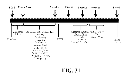
[0096] FIG. 31 is a study timeline. Neonatal rats were delivered prematurely
via cesarean
section at E20.5. To induce NEC, pups were subjected to repeated episodes of
hypercaloric
feeds and hypoxia/hypothermia x 96 hours. Surviving pups were placed with
foster dams and
subjected to developmental milestone testing daily for 23 days. Cognitive
function and
memory tests were performed between 4 -8 weeks of age. Rats were sacrificed
and tissues
collected at 2 months of age.
[0097] FIGS. 32A ¨ 32H show developmental milestones after experimental NEC.
Neonatal
rats were delivered prematurely via cesarean section. To induce NEC, pups were
subjected to
repeated episodes of hypercaloric feeds and hypoxia/hypothermia x 96 hours.
Surviving pups
were placed with foster dams and developmental milestones measured daily for
23 days.
(FIG. 32A, FIG. 32B) air righting test; (FIG. 32C, FIG. 32D) forelimb grasp
test; (FIG.
32E, FIG. 32F) ear opening; (FIG. 32G, FIG. 32H) auditory startle. Error bars
represent
SEM with statistical significance defined as p < 0.05 (one-way ANOVA).
[0098] FIGS. 33A ¨ 33D show Y-maze and novel object test after experimental
NEC. Rat
pups were subjected to experimental NEC. Surviving pups were placed with
surrogate dams.
After weaning, rat behavioral activities were measured. (FIG. 33A, FIG. 33B) Y-
Maze test;
(FIG. 33C, FIG. 33D) novel objects test. Error bars represent SEM with
statistical
significance defined as p < 0.05 (one-way ANOVA).
[0099] FIGS. 34A ¨ 34C show the results of yarns maze test after experimental
NEC. Rat
pups were subjected to experimental NEC. Surviving pups were place with
surrogate dams.
After weaning, rats' behavioral activities were measured. (FIG. 34A) Barnes
Maze setting;
(FIG. 34B) number of holes check at test day; and (FIG. 34C) latency to
finding escape hole.
Error bars represent SEM with statistical significance defined as p < 0.05
(one-way
ANOVA).
[0100] FIGS. 35A ¨ 35C show the results of an elevated plus maze test after
experimental
NEC. Rat pups were subjected to experimental NEC. Surviving pups were placede
with
surrogate dams. After weaning, rats' behavioral activities were measured.
(FIG. 35A)
22
CA 03142272 2021-11-29
WO 2020/247536 PCT/US2020/035976
elevated plus maze setting; (FIG. 35B) time in open arm and Junction; (FIG.
35C) time in
the closed arm. Error bars represent SEM with statistical significance defined
as p < 0.05
(one-way ANOVA).
[0101] FIGS. 36A ¨ 36C show microglia in the brain 2 months after experimental
NEC. Rat
pups were subjected to experimental NEC and surviving pups placed with
surrogate dams.
Rats were weaned at 21-23 days of life and sacrificed at two months of age.
Brains were
harvested and fixed in 4% PFA. Frozen sections were stained with Iba-1
antibody and Alexa
488 conjugated secondary antibody. At least 8 confocal pictures per section
were imaged
using 20X objectives. (FIG. 36A) Representative immunohistochemical images
across the
brain; (FIG. 36B) numbers of activated and amoeboid microglia counted / HPF;
(FIG. 36C)
percent of Iba-1+ cells / HPF quantified using Image J software. Error bars
represent SEM
with statistical significance defined as p < 0.05 (one-way ANOVA).
[0102] FIGS. 37A ¨ 37D show neurotrophic gene expression after experimental
NEC. Rat
pups were subjected to experimental NEC and surviving pups placed with
surrogate dams.
Rats were sacrificed at 2 months of age, the brains harvested, and the
prefrontal cortex (PFC)
and hippocampus collected. Gene expression was measured by RT-qPCR for (FIG.
37A,
FIG. 37B) BDNF and (FIG. 37C, FIG. 37D) Grin2A. Shown are relative copy
numbers
from independent rats. Error bars represent SEM with statistical significance
defined as p <
0.05 (one-way ANOVA).
[0103] FIG. 38 shows rat weight after experimental NEC. Neonatal rats were
delivered
prematurely via cesarean section at E21. To induce NEC, pups were subjected to
repeated
episodes of hypercaloric feeds and hypoxia/hypothermia x 96 hours. Surviving
pups were
placed with foster dams and weighed daily for 21 days. Shown are the weight of
the pups at
day of life (DOL) 4, 10 15 and 21. Error bars represent SEM with statistical
significance
defined as p < 0.05 (one-way ANOVA).
[0104] FIGS. 39A ¨ 391 show additional developmental milestones after
experimental NEC.
Neonatal rats were delivered prematurely via cesarean section at E21. To
induce NEC, pups
were subjected to repeated episodes of hypercaloric feeds and
hypoxia/hypothermia x 96
hours. Surviving pups were placed with foster dams and developmental
milestones were
measured daily for 23 days. (FIG. 39A, FIG. 39B) surface righting test; (FIG.
39C, FIG.
39D) negative geotaxis test; (FIG. 39E, FIG. 39F) open field test; (FIG. 39G,
FIG. 39H)
23
CA 03142272 2021-11-29
WO 2020/247536 PCT/US2020/035976
cliff aversion test; and (FIG. 391) and days of life at which eyes open. Error
bars represent
SEM with statistical significance defined as p<0.05 (one-way ANOVA).
DETAILED DESCRIPTION
[0105] It is to be understood that this invention is not limited to particular
embodiments
described, as such may, of course, vary. It is also to be understood that the
terminology used
herein is for the purpose of describing particular embodiments only, and is
not intended to be
limiting, since the scope of the present invention will be limited only by the
appended claims.
[0106] Unless defined otherwise, all technical and scientific terms used
herein have the same
meanings as commonly understood by one of ordinary skill in the art to which
this invention
belongs. Although any methods and materials similar or equivalent to those
described herein
can be used in the practice or testing of the present invention, the preferred
methods, devices
and materials are now described. All technical and patent publications cited
herein are
incorporated herein by reference in their entirety. Nothing herein is to be
construed as an
admission that the invention is not entitled to antedate such disclosure by
virtue of prior
invention.
[0107] The practice of the present technology will employ, unless otherwise
indicated,
conventional techniques of tissue culture, immunology, molecular biology,
microbiology, cell
biology and recombinant DNA, which are within the skill of the art. See, e.g.,
Sambrook and
Russell eds. (2001) Molecular Cloning: A Laboratory Manual, 3rd edition; the
series Ausubel
et al. eds. (2007) Current Protocols in Molecular Biology; the series Methods
in Enzymology
(Academic Press, Inc., N.Y.); MacPherson et al. (1991) PCR 1: A Practical
Approach (IRL
Press at Oxford University Press); MacPherson et al. (1995) PCR 2: A Practical
Approach;
Harlow and Lane eds. (1999) Antibodies, A Laboratory Manual; Freshney (2005)
Culture of
Animal Cells: A Manual of Basic Technique, 5th edition; Gait ed. (1984)
Oligonucleotide
Synthesis; U.S. Patent No. 4,683,195; Hames and Higgins eds. (1984) Nucleic
Acid
Hybridization; Anderson (1999) Nucleic Acid Hybridization; Hames and Higgins
eds. (1984)
Transcription and Translation; Immobilized Cells and Enzymes (IRL Press
(1986)); Perbal
(1984) A Practical Guide to Molecular Cloning; Miller and Cabo s eds. (1987)
Gene Transfer
Vectors for Mammalian Cells (Cold Spring Harbor Laboratory); Makrides ed.
(2003) Gene
Transfer and Expression in Mammalian Cells; Mayer and Walker eds. (1987)
Immunochemical Methods in Cell and Molecular Biology (Academic Press, London);
and
Herzenberg et al. eds (1996) Weir's Handbook of Experimental Immunology.
24
CA 03142272 2021-11-29
WO 2020/247536 PCT/US2020/035976
[0108] All numerical designations, e.g., pH, temperature, time, concentration
and molecular
weight, including ranges, are approximations which are varied ( + ) or (- ) by
increments of
1.0 or 0.1, as appropriate, or alternatively by a variation of +/- 15 %, or
alternatively 10%, or
alternatively 5% or alternatively 2%. It is to be understood, although not
always explicitly
stated, that all numerical designations are preceded by the term "about". It
also is to be
understood, although not always explicitly stated, that the reagents described
herein are
merely exemplary and that equivalents of such are known in the art.
[0109] As used in the specification and claims, the singular form "a", "an"
and "the" include
plural references unless the context clearly dictates otherwise. For example,
the term "a
bacterium" includes a plurality of bacteria, including mixtures thereof.
[0110] As used herein, the term "comprising" is intended to mean that the
compositions and
methods include the recited elements, but do not exclude others. "Consisting
essentially of'
when used to define compositions and methods, shall mean excluding other
elements of any
essential significance to the combination for the intended use. Thus, a
composition consisting
essentially of the elements as defined herein would not exclude trace
contaminants from the
isolation and purification method and pharmaceutically acceptable carriers,
such as phosphate
buffered saline, preservatives and the like. "Consisting of' shall mean
excluding more than
trace elements of other ingredients and substantial method steps for
administering the
compositions of this invention. Embodiments defined by each of these
transition terms are
within the scope of this invention.
[0111] A "biofilm" intends a thin layer or an organized community of
microorganisms that at
times can adhere to the surface of a structure that may be organic or
inorganic, together with
the polymers, such as DNA, that they secrete and/or release. The biofilms are
very resistant to
microbiotics and antimicrobial agents. They live on gingival tissues, teeth,
and restorations,
causing caries and periodontal disease, also known as periodontal plaque
disease. They also
cause chronic middle ear infections. Biofilms can also form on the surface of
dental implants,
stents, catheter lines and contact lenses. They grow on pacemakers, heart
valve replacements,
artificial joints and other surgical implants. The Centers for Disease Control
estimate that
over 65% of nosocomial (hospital-acquired) infections are caused by biofilms.
Fungal
biofilms also frequently contaminate medical devices. They cause chronic
vaginal infections
and lead to life-threatening systemic infections in people with hobbled immune
systems.
CA 03142272 2021-11-29
WO 2020/247536 PCT/US2020/035976
Biofilms also are involved in numerous diseases. For instance, cystic fibrosis
patients have
Pseudomonas infections that often result in antibiotic resistant biofilms.
[0112] A "prebiotic" intends a nutritional supplement for the probiotic
bacterium. Prebiotics
are food ingredients, for example, oligosaccharides, that are non-digestible
by a subject (e.g.,
by a mammal such as a human), and that stimulates the growth or activity of
one or more
beneficial bacteria and/or inhibit the growth or activity of one or more
pathogenic bacteria. A
prebiotic may selectively stimulate the growth and/or activity of one or a
limited number of
bacteria in the subject.
[0113] A "prebiofilmic" intends a substance that supports biofilm formation
and durability,
for example the prebiofilmic can be a substance that supports the
extracellular matrix of the
biofilm like an eDNA binding polypeptide or protein or alternatively a
substrate that can be
converted into a substance that facilitate adhesion, e.g., sucrose.
[0114] A "DNABII polypeptide or protein" intends a DNA binding protein or
polypeptide
that is composed of DNA-binding domains and thus have a specific or general
affinity for
DNA. In one aspect, they bind DNA in the minor grove. Non-limiting examples of
DNABII
proteins are an integration host factor (IHF) protein and a histone-like
protein from E. coli
strain U93 (HU), examples of which are provided in the attached sequence
listing and
additional strains and polypeptides are provided in Table 5. Also intended are
polypeptide
fragments and equivalent polypeptides that have amino acid modifications that
do not
substantially change the biological activity of the protein or polypeptides,
or active fragment
thereof. Active fragments can include, for example, the c-terminal half or c-
terminal third of
the protein or polypeptide. Other DNA binding proteins that can be associated
with the
biofilm include DPS (Genbank Accession No.: CAA49169), H-NS (Genbank Accession
No.:
CAA47740), Hfq (Genbank Accession No.: ACE63256), CbpA (Genbank Accession No.:
BAA03950) and CbpB (Genbank Accession No.: NP_418813), as well as equivalent
polpyeptides and active fragments thereof.
[0115] A "microsphere" intends a porous and/or semi-permeable biofilm-carrying
and/or
compound-carrying (e.g., drug-carrying) particulate or granular material
within the particular
size range recited. As used herein, a microsphere consisting of particles 50
millimeters or
less in diameter, and about 1 micron or more (e.g., about 1 to about 100 or
alternatively, or
alternatively, about 1 to about 75 microns, or alternatively about 1 to about
50, or
alternatively about 1 to about 25, or alternatively about 1 to about 10
microns, or alternatively
26
CA 03142272 2021-11-29
WO 2020/247536 PCT/US2020/035976
about 0.5 to about 200 microns, or alternatively about 0.5 to about 700
microns, or
alternatively about 1 to about 600 microns, or alternatively less than about
700 microns, or
alternatively less than about 600 microns, or alternatively less than 500
microns, or
alternatively less than about 400 microns, or alternatively less than about
300 microns, or
alternatively less than about 200 microns, or alternatively less than about
100 microns) in
diameter. Non-limiting examples of such include hollow microspheres that are
porous and/or
semi-permeable, and can, in some aspects, contain a pharmaceutical or a drug,
microcapsules,
(in which the excipient forms a skin or shell that surrounds and contains a
cargo, such as a
drug, a chemical reductant, or absorptive or adsorptive molecules), and
microparticles, which
are used as a generic term for any particles in the recited size range,
whether spherical or not,
as those terms are typically used in the art. Table 6 provides non-limiting
examples of
microspheres that are commercially available and their characteristics.
[01161 "In one aspect aubject," as used herein refers to a subject having
sepsis or a sepsis
causing condition (i.e., NEC), exposed to conditions for developing sepsis or
a sepsis causing
condition, or at risk of sepsis or a sepsis causing condition, wherein the
subject receives
treatment of a composition according to the invention. The subject may be
suffering from,
susceptible to, or having suffered from nectrotizing enterocolitis. The
subject may be an
infant, a fetus or a newborn. In one aspect, the subject is an infant subject
of a certain age
selected from 0-1 day, 1-6 days, 6-12 days, 12-18 days, 18-24 days, 24 days to
100 days, or
more than 100 days.
[0117] "Control subject" refers to either of 1) a subject suffering from,
susceptible to, or
having suffered from a disease or condition, e.g., sepsis or the same sepsis
causing condition
as the infant subject (i.e., NEC) or autism or an autism spectrum disorder)
btu not having
been administered the composition and being of the same age and species as the
subject
having received the treatment; or 2) a subject of the same age and species as
the subject
having received the treatment but not having suffered from the disease or
condition.
[0118] A "biodegradable polymer" intends polymers that are biocompatible and
can degrade
in vivo by bodily processes to products that are readily disposable by the
body and should not
accumulate in the body.
101191 By "biocompatible", it is meant that the components of the delivery
system will not
cause tissue injury or injury to the human biological system. To impart
biocompatibility,
polymers and excipients that have had history of safe use in humans or with
GRAS
27
CA 03142272 2021-11-29
WO 2020/247536 PCT/US2020/035976
(Generally Accepted As Safe) status, are preferentially used. By
biocompatibility, it is meant
that the ingredients and excipients used in the composition will ultimately be
"bioabsorbed"
or cleared by the body with no adverse effects to the body. For a composition
to be
biocompatible, and be regarded as non-toxic, it must not cause toxicity to
cells. Similarly, the
term "bioabsorbable" refers to microspheres made from materials which undergo
bioabsorption in vivo over a period of time such that long term accumulation
of the material
in the patient is avoided. The biocompatible nanoparticle is bioabsorbed over
a period of less
than 2 years, preferably less than 1 year and even more preferably less than 6
months. The
rate of bioabsorption is related to the size of the particle, the material
used, and other factors
well recognized by the skilled artisan. A mixture of bioabsorbable,
biocompatible materials
can be used to form the microspheres used in this invention.
[0120] An "integration host factor" or "IHF" protein is a bacterial protein
that is used by
bacteriophages to incorporate their DNA into the host bacteria. These are DNA
binding
proteins that function in genetic recombination as well as in transcription
and translational
regulation. They also bind extracellular microbial DNA. The genes that encode
the IHF
protein subunits in E. coli are himA (Genbank accession No.: P0A6X7.1) and
himD
(P0A6Y1.1) genes. Non-limiting examples of such are provided in the attached
sequence
listing and noted in Table 5.
[0121] "HU" or "histone-like protein from E. coli strain U93" refers to a
class of
heterodimeric proteins typically associated with E. coli. HU proteins are
known to bind DNA
junctions. Related proteins have been isolated from other microorganisms. The
complete
amino acid sequence of E. coli HU was reported by Laine et al. (1980) Eur. J.
Biochem.
103(3):447-481. Antibodies to the HU protein are commercially available from
Abcam.
Non-limiting examples of such are provided in the attached sequence listing.
[0122] The term "protein", "peptide" and "polypeptide" are used
interchangeably and in their
broadest sense to refer to a compound of two or more subunit amino acids,
amino acid
analogs or peptidomimetics. The subunits may be linked by peptide bonds. In
another
embodiment, the subunit may be linked by other bonds, e.g., ester, ether, etc.
A protein or
peptide must contain at least two amino acids and no limitation is placed on
the maximum
number of amino acids which may comprise a protein's or peptide's sequence. As
used
herein the term "amino acid" refers to either natural and/or unnatural or
synthetic amino
28
CA 03142272 2021-11-29
WO 2020/247536 PCT/US2020/035976
acids, including glycine and both the D and L optical isomers, amino acid
analogs and
peptidomimetics.
[0123] A "c-terminal polypeptide" intends the c-terminal half or c-terminal
third of a
polypeptide. As an example, for polypeptides containing 90 amino acids, the c-
terminal
polypeptide would comprise amino acids 46 through 90 or amino acids 60 through
90. In
another aspect, the term intends the c-terminal 20 amino acids from the
carboxy terminus.
[0124] A "n-terminal polypeptide" intends the n-terminal half of a
polypeptide. As an
example, for polypeptides containing 90 amino acids, the c-terminal
polypeptide would
comprise amino acids 1 through 45. In another aspect, the term intends the c-
terminal 20
amino acids from the amino terminus.
101251 The terms "polynucleotide" and "oligonucleotide" are used
interchangeably and refer
to a polymeric form of nucleotides of any length, either deoxyribonucleotides
or
ribonucleotides or analogs thereof. Polynucleotides can have any three-
dimensional structure
and may perform any function, known or unknown. The following are non-limiting
examples of polynucleotides: a gene or gene fragment (for example, a probe,
primer, EST or
SAGE tag), exons, introns, messenger RNA (mRNA), transfer RNA, ribosomal RNA,
RNAi,
ribozymes, cDNA, recombinant polynucleotides, branched polynucleotides,
plasmids,
vectors, isolated DNA of any sequence, isolated RNA of any sequence, nucleic
acid probes
and primers. A polynucleotide can comprise modified nucleotides, such as
methylated
nucleotides and nucleotide analogs. If present, modifications to the
nucleotide structure can
be imparted before or after assembly of the polynucleotide. The sequence of
nucleotides can
be interrupted by non-nucleotide components. A polynucleotide can be further
modified after
polymerization, such as by conjugation with a labeling component. The term
also refers to
both double- and single-stranded molecules. Unless otherwise specified or
required, any
embodiment of this invention that is a polynucleotide encompasses both the
double-stranded
form and each of two complementary single-stranded forms known or predicted to
make up
the double-stranded form.
[0126] A polynucleotide is composed of a specific sequence of four nucleotide
bases: adenine
(A); cytosine (C); guanine (G); thymine (T); and uracil (U) for thymine when
the
polynucleotide is RNA. Thus, the term "polynucleotide sequence" is the
alphabetical
representation of a polynucleotide molecule. This alphabetical representation
can be input
29
CA 03142272 2021-11-29
WO 2020/247536 PCT/US2020/035976
into databases in a computer having a central processing unit and used for
bioinformatics
applications such as functional genomics and homology searching.
Min The term "isolated" or "recombinant" as used herein with respect to
nucleic acids,
such as DNA or RNA, refers to molecules separated from other DNAs or RNAs,
respectively
that are present in the natural source of the macromolecule as well as
polypeptides. The term
"isolated or recombinant nucleic acid" is meant to include nucleic acid
fragments which are
not naturally occurring as fragments and would not be found in the natural
state. The term
"isolated" is also used herein to refer to polynucleotides, polypeptides,
antibodies and
proteins that are isolated from other cellular proteins and is meant to
encompass both purified
and recombinant polypeptides. In other embodiments, the term "isolated or
recombinant"
means separated from constituents, cellular and otherwise, in which the cell,
tissue,
polynucleotide, peptide, polypeptide, protein, antibody or fragment(s)
thereof, which are
normally associated in nature. For example, an isolated cell is a cell that is
separated from
tissue or cells of dissimilar phenotype or genotype. An isolated
polynucleotide is separated
from the 3' and 5' contiguous nucleotides with which it is normally associated
in its native or
natural environment, e.g., on the chromosome. As is apparent to those of skill
in the art, a
non-naturally occurring polynucleotide, peptide, polypeptide, protein,
antibody or
fragment(s) thereof, does not require "isolation" to distinguish it from its
naturally occurring
counterpart.
101281 Glucotransferases are enzymes that establish glycosidic linkages. A non-
limiting
example of a sequence of the GTF protein is available at DSM 20016. gtfW
ABQ83597.1 is
provided at DSM 17938 gtfA WP _003671465. See also, Walter et al. (2008)
Microbiology
154(Pt 1):72-80.
10129] It is to be inferred without explicit recitation and unless otherwise
intended, that when
the present invention relates to a polypeptide, protein, polynucleotide or
antibody, an
equivalent or a biologically equivalent of such is intended within the scope
of this invention.
As used herein, the term "biological equivalent thereof' is intended to be
synonymous with
"equivalent thereof' when referring to a reference protein, antibody,
polypeptide,
polynucleotide or nucleic acid, intends those having minimal homology while
still
maintaining desired structure or functionality. Unless specifically recited
herein, it is
contemplated that any nucleic acid, polynucleotide, polypeptide or protein
mentioned herein
also includes equivalents thereof. For example, an equivalent intends at least
about 70%, or
CA 03142272 2021-11-29
WO 2020/247536 PCT/US2020/035976
alternatively 80 % homology or identity and alternatively, at least about 85
%, or
alternatively at least about 90 %, or alternatively at least about 95 %, or
alternatively 98 %
percent homology or identity across the protein or a particular fragment
thereof, and exhibits
substantially equivalent biological activity to the reference protein,
polypeptide or nucleic
acid.
[0130] A polynucleotide or polynucleotide region (or a polypeptide or
polypeptide region)
having a certain percentage (for example, 80%, 85%, 90%, or 95%) of "sequence
identity" to
another sequence means that, when aligned, that percentage of bases (or amino
acids) are the
same in comparing the two sequences. The alignment and the percent homology or
sequence
identity can be determined using software programs known in the art, for
example those
described in Current Protocols in Molecular Biology (Ausubel et al., eds.
1987) Supplement
30, section 7.7.18, Table 7.7.1. Preferably, default parameters are used for
alignment. A
preferred alignment program is BLAST, using default parameters. In particular,
preferred
programs are BLASTN and BLASTP, using the following default parameters:
Genetic code =
standard; filter = none; strand = both; cutoff = 60; expect = 10; Matrix =
BLOSUM62;
Descriptions = 50 sequences; sort by = HIGH SCORE; Databases = non-redundant,
GenBank
+ EMBL + DDBJ + PDB + GenBank CDS translations + SwissProtein + SPupdate +
PIR.
Details of these programs can be found at the following Internet address:
ncbi.nlm.nih.gov/cgi-bin/BLAST.
101311 "Homology" or "identity" or "similarity" refers to sequence similarity
between two
peptides or between two nucleic acid molecules. Homology can be determined by
comparing
a position in each sequence which may be aligned for purposes of comparison.
When a
position in the compared sequence is occupied by the same base or amino acid,
then the
molecules are homologous at that position. A degree of homology between
sequences is a
function of the number of matching or homologous positions shared by the
sequences. An
"unrelated" or "non-homologous" sequence shares less than 40% identity, or
alternatively
less than 25% identity, with one of the sequences of the present invention.
[01321 As used herein, "expression" refers to the process by which
polynucleotides are
transcribed into mRNA and/or the process by which the transcribed mRNA is
subsequently
being translated into peptides, polypeptides, or proteins. If the
polynucleotide is derived from
genomic DNA, expression may include splicing of the mRNA in an eukaryotic
cell.
31
CA 03142272 2021-11-29
WO 2020/247536 PCT/US2020/035976
[0133] The term "encode" as it is applied to polynucleotides refers to a
polynucleotide which
is said to "encode" a polypeptide if, in its native state or when manipulated
by methods well
known to those skilled in the art, it can be transcribed and/or translated to
produce the mRNA
for the polypeptide and/or a fragment thereof. The antisense strand is the
complement of
such a nucleic acid, and the encoding sequence can be deduced therefrom.
[0134] A "subject" or "patient" of diagnosis or treatment is a cell or an
animal such as a
mammal or a human. Non-human animals subject to diagnosis or treatment and are
those
subject to infections or animal models, for example, simians, murines, such
as, rats, mice,
chinchilla, canine, such as dogs, leporids, such as rabbits, livestock, sport
animals and pets.
[0135] As used herein, the terms "treating," "treatment" and the like are used
herein to mean
obtaining a desired pharmacologic and/or physiologic effect. The effect may be
prophylactic
in terms of completely or partially preventing a disorder or sign or symptom
thereof and/or
may be therapeutic in terms of a partial or complete cure for a disorder
and/or adverse effect
attributable to the disorder.
[0136] To "prevent" intends to prevent a disorder or effect in vitro or in
vivo in a system or
subject that is predisposed to the disorder or effect. Examples of such is
preventing the
formation of a biofilm in a system that is infected with a microorganism known
to produce
one or alternatively, prevent a gastrointestinal disorder by supporting a
healthy state of the
patient's gut.
101371 The term "culturing" refers to the in vitro propagation of cells or
organisms on or in
media of various kinds. It is understood that the descendants of a cell grown
in culture may
not be completely identical (i.e., morphologically, genetically, or
phenotypically) to the
parent cell. By "expanded" is meant any proliferation or division of cells.
[0138] "Pharmaceutically acceptable carriers" refers to any diluents,
excipients or carriers
that may be used in the compositions of the invention. Pharmaceutically
acceptable carriers
include ion exchangers, alumina, aluminum stearate, lecithin, serum proteins,
such as human
serum albumin, buffer substances, such as phosphates, glycine, sorbic acid,
potassium
sorbate, partial glyceride mixtures of saturated vegetable fatty acids, water,
salts or
electrolytes, such as protamine sulfate, disodium hydrogen phosphate,
potassium hydrogen
phosphate, sodium chloride, zinc salts, colloidal silica, magnesium
trisilicate, polyvinyl
pyrrolidone, cellulose-based substances, polyethylene glycol, sodium
carboxymethylcellulo se, polyacrylates, waxes, polyethylene-polyoxypropylene-
block
32
CA 03142272 2021-11-29
WO 2020/247536 PCT/US2020/035976
polymers, polyethylene glycol and wool fat. Suitable pharmaceutical carriers
are described in
Remington's Pharmaceutical Sciences, Mack Publishing Company, a standard
reference text
in this field. They are preferably selected with respect to the intended form
of administration,
that is, oral tablets, capsules, elixirs, syrups and the like and consistent
with conventional
pharmaceutical practices.
[0139] A "biocompatible scaffold" refers to a scaffold or matrix for with the
ability to
support biofilm proliferation upon administration to a subject. In other
embodiments, a
biocompatible scaffold is a precursor to an implantable device which has the
ability to
perform its intended function, with the desired degree of incorporation in the
host, without
eliciting an undesirable local or systemic effects in the host. Biocompatible
scaffolds are
described in U.S. Patent Nos. 6,638,369 and 8,815,276. In one aspect, the
microsphere as
described herein is a biocompatible scaffold.
[0140] "Administration" intends the delivery of a substance to a subject such
as an animal or
human. Administration can be effected in one dose, continuously or
intermittently
throughout the course of treatment. Methods of determining the most effective
means and
dosage of administration are known to those of skill in the art and will vary
with the
composition used for therapy, the purpose of the therapy, as well as the age,
health or gender
of the subject being treated. Single or multiple administrations can be
carried out with the
dose level and pattern being selected by the treating physician or in the case
of pets and
animals, treating vetrenarian. Suitable dosage formulations and methods of
administering the
agents are known in the art. Route of administration can also be determined
and method of
determining the most effective route of administration are known to those of
skill in the art
and will vary with the composition used for treatment, the purpose of the
treatment, the
health condition or disease stage of the subject being treated and the target
cell or tissue.
Non-limiting examples of route of administration include enterally, oral
administration,
vaginal, nasal administration (inhalation), injection, topical application and
by suppository.
[0141] The term "effective amount" refers to a quantity sufficient to achieve
a beneficial or
desired result or effect. In the context of therapeutic or prophylactic
applications, the
effective amount will depend on the type and severity of the condition at
issue and the
characteristics of the individual subject, such as general health, age, sex,
body weight, and
tolerance to pharmaceutical compositions. In the context of a therapeutic
composition, in
some embodiments the effective amount is the amount sufficient to result in a
protective
33
CA 03142272 2021-11-29
WO 2020/247536 PCT/US2020/035976
response against a pathogen or alternatively to support a healthy state of
being. In some
embodiments, the amount is sufficient to accomplish one or more of 1) clear
pathogen; 2)
restore healthy microbiota; 3) modulate the immune system; 4) maintain
metabolism and
metabolic pathways; 5) reduce toxic compounds in the environment (toxic
compounds in
water, soil, air, and compounds such as heavy metals (e.g., chromium, arsenic,
mercury,
radioactive actinides, uranium, plutonium, thorium, polycyclic aromatic
hydrocarbons
(PAH), petroleum hydrocarbon, crude oil, refined oil, herbicide contamination
or pesticide
contamination); and 6) remediate a biofilm).
10142] "Body strength" refers to the ability of the subject to perform
physical work also
known in the art as muscle force production. "Coordination" refers to
organization of the
different elements of a complex body or activity so as to enable them to work
together
effectively. Body strength and/or coordination in infants may be measured by
clinical tests
known to a person of skill in the art, for example, Jamar dynamometer,
handheld
dynamometry, manual muscle testing (MMT), isokinetic dynamometry, trunk
stability test
(TST), unilateral hip bridge endurance test (UHBE), pronator sign, Barre sign,
Romberg test,
Landau reflex, particle suspension, sensory reflex (pinprick, light touch,
position, vibration,
and charger), reflex (biceps, triceps, brachioradialis, patellar, and ankle),
Moro reflex, tonic
neck response, sucking reflex, palmer and planter grasp reflex, and parachute
response.
10143] "Righting mechanism" or righting reflex, also known as the labyrinthine
righting
reflex, is a reflex that corrects the orientation of the body when it is taken
out of its normal
upright position. Righting mechanism may be clinically measured using one or
more
recognized techniques known to the skilled worker, for example, neck on body
righting
reaction (NOB) or body on body righting reaction (BOB).
10144] "Auditory reflex" refers to reflexes of the ear upon introduction of
stimulus. Auditory
reflex may be measured by clinical techniques known to the skilled artisan,
for example, ear
opening auditory reflex, static compliance, physical volume of ear canal,
contralateral reflex,
ipsilateral reflex, and tympanometry.
[0145] "Curiosity" refers to the desire to learn or explore something, often
something novel.
Curiosity may be assessed using methods known in the art, for example, Y-maze,
Novel
Object Recognition Task, STPI (State-Trait Personality Inventory), the Five
Dimensional
Curiosity Scale, Self Curiosity Attitude Interests Scale, Curiosity and
Exploration Inventory-
34
CA 03142272 2021-11-29
WO 2020/247536 PCT/US2020/035976
II, State-Trait Personality Inventory (STPI), and subscales of the Sensation
Seeking Scale
(SSS).
[0146] "Learning ability" refers to the ability to comprehend; to understand
and profit from
experience. "Working memory" refers to the part of short-term memory that is
concerned
with immediate conscious perceptual and linguistic processing. "Short-term
memory" refers
to the capacity for holding, but not manipulating, a small amount of
information in mind in an
active, readily available state for a short period of time. "Long-term memory"
is the stage of
the Atkinson¨Shiffrin memory model where informative knowledge is held
indefinitely. It is
defined in contrast to short-term and working memory, which persist for only
about 18 to 30
seconds. Long-term memory is commonly labelled as explicit memory
(declarative), as well
as episodic memory, semantic memory, autobiographical memory, and implicit
memory
(procedural memory). "Cognition" is the mental action or process of acquiring
knowledge
and understanding through thought, experience, and the senses, encompassing
many aspects
of intellectual functions and processes such as attention, the formation of
knowledge,
memory and working memory, judgment and evaluation, reasoning and computation,
problem solving and decision making, comprehension and production of language.
"Visual-
spatial reasoning" refers to the capacity to understand, reason and remember
the spatial
relations among objects or space. "Object recognition" refers to the ability
to remember an
object that was previously shown and then removed from view, then shown again.
Any of the
traits defined in this paragraph may be measured using a clinical scale or
test known to the
skilled artisan for measuring cognitive ability in infants, for example,
Bayley Scales of Infant
Development (BSID-III) (1-42 months), the Mullen Scales of Early Learning (1-
68 months),
the Fagan Test of Infant Intelligence (FTII) (Birth-12 months), Griffith's
Mental
Development Scales I (0-2 years), Battelle Developmental Inventory (BDI)
(Birth-8 years),
and the Vineland Adaptive Behaviour Scale ( 0-18 years).
[0147] "Infant," as used herein may refer to a human or animal that is very
young. For
example, 0-1 month, 1-6 months, 6-12 months, 12-18 months, 18-24 months, 24
months to 3
years, or older than 3 years. Infant intends not only humans but, for example,
simian, bovine,
porcine, murine, rat, avian, and reptilian animals.
[0148] "Sepsis" as used herein, refers to a life-threatening condition that
arises when the
body's response to infection causes injury to its tissues and organs. This
initial stage is
followed by suppression of the immune system. Sepsis may be diagnosed using
tools known
CA 03142272 2021-11-29
WO 2020/247536 PCT/US2020/035976
to those of ordinary skill in the art, not limited to blood culture, urine
culture, other cultures,
blood tests, lumbar puncture, x-rays.
[0149] Glutamate [NMDA] receptor subunit epsilon-1 is a protein that in humans
is encoded
by the GRIN2A gene or "Grin2A" as used herein.
[0150] "Brain-derived neurotrophic factor" (BDNF) refers to a protein that, in
humans, is
encoded by the BDNF gene. BDNF acts on certain neurons of the central nervous
system and
the peripheral nervous system, helping to support survival of existing
neurons, and
encouraging growth and differentiation of new neurons and synapses. Changes in
expression
of this protein may be measured by, for example, reverse transcription
polymerase chain
reaction (RT-PCR), Western blot, or mass spectrometry.
[0151] "Activated microglia" refers to the inflammatory response mediated by
the resident
immune cells of the CNS, which normally respond to neuronal damage and remove
the
damaged cells by phagocytosis. Activation of microglia is a hallmark of brain
pathology.
Activated microglia may be identified through imaging techniques not limited
to positron
emission tomography (PET) as described in J Neuroimmune Pharmacol. 2009 June ;
4(2):
227-243, the disclosure of which is hereby incorporated by reference.
[0152] "Neurodevelopmental deficiency" as used herein, refers to
neurodevelopmental
impairments of the growth and development of the brain and/or central nervous
system. A
narrower use of the term refers to a disorder of brain function that affects
emotion, learning
ability, self-control and memory which unfolds as an individual develops and
grows. The
neurodevelopmental disorders in the scope of this definition include, but are
not limited to:
Intellectual disability (ID) or intellectual and developmental disability
(IDD), previously
called mental retardation. Specific learning disorders, like dyslexia or
dyscalculia, autism
spectrum disorders, such as Asperger's syndrome or Autistic Disorder. Motor
disorders
including developmental coordination disorder and stereotypic movement
disorder. Tic
disorders including Tourette's syndrome. Traumatic brain injury (including
congenital
injuries such as those that cause cerebral palsy). Communication, speech and
language
disorders. Genetic disorders, such as fragile-X syndrome, Down syndrome,
attention deficit
hyperactivity disorder, schizophrenia, schizotypal disorder, hypogonadotropic
hypogonadal
syndromes. Behavioral disorders including conduct disorder etc. Attention
Deficit
Hyperactivity Disorder. Neurodevelopmental deficiencies may be assessed and
diagnosed by
those of skill in the art using tests described herein or well known to those
of skill in the art
36
CA 03142272 2021-11-29
WO 2020/247536 PCT/US2020/035976
such as ear opening and eye opening, surface righting, air righting, forelimb
grasp, auditory
startle, surface righting, negative geotaxis, openfield traversal, cliff
aversion, Barnes maze,
elevated plus maze, Jamar dynamometer, handheld dynamometry, manual muscle
testing
(MMT), isokinetic dynamometry, trunk stability test (TST), unilateral hip
bridge endurance
test (UHBE), pronator sign, Barre sign, Romberg test, Landau reflex, particle
suspension,
sensory reflex (pinprick, light touch, position, vibration, and charger),
reflex (biceps, triceps,
brachioradialis, patellar, and ankle), Moro reflex, tonic neck response,
sucking reflex, palmer
and planter grasp reflex, parachute response, neck on body righting reaction
(NOB), body on
body righting reaction (BOB), ear opening auditory reflex, static compliance,
physical
volume of ear canal, contralateral reflex, ipsilateral reflex, tympanometry, Y-
maze, Novel
Object Recognition Task, STPI (State-Trait Personality Inventory), the Five
Dimensional
Curiosity Scale, Self Curiosity Attitude Interests Scale, Curiosity and
Exploration Inventory-
II, State-Trait Personality Inventory (STPI), subscales of the Sensation
Seeking Scale (SSS),
Bayley Scales of Infant Development (BSID-III) (1-42 months), the Mullen
Scales of Early
Learning (1-68 months), the Fagan Test of Infant Intelligence (FTII) (Birth-12
months),
Griffith's Mental Development Scales I (0-2 years), Battelle Developmental
Inventory (BDI)
(Birth-8 years), and the Vineland Adaptive Behaviour Scale ( 0-18 years).
[0153] "Infection" as used herein, may refer to a bacterial infection, a viral
infection, a fungal
infection, or a protozoan infection.
[0154] "Sepsis causing condition" as used herein, may refer to any condition
in the subject or
in the mother (maternal) that causes or results in sepsis, for example,
necrotizing enterocolitis
(NEC), Escherichia coli (E coli), Listeria, Streptococcus, Neisseria
meningitidis, Salmonella, Haemophilus influenzae type b, herpes simplex virus,
cytomegalovirus (CMV), candida, bacterial infection of maternal amniotic
fluid, premature
birth, maternal amniotic rupture, fungal infenction, viral infenction,
respiratory syncytial
virus (RSV) ,protozoan infection, lung infection, brain infenction, urinary
tract infection, skin
infection, urinary catheterization of the infant subject, intravenous
placement of an injection
line in the infant subject, inflammation and/or immune dysregulation in the
infant subject,
and infenction resulting from surgery on the infant subject.
[0155] "Cell body thickness" as used herein, refers to the thickness of the
cell body which is
measured by, for example, transmission electron microscopy, transmission
through dye
37
CA 03142272 2021-11-29
WO 2020/247536 PCT/US2020/035976
microscopy, liquid displacement, Coulter method, absoprition, fluorescence,
light
obscuration, or confocal scanning.
[0156] In the case of an in vitro or ex vivo applications, in some embodiments
the effective
amount will depend on the size and nature of the application in question. It
will also depend
on the nature and sensitivity of the in vitro target and the methods in use.
The skilled artisan
will be able to determine the effective amount based on these and other
considerations. The
effective amount may comprise one or more administrations of a composition
depending on
the embodiment.
[0157] The agents and compositions can be used in the manufacture of
medicaments and for
the treatment of humans and other animals by administration in accordance with
conventional
procedures, such as an active ingredient in pharmaceutical compositions.
[0158] An agent or composition of the present invention can be administered
for therapy by
any suitable route of administration. It will also be appreciated that the
preferred route will
vary with the condition and age of the recipient and the disease being
treated.
[0159] Necrotizing enterocolitis ("NEC") is a medical condition primarily seen
in premature
infants where portions of the bowel undergo necrosis (tissue death). It occurs
postnatally
(i.e., is not seen in stillborn infants) and is the second most common cause
of mortality. 7%
of all neonatal intensive care unit admissions are NEC related. The mortality
rate is 12%.
MODES FOR CARRYING OUT THE DISCLOSURE
Microsphere Compositions
[0160] This disclosure provides a composition comprising, consisting
essentially of, or
consisting of a microsphere, a biofilm-generating probiotic bacterium and a
prebiotic,
wherein the prebiotic comprising, consists essentially of, or consists of a
nutritional
supplementation for the probiotic bacterium. In one aspect, the composition
further
comprises, consists essentially of, or consists of one or more of: a biofilm,
a prebiofilmic,
coating on the surface of the microsphere a therapeutic drug or agent, a
chemical reductant, a
molecule that promotes adsorption, a molecule that supports absorption. The
microsphere
comprises, consists essentially of, or consists of a solid core, a hollow
core, wherein in one
aspect, the microsphere encapsulates the prebiotic within the hollow core. The
microsphere
can be biocompatible and/or semi-permeable. In one aspect, the microsphere
comprise a
biofilm layer or coating on the external surface of the microsphere.
38
CA 03142272 2021-11-29
WO 2020/247536 PCT/US2020/035976
Microsphere Components
[0161] In one aspect, the biocompatible microsphere comprises, consists
essentially of, or
consists of a material selected from the group of: a biodegradable polymer, a
non-degradable
polymer, a metal, and wherein the diameter of the microsphere is from about
0.5 microns to
about 1000 microns. Additional preferred ranges are described herein and
incorporated herein
by reference. The microspheres can be porous and/or semi-permeable.
[0162] Non-limiting examples of biodegradable polymers are selected from one
or more of:
dextran; dextranomer; poly(lactic-co-glycolic acid) or PLGA; polycaprolactone
or PLC;
Chitosan; Gelatin; DNA hydrogen; acetalated dextran; poly(lactide);
poly(glycolide);
poly(lactide-co-glycolide); poly(lactic acid); poly(glycolic acid);
poly(lactic acid-co-glycolic
acid); poly(lactide)/poly(ethylene glycol) copolymers;
poly(glycolide)/poly(ethylene glycol)
copolymer; poly(lactide-co-glycolide)/poly(ethylene glycol) copolymers;
poly(lactic
acid)/poly(ethylene glycol) copolymer; poly(glycolic acid)/poly(ethylene
glycol) copolymer;
poly(lactic acid-co-glycolic acid)/poly(ethylene glycol) copolymer;
poly(caprolactone);
poly(caprolactone)/poly(ethylene glycol) copolymer; poly(orthoester);
poly(phosphazene);
poly(hydroxybutyrate); poly(hydroxybutyrate); poly(lactide-co-caprolactone);
polycarbonate;
polyesteramide; polyanhidride; poly(dioxanone); poly(alkylene alkylate);
polyethylene
glycol/polyorthoester copolymer; polyurethane; poly(amino acid);
polyetherester; polyacetal;
polycyanoacrylate; poly(oxyethylene)/poly(oxypropylene) copolymer; Sephadex
copolymers and/or a combination thereof.
[0163] Non-limiting examples of non-biodegradable polymers are selected from
one or more
of poly(ethylene vinyl acetate), poly(vinyl acetate), silicone polymers,
polyurethanes,
polysaccharides such as a cellulosic polymers and cellulose derivatives, acyl
substituted
cellulose acetates and derivatives thereof, copolymers of poly(ethylene
glycol) and
poly(butylene terephthalate), polystyrenes, polyvinyl chloride, polyvinyl
fluoride, poly(vinyl
imidazole), chorosulphonated polyolefins, polyethylene oxide, and copolymers
and blends
thereof.
[0164] Non-limiting examples of polymers comprising, consisting essentially
of, or
consisting of the microsphere are selected from one or more of: Sephadex,
Sephadex G-25,
poly(lactic-co-glycolic acid)(" PLGA"), polycaprolactone ("PLC"), chitosan;
gelatin, DNA
hydrogen; acetalated dextran, poly(lactide), poly(glycolide), poly(lactide-co-
glycolide),
poly(lactic acid), poly(glycolic acid), poly(lactic acid-co-glycolic acid),
39
CA 03142272 2021-11-29
WO 2020/247536 PCT/US2020/035976
poly(lactide)/poly(ethylene glycol) copolymers, poly(glycolide)/poly(ethylene
glycol)
copolymer, poly(lactide-co-glycolide)/poly(ethylene glycol) copolymers,
poly(lactic
acid)/poly(ethylene glycol) copolymer, poly(glycolic acid)/poly(ethylene
glycol) copolymer,
poly(lactic acid-co-glycolic acid)/poly(ethylene glycol) copolymer,
poly(caprolactone),
poly(caprolactone)/poly(ethylene glycol) copolymer, poly(orthoester),
poly(phosphazene),
poly(hydroxybutyrate), poly(hydroxybutyrate), poly(lactide-co-caprolactone);
polycarbonate;
polyesteramide; polyanhidride, poly(dioxanone), poly(alkylene alkylate),
polyethylene
glycol/polyorthoester copolymer, polyurethane, poly(amino acid),
polyetherester; polyacetal,
polycyanoacrylate, poly(oxyethylene)/poly(oxypropylene) copolymer; and a
combination
thereof.
[0165] Non-limiting examples of metals include cobalt, chromium, gold, nickel,
platinum,
stainless steel, titanium, tantalum, nickel-titanium, an alloy, and
combinations thereof.
Prebiotic
[01661 Non-limiting examples of the prebiotic of the composition comprise one
or more of: a
water-soluble carbohydrate, inulin, oligosaccharides, oligofructo se, fructo-
oligosaccharide,
galacto-oligosaccharide, glucose, starch, maltose, maltodextrins,
polydextrose, amylo se,
sucrose, fructose, lactose, isomaltulose, polyols, glycerol, carbonate,
thiamine, choline,
histidine, trehalos, nitrogen, sodium nitrate, ammonium nitrate, phosphorus,
phosphate salts,
hydroxyapatite, potassium, potash, sulfur, homopolysaccharide,
heteropolysaccharide,
cellulose, chitin, vitamins, and combination thereof.
101671 In another aspect, the prebiotic is selected from one or more of
trehalose; nitrogen
such as in sodium nitrate, ammonium nitrate, phosphorus such in phosphate
salts like
hydroxyapatite, potassium such as in potash, sulfur, oligosaccharide,
homopolysaccharide,
heteropolysaccharide, cellulose, chitin, glucose, fructose, sucrose, maltose,
starch,
polydextrose, amylose, glycerol, carbonate, and combinations thereof.
101681 In a yet further aspect, the prebiotic of the composition comprises,
consists essentially
of, or consists of one or more of vitamin mixtures to stimulate microbial
growth, nitrogen
such as in sodium nitrate, ammonium nitrate, phosphorus such in phosphate
salts like
hydroxyapatite, potassium such as in potash, sulfur, oligosaccharide,
homopolysaccharide,
heteropolysaccharide cellulose, chitin; glucose, fructose, sucrose, maltose,
starch,
polydextrose, amylose, glycerol, carbonate, and combinations thereof.
Probiotic Bacterium
CA 03142272 2021-11-29
WO 2020/247536 PCT/US2020/035976
[0169] In one aspect, the probiotic bacterium is selected to provide one or
more of supporting
anti-bacterial immunity, enhancing or supporting a healthy state in the
subject,t enhancing or
supporting the gastrointestinal barrier, or antagonizing disease-related
bacterial infections. In
another aspect, the probiotic bacterium is seleted to prevent pathogen
colonization and/or
limit and/or clear the pathogen, and /or limit excessive inflammatory
responses by down-
regulating cytokine and chemokine production.
[0170] Non-limiting examples of the probiotic bacterium is one or more of L.
acidophilus, L.
crispatus, L. gasseri, group L. delbrueckii, L. salivarius, L. casei, L.
paracasei, L. plantarum,
L. rhamnosus, L. reuteri , L. brevis, L. buchneri, L. fermentum, L. rhamnosus,
B.
adolescentis, B. angulation, B. bifidum, B. breve, B. catenulatum, B.
infantis, B. lactis, B.
longum, B. pseudocatenulatum, S. the rmophiles, Pseudomonas fluorescens, P.
protegens, P.
brassicacearum, P. aeruginosa; Azospirillum. brabrasilense, A. lipferum, A.
halopraeferens,
A. irakense; Acetobacter diazotrophicus; Herbaspirillum seropedicae; Bacillus
subtilis,
Pseudomonas stutzeri, fluorescens, P. putida, P. cepacian, P. vesicularis, P.
paucimobilis;
Bacillus cereus, B. thuringiensis, B. sphaericus; Shewanella oneidensis;
Geobacter
bemidjiensis, G. metallireducens, G. sulfurreducens, G. uraniireducens, G.
lovleyi; Serratia
marcescens, Desulfovibrio vulgaris, D. desulfuri cans, Dechloromonas aromatic,
Deinococcus radiodurans, Methylibium petroleiphilum, Alcanivorax borkumensis,
Archaeglobus fulgidus, Haloferax sp., Halobacterium sp., and combinations
thereof.
[0171] In another aspect, the probiotic is L. reuteri that produces GTF
protein or containing
the GTFW gene (ATCC 23272).
Prebiofilmic
[0172] In other aspect, the prebiofilmic comprises, consists essentially of,
or consists of an
agent that supports biofilm formation and durability, non-limiting examples of
such include a
DNA binding polypeptide or protein and/or a DNABII polypeptide or protein or
an
equivalent of each thereof, optionally, a polypeptide comprising, consisting
essentially of, or
consisting of one or more of the attached sequence listing, or a biologically
active fragment
or equivalent of each thereof, alone or in combination.
Complimentary Agents
[0173] The microspheres and compositions containing the microspheres can
further an agent,
wherein the agent is selective against a pathogen that may compete with the
probiotic
organism. The complimentary agents can be in the core, on the surface of the
micro sphere in
41
CA 03142272 2021-11-29
WO 2020/247536 PCT/US2020/035976
in the composition containing the microspheres. Non-limiting examples of such
include
chemical reductants; molecules and/or surfaces that promote adsorption (in
core or on surface
of microsphere); molecules and/or surfaces that promote absorption (in core or
on surface of
microsphere). In one aspect, the chemical reductants and molecules and/or
surfaces that
promote absorption are coated on the surface of the microsphere.
Biofilm layer
[0174] In one aspect, the microsphere compositions further comprise a biofilm
layer on the
external surface of the microparticle. The layer can be from about about 0.5
micron to about 1
millimiter in depth, and ranges in between, e.g., about 1 micron to about 500
microns, about 1
micron to about 250 microns, about 1 micron to about 200 microns, about 1
micron to about
100 microns, about 1 micron to about 50 microns, about 1 micron to about 40
microns, about
1 micron to about 30 microns, about 2 micron to about 100 microns, about 2
microns to about
50 microns, about 2 microns to about 40 microns, about 2 microns to about 30
microns, about
3 microns to about 100 microns, about 3 microns to about 50 microns, about 3
microns to
about 40 microns, about 3 microns to about 30 microns, about 5 microns to
about 100
microns, about 5 microns to about 50 microns, about 5 microns to about 40
microns, and
about 5 microns to about 30 microns.
Compositions
[01751 This disclosure also provides one or a plurality of microsphere
compositions as
described herein in combination with a carrier, e.g., a pharmaceutically
acceptable carrier or a
biocompatible scaffold. Non-limiting examples pharmaceutically acceptable
carriers include
diluents, excipients or carriers that may be used in the compositions of the
disclosure.
Pharmaceutically acceptable carriers include ion exchangers, alumina, aluminum
stearate,
lecithin, serum proteins, such as human serum albumin, buffer substances, such
as
phosphates, glycine, sorbic acid, potassium sorbate, partial glyceride
mixtures of saturated
vegetable fatty acids, water, salts or electrolytes, such as protamine
sulfate, disodium
hydrogen phosphate, potassium hydrogen phosphate, sodium chloride, zinc salts,
colloidal
silica, magnesium trisilicate, polyvinyl pyrrolidone, cellulose-based
substances, polyethylene
glycol, sodium carboxymethylcellulo se, polyacrylates, waxes, polyethylene-
polyoxypropylene-block polymers, polyethylene glycol and wool fat.
42
CA 03142272 2021-11-29
WO 2020/247536 PCT/US2020/035976
[0176] Non-limiting examples of biocompatible scaffolds, include a scaffold or
matrix for
with the ability to support biofilm proliferation upon administration to a
subject or an
environment to be treated.
[0177] In one aspect, the compositions comprise a plurality of micro spheres
that are the same
or different from each other, e.g, the same or different diameters, the same
or different
microsphere components, the same or different probiotics, the same or
different
complimentary agents, the same or different prebiofilmic, and hollow and/or
solid cores.
[0178] The compositions can be formulated into dosage forms of the biofilm-
generative
probiotic bacterium, e.g., or provide from an effective amount of the
microsphere
composition for the end use, e.g., from about 1 X 10⁵ to 1 X 10¹¹ CFU/ml, or
alternatively
from about 1 X 10⁵ to about 1 X 10¹⁰ CFU/ml, or about 1 X 10⁵ to about 1 X 10⁹
CFU/ml, or
about 1 X 10⁶ to about 1 X 10¹¹ CFU/ml, or about 1 X 10⁶ to about 1 X 10⁹
CFU/ml, or about
1 X 10⁷ to about 1 X 10¹¹ CFU/ml, or about 1 X 10⁷ to about 1 X 10¹⁰ CFU/ml,
or about 1 X
107 to about 1 X 10⁹ CFU/ml, or about 1 X 10⁸ CFU/ml.
[0179] The compositions can be formulated or processed for ease of
administration, storage
and application, e.g., frozen, lyophilized, suspended (suspension formulation)
or powdered;
and processed as a suppository, tablet, solution, suspensions, pills,
capsules, sustained release
formulation.
Applications and Uses
[0180] A method is provided for of preventing, delaying or treating
neurodevelopmental
deficiencies or promoting neurodevelopment in a subject suffering from,
susceptible to, or
having suffered from neurodevelopmental deficiencies, comprising administering
to the
subject an effective amount of a composition comprising a microsphere, a
biofilm-generating
probiotic bacterium and a prebiotic, wherein the prebiotic comprises a
nutritional
supplementation for the probiotic bacterium. In one aspect, subject is
suffering from NEC or
other pathology with similar effects on the brain, or sepsis and/or a sepsis
causing condition.
[0181] Also provided is a method of preventing, decreasing, or delaying
microglial activation
in an subject comprising administering to the subject an effective amount of a
composition
comprising a microsphere, a biofilm-generating probiotic bacterium and a
prebiotic, wherein
the prebiotic comprises a nutritional supplementation for the probiotic
bacterium. In one
aspect, subject is suffering from NEC or other pathology with similar effects
on the brain, or
sepsis and/or a sepsis causing condition. In one aspect, the cell body
thickness of microglia is
43
CA 03142272 2021-11-29
WO 2020/247536 PCT/US2020/035976
decreased in the treatment subject relative to a control subject. In another
aspect, the
dendritic processes of microglia are thinned in the subject relative to a
control subject.
[0182] Furher provided is a method of increasing expression of brain-derived
neurotrophic
factor (BDNF) in a subject comprising administering to the subject an
effective amount of a
composition comprising a microsphere, a biofilm-generating probiotic bacterium
and a
prebiotic, wherein the prebiotic comprises a nutritional supplementation for
the probiotic
bacterium. In one aspect, subject is suffering from NEC or other pathology
with similar
effects on the brain, or sepsis and/or a sepsis causing condition.
[0183] In the above methods, the the increase is relative to a control
subject. In one aspect,
the increased expression of BDNF is in the hippocampus of the subject.
[0184] Further provided is a method of increasing expression of Grin2A in a
subject
comprising administering to the subject an effective amount of a composition
comprising a
microsphere, a biofilm-generating probiotic bacterium and a prebiotic, wherein
the prebiotic
comprises a nutritional supplementation for the probiotic bacterium. In one
aspect, subject is
suffering from NEC or other pathology with similar effects on the brain, or
sepsis and/or a
sepsis causing condition. In one aspect, the increase is relative to a control
subject. In
another aspect, the increased expression of Grin2A is in the prefrontal cortex
of the treatment
subject.
[0185] In aspects of the methods of this disclosure, the microsphere further
comprises a
partial or complete biofilm coating on the external surface of the
microsphere. In a further
aspect, the bacterium and/or the prebiotic diffuse from the interior of the
microsphere to the
surface of the microsphere.
[0186] In general, the compositions of this disclosure find use in therapeutic
applicationsand
the components of the compositions and the carriers and additional agents are
selected for the
specified use.
[0187] In one aspect, the compositions provide one or more of supporting anti-
bacterial
immunity, enhancing or supporting the gastrointestinal barrier, or
antagonizing disease-
related bacterial infections. In another aspect, the compositions prevents
pathogen
colonization and/or limits excessive inflammatory responses by down-regulating
cytokine
and chemokine production.
44
CA 03142272 2021-11-29
WO 2020/247536 PCT/US2020/035976
[0188] In one aspect, the compositions are useful for the treatment of a
mammal such as a
human; simians, murines, such as, rats, mice, chinchilla, canine, such as
dogs, leporids, such
as rabbits, livestock, sport animals and pets.
[0189] The indications and uses vary with the environment. The compositions
can be used in
the treatment or prevention of disease, e.g., psychological disorders, such as
depression or
anxiety, enteric infectious disease, infection-induced colitis, traveler's
diarrhea, inflammatory
bowel disease (IBD), colitis, diarrheal illness, vaginosis, wound, burns,
psoriasis, dermatitis,
tooth decay, periodontitis, sinusitis, or any of chronic and/or recurrent
disease that is caused
by pathogenic bacteria displacing healthy bacteria or nectrotizing
enterocolitis (NEC), to
support anti-bacterial immunity, enhancing or supporting the gastrointestinal
barrier,
correcting or supporting dysbiotic gut flora (and even in the absence of
diseases), disease or
disorders involving intestinal dysmobility, enhancing or supporting the
gastrointestinal
mobility, or antagonizing disease-related bacterial infection; vaginosis;
colitis or traveler's
diarrhea, peritonitis, post-operative ileus, irritable bowel syndrome (IBS),
intestinal pseudo-
obstruction, and/or constipation. In one aspect, treatment excludes
prevention.
[0190] Thus, in one aspect, this disclosure provides method for treating or
preventing a
disease or disorder suitably treated by a biofilm in a subject in need thereof
is provided
herein. The method comprises, consists essentially of, or consists of
administering to the
subject an effective amount of the composition as disclosed herein, having the
components
selected for the particular therapy. Non-limiting examples of diseases include
psychological
disorders, such as depression or anxiety, enteric infectious disease, autism,
autism spectrum
disorders, infection-induced colitis, traveler's diarrhea, inflammatory bowel
disease (IBD),
colitis, diarrheal illness, vaginosis, wound, burns, psoriasis, dermatitis,
tooth decay,
periodontitis, sinusitis, or any of chronic and/or recurrent disease that is
caused by pathogenic
bacteria displacing healthy bacteria or nectrotizing enterocolitis (NEC), and
to support anti-
bacterial immunity, enhancing or supporting the gastrointestinal barrier,
correcting or
supporting dysbiotic gut flora (and even in the absence of diseases), disease
or disorders
involving intestinal dysmobility, enhancing or supporting the gastrointestinal
mobility, or
antagonizing disease-related bacterial infection; vaginosis; colitis or
traveler's diarrhea,
peritonitis, post-operative ileus, irritable bowel syndrome (IBS), intestinal
pseudo-
obstruction, and/or constipation. Additionally, the compositions are useful to
promote health
and/or to maintain gut homeostasis.
CA 03142272 2021-11-29
WO 2020/247536 PCT/US2020/035976
[0191] This disclosure also provides a method for delivering a probiotic to a
subject
comprising, consisting essentially of, or consisting of administering a dose
of a composition
as disclosed herein to the subject, thereby administering the probiotic.
[0192] The dosage and components of the composition will vary with the subject
and purpose
of the therapy. In one aspect, the composition is administered to provide from
about 1 x 10⁷
to about 1 x 10⁹ CFU/ml of the biofilm-generating probiotic bacterium. The
compositions can
be formulated into dosage forms, e.g., or provide from an effective amount of
the
microsphere composition for the end use, e.g., from about 1 X 10⁵ to 1 X 10¹¹
CFU/ml, or
alternatively from about 1 X 10⁵ to about 1 X 10¹⁰ CFU/ml, or about 1 X 10⁵ to
about 1 X 10⁹
CFU/ml, or about 1 X 10⁶ to about 1 X 10¹¹ CFU/ml, or about 1 X 10⁶ to about 1
X 109
CFU/ml, or about 1 X 10⁷ to about 1 X 10¹¹ CFU/ml, or about 1 X10⁷ to about 1
X 1010
CFU/ml, or about 1 X 10⁷ to about 1 X 10⁹ CFU/ml, or about 1 X 10⁸ CFU/ml.
[0193] The compositions can be administered at about 6, 12, 18, 24, 36, 48,
and 72 hours, or
can be administered in a single dose.
[0194] The compositions can be administered orally, vaginally, topically, by
inhalation,
intravenously, intramuscularly, or by suppository. They can be administered in
any suitable
formulation.
[0195] The amount and components of the composition will vary with the purpose
of the
treatment and in a single or multiple doses. In one aspect, the composition is
administered to
provide from about 1 x 10⁷ to about 1 x 10⁹ CFU/ml of the biofilm-generating
probiotic
bacterium. The compositions can be formulated into dosage forms, e.g., or
provide from an
effective amount of the microsphere composition for the end use, e.g., from
about 1 X 10⁵ to
1 X 10¹¹ CFU/ml, or alternatively from about 1 X 10⁵ to about 1 X 10¹⁰ CFU/ml,
or about 1
X 105 to about 1 X 10⁹ CFU/ml, or about 1 X 10⁶ to about 1 X 10¹¹ CFU/ml, or
about 1 X 10⁶
to about 1 X 10⁹ CFU/ml, or about 1 X 10⁷ to about 1 X 10¹¹ CFU/ml, or about 1
X107 to
about 1 X 10¹⁰ CFU/ml, or about 1 X 10⁷ to about 1 X 10⁹ CFU/ml, or about 1 X
108
CFU/ml. The compositions can be delivered in multiple doses, e.g., every 6,
12, 18, 24, 36,
48, and 72 hours, or can be administered in a single dose.
[0196] In the methods of this disclosure, the composition to be admininstered
can comprise
one or a plurality of microsphere compositions as described herein in
combination with a
carrier, e.g., a pharmaceutically acceptable carrier or a biocompatible
scaffold. Non-limiting
46
CA 03142272 2021-11-29
WO 2020/247536 PCT/US2020/035976
examples pharmaceutically acceptable carriers include diluents, excipients or
carriers that
may be used in the compositions of the disclosure.
[0197] In a further aspect, the microsphere further comprises, consists
essentially of, or
consists of a biofilm layer that partially or fully surrounds the microsphere.
10198] In one aspect, the compositions comprise a plurality of micro spheres
that are the same
or different from each other, e.g., the same or different diameters, the same
or different
microsphere components, the same or different probiotics, the same or
different
complimentary agents, the same or different prebiofilmic, the same or
different biofilm
layers, and hollow and/or solid cores.
10199] The compositions can be formulated into dosage forms of the biofilm-
generative
probiotic bacteriumõ e.g., or provide from an effective amount of the
microsphere
composition for the end use, e.g., from about 1 X 10⁵ to 1 X 10¹¹ CFU/ml, or
alternatively
from about 1 X 10⁵ to about 1 X 10¹⁰ CFU/ml, or about 1 X 10⁵ to about 1 X 10⁹
CFU/ml, or
about 1 X 10⁶ to about 1 X 10¹¹ CFU/ml, or about 1 X 10⁶ to about 1 X 10⁹
CFU/ml, or about
1 X 10⁷ to about 1 X 10¹¹ CFU/ml, or about 1 X10⁷ to about 1 X 10¹⁰ CFU/ml, or
about 1 X
107 to about 1 X 10⁹ CFU/ml, or about 1 X 10⁸ CFU/ml.
Medicinal, Nutritional or Therapeutic Uses
[0200] This disclosure provides a composition for nutritional or medicinal
use, wherein the
composition comprises, consists essentially of, or consists of a microsphere,
a biofilm-
generating probiotic bacterium and a prebiotic, wherein the prebiotic
comprises, consists
essentially of, or consists of a nutritional supplementation for the probiotic
bacterium. In one
aspect, the composition further comprises, consists essentially of, or
consists of one or more
of: a prebiofilmic, a biofilm layer, a therapeutic drug or agent. The
microsphere comprises,
consists essentially of, or consists of a solid core, a hollow core, wherein
in one aspect, the
microsphere encapsulates the prebiotic within the hollow core.
10201] In one aspect, the microsphere comprises, consists essentially of, or
consists of a
material selected from the group of: a biodegradable polymer, a non-degradable
polymer, or a
metal, and wherein the diameter of the microsphere is from about 0.5 microns
to about 1000
microns, or alternatively rom about 0.5 microns to about 100 microns, or
alternatively less
than 100 microns.
47
CA 03142272 2021-11-29
WO 2020/247536 PCT/US2020/035976
[0202] Non-limiting examples of biodegradable polymers for medicinal use are
selected from
one or more of dextran, dextranomer, poly(lactic-co-glycolic acid) or PLGA,
polycaprolactone or PLC, chitosan, gelatin, DNA hydrogen, acetalated dextran,
poly(lactide),
poly(glycolide), poly(lactide-co-glycolide), poly(lactic acid), poly(glycolic
acid), poly(lactic
acid-co-glycolic acid), poly(lactide)/poly(ethylene glycol) copolymers,
poly(glycolide)/poly(ethylene glycol) copolymer, poly(lactide-co-
glycolide)/poly(ethylene
glycol) copolymers, poly(lactic acid)/poly(ethylene glycol) copolymer,
poly(glycolic
acid)/poly(ethylene glycol) copolymer, poly(lactic acid-co-glycolic
acid)/poly(ethylene
glycol) copolymer, poly(caprolactone), poly(caprolactone)/poly(ethylene
glycol) copolymer,
poly(orthoester), poly(phosphazene), poly(hydroxybutyrate),
poly(hydroxybutyrate),
poly(lactide-co-caprolactone), polycarbonate, polyesteramide; polyanhidride,
poly(dioxanone), poly(alkylene alkylate), polyethylene glycol/polyorthoester
copolymer,
polyurethane, poly(amino acid), polyetherester, polyacetal, polycyanoacrylate,
poly(oxyethylene)/poly(oxypropylene) copolymer, Sephadex copolymers and/or a
combination thereof.
[0203] Non-limiting examples of non-biodegradable polymers for medicinal use
are selected
from one or more of poly(ethylene vinyl acetate), poly(vinyl acetate),
silicone polymers,
polyurethanes, polysaccharides such as a cellulosic polymers and cellulose
derivatives, acyl
substituted cellulose acetates and derivatives thereof, copolymers of
poly(ethylene glycol)
and poly(butylene terephthalate), polystyrenes, polyvinyl chloride, polyvinyl
fluoride,
poly(vinyl imidazole), chorosulphonated polyolefins, polyethylene oxide, and
copolymers
and blends thereof.
[0204] Non-limiting examples of metals include cobalt, chromium, gold, nickel,
platinum,
stainless steel, titanium, tantalum, nickel-titanium, an alloy, and
combinations thereof.
[0205] Non-limiting examples of the prebiotic of the composition for medicinal
use
comprises, consists essentially of, or consists of one or more of a water-
soluble carbohydrate,
inulin, oligofructo se, fructo-oligosaccharide, galacto-oligosaccharide,
glucose, maltose,
maltodextrins, polydextro se, sucrose, fructose, lactose, isomaltulo se,
polyols, glycerol,
thiamine, choline, histidine, and combination thereof.
[0206] Non-limiting examples of the probiotic bacterium is one or more of L.
acidophilus, L.
crispatus, L. gasseri, group L. delbrueckii, L. salivarius, L. casei, L.
paracasei, L. plantarum,
L. rhamnosus, L. reuteri, L. brevis, L. buchneri, L. fermentum, L. rhamnosus,
B. adolescentis,
48
CA 03142272 2021-11-29
WO 2020/247536 PCT/US2020/035976
B. angulation, B. bifidum, B. breve, B. catenulatum, B. infantis, B. lactis,
B. ion gum, B.
pseudocatenulatum, S. thermophiles, and combinations thereof.
[0207] Non-limiting examples of the prebiofilmic comprises, consists
essentially of, or
consists of an agent that supports biofilm formation and durability, non-
limiting examples of
such include a DNA binding polypeptide or protein and/or a DNABII polypeptide
or protein
or an equivalent of each thereof, optionally, a polypeptide comprising,
consisting essentially
of, or consisting of one or more of the attached sequence listing, or a
biologically active
fragment or equivalent of each thereof, alone or in combination.
[0208] The microspheres and compositions containing the microspheres can
further an agent,
wherein the agent is selective against a pathogen that may compete with the
probiotic
organism. The complimentary agents can be in the core, on the surface of the
microsphere in
in the composition containing the microspheres.
[0209] In a further aspect, the microsphere further comprises, consists
essentially of, or
consists of a biofilm layer that partially or fully surrounds the microsphere.
[0210] The compositions for medicinal use can be provide as a composition,
comprising,
consisting essentially of, or consisting of one or a plurality of microsphere
compositions as
described herein in combination with a carrier, e.g., a pharmaceutically
acceptable carrier or a
biocompatible scaffold.
[0211] In one aspect, the compositions comprise a plurality of microspheres
that are the same
or different from each other, e.g., the same or different diameters, the same
or different
microsphere components, the same or different biofilm layer, the same or
different probiotics,
the same or different complimentary agents, the same or different
prebiofilmic, and hollow
and/or solid cores.
[0212] The compositions can be formulated into dosage forms of the biofilm-
generative
probiotic bacteriumõ e.g., or provide from an effective amount of the
microsphere
composition for the end use, e.g., from about 1 X 10⁵ to 1 X 10¹¹ CFU/ml, or
alternatively
from about 1 X 10⁵ to about 1 X 10¹⁰ CFU/ml, or about 1 X 10⁵ to about 1 X 10⁹
CFU/ml, or
about 1 X 10⁶ to about 1 X 10¹¹ CFU/ml, or about 1 X 10⁶ to about 1 X 10⁹
CFU/ml, or about
1 X 10⁷ to about 1 X 10¹¹ CFU/ml, or about 1 X10⁷ to about 1 X 10¹⁰ CFU/ml, or
about 1 X
107 to about 1 X 10⁹ CFU/ml, or about 1 X 10⁸ CFU/ml.
49
CA 03142272 2021-11-29
WO 2020/247536 PCT/US2020/035976
[0213] The compositions can be formulated or processed for ease of
administration, storage
and application, e.g., frozen, lyophilized, suspended (suspension formulation)
or powdered;
and processed as a suppository, tablet, solution, suspensions, pills,
capsules, sustained release
formulation.
[0214] The compositions are useful for the treatment of a mammal such as a
human, simians,
murines, such as, rats, mice, chinchilla, canine, such as dogs, leporids, such
as rabbits,
livestock, sport animals and pets.
[0215] The compositions can be used in the treatment or prevention of disease,
e.g.,
psychological disorders, such as depression or anxiety, enteric infectious
disease, infection-
induced colitis, traveler's diarrhea, inflammatory bowel disease (IBD),
colitis, diarrheal
illness, vaginosis, wound, burns, psoriasis, dermatitis, tooth decay,
periodontitis, sinusitis, or
any of chronic and/or recurrent disease that is caused by pathogenic bacteria
displacing
healthy bacteria or nectrotizing enterocolitis (NEC), to support anti-
bacterial immunity,
enhancing or supporting the gastrointestinal barrier, correcting or supporting
dysbiotic gut
flora (and even in the absence of diseases), disease or disorders involving
intestinal
dysmobility, enhancing or supporting the gastrointestinal mobility, or
antagonizing disease-
related bacterial infection; vaginosis; colitis or traveler's diarrhea,
peritonitis, post-operative
ileus, irritable bowel syndrome (IBS), intestinal pseudo-obstruction, and/or
constipation.
[0216] Thus, in one aspect, this disclosure provides method for treating or
preventing a
disease suitably treated by a healthy bacteria and/or a biofilm in a subject
in need thereof.
The method comprises, consists essentially of, or consists of administering to
the subject an
effective amount of the composition as disclosed herein, having the components
selected for
the particular therapy. Non-limiting examples of diseases include those
identified above (and
incorporated herein by reference) and include one or more of psychological
disorders, such as
depression or anxiety, enteric infectious disease, infection-induced colitis,
traveler's diarrhea,
inflammatory bowel disease (IBD), colitis, diarrheal illness, vaginosis,
wound, burns,
psoriasis, dermatitis, tooth decay, periodontitis, sinusitis, or any of
chronic and/or recurrent
disease that is caused by pathogenic bacteria displacing healthy bacteria or
nectrotizing
enterocolitis (NEC), to support anti-bacterial immunity, enhancing or
supporting the
gastrointestinal barrier, or antagonizing disease-related bacterial infection;
vaginosis; colitis
or traveler's diarrhea, peritonitis, post-operative ileus, irritable bowel
syndrome, intestinal
pseudo-obstruction, constipation.
CA 03142272 2021-11-29
WO 2020/247536 PCT/US2020/035976
[0217] Thus, this disclosure provides methods for delivering a probiotic
formulation to a
subject in need thereof, e.g., a subject suffering from a disease or condition
disclosed herein,
by administering to the subject an effective amount of an appropriate or
disease-relevant or
health-promoting composition as disclosed herein. The compositions are
administered by
any suitable method of administration, e.g., orally, vaginally, by inhalation,
by injection,
topically or by suppository.
[0218] One can determine if the treatment has been successful by monitoring
for a reduction
in disease symptoms, cytokine production and/or by assaying or assaying for
the presence of
a probiotic culture in the subject.
Nutritional Supplements
[0219] The disclosed compositions also are useful as nutritional supplements
to promote
general health and well-being and maintain gut health and/or homeostasis.
Thus, in one
aspect, this disclosure also provides a method for promoting health and/ or
maintaining gut
homeostasis in a subject in need thereof, the method comprising, or
alternatively consisting
essentially of, or yet further consisting of, administering to the subject an
effective amount of
a composition as described herein, and optionally wherein the suface of the
microsphere is
porous and/or semi-permeable and the prebiotic is released by diffusion or the
microsphere
slowly degrades causing leaks and diffusion from the microsphere. One of skill
in the art can
determine if better general health has been achieved, as well as gut
homeostatis, by
determining if gut discomfort has been reduced or alleviated.
Diarrheal Illness and GI-Related Disorders
[0220] Diarrheal illness occurs in approximately four billion individuals per
year and causes
more than two million deaths worldwide. Among the most important bacterial
causes of
diarrheal illness in infants and young children are the attaching and effacing
(A/E) pathogens,
which upon colonization induce diarrheal disease that is associated with an
increase in
inflammatory cytokines and structural changes to colonic tissue. This acute
infection can
have a lasting effect on gut health, and infection with A/E pathogens and
excessive
inflammatory responses are known risk factors for the development of post-
infectious
irritable bowel syndrome.
[0221] Probiotics are a natural way to protect and restore gut microbiota to a
healthy state
and have been shown to promote health distal to the site of colonization. See
Mackos et al.
(2013) Infection and Immunity 81, No. 9 (3253-3263). Unfortunately, even under
optimal
51
CA 03142272 2021-11-29
WO 2020/247536 PCT/US2020/035976
conditions, probiotic bacteria fail to establish, or sufficiently persist,
minimizing the
magnitude and duration of their healthful effects. One of the rate limiting
steps is the
capacity of introduced bacteria to form a lasting biofilm. When bacteria are
already in the
form of a biofilm (a surface adhered community) as opposed to planktonic (free-
living), they
more readily establish and persist. The positive effects of probiotic bacteria
can be enhanced
by providing them in a biofilm state; this can readily be accomplished by
growing the
bacteria on the surface of a biocompatible and non-toxic microsphere.
Biocompatible
microspheres can be biodegradable polymers, non-biodegradable polymers, a
metal, or a
combination thereof. When this surface is in the form of a microsphere,
prebiotic and/or
prebiofilmic substances can be added as cargo to facilitate establishment and
maintenance of
the probiotic bacterial biofilm.
[0222] Microspheres have added value in ideally providing diffusible prebiotic
(nutritional
supplementation specific/exclusive to probiotic bacteria) cargo that can help
promote
probiotic bacterial establishment and survival while limiting pathogenic
bacterial challenge.
At least for the probiotic bacterium Lactobacillus reuteri, the biofilm state
is advantageous in
establishing in the murine gut over the same bacteria in planktonic form.
[0223] Furthermore, L. reuteri introduced into mice as biofilms have a more
robust and
durable prophylactic effect on the pathogenesis of the enteropathogenic
bacterium,
Citrobacter rodentium, than L. reuteri in its planktonic form. Based on these
results, three
highly integrated examples are developed that yield novel formulations of
probiotics that
provide greater and more lasting effects against dysbiosis preventing or even
treating gut
pathogenesis with a far reduced need for patient compliance.
[0224] The biofilm-generating probiotic bacterium adheres to the surface of
the
biocompatible microsphere and generates a biofilm. The biocompatible
microsphere has
either a solid or hollow core. When the biocompatible microsphere has a hollow
core, it can
carry a prebiotic and any nutritional supplementation for the probiotic
bacterium as a cargo.
It one aspect, for a microsphere with a hollow core, the sphere surface can be
semi-permeable
to allow cargo to diffuse to the bound bacteria at high localized
concentrations or it can be
impermeable but slowly degrade to allow the contents to be released. The
prebiotic can be
encapsulated within the hollow core. The microsphere can also carry a drug, or
a compound,
or an agent, which is selective against the growth or proliferation of a
pathogen. In addition to
a biocompatible microsphere, biofilm-generating probiotic and prebiotic, a
novel probiotic
52
CA 03142272 2021-11-29
WO 2020/247536 PCT/US2020/035976
formulation may also contain a prebiofilmic, which a substance that supports
biofilm
formation and durability, specifically, the prebiofilmic is a DNA binding
polypeptide or
protein and/or a DNABII polypeptide or protein, a fragment and/or an
equivalent of each
thereof. Non-limiting examples of such are provided in the attached sequence
listing. One or
more drug, compound or agent as well as one or more prebiofilmic can be within
a single
micro sphere.
[0225] The prebiotic can support the growth of any probiotic bacteria,
including biofilm-
generating bacteria. The prebiotic is usually one or more of a water-soluble
carbohydrate,
such as inulin, oligofructo se, fructo-oligosaccharide, galacto-
oligosaccharide, glucose,
maltose, maltodextrins, polydextrose, sucrose, fructose, lactose,
isomaltulose, polyols, and
glycerol. The combination of various prebiotics can be used to support the
growth of
probiotics.
[0226] Probiotics are any type of micro-organisms that have health benefits.
Probiotics are
also commonly consumed as part of fermented foods with specially added active
live
cultures, such as in yogurt, soy yogurt, or as dietary supplements. Probiotics
can also be taken
as a suppository. Some limiting examples of probiotics are L. acidophilus, L.
crispatus, L.
gasseri, group L. delbrueckii, L. salivarius, L. casei, L. paracasei, L.
plantarum, L.
rhamnosus, L. reuteri, L. brevis, L. buchneri, L. fermentum, L. rhamnosus, B.
adolescentis, B.
angulation, B. bifidum, B. breve, B. catenulatum, B. infantis, B. lactis, B.
ion gum, B.
pseudocatenulatum, and S. thermophiles. In one aspect, the probiotic is an L.
reuteri that
expresses GTF protein. All strains of L. reuteri possess at least one GTF
protein, although
they can vary between strains, e.g., in D5M20016, the GTF is GTFW and uses
maltose as its
sole substrate while in DSM 17938 the GTF is GTFA, and it uses sucrose as its
sole substate.
[0227] Probiotics support anti-bacterial immunity by preventing pathogen
colonization and/or
limiting excessive inflammatory responses. Without being bound by theory, the
probiotics
down-regulate cytokine and chemokine production.
[0228] The biocompatible micro sphere can be one or more of a biodegradable
polymer, a
non-biodegradable polymer, a metal, or a mixture thereof. The biodegradable
polymer can be
selected from, but not limited to: dextran; dextranomoer; poly(lactic-co-
glycolic acid) or
PLGA; polycaprolactone or PLC; Chitosan; Gelatin; DNA hydrogen; acetalated
dextran;
poly(lactide); poly(glycolide); poly(lactide-co-glycolide); poly(lactic acid);
poly(glycolic
acid); poly(lactic acid-co-glycolic acid); poly(lactide)/poly(ethylene glycol)
copolymers;
53
CA 03142272 2021-11-29
WO 2020/247536 PCT/US2020/035976
poly(glycolide)/poly(ethylene glycol) copolymer; poly(lactide-co-
glycolide)/poly(ethylene
glycol) copolymers; poly(lactic acid)/poly(ethylene glycol) copolymer;
poly(glycolic
acid)/poly(ethylene glycol) copolymer; poly(lactic acid-co-glycolic
acid)/poly(ethylene
glycol) copolymer; poly(caprolactone); poly(caprolactone)/poly(ethylene
glycol) copolymer;
poly(orthoester); poly(phosphazene); poly(hydroxybutyrate);
poly(hydroxybutyrate);
poly(lactide-co-caprolactone); polycarbonate; polyesteramide; polyanhidride;
poly(dioxanone); poly(alkylene alkylate); polyethylene glycol/polyorthoester
copolymer;
polyurethane; poly(amino acid); polyetherester; polyacetal; polycyanoacrylate;
poly(oxyethylene)/poly(oxypropylene) copolymer; Sephadex copolymers (made
from
dextran cross-linked with epicholorhydine, commercially available from Sigma-
Aldrich and
noted in Koo and Wankat (1988) Korean Biochem. J. 21(1)) and/or a combination
thereof.
The non-biodegradable polymer can be selected from, but not limited to,
poly(ethylene vinyl
acetate), poly(vinyl acetate), silicone polymers, polyurethanes,
polysaccharides such as a
cellulosic polymers and cellulose derivatives, acyl substituted cellulose
acetates and
derivatives thereof, copolymers of poly(ethylene glycol) and poly(butylene
terephthalate),
polystyrenes, polyvinyl chloride, polyvinyl fluoride, poly(vinyl imidazole),
chorosulphonated
polyolefins, polyethylene oxide, and copolymers and blends thereof. The metal
can be
selected from, but not limited to, cobalt, chromium, gold, nickel, platinum,
stainless steel,
titanium, tantalum, nickel-titanium, and alloys and combinations thereof.
[0229] The microspheres are selected to facilitate the endurance and
robustness of the
probiotic biofilms are identified and characterized. It has been shown that
probiotic biofilms
formed on the biodegradable (and FDA approved) surface, poly (lactic-co-
glycolic acid)
(PLGA) yields biofilms that are superior at preventing pathogen translocation
through the
epithelial barrier. Other FDA approved or generally regarded as safe (GRAS)
materials that
can be used to create surfaces to grow biofilms are also examined. The results
using
biological effectiveness and durability in animal models and shelf life as the
base criteria are
prioritized. Finally, to further improve the effectiveness of the introduction
and maintenance
of the probiotic biofilm, prebiotic substances to the probiotic biofilm
surface by way of
diffusible cargo within the microspheres are provided.
[0230] In a further aspect, the microspheres are partially or fully coated by
a biofilm layer.
The layer can be from about about 0.5 micron to about 1 millimiter in depth,
and ranges in
between, e.g., about 1 micron to about 500 microns, about 1 micron to about
250 microns,
about 1 micron to about 200 microns, about 1 micron to about 100 microns,
about 1 micron
54
CA 03142272 2021-11-29
WO 2020/247536 PCT/US2020/035976
to about 50 microns, about 1 micron to about 40 microns, about 1 micron to
about 30
microns, about 2 micron to about 100 microns, about 2 microns to about 50
microns, about 2
microns to about 40 microns, about 2 microns to about 30 microns, about 3
microns to about
100 microns, about 3 microns to about 50 microns, about 3 microns to about 40
microns,
about 3 microns to about 30 microns, about 5 microns to about 100 microns,
about 5 microns
to about 50 microns, about 5 microns to about 40 microns, and about 5 microns
to about 30
microns.
Composition for Treatment of Diarrhea! and Other GI-related Disorders
[02311 This disclosure provides a composition comprising, or alternatively
consisting
essentially of, or yet further consisting of, a biocompatible microsphere, a
biofilm-generating
probiotic bacterium and a prebiotic, wherein the prebiotic comprises, consists
essentially of,
or consists of, or alternatively consists essentially of, or yet further
consists of a nutritional
food source or supplement for the culturing and/or growth of the probiotic
bacterium. The
composition can further comprise a prebiofilmic. The prebiofilmic comprises,
consists
essentially of, or consists of a substance that supports biofilm formation and
durability,
specifically; the prebiofilmic can be a DNA binding polypeptide or protein
and/or a DNABII
polypeptide or protein. In one aspect, the composition is frozen, for example
flash frozen. In
another aspect, the composition is lyophilized or dried in powder form. In a
further aspect, it
is formulated for administration as a suppository or in ingestible form (e.g.,
tablet). The
composition can further comprise a mixture of the above-noted microspheres,
e.g., a mixture
containing two or more probiotic bacterium and/or two or prebiofilmics and/or
two or more
nutritional and/or supplement to support the culturing and/or growth of the
probiotic
bacterium.
102321 In some embodiments, the prebiotic comprises, consists essentially of,
or consists of a
water-soluble carbohydrate selected from, but not limited to, one or more of
inulin,
oligofructo se, fructo-oligosaccharide, galacto-oligosaccharide, glucose,
maltose,
maltodextrins, polydextrose, sucrose, fructose, lactose, isomaltulose,
polyols, glycerol, and
combinations thereof. In one aspect, the composition further comprises,
consists essentially
of, or consists of a solid or a liquid carrier, such as a pharmaceutically
acceptable carrier.
102331 As is apparent to those of skill in the art, the prebiotic and
prebiofilmic are selected
in each composition to specifically support the growth of the probiotic
bacterium. By way of
example only, when the probiotic bacterium comprises, consists essentially of,
or consists of
CA 03142272 2021-11-29
WO 2020/247536 PCT/US2020/035976
L. reuteri, the composition comprises, consists essentially of, or consists of
an effective
amount of sucrose, glycerol and optionally HU polypeptide or protein, to
support the growth
and maintenance of the probiotic when administered to the subject or patient.
Non-limiting
examples of prebioflimic compositions include, without limitation, one or more
of the
polypeptides provided in the attached sequence listing, a c-terminal fragment
thereof, or a n-
terminal fragment thereof, or the additional strains and polypeptides and
fragments thereof,
such as the full length or the c-terminal fragment or the n-terminal fragment
of those
provided in Table 5, and equivalents of each thereof. Additional nutritional
supplements for
the support of other probiotic bacterium are disclosed in Bergey's Manual of
Determinative
Bacteriology, 9th Ed, Ed. Holt et al.,WilliamsWilkins (1994),
[0234] Non-limiting examples of a probiotic bacterium for use in the
composition includes,
without limitation, one or more of L. acidophilus, L. crispatus, L. gasseri,
group L.
delbrueckii, L. salivarius, L. casei, L. paracasei, L. plantarum, L.
rhamnosus, L. reuteri, L.
brevis, L. buchneri, L. fermentum, L. rhamnosus, B. adolescentis, B.
angulation, B. bifidum,
B. breve, B. catenulatum, B. infantis, B. lactis, B. ion gum, B.
pseudocatenulatum, S.
thermophiles, or a combination thereof. As is apparent to those of skill in
the art, one or
more bacterium can be combined in a single composition. In some embodiments,
the
probiotic bacterium is Lactobacillus reuteri that in a further aspect,
expresses GTF protein.
In other aspect it express GTFA protein. The bacteria are available from
commercial sources,
such as the American Type Culture Collection (ATCC). In one aspect, the one or
more
probiotic bacterium in the composition supports anti-bacterial immunity. In
other aspects, the
one or more probiotic bacterium in the composition prevents pathogen
colonization and/or
limits excessive inflammatory responses by down-regulating cytokine and
chemokine
production. In some embodiments, the composition further comprises, consists
essentially of,
or consists of an agent, and the agent is selective against a pathogen, such
as a competing
pathogen.
[02351 The biocompatible microsphere comprises, consists essentially of, or
consists of one
or more of a biodegradable polymer, a non-biodegradable polymer, a metal, or a
combination
thereof. In some embodiments, the microsphere comprises, consists essentially
of, or consists
of a solid core. In some embodiments, the microsphere comprises, consists
essentially of, or
consists of a hollow core. In some embodiments, the prebiotic is encapsulated
within the
hollow core of the microsphere and can be released at high concentrations to
just the adhered
56
CA 03142272 2021-11-29
WO 2020/247536 PCT/US2020/035976
probiotic either due to the semi-permeable nature of the microsphere surface
or via the
gradual degradation of the microsphere.
[0236] In one aspect, the disclosure provides a composition comprising, or
alternatively
consisting essentially of, or yet further consisting of, a PGLA-biocompatible
microsphere,
one or more biofilm-generating probiotic bacterium, and a nutritional
supplementation
comprising, consisting essentially of, or consisting of one or more of sucrose
or glycerol in
an amount to support the growth of the probiotic bacterium. The biofilm-
generating probiotic
bacterium may comprise Lactobacillus reuteri ("L. reuteri") that can
optionally express GTF
protein. The composition may further comprise, or alternatively consist
essentially of, or yet
further consist of, an effective amount of IHF or HU polypeptide or protein.
The
composition can further comprise a pharmaceutically acceptable carrier or a
biocompatible
scaffold and is optionally formulated as a suppository.
[0237] The size of the microsphere can range from about 0.5 microns to about
100 microns.
In certain embodiments, the microsphere is less than about 100 microns in
diameter. In other
embodiments, the microsphere is less than about 50 microns, or less than about
40 microns,
or less than about 30 microns, less than about 20 microns, less than about 10
microns, or less
than about 5 microns, or less than 3 microns to 0.5 microns in diameter. In
further
embodiments, the microsphere is from about 0.5 microns to about 90 microns, or
to about 80
microns, or to about 70 microns, or to about 60 microns, or to about 50
microns, or to about
40 microns, or to about 30 microns, or to about 20 microns, or about 10
microns, or about 5
microns, or about 3 microns, or about 2 microns, or about 1 micron, in
diameter.
Alternatively, the diameter is from about 1 to about 100, or alternatively
from about 1 to
about 75, or alternatively from about 1 to about 50, or alternatively from
about 1 to about 25,
or alternatively from about 1 to about 15, or alternatively from about 1 to
about 10, microns
in diameter.
[0238] In some embodiments, the microsphere is a biodegradable polymer, non-
limiting
examples of such include: dextran, dextranomer; poly(lactic-co-glycolic
acid)("PLGA");
polycaprolactone ("PLC"); chitosan; gelatin; DNA hydrogen; acetalated dextran;
poly(lactide); poly(glycolide); poly(lactide-co-glycolide); poly(lactic acid);
poly(glycolic
acid); poly(lactic acid-co-glycolic acid); poly(lactide)/poly(ethylene glycol)
copolymers;
poly(glycolide)/poly(ethylene glycol) copolymer; poly(lactide-co-
glycolide)/poly(ethylene
glycol) copolymers; poly(lactic acid)/poly(ethylene glycol) copolymer;
poly(glycolic
57
CA 03142272 2021-11-29
WO 2020/247536 PCT/US2020/035976
acid)/poly(ethylene glycol) copolymer; poly(lactic acid-co-glycolic
acid)/poly(ethylene
glycol) copolymer; poly(caprolactone); poly(caprolactone)/poly(ethylene
glycol) copolymer;
poly(orthoester); poly(phosphazene); poly(hydroxybutyrate);
poly(hydroxybutyrate);
poly(lactide-co-caprolactone); polycarbonate; polyesteramide; polyanhidride;
poly(dioxanone); poly(alkylene alkylate); polyethylene glycol/polyorthoester
copolymer;
polyurethane; poly(amino acid); polyetherester; polyacetal; polycyanoacrylate;
poly(oxyethylene)/poly(oxypropylene) copolymer; and combinations thereof. In
some
embodiments, the biodegradable polymer is poly(lactic-co-glycolic acid) or
PLGA.
[0239] In some embodiments, the microsphere comprises, consists essentially
of, or consists
of a non-biodegradable polymer. Non-limiting examples of non-biodegradable
polymers,
include without limitation, of one or more of poly(ethylene vinyl acetate),
poly(vinyl acetate),
silicone polymers, polyurethanes, polysaccharides such as a cellulosic
polymers and cellulose
derivatives, acyl substituted cellulose acetates and derivatives thereof,
copolymers of
poly(ethylene glycol) and poly(butylene terephthalate), polystyrenes,
polyvinyl chloride,
polyvinyl fluoride, poly(vinyl imidazole), chorosulphonated polyolefins,
polyethylene oxide,
and copolymers and blends thereof.
[0240] In some embodiments, the microsphere comprises, consists essentially
of, or consists
of a metal. The metal can be selected from, but not limited to, one or more of
cobalt,
chromium, gold, nickel, platinum, stainless steel, titanium, tantalum, nickel-
titanium, and
alloys and combinations thereof.
Pharmaceutical Compositions
[0241] The composition can be formulated as a frozen composition, e.g., flash
frozen, dried
or lyophilized for storage and/or transport. In addition, the composition can
administered
alone or in combination with a carrier, such as a pharmaceutically acceptable
carrier or a
biocompatible scaffold. Compositions of the invention may be conventionally
administered
rectally as a suppository, enterally, parenterally, by injection, for example,
intravenously,
subcutaneously, or intramuscularly. Additional formulations which are suitable
for other
modes of administration include oral formulations. Oral formulations include
such normally
employed excipients such as, for example, pharmaceutical grades of mannitol,
lactose, starch,
magnesium stearate, sodium saccharine, cellulose, magnesium carbonate and the
like. These
compositions take the form of solutions, suppositories, suspensions, tablets,
pills, capsules,
58
CA 03142272 2021-11-29
WO 2020/247536 PCT/US2020/035976
sustained release formulations or powders and contain about 10% to about 95%
of active
ingredient, preferably about 25% to about 70%.
[0242] Typically, compositions are administered in a manner compatible with
the dosage
formulation, and in such amount as will be therapeutically effective for the
disease or
condition by treated. The quantity to be administered depends on the subject
to be treated.
Precise amounts of the composition to be administered depend on the judgment
of the
practitioner. Suitable regimes for initial administration and boosters are
also variable, but are
typified by an initial administration followed by subsequent administrations.
[0243] In many instances, it will be desirable to have multiple
administrations of the
compositions about, at most about or at least about 3, 4, 5, 6, 7, 8, 9, 10
days or more. The
administrations will normally range from 2 day to twelve week intervals, more
usually from
one to two week intervals. Periodic boosters at intervals of 0.5-5 years,
usually two years,
may be desirable to maintain the condition of the immune system
[0244] In some embodiments, additional pharmaceutical compositions are
administered to a
subject to support or augment the compositions as described herein. Different
aspects of the
present invention involve administering an effective amount of the composition
to a subject.
Additionally, such compositions can be administered in combination with
modifiers of the
immune system. Such compositions will generally be dissolved or dispersed in a
pharmaceutically acceptable carrier or aqueous medium.
[0245] The phrases "pharmaceutically acceptable" or "pharmacologically
acceptable" refer to
molecular entities and compositions that do not produce an adverse, allergic,
or other
untoward reaction when administered to an animal, or human. As used herein,
"pharmaceutically acceptable carrier" includes any and all solvents,
dispersion media,
coatings, antibacterial and antifungal agents, isotonic and absorption
delaying agents, and the
like. The use of such media and agents for pharmaceutical active substances is
well known in
the art. Except insofar as any conventional media or agent is incompatible
with the active
ingredients, its use in immunogenic and therapeutic compositions is
contemplated.
[0246] The carrier may be a solvent or dispersion medium containing, for
example, water,
ethanol, polyol (for example, glycerol, propylene glycol, and liquid
poly(ethylene glycol),
and the like), suitable mixtures thereof, and vegetable oils. The proper
fluidity can be
maintained, for example, by the use of a coating, such as lecithin, by the
maintenance of the
required particle size in the case of dispersion, and by the use of
surfactants. The prevention
59
CA 03142272 2021-11-29
WO 2020/247536 PCT/US2020/035976
of the action of undesirable microorganisms can be brought about by various
antibacterial
and antifungal agents, for example, parabens, chlorobutanol, phenol, sorbic
acid, thimerosal,
and the like. In many cases, it will be preferable to include isotonic agents,
for example,
sugars or sodium chloride. Prolonged absorption of the injectable compositions
can be
brought about by the use in the compositions of agents delaying absorption,
for example,
aluminum monostearate and gelatin.
[0247] An effective amount of therapeutic composition is determined based on
the intended
goal. The term "unit dose" or "dosage" refers to physically discrete units
suitable for use in a
subject, each unit containing a predetermined quantity of the composition
calculated to
produce the desired responses discussed above in association with its
administration, i.e., the
appropriate route and regimen. The quantity to be administered, both according
to number of
treatments and unit dose, depends on the result and/or protection desired.
Precise amounts of
the composition also depend on the judgment of the practitioner and are
peculiar to each
individual. Factors affecting dose include physical and clinical state of the
subject, route of
administration, intended goal of treatment (alleviation of symptoms versus
cure), and
potency, stability, and toxicity of the particular composition. Upon
formulation, solutions
will be administered in a manner compatible with the dosage formulation and in
such amount
as is therapeutically or prophylactically effective. The formulations are
easily administered
in a variety of dosage forms, such as the type of injectable solutions
described above.
Processes for Preparing Compositions
[0248] This disclosure also provides a method for preparing a composition as
described
herein, comprising, or alternatively consisting essentially of, or yet further
consists of, the
steps of admixing, contacting or culturing a biocompatible microsphere with a
biofilm-
generating probiotic bacterium and a prebiotic. In one aspect, the method
further comprises,
consists essentially of, or consists of adding or admixing a prebiofilmic that
supports the
formation and growth of a biofilm by the bacterium. Non-limiting examples of
such include,
one or more of a DNA binding polypeptide or protein and/or a DNABII
polypeptide or
protein. In a further aspect, the microspheres are contacted with a biofilm or
placed into a
culture that supports the growth of a biofilm on the surface of the micro
sphere. Additional
components, as disclosed herein, can be further admixed with the microspheres,
etc.
Therapeutic Methods for Colonic and GI Health
CA 03142272 2021-11-29
WO 2020/247536 PCT/US2020/035976
[0249] Diarrheal illness is a major worldwide cause of morbidity and
mortality, and accounts
for approximately 15% of deaths in children. Enterohemorrhagic Escherichia
coli (EHEC)
and enteropathogenic E. coli (EPEC) are two primary bacterial causes of
pediatric diarrhea.
The mechanisms by which these pathogens cause diarrheal disease is not yet
completely
understood, but is initiated when the pathogens colonize the intestinal
epithelium (Nataro and
Kaper (1998) Diarrheagenic Escherichia Coli, Clin Microbiol Rev. 11:142-201).
[0250] A closely related pathogen, namely Citrobacter rodentium is a murine
pathogen that
is widely used to model human EPEC and EHEC infection, because mice are
relatively
resistant to both EPEC and EHEC. In mice, C. rodentium results in colonic
pathology that is
nearly indistinguishable from that produced by EPEC and EHEC in humans
(Borenshtein, M.
et al. (2008) Utility of the Citrobacter rodentium Infection Model in
Laboratory Mice, Curr
Opin Gastroenterol, 24:32-7; Luperchio and Schauer (2001) Molecular
Pathogenesis of
Citrobacter rodentium and Transmissible Murine Colonic Hyperplasia, Microbes
Infect,
3:333-40; Mundy, T.T. et aL (2005) Citrobacter rodentium of Mice and Man, Cell
Microbiol
7:1697-706). This may not be surprising, since C. rodentium possesses a
homologue of the
locus of enterocyte effacement (LEE) pathogenicity island carried by EPEC and
EHEC that
encodes for the effector proteins necessary for the development of attaching
and effacing
(A/E) lesions. These lesions are accompanied by the development of colonic
hyperplasia,
and pathological colitis marked by epithelial defects and leukocyte
infiltration (Luperchio
and Schauer (2001) Molecular Pathogenesis of Citrobacter rodentium and
Transmissible
Murine Colonic Hyperplasia, Microbes Infect. 3:333-40).
[0251] The intestinal epithelium provides a formidable barrier to enteric
pathogens. In order
to cause disease, enteric pathogens must either adhere to or penetrate/invade
host epithelial
cells. Thus, interaction with epithelial cells is the first step in
pathogenicity for all enteric
pathogens, and this step can be studied through the use of A/E pathogens by
assessing colonic
colonization and resultant pathology.
[0252] Colonization of A/E pathogens in the colon is dependent upon the
composition of the
intestinal microbiota. Inducing dysbiosis (the disruption of the native
populations of
beneficial bacteria) within the colonic microbiota by administering
antibiotics (Wlodarska, B.
et aL (2011) Antibiotic Treatment Alters the Colonic Mucus Layer and
Predisposes the Host
to Exacerbated Citrobacter rodentium-Induced Colitis, Infect Immun, 79:1536-
45) or by
inducing an inflammatory response (Lupp, M.L. et al. (2007) Host-Mediated
Inflammation
61
CA 03142272 2021-11-29
WO 2020/247536 PCT/US2020/035976
Disrupts the Intestinal Microbiota and Promotes the Overgrowth of
Enterobacteriaceae, Cell
Host Microbe, 2:119-29) has been shown to greatly enhance pathogen
colonization.
[0253] Colonic dysbiosis can further exacerbate the inflammatory response to
the colonic
pathogen (Wlodarska, B. et al. (2011) Antibiotic Treatment Alters the Colonic
Mucus Layer
and Predisposes the Host to Exacerbated Citrobacter rodentium-Induced Colitis,
Infect
Immun. 79:1536-45), but even in the absence of pathogen challenge, dysbiosis
can propagate
inflammatory responses in genetically susceptible individuals, as evidenced by
the findings of
dysbiosis in patients with inflammatory bowel disease (Machiels et al. (2013)
A Decrease of
the Butyrate-Producing Species Roseburia hominis and Faecalibacterium
prausnitzii Defines
Dysbiosis in Patients with Ulcerative Colitis, Gut, published online first
September 10, 2013;
Morgan et al. (2012) Dysfunction of the Intestinal Microbiome in Inflammatory
Bowel
Disease and Treatment, Genome Biol., 13:R79) or irritable bowel syndrome
(Carroll et al.
(2012) Alterations in Composition and Diversity of the Intestinal Microbiota
in Patients with
Diarrhea-Predominant Irritable Bowel Syndrome, NeurogastroenterolMotil. 24:521-
30,
e248; Chassard, M. et al. (2012) Functional Dysbiosis within the Gut
Microbiota of Patients
with Constipated-Irritable Bowel Syndrome, Aliment Pharmacol Ther. 35:828-38).
[0254] In some embodiments, a method for treating or preventing a disease in a
subject is
provided, comprising, consisting essentially of, or consisting of
administering to a subject an
effective amount of a composition as described above, to a subject in need of
such treatment.
As used herein, a "subject" intends an animal (e.g., murine, bovine, canine,
feline, equine, or
simian) or a human. Non-limiting diseases to be treated include, but not
limited to the
diseases and disorders listed above (and incorporated herein by reference),
such as
psychological disorders, such as depression or anxiety, enteric infectious
disease, infection-
induced colitis, traveler's diarrhea, inflammatory bowel disease (IBD),
colitis, diarrheal
illness, vaginosis, wound, burns, psoriasis, dermatitis, tooth decay,
periodontitis, sinusitis, or
any of chronic and/or recurrent disease that is caused by pathogenic bacteria
displacing
healthy bacteria or nectrotizing enterocolitis (NEC). In addition, the
compositions can be
administered to support anti-bacterial immunity, enhancing or supporting the
gastrointestinal
barrier, or antagonizing disease-related bacterial infection. In some
embodiments, the disease
is vaginosis. In some embodiments, the disease is colitis or traveler's
diarrhea. As is apparent
to the skilled artisan, the composition is specifically selected for the
disease to be treated. In
some embodiments, the composition further comprises, consists essentially of,
or consists of
a prebiofilmic. In some embodiments, the prebiofilmic comprises, consists
essentially of, or
62
CA 03142272 2021-11-29
WO 2020/247536 PCT/US2020/035976
consists of a DNA binding polypeptide or protein and/or a DNABII polypeptide
or protein,
e.g., an IHF or an HU, a fragment thereof and/or an equivalent of each
thereof. In some
embodiments, the composition is administered as a suppository.
[0255] In some embodiments, the composition of the method is administered to
provide from
about 1 x 10⁷ to about 1 x 10⁹ CFU/ml of the biofilm-generating probiotic
bacterium. In
some embodiments, the composition is administered at about 6, 12, 18, 24, 36,
48, and 72
hours. In some embodiments, the composition is administered in a single dose.
[0256] In some embodiments, a method of administering a probiotic is provided,
comprising,
consisting essentially of, or consisting of administering a dose of a
composition as described
above, comprising, or alternatively consisting essentially of, or yet
consisting of, a
biocompatible microsphere, a biofilm-generating probiotic bacterium, a
prebiotic, and a
prebiofilmic to a subject in need of such treatment. In some embodiments, the
composition
of the method is administered to provide from about 1 x 10⁷ to about 1 x 10⁹
CFU/ml of the
biofilm-generating probiotic bacterium. In some embodiments, the composition
is
administered at about 6, 12, 18, 24, 36, 48, and 72 hours. In some
embodiments, the
composition is administered in a single dose.
Kits
[0257] Also provided is a kit comprising a composition comprising a
microsphere, a biofilm-
generating probiotic bacterium and a prebiotic, wherein the prebiotic
comprises a nutritional
supplementation for the probiotic bacterium, and instructions for use in a
method as described
herein, such as promoting neurodevelopment or treating or preventing a
neurodevelopmental
disorder in a subject in need thereof.
[0258] Further provided is kit comprising a composition comprising a
microsphere, a
biofilm-generating probiotic bacterium and a prebiotic, wherein the prebiotic
comprises a
nutritional supplementation for the probiotic bacterium, and instructions for
increasing BDNF
in a subject.
[0259] Yet further provided is a kit comprising a composition comprising a
microsphere, a
biofilm-generating probiotic bacterium and a prebiotic, wherein the prebiotic
comprises a
nutritional supplementation for the probiotic bacterium, and instructions for
increasing
Grin2A in a subject.
63
CA 03142272 2021-11-29
WO 2020/247536 PCT/US2020/035976
Therapeutic methods for the treatment of NEC-inducued neurodevelopmental
deficiencies
[0260] Necrotizing enterocolitis (NEC) is a devastating medical condition that
mainly affects
premature newborns, with an overall mortality rate after surgical therapy that
is as high as
30%, and approaches 50% for extremely low birth weight infants. At present,
there is no
known cure for the disease. Of those who survive NEC, more than half
subsequently exhibit
some degree of cognitive impairment and neurodevelopmental defects.
[0261] Methods are provided for preventing, delaying or treating
neurodevelopmental
deficiencies in an infant subject suffering from, susceptible to, or having
suffered from
nectrotizing enterocolitis (NEC) the methods comprising, or alternatively
consisting
essentially of, or yet further consisting of, administering to the subject an
effective amount of
any of the pharmaceutical compositions disclosed herein. In one aspect the
method
comprises, or consists essentially of treatment. In another aspect, the method
comprises, or
consists essentially or yet further consists of prevention. In one aspect, the
administration is
in utereo. In another aspect it is post-birth. In a further aspect,
administration is enterally.
[0262] In some embodiments, the neurodevelopmental deficiencies comprise,
consist
essentially of, or consist of, deficiencies in one or more of body strength,
coordination, social
engagement, righting mechanism, auditory reflex tests, curiosity, learning
ability, working
memory, short term memory, long-term memory, visuo-spatial reasoning,
cognition, object
recognition, and serotonin.
[0263] In some embodiments, preventing the neurodevelopmental deficiency
comprises,
consists essentially of, or consists of partial prevention of the deficiency.
Partial prevention
of the deficiency may comprise the subject scoring about 5%, about 10%, about
15%, about
20%, about 25%, about 30%, about 35%, about 40%, about 45%, about 50%, about
55%,
about 60%, about 65%, about 70%, about 75%, about 80%, about 85%, about 90%,
or about
95% of what a control subject of the same age and species not having suffered
from NEC
scores on one or more of any clinically recognized tests selected from Jamar
dynamometer,
handheld dynamometry, manual muscle testing (MMT), isokinetic dynamometry,
trunk
stability test (TST), unilateral hip bridge endurance test (UHBE), pronator
sign, Barre sign,
Romberg test, Landau reflex, particle suspension, sensory reflex (pinprick,
light touch,
position, vibration, and charger), reflex (biceps, triceps, brachioradialis,
patellar, and ankle),
Moro reflex, tonic neck response, sucking reflex, palmer and planter grasp
reflex, parachute
64
CA 03142272 2021-11-29
WO 2020/247536 PCT/US2020/035976
response, neck on body righting reaction (NOB), body on body righting reaction
(BOB), ear
opening auditory reflex, static compliance, physical volume of ear canal,
contralateral reflex,
ipsilateral reflex, tympanometry, Y-maze, Novel Object Recognition Task, STPI
(State-Trait
Personality Inventory), the Five Dimensional Curiosity Scale, Self Curiosity
Attitude
Interests Scale, Curiosity and Exploration Inventory-II, State-Trait
Personality Inventory
(STPI), subscales of the Sensation Seeking Scale (SSS), Bayley Scales of
Infant
Development (BSID-III) (1-42 months), the Mullen Scales of Early Learning (1-
68 months),
the Fagan Test of Infant Intelligence (FTII) (Birth-12 months), Griffith's
Mental
Development Scales I (0-2 years), Battelle Developmental Inventory (BDI)
(Birth-8 years),
and the Vineland Adaptive Behaviour Scale ( 0-18 years).
[0264] In some embodiments, preventing the neurodevelopmental deficiency
comprises,
consists essentially of, or consists of complete prevention or delayed time to
the subject
exhibiting one or more symptoms of the deficiency. Complete prevention of the
deficiency
may comprise the subject scoring the same as a control subject of the same age
and species
not having suffered from NEC on one or more of any clinically recognized tests
selected
from Jamar dynamometer, handheld dynamometry, manual muscle testing (MMT),
isokinetic
dynamometry, trunk stability test (TST), unilateral hip bridge endurance test
(UHBE),
pronator sign, Barre sign, Romberg test, Landau reflex, particle suspension,
sensory reflex
(pinprick, light touch, position, vibration, and charger), reflex (biceps,
triceps,
brachioradialis, patellar, and ankle), Moro reflex, tonic neck response,
sucking reflex, palmer
and planter grasp reflex, parachute response, neck on body righting reaction
(NOB), body on
body righting reaction (BOB), ear opening auditory reflex, static compliance,
physical
volume of ear canal, contralateral reflex, ipsilateral reflex, tympanometry, Y-
maze, Novel
Object Recognition Task, STPI (State-Trait Personality Inventory), the Five
Dimensional
Curiosity Scale, Self Curiosity Attitude Interests Scale, Curiosity and
Exploration Inventory-
II, State-Trait Personality Inventory (STPI), subscales of the Sensation
Seeking Scale (SSS),
Bayley Scales of Infant Development (BSID-III) (1-42 months), the Mullen
Scales of Early
Learning (1-68 months), the Fagan Test of Infant Intelligence (FTII) (Birth-12
months),
Griffith's Mental Development Scales I (0-2 years), Battelle Developmental
Inventory (BDI)
(Birth-8 years), and the Vineland Adaptive Behaviour Scale ( 0-18 years).
[0265] In some embodiments, the scoring is done at from 0-1 month, 1-6 months,
6-12
months, 12-18 months, 18-24 months, 24 months to 3 years, or more than 3 years
after
administration of the pharmaceutical composition.
CA 03142272 2021-11-29
WO 2020/247536 PCT/US2020/035976
[0266] In some embodiments administration is selected from enterally, oral,
nasal,
sublingual, buccal, rectal, vaginal, intravenous, intra-arterial, intradermal,
intraperitoneal,
intrathecal, intramuscular, epidural, intracerebral, intracerebroventricular,
transdermal, or any
combination thereof.
[0267] In some embodiments, the pharmaceutical composition is administered to
a fetus of a
subject in need of treatment before the fetus is born (i.e., prenatal). In
some embodiments,
the pharmaceutical composition is administered to a mother pregnant with the
subject in need
of the treatment. In some embodiments, the pharmaceutical composition is
administered to
the subject that has an age selected from 0-1 day, 1-6 days, 6-12 days, 12-18
days, 18-24
days, 24 days to 100 days, or more than 100 days.
[0268] In some embodiments, the pharmaceutical composition comprises, consists
essentially
of, or consists of a PGLA or dextranomer biocompatible microsphere, one or
more biofilm-
generating probiotic bacterium comprising, consisting essentially of, or
consisting of at least
Lactobacillus reuteri ("L. reuteri"), and a nutritional supplementation
comprising, consisting
essentially of, or consisting of one or more of maltose, sucrose, glycerol or
histidine, in an
amount to support the growth of the probiotic bacterium, and optionally
wherein the
microsphere is partially or wholly coated with a biofilm. In some embodiments,
the
pharmaceutical composition comprises, consists essentially of, or consists of
a dextranomer
biocompatible microsphere comprising, consisting essentially of, or consisting
of
Lactobacillus reuteri ("L. reuteri") and a nutritional supplementation
comprising, consisting
essentially of, or consisting of at least maltose or at least maltose. In some
aspect, the
microsphere surface is partially or completely covered with a biofilm.
[0269] In some embodiments, the microsphere has a diameter in the range of
from about 0.5
microns to 75 microns.
[0270] In some embodiments, the infant has an age selected from 0-1 month, 1-6
months, 6-
12 months, 12-18 months, 18-24 months, 24 months to 3 years, or older than 3
years. In some
embodiments, the infant is a human.
[0271] In some embodiments, the composition of the method is administered to
provide from
about 1 x 10⁷ to about 1 x 10⁹ CFU/ml of the biofilm-generating probiotic
bacterium. In
some embodiments, the composition is administered at about 6, 12, 18, 24, 36,
48, and 72
hours. In some embodiments, the composition is administered in a single dose.
66
CA 03142272 2021-11-29
WO 2020/247536 PCT/US2020/035976
[0272] In some embodiments, a method of administering a probiotic is provided,
comprising,
consisting essentially of, or consisting of administering a dose of a
composition as described
above, comprising, or alternatively consisting essentially of, or yet
consisting of, a
biocompatible microsphere, a biofilm-generating probiotic bacterium, a
prebiotic, and a
prebiofilmic to a subject in need of such treatment. In some embodiments, the
composition
of the method is administered to provide from about 1 x 10⁷ to about 1 x 10⁹
CFU/ml of the
biofilm-generating probiotic bacterium. In some embodiments, the composition
is
administered at about 6, 12, 18, 24, 36, 48, and 72 hours. In some
embodiments, the
composition is administered in a single dose.
Experimental Examples
Example I
[0273] To determine if L. reuteri in a biofilm state are superior to
planktonic bacteria for
establishment in the murine gut, L. reuteri was introduced via oral gavage,
but instead of
repeating the gavage daily, which is typically needed for retention of
planktonic bacteria and
for beneficial effects 15, 41, a single administration of L. reuteri was
provided. The L.
reuteri were grown in biofilm cultures or biofilm grown on poly(lactic-co-
glycolic acid)
microspheres, such as PLGA, or other FDA approved and biodegradable
microspheres
(hydrolyzed into lactic acid and glycolic acid) with diameters ranging from 20-
30011m (Beer,
et al., (1998) Poly (Lactic-Glycolic) Acid Copolymer Encapsulation of
Recombinant
Adenovirus Reduces Immunogenicity in Vivo, Gene Ther, 5: 740-6; Kumari, et
al., (2010)
Biodegradable Polymeric Nanoparticles Based Drug Delivery Systems, Colloids
Surf B
Biointerfaces, 75:1-18).
[0274] Similar preparations of planktonic bacteria were prepared but PLGA
microspheres
and prebiofilmics were added just prior to gavage. As shown in FIG. 3, the
number of mice
in which L. reuteri was detected in the murine colon after 7 days increased
when introduced
as a biofilm versus planktonic-grown cells. The presence of PLGA also enhanced
the
number of mice that were positive for L. reuteri compared to conditions where
PLGA was
not present regardless of whether the bacteria were planktonic or in a biofilm
state; this could
indicate that L. reuteri can initiate attachment to the PLGA, a prelude to
biofilm formation,
even during this brief interaction (<30 minutes). In the stomach, the only
conditions where
all mice retained L. reuteri was biofilm-grown cells with the addition of
PLGA. Thus, it is
67
CA 03142272 2021-11-29
WO 2020/247536
PCT/US2020/035976
evident that growing L. reuteri in a biofilm in the presence of PLGA enhances
colonization
and persistence within the stomach and colon compared to planktonic-grown
cells.
Example 2: L. reuteri vs C. rodentium in vitro
[0275] To determine if L. reuteri has the capacity to better compete with C.
rodentium as
either a biofilm or in planktonic state in vitro, a competition assay was
developed. Here C.
rodentium biofilms in glass chamber slides (LB medium, 24 hours, 37 C, 5% CO2)
were
performed. L. reuteri (108 colony forming units (CFUs)) was then added as a
treatment
either as planktonic or in one of three biofilm forms (biofilm, PLGA biofilm,
PLGA+HU
biofilm; preparation as in FIG. 3) in a medium compatible with both organisms.
After 16
hours, the biofilm contents of the chamber slides was removed and aliquots
were plated on
media selective for L. reuteri (MRS) and C. rodentium (LB). C. rodentium
treated with L.
reuteri biofilm showed a >2 fold decrease in CFU/ml compared to untreated
(Table 1),
regardless of the state of the introduced L. reuteri. More interesting, while
all the L. reuteri
proliferated during the 16 hour challenge, the L. reuteri introduced in the
form of a biofilm
yielded >10-fold more CFUs than when added in planktonic form.
Table 1. L. reuteri vs. C. rodentium in vitro competition assays
Condition' C. rodentium L. reuteri
Biofilm Biofilm
(CFU/ml) (CFU/ml)
C.rodentium biofilm
Untreated 1.71 x 10⁹ n/a
+ L. r planktonic 6.00x 10⁸ 9.00x 10⁷
+ L.r biofilm 4.65 x 10⁸ 1.12 x 10⁹
+ L. r PLGA biofilm 5.30x 10⁸ 1.17x 10⁹
+ Lr PLGA HU biofilm 4.30 x 10⁸ 1.08 x 10⁹
L.reuteri biofilm
Untreated n/a 2.00 x 10⁹
+ C. r planktonic 9.20x 10⁶ 1.40x 10⁹
+ C. r biofilm 7.90x 10⁷ 2.60x 10⁹
PLGA + C. r biofilm 5.00 x 10⁷ 2.50 x 10⁹
68
CA 03142272 2021-11-29
WO 2020/247536 PCT/US2020/035976
PLGA HU + C. r biofilm 7.25 x 10⁷ 3.45 x 10⁹
aBiofilms were treated with 108 CFU of challenge condition
102761 In the converse experiment, the L. reuteri biofilm was introduced first
and then treated
with C. rodentium (108 CFUs) in planktonic and biofilm forms. In contrast to
the previous
experiment, L. reuteri was still able to proliferate increasing in CFUs by >10-
fold regardless
of the presence of C. rodentium (<2-fold difference between conditions) but C.
rodentium did
not proliferate during the 16 hour challenge and was actually reduced in CFUs
when
introduced planktonically. These in vitro results show that C. rodentium
biofilms can be
effectively challenged with L. reuteri and when introduced in the biofilm
state, L. reuteri
persist better than planktonic cells. Moreover, preformed L. reuteri biofilms
create a poor
environment for challenge by planktonic C. rodentium to establish.
Example 3: L. reuteri vs C. rodentium in vivo
102771 To determine if L. reuteri has the capacity to better compete with C.
rodentium as
either a biofilm or in planktonic state in vivo, a version of the published
competition assay
was utilized Mackos, et al., (2013) Probiotic Lactobacillus reuteri Attenuates
the Stressor-
Enhanced Severity of Citrobacter Rodentium Infection, Infect Immun, 81:3253-
63). Briefly,
L. reuteri were introduced by oral gavage to mice as described above (L.
reuteri planktonic
vs biofilm in vivo). After 12 hours, an equal number of planktonic C.
rodentium were also
added by gavage. After 12 days, all mice were sacrificed for necropsy. Unlike
the published
work that shows that C. rodentium penetration of brush border epithelia and
propagation to
the spleen can be thwarted by daily doses of planktonic L. reuteri (Mackos, et
al., (2013)
Probiotic Lactobacillus reuteri Attenuates the Stressor-Enhanced Severity of
Citrobacter
rodentium Infection, Infect Immun, 81:3253-63), there was a statistically
significant 10-fold
drop in C. rodentium penetration to the spleen in the prebiofilmic treated L.
reuteri biofilm
with a single dose (FIG. 4). This result is consistent with the magnitude and
robustness of
prebiofilmic treated probiotic biofilms of having a more durable phenotype in
vivo.
Example 4: Characterization Of Probiotic Therapeutic Biofilms For Endurance
And
Robustness.
[0278] This example has provided strong evidence that the probiotics in the
biofilm state
provides a superior formulation to bacteria grown planktonically. It also
provides one
example of how to prepare these biofilms including the frequency of dosing. In
addition, the
example examines the nature of the biofilm itself to begin to determine why
this state out
69
CA 03142272 2021-11-29
WO 2020/247536 PCT/US2020/035976
performs planktonic bacteria. Finally, it examines the shelf life of the
preparations as a
prelude to reduction to practice in human hosts. Combined, this example
identifies and
characterizes the conditions and constituents for probiotic biofilm
preparations.
Example 4.1: Effects Of Growth Phase
102791 L. reuteri forms a robust biofilm in vitro and that L. reuteri in a 24-
hour biofilm
establishes well in the mouse gut were shown. This Example varies the age of
the biofilm to
determine the optimal age for biofilm establishment.
[0280] In vivo L. reuteri biofilms. L. reuteri begins to attach almost
immediately when
exposed to a surface. After 6 hours sufficient biomass has been produced to be
both visible
and to start forming the classic biofilm structures (e.g., mushroom, Abee and
Kuipers, (2011)
Understanding Microbial Behavior within and Outside the Host to Improve Food
Functionality and Safety, Curr Opin Biotechnol, 22:133-5). L. reuteri biofilms
are isolated at
about 6, 12, 18, 24, 36, 48 and 72 hours, that have been grown on PGLA
microspheres with
HU and calf thymus DNA (as described above) normalizing to CFUs (108) and
introducing
them by gavage into mice (9 per time point from triplicate experiments). Each
mouse is
assessed by counting total lactobacilli levels in fecal samples daily for 12
days (cultured on
MRS agar).
[0281] In addition, this Example uses a real-time PCR method to assess 16S
rRNA gene
sequence copy numbers for the Lactobacillus genus (including some species of
Weisella,
Pediococcus, and Leuconostoc due to difficulties with primer specificity) and
specifically for
L. reuteri. The 16S rRNA gene copy numbers is determined in the feces daily
for 12 days, as
well as in the colon, cecum, small intestine (including ileum, jejunum, and
duodenum), and
stomach (including the forestomach) using real-time PCR on Days 1, 3, 6, and
12 post-oral
inoculation. Sham mice with and without planktonic cells serve as controls. A
significant
increase in L. reuteri levels in mice treated with biofilm-grown L. reuteri in
comparison to
sham or planktonic-treated mice is an indicator of durability and robustness.
Example 4.2: Effects Of Growth Conditions
[0282] One set of growth conditions has been used to date, standing cultures
in MRS media
(Jones, and Versalovic, (2009) Probiotic Lactobacillus reuteri Biofilms
Produce
Antimicrobial and Anti-Inflammatory Factors, BMC Microbiol, 9:35), at 37 C.
While not an
exhaustive list, here this Example varies the media, the prebiofilmics as well
as pH and
aerobicity.
CA 03142272 2021-11-29
WO 2020/247536 PCT/US2020/035976
[0283] Varying growth conditions in vitro. In this Example, other media to
grow the biofilms
instead of MRS including LB, THYE (THB with yeast extract), mTSB (modified
tryptic soy
broth) are used as L. reuteri grows in each to varying degrees. In addition,
the Example also
varies the starting pH to about 5.5, 6, 6.5 or 7 as L. reuteri growth is
favored under more
acidic conditions. While L. reuteri can be grown microaerophilically under 5%
CO2,
stressful conditions of times favor biofilm growth (Flemming, and Wingender,
(2010) The
Biofilm Matrix, Nat Rev Microbiol, 8:623-33); here L. reuteri biofilms are
also grown in air
or in the absence of oxygen (anaerobic chamber). Finally, the Example varies
the
prebiofilmics of HU (about 0.1, 1, 10, 100 mina') and calf thymus DNA (about
0.1, 1, 10,
100 min* All the aforementioned biofilms are assessed by CSLM with LIVE/DEAD@
staining in triplicate for height, average thickness and biomass as indicators
of robust growth.
[0284] Varying growth conditions in vivo. Conditions optimal for biofilm
growth are
compared against both the initial standard conditions as well as the
conditions that create the
poorest biofilm (control). Biofilms are introduced by oral gavage into 9 mice
(from triplicate
experiments) for each trial under the conditions optimized in Example 4.1.
Sham mice with
and without planktonic cells serve as controls. L. reuteri levels are assessed
as in Example
4.1, on Days 1, 3, 6, and 12 post-challenge.
Example 4.3: Effects of Bacterial Dosing
[0285] Dosing of L. reuteri; frequency and size. Rhe frequency and or size of
dosing
improves the durability and robustness of the introduction of L. reuteri are
determined. L.
reuteri biofilms are grown on PLGA micro spheres with added HU and calf thymus
DNA for
24 hours (or an age condition as determined in Example 4.1 and 4.2). L.
reuteri biofilms are
introduced to mice by oral gavage creating a matrix of varying the dose (107,
108 and 109
CFUs) as well as the frequency (single dose, or daily dose up to 3 days)
yielding 9 different
conditions. L. reuteri levels are assessed in vivo on Days 1, 3, 6, and 12
post gavage as
outlined in Example 4.1. Nine mice (from triplicate experiments) for each
condition at each
time point are used. Sham mice with and without planktonic cells serve as
controls.
Example 4.4: Testing Dispersed Biofilm Bacteria
[0286] Testing dispersed L. reuteri from biofilms. Dispersed bacteria for
their endurance and
robustness in the mouse gut are examined. L. reuteri biofilms can be dispersed
by antisera to
a DNABII family member (e.g., E. coli IHF). Here this Example tests the
bacteria released
(dispersed) due to anti-IHF treatment. 24 hour L. reuteri biofilms (no added
PLGA, HU or
71
CA 03142272 2021-11-29
WO 2020/247536 PCT/US2020/035976
DNA so as to facilitate dispersal) grown in chamber slides are treated with
anti-IHF. As the
peak of dispersal is about 8 to 12 hours after treatment (Goodman, et al.
(2011) Biofilms Can
Be Dispersed by Focusing the Immune System on a Common Family of Bacterial
Nucleoid-
Associated Proteins, Mucosal Immunol, 4:625-37), conditioned media containing
dispersed
L. reuteri after 12 hours of antibody treatment are used for introduction into
mice by oral
gavage. L. reuteri levels are assessed in vivo on Days 1, 3, 6, and 12 post
challenge as
outlined in Example 4.1. Nine mice for each time point (from triplicate
experiments) with a
similar number for controls using planktonic bacteria and optimized biofilm
bacteria
(Example 4.1 to 4.3) are used.
102871 Biofilms are found to be superior for establishment, persistence and
duration of
probiotic bacteria in the gut. It is not just the biofilm per se that
possesses superior features
to planktonic bacteria but the bacteria that are dispersed from biofilms. In
effect, the biofilm
will act as a dispersed-bacteria generator. Indeed, physiologic differences in
dispersed
bacteria as compared to laboratory grown planktonic bacteria (e.g. in
antibiotic sensitivity)
have been observed.
Example 4.5: She Life
[0288] For reduction to practice and ease of use, L. reuteri preparations need
to be in a
sufficiently stable form.
[02891 Freezing. L. reuteri biofilms have been flash frozen and found no
diminution in CFUs
and minimum inhibitory concentration or MIC (>2 mg/ml ampicillin; MIC for
planktonic L.
reuteri <4 mina') suggesting L. reuteri retains at least one property of its
biofilm state,
enhanced MIC. Optimized L. reuteri biofilms (Example 4.1. to 4.3) for ambient
air freezing
to -20 C and -80 C with and without glycerol (a cryo-protectant; See also
Example 2) as well
as flash freezing to -80 C (placing storage tubes with fresh bacterial
suspensions in dry ice-
ethanol) are examined.
102901 Media are first removed and the resulting biofilm are scraped off and
treated to freeze.
Bacteria are stored at these temperatures for 1 day, 1 week or 1 month and
then thawed at
ambient room temperature to be used for introduction into mice by oral gavage.
Nine mice
from triplicate experiments are used with a similar number for controls using
planktonic
bacteria and optimized biofilm bacteria (Example 4.1 to 4.3). Each mouse is
assessed as in
Example 4.1.
72
CA 03142272 2021-11-29
WO 2020/247536 PCT/US2020/035976
[0291] Desiccation. Optimized L. reuteri biofilms (Example 4.1. to 4.3) via
lyophilization
after freezing using the optimized technique in Example 4.5 are examined.
Desiccated
bacteria are stored at room temperature for about 1 day, 1 week or 1 month and
then
rehydrated with the original biofilm volume of sterile distilled water at
ambient room
temperature to be used for introduction into mice by gavage. Nine mice from
triplicate
experiments are used with a similar number for controls using planktonic
bacteria and
optimized biofilm bacteria (Example 4.1 to 4.3). Each mouse is assessed as in
Example 4.1.
[0292] Finally, a strain of L. reuteri (ATCC23272) is utilized. Additional
strains of L. reuteri
(e.g. strain 100-23, ATCCPTA6475, ATCC55730) are also examined to assess
strain
differences. As an additional control, L. reuteri strains that are
commercially available
(Fleet Pedia-LaxTM Probiotic YumsTM - 100million CFU/tablet, L. reuteri
ProtectisODSM
17938 and Gerber Soothe Colic Drops - 100 million CFU/serving (5 drops, -
200u1), L.
reuteri ProtectisODSM 17938) are examined. This Example finds that by
dissolving each
product in water and using them directly in in vitro competition experiments
with C.
rodentium each product is shown to be no better than the strain of L. reuteri
in planktonic
form.
Example 5: Dentification And Characterization Of Biodegradable Surfaces And
Pre-Biotic
Substances To Facilitate The Endurance And Robustness Of The Probiotic
Biofilms
[0293] Other types of microspheres as well as inherent cargo that may
facilitate either
probiotic growth or inhibit pathogens are explored.
Example 5.1: Testing Empty Microspheres
[0294] Empty microspheres in vitro, DNA, gelatin, Polylactic acid, Poly-a-
caprolactone,
chitosan and acetalated dextran are examined in this Example.
[0295] While PLGA microspheres are utilized as a surface to grow the biofilms,
there are
other FDA approved or GRAS biodegradable microspheres that may prove
advantageous for
the goals. As shown in Table 2, 5 additional types of microspheres are
examined (Chellat, F.
et al. (2000) In Vitro and in Vivo Biocompatibility of Chitosan-Xanthan
Polyionic Complex,
J Biomed Mater Res., 51:107-16; Costa, D. et al. (2012) Swelling Behavior of a
New
Biocompatible Plasmid DNA Hydrogel, Colloids Surf B Biointerfaces 92:106-12;
Kauffman
et al. (2012) Synthesis and Characterization of Acetalated Dextran Polymer and
Microparticles with Ethanol as a Degradation Product, ACS Appl Mater
Interfaces, 4:4149-
55; Kumari et al. (2010) Biodegradable Polymeric Nanoparticles Based Drug
Delivery
73
CA 03142272 2021-11-29
WO 2020/247536
PCT/US2020/035976
Systems, Colloids Surf B Biointerfaces, 75:1-18; Sinha et al. (2004) Poly-
Epsilon-
Caprolactone Microspheres and Nanospheres: An Overview, Int J Pharm. 278:1-23;
Topuz
and Okay (2009) Formation of Hydrogels by Simultaneous Denaturation and Cross-
Linking
of DNA, Biomacromolecules 10:2652-61). Thus, DNA can be used as the
microsphere
material as it is the basis of the EPS for biofilms.
[0296] This is an example of an optimization strategy in vitro and in vivo
from Example 4.
Microspheres from materials in Table 2 and repeat Examples 4.1-4.5 are
constructed.
Microspheres that fail to support in vitro robust biofilm growth using height,
thickness and
biomass, as initial metrics; are no longer be considered. Likewise those
microsphere types
that subsequently fail to surpass in vivo metrics relative to planktonic
bacteria are also no
longer be considered. Shelf life with and without bacteria, stability at low
pH (gastric
conditions) are also contemplated.
Table 2. Types Of Biodegradable Polymeric Microspheres
Type of Microsphere Size Range(m) Degradation Products FDA
Approval
PLGA
(poly-D,L,-lactide-co-glycolide)a'b 20-300 Lactic acid, Glycolic acid X
PCL
(poly-E-caprolactone)a'c 10-500 6-hydroxyhexanoic acid X
3-(2-hydroxyethoxy)propanoic acid
Chitosana'd 20-550 Glucosamine, X
N-acetyl-D-glucosamine
Gelatina 35-100 Amino acids X
DNA (hydrogel)e'f Variable DNA, ethylene glycol diglycidyl
ether
Acetalated dextrang 0.1-10 Dextran, Acetone, Ethanol
a. Kumari A, 2010, Colloid Surface B, supra.
b. Beer SJ, 1998, Gene Ther ., supra.
c. Sinha VR, 2004, Int J Pharm., supra.
d. Chellat F, 2000, J Biomed Mater Res., supra.
74
CA 03142272 2021-11-29
WO 2020/247536 PCT/US2020/035976
e. Costa D, 2012, Colloid Surface B., supra.
f. Topuz F, 2009, Biomacromolecules., supra.
g. Kauffman KJ, 2012, App Mater Interfac., supra.
Example 5.2: Testing Prebiotic Nutrients And Additives That Favor Probiotics
As Cargo
[0297] The cargo of PLGA is known to diffuse slowly or not even at all
relative to the rate of
microsphere hydrolysis (Fredenberg, et al. (2011) The Mechanisms of Drug
Release in
Poly(Lactic-Co-Glycolic Acid)-Based Drug Delivery Systems--a Review, Int J
Pharm,
415:34-52). Here microspheres with prebiotic cargo were synthesized and
evaluated for their
ability to support L. reuteri growth in vitro and in vivo in the mouse models.
[0298] This examines nutrients in vitro. As an initial test cargo is loaded
into PLGA
microsphere during their synthesis (so as to be encapsulated in the interior
of the
microsphere). These cargos include, but not limited to, inulin, fructo-
oligosaccharides, and
galacto-oligosaccharides as they support lactobacilli growth. In addition,
microspheres with
MRS media and/or glycerol are made, as the former is restrictive to Gram-
negative bacteria
some of which are pathogens and the latter stimulates reuterin production (an
antimicrobial
molecule believed to give L. reuteri an advantage against competing bacteria).
L. reuteri
biofilm growth on these microspheres is performed on the conditions observed
in Example 4
(or Example 5.1 with a variant microsphere) and is adjudicated by CSLM for
height,
thickness and biomass.
[0299] This example tests prebiofilmics in vitro. As in Example 4.2, the
ability of
prebiofilmics (HU and DNA) was examined as cargo in PLGA microspheres (and the
microsphere types from Example 5.1) to support in vitro biofilm growth. In
each case,
biofilms are grown under the conditions observed in Example 4 with
microspheres
synthesized in the presence of HU and or DNA (so as to be encapsulated in the
interior of the
microsphere) and are adjudicated by CSLM for height, thickness and biomass.
[0300] This example tests a combination of prebiotics and prebiofilmics in
vitro. Here a
matrix of combinations of the two probiotic and two prebiofilmic cargos is
created (all 16
combinations of two, all 4 combinations of 3, and the single combination of
all 4 equaling 21
total combinations) to find the suitable prebiotics or prebiofilmics. In each
case, biofilms are
grown under the conditions observed in Example 1 with PLGA microspheres (and
the
microsphere types from Example 5.1) synthesized in the presence of cargo and
are
adjudicated by CSLM for height, thickness and biomass.
CA 03142272 2021-11-29
WO 2020/247536 PCT/US2020/035976
[0301] This example tests optimized components in vivo. Conditions from
Example 5.2 that
yielded the biofilms are used for in vivo experiments. The four most promising
conditions for
PLGA micro sphere cargo (or the two most promising PLGA and two most promising
other
type of micro sphere from Example 5.1) are tested on nine mice each derived
from triplicate
experiments. Each mouse is assessed as in Example 4.1 on Days 1, 3, 6, and 12
post-L.
reuteri introduction. Sham mice (no bacteria) and planktonic bacteria serve as
controls.
Example 5.3: Prebiotic Nutrients That Impede Pathogens
[0302] Micro spheres containing various probiotic cargos to determine if they
support
pathogen biofilm growth are examined. The microspheres containing
prebiofilmics come
into contact with a pathogen (i.e., C. rodentium strain DBS120 (pCRP1::Tn5))
as well as
probiotic.
[0303] This example tests pathogen impeding nutrients in vitro. The same
prebiotic and
prebiofilmic substances from Example 5.2, are used as cargo to grow in vitro
biofilms. C.
rodentium is grown in LB media and used to seed biofilms with PLGA and the
aforementioned cargos. Biofilms is adjudicated by CSLM for height, thickness
and biomass
compared to empty PLGA micro spheres.
[0304] This example tests pathogen impeding nutrients in vivo. Taking into
consideration the
results from in vitro biofilm data in Example 5.3, four cargos for C.
rodentium biofilm
growth and use them in vivo in mouse models are examined. Nine mice for each
condition
per time point (from triplicate experiments) are used with planktonic C.
rodentium and sham
(no bacteria) as controls. C. rodentium levels in the stool is determined via
culture on all
days post oral C. rodentium administration. On Days 1, 6, 12 and 24 post-oral
C. rodentium
administration, the colon is removed and transected longitudinally so that
inflammatory
cytokines (e.g., TNF-a), inflammatory mediators (e.g., inducible nitric oxide
synthase
(iNOS)), and chemokines (e.g., CCL2) can be assessed in half of the colon via
real-time RT-
PCR. In the second half of the tissue, immunohistochemistry is used to assess
leukocyte
infiltration into the colon (e.g., F4/80+ macrophages; myeloperoxidase (MPO)+
polymorphonuclear cells). While the aforementioned immune components are
necessary for
protective immunity against C. rodentium, when produced in excess, they can
lead to tissue-
damaging colitis. Thus, colonic pathology is assessed via H&E staining on the
second half of
the tissue.
76
CA 03142272 2021-11-29
WO 2020/247536 PCT/US2020/035976
[0305] Thus, microsphere biofilm preparations can include alternative types of
microspheres
and varying cargo. It is Applicants' belief that biofilms (regardless of
surface) are superior to
planktonic bacteria at seeding probiotic colonization in vivo.
[0306] Non-limiting examples cargos, include without limitation specific
effectors of innate
immunity that reduce inflammation, part of the process leading to dysbiosis.
For example,
micro spheres can comprise conditioned media from L. reuteri as L. reuteri
produce such
substances. Likewise other bacteria are within the scope of this disclosure,
e.g., C. rodentium
and L. reuteri, in general for pathologies due to dysbiosis.
Example 6: Characterization Of L. reuteri's Capacity To Limit Or Displace The
Murine
Gut Enteropathogenic Bacterium C. rodentium.
[0307] Previous examples have identified and characterized the means to create
an L. reuteri
biofilm with the good endurance and robustness in the murine gut while also
examining how
these conditions might affect the murine enteropathogenic C. rodentium. In
this Example, the
formulations of L. reuteri biofilms to determine if they can reduce the
effects of C.
rodentium, or even partially clear introduced or extant pathogen are examined.
Example 6.1: Testing Optimized L. reuteri Biofilm Growth Conditions In C.
rodentium
Challenge; Making Of L. reuteri
[0308] In vitro challenge of L. reuteri with C. rodentium. This Example
systematically
determines which of the conditions improves L. reuteri prophylaxis against C.
rodentium
challenge. As shown in Table 3, the Example systematically performs in vitro
experiments
where L. reuteri is grown in biofilms (about 12, 24, and 48 hours biofilms to
reflect varying
age) and then treated with varying quantities of planktonic C. rodentium (107,
108 and 109
CFUs). L. reuteri biofilm growth conditions from Examples 4.2 (e.g., for
prebiofilmics as the
media for challenge needs to at least facilitate growth of both bacterial
species) as well as 2.1,
2.2 and 2.3 are examined. Mixed biofilms are evaluated after 12 or 24 hours of
treatment by
CSLM and by plate counts on selective media to determine which species'
architecture and
numbers dominate under each condition. Controls include each bacterial species
without the
other under each condition (e.g., the addition of C. rodentium added to PLGA
micro spheres
without L. reuteri in each chamber slide). All experiments are done in
triplicate.
Table 3. Optimal Conditions For L. Reuteri Vs. C. Rodentium Challenge
Condition In vitro In vivo
77
CA 03142272 2021-11-29
WO 2020/247536
PCT/US2020/035976
Example 1
L. reuteri 6, 12, 18, 24, 36, 48, 72 hours biofilm X X
L. reuteri grown in different media (MRS, LB, THYE, mTSB) X
L. reuteri + HU at 0.1, 1, 10, 100 pi.g/m1 X
L. reuteri + DNA at 0.1,2, 10, 50 pi.g/m1 X
L. reuteri grown at varying pH (5.5, 6, 6.5, 7) X
Optimal growth conditions X X
L. reuteri dose CFU/ml (107, 108, 109) X
X
L. reuteri dosage frequency (1, 2, 3 days) X X
Dispersed L. reuteri bacteria X X
Shelf life of L. reuteri biofilm preparations (freezing, desiccation) X
Example 2
L. reuteri + PLGA, PCL, chitosan, gelatin, DNA, acetalated dextran X
microspheres
L. reuteri + nutrient/prebiofilmic/nutrient-prebiofilmic microspheres X
L. reuteri + 4 most promising conditions of loaded microspheres X X
Prebiotic nutrients that impede pathogens X X
Example 3
L. reuteri 12, 24, 48 hours biofilm challenge of C. rodentium planktonic 107 X
X
108, 109 CFU
C. rodentium challenge of L. reuteri at 12, 24, 36 hours post-treatment with
X
L. reuteri
Established C. rodentium infection challenged by L. reuteri with top 3 X
X
conditions at 107, 108, 10 CFU
[0309] In vivo challenge of L. reuteri with C. rodentium. L. reuteri biofilm
preparations for
introduction into animals are prioritized based on the greatest retention or
supremacy of L.
reuteri observed. In addition, L. reuteri is prepared based on any successes
derived from
Examples 4.1, 4.4 and 4.5. In general, L. reuteri biofilms are introduced 12
hours prior to
78
CA 03142272 2021-11-29
WO 2020/247536 PCT/US2020/035976
oral challenge with C. rodentium. Triplicate experiments are conducted for a
final sample
size of 9 mice for each condition and time point that are assessed at 1, 6,
12, and 24 days
post-challenge (peak C. rodentium infection occurs at about Day 12). C.
rodentium levels in
the stool are assessed and pathogen-induced colitis is assessed as in Example
5.3. L. reuteri
levels are also assessed as in Example 4.1. In every case, controls include C.
rodentium
without L. reuteri and C. rodentium challenge plus planktonic L. reuteri.
Example 6.2: Testing Dosing Of Challenge Conditions
[0310] Dosing frequency and timing of L. reuteri with challenge by C.
rodentium in vivo.
This Example tests here how dosing of L. reuteri affect its ability to act as
a prophylactic
against C. rodentium challenge. The Example prioritizes the top three L.
reuteri dosing
conditions to reflect the most robust and durable results derived from Example
4.3. The
Example then uses these conditions to challenge these L. reuteri treated mice
with C.
rodentium (about 12, 24 or 36 hours after the final L. reuteri treatment).
Nine mice (from
triplicate experiments) are used for each condition and time point. Vehicle
mice infected with
C. rodentium and single planktonic L. reuteri serve as controls. C. rodentium
levels and
pathogen-induced colitis are assessed on Days 1, 6, 12 and 24 post-challenge
as in Example
5.3, with L. reuteri levels assessed as in Example 4.1.
Example 6.3 Testing Therapeutic Probiotic Challenge After Pathogenic Treatment
Based
on The Results In Examples 6.1, and 6.2
[0311] In Examples 6.1 and 6.2, conditions for using L. reuteri as a
prophylactic against the
pathologies caused by C. rodentium have been optimized. Here C. rodentium was
introduced
before L. reuteri to determine what effects challenge with L. reuteri has on
extant C.
rodentium pathogenesis.
[0312] Challenge of C. rodentium by L. reuteri biofilms in vitro. This Example
shows that L.
reuteri biofilms effectively challenged C. rodentium biofilms more effectively
than
planktonic L. reuteri. Here L. reuteri in biofilm form under conditions
patterned after the
three conditions from Example 6.1 is used. Briefly, C. rodentium biofilms
(12,24 or 36
hours) are challenged with L. reuteri biofilms (107, 108 and 109 CFUs). Mixed
biofilms are
evaluated after 12 or 24 hours after L. reuteri challenge of C. rodentium
biofilms by CSLM
and by plate counts on selective media to determine which species'
architecture and numbers
dominated under each condition, respectively. Controls include each bacterial
species
79
CA 03142272 2021-11-29
WO 2020/247536 PCT/US2020/035976
without the other under each condition (e.g., the addition of L. reuteri to
chamber slides
without extant C. rodentium). All experiments are done in triplicate.
[0313] Challenge of C. rodentium by L. reuteri biofilms in vivo. Here this
Example
determines if L. reuteri biofilms can challenge prior C. rodentium infection
in the murine
model. Three different C. rodentium conditions (single gavage 12, 24 or 36
hours) prior to
challenge with L. reuteri were examined. Four L. reuteri biofilm conditions
including dosing
(Example 6.2) are used to challenge C. rodentium. At least two of these
conditions are
derived from Example 6.1. Nine mice from triplicate experiments are used to
test each of
these 12 conditions. Pathogen-induced colitis is assessed as in Example 5.3,
with L. reuteri
levels assessed as in Example 4.1.
[0314] Here, this Example determines how effective L. reuteri introduced in
the form of a
biofilm is as a prophylactic to C. rodentium challenge and as a treatment for
extant C.
rodentium infection. To date, L. reuteri under the conditions fails to clear
pathogens like C.
rodentium, so it's particularly important if conditions where a probiotic can
prevent or even
cure an enteropathogenic infection can be found. Results here provide a
rationale for future
probiotic approaches.
[0315] Finally, the in vitro assays are performed on other pathogens as a
prelude to future in
vivo experiments. Pathogens included in an in vitro survey are enteric
pathogens with
different modes of infection, including invasive pathogens (e.g., Salmonella
enterica
subspecies Typhimurium and Shigella flexneri), additional A/E pathogens (e.g.,
Enterohemorrhagic E. coli 0157:H7; and Enteropathogenic E. coli), and toxin-
producing
pathogens (e.g., Vibrio cholera and Enterotoxigenic E. coli); the rate
limiting step in these
experiments is finding co-culturing conditions that sufficiently mimic the in
vivo state.
Example 7: Statistical Analyses And Determination Of Sample Size
[0316] Most of the experiments involve multiple parameters and groups. Thus,
two, three, or
four factor analysis of variance (ANOVA) are primarily used. As an example of
the
statistical approach, in Example 4.1, a between subject ANOVA is used with
probiotic (i.e.,
probiotic vs. vehicle control), condition (i.e., biofilm vs. planktonic), and
time of culture (i.e.,
6, 12, 18, 24, or 36 hours) as between subject's variables. Because different
groups of mice
are harvested on Days 1, 3, 6, and 12 post-oral inoculations, day of harvest
is also used as a
between subject's variable.
CA 03142272 2021-11-29
WO 2020/247536 PCT/US2020/035976
[0317] A significant 4-way interaction is interpreted first using post hoc
independent samples
t-tests with Modified Bonferroni correction factor applied for multiple
comparisons.
Afterward, 3-way and 2-way interactions are interpreted via post hoc testing,
followed by
interpretation of main effects. This general approach is followed for both in
vitro and in vivo
experiments.
[0318] Because of the inherent variability of in vivo experiments,
considerable time was
spent determining the sample size that would be needed to identify
statistically significant
differences between groups. A power analysis conducted using preliminary data
investigating C. rodentium levels after L. reuteri administration with six
different groups
(preliminary sample size of 6), a population mean of 3.95, and population
variance of .75,
indicated that to obtain statistical significance with a=.05, while
maintaining power at 0.8, a
sample size of n=9 per condition per time point would be needed. Thus, all
animal
experiments involve a sample size of nine per treatment and time point. This
is accomplished
by combining data from triplicate experiments, each containing n=3 mice per
treatment and
time point.
[0319] Probiotics have been widely used for digestive health benefits,
although few actually
prevent pathogen colonization and reduce the inflammatory response. The
effects of probiotic
bacteria can be significantly improved by the manner in which they are
introduced into the
host; specifically by growing them in the form of a biofilm. The data suggest
that
colonization in vivo by the probiotic L. reuteri is greatly enhanced when
grown as a biofilm
compared to planktonic-grown cells. In addition, when L. reuteri was grown in
the presence
of a biodegradable surface (PLGA), colonization was also increased indicating
that the
conditions were optimized that allowed a vast improvement in regards to L.
reuteri
establishment within the host.
103201 Unexpectedly and surprisingly, Applicants demonstrated both in vitro
and in vivo that
treatment of L. reuteri as a biofilm in the presence of PLGA prior to
challenging with the
bacterial pathogen C. rodentium, caused a significant reduction in the number
of C.
rodentium compared to planktonic L. reuteri treatment. These data reveal that
a probiotic can
colonize better when presented as a biofilm, indicating that the way in which
bacteria are
introduced can greatly reflect the outcome of disease.
Example 8
81
CA 03142272 2021-11-29
WO 2020/247536 PCT/US2020/035976
[0321] Probiotic microbes have also been shown to reduce anxiety and
depression in
otherwise healthy humans and laboratory animals. A combination of
Lactobacillus helviticus
and B. ion gum administered daily for 30 days was shown to reduce anxiety and
depression in
healthy human volunteers and in healthy rats (Messoudi et al. (2011)
Beneficial
psychological effects of a probiotic formulation (Lactobacillus helveticus
R0052 and
Bifidobacterium longum R0175) in healthy human volunteers. Br J Nutri, 105:755-
764).
[0322] This experiment tests whether L. reuteri preparations are superior at
reducing
infectious colitis-induced sickness, anxiety-like, and depressive-like
behaviors using the same
experimental design as the above-noted studies assessing the effects on
infectious colitis
itself, with minor modifications. The primary difference is that animal
behavior will be
assessed as well as, circulating cytokines, circulating hormones, and neuronal
activation in
the brain.
[0323] To determine whether prophylactic L. reuteri can prevent C. rodentium-
induced
sickness, such as anxiety-like and depressive-like behavior, prophylactic
treatment with L.
reuteri biofilms is assessed to determine if C. rodentium will prevent
bacterium-induced
sickness, anxiety-, and depressive-like behaviors. Preparations of L. reuteri
biofilms that are
found to be superior in in vitro assays are administered to mice via oral
gavage 12 hours prior
to oral challenge with C. rodentium. Triplicate experiments are conducted for
a final sample
size of 9 mice for each condition and time point that are assessed at 1, 6,
12, and 24 days
post-challenge (peak C. rodentium infection occurs about Day 12). At each time
point,
animal behavior is assessed for locomotor activity (such as on the open field
test), anxiety-
like behavior (such as in the light:dark preference test and elevated plus
maze), depressive-
like behavior (such as on the tail suspension test and Porsolt forced swim
task), and sickness
behavior (such as with the sucrose preference test). Blood serum cytokines
associated with
emotional and illness behavior (e.g., IL-la/f3 and IL-6) are assessed on each
day. Circulating
corticosterone levels will also be assessed. Neuronal activation in the brain,
especially the
paraventricular nucleus of the hypothalamus, are assessed using c-Fos
immunoreactivity.
[0324] Whether L. reuteri can be used as a therapeutic to treat C. rodentium-
induced
sickness, anxiety-like, and depressive-like behavior also is assessed. For
example, the
compositions are tested to determine whether treating an established C.
rodentium infection
will reduce sickness, anxiety-, and depressive-like behaviors. Preparations of
L. reuteri that
are found to be superior in in vitro assays are administered to mice via an
oral gavage 12, 24,
82
CA 03142272 2021-11-29
WO 2020/247536 PCT/US2020/035976
and/or 36 hours after oral challenge with C. rodentium. On days 1, 6, 12, and
24 post-C.
rodentium challenge, animal behavior is assessed for locomotor activity (such
as on the open
field test), anxiety-like behavior (such as in the light:dark preference test
and elevated plus
maze), depressive-like behavior (such as on the tails suspension test and
Porsolt forced swim
task), and sickness behavior (such as with the sucrose preference test).
Circulating cytokines
associated with emotional and illness behavior (e.g., IL-la/f3 and IL-6) are
assessed on each
day. Circulating corticosterone levels are also assessed. Neuronal activation
in the brain,
especially the paraventricular nucleus of the hypothalamus, are assessed using
c-Fos
immunoreactivity.
[0325] These examples permit to modify conditions to create a more robust and
long-lasting
probiotic, and once established, and allow us to test these conditions in an
in vivo model that
could ultimately reflect treatments for bacterial infections and human
disease.
Example 9: NEC
[0326] Probiotic administration may be beneficial in the prevention of NEC.
However,
probiotics must be administered daily to achieve beneficial effects.
Applicants describe
herein a novel probiotic delivery system in which the probiotics are grown as
a biofilm on the
surface of prebiotic-loaded biocompatible microspheres, allowing enhanced and
more durable
efficacy with only a single treatment.
[0327] Following cesarean delivery, neonatal rats were subjected to
experimental NEC
[hypoxia/hypothermia/hypertonic feeds (stress)]. On day 1, pups were
randomized to receive
a single enteral dose of the following: (1) vehicle only (100 tL sterile
water) (N=32); (2)
1x10⁹ CFU/mL Lactobacillus reuteri (N=9); (3) prebiotic-loaded biocompatible
microspheres
(N=12); or (4) 1x10⁹ CFU/mL L. reuteri coupled with prebiotic-loaded
biocompatible
microspheres (N=33). Control pups were unstressed (N=10). Pups were sacrificed
when
clinical signs of NEC developed or by 96 hours after birth. A verified
histologic NEC injury
grading system was used to measure the incidence and severity of NEC, with
Grade 2 or
greater injury considered to be consistent with NEC.
[0328] As graphically depicted in FIG. 5A, 69% of untreated stressed pups
developed NEC.
Compared to untreated stressed pups, 67% of pups treated with L. reuteri
(p=0.329), 50% of
pups treated with prebiotic-loaded microspheres (p=0.364), and 33% of pups
treated with L.
reuteri coupled with prebiotic-loaded microspheres (p=0.003) developed NEC. No
unstressed
pups developed NEC. As depicted in FIG. 5B, dosing of both L. reuteri and
prebiotic-loaded
83
CA 03142272 2021-11-29
WO 2020/247536 PCT/US2020/035976
microspheres showed that a sufficient number of L. reuteri (> 108) and
prebiotic
microspheres (>_5 mg) was necessary for optimal prevention of NEC.
[0329] A single dose of a Lactobacillus biofilm coupled with prebiotic-loaded
biocompatible
microspheres reduces the incidence of NEC and therefore is an effective
treatment. Without
being bound by theory, the compositions as disclosed herein are prophylactic
in their use in
subjects in need of such treatment.
Example 10: Dessication Tolerance Assay
[0330] Another advantage of Applicants' invention is improved long-term
survival of
probiotic bacteria. A dessication tolerance assay was used to test stability
and viability of the
bacteria combined with the microspheres. The assay can generally be conducted
by
performing the following steps. To grow the bacteria culture, transfer lml to
al.5m1 of the
culture to a microcentrifuge tube (1 tube per condition per time period to be
tested). Add
about10 ill of hydrated microspheres, trehalose, or nothing to the tube.
Incubate the tube for
30 minutes and then pellet the cells via centrifugation. Remove the
supernatant and wash the
pellet twice with sterile saline. Afterwards, remove all liquid from the
pellet. Place the open
tube on top of Drierite within an enclosed container and place the container
into an incubator
at 40 C. After 7 days, remove the tubes, rehydrate, and suspend the pellet in
lml of growth
medium for 5 minutes. Then, serially dilute and plate for viable colony
forming units.
Finally, repeat rehydration and plating at 30 days and 90 days.
[0331] P. fluorescens and a proprietary Azospirillum sp. were placed after 90
days incubation
at 40 C while on top of Drierite, a strong desiccant, and then rehydrated and
tested for
viability. P. fluorescens with no microspheres shows a complete loss of colony
forming units
(CFU) after just one week in these conditions, whereas when incubated with
cellulose
microspheres, there are 105 viable cells after 90 days in these conditions.
Azospirillum sp.
shows significant loss of CFUs after 30 days and complete loss after 90 days
when grown
without the microsphere formulation; however, when stored in harsh conditions
with the
microspheres, 106 CFU/ml of Azospirillum sp. are viable even after 90 days.
Example 11: Acid Tolerance Protocol (48-Well Plate)
[0332] Micro spheres filled with L. reuteri growth medium as cargo were
utilized to provide a
surface that leaches buffered nutrients to the bacteria for the formation of a
biofilm that
enhances survivability at low pH. Bacterial cells with microspheres show over
a 2 log
increase in viable colony forming units compared to cells without microspheres
after sitting
84
CA 03142272 2021-11-29
WO 2020/247536 PCT/US2020/035976
in pH 2 gastric acid for 4 hours. Further, L. reuteri with microspheres show
increased
adherence to mouse colonic cells, addressing the problem of poor colonization
and
sustainability of orally administered bacteria. Taken together, the novel
microsphere
formulations not only increase survivability at low pH, but also contribute to
colonization of
beneficial bacteria in the gut, making L. reuteri a more efficient probiotic.
[0333] An acid tolerance protocol assay, such as that used to generate the
above information,
can generally be conducted by performing the following steps. First, grow 5m1
culture
overnight at 37 C (5% CO2 or anaerobically) and then dilute the culture 1:2500
in a fresh
medium. Transfer 500m1 per condition per time period to be tested into a 48-
well plate.
Transfer -10u1 of hydrated microspheres or nothing into the well. Afterwards,
incubate at
37 C 5% CO2 (or anaerobically) for 20 hours overnight. At 20 hours, remove the
spent
media from the biofilm and replace with pH 2 gastric acid. At two and four
hours, remove
the acid from the biofilm and suspend cells by pipette mixing in the growth
medium. Finally,
serial dilute and plate the cells.
Example 12: Cellular Adherence Assay
[0334] Micro spheres filled with L. reuteri growth medium as cargo were
utilized to provide a
surface that leaches buffered nutrients to the bacteria for the formation of a
biofilm that
enhances survivability at low pH. Bacterial cells with microspheres show over
a 2 log
increase in viable colony forming units compared to cells without microspheres
after sitting
in pH 2 gastric acid for 4 hours. Further, L. reuteri with microspheres show
increased
adherence to mouse colonic cells, addressing the problem of poor colonization
and
sustainability of orally administered bacteria. These results show that novel
micro sphere
formulations not only increase survivability at low pH, but also contribute to
colonization of
beneficial bacteria in the gut, making L. reuteri a more efficient probiotic.
[0335] A cellular adherence assay, such as that used to generate the above
information, can
generally be conducted by performing the following steps. First, grow up a
mammalian cell
culture line and dilute to -106 cells/ml. Transfer 500u1 of the diluted
mammalian cell lines to
a 48-well plate. Then, grow to confluence (time varies, at least 16 hours) and
grow the
bacterial culture overnight. Afterwards, transfer 500u1 of the bacterial
culture to a 1.5m1
microcentrifuge tube (1 tube per condition per time period). Pellet the
bacterial cells via
centrifugation and wash the pellet 2-3 times to remove all growth medium.
Resuspend the
CA 03142272 2021-11-29
WO 2020/247536 PCT/US2020/035976
pelleted bacteria in a cell line culture medium. Add either micro spheres
hydrated in a cell
line culture medium, micro spheres hydrated in MRS, or nothing to the
suspended bacteria.
[0336] Remove the growth medium from the confluent mammalian cell culture
wells.
Aspirate the bacterial conditions with cell line growth medium into mammalian
cell culture
wells. Incubate at 37 C 5% CO2. After 1 hour, remove the supernatant spent
medium from
each well and wash cells with sterile PBS twice to remove non-adhered
bacteria. Add 500u1
trypsin to each well to dislodge adhered mammalian cells from the plastic and
incubate at
37 C for 5-10 minutes. Thoroughly mix the liquid in each well to resuspend the
mammalian
cells. Then, serially dilute and plate to calculate the number of bacteria
that remained
adhered to the mammalian cells. At 4 and 8 hours, remove the supernatant spent
medium
from each well and wash cells with sterile PBS twice to remove non-adhered
bacteria. Add
500 tl trypsin to each well to dislodge adhered mammalian cells from the
plastic and
incubate at 37 C for 5-10 minutes. Thoroughly mix the liquid in each well to
resuspend the
mammalian cells. Then, serially dilute and plate to calculate the number of
bacteria that
remained adhered to the mammalian cells.
Example 13: Enhanced Probiotic Potential
[0337] In one aspect, the probiotic formulation comprises L. reuteri' s
extracellular
glucosyltransferase (GTF) protein, which in the strain of L. reuteri used in
this study (DSM
20016, containing GTFW encoded by gtfW) (Leemhuis et al., 2013; Bai et al.,
2015)
catalyzes the formation of exopolysaccharides of glucose (glucans) from its
sole known
substrate maltose. By way of background, GTF proteins typically have a glucan
binding
domain that recognizes its own produced exopolysaccharide (Monchois et al.,
1999; Kralj et
al., 2004). The GTF protein, its substrate, and resulting glucan product are
highly strain-
specific in L. reuteri; some are characterized as producing dextran (primarily
a-1,6 linkages),
mutan (primarily a-1,3 linkages), or the aptly named reuteran (primarily a-1,4
linkages)
(Kralj et al., 2002; Kralj et al., 2004). Cell aggregation, biofilm formation,
and gut
colonization are directly linked to the activity of GTFA in L. reuteri strain
TMW1.106;
inactivating gtfA significantly diminishes the ability of L. reuteri to
aggregate, form biofilms,
and colonize the GI tract in vivo (Walter et al., 2008).
[0338] Applicants' novel approach comprises the selection of dextranomer micro
spheres [a
macroscopic porous microsphere that is sold commercially for size exclusion
chromatography (Porath and Flodin, 1959)] as a biocompatible surface so as to
take
86
CA 03142272 2021-11-29
WO 2020/247536 PCT/US2020/035976
advantage of L. reuteri's GTFW native ability to bind to this cross-linked
dextran (Tieking et
al., 2005; Schwab et al., 2007; Walter et al., 2008). GTFW-dependent binding
of L. reuteri to
DMs results in: one, selectivity of binding to DMs and as a result better
binding of L. reuteri
to colonic epithelial cells; two, protection against low pH and three, the
ability of L. reuteri to
acquire the lumina' contents of the DMs at sufficiently high concentrations to
enhance L.
reuteri' s probiotic effects.
Example 13.1: Strains and Culturing Conditions
[0339] Bacterial strains, plasmids and oligonucleotides used are listed in
Table 4. L. reuteri
(ATCC 23272) and Lactobacillus rhamnosus GG (ATCC 53103) were grown in MRS (de
Man, Rogosa, Sharpe) medium (De Man et al., 1960) (BD, Franklin Lakes, NJ) for
16 hours
at 37 C, 5% CO2. Salmonella typhi (strain J5G698) and Citrobacter rodentium
(ATCC
51459) were grown in Lysogeny broth (LB, #63) at 37 C, 5% CO2. Clostridium
difficile
(strain R20291) was grown in degassed brain-heart infusion (BHI) medium (BD,
Franklin
Lakes, NJ) at 37 C in an anaerobic chamber (Thermo Forma Scientific, 1025
Anaerobic
System, Hampton, NJ) established with an atmosphere of 5% H2, 85% N2, and 10%
CO2.
DLD-1 (ATCC CCL-221) human colonic cells were grown in RPMI medium
supplemented
with 10% fetal bovine serum at 37 C, 5% CO2. FHs 74 Int (ATCC CCL-241) human
fetal
small intestinal cells were grown in Hybri-Care medium (ATCC 46-X)
supplemented with 30
ng/ml epidermal growth factor (EGF) and 10% fetal bovine serum at 37 C, 5%
CO2. The
gtfW deletion strain (LMW500) was constructed by insertion of a
chloramphenicol resistance
cassette (Venereau et al.) into the gtfW open reading frame by allelic
exchange as described
previously (Mashburn-Warren et al., 2012). Briefly, lkb fragments upstream and
downstream
of gtfW were amplified by PCR using oligos oSG1082-1083 and oSG1084-1085,
followed by
cloning into pFED760 (Mashburn-Warren et al., 2012) using Notl/Sall and
Sall/XhoI
restriction sites respectively. The cat cassette was amplified from pEVP3
(Mashburn-Warren
et al., 2012) using oligos LMW34-35, followed by cloning into pFED760 that
contained the
upstream and downstream fragments of gtfW using the Sall restriction site. The
resulting gtfW
knock-out construct plasmid (pWAR500) was then introduced into L. reuteri ATCC
23272
by electroporation. L. reuteri electrocompetent cells were prepared by growing
5 ml of
culture in MRS at 37 C with 5% CO2 until OD600nm of -1Ø Cells were then
pelleted and
resuspended in 10 ml of sterile cold 0.5M sucrose and 10% glycerol twice,
followed by a
final resuspension in100 ill sterile cold 0.5M sucrose and 10% glycerol. To
this resuspension
11..tg of pWAR500 was added and the cell/DNA mixture was placed into an ice
cold 2 mm
87
CA 03142272 2021-11-29
WO 2020/247536
PCT/US2020/035976
electroporation cuvette (BioRad, Hercules, CA). Cells were electroporated at
2500V, 25 ill
and 400 S2 using a BioRad Gene Pulser Xcell (BioRad, Hercules, CA).
Immediately after
electroporation, cells were resuspended in 1 mL of MRS and incubated at 30 C
for 2 hours,
followed by serial dilution and plating onto MRS agar containing 5
iig/mlchloramphenicol
and incubated at 30 C. The mutant was selected and confirmed as previously
described
(Chang et al., 2011).
Table 4. Bacterial strains, cell lines, plasmids, and oligos used in this
study.
Bacterial Strains
Description Source /
reference
Lactobacillus reuteri Wild type (GTFW)
American Type
ATCC 23272 Culture
Collection
LMW500 L. reuteri 23272 AgtfW; CmR This
study
LMW501 L. reuteri 23272 + pWAR501 CmR This
study
LMW502 E. coli ER2566 + pWAR502 This
study
LMW503 L. reuteri 23272 + pWAR503 CmR This
study
Lactobacillus Wild type (non-dextran forming GTF)
rhamnosus GG G.
Rajashekara
ATCC 53102
Salmonella enterica Wild type (non-dextran forming GTF)
serovar typhi TY2 J.S.
Gunn
ATCC 700931
Citrobacter Wild type (non-dextran forming GTF)
American Type
rodentium ATCC
Culture
51459
Collection
Clostridium difficile Wild type (non-dextran forming GTF)
R20291 J.K.
Spinler
(BI/NAP1/027)
Human Cell Lines Description Source /
reference
DLD-1 ATCC CCL- Human colonic epithelial cells (colorectal adenocarcinoma)
G.E. Besner
221
88
CA 03142272 2021-11-29
WO 2020/247536
PCT/US2020/035976
FHs 74 Int ATCC Human fetal small intestinal epithelial cells
G.E. Besner
CCL-241
Plasmids Description Source /
reference
pWAR500 pFED760 (Mashburn-Warren et al., 2012) derivative containing
cat and DNA fragments flanking gtfW to create insertion This
study
mutant; see Materials and Methods; CmR, ErmR
pWAR501 pJC136 (Mashburn-Warren et al., 2012) derivative containing
the promoter region of gtfW upstream of the click beetle This
study
luciferase; see Materials and Methods; CmR
pWAR502 pTXB1 derivative containing gtfW (with its stop codon); see
Materials and Methods, AmpR
pWAR503 pJC156 (Mashburn-Warren et al., 2012) derivative containing
the promoter region of elongation factor Tu (EF-Tu) upstream This
study
of the click beetle luciferase; see Materials and Methods; CmR
Oligos Description Source /
reference
oSG1082 GCGTGGCGGCCGCCATTATTTTCATGTAGTGTATTT
This study
(SEQ ID NO. 9)
oSG1083 GCGTGGTCGACCTTTTTTATGTCCATAATCTATT
This study
(SEQ ID NO. 10)
oSG1084 GCGTGGTCGACGAAAATATTTAATATGAAAATGA
This study
(SEQ ID NO. 11)
oSG1085 GCGTGCTCGAGCCAAGCACTATTTCACGAGAAT
This study
(SEQ ID NO. 12)
LMW34 GCGTGGTCGACGATGAAAATTTGTTTGATTT Mashburn-
(SEQ ID NO. 13) Warren
et al.,
2012
LMW35 GCGTGGTCGACTTATAAAAGCCAGTCATTAG Mashburn-
(SEQ ID NO. 14) Warren
et al.,
2012
oSG1102 GCGTGCTCGAGCAACAAGAGTATCAGGGTAAAGC
This study
(SEQ ID NO. 15)
89
CA 03142272 2021-11-29
WO 2020/247536 PCT/US2020/035976
oSG1103 GCGTGGTCGACTCCTTCCCAATAGATGATTGATT
This study
(SEQ ID NO. 16)
OSG1067 GCGTGGTCGACATGGTAAAACGTGAAAAAAATGT
This study
(SEQ ID NO. 17)
oSG1068 GCGGCCGCTCCGCCAGCTTTTTCTAATAACT
This study
(SEQ ID NO. 18)
oSG1120 GCGTGGCTAGCATGAACCTGCCAACAATTCCTAA
This study
(SEQ ID NO. 19)
oSG1126 GCGTGGCTCTTCCGCATTAAATATTTTCTTGGTTT
This study
(SEQ ID NO. 20)
oSG1069 GCGTGCTCGAGCGCAAGAAATACAGTTTCTAATA
This study
(SEQ ID NO. 21)
oSG1070 GCGTGGTCGACAAACCTCCTGATAATTTACAAGT
This study
(SEQ ID NO. 22)
CmR: Chloramphenicol resistant; ErmR: Erythromycin resistant; AmpR: Ampicillin
resistant
*sequences in bold indicate restriction enzyme sequences.
[0340] To estimate transcription from the gtfW promoter (Pg047) , the Pow-
CBluc reporter
plasmid was constructed by amplifying the promoter region 350 bp upstream of
the gtfW start
codon (including the native ribosome binding site) by PCR using oligos oSG1102-
1103. The
resulting DNA fragment was inserted into pJC156 using the XhoI/SalI
restriction sites. The
click beetle luciferase (CBluc) gene was amplified from the Streptococcus
mutans strain
ldhCBGSm (Merritt et al., 2016) using oligos oSG1067-1068 and inserted
downstream of the
gtfW promoter region in pJC156 using SalI/NotI restriction sites. The
resulting reporter
plasmid pWAR501 was transformed into L. reuteri 23272 as described above to
create the
reporter strain LMW501.
[0341] The E. coli giJW overexpression strain (LMW 502) was created by
amplifying the L.
reuteri gtfW open reading frame (including the stop codon) using primers
oSG1120-1126.
The resulting DNA fragment was inserted into pTXB1 (New England BioLabs,
Ipswich,
MA) using NheI/SapI restriction sites. The resulting plasmid, pWAR502 was then
transformed into the E. coli expression strain ER2566 (New England BioLabs,
Ipswich, MA)
CA 03142272 2021-11-29
WO 2020/247536 PCT/US2020/035976
and selected on Luria-Bertani agar containing 100 iig/mlampicillin and
confirmed by DNA
sequencing. This strain allows the overexpression of tagless GTFW protein.
[0342] To produce a L. reuteri strain constitutively expressing click beetle
luciferase, a
reporter plasmid was constructed by amplifying the promoter region 250 bp
upstream of the
elongation factor Tu (EF-Tu) start codon (including the native ribosome
binding site) by PCR
using oligos oSG1069-1070. The resulting DNA fragment was inserted into pJC156
using the
XhoI/SalI restriction sites. The click beetle luciferase (CBluc) gene was
amplified from the S.
mutans strain ldhCBGSm (Merritt et al., 2016) using oligos oSG1067-1068 and
inserted
downstream of the EF-Tu promoter region in pJC156 using SalI/NotI restriction
sites. The
resulting reporter plasmid pWAR503 was transformed into L. reuteri 23272 as
described
above to create LMW503.
Example 13.2: Microsphere Preparation and Application
[0343] Anhydrous dextranomer microspheres (DMs; Sephadex G-25 Superfine) were
purchased from GE Healthcare Life Sciences (Pittsburgh, PA). Anhydrous
cellulose
microspheres (CMs; Cellulobeads D50) were obtained from Kobo Products, Inc.
(South
Plainfield, NJ). Anhydrous microspheres were hydrated in growth medium or
water at 50
mg/ml then autoclaved for 20 minutes. For conditions with microspheres that
contained
maltose, sucrose, fructose, or glucose only, microspheres previously
autoclaved in water were
removed from solution on a vacuum filter apparatus and approximately 50 mg
were collected
via sterile loop into lml of filter-sterilized 1M solution of the sugar (see
FIG. 13). The
microsphere mixture was then vortexed vigorously and incubated for 24 hours at
room
temperature to reach equilibrium.
[0344] For application with L. reuteri, microspheres loaded with water, 1M
maltose, 1M
sucrose, 1M glucose, or 1M fructose were removed from solution on a vacuum
filter
apparatus and collected via a 10 ill sterile loop. Approximately 5 mg of
hydrated
microspheres were then added to 1 ml of 2 x 10⁹ CFU L. reuteri from an
overnight culture
that had previously been pelleted by centrifugation at 3220 x g for 10
minutes, washed twice
with sterile 0.9% saline, and resuspended in 1 ml sterile saline. For
experiments involving
eukaryotic cell lines, 2 x 10⁹ CFU of bacteria were resuspended in 1 ml RPMI
instead of
saline. For experiments with no microspheres but equivalent volume of cargo,
10 ill of cargo
was added to 1 ml of bacteria either in sterile saline or RPMI. For all
experiments, the
bacteria and microsphere mixture were incubated together at room temperature
for 30
91
CA 03142272 2021-11-29
WO 2020/247536 PCT/US2020/035976
minutes (unless otherwise stated) to facilitate bacterial adherence and
biofilm formation on
the microsphere surface prior to use in assays.
Example 13.3: Microsphere Adherence Assay
[0345] L. reuteri culture was grown and prepared as described above and
incubated with
microspheres filled with either: water, 1M maltose, 1M sucrose, 1M fructose,
or 1M glucose.
To examine bacterial adherence to the microspheres, 300 ill of bacteria (from
an overnight
culture containing -2 x 10⁹ CFU) in sterile saline and 5 mg of microspheres
were combined
and incubated for 5 minutes in a Micro Bio-Spin column (BioRad, Hercules, CA)
(see FIG.
14). The columns were then centrifuged (100 x g) for 1 minute. The flow-
through was
serially diluted and plated to calculate the total number of non-adhered
bacteria, and this
value was subtracted from the total number of starting bacteria to derive the
total number of
adhered bacteria. For all experiments, a control preparation that consisted of
bacteria with no
microspheres was used.
Example 13.4: Reporter Assay
[0346] The reporter strain LMW501 was grown at 37 C with 5% CO2 in MRS or MRS
containing 3% glucose, sucrose, fructose, or maltose and optical densities
(0D600.) of the
cultures were measured throughout growth using an Epoch Microplate
Spectrophotometer
(BioTek Instruments Inc., Winooski, VT). At the indicated times, 80 ill
aliquots of the
bacterial cultures were mixed with 20 ill 2 mM D-luciferin in 0.1M citrate
buffer, pH 6.0 and
placed in a Falcon white flat-bottom 96-well plate (Becton, Dickinson Labware,
Franklin
Lakes, NJ), followed by luminescence detection using a Veritas Microplate
Luminometer
(Turner BioSystems Inc., Sunnyvale, CA).
Example 13.5: GTF Enzymatic Assay
[0347] S. mutans was grown in Todd Hewitt Broth at 37 C with 5% CO2 until
early log
phase (0D600nm -0.3), L. reuteri WT and the AgtfW mutant were grown in MRS at
37 C with
5% CO2 until late log phase (0D600nm -1.0) for optimal gtf expression, and the
E. colt OW
overexpression strain was grown in Luria-Bertani broth at 37 C shaking (200
rpm) until mid-
log phase (0D600. -0.4) followed by the addition of 1 mM IPTG to induce gtfW
expression
and was then grown at 37 C shaking for an additional 2 hours. Whole cells of
S. mutans, L.
reuteri WT, L. reuteri AOW, and the E. coli overexpression strain were
assayed for
GTF activity as previously described (Bai et al., 2015) using Periodic acid-
Schiff staining of
SDS-PAGE gels.
92
CA 03142272 2021-11-29
WO 2020/247536 PCT/US2020/035976
Example 13.6: Cargo Diffusion Assay
[0348] The rate of cargo diffusion out of the microspheres was determined by
tracking crystal
violet, a small molecular weight dye (407.979 g/mol) (Fisher Scientific,
Hampton, NJ). The
microspheres were loaded with a 0.1% solution of crystal violet by incubating
20 mg of
microspheres in 1 ml of 0.1% crystal violet solution either with or without
added glycerol
(40% or 80% v/v) overnight to reduce the diffusion rate by increasing
viscosity. After 16
hours, excess crystal violet solution was removed from the microspheres as
described above
using a vacuum filter apparatus. The crystal violet-loaded microspheres were
then placed into
1 ml of water, and aliquots of water were removed and analyzed for diffusion
of crystal violet
into solution using an Epoch Microplate Spectrophotometer (BioTek, Winooski,
VT) at
OD59onm every hour for 16 hours. Percent diffusion was calculated using the
equivalent
amount of crystal violet within the microspheres (10 ill) in water as a
control equivalent to
100% cargo diffusion.
Example 13.7: Reuterin Assay
[0349] Production of reuterin by L. reuteri was measured via a quantitative
colorimetric
assay (Cadieux et al., 2008). As this assay did not differentiate between
similar aldehyde
products, measurements included 3-HPA and any potential derivatives, such as
acrolein and
3-HPA hydrate. L. reuteri was grown overnight in MRS as described above, 1 ml
aliquots of
2 x 10⁹ CFU were pelleted at 3220 x g for 10 minutes, washed twice with
sterile saline, and
resuspended in either 1 ml of sterile saline or 1 ml sterile saline containing
2% v/v glycerol.
DM containing 0%, 2%, 10%, 20%, 30%, 40%, 50%, 60%, 70%, or 80% glycerol were
prepared as described above for other cargo, and added to the resuspended L.
reuteri in saline
(so that the only source of glycerol available for reuterin production was via
the micro sphere
cargo) for 1 hour at 37 C. Cells were then pelleted again and the reuterin-
containing
supernatant was removed, filtered through a 0.45 inn filter, and assayed for
reuterin as
described in Cadieux et al., 2008 without modification. A standard curve using
reuterin at
known concentrations was used to extrapolate bacterial-produced reuterin
concentrations
from DM-glycerol and the 2% v/v glycerol control experimental conditions.
Example 13.8: L. reuteri survival with DM-80% Glycerol
[0350] Overnight cultures of WT L. reuteri were aliquoted into microcentrifuge
tubes,
centrifuged, washed twice with sterile saline, and resuspended in either 1 ml
saline or 1 ml
MRS medium. 5 mg of either DM-water or DM-80% glycerol were then added to the
tubes
93
CA 03142272 2021-11-29
WO 2020/247536 PCT/US2020/035976
and incubated at 37 C. At hourly intervals the tubes were mixed thoroughly and
aliquots
were taken for subsequent serial dilution and plating for viable CFU of
bacteria.
Example 13.9: Histamine Assay
[0351] Production of histamine from L-histidine by L. reuteri was measured via
ELISA
(Enzo Life Sciences, Inc., Farmingdale, NY). L. reuteri was grown overnight in
MRS as
described above, 1 ml aliquots of 2 x 10⁹ CFU were pelleted at 3220 x g for 10
minutes,
washed twice with sterile saline, and resuspended in one of the following
conditions: sterile
saline, saline with 3% maltose, saline with 2% v/v glycerol, 4 mg/ml L-
histidine (Sigma-
Aldrich, St. Louis, MO), 4 mg/ml L-histidine with 3% maltose, or 4mg/m1L-
histidine with
2% v/v glycerol. 5mg of DM containing either 4 mg/ml or 30mg/m1L-histidine
were added
to media lacking L-histidine, so that the only source of L-histidine for L.
reuteri was as cargo
diffusing out of the DMs. Each condition was then incubated at 37 C for 2
hours, after which
time the contents were pelleted and the supernatant was removed for histamine
quantification
via a histamine ELISA kit (Enzo Life Sciences, Inc., Farmingdale, NY)
following the
manufacturer's instructions without modifications. All conditions were done in
at least
triplicate.
Example 13.10: pH Survivability Assay
[0352] Bacteria were exposed to a synthetic gastric acid equivalent to
determine survival at
pH 2. Gastric acid equivalent is a modified version of synthetic gastric fluid
(Cotter et al.,
2001), composed of 0.1M HC1, 0.1M NaCl, and 0.01M KC1, with pH adjusted to 2
using
0.1M NaOH. For the assay, 1 ml of 2 x 10⁹ CFU of L. reuteri from a fresh
overnight culture
were pelleted at 3220 x g for 10 minutes, washed twice with sterile saline,
and resuspended in
1 ml 0.9% sterile saline. The cells were incubated for 30 minutes with
approximately 5 mg of
loaded or unloaded microspheres as described above, and the bacteria-
microsphere mixture
was diluted 1:100 directly into gastric acid equivalent. Aliquots of the
inoculated acid
solution were mixed, serially diluted, and plated at hourly time points for 4
hours to
determine the number of viable bacteria. Bacteria without micro spheres were
used as a
control.
Example 13.11: Adherence to Intestinal Epithelial Cells
[0353] DLD-1 colonic cells and FHs 74 small intestinal cells were cultured as
described
above. When the adherent epithelial cells reached confluence, the growth
medium was
removed, cells were washed twice with sterile phosphate buffered saline (PBS),
and trypsin-
94
CA 03142272 2021-11-29
WO 2020/247536 PCT/US2020/035976
EDTA (0.25%) was added for 10 minutes at 37 C to dislodge the cells from the
culture flask
surface. Total epithelial cells were counted using a hemacytometer (Hausser
Scientific,
Horsham, PA). Cells were then diluted to a concentration of 5 x 10⁵ cells/ml
and 1 ml per
well was seeded into a 24-well plate and incubated at 37 C, 5% CO2. After
either 48 hours
(for DLD-1 cells) or 120 hours (for FHs 74 cells) of growth, the spent medium
was removed
and replaced with 1 ml of RPMI or Hybri-Care medium containing 2 x 10⁹ CFU of
L. reuteri
alone, L. reuteri with 5 mg water-filled DMs, L. reuteri with 5 mg sucrose-
filled DMs, or L.
reuteri with 5 mg maltose-filled DMs. After a one hour incubation, the spent
medium was
removed and the well was washed with 1 ml of sterile PBS 3 times to remove non-
adhered
bacteria. The remaining epithelial cells, with adhered bacteria, were then
trypsinized as
described above, serially diluted, and plated onto solid MRS medium for
enumeration of total
adhered bacteria. For confocal microscopy experiments with DLD-1, Nunc Lab-Tek
8-well
borosilicate chamber slides (Fisher Scientific, Hampton, NJ) were used in
place of 24-well
plates. The chamber slides were treated with collagen prior to DLD-1 seeding
to improve
cellular adherence using the following protocol: a mixture of 100 ill of 7.5%
BSA (Sigma-
Aldrich, St. Louis, MO), 50 ill of 3.79 mg/ml collagen (Millipore, Temecula,
CA), 100 ill of
1 mg/ml rat fibronectin (Biomedical Technologies, Stoughton, MA), and 9.75 ml
of PBS was
prepared, and 200 ill of this solution was added per chamber slide well. After
incubation for 1
hour at 37 C, the solution was removed from the well, and epithelial cells
were seeded and
grown as described above.
Example 13.12: Mucin Adherence Assay
[0354] Mucin agar plates were created using porcine stomach mucin (Sigma-
Aldrich, St.
Louis, MO). Mucin agar plates contained 2% mucin and 0.8% agar to simulate the
consistency of the mucus layer found in vivo (Macfarlane et al., 2005; Van den
Abbeele et al.,
2009). To assess L. reuteri' s ability to bind mucin, 2 x 10⁹ CFU of L.
reuteri that contained a
plasmid that encoded expression of the click beetle luciferase enzyme either
planktonically or
bound to 5 mg DM-water, DM-sucrose, or DM-maltose were incubated on both mucin
agar
and agar without mucin stationary at room temperature. After 60 minutes, the
non-adhered L.
reuteri were removed by washing the plates twice with sterile saline. The
luciferase substrate
D-luciferin (Sigma-Aldrich, St. Louis, MO) was then added to the plates at a
concentration of
0.4 mM to visualize the remaining adhered bacteria. Relative luminosity
generated from the
bacteria on the plates was measured using a FluorChem E system (ProteinSimple,
San Jose,
CA) with a 20 minute exposure setting. To assess the number of bacteria bound
to the mucin
CA 03142272 2021-11-29
WO 2020/247536 PCT/US2020/035976
within the plate (and not any background binding that may occur to the agar
within the plate),
the amount of luminescent signal from the agar-only plates was subtracted from
the mucin
agar plates.
Example 13.13: Confocal microscopy
[0355] All confocal laser scanning microscopy (CLSM) was performed using a
Zeiss LSM
510 confocal microscope (Ziess AG, Oberkochen, Germany). For fluorescent
staining,
dextranomer and cellulose microspheres were pre-stained with Congo Red (Fisher
Scientific,
Hampton, NJ) prior to incubation with the cargo (e.g. sucrose) and experiments
with bacteria.
L. reuteri was stained with SYTO 9 (Life Technologies, Carlsbad, CA).
Differential
fluorescent visualization was performed using the following settings: Congo
Red excitation
554nm / emission 568nm, and SYTO 9 excitation 490nm / emission 525nm. Samples
were
fixed using a custom biofilm fixative containing 1.5% paraformaldehyde, 0.025%
glutaraldehyde, 4.0% acetic acid, and 0.1M phosphate buffer at pH 7.4 (Devaraj
et al., 2015).
All microscopy was performed on samples in Nunc Lab-Tek 8-well borosilicate
chamber
slides (Fisher Scientific, Hampton, NJ). For CLSM experiments with DLD-1
epithelial cells,
DLD-1 was stained with 4', 6-Diamidino-2-Phenylindole (DAPI, Life
Technologies,
Carlsbad, CA), L. reuteri was stained with carboxyfluorescein succinimidyl
ester (CFSE, Life
Technologies, Carlsbad, CA). AxioVision software (Ziess AG, Oberkochen,
Germany) and
ICY (de Chaumont et al., 2012) were used to analyze images and create figures
from CLSM
images. COMSTAT (Heydorn et al., 2000) software was used to quantify bacterial
biomass
in CLSM images.
[0356] For in vitro biofilm assays, overnight cultures of WT and AgtfW L.
reuteri were
diluted into fresh MRS growth medium to 0.01 OD600nm, incubated at 37 C 5% CO2
for 2.5
hours until reaching 0.65 OD600., diluted 1:2500 into either MRS, MRS + 3%
sucrose, or
MRS + 3% maltose, seeded into 8-well borosilicate chamber slides and incubated
for 1, 3, or
6 hours at 37 C 5% CO2. At the designated time intervals, the bacteria were
stained for
viability with LIVE/DEAD stain, fixed, visualized via confocal microscopy, and
quantified
via COMSTAT analysis of the fluorescent signal.
Example 13.14: Scanning Electron Microscopy
[0357] All scanning electron microscopy (SEM) was performed using a Hitachi S-
4800 field
emission SEM (Hitachi, Tokyo, Japan). Samples were prepared as described in
"Adherence
to colonic cells", with the exception that DLD-1 human colonic epithelial
cells were grown
96
CA 03142272 2021-11-29
WO 2020/247536 PCT/US2020/035976
on 15mm diameter thermanox coverslips (Electron Microscopy Sciences, Hatfield,
PA)
placed within the well of a 12-well plate. Samples of DLD-1 cells and adhered
bacteria were
fixed overnight at 4 C in a solution of 2.5% glutaraldehyde in 0.1M phosphate
buffer (pH
7.2). Samples were then washed with double distilled water and stained with a
1% solution of
osmium tetroxide (Sigma-Aldrich, St. Louis, MO) in 0.1M phosphate buffer (pH
7.2) for 1
hour, washed for 5 minutes, stained with a 1% solution of thiocarbohydrazide
(Sigma-
Aldrich, St. Louis, MO), washed for 5 minutes, and further stained with 1%
osmium tetroxide
for 30 minutes. Samples were then dehydrated using a graded series of ethanol:
25% ethanol
for 15 minutes, 50% ethanol for 15 minutes, 70% ethanol for 30 minutes, 95%
ethanol for 15
minutes (twice), 100% ethanol (twice), a 1:1 mixture of 100% ethanol to 100%
hexamethyldisilazane (HMDS, Sigma-Aldrich, St. Louis, MO) for 100 minutes,
100%
HMDS for 15 minutes, and a final immersion in 100% HMDS that was allowed to
air dry
overnight. Dehydrated sample coverslips were then mounted onto 15mm diameter
metal
SEM specimen stubs (Electron Microscopy Sciences, Hatfield, PA) using
colloidal silver
(Electron Microscopy Sciences, Hatfield, PA). The outer edge, where the stub
and coverslip
meet, was then coated with a light layer of colloidal silver, and allowed to
dry overnight.
Samples were sputter coated with gold and palladium for 2 minutes at 25mA
using an
Emitech K550X sputter coater (Quorum Technologies Ltd., Laughton, United
Kingdom).
Example 13.15: Statistical Analysis
103581 All experiments were conducted a minimum of three times and statistical
analysis was
performed via a Student's t-test using GraphPad Prism software (GraphPad
Software, Inc., La
Jolla, CA), wherein a P-value less than 0.05 was accepted as significant.
RESULTS
Example 13.16: Maltose or Sucrose within the Lumen of DMs Improved L. reuteri
Adherence to DMs in a GTF-Dependent Manner
103591 Dextranomer microspheres (DMs) were differentially stained with Congo
Red and L.
reuteri were differentially stained with SYTO 9, and examined binding via
confocal laser
scanning microscopy (CLSM) to determine if probiotic bacteria would adhere to
a
bicompatibel surface and induce the formation of a biofilm. As shown in FIGS.
6A-6C,
aggregates of bacteria were associated with the surface of numerous DMs which
indicated
that L. reuteri was able to adhere to the DM surface within the time allotted.
Since DMs are
cross-linked glucan similar to the native reuteran produced by L. reuteri,
without being bound
97
CA 03142272 2021-11-29
WO 2020/247536 PCT/US2020/035976
by theory Applicants hypothesized that either an increase in GTFW (for
enhanced binding to
DMs) or production of glucan to stimulate aggregation and biofilm formation
would facilitate
the adhered state of L. reuteri. To this end, adherence of L. reuteri to DMs
that contained
luminal cargo of either sucrose (an inducer of gtfW expression but not a
substrate for GTFW;
see FIGS. 15A & 15B) or maltose (the sole substrate of GTFW) were compared. As
shown
in FIGS. 6B & 6C, compared to DMs that contained only water within the lumen
(FIG. 6A)
there were greater numbers of L. reuteri adhered to DMs with either sugar as
cargo.
[0360] To further investigate L. reuteri's ability to bind DMs, other DM lumen
compounds
were tested on the theory that the materials should not affect GTFW protein
mediated binding
and thus unlikely to support increased adherence to DMs. For this assay, the
monosaccharide
subunits of maltose and sucrose (e.g., glucose for maltose, glucose and
fructose for sucrose)
were chosen because the GTF enzyme cannot utilize them to catalyze glucan
polymers.
Interestingly, fructose (and not glucose) was shown to induce gtfW expression
at a rate
similar to sucrose, but did not result in enhanced binding to DMs as was found
with sucrose
(FIG. 15A, FIG. 7A).
[0361] To determine if this GTFW-dependent binding is specific to the glycosyl
linkages of
DMs, L. reuteri binding to cellulose microspheres (CMs) were compared, as DMs
are
composed of polymers of glucose with a-linkages while CMs possess 0-linkages
between the
glucose units (Updegraff, 1969; Kralj et al., 2002). As shown in FIG. 7A, only
-10% of L.
reuteri adhered to CMs regardless of luminal contents. Collectively the data
in FIGS. 7A-7C
indicated that L. reuteri does not bind to CMs, binding to DMs was GTFW-
dependent and
further, that inclusion of maltose or sucrose significantly enhanced the
binding of L reuteri to
DMs. Applicants hypothesized that the predicted glucan binding domain of GTFW
is a
necessary component of L. reuteri 's ability to adhere to DMs. To further test
if the adherence
to DM is GTF-dependent, a mutant strain of L. reuteri (LMW500) was created
with a
chloramphenicol resistance gene inserted in place of the OW gene. As shown in
FIG. 7B, the
AgtfW strain was not able to bind to DMs as effectively as the wild type (WT)
in the spin
column assay, regardless of the cargo within the DM lumen. To further
demonstrate the
difference between the WT and AgtfW, Applicants examined biofilm formation on
glass
chamber slides in media supplemented with sucrose or maltose (FIGS. 16A-16D).
After a 1 h
incubation, the WT had more bacteria present and noticeably more bacterial
aggregation
when sucrose or maltose was added to the growth medium (FIGS. 16A & 16B).
After 3 and
6 hours with sucrose or maltose supplemented media, the WT displayed a
significantly more
98
CA 03142272 2021-11-29
WO 2020/247536 PCT/US2020/035976
robust biofilm with greater biomass compared to the gtfW mutant under every
condition, with
significantly more cells present when sucrose or maltose was in the growth
medium (FIGS.
16A, 16C & 16D).
[0362] Applicants next tested whether bacteria that do not express a similar
GTF would lack
the adherent phenotype shown in FIGS. 7A & 7B. To examine this, Applicants
performed
the disclosed DM adherence assay with another probiotic bacterium and three
enteric
pathogens that L. reuteri would likely encounter within the gastrointestinal
tract:
Lactobacillus rhamnosus GG, a Gram-positive bacterium commonly found in the
genitourinary system and sold commercially as a probiotic; Salmonella typhi, a
Gram-
negative bacterium responsible for typhoid fever in humans; Citrobacter
rodentium, a Gram-
negative bacterium that causes colitis in rodents; and Clostridium difficile,
a Gram-positive
spore-forming bacterium that can cause severe colitis and recurring infections
in humans. As
shown in FIG. 7C, all of the non-GTF expressing bacteria showed minimal
adherence to
DMs, regardless of cargo present within the DM lumen.
Example 13.17: Diffusion of Cargo from DMs
[0363] Initial binding of bacteria to DMs is necessary for the microsphere
compositions.
However equally important is the ability to co-deliver beneficial luminal
cargo needed by the
adherent bacteria during transit of DMs through the gastrointestinal tract.
Targeted delivery
of maltose (or any other beneficial compound) via diffusion out of the DMs
directly to the
probiotic bacterium over time is a desired feature of this system. However,
since the method
of cargo delivery would be diffusion through the porous surface of the
microsphere and not
its degradation, such as occurs in poly(lactic-co-glycolic) acid (PLGA)
microspheres
(Danhier et al., 2012), the rate of diffusion is dependent upon the size of
the microsphere, the
mass of the solute, and the viscosity of the diluent.
[0364] DMs were filled with crystal violet, a small molecular weight stain
(407.979 g/mol),
and the diffusion rate of the dye out of the DMs was tested with and without
changing the
viscosity of the solution in the DM lumen. As shown in FIG. 8, the crystal
violet diffused
out of the DM lumen with a half-life of -6 hours. When the viscosity was
increased by
adding 40% glycerol, the half-life of release was increased to -8 hours. At
80% glycerol, the
half-life of crystal violet release was further enhanced to 12 hours. By 16
hours >95% of all
of the crystal violet had been released under all tested conditions.
99
CA 03142272 2021-11-29
WO 2020/247536 PCT/US2020/035976
Example 13.18: L. reuteri Produced Reuterin From Glycerol-Loaded Microspheres
[0365] An important feature of L. reuteri' s function as a probiotic bacterium
is its ability to
compete with pathogenic bacteria within the host potentially via production of
antimicrobials
e.g. extracellular reuterin (Cleusix et al., 2007; Spinler et al., 2008). Due
to limited glycerol
availability, suboptimal endogenous concentrations of glycerol in the GI tract
would likely
limit adequate reuterin production. In order to obviate the need to provide
high levels of
glycerol to satisfy L. reuteri's optimal needs, Applicants provided targeted
delivery of
glycerol directly to the bacteria attached to the surface of DMs. To test this
in vitro, a
colorimetric assay for reuterin production (Cadieux et al., 2008) was
utilized. As shown in
FIG. 17, DMs filled with glycerol concentrations ranging from 10-80% were able
to induce
reuterin production. Compared to the 2% glycerol solution control, DMs filled
with 80%
glycerol produced on average 53% more reuterin in 1 hour (average
concentration of reuterin
produced: 2% glycerol = 40 mM, DM-80% glycerol = 61 mM). To determine if the
80%
glycerol or the resulting reuterin/downstream metabolites of glycerol
fermentation produced
by L. reuteri is toxic to L. reuteri, Applicants compared hourly colony
forming units (CFU)
of L. reuteri incubated with either DM-water or DM-80% glycerol, in either
sterile saline or
MRS growth medium. As shown in FIG. 18, there was no loss of CFU regardless of
DM
cargo when L. reuteri was incubated in MRS. Incubating L. reuteri in saline
did result in a
steady loss of viable CFU over time, though there was no difference in
viability between the
DM-water and DM-80% glycerol over this time, suggesting the loss of CFU was
not due to
any potentially toxic compounds, such as reuterin or acrolein, from glycerol
fermentation
(FIG. 18). As acrolein in particular is known to be toxic to humans and is a
byproduct of
reuterin production, Applicants next calculated the maximum possible amount of
acrolein
that could be produced from the dosage of L. reuteri and volume of glycerol
provided via
DMs in the formulation, assuming all available glycerol was converted 1:1 into
acrolein. As
shown in FIG. 19, the amount of acrolein that could possibly be produced via
this
formulation is a nominal -6 i.ig (for reference, the World Health Organization
recommends
less than 7.5 ig/kg body weight per day) (Gomes et al., 2002). From these
results and the
data presented in FIG. 8, Applicants suggest that DMs loaded with glycerol
would have two
beneficial effects in vivo, namely slowing the release of beneficial cargo and
providing a
substrate for reuterin production.
100
CA 03142272 2021-11-29
WO 2020/247536 PCT/US2020/035976
Example 13.19: L. reuteri produced histamine from L-histadine-loaded
microspheres
[0366] Histamine produced by L. reuteri has previously been shown to inhibit
pro-
inflammatory cytokines such as TNF via H2 receptors and reduce colitis in an
animal model
(Thomas et al., 2012; Gao et al., 2015). The microsphere formulations
described herein
provide a unique method for delivery of the histamine precursor substrate L-
histidine to L.
reuteri. To test this in vitro, DMs were filled with 30 mg/ml and 4 mg/ml L-
histidine and
measured the amount of histamine produced by the bacteria when the only source
of L-
histidine was via diffusion out of the DMs. As shown in FIG. 9, DM- L-
histidine (4 mg/ml)
resulted in histamine levels only slightly lower than those produced when
bacteria were
incubated in 4 mg/ml L-histidine solution without DMs. When the DMs were
loaded with a
higher concentration of L-histidine, the amount of histamine produced was 6-7
times greater
than the lower 4 mg/ml concentration, consistent with the DM-L-histidine (30
mg/ml)
providing -7 times more L-histidine than the DM-L-histidine (4 mg/ml) (FIG.
9). In
addition, whether other cargo relevant DM cargo substrates, such as maltose
and glycerol,
would negatively affect histamine production was also tested. Addition of
glycerol did not
result in reduced histamine production, regardless of whether the L-histidine
was in solution
or provided via DMs (FIG. 9). With addition of maltose, histamine production
actually
increased when L-histidine was provided in solution, but statistically
unchanged when L-
histidine was provided via DMs (FIG. 9).
Example 13.20: Microspheres Filled with Sucrose or Maltose Improved L. reuteri
Survival
at Low pH
[0367] Orally consumed probiotics face a significant pH challenge upon
reaching the
stomach, where pH values are as low as 1.5 when the stomach is empty (Dressman
et al.,
1990). Enhancing the ability to deliver a maximal number of viable L. reuteri
to the colon is
crucial to its sustainability and effectiveness as a probiotic. L. reuteri
bound to the surface of
DMs in the form of a biofilm should increase survival upon exposure to acid,
and that DMs
filled with sucrose or maltose would result in even greater survival in a GTFW-
dependent
manner. As shown in FIG. 10, less than 0.1% of WT L. reuteri without DMs
survived in
synthetic gastric acid after 4 hours at pH 2, which resulted in a nearly 3 log
loss of viable
probiotic. Addition of water-filled DMs did not significantly alter the
survival rate of WT L.
reuteri in gastric acid; however, when either DM-sucrose or DM-maltose was
delivered with
WT, nearly 1 log more survived the acid stress (FIG. 10). To show that the
protective effect
101
CA 03142272 2021-11-29
WO 2020/247536 PCT/US2020/035976
is dependent on the micro spheres and not the cargo within the DM lumen, L.
reuteri was
incubated with the equivalent amount of diffusible cargo without the DMs. Acid
survival in
the presence of cargo only was no different than L. reuteri alone (FIG. 10),
which strongly
indicated the importance of the bacterial biofilm-on-DM delivery system for
the observed
protective effect.
[0368] To investigate whether this phenotype is GTFW-dependent, synthetic
gastric acid
survival was tested using the AgtfW strain of L. reuteri and found that the
beneficial effect of
DM-sucrose and DM-maltose was lost (FIG. 10). Interestingly, the mutant also
showed
deficiency in acid survival without DMs compared to WT, which indicated that
GTFW's role
in cellular aggregation and biofilm formation (FIGS. 16A-16D) may contribute
significantly
to survival in synthetic gastric acid.
Example 13.21: Microspheres Promote L. reuteri Adherence to Human Intestinal
Epithelial Cells
[0369] This example investigated what effect the DMs, the DM luminal cargo and
the
product of the gtfW gene have on the relative adherence of L. reuteri when
delivered as
planktonic cells or as biofilms on DMs to the human intestinal cell lines DLD-
1 (adult human
colonic epithelial cells) and FHs 74 Int (3-4 months gestation, small
intestine epithelial cells)
in vitro. As shown in FIG. 11A, after a 1 h incubation on DLD-1 cells,
significantly more
WT L. reuteri (without DMs) adhered to the colonic cells compared to AOW
either with or
without DMs, which indicated that GTFW contributed to host cell adherence.
When L.
reuteri adhered to DMs that contained sucrose or maltose were added to colonic
cells,
relative adherence of WT L. reuteri to the colonic cells was increased by 4.7
fold for DMs
that contained sucrose and by 5.2 fold for DMs that contained maltose (FIG.
11A). Although
overall fewer WT L. reuteri adhered to the FHs 74 cells than to DLD-1 cells,
delivering the
bacteria with either DM-sucrose or DM-maltose resulted in 1.8 fold (DM-
sucrose) or 2.7 fold
(DM-maltose) more adhered bacteria compared to WT bacteria without DM (FIG.
11B).
[0370] To further show that DM luminal cargo of maltose and sucrose improved
relative
adherence of L. reuteri to epithelial cells in vitro, Applicant analyzed WT
and AgtfW L.
reuteri adherence after 1 hour incubation on DLD-1 cells visually, using CSLM
(FIGS. 12A
& 12B). As with the CFU data presented in FIGS. 11A & 11B, delivery of WT L.
reuteri as
a biofilm on maltose or sucrose-loaded DMs supported greater adherence to the
DLD-1 cells
than those delivered on water-loaded DMs or with no DMs, both by visual
inspection (FIG.
102
CA 03142272 2021-11-29
WO 2020/247536 PCT/US2020/035976
12A) and when analyzed by quantification of bacterial biomass using COMSTAT
analysis of
CSLM images (FIG. 12B). The observed adherence was significantly diminished in
the
AgtfW mutant compared to the wild type, consistent with measured CFUs (FIG.
11A).
[03711 Finally, the effect of DM adhered WT L. reuteri's ability to bind to
mucin was tested.
While cellular binding of probiotics likely plays a role in colonization, a
healthy GI tract has
a mucus layer on the apical surface of epithelial cells, of which the primary
constituent is
mucin (Turner, 2009). Indeed, it is believed that healthy commensals are found
primarily
within this layer so it is preferred that the formulation maintains it
enhanced probiotic effects
in the presence of mucin. As mucin adherence is not GTF-dependent, but rather
controlled by
specific mucin-binding proteins (Miyoshi et al., 2006; Lukic et al., 2012), it
was anticipated
that being bound to DMs would not have an effect on the ability of L. reuteri
to adhere to
mucin. As shown in FIG. 20, there is no significant difference in relative
adherence of WT L.
reuteri to mucin when delivered as either a planktonic bacterial suspension or
as a biofilm
adhered to DMs after a 60 min incubation on mucin agar plates.
Example 13.22: Discussion
103721 It has been shown that a single dose of L. reuteri delivered as a
biofilm adhered to
DMs reduces the incidence of necrotizing enterocolitis (NEC) by 50% (Olson et
al., 2016) in
a rat pup model. Example 13 shows that L. reuteri bound to DMs with
appropriate luminal
cargo promoted significantly increased survival at low pH and supported
increased adherence
to human epithelial cells in vitro. Importantly L. reuteri and DMs are
considered "generally
recognized as safe" (Grasser et al.) by the FDA. In fact, DMs have been used
in medical
products that are left in the body for long periods of time (years) with no
ill effects (Hoy,
2012), such as with Debrisan , a cicatrizant wound dressing (Jacobsson et al.,
1976),
Deflux , a bulking gel used to treat vesicoureteral reflux (VUR) in children
(Stenberg and
Lackgren, 1995), and SolestaTM, a bulking gel injected submucosaly into the
anal canal to
treat fecal incontinence (Hoy, 2012). The results described herein show a
small subset of
possible beneficial cargos that can be placed into the DM lumen for
utilization by L. reuteri,
and for many applications one can match the lumen cargo precursor to the
desired L. reuteri-
produced effect (e.g. reuterin and histamine. Moreover, this formulation
obviates
recombinant versions of probiotics, an approach not currently approved by the
FDA
(Venugopalan et al., 2010).
103
CA 03142272 2021-11-29
WO 2020/247536 PCT/US2020/035976
[0373] An exciting feature of our novel formulation is the ability to directly
deliver beneficial
compounds to the probiotic bacteria that are adhered to the DM surface as a
biofilm (FIG.
22B). To combine beneficial compounds (prebiotics) with beneficial bacteria to
stimulate
growth is a well-established concept in probiotic research and commercial
applications
(Collins and Gibson, 1999; de Vrese and Schrezenmeir, 2008). There is
significant evidence
to show that synergism between probiotics and prebiotics effectively increases
the overall
population of probiotic bacteria (de Vrese and Schrezenmeir, 2008; van Zanten
et al., 2014)
and promotes effective treatments of diseases such as inflammatory bowel
disease (Geier et
al., 2007) and necrotizing enterocolitis (Asmerom et al., 2015). However a
major drawback
of traditional prebiotics is that they are typically limited to carbohydrates
that are non-
digestible or absorbable by the host to ensure sufficient availability to the
probiotic bacteria
in the gut. The disclosed compositions effectively solve this problem in that
the probiotic
bacterium L. reuteri is now delivered: (1) in association with DMs to which it
adheres in
greater numbers; (2) in the form of a biofilm which confers resistance to
clearance; (3) along
with a cargo of nutrients that promotes bacterial growth; (4) with cargos that
promote
production of the antimicrobial reuterin or histamine; (5) in a format that is
resistant to acid-
mediated killing thus promoting improved survival during transit through the
acidic stomach
and (6) in a manner that appeared to better support adherence to intestinal
epithelial cells and
thus likely to promote persistence in the gut. With regard to L. reuteri-
induced release of
substance potentially beneficial to the host, reuterin has been suggested to
inhibit competition
by other gut flora, and histamine has been shown to have anti-inflammatory
effects. Although
the secondary metabolites produced from glycerol metabolism to generate
reuterin (e.g.
acrolein) and histamine could result in adverse effects at high levels, the
maximum quantities
generated with our formulations are < 1% and < 40% less than what is thought
to be
problematic in humans for acrolein (FIG. 19) and histamine, respectively
(Maintz and
Novak, 2007; Thomas et al., 2012; Engels et al., 2016).
[0374] Using maltose as cargo have particular value for several reasons; it is
the substrate for
this strain of L. reuteri's glucosyltransferase (GTFW) (Leemhuis et al., 2013;
Bai et al.,
2015), induces L. reuteri to aggregate in a GTF-dependent manner (Walter et
al., 2008), and
causes L. reuteri to grow significantly faster and to a higher cell density
(CFU/ml). In this
experiment it is shown that both maltose and sucrose have a positive effect on
L. reuteri
adherence to micro spheres, promote adherence of L. reuteri to human
intestinal epithelial
cells, and improves bacterial survival in gastric acid (FIGS. 6A-6C, 7A-7C,
11A & 11B,
104
CA 03142272 2021-11-29
WO 2020/247536 PCT/US2020/035976
12A & 12B, & 13). S. mutans and L. reuteri GTF proteins are very similar in
sequence and
structure. Sucrose is the sole substrate for S. mutans and most L. reuteri GTF
proteins
(Tieking et al., 2005; Walter et al., 2008), and sucrose has previously been
shown to cause L.
reuteri cultures to aggregate rapidly in a GTF-dependent manner (Walter et
al., 2008). The
positive effect of sucrose to induce GTFW dependent adhesion is likely due to
GTFW acting
as an adhesin to DMs (via the glucan binding domain) and sucrose's ability to
induce gtfW
expression (FIG. 15A). Indeed, failure of sucrose to affect L. reuteri
adherence to CMs
(cross-linked glucan with variant glycosidic linkages) supports this notion.
Sucrose-
dependent biofilm formation has previously been linked to two-component
regulatory
systems in the rodent strain 100-23 of L. reuteri (Frese et al., 2011; Su and
Ganzle, 2014);
however, the genes necessary for this phenomenon appear to be absent in the
human-derived
strain of L. reuteri used in this study (23272 / DSM 20016). Since sucrose is
a preferred
carbon source of the L. reuteri used in this study via its sucrose
phophorylase mediated
metabolism (Ganzle and Follador, 2012) it was not surprising that sucrose had
a positive
impact on biofilm formation and increased adherence to DMs and is likely due
to the
increased doubling time of L. reuteri in the presence of sucrose. The failure
of glucose (a
carbon source but not a OW inducer or GTFW substrate) and fructose [an inducer
of gtfW,
but not a carbon source (FIGS. 15A & 15B), or substrate for GTFW] to enhance
adherence
to DMs suggests that understanding bacterial physiology will be critical in
selecting
beneficial luminal cargos.
[0375] Many parameters important to L. reuteri's survivability and
sustainability within the
host can be improved by delivering L. reuteri as a biofilm on the surface of
DMs that contain
beneficial cargo. With more viable bacteria available after low pH challenge
and supporting
increased adherence to intestinal epithelial cells, the resulting expansion of
probiotic bacteria
available within the host should have an increased potentially beneficial
effect. Further,
targeted nutrients and substrates can be directly delivered to the bacteria
adhered on the DM
surface, which has broad-reaching implications for the type of compounds that
can be co-
delivered with orally consumed L. reuteri, which to date have been limited to
carbohydrates
that are indigestible by the host.
Example 14: Protection from Necrotizing Enterocolits in Rats
[0376] To determine NEC incidence and survival, rat pups were delivered
prematurely, given
a single enteral Lr treatment, and subjected to experimental NEC (hypercaloric
105
CA 03142272 2021-11-29
WO 2020/247536 PCT/US2020/035976
feeds/hypoxia/hypothermia). Pups were sacrificed 96h post-delivery or when
clinical NEC
developed. Tissue was harvested for histologic evaluation and measurement of
inflammatory
markers. Intestinal mucosal barrier integrity was assessed by serum levels of
enterally-
administered FITC-dextran. A bioluminescent strain of Lr was constructed to
assess
persistence in the GI tract. A GtfW-deficient strain of Lr was developed to
assess the role of
biofilm formation. Lr adhered to sucrose- or maltose-loaded DM significantly
reduced
experimental NEC compared to Lr adhered to unloaded DM. Lr adhered to sucrose-
or
maltose-loaded DM improved survival, decreased intestinal permeability, and
reduced
intestinal inflammation.
103771 Neonatal Rat Model of Experimental NEC. All animal studies were
conducted in
compliance with protocol #AR15-00012 approved by the IACUC of The Research
Institute at
Nationwide Children's Hospital. Sprague-Dawley rat pups at 20.5 days
gestational age were
delivered from timed-pregnant dams (Envigo, Indianapolis, IN) via cesarean
section under
CO2 anesthesia. Immediately after delivery, pups were randomized into
experimental groups
that received a single enteral Lr or control treatment via gastric gavage.
Pups were then
subjected to our well-established model of experimental NEC (Dressman et al.,
1990; Freire
et al., 2016) which is a modification of a stress protocol to induce NEC first
introduced by
Barlow et al. in 1974 (Freire et al., 2011; Frese et al., 2011). In short,
pups were subjected to
repeated episodes of: 1) hypertonic, hypercaloric formula feeds via orogastric
gavage five
times daily with 15 g Similac 60/40 (Abbott Nutrition, Columbus, Ohio) in 75
mL of Esbilac
(Pet-Ag, New Hampshire, IL), providing a combined 836.8 kJ/kg/day; 2) three
episodes of
hypoxia and hypothermia each day (placement in a chamber of N2 gas calibrated
to Fi02 <
1.5% for 90 seconds directly followed by placement in a 4 C environment for 10
min); and 3)
gastric gavage of 2 mg/kg lipopolysaccharide (LPS, Sigma-Aldrich, St. Louis,
MO) on the
first day of life. Between each of these episodes, pups were housed in an
incubator at 35 C.
Breastfed control pups were placed with a surrogate dam immediately after
cesarean delivery
and were not exposed to experimental stress.
10378] Lr Biofilm Preparation and Administration. Human feces-derived
Lactobacillus
reuteri 23272 was purchased from American Type Culture Collection (ATCC,
Manassas,
VA) and grown in de Man, Rogosa, and Sharpe (MRS) broth (Fisher Scientific,
Pittsburg,
PA)35 overnight at 37 C under 5% CO2. For planktonic Lr administration, Lr was
pelleted
and resuspended in sterile 0.9% saline and administered via gastric gavage to
neonatal pups
at a dose of 2x10⁸ CFU/pup (a dose consistent with other published studies)
(Gao et al, 2015).
106
CA 03142272 2021-11-29
WO 2020/247536 PCT/US2020/035976
For Lr administered in its biofilm state, Lr was introduced to DM prior to
administration as
described previously (Eaton et al., 2011). In short, sterile, dry DM (Sephadex
G-25
Superfine, GE Healthcare Bio-Sciences, Pittsburgh, PA) were hydrated in water
at 50 mg/mL
and then autoclaved for 20 min. For treatment groups that contained sucrose or
maltose, DM
were removed from solution and collected into 1 mL of sterile 1 M solution of
the sugar. The
solution was vortexed and incubated at RT for 24 h. DM were then removed from
the
solution using a vacuum filter and aseptically scraped with a sterile loop
into a tube
containing resuspended bacteria. Lr was allowed to incubate with DM for 30 min
at RT to
facilitate binding and biofilm formation. Pups were then gavaged with 100 tL
of the
bacterial-DM solution resulting in a final dose of 2x10⁸ CFU/pup.
[0379] Incidence and Severity of Experimental NEC. Immediately after delivery
pups were
randomly divided into 1 of 7 experimental groups that received one of the
following
treatments via gastric gavage: 1) 100 tL sterile water (vehicle control)
(n=49); 2) DM-Sucr
(n=44); 3) 2x10⁸ CFU Lr (n=46); 4) 2x10⁸ CFU Lr + DM (n=43); 5) 2x10⁸ CFU Lr +
DM-
Sucr (n=50); or 6) 2x10⁸ CFU Lr + DM-Malt (n=47). An additional group of pups
were
returned to surrogate dams and served as breastfed unstressed controls. After
receiving their
single treatment dose, pups were subjected to the experimental NEC protocol
previously
described. When signs of NEC developed (bloody stools, severe abdominal
distention,
lethargy, respiratory distress, cyanosis) pups were sacrificed. All remaining
pups were
sacrificed 96 h after delivery. Upon sacrifice, intestinal tissue was
harvested and fixed in 10%
formalin for 24 h. Fixed tissue was paraffin-embedded and then hematoxylin and
eosin
(H&E)-stained transverse sections were prepared. Two independent observers
graded each
section in a blinded fashion using an established histologic injury grading
scale initially
established by Caplan et al. (Dressman et al., 1990; Freire et al., 2016;
Geier et al., 2007)
Histologic injury was classified as: grade 0, no visible histological villus
damage; grade 1,
distal villus enterocyte detachment; grade 2, sloughing of enterocytes to the
mid-villus level;
grade 3, loss of entire villus with preservation of the crypts; and grade 4,
transmural necrosis
(FIG. 22A). Experimental NEC was defined as an injury score of grade 2 or
higher.
103801 Intestinal permeability. Immediately after delivery pups were
randomized to receive
one of the following: 1) 100 tL sterile water (vehicle control) (n=20); 2)
2x10⁸ CFU Lr
(n=20); 3) 2x10⁸ CFU Lr + DM-Sucr (18); or 4) 2x10⁸ CFU Lr + DM-Malt (n=15).
An
additional group of pups were returned to surrogate dams and served as
breastfed unstressed
controls. Pups were then subjected to the experimental NEC protocol for 48 h,
at which time
107
CA 03142272 2021-11-29
WO 2020/247536 PCT/US2020/035976
each received 1500 mg/kg of fluorescein isothiocyanate (FITC) labeled dextran
(FD70,
molecular weight 70,000) (Sigma-Aldrich Inc., St. Louis, MO) suspended in
sterile PBS via
orogastric gavage. Pups were sacrificed 4 h later and serum collected into BD
Microtainer
SST tubes (Becton, Dickinson and Company, Franklin Lakes, NJ). Serum was
extracted and
fluorescence measured with a fluorescent plate reader (SpectraMax M2,
Molecular Devices,
Sunnyvale, CA) using a 492/518 nm filter set. The plasma concentration of FD70
for each
pup was then extrapolated using a standard curve generated from a 1:2 serial
dilution of a
known FD70 concentration.
[0381] Following cesarean delivery, pups were randomly divided to receive one
of two
enteral treatments: 1) 2x10⁸ CFU Lr-Luc (n=6); or 2) 2x10⁸ CFU Lr-Luc + DM-
Sucr (n=6).
Pups were then subjected to experimental NEC. After 48 h all pups were
sacrificed and their
GI tract harvested. The contents of the small intestine and cecum/colon were
separated and
homogenized by bead-beating with 0.7mm beads for 2.5 min. 2 mM D-luciferin
(luciferase
substrate) was added to each tissue homogenate and bioluminescence was
measured using a
Xenogen IVIS Spectrum imaging system (PerkinElmer, Inc, Waltham, MA).
[0382] Statistical analyses. All data are expressed as the mean SEM. One-way
analysis of
variance, followed by pairwise comparison was performed with GraphPad Prism 7
(La Jolla,
CA) and SAS 9.4 software (SAS Institute, Inc., Cary, NC). For animal survival,
a Log-rank
test was performed. For RNA expression, IQR method was used to identify
outliners.
Statistical significance was defined asp < 0.05.
Results
[0383] Effect of Lr biofilm formation on NEC incidence and severity. Nearly
two thirds of
untreated pups subjected to the experimental protocol developed NEC (FIG.
22B). The
incidence of NEC was statistically unchanged for pups that received DM-Sucr
alone
(p=0.343) or planktonic Lr alone (p=0.334). In contrast, compared to untreated
pups, pups
that received Lr + DM had a significant reduction in NEC incidence (p<0.001).
Finally, a
single dose of Lr + DM-Sucr or Lr-DM-Malt resulted in a further decrease in
NEC incidence
to 14% (p<0.001) and 15% (p<0.001), respectively. Importantly, compared to Lr
+ DM, both
Lr + DM-Sucr and Lr + DM-Malt resulted in a significant reduction in NEC
severity
(p=0.045 and p=0.022, respectively). No breast fed pups developed NEC.
[0384] Effect of Lr biofilm formation on survival. All breastfed pups survived
the entire 96 h
protocol (FIG. 23). In contrast, only 20.4% of untreated pups subjected to the
protocol were
108
CA 03142272 2021-11-29
WO 2020/247536 PCT/US2020/035976
alive and free from endpoint criteria (lethargy, bloody stools, agonal
breathing, cyanosis)
after 96 h. Pups treated with DM-Sucr (18.2%), Lr (23.9%), or Lr + DM (25.6%)
had no
improvement in survival. However, pups that were treated with a single dose of
either Lr +
DM-Sucr or Lr + DM-Malt had significantly improved survival compared to
untreated pups.
Pups treated with Lr + DM-Sucr had 58.0% survival (hazard ratio of 2.62 with
95% CI 1.57-
4.37), while pups treated with Lr + DM-Malt had 55.3% survival (hazard ratio
of 2.88 with
95% CI of 1.72-4.84).
[03851 Effect of Lr biofilm formation on intestinal mucosal permeability.
After 48 h of the
experimental NEC protocol, untreated pups had significantly increased
intestinal permeability
compared to breastfed control pups, as demonstrated by significantly higher
serum levels of
FD-70 4 h after enteral FD-70 administration (31.99 6.5 (.tg/mL vs. 2.22
0.3 (.tg/mL;
p=0.003) (FIG. 24). Although pups treated with planktonic Lr alone had some
decrease in
serum FD-70 (17.32 5.0 (.tg/mL; p=0.083), pups treated with a single dose of
Lr + DM-Sucr
or Lr + DM-Malt had a significant reduction in serum FD-70 (10.83 1.2
(.tg/mL; p=0.004,
and 8.98 3.2 (.tg/mL; p=0.007).
[0386] Effect of Lr biofilm formation on Lr persistence in the GI tract. After
48 h of the
experimental NEC protocol, the amount of luminescence detected in the small
intestine of
pups that received a single dose of planktonic Lr was 6.2 x 10³ 1.2 x 10³
RLU/mg tissue,
which increased to 1.1 x 10⁴ 4.8 x 10³RLU/mg tissue in pups that received Lr
+ DM-Sucr
albeit not significantly (p = 0.322) (FIG. 25). In contrast, in the large
intestine there was
significantly more luminescence detected in pups that received Lr + DM-Sucr
compared to
pups that received Lr alone (6.4 x 10⁴ 1.7 x 10⁴ vs. 2.3 x 10⁴ 4.2 x 10³
RLU/mg tissue, p
= 0.038).
[0387] Effect of Lr biofilm formulation on markers of inflammation. Untreated
pups subjected
to experimental NEC, as well as pups treated with a single dose of Lr, had
significant
elevation of IL-6, IL1-(3, CCL-2, CXCL-1, and IL-10 (FIGS. 26A-26E). However,
expression of each of these cytokines was substantially reduced when pups were
treated with
a single dose of Lr + DM-Sucr or Lr + DM-Malt. Compared to pups treated with
planktonic
Lr, the administration of Lr + DM-Sucr or Lr + DM-Malt led to a statistically
significant
reduction in the expression of IL-6, IL-113, CCL-2, and IL-10. CXCL-1
expression was
significantly reduced with the administration of Lr + DM-Malt but not Lr + DM-
Sucr.
109
CA 03142272 2021-11-29
WO 2020/247536 PCT/US2020/035976
[0388] Effect of altered biofilm formation on NEC Incidence and Severity. In
this experiment,
control pups that received water alone had a NEC incidence of 65% (FIG. 27).
In contrast,
pups that received either Lr-DM-Sucr or Lr-DM-Malt had a significantly reduced
NEC
incidence of 21% (p < 0.001) and 22% (p = 0.002), respectively. However, these
protective
effects were lost with administration of the DM-deficient treatments Lr-Sucr
and Lr-Malt,
which had NEC incidences of 52% (p = 0.014) and 51% (p = 0.018), respectively
(FIG. 27).
Similarly, the protective effects of Lr were lost with administration of the
GtfW-deficient
treatments AGtfW-DM-Sucr and AGtfW-DM-Malt, which had NEC incidences of 50% (p
=
0.044) and 41% (p = 0.035), respectively (FIG. 27). No breast fed pups
developed NEC.
Discussion
[0389] Lr was originally isolated from human breast milk (Ghouri et al., 2014)
and is present
in healthy human intestine (Gomes et al., 2002; Gustave et al., 2013). Human-
derived Lr
strains belong to two distinct clades, clade II and clade VI (based on multi-
locus sequencing),
with only clade II strains possessing both anti-inflammatory and anti-
microbial capabilities.
The strain of Lr used for our current studies was clade II Lr ATCC23272 (also
known as
DSM 20016), and was originally isolated from the feces of a healthy human
(Hall-Stoodley et
al., 2004). Some clade II strains of Lr, including ATCC23272, can down-
regulate both
cytokine and chemokine production by colonic epithelial cells stimulated with
C. rodentium
(Heydorn et al., 2000; Higgins et al., 1999) Lr has also been shown to reduce
intestinal
inflammation in both juvenile and adult animals (Hoy, 2012; Ito et al., 2008).
Furthermore,
clade II strains of Lr produce antimicrobial compounds, the best characterized
of which is
reuterin (Jacobsson et al., 1976), which is derived from the substrate
glycerol. Reuterin is a
potent anti-microbial compound that inhibits the growth of numerous pathogenic
microorganisms such as Gram-positive bacteria, Gram-negative bacteria, fungi,
and protozoa
(Johnston et al., 2012). Importantly, clade II strains readily form a biofilm,
a community
architecture of bacteria adhered to a surface, where the bacteria are encased
in a self-
produced matrix of extracellular polymeric substance (EPS). In addition, Lr
has great affinity
for the cross-linked dextran of DM, which results in excellent binding and
subsequent biofilm
formation (Eaton et al., 2011). For these reasons, along with the accumulating
evidence that
Lr is beneficial in human diseases such as colic (Justice et al, 2012),
diarrhea (Kailasapathy,
2014), IgE-mediated eczema (Kralj et al., 2004), and NEC (Kralj et al., 2002),
Lr was chosen
for use in the current experiments.
110
CA 03142272 2021-11-29
WO 2020/247536 PCT/US2020/035976
[0390] In this study, Applicants showed that administration of a single dose
of Lr adhered to
DM in a biofilm state is superior to a single dose of planktonic
administration of Lr for the
prevention of experimental NEC. Importantly, the beneficial effects of the Lr
biofilm can be
significantly enhanced with the addition of either sucrose or maltose to the
DM lumen. In
addition to reducing NEC incidence, Lr + DM-Sucr and Lr + DM-Malt increased
survival.
These treatments reduced intestinal permeability during experimental NEC, thus
preserving
gut barrier function, and facilitated persistence of Lr in the intestinal
tract. Importantly, this
probiotic administration strategy also reduced the excessive inflammation
characteristic of
NEC.
[0391] DM are biodegradable, non-immunogenic, non-mutagenic, non-allergenic,
and
Generally Recognized As Safe (GRAS) by the FDA. They have been used in
numerous FDA-
approved medical products to date, including SolestaTM, a bulking gel injected
submucosally
in the anal canal for treatment of fecal incontinence (Lebeis et al., 2008),
Debrisan , a
cicatrizant wound dressing (Leemhuis et al., 2013), and Deflux , a bulking gel
used to treat
vesicoureteral reflux (Lin et al., 2008). These long-standing uses of DM
provide evidence for
safety in human administration. Further, the DM lumen can be filled with
compounds useful
to Lr but limited in vivo, which diffuse over time directly to Lr adhered to
DM (Lr + DM) as
they transit the GI tract after enteral administration.
[0392] Changes in the microbial community such as the increasing prevalence of
Proteobacteria (which includes many commonly observed Gram-negative
pathogens)2 have
been reported in infants prior to the onset of NEC (Lukic et al., 2012). One
large
observational prospective study showed an increased proportion of
Gammaproteobacteria and
decreased proportion of Negativicutes in infants that went on to develop NEC
compared to
control infants (Cleusix et al., 2007). A separate systematic review provided
similar findings,
demonstrating an increase in Proteobacteria and a decrease in Firmicutes and
Bacteroidetes
preceeding NEC in preterm infants (Collins and Gibson, 1999). In some cases
the pathogen
Cronobacter sakazakii has been definitively linked to NEC outbreaks
(Macfarlane et al.,
2007; Macfarlane et al., 2005). These findings provide further evidence that
dysbiosis is
central the development of NEC
Example 15: Protection from Neurodevelopmental Deficiencies Induced by
Necrotizing
Enterocolits in Rats
111
CA 03142272 2021-11-29
WO 2020/247536 PCT/US2020/035976
[0393] Applicant has shown that Lr induced to form a biofilm by incubation
with
dextranomer microspheres loaded with maltose (Lr-DM-maltose), is able to
reduce the
development of NEC and decrease inflammatory cytokine production in animal
models of
NEC. It is known in the art that in newborns surviving NEC, more than half
exhibit some
degree of cognitive impairment and neurodevelopmental defects. The present
study shows
that biofilm-coated microspheres, e.g., a Lr-DM-maltose microsphere are able
to attenuate or
eliminate the development of NEC ¨ caused neurodevelopmental deficiencies in a
rodent
model, using various tests. The Y maze test and Novel Object Recognition Task
were used
and are described in further detail below.
[0394] Pups exposed to NEC reached developmental milestones significantly
slower (p<0.05
compared with breast fed uninjured pups), which included body strength,
coordination,
righting mechanism, ear opening and auditory reflex tests. Treatment of NEC-
exposed pups
with Lr-DM-maltose abrogated these NEC-induced delays in development.
Furthermore,
using behavioral testing Y-Maze, Novel Object and Elevated plus maze, rats
exposed to NEC
exhibited a reduced curiosity to explore and learn (p<0.05 compared with BF
uninjured
pups), while Lr-DM-maltose treatment improved learning ability (p<0.05
compared with
untreated NEC pups). Exposure to NEC had a negative effect on working and
short term
memory as demonstrated by behaviors on the Y-Maze and Novel Object Recognition
Task,
(p<0.05 compared with BF uninjured rats). The negative effects of NEC were
attenuated by
Lr-DM-maltose (p<0.05 compared with NEC-exposed rats or planktonic Lr-treated
rats).
NEC-exposed rats showed anxiety-like behavior on the Elevated Plus Maze, which
was
alleviated by Lr-DM-maltose (p<0.05 compared with NEC-exposed rats and
planktonic Lr-
treated rats). Finally, Lr-DM-maltose had improved memory and spatial learning
capability
on the Barnes Maze test (p<0.05 compared with untreated NEC pups).
[0395] Necrotizing enterocolitis (NEC) is a leading cause of morbidity and
mortality in
premature infants. It has become increasingly recognized that survivors of NEC
display
subsequent cognitive impairment. Here Applicant has demonstrated that enteral
treatment
with the probiotic Lactobacillus reuteri (Lr) administered in its biofilm
state leads to
improved pup development, cognition, memory, and learning ability after
exposure of rat
pups to experimental NEC.
Example 15.1: - Y maze test
112
CA 03142272 2021-11-29
WO 2020/247536 PCT/US2020/035976
[0396] The Y maze test: The Y maze is a test of short term, working memory.
Rodents
prefer to explore new environments, and when placed in a Y shaped maze (see
FIG. 33A,
with arms labeled A, B, and C), healthy rodents avoid exploring the same arm
of the maze
two consecutive times. For example, after exploring arm A, animals can enter
arm B or C.
After exploring arm B or C, they typically do not enter arm A again; they
prefer a new arm.
Animals with deficiencies in working memory show a reduction in this
preference. Thus,
when the number of unique triads (representative of the incidence of exploring
a new arm of
the maze) are quantified during an 8 min test, a reduction in unique triads
reflects a reduction
in working memory.
[0397] Preparation of rat groups: Lr was induced to form a biofilm by
incubation with
dextranomer micro spheres loaded with maltose (Lr-DM-maltose). Rat pups were
exposed
during the first 4 days of life to repeated episodes of
hypoxia/hypothermia/hypercaloric feeds
to induce experimental NEC. Pups were randomized to receive either no
treatment ("NEC-
stress" in FIG. 29), planktonic Lr (free-living) (0.1m1 of 1x10⁸ CFU/pup)
("NEC stress + Lr"
in FIG. 29), or Lr-DM-maltose (biofilm state) (0.1m1 of 1x10⁸ CFU and 2mg
DM/pup)
("NEC Stress + Lr-DM-Maltose" in FIG. 29). These rats were exposed to the NEC
inducing
conditions for the first 4 days of life, surviving pups were then raised by a
surrogate dam and
left undisturbed until Y maze behavioral testing at 4 weeks of age. Control
pups were breast
fed (BF) and not injured ("Breast-fed" in FIG. 29). Rats exposed to the NEC
paradigm as
pups showed significant reductions in unique triads on the Y maze. This was
not prevented
in rats given planktonic Lr, but in rats given Lr-DM-Maltose, there was no
reduction in the
number of unique triads on the Y maze. This indicates that Lr-DM-Malt prevents
NEC-
induced reductions in working memory.
Example 15.2: - Novel Object Recognition Task
[0398] Novel Object Recognition Task: Rodents prefer to explore novel objects
compared
to familiar objects. However, in order for this preference to be evident,
rodents need to
recognize and remember familiar objects. In the novel object recognition task,
rats are placed
into a cage with two bottle caps for 10 min. At the end of the 10 min, the
rats are taken out of
the cage and placed in their home cage for 3 hrs. Next, the rats are placed
into a cage with a
bottle cap (familiar object) and a petri dish (novel object) for 5 min.
Healthy animals prefer
to spend more time investigating the novel object compared to the familiar
object - this is
113
CA 03142272 2021-11-29
WO 2020/247536 PCT/US2020/035976
quantified by calculating a preference index (amount of time spent
investigating the novel
object/amount of time spent investigating the familiar object) shown in FIG.
30.
[0399] In this experiment, rats were exposed to the NEC paradigm discussed in
Example 15.1
for the first 4 days of life. They were then raised by a surrogate dam and
left undisturbed
until behavioral testing at 4 weeks of age. Control rats were breast fed and
raised by their
dams. Rats exposed to the NEC paradigm of hypoxia/hypothermia/hypercaloric
feeds to
induce experimental NEC as pups showed significant reductions in preference
for the novel
object (FIG. 30). This was not prevented in rats given planktonic Lr (single
oral
administration on the day of birth), but in rats given Lr-DM-Maltose (single
administration
on the day of birth), there was no reduction in the novel object preference
index. This
indicates that Lr-DM-Malt prevents NEC-induced reductions in object
recognition and
memory.
Example 16: A Novel Probiotic Delivery System that Promotes Neurodevelopment
after
Experimental Necrotizing Enterocolitis
16.1 Generation of L. reuteri in its biofilm state
[0400] Preparation of L. reuteri (Lr) in its biofilm state was performed as
described
previously (Olson et al., Am J Physiol Gastrointest Liver Physiol.,
2018;315(3):G408-G19;
Olson et al., Journal of pediatric surgery., 2016;51(6):936-41). In short, L.
reuteri 23272 was
obtained from the American Type Culture Collection (ATCC, Manassas, VA) and
cultured in
de Man Rogosa and Sharpe (MRS) broth (Fisher Scientific, Hampton, NH) at 37 C
with 5%
CO2. Planktonic Lr (free-living) was pelleted and resuspend in sterile 0.9%
saline (1x10⁹
CFU/ml). Lr was induced to form a biofilm by incubation with sterile
dextranomer
microspheres (DM, 50mg/ml, Sephadex G-25 Superfine, GE Healthcare Bio-
Sciences,
Piscataway, NJ) pre-loaded with maltose for 30 min at room temperature, to
form Lr-DM-
maltose (biofilm state), resulting in 1x10⁹ CFU and 20mg DM/ml. Probiotics in
their
planktonic and biofilm state, as well as control treatments, were stored in
amber-colored
Eppendorf tubes to maintain blinding, and were administered blindly to rat
pups by gastric
gavage. Keys to unblinding were only revealed after all experimental results
were
determined.
16.2 Neonatal rat model of experimental NEC.
104011 Neonatal rat pups from timed-pregnant Sprague-Dawley dams (Envigo,
Indianapolis,
IN) were delivered prematurely via cesarean section at E20.5. Pups were
randomized to
114
CA 03142272 2021-11-29
WO 2020/247536 PCT/US2020/035976
receive either PBS control (NEC), 0.1 ml of planktonic Lr (1x10⁸ CFU/pup) or
0.1 ml of Lr-
DM-maltose ((Lr (biofilm), 1x10⁸ CFU and 2mg DM/pup) via gastric gavage. To
induce
NEC, rat pups were exposed to repeated episodes of hypoxia, hypothermia, and
hypercaloric
feeds over a 96-hour time period (FIG. 31). Hypercaloric feeds consisted of 5
daily gavage
feedings of increasing volumes of formula containing 15 g Similac 60/40
(Abbott Nutrition,
Columbus, OH) in 75 mL of Esbilac (Pet-Ag, New Hampshire, IL) which provided a
combined calorie of 836.8 kJ=kg¨l.day-1. Daily hypercaloric feedings were
combined with
3 episodes of hypoxia (<1.5% of oxygen for 90 s) and hypothermia (4 C
environment for 10
min). The pups were given gastric gavage of 2 mg/kg lipopolysaccharide (Sigma-
Aldrich, St.
Louis, MO) on the first day of life. Pups were housed in a temperature (35 C)
and humidity
controlled Bistos baby incubator (BT-500, Medical device depot, MD). Pups that
survived
the 96-hour period of stress were placed with a foster dam on day of life 5.
Additional control
animals were breast-fed (BF) uninjured pups that were not exposed to
experimental NEC.
16.3 Assessment of developmental milestones in rat pups
104021 Pups that survived the stress of experimental NEC were placed with a
foster dam
along with age-matched BF control pups on day 5 of life. Pups were weighed
daily and
developmental milestones (eye and ear opening, surface and air righting
reflexes, strength of
forelimb grasp, auditory startle, negative geotaxis, cliff aversion, and open
field traversal)
assessed for 23 days. Postnatal development after NEC was examined following
the timeline
shown in FIG. 32. Developmental milestones were measured. All tests were
performed in a
cell culture hood by investigators who were blinded to the treatments that the
pups had
received.
16.3.1 Air righting
[04031 The air righting test measures labyrinthine, body righting and
coordination. For this
test, a folded 4-layer towel was placed on a heating pad. The rat pup was
gently held upside
down at approximately 10 inches in height and released onto the towel below
(FIG. 33A). A
pass grade was given if the pup turned right side up and landed on all four
paws on the pad.
The test was repeated daily until the pup landed on all four paws for two
consecutive days.
The day of life at the second consecutive day the test was passed was recorded
and marked as
a passed test.
16.3.2 Forelimb grasp
115
CA 03142272 2021-11-29
WO 2020/247536 PCT/US2020/035976
[0404] The forelimb grasp test is a measure of strength. For this test, a Q-
tip stick was
suspended on top of a box containing a folded 4-layer towel and a heating pad
underneath. A
pup was held with its forepaws resting on the stick and then released (FIG.
33C). The length
of time the pup gripped the stick was measured. If the pup fell off
immediately, the pup was
not retested. A passing grade was given if the pup held on to the stick for at
least two
seconds. The test was repeated daily until the pup passed the test on 2
consecutive days. The
day of life at the second consecutive day passed was recorded.
16.3.3. Ear opening and eye opening
[0405] Rat pups were examined daily to determine the first day that both ears
(FIG. 33E) and
eyes were open.
16.3.4. Auditory startle
[0406] The auditory startle test assesses the auditory reflex. In this test,
rat pups were placed
gently on a pad with an underlying heating pad in a cell culture hood with the
hood blower
off. The investigator clapped hands 5 times at a distance of 10 cm from the
pup (FIG. 33G).
A passing mark was given if the pup responded to the hand clapping with a
startle. Auditory
startle was tested once daily until the rat pup passed the test on two
consecutive days. The
day of life at the second consecutive day passed was recorded.
16.3.5 Surface righting
[0407] Surface righting tests the rat pup's labyrinthine and body righting
mechanisms,
strength, and coordination. Pups were placed in the supine position on a towel
with a heating
pad underneath. The time to reach a prone position was recorded for each pup
(FIG. 40A). A
passing score was given if the pup was able to flip within 30 seconds. Surface
righting was
measured once daily until the rat pup achieved a passing score for two
consecutive days. The
day of life at the second consecutive day passed was recorded.
16.3.6 Negative geotaxis
[0408] The negative geotaxis test also evaluates labyrinthine and body
righting mechanisms,
strength, and coordination. To test negative geotaxis, a mouse cage lid was
placed at an angle
of 35 -45 on top of bedding. Rat pups were placed head down and the time was
recorded for
the pup to turn 180 to the "head up" position (FIG. 40C). If the pup lost
footing and slipped
to the bottom of the lid, the test was repeated once. A passing score was
given if the pup
could turn itself 180 and started to climb up within 30 seconds. Negative
geotaxis was
116
CA 03142272 2021-11-29
WO 2020/247536 PCT/US2020/035976
measured once daily until the rat pups could right itself in less than 30
seconds for two
consecutive days. The day of life at the second consecutive day passed was
recorded.
16.3.7 Open field traversal
[0409] This test measures animal locomotion and the extinguishing of pivoting
behavior. Pat
pups were placed on the center of a pad and the time required to crawl out of
a 5-inch radius
circle was recorded (FIG. 40E). A passing mark was achieved if the rat pup
exited the circle
within 30 seconds. Open field traversal was tested once daily until the rat
pups could exit the
circle in less than 30 seconds for two consecutive days. The day of life at
the second
consecutive day passed was recorded.
16.3.8 Cliff aversion
[0410] The cliff aversion test evaluates labyrinthine reflex, strength, and
coordination in the
rat pup. Rat pups were positioned on a flipped pipette box with the forepaws
and the snout
hanging over the edge of the box on the pad underneath. If the pup fell off
the box, the test
was not repeated. A pass score was given if the pup turned around or crawled
away from the
edge within 30 seconds. The cliff aversion test was measured once daily until
the rat pup
achieved a pass score for two consecutive days. The day of life at the second
consecutive day
passed was recorded and marked as a passed test.
16.4 Assessment of neurodevelopmental, learning, and memory in juvenile rats
[0411] After weaning, rats were left undisturbed except for a series of tests
to assess
developmental milestones, as well as cognitive and anxiety-like behaviors
between 4 and 8
weeks of life (FIG. 32). These tests were performed by investigators who were
blinded to the
treatments that the rats had received. Behavior equipment was cleaned with
Process NPD
(Steris Corporation, Mentor, OH) between tests to eliminate residual odorant
cues.
16.4.1 Y-Maze test
[0412] The Y-maze assesses spatial learning and memory in the animal. The
apparatus
consists of 3 open arms of equal length in the shape of a "Y", with each arm
labeled A, B, and
C (FIG. 34A). 4.5 - 5.5 week-old rats were placed in the center of the Y maze
and allowed to
freely explore the maze for 8 minutes. The order of arm entries for each rat
was recorded for
the entire 8 minutes with a Sony Handycam HDR-CX405 camera (Sony, Japan) and
scored
by two independent researchers blinded to treatment groups. An arm entry was
recorded
when all four paws of the rat entered the arm. Behavior was scored in triads
with a unique
117
CA 03142272 2021-11-29
WO 2020/247536 PCT/US2020/035976
triad consisting of three different arm choices in succession (e.g. ABC, BCA,
ACB etc.).
Unique and non-unique triads were reported as percentages of the number of
totals triads
recorded. The number of total triads and the % of those triads that were
unique was recorded.
16.4.2 Novel object recognition test
[0413] The novel object recognition test is used to measure non-spatial
working memory and
recognition memory. In this test, each rat was placed in a test cage
containing 2 identical
objects (autoclaved cap from a 1L wide mouth glass bottle) on the opposite end
of the cage
for 10 min, and then returned to its home cage. Rats were left in their home
cages for 3 hours
before returning to the test cage with one original object replaced by a novel
object of similar
size but different shape (25m1 cell culture flask). The behavior of the rats
was recorded for 10
minutes (FIG. 34C). Videos were scored by two independent researchers in a
blinded
fashion, recording the time the rat spent exploring the novel object vs. the
original object. The
novel object preference was calculated as ((time spent with novel object ¨
time spent with
original object)/ (time spent with novel object + time spent with original
object)) x 100.
16.4.3 Barnes maze test
[0414] The Barnes maze is used to evaluate spatial learning and memory. The
Barnes maze
platform was obtained from Harvard Apparatus (Halliston, MA) and consists of a
circular
(122 cm in diameter) platform that is raised 120 cm off the ground. The
platform contains
eighteen holes of 10 cm diameter around the edge. The maze is brightly lit and
there are three
different shaped distal visual cues approximately 20cm2 in size in the area
surrounding the
platform. One of the holes leads to an enclosed, hidden escape compartment
under the maze
(FIG. 35A). Since rodents are nocturnal and naturally fearful of brightly lit
open spaces and
heights, they will seek out and prefer the enclosed compartment of the maze.
Rats received 4
training trials per day for 3 consecutive days. During the training trial, the
rats were first
placed under a plastic cup in the center of the maze for 15 sec. After
removing the cup, the
rats were allowed to explore the maze, and a number of parameters recorded to
assess their
ability to learn the task, including the latency to locate the escape hole and
the number of
incorrect holes checked prior to locating the correct hole. If the rats did
not find the escape
hole within 1 min, they were guided to the escape hole and allowed to stay in
the hole for 15
sec prior to removal. This was repeated after x amount of time. Two days after
the final
session of training, the rats underwent a 1 min probe trial in which the
escape tunnel was
removed from the maze. To assess the ability of the rats to remember where the
previous
118
CA 03142272 2021-11-29
WO 2020/247536 PCT/US2020/035976
escape hole was, the latency to find the previous escape hole and the distance
traveled prior
finding the hole was recorded, along with the amount of time the rats spent in
proximity to
the correct hole. To avoid bias, an independent researcher timed and recorded
results for all
three trials performed on the testing day.
16.4.4 Elevated plus maze test
104151 The elevated plus maze is a validated test for anxiety-like behavior
that couples the
willingness of rodents to explore novel areas with the aversiveness of
brightly lit open areas.
The maze consists of two open arms and two closed arms 70cm off the ground.
The closed
arm area is 35 cm x 7.5 cm x 20 cm (FIG. 36A). The elevated plus maze is
connected with an
automatic tracking device and a software Med-PC IV (Med Associations, Inc, St
Albans,
VT). Each rat was put in the middle of the elevated plus maze and its movement
was
recorded by the tracking software for a total of 10 minutes.
16.5 Immunofluorescent staining of the rat brain
[0416] Rats were euthanized and brains were harvested. Half brain or
hippocampus sections
of the brain were fixed in freshly made 4% paraformaldehyde for 8-20 hours at
room
temperature followed by cryoprotection in 15% sucrose for 12-18 hours and then
30%
sucrose for 12-18 hours. Tissues were then embedded in OCT cryostat sectioning
medium
(Fisher Scientific, Hampton, NH) and frozen on dry ice. Frozen tissues were
sectioned (5i.tm
thickness) and tissue sections rehydrated in PBS wash buffer and blocked in
buffer
containing 2% donkey serum, 0.1% Triton, 0.05% tween 20 for 1 hour at room
temperature.
Samples were incubated with Iba-1 (Wako Chemical 1:1000) primary antibody at 4
C
overnight followed by incubation with Fluor488-conjugated donkey anti-rabbit
IgG
polyclonal secondary antibody (Thermo Fisher 1:1000) and mounted with
Vectashield
mounting media with DAPI. A minimum of 8 confocal pictures were taken randomly
across
the whole brain per slide using a Zeiss 800 microscope equipped with Zen 3.0
software (Carl
Zeiss Microscopy GmbH, Jena Germany). Microglia from each image were counted
manually and the images were analyzed using Image J software (1.47v NIH, USA).
The
pictures were evaluated by three different researchers in a blinded fashion
and shown are
average number from independent scoring with all rats.
16.6 RNA isolation and real-time PCR analysis
104171 Rats were euthanized and brains harvested. Prefrontal cortex and
hippocampus
sections of the brain were stored in RNAlater (Thermo Fisher, Waltham, MA).
Next, tissues
119
CA 03142272 2021-11-29
WO 2020/247536 PCT/US2020/035976
(100 mg) were disrupted with 1.0mm Zirconia beads (Biospec Products,
Bartlesville, OK) in
a TissueLyser II (Qiagen, Germantown, MD) 30Hz for 30 seconds three times.
Total RNA
was isolated using the Purelink RNA mini kit (Thermo Fisher) according to the
manufacturer's instructions. cDNA was produced from 11.tg RNA using the
Superscript IV
VILO cDNA synthesis kit with ezDNase Enzyme to remove genomic DNA (Thermo
Fisher,
Waltham, MA). Quantitative real-time (qRT)-PCR was performed in duplicate
using primers
for rat BDNF and Grin2A (Qiagen), and Power up SYBR Green PCR Master mix
(Thermo
Fisher) on a Master cycler rea1p1ex2 (Eppendorf). Target gene expression was
normalized to
the average of GAPDH and 13-actin endogenous controls and expressed as
relative copy
number (RCN) using 2(-AA". These tests were performed by investigators who
were blinded
to the treatments that the rats had received.
16.7 Statistical analyses
[0418] Data were collected in a blinded fashion and analyzed using one-way
ANOVA with
GraphPad Prism 8.2.0 (GraphPad Software, Inc. La Jolla, CA). Error bars
represent SEM
with statistical significance defined asp < 0.05.
16a.1 Lr (Biofilm) treatment attenuates NEC-induced delays in developmental
milestones.
[0419] Air righting test: Rat pups exposed to experimental NEC showed reduced
labyrinthine, body righting and coordination in the air righting test (p
<0.001 compared with
BF pups) (FIG. 33B). Treatment of NEC-exposed pups with planktonic Lr resulted
in no
improvement (p<0.001 vs. BF pups). However, treatment with Lr (biofilm)
attenuated this
NEC-induced delay in development (p = 0.004 vs. NEC+ PBS pups; p = 0.004 vs.
planktonic
Lr pups).
[0420] Forelimb grasp test: Exposure to experimental NEC reduced overall
strength in pups
(p<0.0001 vs. BF pups) (FIG. 33D). Although Lr (biofilm) treatment did not
recover strength
to the BF level, it did significantly improve strength compared to the
planktonic Lr group (p
= 0.0056).
[0421] Ear Opening test: In the ear opening test, pups exposed to experimental
NEC reached
this developmental milestone slower (p < 0.05 vs. BF pups) (FIG. 33F). Lr
(biofilm)
treatment attenuated the NEC effect (p = 0.00372 vs. NEC pups), but ear
opening was still
delayed compared to BF.
120
CA 03142272 2021-11-29
WO 2020/247536 PCT/US2020/035976
[0422] Auditory reflex test: Exposure to experimental NEC resulted in a delay
in the auditory
reflex (p <0.0001 vs. BF pups). Both planktonic Lr and Lr (biofilm) treatment
prevented the
delayed auditory reflex (p = 0.0297 and p = 0.0004 vs. NEC, respectively)
(FIG. 32H).
[0423] Body weight and additional developmental tests: Probiotic treatment did
not improve
all aspects of developmental milestones tested. Pups exposed to experimental
NEC all had
reduced body weight compared to BF pups up to 21 days of life, and this was
not improved
with administration of planktonic Lr or Lr (biofilm) (FIG. 39). Surface
righting (labyrinthine
and body righting mechanisms, strength, and coordination), negative geotaxis
(labyrinthine
and body righting mechanisms, strength, and coordination), open field
traversal (locomotion
and the extinguishing of pivoting behavior), cliff aversion (labyrinthine
reflex, strength, and
coordination) and eye opening tests did not reveal significant improvement
with probiotic
administration (FIGS. 40A ¨ 40E). Overall, Applicant observed mild improvement
in some
developmental milestone tests.
16a.2 Lr (Biofi1m) treatment promotes short-term working memory in pups
exposed to
NEC.
[0424] Learning, memory, risk taking, and exploratory behavior increases in
rat pups as the
brain matures. Thus, Applicant tested neurocognitive function of rats after
exposure to
experimental NEC using validated tests of learning and memory.
[0425] Y-maze test: After weaning from surrogate dams, all pups were left
undisturbed for
1.5 weeks. Applicant then assessed learning and spatial memory behavior with
the Y-Maze
test between weeks 4.5-5.5. Rodents typically seek novel environments, and
thus prefer to
investigate new arms in the Y-Maze rather than returning to the arm that was
previously
visited. Rats exposed to NEC had a significantly lower % of unique triads
explored (p =
0.0035 vs. BF rats) (FIG. 34B). Planktonic Lr treatment did not enhance the %
of unique
triads, whereas treatment with Lr (biofilm) prevented the effects of NEC on
the % of unique
triads (p = 0.0038 vs. NEC + PBS rats and p <0.001 vs. planktonic Lr).
[0426] Novel objects recognition test: This test was performed between weeks
4.5 and 5.5 in
order to measure rodent non-spatial working memory and recognition memory. The
novel
object preference index revealed that rats exposed to NEC spent less time
exploring the novel
objects compared to BF rats (p = 0.0043 vs. BF rats) (FIG. 34D). Planktonic Lr
had no
effect, whereas Lr (biofilm) significantly enhanced short-term memory and
curiosity (p =
0.0075 vs. NEC + PBS and p = 0.0289 vs. planktonic Lr).
121
CA 03142272 2021-11-29
WO 2020/247536 PCT/US2020/035976
[0427] Barnes Maze test: This test was performed between weeks 5-8 to further
examine
spatial learning and memory ability (FIG. 35B, FIG. 35C). NEC did not
significantly affect
the ability of the rats to learn the location of the escape hole during the
training trials (data
not shown). However, exposure to experimental NEC significantly reduced the
ability of the
rats to remember the location of the hole. NEC rats checked more holes (p =
0.00257 vs. BF
rats) and required a longer time to find the escape hole (p = 0.0466 vs. BF
rats) (FIG. 35B).
Whereas planktonic Lr had no significant effect, Lr (biofilm) prevented the
effects of NEC on
the number of holes checked (p = 0.0147 vs. NEC and p=0.05 vs. planktonic Lr)
(FIG. 35B)
and latency in finding the escape hole (p = 0.0258 vs. NEC and p = 0.05 vs.
planktonic Lr)
(FIG. 35C). Taken together, these results indicate that Lr in its biofilm
state enhances spatial
and non-spatial short-term working memory and learning ability after exposure
to
experimental NEC.
16a.3 Lr treatment reduces anxiety in pups exposed to NEC.
[0428] Since Lr (biofilm) prevented the NEC-induced reduction in memory,
Applicant next
examined anxiety-like behavior after probiotic treatment.
[0429] Elevated plus maze test: In this test, rats will explore both the open
and closed arms,
but prefer the relatively security of the closed arms. An increased preference
for the closed
arms is reflective of anxiety-like behavior. After exposure to NEC, rats spent
less time in the
open arms / junction and more time in the closed arms (p<0.0001 vs. BF rats)
(FIG. 36B,
FIG. 36C). Both planktonic Lr and Lr (biofilm) prevented the effects of NEC on
these
anxiety-like behaviors with more time spent in open arms / junction (p=0.0037
for NEC +
PBS vs. NEC + Lr and p = 0.0043 for NEC vs. NEC + Lr (biofilm).
16a.4 Lr (biofilm) decreases Iba-1+ microglia activation in pups exposed to
NEC.
[0430] To begin to elucidate the pathways leading to the NEC-induced
impairment in the
brain, Applicant first examined microglia activation. Applicant immnuostained
sagittal
sections of rat brain with the microglia marker calcium-binding adaptor
molecule 1 (Iba-1)
antibody. Iba-1 protein is specifically and constitutively expressed in all
microglia and is
widely accepted as a marker for both surveillant (ramified) and activated (de-
ramified and
unramified) microglia (Verdonk et al., J Neuroinflammation., 2016;13(1):153).
Applicant
counted the total number of microglia as well as the number of microglia with
an activated
morphology (i.e., increased cell body thickness, reduced/thickened dendritic
processes,
and/or ameboid shape) (FIG. 37). Two months after exposure to experimental
NEC, higher
122
CA 03142272 2021-11-29
WO 2020/247536 PCT/US2020/035976
numbers of activated microglia were present (p < 0.0001 vs. BF rats). Lr
(biofilm), but not
planktonic Lr prevented the NEC-induced increase in the number of activated
microglia in
the brain (p <0.0001 vs. NEC + PBS and p = 0.0075 vs. planktonic Lr) (FIG.
37B).
Similarly, 2-month-old rats had more Iba-1+ microglia after exposure to
experimental NEC
(p = 0.0003 vs. BF rats) (FIG. 37C). Lr (biofilm) reversed the % of Iba-1+
microglia (p =
0.0125 vs. NEC + PBS and p = 0.0203 vs. planktonic Lr). These data demonstrate
that Lr in
its biofilm state reduces the number of activated microglia which may
contribute to reversal
of the detrimental neurological effects caused by NEC.
16a.5 Lr (biofilm) increases BDNF and Grin2A gene expression in pups exposed
to
NEC.
[0431] Applicant assessed changes in gene expression in the brain of rats
after exposure to
experimental NEC. The prefrontal cortex (PFC) in the brain plays an important
role in
attention and short-term working memory, and the hippocampus is actively
involved in
memory acquisition, formation, and maintenance (Dupret et al., PLoS One.,
2008;3(4):e1959). Based on this, Applicant isolated RNA from the PFC and
hippocampus
and measured gene expression of several memory- and learning-related genes.
Brain-derived neurotrophic factor (BDNF)
[0432] BDNF plays an important role in brain growth, neurodevelopment, synapse
remodeling, and responses to stress and injury (Kowianski et al., Cell Mol
Neurobiol.,
2018;38(3):579-93.; Rao et al., Pediatr Res., 2009;65(5):548-52.), in addition
to the
acquisition of both spatial and non-spatial memories (Miranda et al.,
Neurobiol Learn Mem.,
2018;155:337-43). Two months after exposure to experimental NEC, there was
reduced
BDNF gene expression in both the pre-frontal cortex (p <0.001 vs. BF rats) and
hippocampus (p = 0.02 vs. BF rats). There was a trend for treatment with
probiotics to
attenuate the NEC-induced decrease in BDNF expression (p = 0.06 NEC + PBS vs.
NEC +
Lr; p = 0.06 NEC vs. NEC+ Lr (biofilm)) in the prefrontal cortex (FIG. 38A).
This effect
was more pronounced in the hippocampus, were Lr (biofilm) prevented the NEC-
induced
decrease in BDNF (p = 0.01 vs. NEC + PBS) (FIG. 38B).
NMDA receptor subunit 2A (Grin2A)
[0433] Applicant next tested the effects of experimental NEC on brain NMDA
receptor
subunit 2A (Grin2A) gene expression in rats. Grin2A is a glutamate receptor
and ion channel
protein found in nerve cells. Ca2+ flux through NMDA receptors is thought to
be critical in
123
CA 03142272 2021-11-29
WO 2020/247536 PCT/US2020/035976
synaptic plasticity, a cellular mechanism for learning and memory (Baez et
al., Neural Plast.,
2018;2018:5093048). Two months after exposure to experimental NEC, there was
reduced
Grin2A gene expression in both the prefrontal cortex (p = 0.0024 vs. BF rats)
and
hippocampus (p=0.0341 vs. BF rats). Treatment with both planktonic Lr or Lr
(biofilm)
prevented the effects of NEC on Grin2A expression in the prefrontal cortex (p
= 0.004 NEC
+ Lr vs. NEC; p = 0.0187 NEC + Lr (biofilm) vs. NEC) (FIG. 38C), but not in
the
hippocampus (FIG. 38B).
[0434] Applicant has shown that multiple components of neurodevelopment are
significantly
delayed in rats that survive Applicant's experimental NEC paradigm.
Importantly, enteral
treatment with probiotic Lr administered in its biofilm state prevented many
of the
deleterious effects of NEC on neurodevelopmental milestones, as well as
cognitive and
anxiety-like behaviors. The beneficial effects of Lr (biofilm) on
neurodevelopment were
evident early in pup development (i.e., 1-2 weeks of age) and persisted into
pup adolescence
(i.e., 1-2 months of age). Moreover, Applicant demonstrated that rats exposed
to NEC
showed increased microglia activation and decreased BDNF and Grin2A gene
expression,
while administration of Lr in its biofilm state prevented these effects.
[0435] A meta-analysis of human infants revealed that infants with Bell's
stage 2 or 3 NEC
were significantly more likely to suffer neurodevelopmental impairment at a
medium age of
20 months (Rees et al., Arch Dis Child Fetal Neonatal Ed., 2007;92(3):F193-8.;
Hintz et al.,
Pediatrics., 2005;115(3):696-703), which persisted into childhood (Shah et
al., J Pediatr.
2008;153(2):170-5, 5 el.). In addition, patients requiring surgery had
significantly increased
chances of developing cerebral palsy and psychomotor impairment compared to
patients
whose NEC was treated medically. In Applicant's experimental rat NEC model, -
60-70% of
rats develop NEC (Besner, J Pediatr Surg., 2015;50(1):23-9.; Radulescu et al.,
Pediatr Res.,
2009;65(4):437-42). Surviving pups were placed with foster mothers and
examined in the
current study. These surviving pups may not have experienced advanced stages
of NEC, and
are likely to be more comparable to human babies suffering from medical NEC as
opposed to
the more severe surgical NEC. It is not yet known whether the effects of Lr
(biofilm) are
more pronounced with more severe cases of NEC. Despite this limitation, all
rats in this
study, with the exception of breast-fed control rats, endured the stress of
hypercaloric feeds,
hypoxia and hyperthermia as neonates and all of the offspring had lower body
weights
compared to BF, thus demonstrating that although the pups survived they were
still affected
by the NEC paradigm. Importantly, Applicant demonstrated that in Applicant's
model of
124
CA 03142272 2021-11-29
WO 2020/247536 PCT/US2020/035976
experimental NEC, there is reduced spatial working memory (Y-Maze test) and
non-spatial
memory (novel object test) upon reaching maturity. The neurocognitive
abnormalities
displayed in Applicant's stressed rats are similar to the learning and memory
disabilities
reported in human infants who survive NEC (Shah et al., J Pediatr.
2008;153(2):170-5, 5 el.)
and in a mouse model of NEC (Nino et al., Sci Transl Med., 2018;10(471)).
[0436] In this experimental rat model, Applicant administered probiotics
immediately after
C-section and then tested behavior later in life. Applicant found that
treatment with
probiotics in early life can reverse the cognitive impairment caused by
experimental NEC
later in life. In the current study Applicant shows that a single dose of Lr
administered in its
biofilm state prior to induction of experimental NEC significantly improves
memory at two
months of age.
[0437] An important observation in this rat model of experimental NEC was
prolonged
microglia activation months later. Interestingly, Applicant found that
administration of the
novel probiotic formulation in the neonatal period prevented the effects of
NEC on the
number of microglia in the brain that had a reactive/active morphology at 2
months of age,
which is equivalent to school age in children. These results demonstrate that
the effects of
NEC on brain microglia persists far beyond the initial intestinal insult and
that this novel
probiotic delivery system can prevent the detrimental effects of NEC on
microglia and
cognitive outcomes. It is important to note that assessment of microglial
reactivity/activity is
technically challenging due to the need for additional assays of activation
(either in vivo or ex
vivo). However, cell morphology is associated with microglial activational
state. In
Applicant's study, Applicant specifically characterized cells with de-
ramified, unramified, or
ameboid morphology as a surrogate marker for activation.
[0438] It has been shown that early life adversity, such as prolonged maternal
separation or
shock, induces changes in gene transcription that continue throughout life
that promote
changes to physiological and behavioral measures (Meaney, Annu Rev Neurosci.,
2001;24:1161-92). BDNF is a neurotrophic factor that has been related to
neurodevelopment,
neuroprotection, synapse regulation, learning and memory (Kowianski et al.,
Cell Mol
Neurobiol., 2018;38(3):579-93).
[0439] In these experiments, exposure to NEC led to decreased BDNF expression
in both the
prefrontal cortex and hippocampus. Treatment with Lr in its biofilm state, but
not planktonic
Lr, prevented NEC-induced decreases in BDNF mRNA expression in the
hippocampus. In
125
CA 03142272 2021-11-29
WO 2020/247536 PCT/US2020/035976
addition, BDNF promotes phosphorylation of NMDA receptor subunit 2A (Grin2A in
rats)
and enhances NMDA receptor activity, therefore BDNF and the NMDA receptor are
part of
the same cellular network for spatial memory in the hippocampus (Miranda et
al., Neurobiol
Learn Mem., 2018;155:337-43). In Applicant's study, exposure to NEC reduced
Grin2A
expression in both the prefrontal cortex and hippocampus. Both planktonic Lr
and Lr in its
biofilm state prevented the effects of NEC on the expression of Grin2A in the
prefrontal
cortex but not in the hippocampus, suggesting that Applicant's novel probiotic
formulation
may function at different locations in the brain.
[0440] Intestinal inflammation can have effects throughout the body, including
the brain, and
Applicant's study shows that rats that survive experiment NEC have significant
delays in
neurodevelopment and disruption of cognitive and anxiety-like behaviors that
persist into
maturity.
Equivalents
[0441] Unless otherwise defined, all technical and scientific terms used
herein have the same
meaning as commonly understood by one of ordinary skill in the art to which
this invention
belongs.
[0442] The inventions illustratively described herein may suitably be
practiced in the absence
of any element or elements, limitation or limitations, not specifically
disclosed herein. Thus,
for example, the terms "comprising," "including," "containing," etc. shall be
read
expansively and without limitation. Additionally, the terms and expressions
employed herein
have been used as terms of description and not of limitation, and there is no
intention in the
use of such terms and expressions of excluding any equivalents of the features
shown and
described or portions thereof, but it is recognized that various modifications
are possible
within the scope of the invention claimed.
[0443] Thus, it should be understood that the materials, methods, and examples
provided here
are representative of preferred embodiments, are exemplary, and are not
intended as
limitations on the scope of the invention.
[0444] The invention has been described broadly and generically herein. Each
of the
narrower species and sub-generic groupings falling within the generic
disclosure also form
part of the invention. This includes the generic description of the invention
with a proviso or
negative limitation removing any subject matter from the genus, regardless of
whether or not
the excised material is specifically recited herein.
126
CA 03142272 2021-11-29
WO 2020/247536 PCT/US2020/035976
[0445] In addition, where features or aspects of the invention are described
in terms of
Markush groups, those skilled in the art will recognize that the invention is
also thereby
described in terms of any individual member or subgroup of members of the
Markush group.
[0446] All publications, patent applications, patents, and other references
mentioned herein
are expressly incorporated by reference in their entirety, to the same extent
as if each were
incorporated by reference individually. In case of conflict, the present
specification,
including definitions, will control.
[0447] Other embodiments are set forth within the following claims.
127
Table 5
Gram (+) - only HU, Gram (-) - all have HU some also IHF
Bacteria strain Abbreviation Protein
name(s)
S. sobrinus 6715 Ss 1310
(HU)
S. pyogenes MGAS10270 Spyog Spy1239
(HU)
S. gordonii Challis NCTC7868 Sg SGO 0701
(H1pA)
S. agalactiae (Group B Strep)2603V/R GBS SAG 0505
(Hup)
S. mutans UA159 Sm Smu 589
(HU)
S. pneumoniae R6 Spneu spr1020
(HU)
P
S. gallolyticus UCN34 (S. bovis) Sgall YP
003430069 (H1pA)
w S. aureus MW2 Sa MW1362
(HU)
0
S. epidermidis RP62A Se SERP1041
(Hup)
E. coli K12-MG1655 Ec b1712
(HimA)
b0912
(HimD)
(HupA)
(HupB)
H. influenza KW20 Rd Hi HI1221
(HimA)
HI1313
(HimD)
HI0430
(HupA)
Salmonella enteric serovar typhi CT18 Salm Sty1771
(HimA)
Sty0982
(HimD)
TABLE 5 ( cur INIXED )
Aggregatibacter actinomycetemcomitans D11S-1 Aa YP 003255965
(IHFalpha)
YP 003256209
(IhfB)
YP 003255304
(HU)
P. gingivalis W83 Pg PG 0121
(Hup-1)
PG 1258
(Hup-2)
N. gonorrhoeae FA1090 (Oklahoma) Ng NG0603
(IHFp)
NG0030
(IHFU)
N. meningitides MC58 Nm NMB 0729
(HimA)
P
NMB 1302
(HimA)
P. aeruginosa Pa PA3161
(HimD)
PA1804
(HupB)
PA2758
(HimA)
H. pylori 26695 Hp Hp0835
(Hup)
B. burgdorferi B31 Bb BB 0232
(Hbb)
Moraxella catarrhalis RH4 Mc YP 003626307
(HimA)
YP 003627027
(HimD)
YP 003626775
(HupB)
V. cholera El Tor N16961 Vc VC 0273
(HupA)
VC 1914
(HipB)
VC 1919
(HupB)
VC 1222
(HimA)
TABLE 5 ( CONT INUED )
Burkholderia cenocapacia HI2424 Bc Bcen2424 1048
(IHFB)
Bcen2424 1481 (IHFA)
Burkholderia pseudomallei 668 Bp BURPS668 2881
(IHFB)
BURPS668 1718 (IHFA)
Mycobacterium tuberculosis CDC1551 Mtb MT 3064
(HU)
Mycobacterium smegmatis MC2 Ms MSMEG 2389
(Hup)
Traponema denticola ATCC 35405 Td TDE 1709
(HU)
P
Traponema palladium Nichols TP TP 0251
(DNA binding protein II)
Prevotella melaninogenica ATCC 25845 Pm PREME0022
2103 (HupB)
PREME0022 0268 (HupA)
PREME0022 0341 (Hup)
PREME0022 0340 (HimA)
Prevotella intermedia 17 Pi PIN A0704
(Hup)
PIN A1504
(Hup-2)
PIN 0345
(HimA)
PIN 0343
(Hypothetical protein)
Bordetella pertusis Tohama 1 Bpert BP2572
(IhfA)
BP3530
(HupB)
BP0951
(IhfB)
Enterococcus faecalis V583 Ef Ef1550
(hup)
o
N
N
.1
-a
!It
W
CN
0
A Table 6. Physical properties of 9ettiedex dextranomer inicrospheret
:.= Dry bead size Wet bead size
Fractionation Swelling
:.=
:.=
:.=
fr_.(ajlJell_ Permeability [Mr] globular
.Fractionation Exclusion factor
:.=
..
Gel type Low .. High Low High IV
........................................ .. proteins . (Mr] dextrans limit
(Da) (Wig).
._ _________________________________ .
õ...:
C.1-10- 40 120 55 165 19 700
700 >700 23
6-15 40 120 op 180 18 1500
1.500 > 1..,500 2.5.-3.5
i.i..
P
IG-25 superfine 10 40 17 70 9 1,000-
6,000 106-100 >5,000 4-6 -
. ii6,-.25 fine 20 80 35 140
30 1,000-5000 100-100 >5000 4L6 .
ua
,
. iS,25 medium 50 150 85 260
:80 1,000-5,000 100-100 >5000 .44 .
iiG,25 coarse ! > 100 44
,i. 61 510 290
1,060,51:06 100-5 000 >5000 4-6 !: .
.,
: ===
.
50 Superfine 20 50 20 80 115 1,000-
30,000 500-10,000 >30000. 9-11 4
10-50 fine 20 80 34 208 36 1,000-
30,000 500-10,000 > 30,000 9-11
i.i..
G-513 coarse :190 ,300 200 610 490 1,000-
39,000 500-10,000 >30,000 9-11
IG7-75 superfine. 20 so .22 .143 AL 3000-
70;000 1,000-100,000 >70000 12-15
i6,775. 40 120 90 280 #.* 3000. -
80,000 1,000-50,000 >70000 12-15
.i..
iG-11:.)0.superriiie 10 40 25 100 # 4,000-
100,000 1,000-100,000 > 100000 15.-20
iiG100. 40 120 100 .310 # 4,000-
150,000 1,000-1001,000 > 150 000 15-20 .0
n
r DercOLaw: 11 =.--- K(,40)(1_-1)
cr
k4
iiiiti = ear ita'sv.rate th cmits: aP = reres.eare drep cArer bed th ibrP
.f2C1.: L.= t?...4 height M c* K * specift:permeaby cr.matart efeartitie size
alpf Weterreseip
. , .:
. t4
4t wt.:provided by manufattur6r
¨
..=
!It
4.^.,
-a
c,
CA 03142272 2021-11-29
WO 2020/247536 PCT/US2020/035976
References
1. Agerholm-Larsen, L., Bell, M.L., Grunwald, G.K., and Astrup, A. (2000). The
effect of a
probiotic milk product on plasma cholesterol: a meta-analysis of short-term
intervention
studies. European Journal of Clinical Nutrition 54(11), 856-860. doi: DOT
10.1038/sj.ejcn.1601104.
2. Araya, M., Morelli, L., Reid, G., Sanders, M.E., and Stanton, C. (2006).
Probiotics in
food: health and nutritional properties and guidelines for evaluation. Rome:
Food and
Agriculture Organization of the United Nations : World Health Organization.
3. Argues, J.L., Fernandez, J., Gaya, P., Nunez, M., Rodriguez, E., and
Medina, M. (2004).
Antimicrobial activity of reuterin in combination with nisin against food-
borne
pathogens. International Journal of Food Microbiology 95(2), 225-229. doi:
10.1016/j.ijfoodmicro.2004.03.009.
4. Asmerom, M., Crowe, L., and Mann, T. (2015). Understanding the Biologic
Therapies of
Probiotics, Prebiotics, and Synbiotics: Exploring Current Evidence for Use in
Premature
Infants for the Prevention of Necrotizing Enterocolitis. J Perinat Neonatal
Nurs 29(3),
240-247. doi: 10.1097/JPN.0000000000000120.
5. L. T. Axels son, et al., Production of a Broad Spectrum Antimicrobial
Substance by
Lactobacillus Reuteri, Microbial Ecology in Health and Disease, 2 (1989).
6. M. T. Bailey, et al., Stressor Exposure Disrupts Commensal Microbial
Populations in the
Intestines and Leads to Increased Colonization by Citrobacter Rodentium,
Infect Immun,
78 (2010), 1509-19.
7. Bai, Y., van der Kaaij, R.M., Leemhuis, H., Pijning, T., van Leeuwen, S.S.,
Jin, Z., et al.
(2015). Biochemical Characterization of the Lactobacillus reuteri Glycoside
Hydrolase
Family 70 GTFB Type of 4,6-alpha-Glucanotransferase Enzymes That Synthesize
Soluble Dietary Starch Fibers. Appl Environ Microbiol 81(20), 7223-7232. doi:
10.1128/AEM.01860-15.
8. X. M. Ben, et al., Low Level of Galacto-Oligosaccharide in Infant Formula
Stimulates
Growth of Intestinal Bifidobacteria and Lactobacilli, World J Gastroenterol,
14 (2008),
6564-8.
132
CA 03142272 2021-11-29
WO 2020/247536 PCT/US2020/035976
9. R. E. Black, et al., W. H. 0. Child Health Epidemiology Reference Group of,
and Unicef,
"Global, Regional, and National Causes of Child Mortality in 2008: A
Systematic
Analysis", Lancet, 375 (2010), 1969-87.
10. C. P. Braegger, and T. T. MacDonald, Immune Mechanisms in Chronic
Inflammatory
Bowel Disease, Ann Allergy, 72 (1994), 135-41.
11. K. A. Brandstetter, et al., Antibodies Directed against Integration Host
Factor Mediate
Biofilm Clearance from Nasopore, Laryngoscope (2013).
12. Cadieux, P., Wind, A., Sommer, P., Schaefer, L., Crowley, K., Britton,
R.A., et al.
(2008). Evaluation of reuterin production in urogenital probiotic
Lactobacillus reuteri
RC-14. Appl Environ Microbiol 74(15), 4645-4649. doi: 10.1128/AEM.00139-08.
13. Chang, J.C., LaSarre, B., Jimenez, J.C., Aggarwal, C., and Federle, M.J.
(2011). Two
group A streptococcal peptide pheromones act through opposing Rgg regulators
to
control biofilm development. PLoS Pathog 7(8), e1002190. doi:
10.1371/journal.ppat.1002190.
14. Cleusix, V., Lacroix, C., Vollenweider, S., Duboux, M., and Le Blay, G.
(2007).
Inhibitory activity spectrum of reuterin produced by Lactobacillus reuteri
against
intestinal bacteria. BMC Microbiol 7, 101. doi: 10.1186/1471-2180-7-101.
15. Collins, M.D., and Gibson, G.R. (1999). Probiotics, prebiotics, and
synbiotics:
approaches for modulating the microbial ecology of the gut. Am J Clin Nutr
69(5),
1052S-1057S.
16. Cook, M.T., Tzortzis, G., Charalampopoulos, D., and Khutoryanskiy, V.V.
(2012).
Microencapsulation of probiotics for gastrointestinal delivery. J Control
Release 162(1),
56-67. doi: 10.1016/j.jconre1.2012.06.003.
17. Cotter, P.D., Gahan, C.G., and Hill, C. (2001). A glutamate decarboxylase
system
protects Listeria monocytogenes in gastric fluid. Mol Microbiol 40(2), 465-
475.
18. Danhier, F., Ansorena, E., Silva, J.M., Coco, R., Le Breton, A., and
Preat, V. (2012).
PLGA-based nanoparticles: an overview of biomedical applications. J Control
Release
161(2), 505-522. doi: 10.1016/j.jconre1.2012.01.043.
133
CA 03142272 2021-11-29
WO 2020/247536 PCT/US2020/035976
19. de Chaumont, F., Dallongeville, S., Chenouard, N., Herve, N., Pop, S.,
Provoost, T., et al.
(2012). Icy: an open bioimage informatics platform for extended reproducible
research.
Nat Methods 9(7), 690-696. doi: 10.1038/nmeth.2075.
20. De Man, J.C., Rogosa, M., and Sharpe, M.E. (1960). A medium for the
cultivation of
Lactobacilli. J Appl Bact 23(1), 6. doi: 10.1111/j.1365-2672.1960.tb00188.x.
21. J. C. De Man, M.et al., A Medium for the Cultivation of Lactobacilli, J
Applied
Bacteriology, 23 (1960), 130-35.
22. Devaraj, A., Justice, S.S., Bakaletz, L.O., and Goodman, S.D. (2015).
DNABII proteins
play a central role in UPEC biofilm structure. Mol Microbiol 96(6), 1119-1135.
doi:
10.1111/mmi.12994.
23. de Vos, W.M. (2015). Microbial biofilms and the human intestinal
microbiome. Npj
Biofilms And Microbiomes 1, 15005. doi: 10.1038/npjbiofilms.2015.5.
24. de Vrese, M., and Schrezenmeir, J. (2008). Probiotics, prebiotics, and
synbiotics. Adv
Biochem Eng Biotechnol 111, 1-66. doi: 10.1007/10_2008_097.
25. De Weirdt, R., Crabbe, A., Roos, S., Vollenweider, S., Lacroix, C., van
Pijkeren, J.P., et
al. (2012). Glycerol supplementation enhances L. reuteri's protective effect
against S.
Typhimurium colonization in a 3-D model of colonic epithelium. PloS One 7,
e37116.
doi: 10.1371/journal.pone.0037116.
26. Ding, W.K., and Shah, N.P. (2007). Acid, bile, and heat tolerance of free
and
microencapsulated probiotic bacteria. J Food Sci 72(9), M446-450. doi:
10.1111/j.1750-
3841.2007.00565.x.
27. Dressman, J.B., Berardi, R.R., Dermentzoglou, L.C., Russell, T.L.,
Schmaltz, S.P.,
Barnett, J.L., et al. (1990). Upper gastrointestinal (GI) pH in young, healthy
men and
women. Pharm Res 7(7), 756-761.
28. K. A. Eaton, A. et al., Probiotic Lactobacillus Reuteri Ameliorates
Disease Due to
Enterohemorrhagic Escherichia Coli in Germfree Mice, Infect Immun, 79 (2011),
185-91.
29. L. Eckmann, Animal Models of Inflammatory Bowel Disease: Lessons from
Enteric
Infections, Ann N Y Acad Sci, 1072 (2006), 28-38.
134
CA 03142272 2021-11-29
WO 2020/247536 PCT/US2020/035976
30. el-Ziney, M.G., and Debevere, J.M. (1998). The effect of Reuterin on
Listeria
monocytogenes and Escherichia coli 0157:H7 in milk and cottage cheese. J Food
Prot
61(10), 1275-1280.
31. Engels, C., Schwab, C., Zhang, J., Stevens, M.J., Bieri, C., Ebert, M.O.,
et al. (2016).
Acrolein contributes strongly to antimicrobial and heterocyclic amine
transformation
activities of reuterin. Sci Rep 6, 36246. doi: 10.1038/srep36246.
32. Freire, M.O., Devaraj, A., Young, A., Navarro, J.B., Downey, J.S., Chen,
C., et al.
(2016). A Bacterial Biofilm Induced Oral Osteolytic Infection Can be
Successfully
Treated by Immuno-Targeting an Extracellular Nucleoid Associated Protein. Mol
Oral
Microbiol. doi: 10.1111/omi.12155.
33. Freire, M.O., Sedghizadeh, P.P., Schaudinn, C., Gorur, A., Downey, J.S.,
Choi, J.H., et al.
(2011). Development of an animal model for Aggregatibacter
actinomycetemcomitans
biofilm-mediated oral osteolytic infection: a preliminary study. J Periodontol
82(5), 778-
789. doi: 10.1902/jop.2010.100263.
34. Frese, S.A., Benson, A.K., Tannock, G.W., Loach, D.M., Kim, J., Zhang, M.,
et al.
(2011). The evolution of host specialization in the vertebrate gut symbiont
Lactobacillus
reuteri. PLoS Genet 7(2), el001314. doi: 10.1371/journal.pgen.1001314.
35. Ganzle, M.G., and Follador, R. (2012). Metabolism of oligosaccharides and
starch in
lactobacilli: a review. Front Microbiol 3, 340. doi: 10.3389/fmicb.2012.00340.
36. Gao, C., Major, A., Rendon, D., Lugo, M., Jackson, V., Shi, Z., et al.
(2015). Histamine
H2 Receptor-Mediated Suppression of Intestinal Inflammation by Probiotic
Lactobacillus
reuteri. MBio 6(6), e01358-01315. doi: 10.1128/mBio.01358-15.
37. Geier, M.S., Butler, R.N., and Howarth, G.S. (2007). Inflammatory bowel
disease:
current insights into pathogenesis and new therapeutic options; probiotics,
prebiotics and
synbiotics. International Journal of Food Microbiology 115(1), 1-11. doi:
10.1016/j.ijfoodmicro.2006.10.006.
38. Ghouri, Y.A., Richards, D.M., Rahimi, E.F., Krill, J.T., Jelinek, K.A.,
and DuPont, A.W.
(2014). Systematic review of randomized controlled trials of probiotics,
prebiotics, and
synbiotics in inflammatory bowel disease. Clin Exp Gastroenterol 7, 473-487.
doi:
10.2147/CEG.527530.
135
CA 03142272 2021-11-29
WO 2020/247536 PCT/US2020/035976
39. Gomes, R., Meek, M.E., and Eggleton, M. (2002). Acrolein. Geneva: World
Health
Organization.
40. J. E. Gustave, et al., Targeting Bacterial Integration Host Factor to
Disrupt Biofilms
Associated with Cystic Fibrosis, J Cyst Fibros, 12 (2013), 384-9.
41. Hall-Stoodley, L., Costerton, J.W., and Stoodley, P. (2004). Bacterial
biofilms: from the
natural environment to infectious diseases. Nat Rev Microbiol 2(2), 95-108.
doi:
10.1038/nrmicro821.
42. Heydorn, A., Nielsen, A.T., Hentzer, M., Sternberg, C., Givskov, M.,
Ersboll, B.K., et al.
(2000). Quantification of biofilm structures by the novel computer program
COMSTAT.
Microbiology-Uk 146, 2395-2407.
43. L. M. Higgins, et al., Citrobacter Rodentium Infection in Mice Elicits a
Mucosal Thl
Cytokine Response and Lesions Similar to Those in Murine Inflammatory Bowel
Disease, Infect Immun, 67 (1999), 3031-9.
44. Hoy, S.M. (2012). Dextranomer in stabilized sodium hyaluronate
(Solesta(R)): in adults
with faecal incontinence. Drugs 72(12), 1671-1678. doi: 10.2165/11209030-
000000000-
00000.
45. F. Ito, H. et al., Factors Affecting the Loading Efficiency of Water-
Soluble Drugs in Plga
Microspheres, Colloids Surf B Biointerfaces, 61 (2008), 25-9.
46. Jacobsson, S., Jonsson, L., Rank, F., and Rothman, U. (1976). Studies on
healing of
Debrisan-treated wounds. Scand J Plast Reconstr Surg 10(2), 97-101.
47. Johnston, B.C., Ma, S.S.Y., Goldenberg, J.Z., Thorlund, K., Vandvik, P.O.,
Loeb, M., et
al. (2012). Probiotics for the Prevention of Clostridium difficile-Associated
Diarrhea A
Systematic Review and Meta-analysis. Annals of Internal Medicine 157(12), 878-
U225.
48. S. S. Justice, B. et al., Aberrant Community Architecture and Attenuated
Persistence of
Uropathogenic Escherichia Coli in the Absence of Individual Ihf Subunits, PLoS
One, 7
(2012), e48349.
49. Kailasapathy, K. (2014). Microencapsulation for Gastrointestinal Delivery
of Probiotic
Bacteria. Nano- and Microencapsulation for Foods, 167-197. doi: Book_Doi
10.1002/9781118292327.
136
CA 03142272 2021-11-29
WO 2020/247536 PCT/US2020/035976
50. Kralj, S., van Geel-Schutten, G.H., Dondorff, M.M.G., Kirsanovs, S., van
der Maarel,
M.J.E.C., and Dijkhuizen, L. (2004). Glucan synthesis in the genus
Lactobacillus:
isolation and characterization of glucansucrase genes, enzymes and glucan
products from
six different strains. Microbiology-Sgm 150, 3681-3690. doi:
10.1099/micØ27321-0.
51. Kralj, S., van Geel-Schutten, G.H., Rahaoui, H., Leer, R.J., Faber, E.J.,
van der Maarel,
M.J., et al. (2002). Molecular characterization of a novel glucosyltransferase
from
Lactobacillus reuteri strain 121 synthesizing a unique, highly branched glucan
with alpha-
(1-->4) and alpha-(1-->6) glucosidic bonds. Appl Environ Microbiol 68(9), 4283-
4291.
52. S. L. Lebeis, et al., Protective and Destructive Innate Immune Responses
to
Enteropathogenic Escherichia Coli and Related a/E Pathogens, Future Microbiol,
3
(2008), 315-28.
53. Leemhuis, H., Dijkman, W.P., Dobruchowska, J.M., Pijning, T., Grijpstra,
P., Kralj, S., et
al. (2013). 4,6-alpha-Glucanotransferase activity occurs more widespread in
Lactobacillus
strains and constitutes a separate GH70 subfamily. Appl Microbiol Biotechnol
97(1), 181-
193. doi: 10.1007/s00253-012-3943-1.
54. Y. P. Lin, et al., Probiotic Lactobacillus Reuteri Suppress
Proinflammatory Cytokines Via
C-Jun, Inflamm Bowel Dis, 14 (2008), 1068-83.
55. Lukic, J., Strahinic, I., Jovcic, B., Filipic, B., Topisirovic, L., Kojic,
M., et al. (2012).
Different roles for lactococcal aggregation factor and mucin binding protein
in adhesion
to gastrointestinal mucosa. Appl Environ Microbiol 78(22), 7993-8000. doi:
10.1128/AEM.02141-12.
56. Macfarlane, S., and Dillon, J.F. (2007). Microbial biofilms in the human
gastrointestinal
tract. J Appl Microbiol 102(5), 1187-1196. doi: 10.1111/j.1365-
2672.2007.03287.x.
57. Macfarlane, S., Woodmansey, E.J., and Macfarlane, G.T. (2005).
Colonization of mucin
by human intestinal bacteria and establishment of biofilm communities in a two-
stage
continuous culture system. Appl Environ Microbiol 71(11), 7483-7492. doi:
10.1128/AEM.71.11.7483-7492.2005.
58. Mackos, A.R., Galley, J.D., Eubank, T.D., Easterling, R.S., Parry, N.M.,
Fox, J.G., et al.
(2016). Social stress-enhanced severity of Citrobacter rodentium-induced
colitis is CCL2-
dependent and attenuated by probiotic Lactobacillus reuteri. Mucosal Immunol
9(2), 515-
526. doi: 10.1038/mi.2015.81.
137
CA 03142272 2021-11-29
WO 2020/247536 PCT/US2020/035976
59. Maintz, L., and Novak, N. (2007). Histamine and histamine intolerance. Am
J Clin Nutr
85(5), 1185-1196.
60. Mashburn-Warren, L., Morrison, D.A., and Federle, M.J. (2012). The cryptic
competence
pathway in Streptococcus pyogenes is controlled by a peptide pheromone. J
Bacteriol
194(17), 4589-4600. doi: 10.1128/JB.00830-12.
61. McFarland, L.V. (2006). Meta-analysis of probiotics for the prevention of
antibiotic
associated diarrhea and the treatment of Clostridium difficile disease.
American Journal
of Gastroenterology 101(4), 812-822. doi: 10.1111/j.1572-0241.2006.00465.x.
62. Merritt, J., Senpuku, H., and Kreth, J. (2016). Let there be
bioluminescence: development
of a biophotonic imaging platform for in situ analyses of oral biofilms in
animal models.
Environ Microbiol 18(1), 174-190. doi: 10.1111/1462-2920.12953.
63. Miyoshi, Y., Okada, S., Uchimura, T., and Satoh, E. (2006). A mucus
adhesion
promoting protein, MapA, mediates the adhesion of Lactobacillus reuteri to
Caco-2
human intestinal epithelial cells. Biosci Biotechnol Biochem 70(7), 1622-1628.
doi:
10.1271/bbb.50688.
64. Monchois, V., Willemot, R.M., and Monsan, P. (1999). Glucansucrases:
mechanism of
action and structure-function relationships. FEMS Microbiol Rev 23(2), 131-
151. doi: Doi
10.1016/S0168-6445(98)00041-2.
65. Mooser, G., Shur, D., Lyou, M., and Watanabe, C. (1985). Kinetic-Studies
on
Dextransucrase from the Cariogenic Oral Bacterium Streptococcus-Mutans.
Journal of
Biological Chemistry 260(11), 6907-6915.
66. National Institutes of Health, 2013,
http://grants2.nih.gov/grants/guide/pa-files/PA-06-
537.html.
67. Neu, J., and Walker, W.A. (2011). Necrotizing enterocolitis. N Engl J Med
364(3), 255-
264. doi: 10.1056/NEJMra1005408.
68. Nguyen, T.D.T., Kang, J.H., and Lee, M.S. (2007). Characterization of
Lactobacillus
plantarum PH04, a potential probiotic bacterium with cholesterol-lowering
effects.
International Journal of Food Microbiology 113(3), 358-361. doi:
10.1016/j.ijfoodmicro.2006.08.015.
138
CA 03142272 2021-11-29
WO 2020/247536 PCT/US2020/035976
69. L. A. Novotny, et al., Structural Stability of Burkholderia Cenocepacia
Biofilms Is
Reliant on Edna Structure and Presence of a Bacterial Nucleic Acid Binding
Protein,
PLoS One, 8 (2013), e67629.
70. Olson, J.K., Rager, T.M., Navarro, J.B., Mashburn-Warren, L., Goodman,
S.D., and
Besner, G.E. (2016). Harvesting the benefits of biofilms: A novel probiotic
delivery
system for the prevention of necrotizing enterocolitis. J Pediatr Surg 51(6),
936-941. doi:
10.1016/j.jpedsurg.2016.02.062.
71. Porath, J., and Flodin, P. (1959). Gel filtration: a method for desalting
and group
separation. Nature 183(4676), 1657-1659.
72. G. A. Preidis, et al., Probiotics Stimulate Enterocyte Migration and
Microbial Diversity in
the Neonatal Mouse Intestine, FASEB J, 26 (2012), 1960-9.
73. L. A. Sarmiento-Rubiano, et al., Dietary Supplementation with Sorbitol
Results in
Selective Enrichment of Lactobacilli in Rat Intestine, Res Microbiol, 158
(2007), 694-
701.
74. Savino, F., Cordisco, L., Tarasco, V., Palumeri, E., Calabrese, R.,
Oggero, R., et al.
(2010). Lactobacillus reuteri DSM 17938 in infantile colic: a randomized,
double-blind,
placebo-controlled trial. Pediatrics 126(3), e526-533. doi: 10.1542/peds.2010-
0433.
75. Schaefer, L., Auchtung, T.A., Hermans, K.E., Whitehead, D., Borhan, B.,
and Britton,
R.A. (2010). The antimicrobial compound reuterin (3-hydroxypropionaldehyde)
induces
oxidative stress via interaction with thiol groups. Microbiology 156(Pt 6),
1589-1599. doi:
10.1099/micØ035642-0.
76. 0. Schreiber, et al., Lactobacillus Reuteri Prevents Colitis by Reducing P-
Selectin-
Associated Leukocyte- and Platelet-Endothelial Cell Interactions, Am J Physiol
Gastrointest Liver Physiol, 296 (2009), G534-42.
77. Schwab, C., Walter, J., Tannock, G.W., Vogel, R.F., and Ganzle, M.G.
(2007). Sucrose
utilization and impact of sucrose on glycosyltransferase expression in
Lactobacillus
reuteri. Syst Appl Microbiol 30(6), 433-443. doi: 10.1016/j.syapm.2007.03.007.
78. Soh, S.E., Aw, M., Gerez, I., Chong, Y.S., Rauff, M., Ng, Y.P.M., et al.
(2009). Probiotic
supplementation in the first 6 months of life in at risk Asian infants -
effects on eczema
and atopic sensitization at the age of 1 year. Clinical and Experimental
Allergy 39(4),
571-578. doi: 10.1111/j.1365-2222.2008.03133.x.
139
CA 03142272 2021-11-29
WO 2020/247536 PCT/US2020/035976
79. Spinier, J.K., Taweechotipatr, M., Rognerud, C.L., Ou, C.N., Tumwasorn,
S., and
Versalovic, J. (2008). Human-derived probiotic Lactobacillus reuteri
demonstrate
antimicrobial activities targeting diverse enteric bacterial pathogens.
Anaerobe 14(3),
166-171. doi: 10.1016/j.anaerobe.2008.02.001.
80. Stefka, A.T., Feehley, T., Tripathi, P., Qiu, J., McCoy, K., Mazmanian,
S.K., et al.
(2014). Commensal bacteria protect against food allergen sensitization. Proc
Natl Acad
Sci USA 111(36), 13145-13150. doi: 10.1073/pnas.1412008111.
81. Stenberg, A., and Lackgren, G. (1995). A New Bioimplant for the Endoscopic
Treatment
of Vesicoureteral Reflux - Experimental and Short-Term Clinical-Results.
Journal of
Urology 154(2), 800-803. doi: Doi 10.1016/S0022-5347(01)67168-4.
82. Su, M.S., and Ganzle, M.G. (2014). Novel two-component regulatory systems
play a role
in biofilm formation of Lactobacillus reuteri rodent isolate 100-23.
Microbiology 160(Pt
4), 795-806. doi: 10.1099/micØ071399-0.
83. Sung, V., Hiscock, H., Tang, M.L.K., Mensah, F.K., Nation, M.L., Satzke,
C., et al.
(2014). Treating infant colic with the probiotic Lactobacillus reuteri: double
blind,
placebo controlled randomised trial. Bmj-British Medical Journal 348. doi:
10.1136/Bmj.G2107.
84. N. Takemura, et al., Inulin Prolongs Survival of Intragastrically
Administered
Lactobacillus Plantarum No. 14 in the Gut of Mice Fed a High-Fat Diet, J Nutr,
140
(2010), 1963-9.
85. Talarico, T.L., Casas, I.A., Chung, T.C., and Dobrogosz, W.J. (1988).
Production and
isolation of reuterin, a growth inhibitor produced by Lactobacillus reuteri.
Antimicrobial
Agents and Chemotherapy 32, 1854-1858.
86. Thomas, C.M., Hong, T., van Pijkeren, J.P., Hemarajata, P., Trinh, D.V.,
Hu, W., et al.
(2012). Histamine derived from probiotic Lactobacillus reuteri suppresses TNF
via
modulation of PKA and ERK signaling. PLoS One 7(2), e31951. doi:
10.1371/journal.pone.0031951.
87. Tieking, M., Kaditzky, S., Valcheva, R., Korakli, M., Vogel, R.F., and
Ganzle, M.G.
(2005). Extracellular homopolysaccharides and oligosaccharides from intestinal
lactobacilli. J Appl Microbiol 99(3), 692-702. doi: 10.1111/j.1365-
2672.2005.02638.x.
140
CA 03142272 2021-11-29
WO 2020/247536 PCT/US2020/035976
88. Turner, J.R. (2009). Intestinal mucosal barrier function in health and
disease. Nat Rev
Immunol 9(11), 799-809. doi: 10.1038/nri2653.
89. Updegraff, D.M. (1969). Semimicro determination of cellulose in biological
materials.
Anal Biochem 32(3), 420-424.
90. Van den Abbeele, P., Grootaert, C., Possemiers, S., Verstraete, W.,
Verbeken, K., and
Van de Wiele, T. (2009). In vitro model to study the modulation of the mucin-
adhered
bacterial community. Appl Microbiol Biotechnol 83(2), 349-359. doi:
10.1007/s00253-
009-1947-2.
91. van Zanten, G.C., Krych, L., Roytio, H., Forssten, S., Lahtinen, S.J., Abu
Al-Soud, W., et
al. (2014). Synbiotic Lactobacillus acidophilus NCFM and cellobiose does not
affect
human gut bacterial diversity but increases abundance of lactobacilli,
bifidobacteria and
branched-chain fatty acids: a randomized, double-blinded cross-over trial.
FEMS
Microbiol Ecol 90(1), 225-236. doi: 10.1111/1574-6941.12397.
92. Venugopalan, V., Shriner, K.A., and Wong-Beringer, A. (2010). Regulatory
oversight
and safety of probiotic use. Emerg Infect Dis 16(11), 1661-1665. doi:
10.3201/eid1611.100574.
93. Walter, J., Loach, D.M., Alqumber, M., Rockel, C., Hermann, C.,
Pfitzenmaier, M., et al.
(2007). D-alanyl ester depletion of teichoic acids in Lactobacillus reuteri
100-23 results in
impaired colonization of the mouse gastrointestinal tract. Environ Microbiol
9(7), 1750-
1760. doi: 10.1111/j.1462-2920.2007.01292.x.
94. Walter, J., Schwab, C., Loach, D.M., Ganzle, M.G., and Tannock, G.W.
(2008).
Glucosyltransferase A (GtfA) and inulosucrase (Inu) of Lactobacillus reuteri
TMW1.106
contribute to cell aggregation, in vitro biofilm formation, and colonization
of the mouse
gastrointestinal tract. Microbiology 154(Pt 1), 72-80. doi:
10.1099/micØ2007/010637-0.
95. Q. Xia, et al., Quantitative Analysis of Intestinal Bacterial Populations
from Term Infants
Fed Formula Supplemented with Fructo-Oligosaccharides, J Pediatr Gastroenterol
Nutr,
55 (2012), 314-20.
96. Grasser, K.D., Teo, S.H., Lee, K.B., Broadhurst, R.W., Rees, C., Hardman,
C.H., et al.
(1998). DNA-binding properties of the tandem HMG boxes of high-mobility-group
protein 1 (HMG1). Eur J Biochem 253(3), 787-795.
141
CA 03142272 2021-11-29
WO 2020/247536 PCT/US2020/035976
97. Venereau, E., Casalgrandi, M., Schiraldi, M., Antoine, D.J., Cattaneo, A.,
De Marchis, F.,
et al. (2012). Mutually exclusive redox forms of HMGB1 promote cell
recruitment or
proinflammatory cytokine release. J Exp Med 209(9), 1519-1528. doi:
10.1084/jem.20120189.
Sequence Listing
Seq. ID NO. 1: Full Length Wild type (wt) 86-028NP Haemophilus influenzae
IhfA;
Genbank accession No.: AAX88425.1, last accessed March 21, 2011:
MATITKLDIIEYLSDKYHLSK
QDTKNVVENFLEEIRLSLESGQDVKLSGFGNFELRDKSSRPGRNPKTGDVVPVSARR
VVTFKPGQKLRARVEKTK
Seq. ID NO. 2: Full Length wt 86-028NP Haemophilus influenzae HU, Genbank
accession
No.: YP_248142.1, last accessed March 21, 2011: MRFVTIFINHAFNSSQVRLSFAQFLR
QIRKDTFKESNFLFNRRYKFMNKTDLIDAIANAAELNKKQAKAALEATLDAITASLK
EGEPVQLIGFGTFKVNERAARTGRNPQTGAEIQIAASKVPAFVSGKALKDAIK
Seq. ID NO. 3: Full Length wt R2846 Haemophilus influenzae IhfA, Genbank
accession
No.: AD096375, last accessed March 21, 2011:
MATITKLDIIEYLSDKYHLSKQDTKNVVENFL
EEIRLSLESGQDVKLSGFGNFELRDKSSRPGRNPKTGDVVPVSARRVVTFKPGQKLR
ARVEKTK
Seq. ID NO. 4: Full Length wt E. coli K12 IhfA; Genbank accession No.:
AAC74782.1, last
accessed March 21, 2011: MALTKAEMSEYLFDKLGLSKRDAKELVELFFE
EIRRALENGEQVKLSGFGNFDLRDKNQRPGRNPKTGEDIPITARRVVT
FRPGQKLKSRVENASPKDE; DNA Genbank No. NC_000913
Seq. ID NO. 5: Full Length wt P. aeruginosa PA 01 IhfA; Genbank accession No.:
AAG06126.1, last accessed March 21, 2011: MGALTKAEIAERLYEELGLNKREA
KELVELFFEEIRQALEHNEQVKLSGFGNFDLRDKRQRPGRNPKTGEEIPITARRVVTF
RPGQKLKARVEAYAGTKS
Seq. ID NO. 6: Full Length wt Rd Haemophilus influenzae IhfA; Genbank
accession No.:
AAC22959.1, last accessed March 21, 2011: MATITKLDIIEYLSDKYHLSKQDTK
142
CA 03142272 2021-11-29
WO 2020/247536 PCT/US2020/035976
NVVENFLEEIRLSLESGQDVKLSGFGNFELRDKSSRPGRNPKTGDVVPVSARRVVTF
KPGQKLRARVEKTK
SEQ ID NO. 7: E. coli hupA, Genbank accession No.: AP_003818, Last accessed
March 21,
2011: MNKTQLIDVIAEKAELSKTQAKAALESTLAAITESLKEGDAVQLVGFGTFK
VNHRAERTGRNPQTGKEIKIAAANVPAFVSGKALKDAVK
SEQ ID NO. 8: E. coli hupB, Genbank accession No.: AP_001090.1, Last accessed
March
21, 2011: MNKSQLIDKIAAGADISKAAAGRALDAIIASVTESLKEGDDVALVGFG
TFAVKERAARTGRNPQTGKEITIAAAKVPSFRAGKALKDAVNeq. ID NO. 6 Full
Length Wild type (wt) 86-028NP Haemophilus influenzae IhfA; Genbank acce Seq.
ID NO. 6
Full Length Wild type (wt) 86-028NP Haemophilus influenzae IhfA; Genbank
accession No.:
AAX88425.1, last accessed March 21, 2011: MATITKLDIIEYLSDKYHLSK
QDTKNVVENFLEEIRLSLESGQDVKLSGFGNFELRDKSSRPGRNPKTGDVVPVSARR
VVTFKPGQKLRARVEKTK
143