Note : Les descriptions sont présentées dans la langue officielle dans laquelle elles ont été soumises.
CA 03146777 2022-01-10
ANTI-PD-1 ANTIBODY AND PHARMACEUTICAL USE THEREOF
TECHNICAL FIELD
The present invention relates to the field of tumor treatment and molecular
immunology, and
particularly, to an anti-PD-1 antibody and pharmaceutical use thereof. More
particularly, the
present invention relates to mutant anti-PD-1 antibodies.
BACKGROUND
The transmembrane receptor PD-1 (programmed cell death protein 1) is a member
of the CD28
family, and is expressed in activated T cells, B cells and myeloid cells. Both
ligands of PD-1,
PDL1 (programmed cell death 1 ligand 1, or PDL-1) and PDL2 (programmed cell
death 1 ligand
2, or PDL-2), are members of the B7 superfamily. PDL1 is expressed in a
variety of cells
including T cells, B cells, endothelial cells and epithelial cells, and PDL2
is expressed only in
antigen presenting cells such as dendritic cells and macrophages.
The PD-1/PDL1 signaling pathway plays an important role in regulating immune
tolerance,
microbial infection and tumor immune escape. PD-1 is mainly expressed in
immune cells such
as T cells, and the ligand PDL1 of PD-1 is highly expressed in a plurality of
human tumor tissues.
Blocking the PD-1/PDL1 signaling pathway may activate inhibited T cells, which
thus attack
cancer cells. Blocking the PD-1/PDL1 signaling can promote the proliferation
of tumor antigen-
specific T cells, activate tumor cell killing process and further inhibit
local tumor growth (Julie
R et al., 2012, N Engl J Med., 366:2455-2465).
PD-1/PD-L1 is an important specific immune checkpoint. The formation of a PD-
1/PD-L1
complex transmits inhibitory signals and negatively regulates the immune
response of T cells. It
inhibits TCR-mediated T cell activation, cytokine production and T cell
proliferation (Fife et al.,
(2011) Nature Immunology 10:1185-1193), induces the depletion or anergy in
homologous
antigen-specific T cells (Hofmeyer et al., (2011) Journal of Biomedicine and
Biotechnology,
1
Date Recue/Date Received 2022-01-10
CA 03146777 2022-01-10
2011:1-9), promotes the differentiation of Thl cells into Foxp3+ regulatory T
cells (Armanath
et al., (2011) Science Trans. Med., 3:1-13; Francisco et al., (2009)1 Exp.
Med., 206:3015-3029),
and induces the apoptosis of effector T cells. The disruption of PD-L1 genes
results in an
upregulated T cell response and the production of autoreactive T cells
(Latchman et al., (2004)
PNAS, 101:10691-10696). The blockade of PD-1 or PD-L1 by antibody leads to
elevated anti-
tumor immunity (Iwai et al., (2002) PNAS, 99:12293-12297).
In the past nearly 20 years, researchers have made great efforts to develop a
specific immune
checkpoint inhibitor, expecting to provide new immunotherapeutic regimens for
treating cancer.
Among these, the innate T-lymphocyte immune system can respond to a variety of
tumor
antigens owning to its high anti-cancer capacity and broad and precise
specificity. This emerging
cancer immunotherapy enhances the anti-tumor immune response by the adoptive
transfer of
activated effector cells, the immunization against relevant antigens, or the
provision of non-
specific immunostimulants. Thus, PD-1/PD-Li-specific immune checkpoint
inhibitors have
potential for treating related cancers.
The mechanism of action of anti-PD-1 antibodies is to block the binding of PD-
1 proteins on the
surfaces of immune cells to ligands PDL1 or PDL2 thereof, and to activate the
immune cells to
kill a tumor. At present, there is still a need for developing a novel anti-PD-
1 antibody to reduce
or eliminate the damage caused by antibody-mediated ADCC, ADCP and/or CDC
activity on
immune cells to which the anti-PD-1 antibody binds, and to improve the
efficacy of the antibody
therapy. ADCC (antibody-dependent cell-mediated cytotoxicity) refers to
killing of a target cell
by a killer cell (NK cell, macrophage, etc.) that is mediated by binding of
the Fab fragment of an
antibody to an epitope of a virus-infected cell or a tumor cell and binding of
the Fc fragment of
the antibody to an Fc receptor (FcR) on the surface of the killer cell.
CDC (Complement-dependent cytotoxicity) refers to a lytic effect on target
cells by a membrane-
attacking complex that is formed by serial bindings of an antibody to
corresponding antigens on
the surfaces of the cell membranes and the complement Clq and activation of C2-
C9.
2
Date Recue/Date Received 2022-01-10
CA 03146777 2022-01-10
Fc receptors belong to an immunoglobulin family that are expressed on the
surface of specific
immune cells to recognize antibody Fc regions and mediate immune responses.
After the Fab
region recognizes an antigen, the Fc region of the antibody binds to the Fc
receptor on the
immune cell (e.g., a killer cell) to initiate the response function of the
immune cell, such as
phagocytosis and ADCC.
According to the type of antibody recognized by the Fc receptor and the type
of expression cells,
Fc receptors are mainly classified into three types, FcyR, FcaR and FecR. FcyR
can be further
classified into four subtypes, FcyRI (CD64), FcyRII (CD32), FcyRIII (CD16) and
FcRn (neonatal
Fc receptor). Among these, FcyRI, FcyRII and FcyRIII are closely associated
with ADCC effect.
FcyRIII is the most predominant molecule mediating ADCC, with two highly
homologous
subtypes, FcyRIIIa and FcyRIIIb, in different cell types. In FcyRIIIa
populations, two subtypes
distinguished by sites of single-nucleotide polymorphism (SNP), FcyRIIIa V158
with high
affinity and FcyRIIIa F158 with low affinity, are present. FcyRI has higher
affinity for the Fc
region of IgG and participates in ADCC process; FcyRII comprises three
subtypes, FcyRIIa,
FcyRIIb and FcyRIIc (also referred to as CD32a, CD32b and CD32c,
respectively), among which
FcyRIIa has ADCC activity; for FcyRIIa, two subtypes, FcyRIIa H131 and FcyRIIa
R131, are
present in humans due to single nucleotide mutation; FcyRIIb is an inhibitory
receptor, and is a
typical inhibitory FcyR that inhibits nearby ITAM pathways. For example, after
the binding of
the immune complex to BCR, the Fc fragment binds to FcyRIIb on the same cell,
negatively
regulating B cell activation and decreasing secretion of antibodies and
cytokines (Hogarth PM,
Pi etersz GA., 2012, NATURE REVIEWS DRUG DISCOVERY ,11(4):311-331).
The IgG family comprises four members, IgGl, IgG2, IgG3 and IgG4, which differ
in amino
acids in the fragment crystallizable (Fc) region of the heavy chain constant
region, resulting in
their varying affinities for FcyRs. IgG1 is the most abundant subtype in
humans and is also the
most common subtype used in monoclonal antibody medication. IgG1 is capable of
binding
various FcyRs and is able to induce ADCC and CDC effects. IgG2 has the lowest
affinity for
3
Date Recue/Date Received 2022-01-10
CA 03146777 2022-01-10
FcyRs, but is still able to induce monocyte-mediated ADCC by binding to
FcyRIIa. IgG3 features
the highest binding capacity to FcyRs, and can induce ADCC and a greater CDC
effect than
IgGl. IgG4 molecules demonstrate a weak binding to FcyRs other than FcyRI,
having a lower
probability of causing CDC and NK cell-mediated ADCC. However, antibodies of
the IgG4
subtype can mediate ADCP effects through binding to FcyRI, and the ADCP
effects, present in
antibody therapies targeting immune cells, may cause damage to immune cells,
posing
pharmacological adverse effects.
Zhang et al. (Zhang T et al, Cancer Immunol Immunother., 2018; 67(7):1079-
1090.) and Dahan
et al. (Dahan R et al., Cancer cell, 2015,28(3):285-95.) reported that the
binding of Fc fragments
of antibodies targeting immune checkpoints such as PD-1 and CTLA-4 to Fc
receptors negatively
affects antibody-mediated anti-cancer activity, possibly due to Fc-dependent
effector function-
induced immune cell damage including antibody-dependent cell-mediated
cytotoxicity, where
antibody-dependent cellular phagocytosis (ADCP) is an important mechanism
leading to
immune cell damage.
Non-squamous non-small cell lung cancer (NSCLC) and squamous non-small cell
lung cancer
(sNSCLC) are both lung tissue malignancies. Current therapeutic strategies
include early stage
surgery. However, most diagnosed lung cancer patients are at the advanced
stage, showing poor
response to surgery and radiotherapy. Thus chemotherapy has become an
important treatment.
Currently, combined chemotherapy of platinum and other chemotherapeutics is
still the first-line
chemotherapy for lung cancer including advanced sNSCLC and NSCLC (Pfister DG.
et al., I
Clin. Oncol., 2003,22:330; De Ruysscher et al., (2006) Annals of Oncology,
17:543-552).
Chemotherapies are currently mainly classified into the following nine classes
(He Jie, et al.,
Clinical Oncology, Beijing, People's Medical Publishing House, 2016:230-237).
The first class
are drugs that directly bind to DNA and prevent DNA replication, including
various alkylating
agents, mitomycin, bleomycin, dacarbazine, platinum-based drugs (e.g.,
cisplatin and
carboplatin), camptothecins, and derivatives thereof. The second class are
drugs for preventing
4
Date Recue/Date Received 2022-01-10
CA 03146777 2022-01-10
nucleic acid biosynthesis, which mainly affect the enzyme system of tumor
cells and block the
synthesis of precursors of DNA and RNA, thereby inhibiting the formation of
DNA or RNA,
including methotrexate, fluorouracil, 6-mercaptopurine, hydroxyurea and
cytarabine; such drugs
mainly act on cells in S phase, and are antimetabolite chemotherapeutic drugs
and cell cycle-
.. specific anticancer drugs. The third class are chemotherapeutic drugs which
affect transcription
through the pharmacological mechanism that the drugs are inserted into the DNA
double helix
to form non-covalent binding with the DNA double helix, interfering with the
transcription of
genetic information on DNA to the DNA-dependent mRNA and causing compromised
template
function and hindered transcription. The fourth class are those affecting
tubulin and mitosis,
including vinca alkaloids, podophyllotoxins and taxanes. The fifth class are
drugs affecting the
function of ribosomes and blocking protein synthesis; representatives of such
drugs are
harringtonines, which inhibit the initiation of protein synthesis, decompose
the ribosome and
release new peptide chain, but do not block the binding of mRNA and tRNA to
ribosomes; such
drugs cause the reduction of nuclear DNA and cytoplasmic RNA and
depolymerization of
polysomes, and inhibit mitosis. The sixth class are drugs that affect the
tumor cell membrane
such as concanavalin (Con-A) and phytohemagglutinin (PHA); they can bind to
glycoprotein
receptors on the cell membrane, thereby affecting DNA synthesis in tumor cells
and preventing
tumor cell from dividing. The seventh class are drugs that induce apoptosis,
such as arsenic
trioxide. The eighth class are hormones that treat tumors by regulating the
endocrine system,
including estrogens, antiestrogens, progestogens, androgens, antiandrogens,
corticosteroids, and
anticorticosteroids (including dichlorodiphenyldichloroethane and
aminoglutethimide). The
ninth class are anticancer targeted therapies, including monoclonal
antibodies, epidermal growth
factor signaling inhibitors (e.g., targeted drugs against receptor tyrosine
kinase pathway),
ubiquitin-proteasome inhibitors, and angiogenesis inhibitors. However, in
addition to killing
tumor cells, chemotherapeutics also damage normal human cells, so conventional
chemotherapy
regimens for cancer patients often cause serious toxic and side effects. More
importantly, in
5
Date Recue/Date Received 2022-01-10
CA 03146777 2022-01-10
addition to obvious toxicity, chemotherapeutics only demonstrate a short-term
control over the
diseases and a low 5-year survival rate in patients receiving
chemotherapeutics. Therefore,
developing a medication or combination therapy with lower toxicity and higher
efficacy is of
great meaning.
Anlotinib is a quinoline derivative tyrosine kinase inhibitor. As a multi-
target tyrosine kinase
inhibitor (TKI), it affects tumor angiogenesis and proliferation signal
transduction. The major
targets include: receptor tyrosine kinases vascular endothelial growth factor
receptors (VEGFRs)
1 to 3, epidermal growth factor receptor (EGFR), fibroblast growth factor
receptors (FGFRs) 1
to 4, platelet-derived growth factor receptors (PDGFRs) a and (3, and stern
cell factor receptors
(SCFRs) 7, 8 and 9. A phase 2 trial showed that anlotinib improved progression-
free survival
with the potential benefit for overall survival (Han B, et al., Br J Cancer,
2018; 118(5):654-661).
A multicenter, double-blind, randomized phase 3 clinical trial showed that
anlotinib resulted in
extended overall and progression-free survivals in Chinese patients. The
finding suggested that
anlotinib is well tolerated and is a potential third-line or further treatment
for patients with
advanced NSCLC (Han B, et al., JAMA Oncol., 2018 Nov.; 4(11):1569-1575).
Example 24 of Patent No. W02008112407 discloses a quinoline-derived tyrosine
kinase
inhibitor 1 -[[[4-(4-fluoro-2-m ethy1-1H-indo1-5-y1) oxy-6-methoxyquinoli n-7-
yl] oxy]m ethyl]
cyclopropylamine and a method for preparing the same. The structural formula
of the quinoline-
derived tyrosine kinase inhibitor is shown in formula I. Anlotinib
hydrochloride is the
hydrochloride salt of the compound of formula I.
H
N
/
0
0 F
/
0 N
LxNH2
formula I
6
Date Recue/Date Received 2022-01-10
CA 03146777 2022-01-10
Lenvatinib, an oral multiple tyrosine kinase inhibitor developed by Eisai
(Japan), is a multi-target
receptor tyrosine kinase inhibitor that inhibits the kinase activity of VEGFR1
(FLT1), VEGFR2
(KDR) and VEGFR3 (FLT4). In addition to normal cellular function, lenvatinib
also inhibits
other receptor tyrosine kinases involved in pathogenic angiogenesis, tumor
growth and cancer
progression, including fibroblast growth factor (FGF) receptors FGFR1, FGFR2,
FGFR3 and
FGFR4, "rearranged during transfecti on" (RET) receptor, KIT and platelet-
derived growth factor
receptor a (PDGFRa). Lenvatinib also exhibits antiproliferative activity in
hepatocellular
carcinoma cell lines, which is dependent on activated FGFR signaling and
simultaneous
inhibition of phosphorylation of FGF receptor substrate 2a (FRS2a).
The structure of lenvatinib, 4-(3-chloro-
4(cyclopropylaminocarbonyl)aminophenoxy)-7-
methoxy-6-quinolinecarboxamide, is disclosed in Example 368 of U.S. Patent No.
7,612,208.
U.S. Patent No. 7,253,286 discloses the mesylate salt form of lenvatinib
(i.e., lenvatinib
mesylate), named 4- [3 -chl oro-4-(cy cl opropylurei do)phenoxy] -7-
m ethoxyquinoline-6-
carboxamide mesylate, the chemical structure of which is provided below
(formula II):
H&C
H2N
0 0 H 3C -S03H
0
(11111 N N
CI
formula II
However, for a variety of tumors, the disease is still uncontrollable for a
long term after
chemotherapy, and the 5-year survival rate is still very low. Therefore,
developing a medication
or combination therapy with lower toxicity and higher efficacy is of great
meaning.
SUMMARY
By intensive research and creative efforts, the inventor correspondingly
modified the Fc fragment
7
Date Recue/Date Received 2022-01-10
CA 03146777 2022-01-10
of the anti-PD-1 antibody structure to reduce the binding capacity of the Fc
region to Fc receptors,
thereby reducing ADCC, ADCP and/or CDC effects on T cells and increasing the
efficacy of the
anti-PD-1 antibody. The present invention is detailed below.
One aspect of the present invention relates to an antibody, wherein
a heavy chain variable region of the antibody comprises HCDR1-HCDR3 with amino
acid
sequences set forth in SEQ ID NOs: 19-21, respectively, and a light chain
variable region of the
antibody comprises LCDR1-LCDR3 with amino acid sequences set forth in SEQ ID
NOs: 22-
24, respectively;
the antibody is of human IgG1 subtype;
wherein, according to the EU numbering system, a heavy chain constant region
of the antibody
comprises mutations at any 2 or 3 of positions 234, 235 and 237, and an
affinity constant of the
antibody to FcyRIIIa and/or Cl q is reduced after the mutation as compared to
that before the
mutation; preferably, the affinity constant is measured by a Fortebio Octet
system.
In one embodiment of the present invention, the antibody is a monoclonal
antibody.
In one embodiment of the present invention, the antibody is an anti-PD-1
antibody, preferably
an anti-PD-1 monoclonal antibody.
In some embodiments of the present invention, for the antibody, according to
the EU numbering
system, the heavy chain constant region of the antibody comprises the
following mutations at
positions 234, 235 and/or 237:
L234A and L235A;
L234A and G237A;
L235A and G237A;
or
L234A, L235A and G237A.
In the present invention, letters before the position number represent amino
acids before
8
Date Recue/Date Received 2022-01-10
CA 03146777 2022-01-10
mutation, and letters after the position number represent amino acids after
mutation, unless
otherwise specified.
The present invention also relates to an antibody, wherein
a heavy chain variable region of the antibody comprises HCDR1-HCDR3 with amino
acid
sequences set forth in SEQ ID NOs: 19-21, respectively, and a light chain
variable region of the
antibody comprises LCDR1-LCDR3 with amino acid sequences set forth in SEQ ID
NOs: 22-
24, respectively;
the antibody is of human IgG1 subtype;
wherein according to the EU numbering system, a heavy chain constant region of
the antibody
comprises the following mutations at positions 234, 235 and/or 237:
L234A and L235A;
L234A and G237A;
L235A and G237A;
or
L234A, L235A and G237A.
In some embodiments of the present invention, according to the EU numbering
system, the heavy
chain constant region of the antibody further comprises one or more mutations
selected from:
N297A, D265A, D270A, P238D, L328E, E233D, H268D, P271G, A330R, C2265, C2295,
E233P, P33 1S, 5267E, L328F, A330L, M252Y, 5254T, T256E, N297Q, P238S, P238A,
A327Q,
A327G, P329A, K322A, T394D, G236R, G236A, L328R, A3305, P33 1S, H268A, E318A
and
K320A.
In some embodiments of the present invention, for the antibody,
the heavy chain variable region of the antibody comprises an amino acid
sequence selected from
SEQ ID NO: 2 and SEQ ID NO: 6; and
the light chain variable region of the antibody comprises an amino acid
sequence selected from
9
Date Recue/Date Received 2022-01-10
CA 03146777 2022-01-10
SEQ ID NO: 4 and SEQ ID NO: 8.
In some embodiments of the present invention, for the antibody,
the heavy chain variable region of the antibody comprises an amino acid
sequence set forth in
SEQ ID NO: 2, and the light chain variable region of the antibody comprises an
amino acid
sequence set forth in SEQ ID NO: 4;
the heavy chain variable region of the antibody comprises an amino acid
sequence set forth in
SEQ ID NO: 2, and the light chain variable region of the antibody comprises an
amino acid
sequence set forth in SEQ ID NO: 8;
the heavy chain variable region of the antibody comprises an amino acid
sequence set forth in
SEQ ID NO: 6, and the light chain variable region of the antibody comprises an
amino acid
sequence set forth in SEQ ID NO: 4; or
the heavy chain variable region of the antibody comprises an amino acid
sequence set forth in
SEQ ID NO: 6, and the light chain variable region of the antibody comprises an
amino acid
sequence set forth in SEQ ID NO: 8.
In one embodiment of the present invention, for the antibody,
the heavy chain is set forth in SEQ ID NO: 16, and the light chain is set
forth in SEQ ID NO: 12;
or
the heavy chain is set forth in SEQ ID NO: 18, and the light chain is set
forth in SEQ ID NO: 12.
The variable regions of the light chain and the heavy chain determine the
binding of the antigen;
the variable region of each chain comprises three hypervariable regions, i.e.,
complementarity
determining regions (CDRs) (the CDRs of the heavy chain (H) include HCDR1,
HCDR2,
HCDR3, and the CDRs of the light chain (L) include LCDR1, LCDR2, LCDR3;
defined by
Kabat et al., see Sequences ofProteins of Immunological Interest, Fifth
Edition (1991), Volumes
.. 1-3, NIH Publication 91-3242, Bethesda MD).
The amino acid sequences of the CDR regions of the monoclonal antibody in (1)
to (3) above are
Date Recue/Date Received 2022-01-10
CA 03146777 2022-01-10
analyzed by technical means well known to those skilled in the art, for
example, by a VBASE2
database:
The antibodies 14C12, 14C12H1L1(hG1WT), 14C12H1L1(hG1DM) and 14C12H1L1(hG1TM)
involved in the present invention have the same CDRs.
The amino acid sequences of the 3 CDR regions of the heavy chain variable
region are as follows:
HCDR1: GFAFSSYD (SEQ ID NO: 19),
HCDR2: ISGGGRYT (SEQ ID NO: 20), and
HCDR3: ANRYGEAWFAY (SEQ ID NO: 21).
The amino acid sequences of the 3 CDR regions of the light chain variable
region are as follows:
LCDR1: QDINTY (SEQ ID NO: 22),
LCDR2: RAN (SEQ ID NO: 23), and
LCDR3: LQYDEFPLT (SEQ ID NO: 24).
In some embodiments of the present invention, the antibody binds to FcyRIIIa
F158, FcyRI,
FcyRIIa H131, FcyRIIIa V158 and/or FcyRIIb with an affinity constant greater
than about 10-7
M, for example, greater than about 10-6 M, 10-5 M, 10-4 M, or 10-3 M or
greater; preferably, the
affinity constant is measured by a Fortebio Octet system;
preferably, the antibody has no binding signal or a binding signal of less
than 0.1 nm to
FcyRIIIa F158, FcyRI, FcyRIIa H131, FcyRIIIa V158 and/or FcyRIIb; preferably,
the binding
signal refers to a response measured by a Fortebio Octet system.
In some embodiments of the present invention, the antibody binds to Clq with
an affinity
constant greater than about 10-9M, for example, greater than about 10-8M, 10-
7M, 10-6M, or 10-
5 M or greater; preferably, the affinity constant is measured by a Fortebio
Octet system;
preferably, the antibody has no binding signal or a binding signal of less
than 0.1 nm to Clq;
preferably, the binding signal refers to a response measured by a Fortebio
Octet system.
In some embodiments of the present invention, the antibody is a monoclonal
antibody.
11
Date Recue/Date Received 2022-01-10
CA 03146777 2022-01-10
In some embodiments of the present invention, the antibody is a humanized
antibody.
Another aspect of the present invention relates to an isolated nucleic acid
molecule encoding the
antibody according to any embodiment of the present invention.
Yet another aspect of the present invention relates to a vector comprising the
isolated nucleic
acid molecule disclosed herein.
Yet another aspect of the present invention relates to a host cell comprising
the isolated nucleic
acid molecule or the vector disclosed herein.
Yet another aspect of the present invention relates to a conjugate, comprising
an antibody and a
conjugated moiety, wherein the antibody is the antibody according to any
embodiment of the
present invention, and the conjugated moiety is a detectable label;
preferably, the conjugated
moiety is a radioisotope, a fluorescent substance, a luminescent substance, a
colored substance,
or an enzyme.
Yet another aspect of the present invention relates to a kit comprising the
antibody according to
any embodiment of the present invention or comprising the conjugate disclosed
herein;
preferably, the kit further comprises a second antibody specifically
recognizing the antibody;
optionally, the second antibody further comprises a detectable label, for
example, a radioisotope,
a fluorescent substance, a luminescent substance, a colored substance, or an
enzyme.
Yet another aspect of the present invention relates to use of the antibody or
the conjugate
according to any embodiment of the present invention in preparing a kit for
detecting the presence
or level of PD-1 in a sample.
Yet another aspect of the present invention relates to a pharmaceutical
composition comprising
the antibody or the conjugate according to any embodiment of the present
invention; optionally,
the pharmaceutical composition further comprises a pharmaceutically acceptable
carrier and/or
excipient.
12
Date Recue/Date Received 2022-01-10
CA 03146777 2022-01-10
In one or more embodiments of the present invention, the pharmaceutical
composition further
comprises one or more anti-tumor chemotherapeutics;
preferably, the anti-tumor chemotherapeutic is a tyrosine kinase inhibitor;
more preferably, the
anti-tumor chemotherapeutic is anlotinib or a pharmaceutically acceptable salt
thereof (e.g.,
hydrochloride salt), or lenvatinib or a pharmaceutically acceptable salt
thereof (e.g., mesylate
salt).
In one or more embodiments of the present invention, the unit dose of the
pharmaceutical
composition is 100-1000 mg, 200-800 mg, 200-500 mg, 300-600 mg, 400-500 mg, or
450 mg,
based on the mass of the antibody.
Yet another aspect of the present invention relates to a therapeutic
combination comprising: the
antibody according to any embodiment of the present invention, and at least
one (e.g., 1,2 or 3)
anti-tumor chemotherapeutic.
In one or more embodiments of the present invention, for the therapeutic
combination, the anti-
tumor chemotherapeutic is a tyrosine kinase inhibitor; preferably, the anti-
tumor
chemotherapeutic is anlotinib or a pharmaceutically acceptable salt thereof
(e.g., hydrochloride
salt), or lenvatinib or a pharmaceutically acceptable salt thereof (e.g.,
mesylate salt).
In one or more embodiments of the present invention, for the therapeutic
combination, the unit
dose of the antibody is 100-1000 mg, 200-800 mg, 200-500 mg, 300-600 mg, 400-
500 mg, or
450 mg.
In one or more embodiments of the present invention, for the therapeutic
combination, the unit
dose of the anti-tumor chemotherapeutic is 0.1-100 mg, 0.5-50 mg, 0.5-10 mg, 1-
10 mg, 2-8
mg, or 1-5 mg.
In one or more embodiments of the present invention, for the therapeutic
combination, the unit
dose of the anti-tumor chemotherapeutic is 1-20 mg, 2-15 mg, 4-12 mg, or 8-12
mg.
13
Date Recue/Date Received 2022-01-10
CA 03146777 2022-01-10
In one or more embodiments of the present invention, for the therapeutic
combination, wherein
the therapeutic combination is a fixed combination, e.g., in the form of a
solid pharmaceutical
composition or a liquid pharmaceutical composition; or
the therapeutic combination is a non-fixed combination, e.g., the anti-PD-1
antibody and the anti-
tumor chemotherapeutic in the non-fixed combination are each in the form of a
pharmaceutical
composition.
Yet another aspect of the present invention relates to a kit product
comprising the pharmaceutical
composition according to any embodiment of the present invention or the
therapeutic
combination according to any embodiment of the present invention, and a
package insert.
Yet another aspect of the present invention relates to use of the antibody
according to any
embodiment of the present invention, the conjugate disclosed herein, the
pharmaceutical
composition according to any embodiment of the present invention or the
therapeutic
combination according to any embodiment of the present invention in preparing
a medicament
.. for treating and/or preventing a tumor or anemia, or in preparing a
medicament for diagnosing a
tumor or anemia, wherein preferably the tumor is selected from one or more of
melanoma, renal
cancer, prostate cancer, bladder cancer, colon cancer, rectal cancer, gastric
cancer, liver cancer,
lung cancer, ovarian cancer, leukemia, nasopharyngeal cancer and endometrial
cancer;
preferably, the lung cancer is selected from one or more of non-small cell
lung cancer, small cell
lung cancer and squamous cell lung cancer;
preferably, the gastric cancer is gastric adenocarcinoma or gastroesophageal
junction
adenoc arcinom a;
preferably, the tumor is a solid tumor of MSI-H/dMMR phenotype; preferably,
the tumor is
selected from one or more of the following tumors of MSI-H/dMMR phenotype:
colon cancer, rectal cancer, endometrial cancer, gastric cancer, mesothelioma,
sarcoma,
adrenocortical carcinoma, malignant melanoma and ovarian germ cell neoplasm.
14
Date Recue/Date Received 2022-01-10
CA 03146777 2022-01-10
In one or more embodiments of the present invention, for the use, the tumor is
a recurrent,
metastatic (e.g., lymphatic metastasis, brain metastasis, and/or bone
metastasis) or refractory
tumor.
MSI refers to microsatellite instability. Microsatellites are short tandem
repeats throughout the
human genome, including 10-50 repeats of one, two or more nucleotides.
Microsatellites in
certain abnormal cells, such as tumors, are altered in length by insertion or
deletion of repeat
units as compared to normal cells. Such alteration is referred to as MSI.
Based on instability and
extent, MSI can be classified as microsatellite instability-high (MSI-H),
microsatellite instability-
low (MSI-L) and microsatellite stable (MSS). The major cause of MSI is DNA
mismatch repair
(MMR) deficiency. Human mismatch repair genes (MMR genes) can express
corresponding
mismatch repair proteins through transcription and translation. Absence of any
MMR protein
may lead to mismatch repair deficiency, and basepair mismatch will accumulate
in the process
of DNA replication due to such deficiency, ultimately resulting in MSI. About
15% of colorectal
cancers are attributed to the MSI pathway. This was first reported in
colorectal cancer, and may
also occur in gastric cancer, endometrial cancer, adrenocortical carcinoma and
the like (Baretti
M et al., Pharmacol Ther., 2018; 189:45-62). MSI-H/dMMR characteristics were
also found in
mesothelioma, sarcoma, adrenocortical carcinoma, malignant melanoma and
ovarian germ cell
neoplasm in subsequent studies.
MSI-H and dMMR represent the results of two different assays and are
biologically consistent,
called MSI-H/dMMR or MSI-high/dMMR, while MSI-L and MSS are phenotypes of
proficient
MMR (pMMR). The detection of dMMR is to perform an immunohistochemical assay
of protein
expression for four mismatch genes of MSH2, MLH1, MSH6 and PMS2 based on tumor
specimens (including surgical specimens and aspiration specimens). Absence of
any of the four
proteins confirms the dMMR; positive results of all the four proteins indicate
pMMR, i.e., a
complete mismatch repair function. The detection of MSI is to match the length
of the repeated
DNA sequences (microsatellite sequences) in tumor cells and somatic cells, and
to compare the
Date Recue/Date Received 2022-01-10
CA 03146777 2022-01-10
lengths. When 5 standard loci are detected using PCR based on the American NCI
standard,
inconsistencies in two or more loci indicate instability, defined as MSI-H,
one inconsistent locus
indicates MSI-L, and 5 consistent loci indicate MSS. High-throughput
sequencing (also referred
to as next-generation sequencing, or NGS) can also be used as a method for
detecting
microsatellite instability. When more microsatellite loci are selected, such
as more than 5 loci or
additional microsatellite loci, for PCR assay, inconsistency in >30% loci is
defined as MSI-H,
consistency in all loci is defined as MSS, and inconsistency between 0 and 30%
is defined as
MSI-L.
Yet another aspect of the present invention relates to use of the antibody
according to any
embodiment of the present invention, the conjugate described herein, the
pharmaceutical
composition according to any embodiment of the present invention or the
therapeutic
combination according to any embodiment of the present invention in preparing:
a medicament for blocking the binding of PD-1 to PD-L1,
a medicament for down-regulating the activity or level of PD-1,
a medicament for relieving the immunosuppression of PD-1 in an organism, or
a medicament for elevating IFN-y and/or IL-2 expression in T lymphocytes.
Interferon 7 (IFN-7) is produced primarily and innately by natural killer
cells (NK) and natural
killer T cells (NKT) and is produced by effector T cells such as CD4 Thl cells
and CD8 cytotoxic
T lymphocytes stimulated by specific antigens. As an important innate and
acquired immune
cytokine, IFN-y plays an important role in fighting or inhibiting viral
infection and certain
bacterial and protozoal disease infections. Meanwhile, IFN-7 can activate
macrophages, induce
the expression of type II major histocompatibility complex, and activate the
immune response to
control tumor progression (Schoenborn JR, Wilson CB., Regulation of Interferon-
7 During
Innate and Adaptive Immune Responses, Advances in Immunology, 2007, 96:41-
101). In the in
i6
Date Recue/Date Received 2022-01-10
CA 03146777 2022-01-10
vitro experiment of the present invention, the antibody disclosed herein can
induce the IFN-y
secretion to activate the immune response.
Interleukin 2 (IL-2) is produced by T cells. It is a growth factor that
regulates T cell subgroups,
and an important factor in regulating immune responses. It promotes the
proliferation of activated
B cells, and participates in antibody responses, hematopoiesis and tumor
surveillance.
Recombinant human IL-2 has been approved by the U.S. FDA for treating
malignancies,
including melanoma and renal tumor (Chavez, A.R., et al., Pharmacologic
administration of
interleukin-2, Ann. N.Y. Acad. Sci., 2009, 1182:p.14-27). In-vitro studies
demonstrated that the
antibody disclosed herein can specifically relieve the immunosuppression of PD-
1, activate T
cells, and induce IL-2 generation, and is promising in wide applications in
therapies against
diseases such as tumors and parasite infections.
Yet another aspect of the present invention relates to an in vivo or in vitro
method comprising:
administering to a subject in need an effective amount of the antibody
according to any
embodiment of the present invention, the conjugate described herein, the
pharmaceutical
composition according to any embodiment of the present invention or the
therapeutic
combination according to any embodiment of the present invention. The method
is selected from:
a method for blocking the binding of PD-1 to PD-L1,
a method for down-regulating the activity or level of PD-1,
a method for relieving the immunosuppression of PD-1 in an organism, or
a method for elevating IFN-y and/or IL-2 expression in T lymphocytes.
Also related are the antibody according to any embodiment of the present
invention, the
conjugate described herein, the pharmaceutical composition according to any
embodiment of the
present invention or the therapeutic combination according to any embodiment
of the present
invention for use in treating and/or preventing a tumor or anemia, or in
diagnosing a tumor or
17
Date Recue/Date Received 2022-01-10
CA 03146777 2022-01-10
anemia, wherein preferably the tumor is selected from one or more of melanoma,
renal cancer,
prostate cancer, bladder cancer, colon cancer, rectal cancer, gastric cancer,
liver cancer, lung
cancer, ovarian cancer, leukemia, nasopharyngeal cancer and endometrial
cancer;
preferably, the lung cancer is selected from one or more of non-small cell
lung cancer, small cell
lung cancer and squamous cell lung cancer;
preferably, the gastric cancer is gastric adenocarcinoma or gastroesophageal
junction
adenoc arcinom a;
preferably, the tumor is a solid tumor of MSI-H/dMMR phenotype; preferably,
the tumor is
selected from one or more of the following tumors of MSI-H/dMMR phenotype:
colon cancer, rectal cancer, endometrial cancer, gastric cancer, mesothelioma,
sarcoma,
adrenocortical carcinoma, malignant melanoma and ovarian germ cell neoplasm.
In one or more embodiments of the present invention, for the antibody or the
conjugate described
herein, the tumor is a recurrent, metastatic (e.g., lymphatic metastasis,
brain metastasis, and/or
bone metastasis) or refractory tumor.
The antibody according to any embodiment of the present invention, the
conjugate described
herein, the pharmaceutical composition according to any embodiment of the
present invention or
the therapeutic combination according to any embodiment of the present
invention are used for:
blocking the binding of PD-1 to PD-L1,
down-regulating the activity or level of PD-1,
relieving the immunosuppression of PD-1 in an organism, or
elevating IFN-y and/or IL-2 expression in T lymphocytes.
Yet another aspect of the present invention relates to a method of treating
and/or preventing a
tumor or anemia, or a method of diagnosing a tumor or anemia, comprising:
administering to a
subject in need an effective amount of the antibody according to any
embodiment of the present
invention, the conjugate described herein, the pharmaceutical composition
according to any
18
Date Recue/Date Received 2022-01-10
CA 03146777 2022-01-10
embodiment of the present invention or the therapeutic combination according
to any
embodiment of the present invention, wherein preferably the tumor is selected
from one or more
of melanoma, renal cancer, prostate cancer, bladder cancer, colon cancer,
rectal cancer, gastric
cancer, liver cancer, lung cancer, ovarian cancer, leukemia, nasopharyngeal
cancer and
endometrial cancer;
preferably, the lung cancer is selected from one or more of non-small cell
lung cancer, small cell
lung cancer and squamous cell lung cancer;
preferably, the gastric cancer is gastric adenocarcinoma or gastroesophageal
junction
adenoc arcinom a;
preferably, the tumor is a solid tumor of MSI-H/dMMR phenotype; preferably,
the tumor is
selected from one or more of the following tumors of MSI-H/dMMR phenotype:
colon cancer, rectal cancer, endometrial cancer, gastric cancer, mesothelioma,
sarcoma,
adrenocortical carcinoma, malignant melanoma and ovarian germ cell neoplasm.
In one or more embodiments of the present invention, for the method, the tumor
is a recurrent,
metastatic (e.g., lymphatic metastasis, brain metastasis, and/or bone
metastasis) or refractory
tumor.
In one or more embodiments of the present invention, for the method, the
administration is before
or after a surgical treatment and/or before or after a radiotherapy.
In one or more embodiments of the present invention, the method, wherein
the unit dose of the anti-PD-1 antibody is 0.1-100 mg, preferably 1-10 mg
(e.g., 1 mg, 2 mg, 3
mg, 4 mg, 5 mg, 6 mg, 7 mg, 8 mg, 9 mg or 10 mg) per kg body weight;
alternatively, the unit
dose of the anti-PD-1 antibody is 10-1000 mg (e.g., about 100 mg, about 150
mg, about 200 mg,
about 250 mg, about 300 mg, about 350 mg, about 400 mg, about 450 mg, about
500 mg, about
600 mg, about 700 mg, about 800 mg, about 900 mg or about 1000 mg), preferably
50-500 mg,
100-400 mg, 150-300 mg, 150-250 mg or 200 mg in each subject;
preferably, the dose is given once every 3 days, 4 days, 5 days, 6 days, 10
days, 1 week, 2 weeks
19
Date Recue/Date Received 2022-01-10
CA 03146777 2022-01-10
or 3 weeks;
preferably, the route of administration is intravenous drip infusion or
intravenous injection.
In some embodiments, the administration of the anti-PD-1 antibody is performed
in cycles of 2
weeks (14 days) or 3 weeks (21 days), and preferably, the anti-PD-1 antibody
is administered
intravenously on the first day (D1) of each cycle. For example, the anti-PD-1
antibody is
administered once every two weeks (q2w) or three weeks (q3w).
In the present invention, unless otherwise defined, the scientific and
technical terms used herein
have the meanings generally understood by those skilled in the art. In
addition, the laboratory
operations of cell culture, molecular genetics, nucleic acid chemistry and
immunology used
herein are the routine procedures widely used in the corresponding fields.
Meanwhile, in order
to better understand the present invention, the definitions and explanations
of the relevant terms
are provided below.
As used herein, when referring to the amino acid sequence of PD-1 protein
(programmed cell
death protein 1, NCBI GenBank: NP 005009.2), it includes the full length of
the PD-1 protein,
or the extracellular fragment PD-1ECD of PD-1 or a fragment comprising PD-
1ECD, and it also
includes a fusion protein of PD-1ECD, such as a fragment fused to an Fc
protein fragment of a
mouse or human IgG (mFc or hFc). However, those skilled in the art will
appreciate that in the
amino acid sequence of the PD-1 protein, mutations or variations (including
but not limited to,
substitutions, deletions and/or additions) can be naturally produced or
artificially introduced
without affecting biological functions thereof. Therefore, in the present
invention, the term "PD-
1 protein" should include all such sequences and natural or artificial
variants thereof. Moreover,
when describing the sequence fragment of the PD-1 protein, it includes not
only the sequence
fragment but also a corresponding sequence fragment in natural or artificial
variants thereof.
As used herein, when referring to the amino acid sequence of PDL1 protein
(NCBI Genebank
ID: NP 054862.1), it includes the full length of PDL1 protein, or the
extracellular fragment
Date Recue/Date Received 2022-01-10
CA 03146777 2022-01-10
PDLlECD of PDL1 or a fragment comprising PDLlECD; also included are fusion
proteins of
PDLlECD, such as a fragment fused to an Fc protein fragment of a mouse or
human IgG (mFc
or hFc). However, those skilled in the art will appreciate that in the amino
acid sequence of the
PDL1 protein, mutations or variations (including but not limited to,
substitutions, deletions
and/or additions) can be naturally produced or artificially introduced without
affecting biological
functions thereof. Therefore, in the present invention, the term "PDL1
protein" shall include all
such sequences and natural or artificial variants thereof. Moreover, when
describing the sequence
fragment of the PDL1 protein, it includes not only a PDL1 sequence fragment
but also a
corresponding sequence fragment in natural or artificial variants thereof.
As used herein, the term EC50 refers to the half maximum effective
concentration.
As used herein, the term "antibody" refers to an immunoglobulin molecule that
generally consists
of two pairs of polypeptide chains (each pair with one "light" (L) chain and
one "heavy" (H)
chain). Antibody light chains are classified as lc and X, light chains. Heavy
chains are classified
as , 6, 7, a, or c. Isotypes of antibodies are defined as IgM, IgD, IgG, IgA,
and IgE. In light
chains and heavy chains, the variable region and constant region are linked by
a "J" region of
about 12 or more amino acids, and the heavy chain also comprises a "D" region
of about 3 or
more amino acids. Each heavy chain consists of a heavy chain variable region
(VH) and a heavy
chain constant region (CH). The heavy chain constant region consists of 3
domains (CH1, CH2,
and CH3). Each light chain consists of a light chain variable region (VI) and
a light chain constant
region (CO. The light chain constant region consists of one domain CL. The
constant region of
the antibody can mediate the binding of immunoglobulins to host tissues or
factors, including the
binding of various cells of the immune system (e.g., effector cells) to the
first component (Clq)
of classical complement system. The VH and VL regions can be further
subdivided into highly
variable regions (called complementarity determining regions (CDRs)), between
which
conservative regions called framework regions (FRs) are distributed. Each VH
and VL consists of
3 CDRs and 4 FRs arranged from amino terminus to carboxyl terminus in the
following order:
21
Date Recue/Date Received 2022-01-10
CA 03146777 2022-01-10
FR1, CDR1, FR2, CDR2, FR3, CDR3, FR4. The variable regions (VII and VI) of
each heavy
chain/light chain pair form an antibody binding site. The assignment of amino
acids to each
region or domain follows the definition of Kabat Sequences of Proteins of
Immunological
Interest (National Institutes of Health, Bethesda, MD. (1987 and 1991)),
Chothia & Lesk, (1987)
1 Mol. Biol., 196:901-917, or Chothia et al. (1989) Nature, 342:878-883. The
term "antibody" is
not limited by any specific method for producing antibody. For example, the
antibody includes,
in particular, a recombinant antibody, a monoclonal antibody, and a polyclonal
antibody. The
antibody can be antibodies of different isotypes, such as IgG (e.g., subtype
IgGl, IgG2, IgG3 or
IgG4), IgAl, IgA2, IgD, IgE or IgM.
As used herein, the terms "mAb" and "monoclonal antibody" refer to an antibody
or a fragment
thereof that is derived from a group of highly homologous antibodies, i.e.,
from a group of
identical antibody molecules, except for natural mutations that may occur
spontaneously. The
monoclonal antibody is highly specific for a single epitope on an antigen. The
Polyclonal
antibody, relative to the monoclonal antibody, generally comprises at least
two or more different
antibodies which generally recognize different epitopes on an antigen.
Monoclonal antibodies
can generally be obtained by hybridoma technique first reported by Kohler et
al. (Nature,
256:495, 1975), and can also be obtained by recombinant DNA technique (for
example, see U.S.
Patent No. 4,816,567).
As used herein, the term "humanized antibody" refers to an antibody or
antibody fragment
obtained when all or a part of CDR regions of a human immunoglobulin (receptor
antibody) are
replaced by the CDR regions of a non-human antibody (donor antibody), wherein
the donor
antibody may be a non-human (e.g., mouse, rat or rabbit) antibody having
expected specificity,
affinity or reactivity. In addition, some amino acid residues in the framework
regions (FRs) of
the receptor antibody can also be replaced by the amino acid residues of
corresponding non-
human antibodies or by the amino acid residues of other antibodies to further
improve or optimize
the performance of the antibody. For more details on humanized antibodies,
see, for example,
22
Date Recue/Date Received 2022-01-10
CA 03146777 2022-01-10
Jones etal., Nature, 321:522-525 (1986); Reichmann etal., Nature, 332:323-329
(1988); Presta,
Curr. Op. Struct. Biol., 2:593-596 (1992); and Clark, Immunol. Today, 21:397-
402 (2000).
As used herein, the term "isolated" refers to obtaining by artificial means
from natural state. If a
certain "isolated" substance or component is present in nature, it may be the
case that change
occurs in its natural environment, or that it is isolated from the natural
environment, or both. For
example, if a certain non-isolated polynucleotide or polypeptide naturally
exists in a certain living
animal, such a polynucleotide or polypeptide with a higher purity isolated
from such a natural
state is called an isolated polynucleotide or polypeptide. The term "isolated"
does not exclude
the existence of artificial or synthetic substances or other impurities that
do not affect the activity
of the substance.
As used herein, the term "vector" refers to a nucleic acid vehicle into which
a polynucleotide can
be inserted. When a vector allows the expression of the protein encoded by the
inserted
polynucleotide, the vector is called an expression vector. The vector can be
introduced into a host
cell by transformation, transduction, or transfection so that the genetic
substance elements carried
by the vector can be expressed in the host cell. Vectors are well known to
those skilled in the art,
including but not limited to: plasmids; phagemids; cosmids; artificial
chromosomes, such as yeast
artificial chromosome (YAC), bacterial artificial chromosome (BAC), or P 1 -
derived artificial
chromosome (PAC); phages such as lambda phages or M13 phages; and animal
viruses. Animal
viruses that can be used as vectors include, but are not limited to
retroviruses (including
lentiviruses), adenoviruses, adeno-associated viruses, herpes viruses (such as
herpes simplex
virus), poxviruses, baculoviruses, papillomaviruses, and papovaviruses (such
as SV40). A vector
may comprise a variety of elements that control expression, including, but not
limited to promoter
sequences, transcription initiation sequences, enhancer sequences, selection
elements, and
reporter genes. In addition, the vector may further comprise a replication
initiation site.
As used herein, the term "host cell" refers to cells to which vectors can be
introduced, including,
but not limited to, prokaryotic cells such as E. coil or bacillus subtilis,
fungal cells such as yeast
23
Date Recue/Date Received 2022-01-10
CA 03146777 2022-01-10
cells or aspergillus, insect cells such as S2 drosophila cells or Sf9, or
animal cells such as
fibroblasts, CHO cells, COS cells, NSO cells, HeLa cells, BHK cells, HEK 293
cells, or human
cells.
As used herein, the term "specific binding" refers to a non-random binding
reaction between two
molecules, such as a reaction between an antibody and an antigen it targets.
In some
embodiments, an antibody specifically binding to an antigen (or an antibody
specific to an
antigen) means that the antibody binds to the antigen with an affinity (KD) of
less than about 10-
5 M, e.g., less than about 106 M, 1 0-7 M, 1 0-8 M, i0-9 M or 10-10 M or less.
As used herein, the term "KD" refers to a dissociation equilibrium constant
for a specific
antibody-antigen interaction, which is used to describe the binding affinity
between the antibody
and the antigen. A smaller equilibrium dissociation constant indicates a
stronger antibody-
antigen binding and a higher affinity between the antibody and the antigen.
Generally, antibodies
bind to antigens (e.g., PD-1 protein) with a dissociation equilibrium constant
(KD) of less than
about 10-5 M, such as less than about 10-6 M, 10-7 M, 10-8 M, 10-9 M or 104 M
or less. KD can
be determined using methods known to those skilled in the art, e.g., using a
Fortebio system.
As used herein, the terms "monoclonal antibody" and "mAb" have the same
meaning and can be
used interchangeably; the terms "polyclonal antibody" and "pAb" have the same
meaning and
can be used interchangeably; the terms "polypeptide" and "protein" have the
same meaning and
can be used interchangeably. Besides, amino acids are generally represented
herein by single-
letter and three-letter abbreviations known in the art. For example, alanine
can be represented by
A or Ala.
As used herein, the term "pharmaceutically acceptable carrier and/or
excipient" refers to a carrier
and/or excipient that is pharmacologically and/or physiologically compatible
with the subject
and the active ingredient. Such carriers and/or excipients are well known in
the art (see, e.g.,
Remington's Pharmaceutical Sciences, edited by Gennaro AR, 19th Ed.,
Pennsylvania, Mack
Publishing Company, 1995), including but not limited to: pH regulators,
surfactants, adjuvants,
24
Date Recue/Date Received 2022-01-10
CA 03146777 2022-01-10
and ionic strength enhancers. For example, the pH regulators include, but are
not limited to,
phosphate buffer; the surfactants include, but are not limited to, cationic,
anionic, or non-ionic
surfactants, such as Tween-80; the ionic strength enhancers include, but are
not limited to,
sodium chloride.
As used herein, the term "adjuvant" refers to a non-specific immune enhancer,
which can enhance
the immune response of an organism to antigens or change the type of immune
response when
delivered into the organism together with the antigens or in advance. There
are various adjuvants,
including, but not limited to, aluminum adjuvant (e.g., aluminum hydroxide),
Freund's adjuvant
(e.g., complete Freund's adjuvant and incomplete Freund's adjuvant),
Corynebacterium parvum,
lipopolysaccharide, cytokine, etc. The Freund's adjuvant is the most commonly
used adjuvant in
animal experiments. The aluminum hydroxide adjuvant is used more frequently in
clinical trials.
As used herein, the term "effective amount" refers to an amount sufficient to
obtain or at least
partially obtain desired effects. For example, a prophylactically effective
amount against a
disease (e.g., RA) refers to an amount sufficient to prevent, stop, or delay
the onset of the disease
(e.g., RA); a therapeutically effective amount refers to an amount sufficient
to cure or at least
partially stop a disease and complications thereof in patients suffering from
the disease. It is
undoubtedly within the ability of those skilled in the art to determine such
an effective amount.
For example, the amount effective for therapeutic purpose will depend on the
severity of the
disease to be treated, the overall state of the patient's own immune system,
the general condition
of the patient such as age, body weight and gender, the route of
administration, and other
treatments given concurrently, etc.
As used herein, the term "completely eliminated" refers to the absence of
binding signal or an
extremely weak binding signal as detected by existing instrumentation (e.g., a
Fortebio Octet
system). In one embodiment of the present invention, the absence of binding
signal or the
extremely weak binding signal refers to a binding signal (i.e., response)
below 0.1 nm.
A "recurrent" cancer is one that regenerates at the original site or a distant
site after response to
Date Recue/Date Received 2022-01-10
CA 03146777 2022-01-10
a previous treatment (e.g., surgery). A "locally recurrent" cancer is one that
occurs at the same
site as the previously treated cancer after treatment.
A "metastatic" cancer refers to one that spreads from one part of the body
(e.g., the lungs) to
another.
Beneficial Effects
The present invention achieves one or more of the following technical effects
(1) to (9):
(1) The antibodies disclosed herein, in particular 14C12H1L1(hG1TM) and
14C12H1L1(hG1WT), can effectively block the immunosuppression of immune cells
induced
by PD-1/PDL1 binding, and induce secretion of IFN-y and IL-2 in human
peripheral blood
mononuclear cells.
(2) The present invention completely eliminates the binding activity of the
antibodies, in
particular 14C12H1L1(hG1TM), to Fc receptors, i.e., FcyRI, FcyRIIa H131,
FcyRIIIa V158
and/or FcyRIIIa F158, thereby eliminating the ADCC activity or ADCP activity.
(3) The present invention completely eliminates the binding activity of the
antibodies, in
particular 14C12H1L1(hG1TM), to complement Clq, thereby eliminating the CDC
activity.
(4) The present invention significantly reduces the binding activity of the
antibodies, e.g.,
14C12H1L1 (hG1DM), to Fc receptors, i.e., FcyRI, FcyRIIa H131, FcyRIIa R131
and/or
FcyRIIIa V158 and completely eliminates the binding to FcyRIIIa F158 and/or
FcyRIIb, thereby
significantly reducing the ADCC activity.
(5) The present invention completely eliminates the binding activity of the
antibodies, in
particular 14C12H1L1(hG1DM), to complement Clq, thereby eliminating the CDC
activity.
(6) The monoclonal antibodies of the present invention, in particular
14C12H1L1(hG1TM),
14C12H1L1(hG1DM) and 14C12H1L1(hG1WT), can be well and specifically bind to PD-
1, and
can effectively block the binding of PD-1 to PDL1, thereby specifically
relieving the
immunosuppression by PD-1 in an organism and activating T lymphocytes. Among
these, the
26
Date Recue/Date Received 2022-01-10
CA 03146777 2022-01-10
PD-1 antibody 14C12H1L1(hG1TM) has an significantly stronger induction effect
than those of
the control anti-PD-1 antibody nivolumab and the control anti-PDL1 antibody
5C10H2L2-
IgG1mt on IFN-y and IL-2 secretion, showing potential for use in preparing a
medicament for
preventing and treating tumors.
.. (7) The antibodies disclosed herein have ability to effectively prevent and
treat the tumors
described above.
(8) The antibodies disclosed herein have lower toxic and side effects.
(9) The anti-PD-1 antibodies disclosed herein or the anti-PD-1 antibodies in
the therapeutic
combination disclosed herein have a synergistic effect with a
chemotherapeutic.
BRIEF DESCRIPTION OF THE DRAWINGS
FIG. 1: Affinity constant assay of 14C12H1L1(hG1DM) to FcyRI. The antibody
concentrations
for the curve pairs from top to bottom are 50 nM, 25 nM, 12.5 nM, 6.25 nM and
3.12 nM,
respectively.
FIG. 2: Affinity constant assay of 14C12H1L1(hG4) to FcyRI. The antibody
concentrations for
the curve pairs from top to bottom are 50 nM, 25 nM, 12.5 nM, 6.25 nM and 3.12
nM,
respectively.
FIG. 3: Affinity constant assay of 14C12H1L1(hG1WT) to FcyRI. The antibody
concentrations
for the curve pairs from top to bottom are 50 nM, 25 nM, 12.5 nM, 6.25 nM and
3.12 nM,
respectively.
FIG. 4: Affinity constant assay of 14C12H1L1(hG1TM) to FcyRI. The antibody
concentrations
for the curve pairs from top to bottom are 50 nM, 25 nM, 12.5 nM, 6.25 nM and
3.12 nM,
respectively.
FIG. 5: Affinity constant assay of 5C10H2L2-IgG1mt to FcyRI. The antibody
concentrations for
the curve pairs from top to bottom are 50 nM, 25 nM, 12.5 nM, 6.25 nM and 3.12
nM,
respectively.
27
Date Recue/Date Received 2022-01-10
CA 03146777 2022-01-10
FIG. 6: Affinity constant assay of 14C12H1L1(hG1DM) to FcyRIIIa V158. The
antibody
concentrations for the curve pairs from top to bottom are 500 nM, 250 nM, 125
nM, 62.5 nM and
31.25 nM, respectively.
FIG. 7: Affinity constant assay of 14C12H1L1(hG4) to FcyRIIIa V158. The
antibody
concentrations for the curve pairs from top to bottom are 500 nM, 250 nM, 125
nM, 62.5 nM and
31.25 nM, respectively.
FIG. 8: Affinity constant assay of 14C12H1L1(hG1WT) to FcyRIIIa V158. The
antibody
concentrations for the curve pairs from top to bottom are 500 nM, 250 nM, 125
nM, 62.5 nM and
31.25 nM, respectively.
FIG. 9: Affinity constant assay of 14C12H1L1(hG1TM) to FcyRIIIa V158. The
antibody
concentrations for the curve pairs from top to bottom are 500 nM, 250 nM, 125
nM, 62.5 nM and
31.25 nM, respectively.
FIG. 10: Affinity constant assay of 5C10H2L2-IgG1mt to FcyRIIIa V158. The
antibody
concentrations for the curve pairs from top to bottom are 500 nM, 250 nM, 125
nM, 62.5 nM and
31.25 nM, respectively.
FIG. 11: Affinity constant assay of 14C12H1L1(hG1DM) to FcyRIIIa F158. The
antigen
concentrations for the curve pairs from top to bottom are 500 nM, 250 nM, 125
nM, 62.5 nM and
31.25 nM, respectively.
FIG. 12: Affinity constant assay of 14C12H1L1(hG4) to FcyRIIIa F158. The
antibody
concentrations for the curve pairs from top to bottom are 500 nM, 250 nM, 125
nM, 62.5 nM and
31.25 nM, respectively.
FIG. 13: Affinity constant assay of 14C12H1L1(hG1WT) to FcyRIIIa F158. The
antibody
concentrations for the curve pairs from top to bottom are 500 nM, 250 nM, 125
nM, 62.5 nM and
31.25 nM, respectively.
FIG. 14: Affinity constant assay of 14C12H1L1(hG1TM) to FcyRIIIa F158. The
antibody
concentrations for the curve pairs from top to bottom are 500 nM, 250 nM, 125
nM, 62.5 nM and
28
Date Recue/Date Received 2022-01-10
CA 03146777 2022-01-10
31.25 nM, respectively.
FIG. 15: Affinity constant assay of 5C10H2L2-IgG1mt to FcyRIIa F158. The
antibody
concentrations for the curve pairs from top to bottom are 500 nM, 250 nM, 125
nM, 62.5 nM and
31.25 nM, respectively.
FIG. 16: Affinity constant assay of 14C12H1L1(hG1DM) to FcyRIIa H131. The
antibody
concentrations for the curve pairs from top to bottom are 200 nM, 100 nM, 50
nM, 25 nM and
12.5 nM, respectively.
FIG. 17: Affinity constant assay of 14C12H1L1(hG4) to FcyRIIa H131. The
antibody
concentrations for the curve pairs from top to bottom are 200 nM, 100 nM, 50
nM, 25 nM and
12.5 nM, respectively.
FIG. 18: Affinity constant assay of 14C12H1L1(hG1WT) to FcyRIIa H131. The
antibody
concentrations for the curve pairs from top to bottom are 200 nM, 100 nM, 50
nM, 25 nM and
12.5 nM, respectively.
FIG. 19: Affinity constant assay of 14C12H1L1(hG1TM) to FcyRIIa H131. The
antibody
concentrations for the curve pairs from top to bottom are 200 nM, 100 nM, 50
nM, 25 nM and
12.5 nM, respectively.
FIG. 20: Affinity constant assay of 5C10H2L2-IgG1mt to FcyRIIa H131. The
antibody
concentrations for the curve pairs from top to bottom are 200 nM, 100 nM, 50
nM, 25 nM and
12.5 nM, respectively.
FIG. 21: Affinity constant assay of 14C12H1L1(hG1DM) to FcyRIIa R131. The
antibody
concentrations for the curve pairs from top to bottom are 200 nM, 100 nM, 50
nM, 25 nM and
12.5 nM, respectively.
FIG. 22: Affinity constant assay of 14C12H1L1(hG4) to FcyRIIa R131. The
antibody
concentrations for the curve pairs from top to bottom are 200 nM, 100 nM, 50
nM, 25 nM and
12.5 nM, respectively.
FIG. 23: Affinity constant assay of 14C12H1L1(hG1WT) to FcyRIIa R131. The
antibody
29
Date Recue/Date Received 2022-01-10
CA 03146777 2022-01-10
concentrations for the curve pairs from top to bottom are 200 nM, 100 nM, 50
nM, 25 nM and
12.5 nM, respectively.
FIG. 24: Affinity constant assay of 14C12H1L1(hG1TM) to FcyRIIa R131. The
antibody
concentrations for the curve pairs from top to bottom are 200 nM, 100 nM, 50
nM, 25 nM and
12.5 nM, respectively.
FIG. 25: Affinity constant assay of 5C10H2L2-IgG1mt to FcyRIIa R131. The
antibody
concentrations for the curve pairs from top to bottom are 200 nM, 100 nM, 50
nM, 25 nM and
12.5 nM, respectively.
FIG. 26: Affinity constant assay of 14C12H1L1(hG1DM) to FcyRIIb. The antibody
concentrations for the curve pairs from top to bottom are 200 nM, 100 nM, 50
nM, 25 nM and
12.5 nM, respectively.
FIG. 27: Affinity constant assay of 14C12H1L1(hG4) to FcyRIIb. The antibody
concentrations
for the curve pairs from top to bottom are 200 nM, 100 nM, 50 nM, 25 nM and
12.5 nM,
respectively.
FIG. 28: Affinity constant assay of 14C12H1L1(hG1WT) to FcyRIIb. The antibody
concentrations for the curve pairs from top to bottom are 200 nM, 100 nM, 50
nM, 25 nM and
12.5 nM, respectively.
FIG. 29: Affinity constant assay of 14C12H1L1(hG1TM) to FcyRIIb. The antibody
concentrations for the curve pairs from top to bottom are 200 nM, 100 nM, 50
nM, 25 nM and
12.5 nM, respectively.
FIG. 30: Affinity constant assay of 5C10H2L2-IgG1mt to FcyRIIb. The antibody
concentrations
for the curve pairs from top to bottom are 200 nM, 100 nM, 50 nM, 25 nM and
12.5 nM,
respectively.
FIG. 31: Affinity constant assay of 14C12H1L1(hG1DM) to Clq. The antibody
concentrations
for the curve pairs from top to bottom are 20 nM, 10 nM, 5 nM, 2.5 nM and 1.25
nM, respectively.
FIG. 32: Affinity constant assay of 14C12H1L1(hG4) to Clq. The antibody
concentrations for
Date Recue/Date Received 2022-01-10
CA 03146777 2022-01-10
the curve pairs from top to bottom are 20 nM, 10 nM, 5 nM, 2.5 nM and 1.25 nM,
respectively.
FIG. 33: Affinity constant assay of 14C12H1L1(hG1WT) to Clq. The antibody
concentrations
for the curve pairs from top to bottom are 20 nM, 10 nM, 5 nM, 2.5 nM and 1.25
nM, respectively.
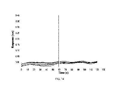
FIG. 34: Affinity constant assay of 14C12H1L1(hG1TM) to Clq. The antibody
concentrations
for the curve pairs from top to bottom are 20 nM, 10 nM, 5 nM, 2.5 nM and 1.25
nM, respectively.
FIG. 35: Affinity constant assay of 5C10H2L2-IgG1mt to Clq. The antigen
concentrations for
the curve pairs from top to bottom are 20 nM, 10 nM, 5 nM, 2.5 nM and 1.25 nM,
respectively.
FIG. 36: IFN-y secretion assay by adding 14C12H1L1 (hG1WT) and
14C12H1L1(hG1TM) to
mixed lymphocyte reaction.
FIG. 37: IL-2 secretion assay by adding 14C12H1L1 (hG1WT) and 14C12H1L1(hG1TM)
to
mixed lymphocyte reaction.
FIG. 38: ADCP effect assay of 14C12H1L1(hG1WT), nivolumab, and
14C12H1L1(hG1TM).
FIG. 39: Killing effect assay of 14C12H1L1(hG1TM) + anlotinib on human non-
small cell lung
cancer cells.
FIG. 40: Inhibited proliferation of mouse colorectal cancer MC38 cells by
14C12H1L1(hG1TM).
FIG. 41: Effectively enhanced immune response of immune cells to human gastric
cancer KATO
III cells by 14C12H1L1(hG1TM).
FIG. 42: Effectively enhanced immune response of immune cells to
nasopharyngeal cancer CNE-
2Z cells by 14C12H1L1(hG1TM).
FIG. 43: Effectively enhanced immune response of immune cells to mesothelioma
NCI-H2452
cells by 14C12H1L1(hG1TM).
FIG. 44: Effectively enhanced immune response of immune cells to human non-
small cell lung
cancer NCI-H446 cells by 14C12H1L1(hG1TM).
FIG. 45: Effectively enhanced immune response of immune cells to
nasopharyngeal cancer CNE-
2Z cells by 14C12H1L1(hG1TM) in combination with anlotinib hydrochloride.
31
Date Recue/Date Received 2022-01-10
CA 03146777 2022-01-10
FIG. 46: Significantly enhanced immune response of immune cells to MSI-H/dMMR
tumor
SW48 cells by 14C12H1L1(hG1TM) in combination with anlotinib.
FIG. 47: Significantly enhanced immune response of immune cells to human
colorectal cancer
SW837 cells of non-MSI-H/dMMR (i.e., MSS) phenotype by 14C12H1L1(hG1TM).
FIG. 48: Significantly enhanced immune response of immune cells to human
colorectal cancer
SW837 cells of non-MSI-H/dMMR (i.e., MSS) phenotype by 14C12H1L1(hG1TM) in
combination with anlotinib.
DETAILED DESCRIPTION
The embodiments of the present invention will be described in detail below
with reference to the
examples. Those skilled in the art will understand that the following examples
are only for
illustrating the present invention, and should not be construed as limitation
on the scope of the
present invention. In the cases where the techniques or conditions are not
specified, the examples
were implemented according to the techniques or conditions described in the
literature in the art
(e.g., see, Molecular Cloning: A Laboratory Manual, authored by J. Sambrook et
al., and
translated by Huang Peitang et al., Third Edition, Science Press) or according
to the product
manual. Reagents or instruments used are commercially available conventional
products if the
manufacturers thereof are not specified.
In the following experiments of the present invention:
BALB/c mice were purchased from Guangdong Medical Laboratory Animal Center.
The anti-PDL1 antibody 5C10H2L2-IgGlmt was prepared by methods described in
PCT
Publication No. W02017148424A1.
The anti-PD-1 antibody nivolumab (trade name: Opdivo) was purchased from the
Bristol-Myers
Squibb.
32
Date Recue/Date Received 2022-01-10
CA 03146777 2022-01-10
Human peripheral blood mononuclear cells were isolated and prepared in Akeso
Biopharma,
Inc., with informed consent of the donor.
Raji-PDL1 is a cell expressing human PD-L1 constructed by Akeso Biopharma on
the basis of
human B cells Raji via transfection.
Ficoll-PaqueTm PLUS (or Ficoll-Paque PLUS) was purchased from GE Healthcare.
Human IL-2 ELISA kit was purchased from Dakewe Biotech Co., Ltd.
RPMI 1640 medium, DMEM medium, Trypsin-EDTA (0.25%) phenol red and Blastidin
were
all purchased from Gibco.
Staphylococcus aureus enterotoxin B (SEB) was purchased from Dianotech.
FBS was purchased from Excell bio.
Mitomycin C (MMC) was purchased from Stressmarq.
The sequence of the isotype control, human anti-hen egg lysozyme IgG (anti-HEL
antibody, or
human IgG, abbreviated as hIgG) is derived from the variable region sequence
of the Fab F10.6.6
sequence in the study reported by Acierno et al., entitled "Affinity
maturation increases the
stability and plasticity of the Fv domain of anti-protein antibodies" (Acierno
et al., J Mol Biol.,
2007; 374(1):130-146).
Anlotinib used in the examples is hydrochloride salt of anlotinib under the
brand name Fukewei0
and generic name anlotinib hydrochloride, and was purchased from CTTQ Pharma.
33
Date Recue/Date Received 2022-01-10
CA 03146777 2022-01-10
Preparation Example 1: Sequence Design of Anti-PD-1 Antibody 14C12 and Its
Humanized
Antibody 14C12H1L1(hG1WT)
The amino acid sequences and encoding nucleotide sequences of the heavy and
light chains of
anti-PD-1 antibody 14C12 and its humanized antibody 14C12H1L1(hG1WT) are
identical to
those of 14C12 and 14C12H1L1 in Chinese Patent Publication No. CN106967172A
(or No.
CN106977602A), respectively.
(1) Heavy and light chain variable region sequences of 14C12
Nucleotide sequence of the heavy chain variable region of 14C12: (354 bp)
GAGGTCAAACTGGTGGAGAGCGGCGGCGGGCTGGTGAAGCCCGGCGGGTCACTGA
AACTGAGCTGCGCCGCTTCCGGCTTCGCCTTTAGCTCCTACGACATGTCATGGGTG
AGGCAGACCCCTGAGAAGCGCCTGGAATGGGTCGCTACTATCAGCGGAGGCGGGC
GATACACCTACTATCCTGACTCTGTCAAAGGGAGATTCACAATTAGTCGGGATAAC
GCCAGAAATACTCTGTATCTGCAGATGTCTAGTCTGCGGTCCGAGGATACAGCTCT
GTACTATTGTGCAAACCGGTACGGCGAAGCATGGTTTGCCTATTGGGGACAGGGCA
CCCTGGTGACAGTCTCTGCC (SEQ ID NO: 1)
Amino acid sequence of the heavy chain variable region of 14C12: (118 aa)
EVKLVES GGGLVKP GGSLKL S CAA S GFAF SSYDMSWVRQTPEKRLEWVATISGGGRY
TYYPDSVKGRFTISRDNARNTLYLQMSSLRSEDTALYYCANRYGEAWFAYWGQGTLV
TVSA (SEQ ID NO: 2)
Nucleotide sequence encoding the light chain variable region of 14C12: (321
bp)
GACATTAAGATGACACAGTCCCCTTCCTCAATGTACGCTAGCCTGGGCGAGCGAGT
GACCTTCACATGCAAAGCATCCCAGGACATCAACACATACCTGTCTTGGTTTCAGC
AGAAGCCAGGCAAAAGCCCCAAGACCCTGATCTACCGGGCCAATAGACTGGTGGA
CGGGGTCCCCAGCAGATTCTCCGGATCTGGCAGTGGGCAGGATTACTCCCTGACCA
TCAGCTCCCTGGAGTATGAAGACATGGGCATCTACTATTGCCTGCAGTATGATGAG
34
Date Recue/Date Received 2022-01-10
CA 03146777 2022-01-10
TTCCCTCTGACCTTTGGAGCAGGCACAAAACTGGAACTGAAG (SEQ ID NO: 3)
Amino acid sequence of the light chain variable region of 14C12: (107 aa)
D IKMTQ SP S SMYA SL GERVTFTCKA S QD INTYL SWF QQKP GKSPKTLIYRANRLVDGV
PSRF SGSGSGQDYSLTISSLEYEDMGIYYCLQYDEFPLTFGAGTKLELK (SEQ ID NO: 4)
(2) Heavy and light chain variable region and heavy and light chain sequences
of humanized
monoclonal antibody 14C12H1L1(hG1WT)
Nucleotide sequence of the heavy chain variable region of 14C12H1L1(hG1WT):
(354 bp)
GAAGTGCAGCTGGTCGAGTCTGGGGGAGGGCTGGTGCAGCCCGGCGGGTCACTGC
GACTGAGCTGCGCAGCTTCCGGATTCGCCTTTAGCTCCTACGACATGTCCTGGGTG
CGACAGGCACCAGGAAAGGGACTGGATTGGGTCGCTACTATCTCAGGAGGCGGGA
GATACACCTACTATCCTGACAGCGTCAAGGGCCGGTTCACAATCTCTAGAGATAAC
AGTAAGAACAATCTGTATCTGCAGATGAACAGCCTGAGGGCTGAGGACACCGCAC
TGTACTATTGTGCCAACCGCTACGGGGAAGCATGGTTTGCCTATTGGGGGCAGGGA
ACCCTGGTGACAGTCTCTAGT (SEQ ID NO: 5)
Amino acid sequence of the heavy chain variable region of 14C12H1L1(hG1WT):
(118 aa)
EVQLVES GGGLVQP GGSLRL S CAA S GFAF SSYDMSWVRQAPGKGLDWVATISGGGRY
TYYPDSVKGRFTISRDNSKNNLYLQMNSLRAEDTALYYCANRYGEAWFAYWGQGTL
VTVSS (SEQ ID NO: 6)
Nucleotide sequence encoding the light chain variable region of
14C12H1L1(hG1WT): (321 bp)
GACATTCAGATGACTCAGAGCCCCTCCTCCATGTCCGCCTCTGTGGGCGACAGGGT
CACCTTCACATGCCGCGCTAGTCAGGATATCAACACCTACCTGAGCTGGTTTCAGC
AGAAGCCAGGGAAAAGCCCCAAGACACTGATCTACCGGGCTAATAGACTGGTGTC
TGGAGTCCCAAGTCGGTTCAGTGGCTCAGGGAGCGGACAGGACTACACTCTGACC
ATCAGCTCCCTGCAGCCTGAGGACATGGCAACCTACTATTGCCTGCAGTATGATGA
Date Recue/Date Received 2022-01-10
CA 03146777 2022-01-10
GTTCCCACTGACCTTTGGCGCCGGGACAAAACTGGAGCTGAAG (SEQ ID NO: 7)
Amino acid sequence of the light chain variable region of 14C12H1L1(hG1WT):
(107 aa)
DIQMTQ SP S SMSA SVGDRVTFTCRA SQDINTYL SWF QQKP GKSPKTLIYRANRLVS GVP
SRFSGSGSGQDYTLTISSLQPEDMATYYCLQYDEFPLTFGAGTKLELK (SEQ ID NO: 8)
Nucleotide sequence of the heavy chain of 14C12H1L1(hG1WT): (1344 bp)
GAAGTGCAGCTGGTCGAGTCTGGGGGAGGGCTGGTGCAGCCCGGCGGGTCACTGC
GACTGAGCTGCGCAGCTTCCGGATTCGCCTTTAGCTCCTACGACATGTCCTGGGTG
CGACAGGCACCAGGAAAGGGACTGGATTGGGTCGCTACTATCTCAGGAGGCGGGA
GATACACCTACTATCCTGACAGCGTCAAGGGCCGGTTCACAATCTCTAGAGATAAC
AGTAAGAACAATCTGTATCTGCAGATGAACAGCCTGAGGGCTGAGGACACCGCAC
TGTACTATTGTGCCAACCGCTACGGGGAAGCATGGTTTGCCTATTGGGGGCAGGGA
ACCCTGGTGACAGTCTCTAGTGCCAGCACCAAAGGACCTAGCGTGTTTCCTCTCGC
CCCCTCCTCCAAAAGCACCAGCGGAGGAACCGCTGCTCTCGGATGTCTGGTGAAGG
ACTACTTCCCTGAACCCGTCACCGTGAGCTGGAATAGCGGCGCTCTGACAAGCGGA
GTCCATACATTCCCTGCTGTGCTGCAAAGCAGCGGACTCTATTCCCTGTCCAGCGTC
GTCACAGTGCCCAGCAGCAGCCTGGGCACCCAGACCTACATCTGTAACGTCAACCA
CAAGCCCTCCAACACCAAGGTGGACAAGAAAGTGGAGCCCAAATCCTGCGACAAG
ACACACACCTGTCCCCCCTGTCCTGCTCCCGAACTCCTCGGAGGCCCTAGCGTCTTC
CTCTTTCCTCCCAAACCCAAGGACACCCTCATGATCAGCAGAACCCCTGAAGTCAC
CTGTGTCGTCGTGGATGTCAGCCATGAGGACCCCGAGGTGAAATTCAACTGGTATG
TCGATGGCGTCGAGGTGCACAACGCCAAAACCAAGCCCAGGGAGGAACAGTACAA
CTCCACCTACAGGGTGGTGTCCGTGCTGACAGTCCTCCACCAGGACTGGCTGAACG
GCAAGGAGTACAAGTGCAAGGTGTCCAACAAGGCTCTCCCTGCCCCCATTGAGAA
GACCATCAGCAAGGCCAAAGGCCAACCCAGGGAGCCCCAGGTCTATACACTGCCT
CCCTCCAGGGACGAACTCACCAAGAACCAGGTGTCCCTGACCTGCCTGGTCAAGG
36
Date Recue/Date Received 2022-01-10
CA 03146777 2022-01-10
GCTTTTATCCCAGCGACATCGCCGTCGAGTGGGAGTCCAACGGACAGCCCGAGAAT
AACTACAAGACCACCCCTCCTGTCCTCGACTCCGACGGCTCCTTCTTCCTGTACAGC
AAGCTGACCGTGGACAAAAGCAGGTGGCAGCAGGGAAACGTGTTCTCCTGCAGCG
TGATGCACGAAGCCCTCCACAACCACTACACCCAGAAAAGCCTGTCCCTGAGCCCC
GGCAAA (SEQ ID NO: 9)
Amino acid sequence of the heavy chain of 14C12H1L1(hG1WT): (448 aa)
EVQLVES GGGLVQP GGSLRL S CAA S GFAF SSYDMSWVRQAPGKGLDWVATISGGGRY
TYYPDSVKGRFTISRDNSKNNLYLQMNSLRAEDTALYYCANRYGEAWFAYWGQGTL
VTVS SA S TKGP SVFPLAP S SKSTS GGTAALGC LVKDYFPEPVTVSWNS GALT S GVHTFP
AVL Q S S GLY SL S SVVTVP S S SLGTQTYICNVNHKP SNTKVDKKVEPKS CDKTHTCPP CP
APELLGGP SVF LFPPKPKDTLMISRTPEVTCVVVDVSHEDPEVKFNWYVDGVEVHNAK
TKPREEQYNSTYRVVSVLTVLHQDWLNGKEYKCKVSNKALPAPIEKTISKAKGQPREP
QVYTLPP SRDELTKNQVSLTCLVKGFYP SDIAVEWE SNGQPENNYKTTPPVLD SDGSFF
LYSKLTVDKSRWQQGNVFSCSVMHEALHNHYTQKSLSLSPGK (SEQ ID NO: 10)
Nucleotide sequence of the light chain of 14C12H1L1(hG1WT): (642 bp)
GACATTCAGATGACTCAGAGCCCCTCCTCCATGTCCGCCTCTGTGGGCGACAGGGT
CACCTTCACATGCCGCGCTAGTCAGGATATCAACACCTACCTGAGCTGGTTTCAGC
AGAAGCCAGGGAAAAGCCCCAAGACACTGATCTACCGGGCTAATAGACTGGTGTC
TGGAGTCCCAAGTCGGTTCAGTGGCTCAGGGAGCGGACAGGACTACACTCTGACC
ATCAGCTCCCTGCAGCCTGAGGACATGGCAACCTACTATTGCCTGCAGTATGATGA
GTTCCCACTGACCTTTGGCGCCGGGACAAAACTGGAGCTGAAGCGAACTGTGGCC
GCTCCCTCCGTCTTCATTTTTCCCCCTTCTGACGAACAGCTGAAATCAGGCACAGCC
AGCGTGGTCTGTCTGCTGAACAATTTCTACCCTAGAGAGGCAAAAGTGCAGTGGAA
GGTCGATAACGCCCTGCAGTCCGGCAACAGCCAGGAGAGTGTGACTGAACAGGAC
TCAAAAGATAGCACCTATTCCCTGTCTAGTACACTGACTCTGTCCAAGGCTGATTA
37
Date Recue/Date Received 2022-01-10
CA 03146777 2022-01-10
CGAGAAGCACAAAGTGTATGCATGCGAAGTGACACATCAGGGACTGTCAAGCCCC
GTGACTAAGTCTTTTAACCGGGGCGAATGT (SEQ ID NO: 11)
Amino acid sequence of the light chain of 14C12H1L1(hG1WT): (214 aa)
DIQMTQ SP S SMSA SVGDRVTFTCRA SQDINTYL SWF QQKP GKSPKTLIYRANRLVSGVP
SRFSGSGSGQDYTLTISSLQPEDMATYYCLQYDEFPLTFGAGTKLELKRTVAAPSVFIFP
PSDEQLKSGTASVVCLLNNFYPREAKVQWKVDNALQSGNSQESVTEQDSKDSTYSLSS
TLTLSKADYEKHKVYACEVTHQGLSSPVTKSFNRGEC (SEQ ID NO:12)
Preparation Example 2: Sequence Design of Humanized Antibody 14C12H1L1(hG4)
The heavy and light chain variable regions are identical to those of
14C12H1L1(hG1WT). Ig
gamma-4 chain C region (ACCESSION: P01861.1) was used as the heavy chain
constant region,
and Ig kappa chain C region (ACCESSION: P01834) was used as the light chain
constant region,
thus giving the antibody 14C12H1L1(hG4). The sequence of 14C12H1L1(hG4) is as
follows:
Nucleotide sequence of the heavy chain of 14C12H1L1(hG4): (1335 bp)
GAAGTGCAGCTGGTCGAGTCTGGGGGAGGGCTGGTGCAGCCCGGCGGGTCACTGC
GACTGAGCTGCGCAGCTTCCGGATTCGCCTTTAGCTCCTACGACATGTCCTGGGTG
CGACAGGCACCAGGAAAGGGACTGGATTGGGTCGCTACTATCTCAGGAGGCGGGA
GATACACCTACTATCCTGACAGCGTCAAGGGCCGGTTCACAATCTCTAGAGATAAC
AGTAAGAACAATCTGTATCTGCAGATGAACAGCCTGAGGGCTGAGGACACCGCAC
TGTACTATTGTGCCAACCGCTACGGGGAAGCATGGTTTGCCTATTGGGGGCAGGGA
ACCCTGGTGACAGTCTCTAGTGCCAGCACCAAAGGGCCCTCGGTCTTCCCCCTGGC
GCCCTGCTCCAGGAGCACCTCCGAGAGCACAGCCGCCCTGGGCTGCCTGGTCAAG
GACTACTTCCCCGAACCGGTGACGGTGTCGTGGAACTCAGGCGCCCTGACCAGCGG
CGTGCACACCTTCCCGGCTGTCCTACAGTCCTCAGGACTCTACTCCCTCAGCAGCGT
GGTGACCGTGCCCTCCAGCAGCTTGGGCACGAAGACCTACACCTGCAACGTAGATC
ACAAGCCCAGCAACACCAAGGTGGACAAGAGAGTTGAGTCCAAATATGGTCCCCC
ATGCCCACCATGCCCAGCACCTGAGTTCCTGGGGGGACCATCAGTCTTCCTGTTCC
38
Date Recue/Date Received 2022-01-10
CA 03146777 2022-01-10
CCCCAAAACCCAAGGACACTCTCATGATCTCCCGGACCCCTGAGGTCACGTGCGTG
GTGGTGGACGTGAGCCAGGAAGACCCCGAGGTCCAGTTCAACTGGTACGTGGATG
GCGTGGAGGTGCATAATGCCAAGACAAAGCCGCGGGAGGAGCAGTTCAACAGCAC
GTACCGTGTGGTCAGCGTCCTCACCGTCCTGCACCAGGACTGGCTGAACGGCAAGG
AGTACAAGTGCAAGGTCTCCAACAAAGGCCTCCCGTCCTCCATCGAGAAAACCATC
TCCAAAGCCAAAGGGCAGCCCCGAGAGCCACAGGTGTACACCCTGCCCCCATCCC
AGGAGGAGATGACCAAGAACCAGGTCAGCCTGACCTGCCTGGTCAAAGGCTTCTA
CCCCAGCGACATCGCCGTGGAGTGGGAGAGCAATGGGCAGCCGGAGAACAACTAC
AAGACCACGCCTCCCGTGCTGGACTCCGACGGCTCCTTCTTCCTCTACAGCAGGCT
AACCGTGGACAAGAGCAGGTGGCAGGAGGGGAATGTCTTCTCATGCTCCGTGATG
CATGAGGCTCTGCACAACCACTACACACAGAAGAGCCTCTCCCTGTCTCTGGGTAA
A (SEQ ID NO: 13)
Amino acid sequence of the heavy chain of 14C12H1L1(hG4): (445 aa)
EVQLVES GGGLVQP GGSLRL S CAA S GFAF SSYDMSWVRQAPGKGLDWVATISGGGRY
TYYPDSVKGRFTISRDNSKNNLYLQMNSLRAEDTALYYCANRYGEAWFAYWGQGTL
VTVSSASTKGPSVFPLAPCSRSTSESTAALGCLVKDYFPEPVTVSWNSGALTSGVHTFP
AVLQSSGLYSLSSVVTVPSSSLGTKTYTCNVDHKPSNTKVDKRVESKYGPPCPPCPAPE
FLGGPSVFLFPPKPKDTLMISRTPEVTCVVVDVSQEDPEVQFNWYVDGVEVHNAKTKP
REEQFNSTYRVVSVLTVLHQDWLNGKEYKCKVSNKGLPSSIEKTISKAKGQPREPQVY
TLPPSQEEMTKNQVSLTCLVKGFYPSDIAVEWESNGQPENNYKTTPPVLDSDGSFFLYS
RLTVDKSRWQEGNVFSCSVMHEALHNHYTQKSLSLSLGK (SEQ ID NO: 14)
The nucleotide sequence of the 14C12H1L1(hG4) light chain is identical to SEQ
ID NO: 11.
The amino acid sequence of the 14C12H1L1(hG4) light chain is identical to SEQ
ID NO: 12.
Preparation Example 3: Sequence Design of Humanized Antibody 14C12H1L1(hG1TM)
On the basis of 14C12H1L1(hG1WT) obtained in Preparation Example 1, a
humanized mutant
39
Date Recue/Date Received 2022-01-10
CA 03146777 2022-01-10
14C12H1L1(hG1TM) was obtained by introducing a leucine-to-alanine point
mutation at
position 234 (L234A), a leucine-to-alanine point mutation at position 235
(L235A), and a
glycine-to-alanine point mutation at position 237 (G237A) in the hinge region
of the heavy chain
according to the EU numbering system.
Nucleotide sequence of the heavy chain of 14C12H1L1(hG1TM): (1344 bp)
GAAGTGCAGCTGGTCGAGTCTGGGGGAGGGCTGGTGCAGCCCGGCGGGTCACTGC
GACTGAGCTGCGCAGCTTCCGGATTCGCCTTTAGCTCCTACGACATGTCCTGGGTG
CGACAGGCACCAGGAAAGGGACTGGATTGGGTCGCTACTATCTCAGGAGGCGGGA
GATACACCTACTATCCTGACAGCGTCAAGGGCCGGTTCACAATCTCTAGAGATAAC
AGTAAGAACAATCTGTATCTGCAGATGAACAGCCTGAGGGCTGAGGACACCGCAC
TGTACTATTGTGCCAACCGCTACGGGGAAGCATGGTTTGCCTATTGGGGGCAGGGA
ACCCTGGTGACAGTCTCTAGTGCCAGCACCAAAGGGCCCAGCGTGTTTCCTCTCGC
CCCCTCCTCCAAAAGCACCAGCGGAGGAACCGCTGCTCTCGGATGTCTGGTGAAGG
ACTACTTCCCTGAACCCGTCACCGTGAGCTGGAATAGCGGCGCTCTGACAAGCGGA
GTCCATACATTCCCTGCTGTGCTGCAAAGCAGCGGACTCTATTCCCTGTCCAGCGTC
GTCACAGTGCCCAGCAGCAGCCTGGGCACCCAGACCTACATCTGTAACGTCAACCA
CAAGCCCTCCAACACCAAGGTGGACAAGAAAGTGGAGCCCAAATCCTGCGACAAG
ACACACACCTGTCCCCCCTGTCCTGCTCCCGAAGCTGCTGGAGCCCCTAGCGTCTTC
CTCTTTCCTCCCAAACCCAAGGACACCCTCATGATCAGCAGAACCCCTGAAGTCAC
CTGTGTCGTCGTGGATGTCAGCCATGAGGACCCCGAGGTGAAATTCAACTGGTATG
TCGATGGCGTCGAGGTGCACAACGCCAAAACCAAGCCCAGGGAGGAACAGTACAA
CTCCACCTACAGGGTGGTGTCCGTGCTGACAGTCCTCCACCAGGACTGGCTGAACG
GCAAGGAGTACAAGTGCAAGGTGTCCAACAAGGCTCTCCCTGCCCCCATTGAGAA
GACCATCAGCAAGGCCAAAGGCCAACCCAGGGAGCCCCAGGTCTATACACTGCCT
CCCTCCAGGGACGAACTCACCAAGAACCAGGTGTCCCTGACCTGCCTGGTCAAGG
GCTTTTATCCCAGCGACATCGCCGTCGAGTGGGAGTCCAACGGACAGCCCGAGAAT
Date Recue/Date Received 2022-01-10
CA 03146777 2022-01-10
AACTACAAGACCACCCCTCCTGTCCTCGACTCCGACGGCTCCTTCTTCCTGTACAGC
AAGCTGACCGTGGACAAAAGCAGGTGGCAGCAGGGAAACGTGTTCTCCTGCAGCG
TGATGCACGAAGCCCTCCACAACCACTACACCCAGAAAAGCCTGTCCCTGAGCCCC
GGCAAA (SEQ ID NO: 15)
Amino acid sequence of the heavy chain of 14C12H1L1(hG1TM): (448 aa)
EVQLVES GGGLVQP GGSLRL S CAA S GFAF SSYDMSWVRQAPGKGLDWVATISGGGRY
TYYPDSVKGRFTISRDNSKNNLYLQMNSLRAEDTALYYCANRYGEAWFAYWGQGTL
VTVSSASTKGPSVFPLAPSSKSTSGGTAALGCLVKDYFPEPVTVSWNSGALTSGVHTFP
AVLQSSGLYSLSSVVTVPSSSLGTQTYICNVNHKPSNTKVDKKVEPKSCDKTHTCPPCP
APEAAGAPSVFLFPPKPKDTLMISRTPEVTCVVVDVSHEDPEVKFNWYVDGVEVHNA
KTKPREEQYNSTYRVVSVLTVLHQDWLNGKEYKCKVSNKALPAPIEKTISKAKGQPRE
PQVYTLPPSRDELTKNQVSLTCLVKGFYPSDIAVEWESNGQPENNYKTTPPVLDSDGSF
FLYSKLTVDKSRWQQGNVFSCSVMHEALHNHYTQKSLSLSPGK (SEQ ID NO: 16)
The nucleotide sequence of the 14C12H1L1(hG1TM) light chain is identical to
SEQ ID NO: 11.
The amino acid sequence of the 14C12H1L1(hG1TM) light chain is identical to
SEQ ID NO: 12.
Preparation Example 4: Sequence Design of Humanized Antibody 14C12H1L1(hG1DM)
On the basis of 14C12H1L1(hG1WT), a humanized mutant antibody 14C12H1L1(hG1DM)
was
obtained by introducing a leucine-to-alanine point mutation at position 234
(L234A) and a
leucine-to-alanine point mutation at position 235 (L235A) in the hinge region
of the heavy chain.
Nucleotide sequence of the heavy chain of 14C12H1L1(hG1DM): (1344 bp)
GAAGTGCAGCTGGTCGAGTCTGGGGGAGGGCTGGTGCAGCCCGGCGGGTCACTGC
GACTGAGCTGCGCAGCTTCCGGATTCGCCTTTAGCTCCTACGACATGTCCTGGGTG
CGACAGGCACCAGGAAAGGGACTGGATTGGGTCGCTACTATCTCAGGAGGCGGGA
GATACACCTACTATCCTGACAGCGTCAAGGGCCGGTTCACAATCTCTAGAGATAAC
41
Date Recue/Date Received 2022-01-10
CA 03146777 2022-01-10
AGTAAGAACAATCTGTATCTGCAGATGAACAGCCTGAGGGCTGAGGACACCGCAC
TGTACTATTGTGCCAACCGCTACGGGGAAGCATGGTTTGCCTATTGGGGGCAGGGA
ACCCTGGTGACAGTCTCTAGTGCTAGCACCAAAGGGCCCAGCGTGTTTCCTCTCGC
CCCCTCCTCCAAAAGCACCAGCGGAGGAACCGCTGCTCTCGGATGTCTGGTGAAGG
ACTACTTCCCTGAACCCGTCACCGTGAGCTGGAATAGCGGCGCTCTGACAAGCGGA
GTCCATACATTCCCTGCTGTGCTGCAAAGCAGCGGACTCTATTCCCTGTCCAGCGTC
GTCACAGTGCCCAGCAGCAGCCTGGGCACCCAGACCTACATCTGTAACGTCAACCA
CAAGCCCTCCAACACCAAGGTGGACAAGAAAGTGGAGCCCAAATCCTGCGACAAG
ACACACACCTGTCCCCCCTGTCCTGCTCCCGAAGCTGCTGGAGGCCCTAGCGTCTT
C CTCTTTCCTCCCAAACC CAAGGACACC CTCATGATCAGCAGAAC CC CTGAAGTCA
CCTGTGTCGTCGTGGATGTCAGCCATGAGGACCCCGAGGTGAAATTCAACTGGTAT
GTCGATGGCGTCGAGGTGCACAACGCCAAAACCAAGCCCAGGGAGGAACAGTACA
ACTCCACCTACAGGGTGGTGTCCGTGCTGACAGTCCTCCACCAGGACTGGCTGAAC
GGCAAGGAGTACAAGTGCAAGGTGTCCAACAAGGCTCTCCCTGCCCCCATTGAGA
AGACCATCAGCAAGGCCAAAGGCCAACCCAGGGAGCCCCAGGTCTATACACTGCC
TCCCTCCAGGGACGAACTCACCAAGAACCAGGTGTCCCTGACCTGCCTGGTCAAGG
GCTTTTATCCCAGCGACATCGCCGTCGAGTGGGAGTCCAACGGACAGCCCGAGAAT
AACTACAAGACCACCCCTCCTGTCCTCGACTCCGACGGCTCCTTCTTCCTGTACAGC
AAGCTGACCGTGGACAAAAGCAGGTGGCAGCAGGGAAACGTGTTCTCCTGCAGCG
TGATGCACGAAGCCCTCCACAACCACTACACCCAGAAAAGCCTGTCCCTGAGCCCC
GGCAAA (SEQ ID NO: 17)
Amino acid sequence of the heavy chain of 14C12H1L1(hG1DM): (448 aa)
EVQLVES GGGLVQP GGSLRL S CAA S GFAF SSYDMSWVRQAPGKGLDWVATISGGGRY
TYYPDSVKGRFTISRDNSKNNLYLQMNSLRAEDTALYYCANRYGEAWFAYWGQGTL
VTVS SA S TKGP SVFPLAP S SKSTS GGTAALGC LVKDYFPEPVTVSWNS GALT S GVHTFP
AVL Q S S GLY SL S SVVTVP S S SLGTQTYICNVNHKP SNTKVDKKVEPKS CDKTHTCPP CP
42
Date Recue/Date Received 2022-01-10
CA 03146777 2022-01-10
APEAAGGPSVFLFPPKPKDTLMISRTPEVTCVVVDVSHEDPEVKFNWYVDGVEVHNA
KTKPREEQYNSTYRVVSVLTVLHQDWLNGKEYKCKVSNKALPAPIEKTISKAKGQPRE
PQVYTLPPSRDELTKNQVSLTCLVKGFYPSDIAVEWESNGQPENNYKTTPPVLDSDGSF
FLYSKLTVDKSRWQQGNVFSCSVMHEALHNHYTQKSLSLSPGK (SEQ ID NO: 18)
The nucleotide sequence of the 14C12H1L1(hG1DM) light chain is identical to
SEQ ID NO: 11.
The amino acid sequence of the 14C12H1L1(hG1DM) light chain is identical to
SEQ ID NO: 12.
Experimental Example 1: Affinity Assay of 14C12H1L1(hG1DM), 14C12H1L1(hG4),
14C12H1L1(hG1WT) and 14C12H1L1(hG1TM) to Fe Receptor FcyRI
The Fe receptor FcyRI, also known as CD64, can bind to the Fe fragment of IgG
antibodies and
is involved in antibody-dependent cell-mediated cytotoxicity (ADCC). The
binding capacity of
a therapeutic monoclonal antibody to Fc receptors will influence the safety
and efficacy of the
antibody. The affinity constants of 14C12H1L1(hG1DM), 14C12H1L1(hG4),
14C12H1L1(hG1WT) and 14C 12H1L1(hG1TM) to FcyRI were measured in this
experiment
using a Fortebio Octet system to evaluate the ADCC activity of the antibodies.
The method for determining the affinity constant of the antibodies to FcyRI by
the Fortebio Octet
system is briefly described as follows: the sample dilution buffer was a
solution of 0.02% Tween-
and 0.1% BSA in PBS, pH 7.4. A 1 g/mL FcyRI solution (from Sinobio) was added
to the
20 HIS1K sensor to immobilize the FcyRI on the sensor surface for 50 s. The
association and
dissociation constants of the antibodies to FcyRI were both determined in the
buffer with the
antibody concentrations being 3.12-50 nM (serial two-fold dilution). The
sensor with
immobilized antigen was equilibrated in the buffer for 60 s, and then the
binding of the
immobilized FcyRI on the sensor to the antibodies was determined for 120 s;
the dissociation of
FcyRI from the antibodies was determined in 120 s. The temperature was 30 C
and the frequency
was 0.3 Hz. The data were fitted and analyzed with a 1:1 model to obtain the
affinity constants
43
Date Recue/Date Received 2022-01-10
CA 03146777 2022-01-10
to FcyRI for the antibodies.
The results of affinity constant assay of 14C12H1L1(hG1DM), 14C12H1L1(hG4),
14C12H1L1(hG1WT) and 14C12H1L1(hG1TM) and the control antibody 5C10H2L2-IgG1mt
to FcyRI are shown in Table 1 and FIGs. 1-5.
Table 1: Kinetics for binding of 14C12H1L1(hG1DM), 14C12H1L1(hG4),
14C12H1L1(hG1WT) and 14C12H1L1(hG1TM) and the control antibody 5C10H2L2-IgGlmt
to FcyRI
Antibody KD (M) kon (1/Ms) SE (kon) kdis (1/s) SE (kdis) Rmax
(nm)
14C12H1L1(hG1DM) N/A N/A N/A N/A N/A N/A
14C12H1L1(hG4) 5.80E-09 6.37E+05 2.21E+04 3.69E-03 1.05E-04 0.45-0.54
14C12H1L1(hG1WT) 2.52E-09 6.94E+05 2.19E+04 1.75E-03 9.14E-05 0.50-0.55
14C12H1L1(hG1TM) N/A N/A N/A N/A N/A N/A
5C10H2L2-IgG1mt N/A N/A N/A N/A N/A N/A
N/A indicates that the antibody had no binding or an extremely weak binding
signal to the
antigen, and thus the results were not analyzed and no corresponding data was
obtained.
The results showed that both 14C12H1L1(hG4) and 14C12H1L1(hG1WT) bound to
FcyRI with
affinity constants of 5.80E-09 M and 2.52E-09 M, respectively;
14C12H1L1(hG1TM) and
5C10H2L2-IgG1mt had no binding or an extremely weak binding signal to FcyRI,
and thus the
results were not analyzed and no corresponding data was obtained.
The results suggested that the binding activities of 14C12H1L1(hG1DM) and
14C12H1L1(hG1TM) to FcyRI are effectively eliminated as compared to
14C12H1L1(hG4) and
14C12H1L 1 (hG1WT).
44
Date Recue/Date Received 2022-01-10
CA 03146777 2022-01-10
Experimental Example 2: Affinity Assay of 14C12H1L1(hG1DM), 14C12H1L1(hG4),
14C12H1L1(hG1WT) and 14C12H1L1(hG1TM) to Fe Receptor FcyRIIIa and subtypes
thereof
(1) Affinity constant assay of 14C12H1L1(hG1DM), 14C12H1L1(hG4),
14C12H1L1(hG1WT)
and 14C12H1L1(hG1TM) to FcyRIIIa V158
.. The Fe receptor FcyRIIIa V158 (also known as CD16a V158), can bind to the
Fe fragment of
IgG antibodies and mediate ADCC effects. The affinity constants of
14C12H1L1(hG1DM),
14C12H1L1(hG4), 14C12H1L1(hG1WT) and 14C12H1L1(hG1TM) to FcyRIIIa V158 were
measured in this experiment using a Fortebio Octet system to evaluate the ADCC
activity of the
antibodies.
The method for determining the affinity constant of the antibodies and the
control antibody
5C10H2L2-IgG1mt to FcyRIIIa V158 by the Fortebio Octet system is briefly
described as
follows: the sample dilution buffer was a solution of 0.02% Tween-20 and 0.1%
BSA in PBS,
pH 7.4. 5 ug/mL FcyRIIIa V158 was immobilized on the HIS1K sensor for 120 s.
The sensor
was equilibrated in a buffer for 60 s, and the binding of the immobilized
FcyRIIIa V158 on the
sensor to the antibodies at concentrations of 31.25-500 nM (serial two-fold
dilution) was
determined for 60 s. The antibody was dissociated in the buffer for 60 s. The
sensor was refreshed
4 times in 10 mM glycine pH 1.5, each for 5 s. The temperature was 30 C and
the frequency
was 0.3 Hz. The data were analyzed by 1:1 model fitting to obtain affinity
constants.
The results of affinity constant assay of 14C12H1L1(hG1DM), 14C12H1L1(hG4),
14C12H1L1(hG1WT) and 14C12H1L1(hG1TM) and the control antibody 5C10H2L2-IgG1mt
to FcyRIIIa V158 are shown in Table 2 and FIGs. 6-10.
Date Recue/Date Received 2022-01-10
CA 03146777 2022-01-10
Table 2: Kinetics for binding of 14C12H1L1(hG1DM), 14C12H1L1(hG4),
14C12H1L1(hG1WT) and 14C12H1L1(hG1TM) and the control antibody 5C10H2L2-IgG1mt
to FcyRIIIa V158
Antibody KD (M) kon (1/Ms) SE (kon) kdis (1/s) SE (kdis)
Rmax (nm)
14C12H1L1(hG1DM) 6.12E-07 1.35E+05 3.74E+04 8.24E-02 8.34E-03 0.33-
0.41
14C12H1L1(hG4) N/A N/A N/A N/A N/A N/A
14C12H1L1(hG1WT) 6.54E-08 2.61E+05 2.10E+04 1.71E-02 6.47E-04 0.80-1.17
14C12H1L1(hG1TM) N/A N/A N/A N/A N/A N/A
5C10H2L2-IgG1mt N/A N/A N/A N/A N/A N/A
N/A indicates that the antibody had no binding or an extremely weak binding
signal to the
antigen, and thus the results were not analyzed and no corresponding data was
obtained.
The results showed that both 14C12H1L1(hG1DM) and 14C12H1L1(hG1WT) bound to
FcyRIIIa V158 with affinity constants of 6.21E-07M and 6.54E-08M,
respectively;
14C12H1L1(hG4), 14C12H1L1(hG1TM) and 5C10H2L2-IgG1mt had no binding or an
extremely weak binding signal to FcyRIIIa V158, and thus the results were not
analyzed.
The results suggested that the binding activities of 14C12H1L1(hG4),
14C12H1L1(hG1TM) and
the control antibody 5C10H2L2-IgG1mt to FcyR IIIa V158 are effectively
eliminated as
compared to 14C12H1L1(hG1DM) and 14C12H1L1(hG1WT).
(2) Affinity constant assay of 14C12H1L1(hG1DM), 14C12H1L1(hG4),
14C12H1L1(hG1WT) and
14C12H1L1(hG1TM) to FcyRIIIa F158
The Fc receptor FcyRIIIa F158 (also known as CD16a F158), can bind to the Fc
fragment of
IgG antibodies and mediate ADCC effects. The affinity constants of
14C12H1L1(hG1DM),
14C12H1L1(hG4), 14C12H1L1(hG1WT), 14C12H1L1(hG1TM) and the control antibody to
46
Date Recue/Date Received 2022-01-10
CA 03146777 2022-01-10
FcyRIIIa F158 were measured in this experiment using a Fortebio Octet system
to evaluate the
ADCC activity of the antibodies.
The method for determining the affinity constant of 14C12H1L1(hG1DM),
14C12H1L1(hG4),
14C12H1L1(hG1WT) and 14C12H1L1(hG1TM) to FcyRIIIa F158 by the Fortebio Octet
system is briefly described as follows: the sample dilution buffer was a
solution of 0.02% Tween-
20 and 0.1% BSA in PBS, pH 7.4. 5 ug/mL FcyRIIIa F158 was immobilized on the
HIS1K
sensor for 120 s. The sensor was equilibrated in a buffer for 60 s, and the
binding of the
immobilized FcyRIIIa F158 on the sensor to the antibodies at concentrations of
31.25-500 nM
(serial two-fold dilution) was determined for 60 s. The antibody was
dissociated in the buffer for
60 s. The sensor was refreshed 4 times in 10 mM glycine pH 1.5, each for 5 s.
The temperature
was 30 C and the frequency was 0.3 Hz. The data were analyzed by 1:1 model
fitting to obtain
affinity constants.
The results of affinity constant assay of 14C12H1L1(hG1DM), 14C12H1L1(hG4),
14C12H1L1(hG1WT) and 14C12H1L1(hG1TM) and the control antibody 5C10H2L2-IgG 1
mt
to FcyRIIIa F158 are shown in Table 3 and FIGs. 11-15.
Table 3: Kinetics for binding of 14C12H1L1 antibodies and isotypes thereof to
FcyRIIIa F158
Antibody KD (M) kon (1/Ms) SE (kon) kdis (1/s) SE (kdis) Rmax
(nm)
14C12H1L1(hG1DM) N/A N/A N/A N/A N/A N/A
14C12H1L1(hG4) N/A N/A N/A N/A N/A N/A
14C12H1L1(hG1WT) 1.02E-07 2.52E+05 2.93E+04 2.56E-02 1.12E-03 0.34-
0.57
14C12H1L1(hG1TM) N/A N/A N/A N/A N/A N/A
5C10H2L2-IgG1mt N/A N/A N/A N/A N/A N/A
N/A indicates that the antibody had no binding or an extremely weak binding
signal to the
antigen, and thus the results were not analyzed and no corresponding data was
obtained.
47
Date Recue/Date Received 2022-01-10
CA 03146777 2022-01-10
The results showed that 14C12H1L1(hG1WT) bound to FcyRIIIa F158 with an
affinity constant
of 1.02E-07M; 14C12H1L1(hG1DM), 14C12H1L1(hG4), 14C12H1L1(hG1TM) and
5C10H2L2-IgGlmt had no binding or an extremely weak binding signal to FcyRIIIa
F158, and
thus the results were not analyzed and no corresponding data was obtained.
The results suggested that the binding activities of 14C12H1L1(hG1DM),
14C12H1L1(hG4) and
14C12H1L1(hG1TM) to FcyRIIIa F158 are effectively eliminated as compared to
14C 12H1L 1 (hG1WT).
Experimental Example 3: Affinity Assay of 14C12H1L1(hG1DM), 14C12H1L1(hG4),
14C12H1L1(hG1WT) and 14C 12H1L1(hG1TM) to Fc Receptor FcyRIIa and subtypes
thereof
(1) Affinity constant assay of 14C12H1L1(hG1DM), 14C12H1L1(hG4),
14C12H1L1(hG1WT) and
14C12H1L1(hG1TM) to FcyRIIa H131
The Fc receptor FcyRIIa H131, also known as CD32a H131, can bind to the Fc
fragment of IgG
antibodies and is involved in antibody-dependent cell-mediated cytotoxicity
(ADCC). The
binding capacity of a therapeutic monoclonal antibody to Fc receptors will
influence the safety
and efficacy of the antibody. The affinity constants of 14C12H1L1(hG1DM),
14C12H1L1(hG4),
14C12H1L1(hG1WT) and 14C12H1L1(hG1TM) to FcyRIIa H131 were measured in this
experiment using a Fortebio Octet system to evaluate the binding capacity of
the antibodies to
Fc receptor.
The method for determining the affinity constant of 14C12H1L1(hG1DM),
14C12H1L1(hG4),
14C12H1L1(hG1WT) and 14C12H1L1(hG1TM) to FcyRIIa H131 by the Fortebio Octet
system
is briefly described as follows: the immobilization dilution buffer was a
solution of PBS, 0.02%
Tween-20 and 0.1% BSA, pH 7.4, and the analyte dilution buffer was a solution
of 0.02% Tween-
20, 0.02% casein and 0.1% BSA in PBS, pH 7.4. 5 g/mL FcyRIIa H131 was
immobilized on
the NTA sensor at an immobilization height of about 1.0 nm. The sensor was
equilibrated in a
buffer of 0.02% Tween-20, 0.02% casein and 0.1% BSA in PBS, pH 7.4 for 300 s
of blocking,
48
Date Recue/Date Received 2022-01-10
CA 03146777 2022-01-10
and the binding of the immobilized FcyRIIa H131 on the sensor to the
antibodies at
concentrations of 12.5-200 nM (serial two-fold dilution) was determined for 60
s. The antibody
was dissociated in the buffer for 60 s. The sensor was refreshed in 10 mM
glycine pH 1.7 and 10
nM nickel sulfate. The temperature was 30 C and the frequency was 0.6 Hz. The
data were
analyzed by 1:1 model fitting to obtain affinity constants.
The results of affinity constant assay of 14C12H1L1(hG1DM), 14C12H1L1(hG4),
14C12H1L1(hG1WT) and 14C12H1L1(hG1TM) and the control antibody 5C10H2L2-IgG1mt
to FcyRIIa H131 are shown in Table 4 and FIGs. 16-20.
Table 4: Kinetics for binding of 14C12H1L1 antibodies and isotypes thereof to
FcyRIIa H131
Sample ID KD (M) kon (1/Ms) SE (kon) kdis (1/s) SE (kdis)
Rmax (nm)
14C12H1L1(hG1DM) N/A N/A N/A N/A N/A N/A
14C12H1L1(hG4) 5.07E-08 2.57E+05 2.37E+04 1.30E-02 6.47E-04 0.18-
0.35
14C12H1L1(hG1WT) 5.74E-08 4.65E+05 5.99E+04 2.67E-02 1.24E-03 0.82-
1.12
14C12H1L1(hG1TM) N/A N/A N/A N/A N/A N/A
5C10H2L2-IgG1mt N/A N/A N/A N/A N/A N/A
N/A indicates that the antibody had no binding or an extremely weak binding
signal to the
antigen, and thus the results were not analyzed and no corresponding data was
obtained.
The results showed that both 14C12H1L1(hG4) and 14C12H1L1(hG1WT) bound to
FcyRIIa H131 with affinity constants of 5.07E-08M and 5.74E-08M, respectively;
14C12H1L1(hG1DM), 14C12H1L1(hG1TM) and 5C10H2L2-IgG1mt had no binding or an
extremely weak binding signal to FcyRIIa H131, and thus the results were not
analyzed and no
corresponding data was obtained.
The results suggested that the binding activities of 14C12H1L1(hG1DM) and
14C12H1L1(hG1TM) to FcyRIIa H131 are effectively eliminated as compared to
14C12H1L1(hG4) and 14C12H1L1(hG1WT).
49
Date Recue/Date Received 2022-01-10
CA 03146777 2022-01-10
(2) Affinity constant assay of 14C12H1L1(hG1DM), 14C12H1L1(hG4),
14C12H1L1(hG1WT)
and 14C12H1L1(hG1TM) to FcyRIIa R131
The Fc receptor FcyRIIa R131, also known as CD32a R131, can bind to the Fc
fragment of IgG
antibodies and is involved in antibody-dependent cell-mediated cytotoxicity
(ADCC). The
binding capacity of a therapeutic monoclonal antibody to Fc receptors will
influence the safety
and efficacy of the antibody. The affinity constants of 14C12H1L1(hG1DM),
14C12H1L1(hG4),
14C12H1L1(hG1WT) and 14C12H1L1(hG1TM) to FcyRIIa R131 were measured in this
experiment using a Fortebio Octet system to evaluate the binding capacity of
the antibodies to
Fc receptor.
The method for determining the affinity constant of 14C12H1L1(hG1DM),
14C12H1L1(hG4),
14C12H1L1(hG1WT) and 14C12H1L1(hG1TM) to FcyRIIa R131 by the Fortebio Octet
system
is briefly described as follows: the immobilization dilution buffer was a
solution of PBS, 0.02%
Tween-20 and 0.1% BSA, pH 7.4, and the analyte dilution buffer was a solution
of 0.02% Tween-
20, 0.02% casein and 0.1% BSA in PBS, pH 7.4. 5 ug/mL FcyRIIa R131 was
immobilized on
the NTA sensor at an immobilization height of about 1.0 nm. The sensor was
equilibrated in a
buffer of 0.02% Tween-20, 0.02% casein and 0.1% BSA in PBS, pH 7.4 for 300 s
of blocking,
and the binding of the immobilized FcyRIIa R131 on the sensor to the
antibodies at
concentrations of 12.5-200 nM (serial two-fold dilution) was determined for 60
s. The antibody
was dissociated in the buffer for 60 s. The sensor was refreshed in 10 mM
glycine pH 1.7 and 10
nM nickel sulfate. The temperature was 30 C and the frequency was 0.6 Hz. The
data were
analyzed by 1:1 model fitting to obtain affinity constants.
The results of affinity constant assay of 14C12H1L1(hG1DM), 14C12H1L1(hG4),
14C12H1L1(hG1WT) and 14C12H1L1(hG1TM) and the control antibody 5C10H2L2-IgG1mt
to FcyRIIa R131 are shown in Table 5 and FIGs. 21-25.
50
Date Recue/Date Received 2022-01-10
CA 03146777 2022-01-10
Table 5: Kinetics for binding of 14C12H1L1 antibodies and isotypes thereof to
FcyRIIa R131
Antibody KD (M) kon (1/Ms) SE (kon) kdis (1/s) SE (kdis) Rmax
(nm)
14C12H1L1(hG1DM) N/A N/A N/A N/A N/A N/A
14C12H1L1(hG4) 3.13E-08 3.50E+05 3.05E+04 1.10E-02 6.43E-04 0.21-
0.46
14C12H1L1(hG1WT) 3.46E-08 6.60E+05 9.46E+04 2.28E-02 1.33E-03 0.39-
0.83
14C12H1L1(hG1TM) 2.32E-07 4.04E+05 9.45E+04 9.38E-02 4.89E-03 0.19-
0.35
5C10H2L2-IgG1mt N/A N/A N/A N/A N/A N/A
N/A indicates that the antibody had no binding or an extremely weak binding
signal to the
antigen, and thus the results were not analyzed and no corresponding data was
obtained.
The results showed that 14C12H1L1(hG4), 14C12H1L1(hG1WT) and 14C12H1L1(hG1TM)
bound to FcyRIIa R131 with affinity constants of 3.13E-08M, 3.46E-08M and
2.32E-07M,
respectively; 14C12H1L1(hG1DM) and 5C10H2L2-IgG1mt had no binding or an
extremely
weak binding signal to FcyRIIa R131, and thus the results were not analyzed
and no
corresponding data was obtained.
The results suggest that among the antibodies with binding activities,
14C12H1L1(hG1TM) has
the weakest binding capacity and the lowest binding activity to FcyRIIa R131
as compared to
14C12H1L1(hG4) and 14C12H1L1(hG1WT).
Experimental Example 4: Affinity constant assay of 14C12H1L1(hG1DM).
14C12H1L1(hG4).
14C12H1L1(hG1WT) and 14C12H1L1(hG1TM) to FcyRIIb
The Fc receptor FcyRIIb (also known as CD32b), can bind to the Fc fragment of
IgG antibodies,
down-regulate functions of immune cells, inhibit the activation and
proliferation of immune cells
and inhibit the secretion of cytokines. The affinity constants of the
antibodies to FcyRIIb were
measured in this experiment using a Fortebio Octet system to evaluate the
binding capacity of
51
Date Recue/Date Received 2022-01-10
CA 03146777 2022-01-10
14C12H1L1(hG1DM), 14C12H1L1(hG4), 14C12H1L1(hG1WT) and 14C12H1L1(hG1TM) to
Fc receptor.
The method for determining the affinity constant of 14C12H1L1(hG1DM),
14C12H1L1(hG4),
14C12H1L1(hG1WT) and 14C12H1L1(hG1TM) to FcyRIIb by the Fortebio Octet system
is
briefly described as follows: the immobilization dilution buffer was a
solution of PBS, 0.02%
Tween-20 and 0.1% BSA, pH 7.4, and the analyte dilution buffer was a solution
of 0.02% Tween-
20, 0.02% casein and 0.1% BSA in PBS, pH 7.4. 5 ug/mL hFc7R1Ib-his was
immobilized on the
NTA sensor at an immobilization height of about 1.0 nm. The sensor was
equilibrated in a buffer
of 0.02% Tween-20, 0.02% casein and 0.1% BSA in PBS, pH 7.4 for 300 s of
blocking, and the
binding of the immobilized hFcyRIIb-his on the sensor to the antibodies at
concentrations of
12.5-200 nM (serial two-fold dilution) was determined for 60 s. The antibody
was dissociated in
the buffer for 60 s. The sensor was refreshed in 10 mM glycine pH 1.7 and 10
nM nickel sulfate.
The temperature was 30 C and the frequency was 0.6 Hz. The data were analyzed
by 1:1 model
fitting to obtain affinity constants.
The results of affinity constant assay of 14C12H1L1(hG1DM), 14C12H1L1(hG4),
14C12H1L1(hG1WT) and 14C12H1L1(hG1TM) and the control antibody 5C10H2L2-IgG1mt
to FcyRIIb are shown in Table 6 and FIGs. 26-30.
Table 6: Kinetics for binding of 14C12H1L1 antibodies and isotypes thereof to
FcyRIIb
Antibody KD (M) kon (1/Ms) SE (kon) kdis (1/s) SE (kdis) Rmax
(nm)
14C12H1L1(hG1DM) N/A N/A N/A N/A N/A N/A
14C12H1L1(hG4) 5.62E-08 2.88E+05 2.94E+04 1.62E-02 7.63E-04 0.22-0.33
14C12H1L1(hG1WT) 6.13E-08 3.18E+05 3.48E+04 1.95E-02 8.96E-04 0.16-0.37
14C12H1L1(hG1TM) N/A N/A N/A N/A N/A N/A
5C10H2L2-IgG1mt N/A N/A N/A N/A N/A N/A
N/A indicates that the antibody had no binding or an extremely weak binding
signal to the
52
Date Recue/Date Received 2022-01-10
CA 03146777 2022-01-10
antigen, and thus the results were not analyzed and no corresponding data was
obtained.
The results showed that both 14C 12H 1 L 1 (hG4) and 14C 12H 1 L 1 (hG 1 WT)
bound to FcyRIIb
with affinity constants of 5.62E-08M and 6.13E-08M, respectively;
14C12H1L1(hG1DM),
14C12H1L1(hG1TM) and 5C10H2L2-IgG1mt had no binding or an extremely weak
binding
signal to FcyRIIb, and thus the results were not analyzed and no corresponding
data was obtained.
The results suggested that the binding activities of 14C12H1L1(hG1DM) and
14C12H1L1(hG1TM) to FcyRIIb are effectively eliminated as compared to
14C12H1L1(hG4)
and 14C12H1L1(hG1WT).
Experimental Example 5: Affinity assay of 14C12H1L1(hG1DM), 14C12H1L1(hG4),
14C12H1L1(hG1WT) and 14C12H1L1(hG1TM) to Clq
Serum complement Clq can bind to the Fc fragment of IgG antibodies and mediate
CDC effects.
The binding capacity of a therapeutic monoclonal antibody to Clq will
influence the safety and
efficacy of the antibody. The affinity constants of 14C12H1L1(hG1DM),
14C12H1L1(hG4),
14C12H1L1(hG1WT) and 14C12H1L1(hG1TM) to Clq were measured in this experiment
using
a Fortebio Octet system to evaluate the CDC activity of the antibodies.
The method for determining the affinity constants of the antibodies to Clq by
the Fortebio Octet
system is briefly described as follows: the sample dilution buffer was a
solution of 0.02% Tween-
and 0.1% BSA in PBS, pH 7.4. 50 ug/mL antibody was immobilized on the FAB2G
sensor at
20 an immobilization height of about 2.0 nm. The sensor was equilibrated in
a buffer for 60 s for
blocking, and the binding of the immobilized antibody on the sensor to the
antigen Cl q at
concentrations of 1.25-20 nM (serial two-fold dilution) was determined for 60
s. The antigen and
antibody were dissociated in the buffer for 60 s. The sensor was refreshed 4
times in 10 mM
glycine pH 1.7, each for 5 s. The shaking speed of the sample plate was 1000
rpm, the temperature
was 30 C and the frequency was 0.6 Hz. The data were analyzed by 1:1 model
fitting to obtain
affinity constants. The data acquisition software was Fortebio Data
Acquisition 7.0, and the data
53
Date Recue/Date Received 2022-01-10
CA 03146777 2022-01-10
analysis software was Fortebio Data Analysis 7Ø
The results of affinity constant assay of 14C12H1L1(hG1DM), 14C12H1L1(hG4),
14C12H1L1(hG1WT) and 14C12H1L1(hG1TM) and the control 5C10H2L2-IgG1mt to Clq
are
shown in Table 7 and FIGs. 31-35.
Table 7: Kinetics for binding of 14C12H1L1 antibodies and isotypes thereof to
Clq
Antibody KD (M) kon (1/Ms) SE (kon) kdis (1/s) SE (kdis)
Rmax (nm)
14C12H1L1(hG1DM) N/A N/A N/A N/A N/A N/A
14C12H1L1(hG4) N/A N/A N/A N/A N/A N/A
14C12H1L1(hG1WT) 1.35E-09 4.83E+06 4.24E+05 6.54E-03 5.33E-04 0.32-
0.53
14C12H1L1(hG1TM) N/A N/A N/A N/A N/A N/A
1.05E- 1.10E-
5C10H2L2-IgG 1 mt 4.43E-09 2.38E+06 4.21E+05
0.19-0.26
02 03
N/A indicates that the antibody had no binding or an extremely weak binding
signal to the
antigen, and thus the results were not analyzed and no corresponding data was
obtained.
The results showed that 14C12H1L1(hG1WT) bound to Clq with an affinity
constant of 1.35E-
09M; 14C12H1L1(hG1DM), 14C12H1L1(hG4) and 14C12H1L1(hG1TM) had no binding or
an
extremely weak binding signal to Clq, and thus the results were not analyzed
and no
corresponding data was obtained.
The results also showed that 5C10H2L2-IgG1mt bound to Clq with an affinity
constant of 4.43E-
09, indicating that it has binding activity to Clq and can cause CDC effects.
Experimental Example 6: Pharmacodynamic Activities of 14C12H1L1(hG1WT) and
14C12H1L1(hG1TM) in Co-culture System of Peripheral Blood Mononuclear Cells
and Raji-
PDL1 Cells
In this experiment, the pharmacodynamic activity of anti-PD-1 antibodies
14C12H1L1 (hG1WT)
and 14C12H1L1 (hG1TM), and the control anti-PD-Li antibodies 5C10H2L2-IgGlmt
and
54
Date Recue/Date Received 2022-01-10
CA 03146777 2022-01-10
nivolumab in relieving immunosuppression mediated by PD-1/PD-L1 were detected
in a co-
culture system of peripheral blood mononuclear cells and Raji-PDL1 cells.
In the mixed lymphocyte reaction, when isolated peripheral blood mononuclear
cells (containing
immune cells expressing immunocompetent PD-1) and the Raji-PDL1 cells
expressing PD-L1
are co-cultured, the interaction of PD-1 and PD-L1 can mediate the function
inhibition of the
immune cells, showing reduced secretion of cytokines IFN-y and IL-2. Anti-PD-1
or anti-PD-Li
antibodies can relieve such immunosuppression of immune cells, leading to
increased cytokine
secretion. Raji is a B cell line. As described above, B cells can be used as
antigen presenting cells
to mediate the immune response of immune cells to tumor cells. In the present
invention, the
Raji-PDL1 and PBMCs co-culture system was used to evaluate the pharmacological
activity of
the anti-PD-1 antibodies, and a Raji-PDL1, PBMCs and tumor cell co-culture
system was used
to evaluate the pharmacological activity of the anti-PD-1 antibodies in
different tumors.
Peripheral blood mononuclear cells were isolated by the Ficoll-Paque Plus (GE
Healthcare, Cat
No.:171440-02), and then stimulated with SEB (0.5 g/mL) for two days. The
stimulated mature
peripheral blood mononuclear cells (1 x10⁵ cells/well) and Raji-PDL1 cells (1
x105 cells/well)
treated with MMC (Mito-mycin C with a treatment concentration of 2 g/mL) for
1 h were added
to a 96-well plate before 14C12H1L1(hG1WT), 14C12H1L1(hG1TM), the control
antibody
nivolumab or the control anti-PD-L1 antibody 5C10H2L2-IgGlmt was added. The
mixture was
well mixed and incubated. After 3 days, the culture supernatant was collected
and detected for
IFN-y secretion and IL-2 secretion by an ELISA kit (purchased from Dakewe
Biotech Co., Ltd.).
The results of the IFN-y secretion in the mixed lymphocyte reaction are shown
in FIG. 36. The
results showed that in the co-culture system of PBMCs and Raji-PDL1,
14C12H1L1(hG1TM)
induced a significantly higher IFN-y secretion than those induced by 14C
12H1L1(hG1WT),
nivolumab or 5C10H2L2-IgGlmt at the same dose level.
The results of the IL-2 secretion in mixed lymphocyte reaction are shown in
FIG. 37. The results
showed that in the co-culture system of PBMCs and Raji-PDL1, 14C12H1L1(hG1TM)
induced
Date Recue/Date Received 2022-01-10
CA 03146777 2022-01-10
a significantly higher IL-2 secretion than those induced by 14C12H1L1(hG1WT),
nivolumab or
5C10H2L2-IgGlmt at the same dose level.
The results suggested that the pharmacodynamic activity of 14C12H1L1(hG1TM) in
relieving
the immunosuppression mediated by PD-1/PD-L1 is significantly superior to that
of nivolumab,
.. 14C12H1L1(hG1WT) or 5C10H2L2-IgGlmt.
Experimental Example 7: Antibody-Mediated Phagocytic Activities of Nivolumab,
14C12H1L1(hG1WT) and 14C12H1L1(hG1TM) on CHO-Kl-PD1
To detect the antibody dependent cellular phagoxytosis (ADCP) activity, murine
macrophages
were used as effector cells and cell lines overexpressing PD1 were used as
target cells. The
femoral bone marrow of Blab/c mice (purchased from Guangdong Medical
Laboratory Animal
Center) was first aseptically collected and lysed by erythrocyte lysis buffer
on ice for 5 min. The
lysis was terminated with DMEM complete medium (containing 10% FBS), and the
lysate was
centrifuged at 1000 rpm and washed twice. The cell pellet was resuspended in
10 mL of DMEM
complete medium and M-CSF were added at a working concentration of 100 ng/mL.
The cells
were cultured for 7 days at 37 C and 5% CO2 in a cell culture chamber for
induction. Half of
the medium was exchanged and M-CSF was added on Days 3 and 5. The induction of
cells was
completed on day 7. The cells were digested with 0.05% trypsin. Macrophages
were collected,
and centrifuged at 750x g for 5 min. The supernatant was discarded and the
cells were suspended
in DMEM complete medium (containing 10% FBS) and counted. The cell was
adjusted to a
proper density and filled into sterile EP tubes for further use.
CHO-Kl-PD1 cells (a CHO-K1 cell line overexpressing PD1) were centrifuged at
170x g for 5
min, washed once with PBS, resuspended and counted. The viability was
determined.
Carboxyfluorescein diacetate succinimidyl ester (CFSE) was diluted to 2.5 LM
with PBS to
.. resuspend the cells (staining density: 10 million cells/mL). A proper
amount of the cells were
incubated in a cell incubator for 20 min. 6 mL of DMEM complete medium was
added to stop
56
Date Recue/Date Received 2022-01-10
CA 03146777 2022-01-10
the staining. The cells were centrifuged at 170x g for 5 min, and the
supernatant was discarded.
1 mL of DMEM complete medium was added. The cells were incubated in an
incubator for 10
min, and adjusted to the experiment density. The cells were coded as CHO-K-PD1-
CFSE.
The test antibodies were diluted in DMEM complete medium to 20, 2 and 0.2
ug/mL (the
working concentrations were 10, 1 and 0.1 ug/mL). An anti-HEL IgG1 antibody
and a medium
were used as the isotype control group and a blank control group. According to
the study design,
the diluted antibodies and CHO-K1-PD1-CFSE cells were added into 1.5-mL EP
tubes
containing macrophages (the final volume was 100 uL and the effector-to-target
ratio was
50,000:150,000). The mixture was well mixed for resuspension and incubated in
an incubator at
37 C for 2 h. 800 uL of PBS containing 1% bovine serum albumin (BSA) was
added at room
temperature to each tube. The mixture was centrifuged at 500x g for 5 min, and
the supernatant
was discarded. The cells were washed once with 800 uL of 1% PBSA. APC anti-
mouse/human
CD1 lb antibody (Biolegend, Cat. No.: 101212) was diluted 400-fold with PBSA
and added to
the corresponding samples at 100 uL/sample. The mixture was mixed well,
incubated on ice for
40 min, and washed with 800 uL of 1% PBSA and centrifuged at 1200x g for 5 min
twice, and
the supernatant was discarded. 200 uL of 1% PBSA was added to each tube to
resuspend the
cells. The cells were transferred to loading tube and analyzed by BD FACS
Calibur flow
cytometer. Macrophages in the system were APC positive, and macrophages
involved in
phagocytosis were APC and CFSE double positive. The phagocytosis rate was
determined as the
ratio of the number of double positive cells to the number of APC positive
cells, and the antibody-
mediated ADCP activity was evaluated. The ADCP activity of each group,
represented by P%,
was calculated according to the following formulas:
Number of macrophages involved in phagocytosis
P%= x100%
Total number of macrophages
The results are shown in FIG. 38.
The results showed that at the same concentration, the phagocytic rates of
14C12H1L1(hG1WT)
and nivolumab were 3.94 and 4.26 times that of the isotype control anti-HEL
antibody,
57
Date Recue/Date Received 2022-01-10
CA 03146777 2022-01-10
respectively, indicating that 14C12H1L1 (hG1WT) and nivolumab have ADCP
effects; and at
the same concentration, the phagocytic rate of 14C12H1L1(hG1TM) was comparable
to that of
the isotype control antibody, indicating that 14C12H1L1(hG1TM) has no ADCP
effect.
The results suggest that the amino acid mutation introduced by
14C12H1L1(hG1TM) can
effectively eliminate the ADCP effect, and a surprising technical effect is
obtained.
Experimental Example 8: Pharmacodynamic Evaluation of 14C12H1L1(hG1TM) +
anlotinib
hydrochloride in Scid/beige Immunodeficient Mouse Model Bearing Human Non-
Small Cell
Lung Cancer HCC827 Subcutaneous Xenograft Tumor
Female Scid/beige immunodeficient mice (purchased from Vital River) were
divided into groups
of 8. 0.2 ug/mL Staphylococcus aureus enterotoxin B (SEB) was added to 1
million/mL PBMC
suspension. PBMC s were incubated for 3 days for activation to increase the
expression of PD1
on PBMCs. Mice were grafted subcutaneously with a mixture of 800,000 SEB-
activated PBMCs
and 6,000,000 HCC827 human non-small cell lung cancer cells (purchased from
GuangZhou
Jennio Biotech Co., Ltd.) on day 0, and divided into 2 groups, the isotype
control antibody group
(i.e., the anti-HEL antibody, prepared by Zhongshan Akeso Biopharma as
described above) and
the 14C12H1L1(hG1TM) + anlotinib hydrochloride group. The 14C12H1L1(hG1TM) was
administered through the tail vein once weekly (the first dose was co-
administered
subcutaneously with the cells), and anlotinib was administered by oral gavage
once daily for 30
days. The specific protocol is shown in Table 8. Tumors were measured
continuously in the
experiment, and the volume was calculated according to the formula: a (tumor
length) x b (tumor
width) x b (tumor width)/2.
58
Date Recue/Date Received 2022-01-10
CA 03146777 2022-01-10
Table 8: Experimental planning and grouping
Group n Model Dosage information
Anti-HEL antibody, 10 mg/kg,
administered via tail vein once weekly for
Isotype control 7 6 000 000 9 doses (the first dose was co-
HCC827 cells administered subcutaneously with
the
+ 800,000 cells)
PBMCs (SEB-
14C12H1L1(hG1TM), 10 mg/kg,
administered via tail vein once weekly for
activated for 3
d 9 doses (the first dose was co-
ays)
14C12H1L1(hG1TM) administered subcutaneously with the
8 Subcutaneous
+ anlotinib cells)
xenograft (d0)
Anlotinib hydrochloride, 3 mg/kg,
administered by oral gavage daily for 30
days
The experimental results are shown in FIG. 39.
The results showed that 14C12H1L1(hG1TM) + anlotinib hydrochloride
significantly inhibited
the increase in tumor volume of human non-small cell lung cancer cells,
indicating good tumor
killing effects.
Experimental Example 9: Pharmacodynamic Evaluation of 14C12H1L1(hG1TM) in
C57BL/6-
hPD1/hPDL1/hCD73 Mouse Model Bearing Colon Cancer MC38-hPDL 1/hCD73
Subcutaneous
Graft Tumor
The mouse MC38 cell line is a mouse colorectal cancer cell line. It has been
demonstrated that
the MC38 cells line is a useful model for studying human MSI-H/dMMR tumors
(Efremova M
et al., Nat Commun., 2018; 9(1):32).
Female C57BL/6-hPD1/hPDL1/hCD73 mice (purchased from Nanjing GemPharmatech
Co.,
Ltd.) were divided into groups of 8 and grafted subcutaneously on the right
forelimb with colon
cancer MC38-hPDL1/hCD73 cells (purchased from Nanjing GemPharmatech Co., Ltd.)
(2 x 10⁶
cells/100 ItL/mouse). The day of grafting was defined as DO. The dosing volume
was adjusted
according to the body weight: 10 uL/g mouse body weight (g). The anti-HEL
antibody (the
59
Date Recue/Date Received 2022-01-10
CA 03146777 2022-01-10
preparation and the source are the same as those of the Experimental Example
8) or
14C12H1L1(hG1TM) was administrated intraperitoneally twice weekly for 3 weeks,
in a total
of 6 doses. The specific protocol is shown in Table 9. Tumors were measured
continuously in
the experiment, and the volume was calculated as the following formula: tumor
volume (mm3) =
.. (tumor length x (tumor width)2)/2.
Table 9: Protocol and grouping
Group N Dose (mg/kg) Frequency Cycle
6 doses in
Isotype Control 8 10 BIW* 3
total
14C12H1L1 8 1 BIW*3 6 doses
in
(hG1TM) total
The experimental results are shown in FIG. 40.
The results showed that as compared to the isotype control antibody, the tumor
growth was
inhibited, indicating that 14C12H1L1(hG1TM) can significantly inhibit the
proliferation of
MC38 cells, and can effectively treat solid tumors of the MSI-H/dMMR
phenotype, such as colon
and/or rectal cancers.
Experimental Example 10: Effectively Enhanced Immune Response of Immune Cells
to Human
Gastric Cancer KATO III Cells by 14C12H1L1(hG1TM)
PBMCs were isolated from healthy human peripheral blood according to the
Ficoll-PaqueTm Plus
reagent instruction, and the isolated PBMCs were counted and frozen. Raji-PDL1
cells were
cultured in RPMI 1640 + 10% FBS complete medium, and KATO III cells (purchased
from
Chinese Academy of Sciences Shanghai Cell Bank) were cultured in DMEM + 10%
FBS
.. complete medium. PBMCs were thawed and activated with 0.5 ug/mL SEB for two
days. On the
day of the experiment, Raji-PDL1 cells were treated with 2 ug/mL MMC for 1 h.
SEB-activated
PBMCs and MMC-treated Raji-PDL1 cells were collected, washed twice with PBS,
resuspended
Date Recue/Date Received 2022-01-10
CA 03146777 2022-01-10
in RPMI 1640 + 10% FBS complete medium and counted. Raji-PDL1 and PBMC cells
were
seeded on 96-well plates at 1 x 10⁵ cells/well. KATO III cells in logarithmic
growth phase were
collected and seeded on the 96-well plate at 5 x 10⁴ cells/well. The diluted
antibody was added
according to the study design. The mixture was mixed evenly and incubated in a
5% CO2
.. incubator at 37 C for 3 days. After 3 days, the cell culture supernatant
was collected and tested
for IL-2 according to ELISA KIT instruction. The media in this experiment were
all 10% FBS +
RPMI 1640.
The experimental results are shown in FIG. 41.
The results showed that 14C12H1L1(hG1TM) co-cultured with human gastric cancer
KATO III
cells exhibited higher pharmacological activity as compared to
14C12H1L1(hG1WT) or
nivolumab. 14C12H1L1(hG1TM) can stimulate PBMCs to secrete more IL-2 at the
same
concentration level, indicating potential for treating gastric cancer.
Experimental Example 11: Effectively Enhanced Immune Response of Immune Cells
to
Nasopharyngeal Cancer CNE-2Z Cells by 14C12H1L1(hG1TM)
Raji-PDL1, CNE-2Z cells (purchased from GuangZhou Jennio Biotech Co., Ltd.)
and PBMCs
were thawed, wherein the PBMCs were stimulated with SEB (0.5 ug/mL) for two
days after 2
hours of thawing. On the day of the experiment, Raji-PDL1 cells were treated
with MMC (Mito-
mycin C with a treatment concentration of 2 ug/mL and a cell treatment density
of 200 x 104
cells/mL) for 1 h. PBMCs were collected, and the treated Raji-PDL1 cells were
washed twice
with PBS. The PBMCs and the Raji-PDL1 cells were added to the cell plate at 10
x 104 cells/well,
and CNE-2Z cells were added at 3 x 10⁴ cells/well. The antibodies (with a
final concentration of
300 nM and a final volume of 200 L) were added according to the experimental
design, and co-
cultured with the cells for 3 days. The culture supernatant was collected and
assayed for IL-2.
The media in this experiment were all 10% FBS + RPMI 1640.
The results are shown in FIG. 42.
61
Date Recue/Date Received 2022-01-10
CA 03146777 2022-01-10
The results showed that 14C12H1L1(hG1TM) co-cultured with human nasopharyngeal
cancer
CNE-2Z cells exhibited higher pharmacological activity as compared to
14C12H1L1(hG1WT).
14C12H1L1(hG1TM) can stimulate PBMCs to secrete more IL-2 at the same
concentration level,
indicating potential for treating nasopharyngeal cancer.
Experimental Example 12: Effectively Enhanced Immune Response of Immune Cells
to
Mesothelioma NCI-H2452 Cells by 14C12H1L1(hG1TM)
Raji-PDL1, NCI-H2452 cells (purchased from Chinese Academy of Sciences,
Shanghai
Institutes for Biological Sciences) and PBMCs were thawed, wherein the PBMCs
were
stimulated with SEB (0.5 lig/mL) for two days after 2 hours of thawing. On the
day of the
experiment, Raji-PDL1 cells were treated with MMC (Mito-mycin C with a
treatment
concentration of 2 ug/mL and a cell treatment density of 200 x 104 cells/mL)
for 1 h. PBMCs
were collected, and the treated Raji-PDL1 cells were washed twice with PBS.
The PBMCs and
the Raji-PDL1 cells were added to the cell plate at 10 x 104 cells/well, and
NCI-H2452 cells were
added at 3 x 10⁴ cells/well. The antibodies (with a final concentration of 300
nM and a final
volume of 200 L) were added according to the experimental design, and co-
cultured with the
cells for 3 days. The culture supernatant was collected and assayed for IL-2.
The media in this
experiment were all 10% FBS + RPMI 1640.
The results are shown in FIG. 43.
The results showed that 14C12H1L1(hG1TM) co-cultured with human mesothelioma
NCI-
H2452 cells exhibited higher pharmacological activity as compared to
14C12H1L1(hG1WT).
14C12H1L1(hG1TM) can stimulate PBMCs to secrete more IL-2 at the same
concentration level,
indicating potential for treating mesothelioma.
62
Date Recue/Date Received 2022-01-10
CA 03146777 2022-01-10
Experimental Example 12: Effectively Enhanced Immune Response of Immune Cells
to Small
Cell Lung Cancer NCI-H446 Cells by 14C12H1L1(hG1TM)
PBMCs were isolated from healthy human peripheral blood according to the
Ficoll-PaqueTm Plus
reagent instruction, and the isolated PBMCs were counted and frozen. Raji-PDL1
and NCI-H446
cells (purchased from Chinese Academy of Sciences, Shanghai Institutes for
Biological
Sciences) were cultured in RPMI 1640 + 10% FBS complete medium. PBMCs were
thawed and
activated with 0.5 g/mL SEB for two days. On the day of the experiment, Raji-
PDL1 cells were
treated with 2 g/mL MMC for 1 h. SEB-activated PBMCs and MMC-treated Raji-
PDL1 cells
were collected, washed twice with PBS, resuspended in RPMI 1640 + 10% FBS
complete
medium and counted. Raji-PDL1 and PBMC cells were seeded on 96-well plates at
1 x 10⁵
cells/well. NCI-H446 cells in logarithmic growth phase were collected and
seeded on the 96-well
plate at 8 x 10⁴ cells/well. The diluted antibody was added according to the
study design. The
mixture was mixed evenly and incubated in a 5% CO2 incubator at 37 C for 3
days. After 3 days,
the cell culture supernatant was collected and tested for IL-2 according to
ELISA KIT instruction.
The media in this experiment were all 10% FBS + RPMI 1640.
The results are shown in FIG. 44.
The results showed that 14C12H1L1(hG1TM) co-cultured with human small cell
lung cancer
NCI-H446 cells exhibited equivalent or higher pharmacological activity as
compared to
14C12H1L1(hG1WT) and nivolumab on the basis of effectively eliminated ADCC,
CDC and
ADCP activities. 14C12H1L1(hG1TM) can stimulate PBMCs to secrete equivalent or
more IL-
2 at the same concentration level, indicating potential for treating small
cell lung cancer.
Experimental Example 13: Effectively Enhanced Immune Response of Immune Cells
to Human
Nasopharyngeal Cancer CNE-2Z Cells by 14C12H1L1(hG1TM) in combination with
anlotinib
hydrochloride
PBMCs were isolated from healthy human peripheral blood according to the
Ficoll-PaqueTm Plus
63
Date Recue/Date Received 2022-01-10
CA 03146777 2022-01-10
reagent instruction, and the isolated PBMCs were counted and frozen. Raji-PDL1
and CNE-2Z
cells (purchased from GuangZhou Jennio Biotech Co., Ltd.) were cultured in
RPMI 1640 + 10%
FBS complete medium. PBMCs were thawed and activated with 0.5 g/mL SEB for
two days.
On the day of the experiment, Raji-PDL1 cells were treated with 2 g/mL MMC
for 1 h. SEB-
activated PBMCs and MMC-treated Raji-PDL1 cells were collected, washed twice
with PBS,
resuspended in RPMI 1640 + 10% FBS complete medium and counted. Raji-PDL1 and
PBMC
cells were seeded on 96-well plates at 1 x 10⁵ cells/well. CNE-2Z cells in
logarithmic growth
phase were collected and seeded on the 96-well plate at 3 x 10⁴ cells/well.
The diluted antibodies
and anlotinib were added according to the study design. The mixture was mixed
evenly and
incubated in a 5% CO2 incubator at 37 C for 3 days. After 3 days, the cell
culture supernatant
was collected and tested for IL-2 according to ELISA KIT instruction. The
media in this
experiment were all 10% FBS + RPMI 1640.
The results are shown in FIG. 45. As compared to the anti-HEL antibody and the
anlotinib
monotherapi es, 14C12H1L1(hG1TM), 14C12H1L1(hG1WT) and nivolumab significantly
enhanced the immune response of immune cells to human nasopharyngeal cancer
CNE-2Z cells
characterized by significantly increased IL-2 secretion level.
14C12H1L1(hG1TM) has superior
pharmacological activity than those of 14C12H1L1(hG1WT) and nivolumab.
Moreover, the pharmacological activity of 14C12H1L1(hG1TM) in combination with
anlotinib
in stimulating immune cell activation was superior to those of
14C12H1L1(hG1TM)
monotherapy, 14C12H1L1(hG1WT) monotherapy, and nivolumab monotherapy and was
also
superior to those of 14C12H1L1(hG1WT) in combination with anlotinib and
nivolumab in
combination with anlotinib.
The above results indicated that 14C12H1L1(hG1TM) in combination with
anlotinib has
potential for treating human nasopharyngeal cancer.
64
Date Recue/Date Received 2022-01-10
CA 03146777 2022-01-10
Experimental Example 14: Significantly Enhanced Immune Response of Immune
Cells to MSI-
H/dMMR Tumor SW48 Cells by 14C12H1L1(hG1TM) in Combination with Anlotinib
SW48 is a human colorectal cancer cell line and is identified with MSI-H/dMMR
phenotype
(Branch Pet al., (1995), Cancer Res, 55(11): 2304-2309.). It was used for
detecting the enhanced
immune cell response to tumor of MSI-H/dMMR phenotype by 14C12H1L1(hG1TM).
PBMCs were isolated from healthy human peripheral blood according to the
Ficoll-PaqueTm Plus
reagent instruction, and the isolated PBMCs were counted and frozen. Raji-PDL1
cells were
cultured in RPMI 1640 + 10% FBS complete medium, and SW48 cells (purchased
from
GuangZhou Jennio Biotech Co., Ltd.) were cultured in DMEM + 10% FBS complete
medium.
.. PBMCs were thawed and activated with 0.5 g/mL SEB for two days. On the day
of the
experiment, Raji-PDL1 cells were treated with 2 g/mL MMC for 1 h. SEB-
activated PBMCs
and MMC-treated Raji-PDL1 cells were collected, washed twice with PBS,
resuspended in RPMI
1640 + 10% FBS complete medium and counted. Raji-PDL1 and PBMC cells were
seeded on
96-well plates at 1 x 10⁵ cells/well. 5W48 cells in logarithmic growth phase
were collected and
seeded on the 96-well plate at 2 x 10⁵ cells/well. The diluted antibody and
anlotinib was added
according to the study design. The mixture was mixed evenly and incubated in a
5% CO2
incubator at 37 C for 3 days. After 3 days, the cell culture supernatant was
collected and tested
for IL-2 according to ELISA KIT instruction. The media in this experiment were
all 10% FBS +
RPMI 1640.
.. The results are shown in FIG. 46.
The results showed that 14C12H1L1(hG1TM), 14C12H1L1(hG1WT) and nivolumab
significantly enhanced the immune response of immune cells to human colorectal
cancer cells
5W48 cells of MSI-H/dMMR phenotype characterized by significantly increased
secretion level
of IL-2 as compared to anti-HEL antibody. 14C12H1L1(hG1TM) has superior
pharmacological
activity than that of 14C12H1L1(hG1WT).
Moreover, the pharmacological activity of 14C12H1L1(hG1TM) in combination with
anlotinib
Date Recue/Date Received 2022-01-10
CA 03146777 2022-01-10
in stimulating immune cell activation was superior to those of
14C12H1L1(hG1WT)
monotherapy, 14C12H1L1(hG1TM) monotherapy, and nivolumab monotherapy and was
also
superior to those of 14C12H1L1(hG1WT) in combination with anlotinib and
nivolumab in
combination with anlotinib.
The above results showed that 14C12H1L1(hG1TM) in combination with anlotinib
has potential
for treating solid tumor of MSI-H/dMMR phenotype, particularly colon cancer
and/or rectal
cancer of MSI-H/dMMR phenotype.
Experimental Example 15: Significantly Enhanced Immune Response of Immune
Cells to
Human Colorectal Cancer SW837 Cells of MSI-H/dMMR Phenotype by
14C12H1L1(hG1TM)
SW837 is a human colorectal cancer cell line of non-MSI-H/dMMR (i.e., MSS)
phenotype (Guo
J et al., Cancer Res., 2011;71(8):2978-2987.), and was used for detecting the
enhanced immune
cell response to tumor of non-MSI-H/dMMR (i.e., MSS) phenotype by
14C12H1L1(hG1TM) in
this example.
PBMCs were isolated from healthy human peripheral blood according to the
Ficoll-PaqueTm Plus
reagent instruction, and the isolated PBMCs were counted and frozen. Raji-PDL1
cells were
cultured in RPMI 1640 + 10% FBS complete medium, and SW837 cells (purchased
from
Shanghai Honsun Biological Technology Co., Ltd) were cultured in 10% FBS +
Leibovitz's L-
15 complete medium (purchased from Gibco). PBMCs were thawed and activated
with 0.5
g/mL SEB for two days. On the day of the experiment, Raji-PDL1 cells were
treated with 2
lig/mL MMC for 1 h. SEB-activated PBMCs and MMC-treated Raji-PDL1 cells were
collected,
washed twice with PBS, resuspended in RPMI 1640 + 10% FBS complete medium and
counted.
Raji-PDL1 and PBMC cells were seeded on 96-well plates at 1 x 10⁵ cells/well.
5W837 cells in
logarithmic growth phase were collected and seeded on the 96-well plate at 5 x
104 cells/well.
The diluted antibody was added according to the study design. The mixture was
mixed evenly
and incubated in a 5% CO2 incubator at 37 C for 3 days. After 3 days, the
cell culture supernatant
66
Date Recue/Date Received 2022-01-10
CA 03146777 2022-01-10
was collected and tested for IL-2 according to ELISA KIT instruction.
The results are shown in FIG. 47.
The results showed that 14C12H1L1(hG1TM), 14C12H1L1(hG1WT) and nivolumab
significantly enhanced the immune response of immune cells to human colorectal
cancer cells
SW837 cells of non-MSI-H/dMMR phenotype. The pharmacological activity of
14C12H1L1(hG1TM) in the medium and high dose groups was superior to that of
14C12H1L1(hG1WT), characterized by significantly increased secretion level of
IL-2.
The above results showed that 14C12H1L1(hG1TM) had better or equivalent
pharmacological
.. activity relative to 14C12H1L1(hG1WT) and nivolumab on the basis of
effectively eliminating
ADCC, CDC or ADCP effects, indicating the potential for treating solid tumor
of non-MSI-
H/dMMR (i.e., MSS) phenotype, particularly colon cancer and/or rectal cancer
of non-MSI-
H/dMMR phenotype.
Experimental Example 16: Significantly Enhanced Immune Response of Immune
Cells to
Human Colorectal Cancer SW837 Cells of MSI-H/dMMR Phenotype by
14C12H1L1(hG1TM)
in Combination with Anlotinib
PBMCs were isolated from healthy human peripheral blood according to the
Ficoll-PaqueTm Plus
reagent instruction, and the isolated PBMCs were counted and frozen. Raji-PDL1
cells were
cultured in RPMI 1640 + 10% FBS complete medium and SW837 cells were cultured
in
Leibovitz's L-15 + 10% FBS complete medium. PBMCs were thawed and activated
with 0.5
g/mL SEB for two days. On the day of the experiment, Raji-PDL1 cells were
treated with 2
ug/mL MMC for 1 h. SEB-activated PBMCs and MMC-treated Raji-PDL1 cells were
collected,
washed twice with PBS, resuspended in RPMI 1640 + 10% FBS complete medium and
counted.
Raji-PDL1 and PBMC cells were seeded on 96-well plates at 1 x 10⁵ cells/well.
5W837 cells in
logarithmic growth phase were collected and seeded on the 96-well plate at 5 x
104 cells/well.
67
Date Recue/Date Received 2022-01-10
CA 03146777 2022-01-10
The diluted antibody was added according to the study design. The mixture was
mixed evenly
and incubated in a 5% CO2 incubator at 37 C for 3 days. After 3 days, the
cell culture supernatant
was collected and tested for IL-2 according to ELISA KIT instruction.
The results are shown in FIG. 48.
The results show that as compared to 14C12H1L1(hG1WT) in combination with
anlotinib and
nivolumab in combination with anlotinib, 14C12H1L1(hG1TM) in combination with
anlotinib
significantly enhanced the immune response of immune cells to human colorectal
cancer SW837
cells of the non-MSI-H/dMMR phenotype characterized by significantly increased
IL-2 secretion
level, indicating a superior therapeutic effect on solid tumors of non-MSI-
H/dMMR phenotype,
particularly colon cancer and/or rectal cancer of non-MSI-H/dMMR phenotype.
Although specific embodiments of the present invention have been described in
detail, those
skilled in the art will understand. Various modifications and substitutions
can be made to those
details according to all the teachings that have been disclosed, and these
changes are all within
.. the protection scope of the present invention. The full scope of the
present invention is given by
the appended claims and any equivalent thereof.
68
Date Recue/Date Received 2022-01-10