Note : Les descriptions sont présentées dans la langue officielle dans laquelle elles ont été soumises.
WO 2021/092071
PCT/US2020/058956
- 1 -
CLASSIFICATION OF TUMOR MICROENVIRONMENTS
REFERENCE TO SEQUENCE LISTING
SUBMITTED ELECTRONICALLY
[0001] The content of the electronically submitted sequence listing
(Name:
4488 003PC04_Seglisting_ST25.txt; Size: 17,402 Bytes; and Date of Creation:
October
30, 2020) is herein incorporated by reference in its entirety.
FIELD
[0002] The present disclosure relates to methods for classifying tumor
microenvironments
(TMEs) based on signature scores or predictive models derived from biomarker
gene
expression data, for identifying subpopulations of cancer patients with
specific TMEs for
treatment with particular therapies, and for treating patients having specific
TMEs with
targeted therapies.
BACKGROUND
100031 A critical problem in the clinical management of cancer is that
cancers are highly
heterogeneous. Biomarkers to select cancer patients who can receive the
maximum benefit
from a treatment have typically relied on immunohistochemistry or expression
of a drug
target (e.g., a receptor), genetic profiles for mutations (e.g., BRCA), or
levels of circulating
factors. Successful diagnostics have been developed for only a handful of
drugs using this
approach and have generally been used for targeted therapies to cancer cells,
e.g.,
HERCEPTIN* (trastuzumab) as a treatment targeting cancers overexpressing the
HER2/Neu receptor. Accurate prediction of an individual cancer responsiveness
to a
particular therapy is generally not achievable due to the multiple factors
modulating such
responsiveness, such as the presence or absence of particular receptors or
other cell
signaling switches. This tends to result in failed therapies or can lead to
substantial
overtreatment.
[0004] Prediction of clinical outcome in cancer is usually achieved by
histopathological
evaluation of tissue samples obtained during surgical resection of the primary
tumor.
Traditional tumor staging (AJCC/UICC-TNN1 classification) summarizes data on
tumor
CA 03151629 2022-3-17
WO 2021/092071
PCT/US2020/058956
- 2 -
burden (T), presence of cancer cells in draining and regional lymph nodes (N)
and evidence
for metastases (M). The current classification provides limited prognostic
information, and
does not predict response to therapy. Numerous patent applications have
described methods
for the prognosis of the survival time of a patient suffering from a solid
cancer and/or
methods for assessing the responsiveness of a patient suffering from a solid
cancer to
antitumoral treatment, e.g., by measuring immunological biomarkers. See, e.g.,
International Application Publications W02015007625, W02014023706,
W02014009535, W02013186374, W02013107907, W02013107900, W02012095448,
W02012072750 and W02007045996, all of which are herein incorporated by
reference in
their entireties. Furthermore, anti-cancer agents can vary in their
effectiveness based on the
unique patient characteristics.
100051 Accordingly, there is a need for targeted therapeutic strategies
that identify patients
who are more likely to respond to a particular anti-cancer agent and, thus,
improve the
clinical outcome for patients diagnosed with cancer.
BRIEF SUMMARY
100061 The present disclosure provides a method for determining the
tumor
microenvironment (TME), also known as stromal phenotype or stromal subtype, of
a cancer
in a subject in need thereof, comprising applying a machine-learning
classifier to a plurality
of RNA expression levels obtained from a gene panel from a tumor tissue sample
from the
subject, wherein the machine-learning classifier identifies the subject as
exhibiting (i.e.,
being biomarker positive) or not exhibiting (i.e., being biomarker negative) a
TME
classification selected from the group consisting of IS (immune suppressed), A
(angiogenic), IA (immune active), ID (immune desert), and combinations
thereof.
100071 Also provided is a method for treating a human subject afflicted
with a cancer
comprising administering a TME-class specific therapy to the subject, wherein,
prior to the
administration, the subject is identified as exhibiting (i.e., being biomarker-
positive) or not
exhibiting (i.e., being biomarker-negative) a TME determined by applying a
machine-
learning classifier to a plurality of RNA expression levels obtained from a
gene panel from
a tumor tissue sample obtained from the subject, wherein the TME is selected
from the
group consisting of IS (immune suppressed), A (angiogenic), IA (immune
active), ID
(immune desert), and combinations thereof.
CA 03151629 2022-3-17
WO 2021/092071
PCT/US2020/058956
-3-
100081 The present disclosure also provides a method for treating a
human subject afflicted
with a cancer comprising
(i) identifying, prior to the administration, a subject exhibiting (i.e_,
being
biomarker-positive) or not exhibiting (i.e., being biomarker-negative) a TME
by applying
a machine-learning classifier to a plurality of RNA expression levels obtained
from a gene
panel from a tumor tissue sample obtained from the subject, wherein the TME is
selected
from the group consisting of IS (immune suppressed), A (angiogenic), IA
(immune active),
ID (immune desert), and combinations thereof; and,
(ii) administering a TME-class specific therapy to the subject.
[0009] Also provided is a method for identifying a human subject
afflicted with a cancer
suitable for treatment with a TME-class specific therapy, the method
comprising applying
a machine-learning classifier to a plurality of RNA expression levels obtained
from a gene
panel from a tumor tissue sample obtained from the subject, wherein the
presence
(biomarker positivity, i.e., being biomarker-positive) or absence (biomarker
negativity, i.e.,
being biomarker-negative) of a TME selected from the group consisting of IS
(immune
suppressed), A (angiogenic), IA (immune active), ID (immune desert), and
combinations
thereof, indicates that a TME-class specific therapy can be administered to
treat the cancer.
[0010] In some aspects, the machine-learning classifier is a model
obtained by Logistic
Regression, Random Forest, Artificial Neural Network (ANN), Support Vector
Machine
(SVM), XGBoost (XGB), glmnet, cforest, Classification and Regression Trees for
Machine-learning (CART), treebag, K-Nearest Neighbors (kNN), or a combination
thereof.
In some aspects, the machine-learning classifier is an ANN. In some aspects,
the ANN is a
feed-forward ANN. In some aspects, the ANN is a multi-layer perceptron.
[0011] In some aspects, the ANN comprises an input layer, a hidden
layer, and an output
layer. In some aspects, the input layer comprises 1, 2, 3, 4, 5, 6, 7, 8, 9,
10, 11, 12, 13, 14,
15, 16, 17, 18, 19, 20, 21, 22, 23, 24, 25, 26, 27, 28, 29, 30, 31, 32, 33,
34, 35, 36, 37, 38,
39, 40, 41, 42, 43, 44, 45, 46, 47, 48, 49, 50, 51, 52, 53, 54, 55, 56, 57,
58, 59, 60, 61, 62,
63, 64, 65, 66, 67, 68, 69, 70, 71, 72, 73, 74, 75, 76, 77, 78, 79, 80, 81,
82, 83, 84, 84, 86,
87, 88, 89, 90, 91, 92, 93, 94, 95, 96, 97, 98, 99, or 100 nodes (neurons). In
some aspects,
each node (neuron) in the input layer corresponds to a gene in the gene panel.
In some
aspects, the gene panel is selected from the genes presented in TABLE 1 and
TABLE 2 (or
in any of the gene panels (Genesets) disclosed in FIG. 28A-G), or from TABLE
5.
CA 03151629 2022-3-17
WO 2021/092071
PCT/US2020/058956
-4-
100121 In some aspects, the gene panel comprises 1, 2, 3, 4, 5, 6, 7,
8, 9, 10, 11, 12, 13, 14,
15, 16, 17, 18, 19, 20, 21, 22, 23, 24, 25, 26, 27, 28, 29, 30, 31, 32, 33,
34, 35, 36, 37, 38,
39, 40, 41, 42, 43, 44, 45, 46, 47, 48, 49, 50, 51, 52, 53, 54, 55, 56, 57,
58, 59, 60, 61, 62,
or 63 genes selected from TABLE I and I, 2, 3, 4, 5, 6, 7, 8, 9, 10, 11, 12,
13, 14, 15, 16,
17, 18, 19, 20, 21, 22, 23, 24, 25, 26, 27, 28, 29, 30, 31, 32, 33, 34, 35,
36, 37, 38, 39, 40,
41, 42, 43, 44, 45, 46, 47, 48, 49, 50, 51, 52, 53, 54, 55, 56, 57, 58, 59,
60, or 61 genes
selected from TABLE 2. In some aspects, the gene panel is a gene panel
selected from
TABLE 5 or from FIG. 28A-G.
[0013] In some aspects, the sample comprises intratumoral tissue. In
some aspects, the
RNA expression levels are transcribed RNA expression levels. In some aspects,
the RNA
expression levels are determined using sequencing or any technology that
measures RNA.
In some aspects, the sequencing is Next Generation Sequencing (NUS). In some
aspects,
the NGS is selected from the group consisting of RNA-Seq, EdgeSeq, PCR,
Nanostring,
whole exome sequencing (WES) or combinations thereof. In some aspects, the RNA
expression levels are determined using fluorescence. In some aspects, the RNA
expression
levels are determined using an Affymetrix microarray or an Agilent microarray.
In some
aspects, the RNA expression levels are subject to quantile normalization. In
some aspects,
the quantile normalization comprises binning input RNA level values into
quantiles. In
some aspects, the input RNA levels are binned into 100 quantiles, 150
quantiles, 200
quantiles, or more. In some aspects, the quantile normalization comprises
quantile
transforming the RNA expression levels to a normal output distribution
function.
[0014] In some aspects, the ANN is trained with a training set
comprising RNA expression
levels for each gene in the gene panel in a plurality of samples obtained from
a plurality of
subjects, wherein each sample is assigned a TME classification. In some
aspects, the TME
classification assigned to each sample in the training set is determined by a
population-
based classifier. In some aspects, the population-based classifier comprises
determining a
Signature 1 score and a Signature 2 score by measuring the RNA expression
levels for each
gene in the gene panel in each sample in the training set; wherein the genes
used to calculate
Signature 1 are genes from TABLE 1 or FIG. 28A-28G, or a combination thereof,
and the
genes used to calculate Signature 2 are genes from TABLE 2 or FIG_ 28A-28G, or
a
combination thereof; and wherein
(i) the TME classification assigned is IA if the Signature 1 score is negative
and the
Signature 2 score is positive (i.e., the subject would be considered IA
biomarker-positive);
CA 03151629 2022-3-17
WO 2021/092071
PCT/US2020/058956
- 5 -
(ii) the THE classification assigned is IS if the Signature 1 score is
positive and the
Signature 2 score is positive (i.e., the subject would be considered IS
biomarker-positive);
(iii) the TME classification assigned is ID if the Signature 1 score is
negative and
the Signature 2 score is negative (i.e., the subject would be considered ID
biomarker-
positive); and,
(iv) the TME classification assigned is A if the Signature 1 score is positive
and the
Signature 2 score is negative (i.e., the subject would be considered A
biomarker-positive).
[0015] In some aspects, the calculation of a
Signature 1 score comprises
(i) measuring the expression level for each gene from TABLE 1, or FIG. 28A-
28G,
or a combination thereof, in the gene panel in a test sample from the subject;
(ii) for each gene, subtracting the mean expression value obtained from the
expression levels of that gene in a reference sample from the expression level
of step (i);
(iii) for each gene, dividing the value obtained in step (ii) by the standard
deviation
per gene obtained from the expression levels of the reference sample; and,
(iv) adding all the values obtained in step (iii) and dividing the resulting
number by
the square root of the number of genes in the gene panel;
wherein if the value obtained in (iv) is above zero, the signature score is a
positive
signature score, and wherein if the value obtained in (iv) is below zero, the
signature score
is a negative signature score.
100161 In some aspects, the calculation of a
Signature 2 score comprises
(i) measuring the expression level for each gene from TABLE 2, or FIG. 28A-
28G,
or a combination thereof, in the gene panel in a test sample from the subject;
(ii) for each gene, subtracting the mean expression value obtained from the
expression levels of that gene in a reference sample from the expression level
of step (i);
(iii) for each gene, dividing the value obtained in step (ii) by the standard
deviation
per gene obtained from the expression levels of the reference sample; and,
(iv) adding all the values obtained in step (iii) and dividing the resulting
number by
the square root of the number of genes in the gene panel;
wherein if the value obtained in (iv) is above zero, the signature score is a
positive
signature score, and wherein if the value obtained in (iv) is below zero, the
signature score
is a negative signature score.
[0017] In some aspects, the ANN is trained by backpropagation. In some
aspects, the
hidden layer comprises 2 nodes (neurons). In some aspects, a sigmoid
activation function
CA 03151629 2022-3-17
WO 2021/092071
PCT/US2020/058956
- 6 -
is applied to the hidden layer. In some aspects, the sigmoid activation
function is a
hyperbolic tangent function. In some aspects, the output layer comprises 4
nodes (neurons).
In some aspects, each one of the 4 output nodes (neurons) in the output layer
corresponds
to a TME output class, wherein the 4 TIVIE output classes are IA (immune
active), IS
(immune suppressed), ID (immune desert), and A (angiogenic). In some aspects,
the ANN
methods disclosed herein further comprise applying a logistic regression
classifier
comprising a Softmax function to the output of the ANN, wherein the Softmax
function
assigns probabilities to each TME output class. In some aspects, the Softmax
function is
implemented through an additional neural network layer_ In some aspects, the
additional
network layer is interposed between the hidden layer and the output layer. In
some aspects,
the additional network layer has the same number of nodes (neurons) as the
output layer.
100181 The present disclosure also provides an ANN for determining the
tumor
microenvironment (TME) of a cancer in a subject in need thereof, wherein the
ANN
identifies the subject as exhibiting (i.e., being biomarker-positive) or not
exhibiting (i.e.,
being biomarker-negative) a TME selected from the group consisting of IS
(immune
suppressed), A (angiogenic), IA (immune active), ID (immune desert), and
combinations
thereof using as input RNA expression levels obtained from a gene panel from a
tumor
tissue sample from the subject, and wherein the presence or absence of a TME
indicates
that the subject can be effectively treated with TME-class specific therapy,
which can be a
drug, a combination of drugs, or a clinical therapy that has a mechanism of
action that
addresses the pathology.
100191 In some aspects, the ANN is a feed-forward ANN. In some aspects,
the ANN is a
multi-layer perceptron. In some aspects, the ANN comprises an input layer, a
hidden layer,
and an output layer. In some aspects, the input layer comprises 1, 2, 3, 4, 5,
6, 7, 8, 9, 10,
11, 12, 13, 14, 15, 16, 17, 18, 19, 20, 21, 22, 23, 24, 25, 26, 27, 28, 29,
30, 31, 32, 33, 34,
35, 36, 37, 38, 39, 40, 41, 42, 43, 44, 45, 46, 47, 48, 49, 50, 51, 52, 53,
54, 55, 56, 57, 58,
59, 60, 61, 62, 63, 64, 65, 66, 67, 68, 69, 70, 71, 72, 73, 74, 75, 76, 77,
78, 79, 80, 81, 82,
83, 84, 84, 86, 87, 88, 89, 90, 91, 92, 93, 94, 95, 96, 97, 98, 99, or 100
nodes (neurons). In
some aspects, each node (neuron) in the input layer corresponds to a gene in
the gene panel.
In some aspects, the gene panel is selected from the genes presented in TABLE
1 and
TABLE 2 (or in any of the gene panels (Genesets) disclosed in FIG. 28A-G), or
TABLE 5.
In some aspects, the gene panel comprises 1, 2, 3, 4, 5, 6, 7, 8, 9, 10, 11,
12, 13, 14, 15, 16,
17, 18, 19, 20, 21, 22, 23, 24, 25, 26, 27, 28, 29, 30, 31, 32, 33, 34, 35,
36, 37, 38, 39, 40,
CA 03151629 2022-3-17
WO 2021/092071
PCT/US2020/058956
-7-
41, 42, 43, 44, 45, 46, 47, 48, 49, 50, 51, 52, 53, 54, 55, 56, 57, 58, 59,
60, 61, 62, or 63
genes selected from TABLE land 1, 2, 3, 4, 5, 6, 7, 8, 9, 10, 11, 12, 13, 14,
15, 16, 17, 18,
19, 20, 21, 22, 23, 24, 25, 26, 27, 28, 29, 30, 31, 32, 33, 34, 35, 36, 37,
38, 39, 40, 41, 42,
43, 44, 45, 46, 47, 48, 49, 50, 51, 52, 53, 54, 55, 56, 57, 58, 59, 60, or 61
genes selected
from TABLE 2. In some aspects, the gene panel is a gene panel selected from
TABLE 5 or
from FIG. 28A-G. In some aspects, the sample comprises intratumoral tissue. In
some
aspects, the RNA expression levels are transcribed RNA expression levels. In
some aspects,
the RNA expression levels are determined using sequencing or any technology
that
measures RNA. In some aspects, the sequencing is Next Generation Sequencing
(NGS). In
some aspects, the NGS is selected from the group consisting of RNA-Seq,
EdgeSeq, PCR,
Nanostring, whole exome sequencing (WES) or combinations thereof
100201 In some aspects, the RNA expression levels are determined using
fluorescence. In
some aspects, the RNA expression levels are determined using an Affymetrix
microarray
or an Agilent microarray. In some aspects, RNA expression levels are subject
to quantile
normalization. In some aspects, the quantile normalization comprises binning
input RNA
level values into quantiles. In some aspects, the input RNA levels are binned
into 100
quantiles, 150 quantiles, 200 quantiles, or more. In some aspects, the
quantile normalization
comprises quantile transforming the RNA expression levels to a normal output
distribution
function. In some aspects, the ANN is trained with a training set comprising
RNA
expression levels for each gene in the gene panel in a plurality of samples
obtained from a
plurality of subjects, wherein each sample is assigned a TME classification.
In some
aspects, the TME classification assigned to each sample in the training set is
determined by
a population-based classifier.
[0021] In some aspects, the population-based classifier comprises
determining a Signature
1 score and a Signature 2 score by measuring the RNA expression levels for
each gene in
the gene panel in each sample in the training set; wherein the genes used to
calculate
Signature 1 are genes from TABLE 1, FIG. 28A-28G, or a combination thereof,
and the
genes used to calculate Signature 2 are genes from TABLE 2, FIG. 28A-28G, or a
combination thereof; and wherein
(i) the TME classification assigned is IA if the Signature 1 score is negative
and the
Signature 2 score is positive (i.e., the subject would be considered IA
biomarker-positive);
(ii) the TME classification assigned is IS if the Signature 1 score is
positive and the
Signature 2 score is positive (i.e., the subject would be considered IS
biomarker-positive);
CA 03151629 2022-3-17
WO 2021/092071
PCT/US2020/058956
- 8 -
(iii) the THE classification assigned is 1:13 if the Signature 1 score is
negative and
the Signature 2 score is negative (i.e., the subject would be considered ID
biomarker-
positive); and,
(iv) the THE classification assigned is A if the Signature 1 score is positive
and the
Signature 2 score is negative (i.e., the subject would be considered A
biomarker-positive).
100221 In some aspects, the calculation of a
Signature 1 score comprises
(i) measuring the expression level for each gene from TABLE 1, FIG. 28A-28G,
or
a combination thereof, in the gene panel in a test sample from the subject;
(ii) for each gene, subtracting the mean expression value obtained from the
expression levels of that gene in a reference sample from the expression level
of step (i);
(iii) for each gene, dividing the value obtained in step (ii) by the standard
deviation
per gene obtained from the expression levels of the reference sample; and,
(iv) adding all the values obtained in step (iii) and dividing the resulting
number by
the square root of the number of genes in the gene panel;
wherein if the value obtained in (iv) is above zero, the signature score is a
positive
signature score, and wherein if the value obtained in (iv) is below zero, the
signature score
is a negative signature score.
100231 In some aspects, the calculation of a
Signature 2 score comprises
(i) measuring the expression level for each gene from TABLE 2, FIG. 28A-28G,
or
a combination thereof, in the gene panel in a test sample from the subject;
(ii) for each gene, subtracting the mean expression value obtained from the
expression levels of that gene in a reference sample from the expression level
of step (i);
(iii) for each gene, dividing the value obtained in step (ii) by the standard
deviation
per gene obtained from the expression levels of the reference sample; and,
(iv) adding all the values obtained in step (iii) and dividing the resulting
number by
the square root of the number of genes in the gene panel;
wherein if the value obtained in (iv) is above zero, the signature score is a
positive
signature score, and wherein if the value obtained in (iv) is below zero, the
signature score
is a negative signature score. In some aspects, the ANN is trained by
backpropagation. In
some aspects, the hidden layer comprises 2, 3, 4, or 5 nodes (neurons). In
some aspects, a
sigmoid activation function is applied to the hidden layer. In some aspects,
the sigmoid
activation function is a hyperbolic tangent function. In some aspects, the
output layer
comprises 4 nodes (neurons).
CA 03151629 2022-3-17
WO 2021/092071
PCT/US2020/058956
-9-
100241 In some aspects, each one of the 4 output nodes in the output
layer corresponds to
a THE output class, wherein the 4 TME output classes are IA (immune active),
IS (immune
suppressed), 1D (immune desert), and A (angiogenic). In some aspects, the ANN
further
comprising applying a logistic regression classifier comprising a Softmax
function to the
output of the ANN, wherein the Softmax function assigns probabilities to each
TME output
class. In some aspects, the Softmax function is implemented through an
additional neural
network layer. In some aspects, the additional network layer is interposed
between the
hidden layer and the output layer. In some aspects, the additional network
layer has the
same number of nodes as the output layer.
[0025] In some aspects of the methods and ANN of the present
disclosure, the THE-class
specific therapy is an IA-class THE therapy, an IS-class TME therapy, an ID-
class THE
therapy, an A-class THE therapy, or a combination thereof. In some aspects,
assignment
of a TME-class specific therapy is based on the presence of a specific stromal
phenotype,
e.g., if a subject presents an IA stromal phenotype (and therefore the subject
is IA
biomarker-positive), an IA-class TME therapy would be administered. In some
aspects,
assignment of a TME-class specific therapy is based on the absence of a
specific stromal
phenotype, e.g., if a subject does not present an IA stromal phenotype (and
therefore the
subject is IA biomarker-negative), an IA-class TME therapy would not be
administered. In
some aspects, assignment of a TME-class specific therapy is based on the
presence and/or
absence of two or more specific stromal phenotypes, e_g., if the subject
presents A and IS
stromal phenotypes (and therefore the subject is A and IS biomarker-positive)
and does not
present ID and IA stromal phenotypes (and therefore the subject is ID and IA
biomarker-
negative), then a particular TME therapy would be administered.
[0026] In some aspects, the IA-class TME therapy comprises a checkpoint
modulator
therapy. In some aspects, the checkpoint modulator therapy comprises
administering an
activator of a stimulatory immune checkpoint molecule. In some aspects, the
activator of a
stimulatory immune checkpoint molecule is an antibody molecule against GITR,
OX-40,
ICOS, 4-1BB, or a combination thereof. In some aspects, the checkpoint
modulator therapy
comprises the administration of a RORy agonist. In some aspects, the
checkpoint modulator
therapy comprises the administration of an inhibitor of an inhibitory immune
checkpoint
molecule. In some aspects, the inhibitor of an inhibitory immune checkpoint
molecule is
an antibody against PD-1 (e.g., sintilimab, tislelizumab, pembrolizumab, or an
antigen
binding portion thereof), PD-L1, PD-L2, CTLA-4, alone or a combination
thereof, or in
CA 03151629 2022-3-17
WO 2021/092071
PCT/US2020/058956
- 10 -
combination with an inhibitor of TIM-3, an inhibitor of LAG-3, an inhibitor of
BTLA, an
inhibitor of TIGIT, an inhibitor of VISTA, an inhibitor of TGF-13 or its
receptors, an
inhibitor of LAIR1, an inhibitor of CD160, an inhibitor of 2B4, an inhibitor
of GITR, an
inhibitor of 0X40, an inhibitor of 4-1BB (CD137), an inhibitor of CD2, an
inhibitor of
CD27, an inhibitor of CDS, an inhibitor of ICAM-1, an inhibitor of LFA-1 (CD1
la/CD18),
an inhibitor of ICOS (CD278), an inhibitor of CD30, an inhibitor of CD40, an
inhibitor of
BAFFR, an inhibitor of HVEM, an inhibitor of CD7, an inhibitor of LIGHT, an
inhibitor
of NKG2C, an inhibitor of SLAMF7, an inhibitor of NKp80, or a CD86 agonist. In
some
aspects, the anti-PD-1 antibody comprises nivolumab, pembrolizumab,
cemiplimab,
PDR001, CBT-501, CX-188, TSR-042, sintilimab, tislelizumab, or an antigen-
binding
portion thereof In some aspects, the anti-PD-1 antibody cross-competes with
nivolumab,
pembrolizumab, cemiplimab, PDR001, CBT-501, CX-188, sintilimab, tislelizumab,
or
TSR-042 for binding to human PD-1. In some aspects, the anti-PD-1 antibody
binds to the
same epitope as nivolumab, pembrolizumab, cemiplimab, PDR001, CBT-501, CX-188,
sintilimab, tislelizumab, or TSR-042. In some aspects, the anti-PD-Li antibody
comprises
avelumab, atezolizumab, durvalumab, CX-072, LY3300054, or an antigen-binding
portion
thereof In some aspects, the anti-PD-L1 antibody (e.g., sintilimab,
tislelizumab,
pembrolizumab, or an antigen binding portion thereof) cross-competes with
avelumab,
atezolizumab, or durvalumab for binding to human PD-L1. In some aspects, the
anti-PD-
Li antibody binds to the same epitope as avelumab, atezolizumab, CX-072,
LY3300054,
or durvalumab. In some aspects, the check point modulator therapy comprises
the
administration of (0 an anti-PD-1 antibody selected from the group consisting
of
nivolumab, pembrolizumab, cemiplimab, PDR001, CBT-501, CX-188, sintilimab,
tislelizumab, or TSR-042; (ii) an anti-PD-L1 antibody selected from the group
consisting
of avelumab, atezolizumab, CX-072, LY3300054, and durvalumab; or (iii) a
combination
thereof
[0027] In some aspects, the IS-class TME therapy comprises the
administration of (1) a
checkpoint modulator therapy and an anti-immunosuppression therapy, and/or (2)
an
antiangiogenic therapy. In some aspects, the checkpoint modulator therapy
comprises the
administration of an inhibitor of an inhibitory immune checkpoint molecule. In
some
aspects, the inhibitor of an inhibitory immune checkpoint molecule is an
antibody against
PD-1 (e.g., sintilimab, tislelizumab, pembrolizumab, or an antigen binding
portion thereof),
PD-L1, PD-L2, CTLA-4, or a combination thereof. In some aspects, the anti-PD-1
antibody
CA 03151629 2022-3-17
WO 2021/092071
PCT/US2020/058956
- 11 -
comprises nivolumab, pembrolizumab, cemiplimab, PDR001, CBT-501, CX-188, TSR-
042, sintilimab, tislelizumab, or an antigen-binding portion thereof. In some
aspects, the
anti-PD-1 antibody cross-competes with nivolumab, pembrolizumab, cemiplimab,
PDR001, CBT-501, sintilimab, tislelizumab, CX-188, or TSR-042, for binding to
human
PD-1. In some aspects, the anti-PD-1 antibody binds to the same epitope as
nivolumab,
pembrolizumab, cemiplimab, PDR001, CBT-501, CX-188, sintilimab, tislelizumab,
or
TSR-042. In some aspects, the anti-PD-L1 antibody comprises avelumab,
atezolizumab,
CX-072, LY3300054, durvalumab, or an antigen-binding portion thereof. In some
aspects,
the anti-PD-Li antibody cross-competes with avelumab, atezolizumab, CX-072,
LY3300054, or durvalumab for binding to human PD-Li. In some aspects, the anti-
PD-Li
antibody binds to the same epitope as avelumab, atezolizumab, CX-072,
LY3300054, or
durvalumab. In some aspects, the anti-CTLA-4 antibody comprises ipilimumab or
the
bispecific antibody XmAb20717 (anti PD-1/anti-CTLA-4), or an antigen-binding
portion
thereof. In some aspects, the anti-CTLA-4 antibody cross-competes with
ipilimumab or the
bispecific antibody XmAb20717 (anti PD-1/anti-CTLA-4) for binding to human
CTLA-4.
In some aspects, the anti-CTLA-4 antibody binds to the same CTLA-4 epitope as
ipilimumab or the bispecific antibody XmAb20717 (anti PD-1/anti-CTLA-4). In
some
aspects, the checkpoint modulator therapy comprises the administration of (i)
an anti-PD-
1 antibody selected from the group consisting of nivolumab, pembrolizumab,
cemiplimab,
PDR001, CBT-501, CX-188, sintilimab, tislelizumab, and TSR-042; (ii) an anti-
PD-Li
antibody selected from the group consisting of avelumab, atezolizumab, CX-072,
LY3300054, and durvalumab; (iii) an anti-CTLA-4 antibody, which is ipilimumab
or the
bispecific antibody XIIIAb20717 (anti PD-1/anti-CTLA-4), or (iv) a combination
thereof.
In some aspects, the antiangiogenic therapy comprises the administration of an
anti-VEGF
antibody selected from the group consisting of varisacumab, bevacizumab,
navicixizumab
(anti-DLL4/anti-VEGF bispecific), and a combination thereof.
[0028] In some aspects, the antiangiogenic therapy comprises the
administration of an anti-
VEGF antibody. In some aspects, the anti-VEGF antibody is an anti-VEGF
bispecific
antibody. In some aspects, the anti-VEGF bispecific antibody is an anti-
DLL4/anti-VEGF
bispecific antibody. In some aspects, the anti-DLL4/anti-VEGF bispecific
antibody
comprises navicixizumab. In some aspects, the antiangiogenic therapy comprises
the
administration of an anti-VEGFR antibody. In some aspects, the anti-VEGFR
antibody is
an anti-VEGFR2 antibody. In some aspects, the anti-VEGFR2 antibody comprises
CA 03151629 2022-3-17
WO 2021/092071
PCT/US2020/058956
- 12 -
ramucirumab. In some aspects, the antiangiogenic therapy comprises the
administration of
navicixizumab, ABL101 (NOV1501), or Al3T165.
[0029] In some aspects, the anti-immunosuppression therapy comprises
the administration
of an anti-PS antibody, anti-PS targeting antibody, antibody that binds f32-
g,lycoprotein 1,
inhibitor of PI3Ky, adenosine pathway inhibitor, inhibitor of MO, inhibitor of
TIM,
inhibitor of LAG3, inhibitor of TGF3 CD47 inhibitor, or a combination thereof.
In some
aspects, the anti-PS targeting antibody is bavituximab, or an antibody that
binds 132-
glycoprotein 1. In some aspects, the PI3K7 inhibitor is LY3023414
(samotolisib) or IPI-
549. In some aspects, the adenosine pathway inhibitor is AB-928. In some
aspects, the
TGFI3 inhibitor is LY2157299 (galunisertib) or the TGFORI inhibitor is
LY3200882. In
some aspects, the CD47 inhibitor is magrolimab (5F9). In some aspects, the
CD47 inhibitor
targets SIRPcc.
[0030] In some aspects, the anti-immunosuppression therapy comprises
the administration
of an inhibitor of TIM-3, an inhibitor of LAG-3, an inhibitor of BTLA, an
inhibitor of
TIGIT, an inhibitor of VISTA, an inhibitor of TGF-13 or its receptors, an
inhibitor of LAIR.!,
an inhibitor of CD160, an inhibitor of 2134, an inhibitor of GITR, an
inhibitor of 0X40, an
inhibitor of 4-1BB (CD137), an inhibitor of CD2, an inhibitor of CD27, an
inhibitor of
CDS, an inhibitor of ICAM-1, an inhibitor of LFA-1 (CD 11 a/CD18), an
inhibitor of ICOS
(CD278), an inhibitor of CD30, an inhibitor of CD40, an inhibitor of BAFFR, an
inhibitor
of HVEM, an inhibitor of CD7, an inhibitor of LIGHT, an inhibitor of NKG2C, an
inhibitor
of SLAMF7, an inhibitor of NKp80, an agonist to CD86, or a combination
thereof.
[0031] In some aspects, the ID-class TIME therapy comprises the
administration of a
checkpoint modulator therapy concurrently or after the administration of a
therapy that
initiates an immune response. In some aspects, the therapy that initiates an
immune
response is a vaccine, a CAR-T, or a neo-epitope vaccine. In some aspects, the
checkpoint
modulator therapy comprises the administration of an inhibitor of an
inhibitory immune
checkpoint molecule. In some aspects, the inhibitor of an inhibitory immune
checkpoint
molecule is an antibody against PD-1 (e.g., sintilimab, tislelizumab,
pembrolizumab, or an
antigen binding portion thereof), PD-L1, PD-L2, CTLA-4, or a combination
thereof In
some aspects, the anti-PD-1 antibody comprises nivolumab, pembrolizumab,
cemiplimab,
PDR001, CBT-501, CX-188, sintilimab, tislelizumab, or TSR-042, or an antigen-
binding
portion thereof. In some aspects, the anti-PD-1 antibody cross-competes with
nivolumab,
CA 03151629 2022-3-17
WO 2021/092071
PCT/US2020/058956
- 13 -
pembrolizumab, cemiplimab, PDR001, CBT-501, CX-188, sintilimab, tislelizumab,
or
TSR-042, for binding to human PD-1. In some aspects, the anti-PD-1 antibody
binds to the
same epitope as nivolumab, pembrolizumab, cemiplimab, PDR001, CBT-501, CX-188,
sintilimab, tislelizumab, or TSR-042. In some aspects, the anti-PD-Li antibody
comprises
avelumab, atezolizumab, CX-072, LY3300054, durvalumab, or an antigen-binding
portion
thereof. In some aspects, the anti-PD-L1 antibody cross-competes with
avelumab,
atezolizumab, CX-072, LY3300054, or durvalumab for binding to human PD-Li. In
some
aspects, the anti-PD-L1 antibody binds to the same epitope as avelumab,
atezolizumab,
CX-072, LY3300054, or durvalumab. In some aspects, the anti-CTLA-4 antibody
comprises ipilimumab or the bispecific antibody XmAb20717 (anti PD-1/anti-CTLA-
4), or
an antigen-binding portion thereof. In some aspects, the anti-CTLA-4 antibody
cross-
competes with ipilimumab or the bispecific antibody XmAb20717 (anti PD-1/anti-
CTLA-
4) for binding to human CTLA-4. In some aspects, the anti-CTLA-4 antibody
binds to the
same CTLA-4 epitope as ipilimumab or the bispecific antibody XmAb20717 (anti
PD-
1/anti-CTLA-4). In some aspects, the checkpoint modulator therapy comprises
the
administration of (i) an anti-PD-1 antibody selected from the group consisting
of
nivolumab, pembrolizumab, cemiplimab PDR001, CBT-501, CX-188, sintilimab,
tislelizumab, and TSR-042; (ii) an anti-PD-Li antibody selected from the group
consisting
of avelumab, atezolizumab, CX-072, LY3300054, and durvalumab; (iv) an anti-
CTLA-4
antibody, which is ipilimumab or the bispecific antibody XmAb20717 (anti PD-
1/anti-
CTLA-4), or (iii) a combination thereof
100321 In some aspects, the A-class TME therapy comprises a VEGF-
targeted therapy and
other anti-angiogenics, an inhibitor of angiopoietin 1 (Ang1), an inhibitor of
angiopoietin
2 (Ang2), an inhibitor of DLL4, a bispecific of anti-VEGF and anti-DLL4, a TKI
inhibitor,
an anti-FGF antibody, an anti-FGFR1 antibody, an anti-FGFR2 antibody, a small
molecule
that inhibits FGFR1, a small molecule that inhibits FGFR2, an anti-PLGF
antibody, a small
molecule against a PLGF receptor, an antibody against a PLGF receptor, an anti-
VEGFB
antibody, an anti-VEGFC antibody, an anti-VEGFD antibody, an antibody to a
VEGF/PLGF trap molecule such as aflibercept, or ziv-aflibercet, an anti-DLL4
antibody,
or an anti-Notch therapy such as an inhibitor of gamma-secretase. In some
aspects, the TKI
inhibitor is selected from the group consisting of cabozantinib, vandetanib,
tivozanib,
axitinib, lenvatinib, sorafenib, regorafenib, sunitinib, fruquitinib,
pazopanib, and any
combination thereof. In some aspects, the TKI inhibitor is fruquintinib. In
some aspects,
CA 03151629 2022-3-17
WO 2021/092071
PCT/US2020/058956
- 14 -
the VEGF-targeted therapy comprises the administration of an anti-VEGF
antibody or an
antigen-binding portion thereof. In some aspects, the anti-VEGF antibody
comprises
varisacumab, bevacizumab, or an antigen-binding portion thereof. In some
aspects, the anti-
VEGF antibody cross-competes with varisacumab, or bevacizumab for binding to
human
VEGF A. In some aspects, the anti-VEGF antibody binds to the same epitope as
varisacumab, or bevacizumab. In some aspects, the VEGF-targeted therapy
comprises the
administration of an anti-VEGFR antibody. In some aspects, the anti-VEGFR
antibody is
an anti-VEGFR2 antibody. In some aspects, the anti-VEGFR2 antibody comprises
ramucirumab or an antigen-binding portion thereof.
[0033] In some aspects, the A-class TME therapy comprises the
administration of an
angiopoietin/TIE2-targeted therapy. In some aspects, the angiopoietin/TIE2-
target therapy
comprises the administration of endog,lin and/or angiopoietin. In some
aspects, the A-class
TME therapy comprises the administration of a DLL4-targeted therapy. In some
aspects,
the DLL4-targeted therapy comprises the administration of navicixizumab,
ABL101
(NOV1501), or ABT165.
[0034] In some aspects, the methods disclosed herein
further comprise
(a) administering chemotherapy;
(b) performing surgery;
(c) administering radiation therapy; or,
(d) any combination thereof.
[0035] In some aspects, the cancer is a tumor. In some aspects, the
tumor is a carcinoma.
In some aspects, the tumor is selected from the group consisting of gastric
cancer, colorectal
cancer, liver cancer (hepatocellular carcinoma, HCC), ovarian cancer, breast
cancer,
NSCLC, bladder cancer, lung cancer, pancreatic cancer, head and neck cancer,
lymphoma,
uterine cancer, renal or kidney cancer, biliary cancer, anal cancer, prostate
cancer, testicular
cancer, urethral cancer, penile cancer, thoracic cancer, rectal cancer, brain
cancer (glioma
and g,lioblastoma), cervicalparotid cancer, esophageal cancer,
gastroesophageal cancer,
larynx cancer, thyroid cancer, adenocarcinomas, neuroblastomas, melanoma, and
Merkel
Cell carcinoma.
[0036] In some aspects, the cancer is relapsed. In some aspects, the
cancer is refractory. In
some aspects, the cancer is refractory following at least one prior therapy
comprising
administration of at least one anticancer agent. In some aspects, the cancer
is metastatic. In
some aspects, the administering effectively treats the cancer. In some
aspects, the
CA 03151629 2022-3-17
WO 2021/092071
PCT/US2020/058956
- 15 -
administering reduces the cancer burden. In some aspects, cancer burden is
reduced by at
least about 10 A, at least about 20%, at least about 30%, at least about 40%,
or about 50%
compared to the cancer burden prior to the administration. In some aspects,
the subject
exhibits progression-free survival of at least about one month, at least about
2 months, at
least about 3 months, at least about 4 months, at least about 5 months, at
least about 6
months, at least about 7 months, at least about 8 months, at least about 9
months, at least
about 10 months, at least about 11 months, at least about one year, at least
about eighteen
months, at least about two years, at least about three years, at least about
four years, or at
least about five years after the initial administration. In some aspects, the
subject exhibits
stable disease about one month, about 2 months, about 3 months, about 4
months, about 5
months, about 6 months, about 7 months, about 8 months, about 9 months, about
10 months,
about 11 months, about one year, about eighteen months, about two years, about
three
years, about four years, or about five years after the initial administration.
100371 In some aspects, the subject exhibits a partial response about
one month, about 2
months, about 3 months, about 4 months, about 5 months, about 6 months, about
7 months,
about 8 months, about 9 months, about 10 months, about 11 months, about one
year, about
eighteen months, about two years, about three years, about four years, or
about five years
after the initial administration. In some aspects, the subject exhibits a
complete response
about one month, about 2 months, about 3 months, about 4 months, about 5
months, about
6 months, about 7 months, about 8 months, about 9 months, about 10 months,
about 11
months, about one year, about eighteen months, about two years, about three
years, about
four years, or about five years after the initial administration.
100381 In some aspects, the administering improves progression-free
survival probability
by at least about 10%, at least about 20%, at least about 30%, at least about
40%, at least
about 50%, at least about 60%, at least about 70%, at least about 80%, at
least about 90%,
at least about 100%, at least about 110%, at least about 120%, at least about
130%, at least
about 140%, or at least about 150%, compared to the progression-free survival
probability
of a subject not exhibiting the TME. In some aspects, the administering
improves overall
survival probability by at least about 25%, at least about 50%, at least about
75%, at least
about 100%, at least about 125%, at least about 150%, at least about 175%, at
least about
200%, at least about 225%, at least about 250%, at least about 275%, at least
about 300%,
at least about 325%, at least about 350%, or at least about 375%, compared to
the overall
survival probability of a subject not exhibiting the TME.
CA 03151629 2022-3-17
WO 2021/092071
PCT/US2020/058956
-16-
100391 The present disclosure also provides a gene panel comprising at
least an angiogenic
biomarker gene from TABLE 1 and an immune biomarker gene from TABLE 2, for use
in
determining the tumor microenvironment of a tumor in a subject in need thereof
using a
machine-learning classifier comprising an ANN disclosed herein, wherein the
tumor
microenvironment is used for (i) identifying a subject suitable for an
anticancer therapy;
(ii) determining the prognosis of a subject undergoing anticancer therapy;
(iii) initiating,
suspending, or modifying the administration of an anticancer therapy; or, (iv)
a
combination thereof.
[0040] Also provided is a non-population based classifier comprising an
ANN as disclosed
herein for identifying a human subject afflicted with a cancer suitable for
treatment with an
anticancer therapy, wherein the machine-learning classifier identifies the
subject as
exhibiting a TME selected from IA, IS, ID, A-class TME, or a combination
thereof, wherein
(i) the therapy is an IA Class TME therapy if the TME is IA or predominantly
IA; (ii) the
therapy is an IS Class TME therapy if the TME is IS or predominantly IS; (iii)
the therapy
is an ID Class TME therapy if the TME is ID or predominantly ID; or (iv) the
therapy is an
A Class TME therapy if the TME is A or predominantly A. In some aspects, a
subject can
exhibit more than one TME, e.g., the subject can be biomarker-positive for IA
and IS, or
IA and ID, or IA and A, etc. A subject being biomarker-positive and/or
biomarker-negative
for more than one stromal phenotype can receive one or more TME-class specific
therapies.
100411 The present disclosure also provides an anticancer therapy for
treating a cancer in a
human subject in need thereof, wherein the subject is identified as exhibiting
a TME
selected from IA, IS, ID or A-class TIVIE or a combination thereof, according
to the
machine-learning classifier comprising an ANN disclosed herein, wherein (i)
the therapy
is an IA-Class TME therapy if the TME is IA or predominantly IA; (ii) the
therapy is an
IS-Class TME therapy if the TME is IS or predominantly IS; (iii) the therapy
is an ID-Class
TME therapy if the TME is ID or predominantly ID; or (iv) the therapy is an A-
Class TME
therapy if the TME is A or predominantly A. In some aspects, a subject can
exhibit more
than one TME, e.g., the subject can be biomarker-positive for IA and IS, or IA
and ID, or
IA and A, etc. A subject being biomarker-positive and/or biomarker-negative
for more than
one stromal phenotype can receive one or more TME-class specific therapies.
[0042] Also provided is a method of assigning a TME class to a cancer
in a subject in need
thereof, the method comprising (i) generating a machine-learning model by
training a
machine-learning method with a training set comprising RNA expression levels
for each
CA 03151629 2022-3-17
WO 2021/092071
PCT/US2020/058956
- 17 -
gene in a gene panel in a plurality of samples obtained from a plurality of
subjects, wherein
each sample is assigned a TME classification; and, (ii) assigning, using the
machine-
learning model, the TME of the cancer in the subject, wherein the input to the
machine-
learning model comprises RNA expression levels for each gene in the gene panel
in a test
sample obtained from the subject.
100431 Also provided is a method of assigning a TME class to a cancer
in a subject in need
thereof, the method comprising generating a machine-learning model by training
a
machine-learning method with a training set comprising RNA expression levels
for each
gene in a gene panel in a plurality of samples obtained from a plurality of
subjects, wherein
each sample is assigned a TME classification; wherein the machine-learning
model assigns
a TME class to the cancer in the subject using as input RNA expression levels
for each
gene in the gene panel in a test sample obtained from the subject.
100441 The disclosure also provides a method of assigning a TME class
to a cancer in a
subject in need thereof, the method comprising using a machine-learning model
to predict
the TME of the cancer in the subject, wherein the machine-learning model is
generated by
training a machine-learning method with a training set comprising RNA
expression levels
for each gene in a gene panel in a plurality of samples obtained from a
plurality of subjects,
wherein each sample is assigned a TME classification.
100451 In some aspects of the methods disclosed herein, the machine-
learning model is
generated by an ANN prepared as disclosed herein. In some aspects, the TME
classification
assigned to each sample in the training set is determined by a population-
based classifier.
In some aspects, the population-based classifier comprises determining a
Signature 1 score
and a Signature 2 score by measuring the RNA expression levels for each gene
in the gene
panel in each sample in the training set; wherein the genes used to calculate
Signature 1 are
genes from TABLE 1, FIG. 28A-28G, or a combination thereof and the genes used
to
calculate Signature 2 are genes from TABLE 2, FIG. 28A-286, or a combination
thereof;
and wherein
(i) the TME classification assigned is IA if the Signature 1 score is negative
and the
Signature 2 score is positive (i.e., the subject would be considered IA
biomarker-positive);
(ii) the TME classification assigned is IS if the Signature 1 score is
positive and the
Signature 2 score is positive (i.e., the subject would be considered IS
biomarker-positive);
CA 03151629 2022-3-17
WO 2021/092071
PCT/US2020/058956
- 18 -
(iii) the THE classification assigned is 1:13 if the Signature 1 score is
negative and
the Signature 2 score is negative (i.e., the subject would be considered ID
biomarker-
positive); and,
(iv) the THE classification assigned is A if the Signature 1 score is positive
and the
Signature 2 score is negative (i.e., the subject would be considered A
biomarker-positive).
100461 In some aspects, the calculation of a
Signature 1 score comprises
(i) measuring the expression level for each gene from TABLE 1, or a subset
thereof,
or a subset of genes from FIG. 28A-28G, in the gene panel in a test sample
from the subject;
(ii) for each gene, subtracting the mean expression value obtained from the
expression levels of that gene in a reference sample from the expression level
of step (i);
(iii) for each gene, dividing the value obtained in step (ii) by the standard
deviation
per gene obtained from the expression levels of the reference sample; and,
(iv) adding all the values obtained in step (iii) and dividing the resulting
number by
the square root of the number of genes in the gene panel;
wherein if the value obtained in (iv) is above zero, the signature score is a
positive
signature score, and wherein if the value obtained in (iv) is below zero, the
signature score
is a negative signature score.
[0047] In some aspects, the calculation of a
Signature 2 score comprises
(i) measuring the expression level for each gene from TABLE 2, or a subset
thereof,
or a subset of genes from FIG. 28A-28G, in the gene panel in a test sample
from the subject;
(ii) for each gene, subtracting the mean expression value obtained from the
expression levels of that gene in a reference sample from the expression level
of step (i);
(iii) for each gene, dividing the value obtained in step (ii) by the standard
deviation
per gene obtained from the expression levels of the reference sample; and,
(iv) adding all the values obtained in step (iii) and dividing the resulting
number by
the square root of the number of genes in the gene panel;
wherein if the value obtained in (iv) is above zero, the signature score is a
positive
signature score, and wherein if the value obtained in (iv) is below zero, the
signature score
is a negative signature score.
[0048] In some aspects, the machine-learning model comprises a logistic
regression
classifier comprising a Softmax function applied to the output of the model,
wherein the
Softmax function assigns probabilities to each THE output class.
CA 03151629 2022-3-17
WO 2021/092071
PCT/US2020/058956
-19-
100491 In some aspects, the method is implemented in a computer system
comprising at
least one processor and at least one memory, the at least one memory
comprising
instructions executed by the at least one processor to cause the at least one
processor to
implement the machine-learning model. In some aspects, the method further
comprises (i)
inputting, into the memory of the computer system, the machine-learning model;
(ii)
inputting, into the memory of the computer system, the gene panel input data
corresponding
to the subject, wherein the input data comprises RNA expression levels; (iii)
executing the
machine-learning model; or, (v) any combination thereof.
[0050] In some aspects, the probabilities of the logistic regression
classifier are overlaid on
a latent space plot of the activation scores of the nodes of the ANN model. In
some aspects,
the logistic regression classifier is trained on the latent space. In some
aspects, the logistic
regression classifier is optimized for PFS (Progression-Free Survival). In
some aspects, the
logistic regression classifier is optimized for BOR (Best Objective Response),
ORR
(Overall Response Rate), MSS/NISI-high (Microsatellite Stable/Microsatellite
Instability-
high) status, PD-1/PD-L1 status, PFS (Progression-Free Survival), NLR
(Neutrophil
Leukocyte Ratio), Tumor Mutation Burden (TMB) or any combination thereof.
BRIEF DESCRIPTION OF THE DRAWINGS/FIGURES
[0051] FIG. 1 shows the normalization of three
datasets prior to classification.
[0052] FIG. 2 is a risk curve comparison from Kaplan-Meier Plot of the
ACRG dataset
after classification of 298 patients into the four stromal subtypes (i.e.,
stromal phenotypes).
[0053] FIG. 3 is a risk curve comparison from Kaplan-Meier Plot of the
TCGA dataset
after classification of 388 patients into the four stromal subtypes (i.e.,
stromal
phenotypes).
[0054] FIG. 4 is a risk curve comparison from Kaplan-Meier Plot of the
Singapore dataset
after classification of 192 patients into the four stromal subtypes (i.e.,
stromal phenotypes).
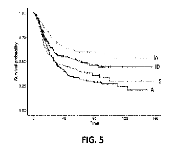
[0055] FIG. 5 is a risk curve comparison from Kaplan-Meier Plot of the
three datasets (878
patients) combined after classification into the four stromal subtypes (i.e.,
stromal
phenotypes).
[0056] FIGS. 6A and 6B show representative gene ontology signatures
expressed as box
plots in the ACRG cohort. FIG. 6A shows box plots of the median and range of
values for
the expression levels from the Treg signature as a function of the four
stromal subtypes
CA 03151629 2022-3-17
WO 2021/092071
PCT/US2020/058956
- 20 -
(i.e., stromal phenotypes) in the ACRG data. FIG. 6B shows a box plot of the
median and
range of values for the expression levels of an inflammatory response
signature as a
function of the four stromal subtypes (i.e., stromal phenotypes) in the ACRG
data.
[0057] FIGS. 7A and 7B show representative gene ontology signatures in
the ACRG
cohort that reflect the biology of the titles of the individual plots. FIG. 7A
shows that
Signature 1 activation is correlated with endothelial cell signature
activation. FIG. 7B
shows that Signature 2 activation is correlated with inflammatory and immune
cell
signature activation.
[0058] FIGS. 8A and 8B show representative gene ontology signatures in
the TCGA
dataset that reflect the biology of the titles of the individual plots. FIG.
8A shows that
Signature 1 activation is correlated with endothelial cell signature
activation. FIG. 8B
shows that Signature 2 activation is correlated with inflammatory and immune
cell
signature activation.
[0059] FIGS. 9A and 9B show representative gene ontology signatures in
the Singapore
cohort that reflect the biology of the titles of the individual plots. FIG. 9A
shows that
Signature 1 activation is correlated with endothelial cell signature
activation. FIG. 9B
shows that Signature 2 activation is correlated with inflammatory and immune
cell
signature activation.
[0060] FIG. 10 is a chart showing tumor microenvironment (TME)
assignments based on
the application of a classifier disclosed herein, as well as treatment classes
assigned to each
THE class.
100611 FIG. 11 depicts a logistic function used in
the logistic regression model.
100621 FIG. 12A is an exemplary small decision tree.
[0063] FIG. 12B shows That predictions for new samples can be made by
averaging the
predictions from the individual trees.
[0064] FIG. 13 shows the parameters from the Random
Forest classifier.
[0065] FIG. 14 shows part of an Artificial Neural Network (ANN)
training set comprising
a number of Samples, each one corresponding to a subject (column A), the TME
class for
the subject's cancer assigned according to the population-based classifier of
the present
disclosure (column B), and RNA expression levels corresponding to different
genes in the
selected gene panel (columns C, D, E, etc.).
[0066] FIG. 15 shows a simplified view of an ANN used as a non-
population based
classifier in the present disclosure. The ANN comprises an input layer with
inputs
CA 03151629 2022-3-17
WO 2021/092071
PCT/US2020/058956
- 21 -
corresponding to each gene in the gene panel (e.g., a 124 gene panel, 105 gene
panel, 98
gene panel, or alternatively an 87 gene panel), a hidden layer comprising two
neurons (or
alternatively 3, 4 or 5 neurons), and an output layer that would correspond to
TME class
assignments (i.e., stromal phenotype assignments).
100671 FIG. 16 is a schematic representation showing alternative ANN
architectures that
can be used to develop a non-population based classifier according to the
present
disclosure.
[0068] FIG. 17 shows that inputs to the ANN corresponding to mRNA
levels (x) for genes
1 to n are fed to the hidden layer neurons, and a bias (b) is applied to the
hidden layer
neurons. The input to the neuron is integrated through a function (f) which
incorporates the
bias and the mRNA expression levels (xi ... xn) normalized according to their
respective
weights (wi wn).
[0069] FIG. 18 shows different activation functions that can be applied
to the neurons in
the hidden layer.
[0070] FIG. 19 shows the artificial neuronal network (ANN) model
architecture. The
"Input layer" is a vector of expressions xi, i E G from a single sample. The
"Hidden layer"
comprises two neurons, each taking gene expression as input. The "Output
layer"
comprises four neurons, each taking activations of the two hidden neurons as
input,
transforming them with the tanh (hyperbolic tangent) activation function as a
weighted sum
to yield (y), followed by a logistic regression classifier (e.g., Softmax
function) (zi) to
produce probabilities of the four phenotype classes (IA, ID, A, IS).
Alternative aspects of
the ANN can comprise, e.g., five neurons instead of two neurons.
[0071] FIG. 20 shows the Kaplan-Meier survival curve for in a
population of gastric cancer
patients with known biomarker status and known outcome treated with
pembrolizumab
monotherapy.
[0072] FIG. 21A shows the application of machine-learning (ANN) to
optimize the cut-
off defining patients that are responders with respect to the non-responders,
and two
possible options for patient selection.
[0073] FIG. 2113 illustrates that in addition to the use of linear
thresholds different from
the Cartesian x:), y=0 thresholds to define patients that are responders with
respect to the
non-responders as exemplified in FIG. 21A, it is possible to use non-linear
thresholds to
define patient populations and to use such non-linear thresholds for patient
selection.
CA 03151629 2022-3-17
WO 2021/092071
PCT/US2020/058956
-22-
100741 FIG. 22 shows the Kaplan-Meier survival curve for Nayi 1B
reproductive cancer
patients with known biomarker status and known outcome.
[0075] FIG. 23 shows probability contours, expressed as a percentage,
of TME classes for
the pembrolizumab patient data of Example 12, overlaid on a latent space plot
of the
activation scores 1 and 2 of the ANN model (x and y axes). The top left
quadrant
corresponds to the A TME stromal phenotype, the lower left quadrant
corresponds to the
ID TME stromal phenotype, the lower right quadrant corresponds to the IA TME
stromal
phenotype, and the top right quadrant corresponds to the IS TME stromal
phenotype.
Patient Best Objective Response outcome is represented by: Progressive Disease
(PD) ¨
circle; Stable Disease (SD) ¨ triangle; Partial Response (PR) - square; and
Complete
Response (CR) - "x." Filled shapes represent the patients with a PD-L1 status
empty
shapes are PD-L1 < 1. Of the 73 patients of Example 12, four were missing PD-
L1 status
and so are omitted from the plot.
100761 FIG. 24 shows probability of biomarker positivity informed by a
logistic regression
classifier based on Progression-Free Survival (PFS) greater than 5 months, of
TME classes
of the pembrolizumab patient data of Example 12, overlaid on a latent space
plot of the
activation scores 1 and 2 of the ANN model (x and y axes). The classifier was
trained based
on the samples using a neutrophil leukocyte ratio less than 4 (NLIV4), using
PFS>5 as a
positive class. The top left quadrant corresponds to the A TME stromal
phenotype, the
lower left quadrant corresponds to the ID TME stromal phenotype, the lower
right quadrant
corresponds to the IA TME stromal phenotype, and the top right quadrant
corresponds to
the IS TME stromal phenotype. Patient Best Objective Response outcome is
represented
by: Progressive Disease (PD) ¨ circle; Stable Disease (SD) ¨ triangle; Partial
Response
(PR) - square; and Complete Response (CR) - "x." Filled shapes represent the
patients
with a PD-Li status >1, empty shapes are PD-L1 <1. Of the 73 patients of
Example 12,
four were missing PD-Li status and so are omitted from the plot.
[0077] FIG. 25 shows probability of biomarker positivity informed by
logistic regression
classifier based on Best Objective Response of TME classes of the
pembrolizumab patient
data of Example 12 overlaid on a latent space plot of the activation scores 1
and 2 of the
ANN model (x and y axes). The classifier was trained based on the samples
using a
neutrophil leukocyte ratio less than 4 (NLR<4), using Complete Responder and
Partial
Responders (CR+PR) as a positive class. Top left quadrant corresponds to the A
TME
CA 03151629 2022-3-17
WO 2021/092071
PCT/US2020/058956
- 23 -
stromal phenotype, the lower left quadrant corresponds to the ID TIME stromal
phenotype,
the lower right quadrant corresponds to the IA TME stromal phenotype, and the
top right
quadrant corresponds to the IS TME stromal phenotype. Patient Best Objective
Response
outcome is represented by: Progressive Disease (PD) ¨ circle; Stable Disease
(SD) ¨
triangle; Partial Response (PR) - square; and Complete Response (CR) - "x."
Filled shapes
represent the patients with a PD-Li status
empty shapes are PD-Li <
L Of the 73
patients of Example 12, four were missing PD-Li status and so are omitted from
the plot.
[0078] FIG. 26 shows probability of TME class of the bavituximab and
pembrolizumab
combination therapy clinical data of Example 7 overlaid on a latent space plot
of activation
scores 1 and 2 of the ANN model (x and y axes), for all patients (n=38). The
top left
quadrant corresponds to the A TME stromal phenotype, the lower left quadrant
corresponds
to the ID TME stromal phenotype, the lower right quadrant corresponds to the
IA TME
stromal phenotype, and the top right quadrant corresponds to the IS TME
stromal
phenotype. Patient Best Objective Response outcome is represented by:
Progressive
Disease (PD) ¨ circle; Stable Disease (SD) ¨ triangle; Partial Response (PR) -
square; and
Complete Response (CR) - "x." Filled shapes represent the patients with
confirmed
responses, empty shapes are unconfirmed responses.
[0079] FIG. 27 shows neural net activation scores (filled circles,
activation score 1 (node
1); open squares, activation score 2 (node 2)) and predicted TME class (ANN
phenotype
call) for tissue samples each from colorectal cancer (left, n=370), gastric
cancer (center,
n=337), and ovarian cancer (right, n=392). The distribution of samples between
the four
TME classes is similar for different disease groups.
[0080] FIG. 28A shows the presence (open cells) or absence Mill cells)
of 124 genes in
Genesets 1 to 44.
[0081] FIG. 28B shows the presence (open cells) or absence (full cells)
of 124 genes in
Genesets 45 to 88.
[0082] FIG. 28C shows the presence (open cells) or absence (full cells)
of 124 genes in
Genesets 89 to 132.
[0083] FIG. 28D shows the presence (open cells) or absence (full cells)
of 124 genes in
Genesets 133 to 177.
[0084] FIG. 28E shows the presence (open cells) or absence (full cells)
of 124 genes in
Geneset 178 to 222.
CA 03151629 2022-3-17
WO 2021/092071
PCT/US2020/058956
-24-
100851 FIG. 28F shows the presence (open cells) or absence (full cells)
of 124 genes in
Geneset 223 to 267.
[0086] FIG. 28G shows the presence (open cells) or absence (full cells)
of 124 genes in
Geneset 268 to 282.
00871 FIG. 29A is an illustrative schematic of gene weights in a first
node of an ANN
model, presented as a histogram of a sample of 30 gene weights (X axis). Open
bars, a
subset of genes of Signature 1, closed bars, a subset of genes of Signature 2.
Weights are
given on the Y axis.
[0088] FIG. 29B is an illustrative schematic of gene weights in a
second node of an ANN
model, presented as a histogram of a sample of 30 gene weights (X axis). Open
bars, a
subset of genes of Signature 1, closed bars, a subset of genes of Signature 2.
Weights are
given on the Y axis.
DETAILED DESCRIPTION
100891 The present disclosure provides methods to classify patients and
cancers according
to population and non-population tumor microenvironment (TME) classification
methods.
The population methods (i.e., population-based classifiers) disclosed herein
can be used
not only as stand-alone classifiers, but also as means to preprocess gene
expression data to
be used as training sets for the generation of non-population models (i.e.,
non-population-
based classifiers) based on the application of machine-learning techniques,
e.g., predictive
models based on Artificial Neural Networks (ANN).
100901 As used herein, the term "non-population-based" method or
classifier is
interchangeable with the terms machine learning (ML) method or ML classifier,
e.g., an
ANN classifier of the present disclosure. As used herein, the term "population-
based"
method or classifier is interchangeable with the terms Z-score method or Z-
score classifier.
100911 In some aspects, genesets that can represent one or more
biological signatures (i.e.,
a Signature 1, Signature 2, Signature 3, ++. Signature N) are used according
to the methods
disclosed herein to compute a Z-score for Signatures 1... N. This comprises a
population
model which can be used to reveal the dominant biologies represented by each
signature
and the TME phenotypes defined by the matrix of those signatures. In some
aspects, a
machine learning model (e.g. ANN) can be trained, e.g., using as features the
geneset
CA 03151629 2022-3-17
WO 2021/092071
PCT/US2020/058956
- 25 -
derived from the signatures, and as expressions a historic patient dataset,
e.g., the ACRG
(Asian Cancer Research Group) patient dataset.
[0092] The machine learning model (e.g., an ANN) learns the (latent)
gene expression
patterns that classify an individual patient into specific TME phenotypes. The
machine
learning model (e.g. ANN) effectively compresses the high dimensional data
(gene
expressions of all genes in the input geneset) into a lower dimensional
(latent) space, e.g.
the two hidden neurons in an ANN disclosed herein. The machine learning model
(e.g.
ANN) then outputs phenotype classes, e.g., four TME phenotype classes, which
themselves
can be used to define biomarker positivity, alone (in whole or in part) or in
combination
with one another (again, in whole or in part), in a drug specific manner.
Alternatively, a
secondary model (e.g., a logistic regression classifier) can be trained on the
latent space in
order to learn not the TME phenotypes, but rather to learn directly the
biomarker positive
versus biomarker negative decision boundary based on patient outcome labels.
[0093] In some aspects, the secondary model (e.g., a logistic
regression classifier) applied
to the ANN classifications according to the methods of the present disclosure
can be
optimized for BOR (Best Objective Response), ORR (Overall Response Rate),
MSS/MSI-
high (IvIicrosatellite Stable/Microsatellite Instability-high) status, PD-1/PD-
Li status, PFS
(Progression-Free Survival), NLR (Neutrophil Leukocyte Ratio), Tumor Mutation
Burden
(T1VIB) or any combination thereof.
[0094] Accordingly, in some aspects, the present disclosure provides
population classifiers
based on the integration of a number of signatures, i.e., global scores
related to the
expression of genes (e.g., those in TABLES 1 and TABLE 2) in particular gene
panels
(e.g., those in TABLES 3 and TABLE 4), such as Signature 1 and Signature 2
disclosed
herein. These signature scores allow patients and cancers to be stratified
according to TME,
and treatment decisions are then guided by the presence or absence of a
particular TME.
[0095] In other aspects, the present disclosure provides non-population
classifiers based on
the application of machine-learning techniques, e.g., logistic regression,
random forests, or
artificial neural networks (ANN). The ANN classifiers disclosed herein are
based, e.g., on
training a neural network using a dataset preprocessed according to the
population-based
classifiers disclosed herein.
[0096] An advantage of the non-population-based classifiers (ANN
classifiers) disclosed
herein over the population-based classifier also disclosed herein, is that a
sample from a
patient who is, e.g., part of a clinical trial or a clinical regimen, can be
correctly assessed
CA 03151629 2022-3-17
WO 2021/092071
PCT/US2020/058956
- 26 -
for stromal phenotype or biomarker positivity, without reference to any other
current
patient data. Thus, while the availability of a latent plot with the
probabilities for each
phenotypic class is useful, it is not required to correctly assess for stromal
phenotype or
biomarker positivity.
100971 The present disclosure also provides methods for treating a
subject, e.g., a human
subject, afflicted with cancer comprising administering a particular therapy
depending on
the classification of the cancer's TME according to the population and/or non-
population-
based classifiers disclosed herein, for example, based on the presence
(biomarker-positive)
and/or absence (biomarker-negative) of one or more TME class assignments
(e.g., whether
the subject is A and IS biomarker-positive, and/or ID and IA biomarker-
negative).
[0098] Also provided are personalized treatments that can be
administered to a subject
having a cancer classified into a particular TME class or group thereof (i.e.,
the subject is
biomarker-positive for a particular TME class or group thereof), or determined
not to have
a cancer classified into a particular TME class or group thereof (i.e., the
subject is
biomarker-negative for a particular TME class or group thereof). The
disclosure also
provides gene panels (e.g., those disclosed in TABLE 3 and TABLE 4) that can
be used for
identifying a human subject afflicted with a cancer suitable for treatment
with a particular
therapeutic agent, e.g., a TIV1E-specific therapy.
[0099] The application of the methods and compositions disclosed herein
can improve
clinical outcomes by matching patients to therapies (e.g., any of the Th1E-
specific therapies
disclosed below or a combination thereof depending on the biomarker-positive
and/or
biomarker-negative status of the subject) with a mechanism of action that
targets one or
more specific stromal subtypes (i.e., stromal phenotypes) or tumor biology.
[0100] Dominant stoma' phenotypes can be directional
but modified for any specific drug
based on the complexity of the mechanism of action of drug, drugs, or clinical
regimen.
Combinations of drugs or clinical regimens (i.e., one or more TME-specific
therapies
disclosed below) can be applied to multiple stromal phenotypes if relevant,
e.g., to a patient
or group of patients that are biomarker-positive for more than one stromal
phenotype or are
predominantly one stromal phenotype, but there is contribution of other
stromal phenotypes
in the biomarker signal as seen in the probability function of the ANN model
or logistic
regressions applied to the latent space, as in this disclosure. Thus, the term
predominantly," as applied to a stromal phenotype disclosed herein indicates
that a patient
or sample is biomarker positive for a particular stromal phenotype (e.g., IA),
but other
CA 03151629 2022-3-17
WO 2021/092071
PCT/US2020/058956
- 27 -
stromal phenotypes (e.g., IS, ID or A) or combinations thereof also contribute
to the
biomarker signal as seen in the probability function of the ML model, e.g.,
ANN model
disclosed herein, or in logistic regressions applied to the latent space.
101011 In some aspects, the patient can be biomarker positive for a
specific part of the
stromal phenotype, e.g., a patient may be considered biomarker-positive above
or below a
specific threshold or combination thereof (e.g., an upper and a lower
threshold) within a
particular stromal phenotype. Stated another way, a stromal phenotype can
match the drug
(e.g., the IA stromal phenotype can match the drug pembrolizumab), but when a
drug or
drug combination can modify multiple stromal phenotypes, the stromal
phenotypes can be
used as a starting point to develop a drug-specific combination, e.g. using
bavituximab plus
pembrolizumab. Accordingly, determining that a patient or a population of
patients are
biomarker-positive for two or more stromal phenotypes can be used to develop
new
therapies by combining two or more TME-specific therapies. For example, the
clinical
regimen of bavituximab and pembrolizumab targets two stromal phenotypes, IA
and IS,
and so a diagnostic or biomarker signature for this combination will be a
synthesis and
refinement based on both stromal phenotypes. Another illustrative example is
the
bispecific antibody navicixizumab, which is both a VEGF- and DLL4-targeting
agent.
While VEGF clearly targets the A stromal phenotype, there are features of the
IS group that
reflect the milieu of the DLL4 biology. Thus, a diagnostic biomarker signature
utilizing an
algorithm that integrates the A and IS stromal phenotypes (or, e.g., subsets
thereof defined
for example by one or more threshold values), and additional genes, as
described herein
can be used to bring out non-angiogenic features of the DLL4 biology.
Terms
101021 In order that the present disclosure can be more readily
understood, certain terms
are first defined. As used in this disclosure, except as otherwise expressly
provided herein,
each of the following terms shall have the meaning set forth below. Additional
definitions
are set forth throughout the disclosure.
[OM] "Administering" refers to the physical introduction of a
composition comprising a
therapeutic agent (e.g., a monoclonal antibody) to a subject, using any of the
various
methods and delivery systems known to those skilled in the art. Preferred
routes of
administration include intravenous, intramuscular, subcutaneous,
intraperitoneal, spinal or
other parenteral routes of administration, for example by injection or
infusion.
CA 03151629 2022-3-17
WO 2021/092071
PCT/US2020/058956
-28-
101041 The phrase "parenteral administration" as used herein means
modes of
administration other than enteral and topical administration, usually by
injection, and
includes, without limitation, intravenous, intramuscular, intraarterial,
intrathecal,
intralymphatic, intralesional, intracapsular, intraorbital, intracardiac,
intradermal,
intraperitoneal, transtracheal, subcutaneous, subcuticular, intraarticular,
subcapsular,
subarachnoid, intraspinal, intraocular, intravitreal, periorbital, epidural
and intrastemal
injection and infusion, as well as in vivo electroporation. Other non-
parenteral routes
include an oral, topical, epidermal or mucosal route of administration, for
example,
intranasally, vaginally, rectally, sublingually or topically. Administering
can also be
performed, for example, once, a plurality of times, and/or over one or more
extended
periods.
[0105] An "antibody" (Ab) shall include, without limitation, a
glycoprotein
immunoglobulin which binds specifically to an antigen and comprises at least
two heavy
(H) chains and two light (L) chains interconnected by disulfide bonds, or an
antigen-
binding portion thereof. Each H chain comprises a heavy chain variable region
(abbreviated
herein as VIE) and a heavy chain constant region. The heavy chain constant
region
comprises three constant domains, Cm, Cm and Cm. Each light chain comprises a
light
chain variable region (abbreviated herein as VL) and a light chain constant
region. The light
chain constant region comprises one constant domain, CL. The Viff and \IL
regions can be
further subdivided into regions of hypervariability, termed complementarity
determining
regions (CDRs), interspersed with regions that are more conserved, termed
framework
regions (FRs). Each VIE and Vt. comprises three CDRs and four FRs, arranged
from amino-
terminus to carboxy-terminus in the following order: FR1, CDR1, FR2, CDR2,
FR3,
CDR3, and FR4. The variable regions of the heavy and light chains contain a
binding
domain that interacts with an antigen. The constant regions of the antibodies
can mediate
the binding of the immunoglobulin to host tissues or factors, including
various cells of the
immune system (e.g., effector cells) and the first component (Clq) of the
classical
complement system.
[0106] An immunoglobulin can derive from any of the commonly known
isotypes,
including but not limited to IgA, secretory IgA, IgG and 1gM.. IgG subclasses
are also well
known to those in the art and include but are not limited to human IgGl, IgG2,
IgG3 and
IgG4. "Isotype" refers to the antibody class or subclass (e.g., IgM or IgG1)
that is encoded
by the heavy chain constant region genes.
CA 03151629 2022-3-17
WO 2021/092071
PCT/US2020/058956
-29-
101071 The term "antibody" includes, by way of example, monoclonal
antibodies; chimeric
and humanized antibodies; human or nonhuman antibodies; wholly synthetic
antibodies;
and single chain antibodies. A nonhuman antibody can be humanized by
recombinant
methods to reduce its immunogenicity in man. Where not expressly stated, and
unless the
context indicates otherwise, the term "antibody" also includes an antigen-
binding fragment
or an antigen-binding portion of any of the aforementioned immunoglobulins,
and includes
a monovalent and a divalent fragment or portion, and a single chain antibody.
As used
herein, the term "antibody" does not include naturally occurring antibodies or
polyclonal
antibodies. As used herein, the term "naturally occurring antibodies" and
"polyclonal
antibodies" do not include antibodies resulting from an immune reaction
induced by a
therapeutic intervention, e.g., a vaccine.
101081 An "isolated antibody" refers to an antibody that is
substantially free of other
antibodies having different antigenic specificities (e.g., an isolated
antibody that binds
specifically to PD-1 is substantially free of antibodies that bind
specifically to antigens
other than PD-1). An isolated antibody that binds specifically to PD-1 (e.g.,
sintilimab,
tislelizumab, pembrolizumab, or an antigen binding portion thereof) can,
however, have
cross-reactivity to other antigens, such as PD-1 molecules from different
species.
Moreover, an isolated antibody can be substantially free of other cellular
material and/or
chemicals.
101091 The term "monoclonal antibody" (mAb) refers to a non-naturally
occurring
preparation of antibody molecules of single molecular composition, i.e.,
antibody
molecules whose primary sequences are essentially identical, and which
exhibits a single
binding specificity and affinity for a particular epitope. A monoclonal
antibody is an
example of an isolated antibody. Monoclonal antibodies can be produced by
hybridoma,
recombinant, transgenic or other techniques known to those skilled in the art.
101101 A "human antibody" (HuMAb) refers to an antibody having variable
regions in
which both the framework and CDR regions are derived from human germline
immunoglobulin sequences. Furthermore, if the antibody contains a constant
region, the
constant region also is derived from human germline immunoglobulin sequences.
The
human antibodies of the disclosure can include amino acid residues not encoded
by human
germline immunoglobulin sequences (e.g., mutations introduced by random or
site-specific
mutagenesis in vitro or by somatic mutation in vivo). However, the term "human
antibody,"
as used herein, is not intended to include antibodies in which CDR sequences
derived from
CA 03151629 2022-3-17
WO 2021/092071
PCT/US2020/058956
- 30 -
the germline of another mammalian species, such as a mouse, have been grafted
onto
human framework sequences. The terms "human antibody" and "fully human
antibody"
and are used synonymously.
101111 A "humanized antibody" refers to an antibody in which some, most
or all of the
amino acids outside the CDRs of a non-human antibody are replaced with
corresponding
amino acids derived from human immunoglobulins. In one aspect of a humanized
form of
an antibody, some, most or all of the amino acids outside the CDRs have been
replaced
with amino acids from human immunoglobulins, whereas some, most or all amino
acids
within one or more CDRs are unchanged. Small additions, deletions, insertions,
substitutions or modifications of amino acids are permissible as long as they
do not
abrogate the ability of the antibody to bind to a particular antigen. A
"humanized antibody"
retains an antigenic specificity similar to that of the original antibody.
101121 A "chimeric antibody" refers to an antibody in which the
variable regions are
derived from one species and the constant regions are derived from another
species, such
as an antibody in which the variable regions are derived from a mouse antibody
and the
constant regions are derived from a human antibody.
101131 A "bispecific antibody" as used herein refers to an antibody
comprising two
antigen-binding sites, a first binding site having affinity for a first
antigen or epitope and a
second binding site having binding affinity for a second antigen or epitope
distinct from
the first.
101141 An "anti-antigen antibody" refers to an antibody that binds
specifically to the
antigen. For example, an anti-PD-1 antibody (e.g. , sintilimab, ti sl el i
zumab,
pembrolizumab, or an antigen binding portion thereof) binds specifically to PD-
1, and an
anti-PD-Li antibody binds specifically to PD-Li.
101151 An "antigen-binding portion" of an antibody (also called an
"antigen-binding
fragment") refers to one or more fragments of an antibody that retain the
ability to bind
specifically to the antigen bound by the whole antibody. It has been shown
that the antigen-
binding function of an antibody can be performed by fragments of a full-length
antibody.
Examples of binding fragments encompassed within the term "antigen-binding
portion" of
an antibody, e.g., an anti-PD-1 antibody (e.g., sintilimab, tislelizumab,
pembroliz-umab, or
an antigen binding portion thereof) or an anti-PD-Li antibody described
herein, include (i)
a Fab fragment (fragment from papain cleavage) or a similar monovalent
fragment
consisting of the W., Vii, LC and CHI domains; (ii) a F(ab')2 fragment
(fragment from
CA 03151629 2022-3-17
WO 2021/092071
PCT/US2020/058956
- 31 -
pepsin cleavage) or a similar bivalent fragment comprising two Fab fragments
linked by a
disulfide bridge at the hinge region; (iii) a Fd fragment consisting of the VH
and CH1
domains; (iv) a Fv fragment consisting of the VL and VH domains of a single
arm of an
antibody, (v) a dAb fragment (Ward et at, (1989) Nature 341:544-546), which
consists of
a VH domain; (vi) an isolated complementarity determining region (CDR) and
(vii) a
combination of two or more isolated CDRs which can optionally be joined by a
synthetic
linker. Furthermore, although the two domains of the FA, fragment, VL and VH,
are coded
for by separate genes, they can be joined, using recombinant methods, by a
synthetic linker
that enables them to be made as a single protein chain in which the VL and VH
regions pair
to form monovalent molecules (known as single chain Fly (scFv); see, e.g.,
Bird c/at (1988)
Science 242:423-426; and Huston et at (1988) Proc. Natl. Acad. Sci. USA
85:5879-5883).
Such single chain antibodies are also intended to be encompassed within the
term "antigen-
binding portion" of an antibody. These antibody fragments are obtained using
available
techniques in the art, and the fragments are screened for utility in the same
manner as are
intact antibodies. Antigen-binding portions can be produced by recombinant DNA
techniques, or by enzymatic or chemical cleavage of intact immunoglobulins.
101161 As used herein, the term "antibody," when applied to a specific
antigen,
encompasses also antibody molecules comprising other binding moieties with
different
binding specificities. Accordingly, in one aspect, the term antibody also
encompasses
antibody drug conjugates (ADC). In another aspect, the term antibody
encompasses
multispecific antibodies, e.g., bispecific antibodies. Thus, for example, the
term anti-PD-1
antibody would also encompass ADCs comprising an anti-PD-1 antibody or an
antigen-
binding portion thereof. Similarly, the term anti-PD-1 antibody would
encompass
bispecific antibodies comprising an antigen-binding portion capable of
specifically binding
to PD-1.
101171 A "cancer" refers to a broad group of various diseases
characterized by the
uncontrolled growth of abnormal cells in the body. Unregulated cell division
and growth
results in the formation of malignant tumors that invade neighboring tissues
and can also
metastasize to distant parts of the body through the lymphatic system or
bloodstream The
term "tumor" refers to a solid cancer. The term "carcinoma" refers to a cancer
of epithelial
origin.
CA 03151629 2022-3-17
WO 2021/092071
PCT/US2020/058956
-32-
101181 The term "immunotherapy" refers to the treatment of a subject
afflicted with, or at
risk of contracting or suffering a recurrence of, a disease by a method
comprising inducing,
enhancing, suppressing or otherwise modifying an immune response. "Treatment"
or
"therapy" of a subject refers to any type of intervention or process performed
on, or the
administration of an active agent to, the subject with the objective of
reversing, alleviating,
ameliorating, inhibiting, slowing down or preventing the onset, progression,
development,
severity or recurrence of a symptom, complication or condition, or biochemical
indicia
associated with a disease.
[0119] In the context of the present disclosure, the terms
"immunosuppressed" or
"immunosuppression" describe the status of the immune response to the cancer.
The
patient's immune response to the cancer can be dampened by immune suppressive
cells in
the tumor microenvironment, thus blocking, preventing, or diminishing an
immune system
attack on the cancer. In immunosuppression therapy the goal is to relieve
immunosuppression (as opposed to causing immunosuppression, e.g., as in the
context of
an organ transplant) by giving patients certain drugs, so that the immune
system can attack
the cancer.
[0120] The term "small molecule" refers to an organic compound having a
molecular
weight of less than about 900 Daltons, or less than about 500 Dalions. The
term includes
agents having the desired pharmacological properties, and includes compounds
that can be
taken orally or by injection. The term includes organic compounds that
modulate the
activity of TGF-P, and/or other molecules associated with enhancing or
inhibiting an
immune response.
101211 "Programmed Death-1" (PD-1) refers to an immunoinhibitory
receptor belonging
to the CD28 family. PD-1 is expressed predominantly on previously activated T
cells in
vivo, and binds to two ligands, PD-Li and PD-L2. The term "PD-1" as used
herein includes
human PD-1 (hPD-1), variants, isoforms, and species homologs of hPD-1, and
analogs
having at least one common epitope with hPD-1. The complete hPD-1 sequence can
be
found under GenBank Accession No. U64863.
[0122] "Programmed Death Ligand-1" (PD-L1) is one of two cell surface
glycoprotein
ligands for PD-1 (the other being PD-L2) that downregulate T cell activation
and cytokine
secretion upon binding to PD-1. The term "PD-Li" as used herein includes human
PD-Li
(hPD-L1), variants, isoforms, and species homologs of hPD-L1, and analogs
having at least
one common epitope with hPD-Ll. The complete hPD-L1 sequence can be found
under
CA 03151629 2022-3-17
WO 2021/092071
PCT/US2020/058956
- 33 -
Gen13ank Accession No. Q9NZQ7. The human PD-Li protein is encoded by the human
CD274 gene (NUB! Gene ID: 29126).
[0123] As used herein, the term "subject" includes any human or
nonhuman animal. The
terms, "subject" and "patient" are used interchangeably herein. The term
"nonhuman
animal" includes, but is not limited to, vertebrates such as dogs, cats,
horses, cows, pigs,
boar, sheep, goat, buffalo, bison, llama, deer, elk and other large animals,
as well as their
young, including calves and lambs, and to mice, rats, rabbits, guinea pigs,
primates such as
monkeys and other experimental animals. Within animals, mammals are preferred,
most
preferably, valued and valuable animals such as domestic pets, race horses and
animals
used to directly produce (e.g., meat) or indirectly produce (e.g., milk) food
for human
consumption, although experimental animals are also included. In specific
aspects, the
subject is a human. Thus, the present disclosure is applicable to clinical,
veterinary and
research uses.
[0124] The terms "treat," "treating," and "treatment," as used herein,
refer to any type of
intervention or process performed on, or administering an active agent to, the
subject with
the objective of reversing, alleviating, ameliorating, inhibiting, or slowing
down or
preventing the progression, development, severity or recurrence of a symptom,
complication, condition or biochemical indicia associated with a disease or
enhancing
overall survival. Treatment can be of a subject having a disease or a subject
who does not
have a disease (e.g., for prophylaxis). As used here, the terms "treat,"
"treating," and
"treatment" refer to the administration of an effective dose or effective
dosage.
101251 The term "effective dose" or "effective dosage" is defined as an
amount sufficient
to achieve or at least partially achieve a desired effect.
[0126] A "therapeutically effective amount" or "therapeutically
effective dosage" of a drug
or therapeutic agent is any amount of the drug that, when used alone or in
combination with
another therapeutic agent, protects a subject against the onset of a disease
or promotes
disease regression evidenced by a decrease in severity of disease symptoms, an
increase in
frequency and duration of disease symptom-free periods, or a prevention of
impairment or
disability due to the disease affliction.
[0127] A therapeutically effective amount or dosage of a drug includes
a "prophylactically
effective amount" or a "prophylactically effective dosage", which is any
amount of the drug
that, when administered alone or in combination with another therapeutic agent
to a subject
CA 03151629 2022-3-17
WO 2021/092071
PCT/US2020/058956
- 34 -
at risk of developing a disease or of suffering a recurrence of disease,
inhibits the
development or recurrence of the disease.
[0128] In addition, the terms "effective" and "effectiveness" with
regard to a treatment
disclosed herein includes both pharmacological effectiveness and physiological
safety.
Pharmacological effectiveness refers to the ability of the drug to promote
cancer regression
in the patient. Physiological safety refers to the level of toxicity, or other
adverse
physiological effects at the cellular, organ and/or organism level (adverse
effects) resulting
from administration of the drug.
[0129] The ability of a therapeutic agent to promote disease
regression, e.g., cancer
regression can be evaluated using a variety of methods known to the skilled
practitioner,
such as in human subjects during clinical trials, in animal model systems
predictive of
efficacy in humans, or by assaying the activity of the agent in in vitro
assays.
[0130] By way of example, an "anti-cancer agent" or combination thereof
promotes cancer
regression in a subject. In some aspects, a therapeutically effective amount
of the
therapeutic agent promotes cancer regression to the point of eliminating the
cancer.
[0131] In some aspects of the present disclosure, the anticancer agents
are administered as
a combination of therapies: a therapy comprising the administration of (i) an
anti-PD-1
antibody (e.g., sintilimab, tislelizumab, pembrolizumab, or an antigen binding
portion
thereof), and (ii) an anti-phosphatidylserine (PS) targeting antibody, e.g.,
bavituximab.
[0132] "Promoting cancer regression" means that administering an
effective amount of the
drug or combination thereof (administered together as a single therapeutic
composition or
as separate compositions in separate treatments as discussed above), results
in a reduction
in cancer burden, e.g., reduction in tumor growth or size, necrosis of the
tumor, a decrease
in severity of at least one disease symptom, an increase in frequency and
duration of disease
symptom-free periods, or a prevention of impairment or disability due to the
disease
affliction.
[0133] Notwithstanding these ultimate measurements of therapeutic
effectiveness,
evaluation of immunotherapeutic drugs must also make allowance for immune-
related
response patterns. The ability of a therapeutic agent to inhibit cancer
growth, e.g., tumor
growth, can be evaluated using assays described herein and other assays known
in the art.
Alternatively, this property of a composition can be evaluated by examining
the ability of
the compound to inhibit cell growth, such inhibition can be measured in vitro
by assays
known to the skilled practitioner.
CA 03151629 2022-3-17
WO 2021/092071
PCT/US2020/058956
-35-
101341 The terms "biological sample" or "sample" as used herein refers
to biological
material isolated from a subject. The biological sample can contain any
biological material
suitable for determining gene expression, for example, by sequencing nucleic
acids.
[0135] The biological sample can be any suitable biological tissue, for
example, cancer
tissue. In one aspect, the sample is a tumor tissue biopsy, e.g., a formalin-
fixed, paraffin-
embedded (FFPE) tumor tissue or a fresh-frozen tumor tissue or the like. In
another aspect,
an intratumoral sample is used. In another aspect, biological fluids can be
present in a tumor
tissue biopsy, but the biological sample will not be a biological fluid per
se.
[0136] The singular forms "a", "an" and "the" include plural referents
unless the context
clearly dictates otherwise. The terms "a" (or "an"), as well as the terms "one
or more," and
"at least one" can be used interchangeably herein. In certain aspects, the
term "a" or "an"
means "single." In other aspects, the term "a" or "an" includes "two or more"
or "multiple."
[0137] Furthermore, "and/or" where used herein is to be taken as
specific disclosure of
each of the two specified features or components with or without the other.
Thus, the term
"and/or" as used in a phrase such as "A and/or B" herein is intended to
include "A and B,"
"A or B," "A" (alone), and "B" (alone). Likewise, the term "and/or" as used in
a phrase
such as "A, B, and/or C" is intended to encompass each of the following
aspects: A, B, and
C; A_, B, or C; A or C; A or B; B or C; A and C; A and B; B and C; A (alone);
B (alone);
and C (alone).
101381 The terms "about," "comprising essentially of," or "consisting
essentially of," refer
to a value or composition that is within an acceptable error range for the
particular value or
composition as determined by one of ordinary skill in the art, which will
depend in part on
how the value or composition is measured or determined, i.e., the limitations
of the
measurement system. For example, "about," "comprising essentially of," or
"consisting
essentially of," can mean within 1 or more than 1 standard deviation per the
practice in the
art. Alternatively, "about," "comprising essentially of," or "consisting
essentially of," can
mean a range of up to 10%. Furthermore, particularly with respect to
biological systems or
processes, the terms can mean up to an order of magnitude or up to 5-fold of a
value. When
particular values or compositions are provided in the specification and
claims, unless
otherwise stated, the meaning of "about," "comprising essentially of," or
"consisting
essentially of," should be assumed to be within an acceptable error range for
that particular
value or composition.
CA 03151629 2022-3-17
WO 2021/092071
PCT/US2020/058956
-36-
101391 As used herein, the term "approximately," as applied to one or
more values of
interest, refers to a value that is similar to a stated reference value. In
certain aspects, the
term "approximately" refers to a range of values that fall within 10%, 9%, 8%,
7%, 6%,
5%, 4%, 3%, 2%, 1%, or less in either direction (greater than or less than) of
the stated
reference value unless otherwise stated or otherwise evident from the context
(except where
such number would exceed 100% of a possible value).
[0140] As described herein, any concentration range, percentage range,
ratio range or
integer range is to be understood to include the value of any integer within
the recited range
and, when appropriate, fractions thereof (such as one tenth and one hundredth
of an
integer), unless otherwise indicated.
[0141] Unless defined otherwise, all technical and scientific terms
used herein have the
same meaning as commonly understood by one of ordinary skill in the art to
which this
disclosure is related. For example, the Concise Dictionary of Biomedicine and
Molecular
Biology, Juo, Pei-Show, 2nd ed., 2002, CRC Press; The Dictionary of Cell and
Molecular
Biology, 3rd ed., 1999, Academic Press; and the Oxford Dictionary of
Biochemistry And
Molecular Biology, Revised, 2000, Oxford University Press, provide one of
skill with a
general dictionary of many of the terms used in this disclosure.
[0142] It is understood that wherever aspects are described herein with
the language
"comprising," otherwise analogous aspects described in terms of "consisting or
and/or
"consisting essentially of" are also provided.
[0143] Units, prefixes, and symbols are denoted in their Systeme
International de Unites
(SI) accepted form. The headings provided herein are not limitations of the
various aspects
of the disclosure, which can be had by reference to the specification as a
whole.
Accordingly, the terms defined are more fully defined by reference to the
specification in
its entirety.
[0144] Abbreviations used herein are defined throughout the present
disclosure. Various
aspects of the disclosure are described in further detail in the following
subsections.
I. Tumor Microenvironment (TME) classification
[0145] The present disclosure provides methods for the classification
of the tumor
microenvironment (TME) of a cancer in a subject in need thereof. These
classifiers can be
population-based classifiers, non-population-based classifiers, or
combinations thereof
CA 03151629 2022-3-17
WO 2021/092071
PCT/US2020/058956
-37-
101461 As used herein the term "population-based classifier" refers to
a method of TME
classification based on calculating one or more signatures corresponding to
one or more
characteristics (e.g., nucleic acid or protein expression levels) of a
population of biomarkers
(e.g., a population of biomarker genes disclosed herein). In some aspects,
each signature is
calculated using gene expression data (e.g., RNA expression data) obtained for
a set of
genes from a gene panel disclosed herein, e.g., a subset of the genes
disclosed in TABLE
1 or TABLE 2, or any of the gene panels (Genesets) disclosed in FIG. 28A-G.
[0147] As used herein, the term "non-population-based classifier"
refers to a method of
TME classification based on the application of a predictive model generated by
machine-
learning, e.g., ANN. In some aspects, the non-population-based classifier is
generated
using, for example, a training set comprising expression data (e.g., RNA
expression data)
preprocessed according to a population-based classifier disclosed herein as
training set.
[0148] In some aspects, there is no difference in the results of the
application of either the
population-based methods or non-population-based methods as disclosed herein
when
archival samples are used as compared to fresh samples (non-archival samples).
Example
7 discloses an application of an ANN method to fresh samples (non-archival
samples).
Example 12 discloses an application of an ANN method to archival samples.
[0149] In some aspects, fresh samples are preferred to archival
samples. As used herein,
the terms "fresh sample," "non-archival sample," and grammatical variants
thereof refer to
a sample (e.g., a tumor sample) which has been processed (e.g., to determine
RNA or
protein expression) before a predetermined period of time, e.g., one week,
after extraction
from a subject. In some aspects, a fresh sample has not been frozen. In some
aspects, a
fresh sample has not been fixed. In some aspects, a fresh sample has been
stored for less
Than about two weeks, less than about one week, or less than six, five, four,
three, or two
days before processing. As used herein, the term "archival sample" and
grammatical
variants thereof refers to a sample (e.g., a tumor sample) which has been
processed (e.g.,
to determine RNA or protein expression) after a predetermined period of time,
e.g., a week,
after extraction from a subject. In some aspects, an archival sample has been
frozen. In
some aspects, an archival sample has been fixed. In some aspect, an archival
sample has a
known diagnostic and/or a treatment history. In some aspects, an archival
sample has been
stored for at least one week, at least one month, at least six months, or at
least one year,
before processing.
CA 03151629 2022-3-17
WO 2021/092071
PCT/US2020/058956
-38-
101501 In some aspects, a population-based classifier of the present
disclosure comprises,
e.g., determining a combined biomarker comprising at least a signature score
determined
by measuring the expression levels of a gene panel (e.g., a gene panel
comprising at least
one gene from TABLE 1 or TABLE 2, or any of the gene panels (Genesets)
disclosed in
FIG. 28A-G, or a combination thereof) in a sample obtained from the subject;
wherein the
at least one signature score allows assignment of the subject's cancer to a
particular TME
class or a combination thereof.
[0151] In some aspects, a non-population-based classifier of the
present disclosure
comprises measuring the expression levels of a gene panel (e.g., a gene panel
comprising
at least one gene from TABLE 1 or TABLE 2, or any of the gene panels
(Genesets)
disclosed in FIG. 28A-G, or a combination thereof) in a sample obtained from
the subject;
and applying a predictive model generated via machine-learning (e.g., a
logistic regression,
a random forest, an artificial neural network, or a support vector machine
model), which
assigns the subject's cancer to a particular TME class or a combination
thereof. In some
aspects, the machine-learning model output (e.g., the output from an ANN
disclosed herein)
is post-processed using a statistical function which assigns the machine-
learning model
output to a particular TME class or a combination thereof.
[0152] Afterwards, the classifier output (e.g., from a population-based
classifier, a non-
population-based classifier, or a combination thereof) assigning the subject's
cancer to a
particular TME or a combination thereof would guide the selection and
administration of a
specific treatment or treatments which have been determined to be effective to
treat the
same type of cancer in other subjects having the same TME, i.e., a TME-class
therapy
disclosed below or a combination thereof
[0153] As used herein, the terms "tumor microenvironment" and "TME"
refer to the
environment surrounding tumor cells, including, e.g., blood vessels, immune
cells,
endothelial cells, fibroblasts, other Aroma' cells, signaling molecules, and
the extracellular
matrix. In some aspects, the terms "stromal subtype," "stromal phenotype," and
grammatical variants thereof are used interchangeably with the term "TWEE."
[0154] The tumor cells and the surrounding microenvironment are closely
related and
interact constantly. In general, tumor microenvironment (also known as, e.g.,
stromal
phenotype) encompasses any structural and/or functional characteristic of the
stroma of a
tumor and tumoral environment. Numerous non-tumoral cell types can exist in a
TME, e.g.,
carcinoma associated fibroblasts, myeloid-derived suppressor cells, tumor-
associated
CA 03151629 2022-3-17
WO 2021/092071
PCT/US2020/058956
- 39 -
macrophages, neutrophils, or tumor infiltrating lymphocytes. In some aspects,
the
classification of a particular TME can include the analysis of the cell types
present in the
stroma. A TME can also be characterized by specific functional
characteristics, e.g., by
abnormal oxygenation levels, abnormal blood vessel permeability, or abnormal
levels of
particular proteins such as collagens, elastin, glycosaminoglycans,
proteoglycans, or
glycoproteins.
[0155] The population-based and non-population-based classifiers
disclosed herein can be
used to assign a patient or a cancer sample to a specific THE class (e.g., ID,
IA, IS, or A)
or to a combination thereof (e.g., ID and IA, ID and IS, ID and A, and so on).
Specific
subpopulations of patients within a specific TME class can be further
classified based on
the application of thresholds (e.g., by using a linear threshold or
combination thereof, as
exemplified in FIG. MA, or by using non-linear thresholds as exemplified in
FIG. 21B, or
combinations thereof).
[0156] This classification functions as a combined biomarker, i.e., it
is a biomarker derived
from discrete biomarkers (e.g., a TIME class or a subset within a specific TME
defined, e.g.,
according to linear or non-linear threshold, or a combination thereof)
integrated into a
single score or a combination thereof in the case of a population-based
classifier, or into a
model in a non-population-based classifier. Accordingly, a patient or cancer
sample can be
"biomarker-positive" for a single TME class, e.g., ID, IA, IS or A, in which
the patient or
sample would be described as being, e.g., ID biomarker-positive, IA biomarker-
positive,
IS biomarker-positive, or A biomarker-positive. In some aspects, a patient or
cancer sample
can be biomarker-positive for more than one TME class. Thus, in some aspects,
a patient
or cancer sample can be biomarker-positive for 2, 3, 4 or more TME classes. In
some
aspects, a patient or cancer sample can be, e.g., ID and IA biomarker-
positive; ID and IS
biomarker-positive; ID and A biomarker-positive; IA and IS biomarker-positive;
IA and A
biomarker-positive; or IS and A biomarker-positive. In some aspects, a patient
or cancer
sample can be, e.g., ID, IA, and IS biomarker-positive; ID, IA, and A
biomarker-positive;
or ID, IS, and A biomarker-positive.
[0157] In some aspects, a combined probability for biomarker positive
status (i.e., a
combination of one or more probabilities coming from the stromal phenotype
classifier) is
used. The combined probability for biomarker positive status can be calculated
using
mathematical techniques known in the art.
CA 03151629 2022-3-17
WO 2021/092071
PCT/US2020/058956
-40-
101581
A patient or cancer
sample can also be defined as "biomarker-negative" for a single
TME class, e.g., ID, IA, IS, or A. Thus, the patient or sample would be
described as being,
e.g., ID biomarker-negative, IA biomarker-negative, IS biomarker-negative, or
A
biomarker- negative. In some aspects, a patient or cancer sample can be
biomarker-negative
for more than one TME class. Thus, in some aspects, a patient or cancer sample
can be
biomarker-negative for 2, 3, 4 or more TME classes. In some aspects, a patient
or cancer
sample can be, e.g., ID and IA biomarker-negative; ID and IS biomarker-
negative; ID and
A biomarker-negative; IA and IS biomarker-negative; IA and A biomarker-
negative; or IS
and A biomarker-negative. In some aspects, a patient or cancer sample can be,
e.g., ID, IA,
and IS biomarker-negative; ID, IA, and A biomarker-negative; or ID, IS, and A
biomarker-
negative.
[0159] In some aspects, a combined probability for biomarker negative
status (i.e., a
combination of one or more probabilities coming from the stromal phenotype
classifier) is
used. The combined probability for biomarker negative status can be calculated
using
mathematical techniques known in the art.
[0160] In some aspects, assignment of a TME-class specific therapy is
based on the
presence of a specific stromal phenotype, i.e., if a subject presents an IA
stromal phenotype
(and therefore the subject is IA biomarker-positive), an IA-class TME therapy
would be
administered. In some aspects, assignment of a TME-class specific therapy is
based on the
absence of a specific stromal phenotype, i.e., if a subject does not present
an IA stromal
phenotype (and therefore the subject is IA biomarker-negative), an IA-class
TME therapy
would not be administered.
[0161] In some aspects, the classification of a patient or cancer
sample to a TME class,
and assignment of a TME class therapy to the patient or cancer is not
biunivocal. In other
words, a patient or cancer sample can be classified as biomarker-positive
and/or biomarker-
negative for more than one TME class, and more than one TME class therapy or a
combination thereof can be used to treat that patient. For example, the
classification of a
patient or cancer sample as biomarker-positive for two different TME classes
(i.e., two
stromal phenotypes) could be used to select a treatment comprising a
combination of
pharmacological approaches in the TME class therapies corresponding to the TME
classes
for which the patient or cancer sample is biomarker-positive. Furthermore, if
the patient or
cancer sample is biomarker-negative for a particular TME class, such knowledge
can be
used to exclude specific pharmacological approaches in the TME class therapy
CA 03151629 2022-3-17
WO 2021/092071
PCT/US2020/058956
- 41 -
corresponding to the TME class for which the patient or cancer sample is
biomarker-
negative. Thus, drugs or combinations thereof, treatments or combinations
thereof, ancUor
clinical regimens or combinations that are useful to treat a cancer sample
classified as
biomarker-positive for a particular TME class, can be combined to treat
patients having
more than one biomarker-positive signal (Le., having a cancer sample
classified as
biomarker-positive for more than one stromal phenotype).
101621 In some aspects, depending on the mechanism of action of a drug
or a clinical
regimen, different classification parameters, e.g., different gene panel
subsets, different
thresholds, different ANN architectures, different activation functions, or
different post-
processing functions, can be used to yield different TME classes, which in
turn would be
used to select appropriate TME class therapies. Accordingly, each drug or drug
regimen
may have different diagnostic gene panels and differently configured
population based or
non-population based classifiers to inform the clinician (such as a medical
doctor), e.g., to
decide whether a patient should be selected for treatment, whether treatment
should be
initiated, whether treatment should be suspended, or whether treatment should
be modified.
101631 In some aspects, a clinician can account for co-variates of
biomarker status of a
patient, and combine the probability of the stromal phenotype or biomarker
status with
MSINISS (Microsatellite Instability/IvIicrosatellite Stability-high) status,
EBV (Epstein-
Barr virus) status, PD-1/PD-L1 status (such as CPS, i.e., combined positive
score),
neutrophil-leukocyte ratio (NLR), or confounding variables such as prior
treatment history.
101641 In some aspects, the clinician is given a binary result from the
algorithm, and the
decision to treat or not treat as described herein is made. In one aspect, the
clinician is
given, e.g., a plot of the patient's result superimposed on a latent space and
interpreted with
probability thresholds, or a linear or polynomial logistic regression.
LA. Gene panels
101651 The population- and non-population-based classifiers of the
present disclosure rely
on the selection of a specific gene panel as the source of the input data used
by the classifier.
In some aspects, each one of the genes in a gene panel of the present
disclosure is referred
to as a "biomarker." The terms "geneset" and "gene panel" are used
interchangeably.
101661 In some aspects, the biomarker is a nucleic acid biomarker. The
term "nucleic acid
biomarker," as used herein, refers to a nucleic acid (e.g., a gene in a gene
panel disclosed
CA 03151629 2022-3-17
WO 2021/092071
PCT/US2020/058956
- 42 -
herein) that can be detected (e.g., quantified) in a subject or a sample
therefrom, e.g., a
sample comprising tissues, cells, stroma, cell lysates, and/or constituents
thereof, e.g., from
a tumor. In some aspects, the term nucleic acid biomarker refers to the
presence or absence
of a specific sequence of interest (e.g., a nucleic acid variant or a single
nucleotide
polymorphism) in a nucleic acid (e.g., a gene in a gene panel disclosed
herein) that can be
detected (e.g., quantified) in a subject or a sample therefrom, e.g., a sample
comprising
tissues, cells, stroma, cell lysates, and/or constituents thereof, e.g., from
a tumor.
[0167] The "level" of a nucleic acid biomarker can, in some aspects,
refer to the
"expression level" of the biomarker, e.g., the level of an RNA or DNA encoded
by the
nucleic acid sequence of the nucleic acid biomarker in a sample. For example,
in some
aspects, the expression level of a particular gene disclosed in TABLE 1 or
TABLE 2, or
any of the gene panels (Genesets) disclosed in FIG. 28A-G, refers to the
amount of mRNA
encoding such gene present in a sample obtained from a subject.
[0168] In some aspects, the "level" of a nucleic acid biomarker, e.g.,
an RNA biomarker,
can be determined by measuring a downstream output (e.g., an activity level of
a target
molecule or an expression level of an effector molecule that is modulated,
e.g., activated or
inhibited, by the nucleic acid biomarker or an expression product, e.g., RNA
or DNA,
thereof).
[0169] In some aspects, the nucleic acid biomarker is an RNA biomarker.
An "RNA
biomarker," as used herein, refers to an RNA comprising the nucleic acid
sequence of a
nucleic acid biomarker of interest, e.g., RNA encoding a particular gene
disclosed in
TABLE 1 or TABLE 2, or any of the gene panels (Genesets) disclosed in FIG. 28A-
G.
[0170] The "expression level" of an RNA biomarker generally refers to a
detected quantity
of RNA molecules comprising the nucleic acid sequence of interest present in
the subject
or sample therefrom, e.g., the quantity of RNA molecules expressed from a DNA
molecule
(e.g., the genome of the subject or the subject's cancer) comprising the
nucleic acid
sequence.
[0171] In some aspects, the expression level of an RNA biomarker is the
quantity of the
RNA biomarker in a tumor stromal sample. In some aspects, an RNA biomarker is
quantified using PCR (e.g., real-time PCR), sequencing (e.g., deep sequencing
or next
generation sequencing, e.g., RNA-Seq), or microarray expression profiling or
other
technologies that utilize RNAse protection in combination with amplification
or
amplification and new quantitation methods such as RNA-Seq or other methods.
CA 03151629 2022-3-17
WO 2021/092071
PCT/US2020/058956
-43-
101721 In some aspects, a population-based classifier disclosed herein
comprises signatures
calculated using expression levels of a gene disclosed in TABLE 1 and TABLE 2
(or in
any of the gene panels (Genesets) disclosed in FIG. 28A-G). For example, a
population-
based classifier comprising two signatures can comprise a Signature 1 obtained
from
expression levels corresponding to the genes disclosed in TABLE 1 or a subset
thereof, and
a Signature 2 obtained from expression levels corresponding to the genes
disclosed in
TABLE 2 or a subset thereof. In some specific aspects, the population-based
classifier can
use subsets (gene panels) disclosed in TABLE 3 and TABLE 4. For example, a
population-
based classifier comprising two signatures can comprise a Signature 1 obtained
from
expression levels corresponding to genes in a gene panel disclosed in TABLE 3,
and a
Signature 2 obtained from expression levels corresponding to genes in a gene
panel
disclosed in TABLE 4 or a subset thereof.
101731 In the population-based classifiers disclosed herein, expression
levels for genes in
a gene panel acquired from a population of samples (e.g., samples from a
clinical study)
can be used to classify groups of samples in the population as belonging to a
TME class (or
a combination thereof, i.e., a sample can be classified not only as biomarker-
positive for a
single TME class, but also can be classified as biornarker-positive for two or
more TME
classes) according to whether calculated signature levels are above or below
certain
threshold values. Subsequently, expression levels for genes in a gene panel
obtained from
a sample or samples from a test subject can be used to classify the subject's
TIVIE into one
of the TME classes identified in the population.
101741 In the non-population-based classifiers disclosed herein,
expression levels for genes
in a gene panel acquired from a population of samples (e.g., samples from a
clinical study)
and their assignments to a TME class (or a combination thereof, i.e., a sample
can be
classified not only as biomarker-positive for a single TME class, but also can
be classified
as biomarker-positive for two or more TME classes) obtained according to the
populations
classifiers disclosed herein can be used as a training set for machine-
learning, e.g., using
an ANN. The machine-learning process would yield a model, e.g., an ANN model.
Subsequently, expression levels for genes in a gene panel obtained from a
sample or
samples from a test subject would be used as input for the model, which would
classify the
subject's TME into a particular TME class (or a combination thereof, i.e., a
sample can be
classified not only as biomarker-positive for a single TME class, but also can
be classified
as biomarker-positive for two or more TME classes).
CA 03151629 2022-3-17
WO 2021/092071
PCT/US2020/058956
-44-
101751 Standard names, aliases, etc. of proteins and genes designated
by identifiers used
throughout this disclosure can be identified, for example, via Genecards
(www.genecards.org) or Uniprot (wwvv.uniprot.org).
TABLE 1. Signature 1 genes and accession numbers (n=63)
Gene Gene Description RefSeq RNA (NM_xxxxxx) and
Transcript variants
Symbol (XM xxxxxx)
ABCC9 ATP binding NM 005691.3, NM
020297.3, NM 0202982 ,
cassette subfamily C XM 005253284.3 XM 005253286.3 XM 005253287.4,
member 9 XM 005253288.3, XM
005253289.3, XM 005253290.3,
XM 006719025.3, XM 011520545.2
AFAP1L2 actin filament NM_001146337.2, NM
001323062.1, NM 001323063.1,
associated protein 1 NM_152406.3, XM_011537558.1, XM_017009036.1
like 2
BACE1 beta-secretase 1 NM_001207048.1, NM
001207049.1, NM 012104.4,
NM 138971.3, NM 138972.3, NM 138973.3
BGN Biglycan NM_001711.5,
XM_017029724.1
BMP5 bone morphogenetic NM_001329754.1,
NM_001329756.1, NM_021073 .3,
protein 5 XM 005249304.3, XM
011514816.2, XM 011514817.2,
XM 017011198.1
COL4A2 collagen type IV NM 001846.3
alpha 2 chain
COL8A1 collagen type VIII NM 001850.4, NM
_020351,3
alpha 1 chain
COL8A2 collagen type VIII NM_001294347.1,
NM_005202.3, XM_005270477.3
alpha 2 chain
CPXM2 carboxypeptidase X, NM 198148.2, XM
005269528.3, XM 011539283.2,
M14 family member XM 011539285.2, XM_011539286.1, XM_017015673.1,
2 XM_017015674.1
CXCL12 C-X-C motif NM_000609.6, NM
001033886.2, NM_001178134.1,
chemokine ligand 12 NM 001277990.1, NM 199168.3
EBF1 early B cell factor 1 NM 001290360.2, NM
001324101.1, NM 001324103.1,
NM_001324106.1, NM_001324107.1, NM_001324108.1,
NM 001324109.1, NM 001324111.1, NM 024007.4,
NM 182708.2, XM 017009192.1, XM 017009193.1,
XM 017009194.1, XM 017009195.1, XM 017009196.1,
XM 017009197.1, XM 017009198.1, )(NI 017009199.1,
XM_017009200.1, XM_017009201.1, XM_017009202.1,
XM 017009203.1, XM 017009204.1
ECM2 extracellular matrix NM 001197295.1, NM
001197296.1, NM_001393.3,
protein 2 XM 017014376.1, XM
017014377.1
EDNRA endothelin receptor NM 001166055.1,
NM_001354797.1, NM_001957.3,
type A NM 001256283.1
ELN Elastin NM_000501.3, NM
001081752.2, NM 001081753.2,
NM 001081754.2, NM 001081755.2, NM 001278912.1,
NM 001278913.1, NM 001278914.1, NM 001278915.1,
CA 03151629 2022-3-17
WO 2021/092071
PCT/US2020/058956
- 45 -
NM_001278916.1,NM_001278917.1,NM_001278918.1,
NM_001278939.1, XM_005250187.1, XM_005250188.1,
XM_011515868.1, XM_011515869.1, XM_011515870.1,
XN1_011515871.1, XM_011515872.1, XM_011515873.1,
XM_011515874.1,XM_011515875.1,XM_011515876.1,
XIV1 011515877.1, XM 017011813.1, XM 017011814.1
EPHA3 EPH receptor A3 NM_005233.5,
NM_182644.2, XM_005264715.2,
XN1 005264716.2
FBLN5 fibulin 5 NM _006329.3
XM_0052672673 XNI_011536356.1
XM 011536357.1 XM 011536358.1 XM 017020929.1
GNAS GNAS complex NM 000516.5, NM
001077488.3, NM 001077489.3,
locus NM_001077490.2,
NM_001309840.1, NM_001309842.1,
NM_001309861.1, NM_001309883.1, NM_016592.3,
NM_080425.3, NM_080426.3, XM_017027812.1,
XM_017027813.1, XM_017027814.1, XM_017027815. 1,
XM_017027816.1, XM_017027817.1, XM_017027818.1,
XN1_017027819.1, XM_017027820.1, XM_017027821.1,
XM 017027822.1
GNB4 G protein subunit NM_021629.3,
XM_005247692.2, XM_006713721.2
beta 4
GUCY1A3 guanylate cyclase 1 NM_000856.5, NM_001130682.2, NIVI_001130683.3,
soluble subunit NM 001130684.2, NM
001130685.2, NM 001130687.2,
alpha 1 NM_001256449.1,
NM_001130686.1, XM_005262955.2,
XM_005262956.2, XM_005262957.2, XM_006714196.2,
XM 006714197.2, XM 006714198.2, XM 011531900.2
HEY2 HES related family NM 012259.2, XNI
017010627.1, XM 017010628.1,
ail transcription X_M_017010629.1
factor with YRPW
motif 2
HSPB2 heat shock protein NM_001541.3
family B (small)
member 2
IL1B interleukin 1 beta NM_000576.2,
XM_017003988.1
ITGA9 integrin subunit NM_002207.2
alpha 9
ITPR1 inositol 1,4,5- NM 001099952.2, NM
002222.5, NM 001168272.1,
trisphosphate XM_005265109.2,
XM_005265110.2, XM_006713131.2,
receptor type 1 XM_011533681.1,
XM_011533682.2, XM_011533683.2,
XM_011533684.1,XM_011533685.1,XM_011533686.1,
XM_011533687.1,XM_011533688.1, XM_011533690.1,
XM_011533691.1, XM_0 n 533692.2, XM_017006357.1,
XM 017006358.1
JANI2 junctional adhesion NM_001270408.1,
NM_021219.3, NM_001270407.1
molecule 2
JAM3 junctional adhesion NM_001205329.1,
NM_032801.4
molecule 3
KCNJ8 potassium voltage- NM_004982.3,
XM_005253358.4, XM_017019283.1,
gated channel XM 017019284.1
CA 03151629 2022-3-17
WO 2021/092071
PCT/US2020/058956
- 46 -
subfamily J member
8
LAIV182 laminin subunit beta NM_002292.3,
X_M_005265127.3
2
LHFP LHFPL tetraspan NM_005780.2 ,
XM_011534861.1
subfamily member 6
LTBP4 latent transforming NM_001042544.1,
NM_001042545.1, NM_003573.2,
growth factor beta XM_011527376.2,
XM_011527377.2, XM_011527378.2,
binding protein 4 XM_011527379.1,
XM_011527380.2, XM_011527381.2,
XM_011527382.2, XM_011527383.2, XM_011527384.2,
XM 011527385.2, XM 011527386.2, XM 011527387.1,
XM 017027352.1, XM 017027353.1, XM 017027354.1
MEOX1 mesenchyme NM_001040002.1,
NM_004527.3, NM_013999.3,
homeobox 1 XM 011524818.1
MGP matrix Gla protein NM 000900.4, NM 001190839.2
MMP12 matrix NM_002426.5
metallopeptidase 12
MMP13 matrix NM 002427.3
metallopeptidase 13
NAALAD2 N-acetylated alpha- NM 001300930.1, NM_005467.3, XM_017017043.1,
linked acidic XM_017017044.1,
XM_017017045.1, XM_017017046. 1
dipeptidase 2
NFATC1 nuclear factor of NM_001278669.1,
NM_001278670.1, NM_001278672.1,
activated T cells 1 NM_001278673.1,
NM_001278675.1, NM_006162.4,
NM_172387.2, NM_172388 .2, NM_172389.2,
NM 172390.2, XM 017025783.1
NOV nephroblastoma NM_002514.3
overexpressed
OLFM1.2A olfactomedin like NM_001282715.1,
NM_182487.3, XM_005251760.4,
2A XM_006716989.2
PCDH17 protocadherin 17 NM_001040429.2,
NM_014459.2, XM_005266357.2,
XM 005266358.2, XM 017020547.1
PDE5A phosphod i este rase NM_001083 .3, NM_03343 O.
2, NM_033437. 3,
5A XM 017008791.1
PDGFRB platelet derived XM 011537659.1, XM
011537658.1 , XM 005268464.2
growth factor , NM 002609.3 , NM
001355017.1, NM 001355016.1
receptor beta
PEG3 paternally expressed NM 001146184.1, NM
001146185.1, NM 001146186.1,
3 NM_001146187.1,
NM_006210.2
PLSCR2 phospholipid NM 001199978.1, NM
001199979.1 , NM 020359.2,
scramblase 2 XM_011513013.2,
XM_011513019.2, XM_011513020.2,
XM 011513021.2, XM 011513022.2, XM 011513023.2,
XM_017006898.1, XM_017006899.1, XM_017006900. 1,
XM 017006901.1, XM 017006902.1, XM 017006903.1,
XM_017006904. 1, XM_017006905.1, XM_017006906. 1,
X_M 017006907.1, X1V1 017006908.1, XM 017006909.1,
XM_017006910.1, XM_017006911.1, XM_017006912.1,
XM 017006913.1, >LEVI 017006914.1, XM 017006915.1
CA 03151629 2022-3-17
WO 2021/092071
PCT/US2020/058956
- 47 -
PLXDC2 plexin domain NM_001282736.1,
NM_032812.8, XM_011519750.2
containing 2
RGS4 regulator of G
NM_0011024452,NM_001113380.1,NM_001113381.1,
protein signaling 4 NM 005613.5
RGS5 regulator of G NM_001195303.2,
NM_001254748.1, NM_001254749.1,
protein signaling 5 NM 003617.3, NM
025226.1
RNF144A ring finger protein NM_001349181.1,
NM_001349182.1, NM_001349183.1,
144A
NM_001349184.1,NM_001349185.1,NM_001349186.1,
NM_014746.5, XM_005246200.3, XM_005246202.4,
XM_017005396.1, XM_017005397.1, XM_017005398.1,
XM 017005399.1, XM 017005400.1, XM 017005401.1,
XM 017005402.1, XM 017005403.1, XM 017005404.1
RRAS RAS related NM_006270.4
RUNX1T1 RUNX1 NM_001198625.1,
NM_001198626.1, NM_001198627.1,
translocati on partner NM_001198628.1, NM_001198629.1, NM_001198630.1,
1
NM_001198631.1,NM_001198632.1,NM_001198633.1,
NM_001198634.1, NM_001198679.1, NM_004349.3,
NM_175634.2, NM_175635.2, NM_175636.2,
XM_006716676.3, XM_011517351.2, XM_011517352.2,
X1\4_011517353.2, XM_017013930.1, XM_017013931.1,
XM_017013932.1, XM_017013933.1, XM_017013934.1,
XM_017013935.1, XM_017013936.1, XM_017013937.1,
XM_017013938.1, XM_017013939.1, XM_017013940.1,
XM_017013941.1
CAV2 caveolae associated NM_004657.5
protein 2
SELP selectin P NM_003005.3,
XM_005245435.1, XM_005245436.3,
XM 005245438.1, XM 005245439.1, XM 005245440.1
SERP1NE2 serpin family E NM_001136528.1,
NM_001136530.1, NM_006216.3,
member 2 XM_005246641.2,
XM_017004329.1, XM_017004330.1,
XM_017004331.1, XM_017004332.1
SGIP1 SH3 domain GRB2 NM_001308203.1,
NM_001350217.1, NM_001350218.1,
like endophilin NM_032291.3,
XM_005271264.3, X1\4_005271268.3,
interacting protein 1 XM_005271270.4, XM_006710961.2, XM_006710966.2,
XM 006710967.2, XM 006710969., XM 006710971.2,
XM_006710972.2, XM_006710973.2, XM_006710974.2,
XM_011542291.1, XM_011542292.1, XM_011542293.1,
XM_017002505.1, XM_017002506.1, XM_017002507.1,
XM_017002508.1, XM_017002509.1, XM_017002510.1,
XM 017002511.1, XM 017002512.1, XM 017002513.1,
XM_017002514.1, XM_017002515.1, XM_017002516.1,
XM_017002517.1, XM_017002518.1, XM_017002519.1,
XM_017002520.1, XM_017002521.1, XM_017002522.1,
XM_017002523.1, XM_017002524.1, XM_017002525.1,
XM_017002526.1, XM_017002527.1, XM_017002528.1,
XM_017002529.1, XM_017002530.1, XM_017002531.1,
XM_017002532.1, XM_017002533.1, XM_017002534.1,
XM 017002535.1, XM 017002536.1, XM 017002537.1
CA 03151629 2022-3-17
WO 2021/092071
PCT/US2020/058956
- 48 -
SMARCA1 SWI/SNF related, NM_001282874.1,
NM_001282875.1, NM_003069.4,
matrix associated, NM_139035.2,
XM_005262461.2, XM_005262462.2,
actin dependent XM_006724782.2,
XM_017029750.1, XM_017029751.1
regulator of
chromatin,
subfamily a,
member!
SPON1 spondin 1 NM_006108.3
STAB2 stabilin 2 NM_017564.9,
XM_011538537.2, XM_011538538.2 ,
XM_011538539.2, XM_011538541.2 , XM_011538542.2,
XM_017019585.1
STEAP4 STEAP4 NM_001205315.1,
NM_001205316.1, NM_024636.3
metalloreductase
TBX2 T-box 2 NM_005994.3
TEK TEK receptor NM_000459.4,
NM_001290077.1, NM_001290078.1,
tyrosine kinase XM 005251561.2, XM
005251563.2
TGFB2 transforming growth NM_001135599.3,
NM_003238.4
factor beta 2
TIVIEM204 transmembrane NM 001256541.1,
NM_024600.5
protein 204
TTC28 tetratricopeptide NM_015281.1,
NM_001145418.1, XM_005261405.2,
repeat domain 28 XM_006724171.4,
XM_011530018.3, XM_011530019.2,
XM 011530020.1, XM 011530021.3, XM 011530022.1,
XM_017028673.2
UTRN Utrophin NM_007124.2,
XM_005267127.5, XM_005267130.2,
XM_005267133.3, XM_006715560.4, XM_011536101.3,
XM_011536102.2, XM_011536106.2, XM_011536109.3,
XM_017011243.2,XM_017011244.1, XM_017011245.1,
XM 024446536.1
TABLE 2. Signature 2 genes and accession numbers (n=61)
Gene Gene Description RefSeq RNA (NM_xxxxxx) and
Transcript variants
Symbol (XM_xxxxxx)
AGR2 anterior gradient 2, NM_006408.3 ,
XM_005249581.4
protein disulphide
isomerase family
member
C 1 lorf9 myelin regulatory NM 001127392.2 ,
NM 013279.3 , XM 005274222.1 ,
factor XM_005274223.1 ,
XM_005274224.1 ,
XM_005274225.1, XM_005274226.1, XM_005274227.1,
XM_005274228.1, XM_011545234.2
DUSP4 dual specificity NM_001394.6 ,
N1v1_057158.3 XM_011544428.2
phosphatase 4
ElF5A eukaryotic NM 001143760.1, NM
001143761.1, NM 001143762.1,
translation initiation NM_001970.4, XM_005256509.2, XM_011523710.2,
factor 5A XM_011523711.2,
XM_011523712.2, XM_011523713.2,
XM_017024300.1, XM_017024301.1
CA 03151629 2022-3-17
WO 2021/092071
PCT/US2020/058956
- 49 -
ETV5 ETS variant 5 NM_004454.2
GAD1 glutamate NM_000817.2,
NM_013445.3 , XM_0052464442
decarboxylase 1 XM_011510922.1 ,
XTVI_017003756.1 , XM_017003757.1,
XM 017003758.1
IQGAP3 IQ motif containing NM_178229.4 ,
XM_011509198.2 XM_O 1 1509200.2 ,
GTPase activating XM_011509201.2 , XM_017000317.1 , XM_017000318.1
protein 3
MST1 macrophage NM_020998.3 ,
XM_006713166.1 XM_011533732.1 ,
stimulating! XM 011533737.2, XM
011533738.2, XM 017006460.1,
XM_017006461.1, XM_017006462.1, XM_017006463.1,
XM_017006464.1, XM_017006465.1, XM_017006466.1,
XIM 017006467.1, XM 017006468.1
MT2A metallothionein 2A NM_005953 .4
MTA2 metastasis NM_001330292.1,
NM_004739.3 , XM_017018561.1
associated 1 family
member 2
PLA2G4A phospholipase A2 NM_001311193.1 ,
NM_024420.2 , XIVI_005245267.3 ,
group IVA X_M_011509642.2
REG4 regenerating family NM_001159352.1 ,
NM_001159353.1 , NM_032044 3
member 4
SRSF6 serine and arginine NM_006275.5
rich splicing factor
6
STRN3 striatin 3 NM 001083893.1, NM
014574.3, XM 005267569.3,
XIM 005267570.3
TRIM7 tripartite motif NM_033342.3,
NM_203293.2, NM_203294.1,
containing 7 NM_203295.1,
NM_203296.1, NM_203297.1,
XIM 017009903.1, XM 017009904.1
USF1 upstream NM_001276373.1,
NM_007122.4, NM_207005.2
transcription factor
1
ZIC2 Zic family member NM_007129.4 ,
XM_011521110.2
2
ClOorf54 V-set NM 022153.1
immunoregulatory
receptor
CCL3 C-C motif NM_002983 .2
chemokine ligand 3
CCL4 C-C motif NM 002984.3
chemokine ligand 4
CD19 CD19 molecule NM_001178098.1,
NM_001770.5, XM_0067211 03.3,
XM 011545981.1, XM 017023893.1
CD274 CD274 molecule NM_001267706.1,
NM_001314029.1, NM_014143.3
CD3E CD3e molecule NM_000733 .3
CD4 CD4 molecule NM_000616.4,
NM_001195014.2, NM_001195015.2,
NM 001195016.2, NM 001195017.2, XM 017020228.1
CA 03151629 2022-3-17
WO 2021/092071
PCT/US2020/058956
- 50 -
CD8B CD8b molecule
NM_001178100.1,NM_004931.4,NM_172101.3,
NM_172102.3, NM_172213 .3, NM_172099.2,
XM 011533164.2
CTLA4 cytotoxic T- NM_001037631.2,
NM_005214.4
lymphocyte
associated protein 4
CXCL10 C-X-C motif NM 001565.3
chemokine ligand
1FNA2 interferon alpha 2 NM_000605.3
1FNB1 interferon beta 1 NM_002176.3
IFING interferon gamma NM_000619.2
LAG3 lymphocyte NM 002286.5, )<M
011520956.1
activating 3
PDCD1 programmed cell NM_005018.2,
XM_006712573.2, XM_017004293.1
death 1
PDCD1LG programmed cell NM_025239.3,
XM_005251600.3
2 death 1 ligand 2
TGFB1 transforming growth NM_000660.6,
XM_011527242. 1
factor beta 1
TIGIT T cell NM_173799.3,
XM_011512538.1, XM_017005865.1
immunoreceptor
with Ig and ITIM
domains
TNFRSF18 TNF receptor NM_004195.2,
NM_148901.1, NM_148902.1,
superfamily XM_017002722.1
member 18
TNFRSF4 TNF receptor NM_003327.3,
XM_011542074.2, XM_011542075.2,
superfamily XM_011542076.2,
XM_011542077.2, M_017002231.1,
member 4 XM 017002232.1
TNFSF18 TNF superfamily NM 005092.3
member 18
TLR9 toll like receptor 9 NM_017442.3, NM_138688.1
HAVCR2 hepatitis A virus NM_032782.4
cellular receptor 2
CD79A CD79a molecule NM_001783.3,
NIvI_021601.3
CXCL11 C-X-C motif NM_001302123.1,
NM_005409.4
chemokine ligand
11
CXCL9 C-X-C motif NM_002416.2
chemokine ligand 9
GZIVIB granzyme B NM_001346011.1,
NM_004131.5, XM_011536685.2
1D01 indoleamine 2,3- NM 002164.5
dioxygenase 1
IGLL5 immunoglobulin NM_001178126.1,
NM_001256296.1
lambda like
polypeptide 5
CA 03151629 2022-3-17
WO 2021/092071
PCT/US2020/058956
-51 -
ADAMTS4 ADAM NM_001320336.1,
NM_005099.5
metallopeptidase
with
thrombospondin
type 1 motif 4
CAPG capping actin
NM_001256139.1,NM_001256140.1, NM_001320732.1,
protein, gelsolin like NM_001320733.1, NM_001320734.1, NM_001747.3,
XIVI 011533122.1, XM 011533123.1
CCL2 C-C motif NM 002982.3
chemokine ligand 2
CTSB cathepsin B NM 0013172371, NM
001908.4, NM 147780.3,
NM_147781.3, NM_147782.3, NM_147783.3,
XM_006716244.2, XM_006716245.2, XM_011543812.2,
XM_017013097.1, XM_017013098.1, XM_017013099.1,
XM 017013100.1, XM 017013101.1
FOLR2 folate receptor beta N1M_000803.4,
NM_001113534.1, NM_001113535.1,
NM 001113536.1, XM 005273856.3
BFE homeostatic iron NM 000410.3, NM
001300749.1, NM 139003.2,
regulator NM_139004.2,
NM_139006.2, NM_139007.2,
NM 139008.2, NM 139009.2, NM 139010.2,
NM_139011.2, NM_139002.2, NM_139005.2,
XIVI 011514543.2
HMOX1 heme oxygenase 1 NM 002133.2
1TP Haptoglobin
NIV1_001126102.2,NM_001318138.1, NM_005143.4
IGFBP3 insulin like growth NM_000598.4,
NM_001013398.1, XM_017012152.1
factor binding
protein 3
MEST mesoderm specific NM_001253900.1,
NM_001253901 .1, NM_001253902.1,
transcript NM_002402.3,
N1V1_177524.2, N1V1_177525.2,
XM 011516222.1, XM 017012218.1
PLAU plasminogen NM_001145031.2,
NM_001319191.1, NM_002658.4,
activator, urokinase X_M_011539866.2
RAC2 Rac family small NM_002872.4 ,
XM_006724286.3
GTPase 2
RNH1 ribonuclease/angiog NM 002939.3,MVI 203383.1,
NM 203384.1,
enin inhibitor 1 NM_203385.1,
NIV1_203386.2, NIV1_203387.2,
NM 203388.2, NM 203389.2, XM 011520255.1,
XM_011520257.2, XM_011520258.2, XM_011520259.2,
XM_011520260.2, XM_011520261.2, XM_011520262.2,
XM_011520263.1, XM_017018106.1
SERP1NE1 serpin family E NM 000602.4, NM
_001165413.2 , XIv1_017012260.1
member 1
TIMP1 TIMP NM 003254.2 , XM
017029766.1
metallopeptidase
inhibitor 1
CA 03151629 2022-3-17
WO 20211092071
PCMS2020/058956
- 52 -
TABLE 3: Signature 1 Gene Panels
Panel N
Gene Symbols
SlA 63 ABCC9, AFAP1L2, BACE1, BGN, BMP5,
COL4A2, C0L8A1,
COL8A2, CPX1V12, CXCL12, EBF1, ECM2, EDNRA, ELN, EPHA3,
FBLN5, GNAS, GNB4, GUCY1A3, IIEY2, HSPB2, IL1B, ITGA9,
ITPR1, JANI2, JAIv13, KCNJ8, LAMB2, LHFP, LTBP4, MEOX1, MGP,
MMP12, MMP13, NAALAD2, NFATC1, NOV, OLFML2A, PCDH17,
PDE5A, PDGFRB, PEG3, PLSCR2, PLXDC2, RGS4, RGS5, RNF144A,
RRAS, RUNX1T1, CAV2, SELP, SERPINE2, SGIP1, SMARCA1,
SPON1, STAB2, STEAP4, TBX2, TEK, TGFB2, TMEM204, TTC28,
UTRN
S 1B 50 ELN, EPHA3, FBLN5, GNAS, GNB4,
GUCY1A3, HEY2, HSPB2,
IL1B, ITGA9, ITPR1, JA/VI2, JAN13, KCNJ8, LAMB2, LHFP, LTBP4,
MEOX1, MGP, MMP12, MMP13, NAALAD2, NFATC1, NOV,
OLFML2A, PCDH17, PDE5A, PDGFRB, PEG3, PLSCR2, PLXDC2,
RGS4, RGS5, RNF144A, BRAS, RUNX1T1, CAV2, SELP, SERPINE2,
SGIP1, SMARCA1, SPON1, STAB2, STEAP4, TBX2, TEK, TGFB2,
TIVIEM204, TTC28, UTRN
S IC 40 ITPR1, JAN12, JAM3, KCNJ8, LAMB2,
LHFP, LTBP4, MEOX1, MGP,
MMP12, MMP13, NAALAD2, NFATC1, NOV, OLFML2A, PCDH17,
PDE5A, PDGFRB, PEG3, PLSCR2, PLXDC2, RGS4, RGS5, RNF144A,
RRAS, RUNX1T1, CAV2, SELP, SERPINE2, SGIP1, SMARCA1,
SPON1, STAB2, STEAP4, TBX2, TEK, TGFB2, TMEM204, TTC28,
UTRN
S ID 30 MMP13, NAALAD2, NFATC1, NOV, OLFML2A,
PCDH17, PDE5A,
PDGFRB, PEG3, PLSCR2, PLXDC2, RGS4, RGS5, RNF144A, RRAS,
RUNX1T1, CAV2, SELP, SERPINE2, SGIP1, SMARCA1, SPON1,
STAB2, STEAP4, TBX2, TEK, TGFB2, TMEM204, TTC28, UTRN
S lE 20 PLXDC2, RGS4, RGS5, RNF144A, RRAS,
RUNX1T1, CAV2, SELP,
SERPINE2, SGIP I, SMARCA1, SPON1, STAB2, STEAP4, TBX2,
TEK, TGFB2, TMENI204, TTC28, UTRN
S1F 10 SMARCA1, SPON1, STAB2, STEAP4,
TBX2, TEK, TGFB2,
TMEM204, TTC28, UTRN
TABLE 4: Signature 2 gene panels
Panel N
Gene Symbols
S2A 61 AGR2, Cllorf9, DUSP4, ElF5A, ETV5,
GAD1, IQGAP3, MST1,
MT2A, MTA2, PLA2G4A, REG4, SRSF6, STRN3, TRIM7, USF1,
ZIC2, ClOorf54, CCL3, CCL4, CD19, CD274, CD3E, CD4, CD8B,
CTLA4, CXCL10, 1FNA2, IFNIEt 1, lFNG, LAG3, PDCD1, PDCD1LG2,
TGFB1, TIGIT, TNFRSF18, TNFRSF4, TNFSF18, TLR9, HAVCR2,
CD79A, CXCL11, CXCL9, GZMB, IDOL IGLL5, ADAN1TS4, CAPG,
CCL2, CTSB, FOLR2, HFE, HMOX1, RP, IGFBP3, MEST, PLAU,
RAC2, RNH1, SERPINEI, TIMP1
S2B 50 REG4, SRSF6, STRN3, TRIM7, USFI,
ZIC2, C1Oorf54, CCL3, CCL4,
CD19, CD274, CD3E, CD4, CD8B, CTLA4, CXCL10, 1FNA2, IFNB 1,
CA 03151629 2022-3-17
WO 2021/092071
PCT/US2020/058956
- 53 -
Panel N
Gene Symbols
IFNG, LAG3, PDCD1, PDCD1LG2, TGFB1, TIGIT, TNFRSF18,
TNFRSF4, TNFSF18, TLR9, HAVCR2, CD79A, CXCL11, CXCL9,
GZMB,11301, IGLL5, ADAMTS4, CAPG, CCL2, CTSB, FOLR2,
FIFE, HMOX1, HP, IGFBP3, MEST, PLAU, RAC2, RNH1, SERPINE1,
TIMP1
S2C 40 CD274, CD3E, CD4, CD8B, CTLA4,
CXCL10, IFNA2, IFNB1, IFNG,
LAG3, PDCD1, PDCD1LG2, TGFB1, TIGIT, TNFRSF18, TNFRSF4,
TNFSF18, TLR9, HAVCR2, CD79A, CXCL11, CXCL9, GZMB, 1D01,
IGLL5, ADAMTS4, CAPG, CCL2, CTSB, FOLR2, FIFE, HMOX1, HP,
IGFBP3, MEST, PLAU, RAC2, RNH1, SERPINE1, TIMP1
S2D 30 PDCD1, PDCD1LG2, TGFB1, TIGIT,
TNFRSF18, TNFRSF4,
TNFSF18, TLR9, HAVCR2, CD79A, CXCL11, CXCL9, GZMB, ID01,
IGLL5, ADAMTS4, CAPG, CCL2, CTSB, FOLR2, FIFE, HMOX1, HP,
IGFBP3, MEST, PLAU, RAC2, RNH1, SERPINE1, TIMP1
S2E 20 CXCL11, CXCL9, GZMB,ID01, IGLL5,
ADAMTS4, CAPG, CCL2,
CTSB, FOLR2, FIFE, HMOX1, HP, IGFBP3, MEST, PLAU, RAC2,
RNH1, SERPINE1, TIMP1
S2F 10 FIFE, HMOX1, HP, IGFBP3, MEST,
PLAU, RAC2, RNH1, SERPINE1,
TIMP1
[0176] In some aspects, a gene panel disclosed herein (e.g., a gene
panel to determine a
Signature 1 score or a Signature 2 score in a population-based classifier, or
a gene panel to
be used as part of the training set or model input in a non-population-based
classifier) does
not comprise ABCC9, AFAP1L2, BGN, C0L442, COL8A1, FBLN5, HEY2, IGFBP3,
LHFP, NAALAD2, PCDH17, PDGFRB, PLXDC2, RGS5, RRAS, SERPINE1, STEAP4,
TEK, TMEM204, or a combination thereof.
[0177] In some aspects, a gene panel disclosed herein (e.g., a gene
panel to determine a
Signature 1 score or a Signature 2 score in a population-based classifier, or
a gene panel to
be used as part of the training set or model input in a non-population-based
classifier) does
not consist of Al3CC9, AFAP1L2, BGN, COL4A2, COL8A1, FBLN5, HEY2, IGFBP3,
LHFP, NAALAD2, PCDH17, PDGFRB, PLXDC2, RGS5, RRAS, SERPINE1, STEAP4,
TEK, and TMEM204.
[0178] In some aspects, a gene panel disclosed herein (e.g., a gene
panel to determine a
Signature 1 score or a Signature 2 score in a population-based classifier, or
a gene panel to
be used as part of the training set or model input in a non-population-based
classifier) does
not comprise ABCC9, COL4A2, MEST, OLFML2A, PCDH17, or a combination thereof
[0179] In some aspects, a gene panel disclosed herein (e.g., a gene
panel to determine a
Signature 1 score or a Signature 2 score in a population-based classifier, or
a gene panel to
CA 03151629 2022-3-17
WO 2021/092071
PCT/US2020/058956
- 54 -
be used as part of the training set or model input in a non-population-based
classifier) does
not consist of ABCC9, COL4A2, MEST, OLFML2A, and PCDH17.
[0180] In some aspects, a gene panel disclosed herein (e.g., a gene
panel to determine a
Signature 1 score or a Signature 2 score in a population-based classifier, or
a gene panel to
be used as part of the training set or model input in a non-population-based
classifier) does
not comprise ADAMTS4, CD274, CXCLIO, 11301, RAC2, or a combination thereof.
[0181] In some aspects, a gene panel disclosed herein (e.g., a gene
panel to determine a
Signature 1 score or a Signature 2 score in a population-based classifier, or
a gene panel to
be used as part of the training set or model input in a non-population-based
classifier) does
not consist of ADAMTS4, CD274, CXCL10, ID01, and RAC2.
[0182] In some aspects, a gene panel disclosed herein (e.g., a gene
panel to determine a
Signature 1 score or a Signature 2 score in a population-based classifier, or
a gene panel to
be used as part of the training set or model input in a non-population-based
classifier) does
not comprise BGN, CCL2, CD19, CD274, CD3E, CD4, CD79A, COL4A2, COL8A1,
CTLA4, CXCL9, GZMB, HAVCR2, ID01, IL1B, LAG3, PDCD1, PDGFRB, TIGIT,
TNFRSF18, TNFRSF4, or a combination thereof.
[0183] In some aspects, a gene panel disclosed herein (e.g., a gene
panel to determine a
Signature 1 score or a Signature 2 score in a population-based classifier, or
a gene panel to
be used as part of the training set or model input in a non-population-based
classifier) does
not consist of BGN, CCL2, CD19, CD274, CD3E, CD4, CD79A, COL4A2, COL8A1,
CTLA4, CXCL9, GZMB, HAVCR2, IDOL 1L1B, LAG3, PDCD1, PDGFRB, TIGIT,
TNFRSF18, and TNFRSF4,
[0184] In some aspects, a gene panel disclosed herein (e.g., a gene
panel to determine a
Signature 1 score or a Signature 2 score in a population-based classifier, or
a gene panel to
be used as part of the training set or model input in a non-population-based
classifier) does
not comprise BGN, CCL2, COL4A2, COL8A1, CTLA4, CXCL10, CXCL9, GZMB,
HAVCR2, 'LIB, LAG3, TIGIT, TNFRSF18, TNFRSF4, or a combination thereof.
[0185] In some aspects, a gene panel disclosed herein (e.g., a gene
panel to determine a
Signature 1 score or a Signature 2 score in a population-based classifier, or
a gene panel to
be used as part of the training set or model input in a non-population-based
classifier) does
not consist of BGN, CCL2, COL4A2, COL8A1, CTLA4, CXCL10, CXCL9, GZMB,
HAVCR2, IL1B, LAG3, TIGIT, TNFRSF18, and TNFRSF4.
CA 03151629 2022-3-17
WO 2021/092071
PCT/US2020/058956
- 55 -
[0186] In some aspects, a gene panel disclosed herein (e.g., a gene
panel to determine a
Signature 1 score or a Signature 2 score in a population-based classifier, or
a gene panel to
be used as part of the training set or model input in a non-population-based
classifier) does
not comprise BGN, CD19, CD274, CD3E, CD4, CD79A, COL4A2, COL8A1, CTLA4,
CXCL10, CXCL9, GZ1V1B, HAVCR2, ID01, IL1B, LAG3, PDCD1, PDGFRB, TIGIT,
TNFRSF18, TNFRSF4, or a combination thereof.
[0187] In some aspects, a gene panel disclosed herein (e.g., a gene
panel to determine a
Signature 1 score or a Signature 2 score in a population-based classifier, or
a gene panel to
be used as part of the training set or model input in a non-population-based
classifier) does
not consist of BGN, CD19, CD274, CD3E, CD4, CD79A, COL4A2, COL8A1, CTLA4,
CXCL10, CXCL9, GZHB, HAVCR2, ID01, IL1B, LAG3, PDCD1, PDGFRB, TIGIT,
TNFRSF18, and TNFRSF4.
[0188] In some aspects, a gene panel disclosed herein (e.g., a gene
panel to determine a
Signature 1 score or a Signature 2 score in a population-based classifier, or
a gene panel to
be used as part of the training set or model input in a non-population-based
classifier) does
not comprise BGN, PDGFRB, or a combination thereof.
[0189] In some aspects, a gene panel disclosed herein (e.g., a gene
panel to determine a
Signature 1 score or a Signature 2 score in a population-based classifier, or
a gene panel to
be used as part of the training set or model input in a non-population-based
classifier) does
not consist of BGN and PDGFRB.
[0190] In some aspects, a gene panel disclosed herein (e.g., a gene
panel to determine a
Signature 1 score or a Signature 2 score in a population-based classifier, or
a gene panel to
be used as part of the training set or model input in a non-population-based
classifier) does
not comprise C10orf54, NFATC1, or a combination thereof.
[0191] In some aspects, a gene panel disclosed herein (e.g., a gene
panel to determine a
Signature 1 score or a Signature 2 score in a population-based classifier, or
a gene panel to
be used as part of the training set or model input in a non-population-based
classifier) does
not consist of ClOorf54 and NFATC1.
[0192] In some aspects, a gene panel disclosed herein (e.g., a gene
panel to determine a
Signature 1 score or a Signature 2 score in a population-based classifier, or
a gene panel to
be used as part of the training set or model input in a non-population-based
classifier) does
not comprise CAPG, DUSP4, LAG3, PLXDC2, TNFRSF18, TNFRSF4, or a combination
thereof.
CA 03151629 2022-3-17
WO 2021/092071
PCT/US2020/058956
-56-
101931 In some aspects, a gene panel disclosed herein (e.g., a gene
panel to determine a
Signature 1 score or a Signature 2 score in a population-based classifier, or
a gene panel to
be used as part of the training set or model input in a non-population-based
classifier) does
not consist of CAPG, DUSP4, LAW, PLXDC2, TNFRSF18, and TNFRSF4.
[0194] In some aspects, a gene panel disclosed herein (e.g., a gene
panel to determine a
Signature 1 score or a Signature 2 score in a population-based classifier, or
a gene panel to
be used as part of the training set or model input in a non-population-based
classifier) does
not comprise CCL2, CCL4, CXCL9, GZMB, MGP, MMP12, RAC2, TIMP1, or a
combination thereof.
[0195] In some aspects, a gene panel disclosed herein (e.g., a gene
panel to determine a
Signature 1 score or a Signature 2 score in a population-based classifier, or
a gene panel to
be used as part of the training set or model input in a non-population-based
classifier) does
not consist of CCL2, CCL4, CXCL9, GZMB, MGP, MMP12, RAC2, and TIMP1.
[0196] In some aspects, a gene panel disclosed herein (e.g., a gene
panel to determine a
Signature 1 score or a Signature 2 score in a population-based classifier, or
a gene panel to
be used as part of the training set or model input in a non-population-based
classifier) does
not comprise CCL2, CD3E, CXCL10, CXCL11, GZMB, or a combination thereof.
[0197] In some aspects, a gene panel disclosed herein (e.g., a gene
panel to determine a
Signature 1 score or a Signature 2 score in a population-based classifier, or
a gene panel to
be used as part of the training set or model input in a non-population-based
classifier) does
not consist of CCL2, CD3E, CXCL10, CXCL11, and GZMB.
[0198] In some aspects, a gene panel disclosed herein (e.g., a gene
panel to determine a
Signature 1 score or a Signature 2 score in a population-based classifier, or
a gene panel to
be used as part of the training set or model input in a non-population-based
classifier) does
not comprise CCL2, CD4, CXCL10, 1VIMP13, TIMP1, or a combination thereof.
[0199] In some aspects, a gene panel disclosed herein (e.g., a gene
panel to determine a
Signature 1 score or a Signature 2 score in a population-based classifier, or
a gene panel to
be used as part of the training set or model input in a non-population-based
classifier) does
not consist of CCL2, CD4, CXCL10, MMP13, and TIMP1.
[0200] In some aspects, a gene panel disclosed herein (e.g., a gene
panel to determine a
Signature 1 score or a Signature 2 score in a population-based classifier, or
a gene panel to
be used as part of the training set or model input in a non-population-based
classifier) does
CA 03151629 2022-3-17
WO 2021/092071
PCT/US2020/058956
- 57 -
not comprise CCL3, CCL4, CTLA4, ETV5, HAVCR2, IFNG, LAG3, MTA2, or a
combination thereof.
[0201] In some aspects, a gene panel disclosed herein (e.g., a gene
panel to determine a
Signature 1 score or a Signature 2 score in a population-based classifier, or
a gene panel to
be used as part of the training set or model input in a non-population-based
classifier) does
not consist of CCL3, CCL4, CTLA4, ETV5, HAVCR2, IFNG, LAG3, and MTA2.
[0202] In some aspects, a gene panel disclosed herein (e.g., a gene
panel to determine a
Signature 1 score or a Signature 2 score in a population-based classifier, or
a gene panel to
be used as part of the training set or model input in a non-population-based
classifier) does
not comprise CCL4, CD3E, CXCL10, CXCL 11, CXCL9, GZMB, HAVCR2, ID01, IFNG,
LAG3, or a combination thereof.
[0203] In some aspects, a gene panel disclosed herein (e.g., a gene
panel to determine a
Signature 1 score or a Signature 2 score in a population-based classifier, or
a gene panel to
be used as part of the training set or model input in a non-population-based
classifier) does
not consist of CCL4, CD3E, CXCL10, CXCL11, CXCL9, GZMB, HAVCR2, IDOL
1FNG, and LAG3.
[0204] In some aspects, a gene panel disclosed herein (e.g., a gene
panel to determine a
Signature 1 score or a Signature 2 score in a population-based classifier, or
a gene panel to
be used as part of the training set or model input in a non-population-based
classifier) does
not comprise CCL4, CD3E, CXCL10, CXCL11, CXCL9, GZMB, HAVCR2, IFNG,
LAG3, PDCD1, or a combination thereof
[0205] In some aspects, a gene panel disclosed herein (e.g., a gene
panel to determine a
Signature 1 score or a Signature 2 score in a population-based classifier, or
a gene panel to
be used as part of the training set or model input in a non-population-based
classifier) does
not consist of CCL4, CD3E, CXCL10, CXCL11, CXCL9, GZMB, HAVCR2, 1FNG,
LAG3, and PDCD1.
[0206] In some aspects, a gene panel disclosed herein (e.g., a gene
panel to determine a
Signature 1 score or a Signature 2 score in a population-based classifier, or
a gene panel to
be used as part of the training set or model input in a non-population-based
classifier) does
not comprise CCL4, CXCL10, CXCL11, CXCL9, IDOL IFNG CCL4, CXCL10,
CXCL11, CXCL9, IFNG, or a combination thereof.
[0207] In some aspects, a gene panel disclosed herein (e.g., a gene
panel to determine a
Signature 1 score or a Signature 2 score in a population-based classifier, or
a gene panel to
CA 03151629 2022-3-17
WO 2021/092071
PCT/US2020/058956
- 58 -
be used as part of the training set or model input in a non-population-based
classifier) does
not consist of CCL4, CXCL10, CXCL11, CXCL9, ID01, 1FNG CCL4, CXCL10,
CXCL11, CXCL9, and 1FNG.
[0208] In some aspects, a gene panel disclosed herein (e.g., a gene
panel to determine a
Signature 1 score or a Signature 2 score in a population-based classifier, or
a gene panel to
be used as part of the training set or model input in a non-population-based
classifier) does
not comprise CCL4, GZMB, or a combination thereof.
[0209] In some aspects, a gene panel disclosed herein (e.g., a gene
panel to determine a
Signature 1 score or a Signature 2 score in a population-based classifier, or
a gene panel to
be used as part of the training set or model input in a non-population-based
classifier) does
not consist of CCL4 and GZMB.
[0210] In some aspects, a gene panel disclosed herein (e.g., a gene
panel to determine a
Signature 1 score or a Signature 2 score in a population-based classifier, or
a gene panel to
be used as part of the training set or model input in a non-population-based
classifier) does
not comprise CD274, CD3E, CD4, CXCL9, GZMB, ID01, IFNG, LAG3, PDCD1LG2,
TIGIT, or a combination thereof.
[0211] In some aspects, a gene panel disclosed herein (e.g., a gene
panel to determine a
Signature 1 score or a Signature 2 score in a population-based classifier, or
a gene panel to
be used as part of the training set or model input in a non-population-based
classifier) does
not consist of CD274, CD3E, CD4, CXCL9, GZMB, IDOL IFNG, LAG3, PDCD1LG2,
and TIGIT.
[0212] In some aspects, a gene panel disclosed herein (e.g., a gene
panel to determine a
Signature 1 score or a Signature 2 score in a population-based classifier, or
a gene panel to
be used as part of the training set or model input in a non-population-based
classifier) does
not comprise CD274, CD3E, CD79A, CXCL10, CXCL9, IDOL IQGAP3, RAC2, or a
combination thereof.
[0213] In some aspects, a gene panel disclosed herein (e.g., a gene
panel to determine a
Signature 1 score or a Signature 2 score in a population-based classifier, or
a gene panel to
be used as part of the training set or model input in a non-population-based
classifier) does
not consist of CD274, CD3E, CD79A, CXCL10, CXCL9, IDOL IQGAP3, and RAC2.
[0214] In some aspects, a gene panel disclosed herein (e.g., a gene
panel to determine a
Signature 1 score or a Signature 2 score in a population-based classifier, or
a gene panel to
be used as part of the training set or model input in a non-population-based
classifier) does
CA 03151629 2022-3-17
WO 2021/092071
PCT/US2020/058956
- 59 -
not comprise CD274, CTLA4, CXCL10, CXCL9, GZMB, HAVCR2, 1FNG, IGFBP3,
LAG3, PDCD1, PDGFRB, TEK, TGFB1, TGFB2, TIGIT, or a combination thereof.
[0215] In some aspects, a gene panel disclosed herein (e.g., a gene
panel to determine a
Signature 1 score or a Signature 2 score in a population-based classifier, or
a gene panel to
be used as part of the training set or model input in a non-population-based
classifier) does
not consist of CD274, CTLA4, CXCL10, CXCL9, GZMB, HAVCR2, IFNG, 1GFBP3,
LAG3, PDCD1, PDGFRB, TEK, TGFB1, TGFB2, and TIGIT.
[0216] In some aspects, a gene panel disclosed herein (e.g., a gene
panel to determine a
Signature 1 score or a Signature 2 score in a population-based classifier, or
a gene panel to
be used as part of the training set or model input in a non-population-based
classifier) does
not comprise CD3E, CTLA4, GZMB, LAG3, TGFB2, or a combination thereof.
[0217] In some aspects, a gene panel disclosed herein (e.g., a gene
panel to determine a
Signature 1 score or a Signature 2 score in a population-based classifier, or
a gene panel to
be used as part of the training set or model input in a non-population-based
classifier) does
not consist of CD3E, CTLA4, GZMB, LAG3, and TGFB2.
[0218] In some aspects, a gene panel disclosed herein (e.g., a gene
panel to determine a
Signature 1 score or a Signature 2 score in a population-based classifier, or
a gene panel to
be used as part of the training set or model input in a non-population-based
classifier) does
not comprise CD4, CD79A, CXCL9, or a combination thereof
[0219] In some aspects, a gene panel disclosed herein (e.g., a gene
panel to determine a
Signature 1 score or a Signature 2 score in a population-based classifier, or
a gene panel to
be used as part of the training set or model input in a non-population-based
classifier) does
not consist of CD4, CD79A, and CXCL9.
[0220] In some aspects, a gene panel disclosed herein (e.g., a gene
panel to determine a
Signature 1 score or a Signature 2 score in a population-based classifier, or
a gene panel to
be used as part of the training set or model input in a non-population-based
classifier) does
not comprise CD79A, CTLA4, EBF1, EPHA3, ETV5, GNAS, PDCD1, PDCD1LG2,
PDGFRB, RUNX1T1, or a combination thereof.
[0221] In some aspects, a gene panel disclosed herein (e.g., a gene
panel to determine a
Signature 1 score or a Signature 2 score in a population-based classifier, or
a gene panel to
be used as part of the training set or model input in a non-population-based
classifier) does
not consist of CD79A, CTLA4, EBF1, EPHA3, ETV5, GNAS, PDCD1, PDCD1LG2,
PDGFRB, and RUNX1T1
CA 03151629 2022-3-17
WO 2021/092071
PCT/US2020/058956
-60-
102221 In some aspects, a gene panel disclosed herein (e.g., a gene
panel to determine a
Signature 1 score or a Signature 2 score in a population-based classifier, or
a gene panel to
be used as part of the training set or model input in a non-population-based
classifier) does
not comprise CD8B, CXCL10, CXCL11, GZMB, 1FNG, or a combination thereof
102231 In some aspects, a gene panel disclosed herein (e.g., a gene
panel to determine a
Signature 1 score or a Signature 2 score in a population-based classifier, or
a gene panel to
be used as part of the training set or model input in a non-population-based
classifier) does
not consist of CD8B, CXCL10, CXCL1 1, GZMB, and IFNG.
[0224] In some aspects, a gene panel disclosed herein (e.g., a gene
panel to determine a
Signature 1 score or a Signature 2 score in a population-based classifier, or
a gene panel to
be used as part of the training set or model input in a non-population-based
classifier) does
not comprise COL4A2.
[0225] In some aspects, a gene panel disclosed herein (e.g., a gene
panel to determine a
Signature 1 score or a Signature 2 score in a population-based classifier, or
a gene panel to
be used as part of the training set or model input in a non-population-based
classifier) does
not consist of C0L4A2.
[0226] In some aspects, a gene panel disclosed herein (e.g., a gene
panel to determine a
Signature 1 score or a Signature 2 score in a population-based classifier, or
a gene panel to
be used as part of the training set or model input in a non-population-based
classifier) does
not comprise CTLA4, CXCL10, CXCL1 1, CXCL9, GZMB, 11301, IFNG, TIGIT, or a
combination thereof.
102271 In some aspects, a gene panel disclosed herein (e.g., a gene
panel to determine a
Signature 1 score or a Signature 2 score in a population-based classifier, or
a gene panel to
be used as part of the training set or model input in a non-population-based
classifier) does
not consist of CTLA4, CXCL 10, CXCL1 1, CXCL9, GZMB, 111301, IING, and TIGIT.
[0228] In some aspects, a gene panel disclosed herein (e.g., a gene
panel to determine a
Signature 1 score or a Signature 2 score in a population-based classifier, or
a gene panel to
be used as part of the training set or model input in a non-population-based
classifier) does
not comprise CTLA4, CXCL10, CXCL11, CXCL9, GZMB, IFNG, TIGIT, or a
combination thereof.
[0229] In some aspects, a gene panel disclosed herein (e.g., a gene
panel to determine a
Signature 1 score or a Signature 2 score in a population-based classifier, or
a gene panel to
CA 03151629 2022-3-17
WO 2021/092071
PCT/US2020/058956
- 61 -
be used as part of the training set or model input in a non-population-based
classifier) does
not consist of CTLA4, CXCL10, CXCL11, CXCL9, GZMB, 1FNG, and TIGIT.
[0230] In some aspects, a gene panel disclosed herein (e.g., a gene
panel to determine a
Signature 1 score or a Signature 2 score in a population-based classifier, or
a gene panel to
be used as part of the training set or model input in a non-population-based
classifier) does
not comprise CTLA4, CXCL10, CXCLII, TIGIT, or a combination thereof.
[0231] In some aspects, a gene panel disclosed herein (e.g., a gene
panel to determine a
Signature 1 score or a Signature 2 score in a population-based classifier, or
a gene panel to
be used as part of the training set or model input in a non-population-based
classifier) does
not consist of CTLA4, CXCL10, CXCL11, and TIGIT.
[0232] In some aspects, a gene panel disclosed herein (e.g., a gene
panel to determine a
Signature 1 score or a Signature 2 score in a population-based classifier, or
a gene panel to
be used as part of the training set or model input in a non-population-based
classifier) does
not comprise CTSB, DUSP4, MT2A, SERPINE2, or a combination thereof.
102331 In some aspects, a gene panel disclosed herein (e.g., a gene
panel to determine a
Signature 1 score or a Signature 2 score in a population-based classifier, or
a gene panel to
be used as part of the training set or model input in a non-population-based
classifier) does
not consist of CTSB, DUSP4, MT2A, and SERPINE2.
[0234] In some aspects, a gene panel disclosed herein (e.g., a gene
panel to determine a
Signature 1 score or a Signature 2 score in a population-based classifier, or
a gene panel to
be used as part of the training set or model input in a non-population-based
classifier) does
not comprise CXCL10, CXCL12, or a combination thereof
102351 In some aspects, a gene panel disclosed herein (e.g., a gene
panel to determine a
Signature 1 score or a Signature 2 score in a population-based classifier, or
a gene panel to
be used as part of the training set or model input in a non-population-based
classifier) does
not consist of CXCL10, and CXCL12.
102361 In some aspects, a gene panel disclosed herein (e.g., a gene
panel to determine a
Signature 1 score or a Signature 2 score in a population-based classifier, or
a gene panel to
be used as part of the training set or model input in a non-population-based
classifier) does
not comprise CXCL10, CXCL9, GZMB, IFNG, IGFBP3, or a combination thereof.
[0237] In some aspects, a gene panel disclosed herein (e.g., a gene
panel to determine a
Signature 1 score or a Signature 2 score in a population-based classifier, or
a gene panel to
CA 03151629 2022-3-17
WO 2021/092071
PCT/US2020/058956
- 62 -
be used as part of the training set or model input in a non-population-based
classifier) does
not consist of CXCL10, CXCL9, GZMB, 1TING, and IGFBP3.
[0238] In some aspects, a gene panel disclosed herein (e.g., a gene
panel to determine a
Signature 1 score or a Signature 2 score in a population-based classifier, or
a gene panel to
be used as part of the training set or model input in a non-population-based
classifier) does
not comprise CXCL10, LAG3, or a combination thereof.
[0239] In some aspects, a gene panel disclosed herein (e.g., a gene
panel to determine a
Signature 1 score or a Signature 2 score in a population-based classifier, or
a gene panel to
be used as part of the training set or model input in a non-population-based
classifier) does
not consist of CXCL10, and LAG3.
[0240] In some aspects, a gene panel disclosed herein (e.g., a gene
panel to determine a
Signature 1 score or a Signature 2 score in a population-based classifier, or
a gene panel to
be used as part of the training set or model input in a non-population-based
classifier) does
not comprise CXCL12, PDGFRB, STEAP4, or a combination thereof.
102411 In some aspects, a gene panel disclosed herein (e.g., a gene
panel to determine a
Signature 1 score or a Signature 2 score in a population-based classifier, or
a gene panel to
be used as part of the training set or model input in a non-population-based
classifier) does
not consist of CXCL12, PDGFRB, and STEAP4.
[0242] In some aspects, a gene panel disclosed herein (e.g., a gene
panel to determine a
Signature 1 score or a Signature 2 score in a population-based classifier, or
a gene panel to
be used as part of the training set or model input in a non-population-based
classifier) does
not comprise CXCL9, GZMB, IFNG, or a combination thereof.
[0243] In some aspects, a gene panel disclosed herein (e.g., a gene
panel to determine a
Signature 1 score or a Signature 2 score in a population-based classifier, or
a gene panel to
be used as part of the training set or model input in a non-population-based
classifier) does
not consist of CXCL9, GZMB, and 1FNG.
[0244] In some aspects, a gene panel disclosed herein (e.g., a gene
panel to determine a
Signature 1 score or a Signature 2 score in a population-based classifier, or
a gene panel to
be used as part of the training set or model input in a non-population-based
classifier) does
not comprise CXCL9, [ENG, or a combination thereof.
[0245] In some aspects, a gene panel disclosed herein (e.g., a gene
panel to determine a
Signature 1 score or a Signature 2 score in a population-based classifier, or
a gene panel to
CA 03151629 2022-3-17
WO 2021/092071
PCT/US2020/058956
- 63 -
be used as part of the training set or model input in a non-population-based
classifier) does
not consist of CXCL9 and IFNG.
[0246] In some aspects, a gene panel disclosed herein (e.g., a gene
panel to determine a
Signature 1 score or a Signature 2 score in a population-based classifier, or
a gene panel to
be used as part of the training set or model input in a non-population-based
classifier) does
not comprise CXCL9, MGP, RAC2, T1MP I, or a combination thereof
[0247] In some aspects, a gene panel disclosed herein (e.g., a gene
panel to determine a
Signature 1 score or a Signature 2 score in a population-based classifier, or
a gene panel to
be used as part of the training set or model input in a non-population-based
classifier) does
not consist of CXCL9, MGP, RAC2, and TIMPL
[0248] In some aspects, a gene panel disclosed herein (e.g., a gene
panel to determine a
Signature 1 score or a Signature 2 score in a population-based classifier, or
a gene panel to
be used as part of the training set or model input in a non-population-based
classifier) does
not comprise EDNRA, IFNG, PDGFRB, TGFB1, or a combination thereof
[0249] In some aspects, a gene panel disclosed herein (e.g., a gene
panel to determine a
Signature 1 score or a Signature 2 score in a population-based classifier, or
a gene panel to
be used as part of the training set or model input in a non-population-based
classifier) does
not consist of EDNRA, IFNG, PDGFRB, and TGFB1.
[0250] In some aspects, a gene panel disclosed herein (e.g., a gene
panel to determine a
Signature 1 score or a Signature 2 score in a population-based classifier, or
a gene panel to
be used as part of the training set or model input in a non-population-based
classifier) does
not comprise ELN.
102511 In some aspects, a gene panel disclosed herein (e.g., a gene
panel to determine a
Signature 1 score or a Signature 2 score in a population-based classifier, or
a gene panel to
be used as part of the training set or model input in a non-population-based
classifier) does
not consist of ELN.
[0252] In some aspects, a gene panel disclosed herein (e.g., a gene
panel to determine a
Signature 1 score or a Signature 2 score in a population-based classifier, or
a gene panel to
be used as part of the training set or model input in a non-population-based
classifier) does
not comprise NOV.
[0253] In some aspects, a gene panel disclosed herein (e.g., a gene
panel to determine a
Signature 1 score or a Signature 2 score in a population-based classifier, or
a gene panel to
CA 03151629 2022-3-17
WO 2021/092071
PCT/US2020/058956
- 64 -
be used as part of the training set or model input in a non-population-based
classifier) does
not consist of NOV.
[0254] In some aspects, a gene panel disclosed herein (e.g., a gene
panel to determine a
Signature 1 score or a Signature 2 score in a population-based classifier, or
a gene panel to
be used as part of the training set or model input in a non-population-based
classifier) does
not comprise EPHA3, GNAS, or a combination thereof.
[0255] In some aspects, a gene panel disclosed herein (e.g., a gene
panel to determine a
Signature 1 score or a Signature 2 score in a population-based classifier, or
a gene panel to
be used as part of the training set or model input in a non-population-based
classifier) does
not consist of EPHA3 and GNAS.
[0256] In some aspects, a gene panel disclosed herein (e.g., a gene
panel to determine a
Signature 1 score or a Signature 2 score in a population-based classifier, or
a gene panel to
be used as part of the training set or model input in a non-population-based
classifier) does
not comprise GNAS. In some aspects, a gene panel disclosed herein (e.g., a
gene panel to
determine a Signature 1 score or a Signature 2 score in a population-based
classifier, or a
gene panel to be used as part of the training set or model input in a non-
population-based
classifier) does not consist of GNAS.
[0257] In some aspects, a gene panel disclosed herein (e.g., a gene
panel to determine a
Signature 1 score or a Signature 2 score in a population-based classifier, or
a gene panel to
be used as part of the training set or model input in a non-population-based
classifier) does
not comprise HAVCR2, PDCD1, TIGIT, or a combination thereof. In some aspects,
a gene
panel disclosed herein (e.g., a gene panel to determine a Signature 1 score or
a Signature 2
score in a population-based classifier, or a gene panel to be used as part of
the training set
or model input in a non-population-based classifier) does not consist of
HAVCR2, PDCD1,
and TIGIT.
[0258] In some aspects, a gene panel disclosed herein (e.g., a gene
panel to determine a
Signature 1 score or a Signature 2 score in a population-based classifier, or
a gene panel to
be used as part of the training set or model input in a non-population-based
classifier) does
not comprise HAVCR2, TIGIT, or a combination thereof. In some aspects, a gene
panel
disclosed herein (e.g., a gene panel to determine a Signature 1 score or a
Signature 2 score
in a population-based classifier, or a gene panel to be used as part of the
training set or
model input in a non-population-based classifier) does not consist of HAVCR2
and TIGIT.
CA 03151629 2022-3-17
WO 2021/092071
PCT/US2020/058956
-65-
102591 In some aspects, a gene panel disclosed herein (e.g., a gene
panel to determine a
Signature 1 score or a Signature 2 score in a population-based classifier, or
a gene panel to
be used as part of the training set or model input in a non-population-based
classifier) does
not comprise IGFBP3, TGFB1, or a combination thereof. In some aspects, a gene
panel
disclosed herein (e.g., a gene panel to determine a Signature 1 score or a
Signature 2 score
in a population-based classifier, or a gene panel to be used as part of the
training set or
model input in a non-population-based classifier) does not consist of IGFBP3
and TGFB1.
[0260] In some aspects, a gene panel disclosed herein (e.g., a gene
panel to determine a
Signature 1 score or a Signature 2 score in a population-based classifier, or
a gene panel to
be used as part of the training set or model input in a non-population-based
classifier) does
not comprise IGFBP3. In some aspects, a gene panel disclosed herein (e.g., a
gene panel to
determine a Signature 1 score or a Signature 2 score in a population-based
classifier, or a
gene panel to be used as part of the training set or model input in a non-
population-based
classifier) does not consist of IGFBP3.
[0261] In some aspects, a gene panel disclosed herein (e.g., a gene
panel to determine a
Signature 1 score or a Signature 2 score in a population-based classifier, or
a gene panel to
be used as part of the training set or model input in a non-population-based
classifier) does
not comprise PDCD1. In some aspects, a gene panel disclosed herein (e.g., a
gene panel to
determine a Signature 1 score or a Signature 2 score in a population-based
classifier, or a
gene panel to be used as part of the training set or model input in a non-
population-based
classifier) does not consist of PDCD1.
102621 In some aspects, a gene panel disclosed herein (e.g., a gene
panel to determine a
Signature 1 score or a Signature 2 score in a population-based classifier, or
a gene panel to
be used as part of the training set or model input in a non-population-based
classifier) does
not comprise PDGFRB. In some aspects, a gene panel disclosed herein (e.g., a
gene panel
to determine a Signature 1 score or a Signature 2 score in a population-based
classifier, or
a gene panel to be used as part of the training set or model input in a non-
population-based
classifier) does not consist of PDGFRB.
[0263] In some aspects, a gene panel disclosed herein (e.g., a gene
panel to determine a
Signature 1 score or a Signature 2 score in a population-based classifier, or
a gene panel to
be used as part of the training set or model input in a non-population-based
classifier) does
not comprise RGS5. In some aspects, a gene panel disclosed herein (e.g., a
gene panel to
determine a Signature 1 score or a Signature 2 score in a population-based
classifier, or a
CA 03151629 2022-3-17
WO 2021/092071
PCT/US2020/058956
- 66 -
gene panel to be used as part of the training set or model input in a non-
population-based
classifier) does not consist of RGS5.
[0264] In some aspects, a gene panel disclosed herein (e.g., a gene
panel to determine a
Signature 1 score or a Signature 2 score in a population-based classifier, or
a gene panel to
be used as part of the training set or model input in a non-population-based
classifier) does
not comprise TGFB1. In some aspects, a gene panel disclosed herein (e.g., a
gene panel to
determine a Signature 1 score or a Signature 2 score in a population-based
classifier, or a
gene panel to be used as part of the training set or model input in a non-
population-based
classifier) does not consist of TGFB1.
[0265] In some aspects, a gene panel disclosed herein (e.g., a gene
panel to determine a
Signature 1 score or a Signature 2 score in a population-based classifier, or
a gene panel to
be used as part of the training set or model input in a non-population-based
classifier) does
not comprise TIGIT. In some aspects, a gene panel disclosed herein (e.g., a
gene panel to
determine a Signature 1 score or a Signature 2 score in a population-based
classifier, or a
gene panel to be used as part of the training set or model input in a non-
population-based
classifier) does not consist of TIGIT.
102661 In some aspects, a gene panel to determine a Signature 1 score
in a population-based
classifier or a gene panel to be used as part of the training set or model
input in a non-
population-based classifier does not include BMP5, GNAS, 1L1B, MMP12, NAALAD2,
and STAB2. In some aspects, a gene panel to determine a Signature 1 score in a
population-
based classifier or a gene panel to be used as part of the training set or
model input in a
non-population-based classifier does not include 1, 2, 3, 4, 5, or 6 genes
selected from the
group consisting of BMP5, GNAS, 1L1B, MMP12, NAALAD2, and STAB2. In some
aspects, a gene panel to determine a Signature 1 score in a population-based
classifier or a
gene panel to be used as part of the training set or model input in a non-
population-based
classifier does not consist of BMP5, GNAS, 'LIB, MMP12, NAALAD2, and STAB2.
[0267] In some aspects, a gene panel to determine a Signature 2 score
in a population-based
classifier or a gene panel to be used as part of the training set or model
input in a non-
population-based classifier does not include AGR2, Cl lorf9, CD79A, ETF5A,
UTE, HP,
MEST, MST1, MT2A, PLA2G4A, PLAU, STRN3, TNFSF18, TRIM7, USF1, and ZIC2.
In some aspects, a gene panel to determine a Signature 2 score in a population-
based
classifier or a gene panel to be used as part of the training set or model
input in a non-
population-based classifier does not include 1, 2, 3, 4, 5, 6, 7, 8, 9, 10,
11, 12, 13, 14, 15 or
CA 03151629 2022-3-17
WO 2021/092071
PCT/US2020/058956
-67-
16 genes selected from the group consisting of AGR2, C110119, CD79A, EIFSA,
FIFE, HP,
MEST, MST1, MT2A, PLA2G4A, PLAU, STRN3, TNFSF18, TR1M7, USFI, and ZIC2.
In some aspects, a gene panel to determine a Signature 2 score in a population-
based
classifier or a gene panel to be used as part of the training set or model
input in a non-
population-based classifier does not consist of AGR2, Cllorf9, CD79A, E1F5A,
FIFE, HP,
MEST, MST1, MT2A, PLA2G4A, PLAU, STRN3, TNFSF18, TRIM7, USF1, and ZIC2.
102681 Gene and Genesets that can be used according to the methods
disclosed herein are
presented in FIG. 28A, FIG. 28B, FIG. 28C, FIG. 28D, FIG. 28E, FIG. 28F, or
FIG. 28G.
Presence of a particular gene in a geneset presented in FIG. 28A-FIG. 28G is
indicated by
an open cell (white), whereas the absence of a particular gene in a geneset
presented in FIG.
28A-FIG. 28G is indicated by a full cell (black).
102691 In some aspects, a gene panel to determine a Signature I or a
Signature 2 in a
population-based classifier, or a gene panel to be used as part of the
training set or model
input in a non-population based classifier disclosed herein comprises ABCC9,
ADAMTS4,
AFAP1L2, AGR2, BACEI, BUN, BMP5, C110RF9, CAPG, CAVIN2, CCL2, CCL3,
CCL4, CD19, CD274, CD3E, CD4, CD79A, CD8B, COL4A2, COL8A1, COL8A2,
CPXM2, CTLA4, CTSB, CXCL10, CXCL11, CXCL12, CXCL9, DUSP4, EBF1, ECM2,
EDNRA, ElF5A, ELN, EPHA3, ETV5, FBLN5, FOLR2, GADI, GNAS, GNI14,
GUCYIAI, GZIVLB, HAVCR2, HEY2, HFE, HMOXI, HP, HSPB2, MO I, 1FNA2,
IFNBI, IFNG, IGFBP3, IGLL5, IL 1B, IQGAP3, ITGA9, ITPR1, JAM2, JAM3, KCNJ8,
LAG3, LAMB2, LHFPL6, LTBP4, MEOXI, MEST, MGP, MMP12, NIMP13, MST1,
MT2A, MTA2, NAALAD2, NFATC1, NOV, OLFML2A, PCDH17, PDCD1,
PDCD1LG2, PDE5A, PDGFRB, PEG3, PLA2G4A, PLAU, PLSCR2, PLXDC2, RAC2,
REG4, RGS4, RGS5, RNF144A, RNH1, RRAS, RUNX1T1, SELP, SERPINE1,
SERPINE2, SGIP I, SMARCAI, SPONI, SRSF6, STAB2, STEAP4, STRN3, TBX2,
TEK, TGFB I, TGFB2, TIGIT, TIMPL TLR9, TMEM204, TNFRSF18, TNFRSF4,
TNFSF18, TRIM7, TTC28, USF I, UTRN, VSIR, and ZIC2. In some aspects, a gene
panel
to determine a Signature 1 or a Signature 2 in a population-based classifier,
or a gene panel
to be used as part of the training set or model input in a non-population
based classifier
disclosed herein consists of ABCC9, ADAMTS4, AFAP1L2, AGR2, BACE1, BGN,
BMP5, C110RF9, CAPG, CAVIN2, CCL2, CCL3, CCL4, CD19, CD274, CD3E, CD4,
CD79A, CD8B, COL4A2, COL8A1, COL8A2, CPXM2, CTLA4, CTSB, CXCL10,
CXCLII, CXCL12, CXCL9, DUSP4, EBFI, ECM2, EDNRA, EWSA, ELN, EPHA3,
CA 03151629 2022-3-17
WO 2021/092071
PCT/US2020/058956
- 68 -
ETV5, FBLN5, FOLR2, GADI, GNAS, GNB4, GUCY1A1, GZMB, HAVCR2, 11EY2,
HFE, TUVIOX1, HP, HSPB2, ID01, IFNA2, IFNB1, 1FNG, IGFBP3, IGLL5, IL1B,
IQGAP3, ITGA9, ITPR1, JAM2, JAM3, KCNJ8, LAG3, LAMB2, LHFPL6, LTBP4,
MEOX1, MEST, MGP, MMP12, MMP13, MST1, MT2A, MTA2, NAALAD2, NFATC I,
NOV, OLFML2A, PCDHI7, PDCD1, PDCDILG2, PDE5A, PDGFRB, PEG3, PLA2G4A,
PLAU, PLSCR2, PLXDC2, RAC2, REG4, RGS4, RGS5, RNF 144A, RNHI, RRAS,
RUNX1T1, SELP, SERPINE1, SERPINE2, SG1P1, SMARCA1, SPON1, SRSF6, STAB2,
STEAP4, STRN3, T13X2, TEK, TGFB1, TGFB2, TIGIT, TEMPI, TLR9, TMEM204,
TNFRSF18, TNFRSF4, TNFSF18, TRIM7, TTC28, USF1, UTRN, VS1R, and ZIC2.
[0270] In some aspects, a gene panel to determine a Signature 1 or a
Signature 2 in a
population-based classifier or a gene panel to be used as part of the training
set or model
input in a non-population based classifier disclosed herein comprises 1, 2, 3,
4, 5, 6, 7, 8,
9, 10, 11, 12, 13, 14, 15, 16, 17, 18, 19, 20, 21, 22, 23, 24, 25, 26, 27, 28,
29, 30, 31, 32,
33, 34, 35, 36, 37, 38, 39, 40, 41, 42, 43, 44, 45, 46, 47, 48, 49, 50, 51,
52, 53, 54, 55, 56,
57, 58, 59, 60, 61, 62, 63, 64, 65, 66, 67, 68, 69, 70, 71, 72, 73, 74, 75,
76, 77, 78, 79, 80,
81, 82, 83, 84, 85, 86, 87, 88, 89, 90, 91, 92, 93, 94, 95, 96, 97, 98, 99,
100, 101, 102, 103,
104, 105, 106, 107, 108, 109, 110, 111, 112, 113, 114, 115, 116, 117, 118,
119, 120, 121,
122, 123, or 124 genes selected from the group consisting of ABCC9, ADAMTS4,
AFAPIL2, AGRZ BACEI, BUN, BlVfP5, C110RF9, CAPG, CAVIN2, CCL2, CCL3,
CCL4, CDI9, CD274, CD3E, CD4, CD79A, CD8B, COL4A2, COL8A1, COL8A2,
CPXM2, CTLA4, CTSB, CXCL10, CXCL11, CXCL12, CXCL9, DUSP4, EBF1, ECM2,
EDNRA, ElF5A, ELN, EPHA3, ETV5, Fl3LN5, FOL1t2, GAD1, GNAS, GNB4,
GUCYIA1, GZMB, HAVCR2, HEY2, FIFE, HMOX1, HP, HSPB2, 1D01, IFNA2,
IFNB1, IFNG, IGFBP3, IGLL5, 1L1B, IQGAP3, ITGA9, ITPR1, JAM2, JAM3, KCNJ8,
LAG3, LAMB2, LHFPL6, LTBP4, MEOXI, MEST, MGP, MIMP12, MMP13, MST I,
MT2A, MTA2, NAALAD2, NFATCI, NOV, OLFML2A, PCDH17, PDCDI,
PDCDILG2, PDE5A, PDGFRB, PEG3, PLA2G4A, PLAU, PLSCR2, PLXDC2, RAC2,
REG4, RGS4, RGS5, RNF144A, RNH1, RRAS, RUNX1T1, SELP, SERPINEI,
SERPINE2, SGIP1, SMARCA1, SPON1, SRSF6, STAB2, STEAP4, STRN3, TBX2,
TEK, TGFB1, TGFB2, TIGIT, TIMP1, TLR9, TMEM204, TNFRSF18, TNFRSF4,
TNFSF18, TM:K/17, TTC28, USF1, UTRN, VS1R, and ZIC2.
[0271] In some aspects, a gene panel to determine a Signature 1 or a
Signature 2 in a
population-based classifier or a gene panel to be used as part of the training
set or model
CA 03151629 2022-3-17
WO 2021/092071
PCT/US2020/058956
- 69 -
input in a non-population based classifier disclosed herein consists of 1, 2,
3, 4, 5, 6, 7, 8,
9, 10, 11, 12, 13, 14, 15, 16, 17, 18, 19, 20, 21, 22, 23, 24, 25, 26, 27, 28,
29, 30, 31, 32,
33, 34, 35, 36, 37, 38, 39, 40, 41, 42, 43, 44, 45, 46, 47, 48, 49, 50, 51,
52, 53, 54, 55, 56,
57, 58, 59, 60, 61, 62, 63, 64, 65, 66, 67, 68, 69, 70, 71, 72, 73, 74, 75,
76, 77, 78, 79, 80,
81, 82, 83, 84, 85, 86, 87, 88, 89, 90, 91, 92, 93, 94, 95, 96, 97, 98, 99,
100, 101, 102, 103,
104, 105, 106, 107, 108, 109, 110, 111, 112, 113, 114, 115, 116, 117, 118,
119, 120, 121,
122, 123, or 124, genes selected from the group consisting of ABCC9, ADAMTS4,
AFAP1L2, AGR2, BACE1, BGN, BMP5, C110RF9, CAPG, CAVIN2, CCL2, CCL3,
CCL4, CD19, CD274, CD3E, CD4, CD79A, CD8B, COL4A2, COL8A1, COL8A2,
CPXM2, CTLA4, CTSB, CXCL10, CXCL11, CXCL12, CXCL9, DUSP4, EBF1, ECM2,
EDNRA, ElF5A, ELN, EPHA3, ETV5, FBLN5, FOLR2, GAD1, GNAS, GNB4,
GUCY1A1, GZMB, HAVCR2, HEY2, FIFE, HMOX1, HP, HSPB2, IDOL IFNA2,
IFNB1, IFNG, IGFBP3, IGLL5, IL1B, IQGAP3, ITGA9, ITPR1, JAIVI2, JAM3, KCNJ8,
LAG3, LAMB2, LHFPL6, LTBP4, MEOX1, MEST, MGP, NIMP12, MMP13, MST1,
MT2A, MTA2, NAALAD2, NFATC1, NOV, OLFML2A, PCDH17, PDCD1,
PDCD1LG2, PDE5A, PDGFRB, PEG3, PLA2G4A, PLAU, PLSCR2, PLXDC2, RAC2,
REG4, RGS4, RGS5, RNF144A, RINTH1, RRAS, RUNX1T1, SELP, SERPINE1,
SERPINE2, SG1P1, SMARCA1, SPON1, SRSF6, STAB2, STEAP4, STRN3, TBX2,
TEK, TGFB1, TGFB2, TIGIT, TIMP1, TLR9, TMEM204, TNFRSF18, TNFRSF4,
TNFSF18, TRIM7, TTC28, USF1, UTRN, VSIRõ and ZIC2.
102721 In some aspects, a gene panel disclosed herein (e.g., a gene
panel to determine a
Signature 1 score or a Signature 2 score in a population-based classifier, or
a gene panel to
be used as part of the training set or model input in a non-population-based
classifier) does
not comprise the genes present in Creneset 1, 2, 3, 4, 5, 6, 7, 8, 9, 10, 11,
12, 13, 14, 15, 16,
17, 18, 19, 20, 21, 22, 23, 24, 25, 26, 27, 28, 29, 30, 31, 32, 33, 34, 35,
36, 37, 38, 39, 40,
41, 42, 43, 44, 45, 46, 47, 48, 49, 50, 51, 52, 53, 54, 55, 56, 57, 58, 59,
60, 61, 62, 63, 64,
65, 66, 67, 68, 69, 70, 71, 72, 73, 74, 75, 76, 77, 78, 79, 80, 81, 82, 83,
84, 85, 86, 87, 88,
89, 90, 91, 92, 93, 94, 95, 96, 97, 98, 99, 100, 101, 102, 103, 104, 105, 106,
107, 108, 109,
110, 111, 112, 113, 114, 115, 116, 117, 118, 119, 120, 121, 122, 123, 124,
125, 126, 127,
128, 129, 130, 131, 132, 133, 134, 135, 136, 137, 138, 139, 140, 141, 142,
143, 144, 145,
146, 147, 148, 149, 150, 151, 152, 153, 154, 155, 156, 157, 158, 159, 160,
161, 162, 163,
164, 165, 166, 167, 168, 169, 170, 171, 172, 173, 174, 175, 176, 177, 178,
179, 180, 181,
182, 183, 184, 185, 186, 187, 188, 189, 190, 191, 192, 193, 194, 195, 196,
197, 198, 199,
CA 03151629 2022-3-17
WO 2021/092071
PCT/US2020/058956
-70-
200, 201, 202, 203, 204, 205, 206, 207, 208, 209, 210, 211, 212, 213, 214,
215, 216, 217,
218, 219, 220, 221, 222, 223, 224, 225, 226, 227, 228, 229, 230, 231, 232,
233, 234, 235,
236, 237, 238, 239, 240, 241, 242, 243, 244, 245, 246, 247, 248, 249, 250,
251, 252, 253,
254, 255, 256, 257, 258, 259, 260, 261, 262, 263, 264, 265, 266, 267, 268,
269, 270, 271,
272, 273, 274, 275, 276, 277, 278, 279, 280, 281 or 282 (the genes indicated
by black cells
in FIG. 28A-G).
102731 In some aspects, a gene panel disclosed herein (e.g., a gene
panel to determine a
Signature 1 score or a Signature 2 score in a population-based classifier, or
a gene panel to
be used as part of the training set or model input in a non-population-based
classifier) does
not consist of the genes present in Geneset 1, 2, 3, 4, 5, 6, 7, 8, 9, 10, 11,
12, 13, 14, 15, 16,
17, 18, 19, 20, 21, 22, 23, 24, 25, 26, 27, 28, 29, 30, 31, 32, 33, 34, 35,
36, 37, 38, 39, 40,
41, 42, 43, 44, 45, 46, 47, 48, 49, 50, 51, 52, 53, 54, 55, 56, 57, 58, 59,
60, 61, 62, 63, 64,
65, 66, 67, 68, 69, 70, 71, 72, 73, 74, 75, 76, 77, 78, 79, 80, 81, 82, 83,
84, 85, 86, 87, 88,
89, 90, 91, 92, 93, 94, 95, 96, 97, 98, 99, 100, 101, 102, 103, 104, 105, 106,
107, 108, 109,
110, 111, 112, 113, 114, 115, 116, 117, 118, 119, 120, 121, 122, 123, 124,
125, 126, 127,
128, 129, 130, 131, 132, 133, 134, 135, 136, 137, 138, 139, 140, 141, 142,
143, 144, 145,
146, 147, 148, 149, 150, 151, 152, 153, 154, 155, 156, 157, 158, 159, 160,
161, 162, 163,
164, 165, 166, 167, 168, 169, 170, 171, 172, 173, 174, 175, 176, 177, 178,
179, 180, 181,
182, 183, 184, 185, 186, 187, 188, 189, 190, 191, 192, 193, 194, 195, 196,
197, 198, 199,
200, 201, 202, 203, 204, 205, 206, 207, 208, 209, 210, 211, 212, 213, 214,
215, 216, 217,
218, 219, 220, 221, 222, 223, 224, 225, 226, 227, 228, 229, 230, 231, 232,
233, 234, 235,
236, 237, 238, 239, 240, 241, 242, 243, 244, 245, 246, 247, 248, 249, 250,
251, 252, 253,
254, 255, 256, 257, 258, 259, 260, 261, 262, 263, 264, 265, 266, 267, 268,
269, 270, 271,
272, 273, 274, 275, 276, 277, 278, 279, 280, 281 or 282 (the genes indicated
by black cells
in FIG. 28A-G).
102741 In some aspects, a gene panel disclosed herein (e.g., a gene
panel to determine a
Signature 1 score or a Signature 2 score in a population-based classifier, or
a gene panel to
be used as part of the training set or model input in a non-population-based
classifier)
comprises the genes present in Geneset 1, 2, 3, 4, 5, 6, 7, 8, 9, 10, 11, 12,
13, 14, 15, 16,
17, 18, 19, 20, 21, 22, 23, 24, 25, 26, 27, 28, 29, 30, 31, 32, 33, 34, 35,
36, 37, 38, 39, 40,
41, 42, 43, 44, 45, 46, 47, 48, 49, 50, 51, 52, 53, 54, 55, 56, 57, 58, 59,
60, 61, 62, 63, 64,
65, 66, 67, 68, 69, 70, 71, 72, 73, 74, 75, 76, 77, 78, 79, 80, 81, 82, 83,
84, 85, 86, 87, 88,
89, 90, 91, 92, 93, 94, 95, 96, 97, 98, 99, 100, 101, 102, 103, 104, 105, 106,
107, 108, 109,
CA 03151629 2022-3-17
WO 2021/092071
PCT/US2020/058956
-71-
110, 111, 112, 113, 114, 115, 116, 117, 118, 119, 120, 121, 122, 123, 124,
125, 126, 127,
128, 129, 130, 131, 132, 133, 134, 135, 136, 137, 138, 139, 140, 141, 142,
143, 144, 145,
146, 147, 148, 149, 150, 151, 152, 153, 154, 155, 156, 157, 158, 159, 160,
161, 162, 163,
164, 165, 166, 167, 168, 169, 170, 171, 172, 173, 174, 175, 176, 177, 178,
179, 180, 181,
182, 183, 184, 185, 186, 187, 188, 189, 190, 191, 192, 193, 194, 195, 196,
197, 198, 199,
200, 201, 202, 203, 204, 205, 206, 207, 208, 209, 210, 211, 212, 213, 214,
215, 216, 217,
218, 219, 220, 221, 222, 223, 224, 225, 226, 227, 228, 229, 230, 231, 232,
233, 234, 235,
236, 237, 238, 239, 240, 241, 242, 243, 244, 245, 246, 247, 248, 249, 250,
251, 252, 253,
254, 255, 256, 257, 258, 259, 260, 261, 262, 263, 264, 265, 266, 267, 268,
269, 270, 271,
272, 273, 274, 275, 276, 277, 278, 279, 280, 281 or 282 (the genes indicated
by black cells
in FIG. 28A-G).
102751 In some aspects, a gene panel disclosed herein (e.g., a gene
panel to determine a
Signature 1 score or a Signature 2 score in a population-based classifier, or
a gene panel to
be used as part of the training set or model input in a non-population-based
classifier)
consists of the genes present in Geneset 1, 2, 3, 4, 5, 6, 7, 8, 9, 10, 11,
12, 13, 14, 15, 16,
17, 18, 19, 20, 21, 22, 23, 24, 25, 26, 27, 28, 29, 30, 31, 32, 33, 34, 35,
36, 37, 38, 39, 40,
41, 42, 43, 44, 45, 46, 47, 48, 49, 50, 51, 52, 53, 54, 55, 56, 57, 58, 59,
60, 61, 62, 63, 64,
65, 66, 67, 68, 69, 70, 71, 72, 73, 74, 75, 76, 77, 78, 79, 80, 81, 82, 83,
84, 85, 86, 87, 88,
89, 90, 91, 92, 93, 94, 95, 96, 97, 98, 99, 100, 101, 102, 103, 104, 105, 106,
107, 108, 109,
110, 111, 112, 113, 114, 115, 116, 117, 118, 119, 120, 121, 122, 123, 124,
125, 126, 127,
128, 129, 130, 131, 132, 133, 134, 135, 136, 137, 138, 139, 140, 141, 142,
143, 144, 145,
146, 147, 148, 149, 150, 151, 152, 153, 154, 155, 156, 157, 158, 159, 160,
161, 162, 163,
164, 165, 166, 167, 168, 169, 170, 171, 172, 173, 174, 175, 176, 177, 178,
179, 180, 181,
182, 183, 184, 185, 186, 187, 188, 189, 190, 191, 192, 193, 194, 195, 196,
197, 198, 199,
200, 201, 202, 203, 204, 205, 206, 207, 208, 209, 210, 211, 212, 213, 214,
215, 216, 217,
218, 219, 220, 221, 222, 223, 224, 225, 226, 227, 228, 229, 230, 231, 232,
233, 234, 235,
236, 237, 238, 239, 240, 241, 242, 243, 244, 245, 246, 247, 248, 249, 250,
251, 252, 253,
254, 255, 256, 257, 258, 259, 260, 261, 262, 263, 264, 265, 266, 267, 268,
269, 270, 271,
272, 273, 274, 275, 276, 277, 278, 279, 280, 281 or 282 (the genes indicated
by black cells
in FIG. 28A-G).
[0276] In some aspects, a gene panel disclosed herein (e.g., a gene
panel to determine a
Signature 1 score or a Signature 2 score in a population-based classifier, or
a gene panel to
be used as part of the training set or model input in a non-population-based
classifier) does
CA 03151629 2022-3-17
WO 2021/092071
PCT/US2020/058956
- 72 -
not comprise the genes absent in Geneset 1, 2, 3, 4, 5, 6, 7, 8, 9, 10, 11,
12, 13, 14, 15, 16,
17, 18, 19, 20, 21, 22, 23, 24, 25, 26, 27, 28, 29, 30, 31, 32, 33, 34, 35,
36, 37, 38, 39, 40,
41, 42, 43, 44, 45, 46, 47, 48, 49, 50, 51, 52, 53, 54, 55, 56, 57, 58, 59,
60, 61, 62, 63, 64,
65, 66, 67, 68, 69, 70, 71, 72, 73, 74, 75, 76, 77, 78, 79, 80, 81, 82, 83,
84, 85, 86, 87, 88,
89, 90, 91, 92, 93, 94, 95, 96, 97, 98, 99, 100, 101, 102, 103, 104, 105, 106,
107, 108, 109,
110, 111, 112, 113, 114, 115, 116, 117, 118, 119, 120, 121, 122, 123, 124,
125, 126, 127,
128, 129, 130, 131, 132, 133, 134, 135, 136, 137, 138, 139, 140, 141, 142,
143, 144, 145,
146, 147, 148, 149, 150, 151, 152, 153, 154, 155, 156, 157, 158, 159, 160,
161, 162, 163,
164, 165, 166, 167, 168, 169, 170, 171, 172, 173, 174, 175, 176, 177, 178,
179, 180, 181,
182, 183, 184, 185, 186, 187, 188, 189, 190, 191, 192, 193, 194, 195, 196,
197, 198, 199,
200, 201, 202, 203, 204, 205, 206, 207, 208, 209, 210, 211, 212, 213, 214,
215, 216, 217,
218, 219, 220, 221, 222, 223, 224, 225, 226, 227, 228, 229, 230, 231, 232,
233, 234, 235,
236, 237, 238, 239, 240, 241, 242, 243, 244, 245, 246, 247, 248, 249, 250,
251, 252, 253,
254, 255, 256, 257, 258, 259, 260, 261, 262, 263, 264, 265, 266, 267, 268,
269, 270, 271,
272, 273, 274, 275, 276, 277, 278, 279, 280, 281 or 282 (the genes indicated
by empty cells
in FIG. 28A-G).
102771 In some aspects, a gene panel disclosed herein (e.g., a gene
panel to determine a
Signature 1 score or a Signature 2 score in a population-based classifier, or
a gene panel to
be used as part of the training set or model input in a non-population-based
classifier) does
not consist of the genes absent in Geneset 1, 2, 3, 4, 5, 6, 7, 8, 9, 10, 11,
12, 13, 14, 15, 16,
17, 18, 19, 20, 21, 22, 23, 24, 25, 26, 27, 28, 29, 30, 31, 32, 33, 34, 35,
36, 37, 38, 39, 40,
41, 42, 43, 44, 45, 46, 47, 48, 49, 50, 51, 52, 53, 54, 55, 56, 57, 58, 59,
60, 61, 62, 63, 64,
65, 66, 67, 68, 69, 70, 71, 72, 73, 74, 75, 76, 77, 78, 79, 80, 81, 82, 83,
84, 85, 86, 87, 88,
89, 90, 91, 92, 93, 94, 95, 96, 97, 98, 99, 100, 101, 102, 103, 104, 105, 106,
107, 108, 109,
110, 111, 112, 113, 114, 115, 116, 117, 118, 119, 120, 121, 122, 123, 124,
125, 126, 127,
128, 129, 130, 131, 132, 133, 134, 135, 136, 137, 138, 139, 140, 141, 142,
143, 144, 145,
146, 147, 148, 149, 150, 151, 152, 153, 154, 155, 156, 157, 158, 159, 160,
161, 162, 163,
164, 165, 166, 167, 168, 169, 170, 171, 172, 173, 174, 175, 176, 177, 178,
179, 180, 181,
182, 183, 184, 185, 186, 187, 188, 189, 190, 191, 192, 193, 194, 195, 196,
197, 198, 199,
200, 201, 202, 203, 204, 205, 206, 207, 208, 209, 210, 211, 212, 213, 214,
215, 216, 217,
218, 219, 220, 221, 222, 223, 224, 225, 226, 227, 228, 229, 230, 231, 232,
233, 234, 235,
236, 237, 238, 239, 240, 241, 242, 243, 244, 245, 246, 247, 248, 249, 250,
251, 252, 253,
254, 255, 256, 257, 258, 259, 260, 261, 262, 263, 264, 265, 266, 267, 268,
269, 270, 271,
CA 03151629 2022-3-17
WO 2021/092071
PCT/US2020/058956
-73-
272, 273, 274, 275, 276, 277, 278, 279, 280, 281 or 282 (the genes indicated
by empty cells
in FIG. 28A-G).
[0278] In some aspects, a gene panel disclosed herein (e.g., a gene
panel to determine a
Signature 1 score or a Signature 2 score in a population-based classifier, or
a gene panel to
be used as part of the training set or model input in a non-population-based
classifier)
comprises the genes absent in Geneset 1, 2, 3, 4, 5, 6, 7, 8, 9, 10, 11, 12,
13, 14, 15, 16, 17,
18, 19, 20, 21, 22, 23, 24, 25, 26, 27, 28, 29, 30, 31, 32, 33, 34, 35, 36,
37, 38, 39, 40, 41,
42, 43, 44, 45, 46, 47, 48, 49, 50, 51, 52, 53, 54, 55, 56, 57, 58, 59, 60,
61, 62, 63, 64, 65,
66, 67, 68, 69, 70, 71, 72, 73, 74, 75, 76, 77, 78, 79, 80, 81, 82, 83, 84,
85, 86, 87, 88, 89,
90, 91, 92, 93, 94, 95, 96, 97, 98, 99, 100, 101, 102, 103, 104, 105, 106,
107, 108, 109, 110,
111, 112, 113, 114, 115, 116, 117, 118, 119, 120, 121, 122, 123, 124, 125,
126, 127, 128,
129, 130, 131, 132, 133, 134, 135, 136, 137, 138, 139, 140, 141, 142, 143,
144, 145, 146,
147, 148, 149, 150, 151, 152, 153, 154, 155, 156, 157, 158, 159, 160, 161,
162, 163, 164,
165, 166, 167, 168, 169, 170, 171, 172, 173, 174, 175, 176, 177, 178, 179,
180, 181, 182,
183, 184, 185, 186, 187, 188, 189, 190, 191, 192, 193, 194, 195, 196, 197,
198, 199, 200,
201, 202, 203, 204, 205, 206, 207, 208, 209, 210, 211, 212, 213, 214, 215,
216, 217, 218,
219, 220, 221, 222, 223, 224, 225, 226, 227, 228, 229, 230, 231, 232, 233,
234, 235, 236,
237, 238, 239, 240, 241, 242, 243, 244, 245, 246, 247, 248, 249, 250, 251,
252, 253, 254,
255, 256, 257, 258, 259, 260, 261, 262, 263, 264, 265, 266, 267, 268, 269,
270, 271, 272,
273, 274, 275, 276, 277, 278, 279, 280, 281 or 282 (the genes indicated by
empty cells in
FIG. 28A-G).
102791 In some aspects, a gene panel disclosed herein (e.g., a gene
panel to determine a
Signature 1 score or a Signature 2 score in a population-based classifier, or
a gene panel to
be used as part of the training set or model input in a non-population-based
classifier)
consists of the genes absent in Geneset 1, 2, 3, 4, 5, 6, 7, 8, 9, 10, 11, 12,
13, 14, 15, 16,
17, 18, 19, 20, 21, 22, 23, 24, 25, 26, 27, 28, 29, 30, 31, 32, 33, 34, 35,
36, 37, 38, 39, 40,
41, 42, 43, 44, 45, 46, 47, 48, 49, 50, 51, 52, 53, 54, 55, 56, 57, 58, 59,
60, 61, 62, 63, 64,
65, 66, 67, 68, 69, 70, 71, 72, 73, 74, 75, 76, 77, 78, 79, 80, 81, 82, 83,
84, 85, 86, 87, 88,
89, 90, 91, 92, 93, 94, 95, 96, 97, 98, 99, 100, 101, 102, 103, 104, 105, 106,
107, 108, 109,
110, 111, 112, 113, 114, 115, 116, 117, 118, 119, 120, 121, 122, 123, 124,
125, 126, 127,
128, 129, 130, 131, 132, 133, 134, 135, 136, 137, 138, 139, 140, 141, 142,
143, 144, 145,
146, 147, 148, 149, 150, 151, 152, 153, 154, 155, 156, 157, 158, 159, 160,
161, 162, 163,
164, 165, 166, 167, 168, 169, 170, 171, 172, 173, 174, 175, 176, 177, 178,
179, 180, 181,
CA 03151629 2022-3-17
WO 2021/092071
PCT/US2020/058956
-74-
182, 183, 184, 185, 186, 187, 188, 189, 190, 191, 192, 193, 194, 195, 196,
197, 198, 199,
200, 201, 202, 203, 204, 205, 206, 207, 208, 209, 210, 211, 212, 213, 214,
215, 216, 217,
218, 219, 220, 221, 222, 223, 224, 225, 226, 227, 228, 229, 230, 231, 232,
233, 234, 235,
236, 237, 238, 239, 240, 241, 242, 243, 244, 245, 246, 247, 248, 249, 250,
251, 252, 253,
254, 255, 256, 257, 258, 259, 260, 261, 262, 263, 264, 265, 266, 267, 268,
269, 270, 271,
272, 273, 274, 275, 276, 277, 278, 279, 280, 281 or 282 (the genes indicated
by empty cells
in FIG. 28A-G).
LB, Samples and sample processing
02801 The methods disclosed herein comprise measuring the expression
levels of a gene
panel selected from a sample, e.g., a biological sample obtained from a
subject. In some
aspects, e.g., when two signature scores are determined (e.g., a Signature 1
score and a
Signature 2 score as disclosed herein), each sample can be the same or it can
be different.
Thus, in some aspects, the first sample and the second sample used
respectively to
determine a first score and a second score are the same sample. In other
aspects, the first
sample and the second sample used respectively to determine a first score and
a second
score are different samples. In some aspects, the sample comprises
intratumoral tissue. In
some aspects, the first sample and/or the second sample comprises intratumoral
tissue. In
some aspects, the first sample and/or the second sample can incidentally
include
peritumoral tissue and/or healthy tissue that has infiltrated a regularly or
irregularly shaped
tumor. Biomarker levels (e.g., expression levels of genes in a gene panel of
the present
disclosure) can be measured in any biological sample that contains or is
suspected to
contain one or more of the biomarkers (e.g., RNA biomarkers) disclosed herein,
including
any tissue sample or biopsy from an animal, subject or patient, e.g., cancer
tissue, tumor,
and/or stroma of a subject. In some aspects, biomarker levels are derived from
tumor tissue
(e.g., fresh tissue, frozen tissue, or preserved tissue). The source of the
tissue sample can
be solid tissue, e.g., from a fresh, frozen and/or preserved organ, tissue
sample, biopsy, or
aspirate. In some aspects, the sample is a cell-free sample, e.g., comprising
cell-free nucleic
acids (e.g., DNA or RNA). A sample can, in some aspects, comprise compounds
that are
not naturally intermixed with the tissue in nature such as preservatives,
anticoagulants,
buffers, fixatives, nutrients, antibiotics or the like.
CA 03151629 2022-3-17
WO 2021/092071
PCT/US2020/058956
-75-
102811
Biomarker levels can, in
some instances, be derived from fixed tumor tissue. In
some aspects, the sample is preserved as a frozen sample or as formalin-,
formaldehyde-,
or paraformaldehyde-fixed paraffin-embedded (FFPE) tissue preparation. For
example, the
sample can be embedded in a matrix, e.g., an FFPE block or a frozen sample. In
some
aspects, a sample can comprise bone marrow; aspirates; scrapings; bone marrow
specimens; tissue biopsy specimens; surgical specimens; etc. In some aspects,
a sample is
or comprises cells obtained from an individual, e.g., from an individual from
whom the
sample is obtained.
[0282] In some aspects, the sample can be obtained, e.g., from surgical
material or from
biopsy (e.g., a recent biopsy, a recent biopsy since last progression, or a
recent biopsy since
the last failed therapy). In some aspects, the biopsy can be archival tissue
from a previous
line of therapy. In some aspects, the biopsy can be from tissue that is
therapy naïve. In some
aspects, biological fluids are not used as samples.
LB.! Expression levels and their measurements
[0283]
The level of expression
of the genes in the gene panels described herein can be
determined using any method in the art. For example, expression levels can be
determined
by detecting expression of nucleic acids (e.g., RNA or mRNA) or proteins
encoded by the
gene. Thus, in some aspects, the expression levels are transcribed RNA levels
and/or
expressed protein levels.
102841 In some aspects, the RNA levels are determined using sequencing
methods, e.g.,
Next Generation Sequencing (NGS). In some aspects, the NGS is RNA-Seq,
EdgeSeq,
PCR, Nanostring, or combinations thereof, or any technologies that measure
RNA. In some
aspects, the RNA measurement methods comprise nuclease protection.
102851 In some aspects, the RNA levels are determined using
fluorescence. In some
aspects, the RNA levels are determined using an Affymetrix microarray or a
microarray
such as sold by Agilent. More detailed description of methods suitable for the
determination
of nucleic acid expression levels (generally mRNA levels) and protein
expression levels
are provided below.
CA 03151629 2022-3-17
WO 2021/092071
PCT/US2020/058956
- 76 -
I.B.La Nucleic acid expression levels
102861 Nucleic acid expression levels can be determined, in some
instances, using methods
of sequencing nucleic acids. Any method of sequencing known in the art can be
used.
Sequencing of nucleic acids isolated by selection methods are typically
carried out using
next-generation sequencing (NGS). Next-generation sequencing includes any
sequencing
method that determines the nucleotide sequence of either individual nucleic
acid molecules
or clonally expanded proxies for individual nucleic acid molecules in a highly
parallel
fashion (e.g., greater than 105 molecules are sequenced simultaneously). In
one aspect, the
relative abundance of the nucleic acid species in the library can be estimated
by counting
the relative number of occurrences of their cognate sequences in the data
generated by the
sequencing experiment. Next generation sequencing methods are known in the
art, and are
described, e.g., in Metzker, M. (2010) Nature Biotechnology Reviews 11:31-46;
Eastel et
at. (2019) Expert Rev. Mol. Dias. 19:591-98; and, McCombie et at (2019) Cold
Spring
Harb, Perspect. Med. 9:a036798; which are herein incorporated by reference in
their
entireties.
102871 In some aspects, next-generation sequencing allows for the
determination of the
nucleotide sequence of an individual nucleic acid biomarker (e.g., Helicos
BioSciences'
HeliScope Gene Sequencing system, and Pacific Biosciences' PacBio RS system).
In other
aspects, the sequencing method determines the nucleotide sequence of clonally
expanded
proxies for individual nucleic acid biomarkers and/or quantification of the
level (e.g.,
relative quantity of copies) of individual nucleic acid biomarkers, e.g., RNA
biomarkers,
e.g., as listed in any of Tables 1-4 (e.g., the Solexa sequencer, Illumina
Inc., San Diego,
Calif; 454 Life Sciences (Branford, Conn.), and Ion Torrent), e.g., massively
parallel short-
read sequencing (e.g., the Solexa sequencer, Illumina Inc., San Diego,
Calif.), which
generates more bases of sequence per sequencing unit than other sequencing
methods that
generate fewer but longer reads. Other methods or machines for next-generation
sequencing include, but are not limited to, the sequencers provided by 454
Life Sciences
(Branford, Conn.), Applied Biosystems (Foster City, Calif.; SOLiD sequencer),
Helicos
BioSciences Corporation (Cambridge, Mass.), and emulsion and microfluidic
sequencing
technology nanodroplets (e.g., GnuBio droplets).
102881 Platforms for next-generation sequencing include, but are not
limited to,
Roche/454's Genome Sequencer (GS) FLX System, Illumina/Solexa's Genome
Analyzer
CA 03151629 2022-3-17
WO 2021/092071
PCT/US2020/058956
- 77 -
(GA), Life/APG's Support Oligonucleotide Ligation Detection (SOLiD) system,
Polonator's G.007 system, Helicos BioSciences' Heli Scope Gene Sequencing
system, and
Pacific Biosciences' PacBio RS system, HTG Molecular Diagnostics' EdgeSeq, and
Nanostring Technology's Hyb & Seq NGS Technology.
102891 NGS technologies can include one or more of steps, e.g.,
template preparation,
sequencing and imaging, and data analysis, which are disclosed more in detail
below.
102901 It is noted that template amplification methods, such as PCR
methods known in the
art, can also be used to quantify biomarker levels. Exemplary template
enrichment methods
include, e.g., microdroplet PCR technology (Tewhey R. et al., Nature Biotech.
2009,
27:1025-1031), custom-designed oligonucl eoti de microarrays (e.g.,
Roche/NimbleGen
oligonucleotide microarrays), and solution-based hybridization methods (e.g.,
molecular
inversion probes (MIPs) (Porreca G. J. et al., Nature Methods, 2007, 4:931-
936;
Krishnakumar S. et at, Proc. Natl. Acad. Sci. USA, 2008, 105:9296-9310; Turner
E. H. et
at, Nature Methods, 2009, 6:315-316), and biotinylated RNA capture sequences
(Gnirke
A. et at, Nat Biotechnot 2009; 27(2):182-9).
102911 (a) Template preparation. Methods for template preparation can
include steps such
as randomly breaking nucleic acids (e.g., RNA) into smaller sizes and
generating
sequencing templates (e.g., fragment templates or mate-pair templates). The
spatially
separated templates can be attached or immobilized to a solid surface or
support, allowing
massive amount of sequencing reactions to be performed simultaneously. Types
of
templates that can be used for NGS reactions include, e.g., clonally amplified
templates
originating from single DNA molecules, and single DNA molecule templates.
Methods for
preparing clonally amplified templates include, e.g., emulsion PCR (emPCR) and
solid-
phase amplification.
102921 EmPCR can be used to prepare templates for NGS. Typically, a
library of nucleic
acid fragments is generated, and adaptors containing universal priming sites
are ligated to
the ends of the fragment. The fragments are then denatured into single strands
and captured
by beads. Each bead captures a single nucleic acid molecule. After
amplification and
enrichment of emPCR beads, a large amount of templates can be attached or
immobilized
in a polyacrylamide gel on a standard microscope slide (e.g., Polonator),
chemically
crosslinked to an amino-coated glass surface (e.g., Life/APG; Polonator), or
deposited into
individual PicoTiterPlate (PTP) wells (e.g., Roche/454), in which the NGS
reaction can be
performed.
CA 03151629 2022-3-17
WO 2021/092071
PCT/US2020/058956
-78-
102931
Solid-phase amplification
can also be used to produce templates for NGS.
Typically, forward and reverse primers are covalently attached to a solid
support. The
surface density of the amplified fragments is defined by the ratio of the
primers to the
templates on the support. Solid-phase amplification can produce hundreds of
millions
spatially separated template clusters (e.g., Illumina/Solexa). The ends of the
template
clusters can be hybridized to universal sequencing primers for NGS reactions.
[0294] Other methods for preparing clonally amplified templates also
include, e.g.,
Multiple Displacement Amplification (MDA) (Lasken R. S. CHIT Opin Microbiol.
2007;
10(5):510-6). MDA is a non-PCR based DNA amplification technique. The reaction
involves annealing random hexamer primers to the template and DNA synthesis by
high
fidelity enzyme, typically bacteriophage (1)29 DNA polymerase at a constant
temperature.
MDA can generate large sized products with lower error frequency.
[0295] Single-molecule templates are another type of templates that can
be used for NGS
reaction. Spatially separated single molecule templates can be immobilized on
solid
supports by various methods. In one approach, individual primer molecules are
covalently
attached to the solid support. Adaptors are added to the templates and
templates are then
hybridized to the immobilized primers. In another approach, single-molecule
templates are
covalently attached to the solid support by priming and extending single-
stranded, single-
molecule templates from immobilized primers. Universal primers are then
hybridized to
the templates. In yet another approach, single polymerase molecules are
attached to the
solid support, to which primed templates are bound.
102961 (b) Sequencing and imaging. Exemplary sequencing and imaging
methods for
NGS include, but are not limited to, cyclic reversible termination (CRT),
sequencing by
ligation (SBL), single-molecule addition (pyrosequencing), and real-time
sequencing.
[0297] CRT uses reversible terminators in a cyclic method that
minimally includes the
steps of nucleotide incorporation, fluorescence imaging, and cleavage.
Typically, a DNA
polymerase incorporates a single fluorescently modified nucleotide
corresponding to the
complementary nucleotide of the template base to the primer. DNA synthesis is
terminated
after the addition of a single nucleotide and the unincorporated nucleotides
are washed
away. Imaging is performed to determine the identity of the incorporated
labeled
nucleotide. Then in the cleavage step, the terminating/inhibiting group and
the fluorescent
dye are removed. Exemplary NGS platforms using the CRT method include, but are
not
limited to, Illumina/Solexa Genome Analyzer (GA), which uses the clonally
amplified
CA 03151629 2022-3-17
WO 2021/092071
PCT/US2020/058956
- 79 -
template method coupled with the four-color CRT method detected by total
internal
reflection fluorescence (T1RF); and Helicos BioSciences/Heli Scope, which uses
the single-
molecule template method coupled with the one-color CRT method detected by
TIRF.
[0298] SBL uses DNA ligase and either one-base-encoded probes or two-
base-encoded
probes for sequencing. Typically, a fluorescently labeled probe is hybridized
to its
complementary sequence adjacent to the primed template. DNA ligase is used to
ligate the
dye-labeled probe to the primer. Fluorescence imaging is performed to
determine the
identity of the ligated probe after non-ligated probes are washed away. The
fluorescent dye
can be removed by using cleavable probes to regenerate a 5'-PO4 group for
subsequent
ligation cycles. Alternatively, a new primer can be hybridized to the template
after the old
primer is removed. Exemplary SBL platforms include, but are not limited to,
Life/APG/SOLiD (support oligonucleotide ligation detection), which uses two-
base-
encoded probes.
[0299] Pyrosequencing method is based on detecting the activity of DNA
polymerase with
another chemiluminescent enzyme. Typically, the method allows sequencing of a
single
strand of DNA by synthesizing the complementary strand along it, one base pair
at a time,
and detecting which base was actually added at each step. The template DNA is
immobile,
and solutions of A, C, G, and T nucleotides are sequentially added and removed
from the
reaction. Light is produced only when the nucleotide solution complements the
first
unpaired base of the template. The sequence of solutions which produce
chemiluminescent
signals allows the determination of the sequence of the template. Exemplary
pyrosequencing platforms include, but are not limited to, Roche/454, which
uses DNA
templates prepared by emPCR with 1-2 million beads deposited into PTP wells.
[0300] Real-time sequencing involves imaging the continuous
incorporation of dye-labeled
nucleotides during DNA synthesis. Exemplary real-time sequencing platforms
include, but
are not limited to, Pacific Biosciences platform, which uses DNA polymerase
molecules
attached to the surface of individual zero-mode waveguide (ZMW) detectors to
obtain
sequence information when phospholinked nucleotides are being incorporated
into the
growing primer strand; LifeNisiGen platform, which uses an engineered DNA
polymerase
with an attached fluorescent dye to generate an enhanced signal after
nucleotide
incorporation by fluorescence resonance energy transfer (FRET); and LI-COR
Biosciences
platform, which uses dye-quencher nucleotides in the sequencing reaction.
CA 03151629 2022-3-17
WO 2021/092071
PCT/US2020/058956
-80-
103011 Other sequencing methods for NGS include, but are not limited
to, nanopore
sequencing, sequencing by hybridization, nano-transistor array based
sequencing, polony
sequencing, scanning tunneling microscopy (STM) based sequencing, and nanowire-
molecule sensor based sequencing.
103021 Nanopore sequencing involves electrophoresis of nucleic acid
molecules in solution
through a nano-scale pore which provides a highly confined space within which
single-
nucleic acid polymers can be analyzed. Exemplary methods of nanopore
sequencing are
described, e.g., in Branton D. et aL, Nat Biotechnot 2008; 26(10):1146-53.
103031 Sequencing by hybridization is a non-enzymatic method that uses
a DNA
microarray. Typically, a single pool of DNA is fluorescently labeled and
hybridized to an
array containing known sequences. Hybridization signals from a given spot on
the array
can identify the DNA sequence. The binding of one strand of DNA to its
complementary
strand in the DNA double-helix is sensitive to even single-base mismatches
when the
hybrid region is short or if specialized mismatch detection proteins are
present. Exemplary
methods of sequencing by hybridization are described, e.g., in Hanna G.J. et
at, J. Clin.
Microbiot 2000; 38 (7): 2715-21; and Edwards J.R. et al., Mu!. Res. 2005; 573
(1-2): 3-
12.
[0304] Polony sequencing is based on polony amplification and
sequencing-by-synthesis
via multiple single-base-extensions (FISSEQ). Polony amplification is a method
to amplify
DNA in situ on a polyacrylamide film. Exemplary polony sequencing methods are
described, e.g., in US Patent Application Publication No. 2007/0087362.
103051 Nano-transistor array based devices, such as Carbon NanoTube
Field Effect
Transistor (CNTFET), can also be used for NGS. For example, DNA molecules are
stretched and driven over nanotubes by micro-fabricated electrodes. DNA
molecules
sequentially come into contact with the carbon nanotube surface, and the
difference in
current flow from each base is produced due to charge transfer between the DNA
molecule
and the nanotubes. DNA is sequenced by recording these differences. Exemplary
Nano-
transistor array based sequencing methods are described, e.g., in U.S. Patent
Application
Publication No. 2006/0246497.
[0306] Scanning tunneling microscopy (STM) can also be used for NGS.
STM uses a
piezo-electric-controlled probe that performs a raster scan of a specimen to
form images of
its surface. STM can be used to image the physical properties of single DNA
molecules,
e.g., generating coherent electron tunneling imaging and spectroscopy by
integrating
CA 03151629 2022-3-17
WO 2021/092071
PCT/US2020/058956
- 81 -
scanning tunneling microscope with an actuator-driven flexible gap. Exemplary
sequencing
methods using STM are described, e.g., in U.S. Patent Application Publication
No.
2007/0194225.
[0307] A molecular-analysis device which is comprised of a nanowire-
molecule sensor can
also be used for NGS. Such device can detect the interactions of the
nitrogenous material
disposed on the nanowires and nucleic acid molecules such as DNA. A molecule
guide is
configured for guiding a molecule near the molecule sensor, allowing an
interaction and
subsequent detection. Exemplary sequencing methods using nanowire-molecule
sensor are
described, e.g., inU U.S. Patent Application Publication No. 2006/0275779.
[0308] Double-ended sequencing methods can be used for NGS. Double-
ended sequencing
uses blocked and unblocked primers to sequence both the sense and antisense
strands of
DNA. Typically, these methods include the steps of annealing an unblocked
primer to a
first strand of nucleic acid; annealing a second blocked primer to a second
strand of nucleic
acid; elongating the nucleic acid along the first strand with a polymerase;
terminating the
first sequencing primer, deblocking the second primer; and elongating the
nucleic acid
along the second strand. Exemplary double ended sequencing methods are
described, e.g.,
in U.S. Patent Serial No. 7,244,567. In an aspect, only the exome is
sequenced, e.g., whole
exome sequencing (WE S).
[0309] (c) Data analysis. After NGS reads have been generated, they can
be aligned to a
known reference sequence or assembled de novo. For example, identifying and
quantifying
copies of nucleic acids (e.g., RNAs) can be accomplished by aligning NGS reads
to a
reference sequence (e.g., a wild-type sequence). Methods of sequence alignment
for NGS
are described e.g., in Trapnell C. and Salzberg S.L. Nature Biotech.,
2009,27:455-457; snd
Saeed & Usman "Biological Sequence Analysis" in Husi H, editor. Computational
Biology.
Brisbane (AU): Codon Publications; 2019 Nov 21. Chapter 4; or Mielczarek &
Szyka
(2016) J. Appl. Genet. 57:71-9; Conesa et al. (2016) Genome Biol. 17:13, which
are herein
incorporated by reference in their entireties. Sequence alignment or assembly
can be
performed using read data from one or more NGS platforms, e.g., mixing
Roche/454 and
Illumina/Solexa read data.
[0310] As disclosed above, various technologies exist for measuring
gene expression
where each platform technology requires specific preprocessing of the raw
data. The
population-based classifier described in the Examples section supports, e.g.,
Affymetrix
CA 03151629 2022-3-17
WO 2021/092071
PCT/US2020/058956
- 82 -
DNA microarray, and high throughput next generation RNA sequencing (NGS).
However,
the methodologies used can be extended to other technologies.
[0311] For microarray data, the Affymetrix chip procedure measures the
intensity pixel
values per cell (each containing a unique probe) which are stored in a CEL
file. In some
aspects, CEL files are processed using the Affy R package. In some aspects,
the expresso
function is applied using the following parameters: RMA (Robust Multichip
Average)
background correction method, quantile normalization, no probe-specific
correction, and
medianpolish summarization (J. W. Tukey, Exploratory Data Analysis, Addison-
Wesley,
1977). In some aspects, the expression values returned by the expresso
function are 10g2-
transformed, and expressions are quantile transformed to normal output
distribution,
binning input values into, e.g., 100 quantiles (see FIG. 1).
103121 In some aspects, lumina RNA-Seq sequencing
reads are processed by cleaning up
reads, aligning them to a reference genome and quantifying gene expression.
Thus, in some
aspects, the analysis steps include three key steps: trimming (e.g., using
BBDuk;
jgi.doe.govidata-and-tools/bbtools/bb-tools-user-guidebbduk-guide/), mapping
(e.g.,
using STAR; see Dobin & Gingeras (2015) Curr. Protoc. Bioinforrnatics
51:11.14.1-
11.14.19), and expression quantification (e.g., using featureCounts; Liao et
al. (2014)
Bioinformatics 30:923-930). In some aspects, the current reference human
genome is
Ensembl, version 92, extended with references for common spike-in standards
such as
ERCC (External RNA Controls Consortium) external RNA controls and SIRV (Spike-
In
RNA Variants). In other aspects, a more recent reference human genome is used.
In some
aspects, as an additional quality control step, a sample of a million reads
(processed, e.g.,
with Seqtk tool; arc.vt.edu/userguide/seqtki) is mapped to rRNA and globin
sequences of
the selected species to determine the overall proportion of these kinds of
reads in the
sample. Results can be reported, e.g., in the summary table of a report tool
such as MultiQC.
In some aspects, raw and normalized (e.g., TPM, Transcripts Per Kilobase
Million; or
FPKM, Fragments Per Kilobase Million) expression values are provided by
software.
[0313] In some specific aspects of the methods disclosed herein, prior
to stratifying the
samples with the Z-score-based model, TPM normalized expressions can be
quantile
transformed to normal output distribution, binning input values into, e.g.,
100 quantiles (see
FIG. 1).
[0314] In some aspects aspect, different batches of expression data can
be independently
normalized in order to train a machine learning model. Independent
normalization can be
CA 03151629 2022-3-17
WO 2021/092071
PCT/US2020/058956
- 83 -
utilized when there is a pronounced batch effect. In some aspects, principal
component
analysis, as known in the art, can reveal batch effects, including those that
might arise, in a
non-limiting example, when sequencing expression values obtained from one
source (e.g.,
RNA Exome (WES)) are used to train a machine learning model in addition to
sequencing
expression values obtained from a different source (e.g., RNA-Seq). In some
aspects,
asynchronicity of sample collection is not a source of batch effects. In some
aspects,
asynchronicity of sample collection is a source of batch effects, which can be
addressed,
e.g., with normalization techniques.
[0315] For all platform technologies disclosed herein, quantile
normalization can be used
for cross-platform harmonization, for example when utilizing Illumina and
EdgeSeq (HTG
Molecular Diagnostics, Inc.) data. Another example is the use of quantile
normalization to
harmonize microarray and RNA-Seq data, e.g., a model can be trained on
microarray data
(e.g., from the ACRG patient dataset) and then applied to a total-RNA platform
(e.g., RNA-
Seq).
[0316] Input values can be binned into, e.g., 10, 15, 20, 25, 30, 35,
40, 45, 50, 55, 60, 65,
70, 75, 80, 85, 90, 95, 100 or more quantiles and applying a normal or uniform
output
distribution function. In some aspects, quantile normalization can be applied
to the normal
distribution for a Z-Score classifier disclosed herein. In some aspects,
quantile
normalization can be applied to the uniform distribution of an ANN classifier
disclosed
herein. In some aspects, the number of quantiles is above, below, or between
any of the
values provided above.
I.B.1.b Protein expression levels
[0317] Exemplary methods for detecting expression levels of proteins
(e.g., polypeptides)
include, but are not limited to, immunohistochemical methods, ELISA, Western
analysis,
HPLC, and proteomics assays. In some aspects, the protein expression level is
determined
by an immunohistochemical method. For example, formalin fixed paraffin
embedded tissue
is contacted with an antibody that specifically binds a biomarker described
herein. Bound
antibody is detected using a secondary antibody coupled to a detectable label
or a detectable
label such as a colorimenic label (e.g., an enzyme substrate product with
1111P or AP).
Antibody positive signals are scored by estimating the ratio of positive tumor
cells and the
CA 03151629 2022-3-17
WO 2021/092071
PCT/US2020/058956
- 84 -
average staining intensity of positive tumor cells. Both the ratio and the
intensity score are
combined into a total score comparing both factors.
[0318] In some aspects, protein expression levels are determined by
digital pathological
methods. Digital pathological methods include scanned images of tissue on a
solid support
such as a glass slide. The glass slide is scanned into a alit slide image
using a scanning
device. The scanned image is typically stored in an information management
system for
archival recording and retrieval. An image analysis tool can be used to obtain
objective
quantitative measurement results from digital slides. For example, the area
and intensity of
immunohistochemical staining can be analyzed using an appropriate image
analysis tool.
Digital pathology systems can include scanners, analysis tools (visualization
software,
information management systems and image analysis platforms), storage and
communication (shared services, software). Digital pathology systems are
available from a
number of commercial sources, such as Aperio Technologies, Inc. (a subsidiary
of Leica
Microsystems GmbH), and Ventana Medical Systems, Inc. (now part of Roche)
available.
Expression levels by can be quantified by a commercial service provider,
including
Flagship Biosciences (Colorado), Pathology, Inc. (California), Quest
Diagnostics (New
Jersey), and Premier Laboratory LLC (Colorado).
I.0 Population-based classifiers
[0319] The population-based classifiers disclosed herein rely on the
integration of
expression levels of a plurality of genes related, e.g., to structural and
fimctional aspects of
the TME, to derive a score which is correlated with responses to particular
anticancer
therapies. Thus, the determination that a cancer's particular TME or
combination has a
particular score (or combination of scores if multiple gene panels are used)
allows the
selection of the appropriate TME-class treatment or combination thereof. Thus,
in one
aspect, the present disclosure provides methods for determining the tumor
microenvironment (TME) of a cancer in a subject in need thereof, wherein the
method
comprises determining a combined biomarker which comprises
(a) a Signature 1 score (e.g., a signature in which gene activation is
correlated with
endothelial cell signature activation); and,
(b) a Signature 2 score (e.g, a signature in which activation is correlated
with
inflammatory and immune cell signature activation),
CA 03151629 2022-3-17
WO 2021/092071
PCT/US2020/058956
- 85 -
wherein
(i) the Signature 1 score is determined by measuring the expression levels of
a gene
panel selected from TABLE 3 in a first sample obtained from the subject; and,
(ii) the Signature 2 score is determined by measuring the expression levels of
a gene
panel selected from TABLE 4 in a second sample obtained from the subject.
103201 In some aspects, the Signature 1 score is determined using a
gene panel selected
from TABLE 3, wherein the gene panel comprises 1, 2, 3, 4, 5, 6, 7, 8, 9, 10,
11, 12, 13,
14, 15, 16, 17, 18, 19, 20, 21, 22, 23, 24, 25, 26, 27, 28, 29, 30, 31, 32,
33, 34, 35, 36, 37,
38, 39, 40, 41, 42, 43, 44, 45, 46, 47, 48, 49, 50, 51, 52, 53, 54, 55, 56,
57, 58, 59, 60, 61,
62, or 63 genes selected from TABLE 1.
[0321] In some aspects, the gene panel selected from TABLE 3 comprises
ABCC9,
AFAP1L2, BACEI, BGN, BMP5, COL4A2, COL8A1, COL8A2, CPXM2, CXCL12,
EBF1, ECM2, EDNRA, ELN, EPHA3, FBLN5, GNAS, GNB4, GUCY1A3, HEY2,
HSPB2, IL1B, ITGA9, ITPR1, JAM2, JAM3, KCNJ8, LAMB2, LHFP, LTBP4, MEOX1,
MGP, MMP12, MMP13, NAALAD2, NFATC1, NOV, OLFML2A, PCDH17, PDE5A,
PDGFRB, PEG3, PLSCR2, PLXDC2, RGS4, RGS5, RNF144A, RRAS, RUNXIT I,
CAV2, SELP, SERPINE2, SGIP1, SMARCAI, SPONI, STAB2, STEAP4, TBX2, TEK,
TGFB2, TMEM204, TTC28, and UTRN; or any combination thereof.
[0322] In some aspects, the gene panel selected from TABLE 3 consists
of ABCC9,
AFAP1L2, BACE1, BGN, BMP5, COL4A2, COL8A1, COL8A2, CPXM2, CXCL12,
EBFI, ECM2, EDNRA, ELN, EPHA3, FBLN5, GNAS, GNB4, GUCY1A3, HEY2,
HSPB2, IL1B, ITGA9, ITPR1, JAM2, JAM3, KCNJ8, LAMB2, LHFP, LTBP4, MEOX1,
MGP, MMP12, NIMP13, NAALAD2, NFATC1, NOV, OLFML2A, PCDH17, PDE5A,
PDGFRB, PEG3, PLSCR2, PLXDC2, RGS4, RGS5, RNF144A, RRAS, R1UNX1T1,
CAV2, SELP, SERP1NE2, SGIP1, SMARCA1, SPON1, STAB2, STEAP4, TBX2, TEK,
TGFB2, TMEM204, TTC28, and UTRN.
[0323] In some aspects, the Signature 2 score is determined using a
gene panel selected
from TABLE 4, wherein the gene panel comprises 1, 2, 3, 4, 5, 6, 7, 8, 9, 10,
11, 12, 13,
14, 15, 16, 17, 18, 19, 20, 21, 22, 23, 24, 25, 26, 27, 28, 29, 30, 31, 32,
33, 34, 35, 36, 37,
38, 39, 40, 41, 42, 43, 44, 45, 46, 47, 48, 49, 50, 51, 52, 53, 54, 55, 56,
57, 58, 59, 60, or
61 genes selected from TABLE 2.
[0324] In some aspects, the gene panel selected from TABLE 4 comprises,
e.g., AGR2,
C 1 lorf9, DUSP4, ElF5A, ETV5, GAD1, IQGAP3, MST1, MT2A, MTA2, PLA2G4A,
CA 03151629 2022-3-17
WO 2021/092071
PCT/US2020/058956
- 86 -
REG4, SRSF6, STRN3, TRIM7, USF1, ZIC2, C10orf54, CCL3, CCL4, CD19, CD274,
CD3E, CD4, CD8B, CTLA4, CXCL10, IFNA2, WNBI, 1FNG, LAG3, PDCD1,
PDCD1LG2, TGFB1, TIGIT, TNFRSF18, TNFRSF4, TNFSF18, TLR9, HAVCR2,
CD79A, CXCL11, CXCL9, GZ1VIB, IDOL IGLL5, ADAMTS4, CAPG, CCL2, CTSB,
FOLR2, HFE, HMOX1, HP, IGFBP3, MEST, PLAU, RAC2, RNH1, SERPINE1, and
TIMPl; or, any combination thereof.
[0325] In some aspects, the gene panel selected from TABLE 4 consists
of AGR2,
C 1 'or-FS), DUSP4, ElF5A, ETV5, GAD1, IQGAP3, MST1, MT2A, MTA2, PLA2G4A,
REG4, SRSF6, STRN3, TRIM7, USF1, ZIC2, C10orf54, CCL3, CCL4, CD19, CD274,
CD3E, CD4, CD8B, CTLA4, CXCL10, IFNA2, lFNB1, 1FNG, LAG3, PDCD1,
PDCD1LG2, TGFB1, TIGIT, TNFRSF18, TNFRSF4, TNFSF18, TLR9, HAVCR2,
CD79A, CXCL11, CXCL9, GZMB, IDOL IGLL5, ADAMTS4, CAPG, CCL2, CTSB,
FOLR2, HFE, HMOX1, HP, IGFBP3, MEST, PLAU, RAC2, RNH1, SERPINE1, and
TIMP1 .
103261 In some aspects, a Signature 1 gene can be an angiogenic
biomarker. The term
"angiogenic biomarker," as used herein, refers to a biomarker (e.g., nucleic
acid biomarker,
e.g., RNA biomarker) that is differentially expressed in a tumor, or stroma
thereof,
comprising pathological levels of angiogenesis relative to a comparable non-
cancerous
tissue or reference sample. Exemplary angiogenic biomarkers are listed in
TABLE 1. In
some aspects, a tumor, or stroma thereof, can exhibit a substantial elevation
or decrease of
expression levels of a plurality of biomarkers listed in TABLE 1.
[0327] In some aspects, a tumor, or stroma thereof, exhibits
substantial elevation or
decrease of at least about 25%, at least about 30%, at least about 35%, at
least about 40%,
at least about 45%, at least about 50%, at least about 55%, at least about
60%, at least about
65%, at least about 70%, at least about 75%, at least about 80%, at least
about 85%, at least
about 90%, at least about 95%, at least about 96%, at least about 97%, at
least about 98%,
at least about 99%, or 100% of the biomarkers listed in TABLE 1, e.g.,
relative to the
median level of a population of patients with cancer.
[0328] In some aspects, a Signature 2 gene can be an immune biomarker.
The term
"immune biomarker" as used herein, refers to a biomarker (e.g., nucleic acid
biomarker,
e.g., RNA biomarker) that is differentially expressed in a tumor, or stroma
thereof,
comprising increased immune infiltration relative to a comparable reference
sample or
samples, such that an immune response can be induced if the tumor is treated
with an
CA 03151629 2022-3-17
WO 2021/092071
PCT/US2020/058956
- 87 -
immunotherapy. Exemplary immune biomarkers are listed in TABLE 2. In some
aspects,
a tumor, or stroma thereof, can exhibit a substantial elevation or decrease of
expression
levels of a plurality of biomarkers listed in TABLE 2.
103291 In some aspects, a tumor, or stroma thereof, exhibits
substantial elevation or
decrease of at least about 25%, at least about 30%, at least about 35%, at
least about 40%,
at least about 45%, at least about 50%, at least about 55%, at least about
60%, at least about
65%, at least about 70%, at least about 75%, at least about 80%, at least
about 85%, at least
about 90%, at least about 95%, at least about 96%, at least about 97%, at
least about 98%,
at least about 99%, or 100% of the biomarkers listed in TABLE 2, e.g.,
relative to the
median level of a population of patients with cancer.
103301 In particular aspects disclosed herein, two classifiers are
used: a Signature 1 score
(derived from measuring expression levels corresponding to biomarker genes of
TABLE
1 or a subset thereof); and, a Signature 2 score (derived from measuring
expression levels
corresponding to biomarkers genes of TABLE 2 or a subset thereof). Two
different states
are considered for each of the classifiers (i.e., a positive or negative score
depending on
whether the score integrating the expression values for the genes in a gene
panel is above
or below a certain threshold). This approach allows the stratification of
cancer samples into
four different TIVIEs.
103311 If additional gene panels are incorporated to the population-
based classifiers of the
present disclosure, the granularity of the TME classification increases. For
example, the
use of three Signature scores, each one with a possible positive or negative
value allows to
stratify a population of samples into eight different TMEs. Alternatively, if
the same
Signature scores used herein have not just a positive or negative state, but
additional states
falling within, e.g., 3 ranges, based on two thresholds, granularity would
also be increased.
In addition to using a plurality of thresholds, Signature score values could
be grouped based
on other criteria, e.g., assigning a score to a certain tercile, quartile, or
quintile, based on
the observed distribution of score values.
103321 It should be appreciated that while the genes of Signature 1 and
Signature 2, as
utilized by the ANN method, have proven predictive, the ANN method has the
capability
to be used with other gene signatures (each one defined by a gene panel
comprising a subset
of the genes disclosed in TABLE 1 and/or TABLE 2) for other TMEs, e.g., the
four TMEs
disclosed herein, combinations thereof, or other TMEs resulting from the
application of
different thresholds to the ANN output, or, e.g., the use of different ANN
architectures,
CA 03151629 2022-3-17
WO 2021/092071
PCT/US2020/058956
- 88 -
weights, or activation functions. The ANN method also has the capability to be
used in
combination with Signatures 1 and 2, optionally with gene signatures for other
TMEs as
described above, and/or with one or more simplified measurements of gene
activity (e.g.,
expression activity and/or expression levels of molecular biomarkers).
03331 Increasing the granularity of the population-based classifiers
can result in increased
precision and increased efficacy of the selected therapies. For example, using
the classifiers
disclosed herein (Signature 1 and Signature 2) but having three states (e.g.,
three ranges
determined by two different thresholds) would allow to stratify a population
of cancer
samples into nine different TMEs. Such increase in granularity of the TME
population
classification would also be associated with an increase in the granularity of
the treatment
options; in other words, the TME classification of cancer samples into a
larger number of
TMEs would allow a more precise determination of an optimal treatment. For
example, a
TME classification into four TMEs can be sufficient to determine that anti-PD-
1 antibodies
(e.g., sintilimab, tislelizumab, pembrolizumab, or an antigen binding portion
thereof) in
general are the best treatment option, but a TME classification into a larger
number of
TMEs could be sufficient to pinpoint a certain anti-PD1 antibody (e.g.,
sintilimab,
tislelizumab, pembrolizumab, or an antigen binding portion thereof), or a
certain anti-
angiogenic, such as a TKI inhibitor, as the best treatment option. Thus, in
some aspects, the
granularity of the classification can be incremented by increasing the number
of TME
classes. In some aspects, the granularity of the classification can also be
incremented by
including combinations of TME classes, e.g., classifying a cancer sample as
biomarker-
positive for 2 (e.g., ID and IS biomarker-positive), 3 (e.g., ID, IA, and IS
biomarker-
positive), or more TME classes.
LC.1 Score calculation and classification
103341 The present disclosure provides the methodology to create a
population-based Z-
score classifier (or set of classifiers) that is able to stratify (or
classify) gene expression
samples into several TME classes or combinations thereof. The term "Z-score,"
also
referred to in the art as a standard score, Z-value, or normal score, among
other terms, is a
dimensionless quantity that is used to indicate the signed, fractional, number
of standard
deviations by which an event is above the mean value being measured. Values
above the
mean have positive Z-scores, while values below the mean have negative Z-
scores.
CA 03151629 2022-3-17
WO 2021/092071
PCT/US2020/058956
-89-
103351 In a particular aspect, the population-based classifier of the
present disclosure
comprises two classifiers (Signature 1 and Signature 2), each one with two
possible states
(positive or negative), which can stratify a population of gene expression
samples into four
different TME classes. The population-based Z-score classifier of the present
disclosure
also is able to classify a test sample with a subject with cancer into one
specific TME class,
or a combination thereof. Based on the assignment of the subject's sample to a
specific
TME class or a combination thereof, it is possible to select a personalized
treatment known
to have a high probability of being effective to treat the subject's cancer.
As used herein,
the TME classifications can also be referred to as stromal types, stromal
subtypes, stromal
phenotypes, or variations thereof In some aspects, the application of
different weights and
parameters to the calculation of Z-scores and/or the application of different
thresholds, can
assign the subject's sample to two or more TMEs. Thus, in some aspects,
depending on
whether assignments to two or more TME classes are considered, a population of
gene
expression samples can be stratified into more than four different TME
classes, e.g., into
the four different TME classes disclosed (A, IS, ID, and IA) and/or
combinations thereof
I.C.1.a Sample Classification.
[0336] The classification or stratification of samples into specific
TME can be effected
using a population-based classifier, i.e., a classification system based on
data (e.g.,
parameters related to the specific cancer, biomarker expression levels,
treatments, and
outcomes of those treatments). In some aspects, the population-based
classifier (or
population-based method) disclosed herein assumes a zero-centered normal
distribution
(.t=0) of gene expression levels.
[0337] In a particular aspect of the population-based classifiers
disclosed herein, the
expression levels for a gene panel obtained from TABLE 1 or TABLE 2, or any of
the gene
panels (Genesets) disclosed in FIG. 28A-G, are determined as disclosed above
across an
entire patient population. Across the whole patient population, the mean and
standard
deviation per gene are calculated from the expression levels of that gene.
These values can
be stored for future use as reference values for each gene in a gene panel.
[0338] From an individual patient sample (test sample), the patient's
standardized
expression level can be determined per each of the genes in the gene panel The
population
mean value is subtracted from the patient's expression level for each gene in
the gene panel.
CA 03151629 2022-3-17
WO 2021/092071
PCT/US2020/058956
- 90 -
The resulting value is then divided by that particular gene standard
deviation, to yield the
Z-score for that gene in the panel. In some aspects, there is no correction
for degrees of
freedom. In other aspects, there is correction for degrees of freedom.
[0339] All the Z-scores corresponding to the genes in the gene panel
are added, and then
divided by the square root of the number of genes. The result is the
Activation Score, zs,
(Signature value) according to Equation 1:
Zs = E Zsd-iddi
(Equation 1)
KEG
wherein z refers to Z-score, s to a sample (patient), g to gene, and G to the
Signature geneset
(i.e., the gene panel). IGI indicates the size of geneset G (i.e., the gene
panel). zs,g is a vector
that describes the magnitude and direction away from the mean of population,
and is unitless;
the Activation Score zs is also unitless.
[0340] When the Activation Score (i.e., the Signature value) is equal
to or greater than zero,
i.e., zs>=0, then that Signature is said to be positive. When the Activation
Score (i.e., the
Signature value) is lower than zero, i.e., zs<0, then that Signature is said
to be negative.
[0341] In some aspects, the calculation of a signature score, e.g., a
Signature 1 or Signature
2, comprises
(i) measuring the expression level (e.g., mRNA expression level) for each gene
in
the gene panel in a test sample from the subject;
(ii) for each gene, subtracting the mean expression value obtained from the
expression levels of that gene in a reference sample from the expression level
of step (i);
(iii) for each gene, dividing the value obtained in step (ii) by the standard
deviation
per gene obtained from the expression levels of the reference sample; and,
(iv) adding all the values obtained in step (iii) and dividing the resulting
number by
the square root of the number of genes in the gene panel,
wherein if the value obtained in (iv) is above zero, the signature score is a
positive
signature score, and wherein if the value obtained in (iv) is below zero, the
signature score
is a negative signature score.
103421 In some aspects, the expression level for each gene in the gene
panel in a test sample
from the subject is merged with population data, e.g., expression data from
the public
datasets disclosed in the Examples section of the present disclosure.
CA 03151629 2022-3-17
WO 2021/092071
PCT/US2020/058956
-91-
103431 It is to be understood that variations of the formula above are
possible, for example,
by grouping the expression levels of several genes (e.g., by gene family, of
by common
functional attributes such as several genes encoding ligands that bind to the
same receptor)
and/or assigning weights to the expression values or the Z-scores, and/or
applying gene-
specific thresholds.
103441 A generalization of this population-based classifier is to
compare patient Z-scores
not to zero but to a signature-specific threshold ("threshold"), where Zs
>=threshold means
positive (+) for the Signature, and zs< threshold means negative (-) for the
Signature. The
threshold is a hyperparameter of the classifier and depends on the disease
being modeled.
The threshold affects sensitivity and specificity of the population-based
classifier.
103451 Accordingly, in some aspects the Activation Score, Zs,
(Signature value) is
calculated according to Equation 2, wherein T is a threshold value which would
apply to
the Activation Score.
Zs = E
4.
iter:G
(Equation 2)
[0346] In some aspects, the Activation Score threshold value is about
+0.01, about +0.02,
about +0.03, about +0.04, about +0.05, about +0.06, about +0.07, about +0.08,
about +0.09,
about +0.10, about +0.15, about +0.20, about +0.25, about +0.30, about +0.35,
about +0.40,
about +0.45, about +0.50, about +0.55, about +0.60, about +0.65, about +0.70,
about +0.75,
about +0.80, about +0.85, about +0.90, about +0.95, about +1, about +2, about
+3, about
+4, about +5, about +6, about +7, about +8, about +9, about +10, or higher
than +10.
[0347] In some aspects, the Activation Score threshold value is about -
0.01, about -0.02,
about -0.03, about -0.04, about -0.05, about -0.06, about -0.07, about -0.08,
about -0.09,
about -0.10, about -0.15, about -0.20, about -0.25, about -0.30, about -0.35,
about -0.40,
about -0.45, about -0.50, about -0.55, about -0.60, about -0.65, about -0.70,
about -0.75,
about -0.80, about -0.85, about -0.90, about -0.95, about -1, about -2, about -
3, about -4,
about -5, about -6, about -7, about -8, about -9, about -10, or lower than -
10.
[0348] Accordingly, in some aspects the Activation Score, Zs,
(Signature value) is
calculated according to Equation 3, wherein T is an independent threshold
value which
would apply to each gene in the panel.
CA 03151629 2022-3-17
WO 2021/092071
PCT/US2020/058956
-92-
4 El Zshg, 4 T
)Riffq
i
gtO
(Equation 3)
¨
[0349] In some aspects, the gene-specific threshold can be at least
about 5%, at least about
10%, at least about 15%, at least about 20%, at least about 25%, at least
about 30%, at least
about 35%, at least about 40%, or at least about 45% more than the mean, or
zero.
[0350] In some aspects, the gene-specific threshold can also be at
least about 5%, at least
about 10%, at least about 15%, at least about 20%, at least about 25%, at
least about 30%,
at least about 35%, at least about 40%, or at least about 45% less than the
mean, or zero.
[0351] In some aspects, the gene-specific threshold, which is unitless,
can be about 0.05,
about 0.10, about 0.15, about 0.20, about 0.25, about 0.30, about 0.35, about
0.40, about
0.45, about 0.50, about 0.55, about 0.60, about 0.65, about 0.70, about 0.75,
about 0.80,
about 0.85, about 0.90, about 0.95 or about 1.00 or more than the mean, or
zero.
[0352] In some aspects, the gene-specific threshold, which is unitless,
can be about 0.05,
about 0.10, about 0.15, about 0.20, about 0.25, about 0.30, about 0.35, about
0.40, about
0.45, about 0.50, about 0.55, about 0.60, about 0.65, about 0.70, about 0.75,
about 0.80,
about 0.85, about 0.90, about 0.95 or about 1.00 or less than the mean, or
zero.
[0353] In yet other aspects, the Activation Score, Zs, (Signature
value) is calculated
according to Equation 4, wherein Ti is an independent threshold value which
would apply
to each gene in the panel, and T2 is a second threshold that would apply to
the Activation
Score.
_
Zat 2:( Zsgt. + iii
jtagit + iz
=[
ett
(Equation 4)
_
[0354] In some aspects, the same threshold can be applied to each
Signature in the
population-based classifier, e.g., Signature 1 and Signature 2. In other
aspects, a different
threshold can be applied to each Signature in the population-based classifier,
e.g., Signature
1 and Signature 2. Thus, in a particular aspect of the present disclosure, the
threshold can
be different for Signature 1 and Signature 2.
[0355] In some aspects, Signature scores can be calculated according to
alternative
methods such as:
CA 03151629 2022-3-17
WO 2021/092071
PCT/US2020/058956
- 93 -
O Signature score = SUM (test expression values ¨ reference expression
values), which
could >0 or <0.
O Signature score = Mean in distribution of (test expression values¨
reference expression
values) with respect to threshold. If above threshold, positive. If below
threshold,
negative.
O Signature score = Median in distribution of (test expression values ¨
reference
expression values) with respect to threshold. If above threshold, positive. If
below
threshold, negative.
103561 In all these alternative methods, a normal distribution of RNA
expression level
values is required.
103571 Prognostications or predictions based on a two Signature
population-based
classifier as disclosed herein, which would provide four TMEs (stromal
phenotypes), can
be made by correlating the Activation Score obtained from a patient's sample
with the table
in FIG. 10. In other words, based on the sign of patient Z-scores, and the
thresholds used
(e.g., positive or negative zs), the patients can be classified into one of
the four TMEs, by
applying the rules in FIG. 10 (patient classification rules based on the sign
of the summed
Signature 1 and Signature 2 Z-scores). These four TMEs are.
(a) IA (immune active): Defined by a negative Signature 1 and a positive
Signature 2.
(b) IS (immune suppressed): Defined by a positive Signature 1 and a
positive
Signature 2.
(c) ID (immune desert): Defined by a negative Signature 1 and a negative
Signature 2.
(d) A (angiogenic): Defined by a positive Signature 1 and a negative
Signature
2.
103581 The IS TME (stromal phenotype) generally does not include EBV
(Epstein-Barr
virus)-positive patients, MSI-H (microsatellite instability biomarker high)
patients, or PD-
Li-high patients. Those patients are generally found in the IA TME (stromal
phenotype).
Generalizations are illustrative, not definitive. Accordingly, in some
aspects, the IS patient
is not an EBV-positive patient. In some aspects, the IS patient is not an MSI-
11 patient. In
some aspects, the IS patient is not a PD-Li high patient. In some aspects, the
IA patient is
CA 03151629 2022-3-17
WO 2021/092071
PCT/US2020/058956
- 94 -
an EBV-positive patient. In some aspects, the IA patient is an MST-ET patient.
In some
aspects, the IA patient is a PD-L1 high patient.
[0359] In some aspects, a patient receiving an IS-class TME therapy is
not an EBV-positive
patient. In some aspects, a patient receiving an IS-class TME therapy not an
MSI-H patient.
In some aspects, a patient receiving an IS-class TME therapy is not a PD-L1
high patient.
103601 In some aspects, a patient receiving an IA-class TME therapy is
an EBV-positive
patient. In some aspects, a patient receiving an IA-class TME therapy is an
MSI-H patient.
In some aspects, a patient receiving an IA-class TME therapy a PD-Li high
patient.
[0361] In some aspects, depending on the application of different
weights and parameters
to the calculation of Z-scores and the application of different thresholds, a
tumor sample
can be classified in two or more TMEs. In these aspects, the tumor sample or
patient would
be biomarker-positive for two or more TMEs, e.g., A and IS biomarker-positive.
Consequently, such tumor or patient could be treated with two or more TME-
class therapies
disclosed herein, e.g., as a combination therapy, wherein each TME-class
therapy would
correspond to one of the TMEs for which the tumor sample or patient is
biomarker-positive.
[0362] For the TME that is dominated by immune activity, such as the IA
(Immune Active)
phenotype, a patient with this biology might be responsive to anti-PD-1 (e.g.,
sintilimab,
tislelizumab, pembrolizumab, or an antigen binding portion thereof), anti-PD-
L1, anti-
CTLA4 (the checkpoint inhibitors, or CPIs), or RORy agonist therapeutics (all
therapeutics
for all stromal subtypes described more thoroughly below).
[0363] For the TME that is dominated by angiogenic activity, such as a
patient classified
as the A (Angiogenic) phenotype, a patient with this biology might be
responsive to VEGF-
targeted therapies, DLL4-targeted therapies, angiopoietin/TIE2-targeted
therapies, anti-
VEGF/anti-DLL4 bispecific antibodies, such as navicixizumab, as well as anti-
VEGF
antibodies such as varisacumab or bevacizumab.
[0364] For the TME that is dominated by immune suppression, such a
patient classified as
the IS (Immune Suppressed) phenotype might be resistant to checkpoint
inhibitors unless
also given a drug to reverse immunosuppression such as anti-phosphatidylserine
(anti-PS)
therapeutics, PI3K1 inhibitors, adenosine pathway inhibitors, EDO, TIMs, LAG3,
TGF13,
and CD47 inhibitors. Bavituximab is a preferred anti-PS therapeutic. A patient
with this
biology also has underlying angiogesis and can also get benefit from anti-
angiogenics, such
as those used for the A stromal subtype.
CA 03151629 2022-3-17
WO 2021/092071
PCT/US2020/058956
-95-
103651 For the TME with no immune activity, such as a patient
classified as the ID
(Immune Desert) phenotype, a patient with this biology would not respond to
checkpoint
inhibitors, anti-angiogenics or other TME targeted therapies, and so should
not be treated
anti-PD-is (e.g., sintilimab, tislelizumab, pembrolizumab, or an antigen
binding portion
thereof), anti-PD-L s, anti-CTLA-4s, or RORT agonists as monotherapies. A
patient with
this biology might be treated with therapies that induce immune activity
allowing them to
then get benefit from checkpoint inhibitor& Therapies that might induce immune
activity
for these patients include vaccines, CAR-Ts, neo-epitope vaccines, including
personalized
vaccines, and TLR-based therapies.
[0366] In one aspect, different subsets of genes within a Signature can
be equally predictive
because such genes represent numerous facets of a wide biology. Thus, a four
TME
classifier as disclosed herein can be generated using the entire genesets of
TABLE 1 and
TABLE 2 (or any of the genesets disclosed in FIG. 28A-G), or use subsets of
genes from
TABLE 1 and TABLE 2 (or subsets of genes from any of the genesets disclosed in
FIG.
28A-G), e.g., the subsets disclosed in TABLE 3 and TABLE 4.
[0367] In some aspects, the population-based classifiers disclosed
herein are used
prognostically. In some aspects, the population¨based classifiers disclosed
here are used
predictively in a clinical setting, i.e., as predictive biomarkers.
[0368] In some aspects, a population can be stratified into more than
four classes if the
classifier determines that samples or patients are biomarker-positive for two
more TME
classes disclosed herein. For example, a population could be stratified as
being IA
biomarker-positive, ID biomarker-positive, A biomarker-positive, IS biomarker-
positive,
IA and ID biomarker-positive, IA and A biomarker-positive, and so forth.
Conversely, a
population could be stratified as being IA biomarker-negative, ID biomarker-
negative, A
biomarker-negative, IS biomarker-negative, IA and ID biomarker-negative, IA
and A
biomarker-negative, and so forth.
I.D Non-population-based classifiers
[0369] In some aspects, the present disclosure provides the methodology
to create non-
population-based classifiers (or sets of classifiers) that are able to
stratify (or classify) gene
expression samples into several TME classes. The underlying tumor biology of
the four
TMEs (i.e., stromal subtypes or phenotypes). IA (immune active), ID (immune
desert), A
CA 03151629 2022-3-17
WO 2021/092071
PCT/US2020/058956
- 96 -
(angiogenic) and IS (immune suppressed), discussed above, can be revealed by
application
of artificial neural network (ANN) methods and other machine-learning
techniques. In
some aspects, application of the methods disclosed herein can classify a tumor
sample or
patient into more than one of the TIVIEs disclosed herein, e.g., a patient or
sample can be
biomarker positive for two or more TINEs.
103701 In the context of the present disclosure, it is to be understood
that the term classifier
includes one or more classifiers, or combinations of classifiers, which can
belong to the
same or different classes (e.g., population and/or non-population classifiers,
or a
combination of non-population classifiers) wherein the term classifier is used
to describe
the output of a mathematical model assigning, e.g., a test sample to a
specific TIME class.
103711 While the population-based classifiers disclosed herein rely on
datasets that have
RNA expression values for many patients to then classify those patients, the
machine-
learning methods (e.g., ANN, logistic regression, or random forests)
replicate, recapitulate,
reproduce, and/or closely estimate the output of the population-based
classifiers.
103721 For example, the ANN method takes as input the gene expression
values of the
genes or subset thereof disclosed herein (i.e. features), and based on the
pattern of
expression, identifies patient samples (i.e., patients) with either
predominantly angiogenic
expression, predominantly activated immune gene expression, a mixture of both
or neither
of these expression patterns. These four phenotypic types are predictive of
the response to
certain types of treatment.
103731 Thus, in some aspects of the current disclosure, a
classification of the TINE as IS
(immuno suppressed), as assigned to a patient sample (i.e., a patient) by a
machine-learning
method disclosed herein (e.g., an ANN), means that the patient has both
activated immune
gene expression and angiogenic gene expression.
103741 The A (angiogenic) TIVIE classification, as assigned to a
patient sample by a non-
population-based classifier disclosed herein, e.g., an ANN, means that the
patient sample
has predominantly angiogenic gene expression. The IA (immune active) TINE
classification, as assigned to a patient sample by a non-population-based
classifier
disclosed herein, e.g., an ANN, means that the patient sample has
predominantly activated
immune gene expression. The ID (immune desert) TINE, as assigned to a patient
sample
by a non-population-based classifier disclosed herein, e.g., an ANN, means
that the patient
sample has no, highly reduced, low, or very low immune gene expression and
angiogenic
immune gene expression.
CA 03151629 2022-3-17
WO 2021/092071
PCT/US2020/058956
-97-
103751 In some aspects, the non-population-based classifier disclosed
herein is a classifier
obtained by the application of machine-learning techniques. In some aspects,
the machine-
learning technique is selected from the group consisting of Logistic
Regression, Random
Forest, Artificial Neural Network (ANN), Support Vector Machine (SVM), XGBoost
(XGB; an implementation of gradient boosted decision trees designed for speed
and
performance), Glmnet (a package that fits a generalized linear model via
penalized
maximum likelihood), cforest (implementation of the random forest and bagging
ensemble
algorithms utilizing conditional inference trees as base learner),
Classification and
Regression Trees for Machine-learning (CART), Treebag (bagging, i.e.,
bootstrap
aggregating, algorithm to improve model accuracy in regression and
classification
problems which building multiple models from separated subsets of train data,
and
constructs a final aggregated model), K-Nearest Neighbors (kNN), or a
combination
thereof.
103761 Logistic Regression often is regarded as one of the best
predictors on small datasets.
However, Tree-based models (e.g., Random Forest, ExtraTrees) and ANNs can
uncover
latent interactions among features. When there is little interaction, though,
Logistic
Regression and more complex models have similar performance.
03771 The non-population based classifiers disclosed herein can be
trained with data
corresponding to a set of samples for which gene expression data, e.g., mRNA
expression
data, corresponding to a gene panel has been obtained. For example, the
training set
comprises expression data from the genes presented in TABLE 1 and TABLE 2 (or
in any
of the gene panels (Genesets) disclosed in FIG. 28A-G), and any combination
thereof In
some aspects, the gene panel comprises 1, 2, 3, 4, 5, 6, 7, 8, 9, 10, 11, 12,
13, 14, 15, 16,
17, 18, 19, 20, 21, 22, 23, 24, 25, 26, 27, 28, 29, 30, 31, 32, 33, 34, 35,
36, 37, 38, 39, 40,
41, 42, 43, 44, 45, 46, 47, 48, 49, 50, 51, 52, 53, 54, 55, 56, 57, 58, 59,
60, 61, 62, 63, 64,
65, 66, 67, 68, 69, 70, 71, 72, 73, 74, 75, 76, 77, 78, 79, 80, 81, 82, 83,
84, 84, 86, 87, 88,
89, 90, 91, 92, 93, 94, 95, 96, 97, 98, 99, or 100 genes. In some aspects, the
gene panel
comprises more than 100 genes. In some aspects, the gene panel comprises
between about
and about 20, about 20 and about 30, about 30 and about 40, about 40 and about
50,
about 50 and about 60, about 60 and about 70, about 70 and about 80, about 80
and about
90, or about 90 and about 100 genes selected from TABLE 1 and TABLE 2 (or from
any
of the gene panels (Genesets) disclosed in FIG. 28A-G).
CA 03151629 2022-3-17
WO 2021/092071
PCT/US2020/058956
-98-
103781 In some aspects, the training dataset comprises further
variables for each sample,
for example the sample classification according to a population-based
classifier disclosed
herein. In other aspects, the training data comprises data about the sample
such as type of
treatment administered to the subject, dosage, dose regimen, administration
route, presence
or absence of co-therapies, response to the therapy (e_g., complete response,
partial
response or lack of response), age, body weight, gender, ethnicity, tumor
size, tumor stage,
presence or absence of biomarkers, etc.
[0379] In some aspects, it is helpful to select genes for the training
dataset on the basis of
a combination of factors including p value, fold change, and coefficient of
variation as
would be understood by a person skilled in the art. In some aspects, the use
of one or more
selection criteria and subsequent rankings permits the selection of the top
2.5%, 5%, 7.5%,
10%, 12.5%, 15%, 17.5%, 20%, 30%, 40%, 50% or more of the ranked genes in a
gene
panel for input into the model. As would be understood, one can select
therefore all of the
individually identified gene or subsets of the genes in TABLES 1 and 2, and
test all possible
combinations of the selected genes to identify useful combinations of genes to
generate a
predictive model. A selection criterion to determine the number of selected
individual genes
to test in combination, and to select the number of possible combinations of
genes will
depend upon the resources available for obtaining the gene data and/or the
computer
resources available for calculating and evaluating classifiers resulting from
the model_
10380] In some aspects, genes can appear to be driver genes, based on
the results of the
training of the machine learning model. The term "driver gene" as used herein,
refers to a
gene which includes a driver gene mutation. In some aspects, a driver gene is
a gene in
which one or more acquired mutations, e.g., driver gene mutations, can be
causally linked
to cancer progression. In some aspects, a driver gene can modulate one or more
cellular
processes including: cell fate determination, cell survival and genome
maintenance. A
driver gene can be associated with (e.g., can modulate) one or more signaling
pathways,
e.g., a TGF-beta pathway, a MAPK pathway, a STAT pathway, a PI3K pathway, a
RAS
pathway, a cell cycle pathway, an apoptosis pathway, a NOTCH pathway, a
Hedgehog
(HH) pathway, a APC pathway, a chromatin modification pathway, a
transcriptional
regulation pathway, a DNA damage control pathway, or a combination thereof
Exemplary
driver genes include oncogenes and tumor suppressors. In some aspects, a
driver gene
provides a selective growth advantage to the cell in which it occurs. In some
aspects, a
driver gene provides a proliferative capacity to the cell in which it occurs,
e.g., allows for
CA 03151629 2022-3-17
WO 2021/092071
PCT/US2020/058956
- 99 -
cell expansion, e.g., clonal expansion. In some aspects, a driver gene is an
oncogene. In
some aspects, a driver gene is a tumor suppressor gene (TSG).
[0381] The presence of noisy, low-expression genes in a geneset can
decrease the
sensitivity of the model. Accordingly, in some aspects, low-expression genes
can be down-
weighted or filtered (eliminated) from the machine learning model. In some
aspects, low-
expression gene filtering is based on a statistic calculated from gene
expression (e.g., RNA
levels). In some aspects, low-expression gene filtering is based on minimum
(min),
maximum (max), average (mean), variance (sd), or combinations thereof of,
e.g., raw read
counts for each gene in the geneset. For each geneset, an optimal filtering
threshold can be
determined. In some aspects, the filtering threshold is optimized to maximize
the number
of differentially expressed genes in the geneset
103821 The non-population based classifiers generated by the machine-
learning methods
disclosed herein (e.g., ANN) can be subsequently evaluated by determining the
ability of
the classifier to correctly call each test subject. In some aspects, the
subjects of the training
population used to derive the model are different from the subjects of the
testing population
used to test the model. As would be understood by a person skilled in the art,
this allows
one to predict the ability of the geneset used to train the classifier as to
their ability to
properly characterize a subject whose stromal phenotype trait characterization
(e.g., TME
class) is unknown.
103831 The data which is input into the mathematical model can be any
data which is
representative of the expression level of the product of the gene being
evaluated, e.g.,
mRNA. Mathematical models useful in accordance with the present disclosure
include
those using supervised and/or unsupervised learning techniques. In some aspect
of the
disclosure, the mathematical model chosen uses supervised leaning in
conjunction with a
"training population" to evaluate each of the possible combinations of
biomarkers. In one
aspect, the mathematical model used is selected from the following: a
regression model, a
logistic regression model, a neural network, a clustering model, principal
component
analysis, nearest-neighbor classifier analysis, linear discriminant analysis,
quadratic
discriminant analysis, a support vector machine, a decision tree, a genetic
algorithm,
classifier optimization using bagging, classifier optimization using boosting,
classifier
optimization using the Random Subspace Method, a projection pursuit, genetic
programming and weighted voting. In some aspects, a logistic regression model
is used. In
CA 03151629 2022-3-17
WO 2021/092071
PCT/US2020/058956
- 100 -
other aspects, a decision tree model if used. In some aspects, a neural
network model is
used.
[0384] The results of applying a mathematical model of the present
disclosure, e.g., an
ANN model, to the data will generate one or more classifiers using one or more
gene panels.
In some aspects, multiple classifiers are created which are satisfactory for
the given purpose
(e.g., to correctly classify a TME, i.e., a stromal phenotype). In this
instance, in some
aspects, a formula is generated which utilizes more than one classifier For
example, a
formula can be generated which utilizes classifiers in series (e.g. first
obtains results of
classifier A, then classifier B; e.g., classifier A differentiates TM:Es; and
classifier B then
determines whether a particular treatment would be assigned to such TME). In
another
aspect, a formula can be generated which results from weighting the results of
more than
one classifier. Other possible combinations and weightings of classifiers
would be
understood and are encompassed herein. In some aspects, different cut-offs
applied to the
same classifier or different classifiers applied to the same sample can result
in the
classification of the sample into different stromal phenotypes. In other
words, depending
on the combination of threshold and/or classifiers, a sample can be classified
in two or more
stromal phenotypes (TMEs) and accordingly the sample can be biomarker-positive
and/or
biomarker-negative for the IA, ID, IS or A TME classes disclosed herein or any
combination thereof (e.g., the subject can be A and IS biomarker-positive and
ID and IA
biomarker-negative).
[0385] Classifiers, e.g., non-population based classifiers (e.g., ANN
models) generated
according to the methods disclosed herein can be used to test an unknown or
test subject.
In one aspect, the model generated by a machine-learning method, e.g., an ANN,
identified
herein can detect whether an individual has a particular TME. In some aspects,
the model
can predict whether a subject will respond to a particular therapy. In other
aspects, the
model can select or be used to select a subject for administration of a
particular therapy.
[0386] In one aspect of the disclosure, each classifier is evaluated
for its ability to properly
characterize each subject of the training population using methods known to a
person
skilled in the art. For example, one can evaluate the classifier using cross
validation, Leave
One Out Cross Validation (LOOCV), n-fold cross validation, or jackknife
analysis using
standard statistical methods. In another aspect, each classifier is evaluated
for its ability to
properly characterize those subjects of the training population which were not
used to
generate the classifier.
CA 03151629 2022-3-17
WO 2021/092071
PCT/US2020/058956
- 101 -103871 .. In some aspects, one can train the classifier using one
dataset, and evaluate the
classifier on another distinct dataset. Accordingly, since the testing dataset
is distinct from
the training dataset, there is no need for cross validation.
103881 In one aspect, the method used to evaluate the classifier for its
ability to properly
characterize each subject of the training population is a method which
evaluates the
classifier's sensitivity (TPF, true positive fraction) and 1-specificity (FPF,
false positive
fraction). In one aspect, the method used to test the classifier is Receiver
Operating
Characteristic ("ROC") which provides several parameters to evaluate both the
sensitivity
and specificity of the result of the model generated, e.g., a model derived
from the
application of an ANN.
103891 In some aspects, the metrics used to evaluate the classifier for
its ability to properly
characterize each subject of the training population comprise classification
accuracy
(ACC), Area Under the Receiver Operating Characteristic Curve (AUC ROC),
Sensitivity
(True Positive Fraction, TPF), Specificity (True Negative Fraction, TNF),
Positive
Predicted Value (PPV), Negative Predicted Value (NPV), or any combination
thereof In
one specific aspect, the metrics used to evaluate the classifier for its
ability to properly
characterize each subject of the training population are classification
accuracy (ACC), Area
Under the Receiver Operating Characteristic Curve (AUC ROC), Sensitivity (True
Positive
Fraction, TPF), Specificity (True Negative Fraction, TNF), Positive Predicted
Value
(PPV), and Negative Predicted Value (NPV).
[0390] In some aspects, the training set includes a reference population
of at least about
10, at least about 20, at least about 30, at least about 40, at least about
50, at least about 60,
at least about 70, at least about 80, at least about 90, at least about 100,
at least about 110,
at least about 120, at least about 130, at least about 140, at least about
150, at least about
160, at least about 170, at least about 180, at least about 190, at least
about 200, at least
about 250, at least about 300, at least about 350, at least about 400, at
least about 450, at
least about 500, at least about 600, at least about 700, at least about 800,
at least about 900,
or at least about 1000 subjects.
[0391] In some aspects, the expression data, e.g., rnRNA expression data,
for some or all
of the genes identified in the present disclosure (e.g., those presented in
TABLE 1 and
TABLE 2; or FIG. 28A-G) are used in a regression model, such as but not
limited to a
logistic regression model or a linear regression model, so as to identify
classifiers useful in
classifying TMEs (i.e., stromal phenotypes). The model is used to test various
combinations
CA 03151629 2022-3-17
WO 2021/092071
PCT/US2020/058956
- 102 -
of two or more of the biomarker genes identified in TABLE 1 and TABLE 2 (or
FIG. 28A-
G) to generate classifiers. In the case of logistic regression models, the
classifiers which
result are in the form of equations which provide a dependent variable Y,
which represents
the presence or absence of a given phenotype (e.g., TME class) where the data
representing
the expression of each of the biomarker genes in the equation is multiplied by
a weighted
coefficient as generated by the regression model. The classifiers generated
can be used to
analyze expression data from a test subject and provide a result indicative of
the probability
of a test subject having a particular TME.
[0392] In general, a multiple regression equation of
interest can be written as
Y =a+/31X1 +AX, + +fikXt +5
wherein Y, the dependent variable, indicates presence (when V is positive) or
absence
(when Y is negative) of the biological feature (e.g., absence or presence of
one or more
pathologies) associated with the first subgroup. This model says that the
dependent variable
Y depends on k explanatory variables (the measured characteristic values for
the k select
genes (e.g., the biomarker genes) from subjects in the first and second
subgroups in the
reference population), plus an error term that encompasses various unspecified
omitted
factors. In the above-identified model, the parameter Pt gauges the effect of
the first
explanatory variable Xi on the dependent variable Y (e.g., a weighting
factor), holding the
other explanatory variables constant. Similarly, 132 gives the effect of the
explanatory
variable X2 on Y, holding the remaining explanatory variables constant.
[0393] A logistic regression model is a non-linear transformation of
the linear regression.
The logistic regression model is often referred to as the "logit" model and
can be expressed
as
1E0/(1- p)] = a +
+/32X2 + = +flkiCh +9
[p/(1-p)]-- exp expAxs exphx2x = -= x expAA ens'
wherein,
a and e are constants
In is the natural logarithm, loge, where e=2.71828...,
p is the probability that the event Y occurs, p(Y=1),
p/(1-p) is the "odds ratio",
CA 03151629 2022-3-17
WO 2021/092071
PCT/US2020/058956
- 103 -
ln[p/(1-p)] is the log odds ratio, or "logit", and all other components of the
model
are the same as the general linear regression equation described above The
term for a and
E can be folded into a single constant. In some aspects, a single term is used
to represent a
and a The "logistic" distribution is an S-shaped distribution function. The
logit distribution
constrains the estimated probabilities (p) to lie between 0 and 1.
103941 In some aspects, the logistic regression model is fit by maximum
likelihood
estimation (MLE). In other words, the coefficients (e.g., a, 131, 132, ...)
are determined by
maximum likelihood. A likelihood is a conditional probability (e.g., P(YIX),
the probability
of Y given X). The likelihood function (L) measures the probability of
observing the
particular set of dependent variable values (Yi, Y2, ..., Yn) that occur in
the sample dataset.
It is written as the probability of the product of the dependent variable&
L Prob Ori * Y2 ***
103951 The higher the likelihood function, the higher the probability
of observing the Ys
in the sample. MLE involves finding the coefficients (a, 01,132, ...) that
makes the log of the
likelihood function (LL <0) as large as possible or -2 times the log of the
likelihood
function (-2LL) as small as possible. In MLE, some initial estimates of the
parameters a,
13i, 02, . .. are made. Then the likelihood of the data given these parameter
estimates is
computed. The parameter estimates are improved and the likelihood of the data
is
recalculated. This process is repeated until the parameter estimates do not
change much
(for example, a change of less than .01 or .001 in the probability). Examples
of logistic
regression and fitting logistic regression models are found in Hastie, The
Elements of
Statistical Learning, Springer, New York, 2001, pp. 95-100.
103961 In another aspect, the expression, e.g., mRNA levels, measured
for each of the
biomarker genes in a gene panel of the present disclosure can be used to train
a neural
network. A neural network is a two-stage regression or classification model. A
neural
network can be binary or non-binary. A neural network has a layered structure
that includes
a layer of input units (and the bias) connected by a layer of weights to a
layer of output
units. For regression, the layer of output units typically includes just one
output unit.
However, neural networks can handle multiple quantitative responses in a
seamless fashion.
As such a neural network can be applied to allow identification of biomarkers
which
differentiate as between more than two populations (i.e., more than two
phenotypic traits),
e.g., the four TME class disclosed herein.
CA 03151629 2022-3-17
WO 2021/092071
PCT/US2020/058956
-104-
103971 In one specific example, a neural network can be trained using
expression data from
the products, e.g., mRNA, of the biomarker genes disclosed in TABLE 1 and
TABLE 2 (or
FIG. 28A-G) for a set of samples obtained from a population of subjects to
identify those
combinations of biomarkers which are specific for a particular TME. Neural
networks are
described in Duda et at., 2001, Pattern Classification, Second Edition, John
Wiley & Sons,
Inc., New York; and Hastie et al., 2001, The Elements of Statistical Learning,
Springer-
Verlag, New York.
[0398] In some aspects, a neural network disclosed herein, e.g., a back-
propagation neural
network (see, for example Abdi, 1994, "A neural network primer", J. Biol
System_ 2,247-
283) containing a single input layer with, e.g., 98 or 87 genes from TABLES 1
and 2 (or
from FIG. 28A-G), a single hidden layer of 2 neurons, and 4 outputs in a
single output layer
can be implemented using the EasyNN-Plus version 4.08 software package (Neural
Planner
Software Inc.), scikit-learn (scikit-learn.org), or any other machine learning
package or
program known in the art.
[0399] The pattern classification and statistical techniques described
above are merely
examples of the types of models that can be used to construct classifiers
useful for
diagnosing or detecting, e.g., one or more pathologies, for example,
Clustering as
described, e.g., on pages 211-256 of Duda and Hart, Pattern Classification and
Scene
Analysis, 1973, John Wiley & Sons, Inc., New York; Principal Component
Analysis, as
described, .e.g.,in Jolliffe, 1986, Principal Component Analysis, Springer,
New York;
Nearest Neighbour Classifier Analysis, as decribed, for example, in Duda,
Pattern
Classification, Second Edition, 2001, John Wiley & Sons, Inc, and inflastie,
2001, The
Elements of Statistical Learning, Springer, New York); Linear Discriminant
Analysis, as
described for example in Duda, Pattern Classification, Second Edition, 2001,
John Wiley
& Sons, Inc; in Hastie, 2001, The Elements of Statistical Learning, Springer,
New York;
or in Venables & Ripley, 1997, Modern Applied Statistics with s-plus,
Springer, New
York); Support Vector Machines, as described, for example, in Cristianini and
Shawe-
Taylor, 2000, An Introduction to Support Vector Machines, Cambridge University
Press,
Cambridge, in Boser et al., 1992, "A training algorithm for optimal margin
classifiers, in
Proceedings of the 5th Annual ACM Workshop on Computational Learning Theory,
ACM
Press, Pittsburgh, PA, pp. 142-152; or in Vapnik, 1998, Statistical Learning
Theory, Wiley,
New York.
CA 03151629 2022-3-17
WO 2021/092071
PCT/US2020/058956
-105-
104001 In some aspects, the non-population-based classifier comprises a
model derived
from an ANN. In some aspects, the ANN is a feed-forward neural network A feed-
forward
neural network is an artificial network wherein connection between the input
and output
nodes do not form a cycle. As used here in the context of an ANN, the terms
"node" and
"neuron" are used interchangeably. Thus, it is different from recurrent neural
networks. In
this network, the information moves in only one direction, forward, from the
input nodes,
through the hidden nodes (if any) and to the output nodes. There are no cycles
or loops in
the network. Except for the input nodes, each node is a neuron that uses a
nonlinear
activation function, which is developed to model the frequency of action
potential, or firing,
of biological neurons.
[0401] In some aspects, the ANN is a single-layer perceptron network,
which consists of a
single layer of output nodes; the inputs are fed directly to the outputs via a
series of weights.
The sum of the products of the weights and the inputs is calculated in each
node, and if the
value is above some threshold (typically 0) the neuron fires and takes the
activated value
(typically 1).
[0402] In some aspects, the ANN is a multi-layer perceptron (MLP). This
class of networks
consists of multiple layers of computational units, usually interconnected in
a feed-forward
way. Each neuron in one layer has directed connections to the neurons of the
subsequent
layer. In many applications, the units of these networks apply an activation
function, e.g.,
a sigmoid function. An MLP comprises at least three layers of nodes: an input
layer, a
hidden layer and an output layer.
[0403] In some aspects, the activation function is a sigmoid function
described according
to the formula y(vi) = tanh(vr), i.e., a hyperbolic tangent that ranges from -
1 to +1. In some
aspects, the activation function is a sigmoid fimction described according to
the formula
y(vi) = Wet% i.e., a logistic function similar in shape to the tanh function
but ranges
from 0 to +1. In these formulas, yi is the output of the ith node (neuron) and
vi is the
weighted sum of the input connections.
[0404] In some aspects, the activation function is a rectifier linear
unit (ReLU) or a variant
thereof, e.g., a noisy ReLU, a leaky ReLU, a parametric ReLU, or an
exponential LU. In
some aspects, the ReLU is defined by the formula f(x) = = max (0, x), wherein
x is the
input to a neuron. The ReLU activation function enables better training of
deep neural
networks (DNN) compared to the hyperbolic tangent or the logistic sigmoid. A
DNN is an
ANN with multiple layers between the input and output layers. DNNs are
typically feed-
CA 03151629 2022-3-17
WO 2021/092071
PCT/US2020/058956
- 106 -
forward networks in which data flows from the input layer to the output layer
without
looping back. DNNs are prone to over-fitting because of the added layers of
abstraction,
which allow them to model rare dependencies in the training data. In some
aspects, the
activation function is the softplus or smoothReLU function, a smooth
approximation of the
ReLU, which is described by the formula f(x) = ln(l+ex"). The derivative of
softplus is the
logistic function.
104051 In some aspects, the MLP comprises three or more layers (an
input and an output
layer with one or more hidden layers) of nonlinearly-activating nodes. Its
multiple layers
and non-linear activation distinguish MLP from a linear perceptron. It can
distinguish data
that is not linearly separable. Since MLPs are fully connected, each node in
one layer
connects with a certain weight wij to every node in the following layer.
Learning occurs in
the perceptron by changing connection weights after each piece of data is
processed, based
on the amount of error in the output compared to the expected result. This is
an example of
supervised learning, and is carried out through backpropagation.
104061 In some aspects, the MLP has 3 layers. In other aspects, the MLP
has more than 3
layers. In some aspects, the MLP has a single hidden layer. In other aspects,
the MLP has
more than one hidden layer.
104071 In some aspects, the input layer comprises 1, 2, 3, 4, 5, 6, 7,
8, 9, 10, 11, 12, 13, 14,
15, 16, 17, 18, 19, 20, 21, 22, 23, 24, 25, 26, 27, 28, 29, 30, 31, 32, 33,
34, 35, 36, 37, 38,
39, 40, 41, 42, 43, 44, 45, 46, 47, 48, 49, 50, 51, 52, 53, 54, 55, 56, 57,
58, 59, 60, 61, 62,
63, 64, 65, 66, 67, 68, 69, 70, 71, 72, 73, 74, 75, 76, 77, 78, 79, 80, 81,
82, 83, 84, 85, 86,
87, 88, 89, 90, 91, 92, 93, 94, 95, 96, 97, 98, 99, 100, 101, 102, 103, 104,
105, 106, 107,
108, 109, 110, 111, 112, 113, 114, 115, 116, 117, 118, 119, 120, 121, 122,
123, 124, 125,
126, 127, 128, 129, 130, 131, 132, 133, 134, 135, 136, 137, 138, 139, 140,
141, 142, 143,
144, 145, 146, 147, 148, 149, or 150 neurons.
104081 In some aspects, the input layer comprises between 70 and 100
neurons. In some
aspects, the input layer comprises between 70 and 80 neurons. In some aspects,
the input
layer comprises between 80 and 90 neurons. In some aspects, the input layer
comprises
between 90 and 100 neurons. In some aspects, the input layer comprises between
70 and
75 neurons. In some aspects, the input layer comprises between 75 and 80
neurons. In some
aspects, the input layer comprises between 80 and 85 neurons. In some aspects,
the input
layer comprises between 85 and 90 neurons. In some aspects, the input layer
comprises
CA 03151629 2022-3-17
WO 2021/092071
PCT/US2020/058956
- 107 -
between 90 and 95 neurons. In some aspects, the input layer comprises between
95 and 100
neurons.
[0409] In some aspects, the input layer comprises between at least
about 1 to at least about
5, between at least about 5 and at least about 10, between at least about 10
and at least about
IS, between at least about 15 and at least about 20, between at least about 20
and at least
about 25, between at least about 25 and at least about 30, between at least
about 30 and at
least about 35, between at least about 35 and at least about 40, between at
least about 40
and at least about 45, between at least about 45 and at least about 50,
between at least about
50 and at least about 55, between at least about 55 and at least about 60,
between at least
about 60 and at least about 65, between at least about 65 and at least about
70, between at
least about 70 and at least about 75, between at least about 75 and at least
about 80, between
at least about 80 and at least about 85, between at least about 85 and at
least about 90,
between at least about 90 and at least about 95, between at least about 95 and
at least about
100, between at least about 100 and at least about 105, between at least about
105 and at
least about 110, between at least about 110 and at least about 115, between at
least about
115 and at least about 120, between at least about 120 and at least about 125,
between at
least about 125 and at least about 130, between at least about 130 and at
least about 135,
between at least about 135 and at least about 140, between at least about 140
and at least
about 145, or between at least about 145 and at least about 150 neurons.
104101 In some aspects, the input layer comprises between at least
about 1 and at least
about 10, between at least about 10 and at least about 20, between at least
about 20 and at
least about 30, between at least about 30 and at least about 40, between at
least about 40
and at least about 50, between at least about 50 and at least about 60,
between at least about
60 and at least about 70, between at least about 70 and at least about 80,
between at least
about 80 and at least about 90, between at least about 90 and at least about
100, between at
least about 100 and at least about 110, between at least about 110 and at
least about 120,
between at least about 120 and at least about 130, between at least about 130
and at least
about 140, or between at least about 140 and at least about 150 neurons.
[0411] In some aspects, the input layer comprises between at least
about 1 and at least
about 20, between at least about 20 and at least about 40, between at least
about 40 and at
least about 60, between at least about 60 and at least about 80, between at
least about 80
and at least about 100, between at least about 100 and at least about 120,
between at least
about 120 and at least about 140, between at least about 10 and at least about
30, between
CA 03151629 2022-3-17
WO 2021/092071
PCT/US2020/058956
- 108 -
at least about 30 and at least about 50, between at least about 50 and at
least about 70,
between at least about 70 and at least about 90, between at least about 90 and
at least about
110, between at least about 110 and at least about 130, or between at least
about 130 and at
least about 150 neurons.
104121 In some aspects, the input layer comprises more than about 1,
more than about 5,
more than about 10, more than about 15, more than about 20, more than about
25, more
than about 30, more than about 35, more than about 40, more than about 45,
more than
about 50, more than about 55, more than about 60, more than about 65, more
than about
70, more than about 75, more than about 80, more than about 85, more than
about 90, more
than about 95, more than about 100, more than about 105, more than about 110,
more than
about 115, more than about 120, more than about 125, more than about 130, more
than
about 135, more than about 140, more than about 145, or more than about 150
neurons.
104131 In some aspects, the input layer comprises less than about 1,
less than about 5, less
than about 10, less than about 15, less than about 20, less than about 25,
less than about 30,
less than about 35, less than about 40, less than about 45, less than about
50, less than about
55, less than about 60, less than about 65, less than about 70, less than
about 75, less than
about 80, less than about 85, less than about 90, less than about 95, less
than about 100,
less than about 105, less than about 110, less than about 115, less than about
120, less than
about 125, less than about 130, less than about 135, less than about 140, less
than about
145, or less than about 150 neurons.
104141 In some aspects, a weight is applied to the input of each one of
the neurons in the
input layer.
104151 In some aspects, the ANN comprises a single hidden layer. In
some aspects, the
ANN comprises 1, 2, 3, 4, 5, 6, 7, 8, 9 or 10 hidden layers. In some aspects,
the single
hidden layer comprises 1, 2, 3, 4, 5, 6, 7, 8, 9, or 10 neuron& In some
aspects, the single
hidden layer comprises at least 1, at least 2, at least 3, at least 4, at
least 5, at least 6, at least
7, at least 8, at least 9, or at least 10 neurons. In some aspects, the single
hidden layer
comprises less than 10, less than 9, less than 8, less than 7, less than 6,
less than 5, less than
4, or less than 3 neurons. In some aspects, the single hidden layer comprises
2 neurons. In
some aspects, the single hidden layer comprises 3 neurons. In some aspects,
the single
hidden layer comprises 4 neurons. In some aspects, the single hidden layer
comprises 5
neurons. In some aspects, a bias is applied to the neurons in the hidden
layer.
CA 03151629 2022-3-17
WO 2021/092071
PCT/US2020/058956
-109-
104161 In some aspects, the ANN comprises four neurons in the output
layer corresponding
to different TMEs. In some aspects, the four neurons in the output layer
correspond to the
four TMEs disclosed above, IA (immune active), IS (immune suppressed), ID
(immune
desert), and A (angiogenic).
104171 In some aspects the classification of the output layer is
normalized to a probability
distribution over predicted output classes, and the components will add up to
1, so that they
can be interpreted as probabilities.
[0418] In some aspects, the multi-class classification of the output
layer values into four
phenotype classes (IA, ID, A, and IS) is supported by applying a logistic
regression
function. In some aspects, the multi-class classification of the output layer
values into four
phenotype classes (IA, ID, A, and IS) is supported by applying a logistic
regression
classifier, e.g., the Softmax function. Softmax assigns decimal probabilities
to each class
that adds up to 1Ø In some aspects, the use of a logistic regression
classifier such as the
Softmax function helps training converge more quickly. In some aspects, the
logistic
regression classifier comprising a Softmax function is implemented through a
neural
network layer just before the output layer. In some aspects, such neural
network layer just
before the output layer has the same number of nodes as the output layer.
[0419] In some aspects, various cut-offs are applied to the results of
the logistic regression
classifier (e.g., Softmax function) depending on the particular dataset used
(see, e.g., cut-
offs applied to select a particular population of subjects, e.g., those
responding to a
particular therapy). Thus, applying different sets of cut-offs can classify a
cancer or a
patient in not only one of the four TMEs disclosed above, IA (immune active),
IS (immune
suppressed), ID (immune desert), or A (angiogenic), but also classify a cancer
or a patient
in more than one TME disclosed above. Accordingly, in some aspects, a cancer
or a patient
can be classified as being biomarker-positive for IA, IS, ID, A, and any
combination
thereof. Conversely, in some aspects, a cancer or a patient can be classified
as being
biomarker-negative for IA, IS, ID, A, and any combination thereof
[0420] In some aspects, the two neurons in the hidden layer of the MLP
ANN disclosed
herein correspond to Signature 1 and Signature 2 identified in the population-
based
classifier of the present disclosure, which can be used to generate the
training dataset.
[0421] In some aspects, all, or a subset of genes of Signature 1, and
all, or a subset of genes
of Signature 2, have positive or negative gene weights in the ANN model for
each hidden
layer (Fig. 29).
CA 03151629 2022-3-17
WO 2021/092071
PCT/US2020/058956
-110-
104221 In some aspects, a machine-learning method disclosed herein,
e.g., an ANN
disclosed herein, has been trained using a geneset provided in the table
below.
TABLE 5: Genesets for use in machine-learning (e.g., ANN) training.
GENES
Training set 1 ABCC9, ADAMTS4, AFAP1L2, AGR2, BACE I, BGN, BMP5, C110RF9,
(n=124) CAPG, CAVIN2, CCL2, CCL3, CCL4, CD19,
CD274, CD3E, CD4,
CD79A, CD8B, COL4A2, COL8A1, COL8A2, CPXM2, CTLA4, CTSB,
CXCL10, CXCL11, CXCL12, CXCL9, DUSP4, EBF1, ECM2, EDNRA,
ElF5A, ELN, EPHA3, ETV5, FBLN5, FOLR2, GAD1, GNAS, GNB4,
GUCY1A1, GZMB, HAVCR2, HEY2, HFE, HMOX1, HP, HSPB2, IDOL
IFNA2, IFNBI, 1FNG, IGFBP3, IGLL5, IL113, IQGAP3, ITGA9, ITPR1,
JANI2, JAM3, KCNJ8, LAG3, LAMB2, LHFPL6, LTBP4, MEOX1, MEST,
MGP, MMP12, MMP13, MST1, MT2A, MTA2, NAALAD2, NFATC1,
NOV, OLFML2A, PCDH17, PDCDI, PDCD1LG2, PDE5A, PDGFRB,
PEG3, PLA2G4A, PLAU, PLSCR2, PLXDC2, RAC2, REG4, RGS4, ROSS,
RNF144A, RNH1, RRAS, RUNX1T1, SELP, SERPINE1, SERP1NE2,
SGIP1, SMARCA1, SPON1, SRSF6, STAB2, STEAP4, STRN3, TBX2,
TEK, TGFBI, TGFB2, TIGIT, TIMP1, TLR9, TMEM204, TNERSF18,
TNFRSF4, TNFSF18, TRIM7, TTC28, USF1, UTRN, VSIR, ZIC2
Training set 2 ABCC9, ADAMTS4, AFAP1L2, AGR2, BACE1, BGN, BMP5, C110RF9,
(n=119) CAPG, CAVIN2, CCL2, CCL3, CCL4, CD19,
CD274, CD3E, CD4,
CD79A, CD8B, COL8A1, COL8A2, CPXM2, CTLA4, CTSB, CXCL10,
CXCL11, CXCL12, CXCL9, DUSP4, EBF I, ECM2, EDNRA, EIF5A, ELN,
EPHA3, ETV5, FBLN5, GAD1, GNAS, GNB4, GUCY1A1, GZMB,
HAVCR2, HEY2, LIFE, HMOX1, HP, HSPB2, IDOL IFNA2, IFNB1,
IFNG, IGFBP3, IGLL5, 'LIB, IQGAP3, ITGA9, JA1V12, JAM3, KCNJ8,
LAG3, LAMB2, LHFPL6, LTBP4, MEOX1, MEST, MOP, MMP12,
MIMP13, MST1, MT2A, MTA2, NAALAD2, NFATC1, NOV, OLFML2A,
PCDH17, PDCD1, PDCD1LG2, PDE5A, PDGFRB, PEG3, PLAU,
PLSCR2, PLXDC2, RAC2, REG4, RGS4, RGS5, RNF144A, R14111, RRAS,
RUN1X1T1, SELP, SERPINE1, SERPINE2, 5GIP1, SMARCA1, SPON1,
SRSF6, STAB2, STEAP4, STRN3, TBX2, TEK, TGFB1, TGFB2, TIGIT,
TIMP1, TLR9, TNFRSF18, TNFRSF4, TNFSF18, TRI/V17, TTC28, USF1,
UTRN, VS1R, ZIC2
Training set 3 ABCC9, ADAMTS4, AFAP1L2, AGR2, BACE1, BGN, BMP5, ClIORF9,
(n=114) CAPG, CAVIN2, CCL2, CCL3, CCL4, CD19,
CD274, CD3E, CD4,
CD79A, CD8B, COL8A2, CPXM2, CTLA4, CTSB, CXCL10, CXCL11,
CXCL12, CXCL9, DUSP4, EBF1, ECM2, EDNRA, ElF5A, ELN, EPHA3,
ETV5, FBLN5, GAD1, GNAS, GNB4, GUCY1A1, GZMB, HAVCR2,
HEY2, HFE, HINA0X1, HP, IDOL 1FNA2, I1FNB1, 1FNG, IGFBP3, IGLL5,
IL1B, IQGAP3, ITGA9, JAN12, JAM3, KCNJ8, LAG3, LAMB2, LHFPL6,
LTBP4, MEOX1, MEST, MOP, MMP12, MMP13, MST I, MT2A, MTA2,
NAALAD2, NFATC1, NOV, PCDH17, PDCD1, PDCD1LG2, PDE5A,
PDGFRB, PEG3, PLAU, PLSCR2, PLXDC2, RAC2, REG4, RGS4, RGS5,
RNF144A, RNH1, BRAS, RUNX1T1, SELP, SERPINE2, SGIP1,
SMARCA1, SPON1, SRSF6, STAB2, STEAP4, STRN3, TBX2, TEK,
CA 03151629 2022-3-17
WO 2021/092071
PCT/US2020/058956
- 111 -
TGFB1, TGFB2, TIGIT, TIMP I, TLR9, TNFRSFI8, TNERSF4, TRIM7,
TTC28, USF1, UTRN, VS1R, ZIC2
Training set 4 Al3CC9, ADAMTS4, AFAP1L2, AG1t2, BACE1, BGN, BMP5, CllORF9,
(n=106) CAPG, CAVIN2, CCL2, CCL3, CCL4, CD19,
CD274, CD3E, CD4,
CD79A, CD8B, CPXM2, CTLA4, CTSB, CXCL10, CXCL11, CXCL12,
CXCL9, DUSP4, EBF I, ECM2, EDNRA, ElF5A, ELN, EPHA3, ETV5,
FBLN5, GAD1, GNAS, GNB4, GZMB, HAVCR2, HEY2, HFE, HMOX1,
HP, IDOL 1FNA2, IFNB I, IFNG, IGFBP3, IGLL5, ILIB, IQGAP3, ITGA9,
JANI2, JANI3, KCNJ8, LAG3, LAMB2, LTBP4, MEOX1, MEST, MGP,
MMP12, MMP13, MST1, MT2A, MTA2, NFATC1, NOV, PCDH17,
PDCD1, PDE5A, PDGFRB, PEG3, PLAU, PLSCR2, PLXDC2, RAC2,
REG4, RGS4, RGS5, RN-HI, RRAS, R1UNX1T1, SELP, SGIPI,
SMARCAI, SPONI, SRSF6, STAB2, STEAP4, STRN3, TBX2, TEK,
TGFB1, TGFB2, TIGIT, ITMP1, TLR9, TNERSF4, TRIM7, ITC28, USF1,
UTRN, VSIR, ZIC2
Training set 5 ABCC9, AFAP1L2, BACEI, BGN, BMP5, COL4A2, COL8A1, COL8A2,
(n=98) CPXM2, CXCL12, EBF1, ECM2, EDNRA, ELN,
EPHA3, FBLN5, GNAS,
GNB4, GUCY1A3, HEY2, HSPB2, ILIB, ITGA9, ITPRI, JAM2, JAM3,
KCNJ8, LAMB2, LHFP, LTBP4, MEOX1, MGP, MMP12, MIMP13,
NAALAD2, NFATC1, NOV, OLFML2A, PCDH17, PDE5A, PDGFRB,
PEG3, PLSCR2, PLXDC2, RGS4, RGS5, RNF144A, RRAS, RUNXIT1,
CAV2, SELP, SERPINE2, SGIP I, SMARCAI, SPONI, STAB2, STEAP4,
TBX2, TEK, TGFB2, TMEM204, TTC28, UTRN, AGR2, C11orf9, DUSP4,
ElF5A, ETV5, GAD I, IQGAP3, MST1, MT2A, MTA2, PLA2G4A, REG4,
SRSF6, STRN3, TRIM7, USF1, ZIC2, C1Oorf54, CCL3, CCL4, CD19,
CD274, CD3E, CD4, CD8B, CTLA4, CXCL10, IFNA2, IFNB I, IFNG,
LAG3, PDCD1, PDCD1LG2, TGFB1, TIGIT
Training set 6 ELN, EPHA3, FBLN5, GNAS, GNB4, GUCYI A3, HEY2, HSPB2, IL113,
(n=98) ITGA9, ITPR1, JAIVI2, JAN13, KCNJ8,
LAN1132, LHFP, LTBP4, NIFOX1,
MGP, MMP12, MMP13, NAALAD2, NFATC1, NOV, OLFML2A,
PCDHI7, PDE5A, PDGFRB, PEG3, PLSCR2, PLXDC2, RGS4, RGS5,
RNF144A, RRAS, RUNX1T1, CAV2, SELP, SERPINE2, SG1P1,
SMARCAI, SPONI, STAB2, STEAP4, TBX2, TEK, TGFB2, TMEM204,
TTC28, UTRN, REG4, SRSF6, STRN3, TRIM7, USF1, ZIC2, C1Oorf54,
CCL3, CCL4, CD19, CD274, CD3E, CD4, CD8B, CTLA4, CXCL10,
IFNA2, IFNB I, 1ING, LAG3, PDCD1, PDCD1LG2, TGFB1, TIGIT,
TNFRSF18, TNFRSF4, TNFSF18, TLR9, HAVCR2, CD794, CXCL 11,
CXCL9, GZMIS, IDOL IGLL5, ADAMTS4, CAPG, CCL2, CTSB, FOLR2,
LIFE, HMOX1, UP, IGFBP3, WIEST, PLAU, RAC2, RNH1
Training set 7 ITPRI, JAM2, JAM3, KCNJ8, LAMB2, LHFP, LTBP4, MEOX1, MGP,
(n=97) MMP12, IVIMP13, NAALAD2, NFATC1, NOV,
OLFML2A, PCDH17,
PDE5A, PDGFRB, PEG3, PLSCR2, PLXDC2, RGS4, RGS5, RNF144A,
RRAS, RUNX1T1, CAV2, SELP, SERPINE2, SGIPI, SMARCA1, SPONI,
STAB2, STEAP4, TBX2, TEK, TGFB2, TNIEM204, TTC28, UTRN,
AGR2, C1lorf9, DUSP4, EIF5A, ETV5, GAD1, IQGAP3, MST1, MT2A,
MTA2, PLA2G4A, REG4, SRSF6, STRN3, TRIM7, USF1, ZIC2,
C10orf54, CCL3, CCL4, CD19, CD274, CD3E, CD4, CD8B, CTLA4,
CXCL10, IFNA2, 1FNB1, TING, LAG3, PDCD1, PDCD1LG2, TGFB1,
CA 03151629 2022-3-17
WO 2021/092071
PCT/US2020/058956
- 112 -
TIGIT, TNFRSF18, TNFRSF4, TNFSF18, TLR9, HAVCR2, CD79A,
CXCL11, CXCL9, GZMB,ID01, IGLL5, ADAMTS4, CAPG, CCL2,
CTSB, FOLR2, FIFE, HMOX1, HP, IGFBP3, MEST, PLAU
Training set 8 CD19, CD274, CD3E, CD4, EDNRA, EPHA3, FBLN5, FOLR2, GAD1,
(n= 97) GNB4, GUCY1A3, GZMB, HAVCR2, HMOX1, HP,
HSPB2, ID01,
IGFBP3, IGLL5, IL1B, IQGAP3, ITGA9, ITPR1, JAN42, JAM3, KCNJ8,
LAG3, LAMB2, LHFP, CD79A, COL4A2, COL8A2, CPXM2, CTSB,
CXCL10, CXCL11, CXCL12, CXCL9, DUSP4, EBF1, LTBP4, MEOX1,
AFAP1L2, SMARCA1, SPON1, STEAP4, STRN3, TBX2, TEK, TGFB2,
TIGIT, TIMP1, TLR9, TMEM204, AGR2, BACE1, BGN, BMP5,
C10orf54, CAPG, CAV2, CCL2, CCL3, CCL4, MEST, MGP, MMP13,
MST1, MT2A, NFATC1, OLFML2A, PCDH17, PDCD1LG2, PDE5A,
PDGFRB, PEG3, PLA2G4A, PLAU, PLSCR2, PLXDC2, RAC2, REG4,
RGS4, RGS5, RRAS, RUNX1T1, SELP, SERPINE1, SG1P1, TINFRSF18,
TNFRSF4, TNFSF18, TR_11147, TTC28, UTRN , ZIC2
Training set 9 MMP13, NAALAD2, NFATC1, NOV, OLFML2A, PCDH17, PDE5A,
(n=87) PDGFRB, PEG3, PLSCR2, PLXDC2, RGS4, RGS5,
RNF144A, RRAS,
RUNX1T1, CAV2, SELP, SERPINE2, SGIP1, SMARCA1, SPON1,
STAB2, STEAP4, TBX2, TEK, TGFB2, TMEM204, TTC28, UTRN,
REG4, SRSF6, STRN3, TRIM7, USF1, ZIC2, C1Oorf54, CCL3, CCL4,
CD19, CD274, CD3E, CD4, CD8B, CTLA4, CXCL10, IFNA2,1FNB1,
IFNG, LAG3, PDCD1, PDCD1LG2, TGFB1, TIGIT, TNFR5F18,
TNFRSF4, TNFSF18, TLR9, HAVCR2, CD79A, CXCL11, CXCL9,
GZMB, IDOL IGLL5, ADAMTS4, CAPG, CCL2, CTSB, FOLR2, HFE,
HMOX1, HP, IGFBP3, MEST, PLAU, RAC2, RNH1, SERPINE1, TIMP1,
AGR2, Cllorf9, DUSP4, EIF5A, ETV5, GAD1, IQGAP3
Training set 10 CPXM2, CTSB, CXCL10, CXCL11, CXCL12, CXCL9, DUSP4, EBF1,
(n=86) EDNRA, EPHA3, FBLN5, FOLR2, GAD1, GNB4,
GUCY1A3, GZMB,
HAVCR2, HMOX1, HP, HSPB2, 1D01, IFNG, IGFBP3, LTBP4, MEOX1,
MEST, MGP, MMP13, AFAP1L2, OLFML2A, PCDH17, PDCD1LG2,
PDE5A, SMARCA1, SPON1, STEAP4, STRN3, TBX2, TEK, TGFB2,
TIGIT, AGR2, BACE1, BUN, BMP5, C1Oorf54, CAPG, CAV2, CCL2,
CCL3, CCL4, CD19, PDGFRB, PEG3, PLA2G4A, PLAU, PLSCR2,
PLXDC2, RAC2, REG4, RGS4, RGS5, RRAS, RUNX1T1, SELP,
SERPINE1, SG1P1, CD274, CD3E, CD4, CD79A, COL4A2, COL8A2,
MST1, MT2A, NFATC1, TIMP1, TLR9, TME.M204, TNFRSF18,
TNFRSF4, TNFSF18, TRIIV17, TTC28, UTRN, ZIC2
Training set 11 EPHA3, FBLN5, FOLR2, GAD1, GNB4, GUCY1A3, GZMB, HAVCR2,
(n=79) HMOX1, HP, HSPB2, 1D01, IFNG, IGFBP3,
IGLL5, 1L1B, IQGAP3,
ITGA9, ITPR1, CD3E, CD4, CD79A, COL4A2, COL8A2, CPXM2, CTSB,
CXCL10, CXCL11, CXCL12, CXCL9, DUSP4, EBF1, EDNRA, NFATC1,
OLFML2A, PCDH17, PDCD1LG2, PDE5A, PDGFRB, PEG3, PLA2G4A,
PLAU, PLSCR2, JAN42, JAM3, KCNJ8, LAG3, LAMB2, LHFP, LTBP4,
MEOX1, MEST, MGP, MMP13, MST1, MT2A, AFAP1L2, AGR2,
BACE1, BUN, BMP5, ClOorf54, CAPG, CAV2, CCL2, CCL3, CCL4,
CD19, CD274, PLXDC2, RAC2, REG4, RGS4, RGS5, RRAS, RUNX1T1,
SELP, SERPINE1, SGIP1
CA 03151629 2022-3-17
WO 2021/092071
PCT/US2020/058956
- 113 -
Training set 12 LAG3, LAMB2, LHFP, PCDH17, PDCD1LG2, PDE5A, PDGFRB, PEG3,
(n=68) PLA2G4A, PLAU, PLSCR2, PLXDC2, RAC2,
REG4, RGS4, CCL4,
CXCL11, CXCL12, CXCL9, DUSP4, EBEL EDNRA, EPHA3, FBLN5,
FOLR2, GAD1, IGLL5, IL1B, IQGAP3, ITGA9, ITPR1, JAM2, JAM3,
RGS5, RR-AS, RUNX1T1, SELP, SERPINEI, SG1P1, SMARCA1, SPON1,
STEAP4, STRN3, TBX2, TEK, TGFB2, TIGIT, TIIVIP1, TLR9, AFAP1L2,
AGR2, BACE1, BGN, BMP5, C1Oorf54, CAPG, CAV2, CCL2, CCL3,
KCNJ8, TMEM204, TNFRSF18, TNFRSF4, TNFSF18, TRIM7, TTC28,
UTRN, ZIC2
Training set 13 FBLN5, FOLR2, GAD1, GNB4, GUCY1A3, GZME, HAVCR2, HMOX1,
(n=68) HP, HSPB2, IDOL 1FNG, IGFBP3, LTBP4,
MEOX1, MEST, MGP, CCL3,
CCL4, CD19, CD274, CD3E, CD4, CD79A, COL4A2, COL8A2, CPXM2,
CTSB, CXCL10, CXCL11, CXCL12, CXCL9, DUSP4, EBF1, EDNRA,
EPHA3, OLFML2A, PCDH17, PDCD1LG2, PDE5A, PDGFRB, PEG3,
PLA2G4A,IVIMP13, MST', 1VIT2A, NFATC1, AFAP1L2, AGR2, BACE1,
BGN, BMP5, C1Oorf54, CAPG, CAV2, CCL2, PLAU, PLSCR2, PLXDC2,
RAC2, REG4, RGS4, RGS5, RRAS, RUNXIT1, SELP, SERPINEL SGIPI
Training set 14 GAD!, GNB4, GUCY1A3, GZMB, HAVCR2, HMOX1, HP, HSPB2,
(n=61) ID01,1FNG, IGFBP3, IGLL5,1L1B, IQGAP3,
1TGA9, ITPR1, CD19,
CD274, CD3E, CD4, CD79A, COL4A2, COL8A2, CPXM2, CTSB,
CXCL10, CXCL11, CXCL12, CXCL9, DUSP4, EBF1, EDNRA, EPHA3,
FBLN5, JAM2, JAM3, KCNJ8, LAG3, AFAP1L2, AGR2, RACE!, BGN,
BMP5, C10orf54, CAPG, CAV2, CCL2, CCL3, CCL4, FOLR2, LAMB2,
LHFP, LTBP4, MEOX1, MEST, MGP, MMP13, MST1, MT2A, NFATC1,
OLFML2A
Training set 15 COL8A2, CPXM2, CTSB, GZMB, HAVCR2, HMOX1, HP, HSPB2, IDOL
(n=51) IFNG, IGFBP3, IGLL5, IL1B, IQGAP3, ITGA9,
ITPR1, JAM2, AFAP1L2,
AGR2, CXCL10, CXCL11, CXCL12, CXCL9, DUSP4, EBF1, EDNRA,
EPHA3, FBLN5, FOLR2, GAD1, GNB4, GUCY1A3, BACE1, BGN,
BMP5, ClOorf54, CAPG, CAV2, CCL2, CCL3, CCL4, CD19, CD274,
CD3E, CD4, CD79A, COL4A2, JAM3, KCNJ8, LAG3, LAMB2, LHFP
Training set 16 CTSB, CXCL10, CXCL11, HMOX1, HP, HSPB2, 1D01, AFAP1L2,
(n=41) AGR2, BACE1, BGN, BMP5, C1Oorf54, CAPG,
CAV2, CCL2, CCL3,
CCL4, CD19, CXCL12, CXCL9, DUSP4, EBF1, EDNRA, EPHA3, FBLN5,
FOLR2, GAD1, GNB4, GUCY1A3, GZMB, HAVCR2, CD274, CD3E,
CD4, CD79A, COL4A2, COL8A2, CPXM2, IFNG, IGFBP3
Training set 17 CD79A, COL4A2, CD19, CD274, CAV2, CCL2, CCL3, CCL4, CXCL11,
(n=31) CXCL12, CXCL9, DUSP4, EBF1, EDNRA, EPHA3,
FBLN5, FOLR2,
CD3E, CD4, CXCL10, COL8A2, CPXM2, CTSB, AFAP1L2, AGR2,
RACE 1, BGN, BMP5, C1Ootf54, CAPG, GAD1
[0423] The practical behavior of a machine learning model of the
present disclosure is to
represent high dimensional data in a compressed form. The compressed data can
be
represented visually in what is known as the latent space. A common example of
this is a
two dimensional graph (X & Y axes), where each patient is plotted as the value
of some
vector X and vector Y. Thus, the latent space is a projection of the
signatures generated by
CA 03151629 2022-3-17
WO 2021/092071
PCT/US2020/058956
- 114 -
the method of the present disclosure, e.g., whether is a projection of the Z-
scores or the
values of the hidden neurons. In some aspects, the latent space can be plotted
in three-
dimensions.
[0424] Disease score values of each patient can be plotted in the
latent space (i.e., the
probability result of the ANN model). Over time, patient data can be
accumulated, or the
results of a retrospective analysis of patient data with disease scores can be
used as a
reference plot, on which the subject patient's ANN probability result is
plotted.
[0425] In some aspects, the latent space is a plot of the hidden
neurons of the ANN model,
and could include all 2-way combinations of those neurons. In some aspects,
the ANN
model predicts four phenotype classes based on the data compressed in the two
hidden
neurons, and plotting those neurons in the latent space also serves as a
projection of the
four output phenotype classes. In some aspects, the phenotype class
assignments of each
patient are visualized in the Neuron 1 versus Neuron 2 latent space.
[0426] The latent space projection may be enhanced by displaying the
probability contours
of the output (phenotype) assignments. In this way, the projection can show
not only where
subjects fall in the latent space, but also the confidence of each phenotype
classification. In
some aspects, clinical reporting can use the phenotype class as the biomarker
logic¨that
is, IA = positive, or IA+IS = positive¨then report out to the clinician the
probability of the
phenotype assignment, which is already an output of the model. The latent
space plot can
also be used to visualize the distance of that patient from the decision
boundary to assist
clinical decision makers in evaluating edge cases and exceptions.
[0427] In some aspects, the boundaries between the TIVLE phenotype
classes are not on the
cartesian axes (x=0, y=0), but elsewhere in the plot.
[0428] In some aspects, a second model can learn the biomarker boundary
from the ANN
model latent space. In some aspects, that second model can be a logistic
regression model.
In some aspects it could be any other kind of regression or machine learning
algorithm. In
some aspects, a logistic regression function may be applied to the latent
space. In some
aspects, combining phenotypes to define the biomarker positive class, i.e. IA
+ IS, the
confidence of the individual phenotype assignments does not equal the
confidence of the
combined class assignment. A logistic regression function is used to learn
what it means to
be biomarker positive and directly reports statistics on being biomarker
positive. A logistic
regression function can be used to fine-tune the biomarker positive/negative
decision
CA 03151629 2022-3-17
WO 2021/092071
PCT/US2020/058956
- 115 -
boundary based on real patient outcome data. In some aspects, the accuracy of
the ANN
model can be improved by slicing the latent space according to a secondary
model.
[0429] In some aspects, the probability function can be plotted in two
dimensions, one axis
representing the probability that the signal is dominated by the genes of
Signature 1, and
the other axis representing the probability that the that the signal is
dominated by the genes
of Signature 2. In some aspects, genes that play a role in angiogenesis and in
immune
functions contribute to each of the probability functions. Each quadrant of
the latent space
plot represents a stromal phenotype. In a further aspect, the threshold is
applied by using
a logistic regression. In some aspects, the logistic regression can be linear
or polynomial.
After a threshold is set, individual patient results can be analyzed according
to the methods
described herein.
I.E. TME-Specific Methods of Treatment
[0430] The present disclosure provides methods for
classifying/stratifying patients ancUor
cancer samples from those patients according to a tumor microenvironment (TME)
determination resulting from applying a classifier derived from a combined
biomarker (e.g.,
a set of gene expression data corresponding to a gene panel). In some aspects,
the classifier
is a non-population based classifier disclosed herein, e.g., an ANN model. In
other aspects,
the classifier is population-based classifier disclosed herein that, e.g.,
integrates several
signature scores (e.g., Signature 1 and Signature 2 in an exemplary aspect).
Based on the
identification of the presence of a particular TM E or a combination thereof
(i.e., whether
the patient is biomarker-positive and/or biomarker-negative for one or more
stromal
phenotypes disclosed herein), a preferred therapy (e.g., a TME-class therapy
disclosed
herein or a combination thereof) can be selected to treat the patient's
cancer.
[0431] In one aspect, the present disclosure provides a method for
treating a human subject
afflicted with a cancer comprising administering "1,4-class TME therapy" to
the subject,
wherein, prior to the administration, the subject is identified via a
population-based
classifier as exhibiting a combined biomarker comprising (a) a negative
Signature 1 score;
and (b) a positive Signature 2 score, wherein (i) the Signature 1 score is
determined by
measuring the expression levels of a gene panel selected from TABLE 3 in a
first sample
obtained from the subject; and, (ii) the Signature 2 score is determined by
measuring the
CA 03151629 2022-3-17
WO 2021/092071
PCT/US2020/058956
- 116 -
expression levels of a gene panel selected from TABLE 4 in a second sample
obtained from
the subject.
[0432] In one aspect, the present disclosure provides a method for
treating a human subject
afflicted with a cancer comprising administering "IA-class TME therapy" to the
subject,
wherein, prior to the administration, the subject is identified via a non-
population-based
classifier, e.g., an ANN classifier, disclosed herein as exhibiting an IA
class TME, wherein
the presence of an IA class TME is determined by applying the ANN classifier
model to a
set of data comprising expression levels of a gene panel selected from TABLE 1
and
TABLE 2 (or a gene panels (Genesets) disclosed in FIG. 28A-G) in a sample
obtained from
the subject.
[0433] The present disclosure also provides a method for treating a
human subject afflicted
with a cancer comprising
(A) identifying via a population-based classifier, prior to the
administration, a
subject exhibiting a combined biomarker comprising
(a) a negative Signature 1 score; and
(b) a positive Signature 2 score,
wherein
(i) the Signature 1 score is determined by measuring the expression levels of
a gene
panel selected from TABLE 3 in a first sample obtained from the subject, and,
(ii) the Signature 2 score is determined by measuring the expression levels of
a gene
panel selected from TABLE 4 in a second sample obtained from the subject;
and,
(B) administering to the subject an IA-class TM E therapy.
[0434] Also provided is a method for identifying a human subject
afflicted with a cancer
suitable for treatment with an IA-class TME therapy, the method comprising
(i) determining a Signature 1 score by measuring the expression levels of a
gene
panel selected from TABLE 3 in a first sample obtained from the subject, and,
(ii) determining a Signature 2 score by measuring the expression levels of a
gene
panel selected from TABLE 4 in a second sample obtained from the subject,
wherein the presence of a combined biomarker comprising
(a) a negative Signature 1 score; and
(b) a positive Signature 2 score, identified via the population-based
classifier prior
to the administration,
CA 03151629 2022-3-17
WO 2021/092071
PCT/US2020/058956
- 1 1 7 -
indicates that a IA-class TME therapy can be administered to treat the cancer.
[0435] The present disclosure also provides a method for treating a
human subject afflicted
with a cancer comprising
(A) identifying via a non-population-based classifier (e.g., an ANN), prior to
the
administration, a subject exhibiting an IA-class TME as determined by
measuring the
expression levels of a gene panel selected from TABLE 1 and TABLE 2 (or any of
the gene
panels (Genesets) disclosed in FIG. 28A-G) in a sample obtained from the
subject; and,
(B) administering to the subject an IA-class TME therapy.
[0436] In some aspects, the LA-class TME therapy can be administered in
combination with
additional TME-class therapies disclosed herein if the subject is biomarker-
positive for
additional stromal phenotypes.
104371 Also provided is a method for identifying a human subject
afflicted with a cancer
suitable for treatment with an IA-class TME therapy, the method comprising
determining
the presence of an IA class in the subject via a non-population classifier
(e.g., an ANN)
disclosed herein as determined by measuring the expression levels of a gene
panel selected
from TABLE 1 and TABLE 2 (or any of the gene panels (Genesets) disclosed in
FIG. 28A-
G) in a sample obtained from the subject; wherein the presence of a combined
IA class
TA/1E indicates that a IA-class TME therapy can be administered to treat the
cancer.
[0438] In some aspects, the IA-class TME therapy comprises a checkpoint
modulator
therapy.
[0439] In some aspects, the checkpoint modulator therapy comprises
administering an
activator of a stimulatory immune checkpoint molecule. In some aspects, the
activator of a
stimulatory immune checkpoint molecule is, e.g., an antibody molecule against
GITR
(glucocorticoid-induced tumor necrosis factor receptor, TNFRSF18), OX-40
(TNFRSF4,
ACT35, CD134, IMD16, TXGP1L, tumor necrosis factor receptor superfamily member
4,
TNF receptor superfamily member 4), ICOS (Inducible T Cell Costimulator), 4-
1BB
(TNFRSF9, CD137, CDw137, ILA, tumor necrosis factor receptor superfamily
member 9,
TNF receptor superfamily member 9), or a combination thereof. In some aspects,
the
checkpoint modulator therapy comprises the administration of a RORy (RORC,
NR1F3,
RORG, RZR-GAMMA, RZRG, TOR, RAR-related orphan receptor gamma, IMD42, RAR
related orphan receptor C) agonist.
[0440] In some aspects, the checkpoint modulator therapy comprises the
administration of
an inhibitor of an inhibitory immune checkpoint molecule. In some aspects, the
inhibitor
CA 03151629 2022-3-17
WO 2021/092071
PCT/US2020/058956
- 118 -
of an inhibitory immune checkpoint molecule is, e.g., (1) an antibody against
PD-1
(PDCD1, CD279, SLEB2, hPD-1, hPD-1, hSLE1, Programmed cell death 1), e.g.,
sintilimab, tislelizumab, pembrolizumab, or an antigen binding portion
thereof, an antibody
against PD-Li (CD274, B7-H, B7H1, PDCD1L1, PDCD1LG1, PDL1, CD274 molecule,
Programmed cell death ligand 1, hPD-L1), an antibody against PD-L2 (PDCD1LG2,
B7DC, Btdc, CD273, PDCD1L2, PDL2, bA574F11.2, programmed cell death 1 ligand
2),
an antibody against CTLA-4 (CTLA4, ALPS5, CD, CD152, CELIAC3, GRD4, GSE,
IDDM12, cytotoxic T-lymphocyte associated protein 4), a bispecific antibody
comprising
at least a binding specificity for PD-L1, PD-L2, or CTLA-4, alone or a
combination thereof,
or (2) any of the antibodies in (1) in combination with an inhibitor of TIM-3
(T-cell
immunog,lobulin and mucin-domain containing-3), an inhibitor of LAG-3
(Lymphocyte-
activation gene 3), an inhibitor of BTLA (B- and T-lymphocyte attenuator), an
inhibitor of
TIGIT (T cell immunoreceptor with Ig and ITIM domains), an inhibitor of VISTA
(V-
domain Ig suppressor of T cell activation), an inhibitor of TGF-it
(transforming growth
factor beta) or its receptors, a CD86 (Cluster of Differentiation 86) agonist,
an inhibitor of
LA1R1 (Leukocyte-associated immunoglobulin-like receptor 1), an inhibitor of
CD160
(Cluster of Differentiation 160), an inhibitor of 2B4 (Natural Killer Cell
Receptor 2B4;
Cluster of Differentiation 244), an inhibitor of GITR, an inhibitor of 0X40,
an inhibitor of
4-1BB (CD137), an inhibitor of CD2 (Cluster of Differentiation 2), an
inhibitor of CD27
(Cluster of Differentiation 27), an inhibitor of CDS (CDP-Diacylglycerol
Synthase 1), an
inhibitor of ICANI-1 (Intercellular Adhesion Molecule 1), an inhibitor of LFA-
1
(Lymphocyte function-associated antigen 1; CD11a/CD18), an inhibitor of ICOS
(Inducible T-cell COStimulator; CD278), an inhibitor of CD30 (Cluster of
Differentiation
30), an inhibitor of CD40 (Cluster of Differentiation 40), an inhibitor of
BAFFR (B-cell
activating factor receptor), an inhibitor of HVEM (Herpesvirus entry
mediator), an inhibitor
of CD7 (Cluster of Differentiation 7), an inhibitor of LIGHT (tumor necrosis
factor
superfamily member 14; TNFSF14), an inhibitor of NKG2C (killer cell lectin
like receptor
C2; ICLRC2, CD159c), an inhibitor of SLAMF7 (SLAM family member 7), an
inhibitor of
NKp80 (Activating Coreceptor NKp80; Lectin-Like Receptor Fl; KLRF1; Killer
Cell
Lectin Like Receptor F1), or any combination thereof.
[0441] In some aspects, the checkpoint modulator therapy comprises the
administration of
a modulator of TIM-3, a modulator of LAG-3, a modulator of BTLA, a modulator
of
CA 03151629 2022-3-17
WO 2021/092071
PCT/US2020/058956
- 119 -
TIGIT, a modulator of VISTA, a modulator of TGF-11 or its receptor, a
modulator of CD86,
a modulator of LAIR.1, a modulator of CD160, a modulator of 2B4, a modulator
of GITR,
a modulator of 0X40, a modulator of 4-113B (CD137), a modulator of CD2, a
modulator
of CD27, a modulator of CDS, a modulator of ICAM-1, a modulator of LFA-1
(CD1 1 a/CD18), a modulator of ICOS (CD278), a modulator of CD30, a modulator
of
CD40, a modulator of BAFFR, a modulator of HVEM, a modulator of CD7, a
modulator
of LIGHT, a modulator of NKG2C, a modulator of SLAMF7, a modulator of NKp80,
or a
combination thereof.
[0442] As used herein, the term "modulator," refers to a molecule that
interacts with a target
either directly or indirectly, and imparts an effect on a biological or
chemical process or
mechanism. For example, a modulator can increase, facilitate, upregulate,
activate, inhibit,
decrease, block, prevent, delay, desensitize, deactivate, down regulate, or
the like, a
biological or chemical process or mechanism. Accordingly, a modulator can be
an
"agonist" or an "antagonist" of the target. The term "agonist" refers to a
compound that
increases at least some of the effect of the endogenous ligand of a protein,
receptor, enzyme
or the like. The term "antagonist" refers to a compound that inhibits at least
some of the
effect of the endogenous ligand of a protein, receptor, enzyme or the like.
[0443] Thus, in some aspects, the checkpoint modulator therapy
comprises the
administration of an agonist or an antagonist of TIM-3, an agonist or an
antagonist of LAG-
3, an agonist or an antagonist of BTLA, an agonist or an antagonist of TIGIT,
an agonist
or an antagonist of VISTA, an agonist or an antagonist of TGF-I3 or its
receptor, an agonist
or an antagonist of CD86, an agonist or an antagonist of LAIRL an agonist or
an antagonist
of CD160, an agonist or an antagonist of 2B4, an agonist or an antagonist of
GITR, an
agonist or an antagonist of 0X40, an agonist or an antagonist of 4-1BB
(CD137), an agonist
or an antagonist of CD2, an agonist or an antagonist of CD27, an agonist or an
antagonist
of CDS, an agonist or an antagonist of ICAM-1, an agonist or an antagonist of
LFA-1
(CD1 1 a/CD18), an agonist or an antagonist of ICOS (CD278), an agonist or an
antagonist
of CD30, an agonist or an antagonist of CD40, an agonist or an antagonist of
BAFFR, an
agonist or an antagonist of HVEM, an agonist or an antagonist of CD7, an
agonist or an
antagonist of LIGHT, an agonist or an antagonist of NKG2C, an agonist or an
antagonist
of SLAMF7, an agonist or an antagonist of NKp80, or any combination thereof.
CA 03151629 2022-3-17
WO 2021/092071
PCT/US2020/058956
-120-
104441 In some aspects, the anti-PD-1 antibody comprises, e.g.,
nivolumab,
pembrolizumab, cemiplimab, sintilimab, tislelizumab, or an antigen-binding
portion
thereof. In some aspects, the anti-PD-1 antibody cross-competes, e.g., with
nivolumab,
pembrolizumab, cemiplimab, sintilimab, or tislelizumab for binding to human PD-
1. In
some aspects, the anti-PD-1 antibody binds, e.g., to the same epitope as
nivolumab,
pembrolizumab, cemiplimab, sintilimab, or tislelizumab.
[0445] In some aspects, the anti-PD-Ll antibody comprises, e.g.,
avelumab, atezolizumab,
durvalumab, or an antigen-binding portion thereof. In some aspects, the anti-
PD-1 antibody
cross-competes, e.g., with avelumab, atezolizumab, or durvalumab for binding
to human
PD-1. In some aspects, the anti-PD-1 antibody binds, e.g., to the same epitope
as avelumab,
atezolizumab, or durvalumab.
104461 In some aspects, the checkpoint modulator therapy comprises the
administration of
(i) an anti-PD-1 antibody, e.g., an antibody selected from the group
consisting of
nivolumab, pembrolizumab, sintilimab, tislelizumab, and cemiplimab; (ii) an
anti-PD-Li
antibody, e.g., an antibody selected from the group consisting of avelumab,
atezolizumab,
and durvalumab; or (iii) a combination thereof.
[0447] The present disclosure provides a method for treating a human
subject afflicted with
a cancer comprising administering an "IS-class TME therapy" to the subject,
wherein,
prior to the administration, the subject is identified via a population-based
classifier as
exhibiting a combined biomarker comprising (a) a positive Signature 1 score;
and (b) a
positive Signature 2 score, wherein (i) the Signature 1 score is determined by
measuring
the expression levels of a gene panel selected from TABLE 3 in a first sample
obtained
from the subject; and, (ii) the Signature 2 score is determined by measuring
the expression
levels of a gene panel selected from TABLE 4 in a second sample obtained from
the subject.
[0448] In one aspect, the present disclosure provides a method for
treating a human subject
afflicted with a cancer comprising administering "IS-class ?ME therapy" to the
subject,
wherein, prior to the administration, the subject is identified via a non-
population-based
classifier, e.g., an ANN classifier, disclosed herein as exhibiting an IS
class TME, wherein
the presence of a IS class TME is determined by applying the ANN classifier
model to a
set of data comprising expression levels of a gene panel selected from TABLE 1
and
TABLE 2 (or from any of the gene panels (Genesets) disclosed in FIG. 28A-G),
in a sample
obtained from the subject.
CA 03151629 2022-3-17
WO 2021/092071
PCT/US2020/058956
-121-
104491 The present disclosure also provides a method for treating a
human subject afflicted
with a cancer comprising
(A) identifying via a population-based classifier, prior to the
administration, a
subject exhibiting a combined biomarker comprising
(a) a positive Signature 1 score; and
(b) a positive Signature 2 score,
wherein
(i) the Signature 1 score is determined by measuring the expression levels of
a gene
panel selected from TABLE 3 in a first sample obtained from the subject, and,
(ii) the Signature 2 score is determined by measuring the expression levels of
a gene
panel selected from TABLE 4 in a second sample obtained from the subject;
and,
(B) administering to the subject an IS-class TME therapy.
[0450] Also provided is a method for identifying a human subject
afflicted with a cancer
suitable for treatment with an 1S-class TME therapy, the method comprising
(i) determining a Signature 1 score by measuring the expression levels of a
gene
panel selected from TABLE 3 in a first sample obtained from the subject; and,
(ii) determining a Signature 2 score by measuring the expression levels of a
gene
panel selected from TABLE 4 in a second sample obtained from the subject,
wherein the presence of a combined biomarker comprising
(a) a positive Signature 1 score; and
(b) a positive Signature 2 score, identified via a population-based classifier
prior to
the administration,
indicates that a IS-class TME therapy can be administered to treat the cancer.
[0451] The present disclosure also provides a method for treating a
human subject afflicted
with a cancer comprising
(A) identifying via a non-population-based classifier (e.g., an ANN), prior to
the
administration, a subject exhibiting an 1S-class TME as determined by
measuring the
expression levels of a gene panel selected from TABLE 1 and TABLE 2 (or any of
the gene
panels (Genesets) disclosed in FIG. 28A-G) in a sample obtained from the
subject; and,
(B) administering to the subject an IS-class TME therapy.
CA 03151629 2022-3-17
WO 2021/092071
PCT/US2020/058956
-122-
104521 In some aspects, the IS-class TME therapy can be administered in
combination with
additional TME-class therapies disclosed herein if the subject is biomarker-
positive for
additional stromal phenotypes.
[0453] Also provided is a method for identifying a human subject
afflicted with a cancer
suitable for treatment with an IS-class TME therapy, the method comprising
determining
the presence of an IS class in the subject via a non-population classifier
(e.g., an ANN)
disclosed herein as determined by measuring the expression levels of a gene
panel selected
from TABLE 1 and TABLE 2 (or any of the gene panels (Genesets) disclosed in
FIG. 28A-
G) in a sample obtained from the subject; wherein the presence of a combined
IS-class
TME indicates that a IS-class TME therapy can be administered to treat the
cancer.
[0454] In some aspects, the IS-class THE therapy comprises, e.g., the
administration of (1)
a checkpoint modulator therapy and an anti-immunosuppression therapy (e.g., a
combination therapy comprising the administration of pembrolizumab and
bavituximab)
and/or (2) an antiangiogenic therapy. In some aspects, the checkpoint
modulator therapy
comprises, e.g., the administration of an inhibitor of an inhibitory immune
checkpoint
molecule. In some aspects, the inhibitor of an inhibitory immune checkpoint
molecule is,
e.g., an antibody against PD-1 (e.g., sintilimab, tislelizumab, pembrolizumab,
or an antigen
binding portion thereof), PD-L1, PD-L2, CTLA-4, or a combination thereof
[0455] In some aspect, the anti-PD-1 antibody comprises, e.g.,
nivolumab, pembrolizumab,
cemiplimab, spartalizumab (PDR001), sintilimab, tislelizumab, or geptanolimab
(CBT-
501), or an antigen-binding portion thereof In some aspects, the anti-PD-1
antibody cross-
competes, e.g., with nivolumab, pembrolizumab, cemiplimab, PDR001, sintilimab,
tislelizumab, or CBT-501, for binding to human PD-I. In some aspects, the anti-
PD-1
antibody binds, e.g., to the same epitope as nivolumab, pembrolizumab,
cemiplimab,
sintilimab, tislelizumab, PDR001, or CBT-501.
[0456] In some aspects, the anti-PD-Li antibody comprises, e.g.,
avelumab, atezolizumab,
durvalumab, or an antigen-binding portion thereof. In some aspects, the anti-
PD-L1
antibody cross-competes, e.g., with avelumab, atezolizumab, or durvalumab for
binding to
human PD-L1. In some aspects, the anti-PD-Li antibody binds, e.g., to the same
epitope
as avelumab, atezolizumab, or durvalumab.
[0457] In some aspects, the anti-CTLA-4 antibody comprises ipilimumab,
or an antigen-
binding portion thereof. In some aspects, the anti-CTLA-4 antibody cross-
competes with
CA 03151629 2022-3-17
WO 2021/092071
PCT/US2020/058956
- 123 -
ipilimumab for binding to human CTLA-4. In some aspects, the anti-CTLA-4
antibody
binds to the same epitope as ipilimumab.
[0458] In some aspects, the check point modulator therapy comprises,
e.g., the
administration of (i) an anti-PD-1 antibody selected, e.g., from the group
consisting of
nivolumab, pembrolizumab, sintilimab, tislelizumab, and cemiplimab; (ii) an
anti-PD-L1
antibody selected, e.g., from the group consisting of avelumab, atezolizumab,
and
dinvalumab; (iii) an anti-CTLA-4 antibody, e.g., ipilimumab, or (iii) a
combination thereof.
[0459] In some aspects, the antiangiogenic therapy comprises, e.g., the
administration of
an anti-VEGF (Vascular endothelial growth factor) antibody selected from the
group
consisting of varisacumab, bevacizumab, navicixizumab (an anti-DLL4/anti-VEGF
bispecific antibody), and a combination thereof. In some aspects, the
antiangiogenic
therapy comprises, e.g., the administration of an anti-VEGFR antibody. In some
aspects,
the anti-VEGFR antibody is an anti-VEGFR2 Vascular endothelial growth factor
receptor
2) antibody. In some aspects, the anti-VEGFR2 antibody comprises ramucirumab.
In some
aspects, the antiangiogenic therapy comprises, e.g., navicixizumab, ABL101
(NOV1501),
or dilpacimab (ABT165).
[0460] In some aspects, the anti-immunosuppression therapy comprises,
e.g., the
administration of an anti-PS (phosphatidyl serine) antibody, anti-PS targeting
antibody,
antibody that binds I32-glycoprotein 1, inhibitor of PI3K7
(phosphatidylinosito1-4,5-
bisphosphate 3-kinase catalytic subunit gamma isoform), adenosine pathway
inhibitor,
inhibitor of IDO, inhibitor of TIM, inhibitor of LAG3, inhibitor of TGF-I3,
CD47 inhibitor,
or a combination thereof.
[0461] In some aspects, the anti-PS targeting antibody is, e.g,
bavituximab or an antibody
that binds 132-g,lycoprotein 1. In some aspects, the PI3Ky inhibitor is, e.g.,
LY3023414
(samotolisib) or IPI-549 (eganelisib). In some aspects, the adenosine pathway
inhibitor is,
e.g., AB-928. In some aspects, the TGFI3 inhibitor is, e.g., LY2157299
(galunisertib) or the
TGFOR1 inhibitor LY3200882. In some aspects, the CD47 inhibitor is, e g ,
magrolimab
(5F9). In some aspects, the CD47 inhibitor targets SIRPa.
[0462] In some aspects, the anti-immunosuppression therapy comprises
the administration
of an inhibitor of TIM-3, an inhibitor of LAG-3, an inhibitor of BTLA, an
inhibitor of
TIGIT, an inhibitor of VISTA, an inhibitor of TGF-I3 or its receptor, an
inhibitor of CD86,
an inhibitor of LAIR1, an inhibitor of CD160, an inhibitor of 284, an
inhibitor of GITR,
CA 03151629 2022-3-17
WO 2021/092071
PCT/US2020/058956
- 124 -
an inhibitor of 0X40, an inhibitor of 4-1BB (CD137), an inhibitor of CD2, an
inhibitor of
CD27, an inhibitor of CDS, an inhibitor of ICAM-1, an inhibitor of LFA-1 (CD1
la/CD18),
an inhibitor of ICOS (CD278), an inhibitor of CD30, an inhibitor of CD40, an
inhibitor of
BAFFR, an inhibitor of HVEM, an inhibitor of CD7, an inhibitor of LIGHT, an
inhibitor
of NKG2C, an inhibitor of SLAMF7, an inhibitor of NKp80, or a combination
thereof.
[0463] In some aspects, the anti-immunosuppression therapy comprises
the administration
of a modulator of TIM-3, a modulator of LAG-3, a modulator of BTLA, a
modulator of
TIGIT, a modulator of VISTA, a modulator of TGF-I3 or its receptor, a
modulator of CD86,
a modulator of LA1R1, a modulator of CD160, a modulator of 2134, a modulator
of GITR,
a modulator of 0X40, a modulator of 4-1BB (CD137), a modulator of CD2, a
modulator
of CD27, a modulator of CDS, a modulator of ICAM-1, a modulator of LFA-1
(CD11a/CD18), a modulator of ICOS (CD278), a modulator of CD30, a modulator of
CD40, a modulator of BAFFR, a modulator of HVEM, a modulator of CD7, a
modulator
of LIGHT, a modulator of NKG2C, a modulator of SLAMF7, a modulator of NKp80,
or a
combination thereof.
[0464] Thus, in some aspects, the anti-immunosuppression therapy
comprises the
administration of an agonist or an antagonist of TIM-3, an agonist or an
antagonist of LAG-
3, an agonist or an antagonist of BTLA, an agonist or an antagonist of TIGIT,
an agonist
or an antagonist of VISTA, an agonist or an antagonist of TGF-P or its
receptor, an agonist
or an antagonist of CD86, an agonist or an antagonist of LA1R1, an agonist or
an antagonist
of CD160, an agonist or an antagonist of 2B4, an agonist or an antagonist of
GITR, an
agonist or an antagonist of 0X40, an agonist or an antagonist of 4-1BB
(CD137), an agonist
or an antagonist of CD2, an agonist or an antagonist of CD27, an agonist or an
antagonist
of CDS, an agonist or an antagonist of ICAM-1, an agonist or an antagonist of
LFA-1
(CD11a/CD18), an agonist or an antagonist of ICOS (CD278), an agonist or an
antagonist
of CD30, an agonist or an antagonist of CD40, an agonist or an antagonist of
BAFFR, an
agonist or an antagonist of HVE1VI, an agonist or an antagonist of CD7, an
agonist or an
antagonist of LIGHT, an agonist or an antagonist of NKG2C, an agonist or an
antagonist
of SLA1s4F7, an agonist or an antagonist of NKp80, or any combination thereof.
[0465] The present disclosure also provides a method for treating a
human subject afflicted
with a cancer comprising administering an "1D-class 1711E therapy" to the
subject,
wherein, prior to the administration, the subject is identified via a
population-based
CA 03151629 2022-3-17
WO 2021/092071
PCT/US2020/058956
- 125 -
classifier as exhibiting a combined biomarker comprising (a) a negative
Signature 1 score;
and (b) a negative Signature 2 score, wherein (i) the Signature 1 score is
determined by
measuring the expression levels of a gene panel selected from TABLE 3 in a
first sample
obtained from the subject; and, (ii) the Signature 2 score is determined by
measuring the
expression levels of a gene panel selected from TABLE 4 in a second sample
obtained from
the subject.
104661 In one aspect, the present disclosure provides a method for
treating a human subject
afflicted with a cancer comprising administering "ID-class 1711E therapy" to
the subject,
wherein, prior to the administration, the subject is identified via a non-
population-based
classifier, e.g., an ANN classifier, disclosed herein as exhibiting an ID
class TME, wherein
the presence of a ID class TME is determined by applying the ANN classifier
model to a
set of data comprising expression levels of a gene panel selected from TABLE 1
and
TABLE 2 (or any of the gene panels (Genesets) disclosed in FIG. 28A-G) in a
sample
obtained from the subject.
104671 Also provided is a method for treating a human subject afflicted
with a cancer
comprising
(A) identifying via a population-based classifier, prior to the
administration, a
subject exhibiting a combined biomarker comprising
(a) a negative Signature 1 score; and
(b) a negative Signature 2 score,
wherein
(i) the Signature 1 score is determined by measuring the expression levels of
a gene
panel selected from TABLE 3 in a first sample obtained from the subject; and,
(ii) the Signature 2 score is determined by measuring the expression levels of
a gene
panel selected from TABLE 4 in a second sample obtained from the subject;
and,
(B) administering to the subject an ID-class TME therapy.
104681 Also provided is method for identifying a human subject
afflicted with a cancer
suitable for treatment with an 1D-class TME therapy, the method comprising
(i) determining a Signature 1 score by measuring the expression levels of a
gene
panel selected from TABLE 3 in a first sample obtained from the subject; and,
(ii) determining a Signature 2 score by measuring the expression levels of a
gene
panel selected from TABLE 4 in a second sample obtained from the subject,
CA 03151629 2022-3-17
WO 2021/092071
PCT/US2020/058956
- 126 -
wherein the presence of a combined biomarker comprising
(a) a negative Signature 1 score; and
(b) a negative Signature 2 score, identified via a population-based classifier
prior to
the administration,
indicates that an ID-class TME therapy can be administered to treat the
cancer.
104691 The present disclosure also provides a method for treating a
human subject afflicted
with a cancer comprising
(A) identifying via a non-population-based classifier (e.g., an ANN), prior to
the
administration, a subject exhibiting an ID-class TME as determined by
measuring the
expression levels of a gene panel selected from TABLE 1 and TABLE 2 (or any of
the gene
panels (Genesets) disclosed in FIG. 28A-G) in a sample obtained from the
subject; and,
(B) administering to the subject an ID-class TME therapy.
104701 In some aspects, the ID-class TME therapy can be administered in
combination with
additional TME-class therapies disclosed herein if the subject is biomarker-
positive for
additional stromal phenotypes.
104711 Also provided is a method for identifying a human subject
afflicted with a cancer
suitable for treatment with an ID-class TME therapy, the method comprising
determining
the presence of an ID-class in the subject via a non-population classifier
(e.g., an ANN)
disclosed herein as determined by measuring the expression levels of a gene
panel selected
from TABLE 1 and TABLE 2 (or any of the gene panels (Genesets) disclosed in
FIG. 28A-
G) in a sample obtained from the subject; wherein the presence of a combined
ID-class
TME indicates that an ID-class TME therapy can be administered to treat the
cancer.
104721 In some aspects, the 1D-class TME therapy comprises the
administration of a
checkpoint modulator therapy concurrently or after the administration of a
therapy that
initiates an immune response.
104731 In some aspects, the therapy that initiates the immune response
is a vaccine (e.g., a
cancer vaccine), a CAR-T, or a neo-epitope vaccine.
104741 In some aspects, the checkpoint modulator therapy is
administered concurrently or
after the administration of a therapy that initiates an immune response and
comprises, e.g.,
the administration of an inhibitor of an inhibitory immune checkpoint
molecule. In some
aspects, the inhibitor of an inhibitory immune checkpoint molecule is, e.g.,
an antibody
against PD-1 (e.g., sintilimab, tislelizumab, pembrolizumab, or an antigen
binding portion
thereof), PD-L1, PD-L2, CTLA-4, or a combination thereof.
CA 03151629 2022-3-17
WO 2021/092071
PCT/US2020/058956
-127-
104751 In some aspects, the anti-PD-1 antibody comprises, e.g.,
nivolumab,
pembrolizumab, cemiplimab, PDR001, or CBT-501, or an antigen-binding portion
thereof
In some aspects, the anti-PD-1 antibody cross-competes, e.g., with nivolumab,
pembrolizumab, cemiplimab, PDR001, sintilimab, tislelizumab, or CBT-501, for
binding
to human PD-1. In some aspects, the anti-PD-1 antibody binds, e.g., to the
same epitope as
nivolumab, pembrolizumab, cemiplimab, PDR001, sintilimab, tislelizumab, or CBT-
501.
[0476] In some aspects, the anti-PD-Ll antibody comprises, e.g.,
avelumab, atezolizumab,
durvalumab, or an antigen-binding portion thereof In some aspects, the anti-PD-
L1
antibody cross-competes, e.g., with avelumab, atezolizumab, or durvalumab for
binding to
human PD-Li. In some aspects, the anti-PD-Li antibody binds, e.g., to the same
epitope
as avelumab, atezolizumab, or durvalumab.
104771 In some aspects, the anti-CTLA-4 antibody comprises ipilimumab,
or an antigen-
binding portion thereof In some aspects, the anti-CTLA-4 antibody cross-
competes with
ipilimumab for binding to human CTLA-4. In some aspects, the anti-CTLA-4
antibody
binds to the same epitope as ipilimumab.
[0478] In some aspects, the check point modulator therapy administered
concurrently or
after the administration of a therapy that initiates an immune response
comprises, e.g., the
administration of (i) an anti-PD-1 antibody selected, e.g., from the group
consisting of
nivolumab, pembrolizumab, sintilimab, tislelizumab, and cemiplimab; (ii) an
anti-PD-Li
antibody selected, e.g., from the group consisting of avelumab, atezolizumab,
and
durvalumab; (iii) an anti-CTLA-4 antibody, e.g., ipilimumab, or (iii) a
combination thereof
104791 The present disclosure also provides a method for treating a
human subject afflicted
with a cancer comprising administering an "A-class TME therapy" to the
subject, wherein,
prior to the administration, the subject is identified via a population-based
classifier as
exhibiting a combined biomarker comprising (a) a positive Signature 1 score;
and (b) a
negative Signature 2 score, wherein (i) the Signature 1 score is determined by
measuring
the expression levels of a gene panel selected from TABLE 3 in a first sample
obtained
from the subject; and, (ii) the Signature 2 score is determined by measuring
the expression
levels of a gene panel selected from TABLE 4 in a second sample obtained from
the subject.
[0480] In one aspect, the present disclosure provides a method for
treating a human subject
afflicted with a cancer comprising administering "A-class TME therapy" to the
subject,
wherein, prior to the administration, the subject is identified via a non-
population-based
classifier, e.g., an ANN classifier, disclosed herein as exhibiting an A class
TME, wherein
CA 03151629 2022-3-17
WO 2021/092071
PCT/US2020/058956
- 128 -
the presence of an A-class TME is determined by applying the ANN classifier
model to a
set of data comprising expression levels of a gene panel selected from TABLE 1
and
TABLE 2 (or any of the gene panels (Genesets) disclosed in FIG. 28A-G) in a
sample
obtained from the subject.
104811 Also provided is a method for treating a human subject afflicted
with a cancer
comprising
(A) identifying via a population-based classifier, prior to the
administration, a
subject exhibiting a combined biomarker comprising
(a) a positive Signature 1 score; and
(b) a negative Signature 2 score,
wherein
(i) the Signature 1 score is determined by measuring the expression levels of
a gene
panel selected from TABLE 3 in a first sample obtained from the subject; and,
(ii) the Signature 2 score is determined by measuring the expression levels of
a gene
panel selected from TABLE 4 in a second sample obtained from the subject;
and,
(B) administering to the subject an A-class TME therapy.
[0482] The present disclosure also provides a method for identifying a
human subject
afflicted with a cancer suitable for treatment with an A-class TME therapy,
the method
comprising
(i) determining a Signature 1 score by measuring the expression levels of a
gene
panel selected from TABLE 3 in a first sample obtained from the subject; and,
(ii) determining a Signature 2 score by measuring the expression levels of a
gene
panel selected from TABLE 4 in a second sample obtained from the subject,
wherein the presence of a combined biomarker comprising
(a) a positive Signature 1 score; and
(b) a negative Signature 2 score, identified via a population-based classifier
prior to
the administration,
indicates that an A-class TME therapy can be administered to treat the cancer.
[0483] The present disclosure also provides a method for treating a
human subject afflicted
with a cancer comprising
(A) identifying via a non-population-based classifier (e.g., an ANN), prior to
the
administration, a subject exhibiting an A-class TME as determined by measuring
the
CA 03151629 2022-3-17
WO 2021/092071
PCT/US2020/058956
- 129 -
expression levels of a gene panel selected from TABLE 1 and TABLE 2 (or any of
the gene
panels (Genesets) disclosed in FIG. 28A-G) in a sample obtained from the
subject; and,
(B) administering to the subject an A-class TME therapy.
104841 In some aspects, the A-class TME therapy can be administered in
combination with
additional TME-class therapies disclosed herein if the subject is biomarker-
positive for
additional stromal phenotypes.
104851 Also provided is a method for identifying a human subject
afflicted with a cancer
suitable for treatment with an A-class TME therapy, the method comprising
determining
the presence of an A-class in the subject via a non-population classifier
(e.g., an ANN)
disclosed herein as determined by measuring the expression levels of a gene
panel selected
from TABLE 1 and TABLE 2 (or any of the gene panels (Genesets) disclosed in
FIG. 28A-
G) in a sample obtained from the subject; wherein the presence of a combined A-
class TME
indicates that an A-class TME therapy can be administered to treat the cancer.
104861 In some aspects, the A-class TME therapy comprises a VEGF-
targeted therapy and
other anti-angiogenics, Angiopoietin 1 and 2 (Angl and Ang2), DLL4 (Delta Like
Canonical Notch Ligand 4), bispecifics of anti-VEGF and anti-DLL4, TKI
(tyrosine kinase
inhibitors) such as fruquintinib, anti-FGF (Fibroblast growth factor)
antibodies and
antibodies or small molecules that inhibit the FGF receptor family (FGFR1 and
FGFR2);
anti-PLGF (Placental growth factor) antibodies and small molecules and
antibodies against
PLGF receptors, anti-VEGFB (Vascular endothelial growth factor B) antibodies,
anti-
VEGFC (Vascular endothelial growth factor C) antibodies, anti-VEGFD (Vascular
endothelial growth factor D); antibodies to VEGF/PLGF trap molecules such as
aflibercept,
or ziv-aflibercet; anti-DLL4 antibodies or anti-Notch therapies, such as
inhibitors of
gamma-secretase.
104871 In some aspects, the anti-angiogenic therapy comprises that
administration of
antagonists to endoglin, e.g., carotuximab (TRC105).
104881 As used herein the term "VEGF-targeted therapy" refers to
targeting the ligands,
i.e., VEGF A (vascular endothelial growth factor A), VEGF B (vascular
endothelial growth
factor B), VEGF C (vascular endothelial growth factor C), VEGF D (vascular
endothelial
growth factor D), or PLGF (placental growth factor); the receptors, e.g,
VEGFR1 (vascular
endothelial growth factor receptor 1), VEGFR2 (vascular endothelial growth
factor
receptor 2), or VEGFR3 (vascular endothelial growth factor receptor 3); or any
combination thereof.
CA 03151629 2022-3-17
WO 2021/092071
PCT/US2020/058956
-130-
104891 In some aspects, the VEGF-target therapy comprises the
administration of an anti-
VEGF antibody or an antigen-binding portion thereof. In some aspects, the anti-
VEGF
antibody comprises, e.g., varisacumab, bevacizumab, or an antigen-binding
portion thereof.
In some aspects, the anti-VEGF antibody cross-competes, e.g., with varisacumab
or
bevacizumab for binding to human VEGF A. In some aspects, the anti-VEGF
antibody
binds, e.g. to the same epitope as varisacumab or bevacizumab.
[0490] In some aspects, the VEGF-targeted therapy comprises the
administration of an
anti-VEGFR antibody. In some aspects, the anti-VEGFR antibody is an anti-
VEGFR2
antibody. In some aspects, the anti-VEGFR2 antibody comprises ramucirumab or
an
antigen-binding portion thereof.
[0491] In some aspects, the A-class TME therapy comprises the
administration of an
angiopoietinint2 (TEK receptor tyrosine kinase; CDC202B)-targeted therapy. In
some
aspects, the angiopoietinint2-target therapy comprises the administration of
endoglin
and/or angiopoietin.
[0492] In some aspects, the A-class TME therapy comprises the
administration of a DLL4-
targeted therapy. In some aspects, the DLL4-targeted therapy comprises the
administration
of navicixizumab, ABL101 (NOV1501), or ABT165.
[0493] In all methods disclosed above, e.g., methods of treating a
subject or selecting a
subject for treatment with a specific therapy, wherein the specific therapy
(e.g., a TME-
class therapy disclosed herein or a combination thereof) is selected according
to the
classification of the cancer's TME (i.e., whether cancer is biomarker-positive
and/or
biomarker-negative for at least one of the TME classes, i.e., stromal
phenotypes, disclosed
herein) using a classifier disclosed herein (e.g., the population and/or non-
population based
classifiers of the present disclosure), the administration of the specific
therapy (e.g., a TME-
class therapy disclosed herein or a combination thereof) can effectively treat
the cancer.
[0494] In some aspects, the administration of a specific therapy
disclosed herein, e.g., an
IA-class TME therapy, an IS-class TME therapy, an ID-class TME therapy, an A-
class
TME therapy, or a combination thereof (e.g., when the subject is biomarker-
positive for
more than one stromal phenotype), reduces the cancer burden. In some aspects,
the
administration of a specific therapy disclosed herein, e.g., a TME-class
therapy disclosed
herein or a combination thereof (e.g., when the subject is biomarker-positive
for more than
one stromal phenotype) to the subject reduces the cancer burden by at least
about 10%, at
least about 15%, at least about 20%, at least about 25%, at least about 30%,
at least about
CA 03151629 2022-3-17
WO 2021/092071
PCT/US2020/058956
- 131 -
35%, at least about 40%, at least about 45%, at least about 50%, at least
about 55%, at least
about 60%, at least about 65%, at least about 70%, at least about 75%, at
least about 80%,
at least about 85%, at least about 90%, at least about 95%, or about 100%
compared to the
cancer burden prior to the administration of the therapy (e.g., a TME-class
therapy
disclosed herein or a combination thereof).
104951 In some aspects, the administration of a specific therapy
disclosed herein, e.g., an
IA-class TME therapy, an IS-class TME therapy, an ID-class TME therapy, an A-
class
TME therapy, or a combination thereof (e.g., when the subject is biomarker-
positive for
more than one stromal phenotype), results in progression-free survival of at
least about one
month, at least about 2 months, at least about 3 months, at least about 4
months, at least
about 5 months, at least about 6 months, at least about 7 months, at least
about 8 months,
at least about 9 months, at least about 10 months, at least about 11 months,
at least about
one year, at least about eighteen months, at least about two years, at least
about three years,
at least about four years, or at least about five years after the initial
administration.
104961 In some aspects, the subject exhibits stable disease after the
administration of a
specific therapy disclosed herein, e.g., an IA-class TME therapy, an IS-class
TME therapy,
an ID-class TME therapy, an A-class TME therapy, or a combination thereof
(e.g., when
the subject is biomarker-positive for more than one stromal phenotype). The
term "stable
disease" refers to a diagnosis for the presence of a cancer, however the
cancer has been
treated and remains in a stable condition, i_e_ one that that is not
progressive, as determined,
e.g., by imaging data and/or best clinical judgment. The term "progressive
disease" refers
to a diagnosis for the presence of a highly active state of a cancer, i.e.,
one that has not been
treated and is not stable or has been treated and has not responded to
therapy, or has been
treated and active disease remains, as determined by imaging data anchor best
clinical
judgment.
104971 "Stable disease" can encompass a (temporary) tumor
shrinkage/reduction in tumor
volume during the course of the treatment compared to the initial tumor volume
at the start
of the treatment (i.e. prior to treatment). In this context, "tumor shrinkage"
can refer to a
reduced volume of the tumor upon treatment compared to the initial volume at
the start of
(i.e. prior to) the treatment. A tumor volume of, for example, less than 100 %
(e.g., of from
about 99 % to about 66 % of the initial volume at the start of the treatment)
can represent
a "stable disease".
CA 03151629 2022-3-17
WO 2021/092071
PCT/US2020/058956
-132-
104981 "Stable disease" can alternatively encompass a (temporary) tumor
growth/increase
in tumor volume during the course of the treatment compared to the initial
tumor volume
at the start of the treatment (i.e. prior to treatment). In this context,
"tumor growth" can
refer to an increased volume of the tumor upon treatment inhibitor compared to
the initial
volume at the start of (i.e. prior to) the treatment. A tumor volume of, for
example, more
than 100 % (e.g. of from about 101% to about 135 % of the initial volume,
preferably of
from about 101% to about 110 % of the initial volume at the start of the
treatment) can
represent a "stable disease".
[0499] The term "stable disease" can include the following aspects. For
example, the tumor
volume does, for example, either not shrink after treatment (i.e. tumor growth
is halted) or
it does, for example, shrink at the start of the treatment but does not
continue to shrink until
the tumor has disappeared (i.e. tumor growth is first reverted but, before the
tumor has, for
example, less than 65 % of the initial volume, the tumor grows again.
[0500] The term "response" when used in reference to the patients or
the tumors to a
specific therapy disclosed herein, e.g., an IA-class TME therapy, an IS-class
TME therapy,
an ID-class TME therapy, an A-class TME therapy, or a combination thereof
(e.g., when
the subject is biomarker-positive for more than one stromal phenotype), can be
reflected in
a "complete response" or "partial response" of the patients or the tumors.
[0501] The term "complete response" as used herein can refer to the
disappearance of all
signs of cancer in response to a specific therapy disclosed herein, e.g., an
IA-class TME
therapy, an IS-class TME therapy, an ID-class TME therapy, an A-class TME
therapy, or
a combination thereof (e.g., when the subject is biomarker-positive for more
than one
stromal phenotype).
[0502] The term "complete response" and the term "complete remission"
can be used
interchangeably herein. For example, a "complete response" can be reflected in
the
continued shrinkage of the tumor (as shown in the appended example) until the
tumor has
disappeared. A tumor volume of, for example, 0 % compared to the initial tumor
volume
(100 %) at the start of (i.e. prior to) the treatment can represent a
"complete response".
[0503] Treatment with a specific therapy disclosed herein, e.g., an IA-
class TME therapy,
an IS-class TME therapy, an ID-class TME therapy, an A-class TME therapy, or a
combination thereof (e.g., when the subject is biomarker-positive for more
than one stromal
phenotype), can result in a "partial response" (or partial remission; e.g. a
decrease in the
size of a tumor, or in the extent of cancer in the body, in response to the
treatment). A
CA 03151629 2022-3-17
WO 2021/092071
PCT/US2020/058956
- 133 -
"partial response" can encompass a (temporary) tumor shrinkage/reduction in
tumor
volume during the course of the treatment compared to the initial tumor volume
at the start
of the treatment (i.e. prior to treatment).
05041 Thus, in some aspects, the subject exhibits a partial response
after the administration
of a specific therapy disclosed herein, e.g., an IA-class TME therapy, an IS-
class TME
therapy, an ID-class TME therapy, an A-class TME therapy, or a combination
thereof (e.g.,
when the subject is biomarker-positive for more than one stromal phenotype).
In other
aspects, the subject exhibits a complete response after the administration of
a specific
Therapy disclosed herein, e.g., an IA-class THE therapy, an IS-class TME
therapy, an ID-
class TME therapy, an A-class TME therapy, or a combination thereof (e.g.,
when the
subject is biomarker-positive for more than one stromal phenotype).
105051 The term "response" can refer to a "tumor shrinkage."
Accordingly, the
administration of a specific therapy disclosed herein, e.g., an IA-class TME
therapy, an IS-
class TME therapy, an ID-class TME therapy, an A-class THE therapy, or a
combination
thereof (e.g., when the subject is biomarker-positive for more than one
stromal phenotype)
to a subject in need thereof can result in a reduction in volume or shrinkage
of the tumor.
105061 In some aspects, following the administration of a specific
therapy disclosed herein,
e.g., an IA-class THE therapy, an IS-class TME therapy, an ID-class TME
therapy, an A-
class TME therapy, or a combination thereof, the tumor can be reduced in size
by at least
about 5%, at least about 10%, at least about 15%, at least about 20%, at least
about 25%,
at least about 30%, at least about 35%, at least about 40%, at least about
45%, at least about
50%, at least about 55%, at least about 60%, at least about 65%, at least
about 70%, at least
about 75%, at least about 80%, at least about 85%, at least about 90%, at
least about 95%,
or about 100% with respect to The tumor's volume prior to the treatment.
105071 In some aspects, the volume of the tumor following the
administration of a specific
therapy disclosed herein, e.g., an IA-class TME therapy, an IS-class TME
therapy, an ID-
class TME therapy, an A-class TME therapy, or a combination thereof (e.g.,
when the
subject is biomarker-positive for more than one stromal phenotype), is at
least about 5%,
at least about 10%, at least about 15%, at least about 200/u, at least about
25%, at least about
30%, at least about 35%, at least about 40%, at least about 45%, at least
about 50%, at least
about 55%, at least about 60%, at least about 65%, at least about 70%, at
least about 75%,
at least about 80%, at least about 85%, or at least about 90% of the original
volume of the
tumor prior to the treatment.
CA 03151629 2022-3-17
WO 2021/092071
PCT/US2020/058956
-134-
105081 In some aspects, the administration of a specific therapy
disclosed herein, e.g., an
IA-class TME therapy, an IS-class TME therapy, an ED-class TME therapy, an A-
class
TME therapy, or a combination thereof (e.g., when the subject is biomarker-
positive for
more than one stromal phenotype) can reduce the growth rate of the tumor by at
least about
5%, at least about 10%, at least about 15%, at least about 200/u, at least
about 25%, at least
about 30%, at least about 35%, at least about 40%, at least about 45%, at
least about 50%,
at least about 55%, at least about 60%, at least about 65%, at least about
70%, at least about
75%, at least about 80%, at least about 85%, at least about 90%, at least
about 95%, or
about 100% with respect to the growth rate of the tumor's prior to the
treatment.
[0509] The term "response" can also refer to a reduction in the number
of tumors, for
example, when a cancer has metastasized.
105101 In some aspects, the administration of a specific therapy
disclosed herein, e.g., an
IA-class TME therapy, an IS-class TME therapy, an ID-class TME therapy, an A-
class
TME therapy, or a combination thereof (e.g., when the subject is biomarker-
positive for
more than one stromal phenotype), improves progression-free survival
probability of the
subject by at least about 10%, at least about 15%, at least about 20%, at
least about 25%,
at least about 30%, at least about 35%, at least about 400/u, at least about
45%, at least about
50%, at least about 55%, at least about 60%, at least about 65%, at least
about 70%, at least
about 75%, at least about 80%, at least about 85%, at least about 90%, at
least about 95%,
at least about 100%, at least about 105%, at least about 110%, at least about
115%, at least
about 120%, at least about 12%. at least about 130%, at least about 135%, at
least about
140%, at least about 145%, or at least about 150%, compared to the progression-
free
survival probability of a subject not exhibiting the combined biomarker, or a
subject not
treated with a specific therapy disclosed herein, e.g., an IA-class TME
therapy, an IS-class
TME therapy, an ID-class TME therapy, an A-class TME therapy, or a combination
thereof
(e.g., when the subject is biomarker-positive for more than one stromal
phenotype).
[0511] In some aspects, the administration of a specific therapy
disclosed herein, e.g., an
IA-class TME therapy, an IS-class TME therapy, an ID-class TME therapy, an A-
class
TME therapy, or a combination thereof (e.g., when the subject is biomarker-
positive for
more than one stromal phenotype) improves overall survival probability by at
least about
25%, at least about 30%, at least about 35%, at least about 40%, at least
about 45%, at least
about 50%, at least about 55%, at least about 60%, at least about 65%, at
least about 70%,
at least about 75%, at least about 80%, at least about 85%, at least about
90%, at least about
CA 03151629 2022-3-17
WO 2021/092071
PCT/US2020/058956
- 135 -
95%, at least about 100%, at least about 110%, at least about 120%, at least
about 125%,
at least about 130%, at least about 140%, at least about 150%, at least about
160%, at least
about 170%, at least about 175%, at least about 180%, at least about 190%, at
least about
200%, at least about 210%, at least about 220%, at least about 225%, at least
about 230%,
at least about 240%, at least about 250%, at least about 260%, at least about
270%, at least
about 275%, at least about 280%, at least about 290%, at least about 300%, at
least about
310%, at least about 320%, at least about 325%, at least about 330%, at least
about 340%,
at least about 350%, at least about 360%, at least about 370%, at least about
375%, at least
about 380%, at least about 390%, or at least about 400%, compared to the
overall survival
probability of a subject not exhibiting the combined biomarker or a subject
not treated with
a specific therapy disclosed herein, e.g., an IA-class TME therapy, an IS-
class TME
therapy, an ID-class TME therapy, an A-class TME therapy, or a combination
thereof (e.g.,
when the subject is biomarker-positive for more than one stromal phenotype).
105121 The present disclosure also provides a gene panel comprising at
least a Signature 1
biomarker gene from TABLE 1 and a Signature 2 biomarker gene from TABLE 2, for
use
in determining the tumor microenvironment (TME), i.e., the stromal phenotype,
of a tumor
in a subject in need thereof via a population-based method disclosed herein,
wherein the
tumor microenvironment or a combination thereof (i.e., a determination of
whether the
subject is biomarker-positive or biomarker-negative for a TME disclosed herein
or a
combination thereof) is used for (i) identifying a subject suitable for an
anticancer therapy;
(ii) determining the prognosis of a subject undergoing anticancer therapy;
(iii) initiating,
suspending, or modifying the administration of an anticancer therapy; or, (iv)
a
combination thereof In some aspects, the gene panel is used according to the
methods
disclosed here, e.g., to classify a tumor from a patient and to administer a
specific therapy
(e.g., a TME-class therapy disclosed herein or a combination thereof) based on
that
classification.
105131 The present disclosure also provides a gene panel comprising at
least a biomarker
gene from TABLE 1 and a biomarker gene from TABLE 2, for use in determining
the
tumor microenvironment (THE), i.e., the stromal phenotype, of a tumor in a
subject in need
thereof via a non-population-based method disclosed herein, e.g., an ANN,
wherein the
presence or absence of a specific tumor microenvironment or a combination
thereof (i.e., a
determination of whether the subject is biomarker-positive or biomarker-
negative for a
TME disclosed herein or a combination thereof) is used for (i) identifying a
subject suitable
CA 03151629 2022-3-17
WO 2021/092071
PCT/US2020/058956
- 136 -
for an anticancer therapy; (ii) determining the prognosis of a subject
undergoing anticancer
therapy; (iii) initiating, suspending, or modifying the administration of an
anticancer
therapy; or, (iv) a combination thereof. In some aspects, the gene panel is
used according
to the methods disclosed here, e.g., to classify a tumor from a patient (e.g.,
to determine
whether a tumor is biomarker-positive or biomarker-negative for a TME
disclosed herein
or a combination thereof) and to administer a specific therapy (e.g., a TME-
class therapy
disclosed herein or a combination thereof) based on that classification.
[0514] The present disclosure also provides a combined biomarker for
identifying via a
population-based classifier a human subject afflicted with a cancer suitable
for treatment
with an anticancer therapy, wherein the combined biomarker comprises a
Signature 1 score
and a Signature 2 score measured in a sample obtained from the subject wherein
(i) the
Signature 1 score is determined by measuring the expression levels of the
genes in a gene
panel of TABLE 3 in a first sample obtained from the subject; and, (ii) the
Signature 2
score is determined by measuring the expression levels of the genes in a gene
panel of
TABLE 4 in a second sample obtained from the subject, and wherein (a) the
therapy is an
IA-Class THE therapy if the Signature 1 score is negative and the Signature 2
score is
positive; (b) the therapy is an IS-Class TME therapy if the Signature 1 score
is positive and
the Signature 2 score is positive; (c) the therapy is an ID-Class TIVIE
therapy if the Signature
1 score is negative and the Signature 2 score is negative, or (d) the therapy
is an A-Class
IMF therapy if the Signature 1 score is positive and the Signature 2 score is
negative. In
some aspects, e.g., when the subject is identified via a population-based
classifier as
biomarker-positive or biomarker-negative for more than one of stromal
phenotypes
disclosed herein, e.g., the subject is biomarker-positive for IA and IS, the
subject can be
administered a combination therapy corresponding to stromal phenotypes for
which the
subject is biomarker positive, e.g., a combination therapy comprising an IA-
class TME
therapy and an IS-class TME therapy.
[0515] The present disclosure also provides a combined biomarker for
identifying via a
non-population-based classifier (e.g., an ANN) a human subject afflicted with
a cancer
suitable for treatment with an anticancer therapy, wherein the cancer's TIME
(i e , stromal
phenotype) is determined by measuring the expression levels, e.g., mRNA
expression
levels, of the genes in a gene panel obtained from TABLE 1 and TABLE 2 (or any
of the
gene panels (Genesets) disclosed in FIG. 28A-G), or any of the gene panels
(Genesets)
disclosed in FIG. 28A-G, in a sample obtained from the subject, and wherein
(a) the
CA 03151629 2022-3-17
WO 2021/092071
PCT/US2020/058956
- 137 -
therapy is an IA-Class TME therapy if the TME assigned in IA class; (b) the
therapy is an
IS-Class TME therapy if the TME assigned in IS class (c) the therapy is an ID-
Class TME
therapy if the TME assigned on ID; or (d) the therapy is an A-Class TME
therapy if the
TME assigned is A class. In some aspects, e.g., when the subject is identified
via a non-
population-based classifier (e.g., an ANN) as biomarker-positive or biomarker-
negative for
more than one of stromal phenotypes disclosed herein, e.g., the subject is
biomarker-
positive for IA and IS, the subject can be administered a combination therapy
corresponding to stromal phenotypes for which the subject is biomarker
positive, e.g., a
combination therapy comprising an IA-class TME therapy and a IS-class TME
therapy.
[0516] The present disclosure also provides an anticancer therapy for
treating a cancer in a
human subject in need thereof, wherein the subject is identified via a
population-based
classifier as exhibiting (i.e., being biomarker-positive) or not exhibiting
(i.e., being
biomarker-negative) a combined biomarker comprising a Signature 1 score and a
Signature
2 score, wherein (i) the Signature 1 score is determined by measuring the
expression levels
of the genes in a gene panel of TABLE 3 in a first sample obtained from the
subject; and,
(ii) the Signature 2 score is determined by measuring the expression levels of
the genes in
a gene panel of TABLE 4 in a second sample obtained from the subject, and
wherein (a)
the therapy is an IA-Class TME therapy if the Signature 1 score is negative
and the
Signature 2 score is positive; (b) the therapy is an IS-Class TME therapy if
the Signature 1
score is positive and the Signature 2 score is positive; (c) the therapy is an
ID-Class TIVIE
therapy if the Signature 1 score is negative and the Signature 2 score is
negative; or (d) the
therapy is an A-Class TME therapy if the Signature 1 score is positive and the
Signature 2
score is negative.
[0517] The present disclosure also provides an anticancer therapy for
treating a cancer in a
human subject in need thereof, wherein the subject is identified via a non-
population-based
classifier (e.g., an ANN) as exhibiting or not exhibiting a specific class TME
(i.e., whether
the subject is biomarker-positive and/or biomarker-negative for one of more of
the stromal
phenotypes disclosed herein) determined by measuring the expression levels,
e.g., mRNA
expression levels, of the genes in a gene panel obtained from TABLE 1 and
TABLE 2 (or
any of the gene panels (Genesets) disclosed in FIG. 28A-G), or any of the gene
panels
(Genesets) disclosed in FIG. 28A-G, in a sample obtained from the subject, and
wherein
(a) the therapy is an IA-Class TME therapy if the TME assigned is IA class;
(b) the therapy
is an IS-Class TME therapy if the TME assigned is IS class (c) the therapy is
an ID-Class
CA 03151629 2022-3-17
WO 2021/092071
PCT/US2020/058956
- 13 8 -
TME therapy if the THE assigned is ID class; or (d) the therapy is an A-Class
TME therapy
if the TME assigned is A class. In some aspects, if the patient is biomarker-
positive for
more than one TME class, the patient can receive a therapy combining TME
specific
therapies corresponding to each of the TME class for which the patient is
biomarker-
positive.
105181 In some aspects, the term "administering" can also comprise
commencing a therapy,
discontinuing or suspending a therapy, temporarily suspending a therapy, or
modifying a
therapy (e.g., increasing dosage or frequency of doses, or adding one of more
therapeutic
agents in a combination therapy).
[0519] In some aspects, samples can, for example, be requested by a
healthcare provider
(e.g., a doctor) or healthcare benefits provider, obtained and/or processed by
the same or a
different healthcare provider (e.g., a nurse, a hospital) or a clinical
laboratory, and after
processing, the results can be forwarded to the original healthcare provider
or yet another
healthcare provider, healthcare benefits provider or the patient. Similarly,
the quantification
of the expression level of a biomarker disclosed herein; comparisons between
biomarker
scores or protein expression levels; evaluation of the absence or presence of
biomarkers;
determination of biomarker levels with respect to a certain threshold;
treatment decisions;
or combinations thereof, can be performed by one or more healthcare providers,
healthcare
benefits providers, and/or clinical laboratories.
105201 As used herein, the term "healthcare provider" refers to
individuals or institutions
that directly interact with and administer to living subjects, e.g., human
patients. Non-
limiting examples of healthcare providers include doctors, nurses,
technicians, therapist,
pharmacists, counselors, alternative medicine practitioners, medical
facilities, doctor's
offices, hospitals, emergency rooms, clinics, urgent care centers, alternative
medicine
clinics/facilities, and any other entity providing general and/or specialized
treatment,
assessment, maintenance, therapy, medication, and/or advice relating to all,
or any portion
of, a patient's state of health, including but not limited to general medical,
specialized
medical, surgical, and/or any other type of treatment, assessment,
maintenance, therapy,
medication and/or advice.
[0521] As used herein, the term "clinical laboratory" refers to a
facility for the examination
or processing of materials derived from a living subject, e.g., a human being.
Non-limiting
examples of processing include biological, biochemical, serological, chemical,
immunohematological, hematological, biophysical, cytological, pathological,
genetic, or
CA 03151629 2022-3-17
WO 2021/092071
PCT/US2020/058956
- 139 -
other examination of materials derived from the human body for the purpose of
providing
information, e.g., for the diagnosis, prevention, or treatment of any disease
or impairment
of, or the assessment of the health of living subjects, e.g., human beings.
These
examinations can also include procedures to collect or otherwise obtain a
sample, prepare,
determine, measure, or otherwise describe the presence or absence of various
substances in
the body of a living subject, e.g., a human being, or a sample obtained from
the body of a
living subject, e.g., a human being.
[0522] As used herein, the term "healthcare benefits provider"
encompasses individual
parties, organizations, or groups providing, presenting, offering, paying for
in whole or in
part, or being otherwise associated with giving a patient access to one or
more healthcare
benefits, benefit plans, health insurance, and/or healthcare expense account
programs.
[0523] In some aspects, a healthcare provider can administer or
instruct another healthcare
provider to administer a therapy disclosed herein to treat a cancer. A
healthcare provider
can implement or instruct another healthcare provider or patient to perform
the following
actions: obtain a sample, process a sample, submit a sample, receive a sample,
transfer a
sample, analyze or measure a sample, quantify a sample, provide the results
obtained after
analyzing/measuring/quantifying a sample, receive the results obtained after
analyzing/measuring/quantifying a sample, compare/score the results obtained
after
analyzing/measuring/quantifying one or more samples, provide the
comparison/score from
one or more samples, obtain the comparison/score from one or more samples,
administer a
therapy, commence the administration of a therapy, cease the administration of
a therapy,
continue the administration of a therapy, temporarily interrupt the
administration of a
therapy, increase the amount of an administered therapeutic agent, decrease
the amount of
an administered therapeutic agent, continue the administration of an amount of
a
therapeutic agent, increase the frequency of administration of a therapeutic
agent, decrease
the frequency of administration of a therapeutic agent, maintain the same
dosing frequency
on a therapeutic agent, replace a therapy or therapeutic agent by at least
another therapy or
therapeutic agent, combine a therapy or therapeutic agent with at least
another therapy or
additional therapeutic agent.
[0524] In some aspects, a healthcare benefits provider can authorize or
deny, for example,
collection of a sample, processing of a sample, submission of a sample,
receipt of a sample,
transfer of a sample, analysis or measurement a sample, quantification of a
sample,
provision of results obtained after analyzing/measuring/quantifying a sample,
transfer of
CA 03151629 2022-3-17
WO 2021/092071
PCT/US2020/058956
- 140 -
results obtained after analyzing/measuring/quantifying a sample,
comparison/scoring of
results obtained after analyzing/measuring/quantifying one or more samples,
transfer of the
comparison/score from one or more samples, administration of a therapy or
therapeutic
agent, commencement of the administration of a therapy or therapeutic agent,
cessation of
the administration of a therapy or therapeutic agent, continuation of the
administration of a
therapy or therapeutic agent, temporary interruption of the administration of
a therapy or
therapeutic agent, increase of the amount of administered therapeutic agent,
decrease of the
amount of administered therapeutic agent, continuation of the administration
of an amount
of a therapeutic agent, increase in the frequency of administration of a
therapeutic agent,
decrease in the frequency of administration of a therapeutic agent, maintain
the same dosing
frequency on a therapeutic agent, replace a therapy or therapeutic agent by at
least another
therapy or therapeutic agent, or combine a therapy or therapeutic agent with
at least another
therapy or additional therapeutic agent.
105251 In addition, a healthcare benefits provides can, e.g., authorize
or deny the
prescription of a therapy, authorize or deny coverage for therapy, authorize
or deny
reimbursement for the cost of therapy, determine or deny eligibility for
therapy, etc.
105261 In some aspects, a clinical laboratory can, for example, collect
or obtain a sample,
process a sample, submit a sample, receive a sample, transfer a sample,
analyze or measure
a sample, quantify a sample, provide the results obtained after
analyzing/measuring/quantifying a sample, receive the results obtained after
analyzing/measuring/quantifying a sample, compare/score the results obtained
after
analyzing/measuring/quantifying one or more samples, provide the
comparison/score from
one or more samples, obtain the comparison/score from one or more samples, or
other
related activities.
105271 The assignment of a patient to a specific TIVIE class or classes
disclosed herein
(resulting from the application of a population-based classifier and/or a non-
population
classifier disclosed herein) can be applied, in addition to the treatment of
patients or to the
selection of a patient for treatment, to other therapeutic or diagnostic
methods. For example,
to methods to devise new methods of treatment (e.g., by selecting patients as
candidates for
a certain therapy or for participation in a clinical trial), to methods to
monitor the efficacy
of therapeutic agents, or to methods to adjust a treatment (e.g.,
formulations, dosage
regimens, or routes of administration).
CA 03151629 2022-3-17
WO 2021/092071
PCT/US2020/058956
-141-
105281 The methods disclosed herein can also include additional steps
such as prescribing,
initiating, and/or altering prophylaxis and/or treatment, based at least in
part on the
determination of the presence or absence of a particular TME in a subject's
cancer through
the application of a population-based classifier and/or non-population-based
classifier
disclosed herein (La, whether the subject is biomarker-positive and/or
biomarker-negative
for one of more of the stromal phenotypes disclosed herein).
[0529] The present disclosure also provides a method of determining
whether to treat with
a specific TME-class therapy disclosed herein or a combination thereof a
patient having a
particular TME identified through the application of a population-based
classifier and/or
non-population-based classifier disclosed herein (i.e., whether the patient is
biomarker-
positive and/or biomarker-negative for one of more of the stromal phenotypes
disclosed
herein). Also provided are methods of selecting a patient diagnosed with a
cancer as a
candidate for treatment with a specific TME-class therapy disclosed herein or
a
combination thereof based on the presence and/or absence of a particular THE
identified
through the application of a population-based classifier and/or a non-
population-based
classifier disclosed herein (i.e., whether the patient is biomarker-positive
and/or biomarker-
negative for one of more of the stromal phenotypes disclosed herein).
[0530] In one aspect, the methods disclosed herein include making a
diagnosis, which can
be a differential diagnosis, based at least in part on the classification of
the TME of a cancer
in a subject (La, whether the subject is biomarker-positive and/or biomarker-
negative for
one of more of the stromal phenotypes disclosed herein), wherein the TIME has
been
classified through the application of a population-based classifier and/or a
non-population-
based classifier disclosed herein. This diagnosis can be recorded in a patient
medical record.
For example, in various aspects, the classification of the cancer's TME (i.e.,
whether the
subject is biomarker-positive and/or biomarker-negative for one of more of the
stromal
phenotypes disclosed herein), the diagnosis of the patients as treatable with
a specific TME-
class specific therapy disclosed herein or a combination thereof, or the
selected treatment
can be recorded in a medical record. The medical record can be in paper form
and/or can
be maintained in a computer-readable medium. The medical record can be
maintained by
a laboratory, physician's office, a hospital, a healthcare maintenance
organization, an
insurance company, and/or a personal medical record website.
[0531] In some aspects, a diagnosis, based on the application of a
population and/or non-
population-based classifier disclosed herein can be recorded on or in a
medical alert article
CA 03151629 2022-3-17
WO 2021/092071
PCT/US2020/058956
- 142 -
such as a card, a worn article, and/or a radio-frequency identification (RFD)
tag. As used
herein, the term "worn article" refers to any article that can be worn on a
subject's body,
including, but not limited to, a tag, bracelet, necklace, or armband.
105321 In some aspects, the sample can be obtained by a healthcare
professional treating or
diagnosing the patient, for measurement of the biomarker levels in the sample
according to
the healthcare professional's instructions (e.g., using a particular assay as
described herein).
In some aspects, the clinical laboratory performing the assay can advise the
healthcare
provider as to whether the patient can benefit from treatment with a specific
TME-class
therapy disclosed herein or a combination thereof based on whether the
patient's cancer is
classified as belonging to a particular TME class (i.e., whether the subject
is biomarker-
positive and/or biomarker-negative for one of more of the stromal phenotypes
disclosed
herein). In some aspects, results of a TME classification (i.e., whether one
or more stromal
phenotypes disclosed herein are present or absent in the subject, i.e.,
whether the subject is
biomarker-positive and/or biomarker-negative for one of more of the stromal
phenotypes
disclosed herein) conducted by applying a population-based classifier and/or a
non-
population-based classifier disclosed herein can be submitted to a healthcare
benefits
provider for determination of whether the patient's insurance will cover
treatment with a
specific TME-class therapy disclosed herein or a combination thereof. In some
aspects, the
clinical laboratory performing the assay can advise the healthcare provide as
to whether the
patient can benefit from treatment with a specific TME-class therapy disclosed
herein or
combination thereof based on the cancer's TME classification (i.e., whether
the subject is
biomarker-positive and/or biomarker-negative for one of more of the stromal
phenotypes
disclosed herein).
IF TME-class specific therapies
105331 The four stromal phenotypes or classes used to identify the
dominant biology of the
tumor microenvironment (TME), i.e., a specific type of stromal phenotype, can
be used to
predict which therapies are more effective to treat a specific class. See,
e.g., FIG. 10.
IF.! IA-Class TINE Therapy
05341 For the TME that is dominated by immune activity, such as the IA
(Immune Active)
phenotype, a patient with this biology (i.e., an IA biomarker-positive
patient) is likely to be
CA 03151629 2022-3-17
WO 2021/092071
PCT/US2020/058956
- 143 -
responsive to immune checkpoint inhibitors (CPIs) such as anti-PD-1 (e.g.,
sintilimab,
tislelizumab, pembrolizumab, or an antigen binding portion thereof), anti-PD-
L1, or anti-
CTLA-4, or to ROW( agonist therapeutics.
105351 Checkpoint inhibitors: In some aspects, the immune checkpoint
inhibitors are
blocking antibodies that bind to PD-1, e.g., nivolumab, cemiplimab (REGN2810),
geptanolimab (CBT-501), pacmilimab (CX-072), dostarlimab (TSR-042),
sintilimab,
tislelizumab, and pembrolizumab; PD-L1, e.g., durvalumab (MEDI4736), avelumab,
lodapolimab (LY-3300054), CX-188, and atezolizumab; or CTLA-4, e.g.,
ipilimumab and
tremelimumab. In some aspect, combination of one or more of such antibodies
can be used.
105361 Tremelimumab, nivolumab, durvalumab and atezolizumab are
described, for
example, in U.S. Patent No. 6,682,736, U.S. Patent No. 8,008,449, U.S. Patent
No. 8,779,108 and U.S. Patent No. 8,217,149, respectively. In some aspects,
atezolizumab
can be replaced by another immune checkpoint antibody, such as another
blocking antibody
that binds to CTLA-4, PD-1 (e.g., sintilimab, tislelizumab, pembrolizumab, or
an antigen
binding portion thereof), PD-Li, or a bispecific blocking antibody that binds
to any
checkpoint inhibitor. In selecting a different blocking antibody, those of
ordinary skill in
the art will know the suitable dose and administration schedule from the
literature. Suitable
examples of anti-CTLA-4 antibodies are those described in U.S. Patent No.
6,207,156.
Other suitable examples of anti-PD-L1 antibodies are those described in U.S.
Patent
No. 8,168,179, which particularly concerns treating PD-Li over-expressing
cancers with
human anti-PD-Ll antibodies, including chemotherapy combinations; U.S. Patent
No, 9,402,899, which particularly concerns treating tumors with antibodies to
PD-Li,
including chimeric, humanized and human antibodies; and U.S. Patent No.
9,439,962,
which particularly concerns treating cancers with anti-PD-L1 antibodies and
chemotherapy.
105371 Further suitable antibodies to PD-Li are those in U.S. Patent
No. 7,943,743,
No. 9,580,505 and No. 9,580,507, kits thereof (U.S. Patent No. 9,580,507) and
nucleic
acids encoding the antibodies (U.S. Patent No. 8,383,796). Such antibodies
bind to PD-L1
and compete for binding with a reference antibody; are defined by VH and VL
genes; or
are defined by heavy and light chain CDR3 (U.S. Patent No. 7,943,743), or
heavy chain
CDR3 (U.S. Patent No. 8,383,796), of defined sequences or conservative
modifications
thereof; or have 90% or 95% sequence identity to reference antibodies. These
anti-PD-Li
antibodies also include those with defined quantitative (including binding
affinity) and
CA 03151629 2022-3-17
WO 2021/092071
PCT/US2020/058956
- 144 -
qualitative properties, immunoconjugates and bispecific antibodies. Further
included are
methods of using such antibodies, and those with defined quantitative
(including binding
affinity) and qualitative properties, including antibodies in single chain
format and those
that are in the format of an isolated CDR, in enhancing an immune response
(U.S. Patent
No. 9,102,725). Enhancing an immune response, as in U.S. Patent No. 9,102,725,
can be
used to treat cancer or an infectious disease, such as a pathogenic infection
by a virus,
bacterium, fungus or parasite.
[0538] Further suitable antibodies to PD-L1 are those in U.S. Patent
Application
No. 2016/0009805, which concerns antibodies to particular epitopes on PD-L1,
including
antibodies of defined CDR sequences and competing antibodies; nucleic acids,
vectors,
host cells, immunoconjugates; detection, diagnostic, prognostic and biomarker
methods;
and treatment methods.
[0539] Specific treatments comprising ipilimumab are disclosed, e.g.,
in US7,605,238;
US8,318,916; US8,784,815; and US8,017,114. Treatments comprising tremelimumab
are
disclosed, e.g., in US6,682,736, US7,109,003, US7,132,281, US7,411,057,
US7,807,797,
US7,824,679, US8,143,379, US8,491,895, and 8,883,984. Treatments with
nivolumab are
disclosed, e.g., in US8,008,449, US8,779,105, US9,387,247, US9,492,539,
US9,492,540,
US8,728,474, US9,067,999, US9,073,994, and US7,595,048. Treatments with
pembrolizumab are disclosed, e.g., in US8,354,509, US8,900,587, and
US8,952,136.
Treatments with cemiplimab are disclosed, e.g., in US20150203579A1. Treatment
with
durvalumab are disclosed, e.g., in US8,779,108 and US 9,493,565. Treatment
with
atezolizumab are disclosed, e.g., in US8,217,149. Treatments with CX-072 are
disclosed,
e.g., in 15/069,622. Treatments with LY300054 are disclosed, e.g., in
U510214586B2.
Treating of tumors with combination of antibodies to PD-1 and CTLA-4 is
disclosed, e.g.,
in US9,084,776, US8,728,474, US9,067,999 and US9,073,994. Treating tumors with
antibodies to PD-1 and CTLA-4, including sub-therapeutic doses and PD-Li
negative
tumors is disclosed, e.g., in US9,358,289. Treating tumors with antibodies to
PD-Li and
CTLA-4 is disclosed, e.g., in US9,393,301 and US9,402,899. All these patents
and
publication are incorporated herein by reference in their entireties.
[0540] Specific therapeutic agents and suitable cancer indication are
identified in the table
below.
CA 03151629 2022-3-17
WO 2021/092071
PCT/US2020/058956
- 145 -
TABLE 6
Target Generic Name Other name
Target
Nivolumab OPDIVOTM
Melanoma
Non-Small Cell Lung Cancer
Renal Cell Carcinoma
Classical Hodgkin Lymphoma
Squamous Cell Carcinoma of the Head and
Neck
Bladder Cancer
Small Cell Lung Cancer
Brain Cancer (Malignant Glioma; AA and
GBM)
Hepatocellular Cancer
Esophageal Cancer
Gastric Cancer
Mesothelioma
PD -1
Multiple Myeloma
Pembrolizumab 10EYTRUDATm Non-Small Cell Lung Cancer
Classical Hodgkin Lymphoma)
Squamous Cell Carcinoma of the Head and
Neck
Gastric Cancer
Breast Cancer
Bladder Cancer
Solid Tumors
Colorectal Cancer
Renal Cell Carcinoma
Multiple Myeloma
Esophageal Cancer
Hepatocellular Cancer
Cemiplimab REGN2810
Non-Small Cell Lung Cancer
Spartalizumab PDR001
Melanoma
Geptanolimab CBT-501
Solid Tumors
Sintilimab TYVYTTm,
Hodgkin's lymphoma
161308
Tislelizumab BGB-A317
Solid tumors
Atezolizumab TECENTR1QTm Bladder Cancer
MPDL3280A
Non-Small Cell Lung Cancer
Renal Cell Carcinoma
Colorectal Cancer
Prostate Cancer
PD-L1
Melanoma
Breast Cancer
Ovarian Cancer
Small Cell Lung Cancer
Avelumab BAVENCIOTM Metastatic
Merkel Cell Carcinoma
Non-Small Cell Lung Cancer
CA 03151629 2022-3-17
WO 2021/092071
PCT/US2020/058956
- 146 -
Ovarian Cancer
Gastric Cancer
Bladder Cancer
Renal Cell Carcinoma
Diffuse Large B-Cell Lymphoma
(DLBCL) - Na
Head & Neck Cancer
Durvalumab MEDI4736 Non-
Small Cell Lung Cancer
Head & Neck Carcinoma
Bladder Cancer
Small Cell Lung Cancer
Pacmilimab CX-072
Solid Tumors or Lymphomas
PROBODYTm
Lodapolimab LY-3300054
Solid Tumors
Ipilimumab YERVOYTM
Unresectable or Metastatic Melanoma
CTLA-4 MDX-010
Adjuvant Melanoma
Tremelimumab AZD9150
Melanoma
[0541] RORy agonist therapeutics: In some aspects, RORy agonist
therapeutics are small
molecule agonists of RORy (Retinoid-related orphan receptor gamma), which
belongs to
the nuclear hormone receptor family. RORy plays a critical role in control
apoptosis during
thymopoiesis and T cell homeostasis. Small molecule agonists in clinical
development
include LYC-55716 (cintirorgon).
Tislelizumab
[0542] Tislelizumab (BGB-A317) is a humanized monoclonal antibody
directed against
PD-1. It prevents PD-1 from binding to the ligands PD-Li and PD-L2 (hence it
is a
checkpoint inhibitor). Tislelizumab can be used for the treatment of solid
cancers, e.g.,
Hodgkin's lymphoma (alone or in combination with an adjuvant therapy such as
platinum-
containing chemotherapy), urothelial cancer, NSCLC, or hepatocellular
carcinoma. In
some aspects, a tislelizumab molecule to be administered to a subject, e.g.,
in accordance
with methods described herein, comprises tislelizumab. Sequences relating to
tislelizumab
are provided in the table below. In some aspects of the present disclosure,
tislelizumab or
an antigen binding portion thereof can be administered in combination with
bavituximab.
CA 03151629 2022-3-17
WO 2021/092071
PCT/US2020/058956
- 147 -
TABLE 7. Tislelizumab Sequences
SEQ ID Description Sequence
NO
28 VH CDR1 GFSLTSYG
29 VH CDR2 IYADGST
30 VH CDR3 ARAYGNYWYIDV
31 VL CDRI ESVSND
32 VL CDR2 YAF
33 VL CDR3 HQAYSSPYT
34 VH
QVQLQESGPGLVKPSETLSLTCTVSGFSLTSYGVHWIRQPPGKGLEWIGVIY
ADGSTNYNPSLICSRVTISKDTSKNQVSLKLSSVTAATITAVYYCARAYGNYW
YIDVWGQGTTVTVSS
35 VL
DIVMTQSPDSLAVSLGERATINCKSSESVSNDVAWYQQKPGQPPKLLINYAF
HRFTGVPDRFSGSGYGTDFTLTISSLQAEDVAVYYCHQAYSSPYTEGOGTKL
EIK
Sintilimab
[0543] Sintilimab (TYVY" is a fully human IgG4 monoclonal antibody
directed against
PD-1. It prevents PD-1 from binding to the ligands PD-L1 and PD-L2 (hence it
is a
checkpoint inhibitor). Sintilimab can be used for the treatment of solid
cancers, e.g.,
Hodgkin's lymphoma, alone or in combination with an adjuvant therapy. In some
aspects,
a sintilimab molecule to be administered to a subject, e.g., in accordance
with methods
described herein, comprises sintilimab. Sequences relating to sintilimab are
provided in the
table below. In some aspects of the present disclosure, sintilimab or an
antigen binding
portion thereof can be administered in combination with bavituximab.
TABLE S. Sintilimab Sequences
SEQ ID Description Sequence
NO
36 VII CDR1 GGTFSSYA
37 VH CDR2 IIPMFDTA
38 VII CDR3 ARAEHSSTGTFDY
39 VL CDRI QGISSW
CA 03151629 2022-3-17
WO 2021/092071
PCT/US2020/058956
-148-
40 VL CDR2 APIs
41 VL CDR3 QQANHLPFT
42 VI-1
QVQLVQSGAEVKKPGSSVKVSCKASGGTFSSYAISWVRQAPGQGLEWMGLII
PMFDTAGYAQKFQ.GRVAITVDESTSTAYMELSSLRSEDTAVYYCARAEHSS
TGTFDYWGQGTLVTVSS
43 VL
DIQMTQSPSSVSASVGDRVTITCRASQGISSWLAWYQQKPGKAPKLLISAAS
SLQSCVP.SRFSGSGSGTDFTLTISSLQPEDFATYYCQQANHLPFTFGCGTK
VEIK
I.F.2 IS-Class TME Therapy
105441 For the TME that is dominated by immune suppression, such a
patient classified as
the IS (Immune Suppressed) phenotype (i.e., an IS biomarker-positive patient)
might be
resistant to checkpoint inhibitors unless also given a drug to reverse
immunosuppression
such as anti-phosphatidylserine (anti-PS) and anti-phosphatidylserine-
targeting.
therapeutics, PI3K1 inhibitors, adenosine pathway inhibitors, DO, TIMs, LAG3,
TGFJ3,
and CD47 inhibitors.
105451 Bavituximab is a preferred anti-PS-targeting therapeutic. A
patient with this biology
also has underlying angiogesis and can also get benefit from anti-angiogenics,
such as those
used for the A stromal subtype.
105461 Specific therapeutics for IS biomarker-positive patients are now
discussed. Anti-PS
and PS-targeting antibodies, include, but are not limited to bavituximab;
PI3K7 inhibitors
such as LY3023414 (samotolisib), IPI-549; Adenosine Pathway inhibitors such as
AB-928
(an oral antagonist of the adenosine 2a and 2b receptors); IDO inhibitors;
anti-TIMs, both
THVIs and TIM-3; anti-LAG3; TGFI3 inhibitors, such as LY2157299
(galunisertib); CD47
inhibitors, such as Forty Seven's magrolimab (5F9).
05471 Specific therapeutics for IS biomarker-positive patients also
includes: Anti-TIGIT
drugs, which are immunosuppressive through triggering of CD155 (Cluster of
Differentiation 155) on dendritic cells (among other activities) and
expression of subset of
Tregs in tumors. A preferred anti-TIGIT antibody is AB-154. Anti-activin A
therapeutics,
because Activin A promotes differentiation of M24ike tumor macrophages and
inhibits
generation of NK cells. Anti-BNIP therapeutics are useful, because bone
morphogenic
protein (BMP) also promotes differentiation of M2-like tumor macrophages and
inhibits
CTLs and DCs.
105481 Further specific therapeutics for IS biomarker-positive patients
also includes: TAM
(Tyro3, Axl, and Mer receptors) inhibitors or TAM product inhibitors; anti-IL-
10
CA 03151629 2022-3-17
WO 2021/092071
PCT/US2020/058956
- 149 -
(interleukin) or anti-LL-10R (interleukin 10 receptor), since IL-10 is
immunosuppressive;
anti-M-CSF, as macrophage-colony stimulating factor (M-CSF) antagonism has
been
shown to deplete TANIs; anti-CCL2 (C-C Motif Chemokine Ligand 2) or anti-CCL2R
(C-
C Motif Chemokine Ligand 2 receptor), the particular pathway targeted by those
drugs
recruits myeloid cells to tumors; MERTK (Tyrosine-protein kinase Mer)
antagonists, as
inhibition of this receptor tyrosine kinase triggers a pro-inflammatory TAM
phenotype and
increases tumor CD8+ cells.
[0549] Other therapeutics for IS biomarker-positive patients include:
STING agonists, as
cytosolic DNA sensing by Stimulator of Interferon Genes (STING) enhances DC-
stimulation of anti-tumor CD8+ T cells, and agonists are part of STINGVAXO;
antibodies
to CCL3 (C-C motif chemokine 3), CCL4 (C-C motif chemokine 4), CCL5 (C-C motif
chemokine 5) or their common receptor CCR5 (C-C motif chemokine receptor type
5), as
these chemokines are products of myeloid-derived suppressor cells (MDSCs) and
activate
CCR5 on regulatory T cells (Tregs); inhibitors of arginase-1 because arginase-
1 is produced
by M2-like TANIs, decreases production of tumor infiltrating lymphocytes
(T1Ls) and
increases production of Tregs; antibodies to CCR4 (C-C motif chemokine
receptor type 4)
can be used to deplete Tregs; antibodies to CCL17 (C-C motif chemokine 17) or
CCL22
(C-C motif chemokine 22) can inhibit CCR4 (C-C motif chemokine receptor type
4)
activation on Tregs; antibodies to GITR (glucocorticoid-induced TNFR-related
protein)
can be used to deplete Tregs; inhibitors of DNA methyltransferases (DNNITs) or
histone
deacetylases (HDACs) that cause the reversal of epigenetic silencing of immune
genes,
such as entinostat.
105501 In pre-clinical models, inhibitors of phosphodiesterase-5,
sildenafil, and tadalafil
significantly inhibited the MDSC functions, which can provide benefit to IS
patients. All-
trans retinoic acid (ATRA) used to differentiate MDSCs into mature dendritic
cells (DCs)
and macrophages may provide benefit to IS patients. VEGF and c-kit signaling
is reported
to be involved in the generation of MDSC. Sunitinib treatment of metastatic
renal cell
carcinoma patients was reported to decrease the number of circulating MDSC,
which may
provide benefit to IS patients.
[0551] Cancers that are the IS phenotype (i.e., IS biomarker-positive),
that is, high for both
Signatures 1 and 2 in a population-based classifier disclosed, or classified
as a IS-class
TME according to a non-population-based classifier disclosed herein, represent
the target
population for bavituximab treatment in combination with a checkpoint
inhibitor such as
CA 03151629 2022-3-17
WO 2021/092071
PCT/US2020/058956
- 150 -
an anti-PD-1 (e.g., sintilimab, tislelizumab, pembrolizumab, or an antigen
binding portion
thereof), anti-PD-L1 or anti-CTLA-4. This is because the present disclosure
notes that
immune responses that take place in the presence of angiogenesis show signs of
immunosuppression, and bavituximab can restore immune activity to
immunosuppressed
cells. For single agent bavituximab to work, the ongoing immune response would
have to
be highly active to the extent that blocking immunosuppression would be
sufficient to
unleash the full potential of the patient's immune response. However, most
late stage cancer
patients are in need of keeping their immune response going, and are likely to
need a
combination with bavituximab and checkpoint inhibitors. Thus, the IS phenotype
disclosed
herein can be used to determine the cancer patients that are likely to respond
to bavituximab
and checkpoint inhibitors.
Bavituximab
[0552] Bavituximab is a PS-targeting antibody. Bavituximab binds
strongly to anionic
phospholipids in the presence of serum. Bavituximab binding to PS is mediated
by
132-glycoprotein 1 (f32GPI), a serum protein. 2GPI is also known as
apolipoprotein H.
[0553] In some aspects, a bavituximab molecule to be administered to a
subject, e.g., in
accordance with methods described herein, comprises bavituximab. Sequences
relating to
bavituximab are provided in the table below.
TABLE 9. Bavituximab Sequences
SEQ Description Sequence
NO
1 VII CDR1 GYNMN
2 VII CDR2 HIDPYYG
3 VII CDR3 YCVKGGYY
4 VL CDR1 RASQD I GS SLN
VL CDR2 ATSSLDS
6 VL CDR3 LQYVSSPPT
22 VH EVQLQ Q SG PE LE KPGASVKL
S C KAS GYS FTGYNMNWVKQ SHGK S LE W I GH I D
PYYGDTSYNQ ICE RGICATL TVD KS S S TAYMQLICSLTSEDSAVYYCVKGGYYGH
WYFDVWCAGTTVTVSS
CA 03151629 2022-3-17
WO 2021/092071
PCT/US2020/058956
- 151 -
23 VL
DIQMTQSPSSLSASLGERVELTCRAEQDIGSSLNWLQQGPDGTIKRLIYATS
SLDSGVPICRFSGSRSGSDYSLT I SSLESEDEVDYYCLQYVSSPPTFGAGTKL
ELK
105541 In some aspects, the bavituximab molecule is administered in
combination with an
anti-PD-1 antibody (e.g., sintilimab, tislelizumab, pembrolizumab, or an
antigen binding
portion thereof). In some aspects, the bavituximab molecule is administered in
combination
with pembrolizumak In some aspects, the bavituximab molecule is administered
in
combination with sintilimab. In some aspects, the bavituximab molecule is
administered in
combination with tislelizumab. In some aspects, the bavituximab molecule is
administered
to a subject having hepatocellular carcinoma, gastric cancer, NSCLC, ovarian
cancer,
breast cancer, head and neck cancer, or pancreatic cancer.
I.F.3 ID-Class TME Therapy
[0555] For the TME with no immune activity, such as a patient
classified as the ID
(Immune Desert) phenotype (i.e., an ID biomarker-positive patient), a patient
with this
biology would not respond to a monotherapy of checkpoint inhibitors, anti-
angiogenics or
other TME targeted therapies, and so should not be treated with anti-PD-is,
anti-PD-L ls,
anti-CTLA-4s, or RORT agonists as monotherapies. A patient with this biology
might be
treated with therapies that induce immune activity allowing them to then get
benefit from
checkpoint inhibitors or other TME targeted therapies. Therapies that might
induce immune
activity for these patients include vaccines, CAR-Ts, neo-epitope vaccines,
including
personalized vaccines, and TLR-based therapies.
[0556] CAR-T therapy is a type of treatment in which a patient's T
cells (a type of immune
system cell) are changed in the laboratory so they will attack cancer cells. T
cells are taken
from a patient's blood. Then the gene for a special receptor that binds to a
certain protein
on the patient's cancer cells is added in the laboratory. The special receptor
is called a
chimeric antigen receptor (CAR). Large numbers of the CAR T-cells are grown in
the
laboratory and given to the patient by infusion. CAR T-cell therapy is being
studied in the
treatment of some types of cancer. Also called chimeric antigen receptor T-
cell therapy. In
some aspects, a CAR-T therapy comprises the administration of IMM-3,
axicabtagene
ciloleucel, AUTO, Immunotox, sparX/ARC-T therapies, or BCMA CAR-T.
105571 Toll-like receptors (TLRs), mammalian homolog of drosophila Toll
protein, are
regarded as critical pattern recognition receptors (PRRs) of innate immunity.
Some TLRs
CA 03151629 2022-3-17
WO 2021/092071
PCT/US2020/058956
- 152 -
on cancer cells may favor cancer progress in an inflammation-dependent or -
independent
way. Inflammatory response stimulated by TLR signaling could promote
oncogenesis by
boosting tumor inflammatory microenvironment. In addition, elevated expression
levels of
certain types of cancer cell TLRs promotes tumorigenesis which is required for
TLR
adapter molecules, but independent of inflammation. Some TLR agonists have
been found
to induce strong antitumor activity by indirectly activating tolerant host
immune system to
destroy cancer cells. Therefore, specific agonists or antagonists of TLRs can
be used to
treat cancer. In some aspects, the TLR-based comprises the administration of
poly(I:C).
Multiple TLR agonists have been considered for clinical application. BCG
(Bacillus
Calmette-Guerin) can be used, e.g., for therapy of superficial bladder cancer
or colorectal
cancer. TLR3 (Toll-like receptor 3) ligand I:PH-3102 (IPH-3 IXX) can be used
to treat, e.g.,
breast cancer. TLR4 (Toll-like receptor 4) agonist monophosphoryl lipid A
(MPL) can be
used, e.g., for the treatment of colorectal cancer. In some aspects, MPL can
be administered
with CERVARIXTm vaccines as an adjuvant for the prophylaxis of HPV (human
papilloma
virus)-associated cervical cancer. In some aspects, flagellin-derived agonist
CBLB502
(entolimod) can be used to treat advanced solid tumors.
105581 In some aspect, the TLR-based therapy comprises the
administration of BCG
(Bacillus Calmette-Guerin), monophosphoryl lipid A (MPL), entolimod (CBLB502),
imiquimod (ALDARA ), 852A (small molecule ssRNA), IMOxine (CpG-ODN),
lefitolimod (MGN1703), dSLIMO (Double-Stem Loop ImmunoModulator), CpG
oligodeoxynucleotides (CpG-ODN), PF3512676 (also known as CpG7909; alone or
combined with chemotherapy), 1018 ISS (alone or in combination with
chemotherapy or
RITUXA", lefitolimod, SD-10I, motolimod (VTX-2337), IMO-2055 (IMOxine; EMD
1201081), tilsotolimod (IMO-2125), DV281, CMP-101, or CPG7907.
105591 Therapeutic cancer vaccines are based on specific stimulation of
the immune system
using tumor antigens to elicit an antitumor response. In some aspects, the
cancer vaccine
comprises, e.g., IGV-001 (IMVAXTm), ilixadencel, IMM-2, T64010 (MVA expressing
MUC-1 and IL-2), TROVAX (MVA expressing fetal oncogene 5T4 (MVA-5T4)),
PROSTVAC (or PSA-TRJCOM ) (MVA expressing PSA), GVAX , recMAGE-A3
(recombinant Melanoma-associated antigen 3) protein plus AS15 immunostimulant,
rindopepimut with GM-CSF plus temozolomide, IMA901 (10 different synthetic
tumor-
associated peptides), recemotide (L-BLP25) (MUC-1-derived lipopeptide), a DC-
based
vaccine (expressing, e.g., a cytokine such as IL-12), a multiepitope vaccine
composed of
CA 03151629 2022-3-17
WO 2021/092071
PCT/US2020/058956
- 153 -
tyrosinase, gp100 and MART-1 peptides, a peptide vaccine (EGFRATI, EphA2,
Her2/neu
peptide) (alone or in combination with bevacizumab), HSPPC-96 (personalized
peptide-
based vaccine) (alone or in combination with bevacizumab, INTUVAX (allogenic
cell-
based therapy) (alone or in combination with sunitinib), PF-06755990 (vaccine)
(alone or
in combination with sunitinib and/or tremelimumab), NEOVAx (neoantigen
peptide)
(alone or in combination with pembrolizumab and/or radiotherapy), the peptide
vaccine
used in clinical trial NCT02600949 (alone or in combination with
pembrolizumab), DPX-
Survivac (encapsulated peptide) (alone or in combination with pembrolizumab
and/or
chemotherapy, e.g., with cyclophosphamide), pTVG-HP (DNA vaccine encoding PAP
antigen) (alone or in combination with nivolumab and/or CM-CSF), GVAX (GM-CSF-
secreting tumor cells) (alone or in combination with nivolumab and/or
chemotherapy, e.g.,
with cyclophosphamide), PROSTVAC (poxviral vector expressing PSA) (alone or
in
combination with nivolumab), PROSTVAC (poxviral vector expressing PSA) (alone
or
in combination with ipilimumab), GVAX (GM-CSF-secreting tumor cells) (alone
or in
combination with nivolumab and ipilimumab, and in combination with CRS-207 and
cyclophosphamide), dendritic cell-based p53 vaccine (alone or in combination
with
nivolumab and ipilimumab), neoantigen DNA vaccine (in combination with
durvalumab),
or CDX-140I vaccine (DEC-205/NY-ES0-1 fusion protein) (alone or in combination
with
atezolizumab and chemotherapy, e.g., guadecitabine).
I.F.4 A-Class TME Therapy
[0560] For the THE that is dominated by angiogenic activity, such as a
patient classified
as the A (Angiogenic) phenotype (i.e., an A biomarker-positive patient), a
patient with this
biology might be responsive to VEGF-targeted therapies, DLL4-targeted
therapies,
Angiopoietin/TIE2-targeted therapies, anti-VEGF/anti-DLL4 bispecific
antibodies, such as
navicixizumab, and anti-VEGF or anti-VEGF receptor antibodies such as
varisacumab,
ramucirumab, bevacizumab, etc.
105611 In some aspects, a dual-variable domain immunoglobulin molecule,
drug, or
therapy with anti-angiogenic effects, such as those that have anti-DLL4 and/or
anti-VEGF
activity, can be selected to treat a patient identified as being biomarker
positive for an
angiogenic signature, or identified as having the A stromal phenotype. In some
aspects, the
dual-variable domain immunoglobulin molecule, drug, or therapy is dilpacimab
(A8T165).
CA 03151629 2022-3-17
WO 2021/092071
PCT/US2020/058956
- 154 -
In some aspects, a dual-targeting protein, drug, or therapy with anti-
angiogenic effects,
such as those that have anti-DLL4 and/or anti-VEGF activity, can be selected
to treat a
patient identified as being biomarker positive for the angiogenic signature,
or identified as
having the A stromal phenotype. In some aspects, the dual-targeting protein,
drug, or
therapy is ABLOO1 (NOV1501, TR009), as taught by U.S. Publication Na
2016/0159929,
which is herein incorporated by reference in its entirety.
Navicixizumab
105621 Navicixizumab, an anti-VEGF/anti-DLL4 bispecific antibody, is
described in
detail, for example, in US. Patents No. 9,376,488, 9,574,009 and 9,879,084,
each of which
is incorporated herein by reference in its entirety.
TABLE 10. Navicixizumab Sequences
SEQ ID
Description Sequence
NO
13 VEGF VH CDR1 NywmH
14 VEGF VH CDR2 D I NPSNGRTSYKE KFKR
15 VEGF VH CDR3 HYDDKYYPLMDY
16 DLL4 VH CDR1 TAYY IH
17 DLL4 VH CDR2 Y I SNYNRATNYNQKFKG
18 DLL4 V4 CDR3 RDYDYDVGMDY
19 VL CDR1 RAS E SVDNYG I S
FMK
20 VL CDR2 AASNQGS
21 VL CDR3 QQSKEVPWTFGG
QVQ LVQ S GAEVKKPGASVK I S CKASGYS FTAYY I HWVKQAPGQG LEW I
24 VH GY I
SNYNRATNYNQKFKGRVTF T TDTSTSTAYMELRSLRSDDTAVYYC
ARDYDYDVGMDYWGQGTLVTVSS
D I VMTQ S PD S LAWS LGE RAT I SCRASESVIDNYG S FMKWFQQKPGQPP
25 VL ICLL
IYAASNQGSGVPDRFSGSGSGTDFTLT I SSLQAEDVAVYYCQQSK
EVPWTFGGGTKVE 1K
CA 03151629 2022-3-17
WO 2021/092071
PCT/US2020/058956
- 155 -
Varisacumab
[0563] Varisacumab, an anti-VEGFA monoclonal antibody, is described in
detail, for
example, in U.S. Patents No. 8,394,943, 9,421,256, and 8,034,905, each of
which is
incorporated herein by reference in its entirety.
TABLE 11. Varisacumab Sequences
SEQ ID Description Sequence
NO
7 VH CDR1 SYAI S
8 VH CDR2 GFDPEDGET I YAQKFQG
9 VH CDR3 GRSMVRGVI I PFNGMDV
VL CDR 1 RASQS I SSYLN
11 VL CDR2 AASSLQS
12 VL CDR3 QQSYSTPLT
26 VII
QVQLVQSGAE'VKKPGASVICVSCICASGGTFSSYAISWVRQAPGQGLEWMGGFD
PEDGET I YAQKFQGRVTMTEDTSTDTAYME LSSLRSEDTAVYYCATGRSMVR
GV I I PFNGMDVWGQGTIVTVS S
27 VL D I RMTQS PS SLSASVGDRVT
I TCRASQS I SSYLNWYQQKPGKAPKLLIYAAS
SLQSGVPSRFSGSGSGTDFTLT I SSLQPEDFATYYCQQSYSTPLTFGGGTKV
E I K
[0564] In some aspects, the varisacumab molecule is administered in
combination with a
second antibody, e.g., an anti-PD-1 antibody (e.g., sintilimab, tislelizumab,
pembrolizumab, or an antigen binding portion thereof). In some aspects, the
varisacumab
molecule is administered in combination with a chemotherapeutic, e.g., taxane,
e.g,
paclitaxel or docetaxel.
[0565] In some aspects, tyrosine kinase inhibitors (TKIs) are used in
anti-angiogenic
therapies. Example TKIs include cabozantinib, vandetanib, tivozanib, axitinib,
lenvatinib,
sorafenib, regorafenib, sunitinib, fruquitinib, and pazopanib. In some
aspects, c-MET
inhibitors can be used.
[0566] Specific therapeutic agents that can be administered as part of
the TME-Class
specific therapies disclosed herein as include in TABLE 12.
CA 03151629 2022-3-17
WO 2021/092071
PCT/US2020/058956
- 156 -
TABLE 12: Therapeutic agents for administration as part of TME-Class specific
therapies
TME-
Therapy Therapeutic agent
Class
Specific examples
family type
Therapy
IA CPM Anti-GITR TRX518,
1NCAGN01876, BMS-986156
IA CPM Anti-0X40 Oxelumab
IA CPM Anti-ICOS
vopratelimab. XmAb23104 (anti-PD-1/anti-
(CD278) ICOS)
IA CPM Anti-4-1BB
ureiumab, utomilumab, INBRX-105 (anti-PD-
(CD137) LI/anti-
4-1BB), MCL A-145 (anti-PD-L1/anti-4-
1BB)
IA CPM RORy agonist LYC-
557I6 (cintirorgon)
IA, IS, CPI Anti-PD-1
nivolumab, pembrolizumab, cemiplimab,
ID PDR001,
CBT-501, CX-188, TSR-042,
XmAb20717 (and PD-I/anti-CTLA-4),
cetrelimab (JNJ-63723283), Gilvetmab (for
canine veterinarian use), sintilimab (IBI308),
tilselizumab, pidilizumab, prolgolimab (BCD
100), camrelizumab (SHR-1210), XmAb23104
(anti-PD-1/anti-ICOS), AK104 (anti-PD-1/anti-
CTLA-4) , MGD019 (anti-PD-1/anti-CTLA-4),
XmAb20717 (anti-PD-1/anti-CTLA-4),
MEDI5752 (anti-PD-1/anti-CTLA-4), MGD013
(anti-PD-1/anti-LAG3), R07121661 (RG7769)
(anti-PD-1/anti-T1M3), B3I318 (anti-PD-
1/undisclosed TAA)
IA, IS, CPI Anti-PD-Ll
atezolizumab, avelumab, durvalumab, CX-072,
ID
LY3300054, INBRX-105 (anti-PD-L I/anti-4-
1BB), MCL A-I45 (anti-PD-L1/anti-4-18B),
KN046 (anti-PD-Ll/anti-CTLA4), FS118 (anti-
PD-L1/anti-LAG3), LY3415244 (anti-PD-
L1/anti-TM43), YW243_55.570, MDPL3280A
IA, IS, CPI Anti-PD-L2 AMP-
224
ID
IA, IS, CPI Anti-CTLA-4
ipilimumab, XmAb20717 (anti PD-1/anti-CTLA-
TD 4),
tremelimumab, AK104 (anti-PD-1/anti-
CTLA-4), MGD019 (anti-PD-1/anti-CTLA-4),
X.mAb20717 (anti-PD-1/anti-CTLA-4),
MEDI5752 (anti-PD-1/anti-CTLA-4), KN046
(anti-PD-L1/anti-C TLA4),
IA, IS CPI, TIM-3 inhibitor
R07121661 (RG7769) (anti-PD-1/anti-TIM3),
MT
LY34I5244 (anti-PD-L1/anti-TM/13)
IA, IS CPI, LAG-3 inhibitor
reladimab, MGD013 (anti-PD-1/anti-LAG3),
MT F S118
(anti-PD-L Wanti-LAG3), BMS-986016
IA, IS CPI, BTLA inhibitor
MT
CA 03151629 2022-3-17
WO 2021/092071
PCT/US2020/058956
- 157 -
TME-
Therapy Therapeutic agent
Class
Specific examples
family type
Therapy
IA, IS CPI, TIGIT inhibitor
Etigilimab (OMP 313M32)
MT
IA, IS CPI, VISTA inhibitor
MT
IA, IS CPI, TGF-I3 inhibitor
LY2157299 (galunisertib)
MT
IA, IS CPI, TGF-I3 R1 inhibitor LY3200882
MT
IA, IS CPI, CD86 agonist
MT
IA, IS CPI, LAIR1 inhibitor
MT
IA, IS CPI, CD160 inhibitor
MT
IA, IS CPI, 2B4 inhibitor
MT
IA, IS CPI, GITR inhibitor
MT
IA, IS CPI, 0X40 inhibitor
MT
IA, IS CPI, 4-1BB (CD137)
MT inhibitor
IA, IS CPI, CD2 inhibitor
MT
IA, IS CPI, CD27 inhibitor
MT
IA, IS CPI, CDS inhibitor
MT
IA, IS CPI, ICAIVI-1 inhibitor
MT
IA, IS CPI, LFA-1
MT (CD11a/CD18)
inhibitor
IA, IS CPI, ICOS (CD278)
MT inhibitor
IA, IS CPI, CD30 inhibitor
MT
IA, IS CPI, CD40 inhibitor
MT
IA, IS CPI, BAFFR inhibitor
MT
IA, IS CPI, HVEM inhibitor
MT
IA, IS CPI, CD7 inhibitor
MT
CA 03151629 2022-3-17
WO 2021/092071
PCT/US2020/058956
- 158 -
TME-
Therapy Therapeutic agent
Class
Specific examples
family type
Therapy
IA, IS CPI, LIGHT inhibitor
AIT
IA, IS CPI, NKG2C inhibitor
MT
IA, IS CPI, SLAMF7 inhibitor
MT
IA, IS CPI, N1Kp80 inhibitor
MT
IS, A AAT Anti-VEGF
varisacumab, bevacizumab, navicixizumab
(OMP-305B83) (anti-DLL4/anti-VEGF),
ABL101 (NOV1501Xanti-DLL4/anti-VEGF),
ranibizumab, faricimab (anti-Ang2/anti-VEGFA),
vanucizumab (anti-Ang2/Anti-VEGF), 131836880
(anti-Ang2/anti-VEGFA), ABT165 (anti-
DLL4/anti-VEGF),
IS AAT Anti-VEGFR1
icrucumab (IMC-18F1)
IS, A AAT Anti-VEGFR2
ramucirumab, alacizumab, 33C3
IS MT Anti-PS targeting
Bavituximab
IS MT Anti-P2-
Bavituximab
glycoprotein 1
IS, A, ID MT PI3K inhibitor
LY3023414 (samotolisib),IPI-549, BICM120,
BYL719
IS MT Adenosine pathway AB-928
inhibitor
IS MT IDO inhibitor
epacadostat (1NCB24360), navoximod (GDC-
0919), BMS-986205
IS MT CD47 inhibitor
magrolimab (5F9), TG-1801 (NI-1701) (anti-
CD47/anti-CD19)
ID WIT Cancer vaccines IGV-001
(Imvax), ilixadence, IMM-2, TG4010
(MVA expressing MUC-1 and IL-2), TroVax
(MVA expressing fetal oncogene 5T4 (MVA-
5T4)), PROSTVAC (or PSA-TRICOM) (MVA
expressing PSA), GVAX, recMAGE-A3 protein
+ AS15 immunostimulant, Rindopepimut with
GM-CSF plus temozolomide, 1MA901 (10
different synthetic tumor-associated peptides),
Tecemotide (L-BLP25) (MUC-1-derived
lipopeptide), a DC-based vaccine (expressing,
e.g., a cytokine such as IL-12), a multiepitope
vaccine composed of tyrosinase, gp100 and
MART-1 peptides, a peptide vaccine (EGFRAII,
EphA2, Her2/neu peptide) (alone or in
combination with bevacizumab), HSPPC-96
(personalized peptide-based vaccine) (alone or in
combination with bevacizumab, Intuvax
CA 03151629 2022-3-17
WO 2021/092071
PCT/US2020/058956
- 159 -
TME-
Therapy Therapeutic agent
Class
Specific examples
family type
Therapy
(allogenic cell-based therapy) (alone or in
combination with Sunitinib), PF-06755990
(vaccine) (alone or in combination with sunitinib
and/or tremelimumab), NeoVax (neoantigen
peptide) (alone or in combination with
pembrolizumab and/or radiotherapy), the peptide
vaccine used in clinical trial NCT02600949
(alone or in combination with pembrolizumab),
DPX-Survivac (encapsulated peptide) (alone or in
combination with pembrolizumab and/or
chemotherapy, e.g., with cydophosphamide),
pTVG-HP (DNA vaccine encoding PAP antigen)
(alone or in combination with nivolumab and/or
CM-CSF), GVAX (GM-CSF-secreting tumor
cells) (alone or in combination with nivolumab
and/or chemotherapy, e.g., with
cyclophosphamide), PRO STVAC (poxviral
vector expressing PSA) (alone or in combination
with nivolumab), PROSTVAC (poxviral vector
expressing PSA) (alone or in combination with
ipilimumab), GVAX (GM-CSF-secreting tumor
cells) (alone or in combination with nivolumab
and ipilimumab, and in combination with CRS-
207 and cyclophosphamide), Dendritic cell-based
p53 vaccine (alone or in combination with
nivolumab and ipilimumab), Neoantigen DNA
vaccine (in combination with durvalumab), or
CDX-1401 vaccine (DEC-205/NY-ES0-1 fusion
protein) (alone or in combination with
atezolizumab and chemotherapy, e.g.,
guadecitabine)
ID HUT CAR-T therapies ]MM-3,
axicabtagene ciloleucel, AUTO,
Immunotox, sparX/ARC-T therapies, BCMA
CAR-T
ID HUT TLR-based
poly(I:C), BCG (Bacillus Calmette Guerin), IPH
therapies 31XX,
monophosphoryl lipid A (MPL),
CBLB502 (entolimod), CBLB502, imiquimod
(ALDARA), 852A (ssRNA), IMOxine (CpG-
ODN), MGN1703 (dSL1M, CpG-ODN),
PF3512676, 1018 ISS, lefitolimod, SD-101,
VTX-2337, EMD 1201081, 1MO-2125, DV281,
CMP-101, or CPG7907
A, IS VTT/A Angiopoietin 1
(Angl) inhibitor
CA 03151629 2022-3-17
WO 2021/092071
PCT/US2020/058956
- 160 -
TME-
Therapy Therapeutic agent
Class
Specific examples
family type
Therapy
A, IS VTT/A Angiopoietin 2
vanucizumab (anti-Ang2/Anti-VEGF), faricimab
(Ang2) inhibitor (anti-
Ang2/anti-VEGFA), nesvacumab,
B183 6880 (anti-Ang2/anti-VEGFA)
A, IS VTT/A DLL4 inhibitor
A, IS VTT/A TKI inhibitor
cabozantinib, vandetanib, tivozanib, axitinib,
lenvatinib, sorafenib, regorafenib, sunitinib,
fruquitinib, pazopanib, apatinib
A, IS, ID VTT/A c-MET inhibitor
A, IS, ID VTT/A Anti-FGF
A, IS, ID VTT/A anti-FGFR1
BEKB8488A (RG7992) (anti-FGFR1/anti-KLB)
A, IS, ID VTT/A Anti-FGFR2
bemarituzumab (FPA144), aprutumab (BAY
1179470)
A, IS, ID VTT/A FGFR1 inhibitor
A, IS, ID VTT/A FGFR2 inhibitor
A, IS VTT/A Anti-PLGF
A, IS VTT/A PLGF inhibitor
A, IS VTT/A Anti-VEGFB
A, IS VTT/A Anti-VEGFC
A, IS VTT/A Anti-VEGFD
A, IS VTT/A Anti-VEGF/PLGF ziv-aflibercept
trap
A, IS VTT/A Anti-DLL4/anti-
navicixizumab (anti-DLL4/anti-VEGF), ABLI01
VEGF
(NOV1501) (anti-DLL4/anti-VEGF), ABT165
(anti-DLL4/anti-VEGF)
A, IS, ID VTT/A Anti-Notch
Brontictuzumab, tarextumab
A, IS ATTT Endoglin
A, IS ATTT Angiopoietin
A, IS ATTT Antagonist to TRC105
endoglin
A, IS vrriA Anti-DLL4
navicixizumab (anti-DLL4/anti-VEGF), ABL101
(NOV1501) (anti-DLL4/anti-VEGF), ABT165
(anti-DLL4/anti-VEGF), demcizumab
IA, IS, Chemo Taxanes
Paclitaxel, docetaxel
ID, A
IA, IS, Chemo Vinca alkaloyds
Vinblastine, vincristine
ID, A
IA, IS, Chemo Anthracyclines
Daunorubicin, doxorubicin, aclacinomycin,
ID, A
dihydroxy anthracin dione, mitoxantrone,
IA, IS, Chemo Topoisomerase
camptothecin, topotecan, irinatecan, 20-S
ID, A inhibitor
camptothecin, 9-nitro-camptothecin, 9-amino-
camptotheci n, G1147211
IA, IS, Chemo Antirnetabolites
methotrexate, 6-mercaptopurine, 6-thioguanine,
ID, A
cytarabine, 5-fluorouracil decarbazine
CA 03151629 2022-3-17
WO 2021/092071
PCT/US2020/058956
- 161 -
TME-
Therapy Therapeutic agent
Class
Specific examples
family type
Therapy
IA, IS, Chemo Alkylating agents
mechlorethamine, thioepa chlorambucil, CC-
M, A 1065,
melphalan, carmustine (BSNU), lomustine
(CCNU), cyclophosphamide, busulfan,
dibromomannitol, streptozotocin, mitomycin C,
cysplatin, cis-dichlorodiamine platinum (II)
(DDP) cisplatin
IA, IS, Chemo Other
etoposide, hydroxyurea, cytochalasin B,
ID, A
gramicidin D, emetine, mitomycin, tenoposide,
colchicine, mithramycin, actinomycin D, 1-
dehydrotestosterone, glucocorticoids,
maytansinoid (e.g., maytansinol or CC-1065)
ID Chemo Antibody-Drug DS-
8201a, glembatumumab vedotin, ABBV-085,
Conjugates (ADC) IMIVIU-130, SUN-15, brentuximab vedotin,
SYD985, BA3011, inotuzumab ozogamicin.
CPM: Check Point Modulator; CPI: Check Point Inhibitor, AAT: Anti-Angiogenic
Therapy;
AIT: Anti-Immunosuppression Therapy; MIT: Immune Response Initiation Therapy,
VTT/A:
VEGF-targeted therapy/Other Antiogenics; ATTT: Angiopoietin/T1E2-Targeted
Therapy;
Chemo: Chemotherapy
LES Adjuvant therapies
[0567]
The methods to select
patients for treatment with a certain therapy and well as the
methods of treatment disclosed herein can also comprise (i) the administration
of additional
therapies, for example, chemotherapy, hormonal therapy, or radiotherapy, (ii)
surgery, or
(iii) combinations thereof In some aspects, there additional (adjuvant)
therapies can be
administered simultaneously or sequentially (before or after) the
administration of the
'ME-specific therapies disclosed above or a combination thereof
[0568] When one or more adjuvant therapies are used in combination with
a TME-specific
therapy as described herein or a combination thereof, there is no requirement
for the
combined results to be additive of the effects observed when each treatment is
conducted
separately. Although at least additive effects are generally desirable, any
increased
therapeutic effect or benefit (e.g., reduced side-effects) above one of the
single therapies
would be of value. Also, there is no particular requirement for the combined
treatment to
exhibit synergistic effects, although this is possible and advantageous.
[0569] "Neoadjuvant therapy" may be given as a first step to shrink a
tumor before the
main treatment, which is usually surgery, is given. Examples of neoadjuvant
therapy
CA 03151629 2022-3-17
WO 2021/092071
PCT/US2020/058956
- 162 -
include chemotherapy, radiation therapy, and hormone therapy. It is a type of
induction
therapy.
[0570] In a particular aspect, A-class THE therapy can be administered
in combination
with chemotherapeutics, e.g., taxanes such as paclitaxel or docetaxel. In some
aspects, A-
class TME therapy can comprise chemotherapy (e.g., taxanes such as paclitaxel
or
docetaxel) combined with VEGF-targeted therapies and/or DLL-4-targeted
therapies.
[0571] Chemotherapy can be administered as standard of care for IA-
class TME therapy,
IS-class TME therapy, ID-class TME therapy, or a combination thereof Thus, if
a patient
or a patient's cancer is assigned to a particular TME class or a combination
thereof (i.e.,
the patient is biomarker-positive for one of more TME classes and/or biomarker-
negative
for one or more TME classes), the specific therapy for that TME class or
combination
thereof (i.e., IA-class TME therapy, IS-class TME therapy, 1D-class TME
therapy, A-class
therapy or a combination thereof) can be added to the standard of care
chemotherapy.
[0572] Promising anti-tumor effects have been reported from clinical
trials using
bavituximab in combination with paclitaxel in patients with HER2 negative
metastatic
breast cancer (Chalasani et at., Cancer Med. 2015 Jul; 4(7):1051-9);
paclitaxel-
carboplatin in advanced non-small cell lung cancer, NSCLC (Digtnnarti et al.,
Lung
Cancer. 2014 Nov; 86(2):231-6); sorafenib in hepatocellular carcinoma (Cheng
et at.,
Ann Surg Oncol. 2016 Dec; 23(Suppl. 5):583-5912016); and with docetaxel in
previously
treated, advanced non-squamous NSCLC (Gerber et at., Clin Lung Cancer. 2016
May;17(3):169-762016), all of which agents are chemotherapeutics.
I.F.5.a Chemotherapy
[0573] TME-specific therapies as described herein may be administered
in combination
with one or more adjuvant chemotherapeutic agents or drugs.
[0574] The term "chemotherapy" refers to various treatment modalities
affecting cell
proliferation and/or survival. The treatment may include administration of
alkylating
agents, antimetabolites, anthracyclines, plant alkaloids, topoisomerase
inhibitors, and other
antitumor agents, including monoclonal antibodies and kinase inhibitors. The
term
"neoadjuvant chemotherapy" relates to a preoperative therapy regimen
consisting of a panel
of hormonal, chemotherapeutic and/or antibody agents, which is aimed to shrink
the
primary tumor, thereby rendering local therapy (surgery or radiotherapy) less
destructive
CA 03151629 2022-3-17
WO 2021/092071
PCT/US2020/058956
- 163 -
or more effective, enabling breast conserving surgery and evaluation of
responsiveness of
tumor sensitivity towards specific agents in vivo.
[0575] Chemotherapeutic drugs can kill proliferating tumor cells,
enhancing the necrotic
areas created by the overall treatment. The drugs can thus enhance the action
of the primary
therapeutic agents of the present disclosure.
105761 Chemotherapeutic agents used in cancer treatment can be divided
into several
groups, depending on their mechanism of action. Some chemotherapeutic agents
directly
damage DNA and RNA. By disrupting replication of the DNA such
chemotherapeutics
either completely halt replication, or result in the production of nonsense
DNA or RNA.
This category includes, for example, cisplatin (PLATINIOL ), daunorubicin
(CERUBlDINE ), doxorubicin (ADRIAMYCIN ), and etoposide (VEPESID ). Another
group of cancer chemotherapeutic agents interferes with the formation of
nucleotides or
deoxyribonucleotides, so that RNA synthesis and cell replication is blocked.
Examples of
drugs in this class include methotrexate (ABITREXATEe), mercaptopurine
(PURINETH01,), fluorouracil (ADRUCITh, and hydroxyurea (HYDREA ). A third
class of chemotherapeutic agents affects the synthesis or breakdown of mitotic
spindles,
and, as a result, interrupts cell division. Examples of drugs in this class
include vinblastine
(VELBAN*), vincristine (ONCOVIN ) and taxenes, such as, paclitaxel (TAXOL ),
and
docetaxel (TAXOTERE ).
105771 In some aspects, the methods disclosed herein include treatment
with a taxane
derivative, e.g., paclitaxel or docetaxel. In some aspects, the method
disclosed herein
includes treatment with an anthracycline derivative, such as, for example,
doxorubicin,
daunorubicin, and aclacinomycin. In some aspects, the method disclosed herein
include
treatment with a topoisomerase inhibitor, such as, for example, camptothecin,
topotecan,
irinotecan, 20-S camptothecin, 9-nitro-camptothecin, 9-amino-camptothecin, or
water
soluble camptothecin analog G1147211. Treatment with any combination of these
and
other chemotherapeutic drugs is specifically contemplated.
[0578] Patients can receive chemotherapy immediately following surgical
removal of a
tumor. This approach is commonly referred to as adjuvant chemotherapy.
However,
chemotherapy can be administered also before surgery, as so-called neoadjuvant
chemotherapy.
CA 03151629 2022-3-17
WO 2021/092071
PCT/US2020/058956
- 164 -
I.F.5.a Radiotherapy
[0579] TME-specific therapies as described herein may be administered
in combination
with radiotherapy.
[0580] The terms "radiation therapy" and "radiotherapy" refers to the
treatment of cancer
with ionizing radiation, which comprises particles having sufficient kinetic
energy to emit
electrons from atoms or molecules and thereby generate ions. The term includes
treatments
with direct ionizing radiation, such as those produced by alpha particles
(helium nuclei),
beta particles (electrons), and atomic particles such as protons, and indirect
ionizing
radiation, such as photons (including gamma and x-rays). Examples of ionizing
radiation
used in radiation therapy include high energy X-rays, y-irradiation, electron
beams, UV
irradiation, microwaves, and photon beams. The direct delivery of
radioisotopes to tumor
cells is also contemplated.
[0581] Most patients receive radiotherapy immediately following
surgical removal of a
tumor. This approach is commonly referred to as adjuvant radiotherapy.
However,
radiotherapy can be administered also before surgery, as so-called neoadjuvant
radiotherapy.
H. Cancer indications
105821 The methods and compositions disclosed herein can be used for
the treatment of
cancer. A "cancer" refers to a broad group of various proliferative diseases
characterized
by the uncontrolled growth of abnormal cells in the body. Unregulated cell
division and
growth results in the formation of malignant tumors that invade neighboring
tissues and
can also metastasize to distant parts of the body through the lymphatic system
or
bloodstream. As used herein, the term "proliferative" disorder or disease
refers to unwanted
cell proliferation of one or more subset of cells in a multicellular organism
resulting in harm
(i.e., discomfort or decreased life expectancy) to the multicellular organism.
For example,
as used herein, proliferative disorder or disease includes neoplastic
disorders and other
proliferative disorders. "Neoplastic," as used herein, refers to any form of
dysregulated or
unregulated cell growth, whether malignant or benign, resulting in abnormal
tissue growth.
Thus, "neoplastic cells" include malignant and benign cells having
dysregulated or
unregulated cell growth. In some aspects, the cancer is a tumor. "Tumor," as
used herein,
CA 03151629 2022-3-17
WO 2021/092071
PCT/US2020/058956
- 165 -
refers to all neoplastic cell growth and proliferation, whether malignant or
benign, and all
pre-cancerous and cancerous cells and tissues.
[0583] In some aspects, the methods and compositions disclosed herein
are used to reduce
or decrease a size of a tumor or inhibit a tumor growth in a subject in need
thereof. In some
aspects, the tumor is a carcinoma (i.e., a cancer of epithelial origin). In
some aspects, the
tumor is, e.g., selected from the group consisting of gastric cancer,
gastroesophageal
junction cancer (GEO, esophageal cancer, colorectal cancer, liver cancer
(hepatocellular
carcinoma, HCC), ovarian cancer, breast cancer, NSCLC (non-small cell lung
cancer),
bladder cancer, lung cancer, pancreatic cancer, head and neck cancer,
lymphoma, uterine
cancer, renal or kidney cancer, biliary cancer, prostate cancer, testicular
cancer, urethral
cancer, penile cancer, thoracic cancer, rectal cancer, brain cancer (glioma
and
glioblastoma), cervical cancer, parotid cancer, larynx cancer, thyroid cancer,
adenocarcinomas, neuroblastomas, melanoma, and Merkel cell carcinoma.
[0584] In some aspects, the cancer is relapsed. The term "relapsed"
refers to a situation
where a subject, that has had a remission of cancer after a therapy, has a
return of cancer
cells. In some aspects, the cancer is refractory. As used herein, the term
"refractory" or
"resistant" refers to a circumstance where a subject, even after intensive
treatment, has
residual cancer cells in the body. In some aspects, the cancer is refractory
following at least
one prior therapy comprising administration of at least one anticancer agent_
In some
aspects, the cancer is metastatic.
[0585] A "cancer" or "cancer tissue" can include a tumor at various
stages. In certain
aspects, the cancer or tumor is stage 0, such that, e.g., the cancer or tumor
is very early in
development and has not metastasized. In some aspects, the cancer or tumor is
stage I, such
that, e.g., the cancer or tumor is relatively small in size, has not spread
into nearby tissue,
and has not metastasized. In other aspects, the cancer or tumor is stage II or
stage III, such
that, e.g., the cancer or tumor is larger than in stage 0 or stage I, and it
has grown into
neighboring tissues but it has not metastasized, except potentially to the
lymph nodes. In
other aspects, the cancer or tumor is stage IV, such that, e.g., the cancer or
tumor has
metastasized. Stage IV can also be referred to as advanced or metastatic
cancer.
[0586] In some aspects, the cancer can include, but is not limited to,
adrenal cortical cancer,
advanced cancer, anal cancer, aplastic anemia, bileduct cancer, bladder
cancer, bone
cancer, bone metastasis, brain tumors, brain cancer, breast cancer, childhood
cancer, cancer
of unknown primary origin, Castleman disease, cervical cancer, colon/rectal
cancer,
CA 03151629 2022-3-17
WO 2021/092071
PCT/US2020/058956
- 166 -
endometrial cancer, esophagus cancer, Ewing family of tumors, eye cancer,
gallbladder
cancer, gastrointestinal carcinoid tumors, gastrointestinal stromal tumors,
gestational
trophoblastic disease, Hodgkin disease, Kaposi sarcoma, renal cell carcinoma,
laryngeal
and hypopharyngeal cancer, acute lymphocytic leukemia, acute myeloid leukemia,
chronic
lymphocytic leukemia, chronic myeloid leukemia, chronic myelomonocytic
leukemia, liver
cancer, non-small cell lung cancer, small cell lung cancer, lung carcinoid
tumor, lymphoma
of the skin, malignant mesothelioma, multiple myeloma, myelodysplastic
syndrome, nasal
cavity and paranasal sinus cancer, nasopharyngeal cancer, neuroblastoma, non-
Hodgkin
lymphoma, oral cavity and oropharyngeal cancer, osteosarcoma, ovarian cancer,
pancreatic
cancer, penile cancer, pituitary tumors, prostate cancer, retinoblastoma,
rhabdomyosarcoma, salivary gland cancer, sarcoma in adult soft tissue, basal
and squarnous
cell skin cancer, melanoma, small intestine cancer, stomach cancer, testicular
cancer, throat
cancer, thymus cancer, thyroid cancer, uterine sarcoma, vaginal cancer, vulvar
cancer,
Waldenstrom macroglobulinemia, Wilms tumor and secondary cancers caused by
cancer
treatment.
[0587] In some aspects, the tumor is a solid tumor. A "solid tumor"
includes, but is not
limited to, sarcoma, melanoma, carcinoma, or other solid tumor cancer.
"Sarcoma" refers
to a tumor which is made up of a substance like the embryonic connective
tissue and is
generally composed of closely packed cells embedded in a fibrillar or
homogeneous
substance. Sarcomas include, but are not limited to, chondrosarcoma,
fibrosarcoma,
lymphosarcoma, melanosarcoma, myxosarcoma, osteosarcoma, Abemethy's sarcoma,
adipose sarcoma, liposarcoma, alveolar soft part sarcoma, ameloblastic
sarcoma, botryoid
sarcoma, chloroma sarcoma, chorio carcinoma, embryonal sarcoma, Wilms' tumor
sarcoma, endometrial sarcoma, stromal sarcoma, Ewing's sarcoma, fascial
sarcoma,
fibroblastic sarcoma, giant cell sarcoma, granulocytic sarcoma, Hodgkin's
sarcoma,
idiopathic multiple pigmented hemorrhagic sarcoma, immunoblastic sarcoma of B
cells,
lymphoma, immunoblastic sarcoma of T-cells, Jensen's sarcoma, Kaposi's
sarcoma,
Kupffer cell sarcoma, angiosarcoma, leukosarcoma, malignant mesenchymoma
sarcoma,
parosteal sarcoma, reticulocytic sarcoma, Rous sarcoma, serocystic sarcoma,
synovial
sarcoma, or telangiectaltic sarcoma.
[0588] The term "melanoma" refers to a tumor arising from the
melanocytic system of the
skin and other organs. Melanomas include, for example, acra-lentiginous
melanoma,
amelanotic melanoma, benign juvenile melanoma, Cloudman's melanoma, S91
melanoma,
CA 03151629 2022-3-17
WO 2021/092071
PCT/US2020/058956
- 167 -
Harding-Passey melanoma, juvenile melanoma, lentigo maligna melanoma,
malignant
melanoma, metastatic melanoma, nodular melanoma, subungal melanoma, or
superficial
spreading melanoma.
105891 The term "carcinoma" refers to a malignant new growth made up of
epithelial cells
tending to infiltrate the surrounding tissues and give rise to metastases.
Exemplary
carcinomas include, e.g., acinar carcinoma, acinous carcinoma, adenocystic
carcinoma,
adenoid cystic carcinoma, carcinoma adenomatosum, carcinoma of adrenal cortex,
alveolar
carcinoma, alveolar cell carcinoma, basal cell carcinoma, carcinoma
basocellulare,
basaloid carcinoma, basosquamous cell carcinoma, bronchioalveolar carcinoma,
bronchiolar carcinoma, bronchogenic carcinoma, cerebriform carcinoma,
cholangiocellular
carcinoma, chorionic carcinoma, colloid carcinoma, comedo carcinoma, corpus
carcinoma,
cribrifortn carcinoma, carcinoma en cuirasse, carcinoma cutaneum, cylindrical
carcinoma,
cylindrical cell carcinoma, duct carcinoma, carcinoma durum, embryonal
carcinoma,
encephaloid carcinoma, epiemioid carcinoma, carcinoma epitheliale adenoides,
exophytic
carcinoma, carcinoma ex ulcere, carcinoma fibrosum, gelatiniform carcinoma,
gelatinous
carcinoma, giant cell carcinoma, carcinoma gigantocellulare, glandular
carcinoma,
granulosa cell carcinoma, hair-matrix carcinoma, hematoid carcinoma,
hepatocellular
carcinoma, Hurthle cell carcinoma, hyaline carcinoma, hypemephroid carcinoma,
infantile
embryonal carcinoma, carcinoma in situ, intraepidermal carcinoma,
intraepithelial
carcinoma, Krompecher's carcinoma, Kulchitzky-cell carcinoma, large-cell
carcinoma,
lenticular carcinoma, carcinoma lenticulare, lipomatous carcinoma,
lymphoepithelial
carcinoma, carcinoma medullare, medullary carcinoma, melanotic carcinoma,
carcinoma
molle, mucinous carcinoma, carcinoma muciparum, carcinoma mucocellulare,
mucoepidemoid carcinoma, carcinoma mucosum, mucous carcinoma, carcinoma
myxomatodes, naspharyngeal carcinoma, oat cell carcinoma, carcinoma
ossificans, osteoid
carcinoma, papillary carcinoma, periportal carcinoma, preinvasive carcinoma,
prickle cell
carcinoma, pultaceous carcinoma, renal cell carcinoma of kidney, reserve cell
carcinoma,
carcinoma sarcomatodes, schneiderian carcinoma, scirrhous carcinoma, carcinoma
scroti,
signet-ring cell carcinoma, carcinoma simplex, small-cell carcinoma, solanoid
carcinoma,
spheroidal cell carcinoma, spindle cell carcinoma, carcinoma spongiosum,
squamous
carcinoma, squamous cell carcinoma, string carcinoma, carcinoma
telangiectaticum,
carcinoma telangiectodes, transitional cell carcinoma, carcinoma tuberosum,
tuberous
carcinoma, verrucous carcinoma, or carcinoma viflosum.
CA 03151629 2022-3-17
WO 2021/092071
PCT/US2020/058956
-168-
105901
Additional cancers that
can be treated according to the methods disclosed herein
include, e.g., leukemia, Hodgkin's disease, non-Hodgkin's lymphoma, multiple
myeloma,
neuroblastoma, breast cancer, ovarian cancer, lung cancer, rhabdomyosarcoma,
primary
thrombocytosis, primary macroglobulinemia, small-cell lung tumors, primary
brain tumors,
stomach cancer, colon cancer, malignant pancreatic insulanoma, malignant
carcinoid,
urinary bladder cancer, premalignant skin lesions, testicular cancer,
lymphomas, thyroid
cancer, papillary thyroid cancer, neuroblastoma, neuroendocrine cancer,
esophageal
cancer, genitourinary tract cancer, malignant hypercalcemia, cervical cancer,
endometrial
cancer, adrenal cortical cancer, prostate cancer,
Henan cancer, ovarian
cancer,
peritoneal cancer, fallopian tube cancer, or uterine papillary serous
carcinoma.
HL Kits and Articles of manufacture
[0591]
The present disclosure
also provides a kit comprising (1) a plurality of
oligonucleotide probes capable of specifically detecting an RNA encoding a
gene
biomarker from TABLE 1, and (ii) a plurality of oligonucleotide probes capable
of
specifically detecting an RNA encoding a gene biomarker from TABLE 2. Also
provided
is an article of manufacture comprising (i) a plurality of oligonucleotide
probes capable of
specifically detecting an RNA encoding a gene biomarker from TABLE 1, and (ii)
a
plurality of oligonucleotide probes capable of specifically detecting an RNA
encoding a
gene biomarker from TABLE 2, wherein the article of manufacture comprises a
microarray,
[0592] Such kits and articles of manufacture can comprise containers,
each with one or
more of the various reagents (e.g., in concentrated form) utilized in the
method, including,
for example, one or more oligonucleotides (e.g., oligonucleotide capable of
hybridizing to
an mRNA corresponding to a biomarker gene disclosed herein), or antibodies
(i.e.,
antibodies capable of detecting the protein expression product of a biomarker
gene
disclosed herein),
[0593] One or more oligonucleotides or antibodies, e.g., capture
antibodies, can be
provided already attached to a solid support. One or more oligonucleotides or
antibodies
can be provided already conjugated to a detectable label.
[0594] The kit can also provide reagents, buffers, and/or
instrumentation to support the
practice of the methods provided herein.
[0595] In some aspects, a kit comprises one or more nucleic acid probes
(e.g.,
oligonucleotides comprising naturally occurring and/or chemically modified
nucleotide
CA 03151629 2022-3-17
WO 2021/092071
PCT/US2020/058956
- 169 -
units) capable of hybridizing a subsequence of the gene sequence of a
biomarker gene
disclosed herein, e.g., under high stringency conditions. In some aspects, one
or more
nucleic acid probes (e.g., oligonucleotides comprising naturally occurring
and/or
chemically modified nucleotide units) capable of hybridizing a subsequence of
the gene
sequence of a biomarker gene disclosed herein, es., under high stringency
conditions are
attached to a microarray, e.g., a microarray chip. In some aspects, the
microarray is, e.g.,
an Affymetrix, Agilent, Applied Microarrays, Arrayjet, or Illumina microarray.
In some
aspects, the array is a DNA microarray. In some aspects, the microarray is a
cDNA
microarray, an RNA microarray, an oligonucleotide microarray, a protein
microarray, a
peptide microarray, a tissue microarray, or a phenotype microarray.
105961 A kit provided according to this disclosure can also comprise
brochures or
instructions describing the methods disclosed herein or their practical
application to classify
a patient's cancer sample. Instructions included in the kits can be affixed to
packaging
material or can be included as a package insert. While the instructions are
typically written
or printed materials they are not limited to such. Any medium capable of
storing such
instructions and communicating them to an end user is contemplated. Such media
include,
but are not limited to, electronic storage media (e.g., magnetic discs, tapes,
cartridges,
chips), optical media (e.g., CD ROM), and the like. As used herein, the term
"instructions"
can include the address of an internet site that provides the instructions.
105971 In some aspects, the kit is an HTG Molecular Edge-Seq sequencing
kit. In other
aspects, the kit is an Illumina sequencing kit, e.g., for the NovaSEq,
NextSeq, of Hi Seq
2500 platforms.
IV. Companion Diagnostic System
105981 The methods disclosed herein can be provided as a companion
diagnostic, for
example available via a web server, to inform the clinician or patient about
potential
treatment choices. The methods disclosed herein can comprise collecting or
otherwise
obtaining a biological sample and performing an analytical method (e.g., apply
a
population-based classifier such as a Signature I- and Signature 2-based
classifier disclosed
herein; or a non-population-based classifier such as a classification model
based on an ANN
disclosed herein) to classify a sample from a patient's tumor, alone or in
combination with
other biomarkers, into a TME class, and based on the TME class assignment
(e.g., presence
or absence of a specific stromal phenotype, i.e., whether the subject is
biomarker-positive
CA 03151629 2022-3-17
WO 2021/092071
PCT/US2020/058956
- 170 -
ancUor biomarker-negative for a stromal phenotype or a combination thereof)
provide a
suitable treatment (e.g., a TME class-specific therapy disclosed herein or a
combination
thereof) for administration to the patient.
[0599] At least some aspects of the methods described herein, due to
the complexity of the
calculations involved, e.g., calculation of Signature scores, preprocessing of
input data to
apply a ANN model, preprocessing of input data to train an ANN, post-
processing the
output of an ANN, training an ANN, or any combination thereof, can be
implemented with
the use of a computer. In some aspects, the computer system comprises hardware
elements
that are electrically coupled via bus, including a processor, input device,
output device,
storage device, computer-readable storage media reader, communications system,
processing acceleration (e.g., DSP or special-purpose processors), and memory.
The
computer-readable storage media reader can be further coupled to computer-
readable
storage media, the combination comprehensively representing remote, local,
fixed and/or
removable storage devices plus storage media, memory, etc. for temporarily
and/or more
permanently containing computer-readable information, which can include
storage device,
memory and/or any other such accessible system resource.
[0600] A single architecture might be utilized to implement one or more
servers that can
be further configured in accordance with currently desirable protocols,
protocol variations,
extensions, etc. However, it will be apparent to those skilled in the art that
aspects may well
be utilized in accordance with more specific application requirements.
Customized
hardware might also be utilized and/or particular elements might be
implemented in
hardware, software or both. Further, while connection to other computing
devices such as
network input/output devices (not shown) may be employed, it is to be
understood that
wired, wireless, modem, and/or other connection or connections to other
computing devices
might also be utilized.
[0601] In one aspect, the system further comprises one or more devices
for providing input
data to the one or more processors. The system further comprises a memory for
storing a
dataset of ranked data elements. In another aspect, the device for providing
input data
comprises a detector for detecting the characteristic of the data element,
e.g., such as a
fluorescent plate reader, mass spectrometer, or gene chip reader.
[0602] The system additionally may comprise a database management
system. User
requests or queries can be formatted in an appropriate language understood by
the database
management system that processes the query to extract the relevant information
from the
CA 03151629 2022-3-17
WO 2021/092071
PCT/US2020/058956
- 171 -
database of training sets. The system may be connectable to a network to which
a network
server and one or more clients are connected. The network may be a local area
network
(LAN) or a wide area network (WAN), as is known in the art Preferably, the
server
includes the hardware necessary for running computer program products (e.g.,
software) to
access database data for processing user requests. The system can be in
communication
with an input device for providing data regarding data elements to the system
(e.g.,
expression values). In one aspect, the input device can include a gene
expression profiling
system including, e.g., a mass spectrometer, gene chip or array reader, and
the like.
[0603] Some aspects described herein can be implemented so as to
include a computer
program product. A computer program product may include a computer readable
medium
having computer readable program code embodied in the medium for causing an
application program to execute on a computer with a database. As used herein,
a "computer
program product" refers to an organized set of instructions in the form of
natural or
programming language statements that are contained on a physical media of any
nature
(e.g., written, electronic, magnetic, optical or otherwise) and that may be
used with a
computer or other automated data processing system. Such programming language
statements, when executed by a computer or data processing system, cause the
computer
or data processing system to act in accordance with the particular content of
the statements.
[0604] Computer program products include without limitation: programs
in source and
object code and/or test or data libraries embedded in a computer readable
medium.
Furthermore, the computer program product that enables a computer system or
data
processing equipment device to act in pre-selected ways may be provided in a
number of
forms, including, but not limited to, original source code, assembly code,
object code,
machine language, encrypted or compressed versions of the foregoing and any
and all
equivalents. In one aspect, a computer program product is provided to
implement the
treatment, diagnostic, prognostic, or monitoring methods disclosed herein, for
example, to
determine whether to administer an certain therapy based on the classification
of a tumor
sample or tumor microenvironment sample from a patient according to the
classifiers
disclosed herein, e.g., a population-based classifier (e.g. based on a
Signature 1 and a
Signature 2 as disclosed herein) or a non-population-based classifier (e.g., a
classification
model based on an ANN as disclosed herein).
CA 03151629 2022-3-17
WO 2021/092071
PCT/US2020/058956
-172-
106051 The computer program product includes a computer readable medium
embodying
program code executable by a processor of a computing device or system, the
program code
comprising:
(a) code that retrieves data attributed to a biological sample from a subject,
wherein
the data comprises expression level data (or data otherwise derived from
expression level
values) corresponding to biomarkers genes in the biological sample (e.g., a
panel a genes
from TABLE 1 to derive a Signature 1 and a panel of gene from TABLE 2 to
derive a
Signature 2, or a panel of genes from TABLE 1 and TABLE 2, or from any of the
genesets
disclosed in FIG. 28A-G, or from TABLE 5 that has been used to train an ANN).
These
values can also be combined with values corresponding, for example, the
patient's current
therapeutic regimen or lack thereof; and,
(b) code that executes a classification method that indicates, e.g., whether
to
administer a therapeutic agent to a patient in need thereof based on the IMF
classification
of the patient's cancer, e.g., a population-based classifier (e.g. based on a
Signature 1 and
a Signature 2 as disclosed herein) or a non-population-based classifier (e.g.,
a classification
model based on an ANN as disclosed herein).
[0606] While various aspects have been described as methods or
apparatuses, it should be
understood that aspects can be implemented through code coupled with a
computer, e.g.,
code resident on a computer or accessible by the computer For example,
software and
databases could be utilized to implement many of the methods discussed above.
Thus, in
addition to aspects accomplished by hardware, it is also noted that these
aspects can be
accomplished through the use of an article of manufacture comprised of a
computer usable
medium having a computer readable program code embodied therein, which causes
the
enablement of the functions disclosed in this description. Therefore, it is
desired that
aspects also be considered protected by this patent in their program code
means as well.
[0607] Furthermore, some aspects can be code stored in a computer-
readable memory of
virtually any kind including, without limitation, RAM, ROM, magnetic media,
optical
media, or magneto-optical media. Even more generally, some aspects could be
implemented in software, or in hardware, or any combination thereof including,
but not
limited to, software running on a general purpose processor, microcode, PLAs,
or ASICs.
[0608] It is also envisioned that some aspects could be accomplished as
computer signals
embodied in a carrier wave, as well as signals (e.g., electrical and optical)
propagated
through a transmission medium. Thus, the various types of information
discussed above
CA 03151629 2022-3-17
WO 2021/092071
PCT/US2020/058956
- 173 -
could be formatted in a structure, such as a data structure, and transmitted
as an electrical
signal through a transmission medium or stored on a computer readable medium.
V. Additional Techniques and Tests
106091 Factors known in the art for diagnosing and/or suggesting,
selecting, designating,
recommending or otherwise determining a course of treatment for a patient or
class of
patients suspected of having cancer can be employed, e.g_, in combination with
measurements of the target sequence expression, or with the methods disclosed
herein.
Accordingly, the methods disclosed herein can include additional techniques
such as
cytology, histology, ultrasound analysis, MRI results, CT scan results, and
measurements
of PSA levels.
[0610] Certified tests for classifying disease status and/or
designating treatment modalities
can also be used in diagnosing, predicting, and/or monitoring the status or
outcome of a
cancer in a subject. A certified test can comprise a means for characterizing
the expression
levels of one or more of the target sequences of interest, and a certification
from a
government regulatory agency endorsing use of the test for classifying the
disease status of
a biological sample.
[0611] In some aspects, the certified test can comprise reagents for
amplification reactions
used to detect and/or quantitate expression of the target sequences to be
characterized in
the test. An array of probe nucleic acids can be used, with or without prior
target
amplification, for use in measuring target sequence expression.
[0612] The test can be submitted to an agency having authority to
certify the test for use in
distinguishing disease status and/or outcome. Results of detection of
expression levels of
the target sequences used in the test and correlation with disease status
and/or outcome can
be submitted to the agency. A certification authorizing the diagnostic and/or
prognostic use
of the test can be obtained.
[0613] Also provided are portfolios of expression levels comprising a
plurality of
normalized expression levels of any of the genesets disclosed herein. In some
aspects, the
genes in the geneset are selected from TABLE 1. In some aspects, the genes in
the geneset
are selected from TABLE 2. In some aspects, the genes in the geneset are
selected from
TABLE 1 and TABLE 2 (or any of the gene panels (Genesets) disclosed in FIG.
28A-G).
In some aspects, the geneset is selected from the genesets disclosed in TABLE
3 or TABLE
4, or any of the genesets disclosed in FIGS. 28A, 28B, 28C, 28D, 28E, 28F, or
28G. Such
CA 03151629 2022-3-17
WO 2021/092071
PCT/US2020/058956
- 174 -
portfolios can be provided by performing the methods described herein to
obtain expression
levels from an individual patient or from a group of patients. The expression
levels can be
normalized by any method known in the art; exemplary normalization methods
that can be
used in various aspects include Robust Multichip Average (RMA), probe
logarithmic
intensity error estimation (PLIER), non-linear fit (NLFIT) quantile-based and
nonlinear
normalization, and combinations thereof. Background correction can also be
performed on
the expression data; exemplary techniques useful for background correction
include mode
of intensities, normalized using median polish probe modeling and sketch-
normalization.
[0614] In some aspects, portfolios are established such that the
combination of genes in the
portfolio exhibit improved sensitivity and specificity relative to known
methods. In
considering a group of genes for inclusion in a portfolio, a small standard
deviation in
expression measurements correlates with greater specificity. Other
measurements of
variation such as correlation coefficients can also be used in this capacity.
The disclosure
also encompasses the above methods where the expression level determines the
status or
outcome of a cancer in the subject with at least about 45% specificity, at
least about 50%
specificity, at least about 55%, at least about 60% specificity, at least
about 65% specificity,
at least about 70% specificity, at least about 75% specificity, at least about
80% specificity,
at least about 85% specificity, at least about 90% specificity, or at least
about 95%
specificity.
106151 In some aspects, the accuracy of the methods disclosed herein
for diagnosing,
monitoring, and/or predicting a status or outcome of a cancer is at least
about 45%, at least
about 50%, at least about 55%, at least about 60%, at least about 65%, at
least about 70%,
at least about 75%, at least about 80%, at least about 85%, at least about
90%, or at least
about 95%.
[0616] The accuracy of a classifier or biomarker can be determined by
the 95% confidence
interval (CI). Generally, a classifier or biomarker is considered to have good
accuracy if
the 95% CI does not overlap 1. In some aspects, the 95% CI of a classifier or
biomarker is
at least about 1.08, at least about 1.10, at least about 1.12, at least about
1.14, at least about
1.15, at least about 1.16, at least about 1.17, at least about 1.18, at least
about 1.19, at least
about 1.20, at least about 1.21, at least about 1.22, at least about 1.23, at
least about 1.24,
at least about 1.25, at least about 1.26, at least about 1.27, at least about
1.28, at least about
1.29, at least about 1.30, at least about 1.31, at least about 1.32, at least
about 1.33, at least
about 1.34, or at least about 1.35 or more. The 95% CI of a classifier or
biomarker may be
CA 03151629 2022-3-17
WO 2021/092071
PCT/US2020/058956
- 175 -
at least about 1.14, at least about 1.15, at least about 1.16, at least about
1.20, at least about
1.21, at least about 1.26, or at least about 1.28. The 95% CI of a classifier
or biomarker
may be less than about 1.75, less than about 1.74, less than about 1.73, less
than about 1.72,
less than about 1.71, less than about 1.70, less than about 1.69, less than
about 1.68, less
than about 1.67, less than about 1.66, less than about 1.65, less than about
1.64, less than
about 1.63, less than about 1.62, less than about 1.61, less than about 1.60,
less than about
1.59, less than about 1.58, less than about 1.57, less than about 1_56, less
than about 1.55,
less than about 1.54, less than about 1.53, less than about 1.52, less than
about 1.51, less
than about 1.50 or less. The 95% CI of a classifier or biomarker may be less
than about
1.61, less than about 1.60, less than about 1.59, less than about 1.58, less
than about 1.56,
1.55, or 1.53. The 95% CI of a classifier or biomarker may be between about
1.10 to 1.70,
between about 1.12 to about 1.68, between about 1.14 to about 1.62, between
about 1.15 to
about 1.61, between about 1.15 to about 1.59, between about 1.16 to about
1.160, between
about 1.19 to about 1.55, between about 1.20 to about 1.54, between about 1.21
to about
1.53, between about 1.26 to about 1.63, between about 1.27 to about 1.61, or
between about
1.28 to about 1.60.
106171 In some aspects, the accuracy of a biomarker or classifier is
dependent on the
difference in range of the 95% CI (e.g., difference in the high value and low
value of the
95% CI interval). Generally, biomarkers or classifiers with large differences
in the range
of the 95% CI interval have greater variability and are considered less
accurate than
biomarkers or classifiers with small differences in the range of the 95% CI
intervals. In
some aspects, a biomarker or classifier is considered more accurate if the
difference in the
range of the 95% CI is less than about 0.60, less than about 0.55, less than
about 0.50, less
than about 0.49, less than about 0.48, less than about 0.47, less than about
0.46, less than
about 0.45, less than about 0.44, less than about 0.43, less than about 0.42,
less than about
0.41, less than about 0.40, less than about 0.39, less than about 0_38, less
than about 0.37,
less than about 0.36, less than about 0.35, less than about 0.34, less than
about 0.33, less
than about 0.32, less than about 0.31, less than about 0.30, less than about
0.29, less than
about 0.28, less than about 0.27, less than about 0.26, less than about 0.25
or less. The
difference in the range of the 95% CI of a biomarker or classifier may be less
than about
0.48, less than about 0.45, less than about 0.44, less than about 0.42, less
than about 0.40,
less than about 0.37, less than about 0.35, less than about 0.33, or less than
about 0.32. In
some aspects, the difference in the range of the 95% CI for a biomarker or
classifier is
CA 03151629 2022-3-17
WO 2021/092071
PCT/US2020/058956
- 176 -
between about 0.25 to about 0.50, between about 0.27 to about 0.47, or between
about 0.30
to about 0.45.
[0618] In some aspects, the sensitivity of the methods disclosed herein
is at least about
45%. In some aspects, the sensitivity is at least about 50%. In some aspects,
the sensitivity
is at least about 55%. In some aspects, the sensitivity is at least about 60%.
In some aspects,
the sensitivity is at least about 65%. In some aspects, the sensitivity is at
least about 70%.
In some aspects, the sensitivity is at least about 75%. In some aspects, the
sensitivity is at
least about 80%. In some aspects, the sensitivity is at least about 85%. In
some aspects, the
sensitivity is at least about 90%. In some aspects, the sensitivity is at
least about 95%.
[0619] In some aspects, the classifiers or biomarkers disclosed herein
are clinically
significant. In some aspects, the clinical significance of the classifiers or
biomarkers is
determined by the AUC value. In order to be clinically significant, the AUC
value is at least
about 0.5, at least about 0.55, at least about 0.6, at least about 0.65, at
least about 0.7, at
least about 0.75, at least about 0.8, at least about 0.85, at least about 0.9,
or at least about
0.95. The clinical significance of the classifiers or biomarkers can be
determined by the
percent accuracy. For example, a classifier or biomarker is determined to be
clinically
significant if the accuracy of the classifier or biornarker is at least about
50%, at least about
55%, at least about 60%, at least about 65%, at least about 70%, at least
about 72%, at least
about 75%, at least about 77%, at least about 80%, at least about 82%, at
least about 84%,
at least about 86%, at least about 88%, at least about 90%, at least about
92%, at least about
94%, at least about 96%, or at least about 98%.
106201 In other aspects, the clinical significance of the classifiers
or biomarkers is
determined by the median fold difference (MDF) value. In order to be
clinically significant,
the MDF value is at least about 0.8, at least about 0.9, at least about 1.0,
at least about 1.1,
at least about 1.2, at least about 1.3, at least about 1.4, at least about
1.5, at least about 1.6,
at least about 1.7, at least about 1.9, or at least about 2Ø In some
aspects, the MDF value
is greater than or equal to 1..1. In other aspects, the MDF value is greater
than or equal to
1.2. Alternatively, or additionally, the clinical significance of the
classifiers or biomarkers
is determined by the t-test P-value. In some aspects, in order to be
clinically significant, the
t-test P-value is less than about 0.070, less than about 0.065, less than
about 0.060, less than
about 0.055, less than about 0.050, less than about 0.045, less than about
0.040, less than
about 0.035, less than about 0.030, less than about 0.025, less than about
0.020, less than
about 0.015, less than about 0.010, less than about 0.005, less than about
0.004, or less than
CA 03151629 2022-3-17
WO 2021/092071
PCT/US2020/058956
- 177 -
about 0.003. The t-test P-value can be less than about 0.050. Alternatively,
the 1-test P-
value is less than about 0.010.
[0621] In some aspects, the clinical significance of the classifiers or
biomarkers is
determined by the clinical outcome. For example, different clinical outcomes
can have
different minimum or maximum thresholds for AUC values, MDF values, t-test P-
values,
and accuracy values that would determine whether the classifier or biomarker
is clinically
significant. In another example, a classifier or biomarker is considered
clinically significant
if the P-value of the t-test was less than about 0.08, less than about 0_07,
less than about
0.06, less than about 0.05, less than about 0.04, less than about 0_03, less
than about 0.02,
less than about 0.01, less than about 0.005, less than about 0.004, less than
about 0.003,
less than about 0.002, or less than about 0.001.
106221 In some aspects, the performance of the classifier or biomarker
is based on the odds
ratio. A classifier or biomarker may be considered to have good performance if
the odds
ratio is at least about 1.30, at least about 1.31, at least about 1.32, at
least about 1.33, at
least about 1.34, at least about 1.35, at least about 1.36, at least about
1.37, at least about
1.38, at least about 1.39, at least about 1.40, at least about 1.41, at least
about 1.42, at least
about 1.43, at least about 1.44, at least about 1.45, at least about 1.46, at
least about 1.47,
at least about 1.48, at least about 1.49, at least about 1.50, at least about
1.52, at least about
1.55, at least about 1.57, at least about 1.60, at least about 1.62, at least
about 1.65, at least
about 1,67, at least about 1.70 or more. In some aspects, the odds ratio of a
classifier or
biomarker is at least about 1.33.
106231 The clinical significance of the classifiers and/or biornarkers
may be based on
Univariable Analysis Odds Ratio P-value (uvaORPval). The Univariable Analysis
Odds
Ratio P-value (uvaORPval) of the classifier and/or biomarker may be between
about 0 and
about 0.4. The Univariable Analysis Odds Ratio P-value (uvaORPval) of the
classifier
and/or biomarker may be between about 0 and about 0.3. The Univariable
Analysis Odds
Ratio P-value (uvaORPval)) of the classifier and/or biomarker may be between
about 0 and
about 0.2. The Univariable Analysis Odds Ratio P-value (uvaORPval)) of the
classifier
and/or biomarker may be less than or equal to 0.25, less than or equal to
about 0.22, less
than or equal to about 0.21, less than or equal to about 0.20, less than or
equal to about
0.19, less than or equal to about 0.18, less than or equal to about 0.17, less
than or equal to
about 0.16, less than or equal to about 0.15, less than or equal to about
0.14, less than or
equal to about 0.13, less than or equal to about 0.12, or less than or equal
to about 0.11.
CA 03151629 2022-3-17
WO 2021/092071
PCT/US2020/058956
-178-
106241 The Univariable Analysis Odds Ratio P-value (uvaORPval) of the
classifier and/or
biomarker may be less than or equal to about 0.10, less than or equal to about
0.09, less
than or equal to about 0.08, less than or equal to about 0.07, less than or
equal to about
0.06, less than or equal to about 0.05, less than or equal to about 0.04, less
than or equal to
about 0.03, less than or equal to about 0.02, or less than or equal to about
0.01. The
Univariable Analysis Odds Ratio P-value (uvaORPval) of the classifier and/or
biomarker
may be less than or equal to about 0.009, less than or equal to about 0.008,
less than or
equal to about 0.007, less than or equal to about 0.006, less than or equal to
about 0.005,
less than or equal to about 0.004, less than or equal to about 0.003, less
than or equal to
about 0.002, or less than or equal to about 0.001.
[0625] The clinical significance of the classifiers and/or bionriarkers
may be based on
multivariable analysis Odds Ratio P-value (mvaORPval). The multivariable
analysis Odds
Ratio P-value (mvaORPval)) of the classifier and/or biomarker may be between
about 0
and about 1. The multivariable analysis Odds Ratio P-value (mvaORPval)) of the
classifier
and/or biomarker may be between about 0 and about 0.9. The multivariable
analysis Odds
Ratio P-value (mvaORPval)) of the classifier and/or biomarker may be between
about 0
and about 0.8. The multivariable analysis Odds Ratio P-value (mvaORPval) of
the classifier
and/or biomarker may be less than or equal to about 0.90, less than or equal
to about 0.88,
less than or equal to about 0.86, less than or equal to about 0.84, less than
or equal to about
0.82, or less than or equal to about 0.80. The multivariable analysis Odds
Ratio P-value
(mvaORPval)) of the classifier and/or biomarker may be less than or equal to
about 0.78,
less than or equal to about 0.76, less than or equal to about 0.74, less than
or equal to about
0.72, less than or equal to about 0.70, less than or equal to about 0.68, less
than or equal to
about 0.66, less than or equal to about 0.64, less than or equal to about
0.62, less than or
equal to about 0.60, less than or equal to about 0.58, less than or equal to
about 0.56, less
than or equal to about 0.54, less than or equal to about 0.52, or less than or
equal to about
0.50. The multivariable analysis Odds Ratio P-value (mvaORPval) of the
classifier and/or
biomarker may be less than or equal to about 0.48, less than or equal to about
0.46, less
than or equal to about 0.44, less than or equal to about 0.42, less than or
equal to about
0.40, less than or equal to about 0.38, less than or equal to about 0.36, less
than or equal to
about 0.34, less than or equal to about 0.32, less than or equal to about
0.30, less than or
equal to about 0.28, less than or equal to about 0.26, less than or equal to
about 0.25, less
than or equal to about 0.22, less than or equal to about 0.21, less than or
equal to about
CA 03151629 2022-3-17
WO 2021/092071
PCT/US2020/058956
-179-
0.20, less than or equal to about 0.19, less than or equal to about 0.18, less
than or equal to
about 0.17, less than or equal to about 0.16, less than or equal to about
0.15, less than or
equal to about 0.14, less than or equal to about 0.13, less than or equal to
about 0.12, or less
than or equal to about 0.11. The multivariable analysis Odds Ratio P-value
(mvaORPval))
of the classifier and/or biomarker may be less than or equal to about 0.10,
less than or equal
to about 0.09, less than or equal to about 0.08, less than or equal to about
0.07, less than or
equal to about 0.06, less than or equal to about 0.05, less than or equal to
about 0.04, less
than or equal to about 0.03, less than or equal to about 0.02, or less than or
equal to about
0.01. The multivariable analysis Odds Ratio P-value (mvaORPval)) of the
classifier and/or
biomarker may be less than or equal to about 0.009, less than or equal to
about 0.008, less
than or equal to about 0.007, less than or equal to about 0.006, less than or
equal to about
0.005, less than or equal to about 0.004, less than or equal to about 0.003,
less than or equal
to about 0.002, or less than or equal to about 0.001.
106261 The clinical significance of the classifiers and/or biomarkers
may be based on the
Kaplan Meier P-value (KM P-value). The Kaplan Meier P-value (KM P-value) of
the
classifier and/or biomarker may be between about 0 and about 0.8. The Kaplan
Meier P-
value (KM P-value) of the classifier and/or biomarker may be between about 0
and about
0.7. The Kaplan Meier P-value (KM P-value) of the classifier and/or biomarker
may be less
than or equal to about 0.80, less than or equal to about 0.78, less than or
equal to about
0.76, less than or equal to about 0.74, less than or equal to about 0.72, less
than or equal to
about 0.70, less than or equal to about 0.68, less than or equal to about
0.66, less than or
equal to about 0.64, less than or equal to about 0.62, less than or equal to
about 0.60, less
than or equal to about 0.58, less than or equal to about 0.56, less than or
equal to about
0.54, less than or equal to about 0.52, or less than or equal to about 0.50.
The Kaplan Meier
P-value (KM P-value) of the classifier and/or biomarker may be less than or
equal to about
0.48, less than or equal to about 0.46, less than or equal to about 0.44, less
than or equal to
about 0.42, less than or equal to about 0.40, less than or equal to about
0.38, less than or
equal to about 0.36, less than or equal to about 0.34, less than or equal to
about 0.32, less
than or equal to about 0.30, less than or equal to about 0.28, less than or
equal to about
0.26, less than or equal to about 0.25, less than or equal to about 0.22, less
than or equal to
about 0.21, less than or equal to about 0.20, less than or equal to about
0.19, less than or
equal to about 0.18, less than or equal to about 0.17, less than or equal to
about 0.16, less
than or equal to about 0.15, less than or equal to about 0.14, less than or
equal to about
CA 03151629 2022-3-17
WO 2021/092071
PCT/US2020/058956
-180-
0.13, less than or equal to about 0.12, or less than or equal to about 0.11.
The Kaplan Meier
P-value (KM P-value) of the classifier and/or biomarker may be less than or
equal to about
0.10, less than or equal to about 0.09, less than or equal to about 0.08, less
than or equal to
about 0.07, less than or equal to about 0.06, less than or equal to about
0.05, less than or
equal to about 0.04, less than or equal to about 0.03, less than or equal to
about 0.02, or less
than or equal to about 0.01. The Kaplan Meier P-value (ICM P-value) of the
classifier and/or
biomarker may be less than or equal to about 0.009, less than or equal to
about 0.008, less
than or equal to about 0.007, less than or equal to about 0.006, less than or
equal to about
0.005, less than or equal to about 0.004, less than or equal to about 0.003,
less than or equal
to about 0.002, or less than or equal to about 0.001.
106271 The clinical significance of the classifiers and/or biomarkers
may be based on the
survival AUC value (survAUC). The survival AUC value (survAUC) of the
classifier
and/or biomarker may be between about 0-1. The survival AUC value (survAUC) of
the
classifier and/or biomarker may be between about 0 to about 0.9. The survival
AUC value
(survAUC) of the classifier and/or biomarker may be less than or equal to
about 1, less than
or equal to about 0.98, less than or equal to about 0.96, less than or equal
to about 0.94, less
than or equal to about 0.92, less than or equal to about 0.90, less than or
equal to about
0.88, less than or equal to about 0.86, less than or equal to about 0.84, less
than or equal to
about 0.82, or less than or equal to about 0.80. The survival AUC value
(survAUC) of the
classifier and/or biomarker may be less than or equal to about 0.80, less than
or equal to
about 0.78, less than or equal to about 0.76, less than or equal to about
0.74, less than or
equal to about 0.72, less than or equal to about 0.70, less than or equal to
about 0.68, less
than or equal to about 0.66, less than or equal to about 0.64, less than or
equal to about
0.62, less than or equal to about 0.60, less than or equal to about 0.58, less
than or equal to
about 0.56, less than or equal to about 0.54, less than or equal to about
0.52, or less than or
equal to about 0.50. The survival AUC value (survAUC) of the classifier and/or
biomarker
may be less than or equal to about 0.48, less than or equal to about 0.46,
less than or equal
to about 0.44, less than or equal to about 0.42, less than or equal to about
0.40, less than or
equal to about 0.38, less than or equal to about 0.36, less than or equal to
about 0.34, less
than or equal to about 0.32, less than or equal to about 0.30, less than or
equal to about
0.28, less than or equal to about 0.26, less than or equal to about 0.25, less
than or equal to
about 0.22, less than or equal to about 0.21, less than or equal to about
0.20, less than or
equal to about 0.19, less than or equal to about 0.18, less than or equal to
about 0.17, less
CA 03151629 2022-3-17
WO 2021/092071
PCT/US2020/058956
- 181 -
than or equal to about 0.16, less than or equal to about 0.15, less than or
equal to about
0.14, less than or equal to about 0.13, less than or equal to about 0.12, or
less than or equal
to about 0.11. The survival AUC value (survAUC) of the classifier and/or
biomarker may
be less than or equal to about 0.10, less than or equal to about 0.09, less
than or equal to
about 0.08, less than or equal to about 0.07, less than or equal to about
0_06, less than or
equal to about 0.05, less than or equal to about 0.04, less than or equal to
about 0.03, less
than or equal to about 0.02, or less than or equal to about 0.01. The survival
AUC value
(survAUC) of the classifier and/or biomarker may be less than or equal to
about 0.009, less
than or equal to about 0.008, less than or equal to about 0.007, less than or
equal to about
0.006, less than or equal to about 0.005, less than or equal to about 0.004,
less than or equal
to about 0.003, less than or equal to about 0.002, or less than or equal to
about 0.001
106281 The clinical significance of the classifiers and/or biomarkers
may be based on the
Univariable Analysis Hazard Ratio P-value (uvaHRPval). The Univariable
Analysis
Hazard Ratio P-value (uvaHRPval) of the classifier and/or biomarker may be
between
about 0 to about 0.4. The Univariable Analysis Hazard Ratio P-value
(uvaHRPval) of the
classifier and/or biomarker may be between about 0 to about 0.3. The
Univariable Analysis
Hazard Ratio P-value (uval1RPval) of the classifier and/or biomarker may be
less than or
equal to about 0.40, less than or equal to about 0.38, less than or equal to
about 0.36, less
than or equal to about 0.34, or less than or equal to about 0_32. The
Univariable Analysis
Hazard Ratio P-value (uvaHRPval) of the classifier and/or biomarker may be
less than or
equal to about 0.30, less than or equal to about 0.29, less than or equal to
about 0.28, less
than or equal to about 0.27, less than or equal to about 0.26, less than or
equal to about
0.25, less than or equal to about 0.24, less than or equal to about 0.23, less
than or equal to
about 0.22, less than or equal to about 0.21, or less than or equal to about
0.20. The
Univariable Analysis Hazard Ratio P-value (uvaHRPval) of the classifier and/or
biomarker
may be less than or equal to about 0.19, less than or equal to about 0.18,
less than or equal
to about 0.17, less than or equal to about 0.16, less than or equal to about
0.15, less than or
equal to about 0.14, less than or equal to about 0.13, less than or equal to
about 0.12, or less
than or equal to about 0.11. The Univariable Analysis Hazard Ratio P-value
(uvaHRPval)
of the classifier and/or biomarker may be less than or equal to about 0.10,
less than or equal
to about 0.09, less than or equal to about 0.08, less than or equal to about
0.07, less than or
equal to about 0.06, less than or equal to about 0.05, less than or equal to
about 0.04, less
than or equal to about 0.03, less than or equal to about 0.02, or less than or
equal to about
CA 03151629 2022-3-17
WO 2021/092071
PCT/US2020/058956
-182-
0.01. The Univariable Analysis Hazard Ratio P-value (uvaHRPval) of the
classifier ancUor
biomarker may be less than or equal to about 0.009, less than or equal to
about 0.008, less
than or equal to about 0.007, less than or equal to about 0.006, less than or
equal to about
0.005, less than or equal to about 0.004, less than or equal to about 0.003,
less than or equal
to about 0.002, or less than or equal to about 0.001.
106291 The clinical significance of the classifiers and/or biomarkers
may be based on the
Multivariable Analysis Hazard Ratio P-value (mvaHRPval)mva HRPval. The
Multivariable Analysis Hazard Ratio P-value (mvaHRPval)mva HRPval of the
classifier
and/or biomarker may be between about 0 to about 1. The Multivariable Analysis
Hazard
Ratio P-value (mvaHRPval)mva HRPval of the classifier and/or b marker may be
between
about 0 to abouty 0.9. The Multivariable Analysis Hazard Ratio P-value
(mvaHRPval)mva
HRPval of the classifier and/or biomarker may be less than or equal to about
1, less than or
equal to about 0.98, less than or equal to about 0.96, less than or equal to
about 0.94, less
than or equal to about 0.92, less than or equal to about 0.90, less than or
equal to about
0.88, less than or equal to about 0.86, less than or equal to about 0.84, less
than or equal to
about 0.82, or less than or equal to about 0.80. The Multivariable Analysis
Hazard Ratio P-
value (mvaHRPval)mva HRPva1 of the classifier and/or biomarker may be less
than or
equal to about 0.80, less than or equal to about 0.78, less than or equal to
about 0.76, less
than or equal to about 0.74, less than or equal to about 0.72, less than or
equal to about
0/0, less than or equal to about 0.68, less than or equal to about 0.66, less
than or equal to
about 0.64, less than or equal to about 0.62, less than or equal to about
0.60, less than or
equal to about 0.58, less than or equal to about 0.56, less than or equal to
about 0.54, less
than or equal to about 0.52, or less than or equal to about 0.50. The
Multivariable Analysis
Hazard Ratio P-value (mvaHRPval)mva HRPval of the classifier and/or biomarker
may be
less than or equal to about 0.48, less than or equal to about 0.46, less than
or equal to about
0.44, less than or equal to about 0.42, less than or equal to about 0.40, less
than or equal to
about 0.38, less than or equal to about 0.36, less than or equal to about
0.34, less than or
equal to about 0.32, less than or equal to about 0.30, less than or equal to
about 0.28, less
than or equal to about 0.26, less than or equal to about 0.25, less than or
equal to about
0.22, less than or equal to about 0.21, less than or equal to about 0.20, less
than or equal to
about 0.19, less than or equal to about 0.18, less than or equal to about
0.17, less than or
equal to about 0.16, less than or equal to about 0.15, less than or equal to
about 0.14, less
than or equal to about 0.13, less than or equal to about 0.12, or less than or
equal to about
CA 03151629 2022-3-17
WO 2021/092071
PCT/US2020/058956
-183-
0.11. The Multivariable Analysis Hazard Ratio P-value (mvaHRPval)mva FIRPval
of the
classifier and/or biomarker may be less than or equal to about 0.10, less than
or equal to
about 0.09, less than or equal to about 0.08, less than or equal to about
(107, less than or
equal to about 0.06, less than or equal to about 0.05, less than or equal to
about 0.04, less
than or equal to about 0.03, less than or equal to about 0.02, or less than or
equal to about
0.01. The Multivariable Analysis Hazard Ratio P-value (mvaHRPval)inva HRPval
of the
classifier and/or biomarker may be less than or equal to about 0.009, less
than or equal to
about 0.008, less than or equal to about 0.007, less than or equal to about
0.006, less than
or equal to about 0.005, less than or equal to about 0.004, less than or equal
to about 0.003,
less than or equal to about 0.002, or less than or equal to about 0.001.
106301 The clinical significance of the classifiers and/or biomarkers
may be based on the
Multivariable Analysis Hazard Ratio P-value (mvaHRPval). The Multivariable
Analysis
Hazard Ratio P-value (mvaHRPval) of the classifier and/or biomarker may be
between
about 0 to about 0.60. Significance of the classifier and/or biomarker may be
based on the
Multivariable Analysis Hazard Ratio P-value (mvaHRPval). The Multivariable
Analysis
Hazard Ratio P-value (mvaHRPval) of the classifier and/or biomarker may be
between
about 0 to about 0.50. Significance of the classifier and/or biomarker may be
based on the
Multivariable Analysis Hazard Ratio P-value (mvaHRPval). The Multivariable
Analysis
Hazard Ratio P-value (mvaHRPval) of the classifier and/or biomarker may be
less than or
equal to about 0.50, less than or equal to about 0.47, less than or equal to
about 0.45, less
than or equal to about 0.43, less than or equal to about 0.40, less than or
equal to about
0.38, less than or equal to about 0.35, less than or equal to about 0.33, less
than or equal to
about 0.30, less than or equal to about 0.28, less than or equal to about
0.25, less than or
equal to about 0.22, less than or equal to about 0.20, less than or equal to
about 0.18, less
than or equal to about 0.16, less than or equal to about 0.15, less than or
equal to about
0.14, less than or equal to about 0.13, less than or equal to about 0.12, less
than or equal to
about 0.11, or less than or equal to about 0.10. The Multivariable Analysis
Hazard Ratio P-
value (mvaHRPval) of the classifier and/or biomarker may be less than or equal
to about
0.10, less than or equal to about 0.09, less than or equal to about 0.08, less
than or equal to
about 0.07, less than or equal to about 0.06, less than or equal to about
0.05, less than or
equal to about 0.04, less than or equal to about 0.03, less than or equal to
about 0.02, or less
than or equal to about 0.01. The Multivariable Analysis Hazard Ratio P-value
(mvaHRPval) of the classifier and/or biomarker may be less than or equal to
about 0.01,
CA 03151629 2022-3-17
WO 2021/092071
PCT/US2020/058956
- 184 -
less than or equal to about 0.009, less than or equal to about 0.008, less
than or equal to
about 0.007, less than or equal to about 0.006, less than or equal to about
0.005, less than
or equal to about 0.004, less than or equal to about 0.003, less than or equal
to about 0.002,
or less than or equal to about 0.001.
106311 The classifiers and/or biomarkers disclosed herein may
outperform current
classifiers or clinical variables in providing clinically relevant analysis of
a sample from a
subject. In some aspects, the classifiers or biomarkers may more accurately
predict a
clinical outcome or status as compared to current classifiers or clinical
variables. For
example, a classifier or biomarker may more accurately predict metastatic
disease.
Alternatively, a classifier or biomarker may more accurately predict no
evidence of disease.
In some aspects, the classifier or biomarker may more accurately predict death
from a
disease. The performance of a classifier or biomarker disclosed herein may be
based on the
AUC value, odds ratio, 95% CI, difference in range of the 95% Cl, p-value or
any
combination thereof.
106321 The performance of the classifiers and/or biomarkers disclosed
herein may be
determined by AUC values and an improvement in performance may be determined
by the
difference in the AUC value of the classifier or biomarker disclosed herein
and the AUG
value of current classifiers or clinical variables. In some aspects, a
classifier and/or
biomarker disclosed herein outperforms current classifiers or clinical
variables when the
AUC value of the classifier and/or or biomarker disclosed herein is greater
than the AUC
value of the current classifiers or clinical variables by at least about 0.05,
by at least about
0.06, by at least about 0.07, by at least about 0.08, by at least about 0.09,
by at least about
0.10, by at least about 0.11, by at least about 0.12, by at least about 0.13,
by at least about
0.14, by at least about 0.15, by at least about 0.16, by at least about 0.17,
by at least about
0.18, by at least about 0.19, by at least about 0.20, by at least about 0.022,
by at least about
0.25, by at least about 0.27, by at least about 0.30, by at least about 0.32,
by at least about
0.35, by at least about 0.37, by at least about 0.40, by at least about 0.42,
by at least about
0.45, by at least about 0.47, or by at least about 0.50 or more. In some
aspects, the AUC
value of the classifier and/or or biomarker disclosed herein is greater than
the AUC value
of the current classifiers or clinical variables by at least about 0.10. In
some aspects, the
AUC value of the classifier and/or or biomarker disclosed herein is greater
than the AUC
value of the current classifiers or clinical variables by at least about 0.13.
In some aspects,
CA 03151629 2022-3-17
WO 2021/092071
PCT/US2020/058956
- 185 -
the AUC value of the classifier ancUor or biomarker disclosed herein is
greater than the
AUC value of the current classifiers or clinical variables by at least about
0.18.
[0633] The performance of the classifiers and/or biomarkers disclosed
herein may be
determined by the odds ratios and an improvement in performance may be
determined by
comparing the odds ratio of the classifier or biomarker disclosed herein and
the odds ratio
of current classifiers or clinical variables. Comparison of the performance of
two or more
classifiers, biomarkers, and/or clinical variables can generally be based on
the comparison
of the absolute value of (1-odds ratio) of a first classifier, biomarker or
clinical variable to
the absolute value of (1-odds ratio) of a second classifier, biomarker or
clinical variable.
Generally, the classifier, biomarker or clinical variable with the greater
absolute value of
(1-odds ratio) can be considered to have better performance as compared to the
classifier,
biomarker or clinical variable with a smaller absolute value of (1-odds
ratio).
[0634] In some aspects, the performance of a classifier, biomarker or
clinical variable is
based on the comparison of the odds ratio and the 95% confidence interval
(CI). For
example, a first classifier, biomarker or clinical variable may have a greater
absolute value
of (1-odds ratio) than a second classifier, biomarker or clinical variable,
however, the 95%
CI of the first classifier, biomarker or clinical variable may overlap 1
(e.g., poor accuracy),
whereas the 95% CI of the second classifier, biomarker or clinical variable
does not overlap
1. In this instance, the second classifier, biomarker or clinical variable is
considered to
outperform the first classifier, biomarker or clinical variable because the
accuracy of the
first classifier, biomarker or clinical variable is less than the accuracy of
the second
classifier, biomarker or clinical variable. In another example, a first
classifier, biomarker
or clinical variable may outperform a second classifier, biomarker or clinical
variable based
on a comparison of the odds ratio; however, the difference in the 95% CI of
the first
classifier, biomarker or clinical variable is at least about 2 times greater
than the 95% CI of
the second classifier, biomarker or clinical variable. In this instance, the
second classifier,
biomarker or clinical variable is considered to outperform the first
classifier.
[0635] In some aspects, a classifier or biomarker disclosed herein more
accurate than a
current classifier or clinical variable. The classifier or biomarker disclosed
herein is more
accurate than a current classifier or clinical variable if the range of 95% CI
of the classifier
or biomarker disclosed herein does not span or overlap 1 and the range of the
95% CI of
the current classifier or clinical variable spans or overlaps 1.
CA 03151629 2022-3-17
WO 2021/092071
PCT/US2020/058956
-186-
106361 In some aspects, a classifier or biomarker disclosed herein more
accurate than a
current classifier or clinical variable. The classifier or biomarker disclosed
herein is more
accurate than a current classifier or clinical variable when difference in
range of the 95%
CI of the classifier or biomarker disclosed herein is about 030, about 0.60,
about 0.50,
about 0.40, about 030, about 0.20, about 0.15, about 0.14, about 0.13, about
0.12, about
0.10, about 0.09, about 0.08, about 0.07, about 0.06, about 0.05, about 0.04,
about 0.03, or
about 0.02 times less than the difference in range of the 95% CI of the
current classifier or
clinical variable. The classifier or biomarker disclosed herein is more
accurate than a
current classifier or clinical variable when difference in range of the 95% CI
of the classifier
or biomarker disclosed herein between about 0.20 to about 0.04 times less than
the
difference in range of the 95% CI of the current classifier or clinical
variable.
VI. Embodiments
[0637] The present disclosure provides population methods for
determining the tumor
microenvironment (TME) of a cancer in a subject in need thereof In some
aspects, the
population method comprises determining a combined biomarker comprising (a) a
Signature 1 score; and, (b) a Signature 2 score, wherein (i) the Signature 1
score is
determined by measuring the expression levels of a gene panel selected from
TABLE 3 (or
from FIG. 28A-28G) in a first sample obtained from the subject; and, (ii) the
Signature 2
score is determined by measuring the expression levels of a gene panel
selected from
TABLE 4 (or from FIG. 28A-28G) in a second sample obtained from the subject.
[0638] Also provided is a method for treating a human subject afflicted
with a cancer
comprising administering IA-class TME therapy to the subject, wherein, prior
to the
administration, the subject's tumor is identified as having a particular TME.
This TME can
be, e.g., defined as combined biomarker comprising (a) a negative Signature 1
score; and
(b) a positive Signature 2 score, wherein (i) the Signature 1 score is
determined by
measuring the expression levels of a gene panel selected from TABLE 3 (or from
FIG.
28A-28G) in a first sample obtained from the subject; and, (ii) the Signature
2 score is
determined by measuring the expression levels of a gene panel selected from
TABLE 4 (or
from FIG. 28A-28G) in a second sample obtained from the subject.
[0639] The present disclosure also provides a method for treating a
human subject afflicted
with a cancer comprising (A) identifying, prior to the administration, a
subject exhibiting a
combined biomarker comprising (a) a negative Signature 1 score; and (b) a
positive
CA 03151629 2022-3-17
WO 2021/092071
PCT/US2020/058956
- 187 -
Signature 2 score, wherein (i) the Signature 1 score is determined by
measuring the
expression levels of a gene panel selected from TABLE 3 (or from FIG. 28A-28G)
in a first
sample obtained from the subject; and, (ii) the Signature 2 score is
determined by measuring
the expression levels of a gene panel selected from TABLE 4 (or from FIG. 28A-
28G) in a
second sample obtained from the subject; and, (B) administering to the subject
an IA-class
TME therapy.
106401 Also provided is a method for identifying a human subject
afflicted with a cancer
suitable for treatment with an IA-class TME therapy, the method comprising (i)
determining a Signature 1 score by measuring the expression levels of a gene
panel selected
from TABLE 3 (or from FIG. 28A-28G) in a first sample obtained from the
subject; and,
(ii) determining a Signature 2 score by measuring the expression levels of a
gene panel
selected from TABLE 4 (or from FIG. 28A-28G) in a second sample obtained from
the
subject, wherein the presence of a combined biomarker comprising (a) a
negative Signature
1 score; and (b) a positive Signature 2 score, prior to the administration,
indicates that a IA-
class TME therapy can be administered to treat the cancer.
106411 In some aspects, the IA-class TME therapy comprises a checkpoint
modulator
therapy. In some aspects, the checkpoint modulator therapy comprises
administering an
activator of a stimulatory immune checkpoint molecule. In some aspects, the
activator of a
stimulatory immune checkpoint molecule is an antibody molecule against GITR,
OX-40,
ICOS, 4-1BB, or a combination thereof. In some aspects, the checkpoint
modulator therapy
comprises the administration of a RORy agonist.
106421 In some aspects, the inhibitor of an inhibitory immune
checkpoint molecule is an
antibody against PD-1 (e.g., sintilimab, tislelizumab, pembrolizumab, or an
antigen binding
portion thereof), PD-L1, PD-L2, CTLA-4, alone or a combination thereof, or in
combination with an inhibitor of TIM-3, an inhibitor of LAG-3, an inhibitor of
BTLA, an
inhibitor of TIGIT, an inhibitor of VISTA, an inhibitor of TGF-I3 or its
receptors, an
inhibitor of LAIR1, an inhibitor of CD160, an inhibitor of 2B4, an inhibitor
of GITR, an
inhibitor of 0X40, an inhibitor of 4-1BB (CD137), an inhibitor of CD2, an
inhibitor of
CD27, an inhibitor of CDS, an inhibitor of ICAM-1, an inhibitor of LFA-1 (CD 1
la/CD18),
an inhibitor of ICOS (CD278), an inhibitor of CD30, an inhibitor of CD40, an
inhibitor of
BAFFR, an inhibitor of HI/EM, an inhibitor of CD7, an inhibitor of LIGHT, an
inhibitor
of NKG2C, an inhibitor of SLAMF7, an inhibitor of NKp80, a CD86 agonist, or a
combination thereof.
CA 03151629 2022-3-17
WO 2021/092071
PCT/US2020/058956
-188-
106431 In some aspects, the inhibitor of an inhibitory immune
checkpoint molecule is an
antibody against PD-1 (e.g., sintilimab, tislelizumab, pembrolizumab, or an
antigen binding
portion thereof), PD-L1, PD-L2, CTLA-4, alone or a combination thereof, or in
combination with a modulator (e.g., an agonist or an antagonist) of TIM-3, a
modulator
(e.g., an agonist or an antagonist) of LAG-3, a modulator (e.g., an agonist or
an antagonist)
of BTLA, a modulator (e.g., an agonist or an antagonist) of TIGIT, a modulator
(e.g., an
agonist or an antagonist) of VISTA, a modulator (e.g., an agonist or an
antagonist) of TGF-
13 or its receptors, a modulator (e.g., an agonist or an antagonist) of LAIR1,
a modulator
(e.g., an agonist or an antagonist) of CD160, a modulator (e.g., an agonist or
an antagonist)
of 2B4, a modulator (e.g., an agonist or an antagonist) of GITR, a modulator
(e.g., an
agonist or an antagonist) of 0X40, a modulator (e.g., an agonist or an
antagonist) of 4-1BB
(CD137), a modulator (e.g., an agonist or an antagonist) of CD2, a modulator
(e.g., an
agonist or an antagonist) of CD27, a modulator (e.g., an agonist or an
antagonist) of CDS,
a modulator (e.g., an agonist or an antagonist) of ICAM-1, a modulator (e.g.,
an agonist or
an antagonist) of LFA-1 (CD11a/CD18), a modulator (e.g., an agonist or an
antagonist) of
ICOS (CD278), a modulator (e.g., an agonist or an antagonist) of CD30, a
modulator (e.g.,
an agonist or an antagonist) of CD40, a modulator (e.g., an agonist or an
antagonist) of
BAFFR, a modulator (e.g., an agonist or an antagonist) of IIVEM, a modulator
(e.g., an
agonist or an antagonist) of CD7, a modulator (e.g., an agonist or an
antagonist) of LIGHT,
a modulator (e.g., an agonist or an antagonist) of NKG2C, a modulator (e.g.,
an agonist or
an antagonist) of SLAMF7, a modulator (e.g., an agonist or an antagonist) of
NKp80, a
modulator (e.g., an agonist or an antagonist) of CD86, or any combination
thereof.
[0644] In some aspects, the anti-PD-1 antibody comprises nivolumab,
pembrolizumab,
cemiplimab, PDR001, CBT-501, CX-188, TSR-042, sintilimab, tislelizumab, or an
antigen-binding portion thereof. In some aspects, the anti-PD-I antibody cross-
competes
with nivolumab, pembrolizumab, cemiplimab, PDR001, CBT-501, CX-188,
sintilimab,
tislelizumab, or TSR-042 for binding to human PD-1. In some aspects, the anti-
PD-1
antibody binds to the same epitope as nivolumab, pembrolizumab, cemiplimab,
PDR001,
CDT-SO!, CX-188, sintilimab, tislelizumab, or TSR-042. In some aspects, the
anti-PD-Li
antibody comprises avelumab, atezolizumab, durvalumab, CX-072, LY3300054, or
an
antigen-binding portion thereof. In some aspects, the anti-PD-1 antibody cross-
competes
with avelumab, atezolizumab, or durvalumab for binding to human PD-1. In some
aspects,
CA 03151629 2022-3-17
WO 2021/092071
PCT/US2020/058956
- 189 -
the anti-PD-1 antibody binds to the same epitope as avelumab, atezolizumab, CX-
072,
LY3300054, sintilimab, tislelizumab, or durvalumab.
[0645] In some aspects, the check point modulator therapy comprises the
administration of
(i) an anti-PD-1 antibody selected from the group consisting of nivolumab,
pembrolizumab,
cemiplimab, PDR001, CBT-501, CX-188, sintilimab, tislelizumab, or TSR-042;
(ii) an
anti-PD-L1 antibody selected from the group consisting of avelumab,
atezolizumab, CX-
072, LY3300054, and durvalumab; or (iii) a combination thereof.
[0646] The present disclosure also provides a method for treating a
human subject afflicted
with a cancer comprising administering an IS-class TME therapy to the subject,
wherein,
prior to the administration, the subject is identified as exhibiting a
combined biomarker
comprising (a) a positive Signature 1 score; and (b) a positive Signature 2
score, wherein
(i) the Signature 1 score is determined by measuring the expression levels of
a gene panel
selected from TABLE 3 (or from FIG 28A-28G) in a first sample obtained from
the
subject; and, (ii) the Signature 2 score is determined by measuring the
expression levels of
a gene panel selected from TABLE 4 (or from FIG. 28A-28G) in a second sample
obtained
from the subject.
[0647] Also provided is a method for treating a human subject afflicted
with a cancer
comprising (A) identifying, prior to the administration, a subject exhibiting
a combined
biomarker comprising (a) a positive Signature 1 score, and (b) a positive
Signature 2 score,
wherein (i) the Signature 1 score is determined by measuring the expression
levels of a
gene panel selected from TABLE 3 (or from FIG. 28A-28G) in a first sample
obtained from
the subject; and, (ii) the Signature 2 score is determined by measuring the
expression levels
of a gene panel selected from TABLE 4 (or from FIG. 28A-28G) in a second
sample
obtained from the subject; and, (B) administering to the subject an IS-class
TME therapy.
[0648] Also provided is a method for identifying a human subject
afflicted with a cancer
suitable for treatment with an IS-class TME therapy, the method comprising (i)
determining
a Signature 1 score by measuring the expression levels of a gene panel
selected from
TABLE 3 (or from FIG. 28A-28G) in a first sample obtained from the subject;
and, (ii)
determining a Signature 2 score by measuring the expression levels of a gene
panel selected
from TABLE 4 (or from FIG. 28A-28G) in a second sample obtained from the
subject,
wherein the presence of a combined biomarker comprising (a) a positive
Signature I score;
and (b) a positive Signature 2 score, prior to the administration, indicates
that a IS-class
TME therapy can be administered to treat the cancer.
CA 03151629 2022-3-17
WO 2021/092071
PCT/US2020/058956
-190-
106491 In some aspects, the IS-class TME therapy comprises the
administration of (1) a
checkpoint modulator therapy and an anti-immunosuppression therapy, and/or (2)
an
antiangiogenic therapy. In some aspects, the checkpoint modulator therapy
comprises the
administration of an inhibitor of an inhibitory immune checkpoint molecule. In
some
aspects, the inhibitor of an inhibitory immune checkpoint molecule is an
antibody against
PD-1 (e.g., sintilimab, tislelizumab, pembrolizumab, or an antigen binding
portion thereof),
PD-L1, PD-L2, CTLA-4, or a combination thereof. In some aspect, the anti-PD-1
antibody
comprises nivolumab, pembrolizumab, cemiplimab, PDR001, CBT-501, CX-188, TSR-
042, sintilimab, tislelizumab, or an antigen-binding portion thereof. In some
aspects, the
anti-PD-1 antibody cross-competes with nivolumab, pembrolizumab, cemiplimab,
PDR001, CBT-501, CX-188, sintilimab, tislelizumab, or TSR-042, for binding to
human
PD-1. In some aspects, the anti-PD-1 antibody binds to the same epitope as
nivolumab,
pembrolizumab, cemiplimab, PDR001, CBT-501, CX-188, sintilimab, tislelizumab,
or
TSR-042. In some aspects, the anti-PD-L1 antibody comprises avelumab,
atezolizumab,
CX-072, LY3300054, durvalumab, or an antigen-binding portion thereof. In some
aspects,
the anti-PD-L1 antibody cross-competes with avelumab, atezolizumab, CX-072,
LY3300054, or durvalumab for binding to human PD-1.
[0650] In some aspects, the anti-PD-L1 antibody binds to the same
epitope as avelumab,
atezolizumab, CX-072, LY3300054, or durvalumab. In some aspects, the anti-CTLA-
4
antibody comprises ipilimumab or the bispecific antibody XmAb20717 (anti PD-
1/anti-
CTLA-4), or an antigen-binding portion thereof. In some aspects, the anti-CTLA-
4
antibody cross-competes with ipilimumab or the bispecific antibody XmAb20717
(anti PD-
1/anti-CTLA-4) for binding to human CTLA-4. In some aspects, the anti-CTLA-4
antibody
binds to the same CTLA-4 epitope as ipilimumab or the bispecific antibody
XmAb20717
(anti PD-1/anti-CTLA-4). In some aspects, the check point modulator therapy
comprises
the administration of (i) an anti-PD-1 antibody selected from the group
consisting of
nivolumab, pembrolizumab, cemiplimab, PDR001, CBT-501, CX-188, and TSR-042;
(ii)
an anti-PD-L1 antibody selected from the group consisting of avelumab,
atezolizumab, CX-
072, LY3300054, and durvalumab; (iii) an anti-CTLA-4 antibody, which is
ipilimumab or
the bispecific antibody XmAb20717 (anti PD-1/anti-CTLA-4), or (iv) a
combination
thereof.
[0651] In some aspects, the antiangiogenic therapy comprises the
administration of an anti-
VEGF antibody selected from the group consisting of vatisacumab, bevacizumab,
CA 03151629 2022-3-17
WO 2021/092071
PCT/US2020/058956
- 191 -
navicixizumab (anti-DLL4/anti-VEGF bispecific), and a combination thereof In
some
aspects, the antiangiogenic therapy comprises the administration of an anti-
VEGFR
antibody. In some aspects, the anti-VEGFR antibody is an anti-VEGFR2 antibody.
In some
aspects, the anti-VEGFR2 antibody comprises ramucirumab.
106521 In some aspects, the antiangiogenic therapy
comprises the administration of
navicixizumab, ABL101 (NOV1501), or ABT165. In some aspects, the anti-
immunosuppression therapy comprises the administration of an anti-PS antibody,
anti-PS
targeting antibody, antibody that binds Q2-glycoprotein 1, inhibitor of PI3K1,
adenosine
pathway inhibitor, inhibitor of1DO, inhibitor of TIM, inhibitor of LAG3,
inhibitor of TGF-
CD47 inhibitor, or a combination thereof. In some aspects, the anti-PS
targeting antibody
is bavituximab, or an antibody that binds P-g,lycoprotein 1. In some aspects,
the PI3K7
inhibitor is LY3023414 (samotolisib) or IPI-549.
106531 In some aspects, the adenosine pathway inhibitor is AB-928. In
some aspects, the
TGFP inhibitor is LY2157299 (galunisertib) or the TGFPRI inhibitor is
LY3200882. In
some aspects, the CD47 inhibitor is magrolimab (5F9). In some aspects, the
CD47 inhibitor
targets SIRPoc. In some aspects, the anti-immunosuppression therapy comprises
the
administration of an inhibitor of TIM-3, an inhibitor of LAG-3, an inhibitor
of BTLA, an
inhibitor of TIGIT, an inhibitor of VISTA, an inhibitor of TGF-I3 or its
receptors, an
inhibitor of LAIR1, an inhibitor of CD160, an inhibitor of 2B4, an inhibitor
of GITR, an
inhibitor of 0X40, an inhibitor of 4-1BB (CD137), an inhibitor of CD2, an
inhibitor of
CD27, an inhibitor of CDS, an inhibitor of ICA.M-1, an inhibitor of LFA-1
(CD11a/CD18),
an inhibitor of ICOS (CD278), an inhibitor of CD30, an inhibitor of CD40, an
inhibitor of
BAFFR, an inhibitor of HVEM, an inhibitor of CD7, an inhibitor of LIGHT, an
inhibitor
of NKG2C, an inhibitor of SLAIVIF7, an inhibitor of NKp80, an agonist to CD86,
or a
combination thereof.
106541 In some aspects, the anti-immunosuppression therapy comprises
the administration
of a modulator (e.g., an agonist or an antagonist) of TIM-3, a modulator
(e.g., an agonist or
an antagonist) of LAG-3, a modulator (e.g., an agonist or an antagonist) of
BTLA, a
modulator (e.g., an agonist or an antagonist) of TIGIT, a modulator (e.g., an
agonist or an
antagonist) of VISTA, a modulator (e.g., an agonist or an antagonist) of TGF-
I3 or its
receptors, a modulator (e.g., an agonist or an antagonist) of LAIR1, a
modulator (e.g., an
agonist or an antagonist) of CD160, a modulator (e.g., an agonist or an
antagonist) of 2B4,
CA 03151629 2022-3-17
WO 2021/092071
PCT/US2020/058956
- 192 -
a modulator (e.g., an agonist or an antagonist) of GITR, a modulator (e.g., an
agonist or an
antagonist) of 0X40, a modulator (e.g., an agonist or an antagonist) of 4-1BB
(CD137), a
modulator (e.g., an agonist or an antagonist) of CD2, a modulator (e.g., an
agonist or an
antagonist) of CD27, a modulator (e.g., an agonist or an antagonist) of CDS, a
modulator
(e.g., an agonist or an antagonist) of ICAM-1, a modulator (e.g., an agonist
or an antagonist)
of LFA-1 (CD11a/CD18), a modulator (e.g., an agonist or an antagonist) of ICOS
(CD278),
a modulator (e.g., an agonist or an antagonist) of CD30, a modulator (e.g., an
agonist or an
antagonist) of CD40, a modulator (e.g., an agonist or an antagonist) of BAFFR,
a modulator
(e.g., an agonist or an antagonist) of HVEM, a modulator (e.g., an agonist or
an antagonist)
of CD7, a modulator (e.g., an agonist or an antagonist) of LIGHT, a modulator
(e.g., an
agonist or an antagonist) of NKG2C, a modulator (e.g., an agonist or an
antagonist) of
SLAMF7, a modulator (e.g., an agonist or an antagonist) of NKp80, a modulator
(e.g., an
agonist or an antagonist) of CD86, or any combination thereof.
[0655] The present disclosure also provides a method for treating a
human subject afflicted
with a cancer comprising administering an ID-class TME therapy to the subject,
wherein,
prior to the administration, the subject is identified as exhibiting a
combined biomarker
comprising (a) a negative Signature 1 score; and (b) a negative Signature 2
score, wherein
(i) the Signature 1 score is determined by measuring the expression levels of
a gene panel
selected from TABLE 3 (or from FIG. 28A-28G) in a first sample obtained from
the
subject; and, (ii) the Signature 2 score is determined by measuring the
expression levels of
a gene panel selected from TABLE 4 (or from FIG. 28A-28G) in a second sample
obtained
from the subject.
106561 Also provided is a method for treating a human subject afflicted
with a cancer
comprising (A) identifying, prior to the administration, a subject exhibiting
a combined
biomarker comprising (a) a negative Signature 1 score, and (b) a negative
Signature 2 score,
wherein (0 the Signature 1 score is determined by measuring the expression
levels of a
gene panel selected from TABLE 3 (or from FIG. 28A-28G) in a first sample
obtained from
the subject; and, (ii) the Signature 2 score is determined by measuring the
expression levels
of a gene panel selected from TABLE 4 (or from FIG. 28A-28G) in a second
sample
obtained from the subject; and, (B) administering to the subject an ID-class
TME therapy.
[0657] Also provided is a method for identifying a human subject
afflicted with a cancer
suitable for treatment with an ID-class TME therapy, the method comprising (i)
determining a Signature 1 score by measuring the expression levels of a gene
panel selected
CA 03151629 2022-3-17
WO 2021/092071
PCT/US2020/058956
- 193 -
from TABLE 3 (or from FIG. 28A-28G) in a first sample obtained from the
subject; and,
(ii) determining a Signature 2 score by measuring the expression levels of a
gene panel
selected from TABLE 4 (or from FIG. 28A-28G) in a second sample obtained from
the
subject, wherein the presence of a combined biomarker comprising (a) a
negative Signature
1 score; and (b) a negative Signature 2 score, prior to the administration,
indicates that a
ID-class TME therapy can be administered to treat the cancer.
[0658] In some aspects, the ID-class TME therapy comprises the
administration of a
checkpoint modulator therapy concurrently or after the administration of a
therapy that
initiates an immune response. In some aspects, the therapy that initiates an
immune
response is a vaccine, a CAR-T, or a neo-epitope vaccine. In some aspects, the
checkpoint
modulator therapy comprises the administration of an inhibitor of an
inhibitory immune
checkpoint molecule. In some aspects, the inhibitor of an inhibitory immune
checkpoint
molecule is an antibody against PD-1 (e.g., sintilimab, tislelizumab,
pembrolizumab, or an
antigen binding portion thereof), PD-L1, PD-L2, CTLA-4, or a combination
thereof.
[0659] In some aspects, the anti-PD-1 antibody comprises nivolumab,
pembrolizumab,
cemiplimab, PDR001, CBT-501, CX-188, sintilimab, tislelizumab, or TSR-042, or
an
antigen-binding portion thereof In some aspects, the anti-PD-I antibody cross-
competes
with nivolumab, pembrolizumab, cemiplimab, PDR001, CBT-501, CX-188,
sintilimab,
tislelizumab, or TSR-042, for binding to human PD-1. In some aspects, the anti-
PD-1
antibody binds to the same epitope as nivolumab, pembrolizumab, cemiplimab,
PDR001,
CBT-501, CX-188, sintilimab, tislelizumab, or TSR-042. In some aspects, the
anti-PD-Li
antibody comprises avelumab, atezolizumab, CX-072, LY3300054, durvalumab, or
an
antigen-binding portion thereof In some aspects, the anti-PD-L1 antibody cross-
competes
with avelumab, atezolizumab, CX-072, LY3300054, or durvalumab for binding to
human
PD-L1. In some aspects, the anti-PD-L1 antibody binds to the same epitope as
avelumab,
atezolizumab, CX-072, LY3300054, or durvalumab.
[0660] In some aspects, the anti-CTLA-4 antibody comprises ipilimumab
or the bispecific
antibody XmAb20717 (anti PD-Uanti-CTLA-4), or an antigen-binding portion
thereof. In
some aspects, the anti-CTLA-4 antibody cross-competes with ipilimumab or the
bispecific
antibody XmAb20717 (anti PD-1/anti-CTLA-4) for binding to human CTLA-4. In
some
aspects, the anti-CTLA-4 antibody binds to the same CTLA-4 epitope as
ipilimumab or the
bispecific antibody XmAb20717 (anti PD-1/anti-CTLA-4). In some aspects, the
check
point modulator therapy comprises the administration of (i) an anti-PD-1
antibody selected
CA 03151629 2022-3-17
WO 2021/092071
PCT/US2020/058956
- 194 -
from the group consisting of nivolumab, pembrolizumab, cemiplimab PDR001, CBT-
501,
CX-188, sintilimab, tislelizumab, and TSR-042; (ii) an anti-PD-L1 antibody
selected from
the group consisting of avelumab, atezolizumab, CX-072, LY3300054, and
durvalumab;
(iv) an anti-CTLA-4 antibody, which is ipilimumab or the bispecific antibody
XmAb20717
(anti PD-1/anti-CTLA-4), or (iii) a combination thereof.
106611 The present disclosure provides a method for treating a human
subject afflicted with
a cancer comprising administering an A-class TME therapy to the subject,
wherein, prior
to the administration, the subject is identified as exhibiting a combined
biomarker
comprising (a) a positive Signature 1 score; and (b) a negative Signature 2
score, wherein
(i) the Signature 1 score is determined by measuring the expression levels of
a gene panel
selected from TABLE 3 (or from FIG. 28A-28G) in a first sample obtained from
the
subject; and, (ii) the Signature 2 score is determined by measuring the
expression levels of
a gene panel selected from TABLE 4 (or from FIG, 28A-28G) in a second sample
obtained
from the subject.
106621 Also provided is a method for treating a human subject afflicted
with a cancer
comprising (A) identifying, prior to the administration, a subject exhibiting
a combined
biomarker comprising (a) a positive Signature 1 score; and (b) a negative
Signature 2 score,
prior to the administration, wherein (i) the Signature 1 score is determined
by measuring
the expression levels of a gene panel selected from TABLE 3 (or from FIG. 28A-
28G) in a
first sample obtained from the subject; and, (ii) the Signature 2 score is
determined by
measuring the expression levels of a gene panel selected from TABLE 4 (or from
FIG.
28A-28G) in a second sample obtained from the subject; and, (B) administering
to the
subject an A-class TN1E therapy.
[0663] Also provided is a method for identifying a human subject
afflicted with a cancer
suitable for treatment with an A-class TINE therapy, the method comprising (i)
determining
a Signature 1 score by measuring the expression levels of a gene panel
selected from
TABLE 3 (or from FIG. 28A-28G) in a first sample obtained from the subject;
and, (ii)
determining a Signature 2 score by measuring the expression levels of a gene
panel selected
from TABLE 4 (or from FIG. 28A-28G) in a second sample obtained from the
subject,
wherein the presence of a combined biomarker comprising (a) a positive
Signature 1 score;
and (b) a negative Signature 2 score, prior to the administration, indicates
that a A-class
TME therapy can be administered to treat the cancer.
CA 03151629 2022-3-17
WO 2021/092071
PCT/US2020/058956
-195-
106641 In some aspects, the A-class TME therapy comprises a VEGF-
targeted therapy and
other anti-angiogenics, an inhibitor of angiopoietin 1 (Ang1), an inhibitor of
angiopoietin
2 (Ang2), an inhibitor of DLL4, a bispecific of anti-VEGF and anti-DLL4, a TKI
inhibitor,
an anti-FGF antibody, an anti-FGFR1 antibody, an anti-FGFR2 antibody, a small
molecule
that inhibits FGFR I, a small molecule that inhibits FGFR2, an anti-PLGF
antibody, a small
molecule against a PLGF receptor, an antibody against a PLGF receptor, an anti-
VEGFB
antibody, an anti-VEGFC antibody, an anti-VEGFD antibody, an antibody to a
VEGF/PLGF trap molecule such as aflibercept, or ziv-aflibercet, an anti-DLL4
antibody,
or an anti-Notch therapy such as an inhibitor of gamma-secretase.
[0665] In some aspects, the TKI inhibitor is selected from the group
consisting of
cabozantinib, vandetanib, tivozanib, axitinib, lenvatinib, sorafenib,
regorafenib, sunitinib,
fruquitinib, pazopanib, and any combination thereof. In some aspects, TKI
inhibitor is
fruquintinib. In some aspects, the VEGF-targeted therapy comprises the
administration of
an anti-VEGF antibody or an antigen-binding portion thereof.
[0666] In some aspects, the anti-VEGF antibody comprises varisacumab,
bevacizumab, or
an antigen-binding portion thereof. In some aspects, the anti-VEGF antibody
cross-
competes with varisacumab, or bevacizumab for binding to human VEGF A. In some
aspects, the anti-VEGF antibody binds to the same epitope as varisacumab, or
bevacizumab. In some aspects, the VEGF-targeted therapy comprises the
administration of
an anti-VEGFR antibody. In some aspects, the anti-VEGFR antibody is an anti-
VEGFR2
antibody. In some aspects, the anti-VEGFR2 antibody comprises ramucirumab or
an
antigen-binding portion thereof
106671 In some aspects, the bispecific anti-VEGF/anti-DLL4 antibody
comprises
navicixizumab or an antigen-binding portion thereof. In some aspects, the
bispecific anti-
VEGF/anti-DLL4 antibody cross-competes with navicixizumab for binding to human
VEGF and/or DLL4. In some aspects, the bispecific anti-VEGF/anti-DLL4 antibody
binds
to the same VEGF and/or DLL4 epitopes as navicixizumab.
[0668] In some aspects, the A-class TME therapy comprises the
administration of an
angiopoietin/TTE2-targeted therapy. In some aspects, the angiopoietinTITE2-
target therapy
comprises the administration of endoglin and/or angiopoietin. In some aspects,
the A-class
TME therapy comprises the administration of a DLL4-targeted therapy. In some
aspects,
the DLL4-targeted therapy comprises the administration of navicixizumab,
ABL101
(NOV1501), or ABT165. In some aspects of the methods disclosed herein, the
method
CA 03151629 2022-3-17
WO 2021/092071
PCT/US2020/058956
- 196 -
further comprises (a) administering chemotherapy; (b) performing surgery; (c)
administering radiation therapy; or, (d) any combination thereof
[0669] In some aspects, the gene panel selected from TABLE 4 comprises
1, 2, 3, 4, 5, 6,
7, 8, 9, 10, 11, 12, 13, 14, 15, 16, 17, 18, 19, 20, 21, 22, 23, 24, 25, 26,
27, 28, 29, 30, 31,
32, 33, 34, 35, 36, 37, 38, 39, 40, 41, 42,43, 44, 45, 46, 47, 48, 49, 50, 51,
52, 53, 54, 55,
56, 57, 58, 59, 60, or 61 genes selected from TABLE 2, or 1 to 124 genes
selected from
FIG. 28A-28G. In some aspects, the gene panel is a gene panel selected from
TABLE 4 or
FIG. 28A-28G. In some aspects, the gene panel selected from TABLE 3 comprises
1,2, 3,
4, 5, 6, 7, 8, 9, 10, 11, 12, 13, 14, 15, 16, 17, 18, 19, 20, 21, 22, 23, 24,
25, 26, 27, 28, 29,
30, 31, 32, 33, 34, 35, 36, 37, 38, 39, 40, 41, 42, 43, 44, 45, 46, 47, 48,
49, 50, 51, 52, 53,
54, 55, 56, 57, 58, 59, 60, 61, 62, or 63 genes selected from TABLE 1 or 1 to
124 genes
selected from FIG. 28A-28G. In some aspects, the gene panel is a gene panel
selected from
TABLE 3 or FIG. 28A-28G.
[0670] In some aspects, the first sample and the second sample are the
same sample. In
some aspects, the first sample and the second sample are different samples. In
some aspects,
the first sample and/or the second sample comprises intratumoral tissue. In
some aspects,
the expression levels are expressed protein levels. In some aspects, the
expression levels
are transcribed RNA expression levels. In some aspects, the RNA expression
levels are
determined using sequencing or any technology that measures RNA. In some
aspects, the
sequencing is Next Generation Sequencing (NGS). In some aspects, the NGS is
selected
from the group consisting of RNA-Seq, EdgeSeq, PCR, Nanostring, or
combinations
thereof. In some aspects, the RNA expression levels are determined using
fluorescence. In
some aspects, the RNA expression levels are determined using an Affymetrix
microarray
or an Agilent microarray. In some aspects, RNA expression levels are subject
to quantile
normalization. In some aspects, the quantile normalization comprises binning
input RNA
level values into quantiles. In some aspects, the input RNA levels are binned
into 100
quantiles. In some aspects, the quantile normalization comprises quantile
transforming the
RNA expression levels to a normal output distribution function.
[0671] In some aspects, the calculation of a signature score comprises
(1) measuring the
expression level for each gene in the gene panel in a test sample from the
subject; (ii) for
each gene, subtracting the mean expression value obtained from the expression
levels of
that gene in a reference sample from the expression level of step (i); (iii)
for each gene,
dividing the value obtained in step (ii) by the standard deviation per gene
obtained from
CA 03151629 2022-3-17
WO 2021/092071
PCT/US2020/058956
- 197 -
the expression levels of the reference sample; and, (iv) adding all the values
obtained in
step (iii) and dividing the resulting number by the square root of the number
of genes in the
gene panel; wherein, if the value obtained in (iv) is above zero, the
signature score is a
positive signature score, and wherein if the value obtained in (iv) is below
zero, the
signature score is a negative signature score.
106721 In some aspects, the reference sample comprises a collection of
reference
expression levels. In some aspects, the reference expression values are
standardized
reference values. In some aspects, the reference expression values are
obtained from a
sample population. In some aspects, the reference expression levels are
derived from a
publicly available database or a combination of databases that are normalized
to one
another. In some aspects, the reference sample comprises a tissue sample
obtained from a
different population. In some aspects, the reference sample comprises a sample
taken at a
different time point. In some aspects, the different time point is an earlier
time point.
106731 In some aspects, the cancer is a tumor. In some aspects, the
tumor is a carcinoma.
In some aspects, the tumor is selected from the group consisting gastric
cancer, colorectal
cancer, liver cancer (hepatocellular carcinoma, HCC), ovarian cancer, breast
cancer,
NSCLC, bladder cancer, lung cancer, pancreatic cancer, head and neck cancer,
lymphoma,
uterine cancer, renal or kidney cancer, biliary cancer, prostate cancer,
testicular cancer,
urethral cancer, penile cancer, thoracic cancer, rectal cancer, brain cancer
(glioma and
glioblastoma), cervicalparotid cancer, esophageal cancer, gastroesophageal
cancer, larynx
cancer, thyroid cancer, adenocarcinomas, neuroblastomas, melanoma, and Merkel
Cell
carcinoma. In some aspects, the cancer is relapsed. In some aspects, the
cancer is refractory.
In some aspects, the cancer is refractory following at least one prior therapy
comprising
administration of at least one anticancer agent. In some aspects, the cancer
is metastatic.
106741 In some aspects, the administering effectively treats the
cancer. In some aspects, the
administering reduces the cancer burden. In some aspects, cancer burden is
reduced by at
least about 10%, at least about 20%, at least about 30%, at least about 40%,
or about 50%
compared to the cancer burden prior to the administration. In some aspects,
the subject
exhibits progression-free survival of at least about one month, at least about
2 months, at
least about 3 months, at least about 4 months, at least about 5 months, at
least about 6
months, at least about 7 months, at least about 8 months, at least about 9
months, at least
about 10 months, at least about 11 months, at least about one year, at least
about eighteen
CA 03151629 2022-3-17
WO 2021/092071
PCT/US2020/058956
- 198 -
months, at least about two years, at least about three years, at least about
four years, or at
least about five years after the initial administration.
[0675] In some aspects, the subject exhibits stable disease about one
month, about 2
months, about 3 months, about 4 months, about 5 months, about 6 months, about
7 months,
about 8 months, about 9 months, about 10 months, about 11 months, about one
year, about
eighteen months, about two years, about three years, about four years, or
about five years
after the initial administration. In some aspects, the subject exhibits a
partial response about
one month, about 2 months, about 3 months, about 4 months, about 5 months,
about 6
months, about 7 months, about 8 months, about 9 months, about 10 months, about
11
months, about one year, about eighteen months, about two years, about three
years, about
four years, or about five years after the initial administration.
106761 In some aspects, the subject exhibits a complete response about
one month, about 2
months, about 3 months, about 4 months, about 5 months, about 6 months, about
7 months,
about 8 months, about 9 months, about 10 months, about 11 months, about one
year, about
eighteen months, about two years, about three years, about four years, or
about five years
after the initial administration.
[0677] In some aspects, the administering improves progression-free
survival probability
by at least about 10%, at least about 20%, at least about 30%, at least about
40%, at least
about 50%, at least about 60%, at least about 70%, at least about 80%, at
least about 90%,
at least about 100%, at least about 110%, at least about 120%, at least about
130%, at least
about 140%, or at least about 150%, compared to the progression-free survival
probability
of a subject not exhibiting the combined biomarker.
106781 In some aspects, the administering improves overall survival
probability by at least
about 25%, at least about 50%, at least about 75%, at least about 100%, at
least about 125%,
at least about 150%, at least about 175%, at least about 200%, at least about
225%, at least
about 250%, at least about 275%, at least about 300%, at least about 325%, at
least about
350%, or at least about 375%, compared to the overall survival probability of
a subject not
exhibiting the combined biomarker.
[0679] The present disclosure also provides a kit comprising (i) a
plurality of
oligonucleofide probes capable of specifically detecting an RNA encoding a
gene
biomarker from TABLE 1 (or from FIG. 28A-28G), and (ii) a plurality of
oligonucleotide
probes capable of specifically detecting an RNA encoding a gene biomarker from
TABLE
2 (or from FIG. 28A-28G). Also provides in an article of manufacture
comprising (i) a
CA 03151629 2022-3-17
WO 2021/092071
PCT/US2020/058956
- 199 -
plurality of oligonucleotide probes capable of specifically detecting an RNA
encoding a
gene biomarker from TABLE 1 (or from FIG. 28A-28G), and (ii) a plurality of
oligonucleotide probes capable of specifically detecting an RNA encoding a
gene
biomarker from TABLE 2 (or from FIG. 28A-28G), wherein the article of
manufacture
comprises a microarray.
[0680] Also provided is a gene panel comprising at least a biomarker
gene from TABLE 1
(or from FIG. 28A-28G) and a biomarker gene from TABLE 2 (or from FIG. 28A-
28G),
for use in determining the tumor microenvironment of a tumor in a subject in
need thereof,
wherein the tumor microenvironment is used for (i) identifying a subject
suitable for an
anticancer therapy; (ii) determining the prognosis of a subject undergoing
anticancer
therapy; (iii) initiating, suspending, or modifying the administration of an
anticancer
therapy; or, (iv) a combination thereof.
[0681] The present disclosure provides a combined biomarker for
identifying a human
subject afflicted with a cancer suitable for treatment with an anticancer
therapy, wherein
the combined biomarker comprises a Signature I score and a Signature 2 score
measured
in a sample obtained from the subject wherein (i) the Signature 1 score is
determined by
measuring the expression levels of the genes in a gene panel of TABLE 3 (or
FIG. 28A-
28(1) in a first sample obtained from the subject; and, (ii) the Signature 2
score is
determined by measuring the expression levels of the genes in a gene panel of
TABLE 4
(or FIG. 28A-28G) in a second sample obtained from the subject; and wherein
(a) the
therapy is an IA Class TME therapy if the Signature 1 score is negative and
the Signature
2 score is positive; (b) the therapy is an IS Class TME therapy if the
Signature 1 score is
positive and the Signature 2 score is positive; (c) the therapy is an ID Class
TME therapy
if the Signature 1 score is negative and the Signature 2 score is negative;
or, (d) the therapy
is an A Class TME therapy if the Signature 1 score is positive and the
Signature 2 score is
negative.
[0682] Also provided is an anticancer therapy for treating a cancer in
a human subject in
need thereof, wherein the subject is identified as exhibiting a combined
biomarker
comprising a Signature 1 score and a Signature 2 score, wherein (i) the
Signature 1 score
is determined by measuring the expression levels of the genes in a gene panel
of TABLE 3
(or FIG. 28A-28G) in a first sample obtained from the subject; and, (ii) the
Signature 2
score is determined by measuring the expression levels of the genes in a gene
panel of
TABLE 4 (or FIG. 28A-28G) in a second sample obtained from the subject, and
wherein
CA 03151629 2022-3-17
WO 2021/092071
PCT/US2020/058956
- 200 -
(a) the therapy is an IA-Class TME therapy if the Signature 1 score is
negative and the
Signature 2 score is positive; (b) the therapy is an IS-Class TME therapy if
the Signature 1
score is positive and the Signature 2 score is positive; (c) the therapy is an
ID-Class TME
therapy if the Signature 1 score is negative and the Signature 2 score is
negative; or, (d) the
therapy is an A-Class TME therapy if the Signature 1 score is positive and the
Signature 2
score is negative.
[0683] El. A method for determining the tumor microenvironment (TME) of
a cancer in a
subject in need thereof, comprising determining a combined biomarker
comprising
(a) a Signature 1 score; and,
(b) a Signature 2 score,
wherein
(i) the Signature 1 score is determined by measuring the expression levels of
a gene
panel selected from TABLE 3 (or FIG. 28A-28G) in a first sample obtained from
the
subject; and,
(ii) the Signature 2 score is determined by measuring the expression levels of
a gene
panel selected from TABLE 4 (or FIG. 28A-28G) in a second sample obtained from
the
subject.
[0684] E2. A method for treating a human subject afflicted with a
cancer comprising
administering IA-class TIVIE therapy to the subject, wherein, prior to the
administration,
the subject is identified as exhibiting a combined biomarker comprising
(a) a negative Signature 1 score; and
(b) a positive Signature 2 score,
wherein
(i) the Signature 1 score is determined by measuring the expression levels of
a gene
panel selected from TABLE 3 (or FIG. 28A-28G) in a first sample obtained from
the
subject; and,
(ii) the Signature 2 score is determined by measuring the expression levels of
a gene
panel selected from TABLE 4 (or FIG. 28A-28G) in a second sample obtained from
the
subject.
[0685] E3. A method for treating a human subject
afflicted with a cancer comprising
(A) identifying, prior to the administration, a subject exhibiting a combined
biomarker comprising
(a) a negative Signature 1 score; and
CA 03151629 2022-3-17
WO 2021/092071
PCT/US2020/058956
- 201 -
(b) a positive Signature 2 score,
wherein
(i) the Signature 1 score is determined by measuring the expression levels of
a gene
panel selected from TABLE 3 (or FIG. 28A-28G) in a first sample obtained from
the
subject; and,
(ii) the Signature 2 score is determined by measuring the expression levels of
a gene
panel selected from TABLE 4 (or FIG. 28A-28G) in a second sample obtained from
the
subject;
and,
(B) administering to the subject an IA-class THE therapy.
[0686] E4. A method for identifying a human subject afflicted with a
cancer suitable for
treatment with an IA-class TM E therapy, the method comprising
(i) determining a Signature 1 score by measuring the expression levels of a
gene
panel selected from TABLE 3 (or FIG. 28A-28G) in a first sample obtained from
the
subject; and,
(ii) determining a Signature 2 score by measuring the expression levels of a
gene
panel selected from TABLE 4 (or FIG. 28A-28G) in a second sample obtained from
the
subject,
wherein the presence of a combined biomarker comprising
(a) a negative Signature 1 score; and
(b) a positive Signature 2 score, prior to the administration,
indicates that a IA-class TME therapy can be administered to treat the cancer.
106871 E5. The method of any one of embodiments E2 to E4, wherein the
IA-class TME
therapy comprises a checkpoint modulator therapy.
[0688] E6. The method of any one of embodiments E2 to E5, wherein
the checkpoint
modulator therapy comprises administering an activator of a stimulatory immune
checkpoint molecule.
[0689] E7. The method of embodiment E6, wherein the activator of a
stimulatory immune
checkpoint molecule is an antibody molecule against GITR, OX-40, ICOS, 4-1BB,
or a
combination thereof.
[0690] ES. The method of embodiment E5, wherein the checkpoint
modulator therapy
comprises the administration of a RORy agonist.
CA 03151629 2022-3-17
WO 2021/092071
PCT/US2020/058956
-202-
106911 E9. The method of embodiment E5, wherein the checkpoint
modulator therapy
comprises the administration of an inhibitor of an inhibitory immune
checkpoint molecule.
[0692] E10. The method of embodiment E9, wherein the inhibitor of an
inhibitory immune
checkpoint molecule is an antibody against PD-1 (e.g., sintilimab,
tislelizumab,
pembrolizumab, or an antigen binding portion thereof), PD-L1, PD-L2, CTLA-4,
alone or
a combination thereof, or in combination with an inhibitor of TIM-3, an
inhibitor of LAG-
3, an inhibitor of BTLA, an inhibitor of TIGIT, an inhibitor of VISTA, an
inhibitor of TGF-
13 or its receptors, an inhibitor of LAW!, an inhibitor of CD160, an inhibitor
of 2B4, an
inhibitor of GITR, an inhibitor of 0X40, an inhibitor of 4-1BB (CD137), an
inhibitor of
CD2, an inhibitor of CD27, an inhibitor of CDS, an inhibitor of ICAM-1, an
inhibitor of
LFA-1 (CD11a/CD18), an inhibitor of ICOS (CD278), an inhibitor of CD30, an
inhibitor
of CD40, an inhibitor of BAFFR, an inhibitor of HVEM, an inhibitor of CD7, an
inhibitor
of LIGHT, an inhibitor of NKG2C, an inhibitor of SLAMF7, an inhibitor of
NKp80, or a
CD86 agonist.
[0693] El 1. The method of embodiment E10, wherein the anti-PD-1
antibody comprises
nivolumab, pembrolizumab, cemiplimab, PDR001, CBT-501, CX-188, TSR-042,
sintilimab, tislelizumab, or an antigen-binding portion thereof
[0694] E12. The method of embodiment E10, wherein the anti-PD-1
antibody cross-
competes with nivolumab, pembrolizumab, cemiplimab, PDR001, CBT-501, CX-188,
sintilimab, tislelizumab, or TSR-042 for binding to human PD-1.
[0695] Ell The method of embodiment E 1 0, wherein the anti-PD-1
antibody binds to the
same epitope as nivolumab, pembrolizumab, cemiplimab, PDR001, CBT-501, CX-188,
sintilimab, tislelizumab, or TSR-042.
[0696] E14. The method of embodiment E10, wherein the anti-PD-L1
antibody comprises
avelumab, atezolizumab, durvalumab, CX-072, LY3300054, or an antigen-binding
portion
thereof
[0697] E15. The method of embodiment E10, wherein the anti-PD-1
antibody cross-
competes with avelumab, atezolizumab, or durvalumab for binding to human PD-i.
[0698] E16. The method of embodiment E10, wherein the anti-PD-1
antibody binds to the
same epitope as avelumab, atezolizumab, CX-072, LY3300054, or durvalumab.
[0699] El 7. The method of embodiment E5, where the check point
modulator therapy
comprises the administration of (i) an anti-PD-1 antibody selected from the
group
consisting of nivolumab, pembrolizumab, cemiplimab, PDR001, CBT-501, CX-188,
CA 03151629 2022-3-17
WO 2021/092071
PCT/US2020/058956
- 203 -
sintilimab, tislelizumab, or TSR-042; (ii) an anti-PD-L1 antibody selected
from the group
consisting of avelumab, atezolizumab, CX-072, LY3300054, and durvalumab; or
(iii) a
combination thereof.
[0700] El 8. A method for treating a human subject afflicted with a
cancer comprising
administering an IS-class TME therapy to the subject, wherein, prior to the
administration,
the subject is identified as exhibiting a combined biomarker comprising
(a) a positive Signature 1 score; and
(b) a positive Signature 2 score,
wherein
(i) the Signature 1 score is determined by measuring the expression levels of
a gene
panel selected from TABLE 3 (or FIG. 28A-28G) in a first sample obtained from
the
subject; and,
(ii) the Signature 2 score is determined by measuring the expression levels of
a gene
panel selected from TABLE 4 (or FIG. 28A-28G) in a second sample obtained from
the
subject.
[0701] E19. A method for treating a human subject
afflicted with a cancer comprising
(A) identifying, prior to the administration, a subject exhibiting a combined
biomarker comprising
(a) a positive Signature 1 score; and
(b) a positive Signature 2 score,
wherein
(i) the Signature 1 score is determined by measuring the expression levels of
a gene
panel selected from TABLE 3 (or FIG. 28A-28G) in a first sample obtained from
the
subject; and,
(ii) the Signature 2 score is determined by measuring the expression levels of
a gene
panel selected from TABLE 4 (or FIG. 28A-28G) in a second sample obtained from
the
subject;
and,
(B) administering to the subject an IS-class TME therapy.
[0702] E20. A method for identifying a human subject afflicted with a
cancer suitable for
treatment with an IS-class TME therapy, the method comprising
CA 03151629 2022-3-17
WO 2021/092071
PCT/US2020/058956
- 204 -
(i) determining a Signature 1 score by measuring the expression levels of a
gene
panel selected from TABLE 3 (or FIG. 28A-28G) in a first sample obtained from
the
subject; and,
(ii) determining a Signature 2 score by measuring the expression levels of a
gene
panel selected from TABLE 4 (or FIG. 28A-28G) in a second sample obtained from
the
subject,
wherein the presence of a combined biomarker comprising
(a) a positive Signature 1 score; and
(b) a positive Signature 2 score,
prior to the administration,
indicates that a IS-class THE therapy can be administered to treat the cancer.
[0703] 21. The method of embodiments E18 to 20, wherein the IS-class
TME therapy
comprises the administration of (1) a checkpoint modulator therapy and an anti-
immunosuppression therapy, and/or (2) an antiangiogenic therapy.
[0704] 22. The method of embodiment E21, wherein the checkpoint
modulator therapy
comprises the administration of an inhibitor of an inhibitory immune
checkpoint molecule.
[0705] E23. The method of embodiment 22, wherein the inhibitor of an
inhibitory
immune checkpoint molecule is an antibody against PD-1, PD-L1, PD-L2, CTLA-4,
or a
combination thereof
[0706] 24. The method of embodiment E23, wherein the and-PD-1 antibody
comprises
nivolumab, pembrolizumab, cemiplimab, PDR001, CBT-501, CX-188, TSR-042,
sintilimab, tislelizumab, or an antigen-binding portion thereof.
[0707] E25. The method of embodiment E23, wherein the anti-PD-1
antibody cross-
competes with nivolumab, pembrolizumab, cemiplimab, PDR001, CBT-501, CX-188,
sintilimab, tislelizumab, or TSR-042, for binding to human PD-1.
[0708] 26. The method of embodiment 23, wherein the anti-PD-1
antibody binds to the
same epitope as nivolumab, pembrolizumab, cemiplimab, PDR001, CBT-501, CX-188,
sintilimab, tislelizumab, or TSR-042.
[0709] 27. The method of embodiment 23, wherein the anti-PD-L1
antibody comprises
avelumab, atezolizumab, CX-072, LY3300054, duryalumab, or an antigen-binding
portion
thereof.
CA 03151629 2022-3-17
WO 2021/092071
PCT/US2020/058956
-205-
107101 28. The method of embodiment E23, wherein the anti-PD-L1
antibody cross-
competes with avelumab, atezolizumab, CX-072, LY3300054, or durvalumab for
binding
to human PD-1.
[0711] 29. The method of embodiment 23, wherein the anti-PD-L1
antibody binds to
the same epitope as avelumab, atezolizumab, CX-072, LY3300054, or durvalumab.
[0712] 30. The method of embodiment 23, wherein the anti-CTLA-4
antibody
comprises ipilimumab or the bispecific antibody XmAb20717 (anti PD-1/anti-C1LA-
4), or
an antigen-binding portion thereof
[0713] 31. The method of embodiment E23, wherein the anti-CTLA-4
antibody cross-
competes with ipilimumab or the bispecific antibody XmAb20717 (anti PD-1/anti-
CTLA-
4) for binding to human CTLA-4.
[0714] 32. The method of embodiment E23, wherein the anti-CTLA-4
antibody binds to
the same CTLA-4 epitope as ipilimumab or the bispecific antibody XmAb20717
(anti PD-
1/anti -CTLA-4).
[0715] 33. The method of embodiment 21, where the check point
modulator therapy
comprises the administration of (i) an anti-PD-1 antibody selected from the
group
consisting of nivolumab, pembrolizumab, cemiplimab, PDR001, CBT-501, CX-188,
sintilimab, tislelizumab, and TSR-042; (ii) an anti-PD-L1 antibody selected
from the group
consisting of avelumab, atezolizumab, CX-072, LY3300054, and durvalumab; (iii)
an anti-
CTLA-4 antibody, which is ipilimumab or the bispecific antibody XmAb20717
(anti PD-
1/anti-CTLA-4), or (iv) a combination thereof
[0716] E34. The method of embodiments E21 to E33, wherein the
antiangiogenic therapy
comprises the administration of an anti-VEGF antibody selected from the group
consisting
of varisacumab, bevacizumab, navicixizumab (anti-DLL4/anti-VEGF bispecific),
and a
combination thereof
[0717] 35. The method of embodiments E21 to E34, wherein the
antiangiogenic therapy
comprises the administration of an anti-VEGFR antibody.
[0718] 36. The method of embodiment 35, wherein the anti-VEGFR
antibody is an anti-
VEGFR2 antibody.
[0719] 37. The method of embodiment E36, wherein the anti-VEGFR2
antibody
comprises ramucirumab.
[0720] 38. The method of embodiments E21 to E37, wherein the
antiangiogenic therapy
comprises the administration of navicixizumab, ABL101 (NOV1501), or ABT165.
CA 03151629 2022-3-17
WO 2021/092071
PCT/US2020/058956
-206-
107211 39. The method of embodiments 21 to E38, wherein the anti-
immunosuppression
therapy comprises the administration of an anti-PS antibody, anti-PS targeting
antibody,
antibody that binds 02-glycoprotein 1, inhibitor of PI3Ky, adenosine pathway
inhibitor,
inhibitor of IDO, inhibitor of TIM, inhibitor of LAG3, inhibitor of TGF-13,
CD47 inhibitor,
or a combination thereof
[0722] 40. The method of embodiment E39, wherein the anti-PS targeting
antibody is
bavituximab, or an antibody that binds 02-g1ycoprotein 1.
[0723] E41. The method of embodiment E39, wherein the PI3K7 inhibitor
is LY3023414
(samotolisib) or 1P1-549.
[0724] 42 The method of embodiment 39, wherein the adenosine pathway
inhibitor is
AB-928.
[0725] 43. The method of embodiment E39, wherein
the TGF13 inhibitor is LY2157299
(galunisertib) or the TGFI3R1 inhibitor is LY3200882.
[0726] 44. The method of embodiment E39, wherein the CD47 inhibitor is
magrolimab
(5F9).
[0727] 45. The method of embodiment 39, wherein
the C047 inhibitor targets S1RPot.
[0728] 46. The methods of embodiments E21 to E45 wherein the anti-
immunosuppression therapy comprises the administration of an inhibitor of TIM-
3, an
inhibitor of LAG-3, an inhibitor of BTLA, an inhibitor of TIGIT, an inhibitor
of VISTA,
an inhibitor of TGF-fl or its receptors, an inhibitor of LAIRL an inhibitor of
CD160, an
inhibitor of 2B4, an inhibitor of GITR, an inhibitor of 0X40, an inhibitor of
4-1BB
(CD137), an inhibitor of CD2, an inhibitor of CD27, an inhibitor of CDS, an
inhibitor of
ICAM-1, an inhibitor of LFA-1 (CD1 1 a/CD18), an inhibitor of ICOS (CD278), an
inhibitor
of CD30, an inhibitor of CD40, an inhibitor of BAFFR, an inhibitor of HVEM, an
inhibitor
of CD7, an inhibitor of LIGHT, an inhibitor of NKG2C, an inhibitor of SLAMF7,
an
inhibitor of NKp80, an agonist to CD86, or a combination thereof.
[0729] 47. A method for treating a human subject afflicted with a
cancer comprising
administering an ID-class TME therapy to the subject, wherein, prior to the
administration,
the subject is identified as exhibiting a combined biomarker comprising
(a) a negative Signature 1 score; and
(b) a negative Signature 2 score,
wherein
CA 03151629 2022-3-17
WO 2021/092071
PCT/US2020/058956
- 207 -
(i) the Signature 1 score is determined by measuring the expression levels of
a gene
panel selected from TABLE 3 (or FIG. 28A-28G) in a first sample obtained from
the
subject; and,
(ii) the Signature 2 score is determined by measuring the expression levels of
a gene
panel selected from TABLE 4 (or FIG. 28A-28G) in a second sample obtained from
the
subject.
107301 E48. A method for treating a human subject
afflicted with a cancer comprising
(A) identifying, prior to the administration, a subject exhibiting a combined
biomarker comprising
(a) a negative Signature 1 score; and
(b) a negative Signature 2 score,
wherein
(i) the Signature 1 score is determined by measuring the expression levels of
a gene
panel selected from TABLE 3 (or FIG. 28A-28G) in a first sample obtained from
the
subject; and,
(ii) the Signature 2 score is determined by measuring the expression levels of
a gene
panel selected from TABLE 4 (or FIG. 28A-28G) in a second sample obtained from
the
subject;
and,
(B) administering to the subject an ID-class TME therapy.
107311 E49. A method for identifying a human subject afflicted with a
cancer suitable for
treatment with an ID-class TME therapy, the method comprising
(i) determining a Signature 1 score by measuring the expression levels of a
gene
panel selected from TABLE 3 (or FIG. 28A-28G) in a first sample obtained from
the
subject; and,
(ii) determining a Signature 2 score by measuring the expression levels of a
gene
panel selected from TABLE 4 (or FIG. 28A-28G) in a second sample obtained from
the
subject,
wherein the presence of a combined biomarker comprising
(a) a negative Signature 1 score; and
(b) a negative Signature 2 score, prior to the administration,
indicates that a ID-class THE therapy can be administered to treat the cancer.
CA 03151629 2022-3-17
WO 2021/092071
PCT/US2020/058956
-208-
107321 50. The method of any one of embodiments E47 to E49, wherein
the ID-class
TME therapy comprises the administration of a checkpoint modulator therapy
concurrently
or after the administration of a therapy that initiates an immune response.
[0733] ESL The method of embodiment 50, wherein the therapy that
initiates an immune
response is a vaccine, a CAR-T, or a neo-epitope vaccine.
[0734] 52. The method of embodiment E50, wherein the checkpoint
modulator therapy
comprises the administration of an inhibitor of an inhibitory immune
checkpoint molecule.
[0735] 53. The method of embodiment 52, wherein the inhibitor of an
inhibitory
immune checkpoint molecule is an antibody against PD-1, PD-L1, PD-L2, CTLA-4,
or a
combination thereof.
[0736] E54. The method of embodiment E53, wherein the anti-PD-1
antibody comprises
nivolumab, pembrolizumab, cemiplimab, PDR001, CBT-501, CX-188, sintilimab,
tislelizumab or TSR-042, or an antigen-binding portion thereof
[0737] 55. The method of embodiment E53, wherein the anti-PD-1
antibody cross-
competes with nivolumab, pembrolizumab, cemiplimab, PDR001, CBT-501, CX-188,
sintilimab, tislelizumab, or TSR-042, for binding to human PD-1.
[0738] E56. The method of embodiment E53, wherein the anti-PD-1
antibody binds to the
same epitope as nivolumab, pembrolizumab, cemiplimab, PDR001, CBT-501, CX-188,
sintilimab, tislelizumab, or TSR-042.
[0739] 57. The method of embodiment 53, wherein the anti-PD-Li
antibody comprises
avelumab, atezolizumab, CX-072, LY3300054, durvalumab, or an antigen-binding
portion
thereof.
[0740] E58. The method of embodiment E53, wherein the anti-PD-L1
antibody cross-
competes with avelumab, atezolizumab, CX-072, LY3300054, or durvalumab for
binding
to human PD-Li.
[0741] 59. The method of embodiment 53, wherein the anti-PD-Li
antibody binds to
the same epitope as avelumab, atezolizumab, CX-072, LY3300054, or durvalumab.
[0742] 60. The method of embodiment 53, wherein the anti-CTLA-4
antibody
comprises ipilimumab or the bispecific antibody 3CrnAb20717 (anti PD-1/anti-
CTLA-4), or
an antigen-binding portion thereof.
[0743] 61. The method of embodiment E53, wherein the anti-CTLA-4
antibody cross-
competes with ipilimumab or the bispecific antibody XmAb20717 (anti PD-1/anti-
CTLA-
4) for binding to human CTLA-4.
CA 03151629 2022-3-17
WO 2021/092071
PCT/US2020/058956
-209-
107441 E62. The method of embodiment E53, wherein the anti-CTLA-4
antibody binds to
the same CTLA-4 epitope as ipilimumab or the bispecific antibody XmAb20717
(anti PD-
1/anti -CTLA-4).
[0745] E63. The method of embodiment E50, where the check point
modulator therapy
comprises the administration of (i) an anti-PD-1 antibody selected from the
group
consisting of nivolumab, pembrolizumab, cemiplimab PDR001, CBT-501, CX-188,
sintilimab, tislelizumab, and TSR-042; (ii) an anti-PD-Li antibody selected
from the group
consisting of avelumab, atezolizumab, CX-072, LY3300054, and durvalumab; (iv)
an anti-
CTLA-4 antibody, which is ipilimumab or the bispecific antibody XmAb20717
(anti PD-
1/anti-CTLA-4), or (iii) a combination thereof.
[0746] E64. A method for treating a human subject afflicted with a
cancer comprising
administering an A-class TME therapy to the subject, wherein, prior to the
administration,
the subject is identified as exhibiting a combined biomarker comprising
(a) a positive Signature 1 score; and
(b) a negative Signature 2 score,
wherein,
(i) the Signature 1 score is determined by measuring the expression levels of
a gene
panel selected from TABLE 3 (or FIG. 28A-28G) in a first sample obtained from
the
subject; and,
(ii) the Signature 2 score is determined by measuring the expression levels of
a gene
panel selected from TABLE 4 (or FIG. 28A-28G) in a second sample obtained from
the
subj ect.
107471 E65. A method for treating a human subject
afflicted with a cancer comprising
(A) identifying, prior to the administration, a subject exhibiting a combined
biomarker comprising
(a) a positive Signature 1 score; and,
(b) a negative Signature 2 score, prior to the administration,
wherein,
(i) the Signature 1 score is determined by measuring the expression levels of
a gene
panel selected from TABLE 3 (or FIG. 28A-28G) in a first sample obtained from
the
subject; and,
CA 03151629 2022-3-17
WO 2021/092071
PCT/US2020/058956
- 210 -
(i i) the Signature 2 score is determined by measuring the expression levels
of a gene
panel selected from TABLE 4 (or FIG. 28A-28G) in a second sample obtained from
the
subject;
and,
(B) administering to the subject an A-class TME therapy.
107481 66. A method for identifying a human subject afflicted with a
cancer suitable for
treatment with an A-class TME therapy, the method comprising
(i) determining a Signature 1 score by measuring the expression levels of a
gene
panel selected from TABLE 3 (or FIG. 28A-28G) in a first sample obtained from
the
subject; and,
(ii) determining a Signature 2 score by measuring the expression levels of a
gene
panel selected from TABLE 4 (or FIG. 28A-28G) in a second sample obtained from
the
subject,
wherein the presence of a combined biomarker comprising
(a) a positive Signature 1 score; and
(b) a negative Signature 2 score, prior to the administration,
indicates that a A-class TME therapy can be administered to treat the cancer.
[0749] E67. The method of embodiments 64 to 66, wherein the A-class
TME therapy
comprises a VEGF-targeted therapy and other anti-angiogenics, an inhibitor of
angiopoietin 1 (Ang1), an inhibitor of angiopoietin 2 (Ang2), an inhibitor of
DLL4, a
bispecific of anti-VEGF and anti-DLL4, a TKI inhibitor, an anti-FGF antibody,
an anti-
FGFR1 antibody, an anti-FGFR2 antibody, a small molecule that inhibits FGFR1,
a small
molecule that inhibits FGFR2, an anti-PLGF antibody, a small molecule against
a PLGF
receptor, an antibody against a PLGF receptor, an anti-VEGFB antibody, an anti-
VEGFC
antibody, an anti-VEGFD antibody, an antibody to a VEGF/PLGF trap molecule
such as
aflibercept, or ziv-aflibercet, an anti-DLL4 antibody, or an anti-Notch
therapy such as an
inhibitor of gamma-secretase.
[0750] 68. The method of embodiment E67, wherein the TKI inhibitor is
selected from
the group consisting of cabozantinib, vandetanib, tivozanib, axitinib,
lenvatinib, sorafenib,
regorafenib, sunitinib, fruquitinib, pazopanib, and any combination thereof
[0751] E69. The method of embodiment E68, wherein
the TKI inhibitor is fruquintinib.
CA 03151629 2022-3-17
WO 2021/092071
PCT/US2020/058956
-211-
107521 70. The method of embodiment 67, wherein the VEGF-targeted
therapy
comprises the administration of an anti-VEGF antibody or an antigen-binding
portion
Thereof.
[0753] 71. The method of embodiment E70, wherein the anti-VEGF
antibody comprises
varisacumab, bevacizumab, or an antigen-binding portion thereof.
[0754] 72. The method of embodiment E70, wherein the anti-VEGF
antibody cross-
competes with varisacumab, or bevacizumab for binding to human VEGF A.
[0755] 73. The method of embodiment E70, wherein the anti-VEGF
antibody binds to
The same epitope as varisacumab, or bevacizumab.
[0756] 74. The method of embodiment E67, wherein the VEGF-targeted
therapy
comprises the administration of an anti-VEGFR antibody.
[0757] 75. The method of embodiment E74, wherein the anti-VEGFR
antibody is an anti-
VEGFR2 antibody.
[0758] 76. The method of embodiment E75, wherein the anti-VEGFR2
antibody
comprises ramucirumab or an antigen-binding portion thereof.
[0759] 77. The method of any one of embodiments E64 to E76, wherein
the A-class
TME therapy comprises the administration of an angiopoietin/TIE2-targeted
therapy.
[0760] 78. The method of embodiment 77, wherein
the angiopoietin/TIE2-target
therapy comprises the administration of endoglin and/or angiopoietin.
[0761] 79. The method of any one of embodiments 64
to E78, wherein the A-class
TME therapy comprises the administration of a DLL4-targeted therapy.
[0762] E80. The method of embodiment E79, wherein the DLL4-targeted
therapy
comprises the administration of navicixizumab, ABL101 (NOV1501), or ABT165.
[0763] 81. The method of any one of embodiments El
to E80, comprising
(a) administering chemotherapy;
(b) performing surgery;
(c) administering radiation therapy; or,
(d) any combination thereof.
[0764] 82. The method of any one of embodiments El to 81, wherein the
gene panel
selected from TABLE 4 comprises 1, 2, 3, 4, 5, 6, 7, 8, 9, 10, 11, 12, 13, 14,
15, 16, 17, 18,
19, 20, 21, 22, 23, 24, 25, 26, 27, 28, 29, 30, 31, 32, 33, 34, 35, 36, 37,
38, 39, 40, 41, 42,
43, 44, 45, 46, 47, 48, 49, 50, 51, 52, 53, 54, 55, 56, 57, 58, 59, 60, or 61
genes selected
from TABLE 2, or 1, 2, 3, 4, 5,6, 7, 8,9, 10, 11, 12, 13, 14, 15, 16, 17, 18,
19, 20, 21, 22,
CA 03151629 2022-3-17
WO 2021/092071
PCT/US2020/058956
-212-
23, 24, 25, 26, 27, 28, 29, 30, 31, 32, 33, 34, 35, 36, 37, 38, 39, 40, 41,
42, 43, 44, 45, 46,
47, 48, 49, 50, 51, 52, 53, 54, 55, 56, 57, 58, 59, 60, 61, 62, 63, 64, 65,
66, 67, 68, 69, 70,
71, 72, 73, 74, 75, 76, 77, 78, 79, 80, 81, 82, 83, 84, 85, 86, 87, 88, 89,
90, 91, 92, 93, 94,
95, 96, 97, 98, 99, 100, 101, 102, 103, 104, 105, 106, 107, 108, 109, 110,
111, 112, 113,
114, 115, 116, 117, 118, 119, 120, 121, 122, 123, or 124 genes selected from
FIG. 28A-
28G.
[0765] E83. The method of any one of embodiments El to E82, wherein the
gene panel
is a gene panel selected from TABLE 4, or from FIG. 28A-28G.
[0766] E84. The method of any one embodiments ES1 to E83, wherein the
gene panel
selected from TABLE 3 comprises 1, 2, 3, 4, 5, 6, 7, 8, 9, 10, 11, 12, 13, 14,
15, 16, 17, 18,
19, 20, 21, 22, 23, 24, 25, 26, 27, 28, 29, 30, 31, 32, 33, 34, 35, 36, 37,
38, 39, 40, 41, 42,
43, 44, 45, 46, 47, 48, 49, 50, 51, 52, 53, 54, 55, 56, 57, 58, 59, 60, 61,
62, or 63 genes
selected from TABLE 1, or 1, 2, 3, 4, 5, 6, 7, 8, 9, 10, 11, 12, 13, 14, 15,
16, 17, 18, 19,20,
21, 22, 23, 24, 25, 26, 27, 28, 29, 30, 31, 32, 33, 34, 35, 36, 37, 38, 39,
40, 41, 42, 43, 44,
45, 46, 47, 48, 49, 50, 51, 52, 53, 54, 55, 56, 57, 58, 59, 60, 61, 62, 63,
64, 65, 66, 67, 68,
69, 70, 71, 72, 73, 74, 75, 76, 77, 78, 79, 80, 81, 82, 83, 84, 85, 86, 87,
88, 89, 90, 91, 92,
93, 94, 95, 96, 97, 98, 99, 100, 101, 102, 103, 104, 105, 106, 107, 108, 109,
110, 111, 112,
113, 114, 115, 116, 117, 118, 119, 120, 121, 122, 123, or 124 genes selected
from FIG.
28A-28G.
[0767] E85. The method of any one of embodiments El to E84, wherein the
gene panel
is a gene panel selected from TABLE 3, or from FIG. 28A-28G.
107681 E86. The method of any one of embodiments El to E85, wherein the
first sample
and the second sample are the same sample.
[0769] E87. The method of any one of embodiments El to E85, wherein the
first sample
and the second sample are different samples.
[0770] E88. The method of any one of embodiments El to E87, wherein the
first sample
and/or the second sample comprises intratumoral tissue.
[0771] E89. The method of any one of embodiments El to E88, wherein the
expression
levels are expressed protein levels.
[0772] E90. The method of any one of embodiments El to E88, wherein the
expression
levels are transcribed RNA expression levels.
[0773] E91. The method of any one of embodiments El to E90, wherein the
RNA
expression levels are determined using sequencing or any technology that
measures RNA.
CA 03151629 2022-3-17
WO 2021/092071
PCT/US2020/058956
-213-
107741 E92. The method of embodiment E91, wherein the sequencing is
Next
Generation Sequencing (NGS).
[0775] E93. The method of embodiment E92, wherein the NGS is selected
from the
group consisting of RNA-Seq, EdgeSeq, PCR, Nanostring, WES, or combinations
thereof
107761 E94. The method of embodiment E90, wherein the RNA expression
levels are
determined using fluorescence.
[0777] E95. The method of embodiment E90, wherein
the RNA expression levels are
determined using an Affymetrix microarray or an Agilent microarray.
[0778] E96. The method of embodiments E90 to E95, wherein RNA
expression levels
are subject to quantile normalization.
[0779] E97. The method of embodiment E96, wherein the quantile
normalization
comprises binning input RNA level values into quantiles.
[0780] E98. The method of embodiment E97, wherein the input RNA levels
are binned
into 100 quantiles.
[0781] E99. The method of embodiments E96 to E98, wherein the quantile
normalization comprises quantile transforming the RNA expression levels to a
normal
output distribution function.
[0782] E100. The method of any one of embodiments El to E99, wherein
the calculation
of a signature score comprises
(i) measuring the expression level for each gene in the gene panel in a test
sample
from the subject;
(ii) for each gene, subtracting the mean expression value obtained from the
expression levels of that gene in a reference sample from the expression level
of step (i);
(iii) for each gene, dividing the value obtained in step (ii) by the standard
deviation
per gene obtained from the expression levels of the reference sample; and,
(iv) adding all the values obtained in step (iii) and dividing the resulting
number by
the square root of the number of genes in the gene panel;
wherein if the value obtained in (iv) is above zero, the signature score is a
positive
signature score, and wherein if the value obtained in (iv) is below zero, the
signature score
is a negative signature score.
[0783] E101. The method of embodiment E100, wherein the reference
sample comprises
a collection of reference expression levels.
CA 03151629 2022-3-17
WO 2021/092071
PCT/US2020/058956
-214-
107841 E102. The method of embodiment E101, wherein the reference
expression values
are standardized reference values.
[0785] E103. The method of embodiment E101, wherein the reference
expression values
are obtained from a sample population.
[0786] E104. The method of embodiment E101, wherein the reference
expression levels
are derived from a publicly available database or a combination of databases
that are
normalized to one another.
[0787] E105. The method of embodiment E100, wherein the reference
sample comprises
a tissue sample obtained from a different population.
[0788] E106. The method of any one of embodiments E100 to E105, wherein
the
reference sample comprises a sample taken at a different time point.
[0789] E107. The method of embodiment E106, wherein the different time
point is an
earlier time point.
[0790] E108. The method of any one of embodiments El to E107, wherein
the cancer is
a tumor.
[0791] E109. The method of embodiment E108, wherein
the tumor is a carcinoma.
[0792] E110. The method of embodiment E108, wherein the tumor is
selected from the
group consisting gastric cancer, colorectal cancer, liver cancer
(hepatocellular carcinoma,
HCC), ovarian cancer, breast cancer, NSCLC, bladder cancer, lung cancer,
pancreatic
cancer, head and neck cancer, lymphoma, uterine cancer, renal or kidney
cancer, biliary
cancer, prostate cancer, testicular cancer, urethral cancer, penile cancer,
thoracic cancer,
rectal cancer, brain cancer (glioma and glioblastoma), cervicalparotid cancer,
esophageal
cancer, gastroesophageal cancer, larynx cancer, thyroid cancer,
adenocarcinomas,
neuroblastomas, melanoma, and Merkel Cell carcinoma.
[0793] E111. The method of any one of embodiments El to E110, wherein
the cancer is
relapsed.
[0794] E112. The method of any one of embodiments El to E110, wherein
the cancer is
refractory.
[0795] E113. The method of embodiment E112, wherein the cancer is
refractory
following at least one prior therapy comprising administration of at least one
anticancer
agent.
[0796] E114. The method of any one of embodiments El to E113, wherein
the cancer is
metastatic.
CA 03151629 2022-3-17
WO 2021/092071
PCT/US2020/058956
-215-
107971 E115. The method of any one of embodiments E2 to E114, wherein
the
administering effectively treats the cancer.
[0798] E116. The method of any one of embodiments E2 to E115, wherein
the
administering reduces the cancer burden.
107991 E117. The method of embodiment E116, wherein cancer burden is
reduced by at
least about 10%, at least about 20%, at least about 30%, at least about 40%,
or about 50%
compared to the cancer burden prior to the administration.
[0800] E118. The method of any one of embodiments E2 to E117, wherein
the subject
exhibits progression-free survival of at least about one month, at least about
2 months, at
least about 3 months, at least about 4 months, at least about 5 months, at
least about 6
months, at least about 7 months, at least about 8 months, at least about 9
months, at least
about 10 months, at least about 11 months, at least about one year, at least
about eighteen
months, at least about two years, at least about three years, at least about
four years, or at
least about five years after the initial administration.
[0801] E119. The method of any one of embodiments E2 to E118, wherein
the subject
exhibits stable disease about one month, about 2 months, about 3 months, about
4 months,
about 5 months, about 6 months, about 7 months, about 8 months, about 9
months, about
months, about 11 months, about one year, about eighteen months, about two
years, about
three years, about four years, or about five years after the initial
administration.
108021 E120. The method of any one of embodiments E2 to E119, wherein
the subject
exhibits a partial response about one month, about 2 months, about 3 months,
about 4
months, about 5 months, about 6 months, about 7 months, about 8 months, about
9 months,
about 10 months, about 11 months, about one year, about eighteen months, about
two years,
about three years, about four years, or about five years after the initial
administration.
[0803] E121. The method of any one of embodiments E2 to E120, wherein
the subject
exhibits a complete response about one month, about 2 months, about 3 months,
about 4
months, about 5 months, about 6 months, about 7 months, about 8 months, about
9 months,
about 10 months, about 11 months, about one year, about eighteen months, about
two years,
about three years, about four years, or about five years after the initial
administration.
[0804] E122. The method of any one of embodiments E2 to E121, wherein
the
administering improves progression-free survival probability by at least about
10%, at least
about 20%, at least about 30%, at least about 40%, at least about 50%, at
least about 60%,
at least about 70%, at least about 80%, at least about 90%, at least about
100%, at least
CA 03151629 2022-3-17
WO 2021/092071
PCT/US2020/058956
- 216 -
about 110%, at least about 120%, at least about 130%, at least about 140 /o,
oral least about
150%, compared to the progression-free survival probability of a subject not
exhibiting the
combined biomarker.
[0805] E123. The method of any one of embodiments E2 to E122, wherein
the
administering improves overall survival probability by at least about 25%, at
least about
50%, at least about 75%, at least about 100%, at least about 125%, at least
about 150%, at
least about 175%, at least about 200%, at least about 225%, at least about
250%, at least
about 275%, at least about 300%, at least about 325%, at least about 350%, or
at least about
375%, compared to the overall survival probability of a subject not exhibiting
the combined
biomarker.
[0806] E124. A kit comprising (i) a plurality of oligonucleotide probes
capable of
specifically detecting an RNA encoding a gene biomarker from TABLE 1, and (ii)
a
plurality of oligonucleotide probes capable of specifically detecting an RNA
encoding a
gene biomarker from TABLE 2.
[0807] E125. An article of manufacture comprising (i) a plurality of
oligonucleotide
probes capable of specifically detecting an RNA encoding a gene biomarker from
TABLE
1 (or FIG. 28A-28G), and (ii) a plurality of oligonucleotide probes capable of
specifically
detecting an RNA encoding a gene biomarker from TABLE 2 (or FIG. 28A-28G),
wherein
the article of manufacture comprises a microarray.
108081 E126. A gene panel comprising at least abiomarker gene from
TABLE 1 (or FIG.
28A-28G) and a biomarker gene from TABLE 2 (or FIG. 28A-28G), for use in
determining
the tumor microenvironment of a tumor in a subject in need thereof, wherein
the tumor
microenvironment is used for
(i) identifying a subject suitable for an anticancer therapy;
(ii) determining the prognosis of a subject undergoing anticancer therapy;
(iii) initiating, suspending, or modifying the administration of an anticancer
therapy; or,
(iv) a combination thereof.
[0809] E127. A combined biomarker for identifying a human subject
afflicted with a
cancer suitable for treatment with an anticancer therapy, wherein the combined
biomarker
comprises a Signature 1 score and a Signature 2 score measured in a sample
obtained from
the subject wherein
CA 03151629 2022-3-17
WO 2021/092071
PCT/US2020/058956
- 217 -
(1) the Signature 1 score is determined by measuring the expression levels of
the
genes in a gene panel of TABLE 3 (or FIG. 28A-28G) in a first sample obtained
from the
subject; and,
(ii) the Signature 2 score is determined by measuring the expression levels of
the
genes in a gene panel of TABLE 4 (or FIG. 28A-28G) in a second sample obtained
from
the subject,
and wherein
the therapy is an IA Class THE therapy if the Signature 1 score is negative
and
the Signature 2 score is positive;
the therapy is an IS Class TME therapy if the Signature 1 score is positive
and the
Signature 2 score is positive;
the therapy is an ID Class TME therapy if the Signature 1 score is negative
and
the Signature 2 score is negative;
the therapy is an A Class TME therapy if the Signature 1 score is positive and
the
Signature 2 score is negative.
108101 E128. An anticancer therapy for treating a cancer in a human
subject in need
thereof; wherein the subject is identified as exhibiting a combined biomarker
comprising a
Signature 1 score and a Signature 2 score, wherein
(i) the Signature 1 score is determined by measuring the expression levels of
the
genes in a gene panel of TABLE 3 (or FIG. 28A-28G) in a first sample obtained
from the
subject; and,
(ii) the Signature 2 score is determined by measuring the expression levels of
the
genes in a gene panel of TABLE 4 (or FIG. 28A-28G) in a second sample obtained
from
the subject,
and wherein
the therapy is an IA-Class TME therapy if the Signature 1 score is negative
and
the Signature 2 score is positive;
the therapy is an IS-Class TME therapy if the Signature 1 score is positive
and the
Signature 2 score is positive;
the therapy is an ID-Class TME therapy if the Signature 1 score is negative
and
the Signature 2 score is negative;
the therapy is an A-Class TME therapy if the Signature 1 score is positive and
the
Signature 2 score is negative.
CA 03151629 2022-3-17
WO 2021/092071
PCT/US2020/058956
- 218 -
Examples
Example 1
Tumor Microenvironment (TME) Classification: Population-based classifier
[0811] The present disclosure describes the methodology to create a
population-based Z-
score classifier (a population-based classifier) that is able to stratify (or
classify) tumor
samples into four classes based on gene expression. As used herein, the four
classes can
also be referred to as tumor microenvironments (TME), stromal types, stromal
subtypes, or
phenotypes, or variations thereof Also herein is described the analytical
pipelines used to
generate expression values from raw microarray (RNA) and RNA-sequencing data.
[0812] For data preprocessing, various technologies exist for measuring
gene expression
where each platform technology requires specific preprocessing of the raw
data_ The
population-based classifier supports Affymetrix DNA microarray, high
throughput next
generation RNA sequencing, and in some aspects, can be extended to other
technologies.
[0813] For microarray data, the Affymetrix chip procedure measures the
intensity pixel
values per cell (each containing a unique probe) which are stored in a CEL
file. CEL files
were processed using the Affy R package. The expresso function was applied
using the
following parameters: RMA (Robust Multichip Average) background correction
method,
quantile normalization, no probe-specific correction, and medianpolish
summarization (J.
W. Tukey, Exploratory Data Analysis, Addison-Wesley, 1977). The expression
values
returned by the expresso function were 10g2-transformed. Finally, expressions
were
quantile transformed to normal output distribution, binning input values into
100 quantiles
(FIG. 1).
[0814] Illumina RNA-Seq sequencing reads were processed by cleaning up
reads, aligning
them to a reference genome and quantifying gene expression. The analysis steps
thus
included three key steps: trimming (BBDuk), mapping (STAR), and expression
quantification (featureCounts). Reference human genome was Ensembl, version
92,
extended with references for common spike-in standards (ERCC and SIRV). As an
additional quality control step, a subsample of a million reads (Seqtk tool)
was mapped to
rRNA and globin sequences of the selected species to determine the overall
proportion of
these kinds of reads in the sample. Results were reported in the summary table
of the
multiqc report.
CA 03151629 2022-3-17
WO 2021/092071
PCT/US2020/058956
-219-
103151 Raw and normalized (TPM, FPKM) expression values were generated
using the
cloud-based Genialis Expressions software, and reported together with all the
technical
details needed to reproduce them. Prior to stratifying the samples with the Z-
score-based
model, TPM normalized expressions were quantile transformed to normal output
distribution, binning input values into 100 quantiles (FIG. 1).
[0816] For other platform technologies, for example EdgeSeq (HTG
Molecular
Diagnostics, Inc.), quantile normalization should be used for cross-platform
analysis,
binning input values into 100 quantiles and applying a normal output
distribution function.
The accuracy of any method increases with the population distribution reaching
normal
distribution.
[0817] Classification of samples. The population-based classifier (or
population-based
method) of the present disclosure assumed a zero-centered normal distribution
(¶i) of
gene expression levels.
[0818] Across the whole patient population, the mean and standard
deviation per gene were
calculated from the expression levels of that gene. For an individual patient,
per each of the
genes, the patient's standardized expression level was taken, the population
mean was
subtracted, then divided by the standard deviation. This was the Z-score. In
some aspects,
there was no correction for degrees of freedom.
[0819] For an individual patient, all the Z-scores within a Signature
were added and then
divided by the square root of the number of genes. The result was the
Activation Score, Zs,
according to Equation 1:
= E
(Equation 1)
gEz6;
where z refers to Z-score, s to sample (patient), g to gene, and G to the
Signature geneset.
IGI indicates the size of geneset G. When the Activation Score was greater
than zero, i.e.,
zs>=0, then that Signature was said to be positive, and so zs<0 meant negative
for
Signature. zs,g is a vector that describes the magnitude and direction away
from the mean
of population, and is unitless; the Activation Score zs is also unitless.
[0820] Prognostications or predictions were made by correlating the
Activation Score with
TABLE 13. Put another way, based on the sign of patient Z-scores, and
thresholds used
(positive or negative zs), the patients were classified into one of the four
stromal subtypes,
CA 03151629 2022-3-17
WO 2021/092071
PCT/US2020/058956
- 220 -
by applying the rules in TABLE 13 (patient classification rules based on the
sign of the
summed Signature 1 and Signature 2 Z-scores). See also FIG. 10.
TABLE 13. Prognostic or Predictive Biologies of Four Classes of Strom&
Subtypes based on
Activation Scores for Signature 1 and Signature 2 genes.
Signature I Signature 2
Class of Stromal Subtype
- +
IA (Immune Active)
+ +
IS (Immune Suppressed)
- -
ID (Immune Desert)
+ -
A (Angiogenic)
[0821] The first biological signature, Signature 1, was determined by
the Activation Score,
Zs of one or more (e g , at least 1, 2, 3, 4, 5, 10, 15, 20, 25, 30, 40, 50,
60, 61, 62, or 63) of
the genes in TABLE 1.
[0822] In some aspects, the genes, which can also be called biomarkers
in the present
disclosure, included one or more of the following: ABCC9, AFAP1L2, BACE1, BGN,
BMP5, COL4A2, COL8A1, COL8A2, CPXM2, CXCL12, EBF1, ECM2, EDNRA, ELN,
EPHA3, FBLN5, GNAS, GNB4, GUCY1A3, HEY2, HSPB2, 1L1B, ITGA9, ITPR1,
JAM2, JAM3, KCNJ8, LAMB2, LHFP, LTBP4, MEOX1, MGP, MMP12, MMP13,
NAALAD2, NFATC1, NOV, OLFML2A, PCDH17, PDE5A, PDGFRB, PEG3, PLSCR2,
PLXDC2, RGS4, RGS5, RNF144A, RRAS, RUNX1T1, CAV2, SELP, SERPINE2,
SGIP1, SMARCA1, SPON1, STAB2, STEAP4, TBX2, TEK, TGFB2, TMEM204,
TTC28, and UTRN.
[0823] The second biological signature, Signature 2, was determined by
the sum of
Activation Score, zs, of one or more (e.g, at least 1, 2, 3, 4, 5, 10, 15, 20,
25, 30, 40, 50,
60, or 61) of the genes in TABLE 2.
[0824] In some aspects, the genes included one or more of the
following: AGR2, Cllorf9,
DUSP4, ElF5A, ETV5, GAD!, IQGAP3, MST1, MT2A, MTA2, PLA2G4A, REG4,
SRSF6, STRN3, TR_1M7, USF1, ZIC2, ClOorf54, CCL3, CCL4, CD19, CD274, CD3E,
CD4, CD8B, CTLA4, CXCL10, IFNA2, 1FNB1, IFNG, LAG3, PDCD1, PDCD1LG2,
TGFB1, TIGIT, TNFRSF18, TNFRSF4, TNFSF18, TLR9, HAVCR2, CD79A, CXCL11,
CA 03151629 2022-3-17
WO 2021/092071
PCT/US2020/058956
- 221 -
CXCL9, GZ1v113, ID01, IGLL5, ADAIvITS4, CAPG, CCL2, CTSB, FOLR2,
HMOX1, HP, IGFBP3, MEST, PLAU, RAC2, RNI-I1, SERPINE1, and
Example 2
Application of Classifiers to Public Datasets
[0825] The classifiers described in Example 1 were used to analyze
three publicly available
datasets according to the population-based method, or classifier, as described
herein.
Datasets were normalized as described herein (FIG. 1). In FIG. 1, the top row
of
histograms shows the distribution of log2 expressions of the Signature 1 and 2
genes, and
shows that the datasets have different ranges and distributions. The RNA
expression levels
in the ACRG and Singapore were analyzed by micro-array (Affymetrix), whereas
the RNA
expression levels in the TCGA data are derived from RNA sequencing.
108261 In the middle row of plots of FIG. 1, the population medians and
Z-scores were
computed. The distributions were all centered around 0 as expected, but that
the overall
shape of the distributions are different due to platform differences (micro-
array and RNA-
Seq). The bottom row of panels of FIG. 1 shows the expression (Z-score) values
after
quantile normalization. As a result of the normalization, it was possible to
classify relative
to the median across all three datasets.
[0827] The population-based method of the present disclosure was used
to classify 298
patients of the Asian Cancer Research Group (ACRG) dataset into four stromal
subtypes.
The ACRG is a not-for-profit pharmaceutical industry consortium that provides
curated
and comprehensive genomic datasets of patients affected with the most commonly
diagnosed cancers in Asia (liver, gastric, and lung). The RNA expression data
in the ACRG
dataset was provided as Affymetrix microarray data. There are 300 patients in
the gastric
cancer dataset, for which two patients' outcome data (overall survival) are
not available.
Thus, some tables in the present disclosure refer to 298 patients, while other
tables or
figures can refer to 300 patients. The patients received chemotherapy only,
and overall
survival rates were curated by the consortium.
[0828] Gastric cancer data from The Cancer Genome Atlas (TCGA) Program
(available at
www.cancer.gov/about-nci/organization/ccg/researchistructural-genomicsitcga)
was used
for the population-based method of the present disclosure was used to classify
388 patients
into four stromal subtypes. The RNA expression data in TCGA was provided as
RNA-Seq,
CA 03151629 2022-3-17
WO 2021/092071
PCT/US2020/058956
- 222 -
and outcome data was provided as overall survival for 388 patients, however
not all co-
variate data was available, so certain tables and figures herein refer to
smaller subsets of
patients.
108291 The Singapore gastric cancer dataset, or Singapore cohort, as
used by the inventors,
comes from the Gastric Cancer Project '08, as found at
www.ncbi.nlm.nih.gov/geo/query/acc.cgi?acc=GSE15459. Two hundred primary
gastric
tumors were profiled on Affymetrix GeneChip Human Genome U133 Plus 2.0 Array,
of
which 192 were used (Liu, et al., (2013) Gastroenterology). Outcome data was
reported as
overall survival in Lei Z, Tan IB, Das K, Deng N et al. Identification
ofmolecular subtypes
of gastric cancer with (Wren' responses to P13-kinase inhibitors and 5-
ihiorouracit
Gastroenterology 2013 Sep;145(3):554-65.
108301 The population-based method, with the threshold set to the mean,
or zero, was used
to classify each of the three datasets. TABLE 14 shows the distribution of the
four stromal
subtypes of the patients in each of the three cohorts after classification.
TABLE 14. Prevalence of the four classes of stromal subtypes of the present
disclosure in three
publicly available gastric cancer datasets (ACRG, TCGA, and Singapore).
Stromal ACRG
TCGA Singapore
Subtype
A 15.2%
19.5% 24.4%
IA 26,5%
20.7% 27.5%
34.8%ID
32.6% 23.1%
IS 23.5%
27.2% 25.1%
108311 Tumor subtypes, as defined by ACRG, were compared to the four
stromal subtypes.
ACRG tumor subtypes in the dataset are not strongly associated with stromal
subtypes of
the present disclosure. The ACRG data was described as having 4 tumor
subtypes: MSI -
Microsatellite Instable; MSS - Microsatellite Stable/EMT - Epithelial-
mesenchymal
transition (occurs during wound healing and the initiation of metastasis in
cancer); TP53-,
the normal phenotype for (tumor) protein p53; and TP53+ is abnormal phenotype
for
(tumor) protein p53 (TABLE 15).
CA 03151629 2022-3-17
WO 2021/092071
PCT/US2020/058956
- 223 -
TABLE 15. Tumor subtypes in the ACRG Dataset, n=300, were not strongly
associated with the
four stromal subtypes.
Stromal MSI MSS/EMT MSS/TP53-
MSS TP53+
Subtype N=68 N=46
N=107 N=79
IA 43 (63 %) 0 (0 %) 20
(19 %) 22 (28 %)
ID 12 (18 %) 0 (0 %) 54
(50 %) 33 (42 %)
A 0 (0 %) 26 (57 %) 13
(12 %) 7 (9 %)
IS 13 (19 %) 20 (43 %) 20
(19 %) 17 (22 %)
[0832] TCGA described four gastric cancer subtypes. TCGA gastric cancer
subtypes Cl,
C2, C3, and C4 (n=232) were compared to stoma' subtypes, as classified
according to
the present disclosure, and the analysis reveals no strong association between
the gastric
cancer subtypes and the stromal subtypes (TABLE 16).
TABLE 16. TCGA gastric cancer subtypes Cl, C2, C3, and C4 (n=232) compared to
stromal
subtypes reveals no strong association.
Stromal Cl C2
C3 C4
Subtype N=47 N=53
N-89 N=43
N=232
IA 0 (0 %) 23 (43 %)
22 (25 %) 14 (33 %)
ID 0 (0 %) 2 (4 %)
38 (43 %) 0 (0 %)
A 23 (49 %) 4 (8 %)
16 (18 %) 8 (19 %)
IS 24(51 %) 24 (45 %)
13 (15%) 13 (30%)
[0833] The Singapore gastric cancer dataset were reported with four
distinct cancer
subtypes: Mesenchymal, Metabolic, Proliferative, and Unstable. TABLE 17 shows
the lack
of correlation between the stromal subtypes of the 192 patients classified
with the
population-based method (threshold at the mean, or zero) of the present
disclosure.
CA 03151629 2022-3-17
WO 2021/092071
PCT/US2020/058956
- 224 -
TABLE 17. The Singapore dataset gastric cancer subtypes (Mesenchymal,
Metabolic,
Proliferative, and Unstable) did not associate strongly with the stromal
subtypes.
Stromal Mesenchymal Metabolic Proliferative Unstable
Subtype N=51 N=40
N=70 N=31
IA 0 (0 %) 6 (15 %)
30 (43 %) 4 (6 %)
ID 0 (0 %) 25 (63 %) 22
(31 %) 7 (23 %)
A 32 (63 %) 3 (8 %)
6(9%) 6 (19 %)
IS 19 (37 %) 6 (15 %)
12 (17 %) 14 (45 %)
108341 For all patients of the three datasets for whom the co-variate
of age was reported,
the relationship of age to the four stromal subtypes of the classified
patients was explored
(TABLE 18). There was no obvious association between age and the four stromal
subtypes,
when the patients of all three datasets were classified with the population-
based method
(threshold at the mean, or zero) of the present disclosure.
TABLE 18, The co-vaiiate of age did not associate with the stromal subtypes of
the present
disclosure in the three publicly available gastric cancer datasets. Only 252
of the 388 subjects
reported age for TCGA data cohort, while age was reported for all 300 ACRG and
192 Singapore
patients.
ACRG IA ID IS
A Overall
N=85 N=99
N=70 N=46 N=300
Age 20-29 0 (0 %) 1 (1 %) 0
(0 %) 1 (2.2 %) 2 (0.6 %)
30-39 4 (4.7 %) 5 (5.1 %) 2
(2.9 %) 1 (2.2 %) 12 (4 %)
40-49 4 (41 %) 4 (4.0 %) 5
(7.1 %) 11 (23.9 %) 24 (8 %)
50-59 20 (23.5 %) 14 (14 %) 20
(28.6 %) 14 (30.4 %) 68 (22.6 %)
60-69 27 (31.8 %) 46 (46.5 %)
26(37.1 %) 11 (23.9 %) 110 (36.6 %)
70-79 25 (29.4 %) 28 (28.3 %) 15
(214 %) 6 (13.0 %) 74 (24.6 %)
CA 03151629 2022-3-17
WO 2021/092071
PCT/US2020/058956
-225-
80-89 5 (5.9 %) 1 (1 %) 2
(2.9 %) 2 (4.3 %) 10 (3.3 %)
TCGA IA ID IS
A Overall
N=63 N=59
N=73 N=52 N=252
Age 20-29 0 (0 %) 0 (0 %) 0
(0 %) 0 (0 %) 0 (0 %)
30-39 0 (0 %) 1 (1.7 %) 0
(0 %) 1 (1.9 %) 2 (0.7 %)
40-49 1 (1.6 %) 3 (5.1 %) 8
(10.3 %) 4 (7.7 %) 16 (6.3 %)
50-59 14 (22.2 %) 14 (23.7%) 19 (24.4%) 15
(28.8 %) 62 (24.6 %)
60-69 17 (27.0 %) 20 (33.9%) 22 (28.2 %) 14
(26.9 %) 73 (29.0 %)
70-79 24 (38.1 %) 17 (28.8 %) 21 (26.9 %) 17
(32.7 %) 79 (31.3 %)
80-89 7 (11.1 %) 4 (6.8 %) 8
(10.3 %) 1 (1.9 %) 20 (7.9 %)
Singapore IA ID IS
A Overall
N=40 N=54
N=51 N=47 N= 192
Age 20-29 0 (0 %) 1 (1.9 %) 1
(2 %) 2 (4.3 %) 4 (2.1 %)
30-39 0 (0 %) 2 (3.7 %) 4
(7.8 %) 1 (2.1 %) 7 (3.6 %)
40-49 5 (12.5 %) 3 (5.6 %) 6
(11.8 %) 4 (8.5 %) 18 (9.4 %)
50-59 4 (10.0 %) 9 (16.7 %) 7
(13.7 %) 10 (21.3 %) 30 (15.6 %)
60-69 10 (25.0 %) 21 (38.9 %) 17
(33.3 %) 18 (38.3 %) 66 (34.4 %)
70-79 17(423 %) 11 (20.4 %) 14 (27.5 %) 8
(17.0%) 50 (26.0 %)
80-89 4 (10.0 %) 7 (13.0 %)
2(3.9%) 4(8.5%) 17 (8.9 %)
108351 For all patients of the three datasets for whom the co-variate
of gender was reported,
the relationship of gender to the four stromal subtypes of the classified
patients was
explored (TABLE 19). There was no obvious association between gender and the
four
CA 03151629 2022-3-17
WO 2021/092071
PCT/US2020/058956
- 226 -
stromal subtypes, when the patients of all three datasets were classified with
the population-
based method (threshold at the mean, or zero) of the present disclosure.
TABLE 19. The co-vatiate of gender did not associate with the stromal subtypes
of the present
disclosure in the three publicly available gastric cancer datasets. Only 254
of the 388 subjects
report gender for TCGA data cohort.
ACRG LA ID IS
A Overall
N=85 N=99 N=70
N=46 N= 300
Male 57 (67 %) 75 (75.7 %) 42
(60 %) 25 (54.3 %) 199 (663 %)
Female 28 (33 %) 24 (24.3 %) 28
(40 %) 21(45.7 %) 101 (33_7 %)
TCGA IA ID IS
A Overall
N=65 N=59 N=78
N=52 N= 254
Male 36 (55.4 %) 41(69.5 %) 50
(64.1 %) 21(59.6 %) 158 (66.3 %)
Female 29 (44.6 %) 18 (30.5 %) 28
(35.9 %) 31(40.4 %) 96 (33.7 %)
Singapore IA ID IS
A Overall
N=40 N=54 N=51
N=47 N= 192
Male 25 (62.5 %) 40 (74.1 %) 33
(64.7%) 27 (57.4 %) 125 (65_1 %)
Female 15 (37.5 %) 14 (25.9 %) 18
(35.3 %) 20 (42.6 %) 67 (34.9 %)
108361 For all patients of the three datasets for whom the co-variate
of cancer stage was
reported, the relationship of cancer stage to the four stromal subtypes of the
classified
patients was explored (TABLE 20). There was no obvious association between
cancer
stage and the four stromal subtypes, when the patients of all three datasets
were classified
with the population-based method (threshold at the mean, or zero) disclosed
herein.
CA 03151629 2022-3-17
WO 2021/092071
PCT/US2020/058956
- 227 -
TABLE 20. The co-variate of cancer stage did not correlate with the stromal
subtypes of the
present disclosure in the three publicly available gastric cancer datasets.
298 of 300 subjects
reported stage of disease in the ACRG; 375 of the 388 TCGA data subjects
reported stage;192
Singapore subjects reported stage.
ACRG IA ID IS
A Overall
N=85 N=98 N=69
N=46 N= 298
Stage 1 6 (7A %) 8 (8.2 %) 9
(13 %) 7 (151 %) 30 (10.1 %)
2 26 (30.6 %) 29 (29.6 %) 25
(36,2 %) 16(34.8 %) 96 (32.2 %)
3 29 (34.1 %) 33 (33.7 %) 20
(29 %) 13 (28.3 %) 95 (31.9 %)
4 24 (28.2 %) 28 (28.65 %) 15 (21,7 %)
10 (21.7 %) 77 (25.6 %)
TCGA IA ID IS
A Overall
N=86 N=117
N=100 N=72 N=375
Stage 1 13(15.1 %) 24 (20.5 %) 7
(7.0 %) 7 (9.7 %) 51 (13.6 %)
2 29 (33.7%) 34 (29.1 %) 32
(32.0 %) 26 (36.1 %) 121 (323 %)
3 34 (39.5 %) 49 (41.9 %) 52
(52.0 %) 30 (41.7 %) 165 (44.0 %)
4 10 (11,6 %) 10 (8.5 %) 9
(9.0 %) 9 (12.5 %) 38 (10,1 %)
Singapore IA ID IS
A Overall
N=40 N=54 N=51
N=47 N= 192
Stage 1 8 (20.0 %) 12 (22.2 %) 1
(2.0 %) 10 (21.3 %) 31 (16.1 %)
2 3 (7.5 %) 10 (18.5 %) 9
(17.6 %) 7 (14.9 %) 29 (15.1 %)
3 19 (47.5 %) 17 (31.5 %) 16
(31.4 %) 20 (42.6 %) 72 (37.5 %)
4 10 (25.0 %) 15 (27.8 %) 25
(49.0 %) 10 (21.3 %) 60 (31.3 %)
CA 03151629 2022-3-17
WO 2021/092071
PCT/US2020/058956
-228-
103371 For all patients of the ACRG for whom the co-variate of Lauren
Tumor
Classification was reported, the relationship of Lauren Tumor Classification
to the four
stromal subtypes of the classified patients was explored (TABLE 21). Lauren
Tumor
Classification of gastric tumors is known in the art; there are three types:
diffuse, intestinal,
and mixed. There was no obvious association between Lauren Tumor
Classification and
the four stromal subtypes, when the ACRG patients were classified with the
population-
based method (threshold at the mean, or zero) of the present disclosure.
TABLE 21. Comparison of stromal subtypes of the present disclosure (population-
based) to the
Lauren Tumor Classification of the ACRG gastric cancer dataset, n=300.
Stromal Subtype Diffuse
Intestinal Mixed
N=142
N=150 N 8
IA 32 (23 %) 50
(33 %) 3 (38 %)
ID 29 (20 %) 68
(45 %) 2 (25 %)
A 34 (24 %) 11
(7 %) 1 (13 %)
IS 47 (33 %) 21
(14 %) 2 (25 %)
108381 Survival curves, known in the art as Kaplan-Meier curves, were
generated based on
the three datasets individually and combined, according to the population-
based method of
the present disclosure (threshold set to the mean, or zero, unless indicated
otherwise)
108391 FIG. 2, a Kaplan-Meier Plot depicts survival curves, plotted as
Survival Probability
on the y-axis versus Time (in months) on the x-axis, for the classified ACRG
cohort.
Survival outcomes were statistically different between the stromal subtypes ID
and IA, as
well as ID and A, but not between ID and IS; see also TABLE 22. The most
favorable
stromal subtype for survivability was IA, or Immune Active, consistent with
the
observation that gastric cancer patients with immune-inflamed tumors have the
best
prognosis. The A and IS groups represent the worst survival risk.
108401 In the IA patients, immune cells were mounting a response to the
neoantigen load
of the cancer. The IS, or Immune Suppressed, patients were not mounting an
immune
response to the cancer. ID, or Immune Desert, patients were not having a lot
of transcription
CA 03151629 2022-3-17
WO 2021/092071
PCT/US2020/058956
- 229 -
of stromal genes tabulated in TABLE 1 and TABLE 2 of the present disclosure.
The
patients appeared to not be mounting an immune response, but nor were they
having
angiogenic proliferation. A, or Angiogenic patients, likely had a rapidly
proliferating tumor
vasculature.
TABLE 22. Data corresponding to the Survival Risk Curve of FIG. 2 of the ACRG
dataset,
classified using the population-based method, (threshold set to the mean, or
zero).
Risk curve comparison from Kaplan HR
95% CI Log rank
Meier-Plot of ACRG data
P-value
ID versus IA
0.519 0.316-0.851 0.023
ID versus A
1.611 1.078-2.41 0.026
ID versus IS
1.059 0.685-1.637 0.8110
[0841] TABLE 22 revealed that survival outcomes were statistically
different between ID
and IA, as well as ID and A, but not between ID and IS. In this survival
analysis, the hazard
ratio (KR) was the ratio of the hazard rates corresponding to the conditions
described by
two levels of an explanatory variable. In this example, the HR of 0.519
between II) and IA
showed increased risk of death for the ID stromal subtype.
[0842] FIG. 3, a Kaplan-Meier Plot depicts survival curves, plotted as
Survival Probability
on the y-axis versus Time (in months) on the x-axis, for the classified TCGA
cohort. In the
TCGA dataset, the survival outcomes were not as statistically different
between several
stromal subtypes as seen in the ACRG dataset (TABLE 23; also compare to FIG. 2
and
TABLE 22). However, when all three datasets were combined (the Singapore
dataset is
described below), the survival outcomes of the four classes of stromal
subtypes, the data
became statistically significant (see TABLE 25).
CA 03151629 2022-3-17
WO 2021/092071
PCT/US2020/058956
- 230 -
TABLE 23. Data corresponding to the Survival Risk Curve of FIG. 3 of the TCGA
dataset,
classified using the population-based method.
Risk curve comparison from HR
95% CI Log rank
Kaplan Meier-Plot of TCGA
P-value
data
ID versus IA 1.068
0.683-1.668 0811
ID versus A 1,296
1296-2,068 0.539
ID versus IS 1.400
0.922-2.124 0.539
108431 FIG. 4, a Kaplan-Meier Plot depicts survival curves, plotted as
Survival Probability
on the y-axis versus Time (in months) on the x-axis, for the classified
Singapore cohort. In
the Singapore dataset, the survival outcomes were not as statistically
different between
several stromal subtypes as seen in the ACRG dataset (TABLE 24; also compare
to FIG.
2 and TABLE 22). However, the data became statistically significant when all
three
datasets were combined (see TABLE 25).
TABLE 24. Data corresponding to the Survival Risk Curve of FIG. 3 of the
Singapore dataset,
classified using the population-based method.
Risk curve comparison from HR
95% CI Log rank
Kaplan Meier-Plot of Singapore
P-value
data
ID versus IA 0.869
0.547-1383 0.0588
ID versus A 1.264
0.796-2.007 0.4772
ID versus IS 1.416
0.944-2.122 0.2970
108441 The Kaplan-Meier plot for the three combined datasets,
classified with a threshold
of zero, or the mean, can be seen in FIG. 5. Survival Probability was plotted
on the y-axis
versus Time (in months) on the x-axis. The statistics are reported in TABLE
25. The
number of patients in each class when all the ACRG, TCGA, and Singapore
datasets were
CA 03151629 2022-3-17
WO 2021/092071
PCT/US2020/058956
- 231 -
combined was as follows: Class ID, n=286, or 32.5%; Class IA, n=199, or 22.6%;
Class A,
11=182, or 20.7%; Class IS, n=213, or 24.2%. Survival outcomes were
statistically different
between the stromal subtypes ID and IA, but not between ID and A, or ID and
IS; see also
TABLE 25. These data suggest that the different stromal biologies described by
these
subtypes differentially correlate to cancer outcomes.
TABLE 25. Data corresponding to the Combined Survival Risk Curve of FIG. 5,
classified using
the population-based method.
Risk curve comparison from HR
95% CI Log rank
Kaplan Meier-Plot of
P-value
Combined ACRG, TCGA and
Singapore data
ID compared to IA 0.731
0.544-0.982 0_0614
ID compared to IS 1.391
1.079-1.794 0_0246
ID compared to A 1.287
0.985-1.681 0_0678
108451 Gene ontology analyses were conducted. FIG. 6A shows box plots
of the median
and range of values the expression levels from the Treg signature (Angelova et
al. (2015)
Genome Bid. 16:64), as a function of the four stromal subtypes in the ACRG
data. FIG.
6B shows a box plot of the median and range of values the expression levels of
an
inflammatory response signature (as defined by GO (Gene Ontology,
(iO_REF:0000022),
as a function of the four stromal subtypes in the ACRG data.
108461 Further gene ontology analyses of the two Signatures, Signature
1 and Signature 2,
were conducted. For the ACRG cohort, Signature 1 pathway activation scores for
each
patient were plotted on the x-axis and endothelial cell signature activation
was plotted on
the y-axis. Trend line represents a linear regression. The endothelial cell
signature was
obtained from Bhasin, et al., BMC Genomics 11:342, 2010. The positive slope
indicated a
positive correlation between the Signature 1 genes of patients in the ACRG
cohort and
endothelial cell signatures (FIG. 7A). Signature 2 pathway activation scores
for each
patient were plotted on the x-axis and pathway activation scores for the
indicated pathways
were plotted on the y-axis. Trend lines represent a linear regression. The
positive slope
CA 03151629 2022-3-17
WO 2021/092071
PCT/US2020/058956
- 232 -
indicated a positive correlation between the Signature 2 pathway activation
scores of
patients in the ACRG cohort and the pathways indicated in the tides of the
plots. It can be
seen from the slopes of the trend lines that genes involved in macrophage
pathways were
least correlated with Signature 2 genes, while genes involved in inflammatory
response
pathways (as defined by GO (Gene Ontology, GO REF:0000022)), and in Tregs and
T cell
pathways (Angelova, et al.) were positively correlated (FIG. 7B). Similar
analyses were
conducted with the TCGA dataset (FIG. SA and FIG. 8B) and the Singapore
dataset (FIG.
9A and FIG. 9B).
[0847] Various thresholds were employed to stratify, or classify, the
patients of the ACRG
dataset (TABLE 26). It can be seen that applying a threshold of + or ¨ 0.4
(for example)
on each individual 2-score (unitless) in will result in changes in the Zs, or
Activation Score,
for the patient, and hence in the numbers of patients assigned to each of the
four stromal
subtypes. In some aspects, different thresholds, and different thresholds for
each of the
Signatures 1 and 2, are appropriate for the methods of the present disclosure.
TABLE 26. Varying the threshold of Signature 1 ("11t) and Signature 2 ("2")
during classification
of the ACRG cohort.
threshold =0 2 >= +0.4 1 >= -OA 2>=
0.4, 1 >= 0.4 1 >= -0.4
IA 24.8% 21.1%
29.2% 22.8% 22.5%
ID 30.2% 33.9%
25.8% 37.2% 28.5%
A 18.8% 21.8%
15.8% 18.5% 20.5%
IS 26.2% 23,2%
29.2% 21.5% 28.5%
Example 3
Pre-treatment Gastric Tumor Microenvironment RNA Signature Correlates with
Clinical Responses to Checkpoint Inhibitor Therapy
108481 Summary: A retrospective data analysis indicated that gastric
cancer tumor
microenvironment phenotypes correlated to clinical responses when patients
were treated
with targeted therapy, such as a checkpoint inhibitor. The analysis included
45 gastric
cancer tumor samples. Data indicated that the immune active (IA) phenotype was
uniquely
responsive to the checkpoint inhibitor relative to the immune suppressed (IS),
immune
desert (11)), and angiogenic (A) phenotypes.
CA 03151629 2022-3-17
WO 2021/092071
PCT/US2020/058956
-233-
103491 Background information, methods and results: A retrospective
classification of
45 patients with gastric cancer who received pembrolizumab, were classified
according to
the population-based method of the present disclosure. RNA expression levels
were
measured by paired-end RNA-Seq and normalized prior to classification. The
data are
reported according to the RECIST Criteria, e.g. Complete Responders (CR),
Partial
Responders (PR)) and SD/PD (Stable Disease/Progressive Disease (See TABLE 27).
Overall Response Rate (ORR) is defined here as the number of CR+PR patients
divided by
the total number of patients. The ORR for all patients was 27% (12/45). Class
response rate
is defined here as the number of CR+PR patients in that stromal subtype class
divided by
the number of patients in that class. When the patients were retrospectively
analyzed and
placed in the Class IA, the response rate was 80%, and in Class IS, the
response rate was
18%. The patients who retrospectively fell in the Class ID had a response rate
of 12%, and
Class A patients had a 0% response rate.
TABLE 27. Pre-treatment Classification of Patients who received Pembrolizumab
for Gastric
Cancer (Mean threshold), n=45.
ORR (CR+PR) CR
PR SD PD
or class response rate
All (ORR) 12/45 (27 %) 3/45
9/45 15/45 18/45
IA 8/10 (80 %) 2/10
6/10 0/10 2/10
ID 2/16 (12 %) 0/16
2/16 7/16 7/16
IS 2/11 (18 %) 1/11
1/11 3/11 6/11
A 0/8 (0 %) 0/8
0/8 5/8 3/8
[0850] The present disclosure provides a method for determining the
tumor
microenvironment (TME) of a cancer in a subject in need thereof (and,
optionally, selecting
the subject for a TME-class specific therapy) comprising applying a machine-
learning
classifier (e.g., an ANN disclosed herein) to a plurality of RNA expression
levels obtained
from a gene panel from a tumor tissue sample from the subject, wherein the
machine-
learning classifier identifies the subject as exhibiting or not exhibiting a
TME selected from
the group consisting of IS (immune suppressed), A (angiogenic), IA (immune
active), ID
(immune desert), and combinations thereof, wherein
CA 03151629 2022-3-17
WO 2021/092071
PCT/US2020/058956
- 234 -
(a) the gene panel is (1) a geneset comprising the genes of Table 1 and 2, or
a
combination thereof or (ii) a geneset selected from the group consisting of
genesets 23, 30,
46, 51, 60, 82, 108, 116, 139, 158, 73, 91, 121, 166, 169, 179, 185, 232, 200,
216, 241, 250,
263, and 278 of FIG. 28A-28G;
(b) the tumor is a tumor from gastric cancer; and,
(c) the TME-class specific therapy comprises the administration of
pembrolizumab.
108511 The present disclosure also provides a method for treating a
human subject afflicted
with a cancer comprising administering a TME-class specific therapy to the
subject,
wherein, prior to the administration, the subject is identified as exhibiting
or not exhibiting
a TME determined by applying a machine-learning classifier (e.g., an ANN
disclosed
herein) to a plurality of RNA expression levels obtained from a gene panel
from a tumor
tissue sample obtained from the subject, wherein the TME is selected from the
group
consisting of IS (immune suppressed), A (angiogenic), IA (immune active), ID
(immune
desert), and combinations thereof, wherein
(a) the gene panel is (i) a geneset comprising the genes of Table 1 and 2, or
a
combination thereof or (ii) a geneset selected from the group consisting of
genesets 23, 30,
46, 51, 60, 82, 108, 116, 139, 158, 73, 91, 121, 166, 169, 179, 185, 232, 200,
216, 241, 250,
263, and 278 of FIG. 28A-28G; and
(b) the tumor is a tumor from gastric cancer; and,
(c) the TME-class specific therapy comprises the administration of
pembrolizumab.
Example 4
Pre-treatment Gastric Tumor Microenvironment RNA Signature Correlated with
Clinical Responses to Anti-Angiogenic Therapy
[0852] Summary: A retrospective data analysis indicated that gastric
cancer stromal
phenotypes correlated to clinical responses when patients were treated with
targeted
Therapy, such as an angiogenic inhibitor. The analysis included 49 gastric
cancer tumor
samples. Data indicated that the angiogenic (A) and immune suppressed (IS)
phenotypes
were uniquely responsive to anti-angiogenic therapy relative to the immune
active (IA) and
immune desert (ID) phenotypes.
[0853] Background information, methods and results: The drug
combination consisting
of ramucirumab, a VEGF inhibitor, and paclitaxel is a commonly used regimen
for second
CA 03151629 2022-3-17
WO 2021/092071
PCT/US2020/058956
- 235 -
line treatment in PDL-1 negative gastric cancer patients. To test if stromal
phenotypes
correlated with clinical outcomes when patients were treated with ramucirumab
and
paclitaxel, the RNA gene signatures were analyzed in pre-treatment archival
tissues from
49 gastric cancer patients and were classified according to the population-
based method of
the present disclosure. The correlation between each stromal phenotype was
tested against
clinical outcome data. With stratification of patients into one of four
phenotypes, the effect
size and clinical significance alters compared to historical data (Wilke et al
2014). The data
are reported according to the RECIST criteria, e.g. Complete Responders (CR),
Partial
Responders (PR)) and SD/PD (Stable Disease/Progressive Disease (See TABLE 28).
RNA
expression levels were measured by paired-end RNA-Seq and normalized prior to
classification. Overall Response Rate (ORR) is defined here as the Number of
CR+PR
patients divided by the total number of patients. For the 49 patients in the
present Example,
the ORR for all patients was 39% (19/49). Class response rate is defined here
as the number
of CR+PR patients in that stromal subtype class divided by the number of
patients in that
class. When the patients were retrospectively analyzed and placed in the Class
IS, the class
response rate was 56%; and in Class A, the class response rate was 37%. The
patients who
retrospectively fell in the Class IA had a class response rate of 33%, and
Class ID patients
had a 25% class response rate. Overall, in this relatively small patient
sample set, the A and
IS tumor microenvironment phenotypes correlated specifically with improved
clinical
outcomes with anti-angiogenic therapy.
TABLE 28. Pre-treatment Classification of Patients who received Ramucirumab
and Paclitaxel
for Gastric Cancer (Mean threshold), n=49.
ORR (CR+PR) CR
PR SD PD
or class response rate
All (ORR) 19/49 (39 %) 0/49
19/49 25/49 5/49
IA 3/9 (33 %) 0/9
3/9 4/9 2/9
ID 4/16 (25 %) 0/16
4/16 11/16 1/16
IS 9/16 (56 %) 0/16
9/16 6/16 1/16
A 3/8 (37 %) 0/8
3/8 4/8 1/8
CA 03151629 2022-3-17
WO 2021/092071
PCT/US2020/058956
-236-
103541 The present disclosure provides a method for determining the
tumor
microenvironment (TME) of a cancer in a subject in need thereof (and,
optionally, selecting
the subject for a TME-class specific therapy) comprising applying a machine-
learning
classifier (e.g., an ANN disclosed herein) to a plurality of RNA expression
levels obtained
from a gene panel from a tumor tissue sample from the subject, wherein the
machine-
learning classifier identifies the subject as exhibiting or not exhibiting a
TME selected from
the group consisting of IS (immune suppressed), A (angiogenic), IA (immune
active), ID
(immune desert), and combinations thereof, wherein
(a) the gene panel is (i) a geneset comprising the genes of Table 1 and 2, or
a
combination thereof or (ii) a geneset selected from the group consisting of
genesets 23, 30,
46, 51, 60, 82, 108, 116, 139, 158, 73, 91, 121, 166, 169, 179, 185, 232, 200,
216, 241, 250,
263, and 278 of FIG. 28A-28G;
(b) the tumor is a tumor from gastric cancer; and,
(c) the TME-class specific therapy comprises the administration of a VEGF
inhibitor, e.g., ramucirumab, and paclitaxel.
108551 The present disclosure also provides a method for treating a
human subject afflicted
with a cancer comprising administering a TME-class specific therapy to the
subject,
wherein, prior to the administration, the subject is identified as exhibiting
or not exhibiting
a TME determined by applying a machine-learning classifier (e.g., an ANN
disclosed
herein) to a plurality of RNA expression levels obtained from a gene panel
from a tumor
tissue sample obtained from the subject, wherein the TME is selected from the
group
consisting of IS (immune suppressed), A (angiogenic), IA (immune active), ID
(immune
desert), and combinations thereof, wherein
(a) the gene panel is (1) a geneset comprising the genes of Table 1 and 2, or
a
combination thereof or (ii) a geneset selected from the group consisting of
genesets 23, 30,
46, 51, 60, 82, 108, 116, 139, 158, 73, 91, 121, 166, 169, 179, 185, 232, 200,
216, 241, 250,
263, and 278 of FIG. 28A-28G; and
(b) the tumor is a tumor from gastric cancer; and,
(c) the TME-class specific therapy comprises the administration of a VEGF
inhibitor, e.g., ramucirumab, and paclitaxel.
CA 03151629 2022-3-17
WO 2021/092071
PCT/US2020/058956
- 23 7 -
Example 5
Pre-treatment Gastric Tumor Microenvironment RNA Signature Correlates with
Clinical Responses to Chemotherapy.
[0856]
Summary: A retrospective
data analysis indicates that gastric cancer tumor
microenvironment phenotypes correlate to clinical responses when patients are
treated with
chemotherapy. The analysis includes 50 gastric cancer tumor samples. Data
indicate that
the angiogenic (A) and immune suppressed (IS) phenotypes are less responsive
to
chemotherapy relative to the immune active (IA) and immune desert (ID)
phenotypes.
[0857] Background information, methods and results: FOLFOX is a
commonly used
chemotherapy combination regimen consisting of fluorouracil, leucovorin and
oxaliplatin.
The overall response rate (ORR) with FOLFOX was reported as 34.8% in untreated
advanced gastric cancer patients (Al-Batran et al. J Clin Oncol. 2008 Mar 20;
26(9):1435-
42). Median time to progression (PFS) and overall survival (OS) was 52 months
and 10/
months, respectively. To test if stromal phenotypes correlate with clinical
outcomes when
patients are treated with chemotherapy, RNA expression is analyzed in pre-
treatment
archival tissues from 50 gastric cancer patients (44 primary tumor samples, 6
metastatic
tumor samples). The correlation between each stromal phenotype is tested
against clinical
outcome data. In A and IS patients, the use of FOLFOX confers less benefit in
comparison
to patients classified to IA and ID phenotypes: in IA and 113 patients the
median PFS and
OS extends to approximately 7.8 months and 14.7 months, respectively. Overall,
in this
relatively small patient sample set, the A and IS tumor microenvironment
phenotypes
correlate specifically with improved clinical outcomes which could suggest
that phenotypes
are predictive for chemotherapy benefit.
[0858] The present disclosure provides a method for determining the
tumor
microenvironment (TME) of a cancer in a subject in need thereof (and,
optionally, selecting
the subject for a TME-class specific therapy) comprising applying a machine-
learning
classifier (e.g., an ANN disclosed herein) to a plurality of RNA expression
levels obtained
from a gene panel from a tumor tissue sample from the subject, wherein the
machine-
learning classifier identifies the subject as exhibiting or not exhibiting a
TME selected from
the group consisting of IS (immune suppressed), A (angiogenic), IA (immune
active), ID
(immune desert), and combinations thereof, wherein
CA 03151629 2022-3-17
WO 2021/092071
PCT/US2020/058956
- 23 8 -
(a) the gene panel is (1) a geneset comprising the genes of Table 1 and 2, or
a
combination thereof or (ii) a geneset selected from the group consisting of
genesets 23, 30,
46, 51, 60, 82, 108, 116, 139, 158, 73, 91, 121, 166, 169, 179, 185, 232, 200,
216, 241, 250,
263, and 278 of FIG. 28A-28G;
(b) the tumor is a tumor from gastric cancer; and,
(c) the TME-class specific therapy comprises the administration of
chemotherapy,
e.g., FOLFOX.
[0859] The present disclosure also provides a method for treating a
human subject afflicted
with a cancer comprising administering a TME-class specific therapy to the
subject,
wherein, prior to the administration, the subject is identified as exhibiting
or not exhibiting
a TME determined by applying a machine-learning classifier (e.g., an ANN
disclosed
herein) to a plurality of RNA expression levels obtained from a gene panel
from a tumor
tissue sample obtained from the subject, wherein the TME is selected from the
group
consisting of IS (immune suppressed), A (angiogenic), IA (immune active), ID
(immune
desert), and combinations thereof, wherein
(a) the gene panel is (i) a geneset comprising the genes of Table 1 and 2, or
a
combination thereof or (ii) a geneset selected from the group consisting of
genesets 23, 30,
46, 51, 60, 82, 108, 116, 139, 158, 73, 91, 121, 166, 169, 179, 185, 232, 200,
216, 241, 250,
263, and 278 of FIG. 28A-28G; and
(b) the tumor is a tumor from gastric cancer; and,
(c) the TME-class specific therapy comprises the administration of
chemotherapy,
e.g., FOLFOX.
Example 6
Colorectal Cancer Tumor Microenvironment RNA Signature Correlates to Clinical
Responses to Anti-angiogenic Therapy.
[0860] Summary: A retrospective data analysis indicates that colorectal
cancer tumor
microenvironment phenotypes correlate to clinical responses when patients are
treated with
targeted therapies, including angiogenesis inhibitors. The analysis includes
analysis of 642
colorectal cancer tumor samples. Data indicate that the angiogenic (A) and
immune
suppressed OS) phenotypes are uniquely responsive to anti-angiogenic therapy
relative to
the immune active (IA) and immune desert (ID) phenotypes.
CA 03151629 2022-3-17
WO 2021/092071
PCT/US2020/058956
-239-
103611 Background information, methods and results: Bevacizumab in
combination
with chemotherapy increases PFS and OS in patient with advanced colorectal
cancer
(Snyder et al. Rev Recent din Trials. 2018;13(2):139-149). The overall
response rate (RR)
in previously untreated metastatic colorectal cancer patients with was
reported as 80% in
left-sided tumors and 83% in right-sided tumors. Median time to progression
(PFS) and
overall survival (OS) in both left- and right-sided tumors was 13 months and
37 months,
respectively. To test if tumor microenvironment phenotypes correlate with
clinical
outcomes when patients are treated with an angiogenesis inhibitor, tumor RNA
gene
signatures are being analyzed from archival tissues collected from 642 gastric
cancer
patients (321 left-sided, 321 right-sided). The correlation between each tumor
phenotype
was tested against clinical outcome data. With stratification of tumors into
one of four
phenotypes, the effect size and significance altered in comparison to
historical data. In A
and IS patients the use of bevacizumab confers modest gains in comparison to
patients
classified to IA and ID phenotypes: in A and IS patients median PFS and OS is
predicted
to shift to 15 months and 39 months, respectively. Progression-free survival
and OS data
in IA and ID patients are consistent with historical values. Overall, the A
and IS tumor
microenvironment phenotypes correlate specifically with improved clinical
outcomes with
angiogenesis inhibitors and have a predictive effect with respect to PFS.
[0862] The present disclosure provides a method for determining the
tumor
microenvironment (TME) of a cancer in a subject in need thereof (and,
optionally, selecting
the subject for a TME-class specific therapy) comprising applying a machine-
learning
classifier (e.g., an ANN disclosed herein) to a plurality of RNA expression
levels obtained
from a gene panel from a tumor tissue sample from the subject, wherein the
machine-
learning classifier identifies the subject as exhibiting or not exhibiting a
TME selected from
the group consisting of IS (immune suppressed), A (angiogenic), IA (immune
active), ID
(immune desert), and combinations thereof, wherein
(a) the gene panel is (i) a geneset comprising the genes of Table 1 and 2, or
a
combination thereof or (ii) a geneset selected from the group consisting of
genesets 23, 30,
46, 51, 60, 82, 108, 116, 139, 158, 73, 91, 121, 166, 169, 179, 185, 232, 200,
216, 241, 250,
263, and 278 of FIG. 28A-28G;
(b) the tumor is a tumor from colorectal cancer, and,
(c) the TME-class specific therapy comprises the administration of bevacizumab
in
combination with chemotherapy.
CA 03151629 2022-3-17
WO 2021/092071
PCT/US2020/058956
-240-
103631 The present disclosure also provides a method for treating a
human subject afflicted
with a cancer comprising administering a TME-class specific therapy to the
subject,
wherein, prior to the administration, the subject is identified as exhibiting
or not exhibiting
a TME determined by applying a machine-learning classifier (e.g., an ANN
disclosed
herein) to a plurality of RNA expression levels obtained from a gene panel
from a tumor
tissue sample obtained from the subject, wherein the TME is selected from the
group
consisting of IS (immune suppressed), A (angiogenic), IA (immune active), ID
(immune
desert), and combinations thereof, wherein
(a) the gene panel is (i) a geneset comprising the genes of Table 1 and 2, or
a
combination thereof or (ii) a geneset selected from the group consisting of
genesets 23, 30,
46, 51, 60, 82, 108, 116, 139, 158, 73, 91, 121, 166, 169, 179, 185, 232, 200,
216, 241, 250,
263, and 278 of FIG. 28A-28G; and
(b) the tumor is a tumor from colorectal cancer, and,
(c) the TME-class specific therapy comprises the administration of bevaciz-
umab in
combination with chemotherapy.
Example 7
Bavituximab Phase lt clinical trial
[0864] This example concerns the use of bavituximab to enhance the
activity of
immunotherapy agents in humans, and particularly concerns treating cancer
patients with
bavituximab in combination with an anti-PD-1 or an anti-PD-Li antibody, with a
characterization of the patient's stromal subtypes according to the present
disclosure.
[0865] An open-label, Phase 1.1 trial of bavituximab with pembrolizumab
in patients that
either (i) had relapsed after achieving a confirmed disease control (CR, PR,
or SD) after
treatment with any checkpoint inhibitor; or (ii) were naive to an anti-PD-1 or
an anti-PD-
Li therapy in advanced gastric or gastroesophageal cancer. The trial was
conducted at
approximately 19 centers world-wide, including in the U.S and Asia. The goals
of the trial
were (i) to see whether the combination was safe and provided a clinically
meaningful
improvement for the combination treatment compared to historical results with
anti-PD-1
or an anti-PD-L1 monotherapy, and (ii) to see if there was a biomarker
subgroup in which
response to the combination therapy was meaningful over other biomarker
subgroups in a
RUO (research use only) scenario.
CA 03151629 2022-3-17
WO 2021/092071
PCT/US2020/058956
-241-
103661 The test product, dose, and mode of administration were as
follows: bavituximab
was supplied as a sterile, preservative-free solution with 10 inIVI acetate at
pH 5.0, and
water for injection. Bavituximab was administered as an intravenous (IV)
infiision at least
3 mg/kg body weight, weekly, according to the clinical protocol. A flat dose
of 200 mg of
pembrolizumab was administered Q3W.
[0867] Formalin-fixed tissue from a recent biopsy was used for
generating RNA sequences
according to the protocol established by a whole RNA sequencing technology
company.
[0868] Patients whose stromal subtypes were IA or IS (as analyzed by
the population-based
method) or were biomarker positive (as analyzed by the ANN method) received
benefit
from the combination treatment of bavituximab and pembrolizumab (a
representative
checkpoint inhibitor).
[0869] TABLE 29 tabulates the results of the application of the ANN
method with
appropriate thresholds, cutoffs, or parameters, to the data of the 38 patients
for whom was
RNA sequencing data was available, as well as the ORR, DCR, and Best Objective
Responses (CR, PR, SD, and PD).
TABLE 29. Biomarker positivity and negativity for the 38 patients with
gastric/gastroesophageal
cancer treated with bavituximab and pembrolizumab combination therapy for
which biomarker
data was available. Biomarker positivity (i.e., presence of the biomarker) or
negativity (i.e.,
absence of the biomarker) was determined using the ANN method.
Biomarker status
Clinical Benefit (%) Positive
(n=22) Negative (n=16)
ORR1
27% 0%
DCR (Disease Control Rate)
45 % 13 %
CR'
9% 0%
PR'
18% 0%
SD
18% 13%
PD
55% 88%
Confirmed Responses & unconfirmed where next scan is pending
108701 Disease Control Rate (DCR) was defined as the percentage of
patients with
advanced or metastatic cancer who have achieved complete response (CR),
partial response
(PR) or stable disease (SD) to a therapeutic intervention in clinical trials
of anticancer
agents. PD is progressive disease.
CA 03151629 2022-3-17
WO 2021/092071
PCT/US2020/058956
-242-
103711 A retrospective analysis of the 38 patients in the ONCG100 trial
for whom there is
biomarker data (i.e., RNA expression data as classified by the ANN method) was
combined
with NLR (neutrophil-leukocyte ratio) data. Performance data is given in TABLE
30.
TABLE 30. Performance values for the 22 patients with biomarker data and NLR
<4.
Biomarkers,
ACC ROC AUC Sensitivity
Specificity PPV NPV
threshold
IA+IS and 0.64 075 1.00
0.50 0.43 1.00
.
NLR < 4 (14/22) (6/6)
(8/16) (6/14) (8/8)
Accuracy (ACC): Number of correct predictions / Total number of predictions
ROC AUG: Receiver Operating Characteristics Area Under the Curve; degree to
which model
is capable of distinguishing between classes
Sensitivity: True biomarker responders / Total actual responders
Specificity: True biomarker non-responders / Total actual non-responders
Positive predictive value (PPV): True biomarker responders / Total predicted
biomarker
responders
Negative predictive value (NPV): True biomarker non-responders/ Total
predicted biomarker
non-responders
[0872] In a group with 80 gastric/gastroesophageal cancer patients
undergoing
combination therapy with bavituximab and pembrolizumab, the biomarker
positivity rate
is approximately 30%.
[0873] TABLE 31 shows population-based Z-score stromal phenotype
classifications and
Best Objective Response for the 23 patients with biomarker data.
TABLE 31. Population-based Z-score classifications and best objective response
for the 23
patients with biomarker data.
TME N # CR # PR # SD #
PD
IA 8/23 1 1 1 5
IS 8/23 0
1 2 5
A 1/23 0 0 0 1
ID 6/23 0 1 2 3
CA 03151629 2022-3-17
WO 2021/092071
PCT/US2020/058956
-243-
103741 TABLE 32 shows the interim results of the trial for all 44
patients. Objective
responses were observed in 9 patients for an overall response rate (ORR) of
20% for all
enrolled patients. Not all patients have confirmed responses.
TABLE 32. Bavituximab and pembrolizumab combination therapy in
gastric/gastroesophageal
cancer study (unconfirmed results; N=44, MSS, PD-L1 positive and negative
patients).
All Patients (N=44)
ORR ICR+PRI
9/44 (20 %)
DCR ICR+PR-I-SDI
17/44 (39 %)
CR
2/44 (5 %)
PR
7/44 (16 %)
SD
8/44 (18 %)
PD
27/44 (61 %)
108751 In addition, other non-RNA signature based biomarkers were used
to assess the
baseline immune status of patients. These included microsatellite instability
(MSI-H),
mismatch repair deficiency (e.g., determined by IHC), EBV (Epstein-Barr virus)
or HPV
(human papilloma virus) positivity (either presence or absence), baseline
132GP1
(132-glycoprotein 1) expression levels, 1FN-y expression levels, and PD-1 or
PD-L1
expression levels, using the Combined Positive Score (CPS). CPS is the number
of PD-L1
staining cells (e.g., tumor cells, lymphocytes, macrophages) divided by the
total number of
viable tumor cells, multiplied by 100.
[0876] It is known in the art that patients who are MSI-H (i.e., high
in microsatellite
instability), and/or have positive EBV signals, and/or are high in PD-Li
expression levels
have better responses to anti-PD-1 or anti-PD-Li monotherapy. In this clinical
trial, it was
expected that MSS (microsatellite stable, the opposite of MSI-H), EBV-
negative, or PD-
Li-low patients would benefit from bavituximab, making the patients better
able to respond
to the pembrolizumab. In the patient subset analyses for MSS (microsatellite
stability), for
the 28 MSS patients, the ORR was 21.0 (n=6); 16 patients had unknown MSS
status.
Twenty percent (20%) of patients with a CPS <1 responded to treatment; both
patients who
were complete responders (CR) had CPS scores less than 1.
CA 03151629 2022-3-17
WO 2021/092071
PCT/US2020/058956
-244-
103771 The present disclosure provides a method for determining the
tumor
microenvironment (TME) of a cancer in a subject in need thereof (and,
optionally, selecting
the subject for a TME-class specific therapy) comprising applying a machine-
learning
classifier (e.g., an ANN disclosed herein) to a plurality of RNA expression
levels obtained
from a gene panel from a tumor tissue sample from the subject, wherein the
machine-
learning classifier identifies the subject as exhibiting or not exhibiting a
TME selected from
the group consisting of IS (immune suppressed), A (angiogenic), IA (immune
active), ID
(immune desert), and combinations thereof, wherein
(a) the gene panel is (i) a geneset comprising the genes of Table 1 and 2, or
a
combination thereof or (ii) a geneset selected from the group consisting of
genesets 23, 30,
46, 51, 60, 82, 108, 116, 139, 158, 73, 91, 121, 166, 169, 179, 185, 232, 200,
216, 241, 250,
263, and 278 of FIG. 28A-28G;
(b) the tumor is a tumor from advanced gastric or gastroesophageal cancer;
and,
(c) the TME-class specific therapy comprises the administration of bavituximab
and
an anti-PD-1 immunotherapy antibody (e.g., nivolumab, pembrolizumab,
cemiplimab,
PDR001, CBT-501, CX-188, sintilimab, tislelizumab, or TSR-042) or an anti-PD-
Li
immunotherapy antibody.
[0878] The present disclosure also provides a method for treating a
human subject afflicted
with a cancer comprising administering a TME-class specific therapy to the
subject,
wherein, prior to the administration, the subject is identified as exhibiting
or not exhibiting
a TME determined by applying a machine-learning classifier (e.g., an ANN
disclosed
herein) to a plurality of RNA expression levels obtained from a gene panel
from a tumor
tissue sample obtained from the subject, wherein the TME is selected from the
group
consisting of IS (immune suppressed), A (angiogenic), IA (immune active), ID
(immune
desert), and combinations thereof, wherein
(a) the gene panel is (i) a geneset comprising the genes of Table 1 and 2, or
a
combination thereof or (ii) a geneset selected from the group consisting of
genesets 23, 30,
46, 51, 60, 82, 108, 116, 139, 158, 73, 91, 121, 166, 169, 179, 185, 232, 200,
216, 241, 250,
263, and 278 of FIG. 28A-28G; and
(b) the tumor is a tumor from advanced gastric or gastroesophageal cancer;
and,
(c) the TME-class specific therapy comprises the administration of bavituximab
and
an anti-PD-1 immunotherapy antibody (e.g., nivolumab, pembrolizumab,
cemiplimab,
CA 03151629 2022-3-17
WO 2021/092071
PCT/US2020/058956
- 245 -
PDR001, CBT-501, CX-188, sintilimab, tislelizumab, or TSR-042) or an anti-PD-
Li
immunotherapy antibody.
Example 8
Bavituximab Phase III clinical trial
108791 The present example describes a Phase III, pivotal trial for
bavituximab and an anti-
PD-1 immunotherapy antibody in gastric cancer, using the methods of the
present
disclosure as a patient selection tool, i.e., an IUO (Investigator Use Only).
108801 The trial is conducted similarly to the clinical trial described
in the previous
example, but in 30 trial centers and with 300 patients with advanced
adenocarcinoma
gastric or gastroesophageal cancer. Patients with gastric cancer have biopsies
taken, and
RNA expression levels for the Signature 1 and Signature 2 genes are measured
and
compared to a population-based reference, using the appropriate thresholds.
Patients who
are IS respond best to bavituximab and a checkpoint inhibitor, and there is
clinically
meaningful improvement from the combination treatment as defined by the
statistical
section of protocol. Patients who are IA will also respond, but ID and A
patients will be
ineligible for the trial.
108811 The present disclosure provides a method for determining the
tumor
microenvironment (TME) of a cancer in a subject in need thereof (and,
optionally, selecting
the subject for a TME-class specific therapy) comprising applying a machine-
learning
classifier (e.g., an ANN disclosed herein) to a plurality of RNA expression
levels obtained
from a gene panel from a tumor tissue sample from the subject, wherein the
machine-
learning classifier identifies the subject as exhibiting or not exhibiting a
TME selected from
the group consisting of IS (immune suppressed), A (angiogenic), IA (immune
active), ID
(immune desert), and combinations thereof, wherein
(a) the gene panel is (i) a geneset comprising the genes of Table 1 and 2, or
a
combination thereof or (ii) a geneset selected from the group consisting of
genesets 23, 30,
46, 51, 60, 82, 108, 116, 139, 158, 73, 91, 121, 166, 169, 179, 185, 232, 200,
216, 241, 250,
263, and 278 of FIG. 28A-28G;
(b) the tumor is a tumor from gastric cancer; and,
CA 03151629 2022-3-17
WO 2021/092071
PCT/US2020/058956
- 246 -
(c) the TME-class specific therapy comprises the administration of bavituximab
and
an anti-PD-1 immunotherapy antibody (e.g., nivolumab, pembrolizumab,
cemiplimab,
PDR001, CBT-501, CX-188, sintilimab, tislelizumab, or TSR-042).
108821 The present disclosure also provides a method for treating a
human subject afflicted
with a cancer comprising administering a TME-class specific therapy to the
subject,
wherein, prior to the administration, the subject is identified as exhibiting
or not exhibiting
a TME determined by applying a machine-learning classifier (e.g., an ANN
disclosed
herein) to a plurality of RNA expression levels obtained from a gene panel
from a tumor
tissue sample obtained from the subject, wherein the TME is selected from the
group
consisting of IS (immune suppressed), A (angiogenic), IA (immune active), ID
(immune
desert), and combinations thereof, wherein
(a) the gene panel is (i) a geneset comprising the genes of Table 1 and 2, or
a
combination thereof or (ii) a geneset selected from the group consisting of
genesets 23, 30,
46, 51, 60, 82, 108, 116, 139, 158, 73, 91, 121, 166, 169, 179, 185, 232, 200,
216, 241, 250,
263, and 278 of FIG. 28A-28G; and
(b) the tumor is a tumor from gastric cancer; and,
(c) the TME-class specific therapy comprises the administration of bavituximab
and
an anti-PD-1 immunotherapy antibody (e.g., nivolumab, pembrolizumab,
cemiplimab,
PDR001, CBT-501, CX-188, sintilimab, tislelizumab, or TSR-042).
Example 9
Anti-VEGF therapy Phase 1/1I trial
[0883] The present example concerns the use of anti-angiogenic
antibodies (e.g.,
monoclonal antibodies specific to VEGF or anti-DLL4 monoclonal antibodies)
and/or
bispecifics antibodies (e.g., the anti-VEGF/anti-DLL4 bispecific
navicixizumab) with one
component associated with VEGF to enhance the activity as a single agent or in
combination with standard of care such as chemotherapy, based on a patient's
stromal
subtypes according to the present disclosure.
[0884] The present example describes an open-label, Phase I/II trial of
anti-VEGF therapy
alone or in combination with standard of care in patients with the following
diseases;
advanced platinum-resistant ovarian cancer that have failed all lines of
approved treatments
for advanced disease (e.g. 4th line), refractory adenocarcinoma of the colon
or rectum after
CA 03151629 2022-3-17
WO 2021/092071
PCT/US2020/058956
- 247 -
least two prior regimens of standard chemotherapies (e.g., 3rd line), or post-
operative
advanced adenocarcinoma gastric or gastroesophageal cancer (e.g., 1st line).
The trial is
conducted at approximately 10 centers world-wide, including the U.S, EU, and
Asia. The
goals of the trial are to see if the monotherapy anti-VEGF treatment or
combination
treatment is safe and a clinically meaningful improvement compared to
historical results.
Including potential predictive outcome in a biomarker positive subgroup (A and
IS) to the
VEGF treatment or combination treatment with VEGF is clinically meaningful in
a RUO
(research use only) scenario.
108851 The test product, dose, and mode of administration are as
follows: Will be
administered as an intravenous (IV) infusion according to the clinical
protocol.
108861 Formalin-fixed tissue from a recent biopsy is used for
generating RNA sequences
according to the protocol established by an RNA sequencing technology company,
such as
Him Molecular Diagnostics (Tucson, Arizona, USA) or Almac (Craigavon, Northern
Ireland, UK). The patient whose stromal subtype is A or IS will receive
benefit from the
anti-VEGF treatment or anti-VEGF combination treatment.
108871 The present disclosure provides a method for determining the
tumor
microenvironment (TME) of a cancer in a subject in need thereof (and,
optionally, selecting
the subject for a TME-class specific therapy) comprising applying a machine-
learning
classifier (e.g., an ANN disclosed herein) to a plurality of RNA expression
levels obtained
from a gene panel from a tumor tissue sample from the subject, wherein the
machine-
learning classifier identifies the subject as exhibiting or not exhibiting a
TME selected from
the group consisting of IS (immune suppressed), A (angiogenic), IA (immune
active), ID
(immune desert), and combinations thereof, wherein
(a) the gene panel is (1) a geneset comprising the genes of Table 1 and 2, or
a
combination thereof or (ii) a geneset selected from the group consisting of
genesets 23, 30,
46, 51, 60, 82, 108, 116, 139, 158, 73, 91, 121, 166, 169, 179, 185, 232, 200,
216, 241, 250,
263, and 278 of FIG. 28A-28G;
(b) the tumor is a tumor from advanced platinum-resistant ovarian cancer that
have
failed all lines of approved treatments for advanced disease (e.g. 4th line),
refractory
adenocarcinoma of the colon or rectum after least two prior regimens of
standard
chemotherapies (e.g., 3rd line), or post-operative advanced adenocarcinoma
gastric or
gastroesophageal cancer (e.g., 1st line); and,
CA 03151629 2022-3-17
WO 2021/092071
PCT/US2020/058956
- 248 -
(c) the TME-class specific therapy comprises the administration of anti-
angiogenic
antibodies (e.g., monoclonal antibodies specific to VEGF or anti-DLL4
monoclonal
antibodies) and/or bispecifics antibodies (e.g., the anti-VEGF/anti-DLL4
bispecific
navicixizumab) with one component associated with VEGF to enhance the activity
as a
single agent or in combination with standard of care such as chemotherapy in
patients with
the cancers of (b).
108881 The present disclosure also provides a method for treating a
human subject afflicted
with a cancer comprising administering a TME-class specific therapy to the
subject,
wherein, prior to the administration, the subject is identified as exhibiting
or not exhibiting
a TME determined by applying a machine-learning classifier (e.g., an ANN
disclosed
herein) to a plurality of RNA expression levels obtained from a gene panel
from a tumor
tissue sample obtained from the subject, wherein the TME is selected from the
group
consisting of IS (immune suppressed), A (angiogenic), IA (immune active), ID
(immune
desert), and combinations thereof, wherein
(a) the gene panel is (i) a geneset comprising the genes of Table 1 and 2, or
a
combination thereof or (ii) a geneset selected from the group consisting of
genesets 23, 30,
46, 51, 60, 82, 108, 116, 139, 158, 73, 91, 121, 166, 169, 179, 185, 232, 200,
216, 241, 250,
263, and 278 of FIG. 28A-28G; and
(b) the tumor is a tumor from advanced platinum-resistant ovarian cancer that
have
failed all lines of approved treatments for advanced disease (e.g. 4th line),
refractory
adenocarcinoma of the colon or rectum after least two prior regimens of
standard
chemotherapies (e.g., 3rd line), or post-operative advanced adenocarcinoma
gastric or
gastroesophageal cancer (e.g., 1st line); and,
(c) the TME-class specific therapy comprises the administration of anti-
angiogenic
antibodies (e.g., monoclonal antibodies specific to VEGF or anti-DLL4
monoclonal
antibodies) and/or bispecifics antibodies (e.g., the anti-VEGF/anti-DLL4
bispecific
navicixizumab) with one component associated with VEGF to enhance the activity
as a
single agent or in combination with standard of care such as chemotherapy in
patients with
the cancers of (b).
CA 03151629 2022-3-17
WO 2021/092071
PCT/US2020/058956
- 249 -
Example 10
Anti-VEGF therapy Phase HI trial
[0889] The present example describes a Phase HI, pivotal trial for one
of the indications of
the previous example with anti-VEGF therapy (e.g., with monoclonal antibodies
specific
to VEGF or anti-DLL4 monoclonal antibodies, and/or with bispecifics
antibodies, e.g., the
anti-VEGF/anti-DLL4 bispecific navicixizumab) alone or in combination with
standard of
care in patients with the following diseases: advanced platinum-resistant
ovarian cancer
that have failed all lines of approved treatments for advanced disease (e.g.,
4th line),
refractory adenocarcinoma of the colon or rectum after least two prior
regimens of standard
chemotherapies (e.g., 3rd line), or post-operative advanced adenocarcinoma
gastric or
gastroesophageal cancer (e.g., 1st line), using the methods of the present
disclosure as a
stratification tool, i.e., an IU0 (Investigator Use Only).
[0890] Patients with above cancers have biopsies taken, and RNA
expression levels for the
Signature 1 and Signature 2 genes are measured and analyzed with the ANN model
(trained
on a population-based reference) and compared to a population-based reference,
using the
appropriate thresholds. Patients who are A or IS, i.e., patients who are
biomarker-positive,
respond best to anti-VEGF treatment or combination anti-VEGF therapy, and
there is
clinically meaningful improvement from the combination treatment compared to
the
predefined statistical plan in the protocol. Patients who are ID or IA will be
ineligible for
the study.
[0891] The present disclosure provides a method for determining the
tumor
microenvironment (TME) of a cancer in a subject in need thereof (and,
optionally, selecting
the subject for a TME-class specific therapy) comprising applying a machine-
learning
classifier (e.g., an ANN disclosed herein) to a plurality of RNA expression
levels obtained
from a gene panel from a tumor tissue sample from the subject, wherein the
machine-
learning classifier identifies the subject as exhibiting or not exhibiting a
TME selected from
the group consisting of IS (immune suppressed), A (angiogenic), IA (immune
active), ID
(immune desert), and combinations thereof, wherein
(a) the gene panel is (i) a geneset comprising the genes of Table 1 and 2, or
a
combination thereof or (ii) a geneset selected from the group consisting of
genesets 23, 30,
46, 51, 60, 82, 108, 116, 139, 158, 73, 91, 121, 166, 169, 179, 185, 232, 200,
216, 241, 250,
263, and 278 of FIG. 28A-28G;
CA 03151629 2022-3-17
WO 2021/092071
PCT/US2020/058956
- 250 -
(b) the tumor is a tumor from advanced platinum-resistant ovarian cancer that
have
failed all lines of approved treatments for advanced disease (e.g., 4th line),
refractory
adenocarcinoma of the colon or rectum after least two prior regimens of
standard
chemotherapies (e.g., 3rd line), or post-operative advanced adenocarcinoma
gastric or
gastroesophageal cancer (e.g., 1st line); and,
(c) the TME-class specific therapy comprises the administration of anti-VEGF
therapy (e.g., with monoclonal antibodies specific to VEGF or anti-DLL4
monoclonal
antibodies, and/or with bispecifics antibodies, e.g., the anti-VEGF/anti-DLL4
bispecific
navicixizumab) alone or in combination with standard of care in patients with
the cancers
of (b).
108921 The present disclosure also provides a method for treating a
human subject afflicted
with a cancer comprising administering a TME-class specific therapy to the
subject,
wherein, prior to the administration, the subject is identified as exhibiting
or not exhibiting
a TME determined by applying a machine-learning classifier (e.g., an ANN
disclosed
herein) to a plurality of RNA expression levels obtained from a gene panel
from a tumor
tissue sample obtained from the subject, wherein the TME is selected from the
group
consisting of IS (immune suppressed), A (angiogenic), IA (immune active), ID
(immune
desert), and combinations thereof, wherein
(a) the gene panel is (i) a geneset comprising the genes of Table 1 and 2, or
a
combination thereof or (ii) a geneset selected from the group consisting of
genesets 23, 30,
46, 51, 60, 82, 108, 116, 139, 158, 73, 91, 121, 166, 169, 179, 185, 232, 200,
216, 241, 250,
263, and 278 of FIG. 28A-28G; and
(b) the tumor is a tumor from advanced platinum-resistant ovarian cancer that
have
failed all lines of approved treatments for advanced disease (e.g., 4th line),
refractory
adenocarcinoma of the colon or rectum after least two prior regimens of
standard
chemotherapies (e.g., 3rd line), or post-operative advanced adenocarcinoma
gastric or
gastroesophageal cancer (e.g., 1St line); and,
(c) the TME-class specific therapy comprises the administration of anti-VEGF
therapy (e.g., with monoclonal antibodies specific to VEGF or anti-DLL4
monoclonal
antibodies, and/or with bispecifics antibodies, e.g., the anti-VEGF/anti-DLL4
bispecific
navicixizumab) alone or in combination with standard of care in patients with
the cancers
of (b).
CA 03151629 2022-3-17
WO 2021/092071
PCT/US2020/058956
- 251 -
Example 1!
Non-population Machine-learning Classifiers
[0893] The mechanics of three types of non-population-based classifiers
based on
machine-learning are provided. Non-population classifiers according to the
present
disclosure encompass, e.g., logistic regression, random forest, and artificial
neural
networks (e.g., a multilayer perceptron presented below). The fitted models
(the
classifiers), the mapping functions, and parameters are provided.
Logistic Regression
[0894] Logistic Regression models the probability of a certain event,
for example, a patient
expressing a certain phenotype. This can be extended to model several classes
of events, for
example, four distinct manifestations of the phenotype.
[0895] The Logistic Regression predicted the probability of target
class (e.g., TME class)
using the following logistic function:
a(t) = ------------------------------------------------------------------------
-------
1 s
[0896] The logistic function (FIG. 11) can be interpreted as taking log-
odds and output
probability. When generalized to multiple features, we can express t as
follows:
fito figq 13,x1 4- = = = -4- DIA*
[0897] And the general logistic function p can be
written as:
p(x) =
.........................................................................
1 e-0104-01xi-32:2--
..................................................................... -*Nana
[0898] Model fitting: The model learns the parameters 13 for which the
predictor (the
logistic function) yields minimal error for the training dataset X (e.g., a
set of rRNA
expression levels corresponding to a gene panel disclosed herein together with
assigned
TME classifications resulting, e.g., from the application of a population-
based classifier
disclosed herein). The fitted model is represented as a set of parameters 13
and the logistic
function. Intuitively, logistic regression searches for the model that makes
the fewest
assumptions in its parameters. Logistic regression also benefits from
regularization,
CA 03151629 2022-3-17
WO 2021/092071
PCT/US2020/058956
- 252 -
without which it is likely to overfit. Logistic regression can be generalized
to multiple
outcomes (for example when the target variable has multiple, say four,
distinct values).
Multinomial logistic regression is a classification method that generalizes
logistic
regression to multiclass problems.
[0899] Here, a set of parameters 13 (e.g., mRNA expression levels in a
gene panel) was
learned for each class (e.g., TME classes). Upon prediction, each class (e.g.,
a TME class)
was assigned a probability, and the sample (e.g., a set of mRNA expression
levels in a gene
panel) was classified to the THE class with the highest probability. The
parameters of the
final Logistic Regression model that were fitted on the ACRG dataset are
defined in the
following table.
TABLE 33: Parameters of the Final Logistic Regression Model.
Logistic Regression Parameters
0.85
max iter 10000
Penalty 12
Solver Saga
multi class multinomial
TME Class TME Class
TME Class TME Class
A IA
ID IS
Intercept bias -1.072810
0.042660 11441252 -10.411102
Exemplary biomarkers in Logistic Regression Model
Beta TME Class
TME Class TME Class TME Class
coefficients* A IA
ID IS
AFAP1L2 -0.025072
0.182194 -0.390321 0.233199
AGR2 -0.148136
0.448640 -0.369014 0.068510
BACE1 a 200963
-0.146490 -0.319934 0.265461
BGN 0.168208
-0.087346 -0.202203 0.121340
BMP5 0.126860
-0.106605 0.079634 -0.099889
*Exemplary genes from a 98 gene set
Random Forest
[0900] Random Forest (Breiman L, 2001) is an ensemble method that
trains hundreds to
Thousands of decision trees. Individual trees are simple predictors (flow-
chart-like
structures) where each internal node denotes a test on a feature (gene), each
branch
represents the outcome of a test (expression being higher or lower than a
given threshold),
CA 03151629 2022-3-17
WO 2021/092071
PCT/US2020/058956
- 253 -
and each leaf holds a class label (a phenotype). The Random Forest model grows
in number
of trees without suffering from overfitting. This observation of a more
complex classifier
(a larger forest with more trees) getting more accurate contrasts with other
techniques
where growth in complexity almost always results in overfitting. This makes
Random
Forest a versatile classifier that can be applied to small datasets as well as
large ones. See
FIGS. 12A and 12B.
[0901] Model fitting: Individual trees were fitted by first drawing a
random sample with
replacement from the training set, and then fitting classification trees on
the random draws
of samples. The model was represented by a set of trees, each with a set of
learned rules
and decision thresholds on features. The parameters of the final Random Forest
model fitted
on the ACRG dataset are defined in FIG.13.
Artificial Neural Network
[0902] A multilayer perceptron (MLP) is a class of feed-forward
artificial neural network.
An MLP consists of at least three layers of nodes: an input layer, a hidden
layer, and an
output layer. Except for the input nodes, each node is a neuron that uses a
nonlinear
activation function. MLP utilizes a supervised leaning technique called
backpropagation
for training. An MLP can distinguish data that is not linearly separable.
[0903] Training set: The ACRG gene expression dataset was used as
training set. The
ACRG training set comprised 235 samples out of 298 available, as 63 samples
were
identified as lying close to the decision boundary of the class labels; these
samples affected
the robustness of the model, and therefore were not included in the training
set. Also
included were 98 continuous variables (a 98 gene panel which comprises a
subset of the
genes presented in the Signature 1 and Signature 2 tables, i.e., TABLE 1 and
TABLE 2),
and corresponded to four target classes (A, IA, IS, and ID tumor
microenvironments). Other
training sets can be used, e.g., those disclosed in TABLE 5. As shown in FIG.
14, each
sample included values (e.g., mRNA levels) for each gene in the gene panel and
its
classification into a specific Class (assigned, e.g., using the population
method based on
two Signatures disclosed herein).
[0904] Neural Layer Architecture: The ANN used was a multi-layer
perceptron (MLP)
comprising an input layer, and output layer, and one hidden layer, as shown in
a simplified
form in FIG. 15. Each neuron in the input layer was connected to the two
neurons in the
CA 03151629 2022-3-17
WO 2021/092071
PCT/US2020/058956
- 254 -
hidden layer, and each of the neurons in the hidden layer was connected to
each of the
neurons in the output layer. Other architectures could be used to practice the
present
invention, for example any of the architectures shown in FIG. 16.
[0905] Training: A goal of the training process was to identify weights
wi for each input
and bias b in the hidden layer such that the neural network minimized the
prediction error
on the training set. See FIG. 17. As show in FIG. 17 each gene in the gene
panel (xi .. xn)
was used as input for each neuron in the hidden layer and a bias b value for
the hidden layer
was identified through the training process. The output from each neuron was a
function of
each gene expression level (xi), weight (WI) and bias (b) as shown in FIG. 17.
[0906] A number of activation functions can be applied in the hidden
layer, as illustrated
in FIG. 18. A hyperbolic tangent activation function (tank) that ranged from -
1 to 1 was
used to generate an ANN classifier as described herein
= tanigyi,
wherein y, was the output of the i th node (neuron) and v, was the weighted
sum of the input
connections.
[0907] As described above, the artificial neural network classifier
comprised gene
expression values in the input layer (corresponding to a 98 gene panel), two
neurons in the
hidden layer that encoded the relation between the two stromal signatures, and
four outputs
which predicted the probability of four stromal phenotypes. See FIG. 19. Multi-
class
classification of the output layer values into four phenotype classes (IA, ID,
A, and IS) was
supported by applying a logistic regression classifier comprising the Softmax
function. Softmax assigned decimal probabilities to each class that had to add
up to
1Ø This additional constraint helped training converge more quickly. Softmax
was
implemented through a neural network layer just before the output layer and
had the same
number of nodes as the output layer
109081 As an additional refinement, various cut-offs were applied to
the results of the
Softmax function depending on the particular dataset used (see, e.g., cut-offs
applied to
pembrolizumab neural net output discussed in the following example.
109091 Inspection of the artificial neural network classifier revealed
that the training
algorithm has indeed learned the weights (listed in TABLE 34) that represented
the sign-
based rule of the Signature 1 and Signature 2 signatures which was introduced
in Population
CA 03151629 2022-3-17
WO 2021/092071
PCT/US2020/058956
- 255 -
Model Based on the Z-Score Algorithm (i.e., the population-based classifier of
the present
disclosure, which was used to generate the training dataset).
[0910] The rule was inferred from the training data automatically. The
algorithm was not
given any assumptions about the Signatures 1 and 2 except for the hidden layer
to include
two neurons. For each hidden neuron, the genes from Signature 1 and Signature
2
contributed to at least some extent, either by a positive or negative gene
weight, however
one hidden neuron was more dominated by one signature, and vice versa (FIG.
29A and
FIG. 29B).
TABLE 34: Artificial neural network weights on the output layer.
Output A Output IA Output 1:13 Output IS
Hidden Neuron 1
L83 -1.96 1.95 -1.82
Hidden Neuron 2
-1.82 1.90 1.77 -1.85
[0911] A list of parameters of the final Artificial Neural Network
model fitted on the
ACRG dataset is shown in TABLE 35.
TABLE 35: Parameters of the Final Artificial Neural Network Model.
MLP
Classifier Parameters
hidden layer 2
sizes
Alpha 2
Solver Lbfgs
Activation Tanh
learning_rate Constant
Hidden Hidden Output Output Output Output
Neuron 1 Neuron 2
TME TME TME TME
Class
Class Class Class
A
IA ID IS
Intercept bias 5.750706 6.132147
-0.707687 -0.641524 -0.375602 -
0.413502
Coefficients* Hidden Hidden
Output A Output IA Output ID Output IS
Neuron 1 Neuron 2
AFAP1L2 -0.151264 -0.117321 Hidden
1.83 -1.96 1.95 -1.82
Neuron 1
CA 03151629 2022-3-17
WO 2021/092071
PCT/US2020/058956
- 256 -
AGR2 -0.437438 0.049720 Hidden -1.82 1.90
1.77 -1.85
Neuron 2
BACEI -0.115562 -0.271820
BGN 0.029208 -0.112965
*Exemplaty genes from a 98 gene set
Example 12
Application of ANN Method to Pembrolizumab monotherapy
[0912] FIG. 20 shows that after treatment of gastric cancer with
pembrolizumab
monotherapy only the TIV1E IS and IA class patients showed complete response,
and that
the number of complete responses in TME class IA was much higher than in IS.
Furthermore, the number of partial responders was also much higher in the IA
class.
[0913] FIG. 21 shows that the ANN classifier could be trained with a
dataset comprising
gene expression data including from patients having a particular cancer
(gastric cancer) and
being treated with a particular therapy (pembrolizumab). The output of the
classifier is
sorted into the TME classes A, IS, ID, IA, but complete responders (CR) and
partial
responders (PR) cluster at a neuron one output value close to 1. Accordingly,
a new
threshold could be implemented in the Softmax function that could effectively
identify
patients within the IS and IA TME classes that would have a higher likelihood
of being
complete or partial responders to pembrolizumab monotherapy. If the selection
included
both IS and IA class patients (Option A; dark area) a number of non-responders
would be
included in the selection. However, if the selection included only IA class
patients (Option
B; dark area) the entire population would presumably be composed of only
complete
responders and partial responders.
[0914] Option 1, i.e., optimizing the threshold but taking both IS and IA
groups, modestly
reduced the optimization of biomarker positive Overall Response Rate (ORR)
from 80%
to 70% ORR (10/14). This option minimized biomarker negatives and maximized
the
capture of total responders from 8/12 to 10/12.
[0915] Option 2, i.e., optimizing the threshold but taking only the IA
subgroup, improved
the optimization of biomarker-positive ORR from 80% to 100% ORR (8/8).
However, there
was no change to minimizing biomarker negatives or to maximizing the capture
of total
responders.
CA 03151629 2022-3-17
WO 2021/092071
PCT/US2020/058956
-257-
109161
In order to find the
boundary of the response, an additional optimization of the
probability score was carried out. Compared to a probability score of 0.50,
this resulted in
a maximization of responders in the biomarker-positive (IA) group, which
allowed for more
accurate prediction of the patients who responded to pembrolizumab, while it
also
minimized the number of biomarker-negative patients in the responder group.
109171 At 0.5 probability score, the performance is 80% PPV (positive
predictive value)
and 94% specificity. At 0.87 probability score, the performance rises to 100%
PPV and
100% specificity without compromising sensitivity and NPV (negative predictive
value).
Sensitivity refers to the number of true biomarker responders divided by
number of actual
responders; specificity refers to the number of true biomarker non-responders
divided by
number of actual non-responders; PPV refers to the number of true biomarker
responders
divided by number of total predicted biomarker responders (how well the
biomarker-
positive rating performs); and NPV ¨refers to the number of true biomarker non-
responders
divided by number of total predicted biomarker non-responders (how well the
biomarker-
negative rating performs).
109181 TABLE 36 shows that after second-line treatment of 73 patients
with gastric cancer,
(77% Pembrolizumab, 23% Nivolizumab) the specificity of the ANN biomarker (IA)
was
83%.
TABLE 36. ANN Probability Score Optimization Compared to Industry Gold
Standard
Biomarker for PD-1, alongside MSI-High Status of 73 patients (77%
Pembrolizumab, 23%
Nivolizumab).
Biomarker, threshold ACC ROC
Sensitivity Specificity PPV NPV
AUC
Immune Active (IA) via 0.79
0.83 0.44 0.91
0.72
0.62 (8/13)
ANN (58/73)
(50/60) (8/18) (50/55)
PD-L1, CPS>1 0.75 079
0.85 0.73 0.41 0.96
.
(Industry Gold Standard) (55/73) (11/13) (44/60) (11/27)
(44/46)
0.85 0.95 0.62 0.88
MSI-H 0.67 038
(5/13)
(62/73) (57/60) (5/8) (57/65)
Accuracy (ACC): Number of correct predictions / Total number of predictions.
ROC AUG: Receiver Operating Characteristics Area Under The Curve; degree to
which
model is capable of distinguishing between classes.
Sensitivity: True biomarker responders / Total actual responders.
Specificity: True biomarker non-responders / Total actual non-responders.
Positive predictive value (PPV): True biomarker responders / Total predicted
biomarker
responders.
CA 03151629 2022-3-17
WO 2021/092071
PCT/US2020/058956
- 258 -
Negative predictive value (NPV): True biomarker non-responders/ Total
predicted biomarker
non-responders
TABLE 37. Comparison of biomarkers in second-line gastric cancer, treated with
pembrolizumab
or nivolizumab (n=73).
NLR PD-Li PD-L1 PDL1 PD-L1
ORR
MSI MSS
< 4 <1 >1
<10 >10
MI 13/73 11/56 0/29 12/40 4/52 8/17 5/8
8/65
patients 18% 20% 0% 30% % 47% 63% 12%
IA/1S 11/32 9/27 0/11 11/20 4/22 7/9 4/4
7/28
N=32 34% 33% 0% 55% 18% 78% 100% 25%
ID/A 2/41 2/29 0/18
1/20 0/30 1/8 1/4 1/37
N=41 5% 7% 0% 5% 0% 13% 25% 3%
IA 8/18 6/14 0/3
8/14 3/10 5/7 3/3 5/15
N=18 44% 43% 0% 57% 30% 71% 100% 33%
ID 2/20 2/14 0/5
1/12 0/12 1/5 1/3 1/17
N=20 10% 14% 0% 8% 0% 20% 33% 6%
A 0/21 0/15 0/13 0/8
0/18 0/3 0/1 0/20
19=21 0% 0% 0% 0% 0% 0% 0% 0%
IS 3/14 3/13 0/8 3/6
1/12 2/2 1/1 2/13
N=14 21% 23% 0% 50% 8% 100% 100% 15%
> 60% 9/19 7/16 0/4
9/15 4/12 5/7 3/3 6/16
¨probability
47% 44% 0% 60% 33% 71% 100% 38%
for IS/IA
<60%
4/54 4141 0/25 3/25 0/38 3/11, 2/5 2/49
probability
7% 10% 0% 12% 0% 27% 40% 4%
for IS/IA
8/65 7/50 0/27
7/34 4/49 3/12
MSS
n/a n/a
12% 14% 0% 21% 8% 25%
109191 The present disclosure provides a method for determining the
tumor
microenvironment (TME) of a cancer in a subject in need thereof (and,
optionally, selecting
the subject for a TME-class specific therapy) comprising applying a machine-
learning
classifier (e.g., an ANN disclosed herein) to a plurality of RNA expression
levels obtained
from a gene panel from a tumor tissue sample from the subject, wherein the
machine-
learning classifier identifies the subject as exhibiting or not exhibiting a
THE selected from
CA 03151629 2022-3-17
WO 2021/092071
PCT/US2020/058956
- 259 -
the group consisting of IS (immune suppressed), A (angiogenic), IA (immune
active), ID
(immune desert), and combinations thereof, wherein
(a) the gene panel is (1) a geneset comprising the genes of Table 1 and 2, or
a
combination thereof or (ii) a geneset selected from the group consisting of
genesets 23, 30,
46, 51, 60, 82, 108, 116, 139, 158, 73, 91, 121, 166, 169, 179, 185, 232, 200,
216, 241, 250,
263, and 278 of FIG. 28A-28G;
(b) the tumor is a tumor from a gastric cancer; and,
(c) the TME-class specific therapy comprises the administration of
pembrolizumab
monotherapy.
[0920] The present disclosure also provides a method for treating a
human subject afflicted
with a cancer comprising administering a TME-class specific therapy to the
subject,
wherein, prior to the administration, the subject is identified as exhibiting
or not exhibiting
a TME determined by applying a machine-learning classifier (e.g., an ANN
disclosed
herein) to a plurality of RNA expression levels obtained from a gene panel
from a tumor
tissue sample obtained from the subject, wherein the TME is selected from the
group
consisting of IS (immune suppressed), A (angiogenic), IA (immune active), ID
(immune
desert), and combinations thereof, wherein
(a) the gene panel is (i) a geneset comprising the genes of Table 1 and 2, or
a
combination thereof or (ii) a geneset selected from the group consisting of
genesets 23, 30,
46, 51, 60, 82, 108, 116, 139, 158, 73, 91, 121, 166, 169, 179, 185, 232, 200,
216, 241, 250,
263, and 278 of FIG. 28A-28G; and
(b) the tumor is a tumor from a gastric cancer; and,
(c) the TN1E-class specific therapy comprises the administration of
pembrolizumab
monotherapy.
Example 13
Application of ANN Method to Ramucirumab and Paclitaxel
109211 ANN model performance on the Ramucirumab plus Paclitaxel data of
Example 4.
Ramucirumab targets angiogenesis, thus responders in the A and IS TMEs were
expected.
Accordingly, the results were combined into the A/IS (both angiogenic-
responsive TMEs)
to compare sensitivity and specificity versus the IA/ID TMEs.
CA 03151629 2022-3-17
WO 2021/092071
PCT/US2020/058956
- 260 -
TABLE 38: Use of ANN model to classify responders to angiogenesis therapy.
Z- Score
ANN
Threshold Sig. 2 Sig. 2 Sig. 2 Sig. 2 Sig. 2 Sig. 1 98
=0 > 0,4 >4).4 >
0,4, >0,4, >-0.4, >-0.4 genes
Sig. 1
Sig. 1 Sig. 1
?0.4 >-0.4 >0.4
Sensitivity
A+IS Responders 58% 58% 58% 58% 63%
58% 63% 63%
/ All Responders
Specificity
IA+ID Non-Responders 63% 63 % 63 % 63 %
53 % 63 % 53 % 60 %
/ All Responders
Model Rank 4 4 4
4 2 4 2 1
[0922] This methodology could be similarly applied to other types of
cancers and to other
therapies, for example, to select which individuals would be candidates for
treatment with
such specific therapy.
[0923] Without any patient selection, the overall ratio of responders
to non-responders
(19/48) was 39.6 % (TABLE 39). Using the ANN method, and in order to find the
boundary of the response, additional optimizations were carried out. This
resulted in a
maximization of responders in the biomarker-positive group, which allowed for
more
accurate prediction of the patients who responded to the combination therapy
of
ramucirumab and paclitaocel, while it also minimized the number of biomarker-
negative
patients in the responder group. After optimization, 73.7 % of the responders
were
biomarker-positive compared to the no-selection percentage of 39.6%. The
biomarker-
positive patients had about 2.5 times the response rate: 73.7%, compared to
the biomarker-
negative rate of 27.7%. The median survival of the biomarker-positive group
was 19
months versus 16.5 months for the biomarker-negative group.
TABLE 39
TME phenotype by biomarker +/-
Responders (PR) (SD/PD)
N=19
(N=29)
Biomarker positive N=30 14
(73.7 %) 16 (55.5)
Biomarker negative N=18 5
(27.8%) 13 (44.8)
109241 The present disclosure provides a method for determining the
tumor
microenvironment (TME) of a cancer in a subject in need thereof (and,
optionally, selecting
CA 03151629 2022-3-17
WO 2021/092071
PCT/US2020/058956
- 261 -
the subject for a TME-class specific therapy) comprising applying a machine-
learning
classifier (e.g., an ANN disclosed herein) to a plurality of RNA expression
levels obtained
from a gene panel from a tumor tissue sample from the subject, wherein the
machine-
learning classifier identifies the subject as exhibiting or not exhibiting a
TME selected from
the group consisting of IS (immune suppressed), A (angiogenic), IA (immune
active), ID
(immune desert), and combinations thereof, wherein
(a) the gene panel is (i) a geneset comprising the genes of Table 1 and 2, or
a
combination thereof or (ii) a geneset selected from the group consisting of
genesets 23, 30,
46, 51, 60, 82, 108, 116, 139, 158, 73, 91, 121, 166, 169, 179, 185, 232, 200,
216, 241, 250,
263, and 278 of FIG. 28A-28G; and,
(b) the TME-class specific therapy comprises the administration of ramucirumab
and paclitaxel.
109251 The present disclosure also provides a method for treating a
human subject afflicted
with a cancer comprising administering a TME-class specific therapy to the
subject,
wherein, prior to the administration, the subject is identified as exhibiting
or not exhibiting
a TME determined by applying a machine-learning classifier (e.g., an ANN
disclosed
herein) to a plurality of RNA expression levels obtained from a gene panel
from a tumor
tissue sample obtained from the subject, wherein the TME is selected from the
group
consisting of IS (immune suppressed), A (angiogenic), IA (immune active), ID
(immune
desert), and combinations thereof, wherein
(a) the gene panel is (i) a geneset comprising the genes of Table 1 and 2, or
a
combination thereof or (ii) a geneset selected from the group consisting of
genesets 23, 30,
46, 51, 60, 82, 108, 116, 139, 158, 73, 91, 121, 166, 169, 179, 185, 232, 200,
216, 241, 250,
263, and 278 of FIG. 28A-28G; and,
(b) the TME-class specific therapy comprises the administration of ramucirumab
and paclitaxel.
Example 14
Navicixizumab Phase 1A Trial
109261 A retrospective data analysis of a Phase lA dose-escalation
trial for patients with
solid tumors. Patients must have had a histologically confirmed malignancy
that was
metastatic or unresectable for which there was no remaining standard curative
therapy and
CA 03151629 2022-3-17
WO 2021/092071
PCT/US2020/058956
- 262 -
no therapy with a demonstrated survival benefit or they must have been
ineligible to receive
such therapy. Patients with the above cancers had biopsies collected.
Exploratory
predictive biomarkers such as DLL4 and VEGF were measured in FFPE tumor
specimens,
either archived or fresh core needle biopsied at study entry (2 FFPE cores
were preferred
whenever possible) by immunohistochemistry. RNA expression levels for the
Signature 1
and Signature 2 genes were retrospectively measured from archived FFPE tumor
specimens, and both the population-based method (Z-score) and the non-
population-based
ANN algorithm were applied using the appropriate thresholds for the tumor
types, as each
tumor type has a specific threshold Patients without an outcome label were
excluded,
leaving 39 in the total biomarker subset of the Phase lA trial data. In this
all-comers dose
escalation trial, 38% achieved SD (Stable Disease) or better (RECIST 1.1
criteria). In the
biomarker-positive subset, 48% achieved SD or better.
109271 Notably, in the gynecological cancers (n=18), all of the
patients with SD or better
fell into the biomarker positive group, with benefit to 58% of the biomarker-
positive (n=12)
and 0% of biomarker-negative (n=6). Model performance is tabulated in TABLE
40; and
abbreviations and definitions are as follows; ACC is accuracy; AUC ROC is area
under the
receiving operator characteristic curve; Sensitivity is the number of true
biomarker
responders divided by number of actual responders; Specificity is the number
of true
biomarker non-responders divided by number of actual non-responders; PPV is
positive
predictive value, i.e., number of true biomarker responders divided by number
of total
predicted biomarker responders; NPV is negative predictive value, i.e., number
of true
biomarker non-responders divided by number of total predicted biomarker non-
responders.
TABLE 40. Z-score and ANN Model Performance in all Patients (n=39) and in
Gynecological
Cancers (n=18).
BASELINE ACC AUC ROC Sensitivity
Specificity PPV NPV
Random 0.53 0.50
0_38 0.62 0_38 0.62
All subjects,
N = 39
Positive class;
Normalization: (pantile-
transformed TPM
ACC AUC ROC
Sensitivity Specificity PPV NPV
Z-score 0.59 0.62
033 0.50 0_48 0.75
CA 03151629 2022-3-17
WO 2021/092071
PCT/US2020/058956
- 263 -
#
of Patients 11/15 12/24 11/23 12/16
ANN 0.56 0.56
0.53 0.58 0.44 0.67
#
of Patients 8/15 14/24 8/18 14/21
Gyne subjects only, N =
18
Positive class;
Normalization: quantile-
transformed TPM
ACC AUC ROC
Sensitivity Specificity PPV NPV
Z-score 0.72 0.77
1.00 0.55 0.58 1.00
#
of Patients 7/7 6/11 7/12 6/6
ANN 0.61 0.63
0.71 0.55 0.50 0.75
#
of Patients 5/7 6/11 5/10 6/8
109281 The present disclosure provides a method for determining the
tumor
microenvironment (TME) of a cancer in a subject in need thereof (and,
optionally, selecting
the subject for a TME-class specific therapy) comprising applying a machine-
learning
classifier (e.g., an ANN disclosed herein) to a plurality of RNA expression
levels obtained
from a gene panel from a tumor tissue sample from the subject, wherein the
machine-
learning classifier identifies the subject as exhibiting or not exhibiting a
TME selected from
the group consisting of IS (immune suppressed), A (angiogenic), IA (immune
active), ID
(immune desert), and combinations thereof, wherein
(a) the gene panel is (i) a geneset comprising the genes of Table 1 and 2, or
a
combination thereof or (ii) a geneset selected from the group consisting of
genesets 23, 30,
46, 51, 60, 82, 108, 116, 139, 158, 73, 91, 121, 166, 169, 179, 185, 232, 200,
216, 241, 250,
263, and 278 of FIG. 28A-28G;
(b) the tumor is a tumor from a gynecological cancer; and,
(c) the TME-class specific therapy comprises the administration of
navicixizumab.
109291 The present disclosure also provides a method for treating a
human subject afflicted
with a cancer comprising administering a TME-class specific therapy to the
subj ect,
wherein, prior to the administration, the subject is identified as exhibiting
or not exhibiting
a TME determined by applying a machine-learning classifier (e.g., an ANN
disclosed
herein) to a plurality of RNA expression levels obtained from a gene panel
from a tumor
tissue sample obtained from the subject, wherein the TME is selected from the
group
consisting of IS (immune suppressed), A (angiogenic), IA (immune active), ID
(immune
desert), and combinations thereof, wherein
CA 03151629 2022-3-17
WO 2021/092071
PCT/US2020/058956
- 264 -
(a) the gene panel is (i) a geneset comprising the genes of Table 1 and 2, or
a
combination thereof or (ii) a geneset selected from the group consisting of
genesets 23, 30,
46, 51, 60, 82, 108, 116, 139, 158, 73, 91, 121, 166, 169, 179, 185, 232, 200,
216, 241, 250,
263, and 278 of FIG. 28A-28G; and
(b) the tumor is a tumor from a gynecological cancer, and,
(c) the TME-class specific therapy comprises the administration of
navicixizumab.
Example 15
Navicixizumab Phase 1B Trial
109301 The present example of the application of the ANN method in a
retrospective
analysis describes a Phase 1B dose escalation and expansion study of
navicixizumab plus
paclitaxel. The trial enrolled 44 platinum resistant ovarian cancer (PROC)
patients who
had failed >2 prior therapies and/or received prior bevacizumab As of the last
interim data
analysis at the end of Q1 2019, the unconfirmed response rate was 43%, and the
confirmed
response rate was 36%.
109311 Response data for 44 patients in the intent-to-treat population
with PROC, uterine,
or fallopian tube cancer in the trial with confirmed responses or progressive
disease
(RECIST criteria) were obtained. See TABLE 41.
TABLE 41. Navi 1B Reproductive Cancer Intent-to-Treat Population Response Rate
and Disease
Control Rate
Best Objective Response Intent-to-Treat Population (N=44)
ORR
43.2%
DCR
77.3%
CR
2.3%
PR
40.9%
SD
34.1%
PD
15.9%
Not evaluable
6.8 %
109321 RNA expression levels for the Signature 1 and Signature 2 genes
were measured
from the patient biopsies. Biopsies were collected at the time of enrollment
or archived
biopsies were used. Population-based (Z-score) and the non-population-based
ANN
algorithms were applied using the appropriate thresholds for reproductive
cancers. Patients
CA 03151629 2022-3-17
WO 2021/092071
PCT/US2020/058956
- 265 -
without an outcome label were excluded, leaving 23 in the total biomarker
subset of the 18
dataset.
109331 Those patients who were positive after the application of the
ANN model were
considered biomarker positive. The ORR and DCR for patients with a known
biomarker
status are given in TABLE 42 and TABLE 43.
TABLE 42. Navi 1B Trial: Biomarker Status for the 23 patients who had RNA
Expression Data
and Confirmed Response Data for Ovarian, Uterine, and Fallopian Cancers.
Biomarker Status
Best Objective Response for
Confirmed and PD Positive
(N=10) Negative (N=13)
ORR 70.0 %
30.8 %
DCR 100.0%
69.2%
CR 0.0%
7.7%
PR 70.0%
23.1%
SD 30.0 %
38.5 %
PD 0.0%
30.8%
PFS (months) 9.2
3.5
TABLE 43. Population-Based Z-score Classifications and Best Objective Response
for the 23
Patients with Biomarker Data and Confirmed Responses in the Navi 1B Trial
Reproductive Cancer
Cohort.
TME N # CR
# PR # SD # PD
IA 6/23 1
2 2 1
IS 9/23 0
5 3 1
A 2/23 0
2 0 0
ID 6/23 0
1 3 2
[0934] Confirmed response meant that the response was confirmed with a
second imaging
scan, taken after the first imaging scan, according to the protocol. By
definition, progressive
disease (PD) is not a confirmed response; PD patients were included in the
denominators
for calculating ORR and DCR. The progression-free survival (PFS) benefit for
biomarker
positive patients was 9.2 months compared to 3.5 months for the biomarker
negative
patients (p=0.0037). The Kaplan-Meier survival curve is provided in FIG. 22.
[0935] The present disclosure provides a method for determining the
tumor
microenvironment (TME) of a cancer in a subject in need thereof (and,
optionally, selecting
CA 03151629 2022-3-17
WO 2021/092071
PCT/US2020/058956
- 266 -
the subject for a TME-class specific therapy) comprising applying a machine-
learning
classifier (e.g., an ANN disclosed herein) to a plurality of RNA expression
levels obtained
from a gene panel from a tumor tissue sample from the subject, wherein the
machine-
learning classifier identifies the subject as exhibiting or not exhibiting a
TME selected from
the group consisting of IS (immune suppressed), A (angiogenic), IA (immune
active), ID
(immune desert), and combinations thereof, wherein
(a) the gene panel is (i) a geneset comprising the genes of Table 1 and 2, or
a
combination thereof or (ii) a geneset selected from the group consisting of
genesets 23, 30,
46, 51, 60, 82, 108, 116, 139, 158, 73, 91, 121, 166, 169, 179, 185, 232, 200,
216, 241, 250,
263, and 278 of FIG. 28A-28G;
(b) the tumor is a reproductive tumor selected from the group consisting of
ovarian,
uterine, and fallopian cancers; and,
(c) the TME-class specific therapy comprises the administration of
navicixizumab
and paclitaxel.
109361 The present disclosure also provides a method for treating a
human subject afflicted
with a cancer comprising administering a TME-class specific therapy to the
subject,
wherein, prior to the administration, the subject is identified as exhibiting
or not exhibiting
a TME determined by applying a machine-learning classifier (e.g., an ANN
disclosed
herein) to a plurality of RNA expression levels obtained from a gene panel
from a tumor
tissue sample obtained from the subject, wherein the TME is selected from the
group
consisting of IS (immune suppressed), A (angiogenic), IA (immune active), ID
(immune
desert), and combinations thereof, wherein
(a) the gene panel is (i) a geneset comprising the genes of Table 1 and 2, or
a
combination thereof or (ii) a geneset selected from the group consisting of
genesets 23, 30,
46, 51, 60, 82, 108, 116, 139, 158, 73, 91, 121, 166, 169, 179, 185, 232, 200,
216, 241, 250,
263, and 278 of FIG. 28A-28G; and
(b) the tumor is a reproductive tumor selected from the group consisting of
ovarian,
uterine, and fallopian cancers; and,
(c) the TME-class specific therapy comprises the administration of
navicixizumab
and paclitaxel.
CA 03151629 2022-3-17
WO 2021/092071
PCT/US2020/058956
- 267 -
Example 16
Tumor Agnostic Models
[0937] FIG. 26 shows the results of the application of an ANN model to
1200 patient
samples sequences using RNA exome sequencing technology to 400 patients'
samples,
each of three different tumor types ¨ colorectal, gastric, and ovarian. The
consistency of
the results across the probable stromal phenotypes revealed that the ANN model
of the
present disclosure is agnostic to tumor type.
109381 The Z-score population-based method and an ANN model were used
on patient data
(n=704) to retrospectively classify the stromal phenotypes of tumors from at
least 17
different origins in the body (TABLE 44). No outcome data was associated with
the
classification, but the distribution of the four phenotypes was similar to the
distribution of
the four phenotypes classified in an analysis of 1,099 samples, representing
samples of
ovarian (n=392), colorectal (n=370), and gastric cancers (n=337), sequenced by
RNA
exome techniques as seen in FIG. 27,
TABLE 44. Stromal phenotypes of 704 patients from at least 17 different
origins.
Biomarker Call N/Total Patient
Samples Percentage
IA (Z-score) 102/704
14.5 %
IA (ANN) 120/704
17.1%
IS JZ-score) 246/704
34.9 %
IS _(ANN) 234/704
33.2 %
A (Z-score) 108/704
153 %
A JANN) 104/704
14.7 %
1D (Z-score) 247/704
35.1 %
ID (ANN) 245/704
34.8 %
Example 17
Latent Space
[0939] Projections of the probability function that resulted from the
application of the ANN
model to the data of Examples 7 and 12 were plotted in a latent space,
represented by
disease scores glyphs (Complete Response, CR; Partial Response, PR; Stable
Disease, SD;
CA 03151629 2022-3-17
WO 2021/092071
PCT/US2020/058956
- 268 -
Progressive Disease, PD). FIG. 23 shows a latent space visualization that
provided a
probability of the subtype call and could be used to inform physicians of
biomarker
confidence to help with treatment decisions. FIG. 24 shows a latent space
visualization of
a secondary logistic regression model that was trained on the latent space in
order to learn
the biomarker positive versus biomarker negative decision boundary based on
patient
outcome labels.
109401 FIG. 25 shows a latent space visualization (logistic regression)
trained with the
patient data of Example 12, in which the subjects had Progression-Free
Survival (PFS) of
greater than 3 months. The disease scores of all the patients were used as
glyphs to mark
the probability score. In FIG. 26 a secondary logistic regression model was
trained on the
latent space in order to learn the biomarker positive versus biomarker
negative decision
boundary based on patient outcome labels for the ONCG100 data of Example 7,
and the
disease scores of the patients for whom there was biomarker data were plotted
109411 The curved contours in the figures occurred due to interaction
terms between
features in the model. In the latent space plots, the features were a
Signature 1 score (e.g.,
a signature in which gene activation was correlated with endothelial cell
signature
activation) and a Signature 2 score (e.g., a signature in which activation was
correlated with
inflammatory and immune cell signature activation). In this context, the term
interaction
refers to a situation in which the effect of one feature on the prediction
depends on the value
of the other feature, i.e., when effects of the two features are not additive.
For example,
adding or subtracting features in the model implies no interaction; however,
multiplying,
dividing, or pairing features in the model implies interaction.
109421 In the plots which predicted binary patient response, contours
were parallel because
the underlying logistic regression did not model interaction between features.
The absence
of interaction terms is one of the fundamental properties of logistic
regression, which makes
it less prone to overfitting and leads to good performance on small datasets.
Thus, if there
are no interaction terms in the model, the contours are always parallel.
109431 On the other hand, the plots that predicted the phenotype (four
classes,
corresponding to four TME) had curved contours Although the underlying model
(a
neuron) for each single phenotype class was equivalent to logistic regression,
renormalization of the four phenotype class probabilities took place for the
four logistic
regressions, so the sum of the four phenotype class probabilities were equal
to one. This
was accomplished using the Softmax function, which is where interaction
between the
CA 03151629 2022-3-17
WO 2021/092071
PCT/US2020/058956
- 269 -
Signature 1 score and the Signature 2 score occurred. Consequently, this model
produced
curved contours.
Example 18
Application of ANN Method to Checkpoint Inhibitor
Monotherapy in Cancer
[0944] In a clinical trial of any solid tumor with an anti-PD-1 or PD-1
therapy, such as
tislelizumab, sintilimab, pembrolizumab, or nivolumab, a patient is selected
for treatment
based on RNA expression analysis of their TME. The patient has a solid tumor
biopsy,
which is processed, e.g., as a formalin-fixed paraffin-embedded block, and a
recent slide
cut from the block is transferred to a service provider for RNA expression
determination
via sequencing, e.g., using RNA-Seq, RNA exome, or microarray sequencing. RNA
expression data are normalized and analyzed according to the algorithm of the
instant
invention.
[0945] Eligibility for treatment in the trial is based on greater than
60% probability of being
biomarker positive (or IA + IS probability > 60%), or on the basis of a
logistical regression
algorithm, trained, e.g., on the data of Example 7, and applied to the latent
space based on
progression-free survival rate greater than, e.g., 5 months (PFS>5) such that
the patients in
the PFS>5 subset are eligible for treatment.
[0946] The clinician is given one or more of the following outputs from
this clinical trial
assay that is used pursuant to an investigational device exemption (IDE): a
binary yes/no
answer, the probability for each TIME class, the patient's probability plotted
on the latent
space plot with the probability contours and the historical outcome data, or
the patient's
probability plotted latent space plot overlaid with logistic regression of the
probability
based on PFS>5.
[0947] This clinical trial enrolls patients that were either naïve to
checkpoint inhibitors or
were ineligible to existing checkpoint inhibitors based on prior biomarker
analyses (e.g.
PD-Li CPS>1). In this trial, greater than 20% of patients respond to treatment
based on a
PR or CR assessment (RECIST criteria).
[0948] The present disclosure provides a method for determining the
tumor
microenvironment (TME) of a cancer in a subject in need thereof (and,
optionally, selecting
the subject for a TME-class specific therapy) comprising applying a machine-
learning
CA 03151629 2022-3-17
WO 2021/092071
PCT/US2020/058956
- 270 -
classifier (e.g., an ANN disclosed herein) to a plurality of RNA expression
levels obtained
from a gene panel from a tumor tissue sample from the subject, wherein the
machine-
learning classifier identifies the subject as exhibiting or not exhibiting a
TME selected from
the group consisting of IS (immune suppressed), A (angiogenic), IA (immune
active), ID
(immune desert), and combinations thereof, wherein
(a) the gene panel is (i) a geneset comprising the genes of Table 1 and 2, or
a
combination thereof or (ii) a geneset selected from the group consisting of
genesets 23, 30,
46, 51, 60, 82, 108, 116, 139, 158, 73, 91, 121, 166, 169, 179, 185, 232, 200,
216, 241, 250,
263, and 278 of FIG. 28A-28G;
(b) the tumor is a solid tumor; and,
(c) the TME-class specific therapy comprises the administration of an anti-PD-
1 or
PD-1 therapy, such as tislelizumab, sintilimab, pembrolizumab, or nivolumab
109491 The present disclosure also provides a method for treating a
human subject afflicted
with a cancer comprising administering a TME-class specific therapy to the
subject,
wherein, prior to the administration, the subject is identified as exhibiting
or not exhibiting
a TME determined by applying a machine-learning classifier (e.g., an ANN
disclosed
herein) to a plurality of RNA expression levels obtained from a gene panel
from a tumor
tissue sample obtained from the subject, wherein the TME is selected from the
group
consisting of IS (immune suppressed), A (angiogenic), IA (immune active), ID
(immune
desert), and combinations thereof, wherein
(a) the gene panel is (i) a geneset comprising the genes of Table 1 and 2, or
a
combination thereof or (ii) a geneset selected from the group consisting of
genesets 23, 30,
46, 51, 60, 82, 108, 116, 139, 158, 73, 91, 121, 166, 169, 179, 185, 232, 200,
216, 241, 250,
263, and 278 of FIG. 28A-28G; and
(b) the tumor is a solid tumor; and,
(c) the TME-class specific therapy comprises the administration of an anti-PD-
1 or
PD-1 therapy, such as tislelizumab, sintilimab, pembrolizumab, or nivolumab.
***
109501 It is to be appreciated that the Detailed Description section,
and not the Summary
and Abstract sections, is intended to be used to interpret the embodiments.
The Summary
and Abstract sections can set forth one or more but not all exemplary
embodiments of the
CA 03151629 2022-3-17
WO 2021/092071
PCT/US2020/058956
- 271 -
present invention as contemplated by the inventor(s), and thus, are not
intended to limit the
present invention and the appended embodiments in any way.
[0951] The present invention has been described above with the aid of
functional building
blocks illustrating the implementation of specified functions and
relationships thereof The
boundaries of these functional building blocks have been arbitrarily defined
herein for the
convenience of the description. Alternate boundaries can be defined so long as
the specified
functions and relationships thereof are appropriately performed.
[0952] The foregoing description of the specific embodiments will so
fully reveal the
general nature of the invention that others can, by applying knowledge within
the skill of
the art, readily modify and/or adapt for various applications such specific
embodiments,
without undue experimentation, without departing from the general concept of
the present
invention. Therefore, such adaptations and modifications are intended to be
within the
meaning and range of equivalents of the disclosed embodiments, based on the
teaching and
guidance presented herein. It is to be understood that the phraseology or
terminology herein
is for the purpose of description and not of limitation, such that the
terminology or
phraseology of the present specification is to be interpreted by the skilled
artisan in light of
the teachings and guidance.
[0953] The breadth and scope of the present invention should not be
limited by any of the
above-described exemplary embodiments, but should be defined only in
accordance with
the following embodiments and their equivalents.
[0954] The contents of all cited references (including literature
references, patents, patent
applications, and websites) that may be cited throughout this disclosure are
hereby
expressly incorporated by reference in their entirety for any purpose, as are
the references
cited therein.
CA 03151629 2022-3-17