Note : Les descriptions sont présentées dans la langue officielle dans laquelle elles ont été soumises.
Description
Title of Invention
COMPOSITION FOR PREVENTING OR TREATING CANCER,
COMPRISING AN TI-CD300C MONOCLONAL ANTIBODIES
Technical Field
The present disclosure relates to an anti-CD300c monoclonal antibody, a
composition for preventing or treating cancer which comprises the antibody, a
composition for anticancer immunotherapy which comprises the antibody, and the
like.
Background Art
Cancer is one of the diseases that account for the largest share of the causes
of
death in modern people. This disease is caused by changes in normal cells due
to
genetic mutations that result from various causes and refers to a malignant
tumor that
does not follow differentiation, proliferation, growth pattern, or the like of
normal cells.
Cancer is characterized by "uncontrolled cell growth." This abnormal cell
growth
causes formation of a mass of cells called a tumor, which infiltrates the
surrounding
tissues and, in severe cases, may metastasize to other organs of the body.
Cancer is an
intractable chronic disease that is not fundamentally cured in many cases even
if it is
treated with surgery, radiotherapy, chemotherapy, and the like, causes pain to
patients,
and ultimately leads to death. In particular, in recent years, the global
cancer incidence
rate is increasing by 5% or higher every year due to increased elderly
population,
environmental deterioration, or the like. According to the WHO report, it is
estimated
that within the next 25 years the number of cancer patients will increase to
30 million,
of which 20 million will die from cancer.
Cancer drug treatments, that is, cancer chemotherapies are generally cytotoxic
1
CA 03158715 2022-5-17
compounds, and treat cancer by attacking and killing cancer cells. However,
these
chemotherapies exhibit high adverse effects since they damage not only cancer
cells but
also normal cells. Thus, targeted cancer chemotherapies have been developed to
decrease adverse effects. These targeted cancer chemotherapies were able to
exhibit
decreased adverse effects, but had a limitation in that resistance occurs with
a high
probability. Therefore, in recent years, interest in cancer immunotherapies,
which use
the body's immune system to decrease problems due to toxicity and resistance,
is rapidly
increasing. As an example of such cancer immunotherapies, immune checkpoint
inhibitors have been developed which specifically bind to PD-Li on the surface
of
cancer cells and inhibit its binding to PD-1 on T cells so that T cells are
activated and
attack cancer cells (Korean Patent Laid-Open Publication No. 10-2018-0099557).
However, even these immune checkpoint inhibitors are not effective in various
types of
cancer. Therefore, there is a need to develop novel cancer immune therapeutics
that
exhibit an equivalent therapeutic effect in various cancers.
Disclosure of Invention
Technical Problem
The present disclosure has been made to solve the problems of the prior art as
described above. An object of the present disclosure is to provide an anti-
CD300c
monoclonal antibody, a composition for preventing or treating cancer which
comprises
the antibody, a composition for anticancer immunotherapy comprising the
antibody, and
the like.
However, the technical problem to be solved by the present disclosure is not
limited to the above-mentioned problems, and other problems which are not
mentioned
will be clearly understood by those skilled in the art from the following
description.
Solution to Problem
According the present disclosure, there is provided an anti-CD300c monoclonal
2
CA 03158715 2022-5-17
antibody, comprising any one or more complementarity-determining region (CDR)
sequences selected from the group consisting of SEQ ID NOs: 2, 4, 6, 8, 10,
12, 14, 16,
18, 20, 22, 24, 26, 28, 30, 32, 34, 36, 38, 40, 42, 44, 46, 48, and 50. The
CDR sequences
may include amino acid sequences having 90% or higher, more preferably 95% or
higher, and most preferably 98% or higher sequence homology to any one or more
CDR
sequences selected from the group consisting of SEQ ID NOs: 2, 4, 6, 8, 10,
12, 14, 16,
18, 20, 22, 24, 26, 28, 30, 32, 34, 36, 38, 40, 42, 44, 46, 48, and 50. The "%
sequence
homology" with respect to amino acid sequences is determined by comparing two
optimally aligned sequences over a comparison window, wherein the portion of
the
amino acid sequence in the comparison window may include additions or
deletions (that
is, gaps) as compared to the reference sequence (that does not include
additions or
deletions) for optimal alignment of the two sequences.
In an embodiment of the present disclosure, the anti-CD300c monoclonal
antibody may have inter-species cross-reactivity. The inter-species cross-
reactivity may
preferably mean cross-reactivity between a human-derived CD300c antigen and a
mammal-derived CD300c antigen, and more preferably cross-reactivity between a
human antigen and a mouse antigen.
In addition, according to the present disclosure, there is provided a
pharmaceutical composition for preventing or treating cancer, comprising the
anti-
CD300c monoclonal antibody as an active ingredient.
In an embodiment of the present disclosure, the cancer may preferably be
colorectal cancer, rectal cancer, colon cancer, thyroid cancer, oral cancer,
pharyngeal
cancer, laryngeal cancer, cervical cancer, brain cancer, lung cancer, ovarian
cancer,
bladder cancer, kidney cancer, liver cancer, pancreatic cancer, prostate
cancer, skin
cancer, tongue cancer, breast cancer, uterine cancer, stomach cancer, bone
cancer, blood
cancer, or the like. However, the cancer is not limited thereto and may
include any type
of cancer in which the CD300c protein is expressed on the surface of cancer
cells.
In another embodiment of the present disclosure, the pharmaceutical
composition may further comprise other conventional cancer immunotherapies or
3
CA 03158715 2022-5-17
chemotherapies. The immunotherapy may preferably be, but is not limited to,
anti-PD-
1, anti-PD-L1, anti- CTLA-4, anti-K1R, anti-LAG3, anti-CD137, anti-0X40, anti-
CD276, anti-CD27, anti-GITR, anti-TIM3, anti-41BB, anti-CD226, anti-CD40, anti-
CD70, anti-ICOS, anti-CD4OL, anti-BTLA, anti-TCR, anti-TWIT, or the like, and
may
include any substance as long as it is currently used as an immunotherapy. In
addition,
the chemotherapy may preferably be, but is not limited to, doxorubicin,
cisplatin,
gemcitabine, oxaliplatin, 5-FU, cetuximab, panitumumab, nimotuzumab,
necitumumab,
a cancer antigen, an anticancer virus, or the like, and may include any
substance as long
as it is currently used as a chemotherapy. The cancer antigen is a cancer
vaccine specific
to carcinoma and may preferably be NY-ESO-1 as a bladder cancer-specific
cancer
antigen, HER2 as a breast cancer-specific cancer antigen, CEA as a colorectal
cancer-
specific cancer antigen, and VEGFR1 or VEGFR2 as a lung cancer-specific cancer
antigen. However, the cancer antigen is not limited thereto and may include
any type
of cancer antigen as long as it is known as a cancer vaccine. Examples of the
anticancer
virus include lmlygic and Pexa-Vec. However, the anticancer virus is not
limited
thereto and may include any anticancer virus as long as it is known as an
anticancer
virus. In a case where the cancer therapy is further included, such a therapy
may
preferably be co-administered with the monoclonal antibody of the present
disclosure,
may be in a form of being bound to the monoclonal antibody of the present
disclosure,
or may be included together with the monoclonal antibody of the present
disclosure in
a vehicle.
In yet another embodiment of the present disclosure, the pharmaceutical
composition is characterized in that it inhibits proliferation, survival,
metastasis,
recurrence, therapy resistance, or the like of cancer or cancer stem cells.
However, the
effect is not limited thereto and may include any effect exerted by the
pharmaceutical
composition of the present disclosure.
In addition, according to the present disclosure, there is provided a cancer
immunotherapy, comprising the anti-CD300c monoclonal antibody as an active
ingredient.
4
CA 03158715 2022-5-17
In addition, according to the present disclosure, there is provided an
adjuvant
for anticancer therapy, comprising the anti-CD300c monoclonal antibody as an
active
ingredient.
In an embodiment of the present disclosure, the adjuvant may activate an
immune function of immune cells to result in enhanced anticancer therapeutic
effects.
In another embodiment of the present disclosure, the anticancer therapy may be
radiation therapy, chemotherapy, immunotherapy, or the like.
In addition, according to the present disclosure, there is provided a method
for
treating cancer, comprising a step of administering to an individual a
composition
comprising the anti-CD300c monoclonal antibody as an active ingredient.
In addition, according to the present disclosure, there is provided a use of a
composition, which comprises the anti-CD300c monoclonal antibody as an active
ingredient, for preventing or treating cancer.
In addition, according to the present disclosure, there is provided a use of
the
anti-CD300c monoclonal antibody for the manufacture of a medicament for use in
cancer treatment.
Advantageous Effects of Invention
The anti-CD300c monoclonal antibody according to the present disclosure
specifically binds, with high binding affinity, to CD300c expressed on the
surface of
various cancers, which activates T cells and at the same time promotes
differentiation
into MI macrophages so that proliferation of cancer cells can be effectively
inhibited.
Thus, the anti-CD300c monoclonal antibody can be effectively used as an
immunotherapy for various cancers. In addition, the anti-CD300c monoclonal
antibody
according to the present disclosure can exhibit a further increased
therapeutic effect
through co-administration with a conventional cancer immunotherapy, and also
has
inter-species cross-reactivity that allows the antibody to be widely applied
to various
mammals. In addition, it is expected that in a case where resistant cancer
cells showing
CA 03158715 2022-5-17
the ability to resist apoptosis are treated with the anti-CD300c monoclonal
antibody of
the present disclosure, this antibody can remarkably weaken resistance of the
cancer
cells, thereby showing excellent efficacy in preventing cancer recurrence. In
addition,
in general, cancer cells inhibit production of the proinflammatory cytokine IL-
2 to
evade the immune system. It was identified that the anti-CD300c monoclonal
antibody
activates the immune system by restoring production of 1L-2 blocked by these
cancer
cells, which induces cancer cell death. Thus, it is believed that the anti-
CD300c
monoclonal antibody can be utilized as a more fundamental cancer
immunotherapy.
Brief Description of Drawings
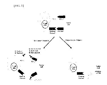
HG. 1 illustrates a schematic diagram, briefly showing the mechanism by which
the anti-CD300c monoclonal antibody and/or CD300c siRNA of the present
disclosure
exhibits an anticancer effect.
HG. 2 illustrates a schematic diagram, briefly showing the mechanism by which
the anti-CD300c monoclonal antibody of the present disclosure acts on
monocytes, T
cells, and cancer cells, respectively.
HG. 3 illustrates results obtained by performing SDS-PAGE on the anti-
CD300c monoclonal antibodies according to an embodiment of the present
disclosure
under a non-reducing condition.
HG. 4 illustrates results obtained by performing SDS-PAGE on the anti-
CD300c monoclonal antibodies according to an embodiment of the present
disclosure
under a reducing condition.
HG. 5 illustrates results obtained by identifying the binding affinity, to a
CD300c antigen, of the anti-CD300c monoclonal antibody according to an
embodiment
of the present disclosure.
HG. 6 illustrates results obtained by identifying, with ELISA, the T cell
activation ability of the anti-CD300c monoclonal antibody according to an
embodiment
of the present disclosure.
6
CA 03158715 2022-5-17
FIG. 7 illustrates results obtained by identifying, with ELISA, the
differentiation
capacity into M1 macrophages of the anti-CD300c monoclonal antibodies
according to
an embodiment of the present disclosure.
FIG. 8 illustrates results obtained by identifying, with ELISA, the capacity
of
the anti-CD300c monoclonal antibodies according to an embodiment of the
present
disclosure for causing differentiation into M1 macrophages.
FIG. 9 illustrates results obtained by identifying, with ELISA, the
differentiation
capacity into M1 macrophages depending on concentrations of the anti-CD300c
monoclonal antibodies according to an embodiment of the present disclosure.
HG. 10 illustrates results obtained by identifying, with ELISA, the
differentiation capacity into M1 macrophages depending on concentrations of
the anti-
CD300c monoclonal antibody according to an embodiment of the present
disclosure.
FIG. 11 illustrates results obtained by identifying, through cell shape, the
differentiation capacity into M1 macrophages of the anti-CD300c monoclonal
antibody
according to an embodiment of the present disclosure.
FIG. 12 illustrates results obtained by identifying, with ELISA, the
differentiation capacity into Ml macrophages of the anti-CD300c monoclonal
antibody
CL7 according to an embodiment of the present disclosure.
FIG. 13 illustrates results obtained by comparing, with ELISA, the
differentiation capacity into M1 macrophages between the anti-CD300c
monoclonal
antibodies according to an embodiment of the present disclosure and a cancer
immunotherapy.
FIG. 14 illustrates results obtained by comparing, with ELISA, the
differentiation capacity into M1 macrophages between the anti-CD300c
monoclonal
antibody CL7 according to an embodiment of the present disclosure and cancer
immunotherapies.
FIG. 15 illustrates results obtained by comparing, with ELISA, the
differentiation capacity into M1 macrophages between the anti-CD300c
monoclonal
7
CA 03158715 2022-5-17
antibody CL7 according to an embodiment of the present disclosure and cancer
immunotherapies.
HG. 16 illustrates results obtained by comparing, with ELISA, the
differentiation capacity into M1 macrophages between the anti-CD300c
monoclonal
antibody CL7 according to an embodiment of the present disclosure and cancer
immunotherapies.
HG. 17 illustrates results obtained by comparing, with ELISA, the
differentiation capacity from MO macrophages into M1 macrophages between the
anti-
CD300c monoclonal antibody according to an embodiment of the present
disclosure
and a cancer immunotherapy.
HG. 18 illustrates results obtained by comparing, with ELISA, the
differentiation capacity into M1 macrophages between the anti-CD300c
monoclonal
antibody according to an embodiment of the present disclosure and a cancer
immunotherapy.
HG. 19 illustrates results obtained by identifying, with ELISA, the
redifferentiation capacity from M2 macrophages into M1 macrophages of the anti-
CD300c monoclonal antibody according to an embodiment of the present
disclosure.
HG. 20 illustrates results obtained by identifying, with EL1SA, the
redifferentiation capacity from M2 macrophages into M1 macrophages of the anti-
CD300c monoclonal antibody according to an embodiment of the present
disclosure.
HG. 21 illustrates results obtained by identifying, with ELISA, the
redifferentiation capacity from M2 macrophages into M1 macrophages of the anti-
CD300c monoclonal antibody according to an embodiment of the present
disclosure.
HG. 22 illustrates results obtained by identifying, with ELISA, the
redifferentiation capacity from MO, Ml, and M2 macrophages into M1 macrophages
of
the anti-CD300c monoclonal antibody according to an embodiment of the present
disclosure.
HG. 23 illustrates results obtained by identifying, with differentiation
capacity
8
CA 03158715 2022-5-17
into M1 macrophages, the effects caused by co-administration of the anti-
CD300c
monoclonal antibody according to an embodiment of the present disclosure and
an anti-
PD-L1 immunotherapy.
MG. 24 illustrates results obtained by identifying, with differentiation
capacity
into M1 macrophages, the effects caused by co-administration of the anti-
CD300c
monoclonal antibody according to an embodiment of the present disclosure and a
cancer
immunotherapy.
MG. 25 illustrates results obtained by identifying the cancer cell growth
inhibitory effects of the anti-CD300c monoclonal antibodies according to an
embodiment of the present disclosure under a condition of 0% FBS.
MG. 26 illustrates results obtained by identifying the cancer cell growth
inhibitory effects of the anti-CD300c monoclonal antibodies according to an
embodiment of the present disclosure under a condition of 0.1% FB S.
MG. 27 illustrates results obtained by comparing the cancer cell (lung cancer)
growth inhibitory effects of the anti-CD300c monoclonal antibodies according
to an
embodiment of the present disclosure and a cancer immunotherapy.
MG. 28 illustrates results obtained by comparing the cancer cell (breast
cancer)
growth inhibitory effects of the anti-CD300c monoclonal antibodies according
to an
embodiment of the present disclosure and a cancer immunotherapy.
MG. 29 illustrates results obtained by identifying the cancer cell growth
inhibitory effects depending on concentrations of the anti-CD300c monoclonal
antibody according to an embodiment of the present disclosure.
MG. 30 illustrates results obtained by identifying the cancer cell growth
inhibitory effects caused by co-administration of the anti-CD300c monoclonal
antibody
according to an embodiment of the present disclosure and a cancer
immunotherapy.
MG. 31 illustrates results obtained by identifying the cancer cell growth
inhibitory effects caused by co-administration of the anti-CD300c monoclonal
antibody
according to an embodiment of the present disclosure and a cancer
immunotherapy.
9
CA 03158715 2022-5-17
FIG. 32 illustrates results obtained by identifying the mechanism of action
caused by co-administration of the anti-CD300c monoclonal antibody according
to an
embodiment of the present disclosure and a cancer immunotherapy.
MG. 33 illustrates results obtained by identifying the binding specificity of
the
anti-CD300c monoclonal antibodies according to an embodiment of the present
disclosure.
MG. 34 illustrates results obtained by identifying the cross-reactivity, in
mice,
of the anti-CD300c monoclonal antibodies according to an embodiment of the
present
disclosure.
MG. 35 illustrates results obtained by identifying the anti-cancer (colorectal
cancer) effects, in mice, of the anti-CD300c monoclonal antibodies according
to an
embodiment of the present disclosure.
MG. 36 schematically illustrates an experimental method for identifying the
effects of the anti-CD300c monoclonal antibody according to an embodiment of
the
present disclosure on cancer growth in vivo.
MG. 37 illustrates results obtained by identifying the cancer growth
inhibitory
effects in vivo of the anti-CD300c monoclonal antibody according to an
embodiment of
the present disclosure.
MG. 38 illustrates results obtained by identifying the effects of the anti-
CD300c
monoclonal antibody according to an embodiment of the present disclosure on an
increase in tumor-infiltrating lymphocytes under a tumor microenvironment in
vivo.
The scale bar indicates 50 um.
MG. 39 illustrates results obtained by identifying the effects of the anti-
CD300c
monoclonal antibody according to an embodiment of the present disclosure on an
increase in M1 macrophages in vivo.
CA 03158715 2022-5-17
Best Mode for Carrying out Invention
The anti-CD300c monoclonal antibody of the present disclosure specifically
binds, with high binding affinity, to a CD300c protein and effectively
inhibits the
mechanism of CD300c, which activates T cells and promotes differentiation into
M1
macrophages so that growth of cancer cells, and development, metastasis, and
the like
of cancer can be effectively inhibited. Thus, the anti-CD300c monoclonal
antibody can
be effectively used for the treatment of various cancers that express a CD300c
antigen
on the surface.
As used herein, the term "antibody" refers to an immunoglobulin molecule that
is immunologically reactive with a specific antigen, and includes all of
polyclonal
antibodies, monoclonal antibodies, and functional fragments thereof In
addition, the
term may include forms produced by genetic engineering, such as chimeric
antibodies
(for example, humanized murine antibodies) and heterologous antibodies (for
example,
bispecific antibodies). Among these, the monoclonal antibodies are antibodies
that
exhibit single binding specificity and affinity against a single antigenic
site (epitope).
Unlike polyclonal antibodies including antibodies that exhibit specificity
against
different epitopes, the monoclonal antibodies exhibit binding specificity and
affinity
against a single epitope on an antigen, which allows for easy quality control
as a
therapeutic agent. In particular, the anti-CD300c monoclonal antibody of the
present
disclosure not only exhibits anticancer activity by itself by specifically
binding to
CD300c-expressing cancer cells, but also stimulates immune cells, thereby
exhibiting
maximized cancer cell-dependent anticancer activity. The antibody includes
variable
region(s) of a heavy chain and/or a light chain in terms of the constitution,
wherein the
variable region includes, as a primary structure thereof, a portion that fonns
an antigen-
binding site of the antibody molecule. The antibody of the present disclosure
may be
composed of a partial fragment containing the variable region. Preferably, the
variable
region may be replaced by a soluble receptor for CD300c. However, the variable
region
is not limited thereto and may include anything as long as the thus formed
antibody
exhibits the same effect as the anti-CD300c monoclonal antibody of the present
disclosure.
11
CA 03158715 2022-5-17
As used herein, the term "immunoglobulin" refers to a concept that encompasses
both an antibody and an antibody-like molecule that has the same structural
characteristics as an antibody and does not have antigenic specificity.
As used herein, the term "single-chain variable fragment (scFv)" refers to a
protein in which light chain and heavy chain variable regions of an antibody
are linked
to each other via a linker consisting of a peptide sequence having about 15
amino acid
residues. The scFv may be in an order of light chain variable domain-linker-
heavy chain
variable region, or an order of heavy chain variable region-linker-light chain
variable
region, and has the same or similar antigen specificity as its original
antibody. The
linking site is a hydrophilic flexible peptide chain mainly composed of
glycine and
serine. The 15-amino acid sequence of "(Gly-Gly-Gly-Gly-Ser)3 or a sequence
similar
thereto is mainly used.
As used herein, the term "cancer immunotherapy" (also referred to as simply
"immunotherapy") collectively refers to a cancer therapy or anticancer agent
that
activates an immune function of immune cells in the body to fight cancer
cells.
Examples thereof may include, but are not limited to, anti-PD-1, anti-PD-L1,
anti-
CTLA-4, anti-MR, anti-LAW, anti-CD137, anti-0X40, anti-CD276, anti-CD27, anti-
GITR, anti-T1M3, anti-41BB, anti-CD226, anti-CD40, anti-CD70, anti-ICOS, anti-
CD4OL, anti-BTLA, anti-TCR, and anti-TIGIT.
As used herein, the teim "adjuvant" refers to an auxiliary drug used for the
purpose of assisting in the efficacy of a main drug, that is, a cancer therapy
to improve
and/or enhance its therapeutic effect, suppressing resistance to the main drug
to improve
and/or enhance its therapeutic effect, or preventing or alleviating harmful
action of the
main drug. The adjuvant of the present disclosure is not limited as long as it
contains
the anti-CD300c monoclonal antibody as an active ingredient.
As used herein, the teim "prevention" means any action that inhibits or delays
onset of diseases such as cancer by administration of the pharmaceutical
composition
according to the present disclosure.
As used herein, the term "treatment" means any action that ameliorates or
12
CA 03158715 2022-5-17
beneficially alters symptoms of cancer or the like by administration of the
pharmaceutical composition according to the present disclosure.
As used herein, the term "individual" refers to a subject to which the
pharmaceutical composition of the present disclosure can be administered, and
the
subject is not limited.
As used herein, the term "pharmaceutical composition" may be characterized
by being in the form of capsules, tablets, granules, injections, ointments,
powders, or
beverages, and the phaimaceutical composition may be characterized by being
for
application to humans. The pharmaceutical composition may be formulated in the
form
of oral preparations such as powders, granules, capsules, tablets, and aqueous
suspensions, preparations for external use, suppositories, and sterile
injectable solutions,
respectively, according to conventional methods, and used.
However, the
pharmaceutical composition is not limited thereto. The pharmaceutical
composition of
the present disclosure may further comprise a pharmaceutically acceptable
carrier. As
the pharmaceutically acceptable carrier, a binder, a glidant, a disintegrant,
an excipient,
a solubilizer, a dispersant, a stabilizer, a suspending agent, a pigment, a
flavor, and the
like may be used for oral administration; a buffer, a preserving agent, a pain-
relieving
agent, a solubilizer, an isotonic agent, a stabilizer, and the like may be
used in admixture
for injections; and a base, an excipient, a lubricant, a preserving agent, and
the like may
be used for topical administration. The preparations of the pharmaceutical
composition
of the present disclosure may be prepared in various ways by being mixed with
the
pharmaceutically acceptable carrier as described above. For example, for oral
administration, the pharmaceutical composition may be formulated in the form
of
tablets, troches, capsules, elixirs, suspensions, syrups, wafers, or the like.
For injections,
the phaimaceutical composition may be foimulated in the form of unit dosage
ampoules
or multiple dosage forms. Alternatively, the pharmaceutical composition may be
formulated into solutions, suspensions, tablets, capsules, sustained-release
preparations,
or the like.
Meanwhile, as examples of carriers, diluents, or excipients suitable for
making
13
CA 03158715 2022-5-17
preparations, lactose, dextrose, sucrose, sorbitol, mannitol, xylitol,
erythritol, maltitol,
starch, gum acacia, alginate, gelatin, calcium phosphate, calcium silicate,
cellulose,
methylcellulose, microcrystalline cellulose, polyvinylpyrrolidone, water,
methyl
hydroxybenzoate, propyl hydroxybenzoate, talc, magnesium stearate, mineral
oil, or the
like may be used. In addition, a filler, an anti-coagulant, a lubricant, a
wetting agent, a
fragrance, an emulsifier, a preservative, and the like may further be
included.
The route of administration of the pharmaceutical composition includes, but is
not limited to, oral, intravenous, intramuscular, intraarterial,
intramedullary, intradural,
intracardiac, transdermal, subcutaneous, intraperitoneal, intranasal,
intestinal, topical,
sublingual, or rectal route. Oral or parenteral administration is preferred.
As used
herein, the term "parenteral" includes subcutaneous, intradermal, intravenous,
intramuscular, intraarticular, intrabursal, intrasternal, intradural,
intralesional, and
intracranial injection or infusion techniques. The pharmaceutical composition
of the
present disclosure may also be administered in the form of suppositories for
rectal
administration.
The pharmaceutical composition of the present disclosure may vary widely
depending on a variety of factors, including activity of a certain compound
used, the
patient's age, body weight, general health status, sex, diet, time of
administration, route
of administration, rate of excretion, drug combination, and severity of a
certain disease
to be prevented or treated. A dose of the pharmaceutical composition may vary
depending on the patient's condition, body weight, severity of disease, drug
form, route
of administration, and duration, and may be appropriately selected by those
skilled in
the art. The pharmaceutical composition may be administered in an amount of
0.0001
to 500 mg/kg or 0.001 to 500 mg/kg, per day. Administration may be made once a
day
or several times a day. The dose is not intended to limit the scope of the
invention in
any way. The pharmaceutical composition according to the present disclosure
may be
formulated in the form of pills, sugar-coated tablets, capsules, liquids,
gels, syrups,
slurries, or suspensions.
Hereinafter, the following examples are provided to help the understanding of
14
CA 03158715 2022-5-17
the present disclosure. However, the following examples are only provided for
easier
understanding of the present disclosure, and the scope of the present
disclosure is not
limited by the following examples.
Examples
Example 1: Selection of anti-CD300c monoclonal antibody
1.1. Construction of anti-CD300c monoclonal antibody library
In order to select anti-CD300c monoclonal antibodies, biopanning was
performed using a lambda phage library, a kappa phage library, a VH3VL1 phage
library,
and an OPALTL phage library. More specifically, a CD300c antigen was added at
a
concentration of 5 Rg/mL to an immunotube, and reaction was allowed to proceed
for 1
hour so that the antigen was adsorbed on the surface of the immunotube. 3%
skim milk
was added to suppress non-specific reactions. Then, 1012 PFU of the antibody
phage
library dispersed in 3% skim milk was added to each immunotube for antigen
binding.
Washing was perfonned 3 times using Iris buffered saline-Tween 20 (TBST)
solution
to remove non-specifically bound phages, and then single-chain variable
fragment
(scFv) phage antibodies, specifically bound to the CD300c antigen, were eluted
using
100 mM triethylamine solution. The eluted phages were neutralized using 1.0 M
Tris-
HCI buffer (pH 7.8). Then, the resultant was subjected to E. coli ER2537 and
infection
was allowed to proceed at 37 C for 1 hour. The infected E. coli was applied
onto LB
agar medium containing carbenicillin. and cultured at 37 C for 16 hours. Then,
the
formed E. coli colonies were suspended using 3 mL of super broth (SB)-
carbenicillin
culture. Some of the suspension was stored at -80 C until use with the
addition of 15%
glycerol, and the remaining portion was reinoculated into SB-carbenicillin-2%
glucose
solution and cultured at 37 C. Then, the obtained culture was centrifuged, and
biopanning was repeated 3 times again using the supernatant containing phage
particles
to obtain and concentrate antigen-specific antibodies.
After repeating the biopanning 3 times, E. coli containing the antibody gene
was
applied onto LB agar medium containing carbenicillin and cultured at 37 C for
16 hours.
CA 03158715 2022-5-17
The formed E. coli colonies were inoculated again into SB-carbenicillin-2%
glucose
solution and cultured at 37 C until the absorbance (at OD 600 nm) reached 0.5.
Then,
IPTG was added and further cultured at 30 C for 16 hours. Thereafter,
periplasmic
extraction was performed. From the results, a library pool of antibodies,
which
specifically bind to the CD300c antigen, was primarily obtained.
1.2. Selection of anti-CD300c monoclonal antibody
In order to select anti-CD300c monoclonal antibodies that specifically bind,
with high binding affinity, to a CD300c antigen, ELISA was perfoimed using the
library
pool obtained in the same manner as in Example 1.1. More specifically, each of
a
CD300c antigen and a CD300a antigen in a coating buffer (0.1 M sodium
carbonate,
pH 9.0) was dispensed onto an ELISA plate at a concentration of 5 p,g/mL per
well, and
then reaction was allowed to proceed at room temperature for 3 hours so that
the antigen
was bound to the plate. Washing was performed 3 times using phosphate buffered
saline-Tween 20 (PBST) to remove unbound antigen, and then 350 pi, of PBST
supplemented with 2% bovine serum albumin (BSA) was added to each well.
Reaction
was allowed to proceed at room temperature for 1 hour, and washing was
performed
again using PBST. Then, 25 jig of periplasmic extract containing scFv obtained
in the
same manner as in Example 1.1 was added thereto, and reaction was allowed to
proceed
for 1 hour at room temperature for antigen binding. After 1 hour, washing was
performed 3 times using PBST to remove unbound scFv, and then 4 Rg/mL of an
antibody for detection was added. Reaction was allowed to proceed again at
room
temperature for 1 hour. Subsequently, the unbound antibody for detection was
removed
using PB ST. Then, anti-rabbit IgG to which HRP was bound was added and
reaction
was allowed to proceed at room temperature for 1 hour. The unbound antibody
was
removed again using PBST. TMB solution was added and reaction was allowed to
proceed for 10 minutes for development. Then, 2 N sulfuric acid solution was
added to
terminate the development, and the absorbance was measured at 450 nm to
identify the
antibodies that specifically bind to the CD300c antigen.
1.3. Identification of anti-CD300c monoclonal antibody sequences
16
CA 03158715 2022-5-17
The nucleotide sequences of the anti-CD300c monoclonal antibodies, which
were selected using the same method as in Example 1.2, were identified. More
specifically, for each of the selected antibody clones, plasmid DNA was
extracted
therefrom using a plasmid miniprep kit. Then, DNA sequencing was performed to
analyze complementarity-determining region (CDR) sequences. As a result, 25
types
of anti-CD300c monoclonal antibodies having different amino acid sequences
were
obtained.
There were 3 types of anti-CD300c monoclonal antibodies selected using the
lambda phage library: 5L18 including the CDR sequence(s) of SEQ ID NO: 36 (the
DNA sequence thereof is SEQ ID NO: 35), CL4 including the CDR sequence(s) of
SEQ
ID NO: 8 (the DNA sequence thereof is SEQ ID NO: 7), and CL5 including the CDR
sequence(s) of SEQ ID NO: 10 (the DNA sequence thereof is SEQ ID NO: 9).
There were 10 types of anti-CD300c monoclonal antibodies selected using the
kappa phage library: CK1 including the CDR sequence(s) of SEQ ID NO: 2 (the
DNA
sequence thereof is SEQ ID NO: 1), CK2 including the CDR sequence(s) of SEQ ID
NO: 4 (the DNA sequence thereof is SEQ ID NO: 3), CK3 including the CDR
sequence(s) of SEQ ID NO: 6 (the DNA sequence thereof is SEQ ID NO: 5), SK11
including the CDR sequence(s) of SEQ ID NO: 22 (the DNA sequence thereof is
SEQ
ID NO: 21), SK12 including the CDR sequence(s) of SEQ ID NO: 24 (the DNA
sequence thereof is SEQ ID NO: 23), SK13 including the CDR sequence(s) of SEQ
ID
NO: 26 (the DNA sequence thereof is SEQ ID NO: 25), 5K14 including the CDR
sequence(s) of SEQ ID NO: 28 (the DNA sequence thereof is SEQ ID NO: 27), SKIS
including the CDR sequence(s) of SEQ ID NO: 30 (the DNA sequence thereof is
SEQ
ID NO: 29), SK16 including the CDR sequence(s) of SEQ ID NO: 32 (the DNA
sequence thereof is SEQ ID NO: 31), and SK17 including the CDR sequence(s) of
SEQ
ID NO: 34 (the DNA sequence thereof is SEQ ID NO: 33).
There were 10 types of anti-CD300c monoclonal antibodies selected using the
VH3VL1 phage library: CB301 H3L1 A10 including the CDR sequence(s) of SEQ ID
NO: 37 (the DNA sequence thereof is SEQ ID NO: 38), CB301 H3L1 Al2 including
17
CA 03158715 2022-5-17
the CDR sequence(s) of SEQ ID NO: 40 (the DNA sequence thereof is SEQ ID NO:
39), CL6 including the CDR sequence(s) of SEQ ID NO: 12 (the DNA sequence
thereof
is SEQ ID NO: 11), CB301 H3L1 E6 including the CDR sequence(s) of SEQ ID NO:
42 (the DNA sequence thereof is SEQ ID NO: 41), CL7 including the CDR
sequence(s)
of SEQ ID NO: 14 (the DNA sequence thereof is SEQ ID NO: 13), CB301 H3L1 F4
including the CDR sequence(s) of SEQ ID NO: 44 (the DNA sequence thereof is
SEQ
ID NO: 43), CL8 including the CDR sequence(s) of SEQ ID NO: 16 (the DNA
sequence
thereof is SEQ ID NO: 15), CB301 H3L1 Gil including the CDR sequence(s) of SEQ
ID NO: 46 (the DNA sequence thereof is SEQ ID NO: 45), CL9 including the CDR
sequence(s) of SEQ ID NO: 18 (the DNA sequence thereof is SEQ ID NO: 17), and
CLIO including the CDR sequence(s) of SEQ ID NO: 20 (the DNA sequence thereof
is
SEQ ID NO: 19).
There were 2 types of anti-CD300c monoclonal antibodies selected using the
OPALTL phage library: CB301 OPALTL B5 including the CDR sequence(s) of SEQ
ID NO: 48 (the DNA sequence thereof is SEQ ID NO: 47) and CB301 OPALTL E6
including the CDR sequence(s) of SEQ ID NO: 50 (the DNA sequence thereof is
SEQ
ID NO: 49).
From the above results, it was possible to identify 25 types of anti-CD300c
monoclonal antibodies that specifically bind, with high binding affinity, to
the CD300c
antigen and can be used for the prevention or treatment of cancer.
1.4. Production and purification of anti-CD300c monoclonal antibody
Using each of the nucleotide sequences of the anti-CD300c monoclonal
antibodies identified in Example 1.3, expression vectors capable of expressing
each
antibody were prepared into which the heavy chain and the light chain are
separately
inserted. More specifically, the expression vectors were prepared by inserting
genes
into the pCIW3.3 vectors using the analyzed CDR sequences so that the vectors
can
express the heavy and light chains, respectively. The prepared expression
vectors for
heavy and light chains were mixed with polyethylenimine (PEI) in a mass ratio
of 1:1
and transfected into 2931 cells to induce antibody expression. Then, on day 8,
the
18
CA 03158715 2022-5-17
culture was centrifuged to remove the cells. The resulting culture was
obtained. The
obtained culture was filtered, and then resuspended using a mixed solution of
0.1 M
N aH2PO4 and 0.1 M N a2HPO4 (pH 7.0). The resuspended solution was purified
through
affinity chromatography using protein A beads (GE Healthcare), and finally
eluted using
an elution buffer (Thermofisher).
In order to identify the produced antibody, each of reducing sample buffer and
non-reducing sample buffer was added to 5 pg of purified antibody, and
electrophoresis
was performed using pre-made SDS-PAGE (Invitrogen). Then, the proteins were
stained using Coomassie Blue. The respective results under a non-reducing
condition
are illustrated in FIG. 3, and the respective results under a reducing
condition are
illustrated in FIG. 4.
As illustrated in FIGS. 3 and 4, it was identified that the anti-CD300c
monoclonal antibodies having a purity of 90% or higher were produced and
purified.
1.5. Determination of antigen-binding affinity of anti-CD300c monoclonal
antibody
Among the anti-CD300c monoclonal antibodies produced in the same manner
as in Example 1.4, binding ELISA was performed to select monoclonal antibodies
that
specifically bind, with better binding affinity, to the CD300c antigen. More
specifically,
each of the CD300c antigen or CD300a antigen in a coating buffer (0.1 M sodium
carbonate, pH 9.0) was dispensed onto an ELISA plate at a concentration of 8
pg/mL
per well, and then reaction was allowed to proceed at room temperature for 3
hours so
that the antigen was bound to the plate. Washing was performed 3 times using
phosphate buffered saline- Tween 20 (PBST) to remove unbound antigen, and then
300
pL of PBST supplemented with 5% bovine serum albumin (BSA) was added to each
well. Reaction was allowed to proceed at room temperature for 1 hour, and
washing
was performed again using PBST. Then, the anti-CD300c monoclonal antibody was
diluted in quadruplicate and added thereto. Reaction was allowed to proceed
for 1 hour
at room temperature for antigen binding. After 1 hour, washing was performed 3
times
using PBST to remove unbound anti-CD300c monoclonal antibody, and then 4 pg/mL
19
CA 03158715 2022-5-17
of an antibody for detection (HRP conjugated anti-Fc IgG) was added. Reaction
was
allowed to proceed again at room temperature for 1 hour. Subsequently, the
unbound
antibody for detection was removed using PBST, and then TMB solution was
added.
Reaction was allowed to proceed for 10 minutes for development. Then, 2 N
sulfuric
acid solution was added to terminate the development, and the absorbance was
measured at 450 nm to identify the antibodies that specifically bind to the
CD300c
antigen. The results are shown in Table 1 and FIG. 5.
[Table 1]
CB301 antibody
EC50 ( g/mL)
CK1
0_056
CK2
0.033
CK3
0393
CL4
0_031
CL5
0.032
CL6
0.148
CL7
0.047
CL8
49.7
CL9
0.094
CLIO
0.039
SK11
0.052
SK12
0.067
SK13
0.044
SK14
0.065
SK15
14.74
SK16
2.42
SK17
0.054
SL18
0.17
As shown in Table 1, as a result of measuring the EC50 (effective
concentration
of drug that causes 50% of the maximum response) values of the anti-CD300c
monoclonal antibodies, it was identified that the remaining all 14 clones
except for 4
clones (CK3, CL8, SKIS, SK16) exhibited high binding affinity of 0.21.1.g/
_______________________________________________ -n_L or lower.
CA 03158715 2022-5-17
As illustrated in FIG. 5, it was found that the anti-CD300c monoclonal
antibodies of the present disclosure bound to the CD300c antigen with high
binding
affinity even in the sigmoid curves for the results of the binding EL1SA.
Example 2: Identification of anti-cancer effects of anti-CD300c monoclonal
antibody on T cells
In order to identify whether the anti-CD300c monoclonal antibody selected by
the method of Example 1.5 exhibits an anticancer effect by activating T cells,
the
production level of interleukin-2 (IL-2) was identified. IL-2 is an immune
factor that
helps growth, proliferation, and differentiation of T cells. Increased
production level of
IL-2 means activation of T cells due to an increase in stimulation that
induces increased
differentiation, proliferation, and growth of T cells. More specifically, each
of anti-CD3
monoclonal antibody and anti-CD28 monoclonal antibody was added to a 96-well
plate
at a concentration of 2 jig/well and fixed for 24 hours. Then, co-treatment
with lx 105
cells/well of Jurkat T cells (human T lymphocyte cell line) and 10 jig/well of
anti-
CD300c monoclonal antibody were performed. The production level of 1L-2 was
measured using an EL1SA kit (IL-2 Quantikine kit, R&D Systems), and then
compared
with the control group that had not been treated with the anti-CD300c
monoclonal
antibody. The results are illustrated in FIG. 6.
As illustrated in FIG. 6, it was identified that the production level of IL-2
increased in a case where Jurkat T cells activated by treatment with the anti-
CD3
monoclonal antibody and the anti-CD28 monoclonal antibody were treated with
the
anti-CD300c monoclonal antibody. From these results, it was found that the
anti-
CD300c monoclonal antibody was able to activate T cells, indicating that the
anti-
CD300c monoclonal antibody can induce anticancer immune action to inhibit
growth
of cancer tissue.
Example 3: Identification of anticancer effect by capacity of anti-CD300c
monoclonal antibody for causing differentiation into macrophages
3.1. Identification of capacity of anti-CD300c monoclonal antibody for
causing differentiation into M1 macrophages
21
CA 03158715 2022-5-17
In order to identify that the anti-CD300c monoclonal antibody selected by the
method of Example 1.5 induces differentiation of monocytes into M1
macrophages,
THP-1 (human monocyte cell line) at 1.5x10⁴ cells/well was dispensed onto a 96-
well
plate, and treatment with 10 g/mL of the anti-CD300c monoclonal antibody
and/or
100 ng/mL of LPS was performed. Reaction was allowed to proceed for 48 hours,
and
then the production level of tumor necrosis factor-a (INF-a), which is a
differentiation
marker of M1 macrophages, was measured using an ELISA kit (Human INF-a
Quantikine kit, R&D Systems). The results are illustrated in FIGS. 7 and 8.
As illustrated in FIG. 7, it was identified that the anti-CD300c monoclonal
antibodies, CIA, CL7, CL 10, and SL18, exhibited an increase in production
level of
INF-a which is about 2 or more times higher than the control group (Con)
treated with
LPS alone.
In addition, as illustrated in FIG. 8, it was identified that all the
experimental
groups treated with the anti-CD300c monoclonal antibody alone without LPS
treatment
exhibited an increase in production level of INF-a as compared with the
control group
(Con) treated with LPS alone.
3.2. Identification of differentiation capacity into MI macrophages
depending on concentrations of anti-CD300c monoclonal antibody
In order to identify that induction of differentiation into M1 macrophages by
the
anti-CD300c monoclonal antibody increases with concentrations of the anti-
CD300c
monoclonal antibody, the production level of INF-a was identified in the same
manner
as in Example 3.1. Treatment with the anti-CD300c monoclonal antibody was
performed at concentrations of 10, 1, and 0.1 Rg/mL. The results are
illustrated in FIG.
9.
As illustrated in FIG. 9, it was identified that the production level of INF-a
increased as the treatment concentration of the anti-CD300c monoclonal
antibody
increased.
In order to identify results with further divided concentrations, treatment
with
22
CA 03158715 2022-5-17
the anti-CD300c monoclonal antibody CL7 was performed at concentrations of 10,
5,
2.5, 1.25, 0.625, 0.313, 0.157, and 0.079 !_tg/mL, and the production level of
INF-a was
identified. The results are illustrated in FIG. 10.
As illustrated in FIG. 10, it was identified that the production level of INF-
a
increased in a concentration-dependent manner with respect to the anti-CD300c
monoclonal antibody.
3.3. Identification of differentiation into MI macrophages caused by anti-
CD300c monoclonal antibody through cell shape.
In order to identify, through cell shape, differentiation pattern into M1
macrophages in a case where monocytes are treated with the anti-CD300c
monoclonal
antibody, THP-1 was treated with 101.1g/mL of the anti-CD300c monoclonal
antibody,
cultured for 48 hours, and then the shape of the cells was observed under a
microscope.
The results are illustrated in FIG. 11.
As illustrated in FIG. 11, it was identified that for the experimental group
(CL?)
treated with the anti-CD300c monoclonal antibody, the shape of THP-1 cells was
changed from suspension cells to circular adherent cells that are in the form
of M1
macrophages. From these results, it was identified that differentiation of
monocytes
into M1 macrophages was promoted by treatment with the anti-CD300c monoclonal
antibody.
3.4. Identification of capacity of anti-CD300c monoclonal antibody CL7 for
causing differentiation into MI macrophages
In order to identify again whether the anti-CD300c monoclonal antibody CL7
promotes differentiation of human monocytes into M1 macrophages, the secretion
levels of INF-a, interleukin-113 (IL-113), and interleukin-8 (1L-8) were
measured using
an ELISA kit (R&D Systems). More specifically, THP-1 at 1.5x10⁴ cells/well was
dispensed onto a 96-well plate, and treatment with 10 pg/mL of the anti-CD300c
monoclonal antibody was performed. Reaction was allowed to proceed for 48
hours,
and then the production levels of INF-a, IL-113, and 1L-8, which are markers
for
23
CA 03158715 2022-5-17
differentiation into M1 macrophages, were measured using an ELISA kit (Human
TNF-
a Quantikine kit, R&D Systems). The results are illustrated in FIG. 12.
As illustrated in FIG. 12, it was identified that all three types of markers
for
differentiation into M1 macrophages increased in the experimental group
treated with
the anti-CD300c monoclonal antibody, as compared with the control group (Con)
not
treated with the anti-CD300c monoclonal antibody.
3.5. Comparison of differentiation capacity into Ml macrophages between
anti-CD300c monoclonal antibody and cancer immunotherapy
In order to compare differentiation capacity into M1 macrophages between the
anti-CD300c monoclonal antibodies and a cancer immunotherapy, the production
level
of INF-a was identified using an ELISA kit in the same manner as in Example
3.1. As
an anti-PD-LA immunotherapy, Imfinzi was used at a concentration of 10 lig/mL.
The
results are illustrated in FIG. 13.
As illustrated in FIG. 13, it was identified that the anti-CD300c monoclonal
antibody resulted in a remarkably increased production level of INF-a as
compared
with the control group treated with lmfinzi (Imf), which is known as a cancer
immunotherapy, alone. From these results, it was found that the anti-CD300c
monoclonal antibody resulted in remarkably increased differentiation capacity
into M1
macrophages as compared with the conventionally known cancer immunotherapy.
For comparison with other cancer immunotherapies, each of lmfinzi, which is
an anti-PD-L1 immunotherapy, Keytruda, which is an anti-PD-1 immunotherapy,
and
an isotype control (immunoglobulin G) antibody was used at a concentration of
10
Rg/mL, and the production levels of INF-a, IL-113, and IL-8 were identified
using an
ELISA kit. The results are illustrated in FIGS. 14 to 16.
As illustrated in FIGS. 14 to 16, it was identified that the anti-CD300c
monoclonal antibody CL7 resulted in remarkably increased production levels of
INF-
a, IL-113, and 1L-8 as compared with lmfinzi, Keytruda, and the IgG antibody.
From
these results, it was found that the anti-CD300c monoclonal antibody was able
to result
24
CA 03158715 2022-5-17
in remarkably increased promotion of differentiation into M1 macrophages as
compared
with the conventional cancer immunotherapies.
3.6. Comparison of differentiation capacity from MO macrophages into M1
macrophages between anti-CD300c monoclonal antibody and anti-PD-Li
immunotherapy
In order to compare differentiation capacity from MO macrophages into M1
macrophages between the anti-CD300c monoclonal antibodies and cancer
immunotherapies, THP-1 at 1.5x10⁴ cells/well was dispensed onto a 96-well
plate, and
treatment with 10 i_tg/mL of the anti-CD300c monoclonal antibody, 10 pg/mL of
Imfinzi,
and/or 200 nM of phorbol-12-myristate-13-acetate (PMA) was performed. Reaction
was allowed to proceed for 48 hours, and then the production levels of INF-a
were
measured using an ELISA kit. The results are illustrated in FIG. 17.
As illustrated in FIG. 17, it was identified that INF-a was not produced in
the
comparative group treated with lmfinzi, which is a cancer immunotherapy,
alone, and
the production level of INF-a increased in the experimental group treated with
the anti-
CD300c monoclonal antibody alone. In addition, it was identified that even in
a case
where THP-1 was differentiated into MO macrophages by treatment with PMA, the
experimental group treated with the anti-CD300c monoclonal antibody exhibited
a
remarkably increased production level of INF-a as compared with the
experimental
group treated with lmfinzi. From these results, it was found that the anti-
CD300c
monoclonal antibody promoted differentiation from MO macrophages into M1
macrophages as compared with a conventional cancer immunotherapy.
3.7. Comparison of differentiation capacity into M1 macrophages between
anti-CD300c monoclonal antibody and anti-PD-Li immunotherapy
In order to compare differentiation capacity into M1 macrophages between the
anti-CD300c monoclonal antibodies and cancer immunotherapies, the production
level
of INF-a was identified in the same manner as in Example 3.1. The results are
illustrated in FIG. 18.
CA 03158715 2022-5-17
As illustrated in FIG. 18, it was identified that in a case where monocytes
were
differentiated into M1 macrophages by treatment with LPS, the experimental
group co-
treated with lmfinzi and LPS did not exhibit a significant difference in
production level
of INF-a, and the experimental group treated with the anti-CD300c monoclonal
antibody and LPS exhibited a significant difference in production level of INF-
a as
compared with the experimental group treated with the anti-CD300c monoclonal
antibody alone.
3.8. Identification of redifferentiation capacity from M2 macrophages into
MI macrophages of anti-CD300c monoclonal antibody
In order to identify that the anti-CD300c monoclonal antibody can
redifferentiate M2 macrophages into M1 macrophages, THP-1 at 1.5x10⁴ cells was
dispensed onto a 96-well plate, and pre-treated for 6 hours by treatment with
320 nM
of PMA. Then, treatment with 20 ng/mL of interleukin-4 (IL-4) and interleukin-
13 (IL-
13), and with 10 [tg/mL of the anti-CD300c monoclonal antibody was performed,
and
reaction was allowed to proceed for 18 hours. The production levels of TNF-a,
1L-113,
and IL-8 were identified using an ELISA kit. The results are illustrated in
FIGS. 19 to
21.
As illustrated in FIGS. 19 to 21, it was identified that among the
experimental
groups not pre-treated with PMA, the experimental group co-treated with IL-4 &
1L-13
and the anti-CD300c monoclonal antibody exhibited increased production levels
of
TNF-a, IL-113, and IL-8; and among the experimental groups pre-treated with
PMA, the
experimental group co-treated with 1L-4 & 1L-13 and the anti-CD300c monoclonal
antibody similarly exhibited increased production levels of TNF-a, 1L-113, and
IL-8.
From these results, it was found that the anti-CD300c monoclonal antibody was
able to
effectively redifferentiate M2 macrophages into M1 macrophages.
3.9. Identification of redifferentiation capacity from MO, Ml, and M2
macrophages into MI macrophages of anti-CD300c monoclonal antibody
In order to identify that the anti-CD300c monoclonal antibody can
redifferentiate MO, Ml, and M2 macrophages into M1 macrophages, THP-1 at
1.5x10⁴
26
CA 03158715 2022-5-17
cells was dispensed onto a 96-well plate, pre-treated with 10 Rg/mL of the
anti-CD300c
monoclonal antibody for 48 hours, and treated with 100 ng/mL of PMA, 100 ng/mL
of
LPS, and 20 ng/mL of IL- 4 and IL-13. Reaction was allowed to proceed for 24
hours.
The production level of INF-a was identified using an ELISA kit. The results
are
illustrated in FIG. 22.
As illustrated in FIG. 22, it was identified that all experimental groups pre-
treated with the anti-CD300c monoclonal antibody exhibited a significant
increase in
production level of INF-a, as compared with the MO macrophage control group
treated
with PMA alone, the M1 macrophage control group treated with LPS alone, and
the M2
macrophage control group treated with IL-4 and 1L-13 alone. From these
results, it was
found that the anti-CD300c monoclonal antibody had excellent capacity to
differentiate
MO, Ml, and M2 macrophages into M1 macrophages.
From these results, it was found that the anti-CD300c monoclonal antibody was
able to further promote differentiation into M1 macrophages as compared with a
conventional cancer immunotherapy, and thus induce anticancer immune action to
inhibit growth of cancer tissue. In particular, it was found that the anti-
CD300c
monoclonal antibody was able to exert an anticancer effect by
redifferentiating M2
macrophages, which are known to be involved in promoting proliferation and
metastasis
of cancer cells, into M1 macrophages.
Example 4: Identification of effects caused by co-administration of anti-
CD300c monoclonal antibody and cancer immunotherapy
4.1. Identification of effects caused by co-administration of anti-CD300c
monoclonal antibody and anti-PD-Li immunotherapy
In order to identify the cancer treatment effects caused by co-administration
of
the anti-CD300c monoclonal antibody and an anti-PD-L1 immunotherapy, NF-KB
(nuclear factor kappa-light-chain-enhancer of activated B cells) signal
transduction was
identified. More specifically, THP-1 at 8.8x10⁵ cells was dispensed onto a 6-
well plate,
and treated with 10 lig/mL of the anti-CD300c monoclonal antibody CL7 and/or
10
Rg/mL of Imfinzi. Incubation was performed for 24 hours, and phosphorylated NF-
kB
27
CA 03158715 2022-5-17
(p-NF-KB) was identified using Western blotting (Cell Signaling Technology).
The
results are illustrated in FIG. 23.
As illustrated in FIG. 23, it was identified that as compared with the
experimental group administered with the immunotherapy lmfinzi alone, the
experimental group administered with the anti-CD300c monoclonal antibody
exhibited
an increased level of p-NF-KB, and the experimental group co-treated with
lmfinzi and
the anti-CD300c monoclonal antibody exhibited a further increased level in p-
NF-KB.
From these results, it was found that co-administration of the anti-CD300c
monoclonal
antibody and Imfinzi promoted differentiation into M1 macrophages.
4.2. Identification of effects caused by co-administration of anti-CD300c
monoclonal antibody and anti-PD-Li immunotherapy and/or anti-PD-1
immunotherapy
In order to identify the cancer treatment effects caused by co-administration
of
the anti-CD300c monoclonal antibody and an anti-PD-L1 immunotherapy and/or an
anti-PD-1 immunotherapy, signal transduction of p38 MAPK (p38 mitogen-
activated
protein kinase) and ERK (extracellular signal-regulated kinase) was
identified. More
specifically, THP-1 at 8.8x10⁵ cells/well was dispensed onto a 6-well plate,
and treated
with 10 [tg/mL of anti-C300c monoclonal antibody CL7, 10 pg/mL of Imfinzi,
and/or
lig/mL of Keytruda. Incubation was performed for 48 hours, and phosphorylated
p38 MAPK (p-p38 MAPK) and phosphorylated ERIC (p-ERIC) were identified using
Western blotting (Cell Signaling Technology). The results are illustrated in
FIG. 24.
As illustrated in FIG. 24, neither p-p38 MAPK nor p-ERK proteins were
observed in the experimental group treated with the immunotherapy alone, and
both
types of proteins were observed in the experimental group treated with the
anti-CD300c
monoclonal antibody. In addition, it was identified that the level of p-p38
MAPK
protein further increased in the experimental groups co-administered with the
immunotherapy(ies).
From these results, it was identified that the anti-CD300c monoclonal antibody
promoted differentiation into M1 macrophages through MAPK signal transduction
and
28
CA 03158715 2022-5-17
this effect further increased in a case of being co-administered with (an)
immunotherapy(ies). Thus, it was found that the anti-CD300c monoclonal
antibody
was able to be used alone as a cancer immunotherapy, and its anti-cancer
therapeutic
effects could be further increased through co-administration with a
conventional
immunotherapy.
Example 5: Anti-cancer effect in vitro of anti-CD300c monoclonal antibody
5.1. Identification of cancer cell growth inhibitory effect of anti-CD300c
monoclonal antibody
In order to identify effects of the monoclonal antibody, which targets CD300c,
on growth of cancer cells, a cell proliferation assay was performed using A549
(human
lung cancer cell line). More specifically, onto a 96-well plate were dispensed
2x10⁴
cells under a condition of 0% fetal bovine serum (FBS) and 6x10³ cells under a
condition of 0.1% fetal bovine serum. Then, treatment with 10 [tginth of the
anti-
CD300c monoclonal antibody was perfoimed and incubation was performed for 5
days.
Then, treatment with CCK-S (DOJINDO) was performed and the absorbance at
Misch-nu
was measured to identify the cancer cell growth inhibitory effects of the anti-
CD300c
monoclonal antibody. The results are illustrated in FIGS. 25 and 26.
As illustrated in FIG. 25, it was identified that all anti-CD300c monoclonal
antibodies except for SK11 and SK14 had an effect of inhibiting proliferation
of cancer
cells under a condition of 0% FBS.
As illustrated in FIG. 26, it was identified that all anti-CD300c monoclonal
antibodies used in the experiment had an effect of inhibiting proliferation of
cancer cells
under a condition of 0.1% FBS.
5.2. Comparison of anti-CD300c monoclonal antibody and cancer cell
growth inhibitory effect with cancer immunotherapy
In order to compare the cancer cell growth inhibitory effects between the anti-
CD300c monoclonal antibody and a cancer immunotherapy, their cell growth
inhibitory
effects were identified using A549 (human lung cancer cell line) and MDA-MB-
231
29
CA 03158715 2022-5-17
(human breast cancer cell line). More specifically, onto a 96-well plate were
dispensed
2x10⁴ cells under a condition of 0% fetal bovine serum (FBS) and 6x10³ cells
under a
condition of 0.1% fetal bovine serum. Treatment with 10 pg/mL of the anti-
CD300c
monoclonal antibody was performed and incubation was performed for 5 days.
Then,
observation was made under an optical microscope. The results are illustrated
in FIGS.
27 and 28.
As illustrated in FIG. 27, it was identified that the anti-CD300c monoclonal
antibody more effectively inhibited proliferation of cancer cells than
lmfinzi, which is
an immunotherapy, in the A549 cell line.
As illustrated in FIG. 28, it was identified that the anti-CD300c monoclonal
antibody more effectively inhibited proliferation of cancer cells than
lmfinzi, which is
an immunotherapy, in the MDA-MB-231 cell line.
5.3. Identification of cancer cell growth inhibitory effects depending on
concentrations of anti-CD300c monoclonal antibody
In order to identify the cancer cell growth inhibitory effects depending on
concentrations of the anti-CD300c monoclonal antibody, 2x10⁴ A549 cells were
dispensed onto a 96-well plate under a condition of 0% fetal bovine serum
(FBS), and
treatment with 10 ligHiL of the anti-CD300c monoclonal antibody was performed.
Incubation was performed for 5 days. Treatment with CCK-8 (DOJINDO) was
performed and reaction was allowed to proceed for 3 hours. Then, the
absorbance at
OD4sonm was measured to identify cancer cell growth inhibitory effects of the
anti-
CD300c monoclonal antibody. The results are illustrated in FIG. 29.
As illustrated in FIG. 29, it was identified that cancer cell growth was
inhibited
depending on concentrations of the anti-CD300c monoclonal antibody.
5.4. Identification of cancer treatment effect by co-administration of anti-
CD300c monoclonal antibody and cancer immunotherapy
In order to identify the cancer treatment effects caused by co-administration
of
the anti-CD300c monoclonal antibody and a cancer immunotherapy, a cell
proliferation
CA 03158715 2022-5-17
assay was performed in the same manner as in Example 5.1. lmfinzi was used as
the
immunotherapy. The results are illustrated in FIG. 30.
As illustrated in FIG. 30, it was identified that cancer cell growth was
effectively
inhibited in a case where the anti-CD300c monoclonal antibody and the
immunotherapy
were co-administered, as compared with a case where the immunotherapy was
administered alone.
In addition, the cancer cell growth inhibitory effects were observed under an
optical microscope. The results are illustrated in FIG. 31.
As illustrated in FIG. 31, it was identified that cancer cell growth was
effectively
inhibited in a case where the anti-CD300c monoclonal antibody and the
immunotherapy
were co-administered.
5.5. Identification of mechanism of action by co-administration of anti-
CD300c monoclonal antibody and cancer immunotherapy
Regarding apoptosis signaling mechanism of cancer cells, in order to identify
the mechanism of action by co-administration of the anti-CD300c monoclonal
antibody
and a cancer immunotherapy, A549 cells were treated with the anti-CD300c
monoclonal
antibody, lmfinzi, and/or Keytruda, each at a concentration of 10 Rg/mL, and
levels of
cleaved caspase-9, which is an apoptosis marker, was identified by Western
blotting
(Cell Signaling Technology). The results are illustrated in FIG. 32.
As illustrated in FIG. 32, it was identified that the level of cleaved caspase-
9
increased in a case where the anti-CD300c monoclonal antibody and lmfinzi were
co-
administered, as compared with the experimental group treated with the anti-
CD300c
monoclonal antibody alone, and the level of cleaved caspase-9 further
increased in a
case where the anti-CD300c monoclonal antibody, lmfinzi, and Keytruda were co-
administered. From these results, it was found that in a case where the anti-
CD300c
monoclonal antibody and an immunotherapy were co-administered, the apoptosis
signaling mechanism of cancer cells was further activated, thereby effectively
inhibiting
proliferation of cancer cells.
31
CA 03158715 2022-5-17
Example 6: Identification of excellent cross-reactivity of anti-CD300c
monoclonal antibody between human and mouse antigens
6.1. Identification of specificity of anti-CD300c monoclonal antibody
Before identifying cross-reactivity of the anti-CD300c monoclonal antibody
between the human and mouse antigens, first, cross-reactivity of the anti-
CD300c
monoclonal antibody for a CD300a antigen, which is known to antagonize a
CD300c
antigen and also has a similar protein sequence thereto, was checked to
identify
specificity of the anti-CD300c monoclonal antibody. More specifically, the
anti-
CD300c monoclonal antibody was subjected to the CD300a antigen at
concentrations
of 0.039, 0.63, and 10 Rg/mL, and binding ELISA was performed in the same
manner
as in Example 1.5. The results are illustrated in FIG. 33.
As illustrated in MG. 33, it was identified that the anti-CD300c monoclonal
antibodies did not bind to CD300a. From these results, it was found that the
anti-
CD300c monoclonal antibodies exhibited high binding specificity to the extent
that they
do not bind to CD300a that has a similar sequence to CD300c.
6.2 Identification of differentiation capacity from mouse macrophages
(Raw264.7) into MI macrophages of anti-CD300c monoclonal antibody
In order to identify that the anti-CD300c monoclonal antibody can even promote
differentiation of mouse macrophages into M1 macrophages, lx10 mouse
macrophages
(Raw264.7) were dispensed at a concentration of lx 104 cells/well onto a 96-
well plate.
Then, treatment with 10 p.g/mL of the anti-CD300c monoclonal antibody was
performed and incubation was performed. The production level of TNF-a was
identified using an ELISA kit. The results are illustrated in FIG. 34.
As illustrated in FIG. 34, it was identified that the production level of INF-
a
increased in the experimental groups treated with the anti-CD300c monoclonal
antibody.
From these results, it was found that the anti-CD300c monoclonal antibody
promoted
differentiation into M1 macrophages by exerting the same action in mice as
well as in
humans.
32
CA 03158715 2022-5-17
6.3 Growth inhibitory effects of anti-CD300c monoclonal antibody on
mouse colorectal cancer cells (CT26)
In order to identify whether the anti-CD300c monoclonal antibody exhibits an
anticancer effect even in mice, a cell proliferation assay was performed in
the same
manner as in Example 4.1 using CT26 (mouse colorectal cancer cell line). The
results
are illustrated in FIG. 35.
As illustrated in FIG. 35, it was identified that the anti-CD300c monoclonal
antibodies exhibited a cancer treatment effect even in mice.
Example 7: Anti-cancer effects in vivo of anti-CD300c monoclonal antibody
7.1. Identification of cancer growth inhibitory effects in vivo of anti-CD300c
monoclonal antibody
In order to identify anticancer effects in vivo of the anti-CD300c monoclonal
antibody, a colorectal cancer cell line (C126) at 2x10⁵ cells was transplanted
by
subcutaneous injection into 8-week-old BALB/c mice to prepare a mouse
syngeneic
tumor model. Breeding and experiments for animals were all conducted in a
specific
pathogen free (SPF) facility. The experimental method is briefly illustrated
in FIG. 36.
12 days after transplantation of the colorectal cancer cell line, the mice
with tumor size
of 50 to 100 mm3 were injected with the anti-CD300c monoclonal antibody CL7,
an
anti-PD-1 antibody, and both CL7 and the anti-PD-1 antibody (Combo),
respectively,
and injected with an equal amount of phosphate buffered saline (PBS) as a
control group.
More specifically, the mice were intraperitoneally injected with 10 mg/kg of
each
material twice a week for two weeks (a total of 4 times). Then, the tumor
volume was
measured for 25 days. The results are illustrated in FIG. 37.
As illustrated in FIG. 37, it was identified that cancer growth was inhibited
in
the experimental group administered with the anti-CD300c monoclonal antibody
alone,
as compared with the control group, and that cancer growth was further
effectively
inhibited in a case where the anti-CD300c monoclonal antibody and the anti-PD-
1
antibody were co-administered.
33
CA 03158715 2022-5-17
7.2. Identification of effects of anti-CD300c monoclonal antibody on
increase in tumor-infiltrating lymphocytes under tumor microenvironment in
vivo
In order to identify the effects of the anti-CD300c monoclonal antibody on
tumor-infiltrating lymphocytes (TIL) in a tumor microenvironment (TME), on day
25
of the experiment performed in the same manner as in Example 7.1, the mice
were
euthanized, 1% parafoimaldehyde (PFA) was intravascularly injected thereinto,
and
then perfusion was performed to obtain cancer tissue. The obtained cancer
tissue was
fixed using 1% PFA, and sequentially dehydrated using 10%, 20%, and 30%
sucrose
solution. The dehydrated cancer tissue was frozen in OCT compound (optimal
cutting
temperature compound), and then the cancer tissue was sectioned to a thickness
of 50
p.m using a cryotome. Staining of CDS+ T cells and CD31 cancer vascular
cells, which
are tumor-infiltrating lymphocyte markers, was performed. The results are
illustrated
in FIG. 38.
As illustrated in FIG. 38, it was identified that the experimental group
administered with the anti-CD300c monoclonal antibody exhibited an increased
level
of CDS cells as compared with the experimental group administered with the
anti-PD-
1 antibody alone. From these results, it was found that the CD300c monoclonal
antibody increased tumor-infiltrating lymphocytes in a tumor microenvironment,
thereby exhibiting an anticancer effect.
7.3. Identification of MI macrophage increase effect in vivo of anti-CD300c
monoclonal antibody
In order to identify whether the anti-CD300c monoclonal antibody increases M1
macrophages in cancer tissue in vivo, staining of iNOS, which is an M1
macrophage
marker, and CD206, which is an M2 macrophage marker, was performed with the
cancer tissue section prepared in the same manner as in Example 7.2. The
results are
illustrated in FIG. 39.
As illustrated in FIG. 39, it was identified that as compared with the control
group, the experimental group treated with the anti-PD-1 antibody exhibited a
partially
increased level of M1 macrophages and the experimental group treated with the
anti-
34
CA 03158715 2022-5-17
CD300c monoclonal antibody exhibited a remarkably increased level of M1
macrophages with almost no observable M2 macrophages. In addition, it was
identified
that the experimental group co-administered with the anti-CD300c monoclonal
antibody and the anti-PD-1 antibody exhibited an increased level of M1
macrophages.
From these results, it was found that the anti-CD300c monoclonal antibody was
able to
effectively promote differentiation into M1 macrophages as compared with a
conventional cancer immunotherapy.
Consequently, it was identified that the anti-CD300c monoclonal antibody of
the present disclosure was able to bind, with high specificity, to a CD300c
antigen and
exhibit inter-species cross-reactivity for, for example, mice, indicating that
the anti-
CD300c monoclonal antibody can be used for various individuals. In addition,
it was
identified both in vitro and in vivo that the anti-CD300c monoclonal antibody
was able
to serve as a cancer immunotherapy by activating T cells and promoting
differentiation
into M1 macrophages, thereby effectively inhibiting proliferation, metastasis,
or the like
of cancer cells, and it was identified that the anti-CD300c monoclonal
antibody was
able to exhibit a further increased therapeutic effect through co-
administration with the
conventional cancer immunotherapy. Thus, it was found that the anti-CD300c
monoclonal antibody was able to be effectively used for anticancer
immunotherapy of
various cancers expressing a CD300c antigen.
The description of the present disclosure as described above is provided for
illustration, and those of ordinary skill in the art to which the present
disclosure pertains
will be able to understand that the embodiments disclosed herein can be easily
modified
into other specific forms without changing the technical spirit or essential
features of
the present disclosure. Therefore, it should be understood that the
embodiments are
illustrative and not restrictive in all respects.
Industrial Applicability
The anti-CD300c monoclonal antibody of the present disclosure specifically
binds to CD300c expressed on the surface of cancer cells, and thus can be used
for the
CA 03158715 2022-5-17
treatment of any cancer that secretes the CD300c antigen. Also, the anti-
CD300c
monoclonal antibody has inter-species cross-reactivity, and thus can be used
for
anticancer treatment of various individuals. In addition, since the anti-
CD300c
monoclonal antibody exhibits an anticancer effect by serving as a cancer
immunotherapy, it can effectively inhibit proliferation, development,
metastasis, or the
like of cancer. This allows the anti-CD300c monoclonal antibody to be
effectively used
for immunotherapy of various cancers.
36
CA 03158715 2022-5-17