Note : Les descriptions sont présentées dans la langue officielle dans laquelle elles ont été soumises.
WO 2021/119489
PCT/US2020/064607
LILRB1-BASED CIILVIERIC ANTIGEN RECEPTOR
CROSS REFERENCE TO RELATED APPLICATIONS
[0001] This application claims priority to U.S. provisional patent
applications 62/946,888, filed
on December 11, 2019, and 63/085,969 filed on August 30, 2020, the contents of
each of which
are incorporated herein by reference in their entireties.
INCORPORATION BY REFERENCE OF SEQUENCE LISTING
[0002] This application contains a Sequence Listing which has been submitted
in ASCII format
via EFS-WEB and is hereby incorporated by reference in its entirety. Said
ASCII copy, created
on December 11, 2020 is named A2BI-015-01W0 SeqList.txt and is 228 KB in size.
BACKGROUND
[0003] Chimeric antigen receptor (CAR) T cell therapy, and T Cell Receptor
(TCR) therapy, is
proving to be an effective therapeutic approach to various diseases,
particularly hematological
malignancies but also other cancers. CAR NK cells may also have clinical
applications.
Conventional CARs provide a stimulatory signal to the engineered immune cell
(e.g. a T cell or
an NK cell). In CAR-T cells, this results in killing activity towards the
target cell identified by
the antigen-binding domain of the CAR Inhibitory CARs (iCARs) have been
developed as a
means to control cell activity or restrict the activity of an activator CAR to
specific cell types.
Fedorov et al. Sei. Transl. Med. 5(215):215ra172 (2013). The inhibitory CAR
generally has the
intracellular domain of an inhibitory signaling molecule (such as PD-1 or CTLA-
4) fused to an
antigen-binding domain (e.g., a single-chain variable fragment, scFv) through
a transmembrane
region and optionally a hinge region.
[0004] Numerous alternative iCAR architectures have been described in the art.
However,
there remains an unmet need for novel alternative inhibitory receptors and
identification of
particular inhibitory receptor architectures having superior performance,
along with associated
compositions and methods of use thereof
SUMMARY
[0005] In one aspect, the disclosure provides chimeric antigen receptors
having the hinge,
transmembrane region, and/or intracellular domain of LILRB1, or functional
fragments or variants
1
CA 03161112 2022- 6-7
WO 2021/119489
PCT/US2020/064607
thereof. The chimeric antigen receptor may include single polypeptide, or more
than one
polypeptide. The receptors may include one or more, or all of the following:
(a) an LILRB1 hinge
domain or functional fragment or variant thereof; (b) an LILRB1 transmembrane
domain or a
functional variant thereof; and (c) an LILRB1 intracellular domain or a
functional variant thereof,
such as an LILRB1 intracellular domain and/or an intracellular domain
comprising at least one
immunoreceptor tyrosine-based inhibitory motif (ITIM) found in the polypeptide
sequence of
LILRB1. In some embodiments, the receptor comprises at least two ITIMS found
in the
polypeptide sequence of LILRB1. The ITIMs of LILRB1 are NLYAAV (SEQ ID NO: 8),
VTYAEV (SEQ ID NO: 9), VTYAQL (SEQ ID NO: 10), and SIYATL (SEQ ID NO: 11). The
receptor may include one, two, three, four, five, six, or more of these ITIMs,
in any combination
including multiple copies of the same ITIM.
[0006] In some embodiments of the receptors of the disclosure, the
intracellular domain
comprises both ITIMs NLYAAV (SEQ ID NO: 8) and VTYAEV (SEQ ID NO: 9). In some
embodiments, the intracellular domain comprises a sequence at least 95%
identical to SEQ ID NO:
12. In some embodiments, the intracellular domain comprises both ITIMs VTYAEV
(SEQ ID NO:
9) and VTYAQL (SEQ ID NO: 10). In some embodiments, the intracellular domain
comprises a
sequence at least 95% identical to SEQ ID NO: 13. In some embodiments, the
intracellular domain
comprises both ITIMs VTYAQL (SEQ ID NO: 10) and SIYATL (SEQ ID NO: 11). In
some
embodiments, the intracellular domain comprises a sequence at least 95%
identical to SEQ ID NO:
14. In some embodiments, the polypeptide comprises an intracellular domain
comprising at least
three immunoreceptor tyrosine-based inhibitory motifs (ITIMs), wherein each
ITIM is
independently selected from NLYAAV (SEQ ID NO: 8), VTYAEV (SEQ ID NO: 9),
VTYAQL
(SEQ ID NO: 10), and SIYATL (SEQ ID NO: 11). In some embodiments, the
intracellular domain
comprises the ITIMs NLYAAV (SEQ ID NO: 8), VTYAEV (SEQ ID NO: 9), and VTYAQL
(SEQ
ID NO: 10). In some embodiments, the intracellular domain comprises a sequence
at least 95%
identical to SEQ ID NO: 15. In some embodiments, the intracellular domain
comprises the ITIMs
VTYAEV (SEQ ID NO: 9), VTYAQL (SEQ ID NO: 10), and SIYATL (SEQ ID NO: 11). In
some
embodiments, the intracellular domain comprises a sequence at least 95%
identical to SEQ ID NO:
16. In some embodiments, the intracellular domain comprises the ITIMs NLYAAV
(SEQ ID NO:
8), VTYAEV (SEQ ID NO: 9), VTYAQL (SEQ ID NO: 10), and SIYATL (SEQ ID NO: 11).
In
some embodiments, the intracellular domain comprises a sequence at least 95%
identical to SEQ
2
CA 03161112 2022- 6-7
WO 2021/119489
PCT/US2020/064607
ID NO: 17. In some embodiments, the intracellular domain comprises a sequence
at least 95%
identical to the LILRB1 intracellular domain (SEQ ID NO: 7). In some
embodiments, the
intracellular domain comprises a sequence of SEQ ID NOS: 12-17.
[0007] In some embodiments of the receptors of the disclosure, the polypeptide
comprises the
LILRB1 transmembrane domain or a functional variant thereof. In some
embodiments, the
LILRB1 transmembrane domain or a functional variant thereof comprises a
sequence at least 95%
identical to SEQ ID NO: 5. In some embodiments, the LILRB1 transmembrane
domain comprises
SEQ ID NO: 5.
100081 In some embodiments of the receptors of the disclosure, the polypeptide
comprises the
LILRB1 hinge domain or functional fragment or variant thereof. In some
embodiments, the
LILRB1 hinge domain or functional fragment or variant thereof comprises a
sequence at least 95%
identical to SEQ ID NO: 4, SEQ ID NO: 18, SEQ ID NO: 19, SEQ ID NO: 80, SEQ ID
NO: 81,
SEQ ID NO: 82, SEQ ID NO: 83, SEQ ID NO: 84 or SEQ ID NO: 93. In some
embodiments, the
LILRB1 hinge domain comprises a sequence identical to SEQ ID NO: 4, SEQ ID NO:
18, SEQ
ID NO: 19, SEQ ID NO: 80, SEQ ID NO: 81, SEQ ID NO: 82, SEQ ID NO: 83, SEQ ID
NO: 84
or SEQ ID NO: 93. In some embodiments, the LILRB I hinge domain or functional
fragment or
variant thereof comprises a sequence at least 95% identical to SEQ ID NO: 4,
SEQ ID NO: 18,
SEQ ID NO: 19, SEQ ID NO: 80, SEQ ID NO:81, SEQ ID NO: 82, SEQ ID NO: 83 or
SEQ ID
NO: 84. In some embodiments, the LILRB1 hinge domain comprises a sequence
identical to SEQ
ID NO: 4, SEQ ID NO: 18, SEQ ID NO: 19, SEQ ID NO: 80, SEQ ID NO: 81, SEQ ID
NO: 82,
SEQ ID NO: 83 or SEQ ID NO: 84.
[0009] In some embodiments of the receptors of the disclosure, the polypeptide
comprises: (a)
an LILRB1 hinge domain or functional fragment or variant thereof, and (b) the
LILRB1
transmembrane domain or a functional variant thereof. In some embodiments, the
polypeptide
comprises a sequence at least 95% identical to SEQ ID NO: 20.
[0010] In some embodiments of the receptors of the disclosure, the polypeptide
comprises: (a)
the LILRB1 transmembrane domain or a functional variant thereof, and (b) an
LILRB1
intracellular domain and/or an intracellular domain comprising at least two
immunoreceptor
tyrosine-based inhibitory motifs (ITIMs), wherein each ITIM is independently
selected from
NLYAAV (SEQ ID NO: 8), VTYAEV (SEQ ID NO: 9), VTYAQL (SEQ m NO: 10), and
SIYATL (SEQ ID NO: 11). In some embodiments, the polypeptide comprises a
sequence at least
3
CA 03161112 2022- 6-7
WO 2021/119489
PCT/US2020/064607
95% identical to SEQ ID NO: 21. In some embodiments, the polypeptide comprises
a sequence of
SEQ ID NO: 21.
[0011] In some embodiments of the receptors of the disclosure, the polypeptide
comprises: (a)
an LILRB I hinge domain or functional fragment or variant thereof; (b) an
LILRB I transmembrane
domain or a functional variant thereof; and (c) an LILRBI intracellular domain
and/or an
intracellular domain comprising at least two immunoreeeptor tyrosine-based
inhibitory motifs
(ITIMs), wherein each ITIM is independently selected from NLYAAV (SEQ ID NO:
8),
VTYAEV (SEQ ID NO: 9), VTYAQL (SEQ ID NO: 10), and SIYATL (SEQ ID NO: 11).
[0012] In some embodiments of the receptors of the disclosure, the polypeptide
comprises a
sequence at least 95% identical to SEQ ID NO: 2 or SEQ ID NO: 3. In some
embodiments, the
polypeptide comprises a sequence at least 99% identical to SEQ ID NO: 20. In
some embodiments,
the polypeptide comprises a sequence at least 99% identical to SEQ ID NO: 21.
In some
embodiments, the polypeptide comprises a sequence at least 99% identical to
SEQ ID NO: 2 or
SEQ ID NO: 3. In some embodiments, the polypeptide comprises a sequence
identical to SEQ ID
NO. 20. In some embodiments, the polypeptide comprises a sequence identical to
SEQ ID NO:
21. In some embodiments, the polypeptide comprises a sequence identical to SEQ
ID NO: 2 or
SEQ ID NO: 3.
[0013] In some embodiments of the receptors of the disclosure, the polypeptide
comprises
antigen-binding domain. In some embodiments, the antigen-binding domain is an
antigen-binding
domain other than the LILRB1 extracellular ligand binding protein. In some
embodiments, the
polypeptide comprises two or more antigen-binding domains. In some
embodiments, the antigen-
binding domain comprises a single chain variable fragment (scFv). In some
embodiments, the
receptor comprises a second polypeptide. In some embodiments, the first
polypeptide comprises a
first chain of an antibody and the second polypeptide comprise a second chain
of said antibody. In
some embodiments, the receptor comprises a Fab fragment of an antibody. In
some embodiments,
(a) the first polypeptide comprises an antigen-binding fragment of the heavy
chain of the antibody,
and (b) the second polypeptide comprises an antigen-binding fragment of the
light chain of the
antibody. In some embodiments, (a) the first polypeptide comprises an antigen-
binding fragment
of the light chain of the antibody, and (b) the second polypeptide comprises
an antigen-binding
fragment of the heavy chain of the antibody. In some embodiments, the first
polypeptide comprises
a first chain of a T-cell receptor (TCR) and the second polypeptide comprises
a second chain of
4
CA 03161112 2022- 6-7
WO 2021/119489
PCT/US2020/064607
said TCR. In some embodiments, in the receptor comprises an extracellular
fragment of a T cell
receptor (TCR). In some embodiments, (a) the first polypeptide comprises an
antigen-binding
fragment of an alpha chain of the TCR, and (b) the second polypeptide
comprises an antigen-
binding fragment of the beta chain of the TCR. In some embodiments, (a) the
first polypeptide
comprises an antigen-binding fragment of the beta chain of the TCR, and (b)
the second
polypeptide comprises an antigen-binding fragment of the alpha chain of the
TCR. In some
embodiments, the receptor comprises a single-chain TCR. In some embodiments,
the scFy
comprises the complementarity determined regions (CDRs) of any one of SEQ ID
NOS: 22-33. In
some embodiments, the scFv comprises a sequence at least 95% identical to any
one of SEQ ID
NOS: 35-46 or 125. In some embodiments, the scFy comprises a sequence at least
95% identical
to any one of SEQ ID NOS: 35, 39, 46 or 125. In some embodiments, the scFy
comprises a
sequence identical to any one of SEQ ID NOS: 35-46 or 125. In some
embodiments, the scFy
comprises a sequence identical to any one of SEQ ID NOS: 35, 39, 46 or 125. In
some
embodiments, the heavy chain of the antibody comprises the heavy chain CDRs of
any one of SEQ
ID NOS: 25-27 or 31-33, and wherein the light chain of the antibody comprises
the light chain
CDRs of any one of SEQ ID NOS: 22-24 or 28-30. In some embodiments, the heavy
chain of the
antibody comprises a sequence at least 95% identical to the heavy chain
portion of any one of SEQ
ID NOS: 35-46 or 125, and wherein the light chain of the antibody comprises a
sequence at least
95% identical to the light chain portion of any one of SEQ ID NOS: 35-46 or
125. In some
embodiments, the heavy chain of the antibody comprises a sequence identical to
the heavy chain
portion of any one of SEQ ID NOS: 35-46 or 125, and wherein the light chain of
the antibody
comprises a sequence identical to the light chain portion of any one of SEQ ID
NOS: 35-46 or 125.
In some embodiments, the heavy chain of the antibody comprises a sequence
identical to the heavy
chain portion of any one of SEQ ID NOS: 35, 39, 46 or 125, and wherein the
light chain of the
antibody comprises a sequence identical to the light chain portion of any one
of SEQ ID NOS: 35,
39, 46 or 125.
[0014] In some embodiments of the receptors of the disclosure, the receptor
comprises an amino
acid sequence at least 95% identical to any one of SEQ ID NOS: 47-71, 77-79,
89-92, 120 or 122.
In some embodiments, the receptor comprises an amino acid sequence of SEQ ID
NOS: 47-71,
77-79, 89-92, 120 or 122.
CA 03161112 2022- 6-7
WO 2021/119489
PCT/US2020/064607
[0015] In some embodiments of the receptors of the disclosure, the receptor is
an inhibitory
receptor.
[0016] The disclosure provides a polynucleotide comprising a nucleic acid
sequence encoding
the receptor or polypeptide of the disclosure.
[0017] The disclosure provides a vector comprising the polynucleotide of the
disclosure. In some
embodiments, the vector further comprises a sequence encoding a promoter
operably linked to the
polynucleotide.
[0018] The disclosure provides an immune cell comprising the receptor,
polynucleotide,
polypeptide or receptor of the disclosure. In some embodiments, the immune
cell activation is
reduced when the cell is contacted with the antigen or a cell expressing the
antigen on its surface.
In some embodiments, immune cell activation comprises expression of a gene
operatively linked
to an NEAT promoter. In some embodiments, the immune cell is a T cell. In some
embodiments,
further comprises an activator receptor. In some embodiments, the activator
receptor is a chimeric
antigen receptor or a T cell receptor.
[0019] The disclosure provides methods making an immune cell, comprising
introducing the
polynucleotide or vector of the disclosure into the immune cell. In some
embodiments, the immune
cell expresses the receptor. In some embodiments, the cell is an immune cell.
In some
embodiments, the immune cell is a T cell. In some embodiments, immune cell
activation is reduced
when the cell is contacted with an antigen specific to the chimeric antigen
receptor, or a cell
expressing the antigen on its surface. In some embodiments, immune cell
activation comprises
expression of a gene operatively linked to an NEAT promoter.
[0020] The disclosure provides methods of treating a subject with a disease or
a disorder,
comprising administering to the subject a plurality of the immune cells of the
disclosure. In some
embodiments, the disease or disorder is cancer.
[0021] The disclosure provides a kit, comprising the receptor, polypeptide,
polynucleotide,
vector or immune cell of the disclosure.
[0022] The disclosure provides an immune cell comprising a chimeric antigen
receptor
comprising a polypeptide, wherein the polypeptide sequence shares at least 95%
identity or at
least 100% identity to SEQ ID NO: 21.
[0023] In some embodiments of the immune cells of the disclosure, the
polypeptide sequence
shares at least 95% identity or at least 100% identity to SEQ ID NO: 3. In
some embodiments,
6
CA 03161112 2022- 6-7
WO 2021/119489
PCT/US2020/064607
the polypeptide sequence shares at least 95% identity or at least 100%
identity to SEQ ID NO: 2
In some embodiments, the chimeric antigen receptor comprises an antigen-
binding domain
comprising CDR-L1, CDR-L2, CDR-L3, CDR-H1, CDR-H2, CDR-H3 sequences according
to
SEQ ID NO: 22-27, respectively. In some embodiments, the chimeric antigen
receptor comprises
an antigen-binding domain comprising CDR-L1, CDR-L2, CDR-L3, CDR-H1, CDR-H2,
CDR-
113 sequences according to SEQ ID NO: 28-33, respectively. In some
embodiments, the
polypeptide sequence shares at least 95% identity or at least 100% identity to
SEQ ID NO: 122.
In some embodiments, the polypeptide sequence shares at least 95% identity or
at least 100%
identity with any one of SEQ ID NOS: 35, 39, 46 or 125 in combination with SEQ
ID NO: 2
[0024] In some embodiments, the immune cell is a T cell. In some embodiments,
the T cell
comprises a chimeric antigen receptor or T cell receptor that specifically
binds to a target
expressed on tumor cells. In some embodiments, the T cell comprises a chimeric
antigen receptor
or T cell receptor that specifically binds to a target selected from etiolate
receptor, avI3E3 integrin,
BCMA, B7-113, B7-H6, CAIX, CD19, CD20, CD22, CD30, CD33, CD37, CD44, CD44v6,
CD44y7/8, CD70, CD123, CD138, CD171, CEA, DLL4, EGP-2, EGP-40, CSPG4, EGFR,
EGFR family including ErbB2 (HER2), EGFRvIII, EPCAM, EphA2, EpCAM, FAP, FBP,
fetal
acetylcholine receptor, Fzd7, GD2, GD3, Glypican-3 (GPC3), h5T4, IL-1R. IL13R-
a2, KDR, ic
light chain, X light chain, LeY, LI CAM, MAGE-Al, mesothelin, MTIC presented
peptides,
MUC1, MUC16, NCAM, NKG2D ligands, Notchl, Notch2/3, NY-ESO-1, PRAME, PSCA,
PSMA, Survivin, TAG-72, TEMs, TERT, VEGFR2, and ROR1.
[0025] The disclosure provide methods of treating and/or preventing cancer in
a subject in need
thereof, comprising administering to the subject the immune cells of the
disclosure. In some
embodiments, the method comprises treating and/or preventing cancer in a
subject in need
thereof, comprising administering to the subject the immune cells of the
disclosure.
100011 Illustrative CARs provided herein include, without
limitation, antibody-based CARs
such as single-chain variable fragment (scFv) CARs, Fab CARs, or others; and T
cell receptor
(TCR)-based CARs.
[0002] In other aspects, the disclosure provides polynucleotides
encoding such receptors;
vectors for delivery of such polynucleotides; and immune cells with such
polynucleotide and
receptors.
7
CA 03161112 2022- 6-7
WO 2021/119489
PCT/US2020/064607
[0003] In further aspects, the disclosure provides methods of
introducing polynucleotide or
vectors encoding such receptors into a cell. Advantageously, immune cell
activation is reduced
when the cell is contacted with the antigen or a cell expressing the antigen
on its surface.
[0004] Yet further aspects and embodiments of the invention are
provided in the detailed
description that follows.
BRIEF DESCRIPTION OF DRAWINGS
[0005] FIG. 1 shows an illustrative diagram of domain arrangement
in an embodiment having
a ligand binding domain (LBD), hinge, transmembra.ne (TM), and intracellular
signaling domain
(ICD).
[0006] FIGS. 2A-2B show illustrative diagrams of domain
arrangements in embodiments
having a ligand binding domain (LBD), hinge, transmembrane (TM), and
intracellular signaling
domain (ICD). When the ligand binding domain comprises two peptides, for
example a
heterodimeric LDB from a T Cell Receptor, each peptide may be fused with a
hinge, TM and
intracellular domain (FIG. 2A). Alternatively, only one peptide of the ligand
binding domain may
be fused to the hinge, TM and intracellular domain (FIG. 2B).
[0007] FIG. 3 shows four illustrative embodiments of immune cells
having an activator scFv-
based chimeric antigen receptor (CAR) [101 and 102] or activator T cell
receptor [103 and 104]
and an inhibitory scFy based CAR [101 and 103] or inhibitory TCR-based CAR
[102 and 104].
[0008] FIG. 4 shows a chart of luminescence in an NEAT-based
reporter assay (relative
luminescence units, RLU) at varying concentrations of MAGE-A3 activating
peptide 1 (MPL
microliters, iitM) for the indicated constructs in the presence of 50 iitM NY-
ESO-1 peptide.
[0009] FIG. 5 shows a chart of luminescence in an NEAT-based
reporter assay (RLU) at
varying concentrations of 1\'IAGE-A3 peptide 1 (MP 1, nM) for the indicated
constructs in the
presence of various concentrations (itM) of NY-ES 0-1 peptide.
[0010] FIG. 6 shows a chart of luminescence in an NEAT-based
reporter assay (RLU) at
varying concentrations of MAGE-A3 peptide 1 (MPL nM) for the indicated
constructs in the
presence of various concentrations (itM) of NY-ES 0-1 peptide.
[0011] FIG. 7 shows a chart of luminescence in an NEAT-based
reporter assay (RLU) at
varying concentrations of MAGE-A3 peptide 1 (MPL nM) for the indicated
constructs in the
presence of 50 pM NY-ESO-1 peptide.
8
CA 03161112 2022- 6-7
WO 2021/119489
PCT/US2020/064607
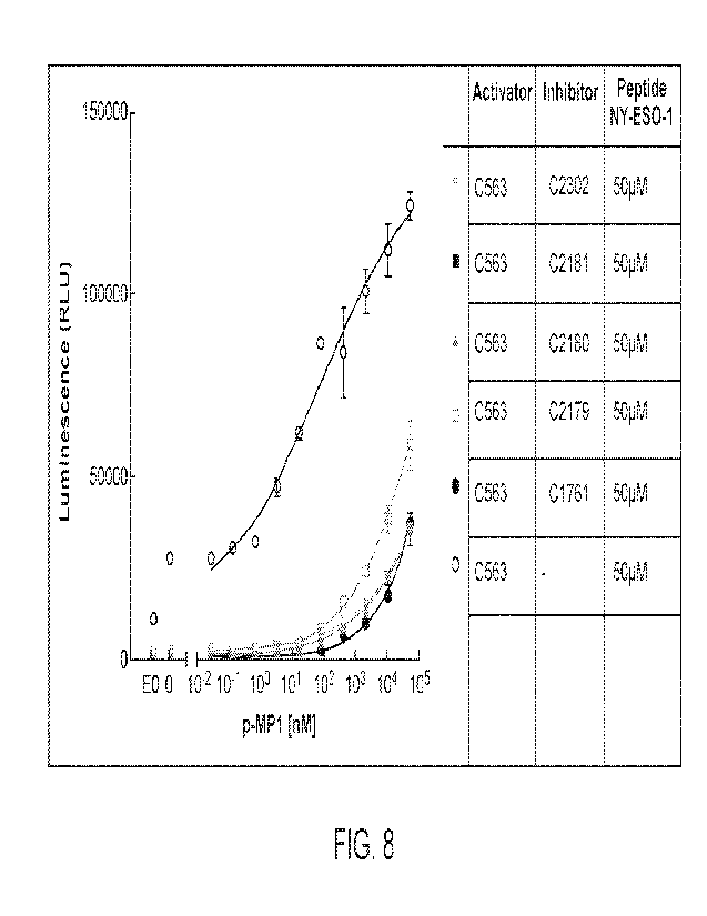
[0012] FIG 8 shows a chart of luminescence in an NEAT-based
reporter assay (RLU) at
varying concentrations of MAGE-A3 peptide 1 (MP1, nM) for the indicated
constructs in the
presence of 50 iuM NY-ESO-1 peptide.
[0013] FIG. 9 shows a chart of luminescence in an NEAT-based
reporter assay (RLU) at
varying concentrations of MAGE-A3 peptide 1 (lVfP1, nM) for the indicated
constructs in the
presence of 5 tiM NY-ESO-1 peptide.
[0014] FIG. 10 shows a chart of luminescence in an NFAT-based
reporter assay (RLU) at
varying concentrations of 1VIAGE-A3 peptide 1 (MN, nM) for the indicated
constructs in the
presence of 50 ILIM NY-ESO-1 peptide.
[0015] FIG. 11 shows a chart of luminescence in an NEAT-based report assay
(RLU) at varying
concentrations of MAGE-A3 peptide 1 (MP 1, nM) for the indicated constructs in
the presence of
50 04 NY-ESO-1 peptide.
[0016] FIG. 12A is a plot showing the effect of LIR-1 hinge on the ability of
an EILA-A*02 scFv
inhibitory receptor to block activation of Jurkat cells by a KRAS TCR. H:
hinge, T: transmembrane
domain, ICD. intracellular domain, s. short. LIR-1 constructs are described in
more detail in FIG.
I 2B. Humanized PA2. I and humanized BB7.2 with shorter LIR-1 hinge block
similarly to
original, longer hinge.
[0017] FIG. 12B is a plot and a table showing EC50 shift (+/- HLA-A*02 target
cells) for Jurkat
cells expressing a KRAS TCR activator and the HLA-A*02 scFv LIR-1 inhibitory
receptor shown
in the table at bottom.
[0018] FIG. 13A is a plot showing the effect of LIR-1 hinge on the ability of
an FILA-A*02
inhibitory receptor to block activation of Jurkat cells by a KRAS TCR. H:
hinge, TM:
transmembrane domain, ICD: intracellular domain, s: short, tr: truncated. LIR-
1 constructs are
described in more detail in FIG. 13B. Mouse PA2.1 with slightly longer hinges
function similarly
to original LIR-1 hinge in T2-Jurkat assay.
[0019] FIG. 13B is a plot and a pair of tables showing EC50 shift (+/- HLA-
A*02 target cells) for
Jurkat cells expressing a KRAS TCR activator and the HLA-A*02 scFv LIR-1
inhibitory receptors
shown in the table at bottom, with hinge lengths shown in the table at left.
[0020] FIG. 14A is a diagram showing a schematic of T2-Jurkat experiments to
evaluate block_er
constructs.
9
CA 03161112 2022- 6-7
WO 2021/119489
PCT/US2020/064607
[0021] FIG 14B is a plot, table and diagram showing the effect of various NY-
ES0-1 scEv LBD
blocker modules (PD-1, C'TLA-4, LIR-1) on EC50 of MAGE-A3 CAR activator (MP1-
LBD 1-
CAR), measured by MAGE peptide titration of cells loaded with a fixed (50 p.M)
NY-ES0-1
blocker peptide concentration. In each of FIGS. 14B-14F, NFAT-luciferase
signal of Jurkat cells
transfected with either activator CAR alone or in combination with each
blocker receptor after 6
hours of co-culture with activator and blocker peptide-loaded T2 cells was
assayed. The baseline
(Jurkat only) varies with different activator alone constructs and can be
especially high with CARs;
in most cases expression of the blocker receptor absent its ligand suppresses
the baseline. Activator
peptide concentrations range from 0 then 10-6 to 102 [tM and luminescence
measurements ranged
from 0 to 80000 RLU.
[0022] FIG. 14C is a plot, table and diagram showing the effect of LIR-I
blocker receptor with
various scEv LBDs (ESO, MP I LBD 1, MP I LBD 2, HPV E6 LBD 1, HPV E6 LBD 2,
HPV E7)
on EC50 of MAGE-A3 CAR activator (MPI -CAR) when loaded with corresponding
blocker
peptide at fixed (50 [iM) peptide concentration, as in FIG. 14B. RLU =
relative light units; error
bars indicate SD (n-2). Activator peptide concentrations range from 0 then
10-3 to 102 viM and
luminescence measurements ranged from 0 to I 00000 RLU.
[0023] FIG. 14D is a plot, table and diagram showing the effect of LIR-1
blocker receptor with
NY-ESO-1 scEv LBD on EC50 of different MAGE-A3 CAR activators (MPI -LBD 1-CAR
or
MP2-CAR) when loaded with 50uMNY-ES0-1 blocker peptide. Activator peptide
concentrations
range from 0 then 10-4 to 102 i.tM and luminescence measurements ranged from 0
to 80000 RLU.
[0024] FIG. 14E is a plot, table and diagram showing the effect of LIR-I
blocker receptor with
NY-ESO-1 scFv LBD on EC50 of different TCR activators (MPI -TCR, MP2-TCR, HPV
E6-
TCR) when loaded with 50uMNY-ES0-1 blocker peptide. Activator peptide
concentrations range
from 0 then 10-4 to 102 i.tM and normalized luminescence measurements ranged
from 0 to 150
RLU.
[0025] FIG. 14F is a plot, table and diagram showing the effect of LIR-1
blocker receptor with
NY-ES0-1 TCR LBDs on EC50 of MAGE-A3 CAR and TCR activators (MP1-LBD 1-CAR,
MP1-TCR) when loaded with 50uM NY-ES0-1 blocker peptide. RLU = relative light
units; error
bars indicate SD (n=2). Activator peptide concentrations range from 0 then
10-7 to 101 and
normalized luminescence measurements ranged from 0 to 150 RLU.
CA 03161112 2022- 6-7
WO 2021/119489
PCT/US2020/064607
[0026] FIG. 15 is a plot showing the effect of blocker peptide loading (50tiM
each of NY-ESO-
1, MAGE-A3, HPV E6, and HPV E7) on activating MAGE-A3 CAR. MP2-CAR [0 !LEM],
EC50
= 44 nM; MP2-CAR [50 M HPVp2], EC50 = 495 n1\4. RLU = relative light units;
error bars
indicate SD (n = 2).
[0027] FIG. 16A is a series of plots showing NFAT-luciferase signal of Jurkat
cells transfected
with either activator MAGE-A3 CAR alone or in combination with various amounts
of NY-ESO-
1 scFv LBD blocker (DNA ratios of activator and blocker receptors components
shown on left as
A:B, activator receptor: blocker receptor after 6 h of co-culture with
activator and blocker peptide-
loaded T2 cells_ T2 cells were loaded with titrated amounts of activator MAGE-
A3 peptide and a
fixed amount of blocker NY-ESO-1 peptide concentration. Activator and/or
blocker peptide
concentrations range from 0 then 10-6 to 102 [iM and luminescence measurements
ranged from 0
to 200000 RLU.
[0028] FIG. 16B is a series of plots showing NFAT-luciferase signal of Jurkat
cells transfected
with either activator MAGE-A3 CAR alone or in combination with various amounts
of NY-ESO-
1 scFv LBD blocker. T2 cells were loaded with titrated amounts of blocker NY-
ESO-1 peptide
and a fixed amount of activator MAGE-A3 peptide concentration above the Emax
concentration
(-0.1 mM). Activator and/or blocker peptide concentrations range from 10-5 to
102 p.1\4 and
luminescence measurements ranged from 0 to 200000 RLU.
[0029] FIG. 16C is a series of plots and two tables showing NFAT-luciferase
signal of Jurkat cells
transfected with either activator MAGE-A3 CAR alone or in combination with
various amounts
of NY-ESO-1 scFv LBD blocker. The x-value blocker NY-ESO-1 peptide
concentrations from
FIG. 16B were normalized to the constant activator MAGE peptide concentrations
used for each
curve and plotted on the x-axis. The ratio of blocker peptide to activator
peptide required for 50 %
blocking (IC50) are indicated for each curve. For all DNA ratios, the B: A
peptide ratio required is
less than 1 indicating that, for this pair of activator CAR and blocker,
similar (or fewer) blocker
pMHC antigens are required on target cells to block activator pMEIC antigens.
Activator and/or
blocker peptide concentrations range from 104 to 104 [tM and luminescence
measurements ranged
from 0 to 200000 RLU.
[0030] FIG. 16D is a table and a plot showing that blocking CD19-CAR activator
is possible with
pl\TEIC blockers at blocker pMEIC antigen densities similar to those required
to activate p1\41-1C
CARs. NFAT-luciferase signal of Jurkat cells transfected with either activator
CD19 CAR alone
11
CA 03161112 2022- 6-7
WO 2021/119489
PCT/US2020/064607
or in combination with various amounts of NY-ESO-1 blocker (DNA ratios shown)
after 6 h of
co-culture with blocker peptide-loaded T2 cells which express endogenous
levels of CD19 antigen.
The ICSO is estimated from the inhibition curves to range from 0.1-1.0 naM,
corresponding to
¨1,500 - 3,500 plVIHCs/cell. RLU = relative light units; error bars indicate
SD (n = 2).
[0031] FIG. 17A is a plot showing the effect of NY-ES0-1-LIR-1 blocker on EC50
of activating
MAGE-A3 CAR (MP1-CAR) when loaded with various concentrations of NY-ESO-1
blocker
peptide. The EC50 shifts are greater as the concentration of blocker peptide
(NY-ESO-1) increases.
The shift in the presence of a negative-control HPV peptide (binds HLA-A*02
but not NY-ESO-
1 blocker scFv) is routinely seen and believed to be caused by competition of
the control peptide
for binding sites on the T2 HLA-A*02 molecules, reducing the number of
activator targets.
Activator concentrations range from 0 then 10-4to 102 04 and luminescence
measurements ranged
from 0 to 140000 RLU.
[0032] FIG. 17B is a plot showing the effect of modified LIR-1 blocker
receptors containing no
ICD or a mutated ICD with NY-ESO-1 scFv LBD on EC50 of MAGE-A3 CAR activator
(MP2-
CAR) when loaded with 10 ittM of NY-ES0-1 blocker peptide. Activator
concentrations range
from 0 then 10-4 to 102 jaM and luminescence measurements ranged from 0 to
140000 RLU.
[0033] FIGS. 17C-17E are a series of plots showing the effect of various NY-
ES0-1 scFv LBD
blocker receptors (CTLA-4 (FIG. 17C), PD-1 (FIG. 17D) and LIR-1 (FIG. 17E)) on
EC50 of
MAGE-A3 CAR activator (MP1-LBD 1-CAR) when blockers are stimulated or
unstimulated.
NFAT-luciferase signal of Jurkat cells transfected with either activator CAR
alone or in
combination with each blocker after 6 h of co-culture with peptide-loaded T2
cells. T2 cells were
loaded with titrated amounts of activating MAGE peptide and tested with and
without loading
additional constant amount (50 uM) of NY-ES0-1 blocker peptide. RLU = relative
light units;
error bars indicate SD (n = 2). Activator concentrations range from 0 then
10-6 to 102 uM and
luminescence measurements ranged from 0 to 100000 RLU.
[0034] FIG. 18A is a diagram and a pair of plots showing that Jurkat cells
transfected with either
HPV E7-CAR or HPV E7-CAR & A2-LIR-1 co-cultured with beads displaying various
ratios of
activator (HPV E7) and blocker (NY-ESO-1) antigen demonstrates blocking in cis
but not trans.
[0035] FIG. 18B is a plot showing that ElLA-A*02-LIR-1 blocker receptor blocks
CD19-CAR
activator at various activator to blocker ratios. Ratios ranged from 0 to 10
and luminescence (RLU)
ranged from 0 to 70000.
12
CA 03161112 2022- 6-7
WO 2021/119489
PCT/US2020/064607
[0036] FIG. 18C is a plot showing surface expression of titrated HI-A-A*02
(A2) LIR-1 blocker
receptor.
[0037] FIG. 18D is a plot showing that an scFy against HLA-A*02 can also serve
as an activator
when fused to activator CAR. 12 cells expressing endogenous EILA-A*02 serve as
the target. RLU
= relative light units; error bars indicate SD (n=2).
[0038] FIG. 19A is a plot showing the effect of LIR-1 blocker receptor with NY-
ESO-1 scFv LBD
on EC50 of different TCR activators (M1P1-TCR, MP2-TCR, HPV E6-TCR) when
loaded with
NY-ESO-1 blocker peptide. For FIG. 14E each set was normalized to the Emax of
the curve
showing response of the activator only. Activator concentrations range from 0
then 10-4to 102 [iM
and luminescence measurements ranged from 0 to 140000 RLU.
[0039] FIG. 19B is a plot showing the effect of LIR-1 blocker receptor with NY-
ESO-1 TCR
LBDs on EC50 of MAGE-A3 CAR and TCR activators (MP1-LBD 1-CAR, MP1-TCR). Each
set
was normalized to the Emax of the curve showing response of the activator
only. RLU = relative
light units; error bars indicate SD (n = 2). Activator concentrations range
from 0 then 10-7 to 101
and luminescence measurements ranged from 0 to 140000 RLU.
[0040] FIG. 20A is a plot showing primary T cells (donor 1) transduced with
HPV E7-TCR
activator and ESO-LIR-1 blocker shifts EC50 ---25x in primary T cell killing
assay (HPV E7 TCR
EC50 = 0.044 nIVI; HPV E7 TCR + ESO-LIR-1, EC50 = 1.1 nM). The assay was
performed using
peptide-loaded MCF7 target cells at a 3:1 E:T. Luciferase measurement
represents live target cells
at 48 hours.
[0041] FIG. 20B is a plot showing that HLA-A*02-LIR-1 blocks NY-ESO-1 CAR
activator at
various activator:blocker DNA ratios in Jurkat cells using NY-ESO-1 peptide-
loaded T2 target
cells. RLU = relative light units; error bars indicate SD (n=2).
[0042] FIG. 21A is a series of images and plots showing that primary T cells
(donor 1) transduced
with CD19 CAR activator and I-ILA-A*02 blocker distinguish "tumor" cells from
"normal" cells
in in vitro cytotoxicity assay and demonstrate selective killing of "tumor"
cells in mixed target cell
assay at 3:1 E:T. Images shown were captured at 72 hours. Shown are
Untransduced, CD19-CAR
T cells, and CD19-CAR T + A2-LIR-1. Measurements were taken between 0 and 150
hours and
normalized fluorescent protein intensity (GFP or RFP) ranged from 0 to 10.
[0043] FIG. 21B is a series of plots showing that primary T cells
(donor 1) selectively kill
tumor cells similarly at various tumor to "normal" cell ratios at 3:1 E:T
ratios in Incucyte imaging
13
CA 03161112 2022- 6-7
WO 2021/119489
PCT/US2020/064607
assay. RLU values were normalized against target cell mixtures grown in the
absence of primary
T cells. Shown are Untransduced, CD19-CAR T cells, and CD19-CAR T + A2-LIR-1.
Measurements were taken between 0 and 150 hours and normalized fluorescent
protein intensity
(GFP or REP) ranged from, on the top row, from left to right: 0 to 3 x10⁷, 0
to 2 x10⁷, 0 to 1.6 x 10⁷,
0 to 7x10⁶, 0 to 2><1U6; bottom row, from left to right: 0 to 710, 0 to 2x10⁵,
0 to 4x10⁵, 0 to
6x10⁵, 0 to 7x10⁵.
[0044] FIG. 21C is a series of plots showing that primary T cells (donor 1)
selectively kill tumor
cells similarly at various tumor to "normal" cell ratios at 3:1 E:T ratios in
quantitative target cell
lysis and IFNy secretion. Shown are Untransduced, CD19-CAR T cells, and CD19-
CAR T + A2-
LIR-1.
[0045] FIG. 22A is a pair of plots showing that Jurkat cells transfected with
MSLN LBD1-CAR
or MSLN LBD1-CAR & A2-LIR-1 co-cultured with K562 cells expressing either MSLN
or
MSLN & HLA-A*02 shows blocking of activation by a high-density antigen with A2-
LIR-1
blocker only in the presence of HLA-A*02.
[0046] FIG. 22B is a pair of plots showing that killing of endogenous MSLN+
HeLa cells by
MLSN LBD1-CAR T cells is blocked in the presence of LILA-A*02 with the A2-LIR-
1 blocker.
[0047] FIG. 22C is a pair of plots showing killing of endogenous MSLN+ HeLa
cells by MLSN
LBD2-CAR T cells. The effect of A2-LIR-1 blocker on T cell killing is in part
controlled by the
activator LBD.
[0048] FIG. 23 is a plot showing that LIR-1 blocker receptors have little
effect on killing efficacy
of activator in absence of blocker antigen. In the absence of NY-ESO-1 blocker
antigen, primary
T cells (donor 1) transduced with HPV E7-TCR activator and ESO-LIR-1 blocker
display similar
killing efficacy to primary T cells transduced with only the I-IPV E7-TCR
activator. Luciferase
measurement represents live target cells. RLU = relative light units; error
bars indicate SD (n =
2).
[0049] FIG. 24A is a pair of plots showing that A2-LIR-1 blocks Jurkat
activation in A2+ Raji
cells but not WT Raji cells. Histograms show Raji WT "tumor" cells and Raji
A2+ "normal" cells
have identical CD19 surface expression while HLA-A*02 is expressed only in
Raji A2 "normal"
cells.
100501 FIG. 24B is a plot showing that A2-LIR-1 blocks Jurkat activation in
A2+ Raji cells but
not WT Raji cells. Jurkat cells transfected with either CD19 or CD19 + A2-LIR-
1 were co-cultured
14
CA 03161112 2022- 6-7
WO 2021/119489
PCT/US2020/064607
with either WT (A2-) Raji cells or A2+ Roll cells at various cell ratios. RLU
= relative light units;
error bars indicate SD (n = 2).
[0051] FIGS. 25A-25B are each a series of images showing the reversibility of
blockade by LIR-
1 inhibitory receptors. Primary T cells (donor 2) transduced with CD19 CAR
activator and }ILA-
A*02 blocker demonstrate reversible blockade (FIG. 25A) and activation (FIG.
25B) after 3 rounds
of antigen exposure (AB¨A---AB and A¨AB¨A) in in vitro cytotoxicity assay at
3:1 E: T. Primary
T cell cytotoxicity assay was reproduced with three FILA-A*02-negative donors.
Images shown
were captured at 72 hours.
[0052] FIGS. 25C-25D are each a pair of plots showing quantification of target
cell lysis (FIG_
25C) and IFNy (FIG. 25D) in response to repeated exposure to multiple rounds
of normal and
target cells, 3:1 E:T (T cells from donor 2). Shown conditions are
Untransduced, CD19-CAR T
cells, and CD19-CAR T + A2-LIR-1. Error bars indicate SEM (n=2). *p<0.05,
**p<0.01,
***p<0.001, ****p<0.0001, determined using a two-way ANOVA followed by Tukey's
multiple-
comparisons test. In this experiment, IFNy response diminished over time,
while cytotoxicity
remained robust.
[0053] FIGS. 26A-26B are each a pair of plots showing cytotoxic T cell killing
and secretion of
IFNy in co-culture with a separate donor (donor 3). Cytotoxic CD19 CAR
activator and HLA-
A*02 blocker transduced T cells demonstrate reversible blocking after multiple
rounds of antigen
exposure in cytotoxic assays and IFNy at 9:1 E: T. We noted that this donor's
T cells survival and
activity tailed off over time in culture. Shown conditions are Untransduced,
CD19-CAR T cells,
and CD19-CAR T + A2-LIR-1. Cytotoxicity (FIG. 26A) and IFNy (FIG. 26B) results
correspond
to FIGS. 25C-25D. Error bars indicate SEM (n = 2). *p <0.05, **p < 0.01,
***p <0.001, ****p
<0.0001, determined using a two-way ANOVA followed by Tukey's multiple-
comparisons test.
[0054] FIG. 27A is a plot showing primary I cells transduced with CD19 CAR
activator and HLA-
A*02 blocker demonstrates ¨20-fold expansion with CD3/28 stimulation over 10
days.
[0055] FIG. 27B is a diagram showing an experiment to show that CAR-T cells
expressing a LIR-
1 blocker receptor selectively kill tumors in a xenograft model. EILA-A*02
NSC) mice were
administered either "tumor cells" (A2-negative Raji cells) or "normal cells"
(A2-positive Raji
cells) subcutaneously and primary T cells (human, FILA-A*02-negative donor 4)
were injected
into the tail vein when Raji xenografts averaged ¨70 mm3.
CA 03161112 2022- 6-7
WO 2021/119489
PCT/US2020/064607
[0056] FIGS. 27C-27E are each a pair of plots that show readouts by caliper
measurement (FIG
27C), human T cell counts in peripheral blood by flow cytometry (FIG. 27D),
and survival (FIG.
27E). Error bars in C-D indicate SEM (n=7). *p<0.05, **p<0.01, ***p<0.001,
****p<0.0001,
determined using a two-way ANOVA followed by Tukey's multiple-comparisons
test.
[0057] FIG. 28A is a series of plots showing flow cytometry analysis of
primary T cell post-
enrichment and expansion prepared for mouse tail vein injection. Tumor volume
was measured at
the number of days from T cell injection, starting 10 days prior to T cell
injection (-10) to 40 days
following T cell injection (40). Tumor volumes ranged from 0 to 2500 mm3.
100581 FIG. 28B is a series of plots showing tumor measurement by caliper
plotted for individual
mice in each group.
[0059] FIG. 28C is a series of plots showing correlation of huCD3+ T cells in
mouse blood to
tumor growth. Graph of huCD3+ T cells compared to tumor volume 10 days and 17
days after T
cell injection.
[0060] FIG. 28D is a pair of plots showing huCD4+ and huCD8+ T cell counts in
peripheral blood
by flow cytometry. Open circles, mice grafted with "normal" cells, closed
circle, mice grafted with
tumor cells. Samples with fewer than 100 cells were excluded from the
analysis. Error bars indicate
SEM (n = 7 in all groups except n = 6 in CD19+/A2- Raji group treated with
CD19-CAR + A2-
LIR-1 T cells).
[0061] FIG. 29A is a series of images showing histological analysis of T cell
infiltration in tumors.
Representative images of tumor samples collected at study termination,
sectioned and stained for
huCD3 are shown.
[0062] FIG. 29B is a plot showing quantification of T cell infiltration using
ImageJ. T cell
infiltration was significantly higher for T cells with CD19-CAR or CD19-CAR +
A2-LIR-1 in
CD19+/A2- tumors compared to untransduced cells. However, in CD19+/A2+ tumors,
CD19-
CAR + A2-LIR-1 T cells were not significantly different compared to
untransduced cells. There
was also a significant drop in infiltration of CD19-CAR + A2-LIR-1 T cells
between CD19+/A2-
and CD19+/A2+ tumors. Qualitatively, CD19-CAR + A2-LIR-1 T cells were less
prevalent in
CD19+/A2+ tumors compared to CD19-CAR only T cells; however, this difference
was not
statistically significant. Saline samples were similarly quantified to show
background staining
levels. Groups of data were analyzed using an ordinary one-way ANOVA, while
individual pairs
16
CA 03161112 2022- 6- 7
WO 2021/119489
PCT/US2020/064607
between "tumor" and "normal" were analyzed using an unpaired t test. ns = not
significant, *p <
0.05, **p < 0.01.
DETAILED DESCRIPTION
[0063] The present disclosure describes receptors having one or
more domains from
Leukocyte immunoglobulin-like receptor subfamily B member 1 (LILRB1, sometimes
referred to
as LIR1 or LIR-1). Numerous receptors, engineered cells, and uses thereof are
contemplated
herein. The inventors have found that chimeric receptors comprising an antigen-
binding domain
and one or more LILRB1 domains, including the LILRB1 intracellular domain, can
inhibit immune
cell signaling even in the presence of activatory chimeric antigen receptors
(CARs) or T cell
receptors (TCRs).
[0064] The term "chimeric antigen receptors" or "CARs" as used
herein, may refer to artificial
T-cell receptors, chimeric T-cell receptors, or chimeric immunoreceptors, for
example, and
encompass engineered receptors that graft an artificial specificity onto a
particular immune
effector cell, such as a helper T cell (CD4+), cy totoxic T cell (CD8+) or NK
cell. CARs may be
employed to impart the specificity of a monoclonal antibody onto a T cell,
thereby allowing a large
number of specific T cells to be generated, for example, for use in adoptive
cell therapy. In specific
embodiments, CARs direct specificity of the cell to a tumor associated
antigen. In some
embodiments, CARs comprise an intracellular signaling domain, a transmembrane
domain, and
an extracellular domain comprising an antigen-binding region. In some
embodiments, CARs
comprise fusions of single-chain variable fragments (scFvs) or scFabs derived
from monoclonal
antibodies, fused to a transmembrane domain and intracellular signaling
domain(s). The fusion
may also comprise a hinge. Either heavy-light (H-L) and light-heavy (L-H)
scEvs may be used.
The specificity of CAR designs may be derived from ligands of receptors (e.g.,
peptides).
Depending on the type of intracellular domain, a CAR can be an activatory
receptor or an
inhibitory receptor. In some embodiments, for example when the CAR is an
activatory receptor,
the CAR comprises domains for additional co-stimulatory signaling, such as
CD3, FcR, CD27,
CD28, CD137, DAP 1 0, and/or 0X40. In some embodiments, molecules can be co-
expressed with
the CAR, including co-stimulatory molecules, reporter genes for imaging (e.g.,
for positron
emission tomography), gene products that conditionally ablate the T cells upon
addition of a pro-
drug, homing receptors, cytokines, and cytokine receptors. As used herein,
characteristics
17
CA 03161112 2022- 6- 7
WO 2021/119489
PCT/US2020/064607
attributed to a chimeric antigen receptor may be understood to refer to the
receptor itself or to a
host cell comprising the receptor.
[0065] As used herein, a "TCR", sometimes also called a "TCR
complex" or "TCR/CD3
complex" refers to a protein complex comprising a TCR alpha chain, a TCR beta
chain, and one
or more of the invariant CD3 chains (zeta, gamma, delta and epsilon),
sometimes referred to as
subunits. The TCR alpha and beta chains can be disulfide-linked to function as
a heterodimer to
bind to peptide-MHC complexes. Once the TCR alpha/beta heterodimer engages
peptide-MHC,
conformational changes in the TCR complex in the associated invariant CD3
subunits are induced,
which leads to their phosphorylation and association with downstream proteins,
thereby
transducing a primary stimulatory signal. In an exemplary TCR complex, the TCR
alpha and TCR
beta polypeptides form a heterodimer, CD3 epsilon and CD3 delta form a
heterodimer, CD3
epsilon and CD3 gamma for a heterodimer, and two CD3 zeta form a homodimer.
[0066] The term "stimulation" refers to a primary response induced
by binding of a stimulatory
domain or stimulatory molecule (e.g., a TCR/CD3 complex) with its cognate
ligand thereby
mediating a signal transduction event, such as, but not limited to, signal
transduction via the
TCR/CD3 complex. Stimulation can mediate altered expression of certain
molecules, and/or
reorganization of cytoskeletal structures, and the like.
[0067] The term "stimulatory molecule" or "stimulatory domain"
refers to a molecule or
portion thereof that, when natively expressed by a T-cell, provides the
primary cytoplasmic
signaling sequence(s) that regulate activation of the TCR complex in a
stimulatory way for at least
some aspect of the T-cell signaling pathway. TCR alpha and/or TCR beta chains
of wild type TCR
complexes do not contain stimulatory domains and require association with CD3
subunits such as
CD3 zeta to initiate signaling. In one aspect, the primary stimulatory signal
is initiated by, for
instance, binding of a TCR/CD3 complex with an a major histocompatibility
complex (MHC)
bound to peptide, and which leads to mediation of a T-cell response,
including, but not limited to,
proliferation, activation, differentiation, and the like. One or more
stimulatory domains, as
described herein, can be fused to the intracellular portion of any one or more
subunits of the TCR
complex, including TCR alpha, TCR beta, CD3 delta, CD3 gamma and CD3 epsilon.
[0068] As used herein, a "domain capable of providing a stimulatory
signal" refers to any
domain that, either directly or indirectly, can provide a stimulatory signal
that enhances or
increases the effectiveness of signaling mediated by the TCR complex to
enhance at least some
18
CA 03161112 2022- 6-7
WO 2021/119489
PCT/US2020/064607
aspect of T-cell signaling. The domain capable of providing a stimulatory
signal can provide this
signal directly, for example with the domain capable of providing the
stimulatory signal is a
primary stimulatory domain or co-stimulatory domain. Alternatively, or in
addition, the domain
capable of providing the stimulatory signal can act indirectly. For example,
the domain can be a
scaffold that recruits stimulatory proteins to the TCR, or provide an
enzymatic activity, such as
kinase activity, that acts through downstream targets to provide a stimulatory
signal.
[0069] As used herein, a "domain capable of providing an inhibitory
signal" refers to any
domain that, either directly or indirectly, can provide an inhibitory signal
that inhibits or decreases
the effectiveness signaling mediated by the TCR complex. The domain capable of
providing an
inhibitory signal can reduce, or block, totally or partially, at least some
aspect of T-cell signaling
or function. The domain capable of providing an inhibitory signal can provide
this signal directly,
for example with the domain capable of providing the inhibitory signal
provides a primary
inhibitory signal. Alternatively, or in addition, the domain capable of
providing the stimulatory
signal can act indirectly. For example, the domain can recruit additional
inhibitory proteins to the
TCR, or can provide an enzymatic activity that acts through downstream targets
to provide an
inhibitory signal.
[0070] Ranges: throughout this disclosure, various aspects of the
invention can be presented
in a range format. It should be understood that the description in range
format is merely for
convenience and brevity and should not be construed as an inflexible
limitation on the scope of
the invention. Accordingly, the description of a range should be considered to
have specifically
disclosed all the possible subranges as well as individual numerical values
within that range. For
example, description of a range such as from 1 to 6 should be considered to
have specifically
disclosed subranges such as from 1 to 3, from 1 to 4, from 1 to 5, from 2 to
4, from 2 to 6, from 3
to 6 etc., as well as individual numbers within that range, for example, 1, 2,
2.7, 3, 4, 5, 5.3, and
6. As another example, a range such as 95-99% identity, includes something
with 95%, 96%, 97%,
98% or 99% identity, and includes subranges such as 96-99%, 96-98%, 96-97%, 97-
99%, 97-98%
and 98-99% identity. This applies regardless of the breadth of the range.
[0071] In general, "sequence identity- or "sequence homology-
refers to an exact nucleotide-
to-nucleotide or amino acid-to-amino acid correspondence of two
polynucleotides or polypeptide
sequences, respectively. Typically, techniques for determining sequence
identity include
determining the nucleotide sequence of a polynucleotide and/or determining the
amino acid
19
CA 03161112 2022- 6-7
WO 2021/119489
PCT/US2020/064607
sequence encoded thereby and comparing these sequences to a second nucleotide
or amino acid
sequence. Two or more sequences (polynucleotide or amino acid) can be compared
by determining
their "percent identity." The percent identity of two sequences, whether
nucleic acid or amino acid
sequences, is the number of exact matches between two aligned sequences
divided by the length
of the shorter sequences and multiplied by 100. Percent identity may also be
determined, for
example, by comparing sequence information using the advanced BLAST computer
program,
including version 2.2.9, available from the National Institutes of Health. The
BLAST program is
based on the alignment method of Karlin and Altschul, Proc. Natl. Acad. Sci.
USA 87:2264-2268
(1990) and as discussed in Altschul, et al., I Mol. Biol. 215.403-410 (1990);
Karlin And Altschul,
Proc. Natl. Acad. Sci. USA 90:5873-5877 (1993); and Altschul et al., Nucleic
Acids Res. 25:3389-
3402 (1997). Briefly, the BLAST program defines identity as the number of
identical aligned
symbols (generally nucleotides or amino acids), divided by the total number of
symbols in the
shorter of the two sequences. The program may be used to determine percent
identity over the
entire length of the proteins being compared. Default parameters are provided
to optimize searches
with short query sequences in, for example, with the blastp program. Ranges of
desired degrees of
sequence identity are approximately 80% to 100% and integer values
therebetween. Typically, the
percent identities between a disclosed sequence and a claimed sequence are at
least 80%, at least
85%, at least 90%, at least 95%, or at least 98%.
[0072] As used herein, a "subsequence" refers to a length of
contiguous amino acids or
nucleotides that form a part of a sequence described herein. A subsequence may
be identical to a
part of a full length sequence when aligned to the full length sequence, or
less than 100% identical
to the part of the full length sequence to which it aligns (e.g., 90%
identical to 50% of the full
sequence, or the like).
[0073] The term "exogenous" is used herein to refer to any
molecule, including nucleic acids,
protein or peptides, small molecular compounds, and the like that originate
from outside the
organism. In contrast, the term "endogenous" refers to any molecule that
originates from inside
the organism (i.e., naturally produced by the organism).
[0074] A polynucleotide is "operably linked" to another
polynucleotide when it is placed into
a functional relationship with the other polynucleotide. For example, a
promoter or enhancer is
operably linked to a coding sequence if it affects the transcription of the
sequence. A peptide is
CA 03161112 2022- 6-7
WO 2021/119489
PCT/US2020/064607
"operably linked" to another peptide when the polynucleotides encoding them
are operably linked,
preferably they are in the same open reading frame.
[0075] A "promoter" is a sequence of DNA needed to turn a gene on
or off Promoters are
located immediately upstream and/or overlapping the transcription start site,
and are usually
between about one hundred to several hundred base pairs in length.
[0076] All publications and patents mentioned herein are hereby
incorporated by reference in
their entirety as if each individual publication or patent was specifically
and individually indicated
to be incorporated by reference. In case of conflict, the present application,
including any
definitions herein, will control. However, mention of any reference, article,
publication, patent,
patent publication, and patent application cited herein is not, and should not
be taken as an
acknowledgment, or any form of suggestion, that they constitute valid prior
art or form part of the
common general knowledge in any country in the world.
[0077] In the present description, any concentration range,
percentage range, ratio range, or
integer range is to be understood to include the value of any integer within
the recited range and,
when appropriate, fractions thereof (such as one tenth and one hundredth of an
integer), unless
otherwise indicated. The term "about", when immediately preceding a number or
numeral, means
that the number or numeral ranges plus or minus 10%.
[0078] All publications, patents, and patent applications mentioned
in this specification are
herein incorporated by reference to the same extent as if each individual
publication, patent, or
patent application was specifically and individually indicated to be
incorporated by reference.
Leukocyte immunoglobulin-like receptor subfamily B member 1 (LILRB1)
[0079] The present disclosure describes receptors having one or
more domains from
Leukocyte immunoglobulin-like receptor subfamily B member 1 (LILRB1, or LIR1).
Numerous
receptors, engineered cells, and uses thereof are contemplated herein.
[0080] Leukocyte immunoglobulin-like receptor subfamily B member 1
(LILRB1), also
known as Leukocyte immunoglobulin-like receptor Bl, as well as ILT2, LIR1,
MIIR7, PIRB,
CD85J, ILT-2 LIR-1, 1VIR-7 and PIR-B, is a member of the leukocyte
immunoglobulin-like
receptor (LIR) family. The LILRB1 protein belongs to the subfamily B class of
LIR receptors.
These receptors contain two to four extracellular immunoglobulin domains, a
transmembrane
domain, and two to four cytoplasmic immunoreceptor tyrosine-based inhibitory
motifs (ITIMs).
21
CA 03161112 2022-6-7
WO 2021/119489
PCT/US2020/064607
The LILRB1 receptor is expressed on immune cells, where it binds to 1W-1C
class I molecules on
antigen-presenting cells and transduces a negative signal that inhibits
stimulation of an immune
response. LILRB1 is thought to regulate inflammatory responses, as well as
cytotoxicity, and to
play a role in limiting auto-reactivity. Multiple transcript variants encoding
different isoforms of
LILRB1 exist, all of which are contemplated as within the scope of the instant
disclosure.
[0081] In some embodiments of the receptors having one or domains
of LILRB1, the one or
more domains of LILRB1 comprise an amino acid sequence that is at least 80%,
at least 90%, at
least 95%, at least 96%, at least 97%, at least 98%, at least 99% or is
identical to a sequence or
subsequence of SEQ ID NO: 1 In some embodiments, the one or more domains of
LILRB1
comprise an amino acid sequence that is identical to a sequence or subsequence
of SEQ ID NO: 1.
In some embodiments, the one or more domains of LILRB1 consist of an amino
acid sequence
that is at least 80%, at least 90%, at least 95%, at least 96%, at least 97%,
at least 98%, at least
99% or is identical to a sequence or subsequence of SEQ ID NO: 1. In some
embodiments, the one
or more domains of LILRB1 consist of an amino acid sequence that is identical
to a sequence or
subsequence of SEQ ID NO. 1.
[0082] In some embodiments of the receptors having one or domains
of LILRB1, the one or
more domains of LILRB1 are encoded by a polynucleotide sequence that is at
least 80%, at least
90%, at least 95%, at least 96%, at least 97%, at least 98%, at least 99% or
is identical to a sequence
or subsequence of SEQ ID NO: 34.
[0083] In some embodiments of the receptors having one or domains
of LILRB1, the one or
more domains of LILRB1 are encoded by a polynucleotide sequence that is
identical to a sequence
or subsequence of SEQ ID NO: 34.
Receptors
[0084] In various embodiments, a chimeric antigen receptor is
provided, comprising a
polypeptide, wherein the polypeptide comprises one or more of: an LILRB1 hinge
domain or
functional fragment or variant thereof; an LILRB1 transmembrane domain or a
functional variant
thereof; and an LILRB1 intracellular domain or an intracellular domain
comprising at least one,
or at least two immunoreceptor tyrosine-based inhibitory motifs (ITIMs),
wherein each ITIM is
independently selected from NLYAAV (SEQ ID NO: 8), VTYAEV (SEQ ID NO: 9),
VTYAQL
(SEQ ID NO: 10), and SIYATL (SEQ ID NO: 11).
22
CA 03161112 2022- 6-7
WO 2021/119489
PCT/US2020/064607
Infracellular Domain
[0085] The disclosure provides chimeric antigen receptors, the
chimeric antigen receptors
comprising a polypeptide. In some embodiments, the polypeptide comprises an
intracellular
domain. In some embodiments, the intracellular domain is an LILRB1
intracellular domain or a
functional variant thereof.
[0086] As used herein, "intracellular domain" refers to the
cytoplasmic or intracellular domain
of a protein, such as a receptor, that interacts with the interior of the
cell, and carries out a cytosolic
function. As used herein, "cytosolic function" refers to a function of a
protein or protein complex
that is carried out in the cytosol of a cell. For example, intracellular
signal transduction cascades
are cytosolic functions.
[0087] As used herein an "immunoreceptor tyrosine-based inhibitory
motif' or "ITIM" refers
to a conserved sequence of amino acids with a consensus sequence of
S/IN/LxYxxIN/L (SEQ ID
NO: 124), or the like, that is found in the cytoplasmic tails of many
inhibitory receptors of the
immune system. After ITIM-possessing inhibitory receptors interact with their
ligand, the ITIM
motif is phosphorylated, allowing the inhibitory receptor to recruit other
enzymes, such as the
phosphotyrosine phosphatases SI-IF'-1 and SHP-2, or the inositol-phosphatase
called SHIP.
100881 In some embodiments, the polypeptide comprises an
intracellular domain comprising
at least one immunoreceptor tyrosine-based inhibitory motif (ITIM), at least
two ITIMs, at least 3
ITIMs, at least 4 ITIMs, at least 5 ITIMs or at least 6 ITIMs. In some
embodiments, the intracellular
domain has 1, 2, 3, 4, 5, or 6 ITIMs.
[0089] In some embodiments, the polypeptide comprises an
intracellular domain comprising
at least one ITIM selected from the group of ITIMs consisting of NLYAAV (SEQ
ID NO: 8),
VTYAEV (SEQ ID NO: 9), VTYAQL (SEQ ID NO: 10), and SIYATL (SEQ ID NO: 11).
[0090] In further particular embodiments, the polypeptide comprises
an intracellular domain
comprising at least two immunoreceptor tyrosine-based inhibitory motifs
(ITIMs), wherein each
ITIM is independently selected from NLYAAV (SEQ ID NO: 8), VTYAEV (SEQ ID NO:
9),
VTYAQL (SEQ ID NO: 10), and SIYATL (SEQ ID NO: 11).
[0091] In some embodiments, the intracellular domain comprises both
ITIMs NLYAAV (SEQ
ID NO: 8) and VTYAEV (SEQ ID NO: 9). In some embodiments, the intracellular
domain
comprises a sequence at least 95% identical to SEQ ID NO: 12. In some
embodiments, the
intracellular domain comprises or consists essentially of a sequence identical
to SEQ ID NO: 12.
23
CA 03161112 2022- 6-7
WO 2021/119489
PCT/US2020/064607
[0092] In some embodiments, the intracellular domain comprises both
ITIMs VTYAEV (SEQ
ID NO: 9) and VTYAQL (SEQ ID NO: 10). In some embodiments, the intracellular
domain
comprises a sequence at least 95% identical to SEQ ID NO: 13. In some
embodiments, the
intracellular domain comprises or consists essentially of a sequence identical
to SEQ ID NO: 13.
[0093] In some embodiments, the intracellular domain comprises both
ITIMs VTYAQL (SEQ
ID NO: 10) and SIYATL (SEQ ID NO: 11). In some embodiments, the intracellular
domain
comprises a sequence at least 95% identical to SEQ ID NO: 14. In some
embodiments, the
intracellular domain comprises or consists essentially of a sequence identical
to SEQ ID NO: 14.
[0094] In some embodiments, the intracellular domain comprises the
ITIMs NLYAAV (SEQ
ID NO: 8), VTYAEV (SEQ ID NO: 9), and VTYAQL (SEQ ID NO: 10). In some
embodiments,
the intracellular domain comprises a sequence at least 95% identical to SEQ ID
NO: 15. In some
embodiments, the intracellular domain comprises or consists essentially of a
sequence identical to
SEQ ID NO: 15.
[0095] In some embodiments, the intracellular domain comprises the
ITIMs VTYAEV (SEQ
ID NO. 9), VTYAQL (SEQ ID NO. 10), and SIYATL (SEQ ID NO. 11). In some
embodiments,
the intracellular domain comprises a sequence at least 95% identical to SEQ ID
NO: 16. In some
embodiments, the intracellular domain comprises or consists essentially of a
sequence identical to
SEQ ID NO: 16.
[0096] In some embodiments, the intracellular domain comprises the
ITIMs NLYAAV (SEQ
ID NO: 8), VTYAEV (SEQ ID NO: 9), VTYAQL (SEQ ID NO: 10), and SIYATL (SEQ ID
NO:
11). In embodiments, the intracellular domain comprises a sequence at least
95% identical to SEQ
ID NO: 17. In some embodiments, the intracellular domain comprises or consists
essentially of a
sequence identical to SEQ ID NO: 17.
[0097] In some embodiments, the intracellular domain comprises a
sequence at least 95%
identical to the LILRB1 intracellular domain (SEQ ID NO: 7). In some
embodiments, the
intracellular domain comprises or consists essentially of a sequence identical
to the LILRB1
intracellular domain (SEQ ID NO: 7).
[0098] LILRB1 intracellular domains or functional variants thereof
of the disclosure can have
at least 1, at least 2, at least 4, at least 4, at least 5, at least 6, at
least 7, or at least 8 ITIMs. In some
embodiments, the LILRB1 intracellular domain or functional variant thereof has
2, 3, 4, 5, or 6
ITIMs.
24
CA 03161112 2022- 6-7
WO 2021/119489
PCT/US2020/064607
[0099] In particular embodiments, the polypeptide comprises an
intracellular domain
comprising two, three, four, five, or six immunoreceptor tyrosine-based
inhibitory motifs (ITIMs),
wherein each ITIM is independently selected from NLYAAV (SEQ ID NO: 8), VTYAEV
(SEQ
ID NO: 9), VTYAQL (SEQ ID NO: 10), and SIYATL (SEQ ID NO: 11).
[0100] In particular embodiments, the polypeptide comprises an
intracellular domain
comprising at least three immunoreceptor tyrosine-based inhibitory motifs
(ITIMs), wherein each
ITIM is independently selected from NLYAAV (SEQ ID NO: 8), VTYAEV (SEQ ID NO:
9),
VTYAQL (SEQ ID NO: 10), and SIYATL (SEQ ID NO: H).
[0101] In particular embodiments, the polypeptide comprises an
intracellular domain
comprising three immunoreceptor tyrosine-based inhibitory motifs (ITIMs),
wherein each ITIM is
independently selected from NLYAAV (SEQ ID NO: 8), VTYAEV (SEQ ID NO: 9),
VTYAQL
(SEQ ID NO: 10), and SIYATL (SEQ ID NO: 11).
[0102] In particular embodiments, the polypeptide comprises an
intracellular domain
comprising four immunoreceptor tyrosine-based inhibitory motifs (ITIMs),
wherein each ITIM is
independently selected from NLYAAV (SEQ ID NO. 8), VTYAEV (SEQ ID NO. 9),
VTYAQL
(SEQ ID NO: 10), and SIYATL (SEQ ID NO: I I ).
[0103] In particular embodiments, the polypeptide comprises an
intracellular domain
comprising five immunoreceptor tyrosine-based inhibitory motifs (ITIMs),
wherein each ITIM is
independently selected from NLYAAV (SEQ ID NO: 8), VTYAEV (SEQ ID NO: 9),
VTYAQL
(SEQ ID NO: 10), and SIYATL (SEQ ID NO: 11).
[0104] In particular embodiments, the polypeptide comprises an
intracellular domain
comprising six immunoreceptor tyrosine-based inhibitory motifs (ITIMs),
wherein each ITIM is
independently selected from NLYAAV (SEQ ID NO: 8), VTYAEV (SEQ ID NO: 9),
VTYAQL
(SEQ ID NO: 10), and SIYATL (SEQ ID NO: 11).
[0105] In particular embodiments, the polypeptide comprises an
intracellular domain
comprising at least seven immunoreceptor tyrosine-based inhibitory motifs
(ITIMs), wherein each
ITIM is independently selected from NLYAAV (SEQ ID NO: 8), VTYAEV (SEQ ID NO:
9),
VTYAQL (SEQ ID NO: 10), and SIYATL (SEQ ID NO: 11).
[0106] In some embodiments, the intracellular domain comprises a
TCR alpha intracellular
domain. In some embodiments, the intracellular domain comprises a TCR alpha
intracellular
domain and an LILRB1 intracellular domain, as described herein. In some
embodiments, a TCR
CA 03161112 2022- 6-7
WO 2021/119489
PCT/US2020/064607
alpha intracellular domain comprises Ser-Ser. In some embodiments, a TCR alpha
intracellular
domain is encoded by a sequence of TCCAGC.
[0107] In some embodiments, the intracellular domain comprises a
TCR beta intracellular
domain. In some embodiments, the intracellular domain comprises a TCR beta
intracellular
domain and an LILRB1 intracellular domain, as described herein. In some
embodiments, the TCR
beta intracellular domain comprises an amino acid sequence having at least 80%
identity, at least
90% identity, or is identical to a sequence of: MAMVKRKDSR (SEQ ID NO: 94). In
some
embodiments, the TCR beta intracellular domain comprises, or consists
essentially of
MAMVKRKDSR (SEQ ID NO: 94). In some embodiments, the TCR beta intracellular
domain is
encoded by a sequence of ATGGCCATGGTCAAGAGAAAGGATTCCAGA (SEQ ID NO: 95).
Transmembrane Domain
[0108] The disclosure provides chimeric antigen receptors the
receptors comprising a
polypeptide. In some embodiments, the polypeptide comprises a transmembrane
domain. In some
embodiments, the transmembrane domain is a LILRB1 transmembrane domain or a
functional
variant thereof.
[0109] A "transmembrane domain", as used herein, refers to a domain
of a protein that spans
membrane of the cell. Transmembrane domains typically consist predominantly of
non-polar
amino acids, and may traverse the lipid bilayer once or several times.
Transmembrane domains
usually comprise alpha helices, a configuration which maximizes internal
hydrogen bonding.
[0110] Transmembrane domains isolated or derived from any source
are envisaged as within
the scope of the fusion proteins of the disclosure.
In particular embodiments, the polypeptide comprises an LILRB1 transmembrane
domain or a
functional variant thereof
[0111] In some embodiments, the LILRB1 transmembrane domain or a
functional variant
thereof comprises a sequence at least 95% identical, at least 96% identical,
at least 97% identical,
at least 98% identical or at least 99% to SEQ ID NO: 5. In some embodiments,
the LILRB1
transmembrane domain or a functional variant thereof comprises a sequence at
least 95% identical
to SEQ ID NO: 5. In some embodiments, the LILRB1 transmembrane domain
comprises a
sequence identical to SEQ ID NO: 5. In embodiments, the LILRB1 transmembrane
domain
consists essentially of a sequence identical to SEQ ID NO: 5.
26
CA 03161112 2022- 6-7
WO 2021/119489
PCT/US2020/064607
[0112]
In some embodiments of the chimeric antigen receptors of the disclosure,
the
transmembrane domain is not a LILRB1 transmembrane domain. In some
embodiments, the
transmembrane domain is one that is associated with one of the other domains
of the fusion protein,
or isolated or derived from the same protein as one of the other domains of
the fusion protein.
[0113]
The transmembrane domain may be derived either from a natural or from a
recombinant
source. Where the source is natural, the domain may be derived from any
membrane-bound or
transmembrane protein. Exemplary transmembrane domains may include at least
the
transmembrane region(s) of e.g., the alpha, beta or zeta chain of the TCR, CD3
delta, CD3 epsilon
or CD3 gamma, CD28, CD3 epsilon, CD45, CD4, CD5, CD8, CD9, CD16, CD22, CD33,
CD37,
CD64, CD80, CD86, CD134, CD137, CD154.
[0114]
In some embodiments, the transmembrane comprises a TCR alpha
transmembrane
domain. In some embodiments, the TCR alpha transmembrane domain comprises an
amino acid
sequence having at least 85% identity, at least 90% identity, at least 95%
identity, at least 96%
identity, at least 97% identity, at least 98% identity, at least 99% identity
or is identical to a
sequence of. VIGFRILLLKVAGFNLLMTLRLW (SEQ ID NO. 96). In some embodiments, the
TCR alpha transmembrane domain comprises, or consists essentially of,
VIGFRILLLKVAGFNLLMTLRLW (SEQ ID NO: 96). In some embodiments, the TCR alpha
transmembrane domain is encoded by a sequence
of:
GTGATTGGGTTCCGAATCCTCCTCCTGAAAGTGGCCGGGTTTAATCTGCTCATGACG
CTGCGGCTGTGG (SEQ ID NO: 97).
[0115]
In some embodiments, the transmembrane comprises a TCR beta
transmembrane
domain. In some embodiments, the TCR beta transmembrane domain comprises an
amino acid
sequence having at least 85% identity, at least 90% identity, at least 95%
identity, at least 96%
identity, at least 97% identity, at least 98% identity, at least 99% identity
or is identical to a
sequence of: TILYEILLGKATLYAVLVSALVL (SEQ ID NO: 98). In some embodiments, the
TCR beta transmembrane domain comprises, or consists essentially of
TILYEILLGKATLYAVLVSALVL (SEQ ID NO: 98). In some embodiments, the TCR beta
transmembrane domain is encoded by a sequence
of
ACCATCCTCTATGAGATCTTGCTAGGGAAGGCCACCTTGTATGCCGTGCTGGTCAGT
GCCCTCGTGCTG (SEQ ID NO: 99).
27
CA 03161112 2022- 6-7
WO 2021/119489
PCT/US2020/064607
[0116] In some embodiments, the TCR alpha and/or TCR beta
transmembrane domain
comprises one or more mutations that attenuate or abolish interaction of the
TCR with the TCR
CD3 subunit. In some embodiments, the TCR alpha transmembrane domain comprises
a R253L
mutation. In some embodiments, the TCR beta transmembrane domain comprises a
K288L
mutation.
[0117] In some embodiments the transmembrane domain comprise a CD28
transmembrane
domain. In some embodiments, the CD28 transmembrane domain comprises an amino
acid
sequence having at least 80% identity, at least 90% identity, at least 95%
identity, at least 99%
identity or is identical to a sequence of FWVLVVVGGVLACYSLLV'TVAFIIFWV (SEQ ID
NO:
100). In some embodiments, the CD28 transmembrane domain comprises or consists
essentially
of FWVLVVVGGVLACYSLLVTVAFIIFWV (SEQ ID NO: 100). In some embodiments, the
CD28 transmembrane domain is encoded by a nucleotide sequence having at least
80% identity,
at least 90% identity, at least 95% identity, at least 99% identity or is
identical to a sequence of
TTCTGGGTGCTGGTCGTTGTGGGCGGCGTGCTGGCCTGCTACAGCCTGCTGGTGACAGTGGCCTTCATCATCTTTTG
GGTG (SEQ ID NO: 101).
[0118] In some embodiments, the transmembrane domain can be
attached to the extracellular
region chimeric antigen receptor, e.g., the antigen-binding domain or ligand
binding domain, via
a hinge, e.g., a hinge from a human protein. For example, in some embodiments,
the hinge can be
a human immunoglobulin (Ig) hinge, e.g., an IgG4 hinge, a CD8a hinge or an
LILRB1 hinge.
Hinge Domain
[0119] The disclosure provides chimeric antigen receptors, the
receptors comprising a
polypeptide. In some embodiments, the polypeptide comprises a hinge domain. In
some
embodiments, the binge domain is a LILRB1 hinge domain or a functional variant
thereof.
[0120] The LILRB1 protein has four immunoglobulin (Ig) like domains
termed D1, D2, D3
and D4. In some embodiments, the LILRB1 hinge domain comprises an LILRB1 D3D4
domain
or a functional variant thereof. In some embodiments, the LILRB1 D3D4 domain
comprises a
sequence at least 95%, at least 96%, at least 97%, at least 98%, at least 99%
or identical to SEQ
ID NO: 18. In some embodiments, the LILRB1 D3D4 domain comprises or consists
essentially of
SEQ ID NO: 18.
[0121] In some embodiments, the polypeptide comprises the LILRB1
hinge domain or
functional fragment or variant thereof. In embodiments, the LILRB 1 hinge
domain or functional
28
CA 03161112 2022- 6-7
WO 2021/119489
PCT/US2020/064607
fragment or variant thereof comprises a sequence at least 95%, at least 96%,
at least 97%, at least
98%, at least 99% identical or identical to SEQ ID NO: 4, SEQ ID NO: 18, or
SEQ ID NO: 19.
In embodiments, the LILRB1 hinge domain or functional fragment or variant
thereof comprises a
sequence at least 95% identical to SEQ ID NO: 4, SEQ ID NO: 18, or SEQ ID NO:
19.
[0122]
In some embodiments, the LILRB1 hinge domain comprises a sequence
identical to
SEQ ID NO: 4, SEQ ID NO: 18, or SEQ ID NO: 19.
[0123]
In some embodiments, the LILRB1 hinge domain consists essentially of a
sequence
identical to SEQ ID NO: 4, SEQ ID NO: 18, or SEQ ID NO: 19.
[0124]
In some embodiments the chimeric antigen receptors of the disclosure,
the polypeptide
comprises a hinge that is not isolated or derived from LILRB1.
[0125]
In some embodiments, the hinge is isolated or derived from CD8a or CD28.
In some
embodiments, the CD8a hinge comprises an amino acid sequence having at least
80% identity, at
least 90% identity, at least 95% identity, at least 99% identity or is
identical to a sequence of
TTTPAPRPPTPAPTIASQPLSLRPEACRPAAGGAVHTRGLDFACD (SEQ ID NO: 102). In
some embodiments, the CD8a hinge
comprises
TTTPAPRPPTPAPTIASQPLSLRPEACRPAAGGAVHTRGLDFACD (SEQ ID NO: 102). In
some embodiments, the CD8a hinge consists
essentially of
TTTPAPRPPTPAPTIASQPLSLRPEACRPAAGGAVHI'RGLDFACD (SEQ ID NO: 102). In
some embodiments, the CD8a hinge is encoded by a nucleotide sequence having at
least 80%
identity, at least 90% identity, at least 95% identity, at least 99% identity
or is identical to a
sequence
of
accacgacgccagcgccgcgaccaccaacaccggcgcccaccatcgcgtogcagccectgtocc
tgcgcccagaggcgtgccggccagoggeggggggcgcagtgcacacgagggggctggacttcgc
ctgtgat (SEQ ID NO: 103).
[0126]
In s ome embodiments, the CD28 hinge comprises an amino acid sequence
having at
least 80% identity, at least 90% identity, at least 95% identity, at least 99%
identity or is identical
to a sequence of CTIEVNIYPPPYLDNEKSNGTIIHVKGKHLCPSPLFPGPSKP (SEQ ID NO:
104).
In some embodiments, the CD28 hinge comprises or consists essentially of
CTIEV1VIYPPPYLDNEKSNGTIIHVKGKIALCPSPLFPGPSKP (SEQ ID NO: 104). In some
embodiments, the CD28 hinge is encoded by a nucleotide sequence having at
least 80% identity,
at least 90% identity, at least 95% identity, at least 99% identity or is
identical to a sequence of
29
CA 03161112 2022- 6- 7
WO 2021/119489
PCT/US2020/064607
tgtaccattgaagttatgtatcctcctccttacctagacaatgagaagagcaatggaaccattatccatg
tgaaagggaaacacctttgtccaagtcccctatttcccggaccttctaagccc (SEQ ID NO: 105).
Combinations of LILRB1 Domains
[0127] In some embodiments, the chimeric antigen receptors of the
disclosure comprise a
polypeptide comprising more than one LILRB1 domain or functional equivalent
thereof For
example, in some embodiments, the polypeptide comprises an LILRB1
transmembrane domain
and intracellular domain, or an LILRB1 hinge domain, transmembrane domain and
intracellular
domain.
[0128] In particular embodiments, the polypeptide comprises an
LILRB1 hinge domain or
functional fragment or variant thereof, and the LILRB I transmembrane domain
or a functional
variant thereof In some embodiments, the polypeptide comprises a sequence at
least 95%
identical, at least 96% identical, at least 97% identical, at least 98%
identical, at least 99% identical
or identical to SEQ ID NO: 20. In some embodiments, the polypeptide comprises
a sequence at
least 95% identical to SEQ ID NO: 20. In some embodiments, the polypeptide
comprises a
sequence identical to SEQ ID NO: 20.
[0129] In further embodiments, the polypeptide comprises: the
LILRB1 transmembrane
domain or a functional variant thereof, and an LILRB1 intracellular domain
and/or an intracellular
domain comprising at least one tmmunoreceptor tyrosine-based inhibitory motif
(ITIM), wherein
the ITIM is selected from NLYAAV (SEQ ID NO: 8), VTYAEV (SEQ ID NO: 9), VTYAQL
(SEQ ID NO: 10), and SIYATL (SEQ ID NO: 11). In some embodiments, the
polypeptide
comprises the LILRB1 transmembrane domain or a functional variant thereof, and
an LILRB1
intracellular domain and/or an intracellular domain comprising at least two
ITIM, wherein each
ITIM is independently selected from NLYAAV (SEQ ID NO: 8), VTYAEV (SEQ ID NO:
9),
VTYAQL (SEQ ID NO: 10), and SIYATL (SEQ ID NO: 11).
[0130] In some embodiments, the polypeptide comprises a LILRB1
transmembrane domain
and intracellular domain. In some embodiments, the polypeptide comprises a
sequence at least
95% identical, at least 96% identical, at least 97% identical, at least 98%
identical, at least 99%
identical or identical to SEQ ID NO: 21. In some embodiments, the polypeptide
comprises a
sequence at least 95% identical to SEQ ID NO: 21. In some embodiments, the
polypeptide
comprises a sequence identical to SEQ ID NO: 21.
CA 03161112 2022- 6-7
WO 2021/119489
PCT/US2020/064607
[0131] In preferred embodiments, the polypeptide comprises: an
LILRB1 hinge domain or
functional fragment or variant thereof; an LILRB1 transmembrane domain or a
functional variant
thereof; and an LILRB I intracellular domain and/or an intracellular domain
comprising at least
two immunoreceptor tyrosine-based inhibitory motifs (ITIMs), wherein each ITIM
is
independently selected from LYAAV (SEQ ID NO: 8), VTYAE (SEQ ID NO:9), VTYAQL
(SEQ
ID NO: 10), and SIYATL (SEQ ID NO: 11).
[0132] In some embodiments, the polypeptide comprises a sequence at
least 95% identical to
SEQ ID NO: 2 or SEQ ID NO: 3, or at least 99% identical to SEQ ID NO: 2 or SEQ
ID NO: 3, or
identical to SEQ ID NO: 2 or SEQ ID NO: 3.
[0133] In some embodiments, the polypeptide comprises a sequence at
least 99% identical to
SEQ ID NO: 20, or at least 99% identical to SEQ ID NO: 20, or identical to SEQ
ID NO: 20.
[0134] In some embodiments, the polypeptide comprises a sequence at
least 99% identical to
SEQ ID NO: 21, or at least 99% identical to SEQ ID NO: 21, or identical to SEQ
ID NO: 21.
Extracellttlar Domains
[0135] The disclosure provides chimeric antigen receptors
comprising a polypeptide. In some
embodiments, the polypeptide comprises a ligand binding domain, such as an
antigen-binding
domain. Suitable antigen-binding domains include, but are not limited to
antigen-binding domains
from antibodies, antibody fragments, scFv, antigen-binding domains derived
from T cell receptors,
and the like. All forms of antigen-binding domains known in the art are
envisaged as within the
scope of the disclosure.
[0136] An "extracellular domain", as used herein, refers to the
extracellular portion of a
protein. For example, the TCR alpha and beta chains each comprise an
extracellular domain, which
comprise a constant and a variable region involved in peptide-MTIC
recognition. The "extracellular
domain" can also comprise a fusion domain, for example of fusions between
additional domains
capable of binding to and targeting a specific antigen and the endogenous
extracellular domain of
the TCR subunit.
[0137] The term "antibody," as used herein, refers to a protein, or
polypeptide sequences
derived from an immunoglobulin molecule, which specifically binds to an
antigen. Antibodies can
be intact immunoglobulins of polyclonal or monoclonal origin, or fragments
thereof and can be
derived from natural or from recombinant sources.
31
CA 03161112 2022- 6-7
WO 2021/119489
PCT/US2020/064607
101381 The terms "antibody fragment" or "antibody binding domain"
refer to at least one
portion of an antibody, or recombinant variants thereof, that contains the
antigen-binding domain,
i.e., an antigenic determining variable region of an intact antibody, that is
sufficient to confer
recognition and specific binding of the antibody fragment to a target, such as
an antigen and its
defined epitope. Examples of antibody fragments include, but are not limited
to, Fab, Fab', F(ab')2,
and Fv fragments, single-chain (sc)Fv ("scFv") antibody fragments, linear
antibodies, single
domain antibodies (abbreviated "sdAb") (either VL or VH), camelid VHH domains,
and multi-
specific antibodies formed from antibody fragments.
[0139] The term "scFv" refers to a fusion protein comprising at
least one antibody fragment
comprising a variable region of a light chain and at least one antibody
fragment comprising a
variable region of a heavy chain, wherein the light and heavy chain variable
regions are
contiguously linked via a short flexible polypeptide linker, and capable of
being expressed as a
single polypeptide chain, and wherein the scFv retains the specificity of the
intact antibody from
which it is derived.
[0140] "Heavy chain variable region" or "VH" (or, in the case of
single domain antibodies,
e.g., nanobodies, "VI-TH") with regard to an antibody refers to the fragment
of the heavy chain that
contains three CDRs interposed between flanking stretches known as framework
regions, these
framework regions are generally more highly conserved than the CDRs and form a
scaffold to
support the CDRs.
[0141] Unless specified, as used herein a scFv may have the VL and
VH variable regions in
either order, e.g., with respect to the N-terminal and C-terminal ends of the
polypeptide, the scFv
may comprise VL-linker-VH or may comprise VH-linker-VL.
[0142] The term "antibody light chain," refers to the smaller of
the two types of polypeptide
chains present in antibody molecules in their naturally occurring
conformations. Kappa ("K") and
lambda ("X") light chains refer to the two major antibody light chain
isotypes.
[0143] The term "recombinant antibody" refers to an antibody that
is generated using
recombinant DNA technology, such as, for example, an antibody expressed by a
bacteriophage or
yeast expression system. The term should also be construed to mean an antibody
which has been
generated by the synthesis of a DNA molecule encoding the antibody and which
DNA molecule
expresses an antibody protein, or an amino acid sequence specifying the
antibody, wherein the
32
CA 03161112 2022- 6-7
WO 2021/119489
PCT/US2020/064607
DNA or amino acid sequence has been obtained using recombinant DNA or amino
acid sequence
technology which is available and well known in the art.
[0144] In some embodiments, for example those embodiments wherein
the receptor comprises
a first and a second polypeptide, the antigen-binding domain is isolated or
derived from a T cell
receptor (TCR) extracellular domain or an antibody.
[0145] In preferred embodiments, the polypeptide comprises antigen-
binding domain, e.g., an
antigen-binding domain other than the LILRB1 antigen-binding protein. An
illustrative
embodiments of receptor having a single antigen-binding domain is depicted in
FIG. 1. The
disclosure contemplated chimeric antigen receptors have two, three, four or
more antigen-binding
domains. The antigen-binding domains may be provided on the same or a
different chain of the
chimeric antigen receptor. In embodiments, the chimeric antigen receptor is a
DARIC as described,
for example in Leung et al. JCI Insight. 2019 Jun 6; 4(11): el24430,
W02015017214A1; and
W02017156484A1.
[0146] In some embodiments, the receptor is an inhibitory chimeric
antigen receptor (iCAR).
Various methods and composition suitable for use with the embodiments
disclosure herein include
those provided in US2018/0044399A 1; W02018 I 48454A 1; and W02017087723 A 1,
each of
which is incorporated herein for all purposes.
[0147] In some embodiments, the antigen-binding domain comprises a
single chain variable
fragment (scFv).
[0148] In some embodiments, the receptor comprises a second
polypeptide. The disclosure
provides receptors having two polypeptides each having a part of a ligand-
binding domain (e.g.
cognates of a heterodimeric LDB, such as a TCRa113- or Fab-based LBD) and each
having an
intracellular domain, as depicted in FIG. 2A. The disclosure further provides
receptors having two
polypeptides, each having a part of a ligand-binding domain (e.g. cognates of
a heterodimeric
LDB, such as a TCRa/3- or Fab-based LBD) and one part of the ligand binding
domain is fused
to a hinge or transmembrane domain, while the other part of the ligand binding
domain has no
intracellular domain, as depicted in FIG. 2B. Further variations include
receptors where each
polypeptide has a hinge domain, and where each polypeptide has a hinge and
transmembrane
domain. In some embodiments, the hinge domain is absent. In other embodiments,
the hinge
domain is a membrane proximal extracellular region (MPER), such as the LILRB1
D3D4 domain.
33
CA 03161112 2022- 6-7
WO 2021/119489
PCT/US2020/064607
In any of the embodiments disclosed herein, the domains may be fused adjacent
to one another
with linkers between them.
[0149] In some embodiments, the first polypeptide comprises a first
chain of an antibody and
the second polypeptide comprise a second chain of said antibody.
[0150] In some embodiments, the receptor comprises a Fab fragment
of an antibody. In
embodiments, an antigen-binding fragment of the heavy chain of the antibody,
and the second
polypeptide comprises an antigen-binding fragment of the light chain of the
antibody. In
embodiments, the first polypeptide comprises an antigen-binding fragment of
the light chain of the
antibody, and the second polypeptide comprises an antigen-binding fragment of
the heavy chain
of the antibody.
[0151] In some embodiments, the first polypeptide comprises a first
chain of a T-cell receptor
(TCR) and the second polypeptide comprises a second chain of said TCR. In
embodiments, the
receptor comprises an extracellular fragment of a T cell receptor (TCR). In
embodiments, the first
polypeptide comprises an antigen-binding fragment of the alpha chain of the
TCR, and the second
polypeptide comprises an antigen-binding fragment of the beta chain of the
TCR. In some
embodiments, the first polypeptide comprises an antigen-binding fragment of
the beta chain of the
TCR, and the second polypeptide comprises an antigen-binding fragment of the
alpha chain of the
TCR.
[0152] In some embodiments, the receptor comprises a single-chain
TCR, such as, without
limitation, those disclosed in W0201709 1905A1.
Illustrative Antigen-Binding Domains
[0153] Various single variable domains known in the art or
disclosed herein are suitable for
use in embodiments. Such scFv's include, for example and without limitation
the following mouse
and humanized scFy antibodies that bind HLA-A*02 in a peptide-independent way
(complementarity determining regions underlined):
C-001765
MVITQ TPLSLPVSLGDQASIS CRS S Q SIVHSNGNTYLEWYLQKPGQ SPKLLIYKVSNRF SG
VPDRF S GS GS GTDF TLKISRVEAEDLGVYYCF Q GSHVPRTS GGGTKLEIKGGGGS GGGG
S GGGGS GGQVQLQ Q S GPELVKPGASVRIS CKASGYTFTSYHIHWVKQRPGQGLEWIGWI
YPGNVNTEYNEKFKGKATLTADKSSSTAYMIFILSSLTSEDSAVYFCAREEITYAMDYWG
QGTSVTVSSYG (SEQ ID NO: 35); or
34
CA 03161112 2022- 6-7
WO 2021/119489
PCT/US2020/064607
DVLMTQTPLSLPVSLGDQASISCRSSQSIVHSNGNTYLEAVYLQKPGQSPKLLIYKVSNRF
SGVPDRFS GS GS GTDF TLKISRVE A EDLGVYY CF QGSHVPRTSGGGTKLEIKGGGGSGG
GGS GGG GS GGQVQL QQS GPELVKPGASVRI S CKAS GYTF TS YHIHWVKQRPGQ GLEWI
GWIYPGNVN __________ IEYNEKFKGKATLTADKS S STAYMELS SLTSEDSAVYFCAREEITYAMD
YVVGQGTSVTVSS (SEQ ID NO: 125, the corresponding polynucleotide sequence is
provided as
SEQ ID NO: 127)
C-002159
QLVQ S GAEVKKP GS SVKVSCKASGYTFTSYHIIHWVRQAPGQGLEWMGWIYPGNVNTE
YNEKFKGKATITADKSTSTAYMELS SLRSEDTAVYYCAREEITYAMDYVVGQGTTVTVS
SGGGGSGGGGSGGGGSGGEIVLTQSPGTLSLSPGERATL SCRS SQSIVHSNGNTYLEWYQ
QKPGQAPRLLIYKVSNRF SGIPDRF S GS GS GTDF TLTI SRLEPEDF AVYYC F Q GSHVPRTF
GGGTKVEIK (SEQ ID NO: 36)
C-002160
QLVQ S GAEVKKP GS SVKVSCKASGYTFTSYHIHWVRQAPGQGLEWIVIGWIYPGNVNTE
YNEKFKGKATITADKS T S TAYNIEL S SLRSEDTAVYYCAREEITYAMDYVVGQGTTVTVS
SGGGGSGGGGSGGGGSGGDIVMTQTPLSLP V TP GEPASI S CRS SQSIVHSNGNTYLEW YL
QKPGQ SPQLLIYKVSNRF S GVPDRF S GS GS GTDF TLKI S RVEAEDVGVYYCF Q GS HVPRT
FGGGTKVEIK (SEQ ID NO: 37)
C-002161
QLVESGGGLVKPGGSLRLSCAAS GYTFTSYHIHVVVRQAPGKGLEWVGWIYPGNVNTEY
NEKFKGRFTISRDD SKNTLYL QMNSLKTED TAVYYCAREEI TYAMD YVVGQ GT TVTV S S
GGGGSGGGGSGGGGSGGDIQMTQSPSSLSASVGDRVTITCRSSQSIVHSNGNTYLEWYQ
QKPGKAPKLLIYKVSNRF S GVP SRF S GS GS GTDF TLTI S SLQPEDF ATYYCF Q GS HVPRTF
GGGTKVEIK (SEQ ID NO: 38)
C-002162
QLVQ S GAEVKKP GS SVKVSCKASGYTFTSYHIHWVRQAPGQGLEWIGWIYPGNVNTEY
NEKFKGKATITADESTNTAYMELS SLRSEDTAVYYCAREEITYAMDYVVGQGTLVTVSS
GGGGSGGGGSGGGGSGÃDIQMTQSPSTLSASVGDRVTITCRSSOSIVHSNGNTYLEWYQ
QKPGKAPKLLIYKVSNRF S GVPARF S GS GS G1EF TLTI S SLQPDDFATYYCFQGSHVPRTF
GQGTKVEVK (SEQ ID NO: 39)
C-002163
QLVQ S GAEVKKP GS SVKVSCKASGYTFTSYBIMHWVRQAPGQGLEWIGYIYPGNVNTE
YNEKFKGKATLTADKSTNTAYMELS SLRSEDTAVYFCAREEITYAMDYVVGQGTLVTVS
CA 03161112 2022- 6-7
WO 2021/119489
PCT/US2020/064607
SGCTGGSGCTGGSGGGGSGGDVQMTQ SP S TL S AS VGDRVTITC SSSQ SIVHSNGNTYMEW
YQ QKPGK A PKLLIYKV S NRF S GVPDRF S GS GS GTEF TLTIS SLQPDDF A TYYCHQGSHVP
RTECTQGTKVEVK (SEQ ID NO: 40)
C-002164
QVQLQQSGPELVKPGASVKMSCKASGYTFTSYHIQWVKQRPGQGLEWIGWIYPGDGST
Q YNEKFKGKTTLTADKS S S TAYMLLS SL T SED S AI YFCAREGTY YAMD Y WGQ GTS V TV
S S GGGGS GCTGGS GCTGGS GGDVLMTQ TPLSLPVSLGDQVSIS CRS S Q SIVHSNGNTYLEW
YLQKPGQ SPKLLIYKVSNRF S GVPDRF S GS GS GTDFTLKISRVEAEDLGVYYCFQ GS HVP
RTFGGGTKLEIK (SEQ ID NO: 41)
C-002165
QLQ LQE S GP GLVKP S E TL S LT C TV S GYTF T S YI-II QWIRQPP GKGLEWI GWI YP GD
GS TOY
NEKFK GRATIS VD T SKNQF SLNLD SVS AAD TAI YYCARE GT YYAMD YWGKGS TV TVS S
GCTGGS GCTGGS GGGGrSGGDIQMTQ SP S SLS ASVGDRVTITCRS S QSIVHSNGNTYLEWYQ
QKPGKAPKLLIYKVSNRF S GVP SRF S GS GS GM F TF TI S SLQPEDIATYYCF Q GS HVPRTF
GPGTKVDIK (SEQ ID NO: 42)
C-002166
EVQLVQSGAELKKPGSSVKVSCKASGYTFTSYHTQWVKQAPGQGLEWIGWIYPGDGST
QYNEKFKGKATLTVDKSTNTAYMELSSLRSEDTAVYYCAREGTYYAMDYVVGQGTLVT
VS SGGGGSGCTGGSGGGGSGGDIQMTQ SPS TL S A S VGDRV TI T CRS S Q SIVHSNGNTYLE
WYQQKPGKAPKLLIYKVSNRF SGVPSRF S GS GS GTDFTLTIS SLQPD DF ATYY CF Q GS HV
PRTFCTQGTKVEVK (SEQ ID NO: 43)
C-002167
QV Q LV Q S GAEVKKP GS S VKV S CKA S GYTF T S YHIQWVRQ AP GQ GLEW1VIGWI YP GD
GS
TQYNEKFKGRVTITADKSTS TAY-NIELS S LRS ED TAVYYCARE GTYYAMD YWGQ GT TV
TVS SGCTGGSGGGGSGGGGSGGEIVLTQ SPGTLSLSP GERA TL S CR S SQSIVHSNGNTYLE
WYQQKPGQAPRLLIYKVSNRF S GIPDRF S GS GS GTDF TL TI S RLEPEDF AVYYC F Q GS HV
PRTECTGGTKVEIK (SEQ ID NO: 44)
C-002168
QV TLK Q S GAEVKKP GS SVKVS C TA S GYTF T S YHV SWVRQ AP GQ GLEWL GRI YP GD GS
T
QYNEKFKGKVTITADKSMDTSFMELTSLTSEDTAVYYCAREGTYYAMDLWGQGTLVT
VS SGGGGSGCTCTGS GCTCTGSGGEIVLTQ SPGTLSLSPGERA TL S CRS S Q SIVHSNGN TYLAW
YQ QKPGQAPRLLISKVSNRF S GVPDRF S GS GS GTD F TL TI S RLEPEDF AVYYC Q Q GS HVP
RTFGGGTKVEIK (SEQ ID NO: 45)
36
CA 03161112 2022- 6-7
WO 2021/119489
PCT/US2020/064607
C-002169
QVQLVQ S GAEVKKPGASVKVS CKAS GYTF TS YHMIFIWVRQ APGQRLEWMGWIYPGD G
S TQYNEKFKGKVTI TRD T S AS TAY1V1ELS SLRSED TAVYYC AREGTYYAMDYWGQ GTLV
TVS S GGGGS GGGGS GGGGS GGDIVNITQ TPLSLPVTPGEPASIS CRS S Q SIVHSNGNTYLD
WYLQKPGQSPQLLIYKVSNRFSGVPDRFSGSGSGTDFTLKISRVEAEDVGVYYCMOGSH
VPRTEGGGTKVEIK (SEQ ID NO: 46)
CDR-L1 I CDR-L2 CDR-L3 CDR-I-11 I CDR-142 I CDR-
I-13
RSSQSIVH KVSNRFSG FQGSHVPR ASGYTF TS WIYPGNVN EEITYAMD
SNGNTYLE VPDR (SEQ T (SEQ ID YHIH (SEQ TEYNEKFK Y (SEQ ID
(SEQ ID NO: ID NO: 23) NO: 24) ID NO: 25) GK (SEQ ID NO:
27)
22) NO: 26)
RSSQSIVH t KVSNRF S G MQ GSHVPR S GYTF TS Y TWIYPGDGS t EGTYYA1VI
SNGNTYLD VPDR (SEQ T (SEQ ID HMH (SEQ TQYNEKFK DY (SEQ ID
(SEQ ID NO: ID NO: 29) NO: 30) ID NO: 31) G (SEQ ID NO:
33)
28) NO: 32
[0154] In some embodiments, the scFy comprises the complementarity
determined regions
(CDRs) of any one of SEQ ID NOS: 22-33. In some embodiments, the scFv
comprises a sequence
at least 95% identical to any one of SEQ ID NOS: 22-33. In some embodiments,
the scFy
comprises a sequence identical to any one of SEQ ID NOS: 22-33. In some
embodiments, the
heavy chain of the antibody comprises the heavy chain CDRs of any one of SEQ
ID NOS: 25-27
or 31-33, and the light chain of the antibody comprises the light chain CDRs
of any one of SEQ
ID NOS: 22-24 or 28-30. In some embodiments, the heavy chain of the antibody
comprises a
sequence at least 95% identical to the heavy chain portion of any one of SEQ
ID NOS: 35-46 or
125, and wherein the light chain of the antibody comprises a sequence at least
95% identical to the
light chain portion of any one of SEQ ID NOS: 35-46 or 125. In some
embodiments, the heavy
chain comprises all of SEQ ID NOS: 25-27, and the light chain comprises all of
SEQ ID NOS: 22-
24. In some embodiments, the heavy chain comprises all of SEQ ID NOS: 31-33,
and the light
chain comprises all of SEQ ID NOS: 28-30.
[0155] In some embodiments, the heavy chain of the antibody
comprises a sequence identical
to the heavy chain portion of any one of SEQ ID NOS: 35-46 or 125, and wherein
the light chain
37
CA 03161112 2022- 6-7
WO 2021/119489
PCT/US2020/064607
of the antibody comprises a sequence identical to the light chain portion of
any one of SEQ ID
NOS: 35-46 or 125.
[0156] In some embodiments, the ScFy comprises a sequence at least
95% identical, at least
96% identical, at least 97% identical, at least 98% identical, at least 99%
identical or identical to
any one of SEQ ID NOS: 35-46 or 125.
B- and T-lymphocyte attentiator (BTLA) Domains
[0157] In some embodiments, the polypeptide comprises a B- and T-
lymphocyte attenuator
(BTLA) hinge domain, transmembrane domain, intracellular domain or a
functional variant,
derivative or combination thereof
[0158] In some embodiments, the polypeptide comprises a BTLA
intracellular domain. In
some embodiments, the BTLA intracellular domain comprises a sequence of
RRHQGKQNELSDTAGREINLVDAHLKSEQTEASTRQNSQVLLSETGIYDNDPDLCFRNIQ
EGSEVYSNPCLEENKPGIVYASLNHSVIGPNSRLARNVKEAPTEYASICVRS (SEQ ID NO:
87). In some embodiments, the BTLA intracellular domain comprises SEQ ID NO:
87, or a
sequence with at least 95% identity thereto. In some embodiments, the BTLA
intracellular domain
consists essentially SEQ ID NO: 87.
[0159] In some embodiments, the BTLA transmembrane domain and
intracellular domain
comprises a sequence at least 95% identical to a sequence of
LLPLGGLPLLITTCF CLF CCLRRHQ GKQNELSDTAGREINLVDAHLKSE Q TEAS TRQNS Q
VLLSETGIYDNDPDLCFRNIQEGSEVYSNPCLEENKPGIVYASLNHSVIGPNSRLARNVKE
APTEYASICVRS (SEQ ID NO: 88). In some embodiments, the BTLA transmembrane
domain
and intracellular domain comprises or consists essentially of a sequence of
SEQ ID NO: 88.
[0160] Signal Peptides
[0161] In some embodiments, the polypeptide comprises a signal
peptide. For example, the
polypeptide comprises a VK1 signal peptide. In some embodiments, the signal
peptide is an N-
terminal signal peptide. In some embodiments, the signal peptide comprises a
sequence at least
95% identical to a sequence of MDMRVPAQLLGLLLLWLRGARC (SEQ ID NO: 128). In
some
embodiments, the signal peptide comprises a sequence of MDMRVPAQLLGLLLLWLRGARC
(SEQ ID NO: 128). In some embodiments, the signal peptide is encoded by a
sequence at least
95% identical to a sequence
of
38
CA 03161112 2022- 6-7
WO 2021/119489
PCT/US2020/064607
ATGGACATGAGGGTCCCCGCTCAGCTCCTGGGGCTCCTGCTACTCTGGCTCCGAGGT
GCCAGATGT (SEQ ID NO: 129), or a sequence identical thereto.
Antigens
[0162] The skilled artisan will understand that any macromolecule,
including virtually all
proteins or peptides, can serve as an antigen for the LILRB 1-based receptors
described herein.
Furthermore, antigens can be derived from recombinant or genomic DNA. A
skilled artisan will
understand that any DNA, which comprises a nucleotide sequences or a partial
nucleotide
sequence encoding a protein that elicits an immune response therefore encodes
an "antigen" as
that term is used herein. Furthermore, one skilled in the art will understand
that an antigen need
not be encoded solely by a full length nucleotide sequence of a gene.
Moreover, a skilled artisan
will understand that an antigen need not be encoded by a "gene" at all. It is
readily apparent that
an antigen can be generated synthesized or can be derived from a biological
sample, or might be
macromolecule besides a polypeptide. Such a biological sample can include, but
is not limited to
a tissue sample, a tumor sample, a cell or a fluid with other biological
components.
[0163] In some embodiments, the antigen-binding domain specifically
binds to a target
selected from etiolate receptor, avr313 integrin, TNF receptor superfamily
member 17 (BCMA),
CD276 molecule (B7-H3), natural killer cell cytotoxicity receptor 3 ligand 1
(B7-H6), carbonic
anhydrase 9 (CAIX), CD19 molecule (CD19), membrane spanning 4-domains Al
(CD20), CD22
molecule (CD22), TNF receptor superfamily member 8 (CD30), CD33 molecule
(CD33), CD37
molecule (CD37), CD44 molecule (CD44), CD44v6, CD44v7/8, CD70 molecule (CD70),
interleukin 3 receptor subunit alpha (CD123), syndecan 1 (CD138), Ll cell
adhesion molecule
(CD171), CEA cell adhesion molecule (CEA), delta like canonical Notch ligand 4
(DLL4),
epithelial cell adhesion molecule (EGP-2), epithelial cell adhesion molecule
(EGP-40),
chondroitin sulfate proteoglycan 4 (CSPG4), epidermal growth factor receptor
(EGER), EGFR
family including ErbB2 (HER2), EGFRvIII, epithelial cell adhesion molecule
(EPCAM), EPH
receptor A2 (EphA2), EpCAM, fibroblast activation protein alpha (FAP), folate
receptor alpha
(FBP), fetal acetylcholine receptor, frizzled class receptor 7 (Fzd7),
diganglioside GD2 (GD2),
ganglioside GD3 (GD3), Glypican-3 (GPC3), trophoblast glycoprotein (h5T4),
interleukin 11
receptor subunit alpha (IL-11R), interleukin 13 receptor subunit alpha 2
(IL13R-a2), kinase insert
domain receptor (KDR), lc light chain, A, light chain, LeY, Li cell adhesion
molecule (L1 CAM),
39
CA 03161112 2022- 6-7
WO 2021/119489
PCT/US2020/064607
MAGE-Al , mesothelin, 1VIEIC presented peptides, mucin 1, cell surface
associated (MUC1),
mucin 16, cell surface associated (MUC16), neural cell adhesion molecule 1
(NCAM), killer cell
lectin like receptor K1 (NKG2D) ligands, Notchl, Notch2/3, NY-ESO-1, PRAME
nuclear
receptor transcriptional regulator (PRAME), prostate stem cell antigen (PSCA),
folate hydrolase
1 (PSMA), Survivin, TAG-72, TEMs, telomerase reverse transcriptase (TERT),
kinase insert
domain receptor (VEGFR2), and receptor tyrosine kinase like orphan receptor
1(ROR1).
[0164] In some embodiments, the antigen-binding domain specifically
binds to a target
selected from CD33, CD38, a human leukocyte antigen (HLA), an organ specific
antigen, a blood-
brain barrier specific antigen, an Epithelial-mesenchymal transition (EMT)
antigen, E-cadherin,
cytokeratin, Opioid-binding protein/cell adhesion molecule (OPCML), HYLA2,
Deleted in
Colorectal Carcinoma (DCC), Scaffold/Matrix attachment region-binding protein
1 (SMAR1), cell
surface carbohydrate and mucin type 0-glycan.
[0165] In some embodiments, the extracellular domain of the LILRB1-
based receptors
described herein comprises an antigen-binding domain specific to an antigen
that is lost through
loss of heterozygosity in cells of a subject.
[0166] As used herein, "loss of heterozygosity (LOH)" refers to a genetic
change that occurs at
high frequency in cancers, whereby one of the two alleles is deleted, leaving
a single mono-allelic
(hemizygous) locus.
[0167] In some embodiments, the LILRB1-based receptor comprises an antigen-
binding domain
specific to a minor histocompatibility antigen (MiHA). MiHAs are peptides
derived from proteins
that contain nonsynonymous differences between alleles and are displayed by
common 1-1LA
alleles. The non-synonymous differences can arise from SNPs, deletions,
frameshift mutations or
insertions in the coding sequence of the gene encoding the MiHA. Exemplary
MiHAs can be about
9-12 amino acids in length and can bind to MHC class I and MHC class II
proteins. Binding of the
TCR to the WIC complex displaying the MiHA can activate T cells. The genetic
and
immunological properties of MiHAs will be known to the person of ordinary
skill in the art, and
specific MiHas described in PCT/U52020/045228, the contents of which are
incorporated by
reference.
[0168] In some embodiments, the LILRB1-based receptor comprises an antigen-
binding domain
specific to an antigen that is lost in cancer cells of a subject through loss
of Y chromosome.
CA 03161112 2022- 6-7
WO 2021/119489
PCT/US2020/064607
[0169] In some embodiments, the LILRB1-based receptor comprises an antigen-
binding domain
specific to an }ILA class I allele. The major histocompatibility complex (MHC)
class I is a protein
complex that displays antigens to cells of the immune system, triggering
immune response. The
Human Leukocyte Antigens (TILAs) corresponding tolVIFIC class I are HLA-A, HLA-
B and 1-11A-
C. HLA-E is known in the art as a non-classical MEIC class I molecule. In some
embodiments, the
antigen for the LILR1-based receptor comprises an HLA class I allele. In some
embodiments,
allele of HLA class I is lost in a target cell, such as a cancer cell, through
loss of heterozygosity
(LOH).
[0170] HLA-A is a group of human leukocyte antigens (HLA) of the major
histocompatibility
complex (MTIC) that are encoded by the HLA-A locus. HLA-A is one of three
major types of
human Mil-IC class I cell surface receptors. The receptor is a heterodimer
comprising a heavy a
chain and smaller f3 chain. The a chain is encoded by a variant of HLA-A,
while the 3 chain (02-
microglobulin) is invariant. There are several thousand IATA-A variants, all
of which fall within
the scope of the instant disclosure.
[0171] In some embodiments, the LILRB1-based receptor comprises an antigen-
binding domain
specific to an HLA-B allele. The TILA-B gene has many possible variations
(alleles). Hundreds
of versions (alleles) of the HLA-B gene are known, each of which is given a
particular number
(such as HLA-B27).
[0172] In some embodiments, the LILRB1-based receptor comprises an antigen-
binding domain
specific to an HLA-C allele. HLA-C belongs to the HLA class I heavy chain
paralogues. This class
I molecule is a heterodimer consisting of a heavy chain and a light chain
(beta-2 microglobulin).
[0173] In some embodiments, the HLA class I allele has broad or ubiquitous RNA
expression.
[0174] In some embodiments, the HLA class I allele has a known, or generally
high minor allele
frequency.
[0175] In some embodiments, the HLA class I allele does not require a peptide-
MEIC antigen, for
example when the HLA class I allele is recognized by a pan-HLA ligand binding
domain.
[0176] In some embodiments, the LILRB1-based receptor comprises an
antigen-binding
domain specific to an HLA-A allele. In some embodiments the HLA-A allele
comprises HLA-
A*02. Various single variable domains known in the art or disclosed herein
that bind to and
recognize }MA-A*02 are suitable for use in embodiments, and are described
herein.
41
CA 03161112 2022- 6-7
WO 2021/119489
PCT/US2020/064607
[0177] In some embodiments, the antigen-binding domain specifically
binds to an FILA-A*02
antigen. In some embodiments, the antigen-binding domain specifically binds to
an HLA-A*02
antigen in a peptide-independent manner.
Polynucleotides and Vectors
[0178] In other aspects, the disclosure provides polynucleotides
comprising a nucleic acid
sequence encoding receptors of the disclosure. In some embodiments, the
polynucleotides encode
one or more of an LILRB1 hinge domain, an LILRB I transmembrane domain and an
LILRB1
intracellular domain or a functional derivative or fragment thereof.
[0179] In some embodiments, the polynucleotide comprises a nucleic
acid sequence that
encodes a polypeptide that is at least 95% identical to any one of SEQ ID NOS:
1-7 or 12-21. In
some embodiments, the polynucleotide comprises a nucleic acid sequence that
encodes a
polypeptide that is at least 95% identical to any one of SEQ ID NOS: 47-71, 77-
79, 89-92, 120 or
122. In some embodiments, the polynucleotide comprises a nucleic acid sequence
that encodes a
polypeptide that is at least 95% identical to the heavy chain portion or the
light chain portion of
any one of SEQ ID NOS: 35-46 or 125. In some embodiments, the polynucleotide
comprises a
nucleic acid sequence that encodes a polypeptide that is at least 95%
identical to the heavy chain
portion or the light chain portion of any one of SEQ ID NOS: 35, 39, 46 or
125. In some
embodiments, the polynucleotide comprises a nucleic acid sequence that encodes
a polypeptide
that is identical to the heavy chain portion or the light chain portion of any
one of SEQ ID NOS:
35, 39, 46 or 125. In another aspect, the disclosure provides vectors
comprising the polynucleotides
encoding receptors of the disclosure.
[0180] In some embodiments, the polynucleotide comprises a sequence
at least 95% identical
to SEQ ID NO: 121 or 123. In some embodiments, the polynucleotide comprises
SEQ ID NO: 121
or 123.
[0181] In some embodiments, the polynucleotide comprises a sequence
of a LILRB1 hinge,
transmembrane and intracellular domain. In some embodiments, the
polynucleotide comprises a
sequence at least 95% identical to SEQ ID NO: 126. In some embodiments, the
polynucleotide
comprises a sequence of SEQ ID NO: 126.
[0182] Vectors derived from retroviruses such as the lentivirus are
suitable tools to achieve
long-term gene transfer since they allow long-term, stable integration of a
transgene and its
42
CA 03161112 2022- 6-7
WO 2021/119489
PCT/US2020/064607
propagation in daughter cells. Lentiviral vectors have the added advantage
over vectors derived
from onco-retroviruses such as murine leukemia viruses in that they can
transduce non-
proliferating cells, such as hepatocytes. They also have the added advantage
of low
immunogenicity.
[0183] The expression of natural or synthetic nucleic acids
encoding receptors is typically
achieved by operably linking a nucleic acid encoding receptor or portions
thereof to a promoter,
and incorporating the construct into an expression vector. The vectors can be
suitable for
replication and integration eukaryotes. Typical cloning vectors contain
transcription and
translation terminators, initiation sequences, and promoters useful for
regulation of the expression
of the desired nucleic acid sequence.
[0184] The polynucleotides encoding the receptors can be cloned
into a number of types of
vectors. For example, the polynucleotides can be cloned into a vector
including, but not limited to
a plasmid, a phagemid, a phage derivative, an animal virus, and a cosmid.
Vectors of particular
interest include expression vectors, replication vectors, probe generation
vectors, and sequencing
vectors.
[0185] Further, the expression vector may be provided to cells,
such as immune cells, in the
form of a viral vector. Viral vector technology is well known in the art and
is described, for
example, in Sambrook et al. (2001, Molecular Cloning: A Laboratory Manual,
Cold Spring Harbor
Laboratory, New York), and in other virology and molecular biology manuals.
Viruses, which are
useful as vectors include, but are not limited to, retroviruses, adenoviruses,
adeno-associated
viruses, herpes viruses, and lentiviruses. In general, a suitable vector
contains an origin of
replication functional in at least one organism, a promoter sequence,
convenient restriction
endonuclease sites, and one or more selectable markers, (e.g., WO 01/96584; WO
01/29058; and
U.S. Pat. No. 6,326,193).
[0186] A number of viral based systems have been developed for gene
transfer into
mammalian cells. For example, retroviruses provide a convenient platform for
gene delivery
systems. A selected gene can be inserted into a vector and packaged in
retroviral particles using
techniques known in the art. The recombinant virus can then be isolated and
delivered to cells of
the subject either in viva or ex viva. A number of retroviral systems are
known in the art. In some
embodiments, adenovirus vectors are used. A number of adenovirus vectors are
known in the art.
In one embodiment, lentivirus vectors are used.
43
CA 03161112 2022- 6-7
WO 2021/119489
PCT/US2020/064607
[0187] In some embodiments, the vector comprises a promoter.
Vectors can also include
additional regulatory elements. Additional regulatory elements, e.g.,
enhancers, regulate the
frequency of transcriptional initiation. Typically, these are located in the
region 30-110 basepairs
(bp) upstream of the start site, although a number of promoters have recently
been shown to contain
functional elements downstream of the start site as well. The spacing between
promoter elements
frequently is flexible, so that promoter function is preserved when elements
are inverted or moved
relative to one another. In the thymidine kinase (tk) promoter, the spacing
between promoter
elements can be increased to 50 bp apart before activity begins to decline.
Depending on the
promoter, it appears that individual elements can function either
cooperatively or independently to
activate transcription.
[0188] One example of a suitable promoter is the immediate early
cytomegalovirus (CMV)
promoter sequence. This promoter sequence is a strong constitutive promoter
sequence capable of
driving high levels of expression of any polynucleotide sequence operatively
linked thereto.
Another example of a suitable promoter is Elongation Growth Factor-la (EF-1a).
However, other
constitutive promoter sequences may also be used, including, but not limited
to the simian virus
40 (SV40) early promoter, mouse mammary tumor virus (MMTV), human
immunodeficiency
virus (HIV) long terminal repeat (LTR) promoter, MoMuLV promoter, an avian
leukemia virus
promoter, an Epstein-Barr virus immediate early promoter, a Rous sarcoma virus
promoter, as well
as human gene promoters such as, but not limited to, the actin promoter, the
myosin promoter, the
hemoglobin promoter, and the creatine kinase promoter. Further, the invention
should not be
limited to the use of constitutive promoters. Inducible promoters are also
contemplated as part of
the invention. The use of an inducible promoter provides a molecular switch
capable of turning on
expression of the polynucleotide sequence which it is operatively linked when
such expression is
desired, or turning off the expression when expression is not desired.
Examples of inducible
promoters include, but are not limited to a metallothionine promoter, a
glucocorticoid promoter, a
progesterone promoter, and a tetracycline promoter.
[0189] In order to assess the expression of receptor the expression
vector to be introduced into
a cell can also contain either a selectable marker gene or a reporter gene or
both to facilitate
identification and selection of expressing cells from the population of cells
sought to be transfected
or infected through viral vectors. In other aspects, the selectable marker may
be carried on a
separate piece of DNA and used in a co-transfection procedure. Both selectable
markers and
44
CA 03161112 2022- 6-7
WO 2021/119489
PCT/US2020/064607
reporter genes may be flanked with appropriate regulatory sequences to enable
expression in the
host cells. Useful selectable markers include, for example, antibiotic-
resistance genes, such as neo
and the like.
[0190] Methods of introducing and expressing genes into a cell are
known in the art. In the
context of an expression vector, the vector can be readily introduced into a
host cell, e.g.,
mammalian, bacterial, yeast, or insect cell by any method in the art. For
example, the expression
vector can be transferred into a host cell by physical, chemical, or
biological means.
[0191] Physical methods for introducing a polynucleotide into a
host cell include calcium
phosphate precipitation, lipofecti on, particle bombardment, m icroinj ecti
on, electroporati on, and
the like. Methods for producing cells comprising vectors and/or exogenous
nucleic acids are well-
known in the art. See, for example, Sambrook et al. (2001, Molecular Cloning:
A Laboratory
Manual, Cold Spring Harbor Laboratory, New York). One method for the
introduction of a
polynucleotide into a host cell is calcium phosphate transfection.
[0192] Biological methods for introducing a polynucleotide of
interest into a host cell include
the use of DNA and RNA vectors. Viral vectors, and especially retroviral
vectors, have become
the most widely used method for inserting genes into mammalian, e.g., human
cells. Other viral
vectors can be derived from lentivirus, poxviruses, herpes simplex virus I,
adenoviruses and adeno-
associated viruses, and the like. See, for example, U.S. Pat. Nos. 5,350,674
and 5,585,362.
[0193] Chemical means for introducing a polynucleotide into a host
cell include colloidal
dispersion systems, such as macromolecule complexes, nanocapsules,
microspheres, beads, and
lipid-based systems including oil-in-water emulsions, micelles, mixed
micelles, and liposomes.
An exemplary colloidal system for use as a delivery vehicle in vitro and in
vivo is a liposome (e.g.,
an artificial membrane vesicle).
[0194] Regardless of the method used to introduce exogenous nucleic
acids into a host cell or
otherwise expose a cell to the inhibitor of the present invention, in order to
confirm the presence
of the recombinant DNA sequence in the host cell, a variety of assays may be
performed. Such
assays include, for example, "molecular biological" assays well known to those
of skill in the art,
such as Southern and Northern blotting, RT-PCR and PCR; "biochemical- assays,
such as
detecting the presence or absence of a particular peptide, e.g., by
immunological means (ELISAs
and Western blots) or by assays described herein to identify agents falling
within the scope of the
invention.
CA 03161112 2022- 6-7
WO 2021/119489
PCT/US2020/064607
Engineered cells
[0195] In another aspect, the disclosure provides immune cells
comprising a nucleic acid
sequence or vector encoding receptors of the disclosure and/or expressing
receptors of the
disclosure.
[0196] In embodiments, immune cell activation is reduced when the
cell is contacted with the
antigen of the LILRB1 based receptors of the disclosure, or a cell expressing
the antigen on its
surface. In embodiments, immune cell activation comprises expression of a gene
operatively
linked to an NF A T promoter. Immune cell activation and/or inhibition of
activation can be
measured by various other methods known in the art. In some embodiments, the
immune cell
comprises an additional exogenous receptor, for example an activator receptor
such as a chimeric
antigen receptor (CAR) or TCR.
[0197] In embodiments, the immune cell is a T cell.
[0198] As used herein, the term "immune cell" refers to a cell
involved in the innate or adaptive
(acquired) immune systems. Exemplary innate immune cells include phagocy tic
cells such as
neutrophils, monocytes and macrophages, Natural Killer (NK) cells,
polymophonuclear
leukocytes such as neutrophils eosinophils and basophils and mononuclear cells
such as
monocytes, macrophages and mast cells. Immune cells with roles in acquired
immunity include
lymphocytes such as T-cells and B-cells.
[0199] As used herein, a "T-cell" refers to a type of lymphocyte
that originates from a bone
marrow precursor that develops in the thymus gland. There are several distinct
types of T-cells
which develop upon migration to the thymus, which include, helper CD4+ T-
cells, cytotoxic CD8+
T cells, memory T cells, regulatory CD4+ T-cells and stem memory T-cells.
Different types of T-
cells can be distinguished by the ordinarily skilled artisan based on their
expression of markers.
Methods of distinguishing between T-cell types will be readily apparent to the
ordinarily skilled
artisan.
Method of making engineered cells
[0200] In another aspect, the disclosure provides methods
comprising introducing a
polynucleotide of the disclosure into cells, optionally using vectors of the
disclosure. In
46
CA 03161112 2022- 6-7
WO 2021/119489
PCT/US2020/064607
embodiments, the resulting cell expresses LILRB1 based receptor encoded by the
polynucleotide_
In embodiments, the cell is an immune cell. In embodiments, the immune cell is
a T cell.
[0201] Methods of transforming populations of immune cells, such as
T cells, with the vectors
of the instant disclosure will be readily apparent to the person of ordinary
skill in the art. For
example, CD3+ T cells can be isolated from PBMCs using a CD3+ T cell negative
isolation kit
(Miltenyi), according to manufacturer's instructions. T cells can be cultured
at a density of 1 x
10^6 cells/mL in X-Vivo 15 media supplemented with 5% human A/B serum and 1%
Pen/strep in
the presence of CD3/28 Dynabeads (1:1 cell to bead ratio) and 300 Units/mL of
IL-2 (Miltenyi).
After 2 days, T cells can be transduced with viral vectors, such as lentiviral
vectors using methods
known in the art. In some embodiments, the viral vector is transduced at a
multiplicity of infection
(MOI) of 5. Cells can then be cultured in IL-2 or other cytokines such as
combinations of IL-
7/15/21 for an additional 5 days prior to enrichment. Methods of isolating and
culturing other
populations of immune cells, such as B cells, or other populations of T cells,
will be readily
apparent to the person of ordinary skill in the art. Although this method
outlines a potential
approach it should be noted that these methodologies are rapidly evolving. For
example excellent
viral transduction of peripheral blood mononuclear cells can be achieved after
5 days of growth to
generate a >99% CD3+ highly transduced cell population.
[0202] Methods of activating and culturing populations of T cells
comprising the receptors,
polynucleotides or vectors of the disclosure will be readily apparent to the
person of ordinary skill
in the art.
[0203] Whether prior to or after genetic modification, T cells can
be activated and expanded
generally using methods as described, for example, in U.S. Pat. Nos.
6,352,694; 6,534,055;
6,905,680; 6,692,964; 5,858,358; 6,887,466; 6,905,681; 7,144,575; 7,067,318;
7,172,869;
7,232,566; 7,175,843; 5,883,223; 6,905,874; 6,797,514; 6,867,041, 10040846;
and U.S. Pat. Appl.
Pub. No. 2006/0121005.
[0204] In some embodiments, T cells of the instant disclosure are
expanded and activated in
vitro. Generally, the T cells of the instant disclosure are expanded in vitro
by contact with a surface
having attached thereto an agent that stimulates a CD3/TCR complex associated
signal and a
ligand that stimulates a co-stimulatory molecule on the surface of the T
cells. In particular, T cell
populations may be stimulated as described herein, such as by contact with an
anti-CD3 antibody.
For co-stimulation of an accessory molecule on the surface of the T cells, a
ligand that binds the
47
CA 03161112 2022- 6-7
WO 2021/119489
PCT/US2020/064607
accessory molecule is used. For example, a population of T cells can be
contacted with an anti-
CD3 antibody and an anti-CD28 antibody, under conditions appropriate for
stimulating
proliferation of the T cells. To stimulate proliferation of either CD4+ T
cells or CD 81- T cells, an
anti-CD3 antibody and an anti-CD28 antibody can be used. Examples of an anti-
CD28 antibody
include 9.3, B-T3, XR-CD28 (Diaclone, Besancon, France) can be used as can
other methods
commonly known in the art (Berg et al., Transplant Proc. 30(8):3975-3977,
1998; Haanen et al.,
J. Exp. Med. 190(9):13191328, 1999; Garland et al., J. Immunol Meth. 227(1-
2):53-63, 1999).
102051 In some embodiments, the primary stimulatory signal and the
co-stimulatory signal for
the T cell may be provided by different protocols. For example, the agents
providing each signal
may be in solution or coupled to a surface. When coupled to a surface, the
agents may be coupled
to the same surface (i.e., in "cis" formation) or to separate surfaces (i.e.,
in "trans" formation).
Alternatively, one agent may be coupled to a surface and the other agent in
solution. In some
embodiments, the agent providing the co-stimulatory signal is bound to a cell
surface and the agent
providing the primary activation signal is in solution or coupled to a
surface. In certain
embodiments, both agents can be in solution. In another embodiment, the agents
may be in soluble
form, and then cross-linked to a surface, such as a cell expressing Fc
receptors or an antibody or
other binding agent which will bind to the agents. In this regard, see for
example, U.S. Patent
Application Publication Nos. 20040101519 and 20060034810 for artificial
antigen presenting cells
(aAPCs) that are contemplated for use in activating and expanding T cells in
the present invention.
102061 In some embodiments, the two agents are immobilized on
beads, either on the same
bead, i.e., "cis,- or to separate beads, i.e., "trans." By way of example, the
agent providing the
primary activation signal is an anti-CD3 antibody or an antigen-binding
fragment thereof and the
agent providing the co-stimulatory signal is an anti-CD28 antibody or antigen-
binding fragment
thereof; and both agents are co-immobilized to the same bead in equivalent
molecular amounts. In
one embodiment, a 1:1 ratio of each antibody bound to the beads for CD4+ T
cell expansion and
T cell growth is used. In some embodiments, the ratio of CD3: CD28 antibody
bound to the beads
ranges from 100:1 to 1:100 and all integer values there between. In one aspect
of the present
invention, more anti-CD28 antibody is bound to the particles than anti-CD3
antibody, i.e., the ratio
of CD3:CD28 is less than one. In certain embodiments of the invention, the
ratio of anti CD28
antibody to anti CD3 antibody bound to the beads is greater than 2:1.
48
CA 03161112 2022- 6-7
WO 2021/119489
PCT/US2020/064607
[0207] Ratios of particles to cells from 1:500 to 500:1 and any
integer values in between may
be used to stimulate T cells or other target cells. As those of ordinary skill
in the art can readily
appreciate, the ratio of particles to cells may depend on particle size
relative to the target cell. For
example, small sized beads could only bind a few cells, while larger beads
could bind many. In
certain embodiments the ratio of cells to particles ranges from 1:100 to 100:1
and any integer
values in-between and in further embodiments the ratio comprises 1:9 to 9:1
and any integer values
in between, can also be used to stimulate T cells. In some embodiments, a
ratio of 1:1 cells to
beads is used. One of skill in the art will appreciate that a variety of other
ratios may be suitable
for use in the present invention. In particular, ratios will vary depending on
particle size and on
cell size and type.
[0208] In further embodiments of the present invention, the cells,
such as T cells, are combined
with agent-coated beads, the beads and the cells are subsequently separated,
and then the cells are
cultured. In an alternative embodiment, prior to culture, the agent-coated
beads and cells are not
separated but are cultured together. In a further embodiment, the beads and
cells are first
concentrated by application of a force, such as a magnetic force, resulting in
increased ligation of
cell surface markers, thereby inducing cell stimulation.
[0209] By way of example, cell surface proteins may be ligated by
allowing paramagnetic
beads to which anti-CD3 and anti-CD28 are attached to contact the T cells. In
one embodiment
the cells (for example, CD4+ T cells) and beads (for example, DYNABEADS
CD3/CD28 T
paramagnetic beads at a ratio of 1:1) are combined in a buffer. Again, those
of ordinary skill in the
art can readily appreciate any cell concentration may be used. In certain
embodiments, it may be
desirable to significantly decrease the volume in which particles and cells
are mixed together (i.e.,
increase the concentration of cells), to ensure maximum contact of cells and
particles. For example,
in one embodiment, a concentration of about 2 billion cells/ml is used. In
another embodiment,
greater than 100 million cells/ml is used. In a further embodiment, a
concentration of cells of 10,
15, 20, 25, 30, 35, 40, 45, or 50 million cells/ml is used. In yet another
embodiment, a concentration
of cells from 75, 80, 85, 90, 95, or 100 million cells/ml is used. In further
embodiments,
concentrations of 125 or 150 million cells/ml can be used. In some
embodiments, cells that are
cultured at a density of 1x10" cells/mL are used.
[0210] In some embodiments, the mixture may be cultured for several
hours (about 3 hours)
to about 14 days or any hourly integer value in between. In another
embodiment, the beads and T
49
CA 03161112 2022- 6-7
WO 2021/119489
PCT/US2020/064607
cells are cultured together for 2-3 days. Conditions appropriate for T cell
culture include an
appropriate media (e.g., Minimal Essential Media or RPMI Media 1640 or, X-vivo
15, (Lonza))
that may contain factors necessary for proliferation and viability, including
serum (e.g., fetal
bovine or human serum), interleukin-2 (IL-2), insulin, IFN-y, IL-4, IL-7, GM-
CSF, IL-10, IL-12,
IL-15, TGF13, and TNF-a or any other additives for the growth of cells known
to the skilled artisan.
Other additives for the growth of cells include, but are not limited to,
surfactant, plasmanate, and
reducing agents such as N-acetyl-cysteine and 2-mercaptoethanol. Media can
include RPMI 1640,
MM-V, DMEM, MEM, a-MEM, F-12, X-Vivo 15, and X-Vivo 20, Optimizer, with added
amino
acids, sodium pyruvate, and vitamins, either serum-free or supplemented with
an appropriate
amount of serum (or plasma) or a defined set of hormones, and/or an amount of
cytokine(s)
sufficient for the growth and expansion of T cells. In some embodiments, the
media comprises X-
VIVO-15 media supplemented with 5% human A/B serum, 1% penicillin/streptomycin
(pen/strep)
and 300 Units/m1 of IL-2 (Miltenyi).
[0211] The T cells are maintained under conditions necessary to
support growth, for example,
an appropriate temperature (e.g., 370 C.) and atmosphere (e.g., air plus 5%
CO2).
[0212] In some embodiments, the T cells comprising receptors of the
disclosure are
autologous. Prior to expansion and genetic modification, a source of T cells
is obtained from a
subject. Immune cells such as T cells can be obtained from a number of
sources, including
peripheral blood mononuclear cells, bone marrow, lymph node tissue, cord
blood, thymus tissue,
tissue from a site of infection, ascites, pleural effusion, spleen tissue, and
tumors. In certain
embodiments of the present invention, any number of T cell lines available in
the art, may be used.
In certain embodiments of the present invention, T cells can be obtained from
a unit of blood
collected from a subject using any number of techniques known to the skilled
artisan, such as
FicollTM separation.
[0213] In some embodiments, cells from the circulating blood of an
individual are obtained by
apheresis. The apheresis product typically contains lymphocytes, including T
cells, monocytes,
granulocytes, B cells, other nucleated white blood cells, red blood cells, and
platelets. In some
embodiments, the cells collected by apheresis may be washed to remove the
plasma fraction and
to place the cells in an appropriate buffer or media for subsequent processing
steps. In some
embodiments, the cells are washed with phosphate buffered saline (PBS). In
alternative
embodiments, the wash solution lacks calcium and may lack magnesium or may
lack many if not
CA 03161112 2022- 6-7
WO 2021/119489
PCT/US2020/064607
all divalent cations. As those of ordinary skill in the art would readily
appreciate a washing step
may be accomplished by methods known to those in the art, such as by using a
semi-automated
"flow-through" centrifuge (for example, the Cobe 2991 cell processor, the
Baxter CytoMate, or
the Haemonetics Cell Saver 5) according to the manufacturer's instructions.
After washing, the
cells may be resuspended in a variety of biocompatible buffers, such as, for
example, Ca2+-free,
Mg2+-free PBS, PlasmaLyte A, or other saline solution with or without buffer.
Alternatively, the
undesirable components of the apheresis sample may be removed and the cells
directly
resuspended in culture media.
[0214] In some embodiments, immune cells such as T cells are
isolated from peripheral blood
lymphocytes by lysing the red blood cells and depleting the monocytes, for
example, by
centrifugation through a PERCOLLTM gradient or by counterflow centrifugal
elutriation. Specific
subpopulations of immune cells, such as T cells, B cells, or CD4+ T cells can
be further isolated
by positive or negative selection techniques. For example, in one embodiment,
T cells are isolated
by incubation with anti-CD4 -conjugated beads, for a time period sufficient
for positive selection
of the desired T cells.
[0215] Enrichment of an immune cell population, such as a T cell
population, by negative
selection can be accomplished with a combination of antibodies directed to
surface markers unique
to the negatively selected cells. One method is cell sorting and/or selection
via negative magnetic
immune-adherence or flow cytometry that uses a cocktail of monoclonal
antibodies directed to cell
surface markers present on the cells negatively selected. For example, to
enrich for CD4+ cells by
negative selection, a monoclonal antibody cocktail typically includes
antibodies to CD 14, CD20,
CD 11b, CD 16, HLA-DR, and CD8.
[0216] For isolation of a desired population of immune cells by
positive or negative selection,
the concentration of cells and surface (e.g., particles such as beads) can be
varied. In certain
embodiments, it may be desirable to significantly decrease the volume in which
beads and cells
are mixed together (i.e., increase the concentration of cells), to ensure
maximum contact of cells
and beads.
[0217] In some embodiments, the cells may be incubated on a rotator
for varying lengths of
time at varying speeds at either 2-10 C or at room temperature.
[0218] T cells for stimulation, or PBMCs from which immune cells
such as T cells are isolated,
can also be frozen after a washing step. Wishing not to be bound by theory,
the freeze and
51
CA 03161112 2022- 6-7
WO 2021/119489
PCT/US2020/064607
subsequent thaw step provides a more uniform product by removing granulocytes
and to some
extent monocytes in the cell population. After the washing step that removes
plasma and platelets,
the cells may be suspended in a freezing solution. While many freezing
solutions and parameters
are known in the art and will be useful in this context, one method involves
using PBS containing
20% DMSO and 8% human serum albumin, or culture media containing 10% Dextran
40 and 5%
Dextrose, 20% Human Serum Albumin and 7.5% DMSO, or 31.25% Plasmalyte-A,
31.25%
Dextrose 5%, 0.45% NaCl, 10% Dextran 40 and 5% Dextrose, 20% Human Serum
Albumin, and
7.5% DMSO or other suitable cell freezing media containing for example, Hespan
and PlasmaLyte
A, the cells then are frozen to ¨80 C. at a rate of 10 per minute and stored
in the vapor phase of a
liquid nitrogen storage tank. Other methods of controlled freezing may be used
as well as
uncontrolled freezing immediately at ¨20 C. or in liquid nitrogen.
Assaying Signaling
[0219] In some embodiments, immune cell activation is reduced when
the cell is contacted
with the antigen corresponding to the LILRB1 based receptor of the disclosure,
or a cell expressing
the antigen on its surface. In some embodiments, immune cell activation
comprises expression of
a gene operatively linked to an NFAT promoter. Nuclear factor of activated T-
cells (NFAT) is a
family of transcription factors shown to be important in immune response. The
NFAT
transcription factor family consists of five members NFATcl, NFATc2, NFATc3,
NFATc4, and
NFAT5. NFAT plays a role in regulating inflammation.
[0220] As used herein, an NFAT promoter is a promoter that is
regulated (i.e., activated or
repressed) when NFAT is expressed in a cell. NEAT target promoters are
described in Badran, B.
M. et al.(2002) J. Biological Chemistry Vol. 277: 47136-47148, and contain
NFAT consensus
sequences such as GGAAA.
[0221] Methods of assessing the effects of receptor activation on
gene expression are known
in the art, and include the use of reporter genes, whose expression can be
quantified. Reporter
genes are used for identifying potentially transfected or transduced cells and
for evaluating the
functionality of regulatory sequences. In general, a reporter gene is a gene
that is not present in or
expressed by the recipient organism or tissue and that encodes a polypeptide
whose expression is
manifested by some easily detectable property, e.g., enzymatic activity.
Expression of the reporter
gene is assayed at a suitable time after the DNA has been introduced into the
recipient cells.
52
CA 03161112 2022- 6- 7
WO 2021/119489
PCT/US2020/064607
Suitable reporter genes may include genes encoding lu cifera se, beta-
galactosid ase,
chloramphenicol acetyl transferase, secreted alkaline phosphatase, or the
green fluorescent protein
gene (e.g., Ui-Tei et al., 2000 FEBS Letters 479: 79-82). Suitable expression
systems are well
known and may be prepared using known techniques or obtained commercially. In
general, the
construct with the minimal 5' flanking region showing the highest level of
expression of reporter
gene is identified as the promoter. Such promoter regions may be linked to a
reporter gene and
used to evaluate agents for the ability to modulate promoter-driven
transcription. In exemplary
embodiments, an NFAT promoter operably linked to a reporter gene is used to
evaluate the
expression of the receptors of the disclosure on NFAT signaling.
Pharmaceutical Compositions
[0222] The disclosure provides pharmaceutical compositions
comprising immune cells
comprising the LILRB1-based receptors of the disclosure a pharmaceutically
acceptable diluent,
carrier or excipient.
[0223] Such compositions may comprise buffers such as neutral
buffered saline, phosphate
buffered saline and the like; carbohydrates such as glucose, mannose, sucrose
or dextrans,
mannitol; proteins; polypeptides or amino acids such as glycine; antioxidants;
chelating agents
such as EDTA or glutathione; and preservatives.
Methods of Treating Disease
[0224] Provided herein are methods of treating a subject in need thereof,
comprising administering
to the subject a therapeutically effective amount of a composition comprising
a plurality of
immune cells comprising the LILRB1-based receptors described herein. In some
embodiments,
the immune cells further comprise an activator receptor, such as an activator
CAR or TCR.
[0225] Additional methods of treating subjects, and activator receptors
combination combined
with inhibitory receptors, are described in PCT/US2020/045228, the contents of
which are
incorporated by reference herein in their entirety.
[0226] In some embodiments, the subject in need thereof has cancer.
In some embodiments,
the methods of treating the subject comprise administering to the subject a
plurality of immune
cells comprising the LILRB1-receptors of the disclosure. In some embodiments,
the plurality of
immune cells further comprises an activator receptor, such as a CAR or a TCR.
In some
53
CA 03161112 2022- 6-7
WO 2021/119489
PCT/US2020/064607
embodiments, the CAR or TCR comprises an antigen-binding domain specific to a
cancer antigen_
Activator receptors specific for cancer antigens can comprise antigen-binding
domains isolated or
derived from any antibody or antigen-binding domain known in the art,
including, but not limited
to, urelumab, utomilumab, oleclumab, naptumomab, ascrinvacumab, tacatuzumab,
nesvacumab,
vanucizumab, belimumab, tabalumab, tibulizumab, belantamab, igovomab,
oregovomab,
sofituzumab, mogamulizumab, talacotuzumab, tavolimab, vonlerolizumab,
ipilimumab,
duvortuxizumab, blinatumomab, coltuximab, denintuzumab, inebilizumab,
loncastuximab,
taplitumomab, ibritumomab, obinutuzumab, ocaratuzumab, ocrelizumab,
ofatumumab, rituximab,
to s itum omab, veltuzumab, s am al izumab, becturn omab, epratuzumab,
inotuzumab,
moxetumomab, pinatuzumab, gomiliximab, lumiliximab, camidanlumab, bas
iliximab,
inolimomab, daclizumab, varlilumab, enoblituzumab, omburtamab, brentuximab,
iratumumab,
gemtuzumab, lintuzumab, vadastuximab, lilotomab, otlertuzumab, tetulomab,
daratumumab,
isatuximab, bivatuzumab, abituzumab, intetumumab, lorvotuzumab, itolizumab,
cusatuzumab,
vorsetuzumab, milatuzumab. polatuzumab, iladatuzumab, galixima, altumomab,
arcitumomab,
labetuzumab, cibisatamab, zolbetuximab, lacnot uz umab, cabiraliz unaab, emact
uz umab,
gimsilumab,lenzilumab, otilimab, mavrilimumab, tremelimumab, ulocuplumab,
tepoditamab,
rovalpituzumab, demcizumab, drozitumab, parsatuzumab, cetuximab,
depatuxizumab, futuximab,
imgatuzumab, laprituximab, matuzumab, necitumumab, nimotuzumab, panitumumab,
zalutumumab, modotuximab, amivantamab, tomuzotuximab, losatuxizumab,
adecatumumab,
citatuzumab, edrecolomab, oportuzumab, solitomab, tucotuzumab, catumaxomab,
ifabotuzumab,
duligotuzumab, elgemtumab, lumretuzumab, patritumab, seribantumab,
zenocutuzumab,
aprutumab, bemarituzumab, vantictumab, dinutuximab, ecromeximab, mitumomab,
codrituzumab, glembatumumab, zatuximab, ertumaxomab, margetuximab,
timigutuzumab,
gancotamab, pertuzumab, trastuzumab, ficlatuzumab, rilotumumab, telisotuzumab,
emibetuzumab, cixutumumab, dalotuzumab, figitumumab, ganitumab, robatumumab,
teprotumumab, flotetuzumab, bermekimab, cergutuzumab, volociximab,
etaracizumab,
relatlimab, carlumab, amatuximab, clivatuzumab, gatipotuzumab, pemtumomab,
cantuzumab,
pankomab, racotumomab, brontictuzumab, tarextumabm vesencumab, camrelizumab,
cetrelimab,
nivolumab,pembrolizumab, pidilizumab, cemiplimab, spartalizumab, atezolizumab,
avelumab,
durvalumab, cirmtuzumab, tenatumomab, fresolimumab, brolucizumab, bevacizumab,
ranibizumab, varisacumab, faricimab, icrucumab, alacizumab, and ramucirumab.
54
CA 03161112 2022- 6-7
WO 2021/119489 PCT/US2020/064607
[0227] In some embodiments, the LILRBI -based receptor of the disclosure
comprises an antigen-
binding domain specific to an antigen that is lost in the cancer cells through
loss of heterozygosity.
In some embodiments, the antigen is a minor histocompatibility antigen (MiHA).
In some
embodiments, the antigen is an BLA class I allele. In some embodiments, the
EILA class I allele
comprises BLA-A, HLA-B or FILA-C. In some embodiments, the HLA class I allele
comprises
HLA-E. In some embodiments, the HLA class I allele is an HLA-A*02 allele. In
some
embodiments, the antigen is not expressed in the target cell due to loss of Y
chromosome. In some
embodiments, the antigen specific to the LILRB I -based receptor is an HLA-
A*02 antigen.
102281 In some embodiments, the subject in need thereof has cancer. Cancer is
a disease in which
abnormal cells divide without control and spread to nearby tissue. In some
embodiments, the
cancer comprises a liquid tumor or a solid tumor. Exemplary liquid tumors
include leukemias and
lymphomas. Further cancers that are liquid tumors can be those that occur, for
example, in blood,
bone marrow, and lymph nodes, and can include, for example, leukemia, myeloid
leukemia,
lymphocytic leukemia, lymphoma, Hodgkin's lymphoma, melanoma, and multiple
myeloma.
Leukemias include, for example, acute lymphoblastic leukemia (ALL), acute
myeloid leukemia
(AML), chronic lymphocytic leukemia (CLL), chronic myelogenous leukemia (CML),
and hairy
cell leukemia. Exemplary solid tumors include sarcomas and carcinomas. Cancers
can arise in
virtually an organ in the body, including blood, bone marrow, lung, breast,
colon, bone, central
nervous system, pancreas, prostate and ovary. Further cancers that are solid
tumors include, for
example, prostate cancer, testicular cancer, breast cancer, brain cancer,
pancreatic cancer,
colon cancer, thyroid cancer, stomach cancer, lung cancer, ovarian cancer,
Kaposi's sarcoma,
skin cancer, squamous cell skin cancer, renal cancer, head and neck cancers,
throat cancer,
squamous carcinomas that form on the moist mucosal linings of the nose, mouth,
throat,
bladder cancer, osteosarcoma, cervical cancer, endometrial cancer, esophageal
cancer,
liver cancer, and kidney cancer. In some embodiments, the condition treated by
the methods
described herein is metastasis of melanoma cells, prostate cancer cells,
testicular cancer cells,
breast cancer cells, brain cancer cells, pancreatic cancer cells, colon cancer
cells, thyroid cancer
cells, stomach cancer cells, lung cancer cells, ovarian cancer cells, Kaposi's
sarcoma cells,
skin cancer cells, renal cancer cells, head or neck cancer cells, throat
cancer cells, squamous
carcinoma cells, bladder cancer cells,
osteosarcoma cells, cervical cancer cells,
endometrial cancer cells, esophageal cancer cells, liver cancer cells, or
kidney cancer cells.
CA 03161112 2022- 6-7
WO 2021/119489
PCT/US2020/064607
[0229] Any cancer wherein a plurality of the cancer cells express the first,
activator ligand and
do not express the second, inhibitor ligand is envisaged as within the scope
of the instant
disclosure. For example, CEA positive cancers that can be treated using the
methods described
herein include colorectal cancer, pancreatic cancer, esophageal cancer,
gastric cancer, lung
adenocarcinoma, head and neck cancer, diffuse large B cell cancer or acute
myeloid leukemia
cancer.
[0230] Treating cancer can result in a reduction in size of a tumor. A
reduction in size of a tumor
may also be referred to as "tumor regression". Preferably, after treatment,
tumor size is reduced
by 5% or greater relative to its size prior to treatment; more preferably,
tumor size is reduced by
10% or greater; more preferably, reduced by 20% or greater; more preferably,
reduced by 30% or
greater; more preferably, reduced by 40% or greater; even more preferably,
reduced by 50% or
greater; and most preferably, reduced by greater than 75% or greater. Size of
a tumor may be
measured by any reproducible means of measurement. The size of a tumor may be
measured as a
diameter of the tumor.
[0231] Treating cancer can result in a reduction in tumor volume. Preferably,
after treatment,
tumor volume is reduced by 5% or greater relative to its size prior to
treatment; more preferably,
tumor volume is reduced by 10% or greater; more preferably, reduced by 20% or
greater; more
preferably, reduced by 30% or greater; more preferably, reduced by 40% or
greater; even more
preferably, reduced by 50% or greater; and most preferably, reduced by greater
than 75% or
greater. Tumor volume may be measured by any reproducible means of
measurement.
[0232] Treating cancer results in a decrease in number of tumors. Preferably,
after treatment,
tumor number is reduced by 5% or greater relative to number prior to
treatment; more preferably,
tumor number is reduced by 10% or greater; more preferably, reduced by 20% or
greater, more
preferably, reduced by 30% or greater; more preferably, reduced by 40% or
greater; even more
preferably, reduced by 50% or greater; and most preferably, reduced by greater
than 75%. Number
of tumors may be measured by any reproducible means of measurement. The number
of tumors
may be measured by counting tumors visible to the naked eye or at a specified
magnification.
Preferably, the specified magnification is 2x, 3x, 4x, 5x, 10x, or 50x.
[0233] Treating cancer can result in a decrease in number of metastatic
lesions in other tissues or
organs distant from the primary tumor site. Preferably, after treatment, the
number of metastatic
lesions is reduced by 5% or greater relative to number prior to treatment;
more preferably, the
56
CA 03161112 2022- 6-7
WO 2021/119489
PCT/US2020/064607
number of metastatic lesions is reduced by 10% or greater; more preferably,
reduced by 20% or
greater; more preferably, reduced by 30% or greater; more preferably, reduced
by 40% or greater;
even more preferably, reduced by 50% or greater; and most preferably, reduced
by greater than
75%. The number of metastatic lesions may be measured by any reproducible
means of
measurement. The number of metastatic lesions may be measured by counting
metastatic lesions
visible to the naked eye or at a specified magnification. Preferably, the
specified magnification is
2x, 3x, 4x, 5x, 10x, or 50x.
[0234] Treating cancer can result in an increase in average survival time of a
population of treated
subjects in comparison to a population receiving carrier alone. Preferably,
the average survival
time is increased by more than 30 days; more preferably, by more than 60 days;
more preferably,
by more than 90 days; and most preferably, by more than 120 days. An increase
in average survival
time of a population may be measured by any reproducible means. An increase in
average survival
time of a population may be measured, for example, by calculating for a
population the average
length of survival following initiation of treatment with an active compound.
An increase in
average survival time of a population may also be measured, for example, by
calculating for a
population the average length of survival following completion of a first
round of treatment with
an active compound.
[0235] Treating cancer can result in an increase in average survival time of a
population of treated
subjects in comparison to a population of untreated subjects. Preferably, the
average survival time
is increased by more than 30 days; more preferably, by more than 60 days; more
preferably, by
more than 90 days; and most preferably, by more than 120 days. An increase in
average survival
time of a population may be measured by any reproducible means. An increase in
average survival
time of a population may be measured, for example, by calculating for a
population the average
length of survival following initiation of treatment with an active compound.
An increase in
average survival time of a population may also be measured, for example, by
calculating for a
population the average length of survival following completion of a first
round of treatment with
an active compound.
[0236] Treating cancer can result in increase in average survival time of a
population of treated
subjects in comparison to a population receiving monotherapy with a drug that
is not a compound
of the present invention, or a pharmaceutically acceptable salt, prodrug,
metabolite, analog or
derivative thereof Preferably, the average survival time is increased by more
than 30 days; more
57
CA 03161112 2022- 6-7
WO 2021/119489
PCT/US2020/064607
preferably, by more than 60 days; more preferably, by more than 90 days; and
most preferably, by
more than 120 days. An increase in average survival time of a population may
be measured by any
reproducible means. An increase in average survival time of a population may
be measured, for
example, by calculating for a population the average length of survival
following initiation of
treatment with an active compound. An increase in average survival time of a
population may also
be measured, for example, by calculating for a population the average length
of survival following
completion of a first round of treatment with an active compound.
[0237] Treating cancer can result in a decrease in the mortality rate of a
population of treated
subjects in comparison to a population receiving carrier alone. Treating
cancer can result in a
decrease in the mortality rate of a population of treated subjects in
comparison to an untreated
population. Treating cancer can result in a decrease in the mortality rate of
a population of treated
subjects in comparison to a population receiving monotherapy with a drug that
is not a compound
of the present invention, or la pharmaceutically acceptable salt, prodrug,
metabolite, analog or
derivative thereof. Preferably, the mortality rate is decreased by more than
2%; more preferably,
by more than 5%, more preferably, by more than 10%, and most preferably, by
more than 25%. A
decrease in the mortality rate of a population of treated subjects may be
measured by any
reproducible means. A decrease in the mortality rate of a population may be
measured, for
example, by calculating for a population the average number of disease-related
deaths per unit
time following initiation of treatment with an active compound. A decrease in
the mortality rate of
a population may also be measured, for example, by calculating for a
population the average
number of disease-related deaths per unit time following completion of a first
round of treatment
with an active compound.
[0238] Treating cancer can result in a decrease in tumor growth rate.
Preferably, after treatment,
tumor growth rate is reduced by at least 5% relative to number prior to
treatment; more preferably,
tumor growth rate is reduced by at least 10%; more preferably, reduced by at
least 20%; more
preferably, reduced by at least 30%; more preferably, reduced by at least 40%;
more preferably,
reduced by at least 50%; even more preferably, reduced by at least 50%; and
most preferably,
reduced by at least 75%. Tumor growth rate may be measured by any reproducible
means of
measurement. Tumor growth rate can be measured according to a change in tumor
diameter per
unit time.
58
CA 03161112 2022- 6-7
WO 2021/119489
PCT/US2020/064607
[0239] Treating cancer can result in a decrease in tumor regrowth. Preferably,
after treatment,
tumor regrowth is less than 5%; more preferably, tumor regrowth is less than
10%; more
preferably, less than 20%; more preferably, less than 30%; more preferably,
less than 40%; more
preferably, less than 50%; even more preferably, less than 50%; and most
preferably, less than
75%. Tumor regrowth may be measured by any reproducible means of measurement.
Tumor
regrowth is measured, for example, by measuring an increase in the diameter of
a tumor after a
prior tumor shrinkage that followed treatment. A decrease in tumor regrowth is
indicated by failure
of tumors to reoccur after treatment has stopped.
[0240] Treating or preventing a cell proliferative disorder can result in a
reduction in the rate of
cellular proliferation. Preferably, after treatment, the rate of cellular
proliferation is reduced by at
least 5%; more preferably, by at least 10%; more preferably, by at least 20%;
more preferably, by
at least 30%; more preferably, by at least 40%; more preferably, by at least
50%; even more
preferably, by at least 50%; and most preferably, by at least 75%. The rate of
cellular proliferation
may be measured by any reproducible means of measurement. The rate of cellular
proliferation is
measured, for example, by measuring the number of dividing cells in a tissue
sample per unit time.
[0241] Treating or preventing a cell proliferative disorder can result in a
reduction in the
proportion of proliferating cells. Preferably, after treatment, the proportion
of proliferating cells is
reduced by at least 5%; more preferably, by at least 10%; more preferably, by
at least 20%; more
preferably, by at least 30%; more preferably, by at least 40%; more
preferably, by at least 50%;
even more preferably, by at least 50%; and most preferably, by at least 75%.
The proportion of
proliferating cells may be measured by any reproducible means of measurement.
Preferably, the
proportion of proliferating cells is measured, for example, by quantifying the
number of dividing
cells relative to the number of nondividing cells in a tissue sample. The
proportion of proliferating
cells can be equivalent to the mitotic index.
[0242] Treating or preventing a cell proliferative disorder can result in a
decrease in size of an area
or zone of cellular proliferation. Preferably, after treatment, size of an
area or zone of cellular
proliferation is reduced by at least 5% relative to its size prior to
treatment; more preferably,
reduced by at least 10%; more preferably, reduced by at least 20%; more
preferably, reduced by
at least 30%; more preferably, reduced by at least 40%; more preferably,
reduced by at least 50%;
even more preferably, reduced by at least 50%; and most preferably, reduced by
at least 75%. Size
of an area or zone of cellular proliferation may be measured by any
reproducible means of
59
CA 03161112 2022- 6-7
WO 2021/119489
PCT/US2020/064607
measurement. The size of an area or zone of cellular proliferation may be
measured as a diameter
or width of an area or zone of cellular proliferation.
[0243] Treating or preventing a cell proliferative disorder can result in a
decrease in the number
or proportion of cells having an abnormal appearance or morphology.
Preferably, after treatment,
the number of cells having an abnormal morphology is reduced by at least 5%
relative to its size
prior to treatment; more preferably, reduced by at least 10%; more preferably,
reduced by at least
20%; more preferably, reduced by at least 30%; more preferably, reduced by at
least 40%; more
preferably, reduced by at least 50%; even more preferably, reduced by at least
50%; and most
preferably, reduced by at least 75%. An abnormal cellular appearance or
morphology may be
measured by any reproducible means of measurement. An abnormal cellular
morphology can be
measured by microscopy, e.g., using an inverted tissue culture microscope. An
abnormal cellular
morphology can take the form of nuclear pleiomorphism.
Kits and Articles of Manufacture
[001] The disclosure provides kits and articles of manufacture comprising the
polynucleotides
and vectors encoding the receptors described herein. In some embodiments, the
kit comprises
articles such as vials, syringes and instructions for use.
[002] In some embodiments, the kit comprises a polynucleotide or vector
comprising a
sequence encoding one or more chimeric antigen receptors of the disclosure.
For example, the
polynucleotide or vector comprises a sequence one or more LILRB1 domains as
described
herein.
[003] In some embodiments, the kit comprises a plurality of immune cells
comprising a
chimeric antigen receptor as described herein. In some embodiments, the
plurality of immune
cells comprises a plurality of T cells.
Polypeptide Sequences for Elements of Illustrative Chimeric Antigen Receptors
Name Sequence
LILRB1 MTPILTVLICLGLSLGPRTHVQAGHLPKPTLWAEPGSVITQ
GSPVTLRCQGGQETQEYRLYREKKTALWITRIPQELVKKG
QFPIPSITVVEHAGRYRCYYGSDTAGRSESSDPLELVVTGA
YIKPTLSAQPSPVVNSGGNVILQCDSQVAFDGFSLCKEGED
EHPQCLNSQPHARGSSRAIFSVGPVSPSRRWVVYRCYAYDS
NSPYEWSLPSDLLELLVLGVSKKPSLSVQPGPIVAPEETLT
CA 03161112 2022- 6-7
WO 2021/119489
PCT/US2020/064607
LQCGSDAGYNRFVLYKDGERDFLQLAGAQPQAGLSQANF
TLGPVSRSYGGQYRCYGAHNLSSEWSAPSDPLDILIAGQF
YDRVSLSVQPGPTVASGENVTLLCQSQGWIVIQTFLLTKEG
A ADDPWRLRSTYQSQKYQAEFPMGPVTSAHAGTYRCYG
SOSSKPYLLTIIPSDPLELVVSGPSGGPSSPTTGPTSTSGPE
DQPLTPTGSDPQSGLGRHLGVVIGILVAVILLLLLLLLLFLI
LREIRRQGKHWTSTQRKADFQHPAGAVGPEPTDRGLQWR
SSPAADAQEENLYAAVKHTQPEDGVEMDTRSPHDEDPQA
VTYAEVKHSRPRRE1VIASPPSPLSGEFLDTKDRQAEEDRQ
MDTEAAASEAPQDVTYAQLHSLTLRREATEPPPSQEGPSP
AVPSIYATLAIHPSQEGPSPAVPSIYATLAIH
SEQ ID NO: 1
LILRB1 hinge- YGSOSSKPYLLTIIPSDPLELVVSGPSGGPSSPTTGPTSTSG
PEDQPLTPTGSDPQSGLGREILGVVIGILVAVILLLLLLLLL
transmembrane-
FLILRHRRQGKHWTSTQRKADFQHPAGAVGPEPTDRGLQ
intracellular domain WRSSPAADAQEENLYAAVKHTQPEDGVEMDTRSPHDEDP
(SEQ ID NO 126 is the QAVTYAEVKHSRPRREMASPPSPLSGEFLDTKDRQAEEDR
:
QMDTEAAASEAPQDVTYAQLHSLTLRREA'1EPPPSQEGPS
polynucleotide sequence) PAVPSIYATLAIH
SEQ ID NO: 2
LILRB1 hinge- VVSGPSGGPS SPTTGPTSTSGPEDQPLTPTGSDPQ SGLGRH
LGVVIGILVAVILLLLLLLLLFLILRHRRQGKHVVTSTQRK
transmembrane-
ADFQHPAGAVGPEPTDRGLQWRSSPA ADAQEENLYA AV
intracellular domain (w/o KHTQPEDGVEMDTRSPHDEDPQAVTYAEVKHSRPRREMA
SPPSPLSGEFLDTKDRQAEEDRQMDTEAAASEAPQDVTYA
YGSQSSKPYLLTHPSD
QLHSLTLRREATEPPPSQEGPSPAVPSIYATLAIH
PLEL, SEQ ID NO: 18) SEQ ID NO: 3
LILRB1 hinge domain YGSOSSKPYLLTHPSDPLELVVSGPSGGPSSPTTGPTSTSG
PEDQPLTPTGSDPQSGLGRHLG
SEQ ID NO: 4
LILRB1 transmembrane VVIGILVAVILLLLLLLLLFLIL
domain SEQ ID NO: 5
LILRB1 intracellular RHRRQGKHWTSTQRKADFQHPAGAVGPEPTDRGLQWRS
d SPAADAQEENLYAAVKHTQPEDGVEMDTRSPHDEDPQAV
omain
TYAEVKHSRPRREMASPPSPLSGEFLDTKDRQAEEDRQM
DTEAAASEAPQDVTYAQLHSLTLRREATEPPPSQEGPSPA
VPSIYATLMH
SEQ ID NO: 7
ITIM1 NLYAAV
SEQ ID NO: 8
ITIM2 VTYAEV
SEQ ID NO: 9
ITIM3 VTYAQL
61
CA 03161112 2022- 6-7
WO 2021/119489
PCT/US2020/064607
SEQ ID NO: 10
ITIM4 SIYATL
SEQ ID NO: 11
ITIM1 -2 NLYAAVKHTQPEDGVEMDTRSPHDEDPQAVTYAEV
SEQ ID NO: 12
ITIM2-3 VTYAEVKHSRPRREMASPPSPLSGEFLDTKDRQAEEDRQ
MDTEA A A SEAPQDVTYAQL
SEQ ID NO: 13
ITIM3-4 VTYAQLHSLTLRREA'1EPPPSQEGPSPAVPSIYATL
SEQ ID NO: 14
ITIM1 -3 NLYAAVKHTQPEDGVE1VIDTRSPFIDEDPQAVTYAEVKHS
RPRREMASPPSPLSGEFLDTKDRQAEEDRQMDTEAAASEA
PQDVTYAQL
SEQ ID NO: 15
ITIM2-4 VTYAEVKHSRPRREMASPPSPLSGEFLDTKDRQAEEDRQ
MD TEA A A SEAPQDVTYAOLHSLTLRREATEPPPSQEGPSP
AVPSIYATL
SEQ ID NO: 16
ITIM1 -4 NLYAAVKHTQPEDGVEMDTRSPEIDEDPQAVTYAEVKHS
RPRREMASPPSPLSGEFLDTKDRQAEEDRQMDTEAAASEA
PQDVTYAOLHSLTLRREATEPPPSQEGPSPAVPSIYATL
SEQ ID NO: 17
D3D4 domain YGSQSSKPYLLTHPSDPLEL
SEQ ID NO: 18
Short hinge VVSGPSGGPSSPTTGPTSTSGPEDQPLTPTGSDPQSGLGRH
LG
SEQ ID NO: 19
Hinge (iTIM hinge) YGSQSSKPYLLTHPSDPLELVVSGPSGGPSSPTTGPTSTSGP
EDQPLTPTGSDPQSGLGRHLGV (SEQ ID NO: 80)
Short hinge 2 VVSGPSGGPSSPTTGPTSTSGPEDQPLTPTGSDPQSGLGRH
LGV (SEQ ID NO: 81)
Long hinge 1 AGSGGSGGSGGSPVPSTPPTPSPSTPPTPSPSGGSGNSSGSG
GSPVPSTPPTPSPSTPPTPSPSASV (SEQ ID NO: 82)
Long hinge 2 AGSGGSGGSGGSPVPSTPPTNSSSTPPTPSPSPVPSTPPTNSS
STPPTPSPSPVPSTPPTNSSSTPPTPSPSASV (SEQ ID NO: 83)
2X short hinge VVSGPSGGPSSPTTGPTSTSGPEDQPLTPTGSDPQSGLGRH
VVSGPSGGPSSPTTGPTSTSGPEDQPLTPTGSDPQSGLGRH
LGV (SEQ ID NO: 84)
Hinge (truncated) TTGPTSTSGPEDQPLTPTGSDPQSGLGRHLGV (SEQ ID NO:
93)
Hinge-transmembrane YGSQSSKPYLLTHPSDPLELVVSGPSGGPSSPTTGPTSTSG
PEDQPLTPTGSDPQSGLGRHLGVVIGILVAVILLLLLLLLL
FLIL
62
CA 03161112 2022- 6-7
WO 2021/119489
PCT/US2020/064607
SEQ ID NO: 20
Transmembrane- VVIGILVAVILLLLLLLLLFLILRHRRQGKHWTSTQRKA
DEQIIPAGAVGPEPTDRGLQWRS SPAADAQEENLYAAVK
intracellular domain.
HTQPEDGVEMDTRSPHDEDPQAVTYAEVKHSRPRREMAS
PPSPLSGEFLDTKDRQAEEDRQMDTEAAASEAPQDVTYA
QLHSLTLRREATEPPPSQEGPSPAVPSIYATLAIH
SEQ ID NO: 21
Polypeptide Sequences for Illustrative Chimeric Antigen Receptors
Name Sequence
C563 (SEQ ID NO: 47) QVQLQESGPGLVKPSDTLSLTCAVSGYSISSSNWWGWIRQ
PPGKGLEWIGYIYYS GS TYYNP SLKS RVTMSVD T S KNQF S
LKLSSVTAVDTAVYYCARIPFGDWWYFDLWGRGTLVTVS
S GGGGS GGGGS GGGGS GGDI QMTQ SP S SLS A SVGDRVTIT
CRASQSIS SYLNWYQQKPGKAPKLLIYAASSLQS GVPSRFS
GS GS GTDF TLTI S S L QPEDFATYYC Q Q S YSFVLTF GGGTKV
EIKTTTPAPRPPTPAPTIAS QPLSLRPEACRPAAGGAVHTRG
LDFACDFWVLVVVGGVLACYSLLVTVAFIIFWVRSKRSRL
LHSDY1VINMTPRRPGPTRKHYQPYAPPRDFAAYRSKRGRK
KLLYIFKQPFMRPVQTTQEEDGCSCREPEEEEGGCELRVKF
SRSADAPAYKQGQNQLYNELNLGRREEYDVLDKRRGRDP
EMGGKPRRKNPQEGLYNELQKDK_MAEAYSEIGMKGERR
RGKGHD GLYQGLSTATKDTYDALHIVIQALPPR
C1759 (SEQ ID NO: 48) DIQMTQSPSSLSASVGDRVTITCRASQSISSYLNWYQQKPG
KAPKLLIYAAS SLQ S GVP SRF SGS GS GTDF TLTIS SLQPEDF
ATYYC Q Q S YS TPLTF GGGTKVEIKGGGGS GGGGS GGGGS
GGEVQLVESGGGLVQPGGSLRLSCAAS GFTVYDYMSWVR
QAPGKGLEWVSVIYSGGSTYYADSVKGRFTISRDNSKNTL
YLQMNSLRAEDTAVYYCARYSYYYYY1VIDVVVGKGTTVT
VS SEGSFRALPCVWSNSSDPLLVSVTGNPS S SWPSPTEPS S
KSGICRHLHVLIGTSVVIELFILLLFFLLYRWCSNKKNAAV
MD QEPAGDRTVNRQD SDEQDPQEVTYAQLDHCVFIQRKI
SRPS QRPKTPLTDTSVYTELPNAEPRSKVVSCPRAPQSGLE
GVF
C1760 (SEQ ID NO: 49) DIQMTQSPSSLSASVGDRVTITCRASQSISSYLNVVYQQKPG
KAPKLLIYAAS SLQ S GVP SRF SGS GS GTDF TLTIS SLQPEDF
ATYYC Q Q S YS TPLTF GGGTKVEIKGGGGS GGGGS GGGGS
GGEVQLVESGGGLVQPGGSLRLSCAAS GF TVYDYIVIS WVR
QAPGKGLEWVSVIYSGGSTYYADSVKGRFTISRDNSKNTL
YLQMNSLRAEDTAVYYCARYSYYYYYIVIDVVVGKGTTVT
VS SF GS FRALPHAWSDP SDPLPVSVTGNS RNLHVLIGT SVV
IIPFAILLFFLLHRWCANKKNAVVMDQEPAGNRTVNREDS
DEQDPQEVTYAQLNHCVF TQRKITRP S QRPKTPP TD T SV
C1761 (SEQ ID NO: 50) DIQMTQSPSSLSASVGDRVTITCRASQSISSYLNWYQQKPG
KAPKLLIYAAS SLQ S GVP SRF SGS GS GTDF TLTIS SLQPEDF
63
CA 03161112 2022- 6-7
WO 2021/119489
PCT/US2020/064607
ATYYCQQ SYSTPLTFGGGTKVEIKGGGGSGGGGSGGGGS
GGEVQLVE S GGGLVQP GGS LRL S C AA S GF TVYD YlVIS WW2_
Q AP GKGLEWV SVIYSGGS TYYADSVKGRFTISRDNSKNTL
YLQMN S LR A ED T A VYYC A RY S YYYYYIVIDVVV GK GTTVT
VS SYGSQS SKPYLLTHP SDPLELVVS GP S GGP S SP TTGPTS T
SGPEDQPLTPTGSDPQ SGLGRHLGVVIGILVAVILLLLLLLL
LFLILRHRRQGKHWTS TQRKADFQHPAGAVGPEPTDRGL
QWRS S PAAD AQEENLYAAVKH T QPED GVEMD TR SPHDE
DP Q AVTYAEVKH SRPRRE1VIA S PP S PL S GEFLD TKDRQAEE
DRQMDTEAAASEAPQDVTYAQLHSLTLRREATEPPPS QEG
PSPAVPSIYATLMH
C1762 (SEQ ID NO: 51) DIQMTQSPS SLSASVGDRVTITCRASQSIS SYLNWYQQKPG
KAPKLLIYAAS SLQ SGVPSRF S GS GS GTDF TL TIS SLQPEDF
ATYYCQQ SYSTPLTFGGGTKVEIKGGGGSGGGGSGGGGS
GGEVQLVE S GGGLVQP GGS LRL S C AA S GFTVYDYMSWVR
Q AP GKGLEWVSVIYS GGS TYYADSVKGRFTISRDNSKNTL
YLQMN SLRAEDTAVYYCARYSYYYYYMDVVVGKGTTVT
VS S TERRAEVP TAHP SP SPRPAGQF Q TLVVGVVGGLL GS L
VLLVWVLAVIC SRAARGTIGARRTGQPLKEDPSAVPVF S V
DYGELDF QWREKTPEPPVPCVPEQTEYATIVFPSGMGTS SP
ARRGS AD GPRS A QPLRPED GHC SWPL
C2057 (SEQ ID NO: 52) 1VIETLLGLLILWLQLQWVS SKQEVTQIPAALSVPEGENLVL
NC SF TD S AIYN LQWFRQ DP GKGLTSLLLI Q S S QRE Q TS GRL
NA SLDK SSGRSTLYIA A SQPGDS A TYLCAVRPLYGGS YIPT
FGRGTSLIVHPYIQNPDPAVYQLRDSKS S DK S VC LF TDFD S
QTNVS Q SKDSDVYITDKCVLDMRSMDFKSNSAVAWSNKS
DF AC ANAFNN S IIPED TF FP S PE S SCDVKLVEKSFETDTNLN
FQNLSVVIGILVAVILLLLLLLLLFLILREI RRQGKHWTSTQR
KADFQHPAGAVGPEPTDRGLQWRS SPAADAQEENLYAAV
KI-ITQPEDGVEMDTRSPHDEDPQAVTYAEVIKEISRPRREMA
SPPSPLSGEFLDTKDRQAEEDRQMD lEAAASEAPQDVTYA
QLHSLTLRREATEPPPS QEGPSPAVPSIYATLAIH
C2058 (SEQ ID NO: 53) MSIGLLC CAALSLLWAGPVNAGVTQTPKF QVLKTGQ S MT
LQCAQD1VINHEYMSWYRQDPGMGLRLIHYSVGAGITD QG
EVPNGYNVSRS TTEDFPLRLL S AAP S QT SVYF CAS SYVGNT
GELFF GEGSRLTVLEDLKNVFPPEVAVFEP SE AEI SHTQK A
TLVCLATGFYPDHVELSWVVVNGKEVHSGVCTDPQPLKEQ
PALND SRYCLS S RLRV S ATFWQ NPRNI-IFRC QVQF YGL S EN
DEWTQDRAKPVTQIVSAEAWGRAD CGFTSESYQQGVLSV
VIGILVAVILLLLLLLLLFLILRIIRRQ GKHWTS TQRKADFQ
HPAGAVGPEPTDRGLQWRS SPAADAQEENLYAAVKI-ITQP
ED GVEMD TRSPHDEDPQAVTYAEVKI-ISRPRREMASPPSPL
SGEFLDTKDRQAEEDRQMDTEAAASEAPQDVTYAQLHSL
TLRREA IEPPPS QEGPSPAVPSIYATLAIH
C2070 (SEQ ID NO: 54) MSIGLLC CAALSLLWAGPVNAGVTQTPKF QVLKTGQ S MT
LQCAQD1VINHEYMSWYRQDPGMGLRLIHYSVGAGITD QG
64
CA 03161112 2022- 6-7
WO 2021/119489
PCT/US2020/064607
EVPNGYNVSRS TTEDFPLRLL S AAP S QT SVYF C AS SWGNT
GELFFGEGSRLTVLEDLKNVEPPEVAVFEPSEAEISHTQKA
TLVCLATGFYPDHVELSWWVNGKEVHSGVCTDPQPLKEQ
P A LND SRYCLS SRLRVS A TFWQ NPRNETFR C QVQF YGL S EN
DEWTQDRAKPVTQIVSAEAWGRAD C GF T SES YQ QGVL S A
TILYEILLGKATLYAVLVSALVLMAMVKRKD SRGGGGGS
GGGGSGGGGSRAARGTIGARRTGQPLKEDP SAVPVF S VD
YGELDFQWREKTPEPPVPCVPEQTEYATIVFPSGMGT S SPA
RRGS AD GPRS AQPLRPED GHC SWPL
C2071 (SEQ ID NO: 55) M S I GLLC CAAL S LLWAGPVNAGVT Q TPKF QVLKTGQ S MT
LQCAQD1VINFIEY1VISWYRQDPGMGLRLIHYSVGAGITD QG
EVPNGYNVSRS TTEDFPLRLLS A APS QT SVYF CA S S YVGNT
GELFF GEGSRL TVLEDLKNVFPPEVAVFEP SEAEI SHTQKA
TLVCLATGFYPDHVELSWWVNGKEVHSGVCTDPQPLKEQ
PALND SRYCLS S RLRV S A IT WQ NPRNEEFRC QVQF YGL S EN
DEWTQDRAKPVTQIVSAEAWGRAD CGF T SES YQ QGVL S A
TILYEILLGKATLYAVLVSALVLMAMVKRKD SRGGGGGS
GGGGS GGGG S NKKNAAVMD QEPAGDRTVNRQD S DE QD P
QEVTYAQLDHCVFIQRKISRPS QRPKTPLTDTSVYTELPNA
EPRSKVVSCPRAPQ SGLEGVF
C2072 (SEQ ID NO: 56) M S I GLLC CAALSLLWAGPVNAGVTQTPKF QVLKTGQ S MT
LQCAQDMNHEYMSWYRQDPGMGLRLIHYSVGAGITD QG
EVPNGYNVSRS TTEDFPLRLL S AAP S QT SVYF CAS SYVGNT
GELFF GEGSRL TVLEDLKNVFPPEVAVFEP SE AEI SHTQK A
TLVCLATGFYPDHVELSWWVNGKEVHSGVCTDPQPLKEQ
PALND SRYCLS S RLRV S ATFWQ NPRNHFRC QVQF YGL S EN
DEWTQDRAKPVTQIVSAEAWGRAD C GF T SES YQ QGVL S A
TILYEILLGKATLYAVLVSALVLMAMVKRKD SRGGGGGS
GGGGSGGGGSRFIRRQGKHWTS TQRKADFQHPAGAVGPE
PTDRGLQWRS SPAADAQEENLYAAVKFITQPEDGVEM DT
RSPHDEDPQAVTYAEVKIISRPRREMASPPSPLSGEFLDTK
DRQAEEDRQMDTEAAASEAPQDVTYAQLHSLTLRREATE
PPPS QE GP SPAVP SIYATLAIH
C2106 (SEQ ID NO: 57) DIQMTQ SP S SLSASVGDRVTITCRAS Q SIS SYLNWYQQKPG
KAPKLLIYAAS SLQ SGVPSRF S GS GS GTDF TL TIS SLQPEDF
A TYYCQQ SYSTPLTFGGGTKVEIKGGGGSGGGGSGGGGS
GGEVQLVE S GGGLVQP GGS LRL S C AA S GFTVYDYNISWVR
Q AP GKGLEWV S VIY S GGS TYYADSVKGRFTISRDNSKNTL
YLQMN SLRAEDTAVYYCARYSYYYYYMDVVVGKGTTVT
VS S TT TPAPRPP TPAP TIA S QPL SLRPEACRPAAGGAVEITR
GLDFACDIYIWAPLAGTCGVLLLSLVITLYCNIIDREKKPR
QH S GDHENLMNVP SDKEMF S RS VT S LATD APA S SEQNGA
LTNGDIL SEDS TL TCMQHYEEVQ TS ASDLLD S QDS TGKPK
CHQ SRELPRIPPES AVD TMLTARS VD GD Q GL GME GPYEVL
KDS S S QENMVED CL YE TVKEIKEVAAAAHLEKGHS GKAK
S T S AS KELP GP Q IEGKAEFAEYASVDRNKKCRQSVNVESI
CA 03161112 2022- 6-7
WO 2021/119489
PCT/US2020/064607
LGNSCDPEEEAPPPVPVKLLDENENLQEKEGGEAEESATD
TTSETNKRFSSLSYKSREEDPTLTEEEISAMYSSVNKPGQL
VNKSGQSLTVPESTYTSIQGDPQRSPSSCNDLYATVKDFEK
TPNSTLPPAGRPSEEPEPDYEAIQ'TLNREEEK A 'TLGTNGHH
GLVPKENDYESISDLQQGRDITRL
C2107 (SEQ ID NO: 58) DIQMTQSPSSLSASVGDRVTITCRASQSISSYLNWYQQKPG
KAPKLLIYAASSLQSGVPSRFSGSGSGTDFTLTISSLQPEDF
ATYYCQQSYSTPL'TFGGGTKVEIKGGGGSGGGGSGGGGS
GCrEVQLVESGGGLVQPGGSLRLS C AA S GFTVYDYMSWVR
Q AP GKGLEWV S VIY S CrCrS TYYAD SVKGRFTISRDNSKNTL
YLQMN SLRAEDTAVYYCARYSYYYYY1VIDVVVGKGTTVT
VS S TT TP APRPP TP AP TI A S QPL SLRPE A CRP A A GGA VHTR
GLDF ACDLWGSLAAVAIFF VI TFLIFLC S S CDREKKPRQHS
GDHENLMNVP SDKEMF S RS V T S LATD APAS SEQNGALTN
GDILSED S TLTCMQHYEEVQTSASDLLD SQDS TGKPKC HQ
SRELPRIPPESAVD TMLTARS VD GDQGLG1VIEGPYEVLKD S
S S QENMVED CLYE TVKEIKEVAAAAH LEKGHS GKAKS TS
A S KELP GP Q TE GKAEFAEYASVDRNKKCRQ SVNVE SILGN
S CDPEEEAPPPVPVKLLDENENLQEKEGGEAEESATDTTSE
TT \XRF S SL SYKSREEDPTLTEEEISAMYS SVNKPGQLVNKS
GQ SLTVPES TY T SI Q GDP QRS P S S CNDLYATVKDFEKTPNS
TLPPAGRP SEEPEPDYEAIQTLNREEEKATLGTNGHEIGLVP
KENDYESI SDLQQGRDITRL
C21 53 (SEQ ID NO: 59) DIQMTQSPSSLSASVGDRV'TITCRASQSISSYLNVVYQQKPG
KAPKLLIYAASSLQSGVPSRFSGSGSGMFTLTISSLQPEDF
ATYYCQQSYSTPLTFGGGTKVEIKGGGGSGGGGSGGCrGS
GCrEVQLVESGGGLVQPGGSLRLS C AA S GFTVYDYMSWVR
Q AP GKGLEWVSVIYS GGS TYYAD SVKGRFTISRDNSKNTL
YLQMN SLRAEDTAVYYCARYSYYYYYMDVVVGKGTTVT
VS S TT TPAPRPP TPAP TIA S QPL SLRPEACRPAAGrGrAVHTR
GLDFACDFWVLVVVGCrVLACYSLLVTVAFIIFWVLRHRR
QGKHWTS TQRKADF QHPAGAVGPEPTDRGLQWRS SPAA
DAQEENLYAAVKHTQPEDGVEMD TR SPHDEDPQAVTYA
EVKHSRPRREMASPPSPLS GEFLDTKDRQAEEDRQMDTEA
AA S E AP QDVTYAQLH S LTLRRE ATEPPP S QE GP SPAVPSIY
ATLAIH
C21 56 ( SEQ ID NO : 60) 1VIETLLGLLILWLQLQWVS SKQEVTQIPAALSVPEGENLVL
NCSFTDSAIYNLQWFRQDPGKGLTSLLLIQ SSQREQTSGRL
NA S LDK S S GRS TLYIAAS QPGD SATYLCAVRPLYGCrS YIP T
FGRGTSLIVIIPYIQNPDPAVYQLRD SKS S DK S VCLF TDFD S
QTNVS Q SKD SDVYI TD KC VLDMRS MDFKSNS AVAWS NK S
DF ACANAFNN S IIPED TF FP S PE S S CDVKLVEKSFETDTNLN
FQNLSVIGFLILLLLVAGFNLLMTLLLWS SLILRIIRRQGKH
WTS TQRKADFQHPAGAVGPEPTDRGLQWRS SPAADAQEE
NLYAAVKHTQPED GVE1VID TR SPHDEDPQAVTYAEVKHS
66
CA 03161112 2022- 6-7
WO 2021/119489
PCT/US2020/064607
RPRREMASPPSPLSGEFLDTKDRQAEEDRQMDTEAAASEA
PQDVTYAQLHSLTLRREATEPPPSQEGPSPAVPSIYATLAIH
C2157 (SEQ ID NO: 61) MSIGLLC CAALSLLWAGPVNAGVTQTPKF QVLKTGQ SMT
LQCAQDMNHEYMSWYRQDPGMGLRLIHYSVGAGITD QG
EVPNGYNVSRS TTEDFPLRLL S AAP S QT SVYF CAS SWGNT
GELFFGEGSRLTVLEDLKNVFPPEVAVFEPSEAEISHTQKA
TLVCLATGFYPDHVELSWWVNGKEVHSGVCTDPQPLKEQ
PALND SRYCLS SRLRVS A TFWQNPRNETFRCQVQFYGLSEN
DEWTQDRAK_PVTQIVSAEAWGRADCGFTSESYQQGVLSA
TILYLILLGLATLYAVLVS ALVLMLILREIRRQ GKHWTS TQ
RKADFQHPAGAVGPEPTDRGLQWRS SPAADAQEENLYAA
VKHTQPEDGVEMD TR SPHDEDPQ A VTYAEVKHSRPRREM
ASPPSPLSGEFLDTKDRQAEEDRQMD lEAAASEAPQDVTY
AQLHSLTLRREATEPPPSQEGPSPAVPSIYATLAIH
C2158 (SEQ ID NO: 62) QVQLQESGPGLVKPSDTLSLTCAVSGYSISSSNVVVVGWIRQ
PPGKGLEWIGYIYYS GS TYYNP SLKS RVTMSVD T S KNQF S
LKLSSVTAVDTAVYYCARIPFGDWWYFDLWGRGTLVTVS
SGGGGSGGGGSGGGGSGGDIQMTQ SP S SLS A SVGDRVTIT
CRASQ SIS S YLNW YQQKPGKAPKLLIYAASSLQ S GVPSRF S
GS GS GTDF TLTI S SLQPEDF A TYYCQQ SYSFVLTF GGGTKV
EIKTTTPAPRPPTPAPTIAS QPLSLRPEACRPAAGGAVHTRG
LDFACDFWVLVVVGGVLACYSLLVTVAFIIFWVLRERRQ
GKHWTSTQRKADFQHPAGAVGPEPTDRGLQWRS SPAAD
A QEENLYA AVKHTQPED GVEMD TR SPHDEDPQ AVTYAE
VKIISRPRRENIASPPSPLSGEFLDTKDRQAEEDRQMDTEAA
ASEAPQDVTYAQLHSLTLRREATEPPPS QE GP S PAVPSIYA
TLAIH
C2179 (SEQ ID NO: 63) DIQMTQ SP S SLSASVGDRVTITCRASQ SIS SYLNWYQQKPG
KAPKLLIYAAS SLQ SGVPSRF SGS GS GTDF TLTIS SLQPEDF
ATYYCQQ SYSTPLTEGGGTKVEIKGGGGSGGGCrSCrCrCrCrS
GCrEVQLVESGGGLVQPGGSLRLSCAAS GF TV YD YMS WVR
Q APGK GLEWVSVIYS GGS TYYAD SVK GRF TI SRDNSKNTL
YLQMN SLRAEDTAVYYCARYSYYYYYMDVVVGKGTTVT
VS SYGSQS SKPYLLIT-IP SDPLELVVS GP S GGP S SP TTGPTS T
SGPEDQPLTPTGSDPQ SGLGRIILGVVIGILVAVILLLLLLLL
LFLILRHRRQRPRREMA SPPSPLSGEFLDTKDRQAEEDRQ
MD TEAAASEAPQDVTYAQLHSLTLRREATEPPP S QEGP S P
AVPSIYATLAIH
C2180 (SEQ ID NO: 64) DIQMTQSPSSLSASVGDRVTITCRASQSISSYLNWYQQKPG
KAPKLLIYAAS SLQ S GVP SRF SGS GS GTDF TLTIS SLQPEDF
ATYYC QQ SYSTPLTFGGGTKVEIKGGGGSGGGGSGGGGS
GGEVQLVESGGGLVQPGGSLRLSCAAS GF TVYDYNIS WVR
QAPGKGLEWV SVIYSGGSTYYADSVKGRFTISRDNSKNTL
YLQMN SLR AED TAVYYC ARYSYYYYYMDVVVGKGTTVT
VS SYGSQS SKPYLLTHP SDPLELVVS GP S GGP S SP TTGPTS T
SGPEDQPLTPTGSDPQ SGLGRIILGVVIGILVAVILLLLLLLL
67
CA 03161112 2022- 6-7
WO 2021/119489
PCT/US2020/064607
LFLILRHRRQGKHWTS TQRKADFQHPAGAVGPEPTDRGL
QWRSSPAADAQEENLYAAVKIITQPEDGVEMDTR SPHDE
DPQAVTYAEVKHSRPRRENIASPPSPLSGEFLDTKDRQAEE
DRQMDTEA A ASEAPQDVTYAQLHSLTLRREATEPPPSQEG
PSPAVPSIYATLAIHRPRREMASPP SPL S GEFLDTKDRQAEE
DRQMDTEAAASEAPQDVTYAQLHSLTLRREATEPPPSQEG
PSPAVPSIYATLMH
C2181 (SEQ ID NO: 65) DIQMTQSPSSLSASVGDRV'TITCRASQSISSYLNWYQQKPG
KAPKLLIYAASSLQSGVPSRFSGSGSGTDFTLTISSLQPEDF
ATYYCQQSYSTPLTFGGGTKVEIKGGGGSGGGGSGGCTGS
GGrEVQLVE S GGGLVQP GGS LRL S C AA S GFTVYDYMSWVR
Q APGK GLEWVSVIYS GGS TYYADSVKGRFTISRDNSKNTL
YLQMN SLRAEDTAVYYCARYSYYYYYMDVVVGKGTTVT
VS SYGSQS SKPYLLTHP SDPLELVVS GP S GGP S SP TTGPTS T
SGPEDQPLTPTGSDPQ S GL GRIM GVVI GILVAVILLLLLLLL
LFLILREIRRQRPRREMASPPSPLSGEFLDTKDRQAEEDRQ
MD TEAAASEAPQDVTYAQLHSLTLRREATEPPPS QEGPSP
AVP S IYATLAIHRPRREMA S PP S PL S GEF LD TKDRQAEEDR
QMDTEAAASEAPQDVTYAQLHSLTLRREATEPPPSQEGPS
PAVPSIYATLMH
C2182 (SEQ ID NO: 66) DIQMTQSPSSLSASVGDRVTITCRASQSISSYLNWYQQKPG
KAPKLLIYAASSLQSGVPSRFSGSGSGTDFTLTISSLQPEDF
ATYYCQQSYSTPLTFGGGTKVEIKGGGGSGGGGSGCTCTGS
GGEVQLVES GGGLVQP GGS LRLS CA AS GFTVYDYMSWVR
Q AP GKGLEWV S VIY S GGS TYYADSVKGRFTISRDNSKNTL
YLQMN SLRAEDTAVYYCARYSYYYYYMDVWGKGTTVT
VS SYGSQS SKPYLLTHP SDPLELVVS GP S GGP S SP TTGPTS T
S GPED QPL TP T G SDP Q S GL GRIM GVVI GILVAVILLLLLLLL
LFLILRHRRQGKHWTS TQRKADFQHPAGAVGPEPTDRGL
QWRS SPAADAQEENLFAAVKHTQPEDGVEMDTRSPHDED
PQAVTFAEVKHSRPRREMASPPSPLSGEFLDTKDRQAEED
RQMD TEAAASEAPQDVTFAQLHSLTLRREATEPPPS QE GP
SPAVPSIFATLMH
C2183 (SEQ ID NO: 67) DIQMTQSPS SLSASVGDRVTITCRASQSIS SYLNYVYQQKPG
KAPKLLIYAAS SLQ SGVPSRF S GS GS GTDF TL TIS SLQPEDF
A TYYCQQ SYSTPLTFGGGTKVEIKGGGGSGGGGSGGGGS
GGrEVQLVE S GGGLVQP GGS LRL S C AA S GFTVYDYMSWVR
Q AP GKGLEWV S VIY S GGS TYYADSVKGRFTISRDNSKNTL
YLQMN SLRAEDTAVYYCARYSYYYYYMDVVVGKGTTVT
VS SYGSQS SKPYLLTFIP SDPLELVVS GP S GGP S SP TTGPTS T
SGPEDQPLTPTGSDPQ S GL GRFIL GVVI GILVAVILLLLLLLL
LFLILRHRRQGKHWTS TQRKADFQHPAGAVGPEPTDRGL
QWRS SPAADAQEENLFAAVKHTQPEDGVEMDTRSPHDED
PQAVTFAEVKHSRPRREMASPPSPLSGEFLDTKDRQAEED
RQMD TEAAASEAPQDVTYAQLHSLTLRREA IEPPPS QE GP
SPAVPSIYATLAIH
68
CA 03161112 2022- 6-7
WO 2021/119489
PCT/US2020/064607
C2184 (SEQ ID NO: 68) DIQMTQSPSSLSASVGDRVTITCRASQSISSYLNWYQQKPG
KAPKLLIYAAS SLQSGVPSRFSGSGSGTDFTLTISSLQPEDF
ATYYCQQ SYSTPLTFGGGTKVEIKGGGGSGGGGSGGGGS
GGEVQLVES GGGLVQP GGS LRLS C A AS GFTVYDYMSWVR
Q AP GKGLEWV S VIY S GGS TYYADSVKGRFTISRDNSKNTL
YLQMN SLRAEDTAVYYCARYSYYYYYMDVWGKGTTVT
VS SYGSQS SKPYLLTHP SDPLELVVS GP S GGP S SP TTGPTS T
S GPED QPL TP T G SDP Q SGLGRBLGVVIGILVAVILLLLLLLL
LFLILRFIRRQGKHWTS TQRKADFQHPAGAVGPEPTDRGL
QWRS S PAAD AQEENLYAAVKI1 T QPED GVE1VID TR SPHDE
DP Q AVTYAEVKIISRPRRE1VIA S PP S PL S GEFLD TKDRQAEE
DRQ1VIDTEAAASEAPQDVTFAQLHSLTLRREATEPPPS QEG
PSPAVPSIFATLAIH
C2218 (SEQ ID NO: 69) DIQMTQ SP S SLSASVGDRVTITCRAS Q SIS SYLNWYQQKPG
KAPKLLIYAAS SLQ SGVPSRF S GS GS GTDF TL TIS SLQPEDF
ATYYCQQ SYSTPLTFGGGTKVEIKGGGGSGGGGSGGGGS
GGEVQLVE S GGGLVQP GGS LRL S C AA S GFTVYDYMSWVR
Q AP GKGLEWV S VIY S GGS TYYADSVKGRFTISRDNSKNTL
YLQMN SLRAEDTAVYYCARYSYYYYYMDVVVGKGTTVT
VS SYGSQS SKPYLLTFIP SDPLELVVS GP S GGP S SP TTGPTS T
SGPEDQPLTPTGSDPQ S GL GRIM GVVI GILVAVILLLLLLLL
LFLILRRHQ GKQNEL SD TAGREINLVDAHLKSEQ II, A S TR
QNS QVLLSETGIYDNDPDLCFRMQEGSEVYSNPCLEENKP
GIVYASLNHSVIGPNSRLARNVKEAPTEYASICVRS
C2219 (SEQ ID NO: 70) DIQMTQ SP S SLSASVGDRVTITCRAS Q SIS SYLNWYQQKPG
KAPKLLIYAAS SLQ SGVPSRF S GS GS GTDF TL TIS SLQPEDF
ATYYCQQ SYSTPLTFGGGTKVEIKGGGGSGGGGSGGGGS
GGEVQLVESGGGLVQPGGSLRLSCAAS GF TVYD YMS WVR
Q AP GKGLEWV S VIY S GGS TYYADSVKGRFTISRDNSKNTL
YLQMN SLRAEDTAVYYCARYSYYYYYMDVWGKGTTVT
VS SYGSQS SKPYLLTHP SDPLELVVS GP S GGP S SP TTGPTS T
SGPEDQPLTPTGSDPQ SGLGRHLGVLLPLGGLPLLITTCFCL
F C CLRRHQGKQNEL SD TAGREINLVDAHLK SE Q 11,AS TRQ
NS QVLLSETGIYDNDPDLCFRMQEGSEVYSNPCLEENKPGI
VYASLNHSVIGPNSRLARNVKEAPTEYASICVRS
C2220 (SEQ ID NO: 71) DIQMTQSPS SLSASVGDRVTITCRASQSIS SYLNVVYQQKPG
KAPKLLIYAAS SLQ SGVPSRF S GS GS GTDF TL TIS SLQPEDF
ATYYCQQ SYSTPLTFGGGTKVEIKGGGGSGGGGSGGGGS
GGEVQLVE S GGGLVQP GGS LRL S C AA S GFTVYDYMSWVR
Q AP GKGLEWVSVIYS GGS TYYADSVKGRFTISRDNSKNTL
YLQMN SLRAEDTAVYYCARYSYYYYYMDVVVGKGTTVT
VS S TDVKSASERPSKDEMASRPWLLYRLLPLGGLPLLITTC
F CLF CCLRRHQ GKQNEL SD TAGREINLVDAHLKSEQ TEAS
IRQNS QVLLSETGIYDNDPDLCFRMQEGSEVYSNPCLEEN
KPGIVYASLNHSVIGPNSRLARNVKE,APTEYASICVRS
69
CA 03161112 2022- 6-7
WO 2021/119489
PCT/US2020/064607
C2302 (SEQ ID NO: 77) DIQMTQSPSSLSASVGDRVTITCRASQSISSYLNWYQQKPG
KAPKLLIYAASSLQSGVPSRFSGSGSGTDFTLTISSLQPEDF
ATYYCQQ SYSTPLTEGGGTKVEIKGCTGGSGCTGGSGCTGGS
GGEVQLVES GGGLVQP GGS LRLS CA AS GFTVYDYIVISWVR
Q AP GKGLEWV S VIY S GGS TYYADSVKGRFTISRDNSKNTL
YLQMN SLRAEDTAVYYCARYSYYYYYMDVWGKGTTVT
VS SYGSQS SKPYLLTHP SDPLELVVS GP S GGP S SP TTGPTS T
S GPED QPL TP T G SDP Q SGLGRBLGVVIGILVAVILLLLLLLL
LFLILRHRRQRPRREMASPPSPLSGEFLDTKDRQAEEDRQ
MD TEAAASEAPQDVTYAQLHSLTLRREATEPPPS QE GP S P
AVP SIYATLAIHRPRREMA S PP S PL S GEF LD TKDRQAEEDR
QMDTEAAASEAPQDVTYAQLHSLTLRREATEPPPSQEGPS
PAVPSIYATLAIHRPRREMASPPSPLSGEFLD TKDRQAEED
RQMD TEAAASEAPQDVTYAQLHSLTLRREA lEPPPS QE GP
SPAVPSIYATLAIH
C T 1 3 8 (SEQ ID NO: 78) MGPVTCSVLVLLLMLRRSNGDGDSVTQTEGLVTLTEGLP
VMLNC TYQ TIYSNPFLFWYVQ1-1LNESPRLLLKSFTDNKRT
EHQGFHATLHKS S S SFHLQKS S AQL SD S ALYYC AFD TN TY
KVIF GKGTHLHVLPNI QNPEPAVYQLKDPRS QDS TLCLFTD
FD S QINVPKTMESGTFITDKTVLD1V1KAMDSKSNGAIAWSN
QTSFTCQDIFKETNTTYPS SDVPCDATLTEKSFETDMNLNF
QNL S VMGLRILLLKVA GFNLLMTLRLW S SRAKRS GS GAT
NF SLLKQAGDVEENPGPMRVRLISAVVLC SLGTGLVDMK
VTQMPRYLIKRMGENVLLECCTQDMSBETMYWYRQDPGL
GLQLIYISYDVD S NS E GDIPKGYRV S RKKREHF SLILD S AK
TNQ T SVYF C AS S S TNTEVFF GKGTRL TVVEDLRNVTPPKV
SLEEP SKAEIANKQKATLVCLARGFFPDHVELSWWVNGK
EVHS GVSTDPQAYKESN Y S Y CL S SRLRV S ATE WHNPRNHF
RC QVQFHGL SEEDKWPEGSPKPVTQNI S AEAWGRA_D C GIT
S AS YQ Q GVL S ATILYEILL GKATLYAVLVS TLVV1VIAMVK
RKNS
C T1 3 9 (SEQ ID NO: 79) MVLVTILLLSAFF SLRGNSAQ SVDQPDAHVTLSEGASLELR
CSYSYSAAPYLFWYVQYPGQ SLQFLLKYITGDTVVKGTK
GFEAEFRKS NS SFNLKKSPAHWSDSAKYFCALEGPDTGNY
KWF GAGTRLKVIAH IQNF'EPAVYQLKDPRS QDS TLCLFT
DFDS QIN VPK TMES GTFI TDK TV LDMKA1VID SKSNGAIAWS
NQTSFTCQDIFKETNATYPS SDVPCDATLTEKSFETDMINLN
FQNLSVMGLRILLLKVAGENLLMTLRLWS SRAKRS GS GAT
NF SLLKQAGDVEENPGPMGIQ TLC CVIF WLIANHTD AGV
TQ TPRHEV AEKGQ TIILKCEP V S GHNDLF W YRQ TKI Q GLE
LLSYFRSKSLASEDGGAFKDRFKAEMLNS SF S TLKIQPTEPR
D S AVYLCAS SF GTAS AE TLYF GS GTRL TVLEDLRNVTPPK
VSLFEP SKAEIANKQKATLVCLARGFFPDHVELSWWVNG
KEVHSGVSTDPQAYKESN Y S YCLS SRLRV S ATF WHNPRN
HFRCQVQFHGLSEEDKWPEGSPKPVTQNIS AE AWGR AD C
CA 03161112 2022- 6-7
WO 2021/119489
PCT/US2020/064607
GITSASYHQGVLSATILYEILLGKATLYAVLVSGLVLMAM
VKKKNS
C1765 with signal peptide MDMRVPAQLLGLLLLWLRGARCDVLMTQTPLSLPVSLGD
(SEQ ID NO: 120), LIR1 QASISCRSSQSIVHSNGNTYLEWYLQKPGQSPKLLIYKVSN
hinge, TM and ICD are RFSGVPDRFSGSGSGTDFTLKISRVEAEDLGVYYCFQGSH
underlined VPRTSGGGTKLEIKGGGGSGGGGSGGGGSGGQVQLQQSG
PELVKPGASVRISCKASGYTFTSYHIHVVVKQRPGQGLEWI
Polynucleotide sequence GWIYPGNVN'TEYNEKFKGKATLTADKSSSTAYIVIHLSSLTS
¨ SEQ ID NO: 121 EDSAVYFCAREEITYAMDYWGQGTSVTVSSYGSQSSKPY
LLTHPSDPLELVVSGPSGGPS SPTTGPTS TSGPEDQPLTPTG
SDPQSGLGRIALGVVIGILVAVILLLLLLLLLFLILREIRRQGK
HWTSTQRKADFQHPAGAVGPEPTDRGLQWRSSPAADAQE
ENLYAAVKHTQPEDGVEMDTRSPEIDEDPQAVTYAEVKHS
RPRREMASPPSPLSGEFLDTKDROAEEDROMDTEAAASEA
PQDVTYAQLHSLTLRREATEPPPSQEGPSPAVPSIYATLMH
C1765 without signal DVLMTQTPLSLPVSLGDQASISCRSSQSIVHSNGNTYLEWY
peptide (SEQ ID NO: LQKPGQSPKLLIYKVSNRFSGVPDRFSGSGSGTDFTLKISR
122), LIR1 hinge, TM and VEAEDLGVYYCFQGSHVPRTSGGGTKLEIKGGGGSGGGG
ICD are underlined SGGGGSGGQVQLQQSGPELVKPGASVRISCKASGYTFTSY
HIHWVKQRPGQGLEWIGWIYPGNVNTEYNEKFKGKATLT
ADKSSSTAYMELSSLTSEDSAVYFCAREEITYAMDYWGQ
GTSVTVSSYGSOSSKPYLLTHPSDPLELVVSGPSGGPSSPTT
Polynucleotide sequence GPTSTSGPEDQPLTPTGSDPQSGLGRALGVVIGILVAVILLL
¨ SEQ ID NO: 123 LLLLLLFLILRHRRQGKHVVTSTQRKADFQHPAGAVGPEPT
DRGLQWRSSPAADAQEENLYAAVKHTQPEDGVEMDTRS
PHDEDPQAVTYAEVKHSRPRREMASPPSPLSGEFLDTKDR
QAEEDRQMDTEAAASEAPQDVTYAQLHSLTLRREATEPPP
SQEGPSPAVPSIYATLAIH
[0244] The present description sets forth numerous exemplary
configurations, methods,
parameters, and the like. It should be recognized, however, that such
description is not intended
as a limitation on the scope of the present disclosure, but is instead
provided as a description of
exemplary embodiments.
EXAMPLES
Example 1: LILRB1-based Inhibitory scFv-CAR compared to PD-1, KIR3DL2, KIR3DL3
[0245] The NY-ES0-1-responsive inhibitory construct was created by
fusing the NY-ES0-1
ligand binding scFy domain (C-266) to domains of receptors including hinge,
transmembrane
region, and/or intracellular domain of Leukocyte immunoglobulin-like receptor
subfamily B
member 1, LILRB1 (LILRB I); Killer cell immunoglobulin-like receptor 3DL2,
KIR3DL2; Killer
71
CA 03161112 2022- 6-7
WO 2021/119489
PCT/US2020/064607
cell immunoglobulin-like receptor 3DL3, KIR3DL3; and/or B- and T-lymphocyte
attenuator,
BTLA. Gene segments were combined using Golden Gate cloning and inserted
downstream of an
eFla promoter contained in a lentiviral expression plasmid (pLentil).
[0246] As reporter cells, Jurkat cells encoding an NEAT Luciferase
reporter were maintained
in RPMI media supplemented with 10% FBS, 1% Pen/Strep and 0.4mg/mL
G418/Geneticin. T2
cells (ATCC CLR-1992) were maintained in IMDM media + 20% FBS and 1%
Pen/Strep. For
each construct to be evaluated, Jurkat cells were transfected via 100aL format
Neon
electroporation system (Thermo Fisher) according to manufacturer's protocol
using the following
settings: 3 pulses, 1500V, 10 msec.
[0247] Co-transfection was performed with 3lig of activating CAR
construct (C-563) or TCR
construct (CT-139) and 3 ag of either inactivating CAR construct or empty
vector (pLentiO) per 1
million cells and recovered in RPMI media supplemented with 20% heat-
inactivated FBS and
0.1% Pen/Strep.
[0248] Peptides, MAGE-A3 (FLWGPRALV) (SEQ ID NO: 106) and modified
NY-ESO-1
(SLLMWITQV) (SEQ ID NO. 107), were synthesized by Genscript. Activating
peptide, MAGE-
A3, was serially diluted 5-fold starting at 501AM. Inactivating peptide, NY-
ESO- I , was diluted to
50 p.M, S uM, 0.5 aM, or 0.05 uM and these constant amounts were added to the
MAGE-A3 serial
dilutions and subsequently loaded onto 10,000 T2 cells in 15 L of RPMI
supplemented with I%
BSA and 0.1 % Pen/Strep and incubated in Corning 384-well Low Flange White
Flat Bottom
Polystyrene TC-treated Microplates. The following day, 10,000 Jurkat cells
were resuspended in
15uL of RPMI supplemented with 10% heat-inactivated FBS and 0.1% Pen/Strep,
added to the
peptide-loaded T2 cells and co-cultured for 6 hours. ONE-Step Luciferase Assay
System (BPS
Bioscience) was used to evaluate Jurkat luminescence. Assays were performed in
technical
duplicates.
LILRB1 compared to PD-1, K1R3DL2, K1R3DL3
[0249] FIG. 4 shows testing of the constructs provided in Table 1.
The data show scFv-
LILRB1 inhibits CAR activation in trans. Because the construct with an LILRB1
domain
demonstrates inhibition of signaling at higher concentrations of MAGE-A3
activator peptide, the
data demonstrate that a CAR with hinge, transmembrane domain, and
intracellular domain from
LILRB1 is superior to CARs generated with the same domains from PD-1, KIR3DL2,
or
KIR3DL3.
72
CA 03161112 2022- 6-7
WO 2021/119489
PCT/US2020/064607
Table 1
LBD Hinge TM ICD
C563 MAGE-A3 pepl CD8[117-161] CD28[135- CD28[136-
scFy 161] 202]
41BB[197-
238]
CD3z[31-127]
C1761 NY-ESO-1 scFv LILRB1 LILRB1 LILRB1
C1759 NY-ES0-1 scFv KIR3DL2 KIR3DL2 KIR3DL2
C1760 NY-ES0-1 scFv KIR3DL3 KIR3DL3 KIR3DL3
C1762 NY-ES0-1 scFv PD1 PD1 PD1
CT139 MAGE-A3 pepl TCR (both alpha and beta chains)
Inhibition by LILRB1-based CAR requires antigen recognition by its ligand
binding
domain
[0250] FIG. 5 shows testing of selected constructs from Table 1.
The data show that LILRB1
inhibits signaling in trans in a dose-dependent fashion. At increasing
concentrations of the
inhibitory peptide NY-ESO-1 the response to activating peptide MAGE-A3 shifts
downward. This
shows that the inhibitory effect of the LILRB1-based CAR is dependent on
engagement of the
antigen to which the ligand binding domain is specific.
LILRB1 CAR inhibits signaling through the T-cell receptor (TCR)
[0251] FIG. 6 shows testing of selected constructs from Table 1, in
particular a TCR specific
for activating peptide MAGE-A3 rather than the CARs used in previous
experiments. The
LILRB1-based inhibitory CAR inhibits TCR-mediated signaling in a dose-
dependent fashion.
LIRLB I CAR inhibition is preserved when two of the fbur native ITIMs are
present
[0252] FIG. 7 show testing of LILRB1 intracellular domains having
inactivating mutations in
the ITIM motifs of LILRB1. Mutation of tyrosine to phenylalanine (Y4F) was
used to inactivate
the ITIMs indicated by a starred number in Table 2.
Table 2
LBD Hinge TM ICD
C563 MAGE-A3 CD8[117-161] CD28[135- CD28[136-202]
pepl scFv 161] 41BB[197-238]
CD3z[31-127]
C1761 NY-ESO-1 scFv LILRB1 LILRB1 LILRB1
(ITIMs:
1,2,3,4)
73
CA 03161112 2022- 6-7
WO 2021/119489
PCT/US2020/064607
C2182 NY-ESO-1 scFv LILRB1 LILRB1 LILRB1
(ITIMs:
1*,2*,3*,4*)
C2183 NY-ESO-1 scFv LILRB1 LILRB1 LILRB1
(ITIMs:
1*,2*,3,4)
C2184 NY-ESO-1 scFv LILRB1 LILRB1 LILRB1
(ITIMs:
1,2,3*,4*)
[0253] Inactivation of all four ITIMs resulted in a non-functional
inhibitory CAR (C2182).
Inactivation of only two of the four ITIMs preserved the inhibitor function of
the CAR at the
concentrations tested (C1760 and C1762). Inactivation of all four ITIM (C1759)
demonstrates that
the ITIMs are necessary for inhibitory function. When all four ITIMs are
mutated, the molecule
loses inhibitory function. When only two of the four ITIMs are mutated,
inhibitory activity is
retained.
102541 FIG. 8 shows testing of L1LRB1 1'1:Ms in combinations
different from those of the
native LILRB1 intracellular domain. As shown in Table 3, CARs having the third
and fourth
ITIMs of LILRB1 in four or six total copies achieve inhibitory activity
comparable to the native
LILRB1 intracellular domain. Inhibitory activity is also observed when only
one copy each of the
third and fourth ITIMs are used (C2179) or when two copies of each ITIM is
used (C2180) or
when multiple copies are used (C2302 or C2180).
Table 3
LBD Hinge TM ICD
C563 MAGE-A3 CD8[117-161] CD281135- CD28[136-202]
pepl scFv 161] 41BB[197-238]
CD3z[31-127]
C1761 NY-ESO-1 scFv LILRB1 LILRB1 LILRB1
(ITIMs:
1,2,3,4)
C2302 NY-ESO- I scFv LILRB1 LILRB1 LILRB1
(ITIMs:
3,4,3,4,3,4)
C2181 NY-ESO-1 scFv LILRB1 LILRB1 LILRB1
(ITIMs:
3,4,3,4)
C2180 NY-ESO-1 scFv LILRB1 LILRB1 LILRB1
(ITIMs:
1,2,3,4,3,4)
C2179 NY-ESO-1 scFv LILRB1 LILRB1 LILRB1
(ITIMs:3,4)
74
CA 03161112 2022- 6-7
WO 2021/119489
PCT/US2020/064607
Example 2: LILRB1 hinge and transmembrane domains enhance the inhibitory
activity of
BTLA-based inhibitory CARs
BTLA-based CAR and LILRBLBTLA-based CARs inhibit signaling via NFAT
[0255] B- and T-lymphocyte attenuator (BTLA), also known as CD272
(cluster of
differentiation 272) interacts with B7 homology B7H4. Unlike CTLA-4 and PD-1,
it is also a
ligand for tumour necrosis factor (receptor) superfamily, member 14
(TNFRSF14). Inhibitory
signaling through BTLA occurs in response to binding of B7H4 or TNFRSF14.
[0256] The full length BTLA protein was cloned into a construct
having an extracellular scFv
domain (C2220), a construct replacing the extracellular domain of BTLA with
that of LILRB1
(C2219), and a construct replacing the extrace117u1ar and transmembrane
domains of BTLA with
those of LILRB1 (C2218) were generated and tested, as shown in Table 4 and
FIG. 9. The
LILRB1-BTLA fusion exhibits inhibitory signaling comparable to the LILRB 1-
based CAR.
Table 4
LBD Hinge TM ICD
C563 MAGE-A3 CD8[117-161] CD28[135- CD28[136-202]
pepl scFv 161] 41BB[197-238]
CD3z[31-127]
C1761 NY-ES0-1 scFv LILRB1 LILRB1 LILRB1
C2218 NY-ESO-1 scFv LILRB1 LILRB1 BTLA
C2219 NY-ESO-1 scFv LILRB1 BTLA BLTA
C2220 NY-ESO-1 scFv BTLA BTLA BTLA
Example 3: LILRBI hinge and transmembrane compared to CD8 or CD28
LILRB1-based CAR with LILRBI hinge and transmembrane is superior to CD8 hinge
and CD28 transmembrane region
[0257] The LILRB 1-based CAR was compared to a CAR having the LILRB
I intracellular
domain but CD8 hinge and CD28 transmembrane region, as shown in Table 5 and
FIG. 110.
Table 5
LBD Hinge TM ICD
C563 MAGE-A3 CD8[117-161] CD28[135- CD28[136-202]
pepl scFv 161] 41BB[197-238]
CD3z[31-127]
C1761 N Y-ES 0-1 scFv LILRB1 LILRB1 LILRB1
C2153 NY-ESO-1 scFv CD8 C28 LILRB1
CA 03161112 2022- 6-7
WO 2021/119489
PCT/US2020/064607
102581 The LILRB1 hinge and transmembrane region generate results
surprisingly superior to
a construct having the CD8 hinge and CD28 transmembrane regions. Emin is
decreased. Overall
dynamic range is increased. Inhibitory potency is increased.
Table 6: Summary of results shown in Examples 1-3.
Inhibitor Activator uM Inhibitor Emin[RIAT] ECso [nM]
Emax [RLU]
Construct Construct peptide
- C563 50 30,000 10
100,000
- CT139 50 0 20
100,000
C1761 C563 50 1,000 >10,000 NA
C1761 CT139 50 400 >1,000 NA
C1759 C563 50 20,000 200
100,000
C1760 C563 50 5,000 60
100,000
C1762 C563 50 5,000 600 90,000
C2184 C563 50 1,000 >1,000 NA
C2183 C563 50 4,000 >1,000 NA
C2182 C563 50 30,000 30 90,000
C2302 C563 50 0 >10,000 NA
C2181 C563 50 0 >10,000 NA
C2180 C563 50 0 >10,000 NA
C2179 C563 50 0 >10,000 NA
C2218 C563 50 1,000 >1,000 NA
C2219 C563 50 2,000 >1,000 NA
C2220 C563 50 3,000 >1,000 NA
C2153 C563 50 20,000 2,000 60,000
76
CA 03161112 2022- 6-7
WO 2021/119489
PCT/US2020/064607
C2107 CT139 5 0 100
100,000
C2106 CT139 5 0 40
100,000
Example 4: TCR-based inhibitory chimeric antigen receptors
Construct design and cloning
[0259] The NY-ES0-1-responsive inhibitory constructs were created
using the high affinity
anti-HEA-A*02:01/NY-ES0-1 1G4a95:LY T cell receptor (TCR) variant. The charged
residues
in the TM of TCRa (R253 and 1(258) and TCR13 (1(288) were mutated to leucine.
Then, the
LILRB1 ITIM (residues 484-650) was appended to the mutated TCRa or TCRI3. Anti-
HLA-
A*02:01/MAGE-A3 single chain variable fragment (scFv) was generated in-house.
The anti-
IILA-A2*02:01/MAGE-A3 chimeric antigen receptor (CAR) used in this study
contains the anti-
HLA-A*02:01/MAGE-A3 scFv, CD8 hinge, CD28 TM, and CD28, 41BB, and CD3`c"
intracellular
domains (ICDs). All fragments, including 5' and 3' BsmBI sites, were amplified
using Q5
polymerase (New England Biolabs) and digested with DpnI (Thermo Scientific) at
37 C for 60
min. The generated PCR fragments were purified using the Nucleospin gel and
PCR cleanup kit
(Macherey-Nagel). The plasmids were Golden Gate assembled in a reaction
containing BsmBI
(Thermo Scientific), T4 DNA ligase (Thermo Scientific), 10 mM ATP, and lx
FastDigest buffer
(Thermo Scientific).
Jurkat NEAT activation assay
[0260] Jurkat T lymphocytes that contain firefly luciferase gene
under the control of the
nuclear factor of activator T cells (NFAT) transcription factor (BPS
Bioscience) were co-
transfected with plasmids encoding TCR and/or scFv-fusion constructs using the
Neon transfection
system (Thermo Fisher). The electroporated cells were incubated in RPMI media
supplemented
with 20% fetal bovine serum (FBS) heat-inactivated at 56 C for 60 min (HIA-
FBS) and 0.1%
pencillin-streptomycin (P/S) (Gibco). TAP deficient T2 lymphoblasts (ATCC RL-
1992) were
loaded with varying amounts of a modified NY-ESO-1 peptide (SLLMWITQV) (SEQ ID
NO:
107) alone, MAGE-A3 peptide (FLWGPRALV) (SEQ ID NO: 106) alone, or varying
amounts of
MAGE-A3 peptide in addition to 50 uM NY-ES 0-1 peptide in RPMI supplemented
with 1% BSA
and 0.1% P/S (Gibco). Peptides used in the assay were synthesized to >95%
purity assessed by
mass spectrometry (Genscript). 18 hours post-transfection, Jurkats were
resuspended at 0.8 x 10⁶
cells per mL in RPMI supplemented with 10% HIA-FBS and 1% P/S. In a 384 well
plate, 12000
77
CA 03161112 2022- 6-7
WO 2021/119489
PCT/US2020/064607
Jurkats were co-cultured with 12000 peptide-loaded T2s per well at 37 C, 5%
CO2 for 6 hours_
NFAT-mediated luciferase production was measured by adding 15 uL of ONE-Step
luciferase
assay reagent (BPS Bioscience). After 20 minutes, luminescence was detected
using a plate reader
(Tecan).
TCR-based inhibitory CARs using the LILRB1 intracellular domain
[0261] As shown in Table 7 and FIG. 11, co-expression of TCR-based
CARs having either
only the extracellular domains of the TCR or also having the transmembrane
regions of the TCRs
with mutations to polar residues, yield functional inhibitory CARs.
Table 7
LBD Hinge TM ICD
C563 MAGE-A3 CD8[117-161] CD28[135-161] CD28[136-202]
pepl scFy 41BB[197-
238]
CD3z [31 -127]
C2057 TCR LILRB 1 LILRB 1
1G4: a95LY a
C2058 TCR LILRB 1 LILRB 1
1G4: a95LY a
C2156 TCR TCR LILRB 1
1G4: a95LY a 1 G4: a95LY a
(R253L/K258L)
C2157 TCR TCR LILRB1
1G4: a95LY a 1 G4: a95LY 13
(K288L)
[0262] TCR-base CARs inhibit the anti-HLA-A*02:01 MAGE-A3 CAR in
trans. Jurkat-
NEAT luciferase reporter cells were transfected with (1) anti-HLA-A*02:01/MAGE-
A3 CAR
alone (C563), (2) co-transfected with MAGE-A3 CAR (C563) and TCRa(R253L/K258L)-
LILRB1 fusion and TCR13(K288L)-LILRB1 ICD fusion TCR-inhibitory fusion
construct (C2156
+ C2157) or (3) co-transfected with MAGE-A3 CAR and TCRaECD/TCRI3ECD-
LIRT1(TMACD) fusion (C2057 + C2058) TCR-inhibitory fusion construct. The
effects of the
two inhibitory variants on NFAT activation was measured by co-culturing
transfected Jurkat cells
with T2 cells loaded with 50 uM NY-ESO-1 peptide in combination with varying
amounts of
MAGE-A3 peptide. Data are summarized in Table 8.
78
CA 03161112 2022- 6-7
WO 2021/119489
PCT/US2020/064607
Table 8: Trans effect of TCR-inhibitory fusion constructs on MAGE-43 CAR
Inhibitor Activator ialµl Inhibitor Erni. [RLU]
ECso[nM] Emax1RLU1
Construct Construct peptide
C563 50 19949 24.52
134443
C2057 + C563 50 2072 >10,000 NA
C2058
C2156 + C563 50 9198 7108 NA
C2157
[0263] Truncated TCR alpha or TCR beta extracellular domains (ECD)-
with LILRB1 TM-
ICD fusions inhibit CAR activation. TCR alpha or TCR beta extracellular
domains (ECD)- with
TCR TM- LILRB1 ICD fusions also acted as inhibitor CARs when the TCR
transmembrane
mutations abolish TCR-CD3 subunit interactions. The experiment demonstrates
that the TCR a
chain and f3 chains can be used to generate inhibitory chimeric antigen
receptors by interfering
with recruitment of stimulatory factors by CD3 subunits.
Example 5: Methods for Examples 6-14
[0264] Cell Culture
[0265] Jurkat cells encoding an NFAT luciferase reporter were
obtained from BPS Bioscience.
All other cell lines used in this study were obtained from ATCC. In culture,
Jurkat cells were
maintained in RPM' media supplemented with 10% FBS, 1% Pen/Strep and 0.4mg/mL
G418/Geneticin. T2, MCF7, Raji, K562 and HeLa cells were maintained as
suggested by ATCC.
"Normal" Raji cells were made by transducing Raji cells with HLA-A*02
lentivirus (custom
lentivirus, Alstem) at a MOI of 5. HLA-A*02-positive Raji cells were sorted
using a FACSMelody
Cell Sorter (BD).
[0266] Plasmid Construction
[0267] The NY-ES0-1-responsive inhibitory construct was created by
fusing the NY-ESO-1
scFv LBD to domains of receptors including hinge, transmembrane region, and/or
intracellular
domain of leukocyte immunoglobulin-like receptor subfamily B member 1, LILRB1
(LIR-1),
programmed cell death protein 1, PDCD1 (PD-1), or cytotoxic T-lymphocyte
protein 4, CTLA4
(CTLA-4). All activating CAR constructs contained an scFv fused to the CD 8a
hinge, CD28 TM,
79
CA 03161112 2022- 6-7
WO 2021/119489
PCT/US2020/064607
and CD28, 4-1BB and CD3zeta ICDs. The CD19-activating CAR scFv was derived
from the
FMC63 mouse hybridoma. MSLN-activating CAR scFvs were derived from human M5
(LBD1)
as described and humanized SS1 (LBD2). Gene segments were combined using
Golden Gate
cloning and inserted downstream of a human EF la promoter contained in a
lentiviral expression
plasmid.
[0268] Jurkat Cell Transfection
[0269] Jurkat cells were transiently transfected via 100uL format
Neon electroporation system
(Thermo Fisher Scientific) according to manufacturer's protocol using the
following settings: 3
pulses, 1500V, 10 msec. Cotransfection was performed with 1-3ug of activator
CAR or TCR
construct and 1-3ug of either scEv or TCR alpha/TCR beta LIR-1 blocker
constructs or empty
vector per 1e6 cells and recovered in RPMI media supplemented with 20% heat-
inactivated FBS
and 0.1% Pen/Strep. To confirm blocker surface expression, Jurkat cells were
stained 18-24 hours
post-transfection with 1 Oug/mL streptavidin-PE-HLA-A*02-p1VIEIC tetramer for
60 minutes at
4 C in PBS with 1% BSA and characterized by flow cytometry (BD FACSCanto II).
[0270] Jurkat-NFAT-luciferase activation studies
[0271] Peptides, MAGE-A3 (MP1; FLWGPRALV; SEQ ID NO: 106) , MAGE-A3
(MP2;
MPKVAELVHFL; SEQ ID NO: 108), HPV E6 (TIFIDIILECV; SEQ ID NO: 109), HPV E7
(YMLDLQPET; SEQ ID NO: 110) and modified NY-ESO-1 ESO (ESO; SLLMVVITQV; SEQ ID
NO: 107), were synthesized by Genscript. Activating peptide was serially
diluted starting at 50uM.
Blocker peptide, NY-ESO-1, was diluted to 50u1VI (unless otherwise indicated)
which was added
to the activating peptide serial dilutions and subsequently loaded onto 1e4 T2
cells in 15uL of
RPMI supplemented with 1% BSA and 0.1 % Pen/Strep and incubated in Corning
384-well Low
Flange White Flat Bottom Polystyrene TC-treated Microplates. The following
day, 1e4 Jurkat cells
were resuspended in 15uL of RPMI supplemented with 10% heat-inactivated FBS
and 0.1%
Pen/Strep, added to the peptide-loaded T2 cells and co-cultured for 6 hours.
ONE-Step Luciferase
Assay System (BPS Bioscience) was used to evaluate Jurkat luminescence. For
assays involving
high density targets, Jurkat cells were similarly transfected and cocultured
with tumor cells
expressing target antigens at various Jurkat:tumor cell ratios. Assays were
performed in technical
duplicates.
[0272] Primary T cell transduction, expansion, and enrichment
CA 03161112 2022- 6-7
WO 2021/119489
PCT/US2020/064607
[0273] Leukopaks were purchased from AllCells . Collection
protocols and donor informed
consent were approved by an Institutional Review Board (IRB), with strict
oversight. HIPA A
compliance and approved protocols were also followed. Frozen PBMCs were thawed
in 37 C
water bath and cultured at 1e6 cells/mL in LymphoONE (Takara) with 1% human
serum and
activated using 1:100 of T cell TransAct (Miltenyi) supplemented with IL-15
(lOng/mL) and IL-
21 (lOng/mL). After 24 hours, lentivirus was added to PBMCs at MOI = 5.
Activator and blocker
receptors were simultaneously co-transduced at a MOI = 5 for each lentivirus.
PBMCs were
cultured for 2-3 additional days to allow cells to expand under TransAct
stimulation. Post
expansion, activator and blocker transduced primary T cells were enriched for
blocker-positive T
cells by positive selection using anti-PE microbeads (Miltenyi) according to
manufacturer's
instructions. Briefly, primary T cells were incubated with 1 Oug/mL
streptavidin-PE-BLA-A*02-
pMHC tetramer for 60 minutes at 4 C in MACS buffer (0.5% BSA + 2mM EDTA in
PBS). Cells
were washed 3 times in MACS buffer and passed through the LS column (Miltenyi)
to separate
blocker-positive cells (a mix of blocker-only and activator + blocker cells)
from untransduced and
activator-only cells.
[0274] Primary T cell in vitro cytotoxicity studies
[0275] For cytotoxicity studies with pMTIC targets, enriched
primary T cells were incubated
with 2e3 MCF7 cells expressing renilla luciferase (Biosettia) loaded with a
titration of target
peptide as described above at an effector: target ratio of 3:1 for 48 hours.
Live luciferase-expressing
MCF7 cells were quantified using a Renilla Luciferase Reporter Assay System
(Promega). For
cytotoxicity studies with non-pMEIC targets, enriched primary T cells were
incubated with 2e3
WT Raji cells ("tumor" cells) or ITLA-A*02 transduced Raji cells ("normal"
cells) at an
effector:target ratio of 3:1 for up to 6 days. WT "tumor" Raji cells stably
expressing GFP and
renilla luciferase (Biosettia) or HLA-A*02 transduced "normal" Raji cells
stably expressing RFP
and firefly luciferase (Biosettia) were imaged together with unlabeled primary
T cells using an
IncuCyte live cell imager. Fluorescence intensity of live Raji cells over time
was quantified using
IncuCyte imaging software. For reversibility studies, enriched primary T cells
were similarly
cocultured with "normal- or "tumor- Raji cells for 3 days and imaged. After 3
days, T cells were
separated from remaining Raji cells using CD19 negative selection and reseeded
with fresh
"normal" or "tumor" Raji cells as described. In separate wells, live
luciferase-expressing Raji cells
were quantified using a Dual-Luciferase Reporter Assay System (Promega) at 72
hours. For
81
CA 03161112 2022- 6-7
WO 2021/119489
PCT/US2020/064607
studies in which IFN7 secretion was assessed, supernatants collected after 48
hours of co-culture
were tested for IFN-y using a BD Human IFN-y flex kit following manufacturer's
exact instructions.
[0276] Mouse Xenograft Study
[0277] Frozen PBMCs were thawed in 37 C water bath and rested
overnight in serum-free
TexlVIACS Medium (Miltenyi) prior to activation. PBMCs were activated in 1.5e6
cells/mL using
T cell TransAct (Miltenyi) and TexMACS Medium supplemented with IL-15
(20ng/mL) and IL-
21 (20ng/mL). After 24 hours, lentivirus was added to PBMCs at a MOI of 5.
PBMCs were
cultured for 8-9 additional days to allow cells to expand under TransAct
stimulation. Post
expansion, T cells were enriched on A2-LIR-1 using anti-PE microbeads
(Miltenyi) against
streptavidin-PE-HLA-A*02-pMLIC for 2-5 additional days prior to in vivo
injection. Enriched T
cells were also validated by flow cytometry (BD FACSCanto II) for expression
of CD19 scFy
activator and FILA-A*02 LIR-1 blocker by sequential staining with CD19-Fc
(1100; R&D
Systems) and goat anti-human IgG-FITC (1:200; Invitrogen) for activator and 1
Oug/mL
streptavidin-APC-HLA-A*02-pMEIC for blocker.
[0278] In vivo experiments were conducted by Explora BioLabs under
Institutional Animal
Care and Use Committee (IACUC)-approved protocols. 5-6 week old female NOD.Cg-
Prkdcscid
Il2rgtmlWjl Tg(HLA-A/H2-D/B2M)1Dvs/SzJ (NSG-HLA-A2/HHD) mice were purchased
from
The Jackson Labs. Animals were acclimated to the housing environment for at
least 3 days prior
to the initiation of the study. Animals were injected with 2e6 WT Raji cells
or HLA-A*02
transduced Raji cells in 100 uL volume subcutaneously in the right flank. When
tumors reached
an average of 70 mm3 (V =LxWx W/2), animals were randomized into 5 groups
(n=7) and 2e6
or le7 T cells were administered via the tail vein. Post T cell injection,
tumor measurements were
performed 3 times per week and blood was collected 10 days and 17 days after
for flow analysis.
One animal from the WT Raji group receiving 1e7 CD19-CAR A2-LIR-1 T cells was
excluded
from the study due to a failed tail vein injection, followed by flow cytometry
confirmation of the
absence of human T cells in the blood. At each time point, human T cells in
the blood were
quantified by flow cytometry (BD FACSCanto II) post RBC lysis. Cells were
stained with anti-
mouse CD45-FITC (clone 30-F11), anti-human CD3-PE (clone SK7), anti-human CD4-
APC
(clone OKT4), and anti-human CD8-PerCP-Cy5.5 (clone RPA-T8). All antibodies
were obtained
from Biolegend and used at a 1:100 dilution. DAPI (Invitrogen) was used to
exclude dead cells
82
CA 03161112 2022- 6- 7
WO 2021/119489
PCT/US2020/064607
from analysis. For histopathological analysis, tumor samples were fixed,
sectioned and stained for
huCD3 (clone EP449E). Image quantification was done using ImageJ software.
[0279] Statistical Analysis
[0280] Statistical analyses were performed using GraphPad Prism
software. All peptide and
cell titration studies are shown as mean standard deviation (SD), while in
vitro and in vivo studies
using primary T cells are shown as mean standard error of the mean (SEM),
unless otherwise
noted. Peptide and cell titration curves were fit using a four-parameter non-
linear regression
analysis. EC50 values were calculated directly from the curves. All other
groups of data were
analyzed using an ordinary two-way ANOVA followed by a Tukey's multiple-
comparisons test,
unless otherwise noted.
Example 6: Assaying the Effect of the LIR-1 Hinge on Blocking Activity
102811 The effects of different LIR-1 hinges on the ability of HLA-A*02 scFv
LIR-1 inhibitory
receptors to block killing by Jurkat cells expressing a KRAS TCR activator was
assayed using
the Jurkat NFat Luciferase assays described supra. A humanized PA2.1 scFv LIR-
1 receptor and
humanized BB7.2 scFv LIR- I with a shorter LIR- I hinge were assayed in Jurkat
cells as
previously described, and the results are shown in FIGS. 12A-12B. Jurkat cells
were transfected
with a KRAS TCR activator receptor and/or HLA-A*02 scFv LIR-1 inhibitory
receptor
(humanized PA2.1 or humanized BB7.2) with a variety of LIR-1 derived hinges,
and co-cultured
with T2 target cells that were either HLA:Al 1 positive, or HA:Al 1 and
HLA:A02 positive.
Inhibitory receptors with both the shorter and longer hinge behaved similarly
(FIG. 12A-12B).
An inhibitory receptor with a mouse PA2.1 scFv and slightly longer hinges was
also assayed
functioned similarly to shorter LIR-1 hinges in the T2-Jurkat assay (FIG. 13A-
13B). Hinge
sequences are shown in black in FIG. 12B and FIG. 13B, with the gray SS in the
sequences in
FIG. 13B representing a linker between the antigen-binding domain and the
hinge, and the gray
VIGIL the start of the LIR-1 transmembrane domain. Hinge, transmembrane domain
and
intracellular domain of the inhibitory receptors were all derived from LIR-1.
FIGS. 12A-12B and
13A-13B show that LIR-1 hinge length can be varied without negatively
effecting the LIR-1
inhibitory receptor. Shorter hinges can provide an advantage when packaging
nucleic acid
sequences encoding LIR-1 inhibitory receptors in lentiviral vectors for
delivery.
[0282] Table 9. Sequences of constructs
83
CA 03161112 2022- 6-7
WO 2021/119489
PCT/US2020/064607
Name Amino Acid Nucleotide Sequence
Sequence
PA2.1.1 TEFTLTISSLQPD GACATTCAAATGACCCAGAGCCCATCCACCCTGAGCGCATCT
4 DFATYYCFQGSHV GTAGGTGACCGGGTCACCATCACTTGTAGATCCAGTCAGAGT
(VL:VH) PRTFGQGTKVEVK ATTGTACACAGTAATGGGAACACCTATTTGGAATGGTATCAG
scEv GGGGSGGGGSGGG CAGAAACCAGGTAAAGCCCCAAAATTGCTCATCTACAAAGTC
LIR1 GSGGQVQLVQSGA TCTAACAGATTTAGTGGTGTACCAGCCAGGTTCAGCGGTTCC
sHTICD EVKKPGSSVKVSC GCAAGTGGTACTGAATTCACCCTCACGATCTCCTCTCTCCAG
KASGYTFTSYHIH CCAGATGATTTCGCCACTTATTACTGTTTTCAAGGTTCACAT
WVRQAPGQGLEWI GTGCCGCGCACATTCGGTCAGGGTACTAAAGTAGAAGTCAAA
GWIYPGNVNTEYN GGCGGAGGTGGAAGCGGAGGGGGAGGATCTGGCGGCGGAGGA
EKFKGKATITADE AGCGGAGGCCAGGTGCAGCTGGTGCAGTCTGGGGCTGAGGTG
STNTAYMELS SLR AAGAAGCCTGGGTCCTCAGTGAAGGTTTCCTGCAAGGCTTCT
SEDTAVYYCAREE GGATACACCTTCACTAGCTATCATATACATTGGGTGCGCCAG
ITYAMDYWGQGTL GCCCCCGGACAAGGGCTTGAGTGGATCGGATGGATCTACCCT
VTVSSVVSGPSGG GGCAATGTTAACACAGAATATAATGAGAAGTTCAAGGGCAAA
PSSPTTGPTSTSG GCCACCATTACCGCGGACGAATCCACGAACACAGCCTACATG
PEDQPLTPTGSDP GAGCTGAGCAGCCTGAGATCTGAAGACACGGCTGTGTATTAC
QSGLGRHLGVVIG TOTGCGAGGGAGGAAATTACCTACGCTATGGACTACTGGGGC
ILVAVILLLLLLL CAGGGAACCCTGGTCACCGTGTCCTCAGTGGTCTCAGGACCG
LLFLILRHRRQGK TCTGGGGGCCCCAGCTCCCCGACAACAGGCCCCACCTCCACA
HWTSTQRKADFQH TCTGGCCCTGAGGACCAGCCCCTCACCCCCACCGGGTCGGAT
PAGAVGPEPTDRC CCCCAGAGTGGTCTGGCAAGGCACCTGCCGCTTCTCATCCGC
LQWRSSPAADAQE ATCTTGGTGGCCGTCATCCTACTGCTCCTCCTCCTCCTCCTC
ENLYAAVKHTQPE CTCTTCCTCATCCTCCGACATCGACGTCAGGGCAAACACTGG
DGVEMDTRSPHDE ACATCGACCCAGAGAAAGGCTGATTTCCAACATCCTGCAGGG
DPQAVTYAEVKHS GCTGTGGGGCCAGAGCCCACAGACAGAGGCCTGCAGTGGAGG
RPRREMASPPSPL TCCAGCCCAGCTGCCGATGCCCAGGAAGAAAACCTCTATGCT
SGEFLDTKDRQAE GCCGTGAAGCACACACAGCCTGAGGATGGGGTGGAGATGGAC
EDRQMDTEAAASE ACTCGGAGCCCACACGATGAAGACCCCCAGGCAGTGACGTAT
APQDVTYAQLHSL GCCGAGGTGAAACACTCCAGACCTAGGAGAGAAATGGCCTCT
TLRREATEPPPSQ CCTCCTTCCCCACTGTCTGGGGAATTCCTGGACACAAAGGAC
EGPSPAVPSIYAT AGACAGGCGGAAGAGGACAGGCAGATGGACACTGAGGCTGCT
LAIH (SEQ ID GCATCTGAAGCCCCCCAGGATGTGACCTACGCCCAGCTGCAC
NO: 92) AGCTTGACCCTCAGACGGGAGGCAACTGAGCCTCCTCCATCC
CAGGAAGGGCCCTCTCCAGCTGTGCCCAGCATCTACGCCACT
CTGGCCATCCACTAG
(SEQ ID NO: 114)
PA2.1. DIQMTQSPSTLSA GACATTCAAATGACCCAGAGCCCATCCACCCTGAGCGCATCT
SVGDRVTITCRSS GTAGGTGACCGGGTCACCATCACTTGTAGATCCAGTCAGAGT
14 QSIVHSNGNTYLE ATTGTACACAGTAATGGGAACACCTATTTGGAATGGTATCAG
(VL VE
WYQQKPGKAPKLL CAGAAACCAGGTAAAGCCCCAAAATTGCTCATCTACAAAGTC
IYKVSNRFSGVPA TCTAACAGATTTAGTGGTGTACCAGCCAGGTTCAGCGGTTCC
) scFv RFSGSGSGTEFTL GGAAGTGGTACTGAATTCACCCTCACGATCTCCTCTCTCCAG
TISSLUDDFATY CCAGATGATTTCGCCACTTATTACTGTTTTCAAGGTTCACAT
LIR1 YCFQGSHVPRTFG GTGCCGCGCACATTCGGTCAGGGTACTAAAGTAGAAGTCAAA
QGTKVEVKGGGGS GGCGGAGGTGGAAGCGGAGGGGGAGGATCTGGCGGCGGAGGA
HTICD GGGGSGGGGSGGQ AGCGGAGGCCAGGTGCAGCTGGTGCAGTCTGGGGCTGAGGTG
VQLVQSGAEVKKP AAGAAGCCTGGGTCCTCAGTGAAGGTTTCCTGCAAGGCTTCT
GSSVKVSCKASGY GGATACACCTTCACTAGCTATCATATACATTGGGTGCGCCAG
TFTSYHIHWVRQA GCCCCCGGACAAGGGCTTGAGTGGATCGGATGGATCTACCCT
PGQGLEWIGWIYP GGCAATGTTAACACAGAATATAATGAGAAGTTCAAGGGCAAA
84
CA 03161112 2022- 6-7
WO 2021/119489
PCT/US2020/064607
GNVNTEYNEKFKG GCCACCATTACCGCGGACGAATCCACGAACACAGCCTACATG
KATITADESTNTA GAGCTGAGCAGCCTGAGATCTGAAGACACGGCTGTGTATTAC
YMELSSLRSEDTA TGTGCGAGGGAGGAAATTACCTACGCTATGGACTACTGGGGC
VYYCAREEITYAM CAGGGAACCCTGGTCACCGTGTCCTCATACGGCTCACAGAGC
DYWGQGTLVTVSS TCCAAACCCTACCTGCTGACTCACCCCAGTGACCCCCTGGAG
YGSQSSKPYLLTH CTCGTGGTCTCAGGACCGTCTGGGGGCCCCAGCTCCCCGACA
PSDPLELVVSGPS ACAGGCCCCACCTCCACATCTGGCCCTGAGGACCAGCCCCTC
GGPSSPTTGPTST ACCCCCACCGGGTCGGATCCCCAGAGTGGTCTGGGAAGGCAC
SGPEDQPLTPTGS CTGGGGGTTGTGATCGGCATCTTGGTGGCCGTCATCCTACTG
DPQSGLGRHLGVV CTCCTCCTCCTCCTCCTCCTCTTCCTCATCCTCCGACATCGA
IGILVAVILLLLL CGTCAGGGCAAACACTGGACATCGACCCAGAGAAAGGCTGAT
LLLLFLILRHRRQ TTCCAACATCCTGCAGGGGCTGTGGGGCCAGAGCCCACAGAC
GKHWTSTQRKADF AGAGGCCTGCAGTGGAGGTCCAGCCCAGCTGCCGATGCCCAG
QHPAGAVGPEPTD GAAGAAAACCTCTATGCTGCCGTGAAGCACACACAGCCTGAG
RGLQWRSSPAADA GATGGGGTGGAGATGGACACTCGGAGCCCACACGATGAAGAC
QEENLYAAVKHTQ CCCCAGGCAGTGACGTATGCCGAGGTGAAACACTCCAGACCT
PEDGVEMDTRSPH AGGAGAGAAATGGCCTCTCCTCCTTCCCCACTGTCTGGGGAA
DEDPQAVTYAEVK TTCCTGGACACAAAGGACAGACAGGCGGAAGAGGACAGGCAG
HSRPRREMAS PPS ATGGACACTGAGGCTGCTGCATCTGAAGCCCCCCAGGATGTG
PLSGEFLDTKDRQ ACCTACGCCCAGCTGCACAGCTTGACCCTCAGACGGGAGGCA
AEEDRQMDTEAAA ACTGAGCCTCCTCCATCCCAGGAAGGGCCCTCTCCAGCTGTG
SEAPQDVTYAQLH CCCAGCATCTACGCCACTCTGGCCATCCACTAG
SLTLRREATEPPP (SEQ ID NO: 113)
SQEGPSPAVPSIY
ATLAIH (SEQ
ID NO: 91)
PA2.1.1 QVQLVQSGAEVKK CAGGTGCAGCTGGTGCAGTCTGGGGCTGAGGTGAAGAAGCCT
4 scFv PGSSVKVSCKASG GGGTCCTCAGTGAAGGTTTCCTGCAAGGCTTCTGGATACACC
LIR1 YTETSYKIHWVRQ TTCACTAGCTATCATATACATTGGGTGCGCCAGGCCCCCGGA
5HTICD APGQGLEWIGWIY CAAGGGCTTGAGTGGATCGGATGGATCTACCCTGGCAATGTT
PGNVNTEYNEKEK AACACAGAATATAATGAGAAGTTCAAGGGCAAAGCCACCATT
GKATITADESTNT ACCGCGGACGAATCCACGAACACAGCCTACATGGAGCTGAGC
AYMELSSLRSEDT AGCCTGAGATCTGAAGACACGGCTGTGTATTACTGTGCGAGG
AVYYCAREEITYA GAGGAAATTACCTACGCTATGGACTACTGGGGCCAGGGAACC
MDYWGQGTLVTVS CTGGTCACCGTGTCCTCAGGCGGAGGTGGAAGCGGAGGGGGA
SGGGGSGGGGSGG GGATCTGGCGGCGGAGGAAGCGGAGGCGACATTCAAATGACC
GGSGGDIQMTQSP CAGAGCCCATCCACCCTGAGCGCATCTGTAGGTGACCGGGTC
STLSASVGDRVTI ACCATCACTTGTAGATCCAGTCAGAGTATTGTACACAGTAAT
TCRSSQSIVHSNG GGGAACACCTATTTGGAATGGTATCAGCAGAAACCAGGTAAA
NTYLEWYQQKPGK GCCCCAAAATTGCTCATCTACAAAGTCTCTAACAGATTTAGT
APKLLIYKVSNRF GGTGTACCAGCCAGGTTCAGCGGTTCCGGAAGTGGTACTGAA
SGVPARFSGSGSG TTCACCCTCACGATCTCCTCTCTCCAGCCAGATGATTTCGCC
TEETLTISSLQPD ACTTATTACTGTTTTCAAGGTTCACATGTGCCGCGCACATTC
DFATYYCFQGSHV GGTCAGGGTACTAAAGTAGAAGTCAAAGTGGTCTCAGGACCG
PRTFGQGTKVEVK TCTGGGGGCCCCAGCTCCCCGACAACAGGCCCCACCTCCACA
VVSGPSGGPSSPT TCTGGCCCTGAGGACCAGCCCCTCACCCCCACCGGGTCGGAT
TGPTSTSGPEDQP CCCCAGAGTGGTCTGGGAAGGCACCTGGGGGTTGTGATCGGC
LTPTGSDPQSGLG ATCTTGGTGGCCGTCATCCTACTGCTCCTCCTCCTCCTCCTC
RHLGVVIGILVAV CTCTTCCTCATCCTCCGACATCGACGTCAGGGCAAACACTGG
ILLLLLLLLLFLI ACATCGACCCAGAGAAAGGCTGATTTCCAACATCCTGCAGGG
LRHRRQGKHWTST GCTGTGGGGCCAGAGCCCACAGACAGAGGCCTGCAGTGGAGG
QRKADFQHPAGAV TCCAGCCCAGCTGCCGATGCCCAGGAAGAAAACCTCTATGCT
GPEPTDRGLQWRS GCCGTGAAGCACACACAGCCTGAGGATGGGGTGGAGATGGAC
SPAADAQEENLYA ACTCGGAGCCCACACGATGAAGACCCCCAGGCAGTGACGTAT
CA 03161112 2022- 6-7
WO 2021/119489
PCT/US2020/064607
AVKHTQPEDGVEM GCCGAGGTGAAACACTCCAGACCTAGGAGAGAAATGGCCTCT
DTRSPHDEDPQAV CCTCCTTCCCCACTGTCTGGGGAATTCCTGGACACAAAGGAC
TYAEVKHSRPRRE AGACAGGCGGAAGAGGACAGGCAGATGGACACTGAGGCTGCT
MASPPSPLSGEFL GCATCTGAAGCCCCCCAGGATGTGACCTACGCCCAGCTGCAC
DTKDRQAEEDRQM AGCTTGACCCTCAGACGGGAGGCAACTGAGCCTCCTCCATCC
DTEAAASEAPQDV CAGGAAGGGCCCTCTCCAGCTGTGCCCAGCATCTACGCCACT
TYAQLHSLTLRRE CTGGCCATCCACTAG
ATEPPPSQEGPSP (SEQ ID NO: 112)
AVPSIYATLAIH
(SEQ ID NO:
90)
PA2.1.1 QVQLVQSGAEVKK CAGGTGCAGCTGGTGCAGTCTGGGGCTGAGGTGAAGAAGCCT
4 scFv- PGSSVKVSCKASG GGGTCCTCAGTGAAGGTTTCCTGCAAGGCTTCTGGATACACC
LIR1 YTFTSYHIHWVRQ TTCACTAGCTATCATATACATTGGGTGCGCCAGGCCCCCGGA
HTICD APGQGLEWIGWIY CAAGGGCTTGAGTGGATCGGATGGATCTACCCTGGCAATGTT
PGNVNTEYNEKEK AACACAGAATATAATGAGAAGTTCAAGGGCAAAGCCACCATT
GKATITADESTNT ACCGCGGACGAATCCACGAACACAGCCTACATGGAGCTGAGC
AYMELSSLRSEDT AGCCTGAGATCTGAAGACACGGCTGTGTATTACTGTGCGAGG
AVYYCAREEITYA GAGGAAATTACCTACGCTATGGACTACTGGGGCCAGGGAACC
MDYWGQGTLVTVS CTGGTCACCGTGTCCTCAGGCGGAGGTGGAAGCGGAGGGGGA
SGGGGSGGGGSGG GGATCTGGCGGCGGAGGAAGCGGAGGCGACATTCAAATGACC
GGSGGDIQMTQSP CAGAGCCCATCCACCCTGAGCGCATCTGTAGGTGACCGGGTC
STLSASVGDRVTI ACCATCACTTGTAGATCCAGTCAGAGTATTGTACACAGTAAT
TCRSSQSIVHSNG GGGAACACCTATTTGGAATGGTATCAGCAGAAACCAGGTAAA
NTYLEWYQQKPGK GCCCCAAAATTGCTCATCTACAAAGTCTCTAACAGATTTAGT
APKLLIYKVSNRF GGTGTACCAGCCAGGTTCAGCGGTTCCGGAAGTGGTACTGAA
SGVPARFSGSGSG TTCACCCTCACGATCTCCTCTCTCCAGCCAGATGATTTCGCC
TEFTLTISSLQPD ACTTATTACTGTTTTCAAGGTTCACATGTGCCGCGCACATTC
DFATYYCFQGSHV GGTCAGGGTACTAAAGTAGAAGTCAAATACGGCTCACAGAGC
PRTFGQGTKVEVK TCCAAACCCTACCTGCTGACTCACCCCAGTGACCCCCTGGAG
YGSQSSKPYLLTH CTCGTGGTCTCAGGACCGTCTGGGGGCCCCAGCTCCCCGACA
PSDPLELVVSGPS ACAGGCCCCACCTCCACATCTGGCCCTGAGGACCAGCCCCTC
GGPSSPTTGPTST ACCCCCACCGGGTCGGATCCCCAGAGTGGTCTGGGAAGGCAC
SGPEDQPLTPTGS CTGGGGGTTGTGATCGGCATCTTGGTGGCCGTCATCCTACTG
DPQSGLGRHLGVV CTCCTCCTCCTCCTCCTCCTCTTCCTCATCCTCCGACATCGA
IGILVAVILLLLL CGTCAGGGCAAACACTGGACATCGACCCAGAGAAAGGCTGAT
LLLLFLILRHRRQ TTCCAACATCCTGCAGGGGCTGTGGGGCCAGAGCCCACAGAC
GKHWTSTQRKADF AGAGGCCTGCAGTGGAGGTCCAGCCCAGCTGCCGATGCCCAG
QHPAGAVGPEPTD GAAGAAAACCTCTATGCTGCCGTGAAGCACACACAGCCTGAG
RGLQWRSSPAADA GATGGGGTGGAGATGGACACTCGGAGCCCACACGATGAAGAC
QEENLYAAVKHTQ CCCCAGGCAGTGACGTATGCCGAGGTGAAACACTCCAGACCT
PEDGVEMDTRSPH AGGAGAGAAATGGCCTCTCCTCCTTCCCCACTGTCTGGGGAA
DEDPQAVTYAEVK TTCCTGGACACAAAGGACAGACAGGCGGAAGAGGACAGGCAG
HSRPRREMAS PPS ATGGACACTGAGGCTGCTGCATCTGAAGCCCCCCAGGATGTG
PLSGEFLDTKDRQ ACCTACGCCCAGCTGCACAGCTTGACCCTCAGACGGGAGGCA
AEEDRQMDTEAAA ACTGAGCCTCCTCCATCCCAGGAAGGGCCCTCTCCAGCTGTG
SEAPQDVTYAQLH CCCAGCATCTACGCCACTCTGGCCATCCACTAG
SLTLRREATEPPP (SEQ ID NO: 111)
SQEGPSPAVPSIY
ATLAIH (SEQ
ID NO: 89)
86
CA 03161112 2022- 6-7
WO 2021/119489
PCT/US2020/064607
Example 7: Comparison of L1R-1, CTLA-4 and PI)-1 Inhibitory Receptors
[0283] An NY-ES0-1-responsive inhibitory construct was created by
fusing the NY-ESO-1
scFv LBD to domains of receptors including hinge, transmembrane region, and/or
intracellular
domain of leukocyte immunoglobulin-like receptor subfamily B member 1, LILRB1
(LIR-1),
programmed cell death protein 1, PDCD1 (PD-1), or cytotoxic T-lymphocyte
protein 4, CTLA4
(CTLA-4). MAGE-A3 activating CAR constructs contained an scFv fused to the
CD8a hinge,
CD28 TM, and CD28, 4-1BB and CD 3zeta intracellular domains (ICDs). Gene
segments were
combined using Golden Gate cloning and inserted downstream of a human EF 1 a
promoter
contained in a lentiviral expression plasmid.
[0284] Initially, peptide-1VIFIC (pMHC) targets for both the
activator and blocker receptors
were used, because pMHCs allow convenient quantification of the pharmacology
of the system
(FIG. 14A). Specifically, a single-chain fragment variable (scFv) that binds
HLA-A*02-NY-ES0-
1(KLmwrrocry) was used as the inhibitor receptor ligand-binding domain (LBD),
and a second scFv
against HLA-A*02-MAGE-A3(nwGPRALv) pMHC (Gallo, unpublished) was as part of an
activator
receptor third-generation CAR. Jurkat effector cells that express luciferase
upon NEAT activation
were used to readout activator sensitivity, with EC50 values reporting the
half-maximum activator
peptide concentration required for a response. Both PD-1 and CTLA-4
intracellular domains (ICD)
mediated a shift in EC50 of activation in Jurkat cells of less than ¨I Ox,
measured by titration of
peptides loaded on T2 cells as stimulus (FIG. 14B).
[0285] A variety of potential inhibitor (blocker) receptor
constructs were screened, and the
LIR-1 blocker constructed was discovered to have stronger blocking properties
than PD-1 and
CTLA-4. This blocker receptor includes the intracellular, transmembrane (TM)
and hinge domains
of the LIR-I (LILRB I) receptor, one of several LIR-family molecules encoded
by the human
genome. The LIR-1 blocker (henceforth referred to as LIR-1) fused to the NY-
ESO-1 LBD
mediated an EC50 shift of >5,000x (FIG. 14B, FIGS. 17A-17D). A control,
titration of unrelated
HLA-A*02-binding peptides provided an estimate of the shift caused by
competition of loaded
peptides on T2 cells for available HLA molecules, a contribution to the total
shift typically less
than ¨I Ox (FIG. 15). For the EC50 shift values reported here, comparisons
were typically to EC5Os
of activator-only constructs. Further, for a given pair of activator/blocker
receptors, the mid-point
of titration for inhibition was approximately constant and depended on the
ratio of activating to
87
CA 03161112 2022- 6-7
WO 2021/119489
PCT/US2020/064607
blocking peptide, presumably directly correlated with the target-antigen ratio
(FIGS. 16A-16D).
The target concentration explored in the majority of these experiments is
estimated to have ranged
from ¨1,000-10,000 copies/cell.
Example 8: LW-1 Inhibitory Receptors with Multiple scFv Ligand Binding Domains
[0286] The activity of the LIR-1 inhibitory receptor was tested
with a variety of antigen-
binding domains specific to other pMHC targets. For four different pMHC
targets, a total of six
different scFvs grafted onto the LIR-1 mediated dramatic shifts in EC50,
ranging from 10 to 1,000x
(FIG 14C) With respect to its interaction with activator receptors, the LIR-1
receptor was also
robust; its blocking behavior was applicable to multiple targets and scFvs
(FIG. 14D). The
blockade was ligand-dependent (FIG. 17A), although many LIR-1 constructs
produced lower
basal/tonic signaling when paired with specific activator receptors. The EC50
shifts depended on
the presence of a fused ICD, as LIR-1 constructs completely lacking ICD, or
containing mutations
in key elements of the ICD, had no effect (FIG. 17B). The ligand-independent
blocker activity,
however, had little effect on the activation EC50 absent ligand (FIG. 16A-
16D). LIR-1 inhibitory
receptors are a modular, adaptable, ligand-gated system that functions across
multiple targets and
antigen-binding domains.
Example 9: LW-1 Inhibitory Domains Fused to TCR alpha and TCR beta
[0287] The LIR-1 inhibitory receptor was tested when fused to
TCRalpha and TCR beta
subunits, or when in combination with a TCR activator receptor. TCRs directed
against 3 different
pMHC targets, 2 from MAGE-A3 and one from HPV (see Methods, supra). In every
case, LIR-1
shifted the activation EC50 by large amounts, estimated to range >1,000x (FIG.
14E; FIG. 19A).
Furthermore, an NY-ESO-1 TCR LBD, when fused to LIR-1, also produced
substantial EC50
shifts (Fig. 14F; FIG. 19B). Indeed, all combinations of a MAGE-A3(FtwoPRALv)
CAR or TCR
with an NY-ES0-1(sLLmwrrov) scFy or TCR displayed large shifts. Thus, the LIR-
1 inhibitory
receptor exhibited modularity that encompassed both CARs and TCRs.
88
CA 03161112 2022- 6-7
WO 2021/119489
PCT/US2020/064607
Example 10: L1R-1 Inhibitory Receptors Respond to Target Antigen Present in
cis to
Activator Receptor Target Antigen
[0288] The ability of the LIR-1 blocker receptor to inhibit
activation by an activator receptor
when activator and inhibitor targets were presented in cis was assayed. In a
first assay, a simplified
stimulus consisting of target-loaded beads roughly the size of cells (d ¨2.8
urn) was used. Jurkat
cells expressing activator and blocker receptors were activated only by beads
that contained the A
(activator) target, not by beads with dual A/B (activator/blocker) targets
(FIG. 18A). Interestingly,
the effector cells were activated by a mixture of A+ and B+ beads, even when
the A+ beads
comprised only 20% of the total. This demonstrated that the cells expressing
the activator and
blocker receptors are: (i) blocked from activation by the blocker receptor
when the targets are
present in cis on the same surface; and, (ii) activated by individual A+ beads
among an excess of
B+ beads.
Example 11: LIR-1 Inhibitory Receptors and Cell Surface Antigens
[0289] The ability of the LIR-1 inhibitor receptor to block
activation in response to non-pMF1C
targets, representing surface antigens that can extend into the realm of
100,000 epitopes/cell, was
assayed. scFvs that bind either the B-cell marker CD19, the solid-tumor
antigen mesothelin
(MSLN), or FILA-A*02 in a peptide-independent fashion were tested. In these
cases, the target
antigen concentration was not controlled, as with exogenous peptide as with
pl\TFICs. Instead, the
ratio of activator to blocker expression was varied using different DNA
concentrations in transient
transfection assays. Though assay sensitivity prevented exploration of the
full range of EC50
shifts, shifts in Emax over 10x were observed. These experiments showed that
the properties of
the LIR-1 receptor in a dual receptor system were generally the same for high-
density targets (FIG.
18B;FIGS. 16-16D, FIG. 21A) and that the blocker receptor blocked activation
of the A receptor
to an extent reflected by its relative surface level on the effector cells
(FIG. 18C). The LIR-1
receptor also behaved in a modular manner in this high-antigen-density
setting. An scFv against
HLA-A*02 acted either as activator (FIG. 18D) or blocker when fused to either
an activator or
blocker receptor, respectively. The LIR-1 receptor is flexible enough to
accommodate low and
high target densities, in principle allowing optimization for p1VlT1C targets
as well as non-pMT1C
surface antigens.
89
CA 03161112 2022- 6-7
WO 2021/119489
PCT/US2020/064607
Example 12: L1R-1 Inhibitory Receptors in Primary T Cells
[0290] The ability of the LIR-I receptor to block activation of
primary T cells was assayed.
pIVIFIC targets were used initially. After enrichment for transduced T cells
via physical selection,
engineered T cells expressing activator and blocker receptors were assayed
using target cells
engineered to express luciferase as the readout for viable cells. With an HPV
TCR as activator, the
NY-ESO-1 scFv fused to LIR-1 shifted the cell-count vs. peptide-concentration
curve in peptide-
loaded MCF7 tumor cells by ---25x (FIG. 20A; FIG. 23). Thus, the behavior of
LIR-1 receptor
demonstrated in Jurkat cell activation assays was extended to primary T cell
functions, including
cytotoxicity.
[0291] To establish proof of concept, the HLA-A*02 LIR-1 construct
was shown to function
as a blocker in the presence of pMHC-dependent activators in Jurkat cells with
T2 target cells
(FIG. 20B). For the activator, a CD19 scFv was used as a CAR. This
activator/blocker pair was
shown to function together as robustly as other pairs previously tested in
Jurkat cells (FIG. 18B).
T model tumor cells that differentially express the LIR-1 receptor ligand,
CD19-positive, HLA-
A*02-negative Raji cells were used. To model the corresponding normal cells,
the same cell line
stably expressing the HLA-A*02 gene was used (FIG. 24A). The cell lines
activated Jurkat cells
if the target cells expressed CD19 only, but not if they expressed both CD19
and 1-1LA-A*02; i.e.,
the HLA-A*02 blocker receptor blocked activation by CD19-CAR in a ligand-
dependent way
(FIG. 24B).
[0292] The CD19/1-1LA-A*02 receptor pair also worked in primary T
cells (FIG. 21).
Engineered T cells killed CD19-expressing Raji cells in the absence of HLA-
A*02 expression.
Raji cells that expressed both CD19 and I-11,A-A*02 were blocked from
cytotoxicity. Importantly,
primary T cells bearing the receptor pairs distinguished CD19+ "tumor" from
CD19+/HLA-
A*02+ "normal" cells in a mixed culture. These findings mirrored results from
the above-
mentioned bead experiment, but in a more complex cellular setting where the
effector cells'
cytotoxicity focused on "tumor" targets, even when surrounded by "normal"
cells. The degree of
selectivity was impressive given that neither receptor had been subjected to
deliberate optimization
for maximal selectivity. This behavior was confirmed with a second antigen,
MSLN (FIG. 22B).
CA 03161112 2022- 6-7
WO 2021/119489
PCT/US2020/064607
Example 13: Reversibility of Inhibition by L1R-1 Inhibitory Receptors
[0293] The LIR-1 inhibitory receptor was tested for its ability to
function reversibly; that is, to
cycle from a state of blockade to activation and back to blockade. Effector T
cells expressing the
LIR-1 receptor and activator receptor were tested to see if they could
function reversibly and
iteratively. The effector cells were co-cultured with Raji cells, either to
mimic tumor (CD19+) or
normal (CD19+/HLA-A*02+) cell encounter. After each round of exposure to
target cells, the Raji
cells were removed from the culture and a new population of target cells was
introduced.
Cytotoxicity and gamma-interferon (IFNy) were measured at the end of each
round. In both
permutations, block-kill-block and kill-block-kill, the T cells functioned as
required by a cell
therapeutic of this type (FIGS. 25A-25D). They reversibly cycled from a state
of block to
cytotoxicity and back, depending on the target cells to which they were
exposed. This result
demonstrates that a T cell expressing an activator receptor and the LIR-1
inhibitory receptor do
not get stuck in one state (blockade or activation), but rather can switch
back and forth as it
integrates signals from normal and tumor cells. Additionally, these
experiments were reproduced
in primary T cells from multiple donors (FIGS. 21A-D, FIGS. 25A-25D; FIGS. 26A-
26B), despite
their complexity, heterogeneity and donor-to-donor variability, demonstrating
the robust functions
of the LIR-1 inhibitory receptor.
Example 14: Human T Cells expressing an Activator Receptor and LIR-I Blocker
Receptor
Selectively Target Cancer Cells in a Mouse Model
[0294] The ability of the CD19/EILA-A*02 activator/blocker pair
engineered in primary T
cells to allow expansion of the T cells in vitro to large numbers using
standard CD3/CD28
stimulation was assayed (FIG. 27A; FIG. 28A). Thus, T cells expressing the
activator and LIR-1
receptors c can be scaled up to sufficient numbers for animal experiments and
ultimately for
patients.
[0295] The CD19/HLA-A*02 activator/blocker combination was tested
in vitro to
demonstrate selective killing of CD19+ tumor cells, while sparing CD19+/HLA-
A*02+ cells in a
mouse xenograft cancer model (FIG. 27B). The same two Raji cell lines
described above, one
CD19+ and the other CD19+/HLA-A*02+, were injected into the flanks of
immunocompromised
(NGS-HLA-A2.1) mice. T cells engineered with the test constructs were injected
at two doses, 2e6
91
CA 03161112 2022- 6-7
WO 2021/119489
PCT/US2020/064607
(not shown) or 1e7 T cells. The growth of the tumor, as well as the
persistence of the implanted T
cells, were analyzed over time. Only the CD19+ tumor cells were killed in the
mouse and the turn or
control tracked with transferred T cell numbers, promoting survival of the
host mice (FIGS. 27C-
27E; FIGS. 28B-28C). The CD19+/HLA-A*02+ cells designed to model normal cells
were
unaffected. Human CD4+/CD8+ cell ratios in the blood of mice bearing
engineered cells tracked
with their control counterpart; i.e., in "tumor"-grafted mice, CD4+ > CD8+
cells, as in the CD19
CAR positive control mice; and, in "normal"-grafted mice, CD4+ < CD8+ cells,
similar to mice
harboring untransduced T cells (FIG. 28D). Moreover, huCD3+ cells in the tumor
(FIGS. 29A-
29B) correlated inversely with tumor volume, and engineered T cells expressing
the two receptors
behaved like untransduced T cells in "normal" grafts (low infiltrate), and
like the CD19 CAR in
"tumor" grafts (high infiltrate). Finally, the mice appeared normal with
regard to typical clinical
observations (Table 10). Together, these results demonstrated that the LIR-1
inhibitory receptor
can function in vivo to inhibit an activator receptor, in a simplified setting
devised to mimic the
different normal and tumor cell types that will be encountered in patients.
Table 10. Clinical observations of mice during in vivo experiment
Study
6 8 10 13 15 17 20 22 24 28
29
Day:
Group Animal ID CO CO CO CO CO CO CO CO CO CO CO
59 NNNNN N N N N
N N, EUT
100 NNNNN N N N N N N
61 NNNNN N N N N 19AN(1) 19AN(1)
Tumor
285 NNNNN N N N N
N N, EUT
Untreated
289 NNNNN N N
N 19AN(1) N N, EUT
87 NNNNN N N N N
N N, EUT
60 NNNNN N N N N N N
19AN(1),
286 NNNNN N N N N 19AN(1)
EUT
Tumor 78 NNNNN N N N N N N
Iintransduced 76 NNNNN N N N N N N
89 NNNNN N N N N
N N, EUT
90 NNNNN N N N N N N
92
CA 03161112 2022- 6-7
WO 2021/119489
PCT/US2020/064607
275 NNNNN N N N N N
N
19AN(1),
83 NNNNN N N N N 19AN(1)
EUT
280 N N N N N 19AN(1) 19AN(1)19AN(1) 19AN(1) N
N
99 NNNNN N N N N N
N
Tumor 284 NNNNN N N N N N N
CD19-CAR 71 NNNNN N N N N N N
only 279 NNNNN N N N N N
N
77 NNNNN N N N N N
N
94 NNNNN N N N N N
N
64 NNNNN N N N N N
N
69 NNNNN N N N N N
N
Tumor
274 NNNNN N N N N N
N
CD19-CAR +
96 NNNNN N N N 19AN(1) 19AN(1)
19AN(1)
A2-LIR1
277 NNNNN N N N 19AN(1) 19AN(1)
19AN(1)
97 NNNNN N N N N N
N
19AN(2),
66 NNNNN N N N 19AN(1) 19AN(2)
EIJT
19AN(2),
294 NNNNN N N 19AN(1) 19AN(1) 19AN(2)
EUT
19AN(2),
293 NNNNN N N 19AN(1) 19AN(1) 19AN(2)
EUT
"Normal"
19AN(2),
91 NNNNN N N 19AN(1) 19AN(1) 19AN(2)
Untreated
EUT
19AN(2),
287 NNNNN N N N 19AN(1) 19AN(2)
EUT
19AN(2),
281 NNNNN N N 19AN(1) 19AN(1) 19AN(2)
EUT
19AN(1),
282 NNNNN N N N 19AN(1) 19AN(1)
EUT
"Normal- 19AN(1),
290 NNNNN N N N N 19AN(1)
Untransduced
EUT
93
CA 03161112 2022- 6-7
WO 2021/119489
PCT/US2020/064607
19AN(1),
25 NNNNN N N N 19AN(1) 19AN(1)
EUT
93 NNNNN N N N N N
N, EUT
84 NNNNN N N N N N
N, EUT
292 NNNNN N N N 19AN(1) 19AN(1)
19AN(1)
19AN(1),
65 NNNNN N N N N 19AN(1)
EUT
79 NNNNN N N N N N
N
270 NNNNN N N N N N
N
283 NNNNN N N N N N
N
"Normal" 82 NNNNN N N N N N N
CD19-CAR 92 N N N N N 19AN(1) 19AN(1) 19AN(1) 19AN(1)
19AN(1) 19AN(1)
only 95 NNNNN N N 19AN(1) 19AN(1) 19AN(1)
19AN(1)
80 NNNNN N N N N N
N
297 NNNNN N N 19AN(1) 19AN(1) 19AN(1)
19AN(1)
86 NNNNN N N N N N
N
74 NNNNN N N N N N
N
288 NNNNN N N N N N
N
"Normal"
273 NNNNN N N N N N
N
CD19-CAR -I
70 NNNNN N N N 19AN(1) 19AN(1)
19AN(1)
A2-LIR1
73 NNNNN N N N 19AN(1) 19AN(1)
19AN(1)
19AN(I),
72 NNNNN N N N 19AN(1)
19AN(1)
EUT
[0296] Tumor and "normal" cells were injected on study day 0 and
treatment started on study
day 10. Clinical observation (CO) were performed 3x/week focusing on poor
health, stress and
pain. Per lACUC guidelines, mice were euthanized if tumors reached > 2000 mm3.
Codes: N =
normal; 19A = Abnormal Tumor [N = Necrotic, 0 = Open], EUT = Euthanized.
Severity codes: 0
= Not present, 1 = Moderate, 2 = Severe.
94
CA 03161112 2022- 6-7