Note : Les descriptions sont présentées dans la langue officielle dans laquelle elles ont été soumises.
CA 03173873 2022-08-30
WO 2021/202649 PCT/US2021/025068
METHODS AND TREATMENT INVOLVING EXCESS FREE LIGHT
CROSS-REFERENCE TO RELATED APPLICATIONS
[001] This application claims the benefit of priority to U.S. Provisional
Application No.
63/003,826, filed April 1, 2020, U.S. Provisional Application No. 63/027,127,
filed May 19,
2020, and U.S. Provisional Application No. 63/133,636, filed January 4, 2021,
the contents of
each of which are incorporated herein by reference for all purposes.
[002] The instant application contains a Sequence Listing which has been
submitted
electronically in ASCII format and is hereby incorporated by reference in its
entirety. Said
ASCII copy, created on March 26, 2021, is named 01118-0047-00PCT US 5T25.txt
and is
27,637 bytes in size.
FIELD OF THE INVENTION
[003] The present disclosure relates to methods of diagnosing and treating
subjects with
conditions associated with elevated free LIGHT levels, including subjects with
Crohn's
Disease (CD) or an inflammatory condition associated with Crohn's Disease,
subjects with
immune dysregulation that may lead to multisystem organ failure, or subjects
with acute lung
injury (ALI) or acute respiratory distress syndrome (ARDS), including those
associated with
coronavirus infection, including COVID-19. For example, in some embodiments,
the
subjects may be treated with anti-LIGHT antibodies. The disclosure also
relates to a novel
assay for detecting free LIGHT.
BACKGROUND OF THE INVENTION
[004] In December 2019, the spread of 2019 novel coronavirus 2019-nCoV
(SARS-
CoV-2) has emerged as a global emergency, causing both high morbidity and
mortality.
SARS-CoV-2 originated in Wuhan China (Wu, F. et al. Nature 579, 265-269
(2020)) but has
rapidly spread worldwide and has been designated a global pandemic by the
World Health
Organization. The virus is highly infectious and it is estimated that up to
60% of the world's
population may eventually become infected by SARS-CoV-2. In the US alone this
would
represent more than 180 million individuals.
[005] COVID-19 is a disease caused by SARS-CoV-2. The initial presentation
of
COVID-19 infection includes fever with or without respiratory symptoms
including cough,
shortness of breath, and pneumonia. In most subjects the illness is mild and
self-limited,
however 15% to 20% experience severe respiratory illness, requiring
hospitalization and
1
CA 03173873 2022-08-30
WO 2021/202649 PCT/US2021/025068
oxygen therapy. (Huang, C. et al. Lancet 395, 497-506 (2020)). Many of these
subjects
require intensive care and ventilation owing to emergence of acute respiratory
distress
syndrome (ARDS), (id.; Graham, R. L., Donaldson, E. F. & Baric, R. S. Nature
reviews.
Microbiology 11, 836-848 (2013)), which is a well-described and potentially
fatal
complication of other viral respiratory syndromes (i.e., SARS, MERS, and
H1N1). Other
complications of COVID-19 include arrhythmia, shock, acute kidney injury,
acute cardiac
injury, liver dysfunction, and secondary infection. (Huang C, et al., Lancet
(2020); Wang, D.
et al., JAMA (2020)).
[006] Accumulating evidence suggest that the main cause for mortality is
unleashed
immune response causing cytokine storm, acute lung injury, and Acute
Respiratory Disease
Syndrome (ARDS) resulting in fatal respiratory failure. Even in subjects who
recover there
may be long-lasting and debilitating sequelae. There is an urgent need for
cytokine-
neutralizing therapeutic agents, which will control COVID-19 associated hyper-
inflammation
and ARDS.
[007] In COVID-19 and other human corona respiratory virus (hCoV)
infections, ARDS
appears to result from a dysregulated hyperinflammatory response manifested by
the release
of excessive pro-inflammatory cytokines and chemokines, coined "cytokine
storm."
(Channappanavar, R. & Perlman, S. Seminars in immunopathology 39, 529-539,
(2017);
Mehta, P. The Lancet (2020)). Cytokines and chemokines have long been thought
to play an
important role in immunity and immunopathology during virus infections. A
rapid and well-
coordinated innate immune response is the first line of defense against viral
infections, but
dysregulated and excessive immune responses may cause immunopathology. (Fehr,
A. R.,
Channappanavar, R. & Perlman, S. Annual review of medicine 68, 387-399 (2017);
Channappanavar, R. et al. Cell host & microbe 19, 181-193 (2016)). Although
there is no
direct evidence for the involvement of pro-inflammatory cytokines and
chemokines in lung
pathology during SARS and MERS, correlative evidence from subjects with severe
disease
suggests a role for hyper-inflammatory responses in hCoV pathogenesis.
(Channappanavar,
Seminars in immunopathology 39, 529-539 (2017); Mehta (2020); Sandoval-Montes,
C. &
Santos-Argumedo, L. Journal of leukocyte biology 77, 513-521 (2005); Xu et
al., Microbiol
6(10):130 (2019)).
[008] The cytokine storm in COVID-19 infection is thought to result from
initial rapid
virus replication which may be more likely in immunocompromised subjects. A
notable
feature of pathogenic human coronaviruses such as SARS-CoV and MERS-CoV is
that both
viruses replicate to high titers very early after infection both in vitro and
in vivo (Gralinski, L.
2
CA 03173873 2022-08-30
WO 2021/202649 PCT/US2021/025068
E. & Bar, R. S. The Journal of pathology 235, 185-195 (2015)). This high
replication could
lead to enhanced cytopathic effects and production of higher levels of pro-
inflammatory
cytokines and chemokines by infected epithelial cells. (Xiao, F. et al.
Gastroenterology
(2020)). These cytokines and chemokines in turn orchestrate massive
infiltration of
inflammatory cells into the lungs. (Gralinski, L. E. & Baric, R. S. The
Journal of pathology
235, 185-195 (2015)). Studies from hCoV infections in humans and experimental
animals
demonstrated a strong correlation between high SARS-CoV and MERS-CoV titers
and
disease severity. Infection also appears to increase secretion of cytokines
(e.g., IL4 and IL10)
which in turn can increase T cell activation. (Sandoval-Montes (2005); Xu, Z.
et al. The
Lancet. Respiratory medicine (2020)). Thus, the cytokine storm that drives
tissue injury and
vascular permeability in the lungs is likely mediated, in part by T cell
activation with
increased expression of cytokines.
[009] In addition, reports indicate that pulmonary (lung) fibrosis, which
is known to be a
result of ARDS, is a known COVID-19 infection complication. (Huang, C. et al.
Lancet 395,
497-506 (2020)).
[0010] Both human and animal studies demonstrate accumulation of
inflammatory
monocyte-macrophages and neutrophils in the lungs following hCoV infection.
These cells
are the predominant source of cytokines and chemokines associated with hCoV
lethal disease
observed both in humans and animal models. (Channappanavar, Seminars in
immunopathology (2017)).
[0011] While a primary focus of treatment of COVID-19 is the development of
appropriate antiviral and vaccination approaches, currently no established
therapy exists for
treatment of ARDS associated with COVID-19. Agents targeting cytokine storm
have
included cytokine-directed therapies, including IL-113 and IL-6 antagonists;
however, there is
no established single therapy for the treatment and/or prevention of ALI
associated with
cytokine storm. The development of a safe and effective therapy for COVID-19-
associated
acute lung injury (ALI) and ARDS could significantly reduce the mortality and
post
infectious morbidity of this global pandemic and alleviate the severe strain
placed on
healthcare systems.
[0012] Further, the initial clinical sign of COVID-19 that allowed case
detection was
pneumonia (Chan, JF, et al., Lancet (2020)). Complications of COVID-19
pneumonia
include acute respiratory distress syndrome (ARDS), arrhythmia, shock, acute
kidney injury,
acute cardiac injury, liver dysfunction, and secondary infection (Huang C, et
al., Lancet
3
CA 03173873 2022-08-30
WO 2021/202649 PCT/US2021/025068
(2020); Wang, D. et al., JAMA (2020)). The main cause of mortality in COVID-19
appears
to be dysregulated hyperimmune response causing cytokine storm, acute lung
injury and
ARDS. Fifteen to 20% of COVID-19 patients experience severe respiratory
illness, requiring
hospitalization and oxygen therapy (Huang C, et al., Lancet (2020)). There are
currently no
treatments to prevent progression of COVID-19 pneumonia to ARDS in patients
with
COVID-19.
[0013] Crohn's Disease (CD) is an idiopathic, chronic, inflammatory
condition of the
gastrointestinal tract with a high risk for complications and need for
surgical interventions.
Crohn's Disease is a life-long disorder that may become clinically apparent at
almost any
time from early childhood to late adulthood. (Freeman, Natural history and
long-term clinical
course of Crohn's disease, Woridi Gastroenterol., 2014:20(1);31-36). The
typical age of
detection or diagnosis of the disease is usually during the late teens and
early twenties, and
during the last two to three decades, over 80% of patients with Crohn's
Disease are diagnosed
before age 40. (Freeman 2014).
[0014] Crohn's Disease may impact the entire gastrointestinal tract. (Shi
and Ng, The
state of the art on treatment of Crohn's disease, I Gastroenterol.,
2018:53;989-998). The
majority of patients have a chronic intermittent course during 10 years after
diagnosis. The
disease appears to be progressive, although the rate of progression may be
altered or slowed
by the use of medication or with surgical intervention. (Freeman 2014). Common
symptoms
include diarrhea, abdominal pain, rectal bleeding, fever, weight loss, and
fatigue. (Veauthier
and Hornecker, Crohn's Disease: Diagnosis and Management, Am. Fam. Physician,
2018:98(11);661-669).
[0015] Corticosteroids and thiopurines remain the main treatments, while
anti-TNF
agents are being increasingly prescribed earlier in disease course. (Shi and
Ng 2018). Anti-
TNF therapies are recommended in patients with high risk for unfavorable
prognosis. (Shi
and Ng 2018). However, primary non-response or secondary loss of response to
anti-TNF
therapy occurs in a large proportion of patients. (Shi and Ng 2018).
Therefore, new and
improved therapies are needed.
[0016] An important immunoregulatory cytokine, LIGHT (acronym for
"homologous to
Lymphotoxin, exhibits Inducible expression and competes with HSV Glycoprotein
D for
binding to HVEM (herpesvirus entry mediator), a receptor expressed on T
lymphocytes"),
also known as TNF SF14 (tumor necrosis factor superfamily member 14) is
secreted in high
levels during viral infection, which supports ARDS-related pulmonary fibrosis
and cytokine
storm. (Xu, W. et al. Frontiers in Microbiology 10, 130 (2019)). Neutrophils
and
4
CA 03173873 2022-08-30
WO 2021/202649 PCT/US2021/025068
macrophages express high levels of LIGHT and TNF and are a major source of
these
inflammatory cytokines. (Kwon, B. S. et al. The Journal of Biological
Chemistry 272,
14272-14276 (1997)).
[0017] LIGHT (TNFSF 14) belongs to the tumor necrosis factor superfamily
and is
expressed by activated T cells, monocytes-macrophages and additional types of
antigen
presenting cells. LIGHT is considered one of the "Master Regulators" of the
immune system
and has a key role in the communication system which controls immune response.
LIGHT
has a dual mechanism of action; exerting its effects by activating both T
cells and B cells as
well as upregulating other inflammatory cytokines.
[0018] LIGHT activates two key receptors, herpesvirus entry mediator (HVEM)
and
lymphotoxin [I receptor (LTPR), both expressed on lung epithelial cells. Early
in infection
LIGHT released from neutrophils and macrophages bind cellular receptors, which
causes
inflammatory cell infiltration, releasing high level of TNF and additional pro-
inflammatory
cytokines. LIGHT also has a co-stimulatory role in T cell activation driving
proinflammatory
and tissue damaging effects. (Ware, C. F. Advances in experimental medicine
and biology
647, 146-155 (2009); Ware, C. F. Immunological reviews 223, 186-201 (2008)).
Therefore,
LIGHT has roles in many immune-mediated pathologies such as Crohn's Disease,
IBD,
Rheumatoid arthritis, and fibrosis. An additional receptor for LIGHT is a
decoy receptor
(coined DCR3), which binds LIGHT and interferes with its activity by competing
with
receptor binding. (Steinberg, M. W., et al., M. Seminars in immunopathology
31, 207-221
(2009); Wroblewski, V. J. et al. Biochemical pharmacology 65, 657-667 (2003)).
In hyper
inflammation and cytokine storm conditions, DcR3 is likely to be overwhelmed,
generating
high DCR3-free (active) LIGHT.
[0019] LIGHT has been shown to play a key role in viral pneumonia. LIGHT
protein has
been reported to be elevated in PBMCs of subjects presenting severe pneumonia
caused by
viral infection, as a part of the TNF family and IL-1 family of genes and
cognate proteins
elevated in these subjects compared to healthy controls. (Xu (2019)). LIGHT
levels correlate
with disease severity- as disease progressed from minor to severe, LIGHT
levels were
elevated.
[0020] High LIGHT levels in the lung may be a driver of pulmonary fibrosis.
Alveolar
and interstitial fibrosis is a hallmark of ARDS, (Marshall, R., Bellingan, G.
& Laurent, G.
Thorax 53, 815-817 (1998)) and is a cause for further lung injury and the need
for supportive
mechanical ventilation. Over-activated fibroblasts are a major cause for
pulmonary fibrosis.
CA 03173873 2022-08-30
WO 2021/202649 PCT/US2021/025068
After an infection, fibroblasts proliferate, differentiate into myofibroblasts
and migrate to the
alveolar airspace. Over activated myofibroblasts secrete extra cellular matrix
and form
attachments to the basement membrane. Consequently, this process results in
obliteration of
alveolar spaces with an irregular extracellular matrix. (Quesnel, C. et al.
The European
respiratory journal 35, 1312-1321(2010)). LIGHT supports pulmonary fibrosis in
several
mechanisms. Recently, da Silva et al (da Silva Antunes, R., Mehta, A. K.,
Madge, L., Tocker,
J. & Croft, M. Front Immunol 9, 576 (2018)), described the role of LIGHT via
its receptor
LTBR in promoting fibroblasts proliferation in the process of pulmonary
fibrosis.
[0021] Genetic deficiency in LIGHT, and blocking LIGHT binding to both of
its
receptors, strongly reduced tissue remodeling and fibrosis in the lungs of
allergen-challenged
mice in models of severe asthma and in a model of idiopathic pulmonary
fibrosis. (Doherty,
T. A. et al. Nature medicine 17, 596-603, (2011)). Neutralizing LIGHT
demonstrate reduced
fibrosis phenotype. (Da Silva (2018); Herro, R. & Croft, M. Pharmacol Res 104,
151-
155(2016)). This strategy of neutralizing LIGHT as a treatment for fibrosis
should be relevant
for ARDS derived fibrosis. High LIGHT levels induce high cytokines secretion
by bronchial
and alveolar epithelial cells in-vitro, via LT13R and HVEM (TNFRSF14), which
support
steroid-resistant lung inflammation. (da Silva Antunes, R., Madge, L.,
Soroosh, P., Tocker, J.
& Croft, M. Journal of immunology (Baltimore, Md.: 1950) 195, 2429-2441(2015);
Herro,
R., Da Silva Antunes, R., Aguilera, A. R., Tamada, K. & Croft, M. The Journal
of allergy
and clinical immunology 136, 757-768 (2015)).
[0022] LIGHT has a role as an important mediator in mucosal inflammation
and
inflammatory bowel disease (IBD) pathogenesis (Cohavy et al., LIGHT expression
by
mucosal T cells may regulate IFN-gamma expression in the intestine, J.
Immunol.,
2004;173(1):251-8; Ware, CF, Network Communications: Lymphotoxins, LIGHT and
TNF,
Annual Rev. Immunol., 2005: 23:787-819; Cohavy et al., LIGHT is constitutively
expressed
on T and NK cells in the human gut and can be induced by CD2-mediated
signaling, J.
Immunol., 2005:174:646-53; Wang et al., The critical role of LIGHT in
promoting intestinal
inflammation and Crohn's disease, J. Immunol., 2005:174;8173-82). The human
LIGHT gene
maps to chromosome 19p13.3, a region that has been implicated in the
pathogenesis of CD
(Granger et al., Genomic characterization of light reveals linkage to an
immune response
locus on chromosome 19p13.3 and distinct isoforms generated by alternate
splicing or
proteolysis, I Immunol., 2001:167:5122-28; Rioux et al., Genome wide search in
Canadian
families with inflammatory bowel disease reveals two novel susceptibility
loci, Am. J. Hum.
Genet., 2000:66:1863-70). The concept that LIGHT provides a critical pro-
inflammatory
6
CA 03173873 2022-08-30
WO 2021/202649 PCT/US2021/025068
signal during cellular immune responses is reinforced by studies in IBD
patients. LIGHT
messenger ribonucleic acid (RNA) is upregulated in biopsies from inflamed
areas of small
bowel (Cohavy et al., LIGHT is constitutively expressed on T and NK cells in
the human gut
and can be induced by CD2-mediated signaling, I Immunol., 2005:174:646-53).
[0023] Decoy receptor 3 belongs to the TNF superfamily (TNFRSF6B) (Yu et
al., A
newly identified member of tumor necrosis factor receptor superfamily (TR6)
suppresses
LIGHT-mediated apoptosis, I Biol. Chem., 1999: 274(20):13733-6). It acts as a
decoy
receptor that competes with death receptors for ligand binding and is
postulated to play a
regulatory role in suppressing Fas ligand (FasL)- and LIGHT-mediated cell
death and T cell
activation as well as to induce angiogenesis via neutralization of TNF-like
ligand 1A (TL1A)
(Yu et al. 1999). Decoy receptor 3 is over-expressed in the epithelial layer
of ileum
specimens in patients with CD, both at actively inflamed and non-active sites.
Decoy receptor
3 serum levels are significantly elevated in patients with active and non-
active CD compared
with healthy controls. The expression of DcR3 in intestinal epithelial cells
is induced by
TNFa. Increased DcR3 expression is associated with activation of nuclear
factor kappa B
(NF-KB) and results in protection of intestinal epithelial cells and lamina
propria T cells from
CD95L-induced apoptosis (Funke et al., Functional characterisation of decoy
receptor 3 in
Crohn's disease. Gut, 2009: Apr;58(4):483-91). Defective variants of DcR3 have
recently
been observed in patients with pediatric onset IBD which further suggests an
important
protective role for DcR3 (Cardinale et al., Targeted resequencing identifies
defective variants
of decoy receptor 3 in pediatric-onset inflammatory bowel disease, Genes
Immun. 2013:
Oct;14(7):447-52), potentially by moderating the effects of TNF and LIGHT.
[0024] The roles of LIGHT and DcR3 in the pathogenesis of IBD, described
above,
provide a rationale for the study of an anti-LIGHT monoclonal antibody in CD
patients with
or without loss-of-function mutations in DcR3.
[0025] Most currently available assays only measure total LIGHT, which
includes
LIGHT bound to its receptors, including DcR3. Total LIGHT may not provide as
accurate of
a picture of the levels of LIGHT causing disease, which may be free, unbound
LIGHT. Thus,
there is a need for improved LIGHT assays that measure free LIGHT alone.
SUMMARY OF THE INVENTION
[0026] The present disclosure includes, for example, any one or a
combination of the
following embodiments:
7
CA 03173873 2022-08-30
WO 2021/202649 PCT/US2021/025068
Embodiment 1. A method of detecting the presence of free LIGHT in a
biological
sample of a subject comprising the steps of:
(a) contacting the biological sample with at least one anti-LIGHT antibody;
(b) incubating the biological sample to allow the anti-LIGHT antibody to bind
to
free LIGHT; and
(c) detecting the presence of complexes formed between the anti-LIGHT antibody
and free LIGHT in the biological sample.
Embodiment 2. A method of diagnosing a condition associated with elevated
free
LIGHT in a subject comprising the steps of:
(a) contacting a biological sample with at least one anti-LIGHT antibody;
(b) incubating the biological sample to allow the anti-LIGHT antibody to bind
to
free LIGHT;
(c) detecting the presence of complexes formed between the anti-LIGHT antibody
and free LIGHT in the biological sample; and
(d) diagnosing the subject as having a condition associated with elevated free
LIGHT if a higher level of free LIGHT is detected as compared to a control.
Embodiment 3. A method of treating a condition associated with elevated
free LIGHT,
comprising administering to a subject in need thereof an effective amount of
an anti-
LIGHT antibody.
Embodiment 4. A method of treating a condition associated with elevated
free LIGHT
in a subject in need thereof, comprising:
(a) contacting a biological sample isolated from the subject with a first anti-
LIGHT antibody;
(b) incubating the biological sample to allow the first anti-LIGHT antibody to
bind to free LIGHT;
(c) detecting the presence of complexes formed between the first anti-LIGHT
antibody and free LIGHT in the biological sample; and
(d) administering to the subject an effective amount of a second anti-LIGHT
antibody, wherein the first and the second antibody differ, thereby treating
the
condition associated with elevated free LIGHT.
Embodiment 5. The method of any one of embodiments 2-4, wherein the
condition
associated with elevated free LIGHT comprises any one or more of:
(a) inflammation, optionally wherein the inflammation is hyperinflammation;
(b) immune dysregulation that leads to multisystem organ failure;
8
CA 03173873 2022-08-30
WO 2021/202649 PCT/US2021/025068
(c) acute lung injury (ALI), optionally wherein the ALI is associated with a
bacterial or viral infection, including coronavirus infection;
(d) acute respiratory distress syndrome (ARDS), optionally wherein the
ARDS is associated with a bacterial or viral infection, including
coronavirus infection;
(e) cytokine storm that drives tissue injury and vascular permeability;
(f) post-infection pulmonary fibrosis; and
(g) pneumonia, optionally wherein the pneumonia is associated with a
bacterial or viral infection, including coronavirus infection.
Embodiment 6. The method of any one of embodiments 2, 4, and 5, wherein
the
detection of free LIGHT indicates that treatment of the condition associated
with
elevated free LIGHT with an anti-LIGHT antibody will be effective.
Embodiment 7. The method of any one of embodiments 3-6, wherein the anti-
LIGHT
antibody administered to the subject suppresses T cell activation.
Embodiment 8. The method of any one of embodiments 3-7, wherein the anti-
LIGHT
antibody administered to the subject suppresses increased expression of
cytokines.
Embodiment 9. The method of any one of embodiments 3-8, wherein the anti-
LIGHT
antibody administered to the subject reduces the subject's risk of mortality
or morbidity.
Embodiment 10. The method of any one of embodiments 3-9, wherein the anti-
LIGHT
antibody administered to the subject prevents progression to ARDS.
Embodiment 11. The method of any one of embodiments 3-10, wherein the anti-
LIGHT
antibody administered to the subject prevents the need for
ventilation/intubation of the
subject.
Embodiment 12. A method of treating severe COVID-19 pneumonia comprising
administering an anti-LIGHT antibody to a subject in need thereof
Embodiment 13. A method of treating acute inflammatory disease associated
with
COVID-19 pneumonia comprising administering an anti-LIGHT antibody to a
subject
in need thereof
Embodiment 14. A method of treating respiratory failure associated with
COVID-19
pneumonia comprising administering an anti-LIGHT antibody to a subject in need
thereof.
Embodiment 15. A method of treating cytokine storm comprising administering
an anti-
LIGHT antibody to a subject in need thereof.
9
CA 03173873 2022-08-30
WO 2021/202649 PCT/US2021/025068
Embodiment 16. A method of treating a dysregulated hyperimmune response
(sometimes referred to as "cytokine storm") comprising administering an anti-
LIGHT
antibody to a subject in need thereof
Embodiment 17. A method of treating Acute Respiratory Disease Syndrome
(ARDS)
comprising administering an anti-LIGHT antibody to a subject in need thereof.
Embodiment 18. The method of any one of embodiments 1 and 2 wherein the
anti-
LIGHT antibody comprises a heavy chain and a light chain that together
comprise one
of the following sets of CDR-H1, CDR-H2, CDR-H3, CDR-L1, CDR-L2, and CDR-L3
amino acid sequences:
(a) SEQ ID NOs: 10, 11, 12, 13, 14, and 15;
(b) SEQ ID NOs: 16, 17, 18, 19, 20, and 21;
(c) SEQ ID NOs: 22, 23, 24, 25, 26, and 27;
(d) SEQ ID NOs: 28, 29, 30, 31, 32, and 33;
(e) SEQ ID NOs: 34, 35, 36, 37, 38, and 39;
(f) SEQ ID NOs: 40, 41, 42, 43, 44, and 45;
(g) SEQ ID NOs: 46, 47, 48, 49, 50, and 51; and
(h) SEQ ID NOs: 52, 53, 54, 55, 56, and 57.
Embodiment 19. The method of embodiment 4, wherein the first anti-LIGHT
antibody
comprises a heavy chain and a light chain that together comprise one of the
following
sets of CDR-H1, CDR-H2, CDR-H3, CDR-L1, CDR-L2, and CDR-L3 amino acid
sequences:
(a) SEQ ID NOs: 10, 11, 12, 13, 14, and 15;
(b) SEQ ID NOs: 16, 17, 18, 19, 20, and 21;
(c) SEQ ID NOs: 22, 23, 24, 25, 26, and 27;
(d) SEQ ID NOs: 28, 29, 30, 31, 32, and 33;
(e) SEQ ID NOs: 34, 35, 36, 37, 38, and 39;
(f) SEQ ID NOs: 40, 41, 42, 43, 44, and 45;
(g) SEQ ID NOs: 46, 47, 48, 49, 50, and 51; and
(h) SEQ ID NOs: 52, 53, 54, 55, 56, and 57,
and wherein the second anti-LIGHT antibody comprises a heavy chain and a light
chain that
together comprise any one of the sets of CDR-H1, CDR-H2, CDR-H3, CDR-L1, CDR-
L2,
and CDR-L3 of (a) - (h) above or SEQ ID NOs: 2, 3, 4, 5, 6, and 7.
Embodiment 20. The method of any one of embodiments 3 and 5-17, wherein the
anti-
LIGHT antibody that is administered to the subject comprises a heavy chain and
a light
CA 03173873 2022-08-30
WO 2021/202649 PCT/US2021/025068
chain that together comprise one of the following sets of CDR-H1, CDR-H2, CDR-
H3,
CDR-L1, CDR-L2, and CDR-L3 amino acid sequences:
(a) SEQ ID NOs: 2, 3, 4, 5, 6, and 7;
(b) SEQ ID NOs: 10, 11, 12, 13, 14, and 15;
(c) SEQ ID NOs: 16, 17, 18, 19, 20, and 21;
(d) SEQ ID NOs: 22, 23, 24, 25, 26, and 27;
(e) SEQ ID NOs: 28, 29, 30, 31, 32, and 33;
(f) SEQ ID NOs: 34, 35, 36, 37, 38, and 39;
(g) SEQ ID NOs: 40, 41, 42, 43, 44, and 45;
(h) SEQ ID NOs: 46, 47, 48, 49, 50, and 51; and
(i) SEQ ID NOs: 52, 53, 54, 55, 56, and 57.
Embodiment 21. The method of any one of the preceding embodiments, wherein
the
antibody comprises the CDR-H1, CDR-H2, CDR-H3, CDR-L1, CDR-L2, and CDR-L3
amino acid sequences of any one of antibodies 1CO2, 13H04, 31A10, 1C06, 98C07,
18E04, 42A02, 29C09, 14B09, 117C06, 114F05, or 62C01 described in WO
2015/107331.
Embodiment 22. The method of any one of embodiments 2-21, wherein the
condition
associated with elevated free LIGHT is coronavirus infection.
Embodiment 23. The method of embodiment 22, wherein the condition
associated with
elevated free LIGHT is a COVID-19 infection.
Embodiment 24. The method of embodiment 23, wherein the coronavirus
infection is a
MERS-CoV or SARS-CoV infection.
Embodiment 25. The method of any one of embodiments 2-24, wherein the
condition
associated with elevated free LIGHT is Crohn's Disease or an inflammatory
condition
associated with Crohn's Disease.
Embodiment 26. The method of any one of the preceding embodiments, wherein
the
anti-LIGHT antibody administered comprises a heavy chain and a light chain
comprising a CDR-H1, CDR-H2, CDR-H3, CDR-L1, CDR-L2, and CDR-L3 amino
acid sequences of SEQ ID NOs: 2, 3, 4, 5, 6, and 7, respectively.
Embodiment 27. The method of any one of the preceding embodiments, wherein
a
single dose of about 16 mg/kg of the anti-LIGHT antibody is administered.
Embodiment 28. The method of any one of the preceding embodiments, wherein
the
subject has received, or is currently receiving, an anti-COVID-19 therapy,
optionally
wherein the therapy is corticosteroids, hydroxychloroquine, and/or remdesivir.
11
CA 03173873 2022-08-30
WO 2021/202649
PCT/US2021/025068
Embodiment 29. The method of embodiment 28, wherein the dose of
corticosteroid is
considered high dose.
Embodiment 30. The method of any one of the preceding embodiments, wherein
the
subject is human.
Embodiment 31. The method of any one of the preceding embodiments, wherein
the
subject has a respiratory disease, optionally caused by a coronavirus
infection.
Embodiment 32. The method of any one of the preceding embodiments, wherein
the
subject has pneumonia.
Embodiment 33. The method of any one of the preceding embodiments, wherein
the
subject has acute lung injury (ALT).
Embodiment 34. The method of any one of the preceding embodiments, wherein
the
subject has acute respiratory distress syndrome (ARDS).
Embodiment 35. The method of any one of the preceding embodiments, wherein
the
subject has a mild coronavirus infection.
Embodiment 36. The method of any one of the preceding embodiments, wherein
the
subject has a moderate coronavirus infection.
Embodiment 37. The method of any one of the preceding embodiments, wherein
the
subject has a severe coronavirus infection.
Embodiment 38. The method of any one of the preceding embodiments, wherein
the
subject is at the Early Infection (Stage I) of a coronavirus infection.
Embodiment 39. The method of any one of the preceding embodiments, wherein
the
subject is at the Pulmonary Phase (Stage II) of a coronavirus infection.
Embodiment 40. The method of any one of the preceding embodiments, wherein
the
subject is at the Hyperinflammation Phase (Stage III) of a coronavirus
infection.
Embodiment 41. The method of any one of the preceding embodiments, wherein
the
subject is a pediatric subject.
Embodiment 42. The method of any one of the preceding embodiments, wherein
the
subject is an adult.
Embodiment 43. A kit for use in a method of any one of embodiments 1-3, 5-
18, and
20-42 comprising an anti-LIGHT antibody and reagents for carrying out the
method.
Embodiment 44. A kit for use in a method of any one of embodiments 4 and 19
comprising a first anti-LIGHT antibody and a second-LIGHT antibody, wherein
the
first and the second antibody differ, and reagents for carrying out the
method.
12
CA 03173873 2022-08-30
WO 2021/202649 PCT/US2021/025068
Embodiment 45. The kit of embodiment 43, further comprising a solid phase
to which
the anti-LIGHT antibody is attached.
Embodiment 46. The kit of embodiment 44, further comprising a solid phase
to which
the first anti-LIGHT antibody is attached.
Embodiment 47. The kit of embodiment 43 or 44, further comprising a solid
phase to
which free LIGHT derived from the biological sample is attached.
Embodiment 48. A method of determining the amount of free/non-bound Tumor
Necrosis Factor Superfamily member 14 (TNFSF14 or LIGHT) in a sample suspected
to contain free LIGHT from a subject comprising:
(a) contacting a sample with a capturing molecule for free LIGHT that
specifically binds to free LIGHT, but not to bound LIGHT;
(b) incubating the sample to allow the capturing molecule to bind to free
LIGHT;
(c) detecting the binding of free LIGHT to the capturing molecule and
determining the amount of free LIGHT in the sample.
Embodiment 49. The method of embodiment 48, wherein the capturing molecule
is an
antibody, optionally wherein the antibody comprises an anti-LIGHT antibody
comprising a heavy chain and a light chain that together comprise one of the
following
sets of CDR-H1, CDR-H2, CDR-H3, CDR-L1, CDR-L2, and CDR-L3 amino acid
sequences:
(a) SEQ ID NOs: 10, 11, 12, 13, 14, and 15;
(b) SEQ ID NOs: 16, 17, 18, 19, 20, and 21;
(c) SEQ ID NOs: 22, 23, 24, 25, 26, and 27;
(d) SEQ ID NOs: 28, 29, 30, 31, 32, and 33;
(e) SEQ ID NOs: 34, 35, 36, 37, 38, and 39;
(f) SEQ ID NOs: 40, 41, 42, 43, 44, and 45;
(g) SEQ ID NOs: 46, 47, 48, 49, 50, and 51; and
(h) SEQ ID NOs: 52, 53, 54, 55, 56, and 57.
Embodiment 50. The method of any one of embodiments 48-49, wherein the
capturing
molecule specifically binds to free LIGHT, but not to a LIGHT/DcR3 complex, or
LIGHT/HVEM complex, or LIGHT/LT(3R complex.
Embodiment 51. The method of any one of embodiments 48-50, wherein the
capturing
molecule specifically binds to free LIGHT at the site at which LIGHT binds to
DcR3 or
in the vicinity of the site at which LIGHT binds to DcR3.
13
CA 03173873 2022-08-30
WO 2021/202649 PCT/US2021/025068
Embodiment 52. The method of embodiment 51, wherein the capturing molecule
specifically binds to free LIGHT at a site at which LIGHT binds to DcR3.
Embodiment 53. The method of any one of embodiments 48-52, wherein a
detection
molecule is provided, wherein the detection molecule binds to LIGHT at site
that is
different from the site at which the capturing molecule binds.
Embodiment 54. The method of any one of embodiments 48-53, further
comprising
comparing the amount of free and total LIGHT in the sample.
Embodiment 55. The method of any one of embodiments 48-54, wherein the
capturing
molecule is an antibody.
Embodiment 56. The method of any one of embodiments 48-55, wherein the
capturing
molecule is an antibody chosen from monoclonal, polyclonal, chimeric, single
chain,
bispecific or bi- effective, simianized, human and humanized antibodies.
Embodiment 57. The method of any one of embodiments 48-56, wherein the
capturing
antibody is monoclonal antibody.
Embodiment 58. The method of any one of embodiments 48-57, wherein the
capturing
antibody is bound to a support (e.g., nanoparticle in Simoa platform).
Embodiment 59. The method of any one of embodiments 48-58, wherein the
capturing
antibody is Enzo ALX-804-841-C100.
Embodiment 60. The method of any one of embodiments 48-59, wherein the
detection
molecule is an antibody.
Embodiment 61. The method of any one of embodiments 48-60, wherein the
detection
molecule is monoclonal antibody.
Embodiment 62. The method of any one of embodiments 48-61, wherein the
capturing
antibody is Enzo ALX-804-841-C100 and the detection antibody is ProSci
RF16062.
Embodiment 63. The method of any one of embodiments 48-62, wherein said
sample is
serum, plasma, saliva, or stool.
Embodiment 64. The method according to any one of embodiments 48-63,
wherein said
sample is a serum sample.
Embodiment 65. A method of treating Crohn's Disease or an inflammatory
condition
associated with Crohn's Disease, comprising administering an anti-LIGHT
antibody to
a subject in need thereof, wherein the anti-LIGHT antibody comprises a heavy
chain
and a light chain that together comprise one of the following sets of CDR-H1,
CDR-H2,
CDR-H3, CDR-L1, CDR-L2, and CDR-L3 amino acid sequences:
14
CA 03173873 2022-08-30
WO 2021/202649 PCT/US2021/025068
(a) SEQ NOs: 2, 3, 4, 5, 6, and 7;
(b) SEQ ID NOs: 10, 11, 12, 13, 14, and 15;
(c) SEQ ID NOs: 16, 17, 18, 19, 20, and 21;
(d) SEQ ID NOs: 22, 23, 24, 25, 26, and 27;
(e) SEQ NOs: 28, 29, 30, 31, 32, and 33;
(f) SEQ ID NOs: 34, 35, 36, 37, 38, and 39;
(g) SEQ ID NOs: 40, 41, 42, 43, 44, and 45;
(h) SEQ ID NOs: 46, 47, 48, 49, 50, and 51; and
(i) SEQ ID NOs: 52, 53, 54, 55, 56, and 57.
Embodiment 66. The method of embodiment 25 or 65, wherein a dose of 1.0
mg/kg of
the anti-LIGHT antibody is administered every 14 days.
Embodiment 67. The method of embodiment 25 or 65, wherein a dose of 3.0
mg/kg of
the anti-LIGHT antibody is administered every 14 days.
Embodiment 68. The method of any one of embodiments 65-67, wherein the
subject is a
human.
Embodiment 69. The method of any one of embodiments 65-68, wherein the
subject is
an adult.
Embodiment 70. The method of any one of embodiments 65-69, wherein the
subject has
failed treatment with an approved therapeutic dose of an anti-TNFa monoclonal
antibody treatment with either no initial response or an initial response to
induction with
subsequent lost response.
Embodiment 71. The method of any one of embodiments 65-70, wherein
administration
of the anti-LIGHT antibody reduces the subject's CDAI score.
Embodiment 72. The method of any one of embodiments 65-71, wherein
administration
of the anti-LIGHT antibody decreases the subject's SES-CD score.
Embodiment 73. The method of any one of embodiments 65-72, wherein
administration
of the anti-LIGHT antibody increases the subject's II3D-Q score.
Embodiment 74. The method of any one of embodiments 3-24 and 26-42, wherein
administration of the anti-LIGHT antibody reduces serum free-LIGHT levels in
the
subject by 85% or more.
Embodiment 75. The method of embodiment 74, wherein the reduction in serum
free-
LIGHT levels occurred in less than 5 days after administration of the anti-
LIGHT
antibody.
CA 03173873 2022-08-30
WO 2021/202649 PCT/US2021/025068
Embodiment 76. The method of embodiment 74, wherein the reduction in serum
free-
LIGHT levels occurred in about 1 day after administration of the anti-LIGHT
antibody.
Embodiment 77. The method of any one of embodiments 3-24, 26-42, and 74-76
wherein administration of the anti-LIGHT antibody reduces the subject's risk
of
mortality by equal to or greater than 50% at 60 days after administration.
Embodiment 78. The method of any one of embodiments 3-24, 26-42, and 74-76,
wherein administration of the anti-LIGHT antibody reduces the subject's risk
of
mortality by equal to or greater than 50% at 28 days after administration.
Embodiment 79. The method of any one of embodiments 3-24, 26-42, and 74-78,
where
the subject is 60 years of age or older.
Embodiment 80. The method of embodiment 79, wherein administration of the
anti-
LIGHT antibody shortens the length of the subject's hospital stay compared to
subjects
receiving standard of care treatment.
Embodiment 81. The method of any one of embodiments 3-24, 26-42, and 74-80,
wherein administration of the anti-LIGHT antibody reduces the subject's risk
of
respiratory failure.
Embodiment 82. The method of any one of embodiments 3-42, 65-73, and 77-81,
wherein administration of the anti-LIGHT antibody reduces serum free-LIGHT
levels
in the subject.
Embodiment 83. Use of an anti-LIGHT antibody in the manufacture of a
medicament
for treating a condition associated with elevated free LIGHT.
Embodiment 84. A composition comprising an anti-LIGHT antibody for use as a
medicament in the treatment of a condition associated with elevated free
LIGHT.
Embodiment 85. A composition comprising an anti-LIGHT antibody for use in
treating
a condition associated with elevated free LIGHT.
Embodiment 86. The use or composition for use according to any one of
embodiments
81-83, wherein the condition associated with elevated free LIGHT is one or
more of:
a. inflammation, optionally wherein the inflammation is hyperinflammation;
b. immune dysregulation that leads to multisystem organ failure;
c. acute lung injury (ALI), optionally wherein the ALI is associated with a
bacterial or viral infection, including coronavirus infection;
16
CA 03173873 2022-08-30
WO 2021/202649
PCT/US2021/025068
d. acute respiratory distress syndrome (ARDS), optionally wherein the
ARDS is associated with a bacterial or viral infection, including
coronavirus infection;
e. cytokine storm that drives tissue injury and vascular permeability;
f. post-infection pulmonary fibrosis;
g. pneumonia, optionally wherein the pneumonia is associated with a
bacterial or viral infection, including coronavirus infection;
h. Crohn's Disease or an inflammatory condition associated with Crohn's
Disease; or
i. COVID-19 infection.
Embodiment 87. The
use or composition for use according to any one of embodiments
83-86, wherein the anti-LIGHT antibody comprises a heavy chain and a light
chain that
together comprise one of the following sets of CDR-H1, CDR-H2, CDR-H3, CDR-L1,
CDR-L2, and CDR-L3 amino acid sequences:
(a) SEQ ID NOs: 2, 3, 4, 5, 6, and 7;
(b) SEQ ID NOs: 10, 11, 12, 13, 14, and 15;
(c) SEQ ID NOs: 16, 17, 18, 19, 20, and 21;
(d) SEQ ID NOs: 22, 23, 24, 25, 26, and 27;
(e) SEQ ID NOs: 28, 29, 30, 31, 32, and 33;
SEQ ID NOs: 34, 35, 36, 37, 38, and 39;
(g) SEQ ID NOs: 40, 41, 42, 43, 44, and 45;
(h) SEQ ID NOs: 46, 47, 48, 49, 50, and 51; and
(i) SEQ ID NOs: 52, 53, 54, 55, 56, and 57.
BRIEF DESCRIPTION OF THE DRAWINGS
[0027] Figure 1 shows capture of free LIGHT (i.e. DCR-free LIGHT, active
LIGHT)
with candidate antibody pair (capture antibody: Enzo ALX-804-841-C100,
detection
antibody: ProSci RF16062; specific epitopes are not detailed for these
antibodies), with
linearity conducted at 1:10, 1:20, and 1:40 dilution. Simoa Tm ultra-high
sensitive assay
(Myriad RBM) was used to detect and measure free LIGHT with high sensitivity,
using
Quanterix's fully automated immunoassay platform: Simoa HD-1 Analyzer and
single
molecule array (Simoa) technology. All incubations take place at room
temperature inside
17
CA 03173873 2022-08-30
WO 2021/202649 PCT/US2021/025068
the Simoa HD-1 analyzer. Capture antibody conjugated paramagnetic beads were
incubated
with standards, samples or controls and biotinylated detection antibodies. The
beads were
then washed and incubated with streptavidin-B-galactosidase (SBG). After the
final wash, the
beads were loaded into the Simoa Disc with enzyme substrate, resorufin B-
galactopyranoside
(RGP). The fluorescence signals are compared to the standard curve and the
quantity of
LIGHT Free is determined for each sample. After screening anti-LIGHT
antibodies in pairs
for sandwich immunoassay-based detection of free LIGHT, assays with one
candidate pair
were performed to test for linearity and specificity.
[0028] Figure 2A-B shows capture of free LIGHT with candidate pair, with
linearity
conducted at 1:10, 1:20, 1:40, 1:80 dilution, shown as a table in Figure 2A
and as a graph in
Figure 2B. The same assay described in Figure 1 was performed at different
dilutions.
[0029] Figure 3 shows DcR3 interference of the capture of free LIGHT was
tested on the
free LIGHT detecting candidate pair (Enzo ALX-804-841-C100 ¨ ProSci RF16062
(capture
¨ detection)). A diluent containing free LIGHT was used, rather than native
free LIGHT in a
serum or plasma sample. DcR3 spiked concentration was 10,000 ng/ml and 11
additional
lower concentrations. Signal inhibition value was calculated as a signal
reduction (1VIFI) for
the Enzo/ProSci pair.
[0030] Figure 4A-C show comparison of DcR3 interference of free LIGHT
detecting
antibody pairs in spiked and unspiked samples. Figure 4A: A spike and recovery
experiment
was performed to assess DcR3 (10 pg/mL) interference on the candidate pair
(Enzo ALX-
804-841-C100 ¨ ProSci RF16062 (capture ¨ detection)). Serum and plasma samples
were
incubated with (spiked) and without (unspiked) 150 pg/mL of free LIGHT (in the
form of
LIGHT standard recombinant antigen). Said spiked and unspiked samples were
incubated
with DcR3. The % recovery signal was calculated compared to the control with
no
interference based on the 1VIFI. The % recovery signal with the interference
is divided by the
signal of the control. The signal inhibition value was calculated for unspiked
serum 1 (which
had the most significant reduction). The signal inhibition value was
calculated for unspiked
serum 1 (which had the most significant reduction). 78% represents the
reduction in signal,
which is related (100%-%recovery). That is, Serum l's DcR3 recovery is 22%,
representing a
78% reduction in signal, and the anti-LIGHT antibody recovery is 15%, which is
85%
reduction in signal. In addition, in the group of LIGHT (150 pg/ml) spiked
samples (lower
panel), sample serum 1 demonstrates 92% inhibition (8% recovery). Figure 4B:
Another
spike and recovery experiment with the same parameters was performed to assess
DcR3
interference on a different free LIGHT detecting candidate pair (ProSci
RF16062 ¨ LSBio
18
CA 03173873 2022-08-30
WO 2021/202649 PCT/US2021/025068
LS-C133566-100 (capture ¨ Detection)). Figure 4C: Graph characterizing DcR3
interference
with recovery for each candidate pair in native free LIGHT and spiked free
LIGHT samples.
The relatively low % recovery signal in the native free LIGHT sample for the
Enzo ALX-
804-841-C100 ¨ ProSci RF16062 candidate pair indicated that the pair binds
native free
LIGHT. In contrast, the relatively little DcR3 interference with % recovery
signal for the
ProSci RF16062 ¨ LSBio LS-C133566-100 candidate pair indicates the pair does
not as
effectively bind native free LIGHT, even though both candidate pairs bound to
non-native
LIGHT standard recombinant antigen to about the same degree.
[0031] Figure 5 shows free LIGHT levels in serum samples from 89 Crohn's
Disease
(CD) subjects were selected and grouped according to time from illness. 89
subjects and 10
healthy controls (gender and age matched) were measured using the free-LIGHT
assay
described herein using the candidate antibody pair. After excluding outliers,
62 samples and 7
controls were analyzed. Crohn's Disease subjects showed significantly high
serum free
LIGHT levels (527.93 pg/ml, average in subjects of 0-1 month from illness)
than in healthy
controls (40.43 pg/ml; P < 0.0021). Free LIGHT serum levels also correlated
with the disease
programs. This suggests that Free LIGHT represents a potential target for the
treatment of
CD and free LIGHT assay can serve as a companion diagnostic for anti-LIGHT
therapy.
[0032] Figure 6 shows serum free LIGHT levels in hospitalized COVID-19
patients
versus healthy controls. Free LIGHT levels in serum were analyzed using the
Kruskal-Wallis
test (non-parametric one-way ANOVA). P-value was <0.0001 indicating higher
free LIGHT
levels for COVID-19 patients versus controls.
[0033] Figure 7 shows serum free LIGHT levels in non-ventilated and
intubated
COVID-19 patients versus healthy controls. Free LIGHT levels in serum were
analyzed using
the non-parametric Kruskal-Wallis test. Separate tests were performed for
comparison of
Non-Ventilated and Intubated patients versus Controls. P-values for both tests
were <0.0001.
[0034] Figure 8 shows serum free LIGHT levels were compared between healthy
controls over 60 years of age (N=14), subjects over 60 years of age that
eventually recovered
(N=5), and subjects over 60 years of age that eventually died (N=23) using the
Kruskal-
Wallis test.
[0035] Figure 9 shows serum free LIGHT levels were compared in subjects in
the study
described in Examples 2 and 3. Square boxes are subjects treated with placebo
(n=34), circles
are subjects treated with the anti-LIGHT monoclonal antibody(n=36). Mean free
LIGHT
levels were comparable at baseline across cohorts. Mean free LIGHT levels
reduced
dramatically by day 1 after treatment with the anti-LIGHT antibody, but
increased in the
19
CA 03173873 2022-08-30
WO 2021/202649 PCT/US2021/025068
placebo treated group. Mean free LIGHT levels were about 100 pg/mL higher in
the patients
> 60 years-old. The pharmacodynamic effect was on top of standard of care
where
approximately 90% of patients received systemic corticosteroids.
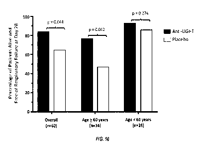
[0036] Figure 10 shows primary endpoint of the study described in Examples
2 and 3:
percentage of patients alive and free of respiratory failure at day 28 in the
anti-LIGHT
monoclonal antibody-treated group compared to the placebo-treated group.
[0037] Figure 11 shows a box plot generated from data from LIGHT testing
using the
free LIGHT assay described herein performed on samples from ARDS patients and
compared
to healthy donor LIGHT levels. There are 79 healthy control data points.
DETAILED DESCRIPTION OF THE INVENTION
[0038] The following definitions are provided to facilitate an
understanding of the
invention. They are not intended to limit the invention in any way.
Definitions
[0039] For purposes of the present invention, "a" or "an" entity refers
to one or more
of that entity; for example, "a cDNA" refers to one or more cDNA or at least
one cDNA. As
such, the terms "a" or "an," "one or more" and "at least one" can be used
interchangeably
herein. It is also noted that the terms "comprising," "including," and
"having" can be used
interchangeably. Furthermore, a compound "selected from the group consisting
of' refers to
one or more of the compounds in the list that follows, including mixtures
(i.e. combinations)
of two or more of the compounds. According to the present invention, an
"isolated," or
"biologically pure" molecule is a compound that has been removed from its
natural milieu.
As such, the terms "isolated" and "biologically pure" do not necessarily
reflect the extent to
which the compound has been purified. An isolated compound of the present
invention can
be obtained from its natural source, can be produced using laboratory
synthetic techniques or
can be produced by any such chemical synthetic route.
[0040] A "coronavirus," "corona respiratory virus," or "CoV" are used
interchangeably herein to refer to a virus belonging to the family
Coronaviridae.
Coronaviruses are enveloped, positive-sense RNA viruses of approximately 31
Kb, making
these viruses the largest known RNA viruses. Coronaviruses infect a variety of
host species,
including humans and several other vertebrates. These viruses predominantly
cause
respiratory and intestinal tract infections and induce a wide range of
clinical manifestations.
In general, coronaviruses can be classified into low pathogenic CoVs
(including human CoVs
(hCoVs)) and highly pathogenic CoVs, such as severe acute respiratory syndrome
CoV
CA 03173873 2022-08-30
WO 2021/202649 PCT/US2021/025068
(SARS-CoV) and Middle East respiratory syndrome CoV (MERS-CoV). Low pathogenic
hCoV infect upper airways and cause seasonal mild to moderate cold-like
respiratory
illnesses in healthy individuals. In contrast, the highly pathogenic hCoVs
(pathogenic hCoV)
infect the lower respiratory tract and cause severe pneumonia, which sometimes
leads to fatal
acute lung injury (ALI) and acute respiratory distress syndrome (ARDS),
resulting in high
morbidity and mortality. SARS-CoV2 is a type of coronavirus. A coronavirus
infection as
used herein includes any of the above, if associated with coronavirus. "COVID-
19 infection"
used herein may also refer to a condition or disease caused by SARS-CoV2.
[0041] An "acute lung injury" or "ALI" herein refers to an acute lung
disease with
bilateral pulmonary infiltrate in a chest radiograph consistent with the
presence of edema and
no clinical evidence of left atrial hypertension; or (if measured) a pulmonary
wedge pressure
of 18 mmHg or less. Additionally, the ratio of arterial oxygen to the fraction
of inspired
oxygen (Pa02/Fi02) must be 300 mmHg or less, regardless of the level of
positive end-
expiratory pressure (PEEP).
[0042] "Acute respiratory distress syndrome" or "ARDS" herein refers to
the most
severe form of ALI, defined by a ratio of arterial oxygen to fraction of
inspired oxygen of 200
mmHg or less. The term ARDS is often informally used interchangeably with ALI,
but by
strict criteria, ARDS should be reserved for the most severe form of the
disease.
[0043] "LIGHT" or "TNFSF 14" herein refers to a specific member protein
of the
tumor necrosis factor superfamily that is expressed by activated T cells,
monocytes-
macrophages and additional types of antigen presenting cells. "LIGHT" is an
acronym for
"homologous to Lymphotoxin, exhibits Inducible expression and competes with
HSV
Glycoprotein D for binding to HVEM (herpesvirus entry mediator), a receptor
expressed on T
lymphocytes."
[0044] "Free LIGHT" or "free (active) LIGHT" herein refers to non-bound
form
LIGHT (e.g., LIGHT bound to DcR3), which is the active form of LIGHT. In
humans, free
LIGHT is neutralized (inactivated) by DcR3, a unique soluble member of the
TNFR
superfamily, which binds LIGHT in high affinity and inhibits its interactions
with two TNF
receptors, HVEM and LTPR. "Bound LIGHT," or the like, refers to LIGHT that is
bound to
a natural ligand, optionally wherein the natural ligand is HVEM, LTPR, or
DcR3. "Total
LIGHT," or the like, refers to the total amount of free LIGHT and bound LIGHT.
[0045] "Elevated free LIGHT" as used herein refers to a level of free
LIGHT detected
in a subject that is higher than a normal control. The normal control can be
determined by
those of skill in the art as applicable to the particular situation. In some
instances, the normal
21
CA 03173873 2022-08-30
WO 2021/202649 PCT/US2021/025068
control is an industry standard agreed upon by those of skill as being a level
or range of levels
that is typical of an individual without a LIGHT-associated condition. In some
instances, the
normal control is a reference level of LIGHT from the same individual taken at
a time point,
and whether the subject has elevated LIGHT is determined based on a sample
from that same
individual taken at a different, typically later, time point.
[0046] The term "antibody" herein is used in the broadest sense and
encompasses
various antibody structures, including but not limited to monoclonal
antibodies, polyclonal
antibodies, multispecific antibodies (e.g., bispecific antibodies), and
antibody fragments so
long as they exhibit the desired antigen-binding activity. As used herein, the
term refers to a
molecule comprising at least complementarity-determining region (CDR) 1, CDR2,
and
CDR3 of a heavy chain and at least CDR1, CDR2, and CDR3 of a light chain,
wherein the
molecule is capable of binding to antigen. The term antibody includes, but is
not limited to,
fragments that are capable of binding antigen, such as Fv, single-chain Fv
(scFv), Fab, Fab',
and (Fab')2. The term antibody also includes, but is not limited to, chimeric
antibodies,
humanized antibodies, human antibodies, and antibodies of various species such
as mouse,
cynomolgus monkey, etc.
[0047] The term "heavy chain" refers to a polypeptide comprising at least a
heavy chain
variable region, with or without a leader sequence. In some embodiments, a
heavy chain
comprises at least a portion of a heavy chain constant region. The term "full-
length heavy
chain" refers to a polypeptide comprising a heavy chain variable region and a
heavy chain
constant region, with or without a leader sequence.
[0048] The term "heavy chain variable region" refers to a region comprising
a heavy
chain complementary determining region (CDR) 1, framework region (FR) 2, CDR2,
FR3,
and CDR3 of the heavy chain. In some embodiments, a heavy chain variable
region also
comprises at least a portion of an FR1 and/or at least a portion of an FR4. In
some
embodiments, a heavy chain CDR1 corresponds to Kabat residues 31 to 35; a
heavy chain
CDR2 corresponds to Kabat residues 50 to 65; and a heavy chain CDR3
corresponds to
Kabat residues 95 to 102. See, e.g., Kabat Sequences of Proteins of
Immunological Interest
(1987 and 1991, NIH, Bethesda, Md.).
[0049] The term "light chain" refers to a polypeptide comprising at least a
light chain
variable region, with or without a leader sequence. In some embodiments, a
light chain
comprises at least a portion of a light chain constant region. The term "full-
length light chain"
refers to a polypeptide comprising a light chain variable region and a light
chain constant
region, with or without a leader sequence. The term "light chain variable
region" refers to a
22
CA 03173873 2022-08-30
WO 2021/202649 PCT/US2021/025068
region comprising a light chain CDR1, FR2, HVR2, FR3, and HVR3. In some
embodiments,
a light chain variable region also comprises an FR1 and/or an FR4. In some
embodiments, a
light chain CDR1 corresponds to Kabat residues 24 to 34; a light chain CDR2
corresponds to
Kabat residues 50 to 56; and a light chain CDR3 corresponds to Kabat residues
89 to 97. See,
e.g., Kabat Sequences of Proteins of Immunological Interest (1987 and 1991,
NIH, Bethesda,
Md.).
[0050] A "chimeric antibody" refers to an antibody in which a portion of
the heavy
and/or light chain is derived from a particular source or species, while the
remainder of the
heavy and/or light chain is derived from a different source or species. In
some embodiments,
a chimeric antibody refers to an antibody comprising at least one variable
region from a first
species (such as mouse, rat, cynomolgus monkey, etc.) and at least one
constant region from
a second species (such as human, cynomolgus monkey, etc.). In some
embodiments, a
chimeric antibody comprises at least one mouse variable region and at least
one human
constant region. In some embodiments, a chimeric antibody comprises at least
one
cynomolgus variable region and at least one human constant region. In some
embodiments,
all of the variable regions of a chimeric antibody are from a first species
and all of the
constant regions of the chimeric antibody are from a second species.
[0051] A "humanized antibody" refers to an antibody in which at least one
amino acid in
a framework region of a non-human variable region has been replaced with the
corresponding
amino acid from a human variable region. In some embodiments, a humanized
antibody
comprises at least one human constant region or fragment thereof. In some
embodiments, a
humanized antibody is an Fab, an scFv, a (Fab)2, etc.
[0052] A "human antibody" as used herein refers to antibodies produced in
humans,
antibodies produced in non-human animals that comprise human immunoglobulin
genes,
such as XenoMouseg, and antibodies selected using in vitro methods, such as
phage display,
wherein the antibody repertoire is based on a human immunoglobulin sequences.
[0053] The term "leader sequence" refers to a sequence of amino acid
residues located at
the N terminus of a polypeptide that facilitates secretion of a polypeptide
from a mammalian
cell. A leader sequence may be cleaved upon export of the polypeptide from the
mammalian
cell, forming a mature protein. Leader sequences may be natural or synthetic,
and they may
be heterologous or homologous to the protein to which they are attached.
[0054] "Percent (%) amino acid sequence identity" and "homology" with
respect to a
peptide, polypeptide or antibody sequence are defined as the percentage of
amino acid
residues in a candidate sequence that are identical with the amino acid
residues in the specific
23
CA 03173873 2022-08-30
WO 2021/202649 PCT/US2021/025068
peptide or polypeptide sequence, after aligning the sequences and introducing
gaps, if
necessary, to achieve the maximum percent sequence identity, and not
considering any
conservative substitutions as part of the sequence identity. Alignment for
purposes of
determining percent amino acid sequence identity can be achieved in various
ways that are
within the skill in the art, for instance, using publicly available computer
software such as
BLAST, BLAST-2, ALIGN or MEGALIGNTM (DNASTAR) software. Those skilled in the
art can determine appropriate parameters for measuring alignment, including
any algorithms
needed to achieve maximal alignment over the full length of the sequences
being compared.
[0055] The terms "inhibition" or "inhibit" refer to a decrease or cessation
of any event
(such as protein ligand binding) or to a decrease or cessation of any
phenotypic characteristic
or to the decrease or cessation in the incidence, degree, or likelihood of
that characteristic.
To "reduce" or "inhibit" is to decrease, reduce or arrest an activity,
function, and/or amount
as compared to a reference. It is not necessary that the inhibition or
reduction be complete.
For example, in certain embodiments, by "reduce" or "inhibit" is meant the
ability to cause
an overall decrease of 20% or greater. In another embodiment, by "reduce" or
"inhibit" is
meant the ability to cause an overall decrease of 50% or greater. In yet
another embodiment,
by "reduce" or "inhibit" is meant the ability to cause an overall decrease of
75%, 85%, 90%,
95%, or greater.
[0056] "Sample" or "subject sample" or "biological sample" generally
refers to a
sample which may be tested for a particular molecule. Samples may include but
are not
limited to cells, body fluids, including blood, serum, plasma, urine, saliva,
stool, tears, pleural
fluid and the like.
[0057] The terms "agent" and "test compound" are used interchangeably
herein and
denote a chemical compound, a mixture of chemical compounds, a biological
macromolecule, or an extract made from biological materials such as bacteria,
plants, fungi,
or animal (particularly mammalian) cells or tissues. Biological macromolecules
include
siRNA, shRNA, antisense oligonucleotides, peptides, peptide/DNA complexes, and
any
nucleic acid based molecule which exhibits the capacity to modulate the
activity of the SNP
containing nucleic acids described herein or their encoded proteins. Agents
are evaluated for
potential biological activity by inclusion in screening assays described
hereinbelow.
[0058] A "subject" can be mammalian. In any of the embodiments involving a
subject,
the subject can be human. In any of the embodiments involving a subject, the
subject can be a
cow, pig, monkey, sheep, dog, cat, fish, or poultry.
24
CA 03173873 2022-08-30
WO 2021/202649 PCT/US2021/025068
[0059] A "pediatric" subject herein is a human of less than 18 years of
age, whereas an
"adult" subject is 18 years or older.
[0060] "Treatment" or "treat" refers to both therapeutic treatment and
prophylactic or
preventative measures. Those in need of treatment include those already with
the disorder as
well as those prone to have the disorder or those in which the disorder is to
be prevented. For
purposes of this invention, beneficial or desired clinical results include,
but are not limited to,
alleviation of symptoms, diminishment of extent of disease, stabilized (i.e.,
not worsening)
state of disease, delay or slowing of disease progression, amelioration or
palliation of the
disease state, and remission (whether partial or total), whether detectable or
undetectable.
"Treatment" can also mean prolonging survival as compared to expected survival
if not
receiving treatment. Those in need of treatment include those already with the
condition or
disorder as well as those prone to have the condition or disorder or those in
which the
condition or disorder is to be prevented.
[0061] The term "effective amount" or "therapeutically effective amount"
refers to an
amount of a drug effective for treatment of a disease or disorder in a
subject, such as to
partially or fully relieve one or more symptoms. In some embodiments, an
effective amount
refers to an amount effective, at dosages and for periods of time necessary,
to achieve the
desired therapeutic or prophylactic result.
Identification of Biomarker LIGHT
[0062] LIGHT (TNFSF14) is an important regulatory cytokine, which serves as
critical
factor in orchestrating a cytokine storm and pulmonary failure associated with
pathogen-
mediated infection, including viral and bacterial infections including
coronavirus (e.g.,
COVID-19).
[0063] In some embodiments, methods for detecting free (active) LIGHT in a
human
subject are provided. In some embodiments, the subject's biological sample
(e.g., serum) is
analyzed for free LIGHT. In some embodiments, the results provide a basis for
understanding
whether an anti-LIGHT therapy may be provided and will be effective. For
instance, if an
elevated level of free LIGHT (e.g., a level of free LIGHT above what is
expected, for
example, using a normal control) then the subject may be diagnosed with a
condition
associated with elevated free LIGHT and/or be deemed to be a suitable
candidate for
treatment with an anti-LIGHT antibody. In some embodiments, the anti-LIGHT
antibody is
an antibody neutralizing LIGHT. In some embodiments, methods for detecting
free (active)
LIGHT in a subject's biological sample (e.g., serum) are provided, wherein the
results
provide a basis for understanding whether an anti-LIGHT therapy may be
provided and will
CA 03173873 2022-08-30
WO 2021/202649 PCT/US2021/025068
be effective. In some embodiments, detection of free LIGHT above a normal
control
indicates that an anti-LIGHT therapy may be provided and effective.
[0064] In some embodiments, the condition associated with elevated free
LIGHT with
which the subject may be diagnosed is Crohn's Disease or an inflammatory
condition
associated with Crohn's Disease. In some embodiments, the condition associated
with
elevated free LIGHT with which the subject may be diagnosed is a coronavirus
infection. In
some embodiments, the coronavirus infection is a moderate or a severe
coronavirus infection.
In some embodiments, conditions associated with elevated free LIGHT include
any one or
more of inflammation, optionally wherein the inflammation is
hyperinflammation, immune
dysregulation that leads to multisystem organ failure, acute lung injury
(ALI), optionally
wherein the ALI is associated with a bacterial or viral infection, including
coronavirus
infection, acute respiratory distress syndrome (ARDS), optionally wherein the
ARDS is
associated with a bacterial or viral infection, including coronavirus
infection, cytokine storm
that drives tissue injury and vascular permeability, post-infection pulmonary
fibrosis, and
pneumonia, optionally wherein the pneumonia is associated with a bacterial or
viral infection,
including coronavirus infection. In some embodiments, the condition associated
with elevated
free LIGHT is mild, moderate, or severe coronavirus infection, optionally
wherein the
coronavirus infection is a COVID-19 infection. In some embodiments, the COVID-
19
infection is associated with ALI or ARDS in the subject. In some embodiments,
the COVID-
19 infection is associated with cytokine storm that drives tissue injury and
vascular
permeability in the lungs and post-infection pulmonary fibrosis. In some
embodiments, the
coronavirus infection is MERS-CoV, SARS-CoV, or SARS-CoV2/COVID-19.
[0065] In some embodiments, methods for diagnosing a condition associated
with
elevated free LIGHT in a subject are provided, wherein the level of free
(active) LIGHT in a
biological sample is detected. If the levels are above a normal control, then
the subject is
diagnosed as having a condition associated with elevated free LIGHT. In some
embodiments,
the condition associated with elevated free LIGHT to be detected is Crohn's
Disease or an
inflammatory condition associated with Crohn's Disease. In some embodiments,
the
condition associated with elevated free LIGHT to be detected is a virus
infection. In some
embodiments, the condition associated with elevated free LIGHT is a
coronavirus infection.
In some embodiments, the condition associated with elevated free LIGHT to be
detected is a
moderate or a severe coronavirus infection. In some embodiments, the condition
associated
with elevated free LIGHT to be detected is a mild, moderate, or severe COVID-
19 infection.
26
CA 03173873 2022-08-30
WO 2021/202649 PCT/US2021/025068
In some embodiments, the COVID-19 infection is associated with ALI or ARDS in
the
subject. In some embodiments, the coronavirus infection is MERS-CoV or SARS-
CoV.
[0066] In some embodiments, methods for detecting free LIGHT in a
biological sample
from a subject may be conducted with a biological sample comprising blood,
urine, serum,
plasma, feces, or gastric lavage bodily fluid samples, or cell samples such as
white blood
cells or mononuclear cells.
[0067] In some embodiments, the method of detecting free (active) LIGHT in
subject is
performed by:
(a) contacting the biological sample with at least one anti-LIGHT antibody;
(b) incubating the biological sample to allow the anti-LIGHT antibody to bind
to
free LIGHT; and
(c) determining the presence of complexes formed between the anti-LIGHT
antibody and free LIGHT in the biological sample.
[0068] This method can further comprise the step of diagnosing the subject
as having a
condition associated with elevated free LIGHT and/or administering an anti-
LIGHT
antibody.
[0069] Various methods known in the art for detecting specific antibody-
antigen binding
can be used. Exemplary immunoassays which can be conducted include
fluorescence
polarization immunoassay (FPIA), fluorescence immunoassay (FIA), enzyme
immunoassay
(EIA), nephelometric inhibition immunoassay (NIA), enzyme linked immunosorbent
assay
(ELISA), and radioimmunoassay (MA), competition assay, and sandwich method.
[0070] An indicator moiety, or label group, can be attached to the subject
antibodies and
is selected to meet the needs of various uses of the method which are often
dictated by the
availability of assay equipment and compatible immunoassay procedures.
Appropriate labels
include, without limitation, radionuclides (for example 1251, 1311, 35S, 3H,
or 32P), enzymes
(for example, alkaline phosphatase, horseradish peroxidase, luciferase, or P-
galactosidase),
fluorescent moieties or proteins (for example, fluorescein, rhodamine,
phycoerythrin, GFP, or
BFP), or luminescent moieties (for example, QdotTM nanoparticles supplied by
the Quantum
Dot Corporation, Palo Alto, Calif).
[0071] General techniques to be used in performing the various immunoassays
noted
above are known to those of ordinary skill in the art.
[0072] ELISA assays are generally known to the skilled artisan and can be
designed to
determine serum LIGHT levels. In one exemplary embodiment, blood is collected,
and the
serum is isolated. If no kit is available, an ELISA can be developed using
plates that are pre-
27
CA 03173873 2022-08-30
WO 2021/202649
PCT/US2021/025068
coated with capture antibody specific for the LIGHT one is measuring. The
plate is next
incubated at room temperature for a period of time before washing. Enzyme-anti-
LIGHT
antibody conjugate is added and incubated. Unbound antibody conjugate is
removed, and the
plate washed before the addition of the chromogenic substrate solution that
reacts with the
enzyme. The plate is read on an appropriate plate reader at an absorbance
specific for the
enzyme and substrate used.
[0073] The competition method compares the competitive binding of an
antigen in a
sample and a known amount of a labeled antigen to the monoclonal antibody of
the present
invention. To carry out an immunological assay based on the competition
method, a sample
containing an unknown amount of the target antigen is added to a solid
substrate to which the
monoclonal antibody of the present invention is coated physically or
chemically by known
means, and the reaction is allowed to proceed. Simultaneously, a predetermined
amount of
the pre-labeled target antigen is added and the reaction is allowed to
proceed. After
incubation, the solid substrate is washed and the activity of the labeling
agent bound to the
solid substrate is measured.
[0074] In the sandwich method, the target antigen in a sample is sandwiched
between the
immobilized monoclonal antibody and the labeled monoclonal antibody, then a
labeling
substrate such as an enzyme is added, substrate color changes are detected,
and thereby
detecting the presence of the antigen. To carry out an immunological assay
based on the
sandwich method, for instance, a sample containing an unknown amount of the
target antigen
is added to a solid substrate to which the monoclonal antibody of the present
invention is
coated physically or chemically by known means, and the reaction is allowed to
proceed.
Thereafter, the labeled monoclonal antibody of the invention is added and the
reaction is
allowed to proceed. After incubation, the solid substrate is washed and the
activity of the
labeling agent bound to the solid substrate is measured.
[0075] In some embodiments, a first antibody is used for a diagnostic and a
second
antibody is used as a therapeutic. In some embodiments, the first and second
antibodies are
different. In some embodiments, the first and second antibodies can both bind
to the antigen
at the same time, by binding to separate epitopes.
Methods of Treating with Anti-LIGHT Antibody
[0076] The
present invention provides a method of treating subjects having elevated
free LIGHT, including subjects having a condition associated with elevated
free LIGHT with
an anti-LIGHT antibody. In some embodiments, the anti-LIGHT antibody is an
antibody
neutralizing LIGHT. In some embodiments, the condition associated with
elevated free
28
CA 03173873 2022-08-30
WO 2021/202649 PCT/US2021/025068
LIGHT is a coronavirus infection, including COVID-19. In some embodiments, the
condition
associated with elevated free LIGHT is Crohn's Disease or an inflammatory
condition
associated with Crohn's Disease. The treatment can be done with or without a
diagnostic test
for detecting elevated free LIGHT in a subject's sample.
[0077] In some embodiments, a biological sample from the subject is first
tested for the
presence and level of free LIGHT in a threshold step before the subject is
treated. In such
embodiments, the method of treating comprises contacting a biological sample
from a subject
with at least one first anti-LIGHT antibody, incubating the biological sample
to allow the
anti-LIGHT antibody to bind to free LIGHT, determining the presence of
complexes formed
between the anti-LIGHT antibody and free LIGHT in the biological sample, and
finally,
based on the positive results of the detecting step and a finding of elevated
free LIGHT,
administering to the subject an effective amount of a second anti-LIGHT
antibody, wherein
the first and the second antibody differ.
[0078] In some embodiments, the subject is treated without a threshold
testing step. In
such cases, it may have been predetermined that the subject could benefit from
an anti-
LIGHT therapy or the anti-LIGHT antibody is being administered for prevention.
In such
embodiments, the method of treating comprises administering to a subject
having a condition
associated with elevated free LIGHT an effective amount of an anti-LIGHT
antibody.
[0079] In some embodiments of the method of treating, the condition
associated with
elevated free LIGHT is Crohn's Disease or an inflammatory condition associated
with
Crohn' s Disease.
[0080] In some embodiments of the method of treating, the condition
associated with
elevated free LIGHT is a coronavirus infection. In some embodiments, the
coronavirus
infection is a COVID-19 infection. In some embodiments, the subject has a
respiratory
disease, optionally caused by a virus, bacteria or fungus. In some
embodiments, the subject
has a respiratory disease caused by coronavirus infection. In some
embodiments, the subject
has a respiratory disease caused by COVID-19. In some embodiments, the subject
has
pneumonia. In some embodiments, the subject has acute lung injury (ALI). In
some
embodiments, the subject has acute respiratory distress syndrome (ARDS). In
some
embodiments, pneumonia is associated with coronavirus infection. In some
embodiments,
ALI or ARDS is associated with coronavirus infection. In some embodiments,
pneumonia is
associated with COVID-19. In some embodiments, the subject has acute lung
injury (ALI)
associated with COVID-19. In some embodiments, the subject has acute
respiratory distress
syndrome (ARDS) associated with COVID-19. In some embodiments, the subject has
a mild
29
CA 03173873 2022-08-30
WO 2021/202649 PCT/US2021/025068
coronavirus infection. In some embodiments, the subject has a moderate
coronavirus
infection. In some embodiments, the subject has a severe coronavirus
infection. In some
embodiments, a method of treating severe COVID-19 pneumonia comprising
administering
an anti-LIGHT antibody to a subject in need thereof is provided. In some
embodiments, a
method of treating acute inflammatory disease associated with COVID-19
pneumonia
comprising administering an anti-LIGHT antibody to a subject in need thereof
is provided. In
some embodiments, a method of treating respiratory failure associated with
COVID-19
pneumonia comprising administering an anti-LIGHT antibody to a subject in need
thereof is
provided. In some embodiments, a method of treating cytokine storm comprising
administering an anti-LIGHT antibody to a subject in need thereof is provided.
In some
embodiments, a method of treating a dysregulated hyperimmune response
(sometimes
referred to as "cytokine storm") comprising administering an anti-LIGHT
antibody to a
subject in need thereof is provided. In some embodiments, a method of treating
Acute
Respiratory Disease Syndrome (ARDS) comprising administering an anti-LIGHT
antibody to
a subject in need thereof is provided.
[0081] In some embodiments, the administration of the anti-LIGHT antibody
suppresses
T cell activation. In some embodiments, the administration of the anti-LIGHT
antibody
suppresses increased expression of cytokines (e.g., a cytokine storm). In some
embodiments,
the administration of the anti-LIGHT antibody suppresses increased expression
of cytokines
(e.g., a cytokine storm) that is caused by a viral and/or bacterial infection,
e.g., from a
coronavirus infection. In some embodiments the administration of the anti-
LIGHT antibody
prevents or treats cytokine storm caused by a virus and/or bacterial
infection, such as by a
coronavirus infection. In some embodiments, the cytokine storm can drive
tissue injury and
vascular permeability in the lungs. Cytokine storm can lead to ARDS and ALI
associated
with coronavirus (e.g., COVID-19) infection. In some embodiments, the
administration of
the anti-LIGHT antibody prevents or treats post-infection pulmonary fibrosis,
optionally
associated with coronavirus infection. In some embodiments, the administration
of the anti-
LIGHT antibody reduces the subject's risk of mortality or morbidity. In some
embodiments,
the administration of the anti-LIGHT antibody prevents progression to ARDS in
a subject
with pneumonia associated with a coronavirus infection (e.g., COVID-19
infection). In some
embodiments, the administration of the anti-LIGHT antibody prevents
progression of ALI
associated with a coronavirus infection, including (e.g., COVID-19 infection)
to ARDS. In
some embodiments, the administration of the anti-LIGHT antibody prevents or
treats ARDS.
CA 03173873 2022-08-30
WO 2021/202649 PCT/US2021/025068
In some embodiments, the administration of the anti-LIGHT antibody prevents
the need for
ventilation/intubation of the subject.
[0082] The methods of treating in the present invention may be carried out
through
conventional administration routes, including without limitation, the oral,
buccal, sublingual,
ocular, topical, parenteral, rectal, intracisternal, intravaginal,
intraperitoneal, intravesical,
local (e.g., powder, ointment, or drop), or nasal routes. The term
"parenteral" includes
subcutaneous, intravenous, intramuscular, or infusion. In certain embodiments,
it may be
appropriate to administer the agent in a continuous infusion or as a
subcutaneous injection
every day, every two or several days, or once a week, or every several weeks,
or once a
month, or once every several months, or at a time interval within a range
defined by any two
of the aforementioned intervals.
[0083] In some embodiments, the anti-LIGHT antibody is administered to a
subject
already receiving another therapy. In the case of a subject with COVID-19, the
other therapy
may be any therapy approved or being tested to treat or ameliorate a symptom
of COVID-19.
Such therapies include, but are not limited to, remdesivir, corticosteroids,
and
hydroxychloroquine. In some embodiments, the other therapy continues (on its
normal
course) during treatment with the anti-LIGHT antibody. In some embodiments,
the other
therapy is discontinued during treatment with the anti-LIGHT antibody. In some
embodiments, the subject has COVID-19 and is receiving a high dose of
corticosteroid and
an anti-LIGHT antibody. In some embodiments, subject receiving the combination
high dose
corticosteroid and anti-LIGHT antibody have better outcomes than those not
receiving the
combination.
[0084] In some embodiments, the dose of anti-LIGHT antibody is about 16
mg/kg with a
maximum dose of 1,200 mg. In some embodiments, the dose is a single
administration. In
some embodiments, the dose is more than a single administration.
[0085] In some embodiments, administration of the anti-LIGHT antibody
reduces serum
free-LIGHT levels in the subject by 85% or more, for example, compared to free-
LIGHT
levels in the subject prior to administration of the anti-LIGHT antibody or
compared to
subjects who are not administered the anti-LIGHT antibody. In some
embodiments, the
reduction in serum free-LIGHT levels in the subject by 85% or more occurs in
less than 5
days or less than 1 day after administration of the anti-LIGHT antibody. In
some
embodiments, the anti-LIGHT antibody administered to the subject reduces the
subject's risk
of mortality by equal to or greater than 50% at 60 days after administration.
In some
embodiments, the anti-LIGHT antibody administered to the subject reduces the
subject's risk
31
CA 03173873 2022-08-30
WO 2021/202649 PCT/US2021/025068
of mortality by equal to or greater than 50% at 28 days after administration.
In some
embodiments, the subject is 60 years of age or older. In some embodiments, the
anti-LIGHT
antibody administered to the subject shortens the length of the subject's
hospital stay
compared to subjects receiving standard of care treatment. In some
embodiments, the anti-
LIGHT antibody administered to the subject reduces the subject's risk of
respiratory failure.
[0086] In some embodiments, the anti-LIGHT antibody is administered as a
dose of from
about 1.0 mg/kg to about 3.0 mg/kg. In some embodiments, the anti-LIGHT
antibody is
administered as a dose of about 1.0, about 1.1, about 1.2, about 1.3, about
1.4, about 1.5,
about 1.6, about 1.7, about 1.8, about 1.9, about 2.0, about 2.1, about 2.2,
about 2.3, about
2.4, about 2.5, about 2.6, about 2.7, about 2.8, about 2.9, or about 3.0
mg/kg. In some
embodiments, the anti-LIGHT antibody is administered every day, every 2 days,
every 3
days, every 4 days, every 5 days, every 6 days, every week, every two weeks,
every three
weeks, or every four weeks. In some embodiments, the anti-LIGHT antibody is
administered
as a dose of from about 1.0 mg/kg to about 3.0 mg/kg every 14 days. In some
embodiments,
the anti-LIGHT antibody is administered as a dose of 1.0 mg/kg every 14 days.
In some
embodiment, the anti-LIGHT antibody is administered as a dose of 3.0 mg/kg
every 14 days.
[0087] In some embodiments, the anti-LIGHT antibody is administered to a
subject who
has failed treatment with an approved therapeutic dose of an anti-TNFa
monoclonal antibody
treatment with either no initial response or an initial response to induction
with subsequent
lost response.
Treatment of Crohn's Disease
[0088] The primary clinical target for CD treatment is the resolution of
symptoms. (Shi
and Ng, The state of the art on treatment of Crohn's disease, J.
Gastroenterol., 2018:53;989-
998). The standard index for assessing symptoms is the Crohn's Disease
Activity Index
(CDAI), which includes eight variables: the number of liquid stools, the
extent of abdominal
pain, general well-being, the occurrence of extraintestinal symptoms, the need
for
antidiarrhea drugs, the presence of abdominal masses, hematocrit, and body
weight. (Shi and
Ng 2018). Control of symptoms does not appear to significantly alter the
natural course of the
disease. (Shi and Ng 2018).
[0089] Mucosal healing is becoming another target in clinical practice. The
Simple
Endoscopic Score for Crohn's Disease (SES-CD) is one of the most common
endoscopic
scoring systems for CD. The SES-CD is scored based on four endoscopic
variables (presence
and size of ulcers, extent of ulcerated surface, extent of affected surface,
and presence and
32
CA 03173873 2022-08-30
WO 2021/202649 PCT/US2021/025068
type of narrowing) in the same five segments. An SES-CD score below 2 is
regarded as
endoscopic remission. A decrease from baseline of at least 50% in an SES-CD
score has been
used as the definition for endoscopic response. There is currently no set,
universal definition
for mucosal healing, but the absence of ulceration at ileocolonoscopy has been
adopted as the
endoscopic endpoint for CD. (Shi and Ng 2018).
[0090] A Patient reported outcome is a measurement derived directly from a
patient about
any aspect of their health status and has the potential to become a treatment
endpoint for CD.
Several scales have been used to assess patients' perspectives towards the
disease, including
the Inflammatory Bowel Disease Questionnaire (IBD-Q). (Shi and Ng 2018).
[0091] In some embodiments, administration of the anti-LIGHT antibody
reduces the
subject's CDAI score, for example, compared to the subject's CDAI score prior
to
administration of the anti-LIGHT antibody or compared to subjects who are not
administered
the anti-LIGHT antibody. In some embodiments, administration of the anti-LIGHT
antibody
decreases the subject's SES-CD score, for example, compared to the subject's
SES-CD score
prior to administration of the anti-LIGHT antibody or compared to subjects who
are not
administered the anti-LIGHT antibody. In some embodiments, administration of
the anti-
LIGHT antibody results in the subject's SES-CD score of below 2. In some
embodiments,
administration of the anti-LIGHT antibody increases the subject's IBD-Q score,
for example,
compared to the subject's IBD-Q score prior to administration of the anti-
LIGHT antibody or
compared to subjects who are not administered the anti-LIGHT antibody. In some
embodiments, administration of the anti-LIGHT antibody results in the
subject's IBD-Q score
of 170 or higher. In some embodiments, administration of the anti-LIGHT
antibody results in
an increase in the subject's IBD-Q score of at least 16 points. In some
embodiments,
administration of the anti-LIGHT antibody results in an increase in the
subject's IBD-Q score
of at least 32 points.
Stages of COVID-19 Disease Progression
[0092] The stages of COVID-19 Disease Progression are shown in Figure 1 of
Siddiqi,
H.K., et al. COVID-19 illness in native and immunosuppressed states: A
clinical-therapeutic
staging proposal. J Heart Lung Transplant 39(5), 405-407 (2020). The disease
can broadly
be divided into three stages: Early Infection, Pulmonary Phase, and
Hyperinflammation
Phase. Stage I is the viral response phase, Stage III is the host inflammatory
response phase,
and Stage II is a combination of the two. As patients progress from Early
Infection (Stage I)
to the Pulmonary Phase (Stage II) at which pulmonary symptoms appear, the host
inflammatory response emerges. It is at this point where, in some patients,
dysregulation of
33
CA 03173873 2022-08-30
WO 2021/202649 PCT/US2021/025068
the inflammatory response occurs. This dysregulation of the inflammatory
response may be
accompanied by elevated free LIGHT levels in biological fluids, such as serum.
This stage,
the Pulmonary Phase, is the optimal stage at which to administer an anti-LIGHT
antibody to
correct this dysregulation and avert cytokine storm and progression to ARDS,
which occurs
in the Hyperinflammation Phase (Stage III).
[0093] Patients in Stage I of COVID-19 are considered to have mild disease.
Patients in
Stage II of COVID-19 are considered to have mild to moderate disease. Patients
in Stage III
of COVID-19 are considered to have severe disease.
[0094] In some embodiments, the anti-LIGHT antibody is administered to a
subject with
mild coronavirus (e.g., COVID-19). In some embodiments, the anti-LIGHT
antibody is
administered during the early infection stage of a coronavirus. In some
embodiments, the
anti-LIGHT antibody is administered during Stage I of a coronavirus. In some
embodiments,
the anti-LIGHT antibody is administered while the subject in need thereof has
mild
constitutional symptoms, a fever > 99.6 F, dry cough, diarrhea, or headache.
In some
embodiments, the subject in need thereof has lymphopenia, increased
prothrombin time,
increased D-dimer and LDH (mild). In some embodiments, the administration of
the anti-
LIGHT antibody treats Stage I of a coronavirus. In some embodiments, the
administration of
the anti-LIGHT antibody prevents progression of the coronavirus to Stage II.
Staging refers
to Figure 1 of Siddiqi, H.K., et al. (2020), incorporated herein in its
entirety.
[0095] In some embodiments, the anti-LIGHT antibody of the method is
administered to
a subject with moderate coronavirus (e.g., COVID-19). In some embodiments, the
anti-
LIGHT antibody is administered during the Pulmonary Phase of a coronavirus. In
some
embodiments, the anti-LIGHT antibody is administered during Stage II of a
coronavirus. In
some embodiments of the invention, the anti-LIGHT antibody is administered
during Stage
IIA or Stage IIB of a coronavirus. In some embodiments, the anti-LIGHT
antibody is
administered while the subject in need thereof is exhibiting shortness of
breath or hypoxia. In
some embodiments, the anti-LIGHT antibody is administered while the subject in
need
thereof meets the clinical criteria for ALI. In some embodiments, the subject
in need thereof
has abnormal chest imaging, transaminitis, or low-normal procalcitonin. In
some
embodiments, the patient is not ventilated/intubated. In some embodiments, the
administration of the anti-LIGHT antibody treats Stage II of a coronavirus. In
some
embodiments, the administration of the anti-LIGHT antibody prevents
progression of the
coronavirus to Stage III.
34
CA 03173873 2022-08-30
WO 2021/202649 PCT/US2021/025068
[0096] In some embodiments, the anti-LIGHT antibody is administered to a
subject with
severe coronavirus (e.g., COVID-19). In some embodiments, the anti-LIGHT
antibody is
administered during the Hyperinflammation Phase of a coronavirus. In some
embodiments,
the anti-LIGHT antibody is administered during Stage III of a coronavirus. In
some
embodiments, the anti-LIGHT antibody is administered while the subject in need
thereof has
ARDS, systemic inflammatory response syndrome (SIRS) / Shock, or Cardiac
Failure. SIRS
is a complex immune response to insult characterized by widespread
inflammation within the
body. Both infectious and non-infectious causes of SIRS have been identified.
In some
embodiments, the patient is ventilated/intubated. In some embodiments, the
administration of
the anti-LIGHT antibody treats Stage III of a coronavirus.
Anti-LIGHT Antibodies
[0097] In some embodiments, an anti-LIGHT antibody is utilized for both
detection/diagnostic and therapeutic purposes, as well as in the assays
described herein. The
anti-LIGHT antibody used for detection or diagnostic purposes is different
from the antibody
used for therapeutic purposes (even in the same subject).
[0098] The anti-LIGHT antibody useful for therapeutic purposes may comprise
the CDR
sequences of the El, E13, E63, F19, or F23 antibodies, which are provided in
WO
2008/027338 and US 8,058,402 B2, US 8,461,307 B2, and US 8,974,787 B2, each of
which
is incorporated herein by reference. The anti-LIGHT antibody useful for
detection/diagnostic
purposes is not that same as that which is used for therapeutic purposes.
[0099] In some embodiments, the anti-LIGHT antibody comprises a heavy chain
comprising three CDR sequences comprising each of SEQ ID NOs: 2, 3, and 4,
respectively.
In some embodiments, the anti-LIGHT antibody comprises a light chain
comprising three
CDR sequences comprising each of SEQ ID NOs: 5, 6, and 7, respectively. In
some
embodiments, the antibody comprises a heavy chain and a light chain, the heavy
chain
comprising three CDR sequences comprising each of SEQ ID NOs: 2, 3, and 4,
respectively,
and the light chain comprising three CDR sequences comprising each of SEQ ID
NOs: 5, 6,
and 7, respectively.
[00100] In some embodiments, the anti-LIGHT antibody comprises a heavy chain
variable
region sequence comprising SEQ ID NO: 8 or that is at least 85%, at least 90%,
at least 95%,
at least 98%, or at least 99% identical to SEQ ID NO:8. In some embodiments,
the anti-
LIGHT antibody comprises a light chain variable region sequence comprising SEQ
ID NO:9
or that is at least 85%, at least 90%, at least 95%, at least 98%, or at least
99% identical to
CA 03173873 2022-08-30
WO 2021/202649 PCT/US2021/025068
SEQ ID NO:9. In some embodiments, the anti-LIGHT antibody comprises a heavy
chain
comprising SEQ ID NO: 8 or that is at least 85%, at least 90%, at least 95%,
at least 98%, or
at least 99% identical to SEQ ID NO:8. In some embodiments, the anti-LIGHT
antibody
comprises a light chain comprising SEQ ID NO:9 or that is at least 85%, at
least 90%, at least
95%, at least 98%, or at least 99% identical to SEQ ID NO:9. In some
embodiments, the
anti-LIGHT antibody comprises both a heavy chain comprising SEQ ID NO: 8 or
that is at
least 85%, at least 90%, at least 95%, at least 98%, or at least 99% identical
to SEQ ID NO:8
and a light chain comprising SEQ ID NO:9 or that is at least 85%, at least
90%, at least 95%,
at least 98%, or at least 99% identical to SEQ ID NO:9.
[00101] In some embodiments, the anti-LIGHT antibody comprises a heavy chain
comprising three CDR sequences comprising each of SEQ ID NOs: 10, 11, and 12,
respectively. In some embodiments, the anti-LIGHT antibody comprises a light
chain
comprising three CDR sequences comprising each of SEQ ID NOs: 13, 14, and 15,
respectively. In some embodiments, the antibody comprises a heavy chain and a
light chain,
the heavy chain comprising three CDR sequences comprising each of SEQ ID NOs:
10, 11,
and 12, respectively, and the light chain comprising three CDR sequences
comprising each of
SEQ ID NOs: 13, 14, and 15, respectively.
[00102] In some embodiments, the anti-LIGHT antibody comprises a heavy chain
comprising three CDR sequences comprising each of SEQ ID NOs: 16, 17, and 18,
respectively. In some embodiments, the anti-LIGHT antibody comprises a light
chain
comprising three CDR sequences comprising each of SEQ ID NOs: 19, 20, and 21,
respectively. In some embodiments, the antibody comprises a heavy chain and a
light chain,
the heavy chain comprising three CDR sequences comprising each of SEQ ID NOs:
16, 17,
and 18, respectively, and the light chain comprising three CDR sequences
comprising each of
SEQ ID NOs: 19, 20, and 21, respectively.
[00103] In some embodiments, the anti-LIGHT antibody comprises a heavy chain
comprising three CDR sequences comprising each of SEQ ID NOs: 22, 23, and 24,
respectively. In some embodiments, the anti-LIGHT antibody comprises a light
chain
comprising three CDR sequences comprising each of SEQ ID NOs: 25, 26, and 27,
respectively. In some embodiments, the antibody comprises a heavy chain and a
light chain,
the heavy chain comprising three CDR sequences comprising each of SEQ ID NOs:
22, 23,
and 24, respectively, and the light chain comprising three CDR sequences
comprising each of
SEQ ID NOs: 25, 26, and 27, respectively.
36
CA 03173873 2022-08-30
WO 2021/202649 PCT/US2021/025068
[00104] In some embodiments, the anti-LIGHT antibody comprises a heavy chain
comprising three CDR sequences comprising each of SEQ ID NOs: 28, 29, and 30,
respectively. In some embodiments, the anti-LIGHT antibody comprises a light
chain
comprising three CDR sequences comprising each of SEQ ID NOs: 31, 32, and 33,
respectively. In some embodiments, the antibody comprises a heavy chain and a
light chain,
the heavy chain comprising three CDR sequences comprising each of SEQ ID NOs:
28, 29,
and 30, respectively, and the light chain comprising three CDR sequences
comprising each of
SEQ ID NOs: 31, 32, and 33, respectively.
[00105] In some embodiments, the anti-LIGHT antibody comprises a heavy chain
comprising three CDR sequences comprising each of SEQ ID NOs: 34, 35, and 36,
respectively. In some embodiments, the anti-LIGHT antibody comprises a light
chain
comprising three CDR sequences comprising each of SEQ ID NOs: 37, 38, and 39,
respectively. In some embodiments, the antibody comprises a heavy chain and a
light chain,
the heavy chain comprising three CDR sequences comprising each of SEQ ID NOs:
34, 35,
and 36, respectively, and the light chain comprising three CDR sequences
comprising each of
SEQ ID NOs: 37, 38, and 39, respectively.
[00106] In some embodiments, the anti-LIGHT antibody comprises a heavy chain
comprising three CDR sequences comprising each of SEQ ID NOs: 40, 41, and 42,
respectively. In some embodiments, the anti-LIGHT antibody comprises a light
chain
comprising three CDR sequences comprising each of SEQ ID NOs: 43, 44, and 45,
respectively. In some embodiments, the antibody comprises a heavy chain and a
light chain,
the heavy chain comprising three CDR sequences comprising each of SEQ ID NOs:
40, 41,
and 42, respectively, and the light chain comprising three CDR sequences
comprising each of
SEQ ID NOs: 43, 44, and 45, respectively.
[00107] In some embodiments, the anti-LIGHT antibody comprises a heavy chain
comprising three CDR sequences comprising each of SEQ ID NOs: 46, 47, and 48,
respectively. In some embodiments, the anti-LIGHT antibody comprises a light
chain
comprising three CDR sequences comprising each of SEQ ID NOs: 49, 50, and 51,
respectively. In some embodiments, the antibody comprises a heavy chain and a
light chain,
the heavy chain comprising three CDR sequences comprising each of SEQ ID NOs:
46, 47,
and 48, respectively, and the light chain comprising three CDR sequences
comprising each of
SEQ ID NOs: 49, 50, and 51, respectively.
[00108] In some embodiments, the anti-LIGHT antibody may comprise the CDR
sequences of the antibodies, which are described in U52013/0323240 and US
8,524,869 B2,
37
CA 03173873 2022-08-30
WO 2021/202649 PCT/US2021/025068
which are incorporated herein by reference. For example, in some embodiments,
the anti-
LIGHT antibody comprises a heavy chain comprising three CDR sequences
comprising each
of SEQ ID NOs: 52, 53, and 54, respectively. In some embodiments, the anti-
LIGHT
antibody comprises a light chain comprising three CDR sequences comprising
each of SEQ
ID NOs: 55, 56, and 57, respectively. In some embodiments, the antibody
comprises a heavy
chain and a light chain, the heavy chain comprising three CDR sequences
comprising each of
SEQ ID NOs: 52, 53, and 54, respectively, and the light chain comprising three
CDR
sequences comprising each of SEQ ID NOs: 55, 56, and 57, respectively.
[00109] In some embodiments, the anti-LIGHT antibody comprises a heavy chain
variable
region sequence comprising SEQ ID NO:58 or that is at least 85%, at least 90%,
at least 95%,
at least 98%, or at least 99% identical to SEQ ID NO:58. In some embodiments,
the anti-
LIGHT antibody comprises a light chain variable region sequence comprising SEQ
ID NO:59
or that is at least 85%, at least 90%, at least 95%, at least 98%, or at least
99% identical to
SEQ ID NO:59. In some embodiments, the anti-LIGHT antibody comprises a heavy
chain
comprising SEQ ID NO:58 or that is at least 85%, at least 90%, at least 95%,
at least 98%, or
at least 99% identical to SEQ ID NO:58. In some embodiments, the anti-LIGHT
antibody
comprises a light chain comprising SEQ ID NO:59 or that is at least 85%, at
least 90%, at
least 95%, at least 98%, or at least 99% identical to SEQ ID NO:59. In some
embodiments,
the anti-LIGHT antibody comprises both a heavy chain comprising SEQ ID NO:58
or that is
at least 85%, at least 90%, at least 95%, at least 98%, or at least 99%
identical to SEQ ID
NO:58 and a light chain comprising SEQ ID NO:59 or that is at least 85%, at
least 90%, at
least 95%, at least 98%, or at least 99% identical to SEQ ID NO:59.
[00110] In some embodiments, the anti-LIGHT antibody may comprise a heavy
chain and
a light chain together comprising one of the following sets of CDR-H1, CDR-H2,
CDR-H3,
CDR-L1, CDR-L2, and CDR-L3 sequences described in the sequence listing from
U52013/0323240: SEQ ID NOs: 18, 19, 20 and SEQ ID NOs: 38, 41, 42 of
U52013/0323240; SEQ ID NOs: 18, 19, 21 and SEQ ID NOs: 39, 41, 42 of
U52013/0323240; SEQ ID NOs: 18, 19, 22 and SEQ ID NOs: 40, 41, 42 of
U52013/0323240; SEQ ID NOs: 23, 24, 25 and SEQ ID NOs: 43, 44, 45 of
U52013/0323240; SEQ ID NOs: 26, 27, 28 and SEQ ID NOs: 46, 47, 48 of
U52013/0323240; SEQ ID NOs: 29, 30, 31 and SEQ ID NOs: 49, 50, 51 of
U52013/0323240; SEQ ID NOs: 32, 33, 34 and SEQ ID NOs: 52, 53, 54 of
U52013/0323240; and SEQ ID NOs: 35, 36, 37 and SEQ ID NOs: 55, 50, 51 of
US2013/0323240.
38
CA 03173873 2022-08-30
WO 2021/202649 PCT/US2021/025068
[00111] In some embodiments, the anti-LIGHT antibody comprises the CDR
sequences of
the 18E04, 98C07, 1CO2, 1C06, 13H04, 31A10, 98C07, 42A02, 29CO2, 14B09,
117C06,
114F05, and 62C01 antibodies described in WO 2015/107331, which is also
incorporated by
reference herein.
[00112] For example, in some embodiments, the anti-LIGHT antibody comprises a
heavy
chain comprising three CDR sequences comprising each of SEQ ID NOs: 60, 61,
and 62,
respectively. In some embodiments, the anti-LIGHT antibody comprises a light
chain
comprising three CDR sequences comprising each of SEQ ID NOs: 63, 64, and 65,
respectively. In some embodiments, the antibody comprises a heavy chain and a
light chain,
the heavy chain comprising three CDR sequences comprising each of SEQ ID NOs:
60, 61,
and 62, respectively, and the light chain comprising three CDR sequences
comprising each of
SEQ ID NOs: 63, 64, and 65, respectively.
[00113] In some embodiments, the anti-LIGHT antibody comprises a heavy chain
comprising three CDR sequences comprising each of SEQ ID NOs: 66, 67, and 68,
respectively. In some embodiments, the anti-LIGHT antibody comprises a light
chain
comprising three CDR sequences comprising each of SEQ ID NOs: 69, 70, and 71,
respectively. In some embodiments, the antibody comprises a heavy chain and a
light chain,
the heavy chain comprising three CDR sequences comprising each of SEQ ID NOs:
66, 67,
and 68, respectively, and the light chain comprising three CDR sequences
comprising each of
SEQ ID NOs: 69, 70, and 71, respectively.
[00114] In some embodiments, the anti-LIGHT antibody comprises a heavy chain
comprising three CDR sequences comprising each of SEQ ID NOs: 72, 73, and 74,
respectively. In some embodiments, the anti-LIGHT antibody comprises a light
chain
comprising three CDR sequences comprising each of SEQ ID NOs: 75, 76, and 77,
respectively. In some embodiments, the antibody comprises a heavy chain and a
light chain,
the heavy chain comprising three CDR sequences comprising each of SEQ ID NOs:
72, 73,
and 74, respectively, and the light chain comprising three CDR sequences
comprising each of
SEQ ID NOs: 75, 76, and 77, respectively.
[00115] In some embodiments, the anti-LIGHT antibody comprises a heavy chain
comprising three CDR sequences comprising each of SEQ ID NOs: 78, 79, and 80,
respectively. In some embodiments, the anti-LIGHT antibody comprises a light
chain
comprising three CDR sequences comprising each of SEQ ID NOs: 81, 82, and 83,
respectively. In some embodiments, the antibody comprises a heavy chain and a
light chain,
the heavy chain comprising three CDR sequences comprising each of SEQ ID NOs:
78, 79,
39
CA 03173873 2022-08-30
WO 2021/202649 PCT/US2021/025068
and 80, respectively, and the light chain comprising three CDR sequences
comprising each of
SEQ ID NOs: 81, 82, and 83, respectively.
Kits and Articles of Manufacture
[00116] Any of the aforementioned methods can be implemented via kits for
the
detection of free LIGHT and/or the diagnosis and/or treatment of a condition
associated with
elevated free LIGHT, including a viral and/or bacterial infection, such as a
coronavirus,
including COVID-19. The kit may contain an antibody, one or more non-naturally
occurring
detectable labels, marker, or reporter, a pharmaceutically acceptable carrier,
a physiologically
acceptable carrier, instructions for use, a container, a vessel for
administration, an assay
substrate, or any combination thereof.
[00117] In some embodiments, the kit is for use in a method of detecting
free LIGHT
in a biological sample (as opposed to total or bound LIGHT).
[00118] In some embodiments, the kit is for use in a method of detecting free
LIGHT in a
biological sample from a subject having or suspected of having coronavirus
infection, ALI,
ARDS, or pulmonary fibrosis.
[00119] In some embodiments, the kit is for use in a method of detecting
free LIGHT
in a biological sample from a subject having or suspected of having Crohn's
Disease or an
inflammatory condition associated with Crohn's Disease.
[00120] In some embodiments, the kit is for use in method of detecting
elevated free
LIGHT in a biological sample from a subject, optionally wherein the subject is
suspected of
having a coronavirus infection and also for treating the subject after
diagnosis by
administering an anti-LIGHT antibody in an effective amount.
[00121] In some embodiments, the kit for use in one of the above methods is
suitable for
use in a subject with mild, moderate, or severe Crohn's Disease or a mild,
moderate, or
severe inflammatory condition associated with Crohn's Disease.
[00122] In some embodiments, the kit for use in one of the above methods is
suitable for a
subject with a mild, moderate, or severe coronavirus infection. In some
embodiments, the
mild, moderate, or severe coronavirus infection is a COVID-19 infection. In
some
embodiments, the coronavirus (including COVID-19) infection is associated with
respiratory
disease. In some embodiments, the coronavirus (including COVID-19) infection
is
associated with ALI or ARDS.
[00123] In some embodiments, the kit for use in a method of detecting
and/or treating
comprises a solid phase to which the anti-LIGHT antibody reagent is attached.
In some
CA 03173873 2022-08-30
WO 2021/202649 PCT/US2021/025068
embodiments, the kit for use in a method of detecting and/or treating
comprises a solid phase
to which free LIGHT derived from the biological sample will be attached.
[00124] The solid phase to be used in the kits of the present invention
includes, but is not
limited, to microplates, magnetic particles, filter papers for
immunochromatography,
polymers such as polystyrene, glass beads, glass filters and other insoluble
carriers. In one
embodiment, a solid substrate containing many compartments or regions has at
least one
compartment coated with antibodies of the invention.
[00125] The kits of the invention may also include a further component to the
diagnostic
agent, the anti-LIGHT antibody. The further component may include, but is not
limited, to
enzymes for labeling, substrates therefor, radioisotopes, light-reflecting
substances,
fluorescent substances, colored substances, buffer solutions, and plates, and
those mentioned
hereinabove.
Free LIGHT Detection Assays
[00126] Disclosed are assays, which are methods for detecting and
measuring free
LIGHT in a sample suspected to contain free LIGHT from a subject. Such methods
may use
antibody pairs for sandwich immunoassay in which free LIGHT from a sample is
captured by
capture antibodies coating, e.g., paramagnetic beads and a detection antibody
further binds to
the captured free LIGHT. A labeling enzyme conjugate such as streptavidin-B-
galactosidase
(SBG), is then added and binds to the detection antibodies. Finally, an enzyme
substrate is
added to yield fluorescence intensities that can correlate with the presence
and amount of free
LIGHT in the sample.
[00127] In some embodiments, a sample suspected of containing free LIGHT is
contacted
with a capturing molecule for free LIGHT that specifically binds to free
LIGHT, but not to
bound LIGHT, and is incubated to allow the capturing molecule to bind to free
LIGHT. Such
embodiments further entail detecting the binding of free LIGHT to the
capturing molecule
and determining the amount of free LIGHT in the sample. The capturing molecule
may be,
but need not be, an antibody. In some embodiments, bound LIGHT refers to LIGHT
bound
to a complex. In some embodiments, bound LIGHT is LIGHT bound to a complex
with
DcR3. In some embodiments, bound LIGHT is LIGHT that is neutralized or
inactivated by
DcR3. In some embodiments, bound LIGHT is LIGHT bound to either of its TNF
receptors,
HVEM and LTBR. In some embodiments, LIGHT bound to a complex comprises LIGHT
bound to DcR3 or an anti-LIGHT antibody.
[00128] In some embodiments, the capturing molecule specifically binds to
free
LIGHT, but not to a LIGHT/DcR3 complex. In some embodiments, the capturing
molecule
41
CA 03173873 2022-08-30
WO 2021/202649 PCT/US2021/025068
specifically binds to free LIGHT at the site at which free LIGHT binds to DcR3
or in the
vicinity of the site at which free LIGHT binds to DcR3 such that, for example,
LIGHT cannot
simultaneously bind to the capturing molecule and to DcR3. In some
embodiments, the
capturing molecule specifically binds to free LIGHT at a site at which LIGHT
binds to DcR3.
In some embodiments, the capturing molecule specifically binds to free LIGHT
at the site or
in the vicinity of the site at which free LIGHT binds to either of its TNF
receptors, HVEM
and LTPR. In some embodiments, the capturing molecule specifically binds to
free LIGHT
at the site at which free LIGHT binds to either of its TNF receptors, HVEM and
LTPR.
[00129] In some embodiments of the methods for detecting and measuring
free LIGHT
in a sample suspected to contain free LIGHT from a subject, the sample is not
only contacted
with a capturing molecule, but also with a detection molecule. Such
embodiments further
entail incubating the sample to allow the capturing molecule and the detection
molecule to
bind to free LIGHT. Such embodiments further entail detecting the binding of
free LIGHT to
the capturing molecule and determining the amount of free LIGHT in the sample.
In some
embodiments, the binding of free LIGHT to the detection molecule is also
detected. In some
embodiments, the detection molecule binds to a different site of free LIGHT
than the capture
molecule.
[00130] In some embodiments of the methods for detecting and measuring
free LIGHT
in a sample suspected to contain free LIGHT from a subject, the methods
further entail
comparing the amount of free and amount of total LIGHT in the sample.
[00131] In some embodiments, the capturing molecule is an antibody. In
some
embodiments, the capturing molecule is an antibody chosen from monoclonal,
polyclonal,
chimeric, single chain, bispecific or bi- effective, simianized, human, and
humanized
antibodies. In some embodiments, the capturing antibody is a monoclonal
antibody. In some
embodiments, the capturing antibody is bound to a support (e.g., nanoparticle
in Simoa
platform). In some embodiments, the capturing antibody is ProSci RF16062.
[00132] In some embodiments, the detection molecule is an antibody. In
some
embodiments, the detection molecule is a monoclonal antibody. In some
embodiments, the
capturing antibody is ProSci RF16062. In some embodiments, the capturing
antibody is
Enzo ALX-804-841-C100. In some embodiments, the detection antibody is DCABH-
13797.
In some embodiments, the detection antibody is ProSci RF16062. In some
embodiments, the
capturing antibody is ProSci RF16062 and the detection antibody is DCABH-
13797. In some
embodiments, the capturing antibody is Enzo ALX-804-841-C100 and the detection
antibody
is ProSci RF16062.
42
CA 03173873 2022-08-30
WO 2021/202649 PCT/US2021/025068
[00133] In some embodiments, the sample is serum, plasma, saliva, or
stool. In some
embodiments, the sample is a serum sample.
EXAMPLE 1
[00134] A study was conducted in which serum from subjects with acute
COVID-19
infections was tested to determine the levels of free LIGHT and other
cytokines/inflammatory
markers during the acute phase of the illness. Whether there are differences
in LIGHT levels
and other inflammatory markers in healthy controls versus subjects with mild
to severe
disease with ALI/ARDS was determined. It was expected that subjects with
severe COVID-
19 infection will have higher levels of serum free LIGHT and other
inflammatory markers
compared to healthy controls or subjects with a mild or moderate coronavirus
infection
without ARDS, and that, furthermore, these markers could be useful in
identifying subjects
who might benefit from treatment with anti-LIGHT therapy. Given LIGHT' s role
in
mediating immune response, particularly its roles in inflammatory cell
infiltration and T cell
activation, these subjects were expected to benefit from anti-LIGHT antibody
therapy. Thus,
the assay results were expected to provide the clinician with guidance as to
which therapeutic
agents are appropriate.
[00135] The study included 30 healthy controls, 28 subjects who were
intubated and
had severe COVID-19 infection, and 20 subjects who were non-ventilated and had
mild to
moderate COVID-19 infection. Blood was drawn from the subjects at a single
timepoint.
Using the free LIGHT assay described herein in Example 4, the presence and
level of free
(active) LIGHT in the blood serum was determined; the determination took place
upon
intubation for intubated patients.
[00136] Free LIGHT was significantly (p<0.0001) elevated in COVID-19
subjects
(N=47) as compared to healthy controls (N=30). See Figure 6.
[00137] Free LIGHT was also significantly (p<0.0001) elevated in serum of non-
ventilated
COVID-19 patients (N=20) and intubated COVID-19 patients (N=27) as compared to
healthy
controls (N=30). See Figure 7.
[00138] Additional evidence suggests that the highest free LIGHT levels were
found in
patients who required ventilator support, particularly in patients over the
age of 60, and that
elevated free LIGHT levels are associated with fatal outcomes in these
patients, with a
statistically significant association between higher serum LIGHT levels and
mortality in
patients over the age of 60 (p=0.0209). See Figure 8. The observed mortality
rate was higher
for patients over 60 years of age (82%) compared to patients <60 years (32%).
43
CA 03173873 2022-08-30
WO 2021/202649 PCT/US2021/025068
EXAMPLE 2
[00139] A randomized, double-blind, placebo-controlled, multicenter, phase
2 clinical trial
to evaluate the efficacy and safety of an anti-LIGHT antibody in adults with
COVID-19
pneumonia and acute lung injury was conducted according to the methods
described in
Example 2. The anti-LIGHT antibody has a VH of SEQ ID NO. 8, and a VL of SEQ
ID
NO: 9.
Example 2.1 ¨ Study Objectives and Endpoints
[00140] The primary objective of the study is to evaluate the effect of the
anti-LIGHT
monoclonal antibody compared with placebo in addition to standard of care on
prevention of
ARDS in adults with COVID-19 pneumonia and acute lung injury. The human anti-
LIGHT
monoclonal antibody administered comprises a heavy chain and a light chain
that together
comprise CDR-H1, CDR-H2, CDR-H3, CDR-L1, CDR-L2, CDR-L3 amino acid sequences:
SEQ ID NOs: 2, 3, 4, 5, 6, and 7.
[00141] The secondary objectives of the study are: to evaluate the safety and
tolerability of
the anti-LIGHT monoclonal antibody compared with placebo in addition to
standard of care,
in adults with COVID-19 pneumonia and acute lung injury; and to evaluate the
effect of the
anti-LIGHT monoclonal antibody compared with placebo in addition to standard
of care, on
mortality in adults with COVID-19 pneumonia and acute lung injury.
[00142] The exploratory objectives are: to evaluate the effect of the anti-
LIGHT
monoclonal antibody compared with placebo in addition to standard of care, on
viral load in
adults with COVID-19 pneumonia and acute lung injury; and to evaluate the PK,
pharmacodynamics (PD), and immunogenicity of the anti-LIGHT monoclonal
antibody in
adults with COVID-19 pneumonia and acute lung injury.
[00143] The primary endpoint of the study is: the proportion of subjects alive
and free of
respiratory failure over 28 days. Respiratory failure is defined based on
resource utilization
including one of the following: endotracheal intubation and mechanical
ventilation; oxygen
delivered by high-flow nasal cannula (heated, humidified, oxygen delivered via
reinforced
nasal cannula at flow rates >20L/min with fraction of delivered oxygen >0.5);
noninvasive
positive pressure ventilation; or extracorporeal membrane oxygenation (ECMO).
[00144] The secondary endpoints of the study are: 1-month mortality defined as
the
proportion of subjects who were alive at the Day 28/early termination (ET);
partial pressure
of arterial oxygen/percentage of inspired oxygen (Pa02/Fi02) ratio; time to
invasive
ventilation; duration of ventilation support; intensive care unit (ICU) length
of study; hospital
44
CA 03173873 2022-08-30
WO 2021/202649 PCT/US2021/025068
length of stay; time to return to room air with resting pulse oximeter >93%;
peak Pa02/Fi02
ratio; partial pressure of oxygen (P02); change in Sequential Organ Failure
Assessment
(SOFA) score; change in body temperature; and adverse event (AE) monitoring
and safety
laboratory determination.
[00145] The exploratory endpoints of the study are viral load in
nasopharyngeal aspirates,
LIGHT levels and inflammatory biomarker patterns (InflammationMAP), plasma
concentrations of the anti-LIGHT monoclonal antibody over time, and
measurement of anti-
drug antibody (ADA).
Example 2.2 ¨ Study Design
[00146] This study is designed to determine the efficacy and safety of the
anti-LIGHT
monoclonal antibody compared with placebo in addition to standard of care,
administered SC
in subjects with COVID-19 pneumonia and acute lung injury. The primary
objective of the
study is to evaluate the effect of the anti-LIGHT monoclonal antibody on
prevention of
ARDS in adults with COVID-19 pneumonia and acute lung injury. The efficacy of
the anti-
LIGHT monoclonal antibody is determined by measuring Pa02/Fi02; alive,
respiratory
failure days; alive ventilator-free days; ICU and hospital length of stay;
return to room air or
baseline oxygen requirement; SOFA score; body temperature; viral load in
nasopharyngeal
aspirates; time to invasive ventilation; and duration of ventilation support.
These are routinely
used and accepted methods to assess respiratory functions. The study also
assesses 1-month
mortality rate, apart from other parameters.
[00147] The study takes place at approximately 10 study sites in the US.
[00148] 83 patients are enrolled in the study. A screening log of study
candidates is
maintained at each study site.
Inclusion Criteria
[00149] Subjects fulfill the following requirements to be randomized into
the study:
1. Subject/legally authorized representative (LAR) is able to understand
and provide
written informed consent and assent (as applicable) to participate in this
study.
2. Subject is >18 years of age at the time of informed consent and assent
(as applicable).
3. Subject is male or non-pregnant, non-lactating female, who if of
childbearing
potential agrees to comply with any applicable contraceptive requirements if
discharged from
the hospital prior to completing the study.
4. Subject has a diagnosis of COVID-19 infection through an approved
testing method.
5. Subject is hospitalized due to clinical diagnosis of pneumonia with
acute lung injury
defined as diffuse bilateral radiographic infiltrates with Pa02/Fi02 >100 and
<300.
CA 03173873 2022-08-30
WO 2021/202649 PCT/US2021/025068
6. If available, subject's oxygen saturation at rest in ambient air <93%.
Exclusion Criteria
[00150] The presence of any of the following criteria excludes a subject from
the study:
1. Subject is intubated.
2. Subject is taking immunomodulators or anti-rejection drugs. The use of
corticosteroids as part of standard of care measures in severe COVID-19
patients is permitted
when clinically indicated and discussed with the Medical Monitor.
3. Subject has been administered an immunomodulating biologic drug within
60 days of
baseline.
4. Subject is in septic shock defined as persistent hypotension requiring
vasopressors to
maintain mean arterial pressure (MAP) of 65 mm Hg or higher and a serum
lactate level
greater than 2 mmol/L (18 mg/dL) despite adequate volume resuscitation.
5. Subject has ALT/AST >5x ULN or creatinine >2.5 mg/di
6. Subject has neutrophils <500/m13
7. Subject has platelets <50,000/m13
8. Subject has known hypersensitivity to any of the components of the anti-
LIGHT
monoclonal antibody
9. Subject has received any live attenuated vaccine, such as varicella-
zoster, oral polio,
or rubella, within 3 months prior to the baseline visit.
Screen Failures
[00151] Subjects who fail inclusion and/or exclusion criteria are not
rescreened for the
study.
Premature Subject Withdrawal
[00152] All subjects are advised that they are free to withdraw from
participation in this
study at any time, for any reason, and without prejudice. Investigators are
instructed to make
every reasonable attempt to keep subjects in the study; however, subjects have
to be
withdrawn from the study if they withdraw consent to participate.
[00153] The sponsor reserves the right to request the withdrawal of a subject
due to
protocol deviations or other reasons.
[00154] The investigator also has the right to withdraw subjects from the
study at any time
for any reason. If a subject is withdrawn before completing the study, the
subject is supposed
to be followed-up as instructed in the Schedule of Assessments (Table 1). The
reason for
withdrawal has to be determined by the investigator and recorded in the
subject's medical
46
CA 03173873 2022-08-30
WO 2021/202649
PCT/US2021/025068
record and on the case report form (CRF). If a subject is withdrawn for more
than 1 reason,
each reason is supposed to be documented in the source document and the most
clinically
relevant reason is to be entered on the CRF.
[00155] Possible reasons for discontinuation include but are not limited to:
= Adverse event
= Major protocol deviation
= Withdrawal by subject from study assessments
= Withdrawal by subject from study drug
= Lost to follow-up
= Other. If Other selected, the investigator required to specify the reason
on the CRF.
Subject Replacement Criteria
[00156] Subjects who withdraw or are discontinued from the study are allowed
to be
replaced.
47
CA 03173873 2022-08-30
WO 2021/202649 PCT/US2021/025068
Table 1: Schedule of Assessments
Procedure Baseli Day Day Day Day Day Day Safety
ne 2 5 8 9 14 28/E Follow-
(Day up
1)
Phone
CallDay
602
Informed consent and X
assent (as applicable)
Randomization X
Concomitant X X X X X X X X
medications3
Adverse events4 X X X X X X X X
Viral load in X X
nasopharyngeal
aspirates'
ECG7 X X X X X X X
Pregnancy testm X
PK X6 X X X
LIGHT and X5 X6 X X X X X
Inflammati onMAP
ADA8 X X X
Investigational product X
administration
Abbreviations: ADA = antidrug antibodies; EC = electrocardiogram; ET = early
termination; InflammationMAP = inflammatory biomarker patterns; LIGHT =
Lymphotoxin-like, exhibits Inducible expression, and competes with Herpes
Virus
Glycoprotein D for Herpesvirus Entry Mediator, a receptor expressed by T
lymphocytes;PK = pharmacokinetics.
1.Day 28 is conducted as a follow-up call for subjects who are discontinued
from the
study and for subjects who are discharged prior to the Day 28 visit.
Additionally, if a
subject is discontinued from the study or discharged from the hospital prior
to Day 28,
the Day 28/ET visit procedures are to be performed. No further visits are
required to be
performed withthe exception of the Day 28 and Day 60 follow-up calls.
2.A safety follow-up call is conducted approximately 59 days ( 7 days) after
administration of investigational product.
48
CA 03173873 2022-08-30
WO 2021/202649
PCT/US2021/025068
3.Concomitant medications are collected throughout the study period.
4.Adverse events are collected throughout the study period.
5. Samples ae collected prior to investigational product administration.
6.Samples are collected 24 hours ( 2 hours) post Day 1 dosing.
7.An ECG is collected daily when a subject is not being assessed by cardiac
monitoring.
8.An ADA sample is collected at Days 8, 14, 28/ET. Additionally, a sample is
collected
when an immunologically related adverse event is reported (e.g., a skin
reaction, lupus-
like syndrome, unexplained thrombocytopenia).
9.If the site is unable to do a quantitative test in order to obtain viral
load, the viral load
samples to be collected at Days 1 and 5 are not required to be collected.
10. For females of childbearing potential. A subject is not considered to be
of
childbearing potential if they are post-menopausal(12 consecutive months of
spontaneous amenorrhea and > age 51 years, and/or surgically sterile (having
undergone
one of the following surgical acts: hysterectomy, bilateral tubal ligation,
bilateral
oophorectomy or bilateral salpingectomy) and at least 6weeks post-
sterilization.
49
CA 03173873 2022-08-30
WO 2021/202649 PCT/US2021/025068
Table 2: Standard of Care Procedures
Procedure
ABG
Chest CT / CXR
Glasgow Coma Scale
Laboratory procedures including CBC, chemistry and urinalysis
CRP
Physical examination
Pulse Oximetry
SOFA
Temperature
Vital signs
Abbreviations: ABG = arterial blood gas; CBC = complete blood count; CRP =
C-reactive protein; CT = computedtomography; CXR = chest x-ray; SOFA =
Sequential Organ Failure Assessment.
These procedures are performed per the site's practice unless defined
otherwise
Example 2.3 ¨ Treatments
Identification of Investigational Product(s), Dose and Mode of Administration
[00157] The anti-LIGHT monoclonal antibody is the investigational product that
is used in
this study. It is supplied in vials. Placebo is sourced locally and provided
as volume-matched
normal saline for injection. The anti-LIGHT monoclonal antibody or placebo is
administered
by SC injection in the abdomen in a zone of 4 to 10 cm from the umbilicus with
the injection
site rotated based on the number of syringes used. The anti-LIGHT monoclonal
antibody or
placebo is administered at baseline (Day 1). The anti-LIGHT monoclonal
antibody is
administered at 16 mg/kg dose (maximum dose of 1200 mg).
Labeling and Packaging
[00158] All packaging and labeling operations are performed by the sponsor or
designee
per Good Manufacturing Practice and Good Clinical Practice (GCP) rules. The
investigational product is sent to the study site by the sponsor or designee.
Labeling is in
local language and dependent upon local regulations.
Treatments Administered
[00159] Eligible subjects receive the anti-LIGHT monoclonal antibody or
placebo on Day
1 in addition to standard of care. The standard of care is to be maintained
throughout the
CA 03173873 2022-08-30
WO 2021/202649 PCT/US2021/025068
study and is allowed to include off-label use of other drugs, devices, or
interventions used to
treat COVID-19.
Dispensing and Storage
[00160] The anti-LIGHT monoclonal antibody supplied by sponsor is used
exclusively in
this clinical study per the instructions of the protocol. Placebo is sourced
locally and provided
as volume matched injection of normal saline. The investigator is responsible
for dispensing
the investigational product per the dosage scheme and for ensuring proper
storage of the
investigational product.
[00161] The unblinded pharmacist is to confirm the receipt of the
investigational product
with his/her signature. A copy of this receipt is to be kept by the unblinded
pharmacist, and
another copy is stored at sponsor and/or designee. Until the investigational
product is
dispensed to the subjects, it is stored at 2 C to 8 C (35.6 F to 46.4 F) and
protected from
light. Investigators or other authorized persons (e.g., pharmacists) are
responsible for storing
the investigational product provided by the sponsor in a secure and safe place
in accordance
with local regulations, labeling specifications, institutional policies and
procedures.
[00162] Control of storage conditions for the investigational product provided
by the
sponsor, especially control of temperature (e.g., refrigerated storage) and
daily temperature
monitoring, and information on in-use stability and instructions for handling
the
investigational product are to be managed according to the rules provided by
the sponsor.
Blinding and Unblinding Treatment Assignment
[00163] This is a double-blind study. All subjects, investigators, and
study personnel
involved in the conduct of the study, including data management, are blinded
to treatment
assignment except for the following individuals: specified unblinded
statistician from the
Contract Research Organization (CRO) who has access to the randomization code;
and
specified unblinded pharmacist(s) from the hospital who have access to the
randomization
code in order to prepare the investigational product.
[00164] The unblinded pharmacist(s) and unblinded statistician do not
otherwise
participate in the study or data analysis prior to unblinding of the study.
[00165] Treatment unblinding is discouraged if knowledge of the treatment
assignment
does not materially change the planned management of a medical emergency.
Unblinding is
permitted in a medical emergency that requires immediate knowledge of the
subject's
treatment assignment. Whenever possible unblinding is to be discussed with the
Sponsor
Medical Monitor. For emergency unblinding the Investigator is to contact the
unblinded
pharmacist(s). If the Investigator is not able to discuss treatment unblinding
in advance, then
51
CA 03173873 2022-08-30
WO 2021/202649 PCT/US2021/025068
they are to notify the Sponsor Medical Monitor as soon as possible about the
unblinding
incident without revealing the subject's treatment assignment. The
Investigator or designee is
to record the date and reason for study discontinuation on the appropriate
eCRF for that
subject. In all cases that are not emergencies, the Investigator is to discuss
the event with the
Sponsor Medical Monitor prior to unblinding the subject's treatment
assignment.
[00166] If the treatment assignment is unblinded for an individual subject,
the Investigator
is to be notified of that subject's treatment assignment without unblinding
the treatment
assignments for the remaining subjects in the study. The Investigator is to
make this decision
after consultation with the Sponsor Medical Monitor.
Selection of Doses in the Study
[00167] The dose of 16 mg/kg (maximum dose of 1200 mg) is selected in an
effort to
maximize the ability to achieve the blockade of LIGHT while ensuring patient
safety. In a
robust toxicology program the anti-LIGHT monoclonal antibody was dosed as high
as 100
mg/kg in monkeys and was well tolerated while the NOAEL was determined to be
60 mg/kg.
In dosing in humans, the anti-LIGHT monoclonal antibody was safe and well
tolerated in
single ascending doses up to 1200 mg in healthy volunteers. There were no
clinically
meaningful treatment-emergent adverse events or changes in ECG parameters or
laboratory
values. A safety review committee analyzed data from individual subjects and
across all
subjects treated in a daily fashion to assess any safety signals with dosing.
Dose Adjustment Criteria
[00168] No dose adjustments are allowed.
Drug Accountability
[00169] Records showing the receipt, dispensing, or other disposition of the
investigational
product including the date, lot identifier, dosage, volume administered to
each subject, and
identification of subjects (subject number and initials) who receive the
investigational
product are maintained by an unblinded pharmacist. The investigator does not
supply the
investigational product to any person except those named as subinvestigators
on the US Food
and Drug Administration (FDA) Form FDA 1572 and designated study personnel,
and
subjects in this study. The investigator does not dispense the investigational
product from any
study locations other than those listed on Form FDA 1572. If any of the
investigational
product is not dispensed, is lost, stolen, spilled, unusable, or received in a
damaged container,
that information is to be documented and reported to sponsor and appropriate
regulatory
agencies, as required.
52
CA 03173873 2022-08-30
WO 2021/202649 PCT/US2021/025068
[00170] Upon completion of the study, unused investigational product may be
left in the
original packaging for final disposition by the sponsor or per the site's
standard practice. Any
partially used investigational product and all empty packaging (e.g., vials)
may be saved for
final disposition by the sponsor, returned to the sponsor's designee for
destruction, or per the
site's standard practice.
Permitted and Prohibited Therapies
[00171] All non-study therapies including but not limited to over-the-counter
and non-
pharmacological treatments received within 7 days prior to baseline and
through the end of
study are recorded on the appropriate electronic case report form (eCRF) page.
Prior Therapies
[00172] Prior treatment includes all treatment received within 7 days of the
date of first
dose of investigational product. Prior treatment information is recorded on
the appropriate
eCRF page.
Concomitant Therapies
[00173] Concomitant therapies refer to all therapies taken between the dates
of the first
dose of investigational product and the end of the follow-up period,
inclusive. Concomitant
treatment information is recorded on the appropriate CRF page.
Permitted Therapies
[00174] Medications considered necessary for the subject's welfare are
administered at the
discretion of the investigator. The Sponsor Medical Monitor may be contacted
in the event
the site is in a situation where further clarity is needed.
[00175] Acceptable methods of birth control are implants, injectables,
combined oral
contraceptives, intrauterine device, sexual abstinence or vasectomized
partner.
Prohibited Therapies
[00176] Subjects may not have been administered an immunomodulating biologic
drug
within 60 days prior to the baseline visit, or have received any live
attenuated vaccine, such
as varicella-zoster, oral polio, or rubella, within 3 months prior to the
baseline visit. During
the study, new initiation of investigational compounds may be prohibited, with
the exception
of corticosteroids, which are permitted to be administered as part of standard
of care
measures in severe COVID-19 patients when clinically indicated and discussed
with the
Medical Monitor. The use of immunomodulatory or anti- rejection drugs may be
prohibited.
Treatment after End of Study
[00177] Subjects are treated per standard clinical practice throughout the
study.
53
CA 03173873 2022-08-30
WO 2021/202649 PCT/US2021/025068
Example 2.4 ¨ Study Procedures
[00178] Subjects/LAR provide written informed consent and assent (as
applicable) before
study-related procedures are initiated.
[00179] For the timing of assessments and procedures throughout the study,
refer to the
schedule of events (Table 1 and Table 2). The procedures listed in Table 2 are
considered
standard of care and are performed per the site's practice unless defined
otherwise.
Throughout the study, every reasonable effort is made by study personnel to
follow the
timing of assessments and procedures in the schedule of events for each
subject.
Study Duration
[00180] The anti-LIGHT monoclonal antibody and placebo treatment are
administered on
Day 1 and the duration of the study period was 60 days.
Efficacy Assessments
[00181] Efficacy response is assessed by the following procedures at the time
points
mentioned in the Schedule of Assessments (Table 1 and Table 2): Arterial blood
gas (ABG)
test to measure Pa02/Fi02; pulse oximetry; the SOFA score measurement (The
SOFA score
is a simple and objective tool to calculate both the number and the severity
of organ
dysfunction in the following 6 organ systems: respiratory, coagulatory, liver,
cardiovascular,
renal, and neurologic [Table 3], and the score can measure individual or
aggregate organ
dysfunction (Vincent JL, Moreno R, Takala J, et al. The SOFA (Sepsis-related
Organ Failure
Assessment) score to describe organ dysfunction/failure. On behalf of the
Working Group on
Sepsis-Related Problems of the European Society of Intensive Care Medicine.
Intensive Care
Med. 1996;22(7):707-710]); body temperature; viral load in nasopharyngeal
aspirates. Apart
from these tests, time to invasive ventilation and duration of ventilation
support, are also
recorded.
Table 3: The Sequential Organ Failure Assessment (SOFA) score
SOFA Score 1 2 3 4
Respiration
Pa02/Fi02 (mmHg) <400 <300 <200 (with <100
(with
respiratory respiratory
support) support)
Coagulation
Platelets x103/mm3 <150 <100 <50 <20
Liver
Bilirubin (mg/dL) 1.2- 2.0- 6.0-11.9 >12.0
1.9 5.9
Cardiovascular'
54
CA 03173873 2022-08-30
WO 2021/202649
PCT/US2021/025068
MAP Dopamine <5 Dopamine >5 or Dopamine >15 or
Hypotension <70 ordobutamine epinephrine epinephrine
>0.1 or
mmH norepinephrine
(any dose) <0.1 or
>0.1
norepinephrine
<0.1
Central Nervous System
Glasgow Coma Score 13-14 10-12 6-9 <6
Renal
Creatinine
1.2- 2.0- 3.5-4.9 or <500 >5.0
or
(mg/dL) orurine
1.9 3.4 <200
output (mL/day)
MA?: mean arterial pressure; Pa02/Fi02; partial pressure of arterial
oxygen/percentage of
inspired oxygen.a Adrenergic agents administered for at least 1 h (doses given
are in i.tg/kg-
nun).
Reference: Vincent JL, Moreno R, Takala J, et al. The SOFA (Sepsis-related
Organ Failure
Assessment) score to describe organ dysfunction/failure. On behalf of the
Working Group on
Sepsis-Related Problems of the European Society of Intensive Care Medicine.
Intensive Care
Med. 1996;22(7):707-710.
Safety Assessments
[00182] Safety and tolerability assessments include the frequency and severity
of AEs as
well as the evaluation of changes in clinical laboratory values, vital signs,
ECG recordings,
and physical examination findings.
Clinical Laboratory Tests to be Performed
[00183] A sample for C-reactive protein is collected at the time points
specified in the
Schedule of Assessments (Table 1). With the exception of PK, LIGHT and
InflammationMAP samples, all other laboratory samples are considered standard
of care and
may be performed per the site's practice.
[00184] Laboratory specimens are analyzed at the hospital laboratory per their
collection
and processing requirements.
Sampled Blood Volume
[00185] The sampled blood volume for this study is shown in Table 4.
Table 4: Sampled Blood Volume per Subject
Assessment Sample Number of Total
Volume Samples Volume
(mL) (mL)
Anti-LIGHT 2.0 4 8.0
monoclonal antibody
concentrationand PK
analysis
CA 03173873 2022-08-30
WO 2021/202649 PCT/US2021/025068
Anti-drug Antibodies 2.0 3 6.0
LIGHT! 2.5 7 17.5
InflammationMAP
Total mL 27.5
InflammationMAP = inflammatory biomarker patterns; LIGHT = Lymphotoxin-like,
exhibits Inducible expression, and competes with Herpes Virus Glycoprotein D
for
Herpesvirus Entry Mediator, a receptor expressed by T lymphocytes
Anti-LIGHT Monoclonal Antibody Concentration, Pharmacokinetic and Anti-drug
Antibody Assessments
[00186] Pharmacokinetics and ADA assessments are calculated from the plasma
concentrations of the anti-LIGHT monoclonal antibody.
Specimen Handling Requirements
[00187] The transmission of infectious agents may occur through contact with
contaminated needles and blood or blood products. Consequently, appropriate
blood and
body fluid precautions may be employed by all study personnel involved in the
collection of
blood and handling of specimens in both the clinic and laboratory settings.
Refer to current
recommendations of the appropriate authorities.
[00188] In addition to appropriate handling of subject samples, specific
regulations exist
regarding the shipment of biologic/etiologic samples. Procedures and
regulations for the
packaging and shipping of infectious samples are outlined in the site and/or
study laboratory
manual. The investigator is responsible for ensuring that all study samples
that are
transported to another location are appropriately packed and shipped per the
applicable
regulations.
Evaluation of Laboratory Values
[00189] The normal ranges of values for the laboratory assessments in this
study are
provided by the local laboratory of each hospital. They are regarded as the
reference ranges
on which decisions are be made for the specific site.
[00190] If a laboratory value is out of the reference range, it is not
necessarily clinically
relevant. The investigator may evaluate the out-of-range values and record
his/her assessment
of the clinical relevance in the subject's source documentation.
[00191] All laboratory values which, in the investigator's opinion, show
clinically relevant
or pathological changes during or after termination of the treatment are to be
discussed with
the medical monitor, as necessary, and reported as AEs and followed.
[00192] All measurements described in this section are recognized standard
methods.
56
CA 03173873 2022-08-30
WO 2021/202649 PCT/US2021/025068
Clinical Examination: Blood Pressure, Pulse Rate, Respiratory Rate,
Temperature,
Height, and Body Weight
[00193] Blood pressure, pulse rate, respiratory rate, temperature, height, and
body weight
are considered standard of care and are performed per the site's practice.
Additional blood
pressure and pulse rate measurements are allowed to be performed, as
determined by the
investigator, to ensure appropriate monitoring of subject safety and accurate
recording of
vital sign measurements. Any changes from baseline which are deemed clinically
significant
by the investigator are recorded as an AE.
Clinical Examination: Electrocardiogram
[00194] A standard 12-lead ECG is considered standard of care and is performed
daily for
those who are not being assessed by cardiac monitor. They are performed per
the site's
practice unless defined otherwise in Table 2. All ECG recordings are
identified with the
subject number, subject initials, date, and time of the recording and a copy
is included with
the subject's source documentation. All ECGs may be performed using the
equipment
supplied by the investigational site.
[00195] Electronic ECG tracings are analyzed per the site's practice. In
addition, the
investigator's assessment of the ECG tracing as normal or abnormal are
documented, and if
abnormal, his/her determination of whether the abnormality is clinically
significant or not
may be documented on the tracing.
[00196] All ECG values which, in the investigator's opinion, show clinically
relevant or
pathological changes during or after termination of the treatment may be
discussed with the
Sponsor Medical Monitor and reported as AEs and followed.
Clinical Examination: Physical Examination
[00197] A complete physical examination is considered standard of care and is
performed
per the site's practice unless defined otherwise in Table 2. Any clinically
significant physical
examination findings are reported as AEs and followed.
Clinical Examination: Adverse Events
[00198] The investigator is responsible for the detection and documentation of
events
meeting the criteria and definition of an AE or SAE described previously. Any
clinically
relevant observations made during the period of hospitalization are considered
AEs.
Pharmacokinetics and Immunogenicity Analyses
[00199] Blood samples for PK analysis are collected on Day 2 at 24 hours ( 2
hours) post
dose and at any time on Days 8, 14 and 28. Blood samples for ADA analysis are
collected at
any time on Days 8, 14 and 28. Additionally, a sample is collected when an
immunologically
57
CA 03173873 2022-08-30
WO 2021/202649 PCT/US2021/025068
related adverse event is reported. Samples are processed to plasma. Time of PK
samples is
recorded in the eCRF. A total of 1.0 mL plasma per PK and ADA sample are
collected from
each subject to measure plasma concentrations of the anti-LIGHT monoclonal
antibody and
ADAs. Pharmacokinetic and ADA samples are to be processed according to the
methods and
directions set forward in the Laboratory Manual(s) and guidance(s).
Pharmacokinetic and
ADA plasma sample analysis are performed by laboratory defined in the
Laboratory
Manual(s) and guidance(s), according to their standard operating procedures
(SOPs) using a
validated enzyme-linked immunosorbent assay (ELISA). Assay and analysis
details are
described in the method validation and bioanalytical information.
Pharmacodynamics
[00200] Blood samples are collected for exploratory analyses. Exploratory
analyses
include are not limited to LIGHT levels and InflammationMAP as specified in
Table 1.
Exploratory biomarker analyses are performed at the laboratories specified in
the Laboratory
Manual(s) and guidance(s).
Example 2.5 ¨ Adverse Events
Adverse Event Collection
[00201] An AE is defined as any untoward medical occurrence in a patient or
clinical
investigation subject administered a pharmaceutical product that does not
necessarily have a
causal relationship with the product. An AE could therefore be any unfavorable
and
unintended sign (including a new, clinically important abnormal laboratory
finding),
symptom, or disease, temporally associated with the product, whether related
to the product.
An AE is considered treatment-emergent if it occurred after the first dose of
investigational
product and within 30 days of a subject's last dose of investigational
product.
[00202] All AEs are collected from the time the informed consent was signed
until the end
of study (Day 60). This includes events occurring regardless of whether
investigational
product is administered. Where possible, a diagnosis rather than a list of
symptoms is
recorded. If a diagnosis is not yet made, then each symptom is listed
individually. All AEs
are captured on the appropriate AE pages in the eCRF and in source documents.
In addition,
to untoward AEs, unexpected benefits outside the investigational product
indication are
captured in the source documents and AE eCRF.
[00203] All AEs are followed to closure (the subject's health has returned to
his/her
baseline status or all variable have returned to normal), regardless of
whether the subject was
still participating in the study. Closure indicates that an outcome has been
reached,
stabilization achieved (the investigator does not expect any further
improvement or
58
CA 03173873 2022-08-30
WO 2021/202649 PCT/US2021/025068
worsening of the event), or the event was otherwise explained. When
appropriate, medical
tests and examinations are performed so that resolution of an event(s) could
be documented.
Severity of Adverse Events
[00204] The severity of AEs are recorded during the course of the event
including the start
and stop dates for each change in severity. An event that changes in severity
is captured as a
new event. Worsening of a pre-treatment events, after initiation of
investigational product is
recorded as new AEs. For example, if the subject experiences mild,
intermittent headaches
prior to dosing with investigational product; however, the headache intensity
increases to
moderate after the first dose of investigational product, a new AE of moderate
intermittent
headaches is recorded in the source documents and eCRF.
[00205] The medical assessment of clinical severity of an AE is determined
using the
definitions outlined in Common Terminology Criteria for Adverse Events
(CTCAE), Version
5.0 (Published November 27, 2017 by the US Department of Health and Human
Services,
National Institutes of Health, National Cancer Institute). Grade 1 is defined
as mild;
asymptomatic or mild symptoms; or clinical or diagnostic observations only; or
intervention
not indicated. Grade 2 is defined as moderate; or minimal, local or non-
invasive intervention
indicated; or limiting age- appropriate instrumental activities of daily
living (ADL). Grade 3
is defined as severe or medically significant but not immediately life-
threatening; or
hospitalization or prolongation of hospitalization indicated; or disabling; or
limiting self-care
ADL. Grade 4 is defined as life-threatening consequences; or urgent
intervention indicated.
Grade 5 is defined as deaths related to AE.
[00206] The above-referenced CTCAE document may be referred to for full
description of
CTCAE terms and instrumental and self-care ADLs. Severity is a classification
of intensity
whereas an SAE is an AE that meets serious criteria.
Relationship Categorization
[00207] A physician investigator makes the assessment of relationship to
investigational
product for each AE. The investigator decides whether, in his or her medical
judgment, there
is a reasonable possibility that the event may have been caused by the
investigational product.
If there is no valid reason for suggesting a relationship, then the AE may be
classified as "not
related".
[00208] Otherwise, the AE is categorized per the guidelines below. The
causality
assessment may be documented in the source document and the eCRF (Table 5).
59
CA 03173873 2022-08-30
WO 2021/202649 PCT/US2021/025068
Table 5: Assessment of Relationship to Investigational Product
Relationship Descripti
on
Not Related Exposure to investigational product has not occurred.
OR
The administration of investigational product and the occurrence of the
AE are notreasonably related in time
OR
The AE is considered likely to be related to an etiology other than the use
of theinvestigational product, that is, there are no facts/evidence or
arguments to suggest a causal
relationship to tle investigational product.
Possibly Related The administration of the investigational product and the
occurrence of the
AE arereasonably related in time.
AND
The AE could not be explained equally well by factors or causes other
than exposure toinvestigational product
Probably Related The administration of investigational product and the
occurrence of the AE
are reasonablyrelated in time.
AND
The AE is more likely explained by exposure to investigational product than
by other factors or causes.
Outcome at the Time of Last Observation
[00209] The outcome at the time of last observation is classified as:
recovered/resolved;
recovered/resolved with sequelae; recovering/resolving; not recovered/not
resolved; fatal; or
unknown.
Reporting of Serious Adverse Events
[00210] Initial and follow-up SAE reports are completed by the investigator or
designee
and sent to the CRO within 24 hours of the first awareness of an SAE. The
investigator or
designee completes, signs and dates the appropriate SAE form and verifies the
accuracy of
the information against corresponding source documents. This information is
sent to the CRO
Pharmacovigilance Department.
Serious Adverse Event Definition
[00211] An SAE is defined as any untoward medical occurrence, whether
considered to be
related to investigational product or not, that at any dose: results in death;
is life-threatening;
requires inpatient hospitalization or prolongation of existing
hospitalization; results in
persistent or significant disability/incapacity; is a congenital anomaly; or
is an important
medical event.
CA 03173873 2022-08-30
WO 2021/202649 PCT/US2021/025068
[00212] Note that the term "life-threatening" in the definition of "serious"
refers to an
event in which the subject is at risk of death at the time of the event; it
does not refer to an
event which hypothetically might have caused death if it were more severe.
[00213] Note that the term "inpatient hospitalization" is defined as 24 hours
in a hospital
or an overnight stay. An elective hospital admission to treat a condition
present before
exposure to the test drug, or a hospital admission for a diagnostic evaluation
of an AE, does
not qualify the condition or event as an SAE. Further, an overnight stay in
the hospital that is
only due to transportation, organization, or accommodation problems and
without medical
background does not need to be considered an SAE.
[00214] Note that the term "congenital anomaly" refers to an infant born to a
mother who
was exposed to the investigational product during pregnancy is an SAE.
However, a newly
diagnosed pregnancy in a subject that receives an investigational product is
not to be
considered an SAE unless it is suspected that the investigational product(s)
interacted with a
contraceptive method and led to the pregnancy.
[00215] Note that medical and scientific judgment may be exercised in deciding
whether it
is appropriate to consider other situations serious, such as important medical
events that are
not immediately life-threatening or do not result in death or hospitalization
but may
jeopardize the subject or may have required intervention to prevent one of the
other outcomes
listed in the definition above. Examples of such events are intensive
treatment in an
emergency room or at home for allergic bronchospasm, blood dyscrasias or
convulsions that
do not result in hospitalization, or development of drug dependency or drug
abuse.
Serious Adverse Event Collection Time Frame
[00216] All SAEs, regardless of the relationship to study, are collected from
the time the
subject/LAR signed the informed consent and assent [if applicable] until the
subject's last
contact. The investigator or designee was to report all SAEs promptly to CRO
within 24
hours of first becoming aware of the event.
[00217] Any SAE(s), regardless of relationship to study, discovered by the
investigator at
any interval after study was completed are reported to CRO within 24 hours of
the first
awareness of the event.
Serious Adverse Event Onset and Resolution Dates
[00218] The onset date of the SAE is defined as the date the event meets
serious criteria.
The resolution date is the date the event no longer meets serious criteria,
the date symptoms
resolve, or the event is considered chronic. In the case of hospitalization,
the hospital
61
CA 03173873 2022-08-30
WO 2021/202649 PCT/US2021/025068
admission and discharge dates are considered respectively, the onset and
resolution date of
the SAE.
[00219] Any signs or symptoms experienced by the subject after signing the
informed
consent form and assent form (if applicable), or leading up to the onset date
of the SAE or
following the resolution date of the SAE are recorded as an AE.
Fatal Outcome
[00220] Fatal is only selected as an outcome when the AE results in death. If
more than 1
AE is possibly related to the subject's death, the outcome of death is to be
indicated for each
such AE.
[00221] Any AE that results in the subject's death have fatal checked as an
outcome with
the date of death recorded as the resolution date. AEs resulting in death were
required to be
reported within 24 hours as a SAE, if not already reported as such. In the
event of a subject's
death, data was to be collected on whether the death occurred after the
withdrawal of care
and, if so, the reason for the withdrawal of care.
[00222] For other AEs, ongoing at the time of death that did not contribute to
the subject's
death, the outcome was to be considered not resolved, without a resolution
date recorded.
Adverse Events of Special Interest
[00223] There are no events from research to date which qualify as AEs of
special interest.
[00224] Adverse drug reactions observed in an anti-LIGHT monoclonal antibody
pre-
clinical observation include injection site reactions.
[00225] Observations for reactions with other biological agents include:
potential for
increased infection (including opportunistic infections such as tuberculosis);
hypersensitivity
reactions (including anaphylaxis); immunogenicity; malignancy; and impaired
immunization.
[00226] Any new infection that occurred on study, regardless of the infecting
agent (i.e.
viral or non-viral), was to be captured. Additionally, the site of infection
and source of culture
(bronchoalveolar lavage, tracheal aspirate, sputum, blood, urine etc.) was to
be captured.
Pregnancy
[00227] All females of childbearing potential who participate in the study are
counseled on
the need to practice adequate birth control and on the importance of avoiding
pregnancy
during study participation. Females are instructed to contact the investigator
or study staff
immediately if pregnancy occurred or is suspected.
[00228] Pregnancy testing is conducted on females of childbearing potential at
baseline.
Any female who is found to be pregnant at baseline is excluded from the study
and
considered to be a screening failure. Any female who is found to be pregnant
after the dosing
62
CA 03173873 2022-08-30
WO 2021/202649 PCT/US2021/025068
is required to be discontinued from the study and the end of study visit
assessments
performed as soon as possible after learning of the pregnancy.
[00229] The investigator reports the pregnancy of any female (study
participant or female
partner of male study participant) who became pregnant during investigational
product
treatment or within 60 days of being randomized and receiving the
investigational product.
The pregnancy is reported within 24 hours of learning of the pregnancy to the
CRO using the
Pregnancy Data Collection Form via the same fax and email address as for SAE
reporting.
The investigator contacts the designated individual(s) who receive SAE
notification and
record information related to the pregnancy on an Exposure in Utero form/other
designated
form provided by the sponsor or its designee.
[00230] The investigator is also responsible for following the pregnancy until
delivery or
termination. These findings are reported on the Pregnancy Data Collection Form
and
forwarded to the designated individual(s). The event meets the SAE criterion
only if it results
in a spontaneous abortion or a congenital anomaly.
Anaphylaxis
[00231] Any AE that represents an anaphylactic reaction is classified using
the definitions
provided in Sampson HA, Munoz-Furlong A, Campbell RL, et al. Second symposium
on the
definition and management of anaphylaxis: Summary Report-Second National
Institute of
Allergy and Infectious Disease/Food Allergy and Anaphylaxis Network Symposium.
J
Allergy Clin Immunol 2006;117(2):391-97, and shown in Table 6.
63
CA 03173873 2022-08-30
WO 2021/202649 PCT/US2021/025068
Table 6: Clinical Criteria for Diagnosing Anaphylaxis
Anaphylaxis is highly likely when any one of the following 3 criteria are
fulfilled:
1. Acute onset of an illness (minutes to several hours) with involvement of
the skin, mucosal
tissue, or both (eg,generalized hives, pruritus or flushing, swollen lips-
tongue-uvula)
AND AT LEAST ONE OF THE FOLLOWING
a. Respiratory compromise (eg, dyspnea, wheeze-bronchospasm, stridor, reduced
PEF,
hypoxemia)
b.Reduced BP or associated symptoms of end-organ dysfunction (eg, hypotonia
[collapse], syncope, incontinence)
2. Two or more of the following that occur rapidly after exposure to a
likely allergen for that
patient (minutes to several hours):
a. Involvement of the skin-mucosal tissue (eg, generalized hives, itch-flush,
swollen lips-
tongue-uvula)
b.Respiratory compromise (eg, dyspnea, wheeze-bronchospasm, stridor, reduced
PEF,
hypoxemia)
c.Reduced BP or associated symptoms (eg, hypotonia [collapse], syncope,
incontinence)
3. Reduced BP after exposure to known allergen for that patient (minutes to
several hours):
a. Infants and children: low systolic BP (age specific) or greater than 30%
decrease in
systolic BP*
b.Adults: systolic BP of less than 90 mm Hg or greater than 30% decrease from
that
person's baseline
*Low systolic blood pressure for children is defined as less than 70 mm Hg
from 1 month to 1 year, less than(70 mm Hg + [2 x age]) from 1 to 10
years, and less than 90 mm Hg from 11 to 17 years.
Abbreviations: Peak expiratory flow; BP = blood pressure
Reference: Sampson HA, Munoz-Furlong A, Campbell RL, et al. Second symposium
on
the definition and management of anaphylaxis: Summary Report-Second National
Institute
of Allergy and Infectious Disease/Food Allergy and Anaphylaxis Network
Symposium. J
Allergy Clin Immunol 2006;117(2):391-97).
Example 2.6 ¨ Statistics
Sample Size Determination
[00232] A total of 83 subjects are randomized to one of two treatment groups
(the anti-
LIGHT monoclonal antibody or placebo in addition to standard of care) in a 1:1
ratio. One
subject withdrew, leaving 82 subjects. This sample size of 82 subjects
provides greater than
80% power to detect a difference of 0.25 (25%) in the proportion of subjects
alive and free of
respiratory failure using a Chi-square exact test at a one-sided significance
level of 0.05. This
calculation assumes that the proportion alive and free of respiratory failure
is 0.60 in the
placebo group and 0.85 in the anti-LIGHT monoclonal antibody group.
64
CA 03173873 2022-08-30
WO 2021/202649 PCT/US2021/025068
Analysis Populations
[00233] This study has several populations of interest. The Randomized
Analysis Set
includes all subjects who are randomized in the study. Subjects are
categorized according to
their randomized treatment group. The Randomized Analysis Set is used for all
disposition,
protocol deviations, and demographic and other baseline characteristics
analyses. The Safety
Analysis Set includes all subjects who are randomized in the study and receive
at least one
dose of investigational product. Subjects are categorized according to their
actual treatment
group. The Safety Analysis Set is used for all exposure and safety analyses.
The Full
Analysis Set includes all subjects who receive at least one dose of
investigational product and
have a baseline and at least one post-baseline efficacy assessment. Subjects
are categorized
according to their randomized treatment group. The Full Analysis Set is used
for all efficacy
and pharmacodynamic analyses. The PK Analysis Set includes all subjects who
receive at
least one dose of investigational product and have at least one post dose
measurable plasma
sample. Subjects are categorized according to their actual treatment group.
The PK Analysis
Set is used for all PK analyses.
Statistical Analyses
[00234] All efficacy and safety variables are summarized using descriptive
statistics.
Descriptive statistics for continuous data include number of subjects (n),
mean, standard
deviation (SD), median, minimum, and maximum. Summaries of change from
baseline
variables include only subjects who had both a baseline value and
corresponding value at the
timepoint of interest. Descriptive statistics for categorical data include
frequency and
percentage.
[00235] Listings are provided for all collected study data.
[00236] The disposition of all subjects randomized in this study are
summarized by
treatment group and completion/discontinuation status. Subjects who
discontinue the study
prematurely are summarized by treatment group and reason for discontinuation.
The number
of subjects in each analysis set was summarized by treatment group.
[00237] All subject data is reviewed for the occurrence of protocol
deviations. Prior to
database lock, all protocol deviations are reviewed and classified with
respect to the potential
to influence experimental outcomes. Protocol deviations are summarized by
treatment group.
[00238] Demographic and other baseline characteristics are summarized by
treatment
group using descriptive statistics.
CA 03173873 2022-08-30
WO 2021/202649
PCT/US2021/025068
[00239] All prior and concomitant medications are coded using the WHO Drug
Dictionary. Prior and concomitant medications are summarized by treatment
group using
descriptive statistics.
[00240] Exposure to investigational product is summarized by treatment group
using
descriptive statistics.
[00241] Safety analyses are conducted using data from the Safety Analysis Set.
Safety
variables included TEAEs, clinical laboratory values, vital signs, and ECG
results. No formal
inferential analyses are conducted for any safety variables, unless otherwise
noted.
[00242] Adverse event verbatim terms are coded using the Medical Dictionary
for
Regulatory Activities (MedDRA). The overall incidence of subjects having at
least one AE
are summarized by treatment group. The incidence of TEAEs is summarized by
treatment
group, system organ class (SOC) and preferred term (PT). Each subject is
counted only once
per SOC and preferred term. An AE was considered treatment-emergent if it
occurred after
the first dose of investigational product and within 30 days after a subject's
last dose of
investigational product.
[00243] For
all continuous laboratory test variables, descriptive statistics for all
reported
values and change from baseline values are summarized by treatment group and
time point.
[00244] For all continuous vital sign and ECG variables, descriptive
statistics for all
reported values and change from baseline values are summarized by treatment
group and
time point. In addition, the frequency and percentage of subjects with
abnormal ECG findings
are summarized.
[00245] The proportion of subjects alive and free of respiratory failure with
90%
confidence interval is now presented. In addition, the proportion of subjects
alive and free of
respiratory failure in the anti-LIGHT monoclonal antibody group has been
compared to that
in the placebo group using a Chi-square test or similar methods. Other
dichotomous efficacy
variables are analyzed similarly. All efficacy variables have been summarized
using
descriptive statistics.
[00246] For all PK variables, descriptive statistics are presented by
collection timepoint
(where applicable) using the PK Analysis Set. Descriptive statistics for
plasma concentrations
include n, number of subjects with concentrations below the level of
quantification (BLQ),
mean, SD, coefficient of variation, median, minimum, and maximum. For
descriptive
summaries, plasma concentrations reported as BLQ are be set to zero.
[00247] For all PD variables, descriptive statistics are presented by
treatment group and
time point.
66
CA 03173873 2022-08-30
WO 2021/202649 PCT/US2021/025068
[00248] For all immunogenicity variables, descriptive statistics are presented
by treatment
group and time point.
EXAMPLE 3
[00249] Presented herein in Example 3 are certain results of the study
conducted according
to the methods as described in Example 2.
[00250] The screening involved identifying patients with COVID-19 associated
pneumonia and mild to moderate ARDs. Then, patients meeting these criteria
(n=83) were
randomized 1:1 to receive standard of care treatment plus the anti-LIGHT
monoclonal
antibody or standard of care treatment plus placebo. One out of 83 patients
withdrew
informed consent. A total of 82 patients were administered either standard of
care treatment
plus the anti-LIGHT monoclonal antibody or standard of care treatment plus
placebo. A full
analysis for secondary endpoints and safety was completed for these patients.
An ITT
analysis for primary endpoint was completed in a group of 62 of the patients
who were free
of high flow 02 or positive pressure 02 with 28-day follow-up.
[00251] Greater than 90% of patients in the study received corticosteroids and
greater than
60% received remdesivir. Patient demographics were as follows in Table 7:
Table 7 ¨ Patient Demographics
Characteristic Anti-LIGHT Placebo (n=42)
monoclonal
antibody (n=41)
Age, years
Mean (SD) 59.2 (14.5) 58.1 (14.2)
Age Group
<60 years (n, %) 20 (48.8%) 21(50.0%)
>60 years (n, %) 21(51.2%) 21(50.0%)
Gender
Male 25 (61%) 32 (76.2%)
Female 16 (39%) 10 (23.8%)
Race
White 31(75.1%) 37 (88.1%)
Black or African American 7 (17.1%) 3 (7.1%)
Asian 2 (4.9%) 0 (0%)
Other 1(2.4%) 2 (4.8%)
Free LIGHT Level at Baseline
Mean (range) pg/mL 348 (63 - 1050) 273 (37 - 843)
Concomitant Medication Use at Baseline*
Systemic corticosteroids 38 (95.0%) 37 (88.1%)
Remdesivir 26 (65.0%) 28 (66.7%)
*Calculated from patients dosed (n=40 for the anti-LIGHT monoclonal antibody,
n=42 for
placebo).
67
CA 03173873 2022-08-30
WO 2021/202649 PCT/US2021/025068
[00252] The study demonstrated robust improvement in the primary endpoint
(proportion
of patients alive and free of respiratory failure over the 28-day study
period) compared to
placebo in COVID-19 patients with ARDS treated with a single dose of the anti-
LIGHT
monoclonal antibody at 16 mg/kg (max 1,200 mg) (n=62), as shown in Figure 10.
A
prespecified subpopulation of patients greater than or equal to 60 years of
age showed similar
improvement in the primary endpoint (n=34); efficacy was highest in this
subpopulation, the
population most vulnerable to severe complications and death with COVID-19
infection.
Patients treated with the anti-LIGHT monoclonal antibody in this subpopulation
also had a
shorter hospital stay compared with placebo treated patients.
[00253] Further, due to the protocol allowing patients to receive high flow
oxygen prior to
randomization, 62 patients were included in the intention-to-treat (ITT)
analysis of the
primary endpoint. There was a numerical mortality benefit favoring the anti-
LIGHT
monoclonal antibody with 4 patients dying on active drug and 9 on placebo as
of the date of
data retrieval.
[00254] A single dose of anti-LIGHT monoclonal antibody reduced mortality be
¨50% in
the study, observed at both the 28-day and 60-day timepoints. 28-day mortality
was
substantially reduced in patients treated with the anti-LIGHT monoclonal
antibody (3
patients) placebo (6 patients). 28-day mortality was 7.7% in patients treated
with the anti-
LIGHT monoclonal antibody and 14.3% in patients treated with placebo. 60-day
mortality
was 10.8% in patients treated with the anti-LIGHT monoclonal antibody and
22.5% in
patients treated with placebo.
[00255] >90% of patients in the study received corticosteroids and >65%
received
remdesivir. Thus the anti-LIGHT monoclonal antibody showed statistically
significant
efficacy on top of corticosteroids and standard of care in COVID-19 ARDS.
[00256] The anti-LIGHT monoclonal antibody dramatically and rapidly reduced
serum
free-LIGHT levels in these patients (about 85% reduction in free LIGHT in one
day). See
Figure 9, where square boxes are subjects treated with placebo, circles are
subjects treated
with the anti-LIGHT monoclonal antibody. Table 8 shows LIGHT levels over time
for
patients in the study who received placebo (n, mean, SD, min and max), and
Table 9 shows
LIGHT levels over time for patients in the study who received the anti-LIGHT
monoclonal
antibody (n, mean, SD, min and max).
Table 8 ¨ LIGHT Levels for Patients Treated With Placebo
68
CA 03173873 2022-08-30
WO 2021/202649 PCT/US2021/025068
Day n Mean SD Min Max
1 39 276.025641 204.181741 37 843
2 38 303.421053 195.424366 73 760
22 349.045455 187.021129 130 897
8 12 427.25 285.03688 98 932
9 13 459.307692 346.335672 86 1240
14 8 685.625 1131.91884 95 3420
28 19 408.157895 309.366425 71 1160
Table 9- LIGHT Levels for Patients Treated With Anti-LIGHT Mab
Day n Mean SD Min Max
1 40 329.425 241.33271931 22 1050
2 39 51.728205128 59.143349267 5.7 360
5 31 42.64516129 51.900898121 5 251
8 15 57.2 60.74678122 13 239
9 10 51.9 34.326698388 12 123
14 4 59.75 23.514180119 27 80
28 25 24.988 13.674255617 5.3 63
[00257] Table 10 shows IL-6 levels over time for patients in the study who
received
placebo (n, mean, SD, min and max), and Table 11 shows IL-6 levels over time
for patients
in the study who received the anti-LIGHT monoclonal antibody (n, mean, SD, min
and max).
Table 10 - IL-6 Levels for Patients Treated With Placebo
Day n Mean SD Min Max
1 39 18.015384615 40.223090308 2.85 190
2 38 10.071052632 15.096422664 2.85 64
5 22 17.904545455 26.750664919 2.85 110
8 12 28.508333333 36.723827002 2.85 100
9 13 59.473076923 152.07703313 2.85 557
14 8 35.8 42.056042542 2.85 106
28 19 26.507894737 54.277297953 2.85 187
69
CA 03173873 2022-08-30
WO 2021/202649 PCT/US2021/025068
Table 11 ¨ IL-6 Levels for Patients Treated With Anti-LIGHT Mab
Day n Mean SD Min Max
1 40 11.855 19.83956097 2.85 117
2 39 9.341025641 17.152109245 2.85 84
31 12.191935484 21.311671672 2.85 93
8 15 11.123333333 10.204968444 2.85 36
9 10 12.84 9.7115852923 2.85 29
14 4 358.225 707.85297026 2.85 1420
28 25 4.992 4.6357011336 2.85 24
[00258] The anti-LIGHT monoclonal antibody was safe and well tolerated with no
appreciable differences in immunosuppression or other SAE between the anti-
LIGHT
monoclonal antibody and placebo. Specifically, the anti-LIGHT monoclonal
antibody was
well-tolerated at a single dose of 16 mg/kg; no serious adverse events were
attributable to the
anti-LIGHT monoclonal antibody; the majority of AEs were judged to be mild or
moderate;
and there was no evidence of increased infections or adverse events related to
immunosuppression (see Table 12 below).
Table 12 ¨ Adverse Events
Anti-LIGHT monoclonal Placebo
antibody (n=42)
(n=40)
Subjects with >1 AE (%) 16 (40%) 21(50%)
Subjects with >1 Drug- 8 (20%) 6 (14.3%)
related AE
AEs > 5% 6(15%) 4(9.5%)
Leukocytosis 4(10%) 3 (7.1%)
Anemia 4(10%) 2(4.8%)
Hepatic enzyme 3 (7.5%) 2 (4.8%)
increase 3 (7.5%) 3 (7.1%)
Acute kidney injury
Respiratory failure
CA 03173873 2022-08-30
WO 2021/202649 PCT/US2021/025068
EXAMPLE 4
FREE LIGHT DETECTION ASSAY
CAPTURE OF FREE LIGHT WITH CANDIDATE PAIR
[00259] Simoa Tm ultra-high sensitive assay (Myriad RBM) was used to detect
and measure
free LIGHT with high sensitivity, using Quanterix's fully automated
immunoassay platform:
Simoa HD-1 Analyzer and single molecule array (Simoa) technology. All
incubations take
place at room temperature inside the Simoa HD-1 analyzer. Capture antibody
conjugated
paramagnetic beads were incubated with standards, samples or controls and
biotinylated
detection antibodies. The beads were then washed and incubated with
streptavidin-B-
galactosidase (SBG). After the final wash, the beads were loaded into the
Simoa Disc with
enzyme substrate, resorufin B-galactopyranoside (RGP). The fluorescence
signals are
compared to the standard curve and the quantity of LIGHT Free is determined
for each
sample. After screening anti-LIGHT antibodies in pairs for sandwich
immunoassay, assays
with one candidate pair (capture antibody: Enzo ALX-804-841-C100, detection
antibody:
ProSci RF16062; specific epitopes are not detailed for these antibodies) were
performed to
test for linearity and specificity.
[00260] Linearity is a measure of the analytes acceptable sample dilution, and
is measured
by the ability to obtain results proportional to the analyte concentration in
sample when
serially diluted. It showed that samples as diluted as 2-fold, 4-fold, 5-old,
10-fold, 20-fold,
40-fold, 80-fold maintained linearity. Linearity conducted at 1:10, 1:20, and
1:40 is shown in
FIG. 1. Linearity conducted at 1:10, 1:20, 1:40, 1:80 is shown in FIG. 2.
Free LIGHT detecting antibody pair, DCR3 interference
[00261] DcR3 interference was tested on the candidate pair Enzo ALX-804-841-
C100 ¨
ProSci RF16062 (capture ¨ detection). A diluent using containing free LIGHT
was used,
rather than native free LIGHT in a serum or plasma sample. DcR3 spiked
concentration was
10,000 ng/ml and 11 additional lower concentrations. Signal inhibition value
was calculated
as a signal reduction (mean fluorescence intensity (MFI)) for the Enzo/ProSci
pair as shown
in FIG. 3. DcR3 exhibits 96.3% of signal inhibition (=(9938-361)/9938) for the
candidate
pair. This demonstrated that the epitope of free LIGHT for the examined pair
overlaps with
the epitope of free LIGHT for DcR3. It thus shows that the candidate pair
binds free LIGHT.
71
CA 03173873 2022-08-30
WO 2021/202649 PCT/US2021/025068
Free LIGHT detecting antibody pair, DcR3 interference comparison
[00262] Spike and recovery experiment was performed to assess DcR3 (10 pg/mL)
interference on the candidate pair Enzo ALX-804-841-C100 ¨ ProSci RF16062
(capture ¨
detection). Serum and plasma samples containing native free LIGHT were
incubated with
(spiked) and without (unspiked) 150 pg/mL of free LIGHT (LIGHT standard
recombinant
antigen). Said spiked and unspiked samples were then incubated with DcR3.
[00263] The % recovery signal was calculated compared to the control with no
interferent
based on the MFI data as shown in FIG. 4A. The % recovery signal with the
interference is
divided by the signal of the control. The signal inhibition value was
calculated for unspiked
serum 1 (which had the most significant reduction). In the set shown in FIG.
4A, 78%
represents the reduction in signal, which is related (100%-%recovery). That
is, serum l's
DcR3 recovery is 22%, representing a 78% reduction in signal. In addition, in
the group of
LIGHT (150 pg/ml) spiked samples (lower panel), sample serum 1 demonstrates
92%
inhibition (8% recovery).
[00264] Another set of data for the same experiment, but using ProSci RF16062
¨ LSBio
LS-C133566-100 (capture ¨ detection) is shown in in FIG. 4B.
[00265] A graph characterizing DcR3 interference with recovery for each
candidate pair in
native free LIGHT and spiked free LIGHT samples is shown in FIG. 4C. The
candidate pair
Enzo ALX-804-841-C100 ¨ ProSci RF16062 (capture ¨ detection) was identified as
a native
free LIGHT binder, since it competes with DcR3 and an anti-LIGHT antibody to
an extent of
above 60% competition. For this pair, serum was a preferable sample to detect
free LIGHT
compared to plasma. Sensitivity, although not fully characterized yet, implied
that samples
dilution can be 10-fold, which reduces background. In contrast, the relatively
little DcR3
interference with % recovery signal for candidate pair ProSci RF16062 ¨ LSBio
LS-
C133566-100 (capture ¨ detection) indicates the pair does not as effectively
bind native free
LIGHT, even though both candidate pairs bound to non-native LIGHT standard
recombinant
antigen to about the same degree.
Assay validation
[00266] The Free LIGHT assay with the free LIGHT binder pair was tested for
the assay
validation parameters of least detectable dose, lower limit of quantitation,
upper limit of
quantitation, dynamic range, precision, spike recovery, linearity, matrix
interferences, freeze-
thaw stability and short-term analyte stability, and met the acceptance
criteria for the above
listed parameters and reproducibly quantitates free LIGHT levels in
serum/plasma samples.
72
CA 03173873 2022-08-30
WO 2021/202649
PCT/US2021/025068
[00267] Antibody competition was performed to assess if any unspecific binding
occurred.
Serum and Plasma samples were incubated with and without 50 [tg/mL of capture
antibody
for 35 minutes at room temperature prior to analysis on the Quanterix HD-1
instrument.
Percent recovery was calculated as capture antibody treated (competitive)
sample vs. non-
treated (control) sample concentration. The % Recovery was all 0%, well within
the
acceptance criterion of <20%.
EXAMPLE 5
FREE LIGHT LEVELS IN CROHN'S DISEASE SAMPLES
[00268] FIG. 5 shows free LIGHT levels in serum samples from 89 Crohn's
Disease (CD)
subjects were selected and grouped according to time from illness. 89 subjects
and 10
healthy controls (gender and age matched) were measured using the free-LIGHT
assay
described herein using the candidate antibody pair. After excluding outliers,
62 samples and 7
controls were analyzed. Crohn's Disease subjects showed significantly higher
serum free
LIGHT levels (527.93 pg/ml, average in subjects of 0-1 month from illness)
than in healthy
controls (40.43 pg/ml; P < 0.0021). Free LIGHT serum levels also correlated
with the disease
programs. This suggests that Free LIGHT represents a potential target for the
treatment of
CD and free LIGHT assay can serve as a companion diagnostic for anti-LIGHT
therapy.
EXAMPLE 6
[00269] This
study is an escalating dose, open-label, signal-finding study to evaluate the
safety, tolerability, and short-term efficacy of an anti-LIGHT monoclonal
antibody in adults
with moderate to severe active Crohn's Disease who have failed prior treatment
with an anti-
TNFa agent, with and without loss of function mutations in decoy receptor 3
(DcR3). The
anti-LIGHT antibody has a VH of SEQ ID NO. 8, and a VL of SEQ ID NO: 9.
Example 6.1 ¨ Study Objectives and Endpoints
[00270] The primary objective of this study is to evaluate the safety and
tolerability of the
anti-LIGHT monoclonal antibody administered by SQ injection to adults with
moderate to
severe, active CD who have failed prior treatment with an anti-tumor necrosis
factor alpha
(anti-TNFa) agent.
[00271] The secondary objectives of this study are to: estimate plasma
concentrations of
the anti-LIGHT monoclonal antibody administered by SQ injection to adults with
moderate
to severe, active CD; and to evaluate response to treatment with the anti-
LIGHT monoclonal
antibody administered by SQ injection to adults with moderate to severe,
active CD.
73
CA 03173873 2022-08-30
WO 2021/202649 PCT/US2021/025068
Example 6.2 ¨ Study Design
[00272] This is a Phase lb, multi-center, open-label, dose-escalation,
signal-finding study
to evaluate the safety, tolerability, PK and short-term efficacy of the anti-
LIGHT monoclonal
antibody in adults with moderate to severe, active CD who have previously
failed anti-TNFa
treatment.
[00273] Four subjects with Crohn's Disease who satisfy all eligibility
criteria are enrolled
in each of 2 dose cohorts. The first cohort receives the anti-LIGHT monoclonal
antibody 1.0
mg/kg SQ every 14 (q14) days.
[00274] Dose escalation proceeds after completion of the first cohort based on
review of
cumulative safety, tolerability, pharmacokinetic, and efficacy data by Data
Monitoring
Committee and after a decision is made to progress to the second cohort. The
estimated dose
escalation for the second cohort is 3.0 mg/kg SQ q14 days, if permitted by
safety data review.
[00275] Each subject's participation includes a screening period, which if
required
includes a 12-week wash-out period for subjects receiving biologic treatment
or who have
received biologic treatment within 12 weeks of the Screening Visit. For
subjects requiring
wash- out, there is optionally a 1- to 14-day time period between the
screening visit and the
start of the wash-out period, as necessary. With the exception of subjects
requiring wash-out
of the biologic certolizumab pegol (Cimzia), only those subjects without
detectable biologic
levels after 8 weeks of wash-out are allowed to enter the study after
confirmation of
undetectable levels; all other subjects (including those receiving
certolizumab pegol
[Cimzia]) are required to complete a full 12-week wash-out period. The wash-
out period
includes the time period from the last dose received prior to the Screening
Visit. Subjects not
requiring a biologic wash-out period are allowed to enter the study after
review and
confirmation of eligibility at screening. Screening is followed by an 8-week,
open-label
treatment period, and a safety follow-up visit approximately 4 weeks after the
last dose. The
maximum study duration is 26 weeks.
[00276] Study visits occur at screening and on Days 0, 7, 14, 21, 28, 35,
42, 49 and 56.
The safety follow-up visit occurs on Day 84. The Schedule of Assessments is
shown in
Table 13.
74
Table 13 Schedule of Assessments
Open-Label Treatment Period
Safety o
Follow t..)
o
Screening Period
t..)
,-,
-
up
o
t..)
o
Visit 2c
.6.
o
Visit for Visit Visit
Dosin
Testing 10 / 11
Visit Wash- g and
ET
Day 84
Previous
Assessment 1 Initial out Pre- Post- Visit Visit Visit
Visit Visit Visit Visit
Biologic Visit or 28
or Week Phone Phone dose dose 3Day 4Day 5Day 6Day
7Day 8Day 9Day , days
Procedure -14 Contact' s C9tact Day 0 Day 0 7 14 21
28 35 42 49 uay
to-12 (if
56 after
required ET
visit
)
P
.
Informed
,
,
consent X
.3u'
,
v, Inclusion/
"
"0
exclusion
" ,
criteria review X X X
,
Testing for
Previous
Biologics Xq
Genotyping for
DcR3 genes X
Demographics
/ medical
1-d
history' X
n
1-i
Physical
cp
examination,
t..)
o
t..)
incl. weight' x X X
X X X ,-,
Vital signs
signs
t..)
u,
o
(BP, pulse, RR,
o,
cio
T)f X X X X
X X X
TB testingg X
12-lead ECG X
X X
Clinical
laboratory
assessmentsh x X X
X X X
0
n.)
o
PK blood
t..)
sampling' X X X X X X X X X X
o
t..)
Blood draws
o
4,.
for exploratory
o
analyse& X
X X
Anti-drug
antibody
assessment X X
X X X
Urinalysis X X
X X
Pregnancy
testing
P
(females of
w
,
,
childbearing
oou'
,
0, potential)" X X
X X
,9
Urine drug
,
screen' X
.
3 ,
w
Stool samplem X
X X
CDAI X X X
X X X
IBD-Q X X
X
Endoscopy
with biopsy
and histology' x
X
n
Adverse event
monitoring X X X X X X X X X X X X X
X X cp
t..)
o
t..)
O-
t..)
u,
o
o
cio
Open-Label Treatment Period
Safety
Follow 0t..)
Screening Period
-up o
t..)
,-,
i-J
Visit 2c
o
Visit for
Visit Visit t..)
o
Dosin
11 .6.
Testing 10 /
o
Visit Wash- g and
Day 84
Previous ET
1 Initial out Pre- Post- Visit Visit 4 Visit
5 Visit 6 Visit 7 Visit 8 Visit 9 or 28
Assessment Phone Biologic dose Day Day
Day Day Day Day Visit
days
Week Phone n dose 3
Day
or Contact s (if 14 21 28
35 42 49
Procedure -14 a
56 ' after
Contact DaY - Day 0 Day
required b 7
ET
to -12
)
visit
Concomitant
medications X X X X X
X X X X X X X X X X .. P
Provide/re-
,
,
confirm access
.3
,
to subject diary
(US sites) X X X X X X
X X X X c,"
"
"
Provide diary
,
-
.3
,
(ex-US sites) X X X X X X
X X X X
c,
Subject diary
reviewed for
completeness X
X X X X X X X X
Assess
individual
subj ect
1-d
stopping
n
X X X X X X X X X
criteria
Anti-LIGHT
cp
t..)
o
monoclonal
t..)
,-,
antibody X X X
X O-
t..)
administratio
u,
o
o
n(on-site)'
cee
a Telephone visit is conducted for confirmation of eligibility and, as needed,
initiates wash-out of current anti-TNFa treatment. If the subject
is not receiving biologic treatment, they begin completing the subject diary
and have visit 2 scheduled >7 days from the initial phone contact.
0
The diary is completed up to the time of the visit.
h Telephone visit is conducted for confirmation whether additional wash-out
time is required following receipt and review of the laboratory
results. If the concentration level is not detectable, the subject begins
completing the subject diary and has visit 2 scheduled >7 days from the
washout phone contact. If the concentration level is detectable, the subject
completes the full 12 week wash-out. Upon completion of the
washout period, the subject begins completing the diary and has visit 2
scheduled >7 days from the end of the washout. The diary is
completed up to the time of the visit.
c After Day 0 (Visit 2), visits at which the anti-LIGHT monoclonal antibody is
administered occur every 14 3 days. These visits are
scheduled relative to Day 0 (Visit 2), which is the baseline visit.
d Medical history includes details regarding all CD-related surgical
procedures and hospitalizations.
e A complete physical examination is performed at Visits 1, 2, 6, and 10/ET.
Brief physical examinations are done at Visits 4 and 8. Height is
measured at V2 only.
f Vitals are taken pre-and post-dose on Visit 2, 4, 6 and 8. Post-dose vitals
are taken at least 60 minutes post-dose, immediately prior to
03w
discharge.
00
g TB testing includes QuantiFERON-TB Gold (QFT) blood testing or PPD skin
testing. If the subject's PPD tine skin test is > 5 mm, a chest
x-ray is also be employed forTB assessment.
u7'
h Clinical laboratory assessments at Visits 1, 6, and 10/ET include: CBC with
differential, hematology, serum albumin, CRP, liver panel,
GGT. Anti-drug antibodies are measured at Visits 2, 6 and 10. Clinical
laboratory assessments at Visits 2, 4, and, 8 include hematology and
serum albumin.
1 Pre-dose PK blood samples are obtained within 60 minutes prior to dose at
Visits
2, 4, 6 and 8.
j Exploratory analyses examined LIGHT, cytokines, RNA sequencing, and flow
cytometry.
k Serum (3-hCG is conducted at Visit 1 and Urine (3-hCG tests are conducted on
Visits 2, 6 and 10.
1 Screening for amphetamines, barbiturates, benzodiazepines, cocaine, opiates,
phencyclidine, cannabinoids, propoxyphene, and methadone as
warranted.
7a3
m Stool samples are obtained at Visits 1, 6 and 10/ET. C.difficile test is
performed only at Visit 1. Fecal calprotectin is measured at all specified
visits.
n Histological confirmation of disease is performed only at Visit 1.
Documentation is provided to site for confirmation. Exploratory
biomarker histology is completedfor samples provided to study central
laboratory provider.
0
o Adverse event monitoring begins at the time informed consent is signed.
P Subjects are required to remain in the clinic for at least 60 minutes after
the anti-LIGHT monoclonal antibody administration for adverse event
monitoring.
q Subjects requiring wash-out have a blood test performed at 8 weeks after the
start of the wash-out period to confirm the previous biologic
therapy is undetectable. If there is no previous biologic therapy detected,
subjects continue onto Visit 2 and are not required to complete the
full 12-week wash-out. If there are detectable levels of the previous biologic
therapy, then subjects complete the full 12-week wash-out before
proceeding to Visit 2. For subjects receiving certolizumab pegol (Cimzia) a 12-
week wash-out is required.
BP=blood pressure; CBC=complete blood count; CDAI=Crohn's Disease Activity
Index; CRP=C-reactive protein; DcR3=decoy receptor 3;
GGT=gamma-glutamyl transferase; ECG=electrocardiogram; ET=early termination;
hCG=human chorionic gonadotropin;
LIGHT=Lymphotoxin-like, exhibits Inducible expression, and competes with
Herpes Virus Glycoprotein D for Herpesvirus Entry Mediator, a
receptor expressed by T lymphocytes; PK=pharmacokinetic; PPD = purified
protein derivative; RR=respiration rate; T=temperature; p
TB =tub ercul o si s; TNF a=tum or necrosis factor alpha
03w
u7'
7a3
cio
CA 03173873 2022-08-30
WO 2021/202649 PCT/US2021/025068
Study Periods
[00277] The study includes a screening period, an open-label treatment period,
and a
safety follow-up visit. The 1.0mg/kg dose cohort completes the study periods
described
below. Following a review of the safety data, a decision is made.
Screening Period
[00278] During the screening period (Week -14 to Day 0), all subjects are
evaluated for
their eligibility to participate in the study. At Visit 1, subjects sign the
informed consent form
before any study-related procedures or evaluations are conducted.
[00279] Demographic information is obtained along with medical history,
including CD
diagnostic information, CD-related procedures/surgeries and medication use
history. Any
existing conditions reported at the screening visit are recorded in the
electronic case report
form (eCRF); current medications are also recorded on the eCRF. Prior CD
therapies
(lifetime recall) are recorded in the eCRF with dates reflecting prior use.
All safety
assessments are conducted, including physical examinations (with weight
measurements),
vital signs (blood pressure, pulse, respiration rate and temperature), 12-lead
ECGs, clinical
laboratory tests, stool sample and urinalysis. A urine drug screening test (as
warranted) and
tuberculosis (TB) testing defined as either a purified protein derivative
(PPD) skin reaction
test or QuantiFERON-TB Gold (QFT) blood test, and as required by protocol,
chest x--ray,
are administered to all subjects. A serum 13-human chorionic gonadotropin ((3-
hCG) test is to
be administered to females of childbearing potential. The CDAI and the
Inflammatory Bowel
Disease Questionnaire (IBD-Q) are administered. Subjects undergo an endoscopy
with
biopsy and histology during the screening period. Subjects are provided access
to a study
diary (electronic or hard copy) to record their daily assessment of well-
being, abdominal pain
and stool frequency including loose and watery stools. The diary is completed
for a minimum
of 7 days immediately prior to initiation of open-label treatment as described
in Table 13.
Subjects record frequency of loose or watery stools and assess overall
abdominal pain for
each day. Finally, adverse events (AEs) are monitored.
[00280] Subjects who are taking a biologic treatment or who have received
biologic
treatment within 12 weeks of the Screening Visit but who are otherwise
eligible for study
participation based on review of all screening evaluations and results begin a
wash-out period
for these medications. With the exception of subjects requiring wash-out of
the biologic
certolizumab pegol (Cimzia), a blood test is administered to subjects
undergoing wash-out 8
weeks after their last dose of biologic treatment to ensure that serum levels
of any previous
biologic treatments are below the level of detection. If it is confirmed that
the levels of
CA 03173873 2022-08-30
WO 2021/202649 PCT/US2021/025068
previous biologic treatment are undetectable, then the subject proceeds to
Visit 2. If the test
results indicate that the previous biologic treatment is detectable, the
subject is required to
complete a full 12-week wash-out period before proceeding to Visit 2. For
subjects receiving
the biologic certolizumab pegol (Cimzia) a 12-week wash-out is required.
Open-label Treatment Period
[00281] Eligible subjects return to the clinic after the screening period (and
wash-out
period, if applicable) on Visit 2, which is Day 0 of the 8-week, open-label
treatment period.
Physical examinations including weight and vital sign assessments, clinical
laboratory tests,
urinalysis, and the CDAI are performed. A urine (3-hCG test is administered to
females of
childbearing potential. Any AEs and concomitant medications are recorded.
Blood is drawn
for PK, ADA and exploratory analyses (including LIGHT, cytokines, RNA
sequencing, and
flow cytometry). Subjects record their daily assessment of well-being,
abdominal pain and
stool frequency including loose and watery stools in their study diary. Diary
data is reviewed
for completeness during each visit. Subjects are re-evaluated to determine
whether they meet
all inclusion criteria and do not satisfy any of the exclusion criteria.
[00282] Eligible subjects are then enrolled in the study and receive their
first dose of the
anti-LIGHT monoclonal antibody in the clinic. Subjects receive the anti-LIGHT
monoclonal
antibody as a SQ injection in the abdomen in a zone of 4 to 10 cm from the
umbilicus with
the injection site rotated with each subsequent dose. The dose of the anti-
LIGHT monoclonal
antibody is administered every 14 days ( 3 days) for 8 weeks. Subjects are
monitored for
AEs during the administration of each dose and for 60 minutes after dosing.
[00283] Subject enrollment within a cohort is staggered by at least one
day, in order to
assess any delayed adverse events.
[00284] Subjects return to the clinic every 14 ( 3) days (Visits 4, 6, 8,
and 10) after the
first anti-LIGHT monoclonal antibody dose for assessment of safety, PK, and
efficacy and to
receive the next dose of investigational product, as shown in Table 13.
Additional weekly
visits between dosing visits (Visits 3, 5, 7, and 9) occur at which time blood
are drawn for PK
analyses, subjects are provided access to a study diary (electronic or hard
copy) for diary data
collection, and AEs and concomitant medication use are recorded.
[00285] Prior to enrolling subjects in the second dose cohort, all subjects in
the first cohort
complete their participation in the study and safety data from those subjects
is reviewed by
the Data Monitoring Committee. A recommendation is provided by the DMC and a
decision
is made by the Sponsor as to whether subjects can be enrolled in the next dose
cohort.
81
CA 03173873 2022-08-30
WO 2021/202649 PCT/US2021/025068
Safety Follow-up Visit
[00286] Approximately 28 days after the final dose, subjects have a safety
follow-up visit.
The safety follow-up visit is conducted in the clinic with the subject. Any
AEs that occurr in
the time since the subject's last anti-LIGHT monoclonal antibody dose are
recorded, along
with any concomitant medication use.
Safety Data Review
[00287] Each investigator revies all available safety data collected at their
site on a weekly
basis, and communicates any safety concerns to the sponsor medical monitor.
The sponsor
medical monitor reviews all relevant safety findings with the coordinating
principal
investigator and DMC as needed.
[00288] Special attention is paid to adverse drug reactions observed in the
anti-LIGHT
monoclonal antibody preclinical study as well as other common adverse
reactions seen with
other biologic treatments which include injection site reactions for the anti-
LIGHT
monoclonal antibody during preclinical observation. Special attention is paid
to adverse
reactions observed with other biologic treatments, which include: potential
for increased
infection including opportunistic infections (such as tuberculosis);
hypersensitivity reactions
(including anaphylaxis); immunogenicity; malignancy; impaired immunization;
and CD
exacerbation.
Data Monitoring Committee
[00289] An external, independent Data Monitoring Committee (DMC) comprising
physicians, scientists and a biostatistician review the study data at regular
intervals for the
duration of the study which includes a meeting after the completion of each
cohort and ad hoc
meetings to assess individual exacerbations of Crohn's Disease meeting Common
Terminology Criteria for Adverse Events (CTCAE) Grade 3 or greater. The DMC's
role is to
protect the interests of the subjects in the study and those still to be
entered in the study by
reviewing cumulative safety, tolerability, pharmacokinetic and efficacy data.
The DMC's
meeting schedule may be adjusted based on recommendations made by the DMC, the
amount
of incremental safety data, and other practical considerations. The data
provided to the DMC
may not be monitored and is not considered "clean" until the database is
locked at the
completion of the study.
[00290] Possible outcomes of the DMC review can include one of the following
recommendations: study can continue; study can continue with modifications;
and study is to
be terminated.
82
CA 03173873 2022-08-30
WO 2021/202649 PCT/US2021/025068
[00291] Data Monitoring Committee recommendations are documented in meeting
minutes which include, at a minimum: a list of meeting participants; a summary
of data
considered during the meeting; and a summary of the DMC recommendation
regarding
further dose cohorts, including any concerns raised.
[00292] The sponsor is responsible for the decision to continue, modify or
terminate the
study. A copy of the DMC meeting recommendation and sponsor decision are sent
to the
study sites upon completion and prior to administration of the next subject
dose or initiation
of the next cohort.
Individual Subject Stopping Criteria
[00293] The following individual subject stopping criteria are used during the
study and
are assessed starting post-dose at Visit 2. The individual subject is stopped
from the study if
the subject develops a CTCAE Grade 3 or higher of the following: injection
site reactions;
opportunistic infections (i.e., tuberculosis); hypersensitivity reactions
(e.g. allergic reactions,
anaphylaxis or cytokine release syndrome); malignancy; decreased white blood
cell count;
decreased neutrophil count; decreased platelets; colonic or ileal hemorrhage;
colonic, ileal or
small intestine obstruction; colonic, ileal or small intestine perforation;
and colonic, ileal or
small intestine stenosis.
[00294] A subject is also to be stopped if liver enzymes are: ALT or AST >8x
ULN; ALT
or AST >5x ULN for more than 2 consecutive weeks or; a single subject ALT or
AST >3x
ULN with the appearance of fatigue, nausea, vomiting, right upper quadrant
pain or
tenderness, fever, rash and or eosinophilia (>5%). Note that subjects who
exhibit ALT or
AST >3x ULN without appearance of any of the above symptoms are required to
have repeat
testing within 48-72 hours to confirm abnormality and determine direction
(increase or
decrease) from the original value. A subject is stopped if Hy's Law is
detected (i.e. ALT or
AST >3x ULN and total bilirubin >2x ULN and no other reason can be found to
explain the
combination of increased AT and TBL, such as viral hepatitis A, B, or C;
preexisting or acute
liver disease; or another drug capable of causing the observed injury).
Study Stopping Criteria
[00295] The following study stopping criteria are used during the study. The
study is
stopped if two or more subjects develop the same CTCAE Grade 3 or if one
subject develops
a CTCAE Grade 4 of the following: injection site reactions; opportunistic
infections (i.e.
tuberculosis); hypersensitivity reactions (e.g. allergic reactions,
anaphylaxis or cytokine
release syndrome); malignancy; decreased white blood cell count; decrease
neutrophil count;
or decreased platelets. The study is stopped if two individual subjects
develop any of the
83
CA 03173873 2022-08-30
WO 2021/202649 PCT/US2021/025068
following liver toxicities: ALT or AST >8x ULN; ALT or AST >5x ULN for more
than
consecutive 2 weeks; or ALT or AST >3x ULN with the appearance of fatigue,
nausea,
vomiting, right upper quadrant pain or tenderness, fever, rash and or
eosinophilia (>5%).
[00296] The study is stopped if one subject meeting Hy's Law is detected (i.e.
ALT or
AST>3x ULN and total bilirubin >2x ULN and no other reason can be found to
explain the
combination of increased AT and TBL, such as viral hepatitis A, B, or C;
preexisting or acute
liver disease; or another drug capable of causing the observed injury).
[00297] Individual reports of exacerbations of Crohn's Disease CTCAE Grade 3
or
greater, such as: colonic or ileal hemorrhage; colonic, ileal or small
intestine obstruction;
colonic, ileal or small intestine perforation; or colonic, ileal or small
intestine stenosis are
reviewed by the DMC at the first available date after report to determine if
modifying the
study or stopping the study is recommended.
Study Design Rationale
[00298] This is the second study in which the anti-LIGHT monoclonal antibody
is
administered to human subjects and the first study in subjects with treatment-
resistant CD.
The study is a pilot study using a dose-escalation design to characterize the
safety and
tolerability of 2 different doses of the anti-LIGHT monoclonal antibody (1.0
mg/kg and 3.0
mg/kg) in the target population. The dose escalation design allows for the
evaluation of safety
and tolerability in small numbers of subjects before proceeding to the next
dose level. The
open-label administration of the anti-LIGHT monoclonal antibody to all
enrolled subjects
minimizes the number of subjects exposed to the study procedures. The
inclusion of PK
blood draws and efficacy assessments allowed for preliminary assessments of
plasma levels
and efficacy, respectively.
[00299] No specific hypotheses are being tested in this pilot study. All data
are to be
summarized using descriptive statistics as appropriate.
Number of Subjects
[00300] Four subjects are enrolled in each of the 2 planned dose cohorts for a
maximum of
8 study subjects. Subjects who withdraw from the study prematurely prior to a
third dose are
permitted to be replaced.
Treatment Assignment
[00301] The first cohort of subjects are assigned to the 1.0 mg/kg dose of the
anti-LIGHT
monoclonal antibody. Subjects are assigned to the second dose cohort, after
the DMC review
of the safety data from the first cohort, and provided study stopping criteria
are not met.
84
CA 03173873 2022-08-30
WO 2021/202649 PCT/US2021/025068
Subject Inclusion Criteria
[00302] Subjects that meet all of the following inclusion criteria are
eligible for enrollment
in the study:
1. Subject is able to speak English fluently and provided written informed
consent
for this study.
2. Subject is male or female, >18 to 75 years of age.
3. Subject has a documented diagnosis of CD via endoscopy/colonoscopy and
histologicalconfirmation.
4. Subject has moderate to severe, active CD as evidenced by Simple Endoscopy
Score for Crohn's Disease (SES-CD) score of Z7 and histological confirmation.
5. Subject has failed treatment with an approved therapeutic dose of an
anti-TNFa
monoclonal antibody treatment with either no initial response (primary non-
responder) or an initial response to induction with subsequent lost response
(secondary non-responder) as defined below.
6. Subject is permitted to receive concurrent treatment with an oral
corticosteroid, and/orazathioprine or 6-mercaptopurine (6-M13) or
methotrexate (MTX).
7. Subject agrees to be genotyped at the DcR3 locus.
[00303] A primary non-responder is defined as a subject for whom treatment
with
infliximab, adalimumab, or certolizumab pegol produced an inadequate initial
response.
Inadequate initial response symptom details occur > 2 weeks after the last
dose of induction
therapy. The algorithm for defining inadequate initial response is shown
below. Subjects
categorized as primary non-responders meet both parts of the algorithm.
Documentation
required includes dates and doses of failed induction therapy and lack of
response details
around disease activity recorded by a treating clinician.
[00304] The following algorithm is used for inadequate initial response to
current or prior
therapy with infliximab, adalimumab, or certolizumab pegol. The subject has
received
induction doses of either infliximab (2 or 3 doses of > 5 mg/kg), adalimumab
(dose of 160
mg followed by a dose of > 80 mg or, dose of 80 mg followed by a dose of > 40
mg), or
certolizumab pegol (2 or 3 doses of > 400 mg); and the subject did not
initially respond to
CA 03173873 2022-08-30
WO 2021/202649 PCT/US2021/025068
these induction doses as documented by the presence of at least 1 of the
following signs or
symptoms related to Crohn's Disease activity: lack of improvement or worsening
in stool
frequency; lack of improvement or worsening in daily abdominal pain;
occurrence, lack of
improvement, or worsening of fever associated with Crohn's Disease; recurring
drainage
from a previously non-draining fistula or development of a new draining
fistula; lack of
improvement or worsening in rectal bleeding; or initiation or increase in
antidiarrheal
medication.
[00305] A secondary non-responder is defined as a subject for whom treatment
with
infliximab, adalimumab, or certolizumab pegol produced an initial response
followed by a
loss of response. Loss of response details occurs > 2 weeks after last dose of
maintenance
therapy. The algorithm for defining loss of response is described below.
Subjects categorized
as secondary non- responders meet both parts of the algorithm. Documentation
required
includes dates and doses of induction and maintenance, initial response and
subsequent loss
of response including details around disease activity recorded by a treating
clinician. The
following is the algorithm for loss of response to prior therapy with
infliximab, adalimumab,
or certolizumab pegol. The subject responded to induction therapy at doses
described above
and received at least 2 maintenance doses of: infliximab (> 5 mg/kg),
adalimumab (dose > 40
mg or, if failed as a pediatric dose of > 20 mg), or certolizumab pegol (>400
mg); and the
subject did not respond to these maintenance doses as documented by the
presence of at least
1 of the following signs or symptoms related to Crohn's Disease activity:
worsening in stool
frequency; worsening in daily abdominal pain; occurrence, or worsening of
fever associated
with Crohn's Disease; recurring drainage from a previously non-draining
fistula or
development of a new draining fistula; worsening in rectal bleeding; or
initiation or increase
in antidiarrheal medication.
Subject Exclusion Criteria
[00306] Subjects who meet any of the following exclusion criteria are not
eligible for
enrollment in the study:
1. Subject has a diagnosis of ulcerative colitis (UC) or indeterminate
colitis.
2. Subject is unable to tolerate or unwilling to undergo study procedures
including
endoscopy and biopsy during the study.
3. Subject has signs or symptoms of bowel obstruction with small bowel
imaging
supporting obstruction.
4. Subject has short bowel syndrome as determined by the investigator.
5. Subject has a current functional colostomy or ileostomy.
86
CA 03173873 2022-08-30
WO 2021/202649 PCT/US2021/025068
6. Subject had a surgical bowel resection within the past 6 months prior to
screening or
is planning any resection during the study period.
7. Clinical suspicion of intra-abdominal abscesses exist, in the opinion of
the
investigator.
8. Subject has concurrent bowel dysplasia or a history of bowel dysplasia
in the 5 years
prior to screening.
9. Subject has a known, active and/or positive test for C. difficile
infection.
10. Subject has history of or current diagnosis of any cancer excluding
cancers that
have been cured by surgical excision (e.g., non melanoma skin cancers).
11. Subject has a history of a lymphoproliferative disorder, including
lymphoma, or
signs and symptoms suggestive of lymphoproliferative disease at any time.
12. Subject has history of or active TB infection or positive TB testing at
screening.
13. Subject has known concurrent viral hepatitis, or acquired immune
deficiency
syndrome (AIDS) or known human immunodeficiency virus (HIV) infection.
14. Subject has been treated with natalizumab (TYSABRIg).
15. Subject has not completed his/her primary vaccination series (particularly
hepatitis B, varicella, measles/mumps/rubella) unless immunity documented
with blood titers.
16. Subject received any live attenuated vaccine, such as varicella-zoster,
oral polio,
orrubella, within 3 months prior to the baseline visit.
17. Subject has any of the following abnormal screening laboratory test
results:
clinically significant ECG abnormalities; aspartate transaminase (AST),
alanine
transaminase (ALT) or total bilirubin >ULN; hemoglobin < 10g/dL; absolute
neutrophil count <1500 cell/mm3, or; estimated glomerular filtration rate <60
mL/min/1.73 m2.
18. Subject has abnormal vital signs during Screening (Visit 1) or prior to
enrollment at the baseline visit (Visit 2).
19. Subject is pregnant or a nursing mother.
20. Subject is sexually active and not on effective contraception.
21. Subject has a history of drug abuse that may inhibit participation in the
clinical
87
CA 03173873 2022-08-30
WO 2021/202649 PCT/US2021/025068
study.
22. Subject has a current or recent history (within 6 months prior to
screening) of
significant and severe renal, hepatic, hematological, gastrointestinal (other
than
CD or conditions outlined above), endocrine, pulmonary, cardiac, or
neurological
disease.
23. Subject has any other clinically significant mental or physical illness or
infection
that, inthe opinion of the investigator, might confound the results of the
study, pose
additional risk to the subject by their participation, or prevent or impede
the subject
from completing the study.
24. There is any concern on the part of the investigator regarding the
subject's safety,
compliance, or suitability with respect to his/her participation in the study.
Screen Failure
[00307] Subjects who fail inclusion and/or exclusion criteria are allowed
be rescreened for
the study with the prior approval of the sponsor's medical monitor. In the
event of a
rescreening, the first screening visit is entered into the eCRF as the
Screening Visit (Visit 1)
and the repeat assessments are entered into the eCRF as an unscheduled visit.
Subject Withdrawal Criteria
[00308] All subjects are advised that they are free to withdraw from
participation in this
study at any time, for any reason, and without prejudice. The investigator
makes every
reasonable attempt to keep subjects in the study; however, subjects are
withdrawn from the
study if they withdraw consent to participate. For subjects who fail to attend
scheduled visits,
the investigator attempts to contact them by telephone or other means to
exclude the
possibility of an AE being the cause of withdrawal. Should that be the cause,
the AE is
documented, reported, and followed.
[00309] The sponsor reserves the right to request the withdrawal of a subject
due to
protocol violations or other reasons.
[00310] The investigator also has the right to withdraw subjects from the
study at any time
for lack of therapeutic effect that is intolerable or otherwise unacceptable
to the subject, for
intolerable or unacceptable AEs, intercurrent illness, for meeting the
individual subject
stopping criteria, noncompliance with study procedures, administrative
reasons, or in the
investigator's opinion, to protect the subject's best interests.
88
CA 03173873 2022-08-30
WO 2021/202649 PCT/US2021/025068
[00311] If a subject is withdrawn before completing the study, the reason for
withdrawal
and the date of discontinuation are recorded on the appropriate eCRF. Whenever
possible and
reasonable, the evaluations that are conducted at the completion of the open-
label treatment
period (i.e., Visit 10) are performed at the time of early termination.
[00312] Subjects who withdraw prior to receiving the third dose of study drug,
if any, are
allowed to be replaced.
[00313] All samples are retained according to applicable rules and
regulations. Blood and
biopsy samples are stored and used for further analysis related to this
research. Saliva
samples are allowed to be used for purposes related to this research. Samples
are given a
unique code that includes no information that names the subject. Any remaining
DNA
samples are stored for future biomarker studies.
Example 6.3 ¨ Treatment of Subjects
Description of Investigational Product
[00314] Subjects in each dose cohort receive the investigational product as
a single SQ
injection in the abdomen in a zone of 4 to 10 cm from the umbilicus with the
injection site
rotated with each subsequent dose. The dose of the anti-LIGHT monoclonal
antibody is
administered on Days 0, 14, 28, and 42. After Day 0, injections occur within
3 days of the
scheduled 14-day intervals.
[00315] Subjects in the first dose cohort receive the anti-LIGHT monoclonal
antibody 1.0
mg/kg for the entire 8-week, open- label treatment period. Data from this
cohort are
reviewed, after all subjects have completed the treatment period and its
associated
assessments and before the second cohort is enrolled. It is anticipated that
there are a
minimum of 2 weeks between subject last visit and the review of cumulative
safety,
tolerability, pharmacokinetic and efficacy data.
[00316] Eligible subjects in the second dose cohort receive the anti-LIGHT
monoclonal
antibody 3.0 mg/kg for the entire 8-week, open-label treatment period. Data
from the second
cohort are reviewed, after all subjects have completed the treatment period
and its associated
assessments.
[00317] Any quality issue noticed with the receipt or use of an
investigational product
provided by the sponsor (deficiency in condition, packaging, appearance,
pertaining
documentation, labeling, expiration date, etc.) is promptly communicated to
the sponsor, who
investigates.
[00318] A potential defect in the quality of investigational product provided
by the sponsor
may be subject to initiation of a recall procedure by the sponsor. In this
case, the investigator
89
CA 03173873 2022-08-30
WO 2021/202649 PCT/US2021/025068
is responsible for promptly addressing any request made by the sponsor, in
order to recall the
investigational product and eliminate potential hazards.
Permitted and Prohibited Therapies
[00319] All prior lifetime CD therapies as well as concomitant medications
used
(including over-the- counter medications and herbal supplements) are recorded
in the source
document and on the appropriate eCRF.
Permitted Therapies
[00320] Subjects are permitted to receive concurrent treatment with an oral
corticosteroid,
and/or azathioprine, 6-MP or MTX. Subjects are not allowed to have their dose
of these
medications increased during the study. If a dose increase of a concomitant
permitted therapy
is required, the subject is discontinued from the study utilizing the Early
Termination visit
procedures. Concurrent treatment with an oral corticosteroid, and/or
azathioprine, 6-MP or
MTX is defined as follows: Oral corticosteroid ¨ Prednisone dose not exceeding
40 mg/day,
with a stable dose for at least 2 weeks prior to baseline; Azathioprine or 6-
MP ¨ Azathioprine
dose of at least 2 mg/kg/day or 6-MP dose of 1 to 1.5 mg/kg/day rounded to the
nearest
available tablet formulation, or a dose that is the highest tolerated for the
subject, in the
opinion of the investigator, for at least 8 weeks prior to baseline with a
stable dose for at least
4 weeks prior to baseline; or MTX dose of 25 mg/week during study, either SQ,
intramuscularly, or orally, for at least 8 weeks prior to baseline with a
stable dose for at least
4 weeks prior to baseline.
[00321] Doses of these therapies are permitted to be decreased during the
study; however,
all doses are within the combinations and dose ranges specified above. These
changes are
recorded on the concomitant medication eCRF.
[00322] For subjects on corticosteroids at the time of study enrollment,
weaning during the
study is done according to the following rules: corticosteroid dose of >20mg
and a maximum
taper rate per week of 10 mg; 10 to <20mg and a maximum taper rate per week of
5 mg; or
<10mg and a maximum taper rate per week of 2.5mg. If a deviation from the
permitted
concomitant therapy combinations, dose ranges or weaning schedule are
necessary, the
subject is withdrawn from study participation.
Prohibited Therapies
[00323] Concomitant use of biologic treatments during the study is prohibited
including
use of anakinra (KINERET , Amgen), abatacept (ORENCIA , Bristol-Myers Squibb),
or
tocilizumab (ACTEMRA , Genentech). A wash-out period of up to 12 weeks is
required
prior to study enrollment (Visit 2) and after confirmation of eligibility from
screening
CA 03173873 2022-08-30
WO 2021/202649 PCT/US2021/025068
procedures performed at Visit 1 for all biologic treatments received within 12
weeks of the
Screening Visit. Prior treatment with natalizumab (TYSABRI , Biogen) excludes
subjects
from participation.
[00324] Vaccination with live or attenuated virus 3 months prior to screening
and at any
time during the study is prohibited.
Contraceptive Methods
[00325] Sexually active study participants agree to the use of effective
contraceptive
methods during the study and for the defined period after the end of study
visit. Approved
methods require double barrier according to the following algorithm: condom
plus intra-
uterine device or condom plus hormonal contraceptive. Should any subject be
sterilized, the
procedure for sterilization is required to have been completed more than 3
months prior to
study screening visit.
[00326] A sexually active male participant is required to use one of the above-
described
double barrier contraceptive methods during the study and for 3 months after
the end-of-
study visit. A sexually active female participant is required use one of the
above-described
double barrier contraceptive methods during the study and for 1 month after
the end-of-study
visit.
[00327] Male subjects also agree not to donate sperm for the duration of the
study and for
up to 3 months after the end-of-study visit.
[00328] Study participants who are abstinent at the time of study entry agree
to use the
approved methods described in this section should they become sexually active
during the
study.
Treatment Compliance
[00329] Treatment with the anti-LIGHT monoclonal antibody is administered by
study
center personnel under direct medical supervision, and an appropriate record
is made in the
source data by the investigator or his/her delegate. The investigator or
designee records the
dosing information on the appropriate eCRF page. It is the investigator's
responsibility to
ensure that an accurate record of the administration of the investigational
product is
maintained.
Randomization and Blinding
[00330] All subjects receive the anti-LIGHT monoclonal antibody in an open-
label
manner.
91
CA 03173873 2022-08-30
WO 2021/202649 PCT/US2021/025068
Treatment after End of Study
[00331] After successful enrollment and subsequent completion of or early
termination
from the study, each subject is treated according to standard clinical
practice. In order to
support the subject's transition from the clinical study, after care medical
expenses such as
co-pays and out of pocket medical/treatment associated costs are covered in
the total amount
of $5,000.00 which may be used up to a total 6 months post-study exit.
Aftercare payments
are administered by a third-party vendor contracted by the Sponsor.
Example 6.4 ¨ Investigational Product Materials and Management
Investigational Product
[00332] The anti-LIGHT monoclonal antibody is the investigational product used
in this
study. It is administered in the dosage form 150 mg/mL solution. The unit dose
is 1.0mg/kg
or 3.0 mg/kg. The route of administration is SQ injection in the abdomen in a
zone of 4 to 10
cm from the umbilicus with the injection site rotated with each subsequent
dose. It is in a
colorless to slightly yellowish brown solution. It is manufactured by sanofi-
aventis group.
Packaging
[00333] All packaging and labeling operations are performed by the sponsor or
designee
according to Good Manufacturing Practice and Good Clinical Practice (GCP)
rules. The
investigational product is sent to the study site by the sponsor or designee.
Labeling is in the
local language and dependent upon local regulations.
Labeling
[00334] The vial and the carton have affixed a label that meet the applicable
regulatory
requirements.
[00335] The investigator saves all unused or partially used medication vials
and all empty
packaging for final disposition locally (sites in Colombia and Israel) or by
the sponsor (US
sites). Syringes used for dosing are treated as biologic waste and disposed of
properly.
Storage
[00336] All investigational product is stored between 2 and 8 C and protected
from light.
Investigators or other authorized persons (e.g., pharmacists) are responsible
for storing the
investigational product provided by the sponsor in a secure and safe place in
accordance with
local regulations, labeling specifications, institutional policies and
procedures.
[00337] Control of storage conditions for the investigational product provided
by the
sponsor, especially control of temperature (e.g., refrigerated storage) and
daily temperature
monitoring, and information on in-use stability and instructions for handling
the
investigational product are managed according to the rules provided by the
sponsor.
92
CA 03173873 2022-08-30
WO 2021/202649 PCT/US2021/025068
Administration
[00338] The anti-LIGHT monoclonal antibody is administered by SQ injection in
the
abdomen in a zone of 4 to 10 cm from the umbilicus with the injection site
rotated with each
subsequent dose.
Accountability
[00339] The investigator maintains adequate records showing the receipt,
administration,
or other disposition of the investigational product including the date, lot
identifier, dosage,
volume administered to each subject, and identification of subjects (subject
number and
initials) who receive the investigational product. The investigator is not
permitted to supply
the investigational product to any other location or person except those named
as sub-
investigators on the Form FDA 1572, designated study personnel, and subjects
in this study.
The investigator is not permitted to dispense the investigational product from
any study sites
other than those listed on Form FDA 1572. If any of the investigational
product is not
dispensed; is lost, stolen, spilled, unusable; or was received in a damaged
container, this
information is documented and reported to sponsor and appropriate regulatory
agencies, as
required.
[00340] Upon completion of the study, unused investigational product is left
in the original
packaging for final disposition locally (sites in Colombia and Israel) or by
the sponsor (US
sites). Any partially used investigational product and all empty packaging
(e.g., vials) is
saved for final disposition locally (sites in Colombia and Israel) or by the
sponsor (US sites)
and returned to the sponsor's designee for destruction.
Handling and Disposal
[00341] Investigational product reconciliation is performed at the site by
the investigator
and the monitoring team using treatment log forms and documented on the site's
investigational product inventory countersigned by the investigator and the
monitoring team.
[00342] After reconciliation authorization by the sponsor, all used,
partially used, and
unused vials and all original packaging is disposed of locally (sites in
Colombia and Israel) or
by the sponsor (US sites). This process is provided to the site by the
sponsor's designee.
Example 6.5 ¨ Study Procedures and Assessments
[00343] Subjects provide written informed consent before any study-related
procedures are
initiated, including the cessation of prohibited concomitant therapy.
[00344] For the timing of assessments and procedures throughout the study,
refer to the
Schedule of Assessments (see Table 13). Throughout the study, every reasonable
effort is
made by study personnel to follow the timing of assessments and procedures in
the schedule
93
CA 03173873 2022-08-30
WO 2021/202649 PCT/US2021/025068
of events for each subject. Visits performed after the Visit 2 baseline visit
are scheduled
relative to Visit 2 in order to maintain 56 days of open-label treatment and
administration
every 14 days +3 days). If a subject misses a study visit for any reason, the
visit is
rescheduled as soon as possible. Each study visit window after Visit 2 is + 3
days. Visit
procedures are performed in the order shown however adjustments are allowed to
be made to
the order to accommodate site-specific requirements.
Study Periods and Visits: Screening (Visit 1, Week-14 to Day 0)
[00345] The subject is screened within 14 weeks before enrollment in the
study. The
followingprocedures are performed at screening:
1. Obtain written informed consent
2. Review inclusion/exclusion criteria
3. Conduct genotyping for DcR3 genes
4. Collect demographic information
5. Record medical and medication history
6. Perform a complete physical examination, including measurements of
weight
7. Collect vital signs, including systolic and diastolic blood pressures,
pulse,
respiration rateand temperature
8. Perform 12-lead ECG
9. Collect blood samples for clinical laboratory tests
10. Perform QuantiFERON-TB Gold (QFT) blood test, or PPD tuberculosis skin
test. If thesubject's PPD tine skin test is > 5 mm, a confirmatory chest x-ray
is
required.
11. Collect blood sample for (3-hCG test (for females of childbearing
potential only)
12. Collect stool sample for C. difficile and fecal calprotectin analyses
13. Collect urine sample for urinalysis
14. Collect urine sample for drug testing as warranted
15. Administer the CDAI
16. Administer the IBD-Q questionnaire
17. Conduct the endoscopy with biopsy
94
CA 03173873 2022-08-30
WO 2021/202649 PCT/US2021/025068
18. Assess and record any AEs and concomitant medications
19. Provide access to patient diary (electronic or hard copy) to record daily
assessment
of abdominal pain, general well-being and number of stools including loose or
watery stool (e.g., 6 or 7 on Bristol stool form scale) and daily abdominal
pain
severity (scale of 0 to 10).
[00346] Note that stool samples are allowed to be obtained at any time during
the visit.
Subjects who fail to meet clinical laboratory entry requirements are allowed
to be re-tested
once as part of the screening period. Extensions of the screening window to
accommodate
clinical laboratories re-testing timeframes (excluding endoscopy with biopsy)
are permitted
but the total screening time, including endoscopy and wash-out, is not to
exceed 16 weeks.
Study Periods and Visits: Eligibility Check/Wash-Out Period (Week-12 to Day 0)
[00347] Each subject receives a telephone call no later than Week -12 (ie, 12
weeks before
the scheduled day of treatment initiation). This call confirms continued
subject eligibility. For
subjects not requiring a wash-out period after review and confirmation of
suitability during
this call, the subject is instructed to begin recording their daily abdominal
pain rating, general
well-being assessment and stool frequency including loose, watery stool
frequency in their
diary for a minimum of 7 days immediately prior to Visit 2. Once completed the
subject
returns >7 days from the initial phone call for Visit 2. Any changes in
concomitant
medication use and any newly occurring AEs since the last evaluation are
recorded.
[00348] For subjects requiring a wash-out, if eligible the subject is asked to
initiate wash-
out of any current biologic treatment, as necessary. With the exception of
subjects requiring
wash-out of the biologic certolizumab pegol (Cimzia), a blood test is
administered to subjects
undergoing wash-out 8 weeks after their last dose of biologic treatment to
ensure that serum
levels of any previous biologic treatments are below the level of detection.
If it is confirmed
that the levels of previous biologic treatment are undetectable, then the
subject is instructed to
begin recording their daily abdominal pain rating, general well-being
assessment and stool
frequency including loose, watery stool frequency in their diary for a minimum
of 7 days
immediately prior to dosing. Once complete the subject returns >7 days from
the washout
phone call for their Visit 2. If the test results indicate that the previous
biologic treatment was
detectable, the subject is required to complete a full 12-week wash-out
period. For subjects
receiving the biologic certolizumab pegol (Cimzia) a 12-week wash-out is
required. Upon
completion of the washout period, the subject begins completing the diary and
has Visit 2
scheduled >7 days from the end of the washout. The diary is completed up to
the time of the
CA 03173873 2022-08-30
WO 2021/202649 PCT/US2021/025068
visit. Any changes in concomitant medication use and any newly occurring AEs
since the last
evaluation are recorded.
[00349] If the subject is not eligible, the subject may have any changes in
concomitant
medication use and any newly occurring AEs since the last evaluation recorded
and may be
removed from screening.
Study Periods and Visits: Open-Label Treatment Period, Baseline Visit (Visit
2, Day 0)
[00350] Prior to administration of the first dose of investigational product
on Day 0, the
following procedures are performed:
1. Review inclusion/exclusion criteria to confirm continued eligibility or
screen failure
status.
If the subject is still eligible for the study, the subsequent procedures are
performed. If
the subject is not eligible, any changes in concomitant medication use and any
newly
occurring AEs since the last evaluation are recorded and the subject is
removed from
screening.
2. Perform a complete physical examination, including measurements of height
and
weight
3. Collect vital signs, including systolic and diastolic blood pressures,
pulse, respiration
rate and temperature
4. Collect blood sample for clinical laboratory tests
5. Collect blood sample for plasma the anti-LIGHT monoclonal antibody
concentration
and PK analysis
6. Collect blood sample for plasma ADA analysis
7. Collect blood sample for exploratory analyses as defined in Table 13
8. Collect urine sample for urinalysis
9. Collect urine sample for urine (3-hCG test (for females of childbearing
potential
only)
10. Administer the CDAI and IBD-Q
11. Review diary completion
12. Assess and record concomitant medications and newly occurring AEs since
the last
evaluation
[00351] In addition, ongoing AEs from the previous visit are assessed and any
change in
their status is recorded.
[00352] After completing the assessments, subjects receive the anti-LIGHT
monoclonal
antibody by SQ injection in the abdomen in a zone of 4 to 10 cm from the
umbilicus with the
96
CA 03173873 2022-08-30
WO 2021/202649
PCT/US2021/025068
injection site rotated with each subsequent dose. Vital sign assessments are
conducted within
60 minutes post-dosing.
[00353]
Subjects remain in the clinic for 60 minutes after the investigational product
has
been administered. During this time, the following details are recorded:
1. Any AEs occurring after dosing
2. Any concomitant medications administered after dosing
3. Assess subject response and available results against individual stopping
rules
criteria.
[00354] After completion of the post-dosing assessments, the subject is
permitted to leave
the clinic.
Study Periods and Visits: Open-Label Treatment Period, Visit 3 (Day 7 [ 3
days])
[00355] Subjects return to the clinic on Day 7 ( 3 days), at which time the
following
procedures are performed:
1. Collect blood samples for plasma anti-LIGHT monoclonal antibody
concentration
and PK analyses
2. Assess and record any AEs and concomitant medications
3. Review diary completion
4. Assess subject response and available results against individual stopping
rules
criteria. In addition, any ongoing AEs from the previous visit are assessed
and any
change in their status was to be recorded.
Study Periods and Visits: Visit 4 (Day 14 [ 3 days])
[00356] Subjects return to the clinic on Day 14 ( 3 days), at which time the
following
procedures are performed prior to dosing:
1. Perform a brief physical examination, including measurement of weight
2. Collect vital signs, including systolic and diastolic blood pressures,
pulse, respiration
rate and temperature
3. Collect blood sample for clinical laboratory tests
4. Collect blood samples for plasma anti-LIGHT monoclonal antibody
concentration,
PK and ADA analyses
5. Administer the CDAI
6. Review diary completion
7. Assess and record concomitant medication use and any newly occurring AEs
since
the last evaluation
97
CA 03173873 2022-08-30
WO 2021/202649
PCT/US2021/025068
[00357] In addition, ongoing AEs from the previous visit are assessed and any
change in
their status is recorded.
[00358] After completing these assessments, subjects receive the anti-LIGHT
monoclonal
antibody by SQ injection in the abdomen in a zone of 4 to 10 cm from the
umbilicus with the
injection site rotated with each subsequent dose. Vital sign assessments are
conducted within
60 minutes post-dosing.
[00359]
Subjects remain in the clinic for 60 minutes after the investigational product
has
been administered. During this time, the following details are recorded:
1. Any AEs occurring after dosing
2. Any concomitant medications administered after dosing.
After completion of the post-dosing assessments, the subject is permitted to
leave the
clinic.
Study Periods and Visits: Visit 5 (Day 21 [ 3 days])
[00360] Subjects return to the clinic on Day 21 ( 3 days), at which time the
following
procedures are performed:
1. Collect blood sample for plasma anti-LIGHT monoclonal antibody
concentration and PK analysis
2. Assess and record any AEs and concomitant medications
3. Review diary completion
4. Assess subject response and available results against individual
stopping rules
criteria.
[00361] In addition, any ongoing AEs from the previous visit are assessed and
any change
in their status is recorded.
Study Periods and Visits: Visit 6 (Day 28 [ 3 days])
[00362] Subjects return to the clinic on Day 28 ( 3 days), at which time the
following
procedures are performed prior to dosing:
1. Perform a complete physical examination, including vital sign assessment
and
measurement weight
2. Collect vital signs, including systolic and diastolic blood pressures,
pulse, respiration
rate and temperature
3. Perform 12-lead ECG
4. Collect blood sample for clinical laboratory tests
5. Collect blood samples for plasma anti-LIGHT monoclonal antibody
concentration,
PK and ADA analyses
98
CA 03173873 2022-08-30
WO 2021/202649
PCT/US2021/025068
6. Collect blood sample for exploratory analyses as defined in Table 13
7. Collect stool for fecal calprotectin analysis
8. Collect urine sample for urinalysis
9. Collect urine sample for urine (3-hCG test (for females of childbearing
potential
only)
10. Administer the CDAI
11. Review diary completion
12. Assess and record concomitant medication use and any newly occurring AEs
since
the last evaluation
[00363] In addition, any ongoing AEs from the previous visit are assessed and
any change
in their status is recorded.
[00364] After completing these assessments, subjects receive the anti-LIGHT
monoclonal
antibody by SQ injection in the abdomen in a zone of 4 to 10 cm from the
umbilicus with the
injection site rotated with each subsequent dose. Vital sign assessments are
conducted within
60 minutes post-dosing.
[00365]
Subjects remain in the clinic for 60 minutes after the investigational product
has
been administered. During this time, the following details are recorded:
1. Any AEs occurring after dosing
2. Any concomitant medications administered after dosing. NOTE: Stool
samples may be obtained at any time during the visit
3. Assess subject response and available results against individual
stopping rules
criteria.
[00366] After completion of the post-dosing assessments, the subject is
permitted to leave
the clinic.
Study Periods and Visits: Visit 7 (Day 35 [ 3 days])
[00367] Subjects return to the clinic on Day 35 ( 3 days), at which time the
following
procedures are performed:
1. Collect blood sample for pharmacokinetic analysis
2. Assess and record any AEs and concomitant medications
3. Review diary completion
4. Assess subject response and available results against individual stopping
rules
criteria.
[00368] In addition, any ongoing AEs from the previous visit are assessed and
any change
in their status is recorded.
99
CA 03173873 2022-08-30
WO 2021/202649 PCT/US2021/025068
Study Periods and Visits: Visit 8 (Day 42 [ 3 days])
[00369] Subjects return to the clinic on Day 42 ( 3 days), at which time the
following
procedures are performed prior to dosing:
1. Perform a brief physical examination, including measurement weight
2. Collect vital signs, including systolic and diastolic blood pressures,
pulse,
respiration rate and temperature
3. Collect blood sample for clinical laboratory tests
4. Collect blood sample for plasma anti-LIGHT monoclonal antibody
concentration and PK analysis
5. Administer the CDAI
6. Review diary completion
7. Assess and record any AEs and concomitant medications
[00370] In addition, any ongoing AEs from the previous visit are assessed and
any change
in their status is recorded.
[00371] After completing these assessments, subjects receive the anti-LIGHT
monoclonal
antibody by SQ injection in the abdomen in a zone of 4 to 10 cm from the
umbilicus with the
injection site rotated with each subsequent dose. Vital sign assessments are
conducted within
60 minutes post-dosing.
[00372] Subjects remain in the clinic for 60 minutes after the
investigational product is
administered. During this time, the following details are recorded:
1. Any AEs occurring after dosing
2. Any concomitant medications administered after dosing.
3. Assess subject response and available results against individual stopping
rules
criteria.
[00373] After completion of the post-dosing assessments, the subject is
permitted to leave
the clinic.
Study Periods and Visits: Visit 9 (Day 42 [ 3 days])
[00374] Subjects return to the clinic on Day 49 ( 3 days), at which time the
following
procedures are performed:
1. Collect blood sample for plasma anti-LIGHT monoclonal antibody
concentration
and PK analysis
2. Assess and record any AEs and concomitant medications
100
CA 03173873 2022-08-30
WO 2021/202649 PCT/US2021/025068
3. Review diary completion
[00375] In addition, any ongoing AEs from the previous visit are assessed and
any change
in their status is recorded
Study Periods and Visits: Visit 10 (Day 56 [ 3 days])
[00376] Subjects return to the clinic on Day 56 ( 3 days), at which time the
following
procedures are performed:
1. Perform a complete physical examination, including measurement of weight
2. Collect vital signs, including systolic and diastolic blood pressures,
pulse,
respiration rate and temperature
3. Perform 12-lead ECG
4. Collect blood sample for clinical laboratory tests
5. Collect blood samples for plasma anti-LIGHT monoclonal antibody
concentration, PK and ADA analysis
6. Collect blood sample for exploratory analyses as defined in Table 13
7. Collect stool for fecal calprotectin analysis
8. Collect urine sample for urinalysis
9. Collect urine sample for urine (3-hCG test (for females of childbearing
potential only)
10. Administer the CDAI
11. Review diary completion
12. Administer IBD-Q questionnaire
13. Conduct the endoscopy and biopsy
14. Assess and record any AEs and concomitant medications
15. Assess subject response and available results against individual
stopping rules
criteria.
[00377] In addition, any ongoing AEs from the previous visit are assessed and
any change
in their status is recorded.
[00378] Note that stool samples are allowed to be obtained at any time during
the visit.
Early Termination
[00379] If a subject is withdrawn from the study for any reason, every effort
is made to
conduct all Day 56 (Visit 10) procedures and assessments.
101
CA 03173873 2022-08-30
WO 2021/202649 PCT/US2021/025068
Study Periods and Visits: Safety Follow-up, Visit 11 (Day 84 [ 3 days])
[00380] Subjects return to clinic 28 days ( 3 days) after Visit 10 or the
early termination
visit for a safety follow-up visit, as appropriate. Subjects have blood
collected for plasma
anti-LIGHT monoclonal antibody concentration, PK and ADA analyses. Any
concomitant
medications and newly occurring AEs since the last visit are recorded. In
addition, ongoing
AEs from the previous visit are assessed and any change in their status is
recorded.
Study Duration
[00381] The overall study duration is approximately 26 weeks (including up to
14 weeks
of screening [inclusive of wash-out of up to 12 weeks, if required], 56 days
of open-label
treatment, and a follow-up visit approximately 28 days after the final dose of
investigational
product).
[00382] The planned sequence and maximum duration of the study periods is as
follows:
1. Screening period: approximately 14 weeks
2. Wash-out period, if applicable: Up to 12 weeks from the subject's last dose
of
biological treatment
3. Open-label treatment period: 56 days (beginning on Day 0 with doses
administered
q14 days)
4. Follow-up: 28 days after the last dose of investigational product. The
maximum
study duration for each subject is approximately 26 weeks.
[00383] The maximum treatment duration for each subject is approximately 56
days, with
SQ injections beginning on Day 0 and continuing every 14 ( 3) days through
Day 42.
[00384] Note: Extensions of the screening window to accommodate clinical
laboratory re-
testing timeframes (excluding endoscopy with biopsy) are permitted but are not
allowed to
exceed a total screening time of 16 weeks.
Safety Assessments
[00385] Safety assessments include monitoring of AEs, clinical laboratory
tests, vital signs
measurements, physical examinations (including measurement of weight) and 12-
lead ECG
parameters. Demographic information and medical and medication histories are
obtained at
the screening visit.
[00386] All safety assessments are recorded on the appropriate eCRF.
DemographicAVIedical History
[00387] Demographic information, a complete medical history (which includes
surgical
history), and medication history is collected at the screening visit by
appropriate site staff as
102
CA 03173873 2022-08-30
WO 2021/202649 PCT/US2021/025068
delegated by the PI and reviewed and verified by a qualified licensed
physician, physician's
assistant, or a nurse practitioner. The medical history is reviewed and
recorded, including:
= Date of birth
= Sex
= Race and ethnicity
= Recent use of medication (30 days prior to enrollment for non-Crohn's
associated indications and lifetime recall for all Crohn's associated
medications)
= CD past and ongoing treatments (recall past year)
= CD-related surgical procedures and hospitalizations
= History of respiratory, cardiovascular, renal, gastrointestinal, hepatic,
endocrine, hematological, neurological, psychiatric, and other diseases;
history of surgical
procedures
Vital Signs
[00388] Vital signs, including systolic and diastolic blood pressure,
pulse, and respiration
rate, are collected as shown in the Schedule of Assessments (see Table 13).
Vital signs are
within the ranges listed below during Screening (Visit 1) and prior to
enrollment at Visit 2:
= Blood Pressure: 80/50 to 140/90 mm/Hg
= Respiratory Rate: 8-20 breaths per minute
= Pulse: 50-120 beats per minute
= Temperature: 97.8 F to 99.1 F (36.5 C to 37.3 C)/average 98.6 F (37 C)
[00389] Vital signs are taken pre- and post-dose at Visits 2, 4, 6 and 8.
Pre-dose vital signs
are taken within 60 minutes before dosing. Post-dose vital signs are taken at
least 60 minutes
after dosing, prior to discharge. Additional blood pressure and pulse
measurements are
optionally performed, as determined by the investigator, in order to ensure
appropriate
monitoring of subject safety and accurate recording of vital sign
measurements. Any changes
from baseline deemed clinically significant by the investigator are recorded
as AEs.
[00390] The same method of blood pressure measurement (auscultatory or
oscillometric)
is used and documented throughout the study for all subjects. In addition, the
conditions of
vital signs measurements are controlled and as consistent as possible in order
to minimize
variability of the readings. Measurements may be collected at a comfortable
room
temperature with little to no background noise, using the same (appropriately
sized) cuff
placed at the same location on the same arm. The cuff has had a bladder length
that is 80%
and a width that is at least 40% of arm circumference (a length-to-width ratio
of 2:1).
103
CA 03173873 2022-08-30
WO 2021/202649 PCT/US2021/025068
[00391] The subject is asked to remove all clothing that covers the location
of cuff
placement. The subject may not have exercised or consumed caffeine, alcohol,
or nicotine
within 30 minutes of collection. The subject may be comfortably seated, with
the legs
uncrossed, with feet flat on the floor, and the back and arm supported, such
that the middle of
the cuff on the upper arm is at the level of the right atrium (the mid-point
of the sternum).
The subject is instructed to relax as much as possible for at least 5 minutes
prior to collection.
The subject may remain quiet during this time and throughout the measurement.
[00392] The bladder is deflated (calibrated for oscillometric method or
manually by
auscultatory method) at a rate of 2-3 mmHg/sec (and the first and last audible
sounds
recorded as systolic and diastolic pressures) after at least 5 minutes of
rest.
[00393] The use of automated devices for measuring pulse is deemed acceptable,
although,
when done manually, pulse may be measured in the brachial/radial artery for at
least 30
seconds. When the timing of these measurements coincides with a blood
collection, blood
pressure and pulse may be obtained prior to the nominal time of the blood
collection.
Physical Examination
[00394] A complete physical examination, including measurements of weight, is
conducted by a qualified licensed physician, physician's assistant, or a nurse
practitioner at
the screening visit and at Visits 2 (Day 0), 6 (Day 28) and 10 (Day 56). Brief
physical
examinations, including measurements of weight, are conducted at Visits 4 (Day
14), and 8
(Day 42) (see Table 13).
[00395] The complete physical examination includes a review of the following
body
systems:
= General appearance
= Skin
= Head, eyes, ears, nose, and throat
= Spine/neck/thyroid
= Musculoskeletal
= Respiratory
= Cardiovascular
= Neurological
= Abdomen (including liver and kidneys)
[00396] Any abnormalities or changes in intensity from baseline noted during
the review
of body systems may be documented in the medical record and reported on the
appropriate
eCRF. If a new clinically significant abnormal finding is reported after the
baseline
104
CA 03173873 2022-08-30
WO 2021/202649 PCT/US2021/025068
examination, it is required to be captured as an AE and documented on the
appropriate AE
eCRF. In addition, resolution of any abnormal findings during the study are
noted in the
medical record and eCRF if clinically significant.
[00397] The brief physical examination is to include a review of general
appearance, skin,
head, eyes, ears, nose, and throat, abdomen, joints and perianal area at a
minimum, with other
systems reviewed as medically needed. Measurement of weight is also collected.
[00398] Subjects remove their shoes before measurements of weight (in kg) are
taken.
Electrocardiogram
[00399] A standard 12-lead ECG is conducted by appropriate site staff as
delegated by the
PI and the results are reviewed and verified by the PI or a qualified licensed
physician
delegated by the PI as shown in the Schedule of Assessments (see Table 13).
[00400] The 12-lead ECG is performed after the subject has been supine for
approximately
minutes. All ECG recordings are identified with the subject number, subject
initials, date,
and time of the recording and are included in the subject's study file.
[00401] The subject is asked to remove all clothing that covers the location
of lead
placement. The subject may not have exercised or consumed caffeine, alcohol,
or nicotine
within 30 minutes prior to collection.
[00402] In some cases, it may be appropriate to repeat abnormal ECGs to rule
out
improper lead placement as contributing to the ECG abnormality. Leads are
placed in the
same positions each time in order to achieve precise ECG recordings. One
complete
recording, including a 10-second rhythm strip, may be taken at each time
point. It may be
immediately assessed as a valid recording and if not valid, it may be
repeated. All ECGs
collected are entered in the eCRF.
[00403] All ECGs are performed using the equipment supplied by the site. ECG
recordings are collected and a copy provided to the study sponsor.
[00404] The following parameters are recorded on the appropriate eCRF: heart
rate, PR,
respiration rate (RR), QRS, and QT interval; corrected QT intervals using both
the Bazett
(QTcB) and Fridericia (QTcF) formulas are also recorded. The investigator's
assessment of
the ECG tracing as normal or abnormal is required to be documented, and if
abnormal,
his/her determination of whether the abnormality is clinically significant is
documented on
the tracing and recorded in the eCRF.
[00405] All ECG values that, in the investigator's opinion, show clinically
relevant or
pathological changes during or after termination of the investigational
product are discussed
with the medical monitor and reported as AEs and followed.
105
CA 03173873 2022-08-30
WO 2021/202649 PCT/US2021/025068
Clinical Laboratory Tests
[00406] Samples for the following clinical laboratory tests are collected
at the time points
specified in the Schedule of Assessments (see Table 13).
Table 14 ¨ Clinical Laboratory Tests
Test type Description
Hematology Hemoglobin, hematocrit, red blood cell
count, red blood cell indices, mean
corpuscular hemoglobin, mean corpuscular
hemoglobin concentration, mean
corpuscular volume, platelet count (or
estimate), and white blood cell count
including differential
Serum chemistry Albumin, total bilirubin, total protein,
calcium, alkaline phosphatase, alanine
aminotransferase, aspartate
aminotransferase, gamma-glutamyl
transferase, blood urea nitrogen, creatinine,
creatine kinase, glucose, sodium, potassium,
chloride, bicarbonate, lactate
dehydrogenase, uric acid, eGFR, and C-
reactive protein
Other ADAs; LIGHT, cytokines (e.g. IL-1 beta,
IL-6, IL-8, and TNF-alpha, and other
exploratory cytokines), flow cytometry
analysis of peripheral blood leukocytes and
RNA sequencing
Fecal chemistry Fecal calprotectin, C. difficile
Urinalysis pH, specific gravity, hemoglobin, white
blood cells, red blood cells, glucose, casts,
protein, ketones, epithelial cells, crystals,
mucous threads, bacteria, yeast, color, and
appearance
Serum & Urine (3-hCG test For women of childbearing potential only
Urine drug screen (screening visit only as Amphetamines, barbiturates,
benzodiazepines, cocaine, opiates,
warranted)
phencyclidine, cannabinoids, propoxyphene
and methadone
Testing for previous biologics (for subjects Commercial test to detect the
serum
concentrations of four of the common
requiring washout)
biologic drugs and biosimilars (infliximab
[Remicade], infliximad-adba [Renflexis],
infliximab-dyyb [Inflectra], adalimumab
[Humira], adalimumab-adbm [Cyltezo],
adalimumab-atto [Amjevita] vedolizumab
[Entyvio] and ustekinumab [Stelara]).
106
CA 03173873 2022-08-30
WO 2021/202649 PCT/US2021/025068
Note: Testing not required for certolizumab
pegol (Cimzia) as there is no commercially
available test to determine serum
concentration.
Sampled Blood Volume
[00407] The sampled blood volume that is taken from each subject is shown in
Table 15.
Table 15 Blood Samples Taken from Each Subject
Assessment Sample Number Total
Volume of Volume
(mL) Sample (mL)
Safety Hematology 2. 6 12.0
0
Clinical 3. 6 21.0
chemistry 5
Anti-LIGHT monoclonal antibody
2. 2 4.0
concentration and PK analysis(Pre-
0
dose Day 0)
Anti-LIGHT monoclonal antibody 2. 9 18.0
concentration and PK analysis 0
Anti-drug Antibodies (ADA) 2. 5 10.0
0
Flow Cytometry 3. 3 9.0
0
RNA sequencing 2. 3 7.5
Cytokines 3. 3 10.5
5
LIGHT 2. 3 6.0
0
Testing for previous biologics 6. 1 6.0
0
Total mL 104.0
Tuberculosis (TB) Testing
[00408] All subjects are screened for tuberculosis using QuantiFERON-TB Gold
(QFT)
blood test, or tuberculin skin reaction test (PPD skin test) at screening. If
the subject's PPD
tine skin test is > 5 mm, a chest x-ray is performed to rule out active or
latent pulmonary TB
infection. Subjects are excluded from the study if they have active or latent
TB as
demonstrated by any of the following:
107
CA 03173873 2022-08-30
WO 2021/202649 PCT/US2021/025068
= A positive QFT test result or a positive PPD skin test reaction >10 mm.
= Chest x-ray in which active or latent pulmonary TB cannot be ruled out.
Genotyping for Decoy Receptor 3
[00409] Two milliliters of saliva for genotyping are collected in a
designated collection
vehicle according to the manufacturer's instructions.
[00410] Deoxyribonucleic acid (DNA) is isolated from the saliva samples from
each study
subject and then evaluated for genetic alteration in TNFRSF6B encoding for the
protein
DcR3 or alterations in at least one DcR3 network gene. All genotyping is
performed in a
CLIA certified laboratory specified in the laboratory manual(s) and
guidance(s). Any
remaining DNA samples are stored for future biomarker studies.
LIGHT, Cytokines, RNA Sequencing and Flow Cytometry Exploratory Analyses
[00411] Blood samples are collected for exploratory analyses. Exploratory
analyses
include but are not limited to LIGHT biomarker, cytokines (e.g. IL-1 beta, IL-
6, IL-8, TNF-
alpha, and other exploratory cytokines), ribonucleic acid (RNA) sequencing;
flow cytometry
of peripheral blood leukocytes at visits specified in Table 13.
Testing for Previous Biologics
[00412] An adequate wash-out period is required to avoid the potential for
confounding
the effects related to dosing with the anti-LIGHT monoclonal antibody
following the prior
use of other biologics. In order to allow for a shorter wash-out period,
commercial tests
available to identify the serum concentrations of biologics are utilized. With
the exception of
Cimzia, a blood test is administered to subjects undergoing wash-out 8 weeks
after their last
dose of biologic treatment to ensure that serum levels of any previous
biologic treatments are
below the level of detection. If the serum concentration of biologic treatment
is undetectable
(i.e., below the level of quantification) according to the respective, CLIA-
validated
commercial test, then the subject is deemed to have completed the wash-out
period and is
permitted to proceed to Visit 2. If biologic treatment is detected, then the
subject completes a
full 12-week wash-out period prior to proceeding to Visit 2. For subjects
receiving the
biologic certolizumab pegol (Cimzia) a 12-week wash-out is required.
Specimen Handling Requirements
[00413] The transmission of infectious agents may occur through contact with
contaminated needles, blood or blood products and/or laboratory specimens.
Consequently,
appropriate blood, body fluid and specimen precautions are employed by all
study personnel
108
CA 03173873 2022-08-30
WO 2021/202649 PCT/US2021/025068
involved in the collection and handling of specimens in both the clinic and
laboratory
settings. Refer to current recommendations of the appropriate authorities.
[00414] In addition to appropriate handling of subject samples, specific
regulations exist
regarding the shipment of biologic samples. Procedures and regulations for the
packaging and
shipping of infectious samples are outlined in the study Laboratory Manual(s).
The
investigator is responsible for ensuring that all study samples that are to be
transported to
another location are appropriately packed and shipped according to the
applicable
regulations.
Evaluation of Laboratory Values
[00415] The normal ranges of values for the clinical safety laboratory
assessments are
provided by the responsible laboratory and submitted to sponsor prior to the
beginning of the
study. They are regarded as the reference ranges upon which clinical decisions
are made.
[00416] If a laboratory value is out of the reference range, it is not
necessarily clinically
relevant, with some exceptions. The investigator is required to evaluate the
out-of-range
values and record his/her assessment of their clinical relevance in the
appropriate eCRF.
[00417] All laboratory values which, in the investigator's opinion, show
clinically relevant
or pathological changes during or after termination of the treatment are
discussed with the
medical monitor and reported as AEs and followed.
Example 6.6 ¨ Adverse Events
Adverse Event Collection
[00418] The investigator is responsible for the detection and documentation of
events
meeting the criteria and definitions of an AE or SAE described below. At each
visit, the
subject is allowed time to spontaneously report any issues since the last
visit or evaluation. At
each visit, the investigator monitors, asks about, and/or evaluates any AEs
using non-leading
questions, such as:
= "How are you feeling?"
= "Have you experienced any issues since your last visit?"
= "Have you taken any new medications since your last visit?"
[00419] Any clinically relevant observations made during each visit are also
considered
AEs.
Definition of Adverse Events, Period of Observation and Recording of Adverse
Events
[00420] An AE is defined as any untoward medical occurrence in a clinical
investigation
subject administered a administered a pharmaceutical product that does not
necessarily have a
109
CA 03173873 2022-08-30
WO 2021/202649 PCT/US2021/025068
causal relationship with the product. An AE can therefore be any unfavorable
and unintended
sign (including a new, clinically important abnormal laboratory finding),
symptom, or
disease, temporally associated with the product, whether or not related to the
product.
[00421] All AEs are collected from the time of the informed consent was signed
until the
final safety follow-up visit. This includes events occurring during the
screening phase of the
study, regardless of whether investigational product is administered. Where
possible, a
diagnosis rather than a list of symptoms is recorded. If a diagnosis has not
been made, then
each symptom is listed individually. All AEs are captured on the appropriate
AE eCRF and in
source documents. In addition to AEs, unexpected benefits outside the
investigational product
indication are also captured in the source documents and AE eCRF.
[00422] All AEs are followed to closure (ie, the subject's health has returned
to his/her
baseline status or all variables have returned to normal), regardless of
whether the subject is
still participating in the study. Closure indicates that an outcome is
reached, stabilization is
achieved (the investigator does not expect any further improvement or
worsening of the
event), or the event is otherwise explained. When appropriate, medical tests
and examinations
are performed so that resolution of an event(s) can be documented.
Severity of Adverse Events
[00423] The severity of AEs is recorded during the course of the event,
including the start
and stop dates for each change in severity. An event that changes in severity
is captured as a
new event. Worsening of pre-treatment events after initiation of the
investigational product
are recorded as new AEs. For example, if the subject experiences mild,
intermittent
headaches prior to dosing with investigational product and the headache
intensity increases to
moderate after the first dose of investigational product, a new AE of moderate
intermittent
headaches is recorded in the source documents and eCRF.
[00424] The medical assessment of clinical severity of an AE is determined
using the
definitions outlined in Common Terminology Criteria for Adverse Events
(CTCAE), Version
4.0 (Published May 28, 2009 with Version 4Ø3 on June 14, 2010 by the US
Department of
Health and Human Services, National Institutes of Health, National Cancer
Institute). Grade
1 is defined as mild; asymptomatic or mild symptoms; or clinical or diagnostic
observations
only; or intervention not indicated. Grade 2 is defined as moderate; or
minimal, local or non-
invasive intervention indicated; or limiting age- appropriate instrumental
activities of daily
living (ADL). Grade 3 is defined as severe or medically significant but not
immediately life-
threatening; or hospitalization or prolongation of hospitalization indicated;
or disabling; or
110
CA 03173873 2022-08-30
WO 2021/202649 PCT/US2021/025068
limiting self-care ADL. Grade 4 is defined as life-threatening consequences;
or urgent
intervention indicated. Grade 5 is death related to AE.
[00425] Severity is a classification of intensity whereas an SAE is an AE that
meets
serious criteria. The above-referenced CTCAE document may be referred to for
full
description of CTCAE terms and instrumental and self-care ADLs.
Relationship Categorization
[00426] A physician/investigator makes the assessment of relationship to the
investigational product for each AE. The investigator decides whether, in his
or her medical
judgment, there is a reasonable possibility that the event could have been
caused by the
investigational product. If there is no valid reason for suggesting a
relationship, then the AE
is classified as "not related." Otherwise, the AE is categorized according to
the guidelines
below. The causality assessment is documented in the source document and the
eCRF (Table
16).
Table 16: Assessment of Relationship to Investigational Product
Relationship Descripti
on
Not Related Exposure to Investigational Product (IP) has not occurred.
OR
The administration of IP and the occurrence of the AE are not
reasonably related in time.
OR
The AE is considered likely to be related to an etiology other than the
use of the IP, that is, there are no facts/evidence or arguments to
suggest a causal relationship to the IP.
Possibly Related The administration of the IP and the occurrence of the AE are
reasonably related in time.
AND
The AE could not be explained equally well by factors or causes other than
exposure to IP.
Probably The administration of the IP and the occurrence of the AE are
Related reasonably related in time.
AND
The AE is more likely explained by exposure to IP than by other
factors or causes.
Outcome at the Time of Last Observation
[00427] The outcome of an AE at the time of last observation is classified as:
recovered/resolved; recovered/resolved with sequelae; recovering/resolving;
not
recovered/not resolved; fatal; or unknown.
111
CA 03173873 2022-08-30
WO 2021/202649 PCT/US2021/025068
Reporting of Serious Adverse Events
[00428] Initial and follow-up SAE reports may be completed by the investigator
and sent
to the sponsor and the CRO within 24 hours of the first awareness of a SAE.
The investigator
completes, signs and dates the appropriate SAE form and verifies the accuracy
of the
information against corresponding source documents. No source documents are
sent with the
SAE form. This SAE information (form) is sent to the CRO pharmacovigilance
department,
with a copy to the sponsor's medical monitor by e-mail or fax.
Definition of Serious Adverse Event
[00429] An SAE is any untoward medical occurrence, whether considered to be
related to
investigational product or not, that at any dose: results in death; is life-
threatening; requires
inpatient hospitalization or prolongation of existing hospitalization; results
in persistent or
significant disability/incapacity; is a congenital anomaly; or is an important
medical event.
[00430] Note that the term "life-threatening" in the definition of "serious"
refers to an
event in which the subject was at risk of death at the time of the event; it
does not refer to an
event which hypothetically might have caused death if it were more severe.
[00431] Note that inpatient hospitalization is defined as 24 hours in a
hospital or an
overnight stay. An elective hospital admission to treat a condition present
before exposure to
the test drug, or a hospital admission for a diagnostic evaluation of an AE,
does not qualify
the condition or event as an SAE. Further, an overnight stay in the hospital
that is only due to
transportation, organization, or accommodation problems and without medical
background
does not need to be considered an SAE.
[00432] Note that a congenital anomaly in an infant born to a mother who was
exposed to
the investigational product during pregnancy is an SAE. However, a newly
diagnosed
pregnancy in a subject that has received an investigational product is not
considered an SAE
unless it is suspected that the investigational product interacted with a
contraceptive method
and led to the pregnancy.
[00433] Note that medical and scientific judgment may be exercised in deciding
whether it
is appropriate to consider other situations serious, such as important medical
events that may
not be immediately life-threatening or result in death or hospitalization but
may jeopardize
the subject or may require intervention to prevent one of the other outcomes
listed in the
definition above. Examples of such events are intensive treatment in an
emergency room or at
home for allergic bronchospasm, blood dyscrasias or convulsions that do not
result in
hospitalization, or development of drug dependency or drug abuse.
112
CA 03173873 2022-08-30
WO 2021/202649 PCT/US2021/025068
Serious Adverse Event Collection Time Frames
[00434] All SAEs, regardless of the relationship to study, are collected from
the time the
subject signs the informed consent until the subject's last visit (office or
telephone contact).
The investigator or designee reports all SAEs promptly to the CRO and
sponsor's medical
monitor within 24 hours of first becoming aware of the event.
[00435] Any SAE(s), regardless of relationship to IP, discovered by the
investigator at any
interval after the study has completed is reported to the CRO and sponsor's
medical monitor
within 24 hours of the first awareness of the event.
Serious Adverse Event Onset and Resolution Dates
[00436] The onset date of the SAE is defined as the date the event meets
serious criteria.
The resolution date is the date the event no longer meets serious criteria,
the date symptoms
resolve or the event is considered chronic. In the case of hospitalization,
the hospital
admission and discharge dates are considered respectively, the onset and
resolution dates of
the SAE.
[00437] Any signs or symptoms experienced by the subject after signing the
informed
consent form, or leading up to the onset date of the SAE or following the
resolution date of
the SAE are recorded as AEs.
Fatal Outcome
[00438] An outcome of "fatal" may only be selected when the AE results in
death. If more
than 1 AE is possibly related to the subject's death, "fatal" outcome is
indicated for each such
AE.
[00439] Any AE that resulted in the subject's death was required have "fatal"
checked as
an outcome with the date of death recorded as the resolution date. Adverse
events resulting in
death may be reported within 24 hours as SAEs, if not already reported as
such.
[00440] For other AEs ongoing at the time of death that did not contribute to
the subject's
death, the outcome is considered "not resolved" with no resolution date
recorded.
Adverse Events of Special Interest and Adverse Drug Reactions
[00441] There are no events from research to date which qualify as an adverse
event of
special interest. Adverse drug reactions observed in the anti-LIGHT monoclonal
antibody
pre-clinical study include injection site reactions.
[00442] Adverse drug reactions with other biologic agents include: potential
for increased
infection (including opportunistic infections such as tuberculosis);
hypersensitivity reactions
113
CA 03173873 2022-08-30
WO 2021/202649 PCT/US2021/025068
(including anaphylaxis); immunogenicity; malignancy; impaired immunization; or
CD
exacerbation.
Pregnancy
[00443] All females of childbearing potential who participate in the study are
counseled on
the need to practice adequate birth control and on the importance of avoiding
pregnancy
during study participation. Study participants are instructed to contact the
investigator or
study staff immediately if pregnancy occurs or is suspected.
[00444] Pregnancy testing is also conducted prior to administration of
investigational
product on every female of childbearing potential. Any female who is found to
be pregnant at
the screening visit is excluded from the study and considered a screening
failure.
[00445] The investigator reports the pregnancy of any female (study
participant or female
partner of male study participant) who becomes pregnant during investigational
product
treatment or within 7 days of discontinuing the investigational product. The
pregnancy is
reported within 24 hours of awareness to the CRO. The investigator contacts
the designated
individual(s) who receive SAE notification and record information related to
the pregnancy
on the designated form provided by the sponsor or its designee.
[00446] Early termination visit assessments are conducted as soon as possible
after
learning of the pregnancy. The investigator is also responsible for following
the pregnancy
until delivery or termination. These findings are reported on the Exposure in
Utero form/other
designated form and forwarded to the designated individual(s). The event meets
the SAE
criterion only if it results in a spontaneous abortion or a congenital
anomaly.
Example 6.7 ¨ Abuse, Misuse, Overdose, and Medication Error
[00447] Abuse, misuse, overdose or medication error involving the
investigational
product, as defined below, is reported to the sponsor using the SAE reporting
procedures
whether or not they result in an AE or SAE. The 24-hour reporting period from
time of first
awareness does not apply to an abuse, misuse, overdose, or medication error
event(s) unless
the abuse, misuse, overdose, or medication error event results in a SAE.
Table 17 ¨ Definitions of Abuse, Misuse, Overdose, and Medication Error
Category Definition
Abuse Persistent or sporadic intentional intake of IP when used for a
non- medical
purpose (for example, to get high, for potential psychoactive effects) in a
manner
that would be detrimental to the individual and/or society
Misuse Intentional use of IP other than as directed or indicated at any
dose. This includes
where IP is not used as directed at the dose prescribed in the protocol.
114
CA 03173873 2022-08-30
WO 2021/202649 PCT/US2021/025068
Overdose Intentional or unintentional intake of a dose of IP exceeding the
dose prescribed to
the subject as part of the study.
Medicati Error made in prescribing, dispensing, administration and/or use
of IP. A
on Error medication error is reportable to the sponsor or its designee if
it involves:
Administration and/or use of an unassigned treatment (for example, incorrect
IP kit
used by subject)
Administration and/or use of expired IP.
IP = investigational product
Note that an abuse, misuse, overdose or medication error event can meet more
than 1 category. Missing doses are not considered medication error events and
do
not need to be reported.
Example 6.8 ¨ Anti-LIGHT Monoclonal Antibody Concentration, Pharmacokinetic
and
Anti-Drug Antibody Assessments
[00448] Pharmacokinetics are calculated from the plasma concentrations of the
anti-
LIGHT monoclonal antibody.
Blood Sample Collection and Analysis
[00449] Blood samples are collected within 60 minutes prior to the
administration of the
anti-LIGHT monoclonal antibody on Days 0, 14, 28 and 42 (Visits 2, 4, 6, and 8
respectively)
and at any time on Days 7, 21, 35, 49, 56, and 84 (Visits 3, 5, 7, 9, 10, and
11) and processed
to plasma. Time of PK samples is recorded in the electronic CRF. A total of
1.0 mL plasma
(0.5 mL per PK and ADA sample) is collected from each subject to measure
plasma
concentrations of the anti-LIGHT monoclonal antibody or ADA samples.
Pharmacokinetic
and ADA samples are processed according to the methods and directions set
forward in the
Laboratory Manual(s) and guidance(s).
[00450] Pharmacokinetic and ADA plasma sample analysis is performed by a
specified
laboratory according to their SOPs using a validated enzyme- linked
immunosorbent assay
(ELISA).
Example 6.9 ¨ Assessment of Efficacy
Crohn's Disease Activity Index
[00451] The CDAI is completed by a qualified licensed physician,
physician's assistant, or
a nurse practitioner at the times shown in the Schedule of Assessments (see
Table 13). Site
personnel conducting the CDAI derive individual and total scores, with
standard weights
calculated as follows: standard weight for men = (height in m)2 x 22.1;
standard weight for
women = (height in m)2 x 20.8.
115
CA 03173873 2022-08-30
WO 2021/202649 PCT/US2021/025068
[00452] Information on abdominal pain and frequency of loose and watery stools
is taken
from subject diary information. The same individual comples the CDAI for a
given subject
throughout the study, whenever possible.
[00453] The CDAI was developed by the National Cooperative Crohn's Disease
Study
group and published in 1976 by Best et al. (1976) (Best et al., Development of
a Crohn's
disease activity index, National Cooperative Crohn's Disease Study,
Gastroenterol.,
1976:70;439-44) to determine variables that best predicted disease activity. A
total of 8 items
were identified (abdominal pain, number of liquid stools, general well-being,
extraintestinal
complication, use of antidiarrheal drugs, abdominal mass, hematocrit, and body
weight).
Each item is scored on individual parameter criteria. Total CDAI scores can
range from 0 to
approximately 600 with higher scores indicating more active disease. The CDAI
has been the
most frequently used efficacy scale for interventional studies in CD (Sostegni
et al., Review:
Crohn's disease: monitoring disease activity, Aliment Pharmacolo Ther.,
2003:17 (Suppl. 2)
11-17).
Endoscopy with Biology and Histology
[00454] All subjects who enroll in the study undergo an endoscopy with biopsy
at
screening and again at Day 56 (Visit 10) or at early termination. Screening
endoscopies are
optionally performed either as a stand-alone endoscopy for purposes of this
protocol or as a
clinically-required endoscopy, provided consenting procedures for this study
are completed
prior to endoscopy.
[00455] Endoscopy evaluation uses the SES-CD. The SES-CD is a simple, easy-to-
use
endoscopic scoring system developed specifically for CD. It assesses 4
variables: size of
ulcers, percentage of ulcerated surface, percentage of affected surface and
the presence of
narrowing across 4 categories per variable on a scale of 0 to 3 (Daperno M,
D'Haens G, Van
Assche G et al. Development and validation of a new, simplified endoscopic
activity score
for Crohn's disease: the SES-CD. Gastroinest Endosc. 2004 Oct; 60(4):505-12).
Biopsies
taken at screening are assessed for histological confirmation of disease. Each
subject has
screening and Visit 10/ET biopsy samples retained for evaluation of
exploratory parameters
(which may include but is not limited to DcR3, LIGHT, HVEM and LTOR) which
could
optionally occur after study completion.
Patient-reported Assessment of Well-being, Abdominal Pain and Stool Frequency
[00456] All subjects who enroll in the study report their daily assessment of
well- being,
abdominal pain and stool frequency including loose and/or watery stools via a
diary
(electronic or hard copy). Abdominal pain is assessed on a scale of 0 to 3
with higher values
116
CA 03173873 2022-08-30
WO 2021/202649 PCT/US2021/025068
indicating greater pain severity. The stool frequency including number of
loose and/or watery
stools per day, equivalent to a score of a 6 or 7 on the Bristol Stool Scale,
is recorded.
[00457] Loose stools are described as fluffy pieces with ragged edges, a mushy
stool.
Watery stools are described as watery, no solid pieces (O'Donnell et al.,
Detection of
pseudodiarrhoea by simple clinical assessment of intestinal transit rate, Br.
Med. 1 1990;
300:439-40).
Quality of Life Assessment ¨ Inflammatory Bowel Disease Questionnaire
[00458] The IBD-Q is a 32 item questionnaire validated to measure quality of
life in
Crohn's Disease. The IBD-Q assesses the dimensions of bowel function,
emotional status,
systemic symptoms and social function (Guyatt et al., A new measure of health
status for
clinical trials in inflammatory bowel disease, Gastroenterol., 1989:96;804-
10). The IBD-Q is
completed by all subjects at screening (Visit 1), before dosing (Visit 2), and
at the end of the
open-label treatment period or early termination (Visit 10/ET) (see Table 13).
Example 6.10 ¨ Statistics
[00459] Pharmacokinetic, efficacy and quality of life data is summarized with
traditional
descriptive statistics. Continuous variables are summarized with N, mean,
standard deviation,
and range. Categorical variables are summarized by frequencies and
percentages.
[00460] No formal inferential analyses are planned.
[00461] The Safety Population includes all subjects who enroll in the study
and receive
any amount of investigational product. The Pharmacokinetic Population includes
all subjects
who receive their assigned dose of the anti-LIGHT monoclonal antibody and for
whom the
anti-LIGHT monoclonal antibody plasma concentration data is available. The
Efficacy
Population includes all subjects who have a baseline and at least 1 post-
baseline efficacy
score.
[00462] Analyses of efficacy focus on the Efficacy Population. Results of
endpoints (e.g.,
CDAI, SES-CD, abdominal pain and loose/watery stool frequency, and IBD-Q) are
summarized by visit for each cohort, both as raw scores and the change from
baseline value.
CDAI individual and total scores are derived programmatically using recorded
data from
patient diary, responses to CDAI questions, laboratory tests, and physical
exam results.
[00463] Quantitative endoscopy and biopsy results are also summarized for both
the
Baseline and End of study visits.
[00464] Analyses of safety data are focused on the Safety Population.
[00465] Safety variables include treatment-emergent AEs (TEAEs), clinical
laboratory
results, vital signs measurements, ECG results, and physical examination
findings.
117
CA 03173873 2022-08-30
WO 2021/202649
PCT/US2021/025068
[00466] Adverse events are coded using Medical Dictionary for Regulatory
Affairs
(MedDRA) version 20. TEAEs are defined as any AE having first onset or
worsening in
severity after the first administration of IP. TEAEs are classified by system
organ class
(SOC) and preferred term and summarized by the number of subjects reporting
each event for
each cohort and overall. Similar summaries are produced for SAEs, AEs leading
to
discontinuation, and AEs with at least a possible relationship to the
investigational product.
The intensity of AEs and the relationship to the investigational product are
also summarized
for each SOC and preferred term.
[00467] For clinical laboratory tests, descriptive summaries of actual
(absolute) values and
change from baseline values are presented by cohort for each study visit. The
number of
subjects with clinical laboratory values below, within, or above normal ranges
at each study
visit are tabulated (shift tables) for each clinical laboratory test by
cohort.
[00468] Vital
signs (systolic and diastolic blood pressure, pulse, and respiratory rate) and
ECG results are summarized by visit and cohort using appropriate descriptive
statistics. The
number and percentage of subjects with abnormal ECG findings is summarized by
cohort for
each study visit.
[00469] Data in this open-label study is monitored continually.
[00470] This is the first use of the anti-LIGHT monoclonal antibody in the
intended
population of patients with CD resistant to anti-TNFa monoclonal antibodies.
The sample
size of the study was based on feasibility.
EXAMPLE 7
[00471] Presented herein in Example 7 are certain results of the study
conducted according
to the methods as described in Example 6.
[00472] Three timepoint serum samples were obtained from the two patients who
have
completed anti-LIGHT antibody treatment in the study described in Example 6.
Healthy adult
donors (n=30 controls) had an average pre-treatment free LIGHT level of 202
pg/mL. The
first patient had an elevated plasma pre-treatment free LIGHT level of 455
pg/mL. After
treatment with the anti-LIGHT antibody (having a VH of SEQ ID NO. 8, and a VL
of SEQ
ID NO: 9) subcutaneous (SQ) every (q) 2 weeks, the subject's plasma free LIGHT
levels
were found to be in the normal range; free-LIGHT levels were 15 and 24 pg/mL
on days 28
and 56, respectively. The second patient had an elevated plasma pre-treatment
free LIGHT
level of 193 pg/mL. After treatment with the anti-LIGHT antibody subcutaneous
(SQ) every
(q) 2 weeks, the subject's plasma free LIGHT levels were found to be in the
normal range;
118
CA 03173873 2022-08-30
WO 2021/202649 PCT/US2021/025068
free-LIGHT levels were 42 and 29 pg/mL on days 28 and 56, respectively. These
data show
that in the outpatient setting even relatively low doses of the anti-LIGHT
antibody can
decrease plasma free LIGHT levels, and that the free LIGHT levels can stay
decreased.
[00473] Moreover, the first patient's clinical improvement was seen to
correlate with this
reduction. Simple Endoscopic Score for Crohn's Disease (SES-CD) the first
patient's score
decreased with treatment. An SES-CD Score of: 0-2 means remission; 3-6 means
mild; 7-15
means moderate; and >15 means severe. The first patient's SES-CD score was 11
at
screening and was 4 at day 56.
EXAMPLE 8
[00474] LIGHT testing using the free LIGHT assay described herein was
performed on
samples from ARDS patients and compared to healthy donor LIGHT levels. The
preliminary
data shows elevated LIGHT levels in the ARDS population compared to healthy
donor levels.
A box plot generated on this data is shown in Figure 11.
[00475] The following Table 18 provides the sequences referred to in this
application.
Table 18 ¨ Table of Sequences
SEQ DESCRIPTION SEQUENCE
ID NO
1 Human DcR3 MRALEGPGLS LLCLVLALPA LLPVPAVRGV
amino acid AETPTYPWRD
sequence AETGERLVCA QCPPGTFVQR PCRRDSPTTC
GPCPPRHYTQ FWNYLERCRY CNVLCGEREE
EARACHATHN RACRCRTGFF AHAGFCLEHA
SCPPGAGVIA PGTPSQNTQC QPCPPGTFSA
SSSSSEQCQP HRNCTALGLA LNVPGSSSHD
TLCTSCTGFP LSTRVPGAEE CERAVIDFVA
FQDISIKRLQ RLLQALEAPE GWGPTPRAGR
AALQLKLRRR LTELLGAQDG ALL VRLLQAL
RVARMPGLER SVRERFLPVH
2 Heavy Chain GYNWH
(HC) CDR1
antibody F19
3 HC CDR2 EITHSGSTNYNPSLKS
antibody F19
4 HC CDR3 EIAVAGTGYYGMDV
antibody F19
119
CA 03173873 2022-08-30
WO 2021/202649
PCT/US2021/025068
SEQ DESCRIPTION SEQUENCE
ID NO
LC CDR1 RASQGINSAFA
antibody F19
6 LC CDR2 DASSLES
antibody F19
7 LC CDR3 QQFNSYPLT
antibody F19
8 Heavy chain QVQLQQWGAG LLKPSETLSL TCAVYGGSFS
variable region GYNWHWIRQP PGKGLEWIGE ITHSGSTNYN
antibody F19 PSLKSRVTIS VDTSKNQFSL KLSSVTAADT
AVYYCVREIA VAGTGYYGMD VWGQGTTVTV
SSASTKGPSV FPLAPCSRST SESTAALGCL
VKDYFPEPVT VSWNSGALTS GVHTFPAVLQ
SSGLYSLSSV VTVPSSSLGT KTYTCNVDHK
PSNTKVDKRV ESKYGPPCPP CPAPEFEGGP
SVFLFPPKPK DTLMISRTPE VTCVVVDVSQ
EDPEVQFNWY VDGVEVHNAK TKPREEQFNS
TYRVVSVLTV LHQDWLNGKE YKCKVSNKGL
PSSIEKTISK AKGQPREPQV YTLPPSQEEM
TKNQVSLTCL VKGFYPSDIA VEWESNGQPE
NNYKTTPPVL DSDGSFFLYS RLTVDKSRWQ
EGNVFSCSVM HEALHNHYTQ KSLSLSLG
9 Light chain AIQLTQSPSS LSASVGDRVT ITCRASQGIN
variable region SAFAWYQQKP GKAPKLLIYD ASSLESGVPS
antibody F19 RFSGSGSGTD FTLTISSLQP EDFATYYCQQ
FNSYPLTFGG GTKVEIKRTV AAPSVFIFPP
SDEQLKSGTA SVVCLLNNFY PREAKVQWKV
DNALQSGNSQ ESVTEQDSKD STYSLSSTLT
LSKADYEKHK VYACEVTHQG LSSPVTKSFN RGEC
Alternative LC RASRGINSAFA
CDR1 antibody
F19
11 Alternative LC DASSLES
CDR2 antibody
F19
12 Alternative LC QQFNSYPLT
CDR3 antibody
F19
13 Alternative LC RVSQGISSYLN
CDR1 antibody
F19
14 Alternative LC SASNLQS
CDR2 antibody
F19
120
CA 03173873 2022-08-30
WO
2021/202649 PCT/US2021/025068
SEQ DESCRIPTION SEQUENCE
ID NO
15 Alternative LC ARTNAPPT
CDR3 antibody
F19
16 Alternative LC RIVISQGISSYLA
CDR1 antibody
F19
17 Alternative LC AASTLQS
CDR2 antibody
F19
18 Alternative LC QQYYSFPYT
CDR3 antibody
F19
19 Alternative LC RASQGVSSYLA
CDR1 antibody
F19
20 Alternative LC DASNRAT
CDR2 antibody
F19
21 Alternative LC QQRSNWHP
CDR3 antibody
F19
22 HC CDR1 RFNIVIN
antibody El
23 HC CDR2 YIS SS SYTIYYADSVKG
antibody El
24 HC CDR3 SIAAFDY
antibody El
25 LC CDR1 RASQGIS SALA
antibody El
26 LC CDR2 DAS SLES
antibody El
27 LC CDR3 QQFNSYRT
antibody El
28 Alternative LC RASQSVSSSYLT
CDR1 antibody
El
29 Alternative LC GAS SRAT
CDR2 antibody
El
121
CA 03173873 2022-08-30
WO 2021/202649
PCT/US2021/025068
SEQ DESCRIPTION SEQUENCE
ID NO
30 Alternative LC QQYGSSMYT
CDR3 antibody
El
31 Alternative LC RASQSVSSSYLA
CDR1 antibody
El
32 Alternative LC GASNRAT
CDR2 antibody
El
33 Alternative LC QQYGSSPWT
CDR3 antibody
El
34 HC CDR1 NAWMS
antibody E13
35 HC CDR2 RIKSKIDGGTTDYAAPVKG
antibody E13
36 HC CDR3 AMAGAFGF
antibody E13
37 LC CDR1 RASQSVSSSYLA
antibody E13
38 LC CDR2 GASSRAT
antibody E13
39 LC CDR3 QQYGSSPMYT
antibody E13
40 HC CDR1 SGGYYWS
antibody E63
41 HC CDR2 YIYYSGSTNYNPSLKS
antibody E63
42 HC CDR3 WITMFRGVGFDP
antibody E63
43 LC CDR1 RASQSIGSSLH
antibody E63
44 LC CDR2 YASQSFS
antibody E63
45 LC CDR3 RQSSSLPLT
antibody E63
46 HC CDR1 GYYWN
antibody F23
122
CA 03173873 2022-08-30
WO
2021/202649 PCT/US2021/025068
SEQ DESCRIPTION SEQUENCE
ID NO
47 HC CDR2 EINQYNPSLKS
antibody F23
48 HC CDR3 EIAIADKGYYGLDV
antibody F23
49 LC CDR1 RASQGISSALA
antibody F23
50 LC CDR2 DASSLES
antibody F23
51 LC CDR3 QQFNSYPLT
antibody F23
52 HC CDR1 SYYIH
53 HC CDR2 PGSDITKYNEKFKG
54 HC CDR3 GISTYSAMDF
55 LC CDR1 KASQDVGTAVA
56 LC CDR2 WASTRHT
57 LC CDR3 QQYSSYPLT
58 HC variable QVQLVQSGAE VKKPGASVKV SCKASGYTFT
region SYYIHWVRQA PGQRLEWMGW IFPGSDITKY
NEKFKGRVTI TRDTSASTAY MELSSLRSED
TAVYYCARED YGISTYSAMD FWGQGTLVTV SS
59 LC variable DIQLTQSPSF LSASVGDRVT ITCKASQDVG
region TAVAWYQQKP GKAPKLLIYW ASTRHTGVPS
RFSGSGSGTE FTLTISSLQP EDFATYYCQQ
YSSYPLTFGQ GTKVEIKR
60 HC CDR1 of HFDIN
18E04
61 HC CDR2 of WMNPDSDNTDYAQEFQG
18E04
62 HC CDR3 of GGTTLDY
18E04
63 LC CDR1 of SGDALPKKYAY
18E04
64 LC CDR2 of EDSKRPS
18E04
65 LC CDR3 of YSTDSSDNHVI
18E04
66 HC CDR1 of DYYMS
98C07
123
CA 03173873 2022-08-30
WO
2021/202649 PCT/US2021/025068
SEQ DESCRIPTION SEQUENCE
ID NO
67 HC CDR2 of YISRSSFIYYSESVKG
98C07
68 HC CDR3 of WELSPFDY
98C07
69 LC CDR1 of RASQGISNYLA
98C07
70 LC CDR2 of AASSLQS
98C07
71 LC CDR3 of QQYNTYPFT
98C07
72 HC CDR1 of YYGIS
1CO2
73 HC CDR2 of WISANSGNTNYAQKFQG
1CO2
74 HC CDR3 of GGVAVLEY
1CO2
75 LC CDR1 of WASQGISSYLA
1CO2
76 LC CDR2 of VASTLQS
1CO2
77 LC CDR3 of QQLKIYPLT
1CO2
78 HC CDR1 of DYYMN
1C06
79 HC CDR2 of DISSRDNTIYYADSVKG
1C06
80 HC CDR3 of ARERGFGDYFGMDV
1C06
81 LC CDR1 of RASQDISSALA
1C06
82 LC CDR2 of DASSLES
1C06
83 LC CDR3 of QQFNTYPLT
1C06
124