Note : Les descriptions sont présentées dans la langue officielle dans laquelle elles ont été soumises.
CA 03175171 2022-09-13
WO 2021/186037 PCT/EP2021/057096
TRIAGING METHOD USING CELL FREE NUCLEOSOME LEVELS
FIELD OF THE INVENTION
The present invention relates to the use of cell free nucleosomes as
biomarkers in body fluid
samples for patients with an infection, particularly for identifying patients
at high risk of
developing a NETosis associated adverse reaction to the infection. It also
relates to the use
of anti-nucleosome antibodies as therapeutic antibodies for the treatment of
NETosis
associated conditions.
BACKGROUND OF THE INVENTION
Influenza spreads around the world in yearly outbreaks, resulting in about
three to five million
cases of severe illness and about 290,000 to 650,000 deaths. More recently,
the emergence
and rapid progression of a new infection, COVI D-19, has escalated to pandemic
status. Some
infections lead to acute respiratory syndrome (ARS), acute respiratory
distress syndrome
(ARDS), or to severe acute respiratory syndrome (SARS) which are potentially
lethal disease
progressions requiring medical treatment. Infection outbreaks and pandemics
place severe
strain on international health care services, therefore methods of triaging
patients to identify
those who are most likely to require hospital intervention are critical in
helping health care
providers to prioritise patients, save lives and manage higher demands on
medical services
more effectively.
COVID-19, influenza and other infections have the potential to progress to the
involvement of
NETosis related complications which may be severe and can be lethal. Such
complications
include sepsis, a life-threatening organ dysfunction that can occur as a
complication to
infection. Methods to treat inappropriate NETosis, to identify individuals at
high risk of NETosis
related complications, to monitor the progress of such complications in need
of such treatment,
to monitor the efficacy of treatments and to monitor the progress of such
disease are currently
lacking.
Holdenrieder etal., Int. J. Cancer (2001) 95: 114-120 previously described
detecting the level
of nucleosomes in serum samples of patients with benign and malignant
diseases. The
epigenetic composition of circulating cell free nucleosomes in terms of their
histone
modification, histone variant, DNA modification and adduct content have also
been
investigated as blood based biomarkers in cancer, see WO 2005/019826, WO
2013/030577,
WO 2013/030579 and WO 2013/084002.
1
CA 03175171 2022-09-13
WO 2021/186037 PCT/EP2021/057096
There remains a need in the art to provide effective treatments for NETosis
related conditions
as well as for simple, cost-effective methods to identify and prioritize
individuals likely to
develop NETosis related complications with poor prognosis upon infection and
to monitor
treatment and progress of disease.
BRIEF DESCRIPTION OF FIGURES
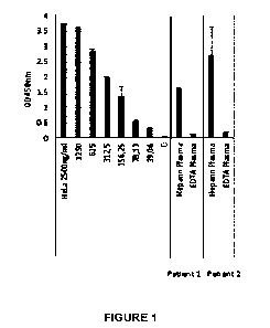
Figure 1. The results of an immunoassay for neutrophil extracellular trap
(NET) derived
nucleosomes in EDTA plasma and heparin plasma samples taken from 2 healthy
volunteers.
The EDTA samples contain low levels of NET derived nucleosome material. In
contrast,
heparin induces NET formation and the heparin plasma samples contain high
levels of induced
NET derived nucleosomes.
Figure 2. Bioanalyzer electrophoresis results for NET derived nucleosomes in
EDTA plasma
and heparin plasma samples taken from 2 healthy volunteers. The EDTA samples
contain low
levels of both mononucleosomes and NET derived nucleosome material. In
contrast, heparin
induces NET formation and the heparin plasma samples contain low levels of
mononucleosomes (peak at approximately 60 seconds) but high levels of induced
NET
derived nucleosomes (wide peak at approximately 110 seconds). The narrow peaks
at
approximately 43 seconds and approximately 110 seconds represent DNA samples
added for
reference purposes.
Figure 3. Levels of nucleosomes containing histone isoform H3.1 measured in 50
patients
admitted to hospital for symptoms of COVID-19 infection, including 34
symptomatic patients
who tested positive for CO VI D-19 infection by PCR and 16 symptomatic
patients who tested
negative by PCR, as well as 50 normal subjects displaying no symptoms of
disease.
Figure 4. Levels of nucleosomes containing histone isoform H3.1 measured in 15
patients
with PCR confirmed COVID-19 infection, including: 5 samples collected from
patients
attending an outpatient hospital appointment or at presentation at the
hospital Emergency
Room (ER); 3 patients hospitalized in normal wards; 2 patients hospitalized in
an intensive
care unit (ICU) who required respiratory support and survived; and 4 patients
hospitalized in
an ICU who required respiratory support and died.
Figure 5. Levels of nucleosomes containing histone modification H3R8Cit
measured in 15
patients with PCR confirmed COVI D-19 infection, including: 5 samples
collected from patients
attending an outpatient hospital appointment or at presentation at the
hospital ER; 3 patients
2
CA 03175171 2022-09-13
WO 2021/186037 PCT/EP2021/057096
hospitalized in normal wards; 2 patients hospitalized in an ICU who required
respiratory
support and survived; and 4 patients hospitalized in an ICU who required
respiratory support
and died.
Figure 6. Results from the experiment described in Example 12 showing the mean
levels of
nucleosomes containing histone isoform H3.1 measured in 16 pigs induced with
sepsis placed
on plasmapheresis. In 9 pigs, plasma was passed through a cartridge containing
NET binders
(treated, closed bars) and in 7 pigs, plasma was passed through a control
cartridge which did
not contain a NETs binder (control, open bars).
Figure 7. Results from the experiment described in Example 12 and shown in
Figure 6, but
for levels in individual test subjects.
Figure 8. H3.1-nucleosome levels measured in human subjects diagnosed with
sepsis and
healthy human subjects.
SUMMARY OF THE INVENTION
According to a first aspect, there is provided a method of monitoring the
progress of a disease
in a subject suffering from an infection, comprising:
(i) contacting a body fluid sample obtained from the subject with a binding
agent to
detect or measure the level of cell free nucleosomes or a component thereof;
(ii) repeating step (i) on one or more occasions; and
(iii) using any changes in the level of cell free nucleosomes or component
thereof to
monitor the progression of the infection in the subject.
According to a further aspect, there is provided a method of assigning a risk
of the
development or progression of a medical complication in a subject suffering
from an infection,
comprising:
(i) contacting a body fluid sample obtained from the subject with a binding
agent to
detect or measure the level of cell free nucleosomes or a component thereof;
and
(ii) using the level of cell free nucleosomes detected to assign the
likelihood that a
medical complication will develop or progress in said subject.
According to a further aspect, there is provided a method of assigning a risk
of an adverse
outcome to a subject suffering from an infection, comprising:
(i) contacting a body fluid sample obtained from the subject with a binding
agent to
detect or measure the level of cell free nucleosomes or a component thereof;
and
3
CA 03175171 2022-09-13
WO 2021/186037 PCT/EP2021/057096
(ii) using the level of cell free nucleosomes detected to assign the
likelihood of an
adverse outcome to said subject,
wherein a subject identified with a high likelihood of an adverse outcome is
assigned
for medical intervention.
According to a further aspect, there is provided a method of selecting a
subject suffering from
an infection, who is in need of medical treatment for a medical complication
of the infection,
comprising:
(i) contacting a body fluid sample obtained from the subject with a binding
agent to
detect or measure the level of cell free nucleosomes or a component thereof;
and
(ii) using the level of cell free nucleosomes detected to indicate the
presence,
progression or development of a medical complication in need of treatment in
said subject.
In preferred embodiments the infection is a respiratory influenza or
coronavirus infection and
the medical complication is ARS, ARDS or SARS or pneumonia. Therefore in one
embodiment
there is provided a method of detecting a subject in need of medical treatment
for pneumonia,
ARS, ARDS or SARS, comprising:
(i) contacting a body fluid sample obtained from the subject with a binding
agent to
detect or measure the level of cell free nucleosomes or a component thereof;
and
(ii) using the level of cell free nucleosomes as an indicator that the subject
is in need
of medical treatment for pneumonia, ARS, ARDS or SARS.
In preferred embodiments the infection is a respiratory influenza or
coronavirus infection and
the medical complication is ARS, ARDS, SARS or pneumonia.
In other preferred embodiments the infection is sepsis. Therefore in one
embodiment there is
provided a method of detecting a subject in need of medical treatment for
sepsis or septic
shock, comprising:
(i) contacting a body fluid sample obtained from the subject with a binding
agent to
detect or measure the level of cell free nucleosomes or a component thereof;
and
(ii) using the level of cell free nucleosomes as an indicator that the subject
is in need
of medical treatment for sepsis or septic shock.
According to a further aspect of the invention, there is provided a method of
monitoring an
infection in a subject, comprising:
(i) contacting a body fluid sample obtained from the subject with a binding
agent to
detect or measure the level of cell free nucleosomes or a component thereof;
4
CA 03175171 2022-09-13
WO 2021/186037 PCT/EP2021/057096
(ii) repeating the detection or measurement of the level of cell free
nucleosomes or a
component thereof in a body fluid obtained from the subject on one or more
occasions;
(iii) using any changes in the level of cell free nucleosomes or component
thereof to
monitor the progression of the infection in the subject.
DETAILED DESCRIPTION
Nucleosomes are released into the circulation on fragmentation of chromatin on
cell death.
Many infections, such as viral infections, initiate cell death through a
variety of mechanisms
(cell binding and entry, endosomal TLR3 activation and gene expression)
thereby increasing
the number of circulating nucleosomes in the blood (Danthi etal., Annu. Rev.
Virol. (2016) 3:
533-53). In addition, infections can induce NETosis whereby post-translational
histone
modifications, such as acetylation or hypercitrullination of histones H3 and
H4 (Wang Y etal.,
J. Cell Biol. (2009) 184(2): 205-213), promote decondensation of chromatin
which is released
into circulation together as a first line response to infection. However,
extracellular
nucleosomes and neutrophil extracellular traps (NETs) can cause severe
complications if not
cleared rapidly. For example, nucleosome binding to the glomerular membrane is
associated
with kidney damage in lupus (Kalaaji etal., Kidney Int. (2007) 71(7): 665-
672), whilst NETs
have been shown to intensify pulmonary injury during viral pneumonia (Ashar et
al., Am. J.
Pathol. (2018) 188(1): 135-148). Indeed, host directed NET toxicity is
associated with
respiratory distress, occlusion of narrow airways, endothelial and epithelial
cell damage,
inflammatory response and thrombus formation and other pathologies (Marcos et
al., Nat.
Med. (2010) 16: 1018-23; Hoeksema etal., Future Microbiol. (2016) 11:441-53).
Most subjects infected with influenza or coronavirus experience mild illness.
However, some
population subgroups, including elderly persons aged over 60 years and persons
with an
underlying medical condition such as diabetes, chronic lung conditions and
particularly chronic
cardiac conditions, are at risk of severe effects including ARS, SARS,
pneumonia and death.
The exact mechanism by which influenza or coronavirus infection leads to
complications
including pneumonia is not clear, but it is thought to be caused by a
hyperimmune reaction to
the viral infection in which excessive NETs contribute to acute injury of the
lung leading to
pneumonia and, in the worst cases, death.
The present invention utilises elevated levels of cell free nucleosomes,
including NETs, to
predict severity of disease and outcome in infectious disease.
Therefore, according to one aspect, there is provided a method of assigning a
risk of an
adverse outcome to a subject suffering from an infection, comprising:
CA 03175171 2022-09-13
WO 2021/186037 PCT/EP2021/057096
(i) contacting a body fluid sample obtained from the subject with a binding
agent to
detect or measure the level of cell free nucleosomes or a component thereof;
and
(ii) using the level of cell free nucleosomes detected to assign the
likelihood of an
adverse outcome to said subject. The method may be used so that a subject
identified with a
high likelihood of an adverse outcome is assigned for medical intervention.
In one embodiment, there is provided a method of assigning a risk of an
adverse outcome to
a subject suffering from an infection, comprising:
(i) contacting a body fluid sample obtained from the subject with a binding
agent to
detect or measure the level of neutrophil extracellular trap material or a
component thereof;
and
(ii) using the level of neutrophil extracellular trap material detected to
assign the
likelihood of an adverse outcome to said subject.
The nucleosome is the basic unit of chromatin structure and consists of a
protein complex of
eight highly conserved core histones (comprising of a pair of each of the
histones H2A, H2B,
H3, and H4). Around this complex is wrapped approximately 146 base pairs of
DNA. Another
histone, H1 or H5, acts as a linker and is involved in chromatin compaction.
The DNA is wound
around consecutive nucleosomes in a structure often said to resemble "beads on
a string" and
this forms the basic structure of open or euchromatin. In compacted or
heterochromatin this
string is coiled and super coiled into a closed and complex structure (Herranz
and EsteIler,
Methods Mol. Biol. (2007) 361: 25-62).
References to "nucleosome" may refer to "cell free nucleosome" when detected
in body fluid
samples. It will be appreciated that the term cell free nucleosome throughout
this document is
intended to include any cell free chromatin fragment that includes one or more
nucleosomes.
It will be understood that the cell free nucleosome may be detected by binding
to a component
thereof. The term "component thereof" as used herein refers to a part of the
nucleosome,
the whole nucleosome does not need to be detected. The component of the cell
free
nucleosomes may be selected from the group consisting of: a histone protein
(i.e. histone H1,
H2A, H2B, H3 or H4), a histone post-translational modification, a histone
variant or isoform, a
protein bound to the nucleosome (i.e. a nucleosome-protein adduct), a DNA
fragment
associated with the nucleosome and/or a modified nucleotide associated with
the nucleosome.
For example, the component thereof may be histone (isoform) H3.1 or histone H1
or DNA.
6
CA 03175171 2022-09-13
WO 2021/186037 PCT/EP2021/057096
Methods and uses of the invention may measure the level of (cell free)
nucleosomes per se.
References to "nucleosomes per se" refers to the total nucleosome level or
concentration
present in the sample, regardless of any epigenetic features the nucleosomes
may or may not
include. Detection of the total nucleosome level typically involves detecting
a histone protein
common to all nucleosomes, such as histone H4. Therefore, nucleosomes per se
may be
measured by detecting a core histone protein, such as histone H4. As described
herein,
histone proteins form structural units known as nucleosomes which are used to
package DNA
in eukaryotic cells.
Normal cell turnover in adult humans involves the creation by cell division of
a huge number
of cells daily and the death of a similar number, mainly by apoptosis. During
the process of
apoptosis chromatin is broken down into mononucleosomes and oligonucleosomes
which are
released from the cells. Under normal conditions the levels of circulating
nucleosomes found
in healthy subjects is reported to be low. Elevated levels are found in
subjects with a variety
of conditions including many cancers, auto-immune diseases, inflammatory
conditions, stroke
and myocardial infarction (Holdenreider & Stieber, Crit. Rev. Olin. Lab. Sci.
(2009) 46(1): 1-
24).
Previous nucleosome ELISA methods were used primarily in cell culture, usually
as a method
to detect apoptosis (Salgame etal., Nucleic Acids Res. (1997) 25(3): 680-681;
Holdenrieder
et al. (2001) supra; van Nieuwenhuijze et al., Ann. Rheum. Dis. (2003) 62: 10-
14), but are
also used for the measurement of circulating cell free nucleosomes in serum
and plasma
(Holdenrieder etal. (2001)). Cell free serum and plasma nucleosome levels
released into the
circulation by dying cells have been measured by ELISA methods in studies of a
number of
different cancers to evaluate their use as a potential biomarker.
The cell free nucleosome may be mononucleosomes, oligonucleosomes, a
constituent part of
a larger chromatin fragment or a constituent part of a NET or a mixture
thereof.
Mononucleosomes and oligonucleosomes can be detected by Enzyme-Linked
ImmunoSorbant Assay (ELISA) and several methods have been reported (e.g.
Salgame etal.
(1997); Holdenrieder et al. (2001); van Nieuwenhuijze et al. (2003)). These
assays typically
employ an anti-histone antibody (for example anti-H2B, anti-H3 or anti-H1,
H2A, H2B, H3 and
H4) as capture antibody and an anti-DNA or anti-H2A-H2B-DNA complex antibody
as
detection antibody.
7
CA 03175171 2022-09-13
WO 2021/186037 PCT/EP2021/057096
Circulating nucleosomes are not a homogeneous group of protein-nucleic acid
complexes.
Rather, they are a heterogeneous group of chromatin fragments originating from
the digestion
of chromatin on cell death and include an immense variety of epigenetic
structures including
particular histone isoforms (or variants), post-translational histone
modifications, nucleotides
or modified nucleotides, and protein adducts. It will be clear to those
skilled in the art that an
elevation in nucleosome levels will be associated with elevations in some
circulating
nucleosome subsets containing particular epigenetic signals including
nucleosomes
comprising particular histone isoforms (or variants), comprising particular
post-translational
histone modifications, comprising particular nucleotides or modified
nucleotides and
comprising particular protein adducts. Assays for these types of chromatin
fragments are
known in the art (for example, see WO 2005/019826, WO 2013/030579, WO
2013/030578,
WO 2013/084002 which are herein incorporated by reference).
A number of proteins occur in NETs that are adducted directly or indirectly to
nucleosomes.
These proteins include, without limitation, myeloperoxidase (M PO), neutrophil
elastase (NE),
lactotransferrin, azurocidin, cathepsin G, leukocyte proteinase 3, lysozyme C,
neutrophil
defensin 1, neutrophil defensin 3, myeloid cell nuclear differentiation
antigen, S100 calcium-
binding protein A8, S100 calcium-binding protein A9, S100 calcium-binding
protein Al2, actin
13, actin y, alpha-actin, plastin-2, cytokeratin-10, catalase, alpha-enolase
and transketolase
(Urban et al., PLOS Pathogens. (2009) 10: e1000639). Any nucleosome-protein
adduct that
occurs in NETs is a useful adduct for the detection of elevated levels of NETs
in methods of
the invention. C-reactive protein (CRP) may also be adducted to nucleosomes in
NETs and
nucleosome-CRP adduct is therefore a useful adduct for the detection of
elevated levels of
NETs in methods of the invention.
In preferred embodiments of the invention the adduct used is a MPO-nucleosome
adduct or a
NE-nucleosome adduct.
In one embodiment, the component of the cell free nucleosome comprises an
epigenetic
feature of the cell free nucleosome.
The biomarker used in the methods of the invention may be the level of cell
free nucleosomes
per se and/or an epigenetic feature of a cell free nucleosome. It will be
understood that the
terms "epigenetic signal structure" and "epigenetic feature" are used
interchangeably herein.
They refer to particular features of the nucleosome that may be detected. In
one embodiment,
the epigenetic feature of the nucleosome is selected from the group consisting
of: a post-
8
CA 03175171 2022-09-13
WO 2021/186037 PCT/EP2021/057096
translational histone modification, a histone isoform, a modified nucleotide
and/or proteins
bound to a nucleosome in a nucleosome-protein adduct.
In one embodiment, the epigenetic feature of the nucleosome comprises one or
more histone
variants or isoforms. The epigenetic feature of the cell free nucleosome may
be a histone
isoform, such as a histone isoform of a core nucleosome, in particular a
histone H3 isoform.
The term "histone variant" and "histone isoform" may be used interchangeably
herein. The
structure of the nucleosome can also vary by the inclusion of alternative
histone isoforms or
variants which are different gene or splice products and have different amino
acid sequences.
Many histone isoforms are known in the art. Histone variants can be classed
into a number of
families which are subdivided into individual types. The nucleotide sequences
of a large
number of histone variants are known and publicly available for example in the
National
Human Genome Research Institute NHGRI Histone Database (Marino-Ramirez et al.
The
Histone Database: an integrated resource for histones and histone fold-
containing proteins.
Database Vol.2011. and http://genome.nhgri.nih.gov/histones/complete.shtml),
the GenBank
(NIH genetic sequence) Database, the EMBL Nucleotide Sequence Database and the
DNA
Data Bank of Japan (DDBJ). For example, variants of histone H2 include H2A1,
H2A2,
mH2A1, mH2A2, H2AX and H2AZ. In another example, histone isoforms of H3
include H3.1,
H3.2, H3.3 and H3t.
In one embodiment, the histone isoform is H3.1.
The structure of nucleosomes can vary by post translational modification (PTM)
of histone
proteins. PTM of histone proteins typically occurs on the tails of the core
histones and common
modifications include acetylation, methylation or ubiquitination of lysine
residues as well as
methylation or citrullination of arginine residues and phosphorylation of
serine residues and
many others. Many histone modifications are known in the art and the number is
increasing
as new modifications are identified (Zhao and Garcia (2015) Cold Spring Harb
Perspect Biol,
7: a025064). Therefore, in one embodiment, the epigenetic feature of the cell
free nucleosome
may be a histone post translational modification (PTM). The histone PTM may be
a histone
PTM of a core nucleosome, e.g. H3, H2A, H2B or H4, in particular H3, H2A or
H2B. In
particular, the histone PTM is a histone H3 PTM. Examples of such PTMs are
described in
WO 2005/019826.
For example, the post translational modification may include acetylation,
methylation, which
may be mono-, di-or tri-methylation, phosphorylation, ribosylation,
citrullination, ubiquitination,
hydroxylation, glycosylation, nitrosylation, glutamination and/or
isomerisation (see Ausio
9
CA 03175171 2022-09-13
WO 2021/186037 PCT/EP2021/057096
(2001) Biochem Cell Bio 79: 693). In one embodiment, the histone PTM is
selected from
citrullination or ribosylation. In a further embodiment, the histone PTM is H3
citrulline (H3cit)
or H4 citrulline (H4cit). In a yet further embodiment, the histone PTM is
H3cit.
In one embodiment, the histone PTM is ribosylation, also referred to as ADP-
ribosylation.
Post-translational histone ADP-ribosylation of nucleosomes occupying promoters
of
inflammatory response markers in macrophages is stimulated by exposure to
lipopolysaccharides leading to elevated transcription and may have antiviral
properties.
Moreover, all members of the Coronavirus family contain a highly conserved
macrodomain
within non-structural protein 3 (n5p3) that regulates post-translational ADP-
ribosylation by
enzymatic removal of covalently attached ADP-ribose from protein targets.
Recombinant
severe acute respiratory syndrome coronavirus (SARS-CoV) strains containing
mutated
macrodomains with reduced nsp3 de-ADP-ribosylation activity, are less
infective and elicit an
early, enhanced interferon (I FN), interferon-stimulated gene (I SG), and
proinflammatory
cytokine response. Therefore, altered levels of circulating ADP-ribosylated
nucleosomes
released from macrophages are expected to be useful in methods of the
invention.
A group or class of related histone post translational modifications (rather
than a single
modification) may also be detected. A typical example, without limitation,
would involve a 2-
site immunoassay employing one antibody or other selective binder directed to
bind to
nucleosomes and one antibody or other selective binder directed to bind the
group of histone
modifications in question. Examples of such antibodies directed to bind to a
group of histone
modifications would include, for illustrative purposes without limitation,
anti-pan-acetylation
antibodies (e.g. a Pan-acetyl H4 antibody [H4panAc]), anti-citrullination
antibodies or anti-
ubiquitin antibodies.
In one embodiment, the epigenetic feature of the nucleosome comprises one or
more DNA
modifications. In addition to the epigenetic signalling mediated by nucleosome
histone isoform
and PTM composition, nucleosomes also differ in their nucleotide and modified
nucleotide
composition. Some nucleosomes may comprise more 5-methylcytosine residues (or
5-
hydroxymethylcytosine residues or other nucleotides or modified nucleotides)
than other
nucleosomes. In one embodiment, the DNA modification is selected from 5-
methylcytosine or
5-hydroxymethylcytosine.
In one embodiment, the epigenetic feature of the nucleosome comprises one or
more protein-
nucleosome adducts or complexes. A further type of circulating nucleosome
subset is
nucleosome protein adducts. It has been known for many years that chromatin
comprises a
CA 03175171 2022-09-13
WO 2021/186037 PCT/EP2021/057096
large number of non-histone proteins bound to its constituent DNA and/or
histones. These
chromatin associated proteins are of a wide variety of types and have a
variety of functions
including transcription factors, transcription enhancement factors,
transcription repression
factors, histone modifying enzymes, DNA damage repair proteins and many more.
These
chromatin fragments including nucleosomes and other non-histone chromatin
proteins or DNA
and other non-histone chromatin proteins are described in the art.
In one embodiment, the protein adducted to the nucleosome (and which therefore
may be
used as a biomarker) is selected from: a transcription factor, a High Mobility
Group Protein or
chromatin modifying enzyme. References to "transcription factor" refer to
proteins that bind to
DNA and regulate gene expression by promoting (i.e. activators) or suppressing
(i.e.
repressors) transcription. Transcription factors contain one or more DNA-
binding domains
(DBDs), which attach to specific sequences of DNA adjacent to the genes that
they regulate.
All of the circulating nucleosomes and nucleosome moieties, types or subgroups
described
herein may be useful in the present invention.
It will be understood that more than one epigenetic feature of cell free
nucleosomes may be
detected in methods and uses of the invention. Multiple biomarkers may be used
as a
combined biomarker. Therefore, in one embodiment, the use comprises more than
one
epigenetic feature of cell free nucleosomes as a combined biomarker. The
epigenetic features
may be the same type (e.g. PTMs, histone isoforms, nucleotides or protein
adducts) or
different types (e.g. a PTM in combination with a histone isoform). For
example, a post-
translational histone modification and a histone variant may be detected (i.e.
more than one
type of epigenetic feature is detected). Alternatively, or additionally, more
than one type of
post-translational histone modification is detected, or more than one type of
histone isoform is
detected. In one aspect, the use comprises a post-translational histone
modification and a
histone isoform as a combined biomarker in a sample, for the diagnosis,
detection, treatment
selection, prognostication or monitoring of an infection. In one embodiment,
the combined
biomarker is H3.1 and H3cit. In an alternative embodiment, the combined
biomarker is H3.1
and H4cit.
The term "biomarker" means a distinctive biological or biologically derived
indicator of a
process, event, or condition. Biomarkers can be used in methods of diagnosis,
e.g. clinical
screening, and prognosis assessment and in monitoring the results of therapy,
identifying
patients most likely to respond to a particular therapeutic treatment, drug
screening and
development. Biomarkers and uses thereof are valuable for identification of
new drug
treatments and for discovery of new targets for drug treatment.
11
CA 03175171 2022-09-13
WO 2021/186037 PCT/EP2021/057096
Biomarkers are also useful as companion diagnostic products for the selection
of patients
suitable for treatment by a particular therapy. We have demonstrated herein
that tests for
circulating nucleosome levels, or levels of nucleosomes containing particular
epigenetic
signals or structures, are useful companion products to therapies for NETs or
NETosis related
diseases.
The methods of the invention are directed to assigning the patient with a risk
of an adverse
outcome. Adverse outcomes include mortality and/or an acute event requiring
immediate
medical care, for example, hospitalisation (i.e. hospital treatment) and/or
surgery. For many
patients, infections are overcome by their own immune system without requiring
medical
intervention. However, in a number of patients, infections can proceed or
increase in severity
without being overcome by the immune system or the patient's own immune
response to an
infection may lead to an adverse outcome. For example, an adverse outcome may
include an
acute coronary or cardiac event (such as a myocardial infarction and/or
stroke), acute multi-
or single-organ failure (such as renal failure, liver failure and/or heart
failure), onset of a
debilitating acute condition and/or an acute respiratory condition (such as
pneumonia,
hypoventilation/bradypnea, acute respiratory distress syndrome (ARDS) severe
acute
respiratory syndrome (SARS), bronchiolitis and/or bronchitis). Thus, in one
embodiment, the
methods described herein assign a patient or subject with a risk of developing
an acute
respiratory condition. In a further embodiment, the acute respiratory
condition is pneumonia.
In a further embodiment, the acute respiratory condition is
hypoventilation/bradypnea. In a yet
further embodiment, the acute respiratory condition is acute respiratory
distress syndrome
(ARDS) and/or severe acute respiratory syndrome (SARS).
Assigning the patient with a risk of an adverse outcome may assign a near- or
short-term risk
or may assign a medium-term risk. A near- or short-term risk includes wherein
the patient may
develop an adverse outcome within 30 days, such as within 2 weeks or 14 days,
within 1 week
or 7 days or within 5 days or less of presentation of symptoms or of a
positive diagnosis. Such
near- or short-term risk may also include wherein the patient may develop an
adverse outcome
within 30 days, such as within 2 weeks or 14 days, within 1 week or 7 days or
within 5 days or
less of performing the methods described herein. An example of a short term
risk includes the
development of NETs related complications to a COVID infection requiring
hospital treatment.
A medium-term risk includes wherein the patient may develop an adverse outcome
more than
30 days after presentation of symptoms, a positive diagnosis and/or the
performing of methods
as described herein. An example of a medium term risk includes the development
of so called
long-COVID wherein the effects of a COVID infection may continue for many
months. Thus,
12
CA 03175171 2022-09-13
WO 2021/186037 PCT/EP2021/057096
in one embodiment, the methods described herein assign a patient or subject
with a risk of
developing an adverse outcome within 2 weeks, or 14 days, of presentation of
symptoms or a
positive diagnosis. In a further embodiment, the methods described herein
assign a risk of
developing an adverse outcome within 1 week, or 7 days, of presentation of
symptoms or a
positive diagnosis. In a yet further embodiment, the methods described herein
assign a risk of
developing an adverse outcome within 5 days of presentation of symptoms or a
positive
diagnosis.
Therefore, in another aspect of the invention, there is provided a method of
identifying a
subject with an infection requiring hospital treatment, comprising:
(i) contacting a body fluid sample obtained from the subject with a binding
agent to
detect or measure the level of cell free nucleosomes or a component thereof;
and
(ii) using the level of cell free nucleosomes detected to determine if the
subject should
be admitted to hospital for treatment.
It will be understood that methods of the invention may also be used to
identify patients who
do not require hospital treatment, i.e. using the level of cell free
nucleosomes detected to
determine if the subject should not be admitted to hospital for treatment.
This mode of the
invention would help to identify patients who can be discharged early if they
have already been
admitted to hospital.
Methods and uses described herein may be tested in body fluid samples, in
particular blood,
serum or plasma samples. Preferably, plasma samples are used. Plasma samples
may be
collected in collection tubes containing one or more anticoagulants such as
ethylenediamine
tetraacetic acid (EDTA), heparin, or sodium citrate, in particular EDTA.
Infections
The methods of the current invention find particular use in managing
infectious outbreaks.
Infections can be caused by different pathogens and environmental factors. In
one
embodiment, the infection is a viral, bacterial, fungal or microbial
infection. Bacterial infections
may include mycobacterial, pneumococcal and influenzae infections, such as
infections (e.g.
pneumonia) caused by Streptococcus pneumoniae, Escherichia coil, Mycobacterium
tuberculosis, Haemophilus influenzae and Staphylococcus aureus. In a further
embodiment,
the infection is a viral infection. Viral infections may include infections
caused by respiratory
syncytial virus (RSV), influenza type A, influenza type B and coronaviruses
(e.g. COVID-19).
13
CA 03175171 2022-09-13
WO 2021/186037 PCT/EP2021/057096
The infection can be defined by the tissue affected by the disease. For
example, the disease
may affect the heart, brain, kidneys, liver, pancreas, lungs and/or blood and
the infection may
be a bacterial, viral, fungal or microbial infection known to commonly affect
such tissues or
organs. In one embodiment, the infection is a respiratory tract infection.
According to this
embodiment, the infection affects the lungs, upper and/or lower respiratory
tract.
Other tissues which may be affected by the disease include peripheral tissues
such as limbs,
hands and feet and the infection may be a bacterial infection (e.g. gangrene).
In one
embodiment, the infection and/or disease may affect multiple tissues or organs
simultaneously. For example, the infection may be a bacterial infection of a
limb, hand or foot
and the disease may also affect the blood (e.g. sepsis). In one embodiment,
the infection is
sepsis. In another example, the disease may be cardiac or coronary failure and
other tissues
or organs affected by the disease may include the kidneys and renal system
and/or the brain
(e.g. stroke). In a yet further example, the disease may affect the lungs or
the infection may
be a respiratory tract infection and other tissues or organs affected may
include the heart,
coronary system and/or brain (e.g. heart failure, myocardial infarction and/or
stroke).
In one embodiment, circulating nucleosome levels are measured in a sample
taken from a
subject suffering from an infection to determine the prognosis of the disease.
In another
embodiment, circulating nucleosome levels are measured in multiple samples
taken at
intervals from a subject suffering from an infection to monitor the progress
of the disease
and/or to assess the efficacy of treatment.
In a further embodiment, circulating nucleosome levels are measured in a
sample taken from
a subject suffering from sepsis or septic shock, in particular to assess the
prognosis of the
disease. Further measurements on multiple samples taken at intervals from a
subject suffering
from sepsis or septic shock may be made to monitor the progress of the disease
and/or to
assess the efficacy of treatment.
In one embodiment, the respiratory tract infection is selected from:
influenza, pneumonia and
severe acute respiratory syndrome (SARS). SARS is a respiratory infection
caused by the
SARS coronavirus (SARS-CoV) and other, related coronaviruses are known (e.g.
COVID-19
(also known as SARS-CoV-2 and previously as 2019-nCoV)). It is known to cause
fever flu-
like symptoms, cough and lethargy and can lead to pneumonia (e.g. direct viral
pneumonia or
secondary bacterial pneumonia).
14
CA 03175171 2022-09-13
WO 2021/186037 PCT/EP2021/057096
The emergence and rapid progression of COVI D-19 to pandemic status places
severe strain
on international health care services. Predictions of infectivity range as
high as 70-80% of
national populations. It appears that whilst most people will experience mild
symptoms, double
digit percentages of those infected may be severely affected.
Identifying COVID-19 positive individuals at high risk of severe reaction or a
complication,
including pneumonia, would allow triaging and facilitate allocation of
strained medical
resources including critical care beds and ventilators, until herd immunity is
established,
protecting communities from future widespread outbreaks. Therefore, in a
preferred
embodiment there is provided a method of identifying a subject infected with
an influenza or
coronavirus infection requiring medical treatment, comprising:
(i) contacting a body fluid sample obtained from the subject with a binding
agent to
detect or measure the level of cell free nucleosomes or a component thereof;
and
(ii) using the level of cell free nucleosomes detected to determine if the
subject requires
medical treatment.
Diagnosis and monitoring methods
According to a further aspect, there is provided a method of monitoring the
severity of an
infection in a subject, comprising:
(i) contacting a body fluid sample obtained from the subject with a binding
agent to
detect or measure the level of cell free nucleosomes or a component thereof;
(ii) repeating the detection or measurement of the level of cell free
nucleosomes or a
component thereof in a body fluid obtained from the subject on one or more
occasions;
(iii) using any changes in the level of cell free nucleosomes or component
thereof to
monitor the progression of the infection in the subject.
According to a further aspect, there is provided a method for monitoring the
progression of an
infection in a subject having or suspected of having an infection, or being
predisposed to a
poor prognosis on infection, which comprises the steps of:
(i) contacting a sample obtained from the subject with a binding agent to
detect or
measure the level of cell free nucleosomes; and
(ii) comparing the level of cell free nucleosomes detected with an earlier
sample
taken from said subject to monitor the progression of the infection.
According to a further aspect, there is provided a method of monitoring the
progress of a
disease in a subject suffering from an infection, comprising:
CA 03175171 2022-09-13
WO 2021/186037 PCT/EP2021/057096
(i) contacting a body fluid sample obtained from the subject with a binding
agent to
detect or measure the level of cell free nucleosomes or a component thereof;
(ii) repeating step (i) on one or more occasions; and
(iii) using any changes in the level of cell free nucleosomes or component
thereof to
monitor the progression of the infection in the subject.
If a subject is determined to not have an infection or to have a mild
infection, then the invention
may still be used for the purposes of monitoring disease progression for
future development
of a medical complication. For example, if the method comprises a sample from
a subject
determined to have a mild infection, then the biomarker level measurements can
be repeated
at another time point to establish if the biomarker level has changed.
Detecting and/or quantifying may be performed directly on the purified or
enriched nucleosome
sample, or indirectly on an extract therefrom, or on a dilution thereof.
Quantifying the amount
of the biomarker present in a sample may include determining the concentration
of the
biomarker present in the sample. Uses and methods of detecting, monitoring and
of diagnosis
according to the invention described herein are useful to confirm the
existence of a disease,
to monitor development of the disease by assessing onset and progression, or
to assess
amelioration or regression of the disease. Uses and methods of detecting,
monitoring and of
diagnosis are also useful in methods for assessment of clinical screening,
prognosis, choice
of therapy, evaluation of therapeutic benefit, i.e. for drug screening and
drug development.
In one embodiment the disease is a condition involving pathological clinical
complications of
high levels of NETs or NETosis.
The detection or measurement may comprise an immunoassay, immunochemical, mass
spectroscopy, chromatographic, chromatin immunoprecipitation or biosensor
method. In
particular, detection and/or measurement may comprise a 2-site immunoassay
method for
nucleosome moieties. Such a method is preferred for the measurement of
nucleosomes or
nucleosome incorporated epigenetic features in situ employing two anti-
nucleosome binding
agents or an anti-nucleosome binding agent in combination with an anti-histone
modification
or anti-histone variant or anti-DNA modification or anti-adducted protein
detection binding
agent. Also, detection and/or measurement may comprise a 2-site immunoassay,
for example
employing combinations of a labelled or immobilized: anti-nucleosome, anti-
histone
modification, anti-histone variant/isoform, anti-DNA modification or anti-
adducted protein
binding agent.
16
CA 03175171 2022-09-13
WO 2021/186037 PCT/EP2021/057096
The inventors herein used a 2-site immunoassay for H3.1-nucleosomes employing
an
immobilized anti-histone H3.1 antibody directed to bind to an epitope around
amino acids 30-
33 of the histone H3.1 protein to capture clipped and non-clipped nucleosomes,
together with
a labelled anti-nucleosome antibody directed to bind to a epitope present in
intact
nucleosomes but not present on isolated (free) histone or DNA nucleosome
components. This
type of epitope may be referred to as a "conformational nucleosome epitope"
herein because
it requires the native three-dimensional configuration of the target
nucleosome to be intact.
H3R8Cit nucleosome measurements described herein were performed using a 2-site
immunoassay employing an immobilized antibody directed to bind to nucleosomes
citrullinated at arginine 8 of histone H3, together with the same labelled
anti-nucleosome
antibody directed to bind to a conformational nucleosome epitope.
In one embodiment, the method of detection or measurement comprises contacting
the body
fluid sample with a solid phase comprising a binding agent that detects cell
free nucleosomes
or a component thereof, and detecting binding to said binding agent.
In one embodiment, the method of detection or measurement comprises: (i)
contacting the
sample with a first binding agent which binds to an epigenetic feature of a
cell free
nucleosome; (ii) contacting the sample bound by the first binding agent in
step (i) with a second
binding agent which binds to cell free nucleosomes; and (iii) detecting or
quantifying the
binding of the second binding agent in the sample.
In another embodiment, the method of detection or measurement comprises: (i)
contacting
the sample with a first binding agent which binds to cell free nucleosomes;
(ii) contacting the
sample bound by the first binding agent in step (i) with a second binding
agent which binds to
an epigenetic feature of the cell free nucleosome; and (iii) detecting or
quantifying the binding
of the second binding agent in the sample.
Detecting or measuring the level of the biomarker(s) may be performed using
one or more
reagents, such as a suitable binding agent. For example, the one or more
binding agents may
comprise a ligand or binder specific for the desired biomarker, e.g.
nucleosomes or component
part thereof, an epigenetic feature of a nucleosome, a structural/shape mimic
of the
nucleosome or component part thereof, optionally in combination with one or
more
interleukins.
17
CA 03175171 2022-09-13
WO 2021/186037 PCT/EP2021/057096
It will be clear to those skilled in the art that the terms "antibody",
"binder" or "ligand" as used
herein are not limiting but are intended to include any binder capable of
binding to particular
molecules or entities and that any suitable binder can be used in the method
of the invention.
It will also be clear that the term "nucleosomes" is intended to include
mononucleosomes,
oligonucleosomes, NETs and any protein-DNA chromatin fragments that can be
analysed in
fluid media.
Methods of detecting biomarkers are known in the art. The reagents may
comprise one or
more ligands or binders, for example, naturally occurring or chemically
synthesised
compounds, capable of specific binding to the desired target. A ligand or
binder may comprise
a peptide, an antibody or a fragment thereof, or a synthetic ligand such as a
plastic antibody,
or an aptamer or oligonucleotide, capable of specific binding to the desired
target. The
antibody can be a monoclonal antibody or a fragment thereof. It will be
understood that if an
antibody fragment is used then it retains the ability to bind the biomarker so
that the biomarker
may be detected (in accordance with the present invention). A ligand/binder
may be labelled
with a detectable marker, such as a luminescent, fluorescent, enzyme or
radioactive marker;
alternatively or additionally a ligand according to the invention may be
labelled with an affinity
tag, e.g. a biotin, avidin, streptavidin or His (e.g. hexa-His) tag.
Alternatively, ligand binding
may be determined using a label-free technology for example that of ForteBio
Inc.
The term "detecting" or "diagnosing" as used herein encompasses
identification, confirmation,
and/or characterisation of a disease state. Methods of detecting, monitoring
and of diagnosis
according to the invention are useful to confirm the existence of a disease,
to monitor
development of the disease by assessing onset and progression, or to assess
amelioration or
regression of the disease. Methods of detecting, monitoring and of diagnosis
are also useful
in methods for assessment of clinical screening, prognosis, choice of therapy,
evaluation of
therapeutic benefit, i.e. for drug screening and drug development.
Methods of the invention may involve normalisation of marker levels. For
example, the level
of cell free nucleosomes containing a particular epigenetic feature may be
normalised against
the level of nucleosomes per se (or some other type of nucleosomes or
parameter) to express
the level as a proportion of nucleosomes containing the feature. For example,
to express the
level of citrullinated nucleosomes as the proportion of nucleosomes that are
citrullinated.
In one embodiment, the method described herein is repeated on multiple
occasions. This
embodiment provides the advantage of allowing the detection results to be
monitored over a
time period. Such an arrangement will provide the benefit of monitoring or
assessing the
18
CA 03175171 2022-09-13
WO 2021/186037 PCT/EP2021/057096
efficacy of treatment of a disease state. Such monitoring methods of the
invention can be
used to monitor onset, progression, stabilisation, amelioration, relapse
and/or remission.
In monitoring methods, test samples may be taken on two or more occasions. The
method
may further comprise comparing the level of the biomarker(s) present in the
test sample with
one or more control(s) and/or with one or more previous test sample(s) taken
earlier from the
same test subject, e.g. prior to commencement of therapy, and/or from the same
test subject
at an earlier stage of therapy. The method may comprise detecting a change in
the nature or
amount of the biomarker(s) in test samples taken on different occasions.
A change in the level of the biomarker in the test sample relative to the
level in a previous test
sample taken earlier from the same test subject may be indicative of a
beneficial effect, e.g.
stabilisation or improvement, of said therapy on the disorder or suspected
disorder.
Furthermore, once treatment has been completed, the method of the invention
may be
periodically repeated in order to monitor for the recurrence of a disease.
Methods for monitoring efficacy of a therapy can be used to monitor the
therapeutic
effectiveness of existing therapies and new therapies in human subjects and in
non-human
animals (e.g. in animal models). These monitoring methods can be incorporated
into screens
for new drug substances and combinations of substances.
In a further embodiment the monitoring of more rapid changes due to fast
acting therapies
may be conducted at shorter intervals of hours or days.
Diagnostic or monitoring kits (or panels) are provided for performing methods
of the invention.
Such kits will suitably comprise one or more ligands for detection and/or
quantification of the
biomarker according to the invention, and/or a biosensor, and/or an array as
described herein,
optionally together with instructions for use of the kit.
A further aspect of the invention is a kit for detecting the presence of an
infection, comprising
a biosensor capable of detecting and/or quantifying one or more of the
biomarkers as defined
herein. As used herein, the term "biosensor" means anything capable of
detecting the
presence of the biomarker. Examples of biosensors are described herein.
Biosensors may
comprise a ligand binder or ligands, as described herein, capable of specific
binding to the
biomarker. Such biosensors are useful in detecting and/or quantifying a
biomarker of the
invention.
19
CA 03175171 2022-09-13
WO 2021/186037 PCT/EP2021/057096
Suitably, biosensors for detection of one or more biomarkers combine
biomolecular
recognition with appropriate means to convert detection of the presence, or
quantitation, of
the biomarker in the sample into a signal. Biosensors can be adapted for
"alternate site"
diagnostic testing, e.g. in the ward, outsubjects' department, surgery, home,
field and
workplace. Biosensors to detect one or more biomarkers of the invention
include acoustic,
plasmon resonance, holographic, Bio-Layer lnterferometry (BLI) and
microengineered
sensors. Imprinted recognition elements, thin film transistor technology,
magnetic acoustic
resonator devices and other novel acousto-electrical systems may be employed
in biosensors
for detection of the one or more biomarkers.
Biomarkers for detecting the presence of a disease are essential targets for
discovery of novel
targets and drug molecules that retard or halt progression of the disorder. As
the level of the
biomarker is indicative of disorder and of drug response, the biomarker is
useful for
identification of novel therapeutic compounds in in vitro and/or in vivo
assays. Biomarkers
described herein can be employed in methods for screening for compounds that
modulate the
activity of the biomarker.
Thus, in a further aspect of the invention, there is provided the use of a
binder or ligand, as
described, which can be a peptide, antibody or fragment thereof or aptamer or
oligonucleotide
directed to a biomarker according to the invention; or the use of a biosensor,
or an array, or a
kit according to the invention, to identify a substance capable of promoting
and/or of
suppressing the generation of the biomarker.
The immunoassays described herein include any method employing one or more
antibodies
or other specific binders directed to bind to the biomarkers defined herein.
Immunoassays
include 2-site immunoassays or immunometric assays employing enzyme detection
methods
(for example ELISA), fluorescence labelled immunometric assays, time-resolved
fluorescence
labelled immunometric assays, chemiluminescent
immunometric assays,
immunoturbidimetric assays, particulate labelled immunometric assays and
immunoradiometric assays as well as single-site immunoassays, reagent limited
immunoassays, competitive immunoassay methods including labelled antigen and
labelled
antibody single antibody immunoassay methods with a variety of label types
including
radioactive, enzyme, fluorescent, time-resolved fluorescent and particulate
labels. All of said
immunoassay methods are well known in the art, see for example Salgame etal.
(1997) and
van Nieuwenhuijze etal. (2003).
CA 03175171 2022-09-13
WO 2021/186037 PCT/EP2021/057096
Identifying, detecting and/or quantifying can be performed by any method
suitable to identify
the presence and/or amount of a specific protein in a biological sample from a
subject or a
purification or extract of a biological sample or a dilution thereof. In
particular, quantifying may
be performed by measuring the concentration of the target in the sample or
samples.
Biological samples that may be tested in a method of the invention include
those as defined
hereinbefore. The samples can be prepared, for example where appropriate
diluted or
concentrated, and stored in the usual manner. The present invention finds
particular use in
plasma samples which may be obtained from the subject.
Identification, detection and/or quantification of biomarkers may be performed
by detection of
the biomarker or of a fragment thereof, e.g. a fragment with C-terminal
truncation, or with N-
terminal truncation. Fragments are suitably greater than 4 amino acids in
length, for example
5, 6, 7, 8,9, 10, 11, 12, 13, 14, 15, 16, 17, 18, 19, 0r20 amino acids in
length. It is noted in
particular that peptides of the same or related sequence to that of histone
tails are particularly
useful fragments of histone proteins.
For example, detecting and/or quantifying can be performed by one or more
method(s)
selected from the group consisting of: immunoassay, immunochromatography,
SELDI (-TOF),
MALDI (-TOF), a 1-D gel-based analysis, a 2-D gel-based analysis, Mass
spectrometry (MS),
reverse phase (RP) LC, size permeation (gel filtration), ion exchange,
affinity, HPLC, UPLC
and other LC or LC MS-based techniques. Appropriate LC MS techniques include
!CAT
(Applied Biosystems, CA, USA), or iTRAQ (Applied Biosystems, CA, USA). Liquid
chromatography (e.g. high pressure liquid chromatography (HPLC) or low
pressure liquid
chromatography (LPLC)), thin-layer chromatography, NMR (nuclear magnetic
resonance)
spectroscopy could also be used.
Methods involving detection and/or quantification of one or more biomarkers of
the invention
can be performed on bench-top instruments, or can be incorporated onto
disposable,
diagnostic or monitoring platforms that can be used in a non-laboratory
environment, e.g. in
the physician's office or at the subject's bedside. Suitable biosensors for
performing methods
of the invention include "credit" cards with optical or acoustic readers.
Biosensors can be
configured to allow the data collected to be electronically transmitted to the
physician for
interpretation and thus can form the basis for e-medicine. Therefore, in a
further aspect of the
invention, there is provided the use of a near patient or point-of-care
immunoassay method
for the measurement of a biomarker according to the invention. In one
embodiment the near
patient immunoassay method comprises a point-of-care immunoassay instrument
(e.g. the
Abbott i-STAT or the LightDeck Diagnostics point-of-care immunoassay
instrument). In one
21
CA 03175171 2022-09-13
WO 2021/186037 PCT/EP2021/057096
embodiment the near patient immunoassay method comprises a lateral flow test.
In a
preferred embodiment the biomarker is a nucleosome or a nucleosome containing
an
epigenetic feature.
The identification of biomarkers for a disease state permits integration of
diagnostic
procedures and therapeutic regimes. The biomarkers provide the means to
indicate
therapeutic response, failure to respond, unfavourable side-effect profile,
degree of
medication compliance and achievement of adequate serum drug levels. The
biomarkers may
be used to provide warning of adverse drug response. Biomarkers are useful in
development
of personalized therapies, as assessment of response can be used to fine-tune
dosage,
minimise the number of prescribed medications, reduce the delay in attaining
effective therapy
and avoid adverse drug reactions. Thus, by monitoring a biomarker of the
invention, subject
care can be tailored precisely to match the needs determined by the disorder
and the
pharmacological profile of the subject, the biomarker can thus be used to
titrate the optimal
dose, predict a positive therapeutic response and identify those subjects at
high risk of severe
side effects.
Biomarker-based tests provide a first line assessment of 'new' subjects, and
provide objective
measures for accurate and rapid diagnosis, not achievable using the current
measures.
Biomarker monitoring methods, biosensors, point-of-care tests, lateral flow
tests and kits are
also vital as subject monitoring tools, to enable the physician to determine
whether relapse is
due to worsening of the disorder. If pharmacological treatment is assessed to
be inadequate,
then therapy can be reinstated or increased; a change in therapy can be given
if appropriate.
As the biomarkers are sensitive to the state of the disorder, they provide an
indication of the
impact of drug therapy.
References to "subject" or "patient" are used interchangeably herein. The
subject may be a
human or an animal subject. In one embodiment, the subject is a human. In one
embodiment,
the subject is a (non-human) animal. The panels and methods described herein
may be
performed in vitro, or ex vivo.
Detecting and/or quantifying may be compared to a cut-off level. Cut-off
values can be
predetermined by analysing results from multiple patients and controls, and
determining a
suitable value for classifying a subject as with or without the disease. For
example, for
diseases where the level of biomarker is higher in patients suffering from the
disease, then if
the level detected is higher than the cut-off, the patient is indicated to
suffer from the disease.
22
CA 03175171 2022-09-13
WO 2021/186037 PCT/EP2021/057096
Alternatively, for diseases where the level of biomarker is lower in patients
suffering from the
disease, then if the level detected is lower than the cut-off, the patient is
indicated to suffer
from the disease. The advantages of using simple cut-off values include the
ease with which
clinicians are able to understand the test and the elimination of any need for
software or other
aids in the interpretation of the test results. Cut-off levels can be
determined using methods in
the art.
Detecting and/or quantifying may also be compared to a control. It will be
clear to those skilled
in the art that the control subjects may be selected on a variety of basis
which may include,
for example, subjects known to be free of the disease or may be subjects with
a different
disease (for example, for the investigation of differential diagnosis). The
"control" may
comprise a healthy subject, a non-diseased subject and/or a subject without an
infection. The
control may also be a subject with the infection displaying no, or mild,
symptoms, such as a
subject infected with a respiratory virus displaying no, or mild, symptoms.
Mild symptoms may
include manageable symptoms which do not require hospital intervention and/or
intensive
medical treatment.
In one embodiment, a subject who tests positive by methods of the invention
may be infected
with a viral disease and additionally suffers, or goes on to suffer, further
medical complications.
In contrast, a control subject may also be infected with a viral disease but
does not suffer, and
does not go on to suffer, medical complications. Comparison with a control is
well known in
the field of diagnostics. The range of values found in the control group may
be used as a
normal or healthy or reference range against which the values found for test
subjects can be
compared. For example, if the reference range is <10 units, then a test value
of 5 units would
be considered normal, or not in need of treatment, but a value of 11 units
would be considered
abnormal and indicative of a need for treatment.
Therefore, in one embodiment, the method additionally comprises comparing the
level of cell
free nucleosomes or component thereof in the body fluid sample of the subject
with one or
more controls. For example, the method may comprise comparing the level of
cell free
nucleosomes present in a sample obtained from the subject with the level of
cell free
nucleosomes present in a sample obtained from a normal subject. The control
may be a
healthy subject.
In one embodiment, the level of cell free nucleosomes or component thereof is
elevated
compared to the control.
23
CA 03175171 2022-09-13
WO 2021/186037 PCT/EP2021/057096
It will be understood that it is not necessary to measure control levels for
comparative
purposes on every occasion. For example, for healthy/non-diseased controls,
once the
'normal range' is established it can be used as a benchmark for all subsequent
tests. A normal
range can be established by obtaining samples from multiple control subjects
without an
infection and testing for the level of biomarker. Results (i.e. biomarker
levels) for subjects
suspected to have an infection can then be examined to see if they fall
within, or outside of,
the respective normal range. Use of a 'normal range' is standard practice for
the detection of
disease.
In one embodiment, the method additionally comprises determining at least one
clinical
parameter for the patient. This parameter can be used in the interpretation of
results. Clinical
parameters may include any relevant clinical information for example, without
limitation, body
temperature, gender, weight, Body Mass Index (BMI), smoking status and dietary
habits.
Therefore, in one embodiment, the clinical parameter is selected from the
group consisting of:
body temperature, age, sex and body mass index (BMI).
In one embodiment, the method of the invention is performed to identify a
subject at high risk
of developing a severe reaction to an infection and therefore in need of
medical intervention.
Such medical intervention may include one or more of the therapies as
described herein.
According to another aspect of the invention, there is provided the use of a
binding agent in
the manufacture of a kit for use in a method of assigning a risk of an adverse
outcome to a
subject suffering from an infection, comprising:
(i) contacting a body fluid sample obtained from the subject with the binding
agent to
detect or measure the level of cell free nucleosomes or a component thereof;
and
(ii) using the level of cell free nucleosomes detected to assign the
likelihood of an
adverse outcome to said subject.
According to a further aspect of the invention, there is provided the use of a
binding agent in
the manufacture of a kit for use in a method of detecting a subject in need of
medical treatment
for pneumonia, acute respiratory syndrome (ARS) or severe acute respiratory
syndrome
(SARS), comprising:
(i) contacting a body fluid sample obtained from the subject with the binding
agent to
detect or measure the level of cell free nucleosomes or a component thereof;
and
(ii) using the level of cell free nucleosomes as an indicator that the subject
is in need
of medical treatment for pneumonia, ARS or SARS.
24
CA 03175171 2022-09-13
WO 2021/186037 PCT/EP2021/057096
According to a further aspect of the invention, there is provided the use of a
binding agent in
the manufacture of a kit for use in a method of detecting a subject in need of
medical treatment
for sepsis or septic shock, comprising:
(i) contacting a body fluid sample obtained from the subject with the binding
agent to
detect or measure the level of cell free nucleosomes or a component thereof;
and
(ii) using the level of cell free nucleosomes as an indicator that the subject
is in need
of medical treatment for sepsis or septic shock.
According to a further aspect of the invention, there is provided a method of
detecting a subject
in need of medical treatment for pneumonia, acute respiratory syndrome (ARS)
or severe
acute respiratory syndrome (SARS), comprising:
(i) contacting a body fluid sample obtained from the subject with a binding
agent to
detect or measure the level of cell free nucleosomes or a component thereof;
and
(ii) using the level of cell free nucleosomes as an indicator that the subject
is in need
of medical treatment for pneumonia, ARS or SARS.
According to a further aspect of the invention, there is provided a method of
detecting a subject
in need of medical treatment for sepsis or septic shock, comprising:
(i) contacting a body fluid sample obtained from the subject with a binding
agent to
detect or measure the level of cell free nucleosomes or a component thereof;
and
(ii) using the level of cell free nucleosomes as an indicator that the subject
is in need
of medical treatment for sepsis or septic shock.
Additional biomarkers
The level of cell free nucleosomes may be detected or measured as one of a
panel of
measurements. The panel may comprise different epigenetic features of the
nucleosome as
described hereinbefore (e.g. a histone isoform and a PTM). Biomarkers useful
in a panel test
for the detection of severe respiratory infections that require medical
intervention include,
without limitation, cytokine moieties (particularly interleukins), C-reactive
protein,
myeloperoxidase, D-Dimer, factor VII-activating protease (FSAP), fibrinogen
and
fibrin/fibrinogen breakdown products. In one embodiment, the panel comprises C-
reactive
protein. In one embodiment, the panel comprises one or more cytokines, such as
one or more
interleukins.
Interleukins (I Ls) are a group of cytokines, usually secreted by leukocytes,
that act as signal
molecules. They have key roles in stimulating immune responses and
inflammation. They
were first identified in the 1970s and have been designated numerically as
more interleukin
CA 03175171 2022-09-13
WO 2021/186037 PCT/EP2021/057096
types have been discovered. Examples of interleukins include, but are not
limited to: IL-1, IL-
2, IL-3, IL-4, IL-5, IL-6, IL-7, IL-8, IL-9, IL-10, IL-11, IL-12, IL-13, IL-14
and IL-15.
In one embodiment, the one or more interleukins is selected from the group
consisting of:
Interleukin-6 (IL-6) and Interleukin-12 (IL-12).
The interleukin may be IL-6. Interleukin-6 (IL-6) is a cytokine with a wide
variety of biological
functions. It is a potent inducer of fever and the acute phase response. The
sequence of
human IL-6 is known in the art and is described at UniProt Accession No.
P05231. In one
particular embodiment, the interleukin may be IL-6.
Alternatively, or additionally, the interleukin may be IL-12. Interleukin-12
(IL-12) is a T cell
stimulating factor because it stimulates the growth and function of T cells.
It is a heterodimeric
cytokine comprised of IL-12A and IL-12B. The sequence of human IL-12A is known
in the art
and is described at UniProt Accession No. P29459 and the sequence of human IL-
12B is also
known and described at UniProt Accession No. P29460. In one particular
embodiment, the
interleukin may be IL-12.
In one embodiment, the panel comprises a cell free nucleosome or an epigenetic
feature
thereof and an interleukin. In another embodiment, the panel comprises an
epigenetic feature
of a cell free nucleosome and two interleukins. For example, the cell free
nucleosome
measurement can be combined with more than one interleukin measurement, such
as IL-6
and IL-12. In a further embodiment, the epigenetic feature of a cell free
nucleosome is selected
from a histone isoform, such as H3.1, and a post translationally modified
histone, such as
H3cit. In a yet further embodiment, the panel of measurements is H3.1, H3cit,
H4cit and IL-6.
In one embodiment, the panel comprises C-reactive protein (CRP). CRP is a
pentameric
protein found in plasma and levels of CRP (whether or not adducted to
nucleosomes) increase
in plasma in response to inflammation, such as in bacterial, viral, fungal and
microbial
infections. CRP levels increase following IL-6 secretion by macrophages and T
cells and its
physiological role is to bind lysophosphatidylcholine expressed on the surface
of dead or dying
cells in order to activate the complement system via C1q. It also binds to
phosphocholine on
the surface of some bacteria and enhances phagocytosis. The measurement of CRP
levels is
useful for determining the progression of disease and the effectiveness of
treatments and
elevated CRP levels have been shown in patients with increased risk of
diabetes, hypertension
and cardiovascular disease. Increased CRP levels have also been found in
patients with
kidney failure and inflammatory bowel disease (I BD, including Crohn's disease
and ulcerative
26
CA 03175171 2022-09-13
WO 2021/186037 PCT/EP2021/057096
colitis) and roughly correlate with coronary heart disease, although as
elevated CRP is not
directly related to heart disease it is not a specific prognostic marker.
Since CRP is increased
during inflammation, viral infections, such as SARS or coronavirus (e.g. COVI
D-19) may also
lead to increased CRP levels in plasma.
In one embodiment, the panel comprises myeloperoxidase (MPO). MPO is expressed
in
neutrophil granulocytes and produces hypohalous acids to carry out their
antimicrobial activity.
It is stored in azurophilic granules and released into the extracellular space
during
degranulation. The levels of MPO have been shown to be a useful predictor for
myocardial
infarction and have been combined with measurement of CRP for increased
accuracy in
predicting myocardial infarction risk in patients. In one embodiment, the
panel comprises
neutrophil elastase (NE).
Models can be derived using the biomarkers of the invention. Methods for
deriving models or
algorithms are well known in the art and suitable software packages are
available. Typical
software tools for this purpose include SPSS (Statistical Package for the
Social Sciences) and
"R". These software packages provide for linear and non-linear data modelling
of clinical data.
It will be clear to those skilled in the art, that any combination of the
biomarkers disclosed
herein may be used in panels and algorithms for the detection or prediction of
a complication
to an infection, and that further markers may be added to a panel including
these markers.
According to an aspect of the invention there is provided the use of a panel
test to detect or
predict a complication to an infection in a patient, wherein the panel test
comprises reagents
to detect measurements of nucleosomes or a component thereof and one or more
interleukins,
in a sample obtained from the patient. In one embodiment the complication is a
NETs
associated complication. In one embodiment the complication is ARDS, ARS, SARS
or an
embolism or thrombotic complication.
Methods of Treatment
According to a further aspect, there is provided a method of treating an
infection in a subject,
which comprises the following steps:
(i) detecting or measuring the level of cell free nucleosomes in a sample
obtained
from the subject;
(ii) using the level measured in step (i) as indicative of the presence
and/or severity
and/or a medical complication of said infection in the subject; and
27
CA 03175171 2022-09-13
WO 2021/186037 PCT/EP2021/057096
(iii) administering a therapy if the subject is determined to have a
severe infection
or a medical complication in step (ii).
According to a further aspect, there is provided a method of treating an
infection in a subject
in need thereof, which comprises the step of administering a therapy (e.g. a
therapeutic agent)
to a subject identified as having differing levels of cell free nucleosomes in
a sample obtained
from said subject, when compared to the level of cell free nucleosomes in a
sample obtained
from a control subject. The therapy may include one or more suitable
treatments for the
condition including without limitation, drugs (e.g. anti-inflammatory drugs,
blood thinning or
clotting inhibitor drugs, therapeutic anti-NETs antibody drugs, DNase drugs,
NETosis inhibitor
drugs, anti-bacterial drugs or anti-viral drugs), apheresis treatments,
ventilator support, fluid
support or others.
In one embodiment, the treatment is selected from one or more of: antibiotic
treatments (e.g.
penicillins, cephalosporins, tetracyclines, aminoglycosides, macrolides,
clindamycin,
sulphonamides, trimethoprim, metronidazole, tinidazole, quinolones and/or
nitrofurantoin),
anti-microbial treatments (e.g. ethambutol, isoniazid, pyrazinamide,
rifampicin,
aminoglycosides (amikacin, kanamycin), polypeptides (capreomycin, viomycin,
enviomycin),
fluoroquinolones (ciprofloxacin, levofloxacin, moxifloxacin), thioamides
(ethionamide,
prothionamide), cycloserine (closerin), terizidone, rifabutin, macrolides
(clarithromycin),
linezolid, thioacetazone, thioridazine, arginine, vitamin D and/or R207910),
anti-viral COVID
treatments (e.g. remdesivir), anti-viral influenza treatments (e.g.
amantadine, umifenovir,
moroxydine, rimantadine, umifenovir, zanamivir and neuraminidase inhibitors,
cap-
dependent endonuclease inhibitors, adamantanes, peramivir, zanamivir,
oseltamivir
phosphate and baloxavir marboxil) as well as anti-viral treatments for other
viral diseases that
may lead to a high level of NETosis and anti-fungal treatments (e.g.
clotrimazole, econazole,
miconazole, terbinafine, fluconazole, ketoconazole and amphotericin).
In one embodiment, the treatment is an anti-inflammatory drug. Many steroidal
and non-
steroidal anti-inflammatory drugs are known in the art. Some examples of
steroidal anti-
inflammatory drugs include without limitation, dexamethasone, hydrocortisone,
cortisone,
betamethasone, prednisone, prednisolone, triamcinolone and methylprednisolone.
Some
examples of non-steroidal anti-inflammatory drugs include without limitation,
aspirin,
celecoxib, diclofenac, difiunisal, etodolac, ibuprofen, indomethacin, CD24Fc
(0D24 protein
attached to the Fc region of immunoglobulin G) and EXO-CD24 (CD24-Exosomes).
28
CA 03175171 2022-09-13
WO 2021/186037 PCT/EP2021/057096
In one embodiment, treatment is a DNase treatment to digest excess NETs or an
inhibitor of
NETosis, such as an anthracycline drug. In a further embodiment, the
anthracycline drug is
selected from: epirubicin, daunorubicin, doxorubicin and idarubicin.
In one embodiment the treatment is a therapeutic antibody drug directed to
bind to NETs or to
a component part of a NET including, without limitation, a therapeutic
antibody directed to bind
to a nucleosome, or to any component part of a nucleosome. Examples include
therapeutic
antibodies directed to bind to nucleosomes containing histone isoform H3.1,
citrullinated
histones, myeloperoxidase, neutrophil elastase or C-Reactive Protein.
In one embodiment the treatment is a nucleic acid scavenger that adsorbs
and/or removes
nucleic acids from the circulation or from the body, for example the DNA
scavenger
polyamidoamine.
No treatments to inhibit NETosis have been used or, to the knowledge of the
authors, tested
for use in humans to date. There are a number of reasons for this. Firstly, as
noted above,
NETosis is an important component of the immune system. This will be
particularly important
in patients who may not yet have produced an antibody response to the
infective agent and in
whom the NETosis process, together with phagocytosis, may be the primary mode
of fighting
infection and preventing its spread. Moreover, in the case of NETosis
inhibitor therapies, this
is even more important because, rather than removing the products of NETosis
(which may
be replaced), the immune process itself is disabled preventing further
production of NETs.
Secondly, the half-life of DNase administered intravenously is reported to be
3-4 hours, but
the half-life of anthracyclines is more than 40 hours and the anthracyclines
persist in the
circulation for at least several days. Disabling the immune system for several
days in a patient
with a severe infection would be sufficient time for a significant worsening
of the disease.
Thirdly, anthracyclines are cytotoxic drugs usually used to treat cancer by
killing cancer cells.
It is clear that an agent which disables one of the important modes of action
of the immune
system and is also long-lived and cytotoxic, should not be administered to a
sick subject whose
disease is not NETosis related and who does not require such treatment.
Treatment of patients
with normal or low NETs with a drug that inhibits NETosis is therefore
inappropriate and
potentially dangerous. Administration of such therapies therefore requires
careful selection of
patients with high NETs levels in need of treatment using the methods
described herein.
Therefore, in one aspect of the invention there is provided a combination
product comprising
a drug for the treatment of a NETosis related disease and a companion
diagnostic test for the
29
CA 03175171 2022-09-13
WO 2021/186037 PCT/EP2021/057096
detection or measurement of cell free nucleosomes. In some embodiments the
combination
product may comprise more than one drug (for example a NETosis inhibitor and a
cardioprotective agent and/or an antibiotic) and/or more than one companion
test (for example
a test for cell free nucleosomes and a test for a cytokine moiety). In one
embodiment the
combination product comprises a DNase drug and a companion diagnostic test for
the
detection or measurement of cell free nucleosomes.
In one embodiment the combination product comprises a drug that digests NETs,
for example
a DNase drug that digests NETs composed of longer chains of nucleosomes into
smaller
fragments comprising shorter chains of oligonucleosomes or mononucleosomes and
a
companion diagnostic test for the detection or measurement of cell free
nucleosomes.
In one embodiment the combination product comprises a nucleic acid scavenger
and a
companion diagnostic test for the detection or measurement of cell free
nucleosomes.
In one embodiment the combination product comprises a therapeutic antibody
drug directed
to bind to NETs, a component part of a NET, MPO, NE or CRP and a companion
diagnostic
test for the detection or measurement of cell free nucleosomes.
In one embodiment, the combination product comprises a drug therapy that
inhibits or
prevents NETs formation by neutrophil cells and a companion diagnostic test
for the detection
or measurement of cell free nucleosomes. No such drugs that inhibit or prevent
NETs
formation are currently used to treat NETosis related diseases. It was
reported by Figueiredo
et al., Immunity (2013) 39: 874-884 that anthracycline drugs were effective in
the treatment
of sepsis induced in mice. A lethal dose of sepsis was induced in mice by
cecal ligation and
puncture that resulted in death in 100% of mice within 72 hours. Treatment
with the
anthracycline drug epirubicin resulted in 75% survival and a reduction in
inflammation as
measured by a variety of circulating inflammatory biomarkers. However, the
mechanism of
action of anthracyclines in achieving this effect could not be determined and
Figueiredo was
silent with respect to NETs or NETosis. Khan et al., Cancers (2019) 11: 1328
later investigated
the effect of anthracycline drugs in vitro on neutrophil cells in cell culture
and reported that
they inhibit NETosis. Khan concluded that anthracyclines and other DNA
chelating drugs could
be considered as potential therapeutic drugs for suppressing unwanted NETosis
in NETs-
related disease. However, as the main toxic side effect of anthracycline drugs
is cardiotoxicity,
Khan proposed that anthracycline drugs could be administered in combination
with
CA 03175171 2022-09-13
WO 2021/186037 PCT/EP2021/057096
dexrazoxane, a cardioprotective agent used for limiting the side effects of
anthracyclines, that
does not affect NETosis or the ability of anthracyclines to suppress NETosis.
Anthracycline drugs are highly effective chemotoxic agents for the treatment
of cancer.
However, they have a number of side effects including alopecia, skin rash,
nausea and
vomiting, malaise, fever, peripheral neurotoxicity, secondary leukaemia and
cardiotoxicity.
Myocardial toxicity is dose limiting because it may lead to potentially fatal
congestive heart
failure (CHF) during therapy or months to years after therapy. For example,
the probability of
developing impaired myocardial function based on a combined index of signs,
symptoms and
decline in left ventricular ejection fraction is estimated to be 1 to 2% at a
total cumulative dose
of 300 mg/m2 of doxorubicin over repeated treatment cycles, 3 to 5% at a dose
of 400 mg/m2,
to 8% at 450 mg/m2 and 6 to 20% at 500 mg/m2. The risk of developing CHF
increases
rapidly with increasing total cumulative doses of doxorubicin in excess of 400
mg/m2. A typical
dose used for chemotherapy is 60-75 mg/m2 per treatment cycle.
The dose of anthracyclines observed to be useful in the treatment of a mouse
model of sepsis
was 0.6 pg/g (Figueiredo et al.) which the inventors have determined is
approximately
equivalent to 25 mg/m2 in an 80 kg human male. Thus, a single intra-venous
dose is less than
1/10 of the dose that causes 1 to 2% myocardial toxicity. The concentration of
anthracycline
drug required for near complete inhibition of NETosis of neutrophil cells in
vitro was 5 pM
(Khan et al.) which is approximately equivalent to 3 pg/ml. Investigation of
the pharmacokinetic
properties of doxorubicin shows that a single 25 mg/m2 bolus intravenous dose
yields a serum
concentration of 10 pg/ml serum. Moreover, the half-life of doxorubicin in the
circulation is
approximately 41 hours and the serum level remains above 3 pg/ml for around 4
days. Thus,
a single 25 mg/m2 bolus dose inhibits NETosis for several days and may be used
as a
treatment for sepsis or other NETosis related diseases. Single bolus dose
levels, or repeated
cumulative dose levels, below 25 mg/m2 may also be effective.
A nanoparticle anthracycline drug formulation has been reported to provide a
targeted
NETosis inhibitor approach where active drug is released only in activated
neutrophils and so
avoids toxic side effects whilst also maintaining the NETosis capacity of
remaining neutrophils
to respond to further infection (Zhang etal., Science Advances 2019;5:
eaax7964). Therefore,
in one embodiment there is provided a combination product comprising a
nanoparticle
anthracycline drug formulation for the treatment of a NETosis related disease
and a
companion diagnostic test for the detection or measurement of cell free
nucleosomes.
31
CA 03175171 2022-09-13
WO 2021/186037 PCT/EP2021/057096
A therapy involving the removal of NETs and NETs degradation products from the
circulation
by plasmapheresis that provides a NETosis treatment that avoids pharmaceutical
toxic side
effects has been reported (W02019053243). Therefore, in one embodiment there
is provided
a combination product comprising a plasmapheresis therapy for the treatment of
a NETosis
related disease and a companion diagnostic test for the detection or
measurement of cell free
nucleosomes.
Anti-inflammatory drug treatments for subjects suffering from COVID-19 based
on 0D24
protein have been reported. One example is 0D24-Exosomes which has been
reported to
cure 29 of 30 moderate/serious COVID cases within several days (Times of
Israel 5 Feb 2021
and ClinicalTrials.gov Identifier: N0T04747574) and involves delivery of 0D24
in exosomes.
Another such treatment, CD24Fc, comprises the nonpolymorphic regions of 0D24
attached
to the Fragment crystallizable region (Fc) region of a human IgG1
(ClinicalTrials.gov Identifier:
N0T04317040). Therefore, in one embodiment there is provided a combination
product
comprising a plasmapheresis therapy for the treatment of a NETosis related
disease and a
companion diagnostic test for the detection or measurement of cell free
nucleosomes.
NETs or NETosis related diseases include infectious diseases such as sepsis,
pneumonia,
COVID and influenza as well as other diseases involving a pathological
elevation in NETs
production including, without limitation, pneumonia, SARS or ARDS of any
cause, thrombotic
or micro-thrombotic conditions, many inflammatory disease conditions and
NETosis related
complications of other diseases including amputation and thrombotic
complications of
diabetes and thrombotic complications of cancer and many other diseases.
The methods may comprise:
(i) measuring the level of cell free nucleosomes (optionally in combination
with the
level of one or more interleukins) in a sample obtained from the subject;
(ii) identifying the subject as suffering from a NETosis related disease
(such as an
infection) in need of treatment based on a higher level of cell free
nucleosomes compared to
a control; and
(iii) administering a treatment to the subject.
In preferred embodiments the treatment is a DNase treatment to digest excess
NETs, an anti-
nucleosome or anti-NETs therapeutic antibody treatment, an apheresis or
plasmapheresis
treatment to remove excess NETs or an inhibitor of NETosis as described
herein.
32
CA 03175171 2022-09-13
WO 2021/186037 PCT/EP2021/057096
In one embodiment, there is a provided a method to identify a subject
suffering from an
infection who has, or is at risk of developing, a medical complication that
requires treatment
comprising the steps of:
(i) measuring the level of cell free nucleosomes (optionally in combination
with the
level of one or more interleukins) in a sample obtained from the subject;
(ii) identifying the subject as suffering from an infection in need of
treatment based
on a higher level of cell free nucleosomes compared to a control; and
(iii) administering a treatment to the subject.
In one embodiment the infection is sepsis or sceptic shock.
In a preferred embodiment, there is provided a method to identify a subject
infected by a
respiratory virus who has, or is at risk of developing, a medical complication
that requires
treatment comprising the steps of:
(i) measuring the level of cell free nucleosomes (optionally in combination
with the
level of one or more interleukins) in a sample obtained from the subject;
(ii) identifying the subject as at risk of developing a medical
complication based on
a higher level of cell free nucleosomes compared to a control; and
(iii) administering a treatment to the subject.
In preferred embodiments the respiratory infection is influenza or coronavirus
and the medical
complication is pneumonia. Suitable treatments may include, without
limitation, respiratory
support using extracorporeal oxygenation, respiratory support using a medical
ventilator
designed to provide mechanical ventilation of air into and out of the lungs of
a patient who is
physically unable to breathe sufficiently unaided and/or provision of oxygen
and/or antiviral,
antibacterial or anti-inflammatory drugs.
According to another aspect of the invention there is provided a method of
treatment for an
infection comprising identifying a patient in need of treatment for said
infection using a panel
test and providing said treatment, wherein the panel test comprises reagents
to detect
measurements of nucleosomes or components thereof. A patient with an infection
is expected
to have a higher level of cell free nucleosomes compared to a control.
Therapeutic Antibodies
Therapeutic antibodies are administered intravenously to neutralise moieties
that cause injury
or disease in a subject. Therapeutic antibodies, and other similar or derived
therapeutic
binders such as Fab and Fv fragments, are normally human or humanized in the
nature of
33
CA 03175171 2022-09-13
WO 2021/186037 PCT/EP2021/057096
their heavy and light chain amino acid sequences. Therapeutic antibodies and
methods for
their development and production are well known in the art. Therefore, in a
further aspect of
the invention there is provided an anti-nucleosome antibody for the treatment
of severe
hyperimmune reactions, including pneumonia.
Therefore, in a further embodiment, there is provided a method of treating a
subject infected
by a respiratory virus with a medical complication, comprising the steps of:
(i) measuring the level of cell free nucleosomes (optionally in combination
with the
level of one or more interleukins) in a sample obtained from the subject;
(ii) identifying the subject as having a medical complication based on a
higher level
of cell free nucleosomes compared to a control; and
(iii) administering a therapeutic anti-nucleosome antibody to the subject.
Prevention or inhibition of NETosis through treatment with NETosis inhibitors
reduces the level
of NETs and nucleosomes in the circulation and/or tissues of subjects
suffering from
conditions involving inappropriately high levels of NETs. This has been shown
to lead to
improved clinical outcomes for subjects suffering from NETosis related disease
conditions
such as sepsis or stroke (Figueiredo et al., Immunity (2013) 39: 874-884 and
Zhang et al.,
Science Advances (2019) 5: eaax7964). Similarly, removal of NETs and
nucleosomes from
the circulation and/or tissues of a subject suffering from NETosis associated
disease
conditions leads to improved clinical outcomes.
The immunoassays described herein for the measurement of H3.1-nucleosomes,
citrullinated
nucleosomes, MPO and NE use high avidity and specificity monoclonal antibodies
for binding
to nucleosomes and NETs. These antibodies bind strongly and specifically to
NETs, NETs
metabolites and nucleosomes. These antibodies may therefore be used as
therapeutic
antibodies to bind to NETs in vivo to neutralise NETs and facilitate their
clearance from the
body, for example by phagocytosis (Weiskopf and Weissman, Mabs (2015) 7:303-
10).
CRP is an acute phase protein known to be physically associated with NETs that
can
additionally induce NETosis. Antibodies to CRP may therefore neutralise and
clear NETs and
also inhibit the induction of NETosis through neutralisation and clearance of
CRP.
Therefore, in one aspect of the invention there is provided a method of
treating a NETosis
related disease comprising the administration of a therapeutic antibody
directed to bind to a
nucleosome or component thereof, DNA, myeloperoxidase, neutrophil elastase or
C-reactive
protein. The therapeutic antibody is able to neutralise or promote the
clearance of NETs from
34
CA 03175171 2022-09-13
WO 2021/186037 PCT/EP2021/057096
a subject suffering from the disease. Methods for the administration of
therapeutic antibodies
are well known in the art.
In another aspect of the invention there is provided an anti-nucleosome, anti-
DNA, anti-
myeloperoxidase, anti-neutrophil elastase or anti-C- Reactive Protein
therapeutic antibody for
use in the treatment of a condition involving excess or inappropriate NETosis
(i.e. a NETosis
related disease).
The anti-histone H3.1 antibody, anti-nucleosome antibody and anti-
citrullinated H3 antibody
used by the inventors for the assay of nucleosomes described herein, have been
selected as
highly avid and specific antibodies and are therefore particularly useful as
therapeutic
antibodies.
In particular, the anti-histone H3.1 antibody may be highly specific.
Nucleosomes are subject
to clipping in which the histone tail is physically and irreversibly removed
by regulated
proteolysis, or clipping. Furthermore, histone degradation has been shown to
be involved in
the formation of NETs (see Papayannopoulos et al. (2010) J. Cell Biol. 191(3):
677-691). On
histone H3, clipping is reported to occur around amino acid position 21 (Yi
and Kim (2018)
BMB Reports, 51(5): 211-218). The amino acid sequence of histone H3.1 at
positions 27-36
is KSAPATGGVK (SEQ ID NO: 1). The amino acid sequence at positions 29-35 does
not
include any commonly post-translationally modified amino acids (for example
lysine, serine or
arginine). Therefore, antibodies directed to bind to this epitope (i.e. amino
acid positions 29-
35) are unaffected, or minimally affected, by the post-translational
modification status of the
nucleosome, and will bind to all or most nucleosomes containing histone H3.1,
regardless of
PTM structure. Therefore, the solid phase capture antibody selected for use by
the inventors
for the immunoassay described herein, was an anti-histone H3.1 antibody
directed to bind to
an epitope located within the core of the histone near to amino acid position
30-33 so that both
intact and clipped nucleosomes are captured by the antibody regardless of
their PTM status.
This maximises the capture of H3.1-nucleosomes and the efficacy of this
approach is clear in
Figure 6. The amino acid sequence of histone H3.1 is known in the art and is
described at
UniProt Accession No. P68431.
Therefore, in one embodiment of the invention, the therapeutic antibody is
directed to bind to
a core histone epitope of histone H3.1 at an amino acid epitope located higher
than amino
acid position 21. In a preferred embodiment, the anti-histone H3.1 therapeutic
antibody is
directed to bind to an epitope located within the core of the histone H3, at
or near to amino
acid position 29-35, in particular at or near to amino acid position 30-33.
CA 03175171 2022-09-13
WO 2021/186037 PCT/EP2021/057096
The labelled antibody used by the inventors herein for immunoassay was an anti-
nucleosome
antibody directed to bind to a conformational nucleosome epitope present in
intact
nucleosomes containing a histone octamer core complexed with DNA. The antibody
does not
bind (or binds weakly) to free histone octamer complexes, free histones (i.e.
without DNA),
free DNA or free histones. Again, the antibody may be relatively unaffected by
the histone
PTM composition of the nucleosomes to be bound.
Therefore, in one embodiment of the invention the therapeutic antibody is
directed to bind to
a conformational nucleosome epitope present in intact nucleosomes containing a
histone
octamer core complexed with DNA.
In one embodiment of the invention the therapeutic antibody is directed to
bind selectively to
clipped nucleosomes, for example by binding to an epitope present in clipped
nucleosomes
wherein one or more histone tails have been removed. In this embodiment, the
epitope may
previously have been masked in intact nucleosomes by the presence of complete
histone tails
(thus preventing antibody binding). Therefore, the epitope bound by the
antibody which is
selective for clipped nucleosomes, may be an epitope that is not accessible in
intact (i.e. whole
or unclipped) nucleosomes. In one embodiment, the clipped nucleosome comprises
a histone
H3, H2A and/or H4 protein where the histone tail has been removed.
In one embodiment a mixture of 2 or more therapeutic antibodies may be
administered to a
subject. For example, without limitation, a mixture of one antibody directed
to bind to a
nucleosome epitope present in intact nucleosomes, together with another
antibody directed to
bind to a core histone epitope of histone H3.1.
The antibodies used by the inventors for immunoassay are mouse monoclonal
antibodies.
These mouse monoclonal antibodies would be useful therapeutic antibodies in
mice. Use in
humans or other animals will lead to an antigenic immune response and the
generation of anti-
mouse immunoglobulin antibodies (because the monoclonal antibodies are foreign
proteins).
To avoid an antigenic response, non-human monoclonal antibodies may be
humanized.
Humanized antibodies are non-human antibodies whose protein composition has
been
modified to be similar to that of naturally occurring human antibodies and are
well known in
the art. Antibodies consist of variable domains including complementarity-
determining regions
(CDRs) which are unique to the antibody and determine the antibody's epitope
binding
specificity and avidity, and constant domains which are species specific. One
method of
humanization involves the fusing of DNA coding for variable CDRs of a non-
human antibody
36
CA 03175171 2022-09-13
WO 2021/186037 PCT/EP2021/057096
with DNA coding for human constant domains. This method can be used to produce
a DNA
vector coding for a largely human antibody that is not antigenic in human
subjects and has the
binding specificity and avidity of the original non-human monoclonal antibody.
Humanized
antibodies may be manufactured on a large scale using the resulting DNA
vectors.
Therefore, in one embodiment the therapeutic antibody is a humanized antibody.
It will be understood that the embodiments described herein may be applied to
all aspects of
the invention, i.e. the embodiment described for the uses may equally apply to
the claimed
methods and so forth.
The invention will now be illustrated with reference to the following non-
limiting examples.
EXAMPLE 1
We induced NETs formation in white cells in fresh healthy whole blood samples
by addition of
heparin and then demonstrated detection of the NETs material produced in
plasma (Lelliott et
al, International Immunology (2019) pii: dxz084). We collected whole blood
samples from two
healthy volunteers, in whom low levels of circulating NET material would be
expected, in EDTA
plasma blood collection tubes and in heparin plasma blood collection tubes.
The two EDTA
plasma blood collection tubes were centrifuged immediately to separate the
cellular and
plasma fractions to minimise contamination by large chromatin (including NET
material) and
the plasma was transferred to cryotubes and frozen. The two heparin plasma
blood collection
tubes were incubated at room temperature for 1 hour with gentle rotation of
the tubes. The
tubes were then centrifuged and the plasma was transferred to cryotubes and
frozen.
The samples were assayed for nucleosomes containing histone isoform H3.1 (H3.1-
nucleosomes) using an ELISA procedure in duplicate. In brief, 20p1 of sample
were added to
a microtiter well containing magnetic particles precoated with an anti-histone
H3.1 antibody.
The sample was incubated and the magnetic particles were isolated and washed.
An anti-
nucleosome antibody directed to bind to a conformational nucleosome epitope
conjugated to
horse radish peroxidase was added to the magnetic particles. The particles
were incubated
and then isolated and washed. The bound anti-nucleosome antibody was measured
using a
coloured substrate reaction. The results are shown in Figure 1 and show that
the NET material
level was high in the heparin tubes but low in the EDTA tubes. This clearly
shows that elevated
levels of circulating NET material can be detected in a simple low cost
immunoassay test.
37
CA 03175171 2022-09-13
NN 0 2021/186037 PCT/EP2021/057096
EXAMPLE 2
DNA was extracted from the two heparin and plasma samples described in EXAMPLE
1. and
applied to a chip-based capillary electrophoresis instrument (Agilent
Bioanalyzer) to analyse
the DNA by fragment size. DNA fragments of approximately 150bp size
corresponding to
mononucleosomes have a retention time of approximately 60 seconds. As shown in
Figure 2,
the level of mononucleosome associated DNA observed was low (as expected for
healthy
volunteers). The DNA fragment size corresponding to NET material has a longer
retention
time of approximately 110 seconds. As shown in Figure 2 the level of NET
material was low
in EDTA plasma (as expected for healthy volunteers), but high in the heparin
plasma tubes in
which NET formation was stimulated by exposure to heparin. This result
confirms that the
elevated nucleosome levels observed in heparin plasma in EXAMPLE 1. above were
NET
derived and not mononucleosomes.
EXAMPLE 3
EDTA plasma samples are collected from 100 subjects who all tested positive
for coronavirus
infection, including 50 control subjects with mild symptoms and 50 test
subjects who have
respiratory complications and are in need for ventilator support. The
circulating NET material
is measured using a nucleosome immunoassay method as described in EXAMPLE 1.
The 50
control subjects are found to have low NET nucleosome levels and these levels
are used to
establish a control range. The 50 test subjects are found to have higher NET
nucleosome
levels and these high levels are used as an indicator that, in addition to the
viral infection, the
subjects have respiratory complications that require treatment.
EXAMPLE 4
The experiment conducted in EXAMPLE 3 is repeated but the immunoassay
performed
measured citrullinated nucleosomes. The 50 control subjects are found to have
low
citrullinated nucleosome levels and these levels are used to establish a
control range. The 50
test subjects are found to have higher citrullinated nucleosome levels and
these high levels
are used as an indicator that, in addition to the viral infection, the
subjects have respiratory
complications that require treatment.
EXAMPLE 5
The experiment conducted in EXAMPLE 3 is repeated but the immunoassay
performed
measures myeloperoxidase-nucleosome adduct levels. The 50 control subjects are
found to
have low myeloperoxidase-nucleosome levels and these levels are used to
establish a control
range. The 50 test subjects are found to have higher myeloperoxidase-
nucleosome levels and
38
CA 03175171 2022-09-13
WO 2021/186037 PCT/EP2021/057096
these high levels are used as an indicator that, in addition to the viral
infection, the subjects
have respiratory complications that require treatment.
EXAMPLE 6
The experiment conducted in EXAMPLE 3 is repeated but the immunoassay
performed
measures neutrophil elastase-nucleosome adduct levels. The 50 control subjects
are found to
have low neutrophil elastase-nucleosome levels and these levels are used to
establish a
control range. The 50 test subjects are found to have higher neutrophil
elastase-nucleosome
levels and these high levels are used as an indicator that, in addition to the
viral infection, the
subjects have respiratory complications that require treatment.
EXAMPLE 7
The experiment conducted in EXAMPLE 5 is repeated but, in addition, the levels
of CRP and
IL6 are also measured in the EDTA plasma samples or in serum samples from the
same
subjects. An algorithm is developed using Logistic Regression analysis of the
results to
maximise the clinical sensitivity and specificity for the identification of
test subjects.
EXAMPLE 8
EDTA plasma samples were collected from 50 healthy subjects in 2019 prior to
the COVID-
19 outbreak and from 50 subjects admitted to hospital for symptoms of
suspected COVID-19
infection. Of the 50 subjects admitted for symptoms of COVID-19 infection, 34
were tested as
positive for COVID-19 infection using a polymerase chain reaction (PCR) test
and 16 were
tested as negative for COVI D-19 infection using the PCR test.
The symptomatic patients were selected to include both patients who
experienced a severe
form of the disease and a milder form of the disease. 40 subjects were
hospitalized including
subjects hospitalized in ICU of whom 2 subjects received mechanical
ventilation for 9 and
days respectively. Hospitalized subjects who tested PCR negative for COVID-19
were
none-the-less treated for the symptoms of respiratory infection.
H3.1-nucleosome levels were measured in the plasma samples and the results are
shown in
Figure 3. All PCR positive COVID-19 patients had elevated levels of plasma
H3.1-
nucleosomes and on this basis 100% of PCR positive CO VI D-19 patients were
discriminated
from normal controls at a specificity of 94% (3 false positives among the 50
control subjects)
and an AUC of 98.7%.
39
CA 03175171 2022-09-13
WO 2021/186037 PCT/EP2021/057096
The PCR positive COVID-19 patients could be divided into 2 groups on the basis
of their H3.1-
nucleosome levels. The first group, comprising 15 of the 34 subjects, had H3.1-
nucleosome
levels below 600ng/ml. The second group, comprising 19 of the 34, subjects had
H3.1-
nucleosome levels that were higher than the upper range limit of the assay
(>700ng/m1).
A similar pattern was observed for the 16 patients suffering with COVID-19
symptoms but
tested negative by PCR of whom 6 had H3.1-nucleosome levels below 600ng/m1 and
10 had
H3.1-nucleosome levels >700ng/ml.
EXAMPLE 9
In order to determine whether circulating H3.1-nucleosome levels are
predictive of CO VI D-19
disease severity, EDTA plasma samples were collected from 14 subjects
diagnosed with
COVID-19 infection by means of a positive PCR COVID-19 viral test result. Of
these, 5
samples were collected from subjects attending at an outpatient hospital
appointment or at
presentation at the hospital Emergency Room (ER), 3 samples were collected
from patients
hospitalized in normal dependency wards (i.e. not in an intensive care unit)
and 6 samples
were collected from patients with very severe disease who were transferred to
the intensive
care unit (ICU) of a tertiary hospital centre for high levels of respiratory
and other clinical
support including mechanical ventilation and Extracorporeal Membrane Oxygen
(in which
patients are cannulated and the function of the patients lungs is replaced by
pumping the
blood through an artificial oxygenator). The mortality of these subjects was 4
deaths among
the 6.
Circulating H3.1-nucleosome levels were measured in the plasma samples and the
results are
shown in Figure 4. There was a clear increase in levels between the subjects
tested at
outpatients/ER and those hospitalized for COVID-19 infection in regular wards.
The Area
Under the Curve (AUC) for this discrimination was 100%. There was also a clear
increase in
levels between the subjects hospitalized for CO VI D-19 infection in regular
wards compared to
those hospitalized in ICU. The AUC for this discrimination was also 100%.
Moreover, the 4
patients who died were found to have the 4 highest circulating H3.1-nucleosome
levels. The
AUC for prediction of mortality was also 100%.
The results show that circulating H3.1-nucleosome levels are predictive of
disease severity
and mortality and may be used prognostically. The methods of the invention can
therefore be
used to predict the level of care required for a patient or subject at various
stages of the clinical
process including at presentation with respiratory infection or at later
stages to determine the
level of clinical support and/or the nature of the treatment required.
CA 03175171 2022-09-13
WO 2021/186037 PCT/EP2021/057096
Similarly, the results show that circulating H3.1-nucleosome levels may be
used to monitor
patients by serial sampling to determine whether levels are rising or falling
to inform clinical
decisions on future treatment and clinical support regimes. For example, a
falling level may
inform a decision that intensive respiratory support is no longer essential
and/or that the
treatment used is effective.
Conversely a rising level may inform a decision that intensive respiratory
support is required
and/or that the current treatment used is not effective and that additional or
alternative
treatments should be considered. Therefore, the methods of the invention may
be used to
select patients in need of a treatment for a NETosis related condition
including, for example
and without limitation, a NETosis inhibitor drug, a treatment to remove NETs
by apheresis or
plasmapheresis or an anti-inflammatory treatment such as a CD24 treatment
(e.g. EXO-CD24
or CD24Fc). Thus, the methods of the invention may be used as companion
diagnostic
products to drugs and treatments for diseases involving NETosis.
In a similar vein, this also indicates that the methods of the invention may
be suitable for use
for the assessment of the effectiveness of investigational drugs to treat
respiratory disease
conditions. In one embodiment circulating H3.1-nucleosome levels may be used
as a
surrogate endpoint in a drug trial either alone or in combination with other
parameters.
EXAMPLE 10
In order to determine if methods of the invention are effective for other
nucleosome moieties,
the same subjects described in EXAMPLE 9, were also tested for levels of
circulating
nucleosomes containing the histone modification of citrullination of the
arginine residue at
position 8 of histone H3 (H3R8Cit-nucleosome). This nucleosome moiety was
selected
because NETs chromatin is known to be citrullinated. The results are shown in
Figure 5 and
confirm that the methods of the invention are effective using citrullinated
nucleosome
measurements and more generally using measurements of any circulating
nucleosome moiety
or NETs moiety including MPO, NE or other NETs component moieties.
The results show that circulating H3R8Cit-nucleosome levels are predictive of
disease severity
and mortality and may be used prognostically. Similarly, H3R8Cit-nucleosome
levels may be
used to predict the level of care required for a patient or subject at various
stages of the clinical
process and to monitor patients by serial sampling as described above in
EXAMPLE 9 for
H3.1-nucleosome levels.
41
CA 03175171 2022-09-13
WO 2021/186037 PCT/EP2021/057096
EXAMPLE 11
A blood sample is taken from a subject suffering from a NETosis related
disease and is
assayed for nucleosomes as described herein as an indicator of excessive
production or over
production of NETs by the subject. If the level of NETs measured is below a
threshold cut-off
value the patient is not given a NETosis inhibiting therapy, such as an
anthracycline therapy.
If the level of NETs measured is above a threshold cut-off value the patient
is given a NETosis
inhibiting therapy, such as an anthracycline therapy. Further blood samples
are taken at
appropriate intervals (for example every 4 hours or daily) to monitor the fall
in NETs levels in
the subject's circulation to determine the effectiveness of the treatment.
Used in this manner,
the methods of the invention provide a combination product comprising a
NETosis inhibitor
therapy and a companion diagnostic for patient selection and monitoring of
patients with
pathological NETs levels in need of a NETosis therapy (e.g. an anti-viral or
antibacterial drug,
an anti-inflammatory drug, a blood thinning or clotting inhibitor drug, a
NETosis inhibitor drug,
a DNase drug, an anti-nucleosome therapeutic antibody drug, an anti-MPO
therapeutic
antibody drug, and anti-NE therapeutic antibody drug or an apheresis or
plasmapheresis
treatment to remove NETs from the circulation).
EXAMPLE 12
Sepsis was induced in 16 pigs by infection with Escherichia coli (E. coil)
bacteria administered
by intravenous infusion over 3 hours (0-3 hours in Figures 6 and 7). The
septic pigs were
treated by a plasmapheresis method to remove NETs from the blood stream as
described in
W02019053243. Briefly, whole blood was removed from the body of the pig
through a tube
into a plasmapheresis device, the whole blood was separated into a cell
fraction and a plasma
fraction, the plasma was passed through a plasmapheresis cartridge containing
a binder of
NETs to remove NETs from the plasma, the plasma was then re-joined with the
blood cells
and returned to the body of the pig. The plasmapheresis treatment was
performed over 5
hours (2-7 hours in Figures 6 and 7). The plasmapheresis cartridges used for 9
of the pigs
contained the NETs binder (treated pigs) and the cartridges used for the other
7 pigs contained
no binder (control pigs).
Eight plasma samples were collected hourly at time points 0-7 hours post
commencement of
infection from each pig. Circulating nucleosomes were measured to ascertain
whether the
method of the invention was (i) effective as a monitor for the course of the
infection and (ii)
effective as a monitor for efficacy of the treatment.
In addition, plasma samples were collected from the plasmapheresis device,
both upstream
of the cartridge (to sample the plasma entering the NETs binder cartridge) and
downstream
42
CA 03175171 2022-09-13
WO 2021/186037 PCT/EP2021/057096
of the cartridge (to sample the plasma leaving the NETs binder cartridge) to
ascertain whether
the extent of the depletion of the plasma by the NETs binder in the cartridge
could be
monitored by methods of the invention. Five upstream and five downstream
samples were
taken hourly at time points 3-7 hours post commencement of infection (3-7
hours in Figures 6
and 7).
The plasma samples were assayed for nucleosomes containing histone isoform
H3.1 (H3.1-
nucleosome) levels. Assay measurements for were performed by immunoassay using
an
automated immunoassay instrument. Briefly, calibrant or sample (50p1) was
incubated with an
acridinium ester labelled anti-nucleosome antibody (50p1) and assay buffer
(100p1) for 1800
seconds at 37 C. Magnetic beads coated with an anti-histone H3.1 antibody
(20p1) were
added and the mixture was incubated a further 900 seconds. The magnetic beads
were then
isolated, washed 3 times and magnetic bound acridinium ester was determined by
luminescence output over 7000 milliseconds.
Mean results for circulating H3.1-nucleosome levels in the control pigs and
treated pigs are
shown in Figure 6a. The control pigs (infected to induce sepsis but not
treated) developed
sepsis over the following hours and this was reflected in an observed rise in
circulating H3.1-
nucleosome levels. The rise in mean H3.1-nucleosome levels was clear at 1 hour
(post
commencement of infection) and accelerated after 3 hours which is consistent
with the time
course of the NETosis process. The H3.1-nucleosome levels continued to rise
and reached
361ng/m1 at 7 hours. A similar initial rise in mean circulating H3.1-
nucleosome levels was
observed in the treated pigs from 0-2 hours. Initiation of plasmapheresis
treatment at 2 hours
resulted in a slowing of the increase in nucleosome levels and the mean level
observed at 7
hours was 15Ong/ml. This is considerably lower than the mean level observed in
the control
pigs which demonstrates the effectiveness of the plasmapheresis method and
shows that the
level of H3.1 nucleosomes is an effective monitor and treatment guide for the
course and
extent of the sepsis disease and an effective monitor for the NETosis process
in vivo.
Mean results for plasma H3.1-nucleosome levels measured in samples taken from
within the
plasmapheresis device upstream from the cartridge during operation are shown
in Figure 6b.
These results are similar to those observed for the mean circulating H3.1-
nucleosome levels
measured shown in Figure 6a.
Mean results for plasma H3.1-nucleosome levels measured in samples taken from
within the
plasmapheresis device downstream from the cartridge during operation are shown
in Figure
6c. For the control pigs, the results of Figure 6c are similar to those in
Figure 6b (and 6a)
43
CA 03175171 2022-09-13
WO 2021/186037 PCT/EP2021/057096
showing that passing plasma through a cartridge containing no binder of NETs
did not
significantly affect the observed H3.1-nucleosome level. This is consistent
with the expected
outcome that the level of NETs in plasma was not significantly affected by
passage through a
cartridge containing no binder of NETs. For the treated pigs, the results of
Figure 6c are all
low. This is consistent with the expected outcome that passing plasma through
a cartridge
containing a binder of NETs resulted in the removal of most or all NETs from
the plasma.
Moreover, the results show that NETs binder within the cartridge was not
saturated with NETs
at 7 hours and continued to bind all or most of the NETs present in plasma
entering the device.
Measurements of the level of H3.1-nucleosomes are therefore useful to
determine when the
binding material within a cartridge has become saturated and is hence no
longer useful as a
tool for the removal of NETs and should be exchanged for a fresh cartridge.
The combined results of Figures 6b and 6c therefore show that measurements of
the level of
H3.1-nucleosomes are useful as a monitor and a guide for treatments for
NETosis and sepsis.
The results for circulating H3.1-nucleosome levels measured in samples taken
from all 16 pigs
are shown individually in Figure 7a. The mean H3.1-nucleosome level observed
in control pigs
at 7 hours was 361ng/m1 and the level was above 120ng/m1 in all control pigs
(range 123-
743ng/m1). In contrast, the mean H3.1-nucleosome level observed in treated
pigs at 7 hours
was 150ng/m1 and the level was below 120ng/m1 in most (7 of 9) treated pigs
(range 27-
111ng/m1). The results demonstrate the effectiveness of the plasmapheresis
treatment
method. The results also show that the level of H3.1 nucleosomes is an
effective monitor and
treatment guide for the course and extent of sepsis disease and an effective
monitor and
treatment guide for excessive NETosis in vivo. Moreover, the results in Figure
7a show that
measurements of circulating H3.1-nucleosome levels can be used to identify
individuals with
elevated levels of NETs as suitable candidates for treatments to reduce levels
of NETs or
NETosis.
The 7 control pigs had elevated nucleosome levels and also had elevated
indicators of clinical
stress and required more intensive medical support than the 7 treated pigs
with lower
nucleosome levels. Moreover, the 2 treated pigs observed to have H3.1-
nucleosome levels
above 12Ong/m1 also had elevated indicators of clinical stress and required
more intensive
medical support. Therefore, the method of the invention is a successful method
for the
monitoring of the efficacy of NETosis treatments.
Results for plasma H3.1-nucleosome levels measured in samples taken from
within the
plasmapheresis device upstream from the cartridge during operation are shown
individually
44
CA 03175171 2022-09-13
WO 2021/186037 PCT/EP2021/057096
for all 16 pigs in Figure 7b. As described above for Figure 6, the results
shown in Figure 7b
are similar to those in Figure 7a. The mean H3.1-nucleosome level observed in
control pigs
at 7 hours was 368ng/m1 (range 121-629ng/m1). In contrast, the H3.1-nucleosome
level
observed in treated pigs at 7 hours was lower with a mean result of 143ng/m1
(for all treated 9
pigs). For the 7 responder pigs the range of results at 7 hours was 34-
127ng/ml.
Results for plasma H3.1-nucleosome levels measured in samples taken from
within the
plasmapheresis device downstream from the cartridge during operation are shown
individually
for all 16 pigs in Figure 7c. The mean level of H3.1-nucleosomes measured for
control pigs at
7 hours was 378ng/m1 (range 147-617ng/m1). In contrast, the mean level of H3.1-
nucleosomes
observed in plasma downstream of the cartridge at 7 hours for treated pigs was
2.4ng/m1
(range 0.7-6.5ng/m1) and was below 7ng/m1 at all time points for all 9 treated
pigs.
The combined results of Figures 7b and 7c show that measurements of the level
of H3.1-
nucleosomes are useful as a monitor and a guide for treatments for NETosis and
sepsis.
In combination, the results shown in Figure 7 indicate that the plasmapheresis
treatment
regime was successful in removing NETs from the circulation of all 9 treated
pigs and that 7
of the 9 pigs responded well to that treatment but 2 were non-responders.
These nucleosome
results correlated extremely well with the clinical observations of the pigs.
Moreover, the 7 treatment responder pigs can be clearly differentiated from
treatment non-
responder pigs and untreated pigs on the basis of observed nucleosome results.
EXAMPLE 13
Plasma samples were obtained from 20 human subjects diagnosed with sepsis and
10 healthy
human subjects. The plasma samples were assayed for nucleosomes containing
histone
isoform H3.1 (H3.1-nucleosome) levels using an automated immunoassay
instrument as
described in EXAMPLE 12. Elevated levels were observed in sepsis samples
compared to
healthy subjects, which is likely due to the effect of NETosis at various
stages of this disease
(Figure 8).