Note : Les descriptions sont présentées dans la langue officielle dans laquelle elles ont été soumises.
WO 2022/015732
PCT/US2021/041431
TITLE
SYSTEMS AND METHODS FOR CELL CAPTURE, BIOMARKER DETECTION, AND
CONTACT-FREE CELL LYSIS
CROSS-REFERENCE TO RELATED APPLICATIONS
[0001] This application claims priority to U.S. Provisional Patent Application
No. 63/051,145,
filed on July 13, 2020. The entirety of the aforementioned application is
incorporated herein by
reference.
BACKGROUND
[0002] Currently available methods relating to analyte detection from vesicles
in samples, analyte
detection platforms, sensors, and analyte detection from a sample suffer from
numerous
drawbacks, that can include, without limitation, slow processing time, limited
analyte sensitivity,
and complicated equipment. Additionally, currently available systems and
methods for lysing
vesicles and vesicle lysis platforms suffer similar disadvantages. Various
embodiments of the
present disclosure address the aforementioned limitations.
SUMMARY
[0003] In an embodiment, the present disclosure pertains to a method of
detecting an analyte from
vesicles in a sample. Such methods generally include one or more of the
following steps of: (a)
flowing the sample through a platform, where vesicle capture particles bind to
the vesicles in the
sample to form particle-vesicle complexes and the particle-vesicle complexes
become
immobilized on a first surface of the platform; (b) lysing the vesicles of the
particle-vesicle
complexes, thereby releasing the analyte; (c) associating the analyte with an
analyte detecting
agent, where the analyte detecting agent is immobilized on a second surface of
the platform; and
(d) detecting the analyte. In some embodiments, the detecting can include
detecting a change in
property of the second surface and correlating the change in property of the
second surface to a
characteristic of the analyte.
1
CA 03185340 2023- 1- 9
WO 2022/015732
PCT/US2021/041431
[0004] In a further embodiment, the present disclosure pertains to a platform
for analyte detection
in a sample. In some embodiments, the platform can include an inlet region for
receiving a sample,
a mixing region for mixing the sample, a capturing region including a first
surface for capturing
one or more components of the sample, where the first surface is downstream
the mixing region,
and a sensing region including a second surface for detecting an analyte from
the sample. In some
embodiments, the second surface includes an analyte detecting agent.
[0005] In an additional embodiment, the present disclosure pertains to sensors
used for analyte
detection. In some embodiments, the sensor includes a surface for detecting an
analyte from a
sample. In some embodiments, the surface includes a dielectric surface and
nanostructures
randomly oriented on the dielectric surface. In some embodiments, the
nanostructures are coupled
to an analyte detecting agent.
[0006] In another embodiment, the present disclosure pertains to a method of
detecting an analyte
from a sample. Such methods generally include one or more of the following
steps of: (a) flowing
the sample through a sensor; and (b) detecting the analyte. In some
embodiments, the sensor
includes a surface for detecting an analyte from a sample. In some
embodiments, the surface
includes a dielectric surface and nanostructures randomly oriented on the
dielectric surface. In
some embodiments, the nanostructures are coupled to an analyte detecting
agent. In some
embodiments, the detecting includes detecting a change in property of the
surface, and correlating
the change in property of the surface to a characteristic of the analytc.
[0007] In further embodiments, the present disclosure relates to methods of
contract-free vesicle
lysis. Such methods generally include one or more of the following steps of:
(a) flowing the sample
through a platform, where vesicle capture particles bind to the vesicles in
the sample to form
particle-vesicle complexes and the particle-vesicle complexes become
immobilized on a surface
of the platform; and (b) lysing the vesicles of the particle-vesicle
complexes. In some
embodiments, the surface includes a magnetic surface. In some embodiments, the
lysing includes
exposing the surface to an alternating magnetic field (AMP). In some
embodiments, the AMP
2
CA 03185340 2023- 1- 9
WO 2022/015732
PCT/US2021/041431
heats the magnetic surface and thereby generates heat. In some embodiments,
the generated heat
lyses the vesicles of the particle-vesicle complexes.
[0008] In an additional embodiment, the present disclosure pertains to contact-
free vesicle lysis
systems. In some embodiments, a vesicle lysis platform includes a surface. In
some embodiments,
the surface includes a magnetic surface.
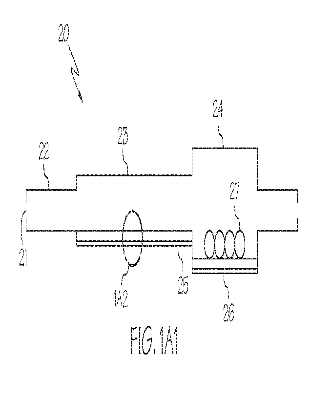
DESCRIPTION OF THE DRAWINGS
[0009] FIGS. 1A1 and 1A2 illustrate an analyte detection platform according to
an aspect of the
present disclosure.
[0010] FIG. 1B illustrates a method of detecting an analyte from vesicles in a
sample according
io to an aspect of the present disclosure.
[0011] FIG. 1C illustrates a sensor according to an aspect of the present
disclosure
[0012] FIG. 1D illustrates a method of detecting an analyte from a sample
according to an aspect
of the present disclosure.
[0013] FIG. 1E illustrates a method of lysing vesicles according to an aspect
of the present
disclosure.
[0014] FIG. 1F illustrates a vesicle lysis platform according to an aspect of
the present disclosure.
[0015] FIG. 2 illustrates a schematic of immunomagnetic capture and plasmonic
detection system.
Cross-sectional schematic of immunomagnetic bacterial enrichment working
principle (left) and
working principle of nano-scale plasmonic sensing platform (right).
[0016] FIGS. 3A-3B illustrate capture efficiency and plasmonic sensing
results. FIG. 3A shows
capture efficiency of S. aureus in whole blood matrix. PC = percent capture
(mean). FIG. 3B
3
CA 03185340 2023- 1- 9
WO 2022/015732
PCT/US2021/041431
shows plasmonic sensing results of S. aureus cell lysate. Peak absorbance
wavelength shifts as a
function of nucleic acid concentration.
[0017] FIGS. 4A-4C illustrate a schematic of an integrated capture and
detection microsystem:
(1) bacterial capture from whole blood; (2) cell lysis; and (3) DNA detection
on a single micro-
chip. The microsystem in this illustration represents an integrated single-
chip platform according
to aspects of the present disclosure.
[0018] FIGS. 5A1-5C illustrate an overview of an integrated microsystem. FIGS.
5A1-5A3 show
chip functionality. Bacterial samples (FIG. 5A1) and functionalized magnetic
nanoparticles
(MNPs) (FIG. 5A2) are pushed through micro-chip in parallel. Mixing and
incubation occur
io throughout the jagged serpentine microchannel (FIG. 5A3). Bacteria-MNP
complexes (FIG. 5B4)
are isolated in the hexagonal microchamber using an external magnet (FIG.
5B5). Bacteria are
thermally lysed (FIG. 5B6). The novel localized surface plasmon resonance
(LSPR) sensor (FIG.
5B7) is exposed to bacterial lysate. Upon nucleic acid hybridization to
sensor, a red shift in the
peak absorbance is observed (FIG. 5B8). FIG. 5C shows sample processing
workflow and
timeline. 12 min is required for bacterial enrichment (100 lL/min, 1 mL
sample), 10 min is
required for bacterial lysis, and 5 min is required for nucleic acid sensing.
A total of 3 min is
required for fluid manipulation (i.e., air, phosphate-buffered saline (PBS)).
Total-analytical-time
for the integrated enrichment and detection platform is 30 min.
[0019] FIGS. 6A-6B illustrate a microfluidic immunomagnetic bacterial capture.
FIG. 6A shows
bacterial capture efficiency as a function of bacterial species (S. aureus, P.
aeruginosa) and input
bacterial concentration. X-axis is presented as a logarithmic scale. Standard
error of the mean is
reported, n = 3 samples per condition. FIG. 6B shows capture antibody
specificity. Input bacterial
concentration is approximately 105 CFU/mL for all reported data series.
Bacteria samples
processed without MNPs (dark gray) represent the average observed bacterial
loss within the
microsystem of three independently evaluated bacterial species: S. aureus, P.
aeruginosa, and E.
coli (n = 3 per bacterial species, n = 9 total). Standard error of the mean is
reported.
4
CA 03185340 2023- 1- 9
WO 2022/015732
PCT/US2021/041431
[0020] FIGS. 7A-7F illustrate a nanoplasmonic sensing of bacterial nucleic
acids. FIG. 7A shows
representative extinction spectra. Following conjugation of peptide nucleic
acid (PNA) probes to
gold nanoparticles a red shift is observed. An additional red shift is
observed upon hybridization
of target nucleic acids to PNA probes are observed. The magnitude of this
second peak wavelength
shift represents the signal of interest. FIGS. 7B-7D shows peak wavelength
shift as a function of
input bacterial load for (FIG. 7B) S. aureus, (FIG. 7C) P. aeruginosa, and
(FIG. 7D) E. coli.
FIGS. 7E-7F show probe specificity characterizations of a P. aeruginosa probe
exposed to (FIG.
7E) E. coli cell lysate, and (FIG. 7F) S. aureus cell lysate. Standard error
of the mean is reported.
[0021] FIGS. 8A-8C illustrate data reproducibility on nanoplasmonic sensors
for (FIG. 8A) S.
aureus, (FIG. 8B) E. coli, and (FIG. 8C) P. aeruginosa. For all nanoplasmonic
sensing studies,
data was collected using 3 biological samples on 3 different sensor devices.
Each device was
exposed to a unique bacterial lysate sample, and three measurements were taken
with each device.
The mean and standard error of the mean are reported. The data in FIGS. 7B-7D
represent all 9
measurements combined.
[0022] FIGS. 9A-9B illustrate performance of integrated bacterial enrichment
and detection
platform. FIG. 9A shows magnitude of peak wavelength shift with integrated
enrichment and
without enrichment as a function of input bacterial concentration; n = 3
samples per condition.
FIG. 9B shows observed signal enhancement factor using integrated microsystem
as a function of
input bacterial concentration. Standard en-or of the mean is reported.
[0023] FIG. 10 illustrates data reproducibility for integrated bacterial
enrichment and
nanoplasmonic detection. Each listed sample represents a unique biological
sample processed on
the system. Each unique biological sample was evaluated on three different
sensors. The mean and
standard error of the mean of the sensing output for each unique sample are
reported. The data in
FIG. 9A represent all 9 measurements combined.
[0024] FIGS. 11A-11B illustrate a multiplexed capture and detection of
polymicrobial samples.
FIG. 11A shows a table reporting peak shift as a function of input sample
composition. FIG. 11B
5
CA 03185340 2023- 1- 9
WO 2022/015732
PCT/US2021/041431
show magnitude of peak wavelength shift in single species samples (i.e., S.
aureus) versus
polymicrobial samples (i.e., S. aureus + P. aeruginosa) as a function of
bacterial concentration.
Standard error of the mean is reported; n = 3 samples per condition.
[0025] FIG. 12 illustrates workflow for device fabrication (1-2) and operation
(3-4). Shown are
(1) Bidirectional microfluidic printing for dispersion of bare gold nanorods
into sensing spots, (2)
Sequence-specific conjugation with PNA probes for 3 clinically relevant
mutations, (3)
Attachment of microfluidic device to deliver sample, circulating tumor DNA
(ctDNA) will bind
to PNA probes if present; and (4) Measure of absorbance spectrum through each
spot to measure
bound ctDNA concentration to probes.
[0026] FIGS. 13A-13E illustrate images of fabricated nanorod spots and
associated spectra. FIG.
13A shows optical image of fabricated nanorod spots. FIG. 13B shows scanning
electron
microscope (SEM) image of fabricated nanorod spots. FIG. 13C shows zoom-in SEM
showing
dispersion of nanorods. FIG. 13D shows multiple nanorod spots on single chip
for multiplexing.
FIG. 13E shows parameters for nanorod printing.
[0027] FIGS. 14A-14B illustrate conjugation workflow and associated spectra.
FIG. 14A shows
workflow for conjugation starting from bare gold nanorods dispersed on glass
slide. First step is
activation of the gold followed by a wash and coupling with the PNA probe.
FIG. 14B shows
associated extinction spectra of the bare rods and the rods after conjugation,
showing an
approximate 20 nm shift in the peak wavelength after successful coupling (779
nm when bare, 808
nm after conjugation).
[0028] FIGS. 15A-15C3 illustrate two dimensional (2D) Electromagnetic
Conformal Layer
Simulation. FIG. 15A shows simulated extinction spectra of bare gold nanorod,
PNA-conjugated
gold nanorods, and PNA-DNA bound gold nanorods. FIG. 15B shows spectral zoom-
in of peak
resonance features, demonstrating a large peak shift after PNA conjugation to
the nanorods and
then a smaller shift upon DNA binding. FIGS. 15C1-C3 show images of simulation
setup
including bare rod, conformal layers, and simulation plane.
6
CA 03185340 2023- 1- 9
WO 2022/015732
PCT/US2021/041431
[0029] FIGS. 16A-16C illustrate sensing curves for 3 different point mutations
in KRAS gene, the
G12D, G12R, and G12V variants. Peak shift is calculated as the difference
between peak
wavelengths at each concentration and without ctDNA. Each data point
represents measurements
on three devices conjugated and put in contact with that sequence. Error bars
represent standard
error of the mean. FIG. 16A shows sensing of G12D synthetic oligos. FIG. 16B
shows sensing
of G12R synthetic oligos. FIG. 16C shows sensing of G12V synthetic oligos.
[0030] FIGS. 17A-17D illustrates multiplexed sensing of 3 mutations in the
KRAS gene. Peak
wavelength shift is calculated as the difference between peak wavelength
before and after ctDNA
addition. Each data point represents measurements on three sensing spots
conjugated and put in
o contact with relevant targets. Error bars represent standard error of the
mean. FIG. 17A shows
sensing measurement of all three conjugated spots, with only G12V synthetic
DNA present. FIG.
17B shows mixed sample of G12V and G12D variant showing no binding to G12R
sensor. FIG.
17C shows mixed samples of all three variants showing approximately equal
binding. FIG. 17D
shows mixed samples of G12D and G12R synthetic DNA showing semi-quantitative
discrimination between wavelength output.
[0031] FIGS. 18A1-18C2 illustrate an overview of a proposed detection
mechanism. FIGS.
18A1-A5 show a microchip design showing Phase I focus on the capture and
transduction of RNA
binding. FIG. 18B shows that initially nanoparticles are tethered to the gold
film by PNA probes.
If severe acute respiratory syndrome coronavirus 2 (SARS-CoV-2) RNA is
present, binding will
occur, and shorten the length of the tether. FIGS. 18C1-C2 show that if PNAs
are unbound, the
longer tether remains out of the plasmonic electric field decay length, but if
PNAs bind to target
RNA, the tether shortens, plasmonic coupling occurs, and binding can be
visualized on dark field
image.
[0032] FIGS.. 19A1-198 illustrate a nanopartiele-on-film simulation overview.
FIGS. 19A1-
19A3 show three geometries of nanoparticles to be tested: nanocube,
nanosphere, and nanorod.
7
CA 03185340 2023- 1- 9
WO 2022/015732
PCT/US2021/041431
FIG. 19B shows preliminary CST simulation data, showing extremely high quality
resonances
with large peak shifts (hundreds of Jim) from small (2-10 nm) thickness
changes.
[0033] FIGS. 20A1-20B illustrate an overview schematic of bacterial enrichment
and contact-free
lysis driven by an AC magnetic field. FIGS. 20A1-20A2 (Step 1): The syringe
pump pushes sample
through hexagonal micro- channel. The external magnet retains bacteria bound
to functionalized
magnetic nanoparticles within the microchannel, while waste products are
collected as the output.
TEM image of S. aureus (-0.5 pm) bound to magnetic nanoparticles (-150 nm) is
shown in FIG.
20A2. FIG. 20B (Step 2): Overview schematic of contact-free cell lysis.
External magnet is
removed, microchip is placed in coil, and microchip is exposed to an AMF.
Bacteria are thermally
lysed, enabling downstream nucleic acid collection and analysis.
[0034] FIGS. 21A1-21C2 illustrate an overview of a device substrate and
heating mechanism.
FIGS. 21A1-21A3 show a magnetic polymer microchip. Substrate modification
consists of three
identical spin coated polymer layers (P-1¨P-3). Magnetic nanoparticles mixed
within the polymer
(PDMS) enable thermal lysis of bacteria, making molecules of interest
available (i.e., DNA) for
analysis (FIG. 21A2). Shown in FIG. 21A3 is an atomic force microscopy image
(AFM)
displaying topography of a magnetic polymer surface. FIG. 21B shows an image
of magnetic
polymer-coated microchip in microfluidic cartridge. FIGS. 21C1-C2 shows a
schematic of heating
mechanism for magnetic nanoparticles embedded in a polymer matrix (FIG. 21C1).
Neel
relaxation¨the rapid change in magnetic moment in opposition to the
nanoparticle' s crystal-line
structure¨drives heat generation (FIG. 21C2).
[0035] FIGS. 22A1-22D illustrate microfluidic immunomagnetic bacterial
capture. FIGS. 22A1-
A2 show Transmission Electron Microscopy (TEM) images of S. aureus bound to
150 nm
magnetic nanoparticles. FIG. 22B shows bacterial capture efficiency as a
function of flow rate.
FIG. 22C shows bacterial capture efficiency as a function of magnetic
nanoparticle mass. FIG.
22D shows bacterial capture efficiency as a function of cell concentration.
Control samples
contained no functionalized magnetic particles and were evaluated to account
for any potential
8
CA 03185340 2023- 1- 9
WO 2022/015732
PCT/US2021/041431
bacterial loss and/or gain within the micro-system. All samples were evaluated
in triplicate.
Standard error of mean is reported.
[0036] FIGS. 23A-23B illustrate magnetic polymer microchip heating. FIG. 23A
shows
representative thermal image of microchip in coil after 30s exposure to ANIF.
FIG. 23B shows
temperature of the microchip as a function of time. Temperature data were
collected using a thermal
camera. Three unique devices were evaluated, and each device was tested in
triplicate. Standard
error of the mean is reported.
[0037] FIGS. 24A-24B illustrate recovered DNA and cell viability. FIG. 24A
shows total
recovered DNA and FIG. 24B shows cell death as a function of cell load
following 60 s exposure
to AMF. All samples were evaluated in triplicate, with three unique devices
used. Standard error
of mean is reported.
[0038] FIGS. 25A-25B illustrate bacterial capture efficiency optimization.
FIG. 25A shows
bacterial capture efficiency as a function of flow rate. Using Applicants'
microfluidic chip,
relatively high flow rates could be achieved, while preserving capture
efficiency. Flowrate
experiments were conducted at bacterial load on the order of 103 CFU/mL, and
with 25 g
functionalized magnetic nanoparticles. Experiments were performed in
triplicate, and standard
error of the mean is reported. FIG. 25B shows bacterial capture efficiency as
a function of magnetic
nanoparticle (MNP) mass. Increased MNP mass resulted in significantly greater
bacterial capture
efficiency. MNP mass optimization experiments were conducted at bacterial load
on the order of
103 CFU/mL, and at a flowrate of 10 mL/h. Experiments were performed in
triplicate, and standard
error of the mean is reported.
[0039] FIGS. 26A-26B illustrate magnetic polymer characterization and
optimization. FIG. 26A
shows characterization of specific absorbance rate of the iron oxide heating
particles as a function
of field frequency. SAR was characterized in water. FIG. 26B shows examples of
various multi-
layer magnetic polymer substrates (left to right: 1-layer, 2-layer, 3-layer, 5-
layer).
9
CA 03185340 2023- 1- 9
WO 2022/015732
PCT/US2021/041431
DETAILED DESCRIPTION
[0040] It is to be understood that both the foregoing general description and
the following detailed
description are illustrative and explanatory, and are not restrictive of the
subject matter, as claimed.
In this application, the use of the singular includes the plural, the word "a"
or "an" means "at least
one", and the use of "or" means "and/or", unless specifically stated
otherwise. Furthermore, the
use of the term "including", as well as other forms, such as "includes" and
"included", is not
limiting. Also, terms such as "element" or "component" encompass both elements
or components
comprising one unit and elements or components that include more than one unit
unless
specifically stated otherwise.
1 [0041] The section headings used herein are for organizational
purposes and are not to be
construed as limiting the subject matter described. All documents, or portions
of documents, cited
in this application, including, but not limited to, patents, patent
applications, articles, books, and
treatises, are hereby expressly incorporated herein by reference in their
entirety for any purpose.
In the event that one or more of the incorporated literature and similar
materials defines a term in
a manner that contradicts the definition of that term in this application,
this application controls.
[0042] Current methods relating to analyte detection from vesicles (e.g.,
cells) in samples, analyte
detection platforms, sensors, and analyte detection from a sample contain
numerous drawbacks
such as, but not limited to, slow processing time, limited sensitivity, and
complicated equipment.
In addition, currently available systems and methods for lysing vesicles have
similar drawbacks.
[0043] Accordingly, a need exists for more effective systems and methods for
analyte detection
from vesicles in samples, analyte detection platforms, sensors, and analyte
detection from a
sample. Furthermore, a need exists for more effective systems and methods for
lysing vesicles.
Various embodiments of the present disclosure address the aforementioned
limitations.
[0044] In some embodiments, the present disclosure pertains to an analyte
detection platform. hi
some embodiments illustrated in FIG. 1A1, the analyte detection platform is in
the farm of
CA 03185340 2023- 1- 9
WO 2022/015732
PCT/US2021/041431
platform 20, which includes an inlet region 21 for receiving a sample, a
mixing region 22, a capture
region 23, and a sensing region 24. As illustrated in FIG. 1A1, the capture
region 23 has a first
surface 25 for capturing one or more components of the sample, where the first
surface 25 is
downstream the mixing region 22. As also shown in FIG. 1A1, the sensing region
24 includes a
second surface 26 for detecting an analyte from the sample, where the second
surface 26 includes
analyte detecting agents 27.
[0045] In a non-limiting embodiment, illustrated in FIG. 1A2, the first
surface 25 is a magnetic
surface. In some embodiments, the magnetic surface includes magnetic particles
28 associated
with polymers 29.
1 [0046] In some embodiments, the analyte detection platforms of the
present disclosure may be
utilized to detect analytes from vesicles in a sample in accordance with the
analyte detection
methods of the present disclosure. For instance, in some embodiments, a sample
containing
vesicles and vesicle capture particles may flow through inlet region 21 of
platform 20 and into
mixing region 22, where vesicle capture particles bind to vesicles and form
particle-vesicle
complexes. Thereafter, the particle-vesicle complexes flow into capture region
23, where they
become immobilized on first surface 25 through various mechanisms as described
herein.
[0047] Next, the immobilized vesicles in the sample are lysed on first surface
25, thereby releasing
thc analyte from the vesicles. Vesicle lysis may also occur through various
mechanisms as
described herein. For instance, in some embodiments, an alternating magnetic
field (AMF) may
be applied to the first surface 25 (e.g., to a magnetic surface shown in FIG.
1A2), thereby heating
first surface 25 and causing lysis of the immobilized vesicles without any
contact between the
vesicles and first surface 25. In some embodiments, the surface is capable of
generating heat upon
exposure to AMP.
[0048] The released analytes then flow through sensing region 24, where they
become associated
with analyte detecting agents 27 on second surface 26. The analytes are then
detected through
11
CA 03185340 2023- 1- 9
WO 2022/015732
PCT/US2021/041431
detecting a change in property of second surface 26 and correlating the change
in the property to
a characteristic of the analyte.
[0049] In some embodiments, the present disclosure pertains to methods of
detecting an analyte
from vesicles in a sample. In some embodiments illustrated in FIG. 1B, the
methods of the present
disclosure include one or more of the following steps of flowing the sample
through a platform
(step 10), forming particle-vesicle complexes when vesicle capture particles
bind to the vesicles
in the sample (step 11), immobilizing the particle-vesicle complexes (step
12), lysing the vesicles
of the particle-vesicle complexes and thereby releasing the analyte (step 13),
associating the
analyte with an analyte detecting agent (step 14), and detecting the analyte
(step 15).
it) [0050] In some embodiments, the analyte detection platforms of the
present disclosure (e.g.,
analyte detection platform 20 shown in FIG. 1A1) can be utilized to practice
the analyte detection
methods of the present disclosure. In some embodiments illustrated herein, the
analyte detection
steps of the present disclosure can have additional embodiments.
[0051] For instance, in some embodiments. step 10 (i.e., flowing the sample
through a platform)
includes introducing a sample into an inlet region of a platform. The sample
may include vesicles
containing analytes. In some embodiments, the sample may contain vesicles and
vesicle capture
particles. In some embodiments, the vesicles and the vesicle capture particles
may be separately
introduced to the inlet region. In some embodiments, the vesicles and the
vesicle capture particles
may be pre-mixed to form the sample prior to introducing into the platform. In
some embodiments,
the vesicles and the vesicle capture particles may be introduced via separate
inlets of a platform
and mixed downstream in the platform.
[0052] In some embodiments, step 11 (i.e., forming particle-vesicle complexes)
involves vesicle
capture particles binding to the vesicles. In some embodiments, the particle-
vesicle complexes
may be formed prior to introducing the sample into the platform (such as when
the vesicles and
the vesicle capture particles are pre-mixed to form the sample). In some
embodiments, the particle-
12
CA 03185340 2023- 1- 9
WO 2022/015732
PCT/US2021/041431
vesicle complexes may be formed following introducing the vesicles and the
vesicle capture
particles into the platform.
[0053] In some embodiments, step 12 (i.e., immobilizing the particle-vesicle
complexes) involves
immobilization of the particle-vesicle complexes on a first surface of the
platform. In some
embodiments, immobilization may be achieved by a magnetic force between the
first surface and
the complexes. In some embodiments, immobilization may be achieved through
biomolecular
binding or electrostatic interaction.
[0054] In some embodiments, step 13 (i.e., lysing the vesicles of the particle-
vesicle complexes,
thereby releasing the analytes) involves breaking open the vesicles to release
analytes. In some
embodiments, this may be achieved through exposing a surface that is in the
form of a microchip
to an alternating magnetic field. In some embodiments, this may be achieved
through heating the
vesicles or putting them in contact with a chemical detergent or biological
enzyme.
I00551 In some embodiments, step 14 (i.e., associating released analytes with
analyte detecting
agents) occurs when an analyte detecting agent is immobilized on a second
surface of the platform.
In some embodiments, the analyte associates with the analyte detection agent
through
biomolecular interaction, complementary hybridization, or electrostatic
interaction.
[0056] In some embodiments, step 14 (i.e., detecting the analyte) includes,
for example, detecting
a change in property of the second surface and correlating the change in
property of the second
surface to a characteristic of the analyte. In some embodiments, the method
can be continuous
and/or repeated until all analytes have been detected.
[0057] Additional embodiments of the present disclosure pertain to sensors. In
some embodiments
illustrated in FIG. IC, the sensors of the present disclosure may be in the
form of sensor 30, which
includes a surface 31 for detecting an analyte from a sample. As illustrated
in FIG. IC, the surface
31 includes a dielectric surface 32 and nanostructures 33 randomly oriented on
the dielectric
13
CA 03185340 2023- 1- 9
WO 2022/015732
PCT/US2021/041431
surface 32. As further illustrated in FIG. 1C, the nanostructures 33 are
coupled to analyte
detecting agents 34. In some embodiments, the sensor is a plasmonic sensor.
[0058] Further embodiments of the present disclosure pertain to methods of
detecting an analyte
from a sample through sensing, such as through the utilization of sensors 30
illustrated in FIG.
1C. In some embodiments, the sensing is plasmonic sensing. In some embodiments
illustrated in
FIG. 1D, the methods of the present disclosure include a step of flowing the
sample through a
sensor (step 40) (e.g., sensor 30). In some embodiments, the sensor includes a
surface (e.g. surface
31) for detecting an analyte from a sample. In some embodiments, the surface
includes a dielectric
surface (e.g., dielectric surface 32) and nanostructures (e.g., nanostructures
33) randomly oriented
on the dielectric surface. In some embodiments, the nanostructures are coupled
to an analyte
detecting agent (e.g., analyte detecting agents 34).
[0059] As illustrated in FIG. 1D, the methods of the present disclosure can
further include the
steps of detecting a change in property of the surface of the sensor (step
41), correlating the change
in property of the surface to a characteristic of the analyte (step 42), and
detecting the analyte (step
43). In some embodiments, the method can be continuous and/or repeated until
all analytes have
been detected.
[0060] Further embodiments of the present disclosure pertain to methods of
contact-free vesicle
lysis. In some embodiments illustrated in FIG. 1E, the method of lysing
vesicles in a sample
generally involves one or more of the following steps of flowing the sample
through a platform
(step 50), and exposing a surface of the platform to an alternating magnetic
field (AMF) to lyse
the vesicles (step 51). In some embodiments, the contact-free vesicle lysis
methods of the present
disclosure result in the release of analytes from the vesicles (step 51), and
the subsequent collection
of the analytes (step 52).
[0061] In some embodiments, vesicle capture particles bind to the vesicles in
the sample to form
particle-vesicle complexes. In some embodiments, the particle-vesicle
complexes become
immobilized on the surface of the platform. In some embodiments, the surface
is a magnetic
14
CA 03185340 2023- 1- 9
WO 2022/015732
PCT/US2021/041431
surface. In some embodiments, the magnetic surface includes a polymer and
magnetic particles
associated with the polymer. In some embodiments, the AMF heats the surface
(e.g., a magnetic
surface) and thereby generates heat, and the generated heat lyses the vesicles
of the particle-vesicle
complexes. In some embodiments, the surface is capable of generating heat upon
exposure to
AMF. In some embodiments, the surface is capable of generating heat upon
exposure to AMF.
some embodiments, the method can be continuous and/or repeated until all
vesicles have been
lysed.
[0062] Additional embodiments of the present disclosure pertain to contact-
free vesicle lysis
systems. In some embodiments illustrated in FIG. IF, the contact-free vesicle
lysis systems of the
present disclosure include a vesicle lysis platform 60, which includes a
surface 61. In some
embodiments, the surface 61 includes magnetic surface 62. In a non-limiting
embodiment,
magnetic surface 62 can include polymers 63 and magnetic particles 64
associated with polymers
63.
[0063] In some embodiments. the contact-free vesicle lysis systems of the
present disclosure may
be utilized to lyse cells in accordance with the contact free cell lysis
methods of the present
disclosure. For instance, in a specific embodiment, a sample containing
vesicles and vesicle
capture particles may flow through vesicle lysis platform 60, where the formed
particle-vesicle
complexes become immobilized on the surface 61 through various mechanisms,
such as magnetic
immobilization, biomolccular binding, or electrostatic interaction.
[0064] For example, in some embodiments, the surface 61 includes a magnetic
surface 62. In this
example, the formed particle-vesicle complexes become immobilized on magnetic
surface 62.
Thereafter, the magnetic surface 62 is exposed to AMF, which heats the
magnetic surface 62 and
thereby generates heat. Thereafter, the generated heat lyses the vesicles of
the particle-vesicle
complexes.
CA 03185340 2023- 1- 9
WO 2022/015732
PCT/US2021/041431
[0065] The contact-free vesicle lysis systems may be used to release the
analyte from the vesicle
for further analysis by other systems as well. As illustrated above, in some
embodiments, the
surface is capable of generating heat upon exposure to AMF.
[0066] As set forth in more detail herein, the systems and methods of the
present disclosure can
have numerous embodiments. For instance, the methods for detecting analytes
from vesicles in a
sample can utilize various sample processing steps, samples, flowing methods,
vesicles, vesicle
capture particles, immobilization methods, lysing methods, and analyte
detecting agents.
Moreover, the methods of the present disclosure can utilize various changes in
properties to detect
numerous types of analytes.
1 [0067] Furthermore, various platforms may be utilized to lyse vesicles
and detect analytes from
the lysed vesicles. For instance, the platforms can include various inlet
regions, capturing regions,
and sensing regions in various arrangements. In addition, the platforms of the
present disclosure
can utilize various analyte detecting agents, surfaces, and platform
configurations.
[0068] Additionally, various sensors and sensing methods may be utilized to
detect various
analytes from various samples. For example, the sensors of the present
disclosure can include
various dielectric surfaces and nanostructures in various orientations. In
addition, the sensors of
the present disclosure can utilize numerous analyte detecting agents and have
various
configurations.
[0069] Additionally, the present disclosure may utilize various contact-free
vesicle lysis platforms
and contact-free vesicle lysis methods. For instance, as set forth in further
detail herein, the
contact-free vesicle lysis platforms and methods of the present disclosure can
utilize various
surfaces, for example magnetic surfaces, that can include, without limitation,
numerous polymers
and magnetic particles. In addition, the methods and platforms of the present
disclosure can lyse
numerous types of vesicles from various samples. The methods and platforms of
the present
disclosure can also utilize various flowing methods, vesicle capture
particles, and surfaces
16
CA 03185340 2023- 1- 9
WO 2022/015732
PCT/US2021/041431
[0070] Analyte Detection from Vesicles in a Sample
[0071] As disclosed in further detail herein, embodiments of the present
disclosure pertain to
methods of detecting an analyte from vesicles in a sample. Such methods
generally include one or
more of the following steps of: (a) flowing the sample through a platform,
where vesicle capture
particles bind to the vesicles in the sample to form particle-vesicle
complexes, and where the
particle-vesicle complexes become immobilized on a first surface of the
platform; (b) lysing the
vesicles of the particle-vesicle complexes, thereby releasing the analyte; (c)
associating the analyte
with an analyte detecting agent, where the analyte detecting agent is
immobilized on a second
surface of the platform; and (d) detecting the analyte. In some embodiments,
the detecting can
include detecting a change in property of the second surface and correlating
the change in property
of the second surface to a characteristic of the analyte.
[0072] Additional Sample Processing Steps
[0073] As set forth in further detail herein, the methods of the present
disclosure can include
additional sample processing steps. For example, in some embodiments, the
method further
includes clearing the sample from the platform after step (a). In some
embodiments, the method
further includes clearing excess or unwanted portions of the sample from the
platform. In some
embodiments, the method further includes the step of removing excess fluid
from the platform.
[0074] In some embodiments, the method further includes the step of
introducing a carrier liquid
to the first surface of the platform before the lysing in step (b). In some
embodiments, the carrier
liquid can include, without limitation, phosphate-buffered saline (PBS), TE
buffer, alcohols,
water-based solutions, and combinations thereof. In some embodiments, the
analyte is released
into the carrier liquid to form a lysate during the lysing in step (b).
[0075] In some embodiments, the method further includes the step of flowing
and exposing the
lysate to the second surface of the platform after step (b). In some
embodiments, step (b) further
17
CA 03185340 2023- 1- 9
WO 2022/015732
PCT/US2021/041431
includes incubating the lysate with the second surface and then clearing the
lysate from the
platform.
[0076] Samples
[0077] As set forth in further detail herein, the methods of the present
disclosure can detect
analytes from vesicles in numerous types of samples. For example, in some
embodiments, the
sample can include, without limitation, a biological sample obtained from a
subject, an
environmental sample obtained from an environment, and combinations thereof.
[0078] In some embodiments, the sample includes a biological sample obtained
from a subject. In
some embodiments, the biological sample can include, without limitation, a
blood sample, a tissue
sample, a urine sample, a saliva sample, a sputum sample, a swab sample, a
swab sample put into
a carrier solution, a processed blood sample, and combinations thereof.
[0079] In some embodiments, the sample includes an environmental sample. In
some
embodiments, the environmental sample can include, without limitation, a food
sample, a water
sample, a swab sample, a swab sample put into a carrier solution, a surface
swab sample, a passive
material sample put into a carrier solution, and combinations thereof.
[0080] Flowing the samples
[0081] As set forth in further detail herein, the methods of the present
disclosure can utilize
numerous methods for flowing the sample through the platform. For instance, in
some
embodiments, the flowing includes flowing the sample through the platform
along with the vesicle
capture particles. In some embodiments, the sample is co-introduced into the
platform along with
the vesicle capture particles. In some embodiments, the sample is pre-
incubated with the vesicle
capture particles prior to co-introduction into the platform.
18
CA 03185340 2023- 1- 9
WO 2022/015732
PCT/US2021/041431
[0082] In some embodiments, the flowing can include flowing the sample through
the platform
while the vesicle capture particles are immobilized on the first surface of
the platform. In some
embodiments, the vesicle capture particles are pre-immobilized on or part of
the first surface.
[0083] In some embodiments, the methods of the present disclosure can further
include a step of
immobilizing the vesicle capture particles on the first surface prior to the
flowing step. In some
embodiments, the flowing occurs through a method that can include, without
limitation, pumping,
mechanical pumping, electrical pumping, syringe-facilitated flow, pipette-
facilitated flow,
capillary flow, peristaltic flow, pressure-driven flow, and combinations
thereof.
[0084] Vesicles
[0085] As outlined in further detail herein, the methods of the present
disclosure can detect
analytes from various vesicles. For instance, in some embodiments, the
vesicles can include,
without limitation, viruses, bacteria, yeast, fungi, prokaryotic cells,
eukaryotic cells, extracellular
vesicles, and combinations thereof. In some embodiments, the vesicles include
viruses. In some
embodiments, the vesicles include severe acute respiratory syndrome
coronavirus 2 (SARS-CoV-
2). In some embodiments, the vesicles include Human Papilloma Virus (HPV).
[0086] In some embodiments, the vesicles include eukaryotic cells. In some
embodiments, the
eukaryotic cells include cancer cells. In some embodiments, the vesicles
include bacteria.
[0087] In some embodiments, the vesicles include extracellular vesicles. In
some embodiments,
thc extracellular vesicles include exosomes.
[0088] Analytes
[0089] As set forth in further detail herein, various analytes can be detected
via the methods of the
present disclosure. For example, in some embodiments, the analyte can include,
without limitation,
nucleotides, oligonucleotides, RNA, DNA, ribosomal RNA (rRNA), messenger RNA
(mRNA),
19
CA 03185340 2023- 1- 9
WO 2022/015732
PCT/US2021/041431
microDNA, microRNA, extrachromosomal circular DNA (eccDNA), circulating tumor
DNA
(ctDNA), small molecules, proteins, mutated versions thereof, and combinations
thereof.
[0090] In some embodiments, the analyte includes RNA. In some embodiments, the
analyte
includes mutated nucleotides. In sonic embodiments, the analyte includes wild-
type nucleotides.
[0091] Vesicle Capture Particles
[0092] As detailed herein, the methods of the present disclosure can utilize
various vesicle capture
particles in numerous manners. For instance, in some embodiments, the vesicle
capture particles
are immobilized on the first surface of the platform prior to the flowing
step. In some embodiments,
the vesicle capture particles are lyophilized on the first surface of the
platform prior to the flowing
step.
[0093] In some embodiments, the vesicle capture particles can include, without
limitation, metal
particles, magnetic particles, polymer-based particles, gelled particles, and
combinations thereof.
In some embodiments, the vesicle capture particles include magnetic particles.
[0094] In some embodiments, the vesicle capture particles are associated with
a binding agent. In
some embodiments, the binding agent binds to the vesicle to be captured from
the sample. In some
embodiments, the binding agent can include, without limitation, antibodies,
peptides, aptamers,
nucleic acids, peptide nucleic acids, polymers, molecularly imprinted
polymers, molecules capable
of facilitating hydrostatic interactions, and combinations thereof. In some
embodiments, the
binding agent includes antibodies. In some embodiments, the binding agent
includes aptamcrs.
[0095] First Surface
[0096] First surfaces generally refer to platform regions that can immobilize
particle-vesicle
complexes. As set forth in further detail herein, the methods and platforms of
the present
disclosure can include various first surfaces.
CA 03185340 2023- 1- 9
WO 2022/015732
PCT/US2021/041431
[0097] For instance, in some embodiments, the first surface includes a
magnetized region or a
region exposed to a magnetic field. In some embodiments, the region is
utilized to immobilize the
vesicle capture particles. In some embodiments, the region includes a magnet
positioned in
proximity to the first surface. In some embodiments, the magnet can include,
without limitation,
permanent magnets, electromagnets, soft magnets, magnetic particles associated
with polymers,
and combinations thereof.
[0098] In some embodiments, the first surface includes a functionalized
region. In some
embodiments, the functionalized region is functionalized with at least one
functional group. In
some embodiments, the at least one functional group is utilized to immobilize
the vesicle capture
particles. In some embodiments, the functional group can include, without
limitation, charged
groups, binding agents, functional groups capable of facilitating
electrostatic interactions, and
combinations thereof.
[0099] In some embodiments, the first surface includes a magnetic surface. In
some embodiments,
the magnetic surface includes polymers and magnetic particles associated with
the polymers. In
some embodiments, the magnetic surface is capable of generating heat upon
exposure to AMF. In
some embodiments, the first surface is in the form of the contact-free vesicle
lysis systems of the
present disclosure (e.g., vesicle lysis system 60 shown in FIG. 1F).
[00100] In some embodiments, the first surface includes a porous region. In
some embodiments,
the porous region is utilized to immobilize the vesicle capture particles
through size-based
separation.
[00101] Immobilizing
[00102] In some embodiments, the methods of the present disclosure can further
include a step of
immobilizing particle-vesicle complexes on first surfaces of platforms.
Immobilization can occur
through various methods. For example, in some embodiments, the immobilizing
occurs by a
method that can include, without limitation, magnet-based immobilization,
pelleting,
21
CA 03185340 2023- 1- 9
WO 2022/015732
PCT/US2021/041431
centrifugation, size-based separations, filtration, inertial separations,
acoustofluidic separations,
material property based separations, dielectrophoretic separations,
immunoaffinity-based
separation, and combinations thereof.
[00103] In some embodiments, the immobilizing includes applying a magnetic
field to the first
surface of the platform. In some embodiments, the magnetic field immobilizes
the particle-vesicle
complexes on the first surface of the platform. In some embodiments, the
magnetic field is applied
below the first surface of the platform.
[00104] In some embodiments, the immobilizing occurs through adhesion of the
particle-vesicle
complexes to the first surface. In some embodiments, the adhesion includes a
charged interaction
between the first surface and the particle-vesicle complexes.
[00105] Lysing
[00106] As set forth in further detail herein, the methods of the present
disclosure can utilize
various techniques to lyse vesicles. For instance, in some embodiments, the
lysing can occur by,
for example, applying heat to a platform, exposing the platform to an
alternating magnetic field,
applying a lysis material to the platform, applying a chemical lysis agent to
the platform, freezing,
mechanical perturbation, and combinations thereof.
[00107] In some embodiments, the lysing occurs by exposing the platform to an
alternating
magnetic field (AMF). In some embodiments, the platform is exposed to an AMF
that is powered
by a supply associated with the platform.
[00108] In some embodiments where the first surface includes a magnetic
surface, the lysing can
include, for example, applying an alternating magnetic field to the magnetic
surface. In some
embodiments, the alternating magnetic field heats the magnetic surface and
thereby generates heat.
In some embodiments, the generated heat lyses the vesicles of the particle-
vesicle complexes. In
some embodiments, the generated heat lyses the vesicles without direct heating
or addition of lysis
materials. In some embodiments, the lysing occurs through no direct
interaction with the vesicle.
22
CA 03185340 2023- 1- 9
WO 2022/015732
PCT/US2021/041431
[00109] In some embodiments where the first surface includes a magnetic
surface (e.g., a polymer
and magnetic particles associated with the polymer), the lysing can include,
for example, applying
an alternating magnetic field to the first surface. In some embodiments, the
alternating magnetic
field heats the magnetic surface and thereby generates heat. Tn some
embodiments, the generated
heat lyses the vesicles of the particle-vesicle complexes. In some
embodiments, the generated heat
lyses the vesicles without direct heating or addition of lysis materials. In
some embodiments, the
lysing occurs through no direct interaction with the vesicle.
[00110] In some embodiments, the lysing occurs by applying a lysis material to
the platform. In
some embodiments, the lysis material can include, without limitation, a
detergent, a chemical lysis
buffer, a biological lysis buffer, and combinations thereof.
[00111] Second Surface
[00112] Second surfaces generally refer to platform regions that can detect
analytes. In some
embodiments, the second surface is the same as the first surface. In some
embodiments, the second
surface is adjacent or proximal to the first surface. In some embodiments, the
second surface is
downstream from the first surface.
[00113] The methods and platforms of the present disclosure can include
various second surfaces.
For instance, in some embodiments, the second surfacc may include one or more
analytc detecting
agents. In some embodiments, the second surface may be in the form of the
sensors of the present
disclosure (e.g., sensor 30 shown in FIG. 1C).
[00114] In some embodiments, the second surface can include a dielectric
surface and
nanostructures associated with the dielectric surface. Tn some embodiments,
the nanostructures
are coupled to an analyte detecting agent. In some embodiments, the dielectric
surface can include,
for example, a glass surface, a plastic surface, a polymer surface, a metallic
surface, a ceramic
surface, and combinations thereof. In some embodiments, the dielectric surface
includes a glass
surface.
23
CA 03185340 2023- 1- 9
WO 2022/015732
PCT/US2021/041431
[00115] In some embodiments, the dielectric surface includes a metallic
surface. In some
embodiments, the metallic surface includes at least one metal. In some
embodiments, the at least
one metal can include, without limitation, gold, silver, copper, transition
metals, metals,
metalloids, and combinations thereof_ In some embodiments_ the metallic
surface is composed
essentially of gold.
[00116] In some embodiments, the nanostructures can include, without
limitation, plasmonic
nanoparticles, metal nanoparticles, magnetic nanoparticles, functionalized
nanoparticles,
functionalized magnetic nanoparticles, nanorods, nanospheres, nanocubes.
magnetic nanorods,
functionalized nanorods, functionalized magnetic nanorods, and combinations
thereof. In some
embodiments, the nanostructures include plasmonic nanoparticles.
[00117] In some embodiments, the nanostructures are directly associated with
the dielectric
surface through direct contact between the nanostructures and the dielectric
surface. In some
embodiments, the nanostructures are indirectly associated with the dielectric
surface through
indirect contact between the nanostructures and the dielectric surface. In
some embodiments, the
nanostructurcs are directly fabricated atop the dielectric surface. In some
embodiments, the
nanostructures are indirectly associated with the dielectric surface through
the analyte detecting
agent. In some embodiments, at least a portion of the analyte detecting agent
is positioned between
the nanostructures and the dielectric surface.
[00118] In some embodiments, the analyte detecting agent shortens upon binding
to the analyte,
thereby bringing the nanostructure closer to the dielectric surface, and
thereby resulting in the
change in the property of the second surface.
[00119] In some embodiments, the second surface is in a form of an array. In
some embodiments,
the array includes a plurality of different analyte detecting agents that are
specific for detecting
different analytes. As such, in some embodiments, the methods of the present
disclosure can be
utilized to detect a plurality of different analytes.
24
CA 03185340 2023- 1- 9
WO 2022/015732
PCT/US2021/041431
[00120] Analyte Detecting Agents
[00121] The methods of the present disclosure can associate an alytes with
analyte detecting agents
in various manners. For example, in some embodiments, associating the analyte
with an analyte
detection agent includes specifically binding the analyte detecting agent to
the analyte.
[00122] The methods and platforms of the present disclosure can utilize
various analyte detecting
agents. For instance, in some embodiments, the analyte detecting agents can
include, without
limitation, aptamers, oligonucleotides, single-stranded oligonucleotides,
double-stranded
oligonucleotides, DNA, RNA, single stranded DNA, antibodies, peptide nucleic
acids (PNAs), and
combinations thereof. In some embodiments, the analyte detecting agent
includes peptide nucleic
io acids (PNAs).
[00123] Analyte detecting agents may be associated with the platforms of the
present disclosure
in various manners. For instance, in some embodiments, the analyte detecting
agents are directly
associated with a second surface of a platform. In some embodiments, the
analyte detecting agents
are indirectly associated with a second surface of a platform through
association with one or more
nanostructures. In some embodiments, the analyte detecting agents may be
immobilized on a
second surface of a platform through, for example, covalent coupling,
hydrostatic coupling,
electrostatic coupling, and combinations thereof.
[00124] Changes in Properties of Second Surfaces
[00125] As outlined herein, the methods of the present disclosure can rely on
various changes in
properties of a second surface to detect an analyte in a sample. For instance,
in some embodiments,
the change in property is characterized by a change in absorbance of the
second surface, a shift in
peak absorbance wavelength of the second surface, a shift in transmittance
wavelength of the
second surface, a shift in reflectance wavelength of the second surface, a
shift in extinction
wavelength of the second surface, a change in plasmonic field intensity of the
second surface,
enhanced resonance sensitivity, a color change in dark field image from the
second surface. a
CA 03185340 2023- 1- 9
WO 2022/015732
PCT/US2021/041431
change in an image of the second surface, a shortening of the analyte
detecting agent, a change in
measured light absorbance, a change in transmittance, a change in reflectance,
a change in
extinction, and combinations thereof. In some embodiments, the change in
property is
characterized by a shift in peak absorbance wavelength of the second surface.
[00126] The methods of the present disclosure can also detect a change in a
property of a second
surface in various manners. For example, in some embodiments, the detecting
the change in
property occurs by a method that can include, without limitation,
visualization, microscopy, dark
field microscopy, spectrometry, spectroscopy, colorimetric analysis, localized
surface plasmon
resonance (LSPR), nuclear magnetic resonance (NMR), surface plasmon resonance,
electrochemistry, and combinations thereof. In some embodiments, the detecting
the change in
property includes visualizing a color or image change of the second surface on
a simple dark field
image.
[00127] Correlation of a Change in Property to an Analyte Characteristic
[00128] As set forth in further detail herein, the methods of the present
disclosure can utilize
various techniques to correlate a change in property of a second surface to a
characteristic of an
analyte. For instance, in some embodiments, the correlating occurs in a
quantitative, semi
quantitative, or qualitative manner.
[00129] Additionally, the methods of the present disclosure can be utilized to
determine various
characteristics of an analyte. For example, in some embodiments, the
characteristic of the analyte
can include, without limitation, the identity of the analyte, the presence of
the analyte, the absence
of the analyte, the concentration of the analyte, the quantity of the analyte,
and combinations
thereof.
[00130] Platforms
[00131] As detailed herein, the methods of the present disclosure can utilize
various platforms for
the detection of analytes. For instance, in some embodiments, the platform
includes a channel. 111
26
CA 03185340 2023- 1- 9
WO 2022/015732
PCT/US2021/041431
some embodiments, the channel can include, without limitation_ a microchannel,
a fluid channel,
and combinations thereof.
[00132] In some embodiments, the channel includes an inlet section for
receiving the sample and
a mixing region for mixing the sample with the vesicle capture particles to
form the particle-vesicle
complexes. In some embodiments, the mixing region is downstream the first
inlet.
[00133] In some embodiments, the platform includes the first surface for
capturing the particle-
vesicle complexes. In some embodiments, the first surface is downstream the
mixing region. In
some embodiments, the platform includes the second surface for detecting the
analyte.
[00134] In some embodiments, the platform further includes a magnet in
proximity to the first
surface. In some embodiments, the inlet section includes a first inlet and a
second inlet converging
into the mixing region. In some embodiments, the first sample is introduced
into the channel
through the first inlet and the vesicle capture particles are introduced into
the channel through the
second inlet.
[00135] In some embodiments, the channel includes channels with diameters of
less than 1 mm.
In some embodiments, the channel includes a portion with a configuration that
can include, without
limitation, a jagged configuration, a serpentine configuration, a hexagonal
configuration, a spiral-
shaped configuration, linear configuration, H-configuration, and combinations
thereof.
[00136] In some embodiments, the channel includes a portion with a spiral
shaped configuration.
In some embodiments, the channel includes a portion with capillary pump.
[00137] In some embodiments, the platform is in the form of a microchannel. In
some
embodiments, the platform is in the form of the analyte detection platforms of
the present
disclosure (e.g., analyte detection platform 20 shown in FIG. 1A).
[00138] Embodiments and Applications
27
CA 03185340 2023- 1- 9
WO 2022/015732
PCT/US2021/041431
[00139] As set forth in further detail herein, the analyte detection methods
of the present disclosure
can have numerous embodiments and applications. For instance, in some
embodiments, the analyte
detection methods of the present disclosure occur without amplification,
replication, growth, or
culture of the analyte. In some embodiments, the analyte detection methods of
the present
disclosure occur without amplification, replication, growth, or culture of the
vesicles.
[00140] In some embodiments, the analyte detection methods of the present
disclosure utilized for
the characterization, detection, or quantification of a plurality of different
analytes. In some
embodiments, the analyte detection methods of the present disclosure are
utilized for
characterization of an infection, cancer, or chronic illness. In some
embodiments, the infection
u) may be, for example, bacterial infections, viral infections,
polymicrobial infections, and
combinations thereof.
[00141] Analyte Detection Platform
[00142] As set forth in further detail herein, an aspect of the present
disclosure relates to a platform
for analyte detection in a sample. In some embodiments, the platform can
include an inlet region
for receiving a sample, a mixing region for mixing the sample, a capturing
region including a first
surface for capturing one or more components of the sample, where the first
surface is downstream
the mixing region, and a sensing region that includes a second surface for
detecting an analyte
from the sample. In some embodiments, the second surface includes an analyte
detecting agent.
[00143] The analyte detection platforms of the present disclosure can include
various
configurations. For instance, in some embodiments, the analyte detection
platforms of the present
disclosure may be in the form of analyte detection platform 20 shown in FIG.
1A1. As described
in more detail herein, the analyte detection platforms of the present
disclosure can include
numerous additional embodiments and variations.
[00144] Inlet Region
28
CA 03185340 2023- 1- 9
WO 2022/015732
PCT/US2021/041431
[00145] As set forth in detail herein, the platforms of the present disclosure
can include various
inlet regions with various configurations. For example, in some embodiments,
the inlet region
includes a first inlet and a second inlet converging into the mixing region.
In some embodiments,
the inlet region includes single inlet region converging into the mixing
region.
[00146] Capturing Region
[00147] As set forth in further detail herein, the platforms of the present
disclosure can include
various capturing regions and first surface configurations. For instance, in
some embodiments,
the capturing region further includes a magnet positioned in proximity to the
first surface. In
some embodiments, the magnet can include, without limitation, permanent
magnets,
electromagnets, soft magnets, alternating current magnets, and combinations
thereof. In some
embodiments, the magnet is heated by an alternating magnetic field. In some
embodiments, the
capturing region includes a magnetic surface. In some embodiments, the
magnetic surface
generates heat upon exposure to AMF.
[00148] In some embodiments, the capturing region includes a magnetic surface.
In some
embodiments, the magnetic surface includes a polymer and magnetic particles
associated with the
polymer. In some embodiments, the capturing region includes first surfaces
that have been
previously described in detail in this Application. In some embodiments,
capturing region is in
thc form of the contact-free vesicle lysis systems of the present disclosure
(e.g., vesicle lysis system
60 shown in FIG. 1F).
[00149] Sensing Region
[00150] As set forth in further detail below, the platforms of the present
disclosure can include
various sensing regions and second surface configurations. For example, in
some embodiments,
the second surface includes second surfaces that have been previously
described in detail in this
Application. In some embodiments, the second surface includes a dielectric
surface and
29
CA 03185340 2023- 1- 9
WO 2022/015732
PCT/US2021/041431
nanostructures associated with the dielectric surface. In some embodiments,
the nanostructures
are coupled to the analyte detecting agent.
[00151] In some embodiments, the dielectric surface includes_ for example, a
glass surface, a
plastic surface, a polymer surface, a transparent surface, a metallic surface,
a ceramic surface, and
combinations thereof. In some embodiments, the dielectric surface includes a
glass surface. In
some embodiments, the dielectric surface includes a metallic surface. In some
embodiments, the
metallic surface includes at least one metal. In some embodiments, the at
least one metal can
include, without limitation, gold, platinum, silver, copper, transition
metals, metals, metalloids,
and combinations thereof. In some embodiments, the metallic surface is
composed essentially of
gold.
[00152] In some embodiments, the nanostructures can include, without
limitation, plasmonic
nanoparticles, metal nanoparticles, magnetic nanoparticles, functionalized
nanoparticles,
functionalized magnetic nanoparticles, nanorods, nano spheres, nanocubes.
magnetic nanorods,
functionalized nanorods, functionalized magnetic nanorods, and combinations
thereof. In some
embodiments, the nanostructures include plasmonic nanoparticles.
[00153] In some embodiments, the nanostructures are directly associated with
the dielectric
surface through direct contact between the nanostructures and the dielectric
surface. In some
embodiments, the nanostructures are indirectly associated with the dielectric
surface through
indirect contact between the nanostructures and the dielectric surface. In
some embodiments, the
nanostructures are indirectly associated with the dielectric surface through
the analyte detecting
agent. In some embodiments, at least a portion of the analyte detecting agent
is positioned between
the nanostructures and the dielectric surface. In some embodiments, the
analyte detecting agent
shortens upon binding to the analyte, thereby bringing the nanostructure
closer to the dielectric
surface.
CA 03185340 2023- 1- 9
WO 2022/015732
PCT/US2021/041431
[00154] In some embodiments, the second surface is in a form of an array. In
some embodiments,
the array includes a plurality of different analyte detecting agents that are
specific for detecting
different analytes.
[00155] In some embodiments, the second surface is the same as the first
surface. In some
embodiments, the second surface is adjacent or proximal to the first surface.
In some embodiments,
the second surface is downstream from the first surface.
[00156] In some embodiments, the second surface may be in the form of the
sensors of the present
disclosure (e.g., sensor 30 shown in FIG. 1C).
[00157] Analyte Detecting Agent
[00158] As detailed herein, the platforms of the present disclosure can
include various analyte
detection agents. For example, in some embodiments, the analyte detecting
agent specifically
binds to an analyte. In some embodiments, the analyte can include, without
limitation, nucleotides,
RNA, DNA, ribosomal RNA (rRNA), messenger RNA (mRNA), microDNA, microRNA,
extrachromosomal circular DNA (eccDNA), cell free DNA (cfDNA), circulating
tumor DNA
(ctDNA), small molecules, proteins, mutated versions thereof, and combinations
thereof.
[00159] In some embodiments, the analyte detecting agent can include, without
limitation,
aptamers, oligonucleotides, single-stranded oligonucleotides, double-stranded
oligonucleotides,
DNA. RNA, single stranded DNA, antibodies, peptide nucleic acids (PNAs),
selective polymers,
and combinations thereof. In some embodiments, the analyte detecting agent
includes peptide
nucleic acids (PNAs).
[00160] Analyte detecting agents may be associated with second surfaces of
platforms in various
manners. For instance, in some embodiments, the analyte detecting agents are
directly associated
with the second surface of a platform. In some embodiments, the analyte
detecting agents are
indirectly associated with the second surface of a platform through
association with one or more
nanostructures. In some embodiments, the analyte detecting agents may be
immobilized on a
31
CA 03185340 2023- 1- 9
WO 2022/015732
PCT/US2021/041431
second surface of a platform through, for example, covalent coupling,
hydrostatic coupling,
electrostatic coupling, and combinations thereof.
[00161] Platform Configuration
[00162] As set forth in further detail herein, the platforms of the present
disclosure can have
numerous configurations. For example, in some embodiments, the platform
includes channels with
diameters of less than 1 mm. In some embodiments, the platform includes a
configuration that can
include, without limitation, a jagged configuration, a serpentine
configuration, a hexagonal
configuration, a spiral-shaped configuration, linear configuration, H-
configuration, and
combinations thereof.
[00163] In some embodiments, the platform includes a spiral shaped
configuration. In some
embodiments, the platform is in the form of a channel. In some embodiments,
the platform is in
the form of a microchannel.
[00164] Sensors
[00165] Another aspect of the present disclosure pertains to sensors used for
analyte detection. In
some embodiments, the sensor includes a surface for detecting an analyte from
a sample. In some
embodiments, the surface includes a dielectric surface and nanostructures
randomly oriented on
the dielectric surface. In some embodiments, the nanostructures are coupled to
an analyte detecting
agent. In some embodiments, the sensor is a plasmonic sensor.
[00166] The sensors of the present disclosure can include various
configurations. For instance, in
some embodiments, the sensors of the present disclosure may be in the form of
sensor 30 shown
in FIG. 1C. As described in more detail herein, the sensors of the present
disclosure can include
numerous additional embodiments and variations.
[00167] Dielectric Surfaces
32
CA 03185340 2023- 1- 9
WO 2022/015732
PCT/US2021/041431
[00168] As set forth in further detail herein, the sensors of the present
disclosure can utilize various
dielectric surfaces. For instance, in some embodiments, the dielectric surface
includes, for
example, a glass surface, a plastic surface, a polymer surface, a metallic
surface, a ceramic surface,
a transparent surface, and combinations thereof. In some embodiments, the
dielectric surface
includes a glass surface.
[00169] In some embodiments, the dielectric surface includes a metallic
surface. In some
embodiments the metallic surface includes at least one metal. In some
embodiments, the at least
one metal can include, without limitation, gold, platinum, silver, copper,
transition metals, metals,
metalloids, and combinations thereof. In some embodiments, the metallic
surface is composed
essentially of gold.
100170] Nanostructures
[00171] As detailed herein, the sensors of the present disclosure can include
various
nanostructures. For example, in some embodiments, the nanostructures can
include, without
limitation, plasmonic nanoparticles, metal nanoparticles, magnetic nanop
articles, functionalized
nanoparticles, functionalized magnetic nanoparticles, gold nanoparticles,
nanorods, nanospheres,
nanocubes, magnetic nanorods, functionalized nanorods, functionalized magnetic
nanorods, and
combinations thereof. In some embodiments, the nanostructures include
plasmonic nanoparticles.
[00172] In some embodiments, the nanostructures include at least one metal. In
some
embodiments, the at least one metal can include, without limitation, gold,
platinum, silver, copper,
transition metals, metals, metalloids, and combinations thereof.
[00173] Tn some embodiments, the nanostructures are directly associated with
the dielectric
surface through direct contact between the nanostructures and the dielectric
surface. In some
embodiments, the nanostructures are indirectly associated with the dielectric
surface through
indirect contact between the nanostructures and the dielectric surface. In
some embodiments, the
nanostructures are dispersed using fluid flow onto the dielectric surface.
33
CA 03185340 2023- 1- 9
WO 2022/015732
PCT/US2021/041431
[00174] In some embodiments, the nanostructures are indirectly associated with
the dielectric
surface through the analyte detecting agent. In some embodiments, the analyte
detecting agent is
positioned between the nanostructures and the dielectric surface. In some
embodiments, the
analyte detecting agent shortens upon binding to the analyte, thereby bringing
the nanostructure
closer to the dielectric surface.
[00175] In some embodiments, the surface is in a form of an array. In some
embodiments, the
array includes a plurality of different analyte detecting agents that are
specific for different
analytes. In some embodiments, the plurality of different analyte detecting
agents are coupled to
the same or different nanostructures. In some embodiments, the nanostructures
are covalently
bound to the dielectric surface. In some embodiments, the nanostructures are
electrostatically
bound to the dielectric surface.
[00176] In some embodiments, the nanostructures include diameters ranging from
about 30 nm to
about 500 nm. In some embodiments, the nanostructures include diameters
ranging from about 30
nm to about 100 nm. In some embodiments, the nanostructures include diameters
of at least about
30 nm. Tr some embodiments, the nanostructures include diameters of at least
about 100 nm. In
some embodiments, the nanostructures include diameters of less than about 100
nm.
[00177] Random Orientation
[00178] As set forth in further detail herein, the nanostructures of the
sensors of the present
disclosure can have a random orientation on dielectric surfaces. For instance,
in some
embodiments, the nanostructures are randomly dispersed on the dielectric
surface. In some
embodiments, the nanostructures are randomly oriented such that their long
axes are not all in the
same direction. In some embodiments, the nanostructures are randomly oriented
such that their
long axes are all in the same direction.
[00179] Analyte Detecting Agents
34
CA 03185340 2023- 1- 9
WO 2022/015732
PCT/US2021/041431
[00180] As set forth in further detail herein, the sensors of the present
disclosure can include
various analyte detecting agents. For example, in some embodiments, the
analyte detecting agent
specifically binds to an analyte. In some embodiments, the analyte can
include, without limitation,
nucleotides, RNA, DNA, ribosomal RNA (rRNA), messenger RNA (mRNA), microDNA,
microRNA, extrachromosomal circular DNA (eccDNA), cell free DNA (cfDNA),
circulating
tumor DNA (ctDNA), small molecules, proteins, mutated versions thereof, and
combinations
thereof. In some embodiments, the analyte includes cell free DNA
(cfIDNA). In some
embodiments, the analyte includes nucleotides derived from lysed cells.
[00181] In some embodiments, the analyte detecting agent can include, without
limitation,
aptamers, oligonucleotides, single-stranded oligonucleotides, double-stranded
oligonucleotides,
DNA, RNA, single stranded DNA, antibodies, peptide nucleic acids (PNAs),
polymers, and
combinations thereof. In some embodiments, the analyte detecting agent
includes peptide nucleic
acids (PNAs).
[00182] Nanostructures may be coupled to analyte detecting agents in various
manners. For
instance, in some embodiments, the analyte detecting agent is immobilized on
the nanostructurcs
through covalent coupling. In some embodiments, the analyte detecting agent is
immobilized on
the nanostructures through electrostatic coupling.
[00183] Configuration
[00184] As set forth in further detail herein, the sensors of the present
disclosure can have
numerous configurations. For example, in some embodiments, the sensor includes
channels with
diameters of less than 1 mm. In some embodiments, the sensor has a
configuration that can
include, without limitation, a jagged configuration, a serpentine
configuration, a hexagonal
configuration, a spiral-shaped configuration, linear configuration, H-
configuration, and
combinations thereof.
CA 03185340 2023- 1- 9
WO 2022/015732
PCT/US2021/041431
[00185] In some embodiments, the sensor includes a spiral shaped
configuration. In some
embodiments, the sensor is in the form of a microchannel. In some embodiments,
the sensor is in
the form of a chamber. In some embodiments, the sensor can have 350 pm x 750
pm ovals in 10
x 10 arrays.
[00186] The sensors of the present disclosure may be components of various
devices. For
instance, in some embodiments, the sensors of the present disclosure may be
components of the
analyte detection platforms of the present disclosure.
[00187] Sensing
[00188] As set forth in further detail herein, another aspect of the present
disclosure pertains to
o sensing. For example, in some embodiments, the present disclosure
pertains to a method of
detecting an analyte from a sample through one or more of the following steps:
(a) flowing the
sample through a sensor; and (b) detecting the analyte. In some embodiments,
the analyte
detection includes detecting a change in property of a sensor surface and
correlating the change in
property of the surface to a characteristic of the analyte. In some
embodiments, the sensing is
plasmonic sensing.
[00189] In some embodiments, the sensor surface includes a dielectric surface
and nanostructures
randomly oriented on the dielectric surface. In some embodiments, thc
nanostructurcs are coupled
to an analyte detecting agent. In some embodiments, the sensor includes the
sensors of the present
disclosure, including the dielectric surfaces, nanostnictures, and anal yte
detecting agents described
previously in this Application for such sensors.
[00190] Samples
[00191] As set forth in further detail herein, analytes can be detected from
various types of
samples. For example, in some embodiments, the sample can include, without
limitation, a
biological sample obtained from a subject, an environmental sample obtained
from an
environment, a swab sample, and combinations thereof. In some embodiments, the
sample
36
CA 03185340 2023- 1- 9
WO 2022/015732
PCT/US2021/041431
includes a biological sample obtained from a subject. In some embodiments, the
biological sample
can include, without limitation, a blood sample, a tissue sample, a urine
sample, a saliva sample,
a sputum sample, a swab sample, a swab sample put into a carrier solution, a
processed blood
sample, and combinations thereof.
[00192] In some embodiments, the sample includes an environmental sample. In
some
embodiments, the environmental sample can include, without limitation, a food
sample, a water
sample, a swab sample, a swab sample put into a carrier solution, a surface
swab sample, a passive
material sample put into a carrier solution, and combinations thereof.
[00193] Flowing the Samples
[00194] As outlined in further detail herein, the methods of the present
disclosure can utilize
various methods of flowing the sample through a sensor. For example, in some
embodiments, the
flowing includes flowing the sample over the sensor.
[00195] In some embodiments, the flowing occurs through a method that can
include, without
limitation, pumping, mechanical pumping, electrical pumping, syringe-
facilitated flow, pipette-
facilitated flow, capillary flow, peristaltic flow, pressure-driven flow, and
combinations thereof.
[00196] Analytes
[00197] As set forth in further detail below, various analytes can be detected
via the methods of
the present disclosure. For example, in some embodiments, the analyte can
include, without
limitation, nucleotides, oligonucleotides, wild-type nucleotides, mutated
nucleotides. double-
stranded nucleotides, RNA, DNA, ribosomal RNA (rRNA), messenger RNA (mRNA),
microDNA, microRNA, extrachromosomal circular DNA (eccDNA), cell free DNA
(cfDNA),
circulating tumor DNA (ctDNA), small molecules, proteins, mutated versions
thereof, and
combinations thereof.
37
CA 03185340 2023- 1- 9
WO 2022/015732
PCT/US2021/041431
[00198] In some embodiments, the analyte includes RNA. In some embodiments,
the analyte
includes cell free DNA (cfDNA). In some embodiments, the analyte includes
nucleotides derived
from lysed cells. In some embodiments, the analyte includes mutated
nucleotides.
[00199] Surfaces
[00200] The sensors that are utilized for the methods of the present
disclosure can include various
surfaces. For instance, in some embodiments, the surface includes a dielectric
surface. In some
embodiments, the dielectric surface can include, for example, a glass surface,
a metallic surface, a
plastic surface, a polymer surface, a ceramic surface, and combinations
thereof. In some
embodiments, the dielectric surface includes a glass surface.
[00201] In some embodiments, the dielectric surface includes a metallic
surface. In some
embodiments, the metallic surface includes at least one metal. In some
embodiments, the at least
one metal can include, without limitation, gold, platinum, silver, copper,
transition metals, metals,
metalloids, and combinations thereof. In some embodiments. the metallic
surface is composed
essentially of gold.
[00202] In some embodiments, the nanostructures can include, without
limitation, plasmonic
nanoparticles, metal nanoparticles, magnetic nanoparticles, functionalized
nanoparticles,
functionalized magnetic nanoparticles, nanorods, nanospheres, nanocubcs,
magnetic nanorods,
functionalized nanorods, functionalized magnetic nanorods, and combinations
thereof.
[00203] In some embodiments, the nanostructures are directly associated with
the dielectric
surface through direct contact between the nanostructures and the dielectric
surface. In some
embodiments, the nanostructures are indirectly associated with the dielectric
surface through
indirect contact between the nanostructures and the dielectric surface. In
some embodiments, the
nanostructures are indirectly associated with the nanostructures through the
analyte detecting
agent. In some embodiments, the analyte detecting agent is positioned between
the nanostructures
and the dielectric surface.
38
CA 03185340 2023- 1- 9
WO 2022/015732
PCT/US2021/041431
[00204] In some embodiments, the analyte detecting agent shortens upon binding
to the analyte,
thereby bringing the nanostructure closer to the dielectric surface, and
thereby resulting in the
change in the property of the surface. In some embodiments, the surface is in
a form of an array.
In some embodiments, the array includes a plurality of different analyte
detecting agents that are
specific for different analytes. As such, in some embodiments, the method is
utilized to detect a
plurality of different analytes.
[00205] Analyte Detecting Agents
[00206] As detailed herein, the sensors that are utilized in accordance with
the methods of the
present disclosure can include various analyte detecting agents. For instance,
in some
embodiments, the analyte detecting agent specifically binds to the analyte. In
some embodiments,
the analyte detecting agent can include, without limitation. aptamers,
oligonucleotides, single-
stranded oligonucleotides, double-stranded oligonucleotides, DNA. RNA, single
stranded DNA,
antibodies, peptide nucleic acids (PNAs), and combinations thereof. In some
embodiments, the
analyte detecting agent includes peptide nucleic acids (PNAs).
[00207] Nanostructures may be coupled to analyte detecting agents in various
manners. For
instance, in some embodiments, the analyte detecting agent is immobilized on
the nanostructures
through covalent coupling. In some embodiments, the analyte detecting agent is
immobilized on
thc nanostructurcs through electrostatic coupling.
[00208] Detecting a Change in Property
[00209] As outlined herein, the methods of the present disclosure can utilize
various changes in
properties of a surface to detect an analyte in a sample. For instance, in
some embodiments, the
change in property is characterized by a change in absorbance of the surface,
a shift in peak
absorbance wavelength of the surface, a change in plasmonic field intensity of
the surface,
enhanced resonance sensitivity, a color change in dark field image from the
surface, a change in
an image of the surface, a shortening of the analyte detecting agent, a change
in measured light
39
CA 03185340 2023- 1- 9
WO 2022/015732
PCT/US2021/041431
absorbance, a change in transmittance, a change in reflectance, a change in
extinction, and
combinations thereof. In some embodiments, the change in property is
characterized by a shift in
peak absorbance of the surface.
[00210] The methods of the present disclosure can also detect a change in a
property of a surface
in various manners. For example, in some embodiments, the detecting the change
in property
occurs by a method that can include, without limitation, visualization,
microscopy, dark field
microscopy, spectrometry, spectroscopy, colorimetric analysis, localized
surface plasmon
resonance (LSPR), surface plasmon resonance, electrochemistry, nuclear
magnetic resonance
(NMR), and combinations thereof. In some embodiments, the detecting includes
visualizing a
io color or image change of the surface on a simple dark field image.
[00211] Correlation of a Change in Property to an Analyte Characteristic
[00212] As set forth in further detail herein, the methods of the present
disclosure can utilize
various techniques to correlate a change in property of a surface to a
characteristic of an analyte.
For instance, in some embodiments, the correlation occurs in a quantitative,
semiquantiative, or
qualitative manner.
[00213] Additionally, the methods of the present disclosure can be utilized to
determine various
characteristics of an analyte. For example, in some embodiments, the
characteristic of the analyte
can include, without limitation, the identity of the analyte, the presence of
the analyte, the absence
of the analyte, the concentration of the analyte, the quantity of the analyte,
and combinations
thereof.
[00214] Embodiments and Applications
[00215] As detailed herein, the methods of the present disclosure can have
various embodiments
and applications. For example, in some embodiments, the method occurs without
amplification,
replication, growth, or culture of the analyte. In some embodiments, the
method is utilized for the
characterization of a plurality of different analytes.
CA 03185340 2023- 1- 9
WO 2022/015732
PCT/US2021/041431
[00216] Contact-Free Vesicle Lysis
[00217] As described in further detail herein, embodiments of the present
disclosure relate to
contract-free vesicle lysis methods. For example, in some embodiments, the
present disclosure
pertains to methods of lysing vesicles in a sample through one or more of the
following steps: (a)
flowing the sample through a platform, where vesicle capture particles bind to
the vesicles in the
sample to form particle-vesicle complexes, and where the particle-vesicle
complexes become
immobilized on a surface of the platform; and (b) lysing the vesicles of the
particle-vesicle
complexes. In some embodiments, the methods of the present disclosure can also
include a step
of collecting an analyte released from the lysed vesicles. In some
embodiments, the collecting
includes flowing the released analyte from the surface into a container.
[00218] Platform surfaces
[00219] The methods of the present disclosure can utilize various platform
surfaces. For instance,
in some embodiments, the platform surface includes a magnetic surface. In some
embodiments,
the magnetic surface includes a polymer and magnetic particles associated with
the polymer. In
some embodiments, the magnetic surface is capable of generating heat upon
exposure to an
alternating magnetic field (AMF).
[00220] Magnetic surfaces that include polymers and magnetic materials may be
in various forms.
For instance, in some embodiments, the magnetic surface is in the form of a
polymer composite.
In some embodiments, the magnetic surfaces is in the form of a polymer matrix.
In some
embodiments, the magnetic particles are imbedded with the polymer.
[00221] The magnetic surfaces of the present disclosure can include various
polymers. For
example, in some embodiments, the polymer can include, without limitation,
polydimethylsiloxane (PMDS), polymethylmethacrylate (PMMA), polyethylene
glycol (PEG),
polyvinylidene fluoride (PVDF), and combinations thereof. In some embodiments,
the polymer
includes polydimethylsiloxane (PDMS).
41
CA 03185340 2023- 1- 9
WO 2022/015732
PCT/US2021/041431
[00222] The magnetic surfaces of the present disclosure can also include
various magnetic
particles. For instance, in some embodiments, the magnetic particles can
include, without
limitation, single-domain magnetic particles, multi-domain magnetic particles,
magnetic
nanoparticles, iron oxide particles, and combinations thereof.
[00223] In addition to magnetic surfaces, the platform surfaces of the present
disclosure can also
include additional components. For example, in some embodiments, the surface
includes a
magnet. hi some embodiments, the magnet is utilized to immobilize the vesicle
capture particles.
In some embodiments, the magnet includes a magnet positioned in proximity to
the surface. In
some embodiments, the magnet can include, without limitation, permanent
magnets,
electromagnets, soft magnets, alternating current magnets, and combinations
thereof.
[00224] Samples
[00225] As discussed in further detail herein, the methods of the present
disclosure can detect
analytes in various types of samples. For instance, in some embodiments, the
sample can include,
without limitation, a biological sample obtained from a subject, an
environmental sample obtained
from an environment, and combinations thereof.
[00226] In some embodiments, the sample includes a biological sample obtained
from a subject.
In some embodiments, the biological sample can include, without limitation, a
blood sample, a
tissue sample, a urine sample, a saliva sample, a sputum sample, a swab
sample, a swab sample
put into a carrier solution, a processed blood sample, and combinations
thereof.
[00227] In some embodiments, the sample includes an environmental sample. In
some
embodiments, the environmental sample can include, without limitation, a food
sample, a water
sample, a swab sample, a swab sample put into a carrier solution, a surface
swab sample, a passive
material sample put into a carrier solution, and combinations thereof.
[00228] Flowing the Samples
42
CA 03185340 2023- 1- 9
WO 2022/015732
PCT/US2021/041431
[00229] As outlined herein, the methods of the present disclosure can utilize
various manners of
flowing the sample through the platforms of the present disclosure. For
instance, in some
embodiments, the flowing occurs through a method that can include, without
limitation, pumping,
mechanical pumping, electrical pumping, syringe-facilitated flow, pipette-
facilitated flow,
capillary flow, peristaltic flow, pressure-driven flow, and combinations
thereof.
[00230] In some embodiments, the flowing includes flowing the sample through
the platform
along with the vesicle capture particles. In some embodiments, the sample is
co-introduced into
the platform along with the vesicle capture particles. In some embodiments,
the sample is pre-
incubated with the vesicle captures particles prior to co-introduction into
the platform. In some
embodiments, the flowing includes flowing the sample through the platform
while the vesicle
capture particles are immobilized on a surface of the platform. In some
embodiments, the method
further includes a step of immobilizing the vesicle capture particles on the
surface prior to the
flowing step.
[00231] Vesicles
[00232] As detailed herein, the methods of the present disclosure can be
utilized to lyse various
vesicles. For example, in some embodiments, the vesicles can include, without
limitation, viruses,
bacteria, yeast, fungi, prokaryotic cells, eukaryotic cells, extracellular
vesicles, and combinations
thereof. In some embodiments, the vesicles include bacteria.
[00233] In some embodiments, the vesicles include viruses. In some
embodiments, the vesicles
include severe acute respiratory syndrome coronavirus 2 (SARS-CoV-2). In some
embodiments,
the vesicles include Human Papilloma Virus (HPV).
[00234] In some embodiments, the vesicles include eukaryotic cells. In some
embodiments, the
eukaryotic cells include cancer cells.
[00235] In some embodiments, the vesicles include extracellular vesicles. In
some embodiments,
the extracellular vesicles include exosomes.
43
CA 03185340 2023- 1- 9
WO 2022/015732
PCT/US2021/041431
[00236] Vesicle Capture Particles
[00237] As detailed herein, the methods of the present disclosure can utilize
numerous vesicle
capture particles. For example, in some embodiments, the vesicle capture
particles can include,
without limitation, metal particles, magnetic particles, polymer-based
particles, gelled particles,
and combinations thereof.
[00238] In some embodiments, the vesicle capture particles include magnetic
particles. In some
embodiments, the vesicle capture particles are associated with a binding
agent. In some
embodiments, the binding agent binds to the vesicle to be captured from the
sample. In some
embodiments, the binding agent can include, without limitation, antibodies,
peptides, aptamers,
oligonucleotides, polymers, molecularly imprinted polymers, and combinations
thereof. In some
embodiments, the binding agent includes antibodies.
[00239] Immobilizing
[00240] In some embodiments, the methods of the present disclosure include a
step of
immobilizing particle-vesicle complexes on a surface of a platform. As
detailed herein, various
methods may be utilized to immobilize particle-vesicle complexes onto
surfaces. In some
embodiments, the immobilizing occurs by a method that can include, without
limitation, magnet-
based immobilization, pelleting, centrifugation, size-based separations,
filtration,
inertial separations, acoustofluidic separations, material property based
separations,.
dielectrophoretic separations, immunoaffinity-based separation, and
combinations thereof.
[00241] In some embodiments, the immobilizing includes applying a magnetic
field to a surface
of a platform. In some embodiments, the magnetic field immobilizes the
particle-vesicle
complexes on the surface of the platform.
[00242] In some embodiments, the immobilizing occurs through adhesion of the
particle-vesicle
complexes to the surface. In some embodiments, the adhesion includes a charged
interaction
between the surface and the particle-vesicle complexes.
44
CA 03185340 2023- 1- 9
WO 2022/015732
PCT/US2021/041431
[00243] Lysin g
[00244] As disclosed in further detail herein, the methods of the present
disclosure can utilize
various lysing methods and techniques to lyse vesicles. For instance, in some
embodiments, the
lysing occurs through no direct interaction with the vesicle. In some
embodiments, the lysing
includes exposing the surface to an alternating magnetic field (AMF). In some
embodiments. the
AMF is powered by a supply associated with the platform.
[00245] In some embodiments, the AMF heats the surface. For example, in some
embodiments,
the AMF heats a magnetic surface of the surface and thereby generates heat. In
some
embodiments, the generated heat lyses the vesicles of the particle-vesicle
complexes. In some
embodiments, the generated heat lyses the vesicles without direct heating or
addition of lysis
materials.
[00246] Analyte Release and Collection
[00247] As outlined herein, the methods of the present disclosure can include
additional steps. For
example, in some embodiments, the methods further include a step of collecting
an analyte
released from the lysed vesicles. In some embodiments, the collecting includes
flowing the
released analyte from the surface into a container.
[00248] In some embodiments, the analyte can include, without limitation,
nucleotides, RNA,
DNA, ribosomal RNA (rRNA), messenger RNA (mRNA), microDNA, microRNA,
extrachromosomal circular DNA (eccDNA), circulating tumor DNA (ctDNA), small
molecules,
proteins, mutated versions thereof, and combinations thereof. In some
embodiments, the analyte
includes DNA.
[00249] In some embodiments, the methods of the present disclosure further
includes analyzing
the collected analyte. In some embodiments, the analyzing includes identifying
the analyte. In
some embodiments, the identifying occurs by a method that can include, without
limitation,
CA 03185340 2023- 1- 9
WO 2022/015732
PCT/US2021/041431
chemical analysis, sequencing, amplification, mass spectroscopy, sensing,
plasmonic sensing, and
combinations thereof.
[00250] Contact-Free Vesicle Lysis Systems
[00251] As outlined in further detail below, various aspects of the present
disclosure pertain to
contact-free vesicle lysis systems. For instance, in some embodiments, the
present disclosure
pertains to a vesicle lysis platform that includes a surface. In some
embodiments, the surface is a
magnetic surface. In some embodiments, the surface includes a magnetic
surface. In some
embodiments, the magnetic surface includes a polymer and magnetic particles
associated with the
polymer. In some embodiments, the surface is capable of generating heat upon
exposure to AMF.
io In some embodiments, the magnetic surface is capable of generating heat
upon exposure to AMF.
[00252] The vesicle lysis platforms of the present disclosure can include
various configurations.
For instance, in some embodiments, the vesicle lysis platforms of the present
disclosure may be in
the form of vesicle lysis platform 60 shown in FIG. 1F. As described in more
detail herein, the
vesicle lysis platforms of the present disclosure can include numerous
additional embodiments and
variations.
[00253] The vesicle lysis platforms of the present disclosure can include
various platform surfaces.
For instance, in some embodiments, the platform surface is a magnetic surface.
In some
embodiments, the platform surface includes a magnetic surface. In some
embodiments, the
magnetic surface includes a polymer and magnetic particles associated with the
polymer.
[00254] Magnetic surfaces
[00255] In embodiments where the platform surface includes a magnetic surface,
the magnetic
surfaces of the vesicle lysis platforms may be in various forms. For instance,
in some embodiments,
the magnetic surface is in the form of a polymer composite. In some
embodiments, the magnetic
surface is in the form of a polymer matrix. In some embodiments, the magnetic
particles are
imbedded with the polymer.
46
CA 03185340 2023- 1- 9
WO 2022/015732
PCT/US2021/041431
[00256] The magnetic surfaces of the present disclosure can include various
polymers. For
example, in some embodiments, the polymer can include, without limitation,
polydimethylsiloxane (PMDS), polymethylmethacrylate (PMMA), polyethylene
glycol (PEG),
polyvinylidene fluoride (PVDF), and combinations thereof. In some embodiments,
the polymer
includes polydimethylsiloxane (PDMS).
[00257] The magnetic surfaces of the present disclosure can also include
various magnetic
particles. For instance, in some embodiments, the magnetic particles can
include, without
limitation, single-domain magnetic particles, multi-domain magnetic particles,
magnetic
nanoparticles, iron oxide particles, and combinations thereof.
io [00258] In addition to magnetic surfaces, the platform surfaces of the
present disclosure can also
include additional components. For example, in some embodiments, the surface
includes a
magnet. RI some embodiments, the magnet is utilized to immobilize the vesicle
capture particles.
In some embodiments, the magnet includes a magnet positioned in proximity to
the surface. In
some embodiments. the magnet can include, without limitation, permanent
magnets,
electromagnets, soft magnets, alternating current magnets, and combinations
thereof.
[00259] Applications and Advantages
[00260] The present disclosure can have various advantages. For instancc, in
some embodiments,
the systems and methods of the present disclosure have at least the following
valuable features:
(1) providing fast processing times; (2) providing flexible detection systems;
(3) allowing for
simpler designs as opposed to systems and methods currently available; and (4)
providing
clinically relevant molecular information. As such, as described in more
detail in the examples
herein, the systems and methods of the present disclosure can be utilized in
various manners and
for various purposes.
[00261] Additional Embodiments
47
CA 03185340 2023- 1- 9
WO 2022/015732
PCT/US2021/041431
[00262] Reference will now be made to more specific embodiments of the present
disclosure and
experimental results that provide support for such embodiments. However,
Applicants note that
the disclosure below is for illustrative purposes only and is not intended to
limit the scope of the
claimed subject matter in any way.
[00263] Example 1. Integrated Microsystem for On-Chip Bacterial Capture and
Molecular
Profiling
[00264] This Example describes an integrated microsystem for on-chip bacterial
capture and
molecular profiling according to aspects of the present disclosure.
[00265] Example 1.1. Key Specifications and Preliminary Data
[00266] As an alternative to existing time-consuming, culture-based diagnostic
methods for
organism detection (i.e. blood culture), Applicants present a coupled micro-
scale system for the
enrichment and detection of bacteria from a whole blood sample that aims to
meet the
specifications outline herein. In Applicants' preliminary work, Applicants
demonstrate micro-scale
immunomagnetic bacterial enrichment from whole blood, and evaluate the
feasibility of a novel
downstream nanoplasmonic sensing platform for the detection of bacterial
nucleic acids from lysed
captured cells.
[00267] Applicants' microscale system relies on an external magnetic field to
retain bacteria bound
to magnetic nanoparticles (MNPs) in the microchannel, while removing unwanted
blood
components, which limit detection sensitivity. Applicants' nano-scale sensing
platform relies on
principles of localized surface plasmon resonance (LSPR) to detect changes in
absorbance spectra
in the sample. Specifically, Applicants' device employs gold nanorods
functionalized with peptide
nucleic acid (PNA) probes complimentary to the sequence of interest. Following
DNA
hybridization to the target sequence, Applicants can observe a shift in the
resonant peak (FIG. 2).
[00268] Based on previous work, Applicants employed a hexagonal-shaped
microchannel for
bacterial enrichment, and exposed the microchannel to an optimized external
magnetic field. To
48
CA 03185340 2023- 1- 9
WO 2022/015732
PCT/US2021/041431
prepare samples for processing, Staphylococcus aureus cells were spiked into
whole blood and
incubated for 1 h with 150 nm magnetic nanoparticles that were functionalized
with polyclonal
anti-Staphylococcus aureus antibodies. Samples were then processed on
Applicants' micro-scale
enrichment system at 5 mL/h. Next, Applicants' nanoplasmonic detection
platform employed gold
nanorods functionalized with peptide nucleic acid (PNA) probes complimentary
to the 16s rRNA
gene sequence ¨ a region of the bacterial genome that is highly conserved
between different
species. The sensor was fabricated through microfluidic conjugation and
assembly of gold
nanorods onto a glass slide and read out using a microscope-coupled
spectrometer. Applicants
evaluated the efficacy of this detection approach using heat-lysed
Staphylococcus aureus at
varying cell concentrations.
[00269] Using this system, Applicants demonstrated successful isolation of
Staphylococcus
aureus from non-diluted whole blood, with capture rates ranging from 50.3%
0.8% to 77.5%
1.4% (SEM) at bacterial loads on the order of 103 cells/mL and 105 cells/mL,
respectively (FIG.
3A). Following cell lysis of S. aureus and exposure to Applicants'
nanoplasmonic sensing
platform, Applicants observed red-shifts in peak absorbance wavelength ranging
from 5.7 3.8
nm to 37.3 3.4 nm (SEM) relative to the mean baseline wavelength at
bacterial loads on the order
of 102 cells/mL and 108 cells/mL, respectively (FIG. 3B). These results
suggest successful
hybridization of bacterial nucleic acids to PNA probes. Further, preliminary
data suggest a limit
of detection as low as 1000 CFU/mL using Applicants' coupled bacterial capture
and detection
analysis platform.
[00270] This bacterial capture and detection system has the potential to
dramatically shorten time-
to diagnosis, which is an important factor to improving patient outcomes. To
the best of Applicants'
knowledge, this is the first Example to couple micro-scale bacterial
enrichment from whole blood
to plasmonic sensing. Moving forward from this initial work, Applicants aim
to: (1) optimize
bacterial isolation from whole blood to increase system sensitivity; and (2)
integrate sample
incubation, bacterial capture, and bacterial DNA detection on a single micro-
chip (FIGS. 4A-C).
Methods and data in support of these two objectives are specified in detail
below.
49
CA 03185340 2023- 1- 9
WO 2022/015732
PCT/US2021/041431
[00271] Example 1.2. Abstract
[00272] Applicants report on a high-throughput, integrated microsystem that
couples
immunomagnetic bacterial enrichment to nanoplasmonic molecular profiling,
enabling the
characterization of bacterial samples in 30 minutes. First, a bacterial sample
is combined with
magnetic nanoparticles (MNPs) that are functionalized with antibodies
targeting bacterial surface
proteins. Using Applicants' microsystem, all sample mixing and incubation
occurs entirely on-
chip, minimizing required sample handling and total-analytical time.
Immunomagnetic bacterial
capture efficiency averaged 68.3% ( 4.9% SEM) and 41.6% ( 1.6% SEM) for S.
aureus and P.
aeruginosa, respectively. Following capture, bacteria are thermally lysed, and
Applicants'
nanoplasmonic sensor is exposed to the bacterial lysate. Applicants'
nanoplasmonic sensing
platform is composed of gold nanoparticles functionalized with peptide nucleic
acid probes
complimentary to species-specific nucleic acid sequences. Following
hybridization of bacterial
nucleic acids to the PNA-nanosensor complex, a red-shift in peak absorbance
wavelength is
observed. Applicants demonstrate species-specific characterization of E coli,
P. aeruginosa, and
S. aureus lysate with shifts in peak absorbance wavelength up to 4.28 0.18
nm. Applicants also
show that the magnitude of this peak wavelength red-shift correlates with the
concentration of
nucleic acids, suggesting the feasibility of semi-quantitative detection of
bacterial pathogens.
Through integration of the bacterial enrichment and sensing, Applicants
effectively drive down
assay limit of detection from ¨104 CFU/mL to ¨103 CFU/mL and observe a mean
signal
enhancement factor of 3.67 ( 1.96 SEM). Finally, Applicants successfully
demonstrate
multiplexed analysis of polymicrobial samples within 30 min. This integrated
diagnostic platform
represents a novel approach to rapid molecular diagnostic testing. The system
described herein is
relevant to a range of clinical applications including bloodstream infections,
skin and soft tissue
infections, and bacterial respiratory infections.
[00273] Example 1.3. Methods
[00274] Example 1.3.1. Bacterial Strains, Culture Conditions and Sample
Preparation
CA 03185340 2023- 1- 9
WO 2022/015732
PCT/US2021/041431
[00275] Staphylococcus aureus (ATCC #27660), Pseuclomonas aeruginosa (ATCC
27853), and
E. coli K12 were each pre-cultured overnight in 5 mL Tryptic soy broth (TSB)
(Becton Dickenson,
Franklin Lakes, NJ) in a 50 mL conical tube (37 'V, 250 rpm shaking). Next,
pre-culture was
inoculated 1:1000 into 25 mL fresh TSB in a 250 mL Erlenmeyer Flask, and
cultured for
approximately 10 h under identical conditions (37 C, 250 rpm shaking).
Cultures were centrifuged
(12,100 x g, 4 C 10 min) and the supernatant was aspirated. For storage of
viable bacteria samples,
bacteria were resuspended in fresh TSB and 50% glycerol (1:1), aliquoted, and
stored at -20 C
until use. For preparation of bacterial lysate samples, an additional PBS wash
step (resuspension,
centrifugation, aspiration) was incorporated to remove any excess
extracellular nucleic acids.
Next, bacteria were resuspended in fresh PBS and aliquoted into 2 mL Eppendorf
tubes.
Immediately following, bacterial samples were lysed using a micro-tube heating
block (100 C, 10
min). Bacterial lysate samples were stored at -20 C until analysis.
[00276] Example 1.3.2. Functionalization of Magnetic Particles
[00277] In this Example, species-specific functionalized magnetic
nanoparticles (MNPs) were
employed to capture S. aureus and P. aeruginosa. For S. aureus, 150 nm
streptavidin-coated MNPs
(SV0150, Ocean Nanotech, San Diego, CA) were functionalized with biotinylated
anti-S. aureus
polyclonal antibodies (PA1-73174, ThermoFisher Scientific, Waltham, MA).
First, 1 mg of MNPs
were washed three times with PBS. Next, suspended MNPs were combined with
approximately
[tg of IgG. The mixture was incubated at room temperature for 30 min under
gentle rotation.
20 Next, the conjugated MNPs were with PBS + 0.1 % Bovine Serum Albumin
(BSA) four times.
Finally, the conjugated MNPs were adjusted to a final concentration of 1
mg/mL. Functionalized
MNPs were stored at 4 C until use. For P. aeruginosa, DynabeadsTM M-270 Epoxy
(ThermoFisher Scientific, Waltham, MA) were functionalized with anti-
lipopolysaccharide
polyclonal antibodies (LS-C71709, LSBio, Seattle, WA) in accordance with the
manufacturer's
protocol. Conjugated MNPs were adjusted to a final concentration of 10 mg/mL
and stored at 4
'V until use.
51
CA 03185340 2023- 1- 9
WO 2022/015732
PCT/US2021/041431
[00278] Example 1.3.3. Nanosensor Assembly: Nanoparticle Deposition On-Chip
[00279] Nanoparticles were dispersed onto glass slides for testing using a
microfluidic printing
protocol. First, standard glass slides were functionalized for 30 minutes in
10% in (3-
Aminopropyl)triethoxysilane in anhydrous ethanol before rinsing three times
with ethanol. 40nm
CTAB -capped gold nanorods (Al2-40-780-CTAB-DIH-1-50, Nanopartz, Loveland, CO)
were
diluted 10x in DI water, resulting in a final concentration of 0.005 mg/mL.
Nanorods were placed
into the wells of a 96-well plate and the glass slide was placed with a custom
holder into a Carterra
Microfluidic Printer. The gold nanorods were printed at specified locations on
the glass slide for
45 minutes at a flow rate of 45 uL/minute. After printing, the gold nanorod
arrays were heated at
60C for 30 minutes. The resulting gold nanorod arrays were visualized using an
Olympus IX7 1
optical microscope. The dispersed arrays were thoroughly rinsed with anhydrous
ethanol. DI
water, and then dried under air.
[00280] Example 1.3.4. Gold Nanosensor Functionalization
[00281] Peptide nucleic acid (PNA) probes targeting species-specific DNA
sequences for S.
aureus, E. coli, and P. aeruginosa were purchased based upon commonly used PCR
primer
sequences (PNABio, Thousand Oaks, CA). A 5 mm square PDMS microwell was placed
over the
gold nanorod arrays on the glass substrate and a pipette used for all fluid
handling. For multiplexed
experiments, multiple microwells were used, each atop a single gold nanorod
array. For
functionalization, the gold nanorods on glass slide were incubated with lmg/mL
dithiobis
succinimidyl propionate (DSP) dissolved in dimethyl sulfoxide (DMSO) for 30
minutes. This
crosslinking molecule activated the gold surface for coupling of free amines
on the PNA. Then,
the sensor arrays were put in contact with lmg/mL PNA probe dispersed in Tris-
EDTA buffer (pH
7.0) for 30 minutes. Transmission spectra were collected before and after
conjugation to quantify
successful PNA conjugation.
[00282] Example 1.3.5. Microchip Design and Fabrication
52
CA 03185340 2023- 1- 9
WO 2022/015732
PCT/US2021/041431
[00283] The integrated micro-device was designed using AutoCAD 2020. The
microchannel
couples a "jagged" serpentine channel to a hexagonal bacterial capture region.
Serpentine-based
mixers have been widely utilized for high-efficiency, low-shear mixing of
biological samples, as
opposed to chaotic advection. Previous work has demonstrated that additional
modifications to the
serpentine-channel side-wall result in further enhancement to mixing
efficiency, with larger width
features having a more significant effect. These findings inspired the design
of the jagged
serpentine model described herein.
[00284] Two neodymium (NdFeB) external magnets were positioned under the
hexagonal
chamber (B424-N52. K&J Magnetics, Pipersville, PA). The surface area of the
micro-device is
approx. 14.1 cm2 (70 mm x 21 mm). The serpentine channel is composed of ten
turns; channel
width is approximately 2 mm and channel height is approximately 100 lam.
Microchannel design
and dimensions are further specified in FIGS. 5A1-5A3 and FIGS. B4-B8. Mixing
and velocity
profiles of the microchannel were characterized in COMSOL Multiphysics prior
to device
fabrication (FIG. 5C). A precision laser photomask was fabricated by Fine Line
Imaging
(Colorado Springs, CO). Next, polydimethylsiloxane (PDMS) devices were
fabricated using a
standard soft lithography fabrication process and bonded to 50 mm x 75 mm
glass slides using
oxygen plasma.
[00285] Example 1.3.6. Sample Processing and Bacterial Quantification
[00286] Bacteria were diluted in PBS to the desired concentration and volume
(1 mL). Anti-S.
zo aureus-MNPs were diluted to a concentration 100 i.ig/mL and anti-
Lipopolysaccharide-MNPs
were diluted to a concentration of 1.5 mg/mL. Using a syringe pump (Harvard
Apparatus PHD
Ultra, Holliston, MA), bacteria and functionalized MNPs were pushed through
the microchip in
parallel at a flowrate of 100 1..IL/min, resulting in an effective flowrate of
200 L/min. Next, air
was pushed through the microchip at a flowrate of 200 L/min to clear
microsystem of remaining
fluid, completing the bacterial capture and enrichment step. To prepare the
system for bacterial
lysis, 50 1_, of PBS followed by air was pushed through the microchip at 100
L/min to fill the
53
CA 03185340 2023- 1- 9
WO 2022/015732
PCT/US2021/041431
hexagonal microchamber capture region. The external magnets were then removed
and the
microchip was heated via hotplate at 110 C for 10 min, resulting in bacterial
lysis. Finally, an
additional 50 L of PBS followed by air was pushed through the microsystem and
collected for
nanoplasmonic sensing (Section 1.4.7) (FIG. 5C).
[00287] Bacteria were quantified on TSB agar plates using traditional plate
counting methods.
Capture efficiency was calculated by quantifying the number of viable bacteria
in the input sample
and comparing it to the number of viable bacteria in the output sample. System
sterilization was
performed by pushing 2 mL of 70% ethanol at 100 pL/hr followed by 2 mL of PBS
at 100 p L/hr.
(For the integrated experimental workflow, PBS wash volume was increased to 4
mL to clear any
io remaining nucleic acids from the microchip). Lastly, approx. 0.5 mL of
air was pushed though the
microsystem to clear any remaining fluid prior to sample processing. In order
to quantify potential
bacterial death and/or loss within the microsystem, control samples,
containing only viable
bacteria (i.e., without magnetic nanoparticles), were processed on the system.
[00288] Example 1.3.7. Gold Nanosensor Operation
[00289] The bare gold nanosensor in phosphate buffered saline (PBS) was
measured before each
sample. For measurement, the cell lysate sample was introduced to the
microwell atop the gold
nanosensor arrays. The sample was allowed to incubate with the nanosensor for
5 minutes at room
temperature to allow cell nucleic acids to bind to the nanosensor before
spectral collection. For
multiplexed testing, the same sample was delivered atop multiple sensing
arrays using a single
microwell. Three spectral measurements were taken of each sample, and each
spectrum contained
both a signal and a background measurement together.
[00290] Example 1.3.8. Spectral Collection and Plasmonic Peak Quantification
[00291] These spectra were collected using a FERGIE Integrated Spectrograph
(Princeton
Instruments) coupled to an optical microscope. A spectrum was taken with both
the nanoparticle
area and the background in a single measurement, so that the background could
be corrected and
54
CA 03185340 2023- 1- 9
WO 2022/015732
PCT/US2021/041431
extinction spectrum calculated. These spectra were then processed in MATLAB to
calculate the
extinction spectrum and peak location. The location of the peak wavelength was
determined
through a calculation of center of mass of peak boundaries.
[00292] Example 1.4. Results
[00293] Example 1.4.1. Overview of Integrated Platform
[00294] Applicants' platform couples microfluidic immunomagnetic bacterial
localization to
nanoplasmonic molecular profiling, enabling characterization of bacterial
samples in 30 min,
eliminating the need for time-intensive culture-based steps, which require
upwards of 24 hours
(FIGS. 5A1-5C. First, a bacterial sample is combined with magnetic
nanoparticles (MNPs) that
are functionalized with antibodies that target bacterial surface proteins.
Bacteria and functionalized
MNPs move in parallel through the microchannel. As on-chip mixing occurs,
bacteria bind to
functionalized MNPs. These bacteria-MNP complexes are retained in a hexagonal
capture region
within microchannel via an external magnet, while excess fluid exits the
microchannel. Following
capture, bacteria are thermally lysed, and the LSPR sensor is exposed to the
concentrated bacterial
lysate. If target nucleic acid sequences are present in the sample, a red
shift in peak absorbance
wavelength is observed (FIGS. 5A1-A3 and 5B4-B8). The total-analytical-time
for the sample
enrichment, lysis, and sensing workflow developed here is 30 min (FIG. C).
[00295] Example 1.4.2. Microfluidic Immunomagnetic Bacterial Capture and
Enrichment
[00296] First, Applicants characterized the bacterial capture efficiency of
the microsystem in two
bacterial species, in addition to conducting a preliminary evaluation of
capture antibody
specificity. Magnetic nanoparticles were functionalized with antibodies
targeting bacterial surface
proteins and combined with bacterial samples. Specifically, immunomagnetic
capture efficiency
was evaluated for both Staphylococcus aureus and Pseutlomonas aeruginosa using
anti-S. aureus
antibodies and anti-lipopolysaccharide antibodies, respectively. Notably,
sample mixing and
incubation with functionalized magnetic nanoparticles occurred on-chip in a
time-window of
CA 03185340 2023- 1- 9
WO 2022/015732
PCT/US2021/041431
approximately 30 seconds of residence time in the microchannel. Bacterial
capture efficiency was
evaluated at bacterial concentrations ranging from approximately 102 CFU/mL to
104 CFU/mL
(FIG. 6A). Mean bacterial capture efficiency for all reported samples was
55.0% ( 6.4% SEM).
For S_ aureus, capture efficiency ranged from 60.5% to 77.3% at starting
bacterial concentrations
of approximately 104 CFU/mL and 103 CFU/mL, respectively. For P. aeruginosa,
capture
efficiency ranged from 38.5% to 43.9% at starting bacterial concentrations of
approximately 103
CFU/mL and 102 CFU/mL, respectively. Although capture efficiency was
significantly greater for
S. aureus than P. aeruginosa, no statistically significant differences were
observed in capture
efficiency as a function of input bacterial concentration.
[00297] Following evaluation of bacterial capture efficiency, Applicants
conducted a preliminary
evaluation of capture antibody specificity to confirm limited antibody cross
reactivity between the
two bacterial species evaluated (FIGS. 6A-6B). Specifically, P. aeruginosa was
exposed to
magnetic nanoparticles functionalized with polyclonal anti-S. aureus
antibodies, and S. aureus, a
Gram-positive bacterium, was exposed to magnetic nanoparticles functionalized
polyclonal anti-
Lipopolysaccharide antibodies. Lipopolysaccharide (LPS) is a major component
of the cell wall
of Gram-negative bacteria. Gram-positive bacteria do not contain LPS.
Statistically significant
capture was not observed when compared to control samples, which contained no
magnetic
particles. These findings suggests limited antibody cross reactivity.
[00298] Example 1.4.3. Species-Specific Nanoplasmonic Sensing of Bacterial
Nucleic Acids
[00299] Next, Applicants demonstrated the feasibility of species-specific
detection using
Applicants' nanoplasmonic bio sensing platform. Colloidal gold nanorods were
functionalized with
species-specific peptide nucleic acid probes (PNA). Upon hybridization of a
target nucleic acid
sequence to a complementary PNA probe, a red-shift in peak absorbance
wavelength was observed
(FIG. 7A). Species-specific sensing was demonstrated in heat-lysed S. aureus,
E. coli, and P.
aeruginosa (FIGS. 7B-7D). In all bacterial species, a significant peak
wavelength shift was first
observed at a cell load of approximately 104 CFU/mL. The magnitude of the peak
wavelength shift
56
CA 03185340 2023- 1- 9
WO 2022/015732
PCT/US2021/041431
successively increased with increasing bacterial concentrations, suggesting
the feasibility of semi-
quantitative sample characterization. Lastly, PNA probe specificity was
confirmed through a series
of negative control experiments in which a species-specific sensor was exposed
to lysate from an
off-target bacterium (i.e., P. aeruginosa sensor exposed to E. coil lysate)
(FIGS. 7E-7F). In these
control experiments, no significant peak wavelength shift was observed,
suggesting probe
specificity to target bacteria. Lastly, strong data reproducibility was
observed (FIGS. 8A-8C).
[00300] Example 1.4.4. Integration of Bacterial Enrichment and Nanoplasmonic
Detection
[00301] Following discrete analysis of both bacterial capture efficiency and
species-specific
nanoplasmonic sensing, Applicants proceeded to characterize the integrated
enrichment and
detection system. Through integration, Applicants observed an -10-fold
increase in platform
sensitivity, effectively decreasing the limit of detection from -104 CFU/mL to
-103 CFU/mL
(FIG. 9A). Further, integration of the discrete capture and detection elements
increased the
magnitude of the peak wavelength shift. Applicants analyzed signal enhancement
factor as a
function of input bacterial concentration and observed a mean signal
enhancement factor 3.67 (
1.96 SEM) (FIG. 9B). The signal enhancement factor ranged from 1.37 to 7.58
for bacterial
concentrations of approximately 105 CFU/mL and 103 CFU/mL, respectively.
Lastly, strong data
reproducibility on Applicants' integrated enrichment and detection system was
also observed
(FIG. 10).
[00302] Next, Applicants moved to evaluate the feasibility of multiplexed
characterization of
polymicrobial bacterial samples. In these experiments, Applicants combined a
fixed concentration
of P. aeruginosa (-105 CFU/mL) with varying concentrations of S. aureus (-103
, 104, 105
CFU/mL). Polymicrobial bacterial samples were processed in parallel with a
mixture of
functionalized magnetic nanoparticles, which contained both anti-S. aureus
MNPs and anti-LPS
MNPs. Following isolation and lysis, bacterial lysate was exposed to a LSPR
sensing array; the
sensing array is composed of spatially disparate, species-specific
nanoplasmonic sensors. As
expected, the peak wavelength shift for P. aeruginosa remained constant, while
the magnitude of
57
CA 03185340 2023- 1- 9
WO 2022/015732
PCT/US2021/041431
peak wavelength shift for S. aureus increased with increasing bacterial load
(FIG. 11A).
Specifically, the mean peak wavelength shift for P. aeruginosa was 1.72 nm
0.13 nm, and the
mean peak wavelength shift for S. aureus ranged from 1.62 nm 0.14 nm to 3.46
nm 0.13 nm
for bacterial concentrations of approximately 103 CFU/m L and 105 CFU/m L,
respectively.
Notably, analysis of polymicrobial samples had no significant observable
effect on the signal
intensity (i.e., magnitude of peak wavelength shift) compared to a single-
species sample (FIG.
11B). This finding suggests the feasibility of semi-quantitative analysis of
polymicrobial samples.
[00303] Example 1.5 Discussion and Conclusions
[00304] To the best of Applicants' knowledge, this is the first study to
couple immunomagnetic
io bacterial isolation to species-specific nanoplasmonic sensing of
bacterial nucleic acids. Applicants
demonstrate multiplexed bacterial capture coupled to species-specific
nanoplasmonic sensing.
Further, Applicants validate this platform in polymicrobial samples and
demonstrate the feasibility
of multiplexed, semi-quantitative samples analysis. Notably, by coupling
enrichment and detection
steps into a single assay ¨ effectively concentrating analytes of interest ¨
Applicants are able to
drive down the assay limit-of-detection by approximately 10-fold.
[00305] Previous work by Applicants' group has shown high-throughput
immunomagnetic
isolation of both circulating tumor cells (CTCs) and S. aureus. That said,
this in the first Example
where sample mixing and incubation with functionalized magnetic nanoparticles
occur entirely
on-chip, minimizing required sample handling steps and dramatically shortening
total-analytical
time. Due to the high-cost and poor stability associated with antibodies,
future efforts will
investigate the use of aptamers for whole-cell isolation.
[00306] Prior work by Applicants' group has demonstrated LSPR sensing of
circulating tumor
DNAs (ctDNA), but this is the first report of employing these functionalized
gold nanoparticles
for species-specific LSPR sensing of bacterial nucleic acids. Future efforts
will explore
optimization of nanoparticles geometry and size to increase detection
sensitivity. Given the
successful validation of the multiplexed platform using two the bacterial
species described in this
58
CA 03185340 2023- 1- 9
WO 2022/015732
PCT/US2021/041431
Example, future studies will also incorporate additional species-specific
probes of key disease-
causing microorganisms, in addition to including sequences to identify key
antibiotic resistance
genes.
[00307] The Example presents a microsystem that couples bacterial enrichment
and localization
to species-specific nanoplasmonic sensing of bacterial nucleic acids. In
addition to being rapid and
high-throughput, Applicants' micro-scale platform can conduct multiplexed,
semi-quantitative
characterization of polymicrobial samples, which is relevant to range of
clinical indications
including bacterial respiratory infections, bloodstream infections, skin and
soft tissue infections.
Moving forward, Applicants aim to characterize platform efficacy in complex
biological matrices
to evaluate its feasibility for use in clinical samples.
[00308] Example 2. Multiplexed Quantification of KRAS Circulating Tumor DNA
Using
Nanoplasmonic Arrays
[00309] This Example describes a multiplexed quantification of KRAS
circulating tumor DNA
using nanoplasmonic arrays according to aspects of the present disclosure.
[00310] In this Example, Applicants demonstrate the development of a
nanoplasmonic sensor
array for multiplexed capture and quantification of circulating tumor DNA
without amplification.
The platform is capable of sensing three mutations in exon 2 of the KRAS gene
within 10 minutes
of sample delivery to the microfluidic sensor. For sensor fabrication, arrayed
spots of unconjugated
gold nanorods were deposited using bidirectional microfluidic printing,
allowing for even
dispersion of the colloidal nanorods onto an activated glass slide substrate.
This unique approach
to nanosensor fabrication allowed for individual sensing spots for each
relevant mutation,
demonstrating the ability to test for a panel of mutations. The rods were
subsequently
functionalized with peptide nucleic acids complementary to the G12D, G12R, and
G12V
mutations in the KRAS gene. Mixed samples of synthetic circulating tumor DNA
spiked into
patient serum samples were then flowed over the sensor using a microfluidic
channel and allowed
to incubate for 10 minutes. A range of ctDNA concentrations were tested to
determine sensitivity,
59
CA 03185340 2023- 1- 9
WO 2022/015732
PCT/US2021/041431
with a sensor limit of detection less than 1 Ong/mL. The data agreed
excellently with
electromagnetic simulations of conjugated and bound on-chip nanoparticles.
This paper
demonstrates a rapid and robust methodology for nanosensor fabrication and for
quantifying
multiple sequences of ctDNA on-chip directly from a patient sample without
amplification. This
approach can extend to detection of clinically relevant ctDNA panels on a
single chip.
[00311] Example 2.1. Materials and Methods
[00312] Example 2.1.1. Overall Workflow
[00313] FIG. 12 shows the fabrication and operation of the plasmonic arrays
for multiplexed
sensing. Firstly, a microfluidic printer (Carterra continuous flow
microspotter) is used to make an
u) array of gold nanorod spots. Then, each spot is individually
functionalized to capture a unique
ctDNA sequence of interest. Following conjugation, a microfluidic channel is
placed over the
conjugated spots, and the sample is allowed to incubate with the sensor.
Finally, each individual
spot is measured for calculation of resonant peak shift and spectral readout.
This workflow allows
for fabrication of the on-chip ctDNA sensor and operation. Through this
process, one can fabricate
and read out the concentration of multiple sequences of ctDNA simultaneously
with a single
sample delivery.
[00314] Example 2.1.2. Glass Slide Functionalization
[00315] Bare glass slides (VWR) were functionalized in 10% APTES (3-
Aminopropyl
triethoxysilane 99%) by volume in anhydrous ethanol. The slide and solution
were incubated
together for 10 minutes before the slides were rinsed three times with pure
ethanol and dried. This
results in a positive charge on the glass slides, promoting electrostatic
interactions with the
negatively charged gold nanorods for the microfluidic printing step. The
resulting hydrophilicity
of the slide could be verified by pipetting a drop of water onto the slide and
observing surface
tension changes.
[00316] Example 2.1.3. Microfluidic Printing of Nanoparticles On-Chip
CA 03185340 2023- 1- 9
WO 2022/015732
PCT/US2021/041431
[00317] 150uL of gold nanorods (40nm x 124nm, resonance peak = 780 nm,
Nanopartz Inc) at a
concentration of OD 0.25 were pipetted into a 96 well plate. Both glass slide
and the well plate
were placed into the Carterra continuous flow microspotter. Before printing, a
flow test was run
each time to ensure that all of the tubing was working properly. A print was
run in a specified
location on the glass slide for 45 minutes at a flow rate of 45uL/min. Printed
spots could be easily
visualized by the naked eye, or through optical or electron microscope
imaging. SEM imaging was
performed using a Tescan Vega3 SEM, and optical imaging was conducted on an
Olympus IX71
equipped with a computer-controlled CCD digital camera (DP72).
[00318] Example 2.1.4. On-Chip Conjugation with PNA Probes
io [00319] After taking a baseline transmission, the gold nanorods were
functionalized for selective
ctDNA capture. For these studies, Applicants used PNA probes specific to the
relevant mutations
in the KRAS gene (PNABio). The PNA probes used for this Example were 5'-TAC
GCC ATC
AGC TCC (SEQ ID: 01; G12D), 5'-TAC GCC ACG AGC TCC (SEQ ID: 02; G12R), and 5' -
TAC
GCC AAC AGC TCC (SEQ ID: 03; Gl2V). Each of these probes was 15 base pairs
long and
complementary to the mutation of interest, with the mutation centered. A prior
study conducted
within Applicants' group conducted thermodynamic simulations to improve
selectivity to point
mutations, a technique which could be employed in future work.
[00320] The conjugation steps were adapted from a protocol for coating gold
foil from
ThermoFisher Scientific. First, the gold nanorods on glass slide were
incubated with 2.5mg/mL
DSP (dithiobis succinimidyl propionate), a cross linker, in DMSO (dimethyl
sulfoxide). The DSP
served as a stable cross linker onto the gold surface and provided active NHS
for free amine
coupling. This incubation occurred for 30 minutes before washing with DMSO and
then water.
Then coupling of lmg/mL PNA probe in Tris-EDTA buffer (10 rnM Tris-HCl and 0.1
mM EDTA,
ThermoFisher) was performed for 1 hour. The surface was rinsed with buffer and
ready to be put
into contact with synthetic ctDNA or the patient sample.
[00321] Example 2.1.5. Device Operation and ctDNA Measurement
61
CA 03185340 2023- 1- 9
WO 2022/015732
PCT/US2021/041431
[00322] After the nanorod spots were functionali zed, a microfluidic chip was
placed on top of
them and bonded to the glass slide for sample delivery. Synthetic double
stranded ctDNA oligos
with 41 base pair length were ordered to match the G12D, G12R, and G12V
sequences with
mutations centered (IDT DNA). The sequences were as follows: 5' ¨ ACT TGT GGT
AGT TGG
AGC TGA TGG CGT AGG CAA GAG TGC CT (SEQ ID: 04; G12D), 5' ¨ ACT TGT GGT AGT
TGG AGC TCG TGG CGT AGG CAA GAG TGC CT (SEQ ID: 05; G12R), 5' ¨ ACT TGT GGT
AGT TGG AGC TGT TGG CGT AGG CAA GAG TGC CT (SEQ ID: 06; G12V). These oligos
were diluted to concentrations of 25ng/mL, 50ng/mL, 75ng/mL, and 100ng/mL
spiked into health
patient serum. The sensing spots were then put into contact with the different
concentrations of the
complementary mutated synthetic ctDNA oligos using the microfluidic channel.
The sensing spots
were incubated with the synthetic ctDNA solutions for 5 minutes to allow
binding before spectral
measurement.
[00323] Example 2.1.6. Optical Setup and Spectrum Collection
[00324] Optical spectra were taken using a setup containing a FERGIE
Integrated Spectrograph
(Princeton Instruments) mounted onto an Olympus microscope. The microscope
white light source
was used as the spectrometer light source with all filters removed, and the
spectrograph was
mounted to the port. The microscope was focused so that the nanorod sensing
area was centered
within the frame with some bare glass slide within the frame of view. All
spectra were collected
through the transparent PDMS microchannel on the glass slide. Then a spectrum
was collected
with the spectrometer slit in place and a center wavelength of 700nm. All
intensity data were saved
in raw form, and a single spectral measurement captured the spectra of both
the signal area
(containing the nanorods) and the background (absent the nanorods). This
intensity data by pixel
was exported in matrix form to MATLAB for processing.
[00325] Example 2.1.7. Electromagnetic Simulation
[00326] To calculate the expected resonance shifts associated with gold
nanorod LSPR modes,
Applicants developed 2D electromagnetic simulations using Lumerical. First,
Applicants studied
62
CA 03185340 2023- 1- 9
WO 2022/015732
PCT/US2021/041431
bare gold nanorods (40x124 nm, same dimensions as used experimentally), with
no surface
coating. Applicants then modeled the PNA conformal layer as a conformal
monolayer with a
thickness of 6.5 nm and a refractive index of 1.46, and the bound PNA+DNA as a
conformal
monolayer with a thickness of 5.7 nm and a refractive index of 1.59. This
accounts for the change
undergone as the single stranded PNA shortens upon hybridization of target
DNA, and represents
the difference in refractive index between single- and double-stranded DNA.
[00327] Example 2.1.8. Spectral Analysis for Resonance Peak
[00328] The data outputted from the spectrometer contains 256 pixel value rows
and 1023
wavelengths (ranging from ¨421nm to ¨ 985nm) as columns. The sample (i.e.,
signal) and
background area were selected from the CCD image and the heatmap of intensity
values. The
sample area contained rows where the sensor was present, and the background
was the bare glass
slide without nanoparticles.
[00329] A custom MATLAB script was designed for data processing. The
extinction was
calculated from the transmittance. These data were then used to find the
resonance peak. The
resonance peak was found using the wavelength corresponding to the center of
mass from the
bounds of the peak. The center of mass was calculated which provided the
resonant peak
wavelength for each of the spectra.
[00330] The data were smoothed using Lowess smoothing before plotting the
curves with the
shifted peaks. For each sample measurement peak wavelength output, three
sensors were
fabricated, and the peak wavelength shift averaged. Spectral shifts were
calculated by subtracting
the peak location when in contact with DNA from the peak of the bare sensor.
The extinction
curves and calculated peak locations were plotted with error-bars representing
the standard error
of the mean.
[00331] Example 2.2. Results and Discussion
[00332] Example 2.2.1. Multiplexed Plasmonic Sensing On-Chip
63
CA 03185340 2023- 1- 9
WO 2022/015732
PCT/US2021/041431
[00333] Almost all methods of ctDNA capture and analysis involve amplification
to produce
enough DNA material to then characterize and quantify sequences. Plasmonic
sensing provides an
alternative, amplification-free method of sequence-specific ctDNA sensing.
This methodology
relies on the standing electromagnetic waves at the surface of a metal and a
dielectric that are
sensitive transducers of refractive index change. Prior work describes
selective capture of ciDNA
sequences using gold nanorods functionalized with peptide nucleic acids (PNAs)
complementary
to the sequence of interest. This Example was done with nanorods in solution
for one particular
sequence of interest, making it hard to multiplex and test for multiple
sequences at once. The
extension of plasmonic sensing to multiplexed applications allows for rapid
capture of a range of
o clinically relevant biomarkers at once. This Example employs nanorods as
the sensing unit
however this process can easily be extended to new geometries of plasmonic
nanoparticles,
potentially with higher sensitivity.
[00334] A common format for multiplexed diagnostics involves 96-well plates
with a range of
individual reactions and samples deposited in them. While this is effective
for laboratory work, it
poses fluid handling challenges that could be improved by thoughtful
integration. Multiplexed
plasmonic sensors are platforms with spatially separated readout "spots" that
are each conjugated
to target a unique biomarker, akin to microarrays. The sample can then bc
delivered to all the
sensing spots and read out at once, allowing for minimal sample preparation
and fluid handling.
Advances in microfluidics and chip design have streamlined this process,
allowing for operation
using a much smaller sample size (lt) and efficient ctDNA capture, enrichment,
and
quantification step.
[00335] Example 2.2.2. Microfluidic Printing and Spectra
[00336] For integration of plasmonic sensing onto a single chip, both
nanolithography and
patterning of colloidal particles have been previously explored.
Nanolithography allows for fine
control of nanoscale features but can be expensive and resource intensive.
There are a number of
methods of patterning colloidal nanoparticles on-chip, including spin coating,
dip coating, and
64
CA 03185340 2023- 1- 9
WO 2022/015732
PCT/US2021/041431
even simple pipetting combined with evaporation. When particles are to he
dispersed on chip,
simply pipetting them and allowing the solvent to evaporate often results in a
phenomenon called
the coffee-ring effect, in which the particles disperse to the edges of the
droplet rather than being
evenly dispersed in the pipetted spot. This clustering of particles along the
edge of the spot hinders
the application of these spots to plasmonic sensing, which works best with
well-defined plasmonic
spots, having well-separated nanorods which reduce undesirable particle near-
field coupling and
resonance broadening effects. A promising alternative to these methods which
avoids the coffee
ring effect is microfluidic printing of nanoparticles onto substrates.
Bidirectional microfluidic
printing removes the evaporation effect associated with patterning and allows
particles to evenly
fill defined spaces.
[00337] An initial fabrication test was conducted by simply pipetting rods
onto a glass slide and
allowing them to evaporate, which resulted in the rods moving to the edge of
the spot, as expected
by the coffee ring effect. Microfluidic printing methodology was developed to
allow for even
dispersion of nanorods within the set spot area without any clustering of the
rods around the edge.
Examples of the printed spots fabricated using the Carterra Continuous Flow
Microspotter can be
seen in FIG. 13. The printed spot geometry is defined by the microspotter
specifications and prints
uniform spots that arc 350 by 500 microns. Insets FIG. 13A and FIG. 13B each
show an
individual spot using optical and SEM imaging. A crisp boundary and uniform
color can be
observed, indicating that the rods are uniformly dispersed through the entire
spot. Inset FIG. 13C
shows the dispersion of rods zoomed in, and it can be observed that the
nanorods are randomly
dispersed with consistent spacing between them. All of these spots could also
be observed by eye
after printing, allowing rapid troubleshooting and microchannel alignment atop
the fabricated
array.
[00338] The printing process was optimized to avoid challenges such as
ineffective deposition due
to lack of glass slide surface functionalization and inconsistent dispersion
of gold nanorods in
solution within the 96-well plate. The combination of glass slide
functionalization with a positive
charge and testing a range of concentrations of nanorods helped to overcome
these challenges. The
CA 03185340 2023- 1- 9
WO 2022/015732
PCT/US2021/041431
glass slide functionalization allowed for favorable electrostatic interactions
between the negatively
charged nanorods and the positively charged glass slide. In determining the
ideal concentration of
nanorods to use for spectral sensing, a range of nanorod concentrations from
optical density (OD)
0.25 to OD 25 were evaluated. The optimal concentration for resonance spectra
figure of merit
was determined to be OD 0.25, which had the largest amplitude extinction
peaks, indicating
minimized near-field coupling. Once this printing process was optimized, and
array of spots was
printed, as shown in inset FIG. 13D, allowing for multiplexed capture with a
separate PNA probe
on each of the spots. The developed microfluidic printing process outlines a
method to pattern a
glass slide with hundreds of sensing spots, each functionalized for a
different analyte of interest
(FIG. 13E).
[00339] Example 2.2.3. Surface Conjugation
[00340] Once the bare gold nanorods were patterned on the chip, a
functionalization process was
conducted to attach peptide nucleic acid probes to each of the nanorod spots.
Nanoparticle
conjugation is often conducted in solution, with the nanoparticles dispersed
in a liquid and mixed
in a tube. While this is a valid option for solution-based tests, integration
with microfluidics allows
for spatial multiplexing and enhanced mixing between the patient sample and
the functionalized
nanoparticles. To this end, the patterned nanorods were functionalized in
microwells after they
were printed into various spots on the glass slide.
[00341] The first step of the conjugation involves activating the gold to bind
with free amines on
the PNA, before incubation with the PNA itself and rinsing with buffer before
testing. This
workflow can be seen in flowchart form in FIG. 14A. In order to verify whether
the conjugation
was successful, the conjugation was performed on the spectrometer setup. For
each workflow, a
spectrum was taken of the bare gold nanorods on the slide, then before the
incubation with DSP in
DMSO, and then after the conjugation. As additional layers coupled to the
nanorods, successive
redshifts could be observed in the resonance peak, due to increased loading of
the plasmonic
nanorod antenna. An example of the extinction spectrum of on-chip patterned
gold nanorods
66
CA 03185340 2023- 1- 9
WO 2022/015732
PCT/US2021/041431
before and after PNA conjugation can be seen in FIG. 14B. A clear shift in the
resonance peak
from 779nm before functionalization to 808nm after functionalization can be
observed.
[00342] Example 2.2.4. Two Dimensional Electromagnetic Simulation
[00343] Applicants' electromagnetic simulation sought to capture the effects
of successive
conformal layers atop the on-chip gold nanorods after conjugation and binding
to ctDNA.
Applicants took into account both the length and refractive index of the PNA
layer and the bound
PNA-ctDNA hybrid. This allows us to anticipate the expected LSPR shift after
both conjugation
with a PNA and then subsequent binding of ctDNA. The geometry of the
simulation was
configured to represent dispersed nanorods on a substrate, similar to
Applicants' on-chip arrays. A
2D array of nanorods with spacing in the x and y directions of X nm was used
in order to avoid
both near-field coupling and far-field diffractive effects, thus approximating
a single isolated
particle. Furthermore, it has previously been shown that well-dispersed random
nanoparticles
exhibit single-particle behavior, validating Applicants' modeling approach.
[00344] This small simulation study provided excellent qualitative backing for
Applicants'
experimental findings. From the simulation results, Applicants determined
about a 20nm peak shift
after conjugation (FIG. 15B) which agrees qualitatively with Applicants'
experimentally
determined value (FIG. 14B). Applicants then saw a subsequent sensing working
range of about
lOnm until the sensor was fully bound. This gives us an idea of the maximum
peak shift Applicants
can expect to see upon binding to ctDNA in solution.
[00345] Example 2.2.5. Multiplexed ctDNA Sensing
[00346] Once the multiplexed nanorod spots were conjugated, they were put in
contact with the
samples of synthetic ctDNA diluted to known concentrations. When in contact,
the analytes of
interest were incubated with the rods and if present, bound to the PNA probes
on the surface of
the rods. The incubation times were tested by taking spectra every minute for
30 minutes and
plotting the maximum shift at a high concentration of synthetic oligos. From
this Example, it was
67
CA 03185340 2023- 1- 9
WO 2022/015732
PCT/US2021/041431
shown that a five minute incubation time in the microfluidic channel was
enough to see the full
shift in spectral peak. Once this was determined, the sensor was put into
contact with serum
samples with a range of synthetic ctDNA concentrations from 0 to 10Ong/mL in
increments of
25ng/mL. These are relevant to the clinically relevant range of ctDNA
circulating in patient blood
at a later stage of gastrointestinal cancers.
[00347] After putting the sensor in contact with solutions of a range of
synthetic ctDNA
concentrations, the spectra were collected and peak locations calculated. The
synthetic ctDNA was
spiked into healthy patient serum samples, to examine potential for
nonspecific binding as well as
interference from a clinical sample. For FIG. 16, only one sequence of
interest was spiked into the
io serum in each test. As expected, a linear relationship between the
concentration of ctDNA and the
peak location was seen (FIG. 16). This data was collected for each sequence of
interest on 3
nanorod spots on 3 chips and compiled with error bars that represent the
standard error of the
mean. The trend of linear relationship between synthetic ctDNA concentration
and sensor output
holds across sequences as well, as shown in insets FIG. 16A. FIG. 16B, and
FIG. 16C, the linear
working range of the sensor approached that of the clinically relevant ctDNA
concentration range,
making this sensor and its fabrication process a promising methodology for
potential clinical use.
[00348] FIGS. 17A-D illustrate multiplexed sensing of 3 mutations in the KRAS
gene. Peak
wavelength shift is calculated as the difference between peak wavelength
before and after ctDNA
addition. Each data point represents measurements on three sensing spots
conjugated and put in
contact with relevant targets. Error bars represent standard error of the
mean. FIG. 17A shows
sensing measurement of all three conjugated spots, with only G12V synthetic
DNA present. FIG.
17B shows mixed sample of G12V and G12D variant showing no binding to G12R
sensor. FIG.
17C shows mixed samples of all three variants showing approximately equal
binding. FIG. 17D
shows mixed samples of G12D and G12R synthetic DNA showing semi-quantitative
discrimination between wavelength output.
68
CA 03185340 2023- 1- 9
WO 2022/015732
PCT/US2021/041431
[00349] Applicants also conducted an additional study with mixed samples of
synthetic ctDNA in
buffer. Applicants showed extremely minimal nonspecific binding (i.e. the G12V
sequence did not
bind to the G12R array spot) and the ability to be semi-quantitative about
relative mutational loads.
Through this study Applicants also demonstrated that Applicants would see peak
shifts for multiple
of the sequences if multiple were present. The exact sequences shown here
could never exist at the
same time, given that they are in the same gene location, but these data show
the promise of this
technique for detection of multiple clinically relevant sequences at once.
These data show that if
there is a mixed population of ctDNAs. as can be expected in human cancers due
to tumor
heterogeneity, this sensor would be able to quantify the concentrations of
each sequence of the
population. It also shows the ability to discriminate point mutations within
this system. Because
the resolution of this spectrometer is a fraction of a nanometer, this means
that the limit of detection
of this sensor is in the range of a several ng/mL, and that it is able to
discriminate between
concentrations in this range. This Example can be extended for capture of
multiple unrelated
mutation sequences, and for discrimination of single base pair changes from
the wild type
sequence.
[00350] Example 2.3. Conclusions
[00351] Applicants demonstrated a novel methodology for the development of a
multiplexed, on-
chip plasmonic array for liquid biopsy that included a method of microfluidic
printing of nanorod
spots onto a glass slide and a conjugation methodology for sequence-specific
detection of ctDNA
for liquid biopsy. First, bidirectional microfluidic printing was performed
for even dispersal and
concentration control of gold nanorods onto a functionalized glass slide. This
allowed for high
throughput printing of evenly dispersed plasmonic spots which overcame common
barriers in
nanoparticle dispersion including the coffee-ring effect. Individual spots of
gold nanorods were
then chemically functionalized with PNA probes for sequence-specific capture
of clinically
relevant mutations within the KRAS gene. The sensor was put into contact with
serum samples
spiked with known concentrations of synthetic ctDNA, and the extinction
spectrum through the
sample was measured. For all three sequences tested, a linear relationship
between synthetic
69
CA 03185340 2023- 1- 9
WO 2022/015732
PCT/US2021/041431
ctDNA concentration and resonant peak location was found, with a limit of
detection approaching
the clinically relevant range. This Example demonstrates a simple methodology
for fabrication and
operation of a multiplexed on-chip plasmonic sensor for liquid biopsy. This
technology lays the
groundwork for amplification-free c iDN A panel characterization and
quantification from patient
plasma and serum samples.
[00352] Example 3. Plasmonic Sensing by Probe Shortening
[00353] This Example describes plasmonic sensing by probe shortening according
to aspects of
the present disclosure.
[00354] Example 3.1. Overview
[00355] Applicants' novel nanosensor concept ¨ plasmonic molecular ruler-based
sensing ¨
couples nanoplasmonic sensing to a simplified colorimetric readout via dark
field imaging. This
concept relies on measurable coupling effects between plasmonic nanoparticles
and a gold
nanofilm upon binding of target nucleic acid sequences. Rationally designed
plasmonic
nanoparticles are tethered to a gold nanofilm by peptide nucleic acid probes
complementary to the
target RNA/DNA. Following binding of target RNA/DNA, the probe shortens into a
double helix,
and the proximity increases between the gold nanoparticles and gold plasmonic
substrate. The
coupling of colloidal particles to the gold substrate results in enhanced
resonance sensitivity, which
can be read out as a color change on a simple dark. field image, or as a
spectral change in the
tran s mittanceireflectance/extinetioniab s orb ance.
[00356] Applicants' preliminary work demonstrates on-chip sample preparation,
optimization of
PNA-nanoparticle chemistry, and successful optical detection of target nucleic
acid sequences, all
of which are utilized. The proposed Example involves the completion of two
technical tasks
towards the development of this novel nanosensor. First, Applicants aim to
optimize nanoparticie
geometries and PNA probe sequences to maximize target RNA/DNA capture and
pl.asmoni.c
coupling enhancement. Next, Applicants will couple Applicants' nanoplasmonic
sensing platform
CA 03185340 2023- 1- 9
WO 2022/015732
PCT/US2021/041431
to a simplified dark field readout system to demonstrate the successful
detection of sequence-
specific RNA at clinically relevant concentrations.
[00357] Example 3.2. Technolo2y
[00358] The innovation in the proposed Example focuses on the development of a
novel
nanoplasmonic detection platform. More specifically, Applicants' novel
nanosensor concept ¨
plasmonic molecular ruler-based sensing ¨ couples nanoplasmonic sensing to a
simplified
colorimetric readout via dark field imaging. This concept relies on measurable
coupling effects
between plasmonic nanoparticles and a gold nanofilm upon binding of target
nucleic acid
sequences, and allows for the sensitive and specific detection of target viral
RNA sequences.
Localized surface plasmons on metal (e.g. gold) nanoparticles are extremely
sensitive to small
changes at their surface, and can be employed to enhance surface sensitivity
for a variety of
measurements. Plasmonic sensing has been demonstrated to be sensitive to a
single molecule
binding to a single nanoparticle. These surface plasmons exhibit enhanced
resonance intensity
when near other plasmonic surfaces, and particle coupling can be used to
measure the presence of
target biomarkers through biorecognition. This amplified intensity enables
extremely sensitive
detection of rare analytes (e.g. nucleic acids). Dark field microscopy allows
for visualization of
these light absorbance changes caused by binding events through an image
capture. By using
nanoparticles of finely controlled dimension, geometry, and chemistry,
Applicants can detect
molecular binding events through a simple dark field image.
[00359] Example 3.3. Product
[00360] Applicants' product is a portable device that takes a patient sample
and identifies the
presence of target RNA/DNA. Patient samples (either directly or in buffer) are
processed on
Applicants' microfluidic chip to immunomagnetically isolate viral particles.
Following capture and
lysis, RNA/DNA hybridizes to plasmonic nanoparticles functionalized with
peptide nucleic acid
probes, and presence is read out via dark field imaging or spectral
measurement. From start to
71
CA 03185340 2023- 1- 9
WO 2022/015732
PCT/US2021/041431
finish, the product aims to limit total-analytical-time to less than 20
minutes, allowing for a rapid,
point-of-care diagnosis.
[00361] Example 3.4. Technical Overview
[00362] Applicants' platform takes a patient sample, isolates and localizes
viral particles over
Applicants' nanosensor, lyses the viral capsid, and performs species-specific
RNA detection. This
exact workflow could be used with lysed bacterial or mammalian cells or cell
free DNA. The
device readout hardware will be designed to be compact, and enable a rapid,
sample-to-answer
workflow from a disposable microchip. Current diagnostic methods rely on the
time-consuming
PCR processes to amplify target nucleic acids. Applicants' technology
eliminates the need for
it) nucleic acid amplification through an ultrasensitive RNA/DNA detection
modality, providing an
answer within minutes.
[00363] FIGS. 18A1-18C2 illustrate an overview of a proposed detection
mechanism. FIGS.
18A1-18A5 show a microchip design showing Phase I focus on the capture and
transduction of
RNA binding. FIG. 18B shows that initially nanoparticles are tethered to the
gold film by PNA
probes. If SARS-CoV-2 RNA is present, binding will occur, and shorten the
length of the tether.
FIGS. 18C1-C2 show that if PNAs are unbound, the longer tether remains out of
the plasmonic
electric field decay length, but if PNAs bind to target RNA, the tether
shortens, plasmonic coupling
occurs, and binding can be visualized on dark field image.
[00364] Example. 3.5 Theoretical platform development and analysis
[00365] A finite difference time domain simulation was developed using CST
Microwave Studio
to investigate electric field enhancement a.s a function of nanoparticle
geometry. The simulation
vvill consist of a 200 ran gold film, a spacer layer (which represents the
length of the PNA), and a
gold nanoparticle atop the spacer. Applicants simulated nanocubes, nanorods,
and nanospheres to
determine the quality and coupling factors. At a base case using a nanocube,
this configuration
shows a much higher quality factor than simple functionalized nanoparticles,
as is indicated by the
72
CA 03185340 2023- 1- 9
WO 2022/015732
PCT/US2021/041431
narrow and high amplitude resonance peak. By changing the spacer layer
thickness, Applicants
can simulate the resonance shifts in the reflectance spectrum, which will
translate to shifts in color
in the dark field image.
[00366] Fl C S. 19A1-18B illustrate a .nanoparticle-on-film simulation
overview. FIGS. 19A1-A3
show three geometries of nanoparticles to be tested: nanocube, nano sphere,
and nanorod. FIG.
19B shows preliminary CST simulation data, showing extremely high quality
resonances with
large peak shifts (hundreds of nm) from small (2-10 nm) thickness changes.
[00367] Example 4. 1Vlicrofluidic Enrichment of Bacteria Coupled to Contact-
Free Lysis on
a Magnetic Polymer Surface for Downstream Molecular Detection
[00368] This Example describes a microfluidic enrichment of bacteria coupled
to contact-free lysis
on a magnetic polymer surface for downstream molecular detection according to
aspects of the
present disclosure.
[00369] Applicants report on a microsystem that couples high-throughput
bacterial
immunomagnetic capture to contact-free cell lysis using an alternating current
magnetic field
(AMF) to enable downstream molecular characterization of bacterial nucleic
acids. Traditional
methods for cell lysis rely on either dilutive chemical methods, expensive
biological reagents, or
imprecise physical methods. Applicants present a microchip with a magnetic
polymer substrate
(Mag-Polymer microchip), which enables highly controlled, on-chip heating of
biological targets
following exposure to an AMF. First, Applicants present a theoretical
framework for the
quantitation of power generation for single-domain magnetic nanoparticles
embedded in a polymer
matrix. Next, Applicants demonstrate successful bacterial DNA recovery by
coupling (1) high-
throughput, sensitive microfluidic immunomagnetic capture of bacteria to (2)
on-chip, contact-
free bacterial lysis using an AMF. The bacterial capture efficiency exceeded
76% at 50 ml/h at
cell loads as low as ¨10 CFU/ml, and intact DNA was successfully recovered at
starting bacterial
concentrations as low as ¨1000 CFU/ml. Using the presented methodology, cell
lysis becomes
non-dilutive, temperature is precisely controlled, and potential contamination
risks are eliminated.
73
CA 03185340 2023- 1- 9
WO 2022/015732
PCT/US2021/041431
This workflow and substrate modification could be easily integrated in a range
of micro-scale
diagnostic systems for infectious disease.
[00370] Example 4.1. Introduction
[003711 Microfluidic platforms have emerged as a popular alternative to
traditional macro-scale
diagnostic methods. Microfluidic systems enable extremely precise fluid
control and manipulation
and have demonstrated their ability to isolate and detect rare cells from both
environmental and
biological samples by harnessing a variety of physical and chemical separation
methods. The
ability to rapidly isolate and specifically detect bacterial pathogens has
applications in infectious
disease, biosecurity, and food and water quality monitoring. Integrated micro-
scale systems could
aid in shortening diagnostic timelines due their demonstrated efficacy as high-
throughput,
sensitive, and specific biomarker isolation and detection platforms.
[00372] Numerous pathogen characterization methods rely on access to
intracellular proteins and
nucleic acids, requiring cell lysis following pathogen isolation. Traditional
methods for cell lysis
rely on either dilutive chemical methods (e.g., detergents), expensive
biological reagents (e.g.,
lysozyme), or imprecise physical methods. There is a significant need for a
rapid, precise, and
reagent-free bacterial lysis method that can be easily integrated with
upstream microfluidic
enrichment processes. This need for highly controlled, non-dilutive cell lysis
becomes especially
relevant when targeting the isolation and analysis of rare cells, which is
relevant to a range of
clinical scenarios including the diagnosis of bloodstream infections and
prosthetic joint infections.
[00373] Here, Applicants utilize microfluidic immunomagnetic separation
methods to rapidly and
specifically capture and concentrate bacteria of interest on the surface of
Applicants' microchip. The
microchip substrate is composed of a unique three-layer magnetic polymer (Mag-
Polymer), which
consists of single-domain magnetic nanoparticles mixed into a
polydimethylsiloxane (PDMS)
matrix. Mechanisms of heat generation from magnetic nanoparticles have been
comprehensively
studied. Most often, these studies are performed within the context cancer
therapy. Specifically,
these studies investigate the use of in vivo localized magnetic nanoparticles
coupled to an external
74
CA 03185340 2023- 1- 9
WO 2022/015732
PCT/US2021/041431
alternating magnetic field for the hyperthermia of cancerous cells and/or
tissues. Significant effort
has been dedicated to optimizing therapeutic effects through rational design
of magnetic
nanoparticle characteristics such as size, geometry, and composition in an
effort to limit field
intensity requirements. For cancer cell hyperthermia, target temperatures
range from approximately
40 to 45 C; however, in this work, Applicants aim to reach significantly
higher temperatures (i.e.,
80-110 C) to enable the highly controlled, on-chip, thermal lysis of
bacteria.
[00374] The presented methodology couples microfluidic bacterial enrichment
with contact-free
lysis using an AC magnetic field (AMF). Following exposure to an AMF. bacteria
are thermally
lysed, enabling additional on-chip and/or downstream nucleic acid
amplification and analysis
io (FIGS. 20A1-20B). First, Applicants present a theoretical framework for
the optimization of Mag-
Polymer microchip heating as a function of magnetic field strength, field
frequency, and magnetic
nanoparticle characteristics. Next, Applicants demonstrate an optimized
microfluidic bacterial
immunomagnetic enrichment system, which enables high-throughput sample
processing, while
achieving extremely low limits-of-detection. Finally, Applicants provide an
experimental
characterization of microchip heating and demonstrate successful recovery of
double-stranded
bacterial DNA for downstream molecular characterization.
[00375] Example 4.2 Bacterial Strains and Cultureconditions
[00376] Staphylococcus aureus (ATCC #27660) was pre-cultured overnight in 5 ml
tryptic soy broth
(TSB) (37 'V, 250 rpm shaking) (Becton Dickenson, Franklin Lakes, NJ). The pre-
culture was
inoculated 1:1000 into 25 ml fresh TSB in a 250 ml Erlenmeyer flask and
cultured for 12 h under
identical conditions (37 C, 250 rpm shaking). The sample was centrifuged (12
100xg, 4 C 10 min),
and the supernatant was aspirated. Bacteria were resuspended in fresh TSB and
50% glycerol (1:1),
aliquoted, and stored at ¨20 'V until use.
[00377] Example 4.3. Functionalization of Magnetic Nanoparticles
CA 03185340 2023- 1- 9
WO 2022/015732
PCT/US2021/041431
[00378] 150 nm streptavidin coated magnetic nanoparticles (SV0150, Ocean
Nanotech, San
Diego, CA) were functionalized with biotinylated anti-S. aureus polyclonal
antibody (PA1-73174,
ThermoFisher Scientific, Waltham, MA). First, magnetic nanoparticles (MNPs)
were washed three
times with PBS. Next, approximately 20 pg of IgG was added to 1 mg of
suspended MNPs. The
mixture was incubated for 30 min at room temperature with gentle rotation.
Finally, conjugated
MNPs were washed four times with 0.1% bovine serum albumin (BSA) in PBS and
adjusted to a
final concentration of 1 mg/ml. Functionalized MNPs were stored at 4 C until
use.
[00379] Example 4.4. Mag-Polymer Microchip Fabrication
[00380] To fabricate the magnetic polymer, 30 nm iron oxide (Fe304)
nanoparticles
(Nanostructured & Amorphous Materials Inc., Katy, TX) were mixed with Sylgard-
184
polydimethylsiloxane (PDMS) (Dow Corning, Midland, MI) to create a 35% (w/w)
mixture. The
curing agent was added to the mixture at a ratio of 1:5 (w/w). The mixture was
manually stirred
and degassed for 60 min. The mixture was then spin coated onto a glass slide
at 600 rpm for 30 s
and baked at 150 'V for 10 min. This step was repeated a total of three times
to create three
polymer layers, having a total thickness equal to ¨200 pm. The three-layer
polymer structure was
selected after experimentation with varying layering and weight density
structures.
[00381] Example 4.5. Sample Preparation, Processing, and Quantification
[00382] To prepare a sample, S. aureus was diluted in PBS to the desired
concentration and
volume (1 ml) and combined with functionalized MNPs. Samples were incubated
for 1 h at room
temperature with gentle rotation. Samples were pushed through the microchip at
flow rates ranging
from 5 ml/h to 50 ml/h with a syringe pump (Harvard Apparatus PHD Ultra,
Holliston, MA). Flow
rate optimization experiments were conducted at a bacterial load on the order
of 103 CFU/ml and
were combined with 25 pg functionalized MNPs per sample. Magnetic nanoparticle
mass
optimization experiments were conducted at bacterial load on the order of 103
CFU/ml at a flow
rate of 10 ml/h. System sensitivity experiments were conducted at a flow rate
of 50 ml/h and were
76
CA 03185340 2023- 1- 9
WO 2022/015732
PCT/US2021/041431
combined with 100 pg functionalized MNPs per sample. Bacteria were quantified
using traditional
plate counting methods on TSB agar plates. Capture efficiency was calculated
by comparing the
number of viable bacteria in the input sample to the number of viable bacteria
in the output sample.
Paired control samples, containing viable bacteria and without magnetic
nanoparticles, were
processed on the system to quantify potential bacterial loss and/or death
within the microsystem.
System sterilization was performed by pushing 5 ml of 70% ethanol at 0.5 ml/h,
followed by 10
ml of PBS at 1 ml/h and ¨2 ml of air to clear the microsystem prior to sample
processing.
[00383] Example 4.6. AC Magnetic Field, DNA Quantification, and Cell Viability
[00384] The AMF induction coil used in these experiments was a single-turn
solenoid coil which
o was custom built by the Hoopes lab at Dartmouth College. It is powered by
a 25-kW generator
(Radyne, Milwaukee, WI) and cooled by a 3-ton ethylene glycol cooling system
(Tek-Temp
Instruments, Croydon, PA). The field was tuned to 165 kHz. The microchip
surface temperature
was measured using a thermal camera (Model SC325, FUR Systems, Wilsonville,
OR). Following
a 60 s exposure to the AMF (200 Oe, 30 s; 500 0e, 30 s), DNA was quantified
using a Qubit 3.0
Fluorometer dsDNA High Sensitivity Assay Kit (ThermoFisher Scientific,
Waltham, MA). Cell
viability was determined using the plate counting method via 10 pl drop
plates.
[00385] Example 4.7. Theoretical framework for magnetic polymer heating
[00386] Previous work by Tong et al. identified that magnetic iron oxide
particles ranging from
30 to 40 nm have a specific absorption rate (SAR) approaching the theoretical
limit when exposed
to a clinically relevant alternating magnetic field (AMF). Thus, ¨30 nm iron
oxide nanoparticles
were selected as the magnetic component of Applicants' polymeric material.
These magnetic
nanoparticles were spiked into polydimethylsiloxane (PDMS); this magnetic
polymer was spin-
coated onto a glass substrate to create a three-layer polymer structure for
microchip heating (FIGS.
21A1-A3 and FIG. 21B). The design of the heating layers was guided by an
attempt to maximize
heating efficiency, while limiting multi-layer microfabrication requirements.
The three-layer
77
CA 03185340 2023- 1- 9
WO 2022/015732
PCT/US2021/041431
polymer structure was selected as the optimal outcome. First, Applicants
sought to maximize the
weight density of the iron oxide in the polymer matrix, while still achieving
reliable and repeatable
polymer cross-linking. Next, Applicants sequentially added polymer layers
until target
temperatures were achieved. Importantly, Applicants wanted to preserve ease-of-
fabrication and
repeatability by employing a spin-coating fabrication methodology.
[00387] Below, Applicants present a theoretical framework for the quantitation
of power
generation from single-domain magnetic nanoparticles confined in a polymer
matrix when
exposed to an AMF.
[00388] Power dissipation (P) from magnetic nanoparticles following exposure
to an AC magnetic
io field can be modeled using the Rosensweig equation,
2
27rf T (Equation 1)
P = irpoxoH f 1+ (2771-02'
where pc) is the permeability constant of free space (4n x 10-7 N/A2), xo is
the magnetic
susceptibility of the particles, H is the magnetic field strength, f is the
magnetic field frequency,
and t is the effective relaxation time. When exposed to an alternating
magnetic field, magnetic
nanoparticles produce heat via three main mechanisms: hysteresis, Brownian
motion, and Neel
relaxation. Given the paramagnetic properties of the single-domain iron oxide
nanoparticles (<30
nm) and their confinement in a polymer matrix, Applicants can assume that
magnetization reversal
and heat generation is primarily limited to Neel relaxation (spin relaxation),
which is dictated by
the anisotropy energy of the nanoparticles (FIGS. 21C1-C2). Therefore, T can
be defined as
KV (Equation 2)
T = TN = Toexp(¨kBT),
where To is the attempt time/period; KV, the anisotropy energy, is the product
of the
magnetocrystalline anisotropy (K) and particle volume (V); kBT, the thermal
energy, is the product
of Boltzmann constant (kB) and absolute temperature (T). By combining Equation
(1) and Equation
78
CA 03185340 2023- 1- 9
WO 2022/015732
PCT/US2021/041431
(2), Applicants can define power generation from a paramagnetic nanoparticle
embedded in a
polymer as follows:
KV
2n f -coexp(¨kBT)
(Equation 3)
P = n- itox0H2 f
1 + (2 f -co exp (KV
n
BT))
[00389] Aside from thoughtful and intentional particle selection, power
dissipation from the
magnetic polymer can be increased by increasing the strength of the magnetic
field (H), and by
optimizing the field frequency (f) such that f is equal to T-1.
[00390] Example 4.8. Microfluidic Immunomagnetic Bacterial enrichment
[00391] Bacterial samples were processed through a hexagonal-shaped
microchannel (30 x 20
mm2) exposed to an optimized external magnetic field. The specifications of
this platform have
been previously reported for the isolation of circulating tumor cells (CTCs).
In this work, the
io microchip glass substrate was modified with a three-layer, spin-coated
magnetic polymer to enable
contact-free heating of the microchip immediately following cell capture.
[00392] Magnetic nanoparticles for cell capture were functionalized with an
anti-S. aureus
polyclonal antibody to selectively bind target bacteria (FIGS. 22A1-A2). The
microfluidic
bacterial capture system was optimized to maximize system sensitivity and
sample throughput.
First, bacterial capture efficiency was evaluated as a function of sample flow
rate. Bacterial
samples were continuously flowed through the microchannel at flow rates
ranging from 5 ml/h to
50 ml/h, but no significant difference in capture efficiency was observed
(FIG. 22B). This finding
suggests that Applicants' microfluidic immunomagnetic capture system is robust
to high flow rates,
enabling rapid sample processing and target biomarker enrichment. Next,
bacterial capture
efficiency was evaluated as a function of magnetic nanoparticle mass.
Applicants observed that
bacterial capture efficiency significantly increased with increasing magnetic
nanoparticle mass
79
CA 03185340 2023- 1- 9
WO 2022/015732
PCT/US2021/041431
(FIG. 22C). These initial experiments allowed for the implementation of
optimized assay
parameters to asses system limit-of-detection.
[00393] Applicants' optimized flow-through immunomagnetic capture platform
demonstrated
successful bacterial capture at high flow rates (50 ml/h), while still
achieving low limits-of-detection
(--10 CFU/ml). By employing optimized assay conditions, Staphylococcus aureus
capture efficiency
ranged from 86.1% 3.34% to 95.93% 4.07% for 105 CFU/ml and 103 CFU/ml,
respectively.
Notably, at bacterial concentrations on the order or 101 CFU/ml, capture
efficiency exceeded 80% for
all samples evaluated, with a mean of 88.7% 3.49% (FIG. 22D). The data
presented suggest that
Applicants' proposed immunomagnetic enrichment platform can rapidly
concentrate bacteria at
extremely low cell loads, which is relevant to a range of infectious disease
diagnostic applications.
[00394] Example 4.9. Microchip heating and quantification of recovered DNA
[00395] Following S. aureus capture, the microchip was exposed to an AMF for
60 s (FIG. 23A).
The field strength was optimized to result in a microchip temperature that
maximized bacterial
lysis, while preserving biological molecules of interest (i.e., dsDNA). For
the first 30 s, the
microchip was exposed to a field of approximately 500 Oe to rapidly achieve
the target temperature
(105.5 C 0.92 C). Once the target temperature was achieved, the field
strength was lowered to
approximately 200 Oe for an additional 30 s to maintain an exposure
temperature ranging from
105.5 C 0.92 C to 100.6 C 0.92 C (FIG. 23B). In addition to the
heating profile evaluated
here, the magnetic polymer substrate enables extremely precise heating at a
range of biologically
relevant temperature profiles. In comparison to other off-chip thermal lysis
methodologies (e.g.,
heating block), the Mag-Polymer substrate modification allows for fine-tuning
of the thermal
gradient and localized heating on rationally patterned regions of the
microchip surface. Additionally,
thermal exposure is highly homogenous and extremely precise, as is indicated
by the relatively small
standard error observed in reported temperatures across multiple devices. This
heating modality also
moves toward the design of a fully integrated microsystem for nucleic acid
recovery from biological
samples.
CA 03185340 2023- 1- 9
WO 2022/015732
PCT/US2021/041431
[00396] Following exposure to the AMF, the efficacy of Applicants' contact-
free cell lysis
platform was evaluated as a function of recovered dsDNA and cell death (FIGS.
24A-B).
Applicants demonstrate successful recovery of intact dsDNA for starting
bacterial sample
concentrations on the order of 103 CFU/ml (59.8 ng/ml 15.2 ng/ml).
Applicants hypothesize that
these low detection limits are feasible as a direct result of Applicants'
microfluidic enrichment step
prior to cell lysis, which effectively localizes and concentrates bacterial
nucleic acids. Specifically,
the starting sample volume of 1 ml is effectively concentrated to an ¨5 pl
sample on the surface
of the microchip. Additionally, cell death was confirmed and ranged from
87.41% 3.95% to
99.98% 0.003% for bacterial sample concentrations on the order of 105 CFU/ml
to 104 CFU/ml,
io respectively.
[00397] Example 4.10. Conclusions
[00398] To the best of Applicants' knowledge, this is the first study to
report on a contact-free lysis
method coupled to a flow-through microfluidic cell capture platform.
Applicants describe a unique
Mag-Polymer microchip design that enables controlled, contact-free, and non-
dilutive cell lysis
following exposure to an AMF. Applicants also provide a theoretical framework
for future work
aimed at optimizing power dissipation from the material in an effort to limit
required external
power and equipment. Applicants demonstrate extremely sensitive and high-
throughput
microfluidic immunomagnetic bacterial enrichment using a hexagonal
microchannel and
optimized external magnetic field. This enrichment platform has been
previously demonstrated for
the enrichment of circulating tumor cells (CTCs), but this study reports its
first successful
translation and application to bacterial enrichment. It is important to note
that by performing
bacterial enrichment prior to lysis, rare biomarker detection sensitivity is
significantly increased.
[00399] Following experimental characterization of microchip heating,
Applicants demonstrate
that this novel methodology is successful at lysing captured bacteria and
recovering intact double-
stranded DNA for downstream characterization of the captured pathogen.
Applicants think that
this methodology is especially relevant to the micro-scale platforms that
target the isolation and
81
CA 03185340 2023- 1- 9
WO 2022/015732
PCT/US2021/041431
detection of rare biomarkers due to the ability to finely tune thermal
exposure, and eliminate
dilutive wash steps and/or chemical buffers. In addition to bacterial cell
lysis, there are numerous
other biological applications (i.e., PCR) that could utilize this Mag-Polymer
microchip substrate
modification and precise heating mechanism for on-chip molecular analysis and
detection. Due to
the relative simplicity of Applicants' magnetic polymer substrate fabrication
method, Applicants
anticipate that this microchip substrate modification could be easily
integrated in a range of micro-
scale diagnostic systems for rapid, precise, and contact-free heating to
enable comprehensive
characterization of the disease-causing pathogens.
[00400] FIGS. 25A-B illustrate bacterial capture efficiency optimization. FIG.
25A shows
bacterial capture efficiency as a function of flow rate. Using Applicants'
microfluidic chip,
relatively high flow rates could be achieved, while preserving capture
efficiency. Flowrate
experiments were conducted at bacterial load on the order of 103 CFU/mL, and
with 251.tg
functionalized magnetic nanoparticles. Experiments were performed in
triplicate, and standard
error of the mean is reported. FIG. 25B shows bacterial capture efficiency as
a function of magnetic
nanoparticle (MNP) mass. Increased MNP mass resulted in significantly greater
bacterial capture
efficiency. MNP mass optimization experiments were conducted at bacterial load
on the order of
103 CFU/mL, and at a flowrate of 10mL/h. Experiments were performed in
triplicate, and standard
error of the mean is reported.
[00401] FIG. 26 illustrates magnetic polymer characterization and
optimization. FIG. 26A shows
characterization of specific absorbance rate of the iron oxide heating
particles as a function of field
frequency. SAR was characterized in water. FIG. 26B shows examples of various
multi-layer
magnetic polymer substrates (left to right: 1-layer, 2-layer, 3-layer, 5-
layer).
[00402] Without further elaboration, it is believed that one skilled in the
art can, using the
description herein, utilize the present disclosure to its fullest extent. The
embodiments described
herein are to be construed as illustrative and not as constraining the
remainder of the disclosure in
any way whatsoever. While the embodiments have been shown and described, many
variations
82
CA 03185340 2023- 1- 9
WO 2022/015732
PCT/US2021/041431
and modifications thereof can be made by one skilled in the art without
departing from the spirit
and teachings of the invention. Accordingly, the scope of protection is not
limited by the
description set out above, but is only limited by the claims, including all
equivalents of the subject
matter of the claims. The disclosures of all patents, patent applications and
publications cited
herein are hereby incorporated herein by reference, to the extent that they
provide procedural or
other details consistent with and supplementary to those set forth herein.
83
CA 03185340 2023- 1- 9