Note : Les descriptions sont présentées dans la langue officielle dans laquelle elles ont été soumises.
CA 03196014 2023-03-20
WO 2022/061255 PCT/US2021/051164
SPECIFICITY ENCHANCED BISPECIFIC ANTIBODY (SEBA)
CROSS REFERENCE TO RELATED APPLICATIONS
[0001] This application claims the benefit of the filing date of U.S.
Provisional Application Ser.
No. 63/081,315 filed September 21, 2020, and U.S. Provisional Application Ser.
No.
63/109,877 filed November 5, 2020 under 35 U.S.C. 119(e), the entire
disclosures of which
are incorporated by reference herein.
TECHNICAL FIELD
[0002] The present disclosure generally relates to the technical field of
antibody cancer
therapeutics, and more particularly relates to bispecific tetravalent
antibodies.
BACKGROUND
[0003] The human epidermal growth factor receptor (EGFR, also known as ErbB1,
HER1)
family has four members, EGFR, HER2, HER3, and HER4. Deregulation of each
member by
means of mutation, amplification, and overexpression plays an important role
in
tumorigenesis and tumor metastasis. Overexpression is associated with the
development of
a wide variety of tumors. Interruption of EGFR signaling, either by blocking
EGFR binding
sites on the extracellular domain of the receptor or by inhibiting
intracellular tyrosine kinase
activity, can prevent the growth of EGFR-expressing tumors and improve the
patient's
condition. For example, HER2 overexpression occurs in 30% of breast cancer
patients,
indicative of increased disease recurrence and a poor prognosis.
Overexpression is also
known to occur in stomach, ovarian, and gastric cancer, adenocarcinoma of
lung, aggressive
forms of uterine cancer, and salivary duct carcinomas. HER2 mutations have
been found in
non-small-cell lung cancers. The underlying HER2 mutation and amplification
produce
aberrant growth signals that activate its downstream signaling pathway leading
to
tumorigenesis. In many of these cases, HER2 dimerizes with HER3 on the surface
of tumor
cells, which activates PI3K/AKT signalling that promotes tumor growth and
survivall.
[0004] Several therapeutic antibodies and small-molecule inhibitors directed
against EGFR
and HER2 have been approved for use in the treatment of cancer25. Therapeutic
anti-EGFR
antibodies, such as cetuximab, panitumumab and nimotuzumab, have been approved
for
treating metastatic colorectal cancer, head and neck squamous cell carcinoma,
and
1
CA 03196014 2023-03-20
WO 2022/061255 PCT/US2021/051164
g1ioma26,27. The monoclonal antibodies against either EGFR or HER2 have
demonstrated
good clinical responses in colon cancer28 squamous cell carcinoma of head and
neck29, breast
and gastric cancers25.
[0005] Trastuzumab (Herceptin) and other agents targeting HER2 have antitumor
efficacy
in patients with HER2-expressing breast cancer and stomach cancer. Trastuzumab
is a
monoclonal antibody that binds to HER2 and the binding increases the activity
of p27, a
protein that halts cell proliferation. Trastuzumab is effective only in
cancers where HER2 is
overexpressed. One year of Trastuzumab therapy is recommended for all patients
with
HER2-positive breast cancer who are also receiving chemotherapy, and there is
no
additional benefit beyond 12 months. Pertuzumab is another monoclonal antibody
capable
of inhibiting dimerization of HER2 with other receptors, such as HER3, and is
a FDA-
approved therapeutics for use in combination with trastuzumab and Docetaxel a
chemotherapeutic agent for the treatment of metastatic HER2-positive breast
cancer2, 4.
[0006] Despite of these success, the long-term benefit seems to be limited in
some patients.
Many forms of tumors that initially respond to these therapeutic agents
eventually progress
due to an acquired resistance to the agents. The development of drug
resistance reduces the
efficacy of these treatments. In the case of HER2-targeted therapies, the
resistance can occur
via upregulation of HER3 or its ligand HRG5. Therefore, the current
therapeutic approaches
aiming at inhibiting the activation of HER2/HER3 signalling pathway have
failed to provide
meaningful clinical benefit31,32.
[0007] In summary, monospecific, bispecific, and combination antibody
therapies targeting
HER2 and/or HER3 currently approved for clinical use have disadvantages of
either a low
response rate to the treatment or the patient developing resistance to the
treatment. There
remains a need of a better treatment for these cancers.
SUMMARY
[0008] The present application generally relates to the technical field of
antibody therapeutic
agents, and more particularly relates to bispecific tetravalent antibodies
against members of
EGFR family.
[0009] In one aspect, the application provides bispecific tetravalent
antibodies having a
binding specificity to a human EGFR (Epithelium Growth Factor Receptor). In
one
embodiment, the antibody comprises, from N terminus to C terminus, a Fab
region, a Fc
2
CA 03196014 2023-03-20
WO 2022/061255 PCT/US2021/051164
domain, and a scFy domain. The Fab region has a first binding specificity to
human EGFR.
The Fc domain has a second binding specificity to HER3. In one embodiment, the
Fab region
may include a variable region having an amino acid sequence having at least
70%, 80%, 85%,
90%, 95%, 98%, 99%, or 100% sequence identity to SEQ ID NO. 1 or 3.
[0010] In one embodiment, the bispecific tetravalent antibody may include an
amino acid
sequence having at least 98%, 95%, or 92% of sequence identity to SEQ ID NO.
11, 13, or a
combination thereof.
[0011] In one embodiment, the first binding affinity may be 2, 3, 4, 5, 6, 7,
8, 9 10, 20, 30, 50,
or 100 folds higher than the second binding affinity. In one embodiment, the
first binding
affinity has a KD less than 20 nM, and the second binding affinity has a KD
more than about
50 nM. In one embodiment, the first binding affinity has a KD less than 10 nM,
and the second
binding affinity has a KD more than about 100 nM. In one embodiment, the first
binding
affinity has a KD less than 5 nM, and the second binding affinity has a KD
more than about
50 nM.
[0012] In one embodiment, the first binding affinity has a KD from about 0.1
to about 150nM,
from about 0.5 to about 50nM, from about 1 to about 10nM, from about 1nM to
about 25nM,
from about 0.1, 0.5, or 1.0 nM to about 10, 25, or 50 nM. In one embodiment,
the first binding
affinity has a KD of about 4.61M.
[0013] In one embodiment, the second binding affinity has a KD from about 10
to about
500nM, from about 10 to 250nM, from about 50 to about 250nM, from about 10 or
50 nM to
about 250 or 500 nM. In one embodiment, the second binding affinity has a KD
of about
117nM.
[0014] In one embodiment, the Fab region may be stapled with a disulphide
bond.
[0015] In one embodiment, the tetravalent bispecific antibody may be an
isolated
monoclonal antibody, a humanized antibody, a chimeric antibody, or a
recombinant
antibody.
[0016] In one embodiment, the bispecific tetravalent antibody includes a human
framework
region.
[0017] In one aspect, the application provides heavy chain, light chain, or a
combination
thereof. In one embodiment, the heavy chain comprises an amino acid sequence
having at
least 98% sequence identity to SEQ ID NO. 9, 13, or a combination thereof. In
one
3
CA 03196014 2023-03-20
WO 2022/061255 PCT/US2021/051164
embodiment, the light chain comprises an amino acid sequence having at least
98%
sequence identity to SEQ ID NO. 11.
[0018] In one aspect, the application provides CDR sequences has at least 98%
sequence
identify to the amino acid sequences as disclosed herein.
[0019] In one aspect, the application provides isolated nucleic acid encoding
the tetravalent
bispecific antibody, the light chain, or the heavy chain as disclosed herein.
[0020] In one aspect, the application provides expression vector comprising
the isolated
nucleic acid as disclosed herein. In one embodiment, the expression vector is
expressible in
a cell.
[0021] In one aspect, the application provides host cell comprising the
nucleic acid as
disclosed herein. In one embodiment, host cell comprising the expression
vector as
disclosed herein. The host cell may be a prokaryotic cell or a eukaryotic
cell.
[0022] In one aspect, the application provides methods of producing a
tetravalent bispecific
antibody, light chain, or heavy chain as disclosed herein. In one embodiment,
the application
includes the steps of culturing the host cell disclosed herein so that the
tetravalent bispecific
antibody, light chain, heavy chain is produced.
[0023] In one aspect, the application provides immunoconjugate comprising the
tetravalent
bispecific antibody disclosed herein and a cytotoxic agent. In one embodiment,
the cytotoxic
agent may be a chemotherapeutic agent, a growth inhibitory agent, a toxin, or
a radioactive
isotope, or a combination thereof.
[0024] In one aspect, the application provides pharmaceutical compositions. In
one
embodiment, the pharmaceutical composition comprises tetravalent bispecific
antibody or
immunoconjugates disclosed herein and a pharmaceutically acceptable carrier.
[0025] In one embodiment, the pharmaceutical composition comprising
radioisotope,
radionuclide, a toxin, a therapeutic agent, a chemotherapeutic agent, or a
combination
thereof.
[0026] In one aspect, the application provides methods of treating a subject
with a cancer. In
one embodiment, the application comprising administering to the subject an
effective
amount of the tetravalent bispecific antibody or the immunoconjugates as
disclosed herein.
In one embodiment, the method may further include the step of co-administering
an effective
amount of a therapeutic agent.
4
CA 03196014 2023-03-20
WO 2022/061255 PCT/US2021/051164
[0027] In one embodiment, the therapeutic agent may be an antibody, a
chemotherapy agent,
an enzyme, or a combination thereof. In one embodiment, the therapeutic agent
may include,
for example, capecitabine, cisplatin, trastuzumab, fulvestrant, tamoxifen,
letrozole,
exemestane, anastrozole, aminoglutethimide, testolactone, vorozole,
formestane, fadrozole,
letrozole, erlotinib, lafatinib, dasatinib, gefitinib, imatinib, pazopinib,
lapatinib, sunitinib,
nilotinib, sorafenib, nab-palitaxel, a derivative or a combination thereof.
[0028] In one embodiment, the cancer comprises cells expressing HER3 or EGFR.
In one
embodiment, the cancer comprises breast cancer, colorectal cancer, pancreatic
cancer, head
and neck cancer, melanoma, ovarian cancer, prostate cancer, non-small lung
cell cancer,
small cell lung cancer, glioma, esophageal cancer, nasopharyngeal cancer,
kidney cancer,
gastric cancer, liver cancer, bladder cancer, cervical cancer, brain cancer,
lymphoma,
leukaemia, myeloma.
[0029] In a further aspect, the application provides solution comprising an
effective
concentration of the tetravalent bispecific antibody or its immunoconjugates.
In one
embodiment, the solution is blood plasma in a subject.
[0030] In one embodiment, the subject is a mammal. In one embodiment, the
subject is a
human.
BRIEF DESCRIPTION OF THE DRAWINGS
[0031] The foregoing and other features of this disclosure may become more
fully apparent
from the following description and appended claims, taken in conjunction with
the
accompanying drawings. Understanding that these drawings depict only several
embodiments arranged in accordance with the disclosure and are, therefore, not
to be
considered limiting of its scope, the disclosure may be described with
additional specificity
and detail through use of the accompanying drawings, in which:
[0032] Figure 1 shows the sequence alignments of SI-1X6.4 and SI-71X14 between
their
heavy chains (A, where all differences are localized to the VH), light chain
(B, where all
differences are localized to the VK), VH (C), and VK (D);
[0033] Figure 2 depicts binding kinetics (affinity) of bispecific and control
antibodies to His-
tagged human EGFR extracellular domain using biolayer interferometry
sensorgrams;
[0034] Figure 3 shows Biolayer interferometry sensorgrams showing binding
kinetics
(affinity) to His-tagged human HER3 extracellular domain. Protein IDs are
shown at the top
CA 03196014 2023-03-20
WO 2022/061255 PCT/US2021/051164
of each panel. Note that in contrast to all other measurement, which were
determined using
AHC sensors, SI-1R12 required setup with AR2G sensors due to lack of Fc
domain.
[0035] Figure 4 shows showing binding kinetics (avidity) to biotinylated human
EGFR
extracellular domain captured onto SA sensors using biolayer interferometry
sensorgrams;
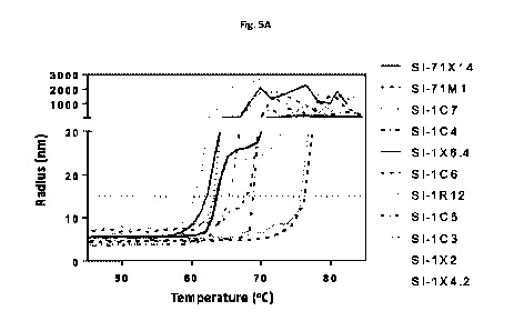
[0036] Figure 5 shows the thermal stability of bispecific antibodies using
dynamic light
scattering (A), and the SEC profile of SI-1X6.4 and SI-71X14, indicating the
lower aggregation
in SI-71X14 with humanized EGFR binding domain derived from cetuximab (B);
[0037] Figure 6 demonstrates the tandem binding of bispecific antibodies (SI-
1X6.4 and SI-
71X14) to EGFR, followed by HER3;
[0038] Figure 7 demonstrates the tandem binding of bispecific antibodies (SI-
1X6.4 and SI-
71X14) to HER3, followed by EGFR;
[0039] Figure 8 shows the surface expression of EGFR family members on Fadu
cancer cells;
[0040] Figure 9 shows the potency of SI-1X6.4 and its parental antibody, SI-
1C6 and on Fadu
cell proliferation;
[0041] Figure 10 shows the potency of SI-71X14 and its parental antibody, SI-
71M1, on Fadu
cell proliferation;
[0042] Figure 11 shows the potency of SI-1X2 and its parental antibody, SI-
1C3, on Fadu cell
proliferation;
[0043] Figure 12 shows the potency of SI-1X4.2 and its parental antibody, SI-
1C5, on Fadu
cell proliferation;
[0044] Figure 13 shows the comparative potency of antibodies, SI-1X6.4, SI-
71X14, SI-1C4,
and SI-1R12, on Fadu cell proliferation; and
[0045] Figure 14 shows the comparative potency of antibodies, SI-1X6.4, SI-
71X14, SI-1C4,
SI-71M1, SI-1C6, and SI-1C7, on Fadu cell proliferation.
DETAILED DESCRIPTION
[0046] This disclosure provides bispecific tetravalent antibodies with
superior therapeutic
properties or efficacies over the currently known anti-EGFR antibodies. In one
embodiment,
the antibodies target members of EGFR family including, without limitation,
EGFR, HER2,
and HER3. These bispecific tetravalent antibodies may inhibit different
receptor-mediated
6
CA 03196014 2023-03-20
WO 2022/061255 PCT/US2021/051164
oncogenic signaling simultaneously therefore overcome resistance in EGFR
inhibitor or
monoclonal antibody treatment.
[0047] The terms "a", "an" and "the" as used herein are defined to mean "one
or more" and
include the plural unless the context is inappropriate.
[0048] The terms "polypeptide", "peptide", and "protein", as used herein, are
interchangeable and are defined to mean a biomolecule composed of amino acids
linked by
a peptide bond.
[0049] The term "antigen" refers to an entity or fragment thereof which can
induce an
immune response in an organism, particularly an animal, more particularly a
mammal
including a human. The term includes immunogens and regions thereof
responsible for
antigenicity or antigenic determinants.
[0050] The terms "antigen- or epitope-binding portion or fragment", "variable
region",
"variable region sequence", or "binding domain" refer to fragments of an
antibody that are
capable of binding to an antigen (such as EGFR in this application). These
fragments may be
capable of the antigen-binding function and additional functions of the intact
antibody.
Examples of binding fragments include, but are not limited to, a single-chain
Fv fragment
(seFv) consisting of the variable light chain (VL) and variable heavy chain
(VH) domains of a
single arm of an antibody connected in a single polypeptide chain by a
synthetic linker, or a
Fab fragment which is a monovalent fragment consisting of the VL, constant
light (CL), VH
and constant heavy 1 (CH1) domains. Antibody fragments can be even smaller sub-
fragments and can consist of domains as small as a single CDR domain, in
particular the CDR3
regions from either the VL and/or VH domains33. Antibody fragments are
produced using
conventional methods known to those skilled in the art. The antibody fragments
can be
screened for utility using the same techniques employed with intact
antibodies.
[0051] The "antigen- or epitope-binding portion or fragment", "variable
region", "variable
region sequence", or "binding domain" may be derived from an antibody of the
present
disclosure by a number of art-known techniques. For example, the antigen-
binding fragment
(Fab) is a region (Fab region) on an antibody that binds to antigens. Purified
monoclonal
antibodies can be cleaved with an enzyme, such as pepsin, and subjected to
HPLC gel
filtration. Papain digestion of antibodies produces two identical antigen
binding fragments,
called "Fab" fragments, each with a single antigen binding site, and a
residual "Fe" fragment,
7
CA 03196014 2023-03-20
WO 2022/061255 PCT/US2021/051164
whose name reflects its ability to crystallize readily. Pepsin treatment
yields an F(ab')2
fragment that has two antigen combining sites and is still capable of cross-
linking antigen.
The appropriate fraction containing Fab fragments can then be collected and
concentrated
by membrane filtration and the like. For further description of general
techniques for the
isolation of active fragments of antibodies34,35.
[0052] The term "antibody" is used in the broadest sense and specifically
covers single
monoclonal antibodies and/or recombinant antibodies (including agonist and
antagonist
antibodies), antibody compositions with polyepitopic specificity, as well as
antibody
fragments (e.g., Fab, F(ab')2, and Fv), so long as they exhibit the desired
biological activity.
In some embodiments, the antibody may be monoclonal, polyclonal, chimeric,
single chain,
multi-specific or multi-effective, human and humanized antibodies, as well as
active
fragments thereof. Examples of active fragments of molecules that bind to
known antigens
include Fab, F(ab')2, scFy and Fv fragments, including the products of a Fab
immunoglobulin
expression library and epitope-binding fragments of any of the antibodies and
fragments
mentioned above.
[0053] The term "Fv" refers to the minimum antibody fragment which contains a
complete
antigen recognition and binding site. This region consists of a dimer of one
heavy and one
light chain variable domain in tight, non-covalent association. It is in this
configuration that
the three CDRs of each variable domain interact to define an antigen binding
site on the
surface of the VH-VL dimer. Collectively, the six CDRs confer antigen binding
specificity to
the antibody. However, even a single variable domain (or half of an Fv
comprising only three
CDRs specific for an antigen) has the ability to recognize and bind antigen,
although at a
lower affinity than the entire binding site.
[0054] In some embodiments, antibody may include immunoglobulin molecules and
immunologically active portions of immunoglobulin molecules, i.e. molecules
that contain a
binding site and that immunospecifically bind an antigen. A typical antibody
refers to
heterotetrameric protein comprising typically of two heavy (H) chains and two
light (L)
chains. Each heavy chain is comprised of a heavy chain variable domain
(abbreviated as VH)
and a heavy chain constant domain. Each light chain is comprised of a light
chain variable
domain (abbreviated as VL) and alight chain constant domain. The light chains
of antibodies
(immunoglobulins) from any vertebrate species can be assigned to one of two
clearly distinct
8
CA 03196014 2023-03-20
WO 2022/061255 PCT/US2021/051164
types, called kappa and lambda, based on the amino acid sequences of their
constant
domains. The VH and VL regions can be further subdivided into domains of
hypervariable
complementarity determining regions (CDR), and more conserved regions called
framework
regions (FR). Each variable domain (either VH or VL) is typically composed of
three CDRs
and four FRs, arranged in the following order: FR1, CDR1, FR2, CDR2, FR3,
CDR3, FR4 from
amino-terminus to carboxy-terminus. Within the variable regions of the light
and heavy
chains there are binding regions that interacts with the antigen.
[0055] Depending on the amino acid sequence of the constant domain of their
heavy chains,
immunoglobulins can be assigned to different classes. There are five major
classes of
immunoglobulins: IgA, IgD, IgE, IgG and IgM, and several of these may be
further divided into
subclasses (isotypes), e.g., IgG-1, IgG-2, IgG-3, and IgG-4; IgA-1 and IgA-2.
The heavy chain
constant domains that correspond to the different classes of immunoglobulins
are called
alpha, delta, epsilon, gamma, and mu, respectively. The subunit structures and
three-
dimensional configurations of different classes of immunoglobulins are well
known.
[0056] The term "valency" refers to valency of antibody referring the number
of antigenic
determinants that an individual antibody molecule can bind. The valency of all
antibodies is
at least two, whereas "antibody affinity" refers to the tendency of an
antibody to bind to a
specific epitope at the surface of an antigen, i.e., to the strength of the
interaction.
[0057] The term "monoclonal antibody" as used herein refers to an antibody
obtained from
a population of substantially homogeneous antibodies, i.e., the individual
antibodies
comprising the population are identical except for possible naturally
occurring mutations
that may be present in minor amounts. Monoclonal antibodies are highly
specific, being
directed against a single antigenic site. Furthermore, in contrast to
conventional
(polyclonal) antibody preparations which typically include different
antibodies directed
against different determinants (epitopes), each monoclonal antibody is
directed against a
single determinant on the antigen. In addition to their specificity, the
monoclonal antibodies
are advantageous in that they are synthesized by the hybridoma culture,
uncontaminated by
other immunoglobulins. The modifier "monoclonal" indicates the character of
the antibody
as being obtained from a substantially homogeneous population of antibodies,
and is not to
be construed as requiring production of the antibody by any particular method.
For example,
the monoclonal antibodies to be used in accordance with the present disclosure
may be
9
CA 03196014 2023-03-20
WO 2022/061255 PCT/US2021/051164
made by the hybridoma method first described by Kohler St Milstein36, or may
be made by
recombinant DNA methods (see, e.g., U.S. Pat. No. 4,816,567)37. "Recombinant"
means the
antibodies are generated using recombinant nucleic acid techniques in
exogeneous host
cells.
[0058] Monoclonal antibodies can be produced using various methods, including
without
limitation, mouse hybridoma, phage display, recombinant DNA, molecular cloning
of
antibodies directly from primary B cells, and antibody discovery methods38,
39, 40.
Monoclonal antibodies may include "chimeric" antibodies (immunoglobulins) in
which a
portion of the heavy and/or light chain is identical with or homologous to
corresponding
sequences in antibodies derived from a particular species or belonging to a
particular
antibody class or subclass, while the remainder of the chain(s) is identical
with or
homologous to corresponding sequences in antibodies derived from another
species or
belonging to another antibody class or subclass, as well as fragments of such
antibodies, so
long as they exhibit the desired biological activity41,42.
[0059] The term "humanized antibody" refers to a type of engineered antibody
having its
CDRs derived from a non-human donor immunoglobulin, the remaining
immunoglobulin-
derived parts of the molecule being derived from one (or more) human
immunoglobulin(s).
In addition, framework support residues may be altered to preserve binding
affinity.
Methods to obtain "humanized antibodies" are well known to those skilled in
the art43,44.
[0060] The terms "isolated" or "purified" refers to a biological molecule free
from at least
some of the components with which it naturally occurs. Either "Isolated" or
"purified," when
used to describe the various polypeptides disclosed herein, means a
polypeptide that has
been identified and separated and/or recovered from a cell or cell culture
from which it was
expressed. Ordinarily, a purified polypeptide will be prepared by at least one
purification
step. An "isolated" or a "purified" antibody refers to an antibody which is
substantially free
of other antibodies having different antigenic a binding specificity.
[0061] The term "immunogenic" refers to substances which elicit or enhance the
production
of antibodies, T-cells or other reactive immune cells directed against an
immunogenic agent
and contribute to an immune response in humans or animals. An immune response
occurs
when an individual produces sufficient antibodies, T-cells and other reactive
immune cells
against administered immunogenic compositions of the present disclosure to
moderate or
CA 03196014 2023-03-20
WO 2022/061255 PCT/US2021/051164
alleviate the disorder to be treated. While the immunogenic response generally
includes
both cellular (T cell) and humoral (antibody) arms of the immune response,
antibodies
directed against therapeutic proteins (anti-drug antibodies, ADA) may consist
of IgM, IgG,
IgE, and/or IgA isotypes.
[0062] The terms "specific binding", "specifically binds to, or "is specific
for a particular
antigen or an epitope" means that the binding is measurably different from a
non-specific
interaction. Specific binding can be measured, for example, by determining
binding of a
molecule compared to binding of a control molecule, which generally is a
molecule of similar
structure that does not have binding activity. For example, specific binding
can be
determined by competition with a control molecule that is similar to the
target.
[0063] The term "affinity" refers to a measure of the attraction between two
polypeptides,
such as antibody/antigen, receptor/ligand, etc. The intrinsic attraction
between two
polypeptides can be expressed as the binding affinity equilibrium dissociation
constant (KD)
of a particular interaction. A KD binding affinity constant can be measured,
e.g., by Bio-Layer
Interferometry, where KD is the ratio of kdis (the dissociation rate constant)
to kon (the
association rate constant), as KD = kdis/kon.
[0064] Specific binding for a particular antigen or an epitope can be
exhibited, for example,
by an antibody having a KD for an antigen or epitope of at least about 10-4 M,
at least about
10-5 M, at least about 10-6 M, at least about 10-7 M, at least about 10-8 M,
at least about 10-
9 M, alternatively at least about 10-10 M, at least about 10-11 M, at least
about 10-12 M, or
greater, where KD refers to the equilibrium dissociation constant of a
particular antibody-
antigen interaction. Typically, an antibody that specifically binds an antigen
will have a KD
that is 20-, 50-, 100-, 500-, 1000-, 5,000-, 10,000- or more times greater for
a control
molecule relative to the antigen or epitope.
[0065] Also, specific binding for a particular antigen or an epitope can be
exhibited, for
example, by an antibody having a KA or Ka for an antigen or epitope of at
least 20-, 50-, 100-,
500-, 1000-, 5,000-, 10,000- or more times greater for the epitope relative to
a control, where
KA or Ka refers to an association rate of a particular antibody-antigen
interaction.
[0066] It is considered by the application that the bispecific antibody
potentially has the
advantage over any combination therapy, which often has greater toxicity than
a single agent
treatment. Bispecific agents, such as bispecific antibody as disclosed in the
application, may
11
CA 03196014 2023-03-20
WO 2022/061255 PCT/US2021/051164
act as a single agent targeting the same antigens as the combination therapy
does but with
the increased efficacy and response rate and reduced toxicity when compare to
the
combination therapy. In comparison to the combination therapy using two
monoclonal
antibodies, a bispecific antibody therapeutics can be less toxic to patients
and/or more
potent due to the increased binding specificity.
[0067] In one aspect, the application provides a bispecific antibody having a
N terminal and
a C terminal, comprising at least two binding domains, wherein the binding
domain
comprises a Fab region and a scFy domain. The scFy domain may be attached to
either the
N terminal or the C terminal of the antibody. The Fab region and the scFy
domain each
independently have a binding specificity to different proteins in the EGFR
family.
[0068] In some embodiments, scFy molecules described herein contain a linker
of (GmS)n that
operably links the VH and VL, regardless of the V-region orientation (LH or
HL). The
remaining positions in the bispecific antibody may be consist of a human IgG
Fc or IgG null
Fc heavy chain, VH-CH1-Hinge-CH2-CH3, and its corresponding kappa or lambda
light chain,
VL-CL. Those scFy domains were genetically linked through a linker of (GmS)n
to either N-
terminal or C-terminal of IgG heavy chain, resulting in a contiguous ¨ 75 kDa
heavy chain
monomer peptide. When co-transfected with the appropriate light chain, the
final
symmetric bispecific molecule may be purified through the human IgG Fc
(Protein A) and
assayed to assess functional activity.
[0069] In one embodiment, the binding domain having the binding specificity to
EGFR
comprises cetuximab, panitumumab, and nimotuzumab. Cetuximab is an EGFR
inhibitor
medication used for the treatment of metastatic colorectal cancer and head and
neck cancer.
Cetuximab is a mouse/human chimeric monoclonal antibody given by intravenous
infusion.
[0070] In one embodiment, the binding domain having the binding specificity to
HER3
comprises MM-111, a bispecific HER2 and HER3 binding protein. MM-ill is a
human serum
albumin protein (HSA)-backed bispecific antibody fragment comprises one
therapeutic
binding to HER3, but its binding to HER2 alone is not sufficient to be
considered as a
therapeutic binding. In contrast, trastuzumab comprises one single therapeutic
binding to
HER2.
[0071] The bispecific antibody disclosed herein has the advantage of
recapitulating the
synergistic effect of simultaneously binding to both EGFR and HER3 using a
single agent.
12
CA 03196014 2023-03-20
WO 2022/061255 PCT/US2021/051164
The bispecific tetravalent antibodies may include an immunoglobulin G (IgG)
moiety with
two heavy chains and two light chains and two scFy moieties being covalently
connected to
either C or N terminals of the heavy or light chains via a linker, such as
(Gly-Gly-Gly-Gly-Ser)n
linkers or a (Gly-Gly-Gly-Ser)n linkers or (GmS)n linkers.
[0072] It is known that having a single therapeutic agent poses significant
challenges due to
the selection of binding moieties and the backbone structure that may affect
the binding
efficiency in vivo and the therapeutic efficacy in patients. For example, ALM
is a bispecific
antibody targeting HER2/HER3, which has antiproliferative activity to tumor
cells in vitro.
But a short circulating half-life makes it an unlikely candidate drug due to
rapid renal
clearance3.
[0073] Both cetuximab and panitumumab are monoclonal antibodies targeting EGFR
(Table
1). They differ in their isotypes, i.e. IgG 1 and IgG2, respectively. This
implies that the
difference in KD values of binding affinity can be beyond the sequences of CDR
and FR.
Indeed, reformatting a "2-in-1" bivalent bispecific antibody to IgG1 affects
the KD values for
binding affinity to EGFR and HER3, respectively. SI-1XC6.4 (C3)
(W02016106157A1 20
,
incorporated herein by reference in its entirety, also known as SI-B001 in
clinical trials,
NCT04603287) is a tetravalent bispecific antibody targeting EGFR and HER3 with
improved
ECSO when directly compared to that of duligotuzumab (also known as MEHD7945A,
"2-in-
1" antibody, or SI-1C4 as described in W02016106157A120). SI-1X6.4 (C3)
comprises the
same anti-EGFR binding domain as that of cetuximab and displays differences in
affinity KD
values (Table la). SI-1X6.4 (C3) comprises the same anti-HER3 binding domain
as that of
MM-111, and their affinity KD values are significantly different from each
other (Table la).
The structural configuration of each bispecific antibody may contribute to
differences in the
efficacy of killing tumor cells. Since many forms of human cancer overly
express either EGFR
or HER2 but not HER3, the unforeseen benefit of a reduced affinity KD of the
anti-HER3
binding domain may allow SI-B001 to bind to HER3 only on EGFR-positive tumor
cells but
not on HER3-positive normal cells.
[0074] The term, therapeutic binding, is referred to a binding domain that has
been tested in
clinical trials in the form of antibody therapeutics for safety. The concept
of Specificity-
Enhanced Bispecific Antibodies (SEBA) defines bispecific antibodies configured
to have a
combination of therapeutic bindings to two tumor antigens on the same tumor
cell but not
13
CA 03196014 2023-03-20
WO 2022/061255 PCT/US2021/051164
on normal cells. Using the EGFR family as an example, there are multiple
therapeutic binding
domains, including those derived from cetuximab, trastuzumab, MM-111, and "2-
in-1", the
objective of SEBA is to develop and/or improve bispecific antibodies as a
single therapeutic
agent comprising therapeutic binding to two members of EGFR family, such as
the pairs of
EGFR/HER2, EGFR/HER3, or HER2/HER3. Each configuration may reveal different
efficacies in the binding specificity, affinity, and avidity, heregulin
binding, inhibition of
EGFR/HER3 dimerization and downstream signaling, and ultimately, the
therapeutic
efficacy and cytotoxicity to patients.
[0075] A potential shortcoming of cetuximab is that its variable regions were
derived from
mice. It has been demonstrated that chimeric antibodies retain non-human
sequences may
have increased capacity for immunogenicity when compared to humanized or human
antibodies.6 On the other hand, humanization can increase the stability of
antibodies by
making the framework regions more compatible.7 Another concern is the occupied
glycan
site at VH N85 (Kabat), where Fab glycosylation could affect the biological
properties of the
antibody, as well as introduce glycan heterogeneity that must be well-
controlled during
manufacturing.8,9 While immunogenicity of cetuximab appears low based on low
incidence
of anti-cetuximab IgG response (5%), hypersensitivity is a common occurrence
due largely
to pre-existing IgE antibodies against the galactose-a-1,3-galactose
oligosaccharide that
modifies the VH when expressed in SP2/0 cells10. To overcome these
liabilities, cetuximab
may opt for humanization and removal of post-translational modification sites
to stabilize
the antibody, and reduce the potential for immunogenicity while retaining high
affinity for
EGFR. In this context a humanized EGFR binding domain, which has harbored a
therapeutic
binding domain from cetuximab, may improve therapeutic efficacy of an existing
SEBA, SI-
B001.
EXAMPLES
[0076] While The following examples are provided by way of illustration only
and not by way
of limitation. Those of skill in the art will readily recognize a variety of
non-critical
parameters that could be changed or modified to yield essentially the same or
similar results.
Example 1: SI-71X14, a bispecific tetravalent anti-EGFRxHER3 antibody
14
CA 03196014 2023-03-20
WO 2022/061255 PCT/US2021/051164
[0077] The humanization of cetuximab was designed using different input models
with
Calculate Mutation Energy set to True (CHARMm forcefield) in order to generate
Best Single
Mutations sequences. The cetuximab models generated by Discovery Studio's
Antibody
Modeling Cascade were used. The Input Sequences were cetuximab VH (ending TVSS
instead
of TVSA) and cetuximab VL. To increase similarity of the VH C-terminus to the
consensus
sequence in human (Figure 1), or to make the Vic C-terminus more VA-like, the
humanization
incorporated changes in the input sequence. After humanization in Discovery
Studio, VL was
further modified by converting the last three residues of the Vic domain into
their
corresponding residues from the A 1-gene. This change was evaluated due to the
known
importance of the last VL beta strand in determining scFy stability and
aggregation
propensity, and the more hydrophobic nature of the VA terminus, which could
provide
packing energy to stabilize the interaction 22, 23, 24. The Top 5 Framework
Templates were
used with Sequence Similarity Cutoff of 10. CDR loop definition was set to
Honegger and
Maximum Templates Per Loop was set to 3 with Optimization Level set to High.
After
generating humanized sequences, VL was further modified by substituting the
last four
residues of the VL to LTVL to mimic the stable FR4 of lambda antibodies.
[0078] Humanized Cetuximab SI-71M1 were designed based on structural analysis
of
cetuximab, by mutating framework residues to those residues occurring with a
frequency of
at least 5% in the human germline that caused the most stable structure in
silico. Because
the energy analysis for this type of humanization depends on the input model,
several input
structures were examined. SI-71X14 is generated by connecting MM-111's HER3
scFV to C
terminal of SI-71M1 heavy chains via (GmS)n linkers.
[0079] Thus, SI-71X14 is a modification of SI-1X6.4 where the cetuximab mouse
variable
regions were replaced with humanized cetuximab variable regions. Except the
primary
sequences that differ from SI-1X6.4, SI-71X14 is also an aEGFR and aHER3
bispecific
tetravalent antibody.
[0080] The amino acid changes are shown in Figure 1. Panel A shows that 17
residue
differences in heavy chain sequences are localized to the anti-EGFR cetuximab
VH domain.
Panel B shows that 22 amino acid differences in the light chain sequences are
localized to the
anti-EGFR cetuximab VK domain. Panel C zooms in on the VH region to show all
amino acid
CA 03196014 2023-03-20
WO 2022/061255 PCT/US2021/051164
differences in the heavy chains, while panel D zooms in on the VK region to
show all
differences in the light chains.
[0081] In addition to these two bispecific proteins, a number of bispecific
and monospecific
molecules were included in subsequent assays with properties described in
Table lb. This
allowed for comparison of different EGFR and HER3 binding domains as well as
different
types of structures.
[0082] Proteins were expressed by transfecting the expression plasmids for SI-
1C7 and SI-
1R12 (single plasmid) or co-transfecting heavy and light chains for SI-1C3, SI-
1C5, SI-1C6,
SI-71M1, SI-1X2, SI-1X6.4, SI-71X14 and SI-1C4, in the ExpiCHO system (Thermo
Fisher).
Briefly, 10 lig of each expression plasmid (or 20 lig of an unpaired plasmid)
was brought to
1m1 with OptiPRO SFM medium. 1m1 of OptiPRO SFM medium containing 80u1
Expifectamine CHO reagent was added to the DNA and incubated at room
temperature for
2.5 minutes. The resulting mixture was then added to 25m1 ExpiCHO cells at
6x10⁶ cells/ml
in a 125m1 Erlenmeyer flask and incubated at 37 C, 5% CO2, 150rpm. Cells were
fed with
8.75m1 ExpiCHO feed and 150 ul of CHO enhancer at 24 hours post-transfection
and shifted
to 32 C, 5% CO2, 150rpm. Cells were fed again at 48 hours post-transfection
with 8.75m1
ExpiCHO feed. Culture supernatant was harvested 8 days post-transfection, spun
for 1 hour
at 4500rpm to pellet the cells and then passed through a 0.2mm filter.
[0083] Fc-containing proteins were purified from the harvested supernatant
using a 1-ml
MabSelect PrismA protein A column (GE Healthcare). The column was equilibrated
with
phosphate-buffered saline. The supernatant was then passed through the column
at a flow
rate of 2 ml/min. The column was washed with 10m1 PBS + 0.1% Triton X-100,
followed by
10m1 PBS + 300mM NaCl, and finally 10m1 PBS. Protein was then eluted by
passing 5m1 of
50 mM sodium acetate, pH 3.5 through the column. The eluted protein was
immediately
neutralized by addition of 0.5m1 1M Tris-C1, pH8Ø
[0084] His-tagged scFy proteins were purified from the harvested supernatant
using a 1-ml
HisTrap HP column (GE). The column was equilibrated with phosphate-buffered
saline
containing 0.5 M NaCl and 20 mM imidazole, pH 7.4. The supernatant was spiked
with 10x
binding buffer to reach 0.5 M NaCl and 20 mM imidazole and run over the column
at a flow
rate of 2 ml/min. The column was washed with 10 column volumes of PBS
containing 0.5 M
16
CA 03196014 2023-03-20
WO 2022/061255 PCT/US2021/051164
NaCl and 20 mM imidazole, and the protein was eluted using PBS containing 0.5
M NaCl and
500 mM imidazole, pH 7.4.
[0085] Immediately after first-step protein A or His tag purification,
proteins were analyzed
by analytical SEC using using Waters Acquity UPLC H-Class with ACQUITY UPLCO
Protein
BEH SEC 200A, 4.6mm x 150mm, 1.7 um column. PBS (125 mM sodium phosphate, 137
mM
sodium chloride, pH 6.8) was used as mobile phase for 10-minute runs at 0.3
ml/min,
injecting 10 lig protein.
[0086] Cetuximab have two intrinsic N-glycosylation sites, N85 (Kabat) and
N297 (Eu),
located in Fab and Fc, respectively. The possible immunogenic N-glycan in the
N85 position
may impact the pharmacokinetic profile and give rise to anti-drug antibody
(ADA). In the
humanization version, the position 85 was mutated from N to D, which
eliminates the
consensus N-glycosylation site, and no glycosylation was detected in any
expressed protein.
This strategy helped protein purification and characterization but had no
effect on the
binding affinity as shown in Table 2,4.
Example 2: Binding kinetics to human EGFR
[0087] Monomeric EGFR extracellular domain binding was measured in a biolayer
interferometry (BLI) binding assay on an Octet Red 384 instrument (Sartorius).
10 ug/mL
of SI-71X14, SI-71M1, SI-1X6.4, SI-1C3, SI-1X2, SI-1C6, SI-1X4.2, SI-1C4, or
SI-1C5 was diluted
in assay buffer (PBS containing 1% bovine serum albumin and 0.05% Tween 20)
and
captured on anti-hulgG Fc (AHC) biosensor tips for 180 seconds. Tips were
washed for 60
seconds in assay buffer and moved to a human EGFR (expressed and purified in-
house)
sample in 1:2 serial dilutions from 100 nM to 0 nM. Binding of EGFR
extracellular domain to
the tips was recorded as biolayer interferometry signals (ialm) over an
association time of
180 seconds. Tips were moved to assay buffer and dissociation was observed for
420
seconds. Sensors were regenerated using 10 mM glycine pH 1.5. Data were
globally fit for
each antibody to a 1:1 binding model to extract kinetic parameters kon, kchs,
and KD (Figure
2, Table 2).
[0088] Notably, all cetuximab-based proteins had similar binding kinetics to
human EGFR.
For example, the mAbs SI-1C6 (cetuximab) and SI-71M1 (humanized cetuximab) had
KD
values of 5.34 and 4.76 nM, respectively. The bispecific (EGFR x HER3)
molecules SI-1X6.4
17
CA 03196014 2023-03-20
WO 2022/061255 PCT/US2021/051164
(containing cetuximab variable regions with mouse framework) and SI-71X14
(humanized
cetuximab framework) had similar EGFR binding kinetics with KDs of 5.38 and
4.61 nM,
respectively. Meanwhile, panitumumab-based mAb (SI-1C3) and bispecific (SI-
1X2)
proteins had slightly higher affinity with KD values of 2.28 and 2.77 nM,
respectively, which
was driven by slower dissociation rate. Nimotuzumab-based mAb (SI-1C5) and
bispecific
(SI-1X4.2) proteins had weaker affinity with KD values of 15.8 and 18.8 nM,
respectively,
based on slower association kinetics and faster dissociation kinetics. The 2-
in-1 bispecific
antibody duligotuzumab (SI-1C4) had EGFR affinity of 14.6 nM with the fastest
dissociation
rate.
Example 3: Binding kinetics to human HER3
[0089] Monomeric HER3 extracellular domain binding was measured in a biolayer
interferometry (BLI) binding assay on an Octet Red 384 instrument (Sartorius).
10 ug/mL
of SI-71X14, SI-1C7, SI-1C4, SI-1X6.4, SI-1X2, or SI-1X4.2 was diluted in
assay buffer (PBS
containing 1% bovine serum albumin and 0.05% Tween 20) and captured on anti-
hulgG Fc
(AHC) biosensor tips for 180 seconds. Tips were washed for 60 seconds in assay
buffer and
moved to a human HER3 (Acro ER3-H5223) sample in 1:2 serial dilutions from 400
nM to 0
nM. Binding of HER3 extracellular domain to the tips was recorded as biolayer
interferometry signals (ialm) over an association time of 180 seconds. Tips
were moved to
assay buffer and dissociation was observed for 420 seconds. Sensors were
regenerated using
mM glycine pH 1.5. Data were globally fit for each antibody to a 1:1 binding
model to
extract kinetic parameters kon, kchs, and KD (Figure 3, Table 3).
[0090] Notably, all proteins whose anti-HER3 domain was derived from MM-111
had similar
binding kinetics to human HER3. For example, cetuximab-based bispecific
proteins SI-1X6.4
(cetuximab variable regions with mouse framework) and SI-71X14 (humanized
cetuximab
framework) had KD values of 107 and 117 nM, respectively. Panitumumab- and
nimotuzumab- based bispecific antibodies SI-1X2 and SI-1X4.2 had HER3 KD
values of 131
and 146 nM, respectively. Finally, a control Fc-scFy protein with the same
anti-HER3 domain
(SI-1C7) had similar binding kinetics with KD of 149 nM. The 2-in-1 bispecific
antibody
duligotuzumab (SI-1C4), which has distinct anti-HER3 variable regions from the
other
bispecific proteins, had significantly tighter HER3 binding with KD 4.29 nM.
18
CA 03196014 2023-03-20
WO 2022/061255 PCT/US2021/051164
[0091] Due to lack of Fc domain, another comparator bispecific protein (SI-
1R12 = MM-111,
HER2 x HER3 albumin fusion) was tested in a different assay format on the same
Octet
instrument using AR2G sensors. 20 ug/mL of SI-1R12 was diluted in 10 mM
acetate pH 6.0
and covalently coupled using EDC/NHS according to manufacturer's instructions
using a
600-second loading step. After immobilization of SI-1R12, tips were washed for
120 seconds
in assay buffer and moved to a human HER3 (Acro ER3-H5223) sample in 1:2
serial dilutions
from 400 nM to 0 nM. Binding of HER3 extracellular domain to the tips was
recorded as
biolayer interferometry signals (ialm) over an association time of 180
seconds. Tips were
moved to assay buffer and dissociation was observed for 420 seconds. Data were
globally fit
for each antibody to a 1:1 binding model to extract kinetic parameters kon,
kchs, and KD
(Figure 3, Table 3). Kinetics of SI-1R12 binding to HER3 were similar to that
of other
bispecific proteins tested, with a KD value of 95.6 nM.
Example 4: Simultaneous binding to human EGFR and HER3
[0092] Bispecific binding to EGFR and HER3 extracellular domains was measured
in a
sandwich-type biolayer interferometry (BLI) binding assay on an Octet RED 384
instrument
(Sartorius). After a 180-second baseline step in assay buffer (PBS with 1% BSA
and 0.05%
Tween 20), biotinylated human EGFR (Acro EGF-H82E3) was loaded onto SA sensors
at 5
ug/m1 in assay buffer for 240 seconds. Following another 180-second baseline
step,
association with 2-fold serial dilutions (0 to 100 nM) of SI-1C7, SI-1X2, SI-
1X4.2, SI-1X6.4, SI-
71M1, or SI-71X14 in assay buffer was performed for 240 seconds followed by a
600-second
dissociation in assay buffer without any protein. Immediately following this
antibody-
binding step, another association step with 500 nM HER3 ECD (in-house
expressed) was
performed, followed by a 600-second dissociation phase. Each
association/dissociation
event were separately fit using a 1:1 binding model to extract the binding
kinetics for
bispecific EGFR and HER3 binding.
[0093] The first binding event measured in this assay is that of antibodies
binding to
immobilized EGFR, which represents the avidity of the interaction as it might
occur on the
cell surface. These data are shown in Figure 4 and kinetic parameters are
shown in Table 4.
The data demonstrate that cetuximab-based antibodies including humanized
cetuximab
mAb (SI-71M1) and bispecific cetuximab x anti-HER3 antibodies SI-1X6.4
(cetuximab
19
CA 03196014 2023-03-20
WO 2022/061255 PCT/US2021/051164
variable regions with mouse framework) and SI-71X14 (humanized cetuximab
framework)
all had very high avidity to immobilized EGFR where the KD of the interaction
was too tight
to be accurately quantified but estimated as less than 1 pM. This high avidity
was driven by
very slow dissociation rate. Similarly, the panitumumab-based EGFR x HER3
bispecific
antibody SI-1X2 also had high avidity with estimated KD less than 1 pM.
Nimotuzumab-based
EGFR x HER3 bispecific antibody SI-1X4.2 also had strong avidity with fitted
KD value of 398
pM. As expected, the Fc-scFy protein specific for HER3, SI-1C7, did not show
binding to EGFR.
[0094] The second event of interest is the captured antibody (already bound
via its anti-
EGFR domains) binding to HER3 protein in solution. The kinetic parameters for
these
interactions are tabulated in Table 5. Cetuximab-based EGFR x HER3 bispecific
antibodies
SI-1X6.4 (cetuximab variable regions with mouse framework) and SI-71X14
(humanized
cetuximab framework) had similar HER3 KD values of 617 and 922 nM,
respectively.
Panitumumab- and nimotuzumab-based EGFR x HER3 bispecific antibodies SI-1X2
and SI-
1X4.2 had similar HER3 affinities of 770 and 165 nM, respectively. Notably,
humanized
cetuximab mAb (SI-71M1) did not show binding in this assay step due to lack of
specificity
for HER3, while Fc-scFy protein SI-1C7 targeting HER3 showed no binding
response due to
lack of loading during the EGFR binding step. Thus, the sandwich assay
demonstrates that
EGFR x HER3 bispecific antibodies are able to simultaneously bind EGFR and
HER3, while
proteins with only specificity to either EGFR or HER3 did not show a response
in the assay.
Example 5: Improved thermal stability
[0095] Dynamic light scattering on a Wyatt DynaPro Plate Reader III was
performed for
protein thermal stability analysis. Proteins were diluted to 1 mg/ml in 25 mM
sodium
acetate, 75 mM sodium chloride, 5% (w/v) sucrose, pH 5.5 in 30 p1/well.
Temperature was
ramped from 25 C to 85 C at 1.0 C/min while monitoring the radius. Due to
difficulty fitting
the differently shaped unfolding curves reproducibly, the temperature at which
the radius
surpassed 15 nm was used as an objective metric of thermal stability. Samples
were run in
duplicate, and buffer alone was run as negative control.
[0096] Figure 5 shows the thermal melting curves of SI-71X14, SI-71M1, SI-1C7,
SI-1C4, SI-
1X6.4, SI-1C6, SI-1R12, SI-1C5, SI-1C3, SI-1X2, and SI-1X4.2 and Table 6 shows
Tm values
for these proteins. EGFR mAbs panitumumab (SI-1C3), nimotuzumab (SI-1C5),
cetuximab
CA 03196014 2023-03-20
WO 2022/061255 PCT/US2021/051164
(SI-1C6), and humanized cetuximab (SI-71M1) had Tm values of 77.05, 65.79,
68.39, and
77.80 C, respectively. Thus, humanization of cetuximab not only increased
thermal stability
by >9 C, but also generated the most stable EGFR mAb of the four tested.
Bispecific EGFR x
HER3 antibodies based on panitumumab (SI-1X2), nimotuzumab (SI-1X4.2),
cetuximab (SI-
1X6.4), and humanized cetuximab (SI-71X14) had Tm values of 63.73, 63.79,
62.33, and
64.10 C, respectively. Thus, addition of anti-HER3 scFy to the C-terminus of
EGFR mAbs
tended to normalize and decrease thermal stability. Notably, the bispecific
EGFR x HER3
antibody based on humanized cetuximab had the highest thermal stability of
this panel, and
increased stability of the parent cetuximab protein by 1.77 C. The control
protein based on
antibody Fc fused to anti-HER3 scFy (SI-1C7) had Tm of 63.17 C, which is
similar to that of
bispecific molecules containing this anti-HER3 scFy domain. The bispecific
antibody SI-1C4
had Tm of 69.80 C, confirming the high stability of the mAb-like platform.
Finally, the MM-
111 bispecific HSA fusion (SI-1R12) had the lowest thermal stability with a Tm
of 60.41 C.
This result demonstrates the favorable stability of the antibody format
compared to other
protein scaffolds.
Example 6: Sequential binding to human EGFR and HER3
[0097] Bispecific binding to EGFR and HER3 extracellular domains was measured
in a
tandem biolayer interferometry (BLI) binding assay on an Octet RED 384
instrument
(Sartorius).
[0098] In one format, antibody protein was captured onto AHC sensors, followed
by a first
association step with EGFR, followed by a second association step with HER3,
followed by a
dissociation step (Figure 6). In particular, after a 20-second baseline step
in assay buffer
(PBS with 1% BSA and 0.05% Tween 20), 10 ug/m1 of antibody protein was loaded
for 180
seconds, followed by a 60-second baseline step. Next, the first association
step with 100 nM
of EGFR (purified in-house) was performed for 720 seconds, followed by the
second
association step with 100 nM of EGFR and 400 nM of HER3 (Acro ER3-H5223) for
720
seconds. Of note, the second step contained HER3 protein, but additionally
contained the
same amount of EGFR as in the first step (100 nM), so that dissociation of
EGFR would not
complicate the kinetics observed in the second step. Finally, a 720-second
dissociation step
was performed.
21
CA 03196014 2023-03-20
WO 2022/061255 PCT/US2021/051164
[0099] In the assay with tandem EGFR then HER3 steps, the control EGFR mAbs SI-
1C6 and
SI-71M1 showed a large response in the EGFR phase, with no significant
increase in response
during the HER3 phase, indicating binding to EGFR in the first step, but not
HER3 in the
second step. The control Fc-scFy protein targeting HER3, SI-1C7, showed no
binding during
the first EGFR step, but a large response during the second HER3 step. The 2-
in-1 control
mAb SI-1C4 showed binding during the EGFR and HER3 steps, indicating that the
first EGFR
step was not sufficient to saturate antibody binding so that additional
molecules of HER3
could bind in the second step. Bispecific EGFR x HER3 antibodies SI-1X6.4 and
SI-71X14
showed significant binding response during both EGFR and HER3 steps,
confirming that
these proteins are able to simultaneously bind to molecules of EGFR and HER3.
[00100] In the assay, we also observe SI-71X14 have better binding response
than SI-1X6.4
(nm in Y axis), about 0.1 nm, for EGFR and EGFR/HER3 association step, it
implies the
binding quantity of SI-71X14 is higher than SI-1X6.4.
[00101] In another format, antibody protein was captured onto AHC sensors,
followed by a
first association step with HER3, followed by a second association step with
EGFR, followed
by a dissociation step (Figure 7). In particular, after a 20-second baseline
step in assay
buffer (PBS with 1% BSA and 0.05% Tween 20), 10 ug/m1 of antibody protein was
loaded
for 180 seconds, followed by a 60-second baseline step. Next, the first
association step with
400 nM of HER3 (Acro ER3-H5223) was performed for 720 seconds, followed by the
second
association step with 100 nM of EGFR (purified in-house) and 400 nM of HER3
for 720
seconds. Of note, the second step contained EGFR protein, but additionally
contained the
same amount of HER3 as in the first step (400 nM), so that dissociation of
HER3 would not
complicate the kinetics observed in the second step. Finally, a 720-second
dissociation step
was performed.
[00102] In the assay with tandem HER3 then EGFR steps, the control EGFR mAbs
SI-1C6 and
SI-71M1 showed no response during the HER3 binding step followed by large
response
during the EGFR step, as expected. The control Fc-scFy protein targeting HER3,
SI-1C7,
showed binding during the first HER3 step, but no binding during the second
EGFR step, as
expected. The 2-in-1 control mAb SI-1C4 showed significant binding during the
first HER3
step, but no additional binding during the second EGFR binding step. The
interpretation is
that both of this antibody's Fab regions bound to HER3 during the first step,
such that there
22
CA 03196014 2023-03-20
WO 2022/061255 PCT/US2021/051164
was no free Fab to bind to EGFR during the second step. Bispecific EGFR x HER3
antibodies
SI-1X6.4 and SI-71X14 showed significant binding response during both EGFR and
HER3
steps, confirming that these molecules are able to simultaneously bind to
molecules of EGFR
and HER3. Importantly, the tetravalent nature of the SI-1X6.4 and SI-71X14
structure, along
with separate binding domains for each antigen, allowed these antibodies to
bind
concurrently to EGFR and HER3 where this phenomenon was not possible for the 2-
in-1
control mAb SI-1C4.
[00103] In the assay, we also observe SI-71X14 have better binding response
than SI-1X6.4
(nm in Y axis), about 0.1 nm, for HER3 and EGFR/HER3 association step, it
implies the
binding quantity of SI-71X14 is higher than SI-1X6.4.
[00104] Taken together, the characterization of binding kinetics implies a
mode of action for
SEBA, i.e. specificity enhanced bispecific antibodies, such as SI-71X14 and SI-
1X6.4. Unlike
the in vitro kinetics, the response and effectiveness of the bispecific
antibody treatment may
depend on tissue distribution of EGFR and HER3. In patients with various forms
of solid
tumors, the expression of EGFR may be deregulated by tumor cells while HER3
may be
expressed by both normal and tumor cells. Incidentally, many anti-HER3
antibody therapies
have failed due to the safety issue, indicating that targeting normal cells
may have
outweighed the tumor cells in vivo. In this application, the result shows that
both SI-1X6.4
and SI-71X14 can enact a sequential binding mode and that SI-71X14 with a
humanized
EGFR binding domain unveils an improved binding kinetics. The differentiated
KD value
between EGFR (strong) and HER3 (weak) underlies the selective binding of both
SI-71X14
and SI-1X6.4 in favor of binding to EGFR-expressing cancer cells relative to
HER3 positive
normal cells. In this context, SEBA may help achieve reduced side effects in
vivo.
Furthermore, having differentiated binding affinity to two tumor-associated
antigens (TAA),
as measured by strong and weak KD values, may be a new strategy for designing
SEBA to
target cancer-causing receptors.
Example 7: Inhibiting tumor cell proliferative
[00105] To assess the growth inhibitory potential of anti-EGFR domain
containing antibodies,
the effect of the cetuximab-derived EGFR domain (wt) and the humanized EGFR
binding
domain were compared while in different therapeutic formats. The growth
inhibitory effects
23
CA 03196014 2023-03-20
WO 2022/061255 PCT/US2021/051164
were tested against the Fadu cell line (hypopharyngeal squamous cell
carcinoma, ATCC HTB-
43) which expresses both EGFR and HER3 proteins, as well as HER2 protein
(Figure 8).
Specific antigen presentation was determined by incubation of Fadu cells with
fluorescent
conjugated antibodies specific for either EGFR, HER3, or HER2 and isotype
match control
antibodies. Antibody binding was quantified on cells using FACS method (BD
Bioscience
LSR-Fortessa).
[00106] The cell line was seeded in 96-well tissue culture plates at a density
of 5000 cells per
well in 200u1s of RPMI-1640 medium containing 1% FBS. Treatments were added
within
the dose range of 90 nM to 85.8 fM. Cells were cultured in the presence of the
test antibodies
for 63 hours in triplicate plates. Nuclei counts were obtained based on the
Fadu cell line
stable expression of nuclear localized fluorescence reporter protein mKate2
using time-
series microscopy (Incucyte Zoom). Data was collected at baseline, and at
intervals during
culture. Normalized proliferation is reported based on well seeding and
untreated control
conditions. Comparative effects of the wt cetuximab and the humanized
cetuximab anti-
proliferative effect were represented in dose response curves and the IC-50 of
inhibition
provided based on regression analysis using Sigmoidal, 4PL, Least squares
fitting, where X
is concentration, and curve fit is provided as R2 value in figures (Graphpad
Prism 9).
[00107] When the wt cetuximab domain is utilized in the bi-specific format
with HER3 (SI-
X6.4), the IC-50 is reduced 3 fold compared to the EGFR mAbs alone (SI-1C6),
while the
addition of the HER3 domain provides a greater overall anti-proliferative
effect (Figure 9).
However, the humanized cetuximab domain restores the anti-proliferative IC-50
when
combined with HER3 binding domain (SI-71X14) in comparison to the humanized
cetuximab
mAb alone (SI-71M1), and retains the greater overall anti-proliferative
effects at higher
concentrations (Figure 10).
[00108] Consistent with the role of HER3 enhancing the function of EGFR
inhibition in the bi-
specific format, panitumumab (SI-1C3) when engineered in the bi-specific
formation with
HER3 domain (SI-1X2) achieves a greater overall anti-proliferative effect
(Figure 11).
Whereas, the EGFR antibody nimotuzumab (SI-1C5) shows poor capability to
inhibit Fadu
cell proliferation in this assay system, and the addition of HER3 to
nimotuzumab in the bi-
specific formation (SI-1X4.2) is not observed to provide a benefit, attesting
to the key
24
CA 03196014 2023-03-20
WO 2022/061255 PCT/US2021/051164
requirement of the EGFR domain facilitating the anti-proliferative benefit to
enable the
benefit of HER3 blocking in the bispecific drug design (Figure 12).
[00109] The Fadu response to humanized cetuximab in combination with the HER3
binding
domain in bi-specific format (SI-71X14) achieves 3x better anti-proliferative
IC-50
compared with the wt cetuximab in the same format (SI-1X6.4). The humanized
EGFR in SI-
71X14 further achieves a significantly greater overall anti-proliferative
effects at higher
concentrations (Figure 13). By way of comparison, SI-1C4 is a bispecific
antibody against
EGFR and HER3 built on the two-in-one platform described by Schaefer30. IC4
has a similar
structure to a monoclonal antibody. The molecule can bind to either EGFR or
HER3 on each
Fab arm, but cannot engage both targets simultaneously on each Fab arm. While
blocking
either EGFR or HER3, and in excess, both receptors, the anti-proliferative
performance of the
two-in-one is inferior to both the bi-specific format of SI-71X14 and SI-1X6.4
(Figure 14).
By way of comparison, SI-1R12 is MM-111, a HER2 X HER3 bispecific that has
reported anti-
proliferative effects. However, while Fadu express both HER2 and HER3,
inhibition is not
achieved. This attests to the key combination of EGFR facilitation of HER3
blocking
antiproliferative effect, rather than HER2 and HER3 on this cell line (Figure
13).
[00110] The reengineering of the cetuximab antibody enable greater anti-
proliferative
potency when engineered in multi-specific formation compared with wt cetuximab
as
demonstrated in T cell engager bi and penta specific structures, as well when
in combination
with HER3 binding domains. The humanized cetuximab domain also achieves
greater
overall anti-proliferative effects when combined with HER3 binding domains
compared to
wt cetuximab.
CA 03196014 2023-03-20
WO 2022/061255
PCT/US2021/051164
TABLES
Table la. The KD value of a TAA binding domain may vary in different
therapeutic
antibodies.
Therapeutics Type of Target EGFR HER2 HER3
ECso
Candidate Mab affinity affinity affinity
(nM)
KD KD KD
(nM) (nM) (nM)
Cetuximabll Bivalent EGFR 0.2
Panitumumab 12 Bivalent EGFR 0.05
Nimotuzumab 13 Bivalent EGFR 67
Necitumumab21 Bivalent EGFR 0.28
Trastuzumab 14 Bivalent HER2 1.8
Pertuzumab 15 Bivalent HER2 0.8
Patritumab 16 Bivalent HER3 1-3
MM-12117 Bivalent HER3 0.75
MM-11118 Monovalent HER2 St 0.3 16
bispecific HER3
HSA-scFv*
Duligotuzumab "2-in-1" EGFR or 19.9 2.63 0.068 -
(MEHD7945A)19 Monovalent HER3 0.589 #
bispecific
SI-1X6.4(C3) Bivalent EGFR St 5.38 107
bispecific HER3
#Results of ADCC analyses using Fadu and NCI-H1975 cells, respectively.
26
CA 03196014 2023-03-20
WO 2022/061255
PCT/US2021/051164
Table lb. The antibodies having therapeutic binding domains for targeting
either EGFR,
HER3, or both.
Origin of anti- EGFR Origin of anti- HER3
Protein Specificity
EGFR Fab valency HER3 scFv valency
SI-1C3 EGFR Panitumumab Bi n/a
n/a
SI-1C5 EGFR Nimotuzumab Bi n/a
n/a
SI-1C6 EGFR Cetuximab Bi n/a n/a
SI-71M1 EGFR Hu-Cetuximab Bi n/a
n/a
SI-1C7 HER3 Fc-scFy n/a MM-111 Bi
SI-1X2 EGFR x Panitumumab Bi MM-111
Bi
HER3
SI-1X4.2 EGFR xNimotuzumab Bi MM-111
Bi
HER3
SI-1X6.4 EGFR x Cetuximab Bi MM-111 Bi
HER3
SI-71X14 EGFR xHu-Cetuximab Bi MM-
111 Bi
HER3
EGFR x
SI-1C4 HER3 Duligotuzumab Mono
Duligotuzumab Mono
SI-1R12 HER2 x n/a n/a MM-111 Mono
HER3
Table 2. Binding kinetics (affinity) of anti-EGFR proteins to His-tagged human
EGFR
extracellular domain, as measured by biolayer interferometry.
KD kon kdis
Protein (M) (1/Ms) (1/s)
SI-71X14 4.61E-09 2.64E+05 1.22E-03
SI-71M1 4.76E-09 2.72E+05 1.30E-03
SI-1X6.4 5.38E-09 2.89E+05 1.56E-03
SI-1C6 5.34E-09 2.71E+05 1.45E-03
SI-1C3 2.28E-09 1.95E+05 4.45E-04
SI-1X2 2.77E-09 1.97E+05 5.46E-04
SI-1C4 1.46E-08 3.00E+05 4.38E-03
SI-1C5 1.58E-08 1.13E+05 1.78E-03
SI-1X4.2 1.88E-08 1.08E+05 2.02E-03
27
CA 03196014 2023-03-20
WO 2022/061255
PCT/US2021/051164
Table 3. Binding kinetics (affinity) of anti-HER3 proteins to His-tagged human
HER3
extracellular domain, as measured by biolayer interferometry. Note that in
contrast to all
other measurement, which were determined using AHC sensors, SI-1R12 required
setup
with AR2G sensors due to lack of Fc domain.
Protein KD kon kdis
(M) (1/Ms) (1/s)
SI-71X14 1.17E-07 2.65E+05 3.08E-02
SI-1C7 1.49E-07 2.24E+05 3.33E-02
SI-1C4 4.29E-09 2.05E+05 8.77E-04
SI-1X6.4 1.07E-07 2.82E+05 3.02E-02
SI-1X2 1.31E-07 2.75E+05 3.61E-02
SI-1X4.2 1.46E-07 2.60E+05 3.80E-02
SI-1R12 9.56E-08 4.41E+05 4.22E-02
Table 4. Binding kinetics (avidity) of anti-EGFR proteins to biotinylated
human EGFR
extracellular domain, as measured by biolayer interferometry.
1 Protein KD (M) kon(1/Ms) kdis(1/s)
SI-1C7 N.D. N.D. N.D.
SI-1X2 <1.0E-12 2.81E+05 <1.0E-07
SI-1X4.2 3.98E-10 1.07E+05 4.25E-05
SI-1X6.4 <1.0E-12 3.20E+05 <1.0E-07
SI-71M1 <1.0E-12 4.66E+05 <1.0E-07
SI-71X14 <1.0E-12 3.53E+05 <1.0E-07
Table 5. Binding kinetics (affinity) of anti-EGFR x HER3 proteins and controls
to human His-
tagged HER3 following binding to biotinylated human EGFR in sandwich-type
Octet assay.
Note that monospecific anti-EGFR (SI-71M1) and anti-HER3 (SI-1C7) proteins did
not show
any binding signal during the HER3 association step, as expected.
Protein KD (M) kon(1/Ms) kdis(1/s)
SI-1C7 N.D. N.D. N.D.
SI-1X2 7.70E-07 3.45E+04 2.65E-02
SI-1X4.2 1.65E-07 2.79E+05 4.59E-02
SI-1X6.4 6.17E-07 2.09E+05 1.29E-01
SI-71M1 N.D. N.D. N.D.
SI-71X14 9.22E-07 7.59E+04 7.00E-02
28
CA 03196014 2023-03-20
WO 2022/061255 PCT/US2021/051164
Table 6. Binding kinetics of anti-HER3 proteins to His-tagged human EGFR
extracellular
domain, as measured by biolayer interferometry. Note that in contrast to all
other
measurement, which were determined using AHC sensors, SI-1R12 required setup
with
AR2G sensors due to lack of Fc domain.
Protein Tm ( C)
SI-71X14 64.1
SI-71M1 77.8
SI-1C7 63.17
SI-1C4 69.8
SI-1X6.4 62.33
SI-1C6 68.39
SI-1R12 60.41
SI-1C5 65.79
SI-1C3 77.05
SI-1X2 63.73
SI-1X4.2 63.79
29
CA 03196014 2023-03-20
WO 2022/061255 PCT/US2021/051164
SEQUENCE LISTING
Sequences of aEGFR variable domains
Sequence Amino acid seq. ID Nucleotide seq. ID
SI-71M1 / SI-71X14 aEGFR VH 1 2
SI-71M1 / SI-71X14 aEGFR VL 3 4
SI-71X14 aHER3 VH 5 6
SI-71X14 aHER3 VL 7 8
Sequences of monoclonal antibody and bispecific antibody
Sequence Amino acid seq. ID Nucleotide seq. ID
SI-71M1 HC 9 10
SI-71M1 LC 11 12
SI-71X14 HC 13 14
SI-71X14 LC 11 12
>Sequence ID 1: SI-71X14 aEGFR VH amino acid sequence
QVQLQQSGPGLVKPSETLSITCTVSGFSLTNYGVHWIRQAPGKGLEWLGVIWSGGNTDYNTPFT
SRFTITKDNSKNQVYFKLRSVRADDTAIYYCARALTYYDYEFAYWGQGTLVTVSS
>Sequence ID 2: SI-71X14 aEGFR VH nucleotide sequence
CAAGTTCAGTTGCAGCAGTCTGGCCCTGGCCTGGTCAAGCCTTCTGAGACACTGTCCATCACCT
GTACCGTGTCCGGCTTCTCCCTGACCAATTACGGCGTGCACTGGATCAGACAGGCCCCTGGCAA
AGGACTGGAATGGCTGGGAGTGATTTGGAGCGGCGGCAACACCGACTACAACACCCCTTTCACC
AGCCGGTTCACCATCACCAAGGACAACTCCAAGAACCAGGTGTACTTCAAGCTGCGGAGCGTGC
GGGCTGATGACACCGCCATCTACTACTGTGCTCGGGCCCTGACCTACTACGACTACGAGTTTGC
TTACTGGGGCCAGGGCACCCTGGTCACAGTTTCTTCT
>Sequence ID 3: SI-71X14 aEGFR VL amino acid sequence
EIVLTQSPSTLSVSPGERATFSCRASQSIGTNIHWYQQKPGKPPRLLIKYASESISGIPDRFSG
SGSGTEFTLTISSVQSEDFAVYYCQQNNNWPTTFGPGTKLTVL
>Sequence ID 4: SI-71X14 aEGFR VL nucleotide sequence
GAGATCGTGCTGACCCAGTCTCCTTCCACACTGTCTGTGTCTCCCGGCGAGAGAGCCACCTTCA
GCTGTAGAGCCTCTCAGTCCATCGGCACCAACATCCACTGGTATCAGCAGAAGCCCGGCAAGCC
TCCTCGGCTGCTGATTAAGTACGCCTCCGAGTCCATCAGCGGCATCCCTGACAGATTCTCCGGC
TCTGGCTCTGGCACCGAGTTTACCCTGACCATCTCCTCCGTGCAGTCCGAGGATTTCGCCGTGT
ACTACTGCCAGCAGAACAACAACTGGCCCACCACCTTTGGACCCGGCACCAAGCTGACCGTGCT
G
>Sequence ID 5: SI-71X14 aHER3 VH amino acid sequence
QVQLQESGGGLVKPGGSLRLSCAASGFTFSSYWMSWVRQAPGKGLEWVANINRDGSASYYVDSV
KGRFTISRDDAKNSLYLQMNSLRAEDTAVYYCARDRGVGYFDLWGRGTLVTVSS
CA 03196014 2023-03-20
WO 2022/061255 PCT/US2021/051164
>Sequence ID 6: SI-71X14 aHER3 VH nucleotide sequence
CAGGTGCAATTGCAGGAGTCGGGGGGAGGCCTGGTCAAGCCTGGAGGGTCCCTGAGACTCTCCT
GTGCAGCCTCTGGATTCACCTTTAGTAGTTATTGGATGAGCTGGGTCCGCCAGGCTCCAGGGAA
GGGGCTGGAGTGGGIGGCCAACATAAACCGCGATGGAAGTGCGAGTTACTATGIGGACTCTGTG
AAGGGCCGATICACCATCTCCAGAGACGACGCCAAGAACTCACTGTATCTGCAAATGAACAGCC
TGAGAGCTGAGGACACGGCTGTGTATTACTGTGCGAGAGATCGTGGGGTGGGCTACTTCGATCT
CTGGGGCCGTGGCACCCTGGTCACCGTCTCGAGC
>Sequence ID 7: SI-71X14 aHER3 VL amino acid sequence
QSALTQPASVSGSPGQSITISCTGTSSDVGGYNFVSWYQQHPGKAPKLMIYDVSDRPSGVSDRF
SGSKSGNTASLIISGLQADDEADYYCSSYGSSSTHVIFGGGTKVTVL
>Sequence ID 8: SI-71X14 aHER3 VL nucleotide sequence
CAGTCTGCCCTGACTCAGCCTGCCTCCGTGTCTGGGTCTCCTGGACAGTCGATCACCATCTCCT
GCACTGGAACCAGCAGTGACGTTGGTGGTTATAACTTTGTCTCCTGGTACCAACAACACCCAGG
CAAAGCCCCCAAACTCATGATCTATGATGICAGTGATCGGCCCTCAGGGGIGICTGATCGCTIC
TCCGGCTCCAAGTCTGGCAACACGGCCTCCCTGATCATCTCTGGCCTCCAGGCTGACGACGAGG
CTGATTATTACTGCAGCTCATATGGGAGCAGCAGCACTCATGTGATTTTCGGCGGAGGGACCAA
GGTGACCGTCCTA
>Sequence ID 9: SI-71M1 HC amino acid sequence
QVQLQQSGPGLVKPSETLSITCTVSGFSLTNYGVHWIRQAPGKGLEWLGVIWSGGNTDYNTPFT
SRFTITKDNSKNQVYFKLRSVRADDTAIYYCARALTYYDYEFAYWGQGTLVTVSSASTKGPSVF
PLAPSSKSTSGGTAALGCLVKDYFPEPVTVSWNSGALTSGVHTFPAVLQSSGLYSLSSVVTVPS
SSLGTQTYICNVNHKPSNTKVDKRVEPKSCDKTHTCPPCPAPELLGGPSVFLFPPKPKDTLMIS
RTPEVTCVVVDVSHEDPEVKFNWYVDGVEVHNAKTKPREEQYNSTYRVVSVLTVLHQDWLNGKE
YKCKVSNKALPAPIEKTISKAKGQPREPQVYTLPPSRDELTKNQVSLTCLVKGFYPSDIAVEWE
SNGQPENNYKTIPPVLDSDGSFFLYSKLTVDKSRWQQGNVFSCSVMHEALHNHYTQKSLSLSPG
>Sequence ID 10: SI-71M1 HC nucleotide sequence
CAAGTTCAGTTGCAGCAGTCTGGCCCTGGCCTGGTCAAGCCTTCTGAGACACTGTCCATCACCT
GTACCGTGTCCGGCTTCTCCCTGACCAATTACGGCGTGCACTGGATCAGACAGGCCCCTGGCAA
AGGACTGGAATGGCTGGGAGTGATTTGGAGCGGCGGCAACACCGACTACAACACCCCTTTCACC
AGCCGGTTCACCATCACCAAGGACAACTCCAAGAACCAGGTGTACTTCAAGCTGCGGAGCGTGC
GGGCTGATGACACCGCCATCTACTACTGTGCTCGGGCCCTGACCTACTACGACTACGAGTTTGC
TTACTGGGGCCAGGGCACCCTGGTCACAGTTTCTTCTGCTAGCACCAAGGGCCCATCGGTCTTC
CCCCTGGCACCCTCCTCCAAGAGCACCTCTGGGGGCACAGCGGCCCTGGGCTGCCTGGTCAAGG
ACTACTTCCCCGAACCGGTGACGGTGTCGTGGAACTCAGGCGCCCTGACCAGCGGCGTGCACAC
CTTCCCGGCTGTCCTACAGTCCTCAGGACTCTACTCCCTCAGCAGCGTGGTGACCGTGCCCTCC
AGCAGCTTGGGCACCCAGACCTACATCTGCAACGTGAATCACAAGCCCAGCAACACCAAGGTGG
ACAAGAGAGTTGAGCCCAAATCTIGTGACAAAACTCACACATGCCCACCGTGCCCAGCACCTGA
ACTCCTGGGGGGACCGTCAGTCTICCTCTICCCCCCAAAACCCAAGGACACCCTCATGATCTCC
CGGACCCCTGAGGTCACATGCGTGGTGGTGGACGTGAGCCACGAAGACCCTGAGGTCAAGTTCA
ACTGGTACGTGGACGGCGTGGAGGTGCATAATGCCAAGACAAAGCCGCGGGAGGAGCAGTACAA
CAGCACGTACCGTGTGGTCAGCGTCCTCACCGTCCTGCACCAGGACTGGCTGAATGGCAAGGAG
TACAAGTGCAAGGICTCCAACAAAGCCCTCCCAGCCCCCATCGAGAAAACCATCTCCAAAGCCA
31
CA 03196014 2023-03-20
WO 2022/061255 PCT/US2021/051164
AAGGGCAGCCCCGAGAACCACAGGTGTACACCCTGCCCCCATCCCGGGATGAGCTGACCAAGAA
CCAGGICAGCCTGACCTGCCIGGICAAAGGCTICTATCCCAGCGACATCGCCGTGGAGTGGGAG
AGCAATGGGCAGCCGGAGAACAACTACAAGACCACGCCTCCCGTGCTGGACTCCGACGGCTCCT
TCTTCCTCTATAGCAAGCTCACCGTGGACAAGAGCAGGTGGCAGCAGGGGAACGTCTTCTCATG
CTCCGTGATGCATGAGGCTCTGCACAACCACTACACGCAGAAGAGCCTCTCCCTGTCTCCGGGT
TAG
>Sequence ID 11: SI-71M1 and SI-71X14 LC amino acid sequence
EIVLTQSPSTLSVSPGERATFSCRASQSIGTNIHWYQQKPGKPPRLLIKYASESISGIPDRFSG
SGSGTEFTLTISSVQSEDFAVYYCQQNNNWPTTFGPGTKLTVLRTVAAPSVFIFPPSDEQLKSG
TASVVCLLNNFYPREAKVQWKVDNALQSGNSQESVTEQDSKDSTYSLSSTLTLSKADYEKHKVY
ACEVTHQGLSSPVTKSFNRGEC
>Sequence ID 12: SI-71M1 and SI-71X14 LC nucleotide sequence
GAGATCGTGCTGACCCAGTCTCCTTCCACACTGTCTGTGTCTCCCGGCGAGAGAGCCACCTTCA
GCTGTAGAGCCTCTCAGTCCATCGGCACCAACATCCACTGGTATCAGCAGAAGCCCGGCAAGCC
TCCTCGGCTGCTGATTAAGTACGCCTCCGAGTCCATCAGCGGCATCCCTGACAGATTCTCCGGC
TCTGGCTCTGGCACCGAGTTTACCCTGACCATCTCCTCCGTGCAGTCCGAGGATTTCGCCGTGT
ACTACTGCCAGCAGAACAACAACTGGCCCACCACCTTTGGACCCGGCACCAAGCTGACCGTGCT
GCGTACGGIGGCTGCACCATCTGICTICATCTICCCGCCATCTGATGAGCAGTTGAAATCTGGA
ACTGCCTCTGTTGTGTGCCTGCTGAATAACTICTATCCCAGAGAGGCCAAAGTACAGTGGAAGG
TGGATAACGCCCTCCAATCGGGTAACTCCCAGGAGAGTGTCACAGAGCAGGACAGCAAGGACAG
CACCTACAGCCICAGCAGCACCCTGACGCTGAGCAAAGCAGACTACGAGAAACACAAAGICTAC
GCCTGCGAAGTCACCCATCAGGGCCTGAGCTCGCCCGTCACAAAGAGCTICAACAGGGGAGAGT
GTTAG
>Sequence ID 13: SI-71X14 HC amino acid sequence
QVQLQQSGPGLVKPSETLSITCTVSGFSLTNYGVHWIRQAPGKGLEWLGVIWSGGNTDYNTPFT
SRFTITKDNSKNQVYFKLRSVRADDTAIYYCARALTYYDYEFAYWGQGTLVTVSSASTKGPSVF
PLAPSSKSTSGGTAALGCLVKDYFPEPVTVSWNSGALTSGVHTFPAVLQSSGLYSLSSVVTVPS
SSLGTQTYICNVNHKPSNTKVDKRVEPKSCDKTHTCPPCPAPELLGGPSVFLFPPKPKDTLMIS
RTPEVTCVVVDVSHEDPEVKFNWYVDGVEVHNAKTKPREEQYNSTYRVVSVLTVLHQDWLNGKE
YKCKVSNKALPAPIEKTISKAKGQPREPQVYTLPPSRDELTKNQVSLTCLVKGFYPSDIAVEWE
SNGQPENNYKTIPPVLDSDGSFFLYSKLTVDKSRWQQGNVFSCSVMHEALHNHYTQKSLSLSPG
GGGGSGGGGSQVQLQESGGGLVKPGGSLRLSCAASGFTFSSYWMSWVRQAPGKGLEWVANINRD
GSASYYVDSVKGRFTISRDDAKNSLYLQMNSLRAEDTAVYYCARDRGVGYFDLWGRGTLVTVSS
GGGGSGGGGSGGGGSQSALTQPASVSGSPGQSITISCTGTSSDVGGYNFVSWYQQHPGKAPKLM
IYDVSDRPSGVSDRFSGSKSGNTASLIISGLQADDEADYYCSSYGSSSTHVIFGGGTKVTVL
>Sequence ID 14: SI-71X14 HC nucleotide sequence
CAAGTICAGTTGCAGCAGICTGGCCCIGGCCIGGICAAGCCTICTGAGACACTGICCATCACCTGTACCG
TGICCGGCTICTCCCTGACCAATTACGGCGTGCACTGGATCAGACAGGCCCCIGGCAAAGGACTGGAATG
GCTGGGAGTGATTTGGAGCGGCGGCAACACCGACTACAACACCCCTTTCACCAGCCGGTTCACCATCACC
AAGGACAACTCCAAGAACCAGGIGTACTICAAGCTGCGGAGCGTGCGGGCTGATGACACCGCCATCTACT
ACTGTGCTCGGGCCCTGACCTACTACGACTACGAGTTTGCTTACTGGGGCCAGGGCACCCTGGTCACAGT
TICTICTGCTAGCACCAAGGGCCCATCGGICTICCCCCIGGCACCCTCCTCCAAGAGCACCTCTGGGGGC
ACAGCGGCCCIGGGCTGCCIGGICAAGGACTACTICCCCGAACCGGTGACGGIGTCGTGGAACTCAGGCG
32
CA 03196014 2023-03-20
WO 2022/061255 PCT/US2021/051164
CCCTGACCAGCGGCGTGCACACCTTCCCGGCTGTCCTACAGTCCTCAGGACTCTACTCCCTCAGCAGCGT
GGTGACCGTGCCCTCCAGCAGCTIGGGCACCCAGACCTACATCTGCAACGTGAATCACAAGCCCAGCAAC
ACCAAGGIGGACAAGAGAGTTGAGCCCAAATCTIGTGACAAAACTCACACATGCCCACCGTGCCCAGCAC
CTGAACTCCIGGGGGGACCGTCAGICTICCICTICCCCCCAAAACCCAAGGACACCCTCATGATCTCCCG
GACCCCTGAGGTCACATGCGTGGTGGTGGACGTGAGCCACGAAGACCCTGAGGTCAAGTTCAACTGGTAC
GIGGACGGCGTGGAGGIGCATAATGCCAAGACAAAGCCGCGGGAGGAGCAGTACAACAGCACGTACCGTG
TGGICAGCGTCCTCACCGTCCTGCACCAGGACTGGCTGAATGGCAAGGAGTACAAGTGCAAGGICTCCAA
CAAAGCCCTCCCAGCCCCCATCGAGAAAACCATCTCCAAAGCCAAAGGGCAGCCCCGAGAACCACAGGTG
TACACCCTGCCCCCATCCCGGGATGAGCTGACCAAGAACCAGGICAGCCTGACCTGCCIGGICAAAGGCT
TCTATCCCAGCGACATCGCCGTGGAGTGGGAGAGCAATGGGCAGCCGGAGAACAACTACAAGACCACGCC
TCCCGTGCTGGACTCCGACGGCTCCTICTICCICTATAGCAAGCTCACCGTGGACAAGAGCAGGIGGCAG
CAGGGGAACGICTICTCATGCTCCGTGATGCATGAGGCTCTGCACAACCACTACACGCAGAAGAGCCICT
CCCTGICTCCGGGIGGCGGIGGAGGGICCGGCGGIGGIGGATCACAGGIGCAATTGCAGGAGTCGGGGGG
AGGCCIGGICAAGCCIGGAGGGICCCTGAGACTCTCCTGTGCAGCCTCTGGATTCACCITTAGTAGTTAT
TGGATGAGCTGGGICCGCCAGGCTCCAGGGAAGGGGCTGGAGIGGGIGGCCAACATAAACCGCGATGGAA
GTGCGAGT TACTATGTGGACTCTGTGAAGGGCCGAT TCACCATCTCCAGAGACGACGCCAAGAACTCACT
GTATCTGCAAATGAACAGCCTGAGAGCTGAGGACACGGCTGIGTATTACTGTGCGAGAGATCGTGGGGIG
GGCTACTTCGATCTCTGGGGCCGTGGCACCCTGGTCACCGTCTCGAGCGGTGGAGGCGGTTCAGGCGGAG
GTGGTTCCGGCGGTGGCGGCTCCCAGTCTGCCCTGACTCAGCCTGCCTCCGTGTCTGGGTCTCCTGGACA
GICGATCACCATCTCCTGCACTGGAACCAGCAGTGACGTTGGIGGITATAACTITGICTCCIGGTACCAA
CAACACCCAGGCAAAGCCCCCAAACTCATGATCTATGATGICAGTGATCGGCCCTCAGGGGIGICTGATC
GCTICTCCGGCTCCAAGICTGGCAACACGGCCTCCCTGATCATCTCTGGCCTCCAGGCTGACGACGAGGC
TGATTATTACTGCAGCTCATATGGGAGCAGCAGCACTCATGTGATTTTCGGCGGAGGGACCAAGGTGACC
GTCCTATAA
33
CA 03196014 2023-03-20
WO 2022/061255 PCT/US2021/051164
Reference
1. Diaz-Serrano, A. et al. Genomic Profiling of HER2-Positive Gastric Cancer:
PI3K/Akt/mTOR Pathway as Predictor of Outcomes in HER2-Positive Advanced
Gastric
Cancer Treated with Trastuzumab. Oncologist.23, 1092-1102 (2018).
2. Durkee, BY, et al. Cost-Effectiveness of Pertuzumab in Human Epidermal
Growth Factor
Receptor 2-Positive Metastatic Breast Cancer. Journal of Clinical Oncology.
2016, 34 (9):
902-9.
3. Robinson, Matthew K., et al. "Targeting ErbB2 and ErbB3 with a bispecific
single-chain
Ey enhances targeting selectivity and induces a therapeutic effect in vitro."
British
journal of cancer 99.9 (2008): 1415-1425.
4. Goel, S. St Winer, E. P. POINT: HER2-Targeted Combinations in Advanced HER2-
Positive
Breast Cancer. Oncology (Williston Park). 29, 797-798, 802 (2015).
5. Luque-Cabal, M. et al. Mechanisms Behind the Resistance to Trastuzumab in
HER2-
Amplified Breast Cancer and Strategies to Overcome It. Clin. Med. Insights
Onco1.10, 21-
30 (2016).
6. Hwang WYK, Foote J. Immunogenicity of engineered antibodies. Methods 2005;
36:3-10.
7. Park SS, Kim J, Brandts JF, Hong HJ. Stability of murine, chimeric and
humanized
antibodies against pre-52 surface antigen of hepatitis B virus. Biologicals
2003; 31:295-
302.
8. Ayoub D, Jabs W, Resemann A, Evers W, Evans C, Main L, Baessmann C, Wagner-
Rousset
E, Suckau D, Beck A. Correct primary structure assessment and extensive glyco-
profiling
of cetuximab by a combination of intact, middle-up, middle-down and bottom-up
ESI and
MALDI mass spectrometry techniques. MAbs 2013; 5:699-710.
9. van de Bovenkamp FS, Hafkenscheid L, Rispens T, Rombouts Y. The Emerging
Importance
of IgG Fab Glycosylation in Immunity. J Immunol 2016; 196:1435-41.
10. Chung CH, Mirakhur B, Chan E, Le Q-T, Berlin J, Morse M, Murphy BA,
Satinover SM, Hosen
J, Mauro D, et al. Cetuximab-Induced Anaphylaxis and IgE Specific for
Galactose-a-1,3-
Galactose. N Engl J Med 2008; 358:1109-17.
11. Cetuximab: hups://wwweina.europa.euienidocumentsiscientific-
discussionierbitux-
epar-scienUfic-discussion en.pdf
34
CA 03196014 2023-03-20
WO 2022/061255 PCT/US2021/051164
12. Garcia-Foncillas, Jesus, et al. "Distinguishing features of cetuximab and
panitumumab in
colorectal cancer and other solid tumors." Frontiers in oncology 9 (2019):
849.
13. Tundidor, Yaima, et al. "Affinity-matured variants derived from
nimotuzumab keep the
original fine specificity and exhibit superior biological activity."
Scientific reports 10.1
(2020): 1-14.
14. Lakayan, Dina, et al. "Affinity profiling of monoclonal antibody and
antibody-drug-
conjugate preparations by coupled liquid chromatography-surface plasmon
resonance
biosensing." Analytical and bioanalytical chemistry 410.30 (2018): 7837-7848.
15. Pertuzumab:https://www,tga.gov.au/sites/defaultifiles/auspar-pertuzumab-
131001.pdf
16. Ma1m, Magdalena, et al. "Targeting HER3 using mono-and bispecific
antibodies or
alternative scaffolds." MAbs. Vol. 8. No. 7. Taylor St Francis, 2016.
17. Aurisicchio, Luigi, et al. "The promise of anti-ErbB3 monoclonals as new
cancer
therapeutics." Oncotarget 3.8 (2012): 744.
18. McDonagh, Charlotte F., et al. "Antitumor activity of a novel bispecific
antibody that
targets the ErbB2/ErbB3 oncogenic unit and inhibits heregulin-induced
activation of
ErbB3." Molecular cancer therapeutics 11.3 (2012): 582-593.
19. Schaefer, Gabriele, et al. "A two-in-one antibody against HER3 and EGFR
has superior
inhibitory activity compared with monospecific antibodies." Cancer cell 20.4
(2011):
472-486.
20. SI-1X6.4(C3): U515/119,694.
21. Liu, Meilin, et al. "Identification and characterization of a fully human
antibody directed
against epidermal growth factor receptor for cancer therapy." (2004): 163-163.
22. Borras, Leo, et al. "Generic approach for the generation of stable
humanized single-
chain Ey fragments from rabbit monoclonal antibodies." Journal of Biological
Chemistry
285.12 (2010): 9054-9066.
23. Ewert, Stefan, et al. "Biophysical properties of human antibody variable
domains."
Journal of molecular biology 325.3 (2003): 531-553.
24. Nieba, Lars, et al. "Disrupting the hydrophobic patches at the antibody
variable/constant domain interface: improved in vivo folding and physical
CA 03196014 2023-03-20
WO 2022/061255 PCT/US2021/051164
characterization of an engineered scFy fragment." Protein engineering 10.4
(1997):
435-444.
25. Arteaga, Carlos L., et al. "Treatment of HER2-positive breast cancer:
current status and
future perspectives." Nature reviews Clinical oncology 9.1 (2012): 16-32.
26. Price, Katharine AR, and Ezra E. Cohen. "Current treatment options for
metastatic head
and neck cancer." Current treatment options in oncology 13.1 (2012): 35-46.
27. Bode, Udo, et al. "Nimotuzumab treatment of malignant gliomas." Expert
opinion on
biological therapy 12.12 (2012): 1649-1659.
28. Price, Timothy J., et al. "Panitumumab versus cetuximab in patients with
chemotherapy-
refractory wild-type KRAS exon 2 metastatic colorectal cancer (ASPECCT): a
randomised, multicentre, open-label, non-inferiority phase 3 study." The
Lancet
Oncology 15.6 (2014): 569-579.
29. Cohen, Roger B. "Current challenges and clinical investigations of
epidermal growth
factor receptor (EGFR)-and ErbB family-targeted agents in the treatment of
head and
neck squamous cell carcinoma (HNSCC)." Cancer treatment reviews 40.4 (2014):
567-
577.
30. Schaefer, Gabriele, et al. "A two-in-one antibody against HER3 and EGFR
has superior
inhibitory activity compared with monospecific antibodies." Cancer cell 20.4
(2011):
472-486.
31. Geuijen, Cecile AW, et al. "Unbiased combinatorial screening identifies a
bispecific IgG1
that potently inhibits HER3 signaling via HER2-guided ligand blockade." Cancer
Cell
33.5 (2018): 922-936.
32. Yu, Shengnan, et al. "A novel asymmetrical anti-HER2/CD3 bispecific
antibody exhibits
potent cytotoxicity for HER2-positive tumor cells." Journal of Experimental St
Clinical
Cancer Research 38.1 (2019): 1-16.
33. Beiboer, Sigrid HW, et al. "Guided selection of a pan carcinoma specific
antibody reveals
similar binding characteristics yet structural divergence between the original
murine
antibody and its human equivalent." Journal of molecular biology 296.3 (2000):
833-
849.
34. Khaw, Ban An, et al. "Technetium-99m labeling of antibodies to cardiac
myosin Fab and
to human fibrinogen." Journal of Nuclear Medicine 23.11 (1982): 1011-1019.
36
CA 03196014 2023-03-20
WO 2022/061255 PCT/US2021/051164
35. Rousseaux, J., R. Rousseaux-Prevost, and Herve Bazin. "[63] Optimal
conditions for the
preparation of proteolytic fragments from monoclonal IgG of different rat IgG
subclasses." Methods in enzymology 121 (1986): 663-669.
36. KOhler, Georges, and Cesar Milstein. "Continuous cultures of fused cells
secreting
antibody of predefined specificity." nature 256.5517 (1975): 495-497.
37. U.S. Pat. No. 4,816,567
38. Siegel, D. L. "Recombinant monoclonal antibody technology." Transfusion
clinique et
biologique 9.1 (2002): 15-22.
39. Tiller, Thomas. "Single B cell antibody technologies." New biotechnology
28.5 (2011):
453-457.
40. Seeber, Stefan, et al. "A robust high throughput platform to generate
functional
recombinant monoclonal antibodies using rabbit B cells from peripheral blood."
PloS
one 9.2 (2014): e86184.
41. U.S. Pat. No. 4,816,567
42. Morrison, Sherie L., et al. "Chimeric human antibody molecules: mouse
antigen-binding
domains with human constant region domains." Proceedings of the National
Academy
of Sciences 81.21 (1984): 6851-6855.
43. Queen, Cary, et al. "A humanized antibody that binds to the interleukin 2
receptor."
Proceedings of the National Academy of Sciences 86.24 (1989): 10029-10033.
44. Hodgson, John. "Making monoclonals in microbes." Bio/technology 9.5
(1991): 421-
425.
37