Note : Les descriptions sont présentées dans la langue officielle dans laquelle elles ont été soumises.
1
WO 2022/131809
PCT/KR2021/019159
Description
Title of Invention: PROTEIN SUBSTRATE TO BIND GROWTH
FACTOR
Technical Field
[1] The present invention relates to an ECM-mimetic and growth factor
complex,
comprising a protein substrate having both integrin binding peptide motif and
growth
factor/cytokine binding peptide motif. In particular embodiments, the present
invention
is directly related to a protein substrate comprising one of FGF, TGF13, PDGF,
and
VEGF binding peptide motifs and one of integrin av, a2, a4, a5, and a9 binding
motifs derived from fibronectin, collagen, laminin, vitronectin, and tenascin.
Background Art
[2] Cell adhesion receptors, integrins, and growth factor receptors are
important
molecular determinants in providing specificity for signaling during
development and/
or pathological processes. Although integrins and growth factor receptors can
inde-
pendently propagate intracellular signals, the synergy of signals provided by
the extra-
cellular matrix (ECM) and growth factors (GFs) appears to regulate complex
processes, including blood vessel development during embryogenesis, wound
healing
as well as tumor growth/metastasis.
[31 GFs are involved in the regulation of a variety of cellular
processes and typically act
as signaling molecules between cells. They promote cell proliferation,
differentiation
and maturation, which vary in growth factors. Most growth factors act in a
diffusible
manner and are generally unstable in a tissue environment. This prolonged
retention is
considered to maintain the activity of growth factors in cells or in their
environment
and to be advantageous in bioprocess, regenerative medicine applications.
[4] Thus, many attempts have been made to improve the
performance of growth factors
(e.g., their active period and stability). The most common strategy to prolong
growth
factors retention in their environment is to anchor growth factor on solid
substrates by
chemical bonding and those substrates could be used for many medical and
biological
applications including wound healing, tissue engineering, etc. (Mirhamed
Hajimiri, et
al, Growth factor conjugation: Strategies and applications, J. Biomedical
Materials
Research, Volume103, Issue 2. 2015 p819-838). In addition, it is very
important to add
biofunctionality such as the regulation of cell functions to biomaterials used
for ar-
tificial organs. Modification of growth factors for immobilization on, or for
high-
affinity binding to cells or scaffold biomaterials has been performed by
various re-
searchers. (See Seiichi Tada, et al, Design and Synthesis of Binding Growth
Factors,
Int J Mol Sci. 2012; 13(5): 6053-6072). But, most of them are of limited
effectiveness,
CA 03201919 2023- 6-9
WO 2022/131809
PCT/KR2021/019159
mainly due to loss of growth factor activity when associated with carriers,
inefficient
release control of the growth factor and poor protection from proteolysis
and/or
degradation.
[51 Extracellular matrix contains numerous components such as
adhesive molecules,
notch signaling molecules, traction-enabling adhesion molecules and
proteoglycan
molecules to bind to growth factors and modulate a number of their activity
(Cao L., et
al. 2009 Promoting angiogenesis via manipulation of VEGF responsiveness with
notch
signaling. Biomaterials 30, 4085-4093; Discher D. E., et al. 2005 Tissue cells
feel and
respond to the stiffness of their substrate. Science 310, 1139-1143; Ramirez
F.& Rifkin
D. B., 2003 Cell signaling events: a view from the matrix. Matrix Biol. 22,
101-107).
[61 Many ECM proteins have binding sites for both growth factors
and cell adhesion
which allow growth factors to be released locally and bind to their cell
surface
receptors. Thus, the ECM functions as a cofactor and presents the growth
factor for
cell surface receptors. Further, localization of growth factors by ECM binding
con-
tributes to the establishment of gradients of soluble chemokines and growth
factor
morphogens, which play an essential role in developmental processes. Growth
factors
can also be sequestered to the ECM, which hereby function as a localized
reservoir.
Degradation of ECM will then release the solid inactive growth factors that
are
transformed to active soluble ligands (Kim, S.-H., et al. 2011. Extracellular
matrix and
cell signalling: the dynamic cooperation of integrin, proteoglycan and growth
factor
receptor. The Journal of endocrinology, 209(2), p139-51; Hynes, et al. 2012.
Overview
of the matrisome - an inventory of extracellular matrix constituents and
functions. Cold
Spring Harbor perspectives Hynes, R.O., 2009; The extracellular matrix: not
just pretty
fibrils. Science (New York, N.Y.), 326(5957), pp.1216-9). Some growth factors
are
known to act in a non-diffusible manner and such growth factors are HB-EGF,
TGF,
TNF, and CSF while most growth factors act in a diffusible manner. (Seiichi
Tada, et
al. Int J Mol Sci. 2012; 13(5): 6053-6072. Design and Synthesis of Binding
Growth
Factors).
[71 The purpose of the present invention is to provides more
simple and reliable protein
substrate to immobilize grow factor with long-lasting stability and
functionality by
utilizing ECM-derived GF binding peptide motif as well as integrin binding
motif.
Disclosure of Invention
Technical Problem
[81 An object of the present invention is to provide a protein
substrate comprising a re-
combinant adhesive protein genetically functionalized with an integrin binding
motif
and a heparin binding motif which is capable of binding or sequestering growth
factors.
CA 03201919 2023- 6-9
3
WO 2022/131809
PCT/KR2021/019159
191 Another object of the present invention is to provide an
extracellular microen-
vironment surface to regulate cell plasticity, wherein said microenvironment
surface
comprises the protein substrate of any one of claims 1 to 17 that can induce
combi-
natorial signaling via activating simultaneously integrins and growth factor
receptors.
Solution to Problem
[10] To achieve the objects, in an aspect, the present invention provides a
protein
substrate comprising a recombinant adhesive protein genetically functionalized
with an
integrin binding motif and a heparin binding motif which is capable of binding
or se-
questering growth factors.
[11] In an embodiment of the present invention, said heparin binding motif
can be derived
from fibronectin domain III, laminin globular domain, heparin binding domain
of
collagen, vitronectin, or bone sialoprotein.
[12] In a preferred embodiment of the present invention, said heparin
binding motif
derived from fibronectin domain Ill can be a peptide of KYILRWRPKNS (SEQ ID
NO: 7), YRVRVTPKEKTGPMKE (SEQ ID NO: 8), SPPRRARVT (SEQ ID NO: 9),
ATETTITIS (SEQ ID NO: 10), VSPPRRARVTDATETTITISWRTKTETITGFG
(SEQ ID NO: 11), ANGQTPIQRYIK (SEQ ID NO: 12), KPDVRSYTITG (SEQ ID
NO: 13), PRARITGYIIKYEKPGSPPREVVPRPRPGV (SEQ ID NO: 14),
WQPPRARI (SEQ ID NO: 15), WQPPRARITGYIIKYEKPG (SEQ ID NO: 16),
YEKPGSPPREVVPRPRP (SEQ ID NO: 17), or KNNQKSEPLIGRKKT (SEQ ID
NO: 18). In another preferred embodiment of the present invention, said
heparin
binding motif derived from laminin globular domain can be a peptide of
GLIYYVAHQNQM (SEQ ID NO: 19), RKRLQVQLSIRT (SEQ ID NO: 20). GLL-
FYMARINHA (SEQ ID NO: 21), KNSFMALYLSKG (SEQ ID NO: 22),
VVRDITRRGKPG (SEQ ID NO: 23), RAYFNGQSFIAS (SEQ ID NO: 24), GEK-
SQFSIRLKT (SEQ ID NO: 25), TLFLAHGRLVFMFNVGHKKL (SEQ ID NO: 26),
TLFLAHGRLVFM (SEQ ID NO: 27), LVFMFNVGHKKL (SEQ ID NO: 28),
GAAWKIKGPIYL (SEQ ID NO: 29), VIRDSNVVQLDV (SEQ ID NO: 30), GKNT-
GDHFVLYM (SEQ ID NO: 31), RLVSYSGVLFFLK (SEQ ID NO: 32), GPLP-
SYLQFVGI (SEQ ID NO: 33), RNRLHLSMLVRP (SEQ ID NO: 34),
LVLFLNHGHFVA (SEQ ID NO: 35), AGQWHRVSVRWG (SEQ ID NO: 36),
KMPYVSLELEMR (SEQ ID NO: 37), RYVVLPR (SEQ ID NO: 38), VRWG-
MQQIQLVV (SEQ ID NO: 39), TVFSVDQDNMLE (SEQ ID NO: 40), APMS-
GRSPSLVLK (SEQ ID NO: 41), VLVRVERATVFS (SEQ ID NO: 42), or
RNIAEIIKDI (SEQ ID NO: 43). In another preferred embodiment of the present
invention, said heparin binding motif derived from heparin binding domain of
collagen
can be a peptide of KGHRGF (SEQ ID NO: 44), TAGSCLRKFSTM (SEQ ID NO:
CA 03201919 2023- 6-9
4
WO 2022/131809
PCT/KR2021/019159
45), or GEFYFDLRLKGDK (SEQ ID NO: 46). In another preferred embodiment of
the present invention, said heparin binding motif derived from heparin binding
domain
of vitronectin can be a peptide of KKQRFRHRNRKGYRSQ (SEQ ID NO: 47). In
another preferred embodiment of the present invention, said heparin binding
motif
derived from heparin binding domain of bone sialoprotein can be a peptide of
KRSR
(SEQ ID NO: 48), or KRRA (SEQ ID NO: 49).
[13] In an embodiment of the present invention, said heparin binding motif
can be capable
of binding basic fibroblast growth factor (bFGF), transforming growth factor
(TGF-p), or platelet derived growth factor (PDGF).
[14] In another embodiment of the present invention, said integrin binding
motif can be
avP3-, avp6-, avp8-, a5p1-, or a9p1 binding peptide. In a preferred embodiment
of
the present invention, said integrin binding motif can be capable of
activating integrin
avp6 and said heparin binding motif can be capable of binding TGF-p. In
another
preferred embodiment of the present invention, said integrin binding motif can
be
capable of activating integrin a531 or a931 and said heparin binding motif can
be
capable of binding bFGF.
[15] In an embodiment of the present invention, the recombinant adhesive
protein can be
derived from a recombinant mussel adhesive protein. In a preferred embodiment
of the
present invention, the recombinant mussel adhesive protein may comprises,
consists
essentially of, or consists of the peptide sequence of SEQ ID NOs: 1-6, and 60-
74. In
another preferred embodiment of the present invention, the integrin binding
motif and/
or the heparin binding motif can be bound to N-terminal and/or C-terminal of
the re-
combinant adhesive protein. In another preferred embodiment of the present
invention,
both of the integrin binding motif and the heparin binding motif can be bound
to N-
terminal or C-terminal of the recombinant adhesive protein.
[16] In an embodiment of the present invention, the integrin binding motif
and the heparin
binding motif can be connected via a spacer linker peptide. In a preferred
embodiment
of the present invention, the spacer linker peptide can be a peptide of SEQ ID
NO: 75.
[17] In another aspect, the present invention provides an extracellular
microenvironment
surface to regulate cell plasticity.
[18] In an embodiment of the present invention, said microenvironment
surface can
comprise the protein substrate of the present invention that can induce
combinatorial
signaling via activating simultaneously integrins and growth factor receptors.
In a
preferred embodiment of the present invention, said cell plasticity can be
epithelial-
mesenchymal transition.
[19] Protein substrates are provided in the form of recombinant adhesive
protein
comprising GF binding peptide motif and integrin binding peptide motif. The
protein
substrates induce or manipulate a broad range of cellular behaviors including
cell
CA 03201919 2023- 6-9
5
WO 2022/131809
PCT/KR2021/019159
adhesion, migration, growth, and survival by activating growth factor receptor
or
integrin, simultaneously or sequentially.
[20] In one aspect, the present invention provides a protein substrate in
the form of re-
combinant adhesive protein comprising three domains of:
[21] 1) A mussel adhesive protein domain that adheres to the surface of
cells, tissues, or
any substrate such as plastics and glass;
[22] 2) A growth factor binding domain which is capable of immobilizing or
sequester
growth factors to activate or inhibit a cognate growth factor receptor; and
[23] 3) An integrin binding domain which is capable of activating integrin.
[24] According to the present invention, any recombinant mussel adhesive
protein can be
used for the purpose of this invention. In an embodiment of the present
invention, the
recombinant mussel adhesive protein may comprises, consists essentially of, or
consists of the peptide of SEQ ID NOs: 1-6, and 60-74. In a preferred
embodiment of
the present invention, the recombinant mussel adhesive protein may be selected
from
foot protein 1 decapeptide repeat (SEQ ID NO: 1), foot protein 3 (SEQ ID NO:
2), foot
protein 5 (SEQ ID NO: 3-4) or its combination. Preferably, the hybrid of foot
protein 1
decapeptide repeat and foot protein 3, foot protein 1 decapeptide repeat and
foot
protein 5, or foot protein 1, foot protein 3 and foot protein 5. Preferably,
the hybrid
protein (SEQ ID NO: 5 and SEQ ID NO: 6) consisted of six repeats of foot
protein 1
decapeptide at both the N- and C-termini of M. edulis foot protein 5 (SEQ ID
NO: 3)
or M. galloprovincialis foot protein 5 (SEQ ID NO: 4) is used for the present
invention.
[25] The GF binding domain in the present invention may be heparin binding
or syndecan
binding peptide motif derived from ECM protein including collagen,
fibronectin,
laminin, vitronectin, fibrinogen, tenascin, or bone sialoprotein. The growth
factor
bound or sequestered by heparin binding or syndecan binding motifs includes
basic fi-
broblast growth factor (bEGF), platelet-derived growth factor (PDGF),
epidermal
growth factor (EGF), and vascular endothelial growth factor (VEGF), and the
cytokine
bound or sequestered by heparin or syndecan binding motifs are transforming
growth
factor II (TGF-p), interleukin-2, and interleukin-6.
[26] In another aspect, the present invention provides a protein substrate
to create
synthetic extracellular microenvironment for culturing valuable cells,
comprising an
ECM-derived integrin binding peptides and one or more GF binding motifs to im-
mobilize exogenous growth factors. The exogenous growth factors that are bound
to
heparin or syndecan binding motifs are retained within the protein substrate
according
to the invention.
[27] In another aspect, the present invention provides a protein substrate
for sustained
growth factor delivery for bioprocess or tissue engineering application.
CA 03201919 2023- 6-9
6
WO 2022/131809
PCT/KR2021/019159
[28] In another aspect, the invention provides a composition comprising the
protein
substrate of the first aspect and a pharmaceutically-acceptable carrier for
cell therapy,
wound healing or tissue engineering.
[29] In another aspect, the present invention provides a protein substrate
as a synthetic ex-
tracellular matrix retaining growth factors or cytokines.
[30] In another aspect, the present invention provides a method of
promoting cell
migration including the step of using a protein substrate to bind both a
growth factor
receptor and an integrin.
[31] The present invention provides a substrate to immobilize growth
factors or cytokines
to deliver to cells, tissues, or organs. Embodiments as well as features and
advantages
of the present invention will be apparent from the further descriptions
herein.
Advantageous Effects of Invention
[32] The protein substrate of the present invention may be used for cell
culture related ap-
plications, for example, surface coating for bioprocess for stem cell
expansion,
delivery of growth factor for tissue engineering, or therapeutic applications.
Brief Description of Drawings
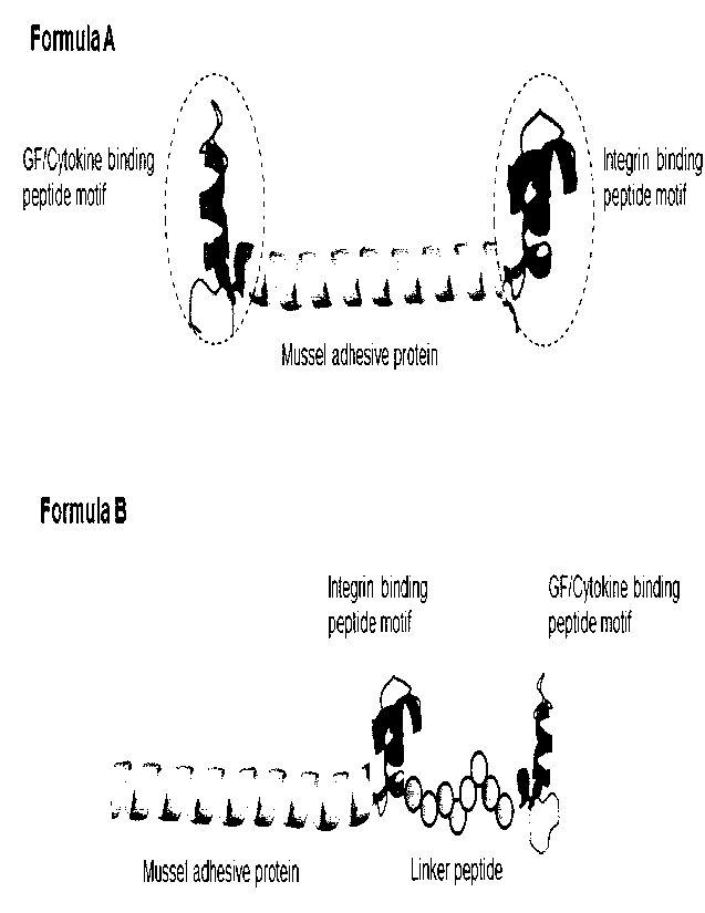
[33] Figure la represents two basic formulas of a protein substrate
presenting integrin
binding motif and heparin binding motifs which is capable of activating or
binding to
various growth factors. Formula A is the protein substrate having integrin
binding &
GF binding peptide motif at C-terminus and N-terminus, and both binding
peptide
motifs are incorporated at C-terminus of the protein substrate in Formula B.
[34] Figure lb represents the action mechanism of the protein substrate of
the present
invention. For specificity, the protein substrate coated surface forms island
like to-
pography that allows specific interaction between binding motifs and growth
factor
receptor or integrin as represented in Figure lb.
[35] Figure 2a and Figure 2b represent layouts of array of heparin binding
peptide motif
to screen any peptide motif having high affinity to GF. Figure 2a is the
layout of fi-
bronectin, collagen, and laminin derived heparin or syndecan binding peptide
motif
and Figure 2b is the layout of laminin LG domain derived heparin or syndecan
binding
peptide.
[361 Figure 3 represents the calculated GF binding affinity to GF
binding motif.
[37] Figure 4a and Figure 4b represent the screening results of various ECM-
derived GF
binding peptide motif to bFGF (Figure 4a) and TGF43 (Figure 4b), respectively.
Figure
4c represents the screening results of laminin-derived GF binding peptide
motif to
bFGF, TGF-13, and PDGF, respectively
[38] Figure 5a and Figure 5b represent the sustained release of TGF-13
bound to the
protein substrate having high affinity to TGF-I3. Figure 5a is the layout of
TGF-I3
CA 03201919 2023- 6-9
7
WO 2022/131809
PCT/KR2021/019159
binding peptide motif with high affinity to TGF-13, and Figure 5b represents
the ab-
sorbance profile of the GF binding motif with high affinity for TGF43 showing
long-
term sustained release of TGF-13.
[39] Figure 6a represents the layout ECM derived peptide motif binding to
epithelial-
mesenchymal transition (EMT) inducible integrin av, a2, and a9, and Figure 6b
represents western blot results of MCF-10A, a breast epithelial cell, cultured
on the
EMT-inducible integrin binding motif coated surface. In Figure 6b, the
fibronectin ex-
pression is represented as an EMT marker induced TGF-I3 bound to the protein
substrate. Aprotein substrate having OF binding motif (YEK & ANGO with high
affinity for TGF-I3 induced high expression of fibronectin while a protein
substrate
having GF binding motif (WQ) with low affinity for TGF-13 did not induce
fibronectin
expression.
[40] Figure 7 represents the trypsin-mediated TGF-[E cleavage analyzed by
Western blot.
TGF-13 bound to protein substrate had low levels of trypsin digestion while
TGF43
without protein substrate was most digested when treated with trypsin.
[41] Figure 8a and Figure 8b represent the effect of GF bound to protein
substrate on the
growth and proliferation of human foreskin fibroblast in low serum condition
(0.5%
FBS). Figure 8a is the effect of PDGF bound to protein substrate, and Figure
8b is the
effect of FGF2 bound to protein substrate on cell growth and proliferation.
Mode for the Invention
[42] The present invention is directed to a protein substrate that induces
signaling
mediated by integrins and growth factor/cytokine receptors, simultaneously or
se-
lectively, to regulate cellular behavior. A protein substrate may be provided
in the form
of recombinant adhesive protein comprising three domains of;
[43] 1) A recombinant mussel adhesive protein domain that adheres to the
surface of cells,
tissues, or any substrate such as plastics or glass;
[44] 2) A growth factor binding domain which is capable of immobilizing or
sequestering
growth factors or cytokines to activate or inhibit a cognate growth factor
receptor or
cytokine receptor; and
[45] 3) An integrin binding domain which is capable of activating integrin
receptors.
[46] As used herein, the term "a substrate" means a substance to which
another substance
is applied. In biology, the surface on which an organism such as a plant,
fungus, or
animal lives can be called as a substrate. This surface can include all
biotic, or abiotic
components.
[47] As used herein, a recombinant mussel adhesive protein refers to a
fusion protein
comprising mussel foot protein FP-5 and mussel foot protein FP-1 decapeptide.
In one
embodiment, a protein substrate provided here comprise a mussel foot protein
that is
CA 03201919 2023- 6-9
8
WO 2022/131809
PCT/KR2021/019159
selected from the group consisting SEQ ID NOs: 5 - 6.
[48] As used herein, the term "GF binding peptide motif" refers to a short
peptide derived
from heparin binding or syndecan binding domain of extracellular matrix
proteins such
as, including but not limited to, fibronectin domain III, laminin LG domain,
collagen
heparin binding domain, vitronectin heparin binding domain, or fibrinogen. In
one em-
bodiment, the GF binding peptide motif comprises 5 - 40 amino acids or its com-
bination thereof. In various embodiments, the GF binding peptide motif
comprises an
amino acid sequence selected from the heparin binding or syndecan binding
peptide
group consisting of fibronectin-derived KYILRWRPKNS (SEQ ID NO: 7),
YRVRVTPKEKTGPMKE (SEQ ID NO: 8), SPPRRARVT (SEQ ID NO: 9),
ATETTITIS (SEQ ID NO: 10), VSPPRRARVTDATETTITISWRTKTETITGFG
(SEQ ID NO: 11), ANGQTPIQRYIK (SEQ ID NO: 12), KPDVRSYTITG (SEQ ID
NO: 13), PRARITGYIIKYEKPGSPPREVVPRPRPGV (SEQ ID NO: 14),
WQPPRARI (SEQ ID NO: 15), WQPPRARITGYIIKYEKPG (SEQ ID NO: 16),
YEKPGSPPREVVPRPRP (SEQ ID NO: 17), KNNQKSEPLIGRKKT (SEQ ID NO:
18), or laminin-derived GLIYYVAHQNQM (SEQ ID NO: 19), RKRLQVQLSIRT
(SEQ ID NO: 20), GLLFYMARINHA (SEQ ID NO: 21), KNSFMALYLSKG (SEQ
ID NO: 22), VVRDITRRGKPG (SEQ ID NO: 23), RAYFNGQSFIAS (SEQ ID NO:
24), GEKSQFSIRLKT (SEQ ID NO: 25), TLFLAHGRLVFMFNVGHKKL (SEQ ID
NO: 26), TLFLAHGRLVFM (SEQ ID NO: 27), LVFMFNVGHKKL (SEQ ID NO:
28), GAAWKIKGPIYL (SEQ ID NO: 29), VIRDSNVVQLDV (SEQ ID NO: 30),
GKNTGDHFVLYM (SEQ ID NO: 31), RLVSYSGVLFFLK (SEQ ID NO: 32),
GPLPSYLQFVGI (SEQ ID NO: 33), RNRLHLSMLVRP (SEQ ID NO: 34),
LVLFLNHGHFVA (SEQ ID NO: 35), AGQWHRVSVRWG (SEQ ID NO: 36),
KMPYVSLELEMR (SEQ ID NO: 37), RYVVLPR (SEQ ID NO: 38), VRWG-
MQQIQLVV (SEQ ID NO: 39), TVFSVDQDNMLE (SEQ ID NO: 40), APMS-
GRSPSLVLK (SEQ ID NO: 41), VLVRVERATVFS (SEQ ID NO: 42), RNIAEIIKD1
(SEQ ID NO: 43), PGRWHKVSVRWE (SEQ ID NO: 76) or collagen-derived
KGHRGF (SEQ ID NO: 44), TAGSCLRKFSTM (SEQ ID NO: 45),
GEFYFDLRLKGDK (SEQ ID NO: 46), or vitronectin-derived KKQR-
FRHRNRKGYRSQ (SEQ ID NO: 47), or bone sialoprotein derived KRSR (SEQ ID
NO: 48), KRRA (SEQ ID NO: 49).
[49] As used herein, the term "integrin binding motif" refers to a short
peptide derived
from extracellular matrix proteins such as, including but not limited to,
fibronectin,
laminin, collagen, vitronectin, or tenascin. The integrin binding motifs bind
to and
activate integrin av, a5, a8 or a9 to support cell adhesion and may induce mor-
phogenesis together with growth factor bound to the heparin binding motif. In
various
embodiments, the heparin binding peptide comprises an amino acid sequence
selected
CA 03201919 2023- 6-9
9
WO 2022/131809
PCT/KR2021/019159
from the group consisting of integrin av binding RGD (SEQ ID NO: 50), RGDV
(SEQ
ID NO: 51), PQVTRGDVFIMP (SEQ ID NO: 52), or integrin a5 binding GRGDSP
(SEQ ID NO: 53), PHSRNSGSGSGSGSGRGDSP (SEQ ID NO: 54), or integrin a9
binding EDGIHEL (SEQ ID NO: 55), VAEIDGIEL (SEQ ID NO: 56), or integrin a8
binding VFDNFVLK (SEQ ID NO: 57).
[50] In one embodiment, the present invention discloses a protein substrate
that provides a
GF binding peptide motif to sequester or bind to growth factors,
simultaneously or se-
lectively. Any suitable GF binding peptide motif can be selected from the
group
consisting heparin binding or syndecan binding motif derived from fibronectin
domain
III, laminin LG domain, collagen heparin domain, vitronectin heparin domain,
or bone
sialoprotein as described in details in the definition term of heparin binding
motif
above.
[51] In one embodiment, a protein substrate to sequester or bind to basic
fibroblast growth
factor (bFGF) is disclosed. Generally, the GF binding peptide motif can be
selected
from heparin/syndecan binding domain of fibronectin, laminin and collagen for
bFGF
binding. Preferably, the fibronectin-derived peptide PRARITGYIIKYEKPGSP-
PREVVPRPRPGV (SEQ ID NO: 14), WQPPRARI (SEQ ID NO: 15), laminin-derived
peptide RYVVLPR (SEQ ID NO: 38), VRWGMQQIQLVV (SEQ ID NO: 39),
VLVRVERATVFS (SEQ ID NO: 42), or collagen-derived KGHRGF (SEQ ID NO:
44) can be selected to sequester or bind to bFGF.
[52] In another embodiment, the present invention discloses a protein
substrate to provide
GF binding peptide motif to sequester or bind to transforming growth factor 13
(TGF-13). The GF binding peptide motif can be selected from heparin/syndecan
binding
domain of fibronectin, laminin, collagen, vitronectin, or bone sialoprotein
for TGF43
binding. Preferably, fibronectin-derived motif ANGQTPIQRYIK (SEQ ID NO: 12),
KPDVRSYTITG (SEQ ID NO: 13), PRARITGYIIKYEKPGSPPREVVPRPRPGV
(SEQ ID NO: 14), WQPPRAR1 (SEQ ID NO: 15), WQPPRARITGYI1KYEKPG (SEQ
ID NO: 16), YEKPGSPPREVVPRPRP (SEQ ID NO: 17), or laminin-derived peptide
GLIYYVAHQNQM (SEQ ID NO: 19), RKRLQVQLSIRT (SEQ ID NO: 20), GLL-
FYMARINHA (SEQ ID NO: 21), KNSFMALYLSKG (SEQ ID NO: 22),
VVRDITRRGKPG (SEQ ID NO: 23), RAYFNGQSFIAS (SEQ ID NO: 24), GEK-
SQFSIRLKT (SEQ ID NO: 25), TLFLAHGRLVFMENVGHKKL (SEQ ID NO: 26),
TLFLAHGRLVFM (SEQ ID NO: 27), LVFMFNVGHKKL (SEQ ID NO: 28),
GAAWK1KGPIYL (SEQ ID NO: 29), V1RDSNVVQLDV (SEQ ID NO: 30), GKNT-
GDHFVLYM (SEQ ID NO: 31), RLVSYSGVLFFLK (SEQ ID NO: 32), GPLP-
SYLQFVGI (SEQ ID NO: 33), RNRLHLSMLVRP (SEQ ID NO: 34),
LVLFLNHGHFVA (SEQ ID NO: 35), AGQWHRVSVRWG (SEQ ID NO: 36),
KMPYVSLELEMR (SEQ ID NO: 37), RYVVLPR (SEQ ID NO: 38), VRWG-
CA 03201919 2023- 6-9
10
WO 2022/131809
PCT/1(122021/019159
MQQ1QLVV (SEQ ID NO: 39), TVFSVDQDNMLE (SEQ ID NO: 40), APMS-
GRSPSLVLK (SEQ ID NO: 41), VLVRVERATVFS (SEQ ID NO: 42), RNIAEIIKDI
(SEQ ID NO: 43), or collagen-derived KGHRGF (SEQ ID NO: 44),
TAGSCLRKFSTM (SEQ ID NO: 45), GEFYFDLRLKGDK (SEQ ID NO: 46), or vit-
ronectin-derived KKQRFRHRNRKGYRSQ (SEQ ID NO: 47), or bone sialoprotein
derived KRSR (SEQ ID NO: 48), KRRA (SEQ ID NO: 49) can be selected to bind to
or sequester TGF-11. More preferably, the heparin binding motif can be
selected from
fibronectin derived ANGQTPIQRYIK (SEQ ID NO: 12), KPDVRSYTITG (SEQ ID
NO: 13), YEKPGSPPREVVPRPRP (SEQ ID NO: 17), or laminin derived KNSF-
MALYLSKG (SEQ ID NO: 22), RYVVLPR (SEQ ID NO: 38), GKNTGDHFVLYM
(SEQ ID NO: 31), RLVSYSGVLFFLK (SEQ ID NO: 32), VLVRVERATVFS (SEQ
ID NO: 42), or vitronectin derived KKQRFRHRNRKGYRSQ (SEQ ID NO: 47), or
bone sialoprotein derived KRSR (SEQ ID NO: 48), KRRA (SEQ ID NO: 49).
[53] In another embodiment, the present invention discloses a protein
substrate to provide
GF binding peptide motif to sequester or bind to platelet-derived growth
factor
(PDGF). The PDGF binding motif can be selected from laminin derived motif
GLIYYVAHQNQM (SEQ ID NO: 19), RKRLQVQLSIRT (SEQ ID NO: 20), GLL-
FYMARINHA (SEQ ID NO: 21), KNSFMALYLSKG (SEQ ID NO: 22),
VVRDITRRGKPG (SEQ ID NO: 23), RAYFNGQSFIAS (SEQ ID NO: 24), GEK-
SQFSIRLKT (SEQ ID NO: 25), TLFLAHGRLVFMFNVGHKKL (SEQ ID NO: 26),
TLFLAHGRLVFM (SEQ ID NO: 27), LVFMFNVGHKKL (SEQ ID NO: 28),
GAAWKIKGPIYL (SEQ ID NO: 29), VIRDSNVVQLDV (SEQ ID NO: 30), GKNT-
GDHFVLYM (SEQ ID NO: 31), RLVSYSGVLFFLK (SEQ ID NO: 32), GPLP-
SYLQFVGI (SEQ ID NO: 33), RNRLHLSMLVRP (SEQ ID NO: 34),
LVLFLNHGHFVA (SEQ ID NO: 35), AGQWHRVSVRWG (SEQ ID NO: 36),
KMPYVSLELEMR (SEQ ID NO: 37), RYVVLPR (SEQ ID NO: 38), VRWG-
MQQ1QLVV (SEQ ID NO: 39), TVFSVDQDNMLE (SEQ ID NO: 40), APMS-
GRSPSLVLK (SEQ ID NO: 41), VLVRVERATVFS (SEQ ID NO: 42), RNIAEIIKDI
(SEQ ID NO: 43), PGRWHKVSVRWE (SEQ ID NO: 76), or vitronectin derived
KKQRFRHRNRKGYRSQ (SEQ ID NO: 47), or bone sialoprotein derived KRSR
(SEQ ID NO: 48), KRRA (SEQ ID NO: 49). More preferably, the PDGF binding motif
can be selected from RKRLQVQLSIRT (SEQ ID NO: 20), KNSFMALYLSKG (SEQ
ID NO: 22), RYVVLPR (SEQ ID NO: 38), GKNTGDHFVLYM (SEQ ID NO: 31),
VLVRVERATVFS (SEQ ID NO: 42), or vitronectin derived KKQR-
FRHRNRKGYRSQ (SEQ ID NO: 47), or bone sialoprotein derived KRSR (SEQ ID
NO: 48).
[54] The present invention further discloses a protein substrate for
sustained release of
growth factor in physiological conditions.
CA 03201919 2023- 6-9
11
WO 2022/131809
PCT/KR2021/019159
[55] It is well known that interactions with heparin sulfate occurring in
the extracellular
matrix have been shown directly to regulate the diffusion of growth factors
such as
FGF (Duchesne L, et al, Transport of fibroblast growth factor 2 in the
pericellular
matrix is controlled by the spatial distribution of its binding sites in
heparan sulfate.
PLoS Biol. 2012; 10(7):e1001361., Dowd CT, et al, Heparan sulfate mediates
bFGF
transport through basement membrane by diffusion with rapid reversible
binding. J
Biol Chem. 1999 19; 274(8):5236-44) as well as the storage and release of FGFs
in
tissue homeostasis (Bashkin P. et al., asic fibroblast growth factor binds to
suben-
dothelial extracellular matrix and is released by heparitinase and heparin-
like
molecules. Biochemistry. 1989 21; 28(4):1737-43).
[56] In one embodiment, the present invention provides a protein substrate
as sustained
release system of TGF-I3 without functional loss over a period of days in
physiological
conditions. The protein substrate provides a TGF-I3 binding motif, selected
from
KNSFMALYLSKG (SEQ ID NO: 22), RYVVLPR (SEQ ID NO: 38), GKNT-
GDHFVLYM (SEQ ID NO: 31), RLVSYSGVLFFLK (SEQ ID NO: 32),
VLVRVERATVFS (SEQ ID NO: 42), for sustained release of TGF-Ii in physiological
conditions.
[57] The present invention also discloses a protein substrate to provide
integrin binding
peptide motif and GF binding peptide motif at the same time. An integrin
binding
motif can be incorporated into N-terminus of the recombinant adhesive protein
and a
growth factor or cytokine binding motif can be incorporated into C-terminus of
said
adhesive protein, or vice versa.
[58] Crosstalk between integrins and growth factor receptors has been well
known. For
example, Jang reported that FGF2-FNIII9-10 fusion protein exhibited a
significant
increase of cell adhesion and proliferation of MG63 cells compared with FNIII9-
10
alone. (Jun-Hyeog Jang & Chong-Pyoung Chung, Engineering and expression of a
re-
combinant fusion protein possessing fibroblast growth factor-2 and fibronectin
fragment. Biotechnology Letters volume 26, p183'7-184-0(2004-)), and
FNIII9-10-mediated adhesion promotes the effect of FGF1 on neurite outgrowth
of
PC12 cells (Choung PH, et al., Synergistic activity of fibronectin and
fibroblast growth
factor receptors on neuronal adhesion and neurite extension through
extracellular
signal-regulated kinase pathway. Biochem Biophys Res Commun, 2002, 295:
898-902).
[59] In one embodiment, the present invention discloses a protein substrate
that mimic fi-
bronectin domain III having RGDSGSGSGSGSGANGQTPIQRYIK (SEQ ID NO:
58).
[60] In another embodiment, the present invention discloses a protein
substrate that mimic
fibronectin domain III (SEQ ID NO: 59), where integrin binding motif RGD (SEQ
ID
CA 03201919 2023- 6-9
1")
WO 2022/131809
PCT/KR2021/019159
NO: 50) was incorporated in its C-terminus of adhesive protein and growth
factor or
cytokine, for example, TGF-13 binding motif ANGQTPIQRYIK (SEQ ID NO: 12) in
its N-terminus of adhesive protein.
[61] The present invention also provides a microenvironment surface that
simultaneously
activates integrin and growth factor receptor in order to control phenotype
plasticity,
which is directly related to wound healing, angiogenesis or pathogenesis such
as
fibrosis or tumor metastasis.
[62] In one embodiment, the present invention provides a synthetic tumor
microen-
vironment surface comprising said protein substrate to induce epithelial to
mes-
enchymal transition (EMT) in epithelial cells.
[63] EMT is a process where epithelial cells lose epithelial proteins
including F-Cadherin,
and gains mesenchymal markers such as N-Cadherin, Vimentin and Fibronectin.
EMT
is associated with many processes, including embryonic or cancer development
and/or
progress (Kalluri R, et al. The basics of epithelial-mesenchymal transition. J
Clin
Invest. 2009;119:1420-8. Jordan NV, et al. Tracking the intermediate stages of
ep-
ithelial-mesenchymal transition in epithelial stem cells and cancer. Cell
Cycle. 201;
10:2865-73), wound healing and tissue repair, and cell migration. (Hosaka K,
et al.
Pericyte-fibroblast transition promotes tumor growth and metastasis. Proc Natl
Acad
Sci USA. 2016;113:E5618-27. Yang X, et al. Silencing Snail suppresses tumor
cell
proliferation and invasion by reversing epithelial-to-mesenchymal transition
and
arresting G2NI phase in non-small cell lung cancer. Int J Oncol. 2017; 50:1251-
60).
While EMT events are essential for development and wound repair, it has also
been
recognized as a contributing factor to fibrotic diseases and cancer. Many
soluble and
insoluble factors including TGF43 and ECM proteins determine the degree and
duration of EMT events. Specifically, cytokines such as TGF-13, TNFa, and IL6
and
hypoxia are capable of inducing EMT in various tumors.
[64] Several extracellular matrix (ECM) proteins, including collagen-1,
fibronectin, and
hyaluronan, and ECM remodeling via extracellular lysyl oxidase are also
implicated in
regulating EMT (Hae-Yun Jung, et al. Clin Cancer Res; 21(5) March 1, 2015.
Molecular Pathways: Linking Tumor Microenvironment to Epithelial-Mesenchymal
Transition in Metastasis). Several integrins have been known to mediate EMT.
For
example, EMT in regulated by integrin av136 via activation of TGF-i1 -Smad2/3
signaling pathway. (Wang J, et al. (2015) Interleukin-lbeta promotes
epithelial-derived
alveolar elastogenesis via av136 integrin-dependent TGF-beta activation. Cell
Physiol
Biochem 36:2198-2216). The expression of several integrin complexes is also up-
regulated during EMT, including a5131, which binds to fibronectin, and the
integrins
a131 & a2131, which interact with collagen I and have been shown to mediate
the
disruption of E-cadherin complexes. Cellular interactions with the ECM have
been
CA 03201919 2023- 6-9
13
WO 2022/131809
PCT/KR2021/019159
shown to be modulated by ECM-associated proteins such as SPARC that is a gly-
coprotein to promote the interaction of collagen and a2131.
[65] In one embodiment, the present invention provides a protein substrate
comprising
integrin binding motif selected from integrin a5.131, a9131, or av133 binding
motif, and
cytokine binding motif selected from ANGQTPIQRYIK (SEQ ID NO: 12),
KPDVRSYTITG (SEQ ID NO: 13) or YEKPGSPPREVVPRPRP (SEQ ID NO: 17) in
order to induce EMT in breast cancer cell line, MCF-10A.
[66] The present invention also provides a method to stabilize growth
factors or cytokincs
against loss of biological activity for long term in cell culture conditions
by admixing a
protein substrate comprising a growth factor or cytokine binding motif with
growth
factor or cytokine in cell culture medium or buffer solution such as PBS
buffer.
1-671 In one embodiment, a composition comprising a protein
substrate presenting
cytokine binding motif ANGQTPIQRYIK (SEQ ID NO: 12) and growth factor binding
motif YEKPGSPPREVVPRPRP (SEQ ID NO: 17) dissolved in cell culture medium
such as DMEM or buffer solution such as PBS. This composition may be stored
for at
least one week without loss of its biological activity as demonstrated in EMT
promotion test in breast epithelial cell line, MCF-10A.
[68]
1-691 Hereinafter, the present invention will be described in
detail with reference to
Preparation Examples, Examples, and Experimental Examples thereof.
[70] However, it should be understood that the following Preparation
Examples,
Examples, and Experimental Examples are given for the purpose of illustration
of the
present invention only, and are not intended to limit the scope of the present
invention.
[71]
[72] EXAMPLES
[73] EXAMPLE 1. PREPARATION OF A PROTEIN SUBSTRATE PRESENTING
1NTEGRIN BINDING MOTIF AND HEPARIN BINDING MOTIF
[74] E. coli based protein expression system was commercialized to produce
a variety of
mussel adhesive proteins including fusion protein of mussel foot protein 1 and
foot
protein 5 in an efficient way (see U52020/0062809A1 and W02011/115420A2), and
the mussel adhesive proteins are commercially available under Trademarks
MAPTrixTm marketed by Kollodis BioSciences, Inc. The method for preparation of
mussel adhesive proteins are fully described in U520200/062809A1 and
W02011/115420A2 which is hereby incorporated by reference for all purposes as
if
fully set forth herein.
[75] The basic formula of a protein substrate is illustrated in Figure la.
Two peptide
motifs can be incorporated into N-terminus and C-terminus of mussel adhesive
protein
(Formula A), respectively as seen in Figure la. Alternatively, both motifs can
be in-
CA 03201919 2023- 6-9
14
WO 2022/131809
PCT/KR2021/019159
corporated into C-terminus or N-terminus (Formula B) as seen in Figure la,
wherein a
spacer linker peptide such as SGSGSGSGSG effectively separate two peptides for
syn-
ergistic effect.
[76] Two types of protein substrates having SEQ ID NO: 58 (hereafter MAP-
RGD-GF)
and SEQ ID NO: 59 (hereafter GF-MAP-RGD) were recombinantly designed and
expressed in Eicoli expression system and purified as set forth in
US2020/0062809A1
and W02011/115420A2. A number of protein substrate having a GF binding motif
was produced with the same procedure. All protein substrate was lyophilized
and
stored at refrigerator for further experiment.
[77]
[78] EXAMPLE 2. GROWTH FACTOR BINDING ASSAY
1791 Multiple arrays for growth factor or cytokine binding assay
are composed of a large
number of heparin binding peptide motifs arranged in 96 well format as
represented in
Figure 2a and Figure 2b. Figure 2a is the array layout of fibronectin,
collagen, and
laminin derived GF binding motif and Figure 2b is the array layout of laminin
globular
domain derived GF binding motif.
[80] For specific binding of peptide motif to a growth factor or cytokine,
low cell-
attachment or ultrahydrophobic surface was coated with the substrate protein
wherein
the substrate protein was formed as particles. So, the surface looks like
particle island
as illustrated in Figure lb. The coating method is state-of-the-art technology
and
detailed procedure is described in the United States Patent Application US
16/546,966
developed by present inventors and hereby fully incorporated as reference.
This
surface can allow biomolecules such as GF or target cells to specifically bind
to the
peptide motif presented on the surface. Non-target biomolecules or cells are
forced to
be suspended, and eventually washed out.
[81] Recombinant basic FGF, TGF-13, PDGF-BB were purchased from R&D Systems
(Camarillo, CA) and Ultralow cell-attachment 96 well plate from Corning
(Corning,
NY). Fatty acid-free bovine serum albumin (BSA) was purchased from Sigma-
Aldrich
Corp. (St. Louise, MO) and fetal bovine serum from Thermo Fisher Scientific
(Waltham, MA).
[82] To eliminate or minimize any non-specific binding of recombinant
growth factors,
the 96 well plate surface coated with the substrate protein was blocked with 2
wt%
BSA in PBST buffer (4 mM phosphate and 155 mM sodium chloride, 0.05 wt%
Tween-20, pH 7.4) by rocking the plate at RT for 1 h. Individual recombinant
growth
factors (50 Nm) dissolved in PBST were then added on to the blocked well plate
including the heparin binding motif and the plate was rocked at 4 C overnight.
The
binding of the growth factor to the protein substrate coated on the well plate
was
confirmed by immunoaffinity assay. In brief, the well plate was sequentially
treated
CA 03201919 2023- 6-9
15
WO 2022/131809
PCT/KR2021/019159
with a primary antibody for the growth factor and its secondary antibody
labeled with
horseradish peroxidase (HRP). Finally, TMB (3,3'.5,5'-tetramethylbenzidine)
substrate
was chemically changed by HRP and its absorbance values at 450 nm were used to
analyze the binding of the growth factors on to each binding motifs. The
Figure 3
shows TMB absorbance values corresponding to the degree of growth factor
binding.
The values were subtracted by the absorbance value of a blank sample without
growth
factors. An absorbance reading over 0.1 was considered as a significant
interaction.
[83] We identified fibronectin derived GF binding motifs specifically bound
to bFGF,
PDGF-BB as shown in Figure 4a and Figure 4b. The GF binding motif having
high affinity to each growth factor are summarized in the following Table 1.
[84] [Table 11
Peptide motif Basic FGF TGF-p PDGF
KYILRWRPKNS High
YRVRVTPKEKTGPMKE
SPPRRARVT
ATETTITIS
VSPPRRARVTDATETTITIS
-WRTKTETITGFG
ANGQTPIQRYIK High High
KPDVRSYTITG High
PRARITGYIIKYEKPGSPPR High
-EVVPRPRPGV
WQPPRARI Moderate Moderate
WQPPRARITGYIIKYEKPG
YEKPGSPPREVVPRPRP High Moderate
KNNQKSEPLIGRKKT
RYVVLPR High
KGHRGF
[85] Most laminin derived GF binding peptide motifs showed higher affinity
to TGF43
and PDGF, but relatively low affinity to bFGF, similar to that of fibronectin
GF
binding motif as seen in Figure 4c.
[86] EXAMPLE 3. SUSTAINED RELEASE OF GROWTH FACTOR BOUND TO THE
PROTEIN SUBSTRATE
[87] Heparin binding domain in ECM proteins has been shown to stabilize
growth in
CA 03201919 2023- 6-9
16
WO 2022/131809
PCT/KR2021/019159
physiological conditions. To determine if the GF binding peptide motif confers
any
protective effect on the growth factors, TGF-I3 was conditioned with PBS
buffer or cell
culture medium, DMEM, in the presence or absence of GF binding peptide motif
identified in the EXAMPLE 2. bFGF (0.5 fig) was added to the protein substrate
coated
surface and incubated at 37 C for 2, 4, and 7 days. We assessed the stability
of TGF-P
over time at 37 C with an enzyme-linked immunosorbent assay (ELISA) revealing
slowly released from the protein substrate and its half time was 4 days. As
seen in
Figure 5b, GF binding motif (ANG-MAP-RGD-IDAP) with high affinity for TGF-I3
exhibit long-term sustained release of TGF-p in cell culture condition.
[88]
[89] EXAMPLES 4. SYNTHETIC MICROENVIRONMENT TO INDUCE TRANSDIF-
FERENTATION OF EPITHELIAL CELL TO MESENCHYMAL CELL
[90] TGF-13 binding motifs identified in EXAMPLES 2 and 3 were arrayed in 6
well
plates. After treated with TGF-P (5 ng) as set forth in EXAMPLE 2, the 6 well
plates
were stored at a CO2 incubator at 37 C for appropriate time (e.g., 2, 4 and 7
days, re-
spectively). MCF-10A, a breast epithelial cell, was seeded and cultured on the
TGF-13
binding peptide motif coated well plate. Two motifs having high affinity to
TGF-I3
could induce MCF-10A to undergo EMT while low TGF43 binding affinity motif,
WQPPRARI (SEQ ID NO: 15), could not induce EMT as evidenced by no fibronectin
upregulation as seen in Figure 6b.
[91]
[92] EXAMPLE 5. GF PROTECTION FROM TRYPSIN ACTIVITY
[93] TGF-p binding peptide motifs identified in EXAMPLES 2 and 3 were
incubated with
TGF-P (2 pg) and trypsin (0.3 Lig) at 30 C for 15 min. After trypsinization,
proteins of
sample were separated by 15% SDS-PAGE in size-dependent manner. Then, SDS-
PAGE was stained by Coomassie blue solution for 2 hr. After de-staining, the
intensity
of band, stained by Coomassie blue solution, were analyzed. ANGQTPIQRY1K (SEQ
ID NO: 12), ANG-M-RGD (SEQ ID NO: 59), KNSFMALYLSKG (SEQ ID NO: 22),
KRSR (SEQ ID NO: 48), and RKRLQVQLSIRT (SEQ ID NO: 20) motifs inhibited
trypsinization of TGF43 compared without MAP-fusion motif as seen in Figure 7.
[94]
[95] EXAMPLE 6. GROWTH FACTOR BOUND PROTEIN SUBSTRATE FOR CELL
GROWTH
1961 A total of 96-well plates (non-tissue culture treated, SPL
Life Science) were coated
with 50 /IMO with the protein substrate having GF binding peptide motif in
sodium
acetate buffer for 1 h at room temperature. 20 ng/me, of FGF2 and PDGF-BB
(BioVision, Milpitas, CA, USA) in DMEM with 0.5% FBS were added to individual
well and incubated for lhr at 37 C in CO2 incubator. Cell growth assays were
CA 03201919 2023- 6-9
17
WO 2022/131809
PCT/KR2021/019159
performed using human foreskin fibroblasts (Hs68, ATCC) in DMEM medium
(Invitrogen) supplemented with 0.5% fetal bovine serum (FBS). Cells were
seeded at
1,500 cells/well on GF bound substrate pre-coated plates and incubated for 48
h at
37 C in CO2 incubator. Then, CCK-8 assay was performed to determine cell
growth.
Hs68 cells were checked for mycoplasma contamination and used in passages from
5
to 10. The protein substrate without GF binding peptide motif and heparin were
used
as negative and positive control, respectively.
[97] As shown in Figure 8a and Figure 8b, GF bound protein substrate
supported cell
growth and proliferation. GF bound to protein substrate strongly supported the
growth
and proliferation of human foreskin fibroblast in low serum condition (0.5%
FBS)
when compared to heparin bound GF.
[98]
[99] From the foregoing description, it will be apparent that variations
and modifications
may be made to the presently disclosed subject matter to adopt it to various
usages and
conditions. Such embodiments arc also within the scope of the following
claims.
[100] The recitation of a listing of elements in any definition of a
variable herein includes
definitions of that variable as any single element or combination (or sub-
combination)
of listed elements. The recitation of an embodiment herein includes that
embodiment
as any single embodiment or in combination with any other embodiments or
portions
thereof.
[101] All patents and publications mentioned in this specification are
herein incorporated
by reference to the same extent as if each independent patent and publication
was
specifically and individually indicated to be incorporated by reference.
CA 03201919 2023- 6-9