Note : Les descriptions sont présentées dans la langue officielle dans laquelle elles ont été soumises.
CA 03208784 2023-07-19
WO 2022/167607 PCT/EP2022/052761
NGF ISOFORM FOR USE IN THE TREATMENT OF OCULAR PATHOLOGIES
FIELD OF THE INVENTION
The present invention relates to the field of treatment of ocular pathologies,
in particular
of treatment of ocular pathologies by administration of a specific isoform of
NGF.
STATE OF THE ART
The nerve growth factor (NGF) is a member of the family of evolutionarily well-
conserved
neurotrophin growth factors, which also includes brain-derived neurotrophic
factor
(BDNF), neurotrophin-3 (NT3) and NT4/5.
It exerts its activity by interacting with two structurally unrelated cell
surface receptors:
the high affinity receptor tyrosine kinase A (TrKA) and the low affinity p75
neurotrophin
receptor (p75NTR).
It has been shown that these two receptors mediate complex and often opposite
effects
of NGF on cells.
TrKA is selective for NGF and triggers different signalling pathways that
promote cell
survival, proliferation and differentiation, such as PI3 kinase,
Ras/extracellular signal¨
regulated kinases (ERK), Akt 1 and protein kinase C (PKC) (Kaplan et al., J
Neurobiol
1994, 25(11):1404-17; Clewes et al., J Biochem 2008, 107: 1124-1135).
Furthermore, the
activation of TrKA inhibits apoptotic signaling in cells, for example by
inhibiting capsase
3 (Nguyen T LX, Eperimental and Molecular Medicine 2010, 42 (8): 583-595).
The p75 neurotrophin receptor (p75NTR,
) belongs to the tumour necrosis factor receptor
superfamily and binds non-specifically all neurotrophins. Contrary to what
observed when
binding to the TrKA receptor, NGF binding to p75NTR induces apoptosis through
the
activation of a number of intracellular mediators similar to those activated
by death
receptors, such as tumor necrosis factor (TN F) and Fas receptors, including
TNF receptor
associated factors (TRAFs), nuclear Kappa B (NFkB), capsases and p53 (Harada C
et
al., Developmental Biology 2006, 290: 57-65; Aloyz R S et al., The Journal of
Cell Biology
1998, (143) 6: 1691-1703).
Thus, the final activity of NGF on cells is significantly dependent on the
level of expression
and on the activation of the above two receptors. Therefore, changes in the
ratio of the
two receptors can alter the balance between protective and deleterious effects
induced
by NGF, thus modifying the final activity of NGF on cells, with the
predominance of either
proliferative, growth promoting and survival effects mediated by TrKA or of
apoptotic
- 1 -
CA 03208784 2023-07-19
WO 2022/167607 PCT/EP2022/052761
responses mediated by p75 NRT (Frade at al., Nature 1996; 383:166-168; Yoon So
et al.,
J Neuroscience 1998; 18: 3273-3281).
The affinity of NGF to p75NTR is lower than to TrKA, but its cell type
distribution is wider
than that of TrKA.
Both NGF receptors are broadly expressed in both the anterior and posterior
segments
of the eye, including cornea, conjunctiva, limbal epithelium, retina and optic
nerve and
NGF has been demonstrated to have a key role in both eye physiology and
pathology,
modulating processes such as cell survival, proliferation, differentiation and
apoptosis
(Garcia et al., Cytokine & Growth Factors Reviews 2017, 34: 43-57; Sornelli et
al.,
Molecular Vision 2010; 16:1439-1447; Di Girolamo et al., J Cell Mol. Med 2008,
12(66):
2799-2811). Several experimental studies demonstrated that TrKA stimulation
promotes
RGCs survival after ischemic injury, optic nerve transection, and ocular
hypertension.
(Carmignoto et al., Journal of Neuroscience 1989, 9(4): 1263-1272.;
Chakrabarti et al.,
Brain Research 1990; 523:11-15; Siliprandi et al., Invest Opthalmol Vis Sci
1993, 34 (12):
3232-3245; Haamedi et al., The Journal of Comparative Biology 2001, 431: 397-
404;
Harada et al., Developmental Biology 2006 290: 57-65; Coassin et al., Graefes
Arch Clin
Exp Ophthalmol 2008, 246:1743-1749).
Notwithstanding the above, the effects of NGF in the visual system are complex
and they
are not univocal, depending upon the cellular context and the cellular
distribution and
level of expression of each NGF receptor. For example, in the retina, RGCs
express TrKA
and glial cells express p75NTR. A study in an animal model of glaucoma showed
that a
selective agonist of the pro-survival TrKA receptor was effective at
preventing RGC death,
while neither NGF nor an antagonist of the pro-apoptotic p75 receptor
protected RGCs.
(Shi et al., Developmental Neurobiology 2007, 67(7): 884-94).
A subsequent study showed enhanced survival of axotomized RGCs in
pharmacological
inhibition of p75NTR or in p75NTR knockout mice. In addition, a combination of
NGF or
TrKA agonists with p75NTR antagonists further potentiated RGC neuroprotection
in vivo
(Lebrun-Julien F, Molecular and Cellular Neuroscience. 2009, 40(4): 410-420).
In summary, several data support the hypothesis that NGF can exert
neuroprotective
effects when binding on RGCs TrKA receptor while acting on glial cells p75NTR
antagonizes this effect (Wang H et al., BioMed Research International 2014,
Article ID
759473).
- 2 -
CA 03208784 2023-07-19
WO 2022/167607 PCT/EP2022/052761
These controversial findings can be explained by the observation that NGF has
different
actions on RGCs because of different relative expression of TrKA and p75NTR in
the retina
upon different circumstances, thereby the failure of NGF trophic support might
be
associated with the progressive up-regulation of p75NTR in relation to TrKA
(Coassin et
al., Graefes Arch Olin Exp Ophthalmol 2008, 246:1743-1749; Mohamed R et al J
Olin
Exp Ophthalmol. 2015; 6(5); doi:10.4172/2155-9570.1000483).
This evidence is confirmed by studies that demonstrate that p75NTR selective
activation
induces RGC death in the normal retina, and accelerates RGC death in diseased
eyes,
while selective TrKA agonists protect RGCs in chronic and acute
neurodegeneration (Bai
Y et al., Journal of Biological Chemistry 2010, 285 (50): 39392-39400; ZhiHua
Shi et al.,
Developmental Neurobiology (2007), 67 (7): 884-894).
Some studies have also demonstrated that in the retina, TrKA is mainly
expressed in
retinal ganglion cells (RGCs) while p75NRT in Muller and glial cells (Harada C
et al.,
Developmental Biology 2006, 290: 57-65) and that the activation of p75NRT in
glia is able
to trigger neurotoxic pathways that counterbalance the protective effect of
TrKA activation
on RGCs.
A further reason for the inconsistency of the data obtained may also be due to
the use of
different routes or regimens of administration of the protein. In fact, the
concentration at
which the protein is administered seems to influence the response to NGF, with
higher
concentrations resulting in a higher activation of p75NTR. Therefore,
administration routes
or regimens that require high concentrations of NGF may be less effective in
inducing
survival activity of NGF than low concentrations.
Other potential therapeutic applications of NGF in other conditions of the eye
are based
on the prosurvival and trophic effect of this neurotrophin and therefore its
efficacy is highly
dependent on the balance between TrKA and p75NTR activation.
In particular, evidence shows that the antiaptototic and trophic effects of
NGF may be
useful in:
- preventing corneal graft rejection in corneal transplantation (Gong N et
al., Invest
Opthalmol Vis Sci, 2007, 48(3): 1043-1052);
- preserving and expanding limbal epithelial progenitor cells (Lambiase et
al., Invest
Opthalmol Vis Sci 2012, 53(13): 8280-8287, Mason SL, et al. Invest Ophthalmol
Vis Sci.
2016; 57: 3708-3713, Touhami A et al., Invest Ophthalmol Vis Sci. 2002, 43:
987-994);
- 3 -
CA 03208784 2023-07-19
WO 2022/167607 PCT/EP2022/052761
- in the treatment of corneal and conjunctival diseases, including phototoxic
keratopathy
(Rocco ML et al., Graefes Archive for Clinical and Experimental Ophthalmology
2018,
256: 729-738), corneal dystrophies and degenerations and corneal ulcers
(Lambiase et
al., Invest Opthalmol Vis Sci 1998, 39: 1272-1275; Lambiase, et al., N Engl J
Med 1998,
.. 338: 1174-1180; Bonini et al., Ophthalmology 2000,107: 1347-1351; Lambiase
et al.,
Invest Ophthalmol Vis Sci 2000, 41: 1063-1069: Blanco-Mezquita et al., Invest
Ophthalmol Vis Sci. 2013, 54(6): 3880-90: You L et al., Invest Ophthalmol Vis
Sci. 2000;
41(3): 692-702; Sornelli F, et al., Mol Vis. 2010, 29;16: 1439-47),
keratoconjunctivitis
sicca (Coassin et al., Graefes Arch Clin Exp Ophthalmol 2008, 246:1743-1749),
retinal
diseases including retinal detachment (Sun et al., Ophthalmologica 2008, 222:
58-61),
diabetic retinopathy (Ali et al., Diabetes, 2008, 57: 889-898), retinal
neurodegeneration
and/or ischemia (Xien et al., Exp Eye Res 2014, 125: 156-63), phototoxic
retinopathy
(Rocco et al., Graefes Arch Clin Exp Ophthalmol 2018, 256: 729-738; Garcia et
al., J
Neurochem 2014, 131(3): 303-13), epiretinal membrane (Minchiotti et al.,
Retina 2008,
28(4): 628-37), macular hole (Zhang et al., BMC Ophthalmol 2019, 19(1): 130),
macular
degeneration (Lambiase et al., Ann 1st Super Sanita 2009, 45 (4): 439-442) and
optic
neuropathies (Mesentier-Louro et al., Mol Neurobiol. 2019, 56(2): 1056-1069;
Guo et al
Sci Rep 2020, 10: 3375).
However, the activation of the p75NTR receptor by NGF makes the in vivo effect
of NGF
in the above pathologies difficult to predict.
A number of different rhNGF variants of 117 and 118 aminoacids have been
identified
when the protein is expressed as a 120-aminoacid sequence in Chinese Hamster
Ovary
cells, due to partial enzymatic digestion by trypsin and/or carboxypeptidase.
These
variants have been analysed and found to be equipotent in both the chick
dorsal root
ganglion cell survival and rat pheochromocytoma neurite extension assays
(Shmelzer et
al., J Neurochem 1992, 59(5):1675-83).
SUMMARY OF THE INVENTION
The Applicant has undertaken studies aimed at clarifying the somewhat
controversial
data in the literature on NGF activity in the eye.
These studies have shown that commercially available NGFs include different
isoforms
of NGF, characterised by aminoacid sequences differing only in length, either
alone or in
admixtures.
- 4 -
CA 03208784 2023-07-19
WO 2022/167607 PCT/EP2022/052761
Furthermore, the present inventors have surprisingly found that these isoforms
of NGF
have different ability to activate pathways induced by the TrKA or p75NTR
receptors,
resulting in a different activity pattern.
In particular, the present inventors have found that the NGF isoform having
the 118
aminoacid sequence of SEQ ID NO: 1 predominantly activates TrKA-mediated
pathways
and inhibits apoptotic pathways mediated by p75NTR, while isoforms having 120,
117 or
119 aminoacid sequences of SEQ ID NO: 2, 3 and 4, respectively, have a higher
ability
to activate p75NTR-dependent apoptotic pathways. These findings, relating to
the different
receptor selectivity of the NGF isoforms, can help to understand the
inconsistencies found
in the literature regarding the therapeutic applications of NGF.
In view of the above, the NGF isoform of SEQ ID NO: 1 is particularly useful
in the
treatment of pathologies where the effects of NGF on proliferation and
survival are
desired and where the proapoptotic effect of p75NTR is detrimental.
Accordingly, a first object the present invention is a NGF for use in the
prevention and/or
treatment of an ocular pathology selected from retinopathies, preferably
selected from
diabetic retinopathy, retinopathy of prematurity, retinal vascular occlusions,
phototoxic
retinopathy, retinal detachment, age-related macular degeneration, macular
degeneration, macular atrophy, macular hole, macular edema and epiretinal
membrane;
limbal stem cell deficiency; corneal pathologies, preferably selected from
keratoconus,
phototoxic keratopathy, persistent epithelial defects, corneal ulcers, corneal
dystrophies
and degeneration and keratoconjunctivitis sicca; conjunctival pathologies;
optic
neuropathies, preferably selected from glaucoma, ischemic, degenerative,
traumatic,
inherited and congenital optic neuropathies; and in the prevention of
allograft rejection in
corneal transplantation, wherein said NGF comprises more than 50% by weight of
the
NGF isoform of SEQ ID NO: 1 relative to the total weight of all NGF isoforms
comprised
in said NGF.
A second object of the present invention relates to a pharmaceutical
composition
comprising a NGF in a therapeutically effective amount, wherein said NGF
comprises
more than 50% by weight of the NGF isoform of SEQ ID NO: 1 relative to the
total weight
of all NGF isoforms comprised in said NGF and at least one pharmaceutically
acceptable
excipient, for use in the treatment of an ocular pathology selected from
retinopathies,
preferably selected from diabetic retinopathy, retinopathy of prematurity,
retinal vascular
occlusions, phototoxic retinopathy, retinal detachment, age-related macular
- 5 -
CA 03208784 2023-07-19
WO 2022/167607 PCT/EP2022/052761
degeneration, macular degeneration, macular atrophy, macular hole, macular
edema and
epiretinal membrane; limbal stem cell deficiency; corneal pathologies,
preferably selected
from keratoconus, phototoxic keratopathy; persistent epithelial defects,
corneal ulcers,
corneal dystrophies and degeneration and keratoconjunctivitis sicca;
conjunctival
pathologies; optic neuropathies, preferably selected from glaucoma, ischemic,
degenerative, traumatic, inherited and congenital optic neuropathies; and in
the
prevention of allograft rejection in corneal transplantation.
A third object of the present invention relates to a method of treating an
ocular pathology
selected from retinopathies, preferably selected from diabetic retinopathy,
retinopathy of
prematurity, retinal vascular occlusions, phototoxic retinopathy, retinal
detachment, age-
related macular degeneration, macular degeneration, macular atrophy, macular
hole,
macular edema and epiretinal membrane; limbal stem cell deficiency; corneal
pathologies, preferably selected from keratoconus, phototoxic keratopathy,
persistent
epithelial defects, corneal ulcers, corneal dystrophies and degeneration and
keratoconjunctivitis sicca; conjunctival pathologies; optic neuropathies,
preferably
selected from glaucoma, ischemic, degenerative, traumatic, inherited and
congenital
optic neuropathies; and in the prevention of allograft rejection in corneal
transplantation,
comprising administering to the subject a NGF in a therapeutically effective
amount,
wherein said NGF comprises more than 50% by weight of the NGF isoform of SEQ
ID
NO: 1 relative to the total of all NGF isoforms comprised in said NGF.
BRIEF DESCRIPTION OF THE FIGURES
Figure 1 shows the expression of the NGF receptors TrKA and p75NTR on the cell
membrane of PC12, I-HCEC, and hTERT-RPE-1 and ARPE-19 cells, measured as
described in Example 2. The data are represented as percentage of positive
cells
expressing the relevant receptor, measured by flow cytometry.
Figure 2 shows the total number of proteins upregulated and downregulated in
each cell
line by treatment with each NGF, measured as described in Example 2 and
represented
as Venn diagram. In details, Figure 2A) shows the Venn diagram for RPE cells,
Figure
2B) shows the Venn diagram for HCEC cells and Figure 2C) shows the Venn
diagram for
PC12 cells.
Figure 3 shows early (30 minutes, Figure 3A) and late (24 hours, Figure 3B)
caspase 3/7
activation in HCEC cells not treated (NT), treated with PBS (PBS), formulation
buffer (FB),
rhNGF-118 (rhNGF-118) or rhNGF-1 (rhNGF-1), as described in Example 3a. The
results
- 6 -
CA 03208784 2023-07-19
WO 2022/167607 PCT/EP2022/052761
are expressed as the ratio between green area (caspase activation) and phase
area (cell
confluence) of seven independent experiments. Student's T test was calculated,
*p-value
<0.05, ns=not significant.
Figure 4 shows early (30 minutes, Figure 4A) and late (24 hours, Figure 4B)
caspase 3/7
activation in RPE cells not treated (NT), treated with PBS (PBS 1X),
formulation buffer
(FB), rhNGF-118 (rhNGF-118) or rhNGF-1 (rhNGF-1), as described in Example 3a.
The
results are expressed as fold of change over not treated (NT) of the ratio
between green
area and phase area of five independent experiments. Student's T test was
calculated,
*p-value<0.05.
Figure 5 shows caspase 3/7 activation in RPE cells not treated (NT) or treated
for 24
hours with 10 M tBHP in the presence of formulation buffer (FB + tBHP), 50
ng/ml of
rhNGF118 (rhNGF-118 + tBHP), 50 ng/ml of rhNGF-2 (rhNGF-2 + tBHP) or 50 ng/ml
of
rhNGF-3 (rhNGF-3+ tBHP), as described in Example 3b.i). Data are presented as
percentage of green area confluence (caspase activation) of three independent
experiments. Student's T test was calculated, *p-value<0.05, **p-value<0.005,
***p-
value<0.0005. Moreover, one-way ANOVA, Bonferroni test, showed a statistical
significance of tBHP versus NT, of rhNGF-118 versus tBHP and of rhNGF-2 versus
rhNGF-118 (not shown).
Figure 6 shows early (30 minutes, Figure 6A) and late (24 hours, Figure 6B)
caspase 3/7
activation in RPE cells not treated (NT) or treated with PBS (PBS 1X), 50
ng/ml rhNGF-
118 (rhNGF-118), 50 ng/ml rhNGF-1 (rhNGF-1), 100 M H202 alone (H202) or in the
presence of 50 ng/ml rhNGF-118 (rhNGF-118+ H202) or 50 ng/ml rhNGF-1 (rhNGF-1+
H202), as described in Example 3b.ii). Data are presented as fold of increase
over not
treated cells (NT) of total green fluorescence (caspase activation) of four
independent
experiments. Student's T test was calculated, *p-value<0.05, **p-value<0.005,
***p-
value<0.0005. Moreover, after 30 minutes one-way ANOVA, Bonferroni test,
showed a
statistical significance of H202 in the presence of rhNGF-1 versus NT, versus
H202 alone
and versus H202 in the presence of rhNGF-118; while after 24 hours a
statistical
significance was observed of H202 in the presence of rhNGF-1 versus NT (not
shown).
Figure 7 shows neurite growth induced by two different concentrations of rhNGF-
118 or
rhNGF-1, measured as described in Example 4.
Figure 8 shows the expanded reverse phase HPLC chromatogram of rhNGF-118.
Figure 9 shows the expanded reverse phase HPLC chromatogram of rhNGF-1.
- 7 -
CA 03208784 2023-07-19
WO 2022/167607 PCT/EP2022/052761
Figure 10 shows the expanded reverse phase HPLC chromatogram of rhNGF-2.
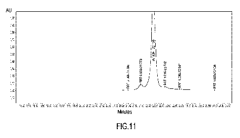
Figure 11 shows the expanded reverse phase HPLC chromatogram of rhNGF-3.
Figure 12 shows the expanded reverse phase HPLC chromatogram of rhNGF-4.
Figure 13 shows neurite growth induced by two different concentrations of
rhNGF-118 or
rhNGF-120 (rhNGF-4), measured as described in Example 4.
DETAILED DESCRIPTION OF THE INVENTION
A first object the present invention is a NGF for use in the prevention and/or
treatment of
a pathology selected from retinopathies, corneal pathologies, optic
neuropathies,
conjunctival pathologies, limbal stem cell deficiency and in the prevention of
allograft
rejection in corneal transplantation, wherein said NGF comprises more than 50%
by
weight of the NGF isoform of SEQ ID NO: 1 relative to the total weight of all
NGF isoforms
comprised in said NGF.
The term "NGF" according to the present invention refers to a functionally
active NGF
isoform or a mixture of functionally active NGF isoforms. Preferably, said NGF
isoforms
are isoforms of human NGF.
In the present context, for "NGF isoform" it is meant any of two or more
functionally active
NGF proteins having different aminoacid sequences, wherein such sequences
differ only
in their length. Preferably, said NGF isoforms have an aminoacid sequence
corresponding to the aminoacid sequence of SEQ ID NO: 2 or a sequence that
differ from
SEQ ID NO: 2 in length, namely for the presence of additional aminoacids or
deletion of
aminoacids at the N-terminal or C-terminal, preferably at the C-terminal.
Preferably, said
NGF isoforms are NGF proteins having the aminoacid sequence SEQ ID NO: 1, 2,
3, 4.
When referring to a specific NGF isoform, all forms of NGF having the
aminoacid
sequence of such isoform are included, independently from the presence of post-
translational modifications, such as oxidation, glycation or glycosylation.
In a preferred embodiment, said NGF isoforms comprised in said NGF are
selected from
NGF isoforms of SEQ ID NO: 1, 2, 3 or 4 or admixtures thereof.
Preferably, said retinopathies are selected from diabetic retinopathy,
retinopathy of
prematurity, retinal vascular occlusions, phototoxic retinopathy, retinal
detachment, age-
related macular degeneration, macular degeneration, macular atrophy, macular
hole,
macular edema and epiretinal membrane.
- 8 -
CA 03208784 2023-07-19
WO 2022/167607 PCT/EP2022/052761
Preferably, said corneal pathologies are selected from keratoconus, phototoxic
keratopathy, persistent epithelial defects, corneal ulcers, corneal
dystrophies and
degeneration, and keratoconjunctivitis sicca.
Preferably, said optic neuropathies are selected from glaucoma and ischemic,
degenerative, traumatic, inherited and congenital optic neuropathies.
According to a particularly preferred embodiment, the above pathology is
selected from
glaucoma, diabetic retinopathy, retinopathy of prematurity, retinal vascular
occlusions,
phototoxic retinopathy, retinal detachment, age-related macular degeneration,
macular
degeneration, macular atrophy, macular hole, macular edema and epiretinal
membrane,
more preferably the above pathology is glaucoma.
The NGF for use according to the present invention preferably consists of a
high purity
NGF isoform of SEQ ID NO: 1.
Preferably, the NGF for use according to the present invention comprises at
least 60%,
more preferably at least 70%, even more preferably at least 80%, even more
preferably
at least 90%, even more preferably at least 95%, even more preferably at least
98%, even
more preferably at least 99%, even more preferably 100% by weight of the NGF
isoform
of SEQ ID NO: 1, relative to the total weight of all NGF isoforms comprised in
said NGF.
Preferably, the NGF for use according to the present invention comprises at
least 60%,
more preferably at least 70%, even more preferably at least 80%, even more
preferably
at least 90%, even more preferably at least 95%, even more preferably at least
98%, even
more preferably at least 99%, even more preferably 100% by weight of the NGF
isoform
of SEQ ID NO: 1, relative to the total weight of all NGF isoforms comprised in
said NGF,
wherein said NGF isoforms comprised in said NGF comprise or are selected from
NGF
isoforms of SEQ ID NO: 1, 2, 3 or 4 or admixtures thereof.
Preferably, the NGF for the use according to the invention comprises NGF
isoforms of
SEQ ID NO: 2, 3 or 4 or their admixtures in total weight lower than 20%, more
preferably
lower than 10%, even more preferably lower than 5%, even more preferably lower
than
2%, even more preferably lower than 1% by weight, relative to the total weight
of all NGF
isoforms comprised in said NGF.
Preferably, said NGF isoform of SEQ ID NO: 1 is not used in combination with
other
isoforms of NGF.
Preferably, said NGF isoform of SEQ ID NO: 1 is not used in combination with
isoforms
of NGF having SEQ ID NO: 2, 3 or 4.
- 9 -
CA 03208784 2023-07-19
WO 2022/167607 PCT/EP2022/052761
Preferably, the NGF for use according to the present invention contains only
one isoform
of NGF, said isoform having the sequence of SEQ ID NO: 1.
Preferably, said NGF isoform of SEQ ID NO: 1 does not contain more than 15%,
preferably more than 10% or than 5% by weight in total of post-translational
modifications.
More preferably, said NGF isoform of SEQ ID NO: 1 does not contain any post-
translational modification, namely it only consists of non-modified
aminoacids.
The NGF for the use according to the invention may comprise other impurities,
in total
amount preferably lower than 20% by weight, more preferably lower than 10%,
even more
preferably lower than 5% by weight relative to the NGF total weight.
The term "other impurities" refers to compounds different from NGF isoforms
and their
post-translational modifications.
Preferably, the above NGF isoform of SEQ ID NO: 1 is a recombinant human NGF.
This
may be manufactured for example in E.Coli according to the process described
in
W02000/022119 and W02013/092776, using an expression vector incorporating the
sequence of the proNGF mutant SP174-101 (SEQ 10 NO: 5 of W02013/092776).
Preferably, the above NGF for use according to the first aspect of the
invention is
administered in the form of a pharmaceutical composition for ophthalmic use.
Accordingly, a further object of the invention is a pharmaceutical composition
comprising
a NGF for use as described above in a therapeutically effective amount and at
least one
pharmaceutically acceptable excipient.
The exact dose and regimen for administration of the present NGF in the
treatment or the
prevention of the above pathologies will depend upon many factors, such as for
instance
the route of administration and the severity of the disease of the individual
receiving
treatment.
Preferably, said pharmaceutical composition is a pharmaceutical composition
for
ophthalmic use.
Preferably, said pharmaceutical composition comprises the above-described NGF
and
one or more ophthalmologically acceptable excipients.
An "ophthalmologically acceptable excipient" is an inert excipient which
allows delivery of
a medicament to the eye and/or eyelids, to treat an ocular disease or
condition without
deleterious effects on the eye.
Preferably, said ophthalmic composition is a liquid ophthalmic composition,
preferably an
aqueous liquid ophthalmic composition, preferably an aqueous eye drop
composition.
-10-
CA 03208784 2023-07-19
WO 2022/167607 PCT/EP2022/052761
This composition is particulry suitable for topical administration to the
anterior segment of
the eye.
Said liquid composition may be in form of a solution, emulsion, or suspension.
Said liquid
composition may include micelles.
Preferably, said liquid composition comprises ophthalmologically acceptable
excipients
selected from ophthalmologically acceptable viscosity enhancers, penetration
enhancers,
buffering agents, osmolarity regulators, preservatives and surfactants.
Viscosity enhancers have the function to increase viscosity of the composition
and to
improve its retention in the conjunctival sac and are preferably selected from
cellulose
derivatives, preferably hydroxymethylcellulose,
hydroxyethylcellulose,
hydroxypropylmethylcellulose, methylcellulose; polyvinylpyrrolidone and
gelling agents,
preferably gellan gum, xanthan gum and carbopol-974.
Penetration enhancers have the function of enhancing drug permeability across
ocular
membranes and are preferably selected from cyclodextrins, chelating agent,
crown
ethers, bile acids and bile salts.
Buffering agents have the function of providing and maintaining the correct pH
of the
formulation to be compatible for use in the eye, preferably at a pH comprised
between 6
and 8. The preferred buffer is phosphate buffer, but other buffers capable of
maintaining
the pH within the desired range, especially buffers suitable for ophthalmic
use, are also
included.
Osmolarity regulators are salts able to make the liquid composition isotonic
with ocular
fluids. The preferred salt is sodium chloride (NaCI) but other biologically
acceptable salts
may be used, such as for instance potassium chloride (KCI), calcium chloride
(CaCl2) and
magnesium chloride (MgCl2) and their admixtures.
Preservatives inhibit microbial activity. Suitable preservatives include for
instance
quaternary ammonium compounds such as benzalkonium chloride,
cetyltrimethylammonium bromide and cetylpyridinium chloride.
Surfactants have the function of making the composition stable and of reducing
or
preventing NGF adsorption to various surfaces of the container and are
preferably
selected from polysorbates such as Tweeny 80, poloxamers such as Pluronics F68
or
proteins such as serum albumin.
Said liquid, eye drop composition can be part of a kit comprising the
composition, a
container for holding the composition and a drop dispenser.
-11-
CA 03208784 2023-07-19
WO 2022/167607 PCT/EP2022/052761
The NGF aqueous composition may comprise a sufficient amount of biologically
acceptable salts to provide the correct fluid tonicity and to maintain the NGF
in solution.
Other additives commonly used in pharmaceutical aqueous compositions and known
to
the technical expert, such as sugars, sugar alcohols, aminoacids, cellulose-
derivatives,
polyethylene glycols, may be present in the NGF aqueous composition.
The NGF aqueous composition comprises water in an amount sufficient to achieve
the
appropriate concentration of composition components.
The liquid composition comprises NGF in therapeutically effective
concentrations.
Preferably, in the liquid composition, the above-described NGF is present at
concentrations ranging from about 0.0001% to about 0.5% w/v, more preferably
from
about 0.001% to about 0.1% w/v, most preferably of about 0.002% w/v of the
aqueous
composition.
According to an embodiment, the present composition is a composition suitable
for
administration to the posterior segment of the eye, preferably by intravitreal
injection or
surgical implantation. The NGF for use according to the present invention is
particularly
advantageous for this type of administration since this requires the use of
higher
concentrations of NGF that, as discussed above, are associated to a higher
activation of
p75NTR. Therefore, at these concentrations, the presence of isoforms different
from the
NGF isoform of SEQ ID NO: 1 may result in a marked increase in p75NTR
activation and,
as a consequence, more relevant side effects.
Hence, for this application, NGF preferably comprises NGF isoforms of SEQ ID
NO: 2, 3
or 4 or their admixtures in total weight lower than 5%, more preferably lower
than 2%,
even more preferably lower than 1% by weight, relative to the total weight of
all NGF
isoforms comprised in said NGF.
Preferably, according to this embodiment, the composition for use according to
the
invention is administered, by intravitreal injection or implantation, to the
retina, sclera,
posterior chamber, vitreous chamber, subretinal space, suprachoroidal segment
of the
eye.
Preferably, according to this embodiment, NGF is present in the composition at
a
concentration ranging from about 0.01% to about 0.1% w/v, more preferably from
about
0.02% w/v to about 0.05% w/v, most preferably from about 0.03% to about 0.04%
w/v.
According to particularly preferred embodiment, said ophthalmic composition is
a
controlled relaease composition for intravitreal administration into the eye.
-12-
CA 03208784 2023-07-19
WO 2022/167607 PCT/EP2022/052761
According to this embodiment, the present composition can be in form of
polymer
microparticles, wherein said polymer is preferably a biodegradable or water-
soluble
polymer, having the property of gradually releasing the above described NGF to
the eye.
The invention will be further described in the following examples, which do
not limit the
scope of the invention described in the claims.
EXPERIMENTAL PART
Example 1- Characterisation of different rhNGFs
a) Materials
Recombinant human NGF (rhNGF) having an aminoacid sequence of 118 aminoacids
(SEQ ID NO: 1, hereinbelow rhNGF-118) was manufactured in E. Coli according to
the
process described in W02000/022119 and W02013/092776, using an expression
vector
incorporating the sequence of the proNGF mutant 5P174-101 (SEQ ID NO: 5 of
W02013/092776).
Four different commercial rhNGFs were purchased:
- rhNGF-1: recombinant human NGF from R&D Systems Inc (product code:256-
GF/CF),
manufactured in a mouse myeloma cell line.
- rhNGF2: recombinant human NGF from Active Bioscience (product code
1745.955),
manufactured in CHO cells.
- rhNGF3: recombinant human NGF from Sino Biological (product code: 11050-
HNAC),
manufactured in CHO cells.
- rhNGF4: recombinant human NGF from Peprotech (product code: 450-01),
manufactured in E. coll.
b) HPLC Analysis of the different rhNGFs
Samples of the above rhNGFs from different origins were analysed by reverse
phase
HPLC-UV, reverse phase UHPLC-MS for determining the molecular weights and by
reverse phase HPLC-MS for peptide mapping. The results of the HPLC analyses
are
shown in Figures 8 to 11 and reported in Tables 1 to 5 below.
Reverse phase HPLC-UV analysis
A Waters HPLC system including a volume pumping gradient system, a cooled
sample
injection device and an UV detector was used for this analysis. The analytical
separation
was performed using a Phenomenex column, model Jupiter 04, 300 A, 250x4.6 mm
(5
pm particle size).
-13-
CA 03208784 2023-07-19
WO 2022/167607 PCT/EP2022/052761
A gradient elution was carried out with a mobile phase consisting of water and
acetonitrile,
both containing 0.05% of trifluoroacetic acid (TFA). The flow rate was 1
mUmin, the
column temperature was maintained at 37 C and the wavelength set at 220 nm.
This procedure was applied in parallel for rhNGF-118 and the commercial rhNGFs
described above.
In details, rhNGF samples to be tested were thawed at room temperature and
treated
according to the instructions provided by suppliers.
Before injection into the HPLC system, all samples were diluted in Formulation
Buffer
(Phosphate buffer 50mM, NaCI 100 mM pH 7.2) to a final concentration ranging
from 0.3
to 0.1 mg/mL.
UHPLC-MS analysis
A UHPLC system coupled to an Orbitrap QExactive Mass Spectrometer (Thermo
Scientific), equipped with a Heated Electrospray Ionization Source (HESI) was
used for
this analysis.
The analytical separations were performed using a Waters column, model Acquity
UPLC
protein BEH C4 300A, 100 x 2.1 mm (1.7 pm particle size). A gradient elution
was carried
out with a mobile phase consisting of water and acetonitrile, both containing
0.05% of
trifluoroacetic acid (TFA). The flow rate was 0.3 mUmin and the column
temperature was
maintained at 33 C. The characterization of intact proteins was performed on
mass
.. spectrometry data using the Biopharma Finder Software (Thermo Scientific).
This procedure was applied in parallel for rhNGF-118 and the commercial rhNGFs
described above.
In details, NGF samples were thawed at room temperature and treated according
to the
instructions provided by suppliers. Before injection into the UHPLC system,
all samples
were diluted in Formulation Buffer to a final concentration ranging from 0.3
to 0.1 mg/mL.
Reverse phase HPLC-MS for peptide mapping of rhNGFs of different origin
A HPLC system coupled to an Orbitrap QExactive Mass Spectrometer (Thermo
Scientific), equipped of a Heated Electrospray Ionization Source (HESI) was
used for this
analysis.
The analytical separations were performed using a Phenomenex column, model
Jupiter
C18 300A, 250 x 2.1 mm (5 pm particle size). A gradient elution was carried
out with a
mobile phase consisting of water and acetonitrile, both containing 0.1% of
trifluoracetic
acid. The flow rate was 0.2 mL/min and the column temperature was maintained
at 53 C.
-14-
CA 03208784 2023-07-19
WO 2022/167607 PCT/EP2022/052761
This procedure was applied in parallel for rhNGF-118 and the commercial rhNGFs
described above.
In details, NGF samples were thawed at room temperature and treated according
to the
instructions provided by suppliers.
A total of 300 pg of each NGF sample was precipitated with trichloroacetic
acid (TCA)
and centrifugated at 04C for 20 minutes. The samples were resuspended and
denaturated
with guanidine-HCI 5.92M and Ammonium Bicarbonate 100 mM pH 7.8. Reduction was
achieved by the addition of dithiothreitol (DTT) 50mM followed by incubation
at 56 C for
90 min. Alkylation was performed by adding iodoacetamide (IAA) 75mM following
by
incubation at room temperature for 30 min, in the dark. Quenching of the
excess of IAA
was performed by the addition to the samples of the solution of 50mM of DDT
followed
by incubation at 37 C for 30 min. The enzymatic digestion was carried out
adding trypsin
(mass ratio 1/17.5 w/w of trypsin/protein) and incubating overnight at 37 C
(about 18
hours). After incubation the samples were centrifugated and analyzed. The
peptide
identification was performed on MS/MS data using the Biopharma Finder Software
(Thermo Fisher).
The reverse phase HPLC chromatograms are shown in Figures 8-12 while the main
peaks of the corresponding HPLC tabulates are reported in Tables 2 to 6.
As can be seen, the expanded chromatogram for rhNGF-118 (Figure 8) and the
Table 2
show a main peak at a retention time (RT) of 21.860 min and other minor peaks.
From the mass spectrometry analysis of rhNGF-118, all the peaks are confirmed
to have
the same sequence of 118 aminoacids wherein the main peak corresponds to rhNGF
118
without any post-translational modification and the minor peaks correspond to
rhNGF-
118 having post translational modifications. The molecular weight (MW) of the
main peak
is of 13252.5 Da (Table 1).
In the Figure 9 the chromatogram of rhNGF-1 is reported. Three main peaks were
detected having the RT at 21.635, 21.815 and 22.034 min (see Table 3). From
the mass
spectrometry analysis, the three main peaks of rhNGF-1 showed sequences of
different
length, composed of 117, 118, 119 and 120 aminoacids (see Table 1 and Table 3
for the
details).
Regarding the rhNGF-2 and rhNGF-3 (Figures 10-11, Tables 4 and 5) they are
characterised by a main peak, at the RTs of 22.091 and 22.050 min
respectively, both
corresponding to a sequence of 117 aminoacids (confirmed by mass
spectrometry).
-15-
CA 03208784 2023-07-19
WO 2022/167607
PCT/EP2022/052761
The retention time and the composition percentage corresponding to each
isoform
comprising their related post-translational modified forms, are summarized in
Table 1
below (second and third column).
Taken together these results demonstrate that the commercial products rhNGF-1,
rhNGF-2 and rhNGF-3 do not have a homogeneous composition and/or contain
shorter
isoforms of rhNGF.
Table 1
%
Product RT (min.) Aminoacid sequence Area MW
(Da)
rhNGF-
21.860 NGF118 (SEQ ID NO: 1) 91.32 13252.5
See Table NGF118 (SEQ ID NO: 1) with post- 8.68
118 n.a.
2 translational
modifications
21.635 NGF120 (SEQ ID NO: 2) 44.86 13479.6
NGF119 (SEQ ID NO: 4) 13408.6
21.815 NGF118 (SEQ ID NO: 1) 15.57 13252.5
rhNGF-1
22.034 NGF117 (SEQ ID NO: 3) 29.38 13096.4
See Table Above NGFs with post-translational 10.19
3 modifications n.a.
22.091 NGF117 (SEQ ID NO: 3) 94.00 13096.4
rhNGF-2 See Table NGF117 with post translational 6.00
4 modifications n.a.
22.150 NGF117 (SEQ ID NO: 3) 91.88 13096.4
rhNGF-3 See Table NGF117 with post translational 8.12
5 modifications n.a.
22.334 rhNGF-120 (SEQ ID NO: 2) 91.66 13479.7
rhNGF-4 See Table rhNGF-120 with postranslational 8.34
n.a.
6 modifications
in which RT means Retention Time (minutes), MW means molecular weight and n.a.
means not assessed.
Table 2: rhNGF-118 (Figure 8)
RRT ID RT(min.) Area % Area
1 0.962 rhNGF-118 with post-translational
21.035 28333 0.35
modification
2 0.964 rhNGF-118 21.075 38142
0.48
3 0.982 rhNGF-118 with post-translational
21.477 448992 5.61
modification
4 1.000 rhNGF-118 with post-translational
21.860 7310567 91.32
modification
5 1.015 rhNGF-118 with post-translational
22.190 75178 0.94
modification
6 1.021 rhNGF-118 with post-translational
22.320 82323 1.03
modification
-16-
CA 03208784 2023-07-19
WO 2022/167607
PCT/EP2022/052761
7 1.036
rhNGF-118 with post translational 22.647 4202
0.05
modification
8 1.041
rhNGF-118 with- post-translational 22.757 4807
0.06
modification
9 1.049
rhNGF-118 with post-translational 22.925 5734
0.07
modification
1.055
rhNGF-118 with post-translational 23.065 2631
0.03
modification
11 1.062
rhNGF-118 with post-translational 23.222 4411
0.06
modification
in which RT means Retention Time and RRT means Relative Retention Time,
relative to
the retention time of rhNGF-118.
Table 3: rhNGF-1 (Figure 9)
RRT ID RT (min.) Area % Area
1 0.946 Related substance 20.642 11024 0.20
2 0.951 Related substance 20.735 24336 0.44
3 0.957 Related substance 20.868 30468 0.55
4 0.973 Related substance 21.232 271995 4.93
5 0.981 Related substance 21.388 109015 1.98
6 0.992 rhNGF-119 and rhNGF-120
21.635 2472926 44.86
7 1,000 rhNGF-118 21.815 858522
15.57
8 1.010 rhNGF-117 22.034 1619783 29.38
9 1.027 Related substance 22.410 69958 1.27
10 1.033 Related substance 22.533 23161 0.42
11 1.047 Related substance 22.841 21619 0.39
in which RT means Retention Time and RRT means Relative Retention Time
relative to
5 the retention time of rhNGF-118. By related substance a post-
translational modification
of a NGF isoform among rhNGF-117, rhNGF-118, rhNGF-119, rhNGF-120 or mixture
thereof, is meant.
Table 4: rhNGF-2 (Figure 10)
RRT ID R T (min.) Area %
Area
1 0.961
rhNGF-117 with post-translational 21.227 19451
0.35
modification
2 0.965
rhNGF-117 with post-translational 21.328 16818
0.30
modification
rhNGF-117 with post-translational
3 0.982 21.703 273260 4'86
modification
4 1.000 rhNGF-117
22.091 5281364 94.00
rhNGF-117 with post-translational
0.49
5 1.037 22.914 27691
modification
in which RT means Retention Time and RRT means Relative Retention Time
relative to
10 the retention time of rhNGF-117.
-17-
CA 03208784 2023-07-19
WO 2022/167607 PCT/EP2022/052761
Table 5: rhNGF-3 (Figure 11)
RRT ID RT (min.) Area %
Area
1 0.265
rhNGF-117 with post-translational 5.864 31704 0.22
modification
rhNGF-117 with post-translational 0.78
2 0.966 21.386 109233
modification
rhNGF-117 with post-translational 6.17
3 0.983 21.773 869744
modification
4 1.000 rhNGF-117 22.150
12947657 91.88
rhNGF-117 with post-translational 0.58
1.016 22.502 81873
modification
6 1.036
rhNGF-117 with post-translational 22.941 44131 0.31
modification
7 1.083
rhNGF-117 with post-translational 23 998 7428 0.05
,
modification
in which RT means Retention Time and RRT means Relative Retention Time
relative to
the retention time of rhNGF-117.
Table 6: rhNGF-4 (Figure 12)
RRT ID RT (min.) Area %
Area
1 RRT 0.971 rhNGF-120 with post-translational 21.682 26924
0.37
modification
2 RRT 0.984 rhNGF-120 with post-translational 21.976 507308 7.06
modification
3 rhNGF rhNGF-120
22.334 6585287 91.66
RRT 1.017 rhNGF-120 with post-translational 22.715 11960 0.17
4
modification
RRT 1.020 rhNGF-120 with post-translational 22.772 32168 0.45
5
modification
6 RRT 1.035 rhNGF-120 with post-translational 23.113 8314
0.11
modification
RRT 1.049 rhNGF-120 with post-translational 23.435 12292 0.17
7
modification
5 in which RT means Retention Time and RRT means Relative Retention Time
relative to
the retention time of rhNGF-120.
As can be seen in Table 1 and in Tables 2 to 6, while rhNGF-118 has a
molecular weight
that corresponds to that of NGF of SEQ ID NO: 1, the analysed commercial
products
comprise isoforms of different molecular weight or their admixtures.
While rhNGF-118 consists of 100% of the NGF isoform having the 118 aminoacids
sequence of SEQ ID NO: 1, possibly with post-translational modifications in a
small
percentage, the commercial products are characterised by the presence of NGF
isoforms
having an aminoacid length different from that of 118 aminoacids of SEQ ID NO:
1.
-18-
CA 03208784 2023-07-19
WO 2022/167607 PCT/EP2022/052761
In particular, rhNGF-1 comprises a mixture of different isoforms of NGF,
having 117 (SEQ
ID NO: 3), 118 (SEQ ID NO: 1), 119 (SEQ ID NO: 4) and 120 (SEQ ID NO: 2)
aminoacids,
with a major percentage of the 119 and 120 aminoacid isoforms.
rhNGF-2 and rhNGF-3 consist of an isoform of NGF having a sequence of 117
aminoacids (SEQ ID NO: 3).
rhNGF-4 consist of an isoform of NGF having a sequence of 120 aminoacids (SEQ
ID
NO: 2).
Also the above isoforms having 117 (SEQ ID NO: 3), 118 (SEQ ID NO: 1), 119
(SEQ ID
NO: 4) and 120 (SEQ ID NO: 2) aminoacids include a small percentage of post-
translational modified protein, for example oxidated or glycated protein.
Example 2- Proteomic analysis
A proteomic analysis was performed on rhNGF-118 and rhNGF-1 in order to
investigate
their ability to regulate NGF-responsive intracellular signaling pathways.
To this aim, two human cell lines derived from different regions of the eye,
Human Retinal
Pigment Epithelium Cells (RPE, hTERT-RPE-1, ATCC CLR-400), Human Corneal
Epithelial Cells (HCEC, P10871-IM InnoProt), and one cell line derived from
pheochromocytoma of the rat adrenal medulla (PC-12, Ceinge, cod. Art.
CF017.04) were
used.
NGF signaling was mediated by two main receptors, TrKA and p75, and therefore,
before
starting the analysis, the expression of these receptors in all cell lines
used was assessed
by FACS (Fluorescence-activated Cell Sorting).
As can be seen in Figure 1, both receptors are expressed in all the cell
lines, even though
at different amounts.
The cells were treated for 5, 10 or 20 minutes with 50 ng/m1 of rhNGF-118 or
rhNGF-1
(mainly constituted by rhNGF-119 and rhNGF-120), then lysed and pooled
together.
25 to 50 micrograms of pooled lysate proteins from each sample were covalently
labelled
with biotin. Free biotin molecules were then removed at the completion of the
labeling by
gel filtration. After blocking non-specific binding sites on the array, an
incubation chamber
was mounted on to the microarray to allow the loading of 2 samples (normally,
one control
and one matching treated sample) side by side on the same chip and to prevent
mixing
of the samples. Following sample incubation, unbound proteins were washed away
and
the array was probed with anti-biotin antibody labelled with a proprietary
fluorescent dye
combination captured with a Perkin-Elmer ScanArray Reader laser array scanner.
-19-
CA 03208784 2023-07-19
WO 2022/167607 PCT/EP2022/052761
Signal quantification was performed with ImaGene9.0 (BioDiscovery) with
predetermined
settings for spot segmentation and background correction. The strength of the
signal was
an indication of the expression level or phosphorylation state of the target
protein found
in the cells, taken with duplicate measurements.
Protein changes among the three cell lines after treatment with each rhNGF and
then,
protein changes among the different rhNGF treatments in each individual cell
line were
analyzed.
The data so obtained show that, in accordance with the different levels of
expression
identified for the two main NGF receptors (TrKA and p75) in the three cell
lines, both the
tested NGFs regulate proteins in a cell-dependent manner (Figure 2).
Furthermore,
although some proteins are identically modulated by the different tested
rhNGFs, other
proteins show a pattern of modulation that was specific for each rhNGF (Figure
2).
On the basis of the regulated proteins, we could identify three main
intracellular pathways
that are modulated by NGF: apoptosis, survival/proliferation and
differentiation.
The results of the analysis show the ability of rhNGF-118 but not of rhNGF-1
to promote
pathways associated to cell proliferation and survival and to inhibit the
activation of
pathways associated with apoptosis, in line with the findings shown in Table 7
below:
Table 7
RPE HCEC P012
rhNGF-118 rhNGF-1 rhNGF-118 rhNGF-1 rhNGF-118 rhNGF-1
p53 - + +
MDM2 + + +
Jun + +
Pim2/3 + +
wherein (+) indicates upregulation and (¨) downregulation.
.. From Table 7, it clearly appears that rhNGF-118 unlike rhNGF-1
downregulates p53, both
reducing p53 expression (see RPE) and inducing MDM2 expression, an important
negative regulator of p53 (see HCEC and P012). In addition, rhNGF-118 was able
to
upregulate Pim2 and Pim3 proteins that overall show anti-apoptotic effect
inactivating
BAD. On the other hand, rhNGF-1 treatments resulted in directly upregulation
of p53 (see
HCEC and PC12) and upregulation of Jun, that can contribute to the apoptotic
effect. The
results obtained suggest that the two tested NGFs activate different
downstream
pathways in the same cell. Overall, treatment of cells with rhNGF-118 results
in a
- 20 -
CA 03208784 2023-07-19
WO 2022/167607 PCT/EP2022/052761
modulation of proteins that points toward a reduction of apoptosis and a
promotion of cell
proliferation. On the contrary, treatment of cells with rhNGF-1 results in
changes of
proteins in line with a potentially increased apoptosis. These data suggest
that rhNGF-
118 exerts a stronger activity over the TrKA-mediated pathways, while rhNGF-1
activates
pathways mediated by p75NTR, as shown in the summary Table 8 below:
Table 8
Receptor
Cell line Process rhNGF-118 rhNGF-1
involved
RPE Apoptosis p75NTR Inhibited Activated
Proliferation TrKA Activated Unchanged
HCEC Apoptosis p75NTR Inhibited Activated
Proliferation TrKA Activated Activated
Apoptosis p75NTR Inhibited Activated
Proliferation TrKA Activated Unchanged
PC12 p75NTR Unchanged Inhibited
Differentiation
TrKA Activated Unchanged
As can be seen from Table 8, in RPE, HCEC and PC12 cells, treatment with rhNGF-
118
inhibits apoptosis and promotes proliferation, while treatment with rhNGF-1
activates
apoptosis and does not change or promotes proliferation. The two NGFs also
have
opposite effects on PC12 cell differentiation.
rhNGF-118 trend to inhibit p53-dependent apoptosis supports a pivotal
protective role of
this molecule against eye pathological conditions of the retina and cornea.
In retina, corneal and conjunctival cells, p53 promotes apoptosis and is
associated with
several eye pathological conditions, while reduction of its expression and
activation
protects retina and corneal cells from death. In a previous study,
upregulation of p53 was
found in the human RPE cell line ARPE-19 after incubation with A2E (the major
RPE
lipofuscin fluorophore and mediator of light damage to RPE cells) and exposure
to high-
energy visible (HEV) light. Instead, transfection with siRNA against TP53
before exposure
to HEV light protected the cells and reduced apoptosis, demonstrating the
crucial role of
p53 in bright light-mediated damage. Furthermore, age-related macular
degeneration
(AMD), a leading cause of blindness in the elderly, is often associated with
lipofuscin
accumulation in the RPE cells and RPE cell death. Thus, given the role of p53
in
lipofuscin-associated cell death in vitro and the fact that the inhibition of
the p53 inhibitor
Mdm2 has been shown to sensitize human RPE cells to apoptosis, it is
reasonable to
assume that RPE cell death in AMD involves the p53 pathway (Vuon et al.,
Invest
Ophthalmol Vis Sci 2012, .53, 1362-1371).
- 21 -
CA 03208784 2023-07-19
WO 2022/167607 PCT/EP2022/052761
Example 3-Analysis of effect on apoptosis
a) Effect on apoptosis in normal conditions
To confirm the different ability of the rhNGF isoforms to inhibit apoptosis,
as suggested
by the proteomic data, the ability of NGFs to induce Caspase 3/7, a commonly
used
marker of apoptosis, was analysed in HCEC and RPE cells by using an Incucytee
Caspase-3/7 Activation assay.
The Incucytee Caspase-3/7 Reagents freely cross the cell membrane of cells. In
apoptotic cells, they are cleaved by activated caspase-3/7 to release DNA-
binding
fluorescent label, thus allowing visualization of these cells as fluorescent
nuclei.
HCEC and RPE cells, described above, were not treated (NT) or treated with PBS
(PBS),
formulation buffer (FB), rhNGF-118 (50 ng/ml) or rhNGF-1 (50 ng/ml) and
induction of
Caspase 3/7 was assessed in each sample after 30 minutes and 24 hours.
As can be seen in Figures 3 and 4, while treatment of the cells with rhNGF-118
did not
result in any significant induction of Caspase 3/7, rhNGF-1 caused an
upregulation of this
apoptotic marker, particularly evident at 24 hours after treatment, compared
to the
untreated control.
b) Effect on apoptosis under oxidative stress conditions
i) Apoptosis induced by tert-butylhydroperoxide (tBHP)
RPE cells were incubated for 24 hours with tert-butylhydroperoxide (tBHP) 10
M, a stress
commonly used to induce apoptosis in cells, in the presence of Formulation
Buffer (FB),
rhNGF-118 (50 ng/ml), rhNGF-2 (50 ng/ml) or rhNGF-3 (50 ng/ml) and the
activation of
Caspase-3/7 was evaluated.
After 24 hours of tBHP exposure, cells treated with rhNGF-118 showed a
significant
reduction of apoptosis compared to the control treated with FB. On the
contrary, treatment
with both rhNGF-2 and rhNGF-3 did not reduce the tBHP-induced apoptosis that
resulted
instead significantly increased compared to rhNGF-118 (Figure 5).
ii) Apoptosis induced by H202
To further assess the antiapoptotic activity of rhNGF-118, 100 M H202, a
stress-inducing
agent commonly used to induce apoptosis in cells, was added to the culture
medium of
RPE cells, in the absence or in the presence of 50 ng/ml of rhNGF-118 or rhNGF-
1. When
applicable, the tested NGF was added simultaneously to the H202. As controls,
cells
untreated or treated only with PBS, H202, rhNGF-118 or rhNGF-1 were used. The
apoptotic signal in each sample was measured after 30 minutes (Figure 6A) and
24 hours
- 22 -
CA 03208784 2023-07-19
WO 2022/167607 PCT/EP2022/052761
(Figure 6B) of incubation and the fold increase in apoptosis relative to the
untreated
sample was calculated.
As expected, H202 treatment resulted in increased apoptotic levels.
Surprisingly, this
increase was prevented when the cells were incubated with rhNGF-118, but not
with
rhNGF-1 (Figure 6A and 6B). On the contrary, when rhNGF-1 was present in the
cells, a
greater level of apoptosis was observed compared to the control cells treated
only with
H202.
Together these data confirm the proteomic analysis results and show that rhNGF-
118
prevents apoptosis in corneal and retinal epithelial cells. More importantly,
these results
also point toward a protective role of rhNGF-118 against stress, such as the
one induced
by H202.
Example 4
Effect on neuronal differentiation
Next, we compared rhNGF-120, rhNGF-118 and rhNGF-1 in an in vitro functional
assay
of neuronal differentiation using P012 cells. This is a highly validated model
to study
neuronal differentiation in vitro, as P012 cells respond to nerve growth
factor (NGF) and
exhibit a typical phenotype of neuronal cells sending out neurites.
PC12 cells express high levels of both TrKA and p75 receptors (Figure 1).
PC12 cells were untreated or treated with each rhNGF for 6 days, at different
concentrations (25 and 50 ng/ml). Larger cell bodies and elaboration of an
extensive
network of neurites was observed in the treated cells compared to the
untreated cells,
which increased in a dose dependent manner. In the absence of NGF, cells were
relatively small rounded and had no visible neurites.
Neurite length was measured by using NeuroTrack software that analyses phase
contrast
images acquired using the IncuCyte Live-Cell Analysis. Cell bodies were
segmented from
background based on texture and/or brightness and neurites (linear features)
were
segmented based on width and brightness. The total neurites length was
normalized to
image area (mm/mm2).
As can be seen from Figure 7, treatment with rhNGF-118 resulted in an
increased
neuritogenesis compared to rh-NGF-1, especially at 25 ng/ml.
As can be seen from Figure 13, treatment with rhNGF-118 resulted in an
increased
neuritogenesis compared to rhNGF-4.
- 23 -
CA 03208784 2023-07-19
WO 2022/167607 PCT/EP2022/052761
Example 5
Binding to TrKA and p75 receptors
One possible reason for the different biological properties of the tested
rhNGF isoforms
could be a different binding affinity for the receptors TrKA and p75NTR.
We therefore performed a surface plasmon resonance (SPR) analysis by
immobilizing
TrKA or p75NTR receptors on the sensor surface and then injecting the
different rhNGFs
isoforms. Affinity values were obtained from measurement of steady-state
binding levels.
The model of Steady State 1:1 calculates the equilibrium dissociation constant
Kd for 1:1
interaction from a plot of steady state binding levels against analyte
concentration.
The results so obtained are shown in the Table 9 below:
Table 9
NGF KDeq
TrKa P75
rhNGF-118 4.332 nM 7.103 nM
rhNGF-1 3.647 nM 5.0565 nM
rhNGF-2 3.852 nM 15.70 nM
rhNG F-3 5.999 nM 39.15 nM
As can be seen from the data, rhNGF-2 and rhNGF-3 show a much higher affinity
for
p75NTR, while both rhNGF-118 and rhNGF-1 show very similar affinity for TrKA
and p75NTR
receptors.
Since we could not observe any difference in the binding affinity of rhNGF-118
and
rhNGF-1, we focused on the kinetic of the binding of these two NGFs to the
receptors.
From the analysis of the sensorgrams, by BIA evaluation software, the kinetic
parameters
(Kon = association rate constant, Koff = dissociation rate constant) and the
binding (Kd)
value as the ratio of kinetic rate constants (Koff /Kon) were assessed.
The results obtained are shown in the Table 10 below:
Table 10
NGF kdcin
TrKa P75
rhNGF-118 Kon=1.666x10⁶ (1/Ms) Kon=6.5x10¹⁶ (1/Ms)
Koff=7.5x10⁻⁴ (1/s) Koff=121.825 (1/s)
kd=0.38 (nM) kd=2.6 (nM)
- 24 -
CA 03208784 2023-07-19
WO 2022/167607 PCT/EP2022/052761
rhNGF-1 Kon=8.3x10⁶ (1/Ms) Kon=8.3x10⁶ (1/Ms)
Koff=6.1x10⁻⁴ (1/s) Koff=0.01113 (1/s)
kd=0.0792 (nM) kd=1.3255 (nM)
As can be seen from the data above, the two NGFs show different kinetics of
binding to
the two receptors.
In particular, rhNGF-118 binds to and dissociates from p75NTR very rapidly,
while rhNGF-
1 dissociates from p75NTR much slower.
Based on these data, we conclude that rhNGF-118 dissociates quickly from
p75NTR and
it is therefore more available for binding TrKA and activating its downstream
signalling.
On the other hand, the slow dissociation of rhNGF-1 from p75NTR could explain
the
prolonged downstream p75NTR-mediated effects.
Example 6
Proliferation inducing activity on human TF-1 cell line
The rhNGF biological activity was performed on TF-1 cell line that proliferate
in response
to growth factors such as NGF. Briefly, cells were cultured on the appropriate
media
(Roswell Park Memorial Institute (RPM!) 1640 supplemented with 10% Fetal
Bovine
Serum (FBS), Penicillin/Streptomycin 50U/50pg/mL and Granulocyte Macrophages-
Colony Stimulating Factor (GM-CSF) 5ng/mL and were maintained at a density
ranging
from 3 x 10⁴/mL and 5 x 10⁵/mL. The cells were subcultured when they were
approximately in the range of 3 x 10⁵/mL and 5 x10⁵/mL.
The assay was performed by using passage 2 (P#2), passage 3 (P#3) or passage 4
(P#4)
after thawing (P#0). The day before to use cells in the test, they were
splitted 1:2 using
fresh medium.
The proliferative effect of the rhNGF-4 and rhNGF-118 was evaluated in the
concentration
range of 0.3pM ¨ 6.76nM by plating the cells in 96 well plates at the density
of 15000
cells/well in the culture medium not supplemented with GM-CSF. Cells were
incubated in
the presence of rhNGF-4 or rhNGF-118 for 48 1 hours at 37 2 C, 5 2% CO2.
After
incubation, live cells were measured by using the Cell Titer 96 Aqueous One
Cell
Proliferation Assay Reagent (Promega) and by reading the absorbance at 490nm.
EC50 was calculated, as the concentration of rhNGF required to induce 50%
proliferation
of the cells. Potency of rhNGF-4 was calculated by the ratio of EC50 of rhNGF-
118 divided
by EC50 of the rhNGF-4 multiplied by 100.
- 25 -
CA 03208784 2023-07-19
WO 2022/167607 PCT/EP2022/052761
Each test was repeated in four different plates containing in parallel three
concentration-
response curves for each kind of rhNGF. Potency was calculated as mean of 3-4
replicates.
The results obtained are shown in the Table 11 below
Table 11
Plate n. EC50 Potency of
(PM) rhNGF-4
rhNGF-118 rhNGF-4 (%vs rhNGF-118)
Plate#1 15.38 117.90 13.04
Plate#2 16.20 138.30 11.71
Plate#3 13.44 135.50 9.92
Mean SD 15.01 1.42 130.57 11.06 11.56 1.57
On the basis of the obtained results, it can be concluded that rhNGF-4 is
about ten-fold
less active in stimulating TF-1 cell line proliferation with respect to rhNGF-
118.
Example 7
Cell Viability Assay
The ability of rhNGF-118 or rhNGF-4 to inhibit cell apoptosis induced by tBHP
was
evaluated on the human retinal pigment epithelium cells ARPE-19. The cells
were first
stained to evaluate the expression of TrkA and p75NTR by flow cytometry, as
described in
Example 2 (see Fig. 1).
Cells were pre-treated with vehicle, 10Ong/mIrhNGF-118 or 10Ong/mIrhNGF-4 for
1 hour
and then treated with vehicle or 50 M tBHP in combination with vehicle,
10Ong/mIrhNGF-
118 or 10Ong/m1 rhNGF-4, in accordance with the pre-treatment used, for 48
hours. Cell
viability was evaluated by Cell Counting Kit-8. Results showed that the
treatment with
tBHP significantly reduces cell viability in comparison to only vehicle,
treatment with
rhNGF-4 is not able to completely restore cell viability, while rhNGF-118
shows cell
viability very close to vehicle sample. These results demonstrate that NGF-118
and not
NGF-4 is able to recover cell viability after oxidative stress.
- 26 -
CA 03208784 2023-07-19
WO 2022/167607 PCT/EP2022/052761
SEQUENCE LISTING
SEQ ID NO: 1
SSSH P1 FHRGEFSVCDSVSVWVGDKTTATDI KG KEVMVLG EVN IN NSVFKQYFFETKC
RDPNPVDSGCRG I DSKHWNSYCTTTHTFVKALTMDGKQAAWRF I RI DTACVCVLSRK
AVR
SEQ ID NO: 2
SSSH P1 FHRGEFSVCDSVSVWVGDKTTATDI KG KEVMVLG EVN IN NSVFKQYFFETKC
RDPNPVDSGCRG I DSKHWNSYCTTTHTFVKALTMDGKQAAWRF I R I DTACVCVLSRK
AVRRA
SEQ ID NO: 3
SSSH P1 FHRGEFSVCDSVSVWVGDKTTATDI KG KEVMVLG EVN IN NSVFKQYFFETKC
RDPNPVDSGCRG I DSKHWNSYCTTTHTFVKALTMDGKQAAWRF I R I DTACVCVLSRK
AV
SEQ ID NO: 4
SSSH P1 FHRGEFSVCDSVSVWVGDKTTATDI KG KEVMVLG EVN IN NSVFKQYFFETKC
RDPNPVDSGCRG I DSKHWNSYCTTTHTFVKALTMDGKQAAWRF I RI DTACVCVLSRK
AV RR
- 27 -