Note : Les descriptions sont présentées dans la langue officielle dans laquelle elles ont été soumises.
CA 03211696 2023-08-23
WO 2022/182743
PCT/US2022/017499
INTERCELLULAR ADHESION MOLECULE 1 (ICAMI) ANTIBODY DRUG
CONJUGATE AND USES THEREOF
RELATED APPLICATIONS
This Application claims the benefit under 35 U.S.C. 119(e) of U.S.
Provisional
Application Serial No. 63/152,747 entitled "INTERCELLULAR ADHESION MOLECULE 1
(ICAM1) ANTIBODY DRUG CONJUGATE AND USES THEREOF," filed on February 23,
2021, the entire contents of which are incorporated herein by reference.
REFERENCE TO A SEQUENCE LISTING SUBMITTED AS A TEXT FILE VIA EFS-
WEB
The instant application contains a Sequence Listing which has been submitted
in ASCII
format via EFS-Web and is hereby incorporated by reference in its entirety.
Said ASCII copy,
created on February 23, 2022, is named C123370193W000-SEQ-ZJG and is 9,612
bytes in
size.
BACKGROUND
Triple negative breast cancer (TNBC) is a heterogeneous disease, defined by
the lack of
estrogen receptor (ER), progesterone receptor (PR), and human epidermal growth
factor
receptor type 2 (HER2). TNBC, which represents 15-20% of all breast cancers,
occurs more
frequently in women under 50 years of age, in African American women, and in
individuals
carrying a breast cancer early onset 1 (BRCA1) gene mutation. Due to the lack
of therapeutic
targets and limited treatment options, the prognosis for TNBC patients remains
the poorest
among all breast cancer patients.
SUMMARY
The present disclosure is based, at least in part, on the surprising finding
that
intercellular adhesion molecule 1 (ICAM1) can be targeted to improve triple
negative breast
cancer (TNBC) treatment and stratify patient populations for precision
medicine. Triple
negative breast cancers proliferate independently of signaling mediated by
several receptors
typically found on the cell surface of other breast cancers, namely estrogen
receptor (ER),
progesterone receptor (PR), and human epidermal growth factor receptor type 2
(HER2),
significantly limiting the range of therapeutic options available for
treatment of these types of
1
CA 03211696 2023-08-23
WO 2022/182743
PCT/US2022/017499
cancers. Due to these limitations, TNBC are typified by the poorest prognosis
among breast
cancer types. These limitations are addressed, at least in part, by the
present disclosure.
Provided herein, in some aspects, are antibody-drug conjugates (ADCs) that
comprise
an antibody against intercellular adhesion molecule 1 (ICAM1), which are
useful for treatment
.. of TNBC. As described below, use of the ADCs comprising an ICAM1 antibody
allowed for
preferential targeting of TNBC cells over non-cancerous cells, which can
improve the
therapeutic window of drugs and limit toxicity. ICAM1 ADCs also display
improved
specificity for TNBC cells over currently approved ADC therapeutics.
Predicting therapeutic
sensitivity and responsiveness among patient populations is also challenging
given the high
genetic heterogeneity of breast cancers. Accordingly, further aspects of the
present disclosure
provide methods of identifying patient populations for treatment with an ICAM1
antibody or
an ADC comprising an ICAM1 antibody in a subject with TNBC.
Aspects of the present disclosure provide methods of treating TNBC comprising
administering to a subject in need thereof an effective amount of an antibody
drug conjugate
(ADC) comprising an intercellular adhesion molecule 1 (ICAM1) antibody
conjugated to a
drug.
In some embodiments, the drug is selected from the group consisting of: N2'-
Deacetyl-
N2'-(3-mercapto-1-oxopropyl)mertansine (DM1), N2'-Deacetyl-N2'-(4-mercapto-4-
methyl-1-
oxopentyl)maytansine (DM4), monomethyl auristatin E (MMAE), monomethyl
auristatin F
(MMAF). In some embodiments, the drug is MIVIAE. In some embodiments, the drug
is
MMAF.
In some embodiments, the ICAM1 antibody and the drug is conjugated via a
linker.
In some embodiments, the linker is a cleavable linker. In some embodiments,
the
cleavable linker is selected from the group consisting of: N-succinimidyl 4-(2-
pyridyldithio)pentanoate (SPP), N-succinimidyl 3-(2-pyridyldithio)butanoate
(SPDB), Sulfo-
SPDB, valine-citrulline (Val-cit), acetyl butyrate, CL2A, and maleimidocaproyl
(MC), and
Mal-EBE-Mal. In some embodiments, the cleavable linker is MC. In some
embodiments, the
cleavable linker is Mal-EBE-Mal.In some embodiments, the linker is a non-
cleavable linker.
In some embodiments, the non-cleavable linker is a selected from the group
consisting of N-
succinimidyl 4-(N-maleimidomethyl)cyclohexane-1-carboxylate (SMCC) and
maleimidomethyl cyclohexane-l-carboxylate (MCC), and MC-VC-PAB. In some
embodiments, the non-cleavable linker is MC-VC-PAB.
In some embodiments, the ICAM1 antibody is selected from the group consisting
of an
IgG, an Ig monomer, a Fab fragment, a F(ab')2 fragment, a Fd fragment, a scFv,
a scAb, a
2
CA 03211696 2023-08-23
WO 2022/182743
PCT/US2022/017499
dAb, a Fv, an affibody, a diabody, a single domain heavy chain antibody, and a
single domain
light chain antibody.
In some embodiments, the ICAM1 antibody is R6.5 or HCD54.
In some embodiments, the ICAM1 antibody is a chimeric antibody. In some
embodiments, the ICAM1 antibody is a humanized antibody. In some embodiments,
the
ICAM1 antibody is a chimerix or humanized version of R6.5 or HCD54.
In some embodiments, the ratio of the ICAM1 antibody and the drug in the ADC
is 1:1
to 1:10. In some embodiments, the ratio of the ICAM1 antibody and the drug in
the ADC is
1:4.
In some embodiments, the ADC is administered via injection. In some
embodiments,
the injection is intravenous injection. In some embodiments, the injection is
intratumoral
injection.
In some embodiments, the ADC is administered at a dosage from 1 mg/kg to 75
mg/kg.
In some embodiments, the ADC is administered at a dosage of 5 mg/kg. In some
embodiments, the ADC is administerd from once every week to once every two
months.
In some embodiments, the subject for which an effective amount of ADC is
administered is a human subject.
In some embodiments, the type of ICAM1-expressing cancer for which an
effective
amount of ADC is administered to a subject in need thereof is breast cancer,
prostate cancer,
ovarian cancer, melanoma, or lung cancer. In some embodiments, the cancer in
TNBC.
Further aspects of the present disclosure provide a method of treating TNBC by
administering to a subject in need thereof an antibody drug conjugate (ADC)
comprising an
intercellular adhesion molecule 1 (ICAM1) antibody conjugated to monomethyl
auristatin E
(MMAE) via a MC-VC-PAB linker.
Further aspects of the present disclosure provide a method of treating TNBC by
administering to a subject in need thereof an antibody drug conjugate (ADC)
comprising an
intercellular adhesion molecule 1 (ICAM1) antibody conjugated to monomethyl
auristatin F
(MNIAF) via a MC linker.
BRIEF DESCRIPTION OF THE DRAWINGS
The accompanying drawings are not intended to be drawn to scale. In the
drawings,
each identical or nearly identical component that is illustrated in various
FIGs. is represented
by a like numeral. For purposes of clarity, not every component may be labeled
in every
drawing. In the drawings:
3
CA 03211696 2023-08-23
WO 2022/182743
PCT/US2022/017499
FIGs. 1A-1E show differential expression of ICAM1 in human TNBC cells versus
normal cells. FIG. 1A: ICAM1 mRNA levels was quantitatively compared in
different breast
cancer subtypes, molecular subtypes of TNBC, cancer grades of TNBC, and breast
tumors with
BRCA1/2 or TP53 mutation. *** p<0.001. FIG. 1B: Human TNBC cell surface
expression of
ICAM1 and TROP2 was compared versus normal MCF10A cells. Non-targeting IgG was
used
as a control. FIG. 1C: IF staining of ICAM1 and TROP2 on human TNBC cells and
normal
MCF10A cells. FIG. 1D: Representative imaging flow cytometry images showing
cellular
internalization of ICAM1 antibodies in human TNBC and normal MCF10A cells.
FIG. 1E:
Signal intensity analysis for ICAM1 antibody-mediated cell internalization
(n=5,000 cells).
FIGs. 2A-2C show selective ablation of human TNBC cells by ICAM1 ADCs. FIG.
2A: Schematic illustration of an ICAM1 ADC. FIG. 2B: DAR characterization of
four
constructed ICAM1 ADCs including IC1-MMAE, IC1-MMAF, IC1-DM1, and IC1-DM4, by
hydrophobic interaction chromatography (HIC). FIG. 2C: In vitro cytotoxicity
of four ICAM1
ADCs against a panel of four human TNBC cell lines (MDA-MB-436, MDA-MB-468,
MBA-
MB-157 and MDA-MB-231) and two non-neoplastic cell lines (MCF10A and HEK293).
FIGs. 3A-3F show tumor-specificity and biodistribution of ICAM1 antibody in
nude
mice. FIG. 3A: Schematic design of TNBC biodistribution in an
immunocompromised nude
mouse model. FIG. 3B: In vivo NIR fluorescent images of nude mice at 48 hours
after the
administration of IgG-Cy5.5, IC1-Cy5.5, or IC1-MMAE-Cy5.5 (n=5 per group).
FIG. 3C:
Quantified MDA-MB-436 tumor accumulation of IgG-Cy5.5, IC1-Cy5.5, or IC1-MMAE-
Cy5.5. * p<0.05, ** p<0.01, NS: not significant. FIG. 3D: Ex vivo NIR
fluorescent images of
MDA-MB-436 tumors treated by IgG-Cy5.5, IC1-Cy5.5, or IC1-MMAE-Cy5.5. FIG. 3E:
Representative ex vivo NIR fluorescent images of six major organs including
brain (B), lung
(LU), heart (H), liver (L), spleen (S), and kidney (K). FIG. 3F: Quantified
normal organ
distribution of IgG-Cy5.5, IC1-Cy5.5, or IC1-MMAE-Cy5.5 (n=5).
FIGs. 4A-4D show tumor-specificity and biodistribution of ICAM1 antibody in
BALB/c mice. FIG. 4A: Schematic design of tumor biodistribution in an
immunocompetent
BALB/c mouse model. FIG. 4B: Ex vivo NIR fluorescent images of 4T1 tumors and
six
normal organs treated by IgG-Cy5.5 and IC1-Cy5.5 (anti-mouse) (n=7 per group).
FIG. 4C:
Quantified 4T1 tumor and normal organ accumulation of IgG-Cy5.5 and IC1-Cy5.5
(anti-
mouse). ** p<0.01, *** p<0.001. NS: not significant. FIG. 4D: Circulating
leukocyte uptake
of IgG-Cy5.5 and IC1-Cy5.5 (anti-mouse) quantified by flow cytometry. NS: not
significant.
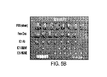
FIGs. 5A-5D show ICAM1 ADCs eradicate standard and late-stage TNBC tumors in
vivo. FIG. 5A: Schematic design of in vivo efficacy of ICAM1 ADC in standard
and late-stage
4
CA 03211696 2023-08-23
WO 2022/182743
PCT/US2022/017499
settings of an orthotopic TNBC model. FIG. 5B: Image of excised orthotopic MDA-
MB-436
tumors from mice treated with PBS (sham), free Dox, ICAM1 antibody (IC1 Ab),
IC1-MMAF
or IC1-MMAE in standard setting (n=7-10 per group). FIG. 5C: Tumor progression
(left) in
standard setting was monitored by tumor volume measurement using a caliper.
Tumor mass
(center) at end point (day 24) of standard setting was quantified by weight.
Mouse body
weights (right) in standard setting receiving PBS (sham), free Dox, IC1 Ab,
IC1-MMAF or
IC1-MMAE. FIG. 5D: Tumor progression (left) receiving PBS (sham), IC1-MMAF or
IC1-
MMAE in late-stage setting was monitored by tumor volume. Tumor mass (center)
at end
point (day 34) of late-stage setting was quantified by weight. Mouse body
weights (right) in
late-stage setting receiving PBS (sham), IC1-MMAF or IC1-MMAE.
FIGs. 6A-6G show ICAM1 ADCs eradicate refractory TNBC tumors in vivo. FIG. 6A:
Schematic design of in vivo efficacy of ICAM1 ADCs in a refractory TNBC mouse
model.
FIG. 6B: Image of excised orthotopic MDA-MB-231 tumors from mice treated with
PBS
(sham), IC1-MMAF or IC1-MMAE. FIG. 6C: Refractory tumor progression (left)
receiving
.. PBS (sham), IC1-MMAF or IC1-MMAE was monitored by tumor volume measurement
using
a caliper. Tumor mass (center) at end point (day 24) was quantified by weight.
Quantified
mouse body weights (right) during administration of PBS (sham), free Dox, IC1
Ab, IC1-
MMAF, or IC1-MMAE. FIG. 6D: Schematic design of dosage-dependent efficacy of
IC1-
MMAE in an orthotopic TNBC tumor model. FIG. 6E: Image of excised orthotopic
MBA-
1\4B-436 tumors from mice treated with PBS (sham) or IC1-MMAE at three
different dosages.
FIG. 6F: Tumor progression (left) receiving PBS (sham) or IC1-MMAE at
different dosages
was monitored by tumor volume. Tumor mass (center) at end point (day 24) was
quantified by
weight. Quantified mouse body weights (right) during IC1-MMAE administration
at different
dosages. FIG. 6G: Chronic liver and renal toxicities of IC1-MMAE were analyzed
by blood
chemistry.
FIGs. 7A and 7B show MTD measurement of IC1-MMAE. FIG. 7A: Schematic
design of maximum tolerable dosage of IC1-MMAE in an immunocompetent BALB/c
mouse
model. FIG. 7B: Quantified mouse bodyweight during MTD test (n = 10 per
group).
FIG. 8 shows selective ablation of non-TNBC human cancer cells by ICAM1 ADCs.
In
vitro cytotoxicity of four ICAM1 ADCs (ICAM1-DM4, ICAM1-DM1, ICAM1-MMAE, and
ICAM1-MMAF) was tested against a panel of four non-TNBC ICAM1-overexpressing
human
cancer cell lines: Du145 (prostate cancer), Caov3 (ovarian cancer), A375
(melanoma), and
PC9 (lung cancer).
5
CA 03211696 2023-08-23
WO 2022/182743
PCT/US2022/017499
DETAILED DESCRIPTION OF CERTAIN EMBODIMENTS
Antibody-drug conjugates (ADCs)
I. ICAM1 Antibodies
Antibody-drug conjugates (ADCs) are a class of immunotherapeutics that
comprise an
antibody conjugated to a drug. The ADCs of the present disclosure can target
cells expressing
ICAM1. ICAM1 is a cell surface glycoprotein that has been shown to bind
integrins of type
CD1 la / CD18, or CD1 lb / CD18 and has been implicated in mediating cell-cell
interactions
and promoting leukocyte endothelial transmigration. ICAM1 is also referred to
as ICAM-1,
BB2, Cluster of Differentiation 54 (CD54), and P3.58.
Non-limiting examples of amino acid sequences encoding ICAM1 include UniProtKB
Accession Nos. P13597 and P05362.
UniProtKB Accession No. P13597 encodes ICAM1 from Mus Muscu/us has the
sequence of:
MASTRAKPTLPLLLALVTVVIPGPGDAQVSIHPREAFLPQGGSVQVNCSSSCKEDLSLGLETQWLKDELESGPNWK
LFELSEIGEDSSPLCFENCGTVQSSASATITVYSFPESVELRPLPAWQQVGKDLTLRCHVDGGAPRTQLSAVLLRG
EEILSRQPVGGHPKDPKEITFTVLASRGDHGANFSCRTELDLRPQGLALFSNVSEARSLRTFDLPATIPKLDTPDL
LEVGTQQKLECSLEGLEPASEARTYLELGGQMPTQESTNS SDSVSATALVEVTEEFDRTLPLRCVLELADQILETQ
RTLTVYNFSAPVLTLSQLEVSEGSQVTVKCEAHSGSKVVLLSGVEPRPPTPQVQFTLNASSEDHKRSFFCSAALEV
AGKFLEKNQTLELHVLYGPRLDETDCLGNWTWQEGSQQTLKCQAWGNPSPKMTCRRKADGALLPIGVVKSVKQEMN
GTYVCHAFS SHGNVTRNVYLTVLYHSQNNWTI I
ILVPVLLVIVGLVMAASYVYNRQRKIRTYKLQKAQEEAIKLKG
QAPPP (SEQ ID NO: 1) .
UniProtKB Accession No. P05362 encodes ICAM1 from Homo Sapiens and has the
sequence:
MAPSSPRPALPALLVLLGALFPGPGNAQTSVSPSKVILPRGGSVLVTCSTSCDQPKLLGIETPLPKKELLLPGNNR
KVYELSNVQEDSQPMCYSNCPDGQSTAKTFLTVYWTPERVELAPLPSWQPVGKNLTLRCQVEGGAPRANLTVVLLR
GEKELKREPAVGEPAEVTTTVLVRRDHHGANFSCRTELDLRPQGLELFENTSAPYQLQTFVLPATPPQLVSPRVLE
VDTQGTVVCSLDGLEPVSEAQVHLALGDQRLNPTVTYGNDSFSAKASVSVTAEDEGTQRLTCAVILGNQSQETLQT
VTIYSFPAPNVILTKPEVSEGTEVTVKCEAHPRAKVTLNGVPAQPLGPRAQLLLKATPEDNGRSFSCSATLEVAGQ
LIHKNQTRELRVLYGPRLDERDCPGNWTWPENSQQTPMCQAWGNPLPELKCLKDGTFPLPIGESVTVTRDLEGTYL
CRARSTQGEVTRKVTVNVLSPRYEIVI ITVVAAAVIMGTAGLSTYLYNRQRKIKKYRLQQAQKGTPMKPNTQATPP
(SEQ ID NO: 2) .
In some embodiments, an ICAM1 protein comprises a sequence that is at least
50%,
55%, 60%, 65%, 70%, 75%, 80%, 85%, 90%, 91%, 92%, 93%, 94%, 95%, 96%, 97%,
98%,
99%, or is 100% identical to SEQ ID NO: 1 or to SEQ ID NO: 2. Additional ICAM1
proteins
are well known and may be identified using publically available databases
including, e.g.,
GenBank. An ICAM1 protein may be from any species, including homo sapiens.
6
CA 03211696 2023-08-23
WO 2022/182743
PCT/US2022/017499
Antibodies of the present disclosure are capable of binding ICAM1. In some
embodiments, the ICAM1 antibody is a monoclonal antibody. In some embodiments,
the
ICAM1 antibody is a polyclonal antibody. In some embodiments, the ICAM1
antibody is a
murine antibody. In some embodiments, the ICAM1 antibody is a humanized
antibody. In
some embodiments, the ICAM1 antibody is a chimeric antibody.
Non-limiting examples of ICAM1 antibodies include clone HCD54 ("HCD54,"
commercially available at BioLegend, catalog #322702), UV3, RR1.1, R6.5 (BIRR-
1 or
Enlimomab, commercially available at Thermo Fisher Scientific, catalog #
BMS1011) and BI-
505. R6.5 (Enlimomab) is a monoclonal murine antibody produced by ATCC HB-9580
hybridoma cells, e.g., as described in United States Patent 5,324,510, which
is herein
incorporated by reference.
UV3 is a monoclonal antibody and has been shown to bind to ICAM-1 on myeloma
cells. In some embodiments, the ICAM1 antibody is a F(ab)'2 fragment of UV3.
See, e.g.,
Huang et al., Hybridoma. 1993 Dec;12(6):661-75; and Coleman et al., J
Immunother. 2006
Sep-Oct;29(5):489-98, which is each herein incorporated by reference. RR1.1 is
a monoclonal
ICAM1 antibody. See, e.g., Rothlein and Springer, 1986 J. Exp. Med. 163, 1132-
1149, which
is herein incorporated by reference. HCD54 is a monoclonal ICAM1 antibody. BI-
505 is a
fully human ICAM1 monoclonal antibody. See, e.g., Hansson et al., Clin Cancer
Res. 2015
Jun 15;21(12):2730-6, which is herein incorporated by reference.
The term "bind" refers to the association of two entities (e.g., two
proteins). Two
entities (e.g., two proteins) are considered to bind to each other when the
affinity (KD) between
them is <10' M, <10-5M, <10' M, <10-7 M, <10-8M, <10-9M, <1040 M, <10-" M, or
<10-12
M. One skilled in the art is familiar with how to assess the affinity of two
entities (e.g., two
proteins).
The term "antibody" encompasses whole antibodies (immunoglobulins having two
heavy chains and two light chains), antibody mimetics, and antibody fragments.
An
"immunoglobulin (Ig)" is a large, Y-shaped protein produced mainly by plasma
cells that is
used by the immune system to neutralize an exogenous substance (e.g., a
pathogens such as
bacteria and viruses). Antibodies may be classified as IgA, IgD, IgE, IgG, and
IgM. "Antibody
fragments" include any antigen binding fragment (i.e., "antigen-binding
portion") or single
chain thereof In some embodiments, an "antibody" refers to a glycoprotein
comprising at least
two heavy (H) chains and two light (L) chains inter-connected by disulfide
bonds, or an antigen
binding portion thereof Each heavy chain is comprised of a heavy chain
variable region
(abbreviated herein as VH) and a heavy chain constant region. The heavy chain
constant region
7
CA 03211696 2023-08-23
WO 2022/182743
PCT/US2022/017499
is comprised of three domains, CH1, CH2 and CH3. Each light chain is comprised
of a light
chain variable region (abbreviated herein as VL) and a light chain constant
region. The light
chain constant region is comprised of one domain, CL. The VH and VL regions
can be further
subdivided into regions of hypervariability, termed complementarity
determining regions
(CDR), interspersed with regions that are more conserved, termed framework
regions (FR).
Each VH and VL is composed of three CDRs and four FRs, arranged from amino-
terminus to
carboxy-terminus in the following order: FR1, CDR1, FR2, CDR2, FR3, CDR3, FR4.
The
variable regions of the heavy and light chains contain a binding domain that
interacts with an
antigen. The constant regions of the antibodies may mediate the binding of the
immunoglobulin
to host tissues or factors, including various cells of the immune system
(e.g., effector cells) and
the first component (Clq) of the classical complement system. In some
embodiments, an
antibody is an immunoglobulin (Ig) monomer. An antibody may be a polyclonal
antibody or a
monoclonal antibody.
In some embodiments, an antibody is a heterotetrameric glycoprotein composed
of two
identical L chains and two H chains (an IgM antibody consists of 5 of the
basic heterotetramer
unit along with an additional polypeptide called J chain, and therefore
contain 10 antigen
binding sites, while secreted IgA antibodies can polymerize to form polyvalent
assemblages
comprising 2-5 of the basic 4-chain units along with J chain). In the case of
IgGs, the 4-chain
unit is generally about 150,000 daltons. Each L chain is linked to a H chain
by one covalent
disulfide bond, while the two H chains are linked to each other by one or more
disulfide bonds
depending on the H chain isotype. Each H and L chain also has regularly spaced
intrachain
disulfide bridges. Each H chain has at the N-terminus, a variable domain (VH)
followed by
three constant domains (CH) for each of the a and y chains and four CH domains
for 11 and
isotypes. Each L chain has at the N-terminus, a variable domain (VL) followed
by a constant
domain (CL) at its other end. The VL is aligned with the VH and the CL is
aligned with the first
constant domain of the heavy chain (CH1). Particular amino acid residues are
believed to form
an interface between the light chain and heavy chain variable domains. The
pairing of a VH and
VL together forms a single antigen-binding site. For the structure and
properties of non-limiting
examples of different classes of antibodies, see, e.g., Basic and Clinical
Immunology, 8th
edition, Daniel P. Stites, Abba I. Terr and Tristram G. Parslow (eds.),
Appleton & Lange,
Norwalk, Conn., 1994, page 71 and Chapter 6, incorporated herein by reference.
In some
embodiments, an antibody is an IgG.
The L chain from any vertebrate species can be assigned to one of two clearly
distinct
types, called kappa and lambda, based on the amino acid sequences of their
constant domains.
8
CA 03211696 2023-08-23
WO 2022/182743
PCT/US2022/017499
Depending on the amino acid sequence of the constant domain of their heavy
chains (CH),
immunoglobulins can be assigned to different classes or isotypes. There are
five classes of
immunoglobulins: IgA, IgD, IgE, IgG, and IgM, having heavy chains designated
a, 6, , y and
[t, respectively. The y and a classes are further divided into subclasses on
the basis of relatively
minor differences in CH sequence and function, e.g., humans express the
following subclasses:
IgGl, IgG2, IgG3, IgG4, IgAl, and IgA2.
The V domain mediates antigen binding and define specificity of a particular
antibody
for its particular antigen. However, the variability is not evenly distributed
across the 110-amino
acid span of the variable domains. Instead, the V regions consist of
relatively invariant stretches
called framework regions (FRs) of 15-30 amino acids separated by shorter
regions of extreme
variability called "hypervariable regions" that are each 9-12 amino acids
long. The variable
domains of native heavy and light chains each comprise four FRs, largely
adopting a 13-sheet
configuration, connected by three hypervariable regions, which form loops
connecting, and in
some cases forming part of, the 13-sheet structure. The hypervariable regions
in each chain are
held together in close proximity by the FRs and, with the hypervariable
regions from the other
chain, contribute to the formation of the antigen-binding site of antibodies
(see, e.g., Kabat et
al., Sequences of Proteins of Immunological Interest, 5th Ed. Public Health
Service, National
Institutes of Health, Bethesda, Md. (1991), incorporated herein by reference).
The constant
domains are not involved directly in binding an antibody to an antigen, but
exhibit various
effector functions, such as participation of the antibody in antibody
dependent cellular
cytotoxicity (ADCC).
In some embodiments, the antibody is a monoclonal antibody. A "monoclonal
antibody" is an antibody obtained from a population of substantially
homogeneous antibodies,
i.e., the individual antibodies comprising the population are identical except
for possible
naturally occurring mutations that may be present in minor amounts. Monoclonal
antibodies
are highly specific, being directed against a single antigenic site.
Furthermore, in contrast to
polyclonal antibody preparations which include different antibodies directed
against different
determinants (epitopes), each monoclonal antibody is directed against a single
determinant on
the antigen. In addition to their specificity, the monoclonal antibodies are
advantageous in that
they may be synthesized uncontaminated by other antibodies. The modifier
"monoclonal" is
not to be construed as requiring production of the antibody by any particular
method. For
example, the monoclonal antibodies useful in the present invention may be
prepared by the
hybridoma methodology first described by Kohler et al., Nature, 256:495
(1975), or may be
made using recombinant DNA methods in bacterial, eukaryotic animal or plant
cells (see, e.g.,
9
CA 03211696 2023-08-23
WO 2022/182743
PCT/US2022/017499
U.S. Pat. No. 4,816,567). Monoclonal antibodies may also be isolated from
phage antibody
libraries, e.g., using the techniques described in Clackson et al., Nature,
352:624-628 (1991)
and Marks et al., J. Mol. Biol., 222:581-597 (1991), incorporated herein by
reference.
The monoclonal antibodies described herein encompass "chimeric" antibodies in
which
.. a portion of the heavy and/or light chain is identical with or homologous
to corresponding
sequences in antibodies derived from a particular species or belonging to a
particular antibody
class or subclass, while the remainder of the chain(s) is identical with or
homologous to
corresponding sequences in antibodies derived from another species or
belonging to another
antibody class or subclass, as well as fragments of such antibodies, so long
as they exhibit the
desired biological activity (see U.S. Pat. No. 4,816,567; and Morrison et al.,
Proc. Natl. Acad.
Sci. USA, 81:6851-6855 (1984)). Chimeric antibodies of interest herein include
"primatized"
antibodies comprising variable domain antigen-binding sequences derived from a
non-human
primate (e.g. Old World Monkey, Ape etc.), and human constant region
sequences.
In some embodiments, the antibody is a polyclonal antibody. A "polyclonal
antibody"
is a mixture of different antibody molecules which react with more than one
immunogenic
determinant of an antigen. Polyclonal antibodies may be isolated or purified
from mammalian
blood, secretions, or other fluids, or from eggs. Polyclonal antibodies may
also be recombinant.
A recombinant polyclonal antibody is a polyclonal antibody generated by the
use of
recombinant technologies. Recombinantly generated polyclonal antibodies
usually contain a
high concentration of different antibody molecules, all or a majority of
(e.g., more than 80%,
more than 85%, more than 90%, more than 95%, more than 99%, or more) which are
displaying a desired binding activity towards an antigen composed of more than
one epitope.
In some embodiments, the antibodies are "humanized" for use in human (e.g., as
therapeutics). "Humanized" forms of non-human (e.g., rodent) antibodies are
chimeric
antibodies that contain minimal sequence derived from the non-human antibody.
Humanized
antibodies are human immunoglobulins (recipient antibody) in which residues
from a
hypervariable region of the recipient are replaced by residues from a
hypervariable region of a
non-human species (donor antibody) such as mouse, rat, rabbit or non-human
primate having
the desired antibody specificity, affinity, and capability. In some instances,
framework region
(FR) residues of the human immunoglobulin are replaced by corresponding non-
human
residues. Furthermore, humanized antibodies may comprise residues that are not
found in the
recipient antibody or in the donor antibody. These modifications are made to
further refine
antibody performance. In general, the humanized antibody will comprise
substantially all of at
least one, and typically two, variable domains, in which all or substantially
all of the
CA 03211696 2023-08-23
WO 2022/182743
PCT/US2022/017499
hypervariable loops correspond to those of a non-human immunoglobulin and all
or
substantially all of the FRs are those of a human immunoglobulin sequence. The
humanized
antibody optionally also will comprise at least a portion of an immunoglobulin
constant region
(Fc), typically that of a human immunoglobulin. For further details, see Jones
et al., Nature
321:522-525 (1986); Riechmann et al., Nature 332:323-329 (1988); and Presta,
Curr. Op.
Struct. Biol. 2:593-596 (1992).
In some embodiments, the antibody is an "antibody fragment" containing the
antigen-
binding portion of a full-length ICAM1 antibody. In some embodiments, an
antibody is a single
domain heavy chain antibody. In some embodiments, an antibody is a single
domain light
chain antibody. The antigen-binding portion of an antibody refers to one or
more fragments of
an antibody that retain the ability to specifically bind to an antigen. It has
been shown that the
antigen-binding function of an antibody can be performed by fragments of a
full-length
antibody. Examples of binding fragments encompassed within the term "antigen-
binding
portion" of an antibody include (i) a Fab fragment, a monovalent fragment
consisting of the
VL, VH, CL and CH1 domains; (ii) a F(ab')2 fragment, a bivalent fragment
comprising two
Fab fragments linked by a disulfide bridge at the hinge region; (iii) a Fd
fragment consisting of
the VH and CH1 domains; (iv) a Fv fragment consisting of the VL and VH domains
of a single
arm of an antibody, (v) a dAb fragment (e.g., as described in Ward et al.,
(1989) Nature
341:544-546, incorporated herein by reference), which consists of a VH domain;
and (vi) an
isolated complementarity determining region (CDR). Furthermore, although the
two domains of
the Fv fragment, VL and VH, are coded for by separate genes, they can be
joined, using
recombinant methods, by a synthetic linker that enables them to be made as a
single protein
chain in which the VL and VH regions pair to form monovalent molecules (known
as single
chain Fv (scFv); see e.g., Bird et al. (1988) Science 242:423-426; and Huston
et al. (1988) Proc.
Natl. Acad. Sci. USA 85:5879-5883, incorporated herein by reference). Such
single chain
antibodies are also intended to be encompassed within the term "antigen-
binding portion" of an
antibody. These antibody fragments are obtained using conventional techniques
known to those
with skill in the art, and the fragments are screened for utility in the same
manner as are full-
length antibodies.
In some embodiments, an antibody fragment may be a Fc fragment, a Fv fragment,
or a
single-change Fv fragment. The Fc fragment comprises the carboxy-terminal
portions of both H
chains held together by disulfides. The effector functions of antibodies are
determined by
sequences in the Fc region, which region is also the part recognized by Fc
receptors (FcR)
found on certain types of cells.
11
CA 03211696 2023-08-23
WO 2022/182743
PCT/US2022/017499
The Fv fragment is the minimum antibody fragment which contains a complete
antigen-
recognition and -binding site. This fragment consists of a dimer of one heavy-
and one light-
chain variable region domain in tight, non-covalent association. From the
folding of these two
domains emanate six hypervariable loops (3 loops each from the H and L chain)
that contribute
the amino acid residues for antigen binding and confer antigen binding
specificity to the
antibody. However, even a single variable domain (or half of an Fv comprising
only three
CDRs specific for an antigen) has the ability to recognize and bind antigen,
although at a lower
affinity than the entire binding site.
Single-chain Fv also abbreviated as "sFv" or "scFv" are antibody fragments
that
comprise the VH and VL antibody domains connected into a single polypeptide
chain.
Preferably, the sFy polypeptide further comprises a polypeptide linker between
the VH and VL
domains which enables the sFy to form the desired structure for antigen
binding (e.g., as
described in Pluckthun in The Pharmacology of Monoclonal Antibodies, vol. 113,
Rosenburg
and Moore eds., Springer-Verlag, New York, pp. 269-315 (1994); Borrebaeck
1995,
-- incorporated herein by reference). In some embodiments, an antibody is a
dimerized scFV (a
diabody), a scFV timer (a triabody), or a scFV tetrameter (a tetrabody).
Antibodies of the present disclosure include antibody mimetics, including
affibody
molecules. An affibody is a small protein comprising a three-helix bundle that
functions as an
antigen binding molecule (e.g., an antibody mimetic). Generally, affibodies
are approximately
58 amino acids in length and have a molar mass of approximately 6 kDa.
Affibody molecules
with unique binding properties are acquired by randomization of 13 amino acids
located in two
alpha-helices involved in the binding activity of the parent protein domain.
Specific affibody
molecules binding a desired target protein can be isolated from pools
(libraries) containing
billions of different variants, using methods such as phage display.
In some embodiments, an ICAM1 antibody binds to an epitope that is present in
the
extracellular portion of an ICAM1. An "extracellular portion" of an ICAM1
refers to the
portion of the ICAM1 that is outside of the cytosol and on the surface of the
cell, as opposed to
the portion that is inside the cytosol or embedded in the plasma membrane of
the cell. The
extracellular portion of an ICAM1 typically comprises the glycosylated amino
terminal portion
of the protein, which mediates cell-cell interactions and promotes leukocyte
endothelial
transmigration.
Methods of producing antibodies (e.g., monoclonal antibodies or polyclonal
antibodies)
are known in the art. For example, a polyclonal antibody may be prepared by
immunizing an
animal, preferably a mammal, with an allergen of choice followed by the
isolation of antibody-
12
CA 03211696 2023-08-23
WO 2022/182743
PCT/US2022/017499
producing B-lymphocytes from blood, bone marrow, lymph nodes, or spleen.
Alternatively,
antibody-producing cells may be isolated from an animal and exposed to an
allergen in vitro
against which antibodies are to be raised. The antibody-producing cells may
then be cultured to
obtain a population of antibody-producing cells, optionally after fusion to an
immortalized cell
line such as a myeloma. In some embodiments, as a starting material B-
lymphocytes may be
isolated from the tissue of an allergic patient, in order to generate fully
human polyclonal
antibodies. Antibodies may be produced in mice, rats, pigs (swine), sheep,
bovine material, or
other animals transgenic for the human immunoglobulin genes, as starting
material in order to
generate fully human polyclonal antibodies. In some embodiments, mice or other
animals
transgenic for the human immunoglobulin genes (e.g. as disclosed in U.S. Pat.
No. 5,939,598),
the animals may be immunized to stimulate the in vivo generation of specific
antibodies and
antibody producing cells before preparation of the polyclonal antibodies from
the animal by
extraction of B lymphocytes or purification of polyclonal serum.
Monoclonal antibodies are typically made by cell culture that involves fusing
myeloma
.. cells with mouse spleen cells immunized with the desired antigen (i.e.,
hyrbidoma technology).
The mixture of cells is diluted and clones are grown from single parent cells
on microtitre
wells. The antibodies secreted by the different clones are then assayed for
their ability to bind
to the antigen (with a test such as ELISA or Antigen Microarray Assay) or
immuno-dot blot.
The most productive and stable clone is then selected for future use.
Drugs
Drugs suitable for use in the ADCs include agents that are therapeutically
active against
triple negative breast cancer (TNBC). Non-limiting examples of drugs include
chemotherapies. In some instances, a drug is a small molecule. In some
embodiments, a drug
is a cytotoxic small molecule. In some embodiments, a drug is a cytostatic
small molecule.
Non-limiting examples of drugs suitable for use in the ADCs include N2'-
Deacetyl-
N2'-(3-mercapto-1-oxopropyl)mertansine (DM1), monomethyl auristatin E (MMAE),
monomethyl auristatin F (MMAF), and duocarmycin, paclitaxel, everolimus,
fluorouracil (5-
FU), gemcitabine, gemcitabine hydrochloride, mitomycin C, and derivatives
thereof In some
embodiments, the drug is maytansine or an analog thereof. In some embodiments,
the drug is
DM1. DM1 is a cytotoxic maytansine analog that has been shown to inhibit
tubulin
polymerization. In some embodiments, the maytansine analog is DM4.
The term "small molecule" refers to molecules, whether naturally-occurring or
artificially created (e.g., via chemical synthesis) that have a relatively low
molecular weight.
13
CA 03211696 2023-08-23
WO 2022/182743
PCT/US2022/017499
Typically, a small molecule is an organic compound (e.g., it contains carbon).
The small
molecule may contain multiple carbon-carbon bonds, stereocenters, and other
functional
groups (e.g., amines, hydroxyl, carbonyls, and heterocyclic rings, etc.). In
certain
embodiments, the molecular weight of a small molecule is not more than about
1,000 g/mol,
not more than about 900 g/mol, not more than about 800 g/mol, not more than
about 700
g/mol, not more than about 600 g/mol, not more than about 500 g/mol, not more
than about
400 g/mol, not more than about 300 g/mol, not more than about 200 g/mol, or
not more than
about 100 g/mol. In certain embodiments, the molecular weight of a small
molecule is at least
about 100 g/mol, at least about 200 g/mol, at least about 300 g/mol, at least
about 400 g/mol, at
least about 500 g/mol, at least about 600 g/mol, at least about 700 g/mol, at
least about 800
g/mol, or at least about 900 g/mol, or at least about 1,000 g/mol.
Combinations of the above
ranges (e.g., at least about 200 g/mol and not more than about 500 g/mol) are
also possible.
Any known chemotherapeutic drugs may be used as the drug in the ADC descirbed
herein. Non-limiting exemplary chemotherapetic drugs include: Actinomycin, All-
trans
retinoic acid, Azacitidine, Azathioprine, Bleomycin, Bortezomib, Carboplatin,
Capecitabine,
Cisplatin, Chlorambucil, Cyclophosphami de, Cytarabine, Daunorubicin,
Docetaxel,
Doxifluridine, Doxorubicin, Epirubicin, Epothilone, Etoposide, Fluorouracil,
Gemcitabine,
Hydroxyurea, Idarubicin, Imatinib, Irinotecan, Mechlorethamine,
Mercaptopurine,
Methotrexate, Mitoxantrone, Oxaliplatin, Paclitaxel, Pemetrexed, Teniposide,
Tioguanine,
Topotecan, Valrubicin, Vinblastine, Vincristine, Vindesine, and Vinorelbine.
III. Linkers
One or more drugs may be conjugated to an ICAM1 antibody using techniques
known
in the art. In some embodiments, multiple (e.g., 2, 3, 4, 5, 6, 7, 8, 9, 10,
or more) drugs are
conjugated to an ICAM1 antibody. The ratio of the ICAM1 antibody and the drug
in the ADC
may be 1:1 to 1:10 (e.g., 1:1, 1:2, 1:3, 1:4, 1:5, 1:6, 1:7, 1:8, 1:9, or
1:10). In some
embodiments, the ratio of the ICAM1 antibody and the drug in the ADC is 1:4.
An ICAM1 antibody may be conjugated to a second entity either directly or via
a
linker. As used herein, "conjugated" or "attached" means two entities are
associated,
preferably through a covalent bond or with sufficient affinity that the
therapeutic or diagnostic
benefit of the association between the two entities is realized. In some
embodiments, a linker
conjugates an ICAM1 antibody to a drug in an ADC. The N-terminus or C-terminus
of an
ICAM1 antibody may be conjugated to a drug. In some embodiments, a linker can
be used to
14
CA 03211696 2023-08-23
WO 2022/182743
PCT/US2022/017499
conjugate an ICAM1 antibody to an imaging agent. The N-terminus or C-terminus
of an
ICAM1 antibody may be conjugated to an imaging agent.
In some embodiments, a linker is a cleavable linker. As used herein, a
cleavable linker
is capable of releasing a conjugated moiety in response to a stimulus. In some
embodiments,
the stimulus is a physiological stimulus. Non-limiting examples of stimuli
include the
presence of an enzyme, acidic conditions, basic conditions, or reducing
conditions. For
example, cleavable linkers include peptide linkers, P-glucuronide linkers,
glutathione-sensitive
linkers (or disulfide linkers) and pH-sensitive linkers. In some embodiments,
a pH-sensitive
linker is cleaved at a pH between 5.0 and 6.5 or between a pH of 4.5 and 5Ø
In some
embodiments, a pH-sensitive linker is not cleaved when the pH is between 7 and
7.5. In some
embodiments, a pH-sensitive linker is not cleaved when the pH is between 7.3
and 7.5. In
some embodiments, a cleavable linker is a protease-sensitive linker.
Examples of cleavable linkers include N-succinimidyl 4-(2-
pyridyldithio)pentanoate
(SPP), N-succinimidyl 3-(2-pyridyldithio)butanoate (SPDB), Sulfo-SPDB, valine-
citrulline
dipeptide (Val-cit), acetyl butyrate, CL2A, maleimidocaproyl (MC), and Mal-EBE-
Mal. In
some embodiments, a cleavable linker is a maleimidocaproyl (MC) or Mal-EBE-Mal
linker.
. See, e.g., Donaghy, MAbs. 2016 May-Jun;8(4):659-71, incorporated herein by
reference.
In some embodiments, a linker is non-cleavable. In some embodiments, a non-
cleavable linker is a linker that is not cleaved within systemic circulation
in a subject. In some
embodiments, a non-cleavable linker is a linker that is resistant to protease
cleavage. Non-
cleavable linkers include N-succinimidyl 4-(Nmaleimidomethyl)cyclohexane-1-
carboxylate
(SMCC), maleimidomethyl cyclohexane-1-carboxylate (MCC), and MC-VC-PAB. In
some
embodiments, the non-cleavable linker is MC-VC-PAB.
Any of the antibody-drug conjugates may be synthesized using methods known in
the
art. See, e.g., Yao et al., Int J Mol Sci. 2016 Feb 2;17(2). pii: E194.
The ADCs comprising ICAM1 antibody conjugated to a drug are also advantageous
to
use therapeutically, in part because the drugs (e.g., chemotherapeutic drugs)
are toxic and
cause severe side effects. By conjugating the drug (e.g., DM1) to the ICAM1
antibody, the
toxicity of the ADC may be reduced by at least 20%, at least 30%, at least
40%, at least 50%,
at least 60%, at least 70%, at least 80%, at least 90%, at least 99%, compared
to the drug in its
free from.
Other ICAM1 Antibody Conjugates
CA 03211696 2023-08-23
WO 2022/182743
PCT/US2022/017499
ICAM1 antibodies and/or any of the ADCs of the present disclosure may be
conjugated
to an imaging agent, which may be useful for predicting the therapeutic
sensitivity of a subject
with TNBC. For example, imaging agents for computed tomography (CT), positron
emission
tomography (PET), magnetic resonance imaging (MM), and endoscopic detection
(e.g.,
-- endoscopic ultrasound) may be used and can include contrast agents. See,
e.g., Bird-
Lieberman et at., Nat Med. 2012;18(2):315-21; Van den Brande et at., Gut.
2007;56(4):509-
17, which is each herein incorporated by reference. In some embodiments, the
contrast agent
is administered as a salt. In some embodiments, the imaging agent is a
gadolinium-based Mill
contrast agent. For example, an imaging agent may be a gadolinium-
diethylenetriamine
pentaacetic acid (Gd-DTPA or DTPA-Gd). See, e.g., Can et at., AJR Am J
Roentgenol. 1984
Aug;143(2):215-24.
One or more imaging agents may be conjugated to an ICAM1 antibody or an ADC
described herein using techniques known in the art. In some embodiments,
multiple (e.g., e.g.,
2, 3, 4, 5, 6, 7, 8, 9, 10, or more) imaging agents are conjugated to an ICAM1
antibody. The
ratio of the ICAM1 antibody or ADC and the imaging agent may be 1:1 to 1:10
(e.g., 1:1, 1:2,
1:3, 1:4, 1:5, 1:6, 1:7, 1:8, 1:9, or 1:10). In some embodiments, the ratio of
the ICAM1
antibody or ADC and the imaging agent is 1:4. Any of the linkers disclosed
herein may be
used to conjugate an imaging agent to an ICAM1 antibody or to an ADC described
herein.
An imaging agent may be visualized with a suitable detection method (e.g., by
CT,
PET, MM, ultrasound, and/or endoscopic detection).
Pharmaceutical Compositions and Uses Thereof
Compositions comprising any of the ADCs or other ICAM1 antibody conjugates
disclosed herein are encompassed by the present disclosure. In some
embodiments, the
composition is formulated as a pharmaceutical composition for administration
to a subject.
A subject may have, be suspected of having, or be at risk for triple negative
breast
cancer (TNBC). Breast cancers are classified based on the receptor proteins
expressed or not
expressed on the cell surface of breast cancer cells. TNBC cells are
characterized as breast
cancer cells without cell surface expression of estrogen receptor (ER),
progesterone receptor
(PR), and human epidermal growth factor receptor 2 (HER2). In contrast, ER+
breast cancer
cells express estrogen receptors at their cell surface, while HER2+ breast
cancer cells express
HER2 at their cell surface.
TNBC may also be stratified based on whether or not the cancer has
metastasized.
TNBC may be classified as stage 0 (carcinoma in situ), stage I, stage II,
stage III, stage IV, or
16
CA 03211696 2023-08-23
WO 2022/182743
PCT/US2022/017499
stages therewithin (e.g., stage IIA, stage JIB, etc.). A non-limiting staging
method is the TNM
system, which evaluates the extent of the tumor (T), the spread of the cancer
to nearby lymph
nodes (N), and whether the cancer has spread to distant sites (M). The various
T, N, and M
levels (e.g., Table 1) may then be used to determine the stage of TNBC cancer
(e.g., Table 2).
Tables 1-2 show TNBC classification based on the Eighth Edition of the
AJCC/UICC TNM
staging system and as described by Cong et al. Sci Rep. 2018 Jul
10;8(1):10383.
Table 1. Non-limiting examples of TNM staging definitions
Ti Maximum tumor diameter <2 cm
T2 Maximum tumor diameter >2, <4 cm
T3 Maximum tumor diameter >4 cm
14 Tumor involves the celiac axis, common
NO No regional lymph node metastasis
N1 Metastasis in 1---3 regional lymph nodes
N2 Metastasis in > 4 regional lymph nodes
MO No distant metastasis
M.1 Distant metastasis
Table 2. TNBC Staging Levels
IA T1 NO MO
IB T2 NO MO
I IA T3 NO MO
HR Ti -T3 Ni M 0
III T4 any N MO
IV any T any N M1
In some embodiments, a subject may have, be suspected of having, or be at risk
for a
cancer other than TNBC. Non-limiting examples of other cancers include breast
cancer,
prostate cancer, ovarian cancer, melanoma, lung cancer, and pancreatic cancer.
In some
embodiments, the breast cancer is not TNBC. Other cancers may also be
stratified based on
whether or not the cancer has metastasized and may be classified as stage 0
(carcinoma in situ),
stage I, stage II, stage III, stage IV, or stages therewithin (e.g., stage
IIA, stage JIB, etc.).
Other cancers may also be classified using the TNM staging method, in which
the various T,
N, and M levels are used to determine the cancer stage (e.g., Tables 1 and 2).
In some embodiments, any of the pharmaceutical compositions disclosed herein
comprising an imaging agent is administered in an effective amount to a
subject to determine
the level of ICAM1 in a tumor of a subject with TNBC or another cancer (e.g.,
CT, PET, MRI,
and endoscopic detection (e.g., endoscopic ultrasound)). The imaging methods
for
17
CA 03211696 2023-08-23
WO 2022/182743
PCT/US2022/017499
determining the level of ICAM1 described herein are advantageous compare to
conventional
methods (e.g., biopsy and analyzing the tissue obtained from the biopsy). The
imaging
methods (e.g., Mill) is non-invasive, and provides a comprehensive view of the
tumor for
ICAM1 level, providing more accurate assessment of the tumor for prediction of
outcome
and/or responsiveness to treatment (e.g., treatment with ICAM1 antibody or ADC
comprising
ICAM1 antibody).
In some embodiments, the level of ICAM1 is detected in a subject with TNBC or
another cancer who has been administered a pharmaceutical composition of the
present
disclosure comprising an ICAM1 antibody and an imaging agent. In some
embodiments, the
ICAM1 level detected in the tumor of the subject is at least 5%, at least 10%,
at least 20%, at
least 30%, at least 40%, at least 50%, at least 60%, at least 70%, at least
80%, at least 90%, at
least 100%, at least 200%, at least 300%, at least 400%, at least 500%, at
least 600%, at least
700%, at least 800%, at least 900%, or at least 1,000% higher than a control.
In some
embodiments, the ICAM1 level detected in the tumor of the subject is
substantially similar to
.. the control.
In some embodiments, a control is a subject with a tumor having a known level
of
ICAM1. In some embodiments, a control is the level of ICAM1 in the breast
tissue of a
subject who does not have a tumor. In some embodiments, a control is a subject
with a tumor
having a low level of ICAM1. In some embodiments, a low level of ICAM1 is not
detectable.
In some embodiments, a control is a subject with a tumor having a high level
of ICAM1. In
some embodiments, a high level of ICAM1 is at least 5%, at least 10%, at least
20%, at least
30%, at least 40%, at least 50%, at least 60%, at least 70%, at least 80%, at
least 90%, at least
100%, at least 200%, at least 300%, at least 400%, at least 500%, at least
600%, at least 700%,
at least 800%, at least 900%, or at least 1,000% higher than the level of
ICAM1 detected in
breast tissue of a healthy subject.
In some embodiments, the level of ICAM1 detected in a tumor using a method
disclosed herein is predictive of a subject with TNBC or another cancer
responding to
treatment with an ICAM1 antibody or an antibody drug conjugate (ADC)
comprising an
intercellular adhesion molecule 1 (ICAM1) antibody conjugated to a drug. In
some
.. embodiments, a higher level of ICAM1 detected in a tumor as compared to the
tumor of a
subject with a lower level of ICAM1 is predicted to be more responsive to
treatment with an
ICAM1 antibody or an ADC disclosed herein. In some embodiments, a subject with
a higher
level of ICAM1 in a tumor as compared to a subject with a lower level of ICAM1
in a tumor is
at least 5%, at least 10%, at least 20%, at least 30%, at least 40%, at least
50%, at least 60%, at
18
CA 03211696 2023-08-23
WO 2022/182743
PCT/US2022/017499
least 70%, at least 80%, at least 90%, at least 100%, at least 200%, at least
300%, at least
400%, at least 500%, at least 600%, at least 700%, at least 800%, at least
900%, or at least
1,000% more responsive to treatment with a composition comprising an ICAM1
antibody
(e.g., an ICAM1 ADC and/or an ICAM1 antibody not conjugated to a drug) In some
embodiments, a method disclosed herein comprises administering an ICAM1
antibody or an
ADC antibody disclosed herein after identifying the subject as being
responsive.
In some embodiments, the level of ICAM1 detected in a tumor using a method
disclosed herein is indicative of the stage of cancer. In some embodiments,
the level of
ICAM1 detected in a tumor is indicative of stage 0, stage I, stage II, stage
III, or stage IV.
Without being bound by a particular theory, in some embodiments,
administration of an
ICAM1 antibody conjugated to an imaging agent or an ICAM1 ADC conjugated to an
imaging
agent may serve a dual purpose of visualizing a tumor and treating the tumor.
In some embodiments, administration of an ICAM1 antibody and/or an ADC
comprising an ICAM1 antibody or a pharmaceutical composition thereof inhibits
the growth of
.. a tumor. In some embodiments, administration of an ICAM1 antibody and/or an
ADC
comprising an ICAM1 antibody or a pharmaceutical composition thereof results
in regression
of a tumor. In some embodiments, administration of an ICAM1 antibody and/or an
ADC
comprising an ICAM1 antibody or a pharmaceutical composition thereof decreases
the size of
a tumor by at least 5%, at least 10%, at least 20%, at least 30%, at least
40%, at least 50%, at
least 60%, at least 70%, at least 80%, at least 90%, at least 100%, at least
200%, at least 300%,
at least 400%, at least 500%, at least 600%, at least 700%, at least 800%, at
least 900%, or at
least 1,000% as compared to a control. In some embodiments, the control is a
subject who has
not been treated with a composition that comprises an ICAM1 antibody.
In some embodiments, administration of an ICAM1 antibody and/or an ADC
comprising an ICAM1 antibody or a pharmaceutical composition thereof disclosed
herein
decreases proliferation by at least 5%, at least 10%, at least 20%, at least
30%, at least 40%, at
least 50%, at least 60%, at least 70%, at least 80%, at least 90%, at least
100%, at least 200%,
at least 300%, at least 400%, at least 500%, at least 600%, at least 700%, at
least 800%, at
least 900%, or at least 1,000% higher than a control. In some embodiments,
proliferation is
measured using Ki67 staining. In some embodiments, the control is a subject
who has not
been treated with a composition that comprises an ICAM1 antibody.
In some embodiments, administration of an ICAM1 antibody and/or an ADC
comprising an ICAM1 antibody or a pharmaceutical composition thereof disclosed
herein
decreases metastasis of a tumor by at least 5%, at least 10%, at least 20%, at
least 30%, at least
19
CA 03211696 2023-08-23
WO 2022/182743
PCT/US2022/017499
40%, at least 50%, at least 60%, at least 70%, at least 80%, at least 90%, at
least 100%, at least
200%, at least 300%, at least 400%, at least 500%, at least 600%, at least
700%, at least 800%,
at least 900%, or at least 1,000% as compared to a control. In some
embodiments, the control
is a subject who has not been treated with a composition that comprises an
ICAM1 antibody.
In some embodiments, administration of an ICAM1 antibody and/or an ADC
comprising an ICAM1 antibody or a pharmaceutical composition thereof disclosed
herein does
not decrease the viability of healthy cells. In some embodiments,
administration of an ADC or
a pharmaceutical composition comprising an ADC disclosed herein allows for the
effective
amount (e.g., concentration) of a drug to be lower than if the drug was not
conjugated to an
ICAM1 antibody. In some embodiments, the effective amount of a drug is lowered
by at least
5%, at least 10%, at least 20%, at least 30%, at least 40%, at least 50%, at
least 60%, at least
70%, at least 80%, at least 90%, at least 100%, at least 200%, at least 300%,
at least 400%, at
least 500%, at least 600%, at least 700%, at least 800%, at least 900%, or at
least 1,000% as
compared to administration of the drug alone.
In some embodiments, the pharmaceutical composition further comprises a
pharmaceutically acceptable carrier. "Pharmaceutically acceptable" refers to
those
compounds, materials, compositions, and/or dosage forms which are, within the
scope of
sound medical judgment, suitable for use in contact with the tissues of human
beings and
animals without excessive toxicity, irritation, allergic response, or other
problem or
complication, commensurate with a reasonable benefit/risk ratio. A
"pharmaceutically
acceptable carrier" may be a pharmaceutically acceptable material, composition
or vehicle,
such as a liquid or solid filler, diluent, excipient, solvent or encapsulating
material, involved in
carrying or transporting the subject agents from one organ, or portion of the
body, to another
organ, or portion of the body. Each carrier must be "acceptable" in the sense
of being
compatible with the other ingredients of the formulation and not injurious to
the tissue of the
patient (e.g., physiologically compatible, sterile, physiologic pH, etc.). The
term "carrier"
denotes an organic or inorganic ingredient, natural or synthetic, with which
the active
ingredient is combined to facilitate the application. The components of the
pharmaceutical
compositions also are capable of being co-mingled with the molecules of the
present
disclosure, and with each other, in a manner such that there is no interaction
which would
substantially impair the desired pharmaceutical efficacy. Some examples of
materials which
can serve as pharmaceutically-acceptable carriers include: (1) sugars, such as
lactose, glucose
and sucrose; (2) starches, such as corn starch and potato starch; (3)
cellulose, and its
derivatives, such as sodium carboxymethyl cellulose, methylcellulose, ethyl
cellulose,
CA 03211696 2023-08-23
WO 2022/182743
PCT/US2022/017499
microcrystalline cellulose and cellulose acetate; (4) powdered tragacanth; (5)
malt; (6) gelatin;
(7) lubricating agents, such as magnesium stearate, sodium lauryl sulfate and
talc; (8)
excipients, such as cocoa butter and suppository waxes; (9) oils, such as
peanut oil, cottonseed
oil, safflower oil, sesame oil, olive oil, corn oil and soybean oil; (10)
glycols, such as
propylene glycol; (11) polyols, such as glycerin, sorbitol, mannitol and
polyethylene glycol
(PEG); (12) esters, such as ethyl oleate and ethyl laurate; (13) agar; (14)
buffering agents, such
as magnesium hydroxide and aluminum hydroxide; (15) alginic acid; (16) pyrogen-
free water;
(17) isotonic saline; (18) Ringer's solution; (19) ethyl alcohol; (20) pH
buffered solutions; (21)
polyesters, polycarbonates and/or polyanhydrides; (22) bulking agents, such as
polypeptides
and amino acids (23) serum component, such as serum albumin, HDL and LDL; (22)
C2-C12
alcohols, such as ethanol; and (23) other non-toxic compatible substances
employed in
pharmaceutical formulations. Wetting agents, coloring agents, release agents,
coating agents,
sweetening agents, flavoring agents, perfuming agents, preservative and
antioxidants can also
be present in the formulation.
The pharmaceutical compositions may conveniently be presented in unit dosage
form
and may be prepared by any of the methods well-known in the art of pharmacy.
The term "unit
dose" when used in reference to a pharmaceutical composition of the present
disclosure refers
to physically discrete units suitable as unitary dosage for the subject, each
unit containing a
predetermined quantity of active material calculated to produce the desired
therapeutic effect in
association with the required diluent; i.e., carrier, or vehicle.
The formulation of the pharmaceutical composition may dependent upon the route
of
administration. Injectable preparations suitable for parenteral administration
or intratumoral,
peritumoral, intralesional or perilesional administration include, for
example, sterile injectable
aqueous or oleaginous suspensions and may be formulated according to the known
art using
.. suitable dispersing or wetting agents and suspending agents. The sterile
injectable preparation
may also be a sterile injectable solution, suspension or emulsion in a
nontoxic parenterally
acceptable diluent or solvent, for example, as a solution in 1,3 propanediol
or 1,3 butanediol.
Among the acceptable vehicles and solvents that may be employed are water,
Ringer's
solution, U.S.P. and isotonic sodium chloride solution. In addition, sterile,
fixed oils are
.. conventionally employed as a solvent or suspending medium. For this
purpose, any bland
fixed oil may be employed including synthetic mono- or di-glycerides. In
addition, fatty acids
such as oleic acid find use in the preparation of injectables. The injectable
formulations can be
sterilized, for example, by filtration through a bacterial-retaining filter,
or by incorporating
21
CA 03211696 2023-08-23
WO 2022/182743
PCT/US2022/017499
sterilizing agents in the form of sterile solid compositions which can be
dissolved or dispersed
in sterile water or other sterile injectable medium prior to use.
Compositions suitable for oral administration may be presented as discrete
units, such
as capsules, tablets, lozenges, each containing a predetermined amount of the
anti-
inflammatory agent. Other compositions include suspensions in aqueous liquids
or non-
aqueous liquids such as a syrup, elixir or an emulsion.
In some embodiments, the pharmaceutical compositions used for therapeutic
administration must be sterile. Sterility is readily accomplished by
filtration through sterile
filtration membranes (e.g., 0.2 micron membranes). Alternatively,
preservatives can be used to
prevent the growth or action of microorganisms. Various preservatives are well
known and
include, for example, phenol and ascorbic acid. The pharmaceutical composition
ordinarily
will be stored in lyophilized form or as an aqueous solution if it is highly
stable to thermal and
oxidative denaturation. The pH of the preparations typically will be about
from 6 to 8, although
higher or lower pH values can also be appropriate in certain instances.
"A therapeutically effective amount" or "effective amount" as used herein
refers to the
amount of each therapeutic agent (e.g., therapeutic agents for treating any of
cancers described
herein) of the present disclosure required to confer therapeutic effect on the
subject, either
alone or in combination with one or more other therapeutic agents. Effective
amounts vary, as
recognized by those skilled in the art, depending on the particular condition
being treated, the
severity of the condition, the individual subject parameters including age,
physical condition,
size, gender and weight, the duration of the treatment, the nature of
concurrent therapy (if any),
the specific route of administration and like factors within the knowledge and
expertise of the
health practitioner. These factors are well known to those of ordinary skill
in the art and can
be addressed with no more than routine experimentation. It is generally
preferred that a
maximum dose of the individual components or combinations thereof be used,
that is, the
highest safe dose according to sound medical judgment. It will be understood
by those of
ordinary skill in the art, however, that a subject may insist upon a lower
dose or tolerable dose
for medical reasons, psychological reasons or for virtually any other reasons.
Empirical considerations, such as the half-life, generally will contribute to
the
determination of the dosage. For example, therapeutic agents that are
compatible with the
human immune system, such as polypeptides comprising regions from humanized
antibodies
or fully human antibodies, may be used to prolong half-life of the polypeptide
and to prevent
the polypeptide being attacked by the host's immune system. Frequency of
administration may
be determined and adjusted over the course of therapy, and is generally, but
not necessarily,
22
CA 03211696 2023-08-23
WO 2022/182743
PCT/US2022/017499
based on treatment and/or suppression and/or amelioration and/or delay of a
disease.
Alternatively, sustained continuous release formulations of a polypeptide may
be appropriate.
Various formulations and devices for achieving sustained release are known in
the art.
In some embodiments, dosage is daily, every other day, every three days, every
four
days, every five days, or every six days. In some embodiments, dosing
frequency is once
every week, every 2 weeks, every 4 weeks, every 5 weeks, every 6 weeks, every
7 weeks,
every 8 weeks, every 9 weeks, or every 10 weeks; or once every month, every 2
months, or
every 3 months, or longer. The progress of this therapy is easily monitored by
conventional
techniques and assays. The dosing regimen (including the anti-cancer agent
used) can vary
overtime.
In some embodiments, for an adult subject of normal weight, doses ranging from
about
0.01 to 1000 mg/kg may be administered. In some embodiments, the dose is
between 1 to 200
mg. The particular dosage regimen, i.e., dose, timing and repetition, will
depend on the
particular subject and that subject's medical history, as well as the
properties of the anti-cancer
agent (such as the half-life of the anti-cancer agent, and other
considerations well known in the
art).
For the purpose of the present disclosure, the appropriate dosage of a
therapeutic agent
as described herein will depend on the specific agent (or compositions
thereof) employed, the
formulation and route of administration, the type and severity of the disease,
whether the anti-
cancer agent is administered for preventive or therapeutic purposes, previous
therapy, the
subject's clinical history and response to the antagonist, and the discretion
of the attending
physician. Typically, the clinician will administer an anti-cancer agent until
a dosage is
reached that achieves the desired result. Administration of one or more anti-
cancer agents can
be continuous or intermittent, depending, for example, upon the recipient's
physiological
condition, whether the purpose of the administration is therapeutic or
prophylactic, and other
factors known to skilled practitioners. The administration of an anti-cancer
agent may be
essentially continuous over a preselected period of time or may be in a series
of spaced dose,
e.g., either before, during, or after developing a disease.
As used herein, the term "treating" refers to the application or
administration of an anti-
cancer agent to a subject in need thereof. "A subject in need thereof', refers
to an individual
who has a disease, a symptom of the disease, or a predisposition toward the
disease, with the
purpose to cure, heal, alleviate, relieve, alter, remedy, ameliorate, improve,
or affect the
disease, the symptom of the disease, or the predisposition toward the disease.
23
CA 03211696 2023-08-23
WO 2022/182743
PCT/US2022/017499
A "subject" to which administration is contemplated refers to a human (i.e.,
male or
female of any age group, e.g., pediatric subject (e.g., infant, child, or
adolescent) or adult
subject (e.g., young adult, middle¨aged adult, or senior adult)) or non¨human
animal. In some
embodiments, the non¨human animal is a mammal (e.g., rodent, e.g., mouse or
rat), primate
(e.g., cynomolgus monkey or rhesus monkey), commercially relevant mammal
(e.g., cattle,
pig, horse, sheep, goat, cat, or dog), or bird (e.g., commercially relevant
bird, such as chicken,
duck, goose, or turkey). The non-human animal may be a male or female at any
stage of
development. The non-human animal may be a transgenic animal or genetically
engineered
animal.
The methods described herein may be used to treat ICAM1-expressiong cancer. In
some embodiments, the ICAM-1 expressing cancer is breast cancer, prostate
cancer, ovarian
cancer, melanoma, or lung cancer. In some embodiments, the ICAM-1 expressing
cancer is
not TNBC.
In some embodiments, the subject is a companion animal (e.g. a pet or service
animal).
"A companion animal," as used herein, refers to pets and other domestic
animals. Non-
limiting examples of companion animals include dogs and cats; livestock such
as horses, cattle,
pigs, sheep, goats, and chickens; and other animals such as mice, rats, guinea
pigs, and
hamsters. In some embodiments, the subject is a research animal. Non-limiting
examples of
research animals include rodents (e.g., rats, mice, guinea pigs, and
hamsters), rabbits, or non-
human primates.
Alleviating a disease (e.g., cancer) includes delaying the development or
progression of
the disease or reducing disease severity. Alleviating the disease does not
necessarily require
curative results. As used therein, "delaying" the development of a disease
means to defer,
hinder, slow, retard, stabilize, and/or postpone progression of the disease.
This delay can be of
varying lengths of time, depending on the history of the disease and/or
individuals being
treated. A method that "delays" or alleviates the development of a disease, or
delays the onset
of the disease, is a method that reduces probability of developing one or more
symptoms of the
disease in a given time frame and/or reduces extent of the symptoms in a given
time frame,
when compared to not using the method. Such comparisons are typically based on
clinical
.. studies, using a number of subjects sufficient to give a statistically
significant result.
"Development" or "progression" of a disease means initial manifestations
and/or
ensuing progression of the disease. Development of the disease can be
detectable and assessed
using standard clinical techniques as well known in the art. However,
development also refers
to progression that may be undetectable. For purpose of this disclosure,
development or
24
CA 03211696 2023-08-23
WO 2022/182743
PCT/US2022/017499
progression refers to the biological course of the symptoms. "Development"
includes
occurrence, recurrence, and onset. As used herein "onset" or "occurrence" of a
disease
includes initial onset and/or recurrence.
Conventional methods, known to those of ordinary skill in the art of medicine,
can be
used to administer the pharmaceutical composition the subject, depending upon
the type of
disease to be treated or the site of the disease. The pharmaceutical
composition can also be
administered via other conventional routes, e.g., administered orally,
parenterally, by
inhalation spray, topically, rectally, nasally, buccally, vaginally or via an
implanted reservoir.
The term "parenteral" as used herein includes subcutaneous, intracutaneous,
intravenous,
intramuscular, intraarticular, intraarterial, intrasynovial, intrasternal,
intrathecal, intralesional,
and intracranial injection or infusion techniques. In some embodiments, the
pharmaceutical
composition is administered via intravenous injection or infusion. In
addition, it can be
administered to the subject via injectable depot routes of administration such
as using 1-, 3-, or
6-month depot injectable or biodegradable materials and methods. In some
embodiments, the
pharmaceutical composition is administered via injection. In some embodiments,
injection is
intravenous injection or intratumoral injection.
EXAMPLES
Example 1: Development of antibody drug conjugates (ADCs) targeting ICAM1
Introduction
Triple negative breast cancer (TNBC) is a heterogeneous disease, defined by
the lack of
estrogen receptor (ER), progesterone receptor (PR), and human epidermal growth
factor
receptor type 2 (HER2). Late-stage and refractory TNBC represents a major
challenge to the
improvement of clinical outcomes for breast cancer patients. In 2021, over
28,000 patients
were estimated to be diagnosed with TNBC in the United States, representing 10-
15% of breast
cancer incidence'. TNBC is more prevalent in non-Hispanic black women, young
women
under the age of 40 and women carrying breast cancer early onset 1 or 2
(BRCA1/2) gene
mutations4' 5. Moreover, patients with late-stage or refractory TNBC tumors
respond poorly to
first line chemotherapy and do not respond to established hormone and HER2-
targeted
therapeutics due to the lack of expression of these molecular targets thereby
dramatically
exacerbating their poor clinical outcomes2' 3,6 The prognosis for TNBC
patients remains the
poorest among all breast cancer patients, as the current 5 year survival rate
of TNBC patients is
less than 77%, significantly lower than the more than 90% survival rate for
patients with other
types of breast cancer'.
CA 03211696 2023-08-23
WO 2022/182743
PCT/US2022/017499
Although several second-line therapeutics including poly(ADP)-ribose
polymerase
(PARP) inhibitors (e.g., Olaparib and Talazoparib) and immune checkpoint
inhibitors (e.g.,
Atezolizumab and Pembrolizumab) have been approved for the treatment of TNBC,
the
clinical benefits of these therapeutics are limited to small subsets (-20-30%)
of TNBC patients,
due to limited BRCA1/2 mutation rates or PD-Li expressionm-12. Novel TNBC-
targeted
therapeutics with different mechanisms of action that can work in synergy with
these approved
modalities are likely to further increase clinical efficacy'''. Antibody drug
conjugates
(ADCs) are an emerging class of immuno-chemotherapeutics featuring the
structure of a
monoclonal antibody conjugated to cytotoxic agents via chemical linkers. The
monoclonal
antibody of an ADC functions as a tumor-homing ligand that guides its
conjugated agent to
selectively ablate antigen-overexpressing tumors while sparing normal tissues.
Compared to T-
cell immunotherapy (e.g., chimeric antigen receptor-T cell (CAR-T) or immune
checkpoint
blockade) or nanomedicines (e.g., liposomes), ADCs feature superior tumor
tissue penetration
due to their ultrasmall size (<10 nm), which is ¨1,000 fold smaller than the
size of a T-cell,
creating an attractive opportunity to increase drug delivery into tumors. ADCs
have
demonstrated promising clinical efficacy in various cancers13, including
aggressive solid
tumors that respond poorly to T-cell immunotherapy. For instance, sacituzumab
govitecan, a
trophoblast antigen 2 (TROP2)-targeted ADC, was approved for treating
refractory TNBC
patients with a ¨33% response rate and a median progression-free survival of
5.5 months16.
Given the poor prognosis and unimpressive efficacy of current TNBC treatment
modalities,
there remains an urgent and unmet need to identify new druggable targets and
to develop more
effective ADCs for TNBC therapy.
ICAM1, also called CD54, is a transmembrane cell surface glycoprotein that
regulates
intercellular adhesion during inflammatory injury, viral infection and
tumorigenesis17. ICAM1
.. is aberrantly overexpressed in multiple types of cancers and is frequently
associated with an
aggressive phenotype and worse prognosis. ICAM1 has previously been identified
as a TNBC
cell surface marker through unbiased quantitative screening of G protein-
coupled receptor
(GPCR) signaling proteins18' 19. ICAM1 was found to be highly enriched on the
cancer cell
surface of human TNBC tumors relative to normal mammary tissues18-22. The NFkB
pathway
has likewise been implicated in aberrantly upregulating ICAM1 expression in
cancer cells by
binding to the ICAM1 promoter and causing its hyperactive transcription23' 24.
An ICAM1
neutralizing antibody has been investigated as a potential cancer therapeutic,
however blocking
ICAM1 signaling cascades alone has not yielded satisfying clinical
efficacy2527. Thus, as
described herein, ICAM1 ADCs that induces potent and durable tumor regression
in vivo have
26
CA 03211696 2023-08-23
WO 2022/182743
PCT/US2022/017499
been developed. It was hypothesized that a rationally designed ICAM1 ADC would
precisely
target and eradicate TNBC tumors while sparing healthy organs and tissues. To
test this
hypothesis, a panel of four ICAM1 ADCs with different chemical linkers and
cytotoxic agents
were constructed and evaluated in vitro against multiple TNBC cell types. The
in vivo efficacy
of these ADCs was also tested in a series of standard, late-stage and
refractory TNBC models.
Results and Discussion
ICAM1 was previously identified as a novel molecular target for TNBC by
measuring
protein expression in a cohort of 149 cases of human breast tumor tissues
along with 144
human normal tissues including breast and 19 other organs using
immunohistochemistry
(IHC)18. ICAM1 staining was found to be present at a significantly greater
level in cancer cells
of TNBC tumors, as compared to other breast cancer subtypes, and is completely
absent in
normal mammary tissues. Genomic analyses were used to further to interrogate
the relationship
of ICAM1 expression with more detailed TNBC characteristics, including its
molecular
subtype, tumor differentiation, and oncogene mutation status, by querying the
R2: Genomics
Analysis and Visualization Platform (huerver I .amc.n1). ICAM1 mRNA expression
was
quantitatively compared in three breast cancer subtypes: ER-positive, HER2-
positive, and
TNBC (FIG. IA). Consistent with pathological staining results18, ICAM1 mRNA
levels in
TNBC tumor tissues (n=116) are significantly higher than those of HER2-
positive (n=40) and
ER-positive (n=808) breast tumors. Given that TNBC has recently been
identified as a
heterogeneous disease classified into four distinct molecular subtypes (basal-
like
immunosuppressed (BLIS), basal-like immunoactivated (BLIA), luminal androgen
receptor (L-
AR) and mesenchymal (MES)28), ICAM1 mRNA levels were also assessed in these
molecular
subtypes. BLIA (n=54) showed the highest level of ICAM1 expression, followed
by IVIES
(n=47), L-AR (n=60), and BLIS (n=37). The correlation between ICAM1 expression
and
TNBC tumor differentiation was also analyzed, and it was found that ICAM1
expression is
significantly higher within poorly differentiated TNBC tumors (grade 3,
n=108), as compared
to moderately differentiated tumors (grade 2, n=53). It is generally known
that poorly
differentiated TNBC tumors are strongly associated with poorer prognoses, thus
ICAM1 may
serve as a potential biomarker for predicting clinical outcomes of TNBC
patients. The
relationship between ICAM1 expression and cancer mutation status was also
examined, and it
was found that ICAM1 levels positively correlate with BRCA1/2 or TP53
mutations, which
occur in 20% and 80% of TNBC tumors, respectively29-31.
27
CA 03211696 2023-08-23
WO 2022/182743
PCT/US2022/017499
Given that TROP2 is a new clinically approved ADC target for TNBC therapy16,
it was
selected as a positive control for the evaluation of potential ICAM1 ADCs. The
cell surface
expression of ICAM1 and TROP2 in three established human TNBC cell lines (MDA-
MB-
436, MDA-MB-157 and MDA-MB-231) was examined, along with normal human mammary
-- epithelial MCF10A cells (FIG. 1B). The cell surface expression of ICAM1 is
11- to 35-fold
higher than TROP2 on two TNBC cell lines (MDA-MB-436 and MDA-MB-468) and is
equivalent in the third cell line (MDA-MB-231). More importantly, ICAM1
surface expression
on normal MCF10A cells is 7-fold lower than that of TROP2. These findings were
confirmed
by immunofluorescent (IF) staining of ICAM1 and TROP2 in the same cells (FIG.
1C). The
-- subcellular localization of ICAM1 is predominantly on the plasma membranes
of TNBC cells
and is undetectable on plasma membranes of normal MCF10A cells. These results
provide
evidence that ICAM1 is more highly overexpressed and is more tumor-specific
than TROP2 in
human TNBC cells, which could potentially contribute to a higher tumor-
specificity and fewer
on-target/off-tumor adverse effects during in vivo delivery of ADCs targeting
ICAM1.
Cellular internalization is another important factor affecting ADC efficacy.
In order to
determine whether ICAM1 ADCs can selectively enter TNBC cells and rapidly
traffic into
endosomes for release of cytotoxic agents, the cellular internalization rates
of ICAM1 antibody
were examined in multiple TNBC cell lines, as well as normal MCF10A cells.
Confocal
fluorescent images revealed that ICAM1 antibodies were rapidly internalized by
all three
TNBC cell lines examined (MDA-MB-436, MDA-MB-157 and MDA-MB-231) after a 1-
hour
incubation (FIG. 1D). The internalized ICAM1 antibodies are aggregated, rather
than
dispersed, in the cytoplasm of TNBC cells, indicating that they are trafficked
into
endosomes/lysosomes by receptor-mediated endocytosis32' 33. In contrast, no
internalization
was observed in normal MCF10A cells due to their lack of ICAM1 antigen
expression.
Cellular internalization rates of ICAM1 antibody in three TNBC cell lines were
approximately
42 to 109-fold higher than those in MCF10A cells, and approximately 21 to 129-
fold higher
than those of observed with a non-targeting IgG (FIG. 1E). These results
confirm that ICAM1
is highly overexpressed in TNBC cells and that it facilitates efficient
cellular internalization of
bound antibodies, suggesting that it has outstanding potential utility as an
ADC target for
-- treating TNBC.
To identify the optimal ADC formulation for TNBC, a panel of ICAM1 ADCs was
designed and constructed by conjugating a monoclonal human/mouse chimeric
ICAM1
antibody (clone#R6.5)34 with cytotoxic drugs (FIG. 2A) to produce four
different ADC
combinations, namely MC-Vc-Pab-MMAE, MC-MMAF, Mal-EBE-Mal-DM1, and Mal-EBE-
28
CA 03211696 2023-08-23
WO 2022/182743
PCT/US2022/017499
Mal-DM4. The ICAM1 ADCs were named according to their antibodies and
conjugated drugs,
as follows: ICAM1 antibody conjugated to MC-Vc-Pab-MMAE (IC1-MMAE); ICAM1
antibody conjugated to MC-MMAF (IC1-MMAF); ICAM1 antibody conjugated to Mal-
EBE-
Mal-DM1 (IC1-DM1); and ICAM1 antibody conjugated to Mal-EBE-Mal-DM4 (IC1-DM4).
This ICAM1 antibody selection is based on the excellent human safety profile
of human/mouse
chimeric ICAM1 antibody (clone#R6.5), which has been previously determined in
Phase I/II
clinical trials27' 34. The choice of ADC linker-drug was then examined. MC-VC-
Pab-MMAE
features an enzyme-cleavable linker that can be selectively cleaved by
cathepsin B proteases in
cell endosomes/lysosomes, resulting in a rapid drug release from ADC
antibodies upon cellular
internalization while maintaining ADC integrity during blood circulation13-15.
The other MC
and Mal-EBE-Mal linkers are non-cleavable thus the drug release depends on
lysosomal
degradation of the conjugated antibody. Both enzyme-cleavable and non-
cleavable linkers are
currently used in clinically-approved ADCs for treating different cancers13-
15. All four drugs
utilized in the examined ICAM1 ADCs are potent microtubule inhibitors with
cytotoxicities
that are approximately 100 to 1,000-fold higher than standard-of-care
chemodrugs (e.g.,
paclitaxel or gemcitabine)13-15. MMAE, MMAF and DM1 are clinically utilized
ADC drugs
and DM4 has also been clinically investigated as an ADC drug for treating
TNBC. However, it
remains unclear which ADC linker-drug combination is most effective for TNBC
treatment in
the absence of an unbiased quantitative screen. Moreover, the drug to antibody
ratio (DAR),
defined as the number of drug molecules conjugated per antibody, is another
important factor
affecting ADC efficacy and human safety. A DAR value of 4.0 was well-
established for
microtubule inhibitor ADC drugs in multiple clinically-approved ADCs (e.g.,
trastuzumab
emtansine and brentuximab vedotin) demonstrating excellent clinical efficacy
and human
safety profiles13-15. Thus, the DAR of the four constructed ICAM1 ADCs was
adjusted to an
equivalent value of 4 by controlling the antibody/drug input ratios during the
ADC
conjugation. The resulting DAR values of four ICAM1 ADCs were determined by
hydrophobic interaction chromatography (HIC) as being 3.96 for IC1-DM1, 3.83
for IC1-
DM4, 3.92 for IC1-MMAE, and 4.09 for IC1-MMAF, respectively (FIG. 2B).
The in vitro cytotoxicity of the four ICAM1 ADCs was then assessed against a
panel of
TNBC cell lines, and half maximum inhibitory concentrations (IC50s) were
quantified using a
Dojindo assay. Among the four ICAM1 ADCs, exposure to IC1-MMAE and IC1-MMAF
elicited a significantly higher in vitro efficacy as compared to IC1-DM1 and
IC-DM4 in three
of the four tested TNBC cell lines (FIG. 2C). The IC50s of IC1-MMAE and IC1-
MMAF were
determined to be 13.1 and 7.3 pM for MDA-MB-436, 250.0 and 68.7 pM for MDA-MB-
468,
29
CA 03211696 2023-08-23
WO 2022/182743
PCT/US2022/017499
221.7 and 116.0 pM for MDA-MB-157, respectively. It is noteworthy that MDA-MB-
436 and
MDA-MB-157 cells represent both Caucasian and African American TNBC cell
lines,
respectively. In contrast, the IC50s in IC1-DM1 and IC1-DM4 are at least 5-
fold higher.
Notably, the IC50s of IC-MMAE and IC-MMAF are approximately 1,000-fold lower
than
.. Doxorubicin, a standard-of-care chemodrug used in TNBC treatment. It was
observed that
MDA-MB-231 cells exhibited strong drug resistance to all four tested ICAM1
ADCs. Due to
the fact that MDA-MB-231 cells were also reported to be resistant to
sacituzumab govitecan,
the clinically used TROP2 ADC for TNBC35, this TNBC cell line was selected for
the
generation of an ADC-resistant TNBC model in the following animal study. The
cytotoxicity
of ICAM1 ADCs was also evaluated in non-neoplastic MCF10A and HEK293 cells,
both of
which lack ICAM1 antigen expression. As expected, no cytotoxicity was observed
in ICAM1-
negative MCF10A and HEK293 cells until a very high ADC concentration of over
6.7 nM was
reached thereby, indicating that the potency of the ADCs in ablating TNBC
cells is dependent
of cell surface expression of ICAM1. Collectively, these in vitro efficacy
screening data
support the notion that IC1-MMAE and IC1-MMAF are more potent ADC formulations
than
ICAM1-DM1 and ICAM1-DM4 for TNBC, warranting further investigation of these
ADCs
using in vivo TNBC models.
Next, the tumor specificity and biodistribution of ICAM1 ADCs was evaluated
using
both an immunocompromised nude mouse model with human TNBC tumors (MDA-MB-436)
and an immunocompetent BALB/c mouse model with murine TNBC tumors (4T1), in
order to
investigate the effects of different immune system backgrounds. First, the
nude mouse model
was used to determine the tumor-specificity of ICAM1 antibody and ADC in
orthotopic human
TNBC tumors (FIG. 3A). Near-infrared fluorescent dye Cy5.5 labeled anti-human
ICAM1
antibody (IC1-Cy5.5) and IC1-MMAE (IC1-MMAE-Cy5.5) were administered
intravenously
and compared to non-targeting IgG conjugated with Cy5.5 (IgG-Cy5.5) at an
equivalent
dosage of 5 mg/kg. At 24 hours post-injection, the nude mice were imaged by in
vivo NIR
fluorescent imaging. Mice treated with IC1-Cy5.5 and IC1-MMAE-Cy5.5 showed
comparable
intratumoral accumulation, approximately 5-fold higher than IgG-Cy5.5 (FIGs.
3B and 3D).
These in vivo tumor accumulation data precisely matched the ex vivo NIR images
of excised
tumors (FIG. 3C). These results suggest that IC1-Cy5.5 and IC1-MMAE-Cy5.5
selectively
recognize and bind human TNBC tumors in vivo, relative to non-targeting IgG-
Cy5.5. The
biodistribution of IC1-Cy5.5, IC1-MMAE-Cy5.5, and IgG-Cy5.5 was further
examined in six
major organs, namely in brain, lung, heart, liver, spleen, and kidney. Liver
and kidney were the
primary off-tumor accumulation sites of IC1-Cy5 and IC1-MMAE in tumor-bearing
mice
CA 03211696 2023-08-23
WO 2022/182743
PCT/US2022/017499
(FIGs. 3E and 3F). However, these are common off-tumor accumulation sites, and
it was
determined that any toxicities were well tolerated, based on serum biomarkers
in subsequent
animal studies.
A potential concern for use of ICAM1 as a drug target for developing TNBC-
targeted
ADCs is its potential on-target/off-tumor toxicity in normal organs and
tissues that normally
express ICAM1. In the nude mouse model, anti-human ICAM1 antibodies cannot
recognize
mouse ICAM1 antigens expressed in normal mouse organs due to the lack of
species cross-
reactivity. To solve this issue, normal organ biodistribution was evaluated
using anti-mouse
ICAM1 antibodies in an immunocompetent BALB/c mouse model with murine 4T1
tumors
(FIG. 4A). Notably, this biodistribution study was conducted in
immunocompetent BALB/c
mice which feature a complete immune system that can more faithfully
recapitulate the
interactions between the TNBC tumor microenvironment and ICAM1 antibodies,
unlike the
immunocompromised nude mouse model. Anti-mouse IC1-Cy5.5 maintains potent 4T1
tumor-
specificity in BALB/c mice, in excellent correlation with anti-human IC1-Cy5.5
in nude mice
(FIG. 4B). The biodistribution of anti-mouse IC1-Cy5.5 was examined in six
normal organs
relative to IgG-Cy5.5 (FIG. 4C). No preferential accumulation of anti-mouse
IC1-Cy5.5 was
observed in examined organs (i.e., brain, lung, heart, liver, and kidney), but
was observed in
the spleen. Additionally, the interaction between the immune system and ICAM1
antibodies
was investigated by comparing the amount of IC1-Cy5.5 and IgG-Cy5.5 taken up
by
circulating leukocytes in tumor-bearing BALB/c mice. IC1-Cy5.5 showed the same
level of
circulating leukocyte uptake as IgG-Cy5.5, suggesting that ICAM1 antibodies do
not target
normal leukocytes in blood circulation (FIG. 4D).
To evaluate the in vivo efficacy of IC1-MMAE and IC1-MMAF, a series of
orthotopic
TNBC models were assessed in standard, late-stage, and refractory tumor
settings. The in vivo
.. efficacy of IC1-MMAE or IC1-MMAF was first examined in the standard setting
of an
orthotopic TNBC model (MDA-MB-436) by initiating systemic IC1-MMAE or IC1-MMAF
treatment at the dose of 5 mg/kg once the tumor reached a volume of 100 mm3
(FIG. 5A).
Separate cohorts of tumor-bearing mice were treated with PBS (sham treatment),
ICAM1
antibody alone (IC1 Ab), or doxorubicin (free Dox) as equivalent dosage
controls. Both IC1-
MMAE and IC1-MMAF elicited significant and durable tumor regression in every
animal
tested throughout the duration of the experiment (FIGs. 5B and 5C). The anti-
tumor activities
of IC1-MMAE and IC1-MMAF were determined by measuring tumor weights, which
were
found to be 99.5% and 99.0% less, respectively, that those of the PBS-treated
group. More
importantly, IC1-MMAE and IC1-MMAF successfully eradicated MDA-MB-436 tumors
in
31
CA 03211696 2023-08-23
WO 2022/182743
PCT/US2022/017499
70% (7/10) and 50% (4/8) animals without tumor recurrence over a period of 28
days. In
comparison, free Dox exhibited very limited efficacy against MDA-MB-436
tumors, while the
Id 1 Ab demonstrated a moderate anti-tumor efficacy, most likely by
neutralizing ICAM1
signaling cascades in MDA-MB-436 tumors as previously reported by others25-27.
IC1-Ab
treatment was significantly less effective than IC1-MMAE and IC1-MMAF
treatment,
however.
To demonstrate the potential of IC1-MMAE and IC1-MMAF in combating even more
aggressive TNBC tumors, the in vivo efficacy of IC1-MMAE and IC1-MMAF was
evaluated
in a late-stage setting of the same orthotopic TNBC model (MDA-MB-436). In the
late-stage
.. setting, MDA-MB-436 tumors were allowed to grow to a volume of 500 mm3
before systemic
administration of IC1-MMAE or IC1-MMAF (FIG. 5A). IC1-MMAE significantly
inhibited
late-stage MDA-MB-436 tumor growth by 93.5% in all cases in comparison with
the PBS-
treated control group (FIG. 5D). While IC1-MMAF achieved 47.8% inhibition of
tumor
growth, this was significantly less effective than IC1-MMAE. Moreover, IC1-
MMAE also
eradicated late-stage MDA-MB-436 tumors in 20% (1/5) animals, while IC1-MMAF
did not
eradicate tumors. In both standard and late-stage settings, no weight loss was
observed in
animals treated with either IC1-MMAF or IC1-MMAE (FIGs. 5C and 5D), suggesting
that
these ICAM1 ADCs are well-tolerated in TNBC tumor-bearing nude mice at a
dosage of 5
mg/kg.
The in vivo efficacy of IC1-MMAE and IC1-MMAF was subsequently evaluated in
refractory (ADC-resistant) TNBC tumors. In this study, the effect of ICAM1
ADCs on
refractory TNBC tumors was examined by using ADC-resistant MDA-MB-231 cells
previously established during the in vitro ADC efficacy test described
previously. IC1-MMAE
and IC1-MMAF were systemically administered at the same dosage of 5 mg/kg
(FIG. 6A).
Both IC1-MMAE and IC1-MMAF potently attenuated refractory MDA-MB-231 tumor
growth
by 96.7% and 84.3% respectively, in comparison with the PBS-treated control
group (FIGs.
6B and 6C). IC1-MMAE also eradicated refractory tumors in 50% (3/6) animals,
while IC1-
MMAF did not eradicate tumors. Similarly, no body weight loss was observed
during IC1-
MMAE and IC1-MMAF treatment at the dose of 5 mg/kg in this refractory TNBC
model.
These data demonstrate that IC1-MMAE and IC1-MMAF are both effective in
treating
orthotopic TNBC tumors in the standard setting of targeted therapy, however
IC1-MMAE is
significantly more effective than IC1-MMAF for treating late-stage and
refractory TNBC
tumors. The higher efficacy of IC1-MMAE is likely due to its protease-
cleavable MC-Vc-Pab
linker, which mediates a faster mechanism of action than IC1-MMAF, which has a
non-
32
CA 03211696 2023-08-23
WO 2022/182743
PCT/US2022/017499
cleavable linker. Based on these in vivo efficacy results, IC1-1\41VIAE was
recognized as the
optimal ICAM1 ADC formation for TNBC therapy.
Next, to determine the optimal dosage of IC1-MIVIAE for TNBC therapy, the in
vivo
efficacy of IC1-MMAE was evaluated in a dosage-dependent manner. In the
standard setting
of the orthotopic TNBC model (MDA-MB-436), three dosages of IC1-MMAE (1, 5, or
10
mg/kg) were tested alongside a PBS control (FIG. 6D). The anti-tumor
activities of IC1-
IVINIAE at 1, 5 and 10 mg/kg were determined to be 50.3%, 96.8% and 94.5%,
respectively, as
compared with the PBS-treated control group (FIGs. 6E and 6F). The tumor
eradication rates
for IC1-MIVIAE at 1, 5, 10 mg/kg were determined to be 0% (0/5), 40% (2/5) and
60% (3/5),
respectively. These results suggest that IC1-MMAE administered at a dose of 5
mg/kg or
higher is capable of eliciting significant and durable TNBC tumor regression
and eradication in
vivo. No body weight loss was observed as a consequence of IC1-MMAE at all
tested dosages.
Thus, 5 mg/kg was selected as the optimal dosage for IC1-MMAE treatment in pre-
clinical
animal models.
Any potential in vivo toxicity of IC1-MMAE at 1, 5 and 10 mg/kg dosages was
also
evaluated using standard blood chemistry analyses. After 24 days, blood
samples from each
group were collected via cardiac puncture and levels of aspartate
aminotransferase (AST) and
alanine aminotransferase (ALT), two established serum biomarkers of liver
toxicity, were
examined in sera from the blood samples. Even at the highest drug dosage (10
mg/kg), no
elevation in either AST or ALT levels was observed relative to the PBS group
(FIG. 6G).
Similarly, renal toxicity of IC1-MMAE was assessed by measuring creatinine and
blood urea
nitrogen (BUN) levels. No renal toxicity was observed among the studied IC1-
MMAE dosage
groups (FIG. 6G).
Finally, the maximum tolerated dose (MTD) of IC1-MMAE in healthy BALB/c mice
was determined by intravenously administering IC1-MMAE at dosages of 25, 50 or
75 mg/kg,
approximately 5 to 15-fold higher than IC1-MMAE dosage (5 mg/kg) used in the
in vivo
efficacy studies (FIG. 7A). No loss of body weight was observed in all IC-
A/MAE dosages
tested, even at highest dosage of 75 mg/kg (FIG. 7B). Collectively, these in
vivo toxicity
studies strongly support the conclusion that IC1-1\41VIAE is not only
effective, but also well-
tolerated as a TNBC treatment in pre-clinical in vivo models.
Beyond TNBC, the anti-tumor activity of four ICAM1 ADCs (ICAM1-DM4, ICAM1-
DM1, ICAM1-MMAE, and ICAM1-MMAF) was examined against four additional human
cancers that frequently express high levels of ICAM1 (FIG. 8). ICAM1 ADCs were
observed
to be effective in ablating each of these additional ICAM1-expressing human
cancer cell lines,
33
CA 03211696 2023-08-23
WO 2022/182743
PCT/US2022/017499
including prostate, ovarian, skin, and lung cancers. These results indicate
that ICAM1 ADCs
have the potential to be broadly used as precision therapeutics for the
treatment of other human
cancers that express significant levels of the ICAM1 antigen.
Together, these results provide the first experimental evidence of utilizing
ICAM1 as
an effective ADC target for TNBC. Due to the high level of ICAM1 expression on
TNBC cells
and low level of ICAM1 expression on healthy cells, ICAM1 ADCs can be used to
specifically
target cytotoxic drugs to TNBC cells while having minimal, if any, effect on
non-cancer cells,
as demonstrated. Alternate linkers and conjugated drugs for ICAM1 ADCs have
also been
evaluated as described herein. While multiple ADCs were found to be effective
for treating
TNBC, an ADC with a protease-cleavable linker and a microtubule inhibitor was
found to be
the optimal formulation for TNBC therapy among those tested, owing to its high
and consistent
efficacy for complete and durable TNBC tumor regression and eradication. The
in vivo
efficacy of this ADC (IC1-MMAE) is also higher than that of a clinically
approved drug,
sacituzumab govitecan, for treating refractory tumors in TNBC in vivo
models35, perhaps as a
result of synergy between its higher TNBC-specificity and better optimized ADC
formulation.
The development of such an effective and well-tolerated therapeutic is
promising for
addressing the currently unmet need in TNBC treatment.
Materials and Methods
Materials
Phycoerythrin (PE)-conjugated anti-human ICAM1 antibody (Clone: HCD54), PE-
conjugated mouse IgG isotype (PE-IgG), anti-mouse ICAM1 antibody
(Clone#YN1/1.7.4) and
RBC lysis buffer were purchased from BioLegend(San Diego, CA, USA). PE-
conjugated anti-
human TROP-2 antibody (Clone#77220), mouse and rat IgG isotype controls were
purchased
R&D systems (Minneapolis, MN, USA). Purified anti-human CD54 Antibody (Clone:
R6.5),
MC-VC-PAB-MMAE, MC-MMAF, Mal-EBE-MaL-DM1, Mal-EBE-Mal-DM4 were obtained
from MabPlex (Yantai, China). Lab-Tek II Chamber Slide System was obtained
from Thermo
Fisher Scientific. Doxorubicin, bovine serum albumin (BSA), AST activity assay
kit, ALT
activity assay kit, creatinine activity assay kit, and urea activity assay kit
were purchased from
Sigma-Aldrich (St. Louis, MO). Dulbecco's PBS, Dulbecco's PBS, 4',6-diamidino-
2-
phenylindole (DAPI), Gibco Dulbecco's modified Eagle's medium (DMEM), Gibco
DMEM/F12(1:1) and Roswell Park Memorial Institute (RPMI)-1640 medium were
purchased
from Invitrogen (Carlsbad, CA). The Dojindo cell counting kit CCK-8 was
purchased from
Dojindo Molecular Technologies (Rockville, MD, USA).
34
CA 03211696 2023-08-23
WO 2022/182743
PCT/US2022/017499
Cell culture
Four human TNBC cell lines (MDA-MB-436, MDA-MB-468, MDA-MB-157, and
MDA-MB-231), one murine TNBC cell line (4T1), non-neoplastic human mammary
MCF10A
cells and human embryonic kidney HEK293 cells were purchased from American
Type
Culture Collection (Manassas, VA, USA). MDA-MB-436, MDA-MB-468, MDA-MB-157,
MDA-MB-231 and HEK293 cells were cultured in DMEM, 4T1 cells were cultured in
RPMI-
1640, and MCF10A cells were cultured in DMEM/F12 (1:1), with all recommended
supplements. All cells were maintained at 37 C in a humidified incubator with
5% CO2.
Genomic analysis of ICAM1 expression in TNBC patients
The ICAM1 mRNA expression of human breast tumors and normal breast tissues
were
analyzed using the R2: Genomics Analysis and Visualization Platform database
(hgserverl.amc.n1/). The following four datasheets were used in the genomic
analysis: Tumor
Breast Invasive Carcinoma-TCGA-1097; Tumor Breast (TNBC)-Brown-198; Tumor
Breast
compendium- Halfwerk-947; Tumor Breast (mutation status)-Meij ers-Heijboer-
155. The
detailed information of microarray experiments and clinical samples are
publically available in
the R2: Genomics Analysis and Visualization Platform database.
Flow cytometry
lx 106 cells were collected and rinsed twice with PBS. Obtained cells were
blocked by
1% BSA in PBS for 30 minutes in an ice bath. After BSA blockage, cells were
incubated with
PE-conjugated ICAM1 or TROP2 antibodies for 1 hour at room temperature,
respectively.
Non-targeting PE-conjugated IgG were used as a control. Cells were rinsed with
1% BSA in
PBS three times, resuspended in PBS, and evaluated using a BD FACSCalibur Flow
Cytometer (BD Biosciences, San Jose, CA, USA).
Immunofluorescent staining
2x 10⁴ cells were seeded in a Lab-Tek II Chamber Slide System with 2 mL cell
culture
medium overnight at 37 C. After medium was removed, cells were rinsed twice
with PBS and
fixed with 4% formaldehyde in PBS at RT for 10 minutes, followed by washing
with PBS.
Samples were blocked with 1% BSA in PBS for 30 minutes in an ice bath. After
BSA
blocking, samples were co-stained with PE-conjugated ICAM1 or TROP2 antibody
for 1 hour
and rinsed with PBS. DAPI was used to stain cell nuclei. Immunofluorescent
stained samples
CA 03211696 2023-08-23
WO 2022/182743
PCT/US2022/017499
were dried overnight in the dark and then examined using a Leica TCS SP5
confocal
fluorescent microscope (Leica Microsystems, Buffalo Grove, IL, USA).
Imaging flow cytometry
lx106 cells were seeded in each well of 6-well chamber slides and allowed to
attach
overnight. Then attached cells was incubated with PE-conjugated ICAM1 antibody
for 1 hours
in 2 mL cell culture media with 1% FBS at 37 C. Then the cell monolayer was
collected and
rinsed with cold PBS twice and resuspended. The cellular internalization rate
of ICAM1
antibody in treated cells were evaluated using an Amnis imagestreamX Mark II
imaging flow
cytometry (Luminex, Austin, TX, USA).
In vitro binding and internalization of ICAM-1 antibodies
The in vitro specific binding of ICAM1 antibody to the human TNBC cell lines
(MDA-
MB-231, MDA-MB-436, MDA-MB-157) was assessed using phycoerythrin (PE)-ICAM1
.. antibody. Non-cancerous MCF10A cells were used as the control. Cells were
seeded in 8-well
chamber slides at a density of 5x10³ cells/well. After recovering for 24
hours, the full media
was replaced with that containing PE-ICAM1 antibody, with 1% FB S. Cells were
incubated
with the PE-ICAM1-containing media at 37 C for an additional 4 hours. The cell
monolayer
was then rinsed with cold phosphate-buffered saline (PBS) and fixed with 4%
paraformaldehyde in the PBS solution. Cell nuclei were counterstained with
4',6'-diamidino-2-
phenylindole hydrochloride (DAPI) using ProLong Gold Antifade Mountant.
Fluorescence
images were acquired and analyzed using a Zeiss LSM 880 confocal microscope
(Oberkochen,
Germany).
.. Preparation and characterization of ICAM1 ADCs
ICAM1-DM1, ICAM1-DM4, ICAM1-MMAE, and ICAM1-MMAF ADCs were
prepared by conjugating ICAM1 antibody with Mal-EBE-Mal-DM1, Mal-EBE-Mal-DM4,
MC-VC-PAB-MMAE, or MC-MMAF, respectively. The drug antibody ratios (DAR) of
synthesized ADCs were characterized by hydrophobic interaction chromatography.
In vitro efficacy and cytotoxicity assays
Human TNBC cell lines and control cells were seeded in a 96-well plate at a
density of
5x10³ cells/well and allowed to adhere overnight. The culture medium was then
replaced with
medium containing IgG or ICAM1 conjugated to DM1, DM4, MMAE, or MMAF at
different
36
CA 03211696 2023-08-23
WO 2022/182743
PCT/US2022/017499
drug concentrations. After cells were cultured for another 48 hours, the
cytotoxicity was
determined by CCK-8 assay following the vendor-provided protocol. Cells were
carefully
rinsed with PBS after the drug-containing medium was removed, and this was
followed by
adding the CCK-8 containing medium solution. The cells were then incubated
with the CCK-8
medium for 4 hours. The plate was read at the absorbance wavelength of 450 nm
using a
Molecular Devices SpectraMax microplate reader (San Jose, CA, USA). Cell
viability was
determined by comparing the absorbance of cells incubated with drugs to that
of the control
cells incubated without the presence of the drug.
Orthotopic TNBC mouse models
Mouse studies presented in this study were performed according to the
protocols
approved by the Institutional Animal Care and Use Committee (IACUC) of Boston
Children's
Hospital. For tumor-specificity and in vivo biodistribution studies,
orthotopic TNBC were
implanted by injecting 2 x10⁶ TNBC cells (MDA-MB-436 or 4T1) into the fourth
right
mammary fat pad of 6-8 weeks old female nude or BALB/c mice (Charles River,
Wilmington,
MA, USA). Tumors were allowed to develop for 2 to 3 weeks until they were at
least 200-300
mm3 in volume, then tumor-bearing mice were randomized into various treatment
groups (n>5
per group) and received intravenously injection of IgG-Cy5.5, IC1-Cy5.5, or
IC1-MMAE-
Cy5.5 at a dosage of 5 mg/kg mouse weight. At 48 hours post-injection, in vivo
NIR
fluorescence imaging was performed on treated animals using an IVIS Lumina II
system
(Caliper, Hopkinton, MA, USA). Then mice were euthanized by CO2 and 500 tL
mouse blood
was collected immediately via cardiac puncture. The NIR fluorescence
intensities of various
organs (brain, heart, liver, lung, kidney, and spleen) and excised tumors were
measured using
IVIS Lumina II. Mouse leukocytes were isolated from whole blood via
centrifugation at
1200 rpm for 15 minutes and followed by removing red blood cells using RBC
lysis buffer.
The fluorescence intensity of IgG-Cy5.5 or IC1-Cy5.5 uptaken leukocytes were
quantified
using a BD FACSCalibur Flow Cytometer (BD Biosciences, San Jose, CA, USA).
For the in vivo efficacy studies, orthotopic TNBC tumors (MDA-MB-436 or MDA-
MB-231) were established in 6-8 weeks old female nude mice as described above.
Tumors
were grown to reach 100 mm3 for standard setting treatment or 500 mm3 for
late¨stage
treatment. Then tumor-bearing were randomly divided into various treatment
groups (n>5 per
group) different groups and received treatment of PBS (sham), Free Dox, IC1
Ab, IC1-MMAE
or IC1-MMAF at an equivalent dosage of 5 mg/kg per week for three weeks via
tail vein
injection. Tumor growth was monitored weekly using caliper. At the endpoint,
animals were
37
CA 03211696 2023-08-23
WO 2022/182743
PCT/US2022/017499
euthanized with CO2 and orthotopic tumors were excised to measure their mass.
In dosage-
dependent experiments, three ascending IC1-MMAE dosages (1, 5, and 10 mg/kg)
were
evaluated in orthotopic TNBC tumors (MDA-MB-436) using the same protocol. Then
mice
were euthanized with CO2 and 500
mouse blood was collected immediately via cardiac
puncture. Collected blood was incubated for 20 minutes at room temperature to
allow clotting
and then mouse serum was isolated after centrifuging at 2,000xg for 10 minutes
in a
refrigerated centrifuge. Serum levels of ALT, AST, creatinine, and BUN were
determined
using their activity assay kits purchased from Sigma-Aldrich (St. Louis, MO,
USA) with
provided protocols. In MTD studies, the in vivo tolerability of IC1-MMAE were
examined in
6-8 weeks old female BALB/c mice. Healthy mice were randomized into various
treatment
groups (n=10 per group) and received intravenously injection of IC1-MMAE at
three
ascending dosages (25, 50, and 75 mg/kg) for one injection. After injection,
mouse bodyweight
loss was used as the acute toxicity indicator and were closely monitored for
upto 14 days.
Statistical analysis
All of the experimental data were obtained in triplicate and are presented as
means
SD unless otherwise mentioned. One- and two-way analysis of variance (ANOVA)
with
Bonferroni post hoc tests were used to analyze statistical variance when
making multiple
comparisons. All statistical analysis was performed using OriginPro 8
software.
Examples of the structures of the linker and drug in the ADCs
The linker and drug structures used in the present disclosure are provided
below.
Name: Mal-EBE-Mal-DM1
Chemical Structure:
0011 ii
y0
0
Li
r
T
0
0¨/
ci 0 0
N-Jc..-==="\\
38
CA 03211696 2023-08-23
WO 2022/182743
PCT/US2022/017499
Name: Mal-EBE-Mal-DM4
Chemical Structure:
OH ti
0
_
N)ri
0 __________________________________________________________
0
"0 0"
0 0/
s
Name: MC-VC-PAB-MMAE
Chemical Structure:
Prote;im.- Meaving Sae: V
q
/`.
9
0 µ`ifs'N's.")\ N 6
5? !
0
HN'
KIN' No
Name: MC-1\4MAF
Chemical Structure:
0 r--
0 '==== 0 ( NH 1
Q
= -1 µ. 6 a
.
N;."-
References
1. R. L. Siegel, K. D. Miller, H. E. Fuchs, A. Jemal, Cancer Statistics,
2021, CA. Cancer
J. Clin. 71, 7-33 (2021).
2. J. M. Lebert, R. Lester, E. Powell, M. Seal, J. McCarthy, Advances in
the Systemic
Treatment of Triple-Negative Breast Cancer, Curr. Oncol. 25, 142-150 (2018).
3. A. Lee, M. B. A. Djamgoz, Triple negative breast cancer: Emerging
therapeutic
modalities and novel combination therapies, Cancer Treat. Rev. 62, 110-122
(2018).
39
CA 03211696 2023-08-23
WO 2022/182743
PCT/US2022/017499
4. W. D. Foulkes, I. E. Smith, J. S. Reis-Filho, Triple-Negative Breast
Cancer, N. Engl. J.
Med. 363, 1938-1948 (2010).
5. R. Dent, M. Trudeau, K. I. Pritchard, W. M. Hanna, H. K. Kahn, C. A.
Sawka, L. A.
Lickley, E. Rawlinson, P. Sun, S. A. Narod, Triple-Negative Breast Cancer:
Clinical
Features and Patterns of Recurrence, Clin. Cancer Res. 13, 4429-4434 (2007).
6. M. Nedeljkovie, A. Damjanovie, Mechanisms of Chemotherapy Resistance in
Triple-
Negative Breast Cancer¨How We Can Rise to the Challenge, Cells 8, 957 (2019).
7. T. Ovcaricek, S. Frkovic, E. Matos, B. Mozina, S. Borstnar, Triple
negative breast
cancer - prognostic factors and survival, Radiol. Oncol. 45 (2011),
doi:10.2478/v10019-010-0054-4.
8. D. Slamon, W. Eiermann, N. Robert, T. Pienkowski, M. Martin, M. Press,
J. Mackey, J.
Glaspy, A. Chan, M. Pawlicki, T. Pinter, V. Valero, M.-C. Liu, G. Sauter, G.
von
Minckwitz, F. Visco, V. Bee, M. Buyse, B. Bendahmane, I. Tabah-Fisch, M.-A.
Lindsay, A. Riva, J. Crown, Adjuvant Trastuzumab in HER2-Positive Breast
Cancer,
N. Engl. J. Med. 365, 1273-1283 (2011).
9. K.-D. Yu, J. Wu, Z.-Z. Shen, Z.-M. Shao, Hazard of Breast Cancer-
Specific Mortality
among Women with Estrogen Receptor-Positive Breast Cancer after Five Years
from
Diagnosis: Implication for Extended Endocrine Therapy, J. Clin. Endocrinol.
Metab.
97, E2201¨E2209 (2012).
10. M. Robson, S.-A. Im, E. Senkus, B. Xu, S. M. Domchek, N. Masuda, S.
Delaloge, W.
Li, N. Tung, A. Armstrong, W. Wu, C. Goessl, S. Runswick, P. Conte, Olaparib
for
Metastatic Breast Cancer in Patients with a Germline BRCA Mutation, N. Engl.
J.
Med. 377, 523-533 (2017).
11. J. K. Litton, H. S. Rugo, J. Ettl, S. A. Hurvitz, A. Goncalves, K.-H.
Lee, L.
Fehrenbacher, R. Yerushalmi, L. A. Mina, M. Martin, H. Roche, Y.-H. Im, R. G.
W.
Quek, D. Markova, I. C. Tudor, A. L. Hannah, W. Eiermann, J. L. Blum,
Talazoparib
in Patients with Advanced Breast Cancer and a Germline BRCA Mutation, N. Engl.
J.
Med. 379, 753-763 (2018).
12. P. Schmid, S. Adams, H. S. Rugo, A. Schneeweiss, C. H. Barrios, H.
Iwata, V. Dieras,
R. Hegg, S.-A. Im, G. Shaw Wright, V. Henschel, L. Molinero, S. Y. Chui, R.
Funke,
A. Husain, E. P. Winer, S. Loi, L. A. Emens, Atezolizumab and Nab-Paclitaxel
in
Advanced Triple-Negative Breast Cancer, N. Engl. J. Med. 379, 2108-2121
(2018).
13. C. H. Chau, P. S. Steeg, W. D. Figg, Antibody¨drug conjugates for
cancer, The Lancet
394, 793-804 (2019).
CA 03211696 2023-08-23
WO 2022/182743
PCT/US2022/017499
14. J. M. Lambert, A. Berkenblit, Antibody¨Drug Conjugates for Cancer
Treatment, Annu.
Rev. Med. 69, 191-207 (2018).
15. M. J. Birrer, K. N. Moore, I. Betella, R. C. Bates, Antibody-Drug
Conjugate-Based
Therapeutics: State of the Science, JNCI J. Natl. Cancer Inst. 111, 538-549
(2019).
16. A. Bardia, I. A. Mayer, L. T. Vandat, S. M. Tolaney, S. J. Isakoff, J.
R. Diamond, J.
0' Shaughnessy, R. L. Moroose, A. D. Santin, V. G. Abramson, N. C. Shah, H. S.
Rugo, D. M. Goldenberg, A. M. Sweidan, R. Iannone, S. Washkowitz, R. M.
Sharkey,
W. A. Wegener, K. Kalinsky, Sacituzumab Govitecan-hziy in Refractory
Metastatic
Triple-Negative Breast Cancer, N. Engl. J. Med. 380, 741-751 (2019).
17. T. M. Bui, H. L. Wiesolek, R. Sumagin, ICAM-1: A master regulator of
cellular
responses in inflammation, injury resolution, and tumorigenesis, J. Leukoc.
Biol. 108,
787-799 (2020).
18. P. Guo, J. Huang, L. Wang, D. Jia, J. Yang, D. A. Dillon, D.
Zurakowski, H. Mao, M.
A. Moses, D. T. Auguste, ICAM-1 as a molecular target for triple negative
breast
cancer, Proc. Natl. Acad. Sci. 111, 14710-14715 (2014).
19. P. Guo, J. Yang, D. Liu, L. Huang, G. Fell, J. Huang, M. A. Moses, D.
T. Auguste,
Dual complementary liposomes inhibit triple-negative breast tumor progression
and
metastasis, Sci. Adv. 5, eaav5010 (2019).
20. P. Guo, J. Yang, D. Jia, M. A. Moses, D. T. Auguste, ICAM-1-Targeted,
Lcn2 siRNA-
Encapsulating Liposomes are Potent Anti-angiogenic Agents for Triple Negative
Breast
Cancer, Theranostics 6, 1-13 (2016).
21. P. Guo, B. Wang, D. Liu, J. Yang, K. Subramanyam, C. R. McCarthy, J.
Hebert, M. A.
Moses, D. T. Auguste, Using Atomic Force Microscopy to Predict Tumor
Specificity
of ICAM1 Antibody-Directed Nanomedicines, Nano Lett. 18, 2254-2262 (2018).
22. P. Guo, J. Yang, J. Huang, D. T. Auguste, M. A. Moses, Therapeutic
genome editing of
triple-negative breast tumors using a noncationic and deformable nanolipogel,
Proc.
Natl. Acad. Sci. 116, 18295-18303 (2019).
23. B. Hoesel, J. A. Schmid, The complexity of NF-KB signaling in
inflammation and
cancer, Mol. Cancer 12, 86 (2013).
24. P. Melotti, E. Nicolis, A. Tamanini, R. Rolfini, A. Pavirani, G.
Cabrini, Activation of
NF-kB mediates ICAM-1 induction in respiratory cells exposed to an adenovirus-
derived vector, Gene Ther. 8, 1436-1442 (2001).
25. C. L. Roland, A. H. Harken, M. G. San, C. C. Barnett, ICAM-1 expression
determines
malignant potential of cancer, Surgery 141, 705-707 (2007).
41
CA 03211696 2023-08-23
WO 2022/182743
PCT/US2022/017499
26. C. Rosette, R. B. Roth, P. Oeth, A. Braun, S. Kammerer, J. Ekblom, M.
F. Denissenko,
Role of ICAM1 in invasion of human breast cancer cells, Carcinogenesis 26, 943-
950
(2005).
27. N. Veitonmaki, M. Hansson, F. Zhan, A. Sundberg, T. Lastedt, A.
Ljungars, Z.-C. Li,
T. Martinsson-Niskanen, M. Zeng, Y. Yang, L. Danielsson, M. Kovacek, A.
Lundqvist,
L. Martensson, I. Teige, G. Tricot, B. Frendeus, A Human ICAM-1 Antibody
Isolated
by a Function-First Approach Has Potent Macrophage-Dependent Antimyeloma
Activity In Vivo, Cancer Cell 23, 502-515 (2013).
28. Y. Bareche, D. Venet, M. Ignatiadis, P. Aftimos, M. Piccart, F. Rothe,
C. Sotiriou,
Unravelling triple-negative breast cancer molecular heterogeneity using an
integrative
multiomic analysis, Ann. Oncol. 29, 895-902 (2018).
29. on behalf of the German Consortium for Hereditary Breast and Ovarian
Cancer (GC-
HBOC), C. Engel, K. Rhiem, E. Hahnen, S. Loibl, K. E. Weber, S. Seiler, S.
Zachariae,
J. Hauke, B. Wappenschmidt, A. Waha, B. Blumcke, M. Kiechle, A. Meindl, D.
Niederacher, C. R. Bartram, D. Speiser, B. Schlegelberger, N. Arnold, P.
Wieacker, E.
Leinert, A. Gehrig, S. Briest, K. Kast, 0. Riess, G. Emons, B. H. F. Weber, J.
Engel, R.
K. Schmutzler, Prevalence of pathogenic BRCA1/2 germline mutations among 802
women with unilateral triple-negative breast cancer without family cancer
history,
BMC Cancer 18 (2018), doi:10.1186/s12885-018-4029-y.
30. B. N. Peshkin, M. L. Alabek, C. Isaacs, J. Eng-Wong, J. A. Zujewski,
Eds. BRCA1/2
mutations and triple negative breast cancers, Breast Dis. 32, 25-33 (2011).
31. J. Li, X. Zhang, Z. Zhang, L. Zheng, S. Jindal, Y. Liu, Association of
p53 expression
with poor prognosis in patients with triple-negative breast invasive ductal
carcinoma,
Medicine (Baltimore) 98, e15449 (2019).
32. J. Huang, A. T. Agoston, P. Guo, M. A. Moses, A Rationally Designed
ICAM1
Antibody Drug Conjugate for Pancreatic Cancer, Adv. Sci. 7, 2002852 (2020).
33. J. Huang, P. Guo, M. A. Moses, Rationally Designed Antibody Drug
Conjugates
Targeting the Breast Cancer-Associated Endothelium, ACS Biomater. Sci. Eng. 6,
2563-2569 (2020).
34. A. F. Kavanaugh, L. S. Davis, R. I. Jain, L. A. Nichols, S. H. Norris,
P. E. Lipsky, A
phase I/II open label study of the safety and efficacy of an anti-ICAM-1
(intercellular
adhesion molecule-1; CD54) monoclonal antibody in early rheumatoid arthritis,
J.
Rheumatol. 23, 1338-1344 (1996).
42
CA 03211696 2023-08-23
WO 2022/182743
PCT/US2022/017499
35. D. M. Goldenberg, T. M. Cardillo, S. V. Govindan, E. A. Rossi, R. M.
Sharkey, Trop-2
is a novel target for solid cancer therapy with sacituzumab govitecan (IMMU-
132), an
antibody-drug conjugate (ADC), Oncotarget 6, 22496-22512 (2015).
36. A. Sun, D. R. Mould, G. Song, J. Kinley, K. Venkatakrishnan, Population
Pharmacokinetics of Brentuximab Vedotin in Adult and Pediatric Patients With
Relapsed/Refractory Hematologic Malignancies: Model-Informed Hypothesis
Generation for Pediatric Dosing Regimens, J. Clin. Pharmacol. 60, 1585-1597
(2020).
EQUIVALENTS AND SCOPE
Those skilled in the art will recognize, or be able to ascertain using no more
than
routine experimentation, many equivalents of the embodiments described herein.
The scope of
the present disclosure is not intended to be limited to the above description,
but rather is as set
forth in the appended claims.
Articles such as "a," "an," and "the" may mean one or more than one unless
indicated
to the contrary or otherwise evident from the context. Claims or descriptions
that include "or"
between two or more members of a group are considered satisfied if one, more
than one, or all
of the group members are present, unless indicated to the contrary or
otherwise evident from
the context. The disclosure of a group that includes "or" between two or more
group members
provides embodiments in which exactly one member of the group is present,
embodiments in
which more than one members of the group are present, and embodiments in which
all of the
group members are present. For purposes of brevity those embodiments have not
been
individually spelled out herein, but it will be understood that each of these
embodiments is
provided herein and may be specifically claimed or disclaimed.
It is to be understood that the disclosure encompasses all variations,
combinations, and
permutations in which one or more limitation, element, clause, or descriptive
term, from one or
more of the claims or from one or more relevant portion of the description, is
introduced into
another claim. For example, a claim that is dependent on another claim can be
modified to
include one or more of the limitations found in any other claim that is
dependent on the same
.. base claim. Furthermore, where the claims recite a composition, it is to be
understood that
methods of making or using the composition according to any of the methods of
making or
using disclosed herein or according to methods known in the art, if any, are
included, unless
otherwise indicated or unless it would be evident to one of ordinary skill in
the art that a
contradiction or inconsistency would arise.
43
CA 03211696 2023-08-23
WO 2022/182743
PCT/US2022/017499
Where elements are presented as lists, e.g., in Markush group format, it is to
be
understood that every possible subgroup of the elements is also disclosed, and
that any element
or subgroup of elements can be removed from the group. It is also noted that
the term
"comprising" is intended to be open and permits the inclusion of additional
elements or steps.
It should be understood that, in general, where an embodiment, product, or
method is referred
to as comprising particular elements, features, or steps, embodiments,
products, or methods
that consist, or consist essentially of, such elements, features, or steps,
are provided as well.
For purposes of brevity those embodiments have not been individually spelled
out herein, but it
will be understood that each of these embodiments is provided herein and may
be specifically
claimed or disclaimed.
Where ranges are given, endpoints are included. Furthermore, it is to be
understood
that unless otherwise indicated or otherwise evident from the context and/or
the understanding
of one of ordinary skill in the art, values that are expressed as ranges can
assume any specific
value within the stated ranges in some embodiments, to the tenth of the unit
of the lower limit
of the range, unless the context clearly dictates otherwise. For purposes of
brevity, the values
in each range have not been individually spelled out herein, but it will be
understood that each
of these values is provided herein and may be specifically claimed or
disclaimed. It is also to
be understood that unless otherwise indicated or otherwise evident from the
context and/or the
understanding of one of ordinary skill in the art, values expressed as ranges
can assume any
subrange within the given range, wherein the endpoints of the subrange are
expressed to the
same degree of accuracy as the tenth of the unit of the lower limit of the
range.
Where websites are provided, URL addresses are provided as non-browser-
executable
codes, with periods of the respective web address in parentheses. The actual
web addresses do
not contain the parentheses.
In addition, it is to be understood that any particular embodiment of the
present
disclosure may be explicitly excluded from any one or more of the claims.
Where ranges are
given, any value within the range may explicitly be excluded from any one or
more of the
claims. Any embodiment, element, feature, application, or aspect of the
compositions and/or
methods of the disclosure, can be excluded from any one or more claims. For
purposes of
brevity, all of the embodiments in which one or more elements, features,
purposes, or aspects
is excluded are not set forth explicitly herein.
44