Note : Les descriptions sont présentées dans la langue officielle dans laquelle elles ont été soumises.
CA 03214633 2023-09-22
Description
Title of Invention: METHOD AND KIT FOR ASSISTING IN DETERMINATION OF
MALIGNANT PANCREATIC CYSTIC TUMOR
Technical Field
[0001]
The present invention relates to a method for detecting malignant pancreatic
cystic
tumor, comprising measuring the amounts of AP0A2 protein variants in a body
fluid sample
of a test subject having pancreatic cystic tumor, and a kit for the detection
of malignant
pancreatic cystic tumor, comprising anti-AP0A2 antibodies.
Background Art
[0002]
Pancreatic cystic tumor is a lesion having a saclike structure (cyst) that
retains a liquid
inside a tumor tissue. In recent years, the frequency with which pancreatic
cystic tumor is
accidentally found in daily medical practice has increased with advancement in
imaging
techniques. Although pancreatic cystic tumor progresses slowly and is
asymptomatic in
many cases, there is a possibility of becoming malignant with time, developing
pancreatic
cancer. Therefore, pancreatic cystic tumor needs to be regularly tested for
whether to benign
or malignant by follow-up.
[0003]
Various imaging techniques such as CT (computerized tomography), MRI (magnetic
resonance imaging), MRCP (magnetic resonance cholangiopancreatography), and
EUS
(endoscopic ultrasound) are used in the detection of pancreatic cystic tumor.
However,
pancreatic cystic tumor is difficult to determine to be benign or malignant
even by the latest
testing techniques (Non Patent Literatures 1 and 2). Although EUS is
reportedly effective for
the detection of a malignant lesion, problems thereof are the dependence of
results on the skills
of a laboratory technician and relatively large invasiveness. An attempt is
also made on a
1
Date Recue/Date Received 2023-09-22
CA 03214633 2023-09-22
testing method for detecting a tumor marker CEA in cystic fluid by EUS-FNA
(endoscopic
ultrasound-fine needle aspiration). Reportedly, a high CEA level is effective
for the
determination of a mucinous lesion and a nonmucinous lesion and however,
cannot be used in
the determination of a malignant lesion and a benign lesion. Likewise,
cytology of detecting
cancer cells in cystic fluid using EUS-FNA is regarded as a testing method
that can obtain
definitive diagnosis as a malignant lesion and however, is not recommended in
Japan for
reasons such as the risk of peritoneal dissemination caused by the leakage of
cystic fluid. In
addition, there is cytology using pancreatic juice collected during endoscopic
retrograde
cholangiopancreatography (ERCP). This method, albeit effective for the
detection of a
malignant lesion, involves the risk of onset of pancreatitis, etc. at the time
of diagnosis (Non
Patent Literatures 1 and 2). The development of a novel testing technique that
can determine
pancreatic cystic tumor to be benign or malignant with high accuracy by a
noninvasive method
is currently demanded for medical sites.
[0004]
The AP0A2 (apolipoprotein A2 or apolipoprotein A-II) protein (GenBank
Accession
No. NP 001634.1) is a member of the apolipoprotein family constituting serum
lipoproteins.
Ten or more apolipoproteins have heretofore been known, and their main
functions are, for
example, the structural stabilization of the lipoproteins, the activation of
enzymes involved in
lipoprotein metabolism, and effects as ligands for lipoprotein receptors
present on cell surface.
The AP0A2 protein is synthesized as a 100-amino acid precursor comprising a
signal peptide
in a liver tissue. Its mature form present in blood consists of 77 amino
acids. The mature
form of the AP0A2 protein is a high-density lipoprotein (HDL)-constituting
apolipoprotein
having a glutamine residue (Q) at its amino terminus (N terminus), a threonine
residue (T) at
the 76th position counted from the N terminus, and glutamine residue (Q) at
the carboxyl
terminus (C terminus) (77th position counted from the N terminus). Also, the
AP0A2
protein has been reported to have variants differing in mass, including AP0A2-
ATQ protein
(full-length AP0A2 protein), AP0A2-AT protein (AP0A2 protein lacking the C-
terminal
glutamine residue (Q)), and AP0A2-A protein (AP0A2 protein lacking the C-
terminal
glutamine and threonine residues (QT)) (Non Patent Literature 3).
2
Date Recue/Date Received 2023-09-22
CA 03214633 2023-09-22
[0005]
According to analysis based on the conformational data of the AP0A2 protein
(PDB
ID: 1L6L) registered in the protein structure data bank (PDB; Protein Data
Bank;
http://www.rcsb.org/pdb/home.do), the AP0A2 proteins are dimerized through the
disulfide
bond (SS-bond) between their cysteine residues on the N-terminal side. Thus,
the AP0A2
protein has been found to exist in blood as dimers having various molecular
weights resulting
from the combinations of the 3 variants. These dimers are known to include,
for example, a
dimer consisting of the full-length AP0A2-ATQ proteins (AP0A2-ATQ/ATQ protein
dimer),
a dimer of the AP0A2-ATQ protein and the AP0A2-AT protein (AP0A2-ATQ/AT
protein
dimer), a dimer consisting of the AP0A2-AT proteins (AP0A2-AT/AT protein
dimer), a
dimer of the AP0A2-AT protein and the AP0A2-A protein (AP0A2-AT/A protein
dimer),
and a dimer consisting of the AP0A2-A proteins (AP0A2-A/A protein dimer).
[0006]
These various AP0A2 protein dimers are known to exhibit quantitative
variations in
the blood of pancreatic cancer patients compared with normal persons.
Particularly, the
AP0A2-ATQ/AT protein dimer has been found as a protein having a mass value of
molecular
weight 17253 9 (m/z) as a result of mass spectrometry. It has been revealed
that: this
protein dimer is significantly decreased in pancreatic cancer patients
compared with normal
persons; and pancreatic cancer can be detected with accuracy as high as an AUC
(area under
the curve) value of 0.85 or higher by using the protein dimer as a pancreatic
cancer marker
(Patent Literature 1 and Non Patent Literature 3). It is known that various
pancreatic tumors
including pancreatic cancer and pancreatic cystic tumor can be detected with
accuracy
equivalent to or higher than mass spectrometry by measuring the amounts of two
types of
variants constituting the various AP0A2 protein dimers, and combining the
measurement
results (Patent Literature 2 and Non Patent Literature 4).
Citation List
Patent Literature
[0007]
3
Date Recue/Date Received 2023-09-22
CA 03214633 2023-09-22
Patent Literature 1: International Publication No. WO 2006/098087
Patent Literature 2: International Publication No. WO 2015/050107
Non Patent Literature
[0008]
Non Patent Literature 1: Working Group of International Association of
Pancreatology
(representative: Masao Tanaka), International consensus guidelines 2017 for
management of
IPMN
Non Patent Literature 2: Working Group of International Association of
Pancreatology
(representative: Masao Tanaka), International consensus guidelines 2012 for
management of
IPMN/MCN
Non Patent Literature 3: Honda K. et al., 2012, PLoS One, Vol. 7, e46908
Non Patent Literature 4: Honda K. et al., 2015, Sci Rep. Vol. 5, 15921
Summary of Invention
Object to be Achieved
[0009]
Previous testing methods for use in the determination of malignant pancreatic
cystic
tumor present problems such as high invasiveness and difficult learning of
testing skills.
[0010]
An object of the present invention is to provide a convenient and noninvasive
method
for assisting in the determination of malignant pancreatic cystic tumor
targeting the blood of a
pancreatic cystic tumor patient, and a kit for assisting in the determination
of malignant
pancreatic cystic tumor.
Means to Achieve the Object
[0011]
In order to attain the object, the present inventors have first studied
whether two
AP0A2 protein variants (AP0A2-AT protein and AP0A2-ATQ protein) reportedly
effective
for assisting in the determination of pancreatic cancer or pancreatic cystic
tumor are effective
4
Date Recue/Date Received 2023-09-22
CA 03214633 2023-09-22
for discriminating malignant pancreatic cystic tumor from benign pancreatic
cystic tumor.
As a result, the present inventors have found that the concentration of the
AP0A2-AT protein
in blood is lower in malignant pancreatic cystic tumor compared with benign
pancreatic cystic
tumor; and the concentration of the AP0A2-ATQ protein in blood is higher
therein. The
present inventors have thereby found that pancreatic cystic tumor can be
determined to be
benign or malignant using the two AP0A2 protein variants in a body fluid
sample, leading to
the completion of the present invention.
[0012]
The present invention encompasses the following aspects:
(1) A method for assisting in the determination of malignant pancreatic cystic
tumor in
a test subject having pancreatic cystic tumor, the method comprising the steps
of measuring in
vitro the amount of AP0A2-AT protein which is a polypeptide comprising the
amino acid
sequence represented by SEQ ID NO: 30 at the carboxyl terminus or/and AP0A2-
ATQ
protein which is a polypeptide comprising the amino acid sequence represented
by SEQ ID
NO: 31 at the carboxyl terminus, present in a body fluid sample obtained from
the test subject,
and assisting in the determination of the pancreatic cystic tumor of the test
subject to be benign
or malignant on the basis of the amount.
(2) The method according to (1), wherein the pancreatic cystic tumor is
intraductal
papillary mucinous neoplasm (IPMN) or mucinous cystic neoplasm (MCN).
(3) The method according to (1) or (2), wherein the determination of the
pancreatic
cystic tumor to be malignant is assisted in when the amount of the AP0A2-AT
protein present
in the body fluid sample obtained from the test subject is lower than the
amount of the
AP0A2-AT protein present in a body fluid sample obtained from a test subject
with known
benign pancreatic cystic tumor.
(4) The method according to (3), wherein the determination of the pancreatic
cystic
tumor to be malignant is assisted in when the amount of the AP0A2-AT protein
present in the
body fluid sample obtained from the test subject is lower than a cutoff value
set on the basis of
the amount of the AP0A2-AT protein present in a body fluid sample obtained
from a test
subject with known benign pancreatic cystic tumor or a cutoff value set on the
basis of the
Date Recue/Date Received 2023-09-22
CA 03214633 2023-09-22
amount of the AP0A2-AT protein present in a sample obtained from a test
subject with known
malignant pancreatic cystic tumor.
(5) The method according to any of (1) to (4), wherein the determination of
the
pancreatic cystic tumor to be malignant is assisted in when the amount of the
AP0A2-ATQ
protein present in the body fluid sample of the test subject is higher than
the amount of the
AP0A2-ATQ protein present in a body fluid sample obtained from a test subject
with known
benign pancreatic cystic tumor.
(6) The method according to (5), wherein the determination of the pancreatic
cystic
tumor to be malignant is assisted in when the amount of the AP0A2-ATQ protein
present in
the body fluid sample obtained from the test subject is higher than a cutoff
value set on the
basis of the amount of the AP0A2-ATQ protein present in a body fluid sample
obtained from
a test subject with known benign pancreatic cystic tumor or a cutoff value set
on the basis of
the amount of the AP0A2-ATQ protein present in a sample obtained from a test
subject with
known malignant pancreatic cystic tumor.
(7) The method according to any of (1) to (6), wherein the body fluid sample
is blood,
plasma, or serum.
(8) The method according to any of (1) to (7), wherein the test subject is
determined in
advance to have pancreatic cystic tumor by an imaging technique and/or the
following steps
(A) to (C):
(A) a first step of measuring the amount of AP0A2-ATQ protein in the sample
using an anti-
AP0A2-ATQ terminus antibody specifically binding to a carboxyl terminal region
of the
AP0A2-ATQ protein consisting of the amino acid sequence represented by SEQ ID
NO: 2,
and an anti-AP0A2-ATQ non-terminus antibody binding to the amino acid sequence
other
than the carboxyl terminal region;
(B) a second step of measuring the amount of AP0A2-AT protein in the sample
using an anti-
AP0A2-AT terminus antibody specifically binding to a carboxyl terminal region
of the
AP0A2-AT protein consisting of the amino acid sequence represented by SEQ ID
NO: 1 and
an anti-AP0A2-AT non-terminus antibody binding to the amino acid sequence
other than the
carboxyl terminal region; and
6
Date Recue/Date Received 2023-09-22
CA 03214633 2023-09-22
(C) a third step for determining the presence or absence of pancreatic cystic
tumor by inputting,
to a preset discriminant, the measurement value of the amount of AP0A2-ATQ
protein
obtained in the first step and the measurement value of the amount of the
AP0A2-AT protein
obtained in the second step, and comparing the resulting discriminant value
with the
discriminant value of a known normal subject.
(9) A kit for assisting in the determination of malignant pancreatic cystic
tumor, the kit
being directed to measuring in vitro the amount of AP0A2-AT protein which is a
polypeptide
comprising the amino acid sequence represented by SEQ ID NO: 30 at the
carboxyl terminus
or/and AP0A2-ATQ protein which is a polypeptide comprising the amino acid
sequence
represented by SEQ ID NO: 31 at the carboxyl terminus, comprised in a body
fluid sample
obtained from a test subject.
(10) The kit according to (9), comprising an anti-AP0A2-AT terminus antibody
or a
binding fragment thereof, or/and an anti-AP0A2-ATQ terminus antibody or a
binding
fragment thereof.
(11) The kit according to (10), wherein the anti-AP0A2-ATQ terminus antibody
or the
binding fragment thereof is an anti-AP0A2-ATQ terminus monoclonal antibody
that
specifically binds to a carboxyl-terminal region of the AP0A2-ATQ protein
consisting of the
amino acid sequence represented by SEQ ID NO: 2, and has heavy chain CDR1,
CDR2, and
CDR3 consisting of the amino acid sequences represented by SEQ ID NOs: 4, 5,
and 6 or 10,
11, and 12, respectively, and light chain CDR1, CDR2, and CDR3 consisting of
the amino
acid sequences represented by SEQ ID NOs: 7, 8, and 9 or 13, 14, and 15,
respectively, or a
binding fragment thereof.
(12) The kit according to (10), wherein the anti-AP0A2-AT terminus antibody or
the
binding fragment thereof comprises an anti-AP0A2-AT terminus monoclonal
antibody that
specifically binds to a carboxyl-terminal region of the AP0A2-AT protein
consisting of the
amino acid sequence represented by SEQ ID NO: 1, and has heavy chain CDR1,
CDR2, and
CDR3 consisting of the amino acid sequences represented by SEQ ID NOs: 33, 34,
and 35,
SEQ ID NOs: 39, 40, and 41, or SEQ ID NOs: 45, 46, and 47, respectively, and
light chain
CDR1, CDR2, and CDR3 consisting of the amino acid sequences represented by SEQ
ID
7
Date Recue/Date Received 2023-09-22
CA 03214633 2023-09-22
NOs: 36, 37, and 38, SEQ ID NOs: 42, 43, and 44, or SEQ ID NOs: 48, 49, and
50,
respectively, or a binding fragment thereof.
(13) The kit according to any of (10) to (12), further comprising an anti-
AP0A2 non-
terminus monoclonal antibody that specifically binds to an amino acid sequence
other than the
carboxyl-terminal region of the AP0A2-AT protein consisting of the amino acid
sequence
represented by SEQ ID NO: 1 or the AP0A2-ATQ protein consisting of the amino
acid
sequence represented by SEQ ID NO: 2, and has heavy chain CDR1, CDR2, and CDR3
consisting of the amino acid sequences represented by SEQ ID NOs: 16, 17, and
18 or 22, 23,
and 24, respectively, and light chain CDR1, CDR2, and CDR3 consisting of the
amino acid
sequences represented by SEQ ID NOs: 19, 20, and 21 or 25, 26, and 27,
respectively.
(14) An anti-AP0A2-AT terminus monoclonal antibody that specifically binds to
a
carboxyl-terminal region of the AP0A2-AT protein consisting of the amino acid
sequence
represented by SEQ ID NO: 1, and has heavy chain CDR1, CDR2, and CDR3
consisting of
the amino acid sequences represented by SEQ ID NOs: 33, 34, and 35, SEQ ID
NOs: 39, 40,
and 41, or SEQ ID NOs: 45, 46, and 47, respectively, and light chain CDR1,
CDR2, and
CDR3 consisting of the amino acid sequences represented by SEQ ID NOs: 36, 37,
and 38,
SEQ ID NOs: 42, 43, and 44, or SEQ ID NOs: 48, 49, and 50, respectively, or a
binding
fragment thereof.
The present specification encompasses the contents disclosed in Japanese
Patent
Application No. 2021-049931, on which the priority of the present application
is based.
Advantageous Effects of Invention
[0013]
The present invention provides a convenient and noninvasive method that can
assist in
the determination of a test subject having pancreatic cystic tumor to have
benign pancreatic
cystic tumor or to have malignant pancreatic cystic tumor by merely measuring
the
concentrations of AP0A2 protein variants comprised in a body fluid sample
obtained from the
test subject.
8
Date Recue/Date Received 2023-09-22
CA 03214633 2023-09-22
Brief Description of Drawings
[0014]
[Figure 11 Figure 1 shows results of measuring the binding activity of an anti-
AP0A2-AT
terminus monoclonal antibody clones 4C6-1, 5D9-3, and 6B4-2 against various
AP0A2
protein variants.
[Figure 21 Figure 2 shows results of measuring AP0A2-AT protein by sandwich
ELISA using
each of three anti-AP0A2-AT terminus monoclonal antibodies (4C6-1, 5D9-3, or
6B4-2) or
an anti-AP0A2-AT terminus polyclonal antibody and an anti-AP0A2 non-terminus
monoclonal antibody.
[Figure 31 Figure 3 shows results of measuring and plotting the concentration
of a pancreatic
tumor marker CA19-9 comprised in the plasma of 15 cases of benign intraductal
papillary
mucinous neoplasm (IPMN) and 7 cases of malignant IPMN.
[Figure 41 Figure 4 shows results of measuring and plotting the concentration
of AP0A2-AT
protein comprised in the plasma of 15 cases of benign IPMN and 7 cases of
malignant IPMN
by sandwich ELISA using a polyclonal antibody specifically recognizing the
amino acid
sequence of a carboxyl (C)-terminal region of the AP0A2-AT protein (anti-AP0A2-
AT
terminus polyclonal antibody) and an antibody specifically recognizing an
amino acid
sequence other than the C-terminal region (anti-AP0A2 protein non-terminus
antibody).
[Figure 51 Figure 5 shows results of measuring and plotting the concentration
of AP0A2-ATQ
protein comprised in the plasma of 15 cases of benign IPMN and 7 cases of
malignant IPMN
by sandwich ELISA using a monoclonal antibody specifically recognizing the
amino acid
sequence of a C-terminal region of the AP0A2-ATQ protein (anti-AP0A2-ATQ
terminus
monoclonal antibody) and an antibody specifically recognizing an amino acid
sequence other
than the C-terminal region (anti-AP0A2 protein non-terminus antibody).
[Figure 61 Figure 6 shows a ROC curve showing the discrimination performance
of 15 cases
of benign IPMN and 7 cases of malignant IPMN using a pancreatic tumor marker
CA19-9.
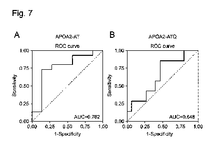
[Figure 71 Figure 7(A) shows a ROC curve showing the discrimination
performance of 15
cases of benign IPMN and 7 cases of malignant IPMN using AP0A2-AT protein.
Figure
9
Date Recue/Date Received 2023-09-22
CA 03214633 2023-09-22
7(B) shows a ROC curve showing the discrimination performance of 15 cases of
benign IPMN
and 7 cases of malignant IPMN using AP0A2-ATQ protein.
Description of Embodiments
[0015]
The target to be assayed according to the present invention is a test subject
having
pancreatic cystic tumor. In the present specification, the "pancreatic cystic
tumor" refers to
every tumor that is formed in the pancreas and exhibits the form of cyst
retaining a liquid or
mucus. Specifically, intraductal papillary mucinous neoplasm (IPMN), mucinous
cystic
neoplasm (MCN), and serous cyst neoplasm (SCN) are included therein.
[0016]
In the present specification, the "malignant pancreatic cystic tumor" refers
to a tumor
with poor prognosis derived from pancreatic cystic tumor, and can be
determined from HGD
(high-grade dysplasia), the presence of an invasive cancer, or the presence of
mural nodule.
In the present specification, the "malignant pancreatic cystic tumor" also
includes a common
type of invasive pancreatic ductal adenocarcinoma (concomitant cancer) that is
formed in the
pancreas of a pancreatic cystic tumor patient and formed at a site different
from pancreatic
cystic tumor.
[0017]
The effectiveness of a tumor marker (CA19-9) in blood has been reported for
the
prediction of such malignant pancreatic cystic tumor. However, work-up such as
pancreatic
juice cytology under ERCP or endoscopic ultrasound-guided fine needle
aspiration (EUS-
FNA) is required for definitive diagnosis (Non Patent Literature 1). When the
"malignant
pancreatic cystic tumor" is determined, treatment by resection appropriate for
the site or
spread of a lesion is discussed.
[0018]
In the present specification, the "benign pancreatic cystic tumor" is a tumor
that is low-
grade dysplasia and is confined to a local area where the tumor has developed
without
invasion to its neighboring tissues or with invasion only to a limited local
area.
Date Recue/Date Received 2023-09-22
CA 03214633 2023-09-22
[0019]
1. Method for assisting in determination of malignant pancreatic cystic tumor
using
anti-AP0A2 protein terminus antibody
The method of the present invention aims to detect malignant pancreatic cystic
tumor in
a test subject. The method of the present invention is a method for assisting
in the
determination of malignant pancreatic cystic tumor in a test subject having
pancreatic cystic
tumor, the method comprising the steps of measuring in vitro the amount of
AP0A2-AT
protein which is a polypeptide comprising the amino acid sequence represented
by SEQ ID
NO: 30 at the carboxyl terminus or/and AP0A2-ATQ protein which is a
polypeptide
comprising the amino acid sequence represented by SEQ ID NO: 31 at the
carboxyl terminus,
present in a body fluid sample obtained from the test subject, and assisting
in the
determination of the pancreatic cystic tumor of the test subject to be benign
or malignant on
the basis of the amount. An approach for measuring the AP0A2-AT protein or/and
the
AP0A2-ATQ protein is not particularly limited. A first embodiment of the
method of the
present invention is a method using an anti-AP0A2 protein terminus antibody in
the
measurement of the AP0A2-AT protein or/and the AP0A2-ATQ protein. Hereinafter,
the
anti-AP0A2 protein terminus antibody that may be used in the first embodiment
of the present
invention will be described.
[0020]
1-1. Anti-AP0A2 protein terminus antibody
In the present specification, the "AP0A2 protein" corresponds to an AP0A2
protein of
each organism species and is preferably a human-derived AP0A2 protein (GenBank
Accession No. NP 001634.1), specifically includes human-derived wild-type
AP0A2 protein
variants shown in SEQ ID NOs: 1, 2, and 3 and further includes their natural
mutants and
fragments thereof. In the present specification, the AP0A2 protein refers to a
protein whose
carboxyl-terminal 6 amino acids have an amino acid sequence represented by any
of SEQ ID
NOs: 30 to 32.
[0021]
11
Date Recue/Date Received 2023-09-22
CA 03214633 2023-09-22
In the present specification, the "variants" mean different molecular forms of
the
AP0A2 protein that may be present in the plasma, serum, or other body fluid
samples of
humans or animals. The AP0A2 protein variants correspond to, for example,
AP0A2
proteins differing in the structure of a C-terminal region, or their natural
mutants.
Specifically, the AP0A2 protein variants correspond to, for example, AP0A2-AT
protein that
is shown in SEQ ID NO: 1 and has the amino acid sequence of a C-terminal
region ending in
AT, AP0A2-ATQ protein that is shown in SEQ ID NO: 2 and the amino acid
sequence of a C-
terminal region ending in ATQ, and AP0A2-A protein that is shown in SEQ ID NO:
3 and the
amino acid sequence of a C-terminal region ending in A.
[0022]
In the present specification, the "carboxyl-terminal region (in the present
specification,
often referred to as the "C-terminal region")" refers to a region consisting
of 6 to 25 amino
acids, preferably 8 to 20 amino acids or 10 to 17 amino acids, including an
amino acid at the
carboxyl terminus (C terminus) and a few consecutive amino acids adjacent
thereto in the
amino acid sequence. In the present invention, the C-terminal region
specifically refers to a
region comprising an amino acid sequence of any of SEQ ID NOs: 30 to 32.
[0023]
In the present specification, the "natural mutant" refers to a naturally
occurring mutant
having, for example, an amino acid sequence derived from the amino acid
sequence
represented by SEQ ID NO: 1, 2, or 3 by the deletion, substitution, or
addition of one or
several amino acids, or having 90% or higher, 92% or higher, or 94% or higher,
preferably
95% or higher, more preferably 97% or higher, further preferably 98% or higher
or 99% or
higher identity to the amino acid sequence. The "identity" refers to the ratio
(%) of the
number of identical amino acid residues in one amino acid sequence to the
number of all
amino acid residues (including the number of gaps) in another amino acid
sequence when
these two amino acid sequences are aligned with or without gaps so as to
attain the largest
degree of coincidence. The term "several" refers to an integer of 2 to 10, for
example, an
integer of 2 to 7, 2 to 5, 2 to 4, or 2 or 3. Specific examples of the natural
mutant include
mutants based on polymorphisms such as SNPs (single nucleotide polymorphisms),
and
12
Date Recue/Date Received 2023-09-22
CA 03214633 2023-09-22
splicing mutants (splicing variants). The substitution is preferably
conservative amino acid
substitution. This is because the conservative amino acid substitution allows
the resulting
protein to have a structure or properties substantially equivalent to the
AP0A2 protein having
the amino acid sequence described above. The conservative amino acids refer to
the
relationship among amino acids classified into the same amino acid groups. For
example, a
nonpolar amino acid group (glycine, alanine, phenylalanine, valine, leucine,
isoleucine,
methionine, proline, and tryptophan), a polar amino acid group (amino acids
except for the
nonpolar amino acids), a charged amino acid group (acidic amino acids
(aspartic acid and
glutamic acid) and a basic amino acid group (arginine, histidine, and
lysine)), an uncharged
amino acid group (amino acids except for the charged amino acids), an aromatic
amino acid
group (phenylalanine, tryptophan, and tyrosine), a branched amino acid group
(leucine,
isoleucine, and valine), and an aliphatic amino acid group (glycine, alanine,
leucine, isoleucine,
and valine) are known as the amino acid groups.
[0024]
The "fragments thereof' refer to fragments of various AP0A2 protein variants
and their
natural mutants, comprising the C-terminal regions of the AP0A2 protein
variants and the
mutants. Specifically, the fragments thereof correspond to protease digestion
products of
various AP0A2 protein variants and their mutants.
[0025]
The method of the present invention can employ anti-AP0A2 protein terminus
antibodies including an anti-AP0A2-AT terminus antibody or/and an anti-AP0A2-
ATQ
terminus antibody. The method of the present invention can employ an anti-
AP0A2 protein
non-terminus that does not bind to the C terminus of AP0A2, in addition to the
anti-AP0A2
protein terminus antibody.
[0026]
The "anti-AP0A2-AT terminus antibody" refers to an antibody capable of
specifically
recognizing and binding to an epitope present in the C-terminal region of the
AP0A2-AT
protein, specifically, a region comprising the amino acid sequence represented
by SEQ ID NO:
30, or a binding fragment thereof. The phrase "specifically recognizing and
binding" means
13
Date Recue/Date Received 2023-09-22
CA 03214633 2023-09-22
that the antibody has no or very weak cross-reactivity with the other AP0A2
protein variants
and can thus neither recognize nor bind to or hardly recognizes and binds to
the other AP0A2
protein variants. Specifically, the anti-AP0A2-AT terminus antibody refers to
an antibody
that specifically binds to the C-terminal region of the AP0A2-AT protein, but
exhibits no
binding to the C-terminal region of the AP0A2-ATQ protein and the C-terminal
region of the
AP0A2-A protein. On the other hand, the "anti-AP0A2-ATQ terminus antibody"
refers to
an antibody capable of specifically recognizing and binding to an epitope
present in the C-
terminal region of the AP0A2-ATQ protein, specifically, a region comprising
the amino acid
sequence represented by SEQ ID NO: 31, or a fragment thereof. Specifically,
the anti-
AP0A2-ATQ terminus antibody refers to an antibody that specifically binds to
the C-terminal
region of the AP0A2-ATQ protein, but exhibits no binding to the C-terminal
region of the
AP0A2-AT protein and the C-terminal region of the AP0A2-A protein. Such an
antibody
directed to the terminus may be any of polyclonal and monoclonal antibodies or
binding
fragments thereof. A monoclonal antibody is preferred for achieving large-
scale production
and for obtaining homogeneous effects.
[0027]
The "anti-AP0A2 protein non-terminus antibody" refers to an anti-AP0A2
antibody
recognizing and binding to an epitope present in a region other than the C-
terminal region in
the full-length amino acid sequence of each AP0A2 protein variant.
Specifically, the anti-
AP0A2 protein non-terminus antibody totally differs from the anti-AP0A2
protein terminus
antibodies in epitope recognized thereby. The term "non-terminus" for the anti-
AP0A2
protein non-terminus antibody is used for the sake of convenience with respect
to the anti-
AP0A2 protein terminus antibodies. Thus, its epitope is not particularly
limited as long as
the epitope is present in a region other than the C-terminal region. The anti-
AP0A2 protein
non-terminus antibody can also include an antibody recognizing and binding to
an epitope
present in the N terminus. In the present specification, antibodies simply
referred to as "anti-
AP0A2 antibodies" refer to antibodies including both an anti-AP0A2 protein
terminus
antibody and an anti-AP0A2 protein non-terminus antibody.
[0028]
14
Date Recue/Date Received 2023-09-22
CA 03214633 2023-09-22
The method of the present invention can employ an anti-AP0A2 protein non-
terminus
antibody. The anti-AP0A2 protein non-terminus antibody is preferably an
antibody that has
almost the same levels of binding activity against an AP0A2 protein (e.g.,
AP0A2-AT)
having a certain C-terminal sequence and binding activity against an AP0A2
protein (e.g.,
AP0A2-ATQ) having a C-terminal sequence different therefrom, and does not
inhibit the
binding of the anti-AP0A2 protein terminus antibodies to the C-terminal
regions. Specific
examples thereof include an "anti-AP0A2-AT non-terminus antibody" binding to
an amino
acid sequence other than the C-terminal region of the AP0A2-AT protein shown
in SEQ ID
NO: 1, an "anti-AP0A2-ATQ non-terminus antibody" binding to an amino acid
sequence
other than the C-terminal region of the AP0A2-ATQ protein shown in SEQ ID NO:
2. In
this case, the antibodies have the same levels of binding activity against
these AP0A2 proteins,
and any of the antibodies do not inhibit the binding of the anti-AP0A2-AT
terminus antibody
and the anti-AP0A2-ATQ terminus antibody to the C-terminal regions of the
AP0A2 proteins.
The anti-AP0A2 protein non-terminus antibody may be any of polyclonal and
monoclonal
antibodies or binding fragments thereof. A monoclonal antibody is preferred
for achieving
large-scale production and for obtaining homogeneous effects.
[0029]
The "monoclonal antibody" used in the present specification refers to an
antibody that
comprises a single immunoglobulin or framework regions (hereinafter, referred
to as "FRs")
and complementarity determining regions (hereinafter, referred to as "CDRs")
and is capable
of specifically recognizing and binding to a particular antigen (epitope).
[0030]
The typical immunoglobulin molecule is a tetramer constituted by two
polypeptide
chain pairs, i.e., two heavy-light chain pairs, in which the heavy chain in
each pair is linked to
its partner light chain through a disulfide bond. Each heavy chain is composed
of a heavy
chain variable region (H chain V region; hereinafter, referred to as "VH") on
the N-terminal
side and a heavy chain constant region (H chain C region; hereinafter,
referred to as "CH") on
the C-terminal side. Each light chain is composed of a light chain variable
region (L chain V
region; hereinafter, referred to as "VL") on the N-terminal side and a light
chain constant
Date Recue/Date Received 2023-09-22
CA 03214633 2023-09-22
region (L chain C region; hereinafter, referred to as "CL") on the C-terminal
side. Of these
regions, VH and VL are particularly important because of their involvement in
the binding
specificity of the antibody. These VH and VL regions each consist of
approximately 110
amino acid residues and internally have three CDRs (CDR1, CDR2, and CDR3)
involved
directly in the binding specificity for the antigen and four FRs (FR1, FR2,
FR3, and FR4)
functioning as the backbone structures of the variable region. The CDRs are
known to be
conformationally complementary to the antigen molecule and to determine the
specificity of
the antibody (E.A. Kabat et al., 1991, Sequences of proteins of immunological
interest, Vol. 1,
eds. 5, NTH publication). The amino acid sequences of the constant regions
rarely vary
among intraspecific antibodies, whereas the amino acid sequences of the CDRs
are highly
variable among antibodies and, hence, are also called hypervariable regions.
In the variable
region, the CDRs and the FRs are arranged in the order of FR1, CDR1, FR2,
CDR2, FR3,
CDR3, and FR4 from the N terminus toward the C terminus. In the immunoglobulin
molecule, VL and VH are paired by dimerization to form an antigen-binding
site. The
immunoglobulin is known to have each class of IgG, IgM, IgA, IgE, and IgD. The
antibody
for use in the method of the present invention may be of any class. IgG is
preferred.
[0031]
The anti-AP0A2-ATQ terminus monoclonal antibody of the present invention
specifically binds to the C-terminal region of the AP0A2-ATQ protein shown in
SEQ ID NO:
2, but exhibits no binding activity against the AP0A2-AT protein shown in SEQ
ID NO: 1 and
the AP0A2-A protein shown in SEQ ID NO: 3. Specific examples of such an
antibody
include anti-AP0A2-ATQ terminus monoclonal antibody clones represented by
antibody
clone names 7F2 and 6G2. The clone 7F2 has CDR1 consisting of the sequence
represented
by SEQ ID NO: 4, CDR2 consisting of the sequence represented by SEQ ID NO: 5,
and CDR3
consisting of the sequence represented by SEQ ID NO: 6 in a heavy chain, and
CDR1
consisting of the sequence represented by SEQ ID NO: 7, CDR2 consisting of the
sequence
represented by SEQ ID NO: 8, and CDR3 consisting of the sequence represented
by SEQ ID
NO: 9 in a light chain. The clone 6G2 has CDR1 consisting of the sequence
represented by
SEQ ID NO: 10, CDR2 consisting of the sequence represented by SEQ ID NO: 11,
and CDR3
16
Date Recue/Date Received 2023-09-22
CA 03214633 2023-09-22
consisting of the sequence represented by SEQ ID NO: 12 in a heavy chain, and
CDR1
consisting of the sequence represented by SEQ ID NO: 13, CDR2 consisting of
the sequence
represented by SEQ ID NO: 14, and CDR3 consisting of the sequence represented
by SEQ ID
NO: 15 in a light chain.
[0032]
The anti-AP0A2-AT terminus monoclonal antibody of the present invention
specifically binds to the C-terminal region of the AP0A2-AT protein shown in
SEQ ID NO: 1,
but exhibits no binding activity against the AP0A2-ATQ protein shown in SEQ ID
NO: 2 and
the AP0A2-A protein shown in SEQ ID NO: 3. Specific examples of such an
antibody
include anti-AP0A2-AT terminus monoclonal antibody clones represented by
antibody clone
names 4C6-1, 5D9-3, and 6B4-2. The clone 4C6-1 has CDR1 consisting of the
sequence
represented by SEQ ID NO: 33, CDR2 consisting of the sequence represented by
SEQ ID NO:
34, and CDR3 consisting of the sequence represented by SEQ ID NO: 35 in a
heavy chain, and
CDR1 consisting of the sequence represented by SEQ ID NO: 36, CDR2 consisting
of the
sequence represented by SEQ ID NO: 37, and CDR3 consisting of the sequence
represented
by SEQ ID NO: 38 in a light chain. The clone 5D9-3 has CDR1 consisting of the
sequence
represented by SEQ ID NO: 39, CDR2 consisting of the sequence represented by
SEQ ID NO:
40, and CDR3 consisting of the sequence represented by SEQ ID NO: 41 in a
heavy chain, and
CDR1 consisting of the sequence represented by SEQ ID NO: 42, CDR2 consisting
of the
sequence represented by SEQ ID NO: 43, and CDR3 consisting of the sequence
represented
by SEQ ID NO: 44 in a light chain. The clone 6B4-2 has CDR1 consisting of the
sequence
represented by SEQ ID NO: 45, CDR2 consisting of the sequence represented by
SEQ ID NO:
46, and CDR3 consisting of the sequence represented by SEQ ID NO: 47 in a
heavy chain, and
CDR1 consisting of the sequence represented by SEQ ID NO: 48, CDR2 consisting
of the
sequence represented by SEQ ID NO: 49, and CDR3 consisting of the sequence
represented
by SEQ ID NO: 50 in a light chain.
[0033]
The anti-AP0A2 protein non-terminus antibody of the present invention is
preferably
an antibody having the same levels of binding activity against the AP0A2
protein variants
17
Date Recue/Date Received 2023-09-22
CA 03214633 2023-09-22
shown in SEQ ID NOs: 1 to 3 when the binding activity is compared among them.
Specific
examples thereof include anti-AP0A2 antibody clones represented by antibody
clone names
MAB1 and MAB2. The clone MAB1 has CDR1 consisting of the amino acid sequence
represented by SEQ ID NO: 16, CDR2 consisting of the amino acid sequence
represented by
SEQ ID NO: 17, and CDR3 consisting of the amino acid sequence represented by
SEQ ID
NO: 18 in a heavy chain, and CDR1 consisting of the amino acid sequence
represented by
SEQ ID NO: 19, CDR2 consisting of the amino acid sequence represented by SEQ
ID NO: 20,
and CDR3 consisting of the amino acid sequence represented by SEQ ID NO: 21 in
a light
chain. The clone MAB2 has CDR1 consisting of the amino acid sequence
represented by
SEQ ID NO: 22, CDR2 consisting of the amino acid sequence represented by SEQ
ID NO: 23,
and CDR3 consisting of the amino acid sequence represented by SEQ ID NO: 24 in
a heavy
chain, and CDR1 consisting of the amino acid sequence represented by SEQ ID
NO: 25,
CDR2 consisting of the amino acid sequence represented by SEQ ID NO: 26, and
CDR3
consisting of the amino acid sequence represented by SEQ ID NO: 27 in a light
chain. The
anti-AP0A2-ATQ non-terminus antibody or the anti-AP0A2-AT non-terminus
antibody can
be used as the anti-AP0A2 protein non-terminus antibody.
[0034]
The "binding fragments thereof' for the "polyclonal and monoclonal antibodies
or
binding fragments thereof' are partial fragments of the polyclonal and
monoclonal antibodies
having an epitope in a C-terminal region of the AP0A2 protein or a region
other than the C
terminus, and are polypeptide chains having activity substantially equivalent
to the antigen-
specific binding activity of the antibodies, or complexes thereof. The binding
fragments each
correspond to an antibody portion comprising at least one antigen-binding
site, i.e., a
polypeptide chain having at least one VL-VH pair, or a complex thereof.
Specific examples
thereof include a large number of sufficiently characterized antibody
fragments resulting from
the cleavage of an immunoglobulin with various peptidases. More specific
examples thereof
include Fab, F(ab')2, and Fab'. The Fab is a fragment resulting from the
papain cleavage of
the IgG molecule on the N-terminal side of the disulfide bonds in the hinges
and is constituted
by a polypeptide consisting of VH and CH 1, which is adjacent to the VH, among
the three
18
Date Recue/Date Received 2023-09-22
CA 03214633 2023-09-22
CH-constituting domains (CH1, CH2, and CH3), and a light chain. The F(ab')2 is
a Fab'
dimer resulting from the pepsin cleavage of the IgG molecule on the C-terminal
side of the
disulfide bonds in the hinges. The Fab' is substantially structurally
equivalent to Fab, though
being slightly longer at H chain than Fab by including a hinge (Fundamental
Immunology,
Paul ed., 3rd ed., 1993). The Fab' can be obtained by reducing F(ab')2 under
mild conditions
and cleaving the disulfide bridges in the hinge region. All of these antibody
fragments
comprise the antigen-binding site and have the ability to specifically bind to
the antigen (i.e., a
particular AP0A2 protein variant in the present invention).
[0035]
The binding fragment of the antibody for use in the method of the present
invention
may be synthesized chemically or by use of a recombinant DNA method. Examples
thereof
include antibody fragments newly synthesized using the recombinant DNA method.
Specifically, the fragment corresponds to, but is not limited to, a monomeric
polypeptide
molecule in which one or more VLs and one or more VHs of the antibody for use
in the
method of the present invention are artificially linked via a linker peptide
or the like having an
appropriate length of a sequence, or a multimeric polypeptide thereof.
Examples of such a
polypeptide include synthetic antibodies such as single-chain Fv (scFv: single
chain fragment
of variable region) (see Pierce catalog and Handbook, 1994-1995, Pierce
Chemical co.,
Rockford, IL), diabody, triabody, and tetrabody. In the immunoglobulin
molecule, VL and
VH are normally positioned on separate polypeptide chains (L chain and H
chain). The
single-chain Fv is a synthetic antibody fragment having a structure where
these variable
regions are linked via a flexible linker having a sufficient length such that
the VL and the VH
are comprised in one polypeptide chain. Both of the variable regions in the
single-chain Fv
are self-assembled with each other to form one functional antigen-binding
site. The single-
chain Fv can be obtained by integrating a recombinant DNA encoding the single-
chain Fv into
the phage genome using a technique known in the art, followed by expression.
The diabody
is a molecule having a structure based on the dimeric structure of the single-
chain Fvs
(Holliger et al., 1993, Proc. Natl. Acad. Sci USA, 90: 6444-6448). For
example, when the
linker has a length shorter than approximately 12 amino acid residues, the two
variable sites in
19
Date Recue/Date Received 2023-09-22
CA 03214633 2023-09-22
the single-chain Fv cannot be self-assembled. By contrast, VL in one Fv chain
can be
assembled with VH in another Fv chain by the formation of the diabody, i.e.,
by the interaction
between the two single-chain Fvs. As a result, two functional antigen-binding
sites can be
formed (Marvin et al., 2005, Acta Pharmacol. Sin., 26: 649-658). The further
addition of
cysteine residues to the C termini of the single-chain Fvs permits a disulfide
bond between
these two Fv chains so that stable diabody can be formed (Alafsen et al.,
2004, Prot. Engr. Des.
Sel., 17: 21-27). Although the diabody is a divalent antibody fragment as
described above,
its antigen-binding sites do not have to bind to the same epitope and may have
bispecificity of
recognizing and specifically binding to different epitopes, respectively. The
triabody or the
tetrabody has a trimeric or tetrameric structure based on the single-chain Fv
structure, as in the
diabody. The triabody and the tetrabody are trivalent and quadrivalent
antibody fragments,
respectively, and may each be a multispecific antibody. The antibody fragment
for use in the
method of the present invention further includes antibody fragments identified
using a phage
display library (see e.g., McCafferty et al., 1990, Nature, Vol. 348, 522-
554), wherein these
antibody fragments have the ability to bind to their antigens. Also see, for
example, Kuby, J.,
Immunology, 3rd Ed., 1998, W.H. Freeman & Co., New York.
[0036]
In the present invention, each anti-AP0A2 antibody can be modified. In this
context,
the modification includes any of functional modifications (e.g.,
glycosylation) required for the
anti-AP0A2-ATQ terminus antibody to have specific binding activity against the
particular
AP0A2 protein variant, and labeling necessary for detecting the antibody for
use in the
method of the present invention. Examples of the antibody labeling include
labeling with
fluorescent dyes (FITC, rhodamine, Texas Red, Cy3, and Cy5), fluorescent
proteins (e.g., PE,
APC, and GFP), enzymes (e.g., horseradish peroxidase, alkaline phosphatase,
and glucose
oxidase), or biotin or (strept)avidin. The antibody glycosylation may be
altered in order to
adjust the affinity of the antibody for the antigen. Such alteration can be
achieved, for
example, by changing one or more glycosylation sites in the antibody sequence.
To be more
specific, one or more amino acid substitutions can be introduced to an amino
acid sequence
constituting one or more glycosylation sites, for example, in FR, to remove
the glycosylation
Date Recue/Date Received 2023-09-22
CA 03214633 2023-09-22
sites. As a result, the glycosylation of the sites can be canceled. Such
deglycosylation is
effective for increasing the affinity of the antibody for the antigen (U.S.
Patent Nos. 5714350
and 6350861).
[0037]
1-2. Preparation of immunogen
In preparing the anti-AP0A2 protein terminus antibodies in the present
invention, each
AP0A2 protein variant is prepared as an immunogen (antigen). Examples of the
AP0A2
protein variant that can be used as an immunogen in the present invention
include AP0A2
proteins having the amino acid sequence represented by SEQ ID NOs: 1 to 3 and
mutants
thereof, and polypeptide fragments of the proteins or the mutants, and their
fusion
polypeptides with other peptides (e.g., signal peptides and labeling
peptides). The AP0A2
protein variant as an immunogen can be synthesized by an approach known in the
art, for
example, a solid-phase peptide synthesis method, for example, using
information on the amino
acid sequence represented by SEQ ID NOs: 1 to 3. The AP0A2 protein variant can
be
prepared by, for example, a method given below.
[0038]
Any of naturally occurring AP0A2 proteins, recombinant AP0A2 proteins, and
synthetic AP0A2 proteins, the whole or a portion of which has been chemically
synthesized
can be used as the AP0A2 protein variant. For example, a variant derived from
any of
naturally occurring AP0A2 proteins, recombinant AP0A2 proteins, and synthetic
AP0A2
proteins can be used as an antigenic AP0A2 protein variant that is prepared in
order to obtain
each antibody binding to the AP0A2 protein C terminus (anti-AP0A2 protein
terminus
antibody) as long as the variant comprises an amino acid sequence consisting
of 6 or more
consecutive amino acids of the C-terminal region.
[0039]
The naturally occurring AP0A2 proteins can be recovered from samples including
body fluid samples such as blood (including plasma and serum), or culture
supernatants of
cultured cells by use of a protein separation and purification technique known
in the art, for
example, gel filtration, ion-exchange chromatography, or affinity
chromatography.
21
Date Recue/Date Received 2023-09-22
CA 03214633 2023-09-22
[0040]
The recombinant AP0A2 proteins can be expressed in microbes, insect cells, or
animal
cells harboring DNAs encoding the proteins and then recovered from the cells
by use of a
protein separation and purification technique known in the art.
[0041]
The synthetic AP0A2 proteins can be synthesized by an approach known in the
art, for
example, a solid-phase peptide synthesis method, for example, using published
information on
the amino acid sequence of the AP0A2 protein. These synthetic AP0A2 proteins
may each
be linked to a carrier protein such as KLH (keyhole limpet hemocyanin), OVA
(ovalbumin),
or BSA (bovine serum albumin).
[0042]
In the case of using a fragment of the AP0A2 protein variant as an immunogen
in the
anti-AP0A2 protein terminus antibody preparation, any of naturally occurring
AP0A2 protein
fragments, recombinant AP0A2 protein fragments, and synthetic AP0A2 protein
fragment
may also be used. For example, an oligopeptide or a polypeptide comprising 6
or more,
preferably 10 or more, more preferably 18 or more, further preferably 30 or
more consecutive
amino acid residues including the C terminus of an amino acid sequence
represented by any of
SEQ ID NOs: 1 to 3 can be used as the AP0A2 protein fragment serving as an
antigen. For
example, a peptide comprising the amino acid sequences represented by SEQ ID
NO: 28 or 29
can be used.
[0043]
In the case of using a fragment of the naturally occurring AP0A2 protein as an
immunogen, for example, in the anti-AP0A2 protein terminus antibody
preparation, the
purified AP0A2 protein is first treated with suitable protease such as trypsin
and then
fractionated on a reverse-phase column to obtain peaks. Subsequently, the
amino acid
sequence of the peptide comprised in each peak is determined with a mass
spectrometer. The
peptide, when comprising, as a partial sequence, a sequence consisting of 6 or
more
consecutive amino acids of the C-terminal region of the AP0A2 protein shown in
any of SEQ
ID NOs: 1 to 3, can be used as the immunogen.
22
Date Recue/Date Received 2023-09-22
CA 03214633 2023-09-22
[0044]
In the case of using a partial amino acid sequence of the recombinant AP0A2
protein
as an immunogen, for example, in the anti-AP0A2 protein terminus antibody
preparation, a
DNA sequence encoding a peptide (C-terminal fragment) consisting of a partial
sequence of 6
or more consecutive amino acids including the C-terminal amino acid residues
of the AP0A2
protein shown in any of SEQ ID NOs: 1 to 3 is first inserted to vectors for
expressions. Next,
the vectors for expressions are transferred to various cells to express the
encoded C-terminal
fragment. Finally, the C-terminal fragment is extracted from the cells after
the expression
according to a routine method. The obtained C-terminal fragment can be used as
the
immunogen.
[0045]
Also in the case of preparing the anti-AP0A2 protein non-terminus antibody in
the
present invention, its preparation method can be basically the same as the
method for
preparing the anti-AP0A2 protein terminus antibodies except that a region that
can be used as
an immunogen in the AP0A2 protein is different from the regions used as an
immunogen for
preparing the anti-AP0A2 protein terminus antibodies. Specifically, the whole
or a portion
of a region other than the C-terminal region of the AP0A2 protein can be used
as an
immunogen. In the case of preparing the anti-AP0A2 protein non-terminus
antibody, as in
the case of preparing the anti-AP0A2 protein terminus antibodies, an
oligopeptide or a
polypeptide comprising amino acid residues of the region other than the C-
terminal region of
the AP0A2 protein can also be used as an antigen.
[0046]
(Preparation of recombinant AP0A2 protein)
Hereinafter, the preparation of a recombinant AP0A2 protein (recombinant AP0A2
protein variant) shown in any of SEQ ID NOs: 1 to 3 will be described in
detail.
[0047]
(a) Preparation of polynucleotide encoding recombinant AP0A2 protein variant
Phages or plasmids capable of autonomously replicating in host microbes can be
used
as vectors for use in the expression of various AP0A2 protein variants.
Examples of the
23
Date Recue/Date Received 2023-09-22
CA 03214633 2023-09-22
plasmids include E. coil-derived plasmids (pET30a, pGEX6p, pUC118, pUC119,
pUC18,
pUC19, etc.), Bacillus subtilis-derived plasmids (pUB110, pTP5, etc.), and
enzyme-derived
plasmids (YEp13, YEp24, YCp50, etc.). Examples of the phages include X, phages
(kgt11,
?ZAP, etc.). In addition, vectors of animal viruses such as vaccinia virus or
insect viruses
such as baculovirus can also be used.
[0048]
The method for inserting a polynucleotide encoding the AP0A2 protein variant
to the
vectors involves, for example, cleaving the purified polynucleotide with
corresponding
appropriate restriction enzymes and ligating the resulting fragment into the
vectors cleaved
with the appropriate restriction enzymes by use of DNA ligase or the like.
[0049]
(b) Transfer of AP0A2 protein variant expression vector into host
The obtained AP0A2 protein variant expression vectors are transferred to hosts
capable
of expressing the expression vectors to obtain transformants capable of
expressing the AP0A2
protein variant (variant-expressing transformants). The hosts used are not
particularly limited
as long as the hosts are suitable for the vectors used and are capable of
expressing the AP0A2
protein variant. For example, bacteria (E. coil (Escherichia coil), Bacillus
subtilis, yeasts,
insect cells, or animal cells (COS cells and CHO cells (Journal of immunology,
1998, Vol. 160,
3393-3402)) are preferably used. The method for transferring the vectors to
the bacteria is
not particularly limited as long as the method is a method known in the art
for transferring the
vectors to the bacteria. Examples thereof include a heat shock method, a
method using
calcium ions, and electroporation. All of these techniques are known in the
art and described
in various literatures. See, for example, Greene & Sambrook, 2012, Molecular
Cloning: A
Laboratory Manual Fourth Ed., Cold Spring Harbor Laboratory Press, Cold Spring
Harbor,
New York. A Lipofectin method (PNAS, 1989, Vol. 86, 6077; and PNAS, 1987, Vol.
84,
7413), electroporation, a calcium phosphate method (Virology, 1973, Vol. 52,
456-467), a
DEAE-dextran method or the like is preferably used in the transformation of
the animal cells.
[0050]
24
Date Recue/Date Received 2023-09-22
CA 03214633 2023-09-22
In the case of using bacteria as the hosts, preferably, the AP0A2 protein
variant
expression vectors are capable of autonomously replicating in the bacteria and
are also
constituted by a promoter sequence, a ribosomal binding sequence, the DNA
sequence
encoding the AP0A2 protein variant, and a transcription termination sequence.
The
expression vectors may also comprise a gene encoding a regulatory factor
controlling the
promoter. Any promoter that can function in the hosts such as E. coil may be
used.
[0051]
Likewise, in the case of using eukaryotic cells such as yeasts, animal cells,
or insect
cells as the hosts, AP0A2 protein variant-expressing transformants can also be
obtained
according to an approach known in the art. The AP0A2 protein variant
expression vectors
for use in the eukaryotic cells comprise a promoter sequence and the DNA
sequence encoding
the AP0A2 protein variant, which may be linked, if desired, to a cis element
(e.g., an
enhancer), a splicing signal (a donor site, an acceptor site, a branch point,
etc.), a poly-A
addition signal, a selective marker sequence, a ribosomal binding sequence (SD
sequence),
and the like.
[0052]
(c) Culture of variant-expressing transformant and expression of recombinant
AP0A2
protein variant
Subsequently, the prepared variant-expressing transformants are cultured. The
method for culturing the variant-expressing transformants in a medium is
carried out according
to an ordinary method for use in the culture of the hosts. In the case of
using, for example,
bacteria as the hosts, the medium is not particularly limited as long as the
medium comprises a
carbon source, a nitrogen source, inorganic salts, etc., utilizable by the
bacteria and the
bacteria are capable of growing or proliferating in the medium. Any of natural
and synthetic
media can be used. More specific examples thereof include an LB medium, but
are not
limited thereto, as a matter of course. For the selective culture of the
variant-expressing
transformants, an antibiotic such as ampicillin or tetracycline may be added
to the medium, if
necessary. The culture is usually carried out at 37 C for 6 to 24 hours under
aerobic
conditions such as culture with aeration and stirring. During the culture
period, the pH is
Date Recue/Date Received 2023-09-22
CA 03214633 2023-09-22
preferably kept at or around a neutral pH. The pH is adjusted using an
inorganic or organic
acid or alkali solution or the like. When the variant-expressing transformants
are animal cells
such as CHO cells, the host cells can be inoculated at 1 x 10⁵ cells/mL to a
DMEM medium
manufactured by Life Technologies Corp. (currently known as Thermo Fisher
Scientific Inc.)
and cultured in a 5% CO2 incubator of 37 C. During the culture, an antibiotic
such as
ampicillin or tetracycline may be added to the medium, if necessary.
[0053]
When the AP0A2 protein variant expression vectors are protein expression
induction-
type vectors comprising a protein expression control system (which corresponds
to, for
example, a repressor gene and an operator for the host bacteria), the
expression of the AP0A2
protein variant needs to be induced by a predetermined treatment of the
variant-expressing
transformants. The expression induction method differs depending on the
protein expression
control system comprised in the vectors. Therefore, induction treatment
suitable for the
system can be carried out. For example, the protein expression control system
most generally
used in the protein expression induction-type vectors for use in the host
bacteria is a system
consisting of a lac repressor gene and a lac operator. This system is capable
of inducing the
expression by IPTG (isopropy1-1-thio-3-D-galactoside) treatment. The
transformants having
the AP0A2 protein expression vectors comprising this system can be allowed to
express the
AP0A2 protein variant of interest by the addition of IPTG in an appropriate
amount (e.g., final
concentration: 1 mM) into the medium.
[0054]
(d) Extraction and/or recovery of recombinant AP0A2 protein variant
When the AP0A2 protein variant is produced inside the bacterial bodies or the
cells
after the culture, the bacterial bodies or the cells can be recovered and
disrupted, followed by
the extraction of the protein of interest. When the AP0A2 protein variant is
secreted outside
the bacterial bodies or the cells, the culture solution can be used directly,
or the supernatant
obtained by the removal of the bacterial bodies or the cells through
centrifugation or the like
can be used. Then, the AP0A2 protein variant can be isolated and purified from
the cultures
by using, alone or in appropriate combination, general protein purification
methods, for
26
Date Recue/Date Received 2023-09-22
CA 03214633 2023-09-22
example, ammonium sulfate precipitation, gel filtration, ion-exchange
chromatography, and
affinity chromatography. Whether or not the AP0A2 protein variant has been
obtained can
be confirmed by SDS-polyacrylamide gel electrophoresis or the like.
[0055]
1-3. Preparation of anti-AP0A2 monoclonal antibody
1-3-1. Methods for preparing anti-AP0A2 monoclonal antibody and hybridoma
Hybridomas producing the anti-AP0A2 monoclonal antibody of the present
invention
can be prepared by a method described below. However, the preparation method
is not
limited thereto, and any of other methods known in the art can be used for the
preparation.
[0056]
(1) Method for preparing anti-AP0A2 monoclonal antibody
In order to prepare the anti-AP0A2 protein terminus monoclonal antibody
specifically
binding to the C-terminal region of any AP0A2 protein shown in SEQ ID NO: 1,
2, or 3 in the
amino acid sequence constituting the AP0A2 protein, monoclonal antibodies can
be prepared
with the AP0A2 protein variant or a peptide comprising the C-terminal region
of the AP0A2
protein variant as an immunogen and then screened for an antibody binding only
to the
particular AP0A2 protein variant using the AP0A2 protein shown in any of SEQ
ID NOs: 1
to 3 or the peptide comprising the C-terminal region of the AP0A2 protein
variant. For
example, the anti-AP0A2-ATQ terminus monoclonal antibody can be selected by
screening
using, as an index, specific binding to the C-terminal region (region
comprising the amino acid
sequence of SEQ ID NO: 31) of the AP0A2-ATQ protein shown in SEQ ID NO: 2
without or
almost without binding to the AP0A2 protein variant shown in SEQ ID NO: 1 or
3. Also,
the anti-AP0A2-AT terminus monoclonal antibody can be selected by screening
using, as an
index, specific binding to the C-terminal region (region comprising the amino
acid sequence of
SEQ ID NO: 30) of the AP0A2-AT protein shown in SEQ ID NO: 1 without or almost
without binding to the AP0A2 protein variant shown in SEQ ID NO: 2 or 3.
[0057]
In order to prepare the anti-AP0A2 protein non-terminus antibody recognizing
amino
acids other than the C-terminal region of the AP0A2 protein, monoclonal
antibodies can be
27
Date Recue/Date Received 2023-09-22
CA 03214633 2023-09-22
prepared with the AP0A2 protein variant or a peptide comprising a partial
sequence thereof as
an immunogen and then screened for the antibody of interest by using, as an
index, the same
levels of binding activity against the AP0A2 protein variants shown in SEQ ID
NOs: 1 to 3 or
their peptides differing in C terminus when the binding activity is compared
among them.
[0058]
(2) Preparation of anti-AP0A2 protein terminus antibody-producing cell
The recombinant AP0A2 protein obtained as an immunogen in the paragraph 1-2 is
dissolved in a buffer solution to prepare an immunogen solution. For effective
immunization,
an adjuvant may be added thereto, if necessary. Examples of the adjuvant
include
commercially available Freund's complete adjuvant (FCA) and Freund's
incomplete adjuvant
(FIA). These adjuvants may be used alone or as a mixture.
[0059]
Next, a mammal, for example, a rat, a mouse (e.g., an inbred mouse BALB/c), or
a
rabbit is immunized by the administration of the prepared immunogen solution.
Examples of
the immunogen administration method include, but are not limited to,
subcutaneous injection
using FIA or FCA, intraperitoneal injection using FIA, and intravenous
injection using 0.15 M
sodium chloride. One dose of the immunogen is appropriately determined
according to the
type of the animal to be immunized, an administration route, etc., and is
approximately 50 to
200 lig per animal. The intervals between the immunization shots are not
particularly limited.
After the priming, 2 to 6, preferably 3 or 4 boosters are performed at
intervals of a few days to
a few weeks, preferably at 1- to 4-week intervals. After the priming, an
antibody titer in the
serum of the immunized animal is measured by ELISA (enzyme-linked
immunosorbent assay)
or the like. Provided that a sufficient rise in antibody titer is shown, the
immunogen is
intravenously or intraperitoneally injected for final immunization. 2 to 5
days, preferably 3
days, after the final immunization date, antibody-producing cells are
collected.
[0060]
1-3-2. Method for preparing anti-AP0A2 monoclonal antibody-producing hybridoma
(1) Recovery of antibody-producing cell from immunized animal and cell fusion
28
Date Recue/Date Received 2023-09-22
CA 03214633 2023-09-22
The antibody-producing cells obtained from the immunized animal can be
subjected to
cell fusion with myeloma cells to prepare hybridomas producing the monoclonal
antibody
specifically recognizing the particular region of the AP0A2 protein. Examples
of the
antibody-producing cells include spleen cells, lymph node cells, and
peripheral blood cells.
Spleen cells or local lymph node cells are preferred. A generally available
established cell
line derived from mice or the like can be used as the myeloma cells for fusion
with the
antibody-producing cells. The cell line used preferably has drug selectivity
and has the
property of being unable to survive in an unfused state in a HAT selective
medium
(comprising hypoxanthine, aminopterin, and thymidine) and being able to grow
therein only in
a state fused with the antibody-producing cells. Also, the established cell
line is preferably
derived from an animal of the same species as the immunized animal. Specific
examples of
the myeloma cells include BALB/c mouse-derived hypoxanthine-guanine-
phosphoribosyl-
transferase (HGPRT)-deficient cell lines P3X62-Ag.8 (ATCCTIB9), P3X63-Ag.8.U1
(JCRB9085), P3/NSI/1-Ag4-1 (JCRB0009), P3x63Ag8.653 (JCRB0028), and 5P2/0-Ag14
(JCRB0029).
[0061]
For the cell fusion between the myeloma cells and the antibody-producing
cells, the
antibody-producing cells and the myeloma cells are mixed at a ratio of
approximately 1:1 to
20:1 in a serum-free medium for animal cell culture, such as a DMEM or
RPMI1640 medium,
and fused with each other through reaction in the presence of a cell fusion
promoter. For
example, polyethylene glycol having an average molecular weight of 1,500 to
4,000 Da can be
used as the cell fusion promoter at a concentration of approximately 10 to
80%. If necessary,
an auxiliary such as dimethyl sulfoxide may be used in combination therewith
for enhancing
fusion efficiency. Alternatively, the antibody-producing cells and the myeloma
cells may be
fused with each other using a commercially available cell fusion apparatus
that employs
electric stimulation (e.g., electroporation) (Nature, 1977, Vol. 266, 550-
552).
[0062]
(2) Selection of hybridoma of interest
29
Date Recue/Date Received 2023-09-22
CA 03214633 2023-09-22
Examples of the method for selecting hybridomas producing the anti-AP0A2
monoclonal antibody of interest from the cells after the cell fusion treatment
include the
following method. The cell suspension is appropriately diluted with, for
example, an
RPMI1640 medium comprising fetal bovine serum and then seeded at approximately
2 x 10⁶
cells/well over a 96-well microtiter plate. A selective medium is added to
each well where
the cells are subsequently cultured with the selective medium appropriately
replaced with a
fresh one. The culture temperature is set to 20 to 40 C, preferably
approximately 37 C.
When the myeloma cells are of HGPRT-deficient line or thymidine kinase (TK)-
deficient line,
only hybridomas of the antibody-producing cells and the myeloma cells can be
selectively
allowed to grow or proliferate by use of a selective medium comprising
hypoxanthine,
aminopterin, and thymidine (HAT medium). Therefore, cells grown from
approximately 10
days after the start of culture in the selective medium can be selected as the
hybridomas.
[0063]
Antibodies produced by the hybridomas selected in the HAT medium are first
screened
by using binding activity against various AP0A2 protein variants shown in SEQ
ID NOs: 1 to
3 as an index. Subsequently, the antibody having binding activity is tested
for cross-
reactivity to select acceptable ones. The "acceptable ones (cross-reactivity)"
mean cross-
reactivity at a negligible level for the intended purposes of the antibody.
For example, a
monoclonal antibody for use in immunological assay can be interpreted as
having practically
no cross-reactivity when signal intensity from cross reaction in a final assay
system can be
suppressed at a background level to less than 1% of signal intensity from
specific reaction.
[0064]
For example, ELISA can be used for confirming reaction specificity for the
particular
AP0A2 protein variant. In this ELISA method, a microplate in which various
AP0A2
protein variants or fragments thereof are separately immobilized as antigens
on different wells
is prepared and reacted by the addition of appropriately diluted samples of
the culture
supernatant of the hybridomas. After sufficient reaction, the wells are washed
and further
reacted by the addition of a labeled form of a secondary antibody directed to
an
immunoglobulin. The wells are washed again and can be finally assayed by use
of the label
Date Recue/Date Received 2023-09-22
CA 03214633 2023-09-22
of the secondary antibody bound with the wells to quantitatively determine the
binding activity
of the antibody present in the culture supernatant against the antigens. For
example, for the
preparation of the anti-AP0A2 protein terminus monoclonal antibody, the
specificity can be
judged by the presence of binding activity only against the C-terminal region
of the particular
AP0A2 protein variant without cross-reactivity with the other AP0A2 protein
variants. For
the preparation of the anti-AP0A2 protein non-terminus monoclonal antibody,
the antibody is
selected by using, as an index, the same levels of binding activity against
all of the AP0A2
protein variants differing in C terminus without the inhibition of the binding
of the anti-
AP0A2 protein terminus monoclonal antibody to the C-terminal region by the
prepared
antibody.
[0065]
The hybridomas can also be selected by use of a recombinant DNA technique.
First,
mRNAs are extracted from the hybridoma group obtained according to the
aforementioned
method. The mRNA extraction can be carried out by use of a method known in the
art.
Subsequently, cDNAs are obtained from the mRNAs using Oligo dT primers or
random
primers. The cDNAs are used as templates in PCR using primer sets comprising
the
nucleotide sequence of the signal sequence upstream of the variable region-
encoding gene and
a nucleotide sequence on the constant region side. The obtained amplification
products can
be inserted to appropriate cloning vectors and cloned to obtain a library of
the variable region
genes of the antibodies produced by the hybridomas. As a more specific non-
limiting
example, PCR is carried out using Mouse Ig Primer provided by Merck Millipore,
and the
amplification products (mouse immunoglobulin variable region cDNAs) are
inserted to the
EcoRI sites of ZERO BLUNT PCR TOPO Vectors provided by Life Technologies Corp.
(currently known as Thermo Fisher Scientific, Inc.) and cloned. The obtained
vector group
can be used as a library of the genes encoding the variable region amino acid
sequences.
Next, a probe is designed on the basis of the amino acid sequence of each
variable region or
each CDR disclosed in the present invention. The library can be screened for
positive clones
to select the hybridomas producing the antibody for use in the method of the
present invention.
[0066]
31
Date Recue/Date Received 2023-09-22
CA 03214633 2023-09-22
(3) Antibody production using hybridoma
The hybridomas according to the present invention can be used in antibody
production
by ascites formation using a mouse. Specifically, the hybridomas are
intraperitoneally
inoculated to a mouse of the origin of the fusion partner cells used for
preparing the
hybridomas, or to a nude mouse. The ascites can be appropriately collected to
recover an
antibody-comprising ascites fluid. More specifically, hybridomas obtained with
5P2/0 cells
as a fusion partner are intraperitoneally inoculated to a BALB/c mouse 10 days
after
inoculation with pristine, and an antibody-comprising ascites fluid can be
recovered.
[0067]
The hybridomas according to the present invention can be used in antibody
production
by culture using a suitable medium. Specifically, the hybridomas can be
inoculated at 1 x
105 cells/mL into a Hybridoma-SFM medium manufactured by Life Technologies
Corp.
(currently known as Thermo Fisher Scientific, Inc.) and cultured in a 5% CO2
incubator of
37 C until the hybridomas are killed to obtain an antibody-comprising culture
supernatant,
though the antibody production method according to the present invention is
not limited
thereto.
[0068]
(4) Method for preparing recombinant anti-AP0A2 monoclonal antibody or
fragment
thereof by recombinant DNA manipulation
The antibody for use in the present invention can also be obtained by
recombinant
DNA manipulation using a cDNA sequence encoding the amino acid sequence of the
antibody.
[0069]
Nucleotide sequences encoding the amino acid sequences of the variable regions
in the
antibody derived from an anti-AP0A2 monoclonal antibody-producing hybridoma,
for
example, the anti-AP0A2 protein terminus monoclonal antibody-producing
hybridoma
obtained by the approach described in the paragraph "1-3-2(2)", are used.
These nucleotide
sequences of VH and VL are linked to nucleotide sequences encoding arbitrary
human CH and
human CL, respectively, and the resulting polynucleotides can be incorporated
into
appropriate expression vectors, which are then transferred to host cells,
followed by expression
32
Date Recue/Date Received 2023-09-22
CA 03214633 2023-09-22
as a complete immunoglobulin molecule. Alternatively, according to a CDR
grafting
antibody technique, polynucleotides encoding the respective amino acid
sequences of the CDR
sequences in the amino acid sequences of the variable regions in the antibody
derived from the
anti-AP0A2 protein terminus monoclonal antibody-producing hybridoma obtained
by the
approach described in the paragraph "1-3-2(2)" may be linked to
polynucleotides encoding the
amino acid sequences of human FR sequences in a predetermined order, and the
resultant can
be incorporated into appropriate expression vectors, which are then
transferred to host cells,
followed by expression as a complete immunoglobulin molecule. In this respect,
an
approach is convenient in which the heavy chain and the light chain to be
paired can be
expressed in the same host cell and produced as a heavy chain/light chain
dimer. Specifically,
each cell is cotransfected with, for example, the light chain expression
vector and the heavy
chain expression vector, and the antibody according to the present invention
can also be
obtained from this transformed cell. Alternatively, polynucleotides encoding
the amino acid
sequences of the variable regions described above may be incorporated directly
to appropriate
expression vectors, which are then transferred to host cells, followed by
expression as
fragments of the immunoglobulin molecule.
Alternatively, as mentioned above,
polynucleotides respectively encoding VL and VH or the light chain and the
heavy chain
comprising the amino acid sequences may be linked via a nucleotide sequence
encoding an
appropriate linker, then incorporated to phages, and expressed as single-chain
Fv or as a
synthetic antibody fragment such as diabody. In addition, according to a
recently developed
phage display antibody technique (Brinkmann et al., 1995, J. Immunol. Methods,
182, 41-50;
and International Publication Nos. W097/13844 and W090-02809), which involves
expressing recombinant antibodies on phage surface by exploiting a gene
engineering
technique, single-chain Fv antibodies diversified by artificially shuffling
genes encoding
heavy and light chains can be expressed as phage-fusion proteins to obtain
specific antibodies.
[0070]
The methods for preparing the polynucleotide encoding the recombinant anti-
AP0A2
antibody or the fragment thereof, preparing vectors carrying the
polynucleotide, and
transferring the vectors to hosts can be carried out by use of a recombinant
DNA technique
33
Date Recue/Date Received 2023-09-22
CA 03214633 2023-09-22
known in the art. The recombinant anti-AP0A2 protein antibody of interest or
the fragment
thereof can be obtained from the culture solution of the transformed cells or
from the inside of
the cells.
[0071]
For example, plasmids, phagemids, cosmids, or virus vectors (e.g., SV40 virus
based
vector, EB virus based vector, and BPV based vector) can be used as
immunoglobulin
expression vectors, though the vectors are not limited thereto. For example, a
BCMGS Neo
vector, which is a BPV based vector, is a desirable vector for efficient
expression of a foreign
gene by the transformation of COS7 cells or the like therewith (Hajime
Karasuyama, "Bovine
papilloma virus vector", Masami Muramatsu and Hiroto Okayama, ed.,
Experimental
Medicine, Suppl.: Handbook of Gene Engineering, 1991, Yodosha Co., Ltd., 297-
299).
[0072]
The vectors can comprise control elements (e.g., a promoter, an enhancer, a
terminator,
a polyadenylation site, and a splicing site) necessary for expressing the
antibody or the
fragment thereof, or an optional selective marker, in addition to the
polynucleotide encoding
the antibody or the fragment thereof.
[0073]
The hosts described in the paragraph "1-2. Preparation of immunogen" as well
as SP2/0
(mouse myeloma) cells (European Journal of Cancer Research Preview (1996) Vol.
5, 512-
519; and Cancer Research (1990) Vol. 50, 1495-1502) are preferably used as the
hosts for
transformation.
[0074]
In the present invention, the host cells comprising the vectors for the
expression of the
antibody or the fragment thereof can be cultured according to a routine method
so that the
antibody is produced into the culture supernatant or the host cells.
Specifically, when the
hosts are CHO cells, the host cells can be inoculated at 1 x 10⁵ cells/mL to a
DMEM medium
manufactured by Life Technologies Corp. (currently known as Thermo Fisher
Scientific, Inc.)
and cultured in a 5% CO2 incubator of 37 C to obtain an antibody-comprising
culture
supernatant. When the host cells are, for example, E. coil cells, the host
cells can be
34
Date Recue/Date Received 2023-09-22
CA 03214633 2023-09-22
inoculated to a general medium for use in E. coil culture, such as an LB
medium, and cultured
for the induction of protein expression to produce the antibody into the
culture supernatant or
the host cells.
[0075]
The expression product antibody or fragment thereof comprising constant
regions can
be purified and recovered from the culture supernatant or cell lysates using a
protein A column,
a protein G column, an anti-immunoglobulin antibody affinity column, or the
like. By
contrast, this purification method cannot be applied to the expression product
consisting of
variable regions without comprising constant regions.
Therefore, other appropriate
purification methods are applied thereto. The antibody or the fragment thereof
can be
expressed as a structure C-terminally fused with, for example, a tag sequence
advantageous for
purification, such as a histidine tag, and thereby purified by affinity
chromatography using the
corresponding ligand. Unless being the tag-fusion protein, such an antibody or
a fragment
thereof can be purified according to a routine protein purification method
such as ammonium
sulfate precipitation, ion-exchange chromatography, reverse-phase
chromatography, gel
filtration chromatography, or hydroxyapatite chromatography.
[0076]
In order to confirm specificity for the particular AP0A2 protein variant or
the fragment
thereof, the monoclonal antibody or the fragment thereof used in the present
invention is
preferably tested for cross-reactivity with the other variants before use, as
mentioned above.
The antigens for which the cross-reactivity of, for example, the anti-AP0A2-
ATQ protein
terminus monoclonal antibody of the present invention or the fragment thereof
should be
confirmed are the AP0A2-AT protein and the AP0A2-A protein.
[0077]
It is more preferred to confirm the cross-reactivity of the antibody or the
fragment
thereof used in the present invention with the proteins described above as
well as other
proteins having a partial structure in common with the AP0A2 protein variants.
ELISA with,
for example, the AP0A2-ATQ protein as an antigen may be used for confirming
cross
reaction. The antibody to be tested for reaction specificity, i.e., the anti-
AP0A2 protein
Date Recue/Date Received 2023-09-22
CA 03214633 2023-09-22
terminus antibody or the fragment thereof, is reacted with the AP0A2 protein
variant in the
presence of other antigen proteins for which the cross-reactivity should be
confirmed. The
cross-reactivity can be confirmed by observing the competition between the
AP0A2 protein
variant and the antigen proteins. Such a method for confirming the cross-
reactivity through
the use of the principles of competitive inhibition eliminates the need of
preparing reaction
systems for all antigens and therefore permits rapid screening.
[0078]
1-3-3. Structural confirmation of region in AP0A2 protein recognized by
obtained anti-
AP0A2 protein terminus monoclonal antibody
The type of the AP0A2 protein variant specifically recognized by the obtained
anti-
AP0A2 monoclonal antibody can be determined by preparing genes of various
AP0A2
protein variants using PCR reaction or the like on the basis of the gene of
the AP0A2 protein,
and analyzing the binding activity of the monoclonal antibody against the
various AP0A2
protein variants obtained from the genes.
[0079]
In the case of the anti-AP0A2 protein terminus monoclonal antibody, such a
method is
specifically carried out as follows: first, the full-length AP0A2 gene or
varying lengths of
fragments lacking 6 bases or 9 bases including the stop codon of the AP0A2
gene from the
stop codon toward the 5' end are prepared, and expression vectors having
inserts of these
fragments are prepared. Such a method for preparing the gene fragments having
deletion
mutation are described in "Zoku-Seikagaku Jikken Koza 1, Idenshi Kenkyuho II
(Experiments
in Biochemistry, second series, Methods in Gene research in English), p. 289-
305, The
Japanese Biochemical Society ed". Next, various AP0A2 protein variants are
prepared by
the aforementioned method from host cells harboring the respective AP0A2
protein variant
expression vectors.
Subsequently, the binding activity of the anti-AP0A2 protein
monoclonal antibody against the various AP0A2 protein variants is evaluated by
ELISA using
these proteins as antigens. The monoclonal antibody can be determined as an
anti-AP0A2
protein terminus monoclonal antibody specifically binding to the particular
AP0A2 protein
36
Date Recue/Date Received 2023-09-22
CA 03214633 2023-09-22
variant when the monoclonal antibody exhibits binding activity only against
the particular
variant and exhibits no or almost no binding activity against the other
variants.
[0080]
The AP0A2 protein variant recognized by the obtained anti-AP0A2 protein
terminus
monoclonal antibody can also be confirmed by a method as described below.
[0081]
First, peptides having the sequences of the C-terminal regions of various
AP0A2
protein variants are each synthesized in a solid phase by a method known in
the art.
Subsequently, the binding activity of the anti-AP0A2 protein terminus
monoclonal antibody
against the various peptides is evaluated by ELISA using these peptides as
antigens. The
anti-AP0A2 protein monoclonal antibody can be determined as an anti-AP0A2
protein
terminus monoclonal antibody specifically binding to the particular AP0A2
protein variant
when the monoclonal antibody is found to have binding activity only against
the peptide
having the sequence of the particular C-terminal region.
[0082]
1-4. Preparation of anti-AP0A2 polyclonal antibody
The anti-AP0A2 polyclonal antibody can be prepared by a method known in the
art.
Hereinafter, the method for obtaining an anti-AP0A2 protein terminus antibody
specifically
binding to the particular AP0A2 protein variant will be specifically given as
an example.
[0083]
1-4-1. Obtainment of antiserum
The anti-AP0A2 protein terminus polyclonal antibody can be prepared in the
same
way as the method for preparing anti-AP0A2 antibody-producing cells described
in the
paragraph 1-3-1(2). A C-terminal fragment having a length of at least 6 or
more amino acids
on the particular AP0A2 protein variant sequence, for example, the peptide
shown in SEQ ID
NO: 28 or 29, can be used as an antigen. Antiserum comprising the polyclonal
antibody
recognizing the AP0A2 protein can be recovered from the blood of the immunized
animal 2 to
days, preferably 3 days, after the final immunization date.
[0084]
37
Date Recue/Date Received 2023-09-22
CA 03214633 2023-09-22
1-4-2. Purification of anti-AP0A2 terminus polyclonal antibody
(1) Preparation of peptide-immobilized column
Affinity columns are prepared by respectively immobilizing the AP0A2 protein C-
terminal region peptide and a C-terminally amide group-added AP0A2 protein C-
terminal
region peptide. The detailed method is described in "Experimental Protocol for
Anti-Peptide
Antibodies", the 2nd edition, Galdcen Medical Shujunsha Co., Ltd. For example,
formyl-
Cellulofine or CNBr agarose carriers having functional groups capable of
binding to amino
groups of peptides, or carriers capable of binding to cysteine residues on
peptide sequences via
maleimide groups covalently bonded to the carriers can be used as carriers for
use in the
affinity columns. The length of the peptide to be immobilized is 6 or more
amino acids,
preferably 10 or more amino acids, more preferably 18 or more amino acids,
further preferably
30 or more amino acids, including the C terminus of the AP0A2 protein.
[0085]
(2) Antibody purification
The anti-AP0A2 protein terminus polyclonal antibody can be purified from the
antiserum using the peptide-immobilized affinity columns. For example, the
antiserum is
diluted with a suitable buffer solution. IgG antibodies comprised in the
antiserum are
adsorbed onto the AP0A2 protein C-terminal region peptide-immobilized affinity
column, and
this adsorbed fraction is recovered. Subsequently, immunoglobulins exhibiting
binding
activity against a region other than the C-terminal region of the peptide are
removed by
adsorption using the C-terminally amidated AP0A2 protein peptide-immobilized
affinity
column. Finally, the resulting non-adsorbed fraction is obtained as an anti-
AP0A2 protein
terminus polyclonal antibody specifically recognizing the particular AP0A2
protein variant.
[0086]
2. Each step of method for assisting in determination of malignant pancreatic
cystic
tumor
A second embodiment of the method of the present invention relates to each
step of a
method for assisting in diagnosis of determining in vitro affection with
malignant pancreatic
cystic tumor. A feature of the present invention is measuring the amount of
AP0A2-AT
38
Date Recue/Date Received 2023-09-22
CA 03214633 2023-09-22
protein or AP0A2-ATQ protein present in a body fluid sample obtained from a
test subject
having pancreatic cystic tumor, and assisting in the determination of the
pancreatic cystic
tumor to be benign or malignant on the basis of the amount.
[0087]
The method of the present invention comprises the steps of: measuring a marker
for
assisting in the determination of malignant pancreatic cystic tumor; and
assisting in the
determination of the pancreatic cystic tumor to be malignant or benign.
Hereinafter, each
step will be described in detail.
[0088]
2-1. Step of measuring marker for assisting in determination of malignant
pancreatic
cystic tumor
The "step of measuring a marker for assisting in the determination of
malignant
pancreatic cystic tumor" is the step of measuring in vitro the amount of the
marker for
assisting in the determination of malignant pancreatic cystic tumor according
to the present
invention, i.e., AP0A2-AT protein or AP0A2-ATQ protein, or a fragment thereof
present in a
body fluid sample derived from the test subject.
[0089]
In the present specification, the "test subject" refers to an individual
determined to have
pancreatic cystic tumor in need of determining the pancreatic cystic tumor to
be benign or
malignant. Affection with pancreatic cystic tumor can be determined by use of
an imaging
technique such as CT, MRI, MRCP, or EUS. A method for detecting pancreatic
tumor using
the concentrations of the AP0A2-AT protein and the AP0A2-ATQ protein in blood
in
combination is also known to be capable of detecting pancreatic cancer or
benign pancreatic
tumor including pancreatic cystic tumor (Patent Literature 2 and Non Patent
Literature 4).
Such an existing tumor marker may be used alone or in combination with an
imaging
technique. Examples of the method for detecting pancreatic tumor using the
marker
specifically include a method comprising the following steps:
39
Date Recue/Date Received 2023-09-22
CA 03214633 2023-09-22
a method for detecting pancreatic cancer or benign pancreatic tumor by
measuring the
amounts of AP0A2 protein variants in a body fluid sample of a test subject,
the method
comprising steps (A) to (C):
(A) a first step of measuring the amount of the AP0A2-ATQ protein in a sample
using an anti-
AP0A2-ATQ terminus antibody that specifically binds to a carboxyl-terminal
region of the
AP0A2-ATQ protein consisting of the amino acid sequence represented by SEQ ID
NO: 2,
and an anti-AP0A2-ATQ non-terminus antibody binding to the amino acid sequence
other
than the carboxyl-terminal region;
(B) a second step of measuring the amount of the AP0A2-AT protein in the
sample using an
anti-AP0A2-AT terminus antibody that specifically binds to a carboxyl-terminal
region of the
AP0A2-AT protein consisting of the amino acid sequence represented by SEQ ID
NO: 1 and
an anti-AP0A2-AT non-terminus antibody binding to the amino acid sequence
other than the
carboxyl-terminal region; and
(C) a third step for determining the presence or absence of pancreatic cystic
tumor by inputting,
to a preset discriminant, the measurement value of the amount of AP0A2-ATQ
protein
obtained in the first step and the measurement value of the amount of the
AP0A2-AT protein
obtained in the second step, and comparing the resulting discriminant value
with the
discriminant value of a known normal subject.
The test subject having pancreatic cystic tumor may be determined by using an
imaging
technique and the detection method each alone or in combination.
[0090]
In this context, examples of the individual include vertebrates. The
individual is
preferably a mammal, for example, a primate (a human, a monkey, a chimpanzee,
an
orangutan, a gorilla, etc.), a rodent (a mouse, a rat, a guinea pig, etc.), or
an ungulate animal
(cattle, a horse, sheep, a goat, etc.), more preferably a human. In the
present specification,
when the test subject is a human, this test subject is particularly referred
to as a "human test
subject" below.
[0091]
Date Recue/Date Received 2023-09-22
CA 03214633 2023-09-22
In the present specification, the "body fluid sample" is a sample that is
subjected to
assisting in the determination of malignant pancreatic cystic tumor, and means
a biological
fluid. The body fluid sample is not particularly limited as long as the body
fluid sample is a
biological fluid possibly comprising the markers for assisting in the
determination of the
malignant pancreatic cystic tumor of the present invention. The body fluid
sample includes,
for example, blood, urine, lymphocyte culture supernatants, spinal fluid,
digestive juice
(including e.g., pancreatic juice, large intestine juice, esophageal gland
secretions, and saliva),
sweat, ascites, runny nose, tear, vaginal fluid, and semen. Blood or urine is
preferred. In
the present specification, the "blood" includes plasma and serum, and whole
blood can also be
preferably used. The whole blood is not limited by its type and can be, for
example, venous
blood, arterial blood, or umbilical cord blood. The body fluid sample may be a
combination
of two or more different body fluid samples obtained from the same individual.
The method
for detecting a malignant pancreatic cystic tumor according to the present
invention permits
detection even from low invasive blood or urine and is therefore very useful
as a convenient
detection method.
[0092]
The "body fluid sample derived from a test subject" refers to a body fluid
sample
already collected from the test subject, and the act of collecting the body
fluid sample is not
included in the embodiment of the method of the present invention. The body
fluid sample
derived from a test subject may be subjected to the method of the present
invention
immediately after being collected from the test subject. Alternatively, the
body fluid sample
derived from a test subject may be refrigerated or frozen immediately after
collection or after
an appropriate treatment and brought to room temperature before being
subjected to the
method of the present invention. When the body fluid sample is blood, the
appropriate
treatment before refrigeration or freezing includes, for example, the
anticoagulation treatment
of collected whole blood by the addition of heparin or the like, followed by
separation as
plasma, and the coagulation treatment of whole blood by the addition of a
coagulation
accelerator or the like, followed by separation as serum. Such a treatment can
be carried out
on the basis of a technique known in the art.
41
Date Recue/Date Received 2023-09-22
CA 03214633 2023-09-22
[0093]
In the present specification, the "amounts of the AP0A2 protein variants"
refer to the
respective quantities of the two AP0A2 protein variants present in the body
fluid sample
derived from a test subject. The quantities may be any of absolute and
relative amounts.
The absolute amounts correspond to the masses or volumes of the two AP0A2
protein
variants comprised in a predetermined amount of the body fluid sample. The
relative
amounts refer to, for example, relative measurement values of the two AP0A2
protein variants
derived from the test subject to the measurement values of standards used.
Examples of the
relative amounts include concentrations, fluorescence intensity, and
absorbance.
[0094]
The amounts of the AP0A2 protein variants can be measured in vitro by use of a
method known in the art. Examples thereof include a measurement method using
substances
capable of specifically binding to the two AP0A2 protein variants,
respectively. Alternative
examples thereof include a method of directly measuring the amounts of the
protein variants of
interest by use of amino acid sequencing, mass spectrometry, electrophoresis,
or the like.
Hereinafter, a method using a specific binding substance widely used for
protein quantification
methods will be described as an example, though this method does not intend to
limit the
scope of the present invention.
[0095]
In the present specification, the term "capable of specifically binding" means
that a
certain substance can substantially bind only to the particular AP0A2 protein
variant as a
target of the present invention. In this case, the presence of nonspecific
binding is acceptable
without influencing the detection of the particular AP0A2 protein variant.
[0096]
Examples of the "substances capable of specifically binding" include AP0A2-
binding
proteins. More specifically, the substances capable of specifically binding
are, for example,
"anti-AP0A2 protein terminus antibodies" recognizing the difference in C-
terminal region
structure and binding to the AP0A2 protein variants as their respective
antigens, preferably
"anti-human AP0A2 protein terminus antibodies" each recognizing and binding to
only one of
42
Date Recue/Date Received 2023-09-22
CA 03214633 2023-09-22
the AP0A2 protein variants when the human AP0A2 protein variants comprising
the amino
acid sequence represented by SEQ ID NO: 1, 2 or 3 are used as the antigens, or
binding
fragments of these antibodies. Alternatively, their chemically modified
derivatives may be
used. In this context, the "chemically modified derivatives" also include, for
example, the
anti-AP0A2 protein terminus antibodies or the antibody fragments thereof
functionally
modified as required for acquiring or maintaining the specific binding
activity for the
particular AP0A2 protein variant, or the anti-AP0A2 protein terminus
antibodies or the
binding fragments modified with labels necessary for detection. The functional
modification
is as mentioned above.
[0097]
The antibodies for use in the assay of the AP0A2 protein variants may be any
of
polyclonal and monoclonal antibodies. Monoclonal antibodies are preferred for
achieving
specific detection. For example, an anti-AP0A2 protein terminus polyclonal
antibody
specifically binding to the AP0A2 protein terminus can be prepared by the
aforementioned
method.
[0098]
The two AP0A2 protein variants can be assayed by an immunological method using
the anti-AP0A2 antibodies each binding only to the particular AP0A2 protein
variant. The
immunological method may be any method using the anti-AP0A2 antibodies and is
preferably
ELISA using the anti-AP0A2 protein terminus antibodies as immobilized
antibodies or
labeled antibodies which are combined with another antibody binding to a
region other than
the AP0A2 protein C terminus (anti-AP0A2 protein non-terminus antibody). For
example,
the amount of the AP0A2-AT protein can be measured by sandwich ELISA using the
anti-
AP0A2-AT terminus antibody as a labeled antibody and using the anti-AP0A2-AT
non-
terminus antibody as an immobilized antibody. The amount of the AP0A2-ATQ
protein can
be measured by sandwich ELISA using the anti-AP0A2-ATQ terminus antibody as an
immobilized antibody and using the anti-AP0A2-ATQ non-terminus antibody as a
labeled
antibody. The anti-AP0A2 protein non-terminus antibody is commercially
available from
43
Date Recue/Date Received 2023-09-22
CA 03214633 2023-09-22
Abcam plc., Fitzgerald Industries International, or the like, and such a
commercially available
product may be used.
[0099]
2-2. Step of determining affection
The "step of determining affection" is the step of assisting in the in vitro
determination
of the pancreatic cystic tumor to be benign or malignant on the basis of the
amount of the
protein measured in the step of measuring a marker for assisting in the
determination of
malignant pancreatic cystic tumor. The pancreatic cystic tumor can be
determined to be
benign or malignant by measuring the amount of the marker for assisting in the
determination
of malignant pancreatic cystic tumor, i.e., the amount of an AP0A2 protein
variant (amount of
AP0A2-AT protein or/and AP0A2-ATQ protein) in a body fluid sample of the test
subject
having pancreatic cystic tumor, and assisting in the determination of
malignant pancreatic
cystic tumor. According to this determination step, any one of the amount of
the AP0A2-AT
protein or the amount of the AP0A2-ATQ protein may be used for the
determination, and the
pancreatic cystic tumor can be determined to be benign or malignant with high
accuracy by
combining the amounts of both the proteins. Hereinafter, each determination
step will be
described in detail.
[0100]
The pancreatic cystic tumor can be determined to be benign or malignant on the
basis
of the amount of the AP0A2-AT protein. Specifically, the amount of the AP0A2-
AT
protein in a body fluid sample of the test subject is measured using an anti-
AP0A2-AT
terminus antibody that specifically binds to a C-terminal region of the AP0A2-
AT protein
consisting of the amino acid sequence represented by SEQ ID NO: 1, and an anti-
AP0A2-AT
non-terminus antibody binding to an amino acid sequence other than the C-
terminal region,
and the determination of the test subject to have malignant pancreatic cystic
tumor can be
assisted in when the amount of AP0A2-AT in the body fluid sample of the test
subject having
pancreatic cystic tumor is lower than that in a benign pancreatic cystic tumor
patient. In this
context, provided that the test subject can be determined to have malignant
pancreatic cystic
tumor, the test subject can be definitively diagnosed with malignant
pancreatic cystic tumor
44
Date Recue/Date Received 2023-09-22
CA 03214633 2023-09-22
and can be determined as a pancreatic cystic tumor patient in need of
treatment such as tumor
resection by surgical operation, by adding detailed imaging technique by CT,
EUS, MRI, or
the like, cystic fluid cytology by EUS-FNA, or scrutiny by pancreatic juice
cytology or the
like using ERCP.
[0101]
The pancreatic cystic tumor can also be determined to be benign or malignant
on the
basis of the amount of the AP0A2-ATQ protein. Specifically, the amount of the
AP0A2-
ATQ protein is measured using an anti-AP0A2-ATQ terminus antibody that
specifically binds
to a C-terminal region of the AP0A2-ATQ protein consisting of the amino acid
sequence
represented by SEQ ID NO: 2, and an anti-AP0A2-ATQ non-terminus antibody
binding to an
amino acid sequence other than the C-terminal region, and the determination of
the test subject
to have malignant pancreatic cystic tumor can be assisted in when the amount
of AP0A2-
ATQ in the body fluid sample of the test subject having pancreatic cystic
tumor is higher than
that in a benign pancreatic cystic tumor patient. In this context as well,
provided that the test
subject can be determined to have malignant pancreatic cystic tumor, the test
subject can be
definitively diagnosed with malignant pancreatic cystic tumor and can be
determined as a
pancreatic cystic tumor patient in need of treatment such as tumor resection
by surgical
operation, by adding detailed imaging technique by CT, EUS, MRI, or the like,
cystic fluid
cytology by EUS-FNA, or scrutiny by ERCP.
[0102]
The pancreatic cystic tumor can also be determined to be benign or malignant
with
higher accuracy by determining malignant pancreatic cystic tumor from only the
amount of the
AP0A2-AT protein, further determining malignant pancreatic cystic tumor on the
basis of the
amount of the AP0A2-ATQ protein, and combining the respective results.
Specifically, the
accuracy of the determination of the test subject to have malignant pancreatic
cystic tumor can
be enhanced by narrowing down the test subject having pancreatic cystic tumor
to a test
subject having a lower amount of the AP0A2-AT protein in the body fluid sample
than that in
a benign pancreatic cystic patient and having a higher amount of the AP0A2-ATQ
protein
than that in a benign pancreatic cystic patient. On the other hand, the
accuracy of the
Date Recue/Date Received 2023-09-22
CA 03214633 2023-09-22
determination of the test subject to have benign pancreatic cystic tumor can
be enhanced when
the amount of the AP0A2-AT protein in the body fluid sample of the test
subject having
pancreatic cystic tumor is equivalent to or lower than that in a benign
pancreatic cystic patient
and the amount of the AP0A2-ATQ protein is equivalent to or higher than that
in a benign
pancreatic cystic patient.
[0103]
In the present invention, a cutoff value preset on the basis of the amount of
the
AP0A2-AT protein in a known benign pancreatic cystic tumor patient group or
malignant
pancreatic cystic tumor patient group can be used in the comparison of the
amount of the
AP0A2-AT protein in the body fluid sample of the test subject with that in a
benign pancreatic
cystic tumor patient. For example, the test subject can be determined to have
malignant
pancreatic cystic tumor when the measurement value of the AP0A2-AT protein in
the test
subject is equal to or lower than the cutoff value set on the basis of the
amount of the AP0A2-
AT protein in a known benign pancreatic cystic tumor patient group. Standard
deviation can
be used in the setting of the cutoff value, and the cutoff value is a value of
"mean - 0.5 x
standard deviation (SD)", more preferably a value of "mean - 1.0 x SD",
further preferably a
value of "mean - 2.0 x SD", particularly preferably a value of "mean - 3.0 x
SD", of the
measurement values of the benign pancreatic cystic tumor patient group.
Alternatively, a
percentile method may be used for the setting, and the cutoff value is a 30-
percentile value,
more preferably a 10-percentile value, further preferably a 5-percentile
value, particularly
preferably a 1-percentile value, of the measurement values of the benign
pancreatic cystic
tumor patient group.
The test subject can also be determined to have malignant pancreatic cystic
tumor when
the measurement value of the AP0A2-AT protein in the test subject is equal to
or lower than
the cutoff value set on the basis of the amount of the AP0A2-AT protein in a
known
malignant pancreatic cystic tumor patient group. Standard deviation can be
used in the
setting of the cutoff value, and the cutoff value is a value of "mean + 0.5 x
SD", more
preferably a value of "mean + 1.0 x SD", further preferably a value of "mean +
2.0 x SD",
particularly preferably a value of "mean + 3.0 x SD", of the measurement
values of the
46
Date Recue/Date Received 2023-09-22
CA 03214633 2023-09-22
malignant pancreatic cystic tumor patient group. Alternatively, a percentile
method may be
used for the setting, and the cutoff value is a 70-percentile value, more
preferably a 90-
percentile value, further preferably a 95-percentile value, particularly
preferably a 99-
percentile value, of the measurement values of the malignant pancreatic cystic
tumor patient
group.
[0104]
Likewise, in the present invention, a cutoff value preset on the basis of the
amount of
the AP0A2-ATQ protein in a known benign pancreatic cystic tumor patient group
or
malignant pancreatic cystic tumor patient group can be used in the comparison
of the amount
of the AP0A2-ATQ protein with that in a benign pancreatic cystic tumor
patient. For
example, the test subject can be determined to have malignant pancreatic
cystic tumor when
the measurement value of the AP0A2-ATQ protein in the test subject is equal to
or higher
than the cutoff value set on the basis of the amount of the AP0A2-ATQ protein
in a known
benign pancreatic cystic tumor patient group. Standard deviation can be used
in the setting of
the cutoff value, and the cutoff value is a value of "mean + 0.5 x standard
deviation (SD)",
more preferably a value of "mean + 1.0 x SD", further preferably a value of
"mean + 2.0 x SD",
particularly preferably a value of "mean + 3.0 x SD", of the measurement
values of the benign
pancreatic cystic tumor patient group. Alternatively, a percentile method may
be used for the
setting, and the cutoff value is a 70-percentile value, more preferably a 90-
percentile value,
further preferably a 95-percentile value, particularly preferably a 99-
percentile value, of the
measurement values of the benign pancreatic cystic tumor patient group.
[0105]
The test subject can also be determined to have malignant pancreatic cystic
tumor when
the measurement value of the AP0A2-ATQ protein in the test subject is equal to
or higher
than the cutoff value set on the basis of the amount of the AP0A2-ATQ protein
in a known
malignant pancreatic cystic tumor patient group. Standard deviation can be
used in the
setting of the cutoff value, and the cutoff value is a value of "mean - 0.5 x
standard deviation
(SD)", more preferably a value of "mean - 1.0 x SD", further preferably a
value of "mean - 2.0
x SD", particularly preferably a value of "mean - 3.0 x SD", of the
measurement values of the
47
Date Recue/Date Received 2023-09-22
CA 03214633 2023-09-22
malignant pancreatic cystic tumor patient group. Alternatively, a percentile
method may be
used for the setting, and the cutoff value is a 30-percentile value, more
preferably a 10-
percentile value, further preferably a 5-percentile value, particularly
preferably a 1-percentile
value, of the measurement values of the malignant pancreatic cystic tumor
patient group.
[0106]
In addition to the methods described above, statistical processing is used in
determining
the amount of the AP0A2-AT protein of the test subject to be lower than that
in a benign
pancreatic cystic tumor patient. Only a test subject analyzed to have a
statistically
significantly "lower" value as compared with the value of the benign
pancreatic cystic tumor
patient may be determined to have a "lower value as compared with the value of
the benign
pancreatic cystic patient". Likewise, statistical processing is used in
determining the amount
of the AP0A2-ATQ protein of the test subject to be higher than that in a
benign pancreatic
cystic tumor patient. Only a test subject analyzed to have a statistically
significantly "higher"
value as compared with the value of the benign pancreatic cystic tumor patient
may be
determined to have a "higher value as compared with the value of the benign
pancreatic cystic
tumor patient". Specific examples of the term "statistically significantly",
for example, when
the obtained value has a small critical value (significance level) include p <
0.05, p < 0.01, or p
< 0.001. In this context, the term "p" or "p value" represents the probability
of a statistical
hypothesis being true by chance in the hypothesized distribution of statistics
in a statistical test.
Thus, smaller "p" or "p value" means that the hypothesis is closer to
trueness. The phrase
"statistically significantly different" means that there is a significant
difference in the statistical
processing of distinctive amounts of the marker for assisting in the
determination of malignant
pancreatic cystic tumor obtained from the test subject and the benign
pancreatic cystic tumor
patient. The test subject can be evaluated as having malignant pancreatic
cystic tumor when
being statistically significantly different as compared with the benign
pancreatic cystic tumor
patient. A test method known in the art capable of determining the presence or
absence of
significance can be appropriately used as the test method for the statistical
processing without
particular limitations. For example, a Student's test or multiple comparison
test method can
be used.
48
Date Recue/Date Received 2023-09-22
CA 03214633 2023-09-22
[0107]
The concentration of the marker for assisting in the determination of
malignant
pancreatic cystic tumor in the body fluid sample of the benign pancreatic
cystic tumor patient
or the malignant pancreatic cystic tumor patient may be measured every time
the concentration
of the marker for assisting in the determination of malignant pancreatic
cystic tumor in the
body fluid sample of the test subject having pancreatic cystic tumor is
measured.
Alternatively, the concentration of the marker for assisting in the
determination of malignant
pancreatic cystic tumor measured in advance can also be used.
Particularly, the
concentration of the marker for assisting in the determination of malignant
pancreatic cystic
tumor is measured in advance under various physical conditions of benign
pancreatic cystic
tumor patients and malignant pancreatic cystic tumor patients. The values are
input to a
computer and databased. This approach is convenient because the concentration
of the
marker for assisting in the determination of malignant pancreatic cystic tumor
in a benign
pancreatic cystic tumor patient or a malignant pancreatic cystic tumor patient
having the
optimum physical conditions for comparison with the test subject can be used
at once by
merely inputting the physical conditions of the test subject into the
computer.
[0108]
In the present specification, the "AUC (area under the curve) value" means an
area
under a receiver operating characteristic curve (ROC curve) and serves as an
index for
measuring the accuracy of a prediction, determination, detection, or diagnosis
method for
classifying patients into a positive group and a negative group. In this
curve, a value (false-
positive rate) determined by subtracting the probability of producing positive
results for
positive patients (sensitivity) and the probability of producing negative
results for negative
patients (specificity) from 1 is plotted when the cutoff value of the marker
is changed from a
smaller value to a larger value. A higher value of AUC means higher accuracy
of a diagnosis
method.
[0109]
In the present specification, the "sensitivity" means a value of (the number
of true
positives) / (the number of true positives + the number of false-negatives).
Higher sensitivity
49
Date Recue/Date Received 2023-09-22
CA 03214633 2023-09-22
can reduce oversight in malignant pathological conditions in pancreatic cystic
tumor patients
and leads to comprehensive narrowing of malignant pancreatic cystic tumor
patients.
[0110]
In the present specification, the "specificity" means a value of (the number
of true
negatives) / (the number of true negatives + the number of false-positives).
Higher
specificity prevents needless additional tests or treatments ascribable to the
misjudgment of
benign pancreatic cystic tumor patients as malignant pancreatic cystic tumor
patients and leads
to the alleviation of burdens on patients or reduction in medical cost.
[0111]
3. Kit for assisting in determination of malignant pancreatic cystic tumor
The "kit for assisting in the determination of malignant pancreatic cystic
tumor" used in
the present invention refers to a kit that is directly or indirectly used for
assisting in the
determination of pancreatic cystic tumor to be benign or malignant. The kit of
the present
invention is a kit for use in the method of the present invention, and
detailed conditions such
as measurement principles are the same as those described in the above
sections 1. and 2.
unless otherwise specified below or unless contradicted. The kit for assisting
in the
determination of malignant pancreatic cystic tumor according to the present
invention is
specifically a kit for measuring in vitro the amount of AP0A2-AT protein which
is a
polypeptide comprising the amino acid sequence represented by SEQ ID NO: 30 at
the
carboxyl terminus or/and AP0A2-ATQ protein which is a polypeptide comprising
the amino
acid sequence represented by SEQ ID NO: 31 at the carboxyl terminus, comprised
in a body
fluid sample obtained from a test subject.
[0112]
The kit of the present invention is not particularly limited by its form as
long as the kit
can detect the AP0A2-AT protein or/and the AP0A2-ATQ protein in a body fluid
sample
obtained from a test subject. The kit may be, for example, a kit for
immunoassay comprising
antibodies against the AP0A2 protein variants as constituents.
Examples of the
immunoassay include enzyme immunoassay (ETA) (e.g., direct competitive ELISA,
indirect
competitive ELISA, and sandwich ELISA), radioimmunoassay (RIA), latex
agglutination
Date Recue/Date Received 2023-09-22
CA 03214633 2023-09-22
method, fluorescence immunoassay (FIA), chemiluminescence enzyme immunoassay
(CLEIA), chemiluminescence immunoassay (CLIA), turbidimetric immunoassay
(TIA), and
immunochromatography. The kit of the present invention is particularly
preferably a kit for
sandwich ELISA.
[0113]
The antibodies against the AP0A2 protein variants are specifically antibodies
including
an anti-AP0A2-AT terminus antibody or/and an anti-AP0A2-ATQ terminus antibody
or/and
their binding fragments. In the form of a kit for ELISA, each of the anti-
AP0A2-AT
terminus antibody and the anti-AP0A2-ATQ terminus antibody may be in the state
of an
antibody solution comprising the antibody dissolved alone in a buffer solution
or may be
dissolved in a buffer solution and prepared into a frozen or freeze-dried
state. The antibodies
may each be in a state immobilized on the surface of a solid phase such as a
microwell plate or
beads. Alternatively, the antibodies may each be labeled. In this case, in a
kit comprising
both the anti-AP0A2-AT terminus and anti-AP0A2-ATQ terminus antibodies, it is
preferred
that both the antibodies should be labeled in different manners. The buffer
solution or the
solid phase surface comprising each of the antibodies preferably further
comprises a stabilizer
(e.g., albumin and sugars) or a preservative (e.g., sodium azide).
[0114]
The anti-AP0A2-ATQ terminus antibody is a monoclonal antibody that
specifically
binds to a carboxyl-terminal region of the AP0A2-ATQ protein consisting of the
amino acid
sequence represented by SEQ ID NO: 2, and preferably has heavy chain CDR1,
CDR2, and
CDR3 consisting of the amino acid sequences represented by SEQ ID NOs: 4, 5,
and 6 or 10,
11, and 12, respectively, and light chain CDR1, CDR2, and CDR3 consisting of
the amino
acid sequences represented by SEQ ID NOs: 7, 8, and 9 or 13, 14, and 15,
respectively.
[0115]
The anti-AP0A2-AT terminus antibody is a monoclonal antibody that specifically
binds to a carboxyl-terminal region of the AP0A2-AT protein consisting of the
amino acid
sequence represented by SEQ ID NO: 1, and preferably has heavy chain CDR1,
CDR2, and
CDR3 consisting of the amino acid sequences represented by SEQ ID NOs: 33, 34,
and 35,
51
Date Recue/Date Received 2023-09-22
CA 03214633 2023-09-22
SEQ ID NOs: 39, 40, and 41, or SEQ ID NOs: 45, 46, and 47, respectively, and
light chain
CDR1, CDR2, and CDR3 consisting of the amino acid sequences represented by SEQ
ID
NOs: 36, 37, and 38, SEQ ID NOs: 42, 43, and 44, or SEQ ID NOs: 48, 49, and
50,
respectively.
[0116]
The kit for ELISA may further comprise, as other constituents, an antibody, a
reagent,
an instrument, and the like necessary for detecting AP0A2-AT or/and AP0A2-ATQ
in a body
fluid sample obtained from a test subject. Specific examples thereof include a
test solution
comprising each anti-AP0A2 non-terminus antibody, a washing solution, and a
test solution
comprising an enzyme substrate in the case of using an enzyme label. For
example, in the
case of immobilizing the anti-AP0A2-AT terminus antibody or/and the anti-AP0A2-
ATQ
terminus antibody on a solid phase, each anti-AP0A2 non-terminus antibody is
preferably
labeled, and the labeled anti-AP0A2 non-terminus antibody is preferably in the
state of an
antibody solution comprising the antibody dissolved in a buffer solution.
[0117]
The anti-AP0A2 non-terminus antibody is a monoclonal antibody that
specifically
binds to an amino acid sequence other than the carboxyl-terminal region of the
AP0A2-AT
protein consisting of the amino acid sequence represented by SEQ ID NO: 1 or
the AP0A2-
ATQ protein consisting of the amino acid sequence represented by SEQ ID NO: 2,
and
preferably has heavy chain CDR1, CDR2, and CDR3 consisting of the amino acid
sequences
represented by SEQ ID NOs: 16, 17, and 18 or 22, 23, and 24, respectively, and
light chain
CDR1, CDR2, and CDR3 consisting of the amino acid sequences represented by SEQ
ID
NOs: 19, 20, and 21 or 25, 26, and 27, respectively.
[0118]
The kit of the present invention is not limited to the kit for ELISA and may
be in other
forms as described above. In ELISA and other forms, all the constituents
necessary for the
kit are well known to those skilled in the art.
Examples
52
Date Recue/Date Received 2023-09-22
CA 03214633 2023-09-22
[0119]
Hereinafter, the present invention will be described further specifically with
reference
to Examples. However, the present invention is not intended to be limited by
these Examples.
[0120]
(Example 1) Preparation of monoclonal antibody specifically recognizing C-
terminal region of
AP0A2-AT protein
(a) Preparation of cell producing antibody recognizing C-terminal region of
AP0A2-
AT protein
A peptide consisting of the amino acid sequence represented by SEQ ID NO: 28,
which
is the sequence of a C-terminal region of the AP0A2-AT protein, is poorly
soluble in water
and low antigenic. Therefore, hydrophilicity was imparted thereto by the
addition of 3
arginine residues to the N-terminal side, and a cysteine residue was further
added to the N
terminus to synthesize an AT peptide. For specificity evaluation, three
arginine residues
were added to the N-terminal side of a peptide consisting of the amino acid
sequence
represented by SEQ ID NO: 29, which is the sequence of a C-terminal region of
the AP0A2-
ATQ protein, to synthesize an ATQ peptide.
[0121]
Subsequently, KLH protein was bound to the cysteine residue of the AT peptide.
The
resulting peptide was mixed as an immunogen with Sigma Adjuvant System
(manufactured by
Sigma-Aldrich Corp.) such that the amount of the immunogen was 50 to 100 lig
per mouse.
The immunogen was intraperitoneally administered to mice (BALB/c) five times
at 2-week
intervals.
[0122]
During the immunization period, blood was collected from the tail vein over
time, and
an antibody titer in the mouse blood was confirmed using the obtained plasma.
A protein
consisting of the peptide bound with bovine serum albumin was adjusted to 3
Kg/mL with a
PBS solution, then added at 50 4/well to Immunoplate Maxisorp (manufactured by
Thermo-
Fisher Scientific Inc.), and immobilized overnight. The solution was
discarded, and
Blocking One (manufactured by Nacalai Tesque, Inc.) was added thereto at 250
4/well.
53
Date Recue/Date Received 2023-09-22
CA 03214633 2023-09-22
The plate was blocked at room temperature for 1 hour or longer. The solution
in each well
was discarded. Then, the well was washed by the addition of PBS-T (0.05%
Tween(R) 20
and PBS). The mouse serum diluted with PBS-T was added thereto at 50 pt/well
and
reacted at room temperature for 2 hours. The solution in each well was
discarded. Then,
the well was washed with PBS-T. Polyclonal Rabbit Anti-Mouse
Immunoglobulins/HRP
(manufactured by GE Healthcare Life Sciences (currently known as Cytiva))
diluted 5000-fold
with PBS-T was added thereto at 50 4/well and reacted at room temperature for
1 hour.
After washing with PBS-T, a TMB solution (manufactured by Kirkegaard & Perry
Laboratories, Inc. (KPL)) was added thereto at 50 L/well for enzymatic
reaction. Then, the
reaction was terminated by the addition of a 2 N sulfuric acid solution at 50
4/well. The
absorbance was measured at 450 nm. As a result, a rise in antibody titer was
able to be
confirmed. Therefore, lymph nodes were harvested and subcutaneously
transplanted to the
kidney of immunodeficient mice (C.B.17/SCID). The mice were boosted three
times, and a
rise in antibody titer in blood was confirmed. Then, the spleen was harvested,
and antibody-
producing cells were separated therefrom.
[0123]
(b) Recovery of antibody-producing cell from mouse and cell fusion
The antibody-producing cells were washed twice in an RPMI1640 medium, then
mixed
with 5P2/0 (mouse myeloma) cells, and subjected to fusion reaction using
PEG1500
(manufactured by Roche Diagnostics K.K.). Subsequently, the fusion cells were
cultured for
approximately 1 week in a HAT medium and cloned.
[0124]
(c) Selection of hybridoma producing antibody recognizing C-terminal region of
AP0A2-AT
protein
Next, using a culture supernatant of the hybridomas selected in a HAT medium,
antibodies specifically recognizing the C-terminal region of the AP0A2-AT
protein were
selected by using, as an index, difference in binding activity against the AT
peptide and the
ATQ peptide. Bovine serum albumin bound with the AT peptide or bovine serum
albumin
bound with the ATQ peptide was adjusted to 3 iag/mL with a PBS solution, then
added at 50
54
Date Recue/Date Received 2023-09-22
CA 03214633 2023-09-22
4/well to Immunoplate Maxisorp, and immobilized overnight. As a result of
screening by
ELISA in the same way as in the step (a), three hybridomas, clones 4C6-1, 5D9-
3, and 6B4-2,
specifically reacting only with the AT peptide, were obtained. Figure 1 shows
the binding
activity of the clones 4C6-1, 5D9-3, and 6B4-2 against the AP0A2-AT peptide
and the
AP0A2-ATQ peptide.
[0125]
As a result of sequence analysis on genes encoding these monoclonal
antibodies, the
CDR sequences of 4C6-1 were shown to be the amino acid sequences represented
by SEQ ID
NOs: 33 to 38; the CDR sequences of 5D9-3 were shown to be the amino acid
sequences
represented by SEQ ID NOs: 39 to 44; and the CDR sequences of 6B4-2 were shown
to be the
amino acid sequences represented by SEQ ID NOs: 45 to 50.
[0126]
(Example 2) Detection of AP0A2-AT protein by sandwich ELISA
The monoclonal antibody 4C6-1, 5D9-3, or 6B4-2 obtained in Example 1 or an
anti-
AP0A2-AT terminus polyclonal antibody and anti-AP0A2 non-terminus monoclonal
antibody MAB1 were used to detect the AP0A2-AT protein by sandwich ELISA.
[0127]
In this study, each anti-AP0A2-AT terminus antibody was labeled with POD using
PEROXIDASE LABELING KIT-SH (manufactured by Dojindo Laboratories Co., Ltd.),
and
the details abided by the attached protocol. The anti-AP0A2 non-terminus
antibody was
adjusted to 4 m/mL with a PBS solution, then added at 100 4/well to
Immunoplate Maxisorp,
and immobilized overnight. On the next day, the solution was discarded, and
the well was
washed by the addition of 400 4 of PBS-T. A blocking buffer solution (1% BSA,
0.05%
Tween 20, and PBS) was added thereto at 400 4/well. The plate was left
standing overnight
at 4 C. Then, the solution was discarded to prepare an antibody-immobilized
plate. Next,
the AP0A2-AT protein serially diluted with a diluent was added thereto at 100
4/well and
reacted at room temperature for 1 hour. Then, the solution in the well was
discarded. Then,
the well was washed with PBS-T. The POD-labeled anti-AP0A2-AT terminus
antibody
diluted with a diluent was added thereto at 100 4/well and reacted at room
temperature for 1
Date Recue/Date Received 2023-09-22
CA 03214633 2023-09-22
hour. After washing with PBS-T, a TMB solution was added thereto at 100
L/well for
enzymatic reaction. Finally, the reaction was terminated by the addition of a
2 N sulfuric
acid solution at 100 pt/well. The absorbance was measured at 450 nm.
[0128]
Figure 2 shows results of detecting each concentration of the AP0A2-AT protein
by
sandwich ELISA using anti-AP0A2 non-terminus monoclonal antibody MAB1 and each
anti-
AP0A2-AT terminus monoclonal antibody or an anti-AP0A2-AT terminus polyclonal
antibody. As shown in Figure 2, increase in absorbance was confirmed in a
manner
dependent on the concentration of the AP0A2-AT protein. This demonstrated that
change in
the concentration of the AP0A2AT protein can be detected by use of ELISA.
[0129]
In the following Examples and Comparative Examples, the performance of a
method
for determining a malignant pancreatic cystic tumor group and a benign
pancreatic cystic
tumor group using the AP0A2-AT protein, the AP0A2-ATQ protein, and CA19-9 was
tested
using plasma obtained from patients with intraductal papillary mucinous
neoplasm (IPMN), a
pancreatic cystic tumor.
[0130]
(Example 3) Determination of malignant pancreatic cystic tumor group using two
AP0A2
variants
(i) Plasma sample of IPMN patient
The concentrations of the AP0A2-AT protein and the AP0A2-ATQ protein were
measured by ELISA targeting plasma collected with informed consent from 15
benign IPMN
patients and 7 malignant IPMN patients.
[0131]
(ii) Measurement of amount of AP0A2-AT protein
The amount of the AP0A2-AT protein in plasma was measured by sandwich ELISA
using an anti-AP0A2-AT terminus polyclonal antibody and the anti-AP0A2 non-
terminus
monoclonal antibody MAB1 labeled with POD targeting the plasma described
above. The
anti-AP0A2 non-terminus monoclonal antibody was labeled with POD using
PEROXIDASE
56
Date Recue/Date Received 2023-09-22
CA 03214633 2023-09-22
LABELING KIT-SH, and the details abided by the attached protocol. The anti-
AP0A2-AT
terminus polyclonal antibody was adjusted to 2 g/mL with a PBS solution, then
added at 100
4/well to Immunoplate Maxisorp (manufactured by Nunc/Thermo Fisher Scientific
Inc.), and
immobilized overnight. On the next day, the solution was discarded, and the
well was
washed by the addition of 400 fiL of PBS-T (0.05% Tween 20 and PBS). A
blocking buffer
(1% BSA, 0.05% Tween 20, and PBS) solution was added thereto at 400 pt/well.
The plate
was left standing at room temperature for 1 hour. Then, the solution was
discarded to prepare
an antibody-immobilized plate. Next, the plasma diluted with a diluent was
added thereto at
100 4/well and reacted at room temperature for 1 hour. In this respect, the
dilution ratio of
the plasma was set to 10000-fold. The antigen solution in the well was
discarded. Then, the
well was washed with PBS-T. The POD-labeled anti-AP0A2 non-terminus polyclonal
antibody diluted to 0.2 pig/mL with a diluent was added thereto at 100 L/well
and reacted at
room temperature for 1 hour. After washing with PBS-T (manufactured by Pierce
Biotechnology, Inc. (currently known as Thermo-Fisher Scientific Inc.)), a TMB
solution was
added thereto at 100 p1/well for enzymatic reaction. Finally, the reaction was
terminated by
the addition of a 0.5 N sulfuric acid solution at 100 4/well. The absorbance
was measured
at 450 nm. The protein concentration in blood was calculated as to the
obtained
measurement value by using, as a preparation, an antigen solution of
recombinant human-
derived AP0A2-AT protein.
[0132]
(iii) Measurement of amount of AP0A2-ATQ protein
The amount of the AP0A2-ATQ protein in plasma was measured by sandwich ELISA
using anti-AP0A2-ATQ terminus monoclonal antibody 7F2 labeled with POD and the
anti-
AP0A2 non-terminus polyclonal antibody in the same way as in the measurement
of the
amount of the AP0A2-AT protein targeting the same plasma as above. The
labeling of the
antibody 7F2 with POD and the sandwich ELISA were also performed in the same
way as
above. The protein concentration in blood was calculated as to the obtained
measurement
value by using, as a preparation, an antigen solution of recombinant human-
derived AP0A2-
ATQ protein.
57
Date Recue/Date Received 2023-09-22
CA 03214633 2023-09-22
[0133]
(iv) Determination of malignant pancreatic cystic tumor group
Figures 4 and 5 each show results of plotting the measurement values of the
AP0A2-
AT protein and the AP0A2-ATQ protein in each sample in a box-and-whisker plot.
A mean
of the AP0A2-AT protein concentrations was found lower in the malignant group
compared
with the benign group, and a mean of the AP0A2-ATQ protein concentrations was
found
higher in the malignant group compared with the benign group.
[0134]
Table 1 shows concentrations in each plasma sample of 7 cases of malignant
IPMN
patients and two types of cutoff values calculated from concentrations in the
plasma of 15
cases of benign IPMN patients or 7 cases of malignant IPMN patients, as to the
AP0A2-AT
protein and the AP0A2-ATQ protein. Cutoff value A of the AP0A2-AT protein
calculated
from the concentrations in the plasma of 15 cases of benign IPMN patients was
set to a value
(39.7 pg/m1) obtained by subtracting 0.5 x SD from a mean of the
concentrations in 15 plasma
samples of the benign IPMN patients. Cutoff value B of the AP0A2-AT protein
calculated
from the concentrations in the plasma of 7 cases of malignant IPMN patients
was set to a value
(57.9 pg/m1) obtained by adding 0.5 x SD to a mean of the concentrations in 7
plasma samples
of the malignant IPMN patients. Cutoff value A of the AP0A2-ATQ protein
calculated from
the concentrations in the plasma of 15 cases of benign IPMN patients was set
to a value (162.2
1.tg/m1) obtained by adding 0.5 x SD to a mean from the benign IPMN patients.
Cutoff value
B of the AP0A2-ATQ protein calculated from the concentrations in the plasma of
7 cases of
malignant IPMN patients was set to a value (120.7 ig/m1) obtained by
subtracting 0.5 x SD
from a mean of the concentrations in 7 plasma samples of the malignant IPMN
patients.
[0135]
In the case of using cutoff value A calculated from the concentrations in the
plasma of
15 cases of benign IPMN patients, 6 cases (malignant A, malignant C, malignant
D, malignant
E, malignant F, and malignant G) among 7 malignant IPMN cases fell below the
cutoff value
in the determination with the AP0A2-AT protein and were correctly determined
to be
malignant, and 3 cases (malignant A, malignant C, and malignant D) exceeded
the cutoff value
58
Date Recue/Date Received 2023-09-22
CA 03214633 2023-09-22
in the determination with the AP0A2-ATQ protein and were correctly determined
to be
malignant. In the case of using cutoff value B calculated from the
concentrations in the
plasma of 7 cases of malignant IPMN patients, 6 cases (malignant A, malignant
C, malignant
D, malignant E, malignant F, and malignant G) among 7 malignant IPMN cases
fell below the
cutoff value in the determination with the AP0A2-AT protein and were correctly
determined
to be malignant, and 5 cases (malignant A, malignant C, malignant D, malignant
E, and
malignant F) exceeded the cutoff value in the determination with the AP0A2-ATQ
protein
and were correctly determined to be malignant.
[0136]
(Comparative Example 1) Discrimination of malignant pancreatic cystic tumor
group from
benign pancreatic cystic tumor group using CA19-9
(i) Plasma sample of IPMN patient
The concentration of CA19-9 in plasma was measured by ELISA targeting the 15
cases
of benign IPMN patients and the 7 cases of malignant IPMN patients used in
Example 3.
[0137]
(ii) Determination of malignant pancreatic cystic tumor group using CA19-9
Figure 3 shows results of plotting the measurement value of each sample in a
box-and-
whisker plot. A mean of the CA19-9 concentrations was found equivalent between
the
benign group and the malignant group. Table 1 shows concentrations in each
plasma sample
of 7 cases of malignant IPMN patients and two types of cutoff values
calculated from
concentrations in the plasma of 15 cases of benign IPMN patients or 7 cases of
malignant
IPMN patients, as to CA19-9. Cutoff value A was set to a value (25.3 U/ml)
obtained by
adding 0.5 x SD to a mean from the benign IPMN patients, and cutoff value B
was set to a
value (11.1 U/ml) obtained by subtracting 0.5 x SD from a mean from the
malignant IPMN
patients.
[0138]
In the case of using cutoff value A calculated from the concentrations in the
plasma of
15 cases of benign IPMN patients, only 2 cases (malignant C and malignant G)
among 7
malignant IPMN cases exceeded the cutoff value and were determined to be
malignant. In
59
Date Recue/Date Received 2023-09-22
CA 03214633 2023-09-22
the case of using cutoff value B calculated from the concentrations in the
plasma of 7 cases of
malignant IPMN patients, 4 cases (malignant B, malignant C, malignant F, and
malignant G)
among 7 malignant IPMN cases exceeded the cutoff value and were determined to
be
malignant.
[0139]
The results of Example 3 and Comparative Example 1 described above
demonstrated
that both the AP0A2-AT protein and the AP0A2-ATQ protein enable malignant
pancreatic
cystic tumor to be detected with higher sensitivity than that of CA19-9.
[0140]
[Table 1]
AP0A2-AT concentration AP0A2-ATQ concentration CA19-9
Sample
(4/m1) (4/m1) (Um')
Malignant A 5.3 314.4 10.7
Malignant B 134.5 31.7 22.7
Malignant C 25.5 265.5 26.7
Malignant D 15.4 173.3 4.7
Malignant E 25.4 123.2 2.3
Malignant F 13.8 155.4 19.6
Malignant G 30.0 115.1 26.4
Mean from 7 malignant cases 35.7 168.4 16.2
Mean from 15 benign cases 77.3 118.3 16.4
Cutoff value A 39.7 162.2 25.3
Cutoff value B 57.9 120.7 11.1
[0141]
(Comparative Example 2) Discrimination of malignant pancreatic cystic tumor
group from
benign pancreatic cystic tumor group using CA19-9
Following Comparative Example 1, discrimination performance was evaluated in
15
cases of benign IPMN patients and 7 cases of malignant IPMN patients using
CA19-9. As a
result of ROC analysis, the AUC value was 0.58, and the discrimination
performance was thus
confirmed to be low.
Date Recue/Date Received 2023-09-22
CA 03214633 2023-09-22
[0142]
(Example 4) Discrimination of malignant pancreatic cystic tumor group from
benign
pancreatic cystic tumor group using AP0A2 protein variant in blood
Following Example 3, discrimination performance was evaluated in 15 cases of
benign
IPMN patients and 7 cases of malignant IPMN patients using the AP0A2-AT
protein and the
AP0A2-ATQ protein as respective markers in blood. As a result of ROC analysis,
the
discrimination performance of the AP0A2-AT protein was AUC value = 0.762, and
the
discrimination performance of the AP0A2-ATQ protein was AUC value = 0.648.
Thus,
these proteins were confirmed to achieve discrimination with high accuracy as
compared with
the discrimination method using CA19-9.
[0143]
The results of Examples and Comparative Examples described above revealed that
the
method for assisting in the determination of malignant pancreatic cystic tumor
using the
AP0A2-AT protein and the AP0A2-ATQ protein according to the present invention
is useful
in the discrimination of malignant pancreatic cystic tumor from benign
pancreatic cystic tumor
by a noninvasive method regarded as being difficult so far.
Industrial Applicability
[0144]
According to the present invention, the determination of pancreatic cystic
tumor to be
benign or malignant can be assisted in by a simple and noninvasive method, and
malignant
pancreatic cystic tumor to be scrutinized or treated can be found. The present
invention is an
invention having wide applicability in the medical field, the pharmaceutical
field, and the like.
[0145]
All publications, patents, and patent applications cited herein are
incorporated herein by
reference in their entirety.
61
Date Recue/Date Received 2023-09-22