Note : Les descriptions sont présentées dans la langue officielle dans laquelle elles ont été soumises.
CA 03216228 2023-10-04
WO 2022/261017
PCT/US2022/032378
CROSS SPECIES SINGLE DOMAIN ANTIBODIES TARGETING PD-Li FOR TREATING SOLID
TUMORS
CROSS REFERENCE TO RELATED APPLICATIONS
This application claims the benefit of U.S. Provisional Application No.
63/208,755, filed June 9,
2021, which is herein incorporated by reference in its entirety.
FIELD
This disclosure concerns shark variable new antigen receptor (VNAR) single-
domain antibodies that
specifically bind both mouse and human programmed death-ligand 1 (PD-L1) and
their use in cancer
immunotherapy and detection of PD-Li positive tumors.
ACKNOWLEDGMENT OF GOVERNMENT SUPPORT
This invention was made with government support under project numbers ZO1
BC010891 and ZIA
BC008756 awarded by the National Cancer Institute, National Institutes of
Health. The government has
certain rights in the invention.
BACKGROUND
Adoptive cell therapy (ACT), particularly using T cells genetically engineered
with chimeric antigen
.. receptors (CAR T cells), has shown great potency as one of the most
effective cancer immunotherapies
(Rosenberg et al., Nat Rev Cancer 2008;8(4):299-308; Rosenberg and Restifo,
Science 2015;348(6230):62-
68; Kochenderfer et al., Blood 2010;116(20):4099-4102). CARs are synthetic
receptors consisting of an
extracellular domain, a hinge region, a transmembrane domain, and
intracellular signaling domains (such as
CD3, CD28, 41BB) that initiate T cell activation (Maher et al., Nat Biotechnol
2002;20(1):70-75; Imai et
al., Leukemia 2004;18(4):676-684; Song et al., Cancer Res 2011;71(13):4617-
4627). CARs can promote
non-major histocompatibility complex (MHC)-restricted recognition of cell
surface components, bind tumor
antigens directly, and trigger a dramatic T cell anti-tumor response (Gross et
al., Proc Natl Acad Sci U S A
1989;86(24):10024-10028). CART cells targeting B cell antigen CD19 have shown
breakthrough clinical
success in patients with advanced B cell lymphoma, which led to their approval
by the U.S. Food and Drug
Administration (FDA) (Kochenderfer et al., Blood 2010;116(20):4099-4102;
Kochenderfer and Rosenberg,
Nat Rev Clin Oncol 2013;10(5):267-276). However, the translation of CART cells
to solid tumors is more
challenging because of a lack of appropriate antigenic targets and the complex
immunosuppressive tumor
microenvironment (TME) (European Association For The Study Of The Liver et
al., J Hepatol
2012;56(4):908-943).
Glypican-2 (GPC2) (Li et al., Proc Natl Acad Sci USA 2017;114(32):E6623-
E6631), GPC3 (Li et
al., Gastroenterology 2020;158(8):2250-2265), and mesothelin (Lv et al., J
Hematol Oncol 2019;12:18;
Zhang et al., Cell Death Dis 2019;10(7):476; Hassan et al., Mol Cancer Ther
2022, doi:10.1158/1535-
- 1 -
CA 03216228 2023-10-04
WO 2022/261017
PCT/US2022/032378
7163.MCT-22-0073; Liu et al., Proc Natl Acad Sci USA 119(19):e2202439119,
2022) are potential antigens
for CAR T therapy in the treatment of solid tumors. However, not all tumors
express highly specific surface
antigens that are suitable for CAR recognition. Programmed death-ligand 1 (PD-
Li or CD274) is aberrantly
expressed on multiple tumor types through oncogenic signaling (Sun et al.,
Immunity 2018;48(3):434-452)
and induction by pro-inflammatory factors such as IFN-y in the immune-reactive
TME (Dong et al., Nat
Med 2002;8(8):793-800). PD-Li expressed on tumors can induce T-cell tolerance
and avoid immune
destruction through binding with its hg and PD-1 on T cells, which may inhibit
the effect of CAR T cells in
solid tumors (Weinstock and McDermott, Ther Adv Urol 2015;7(6):365-377).
Clinically, antibody-based
PD-1/PD-L1 antagonists induce durable tumor inhibition, especially in
melanoma, non-small cell lung
cancer, and renal cancer. However, the response rate is poor in other types of
advanced solid tumor (Sznol,
Cancer J 2014;20(4):290-295). PD-Li-targeted camelid VHH-nanobody-based CAR T
cells were shown to
delay tumor growth in a syngeneic mouse melanoma model (Xie et al., Proc Nall
Acad Sci USA
2019;116(16):7624-7631). Moreover, PD-Li-targeted CAR natural killer (NK)
cells inhibited the growth of
triple negative breast cancer (TNBC), lung cancer, and bladder tumors
engrafted in NOD scid gamma (NSG)
mice (Fabian et al., J Immunother Cancer 2020;8(1):e000450). Furthermore, bi-
specific Trop2/PD-L1
CAR-T cells targeting both Trop2 and PD-Li demonstrated improved killing
effect of CAR-T cells in
gastric cancer (Zhao et al., Am J Cancer Res 2019;9(8):1846-1856).
SUMMARY
Described herein are single-domain shark variable new antigen receptor (VNAR)
monoclonal
antibodies that specifically bind PD-Li. The disclosed VNAR antibodies are
capable of binding PD-L1-
expressing tumor cells from human, mouse and in some instances, canine origin.
Immune cells expressing
CARs based on the disclosed VNAR antibodies can be used to kill PD-Li-positive
tumor cells, for example in
a subject with a PD-Li-positive cancer, such as in a subject with liver cancer
or breast cancer. The present
disclosure provides the first report of human and mouse cross-reactive PD-Li
antibodies and the first
disclosure of single-domain PD-Li antibodies.
Provided herein are polypeptides (for example, single-domain monoclonal
antibodies) that bind,
such as specifically bind, PD-Li. In some examples, the polypeptides (for
example, single-domain
monoclonal antibodies) bind to more than one species of PD-L1, such as human
and mouse PD-L1, or
.. human, mouse, and canine PD-Li. In some embodiments, the polypeptide
includes one or more
complementarity determining region (CDR) sequences, and/or one or both
hypervariable (HV) regions, of
antibody B2, F5, All, A3, A9, A2, A10, A7, A6, C4, Al or D12 provided herein.
Also provided herein are
conjugates that include a disclosed polypeptide. In some examples, provided
are fusion proteins (such as Fc
fusion proteins), chimeric antigen receptors (CARs), CAR-expressing immune
cells (such as T cells, natural
killer cells and macrophages), immunoconjugates (such as immunotoxins), multi-
specific antibodies (such as
bispecific antibodies), antibody-drug conjugates (ADCs), antibody-nanoparticle
conjugates, and antibody-
- 2 -
CA 03216228 2023-10-04
WO 2022/261017
PCT/US2022/032378
radioisotope conjugates (such as for immunoPET imaging) that include a
polypeptide (for example, a single-
domain monoclonal antibody) disclosed herein.
Also provided herein are nucleic acid molecules and vectors encoding the PD-Li
-specific
polypeptides (for example, antibodies), fusion proteins, CARs,
immunoconjugates (such as immunotoxins),
and multi-specific antibodies disclosed herein. Isolated cells that include a
nucleic acid or vector encoding a
PD-Li-specific polypeptide or CAR are further provided.
Compositions that include a pharmaceutically acceptable carrier and a PD-Li -
specific polypeptide,
fusion protein, CAR, immunoconjugate, ADC, multi-specific antibody, antibody-
nanoparticle conjugate,
isolated nucleic acid molecule or vector disclosed herein are also provided by
the present disclosure. Also
provided are solid supports, such as beads (e.g., glass, magnetic, or plastic
beads), multiwell plates, paper, or
nitrocellulose that include one or more PD-Li-specific polypeptides (such as
single-domain monoclonal
antibodies) provided herein.
Methods of detecting PD-Li in a sample, and methods of diagnosing a subject as
having a PD-L1-
positive cancer, are further provided. In some embodiments, the methods
include contacting a sample
obtained from the subject with a polypeptide (for example, a single-domain
monoclonal antibody) disclosed
herein, and detecting binding of the polypeptide to the sample.
Also provided is a method of treating a PD-Li-positive cancer in a subject. In
some embodiments,
the method includes administering to the subject a therapeutically effective
amount of a polypeptide (for
example, a single-domain monoclonal antibody) disclosed herein, or
administering to the subject a
therapeutically effective amount of a fusion protein, CAR (or CAR-expressing
immune cells),
immunoconjugate (such as an immunotoxin), ADC, multi-specific antibody, or
antibody-nanoparticle
conjugate comprising a polypeptide disclosed herein, or a nucleic acid
molecule or vector encoding a
disclosed polypeptide. In some examples, such a method is used in combination
with one or more other
anti-cancer therapies, such as administration of a therapeutically effective
amount of one or more anti-PD-1
monoclonal antibodies (mAbs).
The foregoing and other objects and features of the disclosure will become
more apparent from the
following detailed description, which proceeds with reference to the
accompanying figures.
BRIEF DESCRIPTION OF THE DRAWINGS
FIGS. IA-1H: Isolation of anti-PD-Li single domain antibodies by phage display
from an
engineered shark VNAR phage library. (FIG. 1A) Schematic of library
construction. The variable regions are
shown as CDR1, HV2, HV4, and CDR3. The two canonical cysteines (21C and 82C)
are in white circles,
while the non-canonical cysteines are in black circles. The C29Y mutation is
labeled. Disulfide bridges are
represented by solid black lines, whereas the dotted line represents
elimination of a disulfide bridge. A pair
of primers was used to amplify the randomized CDR3 region. VNAR fragments were
assembled with vector
backbone and the assembled vectors were electroporated into TG1 cells to
generate the library. (FIG. 1B)
- 3 -
CA 03216228 2023-10-04
WO 2022/261017
PCT/US2022/032378
Table comparing the semi-synthetic 18AA CDR3 shark VNAR library with pre-
synthetic shark VNAR library.
(FIG. 1C) Pie charts showing the percentage of average nucleotide (ACTG) ratio
at each randomization
NNS. (FIG. 1D) Phage-displayed single-domain antibody clones were identified
against recombinant mouse
PD-Li-His after four rounds of panning. A gradual increase in phage titers was
observed during each round
of panning. (FIG. 1E) Polyclonal phage ELISA from the output phage of each
round of panning. (FIGS.
1F-1H) Monoclonal phage ELISA analysis of cross-reactivity of PD-Li binders B2
(FIG. 1F), All (FIG.
1G), and F5 (FIG. 1H) to mouse PD-Li and human PD-Li protein within His-tag or
hFc-tag formats.
FIGS. 2A-2E: Verification of specific binding and blocking ability of anti-PD-
Li shark VNARS.
(FIG. 2A) Schematic design for constructing PD-Li KO MDA-MB-231 cell line
using CRISPR-Cas9
method. Two sgRNAs were designed to target the promoter of the endogenous PD-
Li gene. Single PD-Li
KO clones were validated by Western blot and flow cytometry. (FIG. 2B) The
cross-reactive binding of anti-
PD-Li VNARS to native PD-Li as determined by flow cytometry. Three different
tumor cell lines (human
breast cancer cell line MDA-MB-231, murine melanoma cell line B8979HC, and
canine tumor cell line
Jones) and PD-Li knockout (KO) MDA-MB-231 were stained with VNARS. (FIG. 2C)
Epitope mapping of
individual B2, F5, and Al 1. Sequence alignment of PD-Li extracellular domain
(ECD) region of human,
murine, and canine (SEQ ID NOs: 29, 30 and 31 respectively). The conserved
residues are marked with
asterisks (*), the residues with similar properties between variants are
marked with colons (:) and the
residues with marginally similar properties are marked with periods(.). The
main binding residues of the
hPD-L1 identified previously that interact with PD-1 are shaded (residues 54,
56, 115, 121, 123 and 134 of
human PD-Li of SEQ ID NO: 29). The binding peptides of B2 to hPD-L1 are
highlighted (residues 181-198
of SEQ ID NO: 29). (FIG. 2D) Binding kinetics of B2-hFc to hPD-L1 protein.
(FIG. 2E) Blocking activity
of VNAR-hFc to the interaction of hPD-L1 and hPD-1 as determined by the Octet
platform.
FIGS. 3A-3G: PD-Li specific VNAR-based CAR T cells exhibit antigen specific
cytotoxicity on
MDA-MB-231. (FIG. 3A) Surface PD-Li expression on multiple tumor types as
determined by flow
cytometry. (FIG. 3B) Lentiviral construct of PD-Li specific VNAR-based CAR T
cell where CAR and
hEGFRt are expressed separately by the self-cleaving T2A ribosomal skipping
sequence. (FIG. 3C) The
expression of hEGFRt on T cells indicates the transduction efficiency of PD-Li
-targeted CAR T cells
detected by flow cytometry. Mock control cells are untransduced T cells. (FIG.
3D) Cytolytic activity of
PD-Li-targeted CART cells after 24 hours of incubation with MDA-MB-231 in a 2-
fold dose dependent
manner at high effector:target (E:T) ratio (maximum 100:1) or low E:T ratio
(minimum 0.3125:1) for 24
hours and 96 hours. (FIG. 3E) Supernatants were collected from the low E:T
ratio panels (5:1 and 2.5:1) in
FIG. 3C, and TNF-a, IL-2, and IFN-y production were measured by ELISA. (FIG.
3F) Specific killing of
CAR (B2) T cells on WT MDA-MB-231 and PD-Li KO MDA-MB-231 cells after 24 hours
of co-culture.
(FIG. 3G) Varying concentrations of soluble B2 nanobody were included in the
B2 CAR-tumor cell
incubation setup at E:T ratio of 1:1. The killing by CAR (B2) T cells was
observed for 24 hours and 48
hours after incubation using a luciferase cytolytic assay. Tumor cells alone
and with mock T cells in the
- 4 -
CA 03216228 2023-10-04
WO 2022/261017
PCT/US2022/032378
presence of B2 nanobody were used as controls. Statistical analyses are shown
from three independent
experiments. Values represent mean SEM. **P < .01, ***P < .001, ns, not
significant.
FIGS. 4A-4F: CAR (B2) T cells lysed Hep3B tumors by targeting inducible PD-Li.
(FIG. 4A)
Inducible PD-Li expression in Hep3B cells upon 50 kg/ml IFN-y stimulation
followed by depletion of IFN-
y at 24 hours. (FIG. 4B) Inducible PD-Li expression in Hep3B cells after 24
hours incubation with CAR
(B2) T at an E/T ratio of 1:2. IFN-y level in the cell supernatants of CAR
(CD19) T or CAR (B2) T cells co-
cultured with Hep3B cell. (FIG. 4C) Cytolytic activity of CAR (B2) T cells on
Hep3B tumor cells after 24
hours and 96 hours of incubation at various E:T ratios. (FIG. 4D) Schematic of
the Hep3B xenograft NSG
model i.p. infused with 5 million CAR (B2) T cells and CAR (CD19) T cells 12
days after tumor
inoculation. (FIG. 4E) Representative bioluminescence image of Hep3B tumor
growth in the xenograft
model shown in FIG. 4D. (FIG. 4F) Tumor bioluminescence growth curve of mice
treated in FIG. 4E.
FIGS. 5A-5G: Application of bispecific anti-GPC3 and anti-PD-Li CART cells (Bi-
hYP7-B2) in
HCC therapy in vitro. (FIG. 5A) Incubation of Hep3B tumor cells with GPC3-
targeted CAR (hYP7) and
untransduced T cells (mock) for 24 hours at various E:T ratios. The cytolytic
activity of CAR (hYP7) T
cells was measured by tumor cells expressing luciferase. (FIG. 5B) IFN-y
secretion in the supernatants was
measured by ELISA. (FIG. 5C) Surface PD-Li expression on the Hep3B tumor cells
was detected after 24
hours incubation with CAR (CD19) T and CAR (hYP7) T cells at various E:T
ratios using flow cytometry.
(FIG. 5D) Schematic design of bispecific hYP7-B2 CAR T cells. The activated T
cells were co-transduced
with CAR (hYP7) and CAR (B2) lentivirus to co-express both hYP7 scFy and B2
VNAR on the CAR T cells
as the recognition domain. (FIG. 5E) Table of experimental groups of
bispecific CAR (hYP7-B2) T cells
and combination CAR (hYP7) T cells with CAR (B2) T cells. (FIG. 5F) Cytolytic
activity of bispecific
CAR (hYP7-B2) T cells on Hep3B cells after 24 hours and 96 hours of incubation
in vitro. (FIG. 5G) TNF-
a, IL-2, and IFN-y production in the co-culture supernatant from FIG. 5C were
measured by ELISA.
FIGS. 6A-6E: Combined CAR (B2) with CAR (hYP7) T cells achieve a synergistic
anti-tumor
effect in vivo. (FIG. 6A) Experimental schematic of the in vivo study. A
peritoneal Hep3B mouse model
was established via i.p. injection of Hep3B GL (Day -12) followed by i.v.
infusion of 5 million CAR (hYP7)
T cells, CAR (CD19) T cells, CAR (B2) T cells, Bi-hYP7-B2 CAR T cells, or a
combination of 2.5 million
CAR (hYP7) T cells and 2.5 million CAR (B2) T cells (referred to as "hYP7
CAR+B2 CAR") at Day 0.
(FIG. 6B) In comparison with CAR (CD19) T cells, both CAR (hYP7) T and CAR
(B2) T cells individually
inhibited tumor growth in xenografts. Bi-hYP7-B2 CAR T cells failed to regress
tumor burden and
treatment with the bispecific CAR was less effective than mono-specific CAR-T
cells, whereas the
combination group hYP7 CAR+B2 CAR showed a significant synergistic anti-tumor
effect in xenografts.
(FIG. 6C) Mice receiving CAR (B2) T, hYP7 CAR+B2 CAR T, or Bi-hYP7-B2 CAR T
cells had much
higher absolute CD3+CAR+ T cell counts in blood compared with those receiving
CAR (CD19) T or CAR
(hYP7) T cells on week 2 after infusion (left to right: hYP7 CAR, B2 CAR, hYP7
CAR+B2 CAR, Bi-hYP7-
B2 CAR and CD19 CAR). (FIG. 6D) In both CD4+ and CD8+ T subpopulations, CAR
(hYP7) T showed
higher proportion of memory stem cell-like (Tscm) T cells in mice than other
CAR T cells, whereas B2-
- 5 -
CA 03216228 2023-10-04
WO 2022/261017
PCT/US2022/032378
related CAR T cells had a higher proportion of effector memory (Tem) T cells
than CAR (hYP7) T. (FIG.
6E) In vivo, CAR (hYP7) T cells expressed lower levels of PD-1 and LAG-3 than
B2-related CAR T cells
on week 2 after infusion (left to right: hYP7 CAR, B2 CAR, hYP7 CAR+B2 CAR, Bi-
hYP7-B2 CAR and
CD19 CAR).
FIGS. 7A-7H: Tumor regression in the orthotopic MDA-MB-231 xenograft mouse
model by CAR
(B2) T cell infusion. (FIG. 7A) Schematic of the MDA-MB-231 orthotopic
xenograft NSG model i.v.
infused with 5 million CAR (B2) T cells and CAR (CD19) CAR T cells after 17
days of tumor inoculation.
(FIG. 7B) Representative bioluminescence images of MDA-MB-231 tumor growth in
the orthotopic model
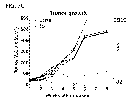
shown in FIG. 7A. (FIG. 7C) Tumor size of MDA-MB-231 in the orthotopic model
treated in FIG. 7A
measured by a digital caliper. Values represent each single mouse. ***P <
.001. (FIG. 7D) Body weight of
mice shown in FIG. 7A. Values shown represent mean SEM. (FIG. 7E)
Representative pictures showing
the restriction of tumor metastasis in CAR (B2) T cell infusion mouse. (FIG.
6F) CAR (B2) T cell
persistence and (FIG. 7G) ex vivo killing on MDA-MB-231 tumor cells after 3
weeks of CAR T cell
infusion. (FIG. 7H) Detection of PD-Li expression in MDA-MB-231 tumor
xenograft by Western blotting.
FIG. 8: Flow cytometry analysis of PD-Li expression and T cell exhaustion
markers (PD-1, LAG-
3, and TIM-3). Shown is expression by mock T cells and anti-PD-Li shark VNAR-
based CAR T cells (B2,
FS, and Au).
FIG. 9: Clustal Omega sequence alignment of VNAR antibodies B2 (SEQ ID NO: 1),
FS (SEQ ID
NO: 2), All (SEQ ID NO: 3), A3 (SEQ ID NO: 4), A9 (SEQ ID NO: 5), A2 (SEQ ID
NO: 6), A10 (SEQ ID
NO: 7), A7 (SEQ ID NO: 8), A6 (SEQ ID NO: 9), C4 (SEQ ID NO: 10), Al (SEQ ID
NO: 11) and D12
(SEQ ID NO: 12). The CDR1, CDR3, HV2 and HV4 of each antibody, according to
the VNAR annotation
described in Stanfield et al. (Science 305:1770-1773, 2004) and Fennell et al.
(J Mol Biol 400:155-170,
2010), are underlined.
SEQUENCE LISTING
The nucleic and amino acid sequences listed in the accompanying sequence
listing are shown using
standard letter abbreviations for nucleotide bases, and three letter code for
amino acids, as defined in 37
C.F.R. 1.822. Only one strand of each nucleic acid sequence is shown, but the
complementary strand is
understood as included by any reference to the displayed strand. The Sequence
Listing is submitted as an
ASCII text file, created on May 23, 2022, 26.8 KB, which is incorporated by
reference herein. In the
accompanying sequence listing:
SEQ ID NO: 1 is the amino acid sequence of VNAR B2.
SEQ ID NO: 2 is the amino acid sequence of VNAR FS.
SEQ ID NO: 3 is the amino acid sequence of VNAR Al 1.
SEQ ID NO: 4 is the amino acid sequence of VNAR A3.
SEQ ID NO: 5 is the amino acid sequence of VNAR A9.
SEQ ID NO: 6 is the amino acid sequence of VNAR A2.
- 6 -
CA 03216228 2023-10-04
WO 2022/261017
PCT/US2022/032378
SEQ ID NO: 7 is the amino acid sequence of VNAR A10.
SEQ ID NO: 8 is the amino acid sequence of VNAR A7.
SEQ ID NO: 9 is the amino acid sequence of VNAR A6.
SEQ ID NO: 10 is the amino acid sequence of VNAR C4.
SEQ ID NO: 11 is the amino acid sequence of VNAR Al.
SEQ ID NO: 12 is the amino acid sequence of VNAR D12.
SEQ ID NO: 13 is the amino acid sequence of a peptide from human PD-Ll.
SEQ ID NO: 14 is a consensus VNAR CDR1 amino acid sequence.
SEQ ID NO: 15 is a consensus VNAR HV2 amino acid sequence.
SEQ ID NO: 16 is a consensus VNAR HV4 amino acid sequence.
SEQ ID NO: 17 is a nucleotide sequence encoding VNAR B2.
SEQ ID NO: 18 is a nucleotide sequence encoding VNAR F5.
SEQ ID NO: 19 is a nucleotide sequence encoding VNAR All.
SEQ ID NO: 20 is a nucleotide sequence encoding VNAR A3.
SEQ ID NO: 21 is a nucleotide sequence encoding VNAR A9.
SEQ ID NO: 22 is a nucleotide sequence encoding VNAR A2.
SEQ ID NO: 23 is a nucleotide sequence encoding VNAR A10.
SEQ ID NO: 24 is a nucleotide sequence encoding VNAR A7.
SEQ ID NO: 25 is a nucleotide sequence encoding VNAR A6.
SEQ ID NO: 26 is a nucleotide sequence encoding VNAR C4.
SEQ ID NO: 27 is a nucleotide sequence encoding VNAR Al.
SEQ ID NO: 28 is a nucleotide sequence encoding VNAR D12.
SEQ ID NO: 29 is the amino acid sequence of human PD-Ll ECD.
1 MRIFAVFIFM TYWHLLNAFT VTVPKDLYVV EYGSNMTIEC KFPVEKQLDL AALIVYWEME
61 DKNIIQFVHG EEDLKVQHSS YRQRARLLKD QLSLGNAALQ ITDVKLQDAG VYRCMISYGG
121 ADYKRITVKV NAPYNKINQR ILVVDPVTSE HELTCQAEGY PKAEVIWTSS DHQVLSGKTT
181 TTNSKREEKL FNVTSTLRIN TTTNEIFYCT FRRLDPEENH TAELVIPELP LAHPPNER
SEQ ID NO: 30 is the amino acid sequence of mouse PD-Ll ECD.
1 MRIFAGIIFT ACCHLLRAFT ITAPKDLYVV EYGSNVTMEC RFPVERELDL LALVVYWEKE
61 DEQVIQFVAG EEDLKPQHSN FRGRASLPKD QLLKGNAALQ ITDVKLQDAG VYCCIISYGG
121 ADYKRITLKV NAPYRKINQR ISVDPATSEH ELICQAEGYP EAEVIWTNSD HQPVSGKRSV
181 TTSRTEGMLL NVTSSLRVNA TANDVFYCTF WRSQPGQNHT AELIIPELPA THPPQNR
SEQ ID NO: 31 is the amino acid sequence of canine PD-Ll ECD.
1 MRMFSVFTFM AYCHLLKAFT ITVSKDLYVV EYGGNVTMEC KFPVEKQLNL FALIVYWEME
61 DKKIIQFVNG KEDLKVQHSS YSQRAQLLKD QLFLGKAALQ ITDVRLQDAG VYCCLIGYGG
121 ADYKRITLKV HAPYRNISQR ISVDPVTSEH ELMCQAEGYP EAEVIWTSSD HRVLSGKTTI
181 TNSNREEKLF NVTSTLNINA TANEIFYCTF QRSGPEENNT AELVIPERLP VPASER
- 7 -
CA 03216228 2023-10-04
WO 2022/261017
PCT/US2022/032378
DETAILED DESCRIPTION
A major challenge in the development of CAR T cells for solid tumors is the
lack of targetable
antigens. Checkpoint molecule PD-Li is highly expressed on many tumors in a
constitutive or interferon-
gamma (IFNy)-inducible manner. IFN-y is the key functional cytokine released
from effector T cells;
.. however, the increased expression of PD-Li on tumor cells binding to PD-1
on effector T cells results in T
cell exhaustion, and inhibition of T cell functions (Chen and Han, J Clin
Invest 2015;125(9):3384-3391). In
the studies disclosed herein, it was hypothesized that the development of CAR
T cells targeting PD-Li
would kill solid tumors via recognizing constitutive or inducible expression
of PD-Li in the tumor
immunosuppressive microenvironment. To test this hypothesis, a panel of anti-
PD-Li nanobodies was
isolated from a newly established semi-synthetic nurse shark VNAR library. The
B2 clone showed specific
binding ability to naïve PD-Li and cross-reacted with both human and mouse
antigens. B2 also functionally
blocked the interaction of PD-Li to PD-1. Moreover, nanobody-based CART cells
showed much higher
transduction efficiency than scFv-based CAR T cells.
PD-Li is not only overexpressed on a larger number of malignancies, but also
on immune cells in
the tumor microenvironment (Sun et al., Immunity 2018;48(3):434-452). T cells
express low levels of
endogenous PD-L1, which makes the development of CART cells that target PD-Li
complex (Xie et al.,
Proc Nail Acad Sci USA 2019;116(16):7624-7631; Qin et al., Biomark Res
2020;8:19). Antigen exposure of
CAR T cells may lead to T cell fratricide and exhaustion, impairing the
proliferation and persistence of CAR
T cells in vitro and in vivo. For example, Xie et al. reported that camelid
VHH-based anti-mouse PD-Li
CAR T cells were found to be self-activated in vitro and PD-Li proficient CAR
T cells could live longer
than WT CAR T cells (Xie et al., Proc Natl Acad Sci USA 2019;116(16):7624-
7631). However, the present
study did not find an upregulated expression of exhaustion markers such as PD-
1, TIM-3, and LAG-3 on the
surface of in vitro activated CAR (B2) T cells compared with mock T cells
(FIG. 8). CAR (B2) T cells
isolated from mouse spleens 3 weeks after infusion still efficiently lysed MDA-
MB-231 cells in vitro (FIG.
7G), which may be due to two reasons. First, shark nanobodies have a unique
structure and binding curve,
which is different from scFy and camelid VHH. B2 may functionally block
interaction of PD-Li to PD-1,
inhibiting CAR T exhaustion. Second, in comparison with other public anti-PD-
Li antibodies, shark VNAR
B2 does not have a comparably high binding affinity to the antigen. Ghorashian
et al. reported a novel
CD19 CAR with a lower affinity binder and found that increased immunoreceptor
affinity may adversely
affect T cell responses (Ghorashian et al., Nat Med 2019;25(9):1408-1414).
To overcome tumor escape mechanisms and enhance anti-tumor efficacy of CAR T
cells, a
combination strategy could be used in solid tumor therapy, such as CAR T cells
with monoclonal antibodies,
small-molecules, or bi-specific CAR T cells targeting different tumor antigens
(Pan et al., Cancer Immunol
Immunother 2018;67(10):1621-1634; Hegde et al., J Chn Invest 2016;126(8):3036-
3052). In the in vitro
experiments disclosed herein, bi-specific CAR (hYP7-B2) T cells targeting both
GPC3 and the tumor
microenvironment marker PD-Li significantly potentiated killing of HCC cells
(Hep3B) by CAR (hYP7) T
cells, indicating that anti-PD-Li B2 nanobody is suitable for engineering of
bi-specific CART cells. It is
- 8 -
CA 03216228 2023-10-04
WO 2022/261017
PCT/US2022/032378
believed that engineered CAR T cells targeting PD-Li exhibit dual function.
These CAR T cells not only
exerted direct killing by recognizing PD-L1, but also blocked interaction of
PD-1 to PD-Li to inhibit T cell
exhaustion.
I. Abbreviations
ACT adoptive cell therapy
ADC antibody-drug conjugate
CAR chimeric antigen receptor
CDR complementarity determining region
CRISPR clustered regularly interspaced short palindromic repeats
E:T effector to target ratio
ECD extracellular domain
FR framework region
GPC3 glypican-3
HCC hepatocellular carcinoma
hEGFRt human epidermal growth factor receptor truncated
HRP horseradish peroxidase
HV hypervariable
IFN interferon
IL interleukin
KO knockout
MHC major histocompatibility complex
MOI multiplicity of infection
NK natural killer
NSG NOD scid gamma
OC ovarian cancer
PBMC peripheral blood mononuclear cells
PD-L1 programmed death ligand 1
PD-1 programmed death 1
PE Pseudomonas exotoxin or phycoerythrin
TIL tumor-infiltrating lymphocytes
TNBC triple negative breast cancer
TNF tumor necrosis factor
VH variable heavy
VL variable light
VNAR variable domain of the immunoglobulin new antigen
receptor
WT wild type
- 9 -
CA 03216228 2023-10-04
WO 2022/261017
PCT/US2022/032378
II. Terms and Methods
Unless otherwise noted, technical terms are used according to conventional
usage. Definitions of
common terms in molecular biology may be found in Benjamin Lewin, Genes X,
published by Jones &
Bartlett Publishers, 2009; and Meyers et al. (eds.), The Encyclopedia of Cell
Biology and Molecular
Medicine, published by Wiley-VCH in 16 volumes, 2008; and other similar
references.
As used herein, the singular forms "a," "an," and "the," refer to both the
singular as well as plural,
unless the context clearly indicates otherwise. For example, the term "an
antigen" includes single or plural
antigens and can be considered equivalent to the phrase "at least one
antigen." As used herein, the term
"comprises" means "includes." It is further to be understood that any and all
base sizes or amino acid sizes,
and all molecular weight or molecular mass values, given for nucleic acids or
polypeptides are approximate,
and are provided for descriptive purposes, unless otherwise indicated.
Although many methods and
materials similar or equivalent to those described herein can be used,
particular suitable methods and
materials are described herein. In case of conflict, the present
specification, including explanations of terms,
will control. In addition, the materials, methods, and examples are
illustrative only and not intended to be
limiting.
To facilitate review of the various embodiments, the following explanations of
terms are provided:
Administration: To provide or give a subject an agent, such as a polypeptide
(for example, a
single-domain monoclonal antibody) provided herein, by any effective route.
Exemplary routes of
administration include, but are not limited to, oral, injection (such as
subcutaneous, intramuscular,
intradermal, intraperitoneal, intravenous, intra-arterial (including hepatic
intra-arterial), intraprostatic, and
intratumoral), sublingual, rectal, transdermal, intranasal, vaginal and
inhalation routes. In some examples
administration is local. In some examples administration is systemic.
Antibody: A polypeptide ligand comprising at least one variable region that
recognizes and binds
(such as specifically recognizes and specifically binds) an epitope of an
antigen, such as a PD-Li antigen.
Mammalian immunoglobulin molecules are composed of a heavy (H) chain and a
light (L) chain, each of
which has a variable region, termed the variable heavy (VI)) region and the
variable light (VL) region,
respectively. Together, the VH region and the VL region are responsible for
binding the antigen recognized
by the antibody. There are five main heavy chain classes (or isotypes) of
mammalian immunoglobulin,
which determine the functional activity of an antibody molecule: IgM, IgD,
IgG, IgA and IgE. Antibody
isotypes not found in mammals include IgX, IgY, IgW and IgNAR. IgY is the
primary antibody produced
by birds and reptiles, and has some functionally similar to mammalian IgG and
IgE. IgW and IgNAR
antibodies are produced by cartilaginous fish, while IgX antibodies are found
in amphibians.
Antibody variable regions contain "framework" regions and hypervariable
regions, known as
"complementarity determining regions" or "CDRs." The CDRs are primarily
responsible for binding to an
epitope of an antigen. The framework regions of an antibody serve to position
and align the CDRs in three-
dimensional space. The amino acid sequence boundaries of a given CDR can be
readily determined using
any of a number of well-known numbering schemes, including those described by
Kabat et al. (Sequences of
- 10 -
CA 03216228 2023-10-04
WO 2022/261017
PCT/US2022/032378
Proteins of Immunological Interest, U.S. Department of Health and Human
Services, 1991; the "Kabat"
numbering scheme), Chothia et al. (see Chothia and Lesk, J Mol Biol 196:901-
917, 1987; Chothia et al.,
Nature 342:877, 1989; and Al-Lazikani et al., (JMB 273,927-948, 1997; the
"Chothia" numbering scheme),
and the ImMunoGeneTics (IMGT) database (see, Lefranc, Nucleic Acids Res 29:207-
9, 2001; the "IMGT"
numbering scheme). The Kabat and IMGT databases are maintained online.
A "single-domain antibody" refers to an antibody having a single domain (a
variable domain) that is
capable of specifically binding an antigen, or an epitope of an antigen, in
the absence of an additional
antibody domain. Single-domain antibodies include, for example, VNAR
antibodies, camelid VHH antibodies,
VH domain antibodies and VL domain antibodies. VNAR antibodies are produced by
cartilaginous fish, such
as nurse sharks, wobbegong sharks, spiny dogfish and bamboo sharks. Camelid
VHH antibodies are
produced by several species including camel, llama, alpaca, dromedary, and
guanaco, which produce heavy
chain antibodies that are naturally devoid of light chains.
A "monoclonal antibody" is an antibody produced by a single clone of
lymphocytes or by a cell into
which the coding sequence of a single antibody has been transfected.
Monoclonal antibodies are produced
by known methods. Monoclonal antibodies include humanized monoclonal
antibodies.
A "chimeric antibody" has framework residues from one species, such as human,
and CDRs (which
generally confer antigen binding) from another species, such as a VNAR that
specifically binds a viral antigen.
A "humanized" antibody is an immunoglobulin including a human framework region
and one or
more CDRs from a non-human (for example a shark, mouse, rabbit, rat, or
synthetic) immunoglobulin. The
non-human immunoglobulin providing the CDRs is termed a "donor," and the human
immunoglobulin
providing the framework is termed an "acceptor." In one embodiment, all CDRs
are from the donor
immunoglobulin in a humanized immunoglobulin. Constant regions need not be
present, but if they are,
they must be substantially identical to human immunoglobulin constant regions,
i.e., at least about 85-90%,
such as about 95% or more identical. Hence, all parts of a humanized
immunoglobulin, except possibly the
CDRs, are substantially identical to corresponding parts of natural human
immunoglobulin sequences. A
humanized antibody binds to the same antigen as the donor antibody that
provides the CDRs. Humanized or
other monoclonal antibodies can have additional conservative amino acid
substitutions which have
substantially no effect on antigen binding or other immunoglobulin functions.
Methods of humanizing shark
VNAR antibodies has been previously described (Kovalenko et al., J Biol Chem
288(24):17408-17419, 2013).
Antibody-drug conjugate (ADC): A molecule that includes an antibody (or
antigen-binding
fragment of an antibody, such an anti-PD-Li antibody provided herein)
conjugated to a drug, such as a
cytotoxic agent. ADCs can be used to specifically target a drug to particular
cells through specific binding
of the antibody to a target antigen expressed on the cell surface. Exemplary
drugs for use with ADCs
include anti-microtubule agents (such as maytansinoids, auristatin E and
auristatin F) and interstrand
crosslinking agents (for example, pyrrolobenzodiazepines; PBDs). In some
cases, the ADC is a bi-specific
ADC, which is comprised of two monoclonal antibodies or antigen-fragments
thereof, each directed to a
different antigen or epitope, conjugated to a drug. In one example, the agent
attached to the antibody is
- 11 -
CA 03216228 2023-10-04
WO 2022/261017
PCT/US2022/032378
IRDye 700 DX (IR700, Li-cor, Lincoln, NE), which can then be used with near
infrared (NIR) light to kill
target cells to which the antibody binds (photoimmunotherapy; see for example
US 8,524,239 and
10,538,590). For example, amino-reactive IR700 can be covalently conjugated to
an antibody using the
NHS ester of IR700.
Binding affinity: Affinity of an antibody for an antigen (such as such an anti-
PD-Li single-domain
antibody provided herein and PD-L1, such as PD-Li from human, mouse or dog).
In one embodiment,
affinity is calculated by a modification of the Scatchard method described by
Frankel et al., Mol. Immunol.,
16:101-106, 1979. In another embodiment, binding affinity is measured by an
antigen/antibody dissociation
rate. In another embodiment, a high binding affinity is measured by a
competition radioimmunoassay. In
another embodiment, binding affinity is measured by ELISA. In some
embodiments, binding affinity is
measured using the Octet system (Creative Biolabs), which is based on bio-
layer interferometry (BLI)
technology. In other embodiments, Kd is measured using surface plasmon
resonance assays using a
BIACORES-2000 or a BIACORES-3000 (BIAcore, Inc., Piscataway, N.J.). In other
embodiments, antibody
affinity is measured by flow cytometry or by surface plasmon reference. An
antibody that "specifically
binds" an antigen (such as PD-L1, such as human, mouse or canine PD-L1) is an
antibody that binds the
antigen with high affinity and does not significantly bind other unrelated
antigens. In some examples, a
monoclonal antibody (such as an anti-PD-Li single-domain antibody provided
herein) specifically binds to a
target (for example, human, mouse or canine PD-L1) with an equilibrium
constant (Kd) of 50 nM or less,
such as 45 nM or less, 40 nM or less, 35 nM or less, 30 nM or less, 25 nM or
less, 20 nM or less, 15 nM or
less, 10 nM or less, or 5 nM or less.
Bispecific antibody: A recombinant protein that includes antigen-binding
fragments of two
different monoclonal antibodies, and is thereby capable of binding two
different antigens or two different
epitopes of the same antigen. Similarly, a multi-specific antibody is a
recombinant protein that includes
antigen-binding fragments of at least two different monoclonal antibodies,
such as two, three or four
different monoclonal antibodies.
Breast cancer: A type of cancer that forms in tissues of the breast, usually
the ducts and lobules.
Types of breast cancer include, for example, ductal carcinoma in situ,
invasive ductal carcinoma, triple
negative breast cancer (TNBC), inflammatory breast cancer, metastatic breast
cancer, medullary carcinoma,
tubular carcinoma and mucinous carcinoma. TNBC refers to a type of breast
cancer in which the cancer
.. cells do not express estrogen receptors, progesterone receptors or
significant levels of HER2/neu protein.
TNBC is also called ER-negative PR-negative HER2/neu-negative breast cancer.
Chemotherapeutic agent: Any chemical agent with therapeutic usefulness in the
treatment of
diseases characterized by abnormal cell growth. Such diseases include tumors,
neoplasms, and cancer. In
one embodiment, a chemotherapeutic agent is an agent of use in treating a PD-
Li-positive tumor. In one
embodiment, a chemotherapeutic agent is a radioactive compound. Exemplary
chemotherapeutic agents that
can be used with the methods provided herein are disclosed in Slapak and Kufe,
Principles of Cancer
Therapy, Chapter 86 in Harrison's Principles of Internal Medicine, 14th
edition; Perry et al., Chemotherapy,
- 12 -
CA 03216228 2023-10-04
WO 2022/261017
PCT/US2022/032378
Ch. 17 in Abeloff, Clinical Oncology 2nd ed., 2000 Churchill Livingstone,
Inc; Baltzer, L., Berkery, R.
(eds.): Oncology Pocket Guide to Chemotherapy, 2nd ed. St. Louis, Mosby-Year
Book, 1995; Fischer, D.S.,
Knobf, M.F., Durivage, H.J. (eds): The Cancer Chemotherapy Handbook, 4th ed.
St. Louis, Mosby-Year
Book, 1993). Combination chemotherapy is the administration of more than one
agent to treat cancer. One
example is the administration of an antibody that binds PD-Li (such as one
provided herein) used in
combination with a radioactive or chemical compound. In one example, a
chemotherapeutic agent is a
biologic, such as a therapeutic antibody (e.g., therapeutic monoclonal
antibody), such as an anti-PD-Li
antibody provided herein, as well as other anti-cancer antibodies, such as
anti-PD-1 or anti-GPC3, anti-
CTLA4 (e.g., ipilimumab), anti-EGFR (e.g., cetuximab), anti-VEGF (e.g.,
bevacizumab), or combinations
thereof (e.g., anti-PD-1 and anti-CTLA-4).
Chimeric antigen receptor (CAR): A chimeric molecule that includes an antigen-
binding portion
(such as single-domain antibody) and a signaling domain, such as a signaling
domain from a T cell receptor
(for example, CD3). Typically, CARs are comprised of an antigen-binding
moiety, a transmembrane
domain and an endodomain. The endodomain typically includes a signaling chain
having an
immunoreceptor tyrosine-based activation motif (ITAM), such as CD3 or FcERIy.
In some instances, the
endodomain further includes the intracellular portion of at least one
additional co-stimulatory domain, such
as CD28, 4-1BB (CD137), ICOS, 0X40 (CD134), CD27 and/or DAP10. In some
examples, the CAR is
multispecific (such as bispecific) or bicistronic. A multispecific CAR is a
single CAR molecule comprised
of at least two antigen-binding domains (such as scFvs and/or single-domain
antibodies) that each bind a
different antigen or a different epitope on the same antigen (see, for
example, US 2018/0230225). For
example, a bispecific CAR refers to a single CAR molecule having two antigen-
binding domains that each
bind a different antigen. A bicistronic CAR refers to two complete CAR
molecules, each containing an
antigen-binding moiety that binds a different antigen. In some cases, a
bicistronic CAR construct expresses
two complete CAR molecules that are linked by a cleavage linker. T cells or NK
cells (or other immune
cells, such as macrophages) expressing a bispecific or bicistronic CAR can
bind cells that express both of
the antigens to which the binding moieties are directed (see, for example, Qin
et al., Blood 130:810, 2017;
and WO/2018/213337).
Complementarity determining region (CDR): A region of hypervariable amino acid
sequence
that defines the binding affinity and specificity of an antibody. The shark
VNAR single-domain antibodies
disclosed herein include two CDRs (CDR1 and CDR3). Shark VNAR antibodies
further include two
hypervariable regions, referred to as HV2 and HV4.
Conjugate: In the context of the present disclosure, a "conjugate" is an
antibody or antibody
fragment (such as an antigen-binding fragment) covalently linked to an
effector molecule or a second protein
(such as a second antibody). The effector molecule can be, for example, a
drug, toxin, therapeutic agent,
detectable label, protein, nucleic acid, lipid, nanoparticle, carbohydrate or
recombinant virus. An antibody
conjugate is often referred to as an "immunoconjugate." When the conjugate
includes an antibody linked to
a drug (e.g., a cytotoxic agent), the conjugate is often referred to as an
"antibody-drug conjugate" or
- 13 -
CA 03216228 2023-10-04
WO 2022/261017
PCT/US2022/032378
Other antibody conjugates include, for example, multi-specific (such as
bispecific or trispecific) antibodies
and chimeric antigen receptors (CARs).
Conservative variant: "Conservative" amino acid substitutions are those
substitutions that do not
substantially affect or decrease the affinity of a protein. For example, a
monoclonal antibody that
specifically binds a target antigen (such as PD-L1) can include at most about
1, at most about 2, at most
about 5, at most about 10, or at most about 15 conservative substitutions and
specifically bind the target
antigen. The term "conservative variant" also includes the use of a
substituted amino acid in place of an
unsubstituted parent amino acid, provided that the antibody specifically binds
the target antigen. Non-
conservative substitutions are those that reduce an activity or binding to the
target antigen.
Conservative amino acid substitution tables providing functionally similar
amino acids are well
known. The following six groups are examples of amino acids that are
considered to be conservative
substitutions for one another:
1) Alanine (A), Serine (S), Threonine (T);
2) Aspartic acid (I)), Glutamic acid (E);
3) Asparagine (N), Glutamine (Q);
4) Arginine (R), Lysine (K);
5) Isoleucine (I), Leucine (L), Methionine (M), Valine (V); and
6) Phenylalanine (F), Tyrosine (Y), Tryptophan (W).
Contacting: Placement in direct physical association; includes both in solid
and liquid form.
Cytotoxic agent: Any drug or compound that kills cells.
Cytotoxicity: The toxicity of a molecule, such as an immunotoxin, to the cells
intended to be
targeted, as opposed to the cells of the rest of an organism. In one
embodiment, in contrast, the term
"toxicity" refers to toxicity of an immunotoxin to cells other than those that
are the cells intended to be
targeted by the targeting moiety of the immunotoxin, and the term "animal
toxicity" refers to toxicity of the
immunotoxin to an animal by toxicity of the immunotoxin to cells other than
those intended to be targeted
by the immunotoxin.
Diagnostic imaging: Coupling antibodies and their derivatives with positron
emitting radionuclides
for positron emission tomography (PET) is a process often referred to as
immunoPET. While full length
antibodies can make good immunoPET agents, their biological half-life
necessitates waiting several days
prior to imaging, resulting in an increase in non-target radiation doses.
Smaller, single domain antibodies
(such as shark VNAR) have biological half-lives amenable to same day imaging.
Drug: Any compound used to treat, ameliorate or prevent a disease or condition
in a subject. In
some embodiments herein, the drug is an anti-tumor agent.
Effector molecule: The portion of an antibody conjugate (or immunoconjugate)
that is intended to
have a desired effect on a cell to which the conjugate is targeted. Effector
molecules are also known as
effector moieties, therapeutic agents, diagnostic agents, or similar terms.
Therapeutic agents (or drugs)
include such compounds as small molecules, nucleic acids, proteins, peptides,
amino acids or derivatives,
- 14 -
CA 03216228 2023-10-04
WO 2022/261017
PCT/US2022/032378
glycoproteins, radioisotopes, lipids, nanoparticles, carbohydrates, or
recombinant viruses. Nucleic acid
therapeutic and diagnostic moieties include antisense nucleic acids,
derivatized oligonucleotides for covalent
cross-linking with single or duplex DNA, and triplex forming oligonucleotides.
Alternatively, the effector
molecule can be contained within an encapsulation system, such as a
nanoparticle, liposome or micelle,
which is conjugated to the antibody. Encapsulation shields the effector
molecule from direct exposure to the
circulatory system. Means of preparing liposomes attached to antibodies are
well known (see, for example,
U.S. Patent No. 4,957,735; and Connor et al., Phann Ther 28:341-365, 1985).
Diagnostic agents or moieties
include radioisotopes and other detectable labels (e.g., fluorophores,
chemiluminescent agents, and
enzymes). Radioactive isotopes include 35S, ;IC, 13N, 150, IsF, 19F, 99oTc,
1311, 3H, 14C, 15N, 90y, 99Tc, "'In
and 1251.
Epitope: An antigenic determinant. These are particular chemical groups or
peptide sequences on
a molecule that are antigenic, meaning that they elicit a specific immune
response. An antibody specifically
binds a particular antigenic epitope on a polypeptide.
Framework region: Amino acid sequences interposed between CDRs. The framework
regions
serve to hold the CDRs in an appropriate orientation for antigen binding.
Fusion protein: A protein comprising at least a portion of two different
(heterologous) proteins. In
some embodiments, the fusion protein includes a polypeptide (such as a single-
domain monoclonal
antibody) disclosed herein and a heterologous protein, such as an Fc protein.
Hepatocellular carcinoma (HCC): A primary malignancy of the liver typically
occurring in
patients with inflammatory livers resulting from viral hepatitis, liver toxins
or hepatic cirrhosis (often caused
by alcoholism). HCC is also called malignant hepatoma.
Heterologous: Originating from a separate genetic source or species. For
example, a shark
antibody is heterologous to a human Fc protein.
Immune response: A response of a cell of the immune system, such as a B cell,
T cell, or
monocyte, to a stimulus. In one embodiment, the response is specific for a
particular antigen (an "antigen-
specific response"). In one embodiment, an immune response is a T cell
response, such as a CD4+ response
or a CD8+ response. In another embodiment, the response is a B cell response,
and results in the production
of antigen-specific antibodies.
Immunoconjugate: A covalent linkage of an effector molecule to an antibody or
functional
fragment thereof. The effector molecule can be, for example, a detectable
label, a photon absorber (such as
IR700), or a toxin (to form an immunotoxin, such as an immunotoxin comprising
Pseudomonas exotoxin or
a variant thereof). Specific, non-limiting examples of toxins include, but are
not limited to, abrin, ricin,
Pseudomonas exotoxin (PE, such as PE35, PE37, PE38, and PE40), diphtheria
toxin (DT), botulinum toxin,
or modified toxins thereof, or other toxic agents that directly or indirectly
inhibit cell growth or kill cells.
For example, PE and DT are highly toxic compounds that typically bring about
death through liver toxicity.
PE and DT, however, can be modified into a form for use as an immunotoxin by
removing the native
targeting component of the toxin (such as the domain Ia of PE and the B chain
of DT) and replacing it with a
- 15 -
CA 03216228 2023-10-04
WO 2022/261017
PCT/US2022/032378
different targeting moiety, such as an antibody. In one embodiment, an
antibody is joined to an effector
molecule. In another embodiment, an antibody joined to an effector molecule is
further joined to a lipid or
other molecule, such as to increase its half-life in the body. The linkage can
be either by chemical or
recombinant means. In one embodiment, the linkage is chemical, wherein a
reaction between the antibody
moiety and the effector molecule has produced a covalent bond formed between
the two molecules to form
one molecule. A peptide linker (short peptide sequence) can optionally be
included between the antibody
and the effector molecule. Because immunoconjugates were originally prepared
from two molecules with
separate functionalities, such as an antibody and an effector molecule, they
are also sometimes referred to as
"chimeric molecules." The term "chimeric molecule," as used herein, therefore
refers to a targeting moiety,
such as a ligand or an antibody, conjugated (coupled) to an effector molecule.
The term "conjugated" or
"linked" refers to making two polypeptides into one contiguous polypeptide
molecule.
Immunoglobulin new antigen receptor (IgNAR) antibody: One of the three
isotypes of
immunoglobulin molecules produced by cartilaginous fish. IgNAR antibodies are
homodimers of one
variable new antigen receptor (VNAR) domain and five constant new antigen
receptor (CNAR) domains (Roux
et al., Proc Natl Acad Sci USA 95:11804-11809, 1998). IgNAR antibodies are a
major component of the
immune system of cartilaginous fish.
Immunoliposome: A liposome with antibodies or antibody fragments conjugated to
its surface.
Immunoliposomes can carry cytotoxic agents or other drugs to antibody-targeted
cells, such as tumor cells.
Isolated: An "isolated" biological component, such as a nucleic acid, protein
(including antibodies)
or organelle, has been substantially separated or purified away from other
biological components in the
environment (such as a cell) in which the component occurs, e.g., other
chromosomal and extra-
chromosomal DNA and RNA, proteins and organelles. Nucleic acids and proteins
that have been "isolated"
include nucleic acids and proteins purified by standard purification methods.
The term also embraces
nucleic acids and proteins prepared by recombinant expression in a host cell
as well as chemically
synthesized nucleic acids.
Label: A detectable compound or composition that is conjugated directly or
indirectly to another
molecule, such as an antibody or a protein, to facilitate detection of that
molecule. Specific, non-limiting
examples of labels include fluorescent tags, enzymatic linkages, and
radioactive isotopes. In one example, a
"labeled antibody" refers to incorporation of another molecule in the
antibody. For example, the label is a
detectable marker, such as the incorporation of a radiolabeled amino acid or
attachment to a polypeptide of
biotinyl moieties that can be detected by marked avidin (for example,
streptavidin containing a fluorescent
marker or enzymatic activity that can be detected by optical or colorimetric
methods). Various methods of
labeling polypeptides and glycoproteins are known and may be used. Examples of
labels for polypeptides
include, but are not limited to, the following: radioisotopes or
radionucleotides (such as 35S, oc, 13N, 150,
18F, 19--',
r 99117c, 1311, 3H, 14C, 15N, 90x,1,
99TC, "'In and 1251), fluorescent labels (such as fluorescein
isothiocyanate (FITC), rhodamine, lanthanide phosphors), enzymatic labels
(such as horseradish peroxidase,
beta-galactosidase, luciferase, alkaline phosphatase), chemiluminescent
markers, biotinyl groups,
- 16 -
CA 03216228 2023-10-04
WO 2022/261017
PCT/US2022/032378
predetermined polypeptide epitopes recognized by a secondary reporter (such as
a leucine zipper pair
sequences, binding sites for secondary antibodies, metal binding domains,
epitope tags), or magnetic agents,
such as gadolinium chelates. In some embodiments, labels are attached by
spacer arms of various lengths to
reduce potential steric hindrance.
Linker: In some cases, a linker is a peptide within an antibody binding
fragment (such as an FA/
fragment) which serves to indirectly bond the variable heavy chain to the
variable light chain. "Linker" can
also refer to a peptide serving to link a targeting moiety, such as an
antibody, to an effector molecule, such
as a cytotoxin or a detectable label. The terms "conjugating," "joining,"
"bonding" or "linking" refer to
making two polypeptides into one contiguous polypeptide molecule, or to
covalently attaching a
radionuclide, drug or other molecule to a polypeptide, such as an antibody or
antibody fragment. In the
specific context, the terms include reference to joining a ligand, such as an
antibody moiety, to an effector
molecule. The linkage can be either by chemical or recombinant means.
"Chemical means" refers to a
reaction between the antibody moiety and the effector molecule such that there
is a covalent bond formed
between the two molecules to form one molecule.
Liver cancer: Any type of cancer occurring in liver tissue. The most common
type of liver cancer
is hepatocellular carcinoma (HCC), which develops in hepatocytes. Other types
of liver cancer include
cholangiocarcinoma, which develops in the bile ducts; liver angiosarcoma,
which is a rare form of liver
cancer that begins in the blood vessels of the liver; and hepatoblastoma,
which is a very rare type of liver
cancer found most often in children.
Neoplasia, malignancy, cancer or tumor: A neoplasm is an abnormal growth of
tissue or cells
that results from excessive cell division. Neoplastic growth can produce a
tumor. The amount of a tumor in
an individual is the "tumor burden" which can be measured as the number,
volume, or weight of the tumor.
A tumor that does not metastasize is referred to as "benign." A tumor that
invades the surrounding tissue
and/or can metastasize is referred to as "malignant."
Operably linked: A first nucleic acid sequence is operably linked with a
second nucleic acid
sequence when the first nucleic acid sequence is placed in a functional
relationship with the second nucleic
acid sequence. For instance, a promoter is operably linked to a coding
sequence if the promoter affects the
transcription or expression of the coding sequence. Generally, operably linked
DNA sequences are
contiguous and, where necessary to join two protein-coding regions, in the
same reading frame.
Pharmaceutically acceptable carriers: The pharmaceutically acceptable carriers
of use are
conventional. Remington: The Science and Practice of Pharmacy, 22'1 ed.,
London, UK: Pharmaceutical
Press, 2013,1, describes compositions and formulations suitable for
pharmaceutical delivery of the antibodies
and other compositions disclosed herein. In general, the nature of the carrier
will depend on the particular
mode of administration being employed. For instance, parenteral formulations
usually comprise injectable
fluids that include pharmaceutically and physiologically acceptable fluids
such as water, physiological
saline, balanced salt solutions, aqueous dextrose, glycerol or the like as a
vehicle. For solid compositions
(such as powder, pill, tablet, or capsule forms), conventional non-toxic solid
carriers can include, for
- 17 -
CA 03216228 2023-10-04
WO 2022/261017
PCT/US2022/032378
example, pharmaceutical grades of mannitol, lactose, starch, or magnesium
stearate. In addition to
biologically neutral carriers, pharmaceutical compositions to be administered
can contain minor amounts of
non-toxic auxiliary substances, such as wetting or emulsifying agents,
preservatives, and pH buffering
agents and the like, for example sodium acetate or sorbitan monolaurate.
Photoimmunotherapy: A targeted therapy that utilizes an antigen-specific
antibody-photoabsorber
conjugate that can be activated by near-infrared light to kill targeted cells.
The photon absorber is typically
based on phthalocyanine dye, such as a near infrared (NIR) phthalocyanine dye
(for example, IRDye
700DX, also know known as IR700). The antibody (for example, a PD-Li-specific
antibody) binds to the
appropriate cell surface antigen (e.g., PD-L1) and the photo-activatable dye
induces lethal damage to cell
membranes after NIR-light exposure. NIR-light exposure (e.g., 690 nm) induces
highly selective, necrotic
cell death within minutes without damage to adjoining cells (see, for example,
U.S. Application No.
2018/0236076). Thus, such methods can be used to kill tumor cells expressing
PD-Li.
Polypeptide: A polymer in which the monomers are amino acid residues joined
together through
amide bonds. When the amino acids are alpha-amino acids, either the L-optical
isomer or the D-optical
isomer can be used. The terms "polypeptide" and "protein" are used herein
interchangeably and include
standard amino acid sequences as well as modified sequences, such as
glycoproteins. The term
"polypeptide" is specifically intended to cover naturally occurring proteins,
as well as proteins that are
recombinantly or synthetically produced. In the context of the present
disclosure, a "polypeptide" is any
protein or polypeptide (natural, recombinant or synthetic) that is capable of
specific binding to a target
antigen, such as PD-Li or portion thereof. Thus, the polypeptides disclosed
herein include at least one, such
as one, two or three, CDR sequences that mediate specific binding to the
target antigen. In some
embodiments, the polypeptide is a single-domain monoclonal antibody, such as a
shark VNAR single-domain
monoclonal antibody, isolated from a phage display library, or a modified form
thereof (such as a humanized
or chimeric single-domain monoclonal antibody). In other embodiments, the
polypeptide comprises
fibronectin (adectin), albumin, protein A (affibody), a peptide aptamer, an
affimer, an affitin, an anticalin, or
another antibody mimetic (see, e.g., Yu et al., Annu Rev Anal Chem 10(1): 293-
320, 2017; Ta and
McNaughton, Future Med Chem 9(12): 1301-1304, 2017; Koutsoumpeli et al., Anal
Chem 89(5): 3051-
3058, 2017), or a similar protein in which one or more CDR sequences have been
incorporated to confer
specific binding to the target antigen.
Preventing, treating or ameliorating a disease: "Preventing" a disease refers
to inhibiting the full
development of a disease. "Treating" refers to a therapeutic intervention that
ameliorates a sign or symptom
of a disease or pathological condition after it has begun to develop, such as
a reduction in viral load.
"Ameliorating" refers to the reduction in the number or severity of signs or
symptoms of a disease.
Programmed death ligand 1 (PD-L1): An immune inhibitory receptor ligand
expressed by
hematopoietic and non-hematopoietic cells, such as T cells, B cells and
several different tumor types. PD-
Li is a type I transmembrane protein with immunoglobulin V-like and C-like
domains. Interaction of PD-
Li with its receptor inhibits T-cell activation and cytokine production.
During infection or inflammation of
- 18 -
CA 03216228 2023-10-04
WO 2022/261017
PCT/US2022/032378
normal tissue, this interaction is important for preventing autoimmunity by
maintaining homeostasis of the
immune response. In tumor microenvironments, this interaction provides an
immune escape for tumor cells
through cytotoxic T-cell inactivation. PD-Li is also known as CD274, B7-H and
B7H1. Nucleic acid and
protein sequences of PD-Li are publicly available, such as under NCBI Gene ID
29126. An exemplary
mouse PD-Li is available under GenBank Accession No. ADK70950.1. An exemplary
canine PD-Li is
available under GenBank Accession No. BA074172.1. An exemplary human PD-Li is
available under
GenBank Accession No. Q9NZQ7.1. Exemplary human, mouse and canine PD-Li
extracellular domains
(ECDs) are set forth herein as SEQ ID NOs: 29, 30, and 31, respectively.
PD-Li-positive cancer: A cancer that expresses PD-Li or can be induced to
express PD-L1, such
as by IFNy. Examples of PDL-1-positive cancers include, but are not limited to
liver cancer (such as
hepatocellular carcinoma), breast cancer (such as triple negative breast
cancer), pancreatic cancer,
melanoma, non-small cell lung cancer (NSCLC), renal cell carcinoma, bladder
cancer, head and neck
squamous cell carcinoma (HNSCC), gastric cancer, urothelial carcinoma and
Merkel cell carcinoma. Thus,
such cancers can be detected and treated with the disclosed compositions and
methods.
Recombinant: A recombinant nucleic acid or protein is one that has a sequence
that is not naturally
occurring or has a sequence that is made by an artificial combination of two
otherwise separated segments of
sequence. This artificial combination is often accomplished by chemical
synthesis or by the artificial
manipulation of isolated segments of nucleic acids, for example, by genetic
engineering techniques.
Sample (or biological sample): A biological specimen containing genomic DNA,
RNA (including
mRNA), protein, or combinations thereof, which can be obtained from a subject.
Examples include, but are
not limited to, blood, serum, urine, semen, sputum, saliva, mucus, nasal wash,
tissue, cells, tissue biopsy,
fine needle aspirate, surgical specimen, feces, cerebral spinal fluid (CSF),
bronchoalveolar lavage (BAL)
fluid, nasopharyngeal samples, oropharyngeal samples, and autopsy material. In
one example, a sample is a
tumor biopsy or fine needle aspirate.
Sequence identity: The similarity between amino acid or nucleic acid sequences
is expressed in
terms of the similarity between the sequences, otherwise referred to as
sequence identity. Sequence identity is
frequently measured in terms of percentage identity (or similarity or
homology); the higher the percentage, the
more similar the two sequences are. Homologs or variants of a polypeptide or
nucleic acid molecule will
possess a relatively high degree of sequence identity when aligned using
standard methods.
Methods of alignment of sequences for comparison are well known. Various
programs and alignment
algorithms are described in: Smith and Waterman, Adv. Appl. Math. 2:482, 1981;
Needleman and Wunsch, J.
Mol. Biol. 48:443, 1970; Pearson and Lipman, Proc. Natl. Acad. Sci. U.S.A.
85:2444, 1988; Higgins and
Sharp, Gene 73:237, 1988; Higgins and Sharp, CABIOS 5:151, 1989; Corpet et
al., Nucleic Acids Research
16:10881, 1988; and Pearson and Lipman, Proc. Natl. Acad. Sci. U.S.A. 85:2444,
1988. Altschul et al.,
Nature Genet. 6:119, 1994, presents a detailed consideration of sequence
alignment methods and homology
calculations.
- 19 -
CA 03216228 2023-10-04
WO 2022/261017
PCT/US2022/032378
The NCBI Basic Local Alignment Search Tool (BLAST) (Altschul et al., J. Mol.
Biol. 215:403, 1990)
is available from several sources, including the National Center for
Biotechnology Information (NCBI,
Bethesda, MD) and on the internet, for use in connection with the sequence
analysis programs blastp, blastn,
blastx, tblastn and tblastx. A description of how to determine sequence
identity using this program is available
on the NCBI website on the internet.
Homologs and variants of an antibody that specifically binds a target antigen
or a fragment thereof are
typically characterized by possession of at least about 75%, for example at
least about 80%, 90%, 95%, 96%,
97%, 98% or 99% sequence identity counted over the full length alignment with
the amino acid sequence of
the antibody using the NCBI Blast 2.0, gapped blastp set to default
parameters. For comparisons of amino
.. acid sequences of greater than about 30 amino acids, the Blast 2 sequences
function is employed using the
default BLOSUM62 matrix set to default parameters, (gap existence cost of 11,
and a per residue gap cost of
1). When aligning short peptides (fewer than around 30 amino acids), the
alignment should be performed
using the Blast 2 sequences function, employing the PAM30 matrix set to
default parameters (open gap 9,
extension gap 1 penalties). Proteins with even greater similarity to the
reference sequences will show
increasing percentage identities when assessed by this method, such as at
least 80%, at least 85%, at least 90%,
at least 95%, at least 98%, or at least 99% sequence identity. When less than
the entire sequence is being
compared for sequence identity, homologs and variants will typically possess
at least 80% sequence identity
over short windows of 10-20 amino acids, and may possess sequence identities
of at least 85% or at least 90%
or 95% depending on their similarity to the reference sequence. Methods for
determining sequence identity
over such short windows are available at the NCBI website on the internet. One
of skill will appreciate that
these sequence identity ranges are provided for guidance only; it is entirely
possible that strongly significant
homologs could be obtained that fall outside of the ranges provided.
Small molecule: A molecule, typically with a molecular weight less than about
1000 Daltons, or in
some embodiments, less than about 500 Daltons, wherein the molecule is capable
of modulating, to some
measurable extent, an activity of a target molecule.
Subject: Living multi-cellular vertebrate organisms, a category that includes
both human and
veterinary subjects, including human and non-human mammals such as pigs, mice,
rats, rabbits, sheep, horses,
cows, dogs, cats and non-human primates.
Synthetic: Produced by artificial means in a laboratory, for example a
synthetic nucleic acid or
protein (for example, an antibody) can be chemically synthesized in a
laboratory.
Therapeutically effective amount: The amount of agent, such as a polypeptide
(e.g., a single-
domain monoclonal antibody specific for PD-Li provided herein), that is
sufficient to prevent, treat
(including prophylaxis), reduce and/or ameliorate one or more symptoms and/or
underlying causes of a
disease or disorder, for example to prevent, inhibit, and/or treat a PD-Li-
positive cancer. In one
embodiment, a therapeutically effective amount is the amount necessary to
eliminate, reduce the size, or
prevent metastasis of a tumor, such as reduce a tumor size and/or volume by at
least 10%, at least 20%, at
least 50%, at least 75%, at least 80%, at least 90%, at least 95%, or even
100%, and/or reduce the number
- 20 -
CA 03216228 2023-10-04
WO 2022/261017
PCT/US2022/032378
and/or size/volume of metastases by at least 10%, at least 20%, at least 50%,
at least 75%, at least 80%, at
least 90%, at least 95%, or even 100%, for example as compared to a
size/volume/number prior to treatment.
When administered to a subject, a dosage will generally be used that will
achieve target tissue concentrations
(for example, in tumors) that has been shown to achieve a desired in vitro
effect.
A therapeutically effective amount of an agent can be administered in a single
dose, or in several
doses, for example daily, during a course of treatment. However, the
therapeutically effective amount can
depend on the subject being treated, the severity and type of the condition
being treated, and the manner of
administration. A unit dosage form of the agent can be packaged in a
therapeutic amount, or in multiples of
the therapeutic amount, for example, in a vial (e.g., with a pierceable lid)
or syringe having sterile
components.
Toxin: An agent that directly or indirectly inhibits the growth of and/or
kills cells. Toxins include,
for example, Pseudomonas exotoxin (PE, such as PE35, PE37, PE38 and PE40),
diphtheria toxin (DT),
botulinum toxin, abrin, ricin, saporin, restrictocin or gelonin, or modified
toxins thereof. For example, PE
and DT are highly toxic compounds that typically bring about death through
liver toxicity. PE and DT,
however, can be modified into a form for use as an immunotoxin by removing the
native targeting
component of the toxin (such as domain Ia of PE or the B chain of DT) and
replacing it with a different
targeting moiety, such as an antibody.
Variable new antigen receptor (VNAR): The single variable domain of the
immunoglobulin new
antigen receptor (IgNAR) antibody found in cartilaginous fish. VNAR antibodies
are comprised of only two
CDRs (CDR1 and CDR3), but also contain two other hypervariable (HV) regions,
referred to as the HV2
and HV4 regions. The CDRs and HV regions are surrounded by framework regions
(FR) in the following
N-terminal to C-terminal order: FR1-CDR1-FR2-HV2-FR3a-HV4-FR3b-CDR3-FR4.
The VNAR domain, like other variable domains, has an immunoglobulin fold that
contains p sheets
held together by two canonical cysteine residues. In addition to the cysteines
found in framework region
(FR) 1 and 3b, the CDR3 can have one or two additional cysteines that form
disulfide bonds with CDR1 or
other framework regions. IgNAR are classified into four types based on the
number and positioning of non-
canonical cysteines in the VNAR domain. Type I VNAR domains contain two
cysteine residues in CDR3 that
form two extra disulfide bonds with FR2 and FR4. Type II VNAR domains have one
non-canonical cysteine
in CDR3 that forms a disulfide bond with a non-canonical cysteine in CDR1.
Type III VNAR domains form a
disulfide bond in CDR3 and FR2, and type IV domains have no additional
disulfide bonds. While type I
VNAR usually have flatter antigen binding regions and CDR3 regions that
average 21 amino acids long, type
II are usually shorter with an average of 15 amino acids and have a protruding
CDR3 that enables binding to
pockets and grooves (Barelle et al., Adv Exp Med Biol 655:49-62, 2009). The
canonical CDR2 loop in
classical IgG is missing in VNAR and is replaced with a short stretch of
highly diverse amino acids, termed
hypervariable region 2 (HV2) (Stanfield et al., Science 305:1770-1773, 2004).
Additionally, there is a
second hypervariable region, named HV4, which is inserted in the middle of
FR3, therefore breaking FR3
into FR3a and FR3b.
-21 -
CA 03216228 2023-10-04
WO 2022/261017
PCT/US2022/032378
Vector: A nucleic acid molecule as introduced into a host cell, thereby
producing a transformed
host cell. A vector may include nucleic acid sequences that permit it to
replicate in a host cell, such as an
origin of replication. A vector may also include one or more selectable marker
genes and other genetic
elements. In some embodiments, the vector is a virus vector, such as an AAV
vector or lentivirus vector.
Single-Domain Antibodies Specific for PD-Li
A single-chain antibody variable fragment (scFv), which consists of variable
heavy (VH) and
variable light (VI) (Oishi et al., Hum Mol Genet 2002;11(23):2951-2960) chains
connected by a flexible
linker (such as (Gly4Ser)3), usually serves as the antigen recognition region
of a CAR construct. However,
proper folding of artificially engineered scFy can alter the specificity and
affinity of the CAR for its target
antigen (Chailyan et al., FEBS J 2011;278(16):2858-2866). In contrast, the
antigen binding domain of
natural single-domain antibodies (heavy chain only) from camelid (VHH) (Hamers-
Casterman et al., Nature
1993;363(6428):446-448) and shark (VNAR) (Flajnik et al., Nat Rev Genet
2010;11(1):47-59) have beneficial
properties for the engineering of CARs. They are small in size (12-15 kDa),
easily expressed, and capable
of binding concave and hidden epitopes that are not accessible to conventional
antibodies (Muyldermans et
al., Annu Rev Biochem 2013;82:775-797). Shark VNAR antibodies have unique
features that are quite
different from camel VHH antibodies, such as a large diversity in the number
and positions of cysteines, and
are evolutionally derived from an ancient single domain antibody that
functions as a variable domain in both
B cell and T cell receptors (Criscitiello et al., Proc Nall Acad Sci USA
2006;103(13):5036-5041; English et
al., Antib Ther 2020;3(1):1-9). A shark VNAR phage-displayed library was
previously constructed (Feng et
al., Antib Ther 2019;2(1):1-11). The present disclosure describes the
reconstruction of a semi-synthetic
shark VNAR phage library in which the VNAR antibodies have a randomized
complementarity determining
region 3 (CDR3) of 18 amino acids in length (Brahmer et al., N Engl J Med
2015;373(2):123-135). Panning
of the reconstructed library led to the identification of 12 PD-Li binders
that are cross-reactive with human
and mouse PD-L1, and in some instances, also bind canine PD-Li. This is the
first report of human and
mouse cross-reactive PD-Li monoclonal antibodies, as well as the first
disclosure of PD-Li-specific single-
domain monoclonal antibodies.
The amino acid sequences of 12 PD-Li -specific single-domain VNAR antibodies
selected from the
re-engineered shark VNAR phage library are provided below (and set forth
herein as SEQ ID NOs: 1-12).
Shark VNAR are comprised of the following regions (N-terminal to C-terminal):
FR1-CDR1-FR2-HV2-FR3a-
HV4-FR3b-CDR3-FR4. CDR and HV regions were determined using shark VNAR
annotation (italics) as
described by Stanfield et al. (Science 305:1770-1773, 2004) and Fennell et al.
(J Mol Biol 400:155-170,
2010). CDRs were also determined using IMGT (bold) and Kabat (underline). The
positions of each CDR
and HV region are shown in Tables 1 and 2.
- 22 -
CA 03216228 2023-10-04
WO 2022/261017
PCT/US2022/032378
Shark VNAR B2 (SEQ ID NO: 1)
ARVDQTPRS VTKETGES LTINCVLRDSS YALGSTYWYRKKS GS TNEESISKGGRYVETVNSGSKSFSL
RINDLTVEDS GTYRCKYTSRLRREGPLSWDGNTVYGGGTVVTVN
Shark VNAR F5 (SEQ ID NO: 2)
ARVDQTPRS VTKETGES LTINCVLRDTNYALGSTYWYRKKLGS TNEESISKGGRYVETVNSGSKS FS
LRINDLTVEDSGTYRCKNLGHPMY/AGA/CRPLP/YGGGTVVTVN
Shark VNAR All (SEQ ID NO: 3)
ARVDQTPRS VTKETGES LTINCVLRDTS YALGSTYWYRKKS GS TNEESISKGGRYVETVNSGSKS FS
LRINDLTVEDSGTYRCKFKSNTEFLNPLTVSMST/YGGGTVVTVN
Shark VNAR A3 (SEQ ID NO: 4)
ARVDQTPQTITKETGES LTINCVLRDSNYALGSTYVVYRKKS GS TNEESISKGGRYVETVNSGSKSFS L
RINDLTVEDSGTYRCKTQKA/PCNMNGYVCGVT/YGGGTVVTVN
Shark VNAR A9 (SEQ ID NO: 5)
ARVDQTPQTITKETGES LTINCVLRDTNYALGSTYVVYRKKLGS TNEESISKGGRYVETVNSGSKS FS L
RINDLTVEDSGTYRCKNTSPWLTYSPWTVSGQTSYGGGTVVTVN
Shark VNAR A2 (SEQ ID NO: 6)
ARVDQTPRSVTKETGESLTINCVLRDTSYALGSTYWYRKKLGSTNEESISKGGRYVETVNSGSKTFS
LRINDLTVEDS GTYRCKVOTTWS YLSPYKIEFA TVYGGGTVVTVN
Shark VNAR A10 (SEQ ID NO: 7)
ARVDQTPRS VTKETGES LTINCVLRDTNYALGSTYWYRKKLGS TNEESISKGGRYVETVNSGSKS FS
LRINDLTVEDSGTYRCKS TTPWNNLTSFTSER VT/YGGGTVVTVN
Shark VNAR A7 (SEQ ID NO: 8)
ARVDQTPRS VTKETGES LTINCVLRDTS YALGSTYWYRKKLGS TNEESISKGGRYVETVNSGSKS FS
LRINDLTVEDS GTYRCKMNOR YPCDNNSWWSL YCGTTVYGGGTVVTVN
Shark VNAR A6 (SEQ ID NO: 9)
ARVDQTPQTITKETGES LTINCVLRDTS YALGSTYVVYRKKS GS TNEESISKGGRYVETVNSGSKSFS L
RINDLRVEDSGTYRCKYSSSWOKA TEGVWEAMTEYGGGTVVTVN
- 23 -
CA 03216228 2023-10-04
WO 2022/261017
PCT/US2022/032378
Shark VNAR C4 (SEQ ID NO: 10)
ARVDQTPRSVTKETGESLTINCVLRDTNYALGSTYWYRKKSGSTNEESISKGGRYVETVNSGSKSFS
LRINDLTVEDSGTYRCKQSASLWIPNLLRVVVPV/SYGGGTVVTVN
Shark VNAR Al (SEQ ID NO: 11)
ARVDQTPRSVTKETGESLTINCVLRDTNYALGSTYWYRKKSGSTNEESISKGGRYVETVNSGSKSFS
LRINDLTVEDSGTYRCKTSSKPLLVSHNVWSAWTEYGGGTVVTVN
Shark VNAR D12 (SEQ ID NO: 12)
ARVDQTPRSVTKETGESLTINCVLRDTNYALGSTYWYRKKSGSTNEESISKGGRYVETVNSGSKSFS
LRINDLTVEDSGTYRCKHOSSWRROAPRVMEMOTLYGGGTVVTVN
FIG. 9 provides an amino acid sequence alignment of the 12 VNAR antibodies. In
the figure, the
positions of the CDR1, HV2, HV4 and CDR3, using shark VNAR annotation, are
underlined. Based on this
alignment, the following consensus CDR1, HV2 and HV4 sequences were
determined:
CDR1 = DX1X2YALGST, where Xi = S or T and X2= N or S (SEQ ID NO: 14)
HV2 = NEESISKG (SEQ ID NO: 15)
HV4 = NSGSK (SEQ ID NO: 16)
Table 1. Positions of the CDRs and HV regions using shark VNAR annotation
VNAR SEQ ID CDR1 HV2 HV4 CDR3
NO:
B2 1 26-33 45-52 60-64 86-102
F5 2 26-33 45-52 60-64 86-102
All 3 26-33 45-52 60-64 86-102
A3 4 26-33 45-52 60-64 86-102
A9 5 26-33 45-52 60-64 86-102
A2 6 26-33 45-52 60-64 86-102
A10 7 26-33 45-52 60-64 86-102
A7 8 26-33 45-52 60-64 86-105
A6 9 26-33 45-52 60-64 86-102
C4 10 26-33 45-52 60-64 86-102
Al 11 26-33 45-52 60-64 86-102
D12 12 26-33 45-52 60-64 86-102
- 24 -
CA 03216228 2023-10-04
WO 2022/261017 PCT/US2022/032378
Table 2. Positions of the CDRs using IMGT and Kabat annotation
VNAR SEQ ID Annotation CDR1 CDR2 CDR3
NO:
B2 1 IMGT 26-33 45-49 84-102
B2 1 Kabat 22-35 45-52 86-102
F5 2 IMGT 26-33 45-49 84-102
F5 2 Kabat 22-35 45-52 86-102
All 3 IMGT 26-33 45-49 84-102
All 3 Kabat 22-35 45-52 86-102
A3 4 IMGT 26-33 45-49 84-102
A3 4 Kabat 22-35 45-52 86-102
A9 5 IMGT 26-33 45-49 84-102
A9 5 Kabat 22-35 45-52 86-102
A2 6 IMGT 26-33 45-49 84-102
A2 6 Kabat 22-35 45-52 86-102
A10 7 IMGT 26-33 45-49 84-102
A10 7 Kabat 22-35 45-52 86-102
A7 8 IMGT 26-33 45-49 84-105
A7 8 Kabat 22-35 45-52 86-105
A6 9 IMGT 26-33 45-49 84-102
A6 9 Kabat 22-35 45-52 86-102
C4 10 IMGT 26-33 45-49 84-102
C4 10 Kabat 22-35 45-52 86-102
Al 11 IMGT 26-33 45-49 84-102
Al 11 Kabat 22-35 45-52 86-102
D12 12 IMGT 26-33 45-49 84-102
D12 12 Kabat 22-35 45-52 86-102
Provided herein are polypeptides that bind (for example, specifically bind) PD-
L1, such as human,
mouse and/or canine PD-Ll. In some embodiments, the polypeptide (for example,
single-domain
monoclonal antibody) includes at least a portion of the amino acid sequence
set forth herein as any one of
SEQ ID NOs: 1-12, such as one or more (such as one, two or three) CDR
sequences and/or one or two
hypervariable regions from any one of antibodies B2, F5, All, A3, A9, A2, A10,
A7, A6, C4, Al or D12
(SEQ ID NOs: 1-12, respectively), as determined using any CDR numbering scheme
(such as IMGT, Kabat,
Paratome or Chothia, or any combination thereof; or using the CDR/HV
annotation described in Stanfield et
al. 2004 and/or Fennell et al. 2010 for shark VNAR).
- 25 -
CA 03216228 2023-10-04
WO 2022/261017
PCT/US2022/032378
In some embodiments, the polypeptide includes the CDR1 and CDR3 sequences of
B2 (SEQ ID
NO: 1). In some examples, the CDR1 and CDR3 sequences respectively include
residues 26-33 and 86-102,
residues 26-33 and 84-102, or residues 22-35 and 86-102 of SEQ ID NO: 1. In
some examples, the
polypeptide further includes the CDR2 sequence of SEQ ID NO: 1, such as
residues 45-49 or residues 45-52
of SEQ ID NO: 1. In specific examples, the polypeptide further includes a HV2
region having the sequence
of SEQ ID NO: 15 and/or a HV4 region having the sequence of SEQ ID NO: 16. In
some examples, the
amino acid sequence of the polypeptide is at least 80%, at least 85%, at least
90%, at least 95%, at least
96%, at least 97%, at least 98% or at least 99% identical to SEQ ID NO: 1. In
particular non-limiting
examples, the amino acid sequence of the polypeptide includes or consists of
SEQ ID NO: 1.
In some embodiments, the polypeptide includes the CDR1 and CDR3 sequences of
F5 (SEQ ID NO:
2). In some examples, the CDR1 and CDR3 sequences respectively include
residues 26-33 and 86-102,
residues 26-33 and 84-102, or residues 22-35 and 86-102 of SEQ ID NO: 2. In
some examples, the
polypeptide further includes the CDR2 sequence of SEQ ID NO: 2, such as
residues 45-49 or residues 45-52
of SEQ ID NO: 2. In specific examples, the polypeptide further includes a HV2
region having the sequence
of SEQ ID NO: 15 and/or a HV4 region having the sequence of SEQ ID NO: 16. In
some examples, the
amino acid sequence of the polypeptide is at least 80%, at least 85%, at least
90%, at least 95%, at least
96%, at least 97%, at least 98% or at least 99% identical to SEQ ID NO: 2. In
particular non-limiting
examples, the amino acid sequence of the polypeptide includes or consists of
SEQ ID NO: 2.
In some embodiments, the polypeptide includes the CDR1 and CDR3 sequences of
All (SEQ ID
NO: 3). In some examples, the CDR1 and CDR3 sequences respectively include
residues 26-33 and 86-102,
residues 26-33 and 84-102, or residues 22-35 and 86-102 of SEQ ID NO: 3. In
some examples, the
polypeptide further includes the CDR2 sequence of SEQ ID NO: 3, such as
residues 45-49 or residues 45-52
of SEQ ID NO: 3. In specific examples, the polypeptide further includes a HV2
region comprising SEQ ID
NO: 15 and/or a HV4 region comprising SEQ ID NO: 16. In some examples, the
amino acid sequence of
the polypeptide is at least 80%, at least 85%, at least 90%, at least 95%, at
least 96%, at least 97%, at least
98% or at least 99% identical to SEQ ID NO: 3. In particular non-limiting
examples, the amino acid
sequence of the polypeptide includes or consists of SEQ ID NO: 3.
In some embodiments, the polypeptide includes the CDR1 and CDR3 sequences of
A3 (SEQ ID
NO: 4). In some examples, the CDR1 and CDR3 sequences respectively comprise
residues 26-33 and 86-
102, residues 26-33 and 84-102, or residues 22-35 and 86-102 of SEQ ID NO: 4.
In some examples, the
polypeptide further includes the CDR2 sequence of SEQ ID NO: 4, such as
residues 45-49 or residues 45-52
of SEQ ID NO: 4. In specific examples, the polypeptide further includes a HV2
region comprising SEQ ID
NO: 15 and/or a HV4 region comprising SEQ ID NO: 16. In some examples, the
amino acid sequence of
the polypeptide is at least 80%, at least 85%, at least 90%, at least 95%, at
least 96%, at least 97%, at least
98% or at least 99% identical to SEQ ID NO: 4. In particular non-limiting
examples, the amino acid
sequence of the polypeptide includes or consists of SEQ ID NO: 4.
- 26 -
CA 03216228 2023-10-04
WO 2022/261017
PCT/US2022/032378
In some embodiments, the polypeptide includes the CDR1 and CDR3 sequences of
A9 (SEQ ID
NO: 5). In some examples, the CDR1 and CDR3 sequences respectively include
residues 26-33 and 86-102,
residues 26-33 and 84-102, or residues 22-35 and 86-102 of SEQ ID NO: 5. In
some examples, the
polypeptide further includes the CDR2 sequence of SEQ ID NO: 5, such as
residues 45-49 or residues 45-52
of SEQ ID NO: 5. In specific examples, the polypeptide further includes a HV2
region comprising SEQ ID
NO: 15 and/or a HV4 region comprising SEQ ID NO: 16. In some examples, the
amino acid sequence of
the polypeptide is at least 80%, at least 85%, at least 90%, at least 95%, at
least 96%, at least 97%, at least
98% or at least 99% identical to SEQ ID NO: 5. In particular non-limiting
examples, the amino acid
sequence of the polypeptide includes or consists of SEQ ID NO: 5.
In some embodiments, the polypeptide includes the CDR1 and CDR3 sequences of
A2 (SEQ ID
NO: 6). In some example, the CDR1 and CDR3 sequences respectively include
residues 26-33 and 86-102,
residues 26-33 and 84-102, or residues 22-35 and 86-102 of SEQ ID NO: 6. In
some examples, the
polypeptide further includes the CDR2 sequence of SEQ ID NO: 6, such as
residues 45-49 or residues 45-52
of SEQ ID NO: 6. In specific examples, the polypeptide further includes a HV2
region comprising SEQ ID
NO: 15 and/or a HV4 region comprising SEQ ID NO: 16. In some examples, the
amino acid sequence of
the polypeptide is at least 80%, at least 85%, at least 90%, at least 95%, at
least 96%, at least 97%, at least
98% or at least 99% identical to SEQ ID NO: 6. In particular non-limiting
examples, the amino acid
sequence of the polypeptide includes or consists of SEQ ID NO: 6.
In some embodiments, the polypeptide includes the CDR1 and CDR3 sequences of
A10 (SEQ ID
NO: 7). In some examples, the CDR1 and CDR3 sequences respectively include
residues 26-33 and 86-102,
residues 26-33 and 84-102, or residues 22-35 and 86-102 of SEQ ID NO: 7. In
some examples, the
polypeptide further includes the CDR2 sequence of SEQ ID NO: 7, such as
residues 45-49 or residues 45-52
of SEQ ID NO: 7. In specific examples, the polypeptide further includes a HV2
region comprising SEQ ID
NO: 15 and/or a HV4 region comprising SEQ ID NO: 16. In some examples, the
amino acid sequence of
the polypeptide is at least 80%, at least 85%, at least 90%, at least 95%, at
least 96%, at least 97%, at least
98% or at least 99% identical to SEQ ID NO: 7. In particular non-limiting
examples, the amino acid
sequence of the polypeptide includes or consists of SEQ ID NO: 7.
In some embodiments, the polypeptide includes the CDR1 and CDR3 sequences of
A7 (SEQ ID
NO: 8). In some examples, the CDR1 and CDR3 sequences respectively include
residues 26-33 and 86-105,
residues 26-33 and 84-105, or residues 22-35 and 86-105 of SEQ ID NO: 8. In
some examples, the
polypeptide further includes the CDR2 sequence of SEQ ID NO: 8, such as
residues 45-49 or residues 45-52
of SEQ ID NO: 8. In specific examples, the polypeptide further includes a HV2
region comprising SEQ ID
NO: 15 and/or a HV4 region comprising SEQ ID NO: 16. In some examples, the
amino acid sequence of
the polypeptide is at least 80%, at least 85%, at least 90%, at least 95%, at
least 96%, at least 97%, at least
98% or at least 99% identical to SEQ ID NO: 8. In particular non-limiting
examples, the amino acid
sequence of the polypeptide includes or consists of SEQ ID NO: 8.
- 27 -
CA 03216228 2023-10-04
WO 2022/261017
PCT/US2022/032378
In some embodiments, the polypeptide includes the CDR1 and CDR3 sequences of
A6 (SEQ ID
NO: 9). In some examples, the CDR1 and CDR3 sequences respectively include
residues 26-33 and 86-102,
residues 26-33 and 84-102, or residues 22-35 and 86-102 of SEQ ID NO: 9. In
some examples, the
polypeptide further includes the CDR2 sequence of SEQ ID NO: 9, such as
residues 45-49 or residues 45-52
of SEQ ID NO: 9. In specific examples, the polypeptide further includes a HV2
region comprising SEQ ID
NO: 15 and/or a HV4 region comprising SEQ ID NO: 16. In some examples, the
amino acid sequence of
the polypeptide is at least 80%, at least 85%, at least 90%, at least 95%, at
least 96%, at least 97%, at least
98% or at least 99% identical to SEQ ID NO: 9. In particular non-limiting
examples, the amino acid
sequence of the polypeptide includes or consists of SEQ ID NO: 9.
In some embodiments, the polypeptide includes the CDR1 and CDR3 sequences of
C4 (SEQ ID
NO: 10). In some examples, the CDR1 and CDR3 sequences respectively include
residues 26-33 and 86-
102, residues 26-33 and 84-102, or residues 22-35 and 86-102 of SEQ ID NO: 10.
In some examples, the
polypeptide further includes the CDR2 sequence of SEQ ID NO: 10, such as
residues 45-49 or residues 45-
52 of SEQ ID NO: 10. In specific examples, the polypeptide further includes a
HV2 region comprising SEQ
ID NO: 15 and/or a HV4 region comprising SEQ ID NO: 16. In some examples, the
amino acid sequence of
the polypeptide is at least 80%, at least 85%, at least 90%, at least 95%, at
least 96%, at least 97%, at least
98% or at least 99% identical to SEQ ID NO: 10. In particular non-limiting
examples, the amino acid
sequence of the polypeptide includes or consists of SEQ ID NO: 10.
In some embodiments, the polypeptide includes the CDR1 and CDR3 sequences of
Al (SEQ ID
NO: 11). In some examples, the CDR1 and CDR3 sequences respectively include
residues 26-33 and 86-
102, residues 26-33 and 84-102, or residues 22-35 and 86-102 of SEQ ID NO: 11.
In some examples, the
polypeptide further includes the CDR2 sequence of SEQ ID NO: 11, such as
residues 45-49 or residues 45-
52 of SEQ ID NO: 11. In specific examples, the polypeptide further includes a
HV2 region comprising SEQ
ID NO: 15 and/or a HV4 region comprising SEQ ID NO: 16. In some examples, the
amino acid sequence of
the polypeptide is at least 80%, at least 85%, at least 90%, at least 95%, at
least 96%, at least 97%, at least
98% or at least 99% identical to SEQ ID NO: 11. In particular non-limiting
examples, the amino acid
sequence of the polypeptide includes or consists of SEQ ID NO: 11.
In some embodiments, the polypeptide includes the CDR1 and CDR3 sequence of
D12 (SEQ ID
NO: 12). In some examples, the CDR1 and CDR3 sequences respectively include
residues 26-33 and 86-
102, residues 26-33 and 84-102, or residues 22-35 and 86-102 of SEQ ID NO: 12.
In some examples, the
polypeptide further includes the CDR2 sequence of SEQ ID NO: 12, such as
residues 45-49 or residues 45-
52 of SEQ ID NO: 12. In specific examples, the polypeptide further includes a
HV2 region comprising SEQ
ID NO: 15 and/or a HV4 region comprising SEQ ID NO: 16. In some examples, the
amino acid sequence of
the polypeptide is at least 80%, at least 85%, at least 90%, at least 95%, at
least 96%, at least 97%, at least
98% or at least 99% identical to SEQ ID NO: 12. In particular non-limiting
examples, the amino acid
sequence of the polypeptide includes or consists of SEQ ID NO: 12.
- 28 -
CA 03216228 2023-10-04
WO 2022/261017
PCT/US2022/032378
In some embodiments, the polypeptide includes a CDR1, HV2 and CDR3, and the
CDR1, HV2 and
CDR3 sequences respectively include SEQ ID NO: 14, SEQ ID NO: 15 and residues
86-102 of SEQ ID NO:
1; SEQ ID NO: 14, SEQ ID NO: 15 and residues 86-102 of SEQ ID NO: 2; SEQ ID
NO: 14, SEQ ID NO:
15 and residues 86-102 of SEQ ID NO: 3; SEQ ID NO: 14, SEQ ID NO: 15 and
residues 86-102 of SEQ ID
NO: 4; SEQ ID NO: 14, SEQ ID NO: 15 and residues 86-102 of SEQ ID NO: 5; SEQ
ID NO: 14, SEQ ID
NO: 15 and residues 86-102 of SEQ ID NO: 6; SEQ ID NO: 14, SEQ ID NO: 15 and
residues 86-102 of
SEQ ID NO: 7; SEQ ID NO: 14, SEQ ID NO: 15 and residues 86-105 of SEQ ID NO:
8; SEQ ID NO: 14,
SEQ ID NO: 15 and residues 86-102 of SEQ ID NO: 9; SEQ ID NO: 14, SEQ ID NO:
15 and residues 86-
102 of SEQ ID NO: 10; SEQ ID NO: 14, SEQ ID NO: 15 and residues 86-102 of SEQ
ID NO: 11; or SEQ
ID NO: 14, SEQ ID NO: 15 and residues 86-102 of SEQ ID NO: 12. In some
examples, the polypeptide
further includes an HV4 region having the sequence of SEQ ID NO: 16. In some
examples, the amino acid
sequence of the polypeptide is at least 80%, at least 85%, at least 90%, at
least 95%, at least 96%, at least
97%, at least 98% or at least 99% identical to any one of SEQ ID NOs: 1-12. In
particular non-limiting
examples, the amino acid sequence of the polypeptide includes or consists of
any one of SEQ ID NOs: 1-12.
In some embodiments, the polypeptide is a single-domain monoclonal antibody.
In some examples,
the single-domain monoclonal antibody is a shark VNAR single-domain antibody.
In some examples, the
single-domain monoclonal antibody is a humanized single-domain monoclonal
antibody or a chimeric
single-domain monoclonal antibody. In other examples, the polypeptide is a
recombinant fibronectin or
albumin.
Also provided are fusion proteins that include a PD-Li-specific polypeptide
(for example, antibody)
disclosed herein and a heterologous protein. In some embodiments, the
heterologous protein is an Fc protein
or a leucine zipper. A single-domain antibody can be fused to an Fc region to
generate a bivalent antibody
(e.g., VNAR-Fc). In some examples, the Fc protein is a human Fc protein, such
as the human IgG1 Fc. In
particular non-limiting examples, the fusion protein includes a single-domain
antibody disclosed herein, a
.. hinge region and an Fc domain (such as the human IgG1 Fc domain). In one
specific example, the fusion
protein further includes a linker, such as a protein linker, such as an Ala-
Ala-Ala linker located between the
single-domain monoclonal antibody and the hinge region.
Also provided herein are chimeric antigen receptors (CARs) that include a
polypeptide (such as a
single-domain monoclonal antibody) disclosed herein. In some embodiments, the
CAR further includes a
hinge region, a transmembrane domain, a costimulatory signaling moiety, a
signaling domain, or any
combination thereof. In specific non-limiting examples, the hinge region
includes a CD8a hinge region, the
transmembrane domain includes a CD8a transmembrane domain, the costimulatory
signaling moiety
includes a 4-1BB signaling moiety and/or the signaling domain includes a CD3
signaling domain.
Also provided herein are PD-Li-specific polypeptides (for example, antibodies)
modified to enable
their use with a universal CAR system. In some embodiments, the PD-Li-specific
polypeptide is fused to
one component of a specific binding pair. In some examples, the antibody is
fused to a leucine zipper or
biotin.
- 29 -
CA 03216228 2023-10-04
WO 2022/261017
PCT/US2022/032378
Further provided are cells expressing a PD-Li-specific CAR. In some examples,
the cell is an
immune cell, such as a T lymphocyte, for example a CTL, a natural killer cell,
a macrophage or an induced
pluripotent stem cell. In some examples, the immune cells are allogeneic
cells, such as allogeneic cells
obtained from a healthy donor. In some examples, the immune cell further
expresses a CAR that
specifically binds glypican-3 (GPC3). In specific non-limiting examples, the T
cells are genetically
modified to express the CAR and optionally to disrupt expression of the
endogenous TCR. CARs and CAR-
expressing cells are further described in section IV.
Also provided herein are immunoconjugates that include a polypeptide (for
example, single-domain
antibody) disclosed herein and an effector molecule. In some embodiments, the
effector molecule is a toxin,
such as, but not limited to, Pseudomonas exotoxin or a variant thereof, such
as PE38. In other embodiments,
the effector molecule is a detectable label, such as, but not limited to, a
fluorophore, an enzyme or a
radioisotope. In other embodiments, the effector molecule is a photon
absorber, such as IR700.
Immunoconjugates comprising a photon absorber can be used for
photoimmunotherapy or in vivo diagnostic
imaging. Immunoconjugates are further described in section V.
Further provided herein are antibody-drug conjugates (ADCs) that include a
drug conjugated to a
polypeptide (for example, single-domain antibody) disclosed herein. In some
embodiments, the drug is a
small molecule, for example an anti-cancer agent, anti-microtubule agent, an
anti-mitotic agent and/or a
cytotoxic agent. ADCs are further described in section VI.
Also provided herein are multi-specific antibodies that include a polypeptide
(for example, single-
domain antibody) disclosed herein and at least one additional monoclonal
antibody or antigen-binding
fragment thereof. In some embodiments, the multi-specific antibody is a
bispecific antibody. In other
embodiments, the multi-specific antibody is a trispecific antibody. Multi-
specific antibodies are further
described in section VII.
Further provided herein are antibody-nanoparticle conjugates that include a
nanoparticle conjugated
to a polypeptide (for example, single-domain antibody) disclosed herein. In
some embodiments, the
nanoparticle includes a polymeric nanoparticle, nanosphere, nanocapsule,
liposome, dendrimer, polymeric
micelle, or niosome. In some embodiments, the nanoparticle includes a
cytotoxic agent. Antibody-
nanoparticle conjugates are further described in section VIII.
Further provided herein are nucleic acid molecules that encode a polypeptide,
an antibody, fusion
protein, CAR, immunoconjugate, or multiple-specific antibody disclosed herein.
In some embodiments, the
nucleic acid molecule is operably linked to a promoter. Vectors that include
the disclosed nucleic acid
molecules are also provided. In some examples, the vector is an expression
vector. In other examples, the
vector is a viral vector. Isolated cells that include a nucleic acid molecule
or vector disclosed herein are
further provided. In some examples, the isolated cell is a prokaryotic cell,
such as an E. coli cell. In other
examples, the isolated cell is a mammalian cell, such as a human cell. Nucleic
acid molecules are further
described in section IX.
- 30 -
CA 03216228 2023-10-04
WO 2022/261017
PCT/US2022/032378
Compositions that include a pharmaceutically acceptable carrier and a
polypeptide (for example,
single-domain monoclonal antibody), fusion protein, CAR, isolated cell (such
as a CAR expressing cell, for
example a CAR T cell, a CAR NK cell or a CAR macrophage), immunoconjugate,
ADC, multi-specific
antibody, antibody-nanoparticle conjugate, isolated nucleic acid molecule or
vector disclosed herein are
further provided by the present disclosure. Compositions are further described
in section X.
Also provided are methods of detecting PD-L1 in a sample, such as a sample
obtained from a
subject. In some embodiments, the method includes contacting the sample with a
polypeptide (for example,
antibody) disclosed herein and detecting binding of the polypeptide to the
sample. Further provided are
methods of diagnosing a subject as having a PD-Li-positive cancer. In some
embodiments, the method
includes contacting a sample obtained from the subject with a polypeptide
disclosed herein and detecting
binding of the polypeptide to the sample, thereby diagnosing the subject as
having a PD-Li-positive cancer.
In some examples of these methods, the polypeptide is directly labeled. In
other examples, the method
includes contacting the polypeptide with a detection antibody, and detecting
the binding of the detection
antibody to the polypeptide, thereby detecting the PD-L1 in the sample or
diagnosing the subject as having a
PD-Li-positive cancer. In some examples, the sample is obtained from a subject
suspected of having a PD-
Li cancer. Diagnostic and detection methods are further described in section
XII.
Further provided are methods of treating a PD-Li-positive cancer in a subject.
In some
embodiments, the method includes administering to the subject a
therapeutically effective amount of a
polypeptide (for example, single-domain monoclonal antibody), fusion protein
(such as a VNAR-Fc), CAR,
isolated cell (such as a CAR expressing immune cell, for example a CAR T cell,
a CAR NK cell or a CAR
macrophage), immunoconjugate, ADC, multi-specific antibody, antibody-
nanoparticle conjugate, isolated
nucleic acid molecule or vector disclosed herein, thereby treating the PD-Li-
positive cancer. In some
examples, the PD-Li-positive cancer is a solid tumor, such as, but not limited
to, a liver cancer, a breast
cancer, pancreatic cancer, melanoma, non-small cell lung cancer (NSCLC), renal
cell carcinoma, a bladder
cancer, head and neck squamous cell carcinoma (HNSCC), a gastric cancer,
urothelial carcinoma, or Merkel
cell carcinoma. In specific examples, the liver cancer is HCC or the breast
cancer is TNBC. Therapeutic
methods are further described in section XI.
IV. Chimeric Antigen Receptors (CARs)
The disclosed polypeptides, such as shark VNAR, can also be used to produce
CARs (also known as
chimeric T cell receptors, artificial T cell receptors or chimeric
immunoreceptors) and/or immune cells, such
as T lymphocytes (such as CTLs), natural killer (NK) cells or macrophages,
engineered to express CARs.
Induced pluripotent stem cells (iPSCs) can also be used to express CARs.
Generally, CARs include a
binding moiety, an extracellular hinge and spacer element, a transmembrane
region and an endodomain that
performs signaling functions (Cartellieri et al., J Biomed Biotechnol
2010:956304, 2010; Dai et al., J Natl
Cancer Inst 108(7):djy439, 2016). In many instances, the binding moiety is an
antigen binding fragment of
a monoclonal antibody, such as a scFv, or a single-domain antibody (for
example, a camel or shark single-
-31 -
CA 03216228 2023-10-04
WO 2022/261017
PCT/US2022/032378
domain antibody). The spacer/hinge region typically includes sequences from
IgG subclasses, such as IgGl,
IgG4, IgD and CD8 domains. The transmembrane domain can be derived from a
variety of different T cell
proteins, such as CD3, CD4, CD8 or CD28. Several different endodomains have
been used to generate
CARs. For example, the endodomain can consist of a signaling chain having an
ITAM, such as CD3 or
FcERIy. In some instances, the endodomain further includes the intracellular
portion of at least one
additional co-stimulatory domain, such as CD28, 4-1BB (CD137, TNFRSF9), OX-40
(CD134), ICOS,
CD27 and/or DAP10.
Immune cells, such as T cells, NK cells, or macrophages, of iPSCs expressing
CARs can be used to
target a specific cell type, such as a PD-Li-positive tumor cell. Thus, the
antibodies disclosed herein can be
used to engineer immune cells or iPSCs that express a CAR containing the PD-Li-
specific monoclonal
antibody, thereby targeting the engineered cells to PD-Li-postive tumor cells.
Multispecific (such as bispecific) or bicistronic CARs are also contemplated
by the present
disclosure. In some embodiments, the multispecific or bispecific CAR includes
a VNAR specific for PD-Li
and a monoclonal antibody specific for a different antigen (or a different
epitope of PD-L1). Similarly, a
bicistronic CAR includes two CAR molecules expressed from the same construct
where one CAR molecule
is a PD-Li-targeted CAR and the second CAR targets a second antigen, such as
GPC3 (for example using
the hYP7 antibody), GPC2 or mesothelin. See, for example, Qin et al., Blood
130:810, 2017; and
WO/2018/213337.
Accordingly, provided herein are CARs that include a PD-Li-specific antibody,
such as any one of
the VNAR disclosed herein. Also provided are isolated nucleic acid molecules
and vectors encoding the
CARs (including bispecific and bicistronic CARs), and host cells, such as T
cells, NK cells, macrophages or
iPSCs expressing the CARs, bispecific CAR or bicistronic CARs. T cells, NK
cells, macrophages or iPSCs
expressing CARs comprised of a PD-Li-specific monoclonal antibody can be used
for the treatment of a
PD-Li-positive cancer. In some embodiments herein, the CAR is a bispecific
CAR. In other embodiments
herein, the CAR is a bicistronic CAR. In some embodiments, the bispecific or
bicistronic CAR includes a
monoclonal antibody (such as a single-domain antibody) or antigen-binding
fragment thereof that
specifically binds GPC3. In specific examples, the monoclonal antibody or
antigen-binding fragment that
specifically binds GPC3 comprises the CDR sequences of antibody hYP7 (see, WO
2019/094482, which is
herein incorporated by reference in its entirety).
In some embodiments, the CAR includes a signal peptide sequence, for example,
N-terminal to the
antigen binding domain. The signal peptide sequence can be any suitable signal
peptide sequence, such as a
signal sequence from granulocyte-macrophage colony-stimulating factor receptor
(GMCSFR),
immunoglobulin light chain kappa, or IL-2. While the signal peptide sequence
may facilitate expression of
the CAR on the surface of the cell, the presence of the signal peptide
sequence in an expressed CAR is not
necessary in order for the CAR to function. Upon expression of the CAR on the
cell surface, the signal
peptide sequence may be cleaved off of the CAR. Accordingly, in some
embodiments, the CAR lacks a
signal peptide sequence.
- 32 -
CA 03216228 2023-10-04
WO 2022/261017
PCT/US2022/032378
In some embodiments, the CARs disclosed herein are expressed from a construct
(such as from a
lentivirus vector) that also expresses a truncated version of human EGFR
(huEGFRt). The CAR and
huEGFRt are separated by a self-cleaving peptide sequence (such as T2A) such
that upon expression in a
transduced cell, the CAR is cleaved from huEGFRt (see, e.g., WO 2019/094482,
which is herein
incorporated by reference).
The human epidermal growth factor receptor is comprised of four extracellular
domains, a
transmembrane domain and three intracellular domains. The EGFR domains are
found in the following N-
terminal to C-terminal order: Domain I ¨ Domain II¨ Domain III ¨ Domain IV ¨
transmembrane (TM)
domain ¨ juxtamembrane domain ¨ tyrosine kinase domain ¨ C-terminal tail.
Domain I and Domain III are
leucine-rich domains that participate in ligand binding. Domain II and Domain
IV are cysteine-rich domains
and do not make contact with EGFR ligands. Domain II mediates formation of
homo- or hetero-dimers with
analogous domains from other EGFR family members, and Domain IV can form
disulfide bonds with
Domain II. The EGFR TM domain makes a single pass through the cell membrane
and may play a role in
protein dimerization. The intracellular domain includes the juxtamembrane
domain, tyrosine kinase domain
and C-terminal tail, which mediate EGFR signal transduction (Wee and Wang,
Cancers 9(52),
doi:10.3390/cancers9050052; Ferguson, Annu Rev Biophys 37:353-373, 2008; Wang
et al., Blood
118(5):1255-1263, 2011).
A truncated version of human EGFR, referred to as "huEGFRt" includes only
Domain III, Domain
IV and the TM domain. Thus, huEGFRt lacks Domain I, Domain II, and all three
intracellular domains.
huEGFRt is not capable of binding EGF and lacks signaling activity. However,
this molecule retains the
capacity to bind particular EGFR-specific monoclonal antibodies, such as FDA-
approved cetuximab (PCT
Publication No. WO 2011/056894, which is herein incorporated by reference).
Transduction of immune cells, such as T cells, NK cells or macrophages, with a
construct (such as a
lentivirus vector) encoding both huEGFRt and a PD-Li-specific CAR disclosed
herein allows for selection
of transduced cells using labelled EGFR monoclonal antibody cetuximab
(ERBITUXTm). For example,
cetuximab can be labeled with biotin, and transduced cells can be selected
using anti-biotin magnetic beads,
which are commercially available (such as from Miltenyi Biotec). Co-expression
of huEGFRt also allows
for in vivo tracking of adoptively transferred CAR-expressing immune cells.
Furthermore, binding of
cetuximab to immune cells expressing huEGFRt induces cytotoxicity of ADCC
effector cells, thereby
providing a mechanism to eliminate transduced immune cells in vivo (Wang et
al., Blood 118(5):1255-1263,
2011), such as at the conclusion of therapy.
Also provided herein are PD-Li-specific monoclonal antibodies (such as a
nanobody disclosed
herein) modified to enable their use with a universal CAR system. Universal
CAR systems have been
developed in order to increase CAR flexibility and expand their use to
additional antigens. Currently, for
each patient who receives CAR immune cell therapy, autologous immune cells
(such as T cells) must be
cultured, expanded, and modified to express an antigen-specific CAR. This
process is lengthy and
expensive, limiting its use. Universal CARs are based on a system in which the
signaling components of the
- 33 -
CA 03216228 2023-10-04
WO 2022/261017
PCT/US2022/032378
CAR are split from the antigen-binding portion of the molecule, but come
together using a "lock-key"
system. For example, biotin-binding immune receptor (BBIR) CARs are comprised
of an intracellular T cell
signaling domain fused to an extracellular domain comprising avidin.
Biotinylated antigen-specific (such as
PD-Li-specific) monoclonal antibodies can then bind the BBIR to direct immune
cells to antigen-expressing
cells. Another example is the split, universal and programmable (SUPRA) CAR
system. In the SUPRA
system, the CAR includes the intracellular signaling domains fused to an
extracellular leucine zipper, which
is paired with an antigen-specific monoclonal antibody fused to a cognate
leucine zipper. For a review of
universal CAR systems, see, for example, Zhao et al., J Hematol Oncol
11(1):132, 2018; and Cho et al., Cell
173:1426-1438, 2018. In some embodiments herein, the PD-Li-specific monoclonal
antibody is fused to
one component of a specific binding pair. In some examples, the monoclonal
antibody is fused to a leucine
zipper or biotin.
Another type of universal CAR can be generated using a sortase enzyme. A
sortase is a prokaryotic
enzyme that modifies surface proteins by recognizing and cleaving a carboxyl-
terminal sorting signal.
Sortase catalyzes transpeptidation between a sortase recognition motif and a
sortase acceptor motif. Thus,
antigen-specific CARs can be generated by contacting an antigen-specific
antibody fused to a sortase
recognition motif with a portion of a CAR molecule that includes the
intracellular signaling domain(s), a
transmembrane region and an extracellular portion comprising a sortase
acceptor motif. In the presence of
the sortase enzyme, the two components become covalently attached to form a
complete antigen-specific
CAR. Accordingly, in some embodiments herein, a PD-Li -specific monoclonal
antibody is modified to
include a sortase recognition motif (see, for example, PCT Publication No. WO
2016/014553).
In some embodiments, the PD-Li CAR is expressed in allogeneic immune cells,
such as allogeneic
T cells, NK cells or macrophages from a healthy donor(s). In some examples,
the allogeneic immune cells
are genetically engineered to express the PD-Li -targeted CAR, for example by
disrupting expression of the
endogenous T cell receptor by insertion of the CAR (see, for example, MacLeod
et al., Mol Ther 25(4): 949-
961, 2017). Gene editing can be performed using any appropriate gene editing
system, such as
CRISPR/Cas9, zinc finger nucleases or transcription activator-like effector
nucleases (TALEN).
V. Immunoconjugates
The disclosed single-domain monoclonal antibodies can be conjugated to a
therapeutic agent or
effector molecule. Immunoconjugates include, but are not limited to, molecules
in which there is a covalent
linkage of a therapeutic agent to an antibody. A therapeutic agent is an agent
with a particular biological
activity directed against a particular target molecule or a cell bearing a
target molecule. One of skill will
appreciate that therapeutic agents can include various drugs, such as
vinblastine, daunomycin and the like,
cytotoxins such as native or modified Pseudomonas exotoxin or diphtheria
toxin, encapsulating agents (such
as liposomes) that contain pharmacological compositions, radioactive agents
such as 1251, 32p, 14,,,
3H and
35S, photon absorbers such as IR700, and other labels, target moieties and
ligands.
- 34 -
CA 03216228 2023-10-04
WO 2022/261017
PCT/US2022/032378
The choice of a particular therapeutic agent depends on the particular target
molecule or cell, and the
desired biological effect. Thus, for example, the therapeutic agent can be a
cytotoxin that is used to bring
about the death of a particular target cell (such as a PD-L1-expressing cell).
Conversely, where it is desired
to invoke a non-lethal biological response, the therapeutic agent can be
conjugated to a non-lethal
pharmacological agent or a liposome containing a non-lethal pharmacological
agent.
With the therapeutic agents and antibodies described herein, one of skill can
readily construct a
variety of clones containing functionally equivalent nucleic acids, such as
nucleic acids which differ in
sequence but which encode the same effector moiety or antibody sequence. Thus,
the present disclosure
provides nucleic acids encoding antibodies and conjugates and fusion proteins
thereof.
Effector molecules can be linked to an antibody of interest using any number
of known means.
Both covalent and noncovalent attachment means may be used. The procedure for
attaching an effector
molecule to an antibody varies according to the chemical structure of the
effector. Polypeptides typically
contain a variety of functional groups; such as carboxylic acid (COOH), free
amine (-NH2) or sulfhydryl (-
SH) groups, which are available for reaction with a suitable functional group
on an antibody to result in the
binding of the effector molecule. Alternatively, the antibody is derivatized
to expose or attach additional
reactive functional groups. The derivatization may involve attachment of any
of a number of known linker
molecules. The linker can be any molecule used to join the antibody to the
effector molecule. The linker is
capable of forming covalent bonds to both the antibody and to the effector
molecule. Suitable linkers
include, but are not limited to, straight or branched-chain carbon linkers,
heterocyclic carbon linkers, or
peptide linkers. Where the antibody and the effector molecule are
polypeptides, the linkers may be joined to
the constituent amino acids through their side groups (such as through a
disulfide linkage to cysteine) or to
the alpha carbon amino and carboxyl groups of the terminal amino acids.
In some circumstances, it is desirable to free the effector molecule from the
antibody when the
immunoconjugate has reached its target site. Therefore, in these
circumstances, immunoconjugates will
comprise linkages that are cleavable in the vicinity of the target site.
Cleavage of the linker to release the
effector molecule from the antibody may be prompted by enzymatic activity or
conditions to which the
immunoconjugate is subjected either inside the target cell or in the vicinity
of the target site.
In view of the large number of methods that have been reported for attaching a
variety of
radiodiagnostic compounds, radiotherapeutic compounds, labels (such as enzymes
or fluorescent molecules),
drugs, toxins, and other agents to antibodies, a skilled person will be able
to determine a suitable method for
attaching a given agent to an antibody or other polypeptide.
The antibodies disclosed herein can be derivatized or linked to another
molecule (such as another
peptide or protein). In general, the antibodies or portion thereof is
derivatized such that the binding to the
target antigen is not affected adversely by the derivatization or labeling.
For example, the antibody can be
functionally linked (by chemical coupling, genetic fusion, noncovalent
association or otherwise) to one or
more other molecular entities, such as another antibody (for example, a
bispecific antibody or a diabody), a
detection agent, a photon absorber, a pharmaceutical agent, and/or a protein
or peptide that can mediate
- 35 -
CA 03216228 2023-10-04
WO 2022/261017
PCT/US2022/032378
association of the antibody or antibody portion with another molecule (such as
a streptavidin core region or a
polyhistidine tag).
One type of derivatized antibody is produced by cross-linking two or more
antibodies (of the same
type or of different types, such as to create bispecific antibodies). Suitable
crosslinkers include those that
are heterobifunctional, having two distinctly reactive groups separated by an
appropriate spacer (such as m-
maleimidobenzoyl-N-hydroxysuccinimide ester) or homobifunctional (such as
disuccinimidyl suberate).
Such linkers are commercially available.
The antibody can be conjugated with a detectable marker; for example, a
detectable marker capable
of detection by ELISA, spectrophotometry, flow cytometry, microscopy or
diagnostic imaging techniques
(such as computed tomography (CT), computed axial tomography (CAT) scans,
magnetic resonance
imaging (MRI), nuclear magnetic resonance imaging (NMRI), magnetic resonance
tomography (MTR),
ultrasound, fiberoptic examination, and laparoscopic examination). Specific,
non-limiting examples of
detectable markers include fluorophores, chemiluminescent agents, enzymatic
linkages, radioactive isotopes
and heavy metals or compounds (for example super paramagnetic iron oxide
nanocrystals for detection by
MRI). For example, useful detectable markers include fluorescent compounds,
including fluorescein,
fluorescein isothiocyanate, rhodamine, 5-dimethylamine-1-napthalenesulfonyl
chloride, phycoerythrin,
lanthanide phosphors and the like. Bioluminescent markers are also of use,
such as luciferase, green
fluorescent protein (GFP) and yellow fluorescent protein (YFP). An antibody
can also be conjugated with
enzymes that are useful for detection, such as horseradish peroxidase, I3-
galactosidase, luciferase, alkaline
phosphatase, glucose oxidase and the like. When an antibody or antigen binding
fragment is conjugated
with a detectable enzyme, it can be detected by adding additional reagents
that the enzyme uses to produce a
reaction product that can be discerned. For example, when the agent
horseradish peroxidase is present, the
addition of hydrogen peroxide and diaminobenzidine leads to a colored reaction
product, which is visually
detectable. An antibody or antigen binding fragment may also be conjugated
with biotin, and detected
through indirect measurement of avidin or streptavidin binding. It should be
noted that the avidin itself can
be conjugated with an enzyme or a fluorescent label.
An antibody may be labeled with a magnetic agent, such as gadolinium.
Antibodies can also be
labeled with lanthanides (such as europium and dysprosium), and manganese.
Paramagnetic particles, such
as superparamagnetic iron oxide, are also of use as labels. An antibody may
also be labeled with a
predetermined polypeptide epitope recognized by a secondary reporter (such as
leucine zipper pair
sequences, binding sites for secondary antibodies, metal binding domains,
epitope tags). In some
embodiments, labels are attached by spacer arms of various lengths to reduce
potential steric hindrance.
An antibody can also be labeled with a radiolabeled amino acid. The radiolabel
may be used for
both diagnostic and therapeutic purposes. For instance, the radiolabel may be
used to detect expression of a
target antigen by x-ray, emission spectra, or other diagnostic techniques.
Examples of labels for
polypeptides include, but are not limited to, the following radioisotopes or
radionucleotides: 3H, 14C, 15N,
35s, 90y, 99Tc, '''In, 1251, 1311.
- 36 -
CA 03216228 2023-10-04
WO 2022/261017
PCT/US2022/032378
An antibody disclosed herein can also be conjugated to a photon absorber. In
some embodiments,
the photon absorber is a phthalocyanine dye, such as, but not limited to,
IRDye 700DX (also known as
"IR700"). Antibody-photoabsorber conjugates can be used for photoimmunotherapy
(for example to kill
PD-Li-positive tumor cells).
An antibody can also be derivatized with a chemical group such as polyethylene
glycol (PEG), a
methyl or ethyl group, or a carbohydrate group. These groups may be useful to
improve the biological
characteristics of the antibody, such as to increase serum half-life or to
increase tissue binding.
Toxins can be employed with the monoclonal antibodies described herein to
produce immunotoxins.
Exemplary toxins include ricin, abrin, diphtheria toxin and subunits thereof,
as well as botulinum toxins A
through F. These toxins are readily available from commercial sources (for
example, Sigma Chemical
Company, St. Louis, MO). Contemplated toxins also include variants of the
toxins described herein (see, for
example, see, U.S. Patent Nos. 5,079,163 and 4,689,401). In one embodiment,
the toxin is Pseudomonas
exotoxin (PE) (U.S. Patent No. 5,602,095). As used herein "Pseudomonas
exotoxin" refers to a full-length
native (naturally occurring) PE or a PE that has been modified. Such
modifications can include, but are not
limited to, elimination of domain Ia, various amino acid deletions in domains
lb, II and III, single amino
acid substitutions and the addition of one or more sequences at the carboxyl
terminus (for example, see
Siegall et al., J. Biol. Chem. 264:14256-14261, 1989).
PE employed with the monoclonal antibodies described herein can include the
native sequence,
cytotoxic fragments of the native sequence, and conservatively modified
variants of native PE and its
cytotoxic fragments. Cytotoxic fragments of PE include those which are
cytotoxic with or without
subsequent proteolytic or other processing in the target cell. Cytotoxic
fragments of PE include PE40, PE38,
and PE35. For additional description of PE and variants thereof, see for
example, U.S. Patent Nos.
4,892,827; 5,512,658; 5,602,095; 5,608,039; 5,821,238; and 5,854,044; U.S.
Patent Application Publication
No. 2015/0099707; PCT Publication Nos. WO 99/51643 and WO 2014/052064; Pai et
al., Proc. Natl. Acad.
Sci. USA 88:3358-3362, 1991; Kondo et al., J. Biol. Chem. 263:9470-9475, 1988;
Pastan et al., Biochim.
Biophys. Acta 1333:C1-C6, 1997.
Also contemplated herein are protease-resistant PE variants and PE variants
with reduced
immunogenicity, such as, but not limited to PE-LR, PE-6X, PE-8X, PE-LR/6X and
PE-LR/8X (see, for
example, Weldon et al., Blood 113(16):3792-3800, 2009; Onda et al., Proc Natl
Acad Sci USA
105(32):11311-11316, 2008; and PCT Publication Nos. WO 2007/016150, WO
2009/032954 and WO
2011/032022, which are herein incorporated by reference).
In some examples, the PE is a variant that is resistant to lysosomal
degradation, such as PE-LR
(Weldon et al., Blood 113(16):3792-3800, 2009; PCT Publication No. WO
2009/032954). In other
examples, the PE is a variant designated PE-LR/6X (PCT Publication No. WO
2011/032022). In other
examples, the PE variant is PE with reducing immunogenicity. In yet other
examples, the PE is a variant
designated PE-LR/8M (PCT Publication No. WO 2011/032022).
- 37 -
CA 03216228 2023-10-04
WO 2022/261017
PCT/US2022/032378
Modification of PE may occur in any previously described variant, including
cytotoxic fragments of
PE (for example, PE38, PE-LR and PE-LR/8M). Modified PEs may include any
substitution(s), such as for
one or more amino acid residues within one or more T-cell epitopes and/or B
cell epitopes of PE, or deletion
of one or more T-cell and/or B-cell epitopes (see, for example, U.S. Patent
Application Publication No.
2015/0099707).
Contemplated forms of PE also include deimmunized forms of PE, for example
versions with
domain II deleted (for example, PE24). Deimmunized forms of PE are described
in, for example, PCT
Publication Nos. WO 2005/052006, WO 2007/016150, WO 2007/014743, WO
2007/031741, WO
2009/32954, WO 2011/32022, WO 2012/154530, and WO 2012/170617.
The antibodies described herein can also be used to target any number of
different diagnostic or
therapeutic compounds to cells expressing PD-Li on their surface (e.g., PD-Li-
positive tumor cells). Thus,
an antibody of the present disclosure can be attached directly or via a linker
to a drug that is to be delivered
directly to cells expressing PD-Li. This can be done for therapeutic,
diagnostic or research purposes.
Therapeutic agents include such compounds as nucleic acids, proteins,
peptides, amino acids or derivatives,
glycoproteins, radioisotopes, photon absorbers, lipids, carbohydrates, or
recombinant viruses. Nucleic acid
therapeutic and diagnostic moieties include antisense nucleic acids,
derivatized oligonucleotides for covalent
cross-linking with single or duplex DNA, and triplex forming oligonucleotides.
Alternatively, the molecule linked to an antibody can be an encapsulation
system, such as a
nanoparticle, liposome or micelle that contains a therapeutic composition such
as a drug, a nucleic acid (for
example, an antisense nucleic acid), or another therapeutic moiety that is
preferably shielded from direct
exposure to the circulatory system. Means of preparing liposomes attached to
antibodies are known (see, for
example, U.S. Patent No. 4,957,735; Connor et al., Pharm. Ther. 28:341-365,
1985).
Antibodies described herein can also be covalently or non-covalently linked to
a detectable label.
Detectable labels suitable for such use include any composition detectable by
spectroscopic, photochemical,
biochemical, immunochemical, electrical, optical or chemical means. Useful
labels include magnetic beads,
fluorescent dyes (for example, fluorescein isothiocyanate, Texas red,
rhodamine, green fluorescent protein,
and the like), radiolabels (for example, 3H, 1251, 355, 14,,u,
or 32P), enzymes (such as horseradish peroxidase,
alkaline phosphatase and others commonly used in an ELISA), and colorimetric
labels such as colloidal gold
or colored glass or plastic (such as polystyrene, polypropylene, latex, and
the like) beads.
Means of detecting such labels are known. Thus, for example, radiolabels may
be detected using
photographic film or scintillation counters, fluorescent markers may be
detected using a photodetector to
detect emitted illumination. Enzymatic labels are typically detected by
providing the enzyme with a
substrate and detecting the reaction product produced by the action of the
enzyme on the substrate, and
colorimetric labels are detected by simply visualizing the colored label.
- 38 -
CA 03216228 2023-10-04
WO 2022/261017
PCT/US2022/032378
VI. Antibody-Drug Conjugates (ADCs)
ADCs are compounds comprised of an antigen-specific antibody (such as a single-
domain antibody
or antigen-binding fragment of an immunoglobulin provided herein that binds PD-
L1) and a drug, for
example a cytotoxic agent (such as an anti-microtubule agent or cross-linking
agent). Because ADCs are
capable of specifically targeting cells expressing a particular antigen, the
drug can be much more potent than
agents used for standard systemic therapy. For example, the most common
cytotoxic drugs currently used
with ADCs have an IC50 that is 100- to 1000-fold more potent than conventional
chemotherapeutic agents.
Common cytotoxic drugs include anti-microtubule agents, such as maytansinoids
and auristatins (such as
auristatin E and auristatin F). Other cytotoxins for use with ADCs include
pyrrolobenzodiazepines (PBDs),
which covalently bind the minor groove of DNA to form interstrand crosslinks.
In many instances, ADCs
comprise a 1:2 to 1:4 ratio of antibody to drug (Bander, Clinical Advances in
Hematology & Oncology 10(8;
suppl 10):3-7, 2012).
The antibody and drug can be linked by a cleavable or non-cleavable linker.
However, in some
instances, it is desirable to have a linker that is stable in the circulation
to prevent systemic release of the
cytotoxic drug that could result in significant off-target toxicity. Non-
cleavable linkers prevent release of
the cytotoxic agent before the ADC is internalized by the target cell. Once in
the lysosome, digestion of the
antibody by lysosomal proteases results in the release of the cytotoxic agent
(Bander, Clinical Advances in
Hematology & Oncology 10(8; suppl 10):3-7, 2012).
One method for site-specific and stable conjugation of a drug to a monoclonal
antibody (or a
nanobody-Fc fusion protein) is via glycan engineering. Monoclonal antibodies
have one conserved N-linked
oligosaccharide chain at the Asn297 residue in the CH2 domain of each heavy
chain (Qasba et al.,
Biotechnol Prog 24:520-526, 2008). Using a mutant 131,4-galactosyltransferase
enzyme (Y289L-Gal-T1;
U.S. Patent Application Publication Nos. 2007/0258986 and 2006/0084162, herein
incorporated by
reference), 2-keto-galactose is transferred to free GlcNAc residues on the
antibody heavy chain to provide a
chemical handle for conjugation.
The oligosaccharide chain attached to monoclonal antibodies can be classified
into three groups
based on the terminal galactose residues ¨ fully galactosylated (two galactose
residues; IgG-G2), one
galactose residue (IgG-G1) or completely degalactosylated (IgG-G0). Treatment
of a monoclonal antibody
with p1,4-galactosidase converts the antibody to the IgG-GO glycoform. The
mutant p1,4-
galactosyltransferase enzyme is capable of transferring 2-keto-galactose or 2-
azido-galactose from their
respective UDP derivatives to the GlcNAc residues on the IgG-G1 and IgG-GO
glycoforms. The chemical
handle on the transferred sugar enables conjugation of a variety of molecules
to the monoclonal antibody via
the glycan residues (Qasba et al., Biotechnol Prog 24:520-526, 2008).
Provided herein are ADCs that include a drug (such as a cytotoxic agent)
conjugated to a
monoclonal antibody that binds (such as specifically binds) PD-Li. In some
embodiments, the drug is a
small molecule. In some examples, the drug is a cross-linking agent, an anti-
microtubule agent and/or anti-
mitotic agent, or any cytotoxic agent suitable for mediating killing of tumor
cells. Exemplary cytotoxic
- 39 -
CA 03216228 2023-10-04
WO 2022/261017
PCT/US2022/032378
agents include, but are not limited to, a PBD, an auristatin, a maytansinoid,
dolastatin, calicheamicin,
nemorubicin and its derivatives, PNU-159682, anthracycline, vinca alkaloid,
taxane, trichothecene, CC1065,
camptothecin, elinafide, a combretastain, a dolastatin, a duocarmycin, an
enediyne, a geldanamycin, an
indolino-benzodiazepine dimer, a puromycin, a tubulysin, a hemiasterlin, a
spliceostatin, or a pladienolide,
as well as stereoisomers, isosteres, analogs, and derivatives thereof that
have cytotoxic activity.
In some embodiments, the ADC includes a pyrrolobenzodiazepine (PBD). The
natural product
anthramycin (a PBD) was first reported in 1965 (Leimgruber et al., J Am Chem
Soc, 87:5793-5795, 1965;
Leimgruber et al., J Am Chem Soc, 87:5791-5793, 1965). Since then, a number of
PBDs, both naturally-
occurring and synthetic analogues, have been reported (Gerratana, Med Res Rev
32(2):254-293, 2012; and
U.S. Patent Nos. 6,884,799; 7,049,311; 7,067,511; 7,265,105; 7,511,032;
7,528,126; and 7,557,099). As one
example, PBD dimers recognize and bind to specific DNA sequences, and have
been shown to be useful as
cytotoxic agents. PBD dimers have been conjugated to antibodies and the
resulting ADC shown to have
anti-cancer properties (see, for example, US 2010/0203007). Exemplary linkage
sites on the PBD dimer
include the five-membered pyrrolo ring, the tether between the PBD units, and
the N10-C11 imine group
(see WO 2009/016516; US 2009/304710; US 2010/047257; US 2009/036431; US
2011/0256157; and WO
2011/130598).
In some embodiments, the ADC includes an antibody conjugated to one or more
maytansinoid
molecules. Maytansinoids are derivatives of maytansine, and are mitotic
inhibitors which act by inhibiting
tubulin polymerization. Maytansine was first isolated from the east African
shrub Maytenus serrata (U.S.
Patent No. 3,896,111). Subsequently, it was discovered that certain microbes
also produce maytansinoids,
such as maytansinol and C-3 maytansinol esters (U.S. Patent No. 4,151,042).
Synthetic maytansinoids are
disclosed, for example, in U.S. Patent Nos. 4,137,230; 4,248,870; 4,256,746;
4,260,608; 4,265,814;
4,294,757; 4,307,016; 4,308,268; 4,308,269; 4,309,428; 4,313,946; 4,315,929;
4,317,821; 4,322,348;
4,331,598; 4,361,650; 4,364,866; 4,424,219; 4,450,254; 4,362,663; and
4,371,533.
In some embodiments, the ADC includes an antibody conjugated to a dolastatin
or auristatin, or an
analog or derivative thereof (see U.S. Patent Nos. 5,635,483; 5,780,588;
5,767,237; and 6,124,431).
Auristatins are derivatives of the marine mollusk compound dolastatin-10.
Dolastatins and auristatins have
been shown to interfere with microtubule dynamics, GTP hydrolysis, and nuclear
and cellular division
(Woyke et al., Antimicrob Agents and Chemother 45(12):3580-3584, 2001) and
have anticancer (U.S. Patent
No. 5,663,149) and antifungal activity (Pettit et al., Antimicrob Agents
Chemother 42:2961-2965, 1998).
Exemplary dolastatins and auristatins include, but are not limited to,
dolastatin 10, auristatin E, auristatin F,
auristatin EB (AEB), auristatin EFP (AEFP), MMAD (Monomethyl Auristatin D or
monomethyl dolastatin
10), MMAF (Monomethyl Auristatin F or N-methylvaline-valine-dolaisoleuine-
dolaproine-phenylalanine),
MMAE (Monomethyl Auristatin E or N-methylvaline-valine-dolaisoleuine-
dolaproine-norephedrine), 5-
benzoylvaleric acid-AE ester (AEVB), and other auristatins (see, for example,
U.S. Publication No.
2013/0129753).
- 40 -
CA 03216228 2023-10-04
WO 2022/261017
PCT/US2022/032378
In some embodiments, the ADC includes an antibody conjugated to one or more
calicheamicin
molecules. The calicheamicin family of antibiotics, and analogues thereof, are
capable of producing double-
stranded DNA breaks at sub-picomolar concentrations (Hinman et al., Cancer Res
53:3336-3342, 1993;
Lode et al., Cancer Res 58:2925-2928, 1998). Exemplary methods for preparing
ADCs with a
.. calicheamicin drug moiety are described in U.S. Patent Nos. 5,712,374;
5,714,586; 5,739,116; and
5,767,285.
hi some embodiments, the ADC includes an anthracycline. Anthracyclines are
antibiotic
compounds that exhibit cytotoxic activity. It is believed that anthracyclines
can operate to kill cells by a
number of different mechanisms, including intercalation of the drug molecules
into the DNA of the cell
thereby inhibiting DNA-dependent nucleic acid synthesis; inducing production
of free radicals which then
react with cellular macromolecules to cause damage to the cells; and/or
interactions of the drug molecules
with the cell membrane. Non-limiting exemplary anthracyclines include
doxorubicin, epirubicin, idarubicin,
daunomycin, daunorubicin, doxorubicin, epirubicin, nemorubicin, valrubicin and
mitoxantrone, and
derivatives thereof. For example, PNU-159682 is a potent metabolite (or
derivative) of nemorubicin
(Quintieri et al., Clin Cancer Res 11(4):1608-1617, 2005). Nemorubicin is a
semisynthetic analog of
doxorubicin with a 2-methoxymorpholino group on the glycoside amino of
doxorubicin (Grandi et al.,
Cancer Treat Rev 17:133, 1990; Ripamonti et al., Br J Cancer 65:703-707,
1992).
In some embodiments, the ADC can further include a linker. In some examples,
the linker is a
bifunctional or multifunctional moiety that can be used to link one or more
drug moieties to an antibody to
form an ADC. In some embodiments, ADCs are prepared using a linker having
reactive functionalities for
covalently attaching to the drug and to the antibody. For example, a cysteine
thiol of an antibody can form a
bond with a reactive functional group of a linker or a drug-linker
intermediate to make an ADC.
hi some examples, a linker has a functionality that is capable of reacting
with a free cysteine present
on an antibody to form a covalent bond. Exemplary linkers with such reactive
functionalities include
maleimide, haloacetamides, a-haloacetyl, activated esters such as succinimide
esters, 4-nitrophenyl esters,
pentafluorophenyl esters, tetrafluorophenyl esters, anhydrides, acid
chlorides, sulfonyl chlorides,
isocyanates, and isothiocyanates.
hi some examples, a linker has a functionality that is capable of reacting
with an electrophilic group
present on an antibody. Examples of such electrophilic groups include, but are
not limited to, aldehyde and
ketone carbonyl groups. In some cases, a heteroatom of the reactive
functionality of the linker can react
with an electrophilic group on an antibody and form a covalent bond to an
antibody unit. Non-limiting
examples include hydrazide, oxime, amino, hydrazine, thiosemicarbazone,
hydrazine carboxylate and
arylhydrazide.
hi some examples, the linker is a cleavable linker, which facilitates release
of the drug. Examples of
cleavable linkers include acid-labile linkers (for example, comprising
hydrazone), protease-sensitive linkers
(for example, peptidase-sensitive), photolabile linkers, and disulfide-
containing linkers (Chari et al., Cancer
Res 52:127-131, 1992; U.S. Patent No. 5,208,020).
- 41 -
CA 03216228 2023-10-04
WO 2022/261017
PCT/US2022/032378
The ADCs disclosed herein can be used for the treatment of a PD-Li-positive
tumor alone or in
combination with another therapeutic agent and/or in combination with any
standard therapy for the
treatment of a PD-Li-positive cancer.
VII. Multi-specific Antibodies
Multi-specific antibodies are recombinant proteins comprised of two or more
monoclonal antibodies
(such as single-domain antibodies) or antigen-binding fragments of two or more
different monoclonal
antibodies. For example, bispecific antibodies are comprised of two different
monoclonal antibodies or
antigen-binding fragments thereof. Thus, bispecific antibodies bind two
different antigens and trispecific
antibodies bind three different antigens.
Provided herein are multi-specific, such as trispecific or bispecific,
monoclonal antibodies
comprising a first PD-Li -specific monoclonal antibody. In some embodiments,
the multi-specific
monoclonal antibody further comprises a second antibody that specifically
binds a different epitope of PD-
Li (such as atezolizumab, avelumab, durvalumab, cosibelimab, KN035
(envafolimab), BMS-936559,
BMS935559, MEDI-4736, MPDL-3280A, or MEDI-4737) or a different cell-surface
antigen. In some
embodiments, the multi-specific monoclonal antibody further comprises a second
antibody that specifically
binds PD-1 (such as nivolumab, JTX-4014 by Jounce Therapeutics, nivolumab,
pembrolizumab,
pidilizumab, cemiplimab, spartalizumab (PDR001), camrelizumab (SHR1210),
sintilimab (IBI308),
tislelizumab (BGB-A317), toripalimab (JS 001, dostarlimab (TSR-042, WBP-285),
INCMGA00012
(MGA012), AMP-224, or AMP-514). In some embodiments, the multi-specific
monoclonal antibody
further comprises a second antibody that specifically binds CTLA-4 (such as
ipilimumab or tremelimumab).
Also provided are isolated nucleic acid molecules and vectors encoding the
multi-specific antibodies, and
host cells comprising the nucleic acid molecules or vectors. Multi-specific
antibodies comprising a PD-L1-
specific antibody can be used for the treatment of a PD-Li -positive cancer.
Thus, provided herein are
methods of treating a subject with a PD-Li -positive cancer by administering
to the subject a therapeutically
effective amount of the PD-Li -targeting multi-specific antibody.
VIII. Antibody-Nanoparticle Conjugates
The monoclonal antibodies disclosed herein can be conjugated to a variety of
different types of
nanoparticles to deliver cytotoxic agents directly to PD-Li -expressing cells
via binding of the antibody to
PD-Li expressed on the surface of cells. The use of nanoparticles reduces off-
target side effects and can
also improve drug bioavailability and reduce the dose of a drug required to
achieve a therapeutic effect.
Nanoparticle formulations can be tailored to suit the drug that is to be
carried or encapsulated within the
nanoparticle. For example, hydrophobic molecules can be incorporated inside
the core of a nanoparticle,
while hydrophilic drugs can be carried within an aqueous core protected by a
polymeric or lipid shell.
Examples of nanoparticles include, but at not limited to, nanospheres,
nanocapsules, liposomes, dendrimers,
- 42 -
CA 03216228 2023-10-04
WO 2022/261017
PCT/US2022/032378
polymeric micelles, niosomes, and polymeric nanoparticles (Fay and Scott,
Immunotherapy 3(3):381-394,
2011).
Liposomes are common types of nanoparticles used for drug delivery. An
antibody conjugated to a
liposome is often referred to as an "immunoliposome." The liposomal component
of an immunoliposome is
typically a lipid vesicle of one or more concentric phospholipid bilayers. In
some cases, the phospholipids
are composed of a hydrophilic head group and two hydrophobic chains to enable
encapsulation of both
hydrophobic and hydrophilic drugs. Conventional liposomes are rapidly removed
from the circulation via
macrophages of the reticuloendothelial system (RES). To generate long-
circulating liposomes, the
composition, size and charge of the liposome can be modulated. The surface of
the liposome may also be
modified, such as with a glycolipid or sialic acid. For example, the inclusion
of polyethylene glycol (PEG)
significantly increases circulation half-life. Liposomes for use as drug
delivery agents, including for
preparation of immunoliposomes, have been described (see, for example, Paszko
and Senge, Curr Med
Chem 19(31)5239-5277, 2012; Immordino et al., Int J Nanomedicine 1(3):297-315,
2006; U.S. Patent
Application Publication Nos. 2011/0268655; 2010/00329981).
Niosomes are non-ionic surfactant-based vesicles having a structure similar to
liposomes. The
membranes of niosomes are composed only of nonionic surfactants, such as
polyglyceryl-alkyl ethers or N-
palmitoylglucosamine. Niosomes range from small, unilamellar to large,
multilamellar particles. These
nanoparticles are monodisperse, water-soluble, chemically stable, have low
toxicity, are biodegradable and
non-immunogenic, and increase bioavailability of encapsulated drugs.
Dendrimers include a range of branched polymer complexes. These nanoparticles
are water-soluble,
biocompatible and are sufficiently non-immunogenic for human use. Generally,
dendrimers consist of an
initiator core, surrounded by a layer of a selected polymer that is grafted to
the core, forming a branched
macromolecular complex. Dendrimers are typically produced using polymers such
as poly(amidoamine) or
poly(L-lysine). Dendrimers have been used for a variety of therapeutic and
diagnostic applications,
including for the delivery of DNA, RNA, bioimaging contrast agents,
chemotherapeutic agents and other
drugs.
Polymeric micelles are composed of aggregates of amphiphilic co-polymers
(consisting of both
hydrophilic and hydrophobic monomer units) assembled into hydrophobic cores,
surrounded by a corona of
hydrophilic polymeric chains exposed to the aqueous environment. In many
cases, the polymers used to
prepare polymeric micelles are heterobifunctional copolymers composed of a
hydrophilic block of PEG,
poly(vinyl pyrrolidone) and hydrophobic poly(L-lactide) or poly(L-lysine) that
forms the particle core.
Polymeric micelles can be used to carry drugs that have poor solubility. These
nanoparticles have been used
to encapsulate a number of drugs, including doxorubicin and camptothecin.
Cationic micelles have also
been developed to carry DNA or RNA molecules.
Polymeric nanoparticles include both nanospheres and nanocapsules. Nanospheres
consist of a solid
matrix of polymer, while nanocapsules contain an aqueous core. The formulation
selected typically depends
on the solubility of the therapeutic agent to be carried/encapsulated; poorly
water-soluble drugs are more
- 43 -
CA 03216228 2023-10-04
WO 2022/261017
PCT/US2022/032378
readily encapsulated within nanospheres, while water-soluble and labile drugs,
such as DNA and proteins,
are more readily encapsulated within nanocapsules. The polymers used to
produce these nanoparticles
include, for example, poly(acrylamide), poly(ester),
poly(alkylcyanoacrylates), poly(lactic acid) (PLA),
poly(glycolic acids) (PGA), and poly(D,L-lactic-co-glycolic acid) (PLGA).
Antibodies can be conjugated to a suitable nanoparticle according to standard
known methods. For
example, conjugation can be either covalent or non-covalent. In some
embodiments in which the
nanoparticle is a liposome, the antibody is attached to a sterically
stabilized, long circulation liposome via a
PEG chain. Coupling of antibodies or antibody fragments to a liposome can also
involve thioester bonds,
for example by reaction of thiols and maleimide groups. Cross-linking agents
can be used to create
.. sulfhydryl groups for attachment of antibodies to nanoparticles (Paszko and
Senge, Curr Med Chem
19(31)5239-5277, 2012).
IX. Nucleic Acid Molecules
Nucleic acid molecules (for example, DNA, cDNA, mRNA, or RNA molecules)
encoding the amino
acid sequences of the disclosed polypeptides, antibodies, fusion proteins, and
conjugates that specifically
bind to PD-L1, are provided. Nucleic acid molecules encoding these molecules
can readily be produced
using the amino acid sequences provided herein (such as the CDR sequences and
the variable domain
sequences), sequences available (such as framework or constant region
sequences), and the genetic code. In
some embodiments, the nucleic acid molecules can be expressed in a host cell
(such as a mammalian cell or
.. a bacterial cell) to produce a disclosed polypeptide, antibody, fusion
protein or antibody conjugate (e.g.,
CAR, immunotoxin, multi-specific antibody).
In some embodiments, the nucleotide sequence of the nucleic acid molecule
encoding a polypeptide
(such as a VNAR single-domain antibody) disclosed herein is at least 80%, at
least 85%, at least 90%, at least
95%, at least 96%, at least 97%, at least 98%, or at least 99% identical to
any one of SEQ ID NOs: 17-28. In
some examples, the nucleotide sequence of the nucleic acid molecule encoding a
disclosed polypeptide
comprises or consists of any one of SEQ ID NOs: 17-28.
Shark VNAR B2 (SEQ ID NO: 17)
GCTCGAGTGGACCAAACACCGAGATCAGTAACAAAGGAGACGGGCGAATCACTGACCATCAA
CTGTGTCCTACGAGATAGTAGCTATGCATTGGGCAGCACGTACTGGTATCGAAAAAAATCGGG
CTCAACAAACGAGGAGAGCATATCGAAAGGTGGACGATATGTTGAAACAGTTAACAGCGGAT
CAAAGTCCTTTTCTTTGAGAATTAATGATCTAACAGTTGAAGACAGTGGCACGTATCGATGCAA
GTACACGTCGCGCCTCCGGCGGGAGGGGCCCTTGTCGTGGGACGGGAACACGGTGTACGGAGG
TGGCACTGTCGTGACTGTGAAT
- 44 -
CA 03216228 2023-10-04
WO 2022/261017
PCT/US2022/032378
Shark VNAR F5 (SEQ ID NO: 18)
GCTCGAGTGGACCAAACACCGAGATCAGTAACAAAGGAGACGGGCGAATCACTGACCATCAA
CTGTGTCCTACGAGATACTAACTATGCATTGGGCAGCACGTACTGGTATCGAAAAAAATTGGG
CTCAACAAACGAGGAGAGCATATCGAAAGGTGGACGATATGTTGAAACAGTTAACAGCGGAT
CAAAGTCCTTTTCTTTGAGAATTAATGATCTAACAGTTGAAGACAGTGGCACGTATCGATGCAA
GAACCTCGGGCACCCGATGTACATCGCGGGCGCCATCTGCCGGCCCTTGCCGATCTACGGAGG
TGGCACTGTCGTGACTGTGAAT
Shark VNAR All (SEQ ID NO: 19)
GCTCGAGTGGACCAAACACCGAGATCAGTAACAAAGGAGACGGGCGAATCACTGACCATCAA
CTGTGTCCTACGAGATACTAGCTATGCATTGGGCAGCACGTACTGGTATCGAAAAAAATCGGG
CTCAACAAACGAGGAGAGCATATCGAAAGGTGGACGATATGTTGAAACAGTTAACAGCGGAT
CAAAGTCCTTTTCTTTGAGAATTAATGATCTAACAGTTGAAGACAGTGGCACGTATCGATGCAA
GTTCAAGAGCAACACCGAGTTCCTCAACCCCTTGACGGTCTCGATGAGCACGATCTACGGAGG
TGGCACTGTCGTGACTGTGAAT
Shark VNAR A3 (SEQ ID NO: 20)
GCTCGAGTGGACCAAACACCGCAAACAATAACAAAGGAGACGGGCGAATCACTGACCATCAA
CTGTGTCCTACGAGATAGTAACTATGCATTGGGCAGCACGTACTGGTATCGAAAAAAATCGGG
CTCAACAAACGAGGAGAGCATATCGAAAGGTGGACGATATGTTGAAACAGTTAACAGCGGAT
CAAAGTCCTTTTCTTTGAGAATTAATGATCTAACAGTTGAAGACAGTGGCACGTATCGATGCAA
GACGCAGAAGGCCATCCCCTGCAACATGAACGGGTACGTGTGCGGGGTGACGATCTACGGAGG
TGGCACTGTCGTGACTGTGAAT
Shark VNAR A9 (SEQ ID NO: 21)
GCTCGAGTGGACCAAACACCGCAAACAATAACAAAGGAGACGGGCGAATCACTGACCATCAA
CTGTGTCCTACGAGATACTAACTATGCATTGGGCAGCACGTACTGGTATCGAAAAAAATTGGG
CTCAACAAACGAGGAGAGCATATCGAAAGGTGGACGATATGTTGAAACAGTTAACAGCGGAT
CAAAGTCCTTTTCTTTGAGAATTAATGATCTAACAGTTGAAGACAGTGGCACGTATCGATGCAA
GAACACGTCCCCGTGGCTGACGTACAGCCCCTGGACCGTCAGCGGGCAGACGTCGTACGGAGG
TGGCACTGTCGTGACTGTGAAT
Shark VNAR A2 (SEQ ID NO: 22)
GCTCGAGTGGACCAAACACCGAGATCAGTAACAAAGGAGACGGGCGAATCACTGACCATCAA
CTGTGTCCTACGAGATACTAGCTATGCATTGGGCAGCACGTACTGGTATCGAAAAAAATTGGG
CTCAACAAACGAGGAGAGCATATCGAAAGGTGGACGATATGTTGAAACAGTTAACAGCGGCTC
AAAGACCTTTTCTTTGAGAATTAATGATCTAACAGTTGAAGACAGTGGCACGTATCGATGCAA
- 45 -
CA 03216228 2023-10-04
WO 2022/261017
PCT/US2022/032378
GGTGCAGACCACCTGGTCCTACTTGAGCCCGTACAAGATCGAGTTCGCGACCGTCTACGGAGG
TGGCACTGTCGTGACTGTGAAT
Shark VNAR A10 (SEQ ID NO: 23)
GCTCGAGTGGACCAAACACCGAGATCAGTAACAAAGGAGACGGGCGAATCACTGACCATCAA
CTGTGTCCTACGAGATACTAACTATGCATTGGGCAGCACGTACTGGTATCGAAAAAAATTGGG
CTCAACAAACGAGGAGAGCATATCGAAAGGTGGACGATATGTTGAAACAGTTAACAGCGGAT
CAAAGTCCTTTTCTTTGAGAATTAATGATCTAACAGTTGAAGACAGTGGCACGTATCGATGCAA
GTCGACCACGCCCTGGAACAACCTCACGTCGTTCACGTCCGAGCGGGTGACCATCTACGGAGG
TGGCACTGTCGTGACTGTGAAT
Shark VNAR A7 (SEQ ID NO: 24)
GCTCGAGTGGACCAAACACCGAGATCAGTAACAAAGGAGACGGGCGAATCACTGACCATCAA
CTGTGTCCTACGAGATACTAGCTATGCATTGGGCAGCACGTACTGGTATCGAAAAAAATTGGG
CTCAACAAACGAGGAGAGCATATCGAAAGGTGGACGATATGTTGAAACAGTTAACAGCGGAT
CAAAGTCCTTTTCTTTGAGAATTAATGATCTAACAGTTGAAGACAGTGGCACGTATCGATGCAA
GATGAACCAGCGCTACCCCTGCGACAACAACTCCTGGTGGTCCCTGTACTGCGGGACCACGGT
CTACGGAGGTGGCACTGTCGTGACTGTGAAT
Shark VNAR A6 (SEQ ID NO: 25)
GCTCGAGTGGACCAAACACCGCAAACAATAACAAAGGAGACGGGCGAATCACTGACCATCAA
CTGTGTCCTACGAGATACTAGCTATGCATTGGGCAGCACGTACTGGTATCGAAAAAAATCGGG
CTCAACAAACGAGGAGAGCATATCGAAAGGTGGACGATATGTTGAAACAGTTAACAGCGGAT
CAAAGTCCTTTTCTTTGAGAATTAATGATCTACGAGTTGAAGACAGTGGCACGTATCGATGCAA
GTACTCGAGCTCCTGGCAGAAGGCCACCGAGGGCGTCTGGGAGGCGATGACGGAGTACGGAG
GTGGCACTGTCGTGACTGTGAAT
Shark VNAR C4 (SEQ ID NO: 26)
GCTCGAGTGGACCAAACACCGAGATCAGTAACAAAGGAGACGGGCGAATCACTGACCATCAA
CTGTGTCCTACGAGATACTAACTATGCATTGGGCAGCACGTACTGGTATCGAAAAAAATCGGG
CTCAACAAACGAGGAGAGCATATCGAAAGGTGGACGATATGTTGAAACAGTTAACAGCGGAT
CAAAGTCCTTTTCTTTGAGAATTAATGATCTAACAGTTGAAGACAGTGGCACGTATCGATGCAA
GCAGTCGGCCTCGCTCTGGATCCCGAACCTCCTCCGCTGGGTGCCCGTGATCTCGTACGGAGGT
GGCACTGTCGTGACTGTGAAT
- 46 -
CA 03216228 2023-10-04
WO 2022/261017
PCT/US2022/032378
Shark VNAR Al (SEQ ID NO: 27)
GCTCGAGTGGACCAAACACCGAGATCAGTAACAAAGGAGACGGGCGAATCACTGACCATCAA
CTGTGTTCTACGAGATACTAACTATGCATTGGGCAGCACGTACTGGTATCGAAAAAAATCGGG
CTCAACAAACGAGGAGAGCATATCGAAAGGTGGACGATATGTTGAAACAGTTAACAGCGGAT
CAAAGTCCTTTTCTTTGAGAATTAATGATCTAACAGTTGAAGACAGTGGCACGTATCGATGCAA
GACGTCGTCGAAGCCCCTCCTCGTCTCCCACAACGTGTGGTCGGCCTGGACGGAGTACGGAGG
TGGCACTGTCGTGACTGTGAAT
Shark VNAR D12 (SEQ ID NO: 28)
GCTCGAGTGGACCAAACACCGAGATCAGTAACAAAGGAGACGGGCGAATCACTGACCATCAA
CTGTGTCCTACGAGATACTAACTATGCATTGGGCAGCACGTACTGGTATCGAAAAAAATCGGG
CTCAACAAACGAGGAGAGCATATCGAAAGGTGGACGATATGTTGAAACAGTTAACAGCGGAT
CAAAGTCCTTTTCTTTGAGAATTAATGATCTAACAGTTGAAGACAGTGGCACGTATCGATGCAA
GCACCAGTCGTCCTGGAGGCGCCAGGCCCCGCGCGTGATGGAGATGCAGACGTTGTACGGAGG
TGGCACTGTCGTGACTGTGAAT
The genetic code can be used to construct a variety of functionally equivalent
nucleic acid
sequences, such as nucleic acids that differ in their sequence but which
encode the same antibody sequence,
or encode a conjugate or fusion protein including the nanobody sequence.
Nucleic acid molecules encoding the polypeptides, antibodies, fusion proteins,
and conjugates that
specifically bind to PD-Li can be prepared by any suitable method including,
for example, cloning of
appropriate sequences or by direct chemical synthesis by standard methods.
Chemical synthesis produces a
single stranded oligonucleotide. This can be converted into double stranded
DNA by hybridization with a
complementary sequence or by polymerization with a DNA polymerase using the
single strand as a
template.
Exemplary nucleic acids can be prepared by cloning techniques. Examples of
appropriate cloning
and sequencing techniques can be found, for example, in Green and Sambrook
(Molecular Cloning: A
Laboratory Manual, 4th ed., New York: Cold Spring Harbor Laboratory Press,
2012) and Ausubel et al.
(Eds.) (Current Protocols in Molecular Biology, New York: John Wiley and Sons,
including supplements).
Nucleic acids can also be prepared by amplification methods. Amplification
methods include the
polymerase chain reaction (PCR), the ligase chain reaction (LCR), the
transcription-based amplification
system (TAS), and the self-sustained sequence replication system (35R).
The nucleic acid molecules can be expressed in a recombinantly engineered cell
such as in bacterial,
plant, yeast, insect and mammalian cells. The antibodies and conjugates can be
expressed as individual
proteins including the single-domain antibody (linked to an effector molecule
or detectable marker as
needed), or can be expressed as a fusion protein. Any suitable method of
expressing and purifying
-47 -
CA 03216228 2023-10-04
WO 2022/261017
PCT/US2022/032378
antibodies and antigen binding fragments may be used; non-limiting examples
are provided in Al-Rubeai
(Ed.), Antibody Expression and Production, Dordrecht; New York: Springer,
2011).
One or more DNA sequences encoding the polypeptides, antibodies, fusion
proteins, or conjugates
can be expressed in vitro by DNA transfer into a suitable host cell. The cell
may be prokaryotic or
eukaryotic. Numerous expression systems available for expression of proteins
including E. coli, other
bacterial hosts, yeast, and various higher eukaryotic cells, for example
mammalian cells, such as the COS,
CHO, HeLa and myeloma cell lines, can be used to express the disclosed
antibodies and antigen binding
fragments. Methods of stable transfer, meaning that the foreign DNA is
continuously maintained in the host,
may be used.
The expression of nucleic acids encoding the antibodies and conjugates
described herein can be
achieved by operably linking the DNA or cDNA to a promoter (which is either
constitutive or inducible),
followed by incorporation into an expression cassette. The promoter can be any
promoter of interest,
including a cytomegalovirus promoter. Optionally, an enhancer, such as a
cytomegalovirus enhancer, is
included in the construct. The cassettes can be suitable for replication and
integration in either prokaryotes
or eukaryotes. Typical expression cassettes contain specific sequences useful
for regulation of the
expression of the DNA encoding the protein. For example, the expression
cassettes can include appropriate
promoters, enhancers, transcription and translation terminators, initiation
sequences, a start codon (i.e.,
ATG) in front of a protein-encoding gene, splicing signals for introns,
sequences for the maintenance of the
correct reading frame of that gene to permit proper translation of mRNA, and
stop codons. The vector can
encode a selectable marker, such as a marker encoding drug resistance (for
example, ampicillin or
tetracycline resistance).
To obtain high level expression of a cloned gene, it is desirable to construct
expression cassettes
which contain, for example, a strong promoter to direct transcription, a
ribosome binding site for
translational initiation (e.g., internal ribosomal binding sequences), and a
transcription/translation
terminator. For E. coli, this can include a promoter such as the T7, trp, lac,
or lambda promoters, a ribosome
binding site, and a transcription termination signal. For eukaryotic cells,
the control sequences can include a
promoter and/or an enhancer derived from, for example, an immunoglobulin gene,
HTLV, 5V40 or
cytomegalovirus, and a polyadenylation sequence, and can further include
splice donor and/or acceptor
sequences (for example, CMV and/or HTLV splice acceptor and donor sequences).
The cassettes can be
transferred into the chosen host cell by any suitable method such as
transformation or electroporation for E.
coli and calcium phosphate treatment, electroporation or lipofection for
mammalian cells. Cells transformed
by the cassettes can be selected by resistance to antibiotics conferred by
genes contained in the cassettes,
such as the amp, gpt, neo and hyg genes.
Modifications can be made to a nucleic acid encoding an antibody described
herein without
diminishing its biological activity. Some modifications can be made to
facilitate the cloning, expression, or
incorporation of the antibody into a fusion protein. Such modifications
include, for example, termination
codons, sequences to create conveniently located restriction sites, and
sequences to add a methionine at the
- 48 -
CA 03216228 2023-10-04
WO 2022/261017
PCT/US2022/032378
amino terminus to provide an initiation site, or additional amino acids (such
as poly His) to aid in
purification steps.
Once expressed, the polypeptides, antibodies, fusion proteins, and conjugates
can be purified
according to standard procedures, including ammonium sulfate precipitation,
affinity columns, column
chromatography, and the like (see, generally, Simpson et al. (Eds.), Basic
methods in Protein Purification
and Analysis: A Laboratory Manual, New York: Cold Spring Harbor Laboratory
Press, 2009). The
polypeptides, antibodies, fusion proteins, and conjugates need not be 100%
pure. Once purified, partially or
to homogeneity as desired, if to be used prophylactically, the antibodies
should be substantially free of
endotoxin.
Methods for expression of polypeptides, antibodies, fusion proteins, and
conjugates, and/or
refolding to an appropriate active form, from mammalian cells, and bacteria
such as E. coli have been
described and are applicable to the antibodies disclosed herein. See, e.g.,
Greenfield (Ed.), Antibodies: A
Laboratory Manual, 2' ed. New York: Cold Spring Harbor Laboratory Press, 2014,
Simpson et al. (Eds.),
Basic methods in Protein Purification and Analysis: A Laboratory Manual, New
York: Cold Spring Harbor
Laboratory Press, 2009, and Ward et al., Nature 341(6242):544-546, 1989.
X. Compositions and Administration
Compositions are provided that include one or more of the disclosed
polypeptides (such as
monoclonal antibodies) that bind (for example specifically bind) PD-L1 in a
carrier. Compositions
comprising fusion proteins (such as nanobody-Fc fusion proteins), ADCs, CARs
(and immune cells
expressing CARs), multi-specific (such as bispecific or trispecific)
antibodies, antibody-nanoparticle
conjugates, immunoliposomes and immunoconjugates are also provided, as are
nucleic acid molecule and
vectors encoding the antibodies or antibody conjugates. The compositions can
be prepared in unit dosage
form for administration to a subject. The amount and timing of administration
are at the discretion of the
treating clinician to achieve the desired outcome. The polypeptide, antibody,
fusion protein, ADC, CAR,
CAR-expressing cell, multi-specific antibody, antibody-nanoparticle conjugate,
immunoliposome or
immunoconjugate can be formulated for systemic or local administration.
The compositions for administration can include a solution of the polypeptide,
antibody, fusion
protein, ADC, CAR, CAR-expressing cell (such as a T cell, NK cell, macrophage
or iPSC), multi-specific
(such as bispecific or trispecific) antibody, antibody-nanoparticle conjugate,
immunoliposome or
immunoconjugate in a pharmaceutically acceptable carrier, such as an aqueous
carrier. A variety of aqueous
carriers can be used, for example, buffered saline and the like. These
solutions are sterile and generally free
of undesirable matter. These compositions may be sterilized by conventional
sterilization techniques. The
compositions may contain pharmaceutically acceptable auxiliary substances as
required to approximate
physiological conditions such as pH adjusting and buffering agents, toxicity
adjusting agents and the like,
for example, sodium acetate, sodium chloride, potassium chloride, calcium
chloride, sodium lactate and the
like. The concentration of antibody in these formulations can vary widely, and
will be selected primarily
- 49 -
CA 03216228 2023-10-04
WO 2022/261017
PCT/US2022/032378
based on fluid volumes, viscosities, body weight and the like in accordance
with the particular mode of
administration selected and the subject's needs.
A typical pharmaceutical composition for intravenous administration includes
about 0.1 to 10 mg of
polypeptide, such as an antibody (or fusion protein, ADC, CAR, multi-specific
antibody, antibody-
nanoparticle conjugate, or immunoconjugate), per subject per day. Dosages from
0.1 up to about 100 mg
per subject per day may be used, particularly if the agent is administered to
a secluded site and not into the
circulatory or lymph system, such as into a body cavity or into a lumen of an
organ. In some embodiments,
the composition can be a liquid formulation including one or more antibodies
in a concentration range from
about 0.1 mg/ml to about 20 mg/ml, or from about 0.5 mg/ml to about 20 mg/ml,
or from about 1 mg/ml to
about 20 mg/ml, or from about 0.1 mg/ml to about 10 mg/ml, or from about 0.5
mg/ml to about 10 mg/ml, or
from about 1 mg/ml to about 10 mg/ml. Actual methods for preparing
administrable compositions will be
known or apparent to a skilled person and are described in more detail in such
publications as Remington:
The Science and Practice of Pharmacy, The University of the Sciences in
Philadelphia, Editor, Lippincott,
Williams, & Wilkins, Philadelphia, PA, 21" Edition (2005).
The polypeptides and monoclonal antibodies disclosed herein can also be
administered by other
routes, including via inhalation or oral.
Polypeptides and antibodies (or antibody conjugates, or nucleic acid molecules
encoding such
molecules) may be provided in lyophilized form and rehydrated with sterile
water before administration,
although they are also provided in sterile solutions of known concentration.
The antibody solution can be
added to an infusion bag containing 0.9% sodium chloride, USP, and in some
cases administered at a dosage
of from 0.5 to 15 mg/kg of body weight. Considerable experience is available
in the administration of
antibody drugs, which have been marketed in the U.S. since the approval of
RITUXAN' in 1997.
Polypeptides, antibodies, Fc fusion proteins, ADCs, CARs (or CAR-expressing
cells), multi-specific (such
as bispecific or trispecific) antibodies, antibody-nanoparticle conjugates,
immunoliposomes or
immunoconjugates can be administered by slow infusion, rather than in an
intravenous push or bolus. In one
example, a higher loading dose is administered, with subsequent, maintenance
doses being administered at a
lower level. For example, an initial loading dose of 4 mg/kg may be infused
over a period of some 90
minutes, followed by weekly maintenance doses for 4-8 weeks of 2 mg/kg infused
over a 30-minute period
if the previous dose was well tolerated.
Controlled release parenteral formulations can be made as implants, oily
injections, or as particulate
systems. For a broad overview of protein delivery systems see, Banga, A.J.,
Therapeutic Peptides and
Proteins: Formulation, Processing, and Delivery Systems, Technomic Publishing
Company, Inc., Lancaster,
PA, (1995). Particulate systems include, for example, microspheres,
microparticles, microcapsules,
nanocapsules, nanospheres, and nanoparticles. Microcapsules contain the
therapeutic protein, such as a
cytotoxin or a drug, as a central core. In microspheres the therapeutic is
dispersed throughout the particle.
Particles, microspheres, and microcapsules smaller than about 1 pm are
generally referred to as
nanoparticles, nanospheres, and nanocapsules, respectively. Capillaries have a
diameter of approximately 5
- 50 -
CA 03216228 2023-10-04
WO 2022/261017
PCT/US2022/032378
p,m so that only nanoparticles are administered intravenously. Microparticles
are typically around 100 p,m in
diameter and are administered subcutaneously or intramuscularly. See, for
example, Kreuter, J., Colloidal
Drug Delivery Systems, J. Kreuter, ed., Marcel Dekker, Inc., New York, NY, pp.
219-342 (1994); and Tice
& Tabibi, Treatise on Controlled Drug Delivery, A. Kydonieus, ed., Marcel
Dekker, Inc. New York, NY,
pp. 315-339, (1992).
Polymers can be used for ion-controlled release of the polypeptide/antibody-
based compositions
disclosed herein. Various degradable and nondegradable polymeric matrices for
use in controlled drug
delivery are known (Langer, Accounts Chem. Res. 26:537-542, 1993). For
example, the block copolymer,
poloxamer 407, exists as a viscous yet mobile liquid at low temperatures but
forms a semisolid gel at body
temperature. It is an effective vehicle for formulation and sustained delivery
of recombinant interleukin-2
and urease (Johnston et al., Pharm. Res. 9:425-434, 1992; and Pec et al., J.
Parent. Sci. Tech. 44(2):58-65,
1990). Alternatively, hydroxyapatite has been used as a microcarrier for
controlled release of proteins
(Ijntema et al., Int. J. Phann.112:215-224, 1994). In yet another aspect,
liposomes are used for controlled
release as well as drug targeting of the lipid-capsulated drug (Betageri et
al., Liposome Drug Delivery
Systems, Technomic Publishing Co., Inc., Lancaster, PA (1993)). Numerous
additional systems for
controlled delivery of therapeutic proteins are known (see U.S. Patent Nos.
5,055,303; 5,188,837; 4,235,871;
4,501,728; 4,837,028; 4,957,735; 5,019,369; 5,055,303; 5,514,670; 5,413,797;
5,268,164; 5,004,697;
4,902,505; 5,506,206; 5,271,961; 5,254,342 and 5,534,496).
XI. Therapeutic Methods
The antibodies, compositions, CARs (and CAR-expressing immune cells or iPSCs),
ADCs, multi-
specific (such as bispecific or trispecific) antibodies, antibody-nanoparticle
conjugates, immunoliposomes
and immunoconjugates disclosed herein can be administered to slow or inhibit
the growth of tumor cells or
inhibit the metastasis of tumor cells, such as a PD-Li -positive solid tumor.
In these applications, a
therapeutically effective amount of a composition is administered to a subject
in an amount sufficient to
inhibit growth, replication or metastasis of cancer cells, or to inhibit a
sign or a symptom of the cancer.
Suitable subjects may include those diagnosed with a solid tumor that
expresses PD-L1, such as, but not
limited to, liver cancer, breast cancer, pancreatic cancer, melanoma, non-
small cell lung cancer (NSCLC),
renal cell carcinoma, bladder cancer, head and neck squamous cell carcinoma
(HNSCC), gastric cancer,
urothelial carcinoma, and Merkel cell carcinoma.
Provided herein is a method of treating a PD-Li-positive cancer in a subject
by administering to the
subject a therapeutically effective amount of a PD-Li -specific polypeptide
(such as a single-domain
antibody), immunoconjugate, CAR (or an immune cell expressing a CAR), ADC,
multi-specific (such as
bispecific or trispecific) antibody, antibody-nanoparticle conjugate,
immunoliposome or composition
disclosed herein. Also provided herein is a method of inhibiting tumor growth
or metastasis of a PD-L1-
positive cancer in a subject by administering to the subject a therapeutically
effective amount of a PD-L1-
specific polypeptide (such as a single-domain antibody), immunoconjugate, CAR
(such as an immune cell
-51 -
CA 03216228 2023-10-04
WO 2022/261017
PCT/US2022/032378
expressing a CAR), ADC, multi-specific (such as bispecific or trispecific)
antibody, antibody-nanoparticle
conjugate, immunoliposome or composition disclosed herein. In some
embodiments, the PD-Li -positive
cancer is a solid tumor, such as liver cancer (e.g., HCC), breast cancer
(e.g., TNBC), pancreatic cancer,
melanoma, NSCLC, renal cell carcinoma, bladder cancer, HNSCC, gastric cancer,
urothelial carcinoma, or
Merkel cell carcinoma.
The tumor does not need to be completely eliminated or inhibited for the
method to be effective.
For example, the method can decrease tumor size (e.g., volume) or metastasis
by a particular amount, for
example by at least 10%, at least 20%, at least 30%, at least 40%, at least
50%, at least 60%, at least 70%, at
least 80%, at least 90%, at least 95%, at least 98%, at least 99%, or even
100% as compared to the absence
of the treatment.
A therapeutically effective amount of a PD-Li -specific polypeptide,
monoclonal antibody, ADC,
CAR (for example an immune cell or iPSC expressing a CAR), multi-specific
(such as bispecific or
trispecific) antibody, immunoconjugate, immunoliposome or composition
disclosed herein will depend upon
the severity of the disease, the type of disease, and the general state of the
patient's health. A therapeutically
effective amount of the antibody-based composition is that which provides
either subjective relief of a
symptom(s) or an objectively identifiable improvement as noted by the
clinician or other qualified observer.
Polypeptides, such as antibodies and conjugates thereof, can be administered,
for example, by intravenous
infusion. Doses of the antibody or conjugate thereof can vary, but generally
range between about 0.5 mg/kg
to about 50 mg/kg, such as a dose of about 1 mg/kg, about 5 mg/kg, about 10
mg/kg, about 20 mg/kg, about
30 mg/kg, about 40 mg/kg, or about 50 mg/kg. In some embodiments, the dose of
the antibody or conjugate
can be from about 0.5 mg/kg to about 5 mg/kg, such as a dose of about 1 mg/kg,
about 2 mg/kg, about 3
mg/kg, about 4 mg/kg or about 5 mg/kg. The antibody or conjugate is
administered according to a dosing
schedule determined by a medical practitioner. In some examples, the antibody
or conjugate is administered
weekly, every two weeks, every three weeks or every four weeks.
In some embodiments, a subject is administered DNA or RNA encoding a disclosed
antibody to
provide in vivo antibody production, for example using the cellular machinery
of the subject. Any suitable
method of nucleic acid administration may be used; non-limiting examples are
provided in U.S. Patent No.
5,643,578, U.S. Patent No. 5,593,972 and U.S. Patent No. 5,817,637. U.S.
Patent No. 5,880,103 describes
several methods of delivery of nucleic acids encoding proteins to an organism.
One approach to
administration of nucleic acids is direct administration with plasmid DNA,
such as with a mammalian
expression plasmid. The nucleotide sequence encoding the disclosed antibody,
or antigen binding fragments
thereof, can be placed under the control of a promoter to increase expression.
The methods include
liposomal delivery of the nucleic acids. Such methods can be applied to the
production of an antibody, or
antigen binding fragments thereof.
In several embodiments, a subject (such as a human subject with a PD-Li-
positive tumor) is
administered an effective amount of a viral vector that includes one or more
nucleic acid molecules
encoding a disclosed antibody. The viral vector is designed for expression of
the nucleic acid molecules
- 52 -
CA 03216228 2023-10-04
WO 2022/261017
PCT/US2022/032378
encoding a disclosed polypeptide (e.g., antibody), and administration of the
effective amount of the viral
vector to the subject leads to expression of an effective amount of the
antibody in the subject. Non-limiting
examples of viral vectors that can be used to express a disclosed antibody or
antigen binding fragment in a
subject include those provided in Johnson et al., Nat. Med., 15(8):901-906,
2009 and Gardner et al., Nature,
519(7541):87-91, 2015, each of which is incorporated by reference herein in
its entirety.
In one embodiment, a nucleic acid encoding a disclosed polypeptide, antibody,
or conjugate thereof,
is introduced directly into tissue. For example, the nucleic acid can be
loaded onto gold microspheres by
standard methods and introduced into the skin by a device such as Bio-Rad's
HELIOSTM Gene Gun. The
nucleic acids can be "naked," consisting of plasmids under control of a strong
promoter.
Typically, the DNA is injected into muscle, although it can also be injected
directly into other sites.
Dosages for injection are usually around 0.5 jig/kg to about 50 mg/kg, and
typically are about 0.005 mg/kg
to about 5 mg/kg (see, e.g., U.S. Patent No. 5,589,466).
Single or multiple administrations of a composition including a disclosed
polypeptide, antibody or
antibody conjugate, or nucleic acid molecule encoding such molecules, can be
administered depending on
the dosage and frequency as required and tolerated by the patient. The dosage
can be administered once, but
may be applied periodically until either a desired result is achieved or until
side effects warrant
discontinuation of therapy. Generally, the dose is sufficient to inhibit
growth or metastasis of a PD-L1-
positive cancer without producing unacceptable toxicity to the patient.
Data obtained from cell culture assays and animal studies can be used to
formulate a range of dosage
for use in humans. The dosage normally lies within a range of circulating
concentrations that include the
ED50, with little or minimal toxicity. The dosage can vary within this range
depending upon the dosage form
employed and the route of administration utilized.
The PD-Li-specific polypeptide, antibody, antibody conjugate, or nucleic acid
molecule encoding
such molecules, or a composition including such molecules, can be administered
to subjects in various ways,
including local and systemic administration, such as, e.g., by injection
subcutaneously, intravenously, intra-
arterially, intraperitoneally, intramuscularly, intradermally, or
intrathecally. In some embodiments, the
composition is administered by inhalation, such as by using an inhaler. In one
embodiment, the polypeptide,
antibody, antigen binding fragment, or nucleic acid molecule encoding such
molecules, or a composition
including such molecules, is administered by a single subcutaneous,
intravenous, intra-arterial,
.. intraperitoneal, intramuscular, intradermal or intrathecal injection once a
day. The polypeptide, antibody,
antigen binding fragment, bispecific antibody, conjugate, or nucleic acid
molecule encoding such molecules,
or a composition including such molecules, can also be administered by direct
injection at or near the site of
disease. A further method of administration is by osmotic pump (e.g., an Alzet
pump) or mini-pump (e.g.,
an Alzet mini-osmotic pump), which allows for controlled, continuous and/or
slow-release delivery of the
.. polypeptide, antibody, antibody conjugate, or nucleic acid molecule
encoding such molecules, or a
composition including such molecules, over a pre-determined period. The
osmotic pump or mini-pump can
be implanted subcutaneously, or near a target site.
- 53 -
CA 03216228 2023-10-04
WO 2022/261017
PCT/US2022/032378
In one example, a PD-Li -specific polypeptide provided herein is conjugated to
IR700, and
photoimmunotherapy is used to treat a PD-Li -positive cancer. For example,
such a method can include
administering to the subject with a PD-Li-positive cancer a therapeutically
effective amount of one or more
PD-Li-specific antibody-IR700 conjugates, wherein the PD-Li-specific antibody
specifically binds to PD-
Li -expressing cells. Following administration of the conjugate, irradiation
is performed at a wavelength of
660 to 740 nm (such as 660 to 710 nm, for example, 680 nm) and at a dose of at
least 1 J cm-2, thereby
treating the PD-Li -positive cancer in the subject. In some examples, the PD-
Li -positive tumor is irradiated
at a wavelength of 660 to 740 nm (such as 660 to 710 nm, for example, 680 nm)
at a dose of at least 1 J cm-2
(such as at least 1 J cm-2, at least 4 J cm-2' at least 10 J cm', at least 50
J cm', or at least 100 J cm-2) thereby
treating the cancer in the subject. In some examples, multiple rounds of
treatment are performed, such as 2,
3, 4, 5, 6, 7, 8, 9 or 10 treatment cycles. In particular examples, a
therapeutically effective dose of a PD-L1-
specific antibody-IR700 conjugates is at least 0.5 milligram per 60 kilogram
(mg/kg), at least 5 mg/60 kg, at
least 10 mg/60 kg, at least 20 mg/60 kg, at least 30 mg/60 kg, at least 50
mg/60 kg, for example 0.5 to 50
mg/60 kg, such as a dose of 1 mg/ 60 kg, 2 mg/60 kg, 5 mg/60 kg, 20 mg/60 kg,
or 50 mg/60 kg, for
example when administered intravenously. In another example, a therapeutically
effective dose of a PD-L1-
specific antibody-IR700 conjugates is at least 10 g/kg, such as at least 100
g/kg, at least 500 g/kg, or at
least 500 g/kg, for example 10 g/kg to 1000 g/kg, such as a dose of 100
g/kg, 250 g/kg, about 500
g/kg, 750 g/kg, or 1000 g/kg, for example when administered i.p. In one
example, a therapeutically
effective dose of an PD-Li-specific antibody-IR700 conjugates is at least 1
g/ml, such as at least 500
g/ml, such as between 20 g/m1 to 100 g/ml, such as 10 g/ml, 20 g/ml, 30
g/ml, 40 g/ml, 50 g/ml,
60 g/ml, 70 g/ml, 80 g/ml, 90 g/m1 or 100 g/m1 administered in a topical
solution.
In some embodiments, the treatment methods further include administration of
other anti-cancer
agents or therapeutic treatments. Any suitable anti-cancer agent can be
administered in combination with
the compositions disclosed herein. Exemplary anti-cancer agents include, but
are not limited to,
chemotherapeutic agents, such as, for example, mitotic inhibitors, alkylating
agents, anti-metabolites,
intercalating antibiotics, growth factor inhibitors, cell cycle inhibitors,
enzymes, topoisomerase inhibitors,
anti-survival agents, biological response modifiers, anti-hormones (e.g. anti-
androgens) and anti-
angiogenesis agents. Other anti-cancer treatments include radiation therapy
and other antibodies that
specifically target cancer cells.
Non-limiting examples of alkylating agents include nitrogen mustards (such as
mechlorethamine,
cyclophosphamide, melphalan, uracil mustard or chlorambucil), alkyl sulfonates
(such as busulfan),
nitrosoureas (such as carmustine, lomustine, semustine, streptozocin, or
dacarbazine).
Non-limiting examples of antimetabolites include folic acid analogs (such as
methotrexate),
pyrimidine analogs (such as 5-FU or cytarabine), and purine analogs, such as
mercaptopurine or
thioguanine.
- 54 -
CA 03216228 2023-10-04
WO 2022/261017
PCT/US2022/032378
Non-limiting examples of natural products include vinca alkaloids (such as
vinblastine, vincristine,
or vindesine), epipodophyllotoxins (such as etoposide or teniposide),
antibiotics (such as dactinomycin,
daunorubicin, doxorubicin, bleomycin, plicamycin, or mitomycin C), and enzymes
(such as L-asparaginase).
Non-limiting examples of miscellaneous agents include platinum coordination
complexes (such as
.. cis-diamine-dichloroplatinum II also known as cisplatin), substituted ureas
(such as hydroxyurea), methyl
hydrazine derivatives (such as procarbazine), and adrenocrotical suppressants
(such as mitotane and
aminoglutethimide).
Non-limiting examples of hormones and antagonists include
adrenocorticosteroids (such as
prednisone), progestins (such as hydroxyprogesterone caproate,
medroxyprogesterone acetate, and magestrol
.. acetate), estrogens (such as diethylstilbestrol and ethinyl estradiol),
antiestrogens (such as tamoxifen), and
androgens (such as testosterone propionate and fluoxymesterone). Examples of
the most commonly used
chemotherapy drugs include Adriamycin, Alkeran, Ara-C, BiCNU, Busulfan, CCNU,
Carboplatinum,
Cisplatinum, Cytoxan, Daunorubicin, DTIC, 5-FU, Fludarabine, Hydrea,
Idarubicin, Ifosfamide,
Methotrexate, Mithramycin, Mitomycin, Mitoxantrone, Nitrogen Mustard, Taxol
(or other taxanes, such as
docetaxel), Velban, Vincristine, VP-16, while some more newer drugs include
Gemcitabine (Gemzar),
Herceptin, Irinotecan (Camptosar, CPT-11), Leustatin, Navelbine, Rituxan STI-
571, Taxotere, Topotecan
(Hycamtin), Xeloda (Capecitabine), Zevelin and calcitriol.
Non-limiting examples of immunomodulators that can be used include AS-101
(Wyeth-Ayerst
Labs.), bropirimine (Upjohn), gamma interferon (Genentech), GM-CSF
(granulocyte macrophage colony
stimulating factor; Genetics Institute), IL-2 (Cetus or Hoffman-LaRoche),
human immune globulin (Cutter
Biological), IMREG (from Imreg of New Orleans, La.), SK&F 106528, and TNF
(tumor necrosis factor;
Genentech).
Exemplary biologics that can be used in combination with the disclosed methods
include one or
more monoclonal antibodies (mAbs) used to treat cancer, such as mAbs specific
for EGFR (e.g., cetuximab),
VEGF (e.g., bevacizumab), PD-1 (e.g., nivolumab, JTX-4014 by Jounce
Therapeutics, nivolumab,
pembrolizumab, pidilizumab, cemiplimab, spartalizumab (PDR001), camrelizumab
(SHR1210), sintilimab
(IBI308), tislelizumab (BGB-A317), toripalimab (JS 001, dostarlimab (TSR-042,
WBP-285),
INCMGA00012 (MGA012), AMP-224, or AMP-514), PD-Li (e.g., atezolizumab,
avelumab, durvalumab,
cosibelimab, KN035 (envafolimab), BMS-936559, BM5935559, MEDI-4736, MPDL-
3280A, or MEDI-
.. 4737), CD25 (e.g., daclizumab or basiliximab), CD20 (e.g., Tositumomab
(Bexxar0); Rituximab (Rituxan,
Mabthera); Ibritumomab tiuxetan (Zevalin, for example in combination with
yttrium-90 or indium-111
therapy); Ofatumumab (Arzerra0), veltuzumab, obinutuzumab, ublituximab,
ocaratuzumab), CD22 (e.g.,
narnatumab, inotuzumab ozogamicin, moxetumomab pasudotox) or CTLA4 (e.g.,
ipilimumab,
tremelimumab). In some examples, the additional therapeutic agent administered
is an anti-cancer
monoclonal antibody, for example one or more of: 3F8, Abagovomab,
Adecatumumab, Afutuzumab,
Alacizumab , Alemtuzumab, Altumomab pentetate, Anatumomab mafenatox,
Apolizumab, Arcitumomab,
Bavituximab, Bectumomab, Belimumab, Besilesomab, Bevacizumab, Bivatuzumab
mertansine,
- 55 -
CA 03216228 2023-10-04
WO 2022/261017
PCT/US2022/032378
Blinatumomab, Brentuximab vedotin, Cantuzumab mertansine, Capromab pendetide,
Catumaxomab, CC49,
Cetuximab, Citatuzumab bogatox, Cixutumumab, Clivatuzumab tetraxetan,
Conatumumab, Dacetuzumab,
Detumomab, Ecromeximab, Eculizumab, Edrecolomab, Epratuzumab, Ertumaxomab,
Etaracizumab,
Farletuzumab, Figitumumab, Galiximab, Gemtuzumab ozogamicin, Girentuximab,
Glembatumumab
.. vedotin, Ibritumomab tiuxetan, Igovomab, Imciromab, Intetumumab, Inotuzumab
ozogamicin, Ipilimumab,
Iratumumab, Labetuzumab, Lexatumumab, Lintuzumab, Lorvotuzumab mertansine,
Lucatumumab,
Lumiliximab, Mapatumumab, Matuzumab, Mepolizumab, Metelimumab, Milatuzumab,
Mitumomab,
Morolimumab, Nacolomab tafenatox, Naptumomab estafenatox, Necitumumab,
Nimotuzumab,
Nofetumomab merpentan, Ofatumumab, Olaratumab, Oportuzumab monatox,
Oregovomab, Panitumumab,
.. Pemtumomab, Pertuzumab, Pintumomab, Pritumumab, Ramucirumab, Rilotumumab,
Rituximab,
Robatumumab, Satumomab pendetide, Sibrotuzumab, Sonepcizumab, Tacatuzumab
tetraxetan,
Taplitumomab paptox, Tenatumomab, TGN1412, Ticilimumab (tremelimumab),
Tigatuzumab, TNX-650,
Trastuzumab, Tremelimumab, Tucotuzumab celmoleukin, Veltuzumab, Volociximab,
Votumumab,
Zalutumumab, or combinations thereof. In a specific example, the disclosed
methods are used in
.. combination with a therapeutic PD-1 mAb, such as one or more of nivolumab,
JTX-4014 by Jounce
Therapeutics, nivolumab, pembrolizumab, pidilizumab, cemiplimab, spartalizumab
(PDR001),
camrelizumab (SHR1210), sintilimab (IBI308), tislelizumab (BGB-A317),
toripalimab (JS 001, dostarlimab
(TSR-042, WBP-285), INCMGA00012 (MGA012), AMP-224, and AMP-514),
In some examples, the methods further include surgical treatment, for example
surgical resection of
the cancer or a portion of it. In some examples, the methods further include
administration of radiotherapy,
for example administration of radioactive material or energy (such as external
beam therapy) to the tumor
site to help eradicate the tumor or shrink it prior to surgical resection.
XII. Methods for Diagnosis and Detection
Methods are also provided for the detection of the presence of PD-Li in vitro
or in vivo. For
example, the disclosed polypeptides, such as nanobodies, can be used for in
vivo imaging to detect a PD-L1-
positive cancer. To use the disclosed polypeptides (such as antibodies or
nanobodies) as diagnostic reagents
in vivo, the polypeptides are labelled with a detectable moiety, such as a
radioisotope, fluorescent label, or
positron emitting radionuclides. As one example, the nanobodies disclosed
herein can be conjugated to a
.. positron emitting radionuclide for use in positron emission tomography
(PET); this diagnostic process is
often referred to as immunoPET. While full length antibodies can make good
immunoPET agents, their
biological half-life necessitates waiting several days prior to imaging, which
increases associated non-target
radiation doses. Smaller, single domain antibodies/nanobodies, such as those
disclosed herein, have
biological half-lives amenable to same day imaging.
To use the disclosed polypeptides (such as antibodies or nanobodies) as
diagnostic reagents in vitro,
the polypeptides can be directly or indirectly labelled with a detectable
moiety (e.g., by using a labeled
secondary antibody that binds to the PD-Li antibody), such as a radioisotope,
enzyme, or fluorescent label.
- 56 -
CA 03216228 2023-10-04
WO 2022/261017
PCT/US2022/032378
In some examples, the presence of PD-Li is detected in a biological sample
from a subject and can be used
to identify a subject with a PD-Li-positive cancer. The sample can be any
sample, including, but not limited
to, blood, serum, urine, semen, sputum, saliva, mucus, nasal wash,
nasopharyngeal samples, oropharyngeal
samples, tissue, cells, tissue biopsy, fine needle aspirate, surgical
specimen, feces, cerebral spinal fluid
(CSF), and bronchoalveolar lavage (BAL) fluid. Biological samples also include
sections of tissues, for
example, frozen sections taken for histological purposes. The method of
detection can include contacting a
cell or sample, with an antibody or antibody conjugate (e.g., a conjugate
including a detectable marker) that
specifically binds to PD-Li under conditions sufficient to form an immune
complex, and detecting the
immune complex (e.g., by detecting a detectable marker conjugated to the
antibody or antigen binding
fragment).
Provided herein is a method of determining if a subject has a PD-Li -positive
cancer by contacting a
sample from the subject with a PD-Li-specific polypeptide (such as a single-
domain antibody) disclosed
herein; and detecting binding of the polypeptide to the sample. An increase in
binding of the polypeptide to
the sample as compared to binding of the polypeptide to a control sample
identifies the subject as having a
PD-Li-positive cancer.
In another embodiment, provided is a method of confirming a diagnosis of a PD-
Li-positive cancer
in a subject by contacting a sample from a subject diagnosed with a PD-Li-
positive cancer with a PD-L1-
specific polypeptide (such as a single-domain antibody) disclosed herein; and
detecting binding of the
polypeptide to the sample. An increase in binding of the polypeptide to the
sample as compared to binding
of the polypeptide to a control sample confirms the diagnosis of a PD-Li-
positive cancer in the subject.
In one embodiment, the polypeptide, antibody or antigen binding fragment is
directly labeled with a
detectable marker. In another embodiment, the polypeptide/antibody that binds
PD-Li (the primary
antibody) is unlabeled and a secondary antibody or other molecule that can
bind the primary antibody is
utilized for detection. The secondary antibody that is chosen is able to
specifically bind the specific species
and class of the first antibody. For example, if the first antibody is a human
IgG, then the secondary
antibody may be an anti-human-IgG. Other molecules that can bind to antibodies
include, without
limitation, Protein A and Protein G, both of which are available commercially.
Suitable labels for the antibody or secondary antibody include various
enzymes, prosthetic groups,
fluorescent materials, luminescent materials, magnetic agents and radioactive
materials. Non-limiting
examples of suitable enzymes include horseradish peroxidase, alkaline
phosphatase, beta-galactosidase, or
acetylcholinesterase. Non-limiting examples of suitable prosthetic group
complexes include
streptavidin/biotin and avidin/biotin. Non-limiting examples of suitable
fluorescent materials include
umbelliferone, fluorescein, fluorescein isothiocyanate, rhodamine,
dichlorotriazinylamine fluorescein,
dansyl chloride or phycoerythrin. A non-limiting exemplary luminescent
material is luminol; a non-limiting
exemplary a magnetic agent is gadolinium, and non-limiting exemplary
radioactive labels include 1251, 1311,
35S or 3H.
- 57 -
CA 03216228 2023-10-04
WO 2022/261017
PCT/US2022/032378
In an alternative embodiment, PD-Li can be assayed in a biological sample by a
competition
immunoassay utilizing PD-Li standards labeled with a detectable substance and
an unlabeled antibody that
specifically binds PD-Li. In this assay, the biological sample, the labeled PD-
Li standards and the antibody
that specifically binds PD-Li are combined and the amount of labeled PD-Li
standard bound to the
unlabeled antibody is determined. The amount of PD-Li in the biological sample
is inversely proportional
to the amount of labeled PD-Li standard bound to the antibody that
specifically binds PD-Li.
The immunoassays and methods disclosed herein can be used for a number of
purposes. In one
embodiment, the antibody that specifically binds PD-Li may be used to detect
the production of PD-Li in
cells in cell culture. In another embodiment, the antibody can be used to
detect the amount of PD-Li in a
biological sample, such as a sample obtained from a subject having or
suspected or having a PD-Li-positive
cancer.
In one embodiment, a kit is provided for detecting PD-Li in a biological
sample, such as a tissue
biopsy, fine needle aspirate, core biopsy, blood, serum, urine, semen, CSF,
nasopharyngeal, oropharyngeal,
sputum, or saliva sample. Kits for detecting PD-Li-positive cells can include
a polypeptide (such as an
antibody) that specifically binds PD-L1, such as any of the nanobodies
disclosed herein. In a further
embodiment, the polypeptide is labeled (for example, with a fluorescent,
radioactive, or an enzymatic label).
In some examples, the polypeptide/antibody is present on a solid support, such
as a bead or multi-well plate.
In some examples, the kit further includes a detectably labeled secondary
antibody that permits detection of
the antibody that specifically binds PD-Li.
In one embodiment, a kit includes instructional materials disclosing means of
use of an antibody that
binds PD-Li. The instructional materials may be written, in an electronic form
or may be visual (such as
video files). The kits may also include additional components to facilitate
the particular application for
which the kit is designed. Thus, for example, the kit may additionally contain
means of detecting a label
(such as enzyme substrates for enzymatic labels, filter sets to detect
fluorescent labels, appropriate
secondary labels such as a secondary antibody, or the like). The kits may
additionally include buffers and
other reagents routinely used for the practice of a particular method. The
kits may additionally include
materials to obtain a sample, such as a swab, syringe, needle, and the like.
Such kits and appropriate
contents are well known.
In one embodiment, the diagnostic kit comprises an immunoassay. Although the
details of the
immunoassays may vary with the particular format employed, the method of
detecting PD-Li in a biological
sample generally includes the steps of contacting the biological sample with
an antibody which specifically
reacts, under immunologically reactive conditions, to PD-Li. The antibody is
allowed to specifically bind
under immunologically reactive conditions to form an immune complex, and the
presence of the immune
complex (bound antibody) is detected directly or indirectly.
The polypeptide (such as VNAR antibodies) disclosed herein can also be
utilized in immunoassays,
such as, but not limited to radioimmunoassays (RIAs), ELISA, lateral flow
assay (LFA), or
immunohistochemical assays. The polypeptides can also be used for fluorescence
activated cell sorting
- 58 -
CA 03216228 2023-10-04
WO 2022/261017
PCT/US2022/032378
(FACS), such as for identifying/detecting PD-Li -positive cells. FACS employs
a plurality of color
channels, low angle and obtuse light-scattering detection channels, and
impedance channels, among other
more sophisticated levels of detection, to separate or sort cells (see U.S.
Patent No. 5,061,620). Any of the
polypeptides (such as single-domain antibodies) that bind PD-L1, as disclosed
herein, can be used in these
assays. Thus, the polypeptides can be used in a conventional immunoassay,
including, without limitation,
ELISA, RIA, LFA, FACS, tissue immunohistochemistry, Western blot or
immunoprecipitation. The
disclosed nanobodies can also be used in nanotechnology methods, such as
microfluidic immunoassays,
which can be used to capture PD-L1, or exosomes containing PD-Li. Suitable
samples for use with a
microfluidic immunoassay or other nanotechnology method, include but are not
limited to, saliva, blood, and
fecal samples. Microfluidic immunoassays are described in U.S. Patent
Application No. 2017/0370921,
2018/0036727, 2018/0149647, 2018/0031549, 2015/0158026 and 2015/0198593; and
in Lin et al., JALA
June 2010, pages 254-274; Lin et al., Anal Chem 92: 9454-9458, 2020; and Herr
et al., Proc Natl Acad Sci
USA 104(13): 5268-5273, 2007, all of which are herein incorporated by
reference).
The following examples are provided to illustrate certain particular features
and/or embodiments.
These examples should not be construed to limit the disclosure to the
particular features or embodiments
described.
EXAMPLES
The following Examples describe a semi-synthetic shark VNAR phage library
based on randomization
of CDR3, which was successfully used to isolate several anti-PD-Li specific
nanobodies. PD-Li-targeted
nanobody-based CAR T cells were demonstrated to be effective in treating GPC3-
positive solid tumors in
animal models of triple-negative breast cancer and liver cancer.
Example 1: Materials and Methods
This example describes the materials and experimental methods for the studies
described in
Examples 2-9.
Cell culture
Human breast cancer cell line MDA-MB-231, human ovarian cancer (OC) cell lines
IGROV-1,
OVCAR8 and NCI-ADR-RES, human pancreatic cancer (PDAC) cell lines KLM1, Panc-
1, and 5U8686),
and lung cancer cell lines L55, EKVX, and H522 were purchased from American
Type Culture Collection
(ATCC). The MDA-MB-231 cell line was transduced with a lentiviral vector
encoding a GFP-firefly-
luciferase (GFP-Luc). The PD-Li knockout (KO) MDA-MB-231 cell line was
constructed using the
.. clustered regularly interspaced short palindromic repeats (CRISPR)/CRISPR-
associated protein 9 (CRISPR-
Cas9) method. The construct was generated following the design principle as
described previously (Li et al.,
Hepatology 2019;70(4):1231-1245). Briefly, two single-guide RNAs (sgRNAs)
targeted to the endogenous
- 59 -
CA 03216228 2023-10-04
WO 2022/261017
PCT/US2022/032378
PD-Li promotor (predicted from the EPD database) were designed and used to
subclone into a
LentiCRISPRy2 vector according to the manufacturer's instructions and sorted
to generate single clones.
Hep3B GFP-Luc was established in a previous study. MDA-MB-231 and Hep3B cells
were cultured in
DMEM supplemented with 10% FBS, 1% L-glutamine, and 1% penicillin¨streptomycin
and other
aforementioned cell lines were cultured in RPMI. Cells were maintained in a
humidified atmosphere
containing 5% CO2 at 37 C. PBMCs were isolated from peripheral blood of
healthy donors using Ficoll
(GE Healthcare) according to the manufacturer's instructions.
Construction of a synthetic 18AA CDR3 nurse shark VNAR phage library
A new synthetic 18AA CDR3 nurse shark VNAR phage library was constructed on
the basis of a
previous naive shark library (Li et al., Proc Natl Acad Sci USA
2017;114(32):E6623-E6631; English et al.,
Antib Ther 2020;3(1):1-9; Feng et al., Antib Ther 2019;2(1):1-11). For the
VNAR DNA cassettes, a non-
canonical cysteine in CDR1 was mutated to tyrosine (C29Y) using naive shark
library VNAR pComb3x
plasmid as the template. A pair of randomized 18AA CDR3 were designed to
amplify the CDR3 loop in the
C29Y mutated template plasmid using the PCR method. Additionally, 20 id of the
linear PCR product was
circularized by intra-molecular self-ligation in 1 ml of ligation buffer using
T4 DNA ligase (New England
Biolabs, Ipswich, MA). Finally, the ligation products were purified by
removing the enzymes and
transformed into 500 id of electroporation competent TG1 cells (Lucigen,
Middleton, WI) to make the
library.
Phage panning method
The phage panning protocol has been described previously (Feng et al., Antib
Ther 2019;2(1):1-11;
Ho et al., J Biol Chem 2005;280(1):607-617). The mouse PD-Li protein from R&D
systems was used for
four rounds of panning. Briefly, Nunc 96-well Maxisorp plate (Thermo
Scientific) was coated with 100
jig/m1 PD-Li in PBS overnight at 4 C. The plate was blocked with 2% bovine
serum albumin in PBS for 1
hour at room temperature. Then 10m-1011 cfu of pre-blocked phage supernatant
in blocking buffer was
added to each well for 1 hour at room temperature to allow binding. The bound
phages were eluted with 100
pi pH 2.0 elution buffer at room temperature after four washes with PBS
containing 0.05% Tween-20. The
eluate was neutralized with 30 id of 1 M Tris-HC1 buffer (pH 8.5) and was used
to infect freshly prepared E.
co/i TG1 cells. After four rounds of panning, single colonies were picked and
identified by using phage
ELISA.
ELISA
The phage ELISA was performed as previously described (Feng et al., Antib Ther
2019;2(1):1-11).
Briefly, a Nunc 96-well Maxisorp plate was coated with 5 jig/m1 antigenic
proteins, including mouse PD-
Li-His, mouse PD-Li-hFc, human PD-Li-His, human PD-Li-hFc, and the irrelevant
antigen human IgG
and PBS in 50 p1 PBS per well overnight at 4 C. The plate was blocked with 2%
BSA in PBS for 1 hour at
- 60 -
CA 03216228 2023-10-04
WO 2022/261017
PCT/US2022/032378
room temperature. Pre-blocked phage supernatant was then added into the plate.
The binding activity was
determined with a horseradish peroxidase (HRP)-conjugated mouse anti-M13
antibody (GE Healthcare). To
detect whether shark binder B2 is specific to human PD-L1, and not B7-H3, the
antigenic proteins human
PD-Li and human B7-H3 were coated into the 96-well Maxisorp plate. PD-Li
specific binder B2 and
B7H3 specific binder B3H1 were used to measure the binding affinity and
specificity to PD-Li and B7H3.
Antibody production and purification
The soluble antibody protein was produced and purified as previous described
(Feng et al., Antib
Ther 2019;2(1):1-11). Briefly, the coding sequences of PD-L1 specific VNAR
binders in the pComb3x
phagemids were transformed into HB2151 E.coli cells. The formed colonies were
pooled for culture in 2 L
2YT media containing 2% glucose, 100 Kg/m1 ampicillin at 37 C until the 0D600
reached 0.8-1. Culture
media was then replaced with 2YT media containing 1mM IPTG (Sigma), 100 Kg/m1
ampicillin, and shaken
at 30 C overnight for soluble protein production. Bacteria pellet was spun
down and lysed with polymyxin B
(Sigma) for 1 h at 37 C to release the soluble protein. The supernatant was
harvested after lysis, and
purified using HisTrap column (GE Healthcare) using AKTA.
Peptide scanning of anti-PD-Li VNAR-recognized epitope
To predict the binding epitope of anti-PD-Li nanobodies, a total of 24
peptides were designed based
on the hPD-L1 ECD amino acid sequence. Each synthesized peptide is 18 amino
acids in length with 9
amino acids of overlap, which allows the minimum antigenic region of hPD-L1
ECD recognized by each
VNAR to be narrowed down by step-by-step peptide mapping onto a 9-mer peptide
epitope. The ELISA was
performed as previously described. In brief, the 24 peptides were coated into
individual wells of a 96-well
Maxisorp plate. Five jig/ml B2-His-flag, All-His-flag, or F5-His-flag were
then added into the plate
followed a horseradish peroxidase (HRP)-conjugated anti-flag antibody. The
binding activity was
determined by 0DA450. Experiments were performed in triplicate and repeated
three times with similar
results.
Shark PD-Li-target VNAR-based CAR T lentiviral construction and generation
Shark PD-Li-target VNAR-based CAR T lentiviral vector was constructed
following the design
principle of the CAR construct published previously (Li et al.,
Gastroenterology 2020;158(8):2250-2265),
replacing the hYP7 scFy with PD-Li -target VNAR fragment (B2 and All) as the
PD-L1 recognition
molecule. The constructs have lentiviral expressing vector pWPT (Addgene
#12255) as the backbone, and
were engineered with expression cassettes encoding CD8a hinge and
transmembrane regions, 4-1BB and
CD3 signaling domains, the self-cleaving T2A ribosomal skipping sequence, and
a truncated human
epidermal growth factor receptor (hEGFRt). The CAR expressing lentivirus were
produced as described
previously (Li et al., Gastroenterology 2020;158(8):2250-2265).
-61 -
CA 03216228 2023-10-04
WO 2022/261017
PCT/US2022/032378
CAR T cell production
Active T cells from healthy donor PBMCs were obtained upon stimulation with
anti-CD3/anti-
CD28 antibody-coated beads (Invitrogen) at a bead to cell ratio of 2:1 for 24
hours in complete RPMI media
with IL-2. Active T cells were transduced with PD-Li-target VNAR CAR
lentivirus. CAR T cells were
expanded for 7 days and subsequently used for in vitro and in vivo assays.
Flow cytometry
Surface PD-Li expression was detected by anti-PD-Li monoclonal antibody
(Biolegend) and goat-
anti-human IgG-phycoerythrin (PE) (Jackson ImmunoResearch). The transduction
efficiency of PD-L1-
targeted CAR T cells was detected by surface hEGFRt expressing using flow
cytometry. The cells were
incubated with anti-EGFR human monoclonal antibody cetuximab (Erbitux) and
goat-anti-human IgG-PE
(Jackson ImmunoResearch) to measure the efficiency of binding on transduced
CAR T cells.
In vitro cytolysis of CAR T cells and activation assays
Cytotoxicity of CAR T cells was determined by a luciferase-based assay. In
brief, the luciferase-
expressing MDA-MB-231 and Hep3B tumor cells were used to establish a cytolytic
assay. The cytolysis of
PD-Li-target CAR T cells was detected by co-culture with MDA-MB-231 GFP-Luc
and Hep3B GFP-Luc at
various E:T ratios for 24 hours or 96 hours followed by measurement of the
luciferase activity using the
luciferase assay system (Promega) on Victor (PerkinElmer). The supernatants
were collected from each co-
culture and used for TNF-a, IL-2, and IFN-y detection using ELISA Kit (BD
biosciences). In the killing
blocking assay of CAR T cells, varying concentration of soluble B2 nanobody
was added into the setup
cytotoxicity system of tumor cells by incubation for 24 hours and 48 hours.
Western blot
Cells were lysed with ice-cold lysis buffer (Cell Signaling Technology), and
total protein was
isolated by centrifugation at 10,000g for 10 minutes at 4 C. Protein
concentration was measured using a
Bicinchoninic acid assay (Pierce) in accordance with the manufacturer's
specifications. Twenty itg of each
cell lysate were loaded onto a 4-20% SDS-PAGE gel for electrophoresis. Both
anti-PDL1 antibody and the
anti-GAPDH antibody were obtained from Cell Signaling Technology.
Animal studies
Five-week-old female NOD/SCID/IL-2Rgcnu11 (NSG) mice (NCI Frederick) were
housed and
treated under a protocol approved by the Institutional Animal Care and Use
Committee at the NIH. For the
orthotopic MDA-MB-231 model, mice were inoculated with 3 million MDA-MB-231-
GFP-Luc cells
suspended in the mixture of PBS:Matrigel (BD Biosciences) at 1:1 in the
inguinal mammary fat pad. Mice
with established tumors were randomly allocated into two groups and
intravenously (i.v.) infused once with
5 million CAR (B2) T cells or relevant control CAR (CD19) T cells. The
peritoneal Hep3B xenograft tumor
- 62 -
CA 03216228 2023-10-04
WO 2022/261017
PCT/US2022/032378
model was established as previously described (Li et al., Gastroenterology
2020;158(8):2250-2265). In
short, 3 million Hep3B-GFP-Luc cells suspended into PBS were intraperitoneally
(i.p.) injected into mice.
After 12 days post tumor inoculation, 10 successfully implanted mice were
randomly allocated into two
groups followed by i.p. infusion once of 5 million CAR (B2) T cells or CAR
(CD19) T cells. Tumors were
measured by total bioluminescent intensity using a Xenogen IVIS Lumina
(PerkinElmer) weekly and tumor
size of orthotopic MDA-MB-231 was calculated using the formula 1/2 (length x
width2) for digital caliper
measurements. Spleens of sacrificed mice were dissociated using a Miltenyi
Biotec tumor dissociation kit
and the obtained immune cells were cultured in vitro. The isolated T cells
were then stained for CAR
expression using flow cytometry and ex vivo killing was detected.
Example 2: Construction of a semi-synthetic nurse shark VNAR library
A naive nurse shark VNAR library was previously constructed from 6 naive adult
nurse sharks
(Ginglymostoma cirratum) with a size of 1.2 x 10 pfu/ml (Li et al., Proc Natl
Acad Sci USA
2017; 114(32): E6623-E6631; English et al., Antib Ther 2020; 3(1): 1-9; Feng
et al., Antib Ther 2019;2(1):1-
11). In the present study, to improve the abundance and utility of the shark
VNAR library, a semi-synthetic
randomized CDR3 18AA shark VNAR library (referred to `18AA CDR3 shark
library') was generated. As
illustrated in FIG. 1A, 70% of VNARs in the naive shark library are Type II
containing two canonical
cysteines located at amino acid 21 and 82 to form a disulfide bond and at
least one extra cysteine in CDR1
and CDR3 to from an interloop disulfide bond. These atypical disulfide bonds
are believed to be essential
for stabilization of IgNAR proteins and can affect the structure of the
antigen-binding surface. The C29Y
mutation and randomized CDR3 loop region changed all VNARs to type IV instead
of four classical types
(Type I, II, III, and IV) and maintained their diversity at 1.2 x 101 pfu/ml
compared with the naive shark
VNAR library (FIG. 1A and 1B). To assess the randomness of sequences
modification, the average nucleotide
ratio at each randomization NNS was estimated based on sequencing analysis,
and it was found that the
CDR3 nucleotides were completely randomized with desired ATGC bases ratios
(FIG. 1C).
Example 3: Isolation of VNAR binders specific to mouse PD-L1
To identify the anti-PD-Li shark VNAR binders that could function in the
murine tumor environment,
mouse PD-Li protein was used as an antigen to screen the new semi-synthetic
shark library. After four
rounds of panning, approximately 1,000-fold enrichment of eluted phage
colonies was obtained (FIG. 1D).
An enhanced binding to PD-Li was also observed after two rounds of phage
panning (FIG. 1E). At the end
of the fourth round of panning, 46 individual clones were identified to bind
mouse PD-Li (mPD-L1) protein
by monoclonal phage ELISA, and 12 unique binders (B2, F5, All, A3, A9, A2,
A10, A7, A6, C4, Al and
D12, respectively set forth herein as SEQ ID NOs: 1-12) were confirmed by
subsequent sequencing. Three
PD-Li-specific binders, B2, All, and F5, showed cross-activity to both mouse
and human PD-Li (hPD-L1)
protein with the His-tag or the hFc-tag formats, as shown by monoclonal phage
ELISA (FIG. 1F-H).
-63 -
CA 03216228 2023-10-04
WO 2022/261017
PCT/US2022/032378
Example 4: Activity of PD-Li-specific single domain VNARs
To determine the antigen specificity of shark VNARõ a PD-Li knockout (KO)
single clones were
established using CRISPR-Cas9 technology in a human TNBC cell line, MDA-MB-
231. To enhance the
PD-Li KO efficiency, two single guide RNAs (sgRNAs) were designed to target
the promoter of the
endogenous PD-Li gene (FIG. 2A). All three individual cell clones were
confirmed by loss of PD-LIt
expression (FIG. 2A), and clone I was used further in the present study. To
determine the cross-species
reactivity of anti-PD-LI shark VNARS against native PD-LI, three PD-LI-
positive tumor cell lines, including
a human breast cancer cell line, a mouse melanoma cell line, and a canine
melanoma cell line, were used to
evaluate the binding ability of B2, Al I, and F5. As shown in FIG. 2B, B2 and
F5 hind human antigens and
cross-react with mouse and canine antigens. B2 showed a higher binding ability
to human and mouse
antigens than F5. Al I binds canine antigen but not human or mouse antigen. In
contrast, no binding was
shown on PD-Li KO cells, indicating that the binding ability of shark VNARs is
antigen specific.
VNAR-hFc fusion proteins were also produced and incubated with hPD-Li-His
protein on the
biolayer interferometry (BLI) Octet platform to determine the binding
kinetics. The KD value of B2 was 1.7
nM and 1.4 nM at a concentration of 100 nm and 50 nM, respectively (FIG. 2C),
whereas F5 failed to yield
an accurate KD value because it showed slight non-specific binding to the
nickel-charged tris-nitrilotriacetic
acid (Ni-NTA) sensor on Octet. To examine whether B2 could functionally block
the interaction between
human PD-1 (hPD-1) and hPD-L1, a blocking assay was developed based on BLI
technology. As shown in
FIG. 2D, B2 partially blocked the interaction of hPD-1 with hPD-L1 compared
with the PBS control. In
contrast, F5 showed positive binding to hPD-L1 but could not block the hPD-
1/hPD-L1 interaction. A
sandwich ELISA was conducted to validate the functional blocking capacity of
B2. It showed that the VNAR
did partially block the interaction of hPD-1 with hPD-L1 (FIG. 2E). In
addition, VNAR B2 specifically binds
to hPD-L1 but not human B7-H3, another B7-CD28 family member (FIG. 2F).
To further explore the binding epitope of anti-PD-Li nanobodies, a peptide
array was synthesized
based on the sequence of the hPD-L1 extracellular domain (ECD) that consisted
of a total of 24 peptides.
As shown in FIG. 2G, nanobodies F5 and B2 strongly bound to peptide #19
(TTNSKREEKLFNVTSTLR;
SEQ ID NO: 13), whereas All didn't bind to any peptides.
These results demonstrate the identification of functionally cross-species
anti-PD-Li shark VNAR
with high affinity.
Example 5: In vitro activity of anti-PD-Li nanobody-based CAR T cells on MDA-
MB-231 tumor cells
Using flow cytometry, it was determined that PD-Li was overexpressed in all
four tested human
tumor types, including breast cancer cell line MDA-MB-231, three ovarian
cancer cell lines (IGROV-1,
OVCAR8, and NCI-ADR-RES), two out of three pancreatic cancer cell lines (KLM1
and 5U8686), and the
lung cancer cell line EKVX, indicating that PD-Li could serve as a pan-cancer
target (FIG. 3A). MDA-
MB-231 is a highly aggressive, invasive, and poorly differentiated TNBC cell
line with limited treatment
options. The anti-PD-Li nanobody-based CAR T cells were generated according to
the design of the
- 64 -
CA 03216228 2023-10-04
WO 2022/261017
PCT/US2022/032378
previously described scFv-based GPC3-targeted CAR T cells in liver cancer (Li
et al., Gastroenterology
2020;158(8):2250-2265). Briefly, the GPC3-specific scFy was replaced with one
of three VNAR fragments
(anti-PD-Li binders B2, All or F5) as the recognition region, along with 4-
1BB, and CD3 signaling
domains and a truncated human EGFR cassette to gauge transduction efficiency
and to switch CAR off
(FIG. 3B). T cells obtained from healthy donor PBMCs were activated with anti-
CD3/CD28 beads followed
by CAR lentivirus transduction and incubated with IL-2. The transduction
efficiency of CAR (B2) T cells
and CAR (F5) T cells is about 90%, while CAR (A11) T cells had a transduction
efficiency of 64% (FIG.
3C). To examine whether anti-PD-Li nanobody-based CAR T cells could recognize
and lyse the tumor
cells, a luciferase-based cytolytic assay was established. Three individual
nanobody-based CAR T cells
were incubated with MDA-MB-231 cells at a high Effector:Target (E:T) ratio
(maximum 100:1) for 24
hours, or at a low E:T ratio (minimum 0.3125:1) for 24 hours or 96 hours in an
E:T ratio-dependent manner.
As shown in FIG. 3D, MDA-MB-231 cells were effectively lysed by CAR (B2) T
cells in a 2-fold dose-
dependent manner at both high and low E:T ratios. Moreover, the long
incubation time of 96 hours could
efficiently increase the cytotoxicity of CAR (B2) T cells compared with 24-
hour incubation at a low E:T
ratio. In contrast, minimal cell lysis was found in MDA-MB-231 cells when
incubated with CAR (A11) T,
CAR (F5) T, and mock T cells. A significantly higher level of TNF-a, IL-2, and
IFN-y was released from
CAR (B2) T cells when co-cultured with MDA-MB-231 at different E:T ratios,
while minimum cytokine
production was observed from CAR (A11) T and CAR (F5) T (FIG. 3E). These
results suggested that PD-
Li-targeted CAR (B2) T cells were able to efficiently lyse MDA-MB-231 tumor
cells in vitro.
Example 6: Killing of PD-Li-specific shark nanobody-based CAR T cells is
antigen specific
To investigate whether the cytolytic activity of CAR (B2) T cells is antigen
dependent, CAR (B2)
cells were incubated with either the MDA-MB-231 WT or MDA-MB-231 PD-L1 KO cell
line (KO clone 1)
at various E:T ratios for 24 hours. As shown in FIG. 3F, CAR (B2) T cells were
not capable of killing PD-
Li KO cells. Furthermore, a corresponding soluble B2 VNAR nanobody was
included in the MDA-MB-231
co-culture setup to detect whether it could affect the cytotoxicity of CAR(B2)
T cells via blocking the
recognition site on tumor cells competitively. As shown in FIG. 3G, inclusion
of a B2 nanobody
significantly inhibited the cytolytic activity of CAR (B2) T cells after 24 or
48 hours of incubation with
tumor cells. In contrast, no specific lysis of tumor cells was found with
either incubation with mock T cells
or tumor cells alone in the presence of B2 nanobody. Taken together, the
cytotoxicity of CAR (B2) T cells
is PD-Li specific.
Example 7: CAR (B2) T cells kill inducible PD-L1+ hepatocellular carcinoma
(HCC) cells in vitro and
in vivo
Hep3B, a hepatocellular carcinoma (HCC) cell line, does not have constitutive
PD-L1 expression.
However, significantly increased expression of PD-L1 was observed on Hep3B
cells upon IFN-y incubation
(4 hours), which reached a peak at 8 hours and gradually decreased over time
after IFN-y removal but
- 65 -
CA 03216228 2023-10-04
WO 2022/261017
PCT/US2022/032378
remained for up to 96 hours (FIG. 4A). Following 24 hours of incubation of
Hep3B with CAR (B2) T cells
at an E:T ratio of 0.5:1, inducible PD-Li expression was found in Hep3B tumor
cells due to the release of
massive IFN-y from CAR-T cells (FIG. 4B). To test whether anti-PD-Li CAR (B2)
T cells could kill
inducible-PD-Li tumor cells, CAR (B2) T cells were incubated with Hep3B GFP-
Luc tumor cells at various
E:T ratios for 24 hours and 96 hours. As shown in FIG. 4C, Hep3B GFP-Luc cells
were effectively lysed by
CAR (B2) T cells in a 2-fold dose-dependent manner after 24 hours and 96 hours
of incubation. To further
evaluate the antitumor effects of CAR (B2) T cells in HCC in vivo, the Hep3B
xenograft model was used
with intraperitoneal (i.p.) injection of Hep3B GFP-Luc tumor cells into the
mouse as previously described
(Li et al., Gastroenterology 2020;158(8):2250-2265). After 12 days of tumor
inoculation, mice were i.p.
infused with 5 million CAR (CD19) T cells or CAR (B2) T cells (FIG. 4D). Four
out of 5 mice treated with
CAR (B2) T cells showed a statistically significant decrease in tumor growth
compared with the CAR
(CD19) T group (FIGS. 4E and 4F). Thus, these in vitro and in vivo results
demonstrated that PD-L1-
targeted VNAR-based CAR (B2) T cells provide a significant benefit in HCC
therapy with a moderate
antitumor activity.
Example 8: Bispecific CAR (hYP7-B2) T cells improve cytotoxicity in vitro
GPC3 has been suggested as an emerging tumor antigen in HCC. As shown in FIG.
5A, GPC3-
targeted CAR (hYP7) T cells specifically lysed Hep3B tumor cells, which is
consistent with the previous
findings (Li et al., Gastroenterology 2020;158(8):2250-2265). A significantly
higher level of IFN-y was
released from CAR (hYP7) T cells compared with CAR (CD19) T cells at E:T ratio
of 5:1 post 24 hours
incubation (FIG. 5B). It was observed that PD-Li expression was induced in
Hep3B cells upon interaction
with CAR (hYP7) T cells (FIG. 5C). In contrast, no PD-Li expression was
detectable on Hep3B cells alone
or after incubation with antigen-mismatched CAR (CD19) T cells. It appears
that IFN-y produced by tumor
infiltrating lymphocytes (TILs) strongly induced upregulation of PD-Li which
may allow cancers to evade
the host immune system (Sharpe and Pauken, Nat Rev Immunol 2018;18(3):153-
167). Therefore, it was
hypothesized that a bispecific anti-GPC3 and anti-PD-Li CAR molecule would
improve the antitumor
response of T cells. Bispecific CAR (hYP7-B2) T cells that co-expressed both
GPC3-targeted hYP7 scFy
and PD-Li -targeted B2 VNAR molecules by co-transcription of CAR (hYP7)
lentivirus and CAR (B2)
lentivirus (FIG. 5D) were generated. In this study, different strategies were
used to compare their activity in
vitro, including bispecific CAR T cells (Bi-hYP7-B2), and a combination method
of CAR (hYP7) T and
CAR (B2) T cells (hYP7+B2) (FIG. 5E). Moreover, CAR (B2) lentivirus was used
at an MOI equal to 5 or
2.5 to produce different B2 intensities of bispecific CAR (hYP7-B2) T cells
and detected their activity after
24 hour and 96 hour incubations with Hep3B cells. As shown in FIG. 5F, the
cytotoxicity of both Bi-
hYP7(MOI 5)-B2(MOI 5) CAR T and Bi-hYP7(MOI 5)-B2(MOI 2.5) CAR T was
significantly higher than
hYP7(MOI 5) CART and B2(MOI 5) CART cells, especially at the lowest E:T ratio
(0.3125:1) after 96
hours incubation, suggesting that the CAR construct engineered with anti-PD-Li
specific B2 was able to
improve the efficiency of CAR T cells. Bi-hYP7(MOI 5)-B2(MOI 5) CAR T showed
higher cytolytic
- 66 -
CA 03216228 2023-10-04
WO 2022/261017
PCT/US2022/032378
activity on Hep3B cells than 24 hour and 96 hour incubation with a combination
of hYP7(MOI 5) CAR T
and B2(MOI 5) CAR T cells (FIG. 5F), indicating that a produced bispecific CAR
T construction has more
potency than just CAR T cells combination. Furthermore, higher levels of TNF-
a, IL-2, and IFN-y
production were observed after 24 hours of incubation with Bi-hYP7(MOI 5)-
B2(MOI 5) CAR T than with
hYP7(MOI 5) CAR T and B2(MOI 5) CAR T cells at two E:T ratios of 5:1 and
2.5:1(FIG. 5G).
Additionally, cytokine production increased at higher B2 intensity in the
bispecific CAR (hYP7-B2) T cells.
Therefore, it was concluded that the bispecific CAR (hYP7-B2) T cells
demonstrated an enhanced antitumor
activity in HCC cells in vitro.
Example 9: Combined CAR (B2) with CAR (hYP7) T cells exhibit a synergistic
anti-tumor effect in
vivo
Bi-hYP7-B2 CAR T cells and the combination of hYP7 CAR T + B2 CAR T cells were
tested for
their ability to reduce tumor size in a liver tumor model. A schematic of the
in vivo study is shown in FIG.
6A. A peritoneal Hep3B mouse model was established via i.p. injection of Hep3B
GL on Day -12 followed
by i.v. infusion of 5 million CAR (hYP7) T cells, CAR (CD19) T cells, CAR (B2)
T cells, Bi-hYP7-B2
CAR T cells, or a combination of 2.5 million CAR (hYP7) T cells and 2.5
million CAR (B2) T cells
(referred to as "hYP7 CAR+B2 CAR") at Day 0. In comparison with CAR (CD19) T
cells, both CAR
(hYP7) T and CAR (B2) T cells individually inhibited tumor growth in
xenografts (FIG. 6B). Bi-hYP7-B2
CAR T cells failed to regress tumor burden and treatment with the bispecific
CAR was less effective than
mono-specific CAR-T cells, whereas the combination group hYP7 CAR+B2 CAR
showed a significant
synergistic anti-tumor effect in xenografts (FIG. 6B). Mice receiving CAR (B2)
T, hYP7 CAR+B2 CAR T,
or Bi-hYP7-B2 CAR T cells had a much higher absolute CD3+CAR+ T cell counts in
blood compared with
those receiving CAR (CD19) T or CAR (hYP7) T cells on week 2 after infusion
(FIG. 6C). In both CD4+
and CD8+ T subpopulations, CAR (hYP7) T showed a higher proportion of memory
stem cell-like (Tscm) T
cells in mice than other CAR T cells, whereas B2-related CAR T cells had
higher proportion of effector
memory (Tem) T cells than CAR (hYP7) T cells. In vivo, CAR (hYP7) T cells
expressed lower levels of
PD-1 and LAG-3 than B2-related CAR T cells on week 2 after infusion (FIG. 6E).
Example 10: Antitumor activity of CAR (B2) T cells in the orthotopic MDA-MB-
231 mouse model
To evaluate anti-tumor efficacy of CAR (B2) T in TNBC, an orthotopic xenograft
mouse model was
established via implanting MDA-MB-231 GFP-Luc tumor cells into mouse mammary
fat pad. Seventeen
days after tumor inoculation, mice were intravenously (i.v.) infused with
either 5 million CAR (B2) T cells
or antigen-mismatched CAR (CD19) T cells when the tumor median bioluminescence
intensity was
1.77x10⁹ (FIG. 7A). Since the bioluminescence signal saturation of the IVIS
imager occurred after 4 weeks
of CAR (CD19) T infusion, both bioluminescence intensity and tumor volume were
used to track the
antitumor efficacy of CAR (B2) CAR T cells. Mice were followed up to 8 weeks
post CAR T cell infusion
except one CAR (CD19) mouse (#1) and two CAR (B2) mice (#2 and #3) were
euthanized after week 3 for
-67 -
CA 03216228 2023-10-04
WO 2022/261017
PCT/US2022/032378
other analyses. Mice treated with CAR (B2) T cells showed significantly
decreased tumor growth compared
with CAR (CD19) T cell (FIGS. 7B and 7C), without a marked loss of body weight
(FIG. 7D). After 5
weeks of CAR T infusions, tumors metastasized in the CAR (CD19) infusion-
treated mice, but not in the
CAR (B2) treated mice (FIG. 7B). In addition, no tumor metastases were found
in the liver or lungs of mice
that were treated with 5 million CAR (B2) T cells (FIG. 7E), indicating
powerful antitumor efficacy of CAR
(B2) T cells for treating metastatic lesions. Furthermore, it was found that
CAR (B2) T cells had a
comparable persistence in the spleen of two mice after 3 weeks of infusion
(FIG. 7F). These spleen-isolated
CAR (B2) T cells still have significant ex vivo cytotoxicity targeting MDA-MB-
231 cells compared to
MDA-MB-231 PD-Li KO cells (FIG. 7G), which suggested that these in vivo
persistent CAR (B2) T cells
remained robust. By the end of week 8, mice were euthanized, and tumors were
isolated from 6 mice to
analyze antigen expression after CAR-T cell treatment in vivo. PD-ILI
expression was normalized by tumor-
specific GFP expression and it was found that there was no significant
difference in PD-L.It expression
between the CAR (CD19) T cell group and CAR (B2) T cell group (FIG 711).
In view of the many possible embodiments to which the principles of the
disclosed subject matter
may be applied, it should be recognized that the illustrated embodiments are
only examples of the disclosure
and should not be taken as limiting the scope of the disclosure. Rather, the
scope of the disclosure is defined
by the following claims. We therefore claim all that comes within the scope
and spirit of these claims.
- 68 -