Note : Les descriptions sont présentées dans la langue officielle dans laquelle elles ont été soumises.
WO 2023/026056
PCT/GB2022/052196
NANOPORE
Field
The invention relates to mutant forms of Cytotoxin K. The invention also
relates to
methods of analyte detection and characterisation using Cytotoxin K, together
with devices
and kits for carrying out such methods.
Background
Nanopore sensing is an approach to sensing that relies on the observation of
individual binding or interaction events between analyte molecules and a
detector.
Nanopore sensors can be created by placing a single pore of nanometer
dimensions in an
insulating membrane and measuring voltage-driven ionic transport through the
pore in the
presence of analytc molecules. The identity of an analyte is revealed through
its distinctive
current signature, notably the duration and extent of current block and the
variance of
current levels. Such nanopore sensors are commercially available, such as the
MinIONTM
device sold by Oxford Nanopore Technologies Ltd, comprising an array of
nanopores
integrated with an electronic chip.
There is currently a need for rapid and cheap nucleic acid (e.g. DNA or RNA)
sequencing technologies across a wide range of applications. Existing
technologies are
slow and expensive mainly because they rely on amplification techniques to
produce large
volumes of nucleic acid and require a high quantity of specialist fluorescent
chemicals for
signal detection. Nanopore sensing has the potential to provide rapid and
cheap nucleic
acid sequencing by reducing the quantity of nucleotide and reagents required.
Furthermore, there is currently a need for new techniques to characterise
polypeptides, especially at the single molecule level. Single molecule
techniques for
characterising biomolecules such as polynucleotides have proven to be
particularly
attractive due to their high fidelity and avoidance of amplification bias.
Whilst techniques to characterise (e.g. sequence) polynucleotides have been
extensively developed, techniques to characterise polypeptidcs are less
advanced, despite
being of very significant biotechnological importance. For example, knowledge
of a
protein sequence can allow structure-activity relationships to be established
and has
implications in rational drug development strategies for developing ligands
for specific
receptors. Identification of post-translational modifications is also key to
understanding
the functional properties of many proteins. For example. typically 30-50% of
protein
1
CA 03229995 2024- 2- 23
WO 2023/026056
PCT/GB2022/052196
species are phosphorylated in eukaryotes. Some proteins may have multiple
phosphorylation sites, serving to activate or inactivate a protein, promote
its degradation,
or modulate interactions with protein partners. There is thus a pressing need
for methods
to characterise proteins and other polypeptides.
Known methods of characterising polypeptides include mass spectrometry and
Edman degradation.
Protein mass spectrometry involves characterising whole proteins or fragments
thereof in an ionised form. Known methods of protein mass spectrometry include
electrospray ionisation (ESI) and matrix-assisted laser desorption/ionisation
(MALDI).
Mass spectrometry has some benefits, but results obtained can be affected by
the presence
of contaminants and it can be difficult to process fragile molecules without
their
fragmentation. Moreover, mass spectrometry is not a single molecule technique
and
provides only bulk information about the sample interrogated. Mass
spectrometry is
unsuitable for characterising differences within a population of polypeptide
samples and is
unwieldy when seeking to distinguish neighbouring residues.
Edman degradation is an alternative to mass spectrometry which allows the
residue-
by-residue sequencing of polypeptides. Edman degradation sequences
polypeptides by
sequentially cleaving the N-terminal amino acid and then characterising the
individually
cleaved residues using chromatography or electrophoresis. However, Edman
sequencing is
slow, involves the use of costly reagents, and like mass spectrometry is not a
single
molecule technique.
One attractive method of single molecule characterization of biomolecules such
as
polypeptides is nanopore sensing. Nanopore sensing is an approach to analyte
detection
and characterization that relies on the observation of individual binding or
interaction
events between the analyte molecules and an ion conducting channel. Nanopore
sensors
can be created by placing a single pore of nanometre dimensions in an
electrically
insulating membrane and measuring voltage-driven ion currents through the pore
in the
presence of analyte molecules. The presence of an analyte inside or near the
nanopore will
alter the ionic flow through the pore, resulting in altered ionic or electric
currents being
measured over the channel. The identity of an analyte is revealed through its
distinctive
current signature, notably the duration and extent of current blocks and the
variance of
current levels during its interaction time with the pore. Nanopore sensing has
the potential
to allow rapid and cheap polypeptide characterisation.
2
CA 03229995 2024- 2- 23
WO 2023/026056
PCT/GB2022/052196
Nanopore sensing and characterisation of polypeptides has been proposed in the
art,
for example WO 2013/123379 and WO 2021/111125. However, there remains a need
for
alternative and/or improved methods of characterising polypeptides.
Two of the essential components of characterising analytes such as nucleic
acids
and amino acids using nanopore sensing are (1) the control of analyte movement
through
the pore and (2) the discrimination of analytes as analytes move through the
pore. In the
past, to achieve analyte discrimination the analyte has been passed through a
mutant of
hemolysin. This has provided current signatures that have been shown to be
analyte
dependent.
While the current range for analyte discrimination has been improved through
mutation of the hemolysin pore, a new nanopore-based system would have higher
performance if the current differences between analytes could be improved
further.
Furthermore, the provision of new and/or alternative system capable of use in
the
characterisation of polypeptide analytes would be of significant benefit to
the proteomics
field.
Summary
The disclosure relates to mutant Cytotoxin K monomers capable of forming a
pore
for use in methods for the characterisation of target analytes.
Accordingly, the invention provides a method of characterising a target
analyte,
comprising:
(a) contacting the target analyte with a pore comprising at least one
mutant
Cytotoxin K monomer comprising a variant of the amino acid sequence of
SEQ ID NO: 1; such that the target analyte moves with respect to the pore;
wherein the variant comprises one or more modifications at one or
more positions in the region of SEQ ID NO: 1 between about S100 and
about K170 which alter the ability of the monomer to interact with the
analyte; and
(b) taking one or more measurements characteristic of the analyte as the
analyte
moves with respect to the pore,
thereby characterising the target analyte
The invention also provides a mutant Cytotoxin K monomer comprising a variant
of the amino acid sequence of SEQ ID NO: 1; wherein the monomer is capable of
forming
a pore; and wherein the variant comprises one or more modifications at one or
more
3
CA 03229995 2024- 2- 23
WO 2023/026056
PCT/GB2022/052196
positions in the region of SEQ 1-1) NO: 1 between about S100 and about K170
which alter
the ability of the monomer to interact with an analyte.
The invention also provides a construct comprising two or more covalently
attached
monomers derived from Cytotoxin K, wherein at least one of the monomers is a
mutant
Cytotoxin K monomer as defined according to the invention.
The invention also provides a polynucleotide which encodes a mutant Cytotoxin
K
monomer according to the invention or a construct according to the invention.
The invention also provides a homo-oligomeric pore comprising a plurality of
mutant monomers according to the invention; wherein said pore is preferably a
heptameric
pore.
The invention also provides a hetero-oligomeric pore comprising at least one
mutant monomer according to the invention; wherein said pore is preferably a
heptameric
pore.
The invention also provides a pore comprising at least one construct according
to
the invention.
The invention also provides a membrane comprising a pore according to the
invention.
The invention also provides an array comprising a plurality of membranes
according to the invention.
The invention also provides a device comprising the array of the invention,
means
for applying a potential across the membranes and means for detecting
electrical or optical
signals across the membranes.
The invention also provides a method of characterising a target analyte,
comprising:
(a) contacting the target analyte with a pore according to the invention
such that
the target analyte moves with respect to the pore; and
(b) taking one or more measurements characteristic of the analyte as the
analyte
moves with respect to the pore,
thereby characterising the target analyte.
The invention also provides a use of a pore according to the invention to
characterise a target analyte.
The invention also provides a method of characterising a target polypeptide,
comprising:
(a) contacting the target polypeptide with a Cytotoxin K
pore such that the
target analyte moves with respect to the pore; and
4
CA 03229995 2024- 2- 23
WO 2023/026056
PCT/GB2022/052196
(b) taking one or more measurements characteristic of
the polypeptide as the
polypeptide moves with respect to the pore,
thereby characterising the target polypeptide.
The invention also provides a use of a Cytotoxin K pore to characterise a
target
polypeptide.
The invention also provides a kit for characterising a target analyte
comprising (a) a
pore according to the invention and (b) a polynucleotide binding protein or
polypeptide
handling enzyme.
Brief Description of the Figures
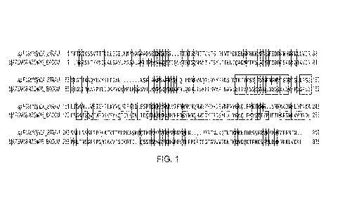
Figure 1. Pairwise sequence alignment of CytK and aHL performed using Clustalx
version 2.1. The transmembrane beta barrel of aHL is indicated by 3 boxes.
SpIP09616IHLA STAAU is aHL and trIA7GM181A7GM18 BACCN is CytK.
Figure 2. Structural model of the CytK pore. The model was made using the aHL
structure as a template for CytK, where the structure of aHL was taken from
the protein
databank (accession code 7AHL). The Modeller software was used to make the
CytK
model. Top row shows the cartoon representation of the CytK model, whilst the
bottom
row shows the surface representation. The left-hand image of the bottom row
shows the
cross section through the pore.
Figure 3. Predicted amino acid sequence of the CytK transmembrane beta barrel.
The
expected central regions of the 3 main constrictions are indicated by dashed
boxes. Any
residue with a number corresponds to residues that are predicted to point into
the cavity of
the pore. Any residue without a number corresponds to residues that are
predicted to point
towards the membrane.
Figure 4. Comparison of the radial profiles of the CytK and aHL channels
generated using
the HOLE mapping software. The CytK model was made using the aHL structure as
a
template and the aHL structure was taken from the protein databank (accession
code
7AHL).
Figure 5. Ionic current profiles through aHL wild-type and CytK wild-type and
mutants
as the voltage is gradually increased in 25 mV steps every 30 seconds in both
the negative
and positive direction from (-)25 mV up to (-)200 mV. The applied voltage is
shown by
dashed lines (blue lines in original colour image), the raw current trace by
grey lines (black
5
CA 03229995 2024- 2- 23
WO 2023/026056
PCT/GB2022/052196
lines in original colour image) and the event detected signal is shown by
black lines (red
lines in original colour image).
Figure 6. Averaged ionic current profiles through aHL wild-type and CytK wild-
type as
the voltage is gradually increased in 25 mV steps every 30 seconds in both the
negative
and positive direction from (-)25 mV up to (-)200 mV. The top row shows the
mean
current within a voltage step grouped either by run (left) or pore batch
(right). The bottom
row shows the mean current of the first 100 ms within a voltage step grouped
either by run
(left) or pore batch (right). Plotting the mean current of the first 100 ms
reduces the
influence of pore gating into the measured current. Pore Batch A =aHL-(WT),
Pore Batch
B= CytK-(WT-H6), Pore Batch C= CytK-(WT-H6), Pore Batch D= CytK-(WT-H6-D8),
Pore Batch E= CytK-(WT-H6-D8).
Figure 7. Averaged ionic current profiles through CytK wild-type and CytK
mutants as the
voltage is gradually increased in 25 mV steps in both the negative and
positive direction
from (-)25 mV up to (-)200 mV. Panels 1 and 3 (top row in original image) show
the mean
current within a voltage step grouped either by run (panel 1) or pore batch
(panel 3). Panels
2 and 4 (bottom row in original image) show the mean current of the first 100
ms within a
voltage step grouped either by run (panel 2) or pore batch (panel 4). Plotting
the mean
current of the first 100 ms reduces the influence of pore gating into the
measured current.
Pore Batch B = CytK-(WT-H6), Pore Batch C = CytK-(WT-H6), Pore Batch D = CytK-
(WT-H6-D8), Pore Batch E = CytK-(WT-H6-D8). Pore Batch F = CytK-(WT-
E113S/K156S-D8), Pore Batch G = CytK-(WT-Q123S/Q146S-D8), Pore Batch H = CytK-
(WT-K129S/E140S-D8), Pore Batch I = CytK-(WT-Q123S/Q146S/K129S/E140S-D8),
Pore Batch J = CytK-(WT-Q123S/Q146S/K129S/E140S-D8), Pore Batch K = CytK-(WT-
E113S/K156S/Q123S/Q146S/K129S/E140S), Pore Batch L = CytK-(WT-
Ell3N/K156S/Q123S/Q146S/K129S/E140S-D8).
Figure 8. Current versus time traces as DNA translocates through aHL wild-type
and
CytK wild-type and mutants. The raw current trace is shown by grey lines
(black lines in
original colour image) and the event detected signal is shown by black lines
(red lines in
original colour image). For each pore, the top row shows the full DNA current
trace, the
middle row shows the first section of the current trace and the bottom row
shows a zoomed
in view of the first section of the current trace.
Figure 9. Table summarizing the pore characteristics of CytK wild-type and
mutants.
SNR is the signal to noise ratio which is the range of the signal divided by
the noise as
6
CA 03229995 2024- 2- 23
WO 2023/026056
PCT/GB2022/052196
DNA is translocating through the pore. Median current is the median current of
the signal
as DNA is translocating through the pore.
Figure 10. Box plots showing the pore characteristics of CytK wild-type and
mutants.
SNR is the signal to noise ratio which is the range of the signal divided by
the noise as
DNA is translocating through the pore. Median current is the median current of
the signal
as DNA is translocating through the pore.
Figure 11. Bar charts showing the pore characteristic of CytK wild-type and
mutants in
condition 7, where condition 7 is 1 mM ATP, 10 mM MgCl2, 100 nM He1308 mutant,
1 M
NaC1, pH8, 100 mM HEPES. 10 mM Potassium Ferrocyanide, 10 mM Potassium
Ferricyanide, 180 mV.
Figure 12. Bar charts showing the pore characteristic of CytK wild-type and
mutants in
condition 9, where condition 9 is 1 mM ATP, 10 mM MgCl2, 100 nM He1308 mutant,
625
mlVI KC1, pH8, 100 mM HEPES, 75 mM Potassium Ferrocyanidc, 25 mM Potassium
Ferricyanide.
Figure 13. The polynucleotide-polypeptide conjugate used to translocate a
peptide through
a nanopore.
Figure 14. Example current versus time traces as a polynucleotide-polypeptide
conjugate
translocates through CytK wild-type and mutants, where the polypeptide section
comprises
GGSGRRSGSG. The peptide section of the squiggles is highlighted by the boxes
(red
boxes in original colour image). The traces begin with a long flat section
corresponding to
the capture of the C3 leader on the adapter.
Figure 15. Example current versus time traces as a polynucleotide-polypeptide
conjugate
translocates through the CytK mutant CytK-(WT-Q123S/Q146S/K129S/E140S), where
the
polypeptide section comprises either GGSGRRSGSG, GGSGYYSGSG or
GGSGDDSGS G. The peptide section of the squiggles is highlighted by the boxes
(red
boxes in original colour image).
Figure 16. The DNA sequencing Y-adapter used to translocate ssDNA through a
nanopore.
Detailed Description
The present invention will be described with respect to particular embodiments
and
with reference to certain drawings but the invention is not limited thereto
but only by the
claims. Any reference signs in the claims shall not be construed as limiting
the scope. Of
course, it is to be understood that not necessarily all aspects or advantages
may be achieved
7
CA 03229995 2024- 2- 23
WO 2023/026056
PCT/GB2022/052196
in accordance with any particular embodiment of the invention. Thus, for
example those
skilled in the art will recognize that the invention may be embodied or
carried out in a
manner that achieves or optimizes one advantage or group of advantages as
taught herein
without necessarily achieving other aspects or advantages as may be taught or
suggested
herein.
The invention, both as to organization and method of operation, together with
features and advantages thereof, may best be understood by reference to the
following
detailed description when read in conjunction with the accompanying drawings.
The
aspects and advantages of the invention will be apparent from and elucidated
with
reference to the embodiment(s) described hereinafter. Reference throughout
this
specification to "one embodiment" or "an embodiment" means that a particular
feature,
structure or characteristic described in connection with the embodiment is
included in at
least one embodiment of the present invention. Thus, appearances of the
phrases "in one
embodiment" or "in an embodiment" in various places throughout this
specification are not
necessarily all referring to the same embodiment, but may. Similarly, it
should be
appreciated that in the description of exemplary embodiments of the invention,
various
features of the invention are sometimes grouped together in a single
embodiment, figure,
or description thereof for the purpose of streamlining the disclosure and
aiding in the
understanding of one or more of the various inventive aspects. This method of
disclosure,
however, is not to be interpreted as reflecting an intention that the claimed
invention
requires more features than are expressly recited in each claim. Rather, as
the following
claims reflect, inventive aspects lie in less than all features of a single
foregoing disclosed
embodiment.
It should be appreciated that "embodiments" of the disclosure can be
specifically
combined together unless the context indicates otherwise. The specific
combinations of all
disclosed embodiments (unless implied otherwise by the context) are further
disclosed
embodiments of the claimed invention.
In addition as used in this specification and the appended claims, the
singular forms
"a", "an", and "the" include plural referents unless the content clearly
dictates otherwise.
Thus, for example, reference to "a polynucleotide" includes two or more
polynucleotides,
reference to "a helicase" includes two or more heli cases, reference to "a
monomer" refers
to two or more monomers, reference to "a pore" includes two or more pores and
the like.
All publications, patents and patent applications cited herein, whether supra
or
infra, are hereby incorporated by reference in their entirety.
8
CA 03229995 2024- 2- 23
WO 2023/026056
PCT/GB2022/052196
Definitions
Where an indefinite or definite article is used when referring to a singular
noun e.g.
"a" or "an", "the", this includes a plural of that noun unless something else
is specifically
stated. Where the term "comprising" is used in the present description and
claims, it does
not exclude other elements or steps. Furthermore, the terms first, second,
third and the like
in the description and in the claims, are used for distinguishing between
similar elements
and not necessarily for describing a sequential or chronological order. It is
to be
understood that the tel _______ Its so used are interchangeable under
appropriate circumstances and
that the embodiments of the invention described herein are capable of
operation in other
sequences than described or illustrated herein. The following terms or
definitions are
provided solely to aid in the understanding of the invention. Unless
specifically defined
herein, all terms used herein have the same meaning as they would to one
skilled in the art
of the present invention. Practitioners are particularly directed to Sambrook
et al.,
Molecular Cloning: A Laboratory Manual, 4`1' ed., Cold Spring Harbor Press,
Plainsview,
New York (2012); and Ausubel et al., Current Protocols in Molecular Biology
(Supplement 114), John Wiley & Sons, New York (2016), for definitions and
terms of the
art. The definitions provided herein should not be construed to have a scope
less than
understood by a person of ordinary skill in the art.
"About" as used herein when referring to a measurable value such as an amount,
a
temporal duration, and the like, is meant to encompass variations of 20 % or
10 %,
more preferably 5 %, even more preferably 1 %, and still more preferably
0.1 %
from the specified value, as such variations are appropriate to perform the
disclosed
methods.
"Nucleotide sequence", "DNA sequence- or "nucleic acid molecule(s)" as used
herein refers to a polymeric form of nucleotides of any length. either
ribonucleotides or
deoxyribonucleotides. This term refers only to the primary structure of the
molecule. Thus,
this term includes double- and single-stranded DNA, and RNA. The term -nucleic
acid" as
used herein, is a single or double stranded covalently-linked sequence of
nucleotides in
which the 3' and 5' ends on each nucleotide are joined by phosphodiester
bonds. The
poi ynucleotide may be made up of deoxyribonucleotide bases or ribonucleotide
bases.
Nucleic acids may be manufactured synthetically in vitro or isolated from
natural sources.
Nucleic acids may further include modified DNA or RNA, for example DNA or RNA
that
has been methylated, or RNA that has been subject to post-translational
modification, for
9
CA 03229995 2024- 2- 23
WO 2023/026056
PCT/GB2022/052196
example 5'-capping with 7-methylguanosine, 3'-processing such as cleavage and
polyadenylation, and splicing. Nucleic acids may also include synthetic
nucleic acids
(XNA), such as hexitol nucleic acid (HNA), cyclohexene nucleic acid (CeNA),
threose
nucleic acid (TNA), glycerol nucleic acid (GNA), locked nucleic acid (LNA) and
peptide
nucleic acid (PNA). Sizes of nucleic acids, also referred to herein as
"polynucleotides" are
typically expressed as the number of base pairs (bp) for double stranded
polynucleotides,
or in the case of single stranded polynucleotides as the number of nucleotides
(nt). One
thousand bp or nt equal a kilobase (kb). Polynucleotides of less than around
40 nucleotides
in length are typically called "oligonucleotides- and may comprise primers for
use in
manipulation of DNA such as via polymerase chain reaction (PCR).
The term "amino acid" in the context of the present disclosure is used in its
broadest sense and is meant to include organic compounds containing amine
(NH2) and
carboxyl (COOH) functional groups, along with a side chain (e.g., a R group)
specific to
each amino acid. In some embodiments, the amino acids refer to naturally
occurring L
amino acids or residues. The commonly used one and three letter abbreviations
for
naturally occurring amino acids are used herein: A=Ala; C=Cys; D=Asp; E=G1u;
F=Phe;
G=Gly; H=His; K=Lys; L=Leu; M=Met; N=Asn; P=Pro; Q=G1n;
R=Arg; S=Ser;
T=Thr; V=Val; W=Trp; and Y=Tyr (Lehninger, A. L., (1975) Biochemistry, 2d ed.,
pp.
71-92, Worth Publishers, New York). The general term "amino acid" further
includes D-
amino acids, retro-inverso amino acids as well as chemically modified amino
acids such as
amino acid analogues, naturally occurring amino acids that are not usually
incorporated
into proteins such as norleucine, and chemically synthesised compounds having
properties
known in the art to be characteristic of an amino acid, such as 13-amino
acids. For example,
analogues or mimetics of phenylalanine or proline, which allow the same
conformational
restriction of the peptide compounds as do natural Phe or Pro, are included
within the
definition of amino acid. Such analogues and mimetics are referred to herein
as "functional
equivalents" of the respective amino acid. Other examples of amino acids are
listed by
Roberts and Vellaccio, The Peptides: Analysis, Synthesis, Biology, Gross and
Meiehofer,
eds., Vol. 5 p. 341, Academic Press, Inc., N.Y. 1983, which is incorporated
herein by
reference.
The terms "polypeptide" and "peptide" are interchangeably used herein to refer
to a
polymer of amino acid residues and to variants and synthetic analogues of the
same. Thus,
these terms apply to amino acid polymers in which one or more amino acid
residues is a
synthetic non-naturally occurring amino acid, such as a chemical analogue of a
CA 03229995 2024- 2- 23
WO 2023/026056
PCT/GB2022/052196
corresponding naturally occurring amino acid, as well as to naturally-
occurring amino acid
polymers. Polypeptides can also undergo maturation or post-translational
modification
processes that may include, but are not limited to: glycosylation, proteolytic
cleavage,
lipidization, signal peptide cleavage, propeptide cleavage, phosphorylation,
and such like.
A peptide can be made using recombinant techniques, e.g., through the
expression of a
recombinant or synthetic polynucleotide. A recombinantly produced peptide it
typically
substantially free of culture medium, e.g., culture medium represents less
than about 20 %,
more preferably less than about 10 %, and most preferably less than about 5 %
of the
volume of the protein preparation.
The term "protein" is used to describe a folded polypeptide having a secondary
or
tertiary structure. The protein may be composed of a single polypeptide, or
may comprise
multiple polypeptides that are assembled to form a multimer. The multimer may
be a
homooligomer, or a heterooligmer. The protein may be a naturally occurring, or
wild type
protein, or a modified, or non-naturally, occurring protein. The protein may,
for example,
differ from a wild type protein by the addition, substitution or deletion of
one or more
amino acids.
A "variant" of a protein encompass peptides, oligopeptides, polypeptides,
proteins
and enzymes having amino acid substitutions, deletions and/or insertions
relative to the
unmodified or wild-type protein in question and having similar biological and
functional
activity as the unmodified protein from which they are derived. The term
"amino acid
identity" as used herein refers to the extent that sequences are identical on
an amino acid-
by-amino acid basis over a window of comparison. Thus, a "percentage of
sequence
identity" is calculated by comparing two optimally aligned sequences over the
window of
comparison, determining the number of positions at which the identical amino
acid residue
(e.g., Ala, Pro, Ser, Thr, Gly, Val, Leu, Ile, Phe, Tyr, Trp, Lys, Arg, His,
Asp, Glu, Asn,
Gln, Cys and Met) occurs in both sequences to yield the number of matched
positions.
dividing the number of matched positions by the total number of positions in
the window
of comparison (i.e., the window size), and multiplying the result by 100 to
yield the
percentage of sequence identity.
For all aspects and embodiments of the present invention, a "variant" has at
least
50%, 60%, 70%, 80%, 90%, 95% or 99% complete sequence identity to the amino
acid
sequence of the corresponding wild-type protein. Sequence identity can also be
to a
fragment or portion of the full length polynucleotide or polypeptide. Hence, a
sequence
may have only 50 % overall sequence identity with a full length reference
sequence, but a
11
CA 03229995 2024- 2- 23
WO 2023/026056
PCT/GB2022/052196
sequence of a particular region, domain or subunit could share 80 %, 90 %, or
as much as
99 % sequence identity with the reference sequence.
The term "wild-type" refers to a gene or gene product isolated from a
naturally
occurring source. A wild-type gene is that which is most frequently observed
in a
population and is thus arbitrarily designed the "normal" or "wild-type" form
of the gene. In
contrast, the term "modified-, "mutant- or "variant- refers to a gene or gene
product that
displays modifications in sequence (e.g., substitutions, truncations, or
insertions), post-
translational modifications and/or functional properties (e.g., altered
characteristics) when
compared to the wild-type gene or gene product. It is noted that naturally
occurring
mutants can be isolated; these are identified by the fact that they have
altered
characteristics when compared to the wild-type gene or gene product. Methods
for
introducing or substituting naturally-occurring amino acids are well known in
the art. For
instance. methionine (M) may be substituted with arginine (R) by replacing the
codon for
methionine (ATG) with a codon for arginine (CGT) at the relevant position in a
polynucleotide encoding the mutant monomer. Methods for introducing or
substituting
non-naturally-occurring amino acids are also well known in the art. For
instance, non-
naturally-occurring amino acids may be introduced by including synthetic
aminoacyl-
tRNAs in the IVTT system used to express the mutant monomer. Alternatively,
they may
be introduced by expressing the mutant monomer in E. coil that are auxotrophic
for
specific amino acids in the presence of synthetic (i.e. non-naturally-
occurring) analogues
of those specific amino acids. They may also be produced by naked ligation if
the mutant
monomer is produced using partial peptide synthesis. Conservative
substitutions replace
amino acids with other amino acids of similar chemical structure, similar
chemical
properties or similar side-chain volume. The amino acids introduced may have
similar
polarity, hydrophilicity, hydrophobicity, basicity, acidity, neutrality or
charge to the amino
acids they replace. Alternatively, the conservative substitution may introduce
another
amino acid that is aromatic or aliphatic in the place of a pre-existing
aromatic or aliphatic
amino acid. Conservative amino acid changes are well-known in the art and may
be
selected in accordance with the properties of the 20 main amino acids as
defined in Table 1
below. Where amino acids have similar polarity, this can also be determined by
reference
to the hydropathy scale for amino acid side chains in Table 2.
12
CA 03229995 2024- 2- 23
WO 2023/026056
PCT/GB2022/052196
Table 1 - Chemical properties of amino acids
Ala aliphatic, hydrophobic, neutral
Met hydrophobic, neutral
Cys polar, hydrophobic, neutral Asn polar, hydrophilic,
neutral
Asp polar, hydrophilic, charged (-)
Pro hydrophobic, neutral
Glu polar, hydrophilic, charged (-)
Gln polar, hydrophilic, neutral
Phe aromatic, hydrophobic, neutral
Arg polar, hydrophilic, charged (+)
Gly aliphatic, neutral Ser polar, hydrophilic,
neutral
His aromatic, polar, hydrophilic, Thr polar, hydrophilic,
neutral
charged (+)
De aliphatic, hydrophobic, neutral
Val aliphatic, hydrophobic, neutral
Lys polar, hydrophilic, charged(+)
Tip aromatic, hydrophobic, neutral
Leu aliphatic, hydrophobic, neutral
Tyr aromatic, polar, hydrophobic
Table 2 - Hydropathy scale
Side Chain Hydropathy
Tie 4.5
Val 4.2
Leu 3.8
Phe 2.8
Cys 2.5
Met 1.9
Ala 1.8
Gly -0.4
Thr -0.7
Ser -0.8
Trp -0.9
Tyr -1.3
Pro -1.6
His -3.2
Gin -3.5
Gln -3.5
Asp -3.5
Asn -3.5
Lys -3.9
Arg -4.5
A mutant or modified protein, monomer or peptide can also be chemically
modified
in any way and at any site. A mutant or modified monomer or peptide is
preferably
chemically modified by attachment of a molecule to one or more cysteines
(cysteine
linkage), attachment of a molecule to one or more lysines, attachment of a
molecule to one
or more non-natural amino acids, enzyme modification of an epitope or
modification of a
13
CA 03229995 2024- 2- 23
WO 2023/026056
PCT/GB2022/052196
terminus. Suitable methods for carrying out such modifications are well-known
in the art.
The mutant of modified protein, monomer or peptide may be chemically modified
by the
attachment of any molecule. For instance, the mutant of modified protein,
monomer or
peptide may be chemically modified by attachment of a dye or a fluorophore.
Mutant Cytotoxin K monomers
The invention provides methods of characterising an analyte using a pore
comprising at least one mutant Cytotoxin K (CytK) monomer.
The invention also provides mutant Cytotoxin K (CytK) monomers. The mutant
CytK monomers may be used to form pores of the invention. A mutant CytK
monomer is
a monomer whose sequence varies from that of a wild-type CytK monomer (SEQ ID
NO:
1) and which retains the ability to form a pore. Methods for confirming the
ability of
mutant monomers to fat __________ 11 pores are well-known in the art and are
discussed in more detail
below. For instance, the ability of a mutant monomer to form a pore can be
determined as
described in the Examples.
Pores comprising the mutant monomers of the invention have an increased
current
range when subject to an applied potential in a nanopore-based method of
analyte
characterisation, relative to a pore consisting of wild type CytK monomers. An
increased
current range makes it easier to identify and characterise target analytes,
and in particular
makes it easier to discriminate between components of the target analyte. For
example,
when the target analyte is a polypeptide, an increased current range makes it
easier to
discriminate between amino acids in the polypeptide.
Pores comprising a mutant CytK monomer of the invention may be used to
characterise any suitable analyte. Suitable analytes are described further
herein. The
increased current range in particular render the pores comprising a mutant
CytK monomer
of the invention particularly applicable to nanopore-based methods of
characterising
polypeptide analytes as described herein. Techniques to characterise
polypeptides are of
significant biotechnological importance. For example, knowledge of a protein
sequence
can allow structure-activity relationships to be established and has
implications in rational
drug development strategies for developing ligands for specific receptors.
Identification of
post-translational modifications is also key to understanding the functional
properties of
many proteins. For example, typically 30-50% of protein species are
phosphorylated in
eukaryotes. Some proteins may have multiple phosphorylation sites, serving to
activate or
inactivate a protein, promote its degradation, or modulate interactions with
protein
14
CA 03229995 2024- 2- 23
WO 2023/026056
PCT/GB2022/052196
partners. Described herein is the successful utilisation of pores comprising a
mutant CytK
monomer in a nanopore based method of characterising a target polypeptide.
Accordingly,
the inventors have surprisingly identified a novel means for characterising
polypeptide
analytes.
The inventors have surprisingly identified a region within the CytK monomer
which can be modified to alter the interaction between the monomer and an
analyte, such
as when the anal yte is characterised using nanopore-based methods of analyte
characterisation described herein comprising the use of a pore comprising a
CytK mutant
monomer of the invention. With reference to the wild type polypeptide sequence
of a
CytK monomer as defined by SEQ ID NO: 1, the region is from about position
S100 to
about position K170 in SEQ ID NO: 1. At least a part of this region typically
contributes
to the membrane spanning region of CytK. At least a part of this region
typically
contributes to the barrel or channel of CytK. At least a part of this region
typically
contributes to the internal wall or lining of CytK.
The improved analyte characterisation properties of the CytK mutant monomers
are
achieved via the introduction of one or more modification at one or more
positions in the
region of SEQ ID NO: 1 between about S100 and about K170 which alter the
ability of the
monomer to interact with the analyte. Preferable mutations are further
described herein.
Accordingly, provided is a mutant CytK monomer comprising a variant of the
amino acid
sequence of SEQ ID NO: 1; wherein the monomer is capable of forming a pore;
and
wherein the variant comprises one or more modifications at one or more
positions in the
region of SEQ ID NO 1: between about S100 and about K170 which alter the
ability of the
monomer to interact with an analyte.
In accordance with the invention, the variant comprises one or more
modifications
at one or more positions in the region of SEQ ID NO: 1 between about S100 and
K170
which alter the ability of the monomer, or preferably the region. to interact
with an analyte.
The interaction between the monomer and the analyte may be increased or
decreased. An
increased interaction between the monomer and an analyte will, for example,
facilitate
capture of the analyte by pores comprising the mutant monomer. A decreased
interaction
between the monomer and an analyte will, for example, improve recognition or
discrimination of the analyte. Recognition or discrimination of the analyte
may be
improved by increasing the current range by virtue of the modifications to the
CytK
monomer between about S100 and K170 of SEQ ID NO: 1 described herein. The
CA 03229995 2024- 2- 23
WO 2023/026056
PCT/GB2022/052196
improved recognition or discrimination of the anal yte may particularly be
improved
achieved via five main mechanisms, namely by independent changes in the:
= sterics (e.g. increasing or decreasing the size of amino acid residues);
= net charge of the amino acid residue at the modified position (e.g.
introducing or removing negative (¨ye) charge and/or introducing or
removing positive (-Fve) charge);
= hydrogen bonding characteristics of the amino acid residue at the
modified
position (e.g. introducing amino acids that can hydrogen bond to the
analyte);
alyte);
p 10 stacking (e.g. introduce to or remove from the amino acid
residue at the
modified position one or more chemical groups that interact through
delocalized electron pi systems); and/or
amino acid residue at the modified position, thereby changing the structure
of the pore (e.g. introducing amino acids that increase or decrease the size
of the barrel or channel).
Thus, the one or more modification may each independently (a) alter the size
of the
amino acid residue at the modified position; (b) alter the net charge of the
amino acid
residue at the modified position; (c) alter the hydrogen bonding
characteristics of the
amino acid residue at the modified position; (d) introduce to or remove from
the amino
acid residue at the modified position one or more chemical groups that
interact through
&localized electron pi systems and/or (c) alter the structure of the amino
acid residue at
the modified position.
Any one or more of these mechanisms of independent alteration may be
responsible
for the improved properties of the pores formed from the mutant monomers of
the
invention. For instance, a pore comprising a mutant monomer of the invention
may
display improved polypeptide and/or polynucleotide reading properties as a
result of
altered sterics, altered hydrogen bonding and an altered structure.
Accordingly, provided herein is a method of characterising a target analyte.
comprising:
(a) contacting the target analyte with a pore comprising at least one
mutant
Cytotoxin K monomer comprising a variant of the amino acid sequence of
SEQ ID NO: 1; such that the target analyte moves with respect to the pore;
wherein the variant comprises one or more modifications at one or
more positions in the region of SEQ ID NO: 1 between about S100 and
16
CA 03229995 2024- 2- 23
WO 2023/026056
PCT/GB2022/052196
about K170 which alter the ability of the monomer to interact with the
analyte; and
(b) taking one or more measurements characteristic of
the analyte as the analyte
moves with respect to the pore,
thereby characterising the target analyte.
Also provided is a mutant CytK monomer comprising a variant of the amino acid
sequence of SEQ ID NO: 1; wherein the monomer is capable of forming a pore;
and
wherein the variant comprises one or more modifications at one or more
positions in the
region of SEQ ID NO 1: between about S100 and about K170 which alter the
ability of the
monomer to interact with an analyte.
The ability of the monomer to interact with a target analyte to interact with
an
analyte can be determined using methods that are well-known in the art. The
monomer
may interact with an analyte in any way, e.g. by non-covalent interactions,
such as
hydrophobic interactions, hydrogen bonding. Van der Waal's forces, pi (70-
cation
interactions or electrostatic forces. For instance, the ability of the region
to bind to an
analyte can be measured using a conventional binding assay. Suitable assays
include, but
are not limited to, fluorescence-based binding assays, nuclear magnetic
resonance (NMR),
Isothermal Titration Calorimetry (ITC) or Electron spin resonance (ESR)
spectroscopy.
Alternatively, the ability of a pore comprising one or more of the mutant
monomers to
interact with an analyte can be determined using any of the methods discussed
above or
below. Preferred assays are described in the Examples.
The one or more modifications are within the region from about position 100 to
about position 170 of SEQ ID NO: 1. The one or more modifications are
preferably within
the region from about position 110 to about position 160 of SEQ ID NO: 1. The
one or
more modifications are yet more preferably within the region from about
position 113 to
about position 156 of SEQ ID NO: 1.
Modifications of protein nanopores that alter their ability to interact with
an
analyte, and in particular improve their current range, are well documented in
the art. For
instance, such modifications are disclosed in WO 2010/034018, WO 2010/055307,
WO
2013/153359 and WO 2016/034591. Similar modifications can be made to the CytK
monomer in accordance with this invention.
Any number of modifications may be made, such as 1, 2, 5, 10, 15, 20, 30 or
more
modifications. Any modification(s) can be made as long as the ability of the
monomer to
interact with a polynucleotide is altered and the monomer remains capable of
forming a
17
CA 03229995 2024- 2- 23
WO 2023/026056
PCT/GB2022/052196
pore. Suitable modifications include, hut are not limited to, amino acid
substitutions,
amino acid additions and amino acid deletions. The one or more modifications
are
preferably one or more substitutions. This is discussed in more detail below.
The one or more modifications preferably (a) alter the steric effect of the
monomer,
or preferably alter the steric effect of the region, (b) alter the net charge
of the monomer, or
preferably alter the net charge of the region, (c) alter the ability of the
monomer, or
preferably of the region, to hydrogen bond with the anal yte, (d) introduce or
remove
chemical groups that interact through delocalized electron pi systems and/or
(e) alter the
structure of the monomer, or preferably alter the structure of the region. The
one or more
modifications more preferably result in any combination of (a) to (e), such as
(a) and (b);
(a) and (c); (a) and (d); (a) and (e); (b) and (c); (b) and (d); (b) and (e);
(c) and (d); (c) and
(e); (d) and (e), (a), (b) and (c); (a), (b) and (d); (a), (b) and (e); (a),
(c) and (d); (a), (c) and
(e); (a), (d) and (c); (b), (c) and (d); (b), (c) and (c); (b), (d) and (c);
(c), (d) and (e); (a), (b),
(c) and d); (a), (b), (c) and (e); (a), (b), (d) and (e); (a), (c), (d) and
(e); (b), (c), (d) and (e);
and (a), (b), (c) and (d).
For (a), the steric effect of the monomer can be increased or decreased. Any
method of altering the steric effects may be used in accordance with the
invention. The
introduction of bulky residues, such as phenylalanine (F), tryptophan (W),
tyrosine (Y) or
histidine (H), increases the sterics of the monomer. The one or more
modifications are
preferably the introduction of one or more of F, W, Y and H. Any combination
of F, W, Y
and H may be introduced. The one or more of F, W, Y and H may be introduced by
addition. The one or more of F, W, Y and H are preferably introduced by
substitution.
Suitable positions for the introduction of such residues are discussed in more
detail below.
The removal of bulky residues, such as phenylalanine (F), tryptophan (W),
tyrosine
(Y) or histidine (H), conversely decreases the sterics of the monomer. The one
or more
modifications are preferably the removal of one or more of F. W. Y and H. Any
combination of F, W, Y and H may be removed. The one or more of F, W, Y and H
may
be removed by deletion. The one or more of F, W, Y and H are preferably
removed by
substitution with residues having smaller side groups. such as serine (S),
threonine (T),
alanine (A) and valine (V).
For (b), the net charge can be altered in any way. The net positive charge is
preferably increased or decreased. The net positive charge can be increased in
any manner.
The net positive charge is preferably increased by introducing, preferably by
substitution,
18
CA 03229995 2024- 2- 23
WO 2023/026056
PCT/GB2022/052196
one or more positively charged amino acids and/or neutralising, preferably by
substitution,
one or more negative charges.
The net positive charge is preferably increased by introducing one or more
positively charged amino acids. The one or more positively charged amino acids
may be
introduced by addition. The one or more positively charged amino acids are
preferably
introduced by substitution. A positively charged amino acid is an amino acid
with a net
positive charge. The positively charged amino acid(s) can be naturally-
occurring or non-
naturally-occurring. The positively charged amino acids may be synthetic or
modified.
For instance, modified amino acids with a net positive charge may be
specifically designed
for use in the invention. A number of different types of modification to amino
acids are
well known in the art. The one or more modifications comprising the
introduction of one
or more positively charged amino acids preferably comprise the introduction of
one or
more of histidine (H), lysine (K) and argininc (R) by way of substitution or
addition,
although most preferably by substitution. Suitable positions for the
introduction of such
residues are discussed in more detail below.
Methods for adding or substituting naturally-occurring amino acids are well
known
in the art. For instance, the nucleotides which constitute a codon that are
comprised within
a polynucleotide coding sequence may be modified such that the nucleotide
contents of the
codon is altered, thereby leading to a different amino acid to be coded for by
said codon.
Such a polynucleotide may then be expressed as discussed below.
Methods for adding or substituting non-naturally-occurring amino acids are
also
well known in the art. For instance, non-naturally-occurring amino acids may
be
introduced by including synthetic aminoacyl-tRNAs in the IVTT system used to
express
the pore. Alternatively, they may be introduced by expressing the monomer in
E. coli that
are auxotrophic for specific amino acids in the presence of synthetic (i.e.
non-naturally-
occurring) analogues of those specific amino acids. They may also be produced
by naked
ligation if the pore is produced using partial peptide synthesis.
In the one or more modifications, any amino acid may be substituted with a
positively charged amino acid. In the one or more modifications, one or more
uncharged
amino acids, non-polar amino acids and/or aromatic amino acids may be
substituted with
one or more positively charged amino acids. Uncharged amino acids have no net
charge.
Suitable uncharged amino acids include, but are not limited to, cysteine (C),
serine (S),
threonine (T), methionine (M), asparagine (N) and glutamine (Q). Non-polar
amino acids
have non-polar side chains. Suitable non-polar amino acids include, but are
not limited to,
19
CA 03229995 2024- 2- 23
WO 2023/026056
PCT/GB2022/052196
glycine (G), alanine (A), proline (P), isoleucine (T), leucine (L) and valine
(V). Aromatic
amino acids have an aromatic side chain. Suitable aromatic amino acids
include, but are
not limited to, histidine (H), phenylalanine (F), tryptophan (W) and tyrosine
(Y).
Preferably, in the one or more modifications, one or more negatively charged
amino acids
are substituted with one or more positively charged amino acids. Suitable
negatively
charged amino acids include, but are not limited to, aspartic acid (D) and
glutamic acid (E).
In the one or more modifications, preferred introductions include, hut are not
limited to, substitution of E with K, M with R, substitution of M with H,
substitution of M
with K, substitution of D with R, substitution of D with H, substitution of D
with K,
substitution of E with R, substitution of E with H, substitution of N with R,
substitution of
T with R and substitution of G with R. Most preferably E is substituted with
K.
In the one or more modifications, any number of positively charged amino acids
may be introduced or substituted. For instance, 1, 2, 5, 10. 15, 20, 25, 30 or
more
positively charged amino acids may be introduced or substituted.
The net positive charge is more preferably increased by neutralising one or
more
negative charges. The one or more negative charges may be neutralised by
substituting
one or more negatively charged amino acids with one or more uncharged amino
acids,
non-polar amino acids and/or aromatic amino acids. The removal of negative
charge
increases the net positive charge. The uncharged amino acids, non-polar amino
acids
and/or aromatic amino acids can be naturally-occurring or non-naturally-
occurring. They
may be synthetic or modified. Suitable uncharged amino acids, non-polar amino
acids and
aromatic amino acids are discussed above. Preferred substitutions include, but
are not
limited to, substitution of E with Q, substitution of E with S, substitution
of E with A,
substitution of D with Q, substitution of E with N, substitution of D with N,
substitution of
D with G and substitution of D with S.
Any number and combination of uncharged amino acids, non-polar amino acids
and/or aromatic amino acids may substituted in the one or more modifications.
For
instance, 1, 2, 5, 10, 15, 20, 25, or 30 or more uncharged amino acids, non-
polar amino
acids and/or aromatic amino acids may be substituted. Negatively charged amino
acids
may be substituted with (1) uncharged amino acids; (2) non-polar amino acids;
(3)
aromatic amino acids; (4) uncharged amino acids and non-polar amino acids; (5)
uncharged amino acids and aromatic amino acids; and (5) non-polar amino acids
and
aromatic amino acids; or (6) uncharged amino acids, non-polar amino acids and
aromatic
amino acids.
CA 03229995 2024- 2- 23
WO 2023/026056
PCT/GB2022/052196
The one or more negative charges may be neutralised by introducing one or more
positively charged amino acids near to, such as within 1, 2, 3 or 4 amino
acids, or adjacent
to one or more negatively charged amino acids. Examples of positively and
negatively
charged amino acids are discussed above. The positively charged amino acids
may be
introduced in any manner discussed above, for instance by substitution.
The net positive charge is preferably decreased by introducing one or more
negatively charged amino acids and/or neutralising one or more positive
charges. Ways in
which this might be done will be clear from the discussion above with
reference to
increasing the net positive charge. All of the embodiments discussed above
with reference
to increasing the net positive charge equally apply to decreasing the net
positive charge
except the charge is altered in the opposite way. In particular, the one or
more positive
charges are preferably neutralised by substituting one or more positively
charged amino
acids with one or more uncharged amino acids, non-polar amino acids and/or
aromatic
amino acids or by introducing one or more negatively charged amino acids near
to, such as
within 1, 2, 3 or 4 amino acids of, or adjacent to one or more negatively
charged amino
acids.
The net negative charge is preferably increased or decreased. All of the above
embodiments discussed above with reference to increasing or decreasing the net
positive
charge equally apply to decreasing or increasing the net negative charge
respectively.
For (c), the ability of the monomer to hydrogen bond may be altered in any
suitable
manner. For example, the one or more modifications may comprise the
introduction of one
or more of serine (S), threonine (T), asparagine (N), glutamine (Q), tyrosine
(Y) or
histidine (H) by addition or substitution, thereby increasing the hydrogen
bonding ability
of the monomer. The one or more modifications preferably comprise the
introduction of
one or more of S, T, N, Q, Y and H in any suitable combination, preferably
wherein the
introduction is by substitution. Suitable positions for the introduction of
such residues are
discussed in more detail below.
The removal of serine (S), threonine (T), asparagine (N), glutamine (Q),
tyrosine
(Y) or histidine (H) decreases the hydrogen bonding ability of the monomer.
For example,
the one or more modifications may comprise the removal of one or more of S, T,
N, Q, Y
and H. The one or more modifications preferably comprise the removal of any
combination of S, T, N, Q, Y and H by deletion or by substitution in any
suitable
combination, thereby decreasing the hydrogen bonding ability of the monomer.
The one or
21
CA 03229995 2024- 2- 23
WO 2023/026056
PCT/GB2022/052196
more modifications preferably comprise the substitution with other amino acids
which
hydrogen bond less well, such as alanine (A), valine (V), isoleucine (I) and
leucine (L).
For (d), the introduction of aromatic residues, such as phenylalanine (F),
tryptophan
(W), tyrosine (Y) or histidine (H), also increases the pi stacking in the
monomer. The
removal of aromatic residues, such as phenylalanine (F), tryptophan (W),
tyrosine (Y) or
histidine (H), also increases the pi stacking in the monomer. Such amino acids
can be
introduced or removed as discussed above with reference to (a).
For (e), one or more modifications made in accordance with the invention which
alter the structure of the monomer. For example, one or more loop regions can
be
removed, shortened or extended. This typically facilitates the entry or exit
of a
polynucleotide into or out of the pore. The one or more loop regions may be
the cis side
of the pore, the trans side of the pore or on both sides of the pore.
Alternatively, one or
more regions of the amino terminus and/or the carboxy terminus of the pore can
be
extended or deleted. This typically alters the size and/or charge of the pore.
It will be clear from the discussion above that the introduction of certain
amino
acids will enhance the ability of the monomer to interact with an analyte via
more than one
mechanism. For instance, the substitution of E with H will not only increase
the net
positive charge (by neutralising negative charge) in accordance with (b), but
will also
increase the ability of the monomer to hydrogen bond in accordance with (c).
The inventors surprisingly identified three constrictions in a pore consisting
of wild
type CytK monomers. A constriction is typically a narrowing in the channel
which runs
through the nanopore which may determine or control the signal obtained in any
of the
known nanopore-based methods of analyte characterisation, or any methods of
analyte
characterisation described herein, when the analyte moves with respect to the
nanopore.
The structure of each CytK monomer in the pore leads to the formation of the
three
constrictions in the barrel region of the pore. The amino acids responsible
for the
formation of the three constrictions are comprised between about S100 and
K170.
Accordingly, the mutant CytK monomer of the invention may comprise one or
modifications at one or more positions in the region of SEQ Ill NO: 1 between
about S100
and K170 which alter the ability of the monomer to interact with an analyte,
wherein the
modifications alter one or more of the three constrictions in a pore
comprising a CytK
monomer of the invention relative to a pore consisting of wild type CytK
monomers. Said
modifications may therefore alter the interaction of the constriction with an
analyte as the
analyte moves through the pore. Preferably, the monomer of the invention is
capable of
22
CA 03229995 2024- 2- 23
WO 2023/026056
PCT/GB2022/052196
forming a pore having a solvent-accessible channel from a first opening to a
second
opening of said pore; the solvent-accessible channel comprising at least one
constriction;
and wherein the one or more modifications are made to amino acids in said
constriction.
Thus, by modifying the region of a CytK monomer which is responsible for
forming the
three constrictions in a wild type CytK pore, the interaction between a CytK
monomer and
an analyte, such as when the analyte is characterised using nanopore-based
methods of
analyte characterisation described herein, can be altered.
The amino acids responsible for the formation of the three constrictions are
comprised between about S100 and K170 of SEQ ID NO:1 defining a CytK monomer,
and
preferably face inwards into the channel region when said CytK monomer
assembles to
form a CytK pore. Preferably, therefore, the one or more modifications which
alter the
characteristics of the constriction region of the CytK monomer of the
invention relative to
a wild type CytK monomer are made to amino acids which face inwards said CytK
monomer assembles to form a CytK pore. The amino acids responsible for the
contribution of a single CytK monomer to a constriction in a CytK pore
typically comprise
a pair of amino acids in the CytK monomer. Accordingly, the one or more
modifications
to the amino acids responsible for the formation of the three constrictions
are comprised
between about S100 and K170 of SEQ ID NO:1 and is preferably a modification to
a pair
of amino acids. Such pairs of amino acids are described further herein.
Thus, the one or more modification that alter the constriction may each
independently (a) alter the size of the constriction (e.g. by increasing or
decreasing the size
of the amino acid residue at the modified position); (b) alter the net charge
of the
constriction (e.g. by altering the net charge of the amino acid residue at the
modified
position); (c) alter the hydrogen bonding characteristics of the amino acid
residues in the
constriction (e.g. by altering the hydrogen bonding characteristics of the
amino acid
residue at the modified position); (d) introduce to or remove from the
constriction one or
more chemical groups that interact through delocalized electron pi systems
(e.g. by
introducing to or remove from the amino acid residue at the modified position
one or more
chemical groups that interact through delocalized electron pi systems); and/or
(e) alter the
structure of the constriction (e.g. by altering the structure of the amino
acid residue at the
modified position). The one or more modifications which alter (a)-(e) with
respect to the
constriction may be identical to those described herein with respect to the
monomer
generally.
23
CA 03229995 2024- 2- 23
WO 2023/026056
PCT/GB2022/052196
As described herein (see particularly the Examples), the inventors have
identified
three constrictions in a wild type CytK pore (i.e. a CytK pore consisting of
wild type CytK
monomers). The loop region of each CytK monomer comprises amino acids which
define
the three constrictions of a wild type CytK pore. Each constriction is defined
by amino
acids on opposite sides of the loop region. The upper constriction (closest to
the cap
region of the pore) is preferably defined by the region of SEQ ID NO: 1
between about
X109 and about T117, more preferably between V111 and T115, and between about
S152
and about X160, preferably between S154 and X158. The lower constriction
(furthest
from the cap region of the pore) is preferably defined by the region of SEQ ID
NO: 1
between about G126 and aboutV132, preferably between S127 and S131, and
between
about P137 and about A143, preferably between S138 and G142. The middle
constriction
(furthest from the cap region of the pore) is preferably defined by the region
of SEQ ID
NO: 1 between about S119 and about G126, preferably between S121 and G125, and
between about A143 and about S150, preferably between T144 and T148.
In wild type CytK, amino acids from about V111 to about S131 of SEQ ID NO: 1
and from about S138 to about T158 of SEQ ID NO: 1 form a loop region of the
pore which
comprises the three constrictions (see Figure 3). Preferably, the amino acids
that form the
three constrictions comprises amino acids in the loop region that face inwards
into the
channel of the pore. More preferably, an amino acid between about V111 to
about S131 of
SEQ ID NO: 1 forms a pair with an amino acid between S138 to about T158 of SEQ
ID
NO: 1 in order to form a constriction in the channel of the pore. Each amino
acid in a pair
within a pair is on the opposite side of the loop region to one another.
Accordingly, in a
monomer of the invention described herein, the variant may comprise one or
more
modifications in the region of SEQ ID NO: 1 between about V111 and about S131;
and/or
between about S135 and about T158. Preferably, in a monomer of the invention
described
herein, the variant may comprise one or more modifications in the region of
SEQ ID NO: 1
between about V111 and about S131 and between about S135 and about T158. In
another
aspect, in a monomer of the invention described herein, the variant may
comprise 1, 2, 3, 4,
5, 6, 7, 8, 9, or 10 or more modifications between about V111 and about S131
in SEQ ID
NO: 1; and 1, 2, 3, 4, 5, 6, 7, 8, 9, or 10 or more modifications between
about S135 and
about T158 in SEQ ID NO:l. Most preferably, the same number of modifications
are
made in the region of SEQ ID NO: 1 between about V111 and about S131 and
between
about S135 and about T158.
24
CA 03229995 2024- 2- 23
WO 2023/026056
PCT/GB2022/052196
In the CytK monomer of the invention, the variant may comprise one or more
modifications in the region of SEQ ID NO: 1 between about S119 and about G126,
preferably between S121 and G125; and/or between about A143 and about S150,
preferably between T144 and T148. Preferably, in a monomer of the invention
described
herein, the variant may comprise one or more modifications in the region of
SEQ ID NO: 1
between about S119 and about G126, preferably between S121 and G125, and
between
about A143 and about S150, preferably between T144 and T148. In another
aspect, in a
monomer of the invention described herein, the variant may comprise 1, 2, 3,
4, or 5 or
more modifications between about S119 and about G126 of SEQ ID NO: 1,
preferably
between S121 and G125; and 1, 2, 3, 4 or 5 or more modifications between about
A143
and about S150 of SEQ ID NO: 1, preferably between T144 and T148. Most
preferably,
the same number of modifications are made in the region of SEQ ID NO: 1
between about
S119 and about G126 of SEQ ID NO: 1, preferably between S121 and G125, and
between
about A143 and about S150 of SEQ ID NO: 1, preferably between T144 and T148.
In the CytK monomer of the invention, the variant may comprise one or more
modifications in the region of SEQ ID NO: 1 between about G126 and about VI
32,
preferably between S127 and S131; and/or between about P137 and about A143,
preferably between S138 and G142. Preferably, in a monomer of the invention
described
herein, the variant may comprise one or more modifications in the region of
SEQ ID NO: 1
between about G126 and about V132, preferably between S127 and S131, and
between
about P137 and about A143, preferably between S138 and G142. In another
aspect, in a
monomer of the invention described herein, the variant may comprise 1, 2, 3,
4, or 5 or
more modifications between about G126 and about V132 of SEQ ID NO: 1,
preferably
between S127 and S131; and 1, 2, 3, 4 or 5 or more modifications between about
P137 and
about A143 of SEQ ID NO: 1, preferably between S138 and G142. Most preferably,
the
same number of modifications are made in the region of SEQ ID NO: 1 between
about
G126 and about V132 of SEQ ID NO: 1, preferably between S127 and S131, and
between
about P137 and about A143 of SEQ ID NO: 1, preferably between S138 and G142.
In the CytK monomer of the invention, the variant may comprise one or more
modifications in the region of SEQ ID NO: 1 between about N109 and about T117,
preferably between Viii and T115; and/or between about S152 and about Y160,
preferably between S154 and T158. Preferably, in a monomer of the invention
described
herein, the variant may comprise one or more modifications in the region of
SEQ ID NO: 1
between about N109 and about T117, preferably between V111 and T115, and
between
CA 03229995 2024- 2- 23
WO 2023/026056
PCT/GB2022/052196
about S152 and about Y160, preferably between S154 and T158. In another
aspect, in a
monomer of the invention described herein, the variant may comprise 1, 2, 3,
4, or 5 or
more modifications between about N109 and about T117 of SEQ ID NO: 1,
preferably
between V111 and T115; and 1, 2, 3, 4 or 5 or more modifications between about
S152 and
about Y160 of SEQ ID NO: 1, preferably between S154 and T158. Most preferably,
the
same number of modifications are made in the region of SEQ ID NO: 1 between
about
N109 and about T117 of SEQ ID NO: 1, preferably between V111 and T115, and
between
about S152 and about Y160 of SEQ ID NO: 1, preferably between S154 and T158.
The variant preferably comprises a modification at one or more of the
following
positions of SEQ ID NO: 1: E113, T115, T117, S119, S121, Q123, G125, S127,
K129,
S131, V132, T133, P134, S135, G136, P137, S138, E140, G142, T144, Q146, T148,
S150,
S152, S154 and K156. The variant preferably comprises modification at 1, 2. 3,
4, 5, 6, 7,
8, 9, 10, 11, 12, 13, 14, 15, 16, 17, 18. 19, 20, 21, 22, 23, 24, or 25 or
more of these
positions. The variant may independently comprise one or more amino acid
substitutions,
additions and/or deletions at said one or more positions. The amino acids
substituted into
the variant may he naturally-occurring or non-naturally occurring derivatives
thereof. The
amino acids substituted into the variant may be D-amino acids. In particular,
the variant
may comprise one or more amino acid substitutions at the positions listed
above, and the
amino acid(s) substituted into the variant are selected from aspartate,
glutamate, serine,
threonine, asparagine, glutamine, glycine, alanine, valine, leucine,
isoleucine, cysteine,
argininc. lysine and phenylalanine.
The variant preferably comprises one or more of the following modifications of
SEQ lD NO: 1:
a) E113 S/T/N/Q/G/A/V/L/I/C/R/K/F/Y;
b) T 115 S/N/Q/G/A/V/L/I/C/R/K/F ;
c) T117S/N/Q/G/A/V/L/I/C/R/K/F;
d) S119T/N/Q/G/A/V/L/I/C/R/K/F;
e) S121T/N/Q/G/A/V/L/I/C/R/K/F;
f) Q123S/T/N/G/A/V/L/1/C/R/K/14/M/Y;
g) G125S/T/N/Q/A/V/L/I/C/R/K/F;
11) S127T/N/Q/G/A/V/L/I/C/R/K/F;
i) K129S/T/N/Q/G/A/V/L/I/C/R/F/Y;
j) S131T/N/Q/G/A/V/L/I/C/R/K/F;
k) V132S/T/N/Q/G/A/L/I/C/R/K/F;
26
CA 03229995 2024- 2- 23
WO 2023/026056
PCT/GB2022/052196
1) T133 S/N/Q/G/A/V/L/I/C/R/K/F;
m) P134S/T/N/Q/G/A/V/L/I/C/R/K/F;
n) S135T/N/Q/G/A/V/L/I/C/R/K/F;
o) G136S /T/N/Q/A/V/L/I/C/R/K/F ;
p) P137S/T/N/Q/G/A/V/L/I/C/R/K/F;
q) S138T/N/Q/G/A/V/L/1/C/R/K/F;
r) E140S/T/N/Q/G/A/V/L/I/C/R/K/F;
s) G142S/T/N/Q/A/V/L/1/C/R/K/F;
t) T144S/N/Q/G/A/V/L/1/C/R/K/F;
u) Q146S/T/N/G/A/V/L/I/C/R/K/F/M/Y;
v) T148S/N/Q/G/A/V/L/I/C/R/K/F;
w) S 150T/N/Q/G/A/V/L/I/C/R/K/F;
x) S152T/N/Q/G/A/V/L/1/C/R/K/F;
y) S154T/N/Q/G/A/V/L/I/C/R/K/F; and
z) K156S/T/N/Q/G/A/V/L/I/C/R/F.
The inventors have particularly identified six amino acids, forming three
pairs, in
the loop region of wild type CytK which are considered to be amino acids which
are
responsible for the three constrictions in a wild type CytK pore. Accordingly,
the variant
may comprise a modification at any one or more the six amino acids as follows:
E113;
b) Q123;
c) K129;
d) E140;
e) Q146; and
f) K156.
The variant may particularly comprise modifications in SEQ ID NO: 1 at Q123
and/or Q146. The variant may particularly comprise modification at Q123 and
Q146 in
SEQ NO: 1.
The variant may particularly comprise modifications in SEQ ID NO: 1 at K129
and/or E140. The variant may particularly comprise modification at K129 and
E140 in
SEQ D NO: 1.
The variant may particularly comprise modifications in SEQ ID NO: 1 at E113
and/or K156. The variant may particularly comprise modification at E113 and
K156 in
SEQ ID NO: 1.
27
CA 03229995 2024- 2- 23
WO 2023/026056
PCT/GB2022/052196
The variant may comprise one or more modifications within two or three of the
constrictions of CytK. Accordingly, the variant may comprise modifications in
SEQ ID
NO: 1 at:
- (i) Q123 and/or Q146; and (ii) K129 and/or E140.
- (i) E113 and/or K156; and (ii) Q123 and/or Q146; or
- (i) E113 and/or K156; and (ii) K129 and/or E140.
More preferably, the variant may comprise one or more modifications within the
middle and lower constriction. Accordingly, the variant may comprise
modifications at
Q123 and/or Q146; and (ii) K129 and/or E140 in SEQ ID NO: 1, and even more
preferably
modifications at all of Q123, Q146, K129 and E140.
The variant may comprise one or more of the following modification in SEQ ID
NO: 1:
a) E113S/N/Y/K/R;
b) Q123S/A/N/M/Y/G/K/R;
c) K129S/N/Y;
d) E140S/N/K/R;
e) Q146S/A/N/M/K/R/G/Y; and
f) K1565/N.
The variant may comprise any of the following modification pairs in SEQ ID NO:
1:
a) El 13S/T/N/Q/G/A/V/L/I/C/R/K/F and K156S/T/N/Q/G/AN/L/I/C/R/F;
b) Q123S/T/N/G/A/V/L/I/C/R/K/F and Q146S/T/N/G/A/V/L/1/C/R/K/F; or
c) K129S/T/N/Q/G/A/V/L/I/C/R/F and E140S/T/N/Q/G/A/V/L/I/C/R/K/F.
The variant even more preferably may comprise any of the following two or more
pairs of mutations in SEQ ID NO:1:
a) El 13S/T/N/Q/G/A/V/L/I/C/R/K/F and K156S/T/N/Q/G/AN/L/I/C/R/F and
Q123S/T/N/G/A/V/L/I/C/R/K/F and Q146S/T/N/G/A/V/L/1/C/R/K/F;
b) El 13S/T/N/Q/G/A/V/L/I/C/R/K/F and K156S/T/N/Q/G/AN/L/I/C/R/F and
K129S/1/N/Q/G/A/V/L/1/C/R/F and E140S/T/N/Q/G/A/V/L/1/C/R/K/F;
c) Q123S/T/N/G/A/V/L/I/C/R/K/F and Q146S/T/N/G/A/V/L/1/C/R/K/F and
K129S/T/N/Q/G/A/V/L/I/C/R/F and El 40S/T/N/Q/G/A/V/L/I/C/R/K/F; or
d) El 13S/T/N/Q/G/A/V/L/I/C/R/K/F and K156S/T/N/Q/G/AN/L/I/C/R/F and
Q123S/T/N/G/A/V/L/I/C/R/K/F and Q146S/T/N/G/A/V/L/1/C/R/K/F and
K129S/T/N/Q/G/A/V/L/I/C/R/F and E 140S/T/N/Q/G/A/V/L/I/C/R/K/F.
28
CA 03229995 2024- 2- 23
WO 2023/026056
PCT/GB2022/052196
The monomer of the invention may particularly comprise a variant of the
sequence
of SEQ ID NO: 1, wherein the variant comprises the following modifications:
a) El13S and K156S;
b) Q123S and Q146S;
c) K129S and E140S;
d) Q123S, Q146S, K129S and E140S; or
e) El 13S, K156S, Q123S, Q146S, K129S and E140S.
In addition to the specific mutations discussed above, the variant may include
other
mutations. Over the entire length of the amino acid sequence of SEQ ID NO: 1,
a variant
will preferably be at least 50% homologous to that sequence based on amino
acid identity.
More preferably, the variant may be at least 55%, at least 60%, at least 65%,
at least 70%,
at least 75%, at least 80%, at least 85%, at least 90% and more preferably at
least 95%,
97% or 99% homologous based on amino acid identity to the amino acid sequence
of SEQ
ID NO: 1 over the entire sequence. There may be at least 80%, for example at
least 85%,
90% or 95%, amino acid identity over a stretch of 100 or more, for example
125, 150, 175
or 200 or more, contiguous amino acids ("hard homology").
Standard methods in the art may be used to determine homology. For example the
UWGCG Package provides the BESTFIT program which can be used to calculate
homology, for example used on its default settings (Devereux et al (1984)
Nucleic Acids
Research 12, p387-395). The PILEUP and BLAST algorithms can be used to
calculate
homology or line up sequences (such as identifying equivalent residues or
corresponding
sequences (typically on their default settings)), for example as described in
Altschul S. F.
(1993) J Mol Evol 36:290-300; Altschul, S.F et al (1990) J Mol Biol 215:403-
10. Software
for performing BLAST analyses is publicly available through the National
Center for
Biotechnology Information (http://www.ncbi.nlm.nih.gov/).
The mutant monomer of the invention may be chemically modified. In particular,
the monomer may be chemically modified in any way and at any site. The mutant
monomer is preferably chemically modified by attachment of a molecule to one
or more
cysteines (cysteine linkage), attachment of a molecule to one or more lysincs,
attachment
of a molecule to one or more non-natural amino acids, enzyme modification of
an epitope
or modification of a terminus. Suitable methods for carrying out such
modifications are
well-known in the art. Suitable non-natural amino acids include, but are not
limited to, 4-
azido-L-phenylalanine (Faz) and any one of the amino acids numbered 1-71 in
Figure 1 of
Liu C. C. and Schultz P. G., Annu. Rev. Biochem., 2010, 79, 413-444. The
mutant
29
CA 03229995 2024- 2- 23
WO 2023/026056
PCT/GB2022/052196
monomer may be chemically modified by the attachment of any molecule. For
instance,
the mutant monomer may be chemically modified by attachment of a polyethylene
glycol
(PEG), a nucleic acid, such as DNA, a dye, a fluorophore or a chromophore.
In some embodiments, the mutant monomer is chemically modified with a
molecular adaptor that facilitates the interaction between a pore comprising
the monomer
and a target analyte, a target nucleotide or target polynucleotide. The
presence of the
adaptor improves the host-guest chemistry of the pore and the nucleotide or
polynucleotide
and thereby improves the sequencing ability of pores formed from the mutant
monomer.
The principles of host-guest chemistry are well-known in the art. The adaptor
has an effect
on the physical or chemical properties of the pore that improves its
interaction with the
nucleotide or polynucleotide. The adaptor may alter the charge of the barrel
or channel of
the pore or specifically interact with or bind to the nucleotide or
polynucleotide thereby
facilitating its interaction with the pore.
The molecular adaptor is preferably a cyclic molecule, for example a
cyclodextrin,
a species that is capable of hybridization, a DNA binder or interchelator, a
peptide or
peptide analogue, a synthetic polymer, an aromatic planar molecule, a small
positively-
charged molecule or a small molecule capable of hydrogen-bonding.
The adaptor may be cyclic. A cyclic adaptor preferably has the same symmetry
as
the pore.
The adaptor typically interacts with the analyte, nucleotide or polynucleotide
via
host-guest chemistry. The adaptor is typically capable of interacting with the
nucleotide or
polynucleotide. The adaptor comprises one or more chemical groups that are
capable of
interacting with the nucleotide or polynucleotide. The one or more chemical
groups
preferably interact with the nucleotide or polynucleotide by non-covalent
interactions, such
as hydrophobic interactions, hydrogen bonding, Van der Waal's forces, it-
cation
interactions and/or electrostatic forces. The one or more chemical groups that
are capable
of interacting with the nucleotide or polynucleotide are preferably positively
charged. The
one or more chemical groups that are capable of interacting with the
nucleotide or
polynucleotide more preferably comprise amino groups. The amino groups can be
attached to primary, secondary or tertiary carbon atoms. The adaptor even more
preferably comprises a ring of amino groups, such as a ring of 6, 7, 8 or 9
amino groups.
The adaptor most preferably comprises a ring of 6 or 9 amino groups. A ring of
protonated
amino groups may interact with negatively charged phosphate groups in the
nucleotide or
polynucleotide.
CA 03229995 2024- 2- 23
WO 2023/026056
PCT/GB2022/052196
The correct positioning of the adaptor within the pore can be facilitated by
host-
guest chemistry between the adaptor and the pore comprising the mutant
monomer. The
adaptor preferably comprises one or more chemical groups that are capable of
interacting
with one or more amino acids in the pore. The adaptor more preferably
comprises one or
more chemical groups that are capable of interacting with one or more amino
acids in the
pore via non-covalent interactions, such as hydrophobic interactions, hydrogen
bonding,
Van der Waal's forces, 7E-Cation interactions and/or electrostatic forces. The
chemical
groups that are capable of interacting with one or more amino acids in the
pore are
typically hydroxyls or amines. The hydroxyl groups can be attached to primary,
secondary
or tertiary carbon atoms. The hydroxyl groups may form hydrogen bonds with
uncharged
amino acids in the pore. Any adaptor that facilitates the interaction between
the pore and
the nucleotide or polynucleotide can be used.
Suitable adaptors include, but are not limited to, cyclodextrins, cyclic
peptides and
cucurbiturils. The adaptor is preferably a cyclodextrin or a derivative
thereof. The
cyclodextrin or derivative thereof may be any of those disclosed in Eliseev,
A. V., and
Schneider, H-J. (1994) J. Am. Chem. Soc. 116, 6081-6088. The adaptor is more
preferably
heptakis-6-amino-f3-cyclodextrin (am7-13CD), 6-monodeoxy-6-monoamino-f3-
cyclodextrin
(am 1-CD) or heptakis-(6-deoxy-6-guanidino)-cyclodextrin (gu7-pCD). The
guanidino
group in gu7-13CD has a much higher pKa than the primary amines in a1n7-f3CD
and so it
more positively charged. This gu7-pCD adaptor may be used to increase the
dwell time of
the nucleotide in the pore, to increase the accuracy of the residual current
measured, as
well as to increase the base detection rate at high temperatures or low data
acquisition
rates.
If a succinimidyl 3-(2-pyridyldithio)propionate (SPDP) crosslinker is used as
discussed in more detail below, the adaptor is preferably heptakis(6-deoxy-6-
amino)-6-N-
mono(2-pyridyl)dithiopropanoyl-P-cyclodextrin (am6amPDP1-PCD).
More suitable adaptors include y-cyclodextrins, which comprise 8 sugar units
(and
therefore have eight-fold symmetry). The y-cyclodextrin may contain a linker
molecule or
may be modified to comprise all or more of the modified sugar units used in
the P-
cyclodextrin examples discussed above.
The molecular adaptor is preferably covalently attached to the mutant monomer.
The adaptor can be covalently attached to the pore using any method known in
the art. The
adaptor is typically attached via chemical linkage. If the molecular adaptor
is attached via
cysteine linkage, the one or more cysteines have preferably been introduced to
the mutant
31
CA 03229995 2024- 2- 23
WO 2023/026056
PCT/GB2022/052196
by substitution. The mutant monomers of the invention can of course comprise a
cysteine
residue at one or both of positions 272 and 283. The mutant monomer may be
chemically
modified by attachment of a molecular adaptor to one or both of these
cysteines.
Alternatively, the mutant monomer may be chemically modified by attachment of
a
molecule to one or more cysteines or non-natural amino acids, such as FAz,
introduced at
other positions.
The reactivity of cysteine residues may be enhanced by modification of the
adjacent
residues. For instance, the basic groups of flanking arginine, histidine or
lysine residues
will change the pKa of the cysteines thiol group to that of the more reactive
S- group. The
reactivity of cysteine residues may be protected by thiol protective groups
such as dTNB.
These may be reacted with one or more cysteine residues of the mutant monomer
before a
linker is attached.
The molecule may be attached directly to the mutant monomer. The molecule is
preferably attached to the mutant monomer using a linker, such as a chemical
crosslinker
or a peptide linker.
Suitable chemical crosslinkers are well-known in the art. Preferred
crosslinkers
include 2,5-dioxopyn-olidin-l-y1 3-(pyridin-2-yldisulfanyl)propanoate, 2,5-
dioxopyrrolidin-l-yl 4-(pyridin-2-yldisulfanyl)butanoate and 2,5-
dioxopyrrolidin-l-y1 8-
(pyridin-2-yldisulfanyl)octananoate. The most preferred crosslinker is
succinimidyl 3-(2-
pyridyldithio)propionate (SPDP). Typically, the molecule is covalently
attached to the
bifunctional crosslinker before the molecule/crosslinker complex is covalently
attached to
the mutant monomer but it is also possible to covalently attach the
bifunctional crosslinker
to the monomer before the bifunctional crosslinker/monomer complex is attached
to the
molecule.
The linker is preferably resistant to dithiothreitol (DTT). Suitable linkers
include,
but are not limited to, iodoacetamide-based and Maleimide-based linkers.
In other embodiment, the monomer may be attached to a polynucleotide binding
protein. This forms a modular sequencing system that may be used in the
methods of the
invention. Polynucleotide binding proteins are discussed below.
The polynucleotide binding protein may be covalently attached to the mutant
monomer. The protein can be covalently attached to the pore using any method
known in
the art. The monomer and protein may be chemically fused or genetically fused.
The
monomer and protein are genetically fused if the whole construct is expressed
from a
single polynucleotide sequence. Genetic fusion of a pore to a polynucleotide
binding
32
CA 03229995 2024- 2- 23
WO 2023/026056
PCT/GB2022/052196
protein is discussed in International Application No. PCT/GB09/001679
(published as WO
2010/004265).
The polynucleotide binding protein may be attached directly to the mutant
monomer or via one or more linkers. The polynucleotide binding protein may be
attached
to the mutant monomer using the hybridization linkers described in
International
Application No. PCT/GB10/000132 (published as WO 2010/086602). Alternatively,
peptide linkers may be used. Peptide linkers are amino acid sequences. The
length,
flexibility and hydrophilicity of the peptide linker are typically designed
such that it does
not to disturb the functions of the monomer and molecule. Preferred flexible
peptide
linkers are stretches of 2 to 20, such as 4, 6, 8, 10 or 16, serine and/or
elycine amino acids.
More preferred flexible linkers include (SG)1, (SG)2. (SG)3, (SG)4, (SG)5 and
(SG)8
wherein S is serine and G is glycine. Preferred rigid linkers are stretches of
2 to 30, such
as 4, 6. 8, 16 or 24. proline amino acids. More preferred rigid linkers
include (P)12
wherein P is proline.
The mutant monomer may be chemically modified with a molecular adaptor and a
polynucleotide binding protein.
Polynucleotides
The present invention also provides polynucleotide sequences which encode a
mutant monomer of the invention. The mutant monomer may be any of those
discussed
above. The polynucleotide sequence preferably comprises a sequence at least
50%, 60%,
70%, 80%, 90% or 95% homologous based on nucleotide identity to the sequence
of SEQ
ID NO: 2 over the entire sequence. There may be at least 80%, for example at
least 85%,
90% or 95% nucleotide identity over a stretch of 300 or more, for example 375,
450, 525
or 600 or more, contiguous nucleotides (-hard homology"). Homology may be
calculated
as described above. The polynucleotide sequence may comprise a sequence that
differs
from SEQ ID NO: 2 on the basis of the degeneracy of the genetic code.
The present invention also provides polynucleotide sequences which encode any
of the
genetically fused constructs of the invention. The polynucleotide preferably
comprises two
or more variants of the sequence shown in SEQ ID NO: 2. The polynucleotide
sequence
preferably comprises two or more sequences having at least 50%, 60%, 70%, 80%,
90% or
95% homology to SEQ ID NO: 2 based on nucleotide identity over the entire
sequence.
There may be at least 80%, for example at least 85%, 90% or 95% nucleotide
identity over
33
CA 03229995 2024- 2- 23
WO 2023/026056
PCT/GB2022/052196
a stretch of 600 or more, for example 750, 900, 1050 or 1200 or more,
contiguous
nucleotides ("hard homology"). Homology may be calculated as described above.
Polynucleotide sequences may be derived and replicated using standard methods
in
the art. Chromosomal DNA encoding wild-type CytK may be extracted from a pore
producing organism, such as Bacillus cereus. The gene encoding the pore
subunit may be
amplified using PCR involving specific primers. The amplified sequence may
then
undergo site-directed mutagenesis. Suitable methods of site-directed
mutagenesis are
known in the art and include, for example, combine chain reaction.
Polynucleotides
encoding a construct of the invention can be made using well-known techniques,
such as
those described in Sambrook. J. and Russell, D. (2001). Molecular Cloning: A
Laboratory
Manual, 3rd Edition. Cold Spring Harbor Laboratory Press, Cold Spring Harbor,
NY.
The resulting polynucleotide sequence may then be incorporated into a
recombinant
replicable vector such as a cloning vector. The vector may be used to
replicate the
polynucleotide in a compatible host cell. Thus polynucleotide sequences may be
made by
introducing a polynucleotide into a replicable vector, introducing the vector
into a
compatible host cell, and growing the host cell under conditions which bring
about
replication of the vector. The vector may be recovered from the host cell.
Suitable host
cells for cloning of polynucleotides are known in the art and described in
more detail
below.
The polynucleotide sequence may be cloned into suitable expression vector. In
an
expression vector, the polynucleotide sequence is typically operably linked to
a control
sequence which is capable of providing for the expression of the coding
sequence by the
host cell. Such expression vectors can be used to express a pore subunit.
The term "operably linked" refers to a juxtaposition wherein the components
described are in a relationship permitting them to function in their intended
manner. A
control sequence "operably linked" to a coding sequence is ligated in such a
way that
expression of the coding sequence is achieved under conditions compatible with
the
control sequences. Multiple copies of the same or different polynucleotide
sequences may
be introduced into the vector.
The expression vector may then be introduced into a suitable host cell. Thus,
a
mutant monomer or construct of the invention can be produced by inserting a
polynucleotide sequence into an expression vector, introducing the vector into
a
compatible bacterial host cell, and growing the host cell under conditions
which bring
about expression of the polynucleotide sequence. The recombinantly-expressed
monomer
34
CA 03229995 2024- 2- 23
WO 2023/026056
PCT/GB2022/052196
or construct may self-assemble into a pore in the host cell membrane.
Alternatively, the
recombinant pore produced in this manner may be removed from the host cell and
inserted
into another membrane. When producing pores comprising at least two different
monomers or constructs, the different monomers or constructs may be expressed
separately
in different host cells as described above, removed from the host cells and
assembled into a
pore in a separate membrane, such as a rabbit cell membrane or a synthetic
membrane.
The vectors may be for example, plasmid, virus or phage vectors provided with
an
origin of replication, optionally a promoter for the expression of the said
polynucleotide
sequence and optionally a regulator of the promoter. The vectors may contain
one or more
selectable marker genes, for example a tetracycline resistance gene. Promoters
and other
expression regulation signals may be selected to be compatible with the host
cell for which
the expression vector is designed. A T7, trc, lac, ara or XL promoter is
typically used.
The host cell typically expresses the monomer or construct at a high level.
Host cells
transformed with a polynucleotide sequence will be chosen to be compatible
with the
expression vector used to transform the cell. The host cell is typically
bacterial and
preferably Escherichia coli. Any cell with a X,DE3 lysogen, for example C41
(DE3), BL21
(DE3), JM109 (DE3), B834 (DE3), TUNER, Origami and Origami B, can express a
vector
comprising the T7 promoter.
The invention also comprises a method of producing a mutant monomer of the
invention or a construct of the invention. The method comprises expressing a
polynucleotide of the invention in a suitable host cell. The polynucleotide is
preferably
part of a vector and is preferably operably linked to a promoter.
Making mutant CytK
The invention also provides a method of improving the ability of a CytK
monomer
comprising the sequence shown in SEQ ID NO: 1 to characterise a target
analyte. The
method comprises making one or more modifications between about position S100
and
about position K170 of SEQ ID NO: 1 which alter the ability of the monomer to
interact
with a polynucleotide and do not affect the ability of the monomer to form a
pore. Any of
the embodiments discussed above with reference to the mutant CytK monomers and
below
with reference to characterising polynucleotides equally apply to this method
of the
invention.
Pores
CA 03229995 2024- 2- 23
WO 2023/026056
PCT/GB2022/052196
The invention also provides various pores. The pores of the invention are
ideal for
characterising analytes. Such pores may be used in the methods provided
herein. The
pores of the invention are especially ideal for characterising, such as
sequencing,
polynucleotides because they can discriminate between different nucleotides
with a high
degree of sensitivity. The pores can be used to characterise nucleic acids,
such as DNA
and RNA, including sequencing the nucleic acid and identifying single base
changes. The
pores of the invention can even distinguish between methylated and
unmethylated
nucleotides. The base resolution of pores of the invention is surprisingly
high. The pores
show almost complete separation of all four DNA nucleotides. The pores can be
further
used to discriminate between deoxycytidine monophosphate (dCMP) and methyl-
dCMP
based on the dwell time in the pore and the current flowing through the pore.
The pores of the invention can also discriminate between different nucleotides
under a range of conditions. In particular, the pores will discriminate
between nucleotides
under conditions that are favourable to the characterising, such as
sequencing, of
polynucleotides. The extent to which the pores of the invention can
discriminate between
different nucleotides can be controlled by altering the applied potential, the
salt
concentration, the buffer, the temperature and the presence of additives, such
as urea,
betaine and DTT. This allows the function of the pores to be fine-tuned,
particularly when
sequencing. This is discussed in more detail below. The pores of the invention
may also be
used to identify polynucleotide polymers from the interaction with one or more
monomers
rather than on a nucleotide by nucleotide basis.
A pore of the invention may be isolated, substantially isolated, purified or
substantially purified. A pore of the invention is isolated or purified if it
is completely free
of any other components, such as lipids or other pores. A pore is
substantially isolated if it
is mixed with carriers or diluents which will not interfere with its intended
use. For
instance, a pore is substantially isolated or substantially purified if it is
present in a form
that comprises less than 10%, less than 5%, less than 2% or less than 1% of
other
components, such as lipids or other pores. Alternatively, a pore of the
invention may be
present in a lipid bilayer.
A pore of the invention may be present as an individual or single pore.
Alternatively, a pore of the invention may be present in a homologous or
heterologous
population or plurality of two or more pores.
Homo-oligorneric pores
36
CA 03229995 2024- 2- 23
WO 2023/026056
PCT/GB2022/052196
The invention also provides a homo-oligomeric pore derived from CytK
comprising identical mutant monomers of the invention. The monomers are
identical in
terms of their amino acid sequence. The homo-oligomeric pore of the invention
is ideal for
characterising, such as sequencing, polynucleotides. Such pores may be used in
the
methods provided herein. The homo-oligomeric pore of the invention may have
any of the
advantages discussed above. The advantages of specific homo-oligomeric pores
of the
invention are indicated in the Examples.
The homo-oligomeric pore may contain any number of mutant monomers. The
pore typically comprises two or more mutant monomers, although typically
comprises at
least 7, at least 8, at least 9 or at least 10 identical mutant monomers, such
as 7, 8, 9 or 10
mutant monomers. Most preferably, the homo-oligomeric pore is a heptameric
pore.
One or more of the mutant monomers is preferably chemically modified as
discussed above. In other words, one or more of the monomers being chemically
modified
(and the others not being chemically modified) does not prevent the pore from
being
homo-oligomeric as long as the amino acid sequence of each of the monomers is
identical.
Hetero-oligorneric pores
The invention also provides a hetero-oligomeric pore derived from CytK
comprising at least one mutant monomer of the invention, wherein at least one
of the
monomers differs from the others. The monomer differs from the others in terms
of its
amino acid sequence. The hetero-oligomeric pore of the invention is ideal for
characterising, such as sequencing, polynucleotides. Such pores may be used in
the
methods provided herein. Hetero-oligomeric pores can be made using methods
known in
the art (e.g. Protein Sci. 2002 Jul;11(7):1813-24).
The hetero-oligomeric pore contains sufficient monomers to form the pore. The
pore typically comprises two or more mutant monomers, although typically
comprises at
least 7, at least 8, at least 9 or at least 10 identical mutant monomers, such
as 7, 8, 9 or 10
mutant monomers. Most preferably, the hetero-oligomeric pore is a heptameric
pore.
In a preferred embodiment, all of the monomers (such as 10, 9, 8 or 7 of the
monomers) are mutant monomers of the invention and at least one of them
differs from the
others. In a more preferred embodiment, the pore comprises eight or nine
mutant
monomers of the invention and at least one of them differs from the others.
They may all
differ from one another.
37
CA 03229995 2024- 2- 23
WO 2023/026056
PCT/GB2022/052196
The mutant monomers of the invention in the pore are preferably approximately
the
same length or are the same length. The barrels of the mutant monomers of the
invention
in the pore are preferably approximately the same length or are the same
length. Length
may be measured in number of amino acids and/or units of length.
In another preferred embodiment, at least one of the mutant monomers is not a
mutant monomer of the invention. In this embodiment, the remaining monomers
are
preferably mutant monomers of the invention. Hence, the pore may comprise 9,
8, 7, 6, 5,
4, 3, 2 or 1 mutant monomers of the invention. Any number of the monomers in
the pore
may not be a mutant monomer of the invention. The pore preferably comprises
seven or
eight mutant monomers of the invention and a monomer which is not a monomer of
the
invention. The mutant monomers of the invention may be the same or different.
The mutant monomers of the invention in the construct are preferably
approximately the same length or are the same length. The barrels of the
mutant
monomers of the invention in the construct are preferably approximately the
same length
or are the same length. Length may be measured in number of amino acids and/or
units of
length.
The pore may comprise one or more monomers which are not mutant monomers of
the invention.
Methods for making pores are discussed in more detail below.
Construct
The invention also provides a construct comprising two or more covalently
attached monomers derived from CytK, wherein at least one of the monomers is a
mutant
monomer of the invention. The construct of the invention retains its ability
to form a pore.
This may be determined as discussed above. One or more constructs of the
invention may
be used to form pores for characterising, such as sequencing, polypeptides or
polynucleotides. Such pores may be used in the methods provided herein. The
construct
may comprise at least 2, at least 3, at least 4, at least 5, at least 6, at
least 7, at least 8, at
least 9 or at least 10 monomers. The construct preferably comprises two
monomers. The
two or more monomers may be the same or different.
At least one monomer in the construct is a mutant monomer of the invention. 2
or
more, 3 or more, 4 or more, 5 or more, 6 or more, 7 or more, 8 or more, 9 or
more or 10 or
more monomers in the construct may be mutant monomers of the invention. All of
the
monomers in the construct are preferably mutant monomers of the invention. The
mutant
38
CA 03229995 2024- 2- 23
WO 2023/026056
PCT/GB2022/052196
monomers may he the same or different. In a preferred embodiment, the
construct
comprises two mutant monomers of the invention.
The monomers in the construct are preferably genetically fused. Monomers are
genetically fused if the whole construct is expressed from a single
polynucleotide
sequence. The coding sequences of the monomers may be combined in any way to
form a
single polynucleotide sequence encoding the construct.
The monomers may be genetically fused in any configuration. The monomers may
be fused via their terminal amino acids. For instance, the amino terminus of
the one
monomer may be fused to the carboxy terminus of another monomer. The second
and
subsequent monomers in the construct (in the amino to carboxy direction) may
comprise a
methionine at their amino terminal ends (each of which is fused to the carboxy
terminus of
the previous monomer). For instance. if M is a monomer (without an amino
terminal
methionine) and mM is a monomer with an amino terminal methionine, the
construct may
comprise the sequence M-mM, M-naM-mM or M-mM-mM-mM. The presences of these
methionines typically results from the expression of the start codons (i.e.
ATGs) at the 5'
end of the polynucleotides encoding the second or subsequent monomers within
the
polynucleotide encoding entire construct. The first monomer in the construct
(in the amino
to carboxy direction) may also comprise a methionine (e.g. mM-mM, mM-mM-mM or
triM-mM-mM-mM).
The two or more monomers may be fused directly together. The monomers are
preferably fused using a linker. The linker may be designed to constrain the
mobility of
the monomers. Preferred linkers are amino acid sequences (i.e. peptide
linkers). Any of
the peptide linkers discussed above may be used.
In another preferred embodiment, the monomers are chemically fused. Two
monomers are chemically fused if the two parts are chemically attached, for
instance via a
chemical crosslinker. Any of the chemical crosslinkers discussed above may be
used. The
linker may be attached to one or more cysteine residues introduced into a
mutant monomer
of the invention. Alternatively, the linker may be attached to a terminus of
one of the
monomers in the construct.
If a construct contains different monomers, crosslinkage of monomers to
themselves may be prevented by keeping the concentration of linker in a vast
excess of the
monomers. Alternatively, a "lock and key" arrangement may be used in which two
linkers
are used. Only one end of each linker may react together to form a longer
linker and the
39
CA 03229995 2024- 2- 23
WO 2023/026056
PCT/GB2022/052196
other ends of the linker each react with a different monomers. Such linkers
are described
in International Application No. PCT/GB10/000132 (published as WO
2010/086602).
Construct-containing pores
The invention also provides a pore comprising at least one construct of the
invention. Such pores may be used in the methods provided herein. A construct
of the
invention comprises two or more covalently attached monomers derived from
CytK,
wherein at least one of the monomers is a mutant CytK monomer of the
invention. In other
words, a construct must contain more than one monomer. At least two of the
monomers in
the pore are in the form of a construct of the invention. The monomers may be
of any
type.
A pore typically contains (a) one construct comprising two monomers and (b) a
sufficient number of monomers to form the pore. The construct may be any of
those
discussed above. The monomers may be any of those discussed above, including
mutant
monomers of the invention.
Another typical pore comprises more than one construct of the invention, such
as
two, three or four constructs of the invention. Such pores further comprise a
sufficient
number of monomers to form the pore. The monomer may be any of those discussed
above. A further pore of the invention comprises only constructs comprising 2
monomers.
A specific pore according to the invention comprises several constructs each
comprising
two monomers. The constructs may oligomerise into a pore with a structure such
that only
one monomer from each construct contributes to the pore. Typically, the other
monomers
of the construct (i.e. the ones not forming the pore) will be on the outside
of the pore.
Mutations can be introduced into the construct as described above. The
mutations may be
alternating, i.e. the mutations are different for each monomer within a two
monomer
construct and the constructs are assembled as a homo-oligomer resulting in
alternating
modifications. In other words, monomers comprising MutA and MutB are fused and
assembled to form an A-B:A-B:A-B:A-B pore. Alternatively, the mutations may be
neighbouring, i.e. identical mutations are introduced into two monomers in a
construct and
this is then oligomerised with different mutant monomers. In other words,
monomers
comprising MutA are fused follow by oligomerisation with MutB-containing
monomers to
form A-A:B:B:B:B:B:B.
One or more of the monomers of the invention in a construct-containing pore
may
be chemically-modified as discussed above.
CA 03229995 2024- 2- 23
WO 2023/026056
PCT/GB2022/052196
Producing pores of the invention
The invention also provides a method of producing a pore of the invention. The
method comprises allowing at least one mutant monomer of the invention or at
least one
construct of the invention to oligomerise with a sufficient number of mutant
CytK
monomers of the invention, constructs of the invention or monomers derived
from CytK to
form a pore. If the method concerns making a homo-oligomeric pore of the
invention, all
of the monomers used in the method are mutant CytK monomers of the invention
having
the same amino acid sequence. If the method concerns making a hetero-
oligomeric pore of
the invention, at least one of the monomers is different from the others. Any
of the
embodiments discussed above with reference to the pores of the invention
equally apply to
the methods of producing the pores.
A preferred way of making a pore of the invention is disclosed in Example 1.
Membrane
The pore of the invention may be present in a membrane. Accordingly, the
invention provides a membrane comprising a pore of the invention.
In the methods of the invention, the polynucleotide is typically contacted
with the
pore of the invention in a membrane. Any membrane may be used in accordance
with the
invention. Suitable membranes are well-known in the art. The membrane is
preferably an
amphiphilic layer. An amphiphilic layer is a layer formed from amphiphilic
molecules,
such as phospholipids, which have both hydrophilic and lipophilic properties.
The
amphiphilic molecules may be synthetic or naturally occurring. Non-naturally
occurring
amphiphiles and amphiphiles which form a monolayer are known in the art and
include,
for example, block copolymers (Gonzalez-Perez et al., Langmuir, 2009, 25,
10447-10450).
Block copolymers are polymeric materials in which two or more monomer sub-
units that
are polymerized together to create a single polymer chain. Block copolymers
typically
have properties that are contributed by each monomer sub-unit. However, a
block
copolymer may have unique properties that polymers formed from the individual
sub-units
do not possess. Block copolymers can be engineered such that one of the
monomer sub-
units is hydrophobic (i.e. lipophilic), whilst the other sub-unit(s) are
hydrophilic whilst in
aqueous media. In this case, the block copolymer may possess amphiphilic
properties and
may form a structure that mimics a biological membrane. The block copolymer
may be a
diblock (consisting of two monomer sub-units), but may also be constructed
from more
41
CA 03229995 2024- 2- 23
WO 2023/026056
PCT/GB2022/052196
than two monomer sub-units to form more complex arrangements that behave as
amphipiles. The copolymer may be a triblock, tetrablock or pentablock
copolymer. The
membrane is preferably a triblock copolymer membrane.
Archaebacterial bipolar tetraether lipids are naturally occurring lipids that
are
constructed such that the lipid forms a monolayer membrane. These lipids are
generally
found in extremophiles that survive in harsh biological environments,
thermophiles,
halophiles and acidophiles. Their stability is believed to derive from the
fused nature of
the final bilayer. It is straightforward to construct block copolymer
materials that mimic
these biological entities by creating a triblock polymer that has the general
motif
hydrophilic-hydrophobic-hydrophilic. This material may form monomeric
membranes that
behave similarly to lipid bilayers and encompass a range of phase behaviours
from vesicles
through to laminar membranes. Membranes formed from these triblock copolymers
hold
several advantages over biological lipid membranes. Because the triblock
copolymer is
synthesised, the exact construction can be carefully controlled to provide the
correct chain
lengths and properties required to form membranes and to interact with pores
and other
proteins.
Block copolymers may also be constructed from sub-units that are not classed
as
lipid sub-materials; for example a hydrophobic polymer may be made from
siloxane or
other non-hydrocarbon based monomers. The hydrophilic sub-section of block
copolymer
can also possess low protein binding properties, which allows the creation of
a membrane
that is highly resistant when exposed to raw biological samples. This head
group unit may
also be derived from non-classical lipid head-groups.
Triblock copolymer membranes also have increased mechanical and environmental
stability compared with biological lipid membranes, for example a much higher
operational temperature or pH range. The synthetic nature of the block
copolymers
provides a platform to customise polymer based membranes for a wide range of
applications.
The membrane is most preferably one of the membranes disclosed in
International
Application No. Per/GB2013/052766 or PCl/GB2013/052767.
The amphiphilic molecules may be chemically-modified or functionalised to
facilitate coupling of the polynucleotide.
The amphiphilic layer may be a monolayer or a bilayer. The amphiphilic layer
is
typically planar. The amphiphilic layer may be curved. The amphiphilic layer
may be
supported.
42
CA 03229995 2024- 2- 23
WO 2023/026056
PCT/GB2022/052196
Amphiphilic membranes are typically naturally mobile, essentially acting as
two
dimensional fluids with lipid diffusion rates of approximately 10-8 cm s-1.
This means
that the pore and coupled polynucleotide can typically move within an
amphiphilic
membrane.
The membrane may be a lipid bilayer. Lipid bilayers are models of cell
membranes
and serve as excellent platforms for a range of experimental studies. For
example, lipid
bilayers can be used for in vitro investigation of membrane proteins by single-
channel
recording. Alternatively, lipid bilayers can be used as biosensors to detect
the presence of
a range of substances. The lipid bilayer may be any lipid bilayer. Suitable
lipid bilayers
include, but are not limited to, a planar lipid bilayer, a supported bilayer
or a liposome.
The lipid bilayer is preferably a planar lipid bilayer. Suitable lipid
bilayers are disclosed in
International Application No. PCT/GB08/000563 (published as WO 2008/102121),
International Application No. PCT/GB08/004127 (published as WO 2009/077734)
and
International Application No. PCT/GB2006/001057 (published as WO 2006/100484).
Methods for forming lipid bilayers are known in the art. Lipid bilayers are
commonly formed by the method of Montal and Mueller (Proc. Natl. Acad. Sci.
USA.,
1972; 69: 3561-3566), in which a lipid monolayer is carried on aqueous
solution/air
interface past either side of an aperture which is perpendicular to that
interface. The lipid
is normally added to the surface of an aqueous electrolyte solution by first
dissolving it in
an organic solvent and then allowing a drop of the solvent to evaporate on the
surface of
the aqueous solution on either side of the aperture. Once the organic solvent
has
evaporated, the solution/air interfaces on either side of the aperture are
physically moved
up and down past the aperture until a bilayer is formed. Planar lipid bilayers
may be
formed across an aperture in a membrane or across an opening into a recess.
The method of Montal & Mueller is popular because it is a cost-effective and
relatively straightforward method of forming good quality lipid bilayers that
are suitable
for protein pore insertion. Other common methods of bilayer formation include
tip-
dipping, painting bilayers and patch-clamping of liposome bilayers.
Tip-dipping bilayer formation entails touching the aperture surface (for
example, a
pipette tip) onto the surface of a test solution that is carrying a monolayer
of lipid. Again,
the lipid monolayer is first generated at the solution/air interface by
allowing a drop of
lipid dissolved in organic solvent to evaporate at the solution surface. The
bilayer is then
formed by the Langmuir-Schaefer process and requires mechanical automation to
move the
aperture relative to the solution surface.
43
CA 03229995 2024- 2- 23
WO 2023/026056
PCT/GB2022/052196
For painted hilayers, a drop of lipid dissolved in organic solvent is applied
directly
to the aperture, which is submerged in an aqueous test solution. The lipid
solution is
spread thinly over the aperture using a paintbrush or an equivalent. Thinning
of the solvent
results in formation of a lipid bilayer. However, complete removal of the
solvent from the
bilayer is difficult and consequently the bilayer formed by this method is
less stable and
more prone to noise during electrochemical measurement.
Patch-clamping is commonly used in the study of biological cell membranes. The
cell membrane is clamped to the end of a pipette by suction and a patch of the
membrane
becomes attached over the aperture. The method has been adapted for producing
lipid
bilayers by clamping liposomes which then burst to leave a lipid bilayer
sealing over the
aperture of the pipette. The method requires stable, giant and unilamellar
liposomes and
the fabrication of small apertures in materials having a glass surface.
Liposomes can be formed by sonication, extrusion or the Mozafari method (Colas
et al. (2007) Micron 38:841-847).
In a preferred embodiment, the lipid bilayer is formed as described in
International
Application No. PCT/GB08/004127 (published as WO 2009/077734). Advantageously
in
this method, the lipid bilayer is formed from dried lipids. In a most
preferred embodiment,
the lipid bilayer is formed across an opening as described in W02009/077734
(PCT/GB08/004127).
A lipid bilayer is formed from two opposing layers of lipids. The two layers
of
lipids arc arranged such that their hydrophobic tail groups face towards each
other to foul'
a hydrophobic interior. The hydrophilic head groups of the lipids face
outwards towards
the aqueous environment on each side of the bilayer. The bilayer may be
present in a
number of lipid phases including, but not limited to, the liquid disordered
phase (fluid
lamellar), liquid ordered phase, solid ordered phase (lamellar gel phase,
interdigitated gel
phase) and planar bilayer crystals (lamellar sub-gel phase, lamellar
crystalline phase).
Any lipid composition that forms a lipid bilayer may be used. The lipid
composition is chosen such that a lipid bilayer having the required
properties, such surface
charge, ability to support membrane proteins, packing density or mechanical
properties, is
formed. The lipid composition can comprise one or more different lipids. For
instance,
the lipid composition can contain up to 100 lipids. The lipid composition
preferably
contains Ito 10 lipids. The lipid composition may comprise naturally-occurring
lipids
and/or artificial lipids.
44
CA 03229995 2024- 2- 23
WO 2023/026056
PCT/GB2022/052196
The lipids typically comprise a head group, an interfacial moiety and two
hydrophobic tail groups which may be the same or different. Suitable head
groups include,
but are not limited to, neutral head groups, such as diacylglycerides (DG) and
ceramides
(CM); zwitterionic head groups, such as phosphatidylcholine (PC),
phosphatidylethanolamine (PE) and sphingomyelin (SM); negatively charged head
groups,
such as phosphatidylglycerol (PG); phosphatidylserine (PS),
phosphatidylinositol (PI),
phosphatic acid (PA) and cardiolipin (CA); and positively charged headgroups,
such as
trimethylammonium-Propane (TAP). Suitable interfacial moieties include, but
are not
limited to, naturally-occurring interfacial moieties, such as glycerol-based
or ceramide-
based moieties. Suitable hydrophobic tail groups include, but are not limited
to, saturated
hydrocarbon chains, such as lauric acid (n-Dodecanolic acid), myristic acid (n-
Tetradecononic acid), palmitic acid (n-Hexadecanoic acid), stearic acid (n-
Octadecanoic)
and arachidic (n-Eicosanoic); unsaturated hydrocarbon chains, such as oleic
acid (cis-9-
Octadecanoic); and branched hydrocarbon chains, such as phytanoyl. The length
of the
chain and the position and number of the double bonds in the unsaturated
hydrocarbon
chains can vary. The length of the chains and the position and number of the
branches,
such as methyl groups, in the branched hydrocarbon chains can vary. The
hydrophobic tail
groups can be linked to the interfacial moiety as an ether or an ester. The
lipids may be
mycolic acid.
The lipids can also be chemically-modified. The head group or the tail group
of the
lipids may be chemically-modified. Suitable lipids whose head groups have been
chemically-modified include, but are not limited to, PEG-modified lipids, such
as 1,2-
Diacyl-sn-Glycero-3-Phosphoethanolamine-N -Wethoxy(Polyethylene glycol)-2000];
functionalised PEG Lipids, such as 1,2-Distearoyl-sn-Glycero-3
Phosphoethanolamine-N-
1Biotiny1(Polyethylene Glycol)20001; and lipids modified for conjugation, such
as 1,2-
Dioleoyl-sn-Glycero-3-Phosphoethanolamine-N-(succinyl) and 1,2-Dipalmitoyl-sn-
Glycero-3-Phosphoethanolanaine-N-(Biotiny1). Suitable lipids whose tail groups
have
been chemically-modified include, but are not limited to, polymerisable
lipids, such as 1,2-
bis(10,12-tricosadiynoy1)-sn-Glycero-3-Phosphocholine; fluorinated lipids,
such as 1-
Palmitoy1-2-(16-Fluoropalmitoy1)-sn-Glycero-3-Phosphocholine; deuterated
lipids, such as
1.2-Dipalmitoyl-D62-sn-Glycero-3-Phosphocholine; and ether linked lipids, such
as 1,2-
Di-O-phytanyl-sn-Glycero-3-Phosphocholine. The lipids may be chemically-
modified or
functionalised to facilitate coupling of the polynucleotide.
CA 03229995 2024- 2- 23
WO 2023/026056
PCT/GB2022/052196
The amphiphilic layer, for example the lipid composition, typically comprises
one
or more additives that will affect the properties of the layer. Suitable
additives include, but
are not limited to, fatty acids, such as palmitic acid, myristic acid and
oleic acid; fatty
alcohols, such as palmitic alcohol, myristic alcohol and oleic alcohol;
sterols, such as
cholesterol, ergosterol, lanosterol, sitosterol and stigmasterol;
lysophospholipids, such as
1-Acy1-2-Hydroxy-sn- Glycero-3-Phosphocholine; and ceramides.
In another preferred embodiment, the membrane comprises a solid state layer.
Solid state layers can be formed from both organic and inorganic materials
including, but
not limited to, microelectronic materials, insulating materials such as Si3N4,
A1203, and
SiO, organic and inorganic polymers such as polyamide, plastics such as Teflon
or
elastomers such as two-component addition-cure silicone rubber, and glasses.
The solid
state layer may be formed from graphene. Suitable graphene layers are
disclosed in
International Application No. PCT/US2008/010637 (published as WO 2009/035647).
If
the membrane comprises a solid state layer, the pore is typically present in
an amphiphilic
membrane or layer contained within the solid state layer, for instance within
a hole, well,
gap, channel, trench or slit within the solid state layer. The skilled person
can prepare
suitable solid state/amphiphilic hybrid systems. Suitable systems are
disclosed in WO
2009/020682 and WO 2012/005857. Any of the amphiphilic membranes or layers
discussed above may be used.
The method of the invention described herein is typically carried out using
(i) an
artificial amphiphilic layer comprising a pore, (ii) an isolated, naturally-
occurring lipid
bilayer comprising a pore, or (iii) a cell having a pore inserted therein. The
method is
typically carried out using an artificial amphiphilic layer, such as an
artificial triblock
copolymer layer. The layer may comprise other transmembrane and/or
intramembrane
proteins as well as other molecules in addition to the pore. Suitable
apparatus and
conditions are discussed below. The method of the invention is typically
carried out in
vitro.
Array
The invention also provides an array comprising a plurality of membranes of
the
invention. In a preferred embodiment, each membrane in the array comprises one
pore of
the invention.
The array is preferably set up to carry out the method of characterising
analytes
described herein. For example, the array may form part of an apparatus
comprising a
46
CA 03229995 2024- 2- 23
WO 2023/026056
PCT/GB2022/052196
chamber further comprising an aqueous solution and a harrier that separates
the chamber
into two sections. The barrier typically has an aperture in which the membrane
containing
the pore is formed. Alternatively the barrier forms the membrane in which the
pore is
present.
Device
The invention also provides a device comprising the array of the invention,
means
for applying a potential across the membranes, and means for detecting
electrical or optical
signals across the membrane. The device of the invention is preferably set up
to carry out
the method of characterising analytes described herein.
Preferably, the device comprises an electrical circuit capable of applying a
potential
and measuring an electrical signal across the membrane and pore.
The device preferably is capable of supporting the plurality of pores and
membranes and being operable to perform analyte characterisation using the
pores and
membrane in accordance with the method of characterising analytes described
herein. The
device particularly may comprise at least one reservoir for holding material
for performing
the characterising; a fluidics system configured to controllably supply
material from the at
least one reservoir to the sensor device; and one or more containers for
receiving respective
samples, the fluidics system being configured to supply the samples
selectively from one
or more containers to the device
Method of characterising analytes
The invention provides a method of determining the presence, absence or one or
more characteristics of a target analyte. In particular, the method is for
characterising a
target analyte. The method of characterising a target analyte comprises:
(a) contacting the target analyte with a pore according to the invention
such that the target analyte moves with respect to the pore; and
(b) taking one or more measurements characteristic of the analyte as the
analyte moves with respect to the pore,
thereby characterising the target analyte.
Steps (a) and (b) of the method are preferably carried out with a potential
applied
across the pore. As discussed in more detail below, the applied potential
typically results
in the formation of a complex between the pore and a polynucleotide binding
protein. The
applied potential may be a voltage potential. Alternatively, the applied
potential may be a
47
CA 03229995 2024- 2- 23
WO 2023/026056
PCT/GB2022/052196
chemical potential. An example of this is using a salt gradient across an
amphiphilic layer.
A salt gradient is disclosed in Holden et al., J Am Chem Soc. 2007 Jul
11;129(27):8650-5.
The method is for determining the presence, absence or one or more
characteristics
of a target analyte. The method may be for determining the presence, absence
or one or
more characteristics of at least one analyte. The method may concern
determining the
presence, absence or one or more characteristics of two or more analytes. The
method may
comprise determining the presence, absence or one or more characteristics of
any number
of analytes, such as 2, 5, 10, 15, 20, 30, 40, 50, 100 or more analytes. Any
number of
characteristics of the one or more analytes may be determined, such as 1, 2,
3, 4, 5, 10 or
more characteristics.
The target analyte is preferably a metal ion, an inorganic salt, a polymer, an
amino
acid, a peptide, a polypeptide, a protein, a nucleotide, an oligonucleotide, a
polynucleotide,
an oligosaccharidc.. The method may concern determining the presence, absence
or one or
more characteristics of two or more analytes of the same type, such as two or
more
proteins, two or more nucleotides or two or more pharmaceuticals.
Alternatively, the
method may concern determining the presence, absence or one or more
characteristics of
two or more analytes of different types, such as one or more proteins, one or
more
nucleotides and one or more pharmaceuticals.
The target analyte can be secreted from cells. Alternatively, the target
analyte can
be an analyte that is present inside cells such that the analyte must be
extracted from the
cells before the invention can be carried out.
The analyte is preferably an amino acid, a peptide, a polypeptides and/or a
protein.
The amino acid, peptide, polypeptide or protein can be naturally-occurring or
non-
naturally-occurring. The polypeptide or protein can include within them
synthetic or
modified amino acids. A number of different types of modification to amino
acids are
known in the art. Suitable amino acids and modifications thereof are above.
For the
purposes of the invention, it is to be understood that the target analyte can
be modified by
any method available in the art.
The protein can be an enzyme. an antibody, a hormone, a growth factor or a
growth
regulatory protein, such as a cytokine. The cytokine may be selected from
interleukins,
preferably IFN-1, IL-1, IL-2, IL-4, IL-5, IL-6, IL-10, IL-12 and IL-13,
intetierons,
preferably IL-g, and other cytokines such as TNF-a. The protein may be a
bacterial
protein, a fungal protein, a virus protein or a parasite-derived protein.
48
CA 03229995 2024- 2- 23
WO 2023/026056
PCT/GB2022/052196
The target analyte is preferably a nucleotide, an oligonucleotide or a
polynucleotide. Nucleotides and polynucleotides are discussed below.
Oligonucleotides
are short nucleotide polymers which typically have 50 or fewer nucleotides,
such 40 or
fewer, 30 or fewer, 20 or fewer, 10 or fewer or 5 or fewer nucleotides. The
oligonucleotides may comprise any of the nucleotides discussed below,
including the
abasic and modified nucleotides.
The target analyte, such as a target polynucleotide, may be present in any of
the
suitable samples discussed below.
The pore is typically present in a membrane as discussed below. The target
analyte
may be coupled or delivered to the membrane using of the methods discussed
below.
Any of the measurements discussed below can be used to determine the presence,
absence or one or more characteristics of the target analyte. The method
preferably
comprises contacting the target analyte with the pore such that the analyte
moves with
respect to, such as moves through, the pore and measuring the current passing
through the
pore as the analyte moves with respect to the pore and thereby determining the
presence,
absence or one or more characteristics of the analyte.
The target analyte is present if the cuiTent flows through the pore in a
manner
specific for the analyte (i.e. if a distinctive current associated with the
analyte is detected
flowing through the pore). The analyte is absent if the current does not flow
through the
pore in a manner specific for the nucleotide. Control experiments can be
carried out in the
presence of the analyte to determine the way in which if affects the current
flowing
through the pore.
The invention can be used to differentiate analytes of similar structure on
the basis
of the different effects they have on the current passing through a pore.
Individual analytes
can be identified at the single molecule level from their current amplitude
when they
interact with the pore. The invention can also be used to determine whether or
not a
particular analyte is present in a sample. The invention can also be used to
measure the
concentration of a particular analyte in a sample. Analyte characterisation
using pores
other than CytK is known in the art.
Polytzucleotide characterisation
The methods of the invention may be utilised to characterise target
polynucleotides.
The invention may therefore provide a method of characterising a target
polynucleotide,
such as sequencing a polynucleotide. There are two main strategies for
characterising or
49
CA 03229995 2024- 2- 23
WO 2023/026056
PCT/GB2022/052196
sequencing polynucleotides using nanopores, namely strand
characterisation/sequencing
and exonuclease characterisation/sequencing. The method of the invention may
concern
either method.
In strand sequencing, the DNA is translocated through the nanopore either with
or
against an applied potential. Exonucleases that act progressively or
processivcly on double
stranded DNA can be used on the cis side of the pore to feed the remaining
single strand
through under an applied potential or the trans side under a reverse
potential. Likewise, a
helicase that unwinds the double stranded DNA can also be used in a similar
manner. A
polymerase may also be used. There are also possibilities for sequencing
applications that
require strand translocation against an applied potential, but the DNA must be
first
"caught" by the enzyme under a reverse or no potential. With the potential
then switched
back following binding the strand will pass cis to trans through the pore and
be held in an
extended conformation by the current flow. The single strand DNA exonucleases
or single
strand DNA dependent polymerases can act as molecular motors to pull the
recently
translocated single strand back through the pore in a controlled stepwise
manner, trans to
cis, against the applied potential.
In one embodiment, the method of characterising a target polynucleotide
involves
contacting the target sequence with a pore of the invention and a helicase
enzyme. Any
helicase may be used in the method. Suitable helicases are discussed below.
Helicases
may work in two modes with respect to the pore. First, the method is
preferably carried
out using a helicase such that it controls movement of the target sequence
through the pore
with the field resulting from the applied voltage. In this mode the 5' end of
the DNA is
first captured in the pore, and the enzyme controls movement of the DNA into
the pore
such that the target sequence is passed through the pore with the field until
it finally
translocates through to the trans side of the bilayer. Alternatively, the
method is preferably
carried out such that a helicase enzyme controls movement of the target
sequence through
the pore against the field resulting from the applied voltage. In this mode
the 3' end of the
DNA is first captured in the pore, and the enzyme controls movement of the DNA
through
the pore such that the target sequence is pulled out of the pore against the
applied field
until finally ejected back to the cis side of the bilayer.
In exonuclease sequencing, an exonuclease releases individual nucleotides from
one end of the target polynucleotide and these individual nucleotides are
identified as
discussed below. In another embodiment, the method of characterising a target
polynucleotide involves contacting the target sequence with a pore and an
exonuclease
CA 03229995 2024- 2- 23
WO 2023/026056
PCT/GB2022/052196
enzyme. Any of the exonuclease enzymes discussed below may be used in the
method.
The enzyme may be covalently attached to the pore as discussed below.
Exonucleases are enzymes that typically latch onto one end of a polynucleotide
and
digest the sequence one nucleotide at a time from that end. The exonuclease
can digest the
polynucleotide in the 5 to 3' direction or 3' to 5' direction. The end of the
polynucleotide
to which the exonuclease binds is typically determined through the choice of
enzyme used
and/or using methods known in the art. Hydroxyl groups or cap structures at
either end of
the polynucleotide may typically be used to prevent or facilitate the binding
of the
exonuclease to a particular end of the polynucleotide.
The method involves contacting the polynucleotide with the exonuclease so that
the
nucleotides are digested from the end of the polynucleotide at a rate that
allows
characterisation or identification of a proportion of nucleotides as discussed
above.
Methods for doing this arc well known in the art. For example, Edman
degradation is used
to successively digest single amino acids from the end of polypeptide such
that they may
be identified using High Performance Liquid Chromatography (HPLC). A
homologous
method may be used in the present invention.
The rate at which the exonuclease functions is typically slower than the
optimal
rate of a wild-type exonuclease. A suitable rate of activity of the
exonuclease in the
method of the invention involves digestion of from 0.5 to 1000 nucleotides per
second,
from 0.6 to 500 nucleotides per second, 0.7 to 200 nucleotides per second,
from 0.8 to 100
nucleotides per second, from 0.9 to 50 nucleotides per second or 1 to 20 or 10
nucleotides
per second. The rate is preferably 1, 10, 100, 500 or 1000 nucleotides per
second. A
suitable rate of exonuclease activity can be achieved in various ways. For
example, variant
exonucleases with a reduced optimal rate of activity may be used in accordance
with the
invention.
In the strand characterisation embodiment, the method comprises contacting the
polynucleotide with a pore of the invention such that the polynucleotide moves
with
respect to, such as through, the pore and taking one or more measurements as
the
polynucleotide moves with respect to the pore. wherein the measurements arc
indicative of
one or more characteristics of the polynucleotide, and thereby characterising
the target
polynucleotide.
In the exonucleotide characterisation embodiment, the method comprises
contacting the polynucleotide with a pore of the invention and an exonucleoase
such that
the exonuclease digests individual nucleotides from one end of the target
polynucleotide
51
CA 03229995 2024- 2- 23
WO 2023/026056
PCT/GB2022/052196
and the individual nucleotides move with respect to, such as through, the pore
and taking
one or more measurements as the individual nucleotides move with respect to
the pore,
wherein the measurements are indicative of one or more characteristics of the
individual
nucleotides, and thereby characterising the target polynucleotide.
An individual nucleotide is a single nucleotide. An individual nucleotide is
one
which is not bound to another nucleotide or polynucleotide by a nucleotide
bond. A
nucleotide bond involves one of the phosphate groups of a nucleotide being
bound to the
sugar group of another nucleotide. An individual nucleotide is typically one
which is not
bound by a nucleotide bond to another polynucleotide of at least 5, at least
10, at least 20,
at least 50, at least 100, at least 200, at least 500, at least 1000 or at
least 5000 nucleotides.
For example, the individual nucleotide has been digested from a target
polynucleotide
sequence, such as a DNA or RNA strand. The nucleotide can be any of those
discussed
below.
The individual nucleotides may interact with the pore in any manner and at any
site. The nucleotides preferably reversibly bind to the pore via or in
conjunction with an
adaptor as discussed above. The nucleotides most preferably reversibly bind to
the pore
via or in conjunction with the adaptor as they pass through the pore across
the membrane.
The nucleotides can also reversibly bind to the barrel or channel of the pore
via or in
conjunction with the adaptor as they pass through the pore across the
membrane.
During the interaction between the individual nucleotide and the pore, the
nucleotide typically affects the current flowing through the pore in a manner
specific for
that nucleotide. For example, a particular nucleotide will reduce the current
flowing
through the pore for a particular mean time period and to a particular extent.
In other
words, the current flowing through the pore is distinctive for a particular
nucleotide.
Control experiments may be carried out to determine the effect a particular
nucleotide has
on the current flowing through the pore. Results from carrying out the method
of the
invention on a test sample can then be compared with those derived from such a
control
experiment in order to identify a particular nucleotide in the sample or
determine whether a
particular nucleotide is present in the sample. The frequency at which the
current flowing
through the pore is affected in a manner indicative of a particular nucleotide
can be used to
determine the concentration of that nucleotide in the sample. The ratio of
different
nucleotides within a sample can also be calculated. For instance, the ratio of
dCMP to
methyl-dCMP can be calculated.
52
CA 03229995 2024- 2- 23
WO 2023/026056
PCT/GB2022/052196
The method involves measuring one or more characteristics of the target
polynucleotide. The target polynucleotide may also be called the template
polynucleotide
or the polynucleotide of interest.
This embodiment also uses a pore of the invention. Any of the pores and
embodiments discussed above with reference to the target analyte may be used.
Polynuelentide analyte
A polynucleotide, such as a nucleic acid, is a macromolecule comprising two or
more nucleotides. The polynucleotide or nucleic acid may comprise any
combination of
any nucleotides. The nucleotides can be naturally occurring or artificial. One
or more
nucleotides in the polynucleotide can be oxidized or methylated. One or more
nucleotides
in the polynucleotide may be damaged. For instance, the polynucleotide may
comprise a
pyrimidine dimer. Such dimers arc typically associated with damage by
ultraviolet light
and are the primary cause of skin melanomas. One or more nucleotides in the
polynucleotide may be modified, for instance with a label or a tag. Suitable
labels are
described below. The polynucleotide may comprise one or more spacers.
A nucleotide typically contains a nucleobase, a sugar and at least one
phosphate group.
The nucleobase and sugar form a nucleoside.
The nucleobase is typically heterocyclic. Nucleobases include, but are not
limited
to, purines and pyrimidines and more specifically adenine (A), guanine (G),
thymine (T),
uracil (U) and cytosine (C).
The sugar is typically a pentose sugar. Nucleotide sugars include, but are not
limited to, ribose and deoxyribose. The sugar is preferably a deoxyribose.
The polynucleotide preferably comprises the following nucleosides:
deoxyadenosine (dA),
deoxyuridine (dU) and/or thymidine (dT), deoxyguanosine (dG) and deoxycytidine
(dC).
The nucleotide is typically a ribonucleotide or deoxyribonucleotide. The
nucleotide
typically contains a monophosphate, diphosphate or triphosphate. The
nucleotide may
comprise more than three phosphates, such as 4 or 5 phosphates. Phosphates may
be
attached on the 5' or 3' side of a nucleotide. Nucleotides include, but are
not limited to.
adenosine monophosphate (AMP), guanosine monophosphate (GMP), thymidine
monophosphate (TMP), uridine monophosphate (UMP), 5-methyl cytidine
monophosphate,
5-hydroxymethylcytidine monophosphate, cytidine monophosphate (CMP), cyclic
adenosine monophosphate (cAMP), cyclic guano sine monophosphate (cGMP),
deoxyadenosine monophosphate (dAMP), deoxyguanosine monophosphate (dGMP),
53
CA 03229995 2024- 2- 23
WO 2023/026056
PCT/GB2022/052196
deoxythymidine monophosphate (dTMP), deoxyuridine monophosphate (dUMP),
deoxycytidine monophosphate (dCMP) and deoxymethylcytidine monophosphate. The
nucleotides are preferably selected from AMP, TMP, GMP, CMP, UMP, dAMP, dTMP,
dGMP, dCMP and dUMP.
A nucleotide may be abasic (i.e. lack a nucleobase). A nucleotide may also
lack a
nucleobase and a sugar (i.e. is a C3 spacer).
The nucleotides in the polynucleotide may be attached to each other in any
manner.
The nucleotides are typically attached by their sugar and phosphate groups as
in nucleic
acids. The nucleotides may be connected via their nucleobases as in pyrimidine
dimers.
The polynucleotide may be single stranded or double stranded. The
polynucleotide is
preferably single stranded. Single stranded polynucleotide characterization is
referred to as
ID in the Examples. At least a portion of the polynucleotide may be double
stranded.
The polynucleotide can be a nucleic acid, such as deoxyribonucleic acid (DNA)
or
ribonucleic acid (RNA). The polynucleotide can comprise one strand of RNA
hybridised
to one strand of DNA. The polynucleotide may be any synthetic nucleic acid
known in the
art, such as peptide nucleic acid (PNA), glycerol nucleic acid (GNA), threose
nucleic acid
(TNA), locked nucleic acid (LNA) or other synthetic polymers with nucleotide
side chains.
The PNA backbone is composed of repeating N-(2-aminoethyl)-glycine units
linked by
peptide bonds. The GNA backbone is composed of repeating glycol units linked
by
phosphodiester bonds. The TNA backbone is composed of repeating threose sugars
linked
together by phosphodiester bonds. LNA is formed from ribonucleotides as
discussed
above having an extra bridge connecting the 2' oxygen and 4' carbon in the
ribose moiety.
Bridged nucleic acids (BNAs) are modified RNA nucleotides. They may also be
called
constrained or inaccessible RNA. BNA monomers can contain a five-membered, six-
membered or even a seven-membered bridged structure with a "fixed" C3'-endo
sugar
puckering. The bridge is synthetically incorporated at the 2', 4'-position of
the ribose to
produce a 2', 4'-BNA monomer.
The polynucleotide is most preferably ribonucleic nucleic acid (RNA) or
deoxyribonucleic acid (DNA).
The polynucleotide can be any length. For example, the polynucleotide can be
at
least 10, at least 50, at least 100, at least 150, at least 200, at least 250,
at least 300, at least
400 or at least 500 nucleotides or nucleotide pairs in length. The
polynucleotide can be
1000 or more nucleotides or nucleotide pairs, 5000 or more nucleotides or
nucleotide pairs
in length or 100000 or more nucleotides or nucleotide pairs in length.
54
CA 03229995 2024- 2- 23
WO 2023/026056
PCT/GB2022/052196
Any number of polynucleotides can be investigated. For instance, the method of
the invention may concern characterising 2, 3, 4, 5, 6, 7, 8, 9, 10, 20, 30,
50, 100 or more
polynucleotides. If two or more polynucleotides are characterised, they may be
different
polynucleotides or two instances of the same polynucleotide.
The polynucleotide can be naturally occurring or artificial. For instance, the
method may be used to verify the sequence of a manufactured oligonucleotide.
The
method is typically carried out in vitro.
Sample
The polynucleotide is typically present in any suitable sample. The invention
is
typically carried out on a sample that is known to contain or suspected to
contain the
polynucleotide. Alternatively, the invention may be carried out on a sample to
confirm the
identity of a polynucicotide whose presence in the sample is known or
expected.
The sample may be a biological sample. The invention may be carried out in
vitro
using a sample obtained from or extracted from any organism or microorganism.
The
organism or microorganism is typically archaeal, prokaryotic or eukaryotic and
typically
belongs to one of the five kingdoms: plantae, animalia, fungi, monera and
protista. The
invention may be carried out in vitro on a sample obtained from or extracted
from any
virus. The sample is preferably a fluid sample. The sample typically comprises
a body
fluid of the patient. The sample may be urine, lymph, saliva, mucus or
amniotic fluid but is
preferably blood, plasma or scrum.
Typically, the sample is human in origin, but alternatively it may be from
another
mammal animal such as from commercially farmed animals such as horses, cattle,
sheep,
fish, chickens or pigs or may alternatively be pets such as cats or dogs.
Alternatively, the
sample may be of plant origin, such as a sample obtained from a commercial
crop, such as
a cereal, legume, fruit or vegetable, for example wheat, barley, oats, canola,
maize, soya,
rice, rhubarb, bananas, apples, tomatoes, potatoes, grapes, tobacco, beans,
lentils, sugar
cane, cocoa, cotton.
The sample may be a non-biological sample. The non-biological sample is
preferably a fluid sample. Examples of non-biological samples include surgical
fluids,
water such as drinking water, sea water or river water, and reagents for
laboratory tests.
The sample is typically processed prior to being used in the invention, for
example by
centrifugation or by passage through a membrane that filters out unwanted
molecules or
CA 03229995 2024- 2- 23
WO 2023/026056
PCT/GB2022/052196
cells, such as red blood cells. The may he measured immediately upon being
taken. The
sample may also be typically stored prior to assay, preferably below -70 C.
Polynucleolide characterisation
The method may involve measuring two, three, four or five or more
characteristics
of the polynucleotide. The one or more characteristics are preferably selected
from (i) the
length of the polynucleotide, (ii) the identity of the polynucleotide, (iii)
the sequence of the
polynucleotide, (iv) the secondary structure of the polynucleotide and (v)
whether or not
the polynucleotide is modified. Any combination of (i) to (v) may be measured
in
accordance with the invention, such as { {ii},
{HO, {iv}, {v}, li,iv1, li,v1,
1ii,iv1, {ii,v }, Iiii,iv1, {iii,v}, liv,v1,
{i,ii,iv}, Ii,ii,v1, {i,iii,iv},
kiv,v1, ii,iii,iv 1, 1ii,iii,v j, lii,iv,v 1, Iiii,iv,v1,
1i,ii,iii,v), 1i,ii,iv,v1,
1 i,iii,iv,v1, lii,iii.iv,v1 or i,ii,iii,iv,v}. Different combinations of (i)
to (v) may be
measured for the first polynucleotide compared with the second polynucleotide,
including
any of those combinations listed above.
For (i), the length of the polynucleotide may be measured for example by
determining the number of interactions between the polynucleotide and the pore
or the
duration of interaction between the polynucleotide and the pore.
For (ii), the identity of the polynucleotide may be measured in a number of
ways.
The identity of the polynucleotide may be measured in conjunction with
measurement of
the sequence of the polynucleotide or without measurement of the sequence of
the
polynucleotide. The former is straightforward; the polynucleotide is sequenced
and
thereby identified. The latter may be done in several ways. For instance, the
presence of a
particular motif in the polynucleotide may be measured (without measuring the
remaining
sequence of the polynucleotide). Alternatively, the measurement of a
particular electrical
and/or optical signal in the method may identify the polynucleotide as coming
from a
particular source.
For (iii), the sequence of the polynucleotide can be determined as described
previously. Suitable sequencing methods, particularly those using electrical
measurements,
are described in Stoddart D et al., Proc Natl Acad Sci, 12;106(19):7702-7,
Lieberman KR
et al, J Am Chem Soc. 2010;132(50):17961-72, and International Application WO
2000/28312.
For (iv), the secondary structure may be measured in a variety of ways. For
instance, if the method involves an electrical measurement, the secondary
structure may be
56
CA 03229995 2024- 2- 23
WO 2023/026056
PCT/GB2022/052196
measured using a change in dwell time or a change in current flowing through
the pore.
This allows regions of single-stranded and double-stranded polynucleotide to
be
distinguished.
For (v), the presence or absence of any modification may be measured. The
method preferably comprises determining whether or not the polynucleotide is
modified by
methylation, by oxidation, by damage, with one or more proteins or with one or
more
labels, tags or spacers. Specific modifications will result in specific
interactions with the
pore which can be measured using the methods described below. For instance,
methylcyotsine may be distinguished from cytosine on the basis of the current
flowing
through the pore during its interaction with each nucleotide.
The target polynucleotide is contacted with a pore of the invention. The pore
is
typically present in a membrane. Suitable membranes are discussed below. The
method
may be carried out using any apparatus that is suitable for investigating a
membrane/pore
system in which a pore is present in a membrane. The method may be carried out
using
any apparatus that is suitable for transmembrane pore sensing. For example,
the apparatus
comprises a chamber comprising an aqueous solution and a barrier that
separates the
chamber into two sections. The barrier typically has an aperture in which the
membrane
containing the pore is formed. Alternatively the bather forms the membrane in
which the
pore is present.
The method may be carried out using the apparatus described in International
Application No. PCT/GB08/000562 (WO 2008/102120).
A variety of different types of measurements may be made. This includes
without
limitation: electrical measurements and optical measurements. Possible
electrical
measurements include: current measurements, impedance measurements, tunnelling
measurements (Ivanov AP et al., Nano Lett. 2011 Jan 12;11(1):279-85), and FET
measurements (International Application WO 2005/124888). Optical measurements
may
be combined with electrical measurements (Soni GV et al., Rev Sci Instrum.
2010
Jan;81(1):014301; Chen C., et al "High spatial resolution nanoslit SERS for
single-
molecule nucleobase sensing." Nat. Comm. (2018)9:1733). The measurement may be
a
transmembrane current measurement such as measurement of ionic current flowing
through the pore.
Electrical measurements may be made using standard single channel recording
equipment as describe in Stoddart D et al., Proc Natl Acad Sci,
12;106(19):7702-7,
Lieberman KR et al, J Am Chem Soc. 2010;132(50):17961-72, and International
57
CA 03229995 2024- 2- 23
WO 2023/026056
PCT/GB2022/052196
Application WO 2000/28312. Alternatively, electrical measurements may be made
using a
multi-channelsystem, for example as described in International Application WO
2009/077734 and International Application WO 2011/067559.
The method is preferably carried out with a potential applied across the
membrane.
The applied potential may be a voltage potential. Alternatively, the applied
potential may
be a chemical potential. An example of this is using a salt gradient across a
membrane,
such as an amphiphilic layer. A salt gradient is disclosed in Holden et al., J
Am Chem Soc.
2007 Jul 11; 129(27):8650-5. In some instances, the current passing through
the pore as a
polynucleotide moves with respect to the pore is used to estimate or determine
the
sequence of the polynucleotide. This is strand sequencing.
The method may involve measuring the current passing through the pore as the
polynucleotide moves with respect to the pore. Therefore the apparatus used in
the method
may also comprise an electrical circuit capable of applying a potential and
measuring an
electrical signal across the membrane and pore. The methods may be carried out
using a
patch clamp or a voltage clamp. The methods preferably involve the use of a
voltage
clamp.
The method of the invention may involve the measuring of a current passing
through the pore as the polynucleotide moves with respect to the pore.
Suitable conditions
for measuring ionic currents through transmembrane protein pores are known in
the art and
disclosed in the Example. The method is typically carried out with a voltage
applied
across the membrane and pore. The voltage used is typically from +5 V to -5 V,
such as
from +4 V to -4 V, +3 V to -3 V or +2 V to -2 V. The voltage used is typically
from -600
mV to +600mV or -400 mV to +400 mV. The voltage used is preferably in a range
having
a lower limit selected from -400 mV, -300 mV, -200 mV, -150 mV, -100 mV, -50
mV, -
20mV and 0 mV and an upper limit independently selected from +10 mV, 20 mV,
+50
mV, +100 naV, +150 mV, +200 mV, +300 mV and +400 mV. The voltage used is more
preferably in the range 100 mV to 240 mV and most preferably in the range of
120 mV to
220 mV. It is possible to increase discrimination between different
nucleotides by a pore
by using an increased applied potential.
The method is typically carried out in the presence of any charge carriers,
such as
metal salts, for example alkali metal salt, halide salts, for example chloride
salts, such as
alkali metal chloride salt. Charge carriers may include ionic liquids or
organic salts, for
example tetramethyl ammonium chloride, trimethylphenyl ammonium chloride,
phenyltrimethyl ammonium chloride, or 1-ethyl-3-methyl imidazolium chloride.
In the
58
CA 03229995 2024- 2- 23
WO 2023/026056
PCT/GB2022/052196
exemplary apparatus discussed above, the salt is present in the aqueous
solution in the
chamber. Potassium chloride (KC1), sodium chloride (NaC1), caesium chloride
(CsC1) or a
mixture of potassium fen-ocyanide and potassium fenicyanide is typically used.
KC1,
NaC1 and a mixture of potassium ferrocyanide and potassium ferricyanide are
preferred.
The charge carriers may be asymmetric across the membrane. For instance, the
type
and/or concentration of the charge carriers may be different on each side of
the membrane.
The salt concentration may be at saturation. The salt concentration may be 3 M
or lower
and is typically from 0.1 to 2.5 M, from 0.3 to 1.9 M, from 0.5 to 1.8 M, from
0.7 to 1.7 M,
from 0.9 to 1.6 M or from 1 M to 1.4 M. The salt concentration is preferably
from 150
mM to 1 M. The method is preferably carried out using a salt concentration of
at least 0.3
M, such as at least 0.4 M, at least 0.5 M, at least 0.6 M. at least 0.8 M, at
least 1.0 M, at
least 1.5 M, at least 2.0 M, at least 2.5 M or at least 3.0 M. High salt
concentrations
provide a high signal to noise ratio and allow for currents indicative of the
presence of a
nucleotide to be identified against the background of normal current
fluctuations.
The method is typically carried out in the presence of a buffer. In the
exemplary apparatus
discussed above, the buffer is present in the aqueous solution in the chamber.
Any buffer
may be used in the method of the invention. Typically, the buffer is phosphate
buffer.
Other suitable buffers are HEPES and Tris-HC1 buffer. The methods are
typically carried
out at a pH of from 4.0 to 12.0, from 4.5 to 10.0, from 5.0 to 9.0, from 5.5
to 8.8, from 6.0
10 8.7 or from 7.0 10 8.8 or 7.5 to 8.5. The pH used is preferably about 7.5.
The method may be carried out at from 0 oC to 100 oC, from 15 oC to 95 oC,
from
16 oC to 90 oC, from 17 oC to 85 oC, from 18 oC to 80 oC, 19 oC to 70 oC, or
from 20 oC
to 60 oC. The methods are typically carried out at room temperature. The
methods are
optionally carried out at a temperature that supports enzyme function, such as
about 37 oC.
Polynucleotide binding protein
The strand characterisation method preferably comprises contacting the
polynucleotide with a polynucleotide binding protein such that the protein
controls the
movement of the polynucleotide with respect to, such as through, the pore.
More preferably, the method comprises (a) contacting the polynucleotide with a
a
pore of the invention and a polynucleotide binding protein such that the
protein controls
the movement of the polynucleotide with respect to, such as through, the pore
and (b)
taking one or more measurements as the polynucleotide moves with respect to
the pore,
59
CA 03229995 2024- 2- 23
WO 2023/026056
PCT/GB2022/052196
wherein the measurements are indicative of one or more characteristics of the
polynucleotide, and thereby characterising the polynucleotide.
More preferably, the method comprises (a) contacting the polynucleotide with a
a
pore of the invention and a polynucleotide binding protein such that the
protein controls
the movement of the polynucicotide with respect to, such as through, the pore
and (b)
measuring the current through the pore as the polynucleotide moves with
respect to the
pore, wherein the current is indicative of one or more characteristics of the
polynucleotide,
and thereby characterising the polynucleotide.
The polynucleotide binding protein may be any protein that is capable of
binding to
the polynucleotide and controlling its movement through the pore. It is
straightforward in
the art to determine whether or not a protein binds to a polynucleotide. The
protein
typically interacts with and modifies at least one property of the
polynucleotide. The
protein may modify the polynucleotide by cleaving it to form individual
nucleotides or
shorter chains of nucleotides, such as di- or trinucleotides. The protein may
modify the
polynucleotide by orienting it or moving it to a specific position, i.e.
controlling its
movement.
The polynucleotide binding protein is preferably derived from a polynucleotide
handling enzyme. A polynucleotide handling enzyme is a polypeptide that is
capable of
interacting with and modifying at least one property of a polynucleotide. The
enzyme may
modify the polynucleotide by cleaving it to form individual nucleotides or
shorter chains of
nucleotides, such as di- or trinucleotides. The enzyme may modify the
polynucleotide by
orienting it or moving it to a specific position. The polynucleotide handling
enzyme does
not need to display enzymatic activity as long as it is capable of binding the
polynucleotide
and controlling its movement through the pore. For instance, the enzyme may be
modified
to remove its enzymatic activity or may be used under conditions which prevent
it from
acting as an enzyme. Such conditions are discussed in more detail below.
The polynucleotide handling enzyme is preferably derived from a nucleolytic
enzyme. The polynucleotide handling enzyme used in the construct of the enzyme
is more
preferably derived from a member of any of the Enzyme Classification (EC)
groups 3.1.11,
3.1.13, 3.1.14, 3.1.15, 3.1.16, 3.1.21, 3.1.22, 3.1.25, 3.1.26, 3.1.27, 3.1.30
and 3.1.31. The
enzyme may be any of those disclosed in International Application No.
PCT/GB10/000133
(published as WO 2010/086603).
Preferred enzymes are polymerases, exonucleases, helicases and topoisomerases,
such as gyrases. Suitable enzymes include, but are not limited to, exonuclease
I from E.
CA 03229995 2024- 2- 23
WO 2023/026056
PCT/GB2022/052196
coli (SEQ ID NO: 3), exonuclease TTT enzyme from E. coli (SEQ ID NO: 4), RecI
from T.
thermophilus (SEQ ID NO: 5) and bacteriophage lambda exonuclease (SEQ ID NO:
6),
TatD exonuclease and variants thereof. Three subunits comprising the sequence
shown in
SEQ ID NO: 5 or a variant thereof interact to form a trimer exonuclease. These
exonucleascs can also be used in the exonuclease method of the invention. The
polymerase may be PyroPhage0 3173 DNA Polymerase (which is commercially
available
from Lucigen0 Corporation), SD Polymerase (commercially available from
Bioron0) or
variants thereof. The enzyme is preferably Phi29 DNA polymerase (SEQ ID NO: 7)
or a
variant thereof. The topoisomerase is preferably a member of any of the Moiety
Classification (EC) groups 5.99.1.2 and 5.99.1.3.
The enzyme is most preferably derived from a helicase, such as He1308 Mbu (SEQ
ID NO: 8), He1308 Csy (SEQ ID NO: 9), He1308 Tga (SEQ ID NO: 10), He1308 Mhu
(SEQ ID NO: 11), TraI Eco (SEQ ID NO: 12). XPD Mbu (SEQ ID NO: 13) or a
variant
thereof. Any helicase may be used in the invention. The helicase may be or be
derived
from a He1308 helicase, a RecD helicase, such as TraI helicase or a TrwC
helicase. a XPD
helicase or a Dda helicase. The helicase may be any of the helicases, modified
helicases or
helicase constructs disclosed in International Application Nos.
PCT/GB2012/052579
(published as WO 2013/057495); PCT/GB2012/053274 (published as WO
2013/098562);
PCT/GB2012/053273 (published as W02013098561); PCT/GB2013/051925 (published as
WO 2014/013260); PCT/GB2013/051924 (published as WO 2014/013259);
PCT/GB2013/051928 (published as WO 2014/013262) and PCT/GB2014/052736.
The helicase preferably comprises the sequence shown in SEQ ID NO: 15 (Trwc
Cba) or as variant thereof, the sequence shown in SEQ ID NO: 8 (He1308 Mbu) or
a
variant thereof or the sequence shown in SEQ ID NO: 14 (Dda) or a variant
thereof.
Variants may differ from the native sequences in any of the ways discussed
below for
transmembrane pores. A preferred variant of SEQ ID NO: 14 comprises (a) E94C
and
A360C or (b) E94C, A360C, C109A and C136A and then optionally (AM1)G1G2 (i.e.
deletion of M1 and then addition G1 and G2).
Any number of helicases may be used in accordance with the invention. For
instance. 1, 2, 3, 4, 5, 6, 7, 8, 9, 10 or more helicases may be used. In some
embodiments,
different numbers of helicases may be used.
The method of the invention preferably comprises contacting the polynucleotide
with two or more helicases. The two or more helicases are typically the same
helicase.
The two or more helicases may be different helicases.
61
CA 03229995 2024- 2- 23
WO 2023/026056
PCT/GB2022/052196
The two or more helicases may be any combination of the helicases mentioned
above. The two or more helicases may be two or more Dda helicases. The two or
more
helicases may be one or more Dda helicases and one or more TrwC helicases. The
two or
more helicases may be different variants of the same helicase.
The two or more helicases are preferably attached to one another. The two or
more
helicases are more preferably covalently attached to one another. The
helicases may be
attached in any order and using any method. Preferred helicase constructs for
use in the
invention are described in International Application Nos. PCT/GB2013/051925
(published
as WO 2014/013260); PCT/GB2013/051924 (published as WO 2014/013259);
PCT/GB2013/051928 (published as WO 2014/013262) and PCT/GB2014/052736.
A variant of SEQ ID NOs: 7, 3, 4, 5, 16, 8, 9, 10, 11, 12, 13, 14 or 15 is an
enzyme
that has an amino acid sequence which varies from that of SEQ ID NO: 7, 3, 4,
5, 16, 8, 9,
10, 11, 12. 13, 14 or 15 and which retains polynucleotide binding ability.
This can be
measured using any method known in the art. For instance, the variant can be
contacted
with a polynucleotide and its ability to bind to and move along the
polynucleotide can be
measured. The variant may include modifications that facilitate binding of the
polynucleotide and/or facilitate its activity at high salt concentrations
and/or room
temperature. Variants may be modified such that they bind polynucleotides
(i.e. retain
polynucleotide binding ability) but do not function as a helicase (i.e. do not
move along
polynucleotides when provided with all the necessary components to facilitate
movement,
e.g. ATP and Mg2+). Such modifications are known in the art. For instance,
modification
of the Mg2+ binding domain in helicases typically results in variants which do
not function
as helicases. These types of variants may act as molecular brakes (see below).
Over the entire length of the amino acid sequence of SEQ ID NO: 7, 3, 4, 5,
16, 8,
9, 10, 11, 12, 13, 14 or 15, a variant will preferably be at least 50%
homologous to that
sequence based on amino acid identity. More preferably, the variant
polypeptide may be at
least 55%, at least 60%, at least 65%, at least 70%, at least 75%, at least
80%, at least 85%,
at least 90% and more preferably at least 95%, 97% or 99% homologous based on
amino
acid identity to the amino acid sequence of SEQ Ill NO: 7, 3,4, 5, 16, 8. 9,
10, 11, 12. 13,
14 or 15 over the entire sequence. There may be at least 80%, for example at
least 85%,
90% or 95%, amino acid identity over a stretch of 200 or more, for example
230, 250, 270,
280, 300, 400, 500, 600, 700, 800, 900 or 1000 or more, contiguous amino acids
("hard
homology"). Homology is determined as described above. The variant may differ
from
the wild-type sequence in any of the ways discussed above with reference to
SEQ ID NO:
62
CA 03229995 2024- 2- 23
WO 2023/026056
PCT/GB2022/052196
1 above. The enzyme may be covalently attached to the pore. Any method may be
used to
covalenfly attach the enzyme to the pore.
A preferred molecular brake is TrwC Cba-Q594A (SEQ ID NO: 15 with the
mutation Q594A). This variant does not function as a helicase (i.e. binds
polynucleotides
but does not move along them when provided with all the necessary components
to
facilitate movement, e.g. ATP and Mg2+).
In strand sequencing, the polynucleotide is translocated through the pore
either with
or against an applied potential. Exonucleases that act progressively or
processively on
double stranded polynucleotides can be used on the cis side of the pore to
feed the
remaining single strand through under an applied potential or the trans side
under a reverse
potential. Likewise, a helicase that unwinds the double stranded DNA can also
be used in
a similar manner. A polymerase may also be used. There are also possibilities
for
sequencing applications that require strand translocation against an applied
potential, but
the DNA must be first "caught" by the enzyme under a reverse or no potential.
With the
potential then switched back following binding the strand will pass cis to
trans through the
pore and he held in an extended conformation by the current flow. The single
strand DNA
exonucleases or single strand DNA dependent polymerases can act as molecular
motors to
pull the recently translocated single strand back through the pore in a
controlled stepwise
manner, trans to cis, against the applied potential.
Any helicase may be used in the method. Helicases may work in two modes with
respect to the pore. First, the method is preferably carried out using a
helicase such that it
moves the polynucleotide through the pore with the field resulting from the
applied
voltage. In this mode the 5' end of the polynucleotide is first captured in
the pore, and the
helicase moves the polynucleotide into the pore such that it is passed through
the pore with
the field until it finally translocates through to the trans side of the
membrane.
Alternatively, the method is preferably carried out such that a helicase moves
the
polynucleotide through the pore against the field resulting from the applied
voltage. In this
mode the 3' end of the polynucleotide is first captured in the pore, and the
helicase moves
the polynucleotide through the pore such that it is pulled out of the pore
against the applied
field until finally ejected back to the cis side of the membrane.
The method may also be carried out in the opposite direction. The 3' end of
the
polynucleotide may be first captured in the pore and the helicase may move the
polynucleotide into the pore such that it is passed through the pore with the
field until it
finally translocates through to the trans side of the membrane.
63
CA 03229995 2024- 2- 23
WO 2023/026056
PCT/GB2022/052196
When the helicase is not provided with the necessary components to facilitate
movement or is modified to hinder or prevent its movement, it can bind to the
polynucleotide and act as a brake slowing the movement of the polynucleotide
when it is
pulled into the pore by the applied field. In the inactive mode, it does not
matter whether
the polynucleotide is captured either 3' or 5' down, it is the applied field
which pulls the
polynucleotide into the pore towards the trans side with the enzyme acting as
a brake.
When in the inactive mode, the movement control of the polynucleotide by the
helicase
can be described in a number of ways including ratcheting, sliding and
braking. Helicase
variants which lack helicase activity can also be used in this way.
The polynucleotide may be contacted with the polynucleotide binding protein
and
the pore in any order. It is preferred that, when the polynucleotide is
contacted with the
polynucleotide binding protein, such as a helicase, and the pore, the
polynucleotide firstly
forms a complex with the protein. When the voltage is applied across the pore,
the
polynucleotide/protein complex then forms a complex with the pore and controls
the
movement of the polynucleotide through the pore.
Any steps in the method using a polynucleotide binding protein are typically
carried out in the presence of free nucleotides or free nucleotide analogues
and an enzyme
cofactor that facilitates the action of the polynucleotide binding protein.
The free
nucleotides may be one or more of any of the individual nucleotides discussed
above. The
free nucleotides include, but are not limited to, adenosine monophosphate
(AMP),
adenosine diphosphatc (ADP), adenosine triphosphate (ATP), guanosine
monophosphate
(GMP), guanosine diphosphate (GDP), guanosine triphosphate (GTP), thymidine
monophosphate (TMP), thymidine diphosphate (TDP), thymidine triphosphate
(TTP),
uridine monophosphate (UMP), uridine diphosphate (UDP), uridine triphosphate
(UTP),
cytidine monophosphate (CMP), cytidine diphosphate (CDP), cytidine
triphosphate (CTP),
cyclic adenosine monophosphate (cAMP), cyclic guanosine monophosphate (cGMP),
deoxyadenosine monophosphate (dAMP), deoxyadenosine diphosphate (dADP),
deoxyadenosine triphosphate (dATP), deoxyguanosine monophosphate (dGMP),
dcoxyguanosinc diphosphate (dGDP). deoxyguanosine triphosphate (dGTP),
deoxythymidine monophosphate (dTMP), deoxythymidine diphosphate (dTDP),
deoxythymidine triphosphate (dTTP), deoxyuri dine monophosphate (dUMP),
deoxyuridine
diphosphate (dUDP), deoxyuridine triphosphate (dUTP), deoxycytidine
monophosphate
(dCMP), deoxycytidine diphosphate (dCDP) and deoxycytidine triphosphate
(dCTP). The
free nucleotides are preferably selected from AMP, TMP, GMP, CMP, UMP, dAMP,
64
CA 03229995 2024- 2- 23
WO 2023/026056
PCT/GB2022/052196
dTMP, dGMP or dCMP. The free nucleotides are preferably adenosine triphosphate
(ATP). The enzyme cofactor is a factor that allows the construct to function.
The enzyme
cofactor is preferably a divalent metal cation. The divalent metal cation is
preferably
Mg2+, Mn2+, Ca2+ or Co2+. The enzyme cofactor is most preferably Mg2+.
Helicase(s) and molecular brake(s)
The method may comprise providing the target analyte, particularly when the
target
analyte is a polynucleotide, with one or more helicases and one or more
molecular brakes
attached to the target polynucleotide. For example, the method of analyte
characterisation
may comprise:
(a) providing the polynucleotide with one or more helicases and one or more
molecular brakes attached to the polynucleotide;
(b) contacting the polynucleotide with a pore of the invention and applying a
potential across the pore such that the one or more helicases and the one or
more
molecular brakes are brought together and both control the movement of the
polynucleotide with respect to, such as through, the pore;
(c) taking one or more measurements as the polynucleotide moves with respect
to
the pore wherein the measurements are indicative of one or more
characteristics of
the polynucleotide and thereby characterising the polynucleotide.
This type of method is discussed in detail in the International Application
PCT/GB2014/052737.
The one or more helicases may be any of those discussed above. The one or more
molecular brakes may be any compound or molecule which binds to the
polynucleotide
and slows the movement of the polynucleotide through the pore. The one or more
molecular brakes preferably comprise one or more compounds which bind to the
polynucleotide. The one or more compounds are preferably one or more
macrocycles.
Suitable macrocycles include, but are not limited to, cyclodextrins,
calixarenes, cyclic
peptides, crown ethers, cucurbiturils, pillararenes, derivatives thereof or a
combination
thereof. The cyclodextrin or derivative thereof may be any of those disclosed
in Eliseev,
A. V., and Schneider, H-J. (1994) J. Am. Chem. Soc. 116, 6081-6088. The agent
is more
preferably heptalcis-6-amino-13-cyclodextrin (arn7-r3CD), 6-monodeoxy-6-
monoarnino-13-
cyclodextrin (aml-PCD) or heptakis-(6-deoxy-6-guanidino)-cyclodextrin (gu7-
I3CD).
The one or more molecular brakes are preferably one or more single stranded
binding
proteins (SSB). The one or more molecular brakes are more preferably a single-
stranded
CA 03229995 2024- 2- 23
WO 2023/026056
PCT/GB2022/052196
binding protein (SSB) comprising a carboxy-terminal (C-terminal) region which
does not
have a net negative charge or (ii) a modified SSB comprising one or more
modifications in
its C-terminal region which decreases the net negative charge of the C-
terminal region.
The one or more molecular brakes are most preferably one of the SSBs disclosed
in
International Application No. PCT/GB2013/051924 (published as WO 2014/013259).
The one or more molecular brakes are preferably one or more polynucleotide
binding
proteins. The polynucleotide binding protein may be any protein that is
capable of binding
to the polynucleotide and controlling its movement through the pore. It is
straightforward
in the art to determine whether or not a protein binds to a polynucleotide.
The protein
typically interacts with and modifies at least one property of the
polynucleotide. The
protein may modify the polynucleotide by cleaving it to form individual
nucleotides or
shorter chains of nucleotides, such as di- or trinucleotides. The moiety may
modify the
polynucleotide by orienting it or moving it to a specific position, i.e.
controlling its
movement.
The polynucleotide binding protein is preferably derived from a polynucleotide
handling enzyme. The one or more molecular brakes may be derived from any of
the
polynucleotide handling enzymes discussed above. Modified versions of Phi29
polymerase (SEQ ID NO: 16) which act as molecular brakes are disclosed in US
Patent
No. 5,576.204. The one or more molecular brakes are preferably derived from a
helicase.
Any number of molecular brakes derived from a helicase may be used. For
instance, 1, 2,
3, 4, 5, 6, 7, 8, 9, 10 or more helicases may be used as molecular brakes. If
two or more
helicases are be used as molecular brakes, the two or more helicases are
typically the same
helicase. The two or more helicases may be different helicases.
The two or more helicases may be any combination of the helicases mentioned
above. The two or more helicases may be two or more Dda helicases. The two or
more
helicases may be one or more Dda helicases and one or more TrwC helicases. The
two or
more helicases may be different variants of the same helicase.
The two or more helicases are preferably attached to one another. The two or
more
helicases are more preferably covalently attached to one another. The
helicases may be
attached in any order and using any method. The one or more molecular brakes
derived
from helicases are preferably modified to reduce the size of an opening in the
polynucleotide binding domain through which in at least one conformational
state the
polynucleotide can unbind from the helicase. This is disclosed in WO
2014/013260.
66
CA 03229995 2024- 2- 23
WO 2023/026056
PCT/GB2022/052196
Preferred helicase constructs for use in the invention are described in
International
Application Nos. PCT/GB2013/051925 (published as WO 2014/013260);
PCT/GB2013/051924 (published as WO 2014/013259); PCT/GB2013/051928 (published
as WO 2014/013262) and PCT/GB2014/052736.
If the one or more helicases are used in the active mode (i.e. when the one or
more
helicases are provided with all the necessary components to facilitate
movement, e.g. ATP
and Mg2+), the one or more molecular brakes are preferably (a) used in an
inactive mode
(i.e. are used in the absence of the necessary components to facilitate
movement or are
incapable of active movement), (b) used in an active mode where the one or
more
molecular brakes move in the opposite direction to the one or more helicases
or (c) used in
an active mode where the one or more molecular brakes move in the same
direction as the
one or more helicases and more slowly than the one or more helicases.
If the one or more helicases are used in the inactive mode (i.e. when the one
or
more helicases are not provided with all the necessary components to
facilitate movement,
e.g. ATP and Mg2+ or are incapable of active movement), the one or more
molecular
brakes are preferably (a) used in an inactive mode (i.e. are used in the
absence of the
necessary components to facilitate movement or are incapable of active
movement) or (b)
used in an active mode where the one or more molecular brakes move along the
polynucleotide in the same direction as the polynucleotide through the pore.
The one or more helicases and one or more molecular brakes may be attached to
the polynucleotide at any positions so that they are brought together and both
control the
movement of the polynucleotide through the pore. The one or more helicases and
one or
more molecular brakes are at least one nucleotide apart, such as at least 5,
at least 10, at
least 50, at least 100, at least 500, at least 1000, at least 5000, at least
10,000, at least
50,000 nucleotides or more apart. If the method concerns characterising a
double stranded
polynucleotide provided with a Y adaptor at one end and a hairpin loop adaptor
at the other
end, the one or more helicases are preferably attached to the Y adaptor and
the one or more
molecular brakes are preferably attached to the hairpin loop adaptor. In this
embodiment,
the one or more molecular brakes are preferably one or more helicases that are
modified
such that they bind the polynucleotide but do not function as a helicase. The
one or more
helicases attached to the Y adaptor are preferably stalled at a spacer as
discussed in more
detail below. The one or more molecular brakes attach to the hairpin loop
adaptor are
preferably not stalled at a spacer. The one or more helicases and the one or
more
molecular brakes are preferably brought together when the one or more
helicases reach the
67
CA 03229995 2024- 2- 23
WO 2023/026056
PCT/GB2022/052196
hairpin loop. The one or more helicases may be attached to the Y adaptor
before the Y
adaptor is attached to the polynucleotide or after the Y adaptor is attached
to the
polynucleotide. The one or more molecular brakes may be attached to the
hairpin loop
adaptor before the hairpin loop adaptor is attached to the polynucleotide or
after the hairpin
loop adaptor is attached to the polynucleotide.
The one or more helicases and the one or more molecular brakes are preferably
not
attached to one another. The one or more helicases and the one or more
molecular brakes
are more preferably not covalently attached to one another. The one or more
helicases and
the one or more molecular brakes are preferably not attached as described in
International
Application Nos. PCT/GB2013/051925 (published as WO 2014/013260);
PCT/GB2013/051924 (published as WO 2014/013259); PCT/GB2013/051928 (published
as WO 2014/013262) and PCT/GB2014/052736.
Spacers
The one or more helicases may be stalled at one or more spacers as discussed
in
International Application No. PCT/GB2014/050175. Any configuration of one or
more
helicases and one or more spacers disclosed in the International Application
may be used
in this invention.
When a part of the polynucleotide enters the pore and moves through the pore
along the field resulting from the applied potential, the one or more
helicases are moved
past the spacer by the pore as the polynucleotide moves through the pore. This
is because
the polynucleotide (including the one or more spacers) moves through the pore
and the one
or more helicases remain on top of the pore.
The one or more spacers are preferably part of the polynucleotide, for
instance they
interrupt(s) the polynucleotide sequence. The one or more spacers are
preferably not part
of one or more blocking molecules, such as speed bumps, hybridised to the
polynucleotide.
There may be any number of spacers in the polynucleotide, such as 1, 2, 3, 4,
5, 6,
7, 8, 9, 10 or more spacers. There are preferably two, four or six spacers in
the
polynucleotide. There may be one or more spacers in different regions of the
polynucleotide, such as one or more spacers in the Y adaptor and/or hairpin
loop adaptor.
The one or more spacers each provides an energy barrier which the one or more
helicases cannot overcome even in the active mode. The one or more spacers may
stall the
one or more helicases by reducing the traction of the helicase (for instance
by removing the
68
CA 03229995 2024- 2- 23
WO 2023/026056
PCT/GB2022/052196
bases from the nucleotides in the polynucleotide) or physically blocking
movement of the
one or more helicases (for instance using a bulky chemical group).
The one or more spacers may comprise any molecule or combination of molecules
that stalls the one or more helicases. The one or more spacers may comprise
any molecule
or combination of molecules that prevents the one or more helicases from
moving along
the polynucleotide. It is straightforward to determine whether or not the one
or more
helicases are stalled at one or more spacers in the absence of a transmembrane
pore and an
applied potential. For instance, the ability of a helicase to move past a
spacer and displace
a complementary strand of DNA can be measured by PAGE.
The one or more spacers typically comprise a linear molecule, such as a
polymer.
The one or more spacers typically have a different structure from the
polynucleotide. For
instance, if the polynucleotide is DNA, the one or more spacers are typically
not DNA. In
particular, if the polynucleotide is deoxyribonucleic acid (DNA) or
ribonucleic acid
(RNA), the one or more spacers preferably comprise peptide nucleic acid (PNA),
glycerol
nucleic acid (GNA), threose nucleic acid (TNA), locked nucleic acid (LNA) or a
synthetic
polymer with nucleotide side chains. The one or more spacers may comprise one
or more
nucleotides in the opposite direction from the polynucleotide. For instance,
the one or
more spacers may comprise one or more nucleotides in the 3' to 5' direction
when the
polynucleotide is in the 5' to 3' direction. The nucleotides may be any of
those discussed
above.
The one or more spacers preferably comprises one or more nitroindoles, such as
one or more 5-nitroindoles, one or more inosines, one or more acridines, one
or more 2-
aminopurines, one or more 2-6-diaminopurines, one or more 5-bromo-
deoxyuridines, one
or more inverted thymidines (inverted dTs), one or more inverted dideoxy-
thymidines
(ddTs), one or more dideoxy-cytidines (ddCs), one or more 5-methylcytidines,
one or more
5-hydroxymethylcytidines, one or more 2'-0-Methyl RNA bases, one or more Iso-
deoxycytidines (Iso-dCs), one or more Iso-deoxyguanosines (Iso-dGs), one or
more iSpC3
groups (i.e. nucleotides which lack sugar and a base), one or more photo-
cleavable (PC)
groups, one or more hexandiol groups, one or more spacer 9 (iSp9) groups, one
or more
spacer 18 (iSp18) groups, a polymer or one or more thiol connections. The one
or more
spacers may comprise any combination of these groups. Many of these groups are
commercially available from IDTO (Integrated DNA Technologies ).
The one or more spacers may contain any number of these groups. For instance,
for 2-aminopurines, 2-6-diaminopurines, 5-bromo-deoxyuridines, inverted dTs,
ddTs,
69
CA 03229995 2024- 2- 23
WO 2023/026056
PCT/GB2022/052196
ddCs, 5-methylcytidines, 5-hydroxymethylcytidines, 2'-0-Methyl RNA bases, Iso-
dCs,
Iso-dGs, iSpC3 groups, PC groups, hexandiol groups and thiol connections, the
one or
more spacers preferably comprise 2, 3, 4,5, 6,7, 8,9, 10, 11, 12 or more. The
one or more
spacers preferably comprise 2, 3, 4, 5, 6, 7, 8 or more iSp9 groups. The one
or more
spacers preferably comprise 2, 3, 4, 5 or 6 or more iSp18 groups. The most
preferred
spacer is four iSp18 groups.
The polymer is preferably a polypeptide or a polyethylene glycol (PEG). The
polypeptide preferably comprises 2, 3, 4, 5, 6, 7, 8, 9, 10, 11, 12 or more
amino acids. The
PEG preferably comprises 2, 3, 4, 5, 6, 7, 8, 9, 10, 11, 12 or more monomer
units.
The one or more spacers preferably comprise one or more abasic nucleotides
(i.e.
nucleotides lacking a nucleobase), such as 2, 3, 4, 5, 6, 7, 8, 9, 10, 11, 12
or more abasic
nucleotides. The nucleobase can be replaced by -H (idSp) or -OH in the abasic
nucleotide. Abasic spacers can be inserted into polynucleotides by removing
the
nucleobases from one or more adjacent nucleotides. For instance,
polynucleotides may be
modified to include 3-methyladenine, 7-methylguanine, 1,N6-ethenoadenine
inosine or
hypoxanthine and the nucleobases may he removed from these nucleotides using
Human
Alkyladenine DNA Glycosylase (hAAG). Alternatively, polynucleotides may be
modified
to include uracil and the nucleobases removed with Uracil-DNA Glycosylase
(UDG). In
one embodiment, the one or more spacers do not comprise any abasic
nucleotides.
The one or more helicases may be stalled by (i.e. before) or on each linear
molecule
spacers. If linear molecule spacers are used, the polynucleotide is preferably
provided with
a double stranded region of polynucleotide adjacent to the end of each spacer
past which
the one or more helicases are to be moved. The double stranded region
typically helps to
stall the one or more helicases on the adjacent spacer. The presence of the
double stranded
region(s) is particularly preferred if the method is carried out at a salt
concentration of
about 100 mM or lower. Each double stranded region is typically at least 10,
such as at
least 12, nucleotides in length. If the polynucleotide used in the invention
is single
stranded, a double stranded region may be formed by hybridising a shorter
polynucleotide
to a region adjacent to a spacer. The shorter polynucleotide is typically
formed from the
same nucleotides as the polynucleotide, but may be formed from different
nucleotides. For
instance, the shorter polynucleotide may be formed from LNA.
If linear molecule spacers are used, the polynucleotide is preferably provided
with a
blocking molecule at the end of each spacer opposite to the end past which the
one or more
helicases are to be moved. This can help to ensure that the one or more
helicases remain
CA 03229995 2024- 2- 23
WO 2023/026056
PCT/GB2022/052196
stalled on each spacer. It may also help retain the one or more helicases on
the
polynucleotide in the case that it/they diffuse(s) off in solution. The
blocking molecule
may be any of the chemical groups discussed below which physically cause the
one or
more helicases to stall. The blocking molecule may be a double stranded region
of
polynucleotide.
The one or more spacers preferably comprise one or more chemical groups which
physically cause the one or more helicases to stall. The one or more chemical
groups are
preferably one or more pendant chemical groups. The one or more chemical
groups may
be attached to one or more nucleobases in the polynucleotide. The one or more
chemical
groups may be attached to the polynucleotide backbone. Any number of these
chemical
groups may be present, such as 2, 3, 4, 5, 6, 7, 8, 9, 10, 11, 12 or more.
Suitable groups
include, but are not limited to, fluorophores, streptavidin and/or biotin,
cholesterol,
methylene blue, dinitrophenols (DNPs), digoxigenin and/or anti-digoxigenin and
dibenzylcyclooctyne groups.
Different spacers in the polynucleotide may comprise different stalling
molecules.
For instance, one spacer may comprise one of the linear molecules discussed
above and
another spacer may comprise one or more chemical groups which physically cause
the one
or more helicases to stall. A spacer may comprise any of the linear molecules
discussed
above and one or more chemical groups which physically cause the one or more
helicases
to stall, such as one or more abasics and a fluorophore.
Suitable spacers can be designed depending on the type of polynucleotide and
the
conditions under which the method of the invention is carried out. Most
helicases bind and
move along DNA and so may be stalled using anything that is not DNA. Suitable
molecules are discussed above.
The method of the invention is preferably carried out in the presence of free
nucleotides and/or the presence of a helicase cofactor. This is discussed in
more detail
below. In the absence of the transmembrane pore and an applied potential, the
one or more
spacers are preferably capable of stalling the one or more helicases in the
presence of free
nucleotides and/or the presence of a helicase cofactor.
If the method of the invention is carried out in the presence of free
nucleotides and
a helicase cofactor as discussed below (such that the one of more helicases
are in the active
mode), one or more longer spacers are typically used to ensure that the one or
more
helicases are stalled on the polynucleotide before they are contacted with the
transmembrane pore and a potential is applied. One or more shorter spacers may
be used
71
CA 03229995 2024- 2- 23
WO 2023/026056
PCT/GB2022/052196
in the absence of free nucleotides and a helicase cofactor (such that the one
or more
helicases are in the inactive mode).
The salt concentration also affects the ability of the one or more spacers to
stall the
one or more helicases. In the absence of the transmembrane pore and an applied
potential,
the one or more spacers are preferably capable of stalling the one or more
helicases at a
salt concentration of about 100 mM or lower. The higher the salt concentration
used in the
method of the invention, the shorter the one or more spacers that are
typically used and
vice versa.
Preferred combinations of features are shown in Table 3 below.
Spacer Spacer length (i.e. Free
Helicase
Polynucleotide Salt [I
composition* number of *)
nucleotides? cofactor?
DNA iSpC3 4 1 M Yes
Yes
DNA iSp18 4 100- Yes
Yes
1000
mM
DNA iSp18 6 <100- Yes
Yes
1000
mM
DNA iSp18 2 1 M Yes
Yes
DNA iSpC3 12 <100- Yes
Yes
1000
mM
DNA iSpC3 20 <100- Yes
Yes
1000
mM
72
CA 03229995 2024- 2- 23
WO 2023/026056
PCT/GB2022/052196
DNA iSp9 6 100- Yes
Yes
1000
mM
DNA idSp 4 1 M Yes
Yes
Table 3
The method may concern moving two or more helicases past a spacer. In such
instances, the length of the spacer is typically increased to prevent the
trailing helicase
from pushing the leading helicase past the spacer in the absence of the pore
and applied
potential. If the method concerns moving two or more helicases past one or
more spacers,
the spacer lengths discussed above may be increased at least 1.5 fold, such 2
fold, 2.5 fold
or 3 fold.
Polyp eptide characterisation
The methods of the invention may also he utilised to characterise target
polypeptides. Accordingly, the invention provides a method of characterising a
target
polypeptide, comprising:
(a) contacting the target polypeptide with a Cytotox in K pore such that
the target analyte moves with respect to the pore; and
(b) taking one or more measurements characteristic of the polypeptide
as the polypeptide moves with respect to the pore,
thereby characterising the target polypeptide
The Cytotoxin K pore may be a wild type pore or a pore comprising a mutant
CytK
monomer of the invention described herein.
The method of polypeptide characterisation described herein may comprise: the
invention may comprise (i) contacting the polypeptide with a polypeptide
handling enzyme
capable of controlling the movement of the polypeptide with respect to the
pore; and (ii)
taking one or more measurements characteristic of the polypcptide as the
polypcptidc
moves with respect to the pore. Although, more preferably, wherein the method
of
characterising a target anal yte comprises the characterising of a target
polypeptide, the
method preferably comprises forming a conjugate with a polynucleotide and
using a
polynucleotide-handling protein, such as a polynucicotidc-handling enzyme to
control the
73
CA 03229995 2024- 2- 23
WO 2023/026056
PCT/GB2022/052196
movement of the conjugate with respect to a nanopore. The methods of the
present
disclosure may also involve the control of the movement of a polypeptide with
respect to a
nanopore using a polypeptide-handling enzyme. Such methods involving the use
of
polypeptide- or polynucleotide-binding proteins are described in more detail
in WO
2021/111125 and arc applicable to methods of polypeptide characterisation
involving the
use of the mutant CytK monomers of the invention described herein.
The methods disclosed herein exploit the ability of polynucleotide-handling
proteins to control the movement of conjugates which do not only comprise
polynucleotides. In particular, conjugates which comprise polypeptides can be
moved in a
controlled manner using polynucleotide-handling proteins, as described herein.
Polynucleotide-handling proteins suitable for use in the disclosed methods are
described in
more detail herein.
Accordingly, the method of characterising a target polypeptide preferably
comprises:
- conjugating the target polypeptide to a polynucleotide to form a
polynucleotide-
polypeptide conjugate;
- contacting the conjugate with a polynucleotide-handling protein capable
of
controlling the movement of the polynucleotide with respect to a nanopore; and
- taking one or more measurements characteristic of the polypeptide as the
conjugate
moves with respect to the nanopore,
thereby characterising the polypeptide.
Any suitable polypeptide can be characterised using the methods disclosed
herein.
In some embodiments the target polypeptide is a protein or naturally occurring
polypeptide. In some embodiments the polypeptide is a synthetic polypeptide.
Polypeptides which can be characterised in accordance with the disclosed
methods are
described in more detail herein.
Any suitable polynucleotide can be used in forming the conjugate for use in
the
methods disclosed herein. In sonic embodiments the polynucleotide has a length
at least as
long as a portion of the target polypeptide to be characterised. In some
embodiments the
polynucleotide has a greater length than the portion of the target polypeptide
to be
characterised. This is discussed in more detail herein. Pc)lynucleotides
suitable for use in
the disclosed methods are disclosed in more detail herein.
74
CA 03229995 2024- 2- 23
WO 2023/026056
PCT/GB2022/052196
In the disclosed methods, the target polypeptide can be conjugated to the
polynucleotide using any suitable means. Some exemplary means are described in
more
detail herein.
The conjugate formed in the disclosed methods is contacted with a
polynucleotide-
handling protein which is capable of controlling the movement of the
polynucleotide with
respect to a nanopore. Exemplary polynucleotide-handling proteins are
described in more
detail herein.
The polynucleotide-handling protein controls the movement of the
polynucleotide
with respect to a nanopore comprising a mutant CytK monomer of the invention.
Any pore
of the invention is suitable for use in the methods of polypeptide
characterisation described
herein.
The disclosed methods comprise taking one or more measurements characteristic
of
the polypeptide as the conjugate moves with respect to the nanopore. The one
or more
measurements can be any suitable measurements. Typically, the one or more
measurements are electrical measurements. e.g. current measurements, and/or
are one or
more optical measurements. Apparatuses for recording suitable measurements,
and the
information that such measurements can provide, are described in more detail
herein.
As disclosed herein, a polynucleotide can be used to control the movement of a
polypeptide with respect to a nanopore comprising a CytK monomer of the
invention. The
movement of the polynucleotide is controlled by the polynucleotide-handling
protein.
Because the polynucleotide is conjugated to the polypcptidc in the conjugate,
the
movement of the polynucleotide drives the movement of the polypeptide.
The use of a polynucleotide-handling protein to control the movement of the
polynucleotide, and thus the movement of the polypeptide, may be associated
with
advantages compared to methods for characterising polypeptides known in the
art. By way
of example, polynucleotide-handling proteins are capable of processing the
handling of
polynucleotides with higher turnover rates compared to polypeptide-handling
enzymes.
This means that characterisation data may be obtained more rapidly for
polypeptides
characterised in accordance with the disclosed methods as compared to
previously known
methods.
These and other advantages will become apparent throughout the present
disclosure.
CA 03229995 2024- 2- 23
WO 2023/026056
PCT/GB2022/052196
The polynucleotide-handling protein is preferably located on the cis side of
the
nanopore and moves the conjugate into the pore, i.e. from the cis side to the
trans side.
The opposite setup could also be used.
In other words, in some embodiments, the polynucleotide-handling protein is
located on the cis side of the nanopore and the polynucleotidc-handling
protein controls the
movement of the conjugate from the cis side of the nanopore to the trans side
of the
nanopore. Thus, in some embodiments, the polynucleotide-handling protein is
located on
the cis side of the nanopore and the polynucleotide-handling protein controls
the
movement of the polynucleotide from the cis side of the nanopore to the trans
side of the
nanopore, thereby controlling the movement of the polypeptide through the
nanopore.
In other embodiments, the polynucleotide-handling protein is located on the
trans
side of the nanopore and the polynucleotide-handling protein controls the
movement of the
conjugate from the trans side of the nanopore to the cis side of the nanopore.
Thus, in
some embodiments, the polynucleotide-handling protein is located on the trans
side of the
nanopore and the polynucleotide-handling protein controls the movement of the
polynucleotide from the trans side of the nanopore to the cis side of the
nanopore, thereby
controlling the movement of the polypeptide through the nanopore.
As explained herein, the conjugate may comprise a leader. Any suitable leader
may
be used, as explained herein. Optionally, the leader may be a polynucleotide.
The leader
may be the same as the polynucleotide in the conjugate or may be different. As
explained
above, the leader may facilitate the threading of the conjugate through the
nanoporc.
In other words, in some embodiments the conjugate comprises one or more
structures of the form L-{P-N}-Põõ wherein:
- L is a leader, wherein L is optionally an N moiety;
- P is a polypeptide;
- N comprises a polynucleotide; and
- m is 0 or 1;
and the method may comprise threading the leader (L) through the nanopore
thereby
contacting the polypeptide (P) with the nanopore.
In some such embodiments, the polynucleotide-handling protein is located on
the
cis side of the nanopore and the method comprises allowing the polynucleotide-
handling
protein to control the movement of the polynucleotide moiety (N) from the cis
side of the
nanopore to the trans side of the nanopore, thereby controlling the movement
of the
polypeptide (P) through the nanopore. In other embodiments, the polynucleotide-
handling
76
CA 03229995 2024- 2- 23
WO 2023/026056
PCT/GB2022/052196
protein is located on the trans side of the nanopore and the method comprises
allowing the
polynucleotide-handling protein to control the movement of the polynucleotide
moiety (N)
from the trans side of the nanopore to the cis side of the nanopore, thereby
controlling the
movement of the polypeptide (P) through the nanopore.
As explained in more detail herein, the conjugate may comprise one or more
adapters and/or anchors.
As explained in more detail herein, in some embodiments the conjugate
comprises
multiple polynucleotides and polypeptides. In such embodiments the
polynucleotide-
handling protein sequentially controls the movement of the polynucleotides
with respect to
the nanopore, thus sequentially moving the polypeptide with respect to the
nanopore. In
this way, each polypeptide within the conjugate can be sequentially
characterised in the
disclosed methods.
For example, the conjugate may comprise one or more structures of the form L-
Pi-
N- P-N ln-Pm , wherein:
- n is a positive integer;
- L is a leader, wherein L is optionally an N moiety;
- each P, which may be the same or different, is a
polypeptide;
- each N, which may be the same or different, comprises a
polynucleotide; and
- is 0 or 1;
and the method may comprise threading the leader (L) through the nanopore
thereby
contacting polypeptide (Pi) with the nanopore.
Typically, in such embodiments, n is from 1 to about 1000, e.g. from 2 to
about
100, such as from about 3 to about 10, e.g. 1, 2, 3, 4, 5, 6,7, 8, 9 or 10.
In some such embodiments, the polynucleotide-handling protein is located on
the
cis side of the nanopore and the method comprises allowing the polynucleotide-
handling
protein to control the movement of each polynucleotide (N) sequentially from
the cis side
of the nanopore to the trans side of the nanopore, thereby controlling the
movement of
each polypeptide (P) sequentially through the nanopore. In other such
embodiments, the
polynucleotide-handling protein is located on the trans side of the nanopore
and the
method comprises allowing the polynucleotide-handling protein to control the
movement
of each polynucleotide (N) sequentially from the trans side of the nanopore to
the cis side
of the nanopore, thereby controlling the movement of each polypeptide (P)
sequentially
through the nanopore.
77
CA 03229995 2024- 2- 23
WO 2023/026056
PCT/GB2022/052196
Those skilled in the art will appreciate that when the conjugate comprises
more
than one polypeptide, it may be advantageous that (as described in more detail
herein) the
polynucleotide-handling protein can remain bound to the conjugate when it
contacts the
polypeptide without dissociating. This particularly allows polynucleotide-
handling protein
to pass over portions of polypeptide in the conjugate as it contacts them, in
order to move
onto sequential portions of polynucleotide in order to control the movement of
the
conjugate with respect to the nanopore.
A conjugate may comprise a polynucleotide and a polypeptide, and is contacted
with a polynucleotide-handling protein such that the polypeptide threads the
nanopore. In
the illustrated embodiment a leader (which is optionally a further
polynucleotide) is used to
facilitate the threading of the polypeptide through the nanopore. Such use is
within the
scope of the disclosed methods, however this is not essential.
The polynucleotide-handling protein processes the polynucleotide conjugated to
the polypeptide. As the polynucleotide-handling protein processes the
polynucleotide, the
conjugate is passed through the nanopore and so the polypeptide is passed
through the
nanopore. As the polypeptide is passed through the nanopore it is
characterised.
The polynucleotide-handling protein may move the conjugate "out" of the pore,
from the "viewpoint" of the polynucleotide-handling protein. For example, as
shown the
polynucleotide-handling protein is located on the cis side of the nanopore and
moves the
conjugate into the pore, i.e. from the trans side to the cis side. The
opposite setup could
also be used.
In other words, in some embodiments, the polynucleotide-handling protein is
located on the cis side of the nanopore and the polynucleotide-handling
protein controls the
movement of the conjugate from the trans side of the nanopore to the cis side
of the
nanopore. Thus, in some embodiments the polynucleotide-handling protein is
located on
the cis side of the nanopore and the polynucleotide-handling protein controls
the
movement of the polynucleotide from the trans side of the nanopore to the cis
side of the
nanopore, thereby controlling the movement of the polypeptide through the
nanopore.
In other embodiments, the polynucleotide-handling protein is located on the
trans
side of the nanopore and the polynucleotide-handling protein controls the
movement of the
conjugate from the cis side of the nanopore to the trans side of the nanopore.
Thus, in
some embodiments the polynucleotide-handling protein is located on the trans
side of the
nanopore and the polynucleotide-handling protein controls the movement of the
78
CA 03229995 2024- 2- 23
WO 2023/026056
PCT/GB2022/052196
polynucleotide from the cis side of the nanopore to the trans side of the
nanopore, thereby
controlling the movement of the polypeptide through the nanopore.
Using similar notation as above, in some embodiments the conjugate comprises
one
or more structures of the form L-{P-N}- Pm, wherein:
- L is a leader, wherein L is optionally an N moiety;
- P is a polypeptide;
- N comprises a polynucleotide;
- m is 0 or 1;
and the method may comprise threading the leader (L) through the nanopore
thereby
contacting the polypeptide (P) with the nanopore.
In some such embodiments the polynucleotide-handling protein is located on the
cis
side of the nanopore and the method comprises allowing the polynucleotide-
handling
protein to control the movement of the polynucleotide (N) from the trans side
of the
nanopore to the cis side of the nanopore, thereby controlling the movement of
the
polypeptide (P) through the nanopore. In other such embodiments the
polynucleotide-
handling protein is located on the trans side of the nanopore and the method
comprises
allowing the polynucleotide-handling protein to control the movement of the
polynucleotide (N) from the cis side of the nanopore to the trans side of the
nanopore,
thereby controlling the movement of the polypeptide (P) through the nanopore
In some embodiments, particularly embodiments where, as discussed above, the
polynucleotide-handling protein controls the movement of the conjugate "out"
of the
nanopore, the conjugate may comprise a blocking moiety attached to the
polypeptide via
an optional linker. The blocking moiety is typically too large to pass through
the nanopore
and so when the movement of the conjugate with respect to the nanopore brings
the
blocking moiety into contact with the nanopore, the further movement of the
conjugate
through the nanopore is prevented. At such time the polynucleotide-handling
protein may
be allowed to transiently unbind from the conjugate. In embodiments of the
disclosed
methods in which the conjugate moves with respect to the nanopore under an
applied force
(e.g. a voltage potential or chemical potential) the conjugate may then move
"back"
through the pore in the opposite direction to the movement controlled by the
polynucleotide-handling protein. The movement of the conjugate back through
the pore
allows the polypeptide portion of the conjugate to be re-characterised again.
The process can be repeated multiple times by sequentially allowing the
polynucleotide-handling protein to bind and rebind to the conjugate. In such a
manner, the
79
CA 03229995 2024- 2- 23
WO 2023/026056
PCT/GB2022/052196
conjugate may oscillate through the pore (i.e. it may be "flossed" through the
nanopore).
This "flossing" allows the polypeptide portion of the conjugate to be
repeatedly
characterised by the nanopore. In some embodiments this allows the accuracy of
the
characterisation information to be increased.
Any suitable blocking moiety can be used in such embodiments. For example, the
conjugate may be modified with biotin and the blocking moiety may be e.g.
streptavidin,
avidin or neutravidin. The blocking moiety may be a large chemical group such
as a
dendrimer. The blocking moiety may be a nanoparticle or a bead. Other suitable
blocking
moieties will be apparent to those skilled in the art.
Accordingly, in some embodiments the method comprises
i) contacting the conjugate with the nanopore such that the blocking moiety
is on the
opposite side of the nanopore to the polynucleotide-handling protein;
ii) contacting the polynucleotide of the conjugate with the polynucleotide-
handling
protein;
iii) allowing the polynucleotide-handling protein to control the movement
of the
polynucleotide with respect to the nanopore thereby controlling the movement
of
the polypeptide through the nanopore;
iv) when the blocking moiety contacts the nanopore thereby preventing
further
movement of the conjugate through the nanopore, allowing the polynucleotide-
handling protein to transiently unbind from the polynucleotide so that the
conjugate
moves through the nanopore under an applied force in a direction opposite to
the
direction of movement controlled by the polynucleotide-handling protein; and
v) optionally repeating steps (ii) to (iv) to oscillate the polypeptide
through the
nanopore.
Polyp eptide
As explained above, the disclosed methods may comprise characterising a target
polypeptide within a conjugate as the conjugate moves with respect to a
nanopore.
Any suitable polypeptide can be characterised in the disclosed methods.
In some embodiments the target polypeptide is an unmodified protein or a
portion
thereof, or a naturally occurring polypeptide or a portion thereof.
In some embodiments the target polypeptide is secreted from cells.
Alternatively,
the target polypeptide can be produced inside cells such that it must be
extracted from cells
for characterisation by the disclosed methods. The polypeptide may comprise
the products
CA 03229995 2024- 2- 23
WO 2023/026056
PCT/GB2022/052196
of cellular expression of a plasmid, e.g. a plasmid used in cloning of
proteins in accordance
with the methods described in Sambrook et al., Molecular Cloning: A Laboratory
Manual,
4th ed., Cold Spring Harbor Press, Plainsview, New York (2012); and Ausubel et
al.,
Current Protocols in Molecular Biology (Supplement 114), John Wiley & Sons,
New York
(2016).
The polypeptide may be obtained from or extracted from any organism or
microorganism. The polypeptide may be obtained from a human or animal, e.g.
from
urine, lymph, saliva, mucus, seminal fluid or amniotic fluid, or from whole
blood, plasma
or serum. The polypeptide may be obtained from a plant e.g. a cereal, legume,
fruit or
vegetable.
The target polypeptide can be provided as an impure mixture of one or more
polypeptides and one or more impurities. Impurities may comprise truncated
forms of the
target polypeptide which are distinct from the "target polypeptides" for
characterisation in
the disclosed methods. For example, the target polypeptide may be a full
length protein
and impurities may comprise fractions of the protein. Impurities may also
comprise
proteins other than the target protein e.g. which may be co-purified from a
cell culture or
obtained from a sample.
A polypeptide may comprise any combination of any amino acids, amino acid
analogs and modified amino acids (i.e. amino acid derivatives). Amino acids
(and
derivatives, analogs etc) in the polypeptide can be distinguished by their
physical size and
charge.
The amino acids/derivatives/analogs can be naturally occurring or artificial.
In some embodiments the polypeptide may comprise any naturally occurring amino
acid. Twenty amino acids are encoded by the universal genetic code. These are
alanine
(A), arginine (R), asparagine (N), aspartic acid (D), cysteine (C), glutamic
acid/glutamate
(E), glutamine (Q), glycine (G), histidine (H), isoleucine (I), leucine (L).
lysine (K),
methionine (M), phenylalanine (F), proline (P), serine (S), threonine (T),
tryptophan (W),
tyrosine (Y) and valine (V). Other naturally occurring amino acids include
selenocysteine
and pyrroly sine.
In some embodiments the polypeptide is modified. In some embodiments the
polypeptide is modified for detection using the disclosed methods. In some
embodiments
the disclosed methods are for characterising modifications in the target
polypeptide.
In some embodiments one or more of the amino acids/derivatives/analogs in the
polypeptide is modified. In some embodiments one or more of the amino
81
CA 03229995 2024- 2- 23
WO 2023/026056
PCT/GB2022/052196
acids/derivatives/analogs in the polypeptide is post-translationally modified.
As such, the
methods disclosed herein can be used to detect the presence, absence, number
of positions
of post-translational modifications in a polypeptide. The disclosed methods
can be used to
characterise the extent to which a polypeptide has been post-translationally
modified.
Any one or more post-translational modifications may be present in the
polypeptide. Typical post-translational modifications include modification
with a
hydrophobic group, modification with a cofactor, addition of a chemical group,
glycation
(the non-enzymatic attachment of a sugar), biotinylation and pegylation. Post-
translational
modifications can also be non-natural, such that they are chemical
modifications done in
the laboratory for biotechnological or biomedical purposes. This can allow
monitoring the
levels of the laboratory made peptide, polypeptide or protein in contrast to
the natural
counterparts.
Examples of post-translational modification with a hydrophobic group include
myristoylation, attachment of myristate, a C14 saturated acid; palmitoylation,
attachment of
palmitate, a C16 saturated acid; isoprenylation or prenylation, the attachment
of an
isoprenoid group; farnesylation, the attachment of a farnesol group;
geranylgeranylation,
the attachment of a geranylgeraniol group; and glypiation, and
glycosylphosphatidylinositol (GPI) anchor formation via an amide bond.
Examples of post-translational modification with a cofactor include
lipoylation,
attachment of a lipoate (Cg) functional group; flavination, attachment of a
Ravin moiety
(e.g. flavin mononucleotide (FMN) or flavin adenine dinucleotide (FAD));
attachment of
heme C, for instance via a thioether bond with cysteine;
phosphopantetheinylation, the
attachment of a 4'-phosphopantetheinyl group; and retinylidene Schiff base
formation.
Examples of post-translational modification by addition of a chemical group
include acylation, e.g. 0-acylation (esters), N-acylation (amides) or S-
acylation
(thioesters); acetylation, the attachment of an acetyl group for instance to
the N-terminus or
to lysine; formylation; alkylation, the addition of an alkyl group, such as
methyl or ethyl;
methylation, the addition of a methyl group for instance to lysine or
arginine; amidation;
butyrylation; gamma-carboxylation; glycosylation, the enzymatic attachment of
a glycosyl
group for instance to arginine, asparagine, cysteine, hydroxylysine, serine,
threonine,
tyrosine or tryptophan; polysialylation, the attachment of polysialic acid;
malonylation;
hydroxylation; iodination; bromination; citrulination; nucleotide addition,
the attachment
of any nucleotide such as any of those discussed above, ADP ribosylation;
oxidation;
phosphorylation, the attachment of a phosphate group for instance to serine,
threonine or
82
CA 03229995 2024- 2- 23
WO 2023/026056
PCT/GB2022/052196
tyrosine (0-linked) or histidine (N-linked); adenylylation, the attachment of
an adenylyl
moiety for instance to tyrosine (0-linked) or to histidine or lysine (N-
linked);
propionylation; pyroglutamate formation; S-glutathionylation; Sumoylation; S-
nitrosylation; succinylation, the attachment of a succinyl group for instance
to lysine;
selenoylation, the incorporation of selenium; and ubiquitinilation, the
addition of ubiquitin
subunits (N-linked).
It is within the scope of the methods provided herein that the polypeptide is
labelled
with a molecular label. A molecular label may be a modification to the
polypeptide which
promotes the detection of the polypeptide in the methods provided herein. For
example the
label may be a modification to the polypeptide which alters the signal
obtained as
conjugate is characterised. For example, the label may interfere with a flux
of ions through
the nanopore. In such a manner, the label may improve the sensitivity of the
methods.
In some embodiments the polypeptide contains one or more cross-linked
sections,
e.g. C-C bridges. In some embodiments the polypeptides is not cross-linked
prior to being
characterised using the disclosed methods.
In some embodiments the polypeptide comprises sulphide-containing amino acids
and thus has the potential to form disulphide bonds. Typically, in such
embodiments, the
polypeptide is reduced using a reagent such as DTT (Dithiothreitol) or TCEP
(tris(2-
carboxyethyl)phosphine) prior to being characterised using the disclosed
methods.
In some embodiments the polypeptide is a full length protein or naturally
occurring
polypeptide. In some embodiments a protein or naturally occurring polypeptide
is
fragmented prior to conjugation to the polynucleotide. In some embodiments the
protein
or polypeptide is chemically or enzymatically fragmented. In some embodiments
polypeptides or polypeptide fragments can be conjugated to form a longer
target
polypeptide.
The polypeptide can be a polypeptide of any suitable length. In some
embodiments
the polypeptide has a length of from about 2 to about 300 peptide units. In
some
embodiments the polypeptide has a length of from about 2 to about 100 peptide
units, for
example from about 2 to about 50 peptide units, e.g. from about 3 to about 50
peptide
units, such as from about 5 to about 25 peptide units, e.g. from about 7 to
about 16 peptide
units, such as from about 9 to about 12 peptide units.
Any number of polypeptides can be characterised in the disclosed methods. For
instance, the method may comprise characterising 2, 3, 4, 5, 6, 7, 8, 9, 10,
20, 30, 50, 100
83
CA 03229995 2024- 2- 23
WO 2023/026056
PCT/GB2022/052196
or more polypeptides. If two or more polypeptides are used, they may be
different
polypeptides or two or more instances of the same polypeptide.
It will thus be apparent that the measurements taken in the disclosed methods
are
typically characteristic of one or more characteristics of the polypeptide
selected from (I)
the length of the polypeptide, (ii) the identity of the polypeptide, (iii) the
sequence of the
polypeptide, (iv) the secondary structure of the polypeptide and (v) whether
or not the
polypeptide is modified. In typical embodiments the measurements are
characteristic of
the sequence of the polypeptide or whether or not the polypeptide is modified,
e.g. by one
or more post-translational modifications. In some embodiments the measurements
are
characteristics of the sequence of the polypeptide.
In some embodiments the polypeptide is in a relaxed form. In some embodiments
the polypeptide is held in a linearized form. Holding the polypeptide in a
linearized form
can facilitate the characterisation of the polypeptide on a residue-by-residue
basis as
"bunching up" of the polypeptide within the nanopore is prevented.
The polypeptide can be held in a linearized form using any suitable means.
For example, if the polypeptide is charged the polypeptide can be held in a
linearized form by applying a voltage.
If the polypeptide is not charged or is only weakly charged then the charge
can be
altered or controlled by adjusting the pH. For example, the polypeptide can be
held in a
linearized form by using high pH to increase the relative negative charge of
the
polypeptide. Increasing the negative charge of the polypeptide allows it to be
held in a
linearized form under e.g. a positive voltage. Alternatively, the polypeptide
can be held in
a linearized form by using low pH to increase the relative positive charge of
the
polypeptide. Increasing the positive charge of the polypeptide allows it to be
held in a
linearized form under e.g. a negative voltage. In the disclosed methods a
polynucleotide-
handling protein is used to control the movement of a polynucleotide with
respect to a
nanopore. As a polynucleotide is typically negatively charged it is generally
most suitable
to increase the linearization of the polypeptide by increasing the pH thus
making the
polypeptide more negatively charged, in common with the polynucleotide. In
this way, the
conjugate retains an overall negative charge and thus can readily move e.g.
under an
applied voltage.
The polypeptide can be held in a linearized form by using suitable denaturing
conditions. Suitable denaturing conditions include, for example, the presence
of
appropriate concentrations of denaturants such as guanidine HC1 and/or urea.
The
84
CA 03229995 2024- 2- 23
WO 2023/026056
PCT/GB2022/052196
concentration of such denaturants to use in the disclosed methods is dependent
on the
target polypeptide to be characterised in the methods and can be readily
selected by those
of skill in the art.
The polypeptide can be held in a linearized form by using suitable detergents.
Suitable detergents for use in the disclosed methods include SDS (sodium
dodecyl sulfate).
The polypeptide can be held in a linearized form by carrying out the disclosed
methods at an elevated temperature. Increasing the temperature overcomes intra-
strand
bonding and allows the polypeptide to adopt a linearized form.
The polypeptide can be held in a linearized form by carrying out the disclosed
methods under strong electro-osmotic forces. Such forces can be provided by
using
asymmetric salt conditions and/or providing suitable charge in the channel of
the nanopore.
The charge in the channel of a protein nanopore can be altered e.g. by
mutagenesis.
Altering the charge of a nanopore is well within the capacity of those skilled
in the art.
Altering the charge of a nanopore generates strong electro-osmotic forces from
the
unbalanced flow of cations and anions through the nanopore when a voltage
potential is
applied across the nanopore.
The polypeptide can be held in a linearized form by passing it through a
structure
such an array of nanopillars, through a nanoslit or across a nanogap. In some
embodiments
the physical constraints of such structures can force the polypeptide to adopt
a linearized
form.
Formation of the conjugate
As explained in more detail herein, the conjugate comprises a polynucleotide
conjugated to the target polypeptide.
The target polypeptide can be conjugate to the polynucleotide at any suitable
position. For example, the polypeptide can be conjugated to the polynucleotide
at the N-
terminus or the C-terminus of the polypeptide. The polypeptide can be
conjugated to the
polynucleotide via a side chain group of a residue (e.g. an amino acid
residue) in the
polypeptide.
In some embodiments the target polypeptide has a naturally occurring reactive
functional group which can he used to facilitate conjugation to the
polynucleotide. For
example, a cysteine residue can be used to form a disulphide bond to the
polynucleotide or
to a modified group thereon.
CA 03229995 2024- 2- 23
WO 2023/026056
PCT/GB2022/052196
In some embodiments the target polypeptide is modified in order to facilitate
its
conjugation to the polynucleotide. For example, in some embodiments the
polypeptide is
modified by attaching a moiety comprising a reactive functional group for
attaching to the
polynucleotide. For example, in some embodiments the polypeptide can be
extended at the
N-terminus or the C-terminus by one or more residues (e.g. amino acid
residues)
comprising one or more reactive functional groups for reacting with a
corresponding
reactive functional group on the polynucleotide. For example, in some
embodiments the
polypeptide can be extended at the N-terminus and/or the C-terminus by one or
more
cysteine residues. Such residues can be used for attachment to the
polynucleotide portion
of the conjugate, e.g. by maleimide chemistry (e.g. by reaction of cysteine
with an azido-
maleimide compound such as azido-[Pol]-maleimide wherein [Poll is typically a
short
chain polymer such as PEG, e.g. PEG2, PEG3, or PEG4; followed by coupling to
appropriately functionalised polynucleotide e.g. polynucleotide carrying a BCN
group for
reaction with the azide). Such chemistry is described in Example 2. For
avoidance of
doubt, when the polypeptide comprises an appropriate naturally occurring
residue at the N-
and/or C-terminus (e.g. a naturally occurring cysteine residue at the N-
and/or C-terminus)
then such residue(s) can be used for attachment to the polynucleotide.
In some embodiments a residue in the target polypeptide is modified to
facilitate
attachment of the target polypeptide to the polynucleotide. In some
embodiments a residue
(e.g. an amino acid residue) in the polypeptide is chemically modified for
attachment to the
polynucleotide. In some embodiments a residue (e.g. an amino acid residue) in
the
polypeptide is enzymatically modified for attachment to the polynucleotide.
The conjugation chemistry between the polynucleotide and the polypeptide in
the
conjugate is not particularly limited. Any suitable combination of reactive
functional
groups can be used. Many suitable reactive groups and their chemical targets
are known in
the art. Some exemplary reactive groups and their corresponding targets
include aryl
azides which may react with amine, carbodiimides which may react with amines
and
carboxyl groups, hydrazides which may react with carbohydrates, hydroxmethyl
phosphines which may react with amines, imidoesters which may react with
amines,
isocyanates which may react with hydroxyl groups, carbonyls which may react
with
hydrazines, maleimides which may react with sulfhydryl groups, NHS-esters
which may
react with amines, PFP-esters which may react with amines, psoralens which may
react
with thymine, pyridyl disulfides which may react with sulfhydryl groups, vinyl
sulfones
86
CA 03229995 2024- 2- 23
WO 2023/026056
PCT/GB2022/052196
which may react with sulfhydryl amines and hydroxyl groups, vinylsulfonamides,
and the
Other suitable chemistry for conjugating the polypeptide to the polynucleotide
includes click chemistry. Many suitable click chemistry reagents are known in
the art.
Suitable examples of click chemistry include, but are not limited to, the
following:
(a) copper(I)-catalyzed azide-alkyne cycloadditions (azide alkyne Huisgen
cycloadditions);
(b) strain-promoted azide-alkyne cycloadditions; including alkene and azide
[3+2]
cycloadditions; alkene and tetrazine inverse-demand Diels -Alder reactions;
and
alkene and tetrazole photoclick reactions;
(e) copper-free variant of the 1,3 dipolar cycloaddition reaction, where an
azide reacts
with an alkyne under strain, for example in a cyclooctane ring such as in
bicycle[6.1.0]nonyne (BCN);
(d) the reaction of an oxygen nucleophile on one linker with an epoxide or
aziridine
reactive moiety on the other; and
(e) the Staudinger ligation, where the alkyne moiety can be replaced by an
aryl
phosphine, resulting in a specific reaction with the azide to give an amide
bond.
Any reactive group may be used to form the conjugate. Some suitable reactive
groups include [1, 4-Bis[3-(2-pyridyldithio)propionamido]butane; 1,1 1-bis-
maleimidotriethyleneglycol; 3,3'-dithiodipropionic acid di(N-
hydroxysuccinimide ester);
ethylene glycol-bis(suceinic acid N-hydroxysuccinimidc ester); 4,4'-
diisothiocyanatostilbene-2,2'-disulfonic acid disodium salt; Bis[2-(4-
azidosalicylamido)ethyl] disulphide; 3-(2-pyridyldithio)propionic acid N-
hydroxysuccinimide ester; 4-maleimidobutyric acid N-hydroxysuccinimide ester;
Iodoacetic acid N-hydroxysuccinimide ester; S-acetylthioglycolic acid N-
hydroxysuccinimide ester; azide-PEG-maleimide; and alkyne-PEG-maleimide. The
reactive group may be any of those disclosed in WO 2010/086602, particularly
in Table 3
of that application.
In some embodiments the reactive functional group is comprised in the
polynucleotide and the target functional group is comprised in the polypeptide
prior to the
conjugation step. In other embodiments the reactive functional group is
comprised in the
polypeptide and the target functional group is comprised in the polynucleotide
prior to the
conjugation step. In some embodiments the reactive functional group is
attached directly
to the polypeptide. In some embodiments the reactive functional group is
attached to the
87
CA 03229995 2024- 2- 23
WO 2023/026056
PCT/GB2022/052196
polypeptide via a spacer. Any suitable spacer can be used. Suitable spacers
include for
example alkyl diamines such as ethyl diamine, etc.
As will be apparent from the above discussed, in some embodiments the
conjugate
comprises a plurality of polypeptide sections and/or a plurality of
polynucleotide sections.
For example the conjugate may comprise a structure of the form ... PNPNP
N...
wherein P is a polypeptide and N is a polynucleotide. In such embodiments the
polynucleotide-handling protein sequentially controls the N portions of the
conjugate with
respect to the nanopore and thus sequentially controls the movement of the P
sections with
respect to the nanopore, thus allowing the sequential characterisation of the
P sections. In
such embodiments the plurality of polynucleotides and polypeptides may be
conjugated
together by the same or different chemistries.
As explained herein, the conjugate may comprise a leader. Any suitable leader
may
be used, as explained herein. In some embodiments the leader is a
polynucleotide. In
embodiments wherein the leader is a polynucleotide the leader may be the same
sort of
polynucleotide as the polynucleotide used in the conjugate, or it may be a
different type of
polynucleotide. For example, the polynucleotide in the conjugate may be DNA
and the
leader may be RNA or vice versa.
In some embodiments the leader is a charged polymer, e.g. a negatively charged
polymer. In some embodiments the leader comprises a polymer such as PEG or a
polysaccharide. In such embodiments the leader may be from 10 to 150 monomer
units
(e.g. ethylene glycol or saccharide units) in length, such as from 20 to 120,
e.g. 30 to 100,
for example 40 to 80 such as 50 to 70 monomer units (e.g. ethylene glycol or
saccharide
units) in length.
The disclosed methods of characterising a target polypeptide described herein
may
comprise conjugating a polypeptide to a polynucleotide and controlling the
movement of
the conjugate with respect to a nanopore using a polynucleotide-handling
protein.
In the disclosed methods, any suitable polynucleotide can be used. Such
polynucleotides are described further herein in relation to methods of
polynucleotide
characterisation.
Coupling
The target analyte, preferably wherein the analyte is a polynucleotide or
polypeptide, is may be coupled to the membrane comprising the pore in the
method of the
invention described herein. The method may comprise coupling the analyte to
the
88
CA 03229995 2024- 2- 23
WO 2023/026056
PCT/GB2022/052196
membrane comprising the pore. The polynucleotide is preferably coupled to the
membrane using one or more anchors. The polynucleotide may be coupled to the
membrane using any known method.
Each anchor comprises a group which couples (or binds) to the analyte and a
group
which couples (or binds) to the membrane. Each anchor may covalently couple
(or bind)
to the analyte and/or the membrane.
If the analyte is a polynucleotide, a Y adaptor and/or a hairpin loop adaptors
(both
of such adaptors are known in the art) may be used, and the polynucleotide is
preferably
coupled to the membrane using the adaptor(s).
The analyte may be coupled to the membrane using any number of anchors, such
as
2. 3, 4 or more anchors. For instance, a analyte may be coupled to the
membrane using
two anchors each of which separately couples (or binds) to both the analyte
and membrane.
The one or more anchors may comprise one or more helicases and/or one or more
molecular brakes.
If the membrane is an amphiphilic layer, such as a copolymer membrane or a
lipid
bilayer, the one or more anchors preferably comprise a polypeptide anchor
present in the
membrane and/or a hydrophobic anchor present in the membrane. The hydrophobic
anchor is preferably a lipid, fatty acid, sterol, carbon nanotube,
polypeptide, protein or
amino acid, for example cholesterol, palmitate, tocopherol, or a charge-
neutralized alkyl-
phosporothioate. In preferred embodiments, the one or more anchors are not the
pore.
The components of the membrane, such as the amphiphilic molecules, copolymer
or lipids, may be chemically-modified or functionalised to form the one or
more anchors.
Examples of suitable chemical modifications and suitable ways of
functionalising the
components of the membrane are discussed in more detail below. Any proportion
of the
membrane components may be functionalised, for example at least 0.01%, at
least 0.1%, at
least 1%, at least 10%, at least 25%, at least 50% or 100%.
The analyte may be coupled directly to the membrane. The one or more anchors
used to couple the analyte to the membrane preferably comprise a linker. The
one or more
anchors may comprise one or more, such as 2, 3, 4 or more, linkers. (inc
linker may be
used couple more than one, such as 2, 3, 4 or more, analytes to the membrane.
Preferred linkers include, but are not limited to, polymers, such as
polynucleotides,
polyethylene glycols (PEGs), polysaccharides and polypeptides. These linkers
may be
linear, branched or circular. For instance, the linker may be a circular
polynucleotide. The
89
CA 03229995 2024- 2- 23
WO 2023/026056
PCT/GB2022/052196
polynucleotide may hybridise to a complementary sequence on the circular
polynucleotide
linker.
The one or more anchors or one or more linkers may comprise a component that
can be cut to broken down, such as a restriction site or a photolabile group.
Functionalised linkers and the ways in which they can couple molecules are
known
in the art. For instance, linkers functionalised with maleimide groups will
react with and
attach to cysteine residues in proteins. In the context of this invention, the
protein may be
present in the membrane or may be used to couple (or bind) to the analyte.
This is
discussed in more detail below.
Crosslinkage of analyte can be avoided using a "lock and key" arrangement.
Only
one end of each linker may react together to form a longer linker and the
other ends of the
linker each react with the polynucleotide or membrane respectively. Such
linkers are
described in International Application No. PCT/GB10/000132 (published as WO
2010/086602).
The use of a linker is preferred in the sequencing embodiments discussed
herein. If
a polynucleotide or polypeptide is permanently coupled directly to the
membrane in the
sense that it does not uncouple when interacting with the pore (i.e. does not
uncouple in
step (b) or (e)), then some sequence data will be lost as the sequencing run
cannot continue
to the end of the analyte due to the distance between the membrane and the
pore. If a
linker is used, then the polynucleotide or polypeptide can be processed to
completion.
The coupling may be permanent or stable. In other words, the coupling may be
such that the analyte remains coupled to the membrane when interacting with
the pore.
The coupling may be transient. In other words, the coupling may be such that
the
polynucleotide may decouple from the membrane when interacting with the pore.
For certain applications, such as aptamer detection, the transient nature of
the
coupling is preferred. If, for example, a permanent or stable linker is
attached directly to
either the 5' or 3' end of a polynucleotide target analyte and the linker is
shorter than the
distance between the membrane and the transmembrane pore's channel, then some
sequence data will be lost as the sequencing run cannot continue to the end of
the
polynucleotide. If the coupling is transient, then when the coupled end
randomly becomes
free of the membrane, then the polynucleotide can be processed to completion.
Chemical
groups that form permanent/stable or transient links are discussed in more
detail below.
The polynucleotide may be transiently coupled to an amphiphilic layer or
triblock
CA 03229995 2024- 2- 23
WO 2023/026056
PCT/GB2022/052196
copolymer membrane using cholesterol or a fatty acyl chain. Any fatty acyl
chain having a
length of from 6 to 30 carbon atom, such as hexadecanoic acid, may be used.
In preferred embodiments, a target analyte, such as a polypeptide or
polynucleotide,
is coupled to an amphiphilic layer such as a triblock copolymer membrane or
lipid bilayer.
Coupling of nucleic acids to synthetic lipid bilayers has been carried out
previously with
various different tethering strategies. These are summarised in Table 4 below.
Table 4
Anchor comprising Type of coupling Reference
Thiol Stable Yoshina-Ishii, C. and S.
G. Boxer (2003).
"Arrays of mobile tethered vesicles on
supported lipid bilayers." J Am Chem Soc
125(13): 3696-7.
Biotin Stable Nikolov, V., R. Lipowsky,
et al. (2007).
"Behavior of giant vesicles with anchored
DNA molecules." Biophys J 92(12): 4356-
68
Cholesterol Transient Pfeiffer, I. and F. Hook
(2004). "Bivalent
cholesterol-based coupling of
oligonucletides to lipid membrane
assemblies." J Am Chem Soc 126(33):
10224-5
Surfactant (e.g. Stable van Lengerich. B., R. J.
Rawle, et al.
Lipid, Paimitate, "Covalent attachment of
lipid vesicles to a
etc) fluid-supported bilayer
allows observation
of DNA-mediated vesicle interactions."
Langmuir 26(11): 8666-72
Charge-neutralized Transient Jones, S., et al.
"Hydrophobic Interaction
alkyl- between DNA Duplexes and
Synthetic and
phosphorothioate Biological Membranes." J
Am Chem Soc
(PPT) belt 143(22): 8305-8313
Synthetic polynucleotides and/or linkers may be functionalised using a
modified
phosphoramidite in the synthesis reaction, which is easily compatible for the
direct
addition of suitable anchoring groups, such as cholesterol, tocopherol,
palmitate, thiol,
lipid and biotin groups. These different attachment chemistries give a suite
of options for
attachment to polynucleoticles. Each different modification group couples the
polynucleotide in a slightly different way and coupling is not always
permanent so giving
different dwell times for the polynucleotide to the membrane. The advantages
of transient
coupling are discussed above.
91
CA 03229995 2024- 2- 23
WO 2023/026056
PCT/GB2022/052196
Coupling of polynucleotides to a linker or to a functionalised membrane can
also be
achieved by a number of other means provided that a complementary reactive
group or an
anchoring group can be added to the polynucleotide. The addition of reactive
groups to
either end of a polynucleotide has been reported previously. A thiol group can
be added to
the 5' of ssDNA or dsDNA using T4 polynucleotide kinase and ATPyS (Grant, G.
P. and
P. Z. Qin (2007). "A facile method for attaching nitroxide spin labels at the
5' terminus of
nucleic acids." Nucleic Acids Res 35(10): e77). An azide group can be added to
the 5'-
phosphate of ssDNA or dsDNA using T4 polynucleotide kinase and y-[2-
AzidoethyThATP
or y46-Azidohexyll-ATP. Using thiol or Click chemistry a tether, containing
either a
thiol, iodoacetamide OPSS or maleimide group (reactive to thiols) or a DIBO
(dibenzocyclooxtyne) or alkyne group (reactive to azides), can be covalently
attached to
the polynucleotide. A more diverse selection of chemical groups, such as
biotin, thiols
and fluorophorcs, can be added using terminal transferase to incorporate
modified
oligonucleotides to the 3' of ssDNA (Kumar, A., P. Tchen, et al. (1988).
"Nonradioactive
labeling of synthetic oligonucleotide probes with terminal deoxynucleotidyl
transferase."
Anal Biochem 169(2): 376-82). Streptavidin/biotin and/or
streptavidin/desthiobiotin
coupling may be used for any other polynucleotide. The Examples below
describes how a
polynucleotide can be coupled to a membrane using streptavidin/biotin and
streptavidin/desthiobiotin. It may also be possible that anchors may be
directly added to
polynucleotides using terminal transferase with suitably modified nucleotides
(e.g.
cholesterol or palmitate).
The one or more anchors preferably couple a polynucleotide target analyte to
the
membrane via hybridisation. Hybridisation in the one or more anchors allows
coupling in
a transient manner as discussed above. The hybridisation may be present in any
part of the
one or more anchors, such as between the one or more anchors and the
polynucleotide,
within the one or more anchors or between the one or more anchors and the
membrane.
For instance, a linker may comprise two or more polynucleotides, such as 3, 4
or 5
polynucleotides, hybridised together. The one or more anchors may hybridise to
the
polynucleotide. The one or more anchors may hybridise directly to the
polynucleotide or
directly to a Y adaptor and/or leader sequence attached to the polynucleotide
or directly to
a hairpin loop adaptor attached to the polynucleotide (as discussed below).
Alternatively,
the one or more anchors may be hybridised to one or more, such as 2 or 3,
intermediate
polynucleotides (or "splints") which are hybridised to the polynucleotide, to
a Y adaptor
92
CA 03229995 2024- 2- 23
WO 2023/026056
PCT/GB2022/052196
and/or leader sequence attached to the polynucleotide or to a hairpin loop
adaptor attached
to the polynucleotide (as discussed below).
The one or more anchors may comprise a single stranded or double stranded
polynucleotide. One part of the anchor may be ligated to a single stranded or
double
stranded polynucleotide. Ligation of short pieces of ssDNA have been reported
using T4
RNA ligase I (Troutt, A. B., M. G. McHeyzer-Williams, et al. (1992). "Ligation-
anchored
PCR: a simple amplification technique with single-sided specificity." Proc
Natl Acad Sci U
S A 89(20): 9823-5). Alternatively, either a single stranded or double
stranded
polynucleotide can be ligated to a double stranded polynucleotide and then the
two strands
separated by thermal or chemical denaturation. To a double stranded
polynucleotide, it is
possible to add either a piece of single stranded polynucleotide to one or
both of the ends
of the duplex, or a double stranded polynucleotide to one or both ends. For
addition of
single stranded polynucleotides to the a double stranded polynucleotide, this
can be
achieved using T4 RNA ligase I as for ligation to other regions of single
stranded
polynucleotides. For addition of double stranded polynucleotides to a double
stranded
polynucleotide then ligation can be "blunt-ended", with complementary 3' dA /
dT tails on
the polynucleotide and added polynucleotide respectively (as is routinely done
for many
sample prep applications to prevent concatemer or dimer formation) or using
"sticky-ends"
generated by restriction digestion of the polynucleotide and ligation of
compatible
adapters. Then, when the duplex is melted, each single strand will have either
a 5' or 3'
modification if a single stranded polynucleotide was used for ligation or a
modification at
the 5' end, the 3' end or both if a double stranded polynucleotide was used
for ligation.
If the polynucleotide is a synthetic strand, the one or more anchors can be
incorporated during the chemical synthesis of the polynucleotide. For
instance, the
polynucleotide can be synthesised using a primer having a reactive group
attached to it.
Adenylated polynucleotides are intermediates in ligation reactions, where an
adenosine-
monophosphate is attached to the 5'-phosphate of the polynucleotide. Various
kits are
available for generation of this intermediate, such as the 5 DNA Adenylation
Kit from
NEB. By substituting ATP in the reaction for a modified nucleotide
triphosphate, then
addition of reactive groups (such as thiols, amines, biotin, azides, etc) to
the 5' of a
polynucleotide can be possible. It may also be possible that anchors could be
directly
added to polynucleotides using a 5' DNA adenylation kit with suitably modified
nucleotides (e.g. cholesterol or palmitate).
93
CA 03229995 2024- 2- 23
WO 2023/026056
PCT/GB2022/052196
A common technique for the amplification of sections of genomic DNA is using
polymerase chain reaction (PCR). Here, using two synthetic oligonucleotide
primers, a
number of copies of the same section of DNA can be generated, where for each
copy the 5'
of each strand in the duplex will be a synthetic polynucleotide. Single or
multiple
nucleotides can be added to 3' end of single or double stranded DNA by
employing a
polymerase. Examples of polymerases which could be used include, but are not
limited to,
Terminal Transferase, K1 enow and E. coli Poly(A) polymerase). By substituting
ATP in
the reaction for a modified nucleotide triphosphate then anchors, such as a
cholesterol,
thiol, amine, azide, biotin or lipid, can be incorporated into double stranded
polynucleotides. Therefore, each copy of the amplified polynucleotide will
contain an
anchor.
Ideally, the polynucleotide is coupled to the membrane without having to
functionalisc the polynucleotide. This can be achieved by coupling the one or
more
anchors, such as a polynucleotide binding protein or a chemical group, to the
membrane
and allowing the one or more anchors to interact with the polynucleotide or by
functionali sing the membrane. The one or more anchors may be coupled to the
membrane
by any of the methods described herein. In particular, the one or more anchors
may
comprise one or more linkers, such as maleimide functionalised linkers.
In this embodiment, the polynucleotide is typically RNA, DNA, PNA, TNA or
LNA and may be double or single stranded. This embodiment is particularly
suited to
gcnomic DNA polynucleotides.
The one or more anchors can comprise any group that couples to, binds to or
interacts with single or double stranded polynucleotides, specific nucleotide
sequences
within the polynucleotide or patterns of modified nucleotides within the
polynucleotide, or
any other ligand that is present on the polynucleotide.
Suitable binding proteins for use in anchors include, but are not limited to.
E. coli
single stranded binding protein, P5 single stranded binding protein, T4 gp32
single
stranded binding protein, the TOPO V dsDNA binding region, human histone
proteins, E.
coli HU DNA binding protein and other archacal, prokaryotic or eukaryotic
single stranded
or double stranded polynucleotide (or nucleic acid) binding proteins,
including those listed
below.
The specific nucleotide sequences could be sequences recognised by
transcription
factors, ribosomes, endonucleases, topoisomerases or replication initiation
factors. The
patterns of modified nucleotides could be patterns of methylation or damage.
94
CA 03229995 2024- 2- 23
WO 2023/026056
PCT/GB2022/052196
The one or more anchors can comprise any group which couples to, binds to,
intercalates with or interacts with a polynucleotide. The group may
intercalate or interact
with the polynucleotide via electrostatic, hydrogen bonding or Van der Waals
interactions.
Such groups include a lysine monomer, poly-lysine (which will interact with
ssDNA or
dsDNA), ethidium bromide (which will intercalate with dsDNA), universal bases
or
universal nucleotides (which can hybridise with any polynucleotide) and osmium
complexes (which can react to methylated bases). A polynucleotide may
therefore be
coupled to the membrane using one or more universal nucleotides attached to
the
membrane. Each universal nucleotide may be coupled to the membrane using one
or more
linkers. The universal nucleotide preferably comprises one of the following
nucleobases:
hypoxanthine, 4-nitroindole, 5-nitroindole, 6-nitroindole, formylindole, 3-
nitropyrrole,
nitroinaidazole, 4-nitropyrazole, 4-nitrobenzinaidazole, 5-nitroindazole, 4-
aminobenzimidazole or phenyl (C6-aromatic ring). The universal nucleotide more
preferably comprises one of the following nucleosides: 2'-deoxyinosine,
inosine, 7-deaza-
2'-deoxyinosine, 7-deaza-inosine, 2-aza-deoxyinosine, 2-aza-inosine, 2-0'-
methylinosine,
4-nitroindole 2'-deoxyribonucleoside, 4-nitroindole ribonucleoside, 5-
nitroindole 2'
deoxyribonucleoside, 5-nitroindole ribonucleoside, 6-nitroindole 2'
deoxyribonucleoside,
6-nitroindole ribonucleoside, 3-nitropyrrole 2' deoxyribonucleoside, 3-
nitropyrrole
ribonucleoside, an acyclic sugar analogue of hypoxanthine, nitroinaidazole 2'
deoxyribonucleoside, nihoimidazole ribonucleoside, 4-nitropyrazole 2'
deoxyribonucleoside, 4-nitropyrazole ribonucleoside, 4-nitrobenzimidazole 2'
deoxyribonucleoside, 4-nitrobenzimidazole ribonucleoside, 5-nitroindazole 2'
deoxyribonucleoside, 5-nitroindazole ribonucleoside, 4-aminobenzimidazole 2'
deoxyribonucleoside, 4-aminobenzimidazole ribonucleoside, phenyl C-
ribonucleoside,
phenyl C-2'-deoxyribosyl nucleoside, 2'-deoxynebularine, 2'-deoxyisoguanosine,
K-2'-
deoxyribose, P-2'-deoxyribose and pyrrolidine. The universal nucleotide more
preferably
comprises 2'-deoxyinosine. The universal nucleotide is more preferably IMP or
dIMP.
The universal nucleotide is most preferably dPMP (2'-Deoxy-P-nucleoside
monophosphatc) or dKMP (N6-methoxy-2, 6-diaminopurine monophosphate).
The one or more anchors may couple to (or bind to) the polynucleotide via
Hoogsteen hydrogen bonds (where two nucleobases are held together by hydrogen
bonds)
or reversed Hoogsteen hydrogen bonds (where one nucleobase is rotated through
180 with
respect to the other nucleobase). For instance, the one or more anchors may
comprise one
or more nucleotides, one or more oligonucleotides or one or more
polynucleotides which
CA 03229995 2024- 2- 23
WO 2023/026056
PCT/GB2022/052196
form Hoogsteen hydrogen bonds or reversed Hoogsteen hydrogen bonds with the
polynucleotide. These types of hydrogen bonds allow a third polynucleotide
strand to
wind around a double stranded helix and form a triplex. The one or more
anchors may
couple to (or bind to) a double stranded polynucleotide by forming a triplex
with the
double stranded duplex.
In this embodiment at least 1%, at least 10%, at least 25%, at least 50% or
100% of
the membrane components may be functionalised.
Where the one or more anchors comprise a protein, they may be able to anchor
directly into the membrane without further functonalisation, for example if it
already has
an external hydrophobic region which is compatible with the membrane. Examples
of such
proteins include, but are not limited to, transmembrane proteins,
intramembrane proteins
and membrane proteins. Alternatively the protein may be expressed with a
genetically
fused hydrophobic region which is compatible with the membrane. Such
hydrophobic
protein regions are known in the art.
The one or more anchors are preferably mixed with the polynucleotide before
contacting with the membrane, but the one or more anchors may he contacted
with the
membrane and subsequently contacted with the polynucleotide.
In another aspect the polynucleotide may be functionalised, using methods
described above, so that it can be recognised by a specific binding group.
Specifically the
analyte may be functionalised with a ligand such as biotin (for binding to
streptavidin),
amylose (for binding to maltose binding protein or a fusion protein), Ni-NTA
(for binding
to poly-histidine or poly-histidine tagged proteins) or a peptides (such as an
antigen).
According to a preferred embodiment, the one or more anchors may be used to
couple a polynucleotide to the membrane when the polynucleotide is attached to
a leader
sequence which preferentially threads into the pore. Leader sequences are
discussed in
more detail below. Preferably, the polynucleotide is attached (such as
ligated) to a leader
sequence which preferentially threads into the pore. Such a leader sequence
may comprise
a homopolymeric polynucleotide or an abasic region. The leader sequence is
typically
designed to hybridise to the one or more anchors either directly or via one or
more
intermediate polynucleotides (or splints). In such instances, the one or more
anchors
typically comprise a polynucleotide sequence which is complementary to a
sequence in the
leader sequence or a sequence in the one or more intermediate polynucleotides
(or splints).
In such instances, the one or more splints typically comprise a polynucleotide
sequence
which is complementary to a sequence in the leader sequence.
96
CA 03229995 2024- 2- 23
WO 2023/026056
PCT/GB2022/052196
An example of a molecule used in chemical attachment is EDC (1-ethy1-343-
dimethylaminopropylicarbodiimide hydrochloride). Reactive groups can also be
added to
the 5' of polynucleotides using commercially available kits (Thermo Pierce,
Part No.
22980). Suitable methods include, but are not limited to, transient affinity
attachment
using histidine residues and Ni-NTA, as well as more robust covalent
attachment by
reactive cysteines, lysines or non natural amino acids.
Kit
Also provided is a kit comprising:
- a pore according to the invention; and
- a polynucleotide binding protein or polypeptide handling
enzyme.
In some embodiments, said pore is modified to alter the ability of the monomer
to
interact with an analyte in accordance with variants described herein. Most
preferably, one
or more constrictions in the pore are modified in accordance with the variants
described
herein, thereby altering the ability of the one or more constrictions to
interact with an
analyte.
The kit may be configured for use with an algorithm, also provided herein,
adapted
to be run on a computer system. The algorithm may be adapted to detect
information
characteristic of a polypeptide (e.g. characteristic of the sequence of the
polypeptide and/or
whether the polypeptide is modified), and to selectively process the signal
obtained as a
conjugate comprising the polypeptide conjugated to a polynucleotide moves with
respect
to the nanopore. Also provided is a system comprising computing means
configured to
detect information characteristic of a polypeptide (e.g. characteristic of the
sequence of the
polypeptide and/or whether the polypeptide is modified) and to selectively
process the
signal obtained as a conjugate comprising the polypeptide conjugated to a
polynucleotide
moves with respect to the nanopore. In some embodiments the system comprises
receiving
means for receiving data from detection of the polypeptide, processing means
for
processing the signal obtained as the conjugate moves with respect to the
nanopore, and
output means for outputting the characterisation information thus obtained.
It is to he understood that although particular embodiments, specific
configurations
as well as materials and/or molecules, have been discussed herein for methods
according to
the present invention, various changes or modifications in form and detail may
be made
without departing from the scope and spirit of this invention. The preceding
embodiments
97
CA 03229995 2024- 2- 23
WO 2023/026056
PCT/GB2022/052196
and following examples are provided for illustration only, and should not be
considered
limiting the application. The application is limited only by the claims.
EXAMPLES
Example - Materials and Methods
Experimental results are described in detail in the Figure legends.
Computational tools
Pairwise sequence alignment (see particularly Figure 1) was performed using
publicly available software, Clustalx (http://www.c1ustal.org/c1usta12/).
A structural model of CytK (see particularly Figure 2) was made using the
Modeller software (https://salilab.org/modeller/).
Pore radial profiles (see particularly Figure 4) were generated using the
publicly
available software, HOLE (http://www.holeprogram.org/).
E coli pore production
See particularly Figures 5-16 and their legends.
DNA encoding the mature form of the CytK protein was synthesized by GenScript
USA Inc. and cloned into a pT7 vector containing ampicillin resistance gene.
DNA
concentration was adjusted to 400 ng/H.L.
The plasmid DNA was thawed at room temperature and mixed by slowly pipetting
up and down. Chemically competent BL21 (DE3) E. coli cells were thawed on ice.
1 pl of
DNA at 400 ng/ 1 was added to the cells and mixed by slowly pipetting up and
down. This
was then left on ice for 25 minutes before heat shocking the cells at 42 C for
45 seconds.
The cells were then left on ice for 2 minutes. 250 !al of SOC (Sigma, S1797)
media that
was pre-warmed to 37 C was added to the cells and left for one hour at 37 C
with shaking.
Half the cells were then plated out on a big LB agar plate containing 50
.1g/m1 ampicillin
and then left to incubate overnight at 37 C.
A single colony of the transformed BL21 (DE3) cells were inoculated in 100 ml
LB
medium with 100 mg/mlcarbenicillin. This starter culture was incubated
overnight at 37 C
and 250 rpm in a 500 ml flask. A 500 ml LB medium containing 100 pg/m1
carbenicillin
98
CA 03229995 2024- 2- 23
WO 2023/026056
PCT/GB2022/052196
was added to a 2.51 flask. This was then added to 5m1 of starter culture
(dilution 1:100)
and the cells were left to divide at 37 C and 250 rpm until 0.D 0.6 was
reached. Upon
reaching O.D. 0.6, the temperature of the incubator was reduced to 18 C and
the cells were
induced with 0.2 mM IPTG (final concentration in the medium). The cells were
incubated
overnight at 18 C and 250 rpm. Finally, the cells were harvested by spinning
them at
6000g for 30 min at 4 'C.
The cell paste was weighed to calculate the right volume of functional lysis
buffer
to prepare (cells are to be resuspended in 100 ml lysis buffer per lOg of
paste). The
required amount of functional lysis buffer was prepared by adding benzonase
(10
1/100m1) and 4 tablets of protease inhibitor cocktail without EDTA to buffer
containing
50 mM Tris/HC1. 0.5 M NaCl, pH 8.0 at room temperature. The cells were
resuspended in
functional lysis buffer and mixed for 1 hour at room temperature with a
magnetic stirrer.
The cell suspension was frozen at -80 C and allowed to thaw at room
temperature. DDM
was added to the cell suspension at a final concentration of 1% and mixed
again for 1 hour
at 37 C with a magnetic stirrer. The cell extract was transferred to 40 ml
Beckman tubes
and spun at 50,000g rpm for 30 minutes at room temperature. The supernatant
was then
filtered through a 0.22 pM PES syringe filter.
Next, the supernatant was loaded onto a 2x 5 mL His Trap FE column (Fisher,
10571680). The column was washed with 50 mM Tris, 0.5 M NaCl, 5 mM imidazole,
0.1% DDM. pH 8.0 (mobile phase A) until a stable baseline of 10 column volumes
(CV)
was maintained. The column was then washed with 50 mM Tris, 2 M NaCl. 5 mM
imidazole, 0.1% DDM, pH 8.0 before being returned to the 150 mM buffer.
Elution was
carried out with 0.5 M imidazole over a gradient of 0-100% over 20CV, where
mobile
phase B comprised 50 mM Tris, 0.5 M NaCl, 0.5 M imidazole, 0.1% DDM, pH 8Ø
The fractions of interest from the HisTrap purification were identified via
SDS-
PAGE. The peak was pooled and then concentrated down using a 50 kDa MWCO
(Millipore, UFC905024) to approximately 1 ml. The concentrated retained
supernatant was
subjected to gel filtration on a 320 ml Superdex200 (Fisher, 11390342) in 50
mM Tris,
0.25 M N aC1, 0.1% DDM, pH 8Ø Fractions identified as containing CytK were
collected
and pooled. Following this, the pooled supernatant was diluted 5x with 50 mM
Tris/HC1,
0.1% DDM, pH 9Ø This was then loaded onto a POROS HQ10 column pre-
equilibrated
in 50 mM Tris/HC1, 0.1% DDM, pH 9Ø The column was washed with 50 mM
Tris/HC1,
0.1% DDM. pH 9.0 until a stable baseline over 10CV was achieved before
starting the
gradient. A gradient from 50 mM Tris/HC1, 0.1% DDM, pH9.0 to 100% 50 mM
Tris/HC1,
99
CA 03229995 2024- 2- 23
WO 2023/026056
PCT/GB2022/052196
0.1% DDM, 1 M NaC1, pH 9.0 was reached over 25CV. The fractions of interest
from the
POROS HQ10 purification were identified via SDS-PAGE, collected and then
assayed in
electrophysiology recordings.
In vitro transcription translation (IVTT) pore production
See particularly Figures 5-16 and their legends.
For a single 25 L reaction the following was prepared:
Component Volume (pl.) Part no.
(Promega part
numbers unless otherwise
stated)
S30 Premix without AA 10.25 L215A
AA-Cysteine 1.25 L447A
AA-Methionine 1.25 L9968
T7 S30 extract for circular 7.5 L414A
DNA
S35 radiolabeled methionine 0.25 Perkin
Elmer, NEGOO9A
005MC
Rifampicin (50 g/ L) 0.5 R8883
DNA (400 ng/ L) 4 N/A
The components above were mixed and incubated at 30 C and 700 rpm on a
Thermo Shaker. Samples were then spun down at 21,000g for 10 minutes at room
temperature. Supernatant was carefully removed and discarded while the pellet
was
resuspended in lx Laemmli buffer (BioRad, 1610737) by pipetting up and down.
The
resuspension was then loaded onto a 7.5% Tris-HC1, pH8.0 slab gel and
electrophoresis at
55 V was performed overnight (16 hours) in lx TGS running buffer (Sigma,
T7777). The
gel was then dried under vacuum for 5 hours at 50 C. An X-ray film (Sigma,
Z370371)
was exposed to the gel for 2 hours and developed using a combination of
Devalex
(Champion, 120102) and Fixaplus (Champion, 120202X) solution in an X-ray film
developer. The film was then placed over the dried gel and the relevant bands
were
extracted, using the film as reference. Each extracted band was rehydrated in
100 !at of 50
tnM Tris/HC1, 2 mM EDTA, pH 8.0 buffer and crushed with a pestle until a
homogenous
slurry was obtained. The slurry was incubated overnight at room temperature,
added to a
0.45 p.m CoStar column (Sigma, CLS8162) and spun at 21,000g for 10 minutes.
The
supernatant was collected and assayed in electrophysiology recordings.
100
CA 03229995 2024- 2- 23
WO 2023/026056
PCT/GB2022/052196
IV curves and DNA squiggle
See particularly Figure legends 5-12 and their legends.
Electrical measurements were acquired from aHL and CytK wild-type and mutant
nanopores that were inserted into MinION flow cells. After a single pore
inserted into the
block co-polymer membrane, 2 mL of a buffer comprising 25 mM Potassium
Phosphate,
150 mM Potassium Ferrocyanide (II), 150 mM Potassium Ferricyanide (III), pH
8.0 was
flowed through the system to remove any excess CytK nanopores. The ionic
current
profiles through the nanopores were then obtained as the voltage was gradually
increased
in 25 mV steps every 30 seconds in both the negative and positive direction
from (-)25 mV
up to (-)200 mV.
A Y-adapter was prepared by annealing DNA oligonucleotides shown in Figure 16.
A DNA motor (Dda helicase) was loaded and closed on the adapter. The
subsequent
material was HPLC purified. The Y-adapter contains a 30 C3 leader section for
easier
capture by the nanopore and a side arm for tethering to the membrane.
The analytc being used to asscss the DNA squiggle was a 3.6-kilobase ssDNA
section from the 3' end of the lambda genome. Preparation of the analyte,
ligating the
analyte to the Y-adapter, SPRI-head clean-up of the ligated analyte and
addition to a
MinION flow cell was carried out using the Oxford Nanopore Technologies Q-SQK-
LSK109 protocol.
Electrical measurements were acquired using MinION Mklb from Oxford
Nanopore Technologies. A standard sequencing script at -180 mV was run for 1-6
hours,
with static flicks every 5 minute to remove extended nanopore blocks. Raw data
was
collected in a bulk FAST5 file using MinKNOW software (Oxford Nanopore
Technologies).
Peptide squiggles
See particularly Figures 15 and 16, and their legends.
Example current versus time traces as a peptide translocates through CytK wild-
type and mutants were obtained by using a conjugate comprising a polypeptide
flanked by
two pieces of polynucleotide; a dsDNA Y adapter (DNA1) and a dsDNA tail
(DNA2). A
polynucleotide-handling protein at the cis side of the nanopore controls the
movement of
the conjugate by first unwinding DNA1 and translocating 5'-3' on ssDNA, then
sliding
across the polypeptide section to finally unwind the DNA2 segment. As this
construct
101
CA 03229995 2024- 2- 23
WO 2023/026056
PCT/GB2022/052196
moves from the cis to trans side of the nanopore, the DNA and polypeptide
sections can be
visualized on a current vs time plot.
A Y-adapter was prepared by annealing DNA oligonucleotides (Figure 13). A DNA
motor (Dda helicase) was loaded and closed on the adapter. The subsequent
material was
HPLC purified. The Y adapter contains a 30 C3 leader section for easier
capture by the
nanopore and a side arm for tethering to the membrane. The DNA tail was made
by
annealing two DNA oligonucleotides, it also contains a side arm for tethering
resulting in
two tethering sites per construct to increase efficiency of capture.
The polypeptide analytes were obtained with azide moieties at the N-terminus
and
directly after the C-terminus using an ethyl diamine spacer in line with the
peptide
backbone. Each analyte was then conjugated to the Y-adapter and DNA tail via
copper-free
Click Chemistry reaction between the azide and BCN (bicyclo[6.1.0]nonyne)
moieties.
The sample was purified using Agencourt AMPure XP (Beckman Coulter) beads,
with two
washes in 28% PEG 8K, 2.5M NaCl, 25mM Tris (pH 8.0) buffer, and eluted into 10
mM
Tris-C1, 50 mM NaCl (pH 8.0).
Electrical measurements were acquired using MinION Mklb from Oxford
Nanopore Technologies and a custom MinION flow cell with either CytK wild-type
or
CytK mutant pores inserted. Flow cells were flushed with a tether mix
containing 50 nM of
DNA tether and SQB buffer lacking ATP. Initially 800 iaL of tether mix was
added for 5
minutes, then a further 200 ILIL of mix were flowed through the system with
the SpotON
port open. DNA-pcptidc constructs were prepared at 0.5nM concentration in
buffer like
SQB from Oxford Nanopore Technologies sequencing kit (SQK-LSK109) but lacking
ATP. and LB from Oxford Nanopore Technologies sequencing kit (SQK-LSK109),
yielding "sequencing mix". 751AL of the sequencing mix was added to a MinION
flow cell
via the SpotON flow cell port. The mixture was incubated on the flow cell for
5-10
minutes to allow for construct tethering and subsequent capture by the
nanopores. In the
absence of ATP, the DNA motor remains stalled on the spacer region of the Y-
adapter, the
conjugates are captured by the nanopores but there is no translocation. After
the
incubation, 200 iL of SQB from Oxford Nanopore Technologies sequencing kit
(SQK-
LSK109) was added, in the presence of ATP the captured DNA-peptide conjugate
is
moved across the nanopore by the helicase resulting in a reproducible current
footprint.
A standard sequencing script at -180mV was run for 1-6 hours, with static
flicks
every 1 minute to remove extended nanopore blocks. Raw data was collected in a
bulk
FASTS file using MinKNOW software (Oxford Nanopore Technologies).
102
CA 03229995 2024- 2- 23
WO 2023/026056
PCT/GB2022/052196
Description of the Sequence Listing
SEQ ID NO: 1 shows the wild type amino acid sequence of a Cytotoxin K monomer.
SEQ ID NO: 2 shows a polynucleotide sequence encoding the wild type Cytotoxin
K
monomer.
SEQ ID NO: 3 shows the amino acid sequence of exonuclease I enzyme (EcoExo I)
from
E. coll.
SEQ ID NO: 4 shows the amino acid sequence of the exonuclease III enzyme from
E. coli.
This enzyme performs distributive digestion of 5' monophosphate nucleosides
from one
strand of double stranded DNA (dsDNA) in a 3' ¨ 5' direction. Enzyme
initiation on a
strand requires a 5' overhang of approximately 4 nucleotides.
SEQ ID NO: 5 shows the amino acid sequence of the RecJ enzyme from T.
thennophilus
(TthRecJ-cd). This enzyme performs proces sive digestion of 5' monophosphate
nucleosides from ssDNA in a 5' ¨ 3' direction. Enzyme initiation on a strand
requires at
least 4 nucleotides.
SEQ ID NO: 6 shows the amino acid sequence of the bacteriophage lambda
exonuclease.
The sequence is one of three identical subunits that assemble into a trimer.
The enzyme
performs highly processive digestion of nucleotides from one strand of dsDNA,
in a 5'-
3' direction (http://www.neb.conainebecomm/products/productM0262.asp). Enzyme
initiation on a strand preferentially requires a 5' overhang of approximately
4 nucleotides
with a 5' phosphate.
SEQ ID NO: 7 shows the amino acid sequence of the Phi29 DNA polymerase.
SEQ ID NO: 8 shows the amino acid sequence of He1308 Mbu.
SEQ ID NO: 9 shows the amino acid sequence of He1308 Csy.
SEQ ID NO: 10 shows the amino acid sequence of He1308 Tga.
SEQ ID NO: 11 shows the amino acid sequence of He1308 Mhu.
SEQ ID NO: 12 shows the amino acid sequence of TraI Eco.
SEQ ID NO: 13 shows the amino acid sequence of XPD Mbu.
SEQ ID NO: 14 shows the amino acid sequence of Dda 1993.
SEQ ID NO: 15 shows the amino acid sequence of Trwc Cba.
SEQ ID NO: 16 shows the polynucleotide sequence encoding the Phi29 DNA
polymerase.
103
CA 03229995 2024- 2- 23
WO 2023/026056
PCT/GB2022/052196
SEQUENCE LISTING
SEQ ID NO: 1
MQTTSQVVTDIGQNAKTHTSYNTFNNEQADNMTMSLKVTFIDDPSADKQIAVINTTGSFMKANPTL
SDAPVDGYPIPGASVTLRYPSQYDIAMNLQDNTSRFFHVAPTNAVEETTVTSSVSYQLGGSIKASV
TPSGPSGESGATGQVTWSDSVSYKQTSYKTNLIDQTNKHVKWNVFENGYNNQNWGIYTRDSYHALY
GNQLFMYSRTYPHETDARGNLVPMNDLPALTNSGFSPGMIAVVISEKDTEQSSIQVAYTKHADDYT
LRPGFTFGTGNWVGNNIKDVDQKTFNKSFVLDWKNKKLVEKK
SEQ ID NO: 2
ATGCAAACCACCTCCCAAGTCGTCACGGACATCGGTCAGAACGCTAAAACCCATACCAGCTACAAT
ACCTTCAATAACGAACAAGCAGATAACATGACCATGAGCCTGAAAGTCACGTTTATTGATGACCCG
TCTGCAGATAAGCAGATTGCTGTTATCAACACCACGGGCTCATTCATGAAAGCAAATCCGACGCTG
TCGGATGCTCCGGTGGACGGTTATCCGATTCCGGGTGCTAGTGTTACCCTGCGTTATCCGTCCCAG
TACGATATCGCGATGAACCTGCAAGACAATACCAGTCGCTTTTTCCATGTGGCGCCGACGAATGCC
GTTGAAGAAACCACGGTCACCAGCTCTGTGAGCTATCAGCTGGGCGGTAGCATCAAAGCCTCTGTG
ACCCCGTCTGGTCCGAGTGGTGAATCCGGTGCAACCGGTCAAGTCACGTGGTCAGATAGCGTGAGC
TATAAACAGACCAGCTACAAGACGAACCTGATTGACCAAACCAATAAACACGTTAAGTGGAACGTC
TTTTTCAATGGCTATAACAATCAGAACTGGGGTATCTACACCCGTGATAGTTATCATGCCCTGTAC
GGCAATCAACTGTTTATGTATTCCCGTACCTACCCGCACGAAACGGATGCGCGCGGTAACCTGGTG
CCGATGAATGACCTGCCGGCCCTGACCAACTCAGGCTTCTCGCCGGGTATGATTGCAGTGGTTATC
TCTGAAAAAGATACCGAACAGAGTTCCATTCAAGTTGCGTATACCAAGCATGCCGATGACTACACG
CTGCGTCCGGGTTTTACCTTCGGTACGGGTAATTGGGTTGGTAACAATATCAAAGATGTCGACCAG
AAAACCTTCAATAAATCGTTCGTGCTGGACTGGAAAAATAAGAAACTGGTGGAAAAGAAATAATGA
SEQ ID NO: 3
1 MMNDGKQQST FLFHDYETFG THPALDRPAQ FAAIRTDSEF NVIGEPEVFY
51 CKPADDYLPQ PGAVLITGIT PQEARAKGEN EAAFAARIHS LFTVPKTCIL
101 GYNNVRFDDE VTRNIFYRNF YDPYAWSWQH DNSRWDLLDV MRACYALRPE
151 GINWPENDDG LPSFRLEHLT KANGIEHSNA HDAMADVYAT IAMAKLVKTR
201 QPRLFDYLFT HRNKHKLMAL IDVPQMKPLV HVSGMFGAWR GNTSWVAPLA
251 WHPENRNAVI MVDLAGDISP LLELDSDTLR ERLYTAKTDL GDNAAVPVKL
301 VHINKCPVLA QANTLRPEDA DRLGINRQHC LDNLKILREN PQVREKVVAI
351 FAEAEPFTPS DNVDAQLYNG FFSDADRAAM KIVLETEPRN LPALDITFVD
401 KRIEKLLFNY RARNFPGTLD YAEQQRWLEH RRQVFTPEFL QGYADELQML
451 VQQYADDKEK VALLKALWQY AEEIV SGSGH HHHHH
SEQ ID NO: 4
1 MKFVSFNING LRARPHQLEA IVEKHQPDVI GLQETKVHDD MFPLEEVAKL
GYNVFYHGQK GHYGVALLTK
71 ETPIAVRRGF PGDDEEAQRR IIMAEIPSLL GNVTVINGYF PQGESRDHPI
KFPAKAQFYQ NLQNYLETEL
141 KRDNPVLIMG DMNISPTDLD IGIGEENRKR WLRTGKCSFL PEEREWMDRL
MSWGLVDTFR HANPQTADRF
211 SWFDYRSKGF DDNRGLRIDL LLASQPLAEC CVETGIDYEI RSMEKPSDHA
PVWATFRR
104
CA 03229995 2024- 2- 23
W02023/026056
PCT/GB2022/052196
SEQ ID NO: 5
1 MFRRKEDLDP PLALLPLKGL REAAALLEEA LRQGKRIRVH GDYDADGLTG
TAILVRGLAA LGADVHPFIP
71 HRLEEGYGVL MERVPEHLEA SDLFLTVDCG ITNHAELREL LENGVEVIVT
DHHTPGKTPP PGLVVHPALT
141 PDLKEKPTGA GVAFLLLWAL HERLGLPPPL EYADLAAVGT IADVAPLWGW
NRALVKEGLA RIPASSWVGL
211 RLLAEAVGYT GKAVEVAFRI APRINAASRL GEAEKALRLL LTDDAAEAQA
LVGELHRLNA RRQTLEEAML
281 RKLLPQADPE AKAIVLLDPE GHPGVMGIVA SRILEATLRP VFLVAQGKGT
VRSLAPISAV EALRSAEDLL
351 LRYGGHKEAA GFAMDEALFP AFKARVEAYA ARFPDPVREV ALLDLLPEPG
LLPQVFRELA LLEPYGEGNP
421 EPLFL
SEQ ID NO: 6
1 MTPDIILQRT GIDVRAVEQG DDAWHKLRLG VITASEVHNV IAKPRSGKKW
PDMKMSYFHT LLAEVCTGVA
71 PEVNAKALAW GKQYENDART LFEFTSGVNV TESPIIYRDE SMRTACSPDG
LCSDGNGLEL KCPFTSRDFM
141 KFRLGGFEAI KSAYMAQVQY SMWVTRKNAW YFANYDPRMK REGLHYVVIE
RDEKYMASFD EIVPEFIEKM
211 DEALAEIGFV FGEQWR
SEQ ID NO: 7
MKEMPRKMYSCAFETTTKVEDCRVWAYGYMNIEDHSEYKIGNSLDEFMAWVLKVQADLYFHNLKFD
GAFIINWLERNGFKWSADGLPNTYNTIISRMGQWYMIDICLGYKGKRKIHTVIYDSLKKLPFPVKK
IAKDFKLTVLKGDIDYHKERPVGYKITPEEYAYIKNDIQIIAEALLIQFKQGLDRMTAGSDSLKGF
KDIITTKKFKKVFPTLSLGLDKEVRYAYRGGFTWLNDRFKEKEIGEGMVFDVNSLYPAQMYSRLLP
YGEPIVFEGKYVWDEDYPLHIQHIRCEFELKEGYIPTIQIKRSRFYKGNEYLKSSGGEIADLWLSN
VDLELMKEHYDLYNVEYISGLKFKATTGLFKDFIDKWTYIKTTSEGAIKQLAKLMLNSLYGKFASN
PDVTGKVPYLKENGALGFRLGEEETKDPVYTPMGVFITAWARYTTITAAQACYDRIIYCDTDSIHL
TGTEIPDVIKDIVDPKKLGYWAHESTFKRAKYLRQKTYIQDIYMKEVDGKLVEGSPDDYTDIKFSV
KCAGMTDKIKKEVTFENFKVGFSRKMKPKPVQVPGGVVLVDDTFTIKSGGSAWSHPQFEKGGGSGG
GSGGSAWSHPQFEK
SEQ ID NO: 8
MMIRELDIPRDIIGFYEDSGIKELYPPQAEAIEMGLLEKKNLLAAIPTASGKTLLAELAMIKAIRE
GGKALYIVPLRALASEKFERFKELAPFGIKVGISTGDLDSRADWLGVNDIIVATSEKTDSLLRNGT
SWMDEITTVVVDEIHLLDSKNRGPTLEVTITKLMRLNPDVQVVALSATVGNAREMADWLGAALVLS
EWRPTDLHEGVLFGDAINFPGSQKKIDRLEKDDAVNLVLDTIKAEGQCLVFESSRRNCAGFAKTAS
SKVAKILDNDIMIKLAGIAEEVESTGETDTAIVLANCIRKGVAFHHAGLNSNHRKLVENGFRQNLI
KVISSTPTLAAGLNLPARRVIIRSYRRFDSNFGMQPIPVLEYKQMAGRAGRPHLDPYGESVLLAKT
YDEFAQLMENYVEADAEDIWSKLGTENALRTHVLSTIVNGFASTRQELFDFFGATFFAYQQDKWML
EEVINDCLEFLIDKAMVSETEDIEDASKLFLRGTRLGSLVSMLYIDPLSGSKIVDGFKDIGKSTGG
NMGSLEDDKGDDITVTDMTLLHLVCSTPDMRQLYLRNTDYTIVNEYIVAHSDEFHEIPDKLKETDY
EWFMGEVKTAMLLEEWVTEVSAEDITRHFNVGEGDIHALADTSEWLMHAAAKLAELLGVEYSSHAY
SLEKRIRYGSGLDLMELVGIRGVGRVRARKLYNAGFVSVAKLKGADISVLSKLVGPKVAYNILSGI
GVRVNDKHFNSAPISSNTLDTLLDKNQKTFNDFQ
105
CA 03229995 2024- 2- 23
W02023/026056
PCT/GB2022/052196
SEQ ID NO: 9
MRISELDIPRPAIEFLEGEGYKKLYPPQAAAAKAGLTDGKSVLVSAPTASOKTLIAAIAMISHLSR
NRGKAVYLSPLRALAAEKFAEFGKIGGIPLGRPVRVGVSTGDFEKAGRSLGNNDILVLTNERMDSL
IRRRPDWMDEVGLVIADEIHLIGDRSRGPTLEMVLTKLRGLRSSPQVVALSATISNADEIAGWLDC
TLVHSTWRPVPLSEGVYQDGEVAMGDGSRHEVAATGGGPAVDLAAESVAEGGQSLIFADTRARSAS
LAAKASAVIPEAKGADAAKLAAAAKKIISSGGETKLAKTLAELVEKGAAFHHAGLNQDCRSVVEEE
FRSGRIRLLASTPTLAAGVNLPARRVVISSVMRYNSSSGMSEPISILEYKQLCGRAGRPQYDKSGE
AIVVGGVNADEIFDRYIGGEPEPIRSAMVDDRALRIHVLSLVTTSPGIKEDDVTEFFLGTLGGQQS
GESTVKFSVAVALRFLQEEGMLGRRGGRLAATKMGRLVSRLYMDPMTAVTLRDAVGEASPGRMHTL
GFLHLVSECSEFMPRFALRQKDHEVAEMMLEAGRGELLRPVYSYECGRGLLALHRWIGESPEAKLA
EDLKFESGDVHRMVESSGWLLRCIWEISKHQERPDLLGELDVLRSRVAYGIKAELVPLVSIKGIGR
VRSRRLFRGGIKGPGDLAAVPVERLSRVEGIGATLANNIKSQLRKGG
SEQ ID NO: 10
MKVDELPVDERLKAVLKERGIEELYPPQAEALKSGALEGRNLVLAIPTASGKTLVSEIVMVNKLIQ
EGGKAVYLVPLKALAEEKYREFKEWEKLGLKVAATTGDYDSTDDWLGRYDIIVATAEKFDSLLRHG
ARWINDVKLVVADEVHLIGSYDRGATLEMILTHMLGRAQILALSATVGNAEELAEWLDASLVVSDW
RPVQLRRGVFHLGTLIWEDGKVESYPENWYSLVVDAVKRGKGALVFVNTRRSAEKEALALSKLVSS
HLTKPEKRALESLASQLEDNPTSEKLKRALRGGVAFHHAGLSRVERTLIEDAFREGLIKVITATPT
LSAGVNLPSFRVIIRDTKRYAGFGWTDIPVLEIQQMMGRAGRPRYDKYGEAIIVARTDEPGKLMER
YIRGKPEKLFSMLANEQAFRSQVLALITNFGIRSFPELVRFLERTFYAHQRKDLSSLEYKAKEVVY
FLIENEFIDLDLEDRFIPLPFGKRTSQLYIDPLTAKKFKDAFPAIERNPNPFGIFQLIASTPDMAT
LTARRREMEDYLDLAYELEDKLYASIPYYEDSRFQGFLGQVKTAKVLLDWINEVPEARIYETYSID
PGDLYRLLELADWLMYSLIELYKLFEPKEEILNYLRDLHLRLRHGVREELLELVRLPNIGRKRARA
LYNAGFRSVEAIANAKPAELLAVEGIGAKILDGIYRHLGIEKRVTEEKPKRKGTLEDFLR
SEQ ID NO: 11
MEIASLPLPDSFIRACHAKGIRSLYPPQAECIEKGLLEGKNLLISIPTASGKTLLAEMAMWSRIAA
GGKCLYIVPLRALASEKYDEFSKKGVIRVGIATGDLDRTDAYLGENDIIVATSEKTDSLLRNRTPW
LSQITCIVLDEVHLIGSENRGATLEMVITKLRYTNPVMQIIGLSATIGNPAQLAEWLDATLITSTW
RPVDLRQGVYYNGKIRFSDSERPIQGKTKHDDLNLCLDTIEEGGQCLVFVSSRRNAEGFAKKAAGA
LKAGSPDSKALAQELRRLRDRDEGNVLADCVERGAAFHHAGLIRQERTIIEEGFRNGYIEVIAATP
TLAAGLNLPARRVIIRDYNRFASGLGMVPIPVGEYHQMAGRAGRPHLDPYGEAVLLAKDAPSVERL
FETFIDAEAERVDSQCVDDASLCAHILSLIATGFAHDQEALSSFMERTFYFFQHPKTRSLPRLVAD
AIRFLTTAGMVEERENTLSATRLGSLVSRLYLNPCTARLILDSLKSCKTPTLIGLLHVICVSPDMQ
RLYLKAADTQLLRTFLFKHKDDLILPLPFEQEEEELWLSGLKTALVLTDWADEFSEGMIEERYGIG
AGDLYNIVDSGKWLLHGTERLVSVEMPEMSQVVKTLSVRVHHGVKSELLPLVALRNIGRVRARTLY
NAGYPDPEAVARAGLSTIARIIGEGIARQVIDEITGVKRSGIHSSDDDYQQKTPELLTDIPGIGKK
MAEKLQNAGIITVSDLLTADEVLLSDVLGAARARKVLAFLSNSEKENSSSDKTEEIPDTQKIRGQS
SWEDFGC
SEQ ID NO: 12
1 MMSIAQVRSA GSAGNYYTDK DNYYVLGSMG ERWAGKGAEQ LGLQGSVDKD
VFTRLLEGRL
61 PDGADLSRMQ DGSNKHRPGY DLTFSAPKSV SMMAMLGGDK RLIDAHNQAV
DFAVRQVEAL
121 ASTRVMTDGQ SETVLTGNLV MALFNHDTSR DQEPQLHTHA VVANVTQHNG
EWKTLSSDKV
106
CA 03229995 2024- 2- 23
W02023/026056
PCT/GB2022/052196
181 GKTGFIENVY ANQIAFGRLY REKLKEQVEA LGYETEVVGK HGMWEMPGVP
VEAFSGRSQA
241 IREAVGEDAS LKSRDVAALD TRKSKQHVDP EIRMAEWMQT LKETGFDIRA
YRDAADQRTE
301 IRTQAPGPAS QDGPDVQQAV TQAIAGLSER KVQFTYTDVL ARTVGILPPE
NGVIERARAG
361 IDEAISREQL IPLDREKGLF TSGIHVLDEL SVRALSRDIM KQNRVTVHPE
KSVPRTAGYS
421 DAVSVLAQDR PSLAIVSGQG GAAGQRERVA ELVMMAREQG REVQIIAADR
RSQMNLKQDE
481 RLSGELITGR RQLLEGMAFT PGSTVIVDQG EKLSLKETLT LLDGAARHNV
QVLITDSGQR
541 TGTGSALMAM KDAGVNTYRW QGGEQRPATI ISEPDRNVRY ARLAGDFAAS
VKAGEESVAQ
601 VSGVREQAIL TQAIRSELKT QGVLGHPEVT MTALSPVWLD SRSRYLRDMY
RPGMVMEQWN
661 PETRSHDRYV IDRVTAQSHS LTLRDAQGET QVVRISSLDS SWSLFRPEKM
PVADGERLRV
721 TGKIPGLRVS GGDRLQVASV SEDAMTVVVP GRAEPASLPV SDSPFTALKL
ENGWVETPGH
781 SVSDSATVFA SVTQMAMDNA TLNGLARSGR DVRLYSSLDE TRTAEKLARH
PSFTVVSEQI
841 KARAGETLLE TAISLQKAGL HTPAQQAIHL ALPVLESKNL AFSMVDLLTE
AKSFAAEGTG
901 FTELGGEINA QIKRGDLLYV DVAKGYGTGL LVSRASYEAE KSILRHILEG
KEAVTPLMER
961 VPGELMETLT SGQRAATRMI LETSDRFTVV QGYAGVGKTT QFRAVMSAVN
MLPASERPRV
1021 VGLGPTHRAV GEMRSAGVDA QTLASFLHDT QLQQRSGETP DFSNTLFLLD
ESSMVGNTEM
1081 ARAYALIAAG GGRAVASGDT DQLQAIAPGQ SFRLQQTRSA ADVVIMKEIV
RQTPELREAV
1141 YSLINRDVER ALSGLESVKP SQVPRLEGAW APEHSVTEFS HSQEAKLAEA
QQKAMLKGEA
1201 FPDIPMTLYE AIVRDYTGRT PEAREQTLIV THLNEDRRVL NSMIHDAREK
AGELGKEQVM
1261 VPVLNTANIR DGELRRLSTW EKNPDALALV DNVYHRIAGI SKDDGLITLQ
DAEGNTRLIS
1321 PREAVAEGVT LYTPDKIRVG TGDRMRFTKS DRERGYVANS VWTVTAVSGD
SVTLSDGQQT
1381 RVIRPGQERA EQHIDLAYAI TAHGAQGASE TFAIALEGTE GNRKLMAGFE
SAYVALSRMK
1441 QHVQVYTDNR QGWTDAINNA VQKGTAHDVL EPKPDREVMN AQRLFSTARE
LRDVAAGRAV
1501 LRQAGLAGGD SPARFIAPGR KYPQPYVALP AFDRNGKSAG IWLNPLTTDD
GNGLRGFSGE
1561 GRVKGSGDAQ FVALQGSRNG ESLLADNMQD GVRIARDNPD SGVVVRIAGE
GRPWNPGAIT
107
CA 03229995 2024- 2- 23
W02023/026056
PCT/GB2022/052196
1621 GGRVWGDIPD NSVQPGAGNG EPVTAEVLAQ RQAEEAIRRE TERRADEIVR
KMAENKPDLP
1681 DGKTELAVRD IAGQERDRSA ISERETALPE SVLRESQRER EAVREVAREN
LLQERLQQME
1741 RDMVRDLQKE KTLGGD
SEQ ID NO: 13
1 MSDKPAFMKY FTQSSCYPNQ QEAMDRIHSA LMQQQLVLFE GACGTGKTLS
ALVPALHVGK
61 MLGKTVIIAT NVHQQMVQFI NEARDIKKVQ DVKVAVIKGK TAMCPQEADY
EECSVKRENT
121 FELMETEREI YLKRQELNSA RDSYKKSHDP AFVTLRDELS KEIDAVEEKA
RGLRDRACND
181 LYEVLRSDSE KFREWLYKEV RSPEEINDHA IKDGMCGYEL VKRELKHADL
LICNYHHVLN
241 PDIFSTVLGW IEKEPQETIV IFDEAHNLES AARSHSSLSL TEHSIEKAIT
ELEANLDLLA
301 DDNIHNLFNI FLEVISDTYN SRFKFGERER VRKNWYDIRI SDPYERNDIV
RGKFLRQAKG
361 DFGEKDDIQI LLSEASELGA KLDETYRDQY KKGLSSVMKR SHIRYVADFM
SAYIELSHNL
421 NYYPILNVRR DMNDEIYGRV ELFTCIPKNV TEPLFNSLFS VILMSATLHP
FEMVKKTLGI
481 TRDTCEMSYG TSFPEEKRLS IAVSIPPLFA KNRDDRHVTE LLEQVLLDSI
ENSKGNVILF
541 FQSAFEAKRY YSKIEPLVNV PVFLDEVGIS SQDVREEFFS IGEENGKAVL
LSYLWGTLSE
601 GIDYRDGRGR TVIIIGVGYP ALNDRMNAVE SAYDHVFGYG AGWEFAIQVP
TIRKIRQAMG
661 RVVRSPTDYG ARILLDGRFL TDSKKRFGKF SVFEVFPPAE RSEFVDVDPE
KVKYSLMNFF
721 MDNDEQ
SEQ ID NO: 14
MTFDDLTEGQKNAFNIVMKAIKEKKHHVTINGPAGTGKTTLTKFIIEALISTGETGIILAAPTHAA
KKILSKLSGKEASTIHSILKINPVTYEENVLFEQKEVPDLAKCRVLICDEVSMYDRKLFKILLSTI
PPWCTIIGIGDNKQIRPVDPGENTAYISPFFTHKDFYQCELTEVKRSNAPIIDVATDVRNGKWIYD
KVVDGHGVRGFTGDTALRDFMVNYFSIVKSLDDLFENRVMAFTNKSVDKLNSIIRKKIFETDKDFI
VGEIIVMQEPLFKTYKIDGKPVSEIIFNNGQLVRIIEAEYTSTFVKARGVPGEYLIRHWDLTVETY
GDDEYYREKIKIISSDEELYKFNLFLGKTAETYKNWNKGGKAPWSDFWDAKSQFSKVKALPASTFH
KAQGMSVDRAFIYTPCIHYADVELAQQLLYVGVTRGRYDVFYV
SEQ ID NO: 15
1 MLSVANVRSP SAAASYFASD NYYASADADR SGQWIGDGAK RLGLEGKVEA
RAFDALLRGE
61 LPDGSSVGNP GQAHRPGTDL TFSVPKSWSL LALVGKDERI IAAYREAVVE
ALHWAEKNAA
121 ETRVVEKGMV VTQATGNLAI GLFQHDTNRN QEPNLHFHAV IANVTQGKDG
KWRTLKNDRL
108
CA 03229995 2024- 2- 23
W02023/026056
PCT/GB2022/052196
181 WQLNTTLNSI AMARFRVAVE KLGYEPGPVL KHGNFEARGI SREQVMAFST
RRKEVLEARR
241 GPGLDAGRIA ALDTRASKEG IEDRATLSKQ WSEAAQSIGL DLKPLVDRAR
TKALGQGMEA
301 TRIGSLVERG RAWLSRFAAH VRGDPADPLV PPSVLKQDRQ TIAAAQAVAS
AVRHLSQREA
361 AFERTALYKA ALDFGLPTTI ADVEKRTRAL VRSGDLIAGK GEHKGWLASR
DAVVTEQRIL
421 SEVAAGKGDS SPAITPQKAA ASVQAAALTG QGFRLNEGQL AAARLILISK
DRTIAVQGIA
481 GAGKSSVLKP VAEVLRDEGH PVIGLAIQNT LVQMLERDTG IGSQTLARFL
GGWNKLLDDP
541 GNVALRAEAQ ASLKDHVLVL DEASMVSNED KEKLVRLANL AGVHRLVLIG
DRKQLGAVDA
601 GKPFALLQRA GIARAEMATN LRARDPVVRE AQAAAQAGDV RKALRHLKSH
TVEARGDGAQ
661 VAAETWLALD KETRARTSIY ASGRAIRSAV NAAVQQGLLA SREIGPAKMK
LEVLDRVNTT
721 REELRHLPAY RAGRVLEVSR KQQALGLFIG EYRVIGQDRK GKLVEVEDKR
GKRFRFDPAR
781 IRAGKGDDNL TLLEPRKLEI HEGDRIRWTR NDHRRGLFNA DQARVVEIAN
GKVTFETSKG
841 DLVELKKDDP MLKRIDLAYA LNVHMAQGLT SDRGIAVMDS RERNLSNQKT
FLVTVTRLRD
901 HLTLVVDSAD KLGAAVARNK GEKASAIEVT GSVKPTATKG SGVDQPKSVE
ANKAEKELTR
961 SKSKTLDFGI
SEQ ID NO: 16
ATGAAACACATGCCGCGTAAAATGTATAGCTGCGCGTTTGAAACCACGACCAAAGTGGAAGATTGT
CGCGTTTGGGCCTATGGCTACATGAACATCGAAGATCATTCTGAATACAAAATCGGTAACAGTCTG
GATGAATTTATGGCATGGGTGCTGAAAGTTCAGGCGGATCTGTACTTCCACAACCTGAAATTTGAT
GGCGCATTCATTATCAACTGGCTGGAACGTAATGGCTTTAAATGGAGCGCGGATGGTCTGCCGAAC
ACGTATAATACCATTATCTCTCGTATGGGCCAGTGGTATATGATTGATATCTGCCTGGGCTACAAA
GGTAAACGCAAAATTCATACCGTGATCTATGATAGCCTGAAAAAACTGCCGTTTCCGGTGAAGAAA
ATTGCGAAAGATTTCAAACTGACGGTTCTGAAAGGCGATATTGATTATCACAAAGAACGTCCGGTT
GGTTACAAAATCACCCCGGAAGAATACGCATACATCAAAAACGATATCCAGATCATCGCAGAAGCG
CTGCTGATTCAGTTTAAACAGGGCCTGGATCGCATGACCGCGGGCAGTGATAGCCTGAAAGGTTTC
AAAGATATCATCACGACCAAAAAATTCAAAAAAGTGTTCCCGACGCTGAGCCTGGGTCTGGATAAA
GAAGTTCGTTATGCCTACCGCGGCGGTTTTACCTGGCTGAACGATCGTTTCAAAGAAAAAGAAATT
GGCGAGGGTATGGTGTTTGATGTTAATAGTCTGTATCCGGCACAGATGTACAGCCGCCTGCTGCCG
TATGGCGAACCGATCGTGTTCGAGGGTAAATATGTTTGGGATGAAGATTACCCGCTGCATATTCAG
CACATCCGTTGTGAATTTGAACTGAAAGAAGGCTATATTCCGACCATTCAGATCAAACGTAGTCGC
TTCTATAAGGGTAACGAATACCTGAAAAGCTCTGGCGGTGAAATCGCGGATCTGTGGCTGAGTAAC
GTGGATCTGGAACTGATGAAAGAACACTACGATCTGTACAACGTTGAATACATCAGCGGCCTGAAA
TTTAAAGCCACGACCGGTCTGTTCAAAGATTTCATCGATAAATGGACCTACATCAAAACGACCTCT
GAAGGCGCGATTAAACAGCTGGCCAAACTGATGCTGAACAGCCTGTATGGCAAATTCGCCTCTAAT
CCGGATGTGACCGGTAAAGTTCCGTACCTGAAAGAAAATGGCGCACTGGGTTTTCGCCTGGGCGAA
GAAGAAACGAAAGATCCGGTGTATACCCCGATGGGTGTTTTCATTACGGCCTGGGCACGTTACACG
109
CA 03229995 2024- 2- 23
WO 2023/026056
PCT/GB2022/052196
AC CATCAC CGCGGC CCAGGCAT GC TATGATCGCATTAT CTACTGTGATACCGATTCTATTCATC TG
AC GGGC AC CGAAATCC C GGATG TGAT TAAAGATATC GT TGATC CGAAAAAACTGGGTTATTGGGCC
CAC GAAAGTAC GTT TAAAC GTG CAAAATAC CTGC GC CAGAAAAC CTACATCCAGGATATCTACATG
AAAGAAGT GGAT GG CAAAC TGG TT GAAGGTTC T C CGGATGATTACACCGATATCAAATTCAGTGTG
AAAT GC GC CGGCAT GAC GGATAAAAT CAAAAAAGAAGT GACC TT CGAAAACTTCAAAGTTGGTT TC
AGCCGCAAAATGAAACCGAAAC CGGTGCAGGTT C CGGGCGGTGTGGTT CTGGTGGATGATAC GT TT
AC CATTAAATCTGGCGGTAGTGCGTGGAGCCAT C CGCAGTTC GAAAAAGGCGGT GGCT C TGGTGGC
GGTT CT GGC GGTAGTGC C TGGAGC CAC C C GCAGT TT GAAAAATAATAA.
110
CA 03229995 2024- 2- 23
WO 2023/026056
PCT/GB2022/052196
The following are numbered aspects of the invention:
1. A mutant Cytotoxin K monomer comprising a variant of the amino acid
sequence
of SEQ ID NO: 1; wherein the monomer is capable of forming a pore; and
wherein the variant comprises one or more modifications at one or more
positions
in the region of SEQ ID NO: 1 between about S100 and about K170 which alter
the ability
of the monomer to interact with an analyte.
2. A monomer according to aspect 1, wherein the variant has at least 70%
identity to
the amino acid sequence of SEQ ID NO: 1.
3. A monomer according to aspect 1 or aspect 2, wherein the one or more
modifications each independently (a) alter the size of the amino acid residue
at the
modified position; (b) alter the net charge of the amino acid residue at the
modified
position; (c) alter the hydrogen bonding characteristics of the amino acid
residue at the
modified position; (d) introduce to or remove from the amino acid residue at
the modified
position one or more chemical groups that interact through delocalized
electron pi systems
and/or (e) alter the structure of the amino acid residue at the modified
position.
4. A monomer according to any one of the preceding aspects, wherein said
monomer
is capable of forming a pore having a solvent-accessible channel from a first
opening to a
second opening of said pore; the solvent-accessible channel comprising at
least one
constriction; and wherein the one or more modifications are made to amino
acids in said
constriction.
5. A monomer according to aspect 4, wherein said modifications alter the
interaction
of the constriction with an analyte as the analyte moves through the pore.
6. A monomer according to aspect 4 or aspect 5, wherein the one or more
modifications (a) alter the size of the constriction; (b) alter the net charge
of the
constriction; (c) alter the hydrogen bonding characteristics of the amino acid
residues in
the constriction; (d) introduce to or remove from the constriction one or more
chemical
groups that interact through delocalized electron pi systems and/or (e) alter
the structure of
the constriction.
111
CA 03229995 2024- 2- 23
WO 2023/026056
PCT/GB2022/052196
7. A monomer according to any one of the preceding aspects, wherein the
variant
comprises one or more modifications at one or more positions in the region of
SEQ ID NO:
1 between about V111 and about T158.
8. A monomer according to any one of the preceding aspects, wherein the
variant
comprises one or more modifications in the region of SEQ ID NO: 1 between
about V111
and about S131; and/or between about S135 and about T158.
9. A monomer according to any one of the preceding aspects, wherein the
variant
comprises one or more modifications in the region of SEQ ID NO: 1 between
about S119
and about G126, preferably between S121 and G125; and/or between about A143
and
about S150, preferably between T144 and T148.
10. A monomer according to any one of the preceding aspects, wherein the
variant
comprises one or more modifications in the region of SEQ ID NO: 1 between
about G126
and about V132, preferably between S127 and S131 and/or between about P137 and
about
A143, preferably between S138 and G142.
11. A monomer according to any one of the preceding aspects, wherein the
variant
comprises one or more modifications in the region of SEQ ID NO: 1 between
about N109
and about T117, preferably between V111 and T115; and/or between about S152
and about
Y160, preferably between S154 and T158.
12. A monomer according to any one of the preceding aspects, comprising a
modification at one or more of the following positions of SEQ ID NO: 1: E113,
T115,
T117, S119, S121, Q123, G125, S127, K129, S131. V132, T133, P134, S135, G136,
P137,
S138, E140, G142, T144, Q146, T148, S150, S152, S154 and K156.
13. A monomer according to any one of the preceding aspects, wherein the
variant
independently comprises one or more amino acid substitutions, additions and/or
deletions
at said one or more positions.
112
CA 03229995 2024- 2- 23
WO 2023/026056
PCT/GB2022/052196
14. A monomer according to any one of the preceding aspects, wherein the
variant
comprises one or more amino acid substitutions and the amino acid(s)
substituted into the
variant are selected from aspartate, glutamate, serine, threonine, asparagine,
glutamine,
glycine, alanine, valine, leucine, isoleucine, cysteine, arginine, lysine and
phenylalanine.
15. A monomer according to any one of the preceding aspects, comprising one
or more
modifications selected from:
Ell3S/T/N/Q/G/A/V/L/I/C/R/K/F/Y
T115S/N/Q/G/A/V/L/I/C/R/K/F
T117S/N/Q/G/A/V/L/I/C/R/K/F
S119T/N/Q/G/A/V/L/I/C/R/K/F
S121T/N/Q/G/A/V/L/I/C/R/K/F
Q123S/T/N/G/A/V/L/1/C/R/K/F/M/Y
G125S/T/N/Q/A/V/L/I/C/R/K/F
S127T/N/Q/G/A/V/L/I/C/R/K/F
K129S/T/N/Q/G/A/V/L/I/C/R/F/Y
S131T/N/Q/G/A/V/L/1/C/R/K/F
V132S/T/N/Q/G/A/L/I/C/R/K/F
T133S/N/Q/G/A/V/L/I/C/R/K/F
P134S/T/N/Q/G/A/V/L/I/C/R/K/F
S135T/N/Q/G/A/V/L/I/C/R/K/F
G136S/T/N/Q/A/V/L/I/C/R/K/F
P137S/T/N/Q/G/A/V/L/I/C/R/K/F
S138T/N/Q/G/A/V/L/I/C/R/K/F
E140S/T/N/Q/G/A/V/L/I/C/R/K/F
G142S/T/N/Q/A/V/L/I/C/R/K/F
T144S/N/Q/G/A/V/L/I/C/R/K/F
Q146S/T/N/G/A/V/L/I/C/R/K/F/M/Y
T148S/N/Q/G/A/V/L/I/C/R/K/P
S150T/N/Q/G/A/V/L/I/C/R/K/F
152T/N/Q/G/A/V/L/I/C/R/K/F
S 154T/N/Q/G/A/V/L/I/C/R/K/F; and
K156S/T/N/Q/G/A/V/L/I/C/R/F.
113
CA 03229995 2024- 2- 23
WO 2023/026056
PCT/GB2022/052196
16. A monomer according to any one of the preceding aspects, comprising a
modification at one or more of: E113, Q123, K129, E140, Q146, and K156.
17. A monomer according to any one of the preceding aspects, comprising
modifications at Q123 and/or Q146.
18. A monomer according to any one of the preceding aspects, comprising
modifications at K129 and/or E140.
19. A monomer according to any one of the preceding aspects, comprising
modifications at E113 and/or K156.
20. A monomer according to any one of the preceding aspects,
comprising
modifications at:
- (i) Q123 and/or Q146; and (ii) K129 and/or E140.
- (i) El 13 and/or K156; and (ii) Q123 and/or Q146; or
- (i) E113 and/or K156; and (ii) K129 and/or E140.
21. A monomer according to any one of the preceding aspects, comprising
modifications at (i) E113 and/or K156; (ii) Q123 and/or Q146; and (iii) K129
and/or E140.
22. A monomer according to any one of the preceding aspects, containing one
or more
of: Ell3S/N/Y/K/R; Q123 S/A/N/M/Y/G/K/R; K129S/N/Y; E140S/N/K/R;
Q146S/A/N/M/K/R/G/Y and K156S/N.
23. A monomer according to any one of the preceding aspects, wherein said
monomer
is chemically modified.
24. A monomer according to aspect 23, wherein said monomer is chemically
modified
by attachment of a molecule to one or more cysteines, attachment of a molecule
to one or
more lysines, attachment of a molecule to one or more non-natural amino acids,
enzyme
modification of an epitope or modification of a terminus.
114
CA 03229995 2024- 2- 23
WO 2023/026056
PCT/GB2022/052196
25. A monomer according to any one of the preceding aspects, wherein said
monomer
is capable of forming a heptameric pore.
26. A construct comprising two or more covalently attached monomers derived
from
Cytotoxin K, wherein at least one of the monomers is a mutant Cytotoxin K
monomer as
defined in any one of the preceding aspects.
27. A construct according to aspect 26, wherein the monomers are
genetically fused or
are attached via a linker.
28. A polynucleotide which encodes a mutant Cytotoxin K monomer according
to any
one of aspects 1-25 or a construct according to aspect 26-27
29. A homo-oligomeric pore comprising a plurality of mutant monomers
according to
any one of aspects 1-25; wherein said pore is preferably a heptameric pore.
30. A hetero-oligomeric pore comprising at least one mutant monomer
according to
any one of aspects 1-25; wherein said pore is preferably a heptameric pore.
31. A pore comprising at least one construct according to aspects 26-27.
32. A construct according to aspects 26 or 27, or a pore according to any
one of aspects
29-31, wherein at least one monomer in said construct or pore is a monomer of
SEQ ID
NO: 1.
33. A membrane comprising a pore according to any one of the aspects 29-31.
34. An array comprising a plurality of membranes according to aspect 33.
35. A device comprising the array of aspect 34, means for applying a
potential across
the membranes and means for detecting electrical or optical signals across the
membranes.
36. A method of characterising a target analyte, comprising:
115
CA 03229995 2024- 2- 23
WO 2023/026056
PCT/GB2022/052196
(a) contacting the target analyte with a pore according to any one of
aspects 29-
31 such that the target analyte moves with respect to the pore; and
(b) taking one or more measurements characteristic of the analyte as the
analyte
moves with respect to the pore,
thereby characterising the target analyte.
37. A method according to aspect 36, wherein the target analyte is a metal
ion, an
inorganic salt, a polymer, an amino acid, a peptide, a polypeptide, a protein,
a nucleotide,
an oligonucleotide, a polynucleotide, an oligosaccharide.
38. A method according to aspect 37, wherein the target analyte is or
comprises a
polypeptide or a polynucleotide.
39. A method according to aspect 37 or aspect 38, wherein the target
analyte comprises
a polynucleotide and said method comprises (i) contacting the polynucleotide
with a
polynucleotide binding protein capable of controlling the movement of the
polynucleotide
with respect to the pore; and (ii) taking one or more measurements
characteristic of the
polynucleotide as the polynucleotide moves with respect to the pore.
40. Use of a pore according to any one of aspects 29-31 to characterise a
target analyte.
41. A method of characterising a target polypeptide, comprising:
(a) contacting the target polypeptide with a Cytotoxin K
pore such that the
target analyte moves with respect to the pore; and
(b) taking one or more measurements characteristic of the polypeptide as
the
polypeptide moves with respect to the pore,
thereby characterising the target polypeptide.
42. A method according to aspect 41, wherein said method comprises (i)
contacting the
polypeptide with a polypeptide handling enzyme capable of controlling the
movement of
the polypeptide with respect to the pore; and (ii) taking one or more
measurements
characteristic of the polypeptide as the polypeptide moves with respect to the
pore.
116
CA 03229995 2024- 2- 23
WO 2023/026056
PCT/GB2022/052196
43. A method according to aspect 41 or aspect 42, wherein the target
analyte comprises
a polynucleotide-polypeptide conjugate and said method comprises (i)
contacting the
conjugate with a polynucleotide binding protein capable of controlling the
movement of
the polynucleotide of the conjugate with respect to the pore; and (ii) taking
one or more
measurements characteristic of the polypeptide as the conjugate moves with
respect to the
pore.
44. A method according to aspect 43, wherein the Cytotoxin K pore is a pore
according
to any one of aspects 29-31.
45. Use of a Cytotoxin K pore to characterise a target polypeptide.
46. Use of a Cytotoxin K pore according to aspect 45, wherein the Cytotoxin
K pore
comprises a mutant Cytotoxin K monomer according to any one of aspects 1 to
25.
47. Use of a Cytotoxin K pore according to aspect 45 or aspect 46, wherein
the
Cytotoxin K pore is a pore according to any one of aspect 29-31.
48. A kit for characterising a target analyte comprising (a) a pore
according to any one
of aspects 29-31 and (b) a polynucleotide binding protein or polypeptide
handling enzyme.
117
CA 03229995 2024- 2- 23