Note : Les descriptions sont présentées dans la langue officielle dans laquelle elles ont été soumises.
CA 02515734 2005-08-11
-1-
DESCRIPTION
SIGNAL AMPLIFICATION METHOD
FOR DETECTING EXPRESSED GENE
Technical Field:
The present invention relates to a signal amplification method for
detecting an expressed gene and more specifically relates to a signal
amplification method for detecting an expressed gene which, by the use of a
self-assembly reaction of an oligonucleotide, is capable of improving
detection sensitivity and detecting a target gene depending on the length
and expression amount of a target RNA.
Background Art:
Usually, in a detecting method for expressed genes with DNA chips,
using a primer having only poly(dT) or a random primer, by a reverse
transcription reaction, a labeled cDNA in which a nucleic acid labeled with a
fluorescent material such as Cy3 and Cy5 is incorporated turns to a probe.
When very small amounts of samples are used, anti-sense RNA is generally
synthesized using a linear amplification method (for example, see "Practical
Manual of DNA Microarray" supervised by Yoshihide HAYASHIZAKI, issued
by YODOSHA CO., LTD., December 1, 2000, pp. 80-90). The linear
amplification method includes: synthesizing a first cDNA strand; then
synthesizing a second cDNA strand using three enzymes of RNase H, DNA
polymerase I and DNA ligase; and finally performing a transcription reaction
CA 02515734 2005-08-11
-2-
in vitro with RNA polymerase to amplify anti-sense RNA. However, the
linear amplification method has the disadvantage that it needs various types
of expensive enzymes and involves a very complicated operation. If the
amount of samples is extremely small, the linear amplification method must
be performed twice or more. There is also a problem that the anti-sense
RNA produced by the linear amplification method tends to be shorter than
the original RNA so that the length of the original RNA cannot precisely be
reflected.
The present inventors have reported a new isothermal nucleic acid
amplification method without using any enzyme (for example, see USP
6,261,846, JP 3267576, and EP 1,002,877A). This method uses a pair of
oligonucleotides each comprising three regions (Honeycomb Probe,
hereinafter referred to as an HCP) in which the three respective regions of a
first HCP and a second HCP are designed to be composed of base sequences
complementary to each other so that only one region of the first HCP may be
hybridized with one region of the second HCP when the two oligonucleotides
are reacted. This design makes it possible for a plurality of pairs of the
HCPs to hybridize to each other and form an assembly substance by a
self-assembly reaction of the HCPs when they are reacted to each other
(Probe Alternation Link Self-Assembly Reaction; this method for the
formation of an assembly substance by the self-assembly reaction of the
HCPs is referred to as a PALSAR method hereinafter).
Disclosure of the Invention:
It is an object of the present invention to provide a signal
CA 02515734 2005-08-11
3-
amplification method for detecting an expressed gene, which can realize
detection with an inexpensive and simple operation in a short time without
expensive enzymes and can realize detection depending on the length and
expressed amount of the original RNA without the use of the linear
amplification method or the PCR method.
In order to solve the above problem, a first aspect of a signal
amplification method for detecting an expressed gene according to the
present invention is characterized in that the detection sensitivity of the
expressed gene on a DNA chip, a DNA microarray, a microwell, or a spherical
bead (in the present invention, a DNA chip, a DNA microarray, a microwell,
or a spherical bead is generically referred to as "a DNA chip") is improved by
the use of a reverse transcription reaction and a self-assembly reaction
forming a selfassembly substance by means of self-assembling of
oligonucleotide probes.
A second aspect of a signal amplification method for detecting an
expressed gene according to the present invention is characterized in that
the detection sensitivity of the expressed gene on a DNA chip is improved by
the use of a reverse transcription reaction and a self-assembly reaction
forming a self-assembly substance by means of self-assembling of
oligonucleotide probes, the signal amplification method comprising the steps
of:
performing a reverse transcription reaction of mRNA using a first
probe containing poly(dT) at the 3' end and a region hybridizable with the
oligonucleotide probe as a primer to form a second probe having a cDNA
region;
CA 02515734 2005-08-11
-4-
separating the mRNA from the second probe;
hybridizing the second probe with a capture probe having a region
complementary to a cDNA region of a target mRNA; and
forming a self-assembly substance by a self-assembly reaction using
the second probe and the oligonucleotide probe.
While any special limitation is not put on the stage where the step for
forming the self-assembly substance by the self-assembly reaction, the
forming step is preferably carried out after the step for hybridizing the
second probe with the capture probe.
In one aspect, the selfassembly reaction may be performed using a
plurality of pairs of oligonucleotide probes, in which the number of base
sequence regions complementary each other is n (n>_3), in such a manner
that by hybridizing the oligonucleotide probes each other in alternation, the
oligonucleotide probes are self-assembled to form a double-stranded
selfassembly substance.
In another aspect, the self-assembly reaction may comprise the steps
of.
providing a first group and a second group,
the first group including a plurality of pairs of dimer-forming probes
containing a pair of an oligonucleotide No.1 and an oligonucleotide No.2,
each oligonucleotide having three regions of a 3' side region, a mid-region
and a 5' side region, in which the mid-regions thereof have base sequence
complementary to each other to form a dimer probe, and the 3' side regions
and the 5' side regions thereof have base sequences not complementary to
each other, and
CA 02515734 2005-08-11
-5-
the second group including a plurality of pairs of cross-linking probes
containing a pair of an oligonucleotide No.3 and an oligonucleotide No.4,
each oligonucleotide having two regions of a 3' side region and a 5' side
region, in which the 3' side regions and the 5' side regions thereof have base
sequences not complementary to each other, and the pairs of the
cross-linking probes having base sequences capable of cross-linking the
dimer probes formed from the dimer-forming probes; and
hybridizing the probes,
wherein the oligonucleotide probes are self-asembled to form the
self-assembly substance.
The base sequences of the probes may be complementary to each
other in the following respective pairs,
the 3' side region of the oligonucleotide No.1 in the first group and
the 3' side region of the oligonucleotide No.3 in the second group;
the 5' side region of the oligonucleotide No.2 in the first group and
the 5' side region of the oligonucleotide No.4 in the second group;
the 3' side region of the oligonucleotide No.4 in the second group and
the 3' side region of the oligonucleotide No.2 in the first group; and
the 5' side region of the oligonucleotide No.3 in the second group and
the 5' side region of the oligonucleotide No.1 in the first group.
The base sequences of the probes may be complementary to each
other in the following respective pairs:,
the 3' side region of the oligonucleotide No.1 in the first group and
the 3' side region of the oligonucleotide No.3 in the second group;
the 5' side region of the oligonucleotide No.2 in the first group and
CA 02515734 2005-08-11
-6-
the 5' side region of the oligonucleotide No.3 in the second group;
the 3' side region of the oligonucleotide No.2 in the first group and
the 3' side region of the oligonucleotide No.4 in the second group; and
the 5' side region of the oligonucleotide No.1 in the first group and
the 5' side region of the oligonucleotide No.4 in the second group.
A third aspect of a signal amplification method for detecting an
expressed gene according to the present invention is characterized in that
the detection sensitivity of the expressed gene on a DNA chip is improved by
the use of a reverse transcription reaction and a self-assembly reaction,
wherein the selfassembly reaction is performed using a plurality of pairs of
a first HCP and a second HCP of oligonucleotide probes, in which the number
of base sequence regions complementary each other is n (n>_3), in such a
manner that by hybridizing the oligonucleotide probes each other in
alternation, the oligonucleotide probes are selfassembled to form a
double-stranded self-assembly substance, the signal amplification method
comprising the steps of:
binding a first probe containing poly(dT) at the 3' end and at least a
part of the base sequence regions of the first HCP to mRNA;
performing a reverse transcription reaction by a reverse
transcriptase to form a second probe containing a cDNA region and at least a
part of the base sequence regions of the first HCP;
removing the mRNA; thereafter
hybridizing the second probe with a capture probe having a region
complementary to a cDNA region of a target mRNA; and
adding both the first HCP and the second HCP or adding the second
CA 02515734 2005-08-11
-7-
HCP to form a self-assembly substance by the self-assembly reaction of the
oligonucleotide probes so that signal amplification can be achieved.
In the signal amplification method of the present invention, there
may be employed mRNA containing poly(A) at the end thereof as a target
expressed gene.
It is preferable that the DNA chip has a support to which the capture
probe for capturing the target gene is bound and the support is a microplate
type, a slide glass type, a particle type, or an electroconductive substrate
type. The support of the microplate type or the particle type may be made
of plastics such as polystyrene. Materials such as glass or plastics may be
employed for the support of the slide glass type. A gold electrode, an ITO
(indium oxide) electrode or the like may be used for the support of the
electroconductive substrate type.
A labeled probe may be hybridized with the selfassembly substance
so that the presence of the self-assembly substance can be detected.
The labeled probe is preferably a probe labeled with an enzyme of
color generation type, an enzyme of luminescence generation type or a
radioisotope.
The presence of the self-assembly substance may be detected by:
adding a fluorescent substance capable of binding to a nucleic acid to
the selfassembly substance; and
measuring a photochemical change of the fluorescent substance.
The presence of the self-assembly substance may be detected by:
labeling in advance at least one of the oligonucleotide probes forming
the selfassembly substance with a fluorescent substance; and
CA 02515734 2005-08-11
-8-
measuring a photochemical change of the fluorescent substance.
The presence of the self-assembly substance may be detected by:
labeling in advance at least one of the oligonucleotide probes forming
the self-assembly substance with a radioisotope; and
detecting the radioisotope.
The presence of the self-assembly substance may be detected by:
labeling in advance at least one of the oligonucleotide probes forming
the self-assembly substance with an enzyme of color generation type or an
enzyme of luminescence generation type; and
measuring a photochemical change due to the enzyme.
The oligonucleotide probes may be comprised of at least one base
selected from the group consisting of DNA, RNA, PNA, and LNA.
Brief Description of the Drawings:
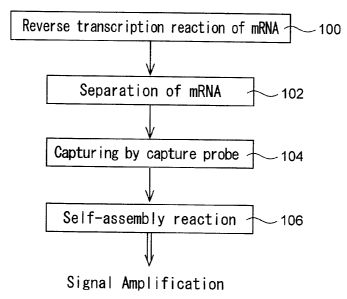
Fig. 1 is a flow chart showing an example of order of steps of a signal
amplification method according to the present invention;
Fig. 2 is a schematic diagram showing in principle the step 200 in a
first example of order of steps of the signal amplification method according
to
the present invention;
Fig. 3 is a schematic diagram showing in principle the step 202 in the
first example of order of steps of the signal amplification method of the
present invention;
Fig. 4 is a schematic diagram showing in principle the step 204 in the
first example of order of steps of the signal amplification method of the
present invention;
CA 02515734 2005-08-11
-9.
Fig. 5 is a schematic diagram showing in principle the step 206 in the
first example of order of steps of the signal amplification method of the
present invention;
Fig. 6 is a schematic diagram showing in principle the step 210 in the
first example of order of steps of the signal amplification method of the
present invention;
Fig. 7 is a schematic diagram showing in principle the step 212 in the
first example of order of steps of the signal amplification method of the
present invention;
Fig. 8 is a schematic diagram showing in principle the step 214 in the
first example of order of steps of the signal amplification method of the
present invention;
Fig. 9 is a schematic diagram showing in principle the step 300 in a
second example of order of steps of the signal amplification method of the
present invention;
Fig. 10 is a schematic diagram showing in principle the step 302 in
the second example of order of steps of the signal amplification method of the
present invention;
Fig. 11 is a schematic diagram showing in principle the step 304 in
the second example of order of steps of the signal amplification method of the
present invention;
Fig. 12 is a schematic diagram showing in principle the step 306 in
the second example of order of steps of the signal amplification method of the
present invention; and
Fig. 13 is a graph showing the results of Example 1 and Comparative
CA 02515734 2005-08-11
-10-
Example 1.
Best Mode for Carrying Out the Invention:
The examples of the present invention are described below with
reference to the attached drawings. It should be understood that the
examples described herein are merely exemplary and that many variations
and modifications may be made without departing from the spirit and scope
of the present invention.
Fig. 1 is a flow chart showing an example of order of steps of the
signal amplification method for detecting an expressed gene according to the
present invention.
As shown in Fig. 1, there is provided a first probe which contains
poly(dT) at the 3' end and a region hybridizable with at least one
oligonucleotide probe for use in a self-assembly reaction. The first probe is
used as a primer and bound to mRNA containing poly(A). A reverse
transcription reaction of the mRNA is performed by a reverse transcriptase
(step 100) so that there is formed a second probe which is comprised of the
first probe and a cDNA region of the mRNA. In a preferred mode, a base
sequence in the 5' side of the first probe includes at least a part of the
base
sequence of the oligonucleotide probe for use in the self-assembly reaction.
The mRNA is then separated from the second probe (step 102). Any
limitations are not specifically put on the method for separating the mRNA,
but methods using, for example, thermal denaturation, alkali denaturation,
RNA digestion with RNase H, or the like may be employed.
After the separation, the second probe is hybridized with a capture
CA 02515734 2005-08-11
-11-
probe having a region complementary to a cDNA region of a target mRNA to
allow the capture probe to capture the second probe (step 104). Preferably,
the capture probe has previously been bound to a support.
The oligonucleotide probes are added so that a self-assembly
substance hybridized with the second probe can be formed by a selfassembly
reaction (step 106) with the result that the signal can be amplified.
If the target mRNA does not exist in the sample, the second probe
cannot bind to the capture probe so that signal amplification cannot be
carried out. Thus, the existence of the target mRNA can be determined by
the signal amplification method of the present invention. Since the signal
amplification method of the present invention does not use the linear
amplification method, detection can be performed depending on the length of
the original RNA, and since the signal amplification method of the present
invention does not use the PCR method, signal amplification can be
performed depending on the amount of expression.
As the selfassembly reaction described above, there may be
employed a self-assembly reaction using a pair of HCPs where each of the
HCPs comprises three regions complementary to each other and the HCPs
can selfassemble by themselves to form a selfassembly substance (for
example, see JP 3267576 and JP 3310662). Alternatively, a selfassembly
reaction may be performed using a pair of dimer-forming probes capable of
forming a dimer by themselves and a pair of crosslinking probes capable of
crosslinking the dimer formed from the dimer-forming probes (for example,
see JP-A 2002-355081).
Although step 104 is followed by step 106 in the example shown in
CA 02515734 2005-08-11
-12-
Fig. 1, step 106 may be performed before or simultaneously with step 104.
Figs. 2 to 8 are schematic diagrams showing in principle a first
example of order of steps of the signal amplification method according to the
present invention. The first example illustrates the signal amplification
method utilizing the PALSAR method using a pair of HCPs previously
labeled with a fluorescent material 22 as the selfassembly reaction, wherein
an HCP containing poly(dT) at the 3' end is used as a first probe 12a.
As shown in Fig. 2, in order to detect mRNA 10a of a target gene, an
oligonucleotide probe (HCP-1) containing poly(dT) at the 3' end and three
regions of the HCP in the 5' side thereof is provided as the first probe 12a
(step 200). As shown in Fig. 3, HCP-1 (12a) containing poly(dT) at the 3'
end is bound to the poly(A) tail part of the mRNA l0a (step 202). As shown
in Fig. 4, a reverse transcription reaction is then performed using a reverse
transcriptase to form a second probe 14a having a sequence complementary
to the mRNA (step 204). Thereafter, as shown in Fig. 5, the mRNA 10a is
dissociated to form a single-stranded oligonucleotide comprising a cDNA
region and the HCP region (step 206).
As shown in Fig. 6, a capture probe 16a having a region
complementary to the cDNA of the target gene is previously bound to a
support 18 (step 210). As shown in Fig. 7, the second probe 14a serving as
an HCP having the formed cDNA region is hybridized with the capture probe
16a (step 212). As shown in Fig. 8, another HCP (HCP-2) of the pair of
HCPs is added to form a self-assembly substance 20a by a self-assembly
reaction (step 214) so that signal amplification can be achieved.
Incidentally, if in the step 206, a washing operation is carried out when the
CA 02515734 2005-08-11
13-
target mRNA is removed, the pair of HCPs must be added in the step 214.
In the above first example, there is used a pair of HCPs, wherein a
part of the 3' side region in the three regions of HCP-1 is poly(dT).
Alternatively, there may be used another. pair of HCPs, wherein the 3' side
region of HCP-1 is entirely poly(dT) and the 3' side region in the three
regions of HCP-2 is entirely poly(A).
Figs. 9 to 12 are schematic diagrams showing in principle a first
example of order of steps of the signal amplification method according to the
present invention. The second example illustrates the signal amplification
method utilizing the PALSAR method using a pair of HCPs unlabeled with a
fluorescent material as the self-assembly reaction, wherein an
oligonucleotide probe containing poly(dT) at the 3' end and one of
complementary regions of the HCP is used as a first probe 12b.
As shown in Fig. 9, in order to detect mRNA 10b of a target gene, an
oligonucleotide probe containing poly(dT) at the 3' end and one of the
complementary regions of the HCP in the 5' side thereof is provided as the
first probe 12b (step 300). As shown in Fig. 10, the oligonucleotide probe
12b is bound to the poly(A) tail part of the mRNA 10b, and a reverse
transcription reaction is performed using a reverse transcriptase to form a
second probe 14b having a sequence complementary to the mRNA (step 302).
The mRNA 10b is separated to form a single-stranded oligonucleotide
comprising a cDNA region and the HCP region.
As shown in Fig. 11, the second probe 14b is hybridized with a
capture probe 16b bounded to a support 18 (step 304). As shown in Fig. 12,
a pair of HCPs is added to form a self-assembly substance 20b by a
CA 02515734 2010-11-10
-14-
self-assembly reaction (step 306). An intercalator 24 or the like is inserted
into the formed self-assembly substance 20b (step 308) so that signal
amplification can be achieved. Steps 306 and 308 may be performed
simultaneously.
For detection of the target gene, a labeling material for detection may
previously be added to the pair of the oligonucleotide probes. Examples of
such a labeling material include radioisotopes such as 1125 and P32,
luminescent materials such as digoxigenin and acridinium esters,
fluorescent materials such as Cy3 and Cy5, and fluorescent donor dyes and
fluorescent acceptor dyes for using fluorescent resonance energy transfer
(FRET) such as biotin for using a fluorescent material such as
4-methylunbelliferyl phosphate.
Alternatively, by adding a dye having the property of binding to
nucleic acids, the target gene can be detected. A fluorescent material
having the property of binding to nucleic acids, such as an intercalator, is
preferably used to detect the target gene. Any fluorescent material having
the property of binding to nucleic acids may be used without limitation.
Examples of such a fluorescent material include SYBR Green I stain, SYBR
Green II stain, SYBR Green Gold stain, Vistra Green stain, Gelstar stain,
Radiant Red stain, PicoGreen, RiboGreen, OllGreen, Hoechst 33258
(Bis-Benzimide), Propidium Iodide, YO-PRO-1 Iodide, YO-PRO-3 Iodide (the
above materials are all manufactured by Molecular Probes Inc.), ethidium
bromide, Distamycin A, TOTO, Psoralen, acridinium orange (Acridine
Orange), AOAO (homodimer), and the like.
While a nucleic acid constituting the pair of the oligonucleotide
*Trade-mark
CA 02515734 2005-08-11
- 15-
probes is usually DNA or RNA, a nucleic acid analogue may constitute them.
Examples of such a nucleic acid analogue include peptide nucleic acids
(PNAs, for example, see the brochure of International Patent Publication No.
WO 92/20702) and locked nucleic acids (LNAs, for example, see Koshkin AA
et al., Tetrahedron 1998, 54, 3607-3630, Koshkin AA et al., J. Am. Chem. Soc.,
1998, 120, 13252-13253 and Wahlestedt C et al., PNAS, 2000, 97, 5633-5638).
The pair of the oligonucleotide probes is generally composed of nucleic acids
of the same kind, but may be composed of a pair of a DNA probe and an RNA
probe. That is, the type of the nucleic acid of the probe may be selected from
DNA, RNA or nucleic acid analogues (such as PNA and LNA). Also, it is not
necessary that a single probe is composed of a single type of nucleic acid,
for
example, DNA only, and, if necessary, for example, an oligonucleotide probe
composed of DNA and RNA (a chimera probe) may be used in an aspect of the
present invention.
In terms of the number of bases, the length of each complementary
base sequence region of the oligonucleotide probe may be at least 5 bases,
preferably from 10 to 100 bases, more preferably from 15 to 30 bases.
These probes may be synthesized by any known methods. For
example, DNA probes may be synthesized by a phosphoamidite method
using DNA Synthesizer Model 394 (Applied Biosystems Inc.). Any other
synthesis methods may also be used such as a phosphotriester method, an
H-phosphonate method, and a thiophosphonate method.
According to the present invention, a self-assembly substance is
formed with a pair of HCPs having complementary regions against a target
gene captured on a DNA chip. While the number of pieces of the
CA 02515734 2005-08-11
-16-
oligonucleotide probe for use is not limited, it may be in the range of 102 to
1015. The reaction buffer solution may have any composition and any
concentration, and any buffer solution commonly used for nucleic acid
amplification may be preferably used. The pH may be in any conventional
range, preferably in the range of 7.0 to 9Ø The reaction temperature may
be from 40 to 80 C, preferably from 55 to 65 C.
In the present invention, any sample potentially containing a target
nucleic acid may be used as a sample for the measurement of a target
expressed gene (mRNA). The target gene may be any properly prepared or
isolated from samples and it is not specifically limited. Examples of such
samples include organism- derived samples such as blood, blood serum, urine,
feces, cerebrospinal fluid, tissue fluid, and cell cultures, and any samples
potentially containing or potentially infected with any eukaryote having
mRNA containing a poly(A) strand at its end, such as fungi. There may be
also used any nucleic acid obtained by amplifying a target gene in samples
with any known method.
(Examples)
Although the present invention is more specifically described by
means of the examples below, it will be understood that the examples
presented are by way of illustration only and should not be construed as any
limitation on the present invention.
The materials below were used in the examples.
(a) Target gene: total RNA extracted from cultured cells
(b) Capture probe:
1) CP-1: 5'-CACGAAACTACCTTCAACTCCATC-3'
CA 02515734 2010-11-10
-17-
2) CP-2: 5'-TGCCGACAGGATGCAGAAGGA-3'
(c) Primers:
1) First probe (poly(dT)-HCP, 78 mer): 5'-GCATATAGATATCTCC
GGCGCGGATACTTTGTGATACCGGGAGTTCGCCCTTATAACGTCTTTTT
TTTTTTTTTTTTT- 3'
2) Poly(dT) primer (18 mer): 5'-TTTTTTTTTTTTTTTTTT-3'
(d) HCPs:
1) HCP-1 (Cy3-labeled 5' end, 60 mer): 5'-Cy3-CGCCGGAGATAT
CTATATGCCCGGTATCACAAAGTATCCGGACGTTATAAGGGCGAACTC-3'
2) HCP-2 (Cy3-labeled 5' end, 60 mer): 5'-Cy3-GCATATAGATATC
TCCGGCGCGGATACTTTGTGATACCGGGAGTTCGCCCTTATAACGTC-3'
(e) Polystyrene particle beads: a single kind of particle beads having the
above two kinds of capture probes fixed thereon.
(Example 1)
Using the total RNA extracted from cultured cells, an attempt was
made to detect beta-actin of a housekeeping gene according to the PALSAR
method.
(1) Reverse Transcription Reaction of RNA and Purification of Reverse
Transcription Product
A reverse transcription reaction was carried out at 37 C for 2 hours
using the target gene, the first probe, a reverse transcriptase (SuperScript
II
manufactured by Invitrogen Corporation), and a reaction solution (Reaction
Buffer, DTT, dNTP, RNase inhibitor). Thereafter, alkali treatment was
performed at 65 C for 30 minutes, and neutralization was performed using
hydrochloric acid. The reverse transcription product of cDNA was purified
*Trade-mark
CA 02515734 2005-08-11
-18-
using QlAquick PCR Purification Kit (manufactured by QIAGEN K. K.).
(2) Hybridization
The obtained cDNA, the particle beads, 6x SSC, 0.2% SDS, and 5x
Denhardt's solution, were then mixed to prepare a composition of 50 l in
total amount. The composition was subjected to hybridization at 42 C for 2
hours. After the hybridization was completed, filtration was performed
with a 0.22 m filter so that the unreacted probe was removed. Then the
particle beads was cleaned once with 2x SSC + 0.1% SDS and once with 0.2x
SSC, and was filtered with the above filter.
Thereafter, a composition of 100 l in total amount consisting of the
obtained particle beads, the HCPs-1 and 2 (each 1.5 pmol/ l), 1% Blocking
Reagent (manufactured by Roche Inc.), 0.1% N-lauroylsarcosine, 0.02% SDS,
and 5x SSC was subjected to hybridization at 65 C for 30 minutes.
(3) Detection
After washing, the particle beads were resuspended in sheath fluid
for a flow cytometer, and the fluorescence of the Cy3 labeled to the HCPs was
measured with the flow cytometer.
(Comparative Example 1 )
The target gene was detected using a conventional Cy3-dNTP uptake
system with a poly(dT) primer.
(1) Reverse Transcription Reaction and Separation Reaction of RNA
The reverse transcription reaction and the purification of the reverse
transcription product were performed under the same process as in Example
1 with the exception of using the poly(dT) primer as primer, and introducing
Cy3-labeled dUTP (manufactured by Amersham Inc.) in addition to dNTP.
CA 02515734 2005-08-11
-19-
(2) Hybridization
The obtained cDNA, the particle beads, 6x SSC, 0.2% SDS, and 5x
Denhardt's solution were then mixed to prepare a composition of 50 l in
total amount. The composition was subjected to hybridization at 42 C for 2
hours. After the hybridization was completed, filtration was performed
with a 0.22 m filter so that the unreacted probe was removed. Then the
composition was cleaned once with 2x SSC + 0.1% SDS, and was filtered.
(3) Detection
After washing, the particle beads were resuspended in a sheath fluid
for a flow cytometer, and the fluorescence of the Cy3 label of the cDNA bound
to the capture probe on the particle beads was measured with the flow
cytometer.
[Results]
The results of Example 1 and Comparative Example 1 are shown in
Fig. 13. The fluorescence intensity was measured for 203 to 504 particle
beads in respect of each one and indicated by the median thereof. As shown
in Fig. 13, the detection sensitivity of Example 1 is significantly higher
than
that of Comparative Example 1.
Capability of Exploitation in Industry:
As described above, according to the present invention, the detection
only through a reverse transcription reaction can be accomplished by an
inexpensive and simple operation in a short time without expensive enzymes.
A further significant advantage is that since there is no need to use the
linear amplification method or the PCR method, the detection can also be
CA 02515734 2005-08-11
-20-
performed depending on the length or expression amount of the original
RNA.
CA 02515734 2006-07-12
-21-
SEQUENCE LISTING
<110> Eisai Co., Ltd.
<120> Signal Amplification Method for Detecting Expressed Gene
<130> 14598-6CA
<140> 2,515,734
<141> 2004-02-13
<150> JP 2003-037212
<151> 2003-02-14
<160> 6
<210> 1
<211> 24
<212> DNA
<213> Artificial Sequence
<220>
<223> Description of Artificial Sequence: CP-1
<400> 1
cacgaaacta ccttcaactc catc 24
<210> 2
<211> 21
<212> DNA
<213> Artificial Sequence
<220>
<223> Description of Artificial Sequence: CP-2
<400> 2
tgccgacagg atgcagaagg a 21
<210> 3
<211> 78
<212> DNA
<213> Artificial Sequence
<220>
<223> Description of Artificial Sequence: first probe
<400> 3
gcatatagat atctccggcg cggatacttt gtgataccgg gagttcgccc ttataacgtc 60
tttttttttt tttttttt 78
<210> 4
<211> 18
<212> DNA
<213> Artificial Sequence
<220>
<223> Description of Artificial Sequence: poly dT primer
<400> 4
tttttttttt tttttttt 18
<210> 5
CA 02515734 2006-07-12
-22-
<211> 60
<212> DNA
<213> Artificial Sequence
<220>
<221> misc feature
<222> (1)
<223> Cy3 attached at the 5'end
<220>
<223> Description of Artificial Sequence: HCP-1
<400> 5
cgccggagat atctatatgc ccggtatcac aaagtatccg gacgttataa gggcgaactc 60
<210> 6
<211> 60
<212> DNA
<213> Artificial Sequence
<220>
<221> misc_feature
<222> (1)
<223> Cy3 attached at the 5'end
<220>
<223> Description of Artificial Sequence: HCP-2
<400> 6
gcatatagat atctccggcg cggatacttt gtgataccgg gagttcgccc ttataacgtc 60