Note : Les descriptions sont présentées dans la langue officielle dans laquelle elles ont été soumises.
CA 02750520 2016-05-26
=
- 1 -
METHODS FOR SCREENING CANDIDATE AGENTS FOR
MODULATING PRORENIN AND RENIN, ASSAYS FOR DETECTING
PRORENIN, AND ANTIBODIES USED THEREIN
CA 02750520 2011-07-22
WO 2010/091182 PCT/US2010/023198
- 2 -
FIELD OF THE INVENTION
[0002] The present invention relates to antibodies that bind to prorenin. In
particular, the
invention relates to monoclonal antibodies that bind to prorenin and inhibit
the activation of
prorenin. The antibodies of the invention are useful for screening for
candidate agents that
inhibit the activation of prorenin and for screening candidate agents that
modulate renin
activity. The antibodies are also useful as diagnostics and for treating
disease states.
BACKGROUND OF THE INVENTION
[0003] The aspartyl protease renin is an important modulator of blood
pressure. Renin is
produced by cleavage of the 395 amino acid zymogen prorenin which circulates
in blood at
between five and ten times the level of active renin. The putative cleavage
site is at the R43L44
sequence of prorenin (Mercure et al., Journal of Biological Chemistry
270(27):16355-16359 (1995)).
[0004] Human prorenin is easily activated to renin in vitro with catalytic
trypsin. A number of
enzymes have been suggested as natural activators of prorenin including the
cathepsins,
plasmin, and various activated coagulation factors (Mercure et al.).
[0005] Once activated, renin hydrolyzes angiotensinogen into angiotensin I.
Angiotensin I is
further processed into angiotensin II by angiotensin converting enzyme (ACE).
Angiotensin II
is a potent constrictor of blood vessels which then leads to an elevation of
blood pressure.
Drugs interfering with the renin-angiotensin system (RAS) are currently being
widely
developed for the treatment of cardiovascular diseases. They not only lower
blood pressure, but
also prevent end-organ damage. In an attempt to develop drugs to combat high
blood pressure,
a number of targets including prorenin, ACE, and renin have been developed.
[0006] The only currently prescribed renin therapeutic is the recently
introduced direct renin
inhibitor AliskirenTM (Novartis Corporation, Basel, Switzerland). However, due
to multiple
feedback mechanisms within RAS, RAS blockade, including the inhibition of
renin, results in
the elevation of both renin and its inactive precursor, prorenin. A rise in
renin and prorenin
occur particularly following treatment with AliskirenTM (Novartis). The
consequences of such
increases in renin and prorenin are currently unknown.
CA 02750520 2011-07-22
WO 2010/091182 PCT/US2010/023198
- 3 -
[0007] However, it is important to note that prorenin is also elevated in
plasma of diabetic
subjects before the occurrence of complications with nephropathy and
retinopathy. Prorenin,
like other components of RAS, can also be detected in urine and may provide
several
advantages. First, urinary prorenin levels may serve as an early marker for
diabetic
nephropathy and/or retinopathy and facilitate the selection of patients
eligible for treatment
with a RAS blocker at a very early stage, Early detection and treatment is
pertinent in view of
the steep rise in diabetes frequency and the devastating consequences of
diabetes in eyes and
kidneys. Secondly, changes in plasma and/or urinary prorenin levels might help
to monitor the
response to RAS blockade.
[0008] Nonetheless, there are no commercially available sandwich prorenin ELIS
A assays.
Schalekamp et.al. describe a prorenin ELISA assay using a monoclonal antibody
directed
against the N-terminal prorenin peptide (Schalekamp et al., Journal of
Hypertension 26:928-
937 (2008)). This antibody, produced by F. Hoffmann-La Roche AG (Basel,
Switzerland),
requires extensive and time consuming pretreatment of the prorenin with a
renin inhibitor to
remove the propiece from the active site, which makes it reactive and
unsuitable for common
use.
[0009] Accordingly, there is a need for prorenin assays without pretreatment.
Such prorenin
assays would help to identify (diabetic) patients requiring RAS blockade
treatment at a very
early stage, thereby greatly reducing the occurrence of nephro- and
retinopathy. Prorenin
measurements may also allow monitoring of the response to RAS blockade, which
may help to
ascertain why some patients respond well to RAS blockade whereas others do
not. Moreover,
such measurements would also help to determine the consequences of the changes
in (pro)renin
concentrations (e.g., (pro)renin receptor activation) that occur during
treatment.
[0010] Recently, the so-called prorenin receptor was discovered. It is
believed that the effects
of increased renin and/or prorenin may be exerted via this receptor. Studies
suggest that
prorenin may function in the absence of cleavage through its binding to the
prorenin receptor.
Prorenin exists in two conformations: 1) the open conformation, where the
active site is
accessible, and 2) the closed conformation, where the active site is not
accessible. Binding of
CA 02750520 2011-07-22
WO 2010/091182 PCT/US2010/023198
- 4 -
prorenin to its receptor results in conformation conversion to the open
conformation, resulting
in non-proteolytic activation. Nguyen et al., J. Clin. Invest. 109:1417
(2002).
[0011] Therefore, there is also the need for methods of inhibiting the
activation of prorenin.
Such methods may include preventing the cleavage of prorenin to form the
active renin or
preventing prorenin from binding its receptor, such as by the use of an
antibody and/or keeping
prorenin in its closed conformation.
SUMMARY OF THE INVENTION
[0012] The present invention relates to a monoclonal antibody which binds to
prorenin at or
near the reactive R43L44 bond. Once bound, the antibody has been shown by SDS
PAGE to
block trypsin from cleaving the zymogen to produce renin. Furthermore, it is
believed that the
antibody of the invention is capable of locking prorenin in its closed
conformation, as well as
blocking binding of prorenin to its receptor.
[0013] The present invention further relates to a method of modulating the
activation of
prorenin by administering the antibody of the invention. Methods of treatment
by
administering the antibody of the invention are also provided, as are
humanized antibodies
derived from the antibody of the present invention. The antibodies of the
invention, and in
particular the humanized antibodies of the invention, are useful in treating
disease states, such
as high cardiovascular disease, blood pressure, diabetes, and disorders
associated therewith, in
which it is desirable to inhibit prorenin.
[0014] It is believed, that based on the ability of the antibody of the
invention to bind to
prorenin and (1) block tryp sin from cleaving prorenin into renin and (2)
essentially lock
prorenin in its inactive, closed conformation and/or inhibit the binding of
prorenin to its
receptor, the antibody of the invention is particularly useful in treating
disease states.
[0015] The antibody of the invention further serves as the basis for
development of a new class
of therapeutic directly targeting prorenin, which sits upstream in the RAS
enzymatic cascade.
[0016] In addition, the antibody of the invention serves as the basis for a
sandwich prorenin
ELISA assay. The assays of the present invention can be used to detect
prorenin in biological
samples, such as urine and blood.
CA 02750520 2011-07-22
WO 2010/091182 PCT/US2010/023198
- 5 -
[0017] The invention further relates to a method of screening for molecules
that inhibit
prorenin activation. The method allows for ease of screening. The method
comprises
providing the prorenin antibody of the invention, prorenin and a candidate
agent to be screened,
and determining whether binding of the antibody to prorenin is modulated by
the presence of
the candidate agent. Once it is determined whether the candidate agent affects
binding of the
antibody of the invention to prorenin, the candidate agent may be further
screened to determine
whether it inhibits prorenin activation.
[0018] The invention also provides chimeric prorenin polypeptides and nucleic
acids encoding
the same. In a preferred embodiment, the chimeric prorenin polypeptide of the
invention
includes a human "pro" region and a non-human renin region. In a preferred
embodiment, the
non-human renin region is a vertebrate renin region, In a particularly
preferred embodiment,
the non-human renin region is a rat renin region. The chimeric proteins of the
invention can be
expressed in various cells, such as in vitro transformed cell lines and/or in
vivo in animals. The
cell lines and animals should be the same species from which the renin region
of the chimeric
prorenin is derived.
[0019] In a preferred embodiment, transgenic animals stably expressing the
chimeric prorenin
of the invention are provided. These transgenic animals may be knock-in
animals, in which the
native prorenin has been replaced with the chimeric protein of the invention.
Alternatively, the
transgenic animal may express the chimeric prorenin of the invention as well
as the native
prorenin.
[0020] Using the antibody of the present invention, which binds to the pro
region of human
prorenin, the chimeric prorenin in the transgenic animal and/or cells from the
transgenic animal
can be studied. For example, prorenin's fate as it binds to its cognate
receptor can be followed,
as well as its metabolism and half-life. These model systems are also useful
in developing
diagnostics assays for various diseases associated with prorenin and renin.
These model
systems are also useful for testing molecules that modulate prorenin, e.g.,
the antibodies of the
present invention, as potential therapeutics for the treatment of disease
states in which it is
desirable to inhibit prorenin.
CA 02750520 2011-07-22
WO 2010/091182 PCT/US2010/023198
- 6 -
BRIEF DESCRIPTION OF THE DRAWINGS
[0021] FIG. 1 depicts an exemplary standard calibration curve prepared in the
assay of the
present invention;
[0022] FIGs. 2-4 shows a Western blot of human prorenin in lane 1 and human
prorenin
incubated with catalytic trypsin in lane 2;
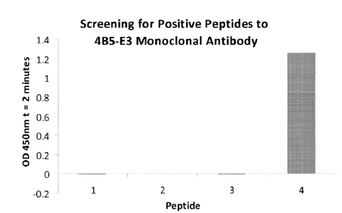
[0023] FIG. 3-5 depicts optical density measurements for the screening of
peptides to
monoclonal antibody 4B5-E3;
[0024] FIG. 6A-C show results from the detection of prorenin in urine samples;
[0025] FIGs. 7 shows SDS PAGE results of prorenin incubated without or with
catalytic
trypsin (lanes 2 and 3, respectively) and prorenin pre-incubated with
monoclonal antibody 4B5-
E3, followed by the addition of catalytic trypsin at 1, 3, 5, 10, and 30
minute intervals (lanes 4-
8, respectively);
[0026] FIG. 8 (A) shows the amino acid (SEQ ID NO:1) and (B) nucleotide
sequence (SEQ
ID NO:2) of human prorenin.
DETAILED DESCRIPTION OF THE INVENTION
[0027] The present invention relates to antibodies that specifically bind to
prorenin, thereby
inhibiting the activation of prorenin and its binding to the prorenin
receptor. The present
invention also relates to methods for detecting prorenin using antibodies of
the present
invention in ELISA assays. The invention disclosed herein further provides for
methods of
screening candidate agents for modulating the activation of prorenin and
candidate agents for
modulating the activity of renin. The antibodies disclosed herein are also
useful as diagnostics
and for treating disease states, such as those associated with high blood
pressure, diabetes, and
complications of diabetes.
Definitions
[0028] In order that the present invention may be more readily understood,
certain terms are
first defined. Additional definitions are set forth throughout the detailed
description and
elsewhere in the specification.
CA 02750520 2011-07-22
WO 2010/091182 PCT/US2010/023198
- 7 -
[0029] The term "antibody" as used herein refers to an immunoglobulin that is
reactive to a
designated protein or peptide or fragment thereof. Suitable antibodies
include, but are not
limited to, human antibodies, primatized antibodies, chimeric antibodies,
monoclonal
antibodies, monospecific antibodies, polyclonal antibodies, polyspecific
antibodies, nonspecific
antibodies, bispecific antibodies, multispecific antibodies, humanized
antibodies, synthetic
antibodies, recombinant antibodies, hybrid antibodies, mutated antibodies,
grafted conjugated
antibodies (i.e., antibodies conjugated or fused to other proteins,
radiolabels, cytotoxins), and
in vitro-generated antibodies. The antibody can be from any class of
antibodies including, but
not limited to, IgG, IgA, IgM, IgD, and IgE, and from any subclass (e.g.,
IgGl, IgG2, IgG3,
and IgG4) of antibodies. The antibody can have a heavy chain constant region
chosen from,
e.g., IgGl, IgG2, IgG3, or IgG4. The antibody can also have a light chain
chosen from, e.g.,
kappa (x) or lambda (X). The antibodies of the invention can be derived from
any species
including, but not limited to mouse, human, camel, llama, fish, shark, goat,
rabbit, chicken, and
bovine. Constant regions of the antibodies can be altered, e.g., mutated, to
modify the
properties of the antibody (e.g., to increase or decrease one or more of: Fc
receptor binding,
antibody glycosylation, the number of cysteine residues, effector cell
function, or complement
function). Typically, the antibody specifically binds to a predetermined
antigen, e.g., an
antigen associated with a disorder, e.g., disorders related to high blood
pressure and diabetes.
[0030] The terms "prorenin activity," "activity of prorenin," "prorenin
activation," and the like
refer to at least one cellular process initiated or interrupted as a result of
the cleavage of
prorenin to form renin or prorenin binding to the prorenin receptor.
[0031] The phrases "inhibit," "antagonize," "block," or "neutralize" prorenin
activity or
activation and its cognates refer to a reduction, inhibition, or otherwise
diminution of at least
one activity of prorenin due to binding the prorenin receptor or the cleavage
of prorenin to form
renin, wherein the reduction, inhibition, or diminution is relative to the
activity of prorenin
when bound to its receptor or the cleavage of prorenin. Prorenin activity can
be measured
using any technique known in the art. Inhibition or antagonism does not
necessarily indicate a
total elimination of the prorenin biological activity. A reduction in activity
may be about 10%,
20%, 30%, 40%, 50%, 60%, 70%, 80%, 90%, 95%, or more.
CA 02750520 2011-07-22
WO 2010/091182 PCT/US2010/023198
- 8 -
[0032] Similarly, the terms "renin activity," "activity of renin," "renin
activation," and the like
refer to at least one cellular process initiated or interrupted as a result of
the hydrolysis of
angiotensinogen into angiotensin I.
[0033] The phrases "inhibit," "antagonize," "block," or "neutralize" renin
activity or
activation and its cognates refer to a reduction, inhibition, or otherwise
diminution of at least
one activity of renin due to binding the hydrolysis of angiotensinogen into
angiotensin I. Renin
activity can be measured using any technique known in the art. Inhibition or
antagonism does
not necessarily indicate a total elimination of the renin biological activity.
A reduction in
activity may be about 10%, 20%, 30%, 40%, 50%, 60%, 70%, 80%, 90%, 95%, or
more.
[0034] The term "isolated" refers to a molecule that is substantially free of
its natural
environment. For instance, an isolated protein is substantially free of
cellular material or other
proteins from the cell or tissue source from which it was derived. The term
also refers to
preparations where the isolated protein is sufficiently pure for
pharmaceutical compositions, or
is at least 70-80% (w/w) pure, at least 80-90% (w/w) pure, at least 90-95%
(w/w) pure, or at
least 95%, 96%, 97%, 98%, 99%, or 100% (w/w) pure.
[0035] The phrase "percent identical" or "percent identity" refers to the
similarity between at
least two different sequences. This percent identity can be determined by
standard alignment
algorithms, for example, the Basic Local Alignment Search Tool (BLAST)
described by
Altshul et al., J. Mol. Biol. 215:403-10 (1990); the algorithm of Needleman et
al., J. Mol. Biol.
48:444-53 (1970); or the algorithm of Meyers et al., Comput. Appl. Biosci.
4:11-17 (1988). A
set of parameters may be the Blosum 62 scoring matrix with a gap penalty of
12, a gap extend
penalty of 4, and a frameshift gap penalty of 5. The percent identity between
two amino acid or
nucleotide sequences can also be determined using the algorithm of Meyers and
Miller,
CABIOS 4:11-17 (1989), which has been incorporated into the ALIGN program
(version 2.0),
using a PAM120 weight residue table, a gap length penalty of 12, and a gap
penalty of 4. The
percent identity is usually calculated by comparing sequences of similar
length.
[0036] The terms "specific binding," "specifically binds," and the like refer
to two molecules
forming a complex that is relatively stable under physiologic conditions.
Specific binding is
characterized by a high affinity and a low-to-moderate capacity as
distinguished from
CA 02750520 2011-07-22
WO 2010/091182 PCT/US2010/023198
- 9 -
nonspecific binding, which usually has a low affinity with a moderate-to-high
capacity.
Typically, binding is considered specific when the association constant Ka is
higher than about
106m-is-i.
If necessary, nonspecific binding can be reduced without substantially
affecting
specific binding by varying the binding conditions. The appropriate binding
conditions, such
as concentration of antibody, ionic strength of the solution, temperature,
time allowed for
binding, concentration of a blocking agent (e.g., serum albumin or milk
casein), etc., can be
improved by a skilled artisan using routine techniques. Illustrative
conditions are set forth
herein, but other conditions known to the person of ordinary skill in the art
fall within the scope
of this invention.
[0037] The phrases "substantially as set out," "substantially identical," and
"substantially
homologous" mean that the relevant amino acid or nucleotide sequence (e.g.,
CDR(s), VH, or
VL domain(s)) will be identical to or have insubstantial differences (e.g.,
through conserved
amino acid substitutions) in comparison to the sequences which are set out.
Insubstantial
differences include minor amino acid changes, such as one or two substitutions
in a five amino
acid sequence of a specified region. In the case of antibodies, the second
antibody has the same
specificity and has at least about 50% of the affinity of the first antibody.
[0038] Sequences substantially identical or homologous to the sequences
disclosed herein are
also part of this application. In some embodiments, the sequence identity can
be about 85%,
90%, 95%, 96%, 97%, 98%, 99%, or higher. Alternatively, substantial identity
or homology
exists when the nucleic acid segments will hybridize under selective
hybridization conditions
(e.g., highly stringent hybridization conditions), to the complement of the
strand. The nucleic
acids may be present in whole cells, in a cell lysate, or in a partially
purified or substantially
pure form.
[0039] As used herein, a "therapeutically effective amount" of an antibody
that binds to
prorenin refers to an amount of the binding protein that is effective, upon
single or multiple
dose administration to a subject (such as a human patient) for treating,
preventing, curing,
delaying, reducing the severity of, and/or ameliorating at least one symptom
of a disorder or a
recurring disorder, or prolonging the survival of the subject beyond that
expected in the
absence of such treatment.
CA 02750520 2011-07-22
WO 2010/091182 PCT/US2010/023198
- 10 -
Antibodies
[0040] The present invention relates to antibodies or fragments thereof that
specifically bind to
human prorenin, having the amino acid sequence set forth in SEQ ID NO:1 (shown
in Figure 8)
and the nucleic acid sequence set for in SEQ ID NO:2 (shown in Figure 8). In
particular,
antibodies or fragments thereof of the present invention bind an epitope from
the N-terminus of
prorenin comprising the 8 amino acids set forth in SEQ ID NO:3. The antibodies
of the present
invention bind to prorenin at or near the reactive bond R43L44, thereby
blocking cleavage of the
zymogen to renin. In addition, the antibody locks prorenin in its closed
conformation, which
inhibits the binding of prorenin to the prorenin receptor. In a preferred
embodiment, an
antibody of the present invention is an anti-human prorenin, such as
monoclonal antibody 4B5-
E3.
[0041] A hybridoma cell line that produces monoclonal antibodies having the
properties of
monoclonal antibody 4B5-E3 has been deposited at American Tissue Culture
Collection
(ATCC) on March 26, 2009, and assigned Deposit Designation Number PTA-9894.
The
address of the depository is 10801 University Blvd, Manassas, Va. 20110,
U.S.A.
[0042] Numerous methods known to those skilled in the art are available for
obtaining
antibodies or fragments thereof. For example, antibodies can be produced using
recombinant
DNA methods (see, e.g., U.S. Patent No. 4,816,567). In one embodiment of the
invention, the
antibodies are monoclonal antibodies. Monoclonal antibodies may also be
produced by
generation of hybridomas in accordance with known methods (see, e.g., Kohler
and Milstein
(1975) Nature 256:495-99). Hybridomas formed in this manner are then screened
using
standard methods, such as enzyme-linked immunosorbent assays (ELISA) and
surface plasmon
resonance (BIACORETM) analysis, to identify one or more hybridomas that
produce an
antibody that specifically binds with a particular antigen. Any form of the
specified antigen
may be used as the immunogen, e.g., recombinant antigen, naturally occurring
forms, any
variants or fragments thereof, and antigenic peptides thereof.
[0043] In addition, the specified antigen can be used to immunize a nonhuman
animal, e.g., a
cynomolgus monkey, a chicken, or a rodent (e.g., a mouse, hamster, or rat). In
one
embodiment, the nonhuman animal includes at least a part of a human
immunoglobulin gene.
CA 02750520 2011-07-22
WO 2010/091182 PCT/US2010/023198
- 11 -
For example, it is possible to engineer mouse strains deficient in mouse
antibody production
with large fragments of the human Ig loci. Using the hybridoma technology,
antigen-specific
monoclonal antibodies, derived from the genes with the desired specificity may
be produced
and selected (see, e.g., XENOMOUSETm (Amgen Inc., Thousand Oaks, CA); Green et
al., Nat.
Genet. 7:13-21(1994); U.S. Patent No. 7,064,244; and International Application
Publication
Nos. WO 96/034096 and WO 96/033735).
[0044] In one embodiment of the invention, the antibody is a monoclonal
antibody that is
obtained from a nonhuman animal, and then modified (e.g., humanized,
deimmunized, or
chimeric) using recombinant DNA techniques known in the art. A variety of
approaches for
making chimeric antibodies have been described (see, e.g., Morrison et al.,
Proc. Natl. Acad.
Sci. USA 81(21):6851-55 (1985); Takeda et al., Nature 314(6010):452-54 (1985);
U.S. Patent
Nos. 4,816,567 and 4,816,397; European Application Publication Nos. EP 0 171
496 and
EP 0 173 494; and United Kingdom Patent No. GB 2 177 096). Humanized
antibodies may
also be produced, for example, using transgenic mice that express human heavy
and light chain
genes, but are incapable of expressing the endogenous mouse immunoglobulin
heavy and light
chain genes. Winter (U.S. Patent No. 5,225,539) describes an exemplary CDR-
grafting method
that may be used to prepare the humanized antibodies described herein. All of
the CDRs of a
particular human antibody may be replaced with at least a portion of a
nonhuman CDR, or only
some of the CDRs may be replaced with nonhuman CDRs. It is only necessary to
replace the
number of CDRs required for binding of the humanized antibody to a
predetermined antigen.
[0045] Humanized antibodies or fragments thereof can be generated by replacing
sequences of
the Fv variable domain that are not directly involved in antigen binding with
equivalent
sequences from human Fv variable domains. Exemplary methods for generating
humanized
antibodies or antigen-binding fragments thereof are provided by, e.g.,
Morrison, Science
229:1202-07 (1985); Oi et al., BioTechniques 4:214 (1986); and U.S. Patent
Nos. 5,585,089;
5,693,761; 5,693,762; 5,859,205; and 6,407,213. Those methods include
isolating,
manipulating, and expressing the nucleic acid sequences that encode all or
part of
immunoglobulin Fv variable domains from at least one of a heavy or light
chain. Such nucleic
acids may be obtained from a hybridoma producing an antibody against a
predetermined target,
CA 02750520 2011-07-22
WO 2010/091182 PCT/US2010/023198
- 12 -
as described above, as well as from other sources. The recombinant DNA
encoding the
humanized antibody molecule can then be cloned into an appropriate expression
vector.
[0046] In certain embodiments, a humanized antigen is improved by the
introduction of
conservative substitutions, consensus sequence substitutions, germline
substitutions and/or
backmutations. Such altered immunoglobulin molecules can be made by any of
several
techniques known in the art, (see, e.g., Teng et al., Proc. Natl. Acad. Sci.
USA 80:7308-73
(1983); Kozbor et al., Immunol. Today 4:7279 (1983); Olsson et al., Meth.
Enzymol. 92:3-16
(1982); International Application Publication No. WO 92/006193; and European
Patent No.
EP 0 239 400).
[0047] In certain embodiments of the invention, the antibody is a single
domain antibody.
Single domain antibodies include antibodies wherein the CDRs are part of a
single domain
polypeptide. Examples include, but are not limited to, heavy chain antibodies,
antibodies that
are naturally devoid of light chains, single domain antibodies derived from
conventional
four-chain antibodies, engineered antibodies , and single domain protein
scaffolds other than
those derived from antibodies. Single domain antibodies include any known in
the art, as well
as any future-determined or -learned single domain antibodies .
[0048] Single domain antibodies may be derived from any species including, but
not limited
to, mouse, human, camel, llama, fish, shark, goat, rabbit, chicken, and
bovine. In one aspect of
the invention, the single domain antibodies can be derived from a variable
region of the
immunoglobulin found in fish, such as, for example, that which is derived from
the
immunoglobulin isotype known as Novel Antigen Receptor (NAR) found in the
serum of
shark. Methods of producing single domain antibodies derived from a variable
region of NAR
(IgNARs) are described in, e.g., International Application Publication No. WO
03/014161 and
Streltsov, Protein Sci. 14:2901-09 (2005). Single domain antibodies also
include naturally
occurring single domain antibodies known in the art as heavy chain antibodies
devoid of light
chains. This variable domain derived from a heavy chain antibody naturally
devoid of a light
chain is known herein as a VHH, or a nanobody, to distinguish it from the
conventional VH of
four-chain immunoglobulins. Such a VHH molecule can be derived from antibodies
raised in
Camelidae species, for example, in camel, llama, dromedary, alpaca, and
guanaco, and is
CA 02750520 2016-05-26
- 13 -
sometimes called a camelid or camelized variable domain (see, e.g.,
Muyldermans (2001)
Biotechnol. 74(4):277-302). Other species besides those in the family
Came/idae may also
produce heavy chain antibodies naturally devoid of light chains. VHH molecules
are about ten
times smaller than IgG molecules. They are single polypeptides and are very
stable, resisting
extreme pH and temperature conditions. Moreover, they are resistant to the
actions of
proteases, which is not the case for conventional antibodies. Furthermore, in
vitro expression
of VHHs can produce high-yield, properly folded functional VHHs. In addition,
antibodies
generated in camelids will recognize epitopes other than those recognized by
antibodies
generated in vitro via antibody libraries or via immunization of mammals other
than camelids
(see, e.g., International Application Publication Nos. WO 97/049805 and WO
94/004678).
[0049] The present invention also provides for pharmaceutical compositions
comprising
antibodies of the present invention and a pharmaceutically acceptable carrier.
In addition,
therapeutically effective amounts of antibodies of the present invention may
be administered to
a subject in need thereof for the treatment, prevention, or amelioration of
high blood pressure,
diabetes, or disorders related to high blood pressure and diabetes.
ELISA Assays
[0050] The antibodies of the present invention may he used in ELISA assays for
detecting
prorenin. In one embodiment, the ELISA assay is a "sandwich" assay. For
example, human
prorenin and renin (in a biological sample) binds a polyclonal or monoclonal
capture antibody,
such as sheep anti-human renin antibody (Molecular Innovations Inc., Novi, MI)
coated on a
microtiter plate. After standard washing steps, an antibody of the present
invention selectively
binds to the captured prorenin. Afterward, the antibody is reacted with the a
tagged antibody,
such as goat anti-mouse IgG secondary antibody conjugated to horseradish
peroxidase, for
detection. The "sandwich" formed is the sheep anti-human renin antibody, an
antibody of
present invention, and the tagged antibody. Alternatively, the antibody of the
present invention
is the capture antibody that is coated on a microtiter plate. The biological
sample comprising
prorenin is then contacted with the plate, and the polyclonal or monoclonal
anti-human renin
antibody is added to bind to the captured prorenin. The tagged antibody for
detection is
CA 02750520 2011-07-22
WO 2010/091182 PCT/US2010/023198
- 14 -
subsequently added to complete the sandwich. Any appropriate substrate for the
tagged
antibody, such as tetramethylbenzidine (TMB), can be used for color
development at the
corresponding wavelength, such as 450nm. A standard calibration curve is
prepared along with
the samples to be measured using dilutions of prorenin. The amount of color
development is
directly proportional to the concentration of prorenin in the sample. The
assay measures total
human prorenin in from about 0.005 ng/ml to about 1.0 ng/ml and has been
validated with
normal citrated pooled human plasma. A typical standard curve is shown in
Figure 1.
The present invention further provides a method for measuring human prorenin
in a urine
sample, as well as in plasma.
[0051] The assay of the present invention is particularly advantageous in that
no pretreatment
of the sample is necessary. An antibody of the present invention binds to and
directly
recognizes the previously described prorenin epitope. Only prorenin, and not
active renin, will
be detected by an antibody of the invention.
Methods of Screening Candidate Agents Capable of Modulating the Activation of
Prorenin
[0052] The present invention provides methods for screening candidate agents
capable of
modulating the activation of prorenin. For example, an antibody of the present
invention can be
used in methods for high throughput screening of potential renin inhibitors.
In a preferred
embodiment, the candidate agent inhibits activation of prorenin. The method
includes
contacting prorenin with an antibody of the invention separately in the
presence and in the
absence of the candidate agent under conditions. After the contacting step,
the amount of
prorenin bound to the antibody is measured. In one embodiment, the prorenin is
measured
using the ELISA assay of the present invention. The ability of the candidate
compound to
compete for binding of the antibody to prorenin is determined based on a
reduction of binding
of the antibody to prorenin. Candidate agents that are capable of inhibiting
the binding of the
antibody to prorenin are selected to determine whether they are capable of
modulating prorenin
activation. In a preferred embodiment, the prorenin activation is inhibited.
Agents capable of
inhibiting prorenin activation may be useful to treat disease states in which
it is desirable to
inhibit prorenin activation, such as high blood pressure and diabetes.
CA 02750520 2011-07-22
WO 2010/091182 PCT/US2010/023198
- 15 -
Methods of Screening Candidate Agents Capable of Modulating the Activity of
Renin
[0053] In a further embodiment of the invention, there is provided a method of
screening
candidate agents capable of modulating the activity of renin. In a preferred
embodiment, the
candidate agent inhibits the activity of renin. The method includes contacting
a renin substrate
with renin separately in the presence and in the absence of candidate agents
under conditions
wherein renin cleaves the substrate in the absence of the candidate agent. The
ability of the
candidate compound to modulate renin activity is quantified by measuring the
amount of
substrate cleaved. This may be accomplished by providing a substrate that is
capable of being
anchored to an anchoring device. In a preferred embodiment, the anchoring
device is a
microtiter plate. Alternatively, the anchoring device may be media, such as
gel or beads. In
one embodiment, the substrate may be anchored to the anchoring device by any
means known
in the art and once anchored, the substrate may be contacted with renin in the
presence and
absence of a candidate agent under conditions in which the renin cleaves the
substrate. The
cleavage of the substrate may be quantified, for example, by linking a
measuring epitope to the
substrate that provides a detectable signal that is removed when the substrate
is cleaved, such as
an immunological signal generated from an antibody.
[0054] In a preferred embodiment, the substrate includes a peptide which
comprises a renin
cleavage site derived from angiotensinogen. Such renin-cleavage-site peptides
are known in
the art, see, e.g., Paschalidou et al., Biochem J., 382:1031-1038 (2004). The
substrate may be
linked to an anchoring epitope, such as biotin, which can be anchored to an
anchoring device.
In on embodiment, the substrate peptide has the amino acid sequence of
DRVYIHPFHLVIHT
(SEQ ID NO:4) and is linked to biotin. In a more preferred embodiment, the
substrate peptide
has the amino acid sequence of KKHPFHLVIH (SEQ ID NO:5) and is linked to
biotin. This
substrate peptide is designed to eliminate hydrophobic residues and to include
polar residues,
which were found to improve solubility and the ability of the peptide to be
cleaved.
[0055] In a further preferred embodiment, the substrate is linked to a
measuring epitope
capable of measuring the cleavage of the substrate peptide. The measuring
epitope may be any
epitope having a signal that is removed when the substrate is cleaved. Thus,
measurement of
cleavage is achieved by measuring the loss of the signal generated by the
measuring epitope.
CA 02750520 2011-07-22
WO 2010/091182 PCT/US2010/023198
- 16 -
In a preferred embodiment, the measuring epitope is an immunological measuring
epitope, such
as an epitope that is recognized by an antibody. In a preferred embodiment,
the measuring
epitope is a peptide of the invention that is recognized by an antibody of the
invention. In a
particularly preferred embodiment, the measuring epitope has the amino acid
sequence selected
from the group consisting of ARLGPEWS (SEQ ID NO:6) and ARLGPEW (SEQ ID NO:7).
[0056] Thus, in accordance with a particularly preferred embodiment, the
present invention
provides a method of identifying candidate agents capable of inhibiting renin
activity
comprising contacting a substrate peptide having the amino acid sequence
KHPFHLVIHARLGPEWS (SEQ ID NO:8) or KHPFHLVIHARLGPEW (SEQ ID NO:9) with
renin in the presence or absence of a candidate agent, wherein the peptide is
preferably linked
to biotin, and determining the amount of cleavage of the peptide by renin. The
peptide is
preferably linked to an anchoring device, such as a microtiter plate coated
with avidin, via the
biotin. In addition, the peptide is preferably linked to a measuring epitope.
The cleavage of the
peptide is preferably measured by contacting the peptide with the antibody of
the invention that
is capable of recognizing the measuring epitope. In a preferred embodiment,
the measuring
epitope comprises an amino acid sequence ARLGPEWS (SEQ ID NO:6) and ARLGPEW
(SEW ID NO:7). The presence of bound antibody is measured by any means known
in the art
and preferably provides a color indicator. If the substrate peptide of the
invention is cleaved,
the measuring epitope is removed and the antibody will not bind to it, thus,
no color will be
detected.
EXAMPLES
[0057] The invention will be further illustrated in the following nonlimiting
examples. These
Examples are set forth to aid in the understanding of the invention but are
not intended to, and
should not be construed to, limit its scope in any way. The Examples do not
include detailed
descriptions of conventional methods that would be well known to those of
ordinary skill in the
art.
CA 02750520 2011-07-22
WO 2010/091182 PCT/US2010/023198
- 17 -
Example 1: Monoclonal Antibody Production
[0058] Several BALB/c female mice were used for this project. For primary
immunization,
Mice were 6 weeks old. Physiological soluble human prorenin was purchased from
Proteos,
Inc. (Kalamazoo, MI).
[0059] Prorenin was emulsified in the complete Freund's adjuvant (CFA) for
initial
immunization. There were three additional boosts at 2-3 weeks intervals with
incomplete
Freund's adjuvant (IFA). Blood was collected for sera ELISA testing 5-7 days
after injection to
determine the titer. The final boost was done 3 days before the cell fusion.
The mouse with the
highest titer was chosen for the spleen cell harvest.
[0060] Mouse myeloma cells were purchased from ATCC. Spleen cells were added
to the
myeloma cells at a ratio of 5 spleen cells per myeloma cell with PEG solution
to assist in the
fusion. The cells were cultured in 96 wells plates in Iscove's Modified
Dulbecco's Media
(IMDM) (Fisher Scientific, Pittsburgh, PA) with 20% fetal bovine serum (FBS),
L-Glutamine,
Pen/Strep, Hybridoma Cloning Supplement (HCS) and hypoxanthine and thymidine
(HT).
Aminoptrin was added at day 2 and one week after the fusion to remove unfused
myeloma
cells.
[0061] Cell clones appeared at about 2 weeks. Supernatant was tested based on
the clone
growth and the media pH change. Positive clones were expanded to 24 wells cell
culture plates.
[0062] Limiting dilution was used for the subcloning. After 10 days of
subcloning, the wells
were marked with only a single growing colony of cells, the supernatant was
screened with
ELISA and Western blot, and the antibodies were isotyped.
[0063] Immobilized Protein-A was used for the monoclonal antibody
purification.
Example 2: Characterization of Prorenin Mouse Monoclonal Antibody 4B5-E3
Example 2.1: ISOSTRIPTm Mouse Monoclonal Antibody Isotyping Kit
[0064] The monoclonal antibody 4B5-E3 was characterized using the ISOSTRIPTh4
Mouse
Monoclonal Antibody Isotyping Kit (Roche Applied Science, Indianapolis, IN).
ISOSTRIPTh4
includes two components: 1) colored latex beads that bear anti-mouse kappa and
anti-mouse
lambda antibodies, which will react with any mouse antibody, and 2) isotyping
strip bearing
immobilized bands of goat anti-mouse antibodies corresponding to each of the
common mouse
CA 02750520 2011-07-22
WO 2010/091182 PCT/US2010/023198
- 18 -
antibody isotypes. The strip also includes control bands. Diluted sample mouse
monoclonal
antibody is added to a development tube that contains the colored latex beads
in which the
sample mouse monoclonal antibody forms a complex with the antibody-coated
beads. When
the isotyping strip is placed in the development tube, the complex flows up
the strip by
capillary action until it is bound by the immobilized goat anti-mouse antibody
specific for the
sample monoclonal's isotype. Using ISOSTRIPTh4, the monoclonal antibody 4B5-E3
of the
invention was determined to be IgG immunoglobulin, subclass 2b with kappa-
light chain.
Example 2.2: Western Blotting
[0065] It was determined from the western blotting of human prorenin (Figure
2) that the
antibody 4B5-E3 blotted only the proform of the enzyme. Therefore, it was
determined that the
epitope of monoclonal must be within the first 43 amino acids which occur N-
terminal to the
putative cleavage site. The prorenin sample (1m1 at lmg/m1) in lane 1 was
incubated in 0.1M
Tris-0.15M NaC1¨ pH 8.0 (TBS) with catalytic trypsin (0.5m1 of 1:1 slurry) for
10 minutes and
run in lane 2 (0.51..tg was loaded in each lane of each sample respectively).
The Western Blot
was blotted with the primary antibody at 1 [tg/m1 and the secondary antibody
at 1:3000. There
is no evidence of any binding to active renin.
Example 3: Monoclonal antibody Screening
[0066] Cell supernatants were loaded on an IMMULONTm (Thermo Fisher Scientific
Inc.,
Waltham, MA) strip plate coated with prorenin and blocked. After 30 minutes of
shaking the
plate at room temperature, the supernatants were aspirated off the plate,
strips were washed in
lx wash buffer three times, and the strips were incubated with a peroxidase-
conjugated affinity
purified goat anti-mouse IgG to subclasses 1+2a+2b+3, Fc fragment-specific
antibody for 30
minutes. Strips were then washed three times, and developed with TMB 1
(Kirkegaard & Perry
Laboratories, Inc., Gaithersburg, MD) substrate for 5 minutes; solution was
quenched with 1N
H2504. The Immulon plate was read on a microtiter plate spectrophotometer with
a set
absorbance at 450nm. Optical density (OD) measurements higher than 3.0 were
considered
positive clones.
[0067] Four biotinylated synthetic peptides (Biosynthesis Inc., Lewisville,
Texas) were made
corresponding to the N-terminal 43 amino acid sequence of prorenin shown
below:
CA 02750520 2011-07-22
WO 2010/091182
PCT/US2010/023198
- 19 -
(43 aa sequence) LPTDTTTFKRIFLKRMPSIRESLKERGVDMARLGPEWSQPMKR (SEQ ID
NO:10)
Peptide 1) Biotin-LPTDTTTFKRIFLKR (SEQ ID NO:11)
Peptide 2) Biotin-TDTTTFKRIFLKRMP (SEQ ID NO:12)
Peptide 3) Biotin-KRMPSIRESLKERGVDM( SEQ ID NO:13)
Peptide 4) Biotin-GVDMARLGPEWSQPMKR (binds monoclonal) (SEQ ID NO:14)
[0068] These four peptides were bound to avidin coated plates at a
concentration of 1 tg/m1
and purified monoclonal 4B5-E3 binding was determined per the screening
procedure
described earlier.
[0069] As shown in Figure 3 below, only peptide 4 gave a positive result with
the monoclonal
antibody. Based on this result it was possible to further narrow the epitope
for the 4B5-E3
monoclonal antibody to residues 30-43 (MARLGPEWSQPMKR; SEQ ID NO:15). Since
the
monoclonal antibody was made to intact human prorenin, it is possible that the
epitope spans
the cleavage site.
[0070] This peptide was further narrowed by testing the following 3 peptides
derived from
peptide 4:
Peptide 4.1) Biotin-ARLGPEWSQPMKR (binds monoclonal) (SEQ ID NO:16)
Peptide 4.2) Biotin-RLGPEWSQPMKR (binds monoclonal) (SEQ ID NO:17)
Peptide 4.3: Biotin-LGPEWSQPMKR (does not bind monoclonal) (SEQ ID NO:18)
[0071] The results for these peptides is shown in Figure 4 below.
[0072] Further narrowing of this peptide led to the discovery that residues 31-
38 and 31-39
(ARLGPEWS (SEQ ID NO: 6) and ARLGPEWSQ, (SEQ ID NO:19 respectively), bound to
the monoclonal antibody. As shown in Figure 5 below, this was determined by
testing the
following 3 peptides:
Peptide 4.4) Biotin-ARLGPEWSQ (binds monoclonal) (SEQ ID NO:20)
Peptide 4.5) Biotin-ARLGPEWS (binds monoclonal) (SEQ ID NO:21)
Peptide 4.6: Biotin-ARLGPEW (does not bind monoclonal)
Example 4: Detection of Human Prorenin in a Human Urine Sample
CA 02750520 2011-07-22
WO 2010/091182 PCT/US2010/023198
- 20 -
[0073] The data provided below show that prorenin was detected in two human
urine samples,
labeled BL and NSB. The assay was conducted by binding a polyclonal sheep anti-
human renin
to a microtiter plate, incubating a urine sample in the microtiter plate
coated with the antibody to
capture any prorenin/renin in the urine sample, washing the plate to remove
residual urine
sample, incubating any captured prorenin/renin with the anti-human prorenin
antibody of the
invention and detecting the binding of the antibody of the invention to the
captured sample. Any
signal detected relates to the presence of prorenin in the sample.
Concentration of the urine
sample, resulted in a correlative increase in the signal obtained. The data
obtained from the urine
samples is provided below in Figures 6A-C.
Example 5: Modulating Prorenin Activity
[0074] Because the epitope was shown to be linear and not conformational and
that binding is
at or very near the scissile bond R43L44, an experiment was set up to
determine if the
monoclonal antibody 4B5-E3 was capable of sterically blocking enzymatic
activation of
prorenin. Prorenin at a concentration of 0.5 mg/ml was incubated without or
with catalytic
trypsin in TBS buffer (Lanes 2 and 3 respectively) for 1 minute (see Figure
7). Prorenin at a
concentration of 0.5 mg/ml was then pre-incubated with the monoclonal antibody
4B5-E3 at a
concentration of 1 mg/ml followed by addition of catalytic trypsin. Samples
were taken at 1, 3,
5, 10 and 30 minute intervals (Lanes 4-8) and run on non reducing SDS PAGE.
Molecular
weight standards were run in lane 1.
[0075] This experiment demonstrates that the monoclonal antibody 4B5-E3
protects prorenin
from enzymatic cleavage by trypsin over the time course of the experiment and
may serve as
the basis of a therapeutic agent in the blockage of renin generation.
Example 6: Modulation of Renin Activity
[0076] In accordance with the invention, renin was preincubated in the
presence and absence
of Aliskiren, a known renin inhibitor for approximately 2 hours at 37 C in 50
mM HEPES, 2
mM EDTA, 1% DMA, 0.5% BSA, pH 7Ø This mixture of renin +/- Aliskirin was
then
contacted with biotin-KHPFHLVIHARLGPEWS and incubated at 37 C for
approximately
eighteen hours. The reaction mixture was then added to an avidin-coated
microtiter plate and
CA 02750520 2011-07-22
WO 2010/091182 PCT/US2010/023198
- 21 -
allowed to adhere thereto for 30 minutes at room temperature and then washed
three times with
wash buffer, containing 10mM Tris, 150mM NaC1, 0.1% BSA and 0.05% Tween. Then
100 pJ
of the antibody of the invention was added to each well of the microtiter
plate and incubated for
30 minutes at room temperature and again washed with wash buffer. Then 100 pJ
of horse
radish peroxidase conjugated goat anti-mouse subclass Fc was added to each
well, incubated
for 30 minutes at room temperature and washed with wash buffer. Then 100 pJ of
TMB ONE
solution, available from Promega Corporation of Madison, WI, was added to all
wells,
incubated for five minutes at room temperature. The reaction was stopped with
50 pJ 1N
H2SO4 and read at 450 nM.
[0077] Table I shows the results from the assay, showing that the presence of
Aliskirin resulted
in the inhibition of cleavage of the substrate peptide by renin.
TABLE I
R+I R+I R+I
0.043 3.363 3.481 3.596 0.098 0.107 0.091 3.429
3.436 3.398
0.043 3.824 3.754 3.497 0.149 0.161 0.145 3.898
4 3.808
B=Blank
R=Renin at lOug/m1
I=Inhibitor Aliskiren at 10X excess molar conc.
P=peptide only