Note : Les descriptions sont présentées dans la langue officielle dans laquelle elles ont été soumises.
CA 03044556 2019-05-22
WO 2017/178562 PCT/EP2017/058873
Combination therapy comprising an inflammatory immunocytokine and a chimeric
antigen
receptor (CAR)-T cell
Field of the invention
The present invention relates to the field of cellular immunotherapy.
Background
Cellular immunotherapy of cancer aims to break the tolerance of malignant
disease to
immune-mediated eradication. One way to achieve this is by adoptive transfer
of tumor
antigen-specific effector T cells. Major limitations have been the rarity of T
cells with
specificity and sufficient avidity for tumor-associated antigens within the
natural T cell
repertoires% and the failure of many tumor cells to present antigen to T
cells. Chimeric
antigen receptor (CAR) engineering now allows to generate large number of
tumor-associated
antigen-specific T cells. CARs consist of antibody-derived ligand-binding
domains linked to
stimulatory T cell signaling pathways. Thus they combine antigen recognition
and signal
transduction in single molecules. CAR engineering can redirect T cells towards
tumor surface
antigens independent of antigen presentation by MHC complex and thereby
overcomes tumor
immune escape by down-regulation of MHC-antigen presentation. Indeed, we and
others have
shown that the interaction of CARs with tumor antigen induces potent T cell
effector
functions and mediates immunoprotection against tumor growth in murine models.
After 15 years of preclinical and early clinical development, recent results
in leukemia have
given a substantial boost to the field. CAR-T cell therapies have however also
started clinical
I
CA 03044556 2019-05-22
WO 2017/178562 PCT/EP2017/058873
exploration in non-hematological solid tumors. In a first-in-man clinical
phase I/II trial, Louis
et al. (2011) have demonstrated moderate antitumor effects of GD2-specific T
cells against
refractory neuroblastomas that correlated with the in vivo persistence of the
T cells.
However, no objective responses were reported from further pilot and phase I
clinical trials in
solid tumors (Kershaw et al, 2006; Lamers et al, 2007; Park et al, 2007).
Whereas minimal residual leukemia cells often circulate in peripheral blood,
efficient
targeting of solid tumors requires the recruitment of CAR T cells to
extravascular sites within
the tumor. CAR-T cells are most effective at high effector to target cell
ratios, while even
relatively small tumors with volumes of 1 cm3 can contain over 109 viable
cancer cells. For
this reason, high numbers of T cells have to infiltrate the tumor. A critical
barrier is the tumor
microenvironment that protects the tumor cells against immune attack and
promotes tumor
growth, survival, angiogenesis and invasion. Features of the tumor niche are a
lack of
immunological danger signals necessary for immune activation, and the presence
of
immunosuppressive factors and cells with immune-regulatory function.
To become effective, CAR-T cells would have to infiltrate such tumor, and
survive and
remain functional within this environment, plus efficiently disrupt tumor-
induced
immunosuppression.
It is one object of the present invention to increase the numbers of
intratumoral CAR-T cells
and the efficacy of CAR-T cell based therapy in the treatment of medical
conditions. It is
another object of the present invention to increase the numbers and efficacy
of CAR-T cells
within tumors for the treatment of solid tumors. It is still another object of
the present
invention to provide new treatment options in the treatment of neoplastic
diseases.
Embodiments of the invention
These and further objects are met with methods and means according to the
independent
claims of the present invention. The dependent claims are related to specific
embodiments.
Summary of the invention
2
CA 03044556 2019-05-22
WO 2017/178562 PCT/EP2017/058873
Before the invention is described in detail, it is to be understood that this
invention is not
limited to the particular component parts of the devices described or process
steps of the
methods described as such devices and methods may vary. It is also to be
understood that the
terminology used herein is for purposes of describing particular embodiments
only, and is not
intended to be limiting. It must be noted that, as used in the specification
and the appended
claims, the singular forms "a," "an" and "the" include singular and/or plural
referents unless
the context clearly dictates otherwise. It is moreover to be understood that,
in case parameter
ranges are given which are delimited by numeric values, the ranges are deemed
to include
these limitation values.
According to one embodiment of the invention, a combination is provided, which
combination comprises at least
a) a fusion protein comprising
al) a binding protein specifically recognizing a cancer-related antigen and
a2) an inflammatory cytokine, and
b) a chimeric antigen receptor (CAR)-T cell recognizing a cancer-related
antigen.
The term "inflammatory cytokine" encompasses a broad and category of small
proteins (-5-
30 kDa) which are important in cell signaling and promote systemic
inflammation. They are
produced predominantly by activated macrophages or lymphocytes and are
involved in the
upregulation of inflammatory reactions.
The term "binding protein specifically recognizing a cancer-related antigen"
refers to proteins
or peptides that bind to cancer-related antigens with high specifity and
selectivity. In the
present context, the binding protein act as homing devices for the cytokine,
i.e., directs the
cytokine to a tumor site.
The term "cancer related antigen" relates to a structure that (i) can be
recognized and bound
by a binding protein, e.g., an antibody, with high specificity, sensitivity
and affinity, (ii) is
highly abundant in precancerous or cancerous tissue, including tumors,
lymphoma and
leukemia, and (iii) is preferably not abundant, or only lowly abundant, in non-
cancerous
tissue.
3
CA 03044556 2019-05-22
WO 2017/178562 PCT/EP2017/058873
The fusion protein comprising a binding protein and an inflammatory cytokine
is called
"immunocytokine" hereinafter.
Chimeric antigen T cell receptors are engineered receptors which graft a
binding-specificity
onto an immune effector T cell, combined with costimulatory domains and the
zeta-chain of
the T cell receptor for cellular activation after binding. Typically, these
receptors are used to
graft the specificity of a monoclonal antibody onto a T cell, with transfer of
their coding
sequence facilitated, e.g., by retroviral vectors.
The most common form of these molecules are fusions of single-chain variable
fragments
(scFv) derived from monoclonal antibodies, fused to costimulatory domains such
as CD28 or
4-1BB and CD3-zeta transmembrane and endodomain. Such molecules result in the
effective
transmission of a zeta signal in response to binding by the scFv of its
target.
The inventors have surprisingly shown that an immunocytokine which binds to a
tumor-
specific target dramatically increases infiltration of CAR-T cells into the
respective tumor.
Without being bound to theory, tumor infiltration of CAR-T cells seems to be a
very limited.
In addition, the combination of naked immunocytokines with non-transduced T-
cells also
yields only limited tumor infiltration of these cells. The finding that both
signals, via CAR
and an immunocytokine, cause effective infiltration, is a surprising fact.
Different speculations exist to explain this finding. Active tumor-mediated
immunosuppression may have a role in limiting the efficacy of CAR-T cells (Zou
2005),
while other authors blame functional changes in T lymphocytes after their ex
vivo
manipulation for the reduced ability of cultured CAR-T cells to penetrate
tumors (Caruana et
al, 2015). It also appears that tumors are oftentimes surrounded by a
desmoplastic stroma that
the cells need to penetrate.
None of these theories, however, plausibly suggests that a combination of CAR-
T cells with
an immunocytokine would improve tumor infiltration of the cells. There is no
plausible
rationale that could explain how the immunocytokine would overcome the
problems
discussed above with respect to tumor infiltration of CAR-T cells
4
CA 03044556 2019-05-22
WO 2017/178562 PCT/EP2017/058873
Based on the current knowledge, it was therefore surprising to find that the
addition of such
fusion protein to suitable CAR-T cells so dramatically enhances the tumor
infiltration of the
cells, hence allowing an increase of anti-tumor efficacy.
Prior art teaches away from such solution. W02015164354A1 discloses CAR-T cell
therapy
(in particular CD 19 CARs) in combination with a IL-33 pathway inhibitor. The
rationale
behind this combination is that some CAR-T cells were suspected to caused
cytokine-related
disease ("cytokine storms"). To avoid or ameliorate this consequence, the
authors suggest co-
administration of an IL-33 pathway inhibitor.
Pegram et al. (2015) suggest CD-19 CAR-T cells with transgenic IL-12 (CD 19
CARs
(19z1IRESIL-12). Such cells express IL-12 and allegedly increase anti-tumor
efficacy. The
authors explain that the systemic administration of IL-12, which has been done
in a parallel
experiment, would cause inflammatory side effects (cytokine storm), hence
their embodiment
would be advantageous. However, because IL12 is expressed in situ by the CAR-T
cells, the
dosing thereof cannot be controlled, which generates substantial risks.
Further, said
embodiment cannot enhance penetration of the CAR-T cells into the tumor.
According to one embodiment, the cancer-related antigen recognized by the
binding protein is
cancer stroma related, and/or the chimeric antigen receptor (CAR)-T cell
recognizes a cancer-
cell related antigen.
Such embodiment relies on a defined interplay between the binding protein and
the CAR-T
cell, with the former binding to cancer stroma and the latter binding to
cancer cells. Without
being bound to theory, this combination provides the advantage that binding
protein and
CAR-T cells do not compete for the same targets (e.g., antigens), so as to
ensure that each can
find its suitable target.
On the other hand, this approach relies on the assumption that the cancer
stroma related
antigen and the cancer-cell related antigen are expressed in the same tumor.
This is not
necessarily the case.
CA 03044556 2019-05-22
WO 2017/178562 PCT/EP2017/058873
Because the binding protein (or the immunocytokine, to be precise) alone has
no cytotoxic
activity, it does not necessarily have to bind to cancer cells. The CAR-T
cells by contrast, do
actually have to bind to cancer cells, to exert their cell killing effect.
The term "cancer stroma related antigen" relates to a structure that (i) can
be recognized and
bound by a binding protein, e.g., an antibody, with high specificity,
sensitivity and affinity,
(ii) is highly abundant in the cancer stroma, i.e., the microenvironment
surrounding the tumor
cells. One example of such cancer stroma related antigen is an antigen that
occurs on Cancer-
associated fibroblasts (CAFs), which in some tumors make up the bulk of cancer
stroma and
affect the tumor microenvironment such that they promote cancer initiation,
angiogenesis,
invasion, and metastasis.
The term "cancer cell related antigen" relates to a structure that (i) can be
recognized and
bound by a binding protein, e.g., an antibody, with high specificity,
sensitivity and affinity,
(ii) is highly abundant on the cellular surface of precancerous or cancerous
cells, including
tumors, lymphoma and leukemia, and (iii) is preferably not abundant, or only
lowly abundant,
in non-cancerous tissue.
In one embodiment of the invention, the cancer-related antigen recognized by
the binding
protein is an angio genesis marker.
Angio genesis markers are proteins that are primarily expressed during angio
genesis, i.e., the
process in which new blood vessels form from pre-existing vessels.
Angiogenesis is a
fundamental step in the transition of tumors and lymphomas from a benign state
to a
malignant one, because the rapidly proliferation of cancer tissue develops a
high demand for
nutrients and oxygen as well as export of metabolic waste products, which
requires thorough
vascularization. Hence, the respective markers are suitable to target cancer
specific structures
which are related to the vascularization of tumors, and cancer tissues in
general.
In one further embodiment of the invention, the cancer related antigen
recognized by the
binding protein is a fibronectin, or a spliced isoform thereof, of a subdomain
thereof
Fibronectin is a high-molecular weight (-440kDa) glycoprotein of the
extracellular matrix
that binds to membrane-spanning receptor proteins called integrins.
Fibronectin binds
6
CA 03044556 2019-05-22
WO 2017/178562 PCT/EP2017/058873
extracellular matrix components such as collagen, fibrin, and heparan sulfate
proteoglycans.
Fibronectin exists as a protein dimer, consisting of two nearly identical
monomers linked by a
pair of disulfide bonds. The fibronectin protein is produced from a single
gene, but alternative
splicing of its pre-mRNA leads to the creation of at least 20 different
isoforms in humans, the
functions of which are discussed, inter alia, in White and Muro 2011.
In one particular embodiment of the invention, the cancer-related antigen
recognized by the
binding protein is a splice isoform of fibronectin. In one other particular
embodiment such
splice isoform of fibronectin is the ED-B domain
The term "EDB domain", or "ED-B-domain", is to be understood as the extra-
domain B of
human fibronectin. It is often referred to as EDB, EIIIB or EDII.
The extra domain B (EDB) of fibronectin is one of the best-characterized
markers of
angiogenesis described so far (Zardi et al., 1987; Kaspar et al. 2006). This
91-amino acid type
III homology domain can be inserted into the fibronectin molecule during
active tissue
remodeling by a mechanism of alternative splicing (Zardi et al., supra). EDB
is essentially
undetectable in healthy adult tissues but is highly abundant in the
vasculature of many
aggressive solid tumors, in particular in the stroma thereof, thus making EDB
a suitable target
for anti-cancer therapy as suggested herein. Anti-EDB-antibodies are known in
the prior art,
and are e.g. described in WO 97/45544.
Preferably, the binding protein which binds to the EDB-domain of fibronectin
exhibits a high
affinity for the EDB-domain of FN. In particular, the binding protein binds to
the EDB
fibronectin domain with nanomolar or subnanomolar affinity. Such binding
proteins are
known in the prior art and are e.g. described in W099/58570.
In one specific embodiment, the binding protein specifically binds to the EDB
oncofetal
fibronectin domain. One such binding protein is huBC1, which is a humanized
antibody that
targets a cryptic sequence of the human ED-B-containing fibronectin isoform, B-
FN, present
in the subendothelial extracellular matrix of most aggressive tumors. B-FN is
oncofetal and
angio genesis -as so ciated.
7
CA 03044556 2019-05-22
WO 2017/178562 PCT/EP2017/058873
In one further embodiment of the invention, the inflammatory cytokine is one
selected from
the group consisting of IL2 and IL15.
IL2 and IL15 belong to the common y-chain family of cytokines. Interleukin 2
(IL2) is a
cytokine signaling molecule of the immune system which regulates the
activities of white
blood cells that are responsible for immunity. IL2 mediates its effects by
binding to IL2
receptors, which are expressed by lymphocytes. IL2 has a direct effect on T
cells, in that it
promotes the differentiation of T cells into effector T cells and into memory
T cells when the
initial T cell is also stimulated by an antigen.
Interleukin 15 (IL15) is a cytokine with structural similarity to IL2 (see
Fig. 9). Like IL2,
IL15 binds to and signals through a complex composed of IL2/IL15 receptor beta
chain
(CD122) and the common gamma chain (gamma-C, CD132). As a consequence, the two
cytokines share signaling elements and functions, specifically induction of T
cell
proliferation. IL15 is secreted by mononuclear phagocytes e.g. following
infection by
virus(es). It has a key role in maintaining populations of memory T cells over
long periods of
time by homeostatic expansion.
In one embodiment of the invention, the CAR-T cell recognizes
disialoganglioside GD2. GD2
is a disialoganglioside antigen expressed on tumor cells of neuroectodermal
origin, including
human neuroblastoma, Ewing sarcoma and melanoma, with highly restricted
expression on
normal tissues, principally to the cerebellum and peripheral nerves in humans.
It is hence a
suitable target for therapeutic approaches with monoclonal antibodies or CAR-T
cells.
In one further embodiment of the invention, the binding protein comprises at
least one of the
group selected from
= antibody,
= modified antibody format,
= antibody derivative or fragment retaining target binding properties
= antibody-based binding protein,
= oligopeptide binder and/or
= an antibody mimetic.
8
CA 03044556 2019-05-22
WO 2017/178562 PCT/EP2017/058873
"Antibodies", also synonymously called "immunoglobulins" (Ig), are generally
comprising
four polypeptide chains, two heavy (H) chains and two light (L) chains, and
are therefore
multimeric proteins, or an equivalent Ig homologue thereof (e.g., a camelid
nanobody, which
comprises only a heavy chain, single domain antibodies (dAbs) which can be
either be
derived from a heavy or light chain); including full length functional
mutants, variants, or
derivatives thereof (including, but not limited to, murine, chimeric,
humanized and fully
human antibodies, which retain the essential epitope binding features of an Ig
molecule, and
including dual specific, bispecific, multispecific, and dual variable domain
immunoglobulins;
Immunoglobulin molecules can be of any class (e.g., IgG, IgE, IgM, IgD, IgA,
and IgY), or
subclass (e.g., IgG1 , IgG2, IgG3, IgG4, IgAl, and IgA2) and allotype.
An "antibody-based binding protein", as used herein, may represent any protein
that contains
at least one antibody-derived VH, VL, or CH immunoglobulin domain in the
context of other
non-immunoglobulin, or non-antibody derived components. Such antibody-based
proteins
include, but are not limited to (i) Fe-fusion proteins of binding proteins,
including receptors or
receptor components with all or parts of the immunoglobulin CH domains, (ii)
binding
proteins, in which VH and or VL domains are coupled to alternative molecular
scaffolds, or
(iii) molecules, in which immunoglobulin VH, and/or VL, and/or CH domains are
combined
and/or assembled in a fashion not normally found in naturally occurring
antibodies or
antibody fragments.
An "antibody derivative or fragment", as used herein, relates to a molecule
comprising at least
one polypeptide chain derived from an antibody that is not full length,
including, but not
limited to (i) a Fab fragment, which is a monovalent fragment consisting of
the variable light
(VL), variable heavy (VH), constant light (CL) and constant heavy 1 (CH1)
domains; (ii) a
F(ab')2 fragment, which is a bivalent fragment comprising two Fab fragments
linked by a
disulfide bridge at the hinge region; (iii) a heavy chain portion of a Fab
(Fa) fragment, which
consists of the VH and CH1 domains; (iv) a variable fragment (Fv) fragment,
which consists of
the VL and VH domains of a single arm of an antibody, (v) a domain antibody
(dAb) fragment,
which comprises a single variable domain; (vi) an isolated complementarity
determining
region (CDR); (vii) a single chain Fv Fragment (scFv); (viii) a diabody, which
is a bivalent,
bispecific antibody in which VH and VL domains are expressed on a single
polypeptide chain,
but using a linker that is too short to allow for pairing between the two
domains on the same
chain, thereby forcing the domains to pair with the complementarity domains of
another chain
9
CA 03044556 2019-05-22
WO 2017/178562 PCT/EP2017/058873
and creating two antigen binding sites; and (ix) a linear antibody, which
comprises a pair of
tandem Fv segments (VH-Cul-VH-Cul) which, together with complementarity light
chain
polypeptides, form a pair of antigen binding regions; and (x) other non-full
length portions of
immunoglobulin heavy and/or light chains, or mutants, variants, or derivatives
thereof, alone
or in any combination. In any case, said derivative or fragment retains target
binding
properties
The term "modified antibody format", as used herein, encompasses antibody-drug-
conjugates,
Polyalkylene oxide-modified scFv, Monobodies, Diabodies, Camelid Antibodies,
Domain
Antibodies, bi- or trispecific antibodies, IgA, or two IgG structures joined
by a J chain and a
secretory component, shark antibodies, new world primate framework + non-new
world
primate CDR, IgG4 antibodies with hinge region removed, IgG with two
additional binding
sites engineered into the CH3 domains, antibodies with altered Fc region to
enhance affinity
for Fc gamma receptors, dimerised constructs comprising CH3+VL+VH, and the
like.
The term "antibody mimetic", as used herein, refers to proteins not belonging
to the
immunoglobulin family, and even non¨proteins such as aptamers, or synthetic
polymers.
Some types have an antibody-like beta-sheet structure. Potential advantages of
"antibody
mimetics" or "alternative scaffolds" over antibodies are better solubility,
higher tissue
penetration, higher stability towards heat and enzymes, and comparatively low
production
costs.
Some antibody mimetics can be provided in large libraries, which offer
specific binding
candidates against every conceivable target. Just like with antibodies, target
specific antibody
mimetics can be developed by use of High Throughput Screening (HTS)
technologies as well
as with established display technologies, just like phage display, bacterial
display, yeast or
mammalian display. Currently developed antibody mimetics encompass, for
example, ankyrin
repeat proteins (called DARPins), C-type lectins, A-domain proteins of S.
aureus, transferrins,
lipocalins, 10th type III domains of fibronectin, Kunitz domain protease
inhibitors, ubiquitin
derived binders (called affilins), gamma crystallin derived binders, cysteine
knots or knottins,
thioredoxin A scaffold based binders, SH-3 domains, stradobodies, "A domains"
of
membrane receptors stabilised by disulfide bonds and Ca2+, CTLA4-based
compounds, Fyn
5H3, and aptamers (peptide molecules that bind to a specific target
molecules).
CA 03044556 2019-05-22
WO 2017/178562 PCT/EP2017/058873
In one particular embodiment of the invention, the binding protein contains at
least one CDR
sequence of the L19 antibody. The tumor-targeting ability of the high-affinity
human antibody
L19 (Pini et al., 1998), specific to EDB, has been well established both in
animal models of
cancer (Borsi et al., 2002; Berndorff et al., 2006; Berndorff et al., 2005;
Demartis et al., 2001)
and in patients with solid tumors (Santimaria et al., 2003). Recently, EDB
expression was also
found in the majority of lymphoma-infiltrated tissue samples from various Non-
Hodgkin
lymphoma patients (Sauer et al., 2006), as well as in Hodgkin lymphoma
(Schliemann et al,
2009).
The binding protein specifically recognizing EDB fibronectin, in particular
the L19 antibody,
can be employed in various antibody formats. Preferred antibody formats are
full IgG, Fab,
(Fab')2, scFv, diabody, or minibody format. Especially preferred are the full
IgG, scFv and
SIP format for the L19 antibody. Most preferred is the L19 antibody in the
scFv format.
Several immunoprotein formats are known in the prior art, e.g. based on the
CH3 domain or
the c2-CH4 domain of IgE. The preferred SIP format for L19 based on the c2-CH4
domain of
IgE and L19 in full IgG format are for example described in W003/076469.
In one embodiment of the invention, the binding protein comprises the
sequences according to
SEQ ID No. 6 to 11. Preferably, the binding protein comprises at least one V
heavy chain
according to SEQ ID No. 1 or at least one V light chain according to SEQ ID
No. 2.
In one further embodiment of the invention, the heavy and the light chain are
connected by a
peptide linker.
In one preferred embodiment of the invention, the peptide linker comprises a
sequence
according to SEQ ID No 3, or a sequence having at least 90% identity to the
sequence
according to SEQ ID No 3.
In another embodiment of the invention, the IL2 or the IL15 is mammalian IL2
or IL15,
preferably human IL2 or IL15, or a functional variant thereof
Functional variants of IL2 or IL15 are variants of human IL2 or IL15 which
exhibit at least
10%, but more preferably more than 50%, and even more preferred more than 90%
of the
11
CA 03044556 2019-05-22
WO 2017/178562 PCT/EP2017/058873
activity of native human IL2 or IL15. Interleukin activities are activities of
Interleukin in
biochemical assays or in vivo.
IL2 activity can be measured by the effect on proliferation and/or
differentiation of activated
T and B lymphocytes and of natural killer cells and/or induction of cytotoxic
T cell activity
and/or NK/lymphokine activated killer (LAK) anti-tumor activity (Meazza et
al., 1996).
In particular, functional variants are cystein-125 muteins of Interleukin 2 as
described in
EP0109748 and other muteins, including cystein muteins as described in
EP0136489, in
particular serine 125-Interleukin 2. Also, the N-terminus of hIL2 variants may
be altered
without significantly affecting the activity, in particular the N-terminal 1-5
amino acids,
especially preferred the N-terminal Alanine may be deleted or altered,
preferably deleted.
Moreover, the Interleukin 2 may contain altered or deleted post-translational
modifications, in
particular the glycosylation pattern may be altered or missing. Different or
absent
glycosylation may be obtained e.g. either by mutating the sequence or by
expression of the
fusion protein in an appropriate host. For example, Aldesleukin, which is
approved for
metastatic RCC, is unglycosylated des-alanyl-1, serine-125 human interleukine-
2 produced in
E. coll.
Interleukin 15 activity can be determined with the methods disclosed by Paxton
2001.
Functional variants of IL15 are, inter alia, ALT-803, produced by Alter
Bioscience; which is a
combined IL15N72D mutant and the soluble domain of IL15Ra. As of 2014 INDs
have been
submitted for clinical trials for 4 indications: metastatic melanoma, relapse
of hematological
malignancies after allogeneic stem cell transplantation, refractory multiple
myeloma, and
BCG-naIve non-muscle invasive bladder cancer in combination with BCG.
Both Interleukin 2 and Interleukin 15 may be produced recombinantly or may be
isolated
from mammalian or human tissue.
In one particular embodiment of the invention, the IL2 comprises a sequence
according to
SEQ ID No 4, or a functional variant thereof.
Said sequence of human Interleukin 2 after cleavage of the propeptide has 133
AA residues,
while the precursor comprising the propetide has 153 AA residues
12
CA 03044556 2019-05-22
WO 2017/178562 PCT/EP2017/058873
In another particular embodiment of the invention the IL15 comprises a
sequence according to
SEQ ID No 12, or a functional variant thereof.
Said sequence of human Interleukin 15 after cleavage of the propeptide has 114
AA residues,
while the precursor comprising the propeptide has 133 AA residues.
The binding protein and the cytokine may be fused directly to one another, or
by means of
one or more chemical linkers or peptide linkers. Such fusion proteins are
known in the prior
art and are e.g. described in W001/062298.
In one embodiment of the invention a fusion protein linker is connecting the
binding protein
and the inflammatory cytokine part. Preferably, the fusion protein linker has
a length of
between? 1 and < 30 amino acids.
In one preferred embodiment, the fusion protein linker comprises a sequence
according to
SEQ ID No. 5.
The fusion protein may be monomeric, or multimeric, e.g., dimeric. Dimeric or
other
multimeric forms may be formed covalently or non-covalently. The fusion
proteins are
preferably produced recombinantly using methods known to the skilled person.
In particular,
prokaryotic or eukaryotic expression systems, e.g. yeast or mammalian
expression systems,
can be used.
Preferably, the fusion protein is the L19-IL2 conjugate Darleukin,
manufactured by Philogen
S.p.A. Darleukin is disclosed, inter alia, in List and Neri (2013).
In another embodiment, the fusion protein is an L19-IL15 conjugate,
manufactured by
Philogen S.p.A., and disclosed, inter alia, in Kaspar et al 2007.
In one particular embodiment of the invention, the chimeric antigen receptor
(CAR) in the T-
cell comprises 14.G2a-zeta, 14.G2a-BBzeta or 14.G2a-28zeta.
13
CA 03044556 2019-05-22
WO 2017/178562 PCT/EP2017/058873
14.G2a-zeta is a fusion of a scFv derived from hybridoma 14g2a, which
recognizes
disialoganglioside GD2. 14.G2a is a GD2-specific antibody from which the CAR
was
derived. The hybridoma cell line 14.G2a (mouse IgG2a0c) 15 was generated by
Dr. R.A.
Reisfeld (La Jolla, CA) (Mujoo et al., 1989).
14.G2a-BBzeta is a 2" generation CAR which furthermore comprises 4-1BB
(CD137), which
acts as the costimulatory signaling domain of the CAR, and serves to enhance
antigen
activation and increase potency (Imai et al., 2004). 14.G2a-BBzeta and GD2.BBz
are used
interchangeably herein.
The alternative 2nd generation CAR, 14.G2a-28zeta, alternatively designated
GD2.28z,
(Liebsch et al. Br J Cancer 2014. PMID: 23839490) contains the costimulatory
domain of
CD28, also to increase CAR-mediated T cell activation.
According to another aspect of the invention, the combination according to the
above
description for use in the treatment of a human or animal subject
= suffering from,
= at risk of developing, and/or
= being diagnosed for
a given pathologic condition is provided. Preferably, said pathologic
condition is a neoplastic
disease. The term neoplastic disease refers to any abnormal growth of tissues
or cells, in
particular of malignant growth. It encompasses primary cancers, secondary
cancers and
metastases, including carcinoma, sarcoma, melanoma, lymphoma, and leukemia.
In a preferred embodiment, the pathologic condition is a solid tumor, in
particular a
lymphoma, carcinoma, or a sarcoma. In another preferred embodiment the
pathologic
condition is leukemia.
According to another aspect of the invention, the fusion protein and the
chimeric antigen
receptor (CAR)-T cell are to be administered as concomitant and/or adjunctive
therapy.
14
CA 03044556 2019-05-22
WO 2017/178562 PCT/EP2017/058873
Adjunctive therapy is therapy that is given in addition to the primary, main,
or initial therapy
to maximize its effectiveness. Concomitant therapy refers to administering a
given medical
treatments at the same time as another treatment.
According to another aspect of the invention, the fusion protein and the
chimeric antigen
receptor (CAR)-T cell are to be administered as sequential therapy. For
example, in one
embodiment the human or animal patient is first treated with the fusion
protein, and then with
the CAR-T cells. In other embodiments, an alternating administration scheme
can be used.
Experiments and Figures
While the invention has been illustrated and described in detail in the
drawings and foregoing
description, such illustration and description are to be considered
illustrative or exemplary and
not restrictive; the invention is not limited to the disclosed embodiments.
Other variations to
the disclosed embodiments can be understood and effected by those skilled in
the art in
practicing the claimed invention, from a study of the drawings, the
disclosure, and the
appended claims. In the claims, the word "comprising" does not exclude other
elements or
steps, and the indefinite article "a" or "an" does not exclude a plurality.
The mere fact that
certain measures are recited in mutually different dependent claims does not
indicate that a
combination of these measures cannot be used to advantage. Any reference signs
in the claims
should not be construed as limiting the scope.
CA 03044556 2019-05-22
WO 2017/178562 PCT/EP2017/058873
Figures
Fig. 1: Experimental design for assessing the antitumor activity of L19-IL2
and CAR-T cells
cotargeting against localized Ewing sarcoma xenografts. The following therapy
groups were
used:
run antibody T cells
1 KSF-1L2
2 L19-IL2
3 non-transduced T cells
4 CAR-T (14.G2a-BBzeta)
L19-1L2 non-transduced T cells
6 L19-1L2 CAR-T (14.G2a-BBzeta)
Figs. 2 ¨ 5 show the results of T cell infiltration experiments by CD3
staining.
Fig. 2A: KSF-IL2; Fig. 2B: L19-IL2. No particular CAR-T cell infiltration can
be detected.
Fig. 3A: Irrelevant, non-transduced T cells without L19-IL2. Intratumoral T
cell infiltration of
about 1% can be detected. The quantitative estimation of percentages relies on
a rough
estimation according to routine of an experienced pathologist.
Fig. 3B: CAR-T cells (14.G2a-BBzeta, but without L19-IL2): Intratumoral T cell
infiltration
is about 5%, but no intravascular and no peritumoral T cells can be found
Fig. 4: Non-transduced T cells plus L19-IL2. Intratumoral T cell infiltration
is about 3% but
most of the T cells were found to be peritumoral
Fig. 5: CAR-T cells (14.G2a-BBzeta) + L19-IL2. Intratumoral T cell
infiltration is about
10%, intravascular: T cell infiltration is about 30%, Peritumoral T cell
infiltration is about
60%.
16
CA 03044556 2019-05-22
WO 2017/178562 PCT/EP2017/058873
Fig. 6: Schematical drawing of the immunocytokine Darleukin (L19-IL2). Note
that a
preferred version of the L19-IL15 conjugate discussed herein has a similar
shape.
Fig. 7A: Different components of an exemplary 1st generation chimeric antigen
receptor. In
this example, the artificial TCR comprises a fusion of an antibody component,
e.g., a single-
chain variable fragment (scFv) derived from a given monoclonal antibody, fused
to the CD3-
zeta transmembrane and endodomain. Such molecules transmit a zeta signal in
response to
target binding of the antibody component. When T cells express this molecule
(usually
achieved by oncoretroviral vector transduction), they recognize and kill
target cells that
express the target detected by the antibody component.
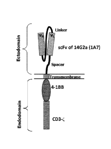
Fig. 7B: Schematic structure of the chimeric antigen receptor 14.G2a-BBzeta
(2nd generation).
The construct comprises an scFv fragment of the antibody 14G2a (1A7), fused to
the 4-1BB
domain and the CD3-zeta (CD3) domain by means of suitable spacers or linkers.
The CD3C
domain transmits a proliferative signal upon binding of the scFv fragment to
its target,
GD2.The 4-1BB costimulatory signaling domain mimic amplifies the activation of
the CAR-
T cells, leading to a more robust signal to the T cell to multiply and kill
the cancer cell.
Fig. 8: The T-cell receptor complex. CD3-zeta is a chain of the CD3 T-cell co-
receptor, which
comprises a CD3y chain, a CD3 6 chain, and two CD38 chains. These chains
associate with
TCR-a and TCR-I3 chains and the CD3C-chain (zeta-chain) to generate an
activation signal in
T lymphocytes. The TCR, CD3C-chain, and CD3 molecules together constitute the
TCR
complex.
Fig. 9: Sequence alignment between IL2 and IL15. Note the structural
similarity between the
two cytokines.
Materials and Methods
1. Sarcoma xenograft experiments
A localized Ewing sarcoma model which relies on subcutaneous xenografting of
2x10⁶ VH-
64 Ewing sarcoma cells per mouse into NOD/scid gamma (NSG) mice was produced.
17
CA 03044556 2019-05-22
WO 2017/178562 PCT/EP2017/058873
Upon a tumor volume of 200-300 mm3 mice received intraperitoneal treatment
with L19-IL2
(30 iLig twice-weekly on days 1, 5, 8, 12, 14, and 20), and with intravenous
injection of 3
doses of 1x10⁷ 14.G2a-BBzeta-transduced T cells, or non-transduced T cells as
controls (see
Fig. 1). Tumor growth was monitored by caliper quantification of diameters. 2
mice were
used in each cohort. Post-therapy tumor sections were used for comparative
histopathological
analysis with regard to (CAR)-T cell infiltration and immunocytokine
localization.
Furthermore, localization of L19-IL2 within the tumor tissue was evaluated
using an anti-
human IL2 antibody in standard immunofluorescence procedures. L19-IL2 and
14.G2a-
BBzeta are described in details elsewhere herein. Control experiments were
done with
(1) L19-IL2 or 14.G2a-BBzeta, respectively, alone
(2) KSF-IL2, which is an immunoconjugate binding to hen egg lysozyme (KSF),
and
serves as negative control
(3) non-transduced T cells likewise serve as negative controls.
Results of this experiment are shown in Figs 2 ¨ 5.
The combination of CAR-T cells and the immunocytokine drastically increased
tumor
infiltration ¨ a finding which was completely unanticipated, because none of
the current
theories that explain the challenges CAR-T cells face when infiltration a
solid tumor (active
tumor-mediated immunosuppression, functional changes in T lymphocytes after ex
vivo
manipulation, physical inhibition of infiltration by the desmoplastic stroma
which the cells
need to penetrate) would render the synergistic effect the immunocytokine has
on CAR-T cell
infiltration obvious.
The functional implication of a cytokine, namely to merely regulate the
activity of T cells, can
not explain its supportive effect in the present scenario, where tumor-
mediated
immunosuppression, functional changes in T lymphocytes after ex vivo
manipulation and/or
physical inhibition of infiltration by the desmoplastic stroma challenge the
anti tumor efficacy
of the T cells.
18
CA 03044556 2019-05-22
WO 2017/178562
PCT/EP2017/058873
References
Kowalczyk A et al. (2009), Cancer letters vol. 281 (2) p. 171-82
List T, Neri D (2013), Clinical pharmacology: advances and applications vol. 5
p. 29-45
Imai C et al. (2004), Leukemia, Apr;18(4):676-84
Zou W (2005) Nat. Rev. Cancer 5, 263-274
Caruana I et al. (2015), Nature medicine vol. 21(5) p. 524-9
Louis C U et al. (2011), Blood, 118: 6050-6
Kershaw M H et al (2005), Clin Cancer Res, 12: 6106-6115
Lamers C H J et al. (2007), Cancer Immunol Immunother, 56: 1875-1883
Park J R et al. (2007), Molecular Therapy 15: 825-833
Schliemann C et al. (2009), Leuk Res. Dec;33(12):1718-22
Zardi et al. (1987), Embo J46:2337-2342
Kaspar et al. (2006), Int J Cancer, 118:1331-1339
Pini et al. (1998), J Biol Chem.;273:21769-21776
Borsi et al. (2002), Int J Cancer.;102:75-85
Berndorff et al. (2006), J Nucl Med. ;47:1707-1716
Berndorff et al. (2005), Clin Cancer Res.;11:7053s-7063s
Demartis et al. (2001), Eur J Nucl Med;28:534-53
Meazza et al. (1996), Br.J.Cancer. 74:788-795
White ES, Muro AF (2011), IUBMB life vol. 63 (7) p. 538-46
Rybak et al (2007), Cancer research vol. 67 (22) p. 10948-57
Paxton, R J (2001), Current protocols in immunology / edited by John E.
Coligan ... [et al.]
vol. Chapter 6 p. Unit 6.22
Kaspar et al (2006), Cancer research vol. 67 (10) p. 4940-8
Pegram et al. (2015), Leukemia vol. 29 (2) p. 415-22
Mujoo, K; Kipps, T J; Yang, H M; Cheresh, D A; Wargalla, U et al. (1989),
Cancer
research,vol. 49 (11) p. 2857-61
19
CA 03044556 2019-05-22
WO 2017/178562 PCT/EP2017/058873
Sequence Listing
Seq No Specification Sequence (One letter code)
1 Vh L19 EVQLLESGGGLVQPGGSLRLSCAASGFTFSSFSMSWVRQAPGKGLEWVSS I
SGSSGTT
YYADSVKGRFT I SRDNSKNTLYLQMNSLRAEDTAVYYCAKPFPYFDYWGQGTLVTVSS
2 VI L19 EIVLTQSPGTLSLSPGERATLSCRASQSVSSSFLAWYQQKPGQAPRLLIYYASSRATG
I PDRFSGSGSGTDFTLT I SRLEPEDFAVYYCQQTGRI PPTFGQGTKVEIK
3 scFv Linker GDGSSGGSGGAS
4 human IL2 APT S S S TKKTQLQLEHLLLDLQMI
LNGINNYKNPKLTRMLTFKFYMPKKATELKHLQC
LEEELKPLEEVLNLAQSKNFHLRPRDL I SNINVIVLELKGSETTFMCEYADETATIVE
FLNRWITFCQS I I STLT
Fusion protein EFSSSSGSSSSGSSSSG
linker
6 CDR1 Vh SFSMS
7 CDR3 Vh PFPYFDY
8 CDR2 Vh S I SGSSGTTYYADSVKG
9 CDR1 VI RASQSVSSSFLA
CDR2 VI YASSRAT
11 CDR3 VI QQTGRI PPT
12 human IL15 NWVNVI SDLKKIEDLIQSMHIDATLYTESDVHPSCKVTAMKCFLLELQVI
SLESGDAS
IHDTVENL I I LANNSLS SNGNVTESGCKECEELEEKNIKEFLQSFVHIVQMF INT S