Note : Les descriptions sont présentées dans la langue officielle dans laquelle elles ont été soumises.
) 7 ~ 7 RAN4095/101
The presenc invention relates to detecting nucleic acid (ONA or RNA) amplification,
and more particularly to real-time monito~ing of multiple nucleic acid amplification reactions,
simultaneously. The invention also relates to a method for using the accumulated data to
quantitate the star~ng concentration of a target nucleic acid sequence provided in one or more
of the mixtures and monitor the effect of amplification reaction conditions on the reaction
kinetics.
Various methods for detecting nucleic acid amplification are known. A number of
assays are known which quantitate the number of starting DNA templates in a polymerase
chain reaction (PCR), for example. Some involve the measurement of PCR product at the
0 end of temperature thermal cycling and relate this level to the starting DNA concentration. ~ ~ -
Such an "endpoint" analysis, as has been typically done using PCR, reveals the presence or
absence of target DNA but generally does not provide a usable measure of the star~ing
number of DNA targets.
Other assays involve the use of a competitor amplification product whose template is
added at known concentration to the reaction mixture before thermal cycling. In competitor-
product protocols, an aliquot of the arnplification is examined by gel-electrophoresis. The
relative amount of target-specific and competitor PCR product is measured; this ratio is used
to calculate the starting number of target templates. The larger the ratio of target-specific
product to competitor-specific product, the higher the starling DNA concentration.
In addition to requiring "downstream" processing, such as hybridization or gel
electrophoresis, these other assays are more limited in dynamic range (i.e., sensitivity to a
range of target nucleic acid concentrations). In competitor assays, the sensitivity to template
concentration differences is compromised when either the target or added competitor DNA is
greatly in excess of the other. The dynamic range of assays that measure the amount of end
25 product can also be limited in that at the chosen number of cycles some reactions may have
reached a "plateau" level of product. Differences in star~ng template levels in these reactions
are therefore not well reflected. FuIthermore, small differences in the measured amount of
product result in widely varying estimates of the starting template concentration, leading to
great inaccuracy due to variable reaction conditions, variations in sampling, or the presence
30 of inhibitors.
Ve / 12.07.94
. : ' , ~'~ ~, `"
- ' :: ,
- - ,-. - : . . - : : . -
-2- 21797~7
Optirnization of PCR conditions typically is accomplished by measuring the effect of
different conditions on the final yield and specificity of PCR product. In order to obtain
information throughout amplification, many replicate samples are placed in a thermal cycler
so that each can be removed from the thermal cycler at a different temperature cycle. The
s removed tubes then are analyzed by gel electrophoresis as a function of cycle number. As is
apparent from this description, such optimization is complex and time-consuming in that it
requires numerous sample manipulation steps as well as downstream electrophoresis
analysis.
Any assay intended for large-scale (e.g., clinical) use should not only be reliable, but
o should be simplifled as much as possible in order to facilitate its automation. Thus, there is
a need for an apparatus and medlod for collecting data indicative of nucleic acid amplification
that can be used, for example, to quantitate sample starting concentrations and optimize
reaction conditions, that provides reliable results and is suitable for automation.
The aim of the present invention is to provide a nucleic acid amplification detection
method and apparatus which makes possible to monitor the amplification of multiple
amplification reaction mixtures, simultaneously in real-time, and which thereby makes
possible to overcome the problerns and disadvantages of the prior art.
According to the invention this aim is achieved by providing an apparatus that is
characterized in that it comprises: -
a thermal cycler including a heat conducting member having multiple recesses formed
therem; and
a sensor arranged for detecting light emitted from said recesses, simultaneously.
In a preferred embodiment the apparatus according to the invention it further comprises
a light source optically coupled to said thermal cycler and arranged to distribute light over a
portion of said heat conducting member having a plurality of said recesses formed therein.
In another preferred embodirnent of the apparatus according to the invention said
recesses of the heat conducting member of the thermal cycler are formed through a surface
thereof for receiving reaction vessels containing a nucleic acid amplification reaction
mixture$ and said sensor is an imaging device optically coupled to said heat conducting
member for generating an image of said surface and reaction vessels when said vessels are
disposed in said recesses of the heat conducting member.
In a further preferred embodiment of the apparatus according to the invention said heat
conducting member of the thermal cycler has multiple recesses formed therein and adapted
`~ ' - ' '
: ~
.: ,
2~ '97~7
- 3
for receiving nucleic acid amplification reaction mixtures, and the apparatus further
comprises
a housing positioned over said heat conducting member and coupled to said thermal
cycler,
s a light source arranged eo emit light in said housing and toward said recesses; and
a dichroic mirror positioned in said housing and above said recesses, said dichroic
mirror being transrnissive to light having a first wavelength and reflective to light having a ~ -
second wavelength that differs from said first wavelength; and
said sensor being arranged to receive light reflected from said second surface of said
dichroic milror.
In a further preferred embodiment of the apparatus according to the invention said heat ~ -
conducting member of the thermal cycler has multiple recesses formed therein and adapted
for receiving nucleic acid amplification reaction rnixtures including a nucleic acid sequence
and a fluorescent binding agent and the apparatus further compAses
a housing positioned over said heat conducting member and being coupled to said
thermal cycler,
a light source aTIanged to emit light in said housing;
a sensor alranged in said housing; and
a dichroic miIror positioned in said housing and above said recesses, said dichroic
mirror being transmissive to light having a wavelength corresponding to the wavelength of
light generated by the excitation light source and reflecdve to light having a wavelength
corresponding to the wavelength of fluorescence emitted from a nucleic acid amplification
mixt~re, including a fluorescent binding agent, when that mixture is disposed in one of the
recesses f~med in the heat conducting member and exposed to light from the excitation light
source, said mirror be oriented to form an optical path between said recesses and said
sensor.
In one embodiment of the apparatus according to the invention, a CCD-camera detects
the accumulation cf double-stranded DNA (dsDNA) in each of multiple polymerase chain
reactions, simultaneously, using the increase in the fluorescence of a detectable fluorescent
dye initially introduced into each amplification reaction mixture. The fluorescence results
from the fluorescent dye binding duplex DNA. This embodiment advantageously eliminates
the need for fiber-optic leads and the accompanying problems associated with coupling the
fiber-optics to the individual reaction mixtures.
According to another aspect of the invendon, the optical system that moves excitation
3s light from a source to the multiple reaction mixtures being amplified in a thermal cycler,
which f~ms part of the apparatus, is configured to provide excitation light to the reaction
, : : -.
21~7 '7
- 4 -
mixtures in a uniform manner. That is, the optical system permits excitation light to be
essentially uniformly distributed over the them~al cycler heat exchanger so that each
amplification reaction mixture receives essentially the same arnount of excitation light.
With this apparatus, the fluorescence data can be collected and used to quantitate the
s initial amount of target nucleic acid sequence. In a preferred method for quantitation,
muitiple amplification reaction mixtures are provided. One amplification reaction mixture
has an unknown concentration of a specific nucleic acid sequence. The other reaction
mixtures include the same specific nucleic acid sequence in differing but known
concentrations. The amplification reaction mixtures of known and unknown nucleic acid
concentration are thermally cycled in parallel for multiple cycles. The fluorescence emitted
from the reaction mixtures is monitored in real time and the number of cycles necessary for ~ -
each reaction mixture to fluoresce at a certain intensity value determined. The number of
cycles necessary for the mixture of unknown nucleic acid concentration to reach that value is
compared to the number of cycles necessary for the mixtures of known nucleic acid
S concentration to reach that value to obtain the initial quantity of said specific nucleic acid
sequence in the mixture of unknown concentration.
It has been found that sensitivity to a range of target nucleic acid concentration of at
least six orders of magnitude is possible because the amplifications are monitored in real-
time. In addition, sensitivity to as ~ew as 100 ssDNA templates in the background of
40,000 cell-equivalents of complex genomic DNA can be obtained.
The invention further advantageously provides a way to process the fluorescence data
to relia~bly analyze the effect of different reaction conditions on the amplification kinetics.
Since multiple amplifications can be monitored simultaneously, the effect of many different
reaction variables can be assessed rapidly. This is useful in optimizing amplification
25 reactions for optimum yield and effficiency.
Real-time monitoring also gives the advantage of detecting instances of partial
inhibition of PCR, which can greatly affect the ability to quantitate accurately. As shown in
Fig. 10D, these instances can be detected by their reaction profile so that these samples can
be repurified and tested again. In addition, instances of total inhibition, which might
30 otherwise lead to the false conclusion that there is no nucleic acid target in a sample, can be
detected in that there is the expectation that given enough cycles, even PCRs without target
DNA will produce fluorescence that is due to nonspecific amplification products. If such
fluorescence is not seen by the expected cycle, the presence of inhibitors can be inferred.
A further advantage of the present invention is that the need for additional time-
3s consuming manipulations to determine the yield of many reactions at the end of the
.
21 ~97~7
amplification ;s eliminated. Thus, the need for downstream hybridization or gelelectrophoresis, for example, is avoided.
Although the non-fiber-optic embodiment of the present invention illustrated in Figs. 2
and 3 has numerous advantages as described above, the sensor, which preferably is a video
s camera, is positioned a significant distance from the reaction mixtures in the heat-conducting
member to minirnize parallax. Accordingly, the light source also is significantly spaced from
the reaction mixtures in the heat-conducting member in order to provide uniform illumination
and avoid interference with the sensor's vision. This arrangement generally requires a
significant amount of space which, in turn, may require an entire darkroom to be allocated to
o the apparatus. However, many clinical laboratories do not have darkrooms or additional
space to allocate to constructing a darkroom. Although X-ray rooms, which can readily be
found in clinical laboratories, can provide a suitable environment for the opdcal paths of the
excitation light and fluorescence emissions, the space in these rooms is essentially
completely allocated to X-ray equipment.
Is According to a further embodiment of the present invention, an excitation light and
fluorescence sensing configuration is incorporated into the apparatus so that the optical pa~s
of the excitation light and fluorescence emissions can be folded to rnake the apparatus more
compact and to simplify enclosing the optical paths in a light-dght environment. In this
manner, the apparatus can be readily used on the bench without the need of a darkroom.
According to this embodiment, the apparatus for monitoring muldple nucleic acid
amplifications simultaneously includes a thermal cycler having a heat-conducting member
that includes multiple recesses formed therein for receiving nucleic acid amplificadon
mixtures. A housing is positioned over the heat-conducdng member so that a light-tight
chamber is formed. A light source is arranged to emit light in the housing chamber to excite
25 amplification mixtures disposed therein. A dichroic miIror is positioned in the housing and
above the recesses. The dichroic miIror is transrnissive to light having a first wavelength and
reflective to light having a second wavelength that differs from the first wavelength. In the
preferred embodiment, the milror is a low pass dichroic mirror. The miIror is constructed
such that it is transparent or transmissive to light having a wavelength corresponding to the
30 wavelength of light generated by the light source and reflective to light having a wavelength
corresponding to the wavelength of fluorescence emitted by the amplification mixtures when
exposed to the excitation light. Alternatively, a high pass dichroic mi~ror can be used. In
that case, the milTor is reflective to light corresponding to the wavelength of light generated
by the light source and transparent or transmissive to light having a wavelength35 corresponding to the wavelength of fluorescence emitted by the amplification mixtures when
exposed to the excitation light. A sensor also is arranged in the housing for sensing
. . . , . ~ . : : ~ . : - ' . ' : ' - - :
212~73~1
- 6 -
fluorescence emitted from the amplification mixtures. In the low pass dichroic mirror
arrangement, the sensor is arranged to receive fluorescence reflected from the mirror.
However, in the high pass arrangement, the sensor and light source posidons are reversed.
With this arrangement, the excitation light source and sensor both can be positioned
s reladvely close to the heat-conducdng member, e.g., from about 6 to 12 inches therefrom,
thereby permitting the opdcal paths of the excitation light source and the fluorescence
emissions to take up a relatively small amount of space. The compactness of thisarrangement facilitates enclosing the optical paths in the housing, which is coupled to the
theImal cycler to effectively form a light-tight chamber or darkroom for the excitation and
10 amplification mixture emission light. In this manner, the apparatus can be used most
anywhere, without the need for dedicating a darkroom to the apparatus. The housing further
advantageously prevents extraneous light, such as light emitted from monitors used with the
apparatus and LED's associated with the thermal cycler and computer equipment, from
reaching the sensor.
s A further advantage of having the sensor positioned very close to the heat conducting
member in which the nucleic acid amplificadon mixtures are to be disposed is that sensitivity
requirements ~f the sensor can be reduced. Since the sensor is moved closer to the heat-
conducting member, the intensity of the light that is emitted from the reacdon mixtures and
reaches the sensor increases. Accordingly, when using a CCD camera type sensor, as in the
preferred embodiment, a less expensive CCD camera with acceptable low light response and
low thermal noise can be substituted for a r~ladvely expensive, cooled CCD camera which is
generally required when sensitivity requirements are greater.
The close proxirnity of the light source to the reaction mixture recesses also provides
advantages. By moving the light source closer to the reætion mixture recesses, the intensity
of the e~citation light, which reaches the reaction mixtures that are placed in the recesses,
increases, thereby increasing the intensity of the fluorescence emissions. In this manner,
smaller concentrations of the target sequence can be detected at an earlier stage of thermal
cycling. In addition, some of the increased fluorescence can be traded for better wavelength
resolution by using a filter with a more naIrow band pass. In this manner, a broader range
of wavelengths can be detected and labels with very close wavelengths can be distinguished.
According to another aspect of this embodiment, a shutter is coupled to the light source
to intermittently expose the reaction mixtures to the excitation light. Preferably, the shutter is
timed to expose the mixtures to the excitation light during the annealing/extension phase
where maximum fluorescence generally can be exhibited. This configuration reduces the
mixture or sample exposure to the excitation light, which can be very intense due to its close
-~ 7 212~7~7 ~:
proximity to the heat-conducting member. By minimizing sarmple exposure to the excitation
light, which typically is W light, but can be of other wavelengths, the shutter improves the
system perfonnance by increasing sample stability. Otherwise the intense excitation light can
cause photo-deactivation of the sample which is commonly known as "bleaching".
s According to a further aspect of this embodiment, a field lens is positioned between the
dichroic mirror and the sensor to minimize parallax across the field therebetween.
A further advantageous feature of the invention involves a heated window which is
provided immediately above the heat-conducting member and can actually be placed on the
tubes containing the amplification mixtures. The heated window prevents heat loss from the
0 reaction tubes, helps maintain the desired thermal cycling temperature profiles of the
mixtures and minimiæs reflux, while peImitting the transmission of light therethrough.
According to the preferred embodiment, the heated window is rnaintained at the denaturation
temperature which is generally about 95-105C.
A filter or wheel of filters preferably is coupled to the sensor so that the sensor can
1S selectively detect light of different wavelengths. The filter(s) also can prevent excitation ligh~
from reaching the sensor. By using a plurality of filters, which can be arranged on a filter
wheel, the filter can be readily changed during a particular annealing/extension phase, for
example, so that emissions of different wavelengths, for example, from differenthomogeneous nuclease probes (described below), can be monitored. Different emission
wavelengths and, thus, nucleic acid sequences in a given sample can be monitored and the
presence of the target sequence determined.
ln addidon, the high sensitivity of the system permits very accurate detection of
fluorescence that is produced using a homogenous assay system such as that described in
U.S. Patent No. 5,210,015 to Gelfand et al. which is hereby incorporated herein by
reference. This assay system uses the 5' to 3' nuclease activity of a nucleic acid polymerase
to cleave annealed, labeled oligonucleotides from hybridiæd probe target duplexes and
release labeled oligonucleotide fragments for detection. This assay is particularly useful for
detecting multiple nucleic acid targets in the same amplification reaction mixture. Such
probes also are useful in conf~llI~ing target nucleic acid presence when there are nonspecific
amplification products.
The homogenous assay uses such a probe whose fluorescence is normally quenched
by fluorescence energy transfer, or F~T, to a second label. The 5' nuclease activity of the
DNA polymerase separates the fluorophore from the probe and quencher, disrupting the
FET and restoring the fluorescence. No processing is required to detect this change, which
3s may be seen using the monitoring apparatus of the present invention that incoIporates the
~ --, . . :- . ,
, . .
.
. - . - . . ,
-8- 21~9787
dichroic mirror arrangement. These probes, if labeled with different fluorophores, can be
used to detect multiple nucleic acid targets. -
The following terms are used in this specification. The accompanying definitions are
provided to aid disclosure, rather than limit the invention.
The term "target nucleic acid sequence" refers to a purified or partially purified nucleic
acid to be amplified.
The term "amplif1cation reaction mixture" refers to an aqueous solution comprising the
various reagents used to amplify a target nucleic acid. These include enzymes, aqueous
buffers, salts, amplification primers, target nucleic acid, and nucleoside triphosphates.
o Depending upon the context, the mixture can be either a complete or an incomplete
amplification reaction mixture.
The term "primer" refers to an oligonucleotide capable of acting as a point of initiation
of DNA synthesis when annealed to a nucleic acid template under conditions in which
synthesis of a primer extension product is initiated, i.e., in the presence of four different
nucleotide triphosphates and a DNA polymerase in an appropriate buffer (pH, ionic strength,
cofactors, etc.) and at a suitable temperature.
The term "template" refers to a portion of the target nucleic sequence to which the
primer anneals.
The above is a b~ief description of some deficiencies in the prior art and advantages of
the present invention.
Other features, advantages and embodiments of the invention will be apparent to those
skilled in the art from the following description, accompanying drawings and appended
claims.
Fig. 1 graphically shows continuous, real-time monitoring of a PCR;
2s Pig. lA is an enlarged view of the area within line lA in Fig. l;
Fig. 2 is a perspective view of the amplification apparatus in accordance with the
principles of the present invention;
Fig. 3 is an enlarged section of the thermal cycler of Fig. 1 to show the thermal cycler
heat exchanger block and cover therefor,
Fig. 4 is a block diagram of the apparatus shown in Fig. 2;
Fig. 5 shows examples of digitized images of multiple reaction mixtures;
Fig 6 illustrates a selected pixel alray for averaging to obtain a single fluorescence -
value;
.. . : . . . ~ . . - .
.. . - ~ . , ~ . . . - . . . .
? ~ 7 8 7
g
Fig. 7 represents cycle-to-cycle fluorescence measurement drift;
Fig. 8A shows multiple fluorescence profiles;
Fig. 8B shows the fluorescence values of Fig. 8A after normalization;
Fig. 8C shows gel electrophoresis of the amplification products represented by dle
5 profiles in Figs. 8A and B;
Fig. 9 shows the linear relationship between the log of starting template copies and the
number of cycles required to reach a selected fluorescence value;
Figs. lOA-D represent the effect of changes in reaction conditions;
Figs. 11-13 are simplified flow charts of the steps for obtaining the fluorescence
lo values;
Fig. 14 is a perspective view of the amplification apparatus of the present invention
illustrating a further embodiment of the excitation light and emissions/fluorescence detector
arrangement in panial section; and
Fig. 15 is an enlarged view of the excitation light and emissions/fluorescence detector
l5 aIrangementofFig. 14inpartialsection;
Fig. 16 shows a transmissivity v. wavelength curve that is representative of a dichroic
mirrc~r constructed in accordance with principles of the present invendon; and
Fig. 17 is a top view of a heating window for the upper portion of the reacdon mixture
containing vessels according to the present invention.
The present invendon involves nucleic acid amplificadon and the detectdon, monitoring ~ -
and quantitation of amplificatdon products. In order to facilitate understanding of the
amplification data collection and processing system of the present invendon, a summary of
nucleic acid amplification processes especially suited for use in conjuncdon with the
invention will first be discussed.
Those of skill will recognize that the present invendon requires amplificadon of the
duplex form of nucleic acid. There exist well-known methods for amplifying nucleic acids.
The means for amplification are not cridcal and this invention will work with any method
where nucleic duplexes are generated. The various methods are reviewed in Bio/Technolo~
8:290-293, April 1990, incorporated herein by reference. They include, but are not limited
to PCR, LCR, QB and 3SR. Although 3SR and QB do not involve thermal cycling, theresult of their amplifications can be monitored by the fluorescence detecting arrangement
discussed below and analyzed in accordance with the principles of the present invendon.
Each method is briefly described below.
PCR amplification of DNA involves repeated cycles of heat denaturing the DNA,
annealing two oligonucleodde primers to sequences that flank the DNA segrnent to be
amplified, and extending the annealed primers with DNA polymerase. The primers
. -
' ~ `' ' :
.
: .
21,'~7(~7
- 10-
hybridize to opposite strands of the target sequence and are oriented so that DNA synthesis
by the polymerase proceeds across the regions between the primers, each successive cycle
essentially doubling the amount of DNA synthesized in ~he previous cycle. This results in
the exponential accumulation of the specific target fragment, at a rate of approximately 2n per
s cycle, where n is the number of cycles. A complete review of this technology can be found
in PCR Technology: Principles and Applications, Ed. Erlich H.A., Stockton Press, New
York 1989.
~Q DNA polymerase is preferred when PCR is used in conjunction with the present
invention although this is not an essential aspect of the invention. ~ag. polymerase, a
lo thermostable polymerase, is active at high temperatures. Methods for the preparation of ~
are disclosed in U.S. Patent No. 4,889,818 and incorporated by reference. However, other
thermostable DNA polymerases isolated from other Thermus species or non-Thermus
species (e.g., Thermus thermophilus or Thermoto~a maritima), as well as non-thermostable
DNA polyrnerase such as T4 DNA polymerase, T7 DNA polymerase, E. coli DNA
polymerase I, or the Klenow fragment of E. coli, can also be used in PCR. Methods for
providing thermostable DNA polymerases are provided in International Patent Applications
with publication Nos.WO-A- 91/09950 and
WO-A- 92/03556, which are both incorporated herein by reference.
The ligase chain reaction is described in International Patent Application with
publication No. WO 89/09835, which is incorporated herein by reference. The process
involves the use of ligase to join oligonucleotide segments that anneal to the target nucleic
acid. Ligase chain reaction (LCR) results in amplification of an o~iginal target molecule and
can prcvide millions of copies of product DNA. Consequently, the LCR results in a net
increase in double-stranded DNA. The present detection methods are applicable to LCR, as
well as PCR. LCR typically requires some means for detecting the product DNA such as an
oligonucleotide probe. When used in conjunction with the disclosed methods for detecting
amplification products, such means are unnecessary, and the LCR result is immediately
detectable.
Another amplification scheme, ~beta replicase, exploits the use of the replicase from
the RNA bacteriophage QB. In this amplification scheme, a modified recombinant
bacteriophage genome with a sequence specific for the targeted sequence is initially ligated to
the nucleic acid to be tested. Following enrichment of the duplexes formed between the
bacteriophage probe and the nucleic acid in a sample, QB replicase is added, which, upon
recognizing the retained recombinant genome, begins making a large number of copies.
- - . - . .. . . . .:
.
~ ~ 1 r / ~3 7 ~ 7
- 11-
The QB system does not require primer sequences and there is no heat denaturation
step as with the PCR and LCR ampliflcation systems. The reaction occurs at one
temperature, typically 37C. The preferred template is a substrate for the Q~replicase,
midva~iant- 1 RNA. A very large increase in the templates is achieved through the use of this
5 system. A review of this amplification system can be found in the International Patent
Application Pub. No. WO 87/06270 and in Lizar~i et aL, 1988, Bio/Technology 6:1197-
1202.
The 3SR system is a variation of an in vitro transcription-based amplification system.
A transcription-based amplification system (TAS) involves the use of primers that encode a
10 promoter sequence as well as a complementary sequence to the target strand to generate
DNA copies of a target strand and the production of RNA copies from the DNA copies with
an RNA polymerase. See, e.g., Example 9B of U.S. Patent No. 4,683,202 and European
Patent Application with publication No.
EP-A- 0 310,æ9. The 3SR System is a system which uses three enzymes to carry out an
15 isothermal replication of target nucleic acids.
The system begins with a target of single-stranded RNA to which a T7 RNA DNA ~;
primer is bound. By extension of the primer with reverse transcriptase, a cDNA is formed,
and RNAseH treatment frees the cDNA from the heteroduplex. A second primer is bound to
the cDNA and a double-stranded cDNA is formed by reverse transcriptase treatment. One
20 (or both) of the primers encodes a promoter, e.g., the promoter for T7 RNA polymerase, so
that the double-stranded cDNA is a transcription template for RNA polymerase. ~ ~ -
~:
Transcription competent cDNAs yield antisense RNA copies of dle original target.The transcripts are then conveIted by the reverse transcriptase to double-stranded cDNA
containing double-stranded promoters, optionally on both ends in an inverted repeat
25 orientation. These DNAs can yield RNAs, which can reenter dhe cycle. A more complete
description of the 3SR system can be found in Guatelli et aL, 1990, Proc. Nad. Acad. Sci.
USA 87:187~1878, and European Patent Application with publication No. EP-A- 0 329
822, bodh of which are incorporated herein by reference. The TAS system is also described
in Gingeras et al., in Innis et aL eds, 1990, PCR Protocols, Academic Press, San Diego,
30 which is incolporated herein by reference.
According to the present invention, nucleic acid arnplification is monitored by detecting
fluorescence emitted when fluorescent dye such as an intercalating fluorescent dye, provided
in the reaction mixture, binds widh the double-stranded nucleic acid during eachannealing/extension phase as the mixture is cycled between two temperatures (dhermal
35 cycling).
2 1 ~ r~ 7 $
- 12-
An increase in fluorescence indicates a positive amplification of target nucleic acid.
Suitable intercalating agents or dyes include, but are not limited to ethidium bromide,
propidium bromide, proflavine, acridine orange, acriflavine, fluorcoumanin, ellipticine,
daunomycin, chloroquine, distamycin D, chromomycin, homidium, mithramycin, ruthenium
5 polypyridyls, anthrarnycin, methidiurn bromide, 2-[2-(4-hydroxyphenyl)-6-benzimidazole-
6-(1-methyl-4-piperazye) benzimidazole trihydrochloride and the like.
Fluorophores and DNA binding chromophores described in the art are suitable for
use in the 5' to 3' nuclease assay disclosed in U.S. Patent No. 5,210,015 are also useful in
the present invention. Suitable donor fluorophores and quenchers are chosen such that the
0 emission spectrum of the donor fluorophore overlaps with the absorption spectrum of the
quencher. Ideally, the fluorophores should have a high Stokes shift (a large difference
between the wavelength for maximum absorption and the wavelength for maximum
emission) to minimiæ interference by scattered excitation light.
Suitable labels which are well known in the art include, but are not limited to,5 fluoroscein and derivatives such as FAM, HEX, TET, and JOE; rhodamine and derivatives
such as Texas Red, ROX, and TAMRA; Lucifer Yellow, and coumarin derivatives such as
7-Me2N-coumarin4-acetate, 7-OH-4-CH3-coumarin-3-acetate, and 7-NH2~-CH3-
coumarin-3-acetate (AMCA). FAM, HEX, TET, JOE, ROX, and TAMRA are m~rketed by
Perkin Elmer, Applied Biosystems Division (Foster City, CA). Texas Red and many other
20 suitable compounds are marketed by Molecular Probes (Eugene, OR). Examples ofchemiluminescent and bioluminescent compounds that may be suitable fo$ use as the energy
donor include luminol (aminophthalhydrazide) and derivadves, and Luciferases.
Referring to Fig. 1, a fluorescence trace indicadve of DNA amplificadon for a single
polymerase chain reacdon (PCR), which involves cycling the temperature of the reacdon
25 mixture between two temperatures, e.g., 94-C and 50C, is shown. The reacdon mixture
included an intercaladng fluorescent DNA binding dye, ethidium bromide (EtBr), which
intercalates, or binds, the double-stranded PCR products and fluoresces during each
annealing/extension phase of the reaction. Consequently, the resuldng fluorescence
increases as the amount of double-stranded DNA increases. As illustrated in Fig. 1,
30 fluorescence intensity r~ses and falls inversely with temperature. The fluorescence intensity
is minimum at the denaturation temperature (94-C) and maximum at the annealing/extension
temperature (50C). Thus, the fluorescence intensity at 50C shows a cycle-dependent
increase reflecting an increase in the amount of double-stranded DNA. When the thermal
cycler returned to 2~C, after the 30 cycles were completed, the fluorescence increased to a
35 final value greater than three times the initial fluorescence value at 25 C. On the other hand,
the fluorescence minima at the denaturadon temperature do not significantly increase,
presumably because at this temperature th re is no dsDNA for EtBr to bind.
~') 1 '1 ~; 'i~ ) 7
- 13-
As disclosed in U.S. Patent Application Serial No. 07/695,201, this data was collected
using a fiber-optic lead and a spectra-fluorometer. The fiber-optic lead was used to input
excitation light directly to the tube containing the reaction mixture and the tube positioned in
a heating/cooling block of a thermal cycler. The same fiber-optic lead was used to return
s fluorescent emissions back to the spectra-fluorometer, where the value corresponding to
emitted fluorescence was read. With this arrangement, the generation of amplification
products can be monitored while the reaction is in progress as evidenced by the data in Fig.
l. Since the signal generated by the binding agent can be detected without having to open
the reaction tube, the signal can be monitored throughout before, during, and after
10 amplification process.
The apparatus for detecting a single PCR and providing the data in Fig. 1 was set up
as follows: a Spex-Fluorolog-2 fluorometer with a fiber-optic accessory (Spex Catalog No.
1950) was set to emit excitation light at 500 nm with a bandwidth of approximately
3.4 nm. A GG 435 nm cut off filter was used to exclude second order light (Purchased
5 from Melles Grist Inc.). The emission light was detected at 570 nm with a bandwidth of
approximately 13.6 nm. A OG530 filter (530 nm cut off) was used to remove excitation
light.
The reaction tube, a 0.5 ml polypropylene tube, contained 60 ng human male DNA.
The top of the tube was cut away for attaching the fiber-optic cable. The fiber-optic cable
20 was glued to the top of the reaction tube with epoxy. The emission light was collected
through the oil overlay in the tube. A black "shroud" was built around the tube and the
reaction was placed in the therrnal cycler. The thermal cycler was programmed to cycle
between 94-C and 50-C for 1 minute each, for 30 cycles, followed by continuous
incubation at 25-C. The fluorometer and thermal cycler were started simultaneously. The
25 parameters of the fluorometer were: dme-based scan with 5 second integradon time; the
emission signal was radoed to that of the excitatdon light to control for changes in source
intensity.
In accordance with the present inventdon, an apparatus is provided for real-dme
detecdon of the amplification of muldple amplification reacdon mixtures, simultaneously.
30 Each reaction rnixture contains a target nucleic acid sequence. Thus, using a thermal cycler
(e.g., a therrnal cycler commercially available from Perkin Elmer Cetus Instruments) having
a headng and cooling block capable of holding up to 48 reacdon tubes, for example, 48
amplificadon reactdons can be carried out and PCR amplification product in all 48 samples
detected simultaneously without handling the samples, opening tubes, or interrupdng the
35 cycling reaction. The number of reactions monitored simultaneously is limited only by the
capacity of the heating and cooling block. Thus, with a 96 well headng and cooling block,
.,~ ,. ; -- ., . ,. - , . ..
2 1 ~ d 3 7 ~ 7
- 14-
96 amplification reactions can be monitored and the amplification product detected
simultaneously. As in the example case described above, it should be understood that
although the embodiments discussed below are described in conjunction with PCRs and
signals generated by a specific fluorescent binding agent indicative of amplification of a
s target DNA sequence in certain examples, these embodiments can be used with other
amplification processes or binding agents such as those sumrnaIized above and disclosed in
U3. Patent No. 5,210,015 to Gelfand et ah which is hereby incorporated herein byreference.
In a first embodiment of the multiple amplification reaction mixture detection
10 apparatus, a suitable optical system moves the excitation light from a source to multiple
reaction tubes that are positioned in a thermal cycler and measures the emission light from
each tube. Such an optical system can comprise multiple fiber-optic leads that
simultaneously read all of the PCR tubes undergoing thermal cycling. Only a single
fluorometer is needed to read fluorescence from the reaction tubes, as each fiber-opdc can be
ls read rapidly one at a time, for exarnple, during the time frame of a PCR temperature soalc
(e.g., an annealing/extension interval such as the interval between a-c illustrated in Fig. lA).
It will be apparent to one skilled in the art that such a detection system is not necessarily
lirnited to a particular thermal cycler machine or reaction vessel.
The arnplification detection arrangements incorporate fiber-optic leads to transmit
20 excitation light and fluorescence emissions. So long as the reaction wells, or tubes, are
light-sealed to prevent external light sources from influencing fluorescence detection, any
over plate, tube cap, or lid apparatus that comprises or can be attached to a fiber-optic lead is
suitable. In one embodiment of the invention, the reaction tube lids can be removed to
accommodate the fiber-optic. However, use of a reaction vessel that has a clear or
25 translucent cap eliminates the need to insert the cable into the tube. It will be ~pparent that
reaction tubes capable of accommodating a fiber-optic cable, without the cable physically
con!acting the amplification reaction components, are desirable.
Referring to Fig. 2, a further embodiment of an apparatus for real-time detection of the
amplification of multiple target nucleic acid sequences, simultaneously, is shown and
30 designated with reference numeral 10. This embodiment advantageously eliminates the need
for the light input and fluorescence output fiber-optic leads described above and the
accompanying problems associated with coupling the fiber-optics to the reacdon tubes. As
discussed above, apparatus 10 will be described in conjunction with PCR amplification of a
DNA target sequence using a fluorescent binding agent signal generator for purposes of
3s simplificadon, but is not intended to be limited to use with such an amplification process,
nucleic acid or amplification signal generator.
- 1S - 2 ~ 7
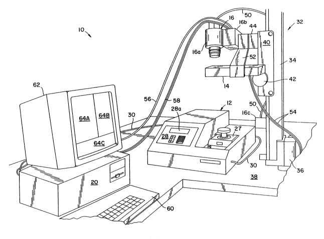
Apparatus 10 generally comprises a thermal cycler 12 for heating and cooling reaction
chamber tubes or vessels 26 (preloaded with the nucleic acid(s) to be amplified), laterally
spaced excitation light sources or lamps 14, imaging device 16 (comprising CCD-carnera
16a, camera controller 16b and cooler 16c), and image processor 20, which can be a PC
5 with a frame grabber card such as the PC VISION PLUS commercially available from lTEX
Inc. and an EEE 488 interface card. These elements are shown in block diagram in Fig. 4.
Although thermal cycler 12 is conventional in construction according to the preferred
embodiment, a summary of certain features of thermal cycler 12 is provided below to aid in
understanding the disclosure of the present invention.
0 Therrnal cycler 12 includes a heat exchanger 22 for heating and cooling reaction tubes
26. Heat exchanger 22 is a heat-conducting block, preferably aluminum, having a plurality
of recesses 24 formed therein and sized to allow a given number of reaction tubes 26 (e.g.,
0.5 ml Eppendorf tubes) to fit therein. The purpose of heat exchanger 22 is to support
reaction tubes 26 and to act as a heat exchanger to transfer thermal energy to and from the
fluids stored in the tubes such that the reaction components may be incubated at various
temperatures for user-defined times. Thus, thermal cycler 12 also includes a system for
heating and cooling the heat exchanger and means, such as a computer system, forcontrolling the heating and cooling system to heat and cool the heat exchanger and reætion
tubes as is conventional in the art. The user programs the computer or means for controlling
the heating and cooling of heat exchanger and reaction tubes to a desired temperature prof~e,
such as that illustrated in Fig. 1, through display/keyboard user interface 28.
The heat exchanger (heat conducting block), means for heating and cooling the heat
exchanger and means for controlling the heating and cooling of the heat exchanger as well as
the entire thermal cycler, can be constructed in accordance with U.S. Patent No. 5,038,852
which is hereby incorporated by reference herein. Suitable thermal cyclers are commercially
available such as the GeneAmp PCR system 9600 therrnal cycler (Perkin-Elmer, NorwaLIc,
CT) depicted in Fig. 2.
It should be understood, however, that other devices for heating and cooling thepreloaded reaction tubes can be used without departing from the scope of the invention. It is
only necessary that whatever device is used for heating and cooling the reaction tubes, be
capable of reaching and sustaining the temperatures involved and æhieve the desired
temperature versus tirne profile. Thus, for purposes of nucleic acid amplification, such a
device should be capable of cycling the temperature of the amplification reaction mixture
between a denaturing temperature T1 (which can be in the range of about 8~105C and
3s preferably 90-100C) and an annealing/extension temperature T2 (which can be in the range
~. . . .
~, . . . . .
2 1 ~ '` r? ~~
- - 16-
of about 30-90C and preferably 5(}70C) where Tl > T2 as is known to those skilled in the
art.
Cover block 27, including handle 27a, is slidably coupled to the heat exchanger for
covering or uncovering heat exchanger 22 as is conventional in the art. Cover block 27 is
s maintained in the laKer position as shown in Fig. 3 during the amplification/detection process
of the present invention embodied in Fig. 2. This permits camera 16a to receive the
fluorescence signals emitted from within the amplification reaction tubes.
Thermal cycler 12 also is provided with a means for sending output data indicative of
the heat exchanger or amplification reaction mixture temperature profile, such as cycle
0 number, temperature, and time, to image processor 20 over line 30. An RS232 bus for
transmitting such data is provided with a GeneAmp PCR system 9600 thermal cycler, for
example. LED display panel 28a also displays such output data to dhe user during operation.
CCD-camera 16a is positioned above heat exchanger 22 and the reaction tubes to
collect fluorescence signals emitted from dhe reaction mixtures dlrough the upper ends of dhe
tubes as the temperature of dhe rnixtures is cycled by device 12 during amplification. It is
important that camera 16a is positioned as far as is practical from heat exchanger æ in order
to redu~e parallax effects on dhe fluorescence image captured by camera 16a. However,
camera 16a must be sufficiendy close to dhe heat exchanger and reaction tubes to minimize
background noise from odher light sources and so that suffficient light from reaction tubes 26
can be captured by dhe camera. It has been found dhat an optimum distance d, between dhe
heat exchanger 22 and camera lens 46, which is directed toward the heat exchanger, is in the
range of about 6 inches to 5 feet depending on dhe lens used. For example, d preferably is
about 2 feet when a 70 mm lens is used. A band pass interference filter 48, preferably a 600
nm filter, is placed in front of dhe lens to limit detection to the desired wavelength, since the
2S fluorescence of interest has wavelengths of about 600 nm.
Referring to Fig.2, camera 16a is shown mounted to a conventional photographic
copy stand 32 for positioning the camera above heat exchanger 22 and reaction tubes 26.
Copy stand 32 includes a vertical support member 34 which is supported in bracket 36.
Bracket 36 is secured to mounting surface 38 which supports apparatus 10. Slide member
40 is slidably mounted to vertical member 34 for movement along the vertical and is
provided with knob 42 which upon turning cooperates with slide member 40 to lock slide
member 40 in a fixed position as is conventional to one of ordinaIy skill. Camera bracket 44
has one end secured to slide member 40 and another end secured to camera 16a Thus, the
height of camera 16a can be adjusted through slide member 40 to the desired height above
the heat exchanger and reaction tubes. Camera 16a preferably is a conventional computer-
.. . . . ~ ... . - ...
21297~7
- 17-
controlled, cooled CCD-camera. Cooling fluid is circulated through camera 16a through
multiconduit line 50. The camera is preferably cooled in order to provide higher quality
images with less noise. One suitable camera is a CC3~-camera commercially available from
Photometrics (Tucson, AZ) under the name STAR 1. It uses a thermoelectrically cooled
S CCD a~ray with a 12-bit per pixel resolution. It also permits exposure times to be varied to
change sensitivity to the fluorescence being detected. An EEE 488 bus (designated with a
reference nur;neral 58) between camera 16a and image processor 20 is provided so tha~ image -
processor 20 can control the exposure of an image of the reaction tubes and ~e tirne period
in which the image is taken.
0 Referring to Fig. 4, two W lamps 14 are laterally spaced about the optical path
between camera 16a and heat exchanger 22. It is only important in this regard that lamps 14
are sufficiently close to camera lens 46 so that their housings do not interfere with the optical
path between all of the reaction tubes and camera lens 46. To this end, lamps 14 can be
suspended from camera bracket 44 via bracket 52. Lamps 14 also are parallel to the heat
exchanger so that excitation light is provided to the reaction tubes in a uniform manner.
Lines 54 extend from lamps 14 and are adapted to fit in a standard 115 volt wall socket for
providing the desired power to the lamps. The wavelength of W lamps 14 is selected
according to the re~quirements of the nucleic acid fluorescent binding agent. For example,
302 nm is a desirable wavelength for excitation of ethidium bromide. A suitable 302 nm
mid-range UV lamp is commercially available from U.V. P.roducts, Inc. (San Gabriel, CA)
under the name Chroma-Vue.
The image taken by camera 16a is transmitted to irnage processor 20 via line 56 which,
when using the STAR 1 system as discussed above, is an RS170 line. Image processor 20
includes a frame grabber for forrning digital images from camera data on line 56. Although
camera 16a includes a 12-bit digital output, the analog output of the camera is used where
image processor 20 is an 8-bit image processor. In such a case, the image processor 20
includes the frame grabber to create 8-bit digital images from the analog signal on line 56 so
that image processor 20 can manipulate the collected images as digital data.
Thermal cycler output data line 30 also is coupled to image processor 20 for providing
the processor with the temperature profile, time, and cycle of the amplification in progress.
In this manner, processor 20 can be programmed to send a trigger signal via line 58 to
camera controller 18, which controls the opening of the shutter for camera 16a, at
preselected times during the course of amplification. Image processor 20 includes a ~
488 bus card for comrnunicating with IEEE 488 line 58, to control camera 16a. Processor
3S 20 also is provided with a conventional keyboard and/or mouse user interface 60/64 and
monitor 62. A general overview of the operation of apparatus 10 is provided below.
, ~ . . , . , . : . .-
. . ~
~3~ 1 2 3 7 3 7
- 18-
Multiple reaction tubes 26 each containing a reaction rnixture (e.g., nucleic acid(s) to
be amplified, a thermostable enzyme to catalyze polymerization, specific oligonucleotide
primers, and four different nucleotide triphosphates) are placed in heat exchanger block 22
(Fig. 3). The reaction tubes can be sealed with conventional caps, a vapor barrier mineral oil
s or ArnpliwaxlM brand sealant. The thermal cycler is activated and the excitation lamps,
which are directed toward the reaction tubes, are energized. Responsive to a signal from
camera controller 16b, camera 16a captures an image of all of the reaction tubes 26 which
image reflects the various levels of fluorescence at the first annealing/extension phase of a
cycle. This analog image is sent to the image processor where the frame grabber digitizes
the image since it has an analog/digital converter.
Fig. 5 shows p~ions of three such digitized images taken during the course of six
simultaneous amplifications. The processor averages the intensity of light emitted from each
reaction tube during the noted annealing/extension phase and normalizes those intensity
values to determine a single fluorescence value for each reaction tube. The normalization
process is discussed below in more detail in connection with Figs. 8A-C. This is repeated
for each cycle of the amplification process. The normalized fluorescence values are saved to
a computer spreadsheet for subsequent analysis and graphing. These normalized
fluorescence values are further processed to determine the initial amount of the target nucleic
acid sequence (i.e., the starting number of target nucleic acid templates before amplification)
or to analyze the effect of amplification reaction conditions on the kinetics of the reaction. A
more detailed discussion of the fluorescence value acquisition and processing steps,
summarized above, is provided below.
The timing of each camera exposure to the emitted fluorescence first will be discussed.
Since the maximum fluorescence occurs at the annealing/extension phase of each cycle in an
amplificadon process undergoing thermal cycling (e.g., see interval a-c Fig. lA) and this is
the indication of the level of nucleic acid amplification, when only one irnage is taken in a
given cycle by carnera 16a it should be taken in this phase of the cycle because that is when
you get the strongest signal and signals during this interval best represent the amount of
material being made. However, as other data throughout the reaction can be useful in
analyzing the entire course of the amplification, it should be understood that it may be
desirable to collect more than one image per cycle. For example, each image can be taken 20
seconds after the annealing/extension ternperature is reached as indicated by reference
character b in Fig. lA. In this example case, when the annealing temperature is 68'C,
processor 20 sends a signal, 20 seconds after the 68C temperature is indicated by the
thermal cycler 12 over RS232 line 30, to camera controller 16a to trigger the camera shutter
(not shown). The exposure time can vary from application to application as would be
apparent to one of ordinary skill. The triggering event to open the camera shutter obviously
., ,. ~ ,.
2~ 7~7
- 19-
can be done manually with the operator monitoring the temperature and time output data
displayed on display 28a.
The CCD-camera sums up the light received during the exposure and produces an
image indicative of the fluorescence of all of the reaction tubes in the heat exchanger at that
time interval. This image is sent over line 56 to image processor 20. Monitor 62, which is
coupled to processor 20, can display three windows labeled 64A-C in Fig. 5. Window 64A
can monitor the camera's view and provide a menu during camera operation for functions
such as camera exposure control. Window 64B displays the digital image and facilitates the
manual selection of the portion of the image to be processed (e.g., averaged as discussed
0 below). Window 64C displays the command line of the computer operating system. The
processor digitizes the image and displays it in window 64B which is used so that the user
can assign a group of pixels to an area of the image that encompasses a single image of a
reaction tube. This can be accomplished using a mouse to draw boxes around the portions
of the digitized image of a reaction tube 26 in window 64B or using a program that looks at
the fluorescence of each pixel to determine which pixels belong to which reaction tube as
would be apparent to one of ordinary skill in the art of computer graphics. This is illustrated
in Fig. 6, where a 6x6 pixel array encompasses the image of a single reaction tube 26. It
should be understood that this array size has been selected merely for purposes of example
and that other array sizes can be used without departing from the scope of the invention.
The pixel values "5" indicate insignificant fluorescence and correspond to the fluorescence
emitted by the upper surface of the heat exchanger, while the pixel values "200" indicate the
maximurn fluorescence detected in the 6x6 pixel alray and correspond to the fluorescence
emitted from the center of the reaction tube which are imaged onto pixels. The pixel values
"100" correspond to the fluorescence emitted by a portion of the heat exchanger and reaction
tube. Of course, these specific pixel values do not necessarily represent actual values, but
are merely exemplary.
Image processor 20 then averages the pixel values in the array assigned to the area
encompassing the reaction tube to obtain a single fluorescence value for that reaction tube for
cycle 1 of the nucleic acid amplification process. However, obtaining a single fluorescence
value for each reaction tube can be accomplished in other ways such as summing the pixel
values. The box outlining the 6x6 array and encompassing the first reaction tube is then ;
moved to encompass the next reaction tube where the pixel value averaging step is repeated
and a single fluorescence value is obtained until a fluorescence value has been assigned to
each tube for cycle 1. Alternatively, the process of obtaining a fluorescence value for each
reaction tube 26 for a given cycle can be done as a parallel process. This process of
calculating a fluorescence value for each reaction tube is repeated each cycle until the
amplification process is complete.
.,, , . - , . . .
: :
.
.
".: .. . :: -
. .:, . . . -
--
2~ 2~7~ ~
- 20-
At the outset, i.e., when heat exchanger block 22 is loaded with reaction tubes 26, a
control tube 26C is also loaded into one of the wells 24. The purpose of the control tube is
to provide a constant fluorescence source against which cycle-to-cycle measurement
variations, due to minor temperature variations or drift in the excitation light intensity, for
S example, can be detected. In this manner, the fluorescence value obtained for each reaction
tube can be corrected to compensate for these variations according to the following. The
control tube 26C is pre-loaded with a fluorescence dye only, such as ethidium bromide
(EtBr). Since the control tube contains no nucleic acid, there is no amplification of its
fluorescence during thermal cycling. Thus, the control tube will emit an essentially constant
o amount of fluorescence when subjected to excitadon light during amplification and serves as
a base line throughout the amplification process. This is illustrated in Fig. 7, where the
fluorescence intensity of the control tube is plotted over several cycles.
Refernng to Fig. 7, io is the inidal fluorescence of the control tube and establishes the
base line. At cycle 2, the recorded intensity of control tube 26 begins to drop off indicating
S that drift in fluorescence is occurring. Thus, solid line 72 cycle 4 corresponds to apparent
intensity values. For example, at cycle 4 when the camera collects data in the
annealing/extension phase of the fourth cycle, the apparent value of the control tube
fluorescence is iA4. A correcdon factor iorlA4 is then determined for that cycle. Each of the
fluorescence values obtained for each reaction tube is muldplied by the correcdon factor to
20 provide actual fluorescence values. The foregoing is repeated for each cycle. In summary,
the correction factor is generally described as iJiAn wherein io is the inidal fluorescence
value of the control tube and iAn is the apparent fluorescence value of the control tube taken
during cycle number n. Each fluorescence value obtained for a reactdon tube by, for
example, averaging (as discussed above) is muldplied by the correction value. The foregoing
25 is repeated for each cycle.
Normalizadon and quantdtation will be descfibed with reference to data generated for a
specific exarnple case for exemplary purposes only. That data is illustrated in Fig. 8A. It
should be understood that the inventdon is not intended to be limited to PCR or the particular
amplificatdon reaction mixtures, number of amplificadons, thermal cycler, temperature
30 profile, or interval in which the fluorescence images were taken as will be apparent to one of
ordinary skill. It is only necessary to monitor the amplificadon reactdon mixtures differing in
starting concentration of a target nucleic acid sequence to quantitate the inidal concentration
of that target sequence in an amplificadon reacdon mixture where that concentradon inidally
is unknown as will be described in more detail below.
Fig. 8A shows the fluorescence values obtained from eight PCR amplificatdons
monitored simultaneously, in accordance with the principles described above, during thermal
2 1 ;. 9 l:, 7
- 21 -
cycling for fifty-five cycles with the CCD-camera. Thus, these values represent the values
obtained for each reaction tube (e.g., by averaging the respective pixel aTrays) corrected for
occurrences of measurement variations (e.g., drift) as discussed above. In this exarnple
case, seven reaction mixtures contained known concentrations of a specific DNA target
S sequence (i.e., a known quantity of starting sarnple) to facilitate quantitation of an unknown
starting amount of a sample as will be described in more detail below. The eighth mixture
contained no target DNA and served as a control reaction to provide background
information. The seven amplifications of known sample were initiated using a dilution
series of single-stranded, HIV template DNA.
0 Prior to making the dilutions, the PCR reaction mixtures were set up in 1001l1
volumes containing 10 mM Tris-HCl, pH 8.3; 50 mM KCl; 3 mM MgC12; 2.5 U of
DNA polymerase (Perkin Elmer, Norwalk, CT); 100 ~lM each d~TP (E'romega Corp.,
Madison, WI); 411g/ml of ethidium bromide (Bio Rad, Richmond, CA); 100 pmole each of
HIV specific (gag region) oligonucleotide primers SK145 and SK431 ~Perkin Elmer,15 Norwalk, CT) and 300 ng of human placental DNA (Sigma, St. Louis, MO). An M13subclone of the HIV gag region was used as the target nucleic acid template.
Starting with 108 templates, 10-fold dilutions were made down to 102 templates and
used with HIV-specific primers to make a 142 bp PCR product. The fluorescence profiles
in Fig. 8A generated from these initial concentrations are designated with reference numerals
20 108, 107 . . . 102, respectively. A control amplification with no added template was also
monitored. The fluorescence profile of this mixture, is shown by the line designated with
reference numeral 0. To simulate a PCR-based screen of lymphocyte DNA for integrated
HIV genomes, all reactions contained 300 ng or 40,000 cell equivalents of human genomic
DNA.
Thermal cycling proceeded for 55 cycles in a GeneAmp PCR System 9600 thermal
cycler (Perkin-Elmer, Norwalk, Cl~ with a 2 temperature program: 94C for 15 sec.
denaturing; 68C for 30 sec. annealing/extension. To provide a "hot-start" as isconventional in the aIt to improve specificity in the reaction (as described in Chou, Q.,
Russell, M., Birch D.E., Raymond, J. and Bloch, W. "Prevention of pre-PCR mis-priming
30 and primer dimerization improves low-copy-number simplifications." Nucleic Acids
Research 20:1717-1723, 1992) an initial hold at 75C was used during which the MgCk
was added (in a 12 111 volume) after the samples had reached this temperature. Fluorescence
images for each thermal cycle were taken by the CCD-camera 20 sec. into the 30 sec. hold at
the annealing temperature with an exposure of about 1 second. As described above, the
35 headng cover block 27 was not used and was positioned away from the reaction tubes to
permit imaging.
. - . . , -
.... :. . ,. : .,
, - ... . : ~
.
- 22 -
As shown in Fig. 8A, the fluorescence from each reaction changes little in earlythermal cycles and then rises as detectable amounts of PCR product are generated. The more
starting template copies in the reaction, the earlier such a rise in fluorescence occurs. See,
for example, the curve designated with the character 108 which designates that that
S fluorescence cuIve coIresponds to the fluorescence emitted by the reaction mixture starting
with the highest concentration of starting templates, i.e., 108 templates. As shown in Fig.
8A, the 108 curve rises first as expected.
However, there is a considerable variance in the initial fluorescence values obtained
from the different amplifications even though those values should be the same. They should
0 be the same because the amounts of single-stranded DNA are so small that the varying
amounts of it in the reaction tubes have a negligible effect on the total fluorescence expected
from a single tube. It is believed that the sources of this variation are inhomogeneity or
nonuniformity of illumination, paTallax, and variable attenuation of the fluorescence due to
the tube caps. Monitoring the amplifications without caps but through a vapor barrier of
5 mineral oil or AmpliwaxlM brand sealant reduces, but does not eliminate, this variadon.
To compensate for inhomogeneity of illumination parallax, and variable attenuation
and, thus, the variations shown in Fig. 8A, the processor deterrnines a normalization factor
for each amplification. Each factor is determined based on the data collected in cycle 1 and is
the ratio of the average initial fluorescence of all reactions to the observed initial fluorescence
20 of each reaction. The processor multiplies all fluorescence values for each amplification by
the respective normalization factor. The result of this normalizadon is shown in Fig. 8B.
Thus, for the reacdon containing 108 stardng template copies, the rado (normalization factor)
is the sum of all eight fluorescence values in cycle 1 divided by 85 which is the inidal
fluorescence value of reacdon 108 as shown in Fig. 8A. The fluorescence values for the
25 reacdon marked 108 then are all muldplied by this factor to generate the normalized curve of
Fig. 8B. Each reacdon curve is similarly normalized. This no~malizadon is based on the
assumpdon that the source of the image variation attenuates or enhances the fluorescence
signal propordonately over the entire range of signal intensides.
Referring to Fig. 8B, all reacdons essendally have the same inidal fluorescence and
30 most of the reacdon profiles have a regular spacing consistent with the dilution series used.
The profiles are very similar in shape and nearly parallel. The early or logarithmic phase of
an efficient PCR involves a doubling of the DNA copy number every cycle. It is predicted
that each 10-fold dilution in starting template would require 3.32 addidonal cycles to bring
the yield of PCR product up to a given concentration. Thus, the normalized values look as
35 expected, for much of the range of fluorescence levels. It can be seen by examinadon of
Fig. 8B that there are close to three cycles separating each of the dilutions except for those
.
- 23 -
star~ing with 102 copies and 0 copies. It is also apparent that this cycle-number-offset is
persisting past the logarithmic phase of the PCRs. In contrast, the number of cycles
separating the dilutions in the unnormalized data of Fig. 8A vary widely.
Using the normalized fluorescence data, such as the data shown in Fig. 8B, the initial
5 amount of target nucleic acid can be quantitated or the effect of different reaction conditions
on the kinetics of the amplification reaction can be analyzed. Quantitation of the initial
amount of target will first be discussed. Fig. 9 shows the linear relationship between the log
of the starting number of template copies and the number of cycles it takes for the
amplification in Fig. 8B to reach the arbitrarily chosen fluorescence level of 190. The line
shown in Fig. 9 is regression fitted to the data points from 108 to 103 initial copies (r2 >
.99). The data point corresponding to 102 initial copies has a statistically significant
deviation from this line (zero copies cannot be placed on a logarithmic plot). Besides the
data point at 102 copies, the data point most deviant from the regression line is at 105,
which, using the equation of the fitted line, predicts a value 13% lower than the known
15 initial number of copies. Similar results are obtained if the fluorescence level chosen in Fig.
8Bisanywherefrom 125-225.
The reason for the deviation from linearity at the data point at 102 copies is shown by
the gel electrophoresis analysis of the amplification products in Fig. 8C. The specificity of
the amplification is such that, starting with 108 down to 105 copies of template, the only
20 visible product is that of the expected size. When 104 starting copies were used, a smaller,
nonspecific amplification product becomes barely visible. This product is more prominent
when 103 copies are used, and when 102 copies are used, an equal amount of nonspecific
product and HIV-specific product are made. Only nonspecific product is made in the control
with no added template. Since fluorescence due to specific product is indistinguishable from
25 that of nonspecific product, the "baseline" of sensitivity of this assay is at the cycle number
by which so much nonspecific product is being synthesized that any additional synthesis of
specific product has little effect on the reaction profile. As long as concentration
determinations are based on interpolation from a set of standards, i.e., reaction tubes
containing known concentrations of starting target nucleic acid sequence, this baseline could
30 be at significantly more cycles than at the cycle number at which the deviation from linearity
begins. In practice we have seen that sample-to-sample variation of the reaction profile at
less than 102 templates complicates reproducible quantitation below this level (data not
shown).
In order to quandtate the initial amount of target nucleic acid in a sample, the35 normalized fluorescence data from the set of standards are first used to establish the
relationship between initial target nucleic acid amount and the number of cycles needed (such
:
, ;
-24-
as that shown in Fig. 9) to reach an arbitrarily chosen level of fluorescence that is within the
range of detection of this instrurnent. This arbitrary fluorescence value (AFV) is preferably
chosen to be in a region of the reaction profile that is, as described with reference to Fig. 8B,
parallel arnong the different standards. This will generally be true if the selected AFV is
s from 0.1 to 0.5 times the maximum fluorescence value obtained by the standard using the
highest initial known target nucleic acid concentration.
After selecting the AFV, for each standard amplification, the correction and normalized
fluor value obtained that is nearest the AFV is chosen. Taking this value, and the fluor
values for the four cycles preceding and the four cycles following the cycle at which this
lo value occurs, a regression line is fitted that relates cycle number (which can be fractional) to
fluorescence. The AFV is then entered into the equation of this regression line and a value
for this standard sarnple of the number of cycles needed to reach the AFV is re~ned.
Having now deterrnined for each standard the number of cycles needed to reach the
AFV, a regression line is fitted to the data that relates the initial target nucleic acid arnount to
s the nurnber of cycles needed to reach the AFV. To now deterrnine the initial target nucleic
acid amount of an unknown sample that was amplified together with the standard samples,
the number of cycles needed to reach the AFV is determined as was done for the standard
sarnple. This number of cycles (which can be fractional) is entered into the equation of the
fitted regression line and the equation returns a value that is the initial amount of target
20 nucleic acid in the unknown sample. This process can be repeated for each unknown
sample. The data manipulations just described can easily and rapidly be performed by a
rnicroprocessor that is suitably programmed.
'l~is mode of nucleic acid quantitation can be used not only with fluorescence resulting
from the non-sequence-specific binding of dyes, but can also be used with fluorescence
25 resulting from the sequence-specific binding of oligonucleotide probes such as those used in
the homogenous amplification/detection systems described in U.S. Patent No. 5,210,015.
These probes require alternative light sources and filters as described in the above patent.
Use of multiple sequence specific probes, each with a distinguishable fluorescence, should
allow simultaneous detection and quantitation of multiple nucleic acid target sequences in a
30 single amplification reaction.
Referring to Figs. lOA-D, analyzing the effect of different reaction condidons on a
PCR will be discussed. In general, these effects can be monitored by adding, subtracting,
or changing components of a standard PCR and observing the change in the ~eaction profile.
Fig. lOA shows a titration of E~ DNA polymerase. Nine replicate PCRs containing 108
35 target HIV templates were initiated using a range of Taq polyrnerase levels as indicated in the
3 7 ~ 7
- 25 -
figure. For enzyme amounts between 1 and 10 units per reaction, fluorescence begins to be
detectable in all reactions by about the same cycle. This indicates that these differences in
enzyme levels are having little effect on the efficiency of amplification in early cycles.
Differences in the levels of PCR product become apparent in later cycl~s, however, such that
5 by cycle 50 almost twice as much DNA has been made using 10 units as compared to 1 unit.
By gel electrophoresis (not shown), there is no evidence of the production of nonspecific,
non-HIV PCR product in these reactions. Using only 0.5 unit Taq polymerase, there is a
sudden absence of delectable product. Since PCR is a process in which DNA increases in
concentration exponentially, it is perhaps not surprising that there is a threshold level of
10 enzyme below which amplification is an apparent failure.
The effect of changing primer concentrations on the kinetics of amplification also can
be monitored. Fi~. 10B shows profiles of PCRs detecting HIV templates (108 initial
ssDNA copies) using primer levels from 2 ~lM (each primer) down to 0.05 ,uM. A constant
level of enzyme (2.5 U) was used in each PCR. Note that from 0.05 to 0.4 ,uM in primer
15 concentration, the DNA level in the reaction rises to a certain level and remains constant. If
these final product levels are estimated using the data in Fig 4 and assuming 10.6 pmoletllg
of 142 bp PCR product, we see that for 0.05, 0.10, and 0.20 IlM primer, 0.05, 0.10, and
0.20 ~M product respectively, is made. At these levels, primer concentration is apparently
the limiting factor on yield of product DNA. In contrast, at 0.8 ~LM primer concentration and
20 above, the DNA level is continuing to rise with each thermal cycle, as in Fig. 6A, and
increasing amounts of primer have little effect on the profile. By comparison with the data in
Fig. 6A, one would conclude that these reactions are now enzyme, and not primer, lirnited.
The result of vaIying KCI concentration using the same HIV test system with 1 llM
primer s and 2.5 U of enzyme per 100 111 reaction are shown in Fig. 10C. ~Lq DNA25 polymerase activity is greatly inhibited by high concentrations of KCI. Consistent with this,
at concentrations of KCI of 125 mM or greater there is no detectable PCR product. With
activated salmon sperm DNA as template there is an optimum in ~a.g DNA polymerase
activity at 50-90 mM KCI. This assay shows that product yield is highest at 50, 75 and 100
mM KCI with only about half as much being rnade at 0 mM KCI. It is possible that this
30 difference is due to low ionic strength destabilizing primer annealing, although inconsistent
with this is the observation that the efficiency of amplification using 0 mM KCI appears
nearly the same as those at 50-100 mM KCl until about cycle 20. Only in later cycles does
product yield fall off, similar to the results obtained with less polymerase in Fig. 6A. If
primer annealing were destabilized, one would expect that amplification efficiency would be
35 diminished for all cycles.
.. . . . .
, ;, ,~: . ., .. , - .
21f~3~7
- 26 -
Lastly, a known inhibitor of PCR, hematin, was added to PCRs at various
concentrations. Hematin was added to the sarne HIV test system used before, except that a
prirner concentration of 0.2 llM was used. As shown in Fig lOD, at hematin concentrations
of 0.2 ~lM and greater, no detectable DNA product is obtained. At 0.1 IlM, the detection of
s product is delayed by 3-4 cycles, suggesting that, in contrast with the results obtained with 0
mM KCI or reduced enzyme, the efficiency of amplification is being detrimentally affected
both in early and later arnplification cycles. This difference in reaction profiles suggests that
partially inhibited PCRs can be distinguished from uninhibited reactions.
Referring to Figs. 11-13, simplified flow charts illustrating the steps performed by the
0 apparatus of the present invention are shown. Fig. 11 illustrates the overall fluorescence
value obtaining steps, while Figs. 12-13 show the fluorescence averaging and normalization
subroutines.
Referring to Fig. 11, the process is started and cycle 1 fluorescence values areobtained. Then, the processor calculates normalization factors for each sample. Then the
15 fluorescence value for each value in that cycle is normalized by multiplying each
fluorescence value by its respective normalization factor. The processor then outputs the
n~malized values and the next cycle of fluoFescence values are obtained This is repeated
until the all of the fluorescence images are processed. Referring to Fig. 12, which illustrates
how the fluorescence values are obtained, the thermal cycler is started and the excitation light
20 source energized as the carnera controller waits for a trigger signal. When the camera
controller receives the trigger signal from the processor, it sends a signal to the camera to
open the camera shutter for a period of n seconds in which the camera sums pixel values
indicative of fluorescence before the shutter is closed The analog image is sent to the image
processor where the image processor frame grabber digitizes the image. At this point the
25 digitized image can be saved for subsequent processing or it can be processed in real-time.
In the latter case, the irnage processor then assigns a group of pixels to an area
encompassing one reaction tube and averages all values in that area to obtain a single
fluorescence value which is stored. The processor repeats these steps until a single
fluorescence value is obtained for all the reaction tubes in that cycle. Then the processor
30 outputs the stored values for that cycle. The foregoing is then repeated for that cycle. Fig.
13 is a subroutine for the normalization factor obtaining step illustrated in Fig. 11. First, the
average value for all of the cycle 1 floor values is determined Then, that average is divided
by each fluorescence value in cycle 1 to get normalization factors for each amplification
reaction mixture which will then be used for the remaining cycles. Although the flow charts
35 do not include cycle-to-cycle correction, such can also be accommodated by the processor.
.. . ...... ~ . . .. . . . .. . .. .. . .
21,'.'~1737
- 27 -
Referring to Figs. 14 and 15, a further embodiment of the excitation light and
fluorescence emissions sensing arrangement for use in conjunction with apparatus 10 is
shown. This excitation and fluorescence sensing arrangement is generally designated with
reference numeral 100 and generally comprises a shell c)r housing 102 for covering recesses
5 24 in a light-tight manner, a lamp or light source 14' for providing excitation light to reaction
samples placed in the recesses, sensor or camera 16a' for detecting or sensing fluorescence
emitted from the reaction samples and dichroic mi~ror 104 for separating the excitation light
and fluorescence emissions so that essentially only the emissions are detected by camera
16a'. The fluorescence or amplification data is processed as described above. ~ ;
Housing or shell 102 is shown as having a top wall 106, side walls 108, 110, front
wall 112 and rear wall l 14. Shell or housing 102 is mounted on the upper surface of thermal
cycler 12 such that the housing encloses heat exchanger 22 and, thus, recesses 24, which are
formed therein and sized to receive a number of tubes 26, which are configured to hold
nucleic acid amplification samples. More specificaUy, housing 102 is constructed so as to
form a light-dght chamber for the samples placed in sample tubes 26 as well as the optical
paths between the light source and the samples and the samples and the sensor.
Accordingly, the housing is mounted to therrnal cycler 12 to provide a light-dght connection
therebetween. In addidon, except for apertures 116 and 118, which are configured to
receive excitadon light source or lamp 14' and lens piece 120 of camera 16a', the walls of
the shell or housing are imperforate and essendally consist of opaque material. In addidon,
the walls preferably comprise a material having a relatdvely low thermal conducdvity, such as
plasdcs, to impede heat transfer from the heat conducting member 22 and the samples in
tubes 26. One suitable material for the housing walls is polycarbonate.
A mechanism is provided to access the interior of housing or shell 102 and, thus, to
the samples in heat exchanger 22. Referring to Figs. 14 and 15, front wall 112 can be
hingedly secured to top wall 106 by hinge 122 in order to provide such access. Although a
pardcular configuradon has been described with respect to the housing, it should be
understood that other configurations can be used to form a light-dght chamber for the
excitation and fluorescence emissions light paths as well as the housing for the elements
housed in housing 102 which are described in more detail below.
Lamp or light source 14' essendally differs from lamp 14 in configuradon. Lamp 14'
includes a condensing shield or shroud 14a' and bulb or filament 14b' for emitting light as
does lamp 14 and as is conventional in the art. Lamp 14' is selected to provide light having
a wavelength corresponding to the wavelength of light needed to excite the fluorescent
binding agent present in the sample to be tested. For example, when using ethidium brornide
as a fluorescent binding agent, light source 14' preferably is selected to ernit UV light at a
- - . ~ . .. .
~ 1 r- ~t~ 5) ~1
- 28 -
wavelength of about 302 nm. A suitable light source for providing 302 nm (~JV) light is
commercially available from U.V. Products, Inc. (CA) under model no. UVM-57 (302 nm).
It should be understood, however, that ethidium bromide can be excited at a wavelength of
about 480 nm, as well as at about 302 nm. In addition, other fluorophores, such as other
s DNA binding dyes or the S' nuclease homogeneous assay probes described above, can be
excited by non-UV wavelengths. Thus, a light source that generates 480 nm light can be
used in the alternative. However, light sources that generate 480 nm light generally provide
a spectrum of visible light. Accordingly, when using such a light source, a filter is used to
obtain the desired 480 nm output. Generally, any light source arrangement can be used as
0 long as the desired wavelength is provided to the reaction mixtures. Monochromometers and
lasers are examples of suitable light sources.
~ ~mp 14~ is coupled to housing 102 such that the excitation light is directed through
aperture 116, which is centered over recesses 24 to minimize nonuniform light distribution
thereover, and toward any samples contained in recesses 24. It should be understood that
5 lamp 14' can be positioned at odher locations as well. For example, lamp 14' can be
completely housed in housing 102 and aperture 116 eliminated. However, the light emitting
element, e.g., bulb 14b, is preferably parallel to the upper surface of heat exchanger æ and
centered over the recesses to enhance the uniformity of dhe light distribution over the heat
exchanger for dhe reasons discussed above. If a filter is used to obtain the desired
20 wavelength, the filter can be mounted, for example, to the housing at aperture 116 between
the optical path of the larnp and the samples provided in recesses 24.
Dichroic rnirror 104, which preferably is a low pass dichroic mirror, is positioned
direcdy over heat exchanger 22 and preferably at an angle of about 45- to dhe upper surface
of the heat exchanger to accommodate dhe orientation of lamp 14' and camera 16a'. In the
25 arrangement shown in the drawings, dichroic miIror 104 is a low pass dichroic mirror,
constructed to be transmissive or transparent to light having a wavelength corresponding to
that emitted from excitadon light source 14' and reflective to light having a wavelength
corresponding to that emitted by the samples in recesses 24 when exposed to the excitation
light (see Fig. lS). Of course, the transrnissive and reflective characteristics of the dichroic
30 mirror are selected according to the particular application. For example, when using
edlidium bromide as the fluorescent binding agent, the excitation light wavelength can be
about 302 or 480 nm as discussed above. In eidher case, the emission wavelength is about
600 nm. Thus, dhe dichroic ~urror can be constructed such that it is transmissive to light
having a wavelength of about 302 or 480 nm or transmissive to bodh about 302 and about
35 480 wavelength light. In both cases the dichroic mirror is constructed to be reflective to light
having a wavelength of about 600 nm. Fig. 16 shows a transmissivity v. wavelength curve
that is representative of suitable characteristics for the dichroic mirror when using ethidium
: ~ .
- .
~2.s!7;~7
- 29 -
bromide as a fluorescent binding agent. In this exarnple, the dichroic mirror is highly
transmissive to light having a wavelength in the range of about 300 to 510 nm (the latter
being the cut-off wavelength) and then becomes highly reflective as the wavelength
approaches 600. It is also noted that a dichroic mirror having the properties illustrated in
S Fig. 16 can be used when other fluorescent binding agents are used such as fluorescein.
Fluorescein has excitation and emission wavelengths of about 495 and 522 nrn, respectively.
As to the degree of transrnissivity and reflectivity, a mirror as shown in Fig. 16 which is
capable of transmitting about 85% of the excitation light, while reflecting about 98% of the
emitted fluorescence toward camera 16a' has provided suitable results for detecting nucleic
10 arnplification with ethidium bromide in accordance with the present invention.
The following is given for purposes of illustradon and is not intended to limit the
invention. The specifications of one suitable dichroic mirror for separadng excitation and
emission light when using ethidiurn bromide as a fluorescent binding light are as follows:
W Transmitdng 510 nm Cut-Off Short Pass: Transmission 2 80% Avg. 30û-500 nm and 2
65% absolute @ 300 nm; reflecdvity 2 90% abs. 525-610 nm; hard refractary oxide surface
coadng; substrate - Corning 7940 Fused Silicon; and Chamfer .010 x 45~ all edges.
Manufacturers of suitable dichroic mirrors include Ornega Optical, Inc. (Vl~ and Janos
Opdcal, Inc. (V I).
Although a low pass arrangement has ben described, a high pass dichroic miTror can
20 be used in the alternative. However, when using a high pass dichroic mirror, the positions
of lamp 14 and camera 16a' are reversed as would be ~pparent to one of ordinaTy skill.
Returning to the arrangement of the componentry of the embodiment shown in Figs.14 and 15, dichroic mirror 104 is hingedly secured to ridge or projecdon 126 by hinge 128
so thal ~e mirror can be pivoted for access to heat exchanger 22. Ridge or projecdon 126
25 extends from upper wall 106 and is constructed to prevent or minimiæ light from light
source 14' from passing over hinge 128 toward camera 16a.
A light shutter, diagrammadcally shown and designated with reference numeral 124 in
Fig. 15, is provided in accordance with the preferred embodiment to limit exposure of the
reacdon samples to the excitadon light. As diagrammadcally shown in Fig. 15, shutter 124
30 can be slidably mounted to upper wall 106 to move between a first posidon (shown in solid
line) where aperture 116 is open and a second posidon (shown in dotted line) where it
covers aperture 116. The shutter can be spring-biased toward the closed position and
solenoid actuated to the open posidon as is convendonal in the art. The shutter can be
controlled, for example, to open only during the annealing/extension phase of each cycle
35 during PCR, i.e., when maximum fluorescence is expected. In this manner, the shutter
.. . - , . . . . .
... . .
.
:.. . . ~ ,
2~ 7~7
- 30-
improves system performance by increasing sample stability. Since the larnp is closer to the
reaction samples in this embodiment, the samples are subjected to relatively strong excitation
light intensity, which can cause sample degradation or photo-deactivation, otherwise known
as "bleaching." The shutter minimiæs the amount of time the sample is exposed to the light
5 and, thus, eliminates or significantly reduces the problem of bleaching. Although the shutter
is shown coupled to housing 102, it can be coupled to lamp 14', positioned directly above
the heat conducting block and, thus, the samples or in some other manner to control sample
exposure to the excitation light. Other mechanisms to control or tirne such exposure can also
be used.
A field or plano convex lens 130 is provided between dichroic mirror 104 and lens
piece 120 of camera 16a'. Field lens 130 deviates the rays reflected from mirror 104 inward
so that essentially all of the fluorescence ernitted from the samples and heat exchanger 22 is
directed toward lens piece 12Q Field lens 130 preferably is slidably mounted to housing
102 so that it can be moved toward or away from lens piece 120 depending on the
5 wavelength of the sample emissions as would be apparent to one of ordinary sldll.
A single filter or a plura1ity of filters, such as a filter wheel, can be coupled to the
camera to permit only certain wavelength(s) to be detected. ReferIing to Fig lS, a filter
wheel 132 is shown coupled to lens piece 120, as is conventional in the art, so that multiple
labels or targets can be detected and monitored. That is, the filters can be interchanged by
20 rotating the wheel so that detection is limited to the desired wavelength. The interchanging
step can be carried out during each annealing/extension phase or after a preselected number
of cycles depending on the application. Alternatively, it is contemplated to use a camera that
distinguishes wavelengths such as a spectral imager.
Referring to Fig. 15, a heated transparent or transmissive window or member 134
25 preferably is provided to improve thermal cycling conditions. Member 134 should be at
least sufficiently transmissive so as to allow for the effective excitation of the samples and
detecdon of the accompanyil~g fluorescence. Heating member or window 134 can comprise
quartz, for example, and can be placed on reaction tubes 26 or slighdy thereabove. Heating
member 134 generally is provided to maintain the thermal cycling temperature profiles of the
30 reaction mixtures to follow the preselected profiles as close as possible and minimize reflux.
The heating member is preferably maintained at a temperature within the range of about 95-
105-C, and preferably about 100C. That is, the window is preferably maintained at a
temperature corresponding to the denaturadon temperature. Member 134 can be heated with
nichrome wire, for example, or other heating elements according to conventional pracdces.
: - . . .. .
,~2~7(73i~
- 31 -
Referring to Fig. 17, a heating member using a nichrome wire heating element is
shown. It is also noted that although heating element 134 is shown as covering a heat
exchanger having only 9 recesses, this is merely for purposes of simplifying the drawing.
Nichrome wire 136 can be embedded in member 134 or secured to the surface thereof. The
s wire preferably is arranged not to extend over the recesses 24 in the heat exchanger and,
thus, not to interfere with the optical paths of the excitation and emission light. The wire
also is coupled to a conventional temperature controller 138 that maintains the temperature of
element 134 at the desired temperature.
A display preferably is coupled to the processor for displaying data such as indicating
0 if the target sequence is present as well as its concentration. In addition, with the continuous
monitoring of the amplifications (discussed above), means for displaying the detected
amplifications can be displayed as the amplifications are progressing. This enables
reductions in thermal cycling times. For example, if a decision can be made that a reaction
mixture contains the target sequence at a relatively early stage, the sample can be removed
5 and another placed in its position. Throughput is thereby increased. This is especially
advantageous when loading a heat exchanger with up to 96 wells with different reacdon
mixtures (e.g., some testing for a genetic disease, some testing for HIV and so forth) or
when the mixtures are loaded at different times.
The above is a detailed description of a particular embodiment of the invention. It is
20 recognized that departures from the disclosed embodiment may be made within the scope of
the invention and that obvious modifications will occur to a person skilled in the art. The
full scope of the invention is set out in the claims dlat follow and their equivalents.
Accordingly, the claims and specification should not be construed to unduly narrow dhe full
scope of protecdon to which dhe invention is entided.
~, .~ , . .~ . .
.., ~;,: ., : ,. ~ .. .. ...
. ~ . - - ~ ~ . .