Note : Les descriptions sont présentées dans la langue officielle dans laquelle elles ont été soumises.
CA 02351341 2001-05-23
WO 00/33894 PCT/US99/28775
COLLAGEN HEMOSTATIC FOAM
FIELD OF THE INVENTION
This invention relates to the field of hemostatic devices for controlling
bleeding.
BACKGROUND OF THE INVENTION
Uncontrolled bleeding can result in shock and death. In surgical patients and
patients receiving anticoagulant medication, the problem of rapid blood loss
arising
lo from, for example, a hemorrhage of a blood vessel, body tissue, organ or
bone can
give rise to a life threatening situation.
Biodegradable devices for controlling bleeding are commercially available.
However, many of these devices require the impregnation of protein agents such
as
thrombin or fibrinogen to be effective. Unfortunately, special storage
conditions are
required to preserve the hemostatic activity of these protein agents. For
example,
many of these devices must be stored under refrigeration conditions to
maintain the
bioactivity of the hemostatic devices into which the protein agents have been
impregnated. Such requirements prohibit certain field applications of the
patch,
where refrigeration facilities are unavailable. Another problem with certain
commercially available hemostatic devices is their lack of flexibility in the
dry state.
Many hemostatic devices do not conform easily to the shape of the body surface
to
which it is applied. In addition, hemostatic devices which further include
hemostatic
agents, such as thrombin, typically require that the thrombin be reconstituted
and
added to the dry devices immediately before use to provide a flexible
hemostatic
device having sufficient hemostatic activity to control bleeding.
SUMMARY OF THE INVENTION
The invention provides a hemostatic device which solves the above-described
and other problems of the prior art devices. Methods for preparing the
hemostatic
devices of the invention also are provided. The hemostatic devices of the
invention
do not require exogenously added protein agents to be effective. Accordingly,
the
hemostatic devices of the invention can withstand elevated temperatures and do
not
CA 02351341 2006-02-16
64371-402
-2-
require refrigeration to retain hemostatic efficacy. In
addition, the hemostatic devices disclosed herein are easy
to use and mold easily to body contours. Accordingly, the
hemostatic devices of the invention are particularly useful
for treating the problematic hemorrhages of parenchymal
organs, spine, and brain. Such hemostatic devices can be
sterilized and packaged in a sterile package for
pharmaceutical applications.
According to one aspect of the invention, there is
provided a process for preparing a hemostatic device,
comprising: (a) suspending a plurality of collagen particles
in water to form a collagen slurry, wherein the collagen
particles have a bulk density sufficient to form a
suspension in water, wherein the collagen slurry has a
collagen concentration in the range of about 1% to about 2%
(weight/volume), and wherein the collagen has not been
subjected to acid dissolution; (b) lyophilizing the collagen
slurry to form the hemostatic device; and (c) crosslinking
the hemostatic device to form a crosslinked hemostatic
device.
The hemostatic devices that are formed in
accordance with this method are foams, preferably
reticulated open cell foams. Foams also are referred to in
the art as "sponges". Preferably, the collagen particles of
the hemostatic device have a hemostatic activity that is
equivalent to the hemostatic activity of the collagen
particles from which the hemostatic device is formed. More
preferably, the hemostatic devices are formed of Avitene
flour and the collagen particles of the hemostatic devices
of the invention have a hemostatic activity equivalent to
the hemostatic activity of Avitene flour.
CA 02351341 2006-02-16
64371-402
-2a-
Hemostasis is a term of art which refers to
cessation of bleeding. Although not wishing to be bound to
any particular theory or mechanism, it is believed that
avoiding contact between the collagen particles and an acid
solution and minimizing exposure of the collagen to
denaturing conditions, such as excessive mechanical shear,
high temperature, or long H20 residence times, during the
fabrication process results in a greater retention of
hemostatic activity by the collagen particles compared to
particles which are subjected to such denaturing conditions.
Accordingly, the hemostatic devices of the invention have a
greater hemostatic activity compared to conventional
collagen hemostatic devices in which the fabrication process
has involved dissolution of collagen in acid solution.
In one embodiment of the invention, the method for
forming a hemostatic device of the invention involves
suspending a plurality of collagen particles (preferably,
collagen fibrils) in water to form a collagen slurry and
subjecting the
CA 02351341 2001-05-23
WO 00/33894 PCTIUS99/28775
-3-
collagen slurry to lyophilization (freeze-drying) to form the hemostatic
device. The
collagen particles have a bulk density sufficient to form a suspension in
water. In
general, the bulk density of the collagen particles is in the range of about
1.5 to about
3.5 Ibs/ft3 and, more preferably, from about 2 to about 3 lbs/ft3. The
particles are
suspended in water to obtain a collagen concentration in the range of about 1%
to
about 2% (weight/volume), and, more preferably, in the range of about 1.1 % to
about
1.64% (weight/volume). In the preferred embodiments, the hemostatic devices
are
formed of collagen particles that have not been subjected to acid dissolution
or other
denaturing conditions.
According to yet another aspect of the invention, a product prepared by the
above-described process is provided. A particular embodiment of this process
in
provided in the Examples. The process, optionally, further includes the step
of cross
linking the collagen within the hemostatic devices of the invention, e.g., by
heating
the collagen fibers of the invention at a temperature and for a period of time
sufficient
to form crosslinks, preferably, without substantially reducing the hemostatic
activity
of the collagen fiber. Preferably, the crosslinked hemostatic devices retain
at least
about 80%, more preferably, at least about 90% and, most preferably, at least
about
95% hemostatic activity compared to the hemostatic activity of the hemostatic
device
prior to crosslinking.
In certain preferred embodiments, the hemostatic device is formed of collagen
particles that have a hemostatic activity equivalent to the hemostatic
activity of
the collagen particles from which the device is formed. In the preferred
embodiments, the hemostatic device is formed of collagen flour, preferably
Avitene
flour, that has not been subjected to acid dissolution. In these and other
embodiments,
the hemostatic device preferably has a density of from about 0.015 to about
0.023
gm/cc; and/or a weight percent solids ranging from about 1.10 to about 1.64
weight
percent.
In yet other embodiments, the hemostatic device of the invention is formed of
collagen and has a hemostatic activity in a pig spleen animal model of
hemostasis that
corresponds to one tamponade for a hemostatic device having a thickness of 3/8
inch,
a length of'/~ inch, and a width of '/2 inch. An exemplary pig spleen animal
model of
hemostasis is provided in the Examples.
CA 02351341 2005-01-27
64371-402
-4-
In certain embodiments, the hemostatic devices of the invention further
include a hemostasis-promoting amount of at least one hemostatic agent. As
used
herein, a "hemostasis-promoting amount" is the amount effective to. accelerate
clot
formation at an interface between a surface (e.g., of a wound or lesion) and
the
hemostatic device. Exemplary hemostatic agents include a thrombin molecule, a
fibrinogen molecule, a source of calcium ions, an RGD peptide, protamine
sulfate, an
epsilon amino caproic acid, and chitin. In the preferred embodiments, the
hei'nostatic
agent is thrombin. The hemostatic agents can be introduced into the hemostatic
devices at any stage during the preparation of these devices, including adding
the
hemostatic agent to the collagen slurry, lyophilizing the agents into the
hemostatic
device during its preparation or applying the agents to the device post-
processing.
In certain embodiments, one or both of the collagen slurry and hemostatic
devices of the invention further include a therapeutically effective amount of
at least
one therapeutic agent, such as agents which promote wound-healing and or
reduce
pain (e.g., vascular pain). Agents which promote wound-healing and/or reduce
pain
include anti-inflammatory agents (steroidal and non-steroidal) such as agents
which
inhibit leukocyte migration into the area of surgical injury, anti-histamines;
agents
which inhibit free radical formation; and bacteriostatic or bacteriocidal
agents.
Various additives, optionally, can be incorporatect into the hemostatic
devices
of the invention without substantially reducing the hemostatic activity of
these
devices. The term 'pharmaceutically-acceptable carrier" as used herein means
one or
more compatible solid or liquid fillers, diluents or encapsulating substances
which are
suitable for administration into a human. The term "carrier" denotes an
organic or
inorganic ingredient, natural or synthetic, with which the active ingredient
is
combined to facilitate the application. The components of the pharmaceutical
compositions also are capable of being co-mingled with ithe collagen fibrils
of the
present invention, and with each other, in a manner such that there is no
interaction
which would substantially impair the desired hemostatic activity.
The hemostatic devices of the invention preferably have one or more
mechanical (e.g., tensile strength, wettability) and/or functional (hemostatic
activity)
properties that are equivalent to or greater than those of commercially
available
hemostatic devices, such as Gelfoamt) 100 (Upjohn Company), Actifoam@ (Davol
Inc., Cranston, RI) and i-Ielistnt (Johnson & Johnson Medical Inc.,
Arlington,
CA 02351341 2006-02-16
64371-402
-5-
Texas). Gelfoam is an absorbable gelatin sponge and is
described in U.S. 2,465,357. Actifoam is a crosslinked
collagen sponge and is described in U.S. 4,953,299 and
5,331,092. Helistat is an absorbable collagen sponge that
is formed of tendon collagen.
The hemostatic devices of the invention can be
formed into a variety of shapes. In certain embodiments,
the hemostatic device is in the form of a flexible sheet
which, optionally, is packaged in a sterile package. More
complex shapes also are contemplated. The hemostatic
devices of the invention are useful for promoting hemostasis
at a site of bleeding (e.g., reducing or eliminating
bleeding from a wound). Accordingly, a further aspect of
the invention involves methods for promoting hemostasis. In
general, such methods of the invention involve manually
pressing a hemostatic device of the invention against a
bleeding surface, such as a surface of a wound or a surface
of a lesion on an organ, tissue or other bleeding surface
of, e.g., a parenchymal organ (e.g., spleen, liver, lung or
pancreas), a spine, a brain, for a period of time until
clotting has occurred at the interface between the
hemostatic device and the surface.
According to another aspect of the invention,
there is provided a hemostatic device prepared by the
process comprising: (a) suspending a plurality of collagen
particles in water to form a collagen slurry, wherein the
collagen particles have a bulk density sufficient to form a
suspension in water, wherein the collagen slurry has a
collagen concentration in the range of about 1% to about 2%
(weight/volume), and wherein the collagen has not been
subjected to acid dissolution; and (b) lyophilizing the
CA 02351341 2006-02-16
64371-402
-5a-
collagen slurry to form the hemostatic device; and
(c) crosslinking the hemostatic device to form a crosslinked
hemostatic device.
According to a further aspect of the invention,
there is provided a use of the hemostatic device as
aforesaid to promote hemostasis at a bleeding surface.
According to yet another aspect of the invention,
there is provided a hemostatic foam comprising collagen
particles, wherein the collagen particles of the hemostatic
foam have a hemostatic activity that is equivalent to the
hemostatic activity of collagen particles from which the
hemostatic foam is formed, wherein the collagen particles
have not been subjected to acid dissolution, and wherein the
hemostatic foam is crosslinked.
According to a yet further aspect of the
invention, there is provided a use of the hemostatic device
as aforesaid to promote hemostasis at a bleeding surface.
According to a still further aspect of the
invention, there is provided a hemostatic foam comprising
collagen, wherein the collagen has not been subjected to
acid dissolution, wherein the hemostatic foam is crosslinked
and wherein the hemostatic foam has a hemostatic activity in
a pig spleen animal model of hemostasis that corresponds to
one thrombin-soaked tamponade for a hemostatic device having
a thickness of 3/8 inch, a length of '-~ inch, and a width of
inch.
According to still another aspect of the
invention, there is provided a hemostatic foam comprising
collagen, wherein the collagen has not been subjected to
acid dissolution, wherein the hemostatic foam is crosslinked
CA 02351341 2006-02-16
64371-402
-5b-
and wherein the hemostatic foam is dry and sufficiently
flexible to conform to the contours of a biological surface
and absorb exudants present at the biological surface.
A number of embodiments of the invention are
summarized above. However, it should be understood that the
various limitations presented in each embodiment are not
mutually exclusive and, accordingly, the limitations can be
combined to obtain further aspects of the invention.
BRIEF DESCRIPTION OF THE DRAWINGS
Figure 1 shows a reticulated open cell foam
structure as illustrated in a photocopy of the SEM image for
a representative hemostatic device of the invention.
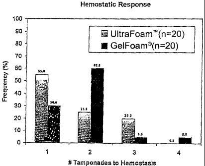
Figure 2 shows the hemostatic responses for a
hemostatic device of the invention (referred to as
Ultrafoam") compared to Gelfoam , indicating that UltrafoamTM
without thrombin performed significantly better than
Gelfoam without thrombin.
DETAILED DESCRIPTION OF THE PREFERRED EMBODIMENTS
According to one aspect of the invention, a device
having hemostatic activity ("hemostatic device") is
provided. The hemostatic device comprises a shaped
structural element that is a biodegradable matrix formed of
a collagen, such as a microfibrillar collagen
(e.g., absorbable Avitene flour), which has not been
CA 02351341 2001-05-23
WO 00/33894 PCT/US99/28775
-6-
subjected to acid dissolution and which has been exposed to minimal denaturing
conditions. Although applicants do not wish to be bound to one particular
theory or
mechanism, it is believe that avoiding contact of the collagen with acid
solution and
minimizing exposure of the collagen to denaturing conditions prior to and
during the
process for forming the device, results in a greater retention of the
hemostatic activity
by the collagen starting material. In a preferred embodiment, the hemostatic
device of
the invention is formed of collagen flour, preferably Avitene flour, which
has not
been subjected to acid dissolution or exposed to denaturing conditions, such
as, e.g.,
excess mechanical shear, high temperatures or long water residence times.
Accordingly, the invention provides hemostatic devices having unexpected
improved
hemostatic properties compared to the hemostatic devices of the prior art that
are
formed by processes which involve collagen dissolution in acid solution or
exposure
to denaturing conditions.
According to one aspect of the invention, a method for preparing a hemostatic
device is provided. The method involves: (a) suspending a plurality of
collagen
particles (preferably, collagen fibrils) in water to form a "collagen slurry",
wherein
the collagen particles have a bulk density sufficient to form a suspension in
water
(preferably, in the range of about 1.5 to about 3.5 lbs/ft3) and wherein the
collagen
slurry has a collagen concentration in the range of about 1% to about 2%
(weight/volume); and (b) lyophilizing the collagen slurry to form a hemostatic
device.
In certain preferred embodiments, the collagen particles that are used to form
the
hemostatic devices of the invention have a bulk density in the range of about
2 to
about 3 lbs/ft3. In these and other preferred embodiments, the collagen slurry
has a
collagen concentration in the range of about 1.1 % to about 1.64%
(weight/volume).
The process of the invention avoids dissolving the collagen in acid solution
and minimizes exposure of the collagen to other process steps which could
denature
the collagen and, thereby, adversely affect its hemostatic activity. In the
preferred
embodiments, the collagen is microfibrillar collagen; more preferably, a
collagen
flour such as Avitene flour. Accordingly, in certain embodiments, the
collagen
particles of the hemostatic devices of the invention have a hemostatic
activity that is
about the same hemostatic activity as Avitene flour. Avitene flour is a
microfibrillar collagen hemostat that is indicated for all surgical
specialties, including
neurosurgery, vascular, orthopaedic, urologic, and other general procedures.
CA 02351341 2001-05-23
WO 00/33894 PCT/US99/28775
-7-
Avitene is available from Davol, Inc. (product numbers 101001, 101002,
101003,
101004, and 101034, Cranston, RI). The process for preparing Avitene flour is
described in U.S. Patent No. 3,742,955, issued to Battista et al.
As used herein, "hemostatic activity" refers to the ability to stop bleeding
and
can be determined, e.g., in animal models that are recognized as predictive of
an in
vivo effect by those of ordinary skill in the art. Exemplary hemostasis animal
models
include the pig and dog spleen animal models. A preferred animal model for
assessing hemostasis activity is provided in the Examples.
Optionally, the method for preparing the hemostatic devices of the invention
i o further includes the step of introducing the collagen slurry into a mold
prior to the
lyophilizing the collagen slurry. The mold is of sufficient dimension to
contain the
slurry during the lyophilization step and to provide a template for
establishing the
final dimensions of the hemostatic device. In the preferred embodiments, the
process
also includes the step of removing a surface layer of the hemostatic device
("skiving")
to remove the thin skin which forms on the surface of the hemostatic device
during
lyophilization.
Optionally, the method for preparing the hemostatic devices of the invention
further includes the step of crosslinking the hemostatic device to form a
crosslinked
hemostatic device. Crosslinking can be accomplished in various ways and the
extent
of crosslinking can be assessed in assays which measure, e.g., the wettability
of the
device or its tensile strength. Representative assays for measuring these
parameters
are provided in the Examples. Exemplary procedures for crosslinking the
hemostatic
devices of the invention include: (1) contacting the collagen slurry with 1-
ethyl-3-(3-
dimethylaminopropyl carbodiimide HC1(EDC) for a period of time and under
conditions sufficient to form a crosslinked-hemostatic device; (2) heating the
lyophilized hemostatic device for a period of time and under conditions
sufficient to
form a crosslinked-hemostatic device; (3) exposing the lyophilized hemostatic
device
to an electron beam for a period of time and under conditions sufficient to
form a
crosslinked-hemostatic device; and (4) exposing the lyophilized hemostatic
device to
gamma sterilization for a period of time and under conditions sufficient to
form a
crosslinked-hemostatic device.
According to another aspect of the invention, the processes for forming the
hemostatic devices of the invention further include the step of introducing a
CA 02351341 2001-05-23
WO 00/33894 PCT/US99/28775
-8-
hemostatic agent into the hemostatic device. The hemostatic agent can be
introduced
into the hemostatic device of the invention at any stage in the process,
including
before the device-formation step (e.g., by adding the hemostatic agent to the
slurry)
and after device formation step (e.g., by soaking the hemostatic device in a
solution
containing one or more hemostatic agents).
It is believed that the hemostatic devices of the invention do not require a
hemostatic agent to function effectively to control bleeding, e.g., hemorrhage
of a
parenchymal organ. As a result, the hemostatic devices of the invention which
do not
further contain a hemostatic agent have good thermal stability and can be
stored for
l o months to a few years without refrigeration and loss of effectiveness.
Such
embodiments of the invention are useful for various medical situations and are
particularly useful for field and emergency use, since each may be stored in a
ready-to-use state for a lengthy period, even in the absence of refrigeration.
Such
devices of the invention also are less expensive to make and/or use compared
to
hemostatic devices which contain a further hemostatic agent to achieve a
comparable
level of hemostatic activity.
One advantage of the hemostatic devices of the invention is their flexibility
compared to hemostatic devices such as Gelfoam , that is, the hemostatic
devices of
the invention can be provided in a fon:n that easily conforms to the contours
of an
organ or biological surface, making the manipulation of applying the devices
quicker
to perform. As a result, there is less overall blood loss to the patient and
less time is
spent in surgery. Further, the hemostatic devices of the invention can be
applied, in
wet or dry state, to a bleeding site and do not require wetting with a sterile
solution
prior to use or to conform to the contour of the biological surface to which
it is
applied.
The hemostatic devices of the invention preferably are formed of an
absorbable collagen from any source, e.g., corium collagen, tendon collagen,
and,
more preferably, the devices are formed of a microfibrillar collagen,
including a
collagen flour, such as Avitene flour. The effectiveness of devices of the
present
invention in promoting clot formation is further enhanced by their lattice
structures
which are of a sufficient size to promote enzyme substrate interactions. In
particular,
the structure of the hemostatic devices of the invention are selected to
enhance contact
between thrombin that, optionally, is provided exogenously in the devices with
CA 02351341 2001-05-23
WO 00/33894 PCT/US99/28775
-9-
endogenous fibrinogen present in the blood exuding from a wound or lesion of,
e.g., a
parenchymal organ, a spine or a brain.
In certain embodiments, at least one hemostatic agent can be included in the
hemostatic devices of the invention. Because certain combinations of
hemostatic
agents can act synergistically, the amount of each hemostatic agent can be
less than
that which would be required to improve the hemostatic activity of the
hemostatic
devices of the invention if the agents were used individually. Accordingly,
the
collective amount of the hemostatic agent(s) which are included in the
hemostatic
devices of the invention is a "hemostasis-promoting amount", i.e., the amount
of at
least one hemostatic agent effective to accelerate clot formation at an
interface
between a surface (e.g., of a wound, of a lesion on a parenchymal organ, the
spine or
the brain) and a hemostatic device of the invention.
Exemplary hemostatic agents that can be applied to the hemostatic devices of
the invention in amounts effective for stimulating hemostasis, include, but
are not
limited to: thrombin, an enzyme which converts fibrinogen to fibrin; calcium,
sodium,
magnesium or other ions that stimulate hemostasis; protamine sulfate, an
epsilon
amino caproic acid, fibrinogen, and chitin. Epsilon amino caproic acid and its
analogs
which possess a similar chemical structure and hemostatic activity for use in
a
hemostatic device are described in U.S. Patent No. 5,645,849, assigned to
Clarion
Pharmaceuticals. In terms of ion additives, calcium chloride is generally a
preferred
additive for introducing a calcium ion into the device.
Additionally or alternatively, the tripeptide RGD, composed of arginine,
glycine and aspartic acid, and optionally serine "RGDS," can be incorporated
into the
hemostatic devices of the invention as a hemostatic agent. RGD is the active
site of
fibrinogen and fibronectin. RGD accelerates wound healing and is believed to
stimulate fibroblast migration. The RGD additive is also much less expensive
than
fibrinogen because it can be synthesized using solid phase chemistry.
Protamine sulfate can be added to the hemostatic devices of the invention in
an amount that is effective to neutralize heparin in the local environment of
the
device. In general, the amount of protamine sulfate is an amount between about
1-15
mg/cm2 of the hemostatic device, more preferably, an amount between 2-5 mg/cm2
of
a wound contacting surface of the hemostatic device.
CA 02351341 2005-01-27
64371-402
-10-
Likewise, RGD or RGDS peptide can be dissolved in double distilled water
and sprayed onto a wound-contacting surface of a hemostatic device of the
invention.
Preferably, such embodiments of the invention contain ain amount of RGD
effective to
enhance clot formation. For example, RGD or RGDS can be applied to a
hemostatic
device of the invention in an amount between about 110-130 mg/cm2. Thus, a
standard size hemostatic device that is a fabric would contain about 1-10
mg/fabric or
about 5-7 mg/fabric of RGD or Rt''iDS. '
Thrombin is an active ingredient found in other hemostatic devices. It is
- believed that the collagen. particles of the hemostatic devices of the
invention have a
hemostatic activity that is equivalent to the hemostatic activity of the
collagen
particles from which the device is formed. Thus, the invention (without
thrombin)
advantageously provides a device having enhanced hemostatic activity compared
to
the hemostatic devices of the prior art. A further increase in the hemostatic
activity of
the hemostatic devices of the invention can be achieved by, optionally,
including a
hemostatic agent in the hemostatic devices and/or the collagen slurry.
As used herein, the term "equivalent" with respect to hemostatic activity
means that the hemostatic activity is substantially the same when measured in
the
same activity assay. An exemplary hemostatic activity assay, a pig spleen
hemostasis
assay, is provided in the examples. The assay can be used to measure the
hemostatic
activity of the devices of the invention and can also be used to measure the
hemostatic
activity of the collagen particles, e.g., collagen flour, from which the
hemostatic
device is fonrned by, for example, by placing powder over the incision,
overlaying the
powder with a sterile gauze, and applying pressure to the wound in the same
manner
as described in the example for a device of the invention. The experimental
results
for the pig spleen assay are reported in terms of the number of tamponades
necessary
to achieve hemostasis at an incision in the pig spleen. The number of
tamponades for
multiple samples is determined to obtain a distribution of the number of
tamponades.
The distribution of tamponades is a measure of the hemostatic activity for the
device
or flour that is being tested. Accordingly, devices which have a similar
distribution of
tamponades have "equivalent" hemostatic activity. For example, if 80 of 100
samples of a first device require one tamponade to achieve hemostasis, and 70
of 7 00
samples of a second device require one tamponade to achieve hemostasis, the
hemostatic activity of the second device is considered to be within 10% of the
CA 02351341 2001-05-23
WO 00/33894 PCT/US99/28775
-11-
hemostatic activity of the first device. Equivalent hemostatic activity means
that the
hemostatic activity for two samples are within at least 50%, more preferably,
within
60%, 70%, 80%, 90% and, most preferably, within 95%.
The preferred hemostatic agent is thrombin (e.g., human or bovine thrombin).
Preferably, the thrombin is a recombinant thrombin to avoid viral or other
contamination from the organism from which the thrombin is derived. The
molecules "thrombin" and "fibrinogen", as defined herein, are meant to include
natural thrombin and fibrinogen molecules derived from an animal or human
origin, a
synthetic form or a recombinant form of the molecules, including functionally
active
1 o analogs that effectively maintain the enzyme's clot promoting activity in
an animal or
human. The species of animal from which the molecule is derived can vary and
depends on the intended use of the hemostatic device. For example, a
hemostatic
device intended for human use for safety reasons preferably contains
recombinant
human thrombin or non-human thrombin, e.g, bovine thrombin. By avoiding use of
human fibrinogen isolated from a human tissue or using viral deactivated human
thrombin, risks associated with viral contamination of purified blood products
are
minimized.
Thrombin and/or other hemostatic agents or additives described as
components of a hemostatic device according to the invention, can be applied
to the
hemostatic device by any of several methods which, preferably, are performed
under
sterile conditions. Thrombin can be applied as a layer to a particular surface
or side
of a hemostatic device of the invention, which surface is then designated the
wound-contacting surface. For example, this can be accomplished by spraying
thrombin in powder form onto a hemostatic device of the invention.
Alternatively, a
solution of thrombin can be coated onto a hemostatic device of the invention
and
dried by lyophilization or by conventional means. In another method of
applying
thrombin, a hemostatic device of the invention is dipped completely or
partially into a
sterile solution of thrombin such that a sufficient amount of thrombin
accumulates
within the hemostatic device effective to inhibit fibrinolysis in a mammal.
Preferably,
the thrombin solution contains 1000 I/U of thrombin dissolved in 1 ml saline.
The
amount of thrombin applied in the solution can vary. Preferably, the total
amount of
thrombin applied to a hemostatic device of the invention or surface thereof is
100-
1000 units/em. It is understood that alternative methods of applying the
hemostatic
3
CA 02351341 2001-05-23
WO 00/33894 PCT/US99/28775
-12-
agents and additives to a hemostatic device of the invention in addition to
the methods
described herein also can be used.
The hemostatic devices of the invention that have been soaked in thrombin
solution or other solution containing a hemostatic agent optionally can be
dried. The
drying step can be accomplished by lyophilization. Other drying procedures
appropriate for a material containing an active protein ingredient can also be
employed, so long as the drying procedure does not denature the proteins or
render
them inactive. Alternatively, hemostatic device can be dried by maintaining it
at
room temperature for a period of 1-3 hours, followed by refrigeration
overnight.
In certain embodiments, the hemostatic devices of the invention further
include a therapeutically effective amount of one or more therapeutic agents,
such as
an agent which promotes wound-healing. Agents which promote wound-healing
include anti-inflammatory agents such as agents which inhibit leukocyte
migration
into the area of surgical injury, anti-histamines; agents which inhibit free
radical
formation; and bacteriostatic or bacteriocidal agents. In general, a
therapeutically
effective amount means that amount necessary to delay the onset of, inhibit
the
progression of, or halt altogether the particular condition being treated.
Generally, a
therapeutically effective amount will vary with the subject's age, condition,
and sex,
as well as the nature and extent of the condition in the subject, all of which
can be
determined by one of ordinary skill in the art. The dosage of therapeutic
agent
contained in the hemostatic devices of the invention may be adjusted to
accommodate
the particular subject and condition being treated.
As used herein, the phrase, "agents which promote wound-healing" refers to
agents, the administration of which, promote the natural healing process of a
wound.
Agents that promote wound-healing include anti-inflammatory agents, agents
which
inhibit free radical formation, and bacteriostatic or bacteriocidal agents.
Anti-inflammatory agents are agents which inhibit or prevent an immune
response in vivo and include: (i) agents which inhibit leukocyte migration
into the
area of surgical injury ("leukocyte migration preventing agents"), and anti-
histamines.
Representative leukocyte migration preventing agents include silver
sulfadiazine,
acetylsalicylic acid, indomethacin, and Nafazatrom. Representative anti-
histamines
include pyrilamine, chlorpheniramine, tetrahydrozoline, antazoline, and other
anti-
inflammatories such as cortisone, hydrocortisone, beta-methasone,
dexamethasone,
CA 02351341 2001-05-23
WO 00/33894 PCT/US99/28775
-13-
fluocortolone, prednisolone, triamcinolone, indomethacin, sulindac, its salts
and its
corresponding sulfide, and the like.
Representative agents which inhibit free radical formation include
antioxidants
that inhibit the formation and/or action of oxide products, superoxide
dismutase
(SOD), catalase, glutathione peroxidase, b-carotene, ascorbic acid,
transferrin, ferritin,
ceruloplasmin, and desferrioxamine a-tocophenol.
Representative bacteriostatic or bacteriocidal agents include antibacterial
substances such as (3-lactam antibiotics, such as cefoxitin, n-formamidoyl
thienamycin
and other thienamycin derivatives, tetracyclines, chloramphenicol, neomycin,
gramicidin, bacitracin, sulfonamides; aminoglycoside antibiotics such as
gentamycin,
kanamycin, amikacin, sisomicin and tobramycin; nalidixic acids and analogs
such as
norfloxican and the antimicrobial combination of fluoroalanine/pentizidone;
nitrofurazones, and the like.
The hemostatic devices of the invention can contain one or more therapeutic
agents, alone or in combination with one or more hemostatic agents.
Various additives, optionally, can be incorporated into the hemostatic devices
of the invention without substantially reducing the hemostatic activity of
these
devices. The term "pharmaceutically-acceptable carrier" as used herein means
one or
more compatible solid or liquid fillers, diluents or encapsulating substances
which are
suitable for administration into a human. The term "carrier" denotes an
organic or
inorganic ingredient, natural or synthetic, with which the active ingredient
is
combined to facilitate the application. The components of the pharmaceutical
compositions also are capable of being co-mingled with the collagen particles
(e.g.,
fibrils) of the present invention, and with each other, in a manner such that
there is no
interaction which would substantially impair the desired hemostatic activity.
According to yet another aspect of the invention, a product prepared by the
above-described process is provided. A particular embodiment of this process
in
provided in the Examples.
According to yet another aspect of the invention, a hemostatic device is
provided which has a hemostatic activity in a pig spleen animal model of
hemostasis
that corresponds to one tamponade for a hemostatic device having a thickness
of 3/8
inch, a length of'/2 inch, and a width of '/2 inch. A detailed description of
the pig
spleen animal model is provided in the Examples.
CA 02351341 2001-05-23
WO 00/33894 PCT/US99/28775
-14-
The hemostatic devices of the invention can be used in a wet or in a dry
state.
Preferably, the hemostatic devices of the invention have a wettability index
that is
equivalent to or less than that of Gelfoam (without Thrombin). As used
herein, a
"wettability index" refers to the time it takes for a sample of known
dimension to
fully hydrate. A preferred embodiment of the invention which are illustrated
in the
Examples has a wettability index of less than or equal to 1 minute in
distilled water at
room temperature.
Preferably, the hemostatic devices of the invention, when wet or dry, have a
hemostatic response time that is equivalent to or less than that of Gelfoam
(with or
without Thrombin). Gelfoam refers to a collagen hemostat formed of denatured
collagen that is available from the Upjohn Company, (product number 0342-01,
0315-
01, 0353-01, 0315-02, 0349-01, 0301-01, 0323-01, 0433-01, Kalamazoo, MI
49001).
In certain embodiments, the hemostatic device of the invention has a higher
percentage of solids than typically is found in hemostatic devices of the
prior art. It is
believed that the higher percentage of solids increases the mechanical
strength
properties of the hemostatic device. Preferably, the hemostatic devices of the
invention have a density in the range of about 0.01 to about 0.030 gm/cc, more
preferably, a range of about 0.015 to about 0.023 gm/cc; and a weight percent
solids
ranging from about 1.0 - 2.0 in the slurry prior to lyophilization, more
preferably, in
the range of about 1.10 to about 1.64 weight percent. In general, the
hemostatic
devices of the invention have a melting point (Tm) in the range of about 93.4
to about
105.7 C, as determined by differential scanning calorimetry, depending upon
the
moisture content of the device.
Representative embodiments which satisfy some or all of the foregoing
criteria, when wet, have an acute mechanical strength and a chronic mechanical
strength that are equivalent to or greater than that of Gelfoam 100. Gelfoam
100
refers to a collagen hemostat that is available from the Upjohn Company
(product
number 0342-01, 0315-01, 0353,01, 0315-02, 0349-01, 0301-01, 0323-01, 0433-01,
Kalamazoo, MI 49001). As used herein, an "acute mechanical strength" refers to
immediate tensile testing after full wetting and is determined by tensile
testing,
according to a standard procedure such as illustrated in the Examples.
Mechanical
strength is, in part, a function of the state of the hemostatic device (wet
versus dry), as
well as a function of its dimensions. For embodiments in which the hemostatic
device
CA 02351341 2001-05-23
WO 00/33894 PCTIUS99/28775
-15-
has a thickness of about 3/8 inch to about '/2 inch width, the acute maximum
load for
the device, when wet, is _ 0.08 lbs (minimum) with the mean acute maximum load
being about 0.14 lbs.
In certain embodiments of the invention, the hemostatic device, when dry, has
a modulus equivalent to or greater than that of Actifoamg. Actifoam refers to
a
collagen hemostat that is available from Davol, Inc. (Cranston, RI). The
process for
preparing Actifoam is described in U.S. Patent No. 4,953,299 and 5,331,092.
As
used herein, "modulus" refers to stiffness and is determined by tensile
testing in
accordance with standard procedures such as those described in the Examples.
In
certain embodiments, the modulus for the hemostatic device, when dry, is less
than or
equal to 86 psi.
Advantageously, the hemostatic devices of the invention need not contain a
hemostatic agent to function effectively to control bleeding, e.g., hemorrhage
of a
parenchymal organ. As a result, the hemostatic devices of the invention which
do not
further contain a hemostatic agent have good thermal stability and can be
stored for
months to a few years without refrigeration and losing effectiveness. Such
embodiments of the invention are useful in various situations, including field
and
emergency use, since each may be stored in a ready-to-use state for a lengthy
period.
One advantage of the present invention is its flexibility compared to
hemostatic devices such as Gelfoam (which is rigid when dry), that is, the
hemostatic devices of the invention can be provided in a form that easily
conforms to
the contours of an organ or biological surface, making the manipulation of
applying
the device quicker to perform. As a result, there is less overall blood loss
to the patient
and less time is spent in surgery. A further advantage of using the hemostatic
devices
of the invention in a dry state is that the dry devices can absorb blood
exuding from a
biological surface, thereby further promoting hemostasis at the interface of
the
hemostatic device and the biological surface.
The hemostatic devices of the invention preferably are formed of an
absorbable collagen (e.g., microfibrillar collagen) as a matrix. In the
preferred
embodiments, the matrix is a flat layer of microfibrillar collagen foam that
is formed
of Avitene flour. The effectiveness of devices of the present invention in
promoting
clot formation is enhanced by their lattice structures, which promote enzyme
substrate
interactions. In particular, the collagen foam structure enhances contact
between
CA 02351341 2001-05-23
WO 00/33894 PCT/US99/28775
-16-
thrombin that optionally is provided exogenously in the device with endogenous
fibrinogen present in the blood exuding from a wound or lesion of a
parenchymal
organ. A representative hemostatic device prepared according to the process
disclosed herein has a reticulated open cell foam structure as illustrated in
the
photocopy of an SEM image (fig. 1).
According to certain embodiments, a hemostatic device of the invention is
contained within a sealed sterile package which facilitates removal of the
device
without contamination. Such a package, for example, can be an aluminum foil
pouch
or other material that is easily sterilized. Radiation, e.g., gamma radiation,
can be
applied to sterilize the device and packaging material together. In yet other
embodiments, a container having dual compartments is provided in which a first
compartment contains distilled water, sterile saline or a sterile buffer, and
a second
compartment contains a hemostatic device of the invention. In field use, the
device
of the second compartment can be readily dipped into an opened first
compartment
and subsequently applied to the wound.
According to still another aspect of the invention, a method for promoting
hemostasis is provided. The method involves the steps of pressing a hemostatic
device of the invention against a surface of a wound or a surface of a lesion
on an
organ, tissue, or other biological surface, e.g., a parenchymal organ, the
spine or the
brain, for a period of time until clotting has occurred at the interface
between the
hemostatic device of the invention and the surface. The device may be applied
to the
surface in a dry state or, alternatively, may be soaked in sterile saline
solution or a
sterile hemostatic agent-containing solution prior to use. Use of a hemostatic
device
of the invention according to the invention, without first soaking in saline
solution
permits quick and simple application of the device in various situations,
including
field situations such as may be encountered by an emergency medical
technician. In
certain embodiments, the hemostatic device is soaked in a thrombin solution
prior to
use to introduce a therapeutically effective amount of thrombin into the
device. Thus,
a hemostatic device of the invention of the invention can be used by applying
a
"wound-contacting" surface of the device, a surface intended to contact the
wound
and containing hemostatic agent(s) and, optionally, additives, with or without
prior
soaking in a sterile solution, to a surface of a bleeding wound or lesion.
Then, the
device is maintained in contact with the surface for a period of time
sufficient for
CA 02351341 2001-05-23
WO 00/33894 PCT/US99/28775
-17-
clotting to occur at the interface between the hemostatic device of the
invention and
the surface and for bleeding to be substantially arrested. In general, the
device is
maintained in contact with the surface for a period of about 3-20 minutes,
preferably,
3-10 minutes, and more preferably, 3-5 minutes.
Where thrombin and/or other hemostatic agents also are present on/in the
hemostatic device, the time period preferably is about 5 minutes. The
hemostatic
device is held in place against the biological surface, preferably with light
pressure,
e.g., by means of a sterile saline soaked sponge. AIternatively, the
hemostatic fabric
may be held in place simply by applying pressure to the hemostatic device by
means
of a gauze or other dry sterile material. Depending on the location of the
wound, a
bandage can be wrapped around the hemostatic device to provide light pressure
on the
wound surface.
The efficacy of the hemostatic devices of the invention can be assessed in art-
recognized animal models that are believed to be predictive of an in vivo
hemostatic
effect in humans. For example, surgical lesions induced in parenchymal organs
of
pigs provide a good model system for hemostasis in the analogous human organs
as
evidenced by preclinical studies which employ pig models. See e.g., SWINE AS
MODELS IN BIOMEDICAL RESEARCH, Swindle, M., Iowa State Univ. Press
(1992).
A preferred use of a hemostatic device according to the present invention is
to
inhibit or completely stop bleeding of a parenchymal organ, such as the liver,
kidney,
spleen, pancreas or lungs. Other preferred uses are to inhibit or completely
stop
bleeding of a wound or lesion on the spine or brain. Additional uses for the
hemostatic devices of the invention include inhibiting bleeding during
surgery, e.g.,
internal/abdominal, vascular (particularly for anastomosis), urological,
gynecological,
thyroidal, neurological, tissue transplant uses, dental, cardiovascular,
cardiothoracic,
ENT (ear, nose, throat) and orthopedic surgeries.
Another use of a hemostatic devices of the invention is topical treatment,
such
as for burn or tissue transplants and as dura replacement or substitutes. A
hemostatic
device of the invention for topical use preferably contains additives, such as
anti-infection medicaments, bactericides, fungicides and wound healing agents,
for
example, neomycin and bacitracin.
CA 02351341 2001-05-23
WO 00/33894 PCTIUS99/28775
-18-
In addition to inducing hemostasis, the hemostatic devices of the inventions
of
the invention can be used to hermetically seal body tissue. For example, when
air
leaks from a wound in the lungs, a hemostatic device of the invention can be
applied
to the surface surrounding the wound, held in place for a period of time
sufficient to
induce hemostasis and allow a hermetic seal to form.
The hemostatic devices of the invention also are useful for treating animals,
preferably humans or other mammals, including domestic mammals and livestock.
The hemostatic devices of the invention can be provided in a variety of sizes
and shapes, depending upon its intended use. Typically, the hemostatic devices
of the
1o invention are provided in a standard size rectangular foam, e.g., 8cm x
12.5cm x 1
cm; 8cm x 12.5cm x 3mm; 8 cm x 6.25cm x 1 cm; 8 cm x 25 cm x 1 cm; 2 cm x 6 cm
x 7 mm; 2.5cm x 2.5cm x 7mm; with an outer dimensional tolerance of +/- 1/8
inch
and a thickness tolerance of +/- 1/16 inch. The hemostatic devices may be cut
to size
with a pair of scissors. The hemostatic devices of the invention may be
spherically,
conically, cuboidally or cylindrically-shaped or prefabricated into small
squares, such
as for packing into a body cavity, such as a dental cavity following a tooth
extraction.
Alternatively, the hemostatic device can be shaped for epistaxis (profusely
bleeding
nostril) or insertion into a cavity. The hemostatic devices of the invention
that are
intended for topical applications can be applied with an adhesive tape, as a
band-aid
form, where the hemostatic device is adhered to an adhesive backing. One or
more
additional layers of wound dressing material, preferably a layer which aids in
absorption of blood or other exudants, can be applied to or incorporated into
the
hemostatic devices of the invention to form a stronger bandage. Alternatively,
the
layer may be applied as a supplement to the backside (non-wound contacting
surface)
of a device according to the invention. Particularly for topical use, the
layer(s) can
contain superabsorbents to wick exudant solution from the wound site. For
hemostatic devices of the inventions intended for internal-surgical
applications, where
an added layer(s) is integral with the device, the layer(s) should be both
biodegradable
and pharmaceutically acceptable.
The hemostatic devices of the invention can be designed to facilitate its
application to fuse ends of a blood vessel or other body lumen having been
severed,
e.g., surgically. To apply a hemostatic device for anastomosis, a rectangular
fabric, for
example, is wrapped around the external surface of the ends of a Dacron 0
graft and
CA 02351341 2001-05-23
WO 00/33894 PCT/US99/28775
-19-
the graft is positioned into place. The hemostatic device portion of the graft
accelerates fibrin growth into the graft to seal the graft in place
(hemostatically and
hermetically). According to certain embodiments of the invention, a kit is
provided
for this application. The kit contains a graft and a hemostatic device of the
invention
that is designed for fitting with the ends of the graft. Alternatively, a kit
is provided
having a hemostatic device of the invention pre-fitted onto at least one end
of a graft.
According to still other aspects of the invention, various specialized kits
can
be provided. The kits contain any of the hemostatic device embodiments
disclosed
herein and a package, wherein the hemostatic device of the invention is
contained
within a sealed sterile package which facilitates removal of the fabric
without
contamination. The kit can contain multiple hemostatic devices of the
inventions,
preferably wherein each hemostatic device is contained within a separate
sealed
sterile package. A kit that is designed for autonomous use, e.g., for
field/military use
can, in addition to a hemostatic device of the invention, further include
disposable
pre-sterilized surgical instruments and/or agents that can be incorporated
into the
device, e.g., thrombin, calcium chloride.
Unless defined otherwise, all technical and scientific terms used herein have
the same meaning as commonly understood by one of ordinary skill in the art of
the
invention. Although any methods and materials similar or equivalent to those
2o described herein can be used in the practice of the invention, the
preferred methods
and materials have been described. Unless mentioned otherwise, the techniques
employed or contemplated herein are standard methodologies well known to one
of
ordinary skill in the art. The materials, methods and examples are
illustrative only and
not limiting.
EXAMPLES
EXAMPLE 1. PREPARATION OF A PREFERRED EMBODIMENT
Description of UltrafoamTM
UltrafoamTM is an absorbable hemostatic sponge prepared as a sterile, porous,
pliable, water insoluble partial hydrochloric acid salt of purified bovine
corium
collagen. UltrafoamTM consists of lyophilized Avitene flour and water. In its
manufacture, swelling of the native collagen fibrils is controlled by ethyl
alcohol to
permit noncovalent attachment of hydrochloric acid to amine groups on the
collagen
CA 02351341 2001-05-23
WO 00/33894 PCT/US99/28775
-20-
molecule and preservation of the essential morphology of native collagen
molecules.
The characteristics of collagen which are essential to its effect on the blood
coagulation mechanisms are substantially preserved, although dry heat
sterilization
causes some cross linking which is evidenced by reduction of hydrating
properties,
and a decrease of molecular weight which implies some limited amount of
degradation of collagen molecules.
UltrafoamTM sizes are:
lcmx lcmx7mm 2.5" x 2.5" x 1/4"
2cm x 6cm x 7mm 3/4" x 2.5" x 1/4"
8cm x 6.25cm x lcm 3-1/8" x 2.5" x 3/8"
8cm x 12.5cm x lcm 3-1/8" x 5" x 3/8"
8cm x 25cm x lcm 3-1/8" x 10" x 3/8"
The steps of adding USP Standard purified water to Avitene flour, then
lyophilizing this slurry, changes the physical appearance of the Avitene
flour from a
loose flour like powder to a solid, pliable light weight foam. This affects
the physical
appearance of the collagen but the chemical composition remains the same as
that of
Avitene flour. Because the added steps do not change the chemical composition
of
the microfibrillar structure of the collagen, the mode of action and
hemostatic
characteristics of the collagen foam are preserved.
To control bleeding, UltrafoamTM typically is cut to the desired size and
applied directly to the source of bleeding. The UltrafoamTM is held in place
with
moderate pressure until hemostasis results. The period of time to hold
pressure on the
foam will vary with the force and severity of bleeding. UltrafoamTM may be
left in
place at the bleeding site when necessary. Prior to removal, the foam should
be
moistened with saline to avoid dislodging the clot.
Manufacturing Process
The process to make a foam product involves the addition of USP water to
Avitene Bulk Flour and removing the water via a lyophilization (freeze
drying)
process. The following is an overview of the process and steps used in the
Manufacture, Quality Control and Test Procedures, for this preferred
embodiment.
In general Avitene bulk flour is mixed with USP purified water, poured into
trays and lyophilized. After processing, it is inspected and released to
manufacturing
and staged for the cleanroom.
CA 02351341 2005-01-27
64371-402
-21-
in the clean rooms, the first step is skiving, a process of removing the
surface
layer of the material with a machine known as a band knife splitter, to remove
the
"skin" formed on the surface during the lyophilization process and then
slicing the
material to the proper thickness. After skiving, the pieces are transferred to
the
cutting operation where the pieces are cut to the appropriate size.
After cutting, the UItrafoamTM pieces are placed into individual polycarbonate
trays which are then sealed with a TyvekTM lid. This packaged unit is then
placed onto a
cart and run through a Despatch Index drying oven wherein it is dried to a
specific
moisture content. After drying, the sealed polycarbonate itrays are placed
into
i o PET/nylon/foil laminate pouches which are heat sealed and the pieces are
transferred
to the sterilization operation. The sterilization employed is dry heat, 126 C
at 20
hours. Upon completion of sterilization the material is packaged and labeled
into
cartons after which it is held in quarantine until it is released to
distribution.
A detailed description of each of the major steps of the process, namely,
mixing, lyophilization, cutting/skivirtg/packaging and sterilization, and
final
packaging is presented below.
Mixin : The Avitene@ Bulk Flour is mixed with USP Purified Water at 13.9
grams of flour per 1.00 Liter USP water using a peristaltic pump at the
highest setting
for approximately one and one half hours to ensure a percent solids of 1.37%
(nominal) as determined during the initial development phase of the product.
Early in the concept development phase of the foam product, studies were
completed to determine the desired formulation for the foam. The studies
examined
the effects of percent collagen (ratio of collagen to USP water), collagen
fiber length
(nominal and shortened), dwell time between mixing and lyophilizing, and
various
cross-linking methods (heat sterilization, gamma sterilization, electron beam
exposure
and chemical cross-linking agents) on the foam. Foam product was made with
various combinations of the aforementioned variables. Based on physical
testing and
evaluations of prototype units, the key factors for production of the foam
were defined
as the ratio of collagen to water, fiber length and cross-linking method.
These factors
were optimized to be 13.9 grams of flour/liter of USP water (with a range of
11. 1 -16.7
grams of flour/liter of USP water), nominal Avitene flour fiber length and
heat
sterilization/cross-linking. The dwell time between mixing and lyophilizing
was
CA 02351341 2001-05-23
WO 00/33894 PCT/US99/28775
-22-
determined to not significantly influence the product, as long as it did not
exceed 27
hours (the maximum dwell time examined in the study).
Once the ratio of Avitene flour to USP water was defined, a mixing process
was developed to ensure that the collagen was evenly distributed throughout
the
flour/water mixture to provide for a uniform percent solids. A peristaltic
pump was
used to accomplish the mixing in a manner as to not compromise the fiber
length of
the flour (low shear). The mixing is performed for approximately 90 minutes
with the
pump set to its highest setting in a stainless steel vessel. The percent
solids for the
mixture preferably is in the range of about 1.10% to about 1.64% solids.
Lyophilization: After mixing the product is transferred into stainless steel
trays which are approximately 12 x 36 inches and put into the lyophilizer
where it is
exposed to a lyophilization cycle of temperature of 5.9 C/hour, 36 C terminal
temperature for 38 hours and 100 mT (milliTorr) vacuum to obtain a foam
product.
The foam product is inspected for thickness, density and general appearance.
Once acceptable, it is released for manufacturing and staged for the clean
room,
where it undergoes the following manufacturing steps:
Skiving Process: The material is received from the lyophilization process in
sheets which are approximately 12 by 36 inches. These sheets are fed into the
bandknife splitter via rollers set at a given distance from the center blade
(stainless
steel) which removes the "skin" layer formed during the lyophilization
process. In
some cases, multiple passes of the material are made through the machine until
the
proper thickness is achieved.
Cutting Process: After skiving, the material is transferred to the cutting
station
where the skived pieces are put through a cutting machine consisting of a
stainless
steel rotary blade cutter an indexing station which cuts the pieces to size.
After the
skiving and cutting operations the pieces are inspected for proper dimensions
(length,
width, thickness), percent moisture and general appearance and are then
released to
the next operation, the drying step.
Packaging and drying process: The packaging and drying step is a continuous
process of packaging into polycarbonate trays and sealing with tyvek lid,
loading the
sealed units onto carts and then placing them into the Despatch Index Drying
Oven
where they are exposed to a cycle of 110 C for approximately 2 hours to reduce
the
moisture level in the product to 4%.
CA 02351341 2001-05-23
WO 00/33894 PCT/US99/28775
-23-
During the drying process, samples are taken from every cart to confirm that
the moisture content of the product is 4% or less. If acceptable, the product
is
removed and sent to the second stage of packaging, foil pouching. If the
products do
not meet the moisture specification after a single exposure to the drying
cycle
additional testing will be performed to determine if the product can withstand
exposure to two drying cycles without detriment to the product or package.
Once dry,
the product is placed into PET/nylon/foil laminated pouches and heat sealed.
EXAMPLE 2. ASSAY FOR DETERMINING TENSILE STRENGTH
This assay is performed to determine the tensile strength of a hemostatic
device of the invention formed of Avitene0 flour (UltrafoamTM) and compare its
tensile strength to that of a Gelfoam0 control. The foam samples were tested
in the
tensile direction using a dog bone shape sample. Foam samples were tested for
tensile properties in the wet state using an Instron Tensile Tester with Flat
Face
Grips, beaker, calipers, de-ionized Water (room temperature), 1" x 2" Bone-
shaped
Steel Roll Die (0.5" wide at the center), Clicker Press, Gelfoam0, Product
Code: 100,
and UltrafoamTM.
The UltrafoamTM that was used for the testing was produced by mixing a
slurry (Avitene(P powder and water - 1.25% w/v) using a mechanical mixer
(variable
speed, electrical stirrer with twin propellers). The slurry was mixed for 24
hours. The
finished slurry still had small clumps of collagen in the mixture. The mixing
was not
a thorough process, but was sufficient for concept testing. The slurry was put
into 4L
jars and lyophilized. Once lyophilized, the foam was placed into aluminum foil
(loosely packaged) and put through a sterilization process of 125 C for 22
hours. The
foam was then ready for testing. The Gelfoam0 was die cut using the clicker
press
and the bone-shaped steel roll die; 10 total bone-shaped pieces of Gelfoam0
were cut
for testing and 30 pieces of UltrafoamTM were cut as well. Samples were placed
in a
beaker of de-ionized water, keeping both the Gelfoam0 and UltrafoamTM
separated.
The air trapped within the samples was removed by kneading the samples (only
necessary with the Gelfoam0 samples).
The gauge length of the grips on the Instron0 Tensile Tester was set to 1
inch.
The first Gelfoam0 sample was placed in the grips and closed using the foot-
pedal.
The sample thickness was measured using the calipers and recorded for the
sample on
CA 02351341 2001-05-23
WO 00/33894 PCT/US99/28775
-24-
the computer. The samples were run at a crosshead speed of 12 in/min in the
tensile
direction.
The following data for each sample was recorded: Maximum Load (lbs.);
Strain at Break (%); Secant Modulus (psi); Energy at Maximum Load (psi). The
procedure was repeated for the rest of the Gelfoam samples. The procedure was
then repeated to test the UltrafoamTM samples.
The Gelfoam samples were immersed in water for two hours and were
kneaded to remove excess air trapped within the samples (to fully hydrate the
samples). The Gelfoam samples did not hydrate quickly. The tensile properties
for
1 o the maximum load of the samples ranged from 0.15 to 0.23 pounds force with
a mean
of 0.17 pounds. The standard deviation was 0.02 pounds.
The UltrafoamTM samples did not require kneading to remove air; however
kneading was performed to be consistent with the treatment of the Gelfoam
samples. The UltrafoamTM samples did not required extensive soak time; they
immediately wicked to become fully hydrated. The tensile properties for the
maximum load of the samples ranged from 0.13 to 0.20 pounds force. The mean
was
0.16 pounds with a standard deviation of 0.03 pounds force.
The UltrafoamTM samples were equivalent to the Gelfoam samples for
maximum load in the tensile direction. The hydrating properties of the
UltrafoamTM
were much quicker (within 2 minutes) than the Gelfoam samples (over two
hours).
EXAMPLE 3. ASSAY FOR DETERMINING DENSITY
This assay is performed to determine the density of dry UltrafoamTM for both
pre and post sterility samples. The equipment used included: digital calipers,
square
or rectangle steel roll die, flat plexiglass, rubber mallet, top-loading scale
(able to
measure to at least 0.001 grams).
Samples for testing were die cut from the above-described foam using the steel
roll die by placing the foam over the die. Next, a piece of flat plexiglass
(big enough
to cover the die) was placed on top of the foam and, by using the rubber
mallet to
gently tap the plexiglass, a shape of foam was cut out from the foam. This
procedure
was repeated until the number of samples needed were cut to shape.
The digital calipers were used to measure the length (L), width (W) and
thickness (T) (at least three measurements were made of each dimension and the
CA 02351341 2001-05-23
WO 00/33894 PCT/US99/28775
-25-
average of the measurements was used). The average measurement of the sample's
dimensions was recorded and the sample was placed on the scale to obtain the
sample
weight of the sample which was then recorded. This procedure was repeated
until all
samples were measured.
If dimensional measurements were measured in units of inches, inches (in)
was converted to centimeters (cm) and the conversions were recorded in
centimeter
units. The following calculation was used to determine the density of the foam
sample(s):
Density = [wt (g)/(L(cm)*W(cm)*T(cm))]
and the density results were recorded in units of grams per cubic centimeter
(g/cc) of
each sample.
EXAMPLE 4. COMPARISON OF HEMOSTATIC ACTIVITY
BETWEEN UltrafoamTM AND Gelfoam
Test Method
The hemostatic response time of UltrafoamTM (Lot 081398) with and without
thrombin, was compared to Gelfoam (Lot 40CAR, with and without thrombin) in a
pig spleen model (J&J Hemostasis protocol) as follows. Small incisions were
made in
the retracted spleen of anesthetized juvenile Yorkshire pigs. The number of
cuts per
spleen ranged from 8 to 18. Eight pigs were required. Thrombin was added to
the
device by soaking the sample in a thrombin solution until fully saturated. The
test
device (approximately 0.5" x 0.5") was placed on the wound, tamponaded with
finger
pressure for 20 seconds, then the pressure was removed and the site was
observed for
re-bleed for two minutes. If re-bleed was observed within two minutes,
pressure was
reapplied for 20 seconds and the cycle was repeated. The endpoint of the
hemostatic
assay is the number of tamponades to achieve no re-bleed. The following
samples
were paired during testing (20 pairs each): UltrafoamTM versus Gelfoam ,
UltrafoamTM versus Gelfoam -thrombin, UltrafoamTM-thrombin versus Gelfoam -
thrombin. A pair was defined as two samples tested one after the other and
adjacent
to one another on the spleen. For each pair, the first sample tested was
alternated
from pair to pair. Each pair was tested at least once, usually twice, and
sometimes 3
times on each animal to better characterize animal to animal variability.
Statistical Methods
CA 02351341 2001-05-23
WO 00/33894 PCT/US99/28775
-26-
Statistical Methods
The frequency of the number of tamponades for each product type within the
paired group, was analyzed using the Fisher's exact test and the Stuart-
Maxwell test
(both one-tailed) at alpha 0.05. It was expected that UltrafoamTM without
thrombin
would need fewer tamponades than Gelfoam without thrombin, but would require
more tamponades than Gelfoam with thrombin. These paired groups were analyzed
separately. Therefore, a one-sided test based on expected results was
appropriate.
The SAS software package was used for calculating the Fisher's exact test for
each paired group. For n x n ("x" means multiplication) contingency tables
(foam
type x number of tamponades), Fisher's exact test yields the probability of
observing
a table that gives at least as much evidence of association as the one
actually
observed, given that the null hypothesis is true. The hypergeometric
probability (p
value) of every possible table is computed (from the SAS/STAT User's Guide,
release
6.03 edition). If'/z x the two-tailed p value (which is the one-tailed p
value) was less
than or equal to 0.05, the frequency distributions were considered
significantly
different.
The Stuart-Maxwell test, which is a generalization of the McNemar test, was
normally calculated using Table 8.5 and formulas 8.18 and 8.19 on page 120 of
"Statistical Methods for Rates and Proportions," Joseph L. Fleiss, 2nd
edition,
published by John Wiley & Sons, New York, NY. The Stuart-Maxwell test involves
determining the number of pairs with the same result and differing results,
and
calculating the value of the Stuart-Maxwell chi-square at 2 degrees of freedom
for
matched pairs with 3 mutually exclusive outcomes. The one-tailed alpha value
was
0.10 (two-tailed value of 0.05 x 2). For the purposes of this calculation, the
3
outcomes were 1, 2, or 3 tamponades. In 2 cases (1 Gelfoam without thrombin
and
I UltrafoamTM without thrombin) a sample that required 4 tamponades was
treated as
a 3 in order to allow the use of the Stuart-Maxwell test.
Results
Overall, the animal model worked well for the comparison of hemostatic
response
time. The tamponade method was representative of actual product use. All
product
samples tested were considered adequate hemostats with differing degrees of
performance.
CA 02351341 2001-05-23
WO 00/33894 PCT/US99/28775
-27-
The frequency (%) of UltrafoamTM samples requiring 1, 2, 3, or 4 tamponades in
the UltrafoamTM/Gelfoam -thrombin pairs was 80, 15, 0, and 5 respectively.
Gelfoam -thrombin exhibited 85, 15, 0, and 0 respectively. Both frequency
distributions were skewed to I tamponade. There was no significant difference
between the UltrafoamTM and Gelfoam -thrombin tamponade frequency
distributions
according to both the Fisher's exact test (one-sided p value > = 0.500) and
the
Stewart-Maxwell test (one sided p value > = 0.303). This result was important
because it indicated that in this model, UltrafoamTM did not require thrombin
to be as
effective as Gelfoam with thrombin.
In the UltrafoamTM-thrombin/Gelfoam -thrombin pairs, the frequency of
UltrafoamTM-thrombin samples requiring 1, 2, 3, or 4 tamponades was 80, 10, 5,
and 0
respectively. Gelfoamg-thrombin exhibited 85, 15, 0, and 0 respectively. Both
frequency distributions were skewed to I tamponade. There was no significant
difference between the UltrafoamTM-thrombin and Gelfoam -thrombin tamponade
frequency distributions according to both Fisher's exact test (one-sided p
value > =
0.500) and the Stewart-Maxwell test (one sided p value > = 0.274). This result
was
important because it indicated that in this model, UltrafoamTM was compatible
with
thrombin and thrombin did not react synergistically with UltrafoamTM.
In the UltrafoamTM vs. Gelfoam pairs, the frequency of UltrafoamTM samples
requiring 1, 2, 3, or 4 tamponades was 55, 25, 20, and 0 respectively (Fig.
2).
Gelfoam exhibited 30, 60, 5, and 5 respectively (Fig. 2). The UltrafoamTM
distribution was skewed to 1 tamponade. The Gelfoamg distribution was skewed
to 2
tamponades. The tamponade frequency distribution for UltrafoamTM was
significantly different than the tamponade frequency distribution for Gelfoam
according to the Fisher's exact test (one-sided p value > = 0.035). The
Stewart-
Maxwell test indicated the tamponade frequency distributions were borderline
significantly different (one-sided p value > = 0.054). This result was
important
because it indicated that in this model, UltrafoamTM without thrombin
performed
significantly better than Gelfoam without thrombin.
The percentage (55%) for 1 tamponade for the UltrafoamTM samples in the
UltrafoamTM/Gelfoam pairs was lower than the percentage (80%) for I tamponade
for the UltrafoamTM in the UltrafoamTM/Gelfoanl -thrombin pairs. The only
experimental factor that could be found that could account for this difference
was the
CA 02351341 2001-05-23
WO 00/33894 PCT/US99/28775
-28-
timing of scalpel blade changes. A large percentage (70%) of the pairs in the
UltrafoamTM/Gelfoam group were tested on spleen cuts made with a fresh
scalpel
blade. None of the pairs in the UltrafoamTM/Gelfoam0-thrombin group were
tested
on spleen cuts made with a fresh scalpel blade. The comparison of UltrafoamTM
to
Gelfoam was valid, since both samples were tested on approximately the same
number of spleen cuts made with fresh scalpel blades. Therefore, the
variability that
may have been introduced by the timing of scalpel blade changes, did not
meaningfully affect the paired results.
Discussion
Overall, the tamponade frequency distributions were similar for all samples
with
the exception of Gelfoam0 without thrombin. UltrafoamTM without thrombin,
UltrafoamTM with thrombin, and Gelfoam0 with thrombin all exhibited tamponade
frequency distributions that were skewed to 1 tamponade. The fact that
UltrafoamTM
without thrombin performed comparably to Gelfoam0 with thrombin was clearly an
important finding. Extrapolating from the animal model to the clinical
situation,
clinical users may choose to use UltrafoamTM in the dry state without
thrombin,
saving time and money, since only one product would be used instead of two.
The
soft and flexible handling characteristics of UltrafoamTM will allow it to be
used in the
dry state.
Gelfoam0 without thrombin exhibited a tamponade frequency distribution that
was skewed to 2 tamponades, whereas UltrafoamTM without thrombin was skewed to
I tamponade. In this study, one statistical analysis method determined that
the two
distributions were different, and a second method determined they were not
different,
although this second test was borderline. However, the data certainly suggest
that
UltrafoamTM exhibited increased hemostatic performance compared to Gelfoam0.
The increased hemostatic activity of UltrafoamTM without thrombin compared to
Gelfoam0 without thrombin was an unexpected outcome because UltrafoamTM is
composed of active microfibrillar collagen whereas Gelfoam is composed of
inactive collagen or gelatin. In addition, UltrafoamTM absorbs fluid more
quickly than
Gelfoam0, and will quickly absorb blood and stimulate the clotting cascade.
The data in this study indicated that UltrafoamTM with and without thrombin
was
an effective hemostat, comparable and, in some instances, exceeding the
industry
standard Gelfoam0-thrombin.
CA 02351341 2001-05-23
WO 00/33894 PCT/US99/28775
-29-
Table 1. Frequency (%) of number of tamponades.
Product Number of Tamponades
N= 20 pairs 1 2 3 4
UltrafoamTM-thrombin/Gelfoam Pairs
UltrafoamTM 80 15 0 5
Gelfoam -thrombin 85 15 0 0
UltrafoamTM-thrombin/Gelfoam -thrombin
Pairs
UltrafoamTM-thrombin 85 10 5 0
Gelfoam -thrombin 85 15 0 0
UltrafoamTM-Gelfoam Pairs
UltrafoamTM 55 25 20 0
Gelfoam 30 60 5 5
Table 2. Number of pairs for each outcome for UltrafoamTM/Gelfoam -thrombin
pairs.
GelfoamTM-Thrombin
Number of Tamponades
UltrafoamTM 1 2 3 Total
Number of Tamponades
1 14 2 0 16
2 2 1 0 3
3 1* 0 0 1
Total 17 3 0 20
*Note: One UltrafoamTM sample required 4 tamponades, but was re-classed to 3
tamponades to allow calculation of The Stewart-Maxwell statistic.
Table 3. Number of pairs for each outcome for UltrafoamTM-thrombin/Gelfoam -
thrombin pairs.
CA 02351341 2005-01-27
64371-402
-30-
GelfoamTM-Thrornbin
Number of Tamponades
UltrafoamTM thrombin 1 2 3 Total
Number of Taanponades
1 14 3 0 17
2 2 0 0 2
3 1 0 0 1
Total 17 3 0 20
Table 4. Number of pairs for each outcome for UltrafoamTM/Gelfoam pairs
Gelfoam
Number of Tamponades
UltrafoamTM 1 2 3 Total
Number of
Tamponades
1 4 7 0 11
2 1 3 15
3 1 2 1 4
Total 6 12 2 20
*Note: One GelfoamO, sample required 4 tamponades, but was re-classed to 3
tamponades to allow calculation of the Stewart-Maxwell statistic.
Although this invention has been described with respect to specific
embodiments,
the details of these embodiments are not to be construed as limitations.
Various
equivalents, changes and modifications may be made without departing from the
spirit
and scope of this invention, and it is understood that such equivalent
embodiments are
part of this invention.