Note : Les descriptions sont présentées dans la langue officielle dans laquelle elles ont été soumises.
CA 02549067 2006-06-12
WO 2005/077262 PCT/SE2005/000184
1
PRESSURE SENSING
Technical field
The present invention relates to management of
fluids used in a medical procedure and more specifically
to pressure sensing in a biological fluid.
Background
There are a number of procedures in which biological
fluids such as blood, blood components as well as mix-
tures of blood or blood components with other fluids as
well as any other liquid comprising biological cells, are
managed. Examples of such procedures include treatments
where blood is taken out in an extracorporeal blood
circuit. Such treatments involve, for example, hemodia-
lysis, hemofiltration, hemodiafiltration, plasmapheresis,
blood component separation, blood oxygenation, etc.
Normally, blood is removed from a blood vessel at a blood
access and returned to the same blood vessel. During
these procedures it is often desirable and also important
to monitor the pressure in the biological fluid system.
US Patent application 20020007137 describes a prior
art dialysis pressure sensing system wherein the pressure
in an extracorporeal blood circuit is measured with an
ordinary pressure transducer.
Typically, when performing pressure sensing using
arrangements according to prior art, the extracorporeal
blood circuit is connected to a patient and a dialysis
machine. The pressure sensor is located within the dia-
lysis machine and operably and structurally connected to
the extracorporeal blood circuit.
Even though the extracorporeal blood circuit typi-
cally is in the form of a disposable arrangement there is
a risk of cross contamination between patients. Between
the pressure sensor in the dialysis machine and the blood
in the disposable extracorporeal circuit is arranged an
CA 02549067 2006-06-12
WO 2005/077262 PCT/SE2005/000184
2
air column in a connector line/column. The air column
exerts a backpressure on the blood, thereby preventing
blood from getting in contact with the pressure sensor/-
machine. The dialysis machine normally comprises pumps of
roller type creating a pulsating flow of blood in such a
way that blood is penetrating into the connector line to
some extent. In case the blood flow is blocked there is a
potential risk that the backpressure exerted on the blood
by the air column in the connector line is overcome and
that blood reach a protective filter, protecting the
pressure sensor. In such a case, cross contamination
could occur if this situation reoccurs with another
patient connected to the machine and the machine has not
been cleaned properly. Also there is a potential risk
that bacteria could grow in blood residuals at the
protective filter.
Another problem is that of leakage, which may occur
due to operator mistakes during set-up of the system.
Needless to say, leakage could be of danger to an opera-
tor of the system in case contaminated blood is present
in the system. Leakage may also lead to erroneous or less
accurate pressure measurements.
International patent application with publication
number WO 02/22187 discloses a blood pump having a dis-
posable blood passage cartridge with integrated pressure
sensors. Signal wires convey information from pressure
transducers to a controller.
Hence, electrical contact problems may occur due to
presence of spillage (or contamination) of fluids such as
blood as well as contamination of particles such as salt
crystals and burrs. Moreover electric connector means
imply that there exist edges, indentations, protrusions
etc. in the vicinity of means for transporting fluids,
which typically enhances the risk of spillage (or conta-
mination) of fluids as well as particles collecting in
the area of the connector means. Needless to say, elec-
trical connectors open to touch by operator, may also
CA 02549067 2006-06-12
WO 2005/077262 PCT/SE2005/000184
3
constitute an added risk of an operator being subject to
electric shock.
Moreover, electric wiring and connectors that are
needed for transmission of pressure information from
pressure sensors according to prior art are unnecessarily
complicated and adds to the risk of mistakes during use.
Thus, there is a general problem of how to provide a
disposable fluid arrangement which is electrically safe,
avoids risks relating to accumulation of spillage (or
contamination) of fluids as well as particles, is easy to
set-up, avoid leakage and which reduces the risk of cross
contamination between patients and/or operators of the
system.
Summary of the invention
An object of the present invention is to provide a
system capable of overcoming problems related to prior
art systems.
The object of the present invention is achieved in
different aspects by way of a device, a use of a device,
a system, a use of a system and a method according to
the appended claims.
An inventive device for transporting biological
fluid in at least a part of an extracorporeal circuit,
where at least part of the extracorporeal circuit is
disposable and comprises at least one pressure sensor
configured to be in fluid communication with the biolo-
gical fluid during use, is characterized in that the at
least one pressure sensor is configured for sensing a
difference between a pressure of the biological fluid and
a reference pressure and comprises an electric circuit
that is configured to be energized by an applied alter-
nating first electromagnetic field and configured to
communicate information indicative of a pressure from the
pressure sensor via a second alternating electromagnetic
field.
In an embodiment, the first and second alternating
electromagnetic fields are one and the same electromag-
CA 02549067 2006-06-12
WO 2005/077262 PCT/SE2005/000184
4
netic field and also in an embodiment, the first and
second alternating electromagnetic fields are in the
radio frequency range.
In an embodiment, the sensor comprises a compress-
ible container, the compression or expansion of which is
indicative of the pressure. Preferably, the container is
open, i.e. configured with an opening or passage etc., to
introduce atmospheric pressure into the container.
According to an embodiment of the present invention
the pressure sensor may include components in the form of
a capacitance and/or an inductance, of which components
at least one is a variable component which varies with
the relative compression and/or expansion of the contai-
ner, said capacitance and/or inductance being part of a
resonance circuit.
By having such a sensor it is possible to measure,
in a wireless manner, the magnitude of the variable
component by measuring the resonance frequency. This is
advantageous in that it avoids the drawbacks related to
prior art devices as discussed above. Thus, either the
variable capacitance or the variable inductance is mea-
sured. From earlier measurements, i.e. calibration measu-
rements, of the variable components dependence of the
pressure the pressure may be determined.
Although it is preferred that the container is open,
it is feasible that in some embodiments the compressible
container may include a gas such as air at any known
pressure, i.e. a reference pressure in a closed contai-
ner. Thereby the container may have a known fixed press-
ure therein, so as to have a reference.
The sensor may be tailored to have any predetermined
resonance frequency in an unaffected state. This may be
used in an identification procedure by way of radio
frequency measurements, in order to provide for identi-
fying between different disposables used in different
applications, such as dialyser, cassette, bloodline,
ultrafilter, tube, connector, container, chamber, fluid
CA 02549067 2006-06-12
WO 2005/077262 PCT/SE2005/000184
bag, blood bag, collection bags, pump segment part of
lineset, oxygenator etc.
A system for managing biological fluids according to
the invention comprises a device with at least one press-
5 ure sensor as discussed above, at least one transmitter
configured to transmit an alternating electromagnetic
field to the at least one sensor in the device, at least
one receiver configured to receive radio frequency infor-
mation from the device, wherein the received information
is indicative of at least one pressure sensed by the
device, and a control unit configured to control the
transmitter and the receiver. In an embodiment, the at
least one sensor is located in close proximity, e.g. 5 to
40 mm, to the at least one transmitter and the at least
one receiver.
An advantage of the invention is that, by disposing
with the need for structurally connecting a pressure sen-
sor to an extracorporeal blood circuit, thereby minimi-
zing the air-blood interface, risks of cross contamina-
tion between patients and/or operators are avoided.
Another advantage is that it is easy to set-up and
thereby avoiding risks of leakage, which may be dangerous
to an operator of the system.
Yet another advantage of the present invention
is that it provides an integrated pressure sensor which
is sufficiently inexpensive to allow each device to be
disposed of after each use.
The above aspects may be separate or combined in the
same embodiment. Embodiments of the present invention
will now be described with reference to the accompanying
drawings.
Brief description of the drawings
Figure 1 shows schematically an extracorporeal blood
circuit connected to a patient.
Figure 2 shows schematically an extracorporeal blood
circuit comprising a device according to an embodiment of
the present invention.
CA 02549067 2006-06-12
WO 2005/077262
PCT/SE2005/000184
6
Figure 3 shows schematically a part of an extra-
corporeal blood circuit comprising a device with a sensor
according to an embodiment of the present invention.
Figure 4 shows part of figure 3 in larger scale.
Figures 5a-5e show schematically a device comprising
a pressure sensor.
Figures 6a and 6b show a tube mounted pressure sen-
sor according to an embodiment of the present invention.
Figure 6c shows a tube mounted pressure sensor
according to an embodiment of the present invention.
Figures 7a and 7b show a system according to the
present invention.
Figures 8a-8c show a respective system according to
the present invention.
Description of embodiments
The invention will be described initially by way of
illustration of an extracorporeal blood circuit during
the process of dialysis followed by a description of
pressure sensors and concluding with a description of a
system comprising a blood circuit, pressure sensors, a
transmitter and a receiver.
Figure 1 discloses a forearm 1 of a human patient.
The forearm comprises an artery 2, in this case the
radial artery, and a vein 3, in this case the cephalic
vein. Openings are surgically created in the artery 2 and
the vein 3 and the openings are connected to form a fis-
tula 4, in which the arterial blood flow is cross-circui-
ted to the vein. Due to the fistula, the blood flow
through the artery and vein is increased and the vein
forms a thickened area downstream of the connecting
openings. When the fistula has matured after a few months
the vein is thicker and may be punctured repeatedly. Nor-
mally, the thickened vein area is called a fistula. As
the skilled person will realize, an artificial vein may
also be used.
An arterial needle 5 is placed in the fistula, in
the enlarged vein close to the connected openings and a
CA 02549067 2006-06-12
WO 2005/077262
PCT/SE2005/000184
7
venous needle 6 is placed downstream of the arterial
needle, normally at least five centimeters downstream
thereof.
The needles are connected to a tube system 7 shown
in figure 2, forming an extracorporeal circuit comprising
a blood pump 8, such as may be found in a dialysis
circuit. The blood pump transfers blood from the blood
vessel, through the arterial needle, the extracorporeal
circuit, the venous needle and back into the blood
vessel.
The extracorporeal blood circuit 7 shown in figure 2
further comprises an arterial clamp 9 and a venous clamp
10 for isolating the patient should an error occur.
Downstream of pump 8 is a dialyzer 11 comprising a
blood compartment 12 and a dialysis fluid compartment 13
separated by a semi permeable membrane 14. Further down-
stream of the dialyzer is a drip chamber 15, separating
air from the blood therein.
Blood passes from the arterial needle past the arte-
rial clamp 9 to the blood pump 8. The blood pump drives
the blood through the dialyzer 11 and further via the
drip chamber 15 and past the venous clamp 10 back to the
patient via the venous needle. The drip chamber may
comprise air or air bubbles.
The dialysis compartment 13 of the dialyzer 11 is
provided with dialysis fluid via a first pump 16, which
obtains dialysis fluid from a source of pure water,
normally RO-water, and one or several concentrates of
ions, metering pumps 17 and 18 being shown for metering
such concentrates.
An exchange of substances between the blood and the
dialysis fluid takes place in the dialyzer through the
semi permeable membrane. Notably, urea is passed from the
blood, through the semi permeable membrane and to the
dialysis fluid present at the other side of the membrane.
The exchange may take place by diffusion under the in-
fluence of a concentration gradient, so called hemo-
CA 02549067 2006-06-12
WO 2005/077262 PCT/SE2005/000184
8
dialysis, and/or by convection due to a flow of liquid
from the blood to the dialysis fluid, so called ultra-
filtration, which is an important feature of hemodia-
filtration or hemofiltration.
Figure 3 shows schematically a section of a part of
a blood circuit 30 with a pressure sensor 323 according
to the present invention. The sensor 323 may be attached
inside a tubing line such as line 70 in figure 2 after
the pump 8 leading to the dialyser, as indicated by
reference numeral 23" in figure 2. Alternatively the
sensor 323 may be arranged in a tubing line 70 before the
pump 8, as indicated by reference numeral 23' in figure
2. As further alternatives the sensor 23 may be arranged
after the dialyzer at reference numeral 23"' or in a
drip chamber such as drip chamber 15 in figure 2.
The pressure sensor 323 comprises a container 25
with a compressible wall 24. A hole 35 in the wall 32 of
the blood circuit ensures that the pressure within the
container 25 is equal to atmospheric pressure. A reso-
nance circuit is enclosed by the compressible container
and comprises a variable capacitor 26 and an inductor 27.
Such a sensor is shown in even larger scale in figure 4.
The variable capacitor may have in one embodiment a
number of interdigital conductors 28 in the form of
fingers arranged on two opposing metal electrodes. A
first of the electrodes 29 may be arranged on the
compressible wall 24 while a second of the electrodes 31
may be fixed in relation to the wall 32 of the blood
circuit, e.g. may be affixed to an interior wall of a
tubing line 70 or a drip chamber 15. As the pressure in
the extracorporeal circuit varies, the compressible wall
of the container will move and accordingly the first
electrode 29 and the second electrode 31 will move in
relation to each other and thus the capacitance will
vary. The resonance frequency of the resonance circuit
constituted by the capacitor and the inductor will then
vary in accordance with the capacitance of the capacitor.
CA 02549067 2006-06-12
WO 2005/077262 PCT/SE2005/000184
9
Outside the blood circuit an exciter antenna 33 in
figure 3 is arranged connected to a tunable oscillator 34
which may be controlled by a control unit 39. The oscil-
lator may drive the antenna to influence the electro-
magnetic field at one or more different frequencies. In
one embodiment the control unit 39 may use the grid-dip
oscillator technique according to which technique the
oscillator frequency is swept over the resonance freq-
uency of the sensor, or other techniques for analyzing
resonance frequencies of LC circuits. The oscillator is
inductively coupled to the sensor and at the resonance
frequency the sensor will be energized and thereby drain
energy from the external circuit. A current-dip in the
oscillator circuit may then be detected. The resonance
frequency of the oscillator circuit may then be detected
and may be transformed into a pressure by an established,
e.g. calibrated, relationship between the frequency of
the dip frequency and the fluid pressure, i.e. the dif-
ference between blood pressure and atmospheric pressure.
A device comprising a pressure sensor 500 will now
be schematically described with reference to figures 5 a-
d. Figure 5a shows the sensor 500 in perspective view and
figures 5b-d shows the sensor 500 in cross section and
forming part of a wall 530 of an extracorporeal blood
circuit having an inside surface 531, being in contact
with the blood, and an outside surface 532, being in
contact with the outside atmosphere.
The sensor 500 comprises a substrate 501 on which a
lid 502 is arranged. A cavity 503 is formed between the
substrate 501 and the lid 502, whereby the substrate 501
and the lid 502 form walls of the cavity 503, defining a
container. The substrate 501 and the lid 502 are made of
an electrically isolating material and the cavity 503 has
been formed by way of, e.g., micro machining, as is known
in the art. The cavity 503 is in pressure communication
with the surroundings by means of a hole 535 in the
substrate 501 in the sense that exchange of gas, i.e.
CA 02549067 2006-06-12
WO 2005/077262 PCT/SE2005/000184
air, is possible between the cavity 503 and the outside
of the cavity 503. The container is also compressible,
where the term compressible is used in the meaning that
the volume of the container may increase as well as
5 decrease depending on the pressure in the extracorporeal
circuit.
A first electrode 504 and a second electrode 505 are
arranged on two opposing walls of the cavity 503 forming
a capacitive arrangement. These electrodes 504,505 form,
10 together with an inductor 506, a resonance circuit simi-
lar to the one described above in connection with figures
3 and 4.
Figure 5c illustrates a situation where the sensor
500 is located in an environment in which the pressure in
the extracorporeal circuit is higher than the pressure
inside the cavity 503, i.e. higher than atmospheric
pressure. This leads to a net pressure force 510 acting
on the lid 502 resulting in a decrease of the volume of
the cavity 503. Consequently, the two electrodes 504,505
are brought closer to each other, changing the capaci-
tance of the electrode arrangement and thereby changing
the resonance frequency of the resonance circuit.
Figure 5d illustrates a situation where the sensor
500 is located in an environment in which the pressure in
the extracorporeal circuit is lower than the pressure
inside the cavity 503, i.e. lower than atmospheric pres-
sure. This leads to a net pressure force 520 acting on
the lid 502 resulting in an increase of the volume of the
cavity 503. Consequently, the two electrodes 504,505 are
brought further away from each other, changing the capa-
citance of the electrode arrangement and thereby changing
the resonance frequency of the resonance circuit.
Figure 5e illustrates schematically an alternative
embodiment of a device comprising a sensor configuration.
A sensor 551 is mounted, e.g. glued or welded, on the
inside wall 550 of a container for a biological fluid,
for example a blood container with, e.g., rigid walls.
CA 02549067 2006-06-12
WO 2005/077262 PCT/SE2005/000184
11
Similar to the embodiment described above, electrodes 554
and 565 and an inductor 566 are located on a sensor lid
554 and a substrate 561, respectively. A cavity 553 is
formed by the lid 552 and the substrate 561. As in the
previous embodiment, the cavity 553 is in pressure
communication with the outside of the container for
biological fluid by means of a hole 555. A pressure
differences between the cavity and the inside of the
container for biological fluid results in flexing of the
lid 552 and consequent relative displacement of the
electrodes 554 and 565.
An alternative embodiment of a device according to
the invention is illustrated in a perspective view in
figure 6a and in a cross sectional view in figure 6b. A
pressure sensor 601, similar to the sensors described
above in connection with figures 5a-e, comprises a cavity
603 and a hole 635 for allowing the cavity 603 to obtain
atmospheric pressure. A part of an electrode pattern 605
is formed on the sensor 601. The sensor 601 is attached
to a tube 602, of which only a short section is shown, by
way of a housing 610. The difference between a pressure
of a fluid within the tube 602 and the atmospheric pres-
sure is sensed via a membrane 612 as described above in
connection with figures 5a-e.
The device, i.e. housing and sensor described above
in figures 6a and 6b, is manufactured, for example, by
way of techniques that employ insert molding.
Yet an alternative embodiment of a device according
to the invention is illustrated in a cross sectional view
in figure 6c. A pressure sensor 681, similar to the sen-
sors described above in connection with figures 5a-e,
comprises a cavity 683 and a hole 685 for allowing the
cavity 683 to obtain atmospheric pressure. A part of an
electrode pattern is formed on the sensor 681. The sensor
681 is attached to a tube 682, of which only a short
section is shown, at a location where the tube 682 is
provided with a hole 690 as described, e.g., in the
CA 02549067 2006-06-12
WO 2005/077262 PCT/SE2005/000184
12
international patent application published with number WO
00/72747. The difference between a pressure of a fluid
within the tube 682 and the atmospheric pressure is sen-
sed as described above in connection with figures 5a-5e.
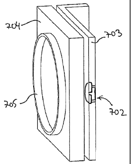
Turning now to figures 7a and 7b, a system 701
according to one embodiment of the present invention will
be briefly described. The system 701 comprises a device
703, such as a cassette, which forms part of an extra-
corporeal blood circuit 711, 712. Two pressure sensors
702, such as the sensors described above, are arranged in
a side wall of the device 703, the arrangement being such
that the sensor is mounted flush with both an inside sur-
face and an outside surface of the wall of the device
703. It is to be noted, however, that it is not necessary
that the sensor is mounted flush with the surfaces.
In operation, the device 703 is arranged at a
dialysis apparatus 704, only a part of which is shown in
figures 7a and 7b, secured by means of mechanical coup-
ling devices 708, 709. Within the dialysis apparatus 704
is an electromagnetic wave transmitter and a receiver
located, schematically illustrated by a coil structure
705. The transmitter and receiver is controlled by a
control unit (not shown) within the apparatus 704.
Figures 8a-c illustrate schematically, by way of a
respective block diagram, systems according to the
present invention. The systems may for example form part,
as described above, of a dialysis machine of which only a
respective side wall 806, 826 and 846 is illustrated.
Moreover, the systems are controlled by means of a
respective controller 801, 821 and 841.
In figure 8a, a first tunable oscillator 808 connec-
ted to a first transmitting and receiving antenna 810
communicates by way of a first alternating electromag-
netic field with a first sensor 802. A second tunable
oscillator 812 connected to a second transmitting and
receiving antenna 814 communicates by way of a second
alternating electromagnetic field with a second sensor
CA 02549067 2006-06-12
WO 2005/077262 PCT/SE2005/000184
13
804. The tunable oscillators 808, 812 thereby provide a
respective signal to the controller 801 indicative of the
conditions sensed by the sensors 802 and 804,
respectively.
In figure 8b, a transmitter 828 connected to a
transmitting antenna 830 generates, i.e. transmits, an
alternating electromagnetic field which interacts with a
sensor 822. A receiver 832 receives, via a receiving
antenna 834, the alternating electromagnetic field, as
modified by interaction with the sensor 822, and thereby
provides a signal to the controller 821 indicative of the
conditions sensed by the sensor 822.
In figure 8c, a transmitter 848 connected to an
antenna 850 generates, i.e. transmits, an alternating
electromagnetic field which interacts with a sensor 842.
A receiver 852 receives, via the same antenna 850, the
alternating electromagnetic field, as modified by inter-
action with the sensor 842, and thereby provides a signal
to the controller 841 indicative of the conditions sensed
by the sensor 842.
After manufacture of a device comprising a pressure
sensor as described above, there might be a wish to test
the sensor so that one may be certain that it functions
properly. One way of doing this is to apply a pressure to
the sensor and measure the resonance frequency of the
sensor. The sensor is made to have a certain resonance
frequency without any applied pressure. If the pressure
sensor has a different resonance frequency when a pres-
sure is applied to the sensor this may be taken as an
indication that the pressure sensor is functioning. How-
ever, it may be that the pressure sensor has a different
resonance frequency without any applied pressure and
still is non-functioning. Thus, in order to be more cer-
tain at least two different testing pressures may be app-
lied to the sensor while the resonance frequency is
measured.
CA 02549067 2012-08-22
14
The testing pressure may be applied in a number
of different ways, for example as a static pressure in a
pressure chamber.
By trimming during manufacturing of the pressure
sensor it may be given different resonance frequencies
which can thus be used to distinguish between different
disposable sets. Thus, different tubing sets for use on
the same machine may be identified as different tubing
sets by discernment of the different resonance frequen-
cies. Moreover, different medical procedures may also
make use hereof.
As mentioned above the calibration at manufacturing
and/or at the beginning of use at startup of a dialysis
session can also provide for ensuring that the pressure
sensor is working. This can be a function test like pro-
cess to see if a proper response to the application of
varying pressures by the blood pump or other mechanical
alteration. The mechanical alteration may be the app-
liance of a mechanical force to test the electronic
response frequency. The force for altering the sensor
mechanically may be applied, e.g., by applying an ultra-
sound wave on the sensor.
The scope of the claims should not be limited by the
preferred embodiments set forth in the examples, but should be
given the broadest interpretation consistent with the
description as a whole.
For example the resonant sensor described above may
be modified in that the inductance is made variable while
the capacitance is fixed.
Another example is that the device for transporting
biological fluid may be used in other extracorporeal
CA 02549067 2012-08-22
management and/or treatments of biological fluids than
specified above. Such other extracorporeal management
and/or treatments may include: separation of blood into
blood components; treatment to reduce pathogens such as
viruses in biological fluids; absorption of specific
cells or substances in blood; cell sorting and treatment
of selected cells.