Sélection de la langue

Search

Sommaire du brevet 2588843 

Énoncé de désistement de responsabilité concernant l'information provenant de tiers

Une partie des informations de ce site Web a été fournie par des sources externes. Le gouvernement du Canada n'assume aucune responsabilité concernant la précision, l'actualité ou la fiabilité des informations fournies par les sources externes. Les utilisateurs qui désirent employer cette information devraient consulter directement la source des informations. Le contenu fourni par les sources externes n'est pas assujetti aux exigences sur les langues officielles, la protection des renseignements personnels et l'accessibilité.

Disponibilité de l'Abrégé et des Revendications

L'apparition de différences dans le texte et l'image des Revendications et de l'Abrégé dépend du moment auquel le document est publié. Les textes des Revendications et de l'Abrégé sont affichés :

  • lorsque la demande peut être examinée par le public;
  • lorsque le brevet est émis (délivrance).
(12) Brevet: (11) CA 2588843
(54) Titre français: SYSTEME D'ENREGISTREMENT DE MESURES OCULAIRES POUR CHIRURGIE REFRACTIVE ET PROCEDES ASSOCIES
(54) Titre anglais: EYE REGISTRATION SYSTEM FOR REFRACTIVE SURGERY AND ASSOCIATED METHODS
Statut: Octroyé
Données bibliographiques
(51) Classification internationale des brevets (CIB):
  • A61F 9/01 (2006.01)
(72) Inventeurs :
  • GRAY, GARY P. (Etats-Unis d'Amérique)
  • CAMPIN, JOHN A. (Etats-Unis d'Amérique)
(73) Titulaires :
  • ALCON INC. (Etats-Unis d'Amérique)
(71) Demandeurs :
  • ALCON REFRACTIVEHORIZONS, INC. (Etats-Unis d'Amérique)
(74) Agent: KIRBY EADES GALE BAKER
(74) Co-agent:
(45) Délivré: 2016-08-16
(86) Date de dépôt PCT: 2005-11-29
(87) Mise à la disponibilité du public: 2006-06-08
Requête d'examen: 2010-07-22
Licence disponible: S.O.
(25) Langue des documents déposés: Anglais

Traité de coopération en matière de brevets (PCT): Oui
(86) Numéro de la demande PCT: PCT/US2005/042952
(87) Numéro de publication internationale PCT: WO2006/060323
(85) Entrée nationale: 2007-05-18

(30) Données de priorité de la demande:
Numéro de la demande Pays / territoire Date
10/999,268 Etats-Unis d'Amérique 2004-11-30

Abrégés

Abrégé français

L~invention concerne un procédé d~orientation pour la chirurgie oculaire correctrice qui enregistre des paires d~images oculaires prises à différents moments et avec un patient dans des positions différentes, comprenant l~extraction de données d~images numériques de référence sur l~AEil d~un patient et l~inclusion des données d~images sur un élément oculaire extérieur à la cornée. Des données d~images en temps réel comportant des données d~images sur l~élément oculaire extérieur à la cornée sont collectées. Une image combinée constituée d~une superposition des ensembles de données est affichée et on détermine si l~image combinée indique un enregistrement adéquat entre elles en fonction des données d~élément oculaire extérieur à la cornée dans les deux ensembles de données. Si l~enregistrement n~est pas adéquat, l~un des ensembles de données est manipulé jusqu~à obtention d~un enregistrement adéquat. L~invention concerne également un système constitué d~un appareil et d~un logiciel pour orienter un programme correctif destiné à la chirurgie oculaire.


Abrégé anglais


An orientation method for corrective eye surgery that registers pairs of eye
images taken at different times and with the patient in different positions
includes retrieving reference digital image data on an eye of the patient,
including image data on an extracorneal eye feature. Real-time image data are
collected that include image data on the extracorneal eye feature. A combined
image is displayed of a superposition of the data sets, and a determination is
made as to whether the combined image indicates an adequate registration
between them based upon the extracorneal eye feature data in the two data
sets. If the registration is not adequate, one of the data sets is manipulated
until an adequate registration is achieved. A system is directed to apparatus
and software for orienting a corrective program for eye surgery.

Revendications

Note : Les revendications sont présentées dans la langue officielle dans laquelle elles ont été soumises.


CLAIMS
1. A computer-implemented method for orienting a surgical system for a
corrective
program for eye surgery comprising:
retrieving a reference data set from a database via a processor, the reference
data set
comprising stored digital image data on an eye of a patient, the stored image
data having been
collected with the patient in a pre-surgical position and including image data
on an extracorneal eye
feature;
collecting a real-time data set via a camera in communication with the
processor, the real-
time data set comprising real-time digital image data on the patient eye in a
surgical position
different from the pre-surgical position, the real-time image data including
image data on the
extracorneal eye feature;
removing, via the processor, pixel data from a predetermined pattern of a
first set of pixels
of the reference data set to yield a reduced reference data set;
removing, via the processor, pixel data from a predetermined pattern of a
second set of
pixels of the real-time data set to yield a reduced real-time data set, the
first set disjoint from the
second set;
producing, via the processor, a combined image comprising a superposition of
the reduced
reference and the reduced real-time data sets;
providing, via the processor, the combined image to a display for display
thereon;
receiving an indication of whether the combined image indicates an adequate
registration
between the reference and the real-time data sets based upon the extracorneal
eye feature data in the
reduced reference and the reduced real-time data sets;
if the registration is not adequate, automatically manipulating one of the
reduced reference
and the reduced real-time data sets until an adequate registration is
achieved; and
modifying the corrective program of the surgical system based on the
indication of the
adequate registration and the automatically manipulating.
2. The method recited in Claim 1, wherein the pre-surgical position is
substantially upright and
the surgical position is substantially prone.
- 9 -

3. The method recited in Claim 1, wherein the stored image data comprise
camera data
organized in pixels.
4. The method recited in Claim 1, wherein the camera is a video camera.
5. The method recited in Claim 4, wherein the collecting step is performed
substantially
continuously during the surgery.
6. The method recited in Claim 1, wherein the extracorneal eye feature
comprises a blood
vessel in a sclera of the eye.
7. The method recited in Claim 1, further comprising prior to the step of
providing, scaling one
of the reference and the real-time data sets to the other of the reference and
the real-time data sets in
order to equalize a display size of the reference and the real-time data sets
displayed in the
superimposed image.
8. The method recited in Claim 1, further comprising the step of receiving
an indication of a
preferred center of a cornea of the eye in the reference data set.
9. The method recited in Claim 8, further comprising following the step of
receiving the
indication of the preferred center of the cornea of the eye, providing for
display an image of the
reference data set with an indicium superimposed thereon comprising a reticle
centered on the
determined preferred cornea center.
10. The method recited in Claim 9, wherein the reticle comprises a pair of
substantially
perpendicular lines intersecting at the determined preferred cornea center.
11. The method recited in Claim 10, wherein the reticle further comprises a
plurality of
substantially perpendicular hatch marks disposed substantially equidistantly
along the lines.
- 10 -

12. The method recited in Claim 10, wherein the reticle further comprises a
circle substantially
smaller than the cornea having a center at the determined preferred cornea
center.
13. The method recited in Claim 8, further comprising the step of receiving
an indication of a
location of a limbus of the eye in the reference data set.
14. The method recited in Claim 13, further comprising the step of
providing for display the
reference data set with a second indicium superimposed thereon comprising a
circle centered on the
determined preferred cornea center and positioned along the determined limbus
location.
15. The method recited in Claim 1, further comprising the steps of:
providing the reference data set for display;
receiving an indication of a location of the extracorneal eye feature in the
displayed
reference data set; and
wherein the step of receiving the indication comprises receiving an indication
of whether the
images of the extracorneal eye feature from the reference data set and from
the real-time data set are
aligned in the combined image, an adequate alignment corresponding to an
adequate registration.
16. The method recited in Claim 1, wherein the removing pixel data from a
first set step
comprises:
organizing the reference data set into pixels;
sampling the pixels of the reference data set to result in the reduced
reference data set,
wherein the removing pixel data from a second set step comprises:
organizing the real-time data set into pixels;
sampling the pixels of the real-time data set to result in a reduced real-time
data set by
removing pixel data from a set of pixels in the real-time data set disjoint
from those of the reduced
reference data set containing data;
wherein the step of producing the combined image comprises:
- 11 -

summing the reduced reference data set and the reduced real-time data set to
obtain a
superimposed image wherein each pixel contains data from only one of the
reduced reference data
set and the reduced real-time data set to form the combined image.
17. The method recited in Claim 16, wherein the reference data set pixel-
sampling step
comprises removing pixel data from alternate pixels.
18. The method recited in Claim 16, wherein the reference data set pixel-
sampling step
comprises determining a location of a limbus of the eye in the reference data
set and removing data
from all pixels circumscribed by the limbus.
19. The method recited in Claim 18, wherein the reference data set pixel-
sampling step
comprises removing pixel data from alternate pixels outside the limbus.
20. A system for orienting a corrective program for eye surgery comprising:
a database housing a reference data set comprising digital image data on an
eye of a patient,
the image data having been collected with the patient in a pre-surgical
position and including image
data on an extracorneal eye feature;
a processor and a display device in signal communication therewith;
a camera for collecting a real-time data set comprising real-time digital
image data on the
patient eye in a surgical position different from the pre-surgical position,
the real-time image data
including image data on the extracorneal eye feature; and
computer software resident on the processor having code segments adapted to:
retrieve the reference data set from the database;
remove pixel data from a predetermined pattern of a first set of pixels of the

reference data set to yield a reduced reference data set;
remove pixel data from a predetermined pattern of a second set of pixels of
the real-
time data set to yield a reduced real-time data set, the first set disjoint
from the second set;
produce a combined image comprising a superposition of the reduced reference
and
the reduced real-time data sets;
- 12 -

provide to the display device the combined image;
receive a determination as to whether the combined image indicates an adequate

registration between the reference and the real-time data sets based upon the
extracorneal eye
feature data in the reduced reference and the reduced real-time data sets;
if the registration is not adequate, manipulate one of the reference and the
real-time
data sets until an adequate registration is achieved; and
modify the corrective program of the surgical system based on the indication
of the
adequate registration and the automatically manipulating.
21. The system recited in claim 20, wherein the determination-receiving
step comprises
receiving input from an operator based upon operator visualization of the
displayed combined
image.
22. The system recited in claim 21, wherein the software further has a code
segment for
receiving operator input for performing the data set manipulation.
23. The system recited in claim 20, wherein the software further has a code
segment for
automatically determining whether the combined image has an adequate
registration.
24. The system recited in claim 23, wherein the software further has a code
segment for
calculating the data set manipulation based upon the determining step.
25. The system recited in claim 20, wherein the stored image data comprise
camera data
organized in pixels.
26. The system recited in claim 20, wherein the extracorneal eye feature
comprises a blood
vessel in a sclera of the eye.
- 13 -

27. The system recited in claim 20, wherein the software further has a code
segment for scaling
one of the reference and the real-time data sets to the other of the reference
and the real-time data
sets in order to equalize a display size of the reference and the real-time
data sets displayed in the
superimposed image.
28. The system recited in claim 20, wherein the software further has a code
segment for
determining a preferred center of a cornea of the eye in the reference data
set.
29. The system recited in claim 28, wherein the software further has a code
segment for
displaying an image of the reference data set with an indicium superimposed
thereon comprising a
reticle centered on the determined preferred cornea center.
30. The system recited in claim 29, wherein the reticle comprises a pair of
substantially
perpendicular lines intersecting at the determined preferred cornea center.
31. The system recited in claim 30, wherein the reticle further comprises a
plurality of
substantially perpendicular hatch marks disposed substantially equidistantly
along the lines.
32. The system recited in claim 30, wherein the reticle further comprises a
circle substantially
smaller than the cornea having a center at the determined preferred cornea
center.
33. The system recited in claim 28, wherein the software further has a code
segment for
determining a location of a limbus of the eye in the reference data set.
34. The system recited in claim 28, wherein the software further has a code
segment for
displaying the reference data set with a second indicium superimposed thereon
comprising a circle
centered on the determined preferred cornea center and positioned along the
determined limbus
location.
- 14 -

35. The system recited in claim 20, wherein the software further has code
segments for:
displaying the reference data set; locating the extracorneal eye feature in
the displayed
reference data set; and
wherein the registration adequacy is determined by determining whether the
images of the
extracorneal eye feature from the reference data set and from the real-time
data set are aligned in the
combined image, an adequate alignment corresponding to an adequate
registration.
36. The system recited in claim 20, wherein the software further has code
segments for:
organizing the reference data set and the real-time data set into pixels;
sampling the pixels of the reference data set to result in a reduced reference
data set;
sampling the pixels of the real-time data set to result in a reduced real-time
data set by
removing pixel data from a set of pixels in the real-time data set disjoint
from those of the reduced
reference data set containing data; and
summing the reduced reference data set and the reduced real-time data set to
obtain a
superimposed image wherein each pixel contains data from only one of the
reduced reference data
set and the reduced real-time data set to form the combined image.
37. The system recited in claim 36, wherein the reference data set pixel-
sampling code segment
is adapted to remove pixel data from alternate pixels.
38. The system recited in claim 36, wherein the reference data set pixel-
sampling code segment
is adapted to determine a location of a limbus of the eye in the reference
data set and remove data
from all pixels circumscribed by the limbus.
39. The system recited in claim 38, wherein the reference data set pixel-
sampling code segment
is adapted to remove pixel data from alternate pixels outside the limbus.
40. The system recited in claim 20, further comprising means for, if the
registration is adequate,
performing the eye surgery.
- 15 -

41.
The system recited in claim 40, wherein the eye surgery performing means
comprises an
excimer laser and control apparatus adapted to achieve a desired corneal
profile.
- 16 -

Description

Note : Les descriptions sont présentées dans la langue officielle dans laquelle elles ont été soumises.


CA 02588843 2012-12-19
EYE REGISTRATION SYSTEM FOR REFRACTIVE
SURGERY AND ASSOCIATED METHODS
TECHNICAL FIELD OF THE INVENTION
10 The present
invention relates to systems and methods for improving objective
measurements preceding corrective eye surgery, and, more particularly, to such

systems and methods for improving results of corrective laser surgery on the
eye.
BACKGROUND OF THE INVENTION
Laser-in-situ-keratomileusis (LASIK) is a common type of laser vision
correction method. It has proven to be an extremely effective outpatient
procedure for
a wide range of vision correction prescriptions. The use of an excimer laser
allows
for a high degree of precision and predictability in shaping the cornea of the
eye.
Prior to the LASIK procedure, measurements of the eye are made to determine
the
amount of corneal material to be removed from various locations on the corneal

surface so that the excimer laser can be calibrated and guided for providing
the
corrective prescription previously determined by the measurements. Refractive
laser
surgery for the correction of astigmatism typically requires that a
cylindrical or
quasicylindrical ablation profile be applied to the eye. The long axis of this
profile
must be properly oriented on the eye in order to accurately correct the visual

aberration.
An objective measurement of a patient's eye is typically made with the patient
sitting in an upright position while focusing on a target image. A wavefront
analyzer
then objectively determines an appropriate wavefront correction for reshaping
the
cornea for the orientation of the eye being examined. The LASIK. or PRK
procedure
is then performed with the patient in a prone position with the eye looking
upward.
It is well known that the eye undergoes movement within the socket
comprising translation and rotation ("cyclotortion") as the patient is moved
from the
upright measuring position to the prone surgery position. Techniques known in
the
art for accommodating this movement have included marking the eye by
cauterizing
-1-

CA 02588843 2007-05-18
WO 2006/060323
PCT/US2005/042952
reference points on the eye using a cautery instrument (U.S. Pat. No.
4,476,862) or
caustic substance, a very uncomfortable procedure for the patient. It is also
known to
mark a cornea using a plurality of blades (U.S. Pat. No. 4,739,761). The
application
on the scleral surface or the injection of a dye or ink is also used to mark
the reference
locations to identify the orientation of the eye during measurement,
permitting a
positioning of the corrective profile to the same orientation prior to
surgery.
However, the time delay from measurement to surgery often causes the ink to
run,
affecting the accuracy of an alignment. Making an impression on the eye (U.S.
Pat.
No. 4,705,035) avoids the caustic effects of cauterizing and the running
effect of the
ink. However, the impression can lose its definition quickly relative to the
time
period between the measurement and surgery.
For correction of astigmatism, it is known to mark the cornea preparatory to
making the surgical incisions (U.S. Pat. No. 5,531,753).
Tracker systems used during the surgical procedure or simply for following
eye movement, while the patient is in a defined position, are known to receive
eye
movement data from a mark on a cornea made using a laser beam prior to surgery

(U.S. Pat. No. 4,848,340) or from illuminating and capturing data on a feature
in or on
the eye, such as a retina or limbus, for example (U.S. 5,029,220; 5,098,426;
5,196,873; 5,345,281; 5,485,404; 5,568,208; 5,620,436; 5,638,176; 5,645,550;
5,865,832; 5,892,569; 5,923,399; 5,943,117; 5,966,197; 6,000,799; 6,027,216).
Commonly owned US 6,702,806, 2004/0143245, and 2004/0143244 address
the problem of registering a pre-surgery image with a live eye image with the
use of
image mapping and manipulation, and also with software for calculating and
imposing a graphical reticle onto a live eye image.
-2-

CA 02588843 2016-01-06
,
BRIEF SUMMARY OF THE INVENTION
The present invention is directed to an orientation system and method for
corrective
eye surgery that aligns (registers) pairs of eye images taken at different
times. An
exemplary embodiment of the method comprises the step of retrieving a
reference data set
comprising stored digital image data on an eye of a patient. The stored image
data will have
been collected with the patient in a pre-surgical position. These data include
image data on
an extracorneal eye feature.
According to one exemplary embodiment, there is provided a computer-
implemented
method for orienting a surgical system for a corrective program for eye
surgery comprising:
retrieving a reference data set from a database via a processor, the reference
data set comprising
stored digital image data on an eye of a patient, the stored image data having
been collected
with the patient in a pre-surgical position and including image data on an
extracorneal eye
feature; collecting a real-time data set via a camera in communication with
the processor, the
real-time data set comprising real-time digital image data on the patient eye
in a surgical
position different from the pre-surgical position, the real-time image data
including image data
on the extracorneal eye feature; removing, via the processor, pixel data from
a predetermined
pattern of a first set of pixels of the reference data set to yield a reduced
reference data set;
removing, via the processor, pixel data from a predetermined pattern of a
second set of pixels
of the real-time data set to yield a reduced real-time data set, the first set
disjoint from the
second set; producing, via the processor, a combined image comprising a
superposition of the
reduced reference and the reduced real-time data sets; providing, via the
processor, the
combined image to a display for display thereon; receiving an indication of
whether the
combined image indicates an adequate registration between the reference and
the real-time data
sets based upon the extracorneal eye feature data in the reduced reference and
the reduced real-
time data sets; if the registration is not adequate, automatically
manipulating one of the reduced
reference and the reduced real-time data sets until an adequate registration
is achieved; and
modifying the corrective program of the surgical system based on the
indication of the adequate
registration and the automatically manipulating.
- 3 -

CA 02588843 2016-01-06
According to a further exemplary embodiment, there is provided a system for
orienting a corrective program for eye surgery comprising: a database housing
a reference data
set comprising digital image data on an eye of a patient, the image data
having been collected
with the patient in a pre-surgical position and including image data on an
extracorneal eye
feature; a processor and a display device in signal communication therewith; a
camera for
collecting a real-time data set comprising real-time digital image data on the
patient eye in a
surgical position different from the pre-surgical position, the real-time
image data including
image data on the extracorneal eye feature; and computer software resident on
the processor
having code segments adapted to: retrieve the reference data set from the
database; remove
pixel data from a predetermined pattern of a first set of pixels of the
reference data set to yield a
reduced reference data set; remove pixel data from a predetermined pattern of
a second set of
pixels of the real-time data set to yield a reduced real-time data set, the
first set disjoint from the
second set; produce a combined image comprising a superposition of the reduced
reference and
the reduced real-time data sets; provide to the display device the combined
image; receive a
determination as to whether the combined image indicates an adequate
registration between the
reference and the real-time data sets based upon the extracorneal eye feature
data in the reduced
reference and the reduced real-time data sets; if the registration is not
adequate, manipulate one
of the reference and the real-time data sets until an adequate registration is
achieved; and
modify the corrective program of the surgical system based on the indication
of the adequate
registration and the automatically manipulating.
A real-time data set is collected that comprises digital image data on the
patient eye
in a surgical position different from the pre-surgical position. These
realtime image data
include image data on the extracorneal eye feature.
A combined image is then displayed that comprises a superposition of the
reference
and the real-time data sets, and a determination is made as to whether the
combined image
indicates an adequate registration between the reference and the realtime data
sets. Such a
determination is made based upon the extracorneal eye feature data in the
reference and the
real-time data sets. If the registration is not adequate, one of the reference
and the real-time
data sets is manipulated, i.e., translated and/or rotated, until an adequate
registration is
achieved.
- 4 -

CA 02588843 2016-01-06
A system of the present invention is directed to apparatus and software for
orienting
a corrective program for eye surgery. The system includes means for performing
the method
steps as outlined above, including computer software for achieving the
superposition of the
reference and the real-time data sets.
Thus an aspect of the present invention provides a system and method for
achieving
a precise registration of the eye by making sure that an eye feature is
positioned in
substantially the same location on the superimposed images. As a result, a
prescription
measurement for reshaping a cornea, for example, will account for the rotation
and
translation of the eye occurring between measurements made with the patient in
a sitting
position and laser surgery with the patient in a supine position.
The features that characterize the invention, both as to organization and
method of
operation, together with further objects and advantages thereof, will be
better understood
from the following description used in conjunction with the accompanying
drawing. It is to
be expressly understood that the drawing is for the purpose of illustration
and description
and is not intended as a definition of the limits of the invention. These and
other objects
attained, and advantages offered, by the present invention will become more
fully apparent
as the description that now follows is read in conjunction with the
accompanying drawing.
- 4a -

CA 02588843 2007-05-18
WO 2006/060323
PCT/US2005/042952
BRIEF DESCRIPTION OF THE SEVERAL VIEWS OF THE DRAWINGS
FIG. 1 is a schematic diagram of the system of the first embodiment of the
present invention.
FIGs. 2A, 2B is a block diagram of the data flow.
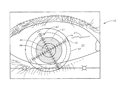
FIG. 3 illustrates the reference data set image.
FIG. 4 illustrates the reference data set image of FIG. 3, but with data from
a
central area including the area inside the limbus removed.
FIG. 5 illustrates a portion of the sampled reference data set image at a
higher
magnification.
FIG. 6 illustrates the sampled reference data set image of FIG. 5
interdigitated
with a sampled real-time data set image.
-5-

CA 02588843 2012-12-19
DETAILED DESCRIPTION OF THE PREFERRED EMBODIMENTS
A description of the preferred embodiments of the present invention will now
be presented with reference to FIGs. 1-6.
A schematic diagram of the system 10 of an embodiment of the invention is
shown in FIG. 1, data flow of an exemplary embodiment of the method 100 in
FIGs.
2A,2B, and displayed images in FIGS. 3-6. In an exemplary embodiment of the
system 10, a patient's eye 11 is imaged in a substantially upright position by
capturing
rip a first video image 12 using a camera such as a charge-coupled-
device (CCD) camera
13 (block 101). Such an image 12 is illustrated in FIG. 3. The first image,
comprising a reference data set, is stored in a database 14 in electronic
communication with a processor 15.
Next an objective measurement is made on the eye 11 to determine a
desired correction profile, using a measurement system 16 such as that
disclosed
in US Patent Publication No. 2005/0099600, although this is not intended as a
limitation (block 102).
Once the correction profile is determined, the patient is made ready for
surgery, and placed in the second position, which is typically prone.
Alternatively,
the first scan to determine the correction profile may be made in a different
location
and at a time prior to the surgical procedure, the time interval being, for
example,
several weeks.
Real-time image data are collected prior to and during surgery using a second
camera 18, in communication with a second system 38 for performing surgery,
and
these data are also stored in the database 14. hi a preferred embodiment both
the first
13 and the second 18 cameras are adapted to collect color images, and these
images
are converted using software resident on the processor 15 to pixel data. It is
useful to
collect color images for viewing by the physician, since preselected
identifiable
images such as a blood vessel 21 (FIG. 3) are more readily seen within the
sclera 23,
since the red color of the vessel 21 is clearly identifiable.
Next the surgeon identifies a plurality of features in the eye 11 using a
graphical user interface (GUI) while viewing the still image of the eye (FIG.
3). Such
features may include a preferred center 40 of the cornea 41 in the reference
set (block
103), the location of the limbus 42 (block 105), and the location of an
extracomeal
-6-

CA 02588843 2007-05-18
WO 2006/060323
PCT/US2005/042952
feature such as a blood vessel 21 (block 107). The system then generates
indicia for
display superimposed on the reference data, including a reticle 43 comprising
crossed,
perpendicular lines 44 with cross-hatching 45 and a central circle 46 centered
about
the corneal center 40 and smaller than the limbal ring 42, with the crossing
point of
the lines 44 corresponding to the cornea center 40 (block 104). The indicia
also
include a ring 47 positioned atop the limbus 42 (block 106).
The reference data set is then manipulated by removing pixel data from all
pixels circumscribed by the limbus 42 (block 108; FIG. 4), and typically by
removing
pixel data from an area 48 beyond the limbus 42, to yield a first reduced
reference
data set.
Prior to and during surgery, a real-time data set comprising real-time digital

image data on the patient eye 11 in a surgical position different from the pre-
surgical
position is collected (block 109). The real-time image data include image data
on the
blood vessel 21. Next one of the reference and the real-time data sets is
scaled to the
other of the reference and the real-time data sets (block 110). This scaling
is
performed in order to equalize a display size of the reference and the real-
time data
sets for subsequent display in a superimposed image.
The pixels of the first reduced reference data set are then sampled to result
in a
second reduced reference data set (block 111; FIG. 5). This sampling
preferably takes
the faun of removing data from a predetermined pattern of pixels, leaving a
data set
having data in all the pixels except those in the predetermined pattern. An
exemplary
predetermined pattern comprises alternate pixels. It can be seen that the
blood vessel
21 is clearly visible in FIG. 5, thereby indicating that the sampling does not
cause a
sufficient loss of resolution to interfere with identification of the vessel
21.
The pixels in the real-time data set are then sampled by removing pixel data
from a set of pixels in the real-time data set disjoint from those of the
second reduced
reference data set (block 112) to yield a reduced real-time data set.
Next the second reduced reference data set and the reduced real-time data set
are summed (block 113), so that each pixel of the summed set contains data
from a
unitary one of the second reduced reference and reduced real-time data sets. A
superimposed image comprising the sum is displayed (block 114; FIG. 6).
-7-

CA 02588843 2007-05-18
WO 2006/060323
PCT/US2005/042952
Examination of FIG. 6 indicates that the blood vessel 21 images from the
second reduced reference and reduced real-time data sets are clearly visible,
and that
they are not in registry (block 115). In such a case, either an automatic or
manual
manipulation of one of the data sets is performed (block 116) until adequate
registration is achieved (block 115), and the data processing beginning at
block 111 is
carried out again.
Once registry is considered adequate, the surgical process can begin (block
117), with monitoring continued during surgery. Thus the treatment pattern,
typically
io a laser shot pattern calculated to achieve a desired corneal profile
using, for example,
an excimer laser, can be modified to account for eye rotation resulting from
the
patient's movement from upright to prone position.
In the foregoing description, certain terms have been used for brevity,
clarity,
is and understanding, but no unnecessary limitations are to be implied
therefrom beyond
the requirements of the prior art, because such words are used for description

purposes herein and are intended to be broadly construed. Moreover, the
embodiments of the apparatus illustrated and described herein are by way of
example,
and the scope of the invention is not limited to the exact details of
construction.
Having now described the invention, the construction, the operation and use of

preferred embodiment thereof, and the advantageous new and useful results
obtained
thereby, the new and useful constructions, and reasonable mechanical
equivalents
thereof obvious to those skilled in the art, are set forth in the appended
claims.
-8-
Dessin représentatif
Une figure unique qui représente un dessin illustrant l'invention.
États administratifs

Pour une meilleure compréhension de l'état de la demande ou brevet qui figure sur cette page, la rubrique Mise en garde , et les descriptions de Brevet , États administratifs , Taxes périodiques et Historique des paiements devraient être consultées.

États administratifs

Titre Date
Date de délivrance prévu 2016-08-16
(86) Date de dépôt PCT 2005-11-29
(87) Date de publication PCT 2006-06-08
(85) Entrée nationale 2007-05-18
Requête d'examen 2010-07-22
(45) Délivré 2016-08-16

Historique d'abandonnement

Il n'y a pas d'historique d'abandonnement

Taxes périodiques

Dernier paiement au montant de 473,65 $ a été reçu le 2023-10-17


 Montants des taxes pour le maintien en état à venir

Description Date Montant
Prochain paiement si taxe générale 2024-11-29 624,00 $
Prochain paiement si taxe applicable aux petites entités 2024-11-29 253,00 $

Avis : Si le paiement en totalité n'a pas été reçu au plus tard à la date indiquée, une taxe supplémentaire peut être imposée, soit une des taxes suivantes :

  • taxe de rétablissement ;
  • taxe pour paiement en souffrance ; ou
  • taxe additionnelle pour le renversement d'une péremption réputée.

Les taxes sur les brevets sont ajustées au 1er janvier de chaque année. Les montants ci-dessus sont les montants actuels s'ils sont reçus au plus tard le 31 décembre de l'année en cours.
Veuillez vous référer à la page web des taxes sur les brevets de l'OPIC pour voir tous les montants actuels des taxes.

Historique des paiements

Type de taxes Anniversaire Échéance Montant payé Date payée
Enregistrement de documents 100,00 $ 2007-05-18
Le dépôt d'une demande de brevet 400,00 $ 2007-05-18
Taxe de maintien en état - Demande - nouvelle loi 2 2007-11-29 100,00 $ 2007-11-05
Taxe de maintien en état - Demande - nouvelle loi 3 2008-12-01 100,00 $ 2008-11-03
Taxe de maintien en état - Demande - nouvelle loi 4 2009-11-30 100,00 $ 2009-11-04
Requête d'examen 800,00 $ 2010-07-22
Taxe de maintien en état - Demande - nouvelle loi 5 2010-11-29 200,00 $ 2010-11-04
Taxe de maintien en état - Demande - nouvelle loi 6 2011-11-29 200,00 $ 2011-11-02
Taxe de maintien en état - Demande - nouvelle loi 7 2012-11-29 200,00 $ 2012-11-07
Taxe de maintien en état - Demande - nouvelle loi 8 2013-11-29 200,00 $ 2013-11-07
Taxe de maintien en état - Demande - nouvelle loi 9 2014-12-01 200,00 $ 2014-11-06
Taxe de maintien en état - Demande - nouvelle loi 10 2015-11-30 250,00 $ 2015-11-10
Taxe finale 300,00 $ 2016-06-15
Taxe de maintien en état - brevet - nouvelle loi 11 2016-11-29 250,00 $ 2016-11-09
Taxe de maintien en état - brevet - nouvelle loi 12 2017-11-29 250,00 $ 2017-11-08
Taxe de maintien en état - brevet - nouvelle loi 13 2018-11-29 250,00 $ 2018-11-08
Taxe de maintien en état - brevet - nouvelle loi 14 2019-11-29 250,00 $ 2019-11-06
Enregistrement de documents 2019-12-18 100,00 $ 2019-12-18
Taxe de maintien en état - brevet - nouvelle loi 15 2020-11-30 450,00 $ 2020-11-04
Taxe de maintien en état - brevet - nouvelle loi 16 2021-11-29 459,00 $ 2021-10-20
Taxe de maintien en état - brevet - nouvelle loi 17 2022-11-29 458,08 $ 2022-10-20
Taxe de maintien en état - brevet - nouvelle loi 18 2023-11-29 473,65 $ 2023-10-17
Titulaires au dossier

Les titulaires actuels et antérieures au dossier sont affichés en ordre alphabétique.

Titulaires actuels au dossier
ALCON INC.
Titulaires antérieures au dossier
ALCON REFRACTIVEHORIZONS, INC.
ALCON REFRACTIVEHORIZONS, LLC
CAMPIN, JOHN A.
GRAY, GARY P.
NOVARTIS AG
Les propriétaires antérieurs qui ne figurent pas dans la liste des « Propriétaires au dossier » apparaîtront dans d'autres documents au dossier.
Documents

Pour visionner les fichiers sélectionnés, entrer le code reCAPTCHA :


Pour visualiser une image, cliquer sur un lien dans la colonne description du document. Pour télécharger l'image (les images), cliquer l'une ou plusieurs cases à cocher dans la première colonne et ensuite cliquer sur le bouton "Télécharger sélection en format PDF (archive Zip)" ou le bouton "Télécharger sélection (en un fichier PDF fusionné)".

Liste des documents de brevet publiés et non publiés sur la BDBC .

Si vous avez des difficultés à accéder au contenu, veuillez communiquer avec le Centre de services à la clientèle au 1-866-997-1936, ou envoyer un courriel au Centre de service à la clientèle de l'OPIC.


Description du
Document 
Date
(yyyy-mm-dd) 
Nombre de pages   Taille de l'image (Ko) 
Abrégé 2007-05-18 2 67
Revendications 2007-05-18 8 302
Dessins 2007-05-18 7 132
Description 2007-05-18 8 362
Dessins représentatifs 2007-07-31 1 5
Page couverture 2007-07-31 1 41
Revendications 2012-12-19 8 271
Description 2012-12-19 8 345
Revendications 2014-01-28 8 279
Revendications 2014-06-16 8 279
Description 2016-01-06 9 423
Revendications 2016-01-06 8 289
Revendications 2015-03-25 7 271
Dessins représentatifs 2016-05-11 1 17
Dessins représentatifs 2016-07-04 1 17
Page couverture 2016-07-04 1 52
PCT 2007-05-18 4 139
Cession 2007-05-18 4 109
Correspondance 2007-07-27 1 19
Cession 2007-08-02 6 260
Poursuite-Amendment 2010-07-22 1 38
Poursuite-Amendment 2012-05-30 3 177
Poursuite-Amendment 2012-06-22 3 109
Poursuite-Amendment 2013-08-01 2 68
Poursuite-Amendment 2012-12-19 13 472
Poursuite-Amendment 2015-03-25 11 423
Modification 2016-01-06 14 535
Poursuite-Amendment 2014-01-28 5 181
Poursuite-Amendment 2014-05-23 2 6
Poursuite-Amendment 2014-06-16 3 92
Poursuite-Amendment 2014-12-02 3 249
Correspondance 2014-12-22 1 23
Poursuite-Amendment 2015-01-29 5 311
Demande d'examen 2015-07-31 5 325
Taxe finale 2016-06-15 1 43