Note : Les descriptions sont présentées dans la langue officielle dans laquelle elles ont été soumises.
84148229
MEDICAL DEVICE I IANDLE
Background
Medical devices typically used for cardiovascular system treatments may
involve complex
and invasive therapies resulting is significant discomfort, pain, and long
recovery times for
patients. Recently, less invasive, percutaneous treatments have been
developed. There is an
ongoing need for improved, less invasive cardiovascular treatments.
Summary
A medical device handle may comprise an elongated handle housing, a rotatable
collar
disposed about the proximal end of the handle housing, a rotatable control
knob disposed about a
proximal portion of the collar, and a slidable door disposed about a distal
portion of the collar.
The medical device handle may further comprise a means for actuating a medical
device disposed
distally of the handle, where the means is disposed within the handle housing.
The door may be
rotatably locked to the collar to prevent relative rotation therebetween.
A medical device handle may comprise a handle housing, a tubular collar
rotatably
disposed about a proximal end of the handle housing, an open-ended control
knob rotatably
disposed over a proximal portion of the tubular collar and the proximal end of
the handle housing,
and an annular door slidably disposed over a distal portion of the collar and
the handle housing,
wherein the annular door includes a plurality of inwardly-extending
protrusions configured to
engage a plurality of recessed slots in the distal portion of the collar such
that the door is rotatably
locked to the collar.
According to an embodiment, there is provided a medical device handle,
comprising: an
elongated handle housing having a proximal end, a distal end, and a
longitudinal axis extending
from the proximal end to the distal end; a rotatable collar disposed about the
proximal end of the
handle housing; a rotatable control knob disposed about the rotatable collar;
and a slidable door
disposed about a distal portion of the rotatable collar; wherein the door is
slidable between a
closed position and an open position; wherein the handle housing includes at
least one stop
extending radially outward from an outer surface of the handle housing;
wherein the door includes
at least one groove in an inner surface thereof; wherein the at least one
groove is configured to
cooperate with the at least one stop to prevent rotation of the door about the
handle housing in the
closed position.
-1-
CA 2858065 2018-05-15
84148229
According to another embodiment, there is provided a medical device handle,
comprising:
a handle housing; a tubular collar rotatably disposed about a proximal end of
the handle housing;
an open-ended control knob rotatably disposed over a proximal portion of the
tubular collar and
the proximal end of the handle housing; and an annular door slidably disposed
over a distal
portion of the collar and the handle housing; wherein the annular door
includes a plurality of
inwardly-extending protrusions configured to engage a plurality of recessed
slots in the distal
portion of the collar such that the door is rotatably locked to the collar.
Brief Description of the Drawings
The invention may be more completely understood in consideration of the
following
detailed description of various embodiments of the invention in connection
with the
accompanying drawings, in which:
Figure 1 is side view of an example medical device system;
Figure 2 is a cross-sectional side view of an example outer sheath;
Figure 3 is a transverse cross-sectional view taken through line 3-3 in Figure
2;
Figure 4 is a side view of an example inner catheter;
- 1 a-
CA 2858065 2018-05-15
CA 02858065 2014-06-03
WO 2013/082583
PCT/US2012/067567
Figure 5 is a cross-sectional view taken through line 5-5 in Figure 4;
Figure 6 is a cross-sectional view taken through line 6-6 in Figure 4;
Figure 7 is a perspective view of a portion of an example implant associated
with the example medical device system;
Figures 8-11 are perspective views that illustrate an example mechanism for
locking an implant;
Figure 12 is a side view of a portion of an example sheathing aid;
Figure 13 is an enlarged plan view illustrating engagement of the example
sheathing aid with an example implant;
Figure 14 is a side view of an example handle;
Figure 15 is a cut away view illustrating some of the interior components of
the example handle;
Figures 16-18 illustrate an example of coordinated movement of handle
components within the example handle;
Figures 19-20 illustrate the rotation of a collar on the example handle;
Figures 21-22 illustrate some of the components within the example handle
during rotation of the collar;
Figure 23 illustrates some elements of the example handle of Figure 14;
Figure 24 is a perspective view of a door of the example handle;
Figure 24A is an end view of the door of Figure 24.
Figure 25 is a perspective view of a collar of the example handle; and
Figure 25A is an end view of the collar of Figure 25.
While the invention is amenable to various modifications and alternative
forms, specifics thereof have been shown by way of example in the drawings and
will
be described in detail. It should be understood, however, that the intention
is not to
limit the invention to the particular embodiments described. On the contrary,
the
intention is to cover all modifications, equivalents, and alternatives falling
within the
spirit and scope of the invention.
Detailed Description
For the following defined terms, these definitions shall be applied, unless a
different definition is given in the claims or elsewhere in this
specification.
All numeric values are herein assumed to be modified by the term "about,"
whether or not explicitly indicated. The term "about" generally refers to a
range of
-2-
CA 02858065 2014-06-03
WO 2013/082583
PCT/US2012/067567
numbers that one of skill in the art would consider equivalent to the recited
value (i.e.,
having the same function or result). In many instances, the term "about" may
include
numbers that are rounded to the nearest significant figure.
The recitation of numerical ranges by endpoints includes all numbers within
that range (e.g. 1 to 5 includes 1, 1.5, 2, 2.75, 3, 3.80, 4, and 5).
As used in this specification and the appended claims, the singular forms "a",
"an", and "the" include plural referents unless the content clearly dictates
otherwise.
As used in this specification and the appended claims, the term "or" is
generally
employed in its sense including "and/or" unless the content clearly dictates
otherwise.
The following detailed description should be read with reference to the
drawings in which similar elements in different drawings are numbered the
same.
The drawings, which are not necessarily to scale, depict illustrative
embodiments and
are not intended to limit the scope of the invention.
Diseases and/or medical conditions that impact the cardiovascular system are
prevalent in the United States and throughout the world. Traditionally,
treatment of
the cardiovascular system was often conducted by directly accessing the
impacted
part of the system. For example, treatment of a blockage in one or more of the
coronary arteries was traditionally treated using coronary artery bypass
surgery. As
can be readily appreciated, such therapies are rather invasive to the patient
and require
significant recovery times and/or treatments. More recently, less invasive
therapies
have been developed, for example, where a blocked coronary artery could be
accessed
and treated via a percutaneous catheter (e.g., angioplasty). Such therapies
have
gained wide acceptance among patients and clinicians.
Some relatively common medical conditions may include or be the result of
inefficiency, ineffectiveness, or complete failure of one or more of the
valves within
the heart. For example, failure of the aortic valve can have a serious effect
on a
human and could lead to serious health condition and/or death if not dealt
with.
Treatment of defective heart valves poses other challenges in that the
treatment often
requires the repair or outright replacement of the defective valve. Such
therapies may
be highly invasive to the patient. Disclosed herein are medical devices that
may be
used for delivering a medical device to a portion of the cardiovascular system
in order
to diagnose, treat, and/or repair the system. At least some of the medical
devices
disclosed herein may be used to deliver and implant a replacement heart valve
(e.g., a
replacement aortic valve). In addition, the devices disclosed herein may
deliver the
-3-
CA 02858065 2014-06-03
WO 2013/082583
PCT/US2012/067567
replacement heart valve percutaneously and, thus, may be much less invasive to
the
patient. The devices disclosed herein may also provide a number of additional
desirable features and benefits as described in more detail below.
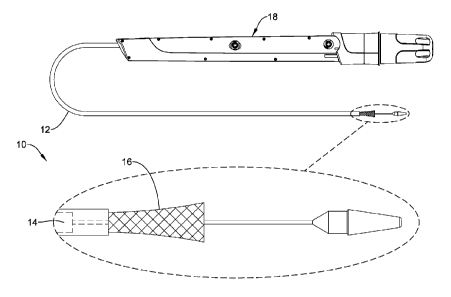
Figure 1 is a side view of an example medical device system 10. It should be
noted that some features of system 10 are either not shown, or are shown
schematically, in Figure 1 for simplicity. Additional details regarding some
of the
components of system 10 are provided in other figures in greater detail.
System 10
may be used to deliver and/or deploy a variety of medical devices to a number
of
locations within the anatomy. In at least some embodiments, system 10 is a
replacement heart valve delivery system (e.g., a replacement aortic valve
delivery
system) that can be used for percutaneous delivery of a replacement heart
valve. This,
however, is not intended to be limiting as system 10 may also be used for
other
interventions including mitral valve replacement, valve repair, valvuloplasty,
and the
like, or other similar interventions.
System 10 may generally be described as a catheter system that includes an
outer sheath or catheter 12 and an inner catheter or tube 14 (a portion of
which is
shown in Figure 1 in phantom line) extending at least partially through outer
sheath
12. A medical device implant 16 may be coupled to inner catheter 14 and
disposed
within outer sheath 12 during delivery of implant 16. A handle 18 may be
disposed at
the proximal end of outer sheath 12 and inner catheter 14. In general, handle
18 may
be configured to manipulate the position of outer sheath 12 relative to inner
catheter
14 as well as aid in the deployment of implant 16.
In use, system 10 may be advanced percutaneously through the vasculature to
a position adjacent to an area of interest. For example, system 10 may be
advanced
through the vasculature to a position adjacent to a defective aortic valve.
During
delivery, implant 16 may be generally disposed in an elongated and low profile
"delivery" configuration within outer sheath 12. Once positioned, outer sheath
12
may be retracted to expose implant 16. Implant 16 may be actuated in order to
expand implant into a generally shortened and larger profile "deployed"
configuration
suitable for implantation within the anatomy. When implant 16 is suitably
deployed
within the anatomy, system 10 can be removed from the vasculature, leaving
implant
16 in place to function as, for example, a suitable replacement for the native
aortic
valve. In at least some interventions, implant 16 may be deployed within the
native
-4-
CA 02858065 2014-06-03
WO 2013/082583
PCT/US2012/067567
valve (e.g., the native valve is left in place and not excised).
Alternatively, the native
valve may be removed and implant 16 may be deployed in its place as a
replacement.
Figures 2-13 (as well as other figures) illustrate some of the components of
system 10. For example, Figure 2 is a cross-sectional side view of outer
sheath 12.
Here it can be seen that outer sheath 12 has a proximal portion 20 and a
distal portion
22. Distal portion 22 may have a slightly enlarged or flared inner diameter,
which
may provide additional space for holding implant 16 therein. For example, the
inner
diameter of outer sheath 12 along proximal portion 20 may be in the range of
about
0.254 to 1.27 cm (0.10 to 0.50 inches), or about 0.508 to 1.016 cm (0.20 to
0.40
inches), or about 0.508 to 0.762 cm (0.20 to 0.30 inches), or about 0.56388
0.0508
cm (0.222 0.002 inches). The inner diameter of outer sheath 12 along distal
portion
22 may be in the range of about 0.254 to 1.27 cm (0.10 to 0.50 inches), or
about 0.508
to 1.016 cm (0.20 to 0.40 inches), or about 0.508 to 0.762 cm (0.20 to 0.30
inches), or
about 0.579 to 0.5842 cm (0.228 to 0.230 inches). At the distal end of distal
portion
22 may be a distal tip 24, which may be flared or otherwise have a funnel-like
shape.
The funnel-like shape increases the outer diameter (and inner diameter) of
outer
sheath 12 at distal tip 24 and may aid in the sheathing and/or re-sheathing of
implant
16 into outer sheath 12. Other than at distal tip 24, outer sheath 12 may have
a
generally constant outer diameter. For example, outer sheath 12 may have an
outer
diameter in the range of about 0.254 to 1.27 cm (0.10 to 0.50 inches), or
about 0.508
to 1.016 cm (0.20 to 0.40 inches), or about 0.508 to 0.762 cm (0.20 to 0.30
inches), or
about 0.6858 cm (0.270 inches). These are just examples. Other embodiments are
contemplated that have differing dimensions (including those appropriate for
differently sized patients including children) and/or arrangements for the
outer
diameter and/or inner diameter of outer sheath 12. These contemplated
embodiments
include outer sheaths with flared or otherwise variable outer diameters,
embodiments
with constant inner diameters, combinations thereof, and the like. Outer
sheath 12
may also have a length that is appropriate for reaching the intended area of
interest
within the anatomy. For example, outer sheath 12 may have a length in the
range of
about 30 to 200 cm, or about 60 to 150 cm, or about 100 to 120 cm, or about
108
0.20 cm. Outer sheath 12 may also be curved. For example, a distal section of
outer
sheath 12 may be curved. In one example, the radius of the curve (measured
from the
center of outer sheath 12) may be in the range of about 2 to 6 cm (20 to 60
mm), or
-5-
84148229
about 3 to 4 cm (30 to 40 mm), or about 3.675 cm (36.75 mm). Again, these
dimensions are examples and are not intended to be limiting.
Outer sheath 12 may be formed from a singular monolithic tube or unitary
member. Alternatively, outer sheath 12 may include a plurality of layers or
portions.
One or more of these layers may include a reinforcing structure such as a
braid, coil,
mesh, combinations thereof, or the like. Figure 3 illustrates one example of a
multilayer structure for outer sheath 12. For example, outer sheath 12 may
include an
inner liner or layer 26. An intermediate or tier layer 28 may be disposed on
inner
liner 26. A reinforcement 30 may be disposed on intermediate layer 28. A
topcoat or
outer layer 32 may be disposed on reinforcement 30. Finally, an outer coating
34
(e.g., a lubricious coating, a hydrophilic coating, a hydrophobic coating,
etc.) may be
disposed along portions or all of topcoat 32. These are just examples. Several
alternative structural configurations are contemplated for outer sheath 12
including
embodiments including two or more layers that may be different from those
shown in
Figure 3, embodiments without a reinforcement, and the like, or other suitable
configurations.
The dimensions and materials utilized for the various layers of outer sheath
12
may also vary. For example, inner liner 26 may include a polymeric material
such as
fluorinated ethylene propylene (FEP) and may have a thickness in the range of
about
0.00254 to 0.0127 cm (0.001 to 0.005 inches) or about 0.00762 0.00254 (0.003
0.001 inches), intermediate layer 28 may include a polymer material such as
polyether
block amide (e.g., PEBAX 6333) and may have a thickness in the range of about
0.00254 to 0.0127 cm (0.001 to 0.005 inches) or about 0.00508 0.00254 (0.002
0.001 inches), outer coating 34 may include a polymer material such as
polyether
block amide (e.g., PEBAX 7233) and may have a thickness in the range of about
0.00254 to 0.0254 cm (0.001 to 0.01 inches). In some embodiments, outer
coating 34
may vary in thickness. For example, along proximal portion 20 outer coating 34
may
have greater thickness, such as about 0.0127 to about 0.0508 cm or about
0.02159 cm
(0.005 to 0.02 inches or about 0.0085 inches), than along distal portion 22
and/or
distal tip 24, which may be about 0.0127 to about 0.0508 cm or about 0.01651
cm
(e.g., about 0.005 to 0.02 inches or about 0.0065 inches). These are just
examples as
other suitable materials may be used.
The form of distal tip 24 may also vary. For example, in at least some
embodiments, inner liner 26 (i.e., a 2.5 mm section thereof) may be extended
up and
-6-
CA 2858065 2018-05-15
CA 02858065 2014-06-03
WO 2013/082583
PCT/US2012/067567
around the distal end of outer sheath 12 (e.g., around reinforcement 30 and
topcoat
32). A ring member (not shown) made from a suitable material such as a 55D
polyether block amide (e.g., 55D PEBAX) may be disposed over inner liner 26
and
heat bonded to form distal tip 24. This may form the funnel-like shape of
distal tip
24.
Reinforcement 30 may also vary in form. In at least some embodiments,
reinforcement 30 may take the form of a braid, coil, mesh, or the like. For
example,
in some embodiments, reinforcement 30 may include a metallic braid (e.g.,
stainless
steel). In some of these embodiments, reinforcement 30 may also include
additional
structures such as one or more longitudinally-extending strands. For example,
reinforcement 30 may include a pair of longitudinally-extending aramid and/or
para
aramid strands (for example, KEVLARcq disposed on opposite sides of the braid.
These strands may or may not be woven into portions or all of the braid.
Figure 4 is a side view of the inner catheter 14. A distal end region of inner
catheter 14 may include a step in outer diameter 40 that defines a decreased
outer
diameter section 42. For example, decreased outer diameter section 42 may have
an
outer diameter in the range of about 0.127 to 0.635 cm (0.05 to 0.25 inches),
or about
0.254 to 0.508 cm (0.10 to 0.20 inches), or about 0.38608 0.00762 (0.152
0.003
inches) as opposed to the remainder of inner catheter 14 where the outer
diameter may
be in the range of about 0.127 to 0.762 cm (0.05 to 0.30 inches), or about
0.254 to
0.635 cm (0.10 to 0.25 inches), or about 0.508 0.0254 cm (0.20 0.01
inches).
Decreased outer diameter section 42 may define a region where other components
of
system 10 may be attached. Some additional details regarding these components
can
be found herein.
In general, inner catheter 14 may take the form of an extruded polymer tube.
Other forms are also contemplated including other polymer tubes, metallic
tubes,
reinforced tubes, or the like including other suitable materials such as those
disclosed
herein. In some embodiments, inner catheter 14 is a singular monolithic or
unitary
member. In other embodiments, inner catheter 14 may include a plurality of
portions
or segments that are coupled together. The total length of inner catheter may
be in the
range of about 60 to 150 cm, or about 80 to 120 cm, or about 100 to 115 cm, or
about
112 0.02 cm. Just like outer sheath 12, inner catheter 14 may also be
curved, for
example adjacent to the distal end thereof. In some embodiments, inner
catheter 14
may have one or more sections with a differing hardness/stiffness (e.g.,
differing
-7-
CA 02858065 2014-06-03
WO 2013/082583
PCT/US2012/067567
shore durometer). For example, inner catheter may have a proximal region 44a
and
an intermediate region 44b. Proximal region 44a may include a generally stiff
polymeric material such as a 72D polyether block amide (e.g., 72D PEBAX) and
may
have a length in the range of about 60 to 150 cm, or about 80 to 120 cm, or
about 100
to 115 cm, or about 109.5 0.02 cm. Intermediate region 44b may include a 40D
polyether block amide (e.g., 40D PEBAX) and may have a length in the range of
about 5 to 25 mm, or about 10 to 20 mm, or about 15 0.01 mm. Decreased outer
diameter section 42 may also differ from regions 44a/44b and, in some
embodiments,
may include a 72D polyether block amide (e.g., 72D PEBAX) and may have a
length
in the range of about 0.5 to 2 cm (5 to 20 mm), or about 0.8 to 1.5 cm (8 to
15 mm),
or about 1 0.001 cm (10 0.01 mm). These are just examples.
Inner catheter 14 may include one or more lumens. For example, Figure 5
(which is a cross-sectional view of inner catheter 14 adjacent to proximal end
portion
36) illustrates that inner catheter 14 may include a first lumen 46, a second
lumen 48,
a third lumen 50, and a fourth lumen 52. In general, lumens 46/48/50/52 extend
along
the entire length of inner catheter 14. Other embodiments are contemplated,
however,
where one or more of lumens 46/48/50/52 extend along only a portion of the
length of
inner catheter 14. For example, fourth lumen 52 may stop just short of the
distal end
of inner catheter 14 and/or be filled in at its distal end to effectively end
fourth lumen
52 proximal of the distal end of inner catheter 14, as illustrated in Figure 6
by the
absence of fourth lumen 52 adjacent to the distal end of inner catheter 14.
Disposed within first lumen 46 may be push-pull rods 84 (not shown in Figure
5, seen in other figures including Figure 7), which are used to expand and/or
elongate
implant 16 as explained in more detail herein. In at least some embodiments,
first
lumen 46 may be lined with a low friction liner 54 (e.g., a FEP liner).
Disposed
within second lumen 48 may be a pin release mandrel 92 (not shown in Figure 5,
seen
in other figures including Figure 7), which is also explained in more detail
herein. In
at least some embodiments, second lumen 48 may be lined with a hypotube liner
56.
Third lumen 50 may be a guidewire lumen and this lumen may also be lined with
a
hypotube liner 58.
Fourth lumen 52 may be used to house a non-stretch wire 60. The form of
non-stretch wire 60 may vary. In some embodiments, non-stretch wire 60 may
take
the form of a stainless steel braid. The non-stretch wire 60 may optionally
include a
pair of longitudinally-extending aramid and/or para aramid strands (for
example,
-8-
CA 02858065 2014-06-03
WO 2013/082583
PCT/US2012/067567
KEVLARO) disposed on opposite sides of the braid. In general, rather than
being
"disposed within" fourth lumen 52, non-stretch wire 60 may be embedded within
fourth lumen 52. In addition, non-stretch wire 60 may extend to a position
adjacent to
distal end portion 38 but not fully to the distal end of inner catheter 14 as
illustrated in
Figure 6 by the absence of fourth lumen 52 adjacent to the distal end of inner
catheter
14. For example, a short distal segment of fourth lumen 52 may be filled in
with
polymer material adjacent to the distal end of inner catheter 14.
Inner catheter 14 may also include a guidewire extension tube 62 that extends
distally from distal end portion 38. A nose cone 64 is attached to guidewire
extension
tube 62. Nose cone 64 generally is designed to have an atraumatic shape. Nose
cone
64 may also include a ridge or ledge 66 that is configured to abut the distal
tip 24 of
outer sheath 12 during delivery of implant 16.
Figure 7 illustrates some of the additional components of system 10 and
implant 16. For example, here it can be seen that implant 16 includes a
plurality of
valve leaflets 68 (e.g., bovine pericardial) which are secured to a
cylindrical braid 70
at a post or commissure post 72, for example at the commissure portions of the
leaflets 68. In this example, implant 16 includes three leaflets 68 secured to
braid 70
with three posts 72. Leaflets 68 may also be secured to the base or "distal
end" of
braid 70. The posts 72, in turn, may be secured to braid 70 (e.g., along the
interior of
braid 70) with sutures or other suitable mechanisms. Positioned adjacent to
(e.g.,
longitudinally spaced from and aligned with) posts 72 are a plurality of
buckles 76,
which may also be sutured to braid 70 (e.g., along the interior of braid 70).
In this
example, one buckle 76 is attached to braid 70 adjacent to each of the three
posts 72.
Accordingly, braid 70 has a total of three buckles 76 and three posts 72
attached
thereto. Other embodiments are contemplated where fewer or more buckles 76 and
posts 72 may be utilized. A seal 74 (shown in cross-section) may be disposed
about
braid 70 and, as the name suggests, may help to seal implant 16 within a
target
implant site or area of interest.
Attachment between implant 16 and inner catheter 14 (and/or outer sheath 12)
may be effected through the use of a three finger coupler 78. Coupler 78 may
generally include a cylindrical base (not shown) that is attached to inner
catheter 14
(e.g., disposed about and attached to reduced outer diameter section 42).
Projecting
distally from the base are three fingers that are each configured to engage
with
implant 16 at posts 72 and buckles 76. A collar 80 may further assist in
holding
-9-
CA 02858065 2014-06-03
WO 2013/082583
PCT/US2012/067567
together these structures. A guide 82 may be disposed over each of the fingers
and
may serve to keep the fingers of coupler 78 associated with push-pull rods 84
extending adjacent to coupler 78. Finally, a pin release assembly 86 may be a
linking
structure that keeps posts 72, buckles 76, and push-pull rods 84 associated
with one
another. Pin release assembly 86 includes a plurality of individual pins 88
that may
be joined together via a coiled connection 90 and held to a pin release
mandrel 92
with a ferrule 94.
During delivery, implant 16 is secured at the distal end of inner catheter 14
by
virtue of the association of the fingers of coupler 78 being coupled with a
projecting
proximal end of buckles 76 (and being held in place with collar 80 disposed
over the
connection) and by virtue of pins 88 securing together push-pull rods 84 and
posts 72.
When implant 16 is advanced within the anatomy to the desired location, outer
sheath
12 may be withdrawn (e.g., moved proximally relative to inner catheter 14) to
expose
implant 16. Then, push-pull rods 84 can be used to expand and "lock" implant
16 in
the expanded or deployed configuration by proximally retracting push-pull rods
84 to
pull posts 72 into engagement with buckles 76. Finally, pins 88 can be
removed,
thereby uncoupling push-pull rods 84 from posts 72, which allows implant 16 to
be
released from system 10 and deployed in the anatomy.
Figures 8-11 illustrate the locking system utilized with system 10. For
simplicity purposes, only one of the three fingers of the coupler 78, only one
of the
three push-pull rods 84, and only one of the posts 72 of the example system 10
are
shown (and implant 16 is not shown). As seen in Figure 8, push-pull rod 84
extends
through guide 82 adjacent to the fingers of coupler 78, through collar 80,
through
buckle 76, and into a hollow t-shaped bar portion 96 of post 72. The distal
end of
push-pull rod 84 may include an opening or aperture (not shown) that can be
aligned
with an opening 98 of t-shaped bar portion 96. When so aligned, pin 88 can be
looped through opening 98 and the opening of push-pull rod 84. This secures
push-
pull rod 84 to post 72 and forms a configuration of these structures that can
be utilized
during delivery of implant 16. As can be appreciated, the proximal end of post
72 and
the distal end of buckle 76 are longitudinally separated and, accordingly,
implant 16 is
in an elongated and generally low-profile configuration suitable for delivery.
When implant 16 reaches the intended target site within the anatomy, a
clinician can proximally retract push-pull rod 84, thereby moving the proximal
ends
of posts 72 toward the distal ends of buckles 76 in order to expand implant
16.
-10-
CA 02858065 2014-06-03
WO 2013/082583
PCT/US2012/067567
Ultimately, push-pull rod 84 can be retracted sufficiently far enough to lock
post 72
with buckle 76 so as to lock implant in an expanded configuration suitable for
implantation within the anatomy. Figure 9 illustrates push-pull rod 84
proximally
retracted. In doing so, post 72 is brought into contact with buckle 76. More
particularly, a raised, generally transversely-oriented ridge 100 on t-shaped
bar
portion 96 may be pulled proximally past buckle 76 so that post 72 is secured
and
held in place by buckle 76. At this point, it is possible to urge push-pull
rods 84
distally to "unlock" implant 16, thereby allowing for repositioning and/or
retraction.
Alternatively, if a clinician is satisfied with the positioning and/or locking
of implant
16 (e.g., after visualization of implant 16 via a suitable imaging technique),
pins 88
may be pulled (e.g., removed from openings 98 and the openings in push-pull
rods
84) to uncouple push-pull rods 84 from posts 72 as shown in Figure 10. Further
retraction of push-pull rods 84 causes a longitudinally-oriented ridge 102 on
push-pull
rods 84 to engage collar 80 and causes collar 80 to slide proximally along the
fingers
of coupler 78. In doing so, a forked end 104 of the fingers, which has a
groove 106
formed therein, is exposed and can be uncoupled from a rail 108, which has a
projection 110 formed thereon that is configured to mate with groove 106, as
shown
in Figure 11. Thereafter, system 10 can be removed from the anatomy, leaving
behind the expanded and deployed implant 16.
Figures 12-13 illustrate another component that may be included with system
10. For example, Figure 12 is a side view of a portion of a sheathing aid 112.
Here it
can be seen that sheathing aid 112 includes a base 114 and a group of petals
including
a set of three longer petals 116 and a pair of shorter petals 118. In use, a
group of
petals 116/118 may be positioned between each of the fingers of coupler 78.
Because
the coupler 78 may have a total of three fingers, sheathing aid 112 may have a
total of
fifteen petals (e.g., three groups that each include three "long" petals 116
and two
"short" petals 118, with each group being positioned between adjacent pairs of
fingers
of coupler 78). Base 114 may be secured to inner catheter 14 adjacent to
coupler 78
(e.g., underneath coupler 78 and between coupler 78 and inner catheter 14).
Sheathing aid 112, as the name suggests, may be used to aid in the sheathing
of implant 16 into outer sheath 12. In addition, sheathing aid 112 may aid in
the
initial sheathing of implant 16 (e.g., removing implant 16 from a packaging
container
such as a bottle and pulling implant 16 into outer sheath 12) and in re-
sheathing
implant 16 during repositioning and/or retraction of implant 16 within the
area of
-11-
CA 02858065 2014-06-03
WO 2013/082583
PCT/US2012/067567
interest. Sheathing may be accomplished via the arrangement and positioning of
the
various petals 116/118. For example, Figure 13 illustrates the longer petals
116
woven in and out of braid 70, and the shorter petals 118 disposed along the
exterior of
braid 70 acting as a funnel for sheathing.
Figure 14 is a side view of handle 18. Here it can be seen that handle 18
includes a handle housing 120. A rotatable control knob 122 may be disposed
about
handle housing 120 (e.g., at a proximal end of handle housing 120) and may be
used
to move one or more of the components of system 10 (e.g., outer sheath 12,
push-pull
rods 84, etc.). A rotatable collar 156 may be disposed about the handle
housing 120.
In some embodiments, control knob 122 may be disposed about a proximal portion
of
collar 156. A slidable door 124 may also be disposed about handle housing 120.
Door 124 may translate distally to expose a distal portion of rotatable collar
156 (not
shown in Figure 14, can be seen in other figures including Figures 19-20)
positioned
generally under door 124. Collar 156 may be rotated to move one or more
components of system 10 (e.g., push-pull rods 84, pin release mandrel 92,
etc.). Some
additional details related to exterior elements of handle 18 (e.g., door 124,
collar 156,
etc.) may be seen in Figures 23-25A and are discussed further herein.
Handle 18 may also include one or more apertures 129a/129b and/or flush
ports 126/128 that can be used to flush system 10. In some embodiments, distal
flush
port 126 and proximal flush port 128 may be accessible from the exterior of
the
handle housing 120 through distal aperture 129a and proximal aperture 129b,
respectively.
Figure 15 is a side view of handle 18 with a portion of handle housing 120
removed, exposing at least some of the interior components. Here it can be
seen that
outer sheath 12 may be attached to a sheath adapter 130. Sheath adapter 130 is
attached to a sheath carriage 132, which may be threaded onto a lead screw
134.
Distal flush port 126 may be disposed on sheath adapter 130. In general,
distal flush
port 126 provides access to the interior or lumen of outer sheath 12 (e.g.,
access to
space between inner catheter 14 and outer sheath 12) so that a clinician can
flush fluid
through the lumen of outer sheath 12 to remove any unwanted materials (e.g.,
air,
fluid, contaminants, etc.) therein prior to use of system 10. In at least some
embodiments, distal flush port 126 has a fuer type connector (e.g., a one-way
lucr
connector) that allows a device such as a syringe with a corresponding
connector to be
attached thereto for flushing.
-12-
CA 02858065 2014-06-03
WO 2013/082583
PCT/US2012/067567
Extending through and proximally from sheath adapter 130 is inner catheter
14. A proximal end of inner catheter 14 is attached (e.g., fixedly attached)
to an
interior body or diverter 136. Diverter 136 is attached to a support body 140.
In
general, diverter 136 and/or support body 140 may have one or more passageways
or
lumens formed therein. In some embodiments, push-pull rods 84 and/or pin
release
mandrel 92 may extend through respective passageways. Alternatively, the
proximal
ends of push-pull rods 84 and/or pin release mandrel 92 may each be attached
to a
shaft or hypotube (e.g., solid in cross-section, tubular, etc.), and each of
the shafts
may extend through the one or more passageways. For example, a first shaft or
hypotube 142 and a second shaft or hypotube 144 may extend through the
passageways in diverter 136, and in some embodiments, the first shaft or
hypotube
142 extends through a first passageway and the second shaft or hypotube 144
extends
through a second passageway that is separate or distinct from the first
passageway. In
at least some embodiments, first shaft 142 is attached to pin release mandrel
92. In at
least some embodiments, second shaft 144 is attached to push-pull rods 84. It
should
be noted that at in least some embodiments of system 10, three push-pull rods
84 are
utilized. In these embodiments, the three push-pull rods 84 come together
(e.g.,
brought into contact with one another or otherwise brought into relatively
close
proximity with one another) adjacent to the distal end of inner catheter 14
and enter
first lumen 46. At one or more positions along their length, push-pull rods 84
may be
attached to one another. For example, in some embodiments, push-pull rods 84
may
be welded together about 10.16 cm (about 4.00 inches) from their distal ends.
In
some embodiments, push-pull rods 84 may be welded together proximate their
proximal ends in addition to or instead of the distal weld. Proximally
thereafter, push-
pull rods 84 may extend to second shaft 144.
A hypotube (e.g., hypotube liner 58 disposed along guidewire lumen 52) may
extend through diverter 136 within a passageway therein and then be "diverted"
around a portion of diverter 136 and support body 140, and ultimately be
extended to
a position at the proximal end of handle 18 so as to provide a user access to
guidewire
lumen 52. Proximal flush port 128 may be disposed on support body 140 that can
be
used to flush the lumens of inner catheter 14 and, for example, may function
similarly
to distal flush port 126.
At their respective proximal ends, first shaft 142 may be secured to a slider
146 and second shaft 144 may be secured to a force limiter body 150. The
-13-
CA 02858065 2014-06-03
WO 2013/082583
PCT/US2012/067567
connections between the various components may include a number of different
types
of connections including mechanical bonding (e.g., pinning, threading,
interference
fit, etc.), adhesive bonding, thermal bonding, etc. Slider 146 may be slidable
relative
to force limiter body 150. In some embodiments, slider 146 may be selectively
locked to force limiter body 150, thereby preventing relative movement between
the
slider 146 and the force limiter body 150. Force limiter body 150 may be
secured to a
push-pull rod carriage 152, which may be threaded onto lead screw 134. Thus,
movement of lead screw 134 can cause movement of push-pull rod carriage 152
and
force limiter body 150 and thus, push-pull rods 84 (via second shaft 144).
Some
additional details regarding this motion can be found herein.
In general, force limiter body 150 forms or defines a stop point that provides
tactile feedback (e.g., resistance to further rotation of control knob 122) to
the user
indicating that push-pull rods 84 have been retracted proximally a sufficient
distance
to lock posts 72 with buckles 76. To verify proper locking, a clinician may
use an
appropriate visualization technique to visualize proper locking (e.g., the
relative
positioning of the posts 72 and the buckles 76). A chock 148 may be positioned
adjacent to slider 146 to selectively lock slider 146 to force limiter body
150. In order
to allow pin release mandrel 92 to be proximally retracted to pull pins 88,
chock 148
can be rotated or otherwise moved to a secondary position or configuration.
When in
this configuration, chock 148 no longer forms a barrier to further movement
of, for
example, slider 146 and pin release mandrel 92. Accordingly, with chock 148 no
longer acting as an impediment, slider 146 and pin release mandrel 92 can be
proximally retracted to facilitate deployment of implant 16 by allowing pins
88 to be
pulled.
Handle 18 also includes a rotatable ring 155 with internal teeth that are
configured to engage with teeth on a gear 157 coupled to lead screw 134. Ring
155 is
coupled to control knob 122 so that rotation of control knob 122 results in
analogous
motion of ring 155 and thus lead screw 134.
Handle 18 is generally configured for coordinated movement of multiple
structures of system 10. For example, handle 18 is configured to allow a user
to move
outer sheath 12 (e.g., relative to inner catheter 14), move push-pull rods 84,
and move
pin release mandrel 92. Moreover, handle 18 is configured so that the
appropriate
structure can be moved at the appropriate time during the intervention so that
implant
16 can be delivered in an efficient manner. Some examples of how the
coordinated
-14-
84148229
movement of system 10 may occur within handle 18 may be similar to those
disclosed
in U.S. Patent Application Pub. No. US 2010/0280495.
To help facilitate the coordinated movement, handle 18 may include a lost
motion barrel 158. Lost motion barrel 158 is configured to engage carriages
132/152
and/or screws associated with carriages 132/152 at different times during the
intervention to stop motion (e.g., create "lost motion" of the appropriate
carriage).
Figures 16-19 illustrate some of the coordinated motion achieved by handle 18.
It
should be noted that some elements of system 10 are not shown in Figures 16-20
for
clarity. For example, Figure 16 illustrates a first position or state for
handle 18 where
outer sheath 12 is extended distally relative to inner catheter 14 (and handle
18) so as
to fully sheath (e.g., contain) implant 16. While in this position, sheath
carriage 132
is positioned adjacent to the distal end of handle 18. In addition, a rod
screw 152a
associated with push-pull rod carriage 152 is extended distally from push-pull
rod
carriage 152 and positioned within lost motion barrel 158. Upon rotation of
control
knob 122 (e.g., in the clockwise direction), lead screw 134 begins to rotate.
Rotation
of lead screw 134 causes sheath carriage 132 to move along lead screw 134 in
the
proximal direction, resulting in proximal movement of outer sheath 12 (e.g.,
"unsheathing" implant 16). This initial rotation of lead screw 134 also causes
rod
screw 152a to rotate. This may be because, for example, a knob or projection
(not
shown) on rod screw 152a may be engaged with a helical thread disposed along
the
interior of lost motion barrel 158. However, because rod screw 152a is spaced
from
push-pull rod carriage 152, it does not exert a force onto push-pull rod
carriage 152.
Thus, initial motion of control knob 122 does not result in movement of push-
pull rod
carriage 152 and, instead, only results in translation of sheath carriage 132
and
rotation (and translation) of rod screw 152a.
Eventually, rod screw 152a (e.g., the knob formed therein) reaches an
essentially linear thread or pathway formed at the end of lost motion barrel
158. The
linear thread allows rod screw 152a to translate along lead screw 134 to a
position
where rod screw 152a contacts (e.g., is threaded within and abuts) push-pull
rod
carriage 152. In doing so, rod screw 152a can contact and move proximally push-
pull
carriage 152. Accordingly, further rotation of lead screw 134 not only causes
sheath
carriage 132 to move proximally but also causes push-pull rod carriage 152 to
move
proximally as shown in Figure 17.
-15-
CA 2858065 2018-06-04
CA 02858065 2014-06-03
WO 2013/082583
PCT/US2012/067567
When sheath carriage 132 reaches lost motion barrel 158, a sheath carriage
screw 132a of sheath carriage 132 enters lost motion barrel 158 as shown in
Figure
18. This may occur in a manner similar to how rod screw 152a threads and
unthreads
with the helical thread formed along lost motion barrel 158. For example,
while
sheath carriage 132 is translating, sheath carriage screw 132a may follow an
essentially linear thread or pathway formed along or adjacent to lost motion
barrel
158. Upon reaching lost motion barrel 158, sheath carriage screw 132a (e.g., a
knob
or projection formed thereon) may shift into engagement with the helical
thread
within lost motion barrel 158 and rotate. This rotation "unthreads" sheath
carriage
screw 132a from sheath carriage 132. Accordingly, additional rotation of lead
screw
134 results in continued proximal movement of push-pull rod carriage 152 while
motion of sheath carnage 132 ceases.
In at least some embodiments, lead screw 132 has a plurality of portions, for
example a first portion 132a and a second portion 132b, with a differing pitch
to its
thread. This may allow carriages 132/152 to travel at different rates along
lead screw
132. For example, the pitch of lead screw 132 along which sheath carriage 132
translates may be generally more spaced or slanted than at positions adjacent
to push-
pull rod carriage 152. Accordingly, the coordinated movement of carriages
132/152
also may be configured so that sheath carriage 132 translates along lead screw
132 at
a greater rate than push-pull rod carriage 152. Other configurations are
contemplated
where the above-mentioned configuration is reversed as well as further
configurations
where the pitch of lead screw 132 is essentially constant or includes a number
of
different pitch regions.
Sufficient proximal retraction of push-pull rod carriage 152, for example as
shown in Figure 18, may result in push-pull rods 84 being sufficiently
retracted so
that posts 72 can engage and lock with buckles 76. When the clinician is
satisfied that
locking is complete (e.g., after verification via an appropriate visualization
technique),
the clinician may proximally retract pin release mandrel 92 in order to pull
pins 88
from openings 98 and openings in push-pull rods 84 to release implant 16.
To initiate release of pins 88, door 124 may be slid distally along a collar
156
(which is positioned on handle 18) as shown in Figure 19. When door 124 is
sufficiently advanced, door 124 and collar 156, together, can be rotated as
shown in
Figure 20. Push-pull rod carriage 152 may also include a radially-extending
proximal
flag member 164. In general, flag member 164 may be designed as a feature that
can
-16-
CA 02858065 2014-06-03
WO 2013/082583
PCT/US2012/067567
prevent collar 156 from being rotated earlier than desired (and, thus, prevent
pins 88
from being pulled earlier than desired). For example, flag member 164 may be
positioned within and follow a groove (not shown) along the interior of collar
156.
While positioned within the groove, flag member 164 essentially forms a
physical
barrier that prevents collar 156 from rotating relative to handle housing 120.
When
push-pull rod carriage 152 is translated proximally to the back of handle
housing 120
(e.g., when push-pull rods 84 are proximally retracted so as to lock posts 72
with
buckles 76), flag member 164 exits the groove in collar 156. Accordingly, flag
member 164 no longer impedes rotation of collar 156 and, as such, collar 156
can
now be rotated to pull pins 88.
Collar 156, via ring 154, is associated with a gear 160 engaged with a
secondary screw 162. Notches at a proximal end of collar 156 engage
protrusions on
ring 154 such that rotation of collar 156 causes corresponding rotation of
ring 154 and
thus secondary screw 162. The initial rotation of collar 156 is sufficient to
rotate
chock 148 (e.g., via a mechanical interaction between collar 156 and chock 148
that
causes chock 148 to shift) from a first configuration where slider 146 (and,
thus, pin
release mandrel 92) is selectively locked to force limiter body 150, to a
secondary
configuration, which permits slider 146 to translate along secondary screw 162
as
secondary screw 162 rotates, to proximally retract and pull pins 88 (e.g., via
first shaft
142 and pin release mandrel 92, which is connected thereto). As seen in Figure
21,
chock 148 in the first configuration engages a ridge 168 along a top portion
of force
limiter body 150 which forms a physical barrier that prevents proximal
translation of
slider 146 relative to force limiter body 150. When collar 156 is rotated to
shift chock
148 into the secondary configuration, slider 146 can translate proximally
within a
groove 166 disposed in the top portion of force limiter body 150 (e.g., as
seen in
Figure 22), as collar 156 is rotated about the handle housing 120 to pull the
pins 88
from the openings 98 and the openings in the distal ends of the push-pull rods
84.
Once pins 88 have been removed, push-pull rods 84 may be withdrawn from
implant
16, thereby deploying the implant at the target site (area of interest).
Following deployment of the implant 16, the control knob 122 may be rotated
to move the sheath carriage 132 distally within the handle housing 120,
thereby
moving outer sheath 12 distally relative to inner catheter 14 and three-finger
coupler
78 so as to cover or re-sheath the elements of system 10 disposed at the
distal end.
System 10 may then be removed from the patient's anatomy.
-17-
CA 02858065 2014-06-03
WO 2013/082583
PCT/US2012/067567
Figure 23 is a side view of a proximal end of handle 18 of Figure 14, with
door 124 removed. As discussed above, handle 18 may include a handle housing
120.
A collar 156 may be disposed about the proximal end of handle housing 120.
Control
knob 122 may be disposed about a proximal portion 500 of collar 156. Control
knob
122 is selectively rotatable about the proximal end of handle housing 120 and
proximal portion 500 of collar 156, and may function as described above.
Collar 156 may be selectively rotatable about handle housing 120, as
described above. Collar 156 may include a proximal portion 500, a central
portion
502, and a distal portion 504, as illustrated in Figure 25. Proximal portion
500 may
include a plurality of notches configured to engage a plurality of protrusions
on ring
154, as described above. Central portion 502 may include a radially-extending
flange
portion 530 disposed about the central portion 502. As may be seen in Figure
14,
flange portion 530 may longitudinally space control knob 122 apart from door
124.
Collar 156 may further include a groove 520 on the interior of collar 156, as
seen in
Figures 25 and 25A. Radially-extending flag member 164 of push-pull rod
carriage
152 may be positioned within the groove 520 to act as an interlocking element
in an
engaged configuration. In the engaged configuration, where flag member 164 is
positioned within groove 520, the flag member physically prevents collar 156
from
rotating relative to handle housing 120. As push-pull rod carriage 152 is
translated
proximally within handle housing 120, the flag member 164 similarly translates
proximally within groove 520. When push-pull rod carriage 152 has translated
proximally a sufficient distance, flag member 164 exits groove 520 to achieve
a
disengaged configuration. In the disengaged configuration, collar 156 is
permitted to
rotate relative to handle housing 120. Distal portion 504 may include a
plurality of
longitudinally-oriented recessed slots 510 in an outer surface thereof.
Door 124 may be disposed about distal portion 504 of collar 156. Door 124
may be axially slidable along a longitudinal axis of handle housing 120
between a
closed position (e.g., as seen in Figure 14) in which door 124 covers distal
portion 504
of collar 156 and an open position (e.g., as seen in Figure 19) in which door
124 does
not cover all or a part of distal portion 504 of collar 156. In some
embodiments,
collar 156, control knob 122, and door 124 may be concentrically disposed
about the
longitudinal axis of handle housing 120.
As seen in Figure 23, handle housing 120 may include at least one stop 400
extending radially outward from an outer surface of handle housing 120. Door
124
-18-
CA 02858065 2014-06-03
WO 2013/082583
PCT/US2012/067567
may include at least one groove 302 in an inner surface thereof (e.g., as seen
in
Figures 24 and 24A), wherein the at least one groove 302 is configured to
receive and
cooperate with the at least one stop 400 to prevent rotation of door 124
relative to
handle housing 120 in the closed position. In some embodiments, the at least
one
groove 302 does not extend the entire length of door 124. Accordingly, when
door
124 is translated distally a sufficient distance, stop 400 will exit groove
302, thereby
permitting door 124 to rotate relative to the handle housing 120.
Handle housing 120 may also include a radially-extending ridge 402 disposed
about the handle housing 120. A distal end of collar 156 may abut a proximal
face of
the ridge 402, as seen in Figure 23. Door may further include a plurality of
protrusions 300 extending radially inward from an inner surface of door 124.
In some
embodiments, protrusions 300 may generally be longitudinally elongated and of
similar size and/or shape as slots 510. Protrusions 300 may engage the
plurality of
recessed slots 510 in the outer surface of collar 156 to prevent relative
rotation
between door 124 and collar 156. That is, protrusions 300 engage with slots
510 in a
manner that rotatably locks door 124 to collar 156 such that door 124 and
collar 156
must rotate together, or in tandem.
Accordingly, door 124 and/or flag member 164 may prevent rotation of collar
156 relative to handle housing 120 when door 124 is in the closed position
and/or
when flag member 164 is in the engaged configuration. After flag member 164
has
been moved proximally to the disengaged configuration, door 124 may be slid
distally
into the open position. A distal end of the plurality of protrusions 300 may
abut the
proximal face of ridge 402 when door 124 is disposed in the open position.
Collar
156 is free to rotate about handle housing 120 under these conditions. Door
124 and
collar 156 cooperate to form a hand gip that may be rotated to move chock 148
and
pull pins 88 to release the push-pull rods 84 from posts 72, as described
above. The
hand grip formed by the cooperation of door 124 and collar 156 provides a
wider,
larger diameter surface for a user/practitioner to grasp and rotate than
collar 156
alone.
The materials that can be used for the various components of system 10
(and/or other systems disclosed herein) and the various tubular members
disclosed
herein may include those commonly associated with medical devices. For
simplicity
purposes, the following discussion makes reference to outer sheath 12 and/or
inner
catheter 14. However, this is not intended to limit the devices and methods
described
-19-
CA 02858065 2014-06-03
WO 2013/082583
PCT/US2012/067567
herein, as the discussion may be applied to other similar tubular members
and/or
components of tubular members or devices disclosed herein.
Outer sheath 12 and/or inner catheter 14 may be made from a metal, metal
alloy, polymer (some examples of which are disclosed below), a metal-polymer
composite, ceramics, combinations thereof, and the like, or other suitable
material.
Some examples of suitable metals and metal alloys include stainless steel,
such as
304V, 304L, and 316LV stainless steel; mild steel; nickel-titanium alloy such
as
linear-elastic and/or super-elastic nitinol; other nickel alloys such as
nickel-
chromium-molybdenum alloys (e.g., UNS: N06625 such as INCONEL 625, UNS:
N06022 such as HASTELLOY C-22k, UNS: N10276 such as HASTELLOY
C276k, other HASTELLOY alloys, and the like), nickel-copper alloys (e.g.,
UNS:
N04400 such as MONEL 400, NICKELVAC 400, NICORROS 400, and the
like), nickel-cobalt-chromium-molybdenum alloys (e.g., UNS: R30035 such as
MP35-N and the like), nickel-molybdenum alloys (e.g., UNS: N10665 such as
HASTELLOY ALLOY B2k), other nickel-chromium alloys, other nickel-
molybdenum alloys, other nickel-cobalt alloys, other nickel-iron alloys, other
nickel-
copper alloys, other nickel-tungsten or tungsten alloys, and the like; cobalt-
chromium
alloys; cobalt-chromium-molybdenum alloys (e.g., UNS: R30003 such as
ELGILOY , PHYNOX , and the like); platinum enriched stainless steel; titanium;
combinations thereof; and the like; or any other suitable material.
As alluded to herein, within the family of commercially available nickel-
titanium or nitinol alloys, is a category designated "linear elastic" or "non-
super-
elastic" which, although may be similar in chemistry to conventional shape
memory
and super elastic varieties, may exhibit distinct and useful mechanical
properties.
Linear elastic and/or non-super-elastic nitinol may be distinguished from
super elastic
nitinol in that the linear elastic and/or non-super-elastic nitinol does not
display a
substantial "superelastic plateau" or "flag region" in its stress/strain curve
like super
elastic nitinol does. Instead, in the linear elastic and/or non-super-elastic
nitinol, as
recoverable strain increases, the stress continues to increase in a
substantially linear,
or a somewhat, but not necessarily entirely linear relationship until plastic
deformation begins or at least in a relationship that is more linear that the
super elastic
plateau and/or flag region that may be seen with super elastic nitinol. Thus,
for the
purposes of this disclosure linear elastic and/or non-super-elastic nitinol
may also be
termed "substantially" linear elastic and/or non-super-elastic nitinol.
-20-
84148229
In some cases, linear elastic and/or non-super-elastic nitinol may also be
distinguishable from super elastic nitinol in that linear elastic and/or non-
super-elastic
nitinol may accept up to about 2-5% strain while remaining substantially
elastic (e.g.,
before plastically deforming) whereas super elastic nitinol may accept up to
about 8%
strain before plastically deforming. Both of these materials can be
distinguished from
other linear elastic materials such as stainless steel (that can also can be
distinguished
based on its composition), which may accept only about 0.2 to 0.44 percent
strain
before plastically deforming.
In some embodiments, the linear elastic and/or non-super-elastic nickel-
titanium alloy is an alloy that does not show any martensite/austenite phase
changes
that are detectable by differential scanning calorimetry (DSC) and dynamic
metal
thermal analysis (DMTA) analysis over a large temperature range. For example,
in
some embodiments, there may be no martensite/austenite phase changes
detectable by
DSC and DMTA analysis in the range of about ¨60 degrees Celsius (AC) to about
120
C in the linear elastic and/or non-super-elastic nickel-titanium alloy. The
mechanical
bending properties of such material may therefore be generally inert to the
effect of
temperature over this very broad range of temperature. In some embodiments,
the
mechanical bending properties of the linear elastic and/or non-super-elastic
nickel-
titanium alloy at ambient or room temperature are substantially the same as
the
mechanical properties at body temperature, for example, in that they do not
display a
super-elastic plateau and/or flag region. In other words, across a broad
temperature
range, the linear elastic and/or non-super-elastic nickel-titanium alloy
maintains its
linear elastic and/or non-super-elastic characteristics and/or properties.
In some embodiments, the linear elastic and/or non-super-elastic nickel-
titanium alloy may be in the range of about 50 to about 60 weight percent
nickel, with
the remainder being essentially titanium. In some embodiments, the composition
is in
the range of about 54 to about 57 weight percent nickel. One example of a
suitable
nickel-titanium alloy is FHP-NT alloy commercially available from Furukawa
Techno
Material Co. of Kanagawa, Japan. Some examples of nickel titanium alloys are
disclosed in U.S. Patent Nos. 5,238,004 and 6,508,803.
Other suitable materials may include ULTANIUMTm (available from
Neo-Metrics) and GUM METALTm (available from Toyota). In some other
embodiments, a superelastic alloy, for example a superelastic nitinol can be
used to
achieve desired properties.
-21-
CA 2858065 2018-05-15
CA 02858065 2014-06-03
WO 2013/082583
PCT/US2012/067567
In at least some embodiments, portions or all of outer sheath 12 and inner
catheter 14 may also be doped with, made of, or otherwise include a radiopaque
material. Radiopaque materials are understood to be materials capable of
producing a
relatively bright image on a fluoroscopy screen or another imaging technique
during a
medical procedure. This relatively bright image aids the user of system 10 in
determining its location. Some examples of radiopaque materials can include,
but are
not limited to, gold, platinum, palladium, tantalum, tungsten alloy, polymer
material
loaded with a radiopaque filler, and the like. Additionally, other radiopaque
marker
bands and/or coils may also be incorporated into the design of system 10 to
achieve
the same result.
In some embodiments, a degree of Magnetic Resonance Imaging (MRI)
compatibility is imparted into system 10. For example, outer sheath 12 and
inner
catheter 14, or portions thereof, may be made of a material that does not
substantially
distort the image and create substantial artifacts (i.e., gaps in the image).
Certain
ferromagnetic materials, for example, may not be suitable because they may
create
artifacts in an MRI image. Outer sheath 12 and inner catheter 14, or portions
thereof,
may also be made from a material that the MRT machine can image. Some
materials
that exhibit these characteristics include, for example, tungsten, cobalt-
chromium-
molybdenum alloys (e.g., UNS: R30003 such as ELGTLOY , PHYNOX Tz), and the
like), nickel-cobalt-chromium-molybdenum alloys (e.g., UNS: R30035 such as
MP35-N and the like), nitinol, and the like, and others.
A sheath or covering (not shown) may be disposed over portions or all of outer
sheath 12 and inner catheter 14 that may define a generally smooth outer
surface for
system 10. In other embodiments, however, such a sheath or covering may be
absent
from a portion of all of system 10, such that outer sheath 12 and inner
catheter 14 may
form an outer surface. The sheath may be made from a polymer or other suitable
material. Some examples of suitable polymers may include
polytetrafluoroethylene
(PTFE), ethylene tetrafluoroethylene (ETFE), fluorinated ethylene propylene
(FEP),
polyoxymethylene (POM, for example, DELRIN available from DuPont), polyether
block ester, polyurethane (for example, Polyurethane 85A), polypropylene (PP),
polyvinylchloride (PVC), polyether-ester (for example, ARNITEL available from
DSM Engineering Plastics), ether or ester based copolymers (for example,
butylene/poly(alkylene ether) phthalate and/or other polyester elastomers such
as
HYTRED available from DuPont), polyamide (for example, DURETHANO
-22-
CA 02858065 2014-06-03
WO 2013/082583
PCT/US2012/067567
available from Bayer or CRISTAMIDal available from Elf Atochem), elastomeric
polyamides, block polyamide/ethers, polyether block amide (PEBA, for example
available under the trade name PEBAX(k), ethylene vinyl acetate copolymers
(EVA),
silicones, polyethylene (PE), Marlex high-density polyethylene, Marlex low-
density
polyethylene, linear low density polyethylene (for example REXELLg),
polyester,
polybutylene terephthalate (PBT), polyethylene terephthalate (PET),
polytrimethylene
terephthalate, polyethylene naphthalate (PEN), polyetheretherketone (PEEK),
polyimide (PI), polyetherimide (PEI), polyphenylene sulfide (PPS),
polyphenylene
oxide (PPO), poly paraphenylene terephthalamide (for example, KEVLARg),
polysulfone, nylon, nylon-12 (such as GRILAMID available from EMS American
Grilon), perfluoro(propyl vinyl ether) (PFA), ethylene vinyl alcohol,
polyolefin,
polystyrene, epoxy, polyvinylidene chloride (PVdC), poly(styrene-b-isobutylene-
b-
styrene) (for example, SIBS and/or SIBS 50A), polycarbonates, ionomers,
biocompatible polymers, other suitable materials, or mixtures, combinations,
copolymers thereof, polymer/metal composites, and the like. In some
embodiments
the sheath can be blended with a liquid crystal polymer (LCP). For example,
the
mixture can contain up to about 6 percent LCP.
In some embodiments, the exterior surface of the system 10 (including, for
example, the exterior surface of outer sheath 12 and inner catheter 14) may be
sandblasted, beadblasted, sodium bicarbonate-blasted, electropolished, etc. In
these
as well as in some other embodiments, a coating, for example a lubricious, a
hydrophilic, a protective, or other type of coating may be applied over
portions or all
of the sheath, or in embodiments without a sheath over portion of outer sheath
12 and
inner catheter 14, or other portions of system 10. Alternatively, the sheath
may
comprise a lubricious, hydrophilic, protective, or other type of coating.
Hydrophobic
coatings such as fluoropolymers provide a dry lubricity which improves device
handling and device exchanges. Lubricious coatings improve steerability and
improve lesion crossing capability. Suitable lubricious polymers are well
known in
the art and may include silicone and the like, hydrophilic polymers such as
high-
density polyethylene (HDPE), polytetrafluoroethylene (PTFE), polyarylene
oxides,
polyvinylpyrolidones, polyvinylalcohols, hydroxy alkyl cellulosics, algins,
saccharides, caprolactones, and the like, and mixtures and combinations
thereof
Hydrophilic polymers may be blended among themselves or with formulated
amounts
of water insoluble compounds (including some polymers) to yield coatings with
-23-
84148229
suitable lubricity, bonding, and solubility. Some other examples of such
coatings and
materials and methods used to create such coatings can be found in U.S. Patent
Nos.
6,139,510 and 5,772,609.
The coating and/or sheath may be formed, for example, by coating, extrusion,
co-extrusion, interrupted layer co-extrusion (ILC), or fusing several segments
end-to-
end. The layer may have a uniform stiffness or a gradual reduction in
stiffness from
the proximal end to the distal end thereof. The gradual reduction in stiffness
may be
continuous as by ILC or may be stepped as by fusing together separate extruded
tubular segments. The outer layer may be impregnated with a radiopaque filler
material to facilitate radiographic visualization. Those skilled in the art
will recognize
that these materials can vary widely without deviating from the scope of the
present
invention.
-24-
CA 2858065 2018-05-15